
Dietary Intake and other Factors in the Etiology of Malnutrition
Upload
independentCategory
view
6download
0
In order to provide our readers with timely access to new content, papers accepted by the American Journal of Tropical Medicine and Hygiene are posted online ahead of print publication. Papers that have been accepted for publication are peer-reviewed and copy edited but do not incorporate all corrections or constitute the final versions that will appear in the Journal. Final, corrected papers will be published online concurrent with the release of the print issue.
O’MEARA AND OTHERS
ETIOLOGY OF PEDIATRIC FEVERS IN KENYA
Etiology of Pediatric Fever in Western Kenya: A Case–Control Study of
Falciparum Malaria, Respiratory Viruses, and Streptococcal Pharyngitis
Wendy P. O’Meara,* Joshua A. Mott, Jeremiah Laktabai, Kabura Wamburu, Barry Fields, Janice
Armstrong, Steve M. Taylor, Charles MacIntyre, Reeshi Sen, Diana Menya, William Pan, Bradly
P. Nicholson, Christopher W. Woods, and Thomas L. Holland
Division of Infectious Diseases and International Health, Duke University Medical Center, Durham, North
Carolina; Duke Global Health Institute, Durham, North Carolina; Moi University School of Public Health, College
of Health Sciences, Eldoret, Kenya; Centers for Disease Control and Prevention, Nairobi, Kenya; Moi University
School of Medicine, College of Health Sciences, Eldoret, Kenya; Durham Veterans Affairs Medical Center,
Durham, North Carolina
* Address correspondence to Wendy P. O’Meara, Duke University, Box 5760, Eldoret 30100, Kenya. E-mail: [email protected]
Abstract.
In Kenya, > 10 million episodes of acute febrile illness are treated annually among children under 5 years. Most are
clinically managed as malaria without parasitological confirmation. There is an unmet need to describe pathogen-
specific etiologies of fever. We enrolled 370 febrile children and 184 healthy controls. We report demographic and
clinical characteristics of patients with Plasmodium falciparum, group A streptococcal (GAS) pharyngitis, and
respiratory viruses (influenza A and B, respiratory syncytial virus [RSV], parainfluenza [PIV] types 1–3,
adenovirus, human metapneumovirus [hMPV]), as well as those with undifferentiated fever. Of febrile children,
79.7% were treated for malaria. However, P. falciparum was detected infrequently in both cases and controls
(14/268 [5.2%] versus 3/133 [2.3%], P = 0.165), whereas 41% (117/282) febrile children had a respiratory viral
infection, compared with 24.8% (29/117) controls (P = 0.002). Only 9/515(1.7%) children had streptococcal
http://ajtmh.org/cgi/doi/10.4269/ajtmh.14-0560The latest version is at Accepted for Publication, Published online March 9, 2015; doi:10.4269/ajtmh.14-0560.
Copyright 2015 by the American Society of Tropical Medicine and Hygiene

infection. Of febrile children, 22/269 (8.2%) were infected with > 1 pathogen, and 102/275(37.1%) had fevers of
unknown etiology. Respiratory viruses were common in both groups, but only influenza or parainfluenza was more
likely to be associated with symptomatic disease (attributable fraction [AF] 67.5% and 59%, respectively). Malaria
was overdiagnosed and overtreated. Few children presented to the hospital with GAS pharyngitis. An enhanced
understanding of carriage of common pathogens, improved diagnostic capacity, and better-informed clinical
algorithms for febrile illness are needed.
INTRODUCTION
In the developing world, acute febrile illness is the most common reason for presenting to a
health-care provider, and. infections leading to these acute febrile illnesses are responsible for
the large majority of childhood deaths after the neonatal period.1 For many years, health-care
providers assumed that malaria was the major etiology of childhood febrile illness, and empiric
treatment guidelines for fever in malaria-endemic areas have emphasized antimalarial
administration. Recently, the burden of malaria has declined in several sub-Saharan countries2,3
including Kenya.4 These data, along with the availability of rapid malaria diagnostics, have
prompted guideline revisions to advocate parasitological confirmation of malaria infection prior
to treatment. However, even in areas in which malaria is historically low and other pathogens are
prevalent, it remains the clinical diagnosis in the majority of patients.5,6
This practice persists in
part due to incomplete understanding of the burden of other etiologies of fever.
Of non-malaria etiologies of fever, multiple studies implicate respiratory viruses as important
causes of childhood fever in east Africa.7–14
In addition, Kenya has a high burden of rheumatic
heart disease,15–17
and it is likely that streptococcal pharyngitis and acute rheumatic fever are
underdiagnosed in febrile patients, though it is unclear whether these patients are presenting to a
provider for care.
We conducted a case–control study of the etiologies of pediatric fevers in a single outpatient
clinic in western Kenya. We enrolled consecutive febrile children and afebrile controls and used

enhanced diagnostic testing to estimate the relative contributions to pediatric fevers of
Plasmodium falciparum malaria, respiratory viruses (including influenza A and B, respiratory
syncytial virus [RSV], parainfluenza virus [PIV] types 1–3, human metapneumovirus [hMPV],
and adenovirus), and group A streptococcal (GAS) pharyngitis. Comparison with asymptomatic
controls allowed estimation of the fraction of fevers attributable to each pathogen.
METHODS
Study site.
Bungoma East district lies 70 km east of the Kenya–Uganda border. The district is rural, and
52% households fall below the poverty line.18
Most families engage in subsistence farming and
small-scale animal husbandry. Very few homes have access to electricity or municipal water,
although latrine coverage is high. Farming and animal husbandry are interspersed closely with
homes. Malaria transmission is perennial with a seasonal peak following the rains in May–June.
More than 95% households own an insecticide-treated net.19
Approximately one-third of the
district is under surveillance through the Webuye Health and Demographic Surveillance System
(WHDSS). A database of > 80,000 residents and their vital statistics is maintained through
biannual household visits.
The study was conducted in the outpatient department of Webuye District Hospital (WDH),
which is located in the single small peri-urban center of Bungoma East district in the center of
the WHDSS. WDH is a public hospital in which 300–350 children are admitted per month and
approximately 1,200 sick children under 5 years of age are cared for on an outpatient basis (not
including immunization and specialty clinics).

Patient enrollment.
Febrile children and afebrile controls in a 2:1 ratio were enrolled continuously over a 13-
month period beginning in November 2011, with approximately equal numbers of children
between 1–5 and 6–12 years enrolled each month to capture pathogen seasonality. Each day, the
study nurse identified all potentially eligible febrile children from the queue at the outpatient
department. Children were eligible if they had a temperature > 37.5C and resided within the
WHDSS. We enrolled consecutive eligible children until the quota was met for that month (17
febrile children between 1 year and 5 years and 17 febrile children between 6 and 12 years).
Afebrile participants were enrolled to achieve a 1:2 ratio in each age category. Children with
apparent skin or soft-tissue infections were excluded owing to the lack of diagnostic uncertainty
(N = 2). Only one child from any household was eligible for enrollment. The parent or guardian
of those meeting the enrollment criteria provided consent for participation. Children older than 8
years of age provided assent. Afebrile control children were enrolled from eye clinic, follow-up
visits for fractures, postoperative visits, or children accompanying parents for other reasons. We
excluded children accompanying sick family members and those who had objective fever or
history of fever within the past 7 days.
Data and sample collection.
A study nurse recorded vital signs, performed a clinical exam, and administered a
standardized questionnaire. Immunization status was reported by the parent or guardian (86%) or
recorded from the child’s record (14%). The laboratory technician drew venous blood sample for
malaria smear, hemoglobin level, and serum samples to be stored for later analysis. The
laboratory technician also performed a throat swab, for rapid diagnosis of GAS pharyngitis, and
a nasopharyngeal swab, which was transferred to viral transport media and frozen at 80C.

Results of the physical exam, hemoglobin test, malaria smear, and rapid streptococcal test
were available immediately for review by the clinical officer, who evaluated the patient, assigned
a diagnosis, and recommended treatment according to usual clinical practice. Final diagnosis and
prescribed treatment were recorded for all children.
Sample analysis.
Group A Streptococcus was detected in throat swabs using Clearview Strep A Exact II
Dipstick (Alere, Orlando, FL) according to manufacturer’s instructions. Hemoglobin
concentration was measured using Mission Plus Hb strips (ACON Laboratories Inc., San Diego,
CA). Nylon flocked swabs were used to collect nasopharyngeal samples, which were then
vortexed in a tube of viral transport media (VTM). The VTM was transferred to cryovials and
frozen at 80C within 48 hours. A total volume of 100 µL of VTM was used for the total
nucleic acid extraction with the MagNa Pure 96 DNA and Viral NA large volume kit (Roche
Diagnostics GmbH, Mannheim, Germany) according to the manufacturer’s instructions. The
samples were then tested via real-time quantitative reverse transcription–polymerase chain
reaction (qRT-PCR) using the AgPath-ID One-Step RT-PCR Kit (Applied Biosystems, Carlsbad,
CA) for eight viral pathogens (adenovirus, influenza A and B viruses, hMPV, PIV 1–3, and
RSV) as previously described.20
Streptococcus pneumoniae was tested by real-time PCR using
Invitrogen Platinum Quantitative PCR Supermix-UDG mastermix (Life Technologies, Carlsbad,
CA).21
For purposes of immediate patient care, thick and thin blood smears were prepared and
stained with 5% Giemsa and read by a laboratory technician. PCR was subsequently performed
on whole blood from all patients who provided a whole blood sample (N = 401) using a real-time
PCR assay targeting the P. falciparum lactate dehydrogenase (pfldh) gene on genomic DNA
(gDNA) extracted from whole blood using Chelex-100 (Sigma-Aldrich, St. Louis, MI).22
Cycle

threshold lines were set manually by personnel masked to other clinical data; all plates included
water as a negative control, reaction plates were assembled in a PCR hood, and a randomly
selected subset of 20% samples were also tested in a real-time PCR assay targeting human
RNAse P to ensure adequate gDNA extraction.
Sample size and data analysis.
We calculated that 200 patients would be required in each strata (febrile under 5, febrile 5
and older, and afebrile) to estimate the strata-specific prevalence of a pathogen expected to be
present in 5% (3%) patients with 95% confidence.
Data were entered into a Microsoft Access database and exported to Stata 11.2 (Statacorp,
College Station, TX) for analysis. 2 tests were used to compare categorical variables across
groups of patients and t tests were used for continuous variables. A P value 0.05 was
considered significant. Attributable fraction (AF) was calculated by estimating the odds of fever
among patients with and without each pathogen. A separate logistic regression model was used
to estimate odds of fever for each pathogen and only children who were tested for that pathogen
were included in the model. Odds ratios (ORs) were adjusted for age and gender. AF was
calculated as AF = 1 1/OR.
Ethical clearance.
The study protocol was approved by the Moi University Research and Ethics Committee,
Webuye District Hospital, and the Duke University Institutional Review Board.

RESULTS
Enrollment.
From November 2011 to December 2012, we enrolled 554 participants, of which 276
(49.8%) were male, and 344 (62.1%) were under 5 years of age; 370 (66.8%) were febrile cases
and 184 (33.2%) were afebrile controls (Table 1). The majority of children (92.0%) over the age
of 5 years were in school. Of febrile children under age 5 years, 26.1% were wasted (weight-for-
age Z-score [WAZ] < 2), compared with 23.3% among afebrile children (P = 0.62). Very few
children reported chronic comorbid conditions, and these were equally divided between febrile
and afebrile children (human immunodeficiency virus [HIV] N = 2, sickle-cell disease N = 8,
asthma N = 4); almost all (99.0%) were fully immunized as per Kenya Ministry of Health
guidelines.
Clinical presentations and management.
Among 370 febrile cases, the most common presenting symptom was cough (48.4%) (Table
1). Vomiting, diarrhea, inability to drink or breast-feed, and nasal congestion were other
common presenting complaints (each > 10%). Median duration of illness was 2 days. Some
patients had received treatment before presentation, including antimalarials (52/370 [14.1%]),
antibacterials (32/370 [11.1%], most commonly amoxicillin, trimethoprim/sulfamethoxazole, or
penicillin), or both (18/370 [4.8%]). Severe anemia (Hb < 7.0 g/dL) was rare, but moderate
anemia (7–11 g/dL) was relatively common in both febrile (28.4%) and afebrile children
(27.7%). Abnormal physical examination findings were rare. Malaria smear was interpreted as
positive for P. falciparum in 35.1% of all febrile patients and 26.7% controls. Overall, 24%
febrile children were admitted to the hospital; none died during their subsequent hospital course.

Over 90% febrile children were given a clinical diagnosis of malaria, including all nine with
GAS pharyngitis. Of febrile children, 41% were diagnosed with acute respiratory tract infection
(ARI) and 39% were diagnosed with both malaria and ARI. Treatment with antimalarials was
common; 80% of all children and 72% children with a negative routine smear were prescribed
antimalarials. 62% of all children were prescribed an antibiotic, and 45% received both an
antibiotic and an antimalarial. Only four of eight febrile children with GAS infection received an
antibiotic, and seven of eight received an antimalarial agent.
Among control children 85% reported no fever in the last 2 weeks, and 81% had no
complaint on the day they were enrolled. Of those who reported a complaint, none were visiting
the clinic for their complaints. A small number of controls reported cough (10%) or congestion
(3.8%).
Microbiologic diagnoses.
Despite the high frequency of malaria clinical diagnoses, only 14/268 (5.2%) febrile patients
and 3/133 (2.3%) controls tested positive for P. falciparum in a real-time PCR assay (Table 2).
Among febrile children, influenza (20.3%) was the most common respiratory virus detected
by PCR assays of nasopharyngeal specimens. Influenza appeared seasonal, with all influenza A
cases presenting between November 2011 and April 2012 and a subsequent series of influenza B
cases between August 2012 and December 2012 (Figure 1). Other common viruses detected
were adenovirus (10.5%) and PIV1/2/3 (10.1%). All tested viruses were also detected in the
afebrile controls. Influenza and PIV were found more commonly in febrile children than among
controls (Table 2).

Rapid streptococcal tests were positive in only eight (2.3%) febrile children and one (0.6%)
afebrile child. Of the eight febrile children with GAS pharyngitis, one reported sore throat and
one met Jones criteria for acute rheumatic fever.23
Overall, 8.2% febrile patients were infected with more than one pathogen. Children with
influenza were significantly more likely to report a family member ill with similar symptoms
than children with malaria, adenovirus, or RSV (P < 0.05).
When comparing microbiological diagnoses to clinical management, we find that 93.1%
children infected with influenza were diagnosed with malaria, 82.7% received an antimalarial
agent, 67.2% received an antibacterial agent, and 51.7% received both. Of children, 50% with
influenza and 24.1% with PIV were diagnosed with acute respiratory tract infection. Of those
with positive malaria PCR, 85.7% received an antimalarial agent, 57.1% received an antibiotic,
and 42.9% received both.
Clinical and microbiologic correlations.
On the basis of microbiologic results, we assigned febrile children to diagnostic groups for
malaria (5.2%), GAS (2.3%), respiratory virus (41.5%), or other/unknown (37.1%) and used
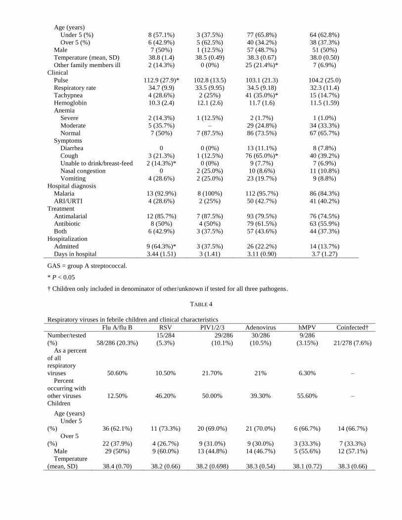
these assignments to evaluate associations with clinical data (Table 3). Most notably, compared
with children in whom no respiratory virus was detected, children with any respiratory virus
more frequently reported that other family members were ill (21.4%), complained of cough
(65%), and were tachypneic (35%; all P < 0.05). Among children with a respiratory virus
detected, there was a trend toward more diarrhea (6/30 [20%]) in children infected with
adenovirus (Table 4) (P = 0.11).
Though 10% afebrile controls reported cough, there was no significant association between
infection and cough in afebrile children.

Population attributable fraction of microbiologic etiologies.
Asymptomatic carriage of pathogens is common, and the proportion of fevers or illness
caused by a detected pathogen can be estimated by comparing carriage in healthy children.
Pathogens equally prevalent in cases and controls are considered infrequent contributors to
febrile illness, and pathogens detected only in febrile children would be considered likely to be
causing fever. In our study population, P. falciparum, influenza, PIV, RSV, hMPV, and
adenovirus were each detected in both febrile cases and afebrile controls, but only influenza and
PIV were detected significantly more frequently in febrile than afebrile children. We thus
estimated the odds of fever given infection for influenza and PIV and calculated, among children
with each pathogen, the fraction of fevers attributable to each (Table 5). In this analysis, of 58
febrile children with influenza, 67.5% could attribute their fevers to the virus while the
remaining 32.5% have afebrile influenza infection. The percentage is slightly higher in younger
children. There was a trend toward greater odds of being febrile with PIV (OR = 2.47, P =
0.069), with an estimated AF of 59%. There were no afebrile children under 5 years with P.
falciparum; therefore, the AF of fever could not be calculated in this age group.
DISCUSSION
We demonstrate that among febrile children presenting in 2011–2012 to a rural Kenyan
district hospital, malaria was both uncommon and overdiagnosed, respiratory viruses including
influenza and PIV were common, and GAS pharyngitis was rare.
Respiratory pathogens were common: we detected at least one virus in nearly half of all
febrile patients and, importantly, one-third of afebrile children. Comparing these groups, only
influenza and PIV were more common in febrile than afebrile children. We draw two important
conclusions from this observation. First, respiratory viruses were overall more commonly
associated with febrile illness than malaria. Second, a substantial proportion of respiratory viral

infections did not cause an overt febrile illness, at least at the time of presentation. Therefore,
efforts to estimate the burden of illness of these pathogens should include surveillance of
asymptomatic control patients.
In malaria-endemic areas, it is not possible to clinically distinguish falciparum malaria from
other etiologies of fever.24–26
Indeed, in our study, the clinical presentations of respiratory viruses
and malaria were not sufficiently distinct to allow them to be distinguished without pathogen-
specific testing. For example, while cough was more common in those with influenza than
malaria (67.2% versus 21.3%), nearly one-third of patients with influenza did not report cough,
and nearly one-quarter of patients with malaria did have cough. Similarly, tachypnea was
common in both groups, and abnormal findings on physical exam were uncommon in both
groups. Improved point-of-care diagnostics are needed to discriminate treatable etiologies of
fever in these children.
GAS pharyngitis was rare in our study, despite the high prevalence of rheumatic heart
disease in Kenya.15,16
Several explanations may account for this observation. First, streptococcal
pharyngitis is primarily a disease of children between the ages of 5 and 15 years27
and the
majority of children enrolled in this study were under the age of 5 years; however, even among
129 febrile patients of ages 5 years and over, as well as 81 afebrile patients in this age group,
only 5 tested positive for GAS throat infection. A second possibility is that, within populations,
streptococcal infections are epidemic, and we sampled in a period between outbreaks. However,
in low-income countries, there are an estimated 0.4 cases of symptomatic GAS pharyngitis per
person per year,16
so it seems unlikely that this evidently high risk population would be almost
entirely free of infection. Third, the sensitivity of our rapid antigen detection test may not have
been sufficient to detect asymptomatic throat carriage. To evaluate this possibility, we performed
culture on sheep blood agar of 58 throat swabs obtained concurrently with the sample used for

the rapid strep test and noted no increase in prevalence in this subset (data not shown). A fourth
possibility is that preenrollment antibiotic treatment (most commonly amoxicillin,
trimethoprim/sulfamethoxazole, or penicillin) may have eradicated streptococcal infection.
However, less than 10% patients reported receipt of an antibacterial agent prior to presentation.
Finally, a fifth and perhaps most likely explanation is that symptomatic group A pharyngitis does
not prompt treatment seeking in our study population. This has been observed elsewhere.28,29
The
same may be true for acute rheumatic fever, which can cause severe acute illness but commonly
presents with subtle manifestations, especially with recurrent episodes.30
Our study supports the
notion that, to prevent rheumatic heart disease, efforts to treat streptococcal pharyngitis and acute
rheumatic fever will need to be implemented in community settings.
Malaria prevalence in febrile and afebrile patients as measured by P. falciparum PCR was
low, in stark contrast with the microscopic diagnosis performed in the hospital. However, three-
quarters of children with a documented negative smear still received an antimalarial, indicating
that overtreatment with antimalarials is not simply related to lack of access to diagnosis.
Our study had several limitations. First, information on comorbid illnesses was reported by
the parent or guardian and may therefore be inaccurate. We did not test for HIV infection;
however, HIV prevalence in adolescents in the study catchment area is known to be less than
1%,31
and prevalence in younger children is similarly low (Wendy P. O’Meara, personal
communication). Second, entrance physical examinations were performed by the study nurse,
and sensitivity and specificity of those physical exam findings is unknown. However, this does
reflect the realities of care and staffing levels at the district hospital level, and prior literature
suggests there is poor predictive value of signs in this setting. Third, we examined a limited set
of pathogens in a population that is likely affected by many others.5,32–34
Further testing of
banked samples is ongoing. Finally, our control group was not randomly selected, instead being

drawn from patients presenting to the hospital for other reasons. Therefore, bias may have been
introduced if this group is not representative of either the pediatric population as a whole or the
population from which the cases were drawn (if, for example, treatment-seeking behaviors are
different for patients with febrile illness compared with other diagnoses). Nevertheless, our
afebrile controls were similar demographically to the febrile cases.
Despite these limitations, this study has several notable strengths. First, by looking at a
broader spectrum of potential pathogens, including parasites, viruses, and a bacterial pathogen,
we were able to describe the relative frequency of these disease-causing agents within the same
children. Second, by enrolling both patients who were managed as outpatients as well as those
ultimately admitted to the hospital, we capture a more complete spectrum of febrile illness than
focusing solely on hospitalized patients. In addition, our study population was not limited to
those with respiratory symptoms. Previous work has focused on the etiology of acute respiratory
infections or pneumonia in patients presenting with respiratory-specific symptoms.7,12,14
However, we found that 30% children who would not have been classified as an acute
respiratory illness were infected with a respiratory virus. Finally, the inclusion of afebrile
controls allows us to estimate the background prevalence of malaria, respiratory viruses, and
group A Streptococcus during the study period, and thus to calculate attributable fractions for
fever. This is especially important given that each of these pathogens can cause symptomatic or
asymptomatic infection (i.e., carriage). In our study, carriage of pathogens was common and this
is likely true in other contexts. Such information should be held in mind when interpreting the
results from fever etiology studies lacking a comparison group.
The costs of malaria overdiagnosis are high, including the morbidity of the missed
opportunity for identification and treatment of the actual underlying illness, the financial costs of
malaria medications, and the cost of increased selection pressure leading to malaria drug

resistance. There is an unmet need for improved diagnostic capacity and improved clinical
algorithms for acute febrile illnesses. Access to diagnostic testing must go hand in hand with
better understanding of the relationship between carriage and disease, and confidence to use the
results to guide treatment decisions; only then will it be possible to improve care of the child
with acute febrile illness.
Received September 5, 2014.
Accepted for publication December 31, 2014.
Acknowledgments:
We thank Kyaw Thwai and Steven Meshnick for their assistance with the P. falciparum real-time PCR testing. We
are deeply grateful to Webuye District Hospital administration for their support and to all the patients and their
guardians who participated.
Financial support: This study was supported by a pilot grant from Duke Global Health Institute.
Authors’ addresses: Wendy P. O’Meara, Division of Infectious Diseases and International Health, Duke University
Medical Center, Duke Global Health Institute, Durham, NC, and Moi University School of Public Health, College of
Health Sciences, Eldoret, Kenya, E-mail: [email protected]. Joshua A. Mott, Barry Fields, and Kabura Wamburu,
Centers for Disease Control and Prevention, Nairobi, Kenya, E-mails: [email protected], [email protected], and
[email protected]. Jeremiah Laktabai and Janice Armstrong, Moi University School of Medicine, College of
Health Sciences, Eldoret, Kenya, E-mails: [email protected] and [email protected]. Steve M. Taylor
and Christopher W. Woods, Division of Infectious Diseases and International Health, Duke University Medical
Center, Duke Global Health Institute, Durham Veterans Affairs Medical Center, Durham, NC, E-mails:
[email protected] and [email protected]. Charles MacIntyre, Reeshi Sen, and William Pan, Duke Global
Health Institute, Durham, NC, E-mails: [email protected], [email protected], and
[email protected]. Diana Menya, Moi University School of Public Health, College of Health Sciences, Eldoret,
Kenya, E-mail: [email protected]. Bradly P. Nicholson, Division of Infectious Diseases and International
Health, Duke University Medical Center, Durham Veterans Affairs Medical Center, Durham, NC, E-mail:
[email protected]. Thomas L. Holland, Division of Infectious Diseases and International Health, Duke
University Medical Center, Durham, NC, E-mail: [email protected].

REFERENCES
<eref>1. World Health Organization. World Health Statistics. Available at:
http://apps.who.int/gho/data/node.main.ChildMortDistRegion?lang=en. Accessed November
19, 2014.</eref>
<jrn>2. O’Meara WP, Mangeni JN, Steketee R, Greenwood B, 2010. Changes in the burden of
malaria in sub-Saharan Africa. Lancet Infect Dis 10: 545–555.</jrn>
<jrn>3. Murray CJ, Rosenfeld LC, Lim SS, Andrews KG, Foreman KJ, Haring D, Fullman N,
Naghavi M, Lozano R, Lopez AD, 2012. Global malaria mortality between 1980 and 2010: a
systematic analysis. Lancet 379: 413–431.</jrn>
<jrn>4. Mwangangi JM, Mbogo CM, Orindi BO, Muturi EJ, Midega JT, Nzovu J, Gatakaa H,
Githure J, Borgemeister C, Keating J, Beier JC, 2013. Shifts in malaria vector species
composition and transmission dynamics along the Kenyan coast over the past 20 years.
Malar J 12: 13.</jrn>
<jrn>5. Crump JA, Morrissey AB, Nicholson WL, Massung RF, Stoddard RA, Galloway RL,
Ooi EE, Maro VP, Saganda W, Kinabo GD, Muiruri C, Bartlett JA, 2013. Etiology of severe
non-malaria febrile illness in northern Tanzania: a prospective cohort study. PLoS Negl Trop
Dis 7: e2324.</jrn>
<jrn>6. Leslie T, Mikhail A, Mayan I, Anwar M, Bakhtash S, Nader M, Chandler C, Whitty CJ,
Rowland M, 2012. Overdiagnosis and mistreatment of malaria among febrile patients at
primary healthcare level in Afghanistan: observational study. BMJ 345: e4389.</jrn>
<jrn>7. Katz MA, Lebo E, Emukule G, Njuguna HN, Aura B, Cosmas L, Audi A, Junghae M,
Waiboci LW, Olack B, Bigogo G, Njenga MK, Feikin DR, Breiman RF, 2012.

Epidemiology, seasonality, and burden of influenza and influenza-like illness in urban and
rural Kenya, 2007–2010. J Infect Dis 206 (Suppl 1): S53–S60.</jrn>
<jrn>8. Yazdanbakhsh M, Kremsner PG, 2009. Influenza in Africa. PLoS Med 6:
e1000182.</jrn>
<jrn>9. Gessner BD, Shindo N, Briand S, 2011. Seasonal influenza epidemiology in sub-
Saharan Africa: a systematic review. Lancet Infect Dis 11: 223–235.</jrn>
<jrn>10. Majanja J, Njoroge RN, Achilla R, Wurapa EK, Wadegu M, Mukunzi S, Mwangi J,
Njiri J, Gachara G, Bulimo W, 2013. Impact of influenza A (H1N1) pdm09 virus on
circulation dynamics of seasonal influenza strains in Kenya. Am J Trop Med Hyg 88: 940–
945.</jrn>
<jrn>11. Waitumbi JN, Kuypers J, Anyona SB, Koros JN, Polhemus ME, Gerlach J, Steele M,
Englund JA, Neuzil KM, Domingo GJ, 2010. Outpatient upper respiratory tract viral
infections in children with malaria symptoms in western Kenya. Am J Trop Med Hyg 83:
1010–1013.</jrn>
<jrn>12. Feikin DR, Njenga MK, Bigogo G, Aura B, Aol G, Audi A, Jagero G, Muluare PO,
Gikunju S, Nderitu L, Balish A, Winchell J, Schneider E, Erdman D, Oberste MS, Katz MA,
Breiman RF, 2012. Etiology and incidence of viral and bacterial acute respiratory illness
among older children and adults in rural western Kenya, 2007–2010. PLoS ONE 7:
e43656.</jrn>
<jrn>13. D’Acremont V, Kilowoko M, Kyungu E, Philipina S, Sangu W, Kahama-Maro J,
Lengeler C, Cherpillod P, Kaiser L, Genton B, 2014. Beyond malaria—causes of fever in
outpatient Tanzanian children. N Engl J Med 370: 809–817.</jrn>

<jrn>14. Berkley JA, Munywoki P, Ngama M, Kazungu S, Abwao J, Bett A, Lassauniére R,
Kresfelder T, Cane PA, Venter M, Scott JA, Nokes DJ, 2010. Viral etiology of severe
pneumonia among Kenyan infants and children. JAMA 303: 2051–2057.</jrn>
<jrn>15. Marijon E, Mirabel M, Celermajer DS, Jouven X, 2012. Rheumatic heart disease.
Lancet 379: 953–964.</jrn>
<jrn>16. Carapetis JR, Steer AC, Mulholland EK, Weber M, 2005. The global burden of group
A streptococcal diseases. Lancet Infect Dis 5: 685–694.</jrn>
<jrn>17. Zuhlke L, Engel ME, Karthikeyan G, Rangarajan S, Mackie P, Cupido B, Mauff K, Islam S, Joachim A,
Daniels R, Francis V, Ogendo S, Gitura B, Mondo C, Okello E, Lwabi P, Al-Kebsi MM, Hugo-Hamman C, Sheta
SS, Haileamlak A, Daniel W, Goshu DY, Abdissa SG, Desta AG, Shasho BA, Begna DM, ElSayed A, Ibrahim AS,
Musuku J, Bode-Thomas F, Okeahialam BN, Ige O, Sutton C, Misra R, Abul Fadl A, Kennedy N, Damasceno A,
Sani M, Ogah OS, Olunuga T, Elhassan HH, Mocumbi AO, Adeoye AM, Mntla P, Ojji D, Mucumbitsi J, Teo K,
Yusuf S, Mayosi BM, 2014. Characteristics, complications, and gaps in evidence-based interventions in rheumatic
heart disease: the Global Rheumatic Heart Disease Registry (the REMEDY study). Eur Heart J.
doi:http://dx.doi.org/10.1093/eurheartj/ehu449.</jrn>
<eref>18. Poverty Rates by County, 2005/6. Kenya Bureau of Statistics. Available at:
https://www.opendata.go.ke/Counties/Poverty-Rates-by-County/z6za-e7yb. Accessed
November 19, 2014.</eref>
<jrn>19. Obala AA, Mangeni JN, Platt A, Aswa D, Abel L, Namae J, OMeara WP, 2015. What
is threatening the effectiveness of insecticide-treated bednets? A case-control study of
environmental, behavioral, and physical factors associated with prevention failure.
(submitted).</jrn>
<jrn>20. Kim C, Ahmed JA, Eidex RB, Nyoka R, Waiboci LW, Erdman D, Tepo A, Mahamud
AS, Kabura W, Nguhi M, Muthoka P, Burton W, Breiman RF, Njenga MK, Katz MA, 2011.
Comparison of nasopharyngeal and oropharyngeal swabs for the diagnosis of eight
respiratory viruses by real-time reverse transcription-PCR assays. PLoS ONE 6:
e21610.</jrn>
<jrn>21. Carvalho Mda G, Tondella ML, McCaustland K, Weidlich L, McGee L, Mayer LW,
Steigerwalt A, Whaley M, Facklam RR, Fields B, Carlone G, Ades EW, Dagan R, Sampson

JS, 2007. Evaluation and improvement of real-time PCR assays targeting lytA, ply, and psaA
genes for detection of pneumococcal DNA. J Clin Microbiol 45: 2460–2466.</jrn>
<jrn>22. Rantala AM, Taylor SM, Trottman PA, Luntamo M, Mbewe B, Maleta K, Kulmala T,
Ashorn P, Meshnick SR, 2010. Comparison of real-time PCR and microscopy for malaria
parasite detection in Malawian pregnant women. Malar J 9: 269.</jrn>
<jrn>23. 1992. Guidelines for the diagnosis of rheumatic fever. Jones criteria, 1992 update.
Special Writing Group of the Committee on Rheumatic Fever, Endocarditis, and Kawasaki
Disease of the Council on Cardiovascular Disease in the Young of the American Heart
Association. JAMA 268: 2069–2073.</jrn>
<jrn>24. Bassat Q, Machevo S, O’Callaghan-Gordo C, Sigaúque B, Morais L, Díez-Padrisa N,
Ribó JL, Mandomando I, Nhampossa T, Ayala E, Sanz S, Weber M, Roca A, Alonso PL,
2011. Distinguishing malaria from severe pneumonia among hospitalized children who
fulfilled integrated management of childhood illness criteria for both diseases: a hospital-
based study in Mozambique. Am J Trop Med Hyg 85: 626–634.</jrn>
<jrn>25. Mayxay M, Castonguay-Vanier J, Chansamouth V, Dubot-Pérès A, Paris DH,
Phetsouvanh R, Tangkhabuanbutra J, Douangdala P, Inthalath S, Souvannasing P, Slesak G,
Tongyoo N, Chanthongthip A, Panyanouvong P, Sibounheuang B, Phommasone K, Dohnt
M, Phonekeo D, Hongvanthong B, Xayadeth S, Ketmayoon P, Blacksell SD, Moore CE,
Craig SB, Burns MA, von Sonnenburg F, Corwin A, de Lamballerie X, González IJ,
Christophel EM, Cawthorne A, Bell D, Newton PN, 2013. Causes of non-malarial fever in
Laos: a prospective study. The Lancet Global Health 1: e46–e54.</jrn>
<jrn>26. Taylor SM, Molyneux ME, Simel DL, Meshnick SR, Juliano JJ, 2010. Does this
patient have malaria? JAMA 304: 2048–2056.</jrn>

<jrn>27. Shulman ST, Bisno AL, Clegg HW, Gerber MA, Kaplan EL, Lee G, Martin JM, Van
Beneden C, 2012. Clinical practice guideline for the diagnosis and management of group A
streptococcal pharyngitis: 2012 update by the Infectious Diseases Society of America. Clin
Infect Dis 55: e86–e102.</jrn>
<jrn>28. Pondei K, Kunle-Olowu OE, Peterside O, 2013. The aetiology of non-malarial febrile
illness in children in the malaria-endemic Niger Delta Region of Nigeria. Asian Pacific
Journal of Tropical Disease 3: 56–60.</jrn>
<jrn>29. Bergmark R, Bergmark B, Blander J, Fataki M, Janabi M, 2010. Burden of disease and
barriers to the diagnosis and treatment of group a beta-hemolytic streptococcal pharyngitis
for the prevention of rheumatic heart disease in Dar Es Salaam, Tanzania. Pediatr Infect Dis
J 29: 1135–1137.</jrn>
<jrn>30. Carapetis JR, McDonald M, Wilson NJ, 2005. Acute rheumatic fever. Lancet 366:
155–168.</jrn>
<jrn>31. Wachira J, Ndege S, Koech J, Vreeman R, Ayuo P, Braitstein P, 2013. HIV testing
uptake and prevalence among adolescents and adults in a large home-based HIV testing
program in western Kenya. J Acquir Immune Defic Syndr 65: e58–e66.</jrn>
<jrn>32. Kasper MR, Blair PJ, Touch S, Sokhal B, Yasuda CY, Williams M, Richards AL,
Burgess TH, Wierzba TF, Putnam SD, 2012. Infectious etiologies of acute febrile illness
among patients seeking health care in south-central Cambodia. Am J Trop Med Hyg 86: 246–
253.</jrn>
<jrn>33. Hertz JT, Munishi OM, Ooi EE, Howe S, Lim WY, Chow A, Morrissey AB, Bartlett
JA, Onyango JJ, Maro VP, Kinabo GD, Saganda W, Gubler DJ, Crump JA, 2012.

Chikungunya and dengue fever among hospitalized febrile patients in northern Tanzania. Am
J Trop Med Hyg 86: 171–177.</jrn>
<jrn>34. Sutherland LJ, Cash AA, Huang YJ, Sang RC, Malhotra I, Moormann AM, King CL,
Weaver SC, King CH, LaBeaud AD, 2011. Serologic evidence of arboviral infections among
humans in Kenya. Am J Trop Med Hyg 85: 158–161.</jrn>
*OUTLEGENDS*F1*FIGURE 1 Seasonality of malaria, influenza A, and influenza B in febrile children.
TABLE 1
Clinical characteristics of febrile and afebrile children
Febrile Afebrile
Total (N = 370) Total (N = 184) P value
n or mean (% or SD) n or mean (% or SD)
Patient demographics
Male 179 (48.4%) 97 (52.7%) 0.37
Age (years) 4.4 (2.82) 4.8 (3.14) 0.08
Under 5 241 (65.1%) 81 (44%) 0.04
In school 163 (44.8%) 84 (53.2%) 0.09
Anthropometrics (mean, SD)
MUAC (cm) 17.25 (8.48) 16.6 (2.31) 0.31
Severe or moderate malnutrition 3 (0.8%) 3 (1.6%) 0.38
Weight for age
Wasted (WAZ < 2)† 82/343 (23.9%) 32/161 (19.9%) 0.31
Received all immunizations 365 (98.9%) 183 (99.5%) 0.07
Vital signs
Temperature 38.16 (0.64) 36.7 (0.6) N/A
Duration of illness (days) 2.13 (2.1) – –
Respiratory rate 33.06 (9.41) – –
Tachypnea* 96 (26%) – –
Pulse (beats/min) 100.92 (22.59) – –
Symptoms
Cough 179 (48.4%) 19 (10.3%) < 0.001
Vomiting 59 (15.9%) 10 (5.4%) < 0.001
Diarrhea 39 (10.5%) 13 (7.1%) 0.19
Unable to drink/breast-feed 23 (6.2%) 0 < 0.001
Nasal congestion 37 (10.0%) 7 (3.8%) 0.01
Difficulty breathing 5 (1.4%) 0 0.11
Chest pain 1 (0.3%) 0 0.48
Headache 8 (2.2%) 2 (1.1%) 0.37
Convulsions 3 (0.8%) 0 0.22
Sore throat 0 0 –
Rash 6 (1.6%) 3 (1.6%) 1.0
Treatment prior to visit
Any 135 (36.5%) – –
Antimalarial 52 (14.1%) – –
Antibiotic 41 (11.1%) – –
Physical examination
Spleen enlarged 2 (0.5%) 0 (0%) 0.32
Liver enlarged 2 (0.5%) 0 (0%) 0.32

Palmar pallor 2 (0.5%) 2 (1.1%) 0.47
Conjuctival pallor 1 (0.3%) 1 (0.5%) 0.61
Wasting 3 (1.1%) 3 (1.63%) 0.38
Rash 10 (2.7%) 3 (1.63%) 0.56
Throat
Tonsillar exudates 1 (0.3%) 2 (1.1%) 0.22
Mouth ulcer 4 (1.1%) 1 (0.5%) 0.53
Ears
Discharge 6 (1.6%) 1 (0.5%) 0.29
Signs of infection 8 (2.2%) 0 (0%) 0.05
Neck
Adenopathy 2 (0.5%) 0 (0%) 0.32
Neck stiffness 2 (0.05%) 0 (0%) 0.32
Heart murmur 2 (0.5%) 1 (0.5%) 0.99
Lungs
Crepitations 2 (0.5%) 0 (0%) 0.32
Chest indrawing/stridor 2 (0.6%) 0 (0%) 0.32
Wheezing 4 (1.1%) 12 (6.5%) 0.001
Dehydration 4 (1.4%) 0 (0%) 0.16
MUAC = mid upper arm circumference; SD = standard deviation; WAZ = wasted-for-age Z-score.
Less than 2% children reported convulsions, difficulty breathing. Less than 2% had edema or eye problems on
exam.
* According to WHO criteria: > 40 BPM for under 5 and > 30 for over 5
† WAZ up to 10 years only.
TABLE 2
Pathogens in febrile and afebrile subjects
Pathogens present Febrile Afebrile
Total (N = 370) Total (N = 184)
Number/tested % Number/tested %
Malaria† 14/268 5.2% 3/133 2.3%
GAS 8/354 2.3% 1/161 0.6%
Flu A/flu B 58/286 20.3%* 9/118 7.6%*
RSV 15/284 5.3% 8/118 6.8%
PIV1/2/3 29/286 10.1%* 5/118 4.2%*
Adenovirus 30/286 10.5% 10/118 8.5%
hMPV 9/286 3.2% 2/118 1.7%
Coinfected‡ 22/269 8.2% 5/115 4.4%
Other/unknown§ 102/275 37.1%* 59/116 50.9%*
hMPV = XXXX; GAS = group A streptococcal; PIV = parainfluenza virus; RSV = respiratory syncytial virus
*P < 0.05 for comparison between Febrile and Afebrile
† As detected by a real-time polymerase chain reaction (PCR) assay.
‡ Denominator for each pathogen includes all children tested for that pathogen. Children are only included in
denominator of “Coinfected” if they were tested for both malaria and respiratory viruses or if they were GAS
positive and had another infection (N = 1).
§ Children only included in denominator of other/unknown if tested for all three pathogens.
TABLE 3
Clinical characteristics of febrile patients by pathogen type
Malaria GAS Respiratory virus Other/unknown†
Number/tested (%) 14/268 (5.2%) 8/354 (2.3%) 117/282 (41.5%) 102/275 (37.1%)
Children
Age (years)
Under 5 (%) 8 (57.1%) 3 (37.5%) 77 (65.8%) 64 (62.8%)
Over 5 (%) 6 (42.9%) 5 (62.5%) 40 (34.2%) 38 (37.3%)
Male 7 (50%) 1 (12.5%) 57 (48.7%) 51 (50%)
Temperature (mean, SD) 38.8 (1.4) 38.5 (0.49) 38.3 (0.67) 38.0 (0.50)
Other family members ill 2 (14.3%) 0 (0%) 25 (21.4%)* 7 (6.9%)
Clinical
Pulse 112.9 (27.9)* 102.8 (13.5) 103.1 (21.3) 104.2 (25.0)
Respiratory rate 34.7 (9.9) 33.5 (9.95) 34.5 (9.18) 32.3 (11.4)
Tachypnea 4 (28.6%) 2 (25%) 41 (35.0%)* 15 (14.7%)
Hemoglobin 10.3 (2.4) 12.1 (2.6) 11.7 (1.6) 11.5 (1.59)
Anemia
Severe 2 (14.3%) 1 (12.5%) 2 (1.7%) 1 (1.0%)
Moderate 5 (35.7%) – 29 (24.8%) 34 (33.3%)
Normal 7 (50%) 7 (87.5%) 86 (73.5%) 67 (65.7%)
Symptoms
Diarrhea 0 0 (0%) 13 (11.1%) 8 (7.8%)
Cough 3 (21.3%) 1 (12.5%) 76 (65.0%)* 40 (39.2%)
Unable to drink/breast-feed 2 (14.3%)* 0 (0%) 9 (7.7%) 7 (6.9%)
Nasal congestion 0 2 (25.0%) 10 (8.6%) 11 (10.8%)
Vomiting 4 (28.6%) 2 (25.0%) 23 (19.7%) 9 (8.8%)
Hospital diagnosis
Malaria 13 (92.9%) 8 (100%) 112 (95.7%) 86 (84.3%)
ARI/URTI 4 (28.6%) 2 (25%) 50 (42.7%) 41 (40.2%)
Treatment
Antimalarial 12 (85.7%) 7 (87.5%) 93 (79.5%) 76 (74.5%)
Antibiotic 8 (50%) 4 (50%) 79 (61.5%) 63 (55.9%)
Both 6 (42.9%) 3 (37.5%) 57 (43.6%) 44 (37.3%)
Hospitalization
Admitted 9 (64.3%)* 3 (37.5%) 26 (22.2%) 14 (13.7%)
Days in hospital 3.44 (1.51) 3 (1.41) 3.11 (0.90) 3.7 (1.27)
GAS = group A streptococcal.
* P < 0.05
† Children only included in denominator of other/unknown if tested for all three pathogens.
TABLE 4
Respiratory viruses in febrile children and clinical characteristics
Flu A/flu B RSV PIV1/2/3 Adenovirus hMPV Coinfected†
Number/tested
(%) 58/286 (20.3%)
15/284
(5.3%)
29/286
(10.1%)
30/286
(10.5%)
9/286
(3.15%) 21/278 (7.6%)
As a percent
of all
respiratory
viruses 50.60% 10.50% 21.70% 21% 6.30% –
Percent
occurring with
other viruses 12.50% 46.20% 50.00% 39.30% 55.60% –
Children
Age (years)
Under 5
(%) 36 (62.1%) 11 (73.3%) 20 (69.0%) 21 (70.0%) 6 (66.7%) 14 (66.7%)
Over 5
(%) 22 (37.9%) 4 (26.7%) 9 (31.0%) 9 (30.0%) 3 (33.3%) 7 (33.3%)
Male 29 (50%) 9 (60.0%) 13 (44.8%) 14 (46.7%) 5 (55.6%) 12 (57.1%)
Temperature
(mean, SD) 38.4 (0.70) 38.2 (0.66) 38.2 (0.698) 38.3 (0.54) 38.1 (0.72) 38.3 (0.66)

Other family
members ill 18 (31.6%)* 1 (6.67%) 6 (20.7%) 3 (10%) 1 (12.5%) 5 (23.8%)
Clinical
Pulse 108.3 (24.4)* 104.5 (16.8) 100.9 (16.0) 97.6 (16.9) 108.7 (24.6) 113.1 (16.4)
Respiratory
rate 34.9 (7.3) 32.3 (9.16) 33.7 (11.8) 35.9 (11.2) 36.9 (13.8) 36.8 (13.4)
Tachypnea 26 (44.8%) 4 (26.7%) 9 (31.0%) 9 (30.0%) 4 (44.4%) 10 (47.6%)
Hemoglobin 12.0 (1.76) 11.53 (1.70) 11.3 (1.15) 11.72 (1.40) 11.58 (1.20) 11.7 (1.13)
Anemia
Severe 1 (1.75%) 1 (6.67%) 0 (0%) 0 (0%) 0 (0%) 0 (0%)
Moderate 11 (19.3%) 2 (13.3%) 11 (37.9%) 8 (26.7%) 3 (33.3%) 6 (28.6%)
Normal 45 (79%) 12 (80.0%) 18 (62.1%) 22 (73.3%) 6 (66.7%) 15 (71.4%)
Symptoms
Diarrhea 6 (10.3%) 1 (6.67%) 2 (6.90%) 6 (20.0%) 1 (11.1%) 3 (14.3%)
Cough 39 (67.2%) 11 (73.3%) 18 (62.1%) 17 (56.7%) 5 (55.6%) 13 (61.9%)
Unable to
drink/breast-
feed 7 (12.1%) 0 (0%) 2 (6.90%) 1 (3.33%) 1 (11.1%) 2 (9.5%)
Nasal
congestion 3 (5.2%) 4 (26.7%) 5 (17.2%) 2 (6.67%) 1 (11.1%) 4 (19.1%)
Vomiting 8 (13.8%) 3 (20.0%) 5 (17.2%) 10 (33.3%) 2 (22.2%) 6 (28.6%)
Treatment
Antimalarial 48 (82.8%) 10 (66.7%) 19 (65.5%) 27 (90%) 6 (66.7%) 15 (71.4%)
Antibiotic 39 (67.2%) 11 (73.3%) 20 (68.9%) 19 (63.3%) 6 (66.7%) 16 (80%)
Both 30 (51.7%) 7 (46.7%) 11 (37.9%) 16 (53.3%) 3 (33.3%) 10 (50%)
Hospitalization
Admitted 15 (25.9%) 2 (13.3%) 6 (20.7%) 6 (20.0%) 2 (22.2%) 4 (19.1%)
Days in
hospital 3.17 (1.03) 4 (.) 3 (0) 3.67 (1.15) 3 (1.41) 3.33 (0.58)
hMPV = XXXX; PIV = parainfluenza virus; RSV = respiratory syncytial virus; SD = standard deviation.
* P < 0.05.
† Only one child was coinfected with malaria (plus respiratory virus) and one child was coinfected with group A
streptococcal (GAS) plus respiratory virus. Eliminated these from coinfected; here coinfected is defined as 2 +
respiratory viruses.
TABLE 5
AF estimates
OR P AF
Malaria 2.420 0.172 –
Under 5* – – –
Over 5 1.161 0.848 –
Flu 3.080 0.003 0.675
Under 5 2.920 0.034 0.658
Over 5 3.500 0.030 0.714
RSV 0.750 0.525 –
Under 5 0.960 0.946 –
Over 5 0.510 0.352 –
PIV1/2/3 2.474 0.069 0.596
Under 5 1.860 0.281 –
Over 5 5.510 0.111 –
Adenovirus 1.210 0.625 –
Under 5 1.290 0.601 –
Over 5 1.300 0.679 –
AF = attributable fraction; OR = odds ratio; PIV = parainfluenza virus; RSV = respiratory syncytial virus.
* There were no afebrile children under 5 years with parasitemia so the odds of fever could not be estimated.

Figure 1
Copyright © 2022 FDOKUMEN


![[Guidelines] | Rheumatic Fever New Zealand - RHD Action |](https://static.fdokumen.com/doc/165x107/6328f1eb2dd4b030ca0c5afa/guidelines-rheumatic-fever-new-zealand-rhd-action-.jpg)

