Lassa Fever
35
Lassa Fever J.B. MCCORMICK and S.P. FISHER-HoCH Background . . . . . . . . . . . . . . . . . . 2 Lassa Fever: Clinical Disease and Sequelae 2.1 Complications and Convalescence 2.2 Deafness in Lassa Fever. 2.3 Lassa Fever in Pregnancy 2.4 Lassa Fever in Children 3 Diagnosis ......... 4 Pathogenesis of Lassa Fever. 4.1 Route of Infection and Early Events .. 4.2 Viral Replication . . . . . . . . . 4.3 Persistence ........... . 4.4 Lymphopenia and Neutrophilia. 4.5 Host Inflammatory Response .. 4.6 Host Control of Virus Replication 4.7 Shock and Vascular Leakage ... 4.8 Hemorrhage and Platelet Function. Epidemiology of Lassa Fever . , 6 Transmission ........... 6.1 Rodent to Humans ...... . 6.2 Transmission from Person to Person: Hospital and Household 7 Burden of Infection and Disease in West Africa 8 Prevention and Control 8.1 Rodent Control. 9 Vaccine References . 1 Background 75 77 80 81 83 84 84 88 88 89 89 90 91 91 91 92 93 95 95 97 99 101 101 101 105 Nineteen arenaviruses have been isolated in the Old and the New World, most of them identified in South or Central America. Current evidence suggests that arena viruses have co-evolved along with their rodent hosts over a time scale of as University of Texas, School of Public Health, Brownsville, TX, USA M. B. A. Oldstone (ed.), Arenaviruses I © Springer-Verlag Berlin Heidelberg 2002
-
Upload
khangminh22 -
Category
Documents
-
view
0 -
download
0
Transcript of Lassa Fever

Lassa Fever J.B. MCCORMICK and S.P. FISHER-HoCH
Background . . . . . . . . . . . . . . . . . .
2 Lassa Fever: Clinical Disease and Sequelae 2.1 Complications and Convalescence 2.2 Deafness in Lassa Fever. 2.3 Lassa Fever in Pregnancy 2.4 Lassa Fever in Children
3 Diagnosis.........
4 Pathogenesis of Lassa Fever. 4.1 Route of Infection and Early Events .. 4.2 Viral Replication . . . . . . . . . 4.3 Persistence ........... . 4.4 Lymphopenia and Neutrophilia. 4.5 Host Inflammatory Response .. 4.6 Host Control of Virus Replication 4.7 Shock and Vascular Leakage ... 4.8 Hemorrhage and Platelet Function.
Epidemiology of Lassa Fever . ,
6 Transmission........... 6.1 Rodent to Humans ...... . 6.2 Transmission from Person to Person: Hospital and Household
7 Burden of Infection and Disease in West Africa
8 Prevention and Control 8.1 Rodent Control.
9 Vaccine
References .
1 Background
75
77 80 81 83 84
84
88 88 89 89 90 91 91 91 92
93
95 95 97
99
101 101
101
105
Nineteen arenaviruses have been isolated in the Old and the New World, most of them identified in South or Central America. Current evidence suggests that arena viruses have co-evolved along with their rodent hosts over a time scale of as
University of Texas, School of Public Health, Brownsville, TX, USA
M. B. A. Oldstone (ed.), Arenaviruses I© Springer-Verlag Berlin Heidelberg 2002
76 J.B. McConnick and S.P. Fisher-Hoch
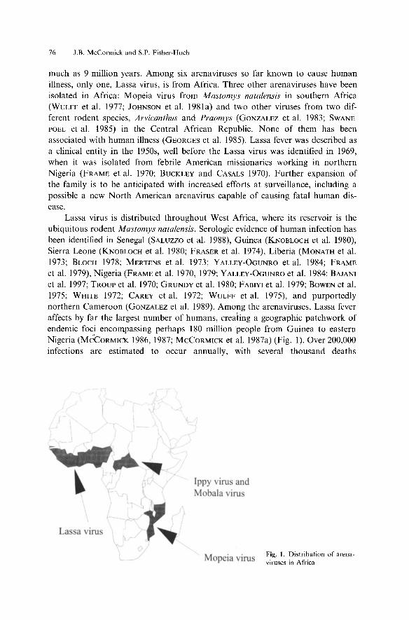
much as 9 million years. Among six arenaviruses so far known to cause human illness, only one, Lassa virus, is from Africa. Three other arena viruses have been isolated in Africa: Mopeia virus from Mastomys natalensis in southern Africa (WULFF et al. 1977; JOHNSON et al. 1981a) and two other viruses from two different rodent species, Arvicanthus and Praomys (GONZALEZ et al. 1983; SWANEPOEL et al. 1985) in the Central African Republic. None of them has been associated with human illness (GEORGES et al. 1985). Lassa fever was described as a clinical entity in the 1950s, well before the Lassa virus was identified in 1969, when it was isolated from febrile American missionaries working in northern Nigeria (FRAME et al. 1970; BUCKLEY and CASALS 1970). Further expansion of the family is to be anticipated with increased efforts at surveillance, including a possible a new North American arenavirus capable of causing fatal human disease.
Lassa virus is distributed throughout West Africa, where its reservoir is the ubiquitous rodent Mastomys natalensis. Serologic evidence of human infection has been identified in Senegal (SALUZZO et al. 1988), Guinea (KNOBLOCH et al. 1980), Sierra Leone (KNOBLOCH et al. 1980; FRASER et al. 1974), Liberia (MONATH et al. 1973; BLOCH 1978; MERTENS et al. 1973; YALLEy-OGUNRO et al. 1984; FRAME et al. 1979), Nigeria (FRAME et al. 1970, 1979; Y ALLEy-OGUNRO et al. 1984; BAJANI et al. 1997; TROUP et al. 1970; GRUNDY et al. 1980; FABIYI et al. 1979; BOWEN et al. 1975; WHITE 1972; CAREY et al. 1972; WULFF et al. 1975), and purportedly northern Cameroon (GONZALEZ et al. 1989). Among the arenaviruses, Lassa fever affects by far the largest number of humans, creating a geographic patchwork of endemic foci encompassing perhaps 180 million people from Guinea to eastern Nigeria (MCCORMICK 1986, 1987; MCCORMICK et al. 1987a) (Fig. 1). Over 200,000 infections are estimated to occur annually, with several thousand deaths
La a vim
Ippy iru and Mobala vim
Mopeia iru Fig. 1. Distribution of arenaviruses in Africa

Lassa Fever 77
(MCCORMICK 1986, 1987). The only longitudinal study of Lassa fever in West Africa took place in eastern Sierra Leone from the late 1970s through 1990 (MCCORMICK et al. 1987b). The Centers for Disease Control and Prevention (CDC) established and supported the study following repeated requests from the government of Sierra Leone to investigate epidemics in the eastern province. Over the years, many aspects of Lassa fever have been defined including the clinical presentation, epidemiology, immunology, pathophysiology, and therapy (MCCORMICK et al. 1987b; TRAPPIER et al. 1993; FISHER-HocH and MCCORMICK 1897). War and social chaos over the past decade have not only interrupted the ongoing studies and prevented new ones from taking place, but also probably increased the disease rates in these areas. Furthermore, the intervention in the war in Sierra Leone has resulted in the deaths of at least 4 expatriates working in Sierra Leone.
2 Lassa Fever: Clinical Disease and Sequelae
Lassa fever begins insidiously, after an incubation period of7-18 days, with fever, weakness, malaise, severe headache, usually frontal , and a very painful sore throat (Fig. 2) (KNOBLOCH et al. 1980; MCCORMICK et al. 1987b; MONATH et al. 1973, 1974; MERTENS et al. 1973). More than 50% of patients then develop joint and lumbar pain, and 60% develop a nonproductive cough. Many also develop a severe
Feyer WcakDe
Molai e Relro lerna I chI' I pain
H adach Diu;ne
omiling ore throat
Pharyngili bdominBI pain
Back paiD ough
Proteinuria Joint pain
Diarrhea bdominallenderne
E udaliyc pbaryngiti oDjunclivili
Dy uria f algia
RaIl' dema
Bleeding
o 20 40 60 80
Percent of patients with sign or symptom
II AI any lime At the time of hospitalization
Fig. 2. Signs and symptoms in Lassa fever
100

78 J.B. McCormick and S.P. Fisher-Hoch
retrosternal chest pain, and about half will have nausea with vomiting or diarrhea and abdominal pain. On physical examination, the respiratory rate, temperature, and pulse rate are elevated, and the blood pressure may be low. There is no characteristic skin rash in Lassa fever, and petechiae and ecchymoses are not seen. About a third of patients will have conjunctivitis; a few with conjunctival hemorrhages have a poor prognosis. More than two-thirds have pharyngitis, half with exudates, diffusely inflamed and swollen posterior pharynx and tonsils, but few if any ulcers or palatal petechiae. The abdomen is tender in 50% of patients, but bowel sounds are usually normal to marginally reduced. Neurological signs in the early stages are limited to a fine tremor, most marked in the lips and tongue (KNOBLOCH et al. 1980; MCCORMICK 1986; MCCORMICK et al. 1987b; CUMMINS et al. 1992; FISHER-HoCH et al. 1985).
Up to a third of hospitalized Lassa fever patients progress to a prostrating illness 6--8 days after onset of fever, usually with persistent vomiting and diarrhea. They are often dehydrated with elevated hematocrit. Proteinuria occurs in twothirds of patients, and blood urea nitrogen may be moderately elevated. About half of Lassa fever patients will have diffuse abdominal tenderness but no localizing signs or loss of bowel sounds. The severe retrosternal or epigastric pain seen in many patients may be due to pleural or pericardial involvement. Bleeding is seen in only 15%-20% of patients, limited primarily to the mucosal surfaces or occasionally manifest as conjunctival hemorrhages or gastrointestinal or vaginal bleeding. Severe pulmonary edema and adult r,?spiratory distress syndrome is common in fatal cases with gross head and neck 'edema, pharyngeal stridor, and hypovolemic shock ,(KNOBLOCH et al. 1980; MCCORMICK et al. 1987b).
Over 70% of patients may have abnormal electrocardiograms including nonspecific ST-segment and T-wave abnormalities, ST-segment elevation, generalized low voltage complexes, and changes reflecting electrolyte disturbance, but none of these correlate with clinical or other measures of disease severity or outcome and are not associated with clinical manifestations of myocarditis (CUMMINS et al. 1989a). Neurological signs are infrequent, but carry a poor prognosis, ranging from confusion to severe encephalopathy with or without general seizures, but without focal signs (MCCORMICK et al. 1987b; CUMMINS et al. 1992; FISHER-HocH et al. 1985). Cerebrospinal fluid is usually normal, but with a few lymphocytes, and low titers of virus relative to serum. Interstitial crepitations indicative of pneumonitis, and pleural and pericardial rubs suggesting effusions, develop in early convalescence in about 20% of hospitalized patients, occasionally in association with congestive cardiac failure, perhaps as a result of pericardial tamponade (MCCORMICK et al. 1987b; CUMMINS et al. 1989a; HIRABAYASHI et al. 1988).
Though the mean white blood cell count in Lassa fever on admission to hospital is normal (6 x 10⁹/1), this may mask marked lymphopenia and later relative or absolute neutrophilia, which may reach as much 30 x 109/1 in severely ill patients (MCCORMICK et al. 1987b; FISHER-HoCH et al. 1988). Based on similar observations with Ebola virus, the lymphopenia of Lassa fever may be related to apoptosis of Lassa virus-specific CTL or CD4 lymphocytes, though objective evidence is required to confirm ~his hypothesis (BAIZE et al. 1999). Thrombocytopenia is
Lassa Fever 79
moderate, even in severely ill patients, but platelet function is markedly depressed or even absent. This abnormality is usually maximal on admission to hospital and is present even when circulating platelet numbers remain above 100 x 109/1. A circulating inhibitor of platelet and neutrophil function has been described and will be discussed in detail in the section on pathogenesis (CUMMINS et a1. 1989b).
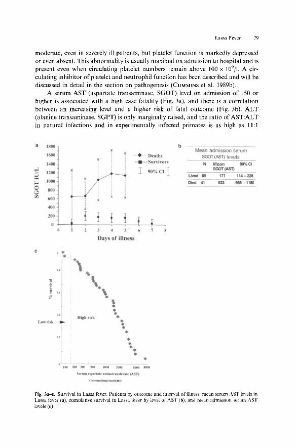
A serum AST (aspartate transaminase, SGOT) level on admission of 150 or higher is associated with a high case fatality (Fig. 3a), and there is a correlation between an increasing level and a higher risk of fatal outcome (Fig. 3b). ALT (alanine transaminase, SGPT) is only marginally raised, and the ratio of AST:ALT in natural infections and in experimentally infected primates is as high as 11: 1
a I 00
1600
I~OO
~ 1200
~ 1000
0 800 ~ en 600
400
200
0
c , . •
u
]
1 .. .. OA
Lo" ri ~ -..,
I
3 6
Days of illn
*,
'\ *\
• 1 • • 111gb rl k • • • • •
• • • • • .. nm uplrilit .mJ.Olra.rn'craw ~ ~n
jl_ttrn.IIoft •• Ullt-,,_I)
b Mean admission serum
Deaths SOOT (AST) levels . ur\ ivor ' N Mean 9O%C1
90% SGOT(AST)
Uved 89 171 114-228 ------Dod 41 923 665 -1180
•
Fig. 3a-e. Survival in Lassa fever. Patients by outcome and interval of illness: mean serum AST levels in Lassa fever (a), cumulative survival in Lassa fever by level of AST (b), and mean admission serum AST levels (e)

80 J.B. McCormick and S.P. Fisher-Hoch
a f·· ... -! • Sa........ I
6,GO 190~ 5,00
f f 1 I 4,00
t ! f E 3,80
;:: r ',80
! ! ~ ',80
0,00 1-3 4-' 7-9 lO-ll 13=15 16-21
Days of iUness
Fig. 4a,b. Survival in Lassa fever. a Titers of Lassa virus in serum of dying and surviving patients. b Cumulative survival in Lassa fever by viremia level (TCIDso). (MCCORMICK et al. 1987b)
(MCCORMICK et al. 1986, 1987b). The degree of hepatic damage observed clinically (no jaundice, prothrombin times, glucose and bilir~bin levels are near normal) and in postmortem tissue in Lassa virus infection is limited and cannot account for the overall clinic.al picture, and certainly not for the severity of the disease observed (MCCORMICK et al. 1986).
Viremia of ~1 x 103.0 is associated with increasing case fatality (Fig. 4). High virus titers occur in the ovary, pancreas, uterus, and placenta in addition to the liver, but histological lesions are limited and not consistent with organ failure. Elevated viremia and AST together carry a risk of death of nearly 80% (Table 1) (MCCORMICK et al. 1986, 1987b).
While the case fatality in hospitalized patients is about 16%, the death rate for all Lassa virus infections (nonhospitalized + hospitalized) may be as low as 2%-3% depending on the geographic and clinical setting (MCCORMICK 1987, 1988). However, in recent outbreaks in Nigeria, much higher death rates have been observed in hospitalized patients possibly due to a variation in virus virulence or to parenteral transmission (FISHER-HoCH et al. 1995). Case fatality may be as much as 30% in the third trimester of pregnancy and exceeds 50% in patients with hemorrhage.
2.1 Complications and Convalescence
Infrequent complications, particularly in early convalescence, include uveItis, orchitis, and acute adrenal insufficiency. More commonly observed are pleural effusion with pleural lfUbs, clinical evidence of pericarditis with pericardial rubs,

Lassa Fever 81
Table 1. Outcome of Lassa fever by admission viremia and AST, Sierra Leone
Outcome Viremia < 103.0 Viremia;:: 103.0
AST< ISO AST;:: ISO AST< ISO AST;:: ISO
Survived 25 7 23 9 Died 4 I 6 31 Case fatality (%) 13.8 12.5 20.7 78
and ascites (MCCORMICK et al. 1987b). Renal or hepatic failure is very rarely seen. A single case report described an interesting complex of hemorrhagic pericarditis and cardiac tamponade with pleural effusions and ascites 6 months after acute Lassa fever (HIRABAYASHI et al. 1988, 1989). Repeated cultures have failed to isolate virus from effusion fluids, but effusion specimens contained high titers of Lassa-specific IgG and numerous lymphocytes. Convalescence may be prolonged with periods of fatigue and weakness. Many patients also exhibit cerebellar signs during convalescence from severe disease, particularly tremors and ataxia, but the majority of these resolve with time (CUMMINS et al. 1992).
2.2 Deafness in Lassa Fever
Nearly 30% of patients with Lassa virus infection suffer an acute loss of hearing in one or both ears (CUMMINS et al. 1 990a). The onset is nearly always during the convalescent phase of illness, and its development and degree are unrelated to the severity of the acute disease. It is unclear whether the damage is due to viral neurotropism, thrombosis, vasculitis, focal hemorrhage, or some other viral or immune response-related phenomenon. A sensorineural hearing deficit (SNHD) was detected in (29%) of confirmed hospitalized Lassa fever patients in Sierra Leone (compared to none in hospitalized febrile controls), and 50% had a bilateral deficit. A SNHD was detected in nearly 80% of those affected prior to a 2-week follow-up and in 21 % of patients at the follow-up (all were symptomatic). The audiometric measurements of this deafness are found in Table 2; no patient gave a history of hearing difficulty prior to acute Lassa fever. The mean auditory threshold of these patients is 55dB (normal < 25dB), and the mean disability is over 20% consistent with substantial deafness (CUMMINS et al. 1990a). About half of the patients show a near or complete recovery by 3-4 months after onset, but the other half continue with significant sensorineural deafness, which becomes permanent if it does not resolve within about a year.
Table 2. Deafness in Lassa fever: epidemiology of acute deafness associated with Lassa fever (CUMMINS et al. 1990a)
Acute Lassa Mean age and sex Number acute Unilateral/ Number permanent MAT fever deafness bilateral deficit
Yes (n = 49) 30.2 (l8M/32F) 14 (29%) 7/7 9/14 (64%) 55dB (95%CI± 7.ldB)
No (n = 20) 30.1 (10M/I OF) 0(0%)

82 J.B. McCormick and S.P. Fisher-Hoch
The onset of hearing impairment in symptomatic patients may be instantaneous in some, while in others it may develop over a few hours. In patients with a hearing loss, antibody to Lassa virus is present before the clinical onset of their hearing loss, which occurs approximately 5-12 days after the fever subsides. Deafness is not associated with the level of AST or viremia and thus not with the degree of disease severity.
At final assessment, 60% of those with acute hearing loss also had a residual hearing loss about 1\3 unilateral and 2\3 bilateral. There was no obvious association between the percentage recovery and the severity of the initial deficit, peak AST recorded during admission, or antiviral therapy. In some patients with initial total bilateral deafness, the extent of spontaneous improvement was dramatic.
The overall prevalence of SNHD in a group of individuals seropositive to Lassa virus was 22% (Table 3). In this study 2\3 of those with hearing loss had a unilateral deficit, and 1\3 a bilateral deficit. The mean MAT of all affected ears in those with antibody and hearing loss was 43.7dB (90%CI = ± 8.6dB), and the mean binaural disability, 30.5% (90%CI = ± 9.7%). A group of seronegative controls from the local healthy adult population matched for age and sex and evaluated for sensorineural thresholds showed 97% of the ears had a MAT < 25dB, and 100% had a MAT < 27.5dB. Thus, the long-term proportion of all Lassainfected patients with residual hearing loss is between 15% and 20%, and the hearing loss disability is over 30%, including some with complete deafness (CUMMINS et al. 1 990a).
Individuals who had experienced deafness of sudden onset (mean duration 7.1 years), apd contcols were also evaluated (Table 4). All denied any hearing problem prior to th~r acute deafness, and all related its onset to an acute febrile illness. In 59%, this illness was severe (hospital admissions), in 16% moderate, and in 25% mild. Mild recovery of hearing occurs at least subjectively in about 40% of patients. Antibody to Lassa virus was present in 81 % of deaf patients and 19% of controls. Positive serology was observed as frequently in those deaf patients who were unaware as those who were aware that Lassa fever may have been the illness associated with their deafness onset.
Table 3. Deafness in Lassa fever: prevalence of sensorineural hearing deficit (SNHD) in persons with previous Lassa virus infection (CUMMINS et al. 1990a)
Serology
Seropositive Seronegative
Number
51 92
SNHD present
II (22%) 2 (%)
Mean MAT
43.7db (90%CI± 8.6dB) :<;27.5db
Table 4. Deafness in Lassa fever: evidence of Lassa virus infection in people with deafness (CUMMINS et al. 1990a)
Deafness of sudden onset
Yes (n = 32) No (n = 32)
Prevalence of LF antibody
26/32 (81 %) 6/32 (19%)
I
MAT Bilateral/unilateral and mean disability
90.1dB (90%CI +/-4.0dB) 30/2 and 74% (+/-5.9%) NA NA

Lassa Fever 83
In total, 94% of patients with a SNHD had a bilateral deficit. Severity ranged from a moderate unilateral deficit to total bilateral loss of hearing. Overall, the degree of impairment was substantial. Of these patients, 72% had a profound or total hearing loss in one or both ears. The mean MAT of all ears evaluated was 90.ldB (90%CI = ±4.0dB) and the mean binaural disability, 74% (±5.9%). This suggests that after the initial improvement seen in some with acute hearing loss, those with residual hearing loss have a profound deficit, which does not improve with time. No associations were observed between MAT values and age, sex, and severity of disease (mild vs. severe) associated with deafness onset. With bilateral impairment, there was a weak correlation between the severity of the deficit in each ear. This is one of the highest rates of sudden onset deafness associated with a single disease ever reported, and it further illustrates the large burden of acute and chronic illness from Lassa fever.
2.3 Lassa Fever in Pregnancy
Lassa fever may be a common cause of maternal mortality in many areas of West Africa, with case fatality about 20%. However, there is a nearly twofold increase in the number of third-trimester Lassa virus infections requiring hospitalization compared with the first two trimesters, and a corresponding two- to threefold risk of maternal death from infection in the third trimester (Table 5). Very high levels of virus replication have been found in placental tissue in third-trimester patients. A fourfold reduction was noted in case fatality among women who spontaneously or were therapeutically aborted compared with those who were not (odds ratio for fatality with pregnancy intact is 5.5 compared with after uterine evacuation) (Table 6). Fetal loss is near 90% and does not seem to vary by trimester. The excess maternal mortality in the third trimester may be related to the relative immunosuppression of pregnancy at that time. Lassa virus is known to be present in the
Table 5. Lassa fever in pregnancy: mortality in pregnant and non-pregnant women with Lassa fever
Category of women Patients Deaths Case fatality (%)
All pregnant 68 14 21 First trimester 6 I 18 Second trimester 22 I 5 Third trimester 40 12 30" Not pregnant 79 10 13
Table 6. Lassa fever in pregnancy: case fatality in women with Lassa fever with and without uterine evacuation
Category
No uterine evacuation Uterine evacuation
Number of women
26 39
• p = 0.05 (x2 = 3.95, DF'= I, odds ratio = 5.57) (95%CI 1.02-30.26).
Case fatality
10/26 (38%) 4/39 (10%)"

84 J.B. McCormick and S.P. Fisher-Hoch
breast milk of infected mothers, and neonates are therefore at risk of congenital, intrapartum, and puerperal infection with Lassa virus.
2.4 Lassa Fever in Children
Lassa fever is common in children, but may be difficult to diagnose because the clinical manifestations are so general (WEBB et al. 1986; MONSON et al. 1987). In very young babies, marked edema ("swollen baby syndrome") has been described in those with severe disease, and near uniform fatality. In both Sierra Leone and Liberia, fetal infection is nearly 100% fatal. In older children, the disease may manifest as diarrhea or pneumonia or simply as an unexplained, prolonged fever. In one hospital study in Sierra Leone, 21 % of pediatric admissions had Lassa fever, with 12% fatality. Outpatient studies in the same hospital found 4% with evidence of previous Lassa infection and a further 10% with febrile illness who seroconverted (Table 7).
3 Diagnosis
The laboratory diagnosis of Lassa virus infections is based on isolation of virus from serum, demonstration of a fourfold rise in IgG antibody titer, or virus-specific IgG and/or IgM :antibody in association with compatible clinical disease (MCCORMICK et al. 1987b). Routine virus isolation may be accomplished easily from serum or tissues in cell cultures, but should be performed in BSL4 laboratory facilities (JOHNSON et al. 1987). Specimens should be drawn preferably into a vacuum tube system to minimize the risk of infection. Virus has also been isolated from urine, throat swabs, breast milk, spinal fluid, pleural and pericardial transudate, and autopsy material (Table 8). Virus may be recovered for 1-2 months in urine, but its detection in urine during acute disease is intermittent (MCCORMICK et al. 1987b). Given the technical and safety considerations for virus isolation, other techniques should be considered for most laboratories, particularly in endemic areas, where the diagnostic need is the greatest. The most reliable and safe routine method for the laboratory at present is detection of virus-specific antibody by IFA (JOHNSON et al. 1981b) or ELISA (BAUSCH et al. 2000). At present, no commercial diagnostic reagents are available. All reagents must come from a
Table 7. Lassa fever in children, Eastern Province, Sierra Leone (WEBB et al. 1986)
Age (years) Number Seroconversion Past infection
<I 80 3/80 (3.8%) 0 1-5 121 15/121 (12.4%) 3/121 (2.5%) 6-10 92 12/95 (12.6%) 6/95 (6.3%) 11-14 63 6/63 (9.5%) 6/63 (9.5%) Total 356 36/356 (10.1 %) 15/356 (4.2%)

Lassa Fever 85
Table 8. Isolation of Lassa virus from sites other than blood in hospitalized patients, Sierra Leone (J.B. McCormick, unpublished data)
Days of illness Urine Throat CSF Breast milk
0-5 0/10 1/3 1/3 NA 6-10 2/43 5/11 2/2 2/7 11-15 1/26 0/1 0/1 2/7 16-20 0/8 1/3 NA 1/4 21 + 0/9 NA NA NA Total (% positive) 3/96 (3.1) 7/18 (39) 3/6 (50) 5/18 (28)
handful of laboratories able to produce them under safe conditions. Many of these BSL 4 laboratories cannot, or do not, produce noninfectious reagents for use by other laboratories.
In most situations, the critical issue is rapid and accurate diagnosis. The ideal situation would be the detection of RNA by RT-PCR, since it would detect replicating virus, rapidly allowing for appropriate treatment and barrier nursing measures. RT -PCR is highly sensitive compared with virus isolation, and of course much more rapid (TRAPPIER et al. 1993) (Fig. 5 and Tables 9-11). Despite the recent advances in simplifying RT-PCR, it is not yet generally available and would only be applicable in situations where the quality control of the test is assured. Recently, a paper was published regarding Lassa viral antigen detection by ELISA (BAUSCH et al. 2000) in which the authors state that in only lout of 590 specimens did they find antigen in the presence of IgM antibody. The authors speculate that the virus might be cleared and thus not present or that the antigen is masked. This is surprising since a study published more than 10 years ago shows clearly the
100 ----0-- Virus isolation
-. . , .• ' " PCRlhybridization .. " ;8 80 .. '"
.., Q
=-'" , = .,' " .5 60
" .. =-'" ... Q
40 ;; .. " ... " Il.
20
3 6 9 12 15 18 21
Days of illness
Fig. 5. Percentage of positive isolation and reverse transcriptase polymerase chain reaction (RT-PCR)/ hybridization in serum of patients with laboratory-confirmed Lassa fever, by day of illness. (TRAPPIER et al. 1993)

86 J.B. McCormick and S.P. Fisher-Hoch
Table 9. Lassa virus detection in Lassa fever: virus isolation and PCR/hybridization for Lassa virus (TRAPPIER et al. 1993)
Virus isolation PCR Hybridization
+ Total + Total
Positive 84 44 128 108 20 128 Negative 39 126 165 63 102 165 Total 123 170 293 171 122 293 Sensitivity' 66 (57-74) 84 (77-90) Specificity' 76 (69--83) 62 (54--69)
• 95% confidence intervals in parentheses.
Table 10. Lassa virus detection in Lassa fever: sensitivity and specificity for PCR/hybridization diagnosis of a patient with Lassa fever compared with those without Lassa fever (TRAPPIER et al. 1993)
PCR/hybridization Patients with Lassa fever Patients without Lassa fever Undiagnosed illness
Positive Negative Total
76 22 98
o 42 42
Sensitivity for correct diagnosis of Lassa fever: 78% (95%CI 68-&5). Specificity for identifying patients without Lassa fever: 100 (95%CI 89--100). Predictive value for Lassa fever if the test is positive 100% (95%CI 94-100).
8 2
10
Predictive value for identifying a patient without Lassa fever if test is negative 66% (95%CI 53-77).
Table 11. Lassa virus dete<;:tion in Lassa fever: relationship between Lassa virus titers and frequency of positive PCR ana PCR/hy\lridization in the diagnosis of Lassa fever (TRAPPIER et al. 1993)
Serum virus titer (TCIDso) PCR alone PCR/hybridization
PCR positive PCR negative Both positive Both negative
Virus negative 35 99 22 54
Positive titers 1.2 0 1 0 0 1.6 9 12 7 6 2.1 4 13 4 8 2.6 6 4 1 0 3.1 17 4 6 0 3.6 5 1 4 1 4.1 14 1 6 0 4.6 5 0 2 0 5.1 5 1 3 0 5.5 1 0 0 0
simultaneous presence of antibody and virus throughout the course of illness (seen in over a hundred specimens from proven cases of Lassa fever; Table 12). Thus, the problem is clearly the masking of antigen by IgM rendering antigen detection as currently practised virtually useless for early diagnosis. Antigen detection ELISA and ELISA for IgM have recently been reported to have a specificity of 90% and a sensitivity of 88% compared with RT-PCR (BAUSCH et al. 2000). It would appear

Lassa Fever 87
Table 12. The simulation presence of virus and specific antibodies to Lassa virus in patients
Intervala Total no. of samples No. positive for
Virus Virus and antibody Percent positive for virus and antibody
1-6 117 87 23 26 7-12 199 114 63 51 13-18 93 28 25 89 19-24 18 2 2 100
a The disease interval is the number of days that clinical symptoms lasted before specimens were taken.
that the critical part of this is IgM, since antigen detection seems to add very little to the two.
The most widely available, simplest, and safest tests are the serological ones (BAUSCH et al. 2000). There are generally two types of tests available, the older and well tried immunofluorescent antibody assay (IFA) and the enzyme-linked immunosorbent assay (ELISA). Each test has advantages and disadvantages depending on the situation and the information that is needed. The advantages of IF A are its simplicity, low cost, ease of manipulation, its ability to control the background reaction, its overall reliability, and its flexibility, since it allows screening for several antigens in one test (JOHNSON et al. 1981a; ELLIOTT et al. 1982). Its disadvantages are a slightly, but not significantly, lower sensitivity than ELISA (BAUSCH et al. 2000), its modest subjectivity for the complete novice, and the cross-reactivity of the reagents with IgM, IgG, and IgA. In earlier studies, confirmed by virus isolation, an IFA IgG titer of at least 16 and an IgM titer of ~4, both by IFA, in a patient with clinically compatible disease are highly specific for Lassa infection. Similarly, an ELISA IgM positive early in disease is highly correlated with the presence of virus and predicative of acute infection (Table 8). IF A IgM antibody appears early in infection (53% positive in the first 6 days of illness, and 76% in the first 12 days of illness (Table 13). Clearly, therefore, IgM by any method is highly associated with acute infection, particularly in a patient with clinically compatible illness (BAUSCH et al. 2000). The use of ELISA in Africa is replete with problems resulting in a significant misrepresentation of reality (BIGGAR 1986; BIGGAR et al. 1985, 1986). While testing the background of each serum has resolved many of these problems, the advantage of IF A is that all positive and negative cells are on the
Table 13. The correlation between IgM and IgG antibodies to Lassa virus in hospitalized patients
Interval Total no. of No. of specimens specimens
IgM-, IgG- IgM +, IgG- IgM-,IgG+ IgM +, IgG + Percent positive for any class of antibody
1-6 128 57 16 3 48/128 53% 55 7-12 136 30 10 3 103/13676% 78 13-18 71 8 I 4 58/71 83% 89

88 J.B. McCormick and S.P. Fisher-Hoch
same spot on the slide, and they can be compared with each other in the same field. Indeed, with a minimum of experience, one can judge immediately the specificity by the appearance of the cells. Furthermore, cells with antigens from multiple viruses can be put on the same spot for the purposes of screening for several viruses at once (JOHNSON et al. 1981b; ELLIOTI et al. 1982).
While newer reagents make the cross-reactivity ofIFA with IgG, IgM, and IgA less of a problem than previously, nevertheless current technology suggests that under optimal conditions, ELISA is slightly more sensitive than the older IF A, which is not surprising. However, it may also provide different information. Where the conditions are optimal and reagents are available, ELISA should be used. Where the simplicity and reliability of IF A are advantageous, it remains a functional and reasonable alternative.
4 Pathogenesis of Lassa Fever
4.1 Route of Infection and Early Events
Natural infection from Lassa virus in the endemic areas of West Africa has two primary sources, infected rodents and infected pati~ts (see Sect. 5 for details). The primary route of virus entry is through cuts or abrasions on the skin, or in some instances mu.cosal surfaces, such as conjunctiva. A second source of direct entry is close contact with st:cretions or blood from patients infected with Lassa virus, particularly in the household setting. In these circumstances, infection is due to direct contact with infected blood, vomitus, urine, or secretions, or from contaminated needles or instruments.
Needle injection may carry a very high risk of mortality (FISHER-HoCH et al. 1995). Otherwise, there are no data that allow us to distinguish between pathogenic processes resulting from direct entry of virus into cuts and scratches or mucosal surfaces. Subcutaneous or mucosal inoculation of virus probably allows direct entry into the peripheral capillary system, and easy access to the lymphatics and the bloodstream. We can speculate that dendritic cells - antigen-presenting cells -become infected or take up viral antigen at the site of entry at an early stage, and then participate in the disease process and immune response. The ubiquitous presence of the putative Lassa receptor, alpha-dystroglycan, particularly on dendritic cells, undoubtedly facilitates viral entry into endothelial or dendritic cells at the site of primary infection (CAO et al. 1998). However, it is not yet clear whether this receptor is the only receptor component required for viral entry into cells. Infection of these initial sites facilitates access to and subsequent dissemination of the virus throughout the reticuloendothelial system, for which Lassa virus has tropism. The presence of alpha-dystroglycan in cells in many organs may also explain why Lassa virus eventually enters and replicates in a wide array of cells and organs (CAO et al. 199$).

Lassa Fever 89
4.2 Viral Replication
Other than an early antibody response to the viral proteins, we know little about the early host response to infection, but we do know that the severity of the disease is related to the level of viremia, and therefore presumably the level of virus replication (Fig. 4 and Tables 14, 15). It is worth noting that studies of genetic control of LCM virulence in mice suggest that the L gene plays a central role in pathogenesis (RIVIERE et al. 1985), and therefore it is interesting to speculate that even in Lassa fever the control of replication, and therefore pathogenesis, may depend in part on the polymerase gene. A single such observation with a reassortant of Mopeia virus L gene and Lassa virus S gene showed a reduced pathogenicity in mice compared with a recombinant with the Lassa L gene (LUKASHEVICH 1992; LUKASHEVICH et al. 1991). Thus, the situation with African arenaviruses may well be similar to that of LCMV in that the L gene, and presumably the polymerase gene, holds the key to virus replication and ultimately pathogenesis.
4.3 Persistence
There is some evidence that Lassa virus may persist, albeit at low titer, and for a limited period of time in primates. For example, virus may be detected intermittently in human urine for up to 60 days, and in monkeys viral RNA was detected in postmortem and biopsy material when examined by RT-PCR up to 112 days after challenge (FISHER-HoCH et al. 2000). Unlike in rodents, persistent virus is sequestered, and contamination of the environment with secondary infections is unlikely. Furthermore, virus is not recoverable in tissue culture from serum or blood of vaccinated and subsequently challenged primates after 14 days, or from tissues after 21 days even by co-cultivation.
Table 14. Viremia in Lassa fever: mean concentration of Lassa virus in blood of hospitalized patients by outcome
Survived Died
Number
315 77
Mean day of illness
11.1 8.0
Mean viremia titer
Table 15. Viremia in Lassa fever: isolation of Lassa virus from blood of hospitalized patients by illness interval and outcome
Days of clinical symptoms
1-3 4--6 7-9 10-12 13-15 16-21 Total
Survived (n) 19 58 84 63 50 64 338 % positive 33 67 54 35 28 3 38 Died (n) 8 30 29 19 11 5 103 % positive 88 90 90 100 82 100 91
90 J.B. McCormick and S.P. Fisher-Hoch
4.4 Lymphopenia and Neutrophilia
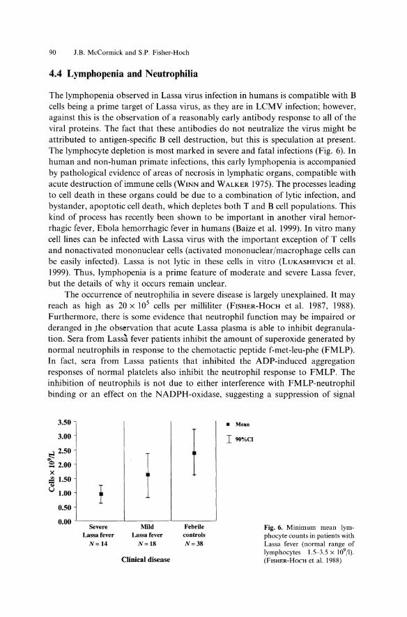
The lymphopenia observed in Lassa virus infection in humans is compatible with B cells being a prime target of Lassa virus, as they are in LCMV infection; however, against this is the observation of a reasonably early antibody response to all of the viral proteins. The fact that these antibodies do not neutralize the virus might be attributed to antigen-specific B cell destruction, but this is speculation at present. The lymphocyte depletion is most marked in severe and fatal infections (Fig. 6). In human and non-human primate infections, this early lymphopenia is accompanied by pathological evidence of areas of necrosis in lymphatic organs, compatible with acute destruction of immune cells (WINN and WALKER 1975). The processes leading to cell death in these organs could be due to a combination of lytic infection, and bystander, apoptotic cell death, which depletes both T and B cell populations. This kind of process has recently been shown to be important in another viral hemorrhagic fever, Ebola hemorrhagic fever in humans (Baize et al. 1999). In vitro many cell lines can be infected with Lassa virus with the important exception of T cells and nonactivated mononuclear cells (activated mononuclear/macrophage cells can be easily infected). Lassa is not lytic in these cells in vitro (LUKASHEVICH et al. 1999). Thus, lymphopenia is a prime feature of moderate and severe Lassa fever, but the details of why it occurs remain unclear.
The occurrence of neutrophilia in severe disease is largely unexplained. It may reach as high as 20 x 105 cells per milliliter (FI,SHER-HoCH et al. 1987, 1988). Furthermore, there is some evidence that neutrophil function may be impaired or deranged in the observation that acute Lassa plasma is able to inhibit degranulation. Sera from Lassa fever patients inhibit the amount of superoxide generated by normal neutrophils in response to the chemotactic peptide f-met-leu-phe (FMLP). In fact, sera from Lassa patients that inhibited the ADP-induced aggregation responses of normal platelets also inhibit the neutrophil response to FMLP. The inhibition of neutrophils is not due to either interference with FMLP-neutrophil binding or an effect on the NADPH-oxidase, suggesting a suppression of signal
3.50
3.00
~ 2.50
'" ~ 2.00 X ~ 1.50 ..
! t.i 1.00
0.50
0.00 Severe
Lassa fever N=14
Mild Lassafever
N=18
Clinical disease
I Febrile controls N=38
• Mean
I90%cI
Fig. 6. Minimum mean lymphocyte counts in patients with Lassa fever (normal range of lymphocytes 1.5-3.5 x 10⁹/1). (FISHER-HoCH et al. 1988)

Lassa Fever 91
transduction (ROBERTS et al. 1989). Thus, the inhibitory factor in Lassa plasma has global effects on cellular function beyond that of platelets and may playa central role in the pathogenesis.
4.5 Host Inflammatory Response
In contrast to the spleen, infection in solid organs (liver, kidney) involves only modest cell damage or death compared with, for example, acute fulminating hepatitis B, and this difference may be explained by the poor cellular inflammatory response observed in tissues from patients with Lassa fever (MCCORMICK et al. 1986; WALKER et al. 1982). Data on this issue are conflicting, as in vitro suppression of IL-8, a molecule critically central to inflammatory cell migration, is consistent with the in vivo picture of a low inflammatory cell response (LUKASHEVICH et al. 1999). This process could be part of a larger set of events precipitated by the infection of central inflammatory cells such as macrophages, but once again further data are not available. However, since elevation of IL-8 has been observed in Argentinian hemorrhagic fever, caution must be exercised in over-interpreting these data, particularly since more extensive studies of the broader inflammatory response have not been done (MARTA et al. 1999).
4.6 Host Control of Virus Replication
It is clear that failure to control viral replication is associated with severe disease. The viral control, as discussed above, is likely to reside in the L segment polymerase gene. The host's control of replication is determined mostly by the T-cell response, based on the observations that early antibody responses in humans do not correlate with outcome (Fig. 7) or with reduced virus replication, and that primates with high levels of antibody to all viral proteins induced by killed vaccine are not protected, and experience no limitation of viral replication (MCCORMICK et al. 1992). Little is known about the T-cell response to Lassa in humans, other than that it occurs, as would be predicted (TER MEULEN et al. 2000). Animals vaccinated with Lassa glycoprotein expressed in vaccinia virus have virtually no detectable antibody response, but they have solid immunity from the same challenge of Lassa virus (FISHER-HocH et al. 1989, 2000) and are clearly able to control virus replication (see Sect. 9).
4.7 Shock and Vascular Leakage
During the progression to severe Lassa fever disease, there is ample clinical evidence of vascular leakage, which may be quite dramatic. There are effusions in the peritoneum, pleura, and pericardia, progressing to facial (but not lower extremity) edema, pulmonary edema with adult respiratory distress syndrome (ARDS), and hypovolemic shock. While in vitro human endothelial cells can be productively infected to high titer with no apparent cellular damage (LUKASHEVICH et al. 1999),

92 J.B. McCormick and S.P. Fisher-Hoch
900 --<>- \I .. n I~ \ lilor falal
~ 00 ___ \Io.n If \ lilor unhor
e 700 ... ~ 600 >,500 "= .8 400 'C ~ 300
~ 200 ... :;: 100
1- 3 4 - 6 7 - 9 10 - 12 1J - 15 16 - 18 19 - 21
Day of illn
Fig. 7. Antibody (IFA) to Lassa virus and outcome of patients with acute Lassa fever
there is no evidence to support a similar process in vivo (FISHER-HoCH et al. 1987). Similar studies of filoviruses have also had mixed results. Infection of endothelial cells in vitro by filoviruses is possible (FELDMANN et al. 1996; SCHNITfLER et al. 1993), even though there is little evidence of this in vivo. However, recent data suggest that just the presence of Ebola transmembrane glycoprotein may adversely affect endothelial cells (YANG et al. 2000), perhaps through binding of the endothelial cell by a mucin site on the Ebola glycoprotein (YANG et al. 2000). There are currently no data for or against such a process in Lassa infection, but it is worth bearing in mind that the clinical evidence of capillary leak is much more marked in Lassa than Ebola infection. ARDS, the frequent cause of death in Lassa infection, has never been reported in Ebola. Enpephalopathy, but not encephalitis, is prominent in some patients with Lissa fever, but there is little or no virus found in CNS free of blood contamination and no evidence of parenchymal damage in the brain. Leaky capillaries and edema may well explain this process. Essentially, there are two major choices for initiating the process of vascular leakage: one is the direct effect of the virus, and the second is the host response to the virus infection. In neither instance is actual host cell destruction an attractive hypothesis, since the processes are readily and rapidly reversible if the patient recovers. Unfortunately, there has been no opportunity to observe patients in places where such a study could be done, for instance we do not even have pulmonary X-ray data from West Africa. However, in early primate studies, a marked decrease in prostacyclin production by endothelium has been documented in primates experimentally infected by Lassa virus (FISHER-HoCH et al. 1987). This observation is just a small part of the complex process involving cytokines, chemokines, and other cellular products not yet studied in this disease.
4.8 Hemorrhage and Platelet Function
Another important clinical manifestation in this infection is that patients begin to bleed. The bleeding is usually oozing from the mucosal surfaces, and not petechiae or ecchymoses as seen with some of the other viral hemorrhagic fevers (FISHERHOCH et al. 1988; MCCPRMICK 1986). Platelets are modestly reduced in number

Lassa Fever 93
(range of 1 x 10⁵/ml), but this drop is insufficient in itself to explain the bleeding (Fig. 8a). An inhibitor of platelet function has been identified in the plasma of nonhuman primates and patients with severe Lassa fever which appears to be a protein of undetermined molecular weight which is heat stable and highly bioactive (Fig. 8b). Its origin is presumably the host since this phenomenon cannot be reproduced with viral material nor can it be blocked by antibodies to Lassa virus (MCCORMICK 1990). A similar inhibitory effect has also been observed with Junin virus-infected serum (CUMMINS et al. 1990b). The effect of the Lassa fever platelet inhibitor has been shown to involve interference with the second messenger mechanisms, not through the thromboxane synthetase pathway but through the calcium calmodulin pathway, which leads to ATP release. This has already been described in an earlier section. This effect is manifest by inhibition of the secondary, irreversible wave of platelet aggregation (Fig. 8c,d). It is possible that this hostderived factor may well have a significant effect on other cells besides platelets and endothelial cells. Platelet and fibrinogen turnover are normal, and there is no increase in fibrinogen breakdown products nor evidence of platelet consumption, so that disseminated intravascular coagulation (DIC) is not a significant component of Lassa fever (FISHER-HoCH et al. 1987, 1988).
More insight into the pathophysiology of zoonotic diseases such as Lassa fever may come from considering the very marked species differences in response to infection. How a virus that can silently and persistently infect rodents with no adverse effects can produce a fulminating, highly fatal infection in higher mammals (primates) with no evidence of persistence is an important biological issue that is worth exploration. Since there is no evolutionary adaptation of the virus to primates, and indeed no advantage to the virus, we have to assume that the dramatic events of Lassa fever are a biological 'accident'.
5 Epidemiology of Lassa Fever
One of the first prospective studies conducted established the frequency of patients admitted with confirmed Lassa fever to hospitals in eastern Sierra Leone. As many as 13% of all medical admissions to two study hospitals were acute Lassa virus infections, and 27 percent of medical deaths in the same two hospitals were attributable to laboratory-confirmed Lassa virus infections (Table 16). Hospital admissions of Lassa virus-infected patients were observed to have a seasonal pattern, with the highest number of cases admitted during the dry season months from February through April or May, but patients were seen at all times of year (Fig. 9). The proportion of deaths among hospitalized patients did not vary by season. The explanation for the seasonality of Lassa fever is not entirely understood, but it may relate to the stability of the virus during the dry season (December to May) and therefore increased availability of infectious virus in village homes where the rodent reservoir is found or to the dynamics of local rodent infestations.

a 40
0 •
Mea
n 3S
O
300
t:i! <:.
250
... ~ 20
0 ii
\..l
ISO
100
r !
t ' , .
I 9
O%
CI
50 0
Seve
re
MIl
d
Febr
Ue
Las
sa fe
ver
Las
safe
ver
cont
rols
N
=1
4
N=
18
N
=3
8
CU
nica
l dis
ease
c J I ... !
Lal
sa P
RP
/con
trol
PP
P
-J
.....
Las
sa P
RPl
Las
sa P
PP
t 4u
MA
DP
Tim
e
b 80
70
=6
0
.. :g 50
~40
.. 3
0 ::i!
. c
20
10 0
d i =
.2 ... • r= E! ., =
os .. ... ... !
Seve
re
MU
d Fe
brU
e L
assa
feve
r L
assa
feve
r co
ntro
ls
N=
14
N
=1
8
N=
38
+
4u
MA
DP
CU
nica
l dis
ease
Imin
Tim
e
Hea
ltby
co
ntro
ls
AD
PN
=36
C
o1L
N=2
7
• A
DP
@ C
olla
gen
'R .... i"' ~ n I [ '" ~ :::1 ~ :r:: o g.

Lassa Fever 95
Fig. 8a-d. Platelets in Lassa fever. a Mean minimum platelets in patients with Lassa fever. b Reversibility of depressed ADP-induced aggregation responses in Lassa fever. Platelets from a patient with severe Lassa fever show a depressed aggregation response mixed I: I with autologous plasma compared with the response of control platelets mixed I: I with control plasma. Platelets from a healthy person show a depressed response in plasma from a Lassa fever patient. e,d Platelets of the Lassa fever patient respond normally after washing and resuspension in normal plasma. Similar improvement is not seen when the Lassa platelets are washed and resuspended in autologous plasma or where PRP from the Lassa fever patient is mixed with control plasma (c). (FlsHER-HocH et al. 1988)
Table 16. Lassa fever in adult medical services of two hospitals, Eastern Province, Sierra Leone
Medical admissions in two hospitals (1 year)
Patients with Lassa fever Medical deaths Deaths from Lassa fever
6 Transmission
6.1 Rodent to Humans
3,473
444 (13%) 281
71 (27%)
The transmission of Lassa virus to humans is directly related to contact between the rodent reservoir M astomys natalensis and humans living in villages of West Africa
umber of patient '
140
120
100
80
60
40
20
o
Moutb
urvivor
o Deatb
Fig. 9. Lassa fever hospital admissions and deaths, eastern Sierra Leone

96 J.B. McCormick and S.P. Fisher-Hoch
Table 17. Studies of the natural host of Lassa virus, M astornys natalensis: distribution of M. natalensis in villages in Sierra Leone (MCCORMICK et al. 1987a)
Distribution
Mastornys (%) Other species Total
Domestic (in viIlage)
846 (57) 645 (43)
1491
Agriculture (village fields)
52 (14) 315 (86) 367
(FRASER et al. 1974; KEENLYSIDE et al. 1983). Systematic longitudinal studies of rodents in villages in Sierra Leone demonstrated that the rodents prefer to occupy human habitats (Table 17) (ROBBINS et al. 1983). These observations suggested that Lassa fever should occur in all age groups given that exposure is primarily in and around village houses. Numerous serologic surveys were carried out in villages of the eastern province. Mastomys natalensis is peridomestic in habit. Rodents are infected in utero and remain infective throughout life, excreting between 1000 and 10,000 infectious viral particles per milliliter of urine (Table 18). The average number of Mastomys per village house is below 10, with only 5%-10% infected, but in some houses as many as 50--75 rodents have been found and as many as 50% of those may excrete Lassa virus. Experimental data show that virtually all Mastomys excrete virus once infected (Table 19). The level of infection in families is related to the number of rodents in the house and the level of infection. The average number of Mastomys rodents in the houses was 2.4, and the total proportion infected was about 30% (Table 19). Mastomys do not tend to move far from the houses (average distance between sites of capf~re and recapture was 22m (Table 19). Those houses with a larger proportion of Mastomys infected (> 25%) were associated with. human populations with higher rates of infection as well (Table 20). Thus, this is an infection spread primarily by rodent contact with humans in houses that afford the ideal environment for the rodent, namely, water, food, and darkness much of the time; because the houses are often shut up during the day, the rodents are able to circulate in darkness even during the day and deposit virus-laden urine on surfaces including tables, floors, beds, and even eating
Table 18. Studies of the natural host of Lassa virus, M astornys natalensis: biology of Lassa virus in M. natalensis (MCCORMICK et al. 1987a)
Virus infection
Viremia Viruria
Virus titers = TCIDso.
33/34 31/34
Virus concentration
Blood (n = 85) Urine (n = 55)
Table 19. Studies of the natural host of Lassa virus, J(astornys natalensis: dynamics of M. naialensis in a west African village in Sierra Leone (MCCORMICK et al. 1987a)
No. of Mastornys/house
Virus-infected (%) Antibody (%) Movement Life span
2.4
84/736 (II) 217/720 (30) 22.3 + 39.6m (mean 50 recaptures) 150-180 days (based on 50 recaptures)

Lassa Fever 97
Table 20. Studies of the natural host of Lassa virus, Mastomys natalensis: prevalence of human antibody to Lassa virus in households correlated with prevalence of antibody in rodents (MCCORMICK et al. 1987a)
Human antibody prevalences
<10% >10%
Rodents with antibody
5 (20%) 40 (45%)
P = 0.038 (Fisher's exact test, two-tailed), odds ratio 3.3 (1.1-10.5).
Rodents with no antibody
20 48
utensils. Locally made straw mattresses are common in poor rural homes, and the rodents burrow inside these. Contact with infected surfaces by individuals with cuts and scratches on their hands and feet and perhaps even contact with contaminated food may result in infection. In some instances, the stirring of dust containing urine may result in virus entry into the respiratory system, but epidemiological evidence (secondary attack rates in households) does not support aerosol infection as a frequent event (KEENLYSIDE et al. 1983). Recent civil disturbances have resulted in the destruction of the more solid, concrete, tin-roofed housing in the endemic areas of Sierra Leone and Liberia, and villagers who are not in refugee camps have to reconstruct with local materials such as mud, reeds, and palm leaves. This is an ideal habitat for Mastomys and will naturally predispose to a greater frequency of contact of humans with infected animals.
There are other risk factors, including catching and cooking rats for eating, which result in even more substantial contact with infected rodents (Table 21) (TER MEULEN et al. 1996). Consumption of rodents is common in the areas endemic for Lassa fever, and although the frequency is difficult to estimate accurately, its importance appears to be substantial.
6.2 Transmission from Person to Person: Hospital and Household
The earliest reported cases of Lassa fever were associated with hospital transmission, and the history of the infection since its discovery is associated with nosocomial transmission (FRASER et al. 1974; MONATH et al. 1973, 1974; MERTENS et al. 1973; TROUP et al. 1970; BOWEN et al. 1975; CAREY et al. 1972; FISHER-HoCH et al. 1995; FRAME et al. 1984).This observation is most likely the result of visibility, that is, infection of expatriates or of hospital staff. The disease has clearly been endemic for centuries, probably much longer. Transmission to hospital staff
Table 21. Risk factors associated with Lassa virus infection, Sierra Leone (D. Bennett and J.B. McCormick, unpublished study)
Activity
Catching rats Eating rats Rats in house Caring for ill person Touching ill person Sexual intercourse with ill person
Risk
p<O.OOI p<O.OOI p<0.009 p<O.OOI p<0.026 p<O.OOI
Stepwise logistic regression of a prospective study of 20 infections among 381 villagers.

98 J.B. McCormick and S.P. Fisher-Hoch
or other patients occurs following close contact with infected secretions, blood, or tissues from hospitalized patients with Lassa fever. There is no credible epidemiological evidence of airborne person-to-person transmission of Lassa virus, and much evidence to suggest that this is not an important mode of transmission since attack rates are low, sporadic in fact, even in crowded lodgings. It is much clearer that close contact with infected secretions from a person ill from Lassa fever in the household is a significant risk factor (KEENLYSIDE et al. 1983) (Table 21). It is also well documented that that person-to-person transmission in a hospital setting may be effectively prevented with simple barrier nursing techniques, available to most hospitals or clinics (FISHER-HoCH 1993; FISHER-HoCH et al. 1985; COOPER et al. 1982; CDC 1988; HELMICK et al. 1986). Indeed, even procedures such as intubation and surgery have been safely performed following basic guidelines (CDC 1988; HELMICK et al. 1986; HOLMES et al. 1990).
Contact in households with persons ill, or recently ill with Lassa fever, as well as sexual contact with someone convalescent with Lassa fever also appear to be significant risk factors (Table 21). It has long been known that Lassa virus may persist at low levels in the urine of humans after infection for up to 2 months (without evidence of circulation in the blood) (EMOND et al. 1982). The source of this virus has not been determined, and in particular there are no studies of the presence of Lassa virus in semen. Thus, the epidemiological implications need follow up to determine, as with Marburg virus (SIEGERT et al. 1968; MARTINI and SCHMIDT 1968; ROWE et al. 1999), if sequestration of the virus in semen is a common biologic event in human infection from Lassa'virus.
The case fatality jn Lassa fever may vary with geographic location, presumably because ofva:'riations;n virus virulence. In the western part of West Africa (Liberia, Guinea, Sierra Leone), the case fatality of hospitalized, untreated patients appears to be around 16% and does not vary significantly according to the patient's age (Table 22). There is evidence, however, that the case fatality of hospitalized patients may be somewhat higher in Nigeria, where in some outbreaks mortality of about 50% has been reported (TROUP et al. 1970; BOWEN et al. 1975; CAREY et al. 1972). More recently, the case fatality in a community outbreak was also about 50%, but in two nosocomial outbreaks, it was above 70% (HOLMES et al. 1990). Since there are differences in the dose and route of infection between person-to-person spread
Table 22. Case fatality of hospitalized patients with Lassa fever by age and sex, Sierra Leone (JOHNSON
et al. 1987)
Age (years) Cases % total Case fatality (%)
10-19 60 14 20 20-29 204 48 16 30-39 105 24 16 ~40 43 10 16 Unknown 18 4 22 Total 430 100 16 Female 243 57 18 Male 187 43 15
Patients seen prior to availability of antiviral treatment.

Lassa Fever 99
in the community and nosocomial spread from needles and direct inoculation in hospitals, all of these factors will influence the outcome, and therefore the direct comparison of case fatality is problematic. The role of virus strain or genotype in severity is not known. It is known, however, that other arenaviruses in Africa appear not to be pathogenic and indeed protect from subsequent illness with the pathogenic ones. As discussed earlier, LCMV varies in virulence in large measure due to its polymerase gene, and similar variation within the African arenaviruses, and indeed within the family of Lassa strains, is quite conceivable.
7 Burden of Infection and Disease in West Africa
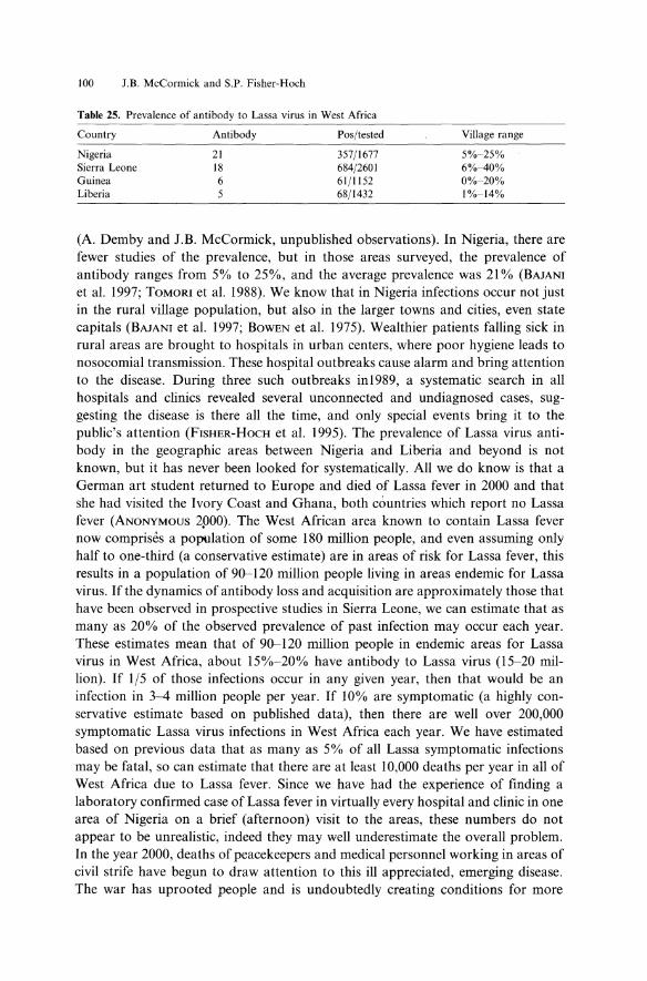
In Sierra Leone, human antibody prevalence ranges from around 5% in coastal villages to 40% in secondary forest and savannah (average of 18%) (Table 23). In some highly endemic villages, 5%-20% of susceptible (antibody-negative) persons are infected per year, and seroconversion to Lassa virus antigen has been observed in 5%-13% of all febrile illnesses in these villages (Table 24) (MCCORMICK 1987). Antibody levels to Lassa virus may decline to unmeasurable levels after 3-5 years, and re-infection, with mild or no illness, appears to occur, based on the dynamics of antibody change in populations and the frequency of silent infections (defined by fourfold antibody rise) in adults (Table 24) (MCCORMICK 1987). Antibody is found in patients of all ages, and the prevalence increases with age, suggesting consistent, relatively uniform exposure during life in endemic areas, as one would expect with an environmental pathogen associated with rodent-infested housing (Table 25).
The prevalence of infection in Liberia and Guinea also range from a low of 1 % to highs of on average 5%-6% (YALLEY-OGUNRO et al. 1984) (Table 25)
Table 23. Prevalence of antibody to Lassa virus by age, Sierra Leone
Age (years) Village A Village B
Pos/total % Pos Pos/total % Pos
<I 1/20 5 1/24 4 1-4 6/50 12 8/79 10 5-9 31/76 29 23/82 28 10--19 50/113 44 19/69 28 20--39 121/245 49 90/212 42 40+ 91/200 45 11/24 45 Total 301/737 41 145/434 31
Table 24. Lassa fever illness to infection and case fatality ratios, West Africa
Illness to infection ratio
% Febrile illness Case fatality ratio (hospitalized, untreated) Case fatality all infections (estimated)
9%-50% (5 studies)
5%-14% (4 studies) 16% (? higher in Nigeria) 2%-4%
100 J.B. McCormick and S.P. Fisher-Hoch
Table 25. Prevalence of antibody to Lassa virus in West Africa
Country Antibody Pos/tested
Nigeria Sierra Leone Guinea Liberia
21 18 6 5
357/1677 684/2601 61/1152 68/1432
Village range
5%-25% 6%-40% 0%-20% 1%-14%
(A. Demby and J.B. McCormick, unpublished observations). In Nigeria, there are fewer studies of the prevalence, but in those areas surveyed, the prevalence of antibody ranges from 5% to 25%, and the average prevalence was 21 % (BAJAN! et al. 1997; TOMaR! et al. 1988). We know that in Nigeria infections occur not just in the rural village population, but also in the larger towns and cities, even state capitals (BAJANI et al. 1997; BOWEN et al. 1975). Wealthier patients falling sick in rural areas are brought to hospitals in urban centers, where poor hygiene leads to nosocomial transmission. These hospital outbreaks cause alarm and bring attention to the disease. During three such outbreaks in1989, a systematic search in all hospitals and clinics revealed several unconnected and undiagnosed cases, suggesting the disease is there all the time, and only special events bring it to the public's attention (FISHER-HoCH et al. 1995). The prevalence of Lassa virus antibody in the geographic areas between Nigeria and Liberia and beyond is not known, but it has never been looked for systematically. All we do know is that a German art student returned to Europe and died of Lassa fever in 2000 and that she had visited the Ivory Coast and Ghana, both countries which report no Lassa fever (ANONYMOUS 2$)00). The West African area known to contain Lassa fever now comprises a population of some 180 million people, and even assuming only half to one-third (a conservative estimate) are in areas of risk for Lassa fever, this results in a population of 90-120 million people living in areas endemic for Lassa virus. If the dynamics of antibody loss and acquisition are approximately those that have been observed in prospective studies in Sierra Leone, we can estimate that as many as 20% of the observed prevalence of past infection may occur each year. These estimates mean that of 90-120 million people in endemic areas for Lassa virus in West Africa, about 15%-20% have antibody to Lassa virus (15-20 million). If 1/5 of those infections occur in any given year, then that would be an infection in 3-4 million people per year. If 10% are symptomatic (a highly conservative estimate based on published data), then there are well over 200,000 symptomatic Lassa vifl,ls infections in West Africa each year. We have estimated based on previous data that as many as 5% of all Lassa symptomatic infections may be fatal, so can estimate that there are at least 10,000 deaths per year in all of West Africa due to Lassa fever. Since we have had the experience of finding a laboratory confirmed case of Lassa fever in virtually every hospital and clinic in one area of Nigeria on a brief (afternoon) visit to the areas, these numbers do not appear to be unrealistic, indeed they may well underestimate the overall problem. In the year 2000, deaths of peacekeepers and medical personnel working in areas of civil strife have begun to draw attention to this ill appreciated, emerging disease. The war has uprooted people and is undoubtedly creating conditions for more

Lassa Fever 101
human contact with the virus, especially in the cities. If Lassa fever were a developed world problem, there would be vociferous demands for control and vaccine.
8 Prevention and Control
8.1 Rodent Control
The key to prevention and control is either to interrupt the contact between infectious source and susceptible persons or to avoid disease in the event of infection. The ideal method of prevention for these rodent-borne diseases is to prevent contact between rodents and humans. The effectiveness of this has been admirably shown in the outbreaks of the arenavirus disease, Bolivian hemorrhagic fever, in Bolivia in the 1960s when rodent control programs in the villages were highly successful in eliminating the epidemic (JOHNSON et al. 1967). Under current social circumstances, the control of rodents as a broad approach to preventing Lassa fever is not realistic. The improvement of housing and food storage might reduce the domestic rodent population, but such changes are not easily made. Rodent trapping in an individual village where transmission is high has demonstrated as much as a fivefold reduction in the rate of virus transmission (Table 26). However, such a program is only applicable in villages with exceptional transmission rates and stable social conditions, and would certainly not be applicable to large areas. Furthermore, without a sustained program of rodent control, the rodents return after a period of a few months (Table 21).
9 Vaccine
Studies of potential vaccines to Lassa virus began in the 1980s when Clegg expressed the nucleocapsid protein of Lassa virus in vaccinia and was able to show the recombinant vaccine protected guinea pigs, but not primates, from subsequent challenge of 102 of virus (CLEGG and LLOYD 1987). Further work showed that both
Table 26. Seroconversion rates to Lassa virus, illness to infection ratios, and febrile illness due to Lassa fever in five Sierra Leone villages (MCCORMICK et al. 1987a)
Seroconversion before trapout
Seroconversion after trapout Relative risk of infection
Seroconversion in control villages: Village A Village B
6.9(100 susceptibles(year
1.3(100 susceptibles(year 5.3 before compared to after
10(100 susceptibles(year 34(100 susceptibles(year

102 J.B. McCormick and S.P. Fisher-Hoch
NP and glycoprotein expressed in vaccinia could protect guinea pigs, but that only the glycoprotein protected primates (MORRISON et al. 1989). The protection of guinea pigs was not related to antibody levels, so it was concluded that cellular immune responses were more important than antibody. Subsequently, immunization of primates with inactivated (gamma-irradiated) whole Lassa virus resulted in antibody responses to both NP and GPC, and a brisk booster response following challenge (Table 27) (Fig. 10). However despite this impressive antibody memory response, the animals all died with serum virus titers equal to unvaccinated controls (Table 28) (MCCORMICK et al. 1992). Data from field studies in the late 1970s and early 1980s in humans and further experimental studies in primates suggested that
Table 27. Antibody titers to Lassa virus in monkeys after vaccination and challenge
Day post-vaccine: 0
14 35 71
101 108
Day post-challenge: 4 8
10 12
Lolt" rno .. 10
o
Vaccinated
0 0 0 64 16 256 32 32 128 64 64 256 64 64 64
256 64 64
256 64 256 1,024 256 1,024 4,096 1,024 4,096 1,639 4,096 4,096
3 Da following chall.n~.
(I 0 J---4p.f.u. of tb. Jo.lab train of La a viru )
Unvaccinated
0 0 0 0 0 0
0 0 0
64
\'-1 •• I'
0 0 0 0 0 0
0 0 4
256
Fig. 10. Viremias in primates by day after vaccination and challenge with 103-4pfu Lassa virus. (FISHER-HoCH et al. 2000)

Lassa Fever 103
Table 28. Postmortem Lassa virus titers (IoglO TCIDso)
Site Vaccinated monkeys Unvaccinated monkeys
Liver 8.5 7.9 6.6 8.0 7.3 Spleen 7.5 7.2 6.3 7.3 6.9 Kidney 6.8 6.7 6.2 6.6 6.9 Lung 7.4 7.4 6.0 6.8 6.6 Adrenal 8.1 7.8 8.1 8.0 7.5 Spinal fluid 5.0 4.2 3.4 3.1 4.3 Serum 6.1 5.7 5.2 6.1 6.1
the immune response to NP is not protective and furthermore that the presence of antibody to either glycoprotein or nucleoprotein at the time of hospital admission was not associated with increased survival or even severity of disease. A more recent study suggests the antibody (for NP) may be a marker for severe or fatal disease (BAUSCH et al. 2000). Since a similar observation was made in primates given NP vaccine and then challenged with lethal virus (e.g., the disease appeared worse) (FISHER-HoCH et al. 2000), the biology of this phenomenon merits further study.
The latter is a more recent, broader study of protection by recombinant expressions of NP, GPC, and combinations of the two involved nonhuman primates; 28 Macaca mulatta (rhesus) and 16 M. fascicularis (cynomolgus) (FISHER-HoCH et al. 2000) (Table 29). The vaccinia viruses expressed S-segment Lassa structural proteins, the full-length glycoprotein (V-LSG), the nucleoprotein (V-LSN), the fulllength glycoprotein and nucleoprotein in the same construct (V-LSG/N), and the single glycoproteins (V-LSG1, residues 1-296, and V-LSG2, deletion of residues 67-234) (MORRISON et al. 1989, 1991). All animals were challenged subcutaneously with 103_104 pfu of the Josiah strain of Lassa virus. Following Lassa virus challenge, all unvaccinated animals died (0% survival), while 9/10 animals vaccinated with all proteins survived (90% survival). Although no animals that received fulllength glycoprotein alone had a high titer of antibody prior to challenge, 17/19
Table 29. Outcome of challenge of nonhuman primates vaccinated with Lassa virus vaccinia recombinants and challenged with virulent Lassa virus CITER (YANG et al. 2000)
Virion protein Protection Vaccine n Survivors Median day of Mean viremia at expression death (range) death (I0glO)
None 0% NTBH vaccinia 3 0 15 (15-19) 5.7 None 7 0 12 (10-15)
Single glycoproteins 0% V-LSGl 2 0 15 (14-16) 6.8 V-LSG2 2 0 12.5 (12-13)
Nucleoprotein 27% V-LSN 11 3 11.5 (9--13) 6.7 Full glycoprotein 88% V-LSG 7 6 21a (21) 2.5
V-LSGI + V-LSG2 2 2 Full S-segment 90% Mopeia virus 2 2
V-LSG+ V-LSN 6 5 11" (II) 7.0 V-LSG/N 2 2
Total 44 20
aThese two animals were challenged 488 and 700 days post-vaccination. All protected animals were challenged between 38 and 354 days post-vaccination.

104 J.B. McCormick and S.P. Fisher-Hoch
survived (88%). In contrast, all animals vaccinated with nucleoprotein developed a high titer of antibody, but 12/15 died (20% survival). All animals vaccinated with single glycoproteins (G 1 or G2) died, but all those that received both single glycoproteins (G 1 + G2) at separate sites survived, showing that both glycoproteins are independently important in protection. Neither group had demonstrable antibody prior to challenge.
The two deaths among the glycoprotein-vaccinated animals were associated with the longest vaccine to challenge intervals (488 and 700 days, range 36-700 days, mean 319 days). Survival diminished as the vaccine to challenge interval extended. A trend towards lengthening the duration of challenge viremia (days) was also observed with an increased interval between vaccination and challenge. The three animals that were vaccinated with the nucleoprotein that survived inadvertently received a lower challenge dose (103pfu compared with 104pfu), showing that the challenge dose also has a bearing on the outcome. Nevertheless, clearly the nucleoprotein vaccine is not protective compared with the glycoprotein.
None of the protective vaccines provided sterilizing immunity, since almost all surviving, asymptomatic animals experienced viremia, even those vaccinated with Mopeia virus (essentially a live attenuated Lassa virus). This is consistent with the hypothesis that virus replication is controlled by CTL responses and not antibody responses. In general, however, the outcome correlates with the level of viremia, and thus protection with limitation of viremia (Taqle 29). The highest virus titers were seen in the animals that had received the V-LSN recombinant vaccine (higher than in unvapcinated animals). This difference was not statistically significant, possible due to the small sample size, and a larger study is needed to settle the issue of whether this vaccine actually potentiates viremia following challenge. What is clear is that animals that received the entire glycoprotein (V-LSG) or all the S-segment proteins (V-LSG/N or V-LSG + V-LSN) showed significantly diminished mean virus titers compared with unvaccinated animals, and that these animals, with two exceptions, were not sick and did not die. These data show that the GPC gene is necessary and sufficient to protect primates against a large parenteral challenge dose. In contrast, the NP gene provides little if any protection, casting doubt on the need for, and indeed the safety of, using an NP gene as part of the vaccine for primates or humans (FISHER-HoCH et al. 2000).
A lack of correlation between the titer of antibody (IFA) to whole Lassa virus antigens prior to challenge and any level of protection has been observed in human disease. Following vaccination and prior to challenge, IFA antibody could be detected in V-LSN/V-LSG and V-LSN vaccinated animals, with titers ranging from 4 to 256. Nor are antibodies neutralizing. Multiple efforts failed to demonstrate the presence of plaque reduction neutralization in serum from vaccinated animals after vaccination and before challenge, as well as in the serum of animals after recovery from challenge (despite very high titer fluorescent antibody) (FISHER-HoCH et al. 1989,2000). What we can now conclude is that epitopes on both glycoproteins are needed for protection, and that these are apparently able to induce immunity in concert, but not indep~ndently (FISHER-HoCH et al. 2000; WRIGHT et al. 1989,

Lassa Fever 105
1990; DI SIMONE and BUCHMEIER 1995). Despite the strong evidence implicating cell-mediated immunity, we still think we have to keep an open mind about some contribution to protective processes by antibodies, particularly ones induced in novel ways.
Mopeia virus is an obvious candidate for an effective live attenuated vaccine, with the advantage that a single administration might induce life-long protection. Mopeia virus is presently classified as a BSL3 pathogen, even though data from Mozambique suggest that this virus is not pathogenic for humans. There are major sequence differences in the S segments of Lassa and Mopeia viruses, but no sites associated with virulence have been genetically mapped. Vaccine candidates expressing the Lassa glycoprotein gene will therefore have to be sought that are likely to pass the stringent requirements for safety needed for a human use vaccine.
Cross-protection against other West African strains of Lassa virus remains to be addressed. The protection afforded by Mopeia virus from Southern Africa certainly supports the idea that broad protection is achievable and desirable, particularly since some Nigerian strains may induce more severe and fatal illnesses than strains from further west (TROUP et al. 1970; BOWEN et al. 1975; CAREY et al. 1972; FISHER-HoCH et al. 1995; KEENLYSIDE et al. 1983). Monoclonal antibody mapping and molecular studies of the glycoproteins of African arenaviruses show a conserved B-cell epitope on G2 across all of the known African arenaviruses including Mopeia and most South American arenaviruses. However, both Gland G2 recognition is necessary for protection based on our primate studies. We concluded from continuous observations over 14 years in Sierra Leone that a single natural infection provides long-term protective immunity against disease, and therefore we believe that a single vaccination will provide lengthy protection, and given the virus circulation in the endemic area, the opportunity for natural boosting from infection is substantial.
The need for a vaccine is clear. The sizeable disease burden, a high frequency of hospitalization and death, with severe sequelae from cardiac disease to deafness, make a compelling case for application of a vaccine. Furthermore, the evidence of an effective vaccine has been published. Similar situations in developed countries would have long ago resulted in a vaccine effort. This is a disease that something can be done about, and it is unacceptable not to act.
References
Anonymous (2000) Lassa fever, case imported to Germany. Wkly Epidemiol Rec 75:8-17 Baize S, Leroy EM, Georges-Courbot MC, Capron M, Lansoud-Soukate J, Debre P, et al. (1999)
Defective humoral responses and extensive intravascular apoptosis are associated with fatal outcome in Ebola virus-infected patients [see comments]. Nat Med 5:423-426
Bajani MD, Tomori 0, Rollin PE, Harry TO, Bukbuk NO, Wilson L, et al. (1997) A survey for antibodies to Lassa virus among health workers in Nigeria. Trans R Soc Trop Med Hyg 91:379-381

106 J.B. McCormick and S.P. Fisher-Hoch
Bausch DG, Rollin PE, Demby AH, Coulibaly M, Kanu J, Conteh AS, et al. (2000) Diagnosis and clinical virology of Lassa fever as evaluated by enzyme-linked immunosorbent assay, indirect fluorescent-antibody test, and virus isolation. J Clin Microbiol 38:2670-2677
Biggar RJ (1986) Possible nonspecific association between malaria and HTLV-III/LAV [letter]. N Engl J Med 315:457-458
Biggar RJ, Gigase PL, Melbye M, Kestens L, Sarin PS, Bodner AJ, et al. (1985) ELISA HTLV retrovirus antibody reactivity associated with malaria and immune complexes in healthy Africans. Lancet 2:520-523
Biggar RJ, Saxinger C, Sarin P, Blattner WA (1986) Non-specificity of HTL V-III reactivity in sera from rural Kenya and eastern Zaire [letter]. East Afr Med J 63:683-684
Bloch A (1978) A serological survey of Lassa fever in Liberia. Bull World Health Org 56:811-813 Bowen GS, Tomori 0, Wulff H, Casals J, Noonan A, Downs WG (1975) Lassa fever in Onitsha, East
Central State, Nigeria in 1974. Bull World Health Org 52:599-604 Buckley SM, Casals J (1970) Lassa fever, a new virus disease of man from West Africa. 3. Isolation and
characterization of the virus. Am J Trop Med Hyg 19:680-691 Cao W, Henry MD, Borrow P, Yamada H, Elder JH, Ravkov EV, et aI. (1998) Identification of
alpha-dystroglycan as a receptor for lymphocytic choriomeningitis virus and Lassa fever virus [see comments]. Science 282:2079-2081
Carey DE, Kemp GE, White HA, Pinneo L, Addy RF, Fom AL, et al. (1972) Lassa fever. Epidemiological aspects of the 1970 epidemic, Jos, Nigeria. Trans R Soc Trop Med Hyg 66:402-408
Centers for Disease Control (1988) Management of patients with suspected viral hemorrhagic fever. MMWR 37:SI-S15
Fraser DW, Campbell CC, Monath TP, Goff PA, Gregg MB (1974) Lassa fever in the Eastern Province of Sierra Leone, 1970-1972. I. Epidemiologic studies. Am J Trop Med Hyg 23:1131-1139
Clegg JC, Lloyd G (1987) Vaccinia recombinant expressing Lassa-virus internal nucleocapsid protein protects guineapigs against Lassa fever. Lancet 2: 186-188
Cooper CB, Gransden WR, Webster M, King M, O'Mahony M, Young S, et al. (1982) A case of Lassa fever: experience at St Thomas's Hospital. Br Med J Clin Res Ed 285:1003-1005
Cummins D, Bennett D, Fisher-Hoch SP, Farrar B, McCofl1'lick JB (l989a) Electrocardiographic abnormalities in patients with Lassa fever. J Trop Med Hyg 92:350-355
Cummins D, Fisher-Hoch SP, Walshe KJ, Mackie 11, McCormick JB, Bennett D, et al. (I 989b) A plasma inhibitor of platelet ag!!regation in patients with Lassa fever. Br J Haematol 72:543-548
Cummins D, McCormick lB, Bennett D, Samba JA, Farrar B, Machin SJ, et al. (1990a) Acute sensorineural deafness in Lassa fever [see comments]. JAMA 264:2093-2096
Cummins D, Molinas FC, Lerer G, Maiztegui 11, Faint R, Machin SJ (l990b) A plasma inhibitor of platelet aggregation in patients with Argentine hemorrhagic fever. Am J Trop Med Hyg 42: 470-475
Cummins D, Bennett D, Fisher-Hoch SP, Farrar B, Machin SJ, McCormick JB (1992) Lassa fever encephalopathy: clinical and laboratory findings. J Trop Med Hyg 95:197-201
Di Simone C, Buchmeier MJ (1995) Protection of mice from lethal LCMV infection by a reconstituted glycoprotein vaccine. Cold Spring Harbor Laboratory Press, Cold Spring Harbor, pp 1-5
Elliott LH, McCormick JB, Johnson KM (1982) Inactivation of Lassa, Marburg, and Ebola viruses by gamma irradiation. J Clin Microbiol 16:704-708
Emond RT, Bannister B, Lloyd G, Southee TJ, Bowen ET (1982) A case of Lassa fever: clinical and virological findings. Br Med J (Clin Res Ed) 285:1001-1002
Fabiyi A, Tomori 0, Pinneo P (1979) Lassa fever antibodies in hospital personnel in the Plateau State of Nigeria. Niger Med J 9:23-25
Feldmann H, Bugany H, Mahner F, Klenk HD, Drenckhahn D, Schnittler HJ (1996) Filovirus-induced endothelial leakage triggered by infected monocytes/macrophages. J Virol 70:2208-2214
Fisher-Hoch SP (1993) Stringent precautions are not advisable when caring for patients with viral haemorrhagic fevers. Rev Med Virol 3:7-\3
Fisher-Hoch SP, McCormick JB (1987) Pathophysiology and treatment of Lassa fever. Curr Topics Microbiol Immunol 134:231-239
Fisher-Hoch SP, Price ME, Craven RB, Price FM, Forthall DN, Sasso DR, et al. (1985) Safe intensivecare management of a severe case of Lassa fever with simple barrier nursing techniques. Lancet 2:1227-1229
Fisher-Hoch SP, Mitchell SW, Sasso DR, Lange JV, Ramsey R, McCormick JB (1987) Physiological and immunologic disturbances associated with shock in a primate model of Lassa fever. J Infect Dis 155:465-474

Lassa Fever 107
Fisher-Hoch S, McCormick JB, Sasso D, Craven RB (1988) Hematologic dysfunction in Lassa fever. J Med ViroI26:127-135
Fisher-Hoch SP, McCormick JB, Auperin D, Brown BG, Castor M, Perez G, et al. (1989) Protection of rhesus monkeys from fatal Lassa fever by vaccination with a recombinant vaccinia virus containing the Lassa virus glycoprotein gene. Proc Nat! Acad Sci USA 86:317-321
Fisher-Hoch SP, Tomori 0, Nasidi A, Perez Oronoz GI, Fakile Y, Hutwagner L, et al. (1995) Review of cases of nosocomial Lassa fever in Nigeria: the high price of poor medical practice. BMJ 311:857-859
Fisher-Hoch SP, Hutwagner L, Brown B, McCormick JB (2000) Effective vaccine for lassa fever. J Virol 74:6777-6783
Frame JD, Baldwin JM Jr, Gocke DJ, Troup JM (1970) Lassa fever, a new virus disease of man from West Africa. I. Clinical description and pathological findings. Am J Trop Med Hyg 19:670-676
Frame JD, Casals J, Dennis EA (1979) Lassa virus antibodies in hospital personnel in western Liberia. Trans R Soc Trop Med Hyg 73:219-224
Frame JD, Jahrling PB, Yalley Ogunro JE, Monson MH (1984) Endemic Lassa fever in Liberia. II. Serological and virological findings in hospital patients. Trans R Soc Trop Med Hyg 78:656-660
Georges AJ, Gonzalez JP, Abdul Wahid S, Saluzzo JF, Meunier DM, McCormick JB (1985) Antibodies to Lassa and Lassa-like viruses in man and mammals in the Central African Republic. Trans R Soc Trop Med Hyg 79:78-79
Gonzalez JP, McCormick JB, Saluzzo JF, Herve JP, Georges AJ, Johnson KM (1983) An arenavirus isolated from wild-caught rodents (Prarnys species) in the Central African Republic. Intervirology 19:105-112
Gonzalez JP, Josse R, Johnson ED, Merlin M, Georges AJ, Abandja J, et al. (1989) Antibody prevalence against haemorrhagic fever viruses in randomized representative Central African populations. Res Virol 140:319-331
Grundy DJ, Bowen ET, Lloyd G (1980) Isolated case of Lassa fever in Zaria, Northern Nigeria [letter]. Lancet 2:649-650
Helmick CG, Webb PA, Scribner CL, Krebs JW, McCormick JB (1986) No evidence for increased risk of Lassa fever infection in hospital staff. Lancet 2: 1202-1205
Hirabayashi Y, Oka S, Goto H, Shimada K, Kurata T, Fisher-Hoch SP, et al. (1988) An imported case of Lassa fever with late appearance of polyserositis. J Infect Dis 158:872-875
Hirabayashi Y, Oka S, Goto H, Shimada K, Kurata T, Fisher-Hoch SP, et al. (1989) The first imported case of Lassa fever in Japan (in Japanese). Nippon Rinsho 47:71-75
Holmes GP, McCormick JB, Trock SC, Chase RA, Lewis SM, Mason CA, et al. (1990) Lassa fever in the United States. Investigation of a case and new guidelines for management [see comments]. N Engl J Med 323:1120-1123
Johnson KM, Webb PA, Kuns ML, Valverde L (1967) On the mode of transmission of Bolivian hemorrhagic fever. Jpn J Med Sci BioI 20:153-159
Johnson KM, Taylor P, Elliott LH, Tomori 0 (l981a) Recovery of a Lassa-related arenavirus in Zimbabwe. Am J Trop Med Hyg 30:1291-1293
Johnson KM, Elliott LH, Heymann DL (l98Ib) Preparation of polyvalent viral immunofluorescent intracellular antigens and use in human serosurveys. J Clin Microbiol 14:527-529
Johnson KM, McCormick JB, Webb PA, Smith ES, Elliott LH, King II (1987) Clinical virology of Lassa fever in hospitalized patients. J Infect Dis 155:456-464
Keenlyside RA, McCormick JB, Webb PA, Smith E, Elliott L, Johnson KM (1983) Case-control study of Mastornys natalensis and humans in Lassa virus-infected households in Sierra Leone. Am J Trop Med Hyg 32:829-837
Knobloch J, McCormick JB, Webb PA, Dietrich M, Schumacher HH, Dennis E (1980) Clinical observations in 42 patients with Lassa fever. Tropenmed Parasitol 31:389-398
Lukashevich IS (1992) Generation of reassortants between African arenaviruses. Virology 188:600-605 Lukashevich IS, Vasiuchkov AD, Stel'makh TA, Scheslenok EP, Shabanov AG (1991) The isolation and
characteristics of reassortants between the Lassa and Mopeia arenaviruses. Vopr Virusol 36: 146-150 Lukashevich IS, Maryankova R, Vladyko AS, Nashkevich N, Koleda S, Djavani M, et aI. (1999) Lassa
and Mopeia virus replication in human monocytes/macrophages and in endothelial cells: different effects on IL-8 and TNF-alpha gene expression. J Med Virol 59:552-560
Marta RF, Montero VS, Hack CE, Sturk A, Maiztegui n, Molinas FC (1999) Proinflammatory cytokines and elastase-alpha-1-antitrypsin in Argentine hemorrhagic fever. Am J Trop Med Hyg 60:85-89
Martini GA, Schmidt HA (1968) Spermatogene Ubertragung des 'Virus Marburg'. (Erreger der 'Marburger Affenkrankheit'). K1in Wochenschr 46:398-400

108 J.B. McCormick and S.P. Fisher-Hoch
McCormick JB (1986) Clinical, epidemiologic, and therapeutic aspects of Lassa fever. Med Microbiol Immunol Berl 175:153-155
McCormick JB (1987) Epidemiology and control of Lass a fever. Curr Topics Microbiol Immunol134:69-78 McCormick JB (1988) Lassa fever: epidemiology, therapy and vaccine development. Kans
enshogaku.Zasshi 62 [Suppl]:353-366 McCormick JB (1990) Arenaviruses. In: Fields BN, Knipe DM (eds) Fields virology, 2nd edn. Raven
Press, New York, pp 1245-1267 McCormick JB, Walker DH, King 11, Webb PA, Elliott LH, Whitfield SG, et al. (1986) Lassa virus
hepatitis: a study of fatal Lassa fever in humans. Am J Trop Med Hyg 35:401-407 McCormick JB, Webb PA, Krebs JW, Johnson KM, Smith ES (1987a) A prospective study of the
epidemiology and ecology of Lassa fever. J Infect Dis 155:437-444 McCormick JB, King 11, Webb PA, Johnson KM, O'Sullivan R, Smith ES, et al. (1987b) A case-control
study of the clinical diagnosis and course of Lassa fever. 1 Infect Dis 155:445-455 McCormick JB, Mitchell SW, Kiley MP, Ruo S, Fisher-Hoch SP (1992) Inactivated Lassa virus elicits a
non protective immune response in rhesus monkeys. J Med Virol 37:1-7 Mertens PE, Patton R, Baum 11, Monath TP (1973) Clinical presentation of Lassa fever cases during the
hospital epidemic at Zorzor, Liberia, March-April 1972. Am J Trop Med Hyg 22:780-784 Monath TP, Mertens PE, Patton R, Moser CR, Baum 11, Pinneo L, et al. (1973) A hospital epidemic of
Lassa fever in Zorzor, Liberia, March-April 1972. Am J Trop Med Hyg 22:773-779 Monath TP, Maher M, Casals 1, Kissling RE, Cacciapuoti A (1974) Lassa fever in the Eastern Province
of Sierra Leone, 1970-1972. II. Clinical observations and virological studies on selected hospital cases. Am J Trop Med Hyg 23:1140-1149
Monson MH, Cole AK, Frame ID, Serwint JR, Alexander S, Jahrling PB (1987) Pediatric Lassa fever: a review of 33 Liberian cases. Am J Trop Med Hyg 36:408-415
Morrison HG, Bauer SP, Lange JV, Esposito 11, McCormick JB, Auperin DD (1989) Protection of guinea pigs from Lassa fever by vaccinia virus recombinants expressing the nucleoprotein or the envelope glycoproteins of Lassa virus. Virology 171: 179-188
Morrison HG, Goldsmith CS, Regnery HL, Auperin DD (1991) Simultaneous expression of the Lassa virus Nand GPC genes from a single recombinant vaccinia yirus. Virus Res 18:231-241
Price ME, Fisher-Hoch SP, Craven RB, McCormick JB (1988) A"prospective study of maternal and fetal outcome in acute Lassa fever infection during pregnancy. BMJ 297:584-587
Riviere Y, Ahmed R, Soutlitern PJ, Buchmeier MJ, Oldstone MB (1985) Genetic mapping oflymphocytic choriomeningitis virus I'athogenicity: virulence in guinea pigs is associated with the L RNA segment. J Virol 55:704-709
Robbins CB, Krebs JW Jr, lohnson KM (1983) Mastomys (Rodentia: Muridae) species distinguished by hemoglobin pattern differences. Am J Trop Med Hyg 32:624-630
Roberts PJ, Cummins D, Bainton AL, Walshe KJ, Fisher-Hoch SP, McCormick JB, Gribben JG, Machin SJ, Linch DC (1989) Plasma from patients with severe Lassa fever profoundly modulates f-met-Ieu-phe induced superoxide generation in neutrophils. Br 1 Haematol 73:152-157
Rowe AK, Bertolli J, Khan AS, Mukunu R, Muyembe-Tamfum 11, Bressler D, et al. (1999) Clinical, virologic, and immunologic follow-up of convalescent Ebola hemorrhagic fever patients and their household contacts, Kikwit, Democratic Republic of the Congo. Commission de Lutte contre les Epidemies a Kikwit. J Infect Dis 179 [Suppll]:S28-S35
Saluzzo IF, Adam F, McCormick JB, Digoutte JP (1988) Lassa fever virus in Senegal [letter]. 1 Infect Dis 157:605
Schnittler HI, Mahner F, Drenckhahn D, Klenk HD, Feldmann H (1993) Replication of Marburg virus in human endothelial cells. A possible mechanism for the development of viral hemorrhagic disease. J Clin Invest 91:1301-1309
Siegert R, Shu HL, Slenczka W (1968) Demonstration of the 'Marburg virus' in the patient (in German). Nachweis des "Marburg-Virus" beim Patienten. Dtsch Med Wochenschr 93:616-619
Swanepoel R, Leman PA, Shepherd AJ, Shepherd SP, Kiley MP, McCormick JB (1985) Identification of Ippy as a Lassa-fever-related virus [letter]. Lancet 1 :639
Ter Meulen 1, Lukashevich I, Sidibe K, Inapogui A, Marx M, Dorlemann A, et al. (1996) Hunting of peridomestic rodents and consumption of their meat as possible risk factors for rodent-to-human transmission of Lassa virus in the Republic of Guinea. Am J Trop Med Hyg 55:661-666
Ter Meulen J, Badusche M, Kuhnt K, Doetze A, Satoguina J, Marti T, et al. (2000) Characterization of human CD4( +) T-cell clones recognizing conserved and variable epitopes of the lassa virus nucleoprotein. J Virol 74:2186-2192

Lassa Fever J09
Tomori 0, Fabiyi A, Sorungbe A, Smith A, McCormick JB (1988) Viral hemorrhagic fever antibodies in Nigerian populations. Am J Trop Med Hyg 38:407-410
Trappier SG, Conaty AL, Farrar BB, Auperin DD, McCormick JB, Fisher-Hoch SP (1993) Evaluation of the polymerase chain reaction for diagnosis of Lassa virus infection. Am J Trop Med Hyg 49:214-221
Troup JM, White HA, Fom AL, Carey DE (1970) An outbreak of Lassa fever on the Jos plateau, Nigeria, in January-February 1970. A preliminary report. Am J Trop Med Hyg 19:695-696
Walker DH, Johnson KM, Lange JV, Gardner JJ, Kiley MP, McCormick JB (1982) Experimental infection of rhesus monkeys with Lassa virus and a closely related arenavirus, Mozambique virus. J Infect Dis 146:360-368
Webb PA, McCormick JB, King 11, Bosman I, Johnson KM, Elliott LH, et al. (1986) Lassa fever in children in Sierra Leone, West Africa. Trans R Soc Trop Med Hyg 80:577-582
White HA (1972) Lassa fever. A study of 23 hospital cases. Trans R Soc Trop Med Hyg 66:390-401 Winn WC Jr, Walker DH (1975) The pathology of human Lassa fever. Bull World Health Org 52:
535-545 Wright KE, Salvato MS, Buchmeier MJ (1989) Neutralizing epitopes of lymphocytic choriomeningitis
virus are conformational and require both glycosylation and disulfide bonds for expression. Virology 171:417-426
Wright KE, Spiro RC, Burns JW, Buchmeier MJ (1990) Post-translational processing of the glycoproteins of lymphocytic choriomeningitis virus. Virology 177:175-183
WulffH, Fabiyi A, Monath TP (1975) Recent isolations of Lassa virus from Nigerian rodents. Bull World Health Org 52:609-613
Wulff H, Mcintosh BM, Hamner DB, Johnson KM (1977) Isolation of an arenavirus closely related to Lassa virus from Mastomys natalensis in south-east Africa. Bull World Health Org 55:441-444
Yalley-Ogunro JE, Frame JD, Hanson AP (1984) Endemic Lassa fever in Liberia. VI. Village serological surveys for evidence of Lassa virus activity in Lofa County, Liberia. Trans R Soc Trop Med Hyg 78:764-770
Yang Z, Duckers HJ, Sullivan NJ, Sanchez A, Nabel EG, Nabel GJ (2000) Identification of the ebola virus glycoprotein as the main viral determinant of vascular cell cytotoxicity and injury. Nat Med 6:886-889