Travel risk, malaria importation and malaria transmission in Zanzibar
Upload
khangminh22Category
view
3download
0
i
SCHOOL OF PUBLIC HEALTH
COLLEGE OF HEALTH SCIENCES
UNIVERSITY OF GHANA
MALARIA AND TYPHOID FEVER CO-INFECTION: A STUDY AMONG
PATIENTS PRESENTING WITH FEBRILE ILLNESSES IN THE GA
WEST MUNICIPAL HOSPITAL, AMASAMAN
BY
TANKO RUFAI
(10552654)
THIS DESSERTATION IS SUBMITTED TO THE UNIVERSITY OF
GHANA, LEGON IN PARTIAL FULFILLMENT OF THE
REQUIREMENT FOR THE AWARD OF MASTER OF PHILOSOPHY
DEGREE IN APPLIED EPIDEMIOLOGY AND DISEASE CONTROL
JULY, 2017
University of Ghana http://ugspace.ug.edu.gh

i
DECLARATION
I, Tanko Rufai, hereby declare that with the exception of cited references to other people’s work
which has been duly acknowledged, this work is the result of my own research work done under
supervision and has neither been presented elsewhere either in part or whole for another degree.
Signature… ………… ………………………….. Date………………………………………….
Tanko Rufai
(Student)
Signature… ………… ………………………….. Date………………………………………….
Dr. Anthony Danso-Appiah
(Supervisor)
Signature………………………………………….. Date………………………………………...
Dr. Reginald Quansah
(Co-Supervisor)
University of Ghana http://ugspace.ug.edu.gh

ii
DEDICATION
This work is dedicated to my mother Hajia Fulera and to the memory of my father Rufai Fuseini.
I will forever be grateful for your advice, prayers and support. Finally to my wife Saudat Tanko,
my children Amir Zaki Tanko and Rabab Tanko for their love and encouragement.
University of Ghana http://ugspace.ug.edu.gh

iii
ACKNOWLEDGEMENT
Glory be to Almighty God for making the impossible possible. Father Your loyal servant am
grateful.
My sincere gratitude goes to Dr. Anthony Danso-Appiah, my supervisor who has been of
enormous help, and inspiration. I am also grateful to Dr. Reginald Quansah my secondary
supervisor for your support.
I wish to thank Dr. Francis Cudjoe of School of Allied Health Sciences, University of Ghana for
supporting me technically and encouraging me. I am also grateful to my other lecturers Prof Edwin
Afari, Dr. Samuel Sackey, Dr. Ernest Kenu etc. for their advice.
To Dr. Doris Arhin, Municipal Director of Health Services and Ag. Medical Superintendent of
the Ga West Municipal Hospital, I express my gratitude for allowing me to use this facility. I am
also grateful to all the nurses at the vital signs section. Mr. Enoch Aninagyei and all the staffs in
the laboratory department, Ga West Municipal Hospital, I am grateful for your support during my
lab work, without you it would have been difficult for me to have undertaken this study. Finally to
all staffs of Ghana Field Epidemiology and Laboratory Training Programme and School of Public
Health, University of Ghana.
University of Ghana http://ugspace.ug.edu.gh

iv
Content TABLE OF CONTENTS page DECLARATION.......................................................................................................................................... i
DEDICATION............................................................................................................................................. ii
ACKNOWLEDGEMENT ......................................................................................................................... iii
LIST OF TABLES .................................................................................................................................... vii
LIST OF FIGURES ................................................................................................................................. viii
LIST OF ABBREVIATIONS ................................................................................................................... ix
ABSTRACT ................................................................................................................................................. x
CHAPTER ONE ......................................................................................................................................... 1
INTRODUCTION ................................................................................................................................... 1
1.1 ACUTE FEBRILE ILLNESS ...................................................................................................... 1
1.2 MALARIA FEVER....................................................................................................................... 1
1.2.1 Global Distribution of Malaria ................................................................................................. 1
1.2.2 Etiology and life cycle ................................................................................................................ 2
1.2.3 Transmission of malaria ............................................................................................................ 4
1.2.4 Presentation of malaria fever .................................................................................................... 4
1.2.5 Diagnosis of malaria................................................................................................................... 5
1.2.6 Treatment and management ..................................................................................................... 5
1.3 TYPHOID FEVER ....................................................................................................................... 6
1.3.1 Causative agent .......................................................................................................................... 6
1.3.2 Transmission .............................................................................................................................. 7
1.3.3 Clinical manifestation ................................................................................................................ 7
1.3.4 Diagnosis ..................................................................................................................................... 8
1.3.5 Treatment ................................................................................................................................... 8
1.4 CO-INFECTION OF MALARIA AND TYPHOID FEVER .................................................... 9
1.5 PROBLEM STATEMENT .......................................................................................................... 9
1.6 JUSTIFICATION / RATIONALE ............................................................................................ 11
1.7 CONCEPTUAL FRAMEWORK OF MALARIA AND TYPHOID FEVER INFECTION 12
............................................................................................................................................................ 12
1.7.1 Narration of conceptual framework ....................................................................................... 12
1.8 GENERAL OBJECTIVE ........................................................................................................... 14
1.8.1 Specific objectives .................................................................................................................... 14
CHAPTER TWO ...................................................................................................................................... 15
University of Ghana http://ugspace.ug.edu.gh

v
LITERATURE REVIEW .................................................................................................................... 15
2.1 MALARIA AND TYPHOID FEVER AS A CAUSE OF FEBRILE ILLNESSES ............... 15
2.2 CO-INFECTION OF MALARIA AND TYPHOID FEVER .................................................. 17
2.3 ANTIBIOTIC SUSCEPTIBILITY OF SALMONELLA ........................................................ 18
2.4 RISK FACTORS FOR MALARIA AND TYPHOID FEVER ............................................... 20
CHAPTER THREE .................................................................................................................................. 22
MATERIALS AND METHODS ......................................................................................................... 22
3.1 STUDY DESIGN ......................................................................................................................... 22
3.2 STUDY SITE ............................................................................................................................... 22
3.3 VARIABLES ............................................................................................................................... 23
3.4 SAMPLING ................................................................................................................................. 26
3.4.1 Study population ...................................................................................................................... 26
3.4.1.1 Inclusion criteria ................................................................................................................... 26
3.4.1.2 Exclusion criteria .................................................................................................................. 27
3.4.2 Sample size determination ....................................................................................................... 27
3.4.3 Sampling method ..................................................................................................................... 27
3.5.1Ethical considerations ............................................................................................................... 28
3.5.2 Informed consent, possible risks and benefits ....................................................................... 28
3.5.3 Sample collection and processing ........................................................................................... 28
3.6 LABORATORY ANALYSIS ..................................................................................................... 29
3.6.1 Haematology ............................................................................................................................. 29
3.6.2 Rapid diagnostic test ................................................................................................................ 29
3.6.3 Determination of malaria parasitaemia ................................................................................. 29
3.6.4 Widal test .................................................................................................................................. 30
3.6.5 Blood culture ............................................................................................................................ 30
3.6.6 Gram staining procedure` ....................................................................................................... 31
3.6.7 Identification of suspected isolates by the standard manual method .................................. 31
3.6.8 Indole test .................................................................................................................................. 32
3.6.9 Oxidase test ............................................................................................................................... 32
3.6.10 Triple sugar iron (TSI) agar test .......................................................................................... 32
3.6.11 Citrate test .............................................................................................................................. 32
3.6.12 Urease test ............................................................................................................................... 33
3.6.13 Antimicrobial susceptibility testing (AST) ........................................................................... 33
University of Ghana http://ugspace.ug.edu.gh

vi
3.6.14 Laboratory quality control .................................................................................................... 33
3.7 DATA ANALYSIS ...................................................................................................................... 34
CHAPTER FOUR ..................................................................................................................................... 36
RESULTS .............................................................................................................................................. 36
4.1a Characteristics of study participants ...................................................................................... 36
4.1.1b Clinical complains among patients presenting with febrile illness .................................... 37
4.2 Malaria and typhoid fever as a cause of acute febrile illness .................................................. 37
4.2.1a Proportion of febrile illness due to malaria, typhoid fever and their co-infection ........... 37
4.21b Parasite density among patients with malaria and typhoid fever ...................................... 38
4.3 Malaria, typhoid fever and socio-demographic characteristics .............................................. 39
4.4 Antimicrobial test results of Salmonellas species isolated ....................................................... 41
4.5 Risk factors associated with malaria and typhoid fever infection .......................................... 42
CHAPTER FIVE ...................................................................................................................................... 44
DISCUSSION ........................................................................................................................................ 44
5.1 Malaria and typhoid fever as causes of acute febrile illness ................................................... 44
5.2 Malaria and typhoid fever co-infection ..................................................................................... 48
5.3 Antimicrobial drug susceptibility .............................................................................................. 50
5.4 Risk factors associated with malaria and typhoid fever .......................................................... 52
5.5 Limitation of the study ............................................................................................................... 53
CHAPTER SIX ......................................................................................................................................... 54
CONCLUSION AND RECOMMENDATIONS ................................................................................ 54
6.1 Conclusion ................................................................................................................................... 54
6.2 Recommendations ....................................................................................................................... 55
References .................................................................................................................................................. 56
APPENDIX A: CONSENT FORM ......................................................................................................... 67
APPENDIX B: QUESTIONAIRE ........................................................................................................... 69
APPENDIX C: MEDIA AND STANDARD SOLUTIONS ................................................................... 71
APPENDIX D: STAINING PROCEDURES ......................................................................................... 78
APPENDIX E: BIOCHEMICAL TESTS ............................................................................................... 80
APPENDIX F: PICTURES OF STANDARD MANUAL BIOCHEMICAL TEST AND
ANTIMICROBIAL TESTING ................................................................................................................ 84
University of Ghana http://ugspace.ug.edu.gh

vii
LIST OF TABLES
Table 1a: Definition and scale of measurement for variables…………………………………....25
Table 1b: Definition and scale of measurement for variables…………………………………...26
Table 2: Socio- demographic characteristic of study participants……………………………….36
Table 3: Parasite density in patients with malaria and co-infection……………………………..38
Table 4: Prevalence of malaria and typhoid fever in association with socio-demographic
characteristic……………………………………………………………………………………..40
Table 5: Antimicrobial susceptibility testing of the Salmonella isolates………………………...41
Table 6: Determinants associated with typhoid fever in febrile patients ………………………..42
Table 7: Determinants associated with malaria in febrile patients ………………………...........43
University of Ghana http://ugspace.ug.edu.gh
viii
LIST OF FIGURES
Figure 1: Life cycle of malaria parasites…………………………………………………………4
Figure 2: Conceptual framework...……………………………………………………………...12
Figure 3: Map of Ga West Municipal...…………………………………………………………23
Figure 4: Clinical presentation among febrile patients…………………….................................37
Figure 5: Prevalence of malaria, typhoid fever and their co-infection using widal test and blood
culture method ……………………..............................................................................................38
Figure 6: Photograph of biochemical reactions of gram negative bacteria……………………...84
Figure 7: Photograph of antimicrobial testing showing zone of inhibition……………………..84
University of Ghana http://ugspace.ug.edu.gh

ix
LIST OF ABBREVIATIONS μL ─ Microliter
AFI ─ Acute febrile illness
SSA ─ Sub-Saharan Africa
BA ─ Blood Agar
BF ─ Blood Film
BHI ─ Brain Heart Infusion
CA ─ Chocolate Agar
CDC ─ Centre for Disease Control and Prevention
EDTA ─ Ethylene Diamine Tetra Acetic Acid
GHS ─ Ghana Health Service
GSS ─ Ghana Statistical Service
GWMH ─ Ga West Municipal Hospital
MAC ─ MacConkey
RDT ─ Rapid Diagnostic Test
WHO ─ World Health Organization
PCR ─ Polymerase Chain Reaction
DHIMS ─ District Health Information Management System
IQR ─ Interquartile Range
University of Ghana http://ugspace.ug.edu.gh

x
ABSTRACT
Introduction: Malaria and typhoid fever cause major health problems especially in low and
middle income countries. People in endemic areas are at risk of developing both infections
concomitantly. These study was conducted to provide an epidemiological data on co-infection of
malaria and typhoid fever in Ga West Municipality Hospital.
Methods: A cross-sectional study involving one hundred and fifty seven (157) febrile patients
attending Ga West Municipal Hospital, Amasaman from February to May, 2017. Blood samples
were collected for blood culture, Widal test, and blood film preparation for microscopy. Data were
analyzed using Stata version 13 statistical software.
Results: The study population involved 157 febrile patients aged between 2years to 37years who
reported to the hospital with fever (temperature 37.6 0C to 42 0C). A total of 82 (52.2%) of the
study participants were females. The median age of all the patients was 6years (IQR=3-11/years).
Out of the 157 febrile patients, 57/157(36.31%) had malaria, 23/157 (14.64%) had typhoid fever
using Widal test and 10/157 (6.37) by blood culture. Comparing patients with only malaria, the
geometric mean parasite density was 174485 (45782-665000) for those with co-infection (p-
value=0.009). Malaria for male 31/57(54.4%) and typhoid 6/10(60%). With age ≤10; malaria
42/57 (73.7%) and typhoid 8/10 (80%).
The co-infection of malaria and typhoid fever using Widal test and blood culture was 5.73% and
1.91% respectively. The isolates exhibited high resistance ranging from 60% - 100% against
ampicillin, tetracycline, co-trimozazole, gentamicin, cefuroxime, chloramphenicol, and
meropenem. The sensitivity also ranged from 66.7% - 100% against cefotaxime, ceftrizone,
ciprofloxacin and amikacin. No isolate of Salmonella typhi were susceptible to gentamicin,
University of Ghana http://ugspace.ug.edu.gh

xi
cefuroxime and co-trimoxazole. Other species of Salmonella were also not susceptible to
tetracycline, ampicillin, co-trimoxazole and cefuroxime. All of the Salmonella isolates were
susceptible to ciprofloxacin and amikacin.
Conclusion: These result of malaria and typhoid fever co-infection for blood culture and Widal
test is 1.9% and 5.73%. All of the Salmonella isolates were susceptible to ciprofloxacin and
amikacin.
University of Ghana http://ugspace.ug.edu.gh

1
CHAPTER ONE
INTRODUCTION
1.1 ACUTE FEBRILE ILLNESS
Fevers can be arbitrarily classified as acute, subacute and chronic fevers based on duration. Acute
fevers that last less than 7 days are typical of diseases such as malaria and viral-related upper
respiratory tract infection while those that last more than 2 weeks in duration are categorized as
sub-acute fevers, usually seen in cases of typhoid fever and intra-abdominal abscess, among others
(Ogoina, 2011).. Acute febrile illness (AFI) is characterised by a rise in body temperature above
the normal range of 36.5–37.5 °C (Hutchison et al., 2008). Chronic or persistent fevers which last
more than 2 weeks in duration are typical of chronic bacterial infections such as tuberculosis, viral
infections , cancers and connective tissue diseases (Ogoina, 2011). Any acute fever which is left
untreated can become chronic fever.
1.2 MALARIA FEVER
Malaria is one of the febrile illness and the most common fatal disease in the world caused by one
or more species of plasmodium. These are Plasmodium falciparum, P. vivax, P. ovale, P. Malariae,
and P. Knowlesi (Singh & Daneshvar, 2013; Samatha et., 2015). The most virulent species, P.
falciparum is also the most prevalent in Africa, while P. vivax is the most widely distributed
parasite outside of Africa (Gething et al., 2012).
1.2.1 Global Distribution of Malaria
Globally, about 214 million new cases of malaria was diagnosed in 2015 of which Africa
accounted for 88%, South-East Asia (10%) and the Eastern Mediterranean region (2%) (WHO,
2015). Within the same period, a total of 438,000 malaria deaths was recorded worldwide of which
University of Ghana http://ugspace.ug.edu.gh

2
90 % (394,200 deaths) occurred in Africa (WHO, 2015). The remaining deaths were recorded in
South-East Asia Region (7%) and the Eastern Mediterranean Region (2%). Of the 306,000 deaths
recorded globally among children under-fives, 95% (292,000 deaths) was from the African
Region.
In Ghana malaria occurs throughout the year and affects people of all ages. The demographic
health survey conducted in 2014 showed that incidence of malaria infection in children under five
years ranged from 11.2% to 40% with the rural areas mostly having the highest prevalence (37.7%)
(GDHS, 2014).
1.2.2 Etiology and life cycle
Malaria is caused by Plasmodium and belongs to the phylum Apicomplexa. It is transmitted when
one is bitten by the female Anopheles mosquitoes and in humans it is caused by Plasmodium
falciparum, P. malariae, P. ovale, P. vivax, and P. knowlesi (Daneshvar et al., 2009). The species
differ in their geographical distribution, type of disease they cause and drug response. The most
widespread species are Plasmodium vivax and Plasmodium falciparum, the latter is attributable to
the severest forms of malaria whilst infections of other species are rarely life-threatening
(Sutherland et al., 2010). Plasmodium ovale is restricted to West Africa sub-region where as
Plasmodium malariae is found worldwide at low prevalence (Carter & Mendis, 2002).
Plasmodium falciparum and Plasmodium malariae causes tropical and quartan malaria
(Harinasuta & Bunnag, 1988). Plasmodium vivax is found in South-East Asia, Central and South
America and just like Plasmodium ovale, causes tertian malaria (Harinasuta & Bunnag, 1988).
Drug response and relapse patterns also differ between species. For example, relapses are
University of Ghana http://ugspace.ug.edu.gh

3
characteristic in Plasmodium vivax and Plasmodium ovale infections. Occasionally, humans
become infected with a zoonotic species, Plasmodium knowlesi found in Asia (Singh et al.,
2004;Daneshvar et al., 2009).
The most common species that causes malaria in Ghana is the Plasmodium falciparum. Others like
Plasmodium malariae, and Plasmodium ovale (MOH, 2015) are also available. Plasmodium vivax
has not been identified in Ghana (MOH, 2015). The principal vectors are Anopheles gambiae and
Anopheles funestus.
The life cycle of the malaria parasite takes place in humans and the female Anopheles mosquito.
Malaria parasites are transmitted through the bite of an infective female Anopheles mosquito. The
mosquito is the definitive host and man is the intermediate host. In humans, parasites multiply
asexually in the liver (exo-erythrocytic) (Vaughan, Aly, & Kappe, 2008) and in the red blood cells
(erythrocytic) schizogony. After several cycles, gametocytes (sexual forms) develop in the red
blood cells of humans (Greenwood et al., 2008; Pukrittayakamee et al., 2008; Miller et al., 2002)
and are released into the blood which are ingested by female Anopheles mosquitoes during a blood
meal. Development in the mosquito is known as sporogony. Male and female gametocytes fuse
into zygotes which undergo further development in the stomach of the mosquito into
ookinetes(Barillas-Mury & Kumar, 2005). The ookinetes migrate to the mid gut to form oocysts
which subsequently develop in to sporozoites, found in the salivary glands of the mosquito.
Injection of sporozoites into a new host during feeding continues the life cycle (Barillas-Mury &
Kumar, 2005).
University of Ghana http://ugspace.ug.edu.gh

4
Figure 1: life cycle of malaria parasite (CDC, 2016).
1.2.3 Transmission of malaria
Malaria parasites are usually transmitted through the bite of an infective female Anopheles
mosquito. It can also be transmitted trans-placentally (Congenital malaria) (Valecha et al., 2007),
transfusion of infected blood (Chauhan et al., 2009) and needle stick injury.
1.2.4 Presentation of malaria fever
The clinical course of malaria infection may be uncomplicated or severe/complicated. Clinical
symptoms that are associated with uncomplicated malaria include fever, chills, sweats, headaches,
cough, muscle pains, joint pains, nausea, abdominal pain, diarrhoea and vomiting which may
progress to severe complications (Caraballo & King, 2014). Complicated malaria is associated
University of Ghana http://ugspace.ug.edu.gh
5
with severe anaemia, kidney failure, coma, hypoglycemia, respiratory distress and death
(Caraballo & King, 2014). Malaria tends to be particularly severe in infants, children < 5 years,
pregnant women, non-immune persons and adults with compromised immunity.
1.2.5 Diagnosis of malaria
Malaria is diagnose by the identification of the parasite in blood. The gold standard is by
examination of blood films using microscopy. Rapid diagnostic tests (RDT) detect the antigen of
the parasite (Holland & Kiechle, 2005) and quantitative buffy coat method (Bhandari et al., 2008).
Serological methods such as immunofluorescence or enzyme immuno assay can be used to detect
malarial antibodies which can give indication of recent infection. Malaria can also be diagnose
using polymerase chain reaction (PCR).Confirming clinical diagnosis with appropriate laboratory
test is very vital.
1.2.6 Treatment and management
Malaria is a preventable and curable disease. Artemisinin-based combination therapy (ACTs) for
uncomplicated malaria are highly effective against Plasmodium falciparum (WHO, 2016). In
Ghana, artermether lumefantrine, artesunate-amodiaquine, or dihydroartermisinin piperaquine is
also recommended (MOH, 2015). For preventive measures it is recommended that people sleep
under insecticide treated nets. The interventions such as antimalarial drugs (quinine and its
derivatives), transfusion, and fluid replacement are mostly used in severe malaria.
University of Ghana http://ugspace.ug.edu.gh

6
1.3 TYPHOID FEVER
Typhoid fever is a systemic protracted febrile illness commonly caused by Salmonella typhi.
Similar but mild form, Paratyphoid fever is caused by S. paratyphi A, S. paratyphi B and S.
paratyphi C (Andualem et al., 2014). Typhoid fever causes serious morbidity in many regions of
the world, accounting for 21 million cases and 222,000 deaths annually (WHO, 2015). Typhoid
fever is common in malaria endemic settings, usually leading to mix-infection. South and Central
Asia, Africa and South and Central America are considered endemic with rates exceeding 100 per
100,000 population per year ( Bhan et al., 2005)
In Africa, due to scarce resource and limited laboratory capacity to diagnose the disease
accurately, most data on typhoid fever are not credible. A survey conducted in Egypt found an
incidence of 59 cases per 100,000 persons per year for typhoid fever (Srikantiah et al., 2006).
1.3.1 Causative agent
Salmonella typhi belongs to the Enterobacteriaceae family and genus Salmonella. It affects only
human and causes typhoid fever ( Bhan et al., 2005) . Salmonella typhi is a gram-negative aerobic
and facultative anaerobic rod-shaped bacterium (Todar, 2008). They are non-sporing and with the
exception of Salmonella typhi, non-capsulate (Cheesbrough, 2006). It grows optimally at 35-37°C,
rod-shaped with a length of 2-3 μm and a diameter of 0.4-0.6 μm (Cheesbrough, 2006). It moves
with the aid of its flagella (H-d antigen). Salmonella typhi contains somatic or O, antigens
associated with toxin, H-d associated with flagella; and Vi-antigen for virulence. It interferes with
the complement (C3b) mediated opsonization of Salmonella typhi. This prevents it from binding
with the phagocytes and subsequently inhibits phagocytosis.
University of Ghana http://ugspace.ug.edu.gh
7
1.3.2 Transmission
Typhoid fever is spread through ingestion of contaminated food and water. Humans excrete the
bacteria in their faeces during the infection and usually persist if they become carriers. Other
medium of transmission is by eating raw fruits and vegetables contaminated with human feces,
milk products and shellfish. The bacterium can be viable for a long duration on food surface,
seawater and sewage water. It can survive in freezing temperature for 3months. . Foods are usually
contaminated by flies which act as mechanical carriers and the bacteria can proliferate to cause
typhoid in human (Guzman et al., 2006). It usually enters the bloodstream to the intestinal mucosa
and subsequently multiplying in the lymph nodes. Most often the incubation period varies between
7 to 21 days.
1.3.3 Clinical manifestation
The main clinical symptoms related to typhoid fever are prolonged fever, malaise, anorexia,
vomiting, severe headache, bradycardia, splenomegaly (Heymann et al., 2008). Initially the fever
is minimal but rises gradually until second week where it can be high and persistent (39–40 ◦C).
Other symptoms include abdominal discomfort, dry cough, myalgia and constipation is more
common in adults than diarrhoea seen in children. A transient, macular rash of rose-colored spots
can occasionally be seen on the trunk (Holmberg, 2012). Physical manifestations that are normally
seen are coated tongue, tender abdomen, hepatomegaly or splenomegaly. Most often nausea and
vomiting are not common but can be present in severe cases. Complications occur in about 15%
of all cases, and include intestinal haemorrhage or perforation, psychosis, meningitis, and
hepatosplenomegaly.
University of Ghana http://ugspace.ug.edu.gh
8
Prior to the discovery of antibiotics case fatality rates were high in untreated cases (10─20%).
With the advent of antibiotics, case fatality rate is only about 1% although relapse may occur in
15─20% of patients while 10% of untreated patients would be infectious within 3 months and
2─5% would turn out to be chronic carriers (Heymann et al., 2008). Generally, reinfection of
typhoid is rare because of the development lifelong immunity after primary infection.
1.3.4 Diagnosis
The presence of clinical symptoms characterised by fever is indicative of typhoid fever but needs
laboratory confirmation (WHO, 2003). The Salmonella typhi can be isolated from blood within a
week and in urine and faeces after first week. Even though blood culture is mostly used for
diagnosis, bone marrow culture can also be used to isolate Salmonella typhi. The sensitivity and
specificity of the conventional Widal test are low due to cross-reactivity with other
microorganisms.
1.3.5 Treatment
Antibiotic treatment is used to resolve typhoid fever infection symptoms (Bhan, Bahl, &
Bhatnagar, 2005) . Sensitivity patterns of Salmonella isolates in the area determine choice of
antibiotics (Bhan et al., 2005). With increasing multidrug resistance (MDR), previously effective
drugs such as ampicillin, chloramphenicol are no longer recommended (Gupta et al., 2008;
Lutterloh et al., 2012). Fluoroquinolones (ofloxacin, ciprofloxacin) are used for treating adults but
should be guided by appropriate antimicrobial susceptibility testing (Heymann et al., 2008 ; Bhan
et al., 2005; WHO, 2003).
University of Ghana http://ugspace.ug.edu.gh

9
Third-generation cephalosporin’s such as cefotaxime and ceftriaxone can be used in cases with
isolates resistant to nalidixic acid. Ceftriaxone used for children and azithromycin for treating
uncomplicated typhoid fever (Effa & Bukirwa, 2008). When there is intestinal perforation, surgery
is commonly recommended.
1.4 CO-INFECTION OF MALARIA AND TYPHOID FEVER
Malaria and typhoid fever co-infection was first described during the American civil war by
Woodward in 1862 among young soldiers presenting with intermittent pyrexia. It was suggested
that it could be a mix infections (Smith, 1982a). Subsequently, many studies have long established
this association (Ammah et al., 1999; Gopinath et al., 1995). People with poor hygiene can
contracting both diseases. Patients with co-infection mostly associated with nausea, vomiting,
abdominal pain, diarrhea and continuous fever (Khan et al., 2005). Anaemia due to massive
haemolysis or dyserythropoesis can occur during malaria infection which can lead to increase iron
in the liver and which support the growth of Salmonella (Bashyam, 2007). Some studies have
shown that Complement C1q and C4B deficiency can make a person susceptible to typhoid fever
infection (Warren et al., 2002; Bishof et al., 1990).
1.5 PROBLEM STATEMENT
Malaria and typhoid fever are well known undifferentiated febrile illnesses which may be
responsible for varying degrees of morbidity and mortality in developing and middle income
countries including Ghana. Due to lack of availability of diagnostics in low and middle income
countries, most cases of acute febrile illnesses are diagnosed as malaria (Stoler & Awandare,
2016). In Ga West Municipal Hospital (GWMH), the number of malaria cases increased from 3934
University of Ghana http://ugspace.ug.edu.gh
10
in 2015 to 4603 in 2016 (DHIMS 2, 2017). Typhoid fever also increased from 1369 to 2033 during
the same period (DHIMS 2, 2017). Meanwhile, people in endemic areas are at risk of contracting
both infections concurrently (Uneke, 2008 ; Nsutebu et al.,2003). Predisposition to co-infection
is usually influenced by their similar epidemiological factors such as dense population, poor
hygiene, and sanitation practices (Iheukwumere et al., 2013; Sharma et al., 2016). Due to their
similar clinical presentations and the likelihood of a misdiagnosis and mistreatment of febrile
patients, it has been suggested that malaria and typhoid fever should be treated concurrently in
endemic communities (Uneke, 2008; Iheukwumere et al., 2013). However, concurrent treatment
may have some public health implications in the sense that, irrational use of antibiotic or anti-
malarial may result in increasing surge of drug resistance, unnecessary cost and exposure of
patients to side effects of antibiotic ( Sharma et al., 2016).
Febrile patients reporting to the GWMH are usually tested for malaria using mRDT at the
outpatient department and subsequently treated separately or concomitantly without investigation
of other possible causes. This tests lack sensitivity at low levels of parasitaemia and persistently
positive tests (for some antigens). In severe cases, patients are requested to do Widal test for
typhoid at the laboratory which is prone to error (Mbuh, Galadima, & Ogbadu, 2003). Meanwhile
reliable diagnosis is by microscopic examination of blood film for malaria or blood, stool or bone
marrow culture for Salmonella. It is therefore advisable to performed both test on individuals
presenting with fever of malaria- typhoid signs and symptoms using accurate diagnostic methods
to ascertain true co-infection followed by appropriate treatment ( Mbuh et al., 2003). Due to drug
resistance, it is usually necessary to carry out sensitivity tests before making an informed choice
of an antibiotic for treatment.
University of Ghana http://ugspace.ug.edu.gh

11
1.6 JUSTIFICATION / RATIONALE
Treatment of both diseases is common among patients especially in the tropics even if their
diagnosis has not been confirmed. This may interfere with diagnosis and also means of increasing
antibiotic resistance. Studies have shown that there are more typhoid cases in areas of drug
resistant malaria and a cross reaction between malaria parasites and Salmonella antigens may cause
false positive Widal agglutination test. An accurate diagnosis such as positive blood culture is
crucial for effective management of patients. A number of studies have shown that malaria could
be co-infecting with typhoid (Nwuzo et al., 2009). Even though similar studies have been done in
other countries, there is limited data on co-infection of malaria and typhoid fever in Ghana. The
present study will provide an epidemiological data on co-infection of malaria and typhoid fever in
Ga West Municipal Hospital and help inform policies towards effective management of febrile
patients against irrational administration of anti-malarial drugs and antibiotics. Data from these
study will also help to plan better control and prevention strategies in the municipality.
University of Ghana http://ugspace.ug.edu.gh

12
1.7 CONCEPTUAL FRAMEWORK OF MALARIA AND TYPHOID FEVER
INFECTION
Figure 2: Conceptual Framework
1.7.1 Narration of conceptual framework
People are at risk of acquiring malaria and typhoid fever infection due to factors related to
environment, community, demographic and socio-economic status. Usually these are among
Socio- demographic
factors
Age
Sex
Marital status
Occupation
Geographical
location
Education
Financial status
MALARIA/
TYPHOID
FEVER
Clinical factors
Anaemia
Transfusion
Sickle cell
G6PD
Community and
environmental factors
Eating habit
Toilet facility
Source of water
Housing conditions
Hand washing
Bed net usage
Population
Diagnosis
Availability of
diagnostics
Length of time for
diagnosis
Sensitivity of
diagnostic technique
Accessibility
Affordability
University of Ghana http://ugspace.ug.edu.gh

13
contributing factors to disease occurrence in a given community. Malaria and typhoid fever are
common in areas with high poverty, unrestrained urbanization and poor infrastructure impact on
contamination of water supplies. Dense population which may result in illegal infrastructure
development, poor sewage system, poor hygiene and sanitation practices leading to malaria and
typhoid infection. According to Parry, (2006), eating food outside, eating vegetables and salad and
water contaminated with human feces pose as risk of typhoid infection.
Other reported risk factors include contact with carriers, poor hands washing and housing ( Bhan
et al., 2005). Factors such as indiscriminate disposal of waste, sewage spillage, lack of good toilet
facilities, inadequate sanitary facilities in homes and general poor sanitation culture lead to malaria
and typhoid fever (WHO, 2003). Improperly cooked and hygienically handled food and food stuff
are being consumed indiscriminatingly due to deteriorating socioeconomic statues coupled with
lack of health education. Typhoid fever occurs at any age but occurs more commonly in children
and young adults. Using insecticide treated nets (ITNs) help reduce malaria (Klinkenberg et al.,
2010)
Clinical factor such as Blood transfusion use in the management anaemia can cause malaria.
Malaria also causes breakage of red blood cells which increases iron content in the body.
Meanwhile Salmonella thrive in the presence of iron. Anaemic patients are highly susceptible to
Salmonella infection. Lack of diagnostic equipment and reagents contribute to poor diagnosis early
stage leading to the spread of infections and under estimation of the disease burden. Abuse of
antibiotics may eventually result in drug resistance, which would contribute to having typhoid
fever carriers who may spread the infection. Availability of diagnostics, length of time for
University of Ghana http://ugspace.ug.edu.gh

14
diagnosis, sensitivity of diagnostic technique which can lead to misdiagnosis and mismanagement
of patients.
1.8 GENERAL OBJECTIVE
To determine the proportion of malaria and typhoid fever co-infection, assess their risk factors and
susceptibility patterns of the Salmonella isolates to antimicrobial agents among febrile patients
attending the Ga West Municipal Hospital
1.8.1 Specific objectives
1. To determine the proportion of malaria and typhoid fever infection among febrile patients
2. To determine the proportion of malaria and typhoid fever co-infection among febrile patients
3. To determine the susceptibility pattern of the Salmonella isolates to antimicrobial agents
4. To assess the risk factors associated with malaria and typhoid fever infection
University of Ghana http://ugspace.ug.edu.gh

15
CHAPTER TWO
LITERATURE REVIEW
2.1 MALARIA AND TYPHOID FEVER AS A CAUSE OF FEBRILE ILLNESSES
Malaria and typhoid are common causes of febrile illnesses globally but more endemic in Africa
and Asia. Diagnosing febrile patients based on clinical signs and symptoms is difficult to
differentiate between malaria and typhoid fever (Moses et al., 2016).
In one study conducted in Kumasi and Sunyani metropolis using mRDT for malaria and Widal
test for typhoid fever, out of the 129 patients, 22(17%) tested positive for typhoid fever, 24 (18.6%)
tested positive for falciparum malaria (Afoakwah et al., 2011).
Several studies in Nigeria have reported on malaria and typhoid fever among febrile patients. In
Abakaliki, Ebonyi state, 33/250 (13.2%) tested positive for malaria, 53/250 (21.2%) typhoid fever
by Widal test and 2 (0.8%) by blood culture (Nwuzo et al., 2009). In Calabar, 202/250 (80.8%)
tested positive for malaria, 117 (46.8%) for typhoid by the Widal test and 2 (0.8%) with blood
culture. The study further found that males 97 (85.8%) were more infected with malaria than
females 105 (76.6%) but this was not statistically significant while the prevalence of typhoid fever
was higher in females 77 (56.2%) than males 40 (35.4%) and was statistically significant
(Archibong et al., 2016). A study amongst one hundred (100) patients with signs and symptoms
of malaria and typhoid in Uyo, Akwa Ibom State, Nigeria, found that 41/100 (41%) tested positive
for malaria, 64/100 (64%) for typhoid using widal and 11/64 (17%) using blood cultures (Edet et
al., 2016). A Similar study at the University of Uyo Teaching Hospital among 145 patients,
reported 51(35.2%) had malaria and 10(7.0%) typhoid/paratyphoid. The study further found
females were more infected than males (Moses et al., 2016). A study by Ukaegbu et al., (2014),
University of Ghana http://ugspace.ug.edu.gh

16
reported 162/300 (54%) for malaria, 68/162 (42%) for typhoid fever by Widal test and 9/162
(5.6%) by stool culture test (Ukaegbu et al., 2014). A study at Nnewi, Anambra state, out of 256
patients, 202(78.90 %) had malaria, 147(57.42 %) by widal and 38(14.84 %) by culture had
typhoid fever. The study further found that, the age group (11-30) years were mostly infected with
malaria and typhoid fever (Ekesiobi et al., 2008). An investigation carried out in Ekwulumili
Community, Anambra State, reported 40/200 (20%) for malaria, 11 (5.5%) typhoid fever (Onyido
et al., 2014). In a research carried out by Mbuh et al., (2003), 60/218(27.5%) tested positive for
malaria, 22/218 (10.1%) typhoid by the Widal test and 1/250 (0.5%) by the culture method.
Studies from Ethiopia have also reported different prevalence rate for malaria and typhoid fever
among febrile patients. A study conducted by Birhanie et al., ( 2014) among 200 febrile patients
recorded 73/200 ( 36.5%) for malaria, 38/200 (19%) using widal test and 1/200 (0.5%) with blood
culture for typhoid fever (Birhanie et al., 2014). The study also showed that malaria was higher in
males while typhoid fever was greater in females. Another study involving 502 who were being
diagnosed with typhoid fever, of which 343 (68.3%) of the febrile patients showed positive slide
Widal test while only 8(1.6%) patients were culture-proved to have typhoid fever (Wasihun et al.,
2015).
In Cameroon, 315 children aged 6months and 15years were studied. Malaria prevalence was
43.4% and 70.2% for microscopy and PCR respectively. The prevalence of typhoid fever was
4.4% using rapid diagnostic test kit (Achonduh-Atijegbe et al., 2016). Another study in Kumba,
Cameroon recorded malaria prevalence of 90.3% (186/206) with parasite density 866 (range: 40 –
64880) parasites/μL of blood, 7.9% (14/178) for typhoid (Ndip et al., 2015).
University of Ghana http://ugspace.ug.edu.gh

17
A Study by Sharma et al., (2016) in India, reported 60/3010 (1.99%) cases of typhoid by blood
culture, 48/60 (80%) were also positive for malaria parasite by peripheral smear examination. In
Guntur, a prevalence of 340/582 (58.4%) for malaria, 132/582 (22.6%) typhoid by Widal and
10/582 (1.8%) blood culture (Samatha et al., 2015).The study also found that 235/582 (40.3%) had
no malaria or typhoid fever. Another study had revealed that typhoid 35/108 (32.40%) is more
prevalent than malaria 11/108(10.80%) in acute fever (Sandhya et al., 2015).
2.2 CO-INFECTION OF MALARIA AND TYPHOID FEVER
In the last two decades, concurrent infection of typhoid fever and malaria have been confirmed by
studies from Africa and Asia (Ohanu et al., 2003; Sur et al., 2006; Kanjilal et al., 2006). The
prevalence using Widal test ranged from 4.4% to 70% (Tanyigna et al., 2000); (Ibadin & Ogbimi,
2004), while cultural technique alone ranged from 11.1% to 26.6% ( Smith et al., 2004; Khan et
al., 2005; Akinyemi et al., 2007). In Ghana, prevalence of malaria- typhoid fever co-infection of
4.65% have been reported using mRDT and Widal test ( Afoakwah et al., 2011).
Association between typhoid fever and malaria have been reported in many studies. Several in
Nigeria. In one study, co-infection obtained when typhoid was diagnosed by Widal test (10.1%)
was significantly higher than by blood culture method (0.5%) ( Mbuh et al., 2003). The study
further observed that overestimation of co-infection with malaria and typhoid fever will greatly
reduce if the diagnosis is based on blood culture. A Study by Nwuzo et al., (2009), reported a co-
infection of 2/250 (0.8%) by culture method and 14/250 (5.6%) by the Widal test. Another study
recorded (28.0%) by Widal test and(0.8%) by blood culture method (Archibong et al., 2016). Other
studies had also reported various prevalence malaria and typhoid fever co-infection in different
University of Ghana http://ugspace.ug.edu.gh

18
states; Uyo, 16% (n = 100) and 17% (n = 64) for widal and blood culture respectively (Edet et al.,
2016), Nnewi 29/256(14.36%) by culture method while 147/256 (57.42%) for widal test (Ekesiobi
et al., 2008), Ekwulumili Community, Nnewi South, co-infection was (5.0%) 10/200 using the
widal test. More co-infection was observed among females (5.41%) (Onyido et al., 2014). In
Lagos, co-infection of malaria and typhoid was observed among patients with complications
(Akinyemi et al., 2007).
A study conducted in New Delhi, India reported 1.59% (48/3010)using culture method for typhoid
and blood smear for malaria parasite however Widal test for typhoid and rapid diagnostic test for
malaria parasite showed co-infection rate of 3.38% (105/3010) ( Sharma et al., 2016). Samatha et
al., (2015), found co-infection of 38/582 (6.5%) using widal and 4/582 (0.7%) using blood culture.
In Ethiopia, Birhanie et al., (2014) showed in their study that, co-infection 13 (6.5%) and 1 (0.5%)
for widal and blood culture while in Kumba, Cameroon had 6.74% co-infection rate (Ndip et al.,
2015). In Pakistan, dual infection of malaria-typhoid fever was reported comprising of Salmonella
typhi and Salmonella para typhi A or B (Khan et al., 2005).
2.3 ANTIBIOTIC SUSCEPTIBILITY OF SALMONELLA
Antimicrobial resistance have been attributed to the irrational use of drugs causing the emergence
of resistant strains. Antimicrobial resistance leads to ineffective chemotherapy, which usually
leads to treatment failure. It can also increase morbidity, cost and ultimately increased risk of
death. Due to the difficulty in diagnosing the cause of a fever based on clinical features alone,
overtreatment with antibacterial drugs is also a common occurrence. The choice of antibiotic
University of Ghana http://ugspace.ug.edu.gh

19
should be based on sensitivity patterns of Salmonella isolates ( Bhan et al., 2005). Ampicillin,
chloramphenicol and trimethoprim-sulfamethoxazole, are not effective for treatment of typhoid
fever (Lutterloh et al., 2012 ; Gupta et al., 2008).
Antimicrobial resistance tends to be more pronounced especially in Africa and Asia (Newman et
al., 2011). Some factors that contribute to treatment failure or drug resistant are incorrect dosing,
poor quality of drug, and misdiagnosis in the individual. Rates of multi-drug resistant (MDR)
among Salmonella typhi was found to be up to 80% in southeast Asia (Wain et al., 1999),70% in
east Africa (Mengo et al., 2010), and also non-Salmonella typhi (Folster et al., 2012; Miriagou et
al., 2004). This could be due to the unnecessary overuse of antimicrobial drugs and further
complicated by the substandard medicines in circulation in several LMICs, which risks patient
safety and may promote antimicrobial resistance(Bate et al., 2008; Bate et al., 2009; Caudron et
al., 2008). A study on MDR in Delhi in 1993, by Daga et al., (1994), highlight the importance of
ciprofloxacin in the treatment of typhoid fever. A study involving clinical profile among 100 children
with positive blood culture for Salmonella typhi, reported that, the isolates were not susceptible to
amoxicillin, chloramphenicol and co-trimoxazole, but sensitive to ciprofloxacin and ceftriaxone
(Kalra et al., 2003). In another study from Rourkela in 2000, the number of MDR strains of
Salmonella typhi constituted 16.1% of the total isolates and were susceptible to chloramphenicol,
ceftriaxone and ciprofloxacin (Das & Bhattacharya, 2000).
In Laos, of the 1095 patients with data for hospital antimicrobial use, 56% received an
antimicrobial drug, and 12% received more than 1 (Mayxay et al., 2013). On the basis of final
diagnosis, only 7% of these patients were regarded as having been treated appropriately (Mayxay
University of Ghana http://ugspace.ug.edu.gh

20
et al., 2013). Although malaria was the admission diagnosis for 148 adults in Tanzania, 44.8% of
them were still prescribed empiric antibacterial agents (Nadjm et al., 2012). A Rwandan study on
malaria hospitalization after implementation of a malaria-control program documented a decrease
in the risk of high parasitaemia after the intervention (Sievers et al., 2008). In one study in
Cameroon, gentamycin and ciprofloxacin were found to be sensitive in treating typhoid fever
(Ndip et al., 2015).
2.4 RISK FACTORS FOR MALARIA AND TYPHOID FEVER
Typhoid can be transmitted by chronic carriers especially where there is poor personal and food
hygiene. In the USA, up to 30% of infections are due to diagnosed chronic carriers ( Bhan et al.,
2005). In endemic areas, transmission is high during dry and rainy seasons. Poverty, uncontrolled
urbanization and inadequate infrastructure do contribute to the contamination of water supplies.
Usually, transmission is through poor hygiene and sewage contamination of water (Bhutta, 2006).
Pipe-borne water remained the only source of drinking water which is likely to be contaminated
due to rusted and leakage of pipes. Parry (2006), noted that eating food outside, eating vegetables
and salad and water contaminated with human faeces pose a risk of typhoid infection. Other risk
factors include contact with other patients, poor hand washing and housing ( Bhan et al., 2005).
The socioeconomic factors, affecting the prevalence and distribution of typhoid/paratyphoid
bacilli include indiscriminate disposal of waste, sewage spillage, lack of good toilet facilities,
inadequate sanitary facilities in homes and generally poor sanitation culture (WHO, 2003).
Typhoid fever occurs at any age but occurs more commonly in children and young adults.
University of Ghana http://ugspace.ug.edu.gh

21
Recently, contrary to these popular findings the disease was found to affect even children aged 1-
5 years in Delhi, India (Walia et al., 2006). In France, children between I to 5 years were more
affected (Desenclos et al., 1996). The age group at greatest risk is 5 to 25 years (Saha et al., 2001).
A study in Pakistan, showed that children are usually burdened with typhoid fever (Graham, 2002;
Brooks et al., 2005; Siddiqui et al., 2006). Another study has also revealed that incidence of
typhoid fever is highest in children less than 5years, with higher complications (Bhutta, 2006).
A significant association was found among participants who slept under an insecticide treated nets
(ITN) were more protected from malaria infection. Typhoid was observed for participants who
had a borehole or well compared to those with piped water (Achonduh-Atijegbe et al., 2016). ITNs
usage reduces malaria infection (Klinkenberg et al., 2010). Association between bed net usage,
impregnation of the bed net with chemicals, and history of travel to malaria endemic areas were
not significantly associated risk factors of malaria but hand washing was associated with typhoid
infection (Birhanie et al., 2014).
University of Ghana http://ugspace.ug.edu.gh

22
CHAPTER THREE
MATERIALS AND METHODS
3.1 STUDY DESIGN
A cross- sectional study was conducted to collect both qualitative and quantitative data on patients
age two years and above with temperature reading ˃37.5oC at the outpatient department between
the months of February and May 2017 in Ga West Municipal Hospital. Blood sample was taken
from these febrile patients suspected to have malaria or typhoid fever to determine the proportion
of malaria and typhoid fever co-infection and susceptibility patterns of the Salmonella isolates to
antimicrobial agents.
3.2 STUDY SITE
Ga West Municipality (GWM) is one of the 16 districts in the Greater Accra Region. The district
is 60% rural and 40% peri-urban and urban, and is made up of about 150 communities with
Amasaman as the district capital.
The GWM shares boundaries with Ga East and Accra Metropolitan Area to the East, Akwapim
South to the North, Ga South to the South and Ga Central to the North-South. It occupies a total
land surface area of 299.578 square kilometers. The projected population of the Municipality for
2015 was 256026 (GSS). Currently the municipality is divided into three (3) sub-municipal areas
for the purpose of planning and delivery of services namely: Amasaman, Ofankor and Pokuase.
The Municipality has a district hospital, four health centres, three clinics, four community based
health planning services (CHPS) zones compounds, nine urban CHPS, ten private hospital/clinic
and four maternity homes. The Ga West Municipal Hospital is the nearest referral hospital that
serves both Amasaman municipality and its environs.
University of Ghana http://ugspace.ug.edu.gh

23
Figure 3: Map of Ga West Municipal Source: Ghana statistical service.
3.3 VARIABLES
The variables to be measured are:
Dependent variables: Malaria and typhoid fever
Independent variables:
Socio-demographic factors: Age, sex, marital status, educational level, occupation and
location
Environmental factors: Source of water, availability of toilet facility, bed net usage, hand
washing habit and eating habit
University of Ghana http://ugspace.ug.edu.gh

24
Clinical factors: Fever, vomiting, diarrhoea, abdominal pain, joint pain, fatique , headache,
history of transfusion and antibiotic used
In the laboratory, data on the malaria parasites and isolated Salmonella species was collected after
blood film examination, culture and the antibiotic susceptibility pattern of the isolates as follows.
1. Causative agents for malaria and typhoid fever
malaria parasite and type of species
Gram negative organisms (Salmonella species)
2. Susceptibility of Salmonella isolates
Resistance pattern of the isolates
Sensitivity pattern of the isolates
University of Ghana http://ugspace.ug.edu.gh

25
Table 1a: Definition and scale of measurement for variables
Variables Operational
Definition
Scale of Measurement Data collection
technique
Malaria Presence of malaria
parasites in the blood
of patients
Categorical
a. Malaria parasite
present
b. No malaria parasite
seen
Laboratory result
Typhoid
fever
Isolation of
Salmonella typhi in
the blood of patients
Categorical
a. Salmonella isolated
b. No Salmonella
isolated
Laboratory result
Gender Sex of patients
Categorical
a. Male
b. Female
Structured
questionnaire/interview
Marital
status
Description of
patients relation
status
Categorical
a. Married
b. Single
c. N/A
Structured
questionnaire/interview
Educational
level
Highest level of
education
Categorical (ordinal)
a. Primary
b. JSS
c. SSS
d. Tertiary
e. Others
f. N/A
Structured
questionnaire/interview
Occupation Occupation of
patients
Categorical
a. Civil servant
b. Merchant
c. Unemployed
d. N/A
Structured
questionnaire/interview
Location/resi
dence
Place of habitation
of patients
Categorical
Structured
questionnaire/interview
Age Age of patients Continuous Structured
questionnaire/interview
Source of
water
Source of household
water used by patients
to be interviewed
Categorical
a. Tap water
b. Borehole
c. River
d. Packaged water
e. Others
Structured
questionnaire/interview
University of Ghana http://ugspace.ug.edu.gh

26
Table 1b: Definition and scale of measurement for variables
Availability
of toilet
facility
Presence of toilet
facility in the house
Categorical / Binary
a. Yes
b. No
Structured
questionnaire/interview
Bed net
usage
Possession and usage
of insecticide treated
net
Categorical / Binary
a. Yes
b. No
Structured
questionnaire/interview
Hand
washing
habit
Washing of hands by
patients before eating
using soap
Categorical
a. Good
b. Poor
Structured
questionnaire/interview
Eating habit How often patients
patronize and eat
foods outside home
Categorical
a. Frequent
b. Infrequent
c. Never
Structured
questionnaire/interview
Fever Patients body
temperature ˃ 37.5oC
using thermometer
Continuous Review of case notes
/folder
Clinical
presentation
Patient complain for
visiting the hospital
Categorical
a. Abdominal pain
b. Diarrhea/vomiting
c. Joint pain/Fatigue
d. Headache
e. Antibiotic used
f. Others
Review of case notes /folder
3.4 SAMPLING
3.4.1 Study population
The study population were patients’ aged ≥ 2years who reported at the outpatient department, Ga
West Municipal Hospital between February and May 2017. After taken the body’s temperature
using the infrared thermometer, those whose reading was ˃37.5oC, suspected of malaria and
typhoid and have met the inclusion criteria were recruited.
3.4.1.1 Inclusion criteria
The inclusion criteria involved;
University of Ghana http://ugspace.ug.edu.gh

27
1. Patients with fever >37.5oC, presenting to the health facility at the outpatients department
2. Patients who have consented to the study.
3.4.1.2 Exclusion criteria
The study excluded;
1. Patients who were on antibiotics and anti-malarial therapy
2. Patients below < 2years
3. Those who did not consent to the study
4. Patients in critical conditions such as convulsion.
3.4.2 Sample size determination
Sample size was calculated from Cochran’s formula n = z²p (1-p) / e²
Where N = the minimum sample size, Z = standard score for the 95% confidence limit = 1.96
P = the known prevalence of 11.2% for malaria in Greater Accra (GDHS, 2014), e = the allowable
error margin=5%, the minimum number of study participants that were enrolled for the study was
153.
3.4.3 Sampling method
The study was a cross-sectional study. Patients age ≥2years presenting with febrile illnesses at the
outpatient department between the months of February and May 2017 in Ga West Municipal
Hospital and were suspected to have malaria or typhoid fever. After taken the body’s temperature
using infrared thermometer, those whose temperature reading was ˃37.5oC and have consented
were randomly selected.
University of Ghana http://ugspace.ug.edu.gh

28
3.5.1Ethical considerations
Before the commencement of the study, ethical clearance was obtained from the Ghana Health
Service Ethics Review Committee. Permission was obtained from the Ga West Municipal
Hospital. Consent was obtained from each patients or guardians of patients age ≥2years presenting
with febrile illnesses at the outpatient department of Ga West Municipal Hospital. Privacy and
confidentiality of the study participants were ensured by conducting interview in private and
conducive environment as far as possible and also information collected was secured and
accessible only to the principal investigator and his supervisor. No patient was obliged to
participate and patients were allowed to step out of the study at any time (see Appendix: informed
consent).
3.5.2 Informed consent, possible risks and benefits
All patients or patient‘s guardians were informed about the study in English or translated in to the
local language. Written informed consent form was given to patients or guardians. Parental consent
was sought on behalves of children. Translation in to the local language was done where the need
arises. No patient was obliged to participate and patients were allowed to step out of the study at
any time (see Appendix: informed consent). Collection of blood was done by qualified medical
laboratory scientists using standard operating procedures. All patients enrolled in the study
received a free-of-charge diagnostic test on malaria, sickle cell phenotype, full blood count (FBC),
blood culture and sensitivity.
3.5.3 Sample collection and processing
Data on the socio-demographic and clinical characteristics of the study participants were collected
using an android technology software kobo-collect by interview. After interviewing each patient,
10mL of venous blood sample was collected from adult patients aseptically with a needle and
University of Ghana http://ugspace.ug.edu.gh

29
syringe. Out of this, 7mL was inoculated immediately to 45mL brain heart infusion broth and the
remaining 3ml was transferred into a sterile EDTA tube for malaria diagnosis and serological
analysis (Widal test). Similarly, 3-4mL blood was collected from children and 1.5–2mL blood was
inoculated to 9mL broth to isolate Salmonella typhi and other Salmonella species. The collection
tubes were labelled with unique identification numbers.
3.6 LABORATORY ANALYSIS
3.6.1 Haematology
The haematological parameters for each patient was estimated using automated haematology
analyzer (sysmex 21N, Germany). These included haemoglobin (Hb) level, total white blood cell
(WBC) counts, total red blood cells (RBCs) counts, mean cell volume (MVC), platelet counts,
neutrophil counts, lymphocyte counts and eosinophil counts
3.6.2 Rapid diagnostic test
The first response rapid diagnostic test (RDT) (Standard Diagnostic Inc.) was used in screening
all blood samples for malaria. In performing this test, 20μL of whole blood and 10μL of a buffer
was added to the sample well of the test kit. It was incubated for 15 minutes and the results was
read immediately. Positive tests would have a purple band on both the test and control regions.
The negatives would have no bands on the test regions.
3.6.3 Determination of malaria parasitaemia
Thick and thin films were prepared to examine malaria parasites. For the thick film, 6µl of blood
was placed at one end of a microscope slide and was quickly spread (circular movement) using
Spreader. For thin blood films, 2µl of blood was put on the same slide. Using the same spreader,
the blood was touched and allowed to run along the edge of the slide. Keeping the spreader at an
angle of 45 degrees, and ensuring that the spreader is in even contact with the surface of the slide,
University of Ghana http://ugspace.ug.edu.gh
30
the spreader was pushed forward to make a thin film. The blood films were thoroughly air-dried
and the thin film was fixed with absolute methanol for species identification.
The blood films were stained with 10% Giemsa solution for 20 minutes for the detection of
Plasmodium parasites and speciation respectively. The slides were observed microscopically
under x100 (oil immersion) objective. Parasite density was done by counting the number of
parasites against 200 white blood cells on thick films and multiply by the total leucocyte count of
the patient. At least 100 high power microscopic fields was examined before declaring a slide
negative.
3.6.4 Widal test
The Widal agglutination test was performed on all blood samples by the rapid slide titration
method using commercial antigen suspension for the somatic (O) and flagella (H) antigens
(Medsource Ozone Biomedicals, India). In performing this test, 50μL of test serum was placed in
two circles on a glass slide and equal volumes each of positive control and normal saline in each
of the last two circles respectively. A drop each of O or H antigens was added to the test serum in
each circle and then to the negative and positive controls. The content of each circle was mixed
using disposal mixing sticks provided and spread to the entire circle after which it was rock gently
for 1 minute and observed for agglutination. Antibody titration was performed for slide reactive
samples using the tube technique. Antibody titer of >1: 80, was considered to be significant and
usually suggestive of infection according to the manufacturer instruction.
3.6.5 Blood culture
Blood culture was done manually by inoculation into thioglycollate broth and incubated
aerobically at 35- 37°C for 7 days and examined visually daily for evidence of bacterial growth.
Indicators of bacterial growth that was used include; turbidity of blood-broth mixture, growth of
University of Ghana http://ugspace.ug.edu.gh

31
micro- colonies, haemolysis, colour changes and gas production. After 24hours of incubation, all
cultures showing growth or no growth was sub-cultured onto solid media plates of MacConkey
agar (MAC), and Blood agar (BA) and Chocolate agar (CA).The BA and MAC agar were
incubated at 35-37°C aerobically and CA anaerobically (5-10% CO2) for 24-48 hours. In the case
where thioglycollate broth showed no growth up to day 7, subcultures were repeated from the broth
on day 7 before it was discarded. On MacConkey, Salmonellae are non–lactose fermenters and
form moist colonies blood agar. Salmonella measures about 2-4mm. All isolates from the
subcultures were gram stained and identified through series of biochemical test.
3.6.6 Gram staining procedure`
The slides were placed on a solid holder and the fixed smear covered with crystal violet stain for
1minute. The stain was then washed off with clean water. After tipping off the water, the smears
were covered with lugol‘s iodine for 1minute after which the iodine was washed off with water.
De-colorization was done rapidly for few seconds with acetone-alcohol and immediately washed
off with water. The smears were covered with neutral red stain for 2 minutes. The stain was washed
off with water and the slides allowed to air-dry using rack. The slides were examined
microscopically, first with the X40 objective to check the staining and distribution of the smear,
and then with oil immersion objective (X100) to look for bacterial and cells.
3.6.7 Identification of suspected isolates by the standard manual method
The standard manual method was used to characterize the isolates obtained. The following steps
were followed for the identification of isolated bacterial, colonial morphology, gram stain reaction;
biochemical test such as Oxidase, Indole, Triple Sugar Iron Agar, Citrate and Urea. A sterilized
vertical wire loop was used to pick about two colonies and inoculated into the various biochemical
tests (Triple Sugar Iron (TSI) Agar, Citrate, and Urea).
University of Ghana http://ugspace.ug.edu.gh

32
3.6.8 Indole test
It is based on the ability of the organism to split tryptophan into indole. Tryptone broth in a test
tube was inoculated with the suspected organism and incubated for 24 hours at 35-37oC. A few
drops of Kovac’s reagent was added. A bright red color indicate a positive test. Salmonella species
are negative.
3.6.9 Oxidase test
The bacteria uses the enzyme cytochrome oxidase to oxidize the substrate “tetramethyl‐p‐
phenylenediamine dihydrochloride” to indophenol giving a dark purple color which is positive
while no colour is negative. The test organism was smeared on the moist filter paper soaked with
the oxidase reagent. Salmonella species are negative.
3.6.10 Triple sugar iron (TSI) agar test
The test is based on ability of the bacteria to ferment glucose, lactose or sucrose and forms
hydrogen sulphide (H2S). The wire loop was used to pick the test organism and inoculated in to
the TSI agar and incubated for 24‐48 hrs. The reaction for Salmonella typhi is alkaline slant and
acid butt with weak hydrogen sulphide production.
3.6.11 Citrate test
This test is based on the ability of the organism to use citrate as the only source of carbon for
metabolism. The wire loop was used to pick the test organism and streaked over the slant citrate
agar in a tube and incubated for 24‐48 hrs. Growth on the slant and change in colour to blue
indicates positive result while no change in green colour is negative. Salmonella typhi and
Salmonella paratyhi show negative reaction while other Salmonella species also indicate positive
reaction.
University of Ghana http://ugspace.ug.edu.gh

33
3.6.12 Urease test
It is based on the ability of the organism to breakdown urea to ammonia using the enzyme urease.
The wire loop was used to pick the test organism and inoculated in to urea agar in a tube and
incubated for 24‐48 hrs. A positive test gives pink-red whilst the negative test gives a yellow-
orange colour. Salmonella species show a negative test.
3.6.13 Antimicrobial susceptibility testing (AST)
The susceptibility of the Salmonella isolates to antimicrobial agents was performed using the
Kirby-Bauer disk diffusion method with antibiotic discs on Müller Hinton agar plates (Bauer et
al., 1966). The antibiotics that were used include; ampicillin, tetracycline, co-trimoxazole,
gentamycin, cefotaxime, cefuroxime, chloramphenicol, ceftriaxone, ciprofloxacin, amikacin and
meropenem. About 2-3 colonies of the same morphological type was picked and emulsified in a
test tube containing 2.5mL of sterile peptone water to form bacterial suspension. The suspension
was vortex and its turbidity compared with Barium chloride (0.5McFarland turbidity standard). A
sterile swab stick was dipped into the suspension and inoculated on the surface of a Muller-Hinton
by streaking the entire agar surface ( Wikler, 2006;Cheesbrough, 2006). Discs prepared with
different antibiotics (Biomark Laboratory, India) were placed firmly on the inoculated medium,
inverted and incubated at 35- 37ºC for 24 hrs. A was used ruler to measure each zone inhibition
(Cheesbrough, 2006; Bauer et al., 1966). The results were compared with the zone diameter
interpretive standards of the Clinical Laboratory Standards Institute NCCLS, 2006 (Wikler, 2006;
Bauer et al., 1966).
3.6.14 Laboratory quality control
Standard operational procedures were followed during processing of each sample and all the
instruments used for sample processing were checked every morning for proper functioning.
University of Ghana http://ugspace.ug.edu.gh

34
Stained quality control (QC) slides was used to check the quality and performance of the Giemsa
stain. Malaria-positive blood was used to prepare QC thick and thin films and stained at the same
time as the patient slides. Before examining the stained patient slides, the QC slides are checked
for the quality of red-cell staining to control the buffer quality, and WBCs are examined for
staining of nuclei and granules and of parasite chromatin and red cell inclusions, if present. If the
QC slides are satisfactory, the stain was considered good quality.
All media prepared and antibiotics to be used were performed according to the clinical and
laboratory standards institute (CLSI) guideline. Sterility testing was performed on all media
prepared. One plate or tube from each batch was incubated at 35oC for 48 hours, one at room
temperature for 24-48 hours, and a third is refrigerated for 7 days, then incubated at 35o C for 48
hours. Some known isolates were also grown on specific media that were required for their growth
and observed to see if the media supported their growth. When there was significant growth, the
media were considered to be of good quality and when there were no growths, or growths were
not significant, the media was considered to be of poor quality. Susceptibility testing was
controlled using the strain Escherichia coli ATTC 25922.
3.7 DATA ANALYSIS
The data were entered and cleaned in MS excel, and exported to Stata 13.0 for analysis. Descriptive
statistic was performed by looking at the proportions. Continuous variables obtained from the
patients are presented using means and standard deviation. Categorical variables were presented
as proportions and frequencies in tables and graphs. The laboratory isolated pathogens and its
antibiotic susceptibility were also presented in frequencies and proportions.
University of Ghana http://ugspace.ug.edu.gh
35
Chi-square and odds ratio (OR) by logistic regression were calculated to determined association
between independent variable with the dependent variable. 𝑃-value <0.05 was considered
statistically significant.
University of Ghana http://ugspace.ug.edu.gh

36
CHAPTER FOUR
RESULTS
4.1a Characteristics of study participants
The study participants involved one hundred and fifty seven (157) febrile patients aged between
2years to 37years who reported to the hospital with fever ranging from 37.6 0C to 42 0C. A total
of 82 (52.2%) of the study participants were females. The median age of all the patients was 6years
(IQR=7-28/years). The median age of the males was 5years (IQR=3-10/years) and that of the
female was 6years (IQR=4-11/years). Majority of the patients 78(49.7%) were within the age range
1-5years and most of them were students 114 (72.6%). Most of the patients 82 (52.2%) were
resident within the Amasaman sub-district (Table 1).
Table 2: Socio-demographic characteristics of study participants
Socio-demographic variables
Frequency (N %)
Sex Female 82 (52.2)
Male 75 (47.8)
Age group 1-5 78 (49.7)
6-10 38 (24.2)
11-15 19 (12.1)
16-20 6 (3.8)
>20 16 (10.2)
Education Not Schooling 28 (17.8)
Nursery/KG 64 (40.8)
Primary 39 (24.8)
JHS 16 (40.8)
SHS 7 (4.5)
Tertiary 3 (1.9)
Occupation Civil servant 5 (3.2)
Merchant 12 (7.6)
N/A 26 (16.6)
Student 114 (72.6)
Residence by Sub-
district
Amasaman 82 (52.2)
Pokuase 43 (27.4)
Ofankor 9 (5.7)
Others 23 (14.6)
University of Ghana http://ugspace.ug.edu.gh

37
4.1.1b Clinical complains among patients presenting with febrile illness
During the period of investigations, all the patients reported with fever, 68.79% presented with
headache and 45.86% with abdominal pain (See figure 4 below)
Figure 4: Clinical presentation among febrile patients
4.2 Malaria and typhoid fever as a cause of acute febrile illness
4.2.1a Proportion of febrile illness due to malaria, typhoid fever and their co-infection
Out of the 157 febrile patients tested for malaria using RDT and confirming with microscopy, 57
were positive for malaria, giving an overall positivity rate of 36.31%. For typhoid fever, using the
Widal test, 23 (14.64%) were reactive while the blood culture recorded 10 (6.37%). The co-
infection of malaria and typhoid fever using Widal test and Blood culture was 9 (5.73%) and
3(1.91%) respectively (See figure 5).
68.79%
45.86%37.58% 36.94%
29.30%
20.38%15.92% 15.92%P
erc
en
tage
Clinical Presentations
University of Ghana http://ugspace.ug.edu.gh
38
Figure 5: Prevalence of malaria, typhoid and their co-infection among febrile patients using
Widal test and blood culture
4.21b Parasite density among patients with malaria and typhoid fever
The geometric mean parasite density (GMPD) was higher in patients with malaria and typhoid
fever co-infection and was statistically significant (p=0.009)
Table 3: Parasite density in patients with malaria and co-infection
Parasitaemia in
para/μl blood (range )
Typhoid fever Malaria only Co-infection
Low parasitaemia
<1000
0 449 ( 233-680 ) 0
Moderate parasitaemia (1000-9999)
0 4774 (1208-9000) 9000 (9000-9000)
High parasitaemia (≥10000)
0 72622 (10775-712348) 174485 (45782-
665000)
F (P-value) 5.085 (0.009) 5.085 (0.009)
Data have been presented as geometric mean (range). F represents F statistic
Malaria only
54
Typhoid only (Blood
culture)
7
Malaria only
54
Typhoid only (Widal
test)
14
3 9
University of Ghana http://ugspace.ug.edu.gh
39
4.3 Malaria, typhoid fever and socio-demographic characteristics
Malaria 54.4% (31/57) and typhoid fever 60% (6/10) was higher among males. The patients within
the age group 1-5years were the most affected for both malaria 49.1% (28/57) and typhoid fever
50% (5/10) as compared to the other age groups. Malaria and typhoid fever infection were higher
among students. There was no statistical difference between the various groups (P-value= >0.05)
(see table 3).
University of Ghana http://ugspace.ug.edu.gh

40
Table 4: Prevalence of typhoid fever and malaria in association with socio-demographic
characteristics
Data have been presented as count (percentage)
Socio-demographic
variables
Typhoid fever P-value Malaria P-
value Negative Positive Negative Positive
Sex Female 78 (95.1) 4 (4.9) 0.424 56(68.3) 26 (31.7) 0.210
Male 69 (92.0) 6 (8.0) 44(58.7) 31 (41.3)
Age group 1-5 73 (93.6) 5 (6.4) 0.643 50(64.1) 28 (35.9) 0.975
6-10 35 (92.1) 3 (7.9) 24(63.2) 14 (36.8)
11-15 19 (100.0) 0 (0.0) 11(57.9) 8 (42.1)
16-20 5 (83.3) 1 (16.7) 4 (66.7) 2 (33.3)
>20 15 (93.8) 1 (6.2) 11(68.8) 5 (31.2)
Educational
background
Not
Schooling
26 (92.9) 2 (7.1) 0.794 18(64.3) 10 (35.7) 0.925
Nursery/KG 59 (92.2) 5(7.8) 41(64.1) 23 (35.9)
Primary 37 (94.9) 2 (5.1) 25(64.1) 14 (35.9)
JHS 16 (100) 0 (0.0) 10(62.5) 6 (37.5)
SHS 6 (85.7) 1 (14.3) 5 (71.4) 2 (28.6)
Tertiary 3 (100.0) 0 (0.0) 1 (33.3) 2 (66.7)
Occupation Civil servant 5 (100.0) 0 (0.0) 0.918 3 (60.0) 2 (40.0) 0.989
Merchant 11 (91.7) 1(8.3) 8 (66.7) 4 (33.3)
N/A 24 (92.3) 2 (7.7) 17(65.4) 9 (34.6)
Student 107 (93.9) 7 (6.1) 72(63.2) 42 (36.8)
Residential
location
Amasaman 80 (97.6) 2 (2.4) 0.198 52(63.4) 30 (36.6) 0.684
Ofankor 8 (88.9) 1 (11.1) 5 (55.6) 4 (44.4)
Others 21 (91.3) 2 (8.7) 17(73.9) 6 (26.1)
Pokuase 38 (88.4) 5(11.6) 26(60.5) 17(39.5)
University of Ghana http://ugspace.ug.edu.gh

41
4.4 Antimicrobial test results of Salmonellas species isolated
All isolates exhibited high resistance ranging from 60% (9/15) to 100% (15/15) against ampicillin,
tetracycline, co-trimozazole, gentamicin, cefuroxime, chloramphenicol, and meropenem. The
sensitivity also range from 66.7% (10/15) to 100 (15/15) against cefotaxime, ceftrizone,
ciprofloxacin and amikacin. No isolate of Salmonella typhi was susceptible to gentamicin,
cefuroxime and co-trimoxazole. Other species of Salmonella including Paratyphi A were also not
susceptible to tetracycline, ampicillin, co-trimoxazole and cefuroxime. All of the Salmonella
isolates were susceptible to ciprofloxacin and amikacin 100% (15/15) (see Table 4).
Table 5: Antimicrobial susceptibility profiles of the Salmonellae isolates
Antibiotics S. typhi
N=10 (%)
S. paratyphi A
N=3 (%)
Other S. species
N=2 (%)
Total
N=15 (%)
Ampicillin 8 (80)R 3 (100)R 2 (100)R 13 (86.7)R
Tetracycline 9 (90)R 3 (100)R 2 (100)R 14 (93.3)R
Co-trimoxazole 10 (100)R 3 (100)R 2 (100)R 15 (100)R
Gentamicin 10 (100)R 2 (66.7)R 2 (100)R 14 (93.3)R
Cefotaxime 7 (70)S 2 (66.7)S 1 (50)S 10 (66.7)S
Cefuroxime 10 (100)R 3 (100)R 1 (50)R 14 (93.3)R
Chloramphenicol 6 (60)R 1 (33.3)R 2 (100)R 9 (60)R
Ceftriaxone 8 (80)S 2 (66.7)S 1 (50)S 11 (73.3)S
Ciprofloxacin 10 (100)S 3 (100)S 2 (100)S 15 (100)S
Amikacin 10 (100)S 3 (100)S 2 (100)S 15 (100)S
Meropenem 8 (80)R 2 (66.7)R 2 (100)R 12 (80)R
R- resistant S- sensitive
Legend
Ampicillin (AMP) – 10ug Co-trimoxazole (COT) – 25ug
Tetracyclin (TET) ─10µg Chloramphenicol (CHL) – 10ug
Ciprofloxacin (CIP) – 5ug Amikacin (AMK) – 30ug
Gentamicin (GEN) – 10ug Meropenem (MEM) ─10µg
Cefuroxime (CRX) – 30ug Ceftriaxone (CTR) ─30µg
Cefotaxime (CTX)
University of Ghana http://ugspace.ug.edu.gh

42
4.5 Risk factors associated with malaria and typhoid fever infection
Table 5 shows differences in proportions of the risk factors usually associated with malaria and
typhoid fever infections. Majority of the participants ate outside their homes occasionally with
90% (9/10) typhoid fever infection and 30 out of the 157 who had no toilet facilities at home
recorded 30% (3/10) typhoid fever. Those who use borehole/well as their source of water also
recorded 80% (8/10). A total of 103 of the patients do not use bed nets with 66.7% (38/57) malaria
infection. There was no statistical significance (p>0.05) between the risk factors and malaria and
typhoid fever infection.
Table 6: Determinants associated with typhoid fever in febrile patients
Data have been presented as count (percentage). OR (CI) represents odds ratio (confidence interval). *
represents the reference variable.
Risk factors Negative Positive P-value OR 95% CI
Eating habit (outside)
Frequently 13 (100.0) 0 (0.0) 0.326 0.000 0.000
Infrequent 124 (93.2) 9 (6.8) 0.631 0.652 0.072 – 5.917
Never 10 (90.9) 1 (9.1) 0.590 * *
Toilet facility
No 27 (90.0) 3 (10.0) 0.365 1.859 0.440 – 7.852
Yes 120 (94.5) 7 (5.5) * * *
Source of water
Borehole 85 (91.4) 8 (8.6) 0.167 2.412 0.489 – 11.895
Packaged 4 (100.) 0 (0.0) 0.597 0.000 0.000
River 5 (100.0) 0 (0.0) 0.553 0.000 0.000
Tap water 49 (96.1) 2 (3.9) 0.704 * *
Others 4 (100.0) 0 (0.0) 0.597 0.000 0.000
Hand washing habits Good 95 (94.1) 6 (5.9) * * *
Poor 52 (92.9) 4 (7.1) 0.768 0.821 0.222 – 3.042
University of Ghana http://ugspace.ug.edu.gh

43
Table 7: Determinants associated with malaria fever among febrile patients
Data have been presented as count (percentage). OR (CI) represents odds ratio (confidence interval). *
represents the reference variable.
Risk factors Negative Positive P-value OR 95% CI
Bed net usage No 65 (63.1) 38 (36.9) 0.833 1.137 0.567 – 2.278
Yes 35 (64.8) 19 (35.2) * * *
University of Ghana http://ugspace.ug.edu.gh

44
CHAPTER FIVE
DISCUSSION
5.1 Malaria and typhoid fever as causes of acute febrile illness
Malaria and typhoid fever infections continue to be major diseases of public health concerns,
especially in the tropics and are known to present clinically similar symptoms with fever being the
major presentation (Igharo et al., 2012). During the period of investigation, all the patients reported
with fever. Most of them also complained of headache (68.8%) and abdominal pain (45.9%).
In the current study, 36.3% of patients with febrile illness were found to have malaria infection
and typhoid fever 14.6% and 6.4% using the traditional Widal test and blood culture respectively.
Blood culture is the gold standard for diagnosis of typhoid fever and therefore there is
overestimation using the Widal test which gives a false positive of (8.3%). This leads to exposure
of patients to side effects of antibiotics and subsequently developing resistance.
Despite the fact that the two diseases are endemic in Ghana, findings from this study show that
malaria was the cause of fever among the febrile patients than typhoid fever. Study conducted in
Kumasi and Sunyani metropolises reported a prevalence of 18.6% for malaria and 17% for typhoid
fever (Afoakwah et al., 2011). In contrast, current finding for malaria is higher than the previous
study and this could be due to the fact that slide examination using microscopy was used as
compared to malaria rapid diagnostic test (mRDT) used in that study. Malaria rapid diagnostic test
is less sensitive and can give false negative when parasite density is low. This finding for typhoid
fever using the Widal test is in agreement with the report from the metropolises. Unfortunately,
there was no culture but typhoid fever has been showed to cross-react with malaria using Widal
University of Ghana http://ugspace.ug.edu.gh

45
test (Pradhan, 2011) and could lead to over-diagnosis. This finding for malaria among febrile
patients is comparable with studies from Nigeria; Akoko State, 37.6% (Igharo et al., 2012), Imo
State, 39% (Opara et al., 2011), and University of Uyo Teaching Hospital 51/145(35.2%) (Moses
et al., 2016). In Ethiopia, Birhanie et al., ( 2014) reported 73/200 ( 36.5%), Cameroon, malaria
prevalence was 43.4% and 70.2% for microscopy and PCR among 315 patients (Achonduh-
Atijegbe et al., 2016).
But it is less than the reports from Calabar, 202/250 (80.8%) (Archibong et al., 2016), Nnewi,
Anambra state, 202/256 (78.90 %) (Ekesiobi et al., 2008), Cameroon, malaria prevalence was
43.4% and 70.2% for microscopy and PCR among 315 patients (Achonduh-Atijegbe et al., 2016),
Kumba, Cameroon ,90.3% (186/206) (Ndip et al., 2015). In India, (48/60 (80%), Sierra Leone
62.3% (Sundufu et al., 2012) and West Gojam, Ethiopia, 62% (Uneke, 2008). This finding was
also higher than what was reported in Abakaliki, Ebonyi State, 33/250 (13.2%) (Nwuzo et al.,
2009). The discrepancy of the results between the studies might be due to seasonal variation, the
difference in geographical locations and sample size.
The reported finding for typhoid fever using the gold standard in this study is substantiated by the
findings from Ghana 6.5%( Labi et al., 2014), Cameroon 7.9% (Ndip et al., 2015) but higher than
what was reported in Abakaliki, Ebonyi state, 2/250 (0.8%) (Nwuzo et al., 2009), Calabar, 2/250
(0.8%) (Archibong et al., 2016), Ethiopia 0.5% (Birhanie et al., 2014) and India 1.2% (Samatha
et al., 2015) and Mbuh et al., (2003), 1/250 (0.5%).
The discrepancy of the findings could be attributed to differences in the environmental conditions
of the studied population. Factors such as improvement in sanitary conditions and availability of
University of Ghana http://ugspace.ug.edu.gh

46
potable water could have contributed to reasons why they observed lower rates of typhoid fever
infections in the area.
The current finding is also lower than what was reported by other studies. In Uyo, Akwa Ibom
State, 11/64 (17%) (Edet et al., 2016) and Nnewi, Anambra state, 38/256 (14.84 %) (Ekesiobi et
al., 2008) and this could be due to indiscriminate disposal of waste, sewage spillage, lack of good
toilet facilities, inadequate sanitary facilities in homes and general poor sanitation culture in the
study area (WHO, 2003).
The study further found that males 97 (85.8%) were more infected with malaria than females 105
(76.6%) but this was not statistically significant while the prevalence of typhoid fever was higher
in females 77 (56.2%) than males 40 (35.4%) and that was statistically significant (Archibong et
al., 2016). A study amongst residents of Uyo, Akwa Ibom State, Nigeria a total of 41/100 (41%)
tested positive for malaria, 64/100 (64%) using widal, 11/64 (17%) using blood cultures (Edet et
al., 2016). A similar study at the University of Uyo Teaching Hospital among 145 patients,
reported 51(35.2%) had malaria and 10(7.0%) typhoid/paratyphoid. A total of 20(13.8%) males
and 31(21.4%) females had malaria, while 3(2.1%) males and 7(4.8%) females had typhoid (Moses
et al., 2016). The study further found that age groups (11-30) years were mostly infected with
malaria and typhoid fever (Ekesiobi et al., 2008).
In this study, malaria infection was higher in males 54.4% (31/57) but there was no statistically
significant association (𝑃 = 0.210). This is similar to what was reported in Ethiopia (Birhanie et
al., 2014) and in Nigeria where they found that males were more infected with malaria than females
University of Ghana http://ugspace.ug.edu.gh

47
but not statistically significant (Archibong et al., 2016) but in contrast with study from Sierra
Leone, females (53.4%) were more affected (Sundufu et al., 2012). This might be due to the fact
that males often refuse to sleep inside insecticide treated nets, sleeping late in the night and most
often spends a lot of time outside.
The prevalence of typhoid fever was greater in males 60% (6/10) but not statistically significant
(𝑃 = 0.424). However, another study has reported higher prevalence among females (Birhanie et
al., 2014). Meanwhile, this finding is supported by a study in Nigeria, which showed that the
frequency of typhoid fever was 29.4% among males and 22.9% among females (Alhassan et al.,
2012). Males may acquire the infection due to the high patronage of outside foods and also females
are more concern about their personal hygiene than male, thus increasing the frequency of typhoid
fever.
The age group 1-5/years had the highest malaria infection 49.1% (28/57) and followed by 6-
10years 24.6% (14/57) but there was no statistically significant association (P=0.975).This might
be due to the low immune response against malaria infection, inappropriate use of bed nets, and
inappropriate use of antimalarial drugs in case of children. This is consistent with the general
observation that the age group is more vulnerable to the disease in areas of high transmission
(Gilles, 1993). Naturally acquired immunity builds up in older adults following repeated exposure
to the parasite and is manifested by lower parasite densities and fewer clinical malaria episodes
than younger children ( Sharma et al., 2004). About 53% (30/57) malaria cases were from
Amasaman sub-district and this could be due to possible breeding sites due to many streams within
University of Ghana http://ugspace.ug.edu.gh

48
the locality. It was observed that Pokuase sub-district had the highest number of typhoid cases and
could be due to the poor sanitary environment and inadequate treated water supply within the area.
5.2 Malaria and typhoid fever co-infection
In this present, study the prevalence of co-infection rate by Gold standard technique i.e. Blood
culture for typhoid and peripheral blood smear examination for malaria parasite was found to be
1.9% (3/157). However serological Widal test for typhoid and microscopic examination for
malaria parasite showed co-infection rate 5.73% (9/157). In Ghana, prevalence of malaria- typhoid
fever co-infection of 4.65% have been reported using mRDT and Widal test ( Afoakwah et al.,
2011).
Generally, the Widal test is used in most hospital laboratories in the tropics because it is easily
available, economical and does not need much expertise, but gives inaccurate results, non-specific,
poorly standardized, confusing and of limited diagnostic value (Koeleman, Regensburg, Van
Katwijk, & MacLaren, 1992). Cross-reactions usually occurs with other infectious diseases
common in the tropics such as tuberculosis, rheumatoid arthritis etc. It has been observed that the
incidence of co-infection will reduce if the diagnosis of typhoid fever is based on blood culture
(Mbuh et al., 2003). In Lagos, Salmonella species were isolated in patients with severe malaria
(Akinyemi et al., 2007). Typhoidal Salmonellosis is implicated for most of the co-infection of
malaria and typhoid fever (Ohanu et al., 2003; Smith et al., 2004).
Meanwhile, the Widal test usually overestimates and therefore making it difficult to assess patient
suffering from a true co-infection. In this current study, the finding of 1.9% co-infection is
University of Ghana http://ugspace.ug.edu.gh

49
substantiated by other studies from Nigeria, Ethiopia, Cameroon and India. In one study, co-
infection of malaria and typhoid fever was 6.5% by Widal test and 0.7% by culture method
(Samatha et al., 2015).
Association between typhoid fever and malaria have been reported in many studies especially in
in Nigeria. In one study, co-infection obtained when typhoid was diagnosed by Widal test (10.1%)
and using blood culture (0.5%) ( Mbuh et al., 2003). A Study by Nwuzo et al., (2009), reported a
co-infection of 2/250 (0.8%) by culture method and 14/250 (5.6%) by the Widal test. Another
study recorded (28.0%) by Widal test and (0.8%) by blood culture method (Archibong et al., 2016).
Other studies have also reported higher prevalence of malaria and typhoid fever co-infection in
different states; Uyo, 16% (n = 100) and 17% (n = 64) for Widal test and blood culture respectively
(Edet et al., 2016), Nnewi 29/256 (14.36%) by culture method while 147/256 (57.42%) for Widal
test (Ekesiobi et al., 2008) and 1.33% on comparing blood culture with peripheral smear which is
similar to our findings (Alhassan et al., 2012).
In India, 1.59% (48/3010) using blood culture for typhoid and peripheral blood smear examination
for malaria parasite, however Widal test for typhoid and rapid diagnostic test for malaria parasite
showed co-infection rate of 3.38% (105/3010) ( Sharma et al., 2016). Samatha et al., (2015), found
co-infection of 38/582 (6.5%) using Widal test and 4/582 (0.7%) using blood culture, 1.6% of
typhoid and malaria co infection by blood culture and peripheral smear examination while 8.5%
by serological method (Verma et at., 2014).
In Ethiopia, Birhanie et al., (2014) showed in their study that, co-infection among febrile patients
was 13 (6.5%) and 1 (0.5%) for Widal test and blood culture while in Kumba, Cameroon had
University of Ghana http://ugspace.ug.edu.gh

50
6.74% co-infection rate (Ndip et al., 2015). In Pakistan, dual infection of malaria-typhoid fever
was seen among patients comprising of Salmonella typhi and Salmonella Paratyphi A or B (Khan
et al., 2005).
Parasitaemia in patients co-infected with both diseases was significantly higher than was observed
for patients with only malaria. This finding is supported by a study in Cameroon (Ndip et al.,
2015). According to the WHO definition of severity, parasitemia is indicated as a possible
predictor of disease severity which may influence the outcome of a malaria infection.
This study also shows that 42.7% of the febrile patients neither had malaria nor typhoid fever. This
could be due to other bacteria, viral or fungi infections. A Study conducted in Ghana, found that
80% feverish patients were treated as malaria based on clinical diagnosis. However, based on
clinical suspicion confirmed by microscopy, the proportion of febrile illness due to malaria was
found to be 10.8%. (Keziah L. Malm et al., 2014).
5.3 Antimicrobial drug susceptibility
Antimicrobial resistance tends to be more common in low and middle income- countries (LMICs)
like Ghana (Newman et al., 2011). Some factors that contribute to treatment failure or drug
resistant are incorrect dosing, poor quality of drug, and misdiagnosis in the individual. The
emergence of multidrug resistance can be attributed partly to the unnecessary overuse of
antimicrobial drugs and further complicated by the substandard medicines in circulation in several
LMICs, which risks patient safety and may promote antimicrobial resistance (Bate et al., 2008;
Bate et al., 2009; Caudron et al., 2008). In this study, all isolates exhibited high resistance ranging
from 60% (9/15) to 100% (15/15) against ampicillin, tetracycline, co-trimozazole, gentamicin,
University of Ghana http://ugspace.ug.edu.gh

51
cefuroxime, chloramphenicol, and meropenem. Ampicillin, chloramphenicol and trimethoprim-
sulfamethoxazole, were not effective in treating typhoid fever (Lutterloh et al., 2012; Gupta et al.,
2008).
The sensitivity also ranges from 66.7% (10/15) to 100 (15/15) against cefotaxime, ceftrizone,
ciprofloxacin and amikacin. No isolate of Salmonella typhi was susceptible to gentamicin,
cefuroxime and co-trimoxazole. Other species of Salmonella including Paratyphi A were also not
susceptible to tetracycline, ampicillin, co-trimoxazole and cefuroxime. Many Salmonella typhi
strains contain plasmids encoding resistance to chloramphenicol, ampicillin and co-trimoxazole
(Daga et al., 1994). A study for a clinical profile in Ahmedabad in 2000, about 80% were resistant
to amoxicillin, chloramphenicol and cotrimoxazole, but sensitive to ciprofloxacin and ceftriaxone
(Kalra et al., 2003). The indiscriminate use of these antibiotics and self-medications may account
for this high resistance to these antibiotics. All of the Salmonella isolates were susceptible to
ciprofloxacin and amikacin 100% (15/15) (see Table 4.3.1). This compares with finding in Nigeria
(Ibrahim et al., 2005). The high susceptibility of Salmonella to ciprofloxacin and amikacin
recorded in this study could be due to the relatively high cost of ciprofloxacin and amikacin, hence
are not abuse.
A study conducted in Ghana, observed high resistance to ampicillin, tetracycline, chloramphenicol,
and co-trimoxazole while ceftriaxone, ciprofloxacin, and amikacin were sensitive (Newman et al.,
2011). In 2011, Groß et al., (2011) reported a high chloramphenicol among Salmonella resistance
in Ghana (Groß et al., 2011).
University of Ghana http://ugspace.ug.edu.gh
52
In Laos, of the 1095 patients with data for hospital antimicrobial use, 56% received an
antimicrobial drug, and 12% received more than 1 (Mayxay et al., 2013). On the basis of final
diagnosis, only 7% of these patients were regarded as having been treated appropriately (Mayxay
et al., 2013). Although malaria was the admission diagnosis for 148 adults in Tanzania, 44.8% of
them were still prescribed empiric antibacterial agents (Nadjm et al., 2012). In one study in
Cameroon, gentamycin and ciprofloxacin were found to be sensitive for the treatment of typhoid
fever(Ndip et al., 2015).
5.4 Risk factors associated with malaria and typhoid fever
The proportions of the risk factors usually associated with malaria and typhoid fever infections
such as bed net usage was not significantly associated with malaria even though about 65% of the
patients do not use bed net which provides protection against malaria infection in contrast to this
finding, A significant association was found where participants who slept under ITN were more
protected from malaria infection. A significant association between the main source of drinking
water and typhoid was observed for participants who had a borehole or well compared to those
with piped water (Achonduh-Atijegbe et al., 2016). Insecticide treated nets (ITNs) have been
shown to be highly efficient at reducing malaria on a community level in urban Ghana
(Klinkenberg et al., 2010)
It can be inferred from (Table 5) that those who ate outside though irregularly, had more of the
typhoid cases and this could be due to buying foods within an insanitary environment. Eating food
outside, drinking contaminated water and eating vegetables and salad that are contaminated with
human waste pose as risk factors for typhoid infection (Parry, 2006).
University of Ghana http://ugspace.ug.edu.gh

53
Eating habit, toilet facility at home, sources of water for drinking were also not significantly
associated with typhoid. This finding is supported by the study in Ethiopia (Birhanie et al., 2014).
In this study, children less than 10years had more of typhoid and malaria cases. A study in India,
found that children between 1-5 years were more affected with typhoid (Walia et al., 2006). It is
also supported by a study in France, (Desenclos et al., 1996) and in Pakistan (Graham, 2002;
Brooks et al., 2005; Siddiqui et al., 2006).
5.5 Limitation of the study
This study is subject to some limitations. First, only the standard manual method was used in
isolating the organisms and this could have been done alongside molecular techniques involving
PCR which are more sensitive and would have been more accurate. Secondly, serotyping for
antigenic structure and grouping of Salmonellae species was not done. Thirdly, children below
2years were not included in the study due to difficulty in taking their sample and ethical issues.
Comparatively, the lower sample size prevents the study from drawing a definitive conclusion.
University of Ghana http://ugspace.ug.edu.gh

54
CHAPTER SIX
CONCLUSION AND RECOMMENDATIONS
6.1 Conclusion
Even though malaria and typhoid fever are common in Ghana and have a similar characteristic,
most of the patients were infected with malaria as compare to typhoid fever among the study
population. In this current study, the prevalence of malaria among febrile patients is 36.3% and
typhoid fever 6.4% using the blood culture. Co-infection of malaria and typhoid fever was 1.9%.
However serological Widal test gave a false positive co-infection of 6/157 (3.8%). The study also
revealed that parasite density is higher among those with co-infection than those with malaria
alone. Children less than 10years were more affected by malaria and typhoid fever. Males were
also more affected by both diseases.
The isolates exhibited high resistance from 60% - 100% against ampicillin, tetracycline, co-
trimoxazole, gentamicin, cefuroxime, chloramphenicol and meropenem. The sensitivity also
ranged from 66.7% - 100% against cefotaxime, ceftrizone, ciprofloxacin and amikacin. No isolate
of Salmonella typhi was susceptible to gentamicin, cefuroxime, and cotrimoxazole. Other species
of Salmonella were also not susceptible to tetracycline, ampicillin, co-trimoxazole and cefuroxime.
All of the Salmonella isolates were susceptible to ciprofloxacin and amikacin.
The proportions of the risk factors usually associated with malaria and typhoid fever infections
such as bed net usage were not significantly associated with malaria even though about 65% of the
patients do not use the bed net and 38/57 (66.7%) had malaria. Eating habit, toilet facility at home,
sources of water for drinking were also not significantly associated with typhoid.
University of Ghana http://ugspace.ug.edu.gh

55
6.2 Recommendations
6.2.1 Implications for clinical practice
Due to the low prevalence of malaria and typhoid fever co-infection, clinicians should not treat
concurrently but rather stick to differential diagnosis. This would help reduce the development of
drug resistance among the population
6.2.2 Implication for policy
The district laboratories in the country must be well resource to be able to carry out microbiological
investigation such as bacteriological culture. This would help reduce the use of non-specific test
such as serological Widal test.
6.2.3 Implications for research
Since there is limited information on malaria and typhoid fever co-infection, similar study may be
done on a larger sample size and also assess the potential risk factors that can lead to malaria and
typhoid fever co- infection in different seasons and different study areas
University of Ghana http://ugspace.ug.edu.gh

56
References
Achonduh-Atijegbe, O. A., Mfuh, K. O., Mbange, A. H. E., Chedjou, J. P., Taylor, D. W.,
Nerurkar, V. R., … Leke, R. (2016). Prevalence of malaria, typhoid, toxoplasmosis and
rubella among febrile children in Cameroon. BMC Infectious Diseases, 16.
https://doi.org/10.1186/s12879-016-1996-y
Akinyemi, K. O., Bamiro, B. S., & Coker, A. O. (2007). Salmonellosis in Lagos, Nigeria:
incidence of Plasmodium falciparum-associated co-infection, patterns of antimicrobial
resistance, and emergence of reduced susceptibility to fluoroquinolones. Journal of Health,
Population, and Nutrition, 25(3), 351.
Alhassan, H. M., Shidali, N. N., Manga, S. B., Abdullahi, K., & Hamid, K. M. (2012). Co-infection
profile of Salmonella typhi and malaria parasite in Sokoto-Nigeria. Global Journal of
Science, Engineering and Technology, 2(201), 13–20.
Ammah, A., Nkuo-Akenji, T., Ndip, R., & Deas, J. E. (1999). An update on concurrent malaria
and typhoid fever in Cameroon. Transactions of the Royal Society of Tropical Medicine
and Hygiene, 93(2), 127–129.
Andualem, G., Abebe, T., Kebede, N., Gebre-Selassie, S., Mihret, A., & Alemayehu, H. (2014).
A comparative study of Widal test with blood culture in the diagnosis of typhoid fever in
febrile patients. BMC Research Notes, 7(1), 653.
Archibong, O., Daniel, Akedor Ibor, U., Ogbe Oyama, I., Edisua E, D., Emem Efeffiom, E., Ekup,
E. U., … Job Akung, U. (2016). Prevalence of Malaria and Typhoid Fever Co-infection
among Febrile Patients Attending College of Health Technology Medical Centre in
Calabar, Cross River State, Nigeria. International Journal of Current Microbiology and
Applied Sciences, 5(4), 825–835. https://doi.org/10.20546/ijcmas.2016.504.095
Barillas-Mury, C., & Kumar, S. (2005). Plasmodium–mosquito interactions: a tale of dangerous
liaisons. Cellular Microbiology, 7(11), 1539–1545.
Bashyam, H. (2007). Surviving malaria, dying of typhoid. Rockefeller University Press. Retrieved
from http://jem.rupress.org/content/204/12/2774.1.short
Bate, R., Coticelli, P., Tren, R., & Attaran, A. (2008). Antimalarial drug quality in the most
severely malarious parts of Africa–a six country study. PLoS One, 3(5), e2132.
Bate, R., Tren, R., Mooney, L., Hess, K., Mitra, B., Debroy, B., & Attaran, A. (2009). Pilot study
of essential drug quality in two major cities in India. PLoS One, 4(6), e6003.
University of Ghana http://ugspace.ug.edu.gh

57
Bauer, A. W., Kirby, W. M. M., Sherris, J. C., & Turck, M. (1966). Antibiotic susceptibility testing
by a standardized single disk method. American Journal of Clinical Pathology, 45(4), 493.
Bhan, M., Bahl, R., & Bhatnagar, S. (2005). Typhoid and paratyphoid fever. The Lancet,
366(9487), 749–762. https://doi.org/10.1016/S0140-6736 (05)67181-4
Bhan, M. K., Bahl, R., & Bhatnagar, S. (2005). Typhoid and paratyphoid fever. The Lancet,
366(9487), 749–762.
Bhandari, P. L., Raghuveer, C. V., Rajeev, A., Bhandari, P. D., & others. (2008). Comparative
study of peripheral blood smear, quantitative buffy coat and modified centrifuged blood
smear in malaria diagnosis. Indian Journal of Pathology and Microbiology, 51(1), 108.
Bhutta, Z. A. (2006). Current concepts in the diagnosis and treatment of typhoid fever. BMJ:
British Medical Journal, 333(7558), 78.
Birhanie, M., Tessema, B., Ferede, G., Endris, M., & Enawgaw, B. (2014). Malaria, Typhoid
Fever, and Their Coinfection among Febrile Patients at a Rural Health Center in Northwest
Ethiopia: A Cross-Sectional Study. Advances in Medicine, 2014, 1–8.
https://doi.org/10.1155/2014/531074
Bishof, N. A., Welch, T. R., & Beischel, L. S. (1990). C4B deficiency: a risk factor for bacteremia
with encapsulated organisms. Journal of Infectious Diseases, 162(1), 248–250.
Brooks, W. A., Hossain, A., Goswami, D., Sharmeen, A. T., Nahar, K., Alam, K., … others.
(2005). Bacteremic typhoid fever in children in an urban slum, Bangladesh. Emerging
Infectious Diseases, 11(2), 326.
Caraballo, H., & King, K. (2014). Emergency department management of mosquito-borne illness:
malaria, dengue, and West Nile virus. Emergency Medicine Practice, 16(5), 1–23.
Carter, R., & Mendis, K. N. (2002). Evolutionary and historical aspects of the burden of malaria.
Clinical Microbiology Reviews, 15(4), 564–594.
Caudron, J.-M., Ford, N., Henkens, M., Mace, C., Kiddle-Monroe, R., & Pinel, J. (2008).
Substandard medicines in resource-poor settings: a problem that can no longer be ignored.
Tropical Medicine & International Health, 13(8), 1062–1072.
CDC. (2016). cdc-malaria biology. Retrieved May 15, 2017, from
https://www.cdc.gov/malaria/about/biology/
Chauhan, V., Negi, R. C., Verma, B., & Thakur, S. (2009). Transfusion transmitted malaria in a
non-endemic area. The Journal of the Association of Physicians of India, 57, 654–656.
University of Ghana http://ugspace.ug.edu.gh

58
Cheesbrough, M. (2006). District Laboratory Practice in Tropical Countries. Leiden: Cambridge
University Press. Retrieved from
http://public.eblib.com/choice/publicfullrecord.aspx?p=321075
Daga, M. K., Sarin, K., & Sarkar, R. (1994). A study of culture positive multidrug resistant enteric
fever–changing pattern and emerging resistance to ciprofloxacin. The Journal of the
Association of Physicians of India, 42(8), 599–600.
Daneshvar, C., Davis, T. M., Cox-Singh, J., Rafa’ee, M. Z., Zakaria, S. K., Divis, P. C., & Singh,
B. (2009). Clinical and laboratory features of human Plasmodium knowlesi infection.
Clinical Infectious Diseases, 49(6), 852–860.
Das, U., & Bhattacharya, S. S. (2000). Multidrug resistant Salmonella typhi in Rourkela, Orissa.
Indian Journal of Pathology & Microbiology, 43(2), 135–138.
Desenclos, J.-C., Bouvet, P., Benz-Lemoine, E., Grimont, F., Desqueyroux, H., Rebiere, I., &
Grimont, P. A. (1996). Large outbreak of Salmonella enterica serotype paratyphi B
infection caused by a goats’ milk cheese, France, 1993: a case finding and epidemiological
study. BMJ, 312(7023), 91–94.
DHIMS 2. (2017). Dhims 2. Retrieved October 5, 2017, from
https://dhims.chimgh.org/dhims/dhis-web-commons/security/login.action
Edet, U., Ebana, R., Etok, C., & Ukanukumo, J. (2016). Prevalence of Malaria and Typhoid Co-
infection amongst Residents of Uyo, Akwa Ibom State, Nigeria. International Journal of
TROPICAL DISEASE & Health, 17(1), 1–6. https://doi.org/10.9734/IJTDH/2016/25920
Effa, E. E., & Bukirwa, H. (2008). Azithromycin for treating uncomplicated typhoid and
paratyphoid fever (enteric fever). The Cochrane Library. Retrieved from
http://onlinelibrary.wiley.com/doi/10.1002/14651858.CD006083.pub2/full
EKESIOBI, Anthony Obinna, IGBODIKA, Maryjude Chiamaka, & NJOKU, Oliver Olugbuo.
(2008). CO-INFECTION OF MALARIA AND TYPHOID FEVER IN A TROPICAL
COMMUNITY. Animal Research International.
Esohe Amanda Igharo, Favour Osazuwa, Segun Abiodun Ajayi, Abiemwense Ebueku, &
Osariemen Igbinigie. (2012). Dual infection with Typhoid and Malaria in Febrile patients
in Ikare Akoko, Nigeria. International Journal of Tropical Medicine.
Folster, J. P., Pecic, G., Rickert, R., Taylor, J., Zhao, S., Fedorka-Cray, P. J. … McDermott, P.
(2012). Characterization of multidrug-resistant Salmonella enterica serovar Heidelberg
University of Ghana http://ugspace.ug.edu.gh

59
from a ground turkey-associated outbreak in the United States in 2011. Antimicrobial
Agents and Chemotherapy, 56(6), 3465–3466.
GDHS. (2014). Ghana_DHS_2014-KIR-21_May_2015.pdf.
Gething, P. W., Elyazar, I. R., Moyes, C. L., Smith, D. L., Battle, K. E., Guerra, C. A., … others.
(2012). A long neglected world malaria map: Plasmodium vivax endemicity in 2010. PLoS
Negl Trop Dis, 6(9), e1814.
Gilles, H. M. (1993). Immunology of Malaria. Bruce-Chwatt’s Essential Malariology, 85–103.
Gopinath, R., Keystone, J. S., & Kain, K. C. (1995). Concurrent falciparum malaria and
Salmonella bacteremia in travelers: report of two cases. Clinical Infectious Diseases, 20(3),
706–708.
Graham, S. M. (2002). Salmonellosis in children in developing and developed countries and
populations. Current Opinion in Infectious Diseases, 15(5), 507–512.
Greenwood, B. M., Fidock, D. A., Kyle, D. E., Kappe, S. H., Alonso, P. L., Collins, F. H., &
Duffy, P. E. (2008). Malaria: progress, perils, and prospects for eradication. The Journal
of Clinical Investigation, 118(4), 1266–1276.
Gro\s s, U., Amuzu, S. K., De Ciman, R., Kassimova, I., Gro\s s, L., Rabsch, W., … Zimmermann,
O. (2011). Bacteremia and antimicrobial drug resistance over time, Ghana. Emerging
Infectious Diseases, 17(10), 1879.
Gupta, S. K., Medalla, F., Omondi, M. W., Whichard, J. M., Fields, P. I., Gerner-Smidt, P. …
Mintz, E. D. (2008). Laboratory-based surveillance of paratyphoid fever in the United
States: travel and antimicrobial resistance. Clinical Infectious Diseases, 46(11), 1656–
1663.
Guzman, C. A., Borsutzky, S., Griot-Wenk, M., Metcalfe, I. C., Pearman, J., Collioud, A. …
Dietrich, G. (2006). Vaccines against typhoid fever. Vaccine, 24(18), 3804–3811.
Harinasuta, T., & Bunnag, D. (1988). The clinical features of malaria. Malaria: Principles and
Practice of Malariology. Edited by Wernsdorfer WH, McGregor I. London: Churchill
Livingstone, 709–734.
Heymann, D. L., & others. (2008). Control of communicable diseases manual. American Public
Health Association. Retrieved from
https://www.cabdirect.org/cabdirect/abstract/20103167361
University of Ghana http://ugspace.ug.edu.gh
60
Holland, C. A., & Kiechle, F. L. (2005). Point-of-care molecular diagnostic systems—past, present
and future. Current Opinion in Microbiology, 8(5), 504–509.
Holmberg, S. (2012). CDC Health Information for International Travel 2012 (yellow book).
Hutchison, J. S., Ward, R. E., Lacroix, J., Hébert, P. C., Barnes, M. A., Bohn, D. J., … others.
(2008). Hypothermia therapy after traumatic brain injury in children. New England Journal
of Medicine, 358(23), 2447–2456.
Ibadin, M. O., & Ogbimi, A. (2004). Antityphoid agglutinins in African school aged children with
malaria. West African Journal of Medicine, 23(4), 276–279.
Ibrahim, Y. K., Adedare, T. A., & Ehinmidu, J. O. (2005). Antibiotic sensitivity profiles of
salmonella organisms isolated from presumptive typhoid patients in Zaria, northern
Nigeria. African Journal of Medicine and Medical Sciences, 34(2), 109–114.
Iheukwumere, I., Nwachukwu, C. N., & Kanu, M. A. (2013). Manifestations, Mismanagement and
Diagnostic Challenges of Malaria and Typhoid Fever. Malar Chemoth Cont Elimination,
2(109), 38–41.
Kalra, S. P., Naithani, N., Mehta, S. R., & Swamy, A. J. (2003). Current trends in the management
of typhoid fever. Medical Journal, Armed Forces India, 59(2), 130.
Kanjilal, S. D., Dutta, A., Mondal, R. K., & Chakravorti, S. (2006). Uncomplicated falciparum
malaria complicated by salmonella septicaemia: cause not coincidence. Journal of the
Indian Medical Association, 104(11), 646–648.
Keziah L. Malm, Kofi M. Nyarko, Ernest Kenu, Constance Bart-Plange, Kojo Koram, Gyapong
J.O … Fred N. Binka. (2014). Malaria and respiratory syncytial virus as causes of acute
febrile illness in an urban paediatric population in Ghana. MalariaWorld Journal, 5, 1.
Khan, M. A., Mekan, S. F., Abbas, Z., & Smego, R. A. (2005). Concurrent malaria and enteric
fever in Pakistan. Singapore Medical Journal, 46(11), 635.
Klinkenberg, E., Onwona-Agyeman, K. A., McCall, P. J., Wilson, M. D., Bates, I., Verhoeff, F.
H. … Donnelly, M. J. (2010). Cohort trial reveals community impact of insecticide-treated
nets on malariometric indices in urban Ghana. Transactions of the Royal Society of
Tropical Medicine and Hygiene, 104(7), 496–503.
Koeleman, J. G. M., Regensburg, D. F., Van Katwijk, F., & MacLaren, D. M. (1992).
Retrospective study to determine the diagnostic value of the Widal test in a non-endemic
University of Ghana http://ugspace.ug.edu.gh

61
country. European Journal of Clinical Microbiology & Infectious Diseases, 11(2), 167–
170.
Labi, A.-K., Obeng-Nkrumah, N., Addison, N. O., & Donkor, E. S. (2014). Salmonella blood
stream infections in a tertiary care setting in Ghana. BMC Infectious Diseases, 14(1).
https://doi.org/10.1186/s12879-014-0697-7
Lutterloh, E., Likaka, A., Sejvar, J., Manda, R., Naiene, J., Monroe, S. S. … others. (2012).
Multidrug-resistant typhoid fever with neurologic findings on the Malawi-Mozambique
border. Clinical Infectious Diseases, cis012.
Mayxay, M., Castonguay-Vanier, J., Chansamouth, V., Dubot-Pérès, A., Paris, D. H.,
Phetsouvanh, R. … others. (2013). Causes of non-malarial fever in Laos: a prospective
study. The Lancet Global Health, 1(1), e46–e54.
Mbuh, F. A., Galadima, M., & Ogbadu, L. (2003). Rate of co-infection with malaria parasites and
Salmonella Typhi in Zaria, Kaduna State, Nigeria. Retrieved from
https://tspace.library.utoronto.ca/handle/1807/3671
Mengo, D. M., Kariuki, S., Muigai, A., & Revathi, G. (2010). Trends in Salmonella enteric serovar
Typhi in Nairobi, Kenya from 2004 to 2006. The Journal of Infection in Developing
Countries, 4(06), 393–396.
Miller, L. H., Baruch, D. I., Marsh, K., & Doumbo, O. K. (2002). The pathogenic basis of malaria.
Nature, 415(6872), 673–679.
Miriagou, V., Tassios, P. T., Legakis, N. J., & Tzouvelekis, L. S. (2004). Expanded-spectrum
cephalosporin resistance in non-typhoid Salmonella. International Journal of
Antimicrobial Agents, 23(6), 547–555.
MOH. (2015). National Guidelines for Laboratory Diagnosis of Malaria in Ghana.
Moses, A., Atting, I., & Inyang, O. (2016). Evidence of Overlapping Infections of Dengue, Malaria
and Typhoid in Febrile Patients Attending a Tertiary Health Facility in Uyo, South-South
Nigeria. British Journal of Medicine and Medical Research, 17(3), 1–9.
https://doi.org/10.9734/BJMMR/2016/27668
Nadjm, B., Mtove, G., Amos, B., Walker, N. F., Diefendal, H., Reyburn, H., & Whitty, C. J.
(2012). Severe febrile illness in adult hospital admissions in Tanzania: a prospective study
in an area of high malaria transmission. Transactions of the Royal Society of Tropical
Medicine and Hygiene, 106(11), 688–695.
University of Ghana http://ugspace.ug.edu.gh

62
Ndip, L., Egbe, F., Kimbi, H., Njom, H., & Ndip, R. (2015). Co-infection of Malaria and Typhoid
Fever in Feverish Patients in the Kumba Health District, Southwest Cameroon: Public
Health Implications. International Journal of TROPICAL DISEASE & Health, 9(4), 1–11.
https://doi.org/10.9734/IJTDH/2015/18935
Newman, M. J., Frimpong, E., Donkor, E. S., Opintan, J. A., & Asamoah-Adu, A. (2011).
Resistance to antimicrobial drugs in Ghana. Infection and Drug Resistance, 4, 215.
https://doi.org/10.2147/IDR.S21769
Nsutebu, E. F., Martins, P., & Adiogo, D. (2003). Short communication: Prevalence of typhoid
fever in febrile patients with symptoms clinically compatible with typhoid fever in
Cameroon. Tropical Medicine & International Health, 8(6), 575–578.
Nwuzo, A. C., Onyeagba, R. A., Iroha, I. R., Nworie, O., & Oji, A. E. (2009). Parasitological,
bacteriological, and cultural determination of prevalence of malaria parasite (Plasmodium
falciparum) and typhoid fever co-infection in Abakaliki, Ebonyi State. Scientific Research
and Essays, 4(10), 966–971.
Ogoina, D. (2011). Fever, fever patterns and diseases called “fever” – A review. Journal of
Infection and Public Health, 4(3), 108–124. https://doi.org/10.1016/j.jiph.2011.05.002
Ohanu, M. E., Mbah, A. U., Okonkwo, P. O., & Nwagbo, F. S. (2003). Interference by malaria in
the diagnosis of typhoid using Widal test alone. West African Journal of Medicine, 22(3),
250–252.
Onyido, A. E., Ifeadi, C. P., Umeanaeto, P. U., Irikannu, K. C., Aribodor, D. N., Ezeanya, L. C.,
… Obiechina, I. O. (2014). Co-Infection of Malaria and Typhoid Fever in Ekwulumili
Community Anambra State, Southeastern Nigeria. New York Science Journal, 7(7), 18–27.
Opara, A. U., Nnodim, J. K., Oluwafemi, B. E., & Nwachukwu, M. I. (2011). Co-infection of
malaria and typhoid fever among patients in Owerri, Imo State, Nigeria. Global Research
Journal of Science, 1(5–8), 2011.
Parry, C. M. (2006). Epidemiological and clinical aspects of human typhoid fever. Salmonella
Infections: Clinical, Immunological and Molecular Aspects. Retrieved from
http://www.langtoninfo.com/web_content/9780521835046_excerpt.pdf
Pradhan, P. (2011). Coinfection of typhoid and malaria. Journal of Medical Laboratory and
Diagnosis, 2(3), 22–26.
University of Ghana http://ugspace.ug.edu.gh

63
Pukrittayakamee, S., Imwong, M., Singhasivanon, P., Stepniewska, K., Day, N. J., & White, N. J.
(2008). Effects of different antimalarial drugs on gametocyte carriage in P. vivax malaria.
The American Journal of Tropical Medicine and Hygiene, 79(3), 378–384.
Richmond Afoakwah1*, Desmond O. Acheampong1, Johnson N. Boampong1, Margaret. &
Sarpong-Baidoo1, Ekene K. Nwaefuna2, and Patience S. Tefe1. (2011). Typhoid Malaria
Co-infection (1).pdf.
Saha, S. K., Baqui, A. H., Hanif, M., Darmstadt, G. L., Ruhulamin, M., Nagatake, T. … Black, R.
E. (2001). Typhoid fever in Bangladesh: implications for vaccination policy. The Pediatric
Infectious Disease Journal, 20(5), 521–524.
Samatha, P., K, C. R., & B, S. S. (2015). MALARIA TYPHOID CO - INFECTION AMONG
FEBRILE PATIENTS. Journal of Evolution of Medical and Dental Sciences, 4(65),
11322–11327. https://doi.org/10.14260/jemds/2015/1633
Sandhya rani T, Balasubramanian, B.Sai Ravi Kiran, Jayarani.K, & Naveenkumar.C. (2015).
INCIDENCE OF MALARIA AND TYPHOID IN ACUTE FEVER IN TERTIARY CARE
HOSPITAL AROUND PONDICHERRY. International Journal of Recent Scientific
Research, Vol. 6,(Issue, 6,), pp.4378-4381.
Sharma, B., Matlani, M., Gaind, R., & Pandey, K. (2016). “Malaria And Typhoid Co-Infection In
India: A Diagnostic Difficulty’’. IOSR Journal of Dental and Medical Sciences, 15(09),
101–104. https://doi.org/10.9790/0853-150911101104
Sharma, S. K., Chattopadhyay, R., Chakrabarti, K., Pati, S. S., Srivastava, V. K., Tyagi, P. K., …
others. (2004). Epidemiology of malaria transmission and development of natural
immunity in a malaria-endemic village, San Dulakudar, in Orissa state, India. The
American Journal of Tropical Medicine and Hygiene, 71(4), 457–465.
Siddiqui, F. J., Rabbani, F., Hasan, R., Nizami, S. Q., & Bhutta, Z. A. (2006). Typhoid fever in
children: some epidemiological considerations from Karachi, Pakistan. International
Journal of Infectious Diseases, 10(3), 215–222.
Sievers, A. C., Lewey, J., Musafiri, P., Franke, M. F., Bucyibaruta, B. J., Stulac, S. N., … Daily,
J. P. (2008). Reduced paediatric hospitalizations for malaria and febrile illness patterns
following implementation of community-based malaria control programme in rural
Rwanda. Malaria Journal, 7(1), 167.
University of Ghana http://ugspace.ug.edu.gh

64
Singh, B., Sung, L. K., Matusop, A., Radhakrishnan, A., Shamsul, S. S., Cox-Singh, J., …
Conway, D. J. (2004). A large focus of naturally acquired Plasmodium knowlesi infections
in human beings. The Lancet, 363(9414), 1017–1024.
Smith, D. C. (1982). The rise and fall of typhomalarial fever: I. Origins. Journal of the History of
Medicine and Allied Sciences, 37(2), 182.
Smith, S. I., Odunukwe, N. N., Niemogha, M. T., Ahmed, A. O., Efienemokwu, C. A., Otuonye,
M. N., … others. (2004). Diagnostic methods for typhoid fever in Nigeria. British Journal
of Biomedical Science, 61(4), 179–181.
Srikantiah, P., Girgis, F. Y., Luby, S. P., Jennings, G., Wasfy, M. O., Crump, J. A., … Mahoney,
F. J. (2006). Population-based surveillance of typhoid fever in Egypt. The American
Journal of Tropical Medicine and Hygiene, 74(1), 114–119.
Stoler, J., & Awandare, G. A. (2016). Febrile illness diagnostics and the malaria-industrial
complex: a socio-environmental perspective. BMC Infectious Diseases, 16(1).
https://doi.org/10.1186/s12879-016-2025-x
Sundufu, A. J., James, M. S., & Foday, I. K. (2012). Role of co-infection with malaria parasites
and Salmonella Typhoid in Bo City, Southern Sierra Leone. Public Health Research, 2(6),
204–207.
Sur, D., von Seidlein, L., Manna, B., Dutta, S., Deb, A. K., Sarkar, B. L. … others. (2006).The
malaria and typhoid fever burden in the slums of Kolkata, India: data from a prospective
community-based study. Transactions of the Royal Society of Tropical Medicine and
Hygiene, 100(8), 725–733.
Sutherland, C. J., Tanomsing, N., Nolder, D., Oguike, M., Jennison, C., Pukrittayakamee, S.,
others. (2010). Two nonrecombining sympatric forms of the human malaria parasite
Plasmodium ovale occur globally. Journal of Infectious Diseases, 201(10), 1544–1550.
Tanyigna, K. B., Bello, C. S., Okeke, N., & Onwukeme, K. E. (2000). Comparison of blood, bone
marrow aspirate, stool and urine cultures in the diagnosis of enteric fever. Nigerian Journal
of Medicine: Journal of the National Association of Resident Doctors of Nigeria, 10(1),
21–24.
Todar, K. (2008). Todar’s online textbook of bacteriology. Madison, Wisconsin.
University of Ghana http://ugspace.ug.edu.gh

65
Ukaegbu, C. O., Nnachi, A. U., Mawak, J. D., & Igwe, C. C. (2014). Incidence of Concurrent
Malaria and Typhoid Fever Infections in Febrile Patients in Jos, Plateau State Nigeria.
Retrieved from http://cactus.unijos.edu.ng/jspui/handle/123456789/449
Uneke, C. J. (2008). Concurrent malaria and typhoid fever in the tropics: the diagnostic challenges
and public health implications. Journal of Vector Borne Diseases, 45(2), 133.
Valecha, N., Bhatia, S., Mehta, S., Biswas, S., & Dash, A. P. (2007). Congenital malaria with
atypical presentation: a case report from low transmission area in India. Malaria Journal,
6(1), 43.
Vaughan, A. M., Aly, A. S., & Kappe, S. H. (2008). Malaria parasite pre-erythrocytic stage
infection: gliding and hiding. Cell Host & Microbe, 4(3), 209–218.
Verma, D., Kishore, S., & Siddique, M. E. (2014). Comparative evaluation of various tests for
diagnosis of concurrent malaria and typhoid Fever in a tertiary care hospital of northern
India. Journal of Clinical and Diagnostic Research: JCDR, 8(5), DC41.
Wain, J., Hien, T. T., Connerton, P., Ali, T., Parry, C. M., Chinh, N. T., others. (1999). Molecular
Typing of Multiple-Antibiotic-ResistantSalmonella enterica Serovar Typhi from Vietnam:
Application to Acute and Relapse Cases of Typhoid Fever. Journal of Clinical
Microbiology, 37(8), 2466–2472.
Walia, M., Gaind, R., Paul, P., Mehta, R., Aggarwal, P., & Kalaivani, M. (2006). Age-related
clinical and microbiological characteristics of enteric fever in India. Transactions of the
Royal Society of Tropical Medicine and Hygiene, 100(10), 942–948.
Warren, J., Mastroeni, P., Dougan, G., Noursadeghi, M., Cohen, J., Walport, M. J., & Botto, M.
(2002). Increased susceptibility of C1q-deficient mice to Salmonella enterica serovar
Typhimurium infection. Infection and Immunity, 70(2), 551–557.
wasihun, A. G., Wlekidan, L. N., Gebremariam, S. A., Welderufael, A. L., Muthupandian, S.,
Haile, T. D., & Dejene, T. A. (2015). Diagnosis and Treatment of Typhoid Fever and
Associated Prevailing Drug Resistance in Northern Ethiopia. International Journal of
Infectious Diseases, 35, 96–102. https://doi.org/10.1016/j.ijid.2015.04.014
WHO. (2003). Background document: the diagnosis, treatment and prevention of typhoid fever.
Retrieved from http://apps.who.int/iris/bitstream/10665/68122/1/WHO_V-
B_03.07_eng.pdf
University of Ghana http://ugspace.ug.edu.gh

66
WHO. (2015). Typhoid. Retrieved March 12, 2017, from
http://www.who.int/immunization/diseases/typhoid/en/
WHO. (2016). Malaria. Retrieved March 12, 2017, from
http://www.who.int/mediacentre/factsheets/fs094/en/
WHO, Global Malaria Programme, & World Health Organization. (2015). World Malaria Report
2015.
Wikler, M. A. (2006). Performance standards for antimicrobial susceptibility testing: Sixteenth
informational supplement (Vol. 26). Clinical and Laboratory Standards Institute.
University of Ghana http://ugspace.ug.edu.gh

67
APPENDIX A: CONSENT FORM
CONSENT FORM
SCHOOL OF PUBLIC HEALTH, COLLEGE OF HEALTH SCIENCES, UNIVERSITY
OF GHANA
Participant ID……………… Date………………………..
Malaria and Typhoid Fever Co-infection: A study among Patients presenting
with febrile illnesses in Ga West Municipal Hospital
General Information about the research
Febrile illness is an acute illness characterized by a rise in body temperature. Fever, vomiting,
diarrhea, headache, lethargy, malaise, skin rash, loss of appetite, constipation and anemia may be
caused by malaria, Typhoid fever or both. Malaria and typhoid fever often present with mimicking
symptoms especially in the early stages. Thus it is very common to see patients undergoing both
typhoid and malaria treatments even if their diagnosis has not been confirmed.
The present study will provide an epidemiological data on co-infection of malaria with Typhoid
fever which may aid development of preventive strategies to control and manage co-infections as
well as contribute to implementation of the guided empirical treatment of common bacteria isolates
causing febrile illnesses.
You will be asked to take part in this study which will contribute to this aim. In connection with
this exercise and if you agree to participate, we will collect about 2-5ml blood sample
(children/adults). We will test the blood sample for the presence of malaria parasites, salmonella
and to carry out susceptibility testing on salmonella isolates if present with various antibiotics that
work best to treat your bacterial infection.
Possible Risks and Discomforts
Collection of blood is considered a low-risk procedure and will be conducted by experienced
Laboratory Technicians and Biomedical Scientist
University of Ghana http://ugspace.ug.edu.gh

68
Possible Benefits
The individual patient will receive a malaria test and an investigation for bacteria in the blood, as
well as antibiotic testing for free. The community will benefit from development of preventive
strategies to control and manage co-infections as well as contribute to implementation of the
guided empirical treatment of common bacteria isolates causing febrile illnesses.
Alternatives to Participation
You are free to decide if you want to participate in the study. Your decision will not affect the
health care you would receive in this facility.
Confidentiality
All information collected will be kept secured, confidential and would be accessible only to the
Principal investigator and his supervisors.
Voluntary Participation and Right to Leave the Research
Your participation is voluntary, and you can leave the study at any time. You are assured that
refusing to participate in this study will not in any way affect your treatment in this facility.
Contacts for Additional Information
For questions about the research and in case of research-related health concerns, please call
Name… ………………………………………………..Tel………………………………….
If you agree to allow your ward or to participate in the study please write your name and sign in
the spaces below.
Name…………………………………..
………………………………………..
Signature /Thumbprint
University of Ghana http://ugspace.ug.edu.gh

69
APPENDIX B: QUESTIONAIRE
Patient ID: ………………. Date…………………………..
A: Personal data:
Location of residence……………………………………………………………….
1. Age years: (1- 10) (11- 20) (21- 30) (31- 40) (41-50) (51-60) (61-70) (>71)
2. Sex: Male Female
3. Marital Status: Married Single Divorced Cohabiting N/A
4. Educational level: Primary JHS SHS Tertiary Others N/A
5. Occupation: Teacher Trader Student Farmer Others
If Others, Specified…………………………………………………..
B: Clinical data:
I. Fever: Yes No II. Vomiting: Yes No III. Diarrhoea: Yes No
IV.Anaemia: Yes No V. Convulsion: Yes No VI. Vomiting: Yes No
VII.Headache: Yes No VIII. Abdominal Pain: Yes No
IX.History of blood transfusion: Yes No X. Antibiotic use within the week: Yes No
XI.Others: Yes No If Others, Specified……………………………………………..
University of Ghana http://ugspace.ug.edu.gh

70
Environmental
1. Number of times you have eaten outside within the week: ……………………
2. Number of persons in households …………………………..
3. Do you have toilet facility in house: Yes No
4. What is the sources of household water: tap water borehole river packaged water
5. Bed net Usage: Yes No
6. Impregnation of bed net: Yes No
Date _______________________________________________________
Signature/thumbprint __________________________________________
University of Ghana http://ugspace.ug.edu.gh
71
APPENDIX C: MEDIA AND STANDARD SOLUTIONS
Preparation of agar media and standard solutions for culture, identification and
antimicrobial susceptibility testing of bacteria isolates:
The following media and standard solutions were aseptically prepared according to manufacturer`s
instructions using sterile distilled water. Where necessary the media and solutions were autoclaved
at 121oC and 15 pressure for 15 min.
With the agar plates, dehydrated powders were dissolved in appropriate volumes of distilled water
according to manufacturer`s instructions. Substances were mixed thoroughly and gently heated to
completely dissolved and autoclaved. When cooled to about 50-55oC, approximately 25ml
volumes were dispensed into 90 cm sterile Petri dishes, left to set and agar surfaces dried.
For the agar slopes, dehydrated powders were dissolved in appropriate volumes of distilled water
according to manufacturer`s instructions. Substances were mixed thoroughly and gently heated to
completely dissolve. Appropriate volumes were then dispensed into appropriate tubes before
autoclaving. Autoclaved tubes were slanted at appropriate gradients during setting for the agar
slopes.
Quality and sterility of prepared media were ascertained by incubating randomly selected media
for growth due to contamination and also inoculating with Escherichia coli positive control strains.
a) Mueller Hinton Agar
Composition gms/Ltr
Beef, infusion 300.0
Casein acid hydrolisate 17.5
University of Ghana http://ugspace.ug.edu.gh

72
Starch 1.5
Agar 17.0
pH 7.3 ± 0.1
Preparation
Prepared according to the manufacturer`s (Himedia Laboratories, India) instructions. When cooled
to about 55oC approximately 25 ml volumes were dispensed into 90 cm sterile Petri dishes, allowed
to set and agar surfaces dried.
b) MacConkey Agar
Composition gm/Ltr
Peptic digest animal tissue 20.0
Lactose 10.0
Sodium taurocholate 5.0
NaCl 5.0
Neutral red 0.04
Agar 20.0
pH 7.4 ± 0.2
Preparation
Prepared according to the manufacturer`s (Biomark Laboratories, India) instructions. When cooled
to about 55 oC, approximately 25 ml volumes were dispensed into 90 cm sterile Petri dishes,
allowed to set and agar surfaces dried.
University of Ghana http://ugspace.ug.edu.gh

73
c) Blood Agar
Composition gm/Ltr
Beef heart infusion (Beef Extract) 10.0
Tryptose 10.0
Sodium Chloride 5.0
Agar 15.0
ii) 5-10 % Sheep Blood 50 ml/Ltr
pH 7.3± 0.2
Preparation
Prepared according to the manufacturer`s (Biomark Laboratories, India) instructions. When cooled
to about 55 oC, approximately 25 ml volumes were dispensed into 90 cm sterile Petri dishes,
allowed to set and agar surfaces dried.
d) Triple Sugar Iron (TSI)
Composition gm/Ltr
Peptic digest of animal tissue 10.0
Casein enzymic hydrolysate 10.0
Yeast extract 3.0
Beef extract 3.0
Lactose 10.0
Sucrose 10.0
Dextrose 1.0
Sodium Chloride 5.0
University of Ghana http://ugspace.ug.edu.gh

74
Ferrous Sulphate 0.20
Sodium Thiosulphate 0.30
Phenol Red 0.024
Agar 12.0
pH 7.4± 0.2
Preparation
Prepared according to the manufacturer`s (Biomark Laboratories, India) instructions. Completely
dissolved mixtures were dispensed into appropriate tubes before they were autoclaved. The tubes
were slanted at appropriate gradient before setting.
e) Urea agar
Composition gm/Ltr
Peptic digest of animal tissue 1.0
Dextrose 1.0
Sodium chloride 5.0
Disodium phosphate 1.2
Monopotassium phosphate 0.8
Phenol red 0.012
Agar 15.0
pH 6.8 ± 0.2
Preparation
Prepared according to the manufacturer`s (Biomark Laboratories, India) instructions. The
completely dissolved mixture was autoclaved and allowed to cool. 5ml of filtered sterilized 40%
University of Ghana http://ugspace.ug.edu.gh

75
urea solution was then added aseptically to the autoclaved mixture before dispensed into sterile
tubes and sloped.
f) Citrate Agar
Composition gm/Ltr
Magnesium sulphate 0.2
Ammonium di-hydrogen phosphate 1.0
Di-potassium phosphate 1.0
Sodium citrate 2.0
Sodium chloride 5.0
Bromothymol blue 0.08
Agar 15.0
pH 6.9
Preparation
Prepared according to the manufacturer`s (Biomark Laboratories, India) instructions. Completely
dissolved mixtures were dispensed into appropriate tubes before they were autoclaved. The tubes
were slanted at appropriate gradient before setting.
University of Ghana http://ugspace.ug.edu.gh
76
h) Peptone water
Composition gm/Ltr
Peptone 10.0
NaCl 5.0
pH 7.1± 0.2
Preparation
Prepared according to the manufacturer`s (BIOTEC) instructions. Completely dissolved mixtures
were dispensed into appropriate tubes before they were autoclaved.
i) Kovac`s indole reagent
Composition
p-dimethylaminobenzaldehyde 5.0g
Amyl alcohol 75ml
Conc. HCl 25ml
Preparation
The aldehyde was first dissolved in the alcohol by warming the mixture gently in a water bath.
The mixture was allowed to cool and the acid carefully added and then kept in a brown bottle to
protect it from sun light.
University of Ghana http://ugspace.ug.edu.gh

77
j) McFarland 0.5 Turbidity Standard (per 100ml)
Composition
1ml Conc. H2SO4
0.5g Dihydrate barium chloride (BaCl2.2H2O)
Preparation
1ml of Conc. H2SO4 was added to 99 ml of distilled water and thoroughly mixed for 1% v/v
solution of H2SO4. 0.5g of Dihydrate barium chloride (BaCl2.2H2O) was dissolved in 50ml
distilled water for 1% v/v solution of barium chloride. 0.6ml of the prepared 1% v/v barium
chloride solution was then added to 99.4ml of the prepared 1% v/v H2SO4 solution. Solutions
thoroughly mixed and dispensed into capped tubes.
University of Ghana http://ugspace.ug.edu.gh

78
APPENDIX D: STAINING PROCEDURES
a) Gram stain
The dried smear was fixed and covered with crystal violet stain for 30 seconds. The stain was
quickly washed off with clean water and all the water was tipped off. The smear was then covered
with Lugol’s iodine for 30 seconds, after which it was washed with clean water. It was then
decolourized rapidly with acetone-alcohol and washed immediately with clean water. The smear
was then covered with neutral red stain for 2 minutes and then washed. Then the back of the slide
was wiped clean and placed in a draining rack for the smear to air-dry. The smear was then
examined microscopically first with 40X objective to check the staining and to see the distribution
of the material. Then it was examined with the oil immersion objective to look for bacteria and
cells.
Results
Gram positive bacteria……………………………………………………Dark purple
Yeast cells………………………………………………………………...Dark purple
Gram negative bacteria……………..…………………………………….Pale to dark red
Nuclei of pus cells………………………………………………………...Red
Epithelial cells………………………..…………………………………...Pale red
b) Giemsa stain
Giemsa stain is used to differentiate nuclear and/or cytoplasmic morphology of platelets, RBCs,
WBCs and parasites. It is the most dependable stain for blood parasites particularly in thick films
University of Ghana http://ugspace.ug.edu.gh

79
is the Giemsa stain containing azure B. The stain must be diluted for use with water buffered to
PH 7.2.
Procedure
Allow the blood film to air dry thoroughly for one hour. Stain with diluted Giemsa stain 1:20 for
20 minutes .Wash by placing film in buffered water for 2 to 5minnutes. Let air dry in a vertical
position and examine using x40 then oil immersion.
Results:
Erythrocytes stain pink, platelets show a light pale pink, lymphocyte cytoplasm appear sky blue
and leucocyte nuclear chromatin stain magenta.
University of Ghana http://ugspace.ug.edu.gh
80
APPENDIX E: BIOCHEMICAL TESTS
a)OXIDASE TEST
Principle:
Cytochrome oxidase is an enzyme found in some bacteria that transfers electrons to oxygen, the
final electron acceptor in some electron transport chains. Thus, the enzyme oxidizes reduced
cytochrome c to make this transfer of energy.
The cytochrome oxidase test uses dyes such as p- phenylenediamine dihydrochloride that
substitute for oxygen as artificial electron acceptors. In the reduced state the dye is colorless
however in the presence of cytochrome oxidase and atmospheric oxygen p- phenylenediamine is
oxidized, forming indophenol blue.
Quality Control Used:
Positive Control: Pseudomonas aeruginosa (ATCC 27853).
Negative Control: E. coli (ATCC 25922).
Procedure:
1. Two to three drops of the reagent was dropped onto a filter paper strip.
2. Using a sterile plastic loop, a pure colony of the test organism was smeared onto the area on the
filter paper containing the reagent.
3. Bacterial colonies observed to have developed a deep blue color at the inoculation site within
10 seconds were considered to have a cytochrome oxidase activity. The test organism was
therefore positive for oxidase test.
University of Ghana http://ugspace.ug.edu.gh

81
b) INDOLE TEST
Principle:
Indole, a benzyl pyrrole, is one of the metabolic degradation products of the amino acid tryptophan.
Indole production is an important characteristic in the identification of many species of
microorganisms being particularly useful in separating Escherichia coli (positive) from members
of the Klebsiella- enterobacter-Hafnia-Serratia group (mostly negative). The indole test is based
on the formation of a red complex when indole reacts with the aldehyde group of p-
dimethylaminobenzaldehyde.
Quality Control Used:
Positive Control: Escherichia coli (ATCC 25922)
Negative Control: Klebsiella pneumoniae (ATCC 700603)
Procedure:
The test organism was inoculated into MIO medium and incubated at 35ºC for 18-24hrs
Two to three drops of Kovac’s reagent using indole reagent droppers were added to the medium
after incubation.
The development of a bright fuchsia red colour at the interface of the reagent and the MIO medium
within seconds after adding the reagent is indicative of the presence of indole and was interpreted
as a positive test
c) CITRATE TEST
Principle:
This test is commonly used as part of a group of tests that distinguish between members of the
Enterobacteriaceae based on their metabolic by-products. The citrate test screens a bacterial isolate
for the ability to utilize citrate as its carbon and energy source. A positive diagnostic test rests on
University of Ghana http://ugspace.ug.edu.gh

82
the generation of alkaline by-products of citrate metabolism. A subsequent increase in the pH of
the medium is demonstrated by the color change of a bromothymol blue pH indicator.
In most common formulation, citrate is the sole source of carbon in the Simmons citrate medium
while inorganic ammonium salt (NH4H2PO4) is the sole fixed nitrogen source. When an organic
acid such as citrate is used as a carbon and energy source, alkaline carbonates and bicarbonates are
produced. The visible presence of growth on the medium and the change in pH indicator color due
to the increased pH are the signs that an organism can import citrate and use it as a sole carbon
and energy source; such organisms are considered to be citrate positive.
Citrate, a Krebs cycle intermediate, is generated by many bacteria; however, utilization of
exogenous citrate requires the presence of citrate transport proteins. Upon uptake by the cell, citrate
is cleaved by citrate lyase to oxaloacetate and acetate. The oxaloacetate is then metabolized to
pyruvate and CO2. Further metabolic breakdown is dependent upon the pH of the medium. Under
alkaline conditions, pyruvate is metabolized to acetate and formate. The carbon dioxide that is
released will subsequently react with water and the sodium ion in the medium to produce sodium
carbonate, an alkaline compound that will raise the pH. In addition, ammonium hydroxide is
produced when the ammonium salts in the medium are used as the sole nitrogen source.
Procedure:
Simmons citrate medium was prepared according to the manufacturer. Tubes were allowed to
attain room temperature prior to inoculation. Fresh pure cultures were for inoculation. A single
isolated colony was stabbed to the bottom of the tube and lightly streaked the surface of the slant.
The cap was placed loosely on the tube since citrate utilization requires oxygen. Inoculated tubes
were incubated for 18 to 24hrs at 35°C in an ambient condition.
University of Ghana http://ugspace.ug.edu.gh

83
d) TRIPLE SUGAR IRON (TSI) AGAR FERMENTATION
Principle:
TSI Agar is used for the determination of carbohydrate fermentation and hydrogen sulfide
production in the identification of Gram-negative bacilli. TSI Agar contains three sugars (dextrose,
lactose and sucrose), phenol red for the detection of carbohydrate fermentation and ferrous
ammonium sulfate for the detection of hydrogen sulfide production (indicated by blackening in
the butt of the tube).Carbohydrate fermentation is detected by the presence of gas and a visible
color change (from red to yellow) of the pH indicator, phenol red. The production of hydrogen
sulfide is also indicated by the presence of a precipitate that blackens the medium in the butt of the
tube.
Procedure:
The medium was prepared based on the manufacturer’s instructions. Tubes were allowed to attain
room temperature before inoculation. Fresh pure cultures were used for inoculation. A selected
single isolated colony was stabbed to the bottom of the tube after which the surface of the slant
was lightly streaked. The cap of tube was placed loosely and inoculated tubes were incubated for
18 to 24hrs at 35°C in an ambient condition.
University of Ghana http://ugspace.ug.edu.gh
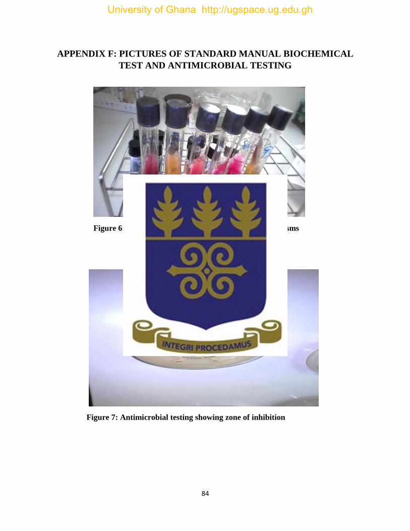
84
APPENDIX F: PICTURES OF STANDARD MANUAL BIOCHEMICAL
TEST AND ANTIMICROBIAL TESTING
Figure 6: Biochemical reactions of gram negative organisms
Figure 7: Antimicrobial testing showing zone of inhibition
University of Ghana http://ugspace.ug.edu.gh
Copyright © 2022 FDOKUMEN