Malaria - DTIC
29
1 Introduction Definition Malaria is an infectious disease caused by coccidian pro- tozoa of the genus Plasmodium, and transmitted by infected female anopheline mosquitoes. Plasmodium sp infecting humans include Plasmodium vivax, Plasmodium falci- parum, Plasmodium malariae, and Plasmodium ovale. The 4 species differ in geographic distribution, microscopic ap- pearance, and clinical features. Synonyms Infections with P. vivax, P. falciparum, P. malariae, and P. ovale are known respectively as vivax or benign tertian malaria, falciparum malaria, quartan malaria, and ovale malaria. The terms tertian and quartan describe the usual periodicity of the fever. General Considerations In the mid-19th century Meckel and others discovered a black granular substance in the blood and tissues of patients with malaria. 1 In 1847, Meckel observed that the granules lay within protoplasmic masses which Afanasiev, in 1879, suggested were the cause of malaria. In 1880, Laveran, a French army surgeon in Algeria, observed malarial pigment in plasma and leukocytes and clear bodies within eryth- rocytes. As the irregularly shaped hyaline bodies grew, Laveran noted that the erythrocytes paled and pigment formed within them. Later he observed male gametes form by exflagellation and described the male and female gam- etes, the trophozoite, and the schizont forms. In 1886, Ma- chiafava and Celli gave the generic name Plasmodium to the parasite. Most early observations of malarial parasites were made on unstained specimens since methylene blue and eosin stains were not applied to blood smears until 1891 by Romanovsky; a forerunner of today’s Wright stain. In 1895, Ross, a British medical officer in India, hypoth- esized that the mosquito is the intermediary host of the ma- larial parasite and began dissecting various species of mos- quitoes. In 1897 he found malarial parasites in the stomach wall of Anopheles mosquitoes, and in 1898 described the complete life cycle of the parasite that causes avian malaria, and the Culex mosquito vector. For his research Ross re- ceived the Nobel prize in 1902. Grassi et al later proved that anopheline mosquitoes transmit malaria to humans. 2 In 2002, researchers sequenced the genomes of P. falci- parum 3 and one of its vectors, Anopheles gambiae. 4 Malaria Mary K. Klassen-Fischer, Ronald C. Neafie and Wayne M. Meyers 10
-
Upload
khangminh22 -
Category
Documents
-
view
4 -
download
0
Transcript of Malaria - DTIC

1
IntroductionDefinition
Malaria is an infectious disease caused by coccidian pro-tozoa of the genus Plasmodium, and transmitted by infected female anopheline mosquitoes. Plasmodium sp infecting humans include Plasmodium vivax, Plasmodium falci-parum, Plasmodium malariae, and Plasmodium ovale. The 4 species differ in geographic distribution, microscopic ap-pearance, and clinical features.
Synonyms Infections with P. vivax, P. falciparum, P. malariae, and
P. ovale are known respectively as vivax or benign tertian malaria, falciparum malaria, quartan malaria, and ovale malaria. The terms tertian and quartan describe the usual periodicity of the fever.
General Considerations In the mid-19th century Meckel and others discovered a
black granular substance in the blood and tissues of patients with malaria.1 In 1847, Meckel observed that the granules lay within protoplasmic masses which Afanasiev, in 1879, suggested were the cause of malaria. In 1880, Laveran, a French army surgeon in Algeria, observed malarial pigment
in plasma and leukocytes and clear bodies within eryth-rocytes. As the irregularly shaped hyaline bodies grew, Laveran noted that the erythrocytes paled and pigment formed within them. Later he observed male gametes form by exflagellation and described the male and female gam-etes, the trophozoite, and the schizont forms. In 1886, Ma-chiafava and Celli gave the generic name Plasmodium to the parasite. Most early observations of malarial parasites were made on unstained specimens since methylene blue and eosin stains were not applied to blood smears until 1891 by Romanovsky; a forerunner of today’s Wright stain.
In 1895, Ross, a British medical officer in India, hypoth-esized that the mosquito is the intermediary host of the ma-larial parasite and began dissecting various species of mos-quitoes. In 1897 he found malarial parasites in the stomach wall of Anopheles mosquitoes, and in 1898 described the complete life cycle of the parasite that causes avian malaria, and the Culex mosquito vector. For his research Ross re-ceived the Nobel prize in 1902. Grassi et al later proved that anopheline mosquitoes transmit malaria to humans.2
In 2002, researchers sequenced the genomes of P. falci-parum 3 and one of its vectors, Anopheles gambiae.4
Malaria
Mary K. Klassen-Fischer, Ronald C. Neafie and Wayne M. Meyers
10

Report Documentation Page Form ApprovedOMB No. 0704-0188
Public reporting burden for the collection of information is estimated to average 1 hour per response, including the time for reviewing instructions, searching existing data sources, gathering andmaintaining the data needed, and completing and reviewing the collection of information. Send comments regarding this burden estimate or any other aspect of this collection of information,including suggestions for reducing this burden, to Washington Headquarters Services, Directorate for Information Operations and Reports, 1215 Jefferson Davis Highway, Suite 1204, ArlingtonVA 22202-4302. Respondents should be aware that notwithstanding any other provision of law, no person shall be subject to a penalty for failing to comply with a collection of information if itdoes not display a currently valid OMB control number.
1. REPORT DATE JUN 2011 2. REPORT TYPE
3. DATES COVERED 00-00-2011 to 00-00-2011
4. TITLE AND SUBTITLE Malaria
5a. CONTRACT NUMBER
5b. GRANT NUMBER
5c. PROGRAM ELEMENT NUMBER
6. AUTHOR(S) 5d. PROJECT NUMBER
5e. TASK NUMBER
5f. WORK UNIT NUMBER
7. PERFORMING ORGANIZATION NAME(S) AND ADDRESS(ES) Inova Central Laboratory,2832 Juniper Street,Fairfax,VA,22031
8. PERFORMING ORGANIZATIONREPORT NUMBER
9. SPONSORING/MONITORING AGENCY NAME(S) AND ADDRESS(ES) 10. SPONSOR/MONITOR’S ACRONYM(S)
11. SPONSOR/MONITOR’S REPORT NUMBER(S)
12. DISTRIBUTION/AVAILABILITY STATEMENT Approved for public release; distribution unlimited
13. SUPPLEMENTARY NOTES See also ADA545141. Chapter 10 from e-book, Topics on the Pathology of Protozoan and InvasiveArthropod Diseases.
14. ABSTRACT
15. SUBJECT TERMS
16. SECURITY CLASSIFICATION OF: 17. LIMITATION OF ABSTRACT Same as
Report (SAR)
18. NUMBEROF PAGES
28
19a. NAME OFRESPONSIBLE PERSON
a. REPORT unclassified
b. ABSTRACT unclassified
c. THIS PAGE unclassified
Standard Form 298 (Rev. 8-98) Prescribed by ANSI Std Z39-18

2
10 • Topics on The paThology of proTozoan and invasive arThropod diseases
Fig 10.1Countries endemic for malaria. Regions shown in green are endemic for Plasmodium vivax only.
Epidemiology Malaria is endemic in tropical and subtropical regions of
Africa, Asia, Central and South America, and Oceania, and on the West Indies island of Hispaniola (Fig 10.1).5,6 Rates of infection vary depending on insect vectors, climate, envi-ronmental conditions, the local species of Plasmodium and its susceptibility to antimalarial drugs, and the genetic com-position, acquired immunity, and behavior of human hosts. Malaria is more prevalent in rural areas, but incidence among urban populations is increasing.
Malaria is one of the most prevalent infectious diseases, with over 40% of the world’s population at risk. Approxi-mately 300 million cases of clinical malaria are reported worldwide each year, with more than a million fatalities. Almost 90% of fatalities are in sub-Saharan Africa, where young children are most affected. According to WHO esti-mates, there are approximately 270 million cases of malaria in Africa per year, and an estimated 5% of African children die of malaria-related illness before their fifth birthday.6 In areas of the world with high infection rates, children, preg-nant women in the second and third trimesters and during the early postpartum period,7 as well as nonimmune visitors are at greatest risk of fatal malarial infection. In areas where
infection rates are lower, the risk is equal for everyone. Al-though residents of endemic areas acquire some immunity, repeated infections cause tremendous loss of life and pro-ductivity. Plasmodium falciparum is the second most com-mon species infecting humans, it is found almost exclu-sively in tropical areas and is the cause of the most severe forms of malaria, and most deaths. Plasmodium malariae is rare now but was common in Europe at one time, and P. ovale, the rarest species, is found in Africa and Southeast Asia. Plasmodium vivax is the most common of all species of plasmodia and has a wide geographical distribution, in-cluding some temperate regions, but causes milder disease.
In nonendemic areas, most cases of malaria develop in immigrants and travelers from endemic regions. However, anywhere anopheline mosquitoes are present, the potential exists for reintroduction of malaria, even in temperate cli-mates where the disease has been eradicated. There are rare reports of stowaway mosquitoes transmitting malaria to people who live near airports.8 Malaria was once common in North America, especially in the Mississippi Valley, but on average, one case per year is now reported in the United States. Most of these patients were infected during travel to
3
Malaria • 10
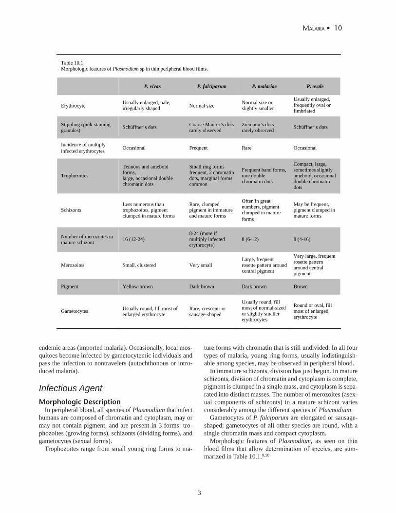
Table 10.1 Morphologic features of Plasmodium sp in thin peripheral blood films.
P. vivax P. falciparum P. malariae P. ovale
Erythrocyte Usually enlarged, pale, irregularly shaped Normal size Normal size or
slightly smaller
Usually enlarged, frequently oval or fimbriated
Stippling (pink-staining granules) Schüffner’s dots Coarse Maurer’s dots
rarely observed Ziemann’s dots rarely observed Schüffner’s dots
Incidence of multiply infected erythrocytes Occasional Frequent Rare Occasional
Trophozoites
Tenuous and ameboid forms, large, occasional double chromatin dots
Small ring forms frequent, 2 chromatin dots, marginal forms common
Frequent band forms, rare double chromatin dots
Compact, large, sometimes slightly ameboid, occasional double chromatin dots
Schizonts Less numerous than trophozoites, pigment clumped in mature forms
Rare, clumped pigment in immature and mature forms
Often in great numbers, pigment clumped in mature forms
May be frequent, pigment clumped in mature forms
Number of merozoites in mature schizont 16 (12-24)
8-24 (more if multiply infected erythrocyte)
8 (6-12) 8 (4-16)
Merozoites Small, clustered Very small Large, frequent rosette pattern around central pigment
Very large, frequent rosette pattern around central pigment
Pigment Yellow-brown Dark brown Dark brown Brown
Gametocytes Usually round, fill most of enlarged erythrocyte
Rare, crescent- or sausage-shaped
Usually round, fill most of normal-sized or slightly smaller erythrocytes
Round or oval, fill most of enlarged erythrocyte
endemic areas (imported malaria). Occasionally, local mos-quitoes become infected by gametocytemic individuals and pass the infection to nontravelers (autochthonous or intro-duced malaria).
Infectious Agent Morphologic Description
In peripheral blood, all species of Plasmodium that infect humans are composed of chromatin and cytoplasm, may or may not contain pigment, and are present in 3 forms: tro-phozoites (growing forms), schizonts (dividing forms), and gametocytes (sexual forms).
Trophozoites range from small young ring forms to ma-
ture forms with chromatin that is still undivided. In all four types of malaria, young ring forms, usually indistinguish-able among species, may be observed in peripheral blood.
In immature schizonts, division has just begun. In mature schizonts, division of chromatin and cytoplasm is complete, pigment is clumped in a single mass, and cytoplasm is sepa-rated into distinct masses. The number of merozoites (asex-ual components of schizonts) in a mature schizont varies considerably among the different species of Plasmodium.
Gametocytes of P. falciparum are elongated or sausage-shaped; gametocytes of all other species are round, with a single chromatin mass and compact cytoplasm.
Morphologic features of Plasmodium, as seen on thin blood films that allow determination of species, are sum-marized in Table 10.1.9,10

4
10 • Topics on The paThology of proTozoan and invasive arThropod diseases
Figure 10.3Five ring trophozoites in single erythrocyte. Multiply infectederythrocytes are very common in Plasmodium falciparum infections.Giemsa. Original magnification x330
Figure 10.4Pink-staining Maurer’s dots in erythrocyte cytoplasm stainedat pH 7.4. Giemsa. Original magnification x300
a b
f
c
g
d
e
Figure 10.2Heavy parasitemia (30%) in patient who died of falciparum malaria. Infected erythrocytes are normal size. Giemsa. Original magnification x330
Figure 10.5 a,b,c,d,e,f,g Plasmodium falciparum trophpzoites. Giemsa. Original magnifications all x333 except e x290. a. Small ring trophozoite, no pigment. b. Double chromatin dots. c. Narrow band forms. d. Flattened marginal forms (arrows). e. Older ring trophozoite. f. Erythrocyte basophillic stippling. g. Mature trophozoite with pigment lump in cytoplasm.
Plasmodium falciparum (Figures 10.2 to 10.10)Erythrocytes infected with P. falciparum are of normal
size. Parasitemia is often much greater in falciparum ma-laria than in the other 3 types. Multiply infected erythro-cytes are common, especially in heavy infections (Figs 10.2 to 10.4). Infection by asexual parasites from the older ring trophozoite stage onward causes erythrocytes to be stippled with pink Maurer’s dots of various shapes and sizes. Mau-rer’s dots are rarely observed unless they are overstained or stain is alkaline (Fig 10.4).
Young ring trophozoites (Fig 10.5a) of P. fal-ciparum are smaller than those of other species (approximately one fifth the diam-eter of an erythrocyte) and more likely to have double chromatin dots (Fig 10.5b). Three and even 4 chromatin dots may rarely be observed. Young ring tro-phozoites have delicate, thread-like cytoplasm; very young trophozoites usually contain no pigment. They may be round, rectangular, flame-shaped, or narrow band-shaped (Fig 10.5c). Flattened marginal forms and bridge forms are more common than in other species (Fig 10.5d). Older ring trophozoites are slightly larger and have more cytoplasm and chromatin (Fig 10.5e). Traces of tiny pigment granules may give the cytoplasm a yellowish tinge, and there may be basophilic stippling (blue dots) (Figs 10.5f). It is charac-teristic in falciparum malaria to observe only ring forms of the asexual cycle, with no older trophozoites or schizonts, in peripheral blood. Mature trophozoites have small, com-pact, light-staining cytoplasm, a larger chromatin dot, and a small, dense, nearly black clump of pigment (Fig 10.5g). Clumping of pigment at this stage is an exclusive character-istic of P. falciparum.

5
Malaria • 10
c
d
a
a
b
b
c
Figure 10.6 a,b,cPlasmodium falciparum schizont forms in thin peripheral blood smears, Giemsa. Original magnifications, a & b Immature schizonts. x330 c. Mature schizont with 15 merozoites and central pigment clump. x300
Figure 10.7 a,b,c,dImmature and mature Plasmodium falciparum gametocyte forms in thin peripheral smears, Giemsa. Original magnifications. Immature gametocytes (a & b). a. Large round form. x400 b. Elongated shape with pointed ends containing chromatin mass and pigment. x300 c. Sausage-shaped mature macrogametocyte with pointed ends, contains compact chromatin and pigment. x450 d. Sausage-shaped mature microgametocyte has rounded ends and contains diffuse pigment and chromatin. x450
Schizonts of P. falciparum are rarely observed in pe-ripheral blood, even in heavy infections. During schi-zogony, the nucleus divides and the cytoplasm breaks apart. In immature schizonts, the chromatin dots and dark brown pigment clumps are dividing and are far more noticeable than the small amount of clear cyto-plasm surrounding them (Figs 10.6a & 10.6b). Mature schizonts contain 8 to 24 minute merozoites that are often arranged in a rosette pattern (Fig 10.6c). When more than one schizont infects an erythrocyte, there may be more merozoites. Each schizont in a multiply infected erythrocyte has a separate clump of dark brown pigment. When the schizonts reach maturity, infected erythrocytes burst, releasing merozoites that enter new erythrocytes to begin another generation.
Immature P. falciparum gametocytes are seldom ob-served in peripheral blood (Figs 10.7a & 10.7b). The earliest forms are small, compact, and round. Pigment is scattered throughout the cytoplasm and the nucleus is sometimes stretched along one side. As it matures, the gametocyte becomes elongate, angular, or oval, and the chromatin tends to migrate toward the center. Fully ma-tured gametocytes, which are more commonly seen than younger forms, are usually crescent- or sausage-shaped. Macrogametocytes are usually slightly longer, more slender with pointed ends, and more deeply stained than microgametocytes (Fig 10.7c). Macrogametocytes have dense blue cytoplasm and a small, compact, red or ma-genta chromatin mass lying in or near the center or near one of the poles. Pigment closely adheres to the chro-matin in separate grains, surrounding it or covering it completely. Microgametocytes are sausage-shaped with rounded ends and have pale, often grayish-blue or pink cytoplasm and loose, irregularly scattered chromatin granules (Fig 10.7d). Abundant small, brownish rodlets and granules of pigment occupy the central portion of the parasite. The ends of mature macro- and microga-metocytes are usually clear and pigment-free. Infected erythrocytes stretch as gametocytes grow longer. Fre-quently, the residuum of erythrocytic material filling the concave side of the parasite develops a faint, bowshaped projecting rim. Sometimes the remains of the erythro-cyte appear as a red zone around the gametocyte.

6
10 • Topics on The paThology of proTozoan and invasive arThropod diseases
Figure 10.8Plasmodium falciparum in thick blood film from patient with 43% parasitemia. Trophozoites have 1 or 2 large chromatin masses and small, compact cytoplasm. Some have faint pigment. Giemsa. Original magnification x290
Figure 10.10Two sausage-shaped Plasmodium falciparum gametocytes in thick blood film. Giemsa. Original magnification x300
Figure 10.9Mature Plasmodium falciparum schizont in thick blood film with clumped pigment in center and merozoites in rosette pattern. Giemsa. Original magnification x330
Thick blood films demonstrate masses of trophozoites (Fig 10.8), a mature schizont with central clumped pigment surrounded by
merozoites in rosette pattern (Fig 10.9), and sausage-shaped gametocytes (Fig 10.10).

7
Malaria • 10
a b
c d
Figure 10.11Erythrocyte with 2 trophozoites, a rare finding in Plasmodium malariae infections. Giemsa. Original magnification x290
Figure 10.12Early ring trophozoite (arrow) of Plasmodium malariae with red chromatin dot and faintly stained blue cytoplasm in normal-sized erythrocyte. Giemsa. Original magnification x290
Figure 10.13Ring trophozoite of Plasmodium malariae with red chromatin dot and prominent blue cytoplasm in normal-sized erythrocyte. Giemsa. Original magnification x300
Figure 10.14 a,b,c,dPlasmodium malariae trophozoites, thin peripheral blood smear, Giemsa. Original magnifications a. Basket appearance. x330 b. Early band form. x450 c. Mature band form. Blue staining of erythrocyte cytoplasm as seen is unusual. x450 d. Mature rounded compact form. x300
Plasmodium malariae (Figures 10.11 to 10.17)Erythrocytes infected with Plasmodium malariae
are normal-sized or slightly smaller. Parasitemia in P. malariae infections is generally less than 2%. When stained at pH 7.5, stippling with various sizes of pale pink, spherical Ziemann’s dots may be seen. Howev-er, this stippling is less distinct than in P. falciparum and P. vivax infections and too rarely observed to be diagnostically significant. Multiply infected erythro-cytes are rare (Fig 10.11).
Very young ring trophozoites (Fig 10.12) usually con-tain no pigment. Young ring trophozoites (Fig 10.13) are roughly the size of P. vivax trophozoites, but some are smaller and some have a broader circle of cytoplasm. Double chromatin dots are rare. Some trophozoites contain a vacuole that gives the parasite the appear-ance of a basket with a handle (Fig 10.14a). Growing trophozoites are usually compact and angular, round, ovoid, or band-shaped. Band forms are more fre-quently observed in P. malariae than in other species (Figs 10.14b & 10.14c). The chromatin in growing or older trophozoites may be round or streaky and fre-quently looks stretched. The pigment is darker than that of P. vivax and is often round and arranged pe-ripherally, sometimes opposite the elongated nucle-us. The large pigment granules may have a yellowish edge that gives the blue cytoplasm a greenish hue, a
characteristic of P. malariae that distinguishes it from P. vivax. Trophozoites of P. malariae rarely become ameboid like P. vivax, but retain their original shape until they ma-ture (Fig 10.14d).

8
10 • Topics on The paThology of proTozoan and invasive arThropod diseases
a b
ca b
cba
Figure 10.15 a,b,cPlasmodium malariae schizont forms in thin peripheral blood smear. Giemsa. Original magnifications. a. Immature schizont. Chromatin and cytoplasm still dividing, light brown granular pigment diffuse. x300 b. Nearly mature schizont with eight merozoites forming rosette. Chromatinc division is complete and pigment is clumped. x330 c. Mature schizont with seven merozoites rosette with clumped central pigment. x330
Figure 10.16 a,bPlasmodium malariae gametocyte forms in thin peripheral blood smears. Giemsa. Original magnifications. a. Macrogametocyte with eccentric chromatin. x330 b. Microgametocyte with central chromatin. x400
Figure 10.17 a,b,cPlasmodium malariae forms in thick peripheral blood smears. Giemsa. Original magnifications x330. a. Two trophozoites with blue cytoplasm, red chromatin and brown pigment. b. Immature schizont with several chromatin masses, undivided cytoplasm and brown pigment. c. Mature schizont with 8 merozoites forming rosette and clumped brown pigment.
Erythrocytes containing schizonts of P. malariae usually do not withdraw from peripheral blood, as they do in vivax malaria. Therefore, erythrocytes with schizonts of P. malariae are often observed in great numbers in peripheral blood. Immature schizonts can be so dark and dense that it is difficult to differenti-ate the divisions of chromatin within the heavily pig-mented cytoplasm (Fig 10.15a). The divisions are fre-quently uneven in size and shape. The pigment remains scattered throughout the cytoplasm until shortly before complete division of the chromatin. Mature schizonts (Figs 10.15b & 10.15c) consist of 6 to 12 (average 8) large merozoites arranged peripherally in a rosette pat-tern around a central clump of dark brown pigment.
Gametocytes (Fig 10.16a & 10.16b) of P. malariae are seldom observed in peripheral blood. It may be dif-ficult to differentiate young gametocytes from round, compact trophozoites. Mature gametocytes of P. ma-lariae are spherical or oval, larger than mature tropho-zoites, but smaller than gametocytes of P. vivax. They usually fill or nearly fill a normal-sized erythrocyte. Macrogametocytes (Fig 10.16a) and microgameto-cytes (Fig 10.16b) are roughly the same size and have the same staining qualities. The chromatin is eccentric in macrogametocytes and central in microgametocytes. The abundant, prominent, dark brown, coarse pigment grains are darker than in P. vivax, particularly in mac-rogametocytes.
Thick blood films demonstrate two trophozoites in Fig 10.17a, an immature schizont in Fig 10.17b, and a mature schizont in Fig 10.17c.

9
Malaria • 10
Figure 10.18 a,b,cRing forms of Plasmodium ovale in normal sized erythrocytes. Giemsa. Original magnifications. a. Small early ring trophozoite, chromatin dot, no pigment. x330 b. Older ring trophozoite, red chromatin dot. x290 c. Ring trophozoite with 2 chromatin dots. x290
Figure 10.19 a,bTrophozoites of Plasmodium ovale in enlarged erythrocytes. Giemsa. Original magnifications x330. a. Ring trophozoite lacks pigment. b. Mature trophozoite contains Schüffner’s dots.
Figure 10.20 a,bOval and fimbriated erythrocytes infected with Plasmodium ovale. Giemsa. Original magnifications. a. Two ring trophozoites in oval erythrocytes. x330 b. Ring trophozoites in fimbriated oval erythrocyte (left) and fimbriated round erythrocyte (right) with macrogametocyte. x290
Figure 10.21 a,b,c,dVarious forms of Plasmodium ovale trophozoites. Giemsa. Original magnifications. a. Multiple infected erythrocytes. x300 b. Slight ameboid configuration with Schüffner’s dots. x290 c. Elongate thin band shaped trophozoite. x330 d. Elongate band shaped trophozoite with 2 chromatin dots. x300
a
c
a a
a b c d
b b
b
Plasmodium ovale (Figures 10.18 to 10.24)Erythrocytes infected with P. ovale are initially normal-
sized (Fig 10.18a–10.18c) but eventually enlarge nearly as much as in P. vivax infection (Figs 10.19a & 10.19b). Parasitemia in P. ovale infections is generally less than 2%. Infected erythrocytes are often oval and fimbriated and may be stippled with Schüffner’s dots (Fig 10.19b). Although fimbriated (Fig 10.20a & 10.20b) and elongated erythro-cytes (the ovalocytes, for which this species is named) (Fig 10.20) are artifacts that occur in dry, fixed, thin films, they are of great diagnostic value. Multiply infected erythrocytes are occasionally observed (Fig 10.21a).
Very young ring trophozoites of P. ovale usually contain no pigment (Fig 10.19a). Young trophozoites are about the same size as P. vivax and have a large chromatin dot and a few may have double chromatin dots (Fig 10.18c). Some have a heavy cytoplasmic circle, like P. malariae. In growing P. ovale trophozoites, the chromatin is solid and compact; the cytoplasm is only slightly ameboid (Fig 10.21b), has few vacuoles and less commonly form bands (Fig 10.21c & 10.21d). Mature trophozoites are round and centrally located. Infected erythrocytes often become oval,

10
10 • Topics on The paThology of proTozoan and invasive arThropod diseases
Figure 10.22 a,b,c,dPlasmodium ovale schizonts. Original magnifications. a. Immature schizont, chromatin and cytoplasm dividing. x300 b. Pigment beginning to clump, 7 chromatin masses. x330 c. Late immature schizont fills most of erythrocyte. x300 d. Mature schizont in rosette pattern with 6 merozoites. x290
Figure 10.23 a,bMature Plasmodium ovale gametocytes.in thin peripheral blood stains. Giemsa. Original magnification x330. a. Mature macrogametocyte with Schüffner’s dots and eccentric chromatin mass. b. Mature microgametocyte with Schüffner’s dots and large central mass of chromatin.
Figure 10.24 a,b,cThick blood film of Plasmodium ovale. Original magnifications. a. Ameboid trophozoite. x300 b. Mature schizont. x330 c. Gametocyte. x300
a b
c d
a b
b
a
c
spindle-, or pear-shaped, with ragged points or fim-briations on one or both ends. Schüffner’s dots may extend to the tips of the fimbriations. In this and later stages, P. ovale closely resembles P. malariae, except that its pigment is lighter and less conspicuous.
Immature schizonts (Figs 10.22a to 10.22c) of P. ovale are usually larger than those of P. malariae. Erythrocytes that contain them are frequently fim-briated and oval or elongated. Mature schizonts (Fig 10.22d) consist of 4 to 16 (average 8) very large merozoites, usually in a rosette pattern.
Mature macrogametocytes with eccentric chro-matin mass (Fig 10.23a) and mature microgameto-cyte with central chromatin mass (Fig 10.23b) of P. ovale are round and slightly smaller than those of P. vivax and contain Schüffner’s dots. Pigment rods are brown-black with a greenish tinge, distinctively ar-ranged in a concentric pattern at right angles to the radius. They are more numerous at the periphery. Mature gametocytes fill or nearly fill an enlarged erythrocyte.
Thick blood films demonstrate an ameboid tropho-zoite in Fig 10.24a, a mature schizont with 8 mero-zoites in Fig 10.24b, and a gametocyte in Fig 10.24c.

11
Malaria • 10
ba c
ba c
a b c d
Figure 10.25Heavy parasitemia (approximately 4%) in patient infected with Plasmodium vivax. Giemsa. Original magnification x250
Figure 10.26 a,b,cYoung trophozoites of Plasmodium vivax in normal sized erythrocytes. Original magnifications. a. Young ring trophozoite, lack pigment. x300b. Marginal ring trophozoite. x 300 c. Tenuous trophozoite. x330
Figure 10.27 a,b,cMultiply infected enlarged erythrocytes with Plasmodium vivax. Giemsa pH 7.2. Original magnifications. a. Schüffner’s dots in 3 older trophozoites. x290 b. Two ring trophozoites. x450 c. Trophozoite with 2 chromatin dots, cytoplasm of erythrocyte has basophilic stippling. x450
Figure 10.28 a,b,c,dVarious forms of Plasmodium vivax trophozoites. Original magnifications. a.Older trophozoite in fimbriated erythrocyte. x450 b. Ameboid trophozoite with Schüffner’s dots. x300 c. Ameboid trophozoite. x330 d. Mature trophozoite. x300
Plasmodium vivax (Figures 10.25 to 10.31)Erythrocytes infected with P. vivax eventually enlarge,
become pale, and irregularly shaped. Parasitemia in P. vivax infections is generally less than 2%, but may be higher (4% in Fig 10.25 & 10.26). Parasitized oval erythrocytes are common. The cytoplasm of parasitized erythrocytes may be stippled with small, fairly uniform pink granules called Schüffner’s dots which are evenly distributed through-out the part of the erythrocyte that is not occupied by the parasite (Fig 10.27a); Schüffner’s dots often become more pronounced and more deeply stained as the parasite grows, and are best demonstrated by careful staining at pH 7.2 pre-venting prolonged washing which obliterates them. Schüff-ner’s dots are more numerous and less coarse than the Mau-rer’s dots sometimes seen in falciparum malaria. Multiply infected erythrocytes (Fig 10.27b) and double chromatin dots (Fig 10.27c) are occasionally observed, and often dif-ferent stages of P. vivax appear simultaneously because of asynchronous maturation within a brood, and alternate day maturation of separate broods.
Young ring trophozoites of P. vivax are large (approxi-mately one third the diameter of an erythrocyte,) consist of blue cytoplasm and a heavy red chromatin dot in the center or at the periphery. Very young ring trophozoites are usu-ally unpigmented (Fig 10.26a). Marginal ring trophozoites may be observed in some erythrocytes (Fig 10.26b). As trophozoites grow their cytoplasm thickens, the chromatin mass enlarges, pigment granules develop, and the host’s cytoplasms occasionally fimbriates (Fig 10.28a). Grow-ing trophozoites develop one or more vacuoles; tenuous pseudopodial processes sometimes give them an ameboid
appearance (Figs 10.28b &10.28c). In young forms hemo-zoin (hematin) , pigment granules may be indistinguishable as separate granules or rods due to the yellowish tinge they lend to the cytoplasm. In mature forms pigment granules are small, yellowish-brown, and angular or rod-shaped. At the end of vegetative growth, pseudopodia are drawn in and trophozoites become compact, with an irregular out-line and mottled cytoplasm (Fig 10.28d). The single chro-matin mass is compact and usually becomes located near the periphery. Full-grown P. vivax trophozoites are larger than those of other species and practically fill an enlarged erythrocyte.

12
10 • Topics on The paThology of proTozoan and invasive arThropod diseases
a b c d e
a
a
b
b
c
c
d e
Figure 10.31 a,b,cThick films of Plasmodium vivax. Giemsa. Original magnifications. a. Ring trophozoites (arrows). x290 b. Mature schizont with yellow-brown pigment (arrow). x330 c. Mature gametocyte (left) immature gametocyte (right). x300
Figure 10.30 a,b,c,d,ePlasmodium vivax gametocytes in thin peripheral blood films. Giemsa. Original magnifications. a. Immature gametocyte. x300 b. Mature macrogametocyte. x330 c. Mature oval macrogametocyte. x330 d. Mature microgametocyte. x450 e. Mature microgametocyte. x300
Figure 10.29 a,b,c,d,ePlasmodium vivax schizonts. Giemsa. Original magnifications. a. Early immature, 7 chromatin masses. x450 b. Extracellular, immature. x330 c. Late immature with 24 merozoites. x450 d. Mature schizont with 10 merozoites and yellow-brown pigment (arrow). x330 e. Mature schizont with 20 merozoites. x330
Schizonts of P. vivax tend to leave peripheral circulation and thus are less frequently found than trophozoites in peripheral blood. Immature schizonts are produced by division of chromatin (Figs 10.29a to 10.29c). When division is complete, the chromatin masses be-come smaller and more rounded and are surrounded by a mass or circle of cytoplasm. Mature schizonts consist of 12 to 24 small, clustered merozoites (average 16, but as few as 9 have been described) (Figs 10.29d & 10.29e). The yellow-brown pigment clumps (some-times arranged loosely into a single mass) (Fig 10.29d) are often localized in the center of the cluster of merozo-ites forming “rosettes” (Fig 10.29e).
Gametocytes of P. vivax develop from released merozoites in the circu-lation of the deep organs before release to the general circulation, for that rea-son young forms are only infrequently found in the peripheral blood. Imma-ture gametocytes are rounded and have homogeneous cytoplasm, often with a vesicular area around the chromatin mass (Fig 10.30a). They are difficult to distinguish from growing and mature trophozoites whose pseudopodia have drawn in prior to desiccation or with maturation. Mature gametocytes expand the erythrocytes (Figs 10.30b to 10.30e), exhibit pigment granules that become evenly distributed throughout the cytoplasm and are more numerous than in tro-phozoites. Slightly immature macrogametocytes (female) of P. vivax can be distinguished from mature trophozoites by their larger size, circular to ovoid contour, homogenous cytoplasm with larger and darker brown pigment granules (often more nu-merous than in trophozoites) and lack of vacuoles. Mature macrogametocytes (Figs 10.30b & 10.30c) have densely blue-staining, homogeneous cytoplasm, a compact chroma-tin mass that is deep red or magenta near the periphery and sometimes surrounded by a colorless vesicular area. Ma-ture microgametocytes (male) (Figs 10.30d & 10.30e) are usually also found in enlarged erythrocytes. Their reticular chromatin mass stains lightly, and is round or stellate and larger than in any other stage, sometimes extending across
the body of the parasite in a broad spindle. The reticular chromatin mass of mature microgametocytes of P. vivax is centrally located and it is often surrounded by a large un-stained vesicular area. Pigment grains and rods are usually lighter than in macrogametocytes.
Thick blood films demonstrate several ring trophozoites (Fig 10.31a), a mature schizont with 16 merozoites (Fig 10.31b) and a mature gametozoite (left) and an immature gametocyte with vacuole surrounding chromatin mass (right) (Fig 10.31c).

13
Malaria • 10
Figure 10.32Life cycle of malarial parasites. Life cycle of Plasmodium sp. 1. Mosquito ingests sexual-stage malarial parasites (micro- and macrogametocytes). 2. Microgametocyte exflagellates in mosquito’s gut. 3. Exflagellated microgamete fertilizes macrogamete, forming a zygote. 4. Zygote develops into ookinete that penetrates epithelial cells of mosquito’s midgut. 5. Ookinete grows into oocyst. 6. Thin motile sporozoites develop, disperse through mosquito’s body cavity, lodge in salivary glands, and are inoculated into host during subsequent blood meal. 7. In human host, sporozoites leave blood and infect hepatocytes. 8-10. Sporozoites undergo schizogony, forming tissue schizonts containing many exoerythrocytic merozoites. 11. Hepatocytes rupture and release merozoites into bloodstream. 12. Plasmodium vivax and P. ovale hypnozoites remain dormant in liver; subsequent schizogony causes relapse. 13. Asexual cycle begins when merozoites from liver invade erythrocytes and develop into young ring form trophozoites. 14. Older mature trophozoites are larger and usually more compact. 15. Mature trophozoite undergoes asexual division (schizogony) to produce mature schizont composed of merozoites. 16. Erythrocyte ruptures, releasing merozoites into bloodstream. 17. Released merozoites invade other erythrocytes and begin new round of asexual reproduction. 18. Some merozoites develop into micro- and macrogametocytes within erythrocytes, which are then ingested by mosquito.
Figure 10.33Female Anopheline mosquito.
Figure 10.34Exflagellate form (arrow) of Plasmodium in peripheral blood. Giemsa. Original magnification x330
Figure 10.35Spherical oocysts attached to fragment of infected Anopheles mosquito gut. Oocysts are approximately 50 µm in diameter. Unstained. Original magnification x120
Figure 10.36Elongated sporozoites, approximately 8 µm to 10 µm by 1 µm, from gut of infected Anopheles mosquito. Giemsa. Original magnification x300
Life Cycle and Transmission The life cycle of malarial parasites is illustrated in Fig-
ure 10.32. The sexual reproductive cycle in the mosquito, called sporogony, takes 8 to 35 days.
A female anopheline mosquito (Fig 10.33) taking a blood meal from an infected host ingests malarial parasites, some of which are in the sexual stage of development (micro- and macrogametocytes). The microgametocyte undergoes a process of exflagellation. The nucleus divides into 4 to 8 nuclei, each of which combines with cytoplasm to form a
14
10 • Topics on The paThology of proTozoan and invasive arThropod diseases
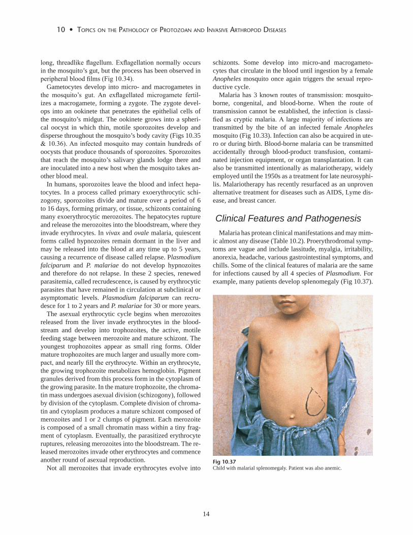
Fig 10.37Child with malarial splenomegaly. Patient was also anemic.
long, threadlike flagellum. Exflagellation normally occurs in the mosquito’s gut, but the process has been observed in peripheral blood films (Fig 10.34).
Gametocytes develop into micro- and macrogametes in the mosquito’s gut. An exflagellated microgamete fertil-izes a macrogamete, forming a zygote. The zygote devel-ops into an ookinete that penetrates the epithelial cells of the mosquito’s midgut. The ookinete grows into a spheri-cal oocyst in which thin, motile sporozoites develop and disperse throughout the mosquito’s body cavity (Figs 10.35 & 10.36). An infected mosquito may contain hundreds of oocysts that produce thousands of sporozoites. Sporozoites that reach the mosquito’s salivary glands lodge there and are inoculated into a new host when the mosquito takes an-other blood meal.
In humans, sporozoites leave the blood and infect hepa-tocytes. In a process called primary exoerythrocytic schi-zogony, sporozoites divide and mature over a period of 6 to 16 days, forming primary, or tissue, schizonts containing many exoerythrocytic merozoites. The hepatocytes rupture and release the merozoites into the bloodstream, where they invade erythrocytes. In vivax and ovale malaria, quiescent forms called hypnozoites remain dormant in the liver and may be released into the blood at any time up to 5 years, causing a recurrence of disease called relapse. Plasmodium falciparum and P. malariae do not develop hypnozoites and therefore do not relapse. In these 2 species, renewed parasitemia, called recrudescence, is caused by erythrocytic parasites that have remained in circulation at subclinical or asymptomatic levels. Plasmodium falciparum can recru-desce for 1 to 2 years and P. malariae for 30 or more years.
The asexual erythrocytic cycle begins when merozoites released from the liver invade erythrocytes in the blood-stream and develop into trophozoites, the active, motile feeding stage between merozoite and mature schizont. The youngest trophozoites appear as small ring forms. Older mature trophozoites are much larger and usually more com-pact, and nearly fill the erythrocyte. Within an erythrocyte, the growing trophozoite metabolizes hemoglobin. Pigment granules derived from this process form in the cytoplasm of the growing parasite. In the mature trophozoite, the chroma-tin mass undergoes asexual division (schizogony), followed by division of the cytoplasm. Complete division of chroma-tin and cytoplasm produces a mature schizont composed of merozoites and 1 or 2 clumps of pigment. Each merozoite is composed of a small chromatin mass within a tiny frag-ment of cytoplasm. Eventually, the parasitized erythrocyte ruptures, releasing merozoites into the bloodstream. The re-leased merozoites invade other erythrocytes and commence another round of asexual reproduction.
Not all merozoites that invade erythrocytes evolve into
schizonts. Some develop into micro-and macrogameto-cytes that circulate in the blood until ingestion by a female Anopheles mosquito once again triggers the sexual repro-ductive cycle.
Malaria has 3 known routes of transmission: mosquito-borne, congenital, and blood-borne. When the route of transmission cannot be established, the infection is classi-fied as cryptic malaria. A large majority of infections are transmitted by the bite of an infected female Anopheles mosquito (Fig 10.33). Infection can also be acquired in ute-ro or during birth. Blood-borne malaria can be transmitted accidentally through blood-product transfusion, contami-nated injection equipment, or organ transplantation. It can also be transmitted intentionally as malariotherapy, widely employed until the 1950s as a treatment for late neurosyphi-lis. Malariotherapy has recently resurfaced as an unproven alternative treatment for diseases such as AIDS, Lyme dis-ease, and breast cancer.
Clinical Features and Pathogenesis Malaria has protean clinical manifestations and may mim-
ic almost any disease (Table 10.2). Proerythrodromal symp-toms are vague and include lassitude, myalgia, irritability, anorexia, headache, various gastrointestinal symptoms, and chills. Some of the clinical features of malaria are the same for infections caused by all 4 species of Plasmodium. For example, many patients develop splenomegaly (Fig 10.37).

15
Malaria • 10
In long-standing chronic malaria or with repeated attacks, the spleen may become enormously enlarged, a condition sometimes called tropical splenomegaly syndrome or big spleen disease. Traumatic splenic rupture can cause death. Asplenia results in rapid progression of disease and high parasitemia.
The classic symptom common to all malarial infections is cyclic fever. Fever peaks around the time of schizogony and is more severe in naive patients than in those who have had previous infections. Malarial pyrogens may be products of ruptured erythrocytes or toxic products of the parasites themselves. When parasites develop synchronously, the great majority undergo schizogony at approximately the same time and fever periodicity is determined by the length of the asexual cycle (Table 10.3). Malarial paroxysms are usually sudden, with 3 discernible stages: chills, fever, and sweating. After the paroxysm, the exhausted patient usually sleeps.
Temporal features of malaria vary with factors such as
host response and the species of Plasmodium. The prepatent period between inoculation and the appearance of parasites in peripheral blood can be lengthened by immunity and pro-phylactic drugs. The incubation period between inoculation and the appearance of clinical symptoms varies depending on the degree of acquired immunity and the number of spo-rozoites inoculated. In all forms of malaria, clinical symp-toms usually appear 2 or 3 days after parasites appear in peripheral blood. In tertian malaria caused by P. vivax, P. falciparum, or P. ovale, the asexual cycle takes 48 hours, resulting in fever every third day. In quartan malaria caused by P. malariae, the asexual cycle takes 72 hours, resulting in fever every fourth day. However, fever periodicity is not a reliable indicator of the species of Plasmodium because the cycle may be erratic in patients with repeated or mixed infections, or with 2 or more asynchronous broods.
Vivax malaria is very rarely fatal. Erythrocytes infected with P. vivax are not sequestered in the microcirculation and thus do not cause the complications associated with
Table 10.3Temporal features of malaria caused by Plasmodium sp infecting humans.
P. vivax P. falciparum P. malariae P. ovale
Prepatent period (days) 10-13 6-12 15-21 10-14
Incubation period (days) 8-20 12-20 18-40 8-19
Periodicity of fever (hours) 48 48 72 48
Time course Weeks to months, relapses up to 5 years
1-2 years (untreated)
Several months, relapses up to 30 years
Same as P. vivax
Table 10.2Clinical features of malaria caused by Plasmodium sp infecting humans.
P. vivax P. falciparum P. malariae P. ovale
Complications Very rare transient cerebral irritation
Severe anemia, frequent cerebral, pulmonary, and renal complications
Rare progressiveglomerulonephritis
Milder than P. vivax
Splenomegaly Less severe than P. falciparum, rup-ture 2 to 3 months after infection.
Frequent Less severe than P. falciparum
Same as P. vivax
Hepatomegaly Less severe than P. falciparum Frequent, with jaundice Very rare Same as P. vivax
Other features Late-day paroxysms, high fever, nausea, vomiting
High fever, nausea,vomiting, diarrhea
Peripheral edema Same as P. vivax

16
10 • Topics on The paThology of proTozoan and invasive arThropod diseases
Figure 10.38Transmission electron micrograph of Plasmodium falciparum in patient with ocular malaria. Note knobs, conical evaginations, on surface of erythrocyte (arrows).
falciparum malaria. Plasmodium vivax infects only young erythrocytes; however, hemolysis-induced hematopoiesis can cause significant parasitemia. Relapses of vivax malaria occur when merozoites are released into circulation from hepatic hypnozoites. Relapses are usually less severe and of shorter duration than the primary attack, but still create a considerable economic burden for individuals and society. Many Africans are resistant to P. vivax infection because they lack the Duffy blood group antigens that are the recep-tors for the merozoites.
Plasmodium falciparum has several unique fea-tures that make it the most virulent species and the one that accounts for most deaths due to malaria. It has no secondary exoerythrocytic stage, it parasitizes erythrocytes of any age, and its preerythrocytic schizonts release many more merozoites than other Plasmodium sp. Knobs on para-sitized erythrocytes facilitate adherence to capillary endo-thelium (Fig 10.38). Because parasitized erythrocytes are sequestered in the microcirculation, some heavily infected patients have few circulating parasites.
Falciparum malaria often begins with severe gastrointes-tinal symptoms. Later, patients may develop high fever, hep-atomegaly, hepatic tenderness, jaundice due to hemolysis, and splenic tenderness. Laboratory tests may show slightly elevated levels of bilirubin and transaminases. Relapses do not occur because there are no hepatic hypnozoites and because erythrocytic parasites do not persist longer than 1 or 2 years. Fatal complications, usually resulting from mi-crovascular disease, occur more frequently in patients who are very young, immunodeficient, or pregnant, or who have high parasitemia.7,11 “Algid malaria” describes the clinical findings of clammy skin, cyanosis, and hypotension due to circulatory collapse.12 Signs and symptoms of cerebral
malaria include headache, nuchal rigidity, altered levels of consciousness, seizures, ataxia, aphasia, and dysarthria. Renal involvement may take the form of glomerulonephri-tis with proteinuria, hemoglobinuria, oliguria, or abnormal urinary sediment. Acute intravascular hemolysis can cause hemoglobinuric nephrosis with renal failure. “Blackwater fever” describes the appearance of dark urine after an acute attack of falciparum malaria. Other complications include gastroenteritis in children, pulmonary edema, severe nor-mocytic anemia, hypoglycemia, and disseminated intravas-cular coagulopathy. Falciparum malaria during pregnancy increases the risk of maternal anemia, fetal death, prematu-rity, intrauterine growth retardation, and low birth weight.7 Falciparum malaria is less severe in heterozygotes for the sickle hemoglobin gene.
In individuals with some acquired immunity to malaria, destruction of pre-erythrocytic stages in the liver by cyto-toxic T cells or other mechanisms can prevent the develop-ment of disease.13 If parasites do enter the blood, antibod-ies may control parasite density and severity of infection.14 Also, immune responses directed at certain parasite compo-nents, such as those involved in fever or adherence to endo-thelial cells, may help to modify the clinical response.15 In cerebral malaria there appears to be a complex interaction between pro-inflammatory and anti-inflammatory cytokines affecting sequestration of circulating blood cells16,17 Para-site-derived molecules—surfaces or soluble—remain nec-essary but not sufficient to explain the development of cere-bral malaria.18 Persons exposed frequently to malaria have increased immunoglobulin production.19 Chronic B-cell ac-tivation is probably a consequence of immune responses to malarial antigens and of nonspecific B-cell stimulation by toxins and mitogens.20

17
Malaria • 10
Figure 10.39Plasmodium falciparum trophozoites in maternal erythrocytes in placenta. Note pigment and blue cytoplasm (arrows). x1255
Figure 10.40Trophozoite in Kupffer’s cell in liver. Note pigment and cytoplasm (arrow). x1485
Figure 10.41Two clumps of malarial pigment in erythrocyte (arrow) inmyocardium. Parasite cytoplasm not visible in photo. x955
Figure 10.42Trophozoites with cytoplasm and pigment in hemoglobin-depleted erythrocytes (arrows) in cerebral capillary. x960
Figure 10.43Spherical clumps of malarial pigment in Kupffer’s cell inliver. x870
10.40). Within an erythrocyte, where trophozoites are most commonly found, the parasite cytoplasm usually stains poorly and may not be visible (Fig 10.41). Infected eryth-rocytes are frequently depleted of hemoglobin (Fig 10.42), frequently, only malarial pigment can be identified (Fig
AIDS may affect some components of the immune re-sponse to P. falciparum.21 After adults leave an endemic area, their protective immunity wanes slowly. It is therefore likely that established antibody responses will be sustained for several years in patients with low CD4 cell counts. In young children, CD4 cells probably play a vital role in the development of immunity to malaria.22
Like vivax malaria, P. malariae infection is not compli-cated by microvascular disease. Quartan malaria often has an insidious onset and may cause leukopenia and mild ane-mia. Relapses do not occur because there are no hepatic hypnozoites. Recrudescence of persistent erythrocytic para-sites causes recurrence. Protracted low-grade asymptomatic parasitemia may persist for up to 30 years, making carriers a source of transfusion malaria. Continued antigen stimula-tion associated with persistent parasitemia may cause im-mune complex glomerulonephritis. Relapses also occur in ovale malaria due to hepatic hypnozoites.
Clinical features of ovale malaria are similar to those of vivax malaria. Symptoms tend to be mild, and fever often subsides after a few cycles with or without treatment.
Pathologic Features Knobs on the surface of erythrocytes parasitized by P. fal-
ciparum adhere to surface receptors on capillary endothelial cells, believed to be thrombospondin, CD36, and ICAM-1 (Fig 10.38).15,23 Because P. falciparum is the only malarial parasite that becomes sequestered in vessels, it is the only species of Plasmodium that we have observed in tissue sec-tions.
In hematoxylin and eosin-stained sections, trophozoites of P. falciparum appear as small (2 µm to 4 µm), usually spherical masses of cytoplasm, each containing at least one round clump of dark, birefringent pigment (Figs 10.39 &

18
10 • Topics on The paThology of proTozoan and invasive arThropod diseases
Figure 10.44Schizont in erythrocyte in dermal blood vessel. Note round clump of pigment and merozoites in rosette pattern. Original magnification x500
Figure 10.45Round clumps of malarial pigment (arrows) in Kupffer’s cell in liver. Elongate aggregate between arrows is also malarial pigment. Original magnification x250
Figure 10.47Large clumps of extracellular nonmalarial pigment in vessel containing erythrocytes. Note that pigment clumps are not round. x800
Figure 10.46Birefringent malarial pigment in liver viewed under polarized light. x250
Figure 10.48Birefringent nonmalarial pigment in lung viewed under polarized light. Original magnification x300
10.43). Schizonts of P. falciparum are occasionally ob-served in tissue sections. They are usually spherical, 4 µm to 5 µm in diameter, and have a clump of pigment surrounded by tiny merozoites in a rosette pattern (Fig 10.44). Gameto-cytes are rarely seen in tissue sections.
Malarial pigment is the end product of hemoglobin diges-tion into a porphyrin conjugated with a protein derived from the globin portion of hemoglobin. Malarial pigment and for-malin pigment are morphologically very similar. Both ap-pear microscopically as brown or black crystals that are bi-refringent under polarized light. However, they can usually be distinguished by certain general characteristics: malarial pigment appears as intracellular round granules (Figs 10.45 & 10.46); formalin pigment is extracellular and frequently rod-shaped (Figs 10.47 & 10.48).
The following steps help to prevent deposition of forma-lin pigment in congested tissues:
• Trim tissue to less than 3 mm thick. • Immediately place tissue in phosphate-buffered neu-

19
Malaria • 10
Figure 10.49Brain of child who died of falciparum malaria, showing flat gyri and pink discoloration caused by engorged meningeal vessels.
Figure 10.50Brain of patient who died of falciparum malaria, demonstrating congestion and petechial hemorrhage in white matter.
Figure 10.51Trophozoites with blue-staining cytoplasm and black pigment in cerebral capillary. Parasitized erythrocytes tend to lie against capillary endothelium. Thomas x970
Figure 10.52Multiple ring hemorrhages in brain. x12
Figure 10.53Dürck’s nodes in cerebellum consisting of central occludedvessel surrounded by demyelination and glial proliferation. x40
Figure 10.54Dürck’s node in cerebellum showing glial proliferation. x150
tral formalin.• Refrigerate fixative and tissues for the first 24
hours.• Change the formalin whenever it becomes
even slightly discolored. Postmortem findings of falciparum malaria usu-
ally include prominent generalized passive con-gestion. Grossly, there is often gray or brown dis-coloration of the brain, liver, and spleen caused by malarial pigment.24
The brain is edematous with broad, flat gyri (Fig 10.49). Congested arachnoid vessels give it a pink-ish cast. When the swollen brain herniates, there may be grooving of the uncinate and cingulate gyri or cerebellar tonsils. The cut surface shows selective congestion and petechial hemorrhage of white mat-ter (Fig 10.50).
Microscopically, masses of erythrocytes fill the lumina of small and medium-sized blood vessels, distending the vessels. Parasitized erythrocytes tend to lie against the endothelial surface of vessels (Fig 10.51). In a ring hemorrhage, a small vessel is oc-cluded by parasitized erythrocytes, surrounded by necrotic parenchyma, then surrounded by hemor-rhage of nonparasitized erythrocytes (Fig 10.52).
Dürck’s nodes are areas of demyelination and glial proliferation surrounding an occluded vessel (Figs 10.53 & 10.54). Ring hemorrhages and Dürck’s nodes are seen in the brains of patients with cerebral malaria of 9 to 10 days duration. Anoxia may result in nonspecific congestion, edema, microinfarcts, and

20
10 • Topics on The paThology of proTozoan and invasive arThropod diseases
Figure 10.55Diffuse slate-gray discoloration in liver caused by accumulation of pigment in Kupffer’s cells, characteristic of acute falciparum malaria.
Figure 10.58Spleen of patient with fatal acute falciparum malaria. Gray discoloration is caused by pigment-laden macrophages.
Figure 10.60Trophozoite in splenic erythrocyte in spleen. Note parasite cytoplasm and round clump of pigment (arrow). x1700
Figure 10.61Congested spleen with tiny clumps of malarial pigment (arrows). x345
Figure 10.59Congested spleen of patient with falciparum malaria. x60
Figure 10.57Malarial parasite in histiocyte in hepatic sinusoid. Note parasite cytoplasm and round clump of pigment (arrow). x1320
Figure 10.56Well-delineated lobular pattern in liver caused by pigment deposition in portal areas in chronic falciparum malaria.
focal hemorrhages. Grossly, the liver appears gray as a result of accumu-
lated malarial pigment (Fig 10.55). There is an increase in Kupffer’s cells, which are enlarged and contain malarial pigment, parasites, erythrocytes, and debris from ruptured erythrocytes (Figs 10.40 & 10.43). Pigment deposition ex-tends to portal areas over time (Fig 10.56). Erythrocytes and histiocytes in the sinusoids may contain parasites (Figs 10.45 & 10.57); parasites and malarial pigment are not seen in hepatocytes. Central veins, sinusoids, and portal vein branches may be congested.
In acute malaria, the spleen is enlarged and darkly pig-mented (Fig 10.58). Complications of splenomegaly can in-clude rupture, hemorrhage, torsion, and infarction. The red pulp is congested with parasitized erythrocytes, macro-phages containing parasites and malarial pigment, and free parasites (Figs 10.59 to 10.63). Malpighian corpus-cles are small and indistinct. In chronic malaria, the spleen is even more enlarged and collections of lymphocytes may appear in the sinusoids. Macrophages containing malarial
21
Malaria • 10
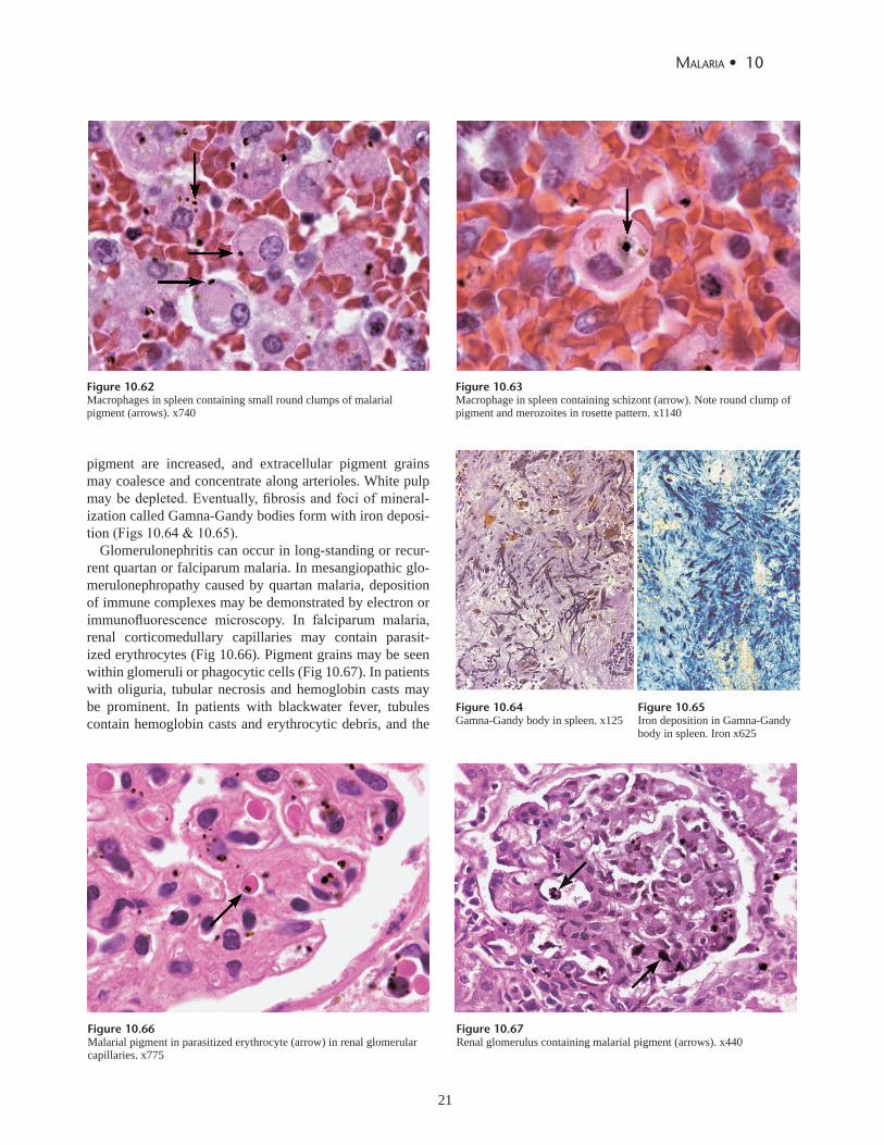
Figure 10.62Macrophages in spleen containing small round clumps of malarial pigment (arrows). x740
Figure 10.66Malarial pigment in parasitized erythrocyte (arrow) in renal glomerular capillaries. x775
Figure 10.67Renal glomerulus containing malarial pigment (arrows). x440
Figure 10.63Macrophage in spleen containing schizont (arrow). Note round clump of pigment and merozoites in rosette pattern. x1140
Figure 10.64Gamna-Gandy body in spleen. x125
Figure 10.65Iron deposition in Gamna-Gandy body in spleen. Iron x625
pigment are increased, and extracellular pigment grains may coalesce and concentrate along arterioles. White pulp may be depleted. Eventually, fibrosis and foci of mineral-ization called Gamna-Gandy bodies form with iron deposi-tion (Figs 10.64 & 10.65).
Glomerulonephritis can occur in long-standing or recur-rent quartan or falciparum malaria. In mesangiopathic glo-merulonephropathy caused by quartan malaria, deposition of immune complexes may be demonstrated by electron or immunofluorescence microscopy. In falciparum malaria, renal corticomedullary capillaries may contain parasit-ized erythrocytes (Fig 10.66). Pigment grains may be seen within glomeruli or phagocytic cells (Fig 10.67). In patients with oliguria, tubular necrosis and hemoglobin casts may be prominent. In patients with blackwater fever, tubules contain hemoglobin casts and erythrocytic debris, and the

22
10 • Topics on The paThology of proTozoan and invasive arThropod diseases
a b c
Figure 10.68Capillary in myocardium occluded by parasitized erythrocytes. x480
Figure 10.69Parasitized erythrocytes in capillaries of pulmonary alveolar septa. x575
Figure 10.70Placenta from patient with acute falciparum malaria showing massive parasitization of erythrocytes in intervillous spaces. x245
Figure 10.71 a,b,c Placenta with massive parasitization of maternal erythrocytes. a. Parasite pigment is readily visible; faintly stained parasite cytoplasm (arrow) is more subtle. x1500. b. Two trophozoites (arrows) in mononuclear cell in intervillous space. Note absence of parasites in fetal circulation (lower left). x1250. c. Placenta with several parasitized erythrocytes (arrows) in villus of fetal circulation, a rare finding. x810.
interstitium may contain focal mononuclear cell infiltrates. Pericardial and endocardial petechiae may be seen in
gross specimens. Microscopically, myocardial capillaries are usually congested and occluded with parasitized eryth-rocytes (Fig 10.68). There may be interstitial edema and focal infiltration by lymphocytes, histiocytes, monocytes, plasma cells, and rare eosinophils.
In the lung, parasitized erythrocytes may be seen within capillaries (Fig 10.69). Alveolar walls may be thickened with chronic inflammatory cells, especially monocytes, lymphocytes, and plasma cells, and alveolar spaces may be filled with proteinaceous fluid, sometimes mixed with eryth-rocytes.
Involvement of the placenta is more likely in younger women who have had few pregnancies.11 Possible complica-tions of malarious placenta include decreased placental size, spontaneous abortion, and maternal death. The placenta is severely affected, with numerous sequestered parasites in the intervillous spaces (Fig 10.70). Most parasites are within erythrocytes (Fig 10.71a to 10.71c), but some are found in monocytes (Fig 10.71b) and some are free in the intervil-lous spaces. Vessels on the fetal side of the placental barrier
are usually parasite-free. Rarely, trauma causes mixing of maternal and fetal blood, allowing passage of parasitized cells into fetal circulation, resulting in congenital malaria

23
Malaria • 10
Figure 10.72Macrophages in bone marrow containing parasitized erythrocytes (arrows). x250
Figure 10.73Bone marrow with parasitized erythrocytes. Note parasite pigment and cytoplasm (arrow). x300
Figure 10.74Smear of bone marrow showing mature schizont with pigment and merozoites in rosette pattern. Dif-Quick x400
Figure 10.75Mucosal capillaries of stomach occluded by parasitized erythrocytes. Original magnification x250
Figure 10.76Parasitized erythrocytes (arrows) in retinal capillary from 53-year-old patient who died of chloroquine-resistant falciparum malaria.x600
Figure 10.77Mature schizont (arrow) in capillary in parathyroid gland .Note dark round clump of pigment and merozoites in rosette pattern. x1550
(Fig 10.71c). Bone marrow may be hypercellular, with hyperplasia
of both erythrocytic and leukocytic precursors. Malarial parasites and their pigments are present in phagocytic cells (Fig 10.72), and congested blood spaces may contain large numbers of parasitized erythrocytes (Fig 10.73). Smears of bone marrow may also demonstrate malarial parasites (Fig 10.74).
As in the spleen, lymph nodes are congested with para-sitized erythrocytes, but pigment accumulation in macro-phages is less than that in the spleen or liver. Lymphoid follicles tend to be inconspicuous, but there is usually prom-inent histiocytic hyperplasia.
Edema, congestion, hemorrhage, and parasitized erythro-cytes within small vessels may be seen in any tissue, includ-ing adrenal gland, gastrointestinal tract (Fig 10.75), skin, adipose tissue, retina25 (Fig 10.76), parathyroid (Fig 10.77), and skeletal muscle, especially in patients with high para-sitemia.
24
10 • Topics on The paThology of proTozoan and invasive arThropod diseases
Diagnosis Diagnosis of malaria is usually established in the labo-
ratory by direct examination of thick or thin peripheral blood films (Table 10.1).26 Microscopic diagnosis is rela-tively cost-effective and has a sensitivity of approximately 50 parasites per ml of blood. A highly skilled microscopist can estimate the level of parasitemia and identify all spe-cies of Plasmodium, based on pertinent factors such as morphologic appearance of the parasite and erythrocytes, geographic considerations, and clinical symptoms. Specific diagnosis depends on scrutinizing blood films and ac-curately identifying all observable forms of the parasite, determining the degree of development of an observed morphologic feature, and how often it occurs.
Because most morphologic forms of malarial parasites and configurations of erythrocytes can be found in all 4 types of malaria, and a given specimen rarely demonstrates all the classic features of a single species, the most useful diagnostic approach combines comparison and exclusion.
For example:
• Erythrocytes enlarged: If a large number of parasitized erythrocytes are oval or fimbriated, the species is likely to be P. ovale. A finding of very few oval or fimbriated parasitized erythrocytes suggests P. vivax.
• Band erythrocytes enlarged: All 4 species of Plasmodium have thin band trophozoites, but only those of P. ma-lariae are large enough to occupy 50% to 75% of the erythrocyte.
• Large numbers of ring trophozoites: If the examiner sees only a large number of ring trophozoites, the species is most likely P. falciparum.
• If only a few ring trophozoites are observed, it may not be possible to determine the species, and the speci-men should be signed out as “malaria, species unde-termined.”
• In vivax, quartan, and ovale malaria, parasitemia is usu-ally less than 2%. In falciparum malaria, parasitemia may be 40% or higher.
• If parasitized erythrocytes are enlarged, quartan malaria can be excluded, except in mixed infections.
• If gametocytes are round, falciparum malaria can be ex-cluded.
Some authorities believe that parasite morphology may be affected by geographic location, parasite strain, and the patient’s age, immune status, and treatment.27 Investigating the patient’s travel history may provide clues to the spe-cies of Plasmodium. Although morphologic features of the
parasite are of greater diagnostic value than travel history, a thorough examiner must determine where the patient has been and what species have been reported in those areas.
Mixed infections are probably underdiagnosed. When one species predominates in a blood film, the other spe-cies is easily overlooked. Furthermore, upon diagnosing one species of malaria, the microscopist usually looks no further. To diagnose a second species, one of its diagnostic forms must also be identified. All 4 species of Plasmodium have diagnostic forms:
• vivax: ameboid trophozoite in enlarged erythrocyte with Schüffner’s dots.
• falciparum: crescent-shaped gametocyte. • malariae: broad band trophozoite. • ovale: oval, fimbriated, enlarged erythrocytes. • Mature schizonts of all 4 species are usually diagnos-
tic when other stages are also present.
Preparation and Examination of Peripheral Blood Films
The biggest pitfall in most laboratories in developed countries is leaving too great a delay between taking the blood sample and making the blood films. As blood cools to room temperature, male gametocytes will divide and release microgametes: these are long sinuous filamentous structures that can be mistaken for organisms such as Bor-relia. If the blood is kept at warmer temperatures, schiz-onts will rupture and merozoites invading erythrocytes will mistakenly give the appearance of the appliqué form of P. falciparum. If P. vivax or P. ovale is left for several hours in EDTA, the build up of acid in the sample will cause the parasitized erythrocytes to shrink and the parasite will roll up, simulating the appearance of P. malariae. This problem is made worse if anticoagulants such as heparin or citrate are used. The anticoagulant that causes the least problems is EDTA. Romanowsky stain or a variant stain is usually used. Some laboratories mistakenly use the same staining pH as they do for routine haematology blood films (pH 6.8): ma-laria blood films must be stained at pH 7.2, or Schüffner’s dots and James’s dots will not be seen.
Both thick and thin films should be prepared, preferably on the same slide, from any patient suspected of having malaria (Fig 10.78). Thick films are useful for screening because a larger volume of blood is examined and erythro-cytes are hemolyzed, concentrating the parasites and possi-bly revealing multiple stages. For diagnosis, thick films are significantly more sensitive than thin films because 16 to 30 times as much blood is examined. Thick films can be many

25
Malaria • 10
Figure 10.78Thin (top) and thick blood films prepared on separate slides.Newsprint should be readable through feathered edge of properly prepared thin film. (Typically, in most busy clinics, both thin and thick smears are placed on the same slide.)
Figure 10.79Staining artifact, not to be mistaken for malarial parasite. Giemsa x1400
Figure 10.80Unknown structures in thin peripheral blood film, representing no known human pathogen. Giemsa x825
Figure 10.81Unknown pseudoparasite in thin peripheral blood film, representing no known human pathogen. Giemsa x1775
Figure 10.82Blood platelets in thin peripheral blood film. Giemsa x1600
Figure 10.83Ring trophozoites of Babesia microti in thin peripheral blood film. Giemsa x780
cell layers thick and are never fixed, since fixation prevents dehemoglobinization. The examiner should use the 100x oil immersion objective to study thick films, and should search for at least 5 minutes (an estimated 100 fields) before re-porting a finding of no parasites.
Thin blood films are helpful in species identification because they reveal morphologic details of parasites and erythrocytes not apparent on thick films. On an ideal thin film, the cells are in a single layer and do not overlap. Ery-throcytes infected by older parasites are more frequently seen along the feathered edge of the thin film; ring forms are usually evenly distributed. To determine whether parasit-ized erythrocytes are enlarged or fimbriated, compare them with adjacent uninfected erythrocytes.
Blood films must be carefully prepared and meticulous-ly stained.28 Slides should be spotlessly clean and free of chemicals, grease, dust, and scratches. Thin films should be fixed in pure methyl alcohol before staining. Ideally, films should be stained within 24 hours, and no later than 72 hours, before the blood loses its affinity for stain. Although Wright’s stain can be used for thin films, the most depend-able stain for visualizing malarial parasites is Giemsa diluted with distilled water and buffered to pH 7.0 to 7.2. Field’s stain is fast and convenient, but the staining process occasionally washes off the entire film.29
Erythrocytes on Giemsa-stained thin films should be pale pink and parasites densely stained. Staining artifacts (Fig 10.79) and other structures (Figs 10.80 & 10.81), including platelets (Fig 10.82), may be mistaken for malarial parasites on thin films. To prevent errors, a skilled examiner must
be familiar with the appearance of normal blood constitu-ents. Because all stages of the erythrocytic cycle may not be visible in a single smear, repeated examinations should be made at different times of day during the course of infec-tion. If the first blood film is negative, additional thick and thin films should be obtained every 6 hours for 24 hours. Examining blood films under polarized light may simplify

26
10 • Topics on The paThology of proTozoan and invasive arThropod diseases
Figure 10.84Section of liver from 21-year-old Vietnam veteran with acute falciparum malaria. Treatment killed malarial parasites; only remnants of infection are round clumps of malarial pigment in Kupffer’s cells. Patient died of staphylococcal infection 6 days post-treatment. x965
Figure 10.85QBC® tube for detecting malarial parasites.
Figure 10.86Trophozoite of Plasmodium falciparum detected by QBC® method. Note well-defined, regularly shaped cytoplasm.
and accelerate screening for malarial parasites. Plasmodium sp must be differentiated from Babesia sp,
protozoa that also invade erythrocytes and closely resemble malarial parasites (Fig 10.83). Babesia sp are round, rod-shaped, piriform, or ameboid, but lack pigment. Parasitized erythrocytes are normal in size. Patients with babesiosis have no schizonts or gametocytes in peripheral blood (see Topic 11 on Babesiosis).
Other Methods of Diagnosis
Postmortem, a diagnosis of malaria is made by identify-ing malarial parasites or their pigment in histologic sec-tions. (Morphologic features are described above and in Figures 10.40 to 10.77.) Adequate treatment for malaria usually eliminates parasites from the patient, but malarial pigment accumulates in the liver and is readily observable at autopsy (Fig 10.84). Absence of malarial pigment in the liver at autopsy suggests that there was no recent attack of acute falciparum malaria.
Electron microscopy is not commonly used for diagno-sis, but can reveal characteristic knobs on the surface of an erythrocyte parasitized by P. falciparum (Fig 10.38).
The quantitative buffy coat (QBC®) technique identifies malarial parasites by fluorescent dye.30 A glass capillary tube containing fluorescent stain is filled with blood and a

27
Malaria • 10
plastic float is inserted (Fig 10.85). When the tube is cen-trifuged, parasitized erythrocytes localize in the upper layer because of their lower density. Parasites are then detectable by ultraviolet or fluorescence microscopy (Fig 10.86).
Enzyme-linked immunosorbent assay (ELISA) and ra-dioimmunoassay (RIA) tests can detect very low densities of malarial antigens in peripheral blood.31,32 These tests are nearly as sensitive as microscopic techniques and do not require a microscope or an experienced microscopist. Rapid dipstick tests that employ immunochromatographic techniques to detect parasite antigens can provide a crude estimate of the level of parasitemia. These tests have a low turnaround time and do not require skilled person-nel or special equipment, but some can detect only one species of Plasmodium. Tests targeting histidine-rich protein-2 (ParaSight®-F and ICT Malaria Pf)29,33,34 detect only P. falciparum. OptiMAL® targets lactate dehydroge-nase and can distinguish P. falciparum from P. vivax.35
DNA hybridization and PCR methods detect Plasmodi-um-specific DNA sequences in erythrocytes. These assays are often highly sensitive and specific, but may require ex-pensive equipment.36
Indirect fluorescent antibody (IFA) test is the preferred method for detecting antimalarial antibodies in serum.37 Serologic assays cannot distinguish current and prior infec-tions and are used primarily in epidemiological studies.
Treatment and Prevention Chloroquine (Aralen®) was once the treatment of choice
for malaria but is no longer reliably effective outside the Middle East, the Caribbean, and Central America because of resistant P. falciparum strains.38 Plasmodium vivax also is chloroquine-resistant in some endemic areas, particularly Papua New Guinea and Indonesia.39 Chloroquine-resistant malaria can be treated with a variety of other agents, in-cluding mefloquine (Lariam®),38 atovaquone-proguanil (Malarone®),40 doxycycline, sulfadoxine-pyrimethamine (Fansidar®), halofantrine, and quinine combined with tetra-cycline. When atovaquone-proguanil is used to treat vivax malaria, it should be followed by primaquine to eradicate persistent liver hypnozoites. Resistance to mefloquine and sulfadoxine-pyrimethamine has become a significant pro- blem in some parts of Southeast Asia and South America.39 Combining mefloquine with artemisinin derivatives may decrease resistance.41 Sulfadoxine-pyrimethamine-resistant malaria has been reported in the Amazon Basin, Southeast
Asia, and some countries in eastern and southern Africa. For complicated falciparum malaria, supportive treat-
ment may include management of hypoglycemia, seizures, pulmonary edema, and renal failure. Exchange transfusion is sometimes recommended for nonimmune patients with high parasitemia.
Oral weekly mefloquine may be the best chemoprophy-laxis for travelers in areas endemic for chloroquine-resistant P. falciparum.42 Doxycycline is an effective alternative for travelers who cannot tolerate the side effects of mefloquine. Proguanil taken daily in conjunction with weekly chlo-roquine is an option for pregnant women traveling in sub-Saharan Africa.38 Weekly oral doses of chloro-quine phosphate are effective prophylaxis in areas with chloroquine-sensitive P. falciparum. Oral weekly hydroxy-chloroquine sulfate (Plaquenil®) is an alternative to chlo-roquine phosphate, and atovaquone-proguanil is useful as prophylaxis. Long-term travelers who have likely been ex-posed to P. vivax or P. ovale may be asymptomatic carriers and at risk for later development of malaria. Primaquine phosphate administered orally once daily for 14 days af-ter departure from an endemic area can eliminate a carrier state.38
Anopheles mosquitoes bite between dusk and dawn. During waking hours, bites are best prevented by covering most skin with clothing and treating exposed skin with an insect repellent that contains a 30% to 35% concentration of N,N diethyl-meta-toluamide (deet).38 In sleeping areas that are not screened or air-conditioned, bites can be prevented by sleeping under mosquito netting treated with permethrin or deltamethrin, or by spraying the room with pyrethroid-containing formulas.43 Other preventive measures include application of environmental mosquito larvicides and drainage of mosquito breeding sites.
Several types of antimalarial vaccine are currently being studied, including cocktails of antigens of asexual blood-stage organisms, DNA recombinant protein, and transmis-sion-blocking vaccines. The search for an effective vaccine is hampered by the parasites’ remarkable capacity to vary critical antigenic structures.44

28
10 • Topics on The paThology of proTozoan and invasive arThropod diseases
References 1. Ross R. Researches on malaria, Nobel Lecture, December 12, 1902. Available
at: http://nobelprize.org/nobel-prizes/medicine/laureates/1902/ross-lecture.html. Accessed March 7, 2011.
2. Grassi GB, Bignami A, Bastianelli G. Report of studies done on malaria during the month of January [in Italian]. RC Accad Lincei.1899;8:100-104.
3. Gardner MJ, Hall N, Fung Eula, et al. Genome sequence of the human malaria parasite Plasmodium falciparum. Nature. 2002;419: 498-511.
4. Holt RA, Subramanian GM, Halpern A, et al. The genome sequence of the malaria mosquito Anopheles gambiae. Science. 2002;298:129-149.
5. Roll Back Malaria. Malaria endemic countries [Malaria Foundation International Web site]. Available at http://www.malaria.org/ endemiclist.html. Accessed Mar. 7, 2011.
6. World Health Organization. Rolling back malaria. In: Jamison DT, Creese A, Prentice T, et al, eds. World Health Report 1999: Making a Difference. Geneva, Switzerland:WHO;1999:49-63.
7. Diagne N, Rogier C, Sokhna CS, et al. Increased susceptibility to malaria during the early postpartum period. N Engl J Med. 2000;343:598-603.
8. El-Bahnasawy MM, Dabbous HKh, Morsy TA. Imported malaria as a threat to Egypt. J Egypt Soc Parasitol. 2010;40:773-788.
9. Smith JW, Melvin DM, Orihel TC, Ash LR, McQuay RM, Thompson JH Jr. Atlas of Diagnostic Medical Parasitology. I. Blood and Tissue Parasites. American Society of Clinical Pathologists: 1976.
10. Wilcox A. Manual for the Microscopical Diagnosis of Malaria in Man. Public Health Service Publication No. 796. Washington, DC: US Department of Health, Education and Welfare; 1960.
11. Anagnos D, Lanoie LO, Palmieri JR, Ziefer A, Connor DH. Effects of placental malaria on mothers and neonates from Zaire. Z Parasitenkd 1986;72:57-64.
12. Kean BH, Smith JA. Death due to estivo-autumnal malaria: a resume of one hundred autopsy cases, 1925-1942. Am J Trop Med Hyg 1944;24:317-322.
13. World Health Organization. 2004 Technical consultation on Malaria and HIV Interactions in Public Health Policy Implications. 23-25 (Geneva, Switzerland).
14. Jouin H, Garraud O, Longacre S, Baleux F, Mercereau-Puijalon O, Milon G. Human antibodies to the polymorphic block 2 domain of the Plasmodium falciparum merozoite surface protein 1 (MSP-1) exhibit a highly skewed, peptide-specific light chain distribution. Immunol Cell Biol. 2005;83:392-395.
15. Ockenhouse CF, Ho M, Tandon NN, et al. Molecular basis of sequestration in severe and uncomplicated Plasmodium falciparum malaria: differential adhesion of infected erythrocytes to CD36 and ICAM-1. J Infect Dis. 1991;164:163-169.
16. Coltel N, V. Combes, Hunt NH, Grau GE. Cerebral malaria: a neurovascular pathology with many riddles still to be solved. Curr Neurovasc Res. 2004;1(2): 91-110.
17. Couper KN, Blount DG, Wilson MS, Halfalla JC, et al. IL-10 from CD4CD25Foxp3CD127 adaptive regulatory T cells modulates parasite clearance and pathology during malaria infection. PLoS Pathog. 2008;4(2): e1000004.
18. Milner DA, Jr. Rethinking cerebral malaria pathology. Curr Opin Infect Dis. 2010;23:456-463.
19. Tobie JE, Abele DC, Wolff SM, Contacos PG, Evans CB. Serum immunoglobulin levels in human malaria and their relationship to antibody production. J Immunol.1966;97:498-505.
20. Muehlenbachs A, Fried M, Lachowitzer J, Mutabingwa TK, Duffy PE. Genome-wide expression analysis of placental malaria reveals features of lymphoid neogenesis during chronic infection. J Immunol 2007;179:557-565
21. UNICEF Malaria Technical Note # 6. Malaria and HIV. 2003 (February) #1c 2470.doc. Accessed January 2011.
22. Stephens R, Langhorne J. Priming of CD4+ T cells and development of CD4+ T cell memory; lessons for malaria. Parasite Immunol. 2006;S 25:25-30.
23. Atkinson CT, Aikawa M. Ultrastructure of malaria-infected erythrocytes. Blood Cells. 1990;16:351-368.
24. Winslow DJ, Connor DH. Human malaria. Med Times. 1967;95:593-608. 25. Hidayat AA, Nalbandian RM, Sammons DW, Fleischman JA, Johnson TE.
The diagnostic histopathologic features of ocular malaria. Ophthalmology. 1993;100:1183-1186.
26. Moody A. Rapid diagnostic tests for malaria parasites. Clin Microbiol Rev. 2002;15:66-78.
27. Gabaldon A, Garnham PCC, Macdonals G, Pampana EJ. Terminology of Malaria and of Malaria Eradication. Geneva, Switzerland: WHO; 1963.
28. Ash LR, Orihel TC. Atlas of Human Parasitology. Chicago, Ill: American Society of Clinical Pathologists; 1980:4.
29. Lema OE, Carter JY, Nagelkerke N, et al. Comparison of five methods of malaria detection in the outpatient setting. Am J Trop Med Hyg. 1999;60:177-182.
30. Wardlaw SC, Levine RA. Quantitative buffy coat analysis. A new laboratory tool functioning as a screening complete blood cell count. JAMA. 1983;249:617-620.
31. Mackey L, McGregor IA, Lambert PH. Diagnosis of Plasmodium falciparum infection using a solid-phase radioimmunoassay for the detection of malaria antigens. Bull World Health Organ. 1980;58:439-444.
32. Spencer HC, Collins WE, Skinner JC. The enzyme-linked immunosorbent assay (ELISA) for malaria. II. Comparison with the malaria indirect fluorescent antibody test (IFA). Am J Trop Med Hyg. 1979;28:933-936.
33. Forney JR, Magill AJ, Wongsrichanalai C, et al. Malaria rapid diagnostic devices: performance characteristics of the ParaSight F device determined in a multisite field study. J Clin Microbiol. 2001;39:2884-2890.
34. Singh N, Valecha N, Sharma VP. Malaria diagnosis by field workers using an immunochromatographic test. Trans R Soc Trop Med Hyg. 1997;91:396-397.
35. Cooke AH, Chiodini PL, Doherty T, Moody AH, Ries J, Pinder M. Comparison of a parasite lactate dehydrogenase-based immunochromatographic antigen detection assay (OptiMAL) with microscopy for the detection of malaria parasites in human blood samples. Am J Trop Med Hyg. 1999;60:173-176.
36. Rubio JM, Benito A, Roche J, et al. Semi-nested, multiplex polymerase chain reaction for detection of human malaria parasites and evidence of Plasmodium vivax infection in Equatorial Guinea. Am J Trop Med Hyg. 1999;60:183-187.
37. Contreras CE, Pance A, Marcano N, Gonzalez N, Bianco N. Detection of specific antibodies to Plasmodium falciparum in blood bank donors from malaria-endemic and non-endemic areas of Venezuela. Am J Trop Med Hyg. 1999;60:948-953.
38. Juckett G. Malaria prevention in travelers. Am Fam Physician. 1999;59:2523-2530, 2535-2536.
39. Barat LM, Bloland PB. Drug resistance among malaria and other parasites. Infect Dis Clin North Am. 1997;11:969-987.
40. Looareesuwan S, Chulay JD, Canfield CJ, Hutchinson DB. Malarone (atovaquone and proguanil hydrochloride): a review of its clinical development for treatment of malaria. Am J Trop Med Hyg. 1999;60:533-541.
41. Bloland PB, Ettling M, Meek S. Combination therapy for malaria in Africa: hype or hope? Bull World Health Organ. 2000;78:13781388.
42. Kramer MH, Lobel HO. Antimalarial chemoprophylaxis in infants and children. Paediatr Drugs. 2001;3:113-121.
43. Newton P, White N. Malaria: new developments in treatment and prevention. Annu Rev Med. 1999;50:179-192.
44. Good MF, Doolan DL. Malaria vaccine design: immunological considerations. Immunity. 2010;33:555-566.
AcknowledgementsFigure 10.32 Contributed by Venetia Valiga
Figure 10.33Contributed by Robert W. Gwadz
Figure 10.37Contributed by William Gordon
Figure 10.85Contributed by SC Wardlaw
Figure 10.86 Courtesy of Becton Dickinson