Misdiagnosis of Dengue Fever and Co-infection With Malaria ...
10
Page 1/10 Misdiagnosis of Dengue Fever and Co-infection With Malaria and Typhoid Fevers in Rural Areas in Southwest Nigeria Lawrence Ehis Okoror ( [email protected] ) Federal University Oye-Ekiti Emmanuel Olufemi Bankefa Federal University Oye-Ekiti Oluchi Mariam Ukhureigbe Federal University Oye-Ekiti Evelyn Olubunmi Ajayi Federal University Oye-Ekiti Stephen Kayode Ojo Federal University Oye-Ekiti Bryan Ogeneh Federal University Oye-Ekiti Research Article Keywords: Infection, Coinfection, IgM, IgG, NS1 Posted Date: March 13th, 2021 DOI: https://doi.org/10.21203/rs.3.rs-292692/v1 License: This work is licensed under a Creative Commons Attribution 4.0 International License. Read Full License
-
Upload
khangminh22 -
Category
Documents
-
view
4 -
download
0
Transcript of Misdiagnosis of Dengue Fever and Co-infection With Malaria ...

Page 1/10
Misdiagnosis of Dengue Fever and Co-infection With Malaria andTyphoid Fevers in Rural Areas in Southwest NigeriaLawrence Ehis Okoror ( [email protected] )
Federal University Oye-EkitiEmmanuel Olufemi Bankefa
Federal University Oye-EkitiOluchi Mariam Ukhureigbe
Federal University Oye-EkitiEvelyn Olubunmi Ajayi
Federal University Oye-EkitiStephen Kayode Ojo
Federal University Oye-EkitiBryan Ogeneh
Federal University Oye-Ekiti
Research Article
Keywords: Infection, Coinfection, IgM, IgG, NS1
Posted Date: March 13th, 2021
DOI: https://doi.org/10.21203/rs.3.rs-292692/v1
License: This work is licensed under a Creative Commons Attribution 4.0 International License. Read Full License

Page 2/10
AbstractBackground
Dengue and malaria have similar symptoms and arthropod vector and their mode of transmission coupled with differential diagnosis.Though typhoid fever differs from dengue and malaria by not having arthropod vector and different mode of transmission, it sharesdifferential diagnosis with Dengue and Malaria which make misdiagnosis possible. This misdiagnosis of these three diseases has sincebeen a major concern towards therapeutic administration because of their co-occurrence in many cases.
Methods
This study focused on the misdiagnosis of dengue fever for malaria or typhoid fever since the three have differential diagnosis and could co-occur. 741 samples were collected from malaria patient and 333 samples for typhoid fever outpatient at the health department facilities inrural communities of South West Nigeria. The samples were tested for dengue virus (DV) NS1 protein, anti DV IgM, anti DV IgG and RT-qPCR.
Result
Of all the samples tested 315 (29.4%) were positive to DV NS1 while 50 (6.7%) and 13 (3.9%) of 714 malaria samples and 333 typhoidsamples respectively had Dengue fever co-infection. Co-infection of the three types of fever occurred in 5 (0.5%). A total of 54 (5%) DV caseswere wrongly diagnosed for malaria while 14 (1.3%) DV cases was wrongly diagnosed as typhoid.
Conclusion
Conclusively, there was signi�cant number of misdiagnosed cases of DV for either malaria or typhoid, hence it is recommended to includeDV screening into routine hospital test especially in cases of malaria and typhoid negative by rapid diagnostic testing.
IntroductionDengue and Malaria fevers are the most common arthropod-borne diseases caused by mosquito bite and they also have similar signs andsymptoms. Typhoid fever, which is caused by Salmonella typhi and 3 types of Salmonella paratyphi also share similar symptoms with bothmalaria and dengue fevers. Co-infection of Malaria and Dengue was �rst reported in 2005 [1], while Orhe et al in 2003 report a co-infection ofmalaria and typhoid fever [2] and that the few cases that have been reported indicated that, co-infections maybe more severe than singlesinfection (Malaria or Dengue fever) [3], although there is a paucity of reports for Dengue and typhoid fevers co-infection. In most cases, it iscommon to �rst think of malaria diagnosis when feverish syndromes are observed in patients and secondly, typhoid fever will be suspectedand even treated empirically. Dengue virus infection is rarely taken into consideration by the clinicians because the disease is not consideredas endemic of which this could lead to fatal consequences. Hence, early diagnosis of Dengue infection will not only prevent complicationssuch as Dengue Hemorrhagic fever (DHF) and Dengue Shock syndrome (DSS) but will also curtail unnecessary consumption of antimalariadrugs and antibiotics thereby reducing menace of antimicrobial resistance.
Dengue fever, also known as break bone fever, is an infectious tropical disease caused by the Dengue virus, a member of the Flaviviridaefamily [4]. About 390 million cases of Dengue infections are reported every year [5] and the virus is a cause of serious health problems inmany tropical and subtropical areas of the world. Dengue hemorrhagic fever (DHF) �rst emerged as a public health problem in 1954, whenthe �rst epidemics occurred in other regions of the world in the 1980s and 1990s caused by all four serotypes of Dengue virus [6]. DHF andDSS, are major public health concerns because of their severe and often fatal disease in children as approximately 90% of DHF victims arechildren less than 15years of age [7].
Dengue virus is transmitted to humans by the bite of an infected Aedes mosquito mostly Aedes Egypti [8]. It primarily propagates in skindendritic cells and replicate in target cells such as the monocytes or macrophages [9]. Symptoms include fever, headache, muscle and jointpains, and a characteristic skin rash that is similar to measles. In few cases, the disease progress into life-threatening DHF resulting inbleeding, low levels of blood platelets or blood and plasma leakage, or into dengue shock syndrome where low blood pressure occurs [10]and can lead to death. Malaria remains the deadliest infectious diseases in Africa and its parasites belongs to the genus Plasmodium. Inhumans, malaria is caused by P. falciparum, P. malariae, P. ovale, P.knowlesi [11]. Among the parasite known to transmit malaria, P.falciparum is the most common species identi�ed (75%) followed P. vivax (20%) [12]. Although P. falciparum traditionally accounts for themajority of deaths while recent studies also suggests that P. vivax malaria can also be life-threatening [13]. Like in Dengue, symptoms ofmalaria include fever, headache, rash, vomit and joint pain.

Page 3/10
Typhoid fever also remains an important public health problem in many developing countries of the world including Nigeria [14] about11 million cases of typhoid fever occur annually with 600,000 deaths. In tropical Africa and some other developing countries enteric fever isrampant because of the low socio-economic and poor hygienic conditions in these regions. During the past few years in Nigeria, there havebeen a high incidence of typhoid fever creating fears or panic in any febrile illness which has led to drug abuse among populace especiallychloramphenicol
Dengue, malaria and typhoid are three major public health concerns in tropical settings and developing countries. Few cases of Dengue-Malaria coinfection has been reported in Nigeria [15] not much of Dengue (DV) and typhoid has been reported. Moreover, concurrent infectionwith the three different infective agents especially DV and malaria lead to an overlap of their clinical features. This can pose a diagnosticchallenge to the physician, especially in endemic areas [16]. There has also been reports of misdiagnosis of the of malaria and DV infections[17], which could be due to differential diagnosis of both infections. Although reports of DV and typhoid infection are sparse, it is thereforeimperative to investigate the co-occurrence due to their differential diagnosis. This study is not aware of any study which investigated DV,malaria and typhoid co-infection and misdiagnosis in Nigeria. Hence this study investigated the frequency of co-occurrence of dengue,malaria and typhoid fevers in rural communities in South West Nigeria, as well as their possible misdiagnosis of dengue fever for malariaand/or typhoid fever.
MethodsStudy Design
This study is a cross sectional research conducted in several health facilities in rural areas of South West, Nigeria.A cross section of thepatients seeking diagnosis for malaria and typhoid were tested for Dengue Virus (DV) NS1,IgM, IgG, using ELISA and Reverse TranscriptasePolymerase Chain Reaction (RT-qPCR). The DV test was done independent of whether the patients are positive for malaria and typhoid ornot.
Inclusion Criteria
This study involved all consenting out-patients that reported to health facilities for malaria and typhoid fever during the course of theresearch. Any sample testing positive to the different types of malaria parasite were included in the study and any sample testing positive todifferent causative agents of typhoid fever was included in the study or both diseases All collected sample must have been tested in thefacility and result registered in the facility register. These samples were then tested for dengue NS1 used as a �rst line marker for denguevirus. All samples testing positive for dengue NSI were again tested for anti DV IgM, IgG and RT-PCR.
Sample collection
5 ml of blood was aseptically collected from patients (n=1074) seeking malaria and/or typhoid diagnosis in the health institutions in ruralareas of South West Nigeria from October to September in the year sampled. The blood samples were collected into EDTA bottles from eachparticipant by a trained phlebotomist using needle and syringe and were immediately transported in cold chain to the MicrobiologyLaboratory.Each bottle was labeled indicating their age,sex and location.Blood samples were shared into 2 EDTA bottles and one to be usedfor DV ELISA and the other for malaria and typhoid.
Test for Malaria
Malaria testing was done in the clinics using the Rapid Diagnostics Testing after which samples were immediately shipped to the laboratoryand con�rmed using the Giemsa staining technique.
Test for Typhoid Fever
Typhoid fever was tested in theclinical laboratory using the slide agglutination and typhoid RDT, samples were shipped to the laboratory forcon�rmation using the tube agglutination technique as adapted to the use of microtiter plate, all the patients were made to come back aweek after the �rst test for a second sample collection used for paired sample testing [18].
Enzyme Linked Immunosorbent Assay
Blood samples for ELISA was immediately centrifuged at 3000 rpm (Beckman Microfuge centrifuge centrifuge) and sera separated from thewhole blood and immediately used for ELISA. Sera stored in the refrigerator were brought out and allowed to attain room temperature as wellas all the reagents. Sera were dispensed into the antibody impregnated ELISA microplate and the test carried out as described by themanufacturer of the ELISA test kits (Melsin Medicals, China). ELISA kits used included ELISA NS1, IgM and IgG for research. ELISA plates

Page 4/10
were loaded into the microplate reader (Molecular devices, USA) at optical density of 450nm, absorbance were generated and analyzed usingthe myassays software to generate the concentration of each sample in each of the parameters for analysis.
RNA Extraction
RNA was extracted using the Norgen Biotek total RNA extraction kit. 100 ml of non-coagulating whole blood was collected into well-labeledRNAse free microfuge tubes and 350 lysis buffer added to the blood in the microfuge tubes and the extraction procedure carried out asdirected by the manufacturer (Norgen Biotek, Ontario Canada). All samples testing positive to DV NS1, IgM and IgG had their RNA extractedRT-PCR.
RT- PCR procedure
RT-qPCR was carried out on all samples testing positive to dengue NS1 by ELISA technique after RNA extraction. A PCR reaction mixture wasset up using the MAXIMA SYBR green with ROX RT-PRCR master mix to achieve a total volume of 25ml using the hot start as described bythe manufacturer. The primers used were obtained from already published research [19] which are universal primers targeting 3′ untranslatedregion of all complete genome sequences of dengue virus available in GenBank (n = 3,305)DENV_F- GCATATTGACGCTGGGARAGAC,DENV_R1-3 -TTCTGTGCCTGGAATGATGCTG, DENV_R4- YTCTGTGCCTGGATWGATGTTG) and probe (DENV_P- CAGAGATCCTGCTGTC).Hence all 4 types of DEV will be detected. The primers were sent to Inqaba Biotech, South Africa for synthesis. The PCR mixtures were putinto the thermocycler (Biorad icycler, (Biorad USA). The PCR program was as follows- UDG pretreatment 50°C 2 mins for 1 cycle, initialdenaturation 95°C for 10 mins 1 cycle, denaturation at 95°C for 15 sec 40 cycle, annealing at 60°C for 30 sec 40 cycle and extension at 72°Cfor 30 sec 40 cycles. Data acquisition was then done and analyzed with the icycler software for CT values.
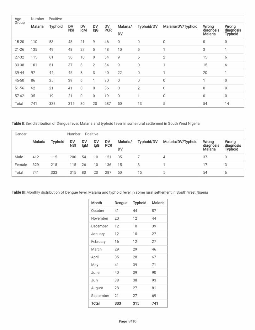
ResultsA total of 1074 blood samples were collected from different health facilities in different rural locations in South West Nigeria of which 741(69%) blood samples were positive for malaria parasites, 333 (31%) were positive to typhoid fever while from the malaria and typhoidpositive samples, 315 (29.4%) were positive to dengue using the NS1 protein (Fig. 1). From DV NS1 results, 80 (25.4%) of the total sampleswere positive to anti DV IgM and 20 (6.3%) positive to anti DV IgG while 287 (91.1%) samples were positive for DV by RT-PCR (Table 1)con�rmation. Of the total number of 741 con�rmed malaria cases, 50 (6.7%) also had DV NSI (co-infection) {t = 5.8540) and also con�rmedby RT-PCR, There was 13 (3.9%) typhoid- DV co-infection (t = 9.3611) from the 333 samples positive to typhoid fever, while malaria/typhoidco-infection with DV had 5 (0.5%) samples positive (p = 0.9296 and p-0.9432 respectively). A total of 54 DV positive samples were wronglydiagnosed as malaria parasites while 14 (1.3%) samples positive for DV weremisdiagnosed as typhoid fever (χ = 86.877, p = 0.0000001),showing an association between all misdiagnosed samples.Age group distribution shows that age groups 21–26, 27–32 and 15–20 havethe highest number of all samples positive for all infections (Fig. 1). Age group 39–44 years had the highest number of DV malaria co-infection, however, age group 27–32 and 33–38 were higher for DV typhoid co-infection while 27–32 and 33–38 years bracket has thehighest positive cases for the 3 co-infection. These age brackets also has the highest number of DV misdiagnosis with 15 cases respectivelyafter age group 39–44 which had 20 cases. Age group 27–32 and 33–38 had the highest misdiagnosed samples for DV with 6 samplesrespectively. Figure 2 shows sex distribution to malaria, typhoid, DV and all their co-infections. Males were more infected with malariaparasites 412 (55.6%) while females were 329 (44.4%). For typhoid 218 (65.5%) were females while males 115 (34.5%) were positive. DVNS1 had 200 (63.5%) positive males and 115 (36.5%) positive samples were females. Other DV parameters also followed the same trend.Monthly distribution shows that the months of October, September, August, July, and June had the highest number of positive cases tomalaria and DV, samples positive to typhoid fever did not show a particular distribution pattern (Table 3). November and June has thehighest number of co-infection cases with Malaria/DV(t= -0.639, p = 0.537) as compared to others, while September has the highest numberof co-infection for the 3 parameters (t = 0.905, p = 0.387) and October and March had the highest number of typhoid and DV co-infection (t = 1.008, p = 0.537) (Fig. 1). The concentration of the NS1 protein detected by ELISA technique as analyzed by the myassays microplatesoftware was moderate andevenly distributed across all samples positive for DV NS1 (mean = 6.25mM) irrespective of sex, age or month ofsample collection (Fig. II), the DV anti IgM concentration was also (mean = 41.94) with men having higher concentration and age distributionshows that adults of age groups 33–38, 37–44 and 27–32 having higher concentration (Fig. III).The Anti DV IgG also vary like the IgM with amean of 51.72 (Fig. IV).
DiscussionThis study is a cross-sectional and comparative analysis of DV, malaria and typhoid fever in co-infection in rural settings of South WestNigeria, with misdiagnosis of dengue fever for malaria or typhoid fever. DV fever co-infection with malaria and typhoid fevers was analyzedusing 4 different parameters which included NS1, IgM, IgG antibodies and later con�rmed with RT-PCR. The NS1 is �rst protein to be

Page 5/10
produced in the infection, which ensures the �rst early window is not missed in the DV infection, the IgM is the �rst antibody to be producedin response to an infection while the IgG will remain even in convalescence. A combination of all these ensures that no case is missed duringthe period of the study. The RT-PCR is used to validate all the results from the serological analysis and that any DV missed by ELISAtechnique will be captured by RT-PCR and at the same time quantifying the antigen in each sample. Of the total 315 DV NS1 positivesamples, 287 samples were RT-PCR positive, thereby con�rming that 28 samples though came in contact with DV but has not yet establishedenough antigen to be detected by PCR, which thereby con�rms the usefulness of the NS1 protein in detecting dengue infection. This studygenerally reveals a high incidence of Malaria/DV co-infection as well as Malaria typhoid but the trio co-infection was not signi�cant. A studyof prevalence of concurrent dengue and malaria was reported by Charrel et al.[1] to have 0% prevalence in Netherlands, which oppose theresult from this study. This reason for this may be due to geographical variation and high level of mosquito control in Netherlands, which islacking in Nigeria. In addition, the mosquito vector for the two diseases have different habitats, malaria mosquito vector has its habitat in theforest [16] while Dengue mosquito vector main habitat is in the city [19]. Hence, overlapping of the habitat may not be easily available inNetherlands as is the case in Nigeria. Signi�cant low Malaria-Dengue concurrent rate can be expected [20]. Dengue-Malaria co-infection hasbeen referred to as uncommon phenomenon in the temperate regions. However, reports showed that the highest dengue-Malaria reportedwas 23.21% found in Pakistan while as low as 0.01% was found in Senegal [21]. Another study conducted in Brazil recorded 2.8% [7]. Thereport from Pakistan was similar to that from this study which is as a result of similar geographic conditions of both regions. The Senegalreport also opposed results from this study and a major factor responsible for that is due to the time of the year and city the Senegalsampling was done, since both countries are in sub-Saharan Africa as indicated by the 2.84% Dengue virus and malarial concurrent infectionwithin Ilorin metropolis In Nigeria [22]. Okoror et al. [23] also reported a prevalence of a high prevalence of DV infection (57.5%) in selectedrural communities in South West Nigeria
since all the hospitals do not o�cially screen for DV, and the differential diagnois DV, malaria and typhoid despite similar clinicalpresentations makes misdiagnosis not unusual. Therefore, co-infections may give rise to wrong diagnosis especially in areas where theclinician depends on empirical treatment. Moreover, the treatment regimens for these coinfections are not the same as those for mono-infections. Hence, a delay in implementing the appropriate treatment regimen for these different infections due to poor diagnosis can resultin fatal consequences in compromising the patient’s health.
Age group distribution revealed that age group 21–26 through to 39–40 have the highest prevalence of Malaria/DV co-infection and theywere statistically signi�cant (p = 0.9296: CI = 0.05). These age group also had the highest prevalence for malaria alone as well as for typhoid.The reason for this may be attributed to active (working class) which are found in this age range and their outdoor engagement on a day today routine, making contact with mosquito unavoidable [24]. The study is also supported by Okoror et al [25] who reported the endemicnature of typhoid in Nigeria. In addition to this, sex distribution shows that more males were more infected with Malaria as compared tofemales and also Malaria DV co-infection was more in males compared to females, as well as typhoid DV coinfection. The overall infectionalso show similar pattern of sexual distribution. This was also reported by Dhanya et al. [26], though more males have been shown to havemore outdoor activities than their female counterparts.
The distribution of Malaria/DV and Typhoid monthly, show that the months of November and June had the highest Malaria/DV co-infectiondistribution while October and March showed the highest co-infection distribution for typhoid and DV with signi�cant statistical differencefrom other months. This coincides with period of high rainfall and progression to dry season for malaria/DV infection and the period of highbreed of mosquitoes. For typhoid/DV co-infection the months of highest prevalence coincides with period of high rainfall as these are theperiod of lowest hygiene due to high �ooding which increases incidence of typhoid fever, and high proliferation of mosquitoes leading tohigh incidence of DV. Some authors have also reported both seasonal and monthly distribution of Malaria/DV co-infection. Dhanya et al. [26]in an India study reported a monthly distribution of Malaria and Dengue co-infection and reported their highest prevalence was between Julyand December which is in agreement with some of the months reported in this study. Savargaonkail et al.[24] also reported similardistribution in India. Total samples for malaria, typhoid and DV also follow similar pattern of distribution. Other factors that may havein�uenced this distribution includes rainfall �uctuation, humidity, and temperature [27, 28].
The concentration of NS1 protein, which was evenly distributed across the study population further goes to con�rm the high prevalence ofDV in the population and their misdiagnosis for malaria and typhoid fever. This is more importantly noticed with the high anti DV IgM andIgG which however was distributed along sex and age. The high concentration noticed in males goes to establish the earlier claims thatmales are more involved with out-door activities in the study population and therefore more exposed to the vector.
ConclusionIn conclusion this research showed that lots of dengue positive samples are misdiagnosed for malaria and typhoid fevers and as suchdengue testing should be incorporated to national testing scheme especially in areas where high dengue infection has been reported like in

Page 6/10
this study population so as not to compromise the patients’ health. The importance of this paper can not be undermined because of thepublic health implication of undermining or misdiagnosis of dengue for malaria or typhoid, a situation which could endanger the health ofthe population because of the like progression of dengue fever to DSS leading to death.
List Of AbbreviationsNS1, DV, DV IgG, DV IgM, RT-QPCR, DHF, DSS, ELISA, EDTA, RDT, RNA, PCR, MAXIMA, SYBR, ROX, RT-PRCR, DENV, UDG, CT
DeclarationsCon�ict of Interest
We declare no con�ict of interest. Author did not receive any form of �nancial assistance whatsoever.
Ethical Clearance
Ethical approval was granted by Ethical Review committee of the Joseph Ayo Babalola University, Ikeji-Arakeji, Nigeria. All participants in thisstudy agreed to participate through �lling of questionnaire.
Consent for Publication
Not applicable
Competing Interest
We declare no competing interest.
Funding
Authors received no funding from anywhere, but study was carried out from their personal purse.
Acknowledgement
We acknowledge the contribution of all our project students who were involved in sample collection during this study.
Availability of Data Materials
Not applicable
Author contribution
LEO conceived the study and was involved from writing of proposal through ethical defense and sample collection, laboratory analysis andto writing up of the paper. EOB was involved in supervision of laboratory analysis and writing of the �nal paper. OMU was involved in samplecollection and laboratory analysis, EOA participated in sample collection and laboratory analysis, SKO and BO were involved in �nal dataanalysis.
References1. Charrel RN, Brouqui P, Foucault C, De Lamballerie X. Concurrent dengue and malaria. Emerg Infect Dis.2005;11(7):1153-1154. [PMC free
article] [PubMed]
2. Orhue, Okoror LE. and IB Enweani. Co-occurrence of malaria and typhoid fever in patients attending clinics in Irrua Specialist HospitalIrrua, Nigeria. Journal of Science Engineering and Technology 2003; (1) 4745-4752.
3. Elpeboin L, Hanf M, Dussart P, Ouar-Epelboin S, Djossou F, Nacher M. Is Dengue and malaria co-infection more severe than singleinfections? A retrospective matched-pair study in French Guiana. Malar J.2012; 11:142.
4. https://www.euro.who.int/__data/assets/pdf_�le/0006/246165/Fact-sheet-Dengue-Eng.pdf
5. Bhatt S, Gething PW, Brady OJ, Messina JP, Farlow AW, Moyes CL. The global distribution and burden of Dengue. 2013;496:504-7.doi:10.1038/nature 12060.

Page 7/10
�. Nimmannitya S. Dengue haemorrhagic fever: current issues and future research. Asian- Oceanian Journal of Paediatrics and ChildHealth. 2002; 1: 1-21.
7. Magalhaes BM, Siqueira AM, Alexandra MA. vivax malaria and dengue fever coinfection: a cross- sectional study in the BrazililianAmazon. PLoS NegL Trop Dis.2014; 8:e3239.10.1371/ journal.pntd.0003239.
�. Kumar V, Nagpai, BN, Veena S, Anna G, Sanjeev K, Paul R. Comparison of egypti breeding in localities of different socio-economic groupsof Delhi; India.; Int. J. of Mosquito Res.2015; 2(3): 83-88.
9. Clyde K, Kyle JL, Harris E. Recent advances in deciphering viral and host determinants of dengue virus replication and pathogenesis. JVirol. 2006;80(23):11418-11431. doi:10.1128/JVI.01257-06
10. Wiwanitkit V “Unusual mode of transmission of dengue”. of infect in Developing Countries (2010); 49(1):51-4.
11. Barber BE, Rajahram GS, Grigg MJ, William T, Anstey NM. World malaria report: time to acknowledge Plasmodium knowlesi malaria.Malar J. 2017;16:135.doi:10.1186/s112936-017-1787-y.
12. Nadjm B, Behrens RH. “Malaria: An update for physicians”. Infect. DisClinics of NorthAmerica2012; 26(2): 243-59.doi:10.1016/j.idc.2012.03.010. PMID 22632637.
13. Baird JK. “Evidence and implications of mortality associated with acute Plasmodium vivax malaria”. Microbiol. Rev.2013; 26(1):36-57,doi10.1128/CMR.00074-12. PMC 3553673. PMID 23297258.
14. Alaribe, AAA Ejezie GC and Ezedinachi, NU. The prevalence of Salmonella antibody among malaria patients in Calabar. of Med. Lab.Sci.2013; 7: 34 – 41. 1998. 7.
15. Nasir S, Shoeb M, Abdul H, Anis AC, Farah D and Shama P. Global prevalence and distribution of coinfection of malaria, dengue andchikungunya: a systemic review. BMCPub.Health2018; 18:710 doi:10.1186/s12889-018-018-5626-z.
1�. Obsomer V, Defourny P, Coosemans M. The Anopheles dirus complex: spatial distribution and environment drivers. Malar J 2007; 6:26.[PMC free article] [PubMed].
17. Wilairatana P, Tangpukdee N and Krudsood SMisdiagnosis of Malaria in Malaria-Dengue Endemic Area. TropMed&Surgery 2014; 2:14.DOI: 10.4172/2329-9088.1000e119
1�. Okoror L.E., Umolu P.I., Orhue P, Omigie O.O. and R. Akpe. Widal test as adapted to the use of microtitre plate. Discovery and Innovation2005; 17 (1/2) 45-49.
19. Alm E, Lesko B, Lindegren G, Ahlm C, So¨derholm S , Falk KI, Lagerqvist N, Universal Single-Probe RT-PCR Assay for Diagnosis of DengueVirus Infections. PLOs Neglected Trop. Dis: 2014;8(12): e3416. doi:10.1371/journal.pntd.0003416
20. Ward DI. A case of fatal Plasmodium falciprum malaria complicated by acute dengue fever in East Timor. Am J TropMed2006;75(1):182-185. [PubMed].
21. Salam N, Mustafa S, Ha�z A, Chaudhary AA, Deeba F, Parveen S. Global Prevalence and distribution of coinfection of malaria, dengueand chikungunya: a systemic review. BMCPub. Health 2018; 18:71010.1186/s12889-018-5626-z.
22. Kolawole OM, Seriki AA, Irekola AA, Bello KE, Adeyemi OO. Dengue virus and malaria concurrent infectionamong febrile subjects withinIlorin metropolis, Nigeria. JMed. Virol.2017; 89(8):1347-1353
23. Okoror, L E, Osanyinlusi S A, Ukhureigbe O M, Udenze, D O.Prevalence of Dengue Virus Antibody among Residents of a Rural Communityin Southwestern, Nigeria. J. of Microbiol, 2019 33(2): 4512 – 4517
24. Savargaonkai D, Sinha S, Srivastava B, Nagpal B.N, Sinha A, Shamim A, Das R, Pandi V, Anvikar AR, Valecha N. An EpidemiologicalStudy of Dengue and its Coinfections in Delhi. Inter. J. of Infect. Dis. 74(2018): 41-46.
25. Okoror LE, Osanyinlusi SA, Ukhureigbw OM and Udenze DO. Prevalence of Dengue Virus among residents of a rural community inSouthwestern Nigeria. Nigerian Journal of Microbiology 2019; 33 (2) : 4512-4517
2�. Dhanya P. R, Shilpa K, Vivek H. A comparative study on dengue and malaria infections and analysis on the prevalence of co-infectionfrom a rural tertiary care hospital. Int J ResMedSci. 2018; 6(6):2111-2115
27. Wongkoon S, Jaroensutasinee M, Jaroensutasinee, K; Distribution, seasonal variation and Dengue transmission prediction in Sisakett,Thailand, Indian J. of Med. Res, 2013;138(3):347-53.
2�. Pakhare A, Yogesh S, Ankur J, Rashmi J, Arun K, Rajnish J. A Study of Spatial and Meteorological determinants of dengue outbreak inBhopal City. J Vector Borne Dis 2016;53 (3):225-233
TablesTable I: Age distribution of Dengue fever, Malaria and typhoid fever in some rural settlement in South West Nigeria
Page 8/10
AgeGroup
Number Positive
Malaria Typhoid DVNSI
DVIgM
DVIgG
DVPCR
Malaria/
DV
Typhoid/DV Malaria/DV/Typhoid WrongdiagnosisMalaria
WrongdiagnosisTyphoid
15-20 110 53 48 21 9 46 0 0 0 0 0
21-26 135 49 48 27 5 48 10 5 1 3 1
27-32 115 61 36 10 0 34 9 5 2 15 6
33-38 101 61 37 8 2 34 9 0 1 15 6
39-44 97 44 45 8 3 40 22 0 1 20 1
45-50 86 25 39 6 1 30 0 0 0 1 0
51-56 62 21 41 0 0 36 0 2 0 0 0
57-62 35 19 21 0 0 19 0 1 0 0 0
Total 741 333 315 80 20 287 50 13 5 54 14
Table II: Sex distribution of Dengue fever, Malaria and typhoid fever in some rural settlement in South West Nigeria
Gender Number Positive
Malaria Typhoid DVNSI
DVIgM
DVIgG
DVPCR
Malaria/
DV
Typhoid/DV Malaria/DV/Typhoid WrongdiagnosisMalaria
WrongdiagnosisTyphoid
Male 412 115 200 54 10 151 35 7 4 37 3
Female 329 218 115 26 10 136 15 8 1 17 3
Total 741 333 315 80 20 287 50 15 5 54 6
Table III: Monthly distribution of Dengue fever, Malaria and typhoid fever in some rural settlement in South West Nigeria
Month Dengue Typhoid Malaria
October 41 44 87
November 20 12 44
December 12 10 39
January 12 10 27
February 16 12 27
March 29 29 46
April 35 28 67
May 41 39 71
June 40 39 90
July 38 38 93
August 28 27 81
September 21 27 69
Total 333 315 741

Page 9/10
Figures
Figure 1
Monthly Distribution of Co-occurrence of Dengue Fever with Malaria and Tyhpoid Fever in Rura Communities in Southwest Nigeria
Figure 2
DV NS1 concentration of samples tested for malaria, typhoid in rural areas of South West Nigeria

Page 10/10
Figure 3
Anti DV IgM concentration of samples tested for malaria, typhoid and testing for positive to Dengue NS1 in rural areas of South West Nigeria
Figure 4
Anti DV IgG concentration of samples tested for malaria, typhoid and testing for positive to Dengue NS1 in rural areas of South West Nigeria