Did Romanization impact Gallic pig morphology? New insights from molar geometric morphometrics
Upload
khangminh22Category
view
0download
0
1
Molar-incisor hypomineralisation: narrative review on etiology,
epidemiology, diagnostics and treatment decision
Accepted for Publication March 18th, 2021
Joëlle A. Dulla, Hendrik Meyer-Lückel
Department of Restorative, Preventive, and Pediatric Dentistry, School of Dental
Medicine, University of Bern, Switzerland
KEYWORDS
Molar-incisor hypomineralisation (MIH), hypersensitivity, post-eruptive enamel
breakdown, differential diagnosis
CORRESPONDENCE
Joëlle A. Dulla, Dr. med. dent.
Klinik für Zahnerhaltung, Präventiv- und Kinderzahnmedizin
Zahnmedizinische Kliniken der Universität Bern
Freiburgstrasse 7
CH-3010 Bern, Switzerland
Tel.: +41316322570

2
SUMMARY
Molar-incisor hypomineralisation (MIH) is clinically defined as demarcated structural
enamel defects affecting at least one first permanent molar with or without the
involvement of incisors. It is foremost a qualitative developmental defect of systemic
origin. The prevalence for MIH is estimated at 12.9% with significant differences
between countries. Its etiology and pathogenesis are still not completely understood.
Several environmental and medical causes have been suggested to alter enamel
maturation. The hypomineralised enamel may collapse shortly after eruption and as a
consequence caries lesions seem more likely to develop. Besides cavitation,
hypersensitivity and/or pain are the hallmarks of clinical symptoms. Both are
associated with increased dental anxiety and fear of children suffering from MIH.
Consequently, patients’ care and management are challenging and necessitates a
large range of non-, micro- and invasive strategies. MIH might be mixed up with three
different other types of developmental defects in the enamel: fluorosis, enamel
hypoplasia, and amelogenesis imperfecta. Careful diagnostic differentiation should be
made before starting any dental treatment. A recent published classification system
links the severity of the lesion to a treatment need index. This index is based on four
values regarding two key symptoms: hypersensitivity and post-eruptiv enamel
breakdown (PEB). Without PEB sealing is strongly recommended in order to prevent
caries. For hypersensitive teeth as well as those with PEB use of glass ionomer cement
as an intermediate cover, but mainly composite resins are materials of choice. For
improvement of aesthetically compromised MIH-incisors, the resin infiltration
technique has been proposed.

3
Introduction
Developmental defects in the enamel (DDE) derive from disturbances in hard tissue
matrices formation and/or mineralization during odontogenesis. These defects can be
localized or appear more widespread, affecting single/multiple teeth or groups of teeth
(COMMISSION ON ORAL HEALTH, RESEARCH AND EPIDEMIOLOGY, FDI 1992). Examples of
DDE are fluorosis (FS), enamel hypoplasia (EH) and amelogenesis imperfecta (AI).
Another entity of DDE is the so called “molar-incisor hypomineralisation” (MIH).
Since the early 1970’s dentists have reported a developmental defect primarily located
in first molars and incisors in the permanent dentition. Early denominations referred to
this clinical condition in a descriptive manner for “which is not”:
1. non-endemic stained enamel (JACKSON 1961),
2. idiopathic hypomineralisation of the enamel of the first molars (KOCH ET AL.
1987),
3. hypomineralisation of the permanent first molars not caused by fluoride
(LEPPÄNIEMI ET AL. 2001)
4. cheese molars (KREULEN ET AL. 1995).
This reflected a poor understanding of its etiology that continues to this date.
WEERHEIJM ET AL. baptized this pathology as MIH in 2001 and was adopted as the
official denomination at the sixth annual conference of the European Academy of
Paediatric Dentistry (EAPD) in 2003 (WEERHEIJM ET AL. 2001). MIH is defined as
developmental (systemic) qualitative enamel defects that are present on one or more
first permanent molars (FPMs), each possibly with different degrees of severity. The
(central) permanent incisors might be affected additionally. If incisors are affected, at
least one first molar must also show enamel hypomineralisation to confirm the
diagnosis MIH.

4
The aim of this review is to summarize the current knowledge on the etiology,
prevalence and diagnostics of MIH as well as provide guidance on treatment decision
as well as discuss evidence of non- and micro-invasive interventions. A second review
by our group will focus on the choice of invasive treatment options.
Prevalence and incidence
Nowadays, MIH is recognized as a global dental problem. In spite of the EAPD criteria
cited above, cross-comparisons of the results from various epidemiological studies
have been difficult due to the use of various indices and criteria, examination variability,
methods of recording and varying age groups (JÄLEVIK 2010). Prevalence is defined by
how often a condition is present in a population, and incidence by how many new cases
occur each year. The reported prevalence of MIH in children and adolescents varies
significantly between studies varying between 2 and 40% (JÄLEVIK 2010). A recent
systematic review and meta-regression analysis (SCHWENDICKE ET AL. 2018) estimated
a global mean (95% CI) prevalence for MIH of 12.9% (11.7–14.3%) with significant
differences between countries. The highest numbers of prevalent cases were found in
high-income and heavily populated countries. Another meta-analysis (ZHAO ET AL.
2018) estimated the prevalence of MIH around 14% globally with no statistical
difference between sexes, but <10-year-olds had a higher prevalence than older
individuals (15% vs 12%). It was also noted that MIH was particularly high in some
regions such as South America (18%) and Spain (21%). For Switzerland no
representative data have been reported so far.
A study on the distribution and severity of MIH affected molars in four areas in Germany
revealed that the majority of children with MIH showed more than one affected molar
(only one affected molar in 39.2%, two affected molars in 33.5%, three affected molars

5
in 12.0% and four affected molars in 15.3%) (PETROU 2012). About half showed
hypomineralisation in FPMs without additionally affected incisors. Twelve percent of
the children with MIH showed hypomineralised defects in at least one of the second
primary molars. Furthermore, FPMs were usually more often and more severely
affected than permanent incisors. Upper permanent incisors were usually more often
affected than lower permanent incisors and, if lower incisors were affected, in most
cases hypomineralisation in upper permanent incisors as well as in FPMs were
documented (PETROU 2012).
This was corroborated by other studies showing that the FPMs and incisors are often
affected, although there can be considerable differences within a dentition. MIH has
also been observed in other teeth of the permanent dentition (second molar, second
premolar, canine) as well as in second primary molars and also primary canines
(ELFRINK ET AL. 2012; FUCHS 2009). MIH is supposed to affect 878 million individuals,
with a global number of incident cases estimated at 16.0 million people in 2016. It is,
therefore, imperative to develop appropriate dental healthcare strategies to treat MIH
and to identify its etiology in order to prevent.
Etiology and pathogenesis
MIH has been reported to have a negative impact on children’s quality of life and socio-
psychological status (DANTAS-NETA ET AL. 2016). Affected teeth are in higher need for
dental treatment, especially those with post-eruptive enamel breakdown (PEB). As a
consequence, caries lesions develop more easily, leading finally to pulpal inflammation
along with hypersensitivity or pain. Thus, it is of great importance to identify the etiology
of MIH and to understand its pathogenesis. Despite an augmented interest in MIH and

6
much published research on the subject, its etiology remains, to this day, not
completely understood (CROMBIE ET AL. 2009; ALALUUSUA 2010; SILVA ET AL. 2016).
Environmental causes
MIH is considered to establish due to an impaired calcium and phosphate incorporation
during enamel matrix formation and enamel maturation. Ameloblasts are sensitive to
insults, both indirect and direct. Even the smallest changes in the environment of the
ameloblasts may irreversibly disrupt the formation of the enamel matrix and the
maturation of the enamel. Another key factor in understanding the etiology of MIH is
the chronology of tooth mineralization. The mineralization of enamel in a first
permanent molar starts just before birth and is completed in the first year of life (REID
& DEAN 2006). Therefore, it seems logical that any possible cause must have occurred
during the period from just before birth until the first year of life (Fig. 1, SCHROEDER
2000, DULLA ET AL. 2018). Research on the etiology of MIH have compiled a long list of
candidates but failed to prove causality due to low numbers of prospective birth-cohort
studies. Nonetheless, ongoing studies have elucidated some risk factors (e.g. “LISA”
and “GINIplus”; FLEXEDER et al. 2020). “LISA” (Influence of life-style factors on the
development of the immune system and allergies in East and West Germany) and
“GINIplus" (German Infant Study on the influence of Nutrition Intervention PLUS
environmental and genetic influences on allergy development) are two populated-
based German birth cohorts. They aim to describe the natural course of chronic
diseases and intermediate phenotypes in childhood and its determinants and to identify
potential genetic effect modifications.
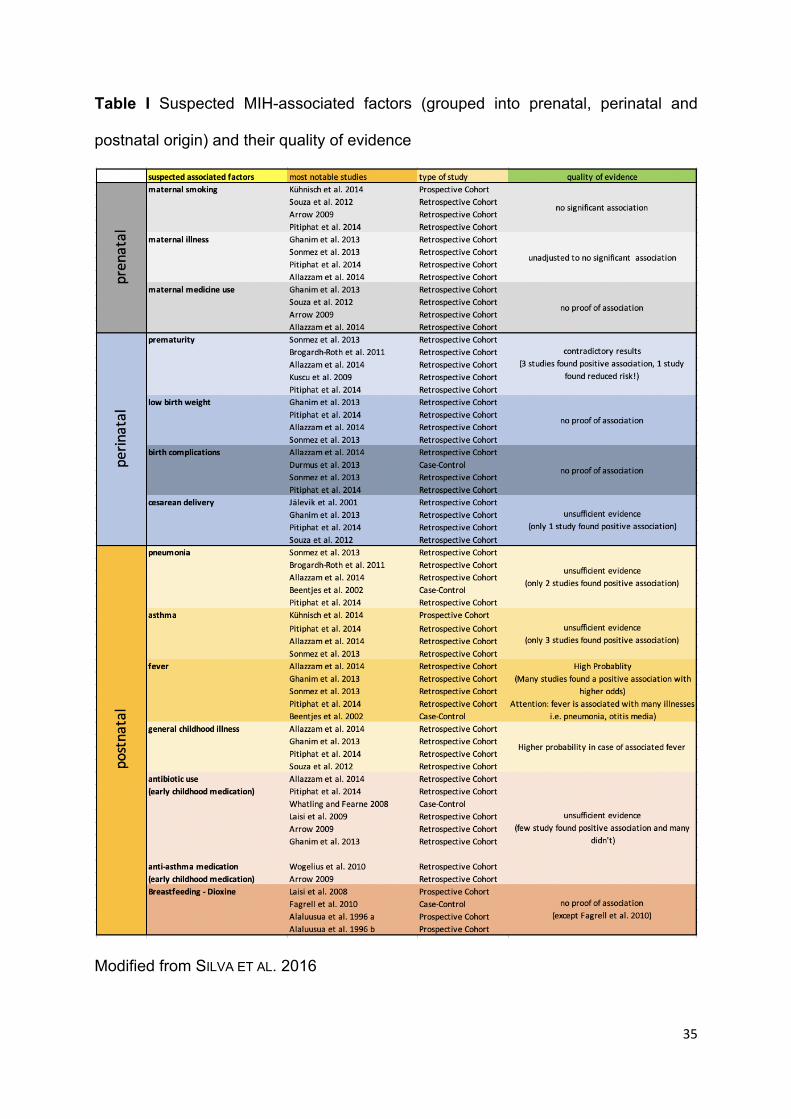
Potential environmental causes can be grouped into either being of prenatal, perinatal
or postnatal origin (Table I). As indicated in Table I there was little evidence of an

7
association between the most frequently investigated prenatal factors (maternal
smoking, illness, medication) and MIH as well as perinatal factors (prematurity, low
birth weight, cesarean delivery, birth complications) and MIH. A consistent finding is
that early childhood illness, in particular pyrexia, appears to be associated with MIH.
Genetic influence
The amelogenesis phase has been shown to be modulated by genes (FINCHAM ET AL.
1999) and the size, shape, structure and composition of the enamel seem to be
influenced by genetic variations. The potential role of genetics or epigenetics in
association with MIH has been discussed (VIEIRA & MANTON 2019; HOČEVAR ET AL.
2020). Several genes related to MIH, such as Enam, Ameloblastin, Amelogenin, Bone
morphogenetic protein 2 have been investigated (BUSSANELI ET AL. 2019; JEREMIAS ET
AL. 2013). In addition to these single gene effects, gene-to-gene interactions may also
play a role in MIH (PANG ET AL. 2020). However, data based on twin studies (TEIXEIRA
ET AL. 2018) only showed tendencies of an influence of genetics on MIH prevalence,
while higher family income and gestational bleeding were strongly positively
associated with MIH. Genetic variability may influence the etiology, but seems not to
act as the primary cause of MIH.

8
Some intriguing observations
MIH can affect one sibling and not the other(s) and teeth forming at the same period
can be affected to varying degrees or not at all. Histological studies reported that MIH
lesions extend through the full thickness of enamel, affecting mainly the coronal and
not the cervical enamel, and often the buccal surface of the tooth (CROMBIE ET AL. 2013;
FAGRELL ET AL. 2013; GAMBETTA-TESSINI ET AL. 2017). VIEIRA & MANTON attempted to
address these variables in the clinical presentation of MIH (VIEIRA & MANTON 2019).
According to these authors, the reason why only one side of a bilateral structure is
affected likely involves differential gene expression between the left and right despite
the dentition being mirrored between one side and the other. As for the disturbances
of specific areas, and the multiple degrees of the severity, they postulated that this
resulting anatomical appearance is a combination of random microenvironment
influences (i.e., pressure from surrounding liquid within the connective tissues
surrounding the enamel organ) and/or genetic variants (i.e., differential levels of
expression at the cellular level and the directionality of molecular signaling).

9
Diagnosis
General diagnostic criteria and a simple classification system were set by the EAPD to
facilitate the diagnosis of MIH (WEERHEIJM ET AL. 2003). It can be summarized by the
following:
• The visual aspect of the lesion is opaque, clearly demarcated from the
healthy enamel, varies in colour (white, yellow, brownish) and size. The
darker opacity indicates more hypomineralised (softer) enamel.
• The enamel thickness is normal, but its breakdown appears after the
eruption of the affected tooth (PEB).
• When restorations are present, these extend in most cases to the buccal
and palatal/lingual surfaces with an opacity at the margin of the restoration.
• For incisors, these restorations are not related to a history of trauma.
• A missing FPM, in an otherwise sound dentition, can be an indication of a
history of MIH.
• In some cases, eruption difficulties of FPMs due to enamel roughness have
been proposed (ALMUALLEM & BUSUTTIL-NAUDI 2018).
Several descriptive classifications have been proposed; e.g. LEPPÄNIEMI ET AL. 2001
rated the severity of MIH within three categories:
1. Mild: opacities without PEB;
2. Moderate: opacities with PEB limited to enamel;
3. Severe: PEB with dentin involvement, atypical restorations and tooth
extraction due to MIH.
Another simple classification system, also based on the severity of MIH, proposed
distinction between mild and severe cases (LYGIDAKIS ET AL. 2010):
10
1. In mild cases, the demarcated opacities do not exhibit a PEB, but can present
occasional sensitivity to external stimulus with less aesthetic concerns.
2. In severe cases, the demarcated enamel is associated with PEB,
hypersensitivity (HS) and high aesthetic demands.
STEFFEN ET AL. in an attempt to standardize MIH diagnostic criteria and treatment
needs, conceived a classification system that links the severity of the lesion to a
treatment need index (TNI) (STEFFEN ET AL. 2017). This index is based on four values
(Fig. 2) regarding two key symptoms that are considered the most important ones with
respect to MIH: HS and PEB. The highest value is recorded for each sextant by the
use of good light and drying with an air syringe.
Teeth affected by MIH show various characteristics (Figs. 3-8). HS is a common
symptom in MIH teeth that might impair oral hygiene, limit dietary habits, cause chronic
pain and trigger dental anxiety. Its intensity depends on the severity of the lesion. A
recent study (RAPOSO ET AL. 2019) concluded that mild cases of MIH are associated
with HS of a low intensity, while severe cases showed more frequently higher degrees
of HS. Severely hypomineralised teeth are at higher risk of developing caries, which
could increase HS considerably. The cause of this HS appears to be chronic pulp
inflammation due to repeated triggers, whether thermal, mechanical, or bacterial
(FAGRELL ET AL. 2008). PEBs occur due to the severe porosity of the hypomineralised
opaque areas that fracture when subjected to masticatory forces, resulting in
unprotected dentin being more prone to external triggers (GARG ET AL. 2012).
Interestingly, whitish discolored MIH teeth seem to be at ca. 33% lower risk for PEB
than yellowish/brownish ones (CABRAL ET AL. 2016).

11
Clinical examination
With the complete eruption of all first molars and incisors at ca. eight years of age
hypomineralised enamel of relevant teeth can be preferably detected and MIH be
diagnosed (LYGIDAKIS ET AL. 2010). Nonetheless, MIH might be diagnosed during the
eruption of the FPM. An early diagnosis may limit the degree and size of PEB and the
high risk of subsequent HS and dental caries (GARG ET AL. 2012).
FPMs are first screened for MIH and then for caries. Teeth should be cleaned with a
toothbrush and fluoride toothpaste and examined in wet condition using a mirror and
probe. The tooth surface can also be gently cleaned with a cotton roll but should
preferably not be dried. Intraoral photography is a great tool to better examine and
assess the damaged tooth on a computer monitor (CHEN ET AL. 2013). Subsequently,
the tooth is dried using air jet or, if not possible with cotton rolls, to examine for caries
and to evaluate possible HS.
Differential diagnosis
MIH might be mixed up with three different other types of DDE: FS, EH, and AI. It can
be helpful that MIH is the most prevalent type. It is a well-demarcated qualitative
“chalky” defect (contrary to EH), non-symmetrical (contrary to FS, AI), limited to one or
more FPM (contrary to AI, FS), with or without central permanent incisors implication
(contrary to FS, AI), caries prone (contrary to FS), generally but not always
hypersensitive (contrary to FS), with no history of trauma on the affected tooth
(WEERHEIJM 2004; COMMISSION ON ORAL HEALTH, RESEARCH AND EPIDEMIOLOGY, FDI
1992; ELCOCK ET AL. 2006; CRAWFORD ET AL. 2007) (Table II). Furthermore, traumatic
injury as well as prolonged periapical inflammation process of a primary tooth may
12
affect the development or maturation of the permanent successor and lead to a so-
called Turner’s tooth / Turner’s hypoplastic tooth, which is one example for EH.
Pain control & therapy
For the affected and suffering quite young child, visiting a dentist is often accompanied
with a great portion of anxiety and reluctance. In order to rebuild the bridge of trust
between the child and the dentist, a painless first examination is absolutely essential.
Thermal stimuli such as air syringe, cold water, or cold instruments should be avoided.
Any subsequent dental treatment should be performed under a very effective pain
control protocol (JÄLEVIK & KLINGBERG 2002).
Pain is a subjective experience especially when it overlaps with anxiety and fear. The
fear of having pain can be overwhelming for a child especially in case of previous
traumatic dental experiences. A prerequisite for any pain control protocol to be
effective is to ensure a proper emotional management of the suffering child otherwise,
the protocol is doomed to failure. This behavioral and emotional management is the
standard in pediatric dentistry (MCNEIL ET AL. 2006). A combination of three techniques
i.e. analgesic, anesthetic and sedation is often necessary in pain management of MIH
teeth.
Systemic analgesic premedication
The use of an analgesic premedication can be helpful in the treatment of
hypersensitive MIH molars. The choice of the drug can be paracetamol or non-steroidal
anti-inflammatory drugs NSAID (Ibuprofen or Metamizole). STEFFEN & VAN WAES
described a treatment protocol based on patients suffering from chronic back pain. The
protocol recommends the ingestion of a very high but short-term dosage (24-48h). The

13
anti-inflammatory effect of NSAIDs is desirable especially in chronically inflamed MIH
teeth. In order to influence chronic pain, medication should be taken > 24 hours before
the dental treatment. The four doses are distributed as follows: > 24h, 12h, 6-8h before
dental treatment and the last dose shortly before the procedure (STEFFEN & VAN WAES
2011).
Anesthesia
Treatment of MIH affected teeth in young patients is challenging due to chronic pulpal
inflammation caused by porous enamel and exposed dentin. Failure in achieving
profound and sufficient anesthesia may lead to behavior problems and affect the
quality of restorations. The clinician might reach the maximum anesthetic dose (limited
by weight and age) and the tooth remains hypersensitive. Type and dose of the
anesthetic are not as important as the accessory techniques employed to achieve
anesthesia (DISCEPOLO & BAKER 2011). For lower FPM, inferior alveolar nerve block
adjunct to buccal infiltration is commonly used. Periodontal ligament injections can help
in the establishment of anesthesia although its safety is constantly debated.
Intraosseous injection technique used in endodontics, is also proven to be effective
and provides profound anesthesia of long duration (60 minutes or greater) (NUSSTEIN
ET AL. 2005). Crestal intraosseous local anesthesia by the use of a computer-assisted
injection system is an effective and safe technique to achieve profound anesthesia in
MIH-affected hypersensitive teeth. This technique could be beneficial when treating an
MIH tooth with a pulpitis (DIXIT & JOSHI 2018; DISCEPOLO & BAKER 2011; FOUAD & LEVIN
2006).

14
Sedation
While anesthesia helps to reduce pain, sedation helps tremendously in removing the
emotional context of fear and anxiety accompanying the dental treatment. Drug
sedation can be used especially in very young children. However, whenever the child
maintains nasal breathing, inhalation sedation (nitrous oxide / oxygen mixture) is the
medication of choice especially in treating MIH cases (ESCH 2009; STEFFEN &
LANGERWEGER 2018; STEFFEN 2018). If the child shows no cooperation at all, general
anesthesia remains the only option.
Treatment decision
Fortunately, not all MIH affected teeth need immediate treatment. In any case, an
intensive prophylactic protocol should be administered as early as MIH is diagnosed.
The purpose is to delay enamel breakdown and prevent the development of dental
caries. Based on the MIH-TNI, BEKES ET AL. have developed two therapy schemes for
patients with either low or high caries risk. Prophylaxis is based on a self-applied
“home” and a professional “in office” part. The former comprises mainly the home-use
of fluoride toothpaste, but also other preventive measures (e.g. CPP-ACP mousse)
haven been proposed, but no conclusive evidence of increased beneficial effects
exists. It should be combined with regular professional application of fluoride varnishes
(BEKES ET AL. 2016; FÜTTERER ET AL. 2020). Nonetheless all of these recommendations
are mainly based on clinical studies regarding caries prevention, since no distinct non-
invasive intervention has yet been proven to be more efficacious than another with
respect to PEB prevention and/or HS reduction in teeth affected by MIH.
Indication and choice of treatment is driven by local factors (HS with or without PEB)
and general factors (mainly patient age). For a FPM affected by MIH without PEB with

15
or without HS, sealing therapy with a resin-based fissure sealant is the method of
choice in addition to intensive home-based preventive measures. In a high caries risk
patient with partially erupted FPM, the application of a flowable glass ionomer cement
(GIC) is recommended as an intermediate protection. In order to stabilize the porous
structure of hypomineralised molars, resin infiltration seem to prevent from enamel
breakdown to a greater extent when compared with fluoride varnish (NOGUEIRA ET AL.
2020). If HS persists after the application of a sealant, a direct or indirect restoration
should be chosen.
The presence of PEB (with or without HS) will result in an invasive, mostly restorative
treatment. The extent of it will determine whether the tooth is restorable or not. The
treatment of a FPM with PEB but without HS is determined by the localization and the
size of the defect. If the loss of substance is not in the fissure and includes less than
1/3 of the tooth surface, the application of a sealant is recommended. However, if there
is substance loss located in the fissure or defect size is more than 1/3 of the tooth or
the defect is close to the pulp, then short-term temporary restoration using GIC with or
without an orthodontic band should be the therapy of choice. After the tooth has
matured, the temporary filling can be replaced by a definitive restauration.
Alternatively, a temporary long-term restoration in form of a steel crown can also be an
option. If a FPM shows substance loss and HS, the treatment follows the same stages
as in MIH-TNI 2 (BEKES ET AL. 2016). It should be noted that restorative
recommendations are only based on a low scientific evidence level (see second paper
regarding MIH of our group; WEBER ET AL. 2021). If future extraction is indicated,
orthodontic treatment should follow. The aim is to allow the second permanent molar
to erupt naturally as a substitute of the lost FPM. Thus, the timing of the extraction (ca.

16
8-11 years of age depending on the status of tooth development) is of great
importance.
Therapy of MIH affected incisors
For improvement of aesthetically compromised MIH-incisors, the resin infiltration
technique originally developed to arrest and mask caries lesions, has been proposed.
However, in contrast to caries lesions and fluorotic teeth, MIH is not always completely
masked by resin infiltration only. A higher degree of surface removal (etching) prior to
the application of the infiltrating resin has been proposed for better aesthetic results.
Still, in non-satisfying cases, as well as when hypoplastic areas are apparent from the
beginning, composite on top of deeply infiltrated areas are necessary to achieve
optimal results (MEYER-LÜCKEL ET AL. 2017; MEYER-LÜCKEL ET AL. 2020).
Conclusion
• The reported prevalence of MIH in children and adolescents varies significantly
between studies varying between 2.4 to 40.2% (JÄLEVIK 2010). Globally a mean
(95% CI) prevalence for MIH of 12.9% (11.7–14.3%), with significant differences
between countries, has been estimated (SCHWENDICKE ET AL. 2018).
• FPMs are usually more often and more severely affected than permanent
incisors. Upper permanent incisors are more often affected than lower
permanent incisors (PETROU 2012).
• Despite an augmented interest in MIH and much published research on the
subject, its etiology is still not completely understood (CROMBIE ET AL. 2009;
ALALUUSUA 2010; SILVA ET AL. 2016). The current evidence suggests that
multiple, most presumably environmental and medical risk factors trigger MIH
during enamel formation and maturation.

17
• MIH might be mixed up with three different other types of DDE: FS, EH, and AI.
Careful diagnostic differentiation should be made before starting any dental
treatment.
• In an attempt to standardize MIH diagnostics and treatment a new classification
system that links the severity of the lesion to a TNI has been proposed (STEFFEN
ET AL. 2017). The index is based on two most important key symptoms with
respect to MIH: HS and PEB.
• Without PEB sealing is strongly recommended in order to prevent caries. For
hypersensitive teeth as well as those with PEB use of GIC as an intermediate
cover, but mainly composite resins are materials of choice. Resin infiltration
might be a suitable additional option in particular for aesthetic improvement in
visible teeth. It should be noted that restorative recommendations are only
based on a low scientific evidence level (WEBER ET AL. 2021).
Conflicts of interest
JAD declares no conflicts of interest, real or perceived, financial or nonfinancial. HML
is appointed as inventor for patents of an infiltration technique for dental caries lesions, held
by Charité-Universitätsmedizin Berlin, and receives royalties from DMG, the manufacturer of
Icon.

18
Zusammenfassung
Ziel dieser Übersichtsarbeit war es, die Literatur nach dem aktuellsten Wissensstand
über die Ätiologie, Prävalenz und Diagnose von MIH sowie über den
Therapieentscheid hinsichtlich non-, mikro- und invasiver Behandlungsoptionen zu
durchsuchen.
Der Begriff Molaren-Inzisiven-Hypomineralisation (MIH) wurde im Jahre 2001 von
WEERHEIJM et al. eingeführt und beschreibt einen zumeist qualitativen Schmelzdefekt
an einem oder mehreren ersten Molaren, häufig mit Beteiligung der permanenten
Inzisiven und hier vor allem im Oberkiefer. MIH ist eine entwicklungsbedingte
Schmelzbildungsstörung aufgrund einer fehlerhaften Kalzium- und
Phosphateinlagerung während der Schmelzmatrixbildung und Schmelzreifung. Die
weltweite Prävalenz von MIH bei Kindern und Jugendlichen wird auf 12,9% geschätzt;
ein ähnliches Vorkommen wird auch für die Schweiz vermutet. Die Ätiologie ist bis
heute nicht vollständig geklärt. In der Literatur werden perinatale Ursachen (z. B.
Hypoxie, Hypokalzämie, Geburtskomplikationen, Frühgeburt, Dioxine, Polychlorierte
Biphenyle), neonatale Ursachen (z.B. Krankheiten im Kleinkindesalter, Medikation,
Fieber) sowie auch die Beeinflussung durch genetische Polymorphismen diskutiert.
Klinisch weisen hypomineralisierte Zähne weissliche, gelbliche bis bräunliche gut
abgrenzbare Opazitäten auf. Je dunkler die Opazität, desto poröser der Schmelz.
Erste permanente Molaren sind in der Regel häufiger und stärker betroffen als
permanente Inzisiven. Obere permanente Inzisiven sind in der Regel häufiger
betroffen als untere permanente Inzisiven. Wenn untere permanente Inzisiven
betroffen sind, ist in den meisten Fällen eine Hypomineralisation der oberen
permanenten Inzisiven sowie der ersten permanenten Molaren feststellbar.

19
Die Hypersensibilität ist ein häufiges Symptom bei MIH-Zähnen, wodurch die
Mundhygiene und die Ernährungsgewohnheiten beeinträchtigt werden. Der
chronische Schmerz kann in der Folge eine gesteigerte Angst vor dem Zahnarzt
verursachen. Eine frühzeitige Diagnose ist entscheidend, um einen post-eruptiven
Schmelzverlust zu verzögern oder am besten zu verhindern.
Kürzlich wurde ein neues Klassifizierungssystem publiziert (MIH-Treatment Need
Index; MIH-TNI), welches im Besonderen das Ausmass an Destruktion der
Zahnhartsubstanz in Kombination mit den bei MIH auftretenden Hypersensibilitäten
berücksichtigt (STEFFEN ET AL. 2017). Anästhesieversager bei hypersensiblen MIH-
Molaren sind häufig. Die krestale intraossäre Lokalanästhesie unter Verwendung eines
computergestützten Injektionssystems kann hilfreich sein, um eine ausreichende
Anästhesie bei hypersensiblen MIH-Molaren zu erreichen. Gelingt keine suffiziente
Schmerzausschaltung, so kann die Einnahme einer analgetischen Prämedikation
sowie auch die Lachgassedation hilfreich sein. Während die Anästhesie hilft
Schmerzen zu lindern oder zu beseitigen, hilft die Sedierung, den emotionalen Kontext
von Angst und Furcht, der mit der Zahnbehandlung verbunden ist, zu beseitigen.
Oftmals ist eine Kombination von analgetischer Prämedikation, Anästhesie und
Sedierung in der Behandlung hypersensibler MIH-Molaren notwendig. Zeigt das Kind
keine Kooperation, bleibt die Vollnarkose die einzige Option.
Die Indikation für eine non-, mikro oder invasive Therapie von MIH-Molaren hängt von
lokalen (Hypersensibilität und post-eruptivem Schmelzverlust) und allgemeinen
Faktoren (Patientenalter und Kariesrisiko) ab. Hierbei stehen eine optimale
Mundhygiene, unterstützt durch die professionelle Applikation von Fluoridlacken im
Vordergrund. Generell liegt keine gesicherte klinische Evidenz zur Überlegenheit einer

20
speziellen prophylaktischen (non-invasiven) Methode bei MIH vor, so dass man sich
am besten an etablierte Methoden zur Verhinderung von Karies hält.
Zur Verbesserung ästhetisch beeinträchtigter MIH-Inzisiven kann die
kunststoffbasierte Infiltrationstechnik angewendet werden, die ursprünglich entwickelt
wurde, um Kariesläsionen zu stoppen und zu maskieren. Zu den invasiven Therapien
wird unsere Arbeitsgruppe in einer zweiten Veröffentlichung Empfehlungen geben
(WEBER ET AL. 2021).
21
Résumé
Le but de cette revue était de rechercher dans la littérature les connaissances les plus
récentes sur l'étiologie, la prévalence et le diagnostic du MIH, ainsi que sur les options
de traitement non-, micro- et invasive.
Le terme d'hypominéralisation molaires incisives (MIH) a été développé en 2001 par
Weerheijm et al. et décrit un défaut d'émail essentiellement qualitatif sur une ou
plusieurs premières molaires, souvent avec atteinte des incisives permanentes et ici
surtout de la mâchoire supérieure. MIH est un trouble du développement de la
formation de l'émail dû à un stockage défectueux du calcium et du phosphate pendant
la formation de la matrice de l'émail et la maturation de l'émail. La prévalence mondiale
de MIH chez les enfants et les adolescents est estimée à 12,9%; une prévalence
similaire est supposée pour la Suisse. L'étiologie n'est pas encore entièrement connue.
Dans la littérature, les causes périnatales (par ex. l’hypoxie, l’hypocalcémie, des
complications à la naissance, la naissance prématurée, les dioxines, les
polychlorobiphényles), les causes néonatales (par ex. les maladies infantiles, des
médicaments, la fièvre) et l'influence des polymorphismes génétiques sont discutées.
Cliniquement, les dents hypominéralisées présentent des opacités blanchâtres,
jaunâtres à brunâtres qui peuvent être clairement définies. Plus l'opacité est foncée,
plus l'émail est poreux. Les premières molaires permanentes sont généralement plus
fréquemment et plus sévèrement atteintes que les incisives permanentes. Les
incisives permanentes supérieures sont généralement plus souvent atteintes que les
incisives permanentes inférieures. Si les incisives permanentes inférieures sont
atteintes, une hypominéralisation des incisives permanentes supérieures et des
premières molaires permanentes peut être identifiée dans la plupart des cas.

22
L'hypersensibilité est un symptôme courant dans les dents MIH, qui affecte l'hygiène
bucco-dentaire et les habitudes alimentaires. Par la suite la douleur chronique peut
provoquer une angoisse accrue du dentiste. Un diagnostic précoce est crucial afin de
retarder la perte d'émail post-éruptive ou, si possible, de la prévenir.
Un nouveau système de classification a été récemment publié (MIH-Treatment Need
Index; MIH-TNI), qui prend notamment en compte le degré de destruction du tissue
dentaire en combinaison avec les hypersensibilités qui apparaissent avec MIH (Steffen
et al. 2017). Les échecs d'anesthésie des molaires hypersensibles sont fréquents.
L'anesthésie locale intra-osseuse utilisant un système d'injection informatisé peut être
utile pour obtenir une anesthésie adéquate dans les molaires hypersensibles. Si la
douleur ne peut pas être éliminée de manière adéquate, l'utilisation d'une
prémédication analgésique et d'une sédation au protoxyde d'azote peut être utile.
Pendant que l'anesthésie aide à soulager ou à éliminer la douleur, la sédation aide à
éliminer le contexte émotionnel de l'anxiété et de la peur associées au traitement
dentaire. Une combinaison de prémédication analgésique, d'anesthésie locale et de
sédation est souvent nécessaire dans le traitement des molaires hypersensibles. Si
l'enfant ne coopère pas, l'anesthésie générale reste la seule option.
L'indication d'un traitement non, micro ou invasif des molaires MIH dépend de facteurs
locaux (hypersensibilité et perte d'émail post-éruptive) et généraux (âge du patient et
risque de carie). L'accent est mis ici sur une hygiène bucco-dentaire optimale,
soutenue par l'application professionnelle de vernis fluorés. En général, il n'y a pas de
preuve clinique fiable de la supériorité d'une méthode prophylactique spéciale (non
invasive) dans le MIH, il est donc préférable de s'en tenir aux méthodes établies de
prévention des caries dentaires.

23
Pour améliorer l'esthétique des incisives atteintes par MIH, la technique d'infiltration à
base de composite, qui a été initialement développée pour arrêter et masquer les
lésions carieuses, peut être utilisée. Notre groupe de travail montre des
recommandations sur les thérapies invasives dans une deuxième publication.

24
References
ALALUUSUA S: Aetiology of Molar-Incisor Hypomineralisation: A systematic review. Eur
Arch Paediatr Dent 11: 53-8 (2010)
ALALUUSUA S, LUKINMAA PL, KOSKIMIES M, PIRINEN S, HÖLTTÄ P, KALLIO M, HOLTTINEN T,
SALMENPERÄ L: Developmental dental defects associated with long breast feeding.
Eur J Oral Sci 104: 493-7 (1996) (a)
ALALUUSUA S, LUKINMAA PL, VARTIAINEN T, PARTANEN M, TORPPA J, TUOMISTO J:
Polychlorinated dibenzo-p-dioxins and dibenzofurans via mother's milk may cause
developmental defects in the child's teeth. Environ Toxicol Pharmacol 1: 193-7
(1996) (b)
ALLAZZAM SM, ALAKI SM, EL MELIGY OA: Molar incisor hypomineralization, prevalence,
and etiology. Int J Dent (2014)
ALMUALLEM Z, BUSUTTIL-NAUDI A: Molar incisor hypomineralisation (MIH) – an overview.
Br Dent J (2018)
ARROW P: Risk factors in the occurrence of enamel defects of the first permanent
molars among schoolchildren in Western Australia. Community Dent Oral Epidemiol
37: 405-15 (2009)
BEENTJES VE, WEERHEIJM KL, GROEN HJ: Factors involved in the aetiology of molar-
incisor hypomineralisation (MIH). Eur J Paediatr Dent 3: 9-13 (2002)
BEKES K, KRÄMER N, VAN WAES H, STEFFEN R: Das Würzburger MIH-Konzept Teil 2.
Der Therapieplan. Oralprophylaxe Kinderzahnheilkd 38: 171–175 (2016)
BROGÅRDH-ROTH S, MATSSON L, KLINGBERG G: Molar-incisor hypomineralization and
oral hygiene in 10- to-12-yr-old Swedish children born preterm. Eur J Oral Sci 119:
33-9 (2011)

25
BUSSANELI DG, RESTREPO M, FRAGELLI CMB, SANTOS-PINTO L, JEREMIAS F, CORDEIRO
RCL, BEZAMAT M, VIEIRA AR, SCAREL-CAMINAGA RM: Genes Regulating Immune
Response and Amelogenesis Interact in Increasing the Susceptibility to Molar-
Incisor Hypomineralization. Caries Res 53: 217-227 (2019)
CABRAL RN, SOVIERO VM, NYVAD B, LEAL SC: Colour of MIH defects as a predictor of
enamel breakdown: A logitudinal 24-month study. Caries Res 50: 235 (2016)
CHEN Y, LEE W, FERRETTI GA, SLAYTON RL, NELSON S: Agreement between
photographic and clinical examinations in detecting developmental defects of
enamel in infants. J Public Health Dent 73: 204-9 (2013)
COMMISSION ON ORAL HEALTH, RESEARCH AND EPIDEMIOLOGY: A review of the
developmental defects of enamel index (DDE Index). Report of an FDI working
group. Int Dent J 42: 411-426 (1992)
CRAWFORD P, ALDRED M, BLOCH-ZUPAN A: Amelogenesis imperfecta. Orphanet J Rare
Dis 2: 17 (2007)
CROMBIE FA, MANTON DJ, PALAMARA JE, ZALIZNIAK I, COCHRANE NJ, REYNOLDS EC:
Characterization of developmentally hypomineralised human enamel. J Dent 41:
611-8 (2013)
CROMBIE F, MANTON D, KILPATRICK N: Aetiology of molar-incisor hypomineralization: a
critical review. Int J Paediatr Dent 19: 73-83 (2009)
DANTAS-NETA NB, MOURA LF, CRUZ PF, MOURA MS, PAIVA SM, MARTINS CC, LIMA MD:
Impact of molar-incisor hypomineralization on oral health-related quality of life in
schoolchildren. Braz Oral Res 30: 117 (2016)
DISCEPOLO K, BAKER S: Adjuncts to traditional local anesthesia techniques in instance
to hypomineralized teeth. N Y State Dent J 1: 22–6 (2011)

26
DIXIT UB, JOSHI AV: Efficacy of Intraosseous Local Anesthesia for Restorative
Procedures in Molar Incisor Hypomineralization-Affected Teeth in Children.
Contemp Clin Dent 9 Suppl 2: 272-277 (2018)
DULLA J, SCHAFFNER M, LUSSI A: Molaren-Inzisiven-Hypomineralisation. Swiss Dent J
128: 7-8 (2018)
DURMUS B, ABBASOGLU Z, PEKER S, KARGUL B: Possible medical aetiological factors and
characteristics of molar incisor hypomineralisation in a group of turkish children.
Acta Stomatol Croat 47: 297–305 (2013)
ELCOCK C, SMITH RN, SIMPSON J, ABDELLATIF A, BÄCKMAN B, BROOK AH: Comparison
of methods for measurement of hypoplastic lesions. Eur J Oral Sci 114 Suppl 1:
365-9 (2006)
ELFRINK ME, TEN CATE JM, JADDOE VW, HOFMAN A, MOLL HA, VEERKAMP JS: Deciduous
molar hypomineralization and molar incisor hypomineralization. J Dent Res 91: 551-
555 (2012)
ESCH J: Anxiolyse und Sedierung mit Lachgas in der Kinderzahnheilkunde.
Quintessenz 60: 1215–23 (2009)
FAGRELL TG, LINGSTRÖM P, OLSSON S, STEINIGER F, NORÉN JG: Bacterial invasion of
dentinal tubules beneath apparently intact but hypomineralized enamel in molar
teeth with molar incisor hypomineralization. Int J Paediatr Dent 18: 333-40 (2008)
FAGRELL TG, LUDVIGSSON J, ULLBRO C, LUNDIN SA, KOCH G: Aetiology of severe
demarcated enamel opacities--an evaluation based on prospective medical and
social data from 17,000 children. Swed Dent J 35: 57-67 (2011)
FAGRELL TG, SALMON P, MELIN L, NORÉN JG: Onset of molar incisor hypomineralization
(MIH). Swed Dent J 37: 61-70 (2013)

27
FINCHAM AG, MORADIAN-OLDAK J, SIMMER JP: The structural biology of the developing
dental enamel matrix. J Struct Biol 126: 270-99 (1999)
FLEXEDER C, KABARY HASSAN L, STANDL M, SCHULZ H, KÜHNISCH J: Is There an
Association between Asthma and Dental Caries and Molar Incisor
Hypomineralisation? Caries Res 54:87-95 (2020)
FOUAD A, LEVIN L: Pulpal reactions to caries and dental procedures. In: Cohen S,
Hargreaves KM, editors. In: Pathways of the pulp. 9th ed. St. Louis, Mo.: Elsevier
Mosby (2006)
FUCHS C, BUSKE G, KRÄMER N: Schmelzbildungsstörungen – Fallbericht einer
generalisierten Schmelzbildungsstörung in der 1. Dentition (Enamel malformations
– Case report of a generalised enamel malformation in the primary dentition). Oral
Prophyl 31: 178–186 (2009)
FÜTTERER J, EBEL M, BEKES K, KLODE C, HIRSCH C: Influence of customized therapy for
molar incisor hypomineralization on children's oral hygiene and quality of life. Clin
Exp Dent Res 6: 33-43 (2020)
GAMBETTA-TESSINI K, MARIÑO R, GHANIM A, ADAMS GG, MANTON DJ: Validation of
quantitative light-induced fluorescence-digital in the quantification of demarcated
hypomineralized lesions of enamel. J Investig Clin Dent 8 (2017)
GARG N, JAIN AK, SAHA S, SINGH J: Essentiality of early diagnosis of molar incisor
hypomineralization in children and review of its clinical presentation, etiology and
management. Int J Clin Pediatr Dent 5: 190-6 (2012)
GHANIM A, MANTON D, BAILEY D, MARIÑO R, MORGAN M: Risk factors in the occurrence
of molar-incisor hypomineralization amongst a group of Iraqi children. Int J Paediatr
Dent 23: 197-206 (2013)

28
HOČEVAR L, KOVAČ J, PODKRAJŠEK KT, BATTELINO S, PAVLIČ A: Dataset on
amelogenesis-related genes variants (<i>ENAM</i> and <i>ENAM</i> interacting
genes) and on human leukocyte antigen alleles (DQ2 and DQ8) distribution in
children with and without molar-incisor hypomineralisation (MIH). Data Brief 32:
106224 (2020)
JACKSON D: A clinical study of non-endemic mottling of enamel. Arch Oral Biol 5: 212-
23 (1961)
JÄLEVIK B: Prevalence and Diagnosis of Molar-Incisor- Hypomineralisation (MIH): A
systematic review. Eur Arch Paediatr Dent 11: 59-64 (2010)
JÄLEVIK B, KLINGBERG GA: Dental treatment, dental fear and behavior management
problems in children with severe enamel hypomineralization of their permanent first
molars. Int J Paediatr Dent 12: 24-32 (2002)
JÄLEVIK B, NORÉN JG, KLINGBERG G, BARREGÅRD L: Etiologic factors influencing the
prevalence of demarcated opacities in permanent first molars in a group of Swedish
children. Eur J Oral Sci 109: 230-4 (2001)
JEREMIAS F, KORUYUCU M, KÜCHLER EC, BAYRAM M, TUNA EB, DEELEY K, PIERRI RA,
SOUZA JF, FRAGELLI CM, PASCHOAL MA, GENCAY K, SEYMEN F, CAMINAGA RM, DOS
SANTOS-PINTO L, VIEIRA AR: Genes expressed in dental enamel development are
associated with molar-incisor hypomineralization. Arch Oral Biol 58: 1434-42 (2013)
KOCH G, GALLONSTEN AL, LUDVIGSSON N, HANSSON BO, HOLST A, ULLBRO C:
Epidemiologic study of idiopathic enamel hypomineralization in permanent teeth of
Swedish children. Community Dent Oral Epidemiol 15: 279-85 (1987)
KREULEN CM, VAN AMERONGEN WE, AKERBOOM HB, BORGMEIJER PJ: Two-year results
with box-only resin composite restorations. ASDC J Dent Child 62: 395-400 (1995)

29
KÜHNISCH J, MACH D, THIERING E, BROCKOW I, HOFFMANN U, NEUMANN C, HEINRICH-
WELTZIEN R, BAUER CP, BERDEL D, VON BERG A, KOLETZKO S, GARCIA-GODOY F,
HICKEL R, HEINRICH J: GINI Plus 10 Study Group. Respiratory diseases are
associated with molar-incisor hypomineralizations. Swiss Dent J 124: 286-93 (2014)
KUSCU OO, CAGLAR E, ASLAN S, DURMUSOGLU E, KARADEMIR A, SANDALLI N: The
prevalence of molar incisor hypomineralization (MIH) in a group of children in a
highly polluted urban region and a windfarm-green energy island. Int J Paediatr Dent
19: 176-85 (2009)
LAISI S, ESS A, SAHLBERG C, ARVIO P, LUKINMAA PL, ALALUUSUA S: Amoxicillin may
cause molar incisor hypomineralization. J Dent Res 88: 132-6 (2009)
LAISI S, KIVIRANTA H, LUKINMAA PL, VARTIAINEN T, ALALUUSUA S: Molar-incisor-
hypomineralisation and dioxins: new findings. Eur Arch Paediatr Dent 9: 224-7
(2008)
LEPPÄNIEMI A, LUKINMAA PL, ALALUUSUA S: Nonfluoride hypomineralizations in the
permanent first molars and their impact on the treatment need. Caries Res: 35: 36-
40 (2001)
LYGIDAKIS NA, DIMOU G, MARINOU D: Molar-incisor-hypomineralisation (MIH). A
retrospective clinical study in Greek children. II. Possible medical aetiological
factors. Eur Arch Paediatr Dent 9: 207-17 (2008)
LYGIDAKIS NA, WONG F, JALEVIK B, VIERROU AM, ALALUUSUA S, ESPELID I: Best clinical
practice guidance for clinicians dealing with children presenting with molar-incisor-
Hypomineralisation (MIH): an EAPD policy document. Eur Arch Paediatr Dent 11:
75-81 (2010)

30
MCNEIL DW, SORRELL JT, VOWLES KE: EMOTIONAL AND ENVIRONMENTAL DETERMINANTS
OF DENTAL PAIN. IN: MOSTOFSKY DI, FORGIOENE AG, GIDDON DB: BEHAVIORAL
DENTISTRY, BLACKWELL MUNKSGAARD, OXFORD, PP 79-97 (2006)
MEYER-LÜCKEL H, PARIS S, SCHULT A: Update Kariesinfiltration; Zahnmedizin up2date,
Thieme, Heft 3, pp 267–290 (2017)
MEYER-LÜCKEL H, SCHMIDT C, PARIS S, SCHULT A: Zehn Jahre Kariesinfiltration -
Erfolgreich auch bei Fluorose und MIH. Zm 110: 286-9 (2020)
NUSSTEIN J, WOOD M, READER A, BECK M, WEAVER J: Comparison of the degree of
pulpal anesthesia achieved with the intraosseous injection and infiltration injection
using 2% lidocaine with 1:100,000 epinephrine. Gen Dent 53: 50-3 (2005)
NOGUEIRA VKC, MENDES SOARES IP, FRAGELLI CMB, BOLDIERI T, MANTON DJ, BUSSANELI
DG, CORDEIRO RCL: Structural integrity of MIH-affected teeth after treatment with
fluoride varnish or resin infiltration: An 18-Month randomized clinical trial. J Dent 29:
105: 103570 (2020)
PANG L, LI X, WANG K, TAO Y, CUI T, XU Q, LIN H: Interactions with the aquaporin 5 gene
increase the susceptibility to molar-incisor hypomineralization. Arch Oral Biol 111:
104637 (2020)
PETROU MA: Prevalence of Molar-Incisor-Hypomineralisation (MIH) among German
school children at four cities in Germany: An epidemiological study. Dissertation
Universität Greifswald (2012)
PITIPHAT W, LUANGCHAICHAWENG S, PUNGCHANCHAIKUL P, ANGWARAVONG O,
CHANSAMAK N: Factors associated with molar incisor hypomineralization in Thai
children. Eur J Oral Sci 122: 265-70 (2014)

31
RAPOSO F, DE CARVALHO RODRIGUES AC, LIA ÉN, LEAL SC : Prevalence of
Hypersensitivity in Teeth Affected by Molar-Incisor Hypomineralization (MIH).
Caries Res 53: 424-430 (2019)
REID DJ, DEAN MC: Variation in modern human enamel formation times. J Hum Evol
50: 329-46 (2006)
SCHROEDER HE: Orale Strukturbiologie Entwicklungsgeschichte, Struktur und Funktion
normaler Hart- und Weichgewebe der Mundhöhle und des Kiefergelenks. 5. Aufl.,
Thieme, Stuttgart (2000)
SCHWENDICKE F, ELHENNAWY K, REDA S, BEKES K, MANTON DJ, KROIS J: Global burden
of molar incisor hypomineralization. J Dent 68: 10-18 (2018)
SILVA MJ, SCURRAH KJ, CRAIG JM, MANTON DJ, KILPATRICK N: Etiology of molar incisor
hypomineralization - A systematic review. Community Dent Oral Epidemiol 44: 342-
53 (2016)
SÖNMEZ H, YILDIRIM G, BEZGIN T: Putative factors associated with molar incisor
hypomineralisation: an epidemiological study. Eur Arch Paediatr Dent 14: 375-80
(2013)
SOUZA JF, COSTA-SILVA CM, JEREMIAS F, SANTOS-PINTO L, ZUANON AC, CORDEIRO RC:
Molar incisor hypomineralisation: possible aetiological factors in children from urban
and rural areas. Eur Arch Paediatr Dent 13:164-70 (2012)
STEFFEN R: Einsatz von Lachgas in der Kinderzahnmedizin: Der aktuelle Stand bei der
Lachgassedierung Bayerisches Zahnärzte Blatt. BZB 5: 66–9 (2018)
STEFFEN R, KRÄMER N, BEKES K: The Würzburg MIH concept: the MIH treatment need
index (MIH TNI): A new index to assess and plan treatment in patients with molar
incisior hypomineralisation (MIH). Eur Arch Paediatr Dent 18: 355-361 (2017)

32
STEFFEN R, LANGERWEGER C: Die Lachgassedation: ein klinischer Leitfaden. Version
4.5, SVK Weiterbildungsmanual für den SVK Fähigkeitsausweis. Zürich: SVK Druck
(2018)
STEFFEN R, VAN WAES H: Die Behandlung von Kindern mit Molaren-Inzisiven-
Hypomineralisation. Eine Herausforderung bei der Schmerzkontrolle und
Verhaltenssteuerung. Quintessenz 62 (2011)
TEIXEIRA RJPB, ANDRADE NS, QUEIROZ LCC, MENDES FM, MOURA MS, MOURA LFAD,
LIMA MDM: Exploring the association between genetic and environmental factors
and molar incisor hypomineralization: evidence from a twin study. Int J Paediatr
Dent 28: 198-206 (2018)
VIEIRA AR, MANTON DJ: On the Variable Clinical Presentation of Molar-Incisor
Hypomineralization. Caries Res 53: 482-488 (2019)
WEBER KR, WIERICHS RJ, MEYER-LUECKEL H, FLURY S: Restoration of teeth affected by
molar-incisor hypomineralisation: a systematic review. Swiss Dent J (2021)
(accepted)
WEERHEIJM KL. Molar incisor hypomineralization (MIH): clinical presentation, aetiology
and management. Dent Update 31: 9-12 (2004)
WEERHEIJM KL, DUGGAL M, MEJÀRE I, PAPAGIANNOULIS L, KOCH G, MARTENS LC,
HALLONSTEN AL: Judgement criteria for molar incisor hypomineralisation (MIH) in
epidemiologic studies: a summary of the European meeting on MIH held in Athens,
2003. Eur J Paediatr Dent 4: 110-3 (2003)
WEERHEIJM KL, JÄLEVIK B, ALALUUSUA S: Molar-incisor hypomineralisation. Caries Res
35: 390-1 (2001)
WHATLING R, FEARNE JM: Molar incisor hypomineralization: a study of aetiological
factors in a group of UK children. Int J Paediatr Dent 18: 155-62 (2008)

33
WOGELIUS P, HAUBEK D, NECHIFOR A, NØRGAARD M, TVEDEBRINK T, POULSEN S.:
Association between use of asthma drugs and prevalence of demarcated opacities
in permanent first molars in 6-to-8-year-old Danish children. Community Dent Oral
Epidemiol 38:145-51 (2010)
ZHAO D, DONG B, YU D, REN Q, SUN Y: The prevalence of molar incisor
hypomineralization: evidence from 70 studies. Int J Paediatr Dent 28: 170-179
(2018)

34
Caption
Fig. 1 Sequence of enamel mineralisation from onset to final maturation for first
permanent molars (FPM), first permanent incisors (I1) and second primary molars
(SPM).
Fig. 2 Index values 1-4 based on the two clinically most important symptoms:
hypersensitivity and post-eruptive enamel breakdown (PEB).
Fig. 3 First permanent upper molar with well demarcated brown-yellowish opacities
and a very small defect of the mesio-palatal cusp. The second primary molar shows
opacities and an irregular surface with substance loss.
Fig. 4 First permanent upper molar with opacities principally located palatally-distally
and with a defect of approximately 1/3 of the surface. The occlusal surface has already
been sealed with a temporary filling material.
Fig. 5 First permanent upper molars showing brown-yellowish opacities and defects
with decay located in the occlusal fissure.
Fig. 6 First permanent upper molar with complete destruction of the surface.
Fig. 7 Permanent upper and lower incisors with well demarcated white-yellowish
opacities. First permanent lower molars with yellowish opacities.
Fig. 8 Eruption of a permanent lower incisor with a well demarcated white-yellowish
opacity on the labial surface.
35
Table I Suspected MIH-associated factors (grouped into prenatal, perinatal and
postnatal origin) and their quality of evidence
Modified from SILVA ET AL. 2016

36
Table II Developmental defects and their typical characteristics
Molar-incisor hypomineralisation
Amelogenesis imperfecta
Fluorosis Enamel hypoplasia
Generalized (all primary and permanent teeth affected)
X
Symmetric x x Reduced enamel thickness
x x
Mainly qualitative defects
x x x
Mainly limited to one or more FPM
x
Often, but not always hypersensitive
x x x
History of trauma or periapical inflammation of primary tooth
x
Copyright © 2022 FDOKUMEN