IgA Vasculitis: Etiology, Treatment, Biomarkers and Epigenetic ...
15
International Journal of Molecular Sciences Review IgA Vasculitis: Etiology, Treatment, Biomarkers and Epigenetic Changes Hitomi Sugino, Yu Sawada * and Motonobu Nakamura Citation: Sugino, H.; Sawada, Y.; Nakamura, M. IgA Vasculitis: Etiology, Treatment, Biomarkers and Epigenetic Changes. Int. J. Mol. Sci. 2021, 22, 7538. https://doi.org/ 10.3390/ijms22147538 Academic Editor: Amedeo Amedei Received: 26 June 2021 Accepted: 12 July 2021 Published: 14 July 2021 Publisher’s Note: MDPI stays neutral with regard to jurisdictional claims in published maps and institutional affil- iations. Copyright: © 2021 by the authors. Licensee MDPI, Basel, Switzerland. This article is an open access article distributed under the terms and conditions of the Creative Commons Attribution (CC BY) license (https:// creativecommons.org/licenses/by/ 4.0/). Department of Dermatology, University of Occupational and Environmental Health,1-1, Iseigaoka, Yahatanishi-Ku, Kitakyushu, Fukuoka 807-8555, Japan; [email protected] (H.S.); [email protected] (M.N.) * Correspondence: [email protected] Abstract: IgA, previously called Henoch-Schönlein vasculitis, is an essential immune component that drives the host immune response to the external environment. As IgA has the unique character- istic of a flexible response to broad types of microorganisms, it sometimes causes an autoreactive response in the host human body. IgA vasculitis and related organ dysfunction are representative IgA-mediated autoimmune diseases; bacterial and viral infections often trigger IgA vasculitis. Recent drug developments and the presence of COVID-19 have revealed that these agents can also trigger IgA vasculitis. These findings provide a novel understanding of the pathogenesis of IgA vasculitis. In this review, we focus on the characteristics of IgA and symptoms of IgA vasculitis and other organ dysfunction. We also mention the therapeutic approach, biomarkers, novel triggers for IgA vasculitis, and epigenetic modifications in patients with IgA vasculitis. Keywords: IgA vasculitis; treatment; biomarker; epigenetic changes 1. Introduction The human body is surrounded by the external environment. The skin is the outermost organ of the human body. It is exposed to various environmental factors, such as microor- ganisms, and drives the cutaneous immune response to protect the host human body. This self-defense action against the external environment sometimes triggers excessive inflammatory reactions, namely autoimmune reactions. Immunoglobulin is a key driver of host defense immunity against microorganisms; there are several subtypes, including IgG, IgM, and IgA. Among other immunoglobulin subtypes, IgA has the unique characteristic of broad recognition of microorganisms. IgA sometimes causes autoimmune reactions, causing the immune complex to drive the inflam- matory response in the host. IgA vasculitis, previously called Henoch–Schönlein vasculitis, is a representative autoimmune disease mediated by IgA deposition on the small blood vessels and causes inflammatory reactions in various organs. In this review, we focus on the basic characteristics of IgA, IgA vasculitis symptoms, therapeutic options, biomarkers, and epigenetic modifications. 2. Genetics Advances in genetic analysis techniques have also been applied to elucidate the pathomechanism of IgA vasculitis [1,2]. López-Mejías et al. confirmed that IgA vasculitis is associated with human leukocyte antigen (HLA) class II, HLA-DRB1*01 allele in 342 Spanish patients with IgA vasculitis and 303 sex and ethnically matched controls. The HLA-DRB1*01 allele was found in 43% of patients with IgA vasculitis and in 7% of controls [3]. This was due to the increased frequency of the HLA-DRB1*0103 phenotype. The same group also examined the samples of 349 Spanish patients with IgA vasculitis and 335 sex- and ethnically matched controls. They found a statistically significant increase Int. J. Mol. Sci. 2021, 22, 7538. https://doi.org/10.3390/ijms22147538 https://www.mdpi.com/journal/ijms
-
Upload
khangminh22 -
Category
Documents
-
view
0 -
download
0
Transcript of IgA Vasculitis: Etiology, Treatment, Biomarkers and Epigenetic ...

International Journal of
Molecular Sciences
Review
IgA Vasculitis: Etiology, Treatment, Biomarkers andEpigenetic Changes
Hitomi Sugino, Yu Sawada * and Motonobu Nakamura
�����������������
Citation: Sugino, H.; Sawada, Y.;
Nakamura, M. IgA Vasculitis:
Etiology, Treatment, Biomarkers and
Epigenetic Changes. Int. J. Mol. Sci.
2021, 22, 7538. https://doi.org/
10.3390/ijms22147538
Academic Editor: Amedeo Amedei
Received: 26 June 2021
Accepted: 12 July 2021
Published: 14 July 2021
Publisher’s Note: MDPI stays neutral
with regard to jurisdictional claims in
published maps and institutional affil-
iations.
Copyright: © 2021 by the authors.
Licensee MDPI, Basel, Switzerland.
This article is an open access article
distributed under the terms and
conditions of the Creative Commons
Attribution (CC BY) license (https://
creativecommons.org/licenses/by/
4.0/).
Department of Dermatology, University of Occupational and Environmental Health,1-1, Iseigaoka,Yahatanishi-Ku, Kitakyushu, Fukuoka 807-8555, Japan; [email protected] (H.S.);[email protected] (M.N.)* Correspondence: [email protected]
Abstract: IgA, previously called Henoch-Schönlein vasculitis, is an essential immune componentthat drives the host immune response to the external environment. As IgA has the unique character-istic of a flexible response to broad types of microorganisms, it sometimes causes an autoreactiveresponse in the host human body. IgA vasculitis and related organ dysfunction are representativeIgA-mediated autoimmune diseases; bacterial and viral infections often trigger IgA vasculitis. Recentdrug developments and the presence of COVID-19 have revealed that these agents can also triggerIgA vasculitis. These findings provide a novel understanding of the pathogenesis of IgA vasculitis.In this review, we focus on the characteristics of IgA and symptoms of IgA vasculitis and other organdysfunction. We also mention the therapeutic approach, biomarkers, novel triggers for IgA vasculitis,and epigenetic modifications in patients with IgA vasculitis.
Keywords: IgA vasculitis; treatment; biomarker; epigenetic changes
1. Introduction
The human body is surrounded by the external environment. The skin is the outermostorgan of the human body. It is exposed to various environmental factors, such as microor-ganisms, and drives the cutaneous immune response to protect the host human body.This self-defense action against the external environment sometimes triggers excessiveinflammatory reactions, namely autoimmune reactions.
Immunoglobulin is a key driver of host defense immunity against microorganisms;there are several subtypes, including IgG, IgM, and IgA. Among other immunoglobulinsubtypes, IgA has the unique characteristic of broad recognition of microorganisms. IgAsometimes causes autoimmune reactions, causing the immune complex to drive the inflam-matory response in the host. IgA vasculitis, previously called Henoch–Schönlein vasculitis,is a representative autoimmune disease mediated by IgA deposition on the small bloodvessels and causes inflammatory reactions in various organs. In this review, we focus onthe basic characteristics of IgA, IgA vasculitis symptoms, therapeutic options, biomarkers,and epigenetic modifications.
2. Genetics
Advances in genetic analysis techniques have also been applied to elucidate thepathomechanism of IgA vasculitis [1,2].
López-Mejías et al. confirmed that IgA vasculitis is associated with human leukocyteantigen (HLA) class II, HLA-DRB1*01 allele in 342 Spanish patients with IgA vasculitisand 303 sex and ethnically matched controls. The HLA-DRB1*01 allele was found in 43%of patients with IgA vasculitis and in 7% of controls [3]. This was due to the increasedfrequency of the HLA-DRB1*0103 phenotype.
The same group also examined the samples of 349 Spanish patients with IgA vasculitisand 335 sex- and ethnically matched controls. They found a statistically significant increase
Int. J. Mol. Sci. 2021, 22, 7538. https://doi.org/10.3390/ijms22147538 https://www.mdpi.com/journal/ijms

Int. J. Mol. Sci. 2021, 22, 7538 2 of 15
in the HLA-B*41:02 allele in patients with IgA vasculitis when compared with controls,which was independent of the previously reported association with the HLA-DRB1*01allele [4].
López-Mejías et al. performed the first genomewide association study (GWAS) usingsamples from 308 IgA vasculitis patients and 1018 healthy controls [5]. A linkage disequi-librium block of polymorphisms that maps to an intergenic region in human leukocyteantigen (HLA) class II, between HLA-DQA1 and HLA-DQB1, was strongly associated withsusceptibility to IgA vasculitis.
Since 2002, there have been several reports on the association between IgA vasculitisand the polymorphisms of several genes other than HLA.
Amoli et al. examined 96 patients and 109 controls [6]. They found a significantassociation between carriage of IL-1 receptor antagonist allele 2 (ILRN*2) and severe renalinvolvement, manifested as nephrotic syndrome and/or renal insufficiency, and permanentrenal involvement (renal sequelae).
Amoli et al. also found a significant association between IL-8 gene polymorphisms andrenal involvement [7]. They found a significantly increased frequency of allele A of the IL-8gene polymorphism in patients with IgA vasculitis who developed renal manifestationscompared with patients without renal involvement.
The combination of genomic data of different pathologies as a single phenotype hasemerged as a useful strategy to identify genetic risk loci shared among immune-mediateddiseases. Carmona’s group have identified a new risk of allele A of IL-8 gene polymorphismon IgA vasculitis with renal involvement [8].
Taken together, although there have been many reports on the association of non-HLAgenes with the risk or severity of IgA vasculitis, HLA genes are most associated with therisk of IgA vasculitis.
3. IgA Structure and Flexibility3.1. The Structure of IgA
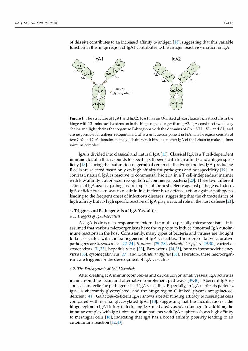
IgA acts in various mammals to protect against external microorganisms. IgA has twosubtypes: IgA1 and IgA2. As a structural difference, IgA1 has an O-linked glycosylationrich structure in the hinge region. This unique structure of 13 amino acids extends in thehinge region longer than IgA2, suggesting that IgA1 broadly acts to recognize variousantigens [9]; however, this site becomes a weak point of IgA1 because it is often the targetof bacterial proteases [10]. IgA consists of two heavy chains and light chains that organizeFab regions with the domains of Cα1, VH1, VL, and CL, and are responsible for antigenrecognition. Among these domains, Cα1 is a unique component of IgA [9]. The Fc regionconsists of two Cα2 and Cα3 domains. As one of the unique characteristics of IgA, this Fcregion, namely the J chain, can bind to another IgA of the J chain to form a dimer immunecomplex [9].
Immunoglobulin is generally produced by B cells; however, IgA is produced by mu-cosal membranes or glands, such as salivary glands, sweat glands, and the gut, and itsamount is greater than that of other types of immunoglobulin [9]. IgA binds to pathogensand viruses on the surface of the mucous membrane to prevent bacterial and viral infec-tions. Furthermore, general immunoglobulins respond to specific bacteria and viruses [11];however, IgA has the capacity to respond to specific bacteria and viruses in addition tovarious non-specific types of pathogens [12–14].
3.2. The Flexibility of IgA
Regarding structural aspects, Cα1 is a specific Fab domain in IgA, and other im-munoglobulins do not exhibit this domain [9] (Figure 1). This specific domain enables IgAto have greater variation in its response to pathogens. IgA also exhibits a hydrophobicsite between the VH and Cα1 domains [15], and a hydrophobic structure is expected tocontribute to protein–protein interactions, including antigen–antibody reactions [16,17].IgA1 has an O-linked glycosylation rich structure in the hinge region and the modification
Int. J. Mol. Sci. 2021, 22, 7538 3 of 15
of this site contributes to an increased affinity to antigen [18], suggesting that this variablefunction in the hinge region of IgA1 contributes to the antigen reactive variation in IgA.
Int. J. Mol. Sci. 2021, 22, x FOR PEER REVIEW 3 of 15
between the VH and Cα1 domains [15], and a hydrophobic structure is expected to con-
tribute to protein–protein interactions, including antigen–antibody reactions [16,17]. IgA1
has an O-linked glycosylation rich structure in the hinge region and the modification of
this site contributes to an increased affinity to antigen [18], suggesting that this variable
function in the hinge region of IgA1 contributes to the antigen reactive variation in IgA.
IgA is divided into classical and natural IgA [13]. Classical IgA is a T cell-dependent
immunoglobulin that responds to specific pathogens with high affinity and antigen spec-
ificity [13]. During the maturation of germinal centers in the lymph nodes, IgA-producing
B cells are selected based only on high affinity for pathogens and not specificity [19]. In
contrast, natural IgA is reactive to commensal bacteria in a T cell-independent manner
with low affinity but broader recognition of commensal bacteria [20]. These two different
actions of IgA against pathogens are important for host defense against pathogens. In-
deed, IgA deficiency is known to result in insufficient host defense action against patho-
gens, leading to the frequent onset of infectious diseases, suggesting that the characteris-
tics of high affinity but no high specific reaction of IgA play a crucial role in the host de-
fense [21].
Figure 1. The structure of IgA1 and IgA2. IgA1 has an O-linked glycosylation rich structure in the
hinge with 13 amino acids extension in the hinge region longer than IgA2. IgA consists of two heavy
chains and light chains that organize Fab regions with the domains of Cα1, VH1, VL, and CL, and
are responsible for antigen recognition. Cα1 is a unique component in IgA. The Fc region consists
of two Cα2 and Cα3 domains, namely J chain, which bind to another IgA of the J chain to make a
dimer immune complex.
4. Triggers and Pathogenesis of IgA Vasculitis
4.1. Triggers of IgA Vasculitis
As IgA is driven in response to external stimuli, especially microorganisms, it is as-
sumed that various microorganisms have the capacity to induce abnormal IgA autoim-
mune reactions in the host. Consistently, many types of bacteria and viruses are thought
to be associated with the pathogenesis of IgA vasculitis. The representative causative
pathogens are Streptococcus [22–24], S. aureus [25–28], Helicobacter pylori [29,30], varicella-
zoster virus [31,32], hepatitis virus [33], Parvovirus [34,35], human immunodeficiency vi-
rus [36], cytomegalovirus [37], and Clostridium difficile [38]. Therefore, these microorgan-
isms are triggers for the development of IgA vasculitis.
4.2. The Pathogenesis of IgA Vasculitis
After creating IgA immunocomplexes and deposition on small vessels, IgA activates
mannan-binding lectin and alternative complement pathways [39,40]. Aberrant IgA re-
sponses underlie the pathogenesis of IgA vasculitis. Especially, in IgA nephritis patients,
IgA1 is aberrantly glycosylated, and the hinge-region O-linked glycans are galactose-de-
ficient [41]. Galactose-deficient IgA1 shows a better binding efficacy to mesangial cells
compared with normal glycosylated IgA1 [18], suggesting that the modification of the
Figure 1. The structure of IgA1 and IgA2. IgA1 has an O-linked glycosylation rich structure in thehinge with 13 amino acids extension in the hinge region longer than IgA2. IgA consists of two heavychains and light chains that organize Fab regions with the domains of Cα1, VH1, VL, and CL, andare responsible for antigen recognition. Cα1 is a unique component in IgA. The Fc region consists oftwo Cα2 and Cα3 domains, namely J chain, which bind to another IgA of the J chain to make a dimerimmune complex.
IgA is divided into classical and natural IgA [13]. Classical IgA is a T cell-dependentimmunoglobulin that responds to specific pathogens with high affinity and antigen speci-ficity [13]. During the maturation of germinal centers in the lymph nodes, IgA-producingB cells are selected based only on high affinity for pathogens and not specificity [19]. Incontrast, natural IgA is reactive to commensal bacteria in a T cell-independent mannerwith low affinity but broader recognition of commensal bacteria [20]. These two differentactions of IgA against pathogens are important for host defense against pathogens. Indeed,IgA deficiency is known to result in insufficient host defense action against pathogens,leading to the frequent onset of infectious diseases, suggesting that the characteristics ofhigh affinity but no high specific reaction of IgA play a crucial role in the host defense [21].
4. Triggers and Pathogenesis of IgA Vasculitis4.1. Triggers of IgA Vasculitis
As IgA is driven in response to external stimuli, especially microorganisms, it isassumed that various microorganisms have the capacity to induce abnormal IgA autoim-mune reactions in the host. Consistently, many types of bacteria and viruses are thoughtto be associated with the pathogenesis of IgA vasculitis. The representative causativepathogens are Streptococcus [22–24], S. aureus [25–28], Helicobacter pylori [29,30], varicella-zoster virus [31,32], hepatitis virus [33], Parvovirus [34,35], human immunodeficiencyvirus [36], cytomegalovirus [37], and Clostridium difficile [38]. Therefore, these microorgan-isms are triggers for the development of IgA vasculitis.
4.2. The Pathogenesis of IgA Vasculitis
After creating IgA immunocomplexes and deposition on small vessels, IgA activatesmannan-binding lectin and alternative complement pathways [39,40]. Aberrant IgA re-sponses underlie the pathogenesis of IgA vasculitis. Especially, in IgA nephritis patients,IgA1 is aberrantly glycosylated, and the hinge-region O-linked glycans are galactose-deficient [41]. Galactose-deficient IgA1 shows a better binding efficacy to mesangial cellscompared with normal glycosylated IgA1 [18], suggesting that the modification of thehinge region in IgA1 is key to inducing IgA-mediated vascular damage. In addition, theimmune complex with IgA1 obtained from patients with IgA nephritis shows high affinityto mesangial cells [18], indicating that IgA has a broad affinity, possibly leading to anautoimmune reaction [42,43].

Int. J. Mol. Sci. 2021, 22, 7538 4 of 15
FcαRI is a receptor for IgA and this signal has a bifunctional action in both proin-flammatory and anti-inflammatory effects [44–46]. FcαRI is expressed on monocytes,macrophages, intestinal dendritic cells, and Kupffer cells [46]. The induction of FcαRIsignaling is associated with the immunoreceptor tyrosine-based activation motif (ITAM).The binding of complexed monomeric serum IgA Fc domains to the membrane distaldomain of FcαRI initiates signal cascades to enhance the inflammatory response, whileuncomplexed monomeric IgA associates with FcαRI, the ITAM inhibitory signal cascade isinitiated to promote anti-inflammatory action. Consistently, excessive IgA immune com-plexes or IgA-opsonized bacteria drive FcαRI-mediated immune cell activation, resultingin severe tissue damage, as observed during chronic inflammation and autoimmunity [47].In addition, FcαRI on Kupffer cells enhances the efficient phagocytosis of bacteria coatedwith serum IgA [48]. Interestingly, FcαRI expression is strictly regulated by cytokines,such as G-CSF [48]. IgA also enhances FcαRI-mediated LTB4 production, resulting inthe enhancement of neutrophil migration in IgA-triggered immune complex depositionsites [49].
After inflammatory damage in blood vessels, transendothelial migration of neu-trophils occurs. VEGF promotes vascular permeability and promotes inflammatory cellmigration from the vessels [50] and is associated with the development of IgA vasculi-tis [51]. The immune complex also enhances C3 and C5 production in endothelial cells,inducing IL-8, E-selectin, and ICAM 1 production [52]. Infiltration of inflammatory cellsalso increased the expression of ICAM 1 [53].
The cytokine profiles in the sera are shown in Table 1. General proinflammatory andinflammatory cytokines are increased in IgA vasculitis and nephritis, such as IL-1β, IL-4,IL-6, IL-8, IL-12p70, IL-17A, TNF-α, and IFN-γ. IL-6 levels are also increased in patientswith IgA colitis. These inflammatory cytokines are orchestrated during the developmentof IgA-mediated autoimmune reactions. However, there are some controversial results,especially for TNF-α. As shown in TNF inhibitor-mediated IgA vasculitis, it seems that theinvolvement of TNF is not simple in the pathogenesis of IgA vasculitis. In addition, IL-17 isupregulated in IgA vasculitis and nephritis; however, IL-17 inhibitors are also related to theoccurrence of IgA vasculitis, suggesting that TNF and IL-17 are involved in the cytokineproduction mediated by IgA vasculitis.
Table 1. Cytokine profiles in IgA vasculitis, nephritis, and colitis.
IgA Vasculitis IgA Vasculitis Nephritis IgA Colitis
IL-1β ↑ [54]→ [55,56]
↑ [55]→ [54,57] No report
IL-2 → [58] → [58] No reportIL-4 ↑ [58] ↑ [58] No report
IL-6 ↑ [54,56,58,59]→ [55]
↑ [54,55,57,58]→ [60] ↑ [60]
IL-8 ↑ [55,56] ↑ [55,60] → [60]IL-9 → [56] No report No report
IL-10 → [55,56]↓ [58]
→ [55,56]↓ [58] No report
IL-12p70 → [54,55] → [54,55] No reportIL-17A ↑ [58] ↑ [58] No reportIL-23 → [56] No report No report
TNF-α↑ [56]
→ [54,55]↓ [58]
→ [54,55,60]↓ [58] → [60]
IFN-γ ↑ [58] ↑ [58]→ [57] No report
In the current study, IgA1 was identified as an autoantibody in host immunity [61],especially in IgA nephritis. IgA1 triggers immune complex deposition in the blood ves-

Int. J. Mol. Sci. 2021, 22, 7538 5 of 15
sels, leading to the activation of complement factors and the exacerbation of vascularinflammation and damage [9].
In IgA1, this unique O-linked carbohydrate site seems to be associated with the patho-genesis of IgA nephritis from the dominant deposition of galactose-deficient IgA comparedwith IgA vasculitis without nephritis and healthy subjects. Interestingly, there was nosignificant difference in galactose-deficient IgA1 deposition between healthy subjects andIgA vasculitis without nephritis [62], suggesting that there is a different IgA1 dominantdeposition pattern in the skin and kidneys in patients with IgA vasculitis.
Fatty acids are involved in various inflammatory reactions in the human body. LTB4is derived from omega-6 fatty acids and exerts inflammatory responses and neutrophil mi-gration and activation. LTB4 is increased in patients with IgA vasculitis [63] and decreasedafter treatment, suggesting that LTB4 might play a role in the acceleration of IgA vasculitisin some parts.
Platelet-activating factor (PAF) is a lipid mediator involved in several allergic diseasesand is released from eosinophils, neutrophils, and mast cells. PAF is involved in IgAvasculitis [64] and promotes IgA production [65].
4.3. The Difference of IgA between Adults and Pediatrics
It remains unclear whether there is a clear difference in IgA levels between pediatricand adult patients. As IgA vasculitis and nephritis show different clinical courses betweenadult and pediatric patients, it is assumed that there are some differences in IgA betweenadult and pediatric patients. The first difference is the amount of IgA, which contributesto the development of IgA [47]. Consistently, IgA production in adults is much higherthan in children [66]. For instance, 2–3-year-old and 5–6-year-old children showed IgAlevels of 40.8% and 69.5%, respectively, compared with adults [66]. IgA levels reach adultproportions after the age of 11 years. These findings suggest that IgA levels in childrenmight not be at a sufficient volume to respond to vessels and the kidneys.
Another possibility is that the frequency of memory B cells might be a clue to answer-ing this question. The frequency of memory B cells in the peripheral blood is higher inadults than in children [67]. Schoolchildren showed a decreased frequency of memoryB cells compared to adults; however, adolescents showed almost the same level of thefrequency of adult memory B cells, without significant differences. These findings suggestthat pediatric memory B cells might show less activity in memorized IgA production,leading to continuous pathogenic IgA production.
The difference might also depend on other immune profile differences. In the com-parison of IgA nephritis between pediatric and adult patients, CD68+ macrophages wereincreased in adult glomerular and interstitial sites in the kidney. Macrophage functionis decreased in pediatric patients compared with adults, such as cytotoxicity [68] andanti-tumor immunity [69].
In contrast, galactose-deficient IgA in patients with pediatric IgA vasculitis and nephri-tis is similar to that in adults [70]. This finding suggests that pathogenic IgA1 might not bedifferent between adult and pediatric IgA vasculitis and nephritis. In addition, there is noevidence of the difference in the survival duration of memory B cells between pediatric andadult patients, and the actual impact of the frequency of memory B cells remains unclear.Therefore, further investigation is necessary to clarify this basic question.
4.4. The Relationship with Coronavirus Disease 2019 (COVID-19)
Interestingly, several studies have reported COVID-19 associated IgA vasculitis [71–77].COVID-19 caused by the severe acute respiratory syndrome coronavirus 2 (SARS-CoV-2)and the COVID-19 pandemic is currently an ongoing global problem and is known tocause vasculitislike syndromes [78]. The characteristics of patients with IgA vasculitisfollowing COVID-19 are summarized in Table 2. There were seven cases of IgA vasculitis.The median age was 23.3 years and the ratio of adults to children was 4:3. Interestingly, allcases were male. Other symptoms excluding cutaneous purpura were observed in three of

Int. J. Mol. Sci. 2021, 22, 7538 6 of 15
seven cases with abdominal pain, and three of seven cases with nephritis. Furthermore,all nephritis cases were adults, and 75% of adult cases showed IgA vasculitis followingCOVID-19 infection, whereas IgA nephritis was not identified in the children.
Table 2. The summary of cases of IgA vasculitis following COVID-19.
Author Age Sex Days after COVID-19Test Positive Involvement Treatment
Suso, et al. [71] 78 Male 5 weeks later SkinNephritis Steroid pulse plus Rituximab
Hoskins, et al. [72] 2 Male Same time SkinAbdominal pain Intravenous steroid
Allez et al. [73] 24 Male Unknown SkinAbdominal pain Methylprednisolone 0.8 mg/day
Sandhu et al. [74] 22 Male Same time SkinNephritis Prednisolone 1 mg/kg
AlGhoozi et al. [75] 4 Male 37 days later Skin Not described
Jacobi, et al. [76] 3 Male Same time SkinAbdominal pain Antibiotic
Li et al. [77] 30 Male Same time SkinNephritis
Losartan 25 mg followingprednisolone 40 mg for 7 days
COVID-19 has the potential to induce a cytokine storm, which might also be involvedin the pathogenesis of IgA vasculitis. In addition, as another possibility, the medications forCOVID-19 are not completely excluded as a trigger, similar to drug-induced IgA vasculitis.
4.5. The Relationship with TNF or IL-17 Inhibitors
In the skin, the TNF and IL-17 pathways are involved in various inflammatory skindiseases. Psoriasis is a representative inflammatory skin disease mediated by inflammatorypathways; it causes scaly erythematous plaques and influences inflammation in variousinternal organs. Consistently, these cytokine-targeted treatments showed strong efficacy inpsoriatic skin inflammation. A neutrophil-mediated inflammatory reaction is downstreamof the TNF- and IL-17 mediated inflammatory pathways. Therefore, these cytokines are alsothought to be associated with the pathogenesis of IgA vasculitis. On the contrary, severalrecent reports suggest a possible pathogenetic role of TNF-mediated immune reactionsin IgA vasculitis. One study reported that two cases showed the deposition of IgA inthe renal biopsy among 39 cases of vasculitis during treatment with TNF inhibitors [79].Another study showed that six out of nine patients experienced IgA vasculitis onset duringTNF inhibitors; however, three of five patients continued to use TNF inhibitors and fullyrecovered while maintaining TNF inhibitor treatment [80]. IL-17A inhibitors have alsobeen reported as triggers of IgA vasculitis [81]. The detailed molecular mechanismsremain unclear and may be related to some paradoxical side effects of these cytokine-targeted inhibitor treatments, as seen in psoriasis dermatitis following these inhibitortreatments [82,83]. Table 1 shows that the fluctuation of TNF in patients with IgA vasculitisis controversial, suggesting that the involvement of TNF is not simple. Some complexcytokine pathways, such as IFN, might also be involved in TNF inhibitor-related IgAvasculitis. Further investigation will be desired to clarify the actual therapeutic efficacy forIgA vasculitis.
5. Symptoms
IgA vasculitis involves various small vessels as well as the skin, joints, gastrointestinaltract, and kidneys. The skin manifestation of IgA vasculitis is palpable purpura commonlydistributed in the lower extremities and sometimes the upper limbs and trunk. Among thesmall vessels, the blood vessels in the skin are mainly affected, predominantly capillaries,venules, or arterioles [84]. The histology of this purpura exhibits nuclear dust, extravasation

Int. J. Mol. Sci. 2021, 22, 7538 7 of 15
of erythrocytes, and inflammatory cell infiltration surrounding the affected vessels withIgA deposition.
Approximately 65% of patients with IgA vasculitis experience abdominal pain [85].In addition, 30% of patients experience gastrointestinal bleeding [85]. Gastrointestinalsymptoms generally appear within 1 week after the onset of purpura in the skin [86].
Kidney involvement is the most important organ dysfunction in determining thetherapeutic options for IgA vasculitis and influencing its prognosis. IgA vasculitis in adultpatients showed a high prevalence of IgA nephritis, with a tendency to be severe comparedwith that in children [87].
6. Treatment6.1. Corticosteroids
IgA vasculitis patients show significant improvement of cutaneous purpura symptomswith the treatment of prednisolone 1 mg/kg daily [88]; however, there was no significant dif-ference in the recurrence rate even after the administration of corticosteroids [88]. Althoughprolonged cutaneous purpura cases tend to become a risk factor for renal dysfunction, thereis no preventive efficacy for IgA nephritis in the early administration of corticosteroids [89].In contrast, IgA nephritis is a therapeutic candidate for corticosteroid administration. Theearly administration of prednisolone contributes to improvement in the renal function [90].
6.2. Colchicine
Colchicine is an alkaloid extracted from the corm of the meadow saffron or autumncrocus [91] and has various anti-inflammatory actions. Colchicine suppresses superox-ide production from neutrophils [92], neutrophil chemotaxis [93], and lysozyme produc-tion [94]. While no clinical trials have evaluated the therapeutic efficacy of colchicine for IgAvasculitis, there were two case studies reporting the efficacy of colchicine for IgA vasculitis.
6.3. Dapsone
Dapsone is an antibiotic that inhibits bacterial synthesis of dihydrofolic acid by com-peting with para-aminobenzoate for the active site of dihydropteroate synthase, resulting inthe inhibition of nucleic acid synthesis [95,96]. In addition, dapsone has anti-inflammatoryeffects, such as the suppression of IL-8, TNF, toxic-free radical production, and inflam-matory cell migration. Therefore, these actions are thought to be a promising therapeuticoption for refractory IgA vasculitis [97]. Dapsone also showed therapeutic action againstcutaneous purpura in IgA vasculitis [98–100].
Previous case reports suggest a possible therapeutic potential of dapsone for IgAvasculitis [98,101]. A recent systematic review analysis identified that therapeutic responsewas obtained in 23.1% of patients during the initial 1–2 days and 65.4% of patients within1 week [95].
6.4. Intravenous Immunoglobulin
Intravenous immunoglobulin is currently used for the treatment of Guillain–Barrésyndrome, myasthenia gravis, and bullous diseases [102,103]. As the mechanism of thera-peutic action of intravenous immunoglobulin, it can impair autoreactive T cell responses byblocking their interaction with antigen presentation cells [104], downregulating antibodyproduction by B cells [105], and blocking Fc-receptor-mediated immune response [106].Recent studies have identified the therapeutic potential of intravenous immunoglobulinfor IgA vasculitis.
One study showed that 11 patients with IgA nephropathy received high-dose im-munoglobulins (2 g/kg body weight), and histological changes and renal function wereevaluated [107]. Proteinuria and reduced glomerular filtration rate were impaired by intra-venous immunoglobulin treatment. The staining intensities of IgA and C3 in glomerulardeposition were also decreased by the treatment. In another study, 14 patients with IgAnephritis received intravenous immunoglobulin treatment, and 1 patient withdrew from

Int. J. Mol. Sci. 2021, 22, 7538 8 of 15
the study [108]. This study showed significantly decreased proteinuria, serum IgA, and β2microglobulin levels.
6.5. Rituximab
Rituximab is a monoclonal antibody against CD20, which is a representative surfacecell marker on B cells and is expected to suppress functional pathogenic IgA-producing Bcells in patients with IgA vasculitis.
There are several clinical reports of rituximab-treated refractory IgA vasculitis [109,110].A recent systematic review of 20 studies including 35 well-characterized IgA vasculitistreated with rituximab showed that 94.3% of patients exhibited clinical improvement and74.3% of patients achieved remission [111].
6.6. Angiotensin-Converting-Enzyme Inhibitor
ACE inhibitors are used for the treatment of hypertension [112,113] and inhibit theactivity of ACE, which is an important component of the renin–angiotensin system con-verting angiotensin I to angiotensin II [114], and hydrolyzes bradykinin [115], leading tolower arteriolar resistance and increased venous capacity.
A randomized controlled trial of IgA nephritis patients followed up for 5 yearsinvestigated the long-term renal outcome of ACE inhibitor/angiotensin receptor antagonisttherapy [116]. The treatment showed lower serum creatinine, lower proteinuria, andfewer numbers progressing to end-stage renal failure. Another randomized trial in IgAnephritis showed that a high-dose regimen of ACE inhibitor (losartan 200 mg/day) showedsignificantly higher eGFR, lower proteinuria, and impaired eGFR reduction.
However, there is no evidence of the anti-inflammatory action of ACE inhibitors. Thistreatment is a part of supportive therapy to reduce IgA nephritis progression, similar tomany renal diseases including SLE and scleroderma-related renal disease.
6.7. Omega-3 Fatty Acids
Omega-3 fatty acids are present in fish oils and nuts [117]. Omega-3 fatty acidsand their metabolites have potential anti-inflammatory action in inflammatory skin dis-eases [117–120]. Purpura is not a typical form of inflammatory skin disease, but causesinflammation in the blood vessels. Several studies have evaluated the anti-inflammatoryaction and therapeutic potential of omega-3 fatty acids in IgA vasculitis and nephritis.
In one study conducted by Hamazaki et al., 20 patients were divided into 2 groupswith or without fish oil treatment (1.6 g EPA and 1.0 g DHA per day) for 1 year and wereevaluated for renal function in IgA nephritis. The fish oil-treated group maintained theirrenal function, whereas the control group showed worsening of renal function [121].
In another study, 14 patients receiving omega-3 fatty acids (0.85 g EPA and 0.57 gDHA) were compared with 14 patients in the control group. The omega-3 fatty acids treatedgroup showed a mean annual change in serum creatinine levels and GFR, indicating afavorable response [122].
A multicenter, placebo-controlled, randomized trial was conducted to evaluate theefficacy of fish oil in patients with IgA nephropathy [123]. Fifty-five patients received12 g of daily fish oil, and 51 patients received olive oil as a placebo. The annual medianchanges in serum creatinine concentrations in the fish oil group were lower than those inthe placebo group. The cumulative percentage of patients who died or had end-stage renaldisease was 40 percent in the placebo group and 10 percent in the fish oil group.
6.8. Immunosuppressive Agents
Immunosuppressive agents show multiple steps of immunosuppressive effects againstinflammatory diseases and exhibit beneficial effects on inflammatory diseases. In IgAnephritis, cyclophosphamide treatment alone has no beneficial impact [124]; however, corti-costeroid combination therapy is effective for IgA nephritis [125]. Azathioprine therapy alsoshowed a therapeutic effect on IgA nephritis with a combination of corticosteroids [126].

Int. J. Mol. Sci. 2021, 22, 7538 9 of 15
6.9. Lectin Pathway Treatment
The lectin pathways mediate glomerular injury and contribute to the development ofIgA nephritis [127]. Narsoplimab is a monoclonal antibody against mannan-binding lectinserine peptidase 2 (MASP-2), a key component of the lectin pathway. Only one case reportshowed that narsoplimab has therapeutic efficacy in refractory IgA nephritis [128].
7. Biomarker
Since the degree of vasculitis is sometimes difficult to evaluate on superficial physicalexamination of the skin, biomarkers are required to predict the progression of IgA vasculitisand the presence of nephritis and colitis. Several studies have investigated biomarkers topredict the severity of IgA vasculitis and other organ involvement.
As biomarkers for IgA vasculitis, the severity of IgA vasculitis is associated withneutrophil/lymphocyte rate [129], serum neopterin and ischemia-modified albumin lev-els [130], skin miRNA-223-3p expression [131], and serum and urine levels of NGAL, KIM-1,and L-FABP [132]. IgA vasculitis patients sometimes exhibit ANCA positivity; however,there were no significant differences in the disease-free survival rates of chronic kidneydisease and end-stage renal disease between IgA vasculitis patients with and withoutANCA [133].
As biomarkers for IgA nephritis, a systematic literature review was performed topredict the presence of IgA nephritis or determine its severity [134]. In urine samples,kidney injury molecule-1 (KIM-1), monocyte chemotactic protein-1 (MCP-1), and N-acetyl-β-glucosaminidase (NAG) correlate with the disease severity of nephritis. In addition,the presence of IgA nephritis is associated with serum Gd-IgA1 and urinary IgA, IgG,IgM, IL-6, IL-8, IL-10, and IgA-IgG and IgA-sCD89 complexes [54], neutrophil countand neutrophil/lymphocyte rate [135], and serum angiotensinogen concentration [136].The acute phase of IgA vasculitis is related to the expression levels of miRNA-33 andmiRNA-34 [137] and aPS/PT-positivity [138].
As biomarkers for colitis, neutrophil count and neutrophil/lymphocyte ratio arehigher in IgA colitis [129,135]. IgA colitis is inversely correlated with the expression ofmiRNA-155-5p and miRNA-146a-5p in the skin [131].
8. Epigenetic Modification
Although a minority of DNA sequence alterations are not infrequent in humans, asexemplified by DNA insertions in HPV-and EBV-related cancers, the majority of DNAsequence information is not generally changed during one’s lifetime; however, the geneinformation is regulated by acquired chemical modifications of DNA and DNA-bindingproteins, especially histones [139,140]. In general, DNA and DNA-binding proteins are incontact with each other to avoid excessive transcriptional activation. However, once DNAis released from histones, open chromatin is formed, followed by the activation of the tran-scription activation response to the appropriate external stimulation [139]. Recent studieshave also identified that epigenetic modifications are also involved in the pathogenesis ofIgA vasculitis.
IgA vasculitis patients exhibit impaired ERK signaling, which is related to the down-regulation of the DNA methyltransferase DNMT1 [141]. The histone demethylase KDM4Cwas identified as a risk factor for IgA vasculitis [142]. The enhancement of histone modifi-cation of H3 acetylation in peripheral blood mononuclear cells was significantly increasedin patients with IgA vasculitis and nephritis, and H3K4 methylation was also increased inpatients with IgA nephritis. In addition, other epigenetic modification enzymes, HDAC1,2, and 4 SIRT1, LSD1, and KDM3A are decreased, whereas CREBBP, PCAF, SETD1A, MLL,STEDB1, and SUV39H2 are increased in IgA vasculitis, suggesting that these epigeneticmodification enzymes might organize DNA and histone modifications in patients with IgAvasculitis and exacerbate inflammatory responses [143].

Int. J. Mol. Sci. 2021, 22, 7538 10 of 15
9. Conclusions
This review updates the recent knowledge and current problems in IgA vasculitis.Although the actual etiology and triggers of IgA vasculitis remain unclear, the presenceof IgA vasculitis following novel drug administration and COVID-19 infection might behelpful to gain a deeper understanding. As the deposition pattern of IgA subtypes seemsto be different between IgA vasculitis and nephritis, there are some different molecularmechanisms that cause IgA deposition in the small vessels in different organs. Currently,there is no specific treatment for skin purpura. Therefore, it is desirable to identify a noveltherapeutic approach for skin eruptions because this is often a refractory and long-lastingform of skin inflammation. In addition, the knowledge of epigenetic modifications in IgAvasculitis is currently being developed. This information will be helpful in gaining a betterunderstanding of the pathogenesis of IgA vasculitis.
Author Contributions: H.S. and Y.S. wrote the manuscript. M.N. supervised the writing of themanuscript. All authors have read and agreed to the published version of the manuscript.
Funding: This research received no external funding.
Institutional Review Board Statement: Not applicable.
Informed Consent Statement: Not applicable.
Data Availability Statement: Not applicable.
Conflicts of Interest: The authors declare no conflict of interest.
References1. López-Mejías, R.; Castañeda, S.; Genre, F.; Remuzgo-Martínez, S.; Carmona, F.D.; Llorca, J.; Blanco, R.; Martín, J.; González-Gay,
M.A. Genetics of immunoglobulin-A vasculitis (Henoch-Schönlein purpura): An updated review. Autoimmun. Rev. 2018, 17,301–315. [CrossRef]
2. González-Gay, M.A.; López-Mejías, R.; Pina, T.; Blanco, R.; Castañeda, S. IgA Vasculitis: Genetics and Clinical and TherapeuticManagement. Curr. Rheumatol. Rep. 2018, 20, 24. [CrossRef]
3. López-Mejías, R.; Genre, F.; Pérez, B.S.; Castañeda, S.; Ortego-Centeno, N.; Llorca, J.; Ubilla, B.; Remuzgo-Martínez, S.; Mijares, V.;Pina, T.; et al. HLA-DRB1 association with Henoch-Schonlein purpura. Arthritis Rheumatol. 2015, 67, 823–827. [CrossRef]
4. López-Mejías, R.; Genre, F.; Pérez, B.S.; Castañeda, S.; Ortego-Centeno, N.; Llorca, J.; Ubilla, B.; Remuzgo-Martínez, S.; Mijares, V.;Pina, T.; et al. Association of HLA-B*41:02 with Henoch-Schönlein Purpura (IgA Vasculitis) in Spanish individuals irrespective ofthe HLA-DRB1 status. Arthritis Res. Ther. 2015, 17, 102. [CrossRef] [PubMed]
5. López-Mejías, R.; Carmona, F.D.; Castañeda, S.; Genre, F.; Remuzgo-Martínez, S.; Sevilla-Perez, B.; Ortego-Centeno, N.; Llorca, J.;Ubilla, B.; Mijares, V.; et al. A genome-wide association study suggests the HLA Class II region as the major susceptibility locusfor IgA vasculitis. Sci. Rep. 2017, 7, 5088. [CrossRef] [PubMed]
6. Amoli, M.M.; Thomson, W.; Hajeer, A.H.; Calviño, M.C.; Garcia-Porrua, C.; Ollier, W.E.; Gonzalez-Gay, M.A. Interleukin 1receptor antagonist gene polymorphism is associated with severe renal involvement and renal sequelae in Henoch-Schönleinpurpura. J. Rheumatol. 2002, 29, 1404–1407.
7. Amoli, M.M.; Thomson, W.; Hajeer, A.H.; Calviño, M.C.; Garcia-Porrua, C.; Ollier, W.E.; Gonzalez-Gay, M.A. Interleukin 8 genepolymorphism is associated with increased risk of nephritis in cutaneous vasculitis. J. Rheumatol. 2002, 29, 2367–2370. [PubMed]
8. Carmona, E.G.; García-Giménez, J.A.; López-Mejías, R.; Khor, C.C.; Lee, J.K.; Taskiran, E.; Ozen, S.; Hocevar, A.; Liu, L.; Gorenjak,M.; et al. Identification of a shared genetic risk locus for Kawasaki disease and IgA vasculitis by a cross-phenotype meta-analysis.Rheumatology 2021. [CrossRef]
9. Bakema, J.E.; van Egmond, M. Immunoglobulin A: A next generation of therapeutic antibodies? MAbs 2011, 3, 352–361. [CrossRef]10. Van Egmond, M.; Damen, C.A.; van Spriel, A.B.; Vidarsson, G.; van Garderen, E.; van de Winkel, J.G. IgA and the IgA Fc receptor.
Trends Immunol. 2001, 22, 205–211. [CrossRef]11. Fuller, A.O.; Spear, P.G. Specificities of monoclonal and polyclonal antibodies that inhibit adsorption of herpes simplex virus to
cells and lack of inhibition by potent neutralizing antibodies. J. Virol. 1985, 55, 475–482. [CrossRef] [PubMed]12. Slack, E.; Balmer, M.L.; Fritz, J.H.; Hapfelmeier, S. Functional flexibility of intestinal IgA—Broadening the fine line. Front. Immunol.
2012, 3, 100. [CrossRef] [PubMed]13. Pabst, O. New concepts in the generation and functions of IgA. Nat. Rev. Immunol. 2012, 12, 821–832. [CrossRef] [PubMed]14. Saito, S.; Sano, K.; Suzuki, T.; Ainai, A.; Taga, Y.; Ueno, T.; Tabata, K.; Saito, K.; Wada, Y.; Ohara, Y.; et al. IgA tetramerization
improves target breadth but not peak potency of functionality of anti-influenza virus broadly neutralizing antibody. PLoS Pathog.2019, 15, e1007427. [CrossRef]

Int. J. Mol. Sci. 2021, 22, 7538 11 of 15
15. Correa, A.; Trajtenberg, F.; Obal, G.; Pritsch, O.; Dighiero, G.; Oppezzo, P.; Buschiazzo, A. Structure of a human IgA1 Fab fragmentat 1.55 Å resolution: Potential effect of the constant domains on antigen-affinity modulation. Acta Crystallogr. Sect. D Biol.Crystallogr. 2013, 69, 388–397. [CrossRef]
16. Li, Y.; Huang, Y.; Swaminathan, C.P.; Smith-Gill, S.J.; Mariuzza, R.A. Magnitude of the hydrophobic effect at central versusperipheral sites in protein-protein interfaces. Structure 2005, 13, 297–307. [CrossRef]
17. Worobec, E.A.; Paranchych, W.; Parker, J.M.; Taneja, A.K.; Hodges, R.S. Antigen-antibody interaction. The immunodominantregion of EDP208 pili. J. Biol. Chem. 1985, 260, 938–943. [CrossRef]
18. Novak, J.; Vu, H.L.; Novak, L.; Julian, B.A.; Mestecky, J.; Tomana, M. Interactions of human mesangial cells with IgA andIgA-containing immune complexes. Kidney Int. 2002, 62, 465–475. [CrossRef]
19. Shimoda, M.; Inoue, Y.; Azuma, N.; Kanno, C. Natural polyreactive immunoglobulin A antibodies produced in mouse Peyer’spatches. Immunology 1999, 97, 9–17. [CrossRef]
20. Quan, C.P.; Berneman, A.; Pires, R.; Avrameas, S.; Bouvet, J.P. Natural polyreactive secretory immunoglobulin A autoantibodiesas a possible barrier to infection in humans. Infect. Immun. 1997, 65, 3997–4004. [CrossRef]
21. Zhang, J.; van Oostrom, D.; Li, J.; Savelkoul, H.F.J. Innate Mechanisms in Selective IgA Deficiency. Front. Immunol. 2021,12, 649112. [CrossRef]
22. Nakatsuka, K. Serum anti-streptococcal IgA, IgG and IgM antibodies in IgA-associated diseases. Acta Paediatr. Jpn. 1993, 35,118–123. [CrossRef] [PubMed]
23. Akagi, M.; Iwanaga, N.; Torisu, Y.; Fujita, H.; Kawahara, C.; Horai, Y.; Izumi, Y.; Kawakami, A. IgA Vasculitis Triggered byInfective Endocarditis of Pulmonary Artery with Congenitally Corrected Transposition of the Great Arteries. Int. Heart J. 2020, 61,404–408. [CrossRef] [PubMed]
24. Montoliu, J.; Miró, J.M.; Campistol, J.M.; Trilla, A.; Mensa, J.; Torras, A.; Revert, L. Henoch-Schönlein purpura complicatingstaphylococcal endocarditis in a heroin addict. Am. J. Nephrol. 1987, 7, 137–139. [CrossRef]
25. Temkiatvises, K.; Nilanont, Y.; Poungvarin, N. Stroke in Henoch-Schönlein purpura associated with methicillin-resistantStaphylococcus aureus septicemia: Report of a case and review of the literature. J. Med. Assoc. Thail. 2008, 91, 1296–1301.
26. Uggeri, S.; Fabbian, F.; Catizone, L. Henoch-Schönlein purpura due to methicillin-sensitive Staphylococcus aureus bacteremiafrom central venous catheterization. Clin. Exp. Nephrol. 2008, 12, 219–223. [CrossRef]
27. Kitamura, T.; Nakase, H.; Iizuka, H. Henoch-Schönlein purpura after postoperative Staphylococcus aureus infection with hepaticIgA nephropathy. J. Nephrol. 2006, 19, 687–690. [PubMed]
28. Hirayama, K.; Kobayashi, M.; Muro, K.; Yoh, K.; Yamagata, K.; Koyama, A. Specific T-cell receptor usage with cytokinemiain Henoch-Schönlein purpura nephritis associated with Staphylococcus aureus infection. J. Intern. Med. 2001, 249, 289–295.[CrossRef]
29. Shin, J.I.; Koh, H.; Lee, J.S. Henoch-Schönlein purpura associated with helicobacter pylori infection: The pathogenic roles of IgA,C3, and cryoglobulins? Pediatr. Dermatol. 2009, 26, 768–769. [CrossRef] [PubMed]
30. Novák, J.; Szekanecz, Z.; Sebesi, J.; Takáts, A.; Demeter, P.; Bene, L.; Sipka, S.; Csiki, Z. Elevated levels of anti-Helicobacter pyloriantibodies in Henoch-Schönlein purpura. Autoimmunity 2003, 36, 307–311. [CrossRef]
31. Häusler, M.G.; Ramaekers, V.T.; Reul, J.; Meilicke, R.; Heimann, G. Early and late onset manifestations of cerebral vasculitisrelated to varicella zoster. Neuropediatrics 1998, 29, 202–207. [CrossRef]
32. Ushigome, Y.; Yamazaki, Y.; Shiohara, T. IgA vasculitis with severe gastrointestinal symptoms may be an unusual manifestationof varicella zoster virus reactivation. Br. J. Dermatol. 2017, 176, 1103–1105. [CrossRef] [PubMed]
33. Maggiore, G.; Martini, A.; Grifeo, S.; De Giacomo, C.; Scotta, M.S. Hepatitis B virus infection and Schönlein-Henoch purpura. Am.J. Dis. Child. 1984, 138, 681–682. [CrossRef] [PubMed]
34. Veraldi, S.; Mancuso, R.; Rizzitelli, G.; Gianotti, R.; Ferrante, P. Henoch-Schönlein syndrome associated with human ParvovirusB19 primary infection. Eur. J. Dermatol. 1999, 9, 232–233. [PubMed]
35. Cioc, A.M.; Sedmak, D.D.; Nuovo, G.J.; Dawood, M.R.; Smart, G.; Magro, C.M. Parvovirus B19 associated adult Henoch Schönleinpurpura. J. Cutan. Pathol. 2002, 29, 602–607. [CrossRef] [PubMed]
36. Brandy-García, A.M.; Santos-Juanes, J.; Suarez, S.; Caminal-Montero, L. IgA vasculitis as a presentation of human immunodefi-ciency virus infection. Reumatol. Clin. 2020, 16, 298–299. [CrossRef]
37. Matsumura, M.; Komeda, Y.; Watanabe, T.; Kudo, M. Purpura-free small intestinal IgA vasculitis complicated by cytomegalovirusreactivation. BMJ Case Rep. 2020, 13. [CrossRef]
38. Kounatidis, D.; Vadiaka, M.; Kouvidou, C.; Sampaziotis, D.; Skourtis, A.; Panagopoulos, F.; Konstantinou, F.; Vallianou, N.G.Clostridioides difficile infection in a patient with immunoglobulin A vasculitis: A triggering factor or a rare complication of thedisease? A case-based review. Rheumatol. Int. 2020, 40, 997–1000. [CrossRef]
39. Roos, A.; Bouwman, L.H.; van Gijlswijk-Janssen, D.J.; Faber-Krol, M.C.; Stahl, G.L.; Daha, M.R. Human IgA activates thecomplement system via the mannan-binding lectin pathway. J. Immunol. 2001, 167, 2861–2868. [CrossRef]
40. Hiemstra, P.S.; Gorter, A.; Stuurman, M.E.; Van Es, L.A.; Daha, M.R. Activation of the alternative pathway of complement byhuman serum IgA. Eur. J. Immunol. 1987, 17, 321–326. [CrossRef]
41. Novak, J.; Moldoveanu, Z.; Renfrow, M.B.; Yanagihara, T.; Suzuki, H.; Raska, M.; Hall, S.; Brown, R.; Huang, W.Q.; Goepfert, A.;et al. IgA nephropathy and Henoch-Schoenlein purpura nephritis: Aberrant glycosylation of IgA1, formation of IgA1-containingimmune complexes, and activation of mesangial cells. Contrib. Nephrol. 2007, 157, 134–138.
Int. J. Mol. Sci. 2021, 22, 7538 12 of 15
42. Noval Rivas, M.; Wakita, D.; Franklin, M.K.; Carvalho, T.T.; Abolhesn, A.; Gomez, A.C.; Fishbein, M.C.; Chen, S.; Lehman, T.J.;Sato, K.; et al. Intestinal Permeability and IgA Provoke Immune Vasculitis Linked to Cardiovascular Inflammation. Immunity2019, 51, 508–521.e6. [CrossRef]
43. Chorzelski, T.P.; Jabłonska, S.; Maciejowska, E. Linear IgA bullous dermatosis of adults. Clin. Dermatol. 1991, 9, 383–392.[CrossRef]
44. Van Gool, M.M.J.; van Egmond, M. IgA and FcαRI: Versatile Players in Homeostasis, Infection, and Autoimmunity. ImmunotargetsTher. 2020, 9, 351–372. [CrossRef]
45. Aleyd, E.; Heineke, M.H.; van Egmond, M. The era of the immunoglobulin A Fc receptor FcαRI; Its function and potential astarget in disease. Immunol. Rev. 2015, 268, 123–138. [CrossRef]
46. Davis, S.K.; Selva, K.J.; Kent, S.J.; Chung, A.W. Serum IgA Fc effector functions in infectious disease and cancer. Immunol. CellBiol. 2020, 98, 276–286. [CrossRef]
47. Breedveld, A.; van Egmond, M. IgA and FcαRI: Pathological Roles and Therapeutic Opportunities. Front. Immunol. 2019, 10, 553.[CrossRef]
48. Van Egmond, M.; van Garderen, E.; van Spriel, A.B.; Damen, C.A.; van Amersfoort, E.S.; van Zandbergen, G.; van Hattum,J.; Kuiper, J.; van de Winkel, J.G. FcalphaRI-positive liver Kupffer cells: Reappraisal of the function of immunoglobulin A inimmunity. Nat. Med. 2000, 6, 680–685. [CrossRef] [PubMed]
49. Van der Steen, L.; Tuk, C.W.; Bakema, J.E.; Kooij, G.; Reijerkerk, A.; Vidarsson, G.; Bouma, G.; Kraal, G.; de Vries, H.E.; Beelen, R.H.;et al. Immunoglobulin A: Fc(alpha)RI interactions induce neutrophil migration through release of leukotriene B4. Gastroenterology2009, 137, e1–e3. [CrossRef] [PubMed]
50. Clauss, M.; Gerlach, M.; Gerlach, H.; Brett, J.; Wang, F.; Familletti, P.C.; Pan, Y.C.; Olander, J.V.; Connolly, D.T.; Stern, D. Vascularpermeability factor: A tumor-derived polypeptide that induces endothelial cell and monocyte procoagulant activity, and promotesmonocyte migration. J. Exp. Med. 1990, 172, 1535–1545. [CrossRef]
51. Topaloglu, R.; Sungur, A.; Baskin, E.; Besbas, N.; Saatci, U.; Bakkaloglu, A. Vascular endothelial growth factor in Henoch-Schonleinpurpura. J. Rheumatol. 2001, 28, 2269–2273. [PubMed]
52. Yang, Y.H.; Tsai, I.J.; Chang, C.J.; Chuang, Y.H.; Hsu, H.Y.; Chiang, B.L. The interaction between circulating complement proteinsand cutaneous microvascular endothelial cells in the development of childhood Henoch-Schönlein Purpura. PLoS ONE 2015,10, e0120411. [CrossRef]
53. Lhotta, K.; Neumayer, H.P.; Joannidis, M.; Geissler, D.; König, P. Renal expression of intercellular adhesion molecule-1 in differentforms of glomerulonephritis. Clin. Sci. 1991, 81, 477–481. [CrossRef]
54. Pillebout, E.; Jamin, A.; Ayari, H.; Housset, P.; Pierre, M.; Sauvaget, V.; Viglietti, D.; Deschenes, G.; Monteiro, R.C.; Berthelot, L.Biomarkers of IgA vasculitis nephritis in children. PLoS ONE 2017, 12, e0188718. [CrossRef] [PubMed]
55. Berthelot, L.; Jamin, A.; Viglietti, D.; Chemouny, J.M.; Ayari, H.; Pierre, M.; Housset, P.; Sauvaget, V.; Hurtado-Nedelec, M.;Vrtovsnik, F.; et al. Value of biomarkers for predicting immunoglobulin A vasculitis nephritis outcome in an adult prospectivecohort. Nephrol. Dial. Transpl. 2018, 33, 1579–1590. [CrossRef]
56. Kuret, T.; Lakota, K.; Žigon, P.; Ogric, M.; Sodin-Šemrl, S.; Cucnik, S.; Tomšic, M.; Hocevar, A. Insight into inflammatory cell andcytokine profiles in adult IgA vasculitis. Clin. Rheumatol. 2019, 38, 331–338. [CrossRef]
57. Rostoker, G.; Rymer, J.C.; Bagnard, G.; Petit-Phar, M.; Griuncelli, M.; Pilatte, Y. Imbalances in serum proinflammatory cytokinesand their soluble receptors: A putative role in the progression of idiopathic IgA nephropathy (IgAN) and Henoch-Schönleinpurpura nephritis, and a potential target of immunoglobulin therapy? Clin. Exp. Immunol. 1998, 114, 468–476. [CrossRef][PubMed]
58. Su, Z.; Lv, X.; Liu, Y.; Zhang, J.; Guan, J.; Gai, Z. Circulating midkine in children with Henoch-Schönlein purpura: Clinicalimplications. Int. Immunopharmacol. 2016, 39, 246–250. [CrossRef]
59. Purevdorj, N.; Mu, Y.; Gu, Y.; Zheng, F.; Wang, R.; Yu, J.; Sun, X. Clinical significance of the serum biomarker index detection inchildren with Henoch-Schonlein purpura. Clin. Biochem. 2018, 52, 167–170. [CrossRef] [PubMed]
60. Kimura, S.; Takeuchi, S.; Soma, Y.; Kawakami, T. Raised serum levels of interleukins 6 and 8 and antiphospholipid antibodies inan adult patient with Henoch-Schönlein purpura. Clin. Exp. Dermatol. 2013, 38, 730–736. [CrossRef] [PubMed]
61. De Sousa-Pereira, P.; Woof, J.M. IgA: Structure, Function, and Developability. Antibodies 2019, 8, 57. [CrossRef]62. Hastings, M.C.; Rizk, D.V.; Kiryluk, K.; Nelson, R.; Zahr, R.S.; Novak, J.; Wyatt, R.J. IgA vasculitis with nephritis: Update of
pathogenesis with clinical implications. Pediatr. Nephrol. 2021. [CrossRef]63. Chen, L.; Wang, Z.; Zhai, S.; Zhang, H.; Lu, J.; Chen, X. Effects of hemoperfusion in the treatment of childhood Henoch-Schönlein
purpura nephritis. Int. J. Artif. Organs 2013, 36, 489–497. [CrossRef]64. Du, L.; Wang, P.; Liu, C.; Li, S.; Yue, S.; Yang, Y. Multisystemic manifestations of IgA vasculitis. Clin. Rheumatol. 2021, 40, 43–52.
[CrossRef] [PubMed]65. Huang, Y.H.; Schäfer-Elinder, L.; Owman, H.; Lorentzen, J.C.; Rönnelid, J.; Frostegård, J. Induction of IL-4 by platelet-activating
factor. Clin. Exp. Immunol. 1996, 106, 143–148. [CrossRef] [PubMed]66. Allansmith, M.; McClellan, B.H.; Butterworth, M.; Maloney, J.R. The development of immunoglobulin levels in man. J. Pediatr.
1968, 72, 276–290. [CrossRef]

Int. J. Mol. Sci. 2021, 22, 7538 13 of 15
67. Van Twillert, I.; van Gaans-van den Brink, J.A.; Poelen, M.C.; Helm, K.; Kuipers, B.; Schipper, M.; Boog, C.J.; Verheij, T.J.; Versteegh,F.G.; van Els, C.A. Age related differences in dynamics of specific memory B cell populations after clinical pertussis infection.PLoS ONE 2014, 9, e85227. [CrossRef] [PubMed]
68. Szelc, C.M.; Mitcheltree, C.; Roberts, R.L.; Stiehm, E.R. Deficient polymorphonuclear cell and mononuclear cell antibody-dependent cellular cytotoxicity in pediatric and adult human immunodeficiency virus infection. J. Infect. Dis. 1992, 166, 486–493.[CrossRef] [PubMed]
69. Vakkila, J.; Jaffe, R.; Michelow, M.; Lotze, M.T. Pediatric cancers are infiltrated predominantly by macrophages and contain apaucity of dendritic cells: A major nosologic difference with adult tumors. Clin. Cancer Res. 2006, 12, 2049–2054. [CrossRef]
70. Lau, K.K.; Wyatt, R.J.; Moldoveanu, Z.; Tomana, M.; Julian, B.A.; Hogg, R.J.; Lee, J.Y.; Huang, W.Q.; Mestecky, J.; Novak, J. Serumlevels of galactose-deficient IgA in children with IgA nephropathy and Henoch-Schönlein purpura. Pediatr. Nephrol. 2007, 22,2067–2072. [CrossRef]
71. Suso, A.S.; Mon, C.; Oñate Alonso, I.; Galindo Romo, K.; Juarez, R.C.; Ramírez, C.L.; Sánchez Sánchez, M.; Mercado Valdivia, V.;Ortiz Librero, M.; Oliet Pala, A.; et al. IgA Vasculitis with Nephritis (Henoch-Schönlein Purpura) in a COVID-19 Patient. KidneyInt. Rep. 2020, 5, 2074–2078. [CrossRef]
72. Hoskins, B.; Keeven, N.; Dang, M.; Keller, E.; Nagpal, R. A Child with COVID-19 and Immunoglobulin A Vasculitis. Pediatr. Ann.2021, 50, e44–e48. [CrossRef]
73. Allez, M.; Denis, B.; Bouaziz, J.D.; Battistella, M.; Zagdanski, A.M.; Bayart, J.; Lazaridou, I.; Gatey, C.; Pillebout, E.; Chaix Baudier,M.L.; et al. COVID-19-Related IgA Vasculitis. Arthritis Rheumatol. 2020, 72, 1952–1953. [CrossRef] [PubMed]
74. Sandhu, S.; Chand, S.; Bhatnagar, A.; Dabas, R.; Bhat, S.; Kumar, H.; Dixit, P.K. Possible association between IgA vasculitis andCOVID-19. Dermatol. Ther. 2021, 34, e14551. [CrossRef]
75. AlGhoozi, D.A.; AlKhayyat, H.M. A child with Henoch-Schonlein purpura secondary to a COVID-19 infection. BMJ Case Rep.2021, 14, e239910. [CrossRef] [PubMed]
76. Jacobi, M.; Lancrei, H.M.; Brosh-Nissimov, T.; Yeshayahu, Y. Purpurona: A Novel Report of COVID-19-Related Henoch-SchonleinPurpura in a Child. Pediatr. Infect. Dis. J. 2021, 40, e93–e94. [CrossRef]
77. Li, N.L.; Papini, A.B.; Shao, T.; Girard, L. Immunoglobulin-A Vasculitis with Renal Involvement in a Patient With COVID-19: ACase Report and Review of Acute Kidney Injury Related to SARS-CoV-2. Can. J. Kidney Health Dis. 2021, 8. [CrossRef] [PubMed]
78. Kandel, N.; Chungong, S.; Omaar, A.; Xing, J. Health security capacities in the context of COVID-19 outbreak: An analysis ofInternational Health Regulations annual report data from 182 countries. Lancet 2020, 395, 1047–1053. [CrossRef]
79. Saint Marcoux, B.; De Bandt, M. Vasculitides induced by TNFalpha antagonists: A study in 39 patients in France. Jt. Bone Spine2006, 73, 710–713. [CrossRef] [PubMed]
80. Villatoro-Villar, M.; Crowson, C.S.; Warrington, K.J.; Makol, A.; Koster, M.J. Immunoglobulin A vasculitis associated withinflammatory bowel disease: A retrospective cohort study. Scand. J. Rheumatol. 2021, 50, 40–47. [CrossRef]
81. Perkovic, D.; Simac, P.; Katic, J. IgA vasculitis during secukinumab therapy. Clin. Rheumatol. 2021, 40, 2071–2073. [CrossRef]82. Sawada, Y.; Nakamura, M.; Hama, K.; Hino, R.; Tokura, Y. A high serum concentration of chemerin in pustular dermatitis
paradoxically induced by etanercept. J. Am. Acad. Dermatol. 2012, 66, e182–e184. [CrossRef]83. Pirard, D.; Arco, D.; Debrouckere, V.; Heenen, M. Anti-tumor necrosis factor alpha-induced psoriasiform eruptions: Three further
cases and current overview. Dermatology 2006, 213, 182–186. [CrossRef] [PubMed]84. Jennette, J.C.; Falk, R.J.; Bacon, P.A.; Basu, N.; Cid, M.C.; Ferrario, F.; Flores-Suarez, L.F.; Gross, W.L.; Guillevin, L.; Hagen, E.C.;
et al. 2012 Revised International Chapel Hill Consensus Conference Nomenclature of Vasculitides. Arthritis Rheum. 2013, 65, 1–11.[CrossRef] [PubMed]
85. Saulsbury, F.T. Clinical update: Henoch-Schönlein purpura. Lancet 2007, 369, 976–978. [CrossRef]86. Esaki, M.; Matsumoto, T.; Nakamura, S.; Kawasaki, M.; Iwai, K.; Hirakawa, K.; Tarumi, K.; Yao, T.; Iida, M. GI involvement in
Henoch-Schönlein purpura. Gastrointest. Endosc. 2002, 56, 920–923. [CrossRef]87. Blanco, R.; Martínez-Taboada, V.M.; Rodríguez-Valverde, V.; García-Fuentes, M.; González-Gay, M.A. Henoch-Schönlein purpura
in adulthood and childhood: Two different expressions of the same syndrome. Arthritis Rheum. 1997, 40, 859–864. [CrossRef]88. Ronkainen, J.; Koskimies, O.; Ala-Houhala, M.; Antikainen, M.; Merenmies, J.; Rajantie, J.; Ormälä, T.; Turtinen, J.; Nuutinen, M.
Early prednisone therapy in Henoch-Schönlein purpura: A randomized, double-blind, placebo-controlled trial. J. Pediatr. 2006,149, 241–247. [CrossRef] [PubMed]
89. Huber, A.M.; King, J.; McLaine, P.; Klassen, T.; Pothos, M. A randomized, placebo-controlled trial of prednisone in early HenochSchönlein Purpura [ISRCTN85109383]. BMC Med. 2004, 2, 7. [CrossRef] [PubMed]
90. Pozzi, C.; Bolasco, P.G.; Fogazzi, G.B.; Andrulli, S.; Altieri, P.; Ponticelli, C.; Locatelli, F. Corticosteroids in IgA nephropathy: Arandomised controlled trial. Lancet 1999, 353, 883–887. [CrossRef]
91. Nuki, G. Colchicine: Its mechanism of action and efficacy in crystal-induced inflammation. Curr. Rheumatol. Rep. 2008, 10,218–227. [CrossRef]
92. Chia, E.W.; Grainger, R.; Harper, J.L. Colchicine suppresses neutrophil superoxide production in a murine model of goutyarthritis: A rationale for use of low-dose colchicine. Br. J. Pharmacol. 2008, 153, 1288–1295. [CrossRef]
93. Caner, J.E. Colchicine inhibition of chemotaxis. Arthritis Rheum. 1965, 8, 757–764. [CrossRef]94. Wright, D.G.; Malawista, S.E. Mobilization and extracellular release of granular enzymes from human leukocytes during
phagocytosis: Inhibition by colchicine and cortisol but not by salicylate. Arthritis Rheum. 1973, 16, 749–758. [CrossRef]

Int. J. Mol. Sci. 2021, 22, 7538 14 of 15
95. Lee, K.H.; Hong, S.H.; Jun, J.; Jo, Y.; Jo, W.; Choi, D.; Joo, J.; Jung, G.; Ahn, S.; Kronbichler, A.; et al. Treatment of refractory IgAvasculitis with dapsone: A systematic review. Clin. Exp. Pediatr. 2020, 63, 158–163. [CrossRef] [PubMed]
96. Wolf, R.; Matz, H.; Orion, E.; Tuzun, B.; Tuzun, Y. Dapsone. Dermatol. Online J. 2002, 8, 2. [CrossRef]97. Lee, K.H.; Park, J.H.; Kim, D.H.; Hwang, J.; Lee, G.; Hyun, J.S.; Heo, S.T.; Choi, J.H.; Kim, M.; Kim, M.; et al. Dapsone as a
potential treatment option for Henoch-Schönlein Purpura (HSP). Med. Hypotheses 2017, 108, 42–45. [CrossRef] [PubMed]98. Bech, A.P.; Reichert, L.J.; Cohen Tervaert, J.W. Dapsone for the treatment of chronic IgA vasculitis (Henoch-Schonlein). Neth. J.
Med. 2013, 71, 220–221. [PubMed]99. Iqbal, H.; Evans, A. Dapsone therapy for Henoch-Schönlein purpura: A case series. Arch. Dis. Child. 2005, 90, 985–986. [CrossRef]100. Hoffbrand, B.I. Dapsone in Henoch-Schönlein purpura—Worth a trial. Postgrad. Med. J. 1991, 67, 961–962. [CrossRef]101. Roman, C.; Dima, B.; Muyshont, L.; Schurmans, T.; Gilliaux, O. Indications and efficiency of dapsone in IgA vasculitis (Henoch-
Schonlein purpura): Case series and a review of the literature. Eur. J. Pediatr. 2019, 178, 1275–1281. [CrossRef]102. Hartung, H.P. Advances in the understanding of the mechanism of action of IVIg. J. Neurol. 2008, 255 (Suppl. 3), 3–6. [CrossRef]
[PubMed]103. Amagai, M.; Ikeda, S.; Hashimoto, T.; Mizuashi, M.; Fujisawa, A.; Ihn, H.; Matsuzaki, Y.; Ohtsuka, M.; Fujiwara, H.; Furuta, J.;
et al. A randomized double-blind trial of intravenous immunoglobulin for bullous pemphigoid. J. Dermatol. Sci. 2017, 85, 77–84.[CrossRef] [PubMed]
104. Sordé, L.; Spindeldreher, S.; Palmer, E.; Karle, A. Tregitopes and impaired antigen presentation: Drivers of the immunomodulatoryeffects of IVIg? Immun. Inflamm. Dis. 2017, 5, 400–415. [CrossRef] [PubMed]
105. Mitrevski, M.; Marrapodi, R.; Camponeschi, A.; Lazzeri, C.; Todi, L.; Quinti, I.; Fiorilli, M.; Visentini, M. Intravenous im-munoglobulin replacement therapy in common variable immunodeficiency induces B cell depletion through differentiation intoapoptosis-prone CD21(low) B cells. Immunol. Res. 2014, 60, 330–338. [CrossRef] [PubMed]
106. Nagelkerke, S.Q.; Dekkers, G.; Kustiawan, I.; van de Bovenkamp, F.S.; Geissler, J.; Plomp, R.; Wuhrer, M.; Vidarsson, G.; Rispens,T.; van den Berg, T.K.; et al. Inhibition of FcγR-mediated phagocytosis by IVIg is independent of IgG-Fc sialylation and FcγRIIbin human macrophages. Blood 2014, 124, 3709–3718. [CrossRef]
107. Rostoker, G.; Desvaux-Belghiti, D.; Pilatte, Y.; Petit-Phar, M.; Philippon, C.; Deforges, L.; Terzidis, H.; Intrator, L.; André, C.;Adnot, S.; et al. High-dose immunoglobulin therapy for severe IgA nephropathy and Henoch-Schönlein purpura. Ann. Intern.Med. 1994, 120, 476–484. [CrossRef]
108. Rostoker, G.; Desvaux-Belghiti, D.; Pilatte, Y.; Petit-Phar, M.; Philippon, C.; Deforges, L.; Terzidis, H.; Intrator, L.; André, C.;Adnot, S.; et al. Immunomodulation with low-dose immunoglobulins for moderate IgA nephropathy and Henoch-Schönleinpurpura. Preliminary results of a prospective uncontrolled trial. Nephron 1995, 69, 327–334. [CrossRef]
109. Fenoglio, R.; Naretto, C.; Basolo, B.; Quattrocchio, G.; Ferro, M.; Mesiano, P.; Beltrame, G.; Roccatello, D. Rituximab therapy forIgA-vasculitis with nephritis: A case series and review of the literature. Immunol. Res. 2017, 65, 186–192. [CrossRef]
110. Maritati, F.; Fenoglio, R.; Pillebout, E.; Emmi, G.; Urban, M.L.; Rocco, R.; Nicastro, M.; Incerti, M.; Goldoni, M.; Trivioli, G.;et al. Brief Report: Rituximab for the Treatment of Adult-Onset IgA Vasculitis (Henoch-Schönlein). Arthritis Rheumatol. 2018, 70,109–114. [CrossRef]
111. Hernández-Rodríguez, J.; Carbonell, C.; Mirón-Canelo, J.A.; Diez-Ruiz, S.; Marcos, M.; Chamorro, A.J. Rituximab treatment forIgA vasculitis: A systematic review. Autoimmun. Rev. 2020, 19, 102490. [CrossRef] [PubMed]
112. Casas, J.P.; Chua, W.; Loukogeorgakis, S.; Vallance, P.; Smeeth, L.; Hingorani, A.D.; MacAllister, R.J. Effect of inhibitors of therenin-angiotensin system and other antihypertensive drugs on renal outcomes: Systematic review and meta-analysis. Lancet 2005,366, 2026–2033. [CrossRef]
113. Dagenais, G.R.; Pogue, J.; Fox, K.; Simoons, M.L.; Yusuf, S. Angiotensin-converting-enzyme inhibitors in stable vascular diseasewithout left ventricular systolic dysfunction or heart failure: A combined analysis of three trials. Lancet 2006, 368, 581–588.[CrossRef]
114. Johnston, C.I. Angiotensin receptor antagonists: Focus on losartan. Lancet 1995, 346, 1403–1407. [CrossRef]115. Gavras, I.; Manolis, A.; Gavras, H. Effects of ACE inhibition on the heart. J. Hum. Hypertens. 1995, 9, 455–458.116. Woo, K.T.; Lau, Y.K.; Zhao, Y.; Liu, F.E.; Tan, H.B.; Tan, E.K.; Stephanie, F.C.; Chan, C.M.; Wong, K.S. Disease progression, response
to ACEI/ATRA therapy and influence of ACE gene in IgA nephritis. Cell. Mol. Immunol. 2007, 4, 227–232. [PubMed]117. Sawada, Y.; Saito-Sasaki, N.; Nakamura, M. Omega 3 Fatty Acid and Skin Diseases. Front. Immunol. 2020, 11, 623052. [CrossRef]
[PubMed]118. Sawada, Y.; Honda, T.; Nakamizo, S.; Otsuka, A.; Ogawa, N.; Kobayashi, Y.; Nakamura, M.; Kabashima, K. Resolvin E1 attenuates
murine psoriatic dermatitis. Sci. Rep. 2018, 8, 11873. [CrossRef]119. Saito-Sasaki, N.; Sawada, Y.; Mashima, E.; Yamaguchi, T.; Ohmori, S.; Yoshioka, H.; Haruyama, S.; Okada, E.; Nakamura, M.
Maresin-1 suppresses imiquimod-induced skin inflammation by regulating IL-23 receptor expression. Sci. Rep. 2018, 8, 5522.[CrossRef]
120. Sawada, Y.; Honda, T.; Hanakawa, S.; Nakamizo, S.; Murata, T.; Ueharaguchi-Tanada, Y.; Ono, S.; Amano, W.; Nakajima, S.;Egawa, G.; et al. Resolvin E1 inhibits dendritic cell migration in the skin and attenuates contact hypersensitivity responses. J. Exp.Med. 2015, 212, 1921–1930. [CrossRef]
121. Hamazaki, T.; Tateno, S.; Shishido, H. Eicosapentaenoic acid and IgA nephropathy. Lancet 1984, 1, 1017–1018. [CrossRef]

Int. J. Mol. Sci. 2021, 22, 7538 15 of 15
122. Alexopoulos, E.; Stangou, M.; Pantzaki, A.; Kirmizis, D.; Memmos, D. Treatment of severe IgA nephropathy with omega-3 fattyacids: The effect of a “very low dose” regimen. Ren. Fail. 2004, 26, 453–459. [CrossRef]
123. Donadio, J.V., Jr.; Bergstralh, E.J.; Offord, K.P.; Spencer, D.C.; Holley, K.E. A controlled trial of fish oil in IgA nephropathy. MayoNephrology Collaborative Group. N. Engl. J. Med. 1994, 331, 1194–1199. [CrossRef] [PubMed]
124. Filler, G.; Hansen, M.; LeBlanc, C.; Lepage, N.; Franke, D.; Mai, I.; Feber, J. Pharmacokinetics of mycophenolate mofetil forautoimmune disease in children. Pediatr. Nephrol. 2003, 18, 445–449. [CrossRef] [PubMed]
125. Flynn, J.T.; Smoyer, W.E.; Bunchman, T.E.; Kershaw, D.B.; Sedman, A.B. Treatment of Henoch-Schönlein Purpura glomeru-lonephritis in children with high-dose corticosteroids plus oral cyclophosphamide. Am. J. Nephrol. 2001, 21, 128–133. [CrossRef][PubMed]
126. Foster, B.J.; Bernard, C.; Drummond, K.N.; Sharma, A.K. Effective therapy for severe Henoch-Schonlein purpura nephritis withprednisone and azathioprine: A clinical and histopathologic study. J. Pediatr. 2000, 136, 370–375. [CrossRef]
127. Endo, M.; Ohi, H.; Ohsawa, I.; Fujita, T.; Matsushita, M. Complement activation through the lectin pathway in patients withHenoch-Schönlein purpura nephritis. Am. J. Kidney Dis. 2000, 35, 401–407. [CrossRef]
128. Selvaskandan, H.; Kay Cheung, C.; Dormer, J.; Wimbury, D.; Martinez, M.; Xu, G.; Barratt, J. Inhibition of the Lectin Pathway ofthe Complement System as a Novel Approach in the Management of IgA Vasculitis-Associated Nephritis. Nephron 2020, 144,453–458. [CrossRef]
129. Nagy, G.R.; Kemény, L.; Bata-Csörgo, Z. Neutrophil-to-lymphocyte ratio: A biomarker for predicting systemic involvement inadult IgA vasculitis patients. J. Eur. Acad. Dermatol. Venereol. 2017, 31, 1033–1037. [CrossRef]
130. Omma, A.; Colak, S.; Can Sandikci, S.; Yucel, C.; Erden, A.; Sertoglu, E.; Ozgurtas, T. Serum neopterin and ischemia modifiedalbumin levels are associated with the disease activity of adult immunoglobulin A vasculitis (Henoch-Schönlein purpura). Int. J.Rheum. Dis. 2019, 22, 1920–1925. [CrossRef]
131. Hocevar, A.; Tomšic, M.; Pižem, J.; Bolha, L.; Sodin-Šemrl, S.; Glavac, D. MicroRNA expression in the affected skin of adultpatients with IgA vasculitis. Clin. Rheumatol. 2019, 38, 339–345. [CrossRef]
132. Dyga, K.; Machura, E.; Swietochowska, E.; Szczepanska, M. Analysis of the association between kidney injury biomarkersconcentration and nephritis in immunoglobulin A vasculitis: A pediatric cohort study. Int. J. Rheum. Dis. 2020, 23, 1184–1193.[CrossRef]
133. Kim, J.Y.; Choi, H.; Kim, M.K.; Lee, S.B.; Park, Y.B.; Lee, S.W. Clinical significance of ANCA positivity in patients with IgAvasculitis: A retrospective monocentric study. Rheumatol. Int. 2019, 39, 1927–1936. [CrossRef]
134. Williams, C.E.C.; Toner, A.; Wright, R.D.; Oni, L. A systematic review of urine biomarkers in children with IgA vasculitis nephritis.Pediatr. Nephrol. 2021. [CrossRef] [PubMed]
135. Ekinci, R.M.K.; Balci, S.; Sari Gokay, S.; Yilmaz, H.L.; Dogruel, D.; Altintas, D.U.; Yilmaz, M. Do practical laboratory indicespredict the outcomes of children with Henoch-Schönlein purpura? Postgrad. Med. 2019, 131, 295–298. [CrossRef] [PubMed]
136. He, X.; Yin, W.; Ding, Y.; Cui, S.J.; Luan, J.; Zhao, P.; Yue, X.; Yu, C.; Laing, X.; Zhao, Y. Higher Serum Angiotensinogen Isan Indicator of IgA Vasculitis with Nephritis Revealed by Comparative Proteomes Analysis. PLoS ONE 2015, 10, e0130536.[CrossRef] [PubMed]
137. Dyga, K.; Machura, E.; Swietochowska, E.; Ziora, K.; Szczepanska, M. Is adiponectin in children with immunoglobulin Avasculitis a suitable biomarker of nephritis in the course of the disease? Endokrynol. Pol. 2020, 71, 512–517. [CrossRef] [PubMed]
138. Hocevar, A.; Rotar, Ž.; Žigon, P.; Cucnik, S.; Ostrovršnik, J.; Tomšic, M. Antiphospholipid antibodies in adult IgA vasculitis:Observational study. Clin. Rheumatol. 2019, 38, 347–351. [CrossRef] [PubMed]
139. Sawada, Y.; Gallo, R.L. Role of Epigenetics in the Regulation of Immune Functions of the Skin. J. Investig. Dermatol. 2021, 141,1157–1166. [CrossRef]
140. Sawada, Y.; Nakatsuji, T.; Dokoshi, T.; Kulkarni, N.N.; Liggins, M.C.; Sen, G.; Gallo, R.L. Cutaneous innate immune tolerance ismediated by epigenetic control of MAP2K3 by HDAC8/9. Sci. Immunol. 2021, 6. [CrossRef]
141. Milillo, A.; Molinario, C.; Costanzi, S.; Vischini, G.; La Carpia, F.; La Greca, F.; Rigante, D.; Gambaro, G.; Gurrieri, F.; Sangiorgi,E. Defective activation of the MAPK/ERK pathway, leading to PARP1 and DNMT1 dysregulation, is a common defect in IgAnephropathy and Henoch-Schönlein purpura. J. Nephrol. 2018, 31, 731–741. [CrossRef] [PubMed]
142. Ortiz-Fernández, L.; Carmona, F.D.; López-Mejías, R.; González-Escribano, M.F.; Lyons, P.A.; Morgan, A.W.; Sawalha, A.H.;Merkel, P.A.; Smith, K.G.C.; González-Gay, M.A.; et al. Cross-phenotype analysis of Immunochip data identifies KDM4C as arelevant locus for the development of systemic vasculitis. Ann. Rheum. Dis. 2018, 77, 589–595. [CrossRef] [PubMed]
143. Luo, S.; Liang, G.; Zhang, P.; Zhao, M.; Lu, Q. Aberrant histone modifications in peripheral blood mononuclear cells from patientswith Henoch-Schönlein purpura. Clin. Immunol. 2013, 146, 165–175. [CrossRef] [PubMed]