The Tetraspanin Protein CD37 Regulates IgA Responses and Anti-Fungal Immunity
11
The Tetraspanin Protein CD37 Regulates IgA Responses and Anti-Fungal Immunity Annemiek B. van Spriel 1 , Mariam Sofi 2 , Kate H. Gartlan 2 , Alie van der Schaaf 1 , Ineke Verschueren 3 , Ruurd Torensma 1 , Reinier A. P. Raymakers 4 , Bruce E. Loveland 5 , Mihai G. Netea 3 , Gosse J. Adema 1 , Mark D. Wright 2. , Carl G. Figdor 1. * 1 Department of Tumor Immunology, Nijmegen Centre for Molecular Life Sciences, University Medical Centre, Nijmegen, The Netherlands, 2 Department of Immunology, Monash University, Alfred Medical Research and Education Precinct, Melbourne, Australia, 3 Department of Medicine and Nijmegen Institute for Infections, Inflammation and Immunity (N4i), University Medical Centre, Nijmegen, The Netherlands, 4 Department of Hematology and Central Hematology Laboratory, University Medical Centre, Nijmegen, The Netherlands, 5 Burnet Institute (Austin Campus), Heidelberg, Australia Abstract Immunoglobulin A (IgA) secretion by plasma cells in the immune system is critical for protecting the host from environmental and microbial infections. However, the molecular mechanisms underlying the generation of IgA + plasma cells remain poorly understood. Here, we report that the B cell–expressed tetraspanin CD37 inhibits IgA immune responses in vivo. CD37-deficient (CD372/2) mice exhibit a 15-fold increased level of IgA in serum and significantly elevated numbers of IgA + plasma cells in spleen, mucosal-associated lymphoid tissue, as well as bone marrow. Analyses of bone marrow chimeric mice revealed that CD37–deficiency on B cells was directly responsible for the increased IgA production. We identified high local interleukin-6 (IL-6) production in germinal centers of CD372/2 mice after immunization. Notably, neutralizing IL-6 in vivo reversed the increased IgA response in CD372/2 mice. To demonstrate the importance of CD37— which can associate with the pattern-recognition receptor dectin-1—in immunity to infection, CD372/2 mice were exposed to Candida albicans. We report that CD372/2 mice are evidently better protected from infection than wild-type (WT) mice, which was accompanied by increased IL-6 levels and C. albicans–specific IgA antibodies. Importantly, adoptive transfer of CD372/2 serum mediated protection in WT mice and the underlying mechanism involved direct neutralization of fungal cells by IgA. Taken together, tetraspanin protein CD37 inhibits IgA responses and regulates the anti-fungal immune response. Citation: van Spriel AB, Sofi M, Gartlan KH, van der Schaaf A, Verschueren I, et al. (2009) The Tetraspanin Protein CD37 Regulates IgA Responses and Anti-Fungal Immunity. PLoS Pathog 5(3): e1000338. doi:10.1371/journal.ppat.1000338 Editor: Scott G. Filler, David Geffen School of Medicine at University of California Los Angeles, United States of America Received October 22, 2008; Accepted February 12, 2009; Published March 13, 2009 Copyright: ß 2009 van Spriel et al. This is an open-access article distributed under the terms of the Creative Commons Attribution License, which permits unrestricted use, distribution, and reproduction in any medium, provided the original author and source are credited. Funding: This research was supported by the Dutch Cancer Society (KWF Grant 2007-3917 to AvS), the Netherlands Organization for Scientific Research (NWO- ALW to AvS and Vidi Grant to MN), the Association of International Cancer Research (MW), and the Australian National Health and Medical Research Council (MW). The authors have no conflicting financial interests. Competing Interests: The authors have declared that no competing interests exist. * E-mail: [email protected] . These authors contributed equally to this work. Introduction Plasma cells (non-dividing antibody-secreting cells, ASC) are terminally differentiated B cells that are central to humoral immunity. Both mucosal and systemic immune responses to infection can induce IgA + plasma cell formation, resulting in production of secretory and serum IgA respectively. In both pathways, isotype class-switching in B cells is tightly regulated by the cytokine-milieu. While it is believed that interleukin-6 (IL-6), originally described as a B-cell stimulating factor [1], stimulates plasma cell differentiation and promotes IgA responses in vivo [2], the molecular mechanisms underlying the development of IgA- secreting plasma cells remain poorly understood. In particular, detailed knowledge about proteins in the B cell membrane that control this process is lacking. IgA-secreting plasma cells are predominantly found in the mucosal-associated lymphoid system (MALT) and bone marrow. During the mucosal immune response, intestinal dendritic cells present mucosal antigen to CD4 + T cells in Peyer’s patches, or present antigen directly to B1 cells in a T cell-independent manner [3,4]. This leads to the local production of secretory IgA, which is important in neutralizing intestinal microbes and controlling gut homeostasis. Secondly, systemic immune responses to T cell- dependent antigens lead to the development of germinal centers (GC) in spleen and peripheral lymph nodes that are the origin of long-lived plasma cells that produce high affinity IgG and IgA antibodies in serum [5–7]. Recent studies have underlined the importance of local production of B cell stimuli (cytokines, TLR ligands) in inducing IgA production during the humoral immune response [3,8], including IL-6 that promotes the generation of IgA-secreting plasma cells [2,3]. Tetraspanins, or transmembrane-four superfamily proteins, are implicated in organizing (immuno)-receptors, integrins, and signaling molecules into functional membrane complexes (tetra- spanin microdomains) [9–12]. Consequently, tetraspanins are important in fundamental cellular processes including migration, proliferation, differentiation, and –when deregulated- cancer [13]. CD37 is expressed exclusively on cells of the immune system, in PLoS Pathogens | www.plospathogens.org 1 March 2009 | Volume 5 | Issue 3 | e1000338
-
Upload
radboudumc -
Category
Documents
-
view
3 -
download
0
Transcript of The Tetraspanin Protein CD37 Regulates IgA Responses and Anti-Fungal Immunity
The Tetraspanin Protein CD37 Regulates IgA Responsesand Anti-Fungal ImmunityAnnemiek B. van Spriel1, Mariam Sofi2, Kate H. Gartlan2, Alie van der Schaaf1, Ineke Verschueren3,
Ruurd Torensma1, Reinier A. P. Raymakers4, Bruce E. Loveland5, Mihai G. Netea3, Gosse J. Adema1,
Mark D. Wright2., Carl G. Figdor1.*
1 Department of Tumor Immunology, Nijmegen Centre for Molecular Life Sciences, University Medical Centre, Nijmegen, The Netherlands, 2 Department of Immunology,
Monash University, Alfred Medical Research and Education Precinct, Melbourne, Australia, 3 Department of Medicine and Nijmegen Institute for Infections, Inflammation
and Immunity (N4i), University Medical Centre, Nijmegen, The Netherlands, 4 Department of Hematology and Central Hematology Laboratory, University Medical Centre,
Nijmegen, The Netherlands, 5 Burnet Institute (Austin Campus), Heidelberg, Australia
Abstract
Immunoglobulin A (IgA) secretion by plasma cells in the immune system is critical for protecting the host fromenvironmental and microbial infections. However, the molecular mechanisms underlying the generation of IgA+ plasmacells remain poorly understood. Here, we report that the B cell–expressed tetraspanin CD37 inhibits IgA immune responsesin vivo. CD37-deficient (CD372/2) mice exhibit a 15-fold increased level of IgA in serum and significantly elevated numbersof IgA+ plasma cells in spleen, mucosal-associated lymphoid tissue, as well as bone marrow. Analyses of bone marrowchimeric mice revealed that CD37–deficiency on B cells was directly responsible for the increased IgA production. Weidentified high local interleukin-6 (IL-6) production in germinal centers of CD372/2 mice after immunization. Notably,neutralizing IL-6 in vivo reversed the increased IgA response in CD372/2 mice. To demonstrate the importance of CD37—which can associate with the pattern-recognition receptor dectin-1—in immunity to infection, CD372/2 mice wereexposed to Candida albicans. We report that CD372/2 mice are evidently better protected from infection than wild-type(WT) mice, which was accompanied by increased IL-6 levels and C. albicans–specific IgA antibodies. Importantly, adoptivetransfer of CD372/2 serum mediated protection in WT mice and the underlying mechanism involved direct neutralizationof fungal cells by IgA. Taken together, tetraspanin protein CD37 inhibits IgA responses and regulates the anti-fungalimmune response.
Citation: van Spriel AB, Sofi M, Gartlan KH, van der Schaaf A, Verschueren I, et al. (2009) The Tetraspanin Protein CD37 Regulates IgA Responses and Anti-FungalImmunity. PLoS Pathog 5(3): e1000338. doi:10.1371/journal.ppat.1000338
Editor: Scott G. Filler, David Geffen School of Medicine at University of California Los Angeles, United States of America
Received October 22, 2008; Accepted February 12, 2009; Published March 13, 2009
Copyright: � 2009 van Spriel et al. This is an open-access article distributed under the terms of the Creative Commons Attribution License, which permitsunrestricted use, distribution, and reproduction in any medium, provided the original author and source are credited.
Funding: This research was supported by the Dutch Cancer Society (KWF Grant 2007-3917 to AvS), the Netherlands Organization for Scientific Research (NWO-ALW to AvS and Vidi Grant to MN), the Association of International Cancer Research (MW), and the Australian National Health and Medical Research Council (MW).The authors have no conflicting financial interests.
Competing Interests: The authors have declared that no competing interests exist.
* E-mail: [email protected]
. These authors contributed equally to this work.
Introduction
Plasma cells (non-dividing antibody-secreting cells, ASC) are
terminally differentiated B cells that are central to humoral
immunity. Both mucosal and systemic immune responses to
infection can induce IgA+ plasma cell formation, resulting in
production of secretory and serum IgA respectively. In both
pathways, isotype class-switching in B cells is tightly regulated by
the cytokine-milieu. While it is believed that interleukin-6 (IL-6),
originally described as a B-cell stimulating factor [1], stimulates
plasma cell differentiation and promotes IgA responses in vivo [2],
the molecular mechanisms underlying the development of IgA-
secreting plasma cells remain poorly understood. In particular,
detailed knowledge about proteins in the B cell membrane that
control this process is lacking.
IgA-secreting plasma cells are predominantly found in the
mucosal-associated lymphoid system (MALT) and bone marrow.
During the mucosal immune response, intestinal dendritic cells
present mucosal antigen to CD4+ T cells in Peyer’s patches, or
present antigen directly to B1 cells in a T cell-independent manner
[3,4]. This leads to the local production of secretory IgA, which is
important in neutralizing intestinal microbes and controlling gut
homeostasis. Secondly, systemic immune responses to T cell-
dependent antigens lead to the development of germinal centers
(GC) in spleen and peripheral lymph nodes that are the origin of
long-lived plasma cells that produce high affinity IgG and IgA
antibodies in serum [5–7]. Recent studies have underlined the
importance of local production of B cell stimuli (cytokines, TLR
ligands) in inducing IgA production during the humoral immune
response [3,8], including IL-6 that promotes the generation of
IgA-secreting plasma cells [2,3].
Tetraspanins, or transmembrane-four superfamily proteins, are
implicated in organizing (immuno)-receptors, integrins, and
signaling molecules into functional membrane complexes (tetra-
spanin microdomains) [9–12]. Consequently, tetraspanins are
important in fundamental cellular processes including migration,
proliferation, differentiation, and –when deregulated- cancer [13].
CD37 is expressed exclusively on cells of the immune system, in
PLoS Pathogens | www.plospathogens.org 1 March 2009 | Volume 5 | Issue 3 | e1000338

contrast to most other tetraspanins. CD37-deficient mice display
defects in various arms of the immune system, including impaired
T cell-dependent IgG responses, T cell hyperproliferation, and
increased antigen-presenting capacity by dendritic cells [14–16].
We have recently demonstrated that the C-type lectin dectin-1
interacts with tetraspanin CD37 [17]. Dectin-1 recognizes b-
glucans that are found in the cell wall of fungi, and is involved in
cytokine production and killing of fungal pathogens including
Candida albicans [18,19].
In this study, we provide novel insights into the mechanisms
underlying IgA production during the humoral immune response.
We demonstrate that the B cell-expressed tetraspanin CD37
inhibits the formation of IgA-secreting plasma cells in vivo that is
critically dependent on IL-6. Moreover, CD37-deficient mice are
protected against C. albicans infection, which was dependent on
fungal-specific IgA antibodies. Taken together, tetraspanin protein
CD37 inhibits IgA responses both in steady state conditions and
during infection. This is the first demonstration that tetraspanins
control the immune-mediated defense against fungal pathogens.
Results/Discussion
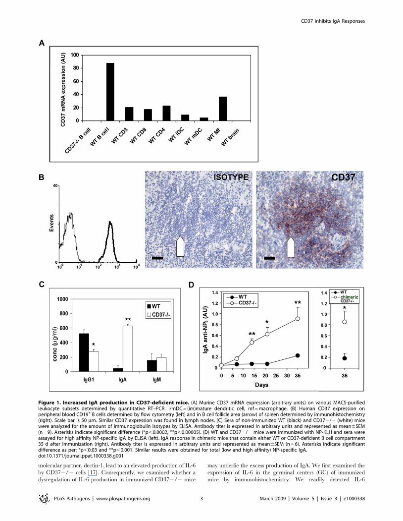
CD37 inhibits IgA production in vivoCD37 expression was determined in different mouse and
human leukocyte populations at the mRNA and protein level,
respectively. In both mice and human, B cells were found to be the
major CD37 expressing cells (Figure 1A and 1B). In lymphoid
tissue, CD37 expression was highest in the B cell follicle area as
expected (Figure 1B, data shown for spleen). This high CD37
expression on B cells prompted us to study humoral immunity in
CD37-deficient mice in detail. When analyzing the basal antibody
levels in sera of naı̈ve mice, we observed that basal levels of serum
IgA in CD372/2 mice were increased more than 15-fold
compared to C57BL/6J wild-type (WT) mice (Figure 1C). At the
same time, IgG1 levels were decreased 2-fold and serum IgM
levels were unaltered compared to WT mice. Next, we
investigated antibody responses after immunization with the T
cell-dependent antigen (NP-KLH) in CD372/2 and WT mice.
Sera were analyzed for the presence of antibodies reactive with
NP3-BSA (high affinity anti-NP antibody) and NP20-BSA (total
anti-NP antibody) [20]. Again, CD372/2 mice developed high
titers of anti-NP IgA, with .10-fold increase of high affinity IgA
compared to controls 21 days after immunization (Figure 1D).
B cells lacking CD37 are responsible for the high IgAproduction
We wanted to investigate whether CD37-deficiency on the B
cell population was indeed responsible for the high IgA response.
WT mice were sublethally irradiated and reconstituted with bone
marrow; 80% from mMT mice (B cell-deficient mice), and 20%
from WT or CD372/2 mice as a source of B cells. This created
WT mice and mice with a CD37-deficient B cell compartment
(referred to as chimeric CD372/2 mice). The percentages of
CD19-, CD3-, GR-1-, and F4/80-expressing cells assessed in
peripheral blood revealed equally efficient reconstitution in WT
and chimeric CD372/2 mice (data not shown). Next, NP-specific
antibody production was analyzed 35 days after immunization
with NP-KLH. The results clearly demonstrate that chimeric mice
with a CD372/2 B cell repertoire also produced high titers of
NP-specific IgA comparable to intact CD372/2 mice after
immunization (Figure 1D, right). Thus, we can conclude that the
elevated IgA response in CD372/2 mice is a B cell-intrinsic
defect.
Increased formation of IgA–secreting plasma cells inCD372/2 mice
The high IgA titers in serum of CD372/2 mice suggest an
enhanced generation of IgA-secreting plasma cells. To determine
if this was the case, spleens, bone marrow, and mucosal-associated
lymphoid tissue from immunized mice were examined for the
frequency of NP-specific IgA-ASC using ELISPOT assays. In line
with IgA levels in serum, the numbers of IgA-ASC were increased
in spleen of CD372/2 mice compared to control WT mice at day
14 following immunization (Figure 2A). This observation was
confirmed by immunohistochemical analysis (Figure 2B). High
numbers of IgA+ cells were present in GC and white pulp area in
spleens of immunized CD372/2 mice, whereas in spleens of WT
mice IgA+ cells were very rarely detected. CD138 stainings
confirmed that the IgA+ cells in CD372/2 spleens were indeed
plasma cells (Figure 2C). Since IgA-ASC are known to migrate
preferentially to the MALT using specific gut-homing receptors
[21], Peyer’s patches and mesenteric lymph nodes of immunized
CD372/2 mice were analyzed for the presence of NP-specific
IgA-ASC. Both the morphology and organization of mesenteric
lymph nodes (Figure 2D) and Peyer’s patches (Figure 2E) of
CD372/2 mice were comparable to WT mice, although
CD372/2 Peyer’s patches were slightly increased in size. The
percentage of IgA+ NP-specific plasma cells was increased in
MALT of immunized CD372/2 mice compared to WT controls
(Figure 2F), demonstrating that IgA-ASC generated in CD372/2
spleens preferentially homed to the MALT rather than to the bone
marrow. IgG1+ NP-specific plasma cells were hardly detected in
MALT of CD372/2 and WT mice as expected. Taken together,
CD372/2 mice possess increased generation of antigen-specific
IgA+ plasma cells after immunization.
Elevated IgA responses in CD372/2 mice are dependenton IL-6
IL-6 is a cytokine that has been implicated in promoting the
generation of IgA-secreting plasma cells [2,3]. Moreover, we have
recently shown that signals transduced through the CD37
Author Summary
Antibody, or immunoglobulin (Ig), production by plasmacells in the immune system is important for protecting thehost from microbial infections. IgA is the most abundantantibody isotype produced in the body. However, themolecular mechanisms underlying the generation of IgA–producing plasma cells remain poorly understood. Wenow report that the B cell–expressed protein CD37regulates IgA immune responses, both in steady-stateconditions and during infection. We found highly in-creased levels of IgA in serum and elevated numbers ofIgA+ plasma cells in lymphoid tissue of mice that aredeficient for CD37 (CD372/2 mice). To demonstrate theimportance of CD37 in immunity to infection, CD372/2mice were exposed to the fungus Candida albicans. C.albicans can cause systemic infection with high mortalityin immunocompromised patients. We demonstrate thatCD372/2 mice are evidently better protected frominfection than wild-type mice, which was dependent onC. albicans–specific IgA antibodies. The underlying mech-anism involved direct neutralization of fungal cells by IgA.In summary, the B cell protein CD37 inhibits IgA responsesand anti-fungal immunity. This study may contribute tothe development of novel immunotherapeutic approachesfor invasive fungal disease.
CD37 Inhibits IgA Responses
PLoS Pathogens | www.plospathogens.org 2 March 2009 | Volume 5 | Issue 3 | e1000338
molecular partner, dectin-1, lead to an elevated production of IL-6
by CD372/2 cells [17]. Consequently, we examined whether a
dysregulation of IL-6 production in immunized CD372/2 mice
may underlie the excess production of IgA. We first examined the
expression of IL-6 in the germinal centers (GC) of immunized
mice by immunohistochemistry. We readily detected IL-6
Figure 1. Increased IgA production in CD37-deficient mice. (A) Murine CD37 mRNA expression (arbitrary units) on various MACS-purifiedleukocyte subsets determined by quantitative RT–PCR. i/mDC = (im)mature dendritic cell, mf = macrophage. (B) Human CD37 expression onperipheral blood CD19+ B cells determined by flow cytometry (left) and in B cell follicle area (arrow) of spleen determined by immunohistochemistry(right). Scale bar is 50 mm. Similar CD37 expression was found in lymph nodes. (C) Sera of non-immunized WT (black) and CD372/2 (white) micewere analyzed for the amount of immunoglobulin isotypes by ELISA. Antibody titer is expressed in arbitrary units and represented as mean6SEM(n = 9). Asterisks indicate significant difference (*p,0.0002, **p,0.00005). (D) WT and CD372/2 mice were immunized with NP-KLH and sera wereassayed for high affinity NP-specific IgA by ELISA (left). IgA response in chimeric mice that contain either WT or CD37-deficient B cell compartment35 d after immunization (right). Antibody titer is expressed in arbitrary units and represented as mean6SEM (n = 6). Asterisks indicate significantdifference as per: *p,0.03 and **p,0.001. Similar results were obtained for total (low and high affinity) NP-specific IgA.doi:10.1371/journal.ppat.1000338.g001
CD37 Inhibits IgA Responses
PLoS Pathogens | www.plospathogens.org 3 March 2009 | Volume 5 | Issue 3 | e1000338

Figure 2. Increased formation of IgA–secreting plasma cells in CD372/2 mice. (A) The frequency of high affinity NP-specific IgA–ASC wasassessed in CD372/2 (white) and WT (black) spleen and bone marrow by ELISPOT at d 14 and 35 of the immune response. Non-immunized mice are
CD37 Inhibits IgA Responses
PLoS Pathogens | www.plospathogens.org 4 March 2009 | Volume 5 | Issue 3 | e1000338
expression in the GC area of spleens of immunized CD372/2
(Figure 3A). In contrast, non-immunized mice (WT and CD372/2
mice) as well as immunized WT mice had non-detectable levels of
IL-6 in spleens. IgA production by peritoneal B1 cells during T cell-
independent responses is not dependent on IL-6 [22], which
correlates with the normal IgA response in CD372/2 mice after T
cell-independent immunization (data not shown).
Next, the effect of IL-6 on IgA production during ex vivo
restimulation experiments was analyzed. Splenocytes of immu-
nized WT and CD372/2 mice were stimulated with NP-KLH in
vitro in the absence or presence of neutralizing IL-6 antibodies.
Figure 3B shows increased IgA production by CD372/2 cultures
compared to WT cells as expected. Blocking IL-6 resulted in
substantially reduced IgA production by CD372/2 cells, which
supported our hypothesis that the mechanism underlying the
elevated IgA responses in CD372/2 mice is controlled at the
level of IL-6. WT and CD372/2 cultures produced 1900 vs.
5500 pg/ml IgA respectively, which decreased to 500 vs.
2000 pg/ml in the presence of neutralizing IL-6 antibodies. We
also established that purified CD372/2 splenic B cells were
capable of autocrine IL-6 production upon restimulation in vitro
using intracellular cytokine stainings (data not shown).
To prove that increased IgA production in CD372/2 mice
was indeed dependent on IL-6 in vivo, we neutralized IL-6 in WT
and CD372/2 mice during immunizations using blocking
antibodies. Serum was analyzed for the presence of high affinity
(anti-NP3) IgA antibodies at different days after immunization.
Evidently, IL-6 neutralization reversed the production of high IgA
levels in CD372/2 serum (Figure 3C, left), demonstrating the
importance of IL-6 in IgA+ plasma cell development in CD372/2
mice. Only 25% of the CD372/2 mice produced NP-specific IgA
titers after treatment with anti–IL-6 antibodies, compared to 100%
of isotype control-treated CD372/2 mice 14 days after immuni-
zation (Figure 3C, right). IgA formation by WT mice (with or
without anti–IL-6) was below the level of detection (not shown). Our
findings are in accordance with observations made in IL-6-deficient
mice that exhibit impaired antibody production during systemic
immune responses [23–25]. Mucosal IgA responses were reported
to be significantly impaired or unaltered in IL-6-deficient mice
dependent on the model of immunization used [2,23,25,26].
Moreover, IL-6 polymorphisms were recently reported to be
associated with IgA-deficiency in patients [27]. Since IL-6 is crucial
for the induction of already-committed (surface IgA+) B cells to
become IgA-secreting plasma cells [2]), our data suggest that CD37
is directly implicated in the inhibition of the late-stage development
of IgA+ plasma cells, i.e. at the level of terminal B cell differentiation.
Taken together, we established that high IL-6 levels directly relate to
the increased IgA production in CD372/2 mice.
IgA production during fungal infection in CD37-deficientmice
Given that IgA contributes to protecting the host from microbial
infections, we hypothesized that CD37-deficiency may have
important implications for the outcome of infectious diseases.
We have recently demonstrated that CD37 interacts with the C-
type lectin dectin-1 [17], a b-glucan receptor that is required for
effective immunity to fungal infections [28,29]. Therefore, the
CD37-deficient immune response during Candida albicans infection
was explored. C. albicans normally colonizes the mucosa without
causing disease, but can cause systemic infection with high
mortality in immunocompromised patients [30,31]. In particular,
the incidence of invasive C. albicans infections is high among cancer
patients [32–34].
CD372/2 and WT mice were infected with C. albicans and IL-6
production by CD372/2 and WT splenocytes was studied upon
restimulation with fungal antigens. CD372/2 splenocytes pro-
duced increased levels of IL-6 compared to WT cells upon exposure
to either live or heat-killed C. albicans, or the fungal cell wall extract
zymosan both 3 and 7 days after infection (Figure 4A). Blocking
dectin-1 with antibody 2A11 inhibited IL-6 production by both WT
and CD372/2 splenocytes stimulated with C. albicans or the dectin-
1 ligand curdlan (Figure S1), showing that IL-6 production is
dependent on dectin-1. As such, CD37 controls dectin-1-mediated
IL-6 production, possibly by recruiting dectin-1 into tetraspanin
microdomains that may alter signal transduction pathways and
subsequent cytokine profiles. In line with our findings, IL-6-deficient
mice are more susceptible to C. albicans and A. fumigatus infection,
which is related to decreased neutrophil effector activity, impaired
Th1-mediated immune responses [25], and defective Th17
responses [35]. Studying Th2/Th1/Th17 cytokine production by
CD372/2 splenocytes revealed that IL-10 production was
comparable between CD372/2 and WT splenocytes, and cIFN
production was low but increased by CD372/2 cells 3 days after
infection (Figure 4A). The role of IL-6 in inducing Th17 responses is
well established in mice. Accordingly, we observed significantly
increased IL-17 production by CD372/2 splenocytes upon C.
albicans stimulation (Figure 4A). Th17 cells have been implicated as
an important effector mechanism against C. albicans infection
[36,37], although IL-17 may also impair anti-fungal immunity
under certain conditions [38,39].
Next, the development of fungal-specific IgA antibodies in
CD372/2 and WT mice exposed to C. albicans was analyzed. WT
mice did not generate a detectable IgA response after C. albicans
after infection. In contrast, all CD372/2 mice exhibited high
titers of IgA antibodies specific for C. albicans and zymosan in their
serum (Figure 4B). Candida-specific IgG was not detected in serum
of WT and CD372/2 mice 7 days after infection (not shown).
Finally, we investigated the role of tetraspanin CD37 in the
outcome of infectious disease. WT and CD372/2 mice were
systemically infected with C. albicans yeasts, and kidneys were
analyzed for the outgrowth of viable C. albicans after 1 and 7 days.
CD372/2 mice exhibited significantly decreased susceptibility to
infection and reduced fungal outgrowth in their kidneys –the main
target organ for C. albicans - 7 days after infection when compared
to WT mice (Figure 4C, left). Histology revealed major infection
areas with abscesses and hyphal infiltration in WT kidneys. In
contrast, morphology of kidneys of CD372/2 mice looked
shown as control (grey). Data are presented as mean6SEM (n = 6). Asterisks indicate significant differences at p,0.05. (B) Immunohistochemicalanalysis of WT (left) and CD372/2 spleens (right) demonstrating IgA–positive plasma cells (arrows) at d 14 after immunization. Scale bar is 50 mm. (C)Immunohistochemical analysis of serial sections from spleens of CD372/2 mice stained with antibodies specific for IgA (left) and plasma cell markerCD138 (right) at d 14 after immunization. Arrows indicate co-localization. Scale bar is 50 mm (upper) and 15 mm (lower). Mesenteric lymph nodes (D)and Peyer’s patches (E) of WT and CD372/2 mice were analyzed by immunohistochemistry at 14 d after NP-KLH immunization using antibodiesspecific for B cells (upper) and IgA (lower). Note the increased numbers of IgA–positive cells in CD372/2 tissue. Scale bars are 300 mm. (F) Percentageof IgA+ and IgG1+ NP-specific plasma cells in Peyer’s patches of WT (black) and CD372/2 (white) mice determined by flow cytometry at d 21 afterimmunization. Data are presented as mean6SEM (n = 4). Asterisk indicates statistical difference at p,0.05.doi:10.1371/journal.ppat.1000338.g002
CD37 Inhibits IgA Responses
PLoS Pathogens | www.plospathogens.org 5 March 2009 | Volume 5 | Issue 3 | e1000338

Figure 3. Increased IgA production after immunization in CD372/2 mice is dependent on IL-6. (A) Immunohistochemical analysis ofspleens from CD372/2 and WT mice 14 d after NP-KLH immunization. Germinal centers (identified in serial sections by PNA staining) are indicated inthe B cell follicles. Immunized WT mice have non-detectable levels of IL-6 in spleens (left). In contrast, B cell follicles in spleens of immunized CD372/2mice are clearly positive for IL-6 (red) in the GC area (right). Spleens of non-immunized mice (WT and CD372/2) were negative for IL-6 staining (notshown). Scale bar is 50 mm. (B) Effect of neutralizing anti–IL-6 during ex vivo restimulation. Splenocytes from WT and CD372/2 mice were prepared 14 dafter NP-KLH immunization, and stimulated in vitro with NP-KLH (1 mg/ml) in the absence or presence of anti–IL-6. Supernatants were collected after48 h, and assayed for IgA production by ELISA (expressed in arbitrary units). Asterisk indicates significant difference (*p,0.002). (C) IL-6 was neutralizedin WT and CD372/2 mice during immunizations using blocking IL-6 antibodies (as described in Materials and methods). High affinity NP-specific IgAwas assayed in serum of CD372/2 mice treated with anti–IL-6 (black) or control antibody (white) (left). Antibody titer is expressed in arbitrary units andrepresented as mean6SEM (n = 6). Asterisks indicate significant difference as per: *p,0.04. Histogram shows percentage of CD372/2 mice with highIgA anti-NP3 levels (above 106background level) in serum after treatment with anti–IL-6 (black) compared to control treated CD372/2 mice (white) atindicated days after immunization (right). Similar results were obtained for total NP-specific antibody (against NP20-BSA).doi:10.1371/journal.ppat.1000338.g003
CD37 Inhibits IgA Responses
PLoS Pathogens | www.plospathogens.org 6 March 2009 | Volume 5 | Issue 3 | e1000338

CD37 Inhibits IgA Responses
PLoS Pathogens | www.plospathogens.org 7 March 2009 | Volume 5 | Issue 3 | e1000338

normal with no mycelial structures and presence of leukocyte
infiltrates (Figure 4C, right). Next, we evaluated survival of WT
and CD372/2 mice after lethal fungal infection. The absence of
CD37 resulted in prolonged survival and decreased mortality after
lethal candidiasis (Figure 4D), further emphasizing the importance
of CD37 in regulating anti-fungal immunity.
In order to get more insight into the underlying mechanism,
CD372/2 serum was analyzed for fungal opsonization and/or
neutralizing capacity. IgA-mediated phagocytosis through Fc
alpha receptor CD89 is well-established in humans [40,41], in
whom serum IgA is mostly monomeric. In mice, serum IgA is
predominantly dimeric/polymeric and it is currently unknown
whether the murine Fca/m receptor is effective in clearing IgA-
opsonized pathogens [42]. We observed similar uptake of C.
albicans by GR-1+ neutrophils in the presence of WT or CD372/2
serum of infected mice (Figure 5A). Serum heat-inactivation
inhibited C. albicans uptake, demonstrating that the phagocytosis
was mainly complement-mediated and not dependent on IgA. Also,
the increase in GR-1-expressing cells in blood after C. albicans
infection was comparable between WT and CD372/2 mice
Figure 4. CD372/2 mice are protected against C. albicans infection. CD372/2 and WT mice (n = 5) were systemically infected with 16105 C.albicans. (A) Spleens were removed 3 or 7 d after infection and splenocytes were stimulated with live, heat-killed C. albicans yeasts, zymosan (E:T ratio2:1) for 48 h, after which IL-6, IL-10, IL-17, and cIFN in supernatants were measured by ELISA. Asterisks indicate significant difference as per: *p,0.05and **p,0.002. (B) Serum of CD372/2 and WT mice was analyzed for the presence of IgA antibodies reactive with C. albicans yeast (left) or zymosan(right) by flow cytometry as described in Materials and methods. Serum of non-infected mice is shown as control. Asterisks indicate significantdifference as per: *p,0.05. (C) left: Subgroups of five or six animals were sacrificed on d 1 or 7, and kidneys were analyzed for the number of viableCandida cells (expressed as log CFUg21 tissue). Horizontal bars represent mean. Asterisks indicate significant difference as per: *p,0.0005. Right:Histology of kidneys from WT and CD372/2 mice 7 d after infection. Note the major infection area and infiltrating hyphae in WT kidneys. (D) Survivalof WT and CD372/2 mice (n = 6) was assessed after intravenous injection of lethal dose of 56105 CFU C. albicans.doi:10.1371/journal.ppat.1000338.g004
Figure 5. Adoptive transfer of CD372/2 serum increases resistance of WT mice to fungal infection. (A) Percentage of GR-1+ bloodleukocytes that phagocytosed FITC-labeled C. albicans in the absence or presence of serum from infected WT or CD372/2 mice for various timepoints at 37uC was quantified by flow cytometry. Sera were heat-inactivated (56uC for 30 min) to inactivate complement (left). In addition, uptake inthe presence of WT or CD372/2 serum was analyzed by fluorescence light microscopy (right; data shown for 30-min. incubation). Bar represents10 mm. (B) C. albicans was incubated in the absence or presence of 20% serum (from infected WT or CD372/2 mice) or 20% IgA–depleted serum andgrowth (CFU) was assessed. Asterisks indicate significant difference as per: p,0.005. (C) WT mice (n = 5) were systemically infected with 16105 C.albicans and untreated, or treated with either pooled CD372/2 serum containing high levels of fungal-specific IgA, or IgA-depleted CD372/2serum. Kidneys were analyzed for the number of viable Candida cells (expressed as log CFUg21 tissue) 7 d after infection. Horizontal bars representsmean. Asterisks indicate significant difference as per: p,0.05.doi:10.1371/journal.ppat.1000338.g005
CD37 Inhibits IgA Responses
PLoS Pathogens | www.plospathogens.org 8 March 2009 | Volume 5 | Issue 3 | e1000338

(Figure S2). Thus, these data demonstrate that IgA in CD372/2
serum does not increase C. albicans phagocytosis by neutrophils.
Next, we investigated whether serum IgA of CD372/2 mice
could directly neutralize fungal cells. C. albicans was grown in the
absence or presence of WT and CD372/2 serum of infected
mice and viability was assessed by classical plate assays. CD372/2
serum significantly inhibited C. albicans growth in contrast to WT
serum (Figure 5B). To demonstrate that the growth inhibition was
due to fungal-specific IgA, CD372/2 serum was depleted for IgA.
Serum deprived from IgA abrogated the effect, and C. albicans
growth was comparable between WT and CD372/2 serum. From
these findings we conclude that IgA in CD372/2 serum directly
neutralizes C. albicans and may thereby prevent dissemination in vivo.
To test this hypothesis, WT mice were infected and treated with
pooled CD372/2 serum containing high levels of IgA anti-C.
albicans. We observed that WT mice were protected from fungal
disease by CD372/2 serum treatment (Figure 5C). Importantly,
the resistance was abrogated when IgA was depleted from
CD372/2 serum, demonstrating that the passive transfer of
resistance to candidiasis in CD372/2 mice is dependent on IgA.
Although the elevated IL-6-IL-17 response may also contribute to
increased anti-fungal effector activity in CD372/2 mice
(Figure 4A), our passive transfer data clearly show that C.
albicans-specific IgA alone is sufficient to confer protection against
invasive fungal disease. Whilst several studies have alluded to a
role for the development of protective anti-fungal antibodies in
experimental candidiasis [43,44], this is—to the best of our
knowledge—the first description of an important role for IgA in
protection against systemic candidiasis. These studies also
highlight the non-redundant role of the tetraspanin CD37, as
CD37-deficiency stimulates anti-fungal immunity even in the
presence of other tetraspanins. The results of this study may
stimulate the development of novel immunotherapeutic strategies
for invasive fungal disease that are urgently warranted [33].
In summary, we reveal a unique role for the tetraspanin CD37
in the humoral immune response; CD37 is a negative regulator of
IgA+ plasma cell generation in vivo and this role is critically
dependent on IL-6. Moreover, this is the first demonstration that
tetraspanins may control the immune-mediated defense against
fungal pathogens. This study increases our understanding of
molecular mechanisms underlying the formation of IgA-secreting
plasma cells and may contribute to better insight into anti-fungal
immunity.
Materials and Methods
Quantitative RT–PCRMurine CD37 expression was analyzed at the mRNA level by
quantitative RT–PCR due to unavailability of anti-mouse CD37
antibodies. Total RNA was extracted from WT and CD372/2
leukocyte populations (purified by MACS sorting, Miltenyi Biotec)
using Trizol (Invitrogen) and transcribed into cDNA using random
hexamer primers (Amersham) and Superscript II RT (Invitrogen).
Brain tissue was taken as negative control. Real-time RT–PCR
was performed in ABI Prism Sequence Detection system 7000.
cDNA was amplified using SYBR Green PCR mastermix (Applied
Biosystems) with mouse CD37 primers (59GTCCTTTGT-
GGGTTTGTCCTT and 59GAGACAGCGCAGCTCCTT-
TAG). The amount of CD37 mRNA expression was normalized
to the housekeeping gene PBGD.
Human CD37 expressionPeripheral blood lymphocytes were stained with anti-CD37
(clone WR17, Serotec) and processed for FACS analysis as
previously described [17]. CD37 staining in tissue was performed
on frozen sections (6 mm) of spleen and lymph node that were
fixed in acetone for 10 minutes at 220uC. Slides were blocked
(5% horse serum) and stained with anti-CD37 or mIgG2a isotype
control. Horse anti-mouse-biotin (Molecular Probes) was used as
secondary Ab, and staining was revealed using SA-alkaline
phosphatase labeling kit (Vector Laboratories) with Fast Red
substrate. Slides were counterstained with Meyer’s heamatoxylin.
MiceCD372/2 mice were generated by homologous recombination
[14] and backcrossed 10 times to the C57BL/6J background. Age
and sex-matched C57BL/6J wild type (WT) mice were obtained
from the Walter and Eliza Hall Institute (Melbourne, Victoria,
Australia) and Charles River (France). CD372/2 mice were bred
at the Burnet Institute Austin Campus (Heidelberg, Victoria,
Australia) and the Central Animal Laboratory (Radboud Univer-
sity Nijmegen, The Netherlands). mMT (B cell-deficient) mice [45]
were bred at the Walter and Eliza Hall Institute. Mice were used
at 8–12 weeks of age. Animal studies were approved by animal
ethics Committee of the Austin&Repatriation Medical Centre,
and the Nijmegen Animal Experiments Committee.
ImmunizationWT and CD372/2 mice (n = 6) were injected intraperitoneally
with 100 mg of NP (4-hydroxy-3-nitrophenylacetate) coupled to
Keyhole Limpet Hemocyanin (KLH) (NP17:KLH, conjugation
ratio, 17:1) precipitated in alum. Mice were bled and sera collected
at days 7, 14, 21 and 35 post-immunization. Lymphoid organs
(spleens, bone marrow, mesenteric lymph nodes, and Peyer’s
patches) were frozen for immunohistochemistry or processed for
FACS analysis.
Detection of Ag-specific immunoglobulin and antibody-secreting cells
Basal immunoglobulin levels in serum of non-immunized mice
(n = 9) were determined by ELISA using goat anti-mouse IgG/
IgA/IgM (BD-Pharmingen). Ig standards were purchased from
BD. Levels of NP-specific antibodies in serum after immunization
were assayed by ELISA using plates coated with 20 mg/mL of
either NP20-BSA or NP3-BSA to detect total and high affinity anti-
NP antibody, respectively. The frequency of total and high affinity
NP-specific ASC in spleen and bone marrow was determined by
ELISPOT as described [20,46], again using NP20-BSA and NP3-
BSA for antibody capture.
ImmunohistochemistrySpleens, mesenteric lymph nodes and Peyer’s patches taken
from mice before and after immunization were embedded in
OCT. Frozen sections (6 mm) were fixed in acetone for 10 min. at
220 C. Slides were blocked (5% goat serum) and stained with
anti-IgA, anti-IgG1, anti-IL6, anti-CD138, anti-CD45R (B220)
(all from BD-Pharmingen), or isotype controls. Goat anti-rat-biotin
(Molecular Probes) was used as secondary Ab, and staining was
revealed using SA-alkaline phosphatase labeling kit (Vector
Laboratories) with Fast Red substrate. Slides were counterstained
with Meyer’s heamatoxylin.
Reconstitution experimentsBone marrow chimeras were generated as previously described
[47]. Briefly, 107 (CD372/2 or WT) and mMT (CD37+) bone
marrow cells were injected intravenously into sublethally irradi-
ated C57Bl/6J recipients at a ratio of 4 parts mMT to 1 part WT
CD37 Inhibits IgA Responses
PLoS Pathogens | www.plospathogens.org 9 March 2009 | Volume 5 | Issue 3 | e1000338

or CD372/2 bone marrow as a source for B cells. This created
WT mice and mice with a CD37-deficient B cell compartment
(referred to as chimeric CD372/2 mice). Six weeks after
reconstitution, heparinized blood was obtained from chimeric
mice and reconstitution of all major leukocyte populations was
monitored by flow cytometry, using the antibodies against: PE-
conjugated 1D3-PE (CD19, BD-Pharmingen), PE-conjugated
RB6-8C5-PE (GR-1, BD-Pharmingen), Cy5-conjugated KT3.1
(CD3, made in house), PE-conjugated F4/80 (BD-Pharmingen),
and FITC-conjugated M1/70 (CD11b, made in house). 2 weeks
thereafter, mice (n = 7) were immunized with NP-KLH as
described above. Sera were collected 35 days after immunization
and analyzed for presence of NP-specific antibodies by ELISA.
Ex vivo restimulation experimentsSplenocytes were prepared from WT and CD372/2 mice
14 days after NP-KLH immunization. Cells were stimulated with
NP-KLH (1 mg/ml) in the absence or presence of 5 mg/ml (azide-
and LPS-free) neutralizing anti–IL-6 MP5-20F3 (Biolegend).
Supernatants were collected after 48 h, and IgA production was
determined by ELISA.
IL-6 neutralization in vivoWT and CD372/2 mice (n = 6) were immunized with NP-
KLH as described above. Mice received 2 injections of
neutralizing (azide- and LPS-free) anti–IL-6 MP5-20F3 or isotype
control; 60 mg intraperitoneally at day 0, and 60 mg intravenously
7 days after immunization. Mice were bled and sera collected at
days 7, 14, 21 and 35 post-immunization. Lymphoid organs
(spleens, bone marrow, mesenteric lymph nodes, and Peyer’s
patches) were frozen in OCT for immunohistochemistry or
processed for FACS analysis.
Candida albicans infection protocolsC. albicans ATCC MYA-3573 (UC820), a well-described clinical
strain [48], was injected intravenously (16105 colony-forming units
(CFU), in 100 mL sterile pyrogen-free saline) in CD372/2 and WT
mice. Blood (50 ml) was collected at different time points after
infection and stained with GR-1-PE to identify neutrophils by flow
cytometry. Subgroups of five or six animals were sacrificed on day 1
or 7, and kidneys were removed aseptically, weighed and
homogenized in sterile saline in a tissue grinder. The number of
viable Candida cells in the tissues was determined by plating serial
dilutions on Sabouraud dextrose agar plates. Colony-forming units
were counted after 24 h of incubation at 37uC, and expressed as log
CFUg21 tissue. Alternatively, kidneys were were embedded in OCT,
and frozen sections (6 mm) were stained with periodic acid-Schiff to
identify C. albicans by histology. In adoptive transfer experiments,
WT mice (n = 5) received 16105 CFU C. albicans intravenously (in
100 mL sterile pyrogen-free saline) with 20% pooled serum of
infected CD372/2 mice that was either untreated or depleted for
IgA (as described below). Colony-forming units were determined in
kidneys 7 days after infection. For survival experiments, CD372/2
and WT mice (n = 6) were injected intravenously with lethal dose of
56105 CFU C. albicans and mice were assessed twice daily.
Restimulation assay with fungal antigensTo assess cytokine production, spleens were removed 3 or
7 days after infection and 56106 splenocytes were stimulated with
16107 live or heat-killed C. albicans yeasts (E:T ratio 2:1), or 1 mg/
ml zymosan (Sigma). Measurement of IL-6, IL-10, IL-17 and
cIFN was performed in supernatants collected after 48 h of
incubation at 37uC (5% CO2) in 24-well plates by commercial
ELISA assays (Biosource, Camarillo, CA; detection limit 16 pg/
ml), according to the instructions of the manufacturer. In dectin-1
blocking studies, antibody 2A11 (10 mg/ml) was added during
stimulations as described [17]. Curdlan is a specific dectin-1 ligand
with no TLR2- or TLR4-stimulating properties [49].
Detection of anti-Candida IgA antibodies in miceSerum of CD372/2 and WT mice (n = 5) was collected 7 days
after C. albicans infection, and different dilutions were incubated
with 16105 Candida or 16105 zymosan particles for 30 min. at
4uC. After washing twice in saline, Candida particles were
incubated with biotinylated anti-mouse IgA (BD-Pharmingen)
followed by SA-CY5 (Invitrogen), and analyzed by flow cytometry.
Serum of non-infected mice was used as control.
IgA depletion of serumStreptavidin-coated 2 mm microspheres (Polysciences, PA) were
incubated with biotinylated anti-mouse IgA (BD Biosciences)
according to the instructions of the manufacturer. Sera of WT and
CD372/2 mice (n = 4) were collected 7 days after C. albicans
infection, pooled, and incubated with 1006106 anti-IgA coated
microspheres (1 h at RT, shaking).
C. albicans phagocytosis and neutralization assaysC. albicans phagocytosis was performed as described [41]. Briefly,
FITC-labeled C. albicans particles were incubated with blood
leukocytes (ratio 4:1) in the absence or presence of serum from
infected WT or CD372/2 mice for various time points at 37uC. As
control, sera were heat-inactivated at 56uC for 30 min. to inactivate
complement. GR-1-PE was used to stain neutrophils and phagocy-
tosis was quantified by flow cytometry. In addition, uptake was
analyzed by fluorescence light microscopy. For neutralization
experiments, C. albicans was cultured overnight at 37uC in Sabouraud
dextrose medium (Oxoid, UK), washed, and 16105 yeasts were
incubated in the absence or presence of 20% serum (from WT or
CD372/2 mice; collected 7 days after infection) or 20% IgA-
depleted serum. Viability was assessed after shaking for 5 h at 37uCby plating serial dilutions on Sabouraud dextrose agar plates.
Statistical analysesData are presented as mean6SEM. Statistical differences were
determined using the unpaired Student’s t-test. Significance was
accepted at the p,0.05 level.
Accession/ID numbersHuman CD37, GeneID 951; Murine CD37, GeneID 12493,
MGI: 88330; Murine IL-6, GeneID: 16193, MGI: 96559; Murine
IgA, GeneID: 238447, MGI: 96444.
Supporting Information
Figure S1 IL-6 production by WT and CD372/2 splenocytes
stimulated by C. albicans is dependent on dectin-1. Spleens were
removed from CD372/2 and WT mice 7 d after infection
(16105 CFU C. albicans) and restimulated with heat-killed C.
albicans (E:T ratio 2:1) or the dectin-1 ligand Curdlan (100 mg/ml)
for 48 h, after which IL-6 in supernatants were measured by
ELISA. Antibody 2A11 was added during stimulations (10 mg/ml)
to block dectin-1.
Found at: doi:10.1371/journal.ppat.1000338.s001 (0.01 MB PDF)
Figure S2 Normal increase of GR-1-positive cells in blood of
CD372/2 mice. Granulopoiesis in WT and CD372/2 mice
(n = 5) systemically infected with 16105 CFU C. albicans. Percentage
CD37 Inhibits IgA Responses
PLoS Pathogens | www.plospathogens.org 10 March 2009 | Volume 5 | Issue 3 | e1000338

of GR-1-positive cells in blood was determined by flow cytometry at
different time points after infection.
Found at: doi:10.1371/journal.ppat.1000338.s002 (0.01 MB PDF)
Acknowledgments
We thank David Tarlinton (The Walter and Eliza Hall Institute of Medical
Research, Australia) for helpful discussions. Maaike van Hout assisted us
with the quantitative RT–PCR experiments. Furthermore, we greatly
appreciate the technical help from the animal house staff of both the
Central Animal Facility of the University Medical Centre Nijmegen and
the Burnet Institute Austin Campus.
Author Contributions
Conceived and designed the experiments: ABvS MGN GJA MDW CGF.
Performed the experiments: ABvS MS KHG AvdS IV BEL. Analyzed the
data: ABvS MS KHG AvdS IV RT RAPR BEL MGN GJA MDW CGF.
Contributed reagents/materials/analysis tools: ABvS RT RAPR BEL
MGN MDW CGF. Wrote the paper: ABvS MDW CGF.
References
1. Hirano T, Taga T, Nakano N, Yasukawa K, Kashiwamura S, et al. (1985)
Purification to homogeneity and characterization of human B-cell differentiation
factor (BCDF or BSFp-2). Proc Natl Acad Sci U S A 82: 5490–5494.
2. Ramsay AJ, Husband AJ, Ramshaw IA, Bao S, Matthaei KI, et al. (1994) The
role of interleukin-6 in mucosal IgA antibody responses in vivo. Science 264:
561–563.
3. Mora JR, Iwata M, Eksteen B, Song SY, Junt T, et al. (2006) Generation of gut-
homing IgA-secreting B cells by intestinal dendritic cells. Science 314: 1157–1160.
4. McGhee JR, Kunisawa J, Kiyono H (2007) Gut lymphocyte migration: we are
halfway ‘home’. Trends Immunol 28: 150–153.
5. Kelsoe G (1996) Life and death in germinal centers (redux). Immunity 4:
107–111.
6. McHeyzer-Williams LJ, Driver DJ, McHeyzer-Williams MG (2001) Germinal
center reaction. Curr Opin Hematol 8: 52–59.
7. Shapiro-Shelef M, Calame K (2005) Regulation of plasma-cell development. Nat
Rev Immunol 5: 230–242.
8. Cerutti A, Rescigno M (2008) The biology of intestinal immunoglobulin A
responses. Immunity 28: 740–750.
9. Wright MD, Tomlinson MG (1994) The ins and outs of the transmembrane 4
superfamily. Immunol Today 15: 588–594.
10. Berditchevski F (2001) Complexes of tetraspanins with integrins: more than
meets the eye. J Cell Sci 114: 4143–4151.
11. Tarrant JM, Robb L, van Spriel AB, Wright MD (2003) Tetraspanins: molecular
organisers of the leukocyte surface. Trends Immunol 24: 610–617.
12. Hemler ME (2005) Tetraspanin functions and associated microdomains. Nat
Rev Mol Cell Biol 6: 801–811.
13. Wright MD, Moseley GW, van Spriel AB (2004) Tetraspanin microdomains in
immune cell signalling and malignant disease. Tissue Antigens 64: 533–542.
14. Knobeloch KP, Wright MD, Ochsenbein AF, Liesenfeld O, Lohler J, et al. (2000)
Targeted inactivation of the tetraspanin CD37 impairs T-cell-dependent B-cell
response under suboptimal costimulatory conditions. Mol Cell Biol 20: 5363–5369.
15. van Spriel AB, Puls KL, Sofi M, Pouniotis D, Hochrein H, et al. (2004) A
regulatory role for CD37 in T cell proliferation. J Immunol 172: 2953–2961.
16. Sheng KC, van Spriel AB, Gartlan KH, Sofi M, Apostolopoulos V, et al. (2008)
Tetraspanins CD37 and CD151 differentially regulate Ag presentation and T-
cell co-stimulation by DC. Eur J Immunol 39: 50–55.
17. Meyer-Wentrup F, Figdor CG, Ansems M, Brossart P, Wright MD, et al. (2007)
Dectin-1 interaction with tetraspanin CD37 inhibits IL-6 production. J Immunol
178: 154–162.
18. Ariizumi K, Shen GL, Shikano S, Xu S, Ritter R 3rd, et al. (2000) Identification
of a novel, dendritic cell-associated molecule, dectin-1, by subtractive cDNA
cloning. J Biol Chem 275: 20157–20167.
19. Brown GD (2006) Dectin-1: a signalling non-TLR pattern-recognition receptor.
Nat Rev Immunol 6: 33–43.
20. Smith KG, Light A, Nossal GJ, Tarlinton DM (1997) The extent of affinity
maturation differs between the memory and antibody-forming cell compart-
ments in the primary immune response. Embo J 16: 2996–3006.
21. Kunkel EJ, Butcher EC (2003) Plasma-cell homing. Nat Rev Immunol 3:
822–829.
22. Beagley KW, Bao S, Ramsay AJ, Eldridge JH, Husband AJ (1995) IgA
production by peritoneal cavity B cells is IL-6 independent: implications for
intestinal IgA responses. Eur J Immunol 25: 2123–2126.
23. Kopf M, Baumann H, Freer G, Freudenberg M, Lamers M, et al. (1994)
Impaired immune and acute-phase responses in interleukin-6-deficient mice.
Nature 368: 339–342.
24. Kopf M, Herren S, Wiles MV, Pepys MB, Kosco-Vilbois MH (1998) Interleukin
6 influences germinal center development and antibody production via a
contribution of C3 complement component. J Exp Med 188: 1895–1906.
25. Romani L, Mencacci A, Cenci E, Spaccapelo R, Toniatti C, et al. (1996) Impaired
neutrophil response and CD4+ T helper cell 1 development in interleukin 6-
deficient mice infected with Candida albicans. J Exp Med 183: 1345–1355.
26. Bromander AK, Ekman L, Kopf M, Nedrud JG, Lycke NY (1996) IL-6-deficient
mice exhibit normal mucosal IgA responses to local immunizations and
Helicobacter felis infection. J Immunol 156: 4290–4297.
27. Lopez-Mejias R, Martinez A, Del Pozo N, Fernandez-Arquero M, Ferreira A, et
al. (2008) Interleukin-6 gene variation in Spanish patients with immunoglobulin-
A deficiency. Hum Immunol 69: 301–305.
28. Taylor PR, Tsoni SV, Willment JA, Dennehy KM, Rosas M, et al. (2007)
Dectin-1 is required for beta-glucan recognition and control of fungal infection.
Nat Immunol 8: 31–38.29. Saijo S, Fujikado N, Furuta T, Chung SH, Kotaki H, et al. (2007) Dectin-1 is
required for host defense against Pneumocystis carinii but not against Candidaalbicans. Nat Immunol 8: 39–46.
30. Gudlaugsson O, Gillespie S, Lee K, Vande Berg J, Hu J, et al. (2003)Attributable mortality of nosocomial candidemia, revisited. Clin Infect Dis 37:
1172–1177.
31. Netea MG, Brown GD, Kullberg BJ, Gow NA (2008) An integrated model ofthe recognition of Candida albicans by the innate immune system. Nat Rev
Microbiol 6: 67–78.32. DiNubile MJ, Hille D, Sable CA, Kartsonis NA (2005) Invasive candidiasis in
cancer patients: observations from a randomized clinical trial. J Infect 50:
443–449.33. van Spriel AB (2003) Novel immunotherapeutic strategies for invasive fungal
disease. Curr Drug Targets Cardiovasc Haematol Disord 3: 209–217.34. Koh AY, Kohler JR, Coggshall KT, Van Rooijen N, Pier GB (2008) Mucosal
damage and neutropenia are required for Candida albicans dissemination. PLoSPathog 4: e35. doi:10.1371/journal.ppat.0040035.
35. Korn T, Bettelli E, Gao W, Awasthi A, Jager A, et al. (2007) IL-21 initiates an
alternative pathway to induce proinflammatory T(H)17 cells. Nature 448:484–487.
36. Huang W, Na L, Fidel PL, Schwarzenberger P (2004) Requirement ofinterleukin-17A for systemic anti-Candida albicans host defense in mice.
J Infect Dis 190: 624–631.
37. LeibundGut-Landmann S, Gross O, Robinson MJ, Osorio F, Slack EC, et al.(2007) Syk- and CARD9-dependent coupling of innate immunity to the
induction of T helper cells that produce interleukin 17. Nat Immunol 8:630–638.
38. Zelante T, De Luca A, Bonifazi P, Montagnoli C, Bozza S, et al. (2007) IL-23and the Th17 pathway promote inflammation and impair antifungal immune
resistance. Eur J Immunol 37: 2695–2706.
39. Bozza S, Zelante T, Moretti S, Bonifazi P, DeLuca A, et al. (2008) Lack of TollIL-1R8 exacerbates Th17 cell responses in fungal infection. J Immunol 180:
4022–4031.40. Weisbart RH, Kacena A, Schuh A, Golde DW (1988) GM-CSF induces human
neutrophil IgA-mediated phagocytosis by an IgA Fc receptor activation
mechanism. Nature 332: 647–648.41. van Spriel AB, van den Herik-Oudijk IE, van Sorge NM, Vile HA, van Strijp JA,
et al. (1999) Effective phagocytosis and killing of Candida albicans via targetingFcgammaRI (CD64) or FcalphaRI (CD89) on neutrophils. J Infect Dis 179:
661–669.
42. Shibuya A, Sakamoto N, Shimizu Y, Shibuya K, Osawa M, et al. (2000) Fcalpha/mu receptor mediates endocytosis of IgM-coated microbes. Nat Immunol
1: 441–446.43. Casadevall A, Feldmesser M, Pirofski LA (2002) Induced humoral immunity and
vaccination against major human fungal pathogens. Curr Opin Microbiol 5:386–391.
44. Wagner RD, Vazquez-Torres A, Jones-Carson J, Warner T, Balish E (1996) B
cell knockout mice are resistant to mucosal and systemic candidiasis ofendogenous origin but susceptible to experimental systemic candidiasis. J Infect
Dis 174: 589–597.45. Kitamura D, Roes J, Kuhn R, Rajewsky K (1991) A B cell-deficient mouse by
targeted disruption of the membrane exon of the immunoglobulin mu chain
gene. Nature 350: 423–426.46. Lalor PA, Nossal GJ, Sanderson RD, McHeyzer-Williams MG (1992)
Functional and molecular characterization of single, (4-hydroxy-3-nitropheny-l)acetyl (NP)-specific, IgG1+ B cells from antibody-secreting and memory B cell
pathways in the C57BL/6 immune response to NP. Eur J Immunol 22:3001–3011.
47. Huntington ND, Xu Y, Puthalakath H, Light A, Willis SN, et al. (2006) CD45
links the B cell receptor with cell survival and is required for the persistence ofgerminal centers. Nat Immunol 7: 190–198.
48. Lehrer RI, Cline MJ (1969) Interaction of Candida albicans with humanleukocytes and serum. J Bacteriol 98: 996–1004.
49. Ferwerda G, Meyer-Wentrup F, Kullberg BJ, Netea MG, Adema GJ (2008)
Dectin-1 synergizes with TLR2 and TLR4 for cytokine production in humanprimary monocytes and macrophages. Cell Microbiol 10: 2058–2066.
CD37 Inhibits IgA Responses
PLoS Pathogens | www.plospathogens.org 11 March 2009 | Volume 5 | Issue 3 | e1000338