IgA-associated Inhibition of Polymorphonuclear Leukocyte Chemotaxis in Neutrophilic Dermatoses

see commentary on page 477
The Oxford classification of IgA nephropathy:rationale, clinicopathological correlations, andclassificationA Working Group of the International IgA Nephropathy Network and the Renal Pathology Society:Daniel C. Cattran1,w, Rosanna Coppo2,w, H. Terence Cook3,w, John Feehally4,w, Ian S.D. Roberts5,w,Stephan Troyanov6,w, Charles E. Alpers7, Alessandro Amore2, Jonathan Barratt4, Francois Berthoux8,Stephen Bonsib9, Jan A. Bruijn10, Vivette D’Agati11, Giuseppe D’Amico12, Steven Emancipator13,Francesco Emma14, Franco Ferrario15, Fernando C. Fervenza16, Sandrine Florquin17, Agnes Fogo18,Colin C. Geddes19, Hermann-Josef Groene20, Mark Haas21, Andrew M. Herzenberg22, Prue A. Hill23,Ronald J. Hogg24, Stephen I. Hsu25, J. Charles Jennette26, Kensuke Joh27, Bruce A. Julian28,Tetsuya Kawamura29, Fernand M. Lai30, Chi Bon Leung31, Lei-Shi Li32, Philip K.T. Li31, Zhi-Hong Liu32,Bruce Mackinnon19, Sergio Mezzano33, F. Paolo Schena34, Yasuhiko Tomino35, Patrick D. Walker36,Haiyan Wang37, Jan J. Weening38, Nori Yoshikawa39 and Hong Zhang37,*
IgA nephropathy is the most common glomerular disease
worldwide, yet there is no international consensus for its
pathological or clinical classification. Here a new classification
for IgA nephropathy is presented by an international
consensus working group. The goal of this new system
was to identify specific pathological features that more
accurately predict risk of progression of renal disease in IgA
nephropathy, thus enabling both clinicians and pathologists
to improve individual patient prognostication. In a
retrospective analysis, sequential clinical data were obtained
on 265 adults and children with IgA nephropathy who were
followed for a median of 5 years. Renal biopsies from all
patients were scored by pathologists blinded to the clinical
data for pathological variables identified as reproducible by
an iterative process. Four of these variables: (1) the mesangial
hypercellularity score, (2) segmental glomerulosclerosis,
(3) endocapillary hypercellularity, and (4) tubular atrophy/
interstitial fibrosis were subsequently shown to have
independent value in predicting renal outcome. These specific
pathological features withstood rigorous statistical analysis
even after taking into account all clinical indicators available
at the time of biopsy as well as during follow-up. The features
have prognostic significance and we recommended they be
taken into account for predicting outcome independent of the
clinical features both at the time of presentation and during
follow-up. The value of crescents was not addressed due to
their low prevalence in the enrolled cohort.
Kidney International (2009) 76, 534–545; doi:10.1038/ki.2009.243;
published online 1 July 2009
KEYWORDS: glomerulonephritis; IgA nephropathy; Oxford classification;
pathology; renal failure
IgA nephropathy (IgAN) is the commonest glomerulardisease worldwide, yet there is no international consensusfor its pathological or clinical classification. Nephrologistsuse clinical information to identify the risk of developingprogressive chronic kidney disease in individual patients withIgAN. There is now extensive evidence that a number ofclinical features at presentation predict risk of progressivechronic kidney disease. In published series, these consistentlyinclude extent of proteinuria, hypertension, and excretoryrenal function.1–5 Recent work also indicates the prognosticimportance of reduction in proteinuria during follow-up,allowing continuing refinement of the prognostic informa-tion given to an individual patient.6 Pathologists havedeveloped a number of classifications of IgAN over the last25 years; some are semiquantitative,7–10 others are single-grade classifications.11–15 Each of these classifications hasbeen developed from expert opinion, each has strengths andlimitations in predicting prognosis, and none has gained pre-eminence. There is continuing debate whether pathologicalfeatures seen on renal biopsy contribute additional prognosticinformation beyond that provided by clinical features.16
This lack of consensus on classifications based onpathology has weakened a number of areas of investigationinto IgAN. It has contributed to slow progress in developinga prognostic system with the sensitivity and specificityto predict outcome for individual patients. It has reduced
o r i g i n a l a r t i c l e http://www.kidney-international.org
& 2009 International Society of Nephrology
Received 17 November 2008; revised 8 April 2009; accepted 19 May
2009; published online 1 July 2009
Correspondence: John Feehally, The John Walls Renal Unit, Leicester
General Hospital, Leicester LE5 4PW, UK. E-mail: [email protected]
wThese authors contributed equally to the work and are named in
alphabetical order.
*Authors’ affiliations are listed in the Acknowledgements.
534 Kidney International (2009) 76, 534–545
the capacity to make international comparisons betweendifferent outcome studies, and it has limited opportunities torefine the stratification of risk for the design of clinicalintervention trials. This is a major disadvantage in a slowlyprogressive disease like IgAN where large studies of longduration are needed to evaluate new interventions unlesspatients with a high risk of progression can be better definedearly in the course of the disease.
In 2004, a proposal to develop a consensus clinico-pathological classification came from the International IgANephropathy Network—an informal network of nephro-logists and scientists with representation from the majorityof nephrology research groups around the world active in thefield of IgAN (www.IgAN-world.org)—and members of theRenal Pathology Society interested in IgAN. A questionnaireamong renal pathologists showed support for the develop-ment of a consensus classification of IgAN provided it couldbe shown to have real clinical utility. Therefore, representa-tives of the International IgA Nephropathy Network and theRenal Pathology Society established a working group to seekagreement on an evidence-based clinicopathological con-sensus classification for IgAN.17 From the beginning this wasdesigned as an international consensus aiming to involvenephrologists and pathologists from as many parts of theworld as possible, including all areas where IgAN is known tobe of high prevalence. Eventually, the consensus group hadrepresentation from 10 countries on four continents.
The goal of the new classification was to identify specificpathological features that more accurately predict risk ofprogression of renal disease in IgAN, which would enableboth clinicians and pathologists to improve individualpatient prognostication. It was recognized that such aclassification may also in the future facilitate the identifica-tion of specific features that may predict response toimmunosuppression or other specific treatments, and refinerecruitment to clinical trials by their capacity to stratifypatients by their risk of progression.
The work was approached without preconceptions to testobjectively the predictive power of a wide range of pathologicalfeatures. The consensus work required unity of purpose and acollaborative approach. Organizational challenges included thedevelopment of tools allowing consistent data collection, andthe need to meet the varying requirements of institutionalreview boards and ethics committees so that anonymizedpathological material and clinical data could be circulatedwithin and beyond its country of origin.
The overall approach used by the working group was thefollowing:
K Agreement on a clinical data set useful for outcomestudies in IgAN;
K Identification of centers willing to provide cases withsufficient clinical data and biopsy material available forreview, including cohorts varying in age, and ingeographical and racial origin;
K Agreement on definitions and scoring of a wide range ofpathological features;
K Testing reproducibility between pathologists of scoringthese features; and
K Analysis of informative pathological features in thecontext of clinical outcome to develop a classification.
By this rigor of approach, we aimed to gain the confidenceof clinicians and investigators worldwide, so that the newclassification will become the norm in routine clinicalpractice and in future clinicopathological outcome reportsof IgAN.
The working group had two consensus meetings in 2005and 2008, both held in Oxford, UK. It has, therefore, beendecided to call the product of our work, the OxfordClassification of IgA Nephropathy.
RESULTS
Clinical data and adequate renal biopsy material from 265patients with IgAN were collected from eight countrieson four continents. Five centers from Asia, six from Europe,two from United States, one from South America, andtwo multicenter networks (Canada and USA) participated inthe study (Table 1). The proportion of children wassimilar in each continent (B30%).
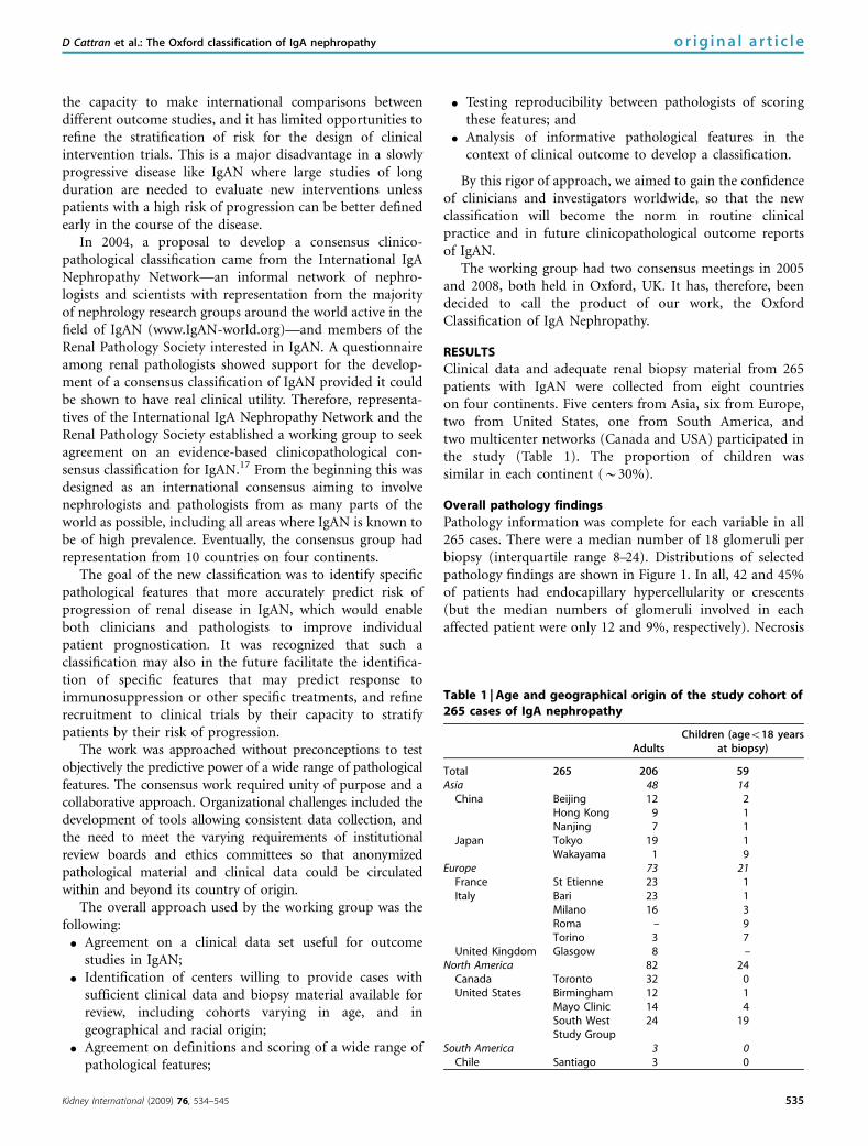
Overall pathology findings
Pathology information was complete for each variable in all265 cases. There were a median number of 18 glomeruli perbiopsy (interquartile range 8–24). Distributions of selectedpathology findings are shown in Figure 1. In all, 42 and 45%of patients had endocapillary hypercellularity or crescents(but the median numbers of glomeruli involved in eachaffected patient were only 12 and 9%, respectively). Necrosis
Table 1 | Age and geographical origin of the study cohort of265 cases of IgA nephropathy
AdultsChildren (ageo18 years
at biopsy)
Total 265 206 59Asia 48 14
China Beijing 12 2Hong Kong 9 1Nanjing 7 1
Japan Tokyo 19 1Wakayama 1 9
Europe 73 21France St Etienne 23 1Italy Bari 23 1
Milano 16 3Roma – 9Torino 3 7
United Kingdom Glasgow 8 –North America 82 24
Canada Toronto 32 0United States Birmingham 12 1
Mayo Clinic 14 4South WestStudy Group
24 19
South America 3 0Chile Santiago 3 0
Kidney International (2009) 76, 534–545 535
D Cattran et al.: The Oxford classification of IgA nephropathy o r i g i n a l a r t i c l e

was seen in only six cases and glomerular basementmembrane duplication in 30 cases. The majority of patientshad no arterial or arteriolar lesions.
Reproducibility of pathology variables
The reproducibility of the various pathological lesions isdescribed in detail in the accompanying paper (Robertset al., The Oxford Classification of IgA Nephropathypathology definitions, correlations and reproducibility).18
Following refinement of the definitions, those pathologicallesions that continued to show poor reproducibility within theworking group were not incorporated into the final classifica-tion, as even lower reproducibility could be expected in routinepractice without the advantage of the iterative processes of theworking group. These included the percentage of normalglomeruli as well as the percentage of glomeruli showingadhesions, glomerular basement membrane duplication, ne-crosis, arteriolar lesions, and interstitial inflammation involvingnon-fibrotic cortex. Reproducibility of scoring for adhesionsincreased when combined with segmental sclerosis, suggestingthat different pathologists identified the same lesion as eithersegmental sclerosis or an adhesion. For subsequent analysis,segmental sclerosis and adhesions were summed.
Correlation between pathology variables
Details of the correlations between the different pathologyvariables are presented in the accompanying paper (Robertset al.). For those variables that displayed considerable correla-tion (r40.8), it was decided to include only one variable fromeach group for further consideration based on reproducibility,ease of identification, and susceptibility to sampling error.
The mesangial hypercellularity score was preferred to thepercentage of glomeruli showing severe mesangial hyper-cellularity as it is more reproducible. A simplification of themesangial hypercellularity score to o50 or 450% showingmesangial hypercellularity is described in the accompanyingpaper. The percentage of glomeruli showing cellular andfibrocellular crescents was preferred to the crescent score,which required a complex calculation including scoring thesize of the crescents in each glomerulus. Interstitial fibrosiscombined with tubular atrophy was preferred to globalglomerulosclerosis, as its quantification is less susceptible toerror due to paucity of glomeruli or subcapsular sampling,whichever was the higher value (interstitial fibrosis or tubularatrophy) chosen. The highest arterial score for any size ofvessel was preferred to either the arcuate or interlobularartery score as it was less susceptible to sampling error.
50
40
30
20
10
0
50
40
30
20
10
0
70
60
50
40
30
20
10
0
60
50
40
30
20
10
0
Artery score% Tubular atrophy andinterstitial fibrosis
Absent 1+ 2+ 3+
% Glom with endocapillary lesions
% Glom with segmental sclerosis or adhesion
% Glom with crescentAbsent 10 20 30 40 50 60 70
Absent 10 20 30 40 50 60 70Absent 10 20 30 40 50 60
Absent 10 20 30
% o
f pat
ient
s
20
15
10
5
0
% o
f pat
ient
s
30
25
20
15
10
5
0
Mesangial score
0.25 0.5 0.75 1.0 1.25 1.5 1.75 2.0 2.25 2.5
Figure 1 | Frequency of pathological features in 265 renal biopsies. Percentage of patients with each pathological feature.The six pathological features illustrated are those with sufficient reproducibility and frequency to merit evaluation for association withclinical outcome. Glom, glomeruli.
536 Kidney International (2009) 76, 534–545
o r i g i n a l a r t i c l e D Cattran et al.: The Oxford classification of IgA nephropathy

Therefore, the selected pathology variables used in thesubsequent analysis were the following:
mesangial hypercellularity score;segmental glomerulosclerosis or adhesion;endocapillary hypercellularity;cellular only and cellular or fibrocellular crescents;tubular atrophy/interstitial fibrosis; andartery score.
Categorization of the selected pathology variables
The independent predictive value of the continuous glome-rular variables could not be easily studied using multivariatelinear analysis in light of severely skewed distributions.Therefore, receiver operating characteristic curves were drawnfor each variable to determine the optimal cutoffs predicting aworse outcome (the most clinically relevant outcome was therate of renal function decline, which we needed to dichotomizeto perform this analysis). The optimal cutoff for the mesangialhypercellularity score was 0.71. This number was approxi-mated to 0.5 (without significant loss of sensitivity) tofacilitate scoring. Segmental glomerulosclerosis, endocapillaryhypercellularity, and extracapillary proliferation were categor-ized as either present or absent as determined by receiveroperating characteristic curve. Tubular atrophy/interstitialfibrosis was classified as absent (0%), mild (1–25%), moderate(26–50%), or severe (450%), because this straightforwardreproducible classification is widely used in clinical practice.
Clinical features and outcome of the cohort
Clinical features at the time of biopsy and during follow-up areshown in Table 2 and are typical of IgAN. At the time of renalbiopsy, the median age was 32 years (4–73 years), withmale predominance. The mean arterial pressure (MAP) was98±18 mm Hg (63% of adult patients had blood pressure
above the value of 130/80 mm Hg and 31% were takingantihypertensive medication). Nine children (15%) hadadjusted blood pressure 4130/80 mm Hg (MAP s.d. score41) or were taking antihypertensive medication. The esti-mated glomerular filtration rate (eGFR) values were evenlydistributed within stages 1, 2, and 3 of the Kidney DiseaseOutcomes Quality Initiative (KDOQI) classification of chronickidney disease, although most children had stage 1 chronickidney disease (77%). Median proteinuria was 1.7 g per 24 h(1.95 g per 24 h per 1.73 m2 in children). Median follow-up was5 years (range: 1–22 years, with 90% followed for more than 3years). Twenty-nine percentage of the patients enrolled (47% ofchildren and 23% of adults) received immunosuppressivetherapy, consisting of variable dosages of corticosteroids withadditional immunosuppressive agents in only 9% of the cases.Patients with segmental glomerulosclerosis, tubular atrophy/interstitial fibrosis, and arterial lesions were more likely to havebeen treated with renin-angiotensin system blockade (RAS)blockade (angiotensin-converting enzyme inhibitor or angio-tensin receptor blocker). Conversely, those with endocapillaryor extracapillary lesions received more immunosuppressivetreatment (Table 3). Fish oil was prescribed for 16% and statinsfor 13% of the patients. No patient had a tonsillectomy duringthe follow-up.
The mean rate of renal function decline was �3.5±8.4 ml/min per 1.73 m2 per year (–3.7±6.6 in adults and–2.7±1.05 in children, P40.1). The end point of 50%decline in eGFR was reached in 22% of the cases and end-stage renal disease (ESRD) was reached in 13%.
Correlations between pathological lesions and clinicalpresentation at renal biopsy
This was a typical cohort of patients with IgAN as indicatedby the strong association observed between initial eGFR,
Table 2 | Clinical characteristics at the time of biopsy and follow-up in 265 patients with IgA nephropathy
At time of biopsy Follow-up
Age (years) 30 (4–73) Duration of follow-up (months) 69 (12–268)Female 28%Pediatric at time of biopsy (o18 years) 22%Ethnicity (Caucasian/African/Asian/Other) 66, 3, 27, and 4%BMI 25±6MAP (mm Hg) 98±17 MAP (mm Hg) 95±10Taking antihypertensive medication 31% No. of antihypertensive medications 0.9 (0–4.7)Treated with RAS blockade 20% Treated with RAS blockade (ACEi and ARB) 74% (68 and 22%)eGFR (ml/min per 1.73 m2) 83±36 Rate of renal function decline (ml/min per 1.73 m2 per year) �3.5±8.4Stage 1, 2, 3 CKD (KDOQI) 36, 38, and 26% 50% Decline in renal function 22%
End-stage renal disease (o15 ml/min per 1.73 m2) 13%Proteinuria (g/day) 1.7 (0.5–18.5) Proteinuria (g/day) 1.1 (0.1–9.3)Previous macroscopic hematuria 34%Previous immunosuppression 14% Immunosuppression 29%
Prednisone 29%Other (cyclophosphamide) 9% (6%)
Previous use of fish oil 6%Known previous tonsillectomy 6%
ACEi, angiotensin-converting inhibitor; ARB, angiotensin receptor blocker; BMI, body mass index; BP, blood pressure; eGFR, estimated glomerular filtration rate; MAP, meanarterial pressure; RAS, renin-angiotensin system.Values are expressed as mean±s.d. or median (range). Calculation of MAP, eGFR, and proteinuria is detailed in the text.eGFR, MAP, and proteinuria at onset were missing in 12% of cases. The median numbers of BP, GFR, and proteinuria measurements per patient were 7, 7, and 6, respectively.
Kidney International (2009) 76, 534–545 537
D Cattran et al.: The Oxford classification of IgA nephropathy o r i g i n a l a r t i c l e
MAP, and proteinuria as well as follow-up MAP andproteinuria and the outcomes measured (data not shown).Mesangial score, segmental glomerulosclerosis, endocapillaryhypercellularity, and extracapillary proliferation were
strongly associated with proteinuria at the time of biopsy.Segmental glomerulosclerosis was associated with reducedeGFR and higher MAP at the time of biopsy. Tubularatrophy/interstitial fibrosis was associated with a reducedinitial eGFR and higher initial MAP and proteinuria. Arterialdisease was strongly associated with initial blood pressureand eGFR but had no relation with initial proteinuria(Table 4).
Correlations between pathological lesions and outcome
By univariate analysis, the rate of renal function decline aswell as survival without ESRD or 50% reduction in initialeGFR were significantly associated with a mesangial hyper-cellularity score 40.5, presence of segmental glomerulo-sclerosis, and tubular atrophy/interstitial fibrosis. As theoutcome in patients with absent tubular atrophy/interstitialfibrosis was identical to the 1–25% group, we merged thesetwo categories to maximize statistical power (Table 5).
In the whole patient group, endocapillary and extra-capillary proliferative lesions were not significantly predictiveof the rate of renal function decline, nor of survival from acombined event. Patients with endocapillary hypercellularitydeteriorated at a rate of �3.8±10.6 ml/min per 1.73 m2 peryear compared with �3.3±6.4 in those without these lesions(P40.1) and those with extracapillary proliferation deterio-rated by �4.4±10.4 ml/min per 1.73 m2 per year comparedwith �2.8±6.3 in those without (P40.1, with similar resultswhen addressing cellular crescents alone). However, therewas a significant interaction between endocapillary hyper-cellularity and immunosuppression (see below). The rate ofrenal function decline was almost identical in the differentartery score groups (data not shown).
Two models of multivariate analysis were calculated.The first model was designed to address whether the
Table 3 | Therapy received during follow-up in relation topathological features
% RASblockade P-value
% Immuno-suppression P-value
Mesangial hypercellularity scorep0.5 71 2140.5 75 40.1 30 40.1
Segmental glomerulosclerosisAbsent 54 o0.001 28 40.1Present 81 29
Endocapillary hypercellularityAbsent 76 17Present 72 40.1 45 o0.001
Extracapillary hypercellularityAbsent 72 20 0.002Present 78 40.1 39
Tubular atrophy/interstitial fibrosisAbsent 48 0.003 31 40.11–25% 76 2826–50% 84 24450% 85 50
Artery scoreAbsent 68 0.04 32 40.1Mild 83 24Moderate 86 19Severe 75 50
Percentage of patients with each pathological feature receiving renin–angiotensinsystem blockade or immunosuppressive therapy.
Table 4 | Correlations between pathological features and clinical features at the time of renal biopsy
MAP GFR Proteinuriamm Hg P-value ml/min per 1.73 m2 P-value g/day P-value
Mesangial hypercellularity scorep0.5 100±18 40.1 84±28 40.1 1.4 (0.6–9.2) 0.001Mesangial hypercellularity score 40.5 98±17 82±38 2.0 (0.5–18.5)No endocapillary hypercellularity 101±19 0.008 76±31 0.001 1.5 (0.5–11.3) 0.01Any endocapillary hypercellularity 95±15 92±40 2.0 (0.5–18.5)No extracapillary proliferation 98±17 40.1 84±37 40.1 1.5 (0.5–18.5) 0.002Any extracapillary proliferation 98±18 80±35 2.2 (0.5–12.0)No segmental glomerulosclerosis 94±16 0.04 95±40 0.003 1.5 (0.5–7.2) 0.004Any segmental glomerulosclerosis 100±18 79±34 1.9 (0.6–18.5)
Tubular atrophy and interstitial fibrosisNone (0%) 91±17 0.03 109±35 o0.001 1.5 (0.5–7.2) 0.03Mild (1–25%) 99±18 86±35 1.7 (0.5–18.5)Moderate (26–50%) 100±12 59±17 1.8 (0.6–7.5)Severe (X51%) 105±24 46±27 3.0 (1.1–9.0)
Artery scoreAbsent 96±17 0.02 92±40 o0.001 1. 8 (0.5–18.5) 40.1Mild 104±15 67±19 1.5 (0.6–4.6)Moderate 101±20 70±25 1.6 (0.8–7.3)Severe 102±7 72±33 1.7 (1.1–2.2)
GFR, glomerular filtration rate; MAP, mean arterial pressure.Mean±s.d., median (range).
538 Kidney International (2009) 76, 534–545
o r i g i n a l a r t i c l e D Cattran et al.: The Oxford classification of IgA nephropathy

biopsy findings predicted long-term outcome independentlyof the initial assessment; it considered the pathologyvariables in addition to the initial clinical data set (eGFR,MAP and proteinuria). The second model was designed toaddress which of the selected pathology variables wereindependent predictors of outcome even when clinicalfollow-up data were taken into account; this model includedpathology data, initial eGFR, and follow-up data (MAP andproteinuria). Linear regression of rate of renal functiondecline correlated with segmental glomerulosclerosis andtubular atrophy/interstitial fibrosis in both models. Themesangial hypercellularity score failed to attain independentsignificance in both models. When the end points of ESRD or50% reduction in eGFR was considered as the outcome, theCox regression showed for both models significant associa-tions for mesangial hypercellularity score and tubularatrophy/interstitial fibrosis, whereas the association forsegmental glomerulosclerosis failed to reach statisticalsignificance in both models. There was a marked reductionin the mean hazard ratio from 1 (with mesangial hypercellu-larity score 40.5) to 0.11 (an 89% reduction) when the scorewas p0.5. There was a rapid escalation of the hazard ratio astubular atrophy/interstitial fibrosis increased: mean hazardratio 5 (when 26–50%) and 8.8 (when 450%). The presenceof endocapillary and extracapillary lesions, and the severityof arterial lesions were not statistically associated witha decreased survival from a combined event (data notshown).
There was a highly significant association by univariateanalysis between follow-up proteinuria and the mesangial
hypercellularity score, the presence of segmental glomerulo-sclerosis or adhesions, and tubular atrophy/interstitialfibrosis; their association with follow-up proteinuria per-sisted even when adjusted for the initial proteinuria, GFR,or MAP (data not shown).
Finally, odds ratios were also derived for a more rapid rateof decline in renal function. Odds ratios were determinedafter splitting the rate of renal function decline into twohalves and adjusting for both initial GFR and follow-upblood pressure and proteinuria (Table 6).
Our analyses included 43 patients with less than 2 years ofobservation, a relatively brief period for a condition as slowlyprogressive as IgAN. To be sure this did not introduceunforeseen bias, we recalculated all the multivariate models
Table 5 | Correlations between pathological features and outcomes: univariate and multivariate pathologic determinantsof slope
Rate of renal function decline (linear regression) Survival from renal failure or a 50% drop in GFR (Cox regression)
Univariate slopeMultivariatea
Univariate hazard ratioMultivariatea
Model A Model B Model A Model B(ml/min per 1.73 m2 per year)b (s.d.) b (s.d.)
(95% CI)
Mesangial hypercellularity scorep0.5 �0.5±3.3 �2.2 (1.3) �0.8 (1.2) 0.06 (0.01–0.45) 0.07 (0.01–0.53) 0.11 (0.01–0.80)40.5 �4.2±9.0 1 1 1
Po0.001 P=0.10 P40.1 P=0.006 P=0.01 P=0.03
Segmental glomerulosclerosisAbsent �0.5±7.5 1 1 1Present �4.4±8.4 �3.6 (1.3) �2.5 (1.1) 3.1 (1.4–7.3) 1.8 (0.6–5.3) 2.5 (0.9–7.3)
P=0.001 P=0.005 P=0.03 P=0.009 P40.1 P=0.09
Tubular atrophy/interstitial fibrosisb
0–25% �2.5±7.6 �5.2 (1.1) �3.7 (1.0) 1 1 126–50% �5.7±8.8 3.5 (1.9–6.5) 6.0 (2.7–13.9) 5.0 (2.3–11.1)450% �11.1±12.6 15.5 (7.5–31.9) 17.3 (5.9–50.9) 8.8 (2.9–26.4)
Po0.001 Po0.001 Po0.001 Po0.001 Po0.001 Po0.001
CI, confidence interval; GFR, glomerular filtration rate; MAP, mean arterial pressure.Endocapillary, extracapillary, and arterial lesions were not associated with the rate of renal function decline or survival from renal failure or a 50% drop in GFR (see text,Correlations between pathological lesions and outcome).aModel A: multivariate with three pathological features + initial GFR, MAP, proteinuria. Model B: multivariate with three pathological features + initial GFR and follow-up MAPand proteinuria.bOutcomes with 0% tubular atrophy/interstitial fibrosis were identical to 1–25% tubular atrophy/interstitial fibrosis, hence the two categories were combined to maximizestatistical power.
Table 6 | Examples of associations between pathologicalvariables and rapid rate of renal function decline
Odds ratio95% confidence
intervals P-value
Tubular atrophy or interstitial fibrosis0–25% 1 (Reference)26–50% 3.0 1.3 7.4 0.0151–100% 21.8 2.3 206.2 0.007
Segmental glomerulosclerosis present 2.8 1.2 6.2 0.01Mesangial hypercellularity score40.5 2.1 0.9 4.7 0.08
GFR, glomerular filtration rate.Decline defined by the worst half of the rate of renal function decline (slope 4�1.6 ml/min per 1.73 m2 per year). This model is adjusted for the initial GFR and thefollow-up blood pressure and proteinuria.
Kidney International (2009) 76, 534–545 539
D Cattran et al.: The Oxford classification of IgA nephropathy o r i g i n a l a r t i c l e

using only patients with greater than 24 months of follow-up(222 patients) and found the same statistically significantpathology variables shown in Table 5 (data not shown).
Interaction of pathological features with therapy
In this retrospective study, it is possible therapy couldconfound correlations between pathology and clinical out-come. Therefore, the use of two major treatments, RASblockade and immunosuppression, was assessed in relationto the selected pathological lesions (Table 3). Those withendocapillary or extracapillary lesions were more likely toreceive immunosuppressive treatment. The relationshipbetween each pathology variable and the rate of renalfunction decline was not influenced by immunosuppressionexcept for endocapillary lesions (P¼ 0.006). In patients whoreceived no immunosuppression, the rate of renal functiondecline in those with endocapillary lesions was �5.4±11.1 ml/min per 1.73 m2 per year, compared with�2.6±5.1 ml/min per 1.73 m2 per year in those withoutendocapillary proliferation (P¼ 0.02). There was no suchdifference in patients treated with immunosuppression,providing indirect evidence that endocapillary lesions areresponsive to immunosuppressive therapy. Finally, we con-firmed this significant interaction using survival from acombined event for outcome and expressing immunosup-pressive therapy as a time-dependant variable (time to startof therapy) to address immortal time bias (data not shown).
We found no statistically significant interactions betweenany of selected pathological features and RAS blockade.
Interaction of pathological features with age and ethnicity
There were marked differences in the pathology findingsbetween children and adults. Younger patients presented withsignificantly less segmental glomerulosclerosis, tubularatrophy/interstitial fibrosis, and fewer vascular lesions, buthad significantly more endocapillary lesions (data not shown).However, the predictive value of each pathology variable onthe rate of renal function decline was not influenced by the ageat the time of biopsy (P40.1 for interaction term).
Finally, we studied whether ethnicity influenced thepredictive value of the biopsy. We only considered Caucasianand Asian patients as there were too few subjects from otherracial groups for analysis. For each pathology variable, theinteraction term with ethnicity was not statistically signi-ficant except for endocapillary lesions (P¼ 0.02); the rateof renal function decline associated with this finding inAsian subjects was significantly better compared with that inCaucasians. However, Asian patients were significantly morelikely to receive immunosuppressive therapy during follow-up (42% compared with 22% in Caucasians, P¼ 0.002) and,in light of the interaction between endocapillary lesionsand immunosuppressive therapy outlined above, this findingrequires cautious interpretation.
In summary, our analysis shows that the following featuresare independently predictive of clinical outcome: a mesangialhypercellularity score 40.5, endocapillary hypercellularity,segmental glomerulosclerosis, and the extent of tubularatrophy/interstitial fibrosis.
Recommendations for renal biopsy reporting
These results would indicate that the renal biopsy report inIgAN should specifically report on these four features,for which brief definitions are shown in Table 7. We suggestthat these should be summarized and scored as shown in(Table 8).
The biopsy report in IgAN should include a detaileddescription of the features present on light microscopy,immunohistochemistry, and electron microscopy. A diagnosticstatement giving the diagnosis and listing andscoring of the four features above that are present in thebiopsy should then follow. Thus, an example of a diagnosticsummary would be as follows:
IgAN with mesangial proliferation, segmental sclerosis,and 40% tubular atrophy/interstitial fibrosis (M1 E0 S1 T1).
In addition, to give a quantitative assessment ofglomerular inflammation and scarring, there should bea summary of the total number of glomeruli and thenumber with endocapillary proliferation, necrosis, cellular/
Table 7 | Definitions of pathological variables used in the classification of IgA nephropathy
Variable Definition Score
Mesangial hypercellularity o4 Mesangial cells/mesangial area=0 M0p0.54–5 Mesangial cells/mesangial area=1 M140.5a
6–7 Mesangial cells/mesangial area=248 Mesangial cells/mesangial area=3The mesangial hypercellularity score is the mean score for all glomeruli
Segmental glomerulosclerosis Any amount of the tuft involved in sclerosis, but not involving the whole tuftor the presence of an adhesion
S0 – absentS1 – present
Endocapillary hypercellularity Hypercellularity due to increased number of cells within glomerularcapillary lumina causing narrowing of the lumina
E0 – absentE1 – present
Tubular atrophy/interstitial fibrosis Percentage of cortical area involved by the tubular atrophy orinterstitial fibrosis, whichever is greater
0–25% – T026–50% – T1450% – T2
aMesangial score should be assessed in periodic acid-Schiff-stained sections. If more than half the glomeruli have more than three cells in a mesangial area, this is categorizedas M1. Therefore, a formal mesangial cell count is not always necessary to derive the mesangial score.
540 Kidney International (2009) 76, 534–545
o r i g i n a l a r t i c l e D Cattran et al.: The Oxford classification of IgA nephropathy

fibrocellular crescents, global glomerulosclerosis, and seg-mental glomerulosclerosis.
As our data were derived from biopsies containing atleast eight glomeruli, we suggest that biopsies with fewerglomeruli should be considered of uncertain value forassessing prognosis.
DISCUSSION
Our objective was to develop a classification for IgANthat would only consider pathological features that hadcorrelation to clinical outcome independent of the clinicaldata, and would improve our current capacity to predictthe outcome of patients with this disease. None of thepreviously established pathological classifications for IgANhave achieved consensus in our community in part due tothe failure to show these correlations.7–9,11–13,19 One majorconcern that applies to all pathology classifications is thatcross-sectional data, such as those obtained from a renalbiopsy specimen, are rarely as powerful a predictor ofoutcome as longitudinal data obtained by a repeated clinicalassessment of patients; this is particularly true in a slowlyprogressive disease such as IgAN. Such longitudinal data havenot been taken into account in the development of previouspathological classifications of IgAN. In our study, theindependent value of specific pathology features was assessedafter the known relevant initial and follow-up clinical andlaboratory data were included in our models. Our objectivewas to create a template that could integrate the identifiedpathological parameters into routine renal pathology reportsand provide specific and new prognostic information forboth the clinician and the patient with IgAN.
The involvement of the International IgA NephropathyNetwork and the Renal Pathology Society enabled us toacquire a study cohort that included a wide age spectrum andwas geographically and racially diverse. Creation of thestandardized platform for the evaluation of renal pathologytissue was critical to our strategy and is reported in the asso-ciated paper (Roberts et al.). This led to agreed definitions foreach pathological feature, which were refined by an iterative
assessment based on reproducibility, eliminating features thatcould not be reconciled or were so infrequent as to be of littleuse in a general classification of the disease. This was aunique strategy never previously undertaken by a group ofrenal pathologists, and led to a final pathology data setsuitable for routine clinical work comprising pathologicalvariables relatively common in IgAN that were easy to assess,had a high degree of reproducibility, and independentlycorrelated with clinical outcome. The variables identifiedwere mesangial hypercellularity score, and the presenceor absence of segmental glomerulosclerosis, endocapillaryhypercellularity, and tubular atrophy/interstitial fibrosis.
We standardized the estimation of GFR, blood pressure,and proteinuria.20–23 Standardization of eGFR was limited bythe use of different methods for measuring serum creatinine,inevitably the case in such a retrospective multicenterinternational study. Although this does introduce someadditional uncertainty about comparison of baseline eGFRbetween individuals, accuracy of the calculation of slope ofeGFR is not affected, as it is based on serial measurementsof serum creatinine in the same institution using thesame method.
The majority of patients had significant proteinuria andreasonable preservation of GFR at the time of biopsy, whichleft us with the potential risk that the extremes of thedisorder, that is the most mild and most severe cases, wouldnot be represented. We expected that pathological lesionsindicative of progression would be rather infrequent in mildcases, making them less informative and, therefore such caseswere best excluded from this study.24,25 At the other end ofthe spectrum, rapidly progressive IgAN is rare, and is almostalways associated with abundant crescents.26,27 The require-ment for study entry of at least 1 year of follow-up excludedthose most likely to have extensive crescent formation.Although 45% of the study cohort had crescents, the mediannumber of glomeruli with crescents was only 9%, and nocase had more than 55% of glomeruli with crescents. Werecognize that the prognostic significance of crescents maywell be confirmed if validation cohorts include more rapidlyprogressive cases, but based on the evidence obtained in thiscohort, we cannot justify their inclusion in this classification.Similarly, we cannot make firm conclusions regarding thesignificance of necrosis, which was rare in this series.Reproducibility for the identification of necrosis was alsopoor for the reasons discussed in the accompanying paper.Despite these limitations, we remain confident we haveproduced pathological criteria that could be applied to mostcases of IgAN.
The critical and unique value of the study is that we haveshown that these pathological features have a value inde-pendent of the patient’s clinical parameters in predicting theoutcome in IgAN. We used three widely accepted clinicaloutcomes in the models that assessed the independentrelevance of these variables. They include a surrogateoutcome—follow-up proteinuria; an outcome using a conti-nuous variable—slope of eGFR; and renal survival—ESRD
Table 8 | Recommended elements in renal biopsy report for acase of IgA nephropathy
Detailed description of the features present onLight microscopyImmunohistochemistryElectron microscopy
Summary of four key pathological featuresMesangial scorep0.5 (M0) or 40.5 (M1)Segmental glomerulosclerosis absent (S0) or present (S1)Endocapillary hypercellularity absent (E0) or present (E1)Tubular atrophy/interstitial fibrosisp25% (T0), 26–50% (T1), or450% (T2)
Total number of glomeruliNumber of glomeruli with endocapillary hypercellularity, extracapillaryproliferation, global glomerulosclerosis, and segmental glomerulo-sclerosis
Kidney International (2009) 76, 534–545 541
D Cattran et al.: The Oxford classification of IgA nephropathy o r i g i n a l a r t i c l e

or 50% reduction from baseline of eGFR. The lattercombined survival estimate can be a misleading end pointin patients with an initial severe impairment of GFR, but theobservation that those reaching ESRD or a 50% reductionfrom initial GFR parallel closely those with the most negativeslopes of eGFR and those with higher follow-up proteinuriaprovides reassurance that these pathological parameterscorrectly identify a poor prognostic cohort.6 Subsequentmultivariate analyses using these outcome variables con-firmed that each of the selected pathological featuresprovided added value in estimating prognosis that wasindependent of both the initial and follow-up clinical data.The correlations between the pathological features and initialclinical presentation were not unexpected, with someexceptions discussed below, and helped to confirm that thecohort was representative of the IgAN population. It is alsopossible that immunosuppressive therapy is a significantconfounding factor. The presence of both endocapillary andextracapillary lesions and crescents was strongly associatedwith subsequent immunosuppressive treatment. Although wecannot state that there was a cause-and-effect relationship,this retrospective analysis does suggest that endocapillaryproliferation may be a lesion more responsive to immuno-suppressive therapy, a suggestion supported by the markedlyhigher rate of decline in renal function in those patients withendocapillary proliferation who did not receive immuno-suppressive treatment compared with those who did.
We obtained information on the use of angiotensin-converting enzyme inhibitors and angiotensin receptorblockers (Table 2), which indicated that they were prescribedin 74% of the cohort at some time during follow-up. Onaccount of the retrospective nature of the study, reliableinformation on duration of treatment or dosing could not beobtained. It is, therefore, not possible to draw conclusionswith any confidence from this retrospective study about theinfluence of renin–angiotensin blockade on outcomes.
The strongest support for the value of the mesangialhypercellularity score, segmental glomerulosclerosis, andextent of tubular atrophy/interstitial fibrosis as independentmarkers of prognosis comes from the modeling inTable 5.8,9,28–31 Whether the rate of decline in eGFR or renalsurvival is used as the outcome, each of the factors remainedstrongly positive by univariate analysis in the slope analysisand by univariate hazard ratio in the renal survival model.When the models were assessed by multivariate analysis withinitial eGFR, as well as when including follow-up bloodpressure and proteinuria, the selected pathology featuresremained significant in one or both of the models. The oddsratios determined after splitting the rate of renal functiondecline into two halves and adjusting for both initial GFRand follow-up blood pressure and proteinuria (Table 6)further support the strong independent relevance of thesepathological features to outcome. The odds ratio for having afaster rate of progression was highest with the greatest degreeof tubular atrophy/interstitial fibrosis and lowest withmesangial hypercellularity. This is not unexpected given the
limitations of current therapy in modifying tubular atrophy/interstitial fibrosis compared with the potential that anti-hypertensive and/or immunosuppressive therapy may modifymesangial hypercellularity. There were differences relatedto pathology and clinical findings in our pediatric cohort.Most were expected, that is there were fewer pathologicalindicators of chronic disease.19 However, it is less obviouswhy there were significantly more endocapillary proliferativelesions in this age group; this has been previously reportedand it can be speculated that this represents a more vigorousresponse to injury than in an older population. This mightexplain the unexpected finding that endocapillary prolifera-tion was associated with a higher eGFR and lower bloodpressure. However, an important observation is that, despitethese differences related to age, the predictive value of each ofthe specific pathological variables was maintained across theage spectrum. Regarding ancestry, there were no significantdifferences in the predictive value of any pathology variablescomparing Caucasians and Asians in our cohort except thatthe presence of endocapillary proliferation had less impact onoutcome in the Asian subjects. This exception may relate tothe higher percentage of Asians receiving immunosuppressivetherapy than Caucasians, possibly indicating an associationbetween treatment response and endocapillary proliferation,rather than with ethnicity.
Previous classifications of glomerular disease (forexample, the World Health Organization/ISN classificationof lupus nephritis) have typically assigned four to six todifferent classes or grades, with multiple pathological featuresincorporated in each class. However, in our judgment, ourapproach using individual specific pathological features iseasier to understand and apply, both at the individual patientlevel and as part of a predictive algorithm. Each of the fourpathological variables that have been identified by ourrigorous methodology is independently associated withoutcome, and it is therefore recommended that routinepathology reports in IgAN should be modified to specificallyreport on these four pathological features without artificialclustering into different ‘classes’ (Table 8). When offering aprognosis to a patient with IgAN, the nephrologist shouldnow interpret the four pathology features, adding this to theclinical features previously shown to be of prognostic value.
Table 9 gives examples of the way in which differentcombinations of the pathological variables affected clinicaloutcome in this cohort of patients. Although the risks arecumulative, caution is required on the evidence available sofar; hazard ratios for each pathological variable cannot bedirectly summed to quantify the risk of progression.
The limitations of this study must be recognized. It is aretrospective observational review. In addition, the originalmaterial was not uniformly collected at source and, by design,comes from a wide variety of countries and centers, each withtheir own laboratory methods of measuring renal functionparameters, that is serum creatinine and urine protein.Despite these limitations, the specific pathological featuresidentified withstood rigorous statistical analysis confirming
542 Kidney International (2009) 76, 534–545
o r i g i n a l a r t i c l e D Cattran et al.: The Oxford classification of IgA nephropathy

their value in predicting prognosis independent of both theinitial and follow-up laboratory data.
These results will need validation on an independentdata set that has been collected prospectively and in auniform manner. Validation may also lead to a furtherrefinement of these findings, for example whether thenumber of glomeruli with evidence of endocapillaryhypercellularity is important, rather than simply the overallpresence or absence of this lesion in the biopsy. Inthe meantime, however, it is our contention that thesepathological features can reliably and consistently beevaluated, using the definitions provided and that bothpathologists and nephrologists will benefit by integration ofthese features into their standard evaluations of the renaltissue in patients with IgAN. In addition, this added valueappears to transcend both the age of the patient at biopsy andtheir ancestry and is derived from a database that isgeneralizable to most patients with IgAN.
MATERIALS AND METHODSPathology definitionsBy an iterative process, pathological lesions were defined, lesionswith poor reproducibility among pathologists were excluded, and asimplified set of pathology variables was agreed that was suitable forfurther evaluation in IgAN. (This rigorous process is described inmore detail in the paper by Roberts et al.)
In brief, the final, simplified pathological variables were selectedbased on reproducibility among pathologists, least susceptibility tosampling error, ease of scoring in routine practice, and independentcorrelation with outcome. Selection followed a pre-specified step-by-step methodology, which is as follows:a. Agreement between pathologists was first assessed and variables
were eliminated that showed poor reproducibility or were too
infrequently represented in the study cohort to be reliablyassessed.
b. Colinearity was measured between the remaining pathologyvariables and identified different groups of variables with a highcorrelation coefficient (rX0.8). Only one variable from eachgroup of highly correlated pathology variables was chosen.
c. Continuous pathology variables were then categorized tofacilitate scoring in the final proposed classification. Cutoffs foreach variable were determined by sensitivity analysis (using therate of renal function decline as the outcome).
d. Definitions were also modified where appropriate to reflect easeof use and established conventions (for example, tubular atrophyand interstitial fibrosis have usually been classified as absent,mild, moderate, or severe).
e. Finally, the selected variables were tested in the study cohort forindependence from other pathological lesions and from knownclinical variables that impact on outcome at onset and duringfollow-up.
Selection of patient cohorts for testing
Inclusion criteria. Cases were biopsy-proven IgAN (definedby the predominant mesangial deposition of IgA) with an initialeGFR X30 ml/min per 1.73 m2, and initial proteinuria 40.5 g per24 h in adults and X0.5 g per 24 h per 1.73 m2 in children.It was necessary to ensure that selected cases included some inwhom there was significant deterioration in GFR over 5 years tomaximize the opportunity to identify discriminatory pathologicalvariables of independent importance in predicting outcome.Patients who had received a range of different antihypertensiveagents and different immunosuppressive treatment schedules wereincluded.
Exclusion criteria. Cases with an initial eGFR o30 ml/minper 1.73 m2 were excluded to minimize use of data from caseswith advanced disease likely to be beyond a ‘point of no return’;
Table 9 | (a) Combinations of glomerular features—examples of impact on deterioration in renal function; (b) combinations ofglomerular and tubulointerstitial features—examples of impact on deterioration in renal function
Glomerular lesions Criteria No. of patients Slope: ml/min per 1.73 m2 per year
(a)Minimal mesangial hypercellularity Without segmental sclerosis M0, S0, E0 13 0.7±2.5
With segmental sclerosis M0, S1, E0 22 �1.5±2.7
Mesangial hypercellularity Without segmental sclerosis M1, S0, E0 31 �2.2±4.3With segmental sclerosis M1, S1, E0 88 �4.7±7.6
Endocapillary proliferation Without segmental sclerosis M0/1, S0, E1 21 1.2±1.2With segmental sclerosis M0/1, S1, E1 90 �4.9±10.0
(b)Glomerular lesions Tubular atrophy/interstitial fibrosis Criteria No. of patients Slope: ml/min per 1.73 m2 per year
Minimal mesangial hypercellularity p25% M0, E0, T0 30 �0.6±3.0426% M0, E0, T1–2 5 �1.0±1.2
Mesangial hypercellularity p25% M1, E0, T0 89 �2.7±5.5426% M1, E0, T1–2 30 �7.9±9.1
Endocapillary proliferation p25% M0/1, E1, T0 88 �3.0±1.9426% M0/1, E1, T1–2 23 �6.9±1.2
Note that certain combinations are very uncommon, for example, tubular atrophy/interstitial fibrosis (T1, T2) occurring with minimal glomerular lesions (M0, E0).
Kidney International (2009) 76, 534–545 543
D Cattran et al.: The Oxford classification of IgA nephropathy o r i g i n a l a r t i c l e

it was recognized that this approach had the potential disadvantageof excluding cases with the most acute course. Cases with o12months of follow-up were excluded to minimize unreliability inthe estimation of the rate of renal function decline calculatedover a short time, recognizing this was likely to exclude themost acute and rapidly progressive cases. Those with protein-uria o0.5 g per 24 h were excluded to ensure the inclusionof patients at risk of progression. Finally, those with secondarycauses of mesangial IgA deposits such as Henoch–Schonlein purpuraor those with comorbid conditions such as diabetes mellitus wereexcluded.
Clinical data setDemographics were date of birth, gender, ethnicity, and age at thetime of biopsy. Children were subjects aged o18 years at biopsy.Clinical parameters collected within 3 months of date of biopsy andduring follow-up included systolic and diastolic blood pressure,weight, height, serum creatinine, and 24 h urine protein or urineprotein:creatinine ratio. To provide consistency between measure-ments in adults and children, proteinuria was expressed in g per 24 hper 1.73 m2 in children and in g per 24 h in adults. Blood pressurewas adjusted for gender and age. Treatment modalities wererecorded including immunosuppressive agents, fish oil, statins,tonsillectomy, and a number of antihypertensive medicationsincluding angiotensin-converting enzyme inhibitor and angiotensinreceptor blockers. Data verification occurred by communicationbetween two of the lead authors (ST and RC) and contributingcenters.
DefinitionseGFR was estimated using the four-variable MDRD formula inadults and the Schwartz formula in children (using the constant0.55). ESRD was defined as GFR o15 ml/min per 1.73 m2. MAP wasdefined as diastolic pressure plus a third of the pulse pressure. Foreach child, the s.d. score for MAP was calculated19 and used tonormalize MAP to adult values. For each patient, an average MAPand proteinuria were determined for each year of observation.Follow-up MAP and proteinuria represent the average of thesevalues for MAP and proteinuria. Immunosuppressive treatment isreported as intent to treat regardless of the type or duration oftherapy. RAS blockade indicates any exposure to either angiotensin-converting enzyme inhibitor or angiotensin receptor blocker,or both.
Statistical methodsNo available data could inform a calculation of the necessarynumber of cases to allow confident exclusion of type 1 statisticalerrors in subsequent analyses. A pragmatic recruitment goal was setof 300 cases comprising 250 adults and 50 children. Centers wereasked to contribute between 5 and 50 cases with at least 5 years offollow-up and a complete clinical data set.
Normally distributed variables were expressed as mean±s.d.and were compared using Student’s t-test, one-way analysisof variance, or Pearson test. Non-parametric variables wereexpressed as median and range and compared using eitherMann–Whitney, Kruskal–Wallis, or Spearman test. Categoricalvariables were expressed in percentages and compared using thePearson w2 test.
Reproducibility was assessed for each variable of the extendedpathology data set using intraclass correlation coefficient, a measureof reproducibility applicable to multiple raters.32 By convention,
intraclass correlation coefficient of 0.40–0.59 is moderate inter-raterreliability, 0.60–0.79 substantial, and 0.80 outstanding.33,34
Continuous pathological variables were categorized to facilitate theapplicability of the proposed classification. The relationship betweencontinuous pathological variables and the rate of renal function decline(dichotomized in two groups using the median value) was depictedwith receiver operating characteristic curves, and the optimal cutoffpredicting a worse outcome was determined from these curves.
Three different clinical outcomes were studied to address thepredictive value of pathology variables, which are as follows: (a) therate of renal function decline (slope of eGFR); (b) survival from a50% reduction in renal function, or ESRD; and (c) proteinuriaduring follow-up (as a surrogate outcome measure). The rate ofrenal function decline was determined by fitting a straight linethrough available data for eGFR using the principle of least squares.This was plotted and visually examined in each patient. Obviousoutliers were censored.
Univariate followed by multiple linear regression was used todetermine independent predictors of slope and follow-up protein-uria. Different relevant multivariate models were tested obeying thestandard statistical rules. Only pathology variables significantlyassociated with outcome were further considered. As the follow-upproteinuria was skewed, its square root was used to respect the linearregression assumptions. Slope was also categorized into two halvesto derive odds ratios of a more rapid rate of renal function declineusing logistic regression. Survival analysis using Cox regression wasperformed to test the association between each pathological findingand a combined event (50% reduction in renal function or ESRD, toincrease the rate of events and permit a valid multivariate analysis).The same models described above were studied through multivariateCox regression.
Three pre-specified interactions were studied: whether age atbiopsy, ethnicity, and immunosuppressive treatment influenced therelation between pathology and the rate of renal function declineusing general linear models.
All P-values were two-tailed and values less than 0.05 wereconsidered statistically significant. Confidence intervals included95% of predicted values. Analyses were carried out using SPSSsoftware (version 11; SPSS Inc., Chicago, IL, USA).
DISCLOSUREThe work reported here was supported by an unrestrictededucational grant from Vifor Aspreva Pharma. All the authorsdeclared no competing interests.
ACKNOWLEDGMENTSThe Working Group acknowledges the generous support of theInternational Society of Nephrology, Kidney Research UK, andVifor Pharma Aspreva.
1University Health Network, Toronto General Research Institute,Toronto, Ontario, Canada; 2Nephrology, Dialysis andTransplantation Unit, Regina Margherita Children’s Hospital,University of Turin, Turin, Italy; 3Imperial College, London, UK;4The John Walls Renal Unit, Leicester General Hospital, Leicester,UK; 5Department of Cellular Pathology, John Radcliffe Hospital,Oxford, UK; 6Hopital du Sacre-Coeur de Montreal, University ofMontreal, Quebec, Canada; 7Department of Pathology, Universityof Washington Medical Center, Seattle, Washington, USA;8Department of Nephrology, Dialysis, and Renal Transplantation,Hopital Nord, CHU de Saint-Etienne, Saint-Etienne, France;
544 Kidney International (2009) 76, 534–545
o r i g i n a l a r t i c l e D Cattran et al.: The Oxford classification of IgA nephropathy
9Department of Pathology, LSU Health Sciences Center,Shreveport, Los Angeles, USA; 10Department of Pathology, LeidenUniversity Medical Center, Leiden, The Netherlands; 11Departmentof Pathology, Columbia University College of Physicians &Surgeons, New York, New York, USA; 12Fondazione D’Amico per laRicerca sulle Malattie Renali, Milan, Italy; 13Department ofPathology, Case Western Reserve University, Cleveland, Ohio, USA;14Division of Nephrology and Dialysis, Department of Nephrologyand Urology, Bambino Gesu Children’s Hospital and ResearchInstitute, Piazza S Onofrio, Rome, Italy; 15Renal ImmunopathologyCenter, San Carlo Borromeo Hospital, Milan, Italy; 16Division ofNephrology and Hypertension, Mayo Clinic, Rochester, Minnesota,USA; 17Department of Pathology, Academic Medical Center,University of Amsterdam, Amsterdam, The Netherlands;18Department of Pathology, Vanderbilt University, Nashville,Tennessee, USA; 19The Renal Unit, Western Infirmary, Glasgow,UK; 20Department of Cellular and Molecular Pathology, GermanCancer Research Center, Heidelberg, Germany; 21Department ofPathology, Johns Hopkins University School of Medicine,Baltimore, Maryland, USA; 22Department of Pathology, UniversityHealth Network and University of Toronto, Ontario, Canada;23St Vincent’s Hospital, Melbourne, Australia; 24Scott and WhiteMedical Center, Temple, Texas, USA; 25Division of Nephrology,Hypertension and Renal Transplantation, College of Medicine,University of Florida, Gainesville, Florida, USA; 26Department ofPathology and Laboratory Medicine, University of North Carolina,Chapel Hill, North Carolina, USA; 27Division of Immunopathology,Clinical Research Center Chiba, East National Hospital, Chiba,Japan; 28Department of Medicine, University of Alabama atBirmingham, Birmingham, Alabama, USA; 29Division ofNephrology and Hypertension, Jikei University School of Medicine,Tokyo, Japan; 30The Chinese University of Hong Kong, Hong Kong;31Department of Medicine, Prince of Wales Hospital, ChineseUniversity of Hong Kong, Hong Kong; 32Research Institute ofNephrology, Jinling Hospital, Nanjing University School ofMedicine, Nanjing, China; 33Departamento de Nefrologıa, Escuelade Medicina, Universidad Austral, Valdivia, Chile; 34Renal, Dialysisand Transplant Unit, Policlinico, Bari, Italy; 35Division ofNephrology, Department of Internal Medicine, JuntendoUniversity School of Medicine, Tokyo, Japan; 36NephropathologyAssociates, Little Rock, Arkansas, USA; 37Renal Division of PekingUniversity First Hospital, Peking University Institute of Nephrology,Beijing, China; 38Erasmus Medical Center, Rotterdam, TheNetherlands and 39Department of Pediatrics, Wakayama MedicalUniversity, Wakayama City, Japan
REFERENCES1. D’Amico G. Natural history of idiopathic IgA nephropathy and factors
predictive of disease outcome. Semin Nephrol 2004; 24: 179–196.2. Donadio JV, Bergstralh EJ, Grande JP et al. Proteinuria patterns and their
association with subsequent end-stage renal disease in IgA nephropathy.Nephrol Dial Transplant 2002; 17: 1197–1203.
3. Ibels LS, Gyory AZ. IgA nephropathy: analysis of the natural history,important factors in the progression of renal disease, and a review of theliterature. Medicine (Baltimore) 1994; 73: 79–102.
4. Nicholls KM, Fairley KF, Dowling JP et al. The clinical course of mesangialIgA associated nephropathy in adults. Q J Med 1984; 53: 227–250.
5. Woo KT, Edmondson RP, Wu AY et al. The natural history of IgA nephritisin Singapore. Clin Nephrol 1986; 25: 15–21.
6. Reich HN, Troyanov S, Scholey JW et al. Remission of proteinuriaimproves prognosis in IgA nephropathy. J Am Soc Nephrol 2007; 18:3177–3183.
7. Alamartine E, Sabatier JC, Berthoux FC. Comparison of pathologicallesions on repeated renal biopsies in 73 patients with primary IgAglomerulonephritis: value of quantitative scoring and approach to finalprognosis. Clin Nephrol 1990; 34: 45–51.
8. Radford Jr MG, Donadio Jr JV, Bergstralh EJ et al. Predicting renaloutcome in IgA nephropathy. J Am Soc Nephrol 1997; 8: 199–207.
9. Katafuchi R, Kiyoshi Y, Oh Y et al. Glomerular score as a prognosticatorin IgA nephropathy: its usefulness and limitation. Clin Nephrol 1998;49: 1–8.
10. Churg J, Sobin LH. Renal Disease, Classification and Atlas of GlomerularDisease, Tokyo, Igaku-Shoin, 1982.
11. Lee SM, Rao VM, Franklin WA et al. IgA nephropathy: morphologicpredictors of progressive renal disease. Hum Pathol 1982; 13: 314–322.
12. Haas M. Histologic subclassification of IgA nephropathy: aclinicopathologic study of 244 cases. Am J Kidney Dis 1997; 29: 829–842.
13. Wakai K, Kawamura T, Endoh M et al. A scoring system to predict renaloutcome in IgA nephropathy: from a nationwide prospective study.Nephrol Dial Transplant 2006; 21: 2800–2808.
14. Manno C, Strippoli GF, D0Altri C et al. A novel simpler histologicalclassification for renal survival in IgA nephropathy: a retrospective study.Am J Kidney Dis 2007; 49: 763–775.
15. Coppo R, Schena FP. IgA nephropathies. In: Davison AM, Ritz E,Cameron JS, Winearls C (eds). Oxford Textbook of Clinical Nephrology,3rd edn. Oxford University Press: Oxford, UK, 2005.
16. Bartosik LP, Lajoie G, Sugar L et al. Predicting progression in IgAnephropathy. Am J Kidney Dis 2001; 38: 728–735.
17. Feehally J, Barratt J, Coppo R et al. International IgA nephropathynetwork clinico-pathological classification of IgA nephropathy. ContribNephrol 2007; 157: 13–18.
18. A Working Group of the International IgA Nephropathy Network and theRenal Pathology Society: Roberts ISD, Cook T, Troyanov S et al. TheOxford classification of IgA Nephropathy: Pathology definitions,correlations, and reproducibility. Kidney Int 2009; 76: 546–556.
19. Mina SN, Murphy WM. IgA nephropathy. A comparative study of theclinicopathologic features in children and adults. Am J Clin Pathol 1985;83: 669–675.
20. Wuhl E, Witte K, Soergel M et al. Distribution of 24-h ambulatory bloodpressure in children: normalized reference values and role of bodydimensions. J Hypertens 2002; 20: 1995–2007.
21. Yoshimoto M, Tsukahara H, Saito M et al. Evaluation of variability ofproteinuria indices. Pediatr Nephrol 1990; 4: 136–139.
22. Work DF, Schwartz GJ. Estimating and measuring glomerular filtrationrate in children. Curr Opin Nephrol Hypertens 2008; 17: 320–325.
23. Levey AS, Bosch JP, Lewis JB et al. A more accurate method to estimateglomerular filtration rate from serum creatinine: a new predictionequation. Modification of Diet in Renal Disease Study Group. Ann InternMed 1999; 130: 461–470.
24. Shen P, He L, Li Y et al. Natural history and prognostic factors of IgAnephropathy presented with isolated microscopic hematuria in Chinesepatients. Nephron Clin Pract 2007; 106: c157–c161.
25. Beukhof JR, Ockhuizen T, Fleuren GJ et al. Relation between proteinuriaand morphology in IgA nephropathy. Contrib Nephrol 1984; 40: 228–235.
26. Abe T, Kida H, Yoshimura M et al. Participation of extracapillary lesions(ECL) in progression of IgA nephropathy. Clin Nephrol 1986; 25: 37–41.
27. Nicholls K, Walker RG, Dowling JP et al. Malignant’ IgA nephropathy.Am J Kidney Dis 1985; 5: 42–46.
28. Johnston PA, Brown JS, Braumholtz DA et al. Clinico-pathologicalcorrelations and long-term follow-up of 253 United Kingdom patientswith IgA nephropathy. A report from the MRC GlomerulonephritisRegistry. Q J Med 1992; 84: 619–627.
29. Frimat L, Briancon S, Hestin D et al. IgA nephropathy: prognosticclassification of end-stage renal failure. L’Association des Nephrologuesde l’Est. Nephrol Dial Transplant 1997; 12: 2569–2575.
30. Koyama A, Igarashi M, Kobayashi M. Natural history and risk factors forimmunoglobulin A nephropathy in Japan. Research Group on ProgressiveRenal Diseases. Am J Kidney Dis 1997; 29: 526–532.
31. Packham DK, Yan HD, Hewitson TD et al. The significance of focal andsegmental hyalinosis and sclerosis (FSHS) and nephrotic rangeproteinuria in IgA nephropathy. Clin Nephrol 1996; 46: 225–229.
32. Armstrong GD. The intraclass correlation as a measure of interraterreliability of subjective judgments. Nurs Res 1981; 30: 314–315, 320A.
33. Landis JR, Koch GG. The measurement of observer agreement forcategorical data. Biometrics 1977; 33: 159–174.
34. Koch GG, Landis JR, Freeman JL et al. A general methodology for theanalysis of experiments with repeated measurement of categorical data.Biometrics 1977; 33: 133–158.
Kidney International (2009) 76, 534–545 545
D Cattran et al.: The Oxford classification of IgA nephropathy o r i g i n a l a r t i c l e
Copyright © 2022 FDOKUMEN