HE4 (WFDC2) gene overexpression promotes ovarian tumor growth
Upload
khangminh22Category
view
4download
0
1
CLINICOPATHOLOGICAL ANALYSIS OF OVARIAN
TUMOURS AND THE ROLE OF ER, PR AND
HER-2/neu IN SURFACE EPITHELIAL TUMOURS
OF OVARY
DISSERTATION
SUBMITTED FOR M.D.(PATHOLOGY)
BRANCH III
APRIL 2016
THE TAMILNADU DR.M.G.R. MEDICAL UNIVERSITY
CHENNAI – TAMILNADU
2
CERTIFICATE
This is to certify that this dissertation titled “CLINICOPATHOLOGICAL
ANALYSIS OF OVARIAN TUMOURS AND THE ROLE OF ER, PR, HER-2/neu IN
SURFACE EPITHELIAL TUMOURS OF OVARY” is the original and bonafide work
done by DR.T. PRIYA under the guidance of Prof. Dr. AL. SANTHI, M.D., Professor &
Head, Department of Pathology at the Thanjavur Medical College and Hospital, Thanjavur
during the tenure of her course in M.D. Pathology from May 2013 to April 2016 held under
the regulation of the Tamilnadu Dr. M.G.R. Medical University, Guindy, Chennai – 600032.
Prof. Dr. AL. SHANTHI, M.D., Professor & Head,
Department of Pathology,
Thanjavur Medical College and Hospital,
Thanjavur
Prof. Dr. SINGARAVELAN,
M.D.(Paed.), DNB (Paed.) Dean in-charge,
Thanjavur Medical College and Hospital,
Thanjavur
Place : Thanjavur
Date : -10-2015
Place : Thanjavur
Date : -10-2015
3
4
CERTIFICATE BY THE GUIDE
This is to certify that this dissertation titled “CLINICOPATHOLOGICAL
ANALYSIS OF OVARIAN TUMOURS AND THE ROLE OF ER, PR, HER-2/neu IN
SURFACE EPITHELIAL TUMOURS OF OVARY” is the original and bonafide work
done by DR.T. PRIYA under my guidance and supervision at the Thanjavur Medical
College and Hospital, Thanjavur during the tenure of her course in M.D. Pathology from
May 2013 to April 2016 held under the regulation of the Tamilnadu Dr. M.G.R. Medical
University, Guindy, Chennai – 600032.
Prof. Dr. AL. SHANTHI, M.D., Professor & Head,
Department of Pathology,
Thanjavur Medical College and Hospital,
Thanjavur
Place : Thanjavur
Date : -10-2015
5
DECLARATION
I, Dr.T.Priya solemnly declare that this Dissertation “CLINICOPATHOLOGICAL
ANALYSIS OF OVARIAN TUMOURS AND THE ROLE OF ER, PR, HER-2/neu IN
SURFACE EPITHELIAL TUMOURS OF OVARY” is a bonafide record of work done
by me in the Department of Pathology, Thanjavur Medical College and Hospital, Thanjavur
under the Guidance and Supervision of Professor Dr.AL.SANTHI, M.D.,D.G.O, Head of
the Department, Department of Pathology, Thanjavur Medical College, Thanjavur between
May 2013 and April 2016.
This Dissertation is submitted to The Tamilnadu Dr. M.G.R Medical University,
Chennai in partial fulfilment of University regulations for the award of M.D Degree
(Branch – III) in Pathology to be held in April 2016.
Dr.T. Priya,
Postgraduate Student,
Thanjavur Medical College.
Thanjavur.

6
ACKNOWLEDGEMENT
First of all I offer my humble obeisance to Almighty for blessing me with courage,
strength and mental tenacity to accomplish this endeavour.
I wish to express my sincere and profound gratitude to my guide Dr.AL.SHANTHI,
M.D., Professor & Head of the Department of Pathology, Thanjavur Medical College and
Hospital, Thanjavur for her valuable guidance, constant encouragement, words of advice and
judicious help during the course of this project.
I take immense pleasure in thanking Dr.A. VASAHAR, M.D., Dr.N.ARUMUGAM, M.D.,
Professors; Dr.M. SENTHIL KUMAR, M.D., DCP., Dr.K.G. PADMANABAN, M.D.,
Associate Professors, for their valuable suggestions, encouragement and guidance
throughout my study.
I would also like to express my sincere thanks to my Assistant Professors
Dr.C. Mythili, M.D., Dr.Babiya Infant, M.D., Dr. Shalini, M.D., Dr.A. Arputham, M.D.,
Dr. Latha, M.D., Dr.P. Hema, DCP., for their help and timely advices throughout my study.
Above all I thank my family and my parents for their consistent support and
encouragement.
I am highly indebted to my Colleagues, all Lab Technicians, Staff of Pathology
Department and Librarians for their sincere help throughout my study. I am thankful to the
Ethical Committee for approving this study.
Above all I thank our DEAN IN-CHARGE for granting me the permission to carry
out this work.
Last but not the least I thank my family and my parents for their consistent support
and encouragement.

7

8
9
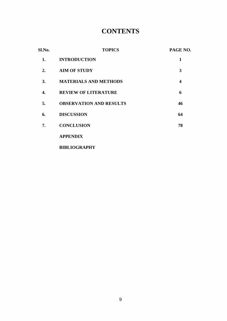
CONTENTS
Sl.No. TOPICS PAGE NO.
1. INTRODUCTION 1
2. AIM OF STUDY 3
3. MATERIALS AND METHODS 4
4. REVIEW OF LITERATURE 6
5. OBSERVATION AND RESULTS 46
6. DISCUSSION 64
7. CONCLUSION 78
APPENDIX
BIBLIOGRAPHY
ABSTRACT
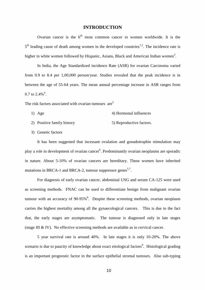
Ovarian cancer is 6th most common cancer in women worldwide. It has been
suggested that incessant ovulation and gonadotrophin stimulation may play a role in
development of ovarian cancer. Ovarian cancer carries highest mortality among all
gynaecological cancers because the early stages are asymptomatic. The tumour is
diagnosed only in late stages (Stage III and IV). No effective screening methods are
available as in cervical cancer. Epidemiological evidence suggests that steroid
hormones (estrogen and progesterone) and amplification of Human epidermal
growth factior-2 (HER-2/neu) gene are implicated in ovarian carcinogenesis. Thus
the steroid hormone receptor positivity on the ovarian surface epithelium and ovarian
carcinoma is of paramount significance for hormonal therapy. This study is
undertaken to review the incidence of ovarian neoplasm in our institution.
A total of 132 cases were evaluated in concordance with clinical history,
histopathological features. Immunohistochemistry (ER, PR) was done for surface
epithelial carcinomas. HER-2/neu expression was compared between malignant
surface epithelial carcinomas and borderline tumours.
Ovarian malignancy (4.1%) was found to be the second most common
malignancy in female genital tract next to cervical malignancy. In our institution
surface epithelial tumour (68.9%) was the most common neoplasm. Mucinous
cystadenoma was the commonest tumour.
Positive ER(36.3%) and PR(45.4%) expression in surface epithelial
malignancies proves the mitogenic role of estrogen and progesterone. HER-2/neu
showed positive expression in surface epithelial malignancies only (60% serous and
33.3% of mucinous carcinomas). Negative expression was seen in
borderline tumours. Hence HER-2/neu helps in differentiating borderline and
malignant tumours.
Thus a panel of markers will be helpful in prognostication of ovarian tumours
and development of targeted therapy.
KEY WORDS:
Ovarian cancer
Surface epithelial tumours
ER, PR
HER-2/neu
ABBREVIATIONS
ER - Estrogen receptor
PR - Progesterone receptor
HER-2/neu - Human epidermal growth factor
10
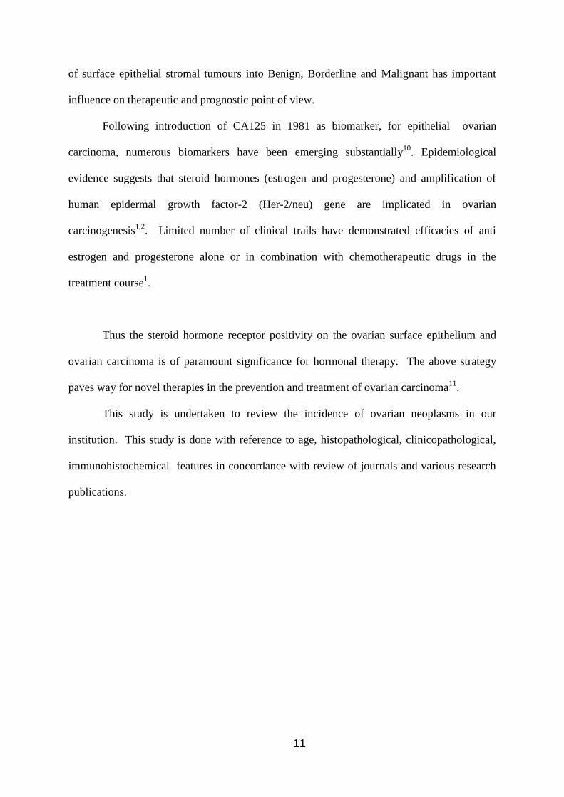
INTRODUCTION
Ovarian cancer is the 6th
most common cancer in women worldwide. It is the
5th
leading cause of death among women in the developed countries1,2
. The incidence rate is
higher in white women followed by Hispanic, Asians, Black and American Indian women3.
In India, the Age Standardized incidence Rate (ASR) for ovarian Carcinoma varied
from 0.9 to 8.4 per 1,00,000 person/year. Studies revealed that the peak incidence is in
between the age of 55-64 years. The mean annual percentage increase in ASR ranges from
0.7 to 2.4%4.
The risk factors associated with ovarian tumours are5
1) Age 4) Hormonal influences
2) Positive family history 5) Reproductive factors.
3) Genetic factors
It has been suggested that incessant ovulation and gonadotrophin stimulation may
play a role in development of ovarian cancer6. Predominantly ovarian neoplasms are sporadic
in nature. About 5-10% of ovarian cancers are hereditary. These women have inherited
mutations in BRCA-1 and BRCA-2, tumour suppressor genes5,7
.
For diagnosis of early ovarian cancer, abdominal USG and serum CA-125 were used
as screening methods. FNAC can be used to differentiate benign from malignant ovarian
tumour with an accuracy of 90-95%8. Despite these screening methods, ovarian neoplasm
carries the highest mortality among all the gynaecological cancers. This is due to the fact
that, the early stages are asymptomatic. The tumour is diagnosed only in late stages
(stage III & IV). No effective screening methods are available as in cervical cancer.
5 year survival rate is around 40%. In late stages it is only 10-20%. The above
scenario is due to paucity of knowledge about exact etiological factors9. Histological grading
is an important prognostic factor in the surface epithelial stromal tumours. Also sub-typing
11
of surface epithelial stromal tumours into Benign, Borderline and Malignant has important
influence on therapeutic and prognostic point of view.
Following introduction of CA125 in 1981 as biomarker, for epithelial ovarian
carcinoma, numerous biomarkers have been emerging substantially10
. Epidemiological
evidence suggests that steroid hormones (estrogen and progesterone) and amplification of
human epidermal growth factor-2 (Her-2/neu) gene are implicated in ovarian
carcinogenesis1,2
. Limited number of clinical trails have demonstrated efficacies of anti
estrogen and progesterone alone or in combination with chemotherapeutic drugs in the
treatment course1.
Thus the steroid hormone receptor positivity on the ovarian surface epithelium and
ovarian carcinoma is of paramount significance for hormonal therapy. The above strategy
paves way for novel therapies in the prevention and treatment of ovarian carcinoma11
.
This study is undertaken to review the incidence of ovarian neoplasms in our
institution. This study is done with reference to age, histopathological, clinicopathological,
immunohistochemical features in concordance with review of journals and various research
publications.
12
AIM OF STUDY
1) To study the incidence of ovarian neoplasms in our institution along with clinical
correlation.
2) To study and compare the incidence of malignant ovarian tumour with other female
genital tract malignancies in our institution.
3) To study the expression of ER, PR, Her-2/neu amplification status in primary surface
epithelial tumour malignancies.
4) To evaluate Her-2/neu expression in distinguishing borderline and malignant surface
epithelial tumours of ovary.

13
MATERIALS AND METHODS
This study is a retrospective study carried out in Department of Pathology, Thanjavur
Medical College from January 2013 to May 2015. A total of 132 cases of ovarian neoplasms
referred from Raja Mirasudhar Government Hospital (RMH) were included in this study.
The gross specimens were fixed in 10% neutral buffered formalin and processed
routinely. Clinical history of the patient including the age, examination findings, radiological
investigation, USG, CT and FNAC reports were evaluated in detail.
4 to 5 bits including the wall with papillary excrescences were taken for cystic
ovarian neoplasms. 3 to 4 bits were taken in solid tumours of size less than 5cms. In
variegated tumours more than 5cms, one block per 1 cm of the tumour was taken in its
greatest dimension. Section of 3 to 4μm were cut and stained with hematoxylin and Eosin
(Appendix - I).
The H&E stained slides were reviewed. Parameters consisting of age, tumour size,
stage of the disease (FIGO Staging) and histological typing were done according to WHO
classification criteria. Serous carcinoma were graded according to recent two tier grading
system.
The immunohistochemical detection of steroid hormonal status (ER, PR) in surface
epithelial stromal tumour were conducted. Also the status of Her-2/neu amplification in
borderline and malignant tumours were studied.
ER, PR, Her-2/neu STAINING:
Representative 4μm sections were taken from paraffin embedded blocks for Immuno
histochemistry (IHC). The procedure was performed according to heat induced epitope
retrieval method with specific antibodies against ER, PR and Her-2/neu.
14
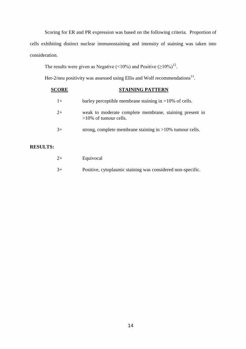
Scoring for ER and PR expression was based on the following criteria. Proportion of
cells exhibiting distinct nuclear immunostaining and intensity of staining was taken into
consideration.
The results were given as Negative (<10%) and Positive (≥10%)12
.
Her-2/neu positivity was assessed using Ellis and Wolf recommendations13
.
SCORE STAINING PATTERN
1+ barley perceptible membrane staining in >10% of cells.
2+ weak to moderate complete membrane, staining present in
>10% of tumour cells.
3+ strong, complete membrane staining in >10% tumour cells.
RESULTS:
2+ Equivocal
3+ Positive, cytoplasmic staining was considered non-specific.
15
REVIEW OF LITERATURE
EMBRYOLOGY
Around sixth week of intrauterine life, sexual differentiation can be recognized. The
premature gonad is identified as ovary by absence of testicular differentiation. The above
process is a passive event. Familial XX gonadal dysgenesis is transmitted as autosomal
recessive trait. The above fact suggests, autosomal genes are essential for human ovarian
organogenesis. Around 5th
week of gestation, gonadal ridges are formed. There after
epithelial proliferation and mesenchymal condensation occurs.
Primordial germ cells are the first to develop in the wall of yolk sac. These epithelial
cells proliferate to form primitive sex cords. Then they migrate to gonadal ridges. In the
surface epithelium there occurs proliferation of second generation of gonadal cortical cords.
These traverse the mesenchyme. These cords split to enclose a single primitive germ cell to
form follicular cells. The germ cells form oogonia. The coelomic epithelium encloses the
developing ovary14
.
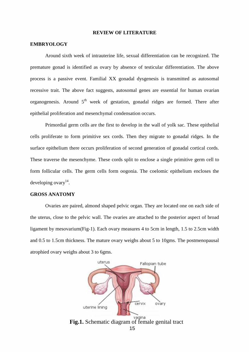
GROSS ANATOMY
Ovaries are paired, almond shaped pelvic organ. They are located one on each side of
the uterus, close to the pelvic wall. The ovaries are attached to the posterior aspect of broad
ligament by mesovarium(Fig-1). Each ovary measures 4 to 5cm in length, 1.5 to 2.5cm width
and 0.5 to 1.5cm thickness. The mature ovary weighs about 5 to 10gms. The postmenopausal
atrophied ovary weighs about 3 to 6gms.
Fig.1. Schematic diagram of female genital tract

16
CUT SURFACE
Reveals a thin white, outer cortex and major areas occupied by medulla. During
reproductive period, fluid filled cystic follicles and bright yellow corpora lutea can be seen15
.
VASCULAR SUPPLY:
Major blood supply of ovary is from Ovarian arteries. Ovarian artery is a branch of
abdominal aorta. It anastomosis with the ovarian branches of internal iliac arteries in the
mesovarium.
VENEOUS DRAINAGE:
The right ovarian vein drains into the inferior vena cava. The left ovarian vein drains into the
left renal vein.
LYMPHATICS:
The lymphatics drain into external iliac and paraaortic nodes7.

17
HISTOLOGY:
The ovary is covered by coelomic epithelium which is continuous with the
mesothelium of peritoneal cavity. The coelomic epithelium is usually a single layer of
cuboidal cells, columnar or focally pseudostratified.
Underneath the surface epithelium, outer collagenous zone termed Tunica Albugenia
and inner cellular cortex containing follicles are seen. The medulla contains stroma and
hilum.
In the cortex lies the primordial follicle which undergoes maturation into primary,
secondary, tertiary and Graffian follicles, which releases oocyte during ovulation. Fig.2, 3
The stroma consist of spindle cells exhibiting storiform pattern7. The blood vessels gain
entrance through hilum. Hilus cells contain crystals of Reinke.
FUNCTIONS
Ovaries produce ova and hormones under the feedback control mechanisms of
hypothalamo-pituitary ovarian axis through endocrine, paracrine and autocrine pathways.
RISK FACTORS FOR OVARIAN CARCINOMA
Fig.2. Histology of ovarian cortex Fig.3. Histology of Graffian follicle
18
Neoplasms of ovary constitutes about 30% of all cancers of female genital tract. The
age adjusted incidence rate is highest in well developed countries.
The factors that risk ovarian carcinoma are three fold.
1. Environmental
2. Genetic
3. Life style.
Higher socio-economic status is included as one of the risk factors.
About 5 - 10% of cases are familial. There is three times increased risk in individuals with
positive family history. Majority are due to mutation in BRCA1, BRCA2 and DNA mismatch
repair genes. Other syndromes which are associated with ovarian cancer are Lynch-II
syndrome and Hereditary Non Polyposis Colon Cancer Syndrome(HNPCC)16
.
Nulliparity, early menstruation, late menopause and ovarian inflammation are more
commonly associated with increased risk for ovarian tumours. The above repeated traumatic
insults might be the cause of ovarian cancers. Oral contraceptive use, multiparous women,
pregnancy, lactation reduces the risk by reducing the ovulation process.
Clomiphene citrate, long term estrogen replacement therapy, obesity favour ovarian
carcinogenesis. Diet rich in cheese, meat increases the risk. Beverages like tea and
vegetables like tomato consumption reduces the risk17
.
CLINICAL FEATURES
Age > 40 Years
Abdominal mass
Abdominal pain
Ascites
In functional ovarian tumours, precocious puberty, menstrual irregularities17
.
19
In high risk individuals serum biomarker assay, transvaginal ultrasound and genetic
studies can be used as screening test18
.
SPREAD AND METASTASES
Modes of spread
Direct
Hematogenous
Lymphatic route.
The metastatic sites are
Contralateral ovary
Peritoneal cavity
Paraaortic lymphnodes
Liver
Lung [Yolksac & Choriocarcinoma] by hematogenous spread16
.
CLASSIFICATION
Novak, in 1967 classified ovarian tumours as benign/malignant and cystic/solid. The
advantage being simple. Disadvantage is that the borderline tumours fell into grey zone19
.
WHO (World Health Organization) in the year 1973 formulated classification based on
histogenesis. The above classification was updated in 1999 and 20037,20
.
WHO CLASSIFICATION OF TUMOURS OF THE OVARY.
I. SURFACE EPITHELIAL – STROMAL.
Serous tumours
Malignant
Adenocarcinoma
20
Borderline tumour
Benign – cystadenoma, adenofibroma, cystadenofibroma.
Mucinous tumours:
Malignant
Adenocarcinoma
Borderline tumour
Benign – Cystadenoma, adenofibroma, cystadenofibroma
Mucinous cystic tumours with pseudomyxoma peritonei
Endometrioid tumours:
Malignant
Adenocarcinoma
Malignant mixed mullerian tumour
Endometrial stromal sarcoma.
Benign – cystadenoma, adenofibroma, cystadenofibroma.
Clear cell tumours:
Malignant
Adenocarcinoma
Borderline tumours
Benign – Cystadenoma, adenofibroma, cystadenofibroma.
Transitional tumours
Malignant
Transitional cell carcinoma (non-Brenner type)
Malignant Brenner tumour
Borderline
Benign Brenner tumour
21
Squamous cell carcinoma
Mixed epithelial tumours
Malignant
Borderline
Benign
Undifferentiated and unclassified tumours:
Undifferentiated carcinoma
Adenocarcinoma, not otherwise specified.
II. SEX CORD – STROMAL TUMOURS.
Granulosa-stromal cell tumour
Granulosa cell tumour
Adult Granulosa cell tumour
Juvenile granulosa cell tumour
Thecoma Fibroma group
Thecoma, not otherwise specified.
Typical
Luteinized.
Fibroma
Cellular Fibroma
Fibrosarcoma
Stromal tumour with minor sex cord elements
Sclerosing stromal tumour
Signet-ring stromal tumour
22
Sertoli-Stromal cell tumours:
Sertoli Leydig cell tumour group.
Well differentiated
Of intermediate differentiation variant with heterologous elements
Poorly differentiated variant with heterologous element.
Retiform variant with heterologous elements.
Sertoli cell tumour
Stromal –Leydig cell tumour
SEX CORD – STROMAL TUMOURS OF MIXED OR UNCLASSIFIED CELL TYPE.
Sex cord tumour with annular tubules
Gynandroblastoma
Sex cord stromal tumours, unclassified.
Steriod cell tumours
Stromal luteoma
Leydig cell tumour group
Hilus cell tumour
Leydig cell tumour, non hilar type
Leydig cell tumour, not otherwise specified.
Steroid cell tumour, not otherwise specified
Well differentiated
Malignant
III. GERM CELL TUMOURS
Primitive Germ cell tumours
Dysgerminoma
Yolk Sac tumour
23
Embryonal carcinoma
Polyembryoma
Non Gestational Choriocarcinoma
Mixed Germ cell tumour
Biphasic or Triphasic Teratoma
Immature Teratoma
Mature Teratoma
Solid
Cystic
Fetiform Teratoma.
Monodermal teratoma and somatic type tumours associated with dermoid cyst
Thyroid tumour groups
Struma Ovarii
Benign
Malignant
Carcinoid tumour
Neuroectodermal tumour group
Carcinoma group
Melanocytic group
Malignant melanoma
Melanocytic nevus
Sarcoma group
Sebaceous tumour group
Pituitary type tumour group
Retinal anlage tumour group.
24
Germ Cell Sex Cord – Stromal Tumour
Gonadoblastoma – Variant with malignant germ cell tumour.
Mixed germ cell – sex cord – Stromal tumour variant with malignant germ cell
tumour.
IV. TUMOURS OF THE RETE OVARII:
Adeno carcinoma
Adenoma
Cystadenoma
Cystadeno fibroma
V. MISCELLANEOUS TUMOURS:
Small cell carcinoma hypercalcemic type
Small cell carcinoma, pulmonary type
Large cell neuroendocrine carcinoma
Hepatoid carcinoma
Primary ovarian mesothelioma
Wilms tumour
Gestational choriocarcinoma
Hydatidiform mole
Adenoid cystic carcinoma
Basal cell tumour
Ovarian Wolffian tumour
Paraganglioma
Myxoma
Soft tissue tumours, not specific to the ovary.
25
VI. TUMOUR LIKE CONDITIONS
Luteoma of pregnancy
Stromal hyperthecosis
Stromal hyperplasia.
Fibromatosis
Massive ovarian edema
VII. LYMPHOID AND HAEMATOPOIETIC TUMOURS
VIII. SECONDARY TUMOURS
I. SURFACE EPITHELIAL – STROMAL TUMOURS:
The cell of origin is from surface epithelium, or serosa of ovary. 60% of ovarian
neoplasm, and 80-90% of primary ovarian malignancies belong to this group. The benign
epithelial tumours are common in middle age. The malignant epithelial tumours are common
in perimenopausal age group16
.
MOLECULAR PATHOGENESIS OF SURFACE EPITHELIAL TUMOURS
High grade serous and Endometrioid carcinomas arise from invagination of surface
epithelium (inclusion cyst). This is associated with P53, BRCA1 and/or BRCA2 mutations.
Low grade serous carcinoma arise from the mutation of RAS-RAF pathway. It is also
associated with adenoma – borderline neoplasm-carcinoma sequence. Mucinous carcinomas
arises in the background of mutation in K-ras oncogene. Low grade Endometrioid carcinoma
arise in the background of endometriosis and mutations in CTNNBI (gene encoding Beta
Catenin) and PTEN21
. Based on cell type epithelial tumours are classified as
Serous
Mucinous
Endometriod
Clear Cell

26
Based on pattern of growth
Cystic
Solid
Based on atypia/invasiveness
Benign
Borderline
Malignant tumours
A) SEROUS TUMOURS:
Serous tumours constitute the most common surface epithelial neoplasms. As a result of
mullerian differentiation along the salphingeal pathway, the cells resemble that of tubal
epithelium7.
Serous tumours constitute 30% of all ovarian tumours out of which, 70% are benign,
5-10% borderline and 20-25% are malignant16,22
.
BENIGN SEROUS TUMOURS:
Incidence is more common in fifth decade.
Gross:
Bilaterality is seen in 10-20%. Usually unilocular, filled with clear serous fluid. They
may be cystadenomas / adenofibroma / cystadenofibromas. Cystadenomas are cystic
neoplasm with smooth inner surface, sometimes with papillary excrescences, hence called
papillary serous cystadenoma. Adenofibroma contain a dense fibrous stromal component.
Cystadenofibroma contain polypoidal excrescences embedded in a fibrous stroma.
Microscopy:
Cyst and polypoidal excrescences are lined by single layer of ciliated epithelium
similar to fallopian tube. No nuclear atypia is seen16,22
.
27
SEROUS BORDERLINE TUMOUR:
Synonyms: Atypical proliferating tumour or serous tumours of low malignant
potential.
Gross:
These tumours are bilateral in 26-36% of cases. Grossly they usually present as cystic.
The inner surface showing more friable and numerous papillary projections.
Cystadenofibroma has white to yellow rubbery areas.
Microscopy:
Diagnostic features:
(1) Atleast 10% of the areas exhibit arborising papillae (hierarchical branching) with
epithelial stratification.
(2) Varying degrees of mild to moderate nuclear atypia.
(3) Absence of destructive stromal invasion.
Serous borderline tumours are classified as
(1) Typical (90%)
(2) Micropapillary (10%)
The typical type has classical branching papillary structures with epithelial tufts overlying the
papillae.
The micropapillary type has thin elongated papillae. These papillae are five times longer than
they are wide. They have no stromal support. They arise directly from the papillae with thick
fibrous stalk.
The following features should be looked for, after the diagnosis
(1) Surface involvement
There is higher risk of transformation to higher stage in this category.
This also enables peritoneal spread.
28
(2) Stromal microinvasion
Microinvasive type has micropapillary structures and discohesive
epithelial cells. They are surrounded by clear space, in the underlying normal
tissue. This invasive foci which is measuring less than 10mm2 and less than
3mm2 in greatest dimension is called as microinvasion.
(3) Lymph node metastases
Metastases are seen in pelvic and paraaortic lymphnodes in 27% of cases.
(4)Peritoneal implants:
In 30% of cases, serous borderline tumours are associated with implants in the
peritoneum. Classified as invasive and noninvasive types.
Invasive implants:
(a) Haphazardly arranged glands are seen in normal tissues like omentum.
(b) Dense fibrous reaction without inflammation are seen surrounding the implants.
(c) Epithelial proliferation is seen.
(d) Nuclear features are similar to low grade serous carcinoma.
(e) Borders are irregular.
(f) Aneuploidy.
These above features are absent in noninvasive implants.57
Even in the presence of invasion the prognosis seems to be good. Because they behave
as noninvasive neoplasms. In child bearing group, conservative surgery is performed along
with close clinical followup.23,24,25,57
MALIGNANT SEROUS CYSTADENOCARCINOMA
It is the most common malignant ovarian neoplasm. It constitutes about 40-50% of
total.
29
Gross:
These tumours are usually large, bilateral (70%). Size ranging from microscopic foci
to more than 20cms in diameter. They present as cystic with papillary excrescences, solid
growth and growth on the surface due to capsular invasion. Large areas of hemorrhage and
necrosis are also seen.
Well differentiated tumours have well formed papillary structures. They have
characteristic fibrovascular core. Psammoma bodies are common in the fibrovascular stalk.
When 75% of the tumour shows psammoma bodies they are called psammoma carcinoma.
They carry favourable prognosis. Moderately differentiated tumour shows crowded
papillae without fibrovascular core. Poorly differentiated types shows no papillary pattern.
The cells are arranged in solid sheets.
The papillae has to differentiated from papillae occurring in Transitional cell
carcinoma, Endometrioid carcinoma, clear cell carcinoma. In Endometrioid carcinoma, there
is villous structure with focal squamous metaplasia. In clear cell carcinoma, the papillae are
lined by hob nail/clear cells. The papillae have hyalinised core. In transitional carcinoma, the
papillae are broad and lined by transitional cells7,6,55
.
Immunostaining shows CK7 positivity, CK20 negativity, WT1, P53 expression31
.
Hormonal markers ER𝛼, PR𝛼 showed higher expression in malignant cases. Her-2/neu was
found to show higher expression for malignant tumours when compared to borderline26
.
GRADING:
The most commonly used Silverberg’s grading system27
:
Table:1:
Score Architecture Cytological atypia Mitotic figures/10Hpf.
1 Glandular Mild 0-9
2 Papillary Moderate 10-24
3 Solid Severe >25
30
Score 3-5 - Grade 1
Score 6-7 - Grade 2
Score 8-9 - Grade 3
2-Tier Grading system for serous carcinoma
Anais Malpica et al. classifies serous tumour into low and high grade based on
nuclear atypia and mitotic rate52
.
Low grade:
1. Mild to moderate nuclear atypia and as a secondary feature.
2. <12 mitosis/10Hpf.
High grade:
1. Marked nuclear atypia and as secondary feature.
2. >12 mitosis/10Hpf and multinucleated cells.
B) MUCINOUS TUMOURS:
Mucinous tumours constitute 14% of all ovarian tumours. Mucinous tumours are the
second most common ovarian tumours. About 30.8% of all surface epithelial tumours belong
to this category. Of these, 75% are benign, 20% are borderline, <5% are invasive
carcinomas. 5% are bilateral.16,28
BENIGN MUCINOUS TUMOURS
Gross:
Usually they are large, unilateral, multilocular. The multiloculated cyst contains
viscous mucoid material. Cystadenofibromas present as solid tumours.
Microscopy:
Depending on the lining epithelium of cyst they are classified as:
1) Intestinal
2) Endocervical type.
31
Endocervical type resembles endocervical epithelium. They have ciliated columnar
epithelium with intracellular mucin. Intestinal type epithelium consists of goblet cells, paneth
cells exhibiting picket fence appearance. Cystadenofibromas present as mucinous glands /
cyst distributed in dense fibrous stroma.29
BORDERLINE MUCINOUS TUMOURS
The lining epithelium is stratified not more than 3 layers. They form filiform
intracystic papillae with minimal stromal support. Nuclear atypia and mitotic figures are
noted. Stromal microinvasion is a feature. This consists of isolated cells/clusters. Each cluster
is composed of 5-10 cells without stromal reaction28,30
.
MALIGNANT MUCINOUS OVARIAN TUMOUR
Synonym : Mucinous cystadenocarcinoma
Gross:
They are mostly unilateral. About 5% of these tumours are bilateral. If bilateral,
nonovarian origin should be excluded. These tumours are common in 40 to 80 years of age.
They are large and have smooth external surface. Cut surface reveals many-thick walled
multilocular cystic spaces. The cyst contains viscous fluid. Solid areas, hemorrhagic areas
and necrosis are not infrequent.
Microscopy:
The lining epithelium is stratified with more than 4 layers. Two forms of stromal
invasion noted as:
(1) Expansile
(2) Infiltrative.
In expansile form, back to back arrangement of glands are seen with no stroma in
between. The infiltrative type shows glands, tubules, cords, cell nest haphazardly infiltrating
the stroma7,16
.
32
Differential Diagnosis:
Metastatic colon adenocarcinoma:
They are mostly bilateral. Nodular growth pattern, surface involvement and
lymphovascular invasion are commonly associated. They are CK20 positive and CK7
negative31
.
MUCINOUS TUMOUR WITH PSEUDOMYXOMA PERITONEI
It includes mucinous ovarian neoplasm, ascites, abundant gelatinous material within
pelvis and abdominal cavity. We must always rule out the possibility of appendiceal
neoplasm or other gastrointestinal primary mucinous tumours metastatic to peritoneum31
.
MIXED OR COLLISION TUMOUR
This is due to biphasic or multiphasic differentiation. Two or more epithelial elements are
present in varying proportions. The above entity is also seen in sex cord stromal and germ
cell tumours. Commonly benign mucinous tumours occur in association with the mature
cystic teratoma7,16
.
c) ENDOMETRIOID TUMOURS:
BENIGN ENDOMETRIOID TUMOURS
Gross:
They are solid, firm, tan with numerous cyst of varying sizes. The tumours are
ranging from 8 to 10cms in diameter.
Microscopy:
Benign appearing glands/cyst are seen. These structures are lined by endometrial type
of cells32
. They are sometimes associated with squamous differentiation.
33
BORDERLINE ENDOMETRIOID TUMOURS:
Gross:
Mostly unilateral ranging from 2-40cms in diameter. Cut surface is grey white, tan.
Predominantly these tumours are solid. Large tumours, may show areas of hemorrhage and
necrosis .
Microscopy:
Crowded Endometrioid glands/cyst are seen. There is cytological atypia and low
mitotic activity is seen. But characteristically, there is no invasion into the stroma. 15-50% of
patients have incidence of same side ovarian endometriosis or at other extra ovarian sites32,60
.
ENDOMETRIOID ADENOCARCINOMA
Hyperestrogenic state, atypical hyperplasia within a foci of endometriosis serve as
risk factor for Endometrioid carcinoma. This suggests the origin of tumour is directly from
ovarian coelomic epithelium. This entity has a very good prognosis33
.
Gross:
They present as solid, cystic friable papillae or as a mural nodule. These tumours
range from 10 - 20cms diameter. Bilaterality is seen in 28% of cases.
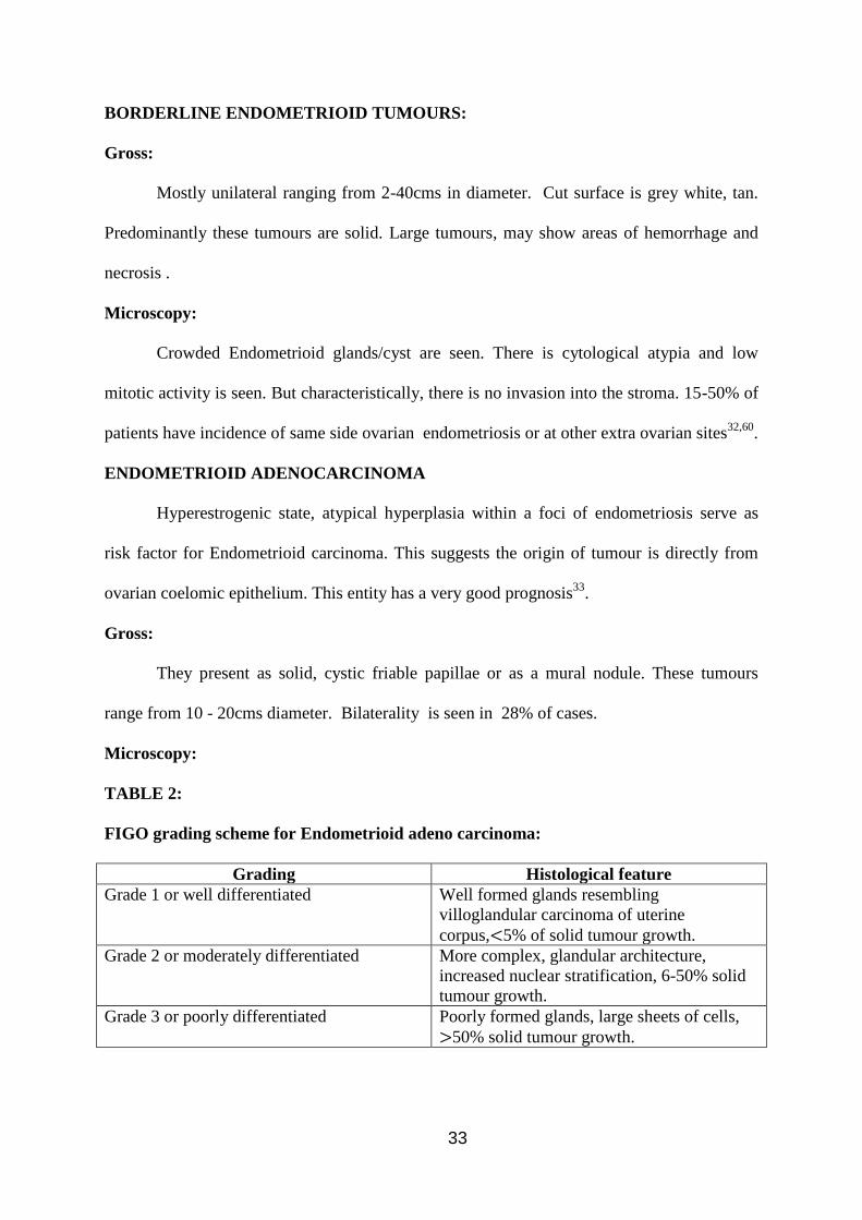
Microscopy:
TABLE 2:
FIGO grading scheme for Endometrioid adeno carcinoma:
Grading Histological feature
Grade 1 or well differentiated Well formed glands resembling
villoglandular carcinoma of uterine
corpus,<5% of solid tumour growth.
Grade 2 or moderately differentiated More complex, glandular architecture,
increased nuclear stratification, 6-50% solid
tumour growth.
Grade 3 or poorly differentiated Poorly formed glands, large sheets of cells,
>50% solid tumour growth.
34
Squamous differentiation is seen in 30% of cases. The differential diagnosis is metastatic
uterine endometrial carcinoma. Endometrioid carcinoma of ovarian origin may have
associated endometriotic foci. The secretory change, squamous metaplasia, expansile
invasion are evident .These tumours shows beta catenin, EMA positivity31
.
.CLEAR CELL (MESONEPHROID) TUMOURS
Clear cell carcinomas are more common in nulliparous women7. They constitute 5%
of all ovarian tumours. Clear cell carcinomas are most commonly associated with pelvic and
endometrial endometriosis33
.
Clear cell tumours are classified into benign, borderline, malignant.
Gross:
Thick walled unilocular cyst is seen. Numerous fleshy yellow nodules project into the
cyst are noted.
Microscopy:
They may present as solid mass, cystic or papillary. The cores of papillae exhibit
prominent hyalinisation. The papillae are lined by one or two layers of polygonal/flattened/or
characteristic hobnail cells. The individual cells have clear cytoplasm containing glycogen.
The nuclear atypia is minimal34
.
Differential diagnosis include
(1) Dysgerminoma which has polygonal shaped cells, inconspicuous nucleoli, presence of
luminal mucin and EMA positivity.
(2) Metastatic renal cell carcinoma should also be considered. The clear cell renal cell
carcinoma is CD10 positive, CK7 negative and vice-versa in clear cell carcinoma of
ovary.

35
D) BRENNER TUMOUR (TRANSITIONAL CELL TUMOUR):
Brenner tumour accounts for 1-2% of all ovarian neoplasm. The average age of
presentation is about 50 years. Bilaterality is seen in 5 to 7% of the cases29
.
The cell of origin is from surface epithelial cell or cyst derived from them. This is
through the process of metaplasia. These tumours are rarely associated with mucinous
cystadenoma and with Transitional cell tumour of bladder.
BENIGN BRENNER TUMOUR
Gross:
Predominantly these tumour are well circumscribed. Cut surface-firm, white or
yellowish measuring about 2 to 30cms diameter. Focal areas of calcification may be seen. 6%
of the cases are bilateral.
Microscopy:
Nests of transitional epithelial cells resembling the urothelium is seen. This is
surrounded by abundant dense fibroblastic stroma. Individual cells have oval nuclei with
longitudinal groove resembling coffee bean. The cells have clear cytoplasm. Cystic change
can be seen34
.
BORDERLINE BRENNER TUMOUR:
Gross:
Large, with an average diameter 16-20cms. Cut surface shows solid and cystic
components.
Microscopy:
Branching papillae lined by transitional epithelium protruding into cystic space.
Spindle to oval cells without mitotic activity is seen. There is no stromal invasion. A benign
Brenner component is always present34
.
36
MALIGNANT BRENNER TUMOUR
Gross:
They are large tumours. Cut surface reveals predominantly solid and few cystic
areas.
Microscopy:
These neoplasm exhibits stromal invasion. This is accompanied by a benign or
borderline Brenner component. The invasive element present as high grade transitional cell
carcinoma35
.
TRANSITIONAL CELL CARCINOMA:
They resemble transitional cell carcinoma of urinary tract in architecture. Most
commonly seen in age group of 50 to 58 years.
Gross:
Bilateral in 15% of cases. Cut surface reveals partly solid and cystic areas.
Occasional polypoidal projections accompanied by areas of hemorrhage and necrosis is seen.
Microscopy
Characteristic papillary structures lined by multilayered transitional epithelium is
seen. The cells exhibit pleomorphism. These tumour should not have a benign/borderline
Brenner component35
.
This type of neoplasm has got a good prognosis. The differential diagnosis of
metastatic tumours from urinary tract should always be considered. Transitional tumours of
urinary tract show classically CK 20 positive, which is absent in tumours of ovarian origin31
.
E) MALIGNANT MIXED MULLERIAN TUMOURS
Gross:
90% of these tumours are bilateral. External surface - Bosselated mass, large tumors.
Cut surface - Partly solid and cystic. Areas of hemorrhage and necrosis are seen.
37
Microscopy:
It is a biphasic neoplasm. It is composed of both malignant epithelial and
mesenchymal component. Any of the surface epithelial tumour constitute the epithelial
component. The mesenchymal component is fibrosarcoma / leiomyosarcoma / Endometrioid
stromal sarcoma34
.
F) ADENOSARCOMA
Gross:
Always unilateral. Cut surface – predominantly solid. Small cysts are occasionally
seen.
Microscopy:
Biphasic tumour in which the epithelial component is benign. But the mesenchymal
component is malignant. The cellular stroma shows periglandular cuffing pattern. The
adenosarcoma of ovary has grave prognosis in comparison with its uterine counterpart31
.
G) ENDOMETRIAL STROMAL SARCOMA:
Gross:
About 70% of tumours present as unilateral tumours. Predominantly the cut surface
is solid. Cystic areas filled with mucoid and hemorrhagic fluid material may be seen.
Microscopy:
50% of cases occur in association with endometriosis. The characterstic features of
numerous thick walled blood vessels are seen. The tumour cells are arranged in whorls
around blood vessels or in diffuse pattern. The individual cells have round to oval nuclei
with scanty cytoplasm31
.

38
H) MIXED EPITHELIAL TUMOUR
Microscopy:
Varying proportions of two or more of five major cell types are seen: serous,
endometrioid, mucinous, clear cell, and transitional types are seen34
.
I) UNDIFFERENTIATED CARCINOMA
Microscopy:
They are uncommon ovarian neoplasm. They have no differentiation. Marked
cytolological atypia is seen.
II. SEX CORD STROMAL TUMOURS:
5% of ovarian neoplasms belong to this group of functional tumours. The cell of
origin is from the sex cord, parenchyma or both of the embryonic gonads. The neoplastic
cells differentiate into either testicular (sertoli-leydig cell tumours) or ovarian type
(Granulosa cell tumours)16,53
. The sensitive and specific immuno marker for sex cord stromal
tumours are inhibin and calretinin 31
.
A) GRANULOSA STROMAL CELL TUMOUR:
They constitute 1 – 2% of ovarian tumours. 70% of sex cord stromal tumours belong to this
group of potentially malignant tumours. The cell of origin is follicular granulosa cells. Two
types:
1) Adult Granulosa cell tumour
2) Juvenile Granulosa cell tumour
ADULT GRANULOSA CELL TUMOUR:
More common in the reproductive age group. 40% of the cases are seen in the
menopausal age group. About 75% of the patients present with abnormal uterine bleeding
due to hyperstrogenism.
39
Gross:
These tumours have an average diameter of 12cms. Cut surface - Yellow to greyish
white in colour, solid and cystic areas are seen. Cystic area filled with hemorrhagic fluid is
seen. Areas of hemorrhage and necrosis may be seen exhibiting a variegated appearance.
Microscopy:
Growth patterns of microfollicular with characterstic Call Exner bodies, Macro
follicular, insular, trabecular, watered silk,diffuse, solid, sarcomatoid are seen. Recent
studies suggest the tumour size and mitotic index has more prognostic significance than the
pattern34,36
.
The individual granulosa cells have scanty cytoplasm with longitudinal nuclear
grooves giving rise to coffee bean appearence. A low mitotic activity and minimal to no
cytological atypia is noted.22
The granulosa cells are often surrounded by fibro thecomatous
stroma(fibroblasts, theca/leutinised cells). The morphology and inhibin positivity strongly
suggests granulosa stromal cell tumour31,58
.
JUVENILE GRANULOSA CELL TUMOUR:
80% of these tumours are seen in adolescent girls. Often they present with complaints
of precocious puberty. The above entity may be seen in association with olliers
disease/mafucci’s syndrome.
Gross:
Lobulated external surface is seen. Cut surface reveals solid to cystic areas. The solid
areas are yellow to tan. They are soft in consistency.
Microscopy:
Macrofollicular pattern are more common. Neoplastic granulosa cells are having
abundant eosinophilic vacuolated cytoplasm. There is no grooving. Numerous mitotic figures
40
are evident. They present with distant metastasis. The patients have grave prognosis when
compared to adult granulosa type37
.
B)THECOMA-FIBROMA GROUP:
a) THECOMA:
4% of all ovarian tumours belong to this group. They occur in post menopausal age group38
.
Gross:
They are almost and always unilateral. An average size of 5-10cms in noted. Cut
surface - solid, grey tan to yellow areas with well defined capsule. Firm in consistency.
Microscopy:
Fasicles of bland oval to spindle shaped cells are seen. Nuclei are fusiform with
abundant pale vacuolated lipid rich cytoplasm. Calcification is prominent. Leutinised
thecoma cells are also evident. They contain leutin cells, in a background of fibromatous than
thecomatous stroma. Edema and microcyst formation are commonly seen. Mitotic figures are
not seen.
Special stains like oil red O demonstrates intracytoplasmic neutral fat. Silver stains
like reticulum, stains around each tumour cell in contrast to granulosa cells in which the
reticulin staining is around the cluster of cells35
.
b) FIBROMA:
Common ovarian tumour occurring around puberty. The patients are asymptomatic.
Fibromas are incidentally discovered during surgery. Ovarian fibroma along with ascites and
right sided pleural effusion constitute Meig’s syndrome. In Gorlen’s syndrome they present
bilaterally39.40
.
Gross:
Most commonly they are unilateral. Average size is around 6cms in diameter. Cut
surface - solid, white, lobulated appearance. Hard in consistency.
41
Microscopy:
These tumours are composed of spindle shaped cells in interlacing fascicles. They are
also seen in storiform pattern admixed with dense collagen. The individual cells have uniform
bland fusiform nuclei with pointed ends and scant cytoplasm. Mitosis is absent. 10% of
tumours have uniform densely cellular and are referred to as cellular fibromas39.40
.
C) SCLEROSING STROMAL TUMOUR OF OVARY
These are benign uncommon neoplasm seen in reproductive age group.
Gross:
Unilateral, well circumscribed with an average of 15cms in diameter. Cut surface
shows solid, grey white, lobulated with specks of yellow areas.
Microscopy:
Characteristic pseudolobular pattern of growth is seen. It has alternating hyper cellular
and hypocellular areas. Hypercellular areas shows numerous thin walled blood vessels,
similar to hemangiopericytoma like pattern. It is composed of collagen producing spindle
cells and lipid containing round cells. The hypocellular areas is composed of edematous
sclerotic fibrous stroma.
Massive Edema of ovary is an important differential diagnosis. Normal ovarian
structures are seen admixed with edematous areas in Edema of ovary. Where as in sclerosing
stromal tumour, the normal ovarian stroma is replaced by the neoplasm7,16
.
D) SERTOLI STROMAL CELL TUMOUR
Tumour consisting of varying proportion of sertoli cells, Leydig cells and stromal
components are seen.
Sertoli Leydig cell group:
These rare tumours constitute less than 0.1% of ovarian tumours22
. The tumours are
more common in younger age group. The patient presents with masculinisation or
42
defeminisation features. Previously these tumours were referred as
Arrhenoblastoma/Androblastoma.
Gross:
Most commonly they are unilateral. Solid, firm with pale yellow areas admixed with
few areas of hemorrhage and necrosis are seen.
Microscopy:
1. MEYER’S type I - Well differentiated tumours.
Sertoli cells are seen in open or closed tubules. They lack significant atypia/mitotic
activity.
2. MEYER’S type II – Intermediate
Composed of cords, sheets and nests of sertoli like cells separated by spindle shaped
stromal cells.
3. MEYERS type III - Poorly differentiated/Sarcomatoid
They are arranged in sarcomatoid pattern with masses of spindle cells27
.
20% of tumours exhibit heterologous elements. They consist of mucinous epithelium
of gastrointestinal tract, skeletal muscle, cartilage and neuroendocrine cells.
E) SERTOLI CELL TUMOURS
They are rare neoplasms. Sertoli cell tumours are more common in reproductive age
group.
Gross:
Unilateral in presentation. Average size is 5 to 7cms in diameter .
Microscopy:
The neoplasm is composed of sertoli cells lining the tubules or grow in nests or solid
sheets. The individual cells are columnar, polygonal, in shape with small round to oval nuclei
with minimal nuclear atypia. They have granular to eosinophilic cytoplasm27
.

43
F) SEX CORD TUMOUR WITH ANNULAR TUBULES
These tumours may occur as solid, bilateral and multifocal tumours accompanied by
Puetz Jegher’s syndrome. They are present as solitary neoplasm without the syndrome. Half
of the patients present with hyperestrogenism.
Microscopy:
Complex annular tubules filled with eosinophilic material are seen. Occasional
hyaline bodies and calcification are also seen. In several tumours the above tumour merged
with that of granulosa cell tumour7,16
.
G) GYNANDROBLASTOMA
Mixture of granulosa cell tumours (estrogenic) and sertoli–Leydig cell tumours
androgenic features are seen.
Gross:
Unilateral with an average size of 1 to 18cms is seen.
Microscopy:
Equal proportion of sertoli leydig cell and granulosa cell tumour are seen. Tubules and
trabeculae of well differentiated sertoli-leydig cells are seen admixed with nests and sheets of
granulosa cells16
.
H) LIPID CELL (LIPOID, STEROID CELL):
These tumours accounts for 0.5% of all ovarian neoplasm. Lipid cell tumours are seen in
all age groups. They occur in association with defeminisation and virilisation syndromes22
.
Gross:
Unilateral, Well circumscribed. Cut surface – yellowish nodules separated by fibrous
septa.
44
Microscopy:
Tumour cells are arranged in solid sheets. The individual tumour cells are large
polyhydral with abundant eosinophilic or vacuolated cytoplasm. The cytoplasm stains
positivity for fat stains27
. 25% of these tumours turn into malignancy. They are characterised
by necrosis and hemorrhage. Nuclear atypia and mitotic activity is increased in such cases16
.
III. GERM CELL TUMOURS
30% of ovarian neoplasm come under this category. The most common neoplasm to
occur in younger age group (< 18 years) are germ cell tumours. Malignant germ cell tumour
(3% of germ cell tumours) are more common in much younger age group27
.
Benign cystic teratoma is the commonest type. They constitute 95% of germ cell
tumours41
. The cell of origin is from germ cell which has undergone defective meiotic
division16
. 8% of the cases are composed of two or more subtypes. They are grouped as
malignant mixed germ cell tumours.
A) DYSGERMINOMA:
They constitute 1% of all ovarian cancers. Bilaterality is around 10%. It is the most
common malignant tumour in association with gonadal dysgenesis. The patients present with
complaints of abdominal mass and pain.
Gross:
Well encapsulated tumours. The average size is 15cms diameter. Cut surface reveals
solid, lobular configuration, tan or white in colour. Soft in consistency.
Microscopy:
The tumour cells are found in lobules/sheets of cells with thin fibrous septa separating
them. The fibrous septa is densely infiltrated by lymphocytes. The individual tumour cells are
polygonal cells with clear to eosinophilic cytoplasm (PAS positive). Round vesicular nucleus
and central prominent nucleoli is seen. In 5% of cases syncytiotrophoblasts are seen16,41
.
45
Anaplastic dysgerminoma resembles embryonic carcinoma. The features of
pscudoglandular/tubular pattern, high mitotic activity, high nuclear atypia is seen41
.
Immunohistochemistry markers of CD117 and PLAP (Placental Alkaline
Phosphatase) are positive31
.
B) YOLK SAC TUMOUR (ENDODERMAL SINUS TUMOUR):
20% of primitive germ cell tumours belong to this category. These tumours are common
in younger age group. The patient present with complaints of abdominal pain and mass.
Gross:
They present as unilateral mass. The average diameter is 15cms. Cut section reveals
soft, grey yellow areas with large areas of hemorrhage and necrosis. Cystic areas are not
infrequent42
.
Microscopy:
Many patterns of reticular, solid, festoon, hepatoid, pseudo papillary, polyvesicular
vitelline are seen. The characteristic schiller duval bodies are seen in 10 to 20% of tumours.
They are described as papillae lined by tumour cells that project into dilated cystic spaces
giving rise to glomeruloid bodies . PAS positive eosinophilic hyaline globules are seen42
.
Clear cell carcinoma should be considered in the differential diagnosis. The nuclei in
yolk sac tumour appears primitive. The papillae of yolksac tumour lack hyalinised core32
.
Yolk sac tumours are positive for cytokeratin, Alpha1Antitrypsin31
.
C) EMBRYONAL CARCINOMA:
These tumours constitute 3% of germ cell tumours. They are most common in
younger age group. These tumours present as a component of mixed germ cell tumours. The
patient present with features of precocious puberty, vaginal bleeding, hirsutism and
amenorrhea.
46
Gross:
External surface is smooth. Cut surface shows, solid, variegated areas of hemorrhage
and necrosis16
.
Microscopy:
Large primitive appearing cells in solid sheets and nests and also forming abortive
glandular structures are seen. Most commonly syncytiotrophoblastic giant cells are seen43
.
Serum alpha feta protein and chorionic gonadotrophin are elevated giving false positive
pregnancy tests31
.
These tumours have resemblance with poorly differentiated adenocarcinoma or
undifferentiated carcinoma. The latter are more common in old age/reproductive age group.
D) POLYEMBRYOMA
Embryonal carcinoma with numerous embryonal bodies are termed polembryoma.
They are composed of an embryonal disc, yolk sac and amniotic cavities7.
E) CHORIOCARCINOMA:
Primary ovarian choriocarcinoma are very rare. They usually present as metastases
from the uterine tumours. They have better prognosis than non gestational choriocarcinoma7.
Gross:
Pure choriocarcinoma present as solid, hemorrhagic and friable mass7.
Microscopy:
Biphasic pattern of syncytial and cytotrophoblastic elements in necrotic and
hemorrhagic back ground7.
Immunohistochemistry for HCG (Human Chorionic Gonadotrophin) is positive.
47
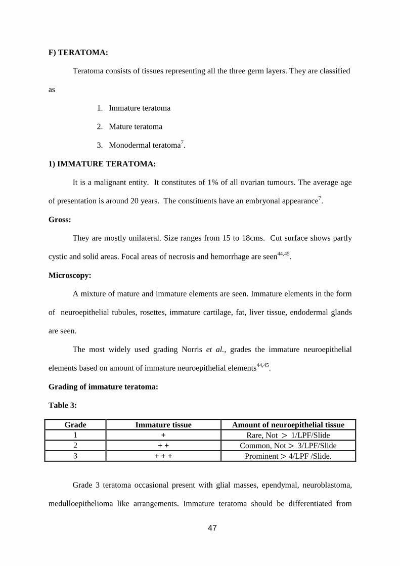
F) TERATOMA:
Teratoma consists of tissues representing all the three germ layers. They are classified
as
1. Immature teratoma
2. Mature teratoma
3. Monodermal teratoma7.
1) IMMATURE TERATOMA:
It is a malignant entity. It constitutes of 1% of all ovarian tumours. The average age
of presentation is around 20 years. The constituents have an embryonal appearance7.
Gross:
They are mostly unilateral. Size ranges from 15 to 18cms. Cut surface shows partly
cystic and solid areas. Focal areas of necrosis and hemorrhage are seen44,45
.
Microscopy:
A mixture of mature and immature elements are seen. Immature elements in the form
of neuroepithelial tubules, rosettes, immature cartilage, fat, liver tissue, endodermal glands
are seen.
The most widely used grading Norris et al., grades the immature neuroepithelial
elements based on amount of immature neuroepithelial elements44,45
.
Grading of immature teratoma:
Table 3:
Grade Immature tissue Amount of neuroepithelial tissue
1 + Rare, Not > 1/LPF/Slide
2 + + Common, Not > 3/LPF/Slide
3 + + + Prominent > 4/LPF /Slide.
Grade 3 teratoma occasional present with glial masses, ependymal, neuroblastoma,
medulloepithelioma like arrangements. Immature teratoma should be differentiated from
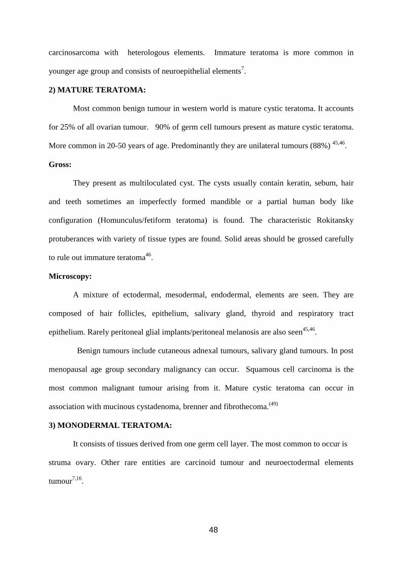
48
carcinosarcoma with heterologous elements. Immature teratoma is more common in
younger age group and consists of neuroepithelial elements7.
2) MATURE TERATOMA:
Most common benign tumour in western world is mature cystic teratoma. It accounts
for 25% of all ovarian tumour. 90% of germ cell tumours present as mature cystic teratoma.
More common in 20-50 years of age. Predominantly they are unilateral tumours (88%) 45,46
.
Gross:
They present as multiloculated cyst. The cysts usually contain keratin, sebum, hair
and teeth sometimes an imperfectly formed mandible or a partial human body like
configuration (Homunculus/fetiform teratoma) is found. The characteristic Rokitansky
protuberances with variety of tissue types are found. Solid areas should be grossed carefully
to rule out immature teratoma46
.
Microscopy:
A mixture of ectodermal, mesodermal, endodermal, elements are seen. They are
composed of hair follicles, epithelium, salivary gland, thyroid and respiratory tract
epithelium. Rarely peritoneal glial implants/peritoneal melanosis are also seen45,46
.
Benign tumours include cutaneous adnexal tumours, salivary gland tumours. In post
menopausal age group secondary malignancy can occur. Squamous cell carcinoma is the
most common malignant tumour arising from it. Mature cystic teratoma can occur in
association with mucinous cystadenoma, brenner and fibrothecoma.(49)
3) MONODERMAL TERATOMA:
It consists of tissues derived from one germ cell layer. The most common to occur is
struma ovary. Other rare entities are carcinoid tumour and neuroectodermal elements
tumour7,16
.
49
STRUMA OVARII:
The predominant component in this type is thyroid tissue. It constitutes 2 to 7% of all
ovarian teratomas. Malignancy is rarely encountered. If it arises, it presents as papillary
carcinoma with typical nuclear features. Grossly they are less than 10cms in maximum
extension. Cut surface is solid tan with glistening surface. Microscopic examination shows
numerous follicles filled with colloid7,16
.
CARCINOID TUMOUR:
The neuroendocrine tumours are more common in older age group. The patient
present with menstrual irregularities and abdominal pain. Grossly they are unilateral. Cut
surface shows tan yellow solid areas. Microscopically, the tumour cells are arranged in
trabecular pattern. The nucleus has salt and pepper chromatin.
The most common site of metastases from intestinal carcinoid is ovary.The metastatic
carcinoid present as multiple nodules and bilateral in presentation.
Immunohistochemistry markers like serotonin, chromogranin are positive7,16
.
G) MALIGNANT MIXED GERM CELL TUMOUR:
5 - 20% of malignant germ cell tumours belong to this category. Usually combination
of dysgerminomas with yolk sac tumours can be seen. They are followed by immature
teratoma, embryonal carcinoma, choriocarcinoma. The later group have grave prognosis.
They are common in younger age group7,16
.
I) MIXED GERM CELL – SEX CORD STROMAL TUMOURS:
Dysgenetic gonadoma as it is otherwise called is associated with XY gonadal
dysgenesis. They are composed of dysgerminoma and sex cord stromal tumours composed of
immature sertoli and granulosa cells. Most commonly they are associated with hyalinization
and calcification. The close differential diagnosis is sex cord tumours with annular tubules.
The latter lacks germ cell components7,16
.
50
IV. TUMOURS OF RETE OVARII:
They are relatively uncommon tumours. They are seen in the postmenopausal women.
The tumour is located in the ovarian hilus.
Microscopy:
Cuboidal cells, nonciliated columnar cells are seen arranged in retiform spaces. They
may present as adenoma, cystadenoma, carcinoma7.
V. TUMOURS OF UNCERTAIN ORIGIN:
A) SMALL CELL CARCINOMA:
They may present as
Hypercalcemic type
Pulmonary type.
HYPERCALCEMIC TYPE:
It is most common form of undifferentiated carcinoma. It is most common in young
female and are always bilateral. The tumours are always associated with hypercalcemic. The
above feature disappears after the removal of tumour.
Grossly they are large, solid with huge areas of necrosis and hemorrhage.
Microscopically, small closely packed cells with high N/C ratio are seen. Numerous mitotic
figures are seen. Because of extra ovarian spread, the prognosis is grim35
.
The pulmonary type resembles the lung tumour. They are sometimes associated with
endometrial carcinoma35
. Tumours are positive for keratin, EMA, NSE and rarely
chromogranin31
.
B) TUMOURS OF PROBABLY WOLFFIAN ORIGIN:
Also called as wolffian adnexal tumour. They are benign tumours. Grossly they are
solid, grey white or yellow in colour. Microscopically the epithelial cells are arranged in
solid sheets with cystic degeneration. They are CK7, Calretinin, Vimentin positive31
.

51
VI) UNCLASSIFIED TUMOURS:
SARCOMAS:
Most of them are fibrosarcoma, leiomyosarcoma, endometrial sarcoma, osteosarcoma,
chondrosarcoma, angiosarcoma, rhabdomyosarcoma. 8% of malignancy in dermoid cyst
occur as sarcoma32
.
Fibrosarcoma:
Common in postmenopausal women. Grossly they are fleshy with areas of
hemorrhage and necrosis. Histology shows spindle shaped cells arranged in Herringbone
pattern. Nuclear atypia and mitotic figures are prominent32
.
VII) METASTATIC TUMOURS:
5 – 10% of ovarian tumours come under this category. The most common primary
tumours to metastasize are large intestine, stomach, appendix. This is followed by breast,
uterine corpus, uterine cervix.
In younger girls, metastasis occur in Neuroblastoma, Rhabdomyosarcoma, Ewing’s
sarcoma13,48,49
.
Features of ovarian metastasis:
1. Bilaterality
2. Surface involvement
3. Nodular pattern of growth
4. Hilar involvement
5. Single cell invasion
6. Desmoplastic reaction
7. Vascular invasion
8. Unusual extra ovarian spread.
The normal ovarian parenchyma is completely replaced. They appear as solid, white,
52
sometimes cystic. Multiple serosal implants is a diagnostic feature48
. Immunohistochemistry
is helpful in differentiating from primary which is CK7 positive of CK20 negative31
.
KRUKENBERG TUMOUR:
The most common primary site is stomach. Grossly, they are
symmetrically/asymmetrically enlarged. Cut surface reveals predominantly homogenous
with firm, white areas. Microscopically, the tumour cells are seen as isolated single cells,
nests, cords. The individual cells have cytoplasmic vacuoles compressing hyperchromatic
nucleus to one side. This gives a signet ring appearance. Abundant pools of mucin are seen in
the stroma50,51
.
Special stains for mucin namely PAS and alcian blue and epithelial markers are
positive31
. This tumour has a grave prognosis50
.
PROGNOSIS OF OVARIAN TUMOURS:
The important factors influencing are as follows.
1. Histological type
2. Molecular abnormality
3. Stage of Ovarian cancer
4. Women’s age and general health
5. Whether newly diagnosed/recurred16
ESTROGEN RECEPTOR:
Estrogen suppresses basal and cisplatin induced apoptosis. The etiopathogenesis is by
increasing DNA repair capacity and avoiding apoptosis56
. This leads to uncontrolled cell
growth and drug resistance. The mechanism of action is by up regulation of c-myc gene.
Estrogen also regulates genes (ezrin, fibulin, cathepsin D and kallikerins) involved in motility
and invasion of extra cellular matrix12
.

53
PROGESTERONE RECEPTOR:
Progesterone promotes cell differentiation and apoptosis.Progesterone inhibits DNA
synthesis and cell division. Many studies have shown that there is decreased risk of ovarian
cancer with oral contraceptives and in multiparous individual. This could be due to cyclic
progestational climate. (serum levels are comparable to luteal phase progesterone level)2,6
.
The mechanism of action is three fold . Firstly a connection exists between
progesterone action and Fas/FasL signaling in normal and malignant ovarian surface
epithelial cell death. Secondly, through decrease in membrane fluidity. Lastly, progesterone
induces a switch from TGF-1 𝛽1 to TGF beta 2/3 expression. This correlated with increased
apoptotic index in ovarian surface epithelium2,11
.
Her-2/neu RECEPTOR
The term neu was coined from the rat homologue of Her-2. The rats were induced to
produce neuroblastoma by nitroso ethyl urea and their DNA was used for research
purpose.61
The Her-2/neu oncogene is located on chromosome 17. Her-2/neu oncogene encodes
a transmembrane glycoprotein tyrosine kinase. This includes four receptors-ErbB1(HER),
ErbB2(HER-2/neu), ErbB3(HER3) and ErbB4(HER4). HER-2 expression is well documented
in breast. It helps in assessment of prognosis (increased risk for early recurrence and
resistance to endocrinopathy) and in the treatment of breast cancers. Targeted monoclonal
antibody has been used a mode of treatment in breast cancers.2,10,59
Various studies have been shown that apart from breast, HER-2 over expression is
also seen in stomach, prostate, ovary, colon and bladder. The four receptors are monomers.
They dimerise when bound by a ligand either with same kind of receptor (homodimerisation)
or with other receptor (heterodimerisation).

54
Receptors : Ligands
HER1 : EGF, amphiregulin, TGF (Transforming growth factor),
epiregulin and betacellulin.
HER2 : ASGP2 forming sialomucin complex, MUC4 and
phosphorylation of HER2
HER3, HER4 : Hergulins, acetylcholine receptor inducing activity,
signals MAP( mitogen activated protein kinase path
way).61
ROLE IN TUMORIGENESIS:
Activation of HER family of receptors causes activation of Ras, MAP (mitogen
activated protein) kinase pathway and P13K (Phosphatidylinositol-3 Kinase) pathway. The
RAS/MAP pathway stimulates cell proliferation. The PI3K pathway inhibits proapoptotic
proteins like Bad, GSK3𝛽, FOXO3a through phosphorylation of AKt.10,61
HER2 Over expression is being correlated to tumour size, grade, increased proportion
of S phase cells and aneuploidy. HER2/HER3 heterodimer has a strong mitogenic effect in
ovarian carcinoma. The combination of cytotoxic chemotherapy and EGFR/HER inhibitors
(Trastuzumab) has a better clinical response.10,59
55
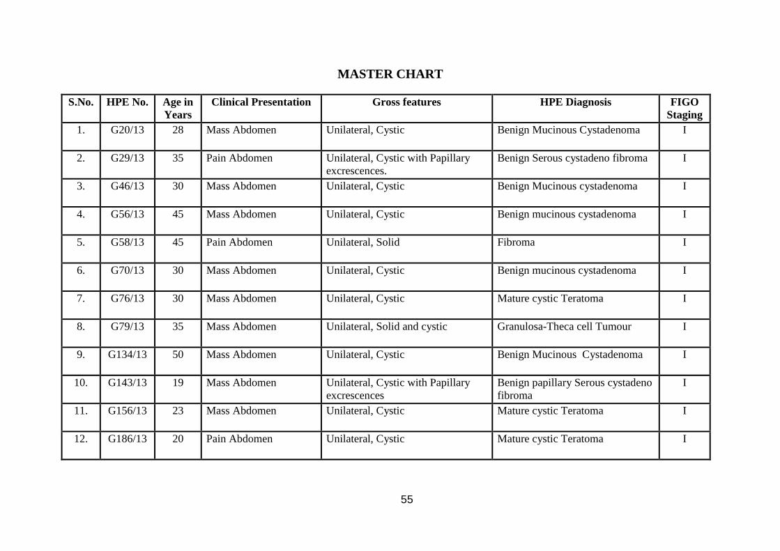
MASTER CHART
S.No. HPE No. Age in
Years
Clinical Presentation Gross features HPE Diagnosis FIGO
Staging
1. G20/13 28 Mass Abdomen Unilateral, Cystic Benign Mucinous Cystadenoma I
2. G29/13 35 Pain Abdomen Unilateral, Cystic with Papillary
excrescences.
Benign Serous cystadeno fibroma I
3. G46/13 30 Mass Abdomen Unilateral, Cystic Benign Mucinous cystadenoma I
4. G56/13 45 Mass Abdomen Unilateral, Cystic Benign mucinous cystadenoma I
5. G58/13 45 Pain Abdomen Unilateral, Solid Fibroma I
6. G70/13 30 Mass Abdomen Unilateral, Cystic Benign mucinous cystadenoma I
7. G76/13 30 Mass Abdomen Unilateral, Cystic Mature cystic Teratoma I
8. G79/13 35 Mass Abdomen Unilateral, Solid and cystic Granulosa-Theca cell Tumour I
9. G134/13 50 Mass Abdomen Unilateral, Cystic Benign Mucinous Cystadenoma I
10. G143/13 19 Mass Abdomen Unilateral, Cystic with Papillary
excrescences
Benign papillary Serous cystadeno
fibroma
I
11. G156/13 23 Mass Abdomen Unilateral, Cystic Mature cystic Teratoma I
12. G186/13 20 Pain Abdomen Unilateral, Cystic Mature cystic Teratoma I

56
13. G260/13 35 Mass Abdomen Unilateral, Cystic Mature cystic Teratoma I
14. G285/13 46 Mass Abdomen Unilateral, Cystic Mature cystic Teratoma I
15. G317/13 21 Mass Abdomen Unilateral, Cystic Mullerinosis I
16. G354/13 55 Mass Abdomen ascites. Unilateral, Solid Krukenberg Tumour II
17. G401/13 15 Pain Abdomen Unilateral, Solid and cystic Dysgerminoma I
18. G402/13 39 Mass Abdomen, ascites,
Omental deposits
Unilateral, Solid, Variegated Endometrioid Carcinoma grade-III III
19. G405/13 40 Mass Abdomen Unilateral, Cystic Mature cystic Teratoma I
20. G452/13 23 Pregnancy associated Unilateral, Cystic Benign mucinous cystadenoma I
21. G478/13 24 Mass Abdomen +
deposits
Unilateral, Solid, Cystic,
Variegated
Mixed germ cell Tumour.
Dysgerminoma and Embryonal
carcinoma.
III
22. G520/13 55 Mass Abdomen Unilateral, Cystic Benign Serous cystadenoma I
23. G521/13 29 Mass Abdomen Unilateral, Cystic Benign Serous cystadenoma I
24. G542/13 48 Mass Abdomen Unilateral, Cystic Benign Serous cystadenoma I
25. G579/13 35 Mass Abdomen Unilateral, Cystic Endometrioid carcinoma I
26. G585/13 35 Mass Abdomen Unilateral, Cystic Benign Papillary Serous
cystadenoma
I

57
27. G588/13 55 Mass Abdomen Unilateral, Cystic Borderline Serous tumour I
28. G695/13 26 Pain Abdomen Unilateral, Cystic Mature cystic teratoma I
29. G700/13 50 Mass Abdomen Unilateral, Cystic Benign mucinous cystadenoma I
30. G708/13 33 Mass Abdomen Bilateral, Cystic with papillary
excrescences
Benign papillary Serous
cystadenoma
I
31. G751/13 50 Pain Abdomen Unilateral, Cystic Mature cystic teratoma I
32. G729/13 19 Mass Abdomen Unilateral, solid and cystic with
papillary excrescences
Mucinous cystadeno carcinoma I
33. G795/13 65 Mass Abdomen ascites Bilateral, Cystic Benign Serous cystadenoma I
34. G811/13 35 Mass Abdomen Bilateral, Cystic Benign mucinous cystdenoma I
35. G829/13 47 Mass Abdomen Unilateral, Cystic Benign mucinous cystdenoma I
36. G843/13 39 Mass Abdomen Unilateral, Cystic Benign mucinous cystdenoma I
37. G845/13 42 Mass Abdomen Unilateral, Solid and Cystic Benign Serous cystadenoma I
38. G851/13 53 Mass Abdomen Unilateral, Solid and cystic with
Hemorrhagic fluid
Fibro thecoma I
39. G853/13 60 Mass Abdomen ascites Bilateral, cystic Benign mucinous cystadenoma I
40. G857/13 33 Mass Abdomen Unilateral, Cystic Benign mucinous cystadenoma I

58
41. G872/13 60 Mass Abdomen Unilateral, Cystic Benign mucinous cystadenoma I
42. G911/13 52 Mass Abdomen Unilateral, Cystic Benign mucinous cystadenoma I
43. G918/13 32 Pain Abdomen Unilateral, Cystic Mature Cystic Teratoma I
44. G919/13 42 Mass, pain Abdomen Unilateral, cystic with Hemorrhagic
material
Twisted/congested Benign
Mucinous cystadenoma
I
45. G920/13 55 Mass Abdomen Unilateral solid variegated Endometrioid carcinoma grade III II
46. G925/13 45 Mass Abdomen Unilateral solid cystic Benign Serous cyst adeno fibroma I
47. G1021/13 21 Pain Abdomen Bilateral, cystic Bilateral mature cystic teratoma I
48. G1049/13 50 Mass Abdomen Unilateral, solid, cyst with papillary
excrescences
Endometrioid adeno carcinoma II
49. G1061/13 65 Mass Abdomen Unilateral cystic with papillary
excrescences
Borderline mucinous cystadenoma I
50. G1084/13 40 Mass Abdomen Unilateral solid and cystic Mucinous cyst adeno carcinoma I
51. G1123/13 20 Pregnancy associated Unilateral, cystic Mature Cystic Teratoma I
52. G1186/13 45 Mass Abdomen Unilateral, solid cystic with
papillary excrescences
Borderline Serous tumour I
53. G1195/13 60 Mass Abdomen unilateral, solid with papillary
excrescences
Papillary Serous cystadeno
carcinoma
II
54. G18/14 50 Mass Abdomen unilateral solid and cystic Papillary Serous cystadeno
Carcinoma
I
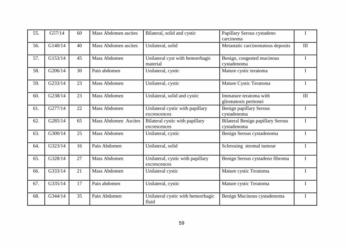
59
55. G57/14 60 Mass Abdomen ascites Bilateral, solid and cystic Papillary Serous cystadeno
carcinoma
I
56. G140/14 40 Mass Abdomen ascites Unilateral, solid Metastatic carcinomatous deposits III
57. G153/14 45 Mass Abdomen Unilateral cyst with hemorrhagic
material
Benign, congested mucinous
cystadenoma
I
58. G206/14 30 Pain abdomen Unilateral, cystic Mature cystic teratoma I
59. G233/14 23 Mass Abdomen Unilateral, cystic Mature Cystic Teratoma I
60. G238/14 23 Mass Abdomen Unilateral, solid and cystic Immature teratoma with
gliomatosis peritonei
III
61. G277/14 22 Mass Abdomen Unilateral cystic with papillary
excrescences
Benign papillary Serous
cystadenoma
I
62. G285/14 65 Mass Abdomen Ascites Bilateral cystic with papillary
excrescences
Bilateral Benign papillary Serous
cystadenoma
I
63. G300/14 25 Mass Abdomen Unilateral, cystic Benign Serous cystadenoma I
64. G323/14 16 Pain Abdomen Unilateral, solid Sclerosing stromal tumour I
65. G328/14 27 Mass Abdomen Unilateral, cystic with papillary
excrescences
Benign Serous cystadeno fibroma I
66. G333/14 21 Mass Abdomen Unilateral cystic Mature cystic Teratoma I
67. G335/14 17 Pain abdomen Unilateral, cystic Mature cystic Teratoma I
68. G344/14 35 Pain Abdomen Unilateral cystic with hemorrhagic
fluid
Benign Mucinous cystadenoma I
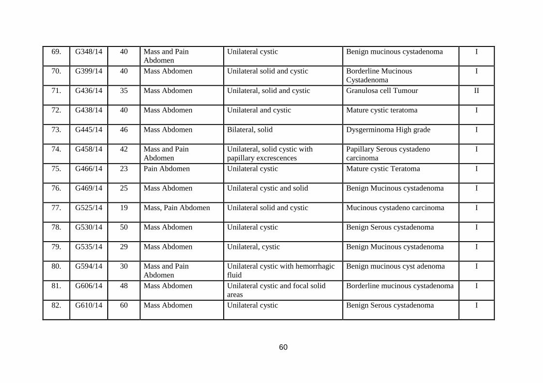
60
69. G348/14 40 Mass and Pain
Abdomen
Unilateral cystic Benign mucinous cystadenoma I
70. G399/14 40 Mass Abdomen Unilateral solid and cystic Borderline Mucinous
Cystadenoma
I
71. G436/14 35 Mass Abdomen Unilateral, solid and cystic Granulosa cell Tumour II
72. G438/14 40 Mass Abdomen Unilateral and cystic Mature cystic teratoma I
73. G445/14 46 Mass Abdomen Bilateral, solid Dysgerminoma High grade I
74. G458/14 42 Mass and Pain
Abdomen
Unilateral, solid cystic with
papillary excrescences
Papillary Serous cystadeno
carcinoma
I
75. G466/14 23 Pain Abdomen Unilateral cystic Mature cystic Teratoma I
76. G469/14 25 Mass Abdomen Unilateral cystic and solid Benign Mucinous cystadenoma I
77. G525/14 19 Mass, Pain Abdomen Unilateral solid and cystic Mucinous cystadeno carcinoma I
78. G530/14 50 Mass Abdomen Unilateral cystic Benign Serous cystadenoma I
79. G535/14 29 Mass Abdomen Unilateral, cystic Benign Mucinous cystadenoma I
80. G594/14 30 Mass and Pain
Abdomen
Unilateral cystic with hemorrhagic
fluid
Benign mucinous cyst adenoma I
81. G606/14 48 Mass Abdomen Unilateral cystic and focal solid
areas
Borderline mucinous cystadenoma I
82. G610/14 60 Mass Abdomen Unilateral cystic Benign Serous cystadenoma I
61
83. G619/14 26 Pain Abdomen Unilateral cystic Mature cystic Teratoma I
84. G621/14 65 Mass and Pain
Abdomen
Unilateral cystic with hemorrhagic Congested Benign Mucinous
cystadenoma
I
85. G644/14 29 Mass Abdomen Unilateral cystic Benign Serous cystadenoma I
86. G676/14 24 asymptomatic Unilateral cystic Benign Serous cystadenoma I
87. G754/14 50 Mass Abdomen Unilateral cystic Mature cystic teratoma I
88. G756/14 35 Mass Abdomen Unilateral cystic Mature cystic teratoma I
89. G771/14 60 Mass and Pain
Abdomen
Unilateral cystic Borderline mucinous cystadenoma I
90. G782/14 35 Mass Abdomen Unilateral solid and cystic Papillary Serous cystadeno
carcinoma
II
91. G798/14 52 Mass Abdomen Unilateral cystic Borderline mucinous cystadenoma I
92. G828/14 50 Mass Abdomen; ascites Unilateral cystic Benign mucinous cystadenoma I
93. G879/14 37 Mass Abdomen Pain Unilateral, cystic with Hemorrhagic
material
Congested Benign Serous
cystadenoma
I
94. G945/14 32 Mass Abdomen Unilateral cystic with papillary
excrescences
Benign papillary Serous
cystadenoma
I
95. G947/14 53 Mass Abdomen ascites Unilateral cystic Benign mucinous cystadenoma I
96. G973/14 70 Mass Abdomen Unilateral solid Fibro thecoma I
62
97. G983/14 22 Pain Abdomen Unilateral cystic Mature cystic Teratoma I
98. G1015/14 44 Mass Abdomen Unilateral cystic Benign mucinous cystadenoma I
99. G1003/14 44 Mass Abdomen Unilateral, cystic Mature cystic teratoma I
100. G1069/14 42 Mass Abdomen Unilateral cystic with papillary
excrescences and solid
Papillary Serous cystadeno
carcinoma
I
101. G1072/14 60 Mass and Pain
Abdomen
Unilateral cystic Mature cystic Teratoma I
102. G1077/14 40 Mass Abdomen ascites Unilateral solid and cystic Endometrioid adeno
carcinomatous deposits
III
103. G1083/14 57 Mass Abdomen Bilateral solid, cystic with papillary
excrescences
Borderline papillary Serous
cystadeno carcinoma
II
104. G1086/14 27 Mass Abdomen Unilateral cystic Benign Serous cystadenoma I
105. G1119/14 39 Pain Abdomen Unilateral Simple Serous cystadenoma I
106. G1124/14 55 Mass Abdomen Unilateral solid Fibroma I
107. G1126/14 30 Mass Abdomen Unilateral cystic with papillary
excrescences
Borderline Serous Tumour I
108. G1140/14 39 Mass, Pain Abdomen Unilateral solid and cystic with
papillary excrescences.
Benign papillary Serous
cystadenoma
I
109. G7/15 26 Mass and Pain
Abdomen
Unilateral, cystic Mature cystic Teratoma I
110. G15/15 65 Mass Abdomen Unilateral, cystic Benign Serous cystadenoma I

63
111. G18/15 63 Mass Abdomen ascites Unilateral, cystic Benign mucinous cystadenoma I
112. G34/15 26 Mass Abdomen Unilateral, cystic with papillary
excrescences
Benign papillary Serous
cystadenoma
I
113. G56/15 32 Mass Abdomen Unilateral, cystic Benign mucinous cystadenoma I
114. G72/15 24 Mass Abdomen Bilateral, cystic Bilateral mature cystic Teratoma I
115. G89/15 43 Mass Abdomen, chronic
liver disease,
splenomegaly
Bilateral, cystic Benign mucinous cystadenoma I
116. G99/15 70 Mass Abdomen Bilateral, solid cystic Bilateral Benign Serous
cystadenoma
II
117. G120/15 53 Mass Abdomen Unilateral solid cystic with
papillary projections and
Hemorrhagic areas and necrosis
Adult granulosa cell tumour I
118. G137/15 60 Mass Abdomen ascites Unilocular cystic Benign mucinous cystadenoma I
119. G143/15 45 Mass Abdomen Unilateral cystic and solid Benign mucinous cystadeno
carcinoma
I
120. G147/15 40 Mass Abdomen Unilateral cystic Borderline mucinous tumour of
ovary
I
121. G168/15 25 Mass Abdomen Unilateral cystic and solid areas Mature cystic teratoma I
122. G176/15 55 Mass Abdomen, ascites
malignant cells
Unilateral, solid with papillary
excrescences and Hemorrhagic
Serous adenocarcinoma II
123. G180/15 65 Mass Abdomen Unilateral, cystic Benign mucinous cystadenoma I
124. G218/15 40 Mass Abdomen Unilateral cystic with Hemorrhagic
fluid
Benign mucinous cystadenoma I
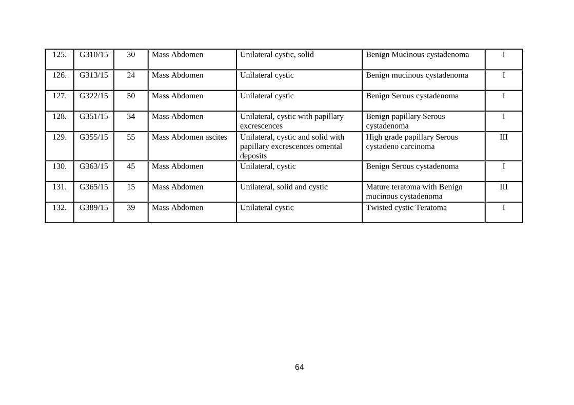
64
125. G310/15 30 Mass Abdomen Unilateral cystic, solid Benign Mucinous cystadenoma I
126. G313/15 24 Mass Abdomen Unilateral cystic Benign mucinous cystadenoma I
127. G322/15 50 Mass Abdomen Unilateral cystic Benign Serous cystadenoma I
128. G351/15 34 Mass Abdomen Unilateral, cystic with papillary
excrescences
Benign papillary Serous
cystadenoma
I
129. G355/15 55 Mass Abdomen ascites Unilateral, cystic and solid with
papillary excrescences omental
deposits
High grade papillary Serous
cystadeno carcinoma
III
130. G363/15 45 Mass Abdomen Unilateral, cystic Benign Serous cystadenoma I
131. G365/15 15 Mass Abdomen Unilateral, solid and cystic Mature teratoma with Benign
mucinous cystadenoma
III
132. G389/15 39 Mass Abdomen Unilateral cystic Twisted cystic Teratoma I

65
OBSERVATION AND RESULTS
This prospective study comprises 132 ovarian neoplasms. The cases were referred
from Raja Mirasudhar Government Hospital (RMH), Thanjavur Medical College. The study
period is from January 2013 to May 2015.
The following table shows the total number of ovarian neoplasms among all the
female neoplasms.
I. TOTAL NUMBER OF OVARIAN NEOPLASM IN COMPARISON WITH THE
TOTAL FEMALE NEOPLASMS
Table 4:
S.NO. PERIOD
TOTAL NO.
OF FEMALE
NEOPLASMS
TOTAL NO.
OF
OVARIAN
NEOPLASMS
PERCENTAGE
1. January 2013 – May 2013 438 19 4.3%
2. June 2013-December 2013 761 33 4.3%
3. January 2014 – May 2014 479 24 5.0%
4. June 2014 – December 2014 676 32 4.7%
5. January 2015 – May 2015 398 24 6.0%
Total 2755 132 4.7%
The average incidence of ovarian neoplasm (Benign and malignant) among female is 4.7%
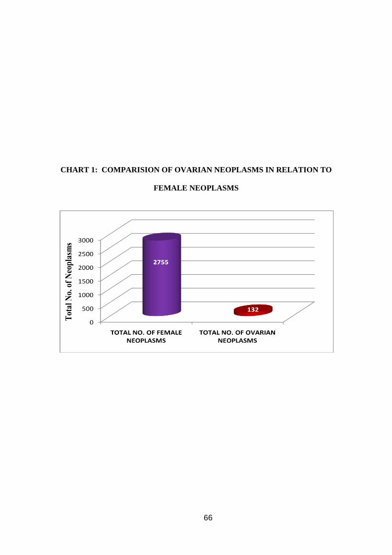
66
CHART 1: COMPARISION OF OVARIAN NEOPLASMS IN RELATION TO
FEMALE NEOPLASMS
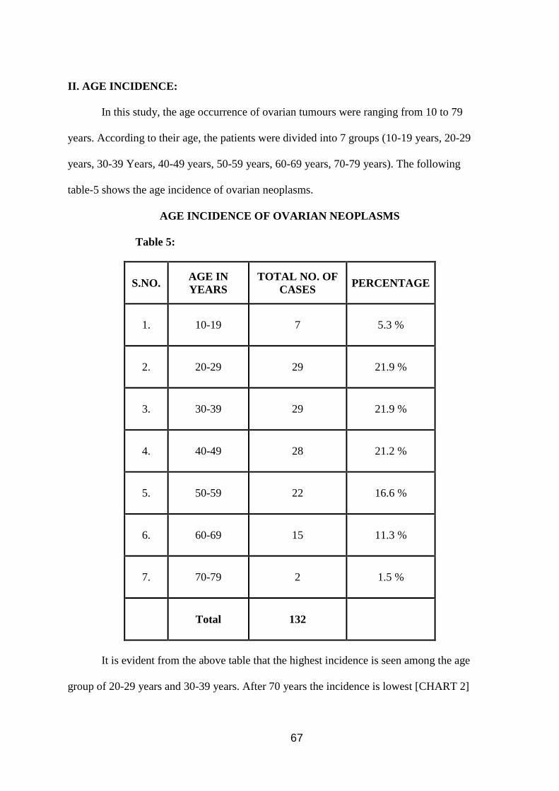
67
II. AGE INCIDENCE:
In this study, the age occurrence of ovarian tumours were ranging from 10 to 79
years. According to their age, the patients were divided into 7 groups (10-19 years, 20-29
years, 30-39 Years, 40-49 years, 50-59 years, 60-69 years, 70-79 years). The following
table-5 shows the age incidence of ovarian neoplasms.
AGE INCIDENCE OF OVARIAN NEOPLASMS
Table 5:
S.NO. AGE IN
YEARS
TOTAL NO. OF
CASES PERCENTAGE
1. 10-19 7 5.3 %
2. 20-29 29 21.9 %
3. 30-39 29 21.9 %
4. 40-49 28 21.2 %
5. 50-59 22 16.6 %
6. 60-69 15 11.3 %
7. 70-79 2 1.5 %
Total 132
It is evident from the above table that the highest incidence is seen among the age
group of 20-29 years and 30-39 years. After 70 years the incidence is lowest [CHART 2]
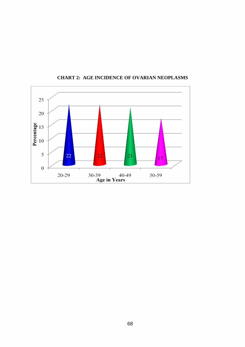
68
CHART 2: AGE INCIDENCE OF OVARIAN NEOPLASMS
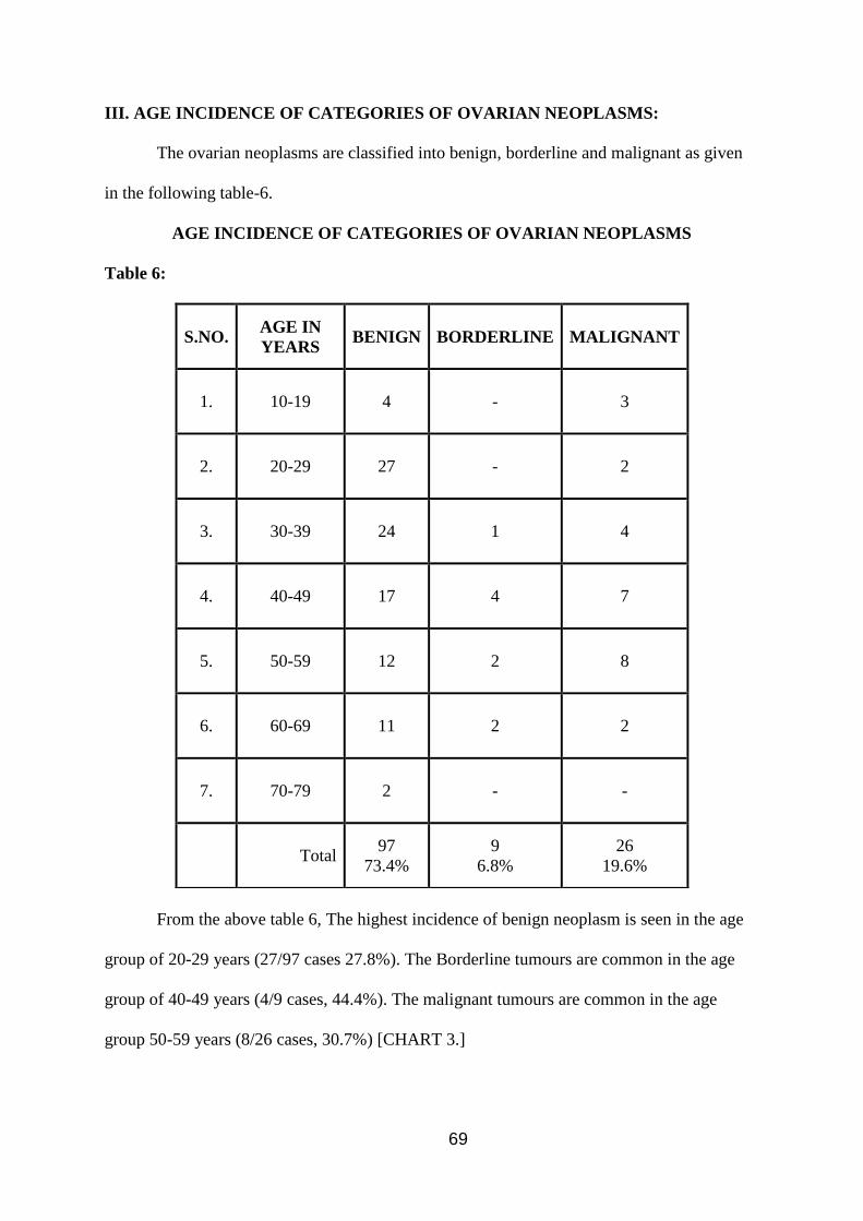
69
III. AGE INCIDENCE OF CATEGORIES OF OVARIAN NEOPLASMS:
The ovarian neoplasms are classified into benign, borderline and malignant as given
in the following table-6.
AGE INCIDENCE OF CATEGORIES OF OVARIAN NEOPLASMS
Table 6:
From the above table 6, The highest incidence of benign neoplasm is seen in the age
group of 20-29 years (27/97 cases 27.8%). The Borderline tumours are common in the age
group of 40-49 years (4/9 cases, 44.4%). The malignant tumours are common in the age
group 50-59 years (8/26 cases, 30.7%) [CHART 3.]
S.NO. AGE IN
YEARS BENIGN BORDERLINE MALIGNANT
1. 10-19 4 - 3
2. 20-29 27 - 2
3. 30-39 24 1 4
4. 40-49 17 4 7
5. 50-59 12 2 8
6. 60-69 11 2 2
7. 70-79 2 - -
Total 97
73.4%
9
6.8%
26
19.6%
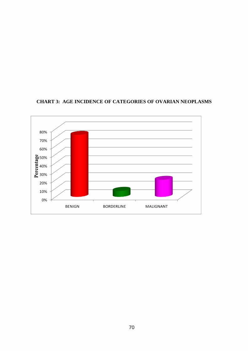
70
CHART 3: AGE INCIDENCE OF CATEGORIES OF OVARIAN NEOPLASMS
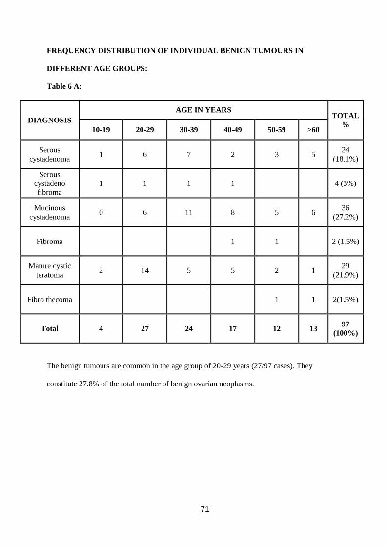
71
FREQUENCY DISTRIBUTION OF INDIVIDUAL BENIGN TUMOURS IN
DIFFERENT AGE GROUPS:
Table 6 A:
DIAGNOSIS
AGE IN YEARS TOTAL
% 10-19 20-29 30-39 40-49 50-59 >60
Serous
cystadenoma 1 6 7 2 3 5
24
(18.1%)
Serous
cystadeno
fibroma
1 1 1 1 4 (3%)
Mucinous
cystadenoma 0 6 11 8 5 6
36
(27.2%)
Fibroma 1 1 2 (1.5%)
Mature cystic
teratoma 2 14 5 5 2 1
29
(21.9%)
Fibro thecoma 1 1 2(1.5%)
Total 4 27 24 17 12 13 97
(100%)
The benign tumours are common in the age group of 20-29 years (27/97 cases). They
constitute 27.8% of the total number of benign ovarian neoplasms.
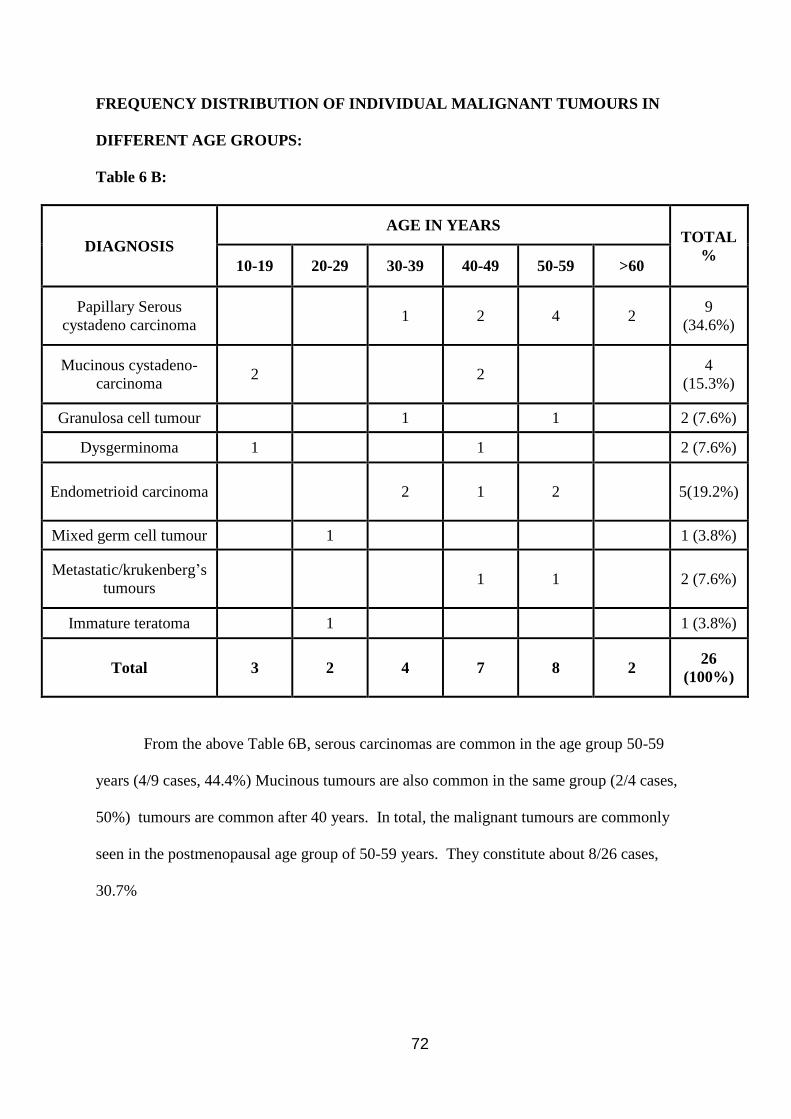
72
FREQUENCY DISTRIBUTION OF INDIVIDUAL MALIGNANT TUMOURS IN
DIFFERENT AGE GROUPS:
Table 6 B:
DIAGNOSIS
AGE IN YEARS TOTAL
% 10-19 20-29 30-39 40-49 50-59 >60
Papillary Serous
cystadeno carcinoma 1 2 4 2
9
(34.6%)
Mucinous cystadeno-
carcinoma 2 2
4
(15.3%)
Granulosa cell tumour 1 1 2 (7.6%)
Dysgerminoma 1 1 2 (7.6%)
Endometrioid carcinoma 2 1 2 5(19.2%)
Mixed germ cell tumour 1 1 (3.8%)
Metastatic/krukenberg’s
tumours 1 1 2 (7.6%)
Immature teratoma 1 1 (3.8%)
Total 3 2 4 7 8 2 26
(100%)
From the above Table 6B, serous carcinomas are common in the age group 50-59
years (4/9 cases, 44.4%) Mucinous tumours are also common in the same group (2/4 cases,
50%) tumours are common after 40 years. In total, the malignant tumours are commonly
seen in the postmenopausal age group of 50-59 years. They constitute about 8/26 cases,
30.7%
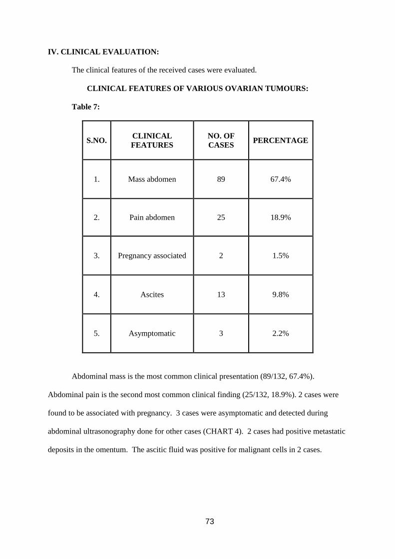
73
IV. CLINICAL EVALUATION:
The clinical features of the received cases were evaluated.
CLINICAL FEATURES OF VARIOUS OVARIAN TUMOURS:
Table 7:
S.NO. CLINICAL
FEATURES
NO. OF
CASES PERCENTAGE
1. Mass abdomen 89 67.4%
2. Pain abdomen 25 18.9%
3. Pregnancy associated 2 1.5%
4. Ascites 13 9.8%
5. Asymptomatic 3 2.2%
Abdominal mass is the most common clinical presentation (89/132, 67.4%).
Abdominal pain is the second most common clinical finding (25/132, 18.9%). 2 cases were
found to be associated with pregnancy. 3 cases were asymptomatic and detected during
abdominal ultrasonography done for other cases (CHART 4). 2 cases had positive metastatic
deposits in the omentum. The ascitic fluid was positive for malignant cells in 2 cases.
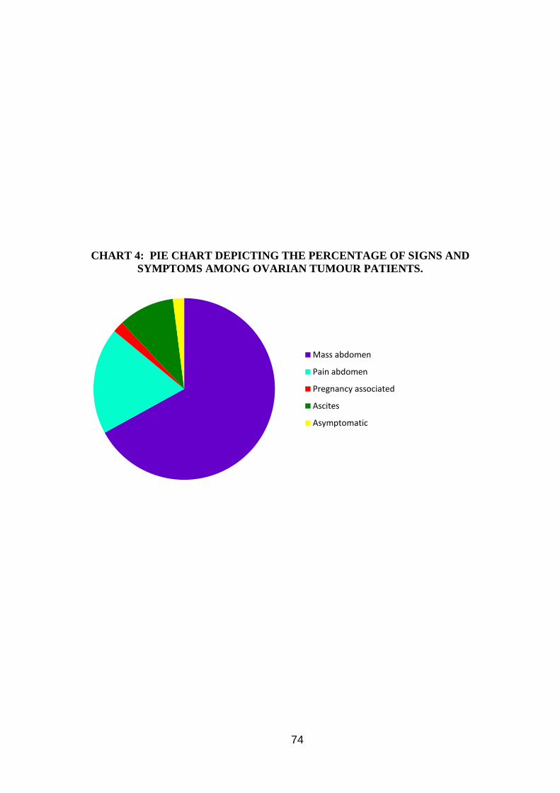
74
CHART 4: PIE CHART DEPICTING THE PERCENTAGE OF SIGNS AND
SYMPTOMS AMONG OVARIAN TUMOUR PATIENTS.
Mass abdomen
Pain abdomen
Pregnancy associated
Ascites
Asymptomatic
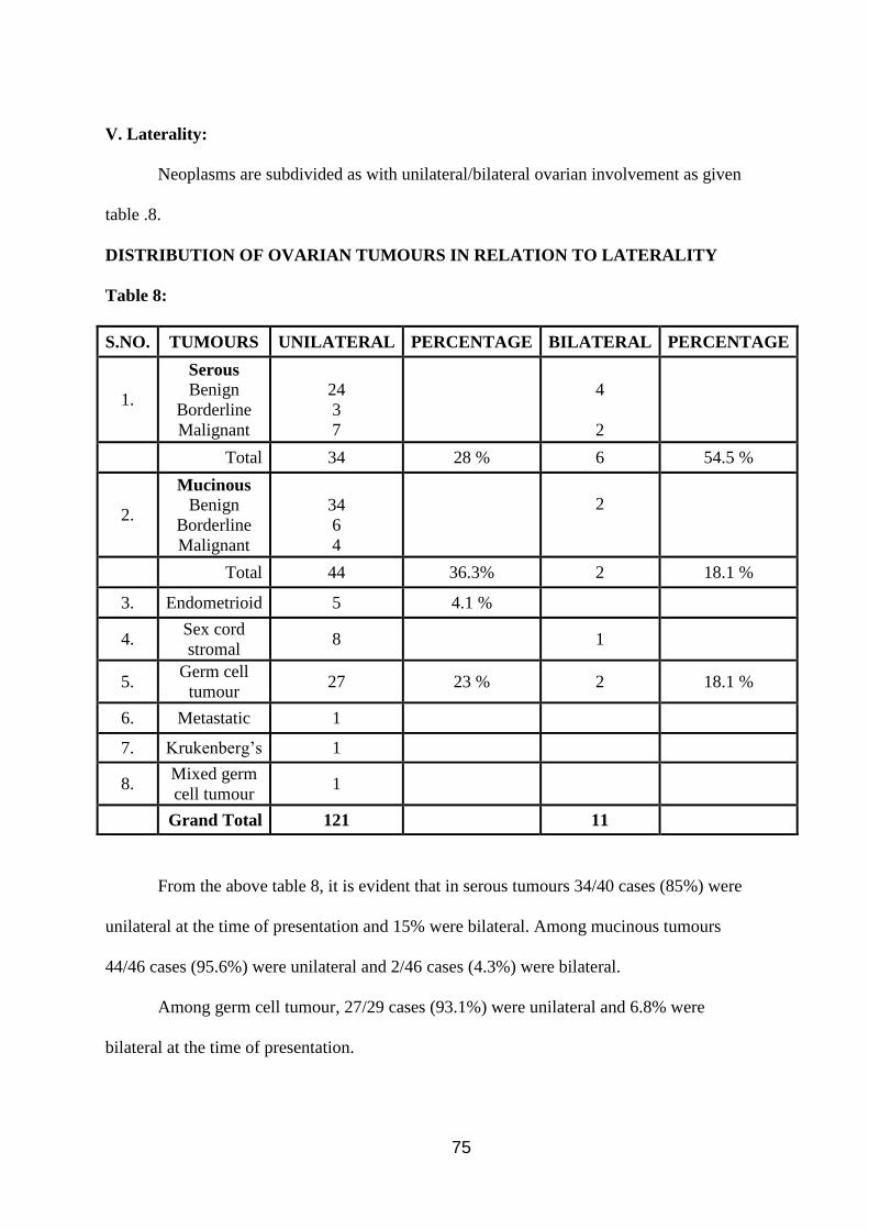
75
V. Laterality:
Neoplasms are subdivided as with unilateral/bilateral ovarian involvement as given
table .8.
DISTRIBUTION OF OVARIAN TUMOURS IN RELATION TO LATERALITY
Table 8:
S.NO. TUMOURS UNILATERAL PERCENTAGE BILATERAL PERCENTAGE
1.
Serous Benign
Borderline
Malignant
24
3
7
4
2
Total 34 28 % 6 54.5 %
2.
Mucinous Benign
Borderline
Malignant
34
6
4
2
Total 44 36.3% 2 18.1 %
3. Endometrioid 5 4.1 %
4. Sex cord
stromal 8 1
5. Germ cell
tumour 27 23 % 2 18.1 %
6. Metastatic 1
7. Krukenberg’s 1
8. Mixed germ
cell tumour 1
Grand Total 121 11
From the above table 8, it is evident that in serous tumours 34/40 cases (85%) were
unilateral at the time of presentation and 15% were bilateral. Among mucinous tumours
44/46 cases (95.6%) were unilateral and 2/46 cases (4.3%) were bilateral.
Among germ cell tumour, 27/29 cases (93.1%) were unilateral and 6.8% were
bilateral at the time of presentation.
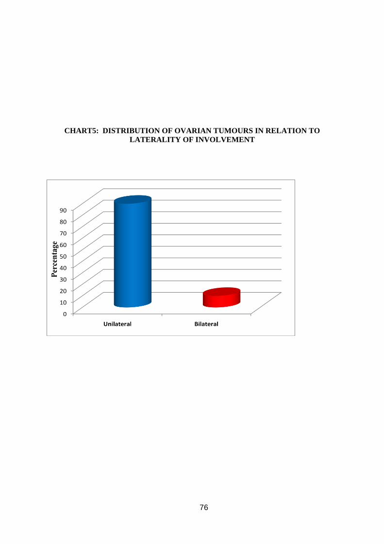
76
CHART5: DISTRIBUTION OF OVARIAN TUMOURS IN RELATION TO
LATERALITY OF INVOLVEMENT
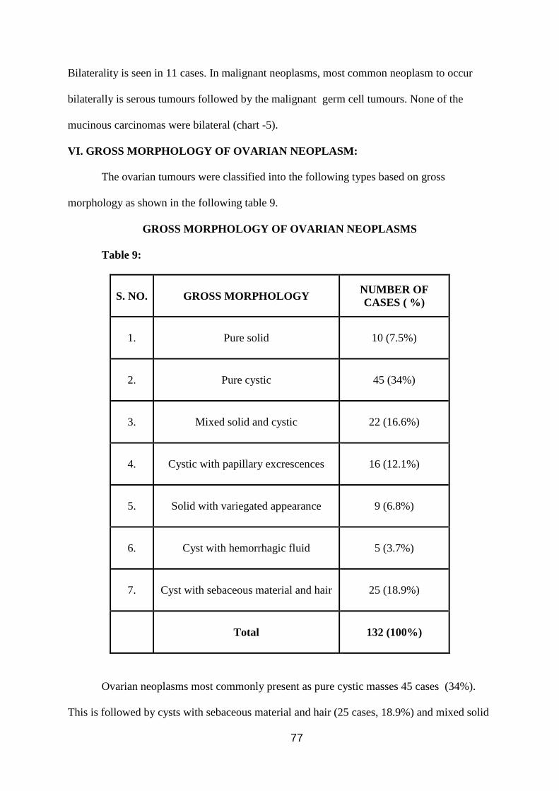
77
Bilaterality is seen in 11 cases. In malignant neoplasms, most common neoplasm to occur
bilaterally is serous tumours followed by the malignant germ cell tumours. None of the
mucinous carcinomas were bilateral (chart -5).
VI. GROSS MORPHOLOGY OF OVARIAN NEOPLASM:
The ovarian tumours were classified into the following types based on gross
morphology as shown in the following table 9.
GROSS MORPHOLOGY OF OVARIAN NEOPLASMS
Table 9:
S. NO. GROSS MORPHOLOGY NUMBER OF
CASES ( %)
1. Pure solid 10 (7.5%)
2. Pure cystic 45 (34%)
3. Mixed solid and cystic 22 (16.6%)
4. Cystic with papillary excrescences 16 (12.1%)
5. Solid with variegated appearance 9 (6.8%)
6. Cyst with hemorrhagic fluid 5 (3.7%)
7. Cyst with sebaceous material and hair 25 (18.9%)
Total 132 (100%)
Ovarian neoplasms most commonly present as pure cystic masses 45 cases (34%).
This is followed by cysts with sebaceous material and hair (25 cases, 18.9%) and mixed solid
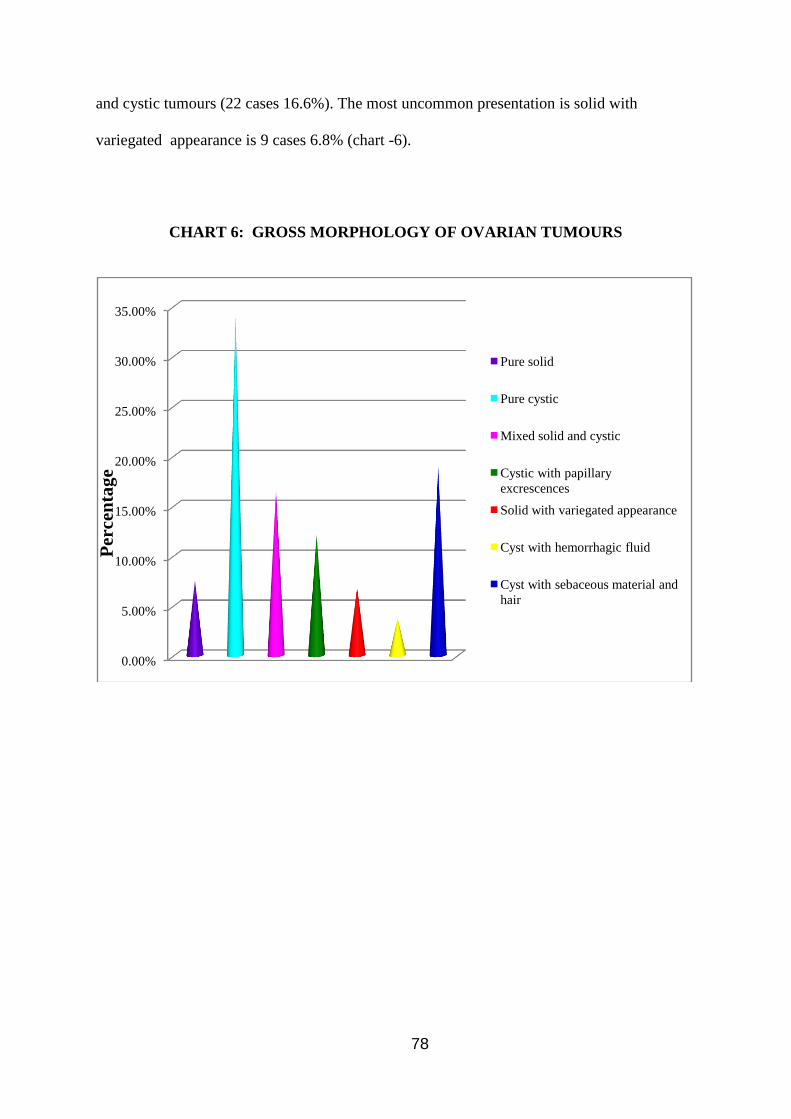
78
and cystic tumours (22 cases 16.6%). The most uncommon presentation is solid with
variegated appearance is 9 cases 6.8% (chart -6).
CHART 6: GROSS MORPHOLOGY OF OVARIAN TUMOURS
0.00%
5.00%
10.00%
15.00%
20.00%
25.00%
30.00%
35.00%
Pure solid
Pure cystic
Mixed solid and cystic
Cystic with papillary
excrescences
Solid with variegated appearance
Cyst with hemorrhagic fluid
Cyst with sebaceous material and
hair
Per
cen
tag
e
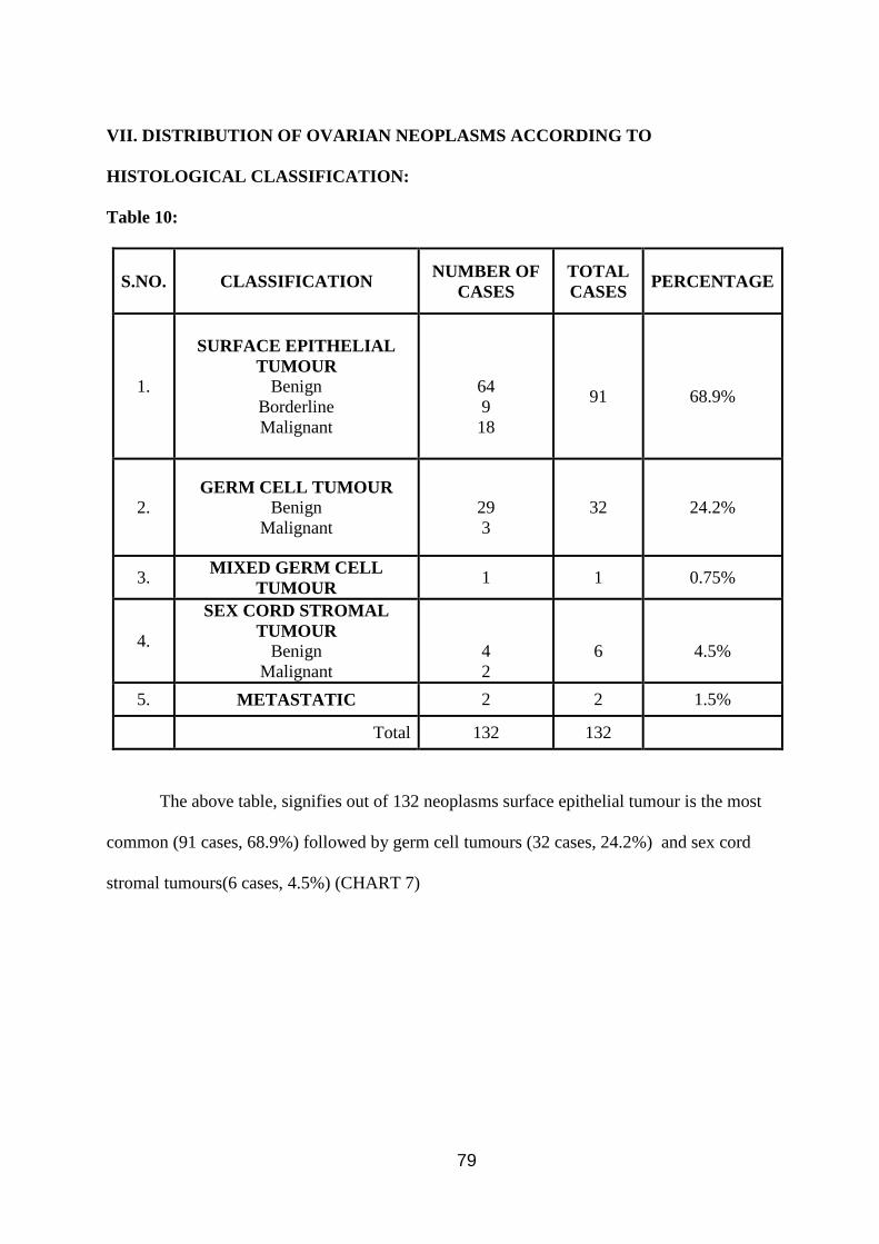
79
VII. DISTRIBUTION OF OVARIAN NEOPLASMS ACCORDING TO
HISTOLOGICAL CLASSIFICATION:
Table 10:
S.NO. CLASSIFICATION NUMBER OF
CASES
TOTAL
CASES PERCENTAGE
1.
SURFACE EPITHELIAL
TUMOUR
Benign
Borderline
Malignant
64
9
18
91
68.9%
2. GERM CELL TUMOUR
Benign
Malignant
29
3
32 24.2%
3. MIXED GERM CELL
TUMOUR 1 1 0.75%
4.
SEX CORD STROMAL
TUMOUR
Benign
Malignant
4
2
6
4.5%
5. METASTATIC 2 2 1.5%
Total 132 132
The above table, signifies out of 132 neoplasms surface epithelial tumour is the most
common (91 cases, 68.9%) followed by germ cell tumours (32 cases, 24.2%) and sex cord
stromal tumours(6 cases, 4.5%) (CHART 7)
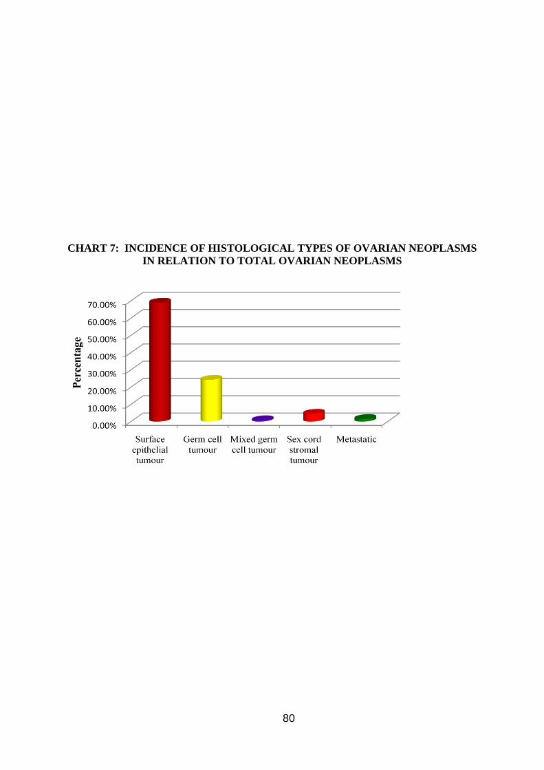
80
CHART 7: INCIDENCE OF HISTOLOGICAL TYPES OF OVARIAN NEOPLASMS
IN RELATION TO TOTAL OVARIAN NEOPLASMS
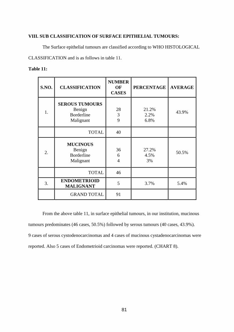
81
VIII. SUB CLASSIFICATION OF SURFACE EPITHELIAL TUMOURS:
The Surface epithelial tumours are classified according to WHO HISTOLOGICAL
CLASSIFICATION and is as follows in table 11.
Table 11:
S.NO. CLASSIFICATION
NUMBER
OF
CASES
PERCENTAGE AVERAGE
1.
SEROUS TUMOURS
Benign
Borderline
Malignant
28
3
9
21.2%
2.2%
6.8%
43.9%
TOTAL 40
2.
MUCINOUS
Benign
Borderline
Malignant
36
6
4
27.2%
4.5%
3%
50.5%
TOTAL 46
3. ENDOMETRIOID
MALIGNANT 5 3.7% 5.4%
GRAND TOTAL 91
From the above table 11, in surface epithelial tumours, in our institution, mucinous
tumours predominates (46 cases, 50.5%) followed by serous tumours (40 cases, 43.9%).
9 cases of serous cystodenocarcinomas and 4 cases of mucinous cystadenocarcinomas were
reported. Also 5 cases of Endometrioid carcinomas were reported. (CHART 8).
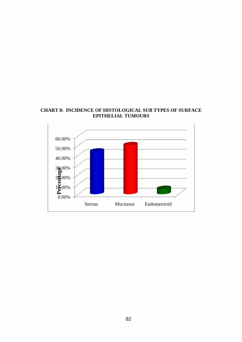
82
CHART 8: INCIDENCE OF HISTOLOGICAL SUB TYPES OF SURFACE
EPITHELIAL TUMOURS
0.00%
10.00%
20.00%
30.00%
40.00%
50.00%
60.00%
Serous Mucinous Endometrioid
Per
cen
tag
e
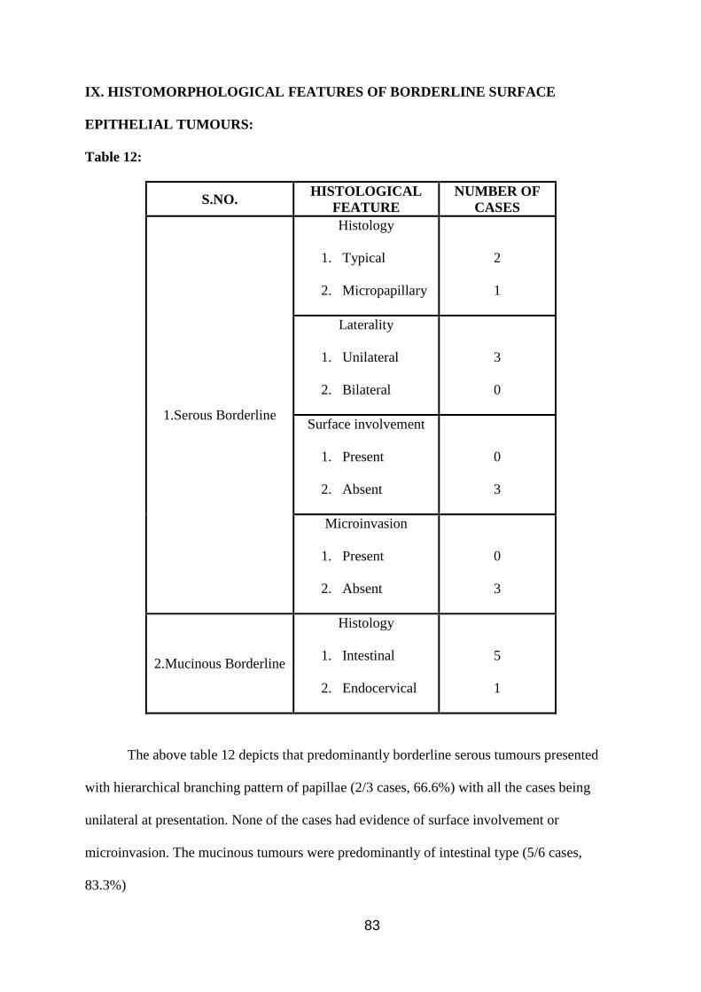
83
IX. HISTOMORPHOLOGICAL FEATURES OF BORDERLINE SURFACE
EPITHELIAL TUMOURS:
Table 12:
S.NO. HISTOLOGICAL
FEATURE
NUMBER OF
CASES
1.Serous Borderline
Histology
1. Typical
2. Micropapillary
2
1
Laterality
1. Unilateral
2. Bilateral
3
0
Surface involvement
1. Present
2. Absent
0
3
Microinvasion
1. Present
2. Absent
0
3
2.Mucinous Borderline
Histology
1. Intestinal
2. Endocervical
5
1
The above table 12 depicts that predominantly borderline serous tumours presented
with hierarchical branching pattern of papillae (2/3 cases, 66.6%) with all the cases being
unilateral at presentation. None of the cases had evidence of surface involvement or
microinvasion. The mucinous tumours were predominantly of intestinal type (5/6 cases,
83.3%)

84
X. HISTOMORPHOLOGY OF MALIGNANT SURFACE EPITHELIAL TUMOUR:
Table 13:
S.NO. HISTOLOGICAL TYPE NUMBER OF
CASES
1. Serous Grading
1. Low Grade
2. High Grade
3
6
Total 9
2. Mucinous Type of invasion
1. Expansive
2. Infiltrating
4
0
Total 4
Malignant serous neoplasms were graded according to recent 2 tier system of classification.
High grade tumours exhibited marked nuclear atypia and >12 mitosis/10Hpf. From the above
table 13, 3/9 cases (33.3%) were low grade carcinomas and 6/9 cases (66.6%) were high
grade carcinomas. High grade carcinomas constitute the commonest type in the malignant
serous tumours. All the 4 cases of mucinous carcinomas presented with expansile type of
invasion with back to back gland arrangement and no stroma in between.
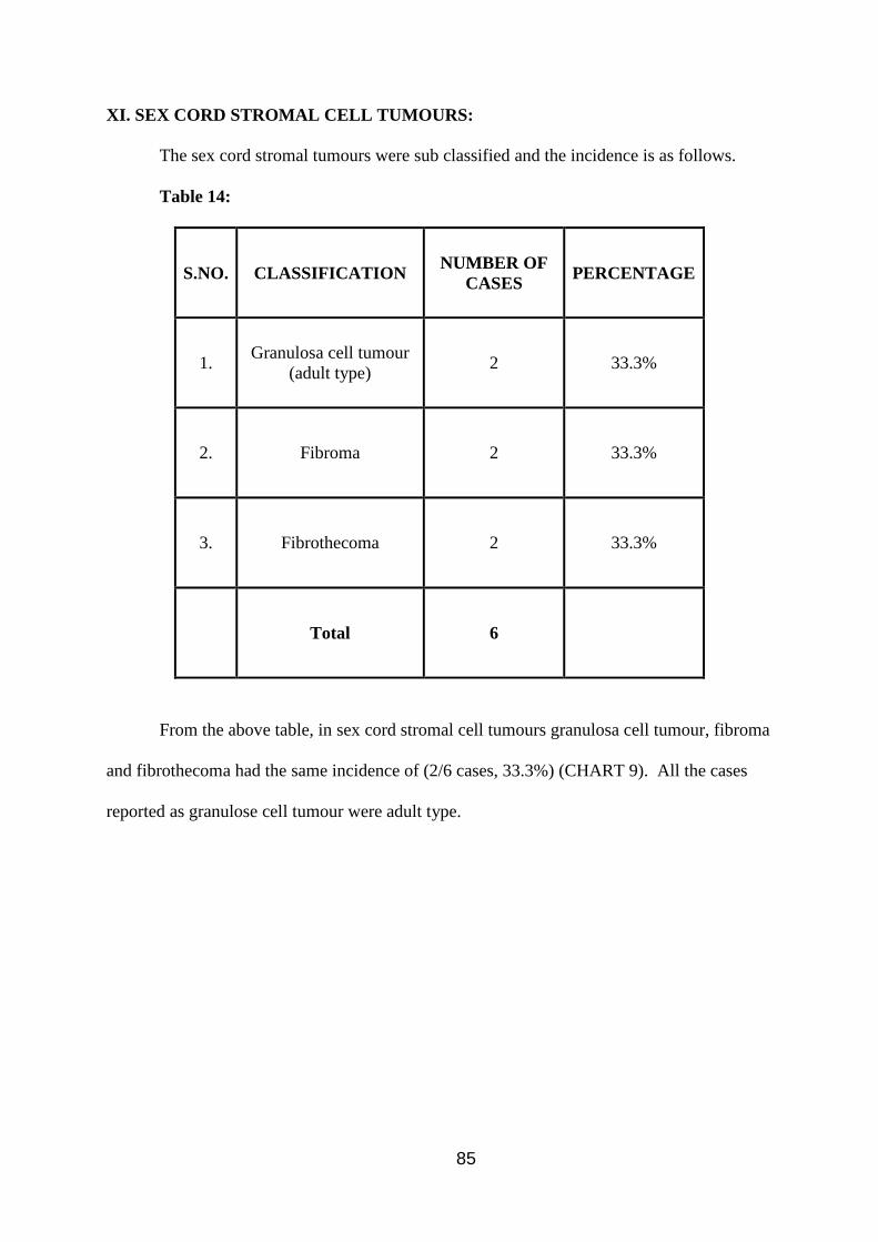
85
XI. SEX CORD STROMAL CELL TUMOURS:
The sex cord stromal tumours were sub classified and the incidence is as follows.
Table 14:
S.NO. CLASSIFICATION NUMBER OF
CASES PERCENTAGE
1. Granulosa cell tumour
(adult type) 2 33.3%
2. Fibroma 2 33.3%
3. Fibrothecoma 2 33.3%
Total 6
From the above table, in sex cord stromal cell tumours granulosa cell tumour, fibroma
and fibrothecoma had the same incidence of (2/6 cases, 33.3%) (CHART 9). All the cases
reported as granulose cell tumour were adult type.
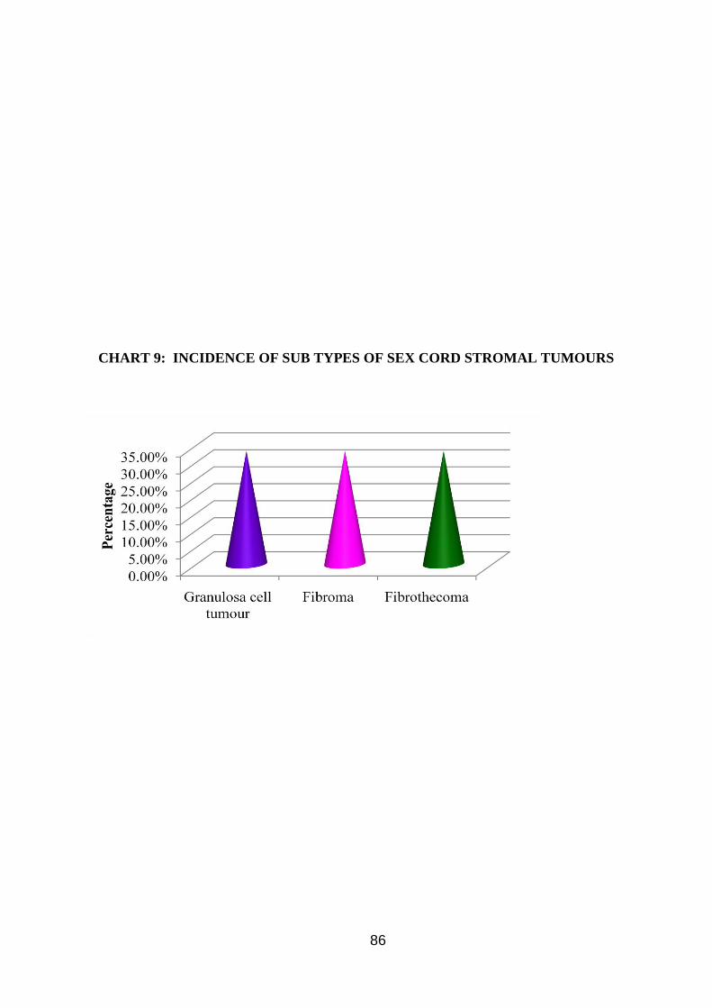
86
CHART 9: INCIDENCE OF SUB TYPES OF SEX CORD STROMAL TUMOURS
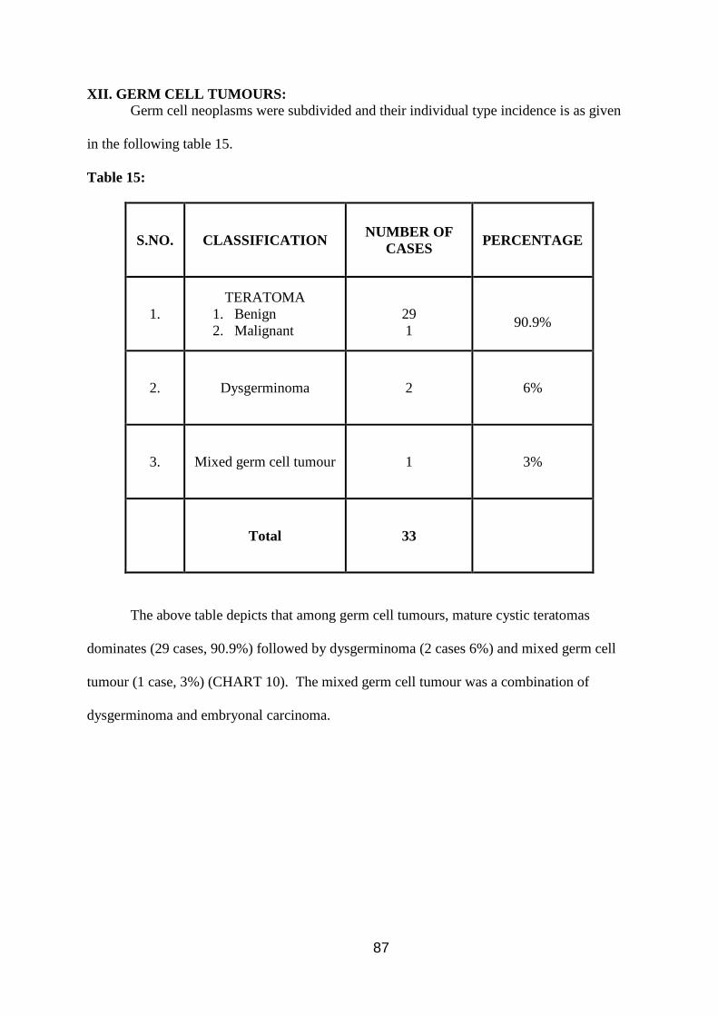
87
XII. GERM CELL TUMOURS:
Germ cell neoplasms were subdivided and their individual type incidence is as given
in the following table 15.
Table 15:
S.NO. CLASSIFICATION NUMBER OF
CASES PERCENTAGE
1.
TERATOMA
1. Benign
2. Malignant
29
1
90.9%
2. Dysgerminoma 2 6%
3. Mixed germ cell tumour 1 3%
Total 33
The above table depicts that among germ cell tumours, mature cystic teratomas
dominates (29 cases, 90.9%) followed by dysgerminoma (2 cases 6%) and mixed germ cell
tumour (1 case, 3%) (CHART 10). The mixed germ cell tumour was a combination of
dysgerminoma and embryonal carcinoma.
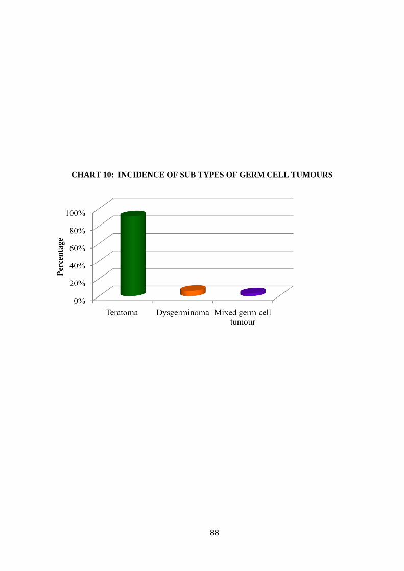
88
CHART 10: INCIDENCE OF SUB TYPES OF GERM CELL TUMOURS

89
XIII. IMMUNOHISTOCHEMISTRY:
EXPRESSION PROFILE OF ER, PR AND Her-2/neu IN MALIGNANT TUMOURS.
Table 16:
Sl.
No.
HPE
No. HPE DIAGNOSIS ER PR Her-2/neu
1 579/13 Endometrioid carcinoma Negative Negative Negative(1+)
2 920/13 Endometrioid carcinoma Positive Positive Negative(0+)
3 1077/14 Endometrioid carcinoma Strong
Positive
Strong
Positive Negative(0+)
4 1195/13 High grade serous carcinoma Positive Positive Negative(1+)
5 57/14 Bilateral High grade serous
carcinoma Positive Negative Positive(3+)
6 458/14 High grade serous carcinoma Negative Positive Positive(3+)
7 782/14 Low grade serous carcinoma Negative Negative Negative(0+)
8 355/14 High grade serous carcinoma Positive Positive Positive(3+)
9 792/13 Mucinous carcinoma Negative Negative Negative(0+)
10 1084/13 Mucinous carcinoma Negative Negative Positive(2+)
11 525/14 Mucinous carcinoma Negative Negative Negative(1+)
11 cases out of 26 malignant surface epithelial ovarian tumors were selected for
immunohistochemistry. ER, PR expression was evaluated in the 11 malignant surface
epithelial ovarian cancers.
ER and PR expression was based on the proportion of cells in a given tumor specimen
exhibiting distinct nuclear immune positivity as well as intensity of staining (Negative <10%
and positive 10%) (Table 16).
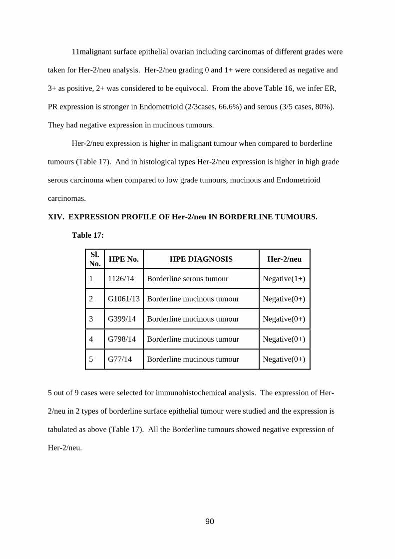
90
11malignant surface epithelial ovarian including carcinomas of different grades were
taken for Her-2/neu analysis. Her-2/neu grading 0 and 1+ were considered as negative and
3+ as positive, 2+ was considered to be equivocal. From the above Table 16, we infer ER,
PR expression is stronger in Endometrioid (2/3cases, 66.6%) and serous (3/5 cases, 80%).
They had negative expression in mucinous tumours.
Her-2/neu expression is higher in malignant tumour when compared to borderline
tumours (Table 17). And in histological types Her-2/neu expression is higher in high grade
serous carcinoma when compared to low grade tumours, mucinous and Endometrioid
carcinomas.
XIV. EXPRESSION PROFILE OF Her-2/neu IN BORDERLINE TUMOURS.
Table 17:
Sl.
No. HPE No. HPE DIAGNOSIS Her-2/neu
1 1126/14 Borderline serous tumour Negative(1+)
2 G1061/13 Borderline mucinous tumour Negative(0+)
3 G399/14 Borderline mucinous tumour Negative(0+)
4 G798/14 Borderline mucinous tumour Negative(0+)
5 G77/14 Borderline mucinous tumour Negative(0+)
5 out of 9 cases were selected for immunohistochemical analysis. The expression of Her-
2/neu in 2 types of borderline surface epithelial tumour were studied and the expression is
tabulated as above (Table 17). All the Borderline tumours showed negative expression of
Her-2/neu.
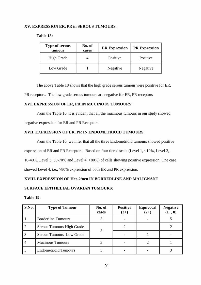
91
XV. EXPRESSION ER, PR in SEROUS TUMOURS.
Table 18:
Type of serous
tumour
No. of
cases ER Expression PR Expression
High Grade 4 Positive Positive
Low Grade 1 Negative Negative
The above Table 18 shows that the high grade serous tumour were positive for ER,
PR receptors. The low grade serous tumours are negative for ER, PR receptors
XVI. EXPRESSION OF ER, PR IN MUCINOUS TUMOURS:
From the Table 16, it is evident that all the mucinous tumours in our study showed
negative expression for ER and PR Receptors.
XVII. EXPRESSION OF ER, PR IN ENDOMETRIOID TUMOURS:
From the Table 16, we infer that all the three Endometrioid tumours showed positive
expression of ER and PR Receptors. Based on four tiered scale (Level 1, <10%, Level 2,
10-40%, Level 3, 50-70% and Level 4, >80%) of cells showing positive expression, One case
showed Level 4, i.e., >80% expression of both ER and PR expression.
XVIII. EXPRESSION OF Her-2/neu IN BORDERLINE AND MALIGNANT
SURFACE EPITHELIAL OVARIAN TUMOURS:
Table 19:
S.No. Type of Tumour No. of
cases
Positive
(3+)
Equivocal
(2+)
Negative
(1+, 0)
1 Borderline Tumours 5 - - 5
2 Serous Tumours High Grade 5
2 2
3 Serous Tumours Low Grade - 1 -
4 Mucinous Tumours 3 - 2 1
5 Endometrioid Tumours 3 - - 3
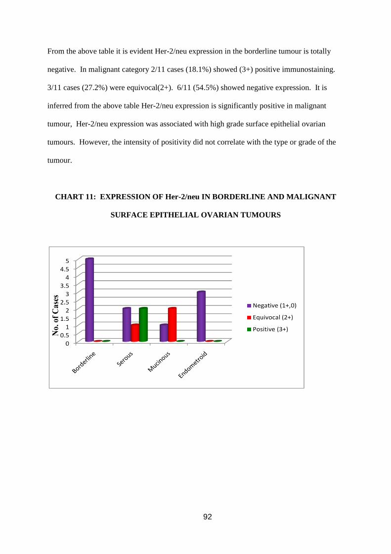
92
From the above table it is evident Her-2/neu expression in the borderline tumour is totally
negative. In malignant category 2/11 cases (18.1%) showed (3+) positive immunostaining.
3/11 cases (27.2%) were equivocal(2+). 6/11 (54.5%) showed negative expression. It is
inferred from the above table Her-2/neu expression is significantly positive in malignant
tumour, Her-2/neu expression was associated with high grade surface epithelial ovarian
tumours. However, the intensity of positivity did not correlate with the type or grade of the
tumour.
CHART 11: EXPRESSION OF Her-2/neu IN BORDERLINE AND MALIGNANT
SURFACE EPITHELIAL OVARIAN TUMOURS
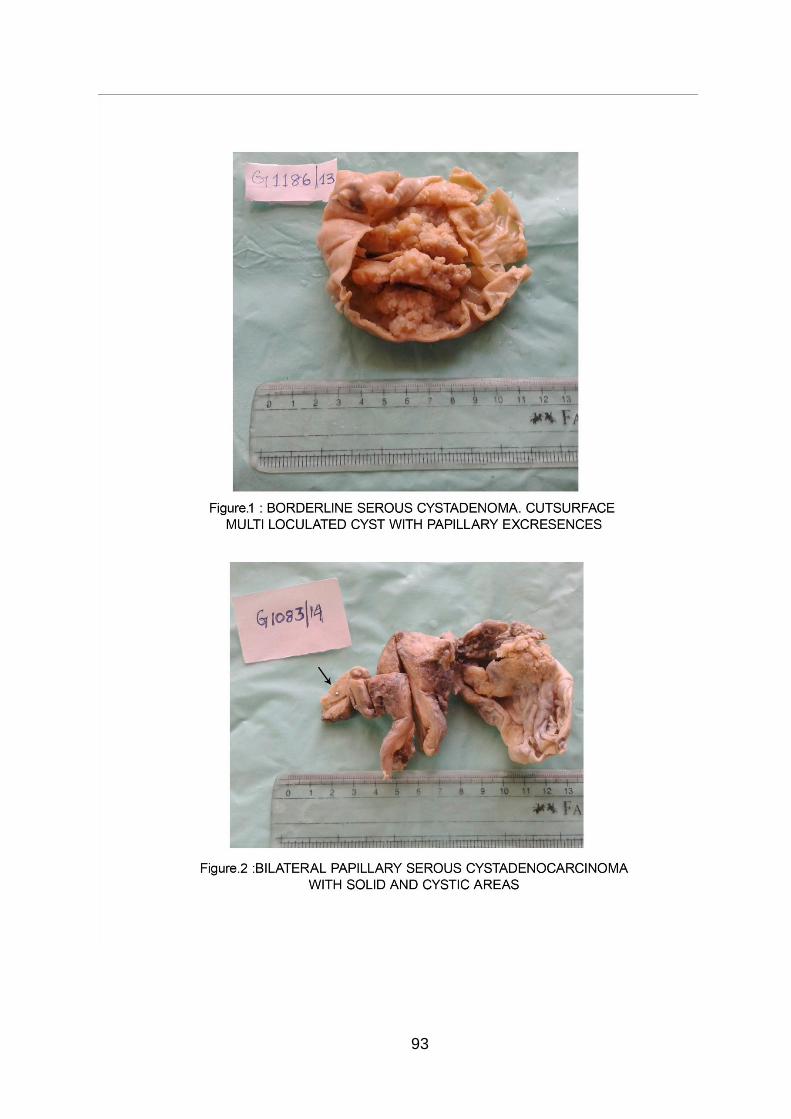
93

94
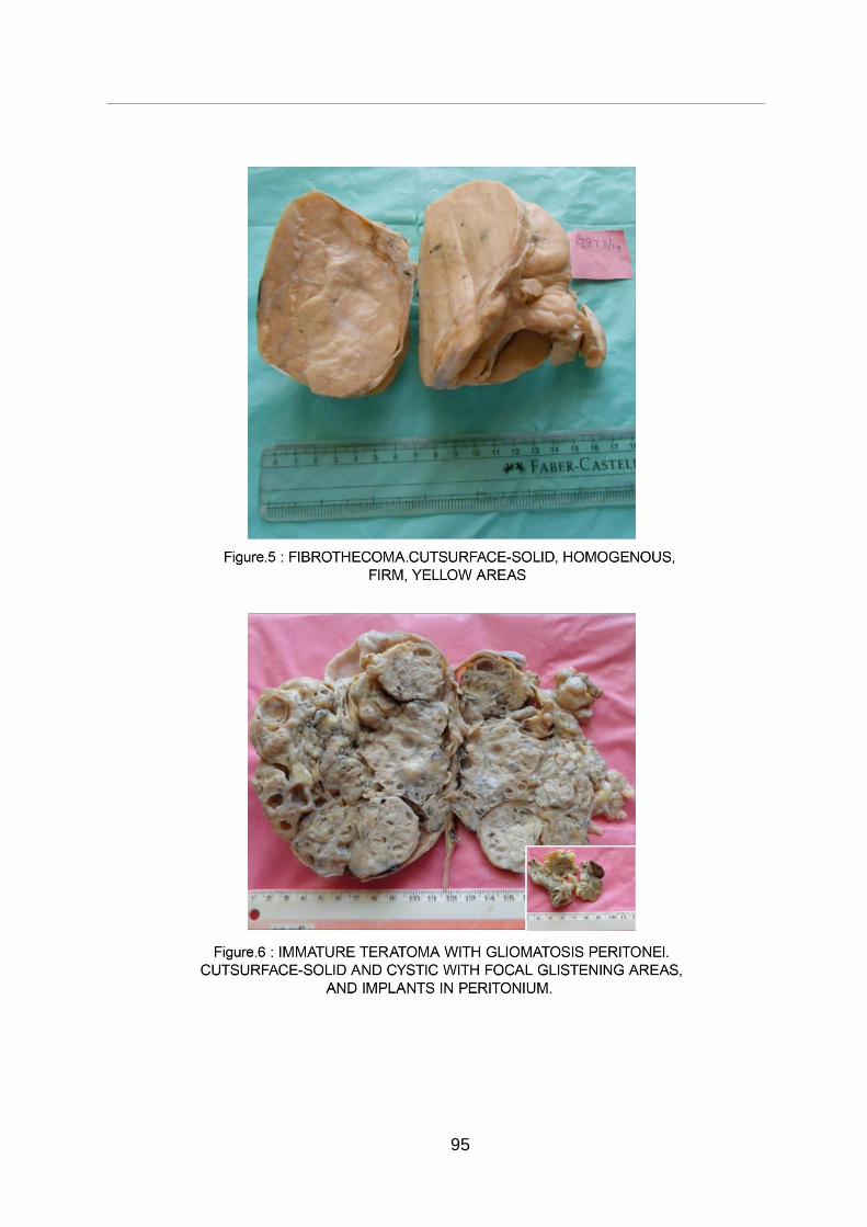
95
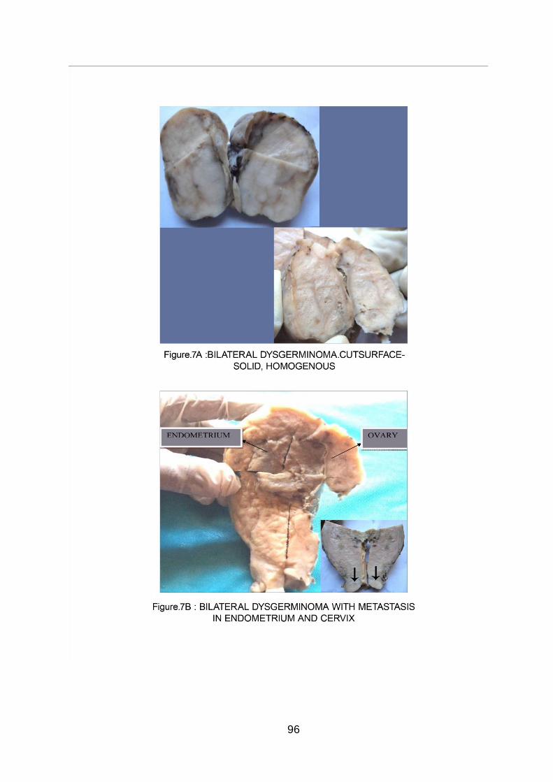
96
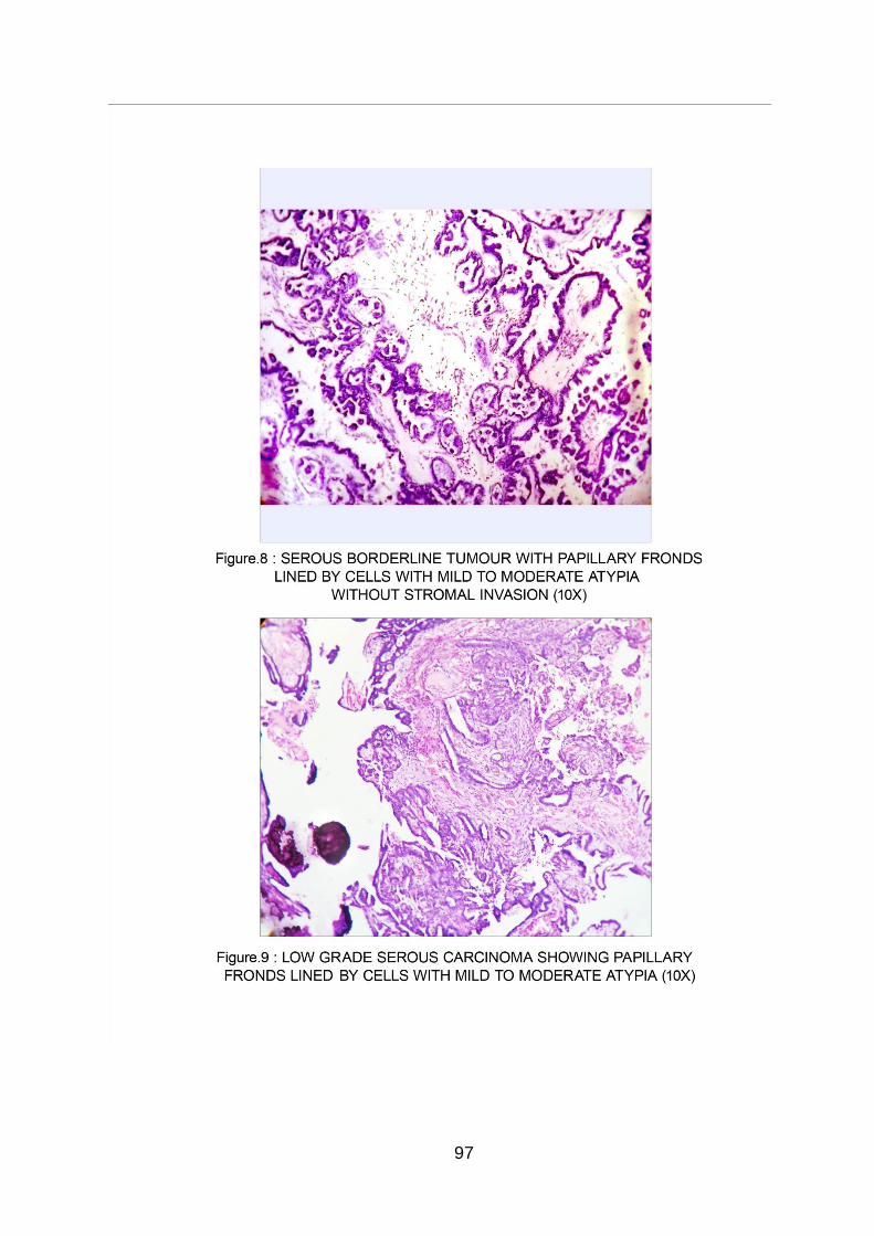
97
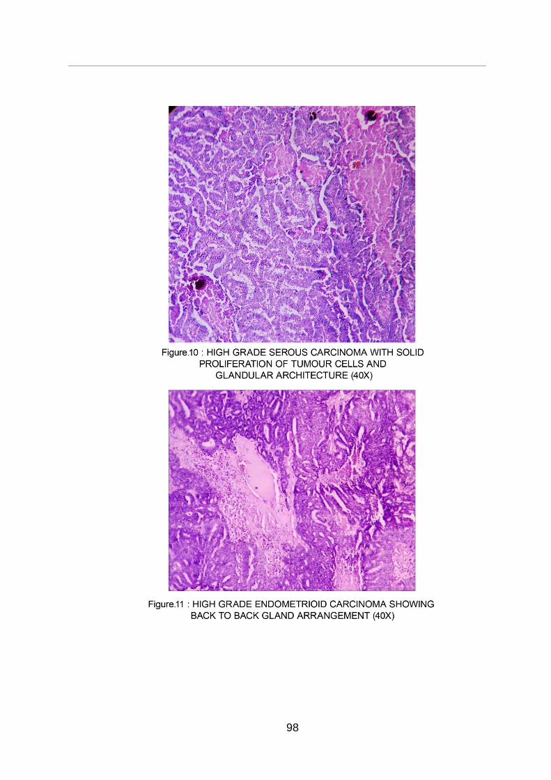
98

99

100
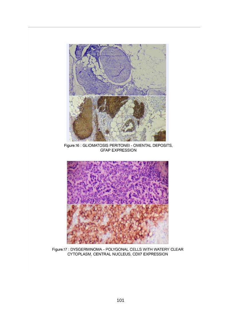
101

102
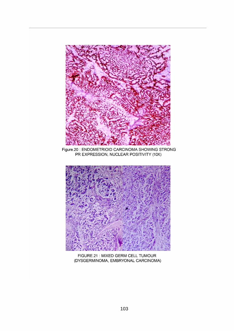
103
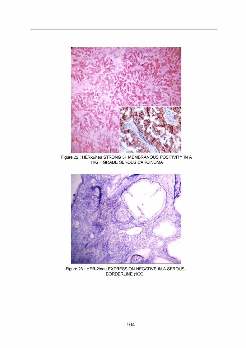
104
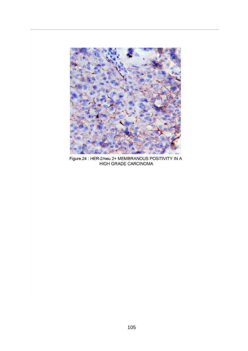
105
106
DISCUSSION
Ovaries are common sites of non-neoplastic and neoplastic lesions. The constant
cyclical changes from puberty to menopause give rise to different cell types, each of which
results in a varieties of tumours32
.
The diverse histopathologies in ovarian tumours reflects the different cell origins63
.
Recent surveillance, epidemiology and end result (SEER) calculations of lifetime risk for
ovarian carcinoma are that, 1 in 55 women will develop ovarian cancer over their life time62
.
As the symptoms are vague and manifest over time ovarian cancers are difficult to detect
until they are in advanced stage62
.
Identification of various histological patterns of ovarian tumours is important in
diagnosis, prognosis and treatment of ovarian cancers. Immunohistochemistry is now
emerging as an important tool in diagnosis of ovarian tumours10
. It has been shown that
estrogen and progesterone receptor level depends on histological grade, stage and variables
among the tumours of same grade56
. Her-2/neu is amplified and overexpressed in 25 to 30%
of human ovarian cancers. It is associated with progression of invasive cancer, poor
prognosis and resistance to chemotherapy53.
In this study, about 14,093 surgical specimens were received over a period of
2 ½ years. Of the 631 gynaecological tumours 132 ovarian neoplasms were reported. This
accounts for about 20.9% of all gynaecological tumours reported in this study. Total number
of malignancies reported during the study period is 1089 and total ovarian malignancies is 26.
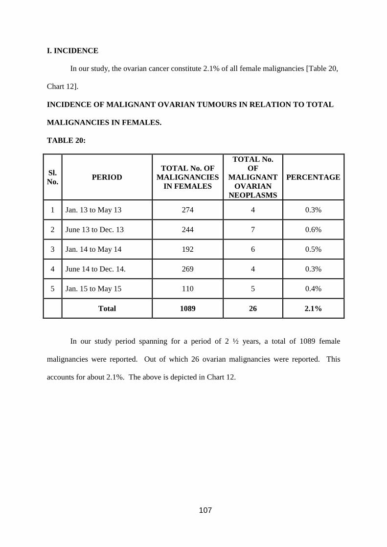
107
I. INCIDENCE
In our study, the ovarian cancer constitute 2.1% of all female malignancies [Table 20,
Chart 12].
INCIDENCE OF MALIGNANT OVARIAN TUMOURS IN RELATION TO TOTAL
MALIGNANCIES IN FEMALES.
TABLE 20:
Sl.
No. PERIOD
TOTAL No. OF
MALIGNANCIES
IN FEMALES
TOTAL No.
OF
MALIGNANT
OVARIAN
NEOPLASMS
PERCENTAGE
1 Jan. 13 to May 13 274 4 0.3%
2 June 13 to Dec. 13 244 7 0.6%
3 Jan. 14 to May 14 192 6 0.5%
4 June 14 to Dec. 14. 269 4 0.3%
5 Jan. 15 to May 15 110 5 0.4%
Total 1089 26 2.1%
In our study period spanning for a period of 2 ½ years, a total of 1089 female
malignancies were reported. Out of which 26 ovarian malignancies were reported. This
accounts for about 2.1%. The above is depicted in Chart 12.
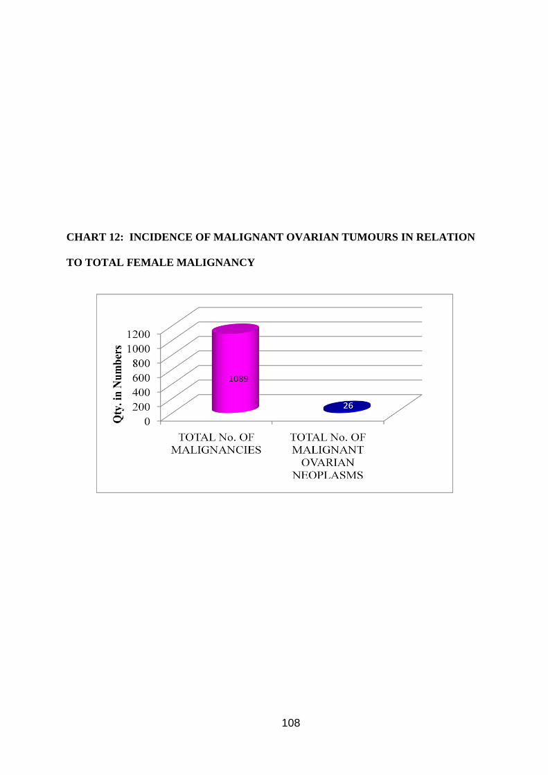
108
CHART 12: INCIDENCE OF MALIGNANT OVARIAN TUMOURS IN RELATION
TO TOTAL FEMALE MALIGNANCY
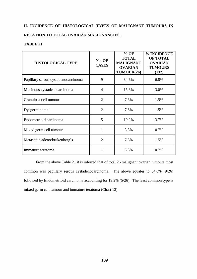
109
II. INCIDENCE OF HISTOLOGICAL TYPES OF MALIGNANT TUMOURS IN
RELATION TO TOTAL OVARIAN MALIGNANCIES.
TABLE 21:
HISTOLOGICAL TYPE No. OF
CASES
% OF
TOTAL
MALIGNANT
OVARIAN
TUMOUR(26)
% INCIDENCE
OF TOTAL
OVARIAN
TUMOURS
(132)
Papillary serous cystadenocarcinoma 9 34.6% 6.8%
Mucinous cystadenocarcinoma 4 15.3% 3.0%
Granulosa cell tumour 2 7.6% 1.5%
Dysgerminoma 2 7.6% 1.5%
Endometrioid carcinoma 5 19.2% 3.7%
Mixed germ cell tumour 1 3.8% 0.7%
Metastatic adeno/krukenberg’s 2 7.6% 1.5%
Immature teratoma 1 3.8% 0.7%
From the above Table 21 it is inferred that of total 26 malignant ovarian tumours most
common was papillary serous cystadenocarcinoma. The above equates to 34.6% (9/26)
followed by Endometrioid carcinoma accounting for 19.2% (5/26). The least common type is
mixed germ cell tumour and immature teratoma (Chart 13).
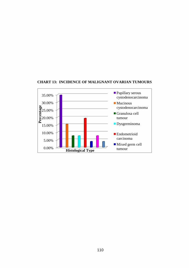
110
CHART 13: INCIDENCE OF MALIGNANT OVARIAN TUMOURS
0.00%
5.00%
10.00%
15.00%
20.00%
25.00%
30.00%
35.00%Papillary serous
cystodenocarcinoma
Mucinous
cystodenocarcinoma
Granulosa cell
tumour
Dysgerminoma
Endometrioid
carcinoma
Mixed germ cell
tumour
Per
cen
tage
Histological Type
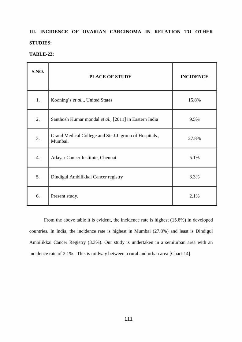
111
III. INCIDENCE OF OVARIAN CARCINOMA IN RELATION TO OTHER
STUDIES:
TABLE-22:
S.NO.
PLACE OF STUDY INCIDENCE
1. Kooning’s et al.,, United States 15.8%
2. Santhosh Kumar mondal et al., [2011] in Eastern India 9.5%
3. Grand Medical College and Sir J.J. group of Hospitals.,
Mumbai. 27.8%
4. Adayar Cancer Institute, Chennai. 5.1%
5. Dindigul Ambilikkai Cancer registry 3.3%
6. Present study. 2.1%
From the above table it is evident, the incidence rate is highest (15.8%) in developed
countries. In India, the incidence rate is highest in Mumbai (27.8%) and least is Dindigul
Ambilikkai Cancer Registry (3.3%). Our study is undertaken in a semiurban area with an
incidence rate of 2.1%. This is midway between a rural and urban area [Chart-14]
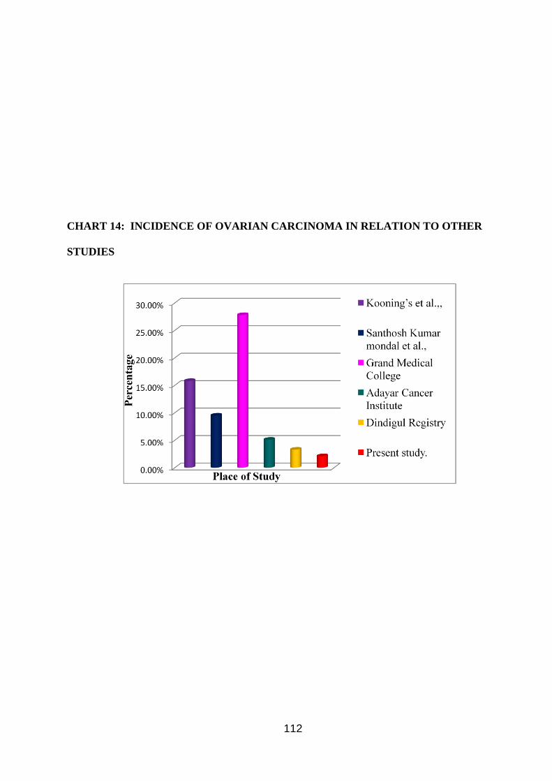
112
CHART 14: INCIDENCE OF OVARIAN CARCINOMA IN RELATION TO OTHER
STUDIES
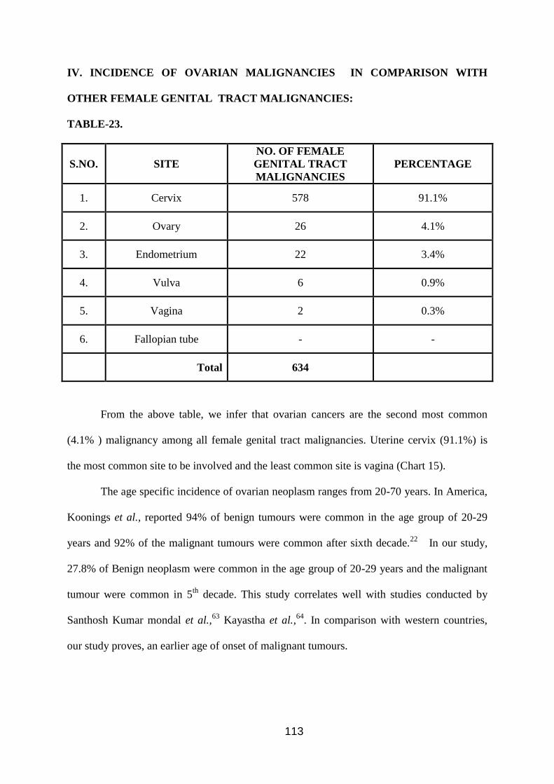
113
IV. INCIDENCE OF OVARIAN MALIGNANCIES IN COMPARISON WITH
OTHER FEMALE GENITAL TRACT MALIGNANCIES:
TABLE-23.
S.NO.
SITE
NO. OF FEMALE
GENITAL TRACT
MALIGNANCIES
PERCENTAGE
1. Cervix 578 91.1%
2. Ovary 26 4.1%
3. Endometrium 22 3.4%
4. Vulva 6 0.9%
5. Vagina 2 0.3%
6. Fallopian tube - -
Total 634
From the above table, we infer that ovarian cancers are the second most common
(4.1% ) malignancy among all female genital tract malignancies. Uterine cervix (91.1%) is
the most common site to be involved and the least common site is vagina (Chart 15).
The age specific incidence of ovarian neoplasm ranges from 20-70 years. In America,
Koonings et al., reported 94% of benign tumours were common in the age group of 20-29
years and 92% of the malignant tumours were common after sixth decade.22
In our study,
27.8% of Benign neoplasm were common in the age group of 20-29 years and the malignant
tumour were common in 5th
decade. This study correlates well with studies conducted by
Santhosh Kumar mondal et al.,63
Kayastha et al.,64
. In comparison with western countries,
our study proves, an earlier age of onset of malignant tumours.
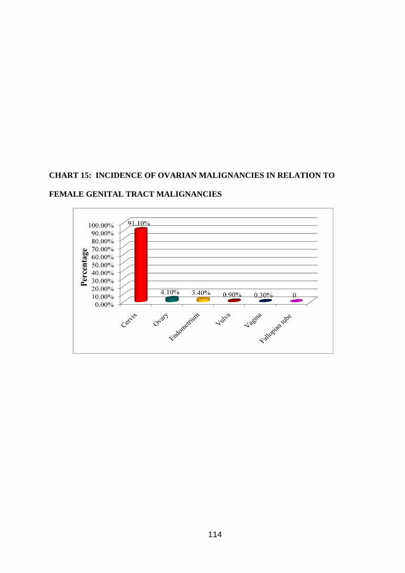
114
CHART 15: INCIDENCE OF OVARIAN MALIGNANCIES IN RELATION TO
FEMALE GENITAL TRACT MALIGNANCIES

115
V. COMPARISON OF PERCENTAGE OF BENIGN, BORDERLINE AND
MALIGNANT TUMOURS WITH OTHER STUDIES:
TABLE-24:
S.NO.
AUTHOR
KOONINGS et al., BENIGN BORDER LINE MALIGNANT
1. Koonings et al., 75.4% 3.6% 20.9%
2. Pilli et al., 76% 2.8% 21.2%
3. Santhosh Kumar
Mondal et al., 63.1% 7.3% 22.9%
4. M.Yogambal et al., 78.6% 0.7% 20.65%
5. Present study 73.4% 6.8% 19.6%
The results of our study is comparable with Koonings et al.,22
, Pilli et al.,66
, and
M.Yogambal et al.,65
. Comparison of incidence of benign, borderline and malignant tumours
with other in our studies is depicted in table 24 (Chart-16).
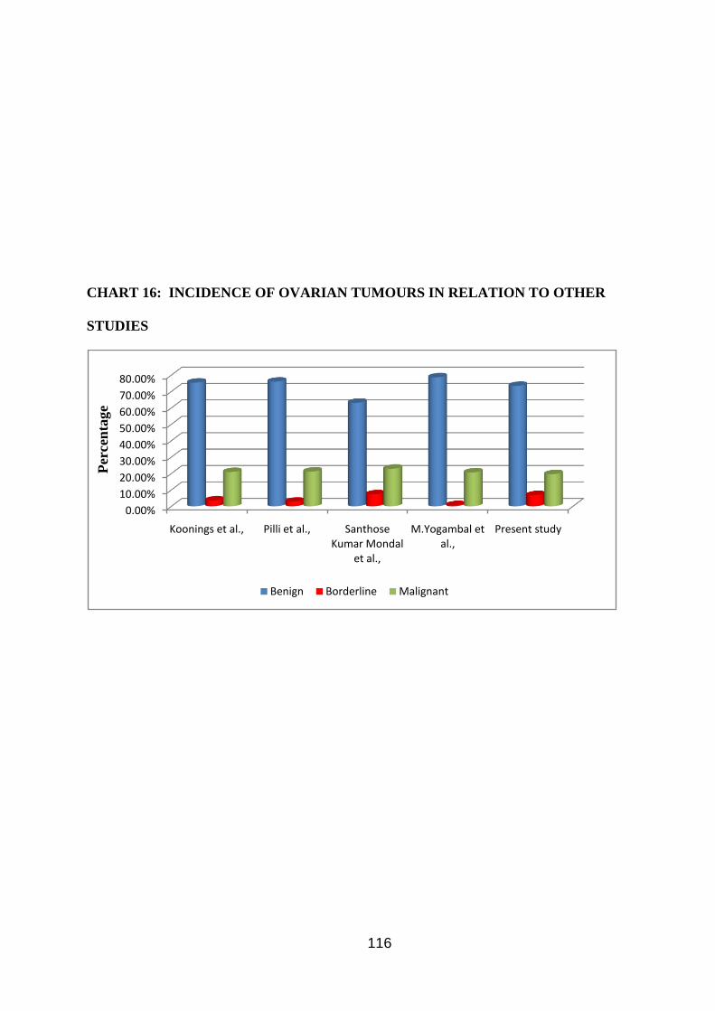
116
CHART 16: INCIDENCE OF OVARIAN TUMOURS IN RELATION TO OTHER
STUDIES
0.00%
10.00%
20.00%
30.00%
40.00%
50.00%
60.00%
70.00%
80.00%
Koonings et al., Pilli et al., Santhose Kumar Mondal
et al.,
M.Yogambal et al.,
Present study
Benign Borderline Malignant
Per
cen
tage
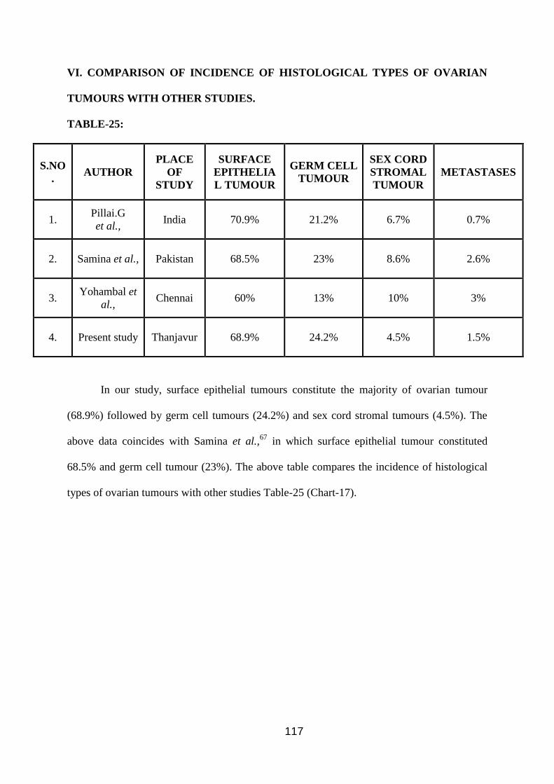
117
VI. COMPARISON OF INCIDENCE OF HISTOLOGICAL TYPES OF OVARIAN
TUMOURS WITH OTHER STUDIES.
TABLE-25:
S.NO
.
AUTHOR
PLACE
OF
STUDY
SURFACE
EPITHELIA
L TUMOUR
GERM CELL
TUMOUR
SEX CORD
STROMAL
TUMOUR
METASTASES
1. Pillai.G
et al., India 70.9% 21.2% 6.7% 0.7%
2. Samina et al., Pakistan 68.5% 23% 8.6% 2.6%
3. Yohambal et
al., Chennai 60% 13% 10% 3%
4. Present study Thanjavur 68.9% 24.2% 4.5% 1.5%
In our study, surface epithelial tumours constitute the majority of ovarian tumour
(68.9%) followed by germ cell tumours (24.2%) and sex cord stromal tumours (4.5%). The
above data coincides with Samina et al.,67
in which surface epithelial tumour constituted
68.5% and germ cell tumour (23%). The above table compares the incidence of histological
types of ovarian tumours with other studies Table-25 (Chart-17).
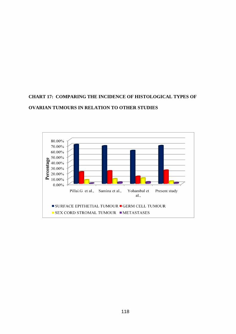
118
CHART 17: COMPARING THE INCIDENCE OF HISTOLOGICAL TYPES OF
OVARIAN TUMOURS IN RELATION TO OTHER STUDIES
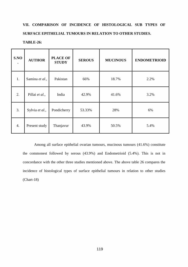
119
VII. COMPARISON OF INCIDENCE OF HISTOLOGICAL SUB TYPES OF
SURFACE EPITHELIAL TUMOURS IN RELATION TO OTHER STUDIES.
TABLE-26:
S.NO
.
AUTHOR PLACE OF
STUDY SEROUS MUCINOUS ENDOMETRIOID
1. Samina et al., Pakistan 66% 18.7% 2.2%
2. Pillai et al., India 42.9% 41.6% 3.2%
3. Sylvia et al., Pondicherry 53.33% 28% 6%
4. Present study Thanjavur 43.9% 50.5% 5.4%
Among all surface epithelial ovarian tumours, mucinous tumours (41.6%) constitute
the commonest followed by serous (43.9%) and Endometrioid (5.4%). This is not in
concordance with the other three studies mentioned above. The above table 26 compares the
incidence of histological types of surface epithelial tumours in relation to other studies
(Chart-18)
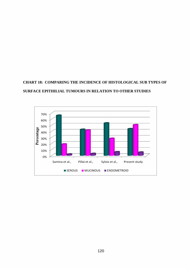
120
CHART 18: COMPARING THE INCIDENCE OF HISTOLOGICAL SUB TYPES OF
SURFACE EPITHILIAL TUMOURS IN RELATION TO OTHER STUDIES
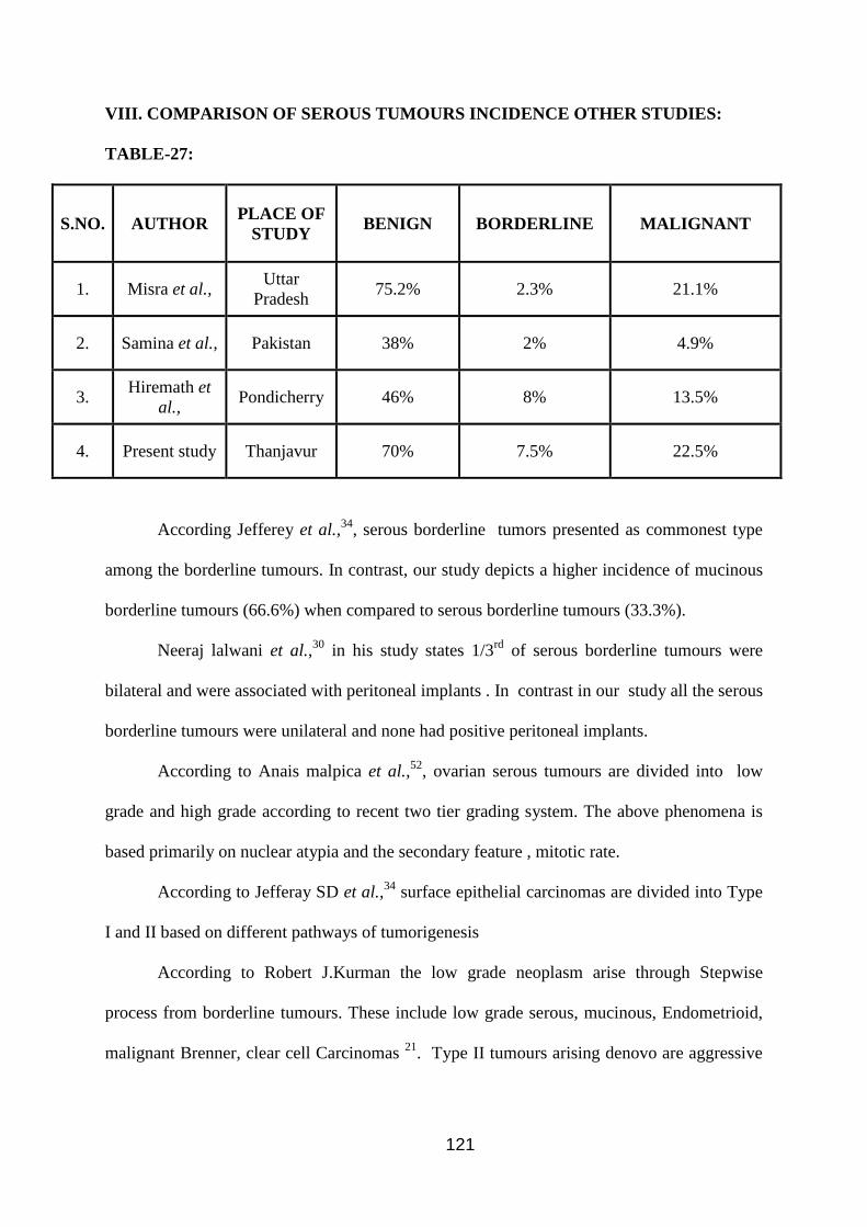
121
VIII. COMPARISON OF SEROUS TUMOURS INCIDENCE OTHER STUDIES:
TABLE-27:
S.NO.
AUTHOR PLACE OF
STUDY BENIGN BORDERLINE MALIGNANT
1. Misra et al., Uttar
Pradesh 75.2% 2.3% 21.1%
2. Samina et al., Pakistan 38% 2% 4.9%
3. Hiremath et
al., Pondicherry 46% 8% 13.5%
4. Present study Thanjavur 70% 7.5% 22.5%
According Jefferey et al.,34
, serous borderline tumors presented as commonest type
among the borderline tumours. In contrast, our study depicts a higher incidence of mucinous
borderline tumours (66.6%) when compared to serous borderline tumours (33.3%).
Neeraj lalwani et al.,30
in his study states 1/3rd
of serous borderline tumours were
bilateral and were associated with peritoneal implants . In contrast in our study all the serous
borderline tumours were unilateral and none had positive peritoneal implants.
According to Anais malpica et al.,52
, ovarian serous tumours are divided into low
grade and high grade according to recent two tier grading system. The above phenomena is
based primarily on nuclear atypia and the secondary feature , mitotic rate.
According to Jefferay SD et al.,34
surface epithelial carcinomas are divided into Type
I and II based on different pathways of tumorigenesis
According to Robert J.Kurman the low grade neoplasm arise through Stepwise
process from borderline tumours. These include low grade serous, mucinous, Endometrioid,
malignant Brenner, clear cell Carcinomas 21
. Type II tumours arising denovo are aggressive
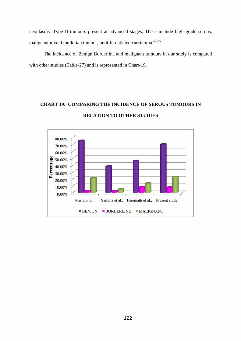
122
neoplasms. Type II tumours present at advanced stages. These include high grade serous,
malignant mixed mullerian tumour, undifferentiated carcinoma.52,55
The incidence of Benign Borderline and malignant tumours in our study is compared
with other studies (Table-27) and is represented in Chart-19.
CHART 19: COMPARING THE INCIDENCE OF SEROUS TUMOURS IN
RELATION TO OTHER STUDIES
0.00%
10.00%
20.00%
30.00%
40.00%
50.00%
60.00%
70.00%
80.00%
Misra et al., Samina et al., Hiremath et al., Present study
BENIGN BORDERLINE MALIGNANT
Per
cen
tage
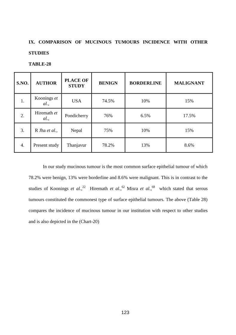
123
IX. COMPARISON OF MUCINOUS TUMOURS INCIDENCE WITH OTHER
STUDIES
TABLE-28
S.NO.
AUTHOR PLACE OF
STUDY BENIGN BORDERLINE MALIGNANT
1. Koonings et
al., USA 74.5% 10% 15%
2. Hiremath et
al., Pondicherry 76% 6.5% 17.5%
3. R Jha et al., Nepal 75% 10% 15%
4. Present study Thanjavur 78.2% 13% 8.6%
In our study mucinous tumour is the most common surface epithelial tumour of which
78.2% were benign, 13% were borderline and 8.6% were malignant. This is in contrast to the
studies of Koonings et al.,22
Hiremath et al.,62
Misra et al.,68
which stated that serous
tumours constituted the commonest type of surface epithelial tumours. The above (Table 28)
compares the incidence of mucinous tumour in our institution with respect to other studies
and is also depicted in the (Chart-20)
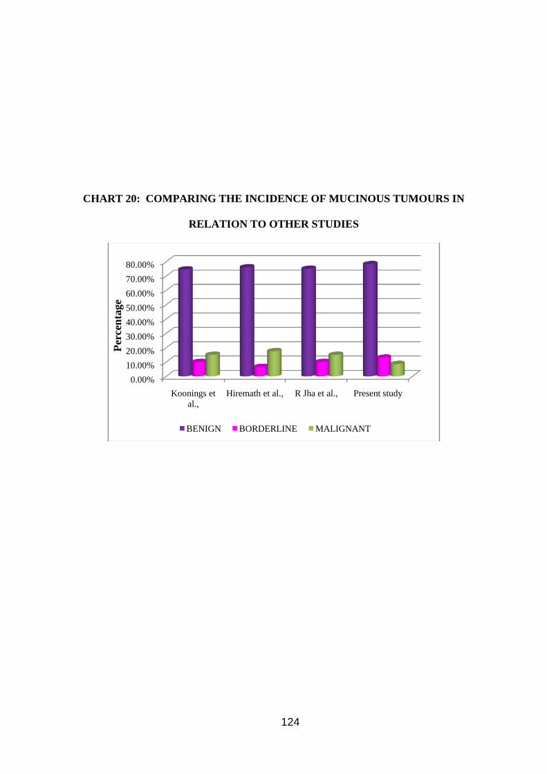
124
CHART 20: COMPARING THE INCIDENCE OF MUCINOUS TUMOURS IN
RELATION TO OTHER STUDIES
0.00%
10.00%
20.00%
30.00%
40.00%
50.00%
60.00%
70.00%
80.00%
Koonings et
al.,
Hiremath et al., R Jha et al., Present study
BENIGN BORDERLINE MALIGNANT
Per
cen
tage
125
According to Jaime et al.,60
endometriosis is associated with high incidence of
Endometrioid, clear cell carcinomas, mucinous and serous carcinoma. In contrast in our
study none of Endometrioid carcinoma (5/132) cases were associated with endometriosis.
In our study 33 cases of germ cell tumours were reported. This constitute about
24.2% of all ovarian neoplasms. In germ cell tumours, mature cystic teratoma is the
commonest group constitutes 90.9%, followed by dysgerminoma 6% and mixed germ cell
tumour 3%. This is in concordance with studies of Kwok et al.,43
and Norries HJ et al.,44
.
According to the above authors, malignancy in germ cell tumours is associated with post
menopausal age group. This is in contrast to our study where one case which was reported as
immature teratoma, was reported in younger age group.
According to P.Singh et al.,47
the commonest tumour associated with pregnancy is
dermoid cyst. In our study ,2 cases were reported which were associated with pregnancy. One
was associated with dermoid cyst, the other case was reported in association with mucinous
cyst adenoma. One case of mixed germ cell tumour was reported. This tumour was a
combination of Dysgerminoma and Embryonal carcinoma.
In our study, a total of 6 cases of sex cord stromal tumours were reported . This
accounts for about 4.5% of all ovarian tumours. Granulosa cell tumour, fibroma,
fibrothecoma had the same incidence of 33.3%. The above data had no coincidence with any
of the studies.
According to Lawrence et al.,69
95% of granulosa cell tumours are unilateral. All the
2 cases in our study were unilateral and none were associated with endometrial hyperplasia.
According to masaki mandai et al.,50
5-10% of neoplasm arise from metastases.
Ovarian metastases have some common features. Some of them are bilaterality, surface
involvement, extensive extra ovarian spread, desmoplastic reaction, vascular invasion and
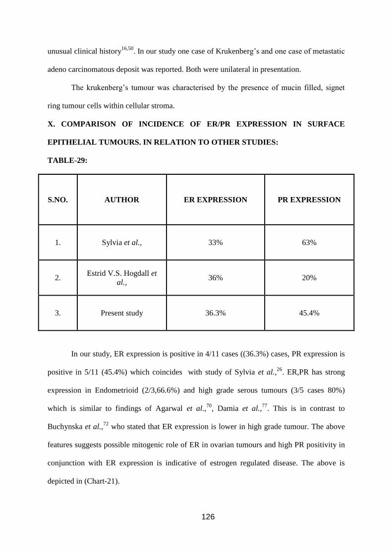
126
unusual clinical history16,50
. In our study one case of Krukenberg’s and one case of metastatic
adeno carcinomatous deposit was reported. Both were unilateral in presentation.
The krukenberg’s tumour was characterised by the presence of mucin filled, signet
ring tumour cells within cellular stroma.
X. COMPARISON OF INCIDENCE OF ER/PR EXPRESSION IN SURFACE
EPITHELIAL TUMOURS. IN RELATION TO OTHER STUDIES:
TABLE-29:
S.NO.
AUTHOR ER EXPRESSION PR EXPRESSION
1. Sylvia et al., 33% 63%
2. Estrid V.S. Hogdall et
al., 36% 20%
3. Present study 36.3% 45.4%
In our study, ER expression is positive in 4/11 cases ((36.3%) cases, PR expression is
positive in 5/11 (45.4%) which coincides with study of Sylvia et al.,26
. ER,PR has strong
expression in Endometrioid (2/3,66.6%) and high grade serous tumours (3/5 cases 80%)
which is similar to findings of Agarwal et al.,70
, Damia et al.,77
. This is in contrast to
Buchynska et al.,72
who stated that ER expression is lower in high grade tumour. The above
features suggests possible mitogenic role of ER in ovarian tumours and high PR positivity in
conjunction with ER expression is indicative of estrogen regulated disease. The above is
depicted in (Chart-21).
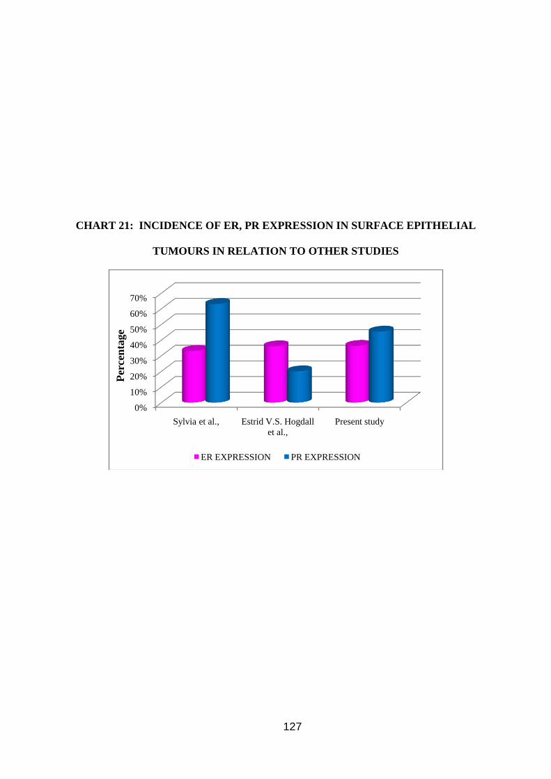
127
CHART 21: INCIDENCE OF ER, PR EXPRESSION IN SURFACE EPITHELIAL
TUMOURS IN RELATION TO OTHER STUDIES
0%
10%
20%
30%
40%
50%
60%
70%
Sylvia et al., Estrid V.S. Hogdall
et al.,
Present study
ER EXPRESSION PR EXPRESSION
Per
cen
tage
128
XI. COMPARISON OF EXPRESSION OF Her-2/neu IN BORDERLINE AND
MALIGNANT SURFACE EPITHELIAL TUMOURS.
TABLE-30:
S.NO.
AUTHOR BORDERLINE MALIGNANT
1. Sapna Go et al., - 48.6%
2. Sylvia et al., 1% 21%
3. Present study - 36.3%
From the above table, we infer that HER-2 expression is negative in borderline
tumours and shows positivity in high grade malignant tumours mostly commonly in high
grade serous tumours (3/5 cases, 60%). This is in concordance with Sylvia et al.,26
and Sapna
Go et al.,74
.
Borderline tumours occur in younger women less than 40 years of age. To preserve
fertility, conservative surgery is the treatment of choice. These patients should be monitored
routinely for CA-125 serum levels and ultrasound examinations. The clinical course lies in
between the benign and malignant tumours. Evidence suggests they are known to metastasize
within the peritoneal cavity28
.
In mucinous borderline tumours the identification of stromal invasion is difficult. This
is because of gross and microscopic multiloculation. But, atypical nuclei and stratification of
more than 3 layers suggests well differentiated mucinous carcinoma even when there is no
stromal invasion28
.

129
In our study, Her-2/neu was positive (3/5 cases 60%) in serous tumours and 1/3
(33.3%) cases of mucinous tumours. This is in concordance with studies of sapna Go et al.,74
and is depicted in the following Table-31 and Chart-22.
Table 31:
S.NO.
AUTHOR EXPRESSION OF Her-2/neu IN
SEROUS TUMOURS MUCINOUS TUMOURS
1. Sapna Goel et al., 72% 66.7%
2. Manisha sarkar et al., 50% -
3. Present study 60% 33.3%
CHART 22: COMPARING THE EXPRESSION OF Her-2/neu IN RELATION TO
OTHER STUDIES
0%
10%
20%
30%
40%
50%
60%
70%
80%
Sapna Goel et al., Manisha sarkar et
al.,
Present study
SEROUS TUMOURS MUCINOUS TUMOURS
Per
cen
tage
130
The scoring system of Her-2/neu, showed variations, in individual authors study
which is depicted in the following Table32.
Table 32:
S.NO.
AUTHOR
SCORING OF Her-2/neu IN SURFACE EPITHELIAL
TUMOURS
3+ 1+,2+
1. Manisha Sarkar et al., 35% 20%
2. Marvann et al., 6% 32%
3. Present study 27% 36.3%
In our study, the intensity of positivity did not correlate with age, size and grade of the
tumours. This is in concordance with Sapna Goel et al.,74
and Sylvia et al.,26
(Chart-22)
From the above data, Her-2/neu expression is found to be positive only in malignant
tumours. This suggests their carcinogenic role and helps in distinguishing borderline and
malignant tumours.
131
CONCLUSION:
A total of 132 cases were evaluated in concordance with clinical history,
histopathological and immunohistochemistry and the following conclusions were arrived at.
1. The incidence of ovarian neoplasms among all female neoplasms is 4.7%
2. The incidence of ovarian malignancies among all female malignancies is 2.1%
3. Ovarian malignancy (4.1%) is the second most common malignancy in female genital
tract next to cervical malignancy.
4. In age groups, ovarian neoplasms are commoner in 2nd
decade and in 4th
decade.
5. The ratio of benign and malignant ovarian neoplasm is 3:1.
6. Predominantly they presented as unilateral tumours (90%) than bilateral (10%).
7. Grossly, benign tumours presented, as cystic neoplasms and the malignant tumours
presented as solid and cystic or purely solid.
8. Regarding Histological type, surface epithelial tumours (68.9%) are the most common
neoplasm among which mucinous cystadenoma is the commonest.
9. Positive expression of ER,PR (steroid receptors) in surface epithelial malignancies
proves the mitogenic role of estrogen in ovarian tumours. PR expression may be related
to estrogenic regulation. The expression of steroid receptors paves way for antihormonal
therapy.
10. Her-2 neu was expressed only in malignant tumours. This suggests their carcinogenic
role. This also helps in differentiating borderline and malignant tumours.
This study is an institution based one with a small sample size of 132 cases. The
results may not actually reflect the original age distribution and histological pattern of
ovarian tumours in Indian population. The epidemiological data of developed countries in
many aspects differ from the developing nations. The differences about hormone receptors
expression by different authors may be due to various parameters like case selection, method
132
of immunohistochemistry and sample size and different grading systems. This should be
standardized to identify reliable prognostic markers in the clinical trial of hormone therapy.
A multicentric, large population based study with facility of follow up will be needed to
prove the prognostic significance of anti Her-2/neu therapy in surface epithelial carcinomas.
Thus a panel of markers will be helpful in prognostication of ovarian tumour and
development of targeted therapy. This study will serve as a reference for future studies.
Continued research studies in future would accomplish an effective therapy for ovarian
cancer.
133
APPENDIX I
HAEMATOXYLIN AND EOSIN STAIN
Preparation of solution :
HARRIS HAEMATOXYLIN
Distilled water-1000ml
Ammonium alum-100g
Absolute ethyl alcohol-50ml
Mercuric oxide-2.5g
100g of ammonium alum dissolved in 1000ml of distilled water by heating and shaking at
60.c. Add solution of 5g of haematoxylin in 50ml of ethylalcohol and bring rapidly to boil.
when it begins to boil, remove from flame and add 2.5 g of mercuric oxide. Mix by swirling
gently.
EOSIN STAIN
Eosin Y- 1 g.
Distilled water-20ml.
95% ethanol-80ml
Glacial acetic acid-0.2ml
Dissolve 1 gm of eosin Y in 20ml of water, add 80 ml of 95% ethyl alcohol and 0.2 ml of
glacial acetic acid.

134
Procedure :
1. Bring the sections to water
2. Dip in Harris haematoxylin for 15 minutes.
3. Rinse in tap water.
4. Differentiate in 1% acid alcohol-3-4 quick dips.
5. Wash in tap water briefly.
6. Dip in ammonia water or saturated lithium carbonate until the sections are blue.
7. Wash in running tap water for 10-20 minutes.
8. Stain with eosin for 15 seconds to 2 minutes depending on the age of the eosin and the
depth of counter stain.
9. Rinse in tap water.
10. Dip in 95% alcohol
11. 3 changes in absolute alcohol.
12. Xylene – 2 changes.
13. Mount in DPX mountant.
135
APPENDIX II
IMMUNOHISTOCHEMISTRY
PREPARATION OF SOLUTIONS:
Tris buffer saline (TBS): 0.005M
Distilled water-10 litres
Sodium chloride- 80gms
Tris (hydroxymethylamine)-6.05g
1 M HCl-44 ml.
Final pH is adjusted to 7.6 with either 1 M HCl or 0.2 M Tris solution.
CITRATE BUFFER SOLUTION
Trisodium citrate-2.94 gm
1 N HCl-5ml
Distilled water-1000 ml
Final pH is adjusted to 6.0 with 1 N HCl.
Preparation of gelatine coated slides:
Chrome alum-0.05gm
Gelatine-0.3 gm
Distilled water-100 ml.
Chrome alum is added to distilled water and then heated to 60.c.gelatin is added
slowly to the heated distilled water. Glass slides are then dipped in this solution and
dried overnight.
Antigen retrieval:
The slides are placed in citrate buffer in the coplin jar and capped. The jar is then
heated in a 750 W domestic microwave oven for 15 minutes.
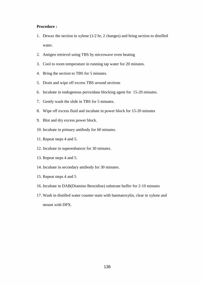
136
Procedure :
1. Dewax the section in xylene (1/2 hr, 2 changes) and bring section to distilled
water.
2. Antigen retrievel using TBS by microwave oven heating
3. Cool to room temperature in running tap water for 20 minutes.
4. Bring the section to TBS for 5 minutes.
5. Drain and wipe off excess TBS around sections
6. Incubate in endogenous peroxidase blocking agent for 15-20 minutes.
7. Gently wash the slide in TBS for 5 minutes.
8. Wipe off excess fluid and incubate in power block for 15-20 minutes
9. Blot and dry excess power block.
10. Incubate in primary antibody for 60 minutes.
11. Repeat steps 4 and 5.
12. Incubate in superenhancer for 30 minutes.
13. Repeat steps 4 and 5.
14. Incubate in secondary antibody for 30 minutes.
15. Repeat steps 4 and 5
16. Incubate in DAB(Diamino Benzidine) substrate buffer for 2-10 minutes
17. Wash in distilled water counter stain with haematoxylin, clear in xylene and
mount with DPX.
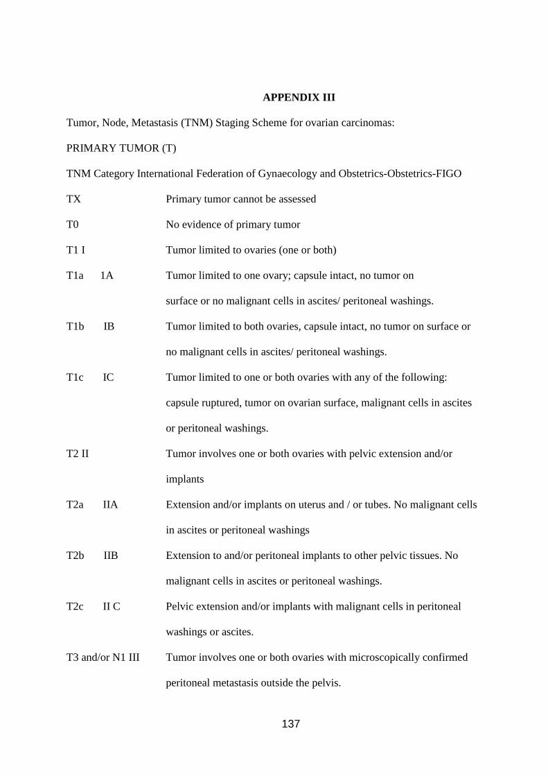
137
APPENDIX III
Tumor, Node, Metastasis (TNM) Staging Scheme for ovarian carcinomas:
PRIMARY TUMOR (T)
TNM Category International Federation of Gynaecology and Obstetrics-Obstetrics-FIGO
TX Primary tumor cannot be assessed
T0 No evidence of primary tumor
T1 I Tumor limited to ovaries (one or both)
T1a 1A Tumor limited to one ovary; capsule intact, no tumor on
surface or no malignant cells in ascites/ peritoneal washings.
T1b IB Tumor limited to both ovaries, capsule intact, no tumor on surface or
no malignant cells in ascites/ peritoneal washings.
T1c IC Tumor limited to one or both ovaries with any of the following:
capsule ruptured, tumor on ovarian surface, malignant cells in ascites
or peritoneal washings.
T2 II Tumor involves one or both ovaries with pelvic extension and/or
implants
T2a IIA Extension and/or implants on uterus and / or tubes. No malignant cells
in ascites or peritoneal washings
T2b IIB Extension to and/or peritoneal implants to other pelvic tissues. No
malignant cells in ascites or peritoneal washings.
T2c II C Pelvic extension and/or implants with malignant cells in peritoneal
washings or ascites.
T3 and/or N1 III Tumor involves one or both ovaries with microscopically confirmed
peritoneal metastasis outside the pelvis.

138
T3a IIIA Microscopic peritoneal metastasis beyond the pelvis (no macroscopic
tumor).
T3b III B Macroscopic peritoneal metastasis beyond the pelvis < 2 cm in
greatest dimension.
T3c III C Macroscopic peritoneal metastasis beyond the pelvis > 2 cm in
greatest dimension.
M1 IV Distant metastasis (excludes peritoneal metastasis)
REGIONAL LYMPHNODE (N):
NX Regional lymph nodes cannot be assessed
N0 No regional lymphnode metastasis.
N1 Regional lymphnode metastasis.
DISTANT METASTASIS (M):
MX Distant metastasis cannot be assessed
M0 No distant metastasis.
M1 Distant metastasis (excludes peritoneal metastasis).
STAGE GROUPING:
Stage IA: T1a N0 M0
Stage 1B: T1b N0 M0
Stage 1C: T1c N0 M0
Stage IIA: T2a N0 M0
Stage IIB: T2b N0 M0
Stage IIC: T2c N0 M0

139
Stage IIIA: T3a N0 M0
Stage IIIB: T3b N0 M0
Stage IIIC: T3c N0 M0
Any T N1 M0
Stage IV : Any T Any N M1.
140
BIBLIOGRAPHY
1. Endocrine signalling in ovarian surface epithelium and cancer peter CK leung and Jung
Hyechin, Human reproduction update Vol.13 NO2 pg 143-16
2. Contribution of 1HC in prognostic assessment of epithelial ovarian carcinoma – review
article ACTA MEDICA 2006 49(3) pg 161-165
3. Ovarian Carcinoma by Race and ethnicity
4. Changing Trends in incidence of ovarian carcinoma the Indian scenario-Asian pacific
journal of Carcinoma prevention Vol.10, 2009
5. Jeffrey T, Quirk et al., risk factors for invasive epithelial ovarian carcinoma by
histological subtype 2004 vol.3
6. Harmonal biology of epithelial ovarian carcinoma with hypothesis concerning the role of
androgens and progesterone-journal of natural Cancer institute Vol.90.No.23. Dec 2,
1998
7. Langley F.A., Fox.H., Ovarian Tumours classification histogenesis etiology. Obstetrical
and Gynaecological Pathology. Fox H and Wells M (Eds) Newyork: Churchil
Livingstone; 1995; 727-969.
8. Lee G et al., FNAC in the diagnosis and classification of recurrent and metastatic gynaec
malignancy 1988 vol.29, 375-378
9. Kurman R.J. et al., molecular pathogenesis & extra ovarian origin of epithelial ovarian
cancer shifts the paradigm. Human pathology 2011, vol.42, 918-931
10. Prognostic biomarkers in ovarian Carcinoma Jie Haung, Wei Her and Anil K Sood.
Cancer Biomark, PMC 2014 Aug.26
11. ER, PR and epithelial ovarian carcinoma reproduction biology and Endocrinology shuk-
Mei-Ho; 7 oct 2003
12. Prognostic value of ER,PR tumour expression in Danish ovarian cancer patients from
the ‘MALOVA ovarian cancer study oncology report 18, 1051-1059, 2007
13. Saadia tariq et al., Study of ovarian tumours in young girls. Professional Medical
Journal 2011; vol.18, 41-45
14. Sadler T.W., Urogenital System In: Langmans Medical Embryology (8th Edition)
Philadelphi : Lippincotts Williams and Wilkins; 2000; 304-345.
15. Inderbir Singh et. al., Human embryology, 7th Edition, 2008.
16. Rosai J. Ovary In. Ackerman’s Surgical Pathology Vol2 (9th Edition). St. Louis: Mosby
2004 1649-1736.
17. Caninstra S.A. 2004, Cancer of Ovary, N Eng. J Med., 351; 2519-2529
141
18. Tuma R.S. Origin of ovarian cancer, may have implications for screening, Journal of the
national cancer institute, Vol102, No.1, pp 11-132, 2010
19. Novak E.R, Gynaecological and obstetrical pathology. 6th edition, Philadelphia
Saunders; 1967
20. Tavassoli F.A. Devillee P (Eds): In diagnostic pathology and molecular genetics; World
health organisation classification of tumours of breast and female genital tract
21. Robert J. Kurman, Le-Ming Shin, Pathogenesis of Ovarian Cancer; Lessons from
Morphology and Molecular Biology and their Clinical Interpretations. Int. J
Gyneacopathol, 2008, April, (27(2) 151-160.
22. Koonings PP, Campbell K, Mishell DR, Relative frequency of primary ovarian
neoplasm; 10 year review. obstet gynaecol 1989;74:921-26
23. S.V. Kane, R. Bhardwaj, H.B. Tonga Onkar, Borderline epithelial tumours of the ovary.
A retrospective analysis of 31 cases. Indian J Cancer 1999:16:18 – 31
24. Marta Ann Crispens. borderline ovarian tumours; A review of recent literature curr opin
obstet Gynaecology 2993;15:39-43
25. Dar et al., Clinical and pathological study of ovarian serous borderline tumours. Cancer
research on 17-08-2011
26. Mary T. Sylvia et al., The expression of immunohistochemical markers estrogen
receptor, progesterone receptor, Her-2/neu, P53 and Ki67, in epithelial ovarian
tumours and its correlation with clinicopathologic variable; Indian Journal of
Pathology and Microbiology – 55(1), Jan-March 2012
27. Steven G. Silverberg. Principles and practice of surgical pathology and cytopathology,
Fourth edition, Vol.2
28. Brigette M Ronnett et al., Mucinous borderline ovarian tumours, points of general
agreement and persistency controversies; Journal of Human Pathology 2004,
446-458
29. Maheswari V, Tyagi SP, Saxsena K, Tyagi N, Sharma R, Aziz M, et al., Surface
epithelial tumours of ovary. Indian Journal of Pathology and Microbiology, 1994, 37.
75-85
30. Neeraj Lalcoani et al., Current update on borderline ovarian tumours, AJR:194,
Feb.2010; 330-335
31. Me cluggage, Recent advances in immunohistochemistry in the diagnosis of ovarian
pathology. Jclin Path 2000;53;327-334
32. Stacey E Mills, Stenbergs Diagnostic Surgical Pathology, Vol.II 5th edition
142
33. Vigano P., Somighana E., Chiodol, Molomech and biological plausibility underlying the
malignant transformation of endometriosis: A critical analysis. Human reproduction
update, 2008, Vol622, pp79-87
34. Jeffray SD, Russel P, Robert KJ, Surface epithelial tumours. In Robert Kurman J.
Blausteins pathology of female genital tract, 5th Edition 2002: 791-881
35. Stanley J. Robbins – Robboys pathology of female genital tract, 2nd Edition
36. Stuart G.C.,E; Dawson, L.M., Update on granulosa cell tumours of the ovary curr
opinion ostet gynecol 2003, V;15, p33-7
37. Tanaka Y, Sasaki Y, Nishihira H, Izavsa T, Nishi T: Ovarian juvenile granulosa cell
tumour associated with Maiffucc’s syndrome. Am J Chin Pathol 1992; 97;
523-527
38. Gaffney EF, Majmadar B. Ultrastructure and immunohistochemical localisation of
estradiol of thecomas. Hum Pathol 1984, 15: 153-160
39. Irving et al., cellular fibroma of ovary: A study of 75 cases including mitotically active
fibroma. American Journal of surgical pathology 2006, August 30(8) 928-938
40. Aparna Ragesh et al., Fibroma of ovary presenting as meigs syndrome. A case report –
NUJHS Vol.2, No.3, Sep. 2012
41. Jacob S, Lalitha K, Gopalakrishnan K, et al., Malignant ovarian tumours – A profile: J
obstet Gynecol Ind 1993;43:413-7
42. Irfan Cicin et al., Yolk sac tumours of ovary evaluation of clinicopathological features
and prognostic features. European Journal of Obstetrics and Gynaecology, February
2009
43. KKM Kwok et al., Malignant mixed germ cell tumour of ovary in a 10 year old girl.
JHK Coll. Radiology 2008;11;92-95
44. Norris HJ, Zirkin HJ, Beason WL: Immature (malignant teratoma) of the ovary –
A clinical & pathological studies of 58 cases, 1976: 37; 2359-2372
45. Min J et al., Clinical characteristics of ovarian teratoma – American Journal of
Obstetrics and gynaecology 2001, 205; 32
46. Robert J. Kurman. Blaustein’s pathology of female genital tract, 5th edition, Page
No:758-768
47. Sing et al., Malignancy associated with benign cystic teratomas of the ovary, Scng. Med.
Journal 1988, Vol29;30-34
48. Sung-jong Lee et al., Clinical characteristics of metastatic tumours to ovaries. Journal
Korean Medical Science 2009, 24, 114-119
143
49. Adriana Diongi et al., ovarian metastatic from Colorectal carcinoma, American Journal
of Clinical Pathology 2000, 114, Page No.111-122
50. Masaki Mandai et al., Krukenberg tumours, CME Journal of gynaecologic oncology
2004, Vol.9, 112-114
51. Lee KR, et al., The distinction between primary and metastatic mucinous Carcinoma of
ovary; gross and histological in 50 cases. American Journal of Surgical Pathology
2003, 27; 281-292
52. Anais Malpaica et al., Grading of ovarian serous carcinoma using two tier grading
system, American Journal of Surgical Pathology, Vol.28, Number 4, April 2004
53. Roth LM, Recent advances in the pathology and classification of ovarian sex cord
stromal tumours. Int. J. Gynecopathol, V25, pp199-215, 2006
54. Dos santos Lisa, MOK Evelyn, Alexia la sonos, Kay Park, Soslow Robert A, Carol
Aghajanain and co: Squamous cell carcinoma arising in mature cystic teratoma of the
ovary – A case series and review of the literature. Gynaecologic oncology 2007, 105:
321-24
55. Halperin R et al., Clinical and molecular comparison between borderline serous ovarian
tumours and advanced serous papillary ovarian carcinoma. European Journal of
gynaecologic oncology 2001, 22(4), 292-296
56. Potential role of estrogen receptor β as a tumour suppressor of epithelial ovarian cancer
– Carine Bossard, Muril Busson. PLOS one 7(9)
57. Dar et al., Clinical and pathological study of ovarian serous borderline tumours cancer
research on 17-08-2011
58. Adhikari et al., Granulosa cell tumour of ovary, Clinicopathological study of six cases.
Journal of pathology of Nepal 2011 Vol 196-99
59. Her-2 status in ovarian carcinoma as a multicenter GINECO study of 320 patients.
PLOS one Nov.2007, issue 11
60. Jaime et al., Clinicopathological characteristics of ovarian carcinoma and associated
with endometriosis. archives of Gynaecology and obstetrics 2011 vol.28, no.2, page
no.479-483
61. Thomas Holbro, Gianluca civenni . et al., The Erb β receptors and their role in Cancer
progression, Experimental cell research 284, 2003 99-110.
62. Hiremath P.B. et al., clinical profile and pathology of ovarian tumour. International
journal of biological and medical of biological and medical research. 2012, 3:1743-
1746.

144
63. Santhosh Kumar Mondal, Ranjana, Suprio Roy Chowdry. Histological pattern and
clinical evaluation of 957 ovarian neoplasm: A 10 year study in a tertiary hospital of
Eastern India Indian J cancer 2011, 433-437.
64. Kayastha study of ovarian tumours in Nepal medical college teaching hospital. Nepal
med coll J.2009;11, 200-202.
65. M.Yogambal et al., ovarian tumours-Incidence and distribution in a tertiary referral
centre in south India 10SR Journal of Dental and medical Sciences VOL 13 Pg74-80.
66. Pillai GS, Suneeta KP, Dhaded AV, Venni VV ovarian tumours, a study of 282 cases
J Indian med ASSOC: 2002 423-424.
67. Samina Zaman et al., A retrospective study of ovarian tumours and tumour like lesions.
Journal Ayub med coll. Abottabad 2010 Vol 22.
68. Mishra RK, Sharma SP, Gupta U, Gauri R pattern of ovarian neoplasm in eastern UP.
J Obstet Gynecol India 1991; 41(2); 242-246.
69. Lawrence M Roth et al., Recent advances in the pathology and classification of ovarian
sex cord stromal tumours . International journal of gynacopathology .
70. Agarwal N, Rao DL, Murgeshan K, Verma U mittal S, Buckshee K. Clinical evaluation
of steroid receptors in ovarian neoplasms. Int J gynecol obstet 1987; 145-9.
71. Damiao RD, Oshima CT; Stavale JN, Goncalves. WJ, Analysis of the expression of
ER,PR and chicken oval albumin upstream promoter transcription factor-1 in ovarian
cancers.
72. Buchynska LG, lurchenko NP, Expression of ER, PR receptors as prognostic factors in
serous ovarian cancers. Exp oncol 2009; 31, 48-51.
73. Sapna Goel et al.,-A comparison study of HER-2/neu oncogene in Benign and malignant
ovarian tumours. International Journal of scientific study July 2014 vol 2 pg.50-54.
Copyright © 2022 FDOKUMEN