
Astrocyte Elevated Gene-1 as a Novel Clinicopathological and ...
Upload
khangminh22Category
view
3download
0

VulvarDisease
K793xFM.indd 1 11/29/06 9:05:56 AM

K793xFM.indd 2 11/29/06 9:05:56 AM
VulvarDiseaseA Clinicopathological
Approach
Edited by
Debra S. HellerUMDNJ–New Jersey Medical School
Newark, New Jersey, U.S.A.
Robert C. WallachNew York University Medical Center
New York, New York, U.S.A.
New York London
K793xFM.indd 3 11/29/06 9:05:56 AM
Informa Healthcare USA, Inc.270 Madison AvenueNew York, NY 10016
© 2007 by Informa Healthcare USA, Inc. Informa Healthcare is an Informa business
No claim to original U.S. Government worksPrinted in the United States of America on acid‑free paper10 9 8 7 6 5 4 3 2 1
International Standard Book Number‑10: 0‑8493‑3793‑3 (Hardcover)International Standard Book Number‑13: 978‑0‑8493‑3793‑2 (Hardcover)
This book contains information obtained from authentic and highly regarded sources. Reprinted material is quoted with permission, and sources are indicated. A wide variety of references are listed. Reasonable efforts have been made to publish reliable data and information, but the author and the publisher cannot assume responsibility for the validity of all materials or for the consequences of their use.
No part of this book may be reprinted, reproduced, transmitted, or utilized in any form by any electronic, mechanical, or other means, now known or hereafter invented, including photocopying, microfilming, and recording, or in any informa‑tion storage or retrieval system, without written permission from the publishers.
For permission to photocopy or use material electronically from this work, please access www.copyright.com (http://www.copyright.com/) or contact the Copyright Clearance Center, Inc. (CCC) 222 Rosewood Drive, Danvers, MA 01923, 978‑750‑8400. CCC is a not‑for‑profit organization that provides licenses and registration for a variety of users. For orga‑nizations that have been granted a photocopy license by the CCC, a separate system of payment has been arranged.
Trademark Notice: Product or corporate names may be trademarks or registered trademarks, and are used only for identification and explanation without intent to infringe.
Library of Congress Cataloging‑in‑Publication Data
Vulvar disease / edited by Debra S. Heller, Robert C. Wallach.p. ; cm.
Includes bibliographical references and index.ISBN‑13: 978‑0‑8493‑3793‑2 (alk. paper)ISBN‑10: 0‑8493‑3793‑3 (alk. paper)1. Vulva‑‑Diseases. I. Heller, Debra S. II. Wallach, Robert C. [DNLM: 1. Vulvar Diseases. WP 200 V9915 2007]
RG261.V8544 2007618.1’6‑‑dc22 2006051475
Visit the Informa Web site atwww.informa.com
and the Informa Healthcare Web site atwww.informahealthcare.com
K793xFM.indd 4 11/29/06 9:05:56 AM
In loving memory of M. Renate Dische, MD, PhD—mentor, friend,inspiration, and supporter of women—and in recognition of the
pioneering study and teaching of Henry C. Falk, MD.

Preface
Vulvar disease is a source of patient distress, provokes multiple consultations withphysicians, and is a challenging diagnostic area for gynecologists, dermatologists,and pathologists. Some gynecologists may be insecure or unfamiliar with the variousvulvar problems, and their training and experience may not encompass the spectrumof disease found on the vulva, especially the dermatologic entities. Dermatologistswho see vulvar disease may not be as familiar with the gynecologic vulvar diseaseentities. Pathologists, except for the rare subspecialists with high-volume practices,can often be challenged in this area.
This book is a collaboration between a gynecologic pathologist with trainingand experience in obstetrics and gynecology (Debra S. Heller) and a gynecologiconcologist who has a special interest in vulvar disease (Robert C. Wallach), with inputfrom gynecologists, dermatologists, and pathologists with interest and expertise inthe field. In addition, a CD-ROM containing all the images in the book is meant toprovide additional teaching and reference material.
This monograph is primarily intended to be a coalescence of views from theseveral disciplines represented, covering clinical as well as pathologic appearances,with treatment guidelines. It is hoped that this focus of attention on vulvar disease willprovide a multidisciplinary reference resource to help us all better serve womenwith vulvar disease.
Debra S. HellerRobert C. Wallach
v

Contents
Preface . . . . vContributors . . . . ix
1. An Approach to the Evaluation of Vulvar Lesions and Complaints . . . 1Bernadette Cracchiolo and Gina AndersonAnatomy . . . . 1Etiology . . . . 1Special Considerations . . . . 7Evaluation . . . . 8References . . . . 10
2. Normal Vulva and Developmental Anomalies . . . . . . . . . . . . . . . . . 11James Scurry and Kathleen Rita MelvilleAnatomy . . . . 11Histology . . . . 18Embryology . . . . 29Developmental Anomalies . . . . 30References . . . . 34
3. Noninfectious Inflammation and Systemic Diseases of the Vulva . . . . 37Sallie M. NeillInflammatory Dermatoses . . . . 37Erosive, Ulcerative, and Bullous Dermatoses . . . . 54Miscellaneous . . . . 60References . . . . 63
4. Vulvovaginal Infections . . . . . . . . . . . . . . . . . . . . . . . . . . . . . . . . 67Rachel Spieldoch and Libby EdwardsViral Infections . . . . 67Bacterial Infections . . . . 77Fungal Infections . . . . 93Parasitic Infections . . . . 100References . . . . 104
vii

5. Pigmented Lesions of the Vulva . . . . . . . . . . . . . . . . . . . . . . . . . 109Hope K. Haefner, Timothy M. Johnson, Lorraine L. Rosamilia, andDouglas R. FullenIntroduction . . . . 109Mechanisms of Hyperpigmentation . . . . 110Conditions with Pigmentation . . . . 110Melanocytic Lesions . . . . 111Conclusion . . . . 129References . . . . 129
6. Neoplasms and Related Lesions of the Vulva . . . . . . . . . . . . . . . . 135Debra S. HellerCysts . . . . 135Benign Tumor-Like Lesions . . . . 138Benign Neoplasms . . . . 143Preinvasive (In Situ) Neoplasms of the Vulva . . . . 157Invasive Neoplasms of the Vulva . . . . 161Benign Lesions of the Urethra . . . . 168Malignant Lesions of the Urethra . . . . 176References . . . . 176
7. Commentary . . . . . . . . . . . . . . . . . . . . . . . . . . . . . . . . . . . . . . . 181Robert C. WallachIntroduction . . . . 181Lichen Sclerosus et Atrophicus . . . . 181LSA and SCC . . . . 182LSA Treatment . . . . 182Vulvar Neoplasia . . . . 183Pigmented Lesions . . . . 183Vulvar Symptoms . . . . 184Genital Mutilation . . . . 184Genital Anatomy . . . . 185References . . . . 185
Index . . . . 189
viii Contents
Contributors
Gina Anderson Department of Obstetrics, Gynecology, and Women’s Health,UMDNJ–New Jersey Medical School, Newark, New Jersey, U.S.A.
Bernadette Cracchiolo Department of Obstetrics, Gynecology, and Women’sHealth, UMDNJ–New Jersey Medical School, Newark, New Jersey, U.S.A.
Libby Edwards Southeast Vulvar Clinic, Charlotte, North Carolina, U.S.A.
Douglas R. Fullen Department of Pathology and Dermatology, The University ofMichigan Hospitals, Ann Arbor, Michigan, U.S.A.
Hope K. Haefner Department of Obstetrics and Gynecology, The University ofMichigan Hospitals, Ann Arbor, Michigan, U.S.A.
Debra S. Heller Departments of Pathology and Laboratory Medicine, andObstetrics, Gynecology, and Women’s Health, UMDNJ–New Jersey MedicalSchool, Newark, New Jersey, U.S.A.
Timothy M. Johnson Departments of Dermatology, Otolaryngology, andSurgery, Melanoma Clinic, The University of Michigan Hospitals, Ann Arbor,Michigan, U.S.A.
Kathleen Rita Melville Nextpath, Newcastle, Australia
Sallie M. Neill St. John’s Dermatology Centre, St. Thomas’ Hospital,London, U.K.
Lorraine L. Rosamilia Departments of Internal Medicine and Dermatology,The Pennsylvania State University, Milton S. Hershey Medical Center,Hershey, Pennsylvania, U.S.A.
James Scurry Nextpath, Newcastle, Australia
Rachel Spieldoch Scottsdale, Arizona, U.S.A.
Robert C. Wallach New York University Medical Center, New York UniversityClinical Cancer Center, New York, New York, U.S.A.
ix


1
An Approach to the Evaluation of VulvarLesions and Complaints
Bernadette Cracchiolo and Gina AndersonDepartment of Obstetrics, Gynecology, and Women’s Health, UMDNJ–New JerseyMedical School, Newark, New Jersey, U.S.A.
ANATOMY
Many women are uncertain about their genital anatomy and may present complain-ing of vaginal itching or simply itching ‘‘down there.’’ The vulva comprises the entireexternal genitalia from the mons pubis anteriorly to the anus posteriorly andlaterally to the genito-crural folds. Included in this area are the labia majora, labiaminora, clitoris, urethral meatus, vestibule, posterior fourchette, hymen, and theBartholin’s, Skene’s, and minor vestibular glands and the vestibular bulbs.
The labia majora and mons contain adipose and fibrous tissue, covered by skinwith numerous sebaceous and hair follicles. The labia minora do not contain adiposetissue, and their skin has sebaceous glands but not hair follicles. On the inner aspectof the labia minora is Hart’s line, which delineates the junction between skin and themucous membrane. The boundaries of the vulvar vestibule are Hart’s line laterallyand the hymenal ring medially, and this area contains numerous glands that secretemucus. The vestibular bulbs are located within the bulbocavernosus muscles oneither side of the vestibule and contain erectile tissue. The clitoris contains botherectile tissue and a very high density of nerve fibers.
The blood supply of the vulva is from branches of the internal and externalpudendal arteries, and lymphatic drainage is primarily to the external and internaliliac nodes via the groins. Branches of the pudendal nerve provide most of the motorand sensory innervation to the vulva.
ETIOLOGY
The evaluation of vulvar lesions can be challenging because there are a variety of poten-tial causes including infectious diseases, nonneoplastic epithelial disorders, and benignand malignant neoplasms. In addition, these conditions will frequently coexist;
1
for example, the patient with recurrent vulvovaginal candidiasis may develop contactdermatitis from frequent application of irritating topical products to the affected skin.In addition, some women present with chronic vulvar itching and burning due todysesthetic vulvodynia and will have no obvious physical findings on examination.
Nonneoplastic Epithelial Disorders of the Vulva
This group of disorders includes the conditions that are sometimes referred to as thevulvar dystrophies or dermatoses. The current recommended classification systemdeveloped by the International Society for the Study of Vulvar Disease for thesedisorders is (1):
1. lichen sclerosus,2. squamous cell hyperplasia,3. other dermatoses.
An updated classification based on pathological criteria is nearing completion.While some of these conditions are discussed in more detail in other sections,
they are briefly reviewed here.
Lichen Sclerosus
Formerly known as lichen sclerosus et atrophicus, lichen sclerosus is a chronic der-matologic condition that is more common in women and may involve both vulvarand extragenital lesions. Patients typically present with severe itching and may alsoreport burning and dyspareunia. The entire vulvar area may be involved, includingthe clitoris and perianal regions. The skin often has the typical ‘‘parchmentappearance’’ with white papules and plaques that may appear crinkled. Fissures,telangiectasias, and stenosis of the introitus may occur. With long-standing disease,there is often alteration of the normal vulvar architecture resulting in absence of thelabia minora, adhesions of the labia majora, and phimosis of the clitoris in extremecases. While it can affect women of any age, it is more commonly seen in thepostmenopausal years and approximately 10% to 15% of cases occur in pediatricpatients (2).
Squamous Cell Hyperplasia
This category includes lichen simplex chronicus and epithelial hyperplasia, and in thepast was often called leukoplakia or hyperplastic dystrophy. Clinically, this typicallypresents with complaints of itching and burning, and often follows contact with atopical irritant, although the patient may not recall such an exposure. Any part ofthe vulva may be affected, and the skin may appear erythematous with fissuringand excoriation. Lichenification, or the accentuation of normal skin markings, whichresults in thickening and raised white plaques, is also commonly seen. The processmay be localized or diffuse and can been seen in all of the reproductive stages. Thiscondition frequently may be associated with long-term use of topical therapiessuch as antifungals or steroids for other conditions, and may be causally related.A history of atopy, allergies, or eczema is also a likely risk factor.
Lichen Planus
A challenging disease to both diagnose and manage, lichen planus is a papulosqua-mous disorder that may involve both cutaneous and mucosal surfaces. On the vulva,lesions are often violaceous papules that cause pruritus. A whitish reticular patternmay appear on the inner labia majora where there is more exposure to moisture, and
2 Cracchiolo and Anderson
on the vestibular and vaginal mucosa the lesions are usually erythematous erosions.Vaginal disease is often associated with a heavy yellow discharge and significant dys-pareunia. Long-standing disease is usually associated with adhesions of the vaginathat begin distally and can result in shortening and narrowing or even obliterationof the vaginal canal. The most common site for extragenital disease is the oral cavity,and up to three-quarters of all patients with lichen planus will develop gingival ororal lesions at some point (3).
Other Dermatoses
While the vulva is not usually the primary site for other dermatologic diseases, manyskin conditions will involve the genital area at some point. The most commonly seenin clinical practice of these is psoriasis. Vulvar psoriasis may have the classic appear-ance of erythematous plaques with silvery, dry scale, but if the area is subject tochronic moisture, then it may have a macerated appearance with fissuring (4).The plaques generally are located on the mons or labia majora and extend to thegenitocrural creases and buttocks.
Infectious Diseases
Infections involving the vulva are commonly seen in clinical practice and should beincluded in the differential diagnosis of any vulvar complaint.
Candidiasis
Almost every woman experiences at least one episode of vulvovaginal candidiasisduring her lifetime. While women typically report vaginal itching with these infec-tions, the site of symptoms is more accurately the vestibule and inner vulva in mostcases. Itching, burning, external dysuria, and dyspareunia are all common. While thepresence of a curdy white discharge is quite sensitive for candida, the absence of thissign does not rule out the infection. It is important to question the patient about pos-sible attempts at self-treatment, because the recent application of over-the-counterantifungal creams and suppositories may affect the appearance of the vulvaand the vagina. Approximately 90% of vulvovaginal candida infections are due toCandida albicans but nonalbicans species, particularly Candida glabrata, are becom-ing increasingly common (5).
Bacterial Vaginosis and Trichomoniasis
While these disorders are typically described as vaginal infections, they may beassociated with vulvar symptoms such as itching, burning, external dysuria, and dys-pareunia. Therefore, thorough evaluation of vaginal pH and wet mount is importantin all patients with vulvovaginal complaints.
Genital Herpes Simplex Virus
Recent studies suggest that the prevalence of genital herpes virus infection in theU.S. population is approximately 25%, and many of those who are infected areunaware of their status; thus, it should certainly be included in the differential diag-nosis of any vulvar complaint (6). Primary infection usually presents with multipleextensive and painful vesicles that rapidly become shallow ulcers and may be any-where on the vulva as well as vagina and cervix. Patients may also have systemicsymptoms of viral infection such as fever, fatigue, and myalgias. Recurrent infections
An Approach to the Evaluation of Vulvar Lesions and Complaints 3
are typically less severe with fewer lesions and no systemic complaints. Some patientsreport prodromal tingling or itching prior to the appearance of lesions, but othersmay be asymptomatic. While the possibility of herpes infection is usually quite dis-tressing to the patient, testing for herpes simplex virus (HSV) should be considered inthe evaluation of any vulvar blister, ulcer, or fissure of uncertain etiology.
Genital Warts
Condylomata acuminata or genital warts are caused by the human papilloma virus(HPV). Patients may complain of vulvar irritation or itching and typically note theappearance of multiple small warty lesions of the vestibule, inner labia minora, andperianal region.
Other Sexually Transmitted Infections
While less common than HSV and condylomata, many other sexually transmittedinfections may present with a vulvar lesion. The lesion of primary syphilis is typicallya painless erythematous ulcer with a smooth base and well-defined edges. Granu-loma inguinale presents with multiple erythematous granulomatous ulcers thatare usually quite friable. Chancroid, caused by the bacterium Hemophilus ducreyi,usually presents as either a pustule with surrounding erythema or a tender vulvarulcer with a ragged edge and necrotic base. Lymphogranuloma venereum is seenmainly in tropical climates and presents initially with a small papule or ulcer, andthen progresses to involvement of the inguinal lymph nodes.
Folliculitis, Cellulitis, and Abscesses
Because of the nature of the vulvar skin with numerous follicles and glands, theseinfections are not uncommon on the vulva. Personal hygiene practices such as shav-ing and application of powders may exacerbate these conditions in susceptiblewomen. Poorly controlled diabetes mellitus and immune suppression from AIDSor other conditions also increase the risk. Small pustules or furuncles are frequentlyseen and patients should be encouraged to avoid picking at these lesions because itcan worsen the infection. Infections and blockage of the ducts of the Bartholin’sglands, Skene’s glands, or minor vestibular glands will result in abscess formation,which necessitates incision and drainage or marsupialization as well as antibiotictherapy. Cellulitis of the vulva may result from the spread of any of the aboveconditions as well as following repair of obstetrical lacerations or other surgical pro-cedures of the vulva. While rare, necrotizing fasciitis of the vulva should be suspectedin the patient with signs of sepsis in the presence of cellulitis (7). Hidradenitis suppu-rativa is a chronic inflammatory condition of the apocrine glands, which can involvethe groin area in addition to the axillary regions. Draining sinus tracts will be presentalong with local induration and scarring of the skin of the labia majora, mons,genitocrural folds, and buttocks.
Solid or Cystic Tumors
Cysts, nodules, and tumors of a variety of types may be present on the vulva andpatients will typically report the presence of ‘‘a lump down there’’ when presentingfor evaluation. Sebaceous and epidermal inclusion cysts are frequently seen on theskin of the labia majora and minora, and in the vestibule. Bartholin’s duct andvestibular cysts are also quite common. A urethral diverticulum or Skene’s duct cystmay also present as a vulvar mass. Lipomas and fibromas may develop in the labia
4 Cracchiolo and Anderson
majora or mons. Leiomyoma and endometriomas are less commonly seen. Otherbenign lesions such as acrochordons, seborrheic keratoses, and nevi may also befound on the vulvar skin.
Vulvar Intraepithelial Neoplasia
This term refers to preinvasive lesions that are warty, basaloid, or differentiated in type.The warty and basaloid types that are often multifocal occur in younger women, andare usually HPV related, while the localized, differentiated type occurs in older womenand are usually HPV negative. Natural history studies documenting progression fromvulvar intraepithelial neoplasia to invasive vulvar cancer are marred by low numbers,previous treatment, short follow-up times, and lack of stratification based on HPV sta-tus. Nevertheless, studies have shown that patients who undergo treatment have a lowchance of developing invasive disease. Although it has long been felt that the majorityof those with untreated vulvar intra-epithelial neoplasia (VIN) III do not progress toinvasion, this is somewhat controversial (8). Patients with immunosuppression are par-ticularly vulnerable and are prone to recurrences, which mirror their immune status.
VIN lesions show a variety of clinical presentations—from a red or pigmentedmacular lesion to a raised, whitish plaque that can occur anywhere on the vulvarstructures. Lesions can appear as hypopigmented in darker-skinned women. Histo-logically, they show increased numbers of undifferentiated cells with nuclear atypia,disorganized epidermal maturation, hyperkeratosis and parakeratosis, and abnormalmitotic figures. Under colposcopic examination, vascular abnormalities can beabsent or minimal, especially when hyperkeratosis is present. Therefore, the bestdiagnostic maneuver is liberal biopsy with either a Keyes punch biopsy or a cervicalbiopsy instrument under local anesthesia. Often hemostasis can be achieved with sil-ver nitrate sticks or a single absorbable stitch. The application of 5% acetic acid cansometimes help to delineate a lesion for directed biopsy. Treatment is either excisionif focal, or laser ablation if widespread or involving the clitoral structures. Sincethere is a high rate of undetected microinvasive or invasive disease, liberal biopsyat the time of laser ablation is advised.
Vulvar Cancer
Vulvar carcinoma should be suspected in a woman with complaints of long-standingduration or those that do not resolve with standard treatment. Although mostlya disease of postmenopausal women, there has been a trend toward younger age atdiagnosis. Many patients will attempt to ignore symptoms or to engage in a varietyof self-treatments prior to presentation. Many physicians will treat lesions with top-ical steroids or estrogens prior to consideration of biopsy. The old adage, ‘‘if canceris the question, tissue is the issue,’’ holds most true for the vulva and early biopsy ofsuspicious lesions is advised. Lesions can be ulcerated, nodular, or superficiallyspreading. A finding of microangiogenesis with mosaicism or any other abnormalvascular pattern is particularly suspicious.
In a review of the clinical events preceding a diagnosis of squamous cell carci-noma of the vulva, Jones and Joura noted that 88% experienced symptoms for morethan six months, 31% had three or more medical consultations, and 27% had appliedtopical estrogens or topical steroids (9). Luckily, these cancers are relatively rare,comprising less than 5% of all gynecologic cancers. As expected, the majority ofvulvar cancers are squamous in histology. However, malignant melanoma is thesecond most common cancer type. Other histologic subtypes are basal cell, adenocar-cinomas, and a variety of soft-tissue sarcomas—leiomyosarcomas, angiosarcomas,
An Approach to the Evaluation of Vulvar Lesions and Complaints 5
rhabdomyosarcomas, epithelioid carcinomas, and Kaposi’s sarcomas. A number ofpelvic and genital tumors such as cancers of the uterus (urethral metastases), blad-der, vagina, cervix and anorectum, and others can metastasize to the vulva. For thisreason, women with pelvic and lower genital tract cancers need to be monitored forcoexistent lesions in the vulva and vice versa.
Interestingly, HPV is seen in only 10% to 50% of invasive lesions as comparedto 90% of invasive cervical cancers. There seem to be two distinct histologic sub-types. One is associated with HPV and has warty or basaloid features, and the other,keratinizing squamous carcinomas, is not HPV related. Both vulvar intraepithelialneoplasia and chronic vulvar inflammatory lesions are associated with higher ratesof progression to invasive disease.
Treatment is individualized and can involve a combination of surgery, chemora-diation, and reconstructive techniques. There is a trend to treat early lesions with localdeep resection with unilateral inguinal node dissection. Advanced lesions confinedlocally can be managed by neoadjuvant chemoradiation with surgical excision andreconstructive flap placement. Treatment of advanced metastatic disease is palliative.
Vulvodynia
While not typically associated with any lesions apparent on the vulva, vulvodyniashould be considered in the differential for complaints of chronic vulvar itchingand/or burning. Generalized vulvodynia generally manifests as chronic, unprovokedvulvar burning in menopausal women but may be provoked. Localized vulvodyniapresents with pain restricted to the vestibule, or other focal area, and provoked withpressure or touch to the area but may be unprovoked. While women with vulvodynialocalized to the vestibule may have erythema of the vestibular mucosa at times, thesedisorders are thought to be due to neuropathic pain and therefore are not usuallyassociated with significant physical findings on examination.
Trauma
In the emergency care setting, traumatic injuries to the vulva may be seen. Accidentalinjuries may occur in the setting of athletic activities such as bicycling or from a fall,and may cause lacerations or hematomas. Sexual assault and accidental injuriesassociated with sexual activity are the most common cause of vulvar trauma (7).
Obstetrical lacerations and episiotomies may be associated with chronic peri-neal pain as well as the formation of neuromas or scar tissue. Genital piercing isbecoming increasingly common and generally involves the labia minora or clitoris.It may be associated with cellulitis or trauma during sexual activity.
Female genital cutting or circumcision is practiced in many countries in Africa andsome in Asia. While this custom exists within the complex, religious, cultural,and familial structure of these societies, it is important for physicians to be familiarwith the potential health consequences of this practice. There are three types offemale circumcision:
� Type I: Excision of the prepuce with or without part or the entire clitoris� Type II: Excision of the prepuce and clitoris along with part or all of the
labia minora� Type III: Excision of all or most of the external genitalia with closure of the
vaginal opening (infibulation)
6 Cracchiolo and Anderson
Chronic pain, recurrent vaginal infections, sebaceous and inclusion cysts, uri-nary incontinence, difficulty with sexual activity, and obstetrical complications areall potential sequelae (10).
Others
Other chronic dermatologic diseases may affect the vulva. The autoimmune blister-ing diseases, pemphigoid and pemphigus vulgaris, are rare but can involve the vulvarskin in affected women. Behcet’s disease involves the triad of genital and oral ulcersas well as ophthalmologic inflammation and may produce very painful chronic ulcersof the vulva. Crohn’s disease may also cause vulvar ulcers and fissures that may pre-cede the intestinal manifestations and therefore make diagnosis challenging (11).
SPECIAL CONSIDERATIONS
There are several categories of patients who may require special consideration intheir evaluation.
Pediatric Patients
Developmental anomalies are more likely to present in young patients. These mayinclude clitoral hypertrophy, congenital labial fusion, hypertrophy of the labiamajora or minora, imperforate or microperforate hymen, transverse or longitudinalvaginal septum, or ambiguous genitalia. Pediatric patients may also develop vulvo-vaginitis. Candida and Escherichia coli are the most common organisms causing thiscondition, but sexually transmitted infections may also be seen. Neonatal infectionsof pathogens such as HSV and HPV may be congenitally acquired, but thepresence of these in older children should prompt the suspicion of sexual abuse.Young girls may insert foreign bodies such as small toys or pieces of tissue in thevagina, which may trigger vaginitis symptoms such as itching and discharge (12).In addition, dermatologic conditions such as irritant dermatitis (diaper dermatitis)and lichen sclerosus may be seen in prepubertal girls. In adolescents presenting with viralsyndrome symptoms such as fever, fatigue, sore throat, and lymphadenopathy as well asvulvar ulcers, Epstein–Barr virus infection or mononucleosis should be considered (13).
Postmenopausal Women
While atrophy due to the hypoestrogenic state typically affects the vagina initially,the vulva can be involved as well. In particular, the mucosa of the vestibule is quiteestrogen sensitive and postmenopausal women who are sexually active are likely tobecome symptomatic eventually. The mucosa will appear pale and smooth, and theintroitus can become quite narrowed if the condition is long standing.
Women with HIV
Immunosuppressed patients typically display multifocal and multicentric disease,and are resistant to standard treatment paradigms. The small vesicles characteristicof herpes simplex can coalesce and form large ulcers, fungal conditions can be per-sistent despite adequate topical treatment, and vulvar dysplasia can be widespread
An Approach to the Evaluation of Vulvar Lesions and Complaints 7

and persist or be more prone to recurrence after treatment. Adequate diagnosis inthis population depends on one’s familiarity with the wide array of vulvar infections,dystrophies, and dysplasias. Again, liberal biopsy as well as culture is the key to con-firming a diagnosis. When treating the immunosuppressed for vulvar conditions, amore aggressive approach should be considered, including early intravenous therapyin severe or refractory cases, and antibiotic or viral suppression and/or prophylaxis.In the case of dysplasias, coexisting disease in other sites is the norm rather than theexception. Treatment needs to be tailored and timed with antiretroviral therapy. Forexample, some dysplasias can show improvement with elevated CD4 counts.
EVALUATION
The evaluation of a patient with a vulvar complaint should include a thorough his-tory and physical examination. Additional diagnostic testing may also be indicated.
History
A complete history of the vulvar problem will include the following:
� Presence, location, and duration of symptoms such as itching, burning,pain, discharge, and visible or palpable lesions
� Aggravating factors such as sexual activity, tight clothing, topical products,and exercise
� Attempts at self-treatment� All exposure to potential contact irritants including soap, bubble baths,
baby wipes, menstrual products, underwear fabrics, and lubricants
It is also important to inquire about potential symptoms of the oral mucosaand other skin sites as well as past medical and gynecologic history.
Physical Examination
Good lighting and a magnifying glass are helpful to achieve a thorough inspection of thevulva. A precise description of lesion type and location should be documented inthe medical record. Use of a diagram or photograph can be very helpful. Use of a specu-lum to examine the vagina and cervix is also important and vaginal pH should bemeasured. Normal pH range in reproductive age women is 3.8 to 4.2. An elevatedpH is suggestive of atrophic change or an infectious process such as trichomoniasisor bacterial vaginosis.
A microscopic evaluation of vaginal discharge with both saline and KOH isessential in every woman with a vulvar or vaginal complaint. Scraping of the vulvarskin and inspection with KOH prep are also useful for possible dermatophyte ortinea infection. Inspection of the nongenital skin and oral cavity is also important.
Other Diagnostics
Fungal cultures are useful when candidiasis is suspected but the office wet mount isnondiagnostic. Routine use of bacterial cultures of the vagina is not indicated due tothe wide range of bacteria included in the normal flora. Vulvar lesions suspicious forgenital herpes or the other sexually transmitted infections reviewed above shouldhave appropriate cultures or serology testing performed. In the patient with findings
8 Cracchiolo and Anderson
suggestive of allergic dermatitis, referral for patch testing may be warranted.Cutaneous sensory testing is useful in patients with chronic vulvar pain consistentwith vulvodynia. The patient should rate her discomfort using a 10-point pain scalewhen light pressure is applied with a cotton-tipped applicator. In addition, thesewomen often experience tenderness or spasm on palpation of the levator muscles.Diagnostic imaging is rarely useful in evaluating vulvar lesions.
Colposcopy
Colposcopic findings, i.e., a magnified view of vulvar dysplasia can be variable.Compared to cervical dysplasia, which most often has an acetowhite appearancewith atypical vascular changes, vulvar dysplasia can occur with minimal colposcopicfindings. A mild acetowhite appearance of the vestibule can occur and should not beconfused with dysplasia. Examination of the non–hair-bearing areas is most ame-nable to colposcopy and most often mirrors similar processes on the cervix. Lesionson hair-bearing, keratinized portions can be subtler; these lesions may extend intothe pilosebaceous apparatus and may not be entirely visible. An important featureof vulvoscopy is how a lesion looks in comparison to its surroundings.
Lesions that show ‘‘leukoplakia’’ are white and sometimes raised; they can beassociated with dysplasia and cancer. ‘‘Erythroplakia’’ refers to a reddened area andis most likely due to an increased density of underlying atypical blood vessels; theycan be flat or raised and may show atypical vessels under colposcopic examination.‘‘Melanotic’’ lesions that are associated 30% of the time with high-grade vulvardysplasias do not show acetowhitening and rarely show the typical evidence ofneoangiogenesis, mosaicism, and punctuations.
A hallmark of cancer is frank demarcation from its surroundings similar to anulcer. However, not all cancers are ulcerous and they can hide within a premalignantzone. These differences fuel the debate over whether a vulvar cancer arises from aprogressive dysplasia or de novo.
Biopsy of the Vulva
The indications for vulvar biopsy include
� lesion with an appearance suggesting malignancy,� clinical suspicion of chronic dermatologic disease,� poor response to an initial trial of management, and� absence of a clear diagnosis.
The indication and an explanation of the procedure should be reviewed withthe patient and informed consent obtained. After the site is prepped with an antisep-tic solution, local infiltration with lidocaine should be performed. Most practitionersuse a Keyes-type punch biopsy instrument to obtain a specimen of at least 3 mm, buta Kevorkian or Kraus biopsy forceps could also be used. In the case of a single lesionto be completely excised, a scalpel should be used. Small biopsy sites (generally lessthan 5 mm) may usually be made hemostatic with pressure and application of silvernitrate. Larger lesions should be reapproximated with suture. The patient shouldbe counseled on postprocedure care including sitz baths to keep the area cleanand use of oral over-the-counter analgesics as needed. A follow-up appointmentshould be scheduled within two weeks to check biopsy site healing and reviewpathology and culture results.
An Approach to the Evaluation of Vulvar Lesions and Complaints 9
REFERENCES
1. Kaufman RH, Faro S, Brown D. Non-neoplastic epithelial disorders of the vulvar skinand mucosa. Benign Diseases of the Vulva and Vagina. 5th ed. Elsevier Mosby, 2005.
2. Kaufman RH, Faro S, Brown D. Vulvar and vaginal disorders in children. BenignDiseases of the Vulva and Vagina. 5th ed. Elsevier Mosby, 2005.
3. Black M, McKay M. Erosive vulvovaginitis. Obstetric and Gynecologic Dermatology.Mosby, 2002.
4. Black M, McKay M. Vulvar manifestations of skin disorders. Obstetric and GynecologicDermatology. Mosby, 2002.
5. Kaufman RH, Faro S, Brown D. Candida. Benign Diseases of the Vulva and Vagina.5th ed. Elsevier Mosby, 2005.
6. Fleming DT, McQuillan GM, Johnson RE, et al. Herpes simplex virus type 2 in theUnited States, 1976 to 1994. N Engl J Med 1997; 337:1105–1111.
7. Foster D. Vulvar disease. Obstet Gynecol 2002; 100(1):145–161.8. Jones RW, Rowan DM. Vulvar intraepithelial III. A clinical study of outcome in 113 cases
with relation to later development of invasive vulvar carcinoma. Obstet Gynecol 1994;83:741–745.
9. Jones RW, Joura EA. Analyzing prior clinical events at presentation in 102 women withvulvar carcinoma: evidence of diagnostic delays. J Reprod Med 1999; 44:766.
10. Toubia N. Caring for Women with Circumcision : A Technical Manual for Health CareProviders. Rainbo, 1999.
11. Black M, McKay M. Vulvar manifestations of systemic disease. Obstetric and Gyneco-logic Dermatology. Mosby, 2002.
12. Black M, McKay M. Pediatric vulvar disorders. Obstetric and Gynecologic Dermatology.Mosby, 2002.
13. Sisson BA, Glick L. Genital ulceration as a presenting manifestation of infectious mono-nucleosis. J Pediatr Adolesc Gynecol 1998; 11:185–187.
10 Cracchiolo and Anderson
2Normal Vulva and DevelopmentalAnomalies
James Scurry and Kathleen Rita MelvilleNextpath, Newcastle, Australia
ANATOMY
Vulva
The vulva represents the external female genitalia, whose functions include sexualarousal, entrance for coitus, exit for the newborn, and outlet for the urinary tract.It lies within the anatomical compartment of the perineum, defined superiorly bythe pelvic diaphragm, inferiorly by the skin between the buttocks and the thighs,and transversely by a diamond-shaped osteoligamentous frame, whose points arethe pubis, ischial tuberosities, and coccyx. The perineum is divided by a transverseline connecting the ischial tuberosities into the anterior (urogenital) and posterior(anal) triangles. The vulva lies within the anterior triangle. It is composed of themons pubis, labia majora, labia minora, vaginal vestibule, clitoris, urethral meatus,bulbs, greater vestibular (Bartholin’s) and paraurethral (Skene’s) glands, hymen, andclinical perineum. The clinical perineum is different from the anatomical perineum,and is defined as the skin between the vulva and the anus.
Surface Anatomy
The surface limits of the vulva are anteriorly, the horizontal limit of the pubic hairof the mons pubis, laterally, the genitocrural folds, posteriorly, the anus, and cen-trally, the hymenal ring of the vaginal introitus (1).
Mons Pubis. The mons pubis is a mound of skin-covered subcutaneous fattytissue overlying the pubic bone. It is continuous with the subcutis of the anteriorabdominal wall superiorly and the labia majora posteriorly. The mons becomescovered with coarse hair and the amount of fat increases at puberty. These graduallydecrease after menopause.
Labia Majora. The right and left labia majora are symmetrical, large, lateralfolds of subcutaneous fat covered by skin that may be pigmented in light-skinnedwomen. They are continuous with the mons pubis anteriorly and ischiorectal fatposteriorly. They fuse anteriorly at the anterior commissure at the base of the mons pubisand posteriorly at the posterior commissure (fourchette), 3 to 4 cm in front of the anus.
11
They flatten posteriorly before fusing. The lateral (outer) surfaces are apposed tothe medial aspects of the thighs and the medial surfaces are apposed to the lateralsurfaces of the labia minora. Like the mons, the lateral surfaces become covered bycoarse hair to a varying degree and the amount of fat increases at puberty. Themedial (inner) surfaces are covered by hairless, moist skin. The medial surfacesmay show small yellow granules, Fordyce spots. Fordyce spots are superficiallylocated sebaceous glands. The uterine round ligaments pass through the inguinalcanal to end in the deep fibrofatty tissue of the labia majora. The floors of thelabia majora are the right and left superficial perineal (Colles’) fasciae.
Labia Minora. The right and left labia minora (nymphae) are quite variable,symmetrical, or occasionally asymmetrical folds of loose fibroelastic tissue withoutfat. The lateral surfaces are covered by thin, moist, hairless, and pigmented skin.The medial surface may be covered by skin similar to the lateral surfaces or bymucosa. The labia minora are medial to and largely hidden by the labia majora.The medial surfaces appose each other and surround the vestibule of the vagina. Thelateral boundaries are the interlabial sulci. The medial boundaries are Hart’slines, which are the visible boundaries between the pigmented skin and the brightpink mucosa. Hart’s lines are usually at the bases of the labia minora, but arevariable. Anteriorly, each labium minus divides into anterior and posterior parts.The anterior parts unite anteriorly to the glans clitoris to form the prepuce, andthe posterior parts unite posteriorly to the clitoris to form the frenulum. The labiaminora fuse posteriorly to form the fourchette. Fordyce spots may be seen on thelabia minora.
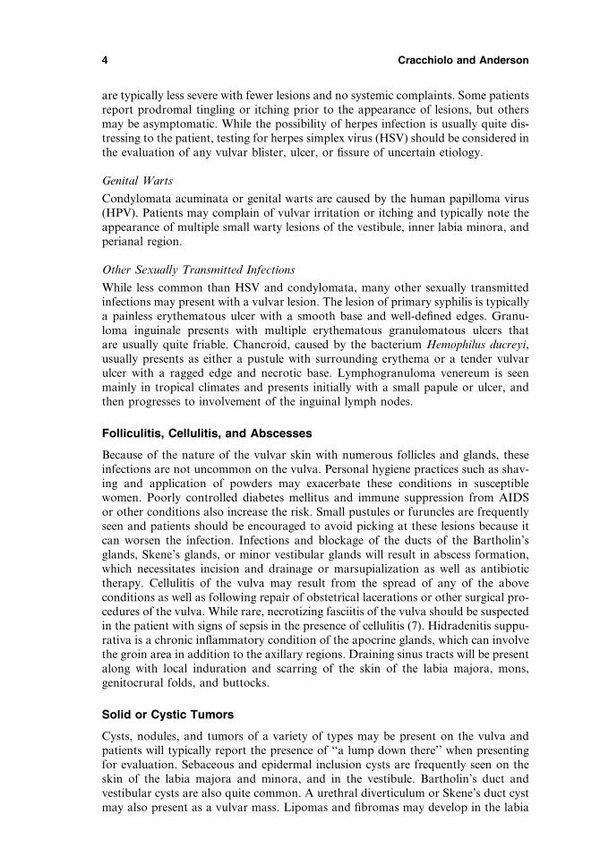
Vestibule of the Vagina. The vestibule is the medial (central), mucosa-coveredpart of the vulva. On embryologic, anatomic, and architectonic grounds, the correctterm is ‘‘vestibule of the vagina’’ not ‘‘vulvar vestibule,’’ as sometimes appears in theliterature (2). The vestibule is composed of loose fibroelastic and smooth musculartissue without fat. The boundaries of the vestibule are Hart’s lines laterally, thehymenal ring medially, the frenulum of the clitoris anteriorly, the fourchette poste-riorly, and Colles’ fascia deeply. The vestibule contains the urethral meatus andvaginal opening in the midline. The ostia of the greater vestibular (Bartholin’s) ductsmay be seen with the naked eye in the five and seven o’clock positions just external tothe hymen. Minute, soft, nonbranching, pink polyps, the vestibular papillae, are anormal finding in one-third of women in the reproductive age group, althoughuncommon after menopause (Fig. 1) (3). They may be acetowhite and must bedistinguished from condylomata acuminata. Compared to condylomata acuminata,vestibular papillae are pinker, soft, and nonbranched.
Hymen. The hymen is a diaphragm of loose fibrous tissue covered on bothsides by mucous membrane that demarcates the vestibule from the vagina. Beforefirst intercourse, the hymen is of variable shape, but often crescentic and coveringonly the posterior margin of the vaginal orifice. Less commonly, it may be morecomplete, but usually still contains one or two openings. At first intercourse, therewill often be tearing of the hymen to produce fissures. The appearance of the hymenhas been used since time immemorial to determine whether penetration has takenplace; however, recent studies of prepubertal non–sexually abused girls indicate awide range of normal appearance (4,5). Changes that were considered the result ofsexual abuse, including a gaping introitus, may be seen in nonabused girls, andmay be difficult to distinguish from changes induced by sexual penetration. The tagsof mucous membrane following fissuring of the hymen are called the caruncles hyme-nalis or carunculae myrtiformes.
12 Scurry and Melville

Urethral Meatus. The urethral meatus lies in the midline between the clitorisand the vaginal opening. Its anteroposterior position is quite variable, but it isusually 20 to 30 mm below the glans. The meatus may be everted and may showtwo or three folds, which become more prominent after menopause. The ostia ofthe paraurethral glands are not visible to the naked eye.
Clitoris, Superficial Part. The clitoris is composed of the body and crura. Thecrura are deep impalpable structures. The body can be felt beneath its prepuce, butthe only part that is exposed is its terminal portion, the glans. The glans is a smooth-surfaced pink nodule covered by moist, thin hairless skin, which lies in the midlineanterior to the urethral meatus. Resting clitoral size varies according to the age ofthe female, parity, and hormonal status (6). Anatomically, the body of the clitorisis about 40 mm long. Clinically, however, the total length of the glans and body isonly 16.3� 4.3 mm, the transverse diameter of the glans is 3.4� 1 mm, and thelength of the glans is 5.1� 1.4 mm (7).
Fossa Navicularis. The fossa navicularis is a shallow depression in the vesti-bule that lies posterior to the vaginal orifice. Its boundaries are anterior, the hymen;posterior, the fourchette, and lateral, the labia minora. It is composed of squamousmucosa overlying a thick lamina propria of loose fibrous and deep fibrous tissue andsmooth muscular tissue.
Perineum. The clinical perineum is the skin between the vulva and the anus.It is composed of hair-bearing skin apart from a variably broad hairless strip inthe midline and deep fibromuscular tissue. In the midline, the perineal body canbe palpated beneath the skin.
Deep Anatomy
Colles’ Fascia. The vulva is subdivided into superficial and deep compart-ments by bilateral transverse fibrous sheets, Colles’ (superficial perineal) fasciae,which are the continuation of the deep fascia of the anterior abdominal wall. Thesuperficial compartment contains skin, skin appendages, and fibromuscular tissue
Figure 1 Vestibular papillomatosis: multiple small nonbranching fronds around the introitus.Source: Courtesy of Dr. Ross Pagano, Carlton, Victoria, Australia.
Normal Vulva and Developmental Anomalies 13
or subcutaneous fat, depending on the site. The deep compartment, the superficialperineal pouch or space, contains the deep part of the body and all of the crura ofthe clitoris, membranous urethra, vestibular bulbs, greater vestibular (Bartholin’s)glands, three pairs of skeletal muscles, and the perineal body. The paired structuresand perineal body are contained in fat and/or fibrous tissue.
Inferior Fascia of the Urogenital Diaphragm. The perineum is separated fromthe pelvis by the urogenital diaphragm. The urogenital diaphragm is composed of themuscles of the levator ani covered superiorly and inferiorly by fasciae. The deepboundary of the vulva is the inferior fascia of urogenital diaphragm or perinealmembrane. The crura, bulbs, and bulbospongiosus muscles attach to the inferiorfascia of the urogenital diaphragm.
Suspensory Ligament of the Clitoris. The suspensory ligament of the clitoris isa thick, multiplanar, fan-shaped fibrofatty sheet, which extends from the deep fasciaof the mons pubis to converge on the body of the clitoris (8). The ligament has super-ficial and deep components. The superficial component is 70 to 80 mm wide in themons and 80 to 90 mm long. It attaches to the clitoral body, glans, and medial labiaminora. The deep component is up to 10 mm thick and more fibrous. It has a narroworigin from the pubic symphysis and converges on the superolateral clitoral body.After reaching the clitoris, the suspensory ligament divides into right and left arms,which continue posteriorly to reach the labia and bulbs.
Clitoris, Deep Part. Anatomically, the clitoris is much larger than clinicallyapparent because most of it is deep to the skin. Recent dissections describe it is atriplanar structure projecting 4 to 6 cm anterior to the pubis (6). The body is locatedin the midline, at the anterior junction of the labia minora, anterior to the vestibule.Only the terminal glans is exposed. The remainder of the body is covered by itsprepuce anteriorly and frenulum posteriorly, which are derived from the labiaminora. The body is curved with its convexity anterior. It is formed by the fusion oftwo cylindrical masses of spongy tissue, the corpora cavernosa. A dense fibrous sheathencloses each corpus cavernosum and forms an incomplete septum between the two.The corpora cavernosa separate posteriorly to form the crus of the clitoris. The armsof the crura are up to 90 mm long, lie along, and insert into the inferior borders of theischiopubic rami of the pubic arch. They also partly insert into the inferior fasciaof the urogenital diaphragm. The arms of the right and left crus are covered by theischiocavernosus muscles. The clitoris is stabilized by the suspensory ligament.
Bulbs of the Vestibule. The vestibular bulbs are paired masses of erectile(spongy) tissue closely related to the anterior and lateral walls of the distal urethraand lateral walls of the distal vagina. When stimulated, the erect bulbs narrow theintroitus to aid sexual intercourse. Anteriorly, the bulbs attach to the clitoral bodyand to each other beneath the clitoris as a narrow bridge anterior to the urethra.They then expand and descend posterolaterally in the superficial perineal pouch toform cylindrical masses in the deep lateral wall of the vagina at the level of the intro-itus. The degree of posterior extension is variable and may depend upon age (6). Thebulbs are usually about 30 mm long and reach at least the three and nine o’clockpositions. The bulbs are covered superficially and laterally by the bulbospongiosusmuscles and deeply by the perineal membrane (urogenital diaphragm), into whichthey insert.
Major Vestibular (Bartholin’s) Glands. The major vestibular (Bartholin’s)glands are mucinous glands that secrete mucus under parasympathetic stimulationduring sexual arousal to aid intercourse. The glands are normally impalpable andnot identified during surgical procedures. They are paired 5 to 10 mm rounded
14 Scurry and Melville

bodies that lie in the superficial perineal pouch covered in mixed fibrous and smoothmuscle tissue, at the posterior end of the bulbs, and covered by the bulbs and bulbos-pongiosus muscles. The ducts, which are just visible to the naked eye, open into thevestibule, very close to the hymen at five and seven o’clock of the introitus.
Superficial Perineal Muscles. Three pairs of skeletal muscles, the ischiocaver-nosus, bulbospongiosus, and superficial transversus perinei muscles, form an incom-plete muscular sheet in the superficial perineal pouch (1). These muscles form bilateraltriangles with the clitoris, perineal body, and ischial tuberosities as the points. Theischiocavernosus muscles arise from medial surface of the ischial bones, overliethe arms of the crura, and join anteriorly to the body of the clitoris, into which they alsoinsert. The bulbospongiosus muscles arise from the perineal body, overlie the greatervestibular (Bartholin’s) glands and bulbs, and fuse just posterior to the clitoris, intowhich they also insert. The superficial transversus perinei muscles arise from the medialsurface of the ischial bones and extend transversely to insert into the perineal body.The ischiocavernosus and bulbospongiosus muscles overlie erectile tissue and theircontraction helps with female erection. The bulbs ospongiosus muscles are also weakvaginal constrictors, the main vaginal constrictor being the levator ani. The transversusperinei muscles help stabilize the perineal body.
Urogenital Diaphragm. The urogenital diaphragm remains a controversialsubject. According to Mostwin, it is a continuous fan-shaped mass of skeletal mus-cles forming part of the levator ani and divided into various parts depending upon itsorigins and insertions (9). It is orientated transversely and covers the anterior surfaceof the urethra and distal vagina. The extent to which the fibers encircle the vagina orurethra depends upon the level at which a transverse section is taken through thestructures. Beginning anteroinferiorly, the skeletal muscle almost completely sur-rounds the distal vagina, being deficient only posteriorly. Superoposteriorly, themuscle fibers insert progressively more anteriorly, until, at the level of the proximalone-third of the urethra, the muscle no longer encircles any part of the vagina, butcompletely encircles the urethra, being thicker anteriorly.
Perineal Body. The perineal body (central tendon of the perineum) is an ill-defined pyramidal-shaped mass of mixed smooth muscle and fibrous tissue betweenthe vagina and the anus. The apex is directed superiorly and base toward the perinealskin. There is only a small amount of subcutaneous fat between the perineal bodyand the skin. The perineal body serves as a site of attachment for skeletal muscles andfasciae of the perineum. The muscular attachments are the levator ani, urethralsphincters, deep transversus perinei, external anal sphincter, peripheral fibers ofthe longitudinal smooth muscle of the rectum, bulbospongiosus, and superficialtransversus perinei muscles. The fascial attachments are the superior and inferiorurogenital diaphragm fasciae and superficial perineal (Colles’) fascia.
Arteries. The arterial supply to the vulva comes from branches of the internaliliac and femoral arteries. The internal iliac artery gives rise to the internal pudendalartery, which supplies the medial and deep parts of the vulva, including the erectiletissues, and shares the supply to the labia (1). The internal pudendal artery enters theperineum from the buttock via the lesser sciatic foramen. It passes anteromediallyalong the ischiopubic ramus within the deep perineal space within a fascial compart-ment (Alcock’s canal) and gives rise to the inferior rectal, perineal, and bulbararteries before dividing to form the deep and dorsal arteries of the clitoris. Thesebranches pierce the inferior fascia of the urogenital diaphragm to enter the super-ficial perineal space. The artery of the bulb of the vestibule and the anterior vaginalartery supply the bulb and greater (Bartholin’s) gland. The dorsal clitoral artery runs
Normal Vulva and Developmental Anomalies 15

alongside the dorsal nerves to terminate in the glans. The deep artery of the clitorissupplies the crus. The femoral artery gives off the superficial and deep externalpudendal arteries to supply the majority of arterial blood to the skin and superficialfascia of the vulva. The external pudendal artery travels from the groin with theround ligament to enter the labia majora where it anastomoses with the labialbranches of the internal pudendal artery.
Veins. Blood is drained from the perineum mainly by the internal pudendalveins, but some is drained by the deep dorsal vein of the clitoris and returned to thevesical plexus or by the external pudendal veins into the great saphenous veins (1).Venous vessels follow the branches of the internal and external pudendal arteriesand are named accordingly. Veins from the bulbs of the vestibule, labia majora,and anal canal unite to form two venae comitantes of the internal pudendal arterycalled the internal pudendal veins. The internal pudendal veins empty into the inter-nal iliac veins. There are no corresponding veins for the deep arteries of the clitoris,and most of the blood from the glans of the clitoris is drained to the deep dorsal veinof the clitoris, an unpaired vessel that runs between the two dorsal arteries. The deepdorsal vein drains into the vesical plexus and communicates with the internalpudendal vein tributaries. The external pudendal veins provide additional drainagefrom the labia majora and empty into the great saphenous veins.
Lymph. The lymphatic drainage of the anal canal, perineal skin, distal ure-thra, and vulva follows the branches of the external pudendal blood vessels to theinguinal lymph nodes and then to the external iliac, common iliac, and para-aorticlymph nodes (1). Midline structures, defined as anterior to urethra or posterior tothe fourchette, have bilateral lymphatic drainage. The groin lymph nodes consistof superficial and deep groups, depending on whether they are superficial or deepto the femoral fascia (10). The superficial group is further divided into upper andlower groups. The upper superficial nodes are found along the line of the inguinalligament, and the lower along the terminal part of the great saphenous vein. Thedeep nodes, one to three in number, lie in the fossa ovalis, medial to the femoral vein.The superficial group most often contains the sentinel node or nodes, but as in othersites, sentinel node studies demonstrate occasional marked variation in lymphaticdrainage that cannot be predicted clinically. Lymphatic vessels of the deep perineum,including the deep perineal space, membranous urethra, and upper vagina follow theinternal pudendal vessels to the internal iliac nodes.
Nerves. The motor and sensory somatic and autonomic nerve supply to thevulva is derived from L1 to S4 (1). The pudendal nerves supply the somatic motorand sensory nerves to most of the vulva, distal vagina, and anal canal. The right andleft pudendal nerves are formed from the S2 to S4 anterior rami of the sacral plexus.Each pudendal nerve travels alongside the internal pudendal artery to give offsimilarly named branches. The pudendal nerve gives rise to the inferior rectal(hemorrhoidal) nerve, which innervates the external anal sphincter, lower anal canal,and perianal skin. At the posterior margin of the urogenital diaphragm, it divides toform the perineal nerve and the dorsal nerve of the clitoris. The perineal nerve sub-divides into many muscular branches and two cutaneous nerves. The two cutaneousbranches, the posterior labial branches, pass anteriorly in the superficial perinealspace to supply sensory innervation to the posterior labia majora. The mons pubisand anterior labia majora are innervated by the anterior labial nerve (branch ofthe ilioinguinal nerve), derived from the lumbar plexus. The paired dorsal nervesof the clitoris run alongside the dorsal arteries of the clitoris in the deep perinealspace to supply the corpora cavernosa, anterior labia minora, and glans.
16 Scurry and Melville

Parasympathetic fibers enter the perineum through the urogenital hiatus alongwith the branches of the vesical plexus and cavernous nerves. Parasympathetic sen-sory nerves monitor a sense of fullness of the anal canal and vagina. Parasympatheticmotor function increases vaginal secretions and causes erection.
Vagina
The vagina is a muscular tube, 70 to 90 mm long, posterior to the urethra andbladder, and anterior to the rectum. It extends from the hymenal ring to the cervix.The upper half is curved anteriorly to lie on the bladder. Proximally, the vagina iscovered by peritoneum posteriorly. The upper vagina is attached posteriorly to theuterosacral ligaments, laterally to the cardinal ligaments, and anteriorly to the baseof the bladder. The cervix projects downwards and backwards through the upperanterior vaginal wall, and shortens the anterior wall by 20 mm, compared to the pos-terior wall. The circumferential recess formed by the angle between the projectingcervix and the upper vagina is the fornix, which is divided into anterior (shallow),posterior (deep), and lateral components. At the upper end, the cervix holds thevagina open, but most of the resting vagina is collapsed with its anterior wall incontact with the posterior wall to form a H- or crescent shape on cross section.The urethra is attached to the anterior vaginal wall. The lower one-third of thevagina passes through the urogenital diaphragm. Below the diaphragm, the vaginais supported by the perineal body and perineal muscles.
The vagina is composed of three layers: mucosa, muscularis, and perimuscularlayers. The mucosa is rugose, with the degree of rugosity depending on hormonalstatus. The muscularis is thin. In the lower vagina, the perimuscular layer is directlycontinuous laterally with the vestibular bulbs.
The lymphatic drainage of the upper two-thirds of the vagina follows that ofthe cervix to the internal and external iliac nodes. The lower one-third drain followsthat of the vulva to the superficial inguinal nodes.
Urethra
The female urethra is a muscular tube 4 cm long and 6 mm in diameter. The urethrabegins at the internal urethral orifice on the anterior urinary bladder wall. It is com-posed of long, upper pelvic and short, lower perineal parts. For the great part of itslength, the pelvic urethra travels anteroinferiorly behind the pubic symphysis. It isattached to the vagina at the vaginocervicourethral angle and at the distal two-thirds. The space between these two attachments is the vesicovaginal space, whichcontains loose connective tissue. The pelvic urethra passes through the urogenitaldiaphragm to emerge as the perineal urethra, which ends as the external urethral ori-fice (urinary meatus). The perineal urethra is almost surrounded by the vestibularbulbs, apart from the posterior aspect. The urethra is normally collapsed and haslongitudinal grooves allowing its lumen to be distensible. It is composed of a mucosaand a muscle coat, which consists of smooth muscle with an inner layer of longitudi-nal bundles and a middle layer with circular bundles (internal urethral sphincter)externally intermingled with circularly orientated striated muscle (external urethralsphincter). The internal sphincter is continuous with the vesical sphincter and, atthe outside, with longitudinal extensions of the detrusor muscle of the bladder wall.The external urethral sphincter completely surrounds the upper two-thirds of theurethra, although it is thinner between the urethra and the vagina. It can be feltwithin the vagina as a transverse ridge beneath the mucosa anteriorly.
Normal Vulva and Developmental Anomalies 17
The blood supply, innervation, and lymphatic drainage of the pelvic urethraare the same as that of the bladder neck. The perineal urethra, like the vulva, issupplied by the pudendal vessels and nerves, and the lymphatic drainage is to theinguinal nodes.
HISTOLOGY
Labium Majus
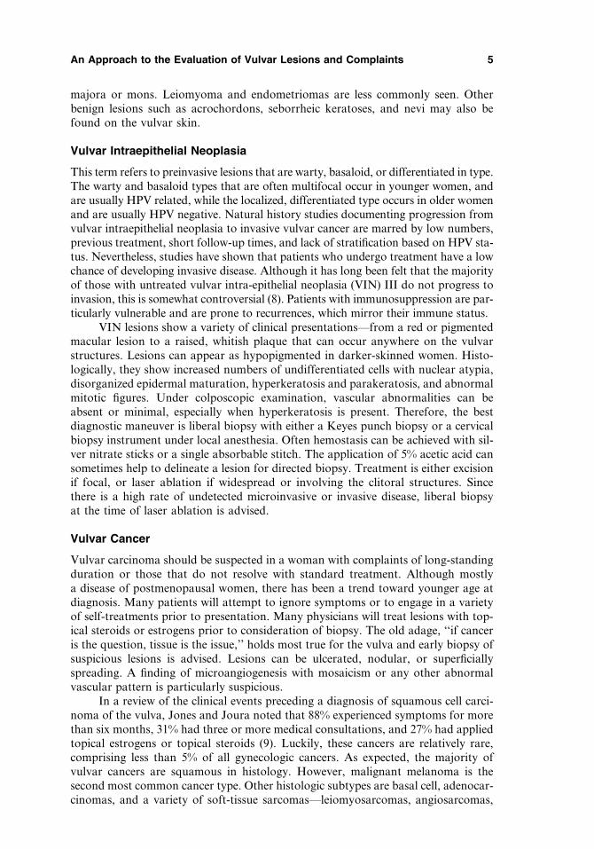
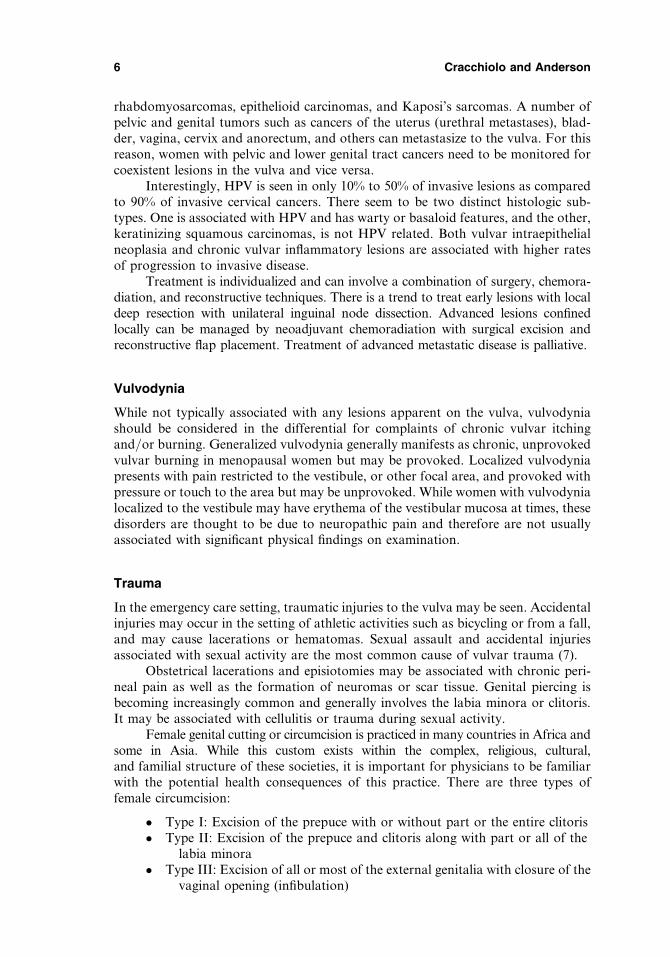
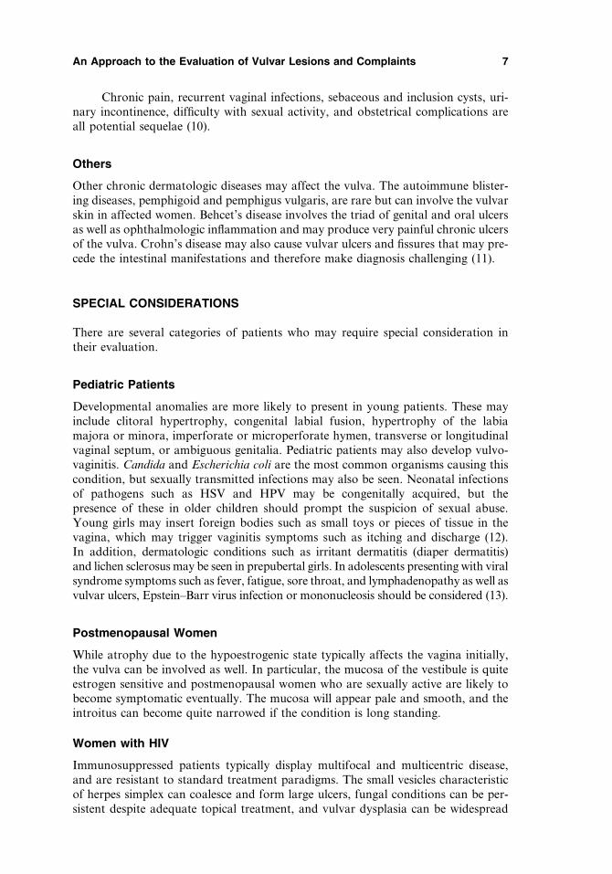
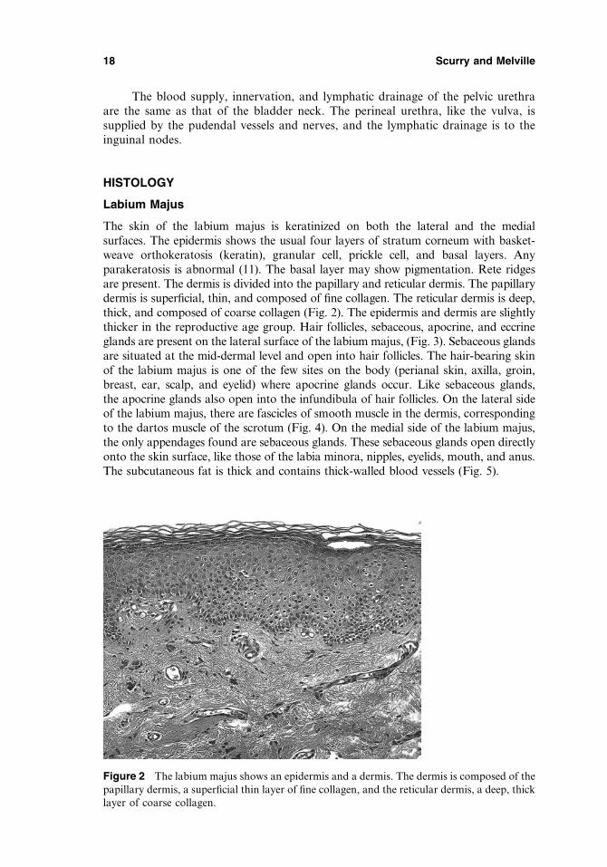
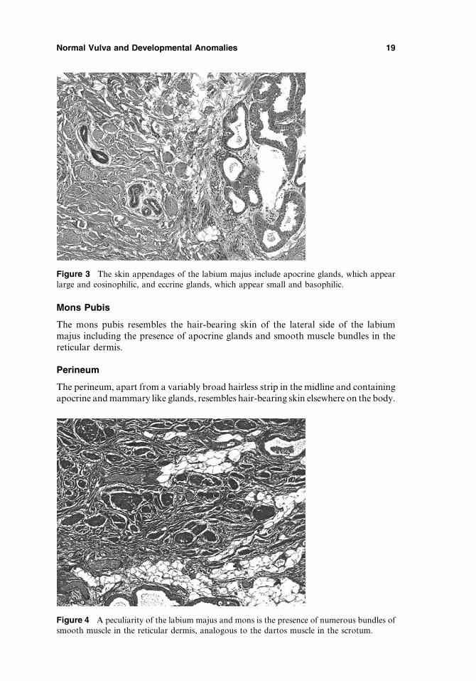
The skin of the labium majus is keratinized on both the lateral and the medialsurfaces. The epidermis shows the usual four layers of stratum corneum with basket-weave orthokeratosis (keratin), granular cell, prickle cell, and basal layers. Anyparakeratosis is abnormal (11). The basal layer may show pigmentation. Rete ridgesare present. The dermis is divided into the papillary and reticular dermis. The papillarydermis is superficial, thin, and composed of fine collagen. The reticular dermis is deep,thick, and composed of coarse collagen (Fig. 2). The epidermis and dermis are slightlythicker in the reproductive age group. Hair follicles, sebaceous, apocrine, and eccrineglands are present on the lateral surface of the labium majus, (Fig. 3). Sebaceous glandsare situated at the mid-dermal level and open into hair follicles. The hair-bearing skinof the labium majus is one of the few sites on the body (perianal skin, axilla, groin,breast, ear, scalp, and eyelid) where apocrine glands occur. Like sebaceous glands,the apocrine glands also open into the infundibula of hair follicles. On the lateral sideof the labium majus, there are fascicles of smooth muscle in the dermis, correspondingto the dartos muscle of the scrotum (Fig. 4). On the medial side of the labium majus,the only appendages found are sebaceous glands. These sebaceous glands open directlyonto the skin surface, like those of the labia minora, nipples, eyelids, mouth, and anus.The subcutaneous fat is thick and contains thick-walled blood vessels (Fig. 5).
Figure 2 The labium majus shows an epidermis and a dermis. The dermis is composed of thepapillary dermis, a superficial thin layer of fine collagen, and the reticular dermis, a deep, thicklayer of coarse collagen.
18 Scurry and Melville
Mons Pubis
The mons pubis resembles the hair-bearing skin of the lateral side of the labiummajus including the presence of apocrine glands and smooth muscle bundles in thereticular dermis.
Perineum
The perineum, apart from a variably broad hairless strip in the midline and containingapocrine and mammary like glands, resembles hair-bearing skin elsewhere on the body.
Figure 3 The skin appendages of the labium majus include apocrine glands, which appearlarge and eosinophilic, and eccrine glands, which appear small and basophilic.
Figure 4 A peculiarity of the labium majus and mons is the presence of numerous bundles ofsmooth muscle in the reticular dermis, analogous to the dartos muscle in the scrotum.
Normal Vulva and Developmental Anomalies 19
Interlabial Sulcus
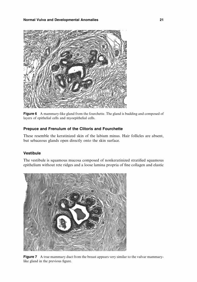
The interlabial sulcus is the main site of mammary-like glands (12). Mammary-likeglands are a newly described type of skin appendage confined to the anogenitalregion. They open by ducts into the interlabial sulcus, and to a lesser extent, perinealand perianal skin. Vulvar mammary-like glands resemble eutopic mammary lactifer-ous ducts and glands with diverticula, branches, lobules, and the expression ofestrogen and progesterone receptors (Figs. 6 and 7). They may also show apocrinemetaplasia and some features of eccrine glands. Hidradenoma papilliferum andmammary conditions of the vulva (ectopic breast, ectopic breast tissue, fibroade-noma, phyllodes tumor, and primary breast-like adenocarcinoma) appear to bederived from mammary-like glands (12,13).
Labium Minus
The labium minus is always keratinized, that is, shows a stratum corneum, indicatingskin rather than mucosa, on the lateral side (Fig. 8) (11). In our experience, themedial side is also always keratinized, although Jones found that keratinizationwas absent in 62% (11). Perhaps because the transition to vestibule may be vague,the allegedly mucosal part may be, in fact, vestibule.
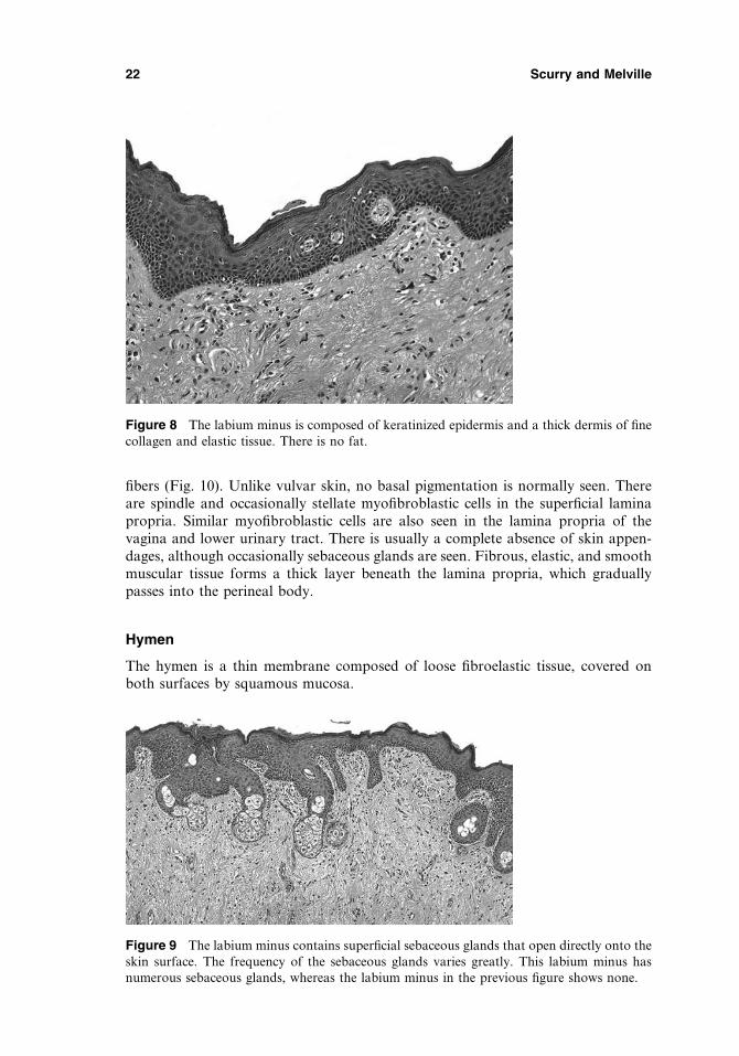
Any parakeratosis is abnormal (11). The basal layer of keratinized labiumminus may be pigmented and small rete ridges are present. Sebaceous glands arelocated superficially in the dermis and open directly onto the surface (Fig. 9). Likethe labium majus, the epidermis and dermis of the labia minora are thicker in thereproductive age group. The frequency of sebaceous glands on both surfaces ofthe labium minus varies greatly. The dermis of the labium minus is composedof fine collagen and elastic fibers. Blood vessels and nerves are numerous. Thereare no hair follicles, eccrine or apocrine glands, or subcutaneous fat on any partof the labium minus.
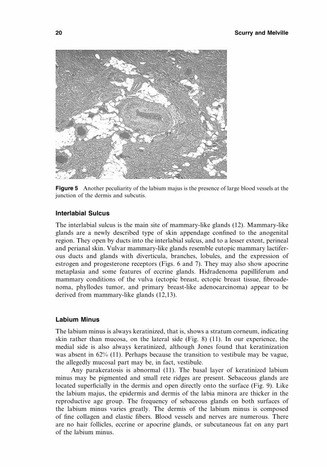
Figure 5 Another peculiarity of the labium majus is the presence of large blood vessels at thejunction of the dermis and subcutis.
20 Scurry and Melville
Prepuce and Frenulum of the Clitoris and Fourchette
These resemble the keratinized skin of the labium minus. Hair follicles are absent,but sebaceous glands open directly onto the skin surface.
Vestibule
The vestibule is squamous mucosa composed of nonkeratinized stratified squamousepithelium without rete ridges and a loose lamina propria of fine collagen and elastic
Figure 7 A true mammary duct from the breast appears very similar to the vulvar mammary-like gland in the previous figure.
Figure 6 A mammary-like gland from the fourchette. The gland is budding and composed oflayers of epithelial cells and myoepithelial cells.
Normal Vulva and Developmental Anomalies 21
fibers (Fig. 10). Unlike vulvar skin, no basal pigmentation is normally seen. Thereare spindle and occasionally stellate myofibroblastic cells in the superficial laminapropria. Similar myofibroblastic cells are also seen in the lamina propria of thevagina and lower urinary tract. There is usually a complete absence of skin appen-dages, although occasionally sebaceous glands are seen. Fibrous, elastic, and smoothmuscular tissue forms a thick layer beneath the lamina propria, which graduallypasses into the perineal body.
Hymen
The hymen is a thin membrane composed of loose fibroelastic tissue, covered onboth surfaces by squamous mucosa.
Figure 8 The labium minus is composed of keratinized epidermis and a thick dermis of finecollagen and elastic tissue. There is no fat.
Figure 9 The labium minus contains superficial sebaceous glands that open directly onto theskin surface. The frequency of the sebaceous glands varies greatly. This labium minus hasnumerous sebaceous glands, whereas the labium minus in the previous figure shows none.
22 Scurry and Melville

Clitoris
Glans
The specific erectile tissue of the glans, which is far better identifiable in neonates,becomes a vague layer of fibroelastic tissue with sinusoidal blood vessels, numerousnerves, and Pacinian corpuscles (Fig. 11). There is a small and solid glandopreputiallamella. Most of the volume of the glans is provided by the tip of the fused corporacavernosa. The epidermis is keratinized, but unlike the labia majora and minora, it isnot hyperpigmented, and does not show skin appendages of any kind.
Figure 10 Nonkeratinized squamous epithelium of the vestibule, vagina, and urethra are similar.
Figure 11 The skin of the glans of the clitoris is composed of keratinized epidermis withoutappendages and two layers of dermis, with both numerous blood vessels and nerves.
Normal Vulva and Developmental Anomalies 23

Body
The body is composed of right and left corpora cavernosa surrounded by a sheet ofdense collagen (the tunica), which also incompletely divides them (Fig. 12). Thecorpora cavernosa consist of erectile tissue with numerous, gaping, thin-walledsinusoidal vessels. The sinuses have endothelium but no smooth muscle in theirwalls. Between the sinusoids, however, there are bundles of smooth muscle, largenerves, and fibrous stroma. Pacinian corpuscles are seen associated with the nerves.
Crura
The crura are composed of corpus cavernous (sinusoidal) tissue, which is surrounded byits tunica. The tunica is absent laterally where the crus attaches to bone. Pacinian cor-puscles are seen, but unlike the body, large nerve and vascular trunks are not present.
Bulbs of the Vestibule
The bulbs are composed of cavernous (sinusoidal and erectile) tissue, but the vascu-lar spaces are larger and more gaping, and the trabeculae with conspicuous arteriesthicker than in clitoral tissues (Fig. 13). They have only a thin capsule rather thana thick fibrous tunic, and do not become as erect as clitoris. Bulbar tissue mergeswith the adventitia of the vagina and gradually passes into rich vascular plexusesin the anterior wall of the vestibule. Large neurovascular bundles and pacinian cor-puscles are not seen.
Vagina
The vagina shows three layers forming its wall, the mucosa, muscular coat, andadventitia. The mucosa is composed of nonkeratinized squamous epithelium richin glycogen and lamina propria. The lamina propria is denser toward the surface
Figure 12 The two corpora cavernosa of the clitoris appear as sinusoidal vascular tissueseparated by a septum composed of dense fibrous tissue of the tunica.
24 Scurry and Melville
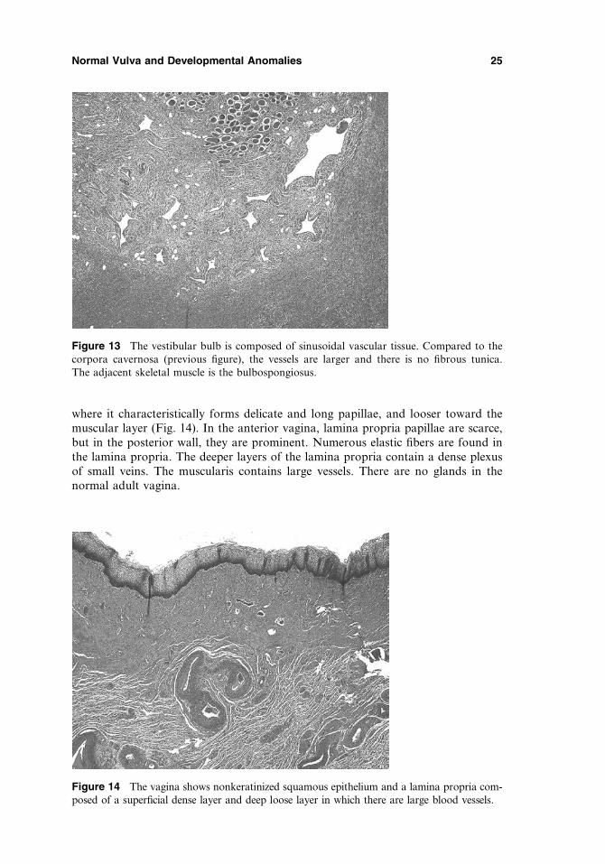
where it characteristically forms delicate and long papillae, and looser toward themuscular layer (Fig. 14). In the anterior vagina, lamina propria papillae are scarce,but in the posterior wall, they are prominent. Numerous elastic fibers are found inthe lamina propria. The deeper layers of the lamina propria contain a dense plexusof small veins. The muscularis contains large vessels. There are no glands in thenormal adult vagina.
Figure 13 The vestibular bulb is composed of sinusoidal vascular tissue. Compared to thecorpora cavernosa (previous figure), the vessels are larger and there is no fibrous tunica.The adjacent skeletal muscle is the bulbospongiosus.
Figure 14 The vagina shows nonkeratinized squamous epithelium and a lamina propria com-posed of a superficial dense layer and deep loose layer in which there are large blood vessels.
Normal Vulva and Developmental Anomalies 25
Urethra
The lining of the urethra is variable. Most of the distal two-thirds of the urethra islined by squamous epithelium, but is regularly interrupted by foci of pseudostratifiedcolumnar epithelium. The upper one-third shows transitional epithelium similar tothat seen elsewhere in the urinary tract. The epithelium overlies a loose laminapropria, which contains mucinous glands and many thin-walled veins. Beneath thislayer, there are inner longitudinal and outer circular layers of smooth muscle.Distally, a sphincter of skeletal muscle surrounds the layers of smooth muscle.
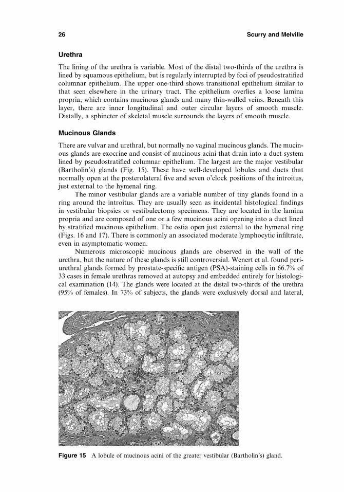
Mucinous Glands
There are vulvar and urethral, but normally no vaginal mucinous glands. The mucin-ous glands are exocrine and consist of mucinous acini that drain into a duct systemlined by pseudostratified columnar epithelium. The largest are the major vestibular(Bartholin’s) glands (Fig. 15). These have well-developed lobules and ducts thatnormally open at the posterolateral five and seven o’clock positions of the introitus,just external to the hymenal ring.
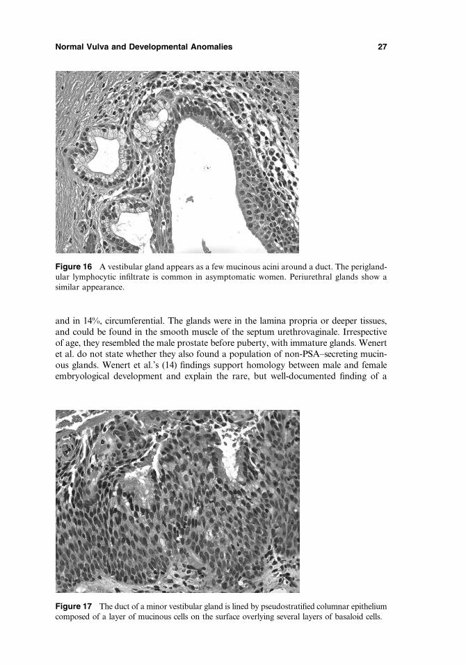
The minor vestibular glands are a variable number of tiny glands found in aring around the introitus. They are usually seen as incidental histological findingsin vestibular biopsies or vestibulectomy specimens. They are located in the laminapropria and are composed of one or a few mucinous acini opening into a duct linedby stratified mucinous epithelium. The ostia open just external to the hymenal ring(Figs. 16 and 17). There is commonly an associated moderate lymphocytic infiltrate,even in asymptomatic women.
Numerous microscopic mucinous glands are observed in the wall of theurethra, but the nature of these glands is still controversial. Wenert et al. found peri-urethral glands formed by prostate-specific antigen (PSA)-staining cells in 66.7% of33 cases in female urethras removed at autopsy and embedded entirely for histologi-cal examination (14). The glands were located at the distal two-thirds of the urethra(95% of females). In 73% of subjects, the glands were exclusively dorsal and lateral,
Figure 15 A lobule of mucinous acini of the greater vestibular (Bartholin’s) gland.
26 Scurry and Melville
and in 14%, circumferential. The glands were in the lamina propria or deeper tissues,and could be found in the smooth muscle of the septum urethrovaginale. Irrespectiveof age, they resembled the male prostate before puberty, with immature glands. Wenertet al. do not state whether they also found a population of non-PSA–secreting mucin-ous glands. Wenert et al.’s (14) findings support homology between male and femaleembryological development and explain the rare, but well-documented finding of a
Figure 16 A vestibular gland appears as a few mucinous acini around a duct. The perigland-ular lymphocytic infiltrate is common in asymptomatic women. Periurethral glands show asimilar appearance.
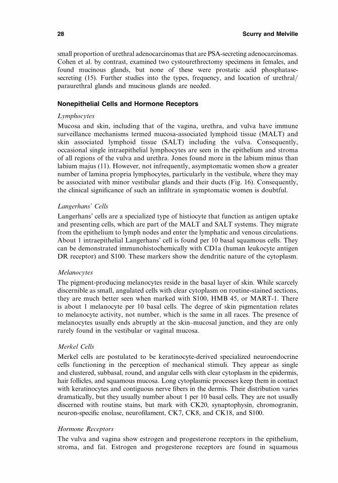
Figure 17 The duct of a minor vestibular gland is lined by pseudostratified columnar epitheliumcomposed of a layer of mucinous cells on the surface overlying several layers of basaloid cells.
Normal Vulva and Developmental Anomalies 27
small proportion of urethral adenocarcinomas that are PSA-secreting adenocarcinomas.Cohen et al. by contrast, examined two cystourethrectomy specimens in females, andfound mucinous glands, but none of these were prostatic acid phosphatase-secreting (15). Further studies into the types, frequency, and location of urethral/paraurethral glands and mucinous glands are needed.
Nonepithelial Cells and Hormone Receptors
Lymphocytes
Mucosa and skin, including that of the vagina, urethra, and vulva have immunesurveillance mechanisms termed mucosa-associated lymphoid tissue (MALT) andskin associated lymphoid tissue (SALT) including the vulva. Consequently,occasional single intraepithelial lymphocytes are seen in the epithelium and stromaof all regions of the vulva and urethra. Jones found more in the labium minus thanlabium majus (11). However, not infrequently, asymptomatic women show a greaternumber of lamina propria lymphocytes, particularly in the vestibule, where they maybe associated with minor vestibular glands and their ducts (Fig. 16). Consequently,the clinical significance of such an infiltrate in symptomatic women is doubtful.
Langerhans’ Cells
Langerhans’ cells are a specialized type of histiocyte that function as antigen uptakeand presenting cells, which are part of the MALT and SALT systems. They migratefrom the epithelium to lymph nodes and enter the lymphatic and venous circulations.About 1 intraepithelial Langerhans’ cell is found per 10 basal squamous cells. Theycan be demonstrated immunohistochemically with CD1a (human leukocyte antigenDR receptor) and S100. These markers show the dendritic nature of the cytoplasm.
Melanocytes
The pigment-producing melanocytes reside in the basal layer of skin. While scarcelydiscernible as small, angulated cells with clear cytoplasm on routine-stained sections,they are much better seen when marked with S100, HMB 45, or MART-1. Thereis about 1 melanocyte per 10 basal cells. The degree of skin pigmentation relatesto melanocyte activity, not number, which is the same in all races. The presence ofmelanocytes usually ends abruptly at the skin–mucosal junction, and they are onlyrarely found in the vestibular or vaginal mucosa.
Merkel Cells
Merkel cells are postulated to be keratinocyte-derived specialized neuroendocrinecells functioning in the perception of mechanical stimuli. They appear as singleand clustered, subbasal, round, and angular cells with clear cytoplasm in the epidermis,hair follicles, and squamous mucosa. Long cytoplasmic processes keep them in contactwith keratinocytes and contiguous nerve fibers in the dermis. Their distribution variesdramatically, but they usually number about 1 per 10 basal cells. They are not usuallydiscerned with routine stains, but mark with CK20, synaptophysin, chromogranin,neuron-specific enolase, neurofilament, CK7, CK8, and CK18, and S100.
Hormone Receptors
The vulva and vagina show estrogen and progesterone receptors in the epithelium,stroma, and fat. Estrogen and progesterone receptors are found in squamous
28 Scurry and Melville
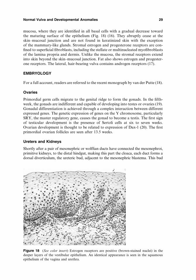
mucosa, where they are identified in all basal cells with a gradual decrease towardthe maturing surface of the epithelium (Fig. 18) (16). They abruptly cease at theskin–mucosal junction and are not found in keratinized skin with the exceptionof the mammary-like glands. Stromal estrogen and progesterone receptors are con-fined to superficial fibroblasts, including the stellate or multinucleated myofibroblastsof the lamina propria and dermis. Unlike the mucosa, the stromal receptors extendinto skin beyond the skin–mucosal junction. Fat also shows estrogen and progester-one receptors. The lateral, hair-bearing vulva contains androgen receptors (17).
EMBRYOLOGY
For a full account, readers are referred to the recent monograph by van der Putte (18).
Ovaries
Primordial germ cells migrate to the genital ridge to form the gonads. In the fifth-week, the gonads are indifferent and capable of developing into testes or ovaries (19).Gonadal differentiation is achieved through a complex interaction between differentexpressed genes. The genetic expression of genes on the Y chromosome, particularlySRY, the master regulatory gene, causes the gonad to become a testis. The first signof testicular development is the presence of Sertoli cells at six to seven weeks.Ovarian development is thought to be related to expression of Dax-1 (20). The firstprimordial ovarian follicles are seen after 13.5 weeks.
Ureters and Kidneys
Shortly after a pair of mesonephric or wolffian ducts have connected the mesonephroi,primitive kidneys, to the distal hindgut, making this part the cloaca, each duct forms adorsal diverticulum, the ureteric bud, adjacent to the mesonephric blastema. This bud
Figure 18 (See color insert) Estrogen receptors are positive (brown-stained nuclei) in thedeeper layers of the vestibular epithelium. An identical appearance is seen in the squamousepithelium of the vagina and urethra.
Normal Vulva and Developmental Anomalies 29

lengthens into the ureter with its pelvis and calyces, and connects the metanephros,definitive kidney to the urogenital derivatives of the cloaca. The original common seg-ment of the duct and ureter is incorporated into the cloaca separating the two systems,which will thereafter part during the formation of the trigone of the bladder.
Tubes, Uterus, and Vagina
At the end of the cloacal period, the wolffian or mesonephric ducts guide Mullerianor paramesonephric ducts, which are newly formed from celomic epithelium atthe cranial end of the genital ridges, toward the urogenital sinus derivatives of thecloaca. Approaching the perineum, the two paramesonephric ducts bend inwardand fuse into a single tube in the midline. The distal tips, which have remained indirect contact with the lateral mesonephric ducts, then gain access to the sinus byusing the orifices of the regressing mesonephric ducts. The original columnar epi-thelium of the distal part of the fused structure transforms into a broad stratifiedsquamous epithelium that practically fills the developing vaginal lumen. Centraldegeneration of the epithelium creates the vaginal lumen. Discrepancy between therelatively small vaginal orifice and the greatly expanding vaginal lumen in combi-nation with the movement toward the surface shapes the hymen.
Vulva
The cloaca is a distal expansion of the hindgut covered by the cloacal membrane.Between four and six weeks of embryonic life, marked differences in growth betweenthe anterior and the posterior parts of the cloaca make the cloaca transform into aU-shaped tube, which then splits into the anterior urogenital sinus and the posterioranal canal after the cloacal membrane has disintegrated. The groove-like communi-cation between the two parts is incorporated and will form a hairless median strip onthe future clinical perineum. The deeper part of the urogenital sinus differentiatesinto the urethra and trigone of the bladder. The superficial part becomes the vesti-bule with its mantle tissue differentiated into erectile and fascial structures. Growthinhibition of most erectile structures and caudalward bending of the original phallicstructure flattens the vulva with its small clitoris and prominent labia minora flank-ing a relatively large persisting urogenital orifice. In the absence of testosterone, thearea between this orifice and the anus does not lengthen disproportionately as in themale and forms the clinical perineum. Bilateral concentrations of mesenchyme outsidethe mantle of the cloaca develop into the perineal muscles and labia majora.
Urethra and Bladder
The urethra is formed from the cranial part of the urogenital sinus. It forms mostof the trigone of the bladder and all the urethra in the female and the prostaticurethra in the male. Most of the bladder is derived from the allantois.
DEVELOPMENTAL ANOMALIES
Developmental genital anomalies are divided into those that show ambiguousexternal genitalia (intersex) and those that are clearly female. The latter group isfurther divided into those that have only genital abnormalities, those that also haveanorectal abnormalities, and those that also have urological abnormalities.
30 Scurry and Melville
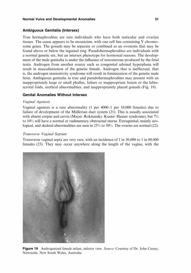
Ambiguous Genitalia (Intersex)
True hermaphrodites are rare individuals who have both testicular and ovariantissues. The cause appears to be mosaicism, with one cell line containing Y chromo-some genes. The gonads may be separate or combined as an ovotestis that may befound above or below the inguinal ring. Pseudohermaphrodites are individuals witha normal genetic sex, but an intersex phenotype for hormonal reasons. The develop-ment of the male genitalia is under the influence of testosterone produced by the fetaltestis. Androgen from another source such as congenital adrenal hyperplasia willresult in masculinization of the genetic female. Androgen that is ineffectual, thatis, the androgen insensitivity syndrome will result in feminization of the genetic malefetus. Ambiguous genitalia in true and pseudohermaphrodites may present with aninappropriately large or small phallus, failure or inappropriate fusion or the labio-scrotal folds, urethral abnormalities, and inappropriately placed gonads (Fig. 19).
Genital Anomalies Without Intersex
Vaginal Agenesis
Vaginal agenesis is a rare abnormality (1 per 4000–1 per 10,000 females) due tofailure of development of the Mullerian duct system (21). This is usually associatedwith absent corpus and cervix (Mayer–Rokitansky–Kuster–Hauser syndrome), but 7%to 10% will have a normal or rudimentary obstructed uterus. Extragenital, mainly uro-logical, and skeletal abnormalities are seen in 25% to 50%. The ovaries are normal (22).
Transverse Vaginal Septum
Transverse vaginal septa are very rare, with an incidence of 1 in 30,000 to 1 in 80,000females (23). They may occur anywhere along the length of the vagina, with the
Figure 19 Androgenized female infant, inferior view. Source: Courtesy of Dr. John Cassey,Newcastle, New South Wales, Australia.
Normal Vulva and Developmental Anomalies 31
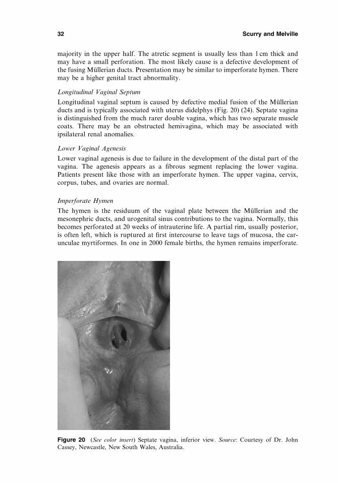
majority in the upper half. The atretic segment is usually less than 1 cm thick andmay have a small perforation. The most likely cause is a defective development ofthe fusing Mullerian ducts. Presentation may be similar to imperforate hymen. Theremay be a higher genital tract abnormality.
Longitudinal Vaginal Septum
Longitudinal vaginal septum is caused by defective medial fusion of the Mullerianducts and is typically associated with uterus didelphys (Fig. 20) (24). Septate vaginais distinguished from the much rarer double vagina, which has two separate musclecoats. There may be an obstructed hemivagina, which may be associated withipsilateral renal anomalies.
Lower Vaginal Agenesis
Lower vaginal agenesis is due to failure in the development of the distal part of thevagina. The agenesis appears as a fibrous segment replacing the lower vagina.Patients present like those with an imperforate hymen. The upper vagina, cervix,corpus, tubes, and ovaries are normal.
Imperforate Hymen
The hymen is the residuum of the vaginal plate between the Mullerian and themesonephric ducts, and urogenital sinus contributions to the vagina. Normally, thisbecomes perforated at 20 weeks of intrauterine life. A partial rim, usually posterior,is often left, which is ruptured at first intercourse to leave tags of mucosa, the car-unculae myrtiformes. In one in 2000 female births, the hymen remains imperforate.
Figure 20 (See color insert) Septate vagina, inferior view. Source: Courtesy of Dr. JohnCassey, Newcastle, New South Wales, Australia.
32 Scurry and Melville
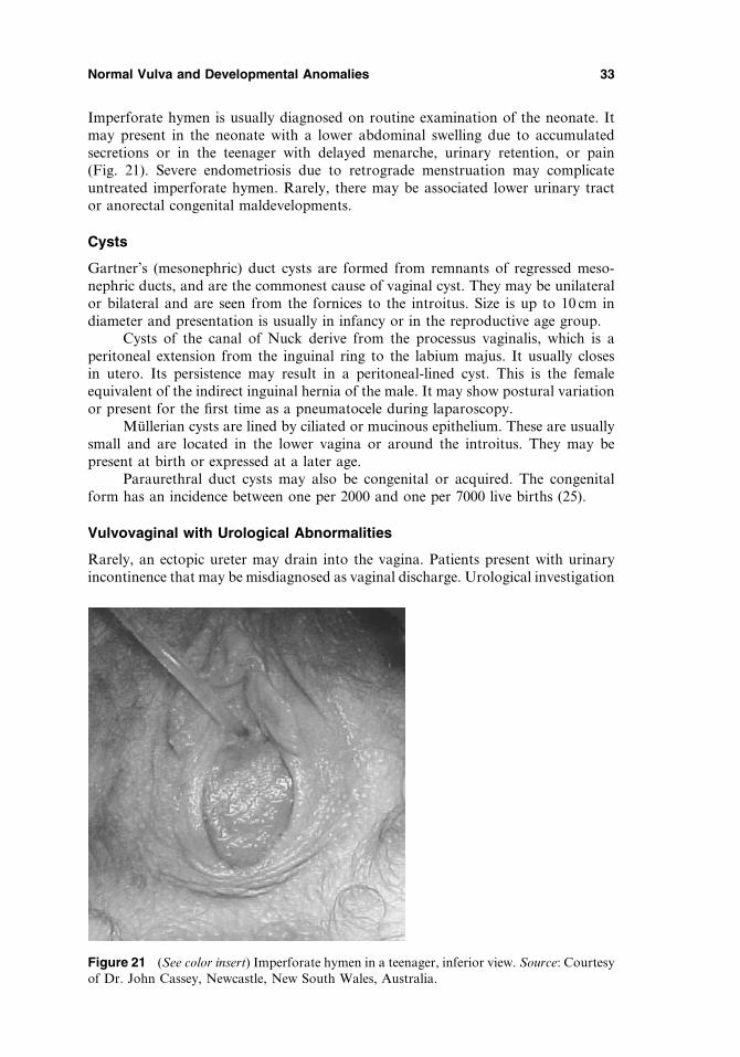
Imperforate hymen is usually diagnosed on routine examination of the neonate. Itmay present in the neonate with a lower abdominal swelling due to accumulatedsecretions or in the teenager with delayed menarche, urinary retention, or pain(Fig. 21). Severe endometriosis due to retrograde menstruation may complicateuntreated imperforate hymen. Rarely, there may be associated lower urinary tractor anorectal congenital maldevelopments.
Cysts
Gartner’s (mesonephric) duct cysts are formed from remnants of regressed meso-nephric ducts, and are the commonest cause of vaginal cyst. They may be unilateralor bilateral and are seen from the fornices to the introitus. Size is up to 10 cm indiameter and presentation is usually in infancy or in the reproductive age group.
Cysts of the canal of Nuck derive from the processus vaginalis, which is aperitoneal extension from the inguinal ring to the labium majus. It usually closesin utero. Its persistence may result in a peritoneal-lined cyst. This is the femaleequivalent of the indirect inguinal hernia of the male. It may show postural variationor present for the first time as a pneumatocele during laparoscopy.
Mullerian cysts are lined by ciliated or mucinous epithelium. These are usuallysmall and are located in the lower vagina or around the introitus. They may bepresent at birth or expressed at a later age.
Paraurethral duct cysts may also be congenital or acquired. The congenitalform has an incidence between one per 2000 and one per 7000 live births (25).
Vulvovaginal with Urological Abnormalities
Rarely, an ectopic ureter may drain into the vagina. Patients present with urinaryincontinence that may be misdiagnosed as vaginal discharge. Urological investigation
Figure 21 (See color insert) Imperforate hymen in a teenager, inferior view. Source: Courtesyof Dr. John Cassey, Newcastle, New South Wales, Australia.
Normal Vulva and Developmental Anomalies 33

is required to determine the urinary tract anatomy. Epispadias is a rare malforma-tion seen in the pediatric age group where there is a deficiency of the urethraanteriorly. Patients are incontinent. The clitoris is bifid and there is anterior separ-ation of the labia. The urethra is short and patulous. Pubic bone separation will beseen on X ray or will be palpable. Hypospadias is abnormal location of theurethral meatus on the anterior vaginal wall. There may be incontinence, meatalstenosis, pooling of urine in the vagina, and occasional vaginal and uterine abnor-malities. Rarely, Gartner’s duct cysts, especially if in infants and/or of large size,are associated with ureteric abnormalities.
Vulvovaginal with Anorectal Abnormalities
In anorectal malformation, a defect in the dorsal cloaca may lead to an abnormalanterior bending anal canal. This may issue into the posterior wall of the femaleurogenital tract at the trigone of the bladder or anywhere between the vicinity ofthe vaginal orifice and the usual site of the anus.
ACKNOWLEDGMENT
We are very grateful for Professor S. C. J. van der Putte’s expert editorial suggestions.
REFERENCES
1. Netter FH. Atlas of human anatomy. Icon Learning Systems. 3rd ed. Teterboro,New Jersey, U.S.A. 2003.
2. Friedman M, Siegler E, Lowenstein L. The vaginal vestibule. J Lower Genital Tract Dis2004; 8:71–72.
3. van Beurden M, van der Lange N, de Craen AJ, et al. Normal findings in vulvarexamination and vulvoscopy. Br J Obstet Gynaecol 1997; 104:320–324.
4. Heger AH, Ticson L, Guerra L, Lister J, et al. Appearance of the genitalia in girls selectedfor non-abuse: review of hymenal morphology and non-specific findings. J Pediatr AdolescGynecol 2002; 15:27–35.
5. Myrhe AK, Berntzen K, Bratlid D. Genital anatomy in non-abused preschool girls. ActaPediatr 2003; 92:1453–1462.
6. O’Connell HE, Hutson JM, Anderson CR, Plenter RJ. Anatomical relationship betweenurethra and clitoris. J Urol 1998; 159:1892–1897.
7. Verkauf BS, Von Thron J, O’Brien WF. Clitoral size in normal women. Obstet Gynecol1992; 80:41–44.
8. Rees MA, O’Connell HE, Plenter RJ, Hutson JM. The suspensory ligament of the clitoris:connective tissue supports of the erectile tissues of the female urogenital region. Clin Anat2000; 13:397–403.
9. Mostwin JL. Current concepts of female pelvic anatomy and physiology. Urol Clin N Am1991; 18:175–195.
10. Micheletti L, Preti M, Zola P, Zanotto Valentino MC, Bocci C, Bogliatto F. A proposedglossary of terminology related to the surgical treatment of vulvar carcinoma. Cancer1998; 83:1369–1375.
11. Jones IS. A histological assessment of normal vulval skin. Clin Exp Dermatol 1983; 8:513–521.
12. van der Putte SC. Mammary-like glands and their disorders. Int J Gynecol Pathol 1994;13:150–160.
34 Scurry and Melville

13. Tbakhi A, Cowan DF, Kumar D, Dyle D. Recurring phyllodes tumor in aberrant breasttissue of the vulva. Am J Surg Pathol 2001; 9:81–83.
14. Wenert N, Albrech M, Sesterhenn I, et al. The ‘‘female prostate’’: location, morphology,immunohistochemical characteristics and significance. Eur Urol 1992; 22:64–69.
15. Cohen RJ, Garrett K, Golding JL, Thomas RB, McNeal JE. Epithelial differentiation ofthe lower urinary tract with recognition of the minor prostatic glands. Hum Pathol 2002;33:905–909.
16. Eva LJ, MacLean AB, Reid WM, Rolfe KJ, Perrett CW. Estrogen receptor expression invulvar vestibulitis syndrome. Am J Obstet Gynecol 2003; 189:458–461.
17. Hodgins MB, Spike RC, Mackie RM, MacLean AB. An immunohistochemical study ofandrogen, oestrogen, and progesterone receptors in the vulva and vagina. Br J ObstetGynaecol 1998; 105:216–222.
18. Van der Putte SC. The development of the perineum in the human. A comprehensivehistological study with special reference to the role of the stromal components. Adv AnatEmbryol Cell Biol 2005; 177:1–131.
19. Voutilainen R. Differentiation of the fetal gonad. Horm Res 1992; 38(suppl 2):66–71.20. Lalli E, Sassone-Corsi P. DAX-1, an unusual orphan receptor at the crossroads of
steroidogenic function and sexual differentiation. Mol Endocrinol 2003; 17:1445–1453.21. Salvatore CA, Lodovicci O. Vaginal agenesis: an analysis of ninety cases. Acta Obstet
Gynecol Scand 1978; 57:89–94.22. Evans TN, Poland ML, Boving RL. Vaginal malformations. Am J Obstet Gynecol 1981;
141:910–920.23. Rock JA, Azzin R. Genital anomalies in childhood. Clin Obstet Gynecol 1987; 30:
682–696.24. Haddad B, Louis-Sylvestre C, Poitout P, Paniel B. Longitudinal vaginal septum: a
retrospective study of 202 cases. Eur J Obstet Gynecol Reprod Biol 1997; 74:197–199.
management always appropriate?. Eur J Pediatr Surg 2002; 12:212–214.
Normal Vulva and Developmental Anomalies 35
25. Ceylan H, Ozokutan BH, Karakok M, Buyukbese S. Paraurethral cyst: is conservative

3Noninfectious Inflammation and SystemicDiseases of the Vulva
Sallie M. NeillSt. John’s Dermatology Centre, St. Thomas’ Hospital, London, U.K.
INFLAMMATORY DERMATOSES
Eczema
Vulvar eczema is characterized by erythema and scaling on the outer labia majoraand erythema and fissures and erosions on the inner aspects. Vulvar eczema ispruritic, often out of proportion to the clinical findings. The skin may become ede-matous, excoriated, and lichenified because of scratching.
Irritant Contact Eczema
This is the commonest form of vulvar eczema in clinical practice. This is usually seenin the elderly and is often because of urinary incontinence. It is also more likely tooccur in fair-skinned individuals who have sensitive skin at other sites as well (Fig. 1).
Allergic Contact Eczema
Allergic contact eczema is a Type IV cell-mediated allergic reaction that takes 48 to72 hours to develop following exposure to the offending allergen. This type of allergyis uncommon on the vulvar skin alone and is likelier to be found if there is perianaleczema as well. The commonest allergens include topical antibiotics and preserva-tives in the ointments or creams that are being used. The topical steroid itself canon rare occasions be responsible. It is important to patch test the patient if the vulvarrash is worsening with treatment.
Seborrheic Eczema
This is the second most common type of vulvar eczema that tends to affect other flex-ural sites including the axillae, submammary, and inguinal folds. There may also bescalp involvement. On occasions it may be very difficult to differentiate this form ofeczema from flexural psoriasis; however, with the former there should be no nailinvolvement (Fig. 2).
37
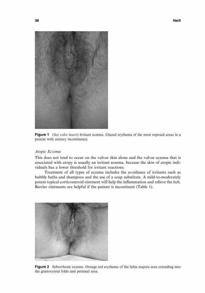
Atopic Eczema
This does not tend to occur on the vulvar skin alone and the vulvar eczema that isassociated with atopy is usually an irritant eczema, because the skin of atopic indi-viduals has a lower threshold for irritant reactions.
Treatment of all types of eczema includes the avoidance of irritants such asbubble baths and shampoos and the use of a soap substitute. A mild-to-moderatelypotent topical corticosteroid ointment will help the inflammation and relieve the itch.Barrier ointments are helpful if the patient is incontinent (Table 1).
Figure 1 (See color insert) Irritant eczema. Glazed erythema of the most exposed areas in apatient with urinary incontinence.
Figure 2 Seborrhoeic eczema. Orange red erythema of the labia majora area extending intothe genitocrural folds and perianal area.
38 Neill
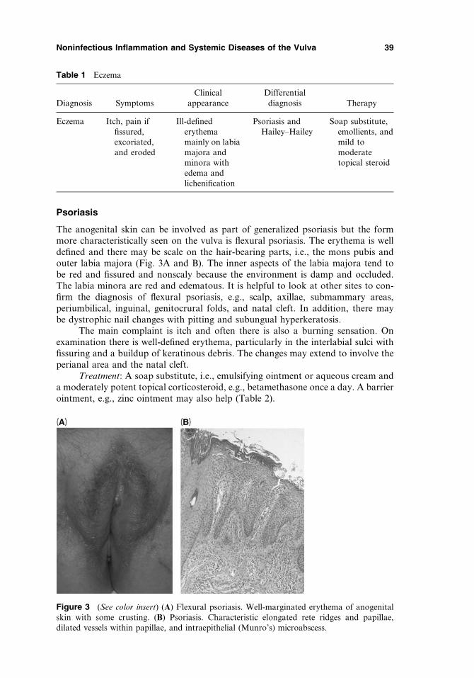
Psoriasis
The anogenital skin can be involved as part of generalized psoriasis but the formmore characteristically seen on the vulva is flexural psoriasis. The erythema is welldefined and there may be scale on the hair-bearing parts, i.e., the mons pubis andouter labia majora (Fig. 3A and B). The inner aspects of the labia majora tend tobe red and fissured and nonscaly because the environment is damp and occluded.The labia minora are red and edematous. It is helpful to look at other sites to con-firm the diagnosis of flexural psoriasis, e.g., scalp, axillae, submammary areas,periumbilical, inguinal, genitocrural folds, and natal cleft. In addition, there maybe dystrophic nail changes with pitting and subungual hyperkeratosis.
The main complaint is itch and often there is also a burning sensation. Onexamination there is well-defined erythema, particularly in the interlabial sulci withfissuring and a buildup of keratinous debris. The changes may extend to involve theperianal area and the natal cleft.
Treatment: A soap substitute, i.e., emulsifying ointment or aqueous cream anda moderately potent topical corticosteroid, e.g., betamethasone once a day. A barrierointment, e.g., zinc ointment may also help (Table 2).
Table 1 Eczema
Diagnosis SymptomsClinical
appearanceDifferentialdiagnosis Therapy
Eczema Itch, pain iffissured,excoriated,and eroded
Ill-definederythemamainly on labiamajora andminora withedema andlichenification
Psoriasis andHailey–Hailey
Soap substitute,emollients, andmild tomoderatetopical steroid
Figure 3 (See color insert) (A) Flexural psoriasis. Well-marginated erythema of anogenitalskin with some crusting. (B) Psoriasis. Characteristic elongated rete ridges and papillae,dilated vessels within papillae, and intraepithelial (Munro’s) microabscess.
Noninfectious Inflammation and Systemic Diseases of the Vulva 39

Reiter’s Disease (Reiter’s Syndrome and Circinate Ulcerative Vulvitis)
This condition is characterized by a nonsuppurative polyarthritis frequentlyaccompanied by urethritis, cervicitis, and conjunctivitis. Reiter’s disease is com-moner in males and it may be triggered by an enteric or a lower genital tractinfection. There seems to be a predisposition in those individuals who are positivefor the human leukocyte antigen B27. The cutaneous manifestations probablyrepresent a particular form of psoriasis. Circinate balanitis is well recognized. Thecorresponding vulvitis is much rarer but has been described (1). The lesions thatoccur can be eroded, ulcerative, or scaly.
Histologically, the changes are of those seen in pustular psoriasis with hyper-keratosis, parakeratosis, psoriasiform hyperplasia, an absent granular layer, andcollections of polymorphs in the epidermis (2). Treatment is the same as for psoriasis(see previous section).
Lichen Planus
Lichen planus (LP) can affect the anogenital skin or vagina in isolation or at thesame time as a generalized outbreak, when about 20% of patients will have genitallesions (3). The vulvar lesions are similar to those seen on nongenital skin sites andmay be violaceous or erythematous papules, white or annular plaques, and erosionswith or without a lacy white border. Sometimes, the main findings are erosions andulcerations. If the LP is confined to the vulva and/or vagina, the lesions are more likelyto be erosive (Fig. 4). Examination of the oral mucosa is helpful as LP may be found onthe tongue (Fig. 5), palate, gingiva, and lateral buccal mucosa. There is a small risk(> 4%) of squamous cell carcinoma (SCC) arising on a background of LP.
The other clinical forms of vulvar LP include
Pigmented Flexural LP
The lesions are characteristically distributed in the inguinal and genitocrural folds. Theindividual lesions are brown pigmented patches, some so deeply pigmented that theyresemble melanocytic nevi. Early lesions may have a violaceous erythematousappearance. This form of LP will also be found in the other flexural sites particularlythe inframammary areas and axillae.
Vulvo-Vaginal-Gingival LP (Syndrome of Hewitt and Pelisse)
This is a distinctive erosive form of LP that is clinically very similar to mucous mem-brane pemphigoid (4,5). In the past, many cases labeled desquamative vaginitis were
Table 2 Psoriasis
Diagnosis SymptomsClinical
appearanceDifferentialdiagnosis Therapy
Psoriasis Itch, burningpain if fissuredor eroded
Beefy red andwell-definederythema
Eczema. Lichenplanus,Reiter’sdisease, andextramammaryPaget’s
Soap substitute,emollients, andmoderate topotent topicalsteroid; barrierointments
40 Neill

probably this entity. Vulvo-vaginal-gingival LP principally affects the inner aspectsof the labia minora, vestibule, and vagina; the lesions are painful and itchy. Patientswill complain of dyspareunia, dysuria and, if there is vaginal involvement, anincreased vaginal discharge, and postcoital bleeding. Clinically, the vulvar lesionsare eroded and have a distinctive fine white lacy border. The anal margin may also
Figure 4 Lichen planus. Eroded areas on a background of pallor. The architecture remainswell preserved.
Figure 5 Lichen planus on the dorsal aspect of the tongue.
Noninfectious Inflammation and Systemic Diseases of the Vulva 41

be involved. The vaginal lesions are velvety red erosions or bright red glazederythema patches (Fig. 6), which when touched are friable and bleed. Vaginal syne-chiae and adhesions occasionally develop, leading to vaginal stenosis. The cervixmay also be involved. The oral involvement is a glassy red erythema of the gingivae(Fig. 7), but more typical lesions of LP can be found on the tongue or lateral buccalmucosae. Very unusual sites of involvement have been described on the conjunctiva(6), lachrymal gland canal (7), esophagus (8), and the auditory canal (9).
Lichen Planopilaris
This is a very rare variant that affects the hair-bearing skin of the vulva (10).
Hypertrophic LP
This variant is often the most difficult to diagnose and treat. The clinical appearanceis of sheets of hyperkeratotic whitened epithelium that encase the vulva and oftenextend perianally. There is usually a loss of the labia minora with introital narrowingand the clitoris may be buried (Fig. 8A). The disease is often so long standing thatthe typical histological features of LP are not found. The diagnosis is confirmed by abiopsy and the typical changes are irregular acanthosis with a saw-tooth appearanceof the rete pegs, an increased granular layer, and disruption of the basal layer with a
Figure 6 (See color insert) Vulvovaginal lichen planus. Glazed erythema on the inner aspectsof the labia minora bordered on the left-hand side with a white edge.
Figure 7 (See color insert) Lichen planus. Glazed erythema of the gingiva with striae in thesulcus.
42 Neill

closely apposed dermal band-like lymphocytic infiltrate (Fig. 8B and C). Theacanthosis and hyperkeratosis are marked in the hypertrophic form and becauseof the chronicity of this form the characteristic band-like infiltrate is not obviousbut will be found focally. Eosinophilic colloid bodies may be seen. Immunofluores-cent (IMF) studies will reveal uneven fibrinogen staining of the basement membraneand IgM cytoid bodies. These studies also help to differentiate LP from the otherblistering diseases, particularly mucous membrane pemphigoid.
The main differential diagnosis is usually lichen sclerosus (LS) but mucousmembrane pemphigoid, pemphigus, lupus erythematosus, and morphea could alsobe included. In some cases the differentiation between LS and LP can be extremelydifficult and a diagnosis depends on a combination of clinical, histological, and IMF
Figure 8 (A) (See color insert) Hypertrophic lichen planus (LP). There is loss of the labia minoraand the clitoris is buried. The vulval skin is white and hyperkeratotic with erythema in the inneraspect. (B) LP—characteristic band-like infiltrate of chronic inflammatory cells in the dermis, saw-tooth deformity of the rete ridges, and hyperkeratosis. (C) (See color insert) LP—saw-toothingdeformity of rete ridges, uneven hypergranulosis, and orthokeratotic hyperkeratosis.
Noninfectious Inflammation and Systemic Diseases of the Vulva 43
findings and response to treatment. Frequently, there are cases of LP misdiagnosedas Zoon’s vulvitis.
Treatment: First-line treatment is with potent topical steroids. If the vagina isinvolved, topical steroids can be introduced with a vaginal applicator, or steroidfoams or suppositories that are available for inflammatory bowel conditions canbe used (11). Topical retinoids are useful for hyperkeratotic and hypertrophic LPand recently topical tacrolimus has been used (12). Other treatments tried with vari-able success include oral steroids, oral and topical retinoid, methotrexate (13),cyclosporin, and azathioprine (Table 3).
Zoon’s Vulvitis (Plasma Cell Vulvitis)
There is some doubt whether plasma cell vulvitis (PCV) is a distinct clinicopatholo-gical entity as many of the reports in females of Zoon’s are probably LP (14).A plasma-cell–rich infiltrate in a vestibular biopsy may be a misleading findingbecause plasma cells are commonly found in inflammatory conditions of the vestibulebecause it is a mucosal epithelium, so PCV is probably not a single disease entity butrepresents a reaction pattern to other inflammatory conditions such as LP (Fig. 9Aand B). The criteria needed to make the diagnosis have varied in the literature andthe essential histological features include epidermal thinning, absent horny and gran-ular layers, and distinctive lozenge-shaped keratinocytes with widened intercellular
Table 3 Lichen Planus
Diagnosis SymptomsClinical
appearanceDifferentialdiagnosis Therapy
Lichen planus Itch, pain iferoded;dyspareunia ifvestibularinvolvement;vaginaldischarge andpostcoitalbleeding ifvaginalinvolvement
Lesions may bepapulosqua-mous, eroded,ulcerated,pigmentedmacules; thereis often post-inflammatorypigmentationand scarring
Lichen sclerosus,mucousmembranepemphigoid,VIN, extra-mammaryPaget’s andHailey–Haileydisease.
Potent topicalsteroids asointments,foams, orsuppositories.Oral steroidsmay berequired forsevere disease.
In steroid-resistantdisease topicalpimecrolimusor tacrolimus.
Second-linetreatmentincludesazathioprine,methotrexate,and retinoids.Blunt dissec-tion toopen upstenosedvagina.
Abbreviation: VIN, vulvar intra-epithelial neoplasia.
44 Neill
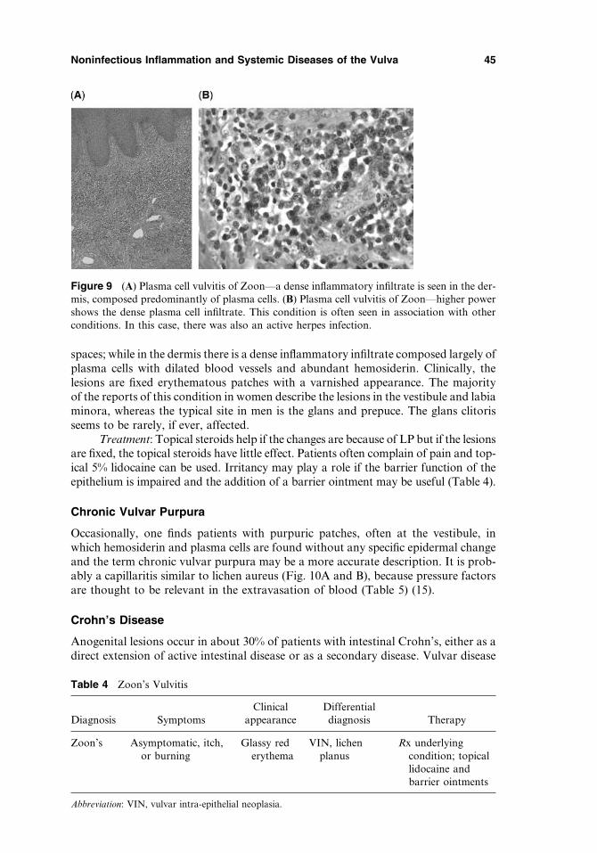
spaces; while in the dermis there is a dense inflammatory infiltrate composed largely ofplasma cells with dilated blood vessels and abundant hemosiderin. Clinically, thelesions are fixed erythematous patches with a varnished appearance. The majorityof the reports of this condition in women describe the lesions in the vestibule and labiaminora, whereas the typical site in men is the glans and prepuce. The glans clitorisseems to be rarely, if ever, affected.
Treatment: Topical steroids help if the changes are because of LP but if the lesionsare fixed, the topical steroids have little effect. Patients often complain of pain and top-ical 5% lidocaine can be used. Irritancy may play a role if the barrier function of theepithelium is impaired and the addition of a barrier ointment may be useful (Table 4).
Chronic Vulvar Purpura
Occasionally, one finds patients with purpuric patches, often at the vestibule, inwhich hemosiderin and plasma cells are found without any specific epidermal changeand the term chronic vulvar purpura may be a more accurate description. It is prob-ably a capillaritis similar to lichen aureus (Fig. 10A and B), because pressure factorsare thought to be relevant in the extravasation of blood (Table 5) (15).
Crohn’s Disease
Anogenital lesions occur in about 30% of patients with intestinal Crohn’s, either as adirect extension of active intestinal disease or as a secondary disease. Vulvar disease
Figure 9 (A) Plasma cell vulvitis of Zoon—a dense inflammatory infiltrate is seen in the der-mis, composed predominantly of plasma cells. (B) Plasma cell vulvitis of Zoon—higher powershows the dense plasma cell infiltrate. This condition is often seen in association with otherconditions. In this case, there was also an active herpes infection.
Table 4 Zoon’s Vulvitis
Diagnosis SymptomsClinical
appearanceDifferentialdiagnosis Therapy
Zoon’s Asymptomatic, itch,or burning
Glassy rederythema
VIN, lichenplanus
Rx underlyingcondition; topicallidocaine andbarrier ointments
Abbreviation: VIN, vulvar intra-epithelial neoplasia.
Noninfectious Inflammation and Systemic Diseases of the Vulva 45
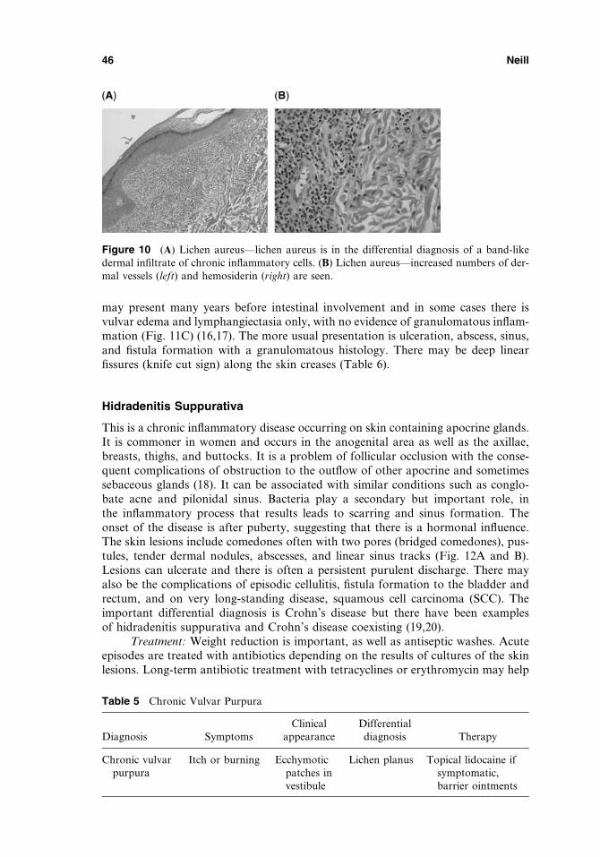
may present many years before intestinal involvement and in some cases there isvulvar edema and lymphangiectasia only, with no evidence of granulomatous inflam-mation (Fig. 11C) (16,17). The more usual presentation is ulceration, abscess, sinus,and fistula formation with a granulomatous histology. There may be deep linearfissures (knife cut sign) along the skin creases (Table 6).
Hidradenitis Suppurativa
This is a chronic inflammatory disease occurring on skin containing apocrine glands.It is commoner in women and occurs in the anogenital area as well as the axillae,breasts, thighs, and buttocks. It is a problem of follicular occlusion with the conse-quent complications of obstruction to the outflow of other apocrine and sometimessebaceous glands (18). It can be associated with similar conditions such as conglo-bate acne and pilonidal sinus. Bacteria play a secondary but important role, inthe inflammatory process that results leads to scarring and sinus formation. Theonset of the disease is after puberty, suggesting that there is a hormonal influence.The skin lesions include comedones often with two pores (bridged comedones), pus-tules, tender dermal nodules, abscesses, and linear sinus tracks (Fig. 12A and B).Lesions can ulcerate and there is often a persistent purulent discharge. There mayalso be the complications of episodic cellulitis, fistula formation to the bladder andrectum, and on very long-standing disease, squamous cell carcinoma (SCC). Theimportant differential diagnosis is Crohn’s disease but there have been examplesof hidradenitis suppurativa and Crohn’s disease coexisting (19,20).
Treatment: Weight reduction is important, as well as antiseptic washes. Acuteepisodes are treated with antibiotics depending on the results of cultures of the skinlesions. Long-term antibiotic treatment with tetracyclines or erythromycin may help
Figure 10 (A) Lichen aureus—lichen aureus is in the differential diagnosis of a band-likedermal infiltrate of chronic inflammatory cells. (B) Lichen aureus—increased numbers of der-mal vessels (left) and hemosiderin (right) are seen.
Table 5 Chronic Vulvar Purpura
Diagnosis SymptomsClinical
appearanceDifferentialdiagnosis Therapy
Chronic vulvarpurpura
Itch or burning Ecchymoticpatches investibule
Lichen planus Topical lidocaine ifsymptomatic,barrier ointments
46 Neill
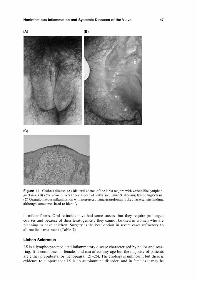
in milder forms. Oral retinoids have had some success but they require prolongedcourses and because of their teratogenicity they cannot be used in women who areplanning to have children. Surgery is the best option in severe cases refractory toall medical treatment (Table 7).
Lichen Sclerosus
LS is a lymphocyte-mediated inflammatory disease characterized by pallor and scar-ring. It is commoner in females and can affect any age but the majority of patientsare either prepubertal or menopausal (21–26). The etiology is unknown, but there isevidence to support that LS is an autoimmune disorder, and in females it may be
Figure 11 Crohn’s disease. (A) Bilateral edema of the labia majora with vesicle-like lymphan-giectasia. (B) (See color insert) Inner aspect of vulva in Figure 9 showing lymphangiectasia.(C) Granulomatous inflammation with non-necrotizing granulomas is the characteristic finding,although sometimes hard to identify.
Noninfectious Inflammation and Systemic Diseases of the Vulva 47
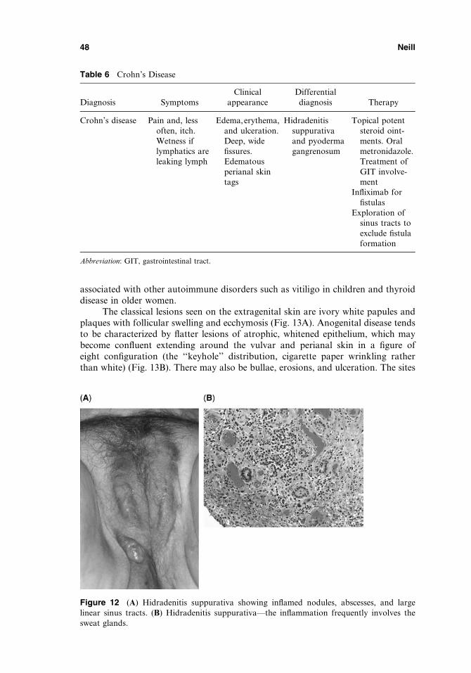
associated with other autoimmune disorders such as vitiligo in children and thyroiddisease in older women.
The classical lesions seen on the extragenital skin are ivory white papules andplaques with follicular swelling and ecchymosis (Fig. 13A). Anogenital disease tendsto be characterized by flatter lesions of atrophic, whitened epithelium, which maybecome confluent extending around the vulvar and perianal skin in a figure ofeight configuration (the ‘‘keyhole’’ distribution, cigarette paper wrinkling ratherthan white) (Fig. 13B). There may also be bullae, erosions, and ulceration. The sites
Table 6 Crohn’s Disease
Diagnosis SymptomsClinical
appearanceDifferentialdiagnosis Therapy
Crohn’s disease Pain and, lessoften, itch.Wetness iflymphatics areleaking lymph
Edema,erythema,and ulceration.Deep, widefissures.Edematousperianal skintags
Hidradenitissuppurativaand pyodermagangrenosum
Topical potentsteroid oint-ments. Oralmetronidazole.Treatment ofGIT involve-ment
Infliximab forfistulas
Exploration ofsinus tracts toexclude fistulaformation
Abbreviation: GIT, gastrointestinal tract.
Figure 12 (A) Hidradenitis suppurativa showing inflamed nodules, abscesses, and largelinear sinus tracts. (B) Hidradenitis suppurativa—the inflammation frequently involves thesweat glands.
48 Neill
most commonly affected are the inner aspects of the labia majora, labia minora,clitoris, and clitoral hood. Less commonly lesions occur in the genitocrural folds.Vestibular involvement is rare and vaginal lesions do not occur, as LS seems to sparenoncornified stratified squamous epithelia, i.e., mucosal epithelium. Perianal lesionsoccur in approximately 30% of female patients in contrast to men who do notseem to develop perianal involvement. Extragenital lesions occur in some womenwith vulvar disease. The extragenital areas may be truncal, at sites of pressure, upperback, wrists, buttocks, and thighs. The Koebner phenomenon has also been reportedat sites of radiotherapy, scar tissue, and vaccination sites. Facial, scalp, and nailinvolvement (27) have all been reported. Lesions of LS in the oral cavity areextremely rare and many of the reports of oral involvement in the literaturehave often not been confirmed histologically and many have probably beenexamples of LP (28). It is not uncommon for patients with vulvar LS to have coexist-ent oral LP.
LS is a scarring dermatosis and the changes that can occur on the vulva includeloss of the labia minora, and sealing over of the clitoral hood, burying the clitoris.Introital narrowing because of anterior and posterior labial fusion sometimes resultsin a tiny opening into the vestibule. Milia may occur.
The classical histology is a thinned epidermis with flattening of the rete pegs.The underlying dermis is hyalinized (homogenized appearance). Below the hyali-nized area is a lymphocytic mixed infiltrate (Fig. 13C). In some cases the epidermisis not thin but thickened, i.e., with squamous cell hyperplasia.
The differential diagnosis includes vitiligo, mucous membrane pemphigoid,LP, and morphea as they may present with a similar clinical appearance. Therecan be clinical and histological overlap between morphea, LP, and LS.
The incidence of SCC developing on LS in clinical practice is of the order of4% or less.
The histological patterns associated with SCC arising on LS include epithelialhyperplasia and both differentiated and undifferentiated intraepithelial neoplasia.
Table 7 Hidradenitis Suppurativa
Diagnosis SymptomsClinical
appearanceDifferentialdiagnosis Therapy
Hidradenitissuppurativa
Painfuldischarge
Pustules, nodules,and abscesses.Linear sinustracts
Crohn’sdisease
Long-term oraltetracyclines
Antiseptic washesand weightreduction; oralretinoids
Surgical excisionto lay opensinus tracts,and excisionand graftingfor extensivedisease poorlycontrolled withmedicaltreatment
Noninfectious Inflammation and Systemic Diseases of the Vulva 49

Verrucous carcinoma has also been described in association with LS. More rarely,vulvar basal cell carcinoma (29) and melanoma (30) have been reported occurringin association with LS. The validity of the case report of an association with malig-nant melanoma and LS was questioned but the original authors upheld their view.
Treatment: The current recommended treatment for uncomplicated LS is apotent topical corticosteroid, e.g., clobetasol propionate (31). There are no randomizedcontrolled trials providing evidence for any particular corticosteroid or treatmentregimen being more effective than any other. The regimen recommended by theauthor for a newly diagnosed case is initially clobetasol propionate once a night
Figure 13 (A) Lichen sclerosus showing white wrinkled skin on the inner aspects with sheetsof ecchymosis on the inner labia minora. (B) (See color insert) Lichen sclerosus involving thevulva and perianal skin with extension into the genitocrural folds, resulting in loss of the labiaminora and sealing over of the clitoral hood. (C) (See color insert) Lichen sclerosus—there isthinning of the epidermis with loss of rete pegs and dermal homogenization. Dermal inflam-mation beneath the area of dermal homogenization is variable and is minimal here.
50 Neill

for four weeks then alternate nights for four weeks and for the final month twice aweek. A 30 g tube of clobetasol propionate should last 12 weeks and the patient isthen reviewed. The clobetasol propionate is then continued and used as and whenrequired. Most patients seem to require 30 to 60 g annually. Some patients will gointo complete remission requiring no further treatment. Others will continue tohave flares and remissions and they are advised to use clobetasol propionate asrequired. A soap substitute is also recommended, and an information sheet on LSand the instructions for the use of the topical steroid. In the past topical testosteronewas used with an improvement in the clinical appearance and diminution of symp-toms. Clobetasol dipropionate is a more effective treatment and has now replacedtestosterone in the treatment of LS. Surgical excision is not necessary in the manage-ment of most LS and it should be used exclusively for the management of functionalproblems because of postinflammatory scarring (32,33), premalignant lesions, andmalignancy (Table 8).
Aphthae
Benign aphthae of the vulva may or may not occur with concomitant oral lesions.The age of onset is in childhood, by about the age of six, and there may be a familyhistory. The lesions are usually multiple and small. Less commonly, the lesions aresolitary or few but larger, referred to as giant aphthae. Herpetic infection has tobe excluded and there is often a premenstrual exacerbation once the menarche isreached. The etiology remains unknown. The lesions are superficial and painful witha yellow base surrounded by a red areola (Fig. 14). They are distributed most fre-quently on the labia minora and heal quickly.
Many eponymous names have been assigned to acute vulvar ulcers, many ofwhich are probably examples of benign aphthae, e.g., Sutton’s ulcer.
Lipschutz’s ulcer, which occurs in young women, is a separate entity. This isusually one large deep painful ulcer that develops very rapidly and is covered withan adherent slough. The ulcer is sited most frequently in the inner aspect of oneof the labia minora. Occasionally, two ulcers develop at apposing sites bilaterally(Fig. 15). The ulcer heals spontaneously over several weeks leaving some scarring.The Lipschutz-type ulcer is sometimes associated with a systemic infection such asinfectious mononucleosis (34–36), typhoid, or paratyphoid fever. Healing is spon-taneous but may take several weeks and leaves some scarring. Differential diagnosesinclude Herpes simplex infection with type 1 or 2, erythema multiforme, and Langer-hans cell histiocytosis.
Table 8 Lichen Sclerosus
Diagnosis SymptomsClinical
appearanceDifferentialdiagnosis Therapy
Lichen sclerosus Severe itch and,if ulcerated orfissured, pain;dyspareunia
White wrinkledepitheliumwith ecchy-moses fissuresin theinterlabialsulci; scarring
Lichen planus,morphea, andmucousmembranepemphigoid
Potent topicalsteroid
Noninfectious Inflammation and Systemic Diseases of the Vulva 51
Treatment: This includes symptomatic treatment with analgesics either oral ortopical. Astringent soaks with either potassium permanganate solution 1:10,000or Monsel’s solution followed by a topical corticosteroid and/or topical tetracyclinehave been used but may exacerbate symptoms.
Figure 14 (See color insert) Benign aphthae with erythematous halo and slough-covered cen-tral ulceration.
Figure 15 (See color insert) Lipschutz’ ulcer. There are two large and deep ulcers apposed oneither side.
52 Neill
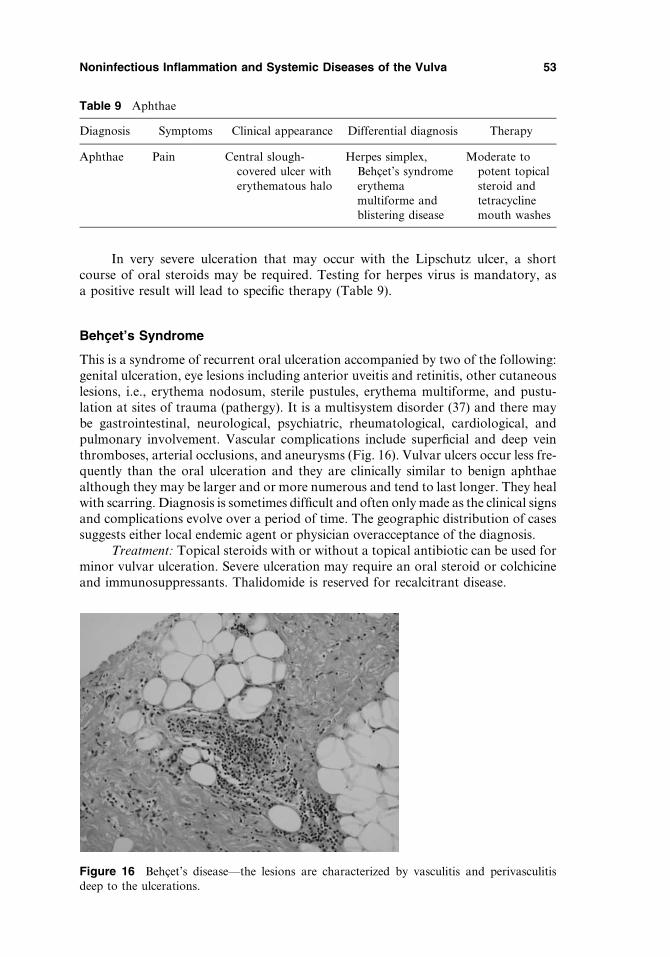
In very severe ulceration that may occur with the Lipschutz ulcer, a shortcourse of oral steroids may be required. Testing for herpes virus is mandatory, asa positive result will lead to specific therapy (Table 9).
Behcet’s Syndrome
This is a syndrome of recurrent oral ulceration accompanied by two of the following:genital ulceration, eye lesions including anterior uveitis and retinitis, other cutaneouslesions, i.e., erythema nodosum, sterile pustules, erythema multiforme, and pustu-lation at sites of trauma (pathergy). It is a multisystem disorder (37) and there maybe gastrointestinal, neurological, psychiatric, rheumatological, cardiological, andpulmonary involvement. Vascular complications include superficial and deep veinthromboses, arterial occlusions, and aneurysms (Fig. 16). Vulvar ulcers occur less fre-quently than the oral ulceration and they are clinically similar to benign aphthaealthough they may be larger and or more numerous and tend to last longer. They healwith scarring. Diagnosis is sometimes difficult and often only made as the clinical signsand complications evolve over a period of time. The geographic distribution of casessuggests either local endemic agent or physician overacceptance of the diagnosis.
Treatment: Topical steroids with or without a topical antibiotic can be used forminor vulvar ulceration. Severe ulceration may require an oral steroid or colchicineand immunosuppressants. Thalidomide is reserved for recalcitrant disease.
Table 9 Aphthae
Diagnosis Symptoms Clinical appearance Differential diagnosis Therapy
Aphthae Pain Central slough-covered ulcer witherythematous halo
Herpes simplex,Behcet’s syndromeerythemamultiforme andblistering disease
Moderate topotent topicalsteroid andtetracyclinemouth washes
Figure 16 Behcet’s disease—the lesions are characterized by vasculitis and perivasculitisdeep to the ulcerations.
Noninfectious Inflammation and Systemic Diseases of the Vulva 53

It is important to have an eye assessment, because ocular complications arecommon. Anticoagulants are indicated for thrombotic disease. It is important tohave a multidisciplinary team involved in the management of patients with Behcet’ssyndrome (Table 10).
EROSIVE, ULCERATIVE, AND BULLOUS DERMATOSES
Chronic Ulcerative and Bullous Diseases
Any chronic solitary ulcer of the genital mucosa must be considered malignant orinfective until proven otherwise. Generalized ulceration of the vulva can be becauseof an underlying bullous condition but because of the friction and forces at the site,blisters do not often remain intact and erosions and ulcers are seen instead. The differ-ential diagnosis of chronic ulceration can come under any of the headings listed below.
Table 10 Behcet’s Syndrome
Diagnosis SymptomsClinical
appearanceDifferentialdiagnosis Therapy
Behcet’s Pain Ulceration andpustulation
Aphthae ThalidomidePotent topical steroids
with or without a topicalantibiotic, oral steroids,and colchicine
Figure 17 (See color insert) Hailey–Hailey disease. There is erythema with maceration in thegenitocrural fold and tiny erosions.
54 Neill
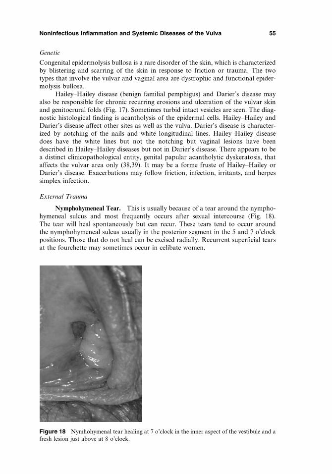
Genetic
Congenital epidermolysis bullosa is a rare disorder of the skin, which is characterizedby blistering and scarring of the skin in response to friction or trauma. The twotypes that involve the vulvar and vaginal area are dystrophic and functional epider-molysis bullosa.
Hailey–Hailey disease (benign familial pemphigus) and Darier’s disease mayalso be responsible for chronic recurring erosions and ulceration of the vulvar skinand genitocrural folds (Fig. 17). Sometimes turbid intact vesicles are seen. The diag-nostic histological finding is acantholysis of the epidermal cells. Hailey–Hailey andDarier’s disease affect other sites as well as the vulva. Darier’s disease is character-ized by notching of the nails and white longitudinal lines. Hailey–Hailey diseasedoes have the white lines but not the notching but vaginal lesions have beendescribed in Hailey–Hailey diseases but not in Darier’s disease. There appears to bea distinct clinicopathological entity, genital papular acantholytic dyskeratosis, thataffects the vulvar area only (38,39). It may be a forme fruste of Hailey–Hailey orDarier’s disease. Exacerbations may follow friction, infection, irritants, and herpessimplex infection.
External Trauma
Nymphohymeneal Tear. This is usually because of a tear around the nympho-hymeneal sulcus and most frequently occurs after sexual intercourse (Fig. 18).The tear will heal spontaneously but can recur. These tears tend to occur aroundthe nymphohymeneal sulcus usually in the posterior segment in the 5 and 7 o’clockpositions. Those that do not heal can be excised radially. Recurrent superficial tearsat the fourchette may sometimes occur in celibate women.
Figure 18 Nymhohymenal tear healing at 7 o’clock in the inner aspect of the vestibule and afresh lesion just above at 8 o’clock.
Noninfectious Inflammation and Systemic Diseases of the Vulva 55

Figure 19 (See color insert) Erythema multiforme—characteristic targetoid lesions. Source:Image courtesy of W. Clark Lambert, MD, PhD., UMDNJ–New Jersey Medical School,Newark, New Jersey, U.S.A.
Figure 20 Stevens–Johnson syndrome. This superficial biopsy of a blister shows the charac-teristic epidermal necrosis.
56 Neill
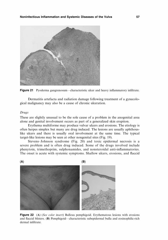
Dermatitis artefacta and radiation damage following treatment of a gynecolo-gical malignancy may also be a cause of chronic ulceration.
Drugs
These are slightly unusual to be the sole cause of a problem in the anogenital areaalone and genital involvement occurs as part of a generalized skin eruption.
Erythema multiforme may produce vulvar ulcers and erosions. The etiology isoften herpes simplex but many are drug induced. The lesions are usually aphthous-like ulcers and there is usually oral involvement at the same time. The typicaltarget-like lesions may be seen at other nongenital sites (Fig. 19).
Stevens–Johnson syndrome (Fig. 20) and toxic epidermal necrosis is asevere problem and is often drug induced. Some of the drugs involved includephenytoin, trimethoprim, sulphonamides, and nonsteroidal anti-inflammatories.The onset is acute with systemic symptoms. Shallow ulcers, erosions, and flaccid
Figure 21 Pyoderma gangrenosum—characteristic ulcer and heavy inflammatory infiltrate.
Figure 22 (A) (See color insert) Bullous pemphigoid. Erythematous lesions with erosionsand flaccid blisters. (B) Pemphigoid—characteristic subepidermal bulla and eosinophilic-richdermal infiltrate.
Noninfectious Inflammation and Systemic Diseases of the Vulva 57
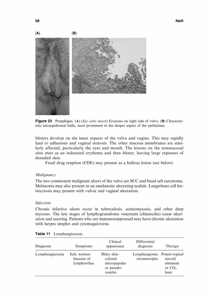
blisters develop on the inner aspects of the vulva and vagina. This may rapidlylead to adhesions and vaginal stenosis. The other mucous membranes are simi-larly affected, particularly the eyes and mouth. The lesions on the nonmucosalsites start as an indurated erythema and then blister, leaving large expanses ofdenuded skin.
Fixed drug eruption (FDE) may present as a bullous lesion (see below).
Malignancy
The two commonest malignant ulcers of the vulva are SCC and basal cell carcinoma.Melanoma may also present as an amelanotic ulcerating nodule. Langerhans cell his-tiocytosis may present with vulvar and vaginal ulceration.
Infection
Chronic infective ulcers occur in tuberculosis, actinomycosis, and other deepmycoses. The late stages of lymphogranuloma venereum (chlamydia) cause ulcer-ation and scarring. Patients who are immunosuppressed may have chronic ulcerationwith herpes simplex and cytomegalovirus.
Figure 23 Pemphigus. (A) (See color insert) Erosions on right side of vulva. (B) Character-istic intraepidermal bulla, most prominent in the deeper aspect of the epithelium.
Table 11 Lymphangiectasia
Diagnosis SymptomsClinical
appearanceDifferentialdiagnosis Therapy
Lymphangiectasia Itch, wetnessbecause oflymphorrhea
Shiny skin-coloredmicropapulesor pseudo-vesicles
Lymphangiomacircumscripta
Potent topicalsteroidointmentor CO2
laser
58 Neill

Inflammatory
LS, LP, and lupus erythematosus can all cause chronic ulceration. Vulvar and vaginallesions are very rare in systemic lupus erythematosus. Hidradenitis suppurativa andCrohn’s disease can sometimes be difficult to distinguish and they can occur together.
Pyoderma gangrenosum (Fig. 21) may occur in association with inflammatorybowel disease, rheumatoid arthritis, and hematological malignancies. Vulvar pyodermagangrenosum can often accompany Crohn’s disease and it can be difficult differentiat-ing this from a fistulous tract from the bowel. The lesion may be solitary and large oroccasionally multiple and small. The initial lesion may be a hemorrhagic vesicle or bulla.
Autoimmune Bullous Diseases
The mucocutaneous autoimmune blistering disorders can affect the vulva andvagina. The integrity of the cellular adhesion of the epidermal cells and/or the base-ment membrane zone is damaged by the antibody attack and the various blisteringdiseases that occur are classified according to the site of the target antigen. Several ofthe autoimmune bullous diseases’ effect can affect the vulva. Pemphigoid (Fig. 22Aand B), linear IgA disease, and pemphigus (Fig. 23A and B) may all present withvulvar and vaginal ulceration.
Treatment: The underlying cause, be it drug, dermatosis, or infection needs tobe identified and managed. Microbiological studies to exclude bacterial or viralinfection must be carried out and a detailed drug history taken, not forgettingover-the-counter medicines. General measures include potassium permanganate1:10,000 soaks or Burow’s solution, topical antibacterial such as silver sulfadiazine,and oral or topical analgesics.
Figure 24 Lichen simplex (kichen simplex chronicus, squamous cell hyperplasia)—there ishyperkeratosis and acanthosis with fusing of rete ridges.
Table 12 Lichen Simplex
Diagnosis Symptoms Clinical appearanceDifferentialdiagnosis Therapy
Lichensimplex
Severe itch Pale lichenified skin;no scarring; usuallyunilateral
Eczema Potent topicalsteroid
Noninfectious Inflammation and Systemic Diseases of the Vulva 59

MISCELLANEOUS
Lymphangiectasia
Vulvar lymphangiectasia is a result of dilatation of superficial lymphatics secondaryto obstruction of previously normal deep lymphatics (40). It is most frequently seen
Figure 25 (A and B) Indurated erythematous and discoid lesions of a fixed drug eruptionbecause of fluconazole. (C) (See color insert) Fixed drug eruption—vacuolization of the basallayer and pigmentary incontinence are seen.
Figure 26 (See color insert) Postinflammatory hyperpigmentation—pigment incontinence is seen.
60 Neill
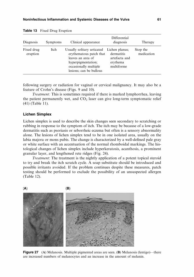
following surgery or radiation for vaginal or cervical malignancy. It may also be afeature of Crohn’s disease (Figs. 9 and 10).
Treatment: This is sometimes required if there is marked lymphorrhea, leavingthe patient permanently wet, and CO2 laser can give long-term symptomatic relief(41) (Table 11).
Lichen Simplex
Lichen simplex is used to describe the skin changes seen secondary to scratching orrubbing in response to the symptom of itch. The itch may be because of a low-gradedermatitis such as psoriasis or seborrheic eczema but often is a sensory abnormalityalone. The lesions of lichen simplex tend to be in one isolated area, usually on thelabia majora or mons pubis. The change is characterized by a well-defined pale grayor white surface with an accentuation of the normal rhomboidal markings. The his-tological changes of lichen simplex include hyperkeratosis, acanthosis, a prominentgranular layer, and lengthened rete ridges (Fig. 24).
Treatment: The treatment is the nightly application of a potent topical steroidto try and break the itch scratch cycle. A soap substitute should be introduced andpossible irritants avoided. If the problem continues despite these measures, patchtesting should be performed to exclude the possibility of an unsuspected allergen(Table 12).
Table 13 Fixed Drug Eruption
Diagnosis Symptoms Clinical appearanceDifferentialdiagnosis Therapy
Fixed drugeruption
Itch Usually solitary urticatederythematous patch thatleaves an area ofhyperpigmentation;occasionally multiplelesions; can be bullous
Lichen planus;dermatitisartefacta anderythemamultiforme
Stop themedication
Figure 27 (A) Melanosis. Multiple pigmented areas are seen. (B) Melanosis (lentigo)—thereare increased numbers of melanocytes and an increase in the amount of melanin.
Noninfectious Inflammation and Systemic Diseases of the Vulva 61
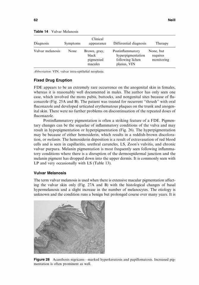
Fixed Drug Eruption
FDE appears to be an extremely rare occurrence on the anogenital skin in females,whereas it is reasonably well documented in males. The author has only seen onecase, which involved the mons pubis, buttocks, and nongenital sites because of flu-conazole (Fig. 25A and B). The patient was treated for recurrent ‘‘thrush’’ with oralfluconazole and developed urticated erythematous plaques on the trunk and anogen-ital skin. There were no further problems on discontinuation of the repeated doses offluconazole.
Postinflammatory pigmentation is often a striking feature of a FDE. Pigmen-tary changes can be the sequelae of inflammatory conditions of the vulva and mayresult in hypopigmentation or hyperpigmentation (Fig. 26). The hyperpigmentationmay be because of either hemosiderin, which results in a reddish-brown discolora-tion, or melanin. The hemosiderin deposition is a result of extravasation of red bloodcells and is seen in capillaritis, urethral caruncles, LS, Zoon’s vulvitis, and chronicvulvar purpura. Melanin pigmentation is most frequently seen following inflamma-tory conditions where there is a disruption of the dermoepidermal junction and themelanin pigment has dropped down into the upper dermis. It is commonly seen withLP and very occasionally with LS (Table 13).
Vulvar Melanosis
The term vulvar melanosis is used when there is extensive macular pigmentation affect-ing the vulvar skin only (Fig. 27A and B) with the histological changes of basalhypermelanosis and a slight increase in the number of melanocytes. The etiology isunknown and the condition runs a benign but prolonged course over many years. It is
Table 14 Vulvar Melanosis
Diagnosis SymptomsClinical
appearance Differential diagnosis Therapy
Vulvar melanosis None Brown, gray,blackpigmentedmacules
Postinflammatoryhyperpigmentationfollowing lichenplanus, VIN
None, butrequiresmonitoring
Abbreviation: VIN, vulvar intra-epithelial neoplasia.
Figure 28 Acanthosis nigricans—marked hyperkeratosis and papillomatosis. Increased pig-mentation is often prominent as well.
62 Neill
essential to have a confirmatory biopsy, because the clinical findings are frequently indis-tinguishable from atypical lentiginous dysplasia or even malignant melanoma (42,43).
Treatment: There is no treatment and the best management is baseline photo-graphic documentation for self-monitoring and physician follow-up. Biopsies shouldbe performed for any unusual changes (Table 14).
Acanthosis Nigricans
This condition is usually related to insulin resistance and rarely with malignancy.Initially, the appearance is of a dark and velvety thickening of the skin in the geni-tocrural folds and upper inner thigh, which later becomes warty. Skin tags may alsooccur (Fig. 28).
Necrolytic Migratory Erythema
This rare dermatosis occurs most frequently on the genital skin and lower abdomen.The distinctive eruption consists of an erythema with erosions, vesiculation, andbulla formation. The rash extends outward with healing and crusting at the outeredge. Postinflammatory pigmentation is common. There is also an associated weightloss, glossitis, and angular cheilitis. The patient may also have diabetes. Histologi-cally, there is marked epidermal necrolysis and a mild dermal lymphocytic infiltrate.It is nearly always associated with an underlying pancreatic glucagonoma. The der-matosis resolves with treatment of the glucagonoma and correction of nutritionaldeficiencies secondary to malabsorption (Table 15).
REFERENCES
1. Edwards L, Hansen RC. Reiter’s syndrome of the vulva. Arch Dermatol 1992; 128:811–814.2. Kanerva L, Kouse M, Niema KM, et al. Ultrahistopathology of balanitis circinate. Br J
Vener Dis 1982; 58:185–195.3. Lewis FM, Shah M, Harrington CI. Vulval involvement in lichen planus: a study of 37
women. Br J Dermatol 1996; 135:89–91.4. Pelisse M, Leibowitch M, Sedel D, et al. Un nouveau syndrome vulvo-vagino-gingival:
lichen plan erosif plurimuqueux. Ann Dermatol Venereol 1982; 109:797–798.5. Eisen D. The vulvo-vaginal gingival syndrome: the clinical characteristics of 22 patients.
Arch Dermatol 1994; 130:1379–1382.
Table 15 Necrolytic Migratory Erythema
Diagnosis SymptomsClinical
appearanceDifferentialdiagnosis Therapy
Necrolyticmigratoryerythema
Weight loss, itch,and painfulrash on lowerabdomen andgenital area;sore mouth
Erythematouspatches thatexpand outwith an erodedand scalyborder.Glossitis,angularcheilitis
Tinea infection,impetiginizedpsoriasis oreczema
Treating anyunderlyingglucagonoma.Correction ofnutritionaldeficiencies
Noninfectious Inflammation and Systemic Diseases of the Vulva 63

6. Moyal-Barracco M, Lautier-Frau M, Becheral PA, et al. Lichen plan conjunctival: uneobservation. Ann Dermatol Venereol 1993; 120:857–859.
7. McNab AA. Lacrimal gland canalicular obstruction in lichen planus. Orbit 1998; 17:201–202.8. Menges M, Hohloch K, Pueschel W, Stallmach A. Lichen planus with oesophageal
involvement. A case report and review of the literature. Digestion 2002; 65:184–189.9. Martin L, Moriniere S, Machet M-C. Bilateral conductive deafness related to erosive
lichen planus. J Laryngol Otol 1998; 112:365–366.10. Grunald MH, Zvulunov A, Halevy S. Lichen planopilaris of the vulva. Br J Dermatol
1998; 136:465–479.11. Anderson M, Kutzner S, Kaufman R. Treatment of vulvovaginal lichen planus with vagi-
nal hydrocortisone suppositories. Obstet Gynecol 2002; 100:359–362.12. Lotery H, Galask RP. Erosive lichen planus of the vulva and vagina. Obstet Gynecol
2003; 102:1121–1125.13. Nylander Lunqvist E, Brit Wahlin Y, Hofer Per-Ake. Methotrexate supplemented with
steroid ointments for the treatment of severe erosive lichen rubber. Acta Derm Venereol2002; 82:63–64.
14. Scurry J, Dennerstein G, Brennan J, et al. Vulvitis circumscripta plasmacellularis. A clin-iopathologic entity? J Reprod Med 1993; 38:14–18.
15. Kato T, Kuramoto Y, Tadaki T, et al. Chronic vulvar purpura. Dermatologica 1990;180:174–176.
16. Urbanek M, McKee PH, Neill SM. Vulvar Crohn’s: difficulties in diagnosis. Clin ExpDermatol 1996; 21:211–214.
17. Martin J, Holdstock G. Isolated vulvar oedema as a feature of Crohns disease. J ObstetGynecol 1997; 17:92–93.
18. Boer J, Weltevreden EF. Hidradenitis suppurativa or acne inverse. A clinicopathologicalstudy of early lesions. Br J Dermatol 1996; 135:721–725.
19. Ostlere LS, Langtry JAA, Mortimer PS, Staughton RCD. Hidradenitis suppurativa inCrohn’s disease. Br J Dermatol 1991; 125:384–386.
20. Burrows NP, Russell-Jones R. Crohn’s disease in association with hidradenitis suppura-tiva. Br J Dermatol 1992; 126:523–529.
21. Wallace HJ. Lichen sclerosus et atrophicus. Trans St Johns Hosp Dermatol Soc 1971;57:9–30.
22. Tremaine RDL, Miller RAW. Lichen scerosus et atrophicus. Int J Dermatol 1989; 28:10–16.23. Meffert JJ, Davis BM, Grimwood RE. Lichen sclerosus. J Am Acad 1995; 32:393–416.24. Ridley CM, Neill SM. Chapter non-infective cutaneous conditions of the vulva. In: Ridley
CM, Neill SM, eds. The Vulva. London: Blackwell Science Ltd, 1999:154–156.25. Powell JJ, Wojnarowska F. Lichen sclerosus. Lancet 1999; 353:1777–1783.26. Ridley CM. Genital lichen sclerosus (lichen sclerosus et atrophicus) in childhood and ado-
lescence. J R Soc Med 1993; 86:69–75.27. Kossard S, Cornish N. Localized lichen sclerosus with nail loss. Aust J Dermatol 1998;
39:119–120.28. Brown AR, Dunlap CL, Bussard DA, et al. Lichen sclerosus of the oral cavity: a report of
two cases. Oral Surg Oral Med Oral Pathol Oral Radiol Endod 1997; 84:165–170.29. Meyrick-Thomas RH, McGibbon DH, Munro DD. Basal cell carcinoma of the vulva in
association with vulvar lichen sclerosus et atrophicus. J R Soc Med 1985; 78:16–18.30. Friedman RJ, Kopf AW, Jones WB. Malignant melanoma in association with lichen scler-
osus on the vulva of a 14 year old. Am J Dermatopathol 1984; 6(suppl 1):253–256.31. Neill SM, Tatnall FM, Cox NH. Guidelines on the management of lichen sclerosus. Br J
Dermatol 2002; 147:640–649.32. Paniel B. Surgical procedures in benign vulvar disease. In: Ridley CM, Neill SM, eds. The
Vulva. Blackwell Science Oxford, 1999:288–290.33. Rouzier R, Haddad B, Deyrolle C, et al. Perineoplasty for the treatment of introital sten-
osis related to vulvar lichen sclerosus. Am J Obstet Gynecol 2002; 186:49–52.
64 Neill
34. Brown ZA, Stenchever MA. Genital ulceration and infectious mononucleosis. Am JObstet Gynecol 1977; 127:673–674.
35. Lampert A, Assier-Bonnet H, Chevallier B, Clerici T, Saiag P. Lipschutz’s genital ulcer-ation: a manifestation of Epstein-Barr virus primary infection. Br J Dermatol 1996;135:663–665.
36. Portnoy J, Ahronheim GA, Ghibu F, et al. Recovery of Epstein-Barr virus from genitalulcers. N Engl J Med 1984; 311:966–968.
37. Suzuki Kurokawa M, Suzuki N. Behcet’s disease. Clin Exp Med 2004; 4:10–20.38. Chorzelski TP, Kudejko J, Jablonska S. Is papular acantholytic dyskeratosis of the vulva
a new entity?. Am J Dermatopathol 1984; 6:557–560.39. Bell HK, Farrar CW, Curley RK. Papular acantholytic dyskeratosis of the vulva. Clin
Exp Dermatol 2001; 2:386–388.40. Handfield-Jones SE, Prendeville WL, Norman S. Vulvar lymphangiectasia. Genitourin
Med 1989; 65:335–337.41. Huigol SC, Neill SM, Barlow RJ. CO2 laser therapy of vulvar lymphangiectasia and lym-
phangioma circumscriptum. Dermatol Surg 2002; 28:575–577.42. Sisson-Torre EQ, Ackerman AB. Melanosis of the vulva: a clinical simulator of malignant
melanoma. Am J Dermatopathol 1985; 7:51–60.43. Rudolph RI. Vulvar melanosis. J Am Acad Dermatol 1990; 23:982–984.
Noninfectious Inflammation and Systemic Diseases of the Vulva 65

4Vulvovaginal Infections
Rachel SpieldochScottsdale, Arizona, U.S.A.
Libby EdwardsSoutheast Vulvar Clinic, Charlotte, North Carolina, U.S.A.
VIRAL INFECTIONS
Varicella Zoster Virus
Varicella zoster virus (VZV) is a member of the herpes virus group. It persists in thebody as a latent infection after the primary infection. Primary infection with VZVresults in chicken pox (varicella). Herpes zoster (shingles) represents a one-timereactivation. VZV enters through the respiratory tract and conjunctiva.
The initial infection results from transmission through the respiratory tract frominoculation through respiratory tract secretions, respiratory contact with airbornedroplets, or by direct contact or inhalation of aerosols from vesicular fluid of skinlesions. By the time the rash appears, the virus has cleared from the respiratory tractand transmission to others is unlikely. The rash of varicella is generalized and pruritic,with individual lesions advancing from macules to papules to vesicular lesions beforecrusting (Fig. 1). The rash first appears on the scalp and progresses to the trunk andextremities. The majority of lesions remain on the trunk in a centripetal distribution.Lesions can also be seen on the mucous membranes of the oropharynx, respiratorytract, vagina, conjunctiva, and cornea. Although mucous membrane lesions alsodevelop as vesicles, the fragile mucous membrane blister roof, lacking a stratumcorneum, is shed almost as soon as it forms, leaving round erosions. Vaginalinvolvement is manifested as a vaginal discharge, often purulent.
Keratinized skin lesions begin as pink papules that develop a central vesicleapproximately 2 to 4 mm in diameter. Classically, these become pustular and developa central crust, leaving an annular blister. Lesions evolve over several days and exhi-bit all stages of development simultaneously. Systemic signs of varicella are generallyself-limited and mild. These consist of malaise, itching, and fever up to 102�F for twoto three days. Adults may experience a more severe clinical course. Recovery fromprimary varicella usually results in lifetime immunity from another generalized out-break of varicella, but not necessarily from reactivation of latent infection.
67

Herpes zoster (shingles) occurs when latent VZV resurfaces. Factors associatedwith recurrent disease include aging, immunosuppression, intrauterine exposure, andvaricella infection at an early age. The eruption occurs unilaterally in the distributionof a dermatome supplied by a dorsal root or extramedullary cranial nerve sensoryganglion. Clinically, this is often seen on the trunk or the area of the fifth cranialnerve, but herpes zoster can appear on the buttock, perineum, and vulva (Fig. 2).Typically, two to four days prior to visible eruptions, patients report pain and par-esthesia in the involved area. This is followed by discrete urticarial plaques scatteredalong a dermatome. Small, clustered blisters appear on the plaques, coalescing intolarger bullae that may become hemorrhagic.
The differential diagnosis of varicella includes folliculitis, primary herpessimplex virus (HSV) infection or eczema herpeticum, and bullous impetigo. Thediagnosis of VZV infection is generally easily made on the basis of the morphologyof the skin findings. The virus can be cultured from mononuclear cells of an infectedperson from five days before to one to two days following the appearance of a rash.Specimens are collected from crusts or by unroofing a fluid-filled vesicle and rubbingthe base with a polyester swab. A number of molecular assays for varicella antibodyare available.
Complications of varicella infection include secondary bacterial infections,pneumonia, central nervous system manifestations including aseptic meningitis andencephalitis, and Reyes syndrome from VZV and aspirin exposure in young children.
Figure 1 Varicella is characterized by discrete papules, vesicles, pustules, and crusts; the geni-tal and diaper areas classically are more prominently affected than most other skin surfaces.
68 Spieldoch and Edwards
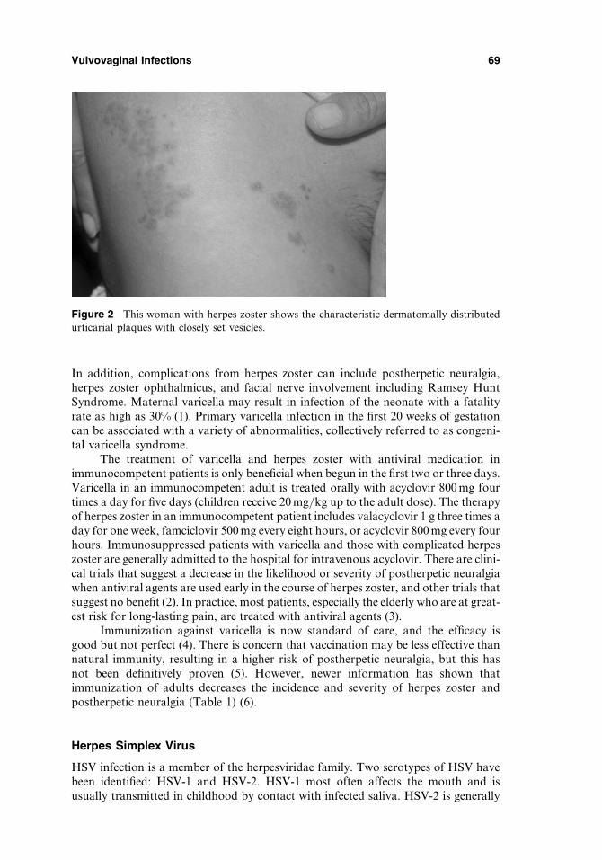
In addition, complications from herpes zoster can include postherpetic neuralgia,herpes zoster ophthalmicus, and facial nerve involvement including Ramsey HuntSyndrome. Maternal varicella may result in infection of the neonate with a fatalityrate as high as 30% (1). Primary varicella infection in the first 20 weeks of gestationcan be associated with a variety of abnormalities, collectively referred to as congeni-tal varicella syndrome.
The treatment of varicella and herpes zoster with antiviral medication inimmunocompetent patients is only beneficial when begun in the first two or three days.Varicella in an immunocompetent adult is treated orally with acyclovir 800 mg fourtimes a day for five days (children receive 20 mg/kg up to the adult dose). The therapyof herpes zoster in an immunocompetent patient includes valacyclovir 1 g three times aday for one week, famciclovir 500 mg every eight hours, or acyclovir 800 mg every fourhours. Immunosuppressed patients with varicella and those with complicated herpeszoster are generally admitted to the hospital for intravenous acyclovir. There are clini-cal trials that suggest a decrease in the likelihood or severity of postherpetic neuralgiawhen antiviral agents are used early in the course of herpes zoster, and other trials thatsuggest no benefit (2). In practice, most patients, especially the elderly who are at great-est risk for long-lasting pain, are treated with antiviral agents (3).
Immunization against varicella is now standard of care, and the efficacy isgood but not perfect (4). There is concern that vaccination may be less effective thannatural immunity, resulting in a higher risk of postherpetic neuralgia, but this hasnot been definitively proven (5). However, newer information has shown thatimmunization of adults decreases the incidence and severity of herpes zoster andpostherpetic neuralgia (Table 1) (6).
Herpes Simplex Virus
HSV infection is a member of the herpesviridae family. Two serotypes of HSV havebeen identified: HSV-1 and HSV-2. HSV-1 most often affects the mouth and isusually transmitted in childhood by contact with infected saliva. HSV-2 is generally
Figure 2 This woman with herpes zoster shows the characteristic dermatomally distributedurticarial plaques with closely set vesicles.
Vulvovaginal Infections 69
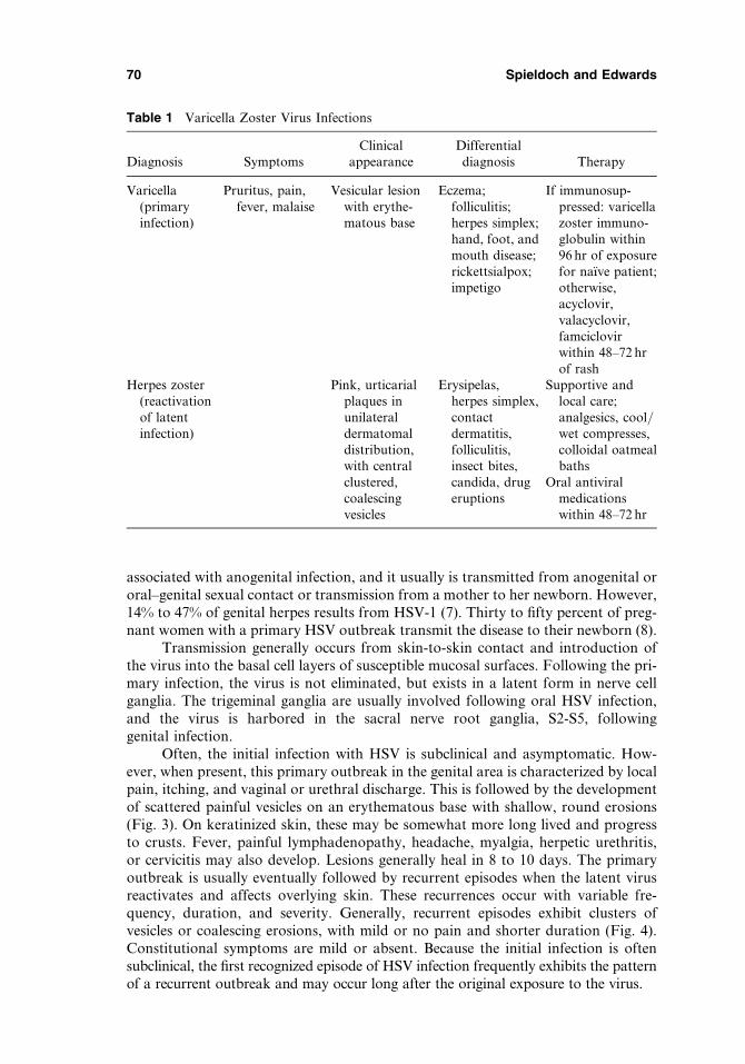
associated with anogenital infection, and it usually is transmitted from anogenital ororal–genital sexual contact or transmission from a mother to her newborn. However,14% to 47% of genital herpes results from HSV-1 (7). Thirty to fifty percent of preg-nant women with a primary HSV outbreak transmit the disease to their newborn (8).
Transmission generally occurs from skin-to-skin contact and introduction ofthe virus into the basal cell layers of susceptible mucosal surfaces. Following the pri-mary infection, the virus is not eliminated, but exists in a latent form in nerve cellganglia. The trigeminal ganglia are usually involved following oral HSV infection,and the virus is harbored in the sacral nerve root ganglia, S2-S5, followinggenital infection.
Often, the initial infection with HSV is subclinical and asymptomatic. How-ever, when present, this primary outbreak in the genital area is characterized by localpain, itching, and vaginal or urethral discharge. This is followed by the developmentof scattered painful vesicles on an erythematous base with shallow, round erosions(Fig. 3). On keratinized skin, these may be somewhat more long lived and progressto crusts. Fever, painful lymphadenopathy, headache, myalgia, herpetic urethritis,or cervicitis may also develop. Lesions generally heal in 8 to 10 days. The primaryoutbreak is usually eventually followed by recurrent episodes when the latent virusreactivates and affects overlying skin. These recurrences occur with variable fre-quency, duration, and severity. Generally, recurrent episodes exhibit clusters ofvesicles or coalescing erosions, with mild or no pain and shorter duration (Fig. 4).Constitutional symptoms are mild or absent. Because the initial infection is oftensubclinical, the first recognized episode of HSV infection frequently exhibits the patternof a recurrent outbreak and may occur long after the original exposure to the virus.
Table 1 Varicella Zoster Virus Infections
Diagnosis SymptomsClinical
appearanceDifferentialdiagnosis Therapy
Varicella(primaryinfection)
Pruritus, pain,fever, malaise
Vesicular lesionwith erythe-matous base
Eczema;folliculitis;herpes simplex;hand, foot, andmouth disease;rickettsialpox;impetigo
If immunosup-pressed: varicellazoster immuno-globulin within96 hr of exposurefor na€��ve patient;otherwise,acyclovir,valacyclovir,famciclovirwithin 48–72 hrof rash
Herpes zoster(reactivationof latentinfection)
Pink, urticarialplaques inunilateraldermatomaldistribution,with centralclustered,coalescingvesicles
Erysipelas,herpes simplex,contactdermatitis,folliculitis,insect bites,candida, drugeruptions
Supportive andlocal care;analgesics, cool/wet compresses,colloidal oatmealbaths
Oral antiviralmedicationswithin 48–72 hr
70 Spieldoch and Edwards
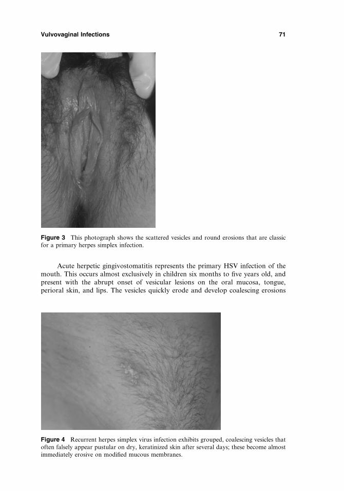
Acute herpetic gingivostomatitis represents the primary HSV infection of themouth. This occurs almost exclusively in children six months to five years old, andpresent with the abrupt onset of vesicular lesions on the oral mucosa, tongue,perioral skin, and lips. The vesicles quickly erode and develop coalescing erosions
Figure 3 This photograph shows the scattered vesicles and round erosions that are classicfor a primary herpes simplex infection.
Figure 4 Recurrent herpes simplex virus infection exhibits grouped, coalescing vesicles thatoften falsely appear pustular on dry, keratinized skin after several days; these become almostimmediately erosive on modified mucous membranes.
Vulvovaginal Infections 71
on mucous membrane and crusts on keratinized skin. The gingivae are swollen, ery-thematous, and friable. These children have high fevers 102�F to 104�F, anorexia,dysphagia, and listlessness.
Reactivation and replication of latent virus can be induced by many factorsincluding fever, trauma, stress, sunlight, and menses. Reactivation is most severein immunocompromised patients. Recurrent genital outbreaks from HSV-2 are morefrequent than from HSV-1.
Even in the absence of clinical recurrences, patients shed the virus asymptoma-tically and intermittently into the genital tract. Most HSV infections are transmittedduring asymptomatic shedding. The HSV shedding rate has been reported as 15.4%of days by culture and 40.2% of days by polymerase chain reaction (PCR), with thesubclinical shedding rate being 6.6% of days by culture and 27.1% of days by PCR (9).Shedding was decreased but not eliminated by chronic suppression with oral acyclo-vir and valacyclovir therapy (9).
The transmission of HSV from an infected mother to the neonate occurs pri-marily when the mother is experiencing the uncommon event of an active primaryHSV infection during labor. The risk in women with recurrent disease or asymptomaticshedding is very low. However, neonatal HSV infection is very often devastating, pro-ducing severe brain damage, eye disease, skin lesions, and, often, death. Fortunately, inthe past few years, the morbidity and mortality have improved substantially, althoughneonatal HSV remains a serious disease. The mortality has decreased from 85% to 29%in the past 30 years, and the proportion of patients with disseminated disease who aredeveloping normally at one year has increased to 83% from 50% (10).
HSV infection of anogenital skin is sometimes confused with candidiasis,folliculitis, impetigo, and allergic contact dermatitis. The diagnosis of HSV isgenerally made by the onset and morphology, and confirmed by laboratory test-ing because this diagnosis frequently produces denial and depression. Cultures arewidely available but notoriously yield false-negative results. A viral culture isperformed by a vigorous swab of a vesicle that has been unroofed. The PCR evalu-ation is extremely sensitive but is less widely available. Swabs frequently must bemailed to an academic medical center or large commercial laboratory. A biopsy fromthe edge of an erosion or a sample of an intact vesicle is very sensitive test for theherpes virus but does not differentiate HSV from the VZV; that differentiation ismade upon the setting. Serologic assays are often used for diagnosing recurrentdisease or past exposure but do not indicate whether the current eruption is becauseof HSV infection.
The treatment of a genital HSV infection requires careful and caring patienteducation and support. Pain control and local care to include soaks and treatmentof secondary infection is especially important for a primary herpetic outbreak.Occasionally, edema and dysuria require insertion of a urinary catheter. Otherwise,the use of any of the antiviral therapies for HSV infection can shorten outbreaks ifgiven early in the course. A 10-day course of acyclovir 200 mg five times a day, vala-cyclovir 1 g b.i.d., or famciclovir 125 mg b.i.d. is indicated for a primary infection,and five day course of acyclovir 200 mg five times a day, valacyclovir 500 mgb.i.d., or famciclovir 125 mg b.i.d. for recurrent disease. Immunosuppressed patientssometimes require higher and/or longer dosing, or occasionally intravenous therapy.Vaccines are being investigated.
Patients who experience frequent recurrences, very severe recurrences, or whohave seronegative partners may benefit from daily suppressive therapy both to avoidrecurrences and to minimize (but not eliminate) transmission risk. These doses are
72 Spieldoch and Edwards
acyclovir 400 mg twice a day, valacyclovir 500 or 1000 mg each day, or famciclovir250 mg twice daily (Table 2).
Human Papillomavirus
Human papillomavirus (HPV) belongs to the Papovaviridae family of double-stranded DNA viruses. Transmission of genital HPV occurs through skin-to-skincontact. There are more than 200 HPV types identified by PCR techniques. DifferentHPV types have preferences for different body tissues, with some types preferentiallyaffecting the mucosa, others affecting thinner keratinized skin such as lower legs ordorsal hands, and still others found primarily on palms and soles.
Common, plantar, and flat warts are examples of extragenital skin lesions.Genitomucosal lesions can be associated with significant clinical disease. HPV types6 and 11 are most often associated with benign disease or low-grade cervical intrae-pithelial neoplasia. High-grade cervical intraepithelial neoplasia (CIN), cervicalsquamous cell carcinoma, vulvar intraepithelial neoplasia, vaginal intraepithelialneoplasia, penile cancer, and anal cancers are traditionally associated with high-riskHPV types 16 and 18. Although 50% of the reproductive age population exhibitssigns of exposure to HPV infection, only 1% to 2% of the adult population activelymanifests clinical genital warts (11,12). Identifiable risk factors for genital warts andcervical dysplasia include multiple sexual partners, sexual activity at an early age,history of other sexually transmitted diseases, age of first pregnancy, previousabnormal Pap smear, cigarette smoking, poor diet, poor hygiene and immuno-compromised state. Patients with an immunocompromised state often have genitalwarts that are resistant to treatment. While infection with HPV is strongly linkedto cervical dysplasia and invasive cervical cancer, other factors are important as well.Other factors such as immune status and environmental factors are important in thedevelopment of cervical neoplasia.
Table 2 Herpes Simplex Virus
Diagnosis SymptomsClinical
appearanceDifferentialdiagnosis Therapy
Herpes simplexvirus (HSV),primaryinfection
Fever, lympha-denopathy, malaise,headache (may beasymptomatic)
Painful scatteredvesicles andround erosionsoften affectingboth mucousmembrane andkeratinizedskin of thegenitalia. (Maybe subclinical)
Herpes zoster;candidiasis;folliculitis;allergic contactdermatitis;hand, foot, andmouth disease
Acyclovir,valacyclovir,famciclovir
HSV,recurrentinfection
Itching or painfollowed by skinlesions, constitu-tional symptomsmild or absent
One or two areasof clusters ofvesicles orround,coalescingerosions
Herpes zoster,candidiasis,folliculitis,allergic contactdermatitis,fixed drugeruption
If mild or late,no therapy;otherwise,acyclovir,valacyclovir,famciclovir
Vulvovaginal Infections 73
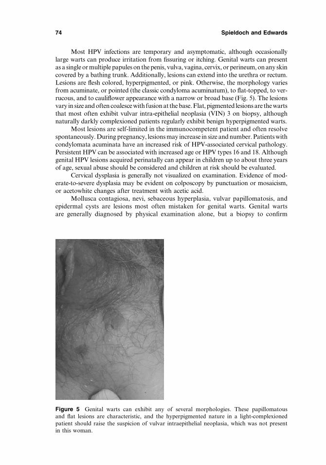
Most HPV infections are temporary and asymptomatic, although occasionallylarge warts can produce irritation from fissuring or itching. Genital warts can presentas a single or multiple papules on the penis, vulva, vagina, cervix, or perineum, on any skincovered by a bathing trunk. Additionally, lesions can extend into the urethra or rectum.Lesions are flesh colored, hyperpigmented, or pink. Otherwise, the morphology variesfrom acuminate, or pointed (the classic condyloma acuminatum), to flat-topped, to ver-rucous, and to cauliflower appearance with a narrow or broad base (Fig. 5). The lesionsvary in size and often coalesce with fusion at the base.Flat, pigmented lesions are the wartsthat most often exhibit vulvar intra-epithelial neoplasia (VIN) 3 on biopsy, althoughnaturally darkly complexioned patients regularly exhibit benign hyperpigmented warts.
Most lesions are self-limited in the immunocompetent patient and often resolvespontaneously. During pregnancy, lesions may increase in size and number. Patients withcondylomata acuminata have an increased risk of HPV-associated cervical pathology.Persistent HPV can be associated with increased age or HPV types 16 and 18. Althoughgenital HPV lesions acquired perinatally can appear in children up to about three yearsof age, sexual abuse should be considered and children at risk should be evaluated.
Cervical dysplasia is generally not visualized on examination. Evidence of mod-erate-to-severe dysplasia may be evident on colposcopy by punctuation or mosaicism,or acetowhite changes after treatment with acetic acid.
Mollusca contagiosa, nevi, sebaceous hyperplasia, vulvar papillomatosis, andepidermal cysts are lesions most often mistaken for genital warts. Genital wartsare generally diagnosed by physical examination alone, but a biopsy to confirm
Figure 5 Genital warts can exhibit any of several morphologies. These papillomatousand flat lesions are characteristic, and the hyperpigmented nature in a light-complexionedpatient should raise the suspicion of vulvar intraepithelial neoplasia, which was not presentin this woman.
74 Spieldoch and Edwards
the diagnosis is indicated in atypical cases, and reveals the presence of koilocytesin the superficial malpighian and granular layer.
Cytologic findings on pap smear often guide the management of patients withcervical dysplasia. HPV DNA testing using hybrid capture techniques can be per-formed at the time of a pap smear to look for specific HPV high-risk types.
The management of genital warts includes patient education regarding the nat-ure of genital warts, transmission, and treatment issues, as well the increased risk ofcervical carcinoma. Although in the past many clinicians sought evidence for HPV inmale partners to prevent reinfection, newer evidence does not indicate that a carefulsearch is fruitful or helpful (13). Frequent Papanicolaou smears are indicated, andwarts suspicious for vulvar intraepithelial neoplasia should be biopsied. There isno ideal therapy for anogenital warts, as evidenced by the very large number of treat-ments and treatment regimens (14). Treatments for genital warts include destructivetherapies performed in the office, such as cryotherapy, excision, podophyllum resin,trichloroacetic and bichloroacetic acid, and laser (14). Home therapy with podofiloxor the immune enhancer imiquimod can also be used. Because imiquimod exerts itseffect at least partly by enhancing the immune system, recurrences are fewer (15).Local injections of alpha interferon are useful in some. However, recurrence follow-ing treatment is usual, and several courses of therapy are often required for definitiveremoval. Patients who are immunosuppressed present greater difficulties becauseelimination of the virus depends upon an intact immune system. Those patients withHIV treated with highly active antiretroviral medications respond much better totherapy for their genital warts than those not on medication for HIV (16).
Vaccines are under investigation, with one showing a beneficial effect for HPV18 in women. Some reviews suggest that many are late in investigation showingpromise as safe and effective prevention, while other reports describe safety butno evidence for efficacy (Table 3) (17,18)a.
Molluscum Contagiosum
Molluscum contagiosum is a benign lesion produced by a virus that is a member ofthe Poxviridae family. Transmission occurs by direct skin contact with an infectedindividual, and when occurring on anogenital skin, it is generally sexually trans-mitted. However, genital mollusca in a child do not suggest sexual contact.
Most often, molluscum contagiosum is an asymptomatic infection, althoughwhen the immune system mounts an inflammatory reaction itching can occur. Mol-luscum contagiosum in adults primarily affects the genital, perianal, groin, upperthigh, and lower abdomen. The eruption is characterized by multiple pearly lesions,at least some of which show umbilication (Fig. 6). Often these are shiny and appearnearly vesicular, leading to the lay term ‘‘water warts.’’ The central umbilication con-tains a white, cord-like core. The lesions vary in size but are generally 1 to 3 mm.Histologically, molluscum bodies are cytoplasmic inclusion bodies within keratino-cytes that contain large numbers of mature virions. The papule consists of acanthoticepithelium with hyperkeratosis. In the immunocompetent host, they often resolvewithout treatment within one to two years. Immunocompromised individuals mayhave lesions that are unusually large, numerous, and resistant to treatment.
a Editor’s note: Two multivalent vaccines against HPV have recently been approved by theFDA and marketed.
Vulvovaginal Infections 75
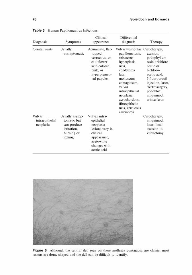
Figure 6 Although the central dell seen on these mollusca contagiosa are classic, mostlesions are dome shaped and the dell can be difficult to identify.
Table 3 Human Papillomavirus Infections
Diagnosis SymptomsClinical
appearanceDifferentialdiagnosis Therapy
Genital warts Usuallyasymptomatic
Acuminate, flat-topped,verrucous, orcauliflowerskin-colored,pink, orhyperpigmen-ted papules
Vulvar/vestibularpapillomatosis,sebaceoushyperplasia,nevi,condylomalata,molluscumcontagiosum,vulvarintraepithelialneoplasia,acrochordons,fibroepithelio-mas, verrucouscarcinoma
Cryotherapy,excision,podophyllumresin, trichloro-acetic orbichloro-acetic acid,5-fluorouracilinjection, laser,electrosurgery,podofilox,imiquimod,a-interferon
Vulvarintraepithelialneoplasia
Usually asymp-tomatic butcan produceirritation,burning oritching
Vulvar intra-epithelialneoplasialesions vary inclinicalappearance,acetowhitechanges withacetic acid
Cryotherapy,imiquimod,laser, localexcision tovulvectomy
76 Spieldoch and Edwards
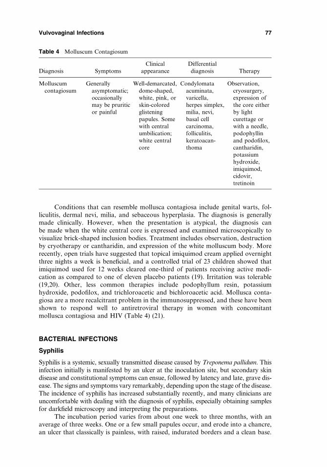
Conditions that can resemble mollusca contagiosa include genital warts, fol-liculitis, dermal nevi, milia, and sebaceous hyperplasia. The diagnosis is generallymade clinically. However, when the presentation is atypical, the diagnosis canbe made when the white central core is expressed and examined microscopically tovisualize brick-shaped inclusion bodies. Treatment includes observation, destructionby cryotherapy or cantharidin, and expression of the white molluscum body. Morerecently, open trials have suggested that topical imiquimod cream applied overnightthree nights a week is beneficial, and a controlled trial of 23 children showed thatimiquimod used for 12 weeks cleared one-third of patients receiving active medi-cation as compared to one of eleven placebo patients (19). Irritation was tolerable(19,20). Other, less common therapies include podophyllum resin, potassiumhydroxide, podofilox, and trichloroacetic and bichloroacetic acid. Mollusca conta-giosa are a more recalcitrant problem in the immunosuppressed, and these have beenshown to respond well to antiretroviral therapy in women with concomitantmollusca contagiosa and HIV (Table 4) (21).
BACTERIAL INFECTIONS
Syphilis
Syphilis is a systemic, sexually transmitted disease caused by Treponema pallidum. Thisinfection initially is manifested by an ulcer at the inoculation site, but secondary skindisease and constitutional symptoms can ensue, followed by latency and late, grave dis-ease. The signs and symptoms vary remarkably, depending upon the stage of the disease.The incidence of syphilis has increased substantially recently, and many clinicians areuncomfortable with dealing with the diagnosis of syphilis, especially obtaining samplesfor darkfield microscopy and interpreting the preparations.
The incubation period varies from about one week to three months, with anaverage of three weeks. One or a few small papules occur, and erode into a chancre,an ulcer that classically is painless, with raised, indurated borders and a clean base.
Table 4 Molluscum Contagiosum
Diagnosis SymptomsClinical
appearanceDifferentialdiagnosis Therapy
Molluscumcontagiosum
Generallyasymptomatic;occasionallymay be pruriticor painful
Well-demarcated,dome-shaped,white, pink, orskin-coloredglisteningpapules. Somewith centralumbilication;white centralcore
Condylomataacuminata,varicella,herpes simplex,milia, nevi,basal cellcarcinoma,folliculitis,keratoacan-thoma
Observation,cryosurgery,expression ofthe core eitherby lightcurettage orwith a needle,podophyllinand podofilox,cantharidin,potassiumhydroxide,imiquimod,cidovir,tretinoin
Vulvovaginal Infections 77
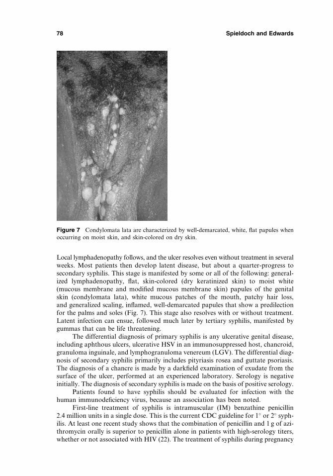
Local lymphadenopathy follows, and the ulcer resolves even without treatment in severalweeks. Most patients then develop latent disease, but about a quarter-progress tosecondary syphilis. This stage is manifested by some or all of the following: general-ized lymphadenopathy, flat, skin-colored (dry keratinized skin) to moist white(mucous membrane and modified mucous membrane skin) papules of the genitalskin (condylomata lata), white mucous patches of the mouth, patchy hair loss,and generalized scaling, inflamed, well-demarcated papules that show a predilectionfor the palms and soles (Fig. 7). This stage also resolves with or without treatment.Latent infection can ensue, followed much later by tertiary syphilis, manifested bygummas that can be life threatening.
The differential diagnosis of primary syphilis is any ulcerative genital disease,including aphthous ulcers, ulcerative HSV in an immunosuppressed host, chancroid,granuloma inguinale, and lymphogranuloma venereum (LGV). The differential diag-nosis of secondary syphilis primarily includes pityriasis rosea and guttate psoriasis.The diagnosis of a chancre is made by a darkfield examination of exudate from thesurface of the ulcer, performed at an experienced laboratory. Serology is negativeinitially. The diagnosis of secondary syphilis is made on the basis of positive serology.
Patients found to have syphilis should be evaluated for infection with thehuman immunodeficiency virus, because an association has been noted.
First-line treatment of syphilis is intramuscular (IM) benzathine penicillin2.4 million units in a single dose. This is the current CDC guideline for 1� or 2� syph-ilis. At least one recent study shows that the combination of penicillin and 1 g of azi-thromycin orally is superior to penicillin alone in patients with high-serology titers,whether or not associated with HIV (22). The treatment of syphilis during pregnancy
Figure 7 Condylomata lata are characterized by well-demarcated, white, flat papules whenoccurring on moist skin, and skin-colored on dry skin.
78 Spieldoch and Edwards
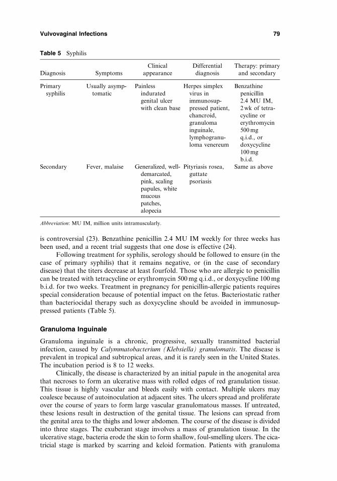
is controversial (23). Benzathine penicillin 2.4 MU IM weekly for three weeks hasbeen used, and a recent trial suggests that one dose is effective (24).
Following treatment for syphilis, serology should be followed to ensure (in thecase of primary syphilis) that it remains negative, or (in the case of secondarydisease) that the titers decrease at least fourfold. Those who are allergic to penicillincan be treated with tetracycline or erythromycin 500 mg q.i.d., or doxycycline 100 mgb.i.d. for two weeks. Treatment in pregnancy for penicillin-allergic patients requiresspecial consideration because of potential impact on the fetus. Bacteriostatic ratherthan bacteriocidal therapy such as doxycycline should be avoided in immunosup-pressed patients (Table 5).
Granuloma Inguinale
Granuloma inguinale is a chronic, progressive, sexually transmitted bacterialinfection, caused by Calymmatobacterium (Klebsiella) granulomatis. The disease isprevalent in tropical and subtropical areas, and it is rarely seen in the United States.The incubation period is 8 to 12 weeks.
Clinically, the disease is characterized by an initial papule in the anogenital areathat necroses to form an ulcerative mass with rolled edges of red granulation tissue.This tissue is highly vascular and bleeds easily with contact. Multiple ulcers maycoalesce because of autoinoculation at adjacent sites. The ulcers spread and proliferateover the course of years to form large vascular granulomatous masses. If untreated,these lesions result in destruction of the genital tissue. The lesions can spread fromthe genital area to the thighs and lower abdomen. The course of the disease is dividedinto three stages. The exuberant stage involves a mass of granulation tissue. In theulcerative stage, bacteria erode the skin to form shallow, foul-smelling ulcers. The cica-tricial stage is marked by scarring and keloid formation. Patients with granuloma
Table 5 Syphilis
Diagnosis SymptomsClinical
appearanceDifferentialdiagnosis
Therapy: primaryand secondary
Primarysyphilis
Usually asymp-tomatic
Painlessinduratedgenital ulcerwith clean base
Herpes simplexvirus inimmunosup-pressed patient,chancroid,granulomainguinale,lymphogranu-loma venereum
Benzathinepenicillin2.4 MU IM,2 wk of tetra-cycline orerythromycin500 mgq.i.d., ordoxycycline100 mgb.i.d.
Secondary Fever, malaise Generalized, well-demarcated,pink, scalingpapules, whitemucouspatches,alopecia
Pityriasis rosea,guttatepsoriasis
Same as above
Abbreviation: MU IM, million units intramuscularly.
Vulvovaginal Infections 79

inguinale are susceptible to superinfection by other bacteria and sexually transmitteddiseases, especially syphilis. Open genital ulcers are a nidus for contracting AIDS.
Other diseases in the differential diagnosis in the early ulcerative stages includeother sexually transmitted ulcers, including syphilis and chancroid. Later diseasewith scarring and granulation tissue can be confused with hidradenitis suppurativa,Crohn’s disease, and genital malignancy. The diagnosis is usually made by punchbiopsy. Donovan bodies are diagnostic and appear as intracytoplasmic inclusionbodies in hystiocytes. Complications of infection include permanent genital destruc-tion and scarring, blockage of the urinary tract from stenosis, genital depigmentation,permanent genital swelling from lymphatic scarring, and even death. Affected areasare at increased risk of developing genital cancer.
Therapy consists of doxycycline 100 mg b.i.d. or trimethoprim-sulfamethoxazoleDS b.i.d. for three weeks (8). Alternative medications include erythromycin, ciproflox-acin, and azithromycin (Table 6).
Lymphogranuloma Venereum
LGV is primarily an infection of the lymphatics and lymph nodes caused by Chlamydiatrachomatis serotypes L1, L2, and L3. LGV is a rare disease that is more frequently seenin men than in women. Transmission is by sexual contact, primarily with asymptomaticfemale carriers.
The incubation period is 3 to 30 days after inoculation. Constitutional symp-toms may be present, and the anorectal syndrome is more common in females andhomosexual males, consisting of pruritus, pain, and tenesmus. LGV presents onthe genitalia as a painless papule, shallow ulcer, herpetiform lesion, or urethritis.This lesion heals in approximately one week. The second stage begins within twoto six weeks after the initial lesion and is referred to as the inguinal stage. Duringthis stage, inguinal lymph nodes swell and form painful, firm nodes that enlarge.Bacteria travel through the regional lymphatics producing unilateral lymphangitis,referred to as buboes. These nodes may rupture or form hard masses. A classic signof LGV is the ‘‘groove sign of Greenblatt,’’ formed by Poupart’s ligament crossingover enlarged femoral lymph nodes. Inguinal and femoral lymphadenopathy is seenin approximately 20% of infected females and is often unilateral (25). The rectum,cervix, and vagina are more commonly affected in females. These areas drain into
Table 6 Granuloma Inguinale
Diagnosis SymptomsClinical
appearanceDifferentialdiagnosis Therapy
Granulomainguinale
Painless Granulomatousulcers, scarring,depigmentation,disfigurement
Chancroid, chancre,lymphogranulomavenereum,anogenitalamebiasis,elephantiasis,genital cancer,fungal infection
Recommendedtreatment:doxycycline ortrimethoprim-sulfamethoxa-zole.Alternativetreatment:ciprofloxacin,erythromycin,azithromycin
80 Spieldoch and Edwards
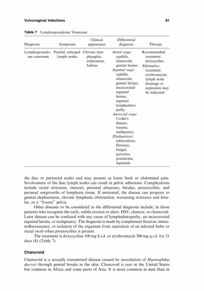
the iliac or perirectal nodes and may present as lower back or abdominal pain.Involvement of the iliac lymph nodes can result in pelvic adhesions. Complicationsinclude rectal strictures, stenosis, perianal abscesses, fistulas, proctocolitis, andperianal outgrowths of lymphatic tissue. If untreated, the disease can progress togenital elephantiasis, chronic lymphatic obstruction, worsening strictures and fistu-las, or a ‘‘frozen’’ pelvis.
Other diseases to be considered in the differential diagnosis include, in thosepatients who recognize the early, subtle erosion or ulcer, HSV, chancre, or chancroid.Later disease can be confused with any cause of lymphadenopathy, an incarceratedinguinal hernia, or malignancy. The diagnosis is made by complement fixation, immu-nofluorescence, or isolation of the organism from aspiration of an infected bubo orrectal swab when proctocolitis is present.
The treatment is doxycycline 100 mg b.i.d. or erythromycin 500 mg q.i.d. for 21days (8) (Table 7).
Chancroid
Chancroid is a sexually transmitted disease caused by inoculation of Haemophilusducreyi through genital breaks in the skin. Chancroid is rare in the United Statesbut common in Africa and some parts of Asia. It is more common in men than in
Table 7 Lymphogranuloma Venereum
Diagnosis SymptomsClinical
appearanceDifferentialdiagnosis Therapy
Lymphogranulo-ma venereum
Painful, enlargedlymph nodes
Chronic lym-phangitis,esthiomene,buboes
Initial stage:syphilis,chancroid,genital herpes
Recommendedtreatment:doxycycline.
Inguinal stage:syphilis,chancroid,genital herpes,incarceratedinguinalhernia,inguinallymphadeno-pathy
Alternativetreatment:erythromycin,lymph nodedrainage oraspiration maybe indicated
Anorectal stage:Crohn’sdisease,trauma,malignancy
Elephantiasis:tuberculosis,filariasis,fungal,parasites,granulomainguinale
Vulvovaginal Infections 81

women and associated with lower socioeconomic groups. Chancroid is commonlyassociated with coexisting syphilis or HIV.
Within four to seven days after exposure, a tender papule arises. Withinanother one to two days, the lesion becomes pustular or ulcerates (Fig. 8). The ulceris extremely painful and surrounded by erythema, and it is often foul smelling.Lesions can be solitary or by autoinoculation form multiple lesions that can coalesceinto ‘‘kissing’’ lesions or giant ulcers. Within one or two weeks, inguinal lymphade-nopathy develops in approximately 50% of patients and is usually unilateral. Thelymphadenitis may become suppurative and rupture. If untreated, extensive ulcer-ation, necrosis, genital and perineal edema, and rectovaginal fistulae can result.
The differential diagnosis includes all sexually transmitted ulcerative diseases,including HSV infection in an immunocompromised patient, as well as aphthae,Crohn’s disease, and hidradenitis suppurativa. The diagnosis of chancroid is likely ifthe patient has one or more painful genital ulcers and the presence of painful regionallymphadenopathy and there is no evidence of T. pallidum or HIV in the ulcer exudate.The diagnosis is difficult to confirm because the appropriate culture media for H. ducreyiis generally not available. Even in the best circumstances, H. ducreyi isolation has a sen-sitivity no more than 80%. Gram-stain evaluation classically shows coccobacilli ingroups, described as ‘‘schools of fish,’’ or in parallel, described as ‘‘railroad tracks.’’
Antimicrobial resistance is becoming more prominent with H. ducreyi. Anti-biotic therapies include azithromycin 1 g orally or ceftriaxone 250 mg IM once,ciprofoxacillin 500 mg b.i.d. for three days, erythromycin 500 mg t.i.d. for sevendays, or amoxicillin/clavulanic acid 500/125 t.i.d. for seven days (Table 8) (26).
Figure 8 (See color insert) Despite the careful descriptions of differing morphologies ofulcers in the various ulcerative sexually transmitted diseases, the ulcers often appear discoura-gingly similar. This lesion of chancroid cannot be diagnosed on morphology alone, but ratheron clinical suspicion and laboratory investigation.
82 Spieldoch and Edwards
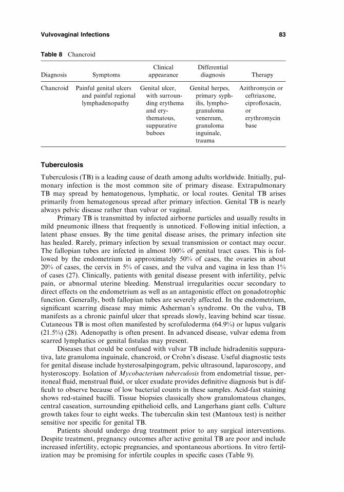
Tuberculosis
Tuberculosis (TB) is a leading cause of death among adults worldwide. Initially, pul-monary infection is the most common site of primary disease. ExtrapulmonaryTB may spread by hematogenous, lymphatic, or local routes. Genital TB arisesprimarily from hematogenous spread after primary infection. Genital TB is nearlyalways pelvic disease rather than vulvar or vaginal.
Primary TB is transmitted by infected airborne particles and usually results inmild pneumonic illness that frequently is unnoticed. Following initial infection, alatent phase ensues. By the time genital disease arises, the primary infection sitehas healed. Rarely, primary infection by sexual transmission or contact may occur.The fallopian tubes are infected in almost 100% of genital tract cases. This is fol-lowed by the endometrium in approximately 50% of cases, the ovaries in about20% of cases, the cervix in 5% of cases, and the vulva and vagina in less than 1%of cases (27). Clinically, patients with genital disease present with infertility, pelvicpain, or abnormal uterine bleeding. Menstrual irregularities occur secondary todirect effects on the endometrium as well as an antagonistic effect on gonadotrophicfunction. Generally, both fallopian tubes are severely affected. In the endometrium,significant scarring disease may mimic Asherman’s syndrome. On the vulva, TBmanifests as a chronic painful ulcer that spreads slowly, leaving behind scar tissue.Cutaneous TB is most often manifested by scrofuloderma (64.9%) or lupus vulgaris(21.5%) (28). Adenopathy is often present. In advanced disease, vulvar edema fromscarred lymphatics or genital fistulas may present.
Diseases that could be confused with vulvar TB include hidradenitis suppura-tiva, late granuloma inguinale, chancroid, or Crohn’s disease. Useful diagnostic testsfor genital disease include hysterosalpingogram, pelvic ultrasound, laparoscopy, andhysteroscopy. Isolation of Mycobacterium tuberculosis from endometrial tissue, per-itoneal fluid, menstrual fluid, or ulcer exudate provides definitive diagnosis but is dif-ficult to observe because of low bacterial counts in these samples. Acid-fast stainingshows red-stained bacilli. Tissue biopsies classically show granulomatous changes,central caseation, surrounding epithelioid cells, and Langerhans giant cells. Culturegrowth takes four to eight weeks. The tuberculin skin test (Mantoux test) is neithersensitive nor specific for genital TB.
Patients should undergo drug treatment prior to any surgical interventions.Despite treatment, pregnancy outcomes after active genital TB are poor and includeincreased infertility, ectopic pregnancies, and spontaneous abortions. In vitro fertil-ization may be promising for infertile couples in specific cases (Table 9).
Table 8 Chancroid
Diagnosis SymptomsClinical
appearanceDifferentialdiagnosis Therapy
Chancroid Painful genital ulcersand painful regionallymphadenopathy
Genital ulcer,with surroun-ding erythemaand ery-thematous,suppurativebuboes
Genital herpes,primary syph-ilis, lympho-granulomavenereum,granulomainguinale,trauma
Azithromycin orceftriaxone,ciprofloxacin,orerythromycinbase
Vulvovaginal Infections 83
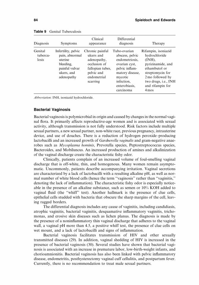
Bacterial Vaginosis
Bacterial vaginosis is polymicrobial in origin and caused by changes in the normal vagi-nal flora. It primarily affects reproductive-age women and is associated with sexualactivity, although transmission is not fully understood. Risk factors include multiplesexual partners, a new sexual partner, non-white race, previous pregnancy, intrauterinedevice, and use of douches. There is a reduction of hydrogen peroxide–producinglactobacilli and an increased growth of Gardnerella vaginalis and gram-negative anae-robes such as Mycoplasma hominis, Prevotella species, Peptostreptococcus species,Bacteroides, and Mobiluncus. An increased production of amines and alkalinizationof the vaginal discharge create the characteristic fishy odor.
Clinically, patients complain of an increased volume of foul-smelling vaginaldischarge that is off-white, thin, and homogenous. Many women remain asympto-matic. Uncommonly, patients describe accompanying irritation. Vaginal secretionsare characterized by a lack of lactobacilli with a resulting alkaline pH, as well as nor-mal number of white blood cells (hence the term ‘‘vaginosis’’ rather than ‘‘vaginitis,’’denoting the lack of inflammation). The characteristic fishy odor is especially notice-able in the presence of an alkaline substance, such as semen or 10% KOH added tovaginal fluid (the ‘‘whiff’’ test). Another hallmark is the presence of clue cells,epithelial cells studded with bacteria that obscure the sharp margins of the cell, leav-ing ragged borders.
The differential diagnosis includes any cause of vaginitis, including candidiasis,atrophic vaginitis, bacterial vaginitis, desquamative inflammatory vaginitis, tricho-monas, and erosive skin diseases such as lichen planus. The diagnosis is made bythe presence of a noninflammatory thin vaginal discharge that adheres to the vaginalwall, a vaginal pH more than 4.5, a positive whiff test, the presence of clue cells onwet mount, and a lack of lactobacilli and signs of inflammation.
Bacterial vaginosis facilitates transmission of HIV and other sexuallytransmitted diseases (29). In addition, vaginal shedding of HIV is increased in thepresence of bacterial vaginosis (30). Several studies have shown that bacterial vagi-nosis is associated with an increase in premature labor, low-birth-weight infants, andchorioamnionitis. Bacterial vaginosis has also been linked with pelvic inflammatorydisease, endometritis, posthysterectomy vaginal cuff cellulitis, and postpartum fever.Currently, there is no recommendation to treat male sexual partners.
Table 9 Genital Tuberculosis
Diagnosis SymptomsClinical
appearanceDifferentialdiagnosis Therapy
Genitaltubercu-losis
Infertility, pelvicpain, abnormaluterinebleeding,painful vulvarulcers, andadenopathy
Chronic painfululcers andadenopathy,occlusion offallopian tubes,pelvic andendometrialscarring
Tubo-ovarianabscess, pelvicendometriosis,ovarian cyst,pelvic inflam-matory disease,mycoticinfection,enterobiasis,carcinoma
Rifampin, isoniazidhydrochloride(INH),pyrizinamide, andethambutol orstreptomycin for2 mo followed bytwo drugs, i.e., INHand rifampin for4 mos
Abbreviation: INH, isoniazid hydrochloride.
84 Spieldoch and Edwards
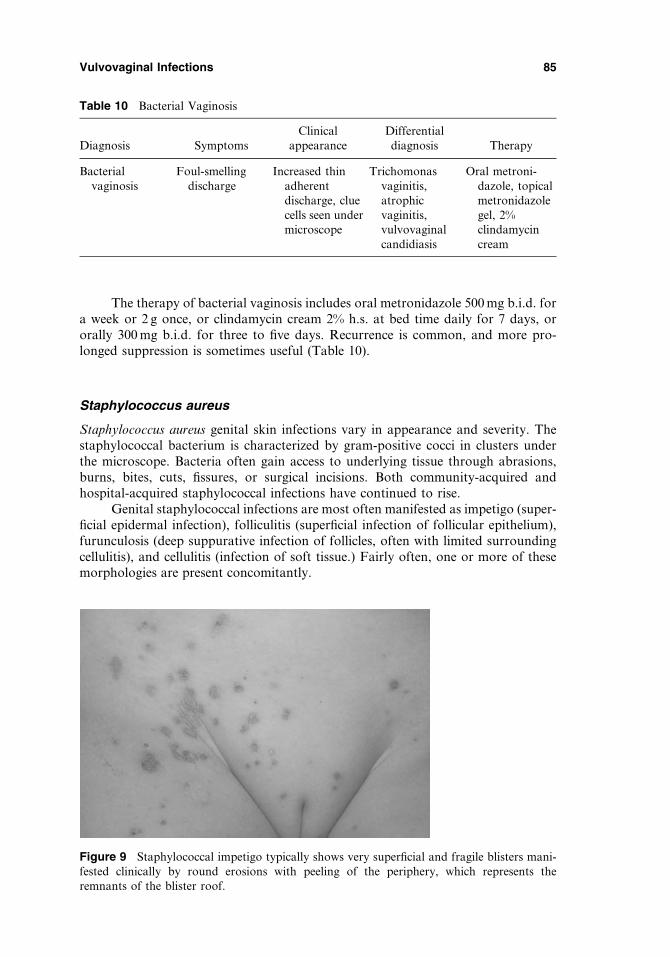
The therapy of bacterial vaginosis includes oral metronidazole 500 mg b.i.d. fora week or 2 g once, or clindamycin cream 2% h.s. at bed time daily for 7 days, ororally 300 mg b.i.d. for three to five days. Recurrence is common, and more pro-longed suppression is sometimes useful (Table 10).
Staphylococcus aureus
Staphylococcus aureus genital skin infections vary in appearance and severity. Thestaphylococcal bacterium is characterized by gram-positive cocci in clusters underthe microscope. Bacteria often gain access to underlying tissue through abrasions,burns, bites, cuts, fissures, or surgical incisions. Both community-acquired andhospital-acquired staphylococcal infections have continued to rise.
Genital staphylococcal infections are most often manifested as impetigo (super-ficial epidermal infection), folliculitis (superficial infection of follicular epithelium),furunculosis (deep suppurative infection of follicles, often with limited surroundingcellulitis), and cellulitis (infection of soft tissue.) Fairly often, one or more of thesemorphologies are present concomitantly.
Table 10 Bacterial Vaginosis
Diagnosis SymptomsClinical
appearanceDifferentialdiagnosis Therapy
Bacterialvaginosis
Foul-smellingdischarge
Increased thinadherentdischarge, cluecells seen undermicroscope
Trichomonasvaginitis,atrophicvaginitis,vulvovaginalcandidiasis
Oral metroni-dazole, topicalmetronidazolegel, 2%clindamycincream
Figure 9 Staphylococcal impetigo typically shows very superficial and fragile blisters mani-fested clinically by round erosions with peeling of the periphery, which represents theremnants of the blister roof.
Vulvovaginal Infections 85
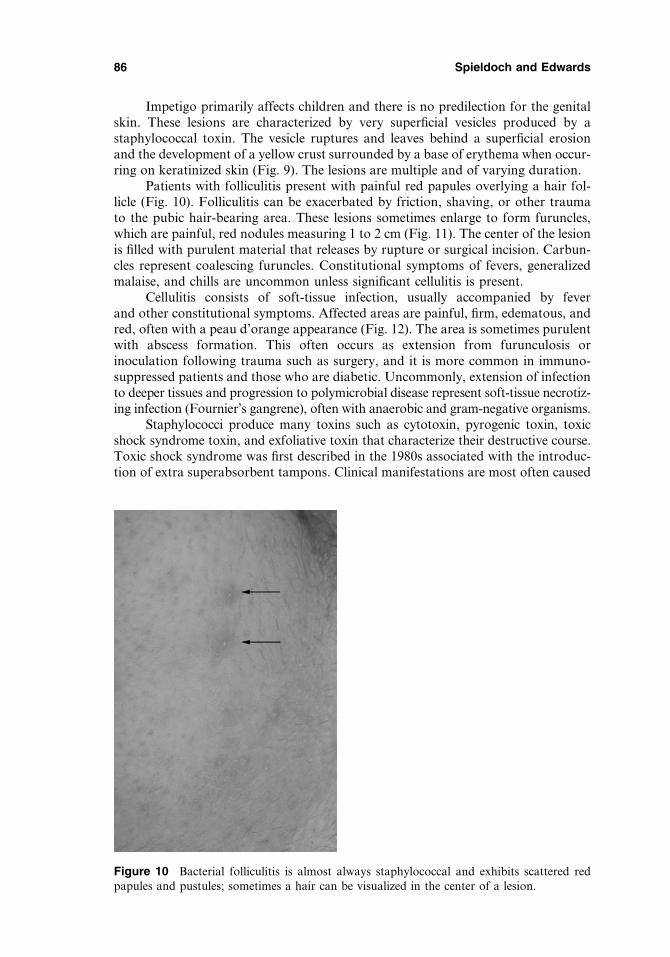
Impetigo primarily affects children and there is no predilection for the genitalskin. These lesions are characterized by very superficial vesicles produced by astaphylococcal toxin. The vesicle ruptures and leaves behind a superficial erosionand the development of a yellow crust surrounded by a base of erythema when occur-ring on keratinized skin (Fig. 9). The lesions are multiple and of varying duration.
Patients with folliculitis present with painful red papules overlying a hair fol-licle (Fig. 10). Folliculitis can be exacerbated by friction, shaving, or other traumato the pubic hair-bearing area. These lesions sometimes enlarge to form furuncles,which are painful, red nodules measuring 1 to 2 cm (Fig. 11). The center of the lesionis filled with purulent material that releases by rupture or surgical incision. Carbun-cles represent coalescing furuncles. Constitutional symptoms of fevers, generalizedmalaise, and chills are uncommon unless significant cellulitis is present.
Cellulitis consists of soft-tissue infection, usually accompanied by feverand other constitutional symptoms. Affected areas are painful, firm, edematous, andred, often with a peau d’orange appearance (Fig. 12). The area is sometimes purulentwith abscess formation. This often occurs as extension from furunculosis orinoculation following trauma such as surgery, and it is more common in immuno-suppressed patients and those who are diabetic. Uncommonly, extension of infectionto deeper tissues and progression to polymicrobial disease represent soft-tissue necrotiz-ing infection (Fournier’s gangrene), often with anaerobic and gram-negative organisms.
Staphylococci produce many toxins such as cytotoxin, pyrogenic toxin, toxicshock syndrome toxin, and exfoliative toxin that characterize their destructive course.Toxic shock syndrome was first described in the 1980s associated with the introduc-tion of extra superabsorbent tampons. Clinical manifestations are most often caused
Figure 10 Bacterial folliculitis is almost always staphylococcal and exhibits scattered redpapules and pustules; sometimes a hair can be visualized in the center of a lesion.
86 Spieldoch and Edwards
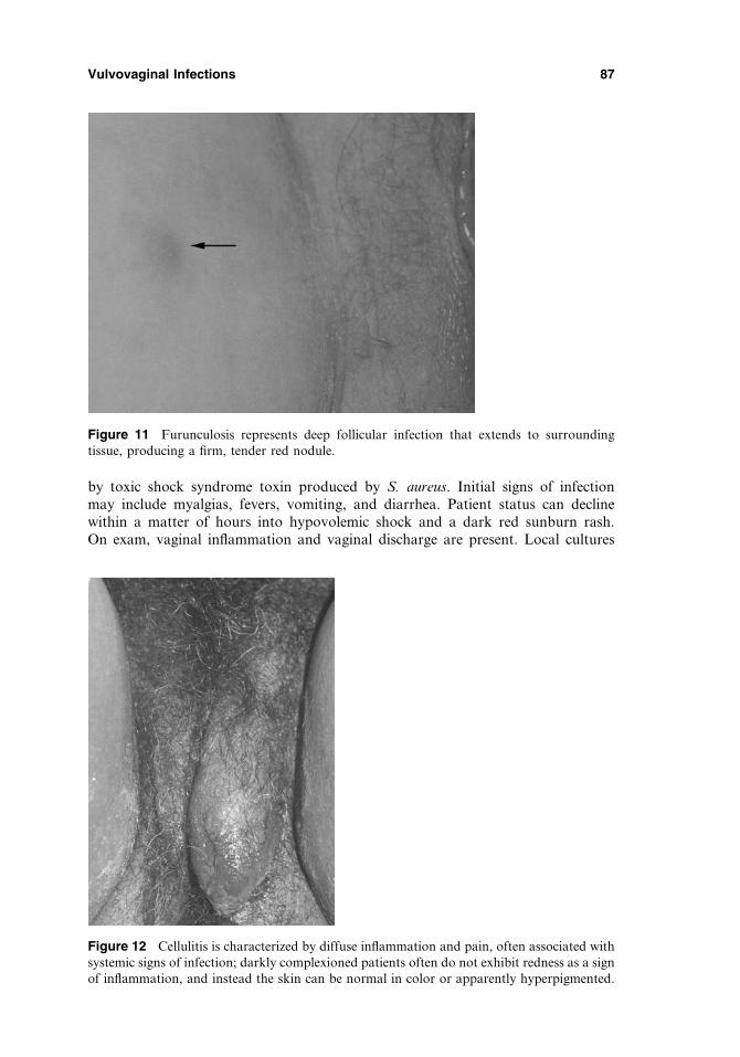
by toxic shock syndrome toxin produced by S. aureus. Initial signs of infectionmay include myalgias, fevers, vomiting, and diarrhea. Patient status can declinewithin a matter of hours into hypovolemic shock and a dark red sunburn rash.On exam, vaginal inflammation and vaginal discharge are present. Local cultures
Figure 11 Furunculosis represents deep follicular infection that extends to surroundingtissue, producing a firm, tender red nodule.
Figure 12 Cellulitis is characterized by diffuse inflammation and pain, often associated withsystemic signs of infection; darkly complexioned patients often do not exhibit redness as a signof inflammation, and instead the skin can be normal in color or apparently hyperpigmented.
Vulvovaginal Infections 87
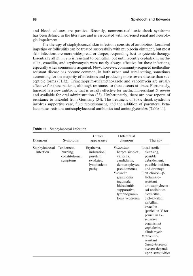
and blood cultures are positive. Recently, nonmenstrual toxic shock syndromehas been defined in the literature and is associated with worsened renal and neurolo-gic impairment.
The therapy of staphylococcal skin infections consists of antibiotics. Localizedimpetigo or folliculitis can be treated successfully with mupirocin ointment, but mostskin infections are more widespread or deeper, responding best to systemic therapy.Essentially all S. aureus is resistant to penicillin, but until recently cephalexin, methi-cillin, oxacillin, and erythromycin were nearly always effective for these infections,especially when community acquired. Now, however, community-acquired methicillin-resistant disease has become common, in both urban and rural setting, sometimesaccounting for the majority of infections and producing more severe disease than sus-ceptible forms (31,32). Trimethoprim-sulfamethoxazole and vancomycin are usuallyeffective for these patients, although resistance to these occurs at times. Fortunately,linezolid is a new antibiotic that is usually effective for methicillin-resistant S. aureusand available for oral administration (33). Unfortunately, there are now reports ofresistance to linezolid from Germany (34). The treatment of toxic shock syndromeinvolves supportive care, fluid replenishment, and the addition of parenteral beta-lactamase–resistant antistaphylococcal antibiotics and aminoglycosides (Table 11).
Table 11 Staphylococcal Infection
Diagnosis SymptomsClinical
appearanceDifferentialdiagnosis Therapy
Staphylococcalinfection
Tenderness,burning,constitutionalsymptoms
Erythema,induration,purulentexudates,lymphadeno-pathy
Folliculitis:herpes simplex,varicella,candidiasis,dermatophytes,pseudomonas
Local sterilecleansing,possibledebridement,possible incision,and drainage
Furuncle:granulomainguinale,hidradenitissuppurativa,lymphogranu-loma venereum
First choice—b-lactamase–resistantantistaphylococ-cal antibiotics:cloxacillin,dicloxacillin,nafcillin,oxacillin(penicillin V forpenicillin G–sensitiveorganisms)cephalexin,clindamycin
Methicillin-resistantStaphylococcusaureus: dependsupon sensitivities
88 Spieldoch and Edwards

Necrotizing Fasciitis (Deep Tissue Necrotizing Infection)
The term ‘‘necrotizing fasciitis’’ was first used by Wilson in 1952 to describe bacterialinfection producing necrosis of the fascia and subcutaneous tissue with sparing of theunderlying muscle. Over half of these infections occur on the perineum, where theyare called Fournier’s gangrene (35). Common recent terminology that is less specificbut includes serious destructive necrotizing infection is that of deep tissue necrotizinginfection. These infections share common causes and treatment. The diseasemost often begins with a streptococcal infection in a breach in the skin in animmunosuppressed or diabetic patient, and perforating vessels are destroyed asthe decomposition progresses, producing overlying ischemic necrosis. With thedestruction of vessels, antibiotics cannot be delivered to the center of the necroticand infected areas. The infection often becomes polymicrobial, with gram-negativeorganisms and anaerobic bacteria frequently playing a role. Gas-producing bacteriaare fairly common, producing ‘‘gas gangrene.’’
The mortality rates of necrotizing fasciitis range from 6% to 76%. Mortalityrates increase with delayed diagnosis and delayed surgical debridement. On the vulvaand perineum, the infection may follow trivial injury such as a vulvar biopsy or fol-liculitis. However, it is more often seen after episiotomy, vulvar surgery, or vulvarabscess formation. Immunocompromised conditions such as advanced age, diabetesmellitus, chronic renal failure, cancer, and illicit drug use are risk factors for this dis-ease process. Classic diagnostic signs are not always present.
Early clinical signs and symptoms include poorly demarcated areas of intenseskin pain and pallor. Blister or bulla formation represents the beginning of criticalskin ischemia. Later signs include hemorrhagic bullae, skin anesthesia from nerve
Figure 13 (See color insert) Necrotizing fasciitis is characterized by hemorrhagic bullaebecause of ischemia from underlying infection that has destroyed the cutaneous vascular supply;resulting ulceration and hemorrhage can be seen in this patient who is in the healing phase.
Vulvovaginal Infections 89
damage, and skin gangrene (Fig. 13). Tissue crepitus may be palpable. Systemic man-ifestations may include high fever, hypotension, multiorgan failure, and systemicshock. Symptoms usually develop within hours or several days with rapid pro-gression. Histologically, the bacteria proliferate within the superficial fascia and tissuenecrosis is present. Enzymes and toxins produced allow for superficial spread of thebacteria. Angiothrombotic microbial invasion and liquefaction necrosis ensue.
The differential diagnosis includes cellulitis, which can sometimes be indis-tinguishable, brown recluse spider bite, and bullous pyoderma gangrenosum. A plainradiograph, computed tomography (CT), ultrasound, or magnetic resonance ima-ging may be helpful in diagnosis. Plain radiographs can show soft-tissue air in somepatients. On CT, findings include deep fascial thickening, enhancement, and fluidand gas in the soft-tissue planes. Fine-needle aspiration or incisional biopsy may alsobe helpful for diagnosis when necrotizing fasciitis is suspected. A stab incision yieldsdecomposed necrotic tissue rather than the inflamed and edematous tissue of cellu-litis. During surgery, findings include a lack of tissue resistance, lack of bleeding ofthe fascia during dissection, and foul-smelling dishwater-colored pus. Laboratoryvalues include an elevated white cell count, glucose, urea, and creatinine. Hypoalbu-minemia, acidosis, and an altered coagulation profile may also be present.
The most important aspect of management is early diagnosis, antibiotics, andaggressive debridement (36). Methicillin-resistant S. aureus has been reported incommunity-acquired necrotizing fasciitis, so that this should be considered in thechoice of an antibiotic (37). Hyperbaric oxygen has been suggested by some, but thishas not been shown to be of use in other series (38). Polyspecific immunoglobulinmay be beneficial adjunctive therapy (39). Management requires early diagnosis,immediate resuscitation, aggressive and repeated surgical debridement, hyperbaricoxygen, and broad-spectrum antibiotics (Table 12).
Erythrasma
Erythrasma is a superficial bacterial infection of skin folds that mimics tinea cruris.Caused by Corynebacterium minutissimum, this medically insignificant disease occursprimarily in men.
Usually, erythrasma is asymptomatic. Morphologically, it is characterizedby well-demarcated, pink/tan, lightly scaling, or slightly crinkled plaques that, in the
Table 12 Necrotizing Fasciitis
Diagnosis SymptomsClinical
appearanceDifferentialdiagnosis Treatment
Necrotizingfasciitis (deepnecrotizingsoft-tissueinfection)
Severe paindisproportion-ate to localfindings, sys-temic shock
Erythema,swelling,blisters andhemorrhagicbullae,indistinctmargins
Cellulitis,erysipelas,brown reclusespider bite,bullouspyodermagangrenosum
Early diagnosis,resuscitation,surgicaldebridement,broad-spectrumantibiotics,with or withouthyperbaricoxygen toaffected tissues
90 Spieldoch and Edwards
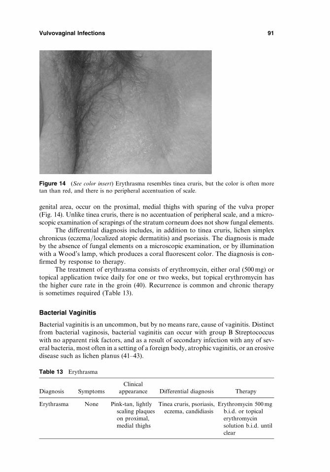
genital area, occur on the proximal, medial thighs with sparing of the vulva proper(Fig. 14). Unlike tinea cruris, there is no accentuation of peripheral scale, and a micro-scopic examination of scrapings of the stratum corneum does not show fungal elements.
The differential diagnosis includes, in addition to tinea cruris, lichen simplexchronicus (eczema/localized atopic dermatitis) and psoriasis. The diagnosis is madeby the absence of fungal elements on a microscopic examination, or by illuminationwith a Wood’s lamp, which produces a coral fluorescent color. The diagnosis is con-firmed by response to therapy.
The treatment of erythrasma consists of erythromycin, either oral (500 mg) ortopical application twice daily for one or two weeks, but topical erythromycin hasthe higher cure rate in the groin (40). Recurrence is common and chronic therapyis sometimes required (Table 13).
Bacterial Vaginitis
Bacterial vaginitis is an uncommon, but by no means rare, cause of vaginitis. Distinctfrom bacterial vaginosis, bacterial vaginitis can occur with group B Streptococcuswith no apparent risk factors, and as a result of secondary infection with any of sev-eral bacteria, most often in a setting of a foreign body, atrophic vaginitis, or an erosivedisease such as lichen planus (41–43).
Figure 14 (See color insert) Erythrasma resembles tinea cruris, but the color is often moretan than red, and there is no peripheral accentuation of scale.
Table 13 Erythrasma
Diagnosis SymptomsClinical
appearance Differential diagnosis Therapy
Erythrasma None Pink-tan, lightlyscaling plaqueson proximal,medial thighs
Tinea cruris, psoriasis,eczema, candidiasis
Erythromycin 500 mgb.i.d. or topicalerythromycinsolution b.i.d. untilclear
Vulvovaginal Infections 91
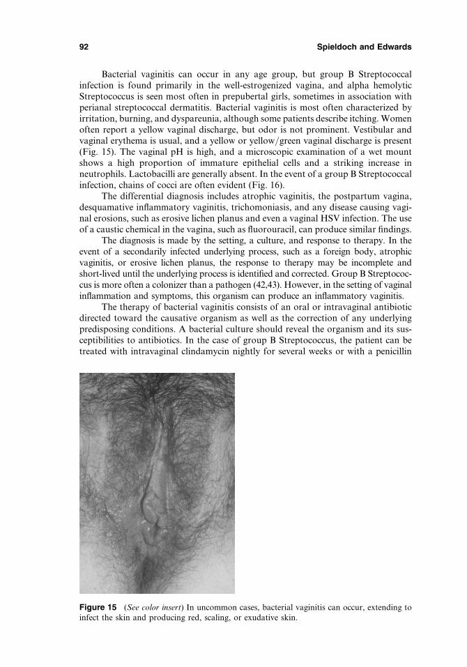
Bacterial vaginitis can occur in any age group, but group B Streptococcalinfection is found primarily in the well-estrogenized vagina, and alpha hemolyticStreptococcus is seen most often in prepubertal girls, sometimes in association withperianal streptococcal dermatitis. Bacterial vaginitis is most often characterized byirritation, burning, and dyspareunia, although some patients describe itching. Womenoften report a yellow vaginal discharge, but odor is not prominent. Vestibular andvaginal erythema is usual, and a yellow or yellow/green vaginal discharge is present(Fig. 15). The vaginal pH is high, and a microscopic examination of a wet mountshows a high proportion of immature epithelial cells and a striking increase inneutrophils. Lactobacilli are generally absent. In the event of a group B Streptococcalinfection, chains of cocci are often evident (Fig. 16).
The differential diagnosis includes atrophic vaginitis, the postpartum vagina,desquamative inflammatory vaginitis, trichomoniasis, and any disease causing vagi-nal erosions, such as erosive lichen planus and even a vaginal HSV infection. The useof a caustic chemical in the vagina, such as fluorouracil, can produce similar findings.
The diagnosis is made by the setting, a culture, and response to therapy. In theevent of a secondarily infected underlying process, such as a foreign body, atrophicvaginitis, or erosive lichen planus, the response to therapy may be incomplete andshort-lived until the underlying process is identified and corrected. Group B Streptococ-cus is more often a colonizer than a pathogen (42,43). However, in the setting of vaginalinflammation and symptoms, this organism can produce an inflammatory vaginitis.
The therapy of bacterial vaginitis consists of an oral or intravaginal antibioticdirected toward the causative organism as well as the correction of any underlyingpredisposing conditions. A bacterial culture should reveal the organism and its sus-ceptibilities to antibiotics. In the case of group B Streptococcus, the patient can betreated with intravaginal clindamycin nightly for several weeks or with a penicillin
Figure 15 (See color insert) In uncommon cases, bacterial vaginitis can occur, extending toinfect the skin and producing red, scaling, or exudative skin.
92 Spieldoch and Edwards
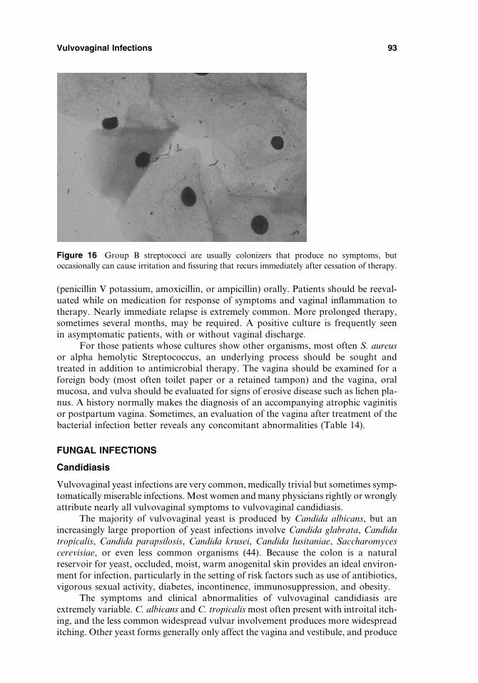
(penicillin V potassium, amoxicillin, or ampicillin) orally. Patients should be reeval-uated while on medication for response of symptoms and vaginal inflammation totherapy. Nearly immediate relapse is extremely common. More prolonged therapy,sometimes several months, may be required. A positive culture is frequently seenin asymptomatic patients, with or without vaginal discharge.
For those patients whose cultures show other organisms, most often S. aureusor alpha hemolytic Streptococcus, an underlying process should be sought andtreated in addition to antimicrobial therapy. The vagina should be examined for aforeign body (most often toilet paper or a retained tampon) and the vagina, oralmucosa, and vulva should be evaluated for signs of erosive disease such as lichen pla-nus. A history normally makes the diagnosis of an accompanying atrophic vaginitisor postpartum vagina. Sometimes, an evaluation of the vagina after treatment of thebacterial infection better reveals any concomitant abnormalities (Table 14).
FUNGAL INFECTIONS
Candidiasis
Vulvovaginal yeast infections are very common, medically trivial but sometimes symp-tomatically miserable infections. Most women and many physicians rightly or wronglyattribute nearly all vulvovaginal symptoms to vulvovaginal candidiasis.
The majority of vulvovaginal yeast is produced by Candida albicans, but anincreasingly large proportion of yeast infections involve Candida glabrata, Candidatropicalis, Candida parapsilosis, Candida krusei, Candida lusitaniae, Saccharomycescerevisiae, or even less common organisms (44). Because the colon is a naturalreservoir for yeast, occluded, moist, warm anogenital skin provides an ideal environ-ment for infection, particularly in the setting of risk factors such as use of antibiotics,vigorous sexual activity, diabetes, incontinence, immunosuppression, and obesity.
The symptoms and clinical abnormalities of vulvovaginal candidiasis areextremely variable. C. albicans and C. tropicalis most often present with introital itch-ing, and the less common widespread vulvar involvement produces more widespreaditching. Other yeast forms generally only affect the vagina and vestibule, and produce
Figure 16 Group B streptococci are usually colonizers that produce no symptoms, butoccasionally can cause irritation and fissuring that recurs immediately after cessation of therapy.
Vulvovaginal Infections 93

symptoms of burning, irritation, rawness, and soreness rather than itching. Womenand infants who are estrogen deficient rarely experience vaginal yeast infections,although the anogenital skin can certainly exhibit C. albicans infections.
Most often, the physical examination only shows vestibular erythema. How-ever, more severe infections exhibit more of the following vulvar abnormalities:erythema, edema of the modified mucous membranes, a superficial, shiny ‘‘glazed’’texture, fissuring of skin folds, peripheral scale and peeling, and satellite red papules,pustules, and collarettes (circular scale that represents the edges of a desquamatedblister roof) (Figs. 17 and 18). The vagina may or may not exhibit erythema.Although vaginal secretions are generally reported as a dry ‘‘cottage cheese’’ consist-ency, vaginal secretions are often normal clinically.
The differential diagnosis consists of several of the diseases characterized byitching, redness, and fissuring. Most common is lichen simplex chronicus (eczema/atopic dermatitis), often with intermittent, superimposed Candida infection. Lichen
Table 14 Bacterial Vaginitis
Diagnosis SymptomsClinical
appearanceDifferentialdiagnosis Therapy
Bacterialvaginitis
Introital irritation,dyspareunia,discharge
Erythema of thevestibule andvagina; vaginalsecretions withincreased whiteblood cells,immature cells
Atrophicvaginitis,desquamativeinflammatoryvaginitis
Topical orsystemicantibiotics,evaluation forunderlyingfactors
Figure 17 (See color insert) Vulvovaginal candidiasis can produce redness, edema, and fissuring.
94 Spieldoch and Edwards
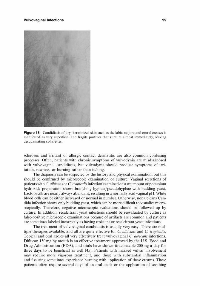
sclerosus and irritant or allergic contact dermatitis are also common confusingprocesses. Often, patients with chronic symptoms of vulvodynia are misdiagnosedwith vulvovaginal candidiasis, but vulvodynia should produce symptoms of irri-tation, rawness, or burning rather than itching.
The diagnosis can be suspected by the history and physical examination, but thisshould be confirmed by microscopic examination or culture. Vaginal secretions ofpatients with C. albicans or C. tropicalis infection examined on a wet mount or potassiumhydroxide preparation shows branching hyphae/pseudohyphae with budding yeast.Lactobacilli are nearly always abundant, resulting in a normally acid vaginal pH. Whiteblood cells can be either increased or normal in number. Otherwise, nonalbicans Can-dida infection shows only budding yeast, which can be more difficult to visualize micro-scopically. Therefore, negative microscopic evaluations should be followed up byculture. In addition, recalcitrant yeast infections should be reevaluated by culture asfalse-positive microscopic examinations because of artifacts are common and patientsare sometimes labeled incorrectly as having resistant or recalcitrant yeast infections.
The treatment of vulvovaginal candidiasis is usually very easy. There are mul-tiple therapies available, and all are quite effective for C. albicans and C. tropicalis.Topical and oral azoles all very effectively treat vulvovaginal C. albicans infections.Diflucan 150 mg by mouth is an effective treatment approved by the U.S. Food andDrug Administration (FDA), and trials have shown itraconazole 200 mg a day forthree days to be beneficial as well (45). Patients with marked vulvar involvementmay require more vigorous treatment, and those with substantial inflammationand fissuring sometimes experience burning with application of these creams. Thesepatients often require several days of an oral azole or the application of soothing
Figure 18 Candidiasis of dry, keratinized skin such as the labia majora and crural creases ismanifested as very superficial and fragile pustules that rupture almost immediately, leavingdesquamating collarettes.
Vulvovaginal Infections 95
nystatin ointment in addition to standard treatments for vaginal candidiasis.Although more is being written about resistant C. albicans in immunocompetentpatients, these authors have not observed this phenomenon. All ‘‘resistant’’ yeastinfections in immunocompetent women have been either nonalbicans Candida infec-tion or a different underlying diagnosis altogether.
Nonalbicans Candida infections are frequently resistant to therapy (46). Gen-erally, a course of any standard anticandidal therapy is prescribed. Those who failthis are usually given either boric acid vaginal capsules 600 mg once or twice aday for two to four weeks or flucytosine cream (47). Nystatin vaginal tablets or oint-ment twice a day for two to four weeks is a time-honored and soothing treatmentthat occasionally is useful in nonalbicans Candida infections resistant to azoles. Gen-tian violet can be beneficial but irritating. Amphotericin intravaginally has been usedwith some success (48). Very often, the infection does not clear, but symptoms can becontrolled with less frequent dosing.
Some patients experienced frequently recurrent vulvovaginal candidiasis.These patients can generally be managed with fluconazole 150 mg weekly (49), althougha very rare patient requires every fourth day dosing. Medication is continued for threeto six months. In some women, the fluconazole has broken this cycle and can bediscontinued. Other patients require a longer course. Hepatotoxicity is not a major con-cern with weekly dosing, but some clinicians obtain liver function tests several times ayear. Some women benefit from oral or topical therapy as a preventive at times ofhigh-risk such as during antibiotic therapy, vigorous sexual activity, or before menses.
Although some clinicians and patients believe that diet, use of oral or vaginallactobacillus supplements, and special disinfecting of underwear can minimize ortreat vulvovaginal candidiasis, there are no data to support these beliefs (50,51).In addition, there is no evidence that vulvovaginal candidiasis produces systemicsymptoms of depression, bloating, constipation, headaches, etc (Table 15).
Table 15 Vulvovaginal Candida Albicans
Diagnosis Symptoms Clinical appearance Differential diagnosis Therapy
Vulvovagin-al Candidaalbicans
Vulvovagi-nal pru-ritus
Variable from noabnormalities onexamination tovulvar erythema,edema, scale,collarettes, vaginalredness. Vaginalsecretionssometimes whiteand clumped,always with fungalelements micro-scopically
Vulvar disease—psoriasis, lichensimplex chronicus/eczema, tinea cruris;Vaginal disease—bacterial vaginosis,bacterial vaginitis,desquamativeinflammatoryvaginitis, atrophicvaginitis,vulvodynia/vestibulitis(vestibulodynia)
Topical—anyazole creamq.d.–b.i.d. tothe vulva,cream orsuppositoryhours one toseven nights,oral—fluco-nazole 150 mgPO once
Non-albicansCandida
Irritation,soreness,burning
Normal exam,budding yeastmicroscopically
Vulvodynia/vulvarvestibulitis(vestibulodynia)
Same as forC. albicans ifresistant. Boricacid, nystatin,flucytosine,gentian violet,amphotericin
96 Spieldoch and Edwards

Tinea Cruris
Tinea cruris, a dermatophytosis infection of the anogenital skin, represents ‘‘ring-worm.’’ Also called ‘‘jock itch,’’ this is a disease much more common in men thanin women. Primarily caused by Epidermophyton floccosum, Trichophyton rubrum,and Trichophyton mentagrophytes, tinea cruris often represents infection that hasspread from the great toe nails (onychomycosis) or the feet (tinea pedis), also infectionsthat are much more common in men. Obesity also predisposes to tinea cruris (52).
The predominant symptom of tinea cruris is that of itching. The rash occursprimarily on proximal, medial thighs, sometimes extending to the crural creaseand hair-bearing labia majora. Because dermatophytes infect the stratum corneumof clinically hair-bearing skin, the modified mucous membranes are spared and thevagina is never affected. Classically, tinea cruris presents as well-demarcated, red,scaling plaques of the proximal, medial thighs, with the borders exhibiting moremarked scale. Often, there is central clearing, and the infection progresses peripher-ally (Fig. 19). Sometimes, fungal organisms track down hair follicles, producingfungal folliculitis manifested by palpable red papules within the plaque. This is mostmarked in women with coarse terminal hair and this is almost universal in patientswho have treated their tinea cruris with topical corticosteroids (Fig. 20).
The differential diagnosis of tinea cruris includes any red, scaling infection ordermatoses that can affect this area. Candidiasis, psoriasis, lichen simplex chronicus(eczema/localized atopic dermatitis), irritant or allergic contact dermatitis, and ery-thrasma are the most likely diseases requiring differentiation.
The diagnosis is made tentatively by the location and morphology, particularlywhen seen in the patient with fungal infection of the feet. The diagnosis is confirmed
Figure 19 (See color insert) Classic tinea cruris shows a red plaque on the proximal, medialthighs with accentuation and scale at the periphery of the plaque.
Vulvovaginal Infections 97

by a microscopic examination of scrapings of the stratum corneum, culture, or responseto therapy.
The treatment of tinea cruris depends upon its extent. Those patients with lim-ited disease and no signs of fungal folliculitis usually respond to any topical azoleapplied once or twice a day. Miconazole, clotrimazole, econazole, and ketoconazoleare those most often marketed for this problem. However, any azole used for vulvo-vaginal candidiasis is also effective and often more inexpensively available over thecounter. These include tioconazole and butoconazole. Topical terbinafine used onceor twice a day is also effective (53). Nystatin is useless for dermatophytosis. Exten-sive disease, clinical fungal folliculitis, and disease in an area of marked terminal hairare best treated with an oral medication. Griseofulvin 500 mg twice daily, terbinafine250 mg daily, fluconazole 100/200 mg a day, and itraconazole 200 mg a day are allhighly effective and safe. A trial of itraconazole given as 200 mg on two or threeoccasions showed cure rates of 86% to 100% (54). Patients should be reevaluatedafter about two weeks for clinical cure. Recurrence is common when fungal infectionof the feet is present. Early treatment of recurrent disease with a topical medication isgenerally effective. Alternatively, onychomycosis can be more definitively treatedwith standard doses of itraconazole or terbinafine (Table 16).
Pityriasis Versicolor
Pityriasis versicolor is a fungal infection of keratinized skin produced by severaldifferent kinds of Malassezia species (55). Also known by the misnomer, tinea
Figure 20 Tinea cruris that is treated with a steroid as well as a topical antifungal agent isless likely to completely clear, and this patient has developed an associated fungal folliculitismanifested by red papules within the plaque.
98 Spieldoch and Edwards
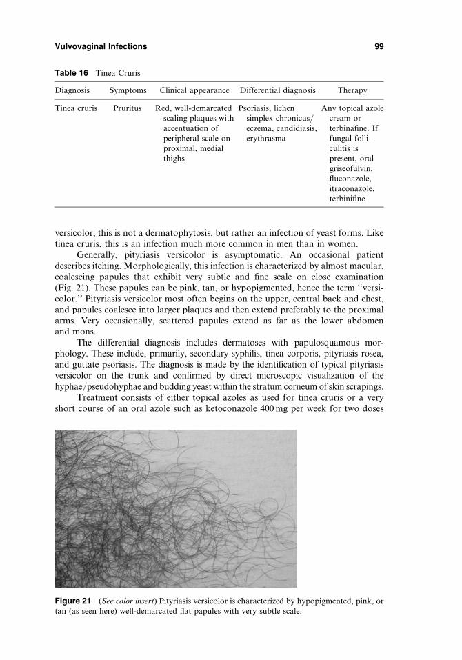
versicolor, this is not a dermatophytosis, but rather an infection of yeast forms. Liketinea cruris, this is an infection much more common in men than in women.
Generally, pityriasis versicolor is asymptomatic. An occasional patientdescribes itching. Morphologically, this infection is characterized by almost macular,coalescing papules that exhibit very subtle and fine scale on close examination(Fig. 21). These papules can be pink, tan, or hypopigmented, hence the term ‘‘versi-color.’’ Pityriasis versicolor most often begins on the upper, central back and chest,and papules coalesce into larger plaques and then extend preferably to the proximalarms. Very occasionally, scattered papules extend as far as the lower abdomenand mons.
The differential diagnosis includes dermatoses with papulosquamous mor-phology. These include, primarily, secondary syphilis, tinea corporis, pityriasis rosea,and guttate psoriasis. The diagnosis is made by the identification of typical pityriasisversicolor on the trunk and confirmed by direct microscopic visualization of thehyphae/pseudohyphae and budding yeast within the stratum corneum of skin scrapings.
Treatment consists of either topical azoles as used for tinea cruris or a veryshort course of an oral azole such as ketoconazole 400 mg per week for two doses
Table 16 Tinea Cruris
Diagnosis Symptoms Clinical appearance Differential diagnosis Therapy
Tinea cruris Pruritus Red, well-demarcatedscaling plaques withaccentuation ofperipheral scale onproximal, medialthighs
Psoriasis, lichensimplex chronicus/eczema, candidiasis,erythrasma
Any topical azolecream orterbinafine. Iffungal folli-culitis ispresent, oralgriseofulvin,fluconazole,itraconazole,terbinifine
Figure 21 (See color insert) Pityriasis versicolor is characterized by hypopigmented, pink, ortan (as seen here) well-demarcated flat papules with very subtle scale.
Vulvovaginal Infections 99
or fluconazole 300 mg twice, one week apart (56,57). Itraconazole given orally as aone-time dose of 400 or 200 mg a day for a week is effective as well (58). Seleniumsulfide shampoos and lotions are frequently irritating and annoying in the anogenitalarea and generally should be avoided.
Pityriasis versicolor is frequently a recurrent condition, and patient should beadvised that retreatment may be necessary in the future. Some patients benefit fromonce or twice monthly application of an azole cream to prevent recurrence. Oral itra-conazole given intermittently has also been shown efficacious for the prevention ofrecurrent pityriasis versicolor (Table 17) (59).
PARASITIC INFECTIONS
Trichomonas
Trichomonas vaginitis is a sexually transmitted infection produced by the protozoanTrichomonas vaginalis. The organism is found not only in the vagina but also in theurethra and in Skene’s glands and paraurethral ducts.
The primary symptom of Trichomonas vaginitis is severe pruritus, irritation,and, often, vulvar dysuria, accompanied by a grossly and microscopically purulentvaginal discharge. Sometimes, pelvic pain is present. The severity of disease varieswidely, with some patients exhibiting fairly mild symptoms and signs. The infectedmale partner is usually asymptomatic. A physical examination reveals deep rednessof the introitus, vagina, and cervix, classically but nonspecifically and unpredictablyproducing a ‘‘strawberry cervix.’’ This finding is characterized by a red cervix cov-ered with monomorphous, discrete, bright-red, tiny papules. Vaginal secretions aremost often described as yellow and frothy. Microscopically, vaginal secretions showsmall (about the size of white blood cells), rapidly moving, teardrop-shaped, flagel-late organisms that quickly lose motility and distinctive shape as they cool. Inaddition, very large numbers of neutrophils are present and lactobacilli are absent,resulting in a high vaginal pH.
The differential diagnosis includes all causes of purulent, inflammatory vagin-itis. Desquamative inflammatory vaginitis, atrophic vaginitis, and the occasional
Table 17 Pityriasis Versicolor
Diagnosis SymptomsClinical
appearanceDifferentialdiagnosis Therapy
Pityriasisversicolor
Usually none;occasionalmild irritationor pruritus
Pink, brown, orhypopigmen-ted, well-demarcated,lightly scaling2–10 mmpapulescoalescing intolarger plaquescentrally attimes
Pityriasis rosea,secondarysyphilis,guttatepsoriasis, tineacorporis/cruris
Any topical azolecream appliedone to twotimes a dayuntil clear; ifextensive,ketoconazole100 mg/dayfor 5 daysor fluconazole300 mg/wkfor 2 weeks
100 Spieldoch and Edwards

bacterial vaginitis (not vaginosis) produce vestibular and vaginal/cervical erythemaand vaginal secretions with similar microscopic findings. However, the causativeorganism is absent. The diagnosis is made by the setting and the identification ofthe organism microscopically.
Treatment consists of oral metronidazole 500 mg twice a day for one week or2 g orally in one dose. Topical medications are not sufficient because of the organ-isms protected within the urethra and paraurethral ducts. Sexual partners shouldbe treated as well. Trichomonas resistant to metronidazole occurs occasionally,and those patients can be treated with tinidazole (Table 18) (60,61).
Scabies
Scabies is an excruciatingly pruritic skin infection produced by the mite, Sarcoptesscabiei. The mite is transmitted by close contact with an infected person, and infectsthe stratum corneum of the skin. The itching occurs after a week or two, as a resultof the host immune response to the presence of the mite. Therefore, not all infectedpeople may be symptomatic initially.
In addition to the intense itching, which is often the worst at night, patientspresent primarily with signs consistent with scratching. Redness, excoriations, andscale are usual. The classic specific lesion of scabies is the burrow, a 1 to 3 mmred, edematous linear papule or vesicle, most often located in the finger and toeweb spaces, the ventral wrists, and on inframammary skin. However, in the axillaand on anogenital skin (most often, however, on the scrotum and penis) very itchy,red, excoriated, dermal nodules are characteristic (Fig. 22).
The differential diagnosis includes other very itchy skin diseases includinglichen simplex chronicus (eczema/atopic dermatitis) and irritant/allergic contactdermatitis. The more discreet and specific nodules can be confused with folliculitis,furunculosis, and hidradenitis suppurativa, although scabetic nodules never drain.
The diagnosis is made by the history of intense pruritus, morphology, and dis-tribution, and it is confirmed by demonstration of the organism (Fig. 23). A deepscraping/shave of a burrow often but certainly not always yields the microscopicidentification of a mite, ova, or scybala (feces). Scraping a scabetic nodule of the ano-genital skin is not useful. However, a punch biopsy is characteristic of an insect bite.
The treatment of scabies includes elimination of the mite, control of the inflam-matory response, and avoidance of reinfection by treating other family members and
Table 18 Trichomoniasis
Diagnosis Symptoms Clinical appearanceDifferentialdiagnosis Therapy
Trichomo-niasis
Intense pruritusand burning,irritation
Introital, vaginal, andcervical erythema,‘‘strawberry’’cervix, purulentvaginal secretionsthat microscopicallyshow white bloodcells andtrichomonads
Vaginal candi-diasis, atrophicvaginitis,bacterialvaginitis,bacterialvaginosis,desquamativeinflammatoryvaginitis
Oral metro-nidazole500 mg b.i.d.for 1 wk or 2 gonce
Vulvovaginal Infections 101
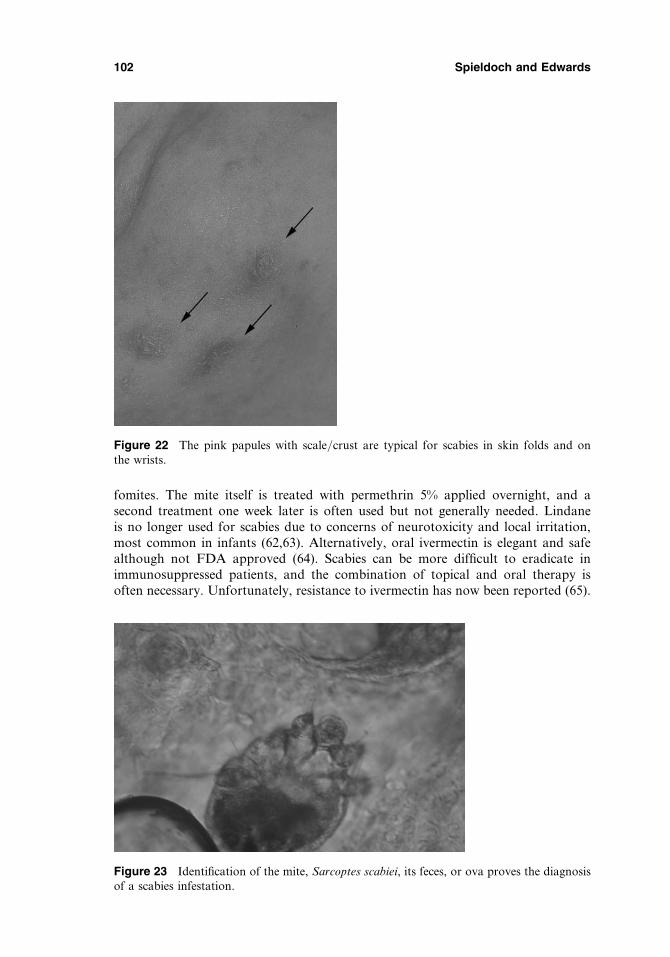
fomites. The mite itself is treated with permethrin 5% applied overnight, and asecond treatment one week later is often used but not generally needed. Lindaneis no longer used for scabies due to concerns of neurotoxicity and local irritation,most common in infants (62,63). Alternatively, oral ivermectin is elegant and safealthough not FDA approved (64). Scabies can be more difficult to eradicate inimmunosuppressed patients, and the combination of topical and oral therapy isoften necessary. Unfortunately, resistance to ivermectin has now been reported (65).
Figure 22 The pink papules with scale/crust are typical for scabies in skin folds and onthe wrists.
Figure 23 Identification of the mite, Sarcoptes scabiei, its feces, or ova proves the diagnosisof a scabies infestation.
102 Spieldoch and Edwards
The short-term application of an ultrapotent topical corticosteroid such asclobetasol dipropionate ointment may help control itching. However, the thickenednature of scabetic nodules in the genital area precludes good penetration, and a fewdrops of triamcinolone acetonide 10 mg/cc often produce rapid symptomaticimprovement. Finally, all clothing and linens used within 48 hours should be eitherwashed with hot soapy water or bagged for several days to eliminate the mite. Allthose living in the same home or in close contact should be treated with permethrinor ivermectin. Persistence of disease is frequently reinfection rather than resistance(Table 19).
Pubic Lice
Pubic lice, Phthirus pubis, are small, macroscopic parasites that are generally sexuallytransmitted, annoying, and medically trivial. Fomites also are known to transmitlice. Also called ‘‘crabs,’’ these organisms localize to the pubic hair and eyelashes,and these are distinct from head lice.
The primary symptom of lice is itching. Physical findings are often very, verysubtle. The 1 to 2 mm mites are nearly skin colored and attached to hair. Very closeexamination with magnification generally also reveals nits, light-colored oval eggsglued to the hairs. The skin is usually normal in appearance (Fig. 24). Modifiedmucous membranes and the vaginal epithelium are not affected.
Table 19 Scabies
Diagnosis Symptoms Clinical appearanceDifferentialdiagnosis Therapy
Scabies Intensepruritus
Excoriations,burrows, inflameddermal nodules
Eczema/lichensimplexchronicus,folliculitis,candidiasis
5% permethrincreamovernight orivermectinorally
Figure 24 (See color insert) Pubic lice are small parasites that cling to pubic hair, with nitsappearing as teardrop-shaped eggs attached to hair shafts.
Vulvovaginal Infections 103
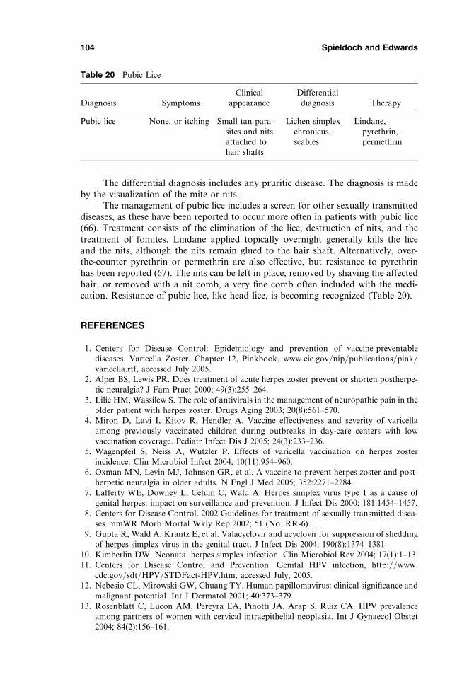
The differential diagnosis includes any pruritic disease. The diagnosis is madeby the visualization of the mite or nits.
The management of pubic lice includes a screen for other sexually transmitteddiseases, as these have been reported to occur more often in patients with pubic lice(66). Treatment consists of the elimination of the lice, destruction of nits, and thetreatment of fomites. Lindane applied topically overnight generally kills the liceand the nits, although the nits remain glued to the hair shaft. Alternatively, over-the-counter pyrethrin or permethrin are also effective, but resistance to pyrethrinhas been reported (67). The nits can be left in place, removed by shaving the affectedhair, or removed with a nit comb, a very fine comb often included with the medi-cation. Resistance of pubic lice, like head lice, is becoming recognized (Table 20).
REFERENCES
1. Centers for Disease Control: Epidemiology and prevention of vaccine-preventablediseases. Varicella Zoster. Chapter 12, Pinkbook, www.cic.gov/nip/publications/pink/varicella.rtf, accessed July 2005.
2. Alper BS, Lewis PR. Does treatment of acute herpes zoster prevent or shorten postherpe-tic neuralgia? J Fam Pract 2000; 49(3):255–264.
3. Lilie HM, Wassilew S. The role of antivirals in the management of neuropathic pain in theolder patient with herpes zoster. Drugs Aging 2003; 20(8):561–570.
4. Miron D, Lavi I, Kitov R, Hendler A. Vaccine effectiveness and severity of varicellaamong previously vaccinated children during outbreaks in day-care centers with lowvaccination coverage. Pediatr Infect Dis J 2005; 24(3):233–236.
5. Wagenpfeil S, Neiss A, Wutzler P. Effects of varicella vaccination on herpes zosterincidence. Clin Microbiol Infect 2004; 10(11):954–960.
6. Oxman MN, Levin MJ, Johnson GR, et al. A vaccine to prevent herpes zoster and post-herpetic neuralgia in older adults. N Engl J Med 2005; 352:2271–2284.
7. Lafferty WE, Downey L, Celum C, Wald A. Herpes simplex virus type 1 as a cause ofgenital herpes: impact on surveillance and prevention. J Infect Dis 2000; 181:1454–1457.
8. Centers for Disease Control. 2002 Guidelines for treatment of sexually transmitted disea-ses. mmWR Morb Mortal Wkly Rep 2002; 51 (No. RR-6).
9. Gupta R, Wald A, Krantz E, et al. Valacyclovir and acyclovir for suppression of sheddingof herpes simplex virus in the genital tract. J Infect Dis 2004; 190(8):1374–1381.
10. Kimberlin DW. Neonatal herpes simplex infection. Clin Microbiol Rev 2004; 17(1):1–13.11. Centers for Disease Control and Prevention. Genital HPV infection, http://www.
cdc.gov/sdt/HPV/STDFact-HPV.htm, accessed July, 2005.12. Nebesio CL, Mirowski GW, Chuang TY. Human papillomavirus: clinical significance and
malignant potential. Int J Dermatol 2001; 40:373–379.13. Rosenblatt C, Lucon AM, Pereyra EA, Pinotti JA, Arap S, Ruiz CA. HPV prevalence
among partners of women with cervical intraepithelial neoplasia. Int J Gynaecol Obstet2004; 84(2):156–161.
Table 20 Pubic Lice
Diagnosis SymptomsClinical
appearanceDifferentialdiagnosis Therapy
Pubic lice None, or itching Small tan para-sites and nitsattached tohair shafts
Lichen simplexchronicus,scabies
Lindane,pyrethrin,permethrin
104 Spieldoch and Edwards

14. Ting PT, Dytoc MT. Therapy of external anogenital warts and molluscum contagiosum: aliterature review. Dermatol Ther 2004; 17:68–101.
15. Cox JT, Petry KU, Rylander E, Roy M. Using imiquimod for genital warts in femalepatients. J Womens Health (Larchmt) 2004; 13:265–271.
16. Massad LS, Silverberg MJ, Springer G, et al. Effect of antiretroviral therapy on the incidenceof genital warts and vulvar neoplasia among women with the human immunodeficiency virus.Am J Obstet Gynecol 2004; 190(5):1241–1248. (Erratum in: Am J Obstet Gynecol 2004;191:1842–1844).
17. Berry JM, Palefsky JM. A review of human papillomavirus vaccines: from basic science toclinical trials. Front Biosci 2003; 8:s333–s345.
18. Stanley MA. Progress in prophylactic and therapeutic vaccines for human papillomavirusinfection. Expert Rev Vaccines 2003; 2:381–389.
19. Theos AU, Cummins R, Silverberg NB, Paller AS. Effectiveness of imiquimod cream 55for treating childhood molluscum contagiosum in a double-blind, randomized pilot trial.Cutis 2004; 74:134–8, 141–142.
20. Tran H, Moreno G, Shumack S. Imiquimod as a dermatological therapy. Expert OpinPharmacother 2004; 5:427–438.
21. Maurer T, Rodrigues LK, Ameli N, et al. The effect of highly active antiretroviral therapyon dermatologic disease in a longitudinal study of HIV type 1-infected women. Clin InfectDis 2004; 15(38):579–584.
22. Kiddugavu mg, Kiwanuka N, Wawer MJ, et al., the Rakai Study Group. Effectiveness ofsyphilis treatment using azithromycin and/or benzathine penicillin in Rakai, Uganda. SexTransm Dis 2005; 32:1–6.
23. Berman SM. Maternal syphilis: pathophysiology and treatment. Bull World Health Organ2004; 82(6):433–438.
24. Watson-Jones D, Gumodoka B, Weiss H, et al. Syphilis in pregnancy in Tanzania. II.The effectiveness of antenatal syphilis screening and single-dose benzathine penicillintreatment for the prevention of adverse pregnancy outcomes. J Infect Dis 2002; 186:948–957.
25. Kaufman R, Faro S, Brown D. Benign Diseases of the Vulva and Vagina. Elsevier andMosby, 2005.
26. Hollier LM, Workowski K. Treatment of sexually transmitted diseases in women. ObstetGynecol Clin North Am 2003; 30:751–775, vii–viii.
27. Aliyu MH, Aliyu SH, Salihu H. ‘‘Female genital tuberculosis: a global view.’’ Int J FertilWomens Med 2004; 49:123–136.
28. Yasmeen N, Kanjee A. Cutaneous tuberculosis: a three-year prospective study. J Pak MedAssoc 2005; 55(1):10–12.
29. Zariffard MR, Novak RM, Lurain N, Sha BE, Graham P, Spear GT. Induction of tumornecrosis factor-alpha secretion and toll-like receptor 2 and 4 mRNA expression by genitalmucosal fluids from women with bacterial vaginosis. J Infect Dis 2005; 191:1913–1921.
30. Sha BE, Zariffard MR, Wang QJ, et al. Female genital-tract HIV load correlates inverselywith Lactobacillus species but positively with bacterial vaginosis and Mycoplasma hominis.J Infect Dis 2005; 191:25–32.
31. Moran GJ, Amii RN, Abrahamian FM, Talan DA. Methicillin-resistant Staphylococcusaureus in community-acquired skin infections. Emerg Infect Dis 2005; 11:928–930.
32. Schulz P, Allen M, Murray Q, et al. Infections due to community-acquired methicillin-resistant Staphylococcus aureus: an emergent epidemic in Kentucky. J Ky Med Assoc2005; 103:194–203.
33. Weigelt J, Itani K, Stevens D, Lau W, Dryden M, Knirsch C. Linezolid CSSTI StudyGroup. Linezolid versus vancomycin in treatment of complicated skin and soft tissueinfections. Antimicrob Agents Chemother 2005; 49:2260–2266.
34. Brauers J, Kresken M, Hafner D, Shah PM, German Linezolid Resistance Study Group.Surveillance of linezolid resistance in Germany, 2001–2002. Clin Microbiol Infect 2005;11:39–46.
Vulvovaginal Infections 105
35. Taviloglu K, Cabioglu N, Cagatay A, et al. Idiopathic necrotizing fasciitis: risk factorsand strategies for management. Am Surg 2005; 71:315–320.
36. Muqim R. Necrotizing fasciitis: management and outcome. J Coll Physicians Surg Pak2003; 13:711–714.
37. Miller LG, Perdreau-Remington F, Rieg G, et al. Necrotizing fasciitis caused bycommunity-associated methicillin-resistant Staphylococcus aureus in Los Angeles. N EnglJ Med 2005; 352(14):1445–1453.
38. Mindrup SR, Kealey GP, Fallon B. Hyperbaric oxygen for the treatment of Fournier’sgangrene. J Urol 2005; 173:1975–1453.
39. Norrby-Teglund A, Muller MP, Mcgeer A, et al. Successful management of severe groupA streptococcal soft tissue infections using an aggressive medical regimen including intra-venous polyspecific immunoglobulin together with a conservative surgical approach.Scand J Infect Dis 2005; 37:166–172.
40. Holdiness MR. Management of cutaneous erythrasma. Drugs 2002; 62:1131–1141.41. Donder GG, Vereecken A, Bosmans E, Dekeersmaecker A, Salembier G, Spitz B. Defi-
nition of a type of abnormal vaginal flora that is distinct from bacterial vaginosis: aerobicvaginitis. BJOG 2002; 109:34–43.
42. Honig E, Mouton JW, van der Meijden WI. Can group B streptococci cause symptomaticvaginitis? Infect Dis Obstet Gynecol 1999; 7:206–209.
43. Shaw C, Mason M, Scoular A. Group B streptococcus carriage and vulvovaginal symp-toms: causal or casual? A case-control study in a GUM clinic population. Sex TransmInfect 2003; 79:246–248.
44. Martens MG, Hoffman P, El-Zaatari M. Fungal species changes in the female genitaltract. J Low Genit Tract Dis 2004; 8(1):21–24.
45. De Punzio C, Garutti P, Mollica G, Nappi C, Piccoli R, Genazzani AR. Fluconazole150 mg single dose versus itraconazole 200 mg per day for 3 days in the treatment of acutevaginal candidiasis: a double-blind randomized study. Eur J Obstet Gynecol Reprod Biol2003; 106:193–197.
46. Richter SS, Galask RP, Messer SA, Hollis RJ, Diekema DJ, Pfaller MA. Antifungal sus-ceptibilities of Candida species causing vulvovaginitis and epidemiology of recurrent cases.J Clin Microbiol 2005; 43:2155–2162.
47. Sobel JD, Chaim W, Nagappan V, Leaman D. Treatment of vaginitis caused by Candidaglabrata: use of topical boric acid and flucytosine. Am J Obstet Gynecol 2003; 189:1297–1300.
48. Phillips AJ. Treatment of non-albicans Candida vaginitis with amphotericin B vaginalsuppositories. Am J Obstet Gynecol 2005; 192:2009–2012.
49. Sobel JD, Wiesenfeld HC, Martens M, et al. Maintenance fluconazole therapy for recur-rent vulvovaginal candidiasis. N Engl J Med 2004; 351:876–883.
50. Bern VH. New study shows probiotics ineffective in prevention of post-antibiotic vaginalcandidiasis. Schweiz Rundsch Med Prax 2004; 93:1 p following 1843.
51. Jeavons HS. Prevention and treatment of vulvovaginal candidiasis using exogenousLactobacillus. Obstet Gynecol Neonatal Nurs 2003; 32:287–296.
52. Scheinfeld NS. Obesity and dermatology. Clin Dermatol 2004; 22:303–309.53. Budimulja U, Bramono K, Urip KS, et al. Once daily treatment with terbinafine 1% cream
(Lamisil) for one week is effective in the treatment of tinea corporis and cruris. A placebo-controlled study. Mycoses 2001; 44:300–306.
54. Sanmano B, Hiruma M, Mizoguchi M, Ogawa H. Abbreviated oral itraconazole therapyfor tinea corporis and tinea cruris. Mycoses 2003; 46:316–321.
55. Tarazooie B, Kordbacheh P, Zaini F, et al. Study of the distribution of Malassezia speciesin patients with pityriasis versicolor and healthy individuals in Tehran, Iran. BMC Derma-tol 2004; 4:5.
56. Farschian M, Yaghoobi R, Samadi K. Fluconazole versus ketoconazole in the treatmentof tinea versicolor. J Dermatolog Treat 2002; 13:73–76.
57. Karakas M, Durdu M, Memisoglu HR. Oral fluconazole in the treatment of tinea versi-color. J Dermatol 2005; 32:19–21.
106 Spieldoch and Edwards
58. Kose O, Bulent Tastan H, Riza Gur A, Kurumlu Z. Comparison of a single 400 mg doseversus a 7-day 200 mg daily dose of itraconazole in the treatment of tinea versicolor. J Der-matolog Treat 2002; 13:77–79.
59. Faergemann J, Gupta AK, Al Mofadi A, Abanami A, Shareaah AA, Marynissen G. Effi-cacy of itraconazole in the prophylactic treatment of pityriasis (tinea) versicolor. ArchDermatol 2002; 138:69–73.
60. Cohen CE, Desmond NM. Clinically resistant trichomoniasis. Sex Transm Infect 2005;81:181.
61. Cudmore SL, Delgaty KL, Hayward-McClelland SF, Petrin DP, Garber GE. Treatmentof infections caused by metronidazole-resistant Trichomonas vaginalis. Clin Microbiol Rev2004; 17:783–793.
62. Bhalla M, Thami GP. Reversible neurotoxicity after an overdose of topical lindane in aninfant. Pediatr Dermatol 2004; 21:597–599.
63. Yu KJ, Chen HH, Chang YC, Hong HS, Ho HC. Ulcerative irritant contact dermatitisfrom lindane. Contact Dermatitis 2005; 52:118–119.
64. Del Giudice P. The treatment of scabies: topical or systemic? Ann Dermatol Venereol2004; 131:1045–1047.
65. Currie BJ, Harumal P, McKinnon M, Walton SF. First documentation of in vivo andin vitro ivermectin resistance in Sarcoptes scabiei. Clin Infect Dis 2004; 39:e8–e12.
66. Varela JA, Otero L, Espinosa E, Sanchez C, Junquera ML, Vazquez F. Phthirus pubis in asexually transmitted diseases unit: a study of 14 years. Sex Transm Dis 2003; 30:292–296.
67. Speare R, Koehler JM. A case of pubic lice resistant to pyrethrins. Aust Fam Physician2001; 30:572–574.
Vulvovaginal Infections 107


5Pigmented Lesions of the Vulva
Hope K. HaefnerDepartment of Obstetrics and Gynecology, The University of Michigan Hospitals,Ann Arbor, Michigan, U.S.A.
Timothy M. JohnsonDepartments of Dermatology, Otolaryngology, and Surgery, Melanoma Clinic,The University of Michigan Hospitals, Ann Arbor, Michigan, U.S.A.
Lorraine L. RosamiliaDepartments of Internal Medicine and Dermatology, The Pennsylvania State University,Milton S. Hershey Medical Center, Hershey, Pennsylvania, U.S.A.
Douglas R. FullenDepartment of Pathology and Dermatology, The University of Michigan Hospitals,Ann Arbor, Michigan, U.S.A.
INTRODUCTION
Numerous pigmented conditions affect the vulva. These include common benign vulvarconditions with a variety of terminology, such as nevus, seborrheic keratosis, acanthosisnigricans (AN), lentigo simplex (lentigines), melanosis, condylomata, pigmented apocrinehamartoma, postinflammatory hyperpigmentation in association with lichen simplexchronicus, vulvar intraepithelial neoplasia (VIN), and angiokeratoma to name a few.Various medical conditions have hyperpigmentation as a sign, such as Cushing’s disease,Addison’s disease, discoid lupus erythematosus, LAMB syndrome (lentigines, atrial myx-omas, mucocutaneous myxomas, and blue nevi) (1), LEOPARD syndrome (lentigines,electrocardiographic-conduction abnormalities, ocular hypertelorism, pulmonary sten-osis, abnormal genitalia, retardation of growth, and deafness) (2), Peutz–Jegher (3),and Bannayan–Riley–Ruvalcaba syndrome (4), as well as rarer conditions (5–10).Treatments for medical conditions or trauma can be associated with hyperpigmentation(radiation therapy, chemotherapeutic agents, antimalarials, and metals). While mela-noma is the most common malignant process to be associated with hyperpigmentation,other vulvar precancers and cancers may have hyperpigmentation (Paget’s disease,squamous cell carcinoma, verrucous carcinoma, and basal cell carcinoma) (11).
The prevalence of pigmented lesions on the vulva was investigated by Rockand Rock in 1990. They performed a yearlong prospective study evaluatingpigmented lesions seen in a gynecology practice. Approximately 12% of patients
109
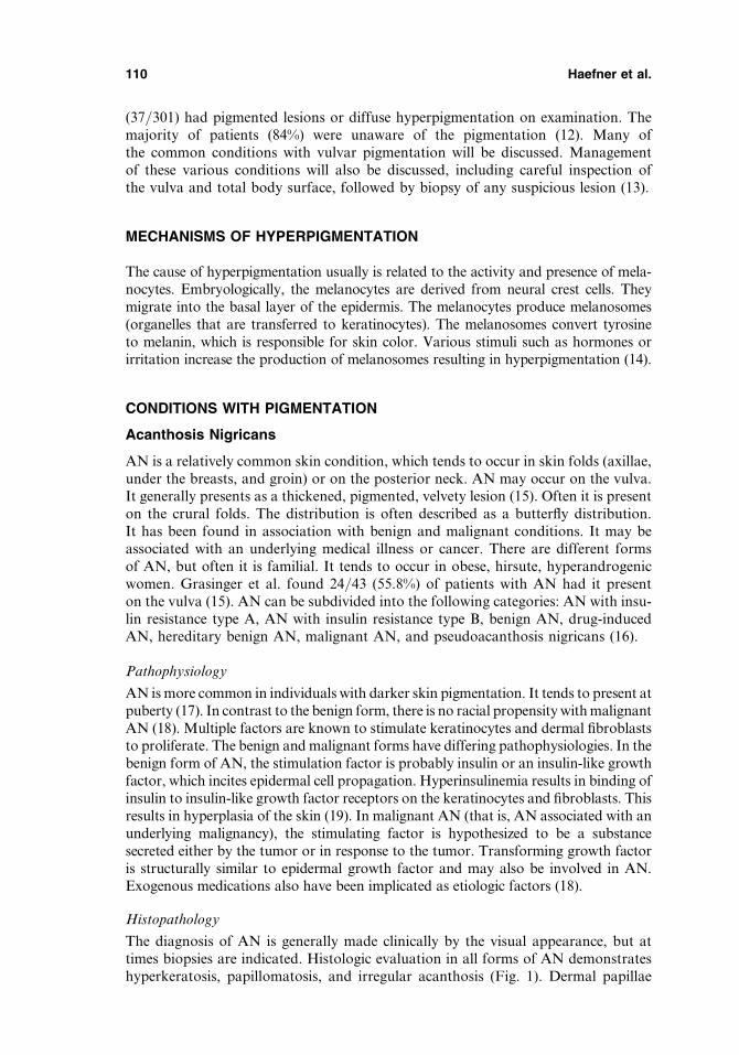
(37/301) had pigmented lesions or diffuse hyperpigmentation on examination. Themajority of patients (84%) were unaware of the pigmentation (12). Many ofthe common conditions with vulvar pigmentation will be discussed. Managementof these various conditions will also be discussed, including careful inspection ofthe vulva and total body surface, followed by biopsy of any suspicious lesion (13).
MECHANISMS OF HYPERPIGMENTATION
The cause of hyperpigmentation usually is related to the activity and presence of mela-nocytes. Embryologically, the melanocytes are derived from neural crest cells. Theymigrate into the basal layer of the epidermis. The melanocytes produce melanosomes(organelles that are transferred to keratinocytes). The melanosomes convert tyrosineto melanin, which is responsible for skin color. Various stimuli such as hormones orirritation increase the production of melanosomes resulting in hyperpigmentation (14).
CONDITIONS WITH PIGMENTATION
Acanthosis Nigricans
AN is a relatively common skin condition, which tends to occur in skin folds (axillae,under the breasts, and groin) or on the posterior neck. AN may occur on the vulva.It generally presents as a thickened, pigmented, velvety lesion (15). Often it is presenton the crural folds. The distribution is often described as a butterfly distribution.It has been found in association with benign and malignant conditions. It may beassociated with an underlying medical illness or cancer. There are different formsof AN, but often it is familial. It tends to occur in obese, hirsute, hyperandrogenicwomen. Grasinger et al. found 24/43 (55.8%) of patients with AN had it presenton the vulva (15). AN can be subdivided into the following categories: AN with insu-lin resistance type A, AN with insulin resistance type B, benign AN, drug-inducedAN, hereditary benign AN, malignant AN, and pseudoacanthosis nigricans (16).
Pathophysiology
AN is more common in individuals with darker skin pigmentation. It tends to present atpuberty (17). In contrast to the benign form, there is no racial propensity with malignantAN (18). Multiple factors are known to stimulate keratinocytes and dermal fibroblaststo proliferate. The benign and malignant forms have differing pathophysiologies. In thebenign form of AN, the stimulation factor is probably insulin or an insulin-like growthfactor, which incites epidermal cell propagation. Hyperinsulinemia results in binding ofinsulin to insulin-like growth factor receptors on the keratinocytes and fibroblasts. Thisresults in hyperplasia of the skin (19). In malignant AN (that is, AN associated with anunderlying malignancy), the stimulating factor is hypothesized to be a substancesecreted either by the tumor or in response to the tumor. Transforming growth factoris structurally similar to epidermal growth factor and may also be involved in AN.Exogenous medications also have been implicated as etiologic factors (18).
Histopathology
The diagnosis of AN is generally made clinically by the visual appearance, but attimes biopsies are indicated. Histologic evaluation in all forms of AN demonstrateshyperkeratosis, papillomatosis, and irregular acanthosis (Fig. 1). Dermal papillae
110 Haefner et al.

extend upward in finger-like projections. Horn pseudocysts, as seen in seborrheickeratosis, may be seen. Slight hyperpigmentation of the basal layer of the epidermisis common, but melanocytes are not increased.
Treatment
The treatment of AN must address the underlying cause rather than the appearanceof the skin. For example, if insulin resistance is present, it should be managed appro-priately. Symptomatic treatments for cutaneous changes include topical antibiotics,topical or systemic retinoids, and keratolytics (20). At times, corticosteroids areutilized. If an underlying endocrinologic abnormality is the cause, the best treatmentis weight loss (21) (Table 1).
MELANOCYTIC LESIONS
A variety of melanocytic tumors exist. Benign tumors consist of mainly melanocyticnevi and benign vulvar melanosis (lentigo), while melanoma represents the mostcommon malignant melanocytic tumor type.
Figure 1 Acanthosis nigricans. Lesion demonstrates hyperkeratosis, papillomatosis, andirregular acanthosis.
Table 1 Acanthosis Nigricans
Diagnosis SymptomsClinical
appearanceDifferentialdiagnosis Therapy
Acanthosisnigricans
Asymptomaticgenerally,but mayhavepruritusand localirritation
Found inintertriginousareas
Areas ofdarkening andthickening ofthe skin; mayhave a velvetyor wartyappearance
Addison’s diseaseIntertrigoHemochromatosisPellagraBecker nevusHypertrophic
seborrheic keratosisIchthyosis hystrixLinear epidermal nevusPemphigus vegetansHidradenitis
suppurativa
Address underlyingcause
Topical antibioticsTopical or systemic
retinoidsKeratolyticsCorticosteroids
Pigmented Lesions of the Vulva 111
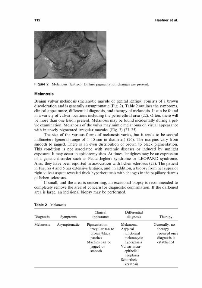
Melanosis
Benign vulvar melanosis (melanotic macule or genital lentigo) consists of a browndiscoloration and is generally asymptomatic (Fig. 2). Table 2 outlines the symptoms,clinical appearance, differential diagnosis, and therapy of melanosis. It can be foundin a variety of vulvar locations including the periurethral area (22). Often, there willbe more than one lesion present. Melanosis may be found incidentally during a pel-vic examination. Melanosis of the vulva may mimic melanoma on visual appearancewith intensely pigmented irregular macules (Fig. 3) (23–25).
The size of the various forms of melanosis varies, but it tends to be severalmillimeters (general range of 1–15 mm in diameter) (26). The margins vary fromsmooth to jagged. There is an even distribution of brown to black pigmentation.This condition is not associated with systemic diseases or induced by sunlightexposure. It may occur in episiotomy sites. At times, lentigines may be an expressionof a genetic disorder such as Peutz–Jeghers syndrome or LEOPARD syndrome.Also, they have been reported in association with lichen sclerosus (27). The patientin Figures 4 and 5 has extensive lentigos, and, in addition, a biopsy from her superiorright vulvar aspect revealed thick hyperkeratosis with changes in the papillary dermisof lichen sclerosus.
If small, and the area is concerning, an excisional biopsy is recommended tocompletely remove the area of concern for diagnostic confirmation. If the darkenedarea is large, an incisional biopsy may be performed.
Figure 2 Melanosis (lentigo). Diffuse pigmentation changes are present.
Table 2 Melanosis
Diagnosis SymptomsClinical
appearanceDifferentialdiagnosis Therapy
Melanosis Asymptomatic Pigmentation;irregular tan tobrown/blackpatches
MelanomaAtypical
junctionalmelanocytichyperplasia
Generally, notherapyrequired oncediagnosis isestablishedMargins can be
jagged orsmooth
Vulvar intra-epithelialneoplasia
Seborrheickeratosis
112 Haefner et al.

Histopathology
On visual inspection of the vulva, it can be difficult to differentiate melanosis frommelanoma; however, it is not difficult on histologic grounds. Melanosis lacks a sub-stantial melanocytic proliferation, nesting pattern of melanocytes, or melanocyteatypia. Dendritic melanocytes are normal in number or only slightly increased alongthe basal layer of the epidermis in association with hyperpigmentation (Fig. 6). Onoccasion, the melanin pigment is also located in the upper dermis. Acanthosis maybe present (28).
Figure 3 Diffuse darkening, grossly suspicious for melanoma; however, a biopsy revealedmelanosis (lentigo).
Figure 4 Lentigo extensively involving the vulva. Of note, pruritus present on superior rightvulva. This patient was found to have lichen sclerosus as well as the itch–scratch cycle.
Pigmented Lesions of the Vulva 113
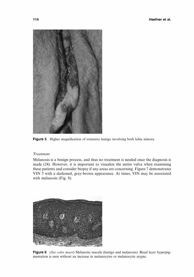
Treatment
Melanosis is a benign process, and thus no treatment is needed once the diagnosis ismade (24). However, it is important to visualize the entire vulva when examiningthese patients and consider biopsy if any areas are concerning. Figure 7 demonstratesVIN 3 with a darkened, gray-brown appearance. At times, VIN may be associatedwith melanosis (Fig. 8).
Figure 5 Higher magnification of extensive lentigo involving both labia minora.
Figure 6 (See color insert) Melanotic macule (lentigo and melanosis). Basal layer hyperpig-mentation is seen without an increase in melanocytes or melanocytic atypia.
114 Haefner et al.
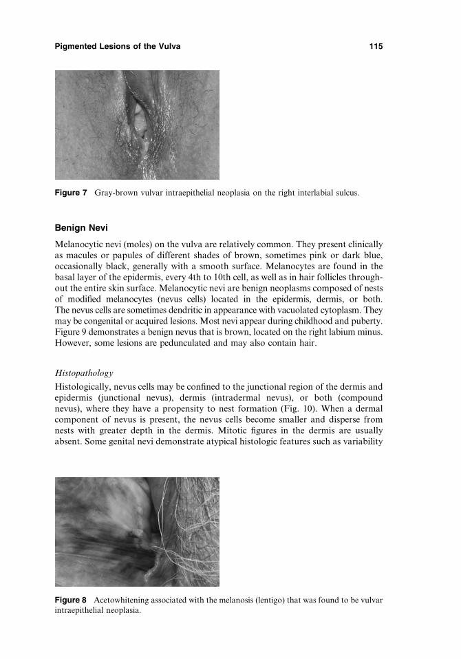
Benign Nevi
Melanocytic nevi (moles) on the vulva are relatively common. They present clinicallyas macules or papules of different shades of brown, sometimes pink or dark blue,occasionally black, generally with a smooth surface. Melanocytes are found in thebasal layer of the epidermis, every 4th to 10th cell, as well as in hair follicles through-out the entire skin surface. Melanocytic nevi are benign neoplasms composed of nestsof modified melanocytes (nevus cells) located in the epidermis, dermis, or both.The nevus cells are sometimes dendritic in appearance with vacuolated cytoplasm. Theymay be congenital or acquired lesions. Most nevi appear during childhood and puberty.Figure 9 demonstrates a benign nevus that is brown, located on the right labium minus.However, some lesions are pedunculated and may also contain hair.
Histopathology
Histologically, nevus cells may be confined to the junctional region of the dermis andepidermis (junctional nevus), dermis (intradermal nevus), or both (compoundnevus), where they have a propensity to nest formation (Fig. 10). When a dermalcomponent of nevus is present, the nevus cells become smaller and disperse fromnests with greater depth in the dermis. Mitotic figures in the dermis are usuallyabsent. Some genital nevi demonstrate atypical histologic features such as variability
Figure 8 Acetowhitening associated with the melanosis (lentigo) that was found to be vulvarintraepithelial neoplasia.
Figure 7 Gray-brown vulvar intraepithelial neoplasia on the right interlabial sulcus.
Pigmented Lesions of the Vulva 115

in size and shape of nests and fusion of some nests resulting in confluence along thebasal layer of the epidermis and cytologic atypia of nevus cells.
Treatment
Observation without treatment is recommended for a stable, unchanging benignnevus. However, if change in size, shape, color, pruritus or any difference inappearance occurs, removal is recommended (Table 3).
Atypical Junctional Melanocytic Hyperplasia
Atypical junctional melanocytic hyperplasia (AJMH) is a superficial process localized tothe junctional region. This condition is distinctive from common acquired nevi. Thesechanges are macular, often clinically ill-defined, may be large and uniformly darkly pig-mented, with an asymmetric outline and irregular borders (29) (Table 4). The biologicbehavior of AJMH is uncertain and unpredictable. However, AJMH may progress tomelanoma and is associated with frequent local recurrence after incomplete removal,over months or decades. Immunosuppressed patients (e.g., transplantation patients) withAJMH may have a higher incidence of progression to melanoma (29). Figures 11–15show the appearance of AJMH on the vulva (changes vary from mild to severe atypia).
Figure 9 (See color insert) Benign nevus on right labium minus.
Figure 10 Genital compound nevus. Nests of cells are visible in the epidermis and dermis.
116 Haefner et al.
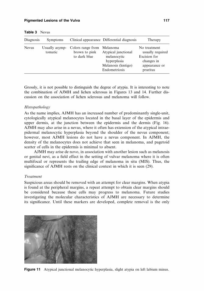
Grossly, it is not possible to distinguish the degree of atypia. It is interesting to notethe combination of AJMH and lichen sclerosus in Figures 13 and 14. Further dis-cussion on the association of lichen sclerosus and melanoma will follow.
Histopathology
As the name implies, AJMH has an increased number of predominantly single-unit,cytologically atypical melanocytes located in the basal layer of the epidermis andupper dermis, at the junction between the epidermis and the dermis (Fig. 16).AJMH may also arise in a nevus, where it often has extension of the atypical intrae-pidermal melanocytic hyperplasia beyond the shoulder of the nevus component;however, most AJMH lesions do not have a nevus component. In AJMH, thedensity of the melanocytes does not achieve that seen in melanoma, and pagetoidscatter of cells in the epidermis is minimal to absent.
AJMH may arise de novo, in association with another lesion such as melanosisor genital nevi, as a field effect in the setting of vulvar melanoma where it is oftenmultifocal or represents the trailing edge of melanoma in situ (MIS). Thus, thesignificance of AJMH rests on the clinical context in which it is seen (29).
Treatment
Suspicious areas should be removed with an attempt for clear margins. When atypiais found at the peripheral margins, a repeat attempt to obtain clear margins shouldbe considered because these cells may progress to melanoma. Future studiesinvestigating the molecular characteristics of AJMH are necessary to determineits significance. Until these markers are developed, complete removal is the only
Table 3 Nevus
Diagnosis Symptoms Clinical appearance Differential diagnosis Therapy
Nevus Usually asymp-tomatic
Colors range frombrown to pinkto dark blue
MelanomaAtypical junctional
melanocytichyperplasia
Melanosis (lentigo)Endometriosis
No treatmentusually required
Excision forchanges inappearance orpruritus
Figure 11 Atypical junctional melanocytic hyperplasia, slight atypia on left labium minus.
Pigmented Lesions of the Vulva 117
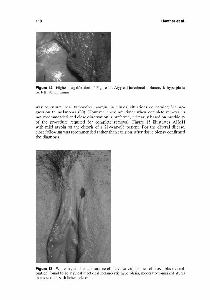
way to ensure local tumor-free margins in clinical situations concerning for pro-gression to melanoma (30). However, there are times when complete removal isnot recommended and close observation is preferred, primarily based on morbidityof the procedure required for complete removal. Figure 15 illustrates AJMHwith mild atypia on the clitoris of a 21-year-old patient. For the clitoral disease,close following was recommended rather than excision, after tissue biopsy confirmedthe diagnosis.
Figure 12 Higher magnification of Figure 11. Atypical junctional melanocytic hyperplasiaon left labium minus.
Figure 13 Whitened, crinkled appearance of the vulva with an area of brown-black discol-oration, found to be atypical junctional melanocytic hyperplasia, moderate-to-marked atypiain association with lichen sclerosus.
118 Haefner et al.

Melanoma In Situ
MIS is defined as Stage 0, confined to the epidermis and adnexal epithelium. It wasclassified as a subset of the International Society for the Study of Vulvovaginal Diseaseclassification of VIN (31) but has subsequently been discarded in the newer classification(31a). The concept of MIS was first described in 1949. In 1992, a panel at the ConsensusDevelopment Conference sponsored by the National Institutes of Health officiallyacknowledged this diagnostic category (32). The terminology was inspired by that ofcutaneous squamous cell carcinoma; however, there is one major difference. Squamouscell carcinoma in situ is a premalignant condition, whereas MIS is considered amalignancy with a risk of metastasis, where there is an inability to detect an invasivecomponent with tissue processing with standard histopathological techniques.
MIS often presents as a macular deeply pigmented lesion, initially uniform inappearance, but with time develops asymmetry, irregular borders, and colorvariegation (Table 5).
Histopathology
MIS is characterized histologically by a proliferation of malignant melanocytesconfluently in the epidermis (Fig. 17). In the mucosal lentiginous type, the melanomacells are arrayed predominantly in single cells in association with elongation of reteridges. Pagetoid intraepidermal spread of melanoma cells is usually minimal to
Figure 14 Higher power of atypical junctional melanocytic hyperplasia, moderate-to-marked atypia in association with lichen sclerosus.
Figure 15 Atypical junctional melanocytic hyperplasia, slight atypia on prepuce, frenulum,and clitoris.
Pigmented Lesions of the Vulva 119
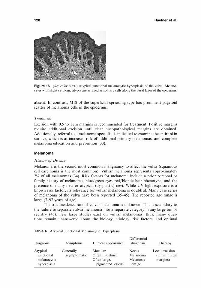
absent. In contrast, MIS of the superficial spreading type has prominent pagetoidscatter of melanoma cells in the epidermis.
Treatment
Excision with 0.5 to 1 cm margins is recommended for treatment. Positive marginsrequire additional excision until clear histopathological margins are obtained.Additionally, referral to a melanoma specialist is indicated to examine the entire skinsurface, which is at increased risk of additional primary melanomas, and completemelanoma education and prevention (33).
Melanoma
History of Disease
Melanoma is the second most common malignancy to affect the vulva (squamouscell carcinoma is the most common). Vulvar melanoma represents approximately2% of all melanomas (34). Risk factors for melanoma include a prior personal orfamily history of melanoma, blue/green eyes–red/blonde hair phenotype, and thepresence of many nevi or atypical (dysplastic) nevi. While UV light exposure is aknown risk factor, its relevance for vulvar melanoma is doubtful. Many case seriesof melanoma of the vulva have been reported (35–45). The reported age range islarge (7–97 years of age).
The true incidence rate of vulvar melanoma is unknown. This is secondary tothe failure to separate vulvar melanoma into a separate category in any large tumorregistry (46). Few large studies exist on vulvar melanomas; thus, many ques-tions remain unanswered about the biology, etiology, risk factors, and optimal
Figure 16 (See color insert) Atypical junctional melanocytic hyperplasia of the vulva. Melano-cytes with slight cytologic atypia are arrayed as solitary cells along the basal layer of the epidermis.
Table 4 Atypical Junctional Melanocytic Hyperplasia
Diagnosis Symptoms Clinical appearanceDifferentialdiagnosis Therapy
Atypicaljunctionalmelanocytichyperplasia
Generallyasymptomatic
MacularOften ill-definedOften large,
pigmented lesions
Nevus Local excision(initial 0.5 cmmargins)
MelanomaMelanosisLentigo
120 Haefner et al.
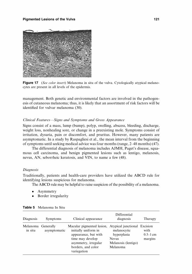
management. Both genetic and environmental factors are involved in the pathogen-esis of cutaneous melanoma; thus, it is likely that an assortment of risk factors will beidentified for vulvar melanoma (30).
Clinical Features—Signs and Symptoms and Gross Appearance
Signs consist of a mass, lump (bump), polyp, swelling, abscess, bleeding, discharge,weight loss, nonhealing sore, or change in a preexisting mole. Symptoms consist ofirritation, dysuria, pain or discomfort, and pruritus. However, many patients areasymptomatic. In a study by Raspagliesi et al., the mean interval from the beginningof symptoms until seeking medical advice was four months (range, 2–48 months) (47).
The differential diagnosis of melanoma includes AJMH, Paget’s disease, squa-mous cell carcinoma, and benign pigmented lesions such as lentigo, melanosis,nevus, AN, seborrheic keratosis, and VIN, to name a few (48).
Diagnosis
Traditionally, patients and health-care providers have utilized the ABCD rule foridentifying lesions suspicious for melanoma.
The ABCD rule may be helpful to raise suspicion of the possibility of a melanoma.
� Asymmetry� Border irregularity
Table 5 Melanoma In Situ
Diagnosis Symptoms Clinical appearanceDifferentialdiagnosis Therapy
Melanomain situ
Generallyasymptomatic
Macular pigmented lesion,initially uniform inappearance, but withtime may developasymmetry, irregularborders, and colorvariegation
Atypical junctionalmelanocytichyperplasia
Excisionwith0.5–1 cmmarginsNevus
Melanosis (lentigo)Melanoma
Figure 17 (See color insert) Melanoma in situ of the vulva. Cytologically atypical melano-cytes are present in all levels of the epidermis.
Pigmented Lesions of the Vulva 121
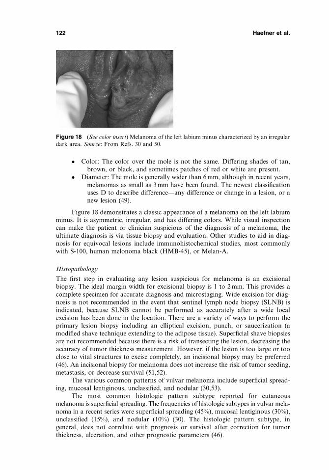
� Color: The color over the mole is not the same. Differing shades of tan,brown, or black, and sometimes patches of red or white are present.
� Diameter: The mole is generally wider than 6 mm, although in recent years,melanomas as small as 3 mm have been found. The newest classificationuses D to describe difference—any difference or change in a lesion, or anew lesion (49).
Figure 18 demonstrates a classic appearance of a melanoma on the left labiumminus. It is asymmetric, irregular, and has differing colors. While visual inspectioncan make the patient or clinician suspicious of the diagnosis of a melanoma, theultimate diagnosis is via tissue biopsy and evaluation. Other studies to aid in diag-nosis for equivocal lesions include immunohistochemical studies, most commonlywith S-100, human melonoma black (HMB-45), or Melan-A.
Histopathology
The first step in evaluating any lesion suspicious for melanoma is an excisionalbiopsy. The ideal margin width for excisional biopsy is 1 to 2 mm. This provides acomplete specimen for accurate diagnosis and microstaging. Wide excision for diag-nosis is not recommended in the event that sentinel lymph node biopsy (SLNB) isindicated, because SLNB cannot be performed as accurately after a wide localexcision has been done in the location. There are a variety of ways to perform theprimary lesion biopsy including an elliptical excision, punch, or saucerization (amodified shave technique extending to the adipose tissue). Superficial shave biopsiesare not recommended because there is a risk of transecting the lesion, decreasing theaccuracy of tumor thickness measurement. However, if the lesion is too large or tooclose to vital structures to excise completely, an incisional biopsy may be preferred(46). An incisional biopsy for melanoma does not increase the risk of tumor seeding,metastasis, or decrease survival (51,52).
The various common patterns of vulvar melanoma include superficial spread-ing, mucosal lentiginous, unclassified, and nodular (30,53).
The most common histologic pattern subtype reported for cutaneousmelanoma is superficial spreading. The frequencies of histologic subtypes in vulvar mela-noma in a recent series were superficial spreading (45%), mucosal lentiginous (30%),unclassified (15%), and nodular (10%) (30). The histologic pattern subtype, ingeneral, does not correlate with prognosis or survival after correction for tumorthickness, ulceration, and other prognostic parameters (46).
Figure 18 (See color insert) Melanoma of the left labium minus characterized by an irregulardark area. Source: From Refs. 30 and 50.
122 Haefner et al.
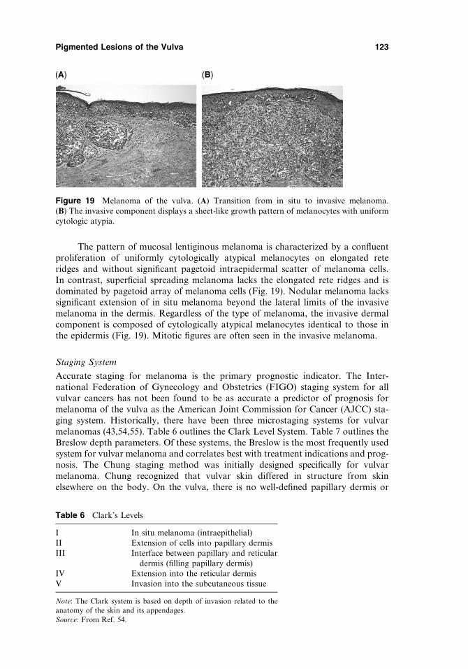
The pattern of mucosal lentiginous melanoma is characterized by a confluentproliferation of uniformly cytologically atypical melanocytes on elongated reteridges and without significant pagetoid intraepidermal scatter of melanoma cells.In contrast, superficial spreading melanoma lacks the elongated rete ridges and isdominated by pagetoid array of melanoma cells (Fig. 19). Nodular melanoma lackssignificant extension of in situ melanoma beyond the lateral limits of the invasivemelanoma in the dermis. Regardless of the type of melanoma, the invasive dermalcomponent is composed of cytologically atypical melanocytes identical to those inthe epidermis (Fig. 19). Mitotic figures are often seen in the invasive melanoma.
Staging System
Accurate staging for melanoma is the primary prognostic indicator. The Inter-national Federation of Gynecology and Obstetrics (FIGO) staging system for allvulvar cancers has not been found to be as accurate a predictor of prognosis formelanoma of the vulva as the American Joint Commission for Cancer (AJCC) sta-ging system. Historically, there have been three microstaging systems for vulvarmelanomas (43,54,55). Table 6 outlines the Clark Level System. Table 7 outlines theBreslow depth parameters. Of these systems, the Breslow is the most frequently usedsystem for vulvar melanoma and correlates best with treatment indications and prog-nosis. The Chung staging method was initially designed specifically for vulvarmelanoma. Chung recognized that vulvar skin differed in structure from skinelsewhere on the body. On the vulva, there is no well-defined papillary dermis or
Figure 19 Melanoma of the vulva. (A) Transition from in situ to invasive melanoma.(B) The invasive component displays a sheet-like growth pattern of melanocytes with uniformcytologic atypia.
Table 6 Clark’s Levels
I In situ melanoma (intraepithelial)II Extension of cells into papillary dermisIII Interface between papillary and reticular
dermis (filling papillary dermis)IV Extension into the reticular dermisV Invasion into the subcutaneous tissue
Note: The Clark system is based on depth of invasion related to the
anatomy of the skin and its appendages.
Source: From Ref. 54.
Pigmented Lesions of the Vulva 123

squamous mucous membrane in the labia. Chung’s early and advanced levels are thesame as Clark’s, but the intermediate levels differ and are based on the depth (inmillimeters) from the granular layer of the skin or the outermost epithelial layer (56).It is rarely used in clinical practice today.
Breslow Thickness
The Breslow system is based on the thickness of invasion, measured in millimeters. Itis the most commonly used system for vulvar melanoma. The thickness of themelanoma is measured from the granular layer of the overlying epithelium tothe deepest point of invasion (53).
Treatment
Historically, vulvar melanomas were treated with radical resections involving en blocvulvectomy and inguinofemoral lymphadenectomy through a single incision. Thisprocedure is associated with significant complications and offers no additional over-all or disease-free survival benefit over narrower wide local excision with adequatetumor-free margins. Vulvar melanoma is treated with a local excision with marginsbased on the Breslow depth (Table 7). It is important to obtain adequate and histo-logic tumor-free margins.
Table 8 describes the recommended margins that should be obtained forvarious tumor thicknesses. Frozen section analysis is neither sensitive nor specificfor histologic evaluation of local margins for melanoma and, therefore, is contrain-dicated (46). If permanent sections reveal positive margins, subsequent excision isindicated to obtain tumor-free margins.
Lymph Node Evaluation for Clinically Localized Disease
Elective lymph node dissection (ELND) is never indicated for melanoma basedon high levels of evidence gained through randomized trials. However, a small
Table 7 The Breslow Index
Stage I Invasion < 0.76 mmStage II Invasion between 0.76 and 1.50 mmStage III Invasion between 1.51 and 2.25 mmStage IV Invasion between 2.26 and 3.00 mmStage V Invasion > 3.00 mm
Source: From Ref. 57.
Table 8 Recommended Clinical Margins for Melanoma Excision
Tumorthickness
Recommendedclinical margins
In situ 0.5 cm�1.0 mm 1.0 cm1.01–2.0 mm 1–2 cm2.01–4.0 mm 2.0 cm> 4 mm 2.0 cm
Source: Adapted from Refs. 58 and 59.
124 Haefner et al.
subset with positive occult nodal metastasis may benefit from immediate completelymphadenectomy. Lymph node assessment by sentinel lymph node biopsy (SLNB)was developed as a minimally invasive procedure to identify those with occult met-astasis while sparing those without metastasis the morbidity associated with ELND.SLNB is recommended in relatively healthy patients with lesions equal to 1 mm ormore in tumor thickness or in cases more than 1 mm with ulceration. If the SLNBis positive, complete regional lymphadenectomy is indicated.
The sentinel lymph node can be identified from 95% to 99% of the time, with lessthan 5% false-negative results in highly experienced centers that perform the procedureroutinely (57). Sentinel nodes should be fixed and stained with both hematoxylin and eosin(H&E) and immunohistochemical stains if H&E is negative (e.g., S-100, HMB-45, and Melan-A) because H&E alone will miss up to 12% of positive nodes (58). Becausepatients with melanomas that are 1.0 mm or less in thickness rarely have nodal disease (59),sentinel node evaluation is generally reserved for patients with melanomas that are 1 mmthick or more. Randomized, prospective trials are being performed, evaluating the sur-vival benefit with SLNB. Even if no survival benefit is proven, SLNB still has severalbenefits, including accurate prognostic staging information for counseling and addressingimportant emotional distress issues associated with the diagnosis of cancer, identifyingpatients who may benefit from early therapeutic lymphadenectomy and adjuvant therapy,and grouping patients into more homogeneous cohorts for better clinical trials (60).
A staging system for cutaneous melanoma was published in 2001. The 2002 AJCCtumor node metastasis (TNM) staging system for melanoma can be found at the websiteof the National Comprehensive Cancer Network (58).
Histologic Evaluation
Numerous histologic factors are routinely reported for melanoma with potentialprognostic and treatment effects (63). Histological diagnosis and interpretation bya pathologist with expertise in melanoma is critical. The factors routinely reportedin a melanoma profile from the University of Michigan Multidisciplinary MelanomaProgram include the following: body site, classification, Clark’s level, Breslow depth,host response, regression, mitoses/mm2, growth phase, satellitosis, ulceration, angio-lymphatic spread, neurotropism, margins, and coexisting nevus.
Numerous tumor markers are available to improve sensitivity and specificity inidentifying melanoma (64), but most are easily diagnosed without additional studiesby a pathologist with expertise in melanoma. Table 9 lists other tests that are utilizedto assist in the diagnosis of a melanoma These include immunohistochemistry withantibodies to S-100 protein (S-100) (65), melanoma antigen–encoding genes MAGE 1and MAGE 3 (66,67), melanoma antigen recognized by T-cells 1 (MART-1), (68)monoclonal antibody HMB-45, and microphthalmia transcription factor (Mitf);(69–72) and molecular diagnostic tools and reverse transcriptase polymerase chainreaction (RT-PCR) positivity (73–77).
Prognosis
The most important prognostic factor is the Breslow depth of invasion of the primarylesion, followed by ulceration status and mitotic rate. The most powerful predictor ofoverall survival is the status of the SLN in those with lesions more than 1 mm in tumorthickness. Vulvar melanoma tends to be detected later than cutaneous melanomabecause of the location. This results in more advanced lesions with deeper Breslowthickness at presentation, hence a poorer prognosis. Once metastasis occurs,
Pigmented Lesions of the Vulva 125

prognosis is based on the number of nodes involved, ulceration status of the primarylesion, and status of extracapsular spread. Distant metastasis is associated with agrave prognosis with metastasis to the skin and subcutaneous tissue, at best associa-ted with an 18% five-year survival. Brain metastasis is the most ominous and is
Table 9 Tests Used to Assist in Melanoma Diagnosis
I: Immunohistochemistry(see antibodies below)
These tests are used in the traditional evaluation of melanoma.However, examination of tissues for nodal metastases by H&Ealone can result in overdiagnosis or underdiagnosis.
S-100 This calcium-channel binding protein is a sensitive marker ofmelanocyte differentiation. It is highly sensitive but not specificfor melanoma. It is also seen in benign nevus cells, histiocytes,schwannomas, neurofibromas, and malignant peripheral nervesheath tumors.
MAGE-1 and MAGE-3 These markers are highly specific for melanomas.MART-1/Melan-A The MART-1/Melan-A recognizes a protein of 18 kDa. The
MART-1 recognizes a subcellular fraction in melanosomes. Thistest is moderately sensitive but not specific for melanoma.Melan-A is a useful addition to melanoma panels because it isapparently specific for melanocytic lesions. Expression ofMART-1 micrometastasis in sentinel lymph nodes has beencorrelated with poorer survival in some studies. Studies haveshown that MART-1 is more sensitive than HMB-45 whenlabeling metastatic melanomas.
HMB-45 HMB-45 is a highly sensitive but not specific test for melanoma. Theantibody HMB-45 reacts with a 10 kDa cytoplasmic glycoprotein(part of the gp100 premelanosome complex). It demonstrates alack of reactivity with spindle melanomas, thus limiting itsusefulness as an immunohistochemical marker for melanoma.
Microphthalmiatranscription factor(Mitf) andtyrosinase
Mitf is a transcription regulator of the tyrosinase gene, whichexhibits highly sensitive and specific immunoreactivity formelanocytic lesions, with the exception of desmoplasticmelanomas. The expression of Mitf has also been linked toprognosis in melanomas of intermediate thickness.
Tyrosinase is a key enzyme in melanin biosynthesis, catalyzing theconversion of tyrosine to dihydroxyphenylalanine (DOPA) andDOPA to dopaquinone. The monoclonal antibody developedagainst tyrosinase is immunoreactive against normal melanocytes,nevus cells, and melanoma cells. Tyrosinase expression diminishesin higher-stage melanomas, with 100% staining in Stage I and IIlesions, but only 86% in Stage III and IV tumors.
II: Molecular diagnostictools
A need for greater sensitivity and specificity for lymph nodescreening has prompted the use of molecular techniques todetect micrometastasis.
III: Reverse transcriptasepolymerase chainreaction (RT-PCR)positivity
RT-PCR assay for molecular detection of melanoma-specifictumor markers such as tyrosinase messenger RNA in lymphnode tissue is more sensitive for the detection of occultmetastases than routine histology; its sensitivity approaches theoverall incidence of metastatic disease.
Abbreviations: MAGE, melanoma antigen–encoding genes; MART-1, melanoma antigen recognized by
T-cells 1.
Source: Adapted from Ref. 64.
126 Haefner et al.
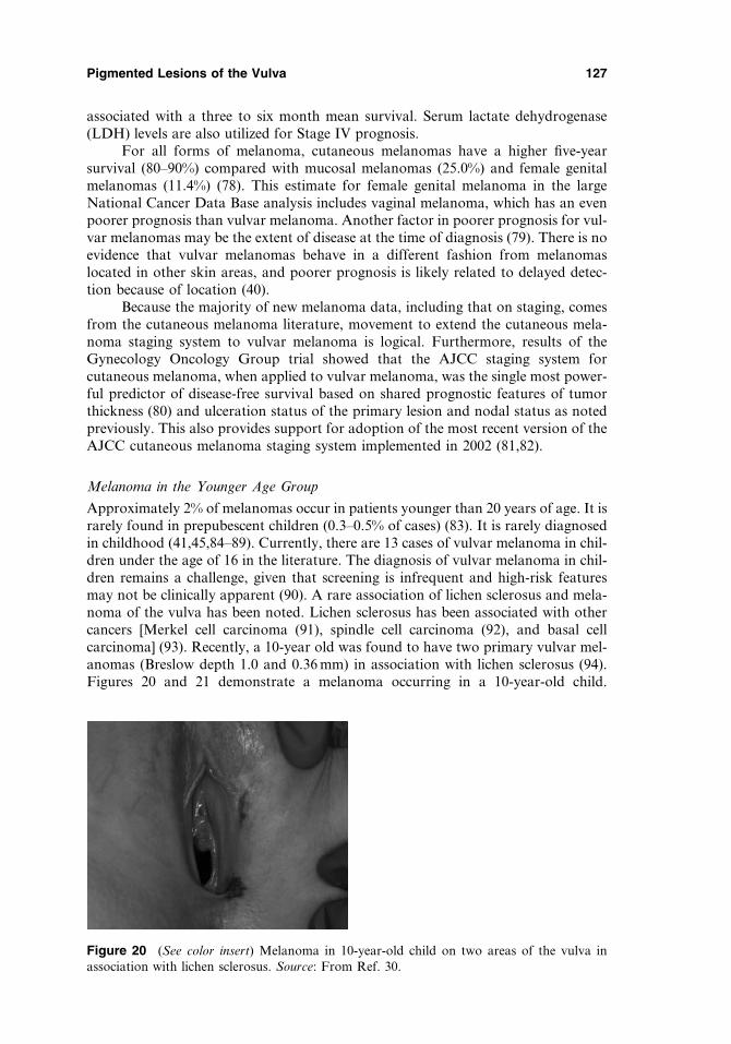
associated with a three to six month mean survival. Serum lactate dehydrogenase(LDH) levels are also utilized for Stage IV prognosis.
For all forms of melanoma, cutaneous melanomas have a higher five-yearsurvival (80–90%) compared with mucosal melanomas (25.0%) and female genitalmelanomas (11.4%) (78). This estimate for female genital melanoma in the largeNational Cancer Data Base analysis includes vaginal melanoma, which has an evenpoorer prognosis than vulvar melanoma. Another factor in poorer prognosis for vul-var melanomas may be the extent of disease at the time of diagnosis (79). There is noevidence that vulvar melanomas behave in a different fashion from melanomaslocated in other skin areas, and poorer prognosis is likely related to delayed detec-tion because of location (40).
Because the majority of new melanoma data, including that on staging, comesfrom the cutaneous melanoma literature, movement to extend the cutaneous mela-noma staging system to vulvar melanoma is logical. Furthermore, results of theGynecology Oncology Group trial showed that the AJCC staging system forcutaneous melanoma, when applied to vulvar melanoma, was the single most power-ful predictor of disease-free survival based on shared prognostic features of tumorthickness (80) and ulceration status of the primary lesion and nodal status as notedpreviously. This also provides support for adoption of the most recent version of theAJCC cutaneous melanoma staging system implemented in 2002 (81,82).
Melanoma in the Younger Age Group
Approximately 2% of melanomas occur in patients younger than 20 years of age. It israrely found in prepubescent children (0.3–0.5% of cases) (83). It is rarely diagnosedin childhood (41,45,84–89). Currently, there are 13 cases of vulvar melanoma in chil-dren under the age of 16 in the literature. The diagnosis of vulvar melanoma in chil-dren remains a challenge, given that screening is infrequent and high-risk featuresmay not be clinically apparent (90). A rare association of lichen sclerosus and mela-noma of the vulva has been noted. Lichen sclerosus has been associated with othercancers [Merkel cell carcinoma (91), spindle cell carcinoma (92), and basal cellcarcinoma] (93). Recently, a 10-year old was found to have two primary vulvar mel-anomas (Breslow depth 1.0 and 0.36 mm) in association with lichen sclerosus (94).Figures 20 and 21 demonstrate a melanoma occurring in a 10-year-old child.
Figure 20 (See color insert) Melanoma in 10-year-old child on two areas of the vulva inassociation with lichen sclerosus. Source: From Ref. 30.
Pigmented Lesions of the Vulva 127
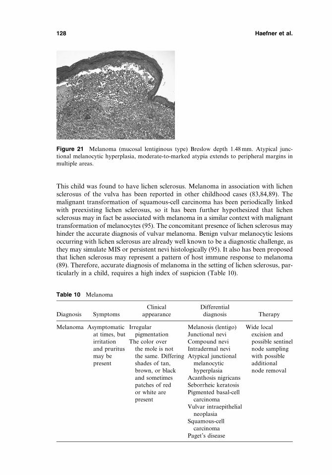
This child was found to have lichen sclerosus. Melanoma in association with lichensclerosus of the vulva has been reported in other childhood cases (83,84,89). Themalignant transformation of squamous-cell carcinoma has been periodically linkedwith preexisting lichen sclerosus, so it has been further hypothesized that lichensclerosus may in fact be associated with melanoma in a similar context with malignanttransformation of melanocytes (95). The concomitant presence of lichen sclerosus mayhinder the accurate diagnosis of vulvar melanoma. Benign vulvar melanocytic lesionsoccurring with lichen sclerosus are already well known to be a diagnostic challenge, asthey may simulate MIS or persistent nevi histologically (95). It also has been proposedthat lichen sclerosus may represent a pattern of host immune response to melanoma(89). Therefore, accurate diagnosis of melanoma in the setting of lichen sclerosus, par-ticularly in a child, requires a high index of suspicion (Table 10).
Figure 21 Melanoma (mucosal lentiginous type) Breslow depth 1.48 mm. Atypical junc-tional melanocytic hyperplasia, moderate-to-marked atypia extends to peripheral margins inmultiple areas.
Table 10 Melanoma
Diagnosis SymptomsClinical
appearanceDifferentialdiagnosis Therapy
Melanoma Asymptomaticat times, butirritationand pruritusmay bepresent
Irregularpigmentation
The color overthe mole is notthe same. Differingshades of tan,brown, or blackand sometimespatches of redor white arepresent
Melanosis (lentigo)Junctional neviCompound neviIntradermal neviAtypical junctional
melanocytichyperplasia
Acanthosis nigricansSeborrheic keratosisPigmented basal-cell
carcinomaVulvar intraepithelial
neoplasiaSquamous-cell
carcinomaPaget’s disease
Wide localexcision andpossible sentinelnode samplingwith possibleadditionalnode removal
128 Haefner et al.
CONCLUSION
A variety of vulvar conditions are associated with pigmentation abnormalities.Visual examination can help to identify abnormalities at an early disease stage.Vulvar self-examination is important, allowing patients to benefit from early diseasediagnosis. Removal of abnormal and changing vulvar lesions for histologic evalua-tion is of utmost importance. When vulvar melanomas are identified in patients, theyshould be referred to centers experienced with this disease process. A multidisciplin-ary approach to treating and following these patients is required for optimal careand outcomes. Vulvar melanoma behaves biologically similarly to cutaneous mela-noma with similar prognostic factors. An important goal is to improve knowledgeand facilitate grouping of cases from different institutions to optimize diagnosisand treatment regimens for patients with vulvar melanoma.
REFERENCES
1. Rhodes AR, Silverman RA, Harrist TJ, et al. Mucocutaneous lentigines, cardiomucocu-taneous myxomas, and multiple blue nevi: the ‘‘LAMB’’ syndrome. J Am Acad Dermatol1984; 10(1):72–82.
2. Voron DA, Hatfield HH, Kalkhoff RK. Multiple lentigines syndrome. Case report andreview of the literature. Am J Med 1976; 60(3):447–456.
3. Ridley CM. A review of the recent literature on diseases of the vulva. I. General aspects.Br J Dermatol 1972; 86(6):641–647.
4. Fargnoli MC, Orlow SJ, Semel-Concepcion J, et al. Clinicopathologic findings in theBannayan-Riley-Ruvalcaba syndrome. Arch Dermatol 1996; 132(10):1214–1218.
5. Lenane P, Sullivan DO, Keane CO, et al. The Laugier-Hunziker syndrome. J Eur AcadDermatol Venereol 2001; 15(6):574–577.
6. Pandolfino TL, Cotell S, Katta R. Pigmented vulvar macules as a presenting feature of theCarney complex. Int J Dermatol 2001; 40(11):728–730.
7. Vega Gutierrez J, Miranda Romero A, Martinez G, Torrero MV, Lopez de Juan M.Hyperpigmentation mimicking Laugier syndrome, levodopa therapy and Addison’sdisease. J Eur Acad Dermatol Venereol 2003; 17(3):324–327.
8. Milde P, Goerz G, Plewig G. Dowling-Degos disease with exclusively genital manifesta-tions. Hautarzt 1992; 43(6):369–372.
9. O’Goshi K, Terui T, Tagami H. Dowling-Degos disease affecting the vulva. Acta DermVenereol 2001; 81(2):148.
10. Stephens-Groff SM, Hansen RC, Bangert J. Benign pigmented apocrine vulvar hamarto-mas. Pediatr Dermatol 1993; 10(2):123–124.
11. Feakins RM, Lowe DG. Basal cell carcinoma of the vulva: a clinicopathologic study of 45cases. Int J Gynecol Pathol 1997; 16(4):319–324.
12. Rock B, Hood AF, Rock JA. Prospective study of vulvar nevi. J Am Acad Dermatol 1990;22(1):104–106.
13. Rock B. Pigmented lesions of the vulva. Dermatol Clin 1992; 10(2):361–370.14. Stulberg DL, Clark N. Common hyperpigmentation disorders in adults. Part 1. Diagnos-
tic approach, cafe au lait macules, diffuse hyperpigmentation, sun exposure, andphototoxic reactions. Am Fam Physician 2003; 68(10):1955–1960.
15. Grasinger CC, Wild RA, Parker IJ. Vulvar acanthosis nigricans: a marker for insulinresistance in hirsute women. Fertil Steril 1993; 59(3):583–586.
16. http://www.rarediseases.org/search/rdbdetail_abstract.html?disname¼Acanthosis%20Nigricans (accessed October 2005).
17. Stuart CA, Driscoll MS, Lundquist KF, et al. Acanthosis nigricans. J Basic Clin PhysiolPharmacol 1998; 9(2–4):407–418.
Pigmented Lesions of the Vulva 129

18. Levine N, Baron J, Cruz PD, et al. Acanthosis nigricans. Emedicine June 2002 www.emedicine.com/derm/topic1.htm (accessed October 2005).
19. Cruz PD Jr., Hud JA Jr. Excess insulin binding to insulin-like growth factor receptors: pro-posed mechanism for acanthosis nigricans. J Invest Dermatol 1992; 98(6 suppl):82S–85S.
20. Schwartz RA. Acanthosis nigricans. J Am Acad Dermatol 1994; 31(1):1–19; quiz 20–22.21. Katz AS, Goff DC, Feldman SR. Acanthosis nigricans in obese patients: presentations and
implications for prevention of atherosclerotic vascular disease. Dermatol Online J 2000; 6(1):1.22. Jackson R. Melanosis of the vulva. J Dermatol Surg Oncol 1984; 10(2):119–121.23. Lenane P, Keane CO, Connell BO, et al. Genital melanotic macules: clinical, histologic,
immunohistochemical, and ultrastructural features. J Am Acad Dermatol 2000;42(4):640–644.
24. Rudolph RI. Vulvar melanosis. J Am Acad Dermatol 1990; 23(5 Pt 2):982–984.25. Estrada R, Kaufman R. Benign vulvar melanosis. J Reprod Med 1993; 38(1):5–8.26. Okulicz JF, Jozwiak S, Schwartz, RA. Lentigo Emedicine. www.emedicine.com/derm/
topic221.htm (accessed October 2005).27. El Shabrawi-Caelen L, Soyer HP, Schaeppi H, et al. Genital lentigines and melanocytic
nevi with superimposed lichen sclerosus: a diagnostic challenge. J Am Acad Dermatol2004; 50(5):690–694.
28. Jih DM, Elder DE, Elenitsas R. A histopathologic evaluation of vulvar melanosis. ArchDermatol 1999; 135(7):857–858.
29. Wolff K, Johnson RS, eds. Fitzpatrick’s Color Atlas and Synopsis of Dermatology.Section 12. 5th ed. McGraw-Hill Professional, New York, New York, 2005 (online.statref. com).
30. Wechter ME, Gruber SB, Haefner HK, et al. Vulvar Melanoma: a report of 20 cases andreview of the literature. J Am Acad Dermatol 2004; 50(4):554–562.
31. Wilkinson EJ, Kneale B, Lynch PJ. Report of the ISSVD terminology committee.J Reprod Med 1986; 31:973–974.
31a.Sideri M, Jones RW, Wilkinson EJ, et al. Squamous vulvar intraepithelial neoplasia: 2004modified terminology, ISSVD vulvar oncology subcommittee. J Reprod Med 2005;50:807–810.
32. NIH Consensus Conference. Diagnosis and treatment of early melanoma. JAMA 1992;268(10):1314–1319.
33. www.nccn.org.34. Verschraegen CF, Benjapibal M, Supakarapongkul W, et al. Vulvar melanoma at the M.
D. Anderson Cancer Center: 25 years later. Int J Gynecol Cancer 2001; 11(5):359–364.35. Raber G, Mempel V, Jackisch C, et al. Malignant melanoma of the vulva. Report of 89
patients. Cancer 1996; 78(11):2353–2358.36. Ragnarsson-Olding BK, Nilsson BR, Kanter-Lewensohn LR, et al. Malignant melanoma
of the vulva in a nationwide, 25-year study of 219 Swedish females: predictors of survival.Cancer 1999; 86(7):1285–1293.
37. Ragnarsson-Olding BK, Kanter-Lewensohn LR, Lagerlof B, et al. Malignant melanomaof the vulva in a nationwide, 25-year study of 219 Swedish females: clinical observationsand histopathologic features. Cancer 1999; 86(7):1273–1284.
38. Scheistroen M, Trope C, Kaern J, et al. Malignant melanoma of the vulva FIGO stage I:evaluation of prognostic factors in 43 patients with emphasis on DNA ploidy and surgicaltreatment. Gynecol Oncol 1996; 61(2):253–258.
39. Scheistroen M, Trope C, Koern J, et al. Malignant melanoma of the vulva. Evaluationof prognostic factors with emphasis on DNA ploidy in 75 patients. Cancer 1995; 75(1):72–80.
40. Phillips GL, Bundy BN, Okagaki T, et al. Malignant melanoma of the vulva treated byradical hemivulvectomy. A prospective study of the gynecologic oncology group. Cancer1994; 73(10):2626–2632.
41. Ragnarsson-Olding B, Johansson H, Rutqvist LE, et al. Malignant melanoma of the vulvaand vagina. Trends in incidence, age distribution, and long-term survival among 245consecutive cases in Sweden 1960–1984. Cancer 1993; 71(5):1893–1897.
130 Haefner et al.
42. Bradgate MG, Rollason TP, McConkey CC, et al. Malignant melanoma of thevulva: a clinicopathological study of 50 women. Br J Obstet Gynaecol 1990; 97(2):124–133.
43. Chung AF, Woodruff JM, Lewis JL Jr. Malignant melanoma of the vulva: a report of 44cases. Obstet Gynecol 1975; 45(6):638–646.
44. Stang A, Streller B, Eisinger B, et al. Population-based incidence rates of malignantmelanoma of the vulva in Germany. Gynecol Oncol 2005; 96(1):216–221.
45. Creasman WT, Phillips JL, Menck HR. A survey of hospital management practices forvulvar melanoma. J Am Coll Surg 1999; 188(6):670–675.
46. Wechter ME, Reynolds RK, Haefner HK, et al. Vulvar melanoma: review of diagnosis,staging, and therapy. J Low Genit Tract Dis 2004; 8:58–69.
47. Raspagliesi F, Ditto A, Paladini D, et al. Prognostic indicators in melanoma of the vulva.Ann Surg Oncol 2000; 7(10):738–742.
48. Irvin WP Jr., Legallo RL, Stoler MH, et al. Vulvar melanoma: a retrospective analysis andliterature review. Gynecol Oncol 2001; 83(3):457–465.
49. http://www.nccn.org/patients/patient_gls/_english/_melanoma/2_detection.asp.50. Granter SR, Lazar AJF, Haefner HK, McKee PH. Melanocytic Lesions of the Vulva, in
Diagnostic Gynecologic and Obstetric Pathology. Crum CP, Lee KR eds. ElsevierSaunders, 2006:169.
51. Johnson TM, Smith JW 2nd, Nelson BR, et al. Current therapy for cutaneous melanoma.J Am Acad Dermatol 1995 32 (5 Pt 1):689–707.
52. Arca MJ, Biermann JS, Johnson TM, et al. Biopsy techniques for skin, soft-tissue, andbone neoplasms. Surg Oncol Clin N Am 1995; 4(1):157–174.
53. Finan MA, Barre G. Bartholin’s gland carcinoma, malignant melanoma and other raretumours of the vulva. Best Pract Res Clin Obstet Gynaecol 2003; 17(4):609–633..
54. Clark WH Jr., From L, Bernardino EA, Mihm MC. The histogenesis and behavior ofprimary human malignant melanomas of the skin. Cancer Res 1969; 29:705–727.
55. Breslow A. Thickness, cross-sectional areas and depth of invasion in the prognosis ofcutaneous melanoma. Ann Surg 1970; 172(5):902–908.
56. Hopkins MP, Nemunaitis-Keller J. Carcinoma of the vulva. Obstet Gynecol Clin NorthAm 2001; 28(4):791–804.
57. Reintgen D, Thompson JF, Gershenwald JE. Intraoperative mapping and sentinel nodetechnology. In: Balch CM, Houghton AN, Sober AJ, Soong S, eds. Cutaneous melanoma.4th ed. St. Louis: Quality Medical Publishing, 2003:353–377.
58. Yu LL, Flotte TJ, Tanabe KK, et al. Detection of microscopic melanoma metastases insentinel lymph nodes. Cancer 1999; 86:617–627.
59. Bleicher RJ, Essner R, Foshag LJ, et al. Role of sentinel lymphadenectomy in thin inva-sive cutaneous melanomas. J Clin Oncol 2003; 21(7):1326–1331.
60. McMasters KM, Reintgen DS, Ross MI, et al. Sentinel lymph node biopsy for melanoma:controversy despite widespread agreement. J Clin Oncol 2001; 19(11):2851–2855.
61. Balch CM, Buzaid AC, Soong SJ, et al. Final version of the American Joint Committeeon cancer staging system for cutaneous melonoma. J Clin Oncol 2001; 19:3635–3648.
62. http://www.nccn.org/professionals/physician_gls/PDF/melanoma.pdf.63. Wilkinson EJ. Protocol for the examination of specimens from patients with carcinomas
and malignant melanomas of the vulva: a basis for checklists. Cancer Committee of theAmerican College of Pathologists. Arch Pathol Lab Med 2000; 124(1):51–56.
64. Armstrong AW, Liu V, Mihm MC, Jr. Pathologic characteristics of melonoma. 2005,UptoDate Online; www.uptodate.com.
65. Blessing K, Sanders DS, Grant JJ. Comparison of immunohistochemical staining of thenovel antibody melan-A with S100 protein and HMB-45 in malignant melanoma andmelanoma variants. Histopathology 1998; 32(2):139–146.
66. Chen YT, Stockert E, Chen Y, et al. Identification of the MAGE-1 gene product by mono-clonal and polyclonal antibodies. Proc Natl Acad Sci U S A 1994; 91(3):1004–1008.
Pigmented Lesions of the Vulva 131
67. Hofbauer GF, Schaefer C, Noppen C, et al. MAGE-3 immunoreactivity in formalin-fixed,paraffin-embedded primary and metastatic melanoma: frequency and distribution. Am JPathol 1997; 151(6):1549–1553.
68. Hochberg M, Lotem M, Gimon Z, et al. Expression of tyrosinase, MIA and MART-1 in sen-tinel lymph nodes of patients with malignant melanoma. Br J Dermatol 2002; 146(2):244–249.
69. King R, Googe PB, Weilbaecher KN, et al. Microphthalmia transcription factorexpression in cutaneous benign, malignant melanocytic, and nonmelanocytic tumors.Am J Surg Pathol 2001; 25(1):51–57.
70. Busam KJ, Iversen K, Coplan KC, et al. Analysis of microphthalmia transcription factorexpression in normal tissues and tumors, and comparison of its expression with S-100 pro-tein, gp100, and tyrosinase in desmoplastic malignant melanoma. Am J Surg Pathol 2001;25(2):197–204.
71. Salti GI, Manougian T, Farolan M, et al. Micropthalmia transcription factor: a newprognostic marker in intermediate-thickness cutaneous malignant melanoma. CancerRes 2000; 60(18):5012–5016.
72. Hofbauer GF, Kamarashev J, Geertsen R, et al. Tyrosinase immunoreactivity in formalin-fixed, paraffin-embedded primary and metastatic melanoma: frequency and distribution.J Cutan Pathol 1998; 25(4):204–209.
73. Bostick PJ, Morton DL, Turner RR, et al. Prognostic significance of occult metastasesdetected by sentinel lymphadenectomy and reverse transcriptase-polymerase chainreaction in early-stage melanoma patients. J Clin Oncol 1999; 17(10):3238–3244.
74. Shivers SC, Wang X, Li W, et al. Molecular staging of malignant melanoma: correlationwith clinical outcome. JAMA 1998; 280(16):1410–1415.
75. Goydos JS, Ravikumar TS, Germino FJ, et al. Minimally invasive staging of patients withmelanoma: sentinel lymphadenectomy and detection of the melanoma-specific proteinsMART-1 and tyrosinase by reverse transcriptase polymerase chain reaction. J Am CollSurg 1998; 187(2):182–188; discussion 188–190.
76. Li W, Stall A, Shivers SC, et al. Clinical relevance of molecular staging for melanoma:comparison of RT-PCR and immunohistochemistry staining in sentinel lymph nodes ofpatients with melanoma. Ann Surg 2000; 231(6):795–803.
77. Blaheta HJ, Ellwanger U, Schittek B, et al. Examination of regional lymph nodes bysentinel node biopsy and molecular analysis provides new staging facilities in primarycutaneous melanoma. J Invest Dermatol 2000; 114(4):637–642.
78. Chang AE, Karnell LH, Menck HR. The National Cancer Data Base report on cutaneousand noncutaneous melanoma: a summary of 84,836 cases from the past decade. TheAmerican College of Surgeons Commission on Cancer and the American Cancer Society.Cancer 1998; 83(8):1664–1678.
79. Panizzon RG. Vulvar melanoma. Semin Dermatol 1996; 15(1):67–70.80. Ragnarsson-Olding BK. Primary malignant melanoma of the vulva–an aggressive tumor
for modeling the genesis of non-UV light-associated melanomas. Acta Oncol 2004;43(5):421–435.
81. Balch CM, Soong SJ, Gershenwald JE, et al. Prognostic factors analysis of 17,600 mela-noma patients: validation of the American Joint Committee on Cancer melanoma stagingsystem. J Clin Oncol 2001; 19(16):3622–3634.
82. Balch CM, Buzaid AC, Soong SJ, et al. Final version of the American Joint Committee onCancer staging system for cutaneous melanoma. J ClinOncol 2001; 19(16):3635–3648.
83. Egan CA, Bradley RR, Logsdon VK, et al. Vulvar melanoma in childhood. Arch Derma-tol 1997; 133(3):345–348.
84. Friedman RJ, Kopf AW, Jones WB. Malignant melanoma in association with lichensclerosus on the vulva of a 14-year-old. Am J Dermatopathol 1984; 6(suppl):253–256.
85. Pack GT, Oropeza R. A comparative study of melanomas and 58 epidermoid carcinomasof the vulva: a review of 44 melanomas and 58 epidermoid carcinomas (1930–1965). RevSurg 1967; 24(5):305–324.
132 Haefner et al.
86. Trimble EL, Lewis JL Jr., Williams LL, et al. Management of vulvar melanoma. GynecolOncol 1992; 45(3):254–258.
87. Woolcott RJ, Henry RJ, Houghton CR. Malignant melanoma of the vulva: Australianexperience. J Reprod Med 1988; 33(8):699–702.
88. Hulagu C, Erez S. Juvenile melanoma of clitoris. J Obstet Gynaecol Br Commonw 1973;80(1):89–91.
89. Hassanein AM, Mrstik ME, Hardt NS, et al. Malignant melanoma associated with lichensclerosus in the vulva of a 10-year-old. Pediatr Dermatol 2004; 21(4):473–476.
90. Ruiz-Maldonado R, Orozco-Covarrubias ML. Malignant melanoma in children. A review.Arch Dermatol 1997; 133(3):363–371.
91. Bottles K, Lacey CG, Goldberg J, et al. Merkel cell carcinoma of the vulva. Obstet Gyne-col 1984; 63(3 suppl):61S–65S.
92. Santos-Briz A, Antunez P, Lopez-Rios F, et al. Human papillomavirus-negative spindlecell carcinoma of the vulva associated with lichen sclerosus: case report and literaturereview. Am J Dermatopathol 2002; 24(2):135–138.
93. Thomas RH, McGibbon DH, Munro DD. Basal cell carcinoma of the vulva in associationwith vulval lichen sclerosus et atrophicus. J R Soc Med 1985; 78(suppl 11):16–18.
94. Rosamilia LL, Schwartz JL, Lowe L, et al. Letter: Vulvar melanoma in a 10 year old childin association with lichen sclerosus. J Am Acad Dermatol 2006; 54(2 suppl):S52–S53.
95. Carlson JA, Mu XC, Slominski A, et al. Melanocytic proliferations associated with lichensclerosus. Arch Dermatol 2002; 138(1):77–87.
Pigmented Lesions of the Vulva 133

6Neoplasms and Related Lesionsof the Vulva
Debra S. HellerDepartments of Pathology and Laboratory Medicine, and Obstetrics, Gynecology,and Women’s Health, UMDNJ–New Jersey Medical School, Newark,New Jersey, U.S.A.
CYSTS
Bartholin’s Cyst and Abscess
The two Bartholin’s gland ducts drain into the posterolateral vestibule (Fig. 1Aand B). Bartholin’s duct cysts develop secondary to blockage of the duct and maybecome infected, leading to abscesses. A variety of aerobic and anaerobic organismshave been isolated from these abscesses, including Neisseria gonorrhoeae, Chlamydiatrachomatis, and various bowel flora (1). Bartholin’s duct cysts/abscesses are mostcommon during reproductive life. A Bartholin’s mass in a woman over the age of40 always raises the question of a malignancy, and a tissue specimen for pathologicalexamination is indicated. Most therapeutic options for treatment of a Bartholin’scyst or abscess do not lead to a surgical pathology specimen; however, if one isreceived, the Bartholin’s gland and duct may be recognized by their unique mix ofepithelial types; mucinous in the glands, transitional in the ducts, and squamousat the orifice, with some admixture of types in the ducts common (Table 1).
Epidermal Inclusion Cyst
Epidermal inclusion cysts are common on the vulva, and may be multiple (Fig. 2Aand B). They present as firm smooth nodules, and are filled with keratinaceousdebris. They tend to enlarge. Histologically they are lined by stratified squamous epi-thelium, which may contain the granular cell layer (2). The lesions have alsooccurred in the clitoral area in women undergoing female circumcision (3). Theydo not require therapy, but may be excised if symptomatic (Table 2).
Mucinous and Ciliated Cysts
Mucinous cysts and ciliated cysts of the vulva are uncommon lesions (Fig. 3A–C) (4–7).Their origin is uncertain, with theories including paramesonephric or mesonephric
135
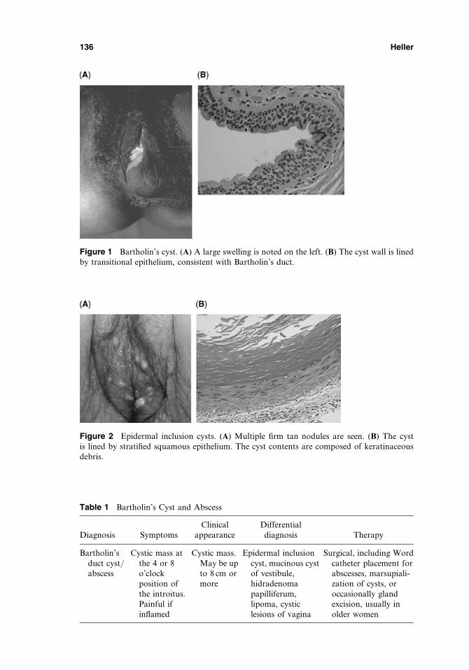
Figure 1 Bartholin’s cyst. (A) A large swelling is noted on the left. (B) The cyst wall is linedby transitional epithelium, consistent with Bartholin’s duct.
Figure 2 Epidermal inclusion cysts. (A) Multiple firm tan nodules are seen. (B) The cystis lined by stratified squamous epithelium. The cyst contents are composed of keratinaceousdebris.
Table 1 Bartholin’s Cyst and Abscess
Diagnosis SymptomsClinical
appearanceDifferentialdiagnosis Therapy
Bartholin’sduct cyst/abscess
Cystic mass atthe 4 or 8o’clockposition ofthe introitus.Painful ifinflamed
Cystic mass.May be upto 8 cm ormore
Epidermal inclusioncyst, mucinous cystof vestibule,hidradenomapapilliferum,lipoma, cysticlesions of vagina
Surgical, including Wordcatheter placement forabscesses, marsupiali-zation of cysts, oroccasionally glandexcision, usually inolder women
136 Heller
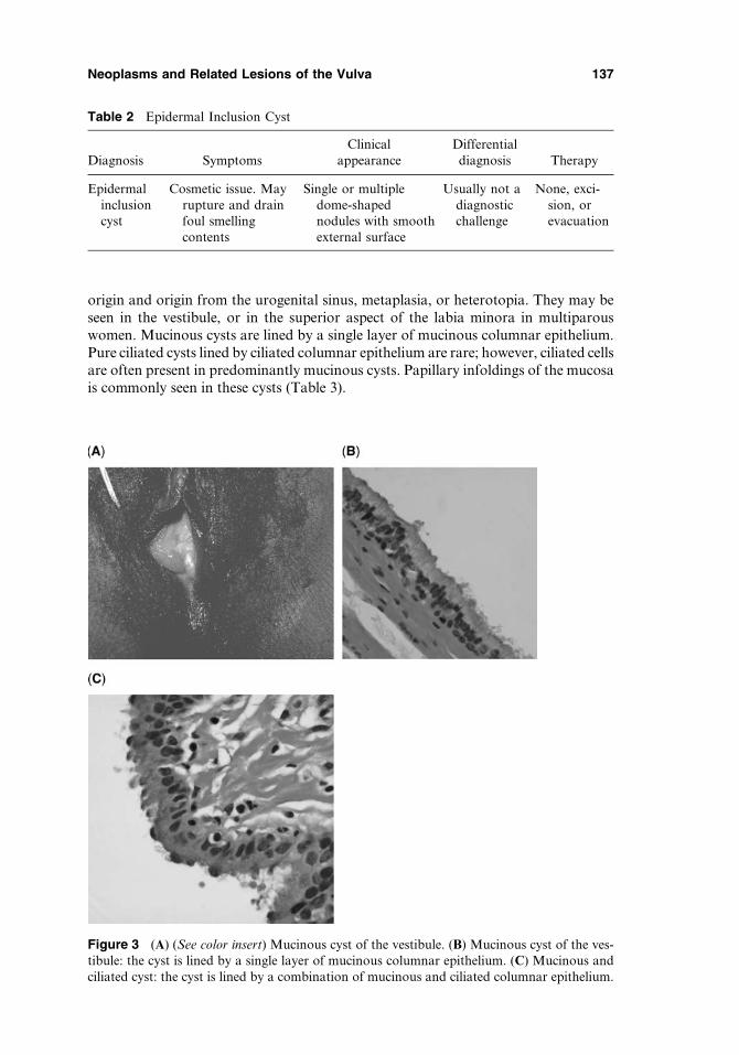
origin and origin from the urogenital sinus, metaplasia, or heterotopia. They may beseen in the vestibule, or in the superior aspect of the labia minora in multiparouswomen. Mucinous cysts are lined by a single layer of mucinous columnar epithelium.Pure ciliated cysts lined by ciliated columnar epithelium are rare; however, ciliated cellsare often present in predominantly mucinous cysts. Papillary infoldings of the mucosais commonly seen in these cysts (Table 3).
Table 2 Epidermal Inclusion Cyst
Diagnosis SymptomsClinical
appearanceDifferentialdiagnosis Therapy
Epidermalinclusioncyst
Cosmetic issue. Mayrupture and drainfoul smellingcontents
Single or multipledome-shapednodules with smoothexternal surface
Usually not adiagnosticchallenge
None, exci-sion, orevacuation
Figure 3 (A) (See color insert) Mucinous cyst of the vestibule. (B) Mucinous cyst of the ves-tibule: the cyst is lined by a single layer of mucinous columnar epithelium. (C) Mucinous andciliated cyst: the cyst is lined by a combination of mucinous and ciliated columnar epithelium.
Neoplasms and Related Lesions of the Vulva 137
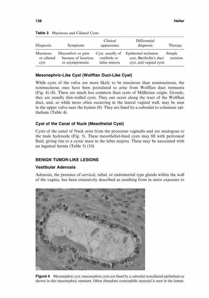
Mesonephric-Like Cyst (Wolffian Duct-Like Cyst)
While cysts of the vulva are more likely to be mucinous than nonmucinous, thenonmucinous ones have been postulated to arise from Wolffian duct remnants(Fig. 4) (8). These are much less common than cysts of Mullerian origin. Grossly,they are usually thin-walled cysts. They can occur along the tract of the Wolffianduct, and, so while more often occurring in the lateral vaginal wall, may be seenin the upper vulva near the hymen (9). They are lined by a cuboidal to columnar epi-thelium (Table 4).
Cyst of the Canal of Nuck (Mesothelial Cyst)
Cysts of the canal of Nuck arise from the processus vaginalis and are analogous tothe male hydrocele (Fig. 5). These mesothelial-lined cysts may fill with peritonealfluid, giving rise to a cystic mass in the labia majora. These may be associated withan inguinal hernia (Table 5) (10).
BENIGN TUMOR-LIKE LESIONS
Vestibular Adenosis
Adenosis, the presence of cervical, tubal, or endometrial type glands within the wallof the vagina, has been extensively described as resulting from in utero exposure to
Table 3 Mucinous and Ciliated Cysts
Diagnosis SymptomsClinical
appearanceDifferentialdiagnosis Therapy
Mucinousor ciliatedcyst
Discomfort or painbecause of location,or asymptomatic
Cyst, usually ofvestibule orlabia minora
Epidermal inclusioncyst, Bartholin’s ductcyst, and vaginal cysts
Simpleexcision
Figure 4 Mesonephric cyst: mesonephric cysts are lined by a cuboidal nonciliated epithelium asshown in this mesonephric remnant. Often abundant eosinophilic material is seen in the lumen.
138 Heller
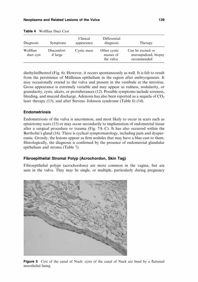
diethylstilbestrol (Fig. 6). However, it occurs spontaneously as well. It is felt to resultfrom the persistence of Mullenan epithelium in the region after embryogenesis. Itmay occasionally extend to the vulva and present in the vestibule at the introitus.Gross appearance is extremely variable and may appear as redness, nodularity, orgranularity, cysts, ulcers, or protuberances (12). Possible symptoms include soreness,bleeding, and mucoid discharge. Adenosis has also been reported as a sequela of CO2
laser therapy (13), and after Stevens–Johnson syndrome (Table 6) (14).
Endometriosis
Endometriosis of the vulva is uncommon, and most likely to occur in scars such asepisiotomy scars (15) or may occur secondarily to implantation of endometrial tissueafter a surgical procedure or trauma (Fig. 7A–C). It has also occurred within theBartholin’s gland (16). There is cyclical symptomatology, including pain and dyspar-eunia. Grossly, the lesions appear as firm nodules that may have a blue cast to them.Histologically, the diagnosis is confirmed by the presence of endometrial glandularepithelium and stroma (Table 7).
Fibroepithelial Stromal Polyp (Acrochordon, Skin Tag)
Fibroepithelial polyps (acrochordons) are more common in the vagina, but areseen in the vulva. They may be single, or multiple, particularly during pregnancy
Table 4 Wolffian Duct Cyst
Diagnosis SymptomsClinical
appearanceDifferentialdiagnosis Therapy
Wolffianduct cyst
Discomfortif large
Cystic mass Other cysticmasses ofthe vulva
Can be excised ormarsupialized, biopsyrecommended
Figure 5 Cyst of the canal of Nuck: cysts of the canal of Nuck are lined by a flattenedmesothelial lining.
Neoplasms and Related Lesions of the Vulva 139
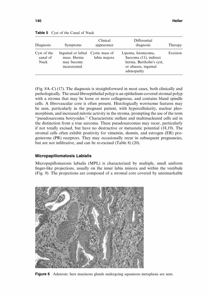
(Fig. 8A–C) (17). The diagnosis is straightforward in most cases, both clinically andpathologically. The usual fibroepithelial polyp is an epithelium-covered stromal polypwith a stroma that may be loose or more collagenous, and contains bland spindlecells. A fibrovascular core is often present. Histologically worrisome features maybe seen, particularly in the pregnant patient, with hypercellularity, nuclear pleo-morphism, and increased mitotic activity in the stroma, prompting the use of the term‘‘pseudosarcoma botryoides.’’ Characteristic stellate and multinucleated cells aid inthe distinction from a true sarcoma. These pseudosarcomas may recur, particularlyif not totally excised, but have no destructive or metastatic potential (18,19). Thestromal cells often exhibit positivity for vimentin, desmin, and estrogen (ER) pro-gesterone (PR) receptors. They may occasionally recur in subsequent pregnancies,but are not infiltrative, and can be re-excised (Table 8) (20).
Micropapillomatosis Labialis
Micropapillomatosis labialis (MPL) is characterized by multiple, small uniformfinger-like projections, usually on the inner labia minora and within the vestibule(Fig. 9). The projections are composed of a stromal core covered by unremarkable
Table 5 Cyst of the Canal of Nuck
Diagnosis SymptomsClinical
appearanceDifferentialdiagnosis Therapy
Cyst of thecanal ofNuck
Inguinal or labialmass. Herniamay becomeincarcerated
Cystic mass oflabia majora
Lipoma, leiomyoma,Sarcoma (11), indirecthernia, Bartholin’s cyst,or abscess, inguinaladenopathy
Excision
Figure 6 Adenosis: here mucinous glands undergoing squamous metaplasia are seen.
140 Heller
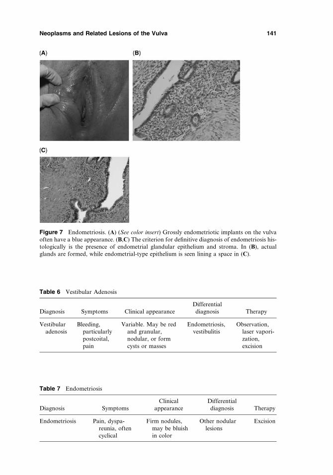
Table 6 Vestibular Adenosis
Diagnosis Symptoms Clinical appearanceDifferentialdiagnosis Therapy
Vestibularadenosis
Bleeding,particularlypostcoital,pain
Variable. May be redand granular,nodular, or formcysts or masses
Endometriosis,vestibulitis
Observation,laser vapori-zation,excision
Figure 7 Endometriosis. (A) (See color insert) Grossly endometriotic implants on the vulvaoften have a blue appearance. (B,C) The criterion for definitive diagnosis of endometriosis his-tologically is the presence of endometrial glandular epithelium and stroma. In (B), actualglands are formed, while endometrial-type epithelium is seen lining a space in (C).
Table 7 Endometriosis
Diagnosis SymptomsClinical
appearanceDifferentialdiagnosis Therapy
Endometriosis Pain, dyspa-reunia, oftencyclical
Firm nodules,may be bluishin color
Other nodularlesions
Excision
Neoplasms and Related Lesions of the Vulva 141
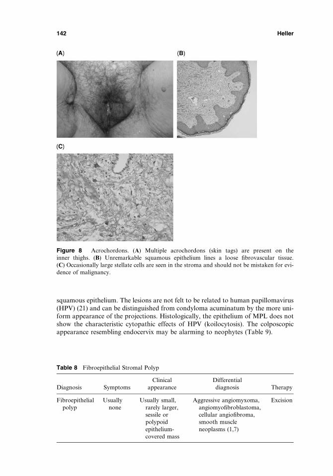
squamous epithelium. The lesions are not felt to be related to human papillomavirus(HPV) (21) and can be distinguished from condyloma acuminatum by the more uni-form appearance of the projections. Histologically, the epithelium of MPL does notshow the characteristic cytopathic effects of HPV (koilocytosis). The colposcopicappearance resembling endocervix may be alarming to neophytes (Table 9).
Figure 8 Acrochordons. (A) Multiple acrochordons (skin tags) are present on theinner thighs. (B) Unremarkable squamous epithelium lines a loose fibrovascular tissue.(C) Occasionally large stellate cells are seen in the stroma and should not be mistaken for evi-dence of malignancy.
Table 8 Fibroepithelial Stromal Polyp
Diagnosis SymptomsClinical
appearanceDifferentialdiagnosis Therapy
Fibroepithelialpolyp
Usuallynone
Usually small,rarely larger,sessile orpolypoidepithelium-covered mass
Aggressive angiomyxoma,angiomyofibroblastoma,cellular angiofibroma,smooth muscleneoplasms (1,7)
Excision
142 Heller
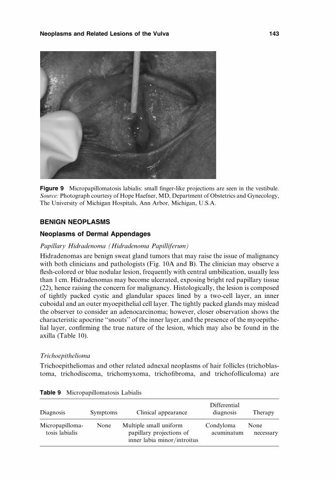
BENIGN NEOPLASMS
Neoplasms of Dermal Appendages
Papillary Hidradenoma (Hidradenoma Papilliferum)
Hidradenomas are benign sweat gland tumors that may raise the issue of malignancywith both clinicians and pathologists (Fig. 10A and B). The clinician may observe aflesh-colored or blue nodular lesion, frequently with central umbilication, usually lessthan 1 cm. Hidradenomas may become ulcerated, exposing bright red papillary tissue(22), hence raising the concern for malignancy. Histologically, the lesion is composedof tightly packed cystic and glandular spaces lined by a two-cell layer, an innercuboidal and an outer myoepithelial cell layer. The tightly packed glands may misleadthe observer to consider an adenocarcinoma; however, closer observation shows thecharacteristic apocrine ‘‘snouts’’ of the inner layer, and the presence of the myoepithe-lial layer, confirming the true nature of the lesion, which may also be found in theaxilla (Table 10).
Trichoepithelioma
Trichoepitheliomas and other related adnexal neoplasms of hair follicles (trichoblas-toma, trichodiscoma, trichomyxoma, trichofibroma, and trichofolliculoma) are
Figure 9 Micropapillomatosis labialis: small finger-like projections are seen in the vestibule.Source: Photograph courtesy of Hope Haefner, MD, Department of Obstetrics and Gynecology,The University of Michigan Hospitals, Ann Arbor, Michigan, U.S.A.
Table 9 Micropapillomatosis Labialis
Diagnosis Symptoms Clinical appearanceDifferentialdiagnosis Therapy
Micropapilloma-tosis labialis
None Multiple small uniformpapillary projections ofinner labia minor/introitus
Condylomaacuminatum
Nonenecessary
Neoplasms and Related Lesions of the Vulva 143
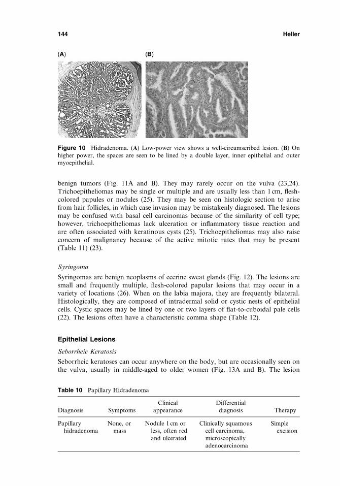
benign tumors (Fig. 11A and B). They may rarely occur on the vulva (23,24).Trichoepitheliomas may be single or multiple and are usually less than 1 cm, flesh-colored papules or nodules (25). They may be seen on histologic section to arisefrom hair follicles, in which case invasion may be mistakenly diagnosed. The lesionsmay be confused with basal cell carcinomas because of the similarity of cell type;however, trichoepitheliomas lack ulceration or inflammatory tissue reaction andare often associated with keratinous cysts (25). Trichoepitheliomas may also raiseconcern of malignancy because of the active mitotic rates that may be present(Table 11) (23).
Syringoma
Syringomas are benign neoplasms of eccrine sweat glands (Fig. 12). The lesions aresmall and frequently multiple, flesh-colored papular lesions that may occur in avariety of locations (26). When on the labia majora, they are frequently bilateral.Histologically, they are composed of intradermal solid or cystic nests of epithelialcells. Cystic spaces may be lined by one or two layers of flat-to-cuboidal pale cells(22). The lesions often have a characteristic comma shape (Table 12).
Epithelial Lesions
Seborrheic Keratosis
Seborrheic keratoses can occur anywhere on the body, but are occasionally seen onthe vulva, usually in middle-aged to older women (Fig. 13A and B). The lesion
Figure 10 Hidradenoma. (A) Low-power view shows a well-circumscribed lesion. (B) Onhigher power, the spaces are seen to be lined by a double layer, inner epithelial and outermyoepithelial.
Table 10 Papillary Hidradenoma
Diagnosis SymptomsClinical
appearanceDifferentialdiagnosis Therapy
Papillaryhidradenoma
None, ormass
Nodule 1 cm orless, often redand ulcerated
Clinically squamouscell carcinoma,microscopicallyadenocarcinoma
Simpleexcision
144 Heller

Figure 11 Trichoepithelioma. (A) Epithelial subunits surrounded by a dense fibroconnectivetissue stroma. (B) Detail of basaloid cells of an epithelial subunit.
Table 11 Trichoepithelioma
Diagnosis SymptomsClinical
appearanceDifferentialdiagnosis Therapy
Trichoepithelioma None Small, flesh-colored plaqueor nodule
Basal orsquamous cellcarcinoma
Excision
Figure 12 (See color insert) Syringoma: the characteristic comma-shaped epithelial nests in acollagenous background are seen.
Table 12 Syringoma
Diagnosis SymptomsClinical
appearance Differential diagnosis Therapy
Syringoma None orpruritus
Multiple smallflesh to yellowcoloredpapules ornodules
Candidiasis, lichen simplexchronicus, condylomaacuminatum, hemangioma,lymphangioma circumscriptum,Fox-Fordyce disease (26)
None usuallyrequired,but aresometimesexcised
Neoplasms and Related Lesions of the Vulva 145
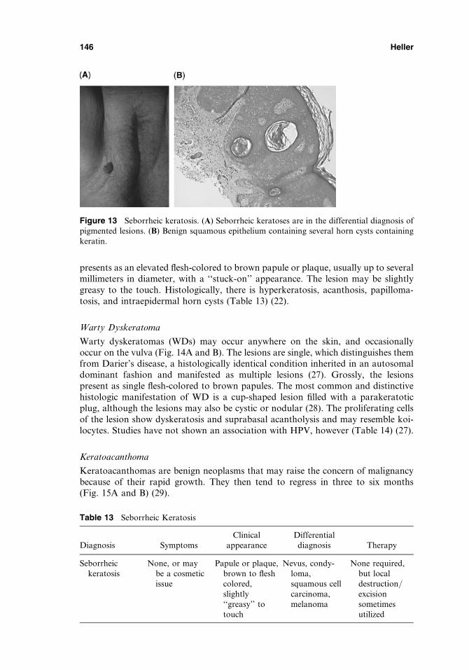
presents as an elevated flesh-colored to brown papule or plaque, usually up to severalmillimeters in diameter, with a ‘‘stuck-on’’ appearance. The lesion may be slightlygreasy to the touch. Histologically, there is hyperkeratosis, acanthosis, papilloma-tosis, and intraepidermal horn cysts (Table 13) (22).
Warty Dyskeratoma
Warty dyskeratomas (WDs) may occur anywhere on the skin, and occasionallyoccur on the vulva (Fig. 14A and B). The lesions are single, which distinguishes themfrom Darier’s disease, a histologically identical condition inherited in an autosomaldominant fashion and manifested as multiple lesions (27). Grossly, the lesionspresent as single flesh-colored to brown papules. The most common and distinctivehistologic manifestation of WD is a cup-shaped lesion filled with a parakeratoticplug, although the lesions may also be cystic or nodular (28). The proliferating cellsof the lesion show dyskeratosis and suprabasal acantholysis and may resemble koi-locytes. Studies have not shown an association with HPV, however (Table 14) (27).
Keratoacanthoma
Keratoacanthomas are benign neoplasms that may raise the concern of malignancybecause of their rapid growth. They then tend to regress in three to six months(Fig. 15A and B) (29).
Figure 13 Seborrheic keratosis. (A) Seborrheic keratoses are in the differential diagnosis ofpigmented lesions. (B) Benign squamous epithelium containing several horn cysts containingkeratin.
Table 13 Seborrheic Keratosis
Diagnosis SymptomsClinical
appearanceDifferentialdiagnosis Therapy
Seborrheickeratosis
None, or maybe a cosmeticissue
Papule or plaque,brown to fleshcolored,slightly‘‘greasy’’ totouch
Nevus, condy-loma,squamous cellcarcinoma,melanoma
None required,but localdestruction/excisionsometimesutilized
146 Heller

Figure 14 Warty dyskeratoma. (A) Low power shows the typical suprabasal acantholysis.(B) Suprabasal acantholysis with acanthosis, hyperkeratosis, and dyskeratosis.
Table 14 Warty Dyskeratoma
Diagnosis Symptoms Clinical appearance Differential diagnosis Therapy
Wartydyskeratoma
None Single flesh tobrown papule withhyperkeratotic plugin center
Seborrheic keratosis, vulvarintraepithelial neoplasia,SCC, condyloma, Darier’sdisease, keratoacanthoma
Excision
Abbreviation: SCC, squamous cell carcinoma.
Figure 15 Keratoacanthoma. (A) Low-power view of a papule with a central hyperkeratoticplug. (B) The typical eosinophilic cells of the lesion. The rapid growth as well as the atypia thatmay be seen histologically may lead to a suspicion of a squamous cell carcinoma.
Neoplasms and Related Lesions of the Vulva 147

Keratoacanthomas are raised pink or flesh-colored lesions with a characteristiccentral invagination filled with keratin (22). If untreated, they will eventually invol-ute and leave a scar. Histologically, the characteristic cup-shaped architecture withthe keratin-filled crater is best appreciated on lower power. The cytoplasm of thesquamous cells may be very eosinophilic and ‘‘glassy’’ (22), with minimal atypiaor mitotic activity (29). The lesion may be difficult to differentiate from squamouscell carcinoma on a small biopsy, and a larger excisional biopsy may be requiredto make the distinction (29). The cup shape of the lesion may mimic a WD; howeverkeratoacanthomas are likely to lack the acantholysis of WD, as well as have moreatypia and mitotic activity than WD (Table 15) (28).
Mesenchymal Lesions
Dermatofibroma
Dermatofibromas are rare on the vulva (Fig. 16). They are usually slow-growing soli-tary reddish brown lesions that are flat or slightly raised, typically less than 1 cm indiameter. Characteristically, a ‘‘dimple sign’’ may be seen, a central umbilicationupon squeezing the lesion. Histologically, the lesion is well circumscribed, with his-tiocytes and spindle cells interspersed with collagen bundles. Xanthoma cells andmultinucleated giant cells may also be present. Overlying epithelium may showacanthosis and increased pigmentation (Table 16) (22).
Table 15 Keratoacanthoma
Diagnosis Symptoms Clinical appearanceDifferentialdiagnosis Therapy
Keratoacan-thoma
Rapidlygrowingnodule
Raised pink or flesh-colored nodule on vulva,up to several cm, withkeratinaceous plug
Squamous cellcarcinoma,warty dys-keratoma
Excision orobservation aslesion regresses
Figure 16 Dermatofibroma: benign-appearing fibroblasts in a collagenous background. Thelesions also contain histiocytes, and Touton giant cells may be seen in some cases.
148 Heller

Hemangioma (De Morgan’s Spots) and Senile Telangiectasiae
Small capillary hemangiomas (cherry angiomas, De Morgan’s spots) of the vulva arenot unusual in adults, particularly in the fair-skinned elderly (Fig. 17A). Theyare usually asymptomatic, but may rarely enlarge and bleed with pregnancy (30).Congenital capillary hemangiomas of the vulva may also occur in children and fol-low the usual history of growth, a static period, and then involution in most cases.Rarely, the potential for significant hemorrhage can occur with a larger congenitalhemangioma (31). Histologically, cherry angiomas are characterized by a dermalproliferation of endothelial-lined vessels (Fig. 17B). Senile telangiectasiae occur withincreasing frequency with age, and are more common in Caucasian women. They areblue-purple smooth-surfaced lesions, and occasionally rupture with trauma andbleed (Fig. 17C) (Table 17).
Table 16 Dermatofibroma
Diagnosis SymptomsClinical
appearance Differential diagnosis Therapy
Dermatofibroma Usuallynone
Small flat ornodular lesion,reddish brown
Pigmented lesionsincluding nevus,melanoma
Excision
Dermatofibrosarcomaprotuberans
Figure 17 (A) Hemangioma: raised lesions which are red in color are seen. (B) Hemangioma:the lesion is composed of multiple dilated vascular spaces. (C) (See color insert) Seniletelangiectasiae.
Neoplasms and Related Lesions of the Vulva 149
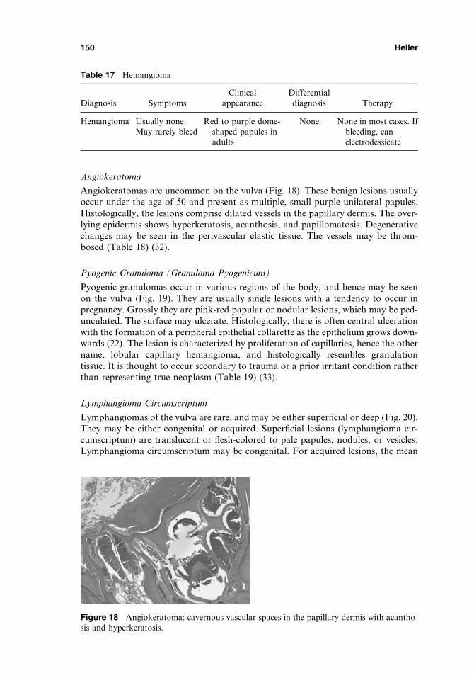
Angiokeratoma
Angiokeratomas are uncommon on the vulva (Fig. 18). These benign lesions usuallyoccur under the age of 50 and present as multiple, small purple unilateral papules.Histologically, the lesions comprise dilated vessels in the papillary dermis. The over-lying epidermis shows hyperkeratosis, acanthosis, and papillomatosis. Degenerativechanges may be seen in the perivascular elastic tissue. The vessels may be throm-bosed (Table 18) (32).
Pyogenic Granuloma (Granuloma Pyogenicum)
Pyogenic granulomas occur in various regions of the body, and hence may be seenon the vulva (Fig. 19). They are usually single lesions with a tendency to occur inpregnancy. Grossly they are pink-red papular or nodular lesions, which may be ped-unculated. The surface may ulcerate. Histologically, there is often central ulcerationwith the formation of a peripheral epithelial collarette as the epithelium grows down-wards (22). The lesion is characterized by proliferation of capillaries, hence the othername, lobular capillary hemangioma, and histologically resembles granulationtissue. It is thought to occur secondary to trauma or a prior irritant condition ratherthan representing true neoplasm (Table 19) (33).
Lymphangioma Circumscriptum
Lymphangiomas of the vulva are rare, and may be either superficial or deep (Fig. 20).They may be either congenital or acquired. Superficial lesions (lymphangioma cir-cumscriptum) are translucent or flesh-colored to pale papules, nodules, or vesicles.Lymphangioma circumscriptum may be congenital. For acquired lesions, the mean
Table 17 Hemangioma
Diagnosis SymptomsClinical
appearanceDifferentialdiagnosis Therapy
Hemangioma Usually none. Red to purple dome-shaped papules inadults
None None in most cases. Ifbleeding, canelectrodessicate
May rarely bleed
Figure 18 Angiokeratoma: cavernous vascular spaces in the papillary dermis with acantho-sis and hyperkeratosis.
150 Heller
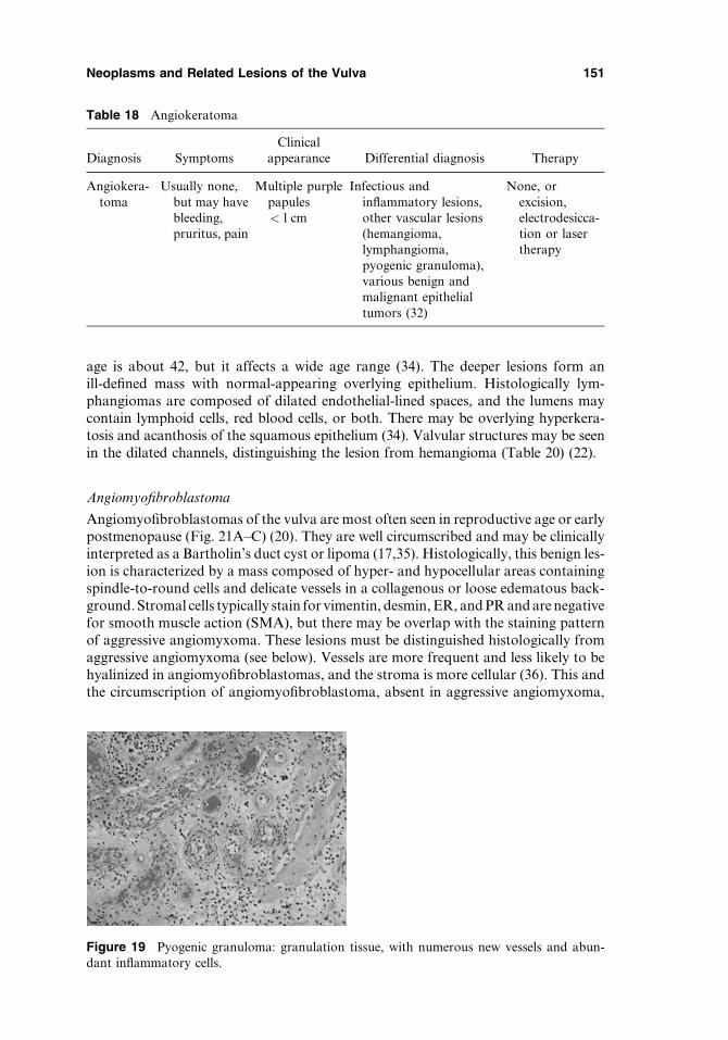
age is about 42, but it affects a wide age range (34). The deeper lesions form anill-defined mass with normal-appearing overlying epithelium. Histologically lym-phangiomas are composed of dilated endothelial-lined spaces, and the lumens maycontain lymphoid cells, red blood cells, or both. There may be overlying hyperkera-tosis and acanthosis of the squamous epithelium (34). Valvular structures may be seenin the dilated channels, distinguishing the lesion from hemangioma (Table 20) (22).
Angiomyofibroblastoma
Angiomyofibroblastomas of the vulva are most often seen in reproductive age or earlypostmenopause (Fig. 21A–C) (20). They are well circumscribed and may be clinicallyinterpreted as a Bartholin’s duct cyst or lipoma (17,35). Histologically, this benign les-ion is characterized by a mass composed of hyper- and hypocellular areas containingspindle-to-round cells and delicate vessels in a collagenous or loose edematous back-ground. Stromal cells typically stain for vimentin, desmin, ER, and PR and are negativefor smooth muscle action (SMA), but there may be overlap with the staining patternof aggressive angiomyxoma. These lesions must be distinguished histologically fromaggressive angiomyxoma (see below). Vessels are more frequent and less likely to behyalinized in angiomyofibroblastomas, and the stroma is more cellular (36). This andthe circumscription of angiomyofibroblastoma, absent in aggressive angiomyxoma,
Figure 19 Pyogenic granuloma: granulation tissue, with numerous new vessels and abun-dant inflammatory cells.
Table 18 Angiokeratoma
Diagnosis SymptomsClinical
appearance Differential diagnosis Therapy
Angiokera-toma
Usually none,but may havebleeding,pruritus, pain
Multiple purplepapules< 1 cm
Infectious andinflammatory lesions,other vascular lesions(hemangioma,lymphangioma,pyogenic granuloma),various benign andmalignant epithelialtumors (32)
None, orexcision,electrodesicca-tion or lasertherapy
Neoplasms and Related Lesions of the Vulva 151
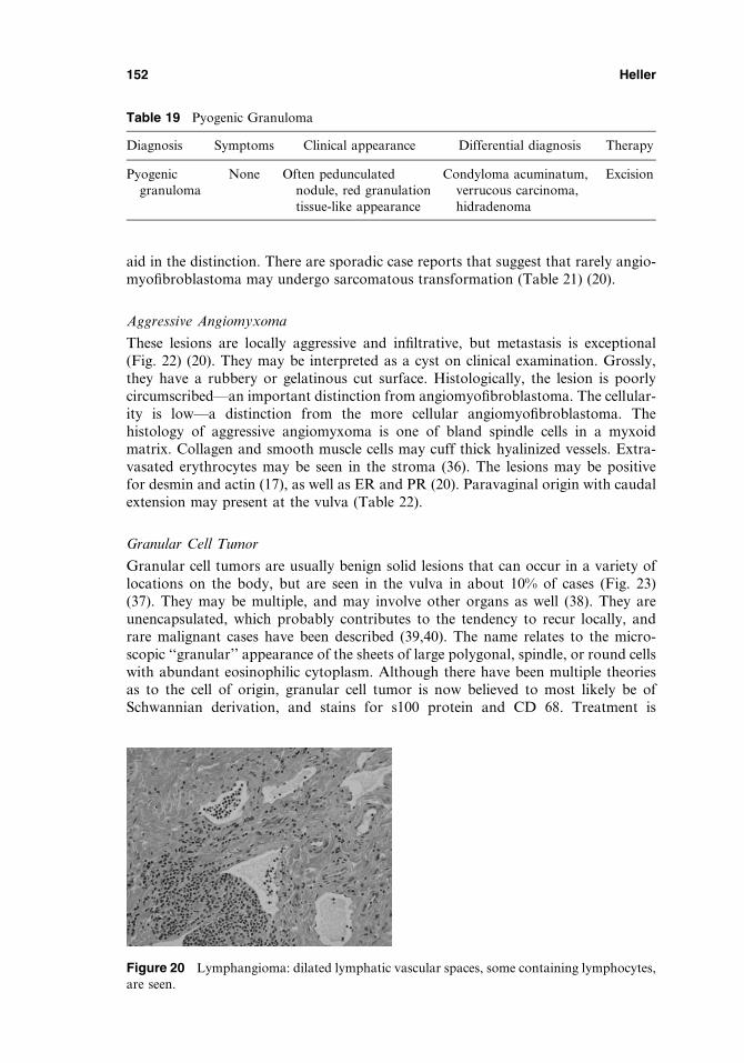
aid in the distinction. There are sporadic case reports that suggest that rarely angio-myofibroblastoma may undergo sarcomatous transformation (Table 21) (20).
Aggressive Angiomyxoma
These lesions are locally aggressive and infiltrative, but metastasis is exceptional(Fig. 22) (20). They may be interpreted as a cyst on clinical examination. Grossly,they have a rubbery or gelatinous cut surface. Histologically, the lesion is poorlycircumscribed—an important distinction from angiomyofibroblastoma. The cellular-ity is low—a distinction from the more cellular angiomyofibroblastoma. Thehistology of aggressive angiomyxoma is one of bland spindle cells in a myxoidmatrix. Collagen and smooth muscle cells may cuff thick hyalinized vessels. Extra-vasated erythrocytes may be seen in the stroma (36). The lesions may be positivefor desmin and actin (17), as well as ER and PR (20). Paravaginal origin with caudalextension may present at the vulva (Table 22).
Granular Cell Tumor
Granular cell tumors are usually benign solid lesions that can occur in a variety oflocations on the body, but are seen in the vulva in about 10% of cases (Fig. 23)(37). They may be multiple, and may involve other organs as well (38). They areunencapsulated, which probably contributes to the tendency to recur locally, andrare malignant cases have been described (39,40). The name relates to the micro-scopic ‘‘granular’’ appearance of the sheets of large polygonal, spindle, or round cellswith abundant eosinophilic cytoplasm. Although there have been multiple theoriesas to the cell of origin, granular cell tumor is now believed to most likely be ofSchwannian derivation, and stains for s100 protein and CD 68. Treatment is
Figure 20 Lymphangioma: dilated lymphatic vascular spaces, some containing lymphocytes,are seen.
Table 19 Pyogenic Granuloma
Diagnosis Symptoms Clinical appearance Differential diagnosis Therapy
Pyogenicgranuloma
None Often pedunculatednodule, red granulationtissue-like appearance
Condyloma acuminatum,verrucous carcinoma,hidradenoma
Excision
152 Heller
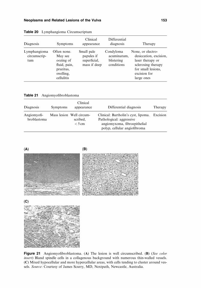
Figure 21 Angiomyofibroblastoma. (A) The lesion is well circumscribed. (B) (See colorinsert) Bland spindle cells in a collagenous background with numerous thin-walled vessels.(C) Mixed hypocellular and more hypercellular areas, with cells tending to cluster around ves-sels. Source: Courtesy of James Scurry, MD, Nextpath, Newcastle, Australia.
Table 20 Lymphangioma Circumscriptum
Diagnosis SymptomsClinical
appearanceDifferentialdiagnosis Therapy
Lymphangiomacircumscrip-tum
Often none.May seeoozing offluid, pain,pruritus,swelling,cellulitis
Small palepapules ifsuperficial,mass if deep
Condylomaacuminatum,blisteringconditions
None, or electro-desiccation, excision,laser therapy orsclerosing therapyfor small lesions,excision forlarge ones
Table 21 Angiomyofibroblastoma
Diagnosis SymptomsClinical
appearance Differential diagnosis Therapy
Angiomyofi-broblastoma
Mass lesion Well circum-scribed,< 5 cm
Clinical: Bartholin’s cyst, lipoma.Pathological: aggressive
angiomyxoma, fibroepithelialpolyp, cellular angiofibroma
Excision
Neoplasms and Related Lesions of the Vulva 153

excision. In addition to the potential clinical impression of carcinoma because of thepresent of a mass, the overlying squamous epithelium often shows pseudoepithelio-matous hyperplasia, which may be mistaken histologically for a squamous cellcarcinoma, particularly on superficial biopsies (Table 23) (37).
Leiomyoma
Smooth muscle neoplasms of the vulva are relatively uncommon (Fig. 24). They areusually benign, although leiomyosarcomas have been described arising in the vulva.The age range of cases is wide. Theories of origin of smooth muscle neoplasms in thisregion have included erectile tissue, blood vessel smooth muscle, round ligament,dartos muscle, and erector pili muscle (41). The lesions stain for smooth muscle mar-kers immunohistochemically. The criteria for distinguishing benign from malignanttumors is less well established than in the uterine counterpart. Histology may be vari-able, with epithelioid cells in a plexiform pattern and myxoid change not infrequent.Features associated with recurrence or metastasis in one series (42) were size of5.0 cm or greater, infiltrative margin, five or more mitoses per 10 HPF, and grade2 to 3 nuclear atypia. The authors diagnosed leiomyosarcoma when at least threeof these criteria were met, atypical leiomyoma when two were met, and leiomyomafor one or none. Nucci and Fletcher (17) suggest that any mitotic activity, nuclearpleomorphism, or infiltrative margin are risk factors for local recurrence, often manyyears later, and term a lesion with any of these features, but falling short of the defi-nition of sarcoma, atypical (Table 24).
Figure 22 (See color insert) Aggressive angiomyxoma: the lesion tends to be more hypocel-lular than angiomyofibroblastoma, with a myxoid background. Vessels tend to be thickerwalled. The lesions are poorly circumscribed.
Table 22 Aggressive Angiomyxoma
Diagnosis SymptomsClinical
appearance Differential diagnosis Therapy
Aggressiveangiomyxoma
Mass Mass, cyst Clinical: Bartholin’s cyst, orother cystic lesion.
Pathologic:angiomyofibroblastoma,fibroepithelial polyp, myxoma,other myxoid lesions
Completeexcision
154 Heller

Figure 23 (See color insert) Granular cell tumor: the name comes from the granular appear-ance of the eosinophilic cytoplasm.
Table 23 Granular Cell Tumor
Diagnosis SymptomsClinical
appearance Differential diagnosis Therapy
Granularcelltumor
Smallpainlessmass
Firm subcu-taneous mass< 5 cm
Bartholin’s duct cyst, lipoma,hidradenoma, fibroma.Squamous cell carcinoma orsexually transmitted disease maybe suspected by the clinician ifoverlying squamous epithelium isulcerated (39)
Wideexcision
Figure 24 Leiomyoma. (A) A large nodule is seen in the right labium majus. (B) Typicalappearance with interlacing fascicles of bland smooth muscle cells.
Table 24 Leiomyoma
Diagnosis SymptomsClinical
appearanceDifferentialdiagnosis Therapy
Leiomyoma Painless mass.Erythema, pruritus,pain also occur
Wide range ofsize. Wellcircumscribed.
Bartholin ductcyst, fibroma
Excision
Neoplasms and Related Lesions of the Vulva 155

Fibroma
Fibromas, while uncommon, are the most common benign neoplasm of the vulva,occurring most often in women aged 20 to 40, but have been reported across a broadage range (Fig. 25A and B). They may be small, but if over 8.0 cm, are usuallypedunculated (42). They have an intact overlying squamous epithelium and arehypocellular lesions composed mainly of collagen, with a fibroblast component(Table 25) (22,42).
Lipoma
Lipomas are composed of mature adipose tissue with a thin fibrous capsule (Fig. 26Aand B). They are generally asymptomatic, however may be of cosmetic concern.Rarely liposarcomas and atypical lipomas have been described. In Nucci’s series(43), most resolved with excision, although one incompletely resected lesionrecurred. These lesions were less circumscribed and showed varying degrees ofatypia. One case was described as presenting as a Bartholin abscess (44). Recently,characteristic cytogenetic aberrations have been described as important in predictingthe behaviors of lipomatous tumors (45); hence fresh tissue should be provided forcytogenetic analysis in lesions that are large and/or deep seated (Table 26).
Mammary-Like Tissue Within Vulva
Van der Putte (46) has described anogenital ‘‘sweat’’ glands that mimic the histologyof breast tissue, most highly concentrated in the interlabial sulcus of the vulva. Theseare distinct from, but with some similarities to both apocrine and eccrine glands.Further postulated is that a variety of lesions can arise in these glands, ranging fromfibrocystic change to fibroadenomas (47), hidradenoma papilliferum, and cases of
Figure 25 Fibroma. (A) Typical pedunculated lesion. (B) The lesion is composed of fibro-blasts and abundant collagen.
Table 25 Fibroma
Diagnosis SymptomsClinical
appearanceDifferentialdiagnosis Therapy
Fibroma None for smallerlesions, discomfort/pain for larger
Pedunculatedmass
Leiomyoma Excision
156 Heller
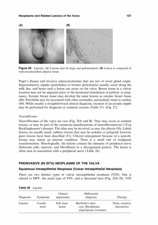
Paget’s disease and invasive adenocarcinomas that are not of sweat gland origin.Supernumerary nipples (polythelia) or breasts (polymastia) usually occur along themilk line, and hence such a lesion can occur on the vulva. Breast tissue in a vulvarlocation may not be apparent prior to the hormonal stimulation of puberty or preg-nancy. Ectopic breast tissue may develop the same lesions as eutopic breast tissue(48). Polythelia may be associated with other anomalies, particularly renal or cardiac(49). While usually a straightforward clinical diagnosis, excision of an ectopic nipplemay be performed for diagnosis or cosmetic reasons (Table 27) (Fig. 27).
Neurofibroma
Neurofibromas of the vulva are rare (Fig. 28A and B). They may occur as isolatedlesions, or may be part of the cutaneous manifestations of neurofibromatosis l (VonRecklinghausen’s disease). The labia may be involved, as may the clitoris (50). Labiallesions are usually small, rubbery lesions that may be nodular or polypoid; howevergiant lesions have been described (51). Clitoral enlargement because of a neurofi-broma may mimic an intersex condition. There is a small risk of malignanttransformation. Histologically, the lesions contain the elements of peripheral nerve(Schwann cells, neurons, and fibroblasts) in a disorganized pattern. The lesion isoften seen in association with a peripheral nerve (Table 28).
PREINVASIVE (IN SITU) NEOPLASMS OF THE VULVA
Squamous Intraepithelial Neoplasia (Vulvar Intraepithelial Neoplasia)
There are two distinct types of vulvar intraepithelial neoplasia (VIN). One isrelated to HPV, the usual type of VIN, and is discussed here (Fig. 29A–D). VIN
Figure 26 Lipoma. (A) Lesions may be large and pedunculated. (B) Lesion is composed ofwell-circumscribed adipose tissue.
Table 26 Lipoma
Diagnosis SymptomsClinical
appearanceDifferentialdiagnosis Therapy
Lipoma Usuallynone
Soft masslesion
Bartholin’s ductcyst, fibrolipoma,angiolipoma (variants)
None, excision,liposuction
Neoplasms and Related Lesions of the Vulva 157
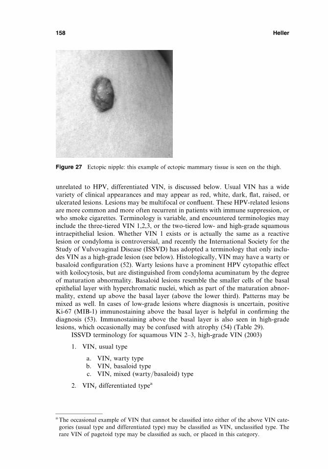
unrelated to HPV, differentiated VIN, is discussed below. Usual VIN has a widevariety of clinical appearances and may appear as red, white, dark, flat, raised, orulcerated lesions. Lesions may be multifocal or confluent. These HPV-related lesionsare more common and more often recurrent in patients with immune suppression, orwho smoke cigarettes. Terminology is variable, and encountered terminologies mayinclude the three-tiered VIN 1,2,3, or the two-tiered low- and high-grade squamousintraepithelial lesion. Whether VIN 1 exists or is actually the same as a reactivelesion or condyloma is controversial, and recently the International Society for theStudy of Vulvovaginal Disease (ISSVD) has adopted a terminology that only inclu-des VIN as a high-grade lesion (see below). Histologically, VIN may have a warty orbasaloid configuration (52). Warty lesions have a prominent HPV cytopathic effectwith koilocytosis, but are distinguished from condyloma acuminatum by the degreeof maturation abnormality. Basaloid lesions resemble the smaller cells of the basalepithelial layer with hyperchromatic nuclei, which as part of the maturation abnor-mality, extend up above the basal layer (above the lower third). Patterns may bemixed as well. In cases of low-grade lesions where diagnosis is uncertain, positiveKi-67 (MIB-1) immunostaining above the basal layer is helpful in confirming thediagnosis (53). Immunostaining above the basal layer is also seen in high-gradelesions, which occasionally may be confused with atrophy (54) (Table 29).
ISSVD terminology for squamous VIN 2–3, high-grade VIN (2003)
1. VIN, usual type
a. VIN, warty typeb. VIN, basaloid typec. VIN, mixed (warty/basaloid) type
2. VINr differentiated typea
a The occasional example of VIN that cannot be classified into either of the above VIN cate-gories (usual type and differentiated type) may be classified as VIN, unclassified type. Therare VIN of pagetoid type may be classified as such, or placed in this category.
Figure 27 Ectopic nipple: this example of ectopic mammary tissue is seen on the thigh.
158 Heller

Differentiated Vulvar Intraepithelial Neoplasia
Differentiated VIN, a lesion unrelated to HPV, is much less common than the usualHPV-related VIN (Fig. 30A and B). It occurs in postmenopausal women, significantlyolder than women in whom usual VIN is seen, and is thought to pose a more signifi-cant risk of progressing to invasive squamous cell carcinoma (55). It is associatedwith, and may be seen adjacent to, the non–HPV-related squamous cell carcinomasof the vulva that arise in older women. Clinically, the lesions of differentiated VINare more subtle than usual VIN lesions. They may be unifocal or multifocal and tendto be pale white or gray plaque-like or nodular areas. They may appear simply asthickened areas. Differentiated VIN is often seen in a background of lichen sclerosusor squamous hyperplasia (56,57) The lesion may also be quite subtle histologically, andmay be misdiagnosed as squamous cell hyperplasia because of the parakeratosisand acanthosis. Atypia is minimal, and confined to the basal layers, with large eosi-nophilic keratinocytes containing nuclei with coarse chromatin and prominentnucleoli. Whorls of differentiated cells with or without keratin pearls may be seen(56). The lesion usually stains for p53, as opposed to usual VIN, which does not(Table 30) (57).
Paget’s Disease of the Vulva (Extramammary Paget’s Disease)
Paget’s disease of the vulva presents as a velvety red lesion with overlying white pla-ques and may present with severe pruritis, burning, or bleeding (Fig. 31A and B).
Table 27 Ectopic Nipple
Diagnosis SymptomsClinical
appearanceDifferentialdiagnosis Therapy
Ectopic nipple Usually none Usuallyrecognizable
Other pigmented lesions.If ectopic breast tissue,other mass lesions
Excision ifwarranted
Figure 28 (A) Neurofibromatosis: numerous lesions are seen on the mons pubis. (B) Neuro-fibroma: typical ‘‘shredded carrot’’ bundles of collagen in a myxoid background containingcells of neural origin.
Neoplasms and Related Lesions of the Vulva 159

The lesion tends to recur, probably related to the fact that histologically the lesionextends beyond the grossly detectable borders. It is associated with both underlyingskin adnexal and Bartholin’s adenocarcinomas, as well as noncontiguous adenocar-cinomas of other organs such as the rectum or breast (58,59). The incidence of these
Table 28 Neurofibroma
Diagnosis Symptoms Clinical appearanceDifferentialdiagnosis Therapy
Neurofi-broma
None if small. Masslesion if large. Maycause clitoralenlargement
Small brown nodules orplaques. May bepedunculated. May havea violaceous color
Other pig-mentedlesions
Excision ifwarranted
Figure 29 Vulvar intraepithelial neoplasia (VIN), usual type. (A) (See color insert) This VINlesion is pigmented. (B) (See color insert) This is an example of a depigmented VIN lesion. (C)(See color insert) In the warty pattern, cytopathic features of human papilloma virus areprominent. (D) In the basaloid pattern, the entire epithelium is placed by small basaloid cells.
160 Heller
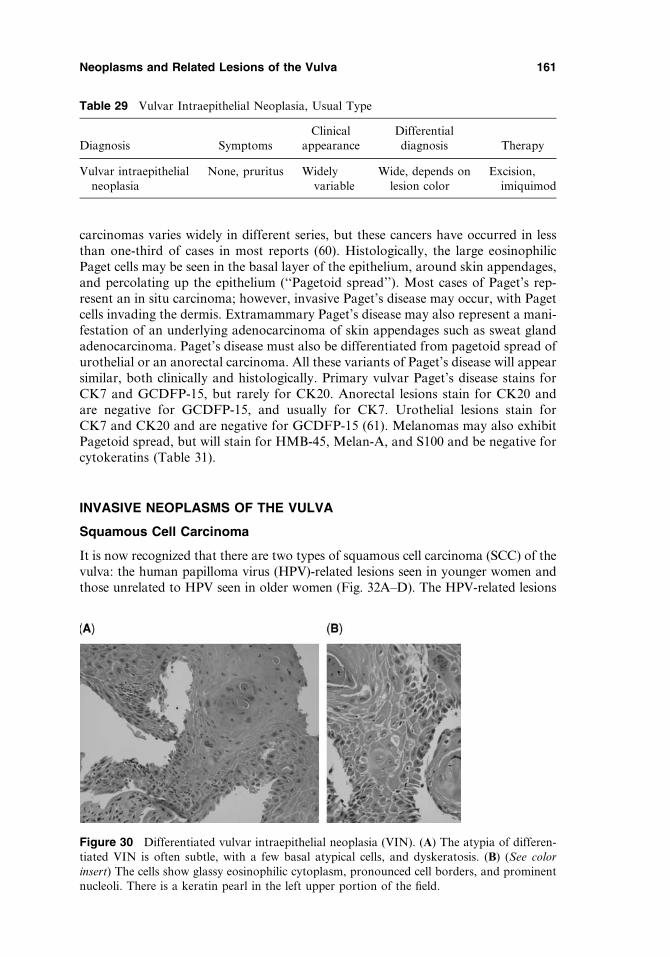
carcinomas varies widely in different series, but these cancers have occurred in lessthan one-third of cases in most reports (60). Histologically, the large eosinophilicPaget cells may be seen in the basal layer of the epithelium, around skin appendages,and percolating up the epithelium (‘‘Pagetoid spread’’). Most cases of Paget’s rep-resent an in situ carcinoma; however, invasive Paget’s disease may occur, with Pagetcells invading the dermis. Extramammary Paget’s disease may also represent a mani-festation of an underlying adenocarcinoma of skin appendages such as sweat glandadenocarcinoma. Paget’s disease must also be differentiated from pagetoid spread ofurothelial or an anorectal carcinoma. All these variants of Paget’s disease will appearsimilar, both clinically and histologically. Primary vulvar Paget’s disease stains forCK7 and GCDFP-15, but rarely for CK20. Anorectal lesions stain for CK20 andare negative for GCDFP-15, and usually for CK7. Urothelial lesions stain forCK7 and CK20 and are negative for GCDFP-15 (61). Melanomas may also exhibitPagetoid spread, but will stain for HMB-45, Melan-A, and S100 and be negative forcytokeratins (Table 31).
INVASIVE NEOPLASMS OF THE VULVA
Squamous Cell Carcinoma
It is now recognized that there are two types of squamous cell carcinoma (SCC) of thevulva: the human papilloma virus (HPV)-related lesions seen in younger women andthose unrelated to HPV seen in older women (Fig. 32A–D). The HPV-related lesions
Table 29 Vulvar Intraepithelial Neoplasia, Usual Type
Diagnosis SymptomsClinical
appearanceDifferentialdiagnosis Therapy
Vulvar intraepithelialneoplasia
None, pruritus Widelyvariable
Wide, depends onlesion color
Excision,imiquimod
Figure 30 Differentiated vulvar intraepithelial neoplasia (VIN). (A) The atypia of differen-tiated VIN is often subtle, with a few basal atypical cells, and dyskeratosis. (B) (See colorinsert) The cells show glassy eosinophilic cytoplasm, pronounced cell borders, and prominentnucleoli. There is a keratin pearl in the left upper portion of the field.
Neoplasms and Related Lesions of the Vulva 161
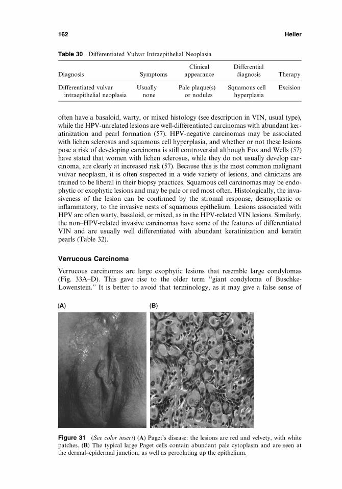
often have a basaloid, warty, or mixed histology (see description in VIN, usual type),while the HPV-unrelated lesions are well-differentiated carcinomas with abundant ker-atinization and pearl formation (57). HPV-negative carcinomas may be associatedwith lichen sclerosus and squamous cell hyperplasia, and whether or not these lesionspose a risk of developing carcinoma is still controversial although Fox and Wells (57)have stated that women with lichen sclerosus, while they do not usually develop car-cinoma, are clearly at increased risk (57). Because this is the most common malignantvulvar neoplasm, it is often suspected in a wide variety of lesions, and clinicians aretrained to be liberal in their biopsy practices. Squamous cell carcinomas may be endo-phytic or exophytic lesions and may be pale or red most often. Histologically, the inva-siveness of the lesion can be confirmed by the stromal response, desmoplastic orinflammatory, to the invasive nests of squamous epithelium. Lesions associated withHPV are often warty, basaloid, or mixed, as in the HPV-related VIN lesions. Similarly,the non–HPV-related invasive carcinomas have some of the features of differentiatedVIN and are usually well differentiated with abundant keratinization and keratinpearls (Table 32).
Verrucous Carcinoma
Verrucous carcinomas are large exophytic lesions that resemble large condylomas(Fig. 33A–D). This gave rise to the older term ‘‘giant condyloma of Buschke-Lowenstein.’’ It is better to avoid that terminology, as it may give a false sense of
Figure 31 (See color insert) (A) Paget’s disease: the lesions are red and velvety, with whitepatches. (B) The typical large Paget cells contain abundant pale cytoplasm and are seen atthe dermal–epidermal junction, as well as percolating up the epithelium.
Table 30 Differentiated Vulvar Intraepithelial Neoplasia
Diagnosis SymptomsClinical
appearanceDifferentialdiagnosis Therapy
Differentiated vulvarintraepithelial neoplasia
Usuallynone
Pale plaque(s)or nodules
Squamous cellhyperplasia
Excision
162 Heller

benignity to the clinician. These lesions are associated with HPV type 6 and 11 (62).There is often reactive induration of tissue adjacent to a verrucous carcinoma, as wellas reactive lymphadenopathy, which may raise the clinical suspicion of metastatic dis-ease (63). Pathological diagnosis may be difficult, as superficial biopsies may not
Figure 32 Squamous cell carcinoma of the vulva. (A) This is an example of an exophyticlesion. (B) (See color insert) This is an example of an endophytic lesion. (C) Typical appear-ance of squamous cell carcinoma, non–HPV-related type. Irregular tumor nests are invadingthe stroma, which is exhibiting a desmoplastic reaction. (D) In the non–HPV-related tumors,the lesions are often well differentiated and exhibit keratin pearls as demonstrated here.Abbreviation: HPV, human papilloma virus.
Table 31 Extramammary Paget’s Disease
Diagnosis SymptomsClinical
appearance Differential diagnosis Therapy
ExtramammaryPaget’sdisease
Pruritus,burning,bleeding
Red velvetylesions withwhite plaques
Melanoma (histologically),Paget’s disease ofurothelial or anorectalorigin
Wide excisionfor non-invasivedisease
Neoplasms and Related Lesions of the Vulva 163

Figure 33 Verrucous carcinoma. (A) Lesions grossly appear large and warty. (B) Low-powerview demonstrating the warty architecture. (C) Lesions show minimal atypia, and there maybe human papilloma virus cytopathic changes. (D) These lesions exhibit a ‘‘pushing’’ border,in contrast to the invasive border seen in usual squamous cell carcinoma.
Table 32 Squamous Cell Carcinoma
Diagnosis SymptomsClinical
appearance Differential diagnosis Therapy
Invasivesquamouscell carci-noma
None whensmall.Pruritusandbleedingwhen large
Ulcerating orexophyticlesion
Basal cell carcinoma,keratoacanthoma,sexually transmitteddiseases if ulcerative
Usually radical surgery,with possible adjuvantradiation therapy.Cryosurgery to theprimary lesion hasbeen reported inadvanced disease
164 Heller
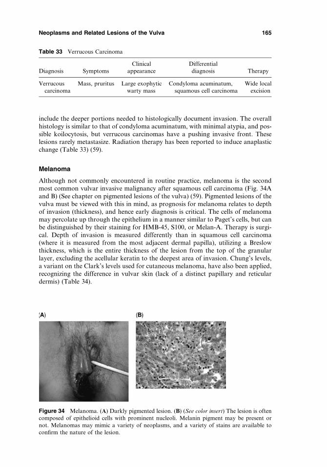
include the deeper portions needed to histologically document invasion. The overallhistology is similar to that of condyloma acuminatum, with minimal atypia, and pos-sible koilocytosis, but verrucous carcinomas have a pushing invasive front. Theselesions rarely metastasize. Radiation therapy has been reported to induce anaplasticchange (Table 33) (59).
Melanoma
Although not commonly encountered in routine practice, melanoma is the secondmost common vulvar invasive malignancy after squamous cell carcinoma (Fig. 34Aand B) (See chapter on pigmented lesions of the vulva) (59). Pigmented lesions of thevulva must be viewed with this in mind, as prognosis for melanoma relates to depthof invasion (thickness), and hence early diagnosis is critical. The cells of melanomamay percolate up through the epithelium in a manner similar to Paget’s cells, but canbe distinguished by their staining for HMB-45, S100, or Melan-A. Therapy is surgi-cal. Depth of invasion is measured differently than in squamous cell carcinoma(where it is measured from the most adjacent dermal papilla), utilizing a Breslowthickness, which is the entire thickness of the lesion from the top of the granularlayer, excluding the acellular keratin to the deepest area of invasion. Chung’s levels,a variant on the Clark’s levels used for cutaneous melanoma, have also been applied,recognizing the difference in vulvar skin (lack of a distinct papillary and reticulardermis) (Table 34).
Figure 34 Melanoma. (A) Darkly pigmented lesion. (B) (See color insert) The lesion is oftencomposed of epithelioid cells with prominent nucleoli. Melanin pigment may be present ornot. Melanomas may mimic a variety of neoplasms, and a variety of stains are available toconfirm the nature of the lesion.
Table 33 Verrucous Carcinoma
Diagnosis SymptomsClinical
appearanceDifferentialdiagnosis Therapy
Verrucouscarcinoma
Mass, pruritus Large exophyticwarty mass
Condyloma acuminatum,squamous cell carcinoma
Wide localexcision
Neoplasms and Related Lesions of the Vulva 165
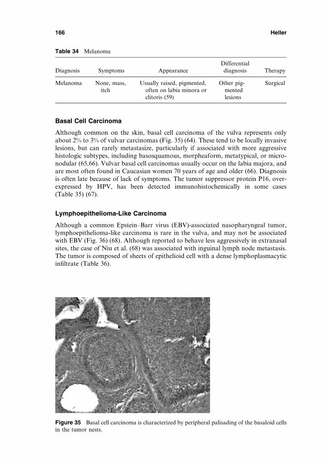
Basal Cell Carcinoma
Although common on the skin, basal cell carcinoma of the vulva represents onlyabout 2% to 3% of vulvar carcinomas (Fig. 35) (64). These tend to be locally invasivelesions, but can rarely metastasize, particularly if associated with more aggressivehistologic subtypes, including basosquamous, morpheaform, metatypical, or micro-nodular (65,66). Vulvar basal cell carcinomas usually occur on the labia majora, andare most often found in Caucasian women 70 years of age and older (66). Diagnosisis often late because of lack of symptoms. The tumor suppressor protein P16, over-expressed by HPV, has been detected immunohistochemically in some cases(Table 35) (67).
Lymphoepithelioma-Like Carcinoma
Although a common Epstein–Barr virus (EBV)-associated nasopharyngeal tumor,lymphoepithelioma-like carcinoma is rare in the vulva, and may not be associatedwith EBV (Fig. 36) (68). Although reported to behave less aggressively in extranasalsites, the case of Niu et al. (68) was associated with inguinal lymph node metastasis.The tumor is composed of sheets of epithelioid cell with a dense lymphoplasmacyticinfiltrate (Table 36).
Table 34 Melanoma
Diagnosis Symptoms AppearanceDifferentialdiagnosis Therapy
Melanoma None, mass,itch
Usually raised, pigmented,often on labia minora orclitoris (59)
Other pig-mentedlesions
Surgical
Figure 35 Basal cell carcinoma is characterized by peripheral palisading of the basaloid cellsin the tumor nests.
166 Heller
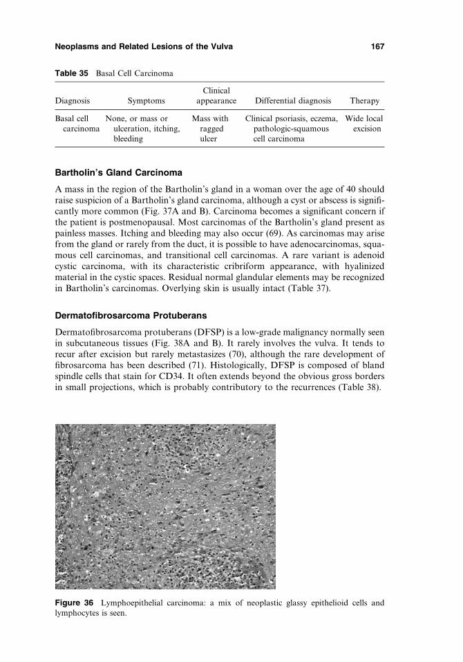
Bartholin’s Gland Carcinoma
A mass in the region of the Bartholin’s gland in a woman over the age of 40 shouldraise suspicion of a Bartholin’s gland carcinoma, although a cyst or abscess is signifi-cantly more common (Fig. 37A and B). Carcinoma becomes a significant concern ifthe patient is postmenopausal. Most carcinomas of the Bartholin’s gland present aspainless masses. Itching and bleeding may also occur (69). As carcinomas may arisefrom the gland or rarely from the duct, it is possible to have adenocarcinomas, squa-mous cell carcinomas, and transitional cell carcinomas. A rare variant is adenoidcystic carcinoma, with its characteristic cribriform appearance, with hyalinizedmaterial in the cystic spaces. Residual normal glandular elements may be recognizedin Bartholin’s carcinomas. Overlying skin is usually intact (Table 37).
Dermatofibrosarcoma Protuberans
Dermatofibrosarcoma protuberans (DFSP) is a low-grade malignancy normally seenin subcutaneous tissues (Fig. 38A and B). It rarely involves the vulva. It tends torecur after excision but rarely metastasizes (70), although the rare development offibrosarcoma has been described (71). Histologically, DFSP is composed of blandspindle cells that stain for CD34. It often extends beyond the obvious gross bordersin small projections, which is probably contributory to the recurrences (Table 38).
Table 35 Basal Cell Carcinoma
Diagnosis SymptomsClinical
appearance Differential diagnosis Therapy
Basal cellcarcinoma
None, or mass orulceration, itching,bleeding
Mass withraggedulcer
Clinical psoriasis, eczema,pathologic-squamouscell carcinoma
Wide localexcision
Figure 36 Lymphoepithelial carcinoma: a mix of neoplastic glassy epithelioid cells andlymphocytes is seen.
Neoplasms and Related Lesions of the Vulva 167

Metastatic Tumors to Vulva
A variety of neoplasms have been reported to metastasize to the vulva, includingother neoplasms of the genital tract (endometrium, ovary, and cervix) (Fig. 39). Gas-trointestinal and urinary tract neoplasms may also metastasize to vulva on occasion,and rare cases of metastatic breast cancer to vulva have also occurred (72). Clini-cally, the lesions may appear similar to a primary vulvar carcinoma. Treatment isdirected to the primary neoplasm, although excision for palliation may be utilized(Table 39).
BENIGN LESIONS OF THE URETHRA
Urethral Prolapse
Urethral prolapse is seen primarily in African-American girls under the age of five,or in postmenopausal Caucasian women (Fig. 40) (73). The condition may presentwith bleeding. Examination reveals a donut-shaped mass of red tissue surroundingthe urethral meatus, with a centrally placed opening. Medical measures are usuallyattempted prior to surgery. In the postmenopausal patient, the concern for a urethralcarcinoma may arise (Table 40) (74).
Urethral Caruncle
Urethral caruncles are not uncommon in postmenopausal women, but their brightred fleshy appearance may occasionally raise the concern for a carcinoma (Fig. 41).
Table 36 Lymphoepithelioma-Like Carcinoma
Diagnosis SymptomsClinical
appearanceDifferentialdiagnosis Therapy
Lymphoepithelioma-like carcinoma
Mass Mass Lymphoma Surgical
Figure 37 Bartholin’s carcinoma. (A) Note swelling in the region of the left Bartholin’sgland. (B) These lesions may be squamous, transitional, or adenocarcinoma as shown here.
168 Heller

Caruncles, which probably represent ectropion of the distal posterior urethra (76),may be related to the hypoestrogenic milieu of menopause. They appear as red poly-poid protrusions from the urethra. Histologically, the diagnosis is usually clear cut.The lesion has a polypoid or papillary configuration and is lined by sometimeshyperplastic squamous or transitional cells. There are abundant vessels and inflam-matory cells in the stroma, with an appearance of granulation tissue; however,Young et al. (77) have described an unusual variant containing atypical stromal cellsmimicking a lymphoma or sarcoma (Table 41).
Malacoplakia of the Urethra
Malacoplakia is a rare form of granulomatous inflammation usually affecting thegenitourinary tract (78). Its occurrence in the female urethra is exceedingly rare.Histologically, the lesions are composed of a mixed inflammatory infiltrate withabundant histiocytes with granular cytoplasm, many containing characteristic intra-and extracellular Michaelis–Guttman bodies (Fig. 42). These bodies are felt to arise
Table 37 Bartholin’s Gland Carcinoma
Diagnosis SymptomsClinical
appearanceDifferentialdiagnosis Therapy
Bartholin’sglandcarcinoma
Painless mass,itching,bleeding
Mass inregion ofBartholin’sgland
Bartholin’scyst orabscess
Surgery, usually modifiedradical vulvectomy, andlymphadenectomy. Adjuvantradiation may be utilized,and rarely chemotherapy
Figure 38 Dermatofibrosarcoma protuberans. (A) The lesion is arising from the inner leftlabia minora. (B) An important clue to the nature of this bland-appearing spindle cell lesionis its infiltrative nature, as seen in this subcutaneous adipose tissue.
Neoplasms and Related Lesions of the Vulva 169
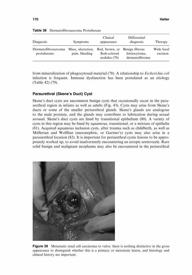
from mineralization of phagocytosed material (78). A relationship to Escherichia coliinfection is frequent. Immune dysfunction has been postulated as an etiology(Table 42) (79).
Paraurethral (Skene’s Duct) Cyst
Skene’s duct cysts are uncommon benign cysts that occasionally occur in the para-urethral region in infants as well as adults (Fig. 43). Cysts may arise from Skene’sducts or some of the smaller periurethral glands. Skene’s glands are analogousto the male prostate, and the glands may contribute to lubrication during sexualarousal. Skene’s duct cysts are lined by transitional epithelium (80). A variety ofcysts in this region may be lined by squamous, transitional, or a mixture of epithelia(81). Acquired squamous inclusion cysts, after trauma such as childbirth, as well asMullerian and Wolffian (mesonephric, or Gartner’s) cysts may also arise in aparaurethral location (82). It is important for periurethral cystic lesions to be appro-priately worked up, to avoid inadvertently encountering an ectopic ureterocele. Raresolid benign and malignant neoplasms may also be encountered in the periurethral
Table 38 Dermatofibrosarcoma Protuberans
Diagnosis SymptomsClinical
appearanceDifferentialdiagnosis Therapy
Dermatofibrosarcomaprotuberans
Mass, ulceration,pain, bleeding
Red, brown, orflesh-colorednodules (70)
Benign fibroushistiocytoma,dermatofibroma
Wide localexcision
Figure 39 Metastatic renal cell carcinoma to vulva: there is nothing distinctive in the grossappearance to distinguish whether this is a primary or metastatic lesion, and histology andclinical history are important.
170 Heller
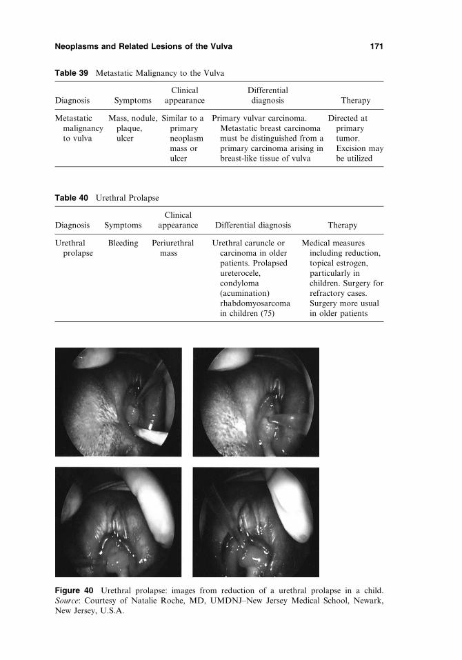
Figure 40 Urethral prolapse: images from reduction of a urethral prolapse in a child.Source: Courtesy of Natalie Roche, MD, UMDNJ–New Jersey Medical School, Newark,New Jersey, U.S.A.
Table 39 Metastatic Malignancy to the Vulva
Diagnosis SymptomsClinical
appearanceDifferentialdiagnosis Therapy
Metastaticmalignancyto vulva
Mass, nodule,plaque,ulcer
Similar to aprimaryneoplasmmass orulcer
Primary vulvar carcinoma.Metastatic breast carcinomamust be distinguished from aprimary carcinoma arising inbreast-like tissue of vulva
Directed atprimarytumor.Excision maybe utilized
Table 40 Urethral Prolapse
Diagnosis SymptomsClinical
appearance Differential diagnosis Therapy
Urethralprolapse
Bleeding Periurethralmass
Urethral caruncle orcarcinoma in olderpatients. Prolapsedureterocele,condyloma(acumination)rhabdomyosarcomain children (75)
Medical measuresincluding reduction,topical estrogen,particularly inchildren. Surgery forrefractory cases.Surgery more usualin older patients
Neoplasms and Related Lesions of the Vulva 171
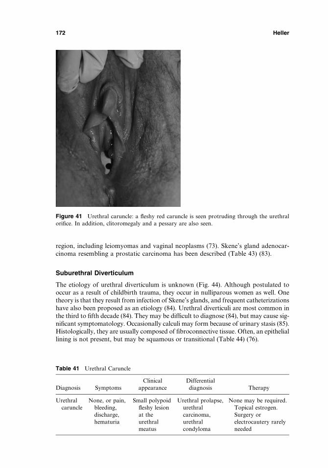
region, including leiomyomas and vaginal neoplasms (73). Skene’s gland adenocar-cinoma resembling a prostatic carcinoma has been described (Table 43) (83).
Suburethral Diverticulum
The etiology of urethral diverticulum is unknown (Fig. 44). Although postulated tooccur as a result of childbirth trauma, they occur in nulliparous women as well. Onetheory is that they result from infection of Skene’s glands, and frequent catheterizationshave also been proposed as an etiology (84). Urethral diverticuli are most common inthe third to fifth decade (84). They may be difficult to diagnose (84), but may cause sig-nificant symptomatology. Occasionally calculi may form because of urinary stasis (85).Histologically, they are usually composed of fibroconnective tissue. Often, an epitheliallining is not present, but may be squamous or transitional (Table 44) (76).
Figure 41 Urethral caruncle: a fleshy red caruncle is seen protruding through the urethralorifice. In addition, clitoromegaly and a pessary are also seen.
Table 41 Urethral Caruncle
Diagnosis SymptomsClinical
appearanceDifferentialdiagnosis Therapy
Urethralcaruncle
None, or pain,bleeding,discharge,hematuria
Small polypoidfleshy lesionat theurethralmeatus
Urethral prolapse,urethralcarcinoma,urethralcondyloma
None may be required.Topical estrogen.Surgery orelectrocautery rarelyneeded
172 Heller
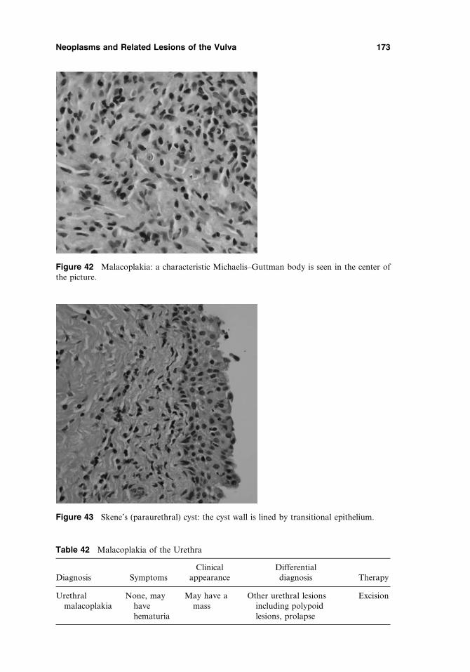
Figure 42 Malacoplakia: a characteristic Michaelis–Guttman body is seen in the center ofthe picture.
Figure 43 Skene’s (paraurethral) cyst: the cyst wall is lined by transitional epithelium.
Table 42 Malacoplakia of the Urethra
Diagnosis SymptomsClinical
appearanceDifferentialdiagnosis Therapy
Urethralmalacoplakia
None, mayhavehematuria
May have amass
Other urethral lesionsincluding polypoidlesions, prolapse
Excision
Neoplasms and Related Lesions of the Vulva 173
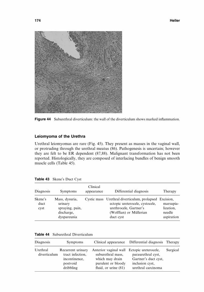
Leiomyoma of the Urethra
Urethral leiomyomas are rare (Fig. 45). They present as masses in the vaginal wall,or protruding through the urethral meatus (86). Pathogenesis is uncertain; howeverthey are felt to be ER dependent (87,88). Malignant transformation has not beenreported. Histologically, they are composed of interlacing bundles of benign smoothmuscle cells (Table 45).
Figure 44 Suburethral diverticulum: the wall of the diverticulum shows marked inflammation.
Table 43 Skene’s Duct Cyst
Diagnosis SymptomsClinical
appearance Differential diagnosis Therapy
Skene’sductcyst
Mass, dysuria,urinaryspraying, pain,discharge,dyspareunia
Cystic mass Urethral diverticulum, prolapsedectopic ureterocele, cystocele,urethrocele, Gartner’s(Wolffian) or Mullerianduct cyst
Excision,marsupia-lization,needleaspiration
Table 44 Suburethral Diverticulum
Diagnosis Symptoms Clinical appearance Differential diagnosis Therapy
Urethraldiverticulum
Recurrent urinarytract infection,incontinence,postvoiddribbling
Anterior vaginal wallsuburethral mass,which may drainpurulent or bloodyfluid, or urine (81)
Ectopic ureterocele,paraurethral cyst,Gartner’s duct cyst,inclusion cyst,urethral carcinoma
Surgical
174 Heller
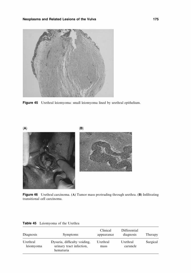
Figure 45 Urethral leiomyoma: small leiomyoma lined by urethral epithelium.
Table 45 Leiomyoma of the Urethra
Diagnosis SymptomsClinical
appearanceDifferentialdiagnosis Therapy
Urethralleiomyoma
Dysuria, difficulty voiding,urinary tract infection,hematuria
Urethralmass
Urethralcaruncle
Surgical
Figure 46 Urethral carcinoma. (A) Tumor mass protruding through urethra. (B) Infiltratingtransitional cell carcinoma.
Neoplasms and Related Lesions of the Vulva 175
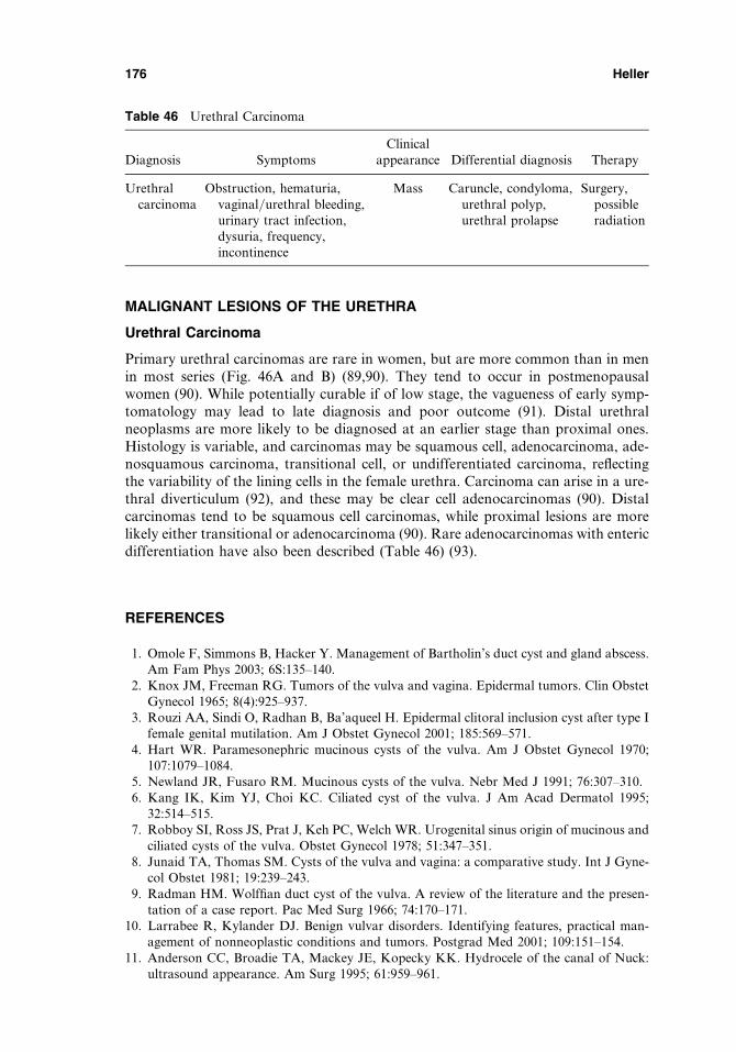
MALIGNANT LESIONS OF THE URETHRA
Urethral Carcinoma
Primary urethral carcinomas are rare in women, but are more common than in menin most series (Fig. 46A and B) (89,90). They tend to occur in postmenopausalwomen (90). While potentially curable if of low stage, the vagueness of early symp-tomatology may lead to late diagnosis and poor outcome (91). Distal urethralneoplasms are more likely to be diagnosed at an earlier stage than proximal ones.Histology is variable, and carcinomas may be squamous cell, adenocarcinoma, ade-nosquamous carcinoma, transitional cell, or undifferentiated carcinoma, reflectingthe variability of the lining cells in the female urethra. Carcinoma can arise in a ure-thral diverticulum (92), and these may be clear cell adenocarcinomas (90). Distalcarcinomas tend to be squamous cell carcinomas, while proximal lesions are morelikely either transitional or adenocarcinoma (90). Rare adenocarcinomas with entericdifferentiation have also been described (Table 46) (93).
REFERENCES
1. Omole F, Simmons B, Hacker Y. Management of Bartholin’s duct cyst and gland abscess.Am Fam Phys 2003; 6S:135–140.
2. Knox JM, Freeman RG. Tumors of the vulva and vagina. Epidermal tumors. Clin ObstetGynecol 1965; 8(4):925–937.
3. Rouzi AA, Sindi O, Radhan B, Ba’aqueel H. Epidermal clitoral inclusion cyst after type Ifemale genital mutilation. Am J Obstet Gynecol 2001; 185:569–571.
4. Hart WR. Paramesonephric mucinous cysts of the vulva. Am J Obstet Gynecol 1970;107:1079–1084.
5. Newland JR, Fusaro RM. Mucinous cysts of the vulva. Nebr Med J 1991; 76:307–310.6. Kang IK, Kim YJ, Choi KC. Ciliated cyst of the vulva. J Am Acad Dermatol 1995;
32:514–515.7. Robboy SI, Ross JS, Prat J, Keh PC, Welch WR. Urogenital sinus origin of mucinous and
ciliated cysts of the vulva. Obstet Gynecol 1978; 51:347–351.8. Junaid TA, Thomas SM. Cysts of the vulva and vagina: a comparative study. Int J Gyne-
col Obstet 1981; 19:239–243.9. Radman HM. Wolffian duct cyst of the vulva. A review of the literature and the presen-
tation of a case report. Pac Med Surg 1966; 74:170–171.10. Larrabee R, Kylander DJ. Benign vulvar disorders. Identifying features, practical man-
agement of nonneoplastic conditions and tumors. Postgrad Med 2001; 109:151–154.11. Anderson CC, Broadie TA, Mackey JE, Kopecky KK. Hydrocele of the canal of Nuck:
ultrasound appearance. Am Surg 1995; 61:959–961.
Table 46 Urethral Carcinoma
Diagnosis SymptomsClinical
appearance Differential diagnosis Therapy
Urethralcarcinoma
Obstruction, hematuria,vaginal/urethral bleeding,urinary tract infection,dysuria, frequency,incontinence
Mass Caruncle, condyloma,urethral polyp,urethral prolapse
Surgery,possibleradiation
176 Heller

12. Kranl C, Zelger B, Kofler H, Heim K, Sepp N, Fritsch P. Vulval and vaginal adenosis. Br JDermatol 1998; 139:128–131.
13. Sedlacek TV, Riva JH, Magen AB, Mangan CE, Cunnane MF. Vaginal and vulvaradenosis. An unsuspected side effect of CO2 laser vaporization. J Reprod Med 1990;35(11):995–1001.
14. Bonafe JL, Thibaut I, Hoff J. Introital adenosis associated with the Stevens-Johnsonsyndrome. Clin Exp Dermatol 1990; 15(5):356–357.
15. Cheng DL, Heller DS, Oh C. Endometriosis of the perineum; report of two new cases anda review of literature. Eur J Obstet Gynecol Reprod Biol 1991; 42:81–84.
16. Gocmen A, Inaloz HS, Sari I, Inaloz SS. Endometriosis in the Bartholin gland. Eur JObstet Gynecol Reprod Biol 2004; 114(1):110–111.
17. Nucci MR, Fletcher CDM. Vulvovaginal soft tissue tumours: update and review. Histo-pathology 2000; 36(2):97–108.
18. Nucci MR, Young RH, Fletcher CD. Cellular pseudosarcomatous fibroepithelial stromalpolyps of the lower female genital tract: an under-recognized lesion often misdiagnosed assarcoma. Am J Surg Pathol 2000; 24(2):231–240.
19. Ostor AG, Fortune DW, Riley CB. Fibroepithelial polyps with atypical stromal cells(pseudosarcoma botryoides) of vulva and vagina. A report of 13 cases. Int J GynecolPathol 1988; 7(4):351–360.
20. McCluggage WG. A review and update of morphologically bland vulvovaginal mesenchy-mal lesions. Int J Gynecol Pathol 2005; 24(l):26–38.
21. Bergeron C, Ferenczy A, Richart RM, Guralnick M. Micropapillomatosis labialis appearsunrelated to human papillomavirus. Obstet Gynecol 1990; 76:28l–286.
22. Hood AF, Lamadue J. Benign vulvar tumors. Dermatol Clin 1992; 10:371–385.23. Regauer S, Nogales, FF. Vulvar trichogenic tumors. A comparative study with vulvar
basal cell carcinoma. Am J Surg Pathol 2005; 29:479–484.24. Peterdy GA, Huettner PC, Rajaram V, Lind AC. Trichofolliculoma of the vulva associa-
ted with vulvar intraepithelial neoplasia: report of three cases and review of the literature.Int J Gynecol Pathol 2002; 21:224–230.
25. Cho D, Woodruff JD. Trichoepithelioma of the vulva. A report of two cases. J ReprodMed 1988; 33:317–319.
26. Bal N, Asian E, Kayaselcuk F, Tarim E, Tuncer I. Vulvar syringoma aggravated by preg-nancy. Pathol Oncol Res 2003; 9:196–197.
27. Duray PH, Merino MJ, Axiotis C. Warty dyskeratoma of the vulva. Int J Gynecol Pathol1983; 2(3):286–293.
28. Kaddu S, Dong H, Mayer G, Kerl H, Cerroni L. Warty dyskeratoma ‘‘follicular dysker-atoma’’: analysis of clinicopathologic features of a distinctive follicular adnexal neoplasm.J Am Acad Dermatol 2002; 47:423–428.
29. Chen W, Koenig C. Vulvar keratoacanthoma: a report of two cases. Int J Gynecol Pathol2004; 23:284–286.
30. Haley JC, Mirowski GW, Hood A. Benign vulvar tumors. Semin Cutan Med Surg 1998;17:196–204.
31. Bava GL, Dalmonte P, Oddone M, Rossi U. Life-threatening hemorrhage from a vulvarhemangioma. J Ped Surg 2002; 37:1–3.
32. Cohen PR, Young AW, Tovell HM. Angiokeratoma of the vulva: diagnosis and review ofthe literature. Obstet Gynecol Survey 1989; 44:339–346.
33. Gupta S, Radotra BD, Kumar B. Multiple, genital lobular capillary haemangioma (pyo-genic granuloma) in a young woman: a diagnostic puzzle. Sex Trans Infect 2000; 76:51–52.
34. Valstos A, Malpica A, Follen M. Lymphangioma circumscriptum of the vulva: a review ofthe literature. Obstet Gynecol 2003; 101:946–954.
35. Hoiigucni H, Matsui-Horiguchr M, Fujiwara M, et al. Angiomyofibroblastoma of thevulva: report of a case with immunohistochemical and molecular analysis. Int J GynecolPathol 2003; 22(3):277–284.
Neoplasms and Related Lesions of the Vulva 177
36. Heller DS. The Lower Female Genital Tract—A Clinicopathologic Approach. Baltimore,MD: Williams & Wilkins, 1998:126–127.
37. Nielson GP, Young RH. Mesenchymal tumors and tumor-like lesions of the female geni-tal tract: a selective review with emphasis on recently described entities. Int J GynecolPathol 2002; 20:105–127.
38. Althausen AM, Kowalski DP, Ludwig ME, Curry SL, Greene JF. Granular cell tumors: anew clinically important histologic finding. Gynecol Oncol 2000; 77:310–313.
39. Cohen Z, Kapuller V, Maor E, Mares AJ. Granular cell tumor (myoblastoma) of the labiamajor: a rare benign tumor of childhood. J Pediatr Adolesc Gynecol 1999; 12:155–156.
40. Ramos PC, Kapp DS, Longacre TA, Teng NNH. Malignant granular cell tumor of thevulva in a 17-year-old: case report and literature review. Int J Gynecol Cancer 2000;10:429–434.
41. Nielen GP, Rosenberg AB, Koerner FC, Young RH, Scully RE. Smooth muscle tumorsof the vulva. A clinicopathological study of 25 cases and review of the literature. Am JSurg Pathol 1996; 20:779–793.
42. Isoda H, Kurokawa H, Kuroda M, et al. Fibroma of the vulva. Comput Med ImagingGraph 2002; 26:139–142.
43. Nucci MR, Fletcher CD. Liposarcoma (atypical lipomatous tumors) of the vulva: a clin-icopathologic study of six cases. Int J Gynecol Pathol 1998; 17:17–23.
44. Rogers RG, Thorp JM Jr. Related articles, liposarcoma of the vulva: a case report.J Reprod Med 1995; 40:863–864.
45. Rubin BP, Dal Cin P. The genetics of lipomatous tumors. Semin Diagn Pathol 2001;18(4):286–293.
46. Van der Putte SC. Anogenital ‘‘sweat’’ glands. Histology and pathology of a gland thatmay mimic mammary glands. Am J Dermatopathol 1991; 13:557–567.
47. Baisre A, Hejler DS, Lee J, Zheng P. Fibroadenoma of the vulva. A report of two cases.J Reprod Med 2002; 47(11):949–951.
48. Grossl NA. Supernumerary breast tissue: historical perspectives and clinical features.South Med J 2000; 93(1):29–32.
49. Pellegrini JR, Wagner RF Jr. Polythelia and associated conditions. Am Fam Physician1983; 28(3):129–132.
50. Blickstein I, Lurie S. The gynaecological problems of neurofibromatosis. Aust N Z JObstet Gynaecol 1990; 30:380–382.
51. Amita M, Darshit D, Rekha W, Hemant T. Giant neurofibroma of the vulva. Aust N Z JObstet Gynaecol 2005; 45:84–85.
52. Kaufman RH. Intraepithelial neoplasia of the vulva. Gynecol Oncol 1995; 56(1):8–21.53. Logani S, Lu D, Quint W, Ellenson LH, Pirog EC. Low-grade vulvar and vaginal intrae-
pithelial neoplasia: correlation of histologic features with human papillomavirus DNAdetection and MIB-I immunostaining. Mod Pathol 2003; 16:735–741.
54. Cracchiolo BM, Heller DS, Clement PM, Wolff EC, Park MH, Hanauske-Abel HM.Eukaryotic initiation factor 5A-I (eJJF5A-I) as a diagnostic marker for aberrant prolifer-ation in intraepithelial neoplasia of the vulva. Gynecol Oncol 2004; 94(1):217–222.
55. Yang B, Hart WR. Vulvar intraepithelial neoplasia of the simplex (differentiated) type. Aclinicopathologic study including analysis of HPV and p53 expression. Am J Surg Pathol2000; 24:429–441.
56. Hart WR. Vulvar intraepithelial neoplasia: historical aspects and current status. Int JGynecol Pathol 2001; 20:16–30.
57. Fox H, Wells M. Recent advances in pathology of the vulva. Histopathology 2003;42:209–216.
58. Foster DC. Vulvar disease. Obstet Gynecol 2002; 100:145–163.59. Ghurani GB, Penalver MA. An update on vulvar cancer. Am J Obstet Gynecol 2001;
185:294–299.60. Parker LP, Parker JR, Bodurka-Bevers D, et al. Paget’s disease of the vulva: pathology,
pattern of involvement, and prognosis. Gynecol Oncol 2000; 77:183–189.
178 Heller

61. Brown HM, Wilkinson EJ. Uroplakin-III to distinguish primary vulvar Paget diseasefrom Paget disease secondary to urothelial carcinoma. Hum Pathol 2002; 33:545–548.
62. Massad LS, Ahuja J, Bitterman P. Verrucous carcinoma of the vulva in a patient infectedwith the human immunodeficiency virus. Gynecol Oncol 1999; 73:315–318.
63. Dvoretsky PM, Bonflglio TA. The pathology of vulvar squamous cell carcinoma and ver-rucous carcinoma. Pathol Annu 1986; 21(Pt 2):23–45.
64. Piura B, Rabinovich A, Dgani R. Basal cell carcinoma of the vulva. J Surg Oncol 1999;70:172–176.
65. Gibson GE, Ahmed I. Perianal and genital basal cell carcinoma: a clinicopathologicreview of 51 cases. J Am Acad Dermatol 2001; 45(1):68–71.
66. Mulayim N, Silver DF, Ocal IT, Babalola E. Vulvar basal cell carcinoma: two unusualpresentations and review of the literature. Gynecol Oncol 2002; 85:532–537.
67. Santos M, Montagut C, Mellado B, et al. Immunohistochemical staining for p16 and p53in premalignant and malignant epithelial lesions of the vulva. Int J Gynecol Pathol 2004;23(3):206–214.
68. Niu W, Heller DS, D’Cruz C. Lymphoepithelioma-like carcinoma of the vulva. J LowGenit Tract Dis 2003; 7:184–186.
69. Finan MA, Barre G. Bartholin’s gland carcinoma, malignant melanoma and other raretumors of the vulva. Best Pract Res Clin Obstet Gynaecol 2003; 17:609–633.
70. Moodley M, Moodley J. Dermatofibrosarcoma protuberans of the vulva: a case reportand review of the literature. Gynecol Oncol 2000; 78:74–75.
71. Ghorbani RP, Malpica A, Ayala AG. Dermatofibrosarcoma protuberans of the vulva:clinicopathologic and immunohistochemical analysis of four cases, one with fibrosarcoma-tous change, and review of the literature. Int J Gynecol Pathol 1999; 18:366–373.
72. Sheen-Chen S, Eng H, Huang C. Breast cancer metastatic to vulva. Gynecol Oncol 2004;58:858–860.
73. Blaivas JG, Flisser AJ, Bleustein CB, Panagopoulos, G. Periurethral masses: etiology anddiagnosis in a large series of women. Obstet Gynecol 2004; 103:842–847.
74. Golomb J, Merimsky E, Braf Z. Strangulated prolapse of the urethra in the elderly female.Int J Gynaecol Obstet 1985; 23:61–63.
75. Valerie E, Gilchrist BF, Frischer J, Scriven R, Klotz DH, Ramenofsky ML. Diagnosis andtreatment of urethral prolapse in children. Urology 1999; 54:1082–1084.
76. Eilber KS, Raz S. Benign cystic lesions of the vaginal: a literature review. J Urol 2003;170:717–722.
77. Young RH, Oliva E, Garcia JAS, Bhan AK, Clement PB. Urethral caruncle with atypicalstromal cells simulating lymphoma or sarcoma—a distinctive pseudoneoplastic lesion offemales. A report of six cases. Am J Surg Pathol 1996; 20:1190–1195.
78. Gross AJ, Herbst H, Dieckmann KP. Recurrent malacoplakia of the female urethra.J Urol 1991; 146:838–839.
79. Love KD, Chitale SV, Vohra AK, Ball RY. Urethral stricture associated with malacoplakia:a case report and review of the literature. Urology 2001; 57(1):169.
80. Miller EV. Skene’s duct cyst. J Urol 1984; 131:966–967.81. Sharifi-Aghdas F, Ghaderian N. Female paraurethral cysts: experience of 25 cases. BJU
Int 2004; 93:353–356.82. Das SP. Paraurethral cysts in women. J Urol 1981; 126:41–43.83. Pongtippan A, Malpica A, Levenback C, Deavers MT, Silva EG. Skene’s gland adenocar-
cinoma resembling prostatic adenocarcinoma. Int J Gynecol Pathol 2004; 23:71–74.84. Rufford J, Cardozo, L. Urethral diverticula; a diagnostic dilemma. BJU Int 2004;
94:1044–1047.85. Chaliha C, Hines J, Williams G. Female urethral diverticula. J Obstet Gynecol 1997;
17:465–467.86. Goto K, Orisaka S, Kurokawa T, Miyazaki M, Kotsuji F. Leiomyoma of the female
urethra: urodynamic changes after surgical intervention. Int Urogynecol J Pelvic FloorDysfunct 2005; 16:162–164.
Neoplasms and Related Lesions of the Vulva 179

87. Deka PM, Rajeev TP. Leiomyoma of the female urethra: a case report. Urol Int 2003;71:224–225.
88. Leidinger RJ, Das S. Leiomyoma of the female urethra a report of two cases. J ReprodMed 1995; 40:229–231.
89. Bolduan JP, Farah RN. Primary urethral neoplasms: review of 30 cases. J Urol 1981;125:198–200.
90. Amin MB, Young RH. Primary carcinomas of the urethra. Semin Diagn Pathol 1997;14:147–160.
91. Thyavihally YB, Wuntkal R, Bakshi G, Uppin S, Tongaonkar HB. Primary carcinoma ofthe female urethra: single center experience of 18 cases. Jpn J Clin Oncol 2005; 35:84–87.
92. Rajan N, Tucci, P, Mallouh C, Choudhury M. Carcinoma in female urethral diverticu-lum: case reports and review of management. J Urol 1993; 150:1911–1913.
93. Heller DS, Merrell M, Sama J, Anderson J, Cracchiolo B. Periurethral vaginal adenocar-cinoma of the intestinal type: a report of two cases and review of the literature. GynecolOncol 2000; 77:478–481.
180 Heller
7Commentary
Robert C. WallachNew York University Medical Center, New York University Clinical Cancer Center,New York, New York, U.S.A.
INTRODUCTION
The combination of increasing longevity in the industrialized nations and diseaseprocesses more commonly found in the elderly may contribute to a heightenedawareness of vulvar disease, much of which is related to advanced age.
LICHEN SCLEROSUS ET ATROPHICUS
Lichen sclerosus et atrophicus (LSA) may be found in males and females of all ages(1,2), but is typically a problem in older women appearing in the vulvar ‘‘keyhole’’distribution (3,4). While it is generally assumed that LSA is an acquired disease,there are no longitudinal studies demonstrating its origin in normal skin, so thatthe possibility of congenital predisposition cannot be ruled out. Because the diseaseis occasionally found in premenarchal girls, an argument for congenital origin or ge-netic control may have some weight.
Attention has been paid to a potential defect in the immunological processleading to the development of LSA although laboratory evidence is not generally sup-portive (5–7). The mechanism leading to the histologic appearance of the dermis inLSA has not been elucidated but could be related to some deranged local tissueresponse, leading to the loss of defined architecture referred to as ‘‘homogenization’’and the chronic inflammatory dermal response with round-cell infiltrate. A therapeuticinference from this consideration led to trials of topical tacrolimus and pimecrolimusthat as macrolide immunosuppressants may be effective by inhibiting T-cell activationand cytokine production (6,8).
A familial tendency to LSA has been noted on occasion (9) but with only anoccasional phenotypic demonstration, there is no significant impact on understand-ing this disease. I have personally seen a postmenopausal patient with LSA, whosesister also had the disease and I treated another patient with coexistent LSA andsquamous cell carcinoma (SCC) whose daughter had severe LSA but no SCC. Theissue of genetic predisposition would be of greater importance if a preventive therapywere available.
181

LSA AND SCC
Vulvar LSA has been incriminated as a precursor to some vulvar SCC, but the pro-gression is suggested to occur in only 3% to 4% of patients with LSA (10–12). Thereare no studies to show LSA attack rates in the normal vulva with subsequent ‘‘pro-gression’’ to SCC. The finding of LSA in patients with SCC does not necessarilyconfirm a causal relation as this may be pure coincidence, but a somewhat higher riskfor SCC probably exists for vulvar dysplasia/hyperplasia (13). Because vulvar LSAis a pathologic condition, its ultimate cause may also predispose to SCC, or theLSA may provide a more fertile milieu for carcinogenesis, possibly abetted by lossof local tissue response or diminished immune surveillance. Because the patient withLSA may be at increased risk for SCC, close follow-up is needed especially in thosecases where there has been associated squamous cell hyperplasia. A report from awell-known contributor (14) citing increased metabolic activity in LSA with theimplication that this was related to progression to SCC was widely accepted whenit appeared. Subsequent review suggested a major error in data analysis indicatingthat there was, in fact, decreased activity consistent with an atrophic lesion.
In the preceding paragraphs, reference has been made to lichen sclerosus etatrophicus (LSA) as opposed to the more common current use of the simpler term,LS. The use of the older, longer name is continued in some current literature (15) butis clearly fading in popularity. This author had the opportunity to work and studywith the gynecologist Henry C. Falk, who, along with his colleague in dermatology,Arthur B. Hyman, was largely responsible for establishing the diagnostic criteria forthis entity and clarifying the previous confusion regarding vulvar disease. ‘‘Kraurosis,’’an imprecise and overly inclusive term, has been appropriately jettisoned. The longername, LSA is used here, partly out of loyalty (16,17). As in other parts of language,usage dictates meaning and the shorter version is in vogue. But, the histologicappearance of the epithelium certainly merits recognition of atrophy (atrophicus),while the dermal appearance may not be appropriately called sclerotic (sclerosus)and future studies of the mechanism for the homogenization of the dermis withthe round cell infiltrate may lead to a more specific name.
LSA TREATMENT
Embryologic homologies and empiric use led to the advocacy of topical androgensfor LS. Long clinical experience and anecdotal reports attest to the popularity andapparent success of topical testosterone in the treatment of LS (9,18–21) and thereare a few patients whose successful treatment has included low-dose oral testoster-one. But, with the advent of the very potent topical corticosteroid, clobetasol, andwith reports of its effectiveness (20), topical testosterone has little or no support inrecent publications (21–23), but should not be dismissed. An appropriate clinicalstudy to evaluate the efficacy of topical testosterone would be difficult to mount:withholding clobetasol would not be appropriate, but the criteria for determiningthe benefit of both agents together as opposed to the corticosteroid alone wouldbe hard to construct (23).
Vitamin A, formerly, and the oral retinoid, acitretin, recently, have also beenadvocated in the treatment of LS and the latter may interact with nuclear receptorsto alter gene expression (24).
182 Wallach
VULVAR NEOPLASIA
The relation of human papilloma virus (HPV) infection to the development of cer-vical carcinoma suggests that a similar process may contribute to vulvar neoplasia(25) but additional epigenetic events (26) may be active in the progression to vulvarneoplasia. HPV-mediated transformation of human epithelial cells has been recog-nized as a multistep process resulting from the deregulated transcription of the viraloncogenes, E6 and E7, in proliferating cells. Interference with cell cycle regulatorsinduces genetic instability and oncogenic alterations may lead to a malignant pheno-type with tumor cell immortalization and inactivation of tumor suppressor genes.But, it has been suggested that there are two pathways leading to the developmentof SCC of the vulva (27). As with cervical carcinoma, one route is HPV dependentwith premalignant changes of vulvar intraepithelial neoplasia analogous to cervicaldisease. But, an HPV-independent genesis without high-risk HPV seems to occur,although loss of or occult HPV may confuse the issue.
The diagnosis of vulvar dysplasia or carcinoma in situ is usually the result of abiopsy provoked by abnormal appearance to vulvar skin with or without symptoms.The risk of progression to invasive carcinoma is not established but local ablation isgenerally recommended. Options for treatment include surgical excision (wide localexcision) or other tissue destruction techniques such as cryosurgery, laser vaporiza-tion, or topical chemotherapy, all of which include the side effects of treatment andthe possibility of incomplete removal (28–33).
Invasive squamous cell cancer (SCC) of the vulva is usually identified ondirect inspection but requires biopsy confirmation (34). Sentinel node sampling isgaining acceptance in the evaluation and management of the extent of disease(35–38). Surgical excision has been the standard treatment with the goal of remov-ing the primary tumor and extending the surgery to include the primary drainage inthe groin nodes. For disease extending to Cloquet’s node or to the deep pelvicnodes, radiation therapy is generally added. Combined therapy including conven-tional surgery or cryosurgery or other local destructive therapy for the primarytumor has been used with radiation therapy added for control of nodal disease(34,39–42) and the role of chemotherapy in conjunction with other modalities hasbeen studied (43,44).
PIGMENTED LESIONS
Pigmented lesions of the vulva will usually require biopsy for diagnosis, and 3% ofmelanomas occur on the vulva that has about 1.5% of the body skin area. Actinicradiation, sunlight, may play a role in the development of melanoma in exposedareas of skin, but the relatively high risk for melanoma of the vulva does not seemto reflect that origin (45,46).
A host of common or rare dermatologic and other diseases may present on thevulva, and a liberal policy of biopsy for visible or symptomatic lesions is mandatory.Access to the world literature through PubMed or other library source is especiallyimportant for diagnosis of unusual lesions whose gross clinical appearance may notbe diagnostic (47) or for diseases not frequently treated by those who manage vulvarcomplaints (e.g.: aggressive angiomyxoma) (48,49).
Commentary 183
VULVAR SYMPTOMS
In the absence of a histologic abnormality, vulvar pruritus, discomfort, and pain(vulvodynia) may or may not respond to a variety of treatments includingimproved hygiene, oral or topical medications, injections, or surgery (50). Mostclinicians finding local inflammatory conditions of the vulva without a target lesionfor biopsy will try to improve the local environment with attention to clothing andcleanliness (51), but if simple measures do not lead to rapid improvement, biopsiesare necessary. Although irritant contact dermatitis is more common than allergiccontact dermatitis, both the original irritant and the secondary sensitization to top-ical medications should be considered both in established vulvitis and in the initialtreatment of a new complaint (52,53). As in similar complaints on other parts ofthe body, vulvodynia may persist after the original offending agent is long gone,with continued ineffective and irritating treatment used by the patient inan effort to control local symptoms while exacerbating them. It has been suggestedthat previous Candida albicans infection may predispose patients to a subsequenthypersensitivity response, which may be related to the complaint of discomfortor pain (54).
Vulvar vestibulitis syndrome (VVS) has been cited as the pathologic cause foroccasionally intractable pain. Treatment options may include surgery (55) but with-out a guarantee of good result. With the potential for new or more severe symptoms,extreme caution is necessary before embarking on a surgical attack. The psychologi-cal aspects of VVS rank as high as the physical findings in deciding on a managementplan (56), although the psychological characteristics as well as the local pathologyare inconsistent. It has been suggested that some clinicians encounter, recognize,and treat more cases of VVS than are seen in general gynecologic experience. It isunclear whether referral patterns or diminished threshold for making the diagnosislead to clustering of patients with this diagnosis.
GENITAL MUTILATION
Victims of female genital mutilation are rarely seen in the United States but aredistressingly common in Africa (57,58). Surgical reconstruction will occasionallybe useful and there are special considerations for the management of infertilityand childbirth (59,60). The management of discomfort and pain will require thought-ful attention. The serious and occasionally lethal damage done to women subjectedto female circumcision and infibulation has provoked transient attention in Europeand North America, and the damage has been studied and documented. An excellentreview of the history of female circumcision is marred by deference to social customwith tacit partial approbation of this act (61). Equating female circumcision per-formed by untrained practitioners using primitive instruments without anesthesiato male circumcision performed by trained professionals with an established recordof safety is a reckless implication in that review. Male circumcision as practiced inindustrial societies does not leave a trail of blood and damage as is found too oftenwith female circumcision, which is occasionally fostered by amateur midwives andothers with a social or financial incentive. The generation of political and socialpressure to control or eliminate the practice of female genital mutilation meritsthe support of all.
184 Wallach
GENITAL ANATOMY
The clitoris has been the subject of recent studies ranging from description of theanatomy to the alleged functional benefits of clitoral hood piercing (62–64).A chapter in this text adds accuracy and completeness to the subject of genitalanatomy, a subject that has been underemphasized in the past despite the valuablecontribution of an encyclopedic illustrated text (65). The appropriate restrictionson exposure of the sexual anatomy in the clinical realm lead to a distressing lackof knowledge in medical students whose teaching of this subject is largely from textsthat are inadequate in this area. Computerized and virtual anatomy teaching mayimprove knowledge of this body region.
Although representing only a small fraction of the external topography, thevulva is the location of many vital normal functions including coitus leading toconception, the terminal passage for birth and egress of urine with contributionsto the control of defecation. Pathologic processes of the vulva can lead to troublingsymptoms as well as life-threatening disease. Sexually transmitted diseases and ecto-parasites as well as the full range of other dermatologic conditions and degenerativeprocesses and trauma may involve the vulva. The vulva and its pathology are cer-tainly subjects that merit attention and study and it is hoped that this text reflectsthose goals, provides useful information and reference sources, and will provokefurther like efforts.
REFERENCES
1. Barclay DL, Macey HB, Reed RJ. Lichen sclerosus of the vulva in children. Obstet Gyne-col 1966; 27:637–642.
2. Clark JA, Muller SA. Lichen sclerosus in children. Arch Dermatol 1967; 95:476–482.3. Steigleder GK, Schluter M. Lichen sclerosus et atrophicus. In: Andrade R, Gumport SL,
Popkin GL, Rees TO, eds. Cancer of the Skin. Philadelphia: W.B.Saunders Company,1976:635–645.
4. Grimmer H. Lichen sclerosus (Kraurosis) vulvae (mit besonderer Berucksichtigung dersubfocalen Corticoidtherapie). Z Haut Geschlechtskr 1970; 45:535.
5. Foldes-Papp Z, Reich O, Demel U, Tilz GP. Lack of specific immunological disease pat-tern in vulvar lichen sclerosus. Exp Mol Pathol 2005; 79:176–185.
6. Ruzicka T, Assmann T, Lebwohl M. Potential future dermatological indications for tacro-limus ointment. Eur J Dermatol 2003; 13:331–342.
7. Smith YR, Haefner HK. Vulvar lichen sclerosus: pathophysiology and treatment. Am JClin Dermatol 2004; 5:105–125.
8. Goldstein AT, Marinoff SC, Christopher K. Pimecrolimus for the treatment of vulvarlichen sclerosus: a report of 4 cases. J Reprod Med 2004; 49:778–780.
9. Gardner HL, Kaufman RH. Benign Diseases of the Vulva and Vagina. 2nd ed. Boston:G.K. Hall Medical Publishers, 1981:178–191.
10. Hart WR, Norris HJ, Helwig EB. Relation of Lichen sclerosus of the vulva to the devel-opment of carcinoma. Obstet Gynecol 1975; 45:369–377.
11. Jeffcoate TNA, Woodcock AS. Premalignant conditions of the vulva with particular ref-erence to chronic epithelial dystrophies. Brit Med J 1961; 15:127.
12. Wallace HJ, Whimster IW. Vulvar atrophy and leukoplakia. Brit J Derm 1951; 63:241–257.13. Jones RW, Sadler L, Grant S, Whineray J, Exeter M, Rowan D. Clinically identifying
women with vulvar lichen sclerosus at increased risk of squamous cell carcinoma: acase-control study. J Reprod Med 2004; 49:808–811.
Commentary 185
14. Woodruff JD et al. Metabolic activity in normal and abnormal vulvar epithelia. Am JObstet Gynecol 1965; 91:809–819.
15. Khandpur S, Sugandhan S. Extensive lichen sclerosus. Indian Pediatr 2005; 42:1150–1151.
16. Hyman AB, Falk HC. White lesions of the vulva; discussion of lichenification (lichenchronicus simplex), leukoplakia, Bowen’s disease, kraurosis vulvae, Lichen sclerosus,and senile and essential atrophies of the vulva. Obstet Gynecol 1958; 12:407–413.
17. Falk HC. White vulval lesions: differential diagnosis and treatment. Postgrad Med 1963;34:567–571.
18. Cinberg BL. Postmenopausal pruritus vulvae. Am J Obstet Gynecol 1945; 49:647–657.19. di Paola GR et al. Treatment of lichen sclerosus et atrophicus of the vulvae with topical
testosterone. Rev Argent Ginec Obstet 1971; 2:224–230.20. Bornstein J, Heifetz S, Kellner Y, Stolar Z, Abramovici H. Clobetasol dipropionate 0.05%
versus testosterone propionate 2% topical application for severe vulvar lichen sclerosus.Am J Obstet Gynecol 1998; 178:80–84.
21. Sideri M, Origoni M, Spinaci L, Ferrari A. Topical testosterone in the treatment of vulvarlichen sclerosus. Int J Gynaecol Obstet 1994; 46:53–56.
22. Cattaneo A, Carli P, De Marco A, et al. Testosterone maintenance therapy. Effects onvulvar lichen sclerosus treated with clobetasol propionate. J Reprod Med 1996; 41:99–102.
23. Cooper SM, Gao XH, Powell JJ, Wojnarowska F. Does treatment of vulvar lichen scle-rosus influence its prognosis? Arch Dermatol 2004; 140:702–706.
24. Bousema MT, Romppanen U, Geiger JM, et al. Acitretin in the treatment of severeLichen sclerosus of the vulva: a double-blind placebo-controlled study. J Am Acad Der-matol 1994; 30:225–231.
25. Steenbergen RD, de Wilde J, Wilting SM, Brink AA, Snijders PJ, Meijer CJ. HPV-mediated transformation of the anogenital tract. J Clin Virol 2005; 32:S25–S33.
26. Stoltzfus P, Heselmeyer-Haddad K, et al. Gain of chromosome 3q is an early andconsistent genetic aberration in carcinomas of the vulva. Int J Gynecol Cancer 2005;15:120–126.
27. van der Avoort lA, Shirango H, Hoevenaars BM, et al. Vulvar squamous cell carcinoma isa multifactorial disease following two separate and independent pathways. Int J GynecolPathol 2006; 25:22–29.
28. Brown JV III, Goldstein BH, Rettenmaier MA, Aylward MM, Graham CL, Micha JP.Laser ablation of surgical margins after excisional partial vulvectomy for VIN: effect onrecurrence. J Reprod Med 2005; 50(5):345–350.
29. McCarron PA, Donnelly RF, Zawislak A, Woolfson AD, Price JH, Mcclelland R. Evalu-ation of a water-soluble bioadhesive patch for photodynamic therapy of vulval lesions. IntJ Pharm 2005; 93:11–23.
30. Marchitelli C, Secco G, Perrotta M, Lugones L, Pesce R, Testa R. Treatment of bowenoidand basaloid vulvar intraepithelial neoplasia 2/3 with imiquimod 5% cream. J ReprodMed 2004; 49:876–882.
31. Campbell SM, Gould DJ, Salter L, Clifford T, Curnow A. Photodynamic therapy usingmeta-tetrahydroxyphenylchlorin (Foscan) for the treatment of vulval intraepithelial neo-plasia. Br J Dermatol 2004; 151:1076–1080.
32. Goepel C, Stoerer S, Koelbl H. Tenascin in preinvasive lesions of the vulva and vulvarcancer. Anticancer Res 2003; 23:4587–4591.
33. Penna C, Fallani MG, Fambrini M, Zipoli E, Marchionni M. CO2 laser surgery for vulvarintraepithelial neoplasia. Excisional, destructive and combined techniques. J Reprod Med2002; 7:913–918.
34. Way S. Malignant Disease of the Vulva. New York: Churchill Livingstone, 1982:33–59.35. Merisio C, Berretta R, gualdi M, et al. Radioguided sentinel lymph node detection in vul-
var cancer. Int J Gynecol Cancer 2005; 15:493–497.36. de Hullu JA, Oonk MH, Ansink AC, Hollema H, Jager PL, van der Zee Ag. Pitfalls in the
sentinel lymph node procedure in vulvar cancer. Gynecol Oncol 2004; 94:10–15.
186 Wallach
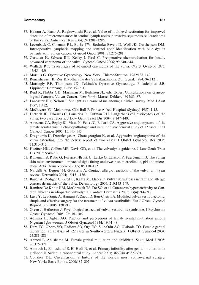
37. Hakam A, Nasir A, Raghuwanshi R, et al. Value of multilevel sectioning for improveddetection of micrometastases in sentinel lymph nodes in invasive squamous cell carcinomaof the vulva. Anticancer Res 2004; 24:1281–1286.
38. Levenback C, Coleman RL, Burke TW, Bodurka-Bevers D, Wolf JK, Gershenson DM.Intraoperative lymphatic mapping and sentinel node identification with blue dye inpatients with vulvar cancer. Gynecol Oncol 2001; 83:276–281.
39. Gerszten K, Selvara RN, Kelley J, Faul C. Preoperative chemoradiation for locallyadvanced carcinoma of the vulva. Gynecol Oncol 2006; 99:640–644.
40. Wallach RC. Cryosurgery of advanced carcinoma of the vulva. Obstet Gynecol 1976;47:454–458.
41. Martius G. Operative Gynecology. New York: Thieme-Stratton, 1982:136–142.42. Renziehausen K. Zur Kryotherapie des Vulvakarzinoms. Zbl Gynak 1974; 96:1121.43. Mattingly RF, Thompson JD. TeLinde’s Operative Gynecology. Philadelphia: J.B.
Lippincott Company, 1985:719–731.44. Reid R, Phibbs GD. Markman M, Beilinson JL, eds. Expert Consultations on Gyneco-
logical Cancers, Vulvar Cancer. New York: Marcel Dekker, 1997:83–87.45. Lancaster HO, Nelson J. Sunlight as a cause of melanoma; a clinical survey. Med J Aust
1957; 1:452.46. McGovern VJ. Melanoma. Clin Bull R Prince Alfred Hospital (Sydney) 1957; 1:45.47. Dietrich JF, Edwards C, Laucirica R, Kufman RH. Langerhans cell histiocytosis of the
vulva: two case reports. J Low Genit Tract Dis 2004; 8:147–149.48. Amezcua CA, Begley SJ, Mata N, Felix JC, Ballard CA. Aggressive angiomyxoma of the
female genital tract: a clinicopathologic and immunohistochemical study of 12 cases. Int JGynecol Cancer 2005; 15:140–145.
49. Dragoumis K, Drevelengas A, Chatzigeorgiou K, et al. Aggressive angiomyxoma of thevulva extending into the pelvis: report of two cases. J Obstet Gynaecol Res 2005;31:310–313.
50. Haefner HK, Collins ME, Davis GD, et al. The vulvodynia guideline. J Low Genit TractDis 2005; 9:40–51.
51. Runeman B, Rybo G, Forsgren-Brusk U, Larko O, Larsson P, Faergemann J. The vulvarskin microenvironment: impact of tight-fitting underwear on microclimate, pH and micro-flora. Acta Derm Venereol 2005; 85:118–122.
52. Nardelli A, Degreef H, Goossens A. Contact allergic reactions of the vulva: a 14-yearreview. Dermatitis 2004; 15:131–136.
53. Bauer A, Rodiger C, Greif C, Kaatz M, Elsner P. Vulvar dermatoses irritant and allergiccontact dermatitis of the vulva. Dermatology 2005; 210:143–149.
54. Ramirez De Knott HM, McCormick TS, Do SO, et al. Cutaneous hypersensitivity to Can-dida albicans in idiopathic vulvodynia. Contact Dermatitis 2005; 53(4):214–218.
55. Lavy Y, Lev-Sagie A, Hamani Y, Zacut D, Ben-Chetrit A. Modified vulvar vestibulectomy:simple and effective surgery for the treatment of vulvar vestibulitis. Eur J Obstet GynecolReprod Biol 2005; 120:915.
56. Green J, Hetherton J. Psychological aspects of vulvar vestibulitis syndrome. J PsychosomObstet Gynaecol 2005; 26:101–106.
57. Adinma JI, Agbai AO. Practice and perceptions of female genital mutilation amongNigerian Igbo women. J Obstet Gynaecol 1944; 19:44–48.
58. Dare FO, Oboro VO, Fadiora SO, Orji EO, Sule-Odu AO, Olabode TO. Female genitalmutilation: an analysis of 522 cases in South-Western Nigeria. J Obstet Gynaecol 2004;24:281–283.
59. Ahmed B, Abushama M. Female genital mutilation and childbirth. Saudi Med J 2005;26:376–378.
60. Almroth L, Elmusharaf S, EI Hadi N, et al. Primary infertility after genital mutilation ingirlhood in Sudan: a case-control study. Lancet 2005; 366(9483):385–391.
61. Gollaher DL. Circumcision, a history of the world’s most controversial surgery.New York: Basic Books, 2000:187–207.
Commentary 187

62. Millner VS, Eichold BH II, Sharpe TH, Lynn SC Jr. First glimpse of the functionalbenefits of clitoral hood piercings. Am J Obstet Gynecol 2005; 19:675–676.
63. O’Connell HE, DeLancey JO. Clitoral anatomy in nulliparous, healthy, premenopausalvolunteers using unenhanced magnetic resonance imaging. J Urol 2005; 173:2060–2063.
64. Lloyd J, Crouch NS, Minto CL, Liao LM, Creighton SM. Female genital appearance:‘‘normality’’ unfolds. BJOG 2005; 12:643–646.
65. Dickinson RL. Human Sex Anatomy. Huntington: Robert F. Krieger Publishing Co Inc,1971:145.
188 Wallach

Index
Acanthosis nigricans (AN), 63diagnosis, 110histopathology, 110pathophysiology, 110treatment, 111
Acrochordons. See Fibroepithelialstromal polyp
AdenocarcinomaBartholin’s, 160Skene’s gland, 172, 174
Adenosis, vestibular, 138–139Angiokeratoma, 150–151Angiomyofibroblastoma, 151, 153Angiomyxoma, aggressive, 152, 154Anogenital disease, 48Apocrine glands, 46Atypical junctional melanocytic
hyperplasia (AJMH)biologic behavior, 116histopathology, 117treatment, 117
Bacterial folliculitis, 86Bacterial infections, 77–93Bacterial vaginosis, 3, 84
diagnosis, 84, 92risk factors, 84therapy, 84, 92
Bartholin’s adenocarcinomas, 160Bartholin’s duct cyst, 135–136Bartholin’s glands. See Vestibular glandsBasal cell carcinoma, 166Basal layer hyperpigmentation, 114Basaloid lesions, 158Behcet’s syndrome, 53–54Behcet’s disease, 7Benign nevi. See Melanocytic neviBenign tumor-like lesions
endometriosis, 139–140
[Benign tumor-like lesions]fibroepithelial stromal polyp, 139vestibular adenosis, 138–139
Bullous diseases, 54
Calymmatobacteriumgranulomatosis, 79
Candida albicans, 3, 93vulvovaginal, 96
Candida glabrata, 3, 93Candida krusei, 93Candida lusitaniae, 93Candida parapsilosis, 93Candida tropicalis, 93Candidiasis, 93
diagnosis, 94–95vulvovaginal, 3, 93
CarcinomaBartholin’s gland, 167basal cell, 166–167cervical, 183lymphoepithelioma-like, 167squamous cell, 161–164urethral, 175verrucous, 162–165
Caruncles hymenalis, 12Cellulitis, 4, 86Cervical carcinoma, 183Cervical dysplasia, 74Chancroid, 81–83Chickenpox. See VaricellaChlamydia trachomatis, 80, 135Circumcision, female genital, 6Clitoris, 13
prepuce and frenulum of, 21suspensory ligament of, 14
Clobetasol propionate, 51Colle’s fascia, 13Condylomata acuminata, 4
189

Condylomata lata, 78Corynebacterium minutissimum, 90Crohn’s disease, 45–46Cystic tumors, 4Cysts
ciliated, 135epidermal inclusion cysts, 135–137mesonephric-like, 138mesothelial, 138mucinous, 135, 137–138paraurethral (Skene’s duct), 170
Darier’s disease, 55, 146Dermatofibroma, 148–149Dermatofibrosarcoma protuberans (DFSP),
167, 169–170Dermatophytosis, 97Desmin, 140Dysplasia
cervical, 74vulvar, 9
diagnosis of, 183
Eczema, 37–38Elective lymph node dissection
(ELND), 124Endometriosis, 139–141Epidermophyton floccosum, 97Epithelial lesions
keratoacanthomas, 146–148seborrheic keratosis, 144–146warty dyskeratomas (WDs), 146–147
Epstein–Barr virus (EBV), 166Erythema, 39, 42, 63Erythrasma, 90, 91Erythroplakia, 9Escherichia coli infection, 170Estrogen receptors (ER), 29, 140
Fasciitis, necrotizing, 89–90Fibers
elastic, 22parasympathetic, 17
Fibroepithelial stromal polyp, 139Fibroma, 156Fixed drug eruption (FDE), 62Folliculitis, 4
bacterial, 86Fournier’s gangrene, 89Fungal infections, 93–100Furunculosis, 87
Gardnerella vaginalis, 84Genital anomalies
imperforate hymen, 32, 33longitudinal vaginal septum, 32transverse vaginal septum, 32vaginal agenesis, 31–32vulvovaginal abnormalities, 34
Genital herpes virus infection, 3Genital mucosa
chronic ulcer of, 54genetic, 55
Genital mutilation, 184Genital staphylococcal infections, 85Genital tuberculosis, 84Genital warts, 4
therapy of, 75Genitalia, ambiguous, 31Granular cell tumors, 152–155Granuloma inguinale, 79–80Granuloma pyogenicum. See Pyogenic
granulomas
Haemophilus ducreyi, 81Hailey–Hailey disease, 54Hemangioma, 149–150Hemophilus ducreyi, 4Herpes simplex virus (HSV), 4, 73
infection, 69of anogenital skin, 72diagnosis of, 72of mouth, 71neonatal, 72recurrent, 71treatment, 72
serotypes of, 69Herpes zoster, 69Hidradenitis suppurativa, 46, 48–49Hidradenoma, papillary, 143–144Hidradenoma papilliferum, 20Human immunodeficiency virus (HIV), 7Human papilloma virus (HPV), 141
infection, 183types, 73
Hyperinsulinemia, 110Hyperpigmentation, 110
basal layer, 114Hyperplasia, squamous cell, 2
Keratoacanthomas, 146–148
Labia minora, 1Leiomyoma, 154
of urethra, 174–175
190 Index

Lesions, 40diagnosis of, 8etiology of, 1sexually transmitted infections with, 4See also individual lesion types
Leukoplakia, 9Lichen aureus, 45–46Lichen planopilaris, 42Lichen planus (LP), 40–45
hypertrophic, 42–43immunofluorescent (IMF) studies of, 43and lichen sclerosus (LS), 43on tongue, 41treatment of, 44vulvar, 40
clinical forms of, 40pigmented flexural, 40
Lichen sclerosus (LS), 2, 127lesions of, 49squamous cell carcinoma (SCC) and, 40treatment of, 50, 182
Lichen sclerosus et atrophicus (LSA)metabolic activity in, 182vulvar, 182
Lichen simplex, 59histological changes of, 61lesions of, 61treatment of, 61
Lipoma, 156Lipschutz’s ulcer, 51
treatment, 52–53Lupus erythematosus, 59Lymphangiectasia, 60
treatment, 61Lymphangioma circumscriptum, 150–153Lymphoepithelioma-like carcinoma,
166, 168Lymphogranuloma venereum (LGV)
complications, 81symptoms, 80treatment, 81
Malacoplakia, 169, 173Malignant melanoma, 5Melanocytes, 28Melanocytic hyperplasia, 120Melanocytic lesions, 111Melanocytic nevi
histopathology, 115treatment, 116
MelanomaBreslow system, 124clinical advantages of, 121
[Melanoma]differential diagnosis of, 121factors, 125histopathology of, 122with lichen sclerosus, 127lymph node evaluation for, 124malignant, 5signs and symptoms, 121staging system, 123treatment of, 124
Melanoma antigen–encoding genes(MAGE), 125
Melanoma antigen recognized by T-cells 1(MART-1), 125
Melanoma in situ (MIS)definition of, 119differential diagnosis of, 121histopathology of, 119treatment of, 120
Melanosisbenign vulvar, 112diagnosis of, 114histopathology, 113treatment of, 114
Melanotic macule, 112Merkel cells, 28Mesenchymal lesions
aggressive angiomyxoma, 152, 154angiokeratoma, 150–150angiomyofibroblastoma, 151, 153dermatofibroma, 148–149fibroma, 156granular cell tumors, 152–155hemangioma, 149–150leiomyoma, 154lipoma, 156lymphangioma circumscriptum,
150–151neurofibroma, 157, 160
Mesonephric duct cysts, 33Mesonephric-like cysts, 138Mesothelial cyst, 138Metastatic tumors, 168, 171Micropapillomatosis labialis (MPL),
140, 143Microphthalmia transcription factor, 125Molluscum contagiosum, 75
treatment, 77Monsel’s solution, 51Mucinous cysts, 135, 137, 138Mucinous glands, 26Mucosa-associated lymphoid tissue
(MALT), 28Mucous membrane lesions, 67
Index 191
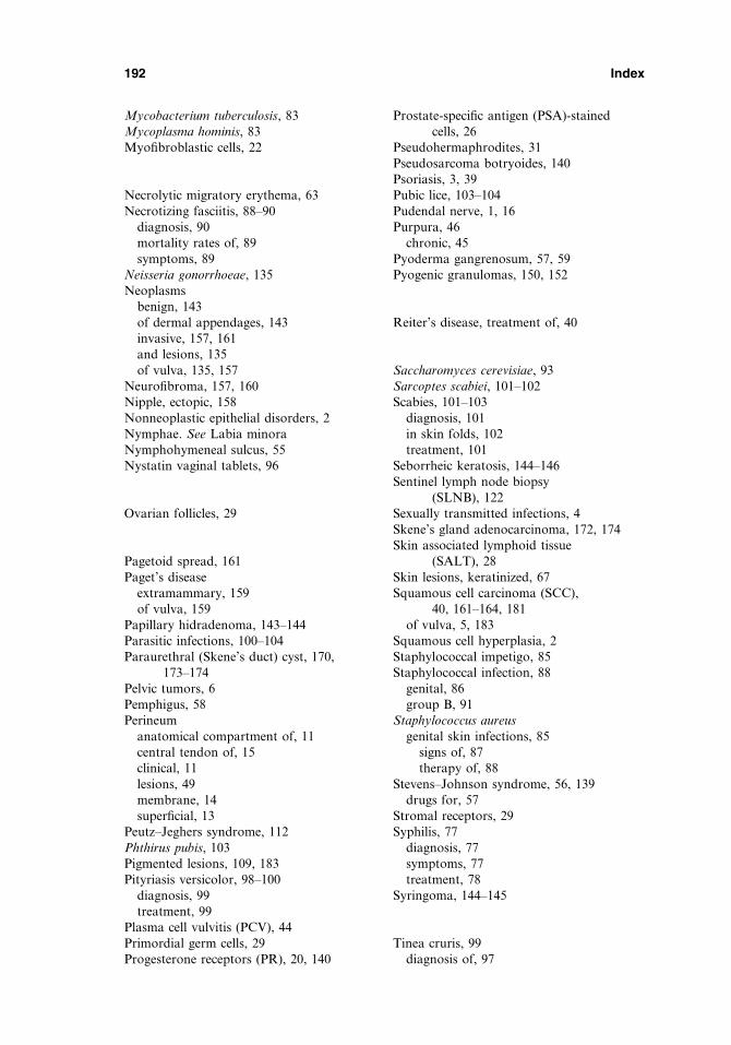
Mycobacterium tuberculosis, 83Mycoplasma hominis, 83Myofibroblastic cells, 22
Necrolytic migratory erythema, 63Necrotizing fasciitis, 88–90
diagnosis, 90mortality rates of, 89symptoms, 89
Neisseria gonorrhoeae, 135Neoplasms
benign, 143of dermal appendages, 143invasive, 157, 161and lesions, 135of vulva, 135, 157
Neurofibroma, 157, 160Nipple, ectopic, 158Nonneoplastic epithelial disorders, 2Nymphae. See Labia minoraNymphohymeneal sulcus, 55Nystatin vaginal tablets, 96
Ovarian follicles, 29
Pagetoid spread, 161Paget’s disease
extramammary, 159of vulva, 159
Papillary hidradenoma, 143–144Parasitic infections, 100–104Paraurethral (Skene’s duct) cyst, 170,
173–174Pelvic tumors, 6Pemphigus, 58Perineum
anatomical compartment of, 11central tendon of, 15clinical, 11lesions, 49membrane, 14superficial, 13
Peutz–Jeghers syndrome, 112Phthirus pubis, 103Pigmented lesions, 109, 183Pityriasis versicolor, 98–100
diagnosis, 99treatment, 99
Plasma cell vulvitis (PCV), 44Primordial germ cells, 29Progesterone receptors (PR), 20, 140
Prostate-specific antigen (PSA)-stainedcells, 26
Pseudohermaphrodites, 31Pseudosarcoma botryoides, 140Psoriasis, 3, 39Pubic lice, 103–104Pudendal nerve, 1, 16Purpura, 46
chronic, 45Pyoderma gangrenosum, 57, 59Pyogenic granulomas, 150, 152
Reiter’s disease, treatment of, 40
Saccharomyces cerevisiae, 93Sarcoptes scabiei, 101–102Scabies, 101–103
diagnosis, 101in skin folds, 102treatment, 101
Seborrheic keratosis, 144–146Sentinel lymph node biopsy
(SLNB), 122Sexually transmitted infections, 4Skene’s gland adenocarcinoma, 172, 174Skin associated lymphoid tissue
(SALT), 28Skin lesions, keratinized, 67Squamous cell carcinoma (SCC),
40, 161–164, 181of vulva, 5, 183
Squamous cell hyperplasia, 2Staphylococcal impetigo, 85Staphylococcal infection, 88
genital, 86group B, 91
Staphylococcus aureusgenital skin infections, 85
signs of, 87therapy of, 88
Stevens–Johnson syndrome, 56, 139drugs for, 57
Stromal receptors, 29Syphilis, 77
diagnosis, 77symptoms, 77treatment, 78
Syringoma, 144–145
Tinea cruris, 99diagnosis of, 97
192 Index

[Tinea cruris]symptoms, 97treatment, 98
Trauma, 6Treponema pallidum, 77Trichoepitheliomas, 143–145Trichomonas vaginalis, 100Trichomonas vaginitis, 3, 100–101Trichophyton mentagrophytes, 97Trichophyton rubrum, 97Trimethoprim-sulfamethoxazole, 88Tuberculin skin test, 83Tuberculosis (TB), genital, 83Tumors
cystic, 4genital, 6metastatic, 168, 171pelvic, 6
Ulcers, 53, 58Urethra
benign lesions of, 168carcinoma of, 175–176caruncles, 168, 172diverticulum, 172leiomyoma of, 174–175malacoplakia of, 169malignant lesions of, 175pelvic, 17perineal, 17
Urogenital diaphragm, 15Urticarial plaques, 68
Vaginadiseases, 3layers of, 17lymphatic drainage of, 17
Vaginosis, bacterial, 3, 84Vancomycin, 88Varicella zoster virus (VSV), 67
diagnosis of, 68infection, 67, 70
complications of, 68treatment, 69
Verrucous carcinoma, 50, 162–165Vestibular adenosis, 138–139Vestibular glands, 4, 26–27Vimentin, 140Viral infections, 67–77Vitiligo, 48Von Recklinghausen’s disease, 157
Vulvabenign aphthae of, 51
treatment, 52biopsy of, 9blood supply of, 1deep anatomy
arteries, 15Colle’s fascia, 13perineal body, 15urogenital diaphragm, 15vestibular glands, 14
dysesthesia, 6embryology of, 29–30histology of, 18
interlabial sulcus, 20labium majus, 18labium minus, 20mons pubis, 19mucinous glands, 26perineum, 19urethra, 26vagina, 25
lymphatic drainage of, 16superficial fascia of, 16surface anatomy
clinical perineum, 13clitoris, 13fossa navicularis, 13labia majora, 11labia minora, 12mons pubis, 11urethral meatus, 13vagina vestibule, 12
surface limits of, 11Vulvar intraepithelial neoplasia
(VIN), 5squamous, 158, 161–162
Vulvar vestibulitis syndrome (VVS),treatment of, 184
Vulvodynia, 95Vulvovaginal candidiasis, 3, 93
treatment, 95Vulvovaginal infections, 75, 93
Warty dyskeratomas (WDs), 146–147Warty lesions, 158Wolffian duct-like cysts. See Mesonephric-
like cysts
Zoon’s vulvitis, 44–45treatment, 45
Index 193

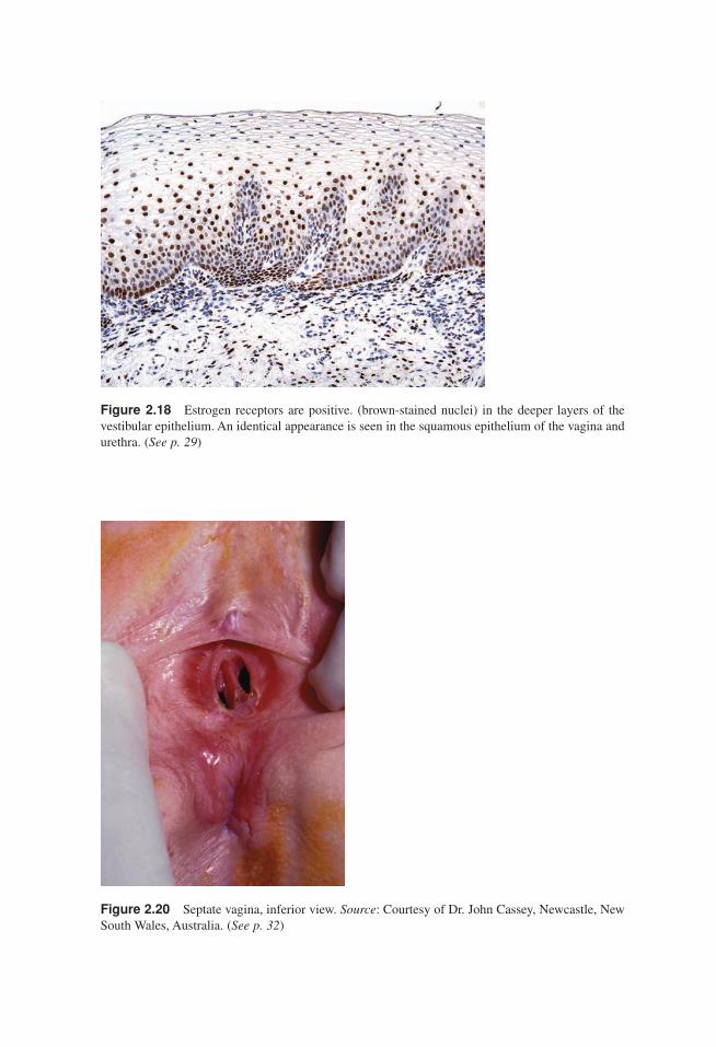
Figure 2.18 Estrogen receptors are positive. (brown-stained nuclei) in the deeper layers of thevestibular epithelium. An identical appearance is seen in the squamous epithelium of the vagina andurethra. (See p. 29)
Figure 2.20 Septate vagina, inferior view. Source: Courtesy of Dr. John Cassey, Newcastle, NewSouth Wales, Australia. (See p. 32)
Heller color insert new 11/1/06 11:25 AM Page 1
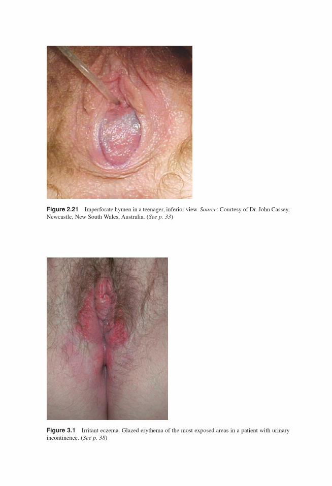
Figure 2.21 Imperforate hymen in a teenager, inferior view. Source: Courtesy of Dr. John Cassey,Newcastle, New South Wales, Australia. (See p. 33)
Figure 3.1 Irritant eczema. Glazed erythema of the most exposed areas in a patient with urinaryincontinence. (See p. 38)
Heller color insert new 11/1/06 11:25 AM Page 2
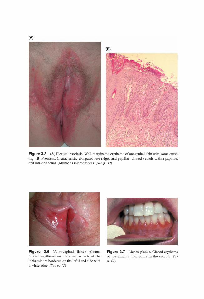
Figure 3.3 (A) Flexural psoriasis. Well-marginated erythema of anogenital skin with some crust-ing. (B) Psoriasis. Characteristic elongated rete ridges and papillae, dilated vessels within papillae,and intraepithelial. (Munro’s) microabscess. (See p. 39)
Figure 3.6 Vulvovaginal lichen planus.Glazed erythema on the inner aspects of thelabia minora bordered on the left-hand side witha white edge. (See p. 42)
Figure 3.7 Lichen planus. Glazed erythemaof the gingiva with striae in the sulcus. (See p. 42)
Heller color insert new 11/1/06 11:25 AM Page 3
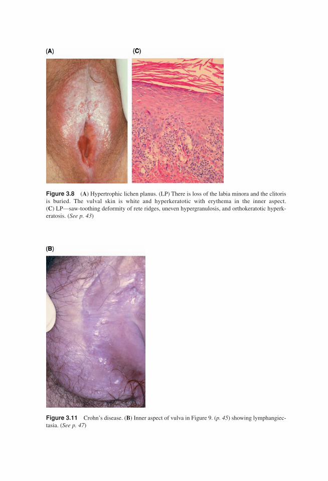
Figure 3.8 (A) Hypertrophic lichen planus. (LP) There is loss of the labia minora and the clitorisis buried. The vulval skin is white and hyperkeratotic with erythema in the inner aspect. (C) LP—saw-toothing deformity of rete ridges, uneven hypergranulosis, and orthokeratotic hyperk-eratosis. (See p. 43)
Figure 3.11 Crohn’s disease. (B) Inner aspect of vulva in Figure 9. (p. 45) showing lymphangiec-tasia. (See p. 47)
Heller color insert new 11/1/06 11:25 AM Page 4
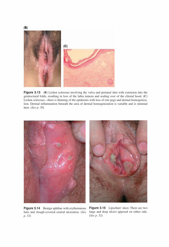
Figure 3.13 (B) Lichen sclerosus involving the vulva and perianal skin with extension into thegenitocrural folds, resulting in loss of the labia minora and sealing over of the clitoral hood. (C)Lichen sclerosus—there is thinning of the epidermis with loss of rete pegs and dermal homogeniza-tion. Dermal inflammation beneath the area of dermal homogenization is variable and is minimalhere. (See p. 50)
Figure 3.14 Benign aphthae with erythematoushalo and slough-covered central ulceration. (See p. 52)
Figure 3.15 Lipschutz’ ulcer. There are twolarge and deep ulcers apposed on either side.(See p. 52)
Heller color insert new 11/1/06 11:25 AM Page 5
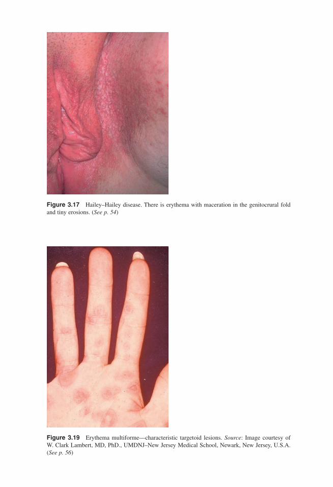
Figure 3.17 Hailey–Hailey disease. There is erythema with maceration in the genitocrural foldand tiny erosions. (See p. 54)
Figure 3.19 Erythema multiforme—characteristic targetoid lesions. Source: Image courtesy of W. Clark Lambert, MD, PhD., UMDNJ–New Jersey Medical School, Newark, New Jersey, U.S.A.(See p. 56)
Heller color insert new 11/1/06 11:25 AM Page 6
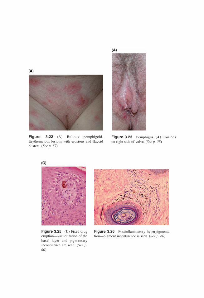
Figure 3.22 (A) Bullous pemphigoid.Erythematous lesions with erosions and flaccidblisters. (See p. 57)
Figure 3.23 Pemphigus. (A) Erosionson right side of vulva. (See p. 58)
Figure 3.25 (C) Fixed drugeruption—vacuolization of thebasal layer and pigmentaryincontinence are seen. (See p.60)
Figure 3.26 Postinflammatory hyperpigmenta-tion—pigment incontinence is seen. (See p. 60)
Heller color insert new 11/1/06 11:25 AM Page 7

Figure 4.8 Despite the careful descriptions of differing morphologies of ulcers in the variousulcerative sexually transmitted diseases, the ulcers often appear discouragingly similar. This lesionof chancroid cannot be diagnosed on morphology alone, but rather on clinical suspicion and labora-tory investigation. (See p. 82)
Figure 4.13 Necrotizing fasciitis is characterized by hemorrhagic bullae because of ischemiafrom underlying infection that has destroyed the cutaneous vascular supply; resulting ulceration andhemorrhage can be seen in this patient who is in the healing phase. (See p. 89)
Heller color insert new 11/1/06 11:25 AM Page 8

Figure 4.14 Erythrasma resembles tinea cruris, but the color is often more tan than red, and thereis no peripheral accentuation of scale. (See p. 91)
Figure 4.15 In uncommon cases, bacterialvaginitis can occur, extending to infect the skinand producing red, scaling, or exudative skin.(See p. 92)
Figure 4.17 Vulvovaginal candidiasis canproduce redness, edema, and fissuring. (Seep. 94)
Heller color insert new 11/1/06 11:25 AM Page 9
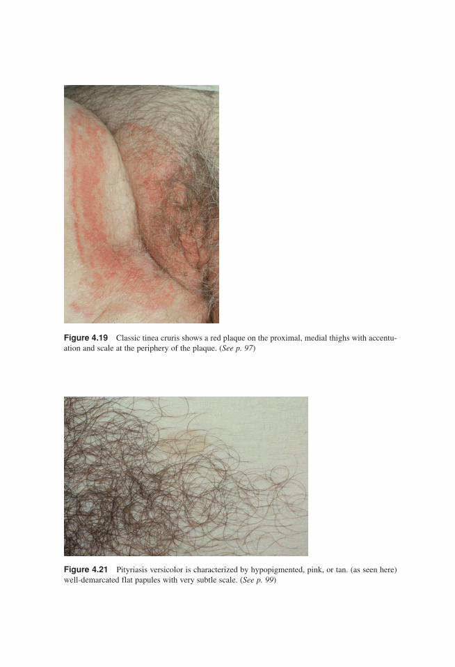
Figure 4.19 Classic tinea cruris shows a red plaque on the proximal, medial thighs with accentu-ation and scale at the periphery of the plaque. (See p. 97)
Figure 4.21 Pityriasis versicolor is characterized by hypopigmented, pink, or tan. (as seen here)well-demarcated flat papules with very subtle scale. (See p. 99)
Heller color insert new 11/1/06 11:25 AM Page 10
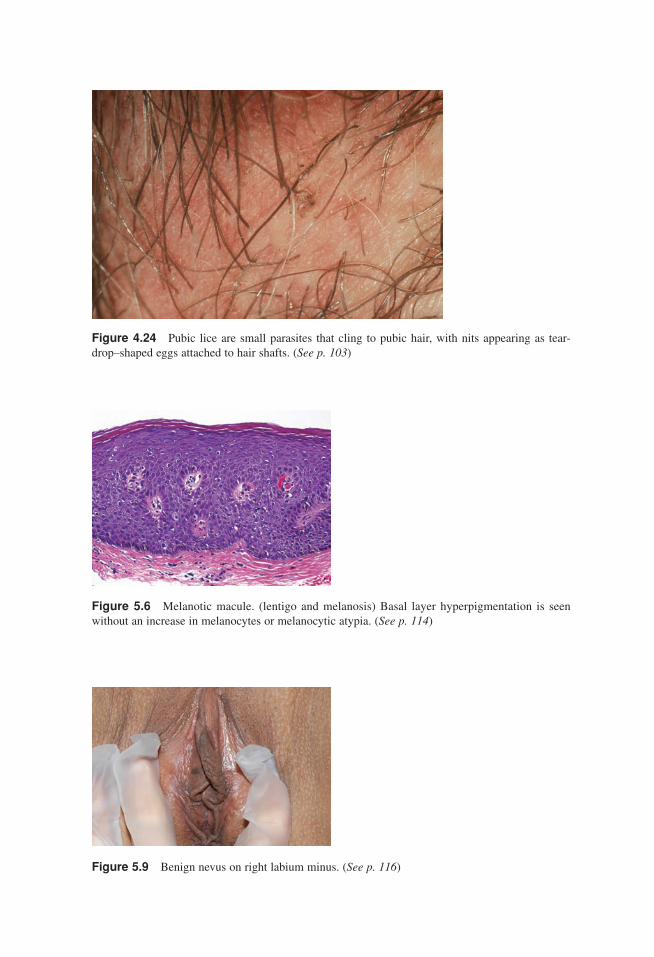
Figure 4.24 Pubic lice are small parasites that cling to pubic hair, with nits appearing as tear-drop–shaped eggs attached to hair shafts. (See p. 103)
Figure 5.6 Melanotic macule. (lentigo and melanosis) Basal layer hyperpigmentation is seenwithout an increase in melanocytes or melanocytic atypia. (See p. 114)
Figure 5.9 Benign nevus on right labium minus. (See p. 116)
Heller color insert new 11/1/06 11:26 AM Page 11
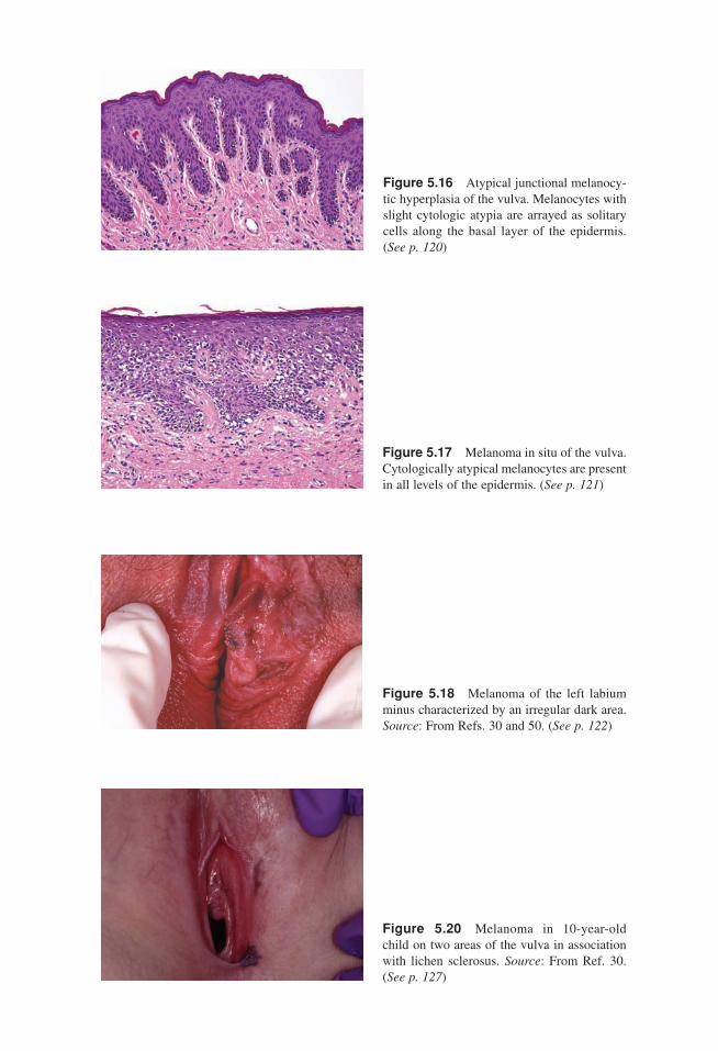
Figure 5.16 Atypical junctional melanocy-tic hyperplasia of the vulva. Melanocytes withslight cytologic atypia are arrayed as solitarycells along the basal layer of the epidermis.(See p. 120)
Figure 5.17 Melanoma in situ of the vulva.Cytologically atypical melanocytes are presentin all levels of the epidermis. (See p. 121)
Figure 5.18 Melanoma of the left labiumminus characterized by an irregular dark area.Source: From Refs. 30 and 50. (See p. 122)
Figure 5.20 Melanoma in 10-year-oldchild on two areas of the vulva in associationwith lichen sclerosus. Source: From Ref. 30.(See p. 127)
Heller color insert new 11/1/06 11:26 AM Page 12

Figure 6.3 (A) Mucinous cyst of the vestibule.(See p. 137)
Figure 6.7 Endometriosis. (A) Grossly endometrioticimplants on the vulva often have a blue appearance. (See p. 141)
Figure 6.12 Syringoma: the characteristiccomma-shaped epithelial nests in a collagen-ous background are seen. (See p. 145)
Figure 6.17 (C) Senile telangiectasiae. (See p. 149)
Heller color insert new 11/1/06 11:26 AM Page 13
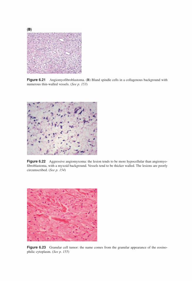
Figure 6.21 Angiomyofibroblastoma. (B) Bland spindle cells in a collagenous background withnumerous thin-walled vessels. (See p. 153)
Figure 6.22 Aggressive angiomyxoma: the lesion tends to be more hypocellular than angiomyo-fibroblastoma, with a myxoid background. Vessels tend to be thicker walled. The lesions are poorlycircumscribed. (See p. 154)
Figure 6.23 Granular cell tumor: the name comes from the granular appearance of the eosino-philic cytoplasm. (See p. 155)
Heller color insert new 11/1/06 11:26 AM Page 14
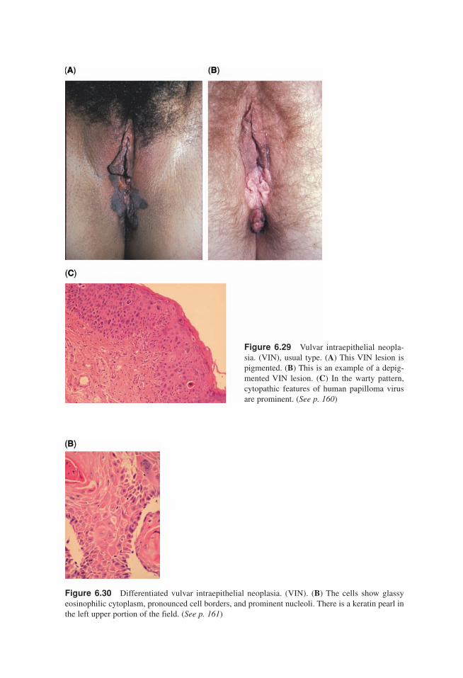
Figure 6.29 Vulvar intraepithelial neopla-sia. (VIN), usual type. (A) This VIN lesion ispigmented. (B) This is an example of a depig-mented VIN lesion. (C) In the warty pattern,cytopathic features of human papilloma virusare prominent. (See p. 160)
Figure 6.30 Differentiated vulvar intraepithelial neoplasia. (VIN). (B) The cells show glassyeosinophilic cytoplasm, pronounced cell borders, and prominent nucleoli. There is a keratin pearl inthe left upper portion of the field. (See p. 161)
Heller color insert new 11/1/06 11:26 AM Page 15
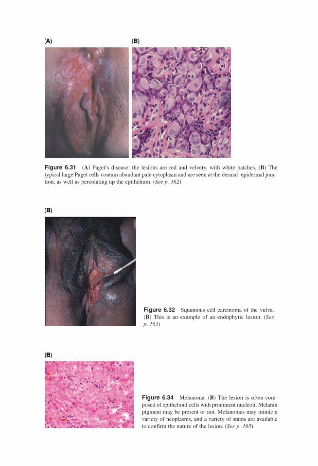
Figure 6.31 (A) Paget’s disease: the lesions are red and velvety, with white patches. (B) The typical large Paget cells contain abundant pale cytoplasm and are seen at the dermal–epidermal junc-tion, as well as percolating up the epithelium. (See p. 162)
Figure 6.32 Squamous cell carcinoma of the vulva.(B) This is an example of an endophytic lesion. (See p. 163)
Figure 6.34 Melanoma. (B) The lesion is often com-posed of epithelioid cells with prominent nucleoli. Melaninpigment may be present or not. Melanomas may mimic avariety of neoplasms, and a variety of stains are availableto confirm the nature of the lesion. (See p. 165)
Heller color insert new 11/1/06 11:26 AM Page 16
Copyright © 2022 FDOKUMEN


![Nitric oxide donor trans-[RuCl ([15] aneN4) NO] 2+ as a possible therapeutic approach for Chagas' disease](https://static.fdokumen.com/doc/165x107/6320e9c518429976e4066c52/nitric-oxide-donor-trans-rucl-15-anen4-no-2-as-a-possible-therapeutic-approach.jpg)