Hydatid disease-final
34
Hydatid Disease AT A GLANCE Prepared by; Ibrahim Shibl 6 th year medical student Under supervision of; Prof. Dr. Mona Shehata Prof. Dr. Walaa El-khalawany • Tanta University • Faculty of Medicine • Tropical Medicine and Infectious Diseases Department
-
Upload
independent -
Category
Documents
-
view
5 -
download
0
Transcript of Hydatid disease-final

Hydatid DiseaseAT A GLANCE
Prepared by;Ibrahim Shibl 6th year medical student
Under supervision of;Prof. Dr. Mona ShehataProf. Dr. Walaa El-khalawany
• Tanta University• Faculty of Medicine• Tropical Medicine and Infectious
Diseases Department

Agenda 📑 Definition & clinical varieties Epidemiology Life cycle & cyst formation Management
Diagnosis History taking Clinical presentation Complications Investigations
Treatment Prevention

Definition
Hydatid disease or echinococcosis is a parasitic zoonosis, caused by the larval stages of different species of the cestode (tapeworm) of genus Echinococcus.

Clinical varieties
Echinococcus
E. granulosus
Cystic echinococcosis
E. multilocularis
Alveolar echinococcosis
E. vogeli
Polycystic echinococcosis

Distribution of E. granulosus

Life cycle of E. granulosus

Cyst formation
Liver 70%
Pulmonary
Spleen, brain, bone

Cyst formation
a)Germinal layerb)Daughter cyst c)Scolicesd)Cavity of primary cyst
e)Laminated layerf)Host response layer

Agenda 📑 Definition & clinical varieties Epidemiology Life cycle & cyst formation Management
Diagnosis History taking Clinical presentation Complications Investigations
Treatment Prevention
We are here

Management of a case of liver hydatid cystHistory

Clinical presentation
Unless complicated, diagnosis is based mainly on CLINICAL SUSPICION

Clinical presentation
Asymptomatic

Clinical presentation

Clinical presentation
NNcomplicated?!!

complications
Rupture
Peritoneal
cavity
Bile duct
Hepatic vein
Lung
Colon
1. Rupture

complications
Secondary invasion by pyogenic organisms follows rupture into biliary passages, giving the picture of a pyogenic abscess
2. Infection

complications
Recurrent urticaria
Anaphylactic shock
3. Hydatid allergy

complications
4. Pressure necrosisExpansion of the cyst may cause pressure necrosis of the surrounding tissues with deterioration of function
5.Membranous glomerulonephritismay be related to glomerular deposits of hydatid antigen

Investigations
1. US is the main diagnostic tool
A. Imaging

US staging

US staging Active
1. CL: cystic lesion with no cyst wall2. CE1: with cyst wall3. CE2: with cyst wall + internal septation
Transitional 4. CE3: detached laminar layer (water-lily
sign) / partially collapsed Inactive
5. CE4: heterogeneous mass6. CE5: thick calcified wall

Investigations
2. X ray
A. Imaging

Investigations
3. CT scan
A. Imaging

Investigations B. ELISA

Investigations C. Eosinophilia
Eosinophilia of greater than 7% is found in about 30% of patients.
Eosinophilia may follow leakage or rupture of a cyst. NNN

Agenda 📑 Definition & clinical
varieties Epidemiology Life cycle & cyst formation Management
Diagnosis History taking Clinical presentation Complications Investigations
Treatment Prevention
✌ Almost there 📑

Treatment options of liver hydatid cyst
Medical Percutaneous drainage
Surgery Radical Conservative
Approach Open Laparoscopic
According to; Size of the cyst Location of the cyst Clinical Manifestations
Overall health of the patient
US staging

Treatment
Mebendazole Albendazole
1. medical For small CL, CE1 and CE3

Treatment 2. Percutaneous drainage “PAIR technique”
For large CE1 lesions and uncomplicated CE3
US guided;
Percutaneous punctureAspirateInject 95% ethanol or hypertonic saline
Re-aspirate

Treatment 2. Percutaneous drainage “PAIR technique”
Usually albendazole prophylaxis is started 1 week before PAIR and continued for 3-4 weeks thereafter
PAIR technique is done with extreme caution and under umbrella of antihistamines and corticosteroids for fear of anaphylaxis

Treatment 2. Percutaneous drainage “PAIR technique”
PAIR is contraindicated for;1. Superficially located cysts (risk of
rupture)2. Cysts with multiple thick internal septal
divisions (honeycombing pattern)3. Cysts communicating with the biliary tree4. Calcified or inactive cysts
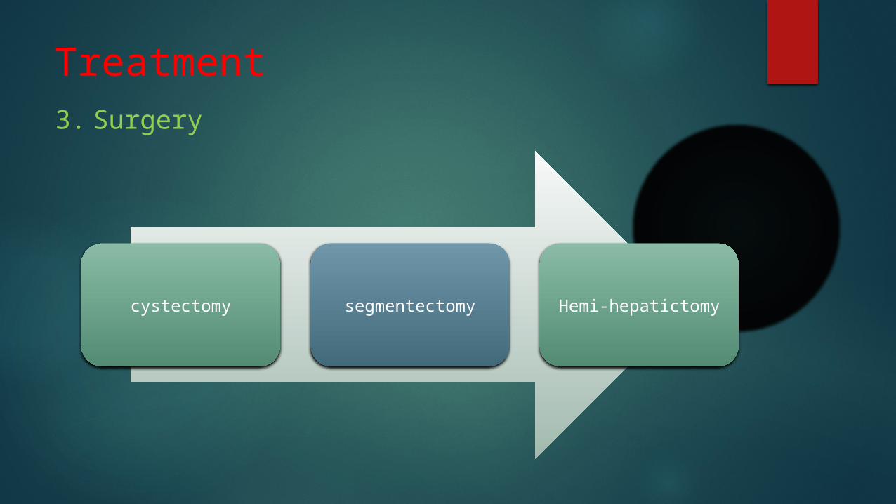
Treatment 3. Surgery
cystectomy segmentectomy Hemi-hepatictomy

Prevention of hydatid disease

✉ Take-home message
Echinococcosis is still endemic in middle east and Mediterranean countries ---- keep in mind
Suspicion is always in favor of your patient
Prevention is better than cure
If I ask you about this presentation after 1 week, you should be able to remember THREE points