Information Retrieval via Truncated Hilbert-Space Expansions

Truncated beta-amyloid peptide species in pre-clinical Alzheimer’s
disease as new targets for the vaccination approach
Nicolas Sergeant,* Stepanie Bombois,* Antoine Ghestem,* Herve Drobecq,� Vesna Kostanjevecki,�Carla Missiaen,� Annick Wattez,* Jean-Phillipe David,* Eugeen Vanmechelen,�Christian Sergheraert� and Andre Delacourte*
*INSERM U422, Groupe VCDN, Equipe Proteomique, Lille, France
�UMR 8525, CNRS – Institut Pasteur de Lille – Universite de Lille 2 – Institut de Biologie de Lille – Departement Synthese Structure
et Fonction des Biomolecules, Lille, France
�Innogenetics NV, Zwijnaarde, Belgium
Abstract
Vaccination against human beta-amyloid peptide (Ab) has
been shown to remove the amyloid burden produced in
transgenic mice overexpressing the mutated human amyloid
precursor protein (APP) gene. For human beings, the effi-
ciency of this therapeutic strategy has to take into account the
specificities of human amyloid, especially at the early stages
of ‘sporadic’ Alzheimer’s disease (AD). Ab 40/42 were previ-
ously quantified in tissues from our well-established brain
bank, including non-demented individuals with both mild
amyloid and tau pathologies, hence corresponding to the
earliest stages of Alzheimer pathology. Herein, we have
adapted a proteomic method combined with western blotting
and mass spectrometry for the characterization of insoluble
Ab extracted in pure-formic acid. We demonstrated that
amino-truncated Ab species represented more than 60% of all
Ab species, not only in full blown AD, but also, and more
interestingly, at the earliest stage of Alzheimer pathology. At
this stage, Ab oligomers were exclusively made of Ab-42
species, most of them being amino-truncated. Thus, our
results strongly suggest that amino-truncated Ab-42 species
are instrumental in the amyloidosis process. In conclusion, a
vaccine specifically targeting these pathological amino-trun-
cated species of Ab-42 are likely to be doubly beneficial, by
inducing the production of specific antibodies against patho-
logical Ab products that are, in addition, involved in the early
and basic mechanisms of amyloidosis in humans.
Keywords: Alzheimer’s disease, beta-amyloid peptide, pro-
teomic, diagnostic, vaccination, physiopathology.
J. Neurochem. (2003) 85, 1581–1591.
Alzheimer’s disease (AD) is a progressive dementing
disorder characterized by the conjunction of two degenerat-
ive processes: tau pathology and amyloid pathology. Both
degenerative processes are found in familial autosomal
dominant AD and in ‘sporadic’ AD (non-mendelean).
However, familial autosomal dominant AD due to mutations
on amyloid precursor protein (APP) or presenilin genes are
extremely rare (Campion et al. 1999) and furthermore, less is
known about the physiopathology of ‘sporadic’ AD.
The cognitive decline that characterizes AD is well
correlated to the cortical spreading of tau pathology (Dela-
courte et al. 1999). However, the amyloid pathology is in
close relationship with the aetiology of the disease. We have
recently shown a strong correlation between the amyloid
pathology and the dynamic of progression of tau pathology
in cortical brain areas, hence demonstrating a synergy
between tau and amyloid pathologies in ‘sporadic’ AD
(Delacourte et al. 2002; Sergeant et al. 2002). It suggests
overall that the amyloid pathology could trigger the spread-
ing of tau pathology, thus leading to AD. This hypothesis is
also illustrated in transgenic mouse models, in which the tau
Received February 18, 2003; 2003; accepted March 14, 2003.
Address correspondence and reprints requests to Andre Delacourte,
INSERM U422, Groupe VCDN, Equipe Proteomique 1, place de Verdun
59045 Lille cedex, France.
E-mail: [email protected]
Abbreviations used: Ab, beta-amyloid peptide; AD, Alzheimer’s dis-ease; APP, amyloid precursor protein; MALDI-TOF, Matrix-Assisted
Desorption and Ionization–Time-of-Flight; MS, mass spectrometry;
nano-LC, nanoliquid chromatography; pI, isoelectric point; Q-TOF,
Quadrupole–Time-of-Flight.
Journal of Neurochemistry, 2003, 85, 1581–1591 doi:10.1046/j.1471-4159.2003.01818.x
� 2003 International Society for Neurochemistry, J. Neurochem. (2003) 85, 1581–1591 1581

pathology is enhanced by the induction or coexistence of an
amyloid pathology (Gotz et al. 2001; Lewis et al. 2001).
A possible treatment of AD would be to reduce the
progression of tau pathology by targeting directly the
amyloid pathology. Such treatment is already tested and
opens new perspectives in AD therapy. The vaccination of
transgenic mice producing amyloid plaques by targeting
actively or passively the human beta-amyloid peptide (Ab),the major component of amyloid deposits, leads to a reduced
burden of amyloid deposition (Schenk et al. 1999; 2001;
DeMattos et al. 2001) and restores cognitive function
(Dodart et al. 2002; Janus et al. 2000). In human, vaccin-
ation leads to antibodies against amyloid deposits (Hock
et al. 2002). Physiologically, Ab is a 37–42 amino acid
peptide that derives from the catabolism of a type I
transmembrane glycoprotein, named amyloid precursor pro-
tein (APP) (Wiltfang et al. 2002). APP is ubiquitously
expressed in all tissues, and in large amounts in the central
nervous system. In transgenic mice, vaccination aimed to
target the human Ab sequence that is different from the Absequence of mouse (R to G at position 5, Y to F at position
10 and H to R at position 13 of Ab sequence, respectively).Therefore, the risk of autoimmunity response for transgenic
mice is reduced.
Moreover, identified Ab peptides extracted from the
amyloid deposits of Alzheimer brain are composed of
heterogeneous species that include the full-length Ab 40/42peptides and additional shorter carboxy-terminal Ab peptidesas well as amino-terminal truncated species (Kalback et al.
2002). However, little is known about which species of Abpeptides are present at the very first steps of amyloid
deposition, even before the appearance of clinical symptoms
of dementia. We have investigated and characterized the Abpeptides involved in the first steps of amyloid deposition.
Those early deposits were found in non-demented individ-
uals that had developed evidences of Alzheimer pathogen-
esis. They had both an amyloid and tau pathology
demonstrated at the neuropathological and biochemical level
(Delacourte et al. 2002) but were clinically asymptomatic.
They were thus defined to belong to the infra-clinical or pre-
clinical stages of Alzheimer pathology (Delacourte et al.
1999, 2002). The aggregated species of Ab were directlyextracted from the brain tissue using formic acid and two-
dimensional gel electrophoresis analysis was adapted to
formic acid-treated samples. Ab species were identified usingmass spectrometry (MS) and confirmed using multiple
immunological tools against Ab peptides. Our data demon-strated for the first time that the major Ab species found toaggregate at the earliest stages of Alzheimer pathology were
amino-truncated and, as previously established (Delacourte
et al. 2002), they all corresponded to Ab-42 peptides.
Moreover, by using a western blotting approach to determine
the ratio of full-length Ab-42 upon all Ab x-42 species, theseamino-truncated species of Abwere shown to represent more
than 60% of all Ab species in non-demented as well as inconfirmed AD individuals.
In conclusion, the present work is important at a physio-
pathological point of view, as well as essential in developing
new diagnostic and therapeutic strategies, including vaccin-
ation. Among the already existing vaccination approach, a
unique opportunity to develop further vaccination strategy
would be to target non-physiological species of Ab peptidesthat are, in addition, specifically found in the most prevalent
‘sporadic’ form of Alzheimer’s pathology, and involved in
the very first steps of amyloid deposition.
Materials and methods
Patients
All of the brain autopsy materials used in the present study were
from our brain bank. Ten non-familial (sporadic) AD cases and eight
non-demented cases have been extensively described (Delacourte
et al. 1999, 2002). The five AD cases fulfilled the neuropathological
diagnostic criteria of AD as established by the National Institute on
Aging and the Reagan Institute Working Group on diagnostic
criteria for the neuropathological assessment of Alzheimer disease
(Hyman and Trojanowski 1997). The five non-demented cases
correspond to neurofibrillary stages I and II [similar to the tau
pathology stages 1–6, according to Delacourte et al. (1999, 2002)]
and stage B and C for amyloid deposition, according to neuropath-
ological staging of Braak and Braak (1991). Postmortem intervals
ranged from 5 to 61 h (mean of 30 ± 10 h). At autopsy, one brain
hemisphere was deep-frozen for biochemical analysis and the other
hemisphere was formalin-fixed for both neuropathological exam-
ination and histochemistry.
Antibodies
The amino-terminal region of Ab peptides was analysed with WO2(Abeta GmBH, Heidelberg, Germany) and 6E10 (Senetek PLC,
Napa, CA, USA) antibodies. These recognize the amino-acid
sequences 5–8 [N-ter (5–8)] and 4–13 [N-ter (4–13)], respectively
(Cherny et al. 1999; Terai et al. 2001). Ab x-42 species were studiedusing 21F12 antibody (Athena Neurosciences, Worcester, MA, USA)
and ADA42 antiserum. Ab x-40 species were analysed with our
antiserum ADA40 (Delacourte et al. 2002). Monoclonal antibody
3D6 (Athena Neurosciences) was used to detect the full-length Ab1-x species. It is specific to the free amino-terminal region (sequence
1–5) of Ab, necessarily including the first aspartate residue (Johnson-Wood et al. 1997; Walsh et al. 2000, 2002; Bard et al. 2003).
Formic acid isolation of Ab aggregates and two-dimensional
gel electrophoresis
The brain tissue samples from the temporal, frontal, parietal and
occipital cortex were processed as already described (Delacourte
et al. 2002). Formic acid (Prolabo, Fontenay s/Bois, France)
extracted brain tissue homogenate (100 lL) was evaporated undernitrogen and dissolved in 400 lL of two-dimensional electrophor-esis lysis buffer [7 M urea, 2 M thiourea, 4% (v/v) Triton X-100,
20 mM dithiothreitol and 0.6% (v/v) Pharmalytesa pH 3–10].
The sample was sonicated. Immobilized pH gradient strip pH 4–7
1582 N. Sergeant et al.
� 2003 International Society for Neurochemistry, J. Neurochem. (2003) 85, 1581–1591

(Bio-Rad, Marnes la Coquette, France) was equilibrated with the
sample for 15 h (Sergeant et al. 2002). Isoelectrofocusing was
performed using the Protean IEF cell following the manufacturer’s
(Bio-Rad) instructions. Polypeptides were resolved on Tris-Tricine
gels as described previously (Sergeant et al. 2002). The gels were
transferred for immunodetection using the Multiphor transfer unit
(Amersham-Pharmacia Biotech, Saclay, France), according to the
manufacturer’s instructions, or they were stained with Coomassie
Brilliant Blue G250 (Sigma, Saint Quentin Fallavier, France) for MS
analyses. Isoelectric points, molecular weights and the percentage of
volume (mean of spot intensities over the area of the spot) of each
Ab peptide variant were determined using Melanie III two-
dimensional gel analysis software (Genebio, Geneva, Switzerland).
Calibration of the location of full-length Ab peptide 40 or 42 wasperformed by adding 10 ng of synthetic Ab-40 and Ab-42 (BachemAG, Bubendorf, Switzerland) to a formic acid brain tissue
homogenate (not shown).
Mass spectrometry characterization
Coomassie Blue stained polypeptides spots were cut into 1 mm2 gel
pieces and washed twice with 50% (v/v) CH3CN in 25 mM Tris-HCl
pH 9. Gel pieces were dehydrated in a Speed-Vac and then in-gel-
digested overnight with 10 ng of Endoproteinase Lys-C (EC
3.4.21.19, Roche Molecular Biochemicals, Meylan, France) in
3 lL of Tris-HCl pH 9. The resulting digested peptides were
recovered in 10 lL of 50% (v/v) CH3CN and 1% trifluoroacetic acid(TFA). Samples were then prepared by the dry-droplet method. One
mL of the peptide mixture was mixed with freshly dissolved
b-cyano-4-hydroxycinnaminic acid 0.5 mL [5 mg/mL in 50% (v/v)
CH3CN and 0.1% trifluoroacetic acid], and spotted on the sample
plate. The dry spot was then washed with 5 lL of 0.1%
trifluoroacetic acid. Mass spectrometry was performed with a
Matrix-Assisted Desorption and Ionization–Time-of-Flight (MAL-
DI-TOF) Voyager-DE-STR (Applied Biosystems, Palo Alto, CA,
USA) set to the following parameters: positive mode, reflector,
voltage 20 kV, grid 61%, delayed extraction 90 ns, low mass gate
500 amu. The laser energy required to desorbe/ionize samples was
kept to a low value, compatible with a good signal/noise ratio.
Spectra were calibrated externally using the [M + H+] monoisotopic
ions from trypsinized lysozyme. The characterization of two two-
dimensional spots, that were digested with trypsin (EC 3.4.21.4,
Promega) (spot 1 and spot 2, Table 2), was performed by nanoliquid
chromatography (nano-LC/MS) coupled with a Quadrupole–Time-
of-Flight (Q-TOF) mass spectrometer (Micromass Limited, Man-
chester, UK). Digested peptides were sequenced in tandem MS
mode.
Western blot quantification of the proportion of full-length AbTris-Tricine gels and electrophoresis of formic acid-treated brain
tissue homogenates was performed as already described (Delacourte
et al. 2002). In addition to brain tissue homogenates, 10 and 20 ng
of synthetic Ab 1–42 (Bachem) were loaded. Two Tris-Tricine gelswere processed at the same time. Following semi-dry protein
transfer, one membrane was incubated with 3D6 antibody. The
second membrane was incubated with 21F12 antibody. Multiple
exposures were performed, ECL films were digitized using Labscan
and ImageMaster 1D Elite Software (Pharmacia), the amount of Abdetected was obtained in the linear range between the two quantities
of synthetic Ab loaded. Thus, corresponding amounts of Ab 1-x andAb x-42 were determined following 3D6 and 21F12 labelling,
respectively. The quantification was only performed for monomeric
Ab species. The proportion of full-length Ab was established bydividing the amount of Ab 1-x by that of Ab x-42.
Results
Two-dimensional characterization of aggregates species
of Ab of Alzheirmer’s disease brain tissue
Aggregates consisting of Ab in Alzheimer brains were
soluble only in pure formic acid, as previously described
(Delacourte et al. 2002; Kalback et al. 2002). Such formic
acid-soluble Ab species were resolved by two-dimensionalelectrophoresis and detected using a panel of specific Abantibodies, including Ab x-40 and Ab x-42 antibodies
(Fig. 1, Ab-40 and Ab-42 panels), as well as amino-terminalspecific Ab antibodies [Fig. 1, N-ter (5–8) and N-ter (4–13)panels]. Further characterizations were performed by MS
analysis of the two-dimensional spots with identical locations
to Ab peptides detected with Ab antibodies (Fig. 1,
Coomassie Blue panel). This characterization of Ab specieswas performed on brain tissues of AD cases in which the
total amount of formic acid-soluble Ab (above 500 lg/g ofbrain tissue; as established in Delacourte et al. 2002) enabled
subsequent MS analysis. Ten detected Ab spots were stainedalso by Coomassie Blue (Fig. 1, Coomassie Blue panel). An
in-gel digestion with Endoprotease-Lys C was performed and
the resulting peptides were analysed by MS. The peptide
masses of nine spots positively matched with Ab sequencefollowing MALDI-TOF/MS analysis (Fig. 2 and Table 1). In
all spots analysed, the peptide mass 1325.6 was observed,
corresponding to the Ab sequence 17–28. The peptides
corresponding to the sequence 29–40/42 were not detected
using MALDI-TOF. Using nano-LC/MS, the carboxy-ter-
minal Ab 29–40/42 peptides were detected in two-dimen-
sional spots 1 and 2 following in-gel digestion with trypsin
(Table 2). No additional shorter carboxy-terminal Ab 1-x
were observed following Q-TOF MS analysis (Table 2).
Altogether, two-dimensional gel electrophoresis followed
either by western blotting with specific Ab antibodies or MSanalysis enabled the characterization of the human brain
tissue Ab species solubilized with formic acid. Only Abmonomers were characterized using MS. The immunodetec-
ted dimeric species at 8 kDa (Fig. 1) were not stained with
Coomassie, hence not analysed by MS.
According to the isoelectric points (pI) of the spots
analysed, the labelling with N-terminal and C-terminal
antibodies and MS analyses, the following spots were
characterized. The results are summarized on Table 1. Spot
1 and 2 corresponded to full-length Ab-40/42 peptides
(Table 1 and Table 2). Spots 3–7 and 9–10 corresponded to
amino-terminal truncated and post-translationally modified
b-Amyloid peptides in early amyloid deposits 1583
� 2003 International Society for Neurochemistry, J. Neurochem. (2003) 85, 1581–1591

variants of Ab. The major truncated variants consisted of Abstarting at amino-acid positions 2–5 and 8–10. The post-
translational modifications characterized were pyroglutamy-
lation at position 3 and methylation (Table 1). The mass
spectra are presented for spots 4, 5, 7, 9 and 10 (Fig. 2).
Interestingly, spots 6, 7, 9 and 10 contained similar Abvariants but were separated as two spots, suggesting that an
as yet unidentified modification was present.
The proportion of each Ab species, following the immu-nolabelling with 21F12 (Ab-42) antibody, was determinedusing Melanie III software and expressed as the percentage
of volume (Table 1). The full-length Ab peptides represented37 ± 7% of all Ab species. Taken together, the truncated
variants thus accounted for more than 60%, among which
17 ± 7% and 20 ± 4% corresponded to truncated species
starting at residues 4, 5 and 8, 9 and 10, respectively
(Table 1). Surprisingly, the two-dimensional pattern of Ab-40 as revealed by the ADA40 antiserum completely over-
lapped the pattern obtained with WO2, which detects the
amino-terminal region of Ab. These results further suggestthat the identified truncated Ab derived from the Ab-42 andnot from Ab-40 species.
Characterization of Ab species in the first amyloid
deposits in non-demented individuals
The Ab species that aggregate in the first steps of amylo-idosis were investigated in the brain tissue of non-demented
patients. These patients were previously shown to have
amyloid deposits and tau pathology at the neuropathological
level and the biochemical level, but they had no cognitive
deficits. The three cases analysed by two-dimensional
western blotting had traces or low amounts of aggregated
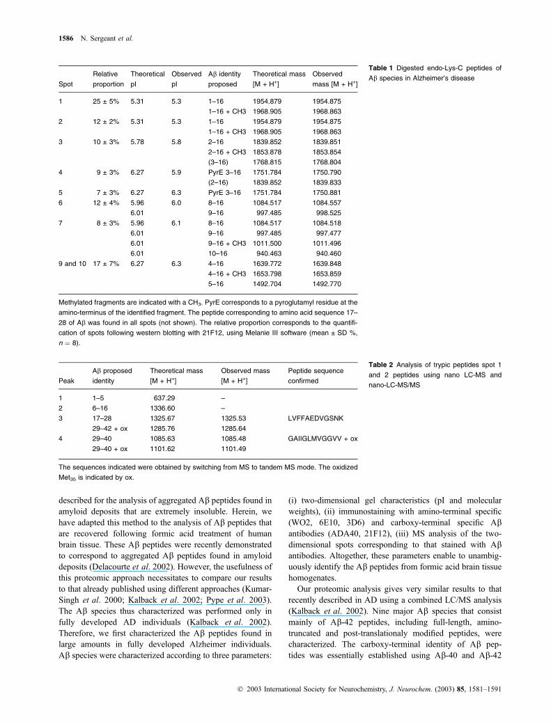
Ab (below 50 lg/g of brain tissue) (Delacourte et al. 2002).Ab aggregates were exclusively comprised of Ab-42 species(Fig. 3, Ab-40 and Ab-42 panels). Antibodies against theamino-terminal region of Ab only detected a single spot
corresponding to the full-length Ab peptide [Fig. 3, N-ter (5–8) and N-ter (4–13) panels]. The Ab-42 specific antibody21F12 labelled in addition spots 4, 5, 6 and 10 (Fig. 3, Ab-42panel) as well as dimers. The Ab-42 species in the brain ofnon-demented patients, as in Alzheimer brain, corresponded
to amino-terminal truncated variants starting at position
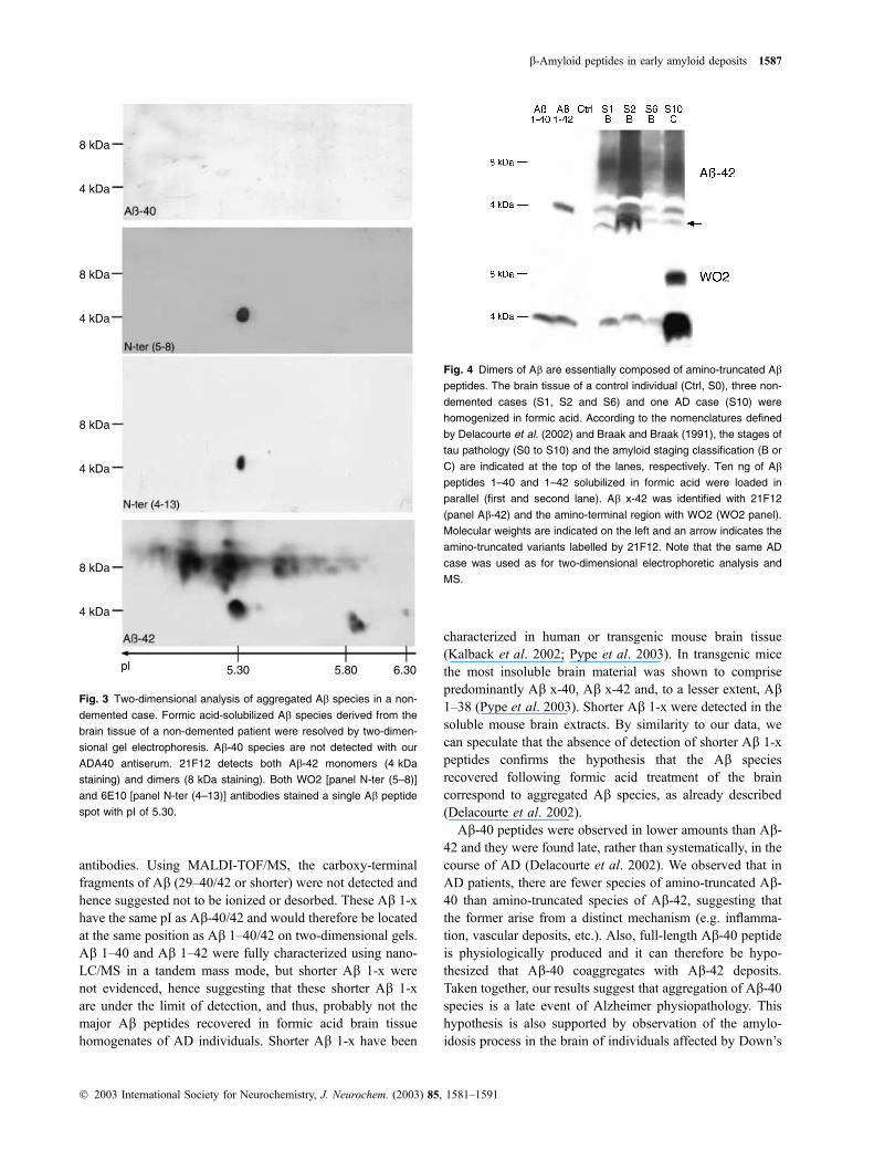
3-pyroglutamyl, 4, 5, 8 and 9. The lack of staining of Abdimers with amino-terminal Ab antibodies WO2 and 6E10on two-dimensional western blots [Fig. 3, N-ter (5–8) and
N-ter (4–13) panels] and WO2 antibody on one-dimensional
western blot [Fig. 4, N-ter (5–8) panel] suggested that Abdimers were exclusively composed of amino-terminally
truncated Ab-42 species (Figs 3 and 4, Ab-42 panels).
Proportion of Ab full-length in formic acid-treated brain
tissue of non-demented and AD individuals
The quantification of the proportion of full-length Ab peptidewas investigated further using 3D6 antibody. This monoclo-
nal antibody is specific for the free amino-terminal region of
Ab including the first aspartic residue (Johnson-Wood et al.
1997; Walsh et al. 2000). This specificity was verified using
our two-dimensional gel electrophoresis method (Fig. 5).
8 kDa
4 kDa
8 kDa
4 kDa
8 kDa
4 kDa
8 kDa
4 kDa
pl 5.30 5.80 6.30
Fig. 1 Two-dimensional electrophoretic analysis of Ab species in
Alzheimer’s disease. Ab aggregates solubilized with formic acid were
resolved by two-dimensional gel electrophoresis. Ab monomers
(4 kDa) and dimers (8 kDa) were labelled with antibodies WO2 and
6E10 against the amino-terminal region of Ab [N-ter(5–8) and N-ter(4–
13) panels, respectively]. The carboxy-terminal tails of Ab-42 and
Ab-40 were detected with 21F12 and ADA40, respectively (Ab-42 and
Ab-40 panels). The major Ab species recovered with our extraction
method were stained with Coomassie Blue and 10 spots were sub-
sequently analysed by MS (Table 1). The results presented were
obtained from the AD patient showing the largest quantity of amyloid
deposits. The pI and the Ab spots used for MS analysis are indicated.
Note Coomassie Blue does not stained dimeric species of Ab.
1584 N. Sergeant et al.
� 2003 International Society for Neurochemistry, J. Neurochem. (2003) 85, 1581–1591
Among all Ab species detected in an AD individual (Fig. 5a,Ab x-42 panel), 3D6 immunolabelling revealed only the spotcorresponding to full-length Ab and little dimeric species at8 kDa [Fig. 5a, N-ter (1–5)].
To establish the ratio of full-length Ab peptides, formicacid-treated brain tissue homogenates were loaded on two
Tris-Tricine gels, known quantities of synthetic Ab 1–42
peptide was loaded in parallel. One membrane was incubated
with 3D6 antibody, the second membrane with 21F12
antibody (Fig. 5b, Ab 1-x and Ab x-42 panel, respectively).The signal for the Ab monomers (at 4 kDa) was quantify
according to the immunoreactivity of each antibody with the
none amount of synthetic Ab-42 peptide. Thus, the amountof Ab quantified following 3D6 or 21F12 labelling corres-ponded only to the full-length Ab 1-x or Ab x-42,
respectively. By dividing the quantified amounts the ratio
of the full-length Ab was established.Thus, additional AD and non-demented cases were
analysed. The global amounts of full-length Ab was rangingfrom 75% (Fig. 5b, lane 8) to 5% (Fig. 5b, lane 10), with
an average amount of 29% ± 16.4% and 32% ± 27.1%
for AD (n ¼ 12) and non-demented individuals (n ¼ 8),
respectively. Autolysis of Ab peptides could occur during
the postmortem interval and be responsible for underesti-
mating the proportion of full-length Ab-42 peptides. The
postmortem delays of all individuals analysed (AD n ¼ 12,
non-demented n ¼ 8) were compared to the Ab ratio
calculated, using the non-parametric correlation test of
Spearman’s range. No correlation was found between
postmortem intervals and Ab ratio (Z ¼ ) 0.127, p £ 0.9).Interestingly, 3D6 faintly detected Ab dimers at 8 kDa
dimers of Ab when compared to that detected with 21F12(Fig. 5b, panel Ab 1-x and Ab x-42). Overall, both two-
dimensional gel analyses and one-dimensional western-
blotting strongly suggest that amino-truncated Ab x-42
altogether are the major species found in amyloid deposits,
even in non-demented individuals with both tau and amyloid
pathology.
Discussion
We describe here the aggregated Ab species that seed the firstamyloid deposits in non-demented individuals using a
precise proteomic approach adapted to the analysis of Absolubilized in formic acid-treated human brain tissue.
A proteomic approach adapted to the qualitative study
of aggregate Ab peptides of human brain tissue
Two-dimensional gel electrophoresis is well adapted for the
analysis of soluble proteins. This technique has never been
Fig. 2 Mass spectrometric analysis of two-dimensional gels Ab spots.
Formic acid brain tissue homogenate was resolved by two-dimen-
sional gel electrophoresis and stained with Coomassie blue G250.
Spots with identical pI and apparent molecular weights than Ab x-42
peptides detected by 21F12 antibody were excised and in-gel digested
with Endo-Lys C. The resulting peptides were analysed by MALDI-
TOF MS. The mass spectra are presented for spots 4, 5, 7 and 9 and
10 and masses are indicated at the top of the peaks. Results are
summarized in Table 1. Masses not related to Ab are indicated by an
asterisk (*). Note that the mass of 1325.66 is observed on each
spectra and corresponds to the Ab sequence 17–28.
b-Amyloid peptides in early amyloid deposits 1585
� 2003 International Society for Neurochemistry, J. Neurochem. (2003) 85, 1581–1591
described for the analysis of aggregated Ab peptides found inamyloid deposits that are extremely insoluble. Herein, we
have adapted this method to the analysis of Ab peptides thatare recovered following formic acid treatment of human
brain tissue. These Ab peptides were recently demonstratedto correspond to aggregated Ab peptides found in amyloiddeposits (Delacourte et al. 2002). However, the usefulness of
this proteomic approach necessitates to compare our results
to that already published using different approaches (Kumar-
Singh et al. 2000; Kalback et al. 2002; Pype et al. 2003).
The Ab species thus characterized was performed only in
fully developed AD individuals (Kalback et al. 2002).
Therefore, we first characterized the Ab peptides found inlarge amounts in fully developed Alzheimer individuals.
Ab species were characterized according to three parameters:
(i) two-dimensional gel characteristics (pI and molecular
weights), (ii) immunostaining with amino-terminal specific
(WO2, 6E10, 3D6) and carboxy-terminal specific Abantibodies (ADA40, 21F12), (iii) MS analysis of the two-
dimensional spots corresponding to that stained with Abantibodies. Altogether, these parameters enable to unambig-
uously identify the Ab peptides from formic acid brain tissuehomogenates.
Our proteomic analysis gives very similar results to that
recently described in AD using a combined LC/MS analysis
(Kalback et al. 2002). Nine major Ab species that consist
mainly of Ab-42 peptides, including full-length, amino-
truncated and post-translationaly modified peptides, were
characterized. The carboxy-terminal identity of Ab pep-
tides was essentially established using Ab-40 and Ab-42
Spot
Relative
proportion
Theoretical
pI
Observed
pI
Ab identity
proposed
Theoretical mass
[M + H+]
Observed
mass [M + H+]
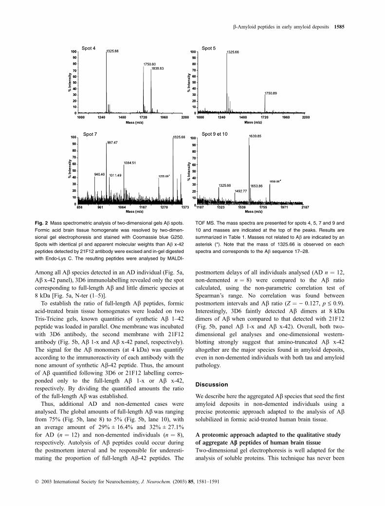
1 25 ± 5% 5.31 5.3 1–16 1954.879 1954.875
1–16 + CH3 1968.905 1968.863
2 12 ± 2% 5.31 5.3 1–16 1954.879 1954.875
1–16 + CH3 1968.905 1968.863
3 10 ± 3% 5.78 5.8 2–16 1839.852 1839.851
2–16 + CH3 1853.878 1853.854
(3–16) 1768.815 1768.804
4 9 ± 3% 6.27 5.9 PyrE 3–16 1751.784 1750.790
(2–16) 1839.852 1839.833
5 7 ± 3% 6.27 6.3 PyrE 3–16 1751.784 1750.881
6 12 ± 4% 5.96 6.0 8–16 1084.517 1084.557
6.01 9–16 997.485 998.525
7 8 ± 3% 5.96 6.1 8–16 1084.517 1084.518
6.01 9–16 997.485 997.477
6.01 9–16 + CH3 1011.500 1011.496
6.01 10–16 940.463 940.460
9 and 10 17 ± 7% 6.27 6.3 4–16 1639.772 1639.848
4–16 + CH3 1653.798 1653.859
5–16 1492.704 1492.770
Methylated fragments are indicated with a CH3. PyrE corresponds to a pyroglutamyl residue at the
amino-terminus of the identified fragment. The peptide corresponding to amino acid sequence 17–
28 of Ab was found in all spots (not shown). The relative proportion corresponds to the quantifi-
cation of spots following western blotting with 21F12, using Melanie III software (mean ± SD %,
n ¼ 8).
Table 1 Digested endo-Lys-C peptides of
Ab species in Alzheimer’s disease
Peak
Ab proposed
identity
Theoretical mass
[M + H+]
Observed mass
[M + H+]
Peptide sequence
confirmed
1 1–5 637.29 –
2 6–16 1336.60 –
3 17–28 1325.67 1325.53 LVFFAEDVGSNK
29–42 + ox 1285.76 1285.64
4 29–40 1085.63 1085.48 GAIIGLMVGGVV + ox
29–40 + ox 1101.62 1101.49
The sequences indicated were obtained by switching from MS to tandem MS mode. The oxidized
Met35 is indicated by ox.
Table 2 Analysis of trypic peptides spot 1
and 2 peptides using nano LC-MS and
nano-LC-MS/MS
1586 N. Sergeant et al.
� 2003 International Society for Neurochemistry, J. Neurochem. (2003) 85, 1581–1591
antibodies. Using MALDI-TOF/MS, the carboxy-terminal
fragments of Ab (29–40/42 or shorter) were not detected andhence suggested not to be ionized or desorbed. These Ab 1-xhave the same pI as Ab-40/42 and would therefore be locatedat the same position as Ab 1–40/42 on two-dimensional gels.Ab 1–40 and Ab 1–42 were fully characterized using nano-LC/MS in a tandem mass mode, but shorter Ab 1-x were
not evidenced, hence suggesting that these shorter Ab 1-xare under the limit of detection, and thus, probably not the
major Ab peptides recovered in formic acid brain tissue
homogenates of AD individuals. Shorter Ab 1-x have been
characterized in human or transgenic mouse brain tissue
(Kalback et al. 2002; Pype et al. 2003). In transgenic mice
the most insoluble brain material was shown to comprise
predominantly Ab x-40, Ab x-42 and, to a lesser extent, Ab1–38 (Pype et al. 2003). Shorter Ab 1-x were detected in thesoluble mouse brain extracts. By similarity to our data, we
can speculate that the absence of detection of shorter Ab 1-xpeptides confirms the hypothesis that the Ab species
recovered following formic acid treatment of the brain
correspond to aggregated Ab species, as already described(Delacourte et al. 2002).
Ab-40 peptides were observed in lower amounts than Ab-42 and they were found late, rather than systematically, in the
course of AD (Delacourte et al. 2002). We observed that in
AD patients, there are fewer species of amino-truncated Ab-40 than amino-truncated species of Ab-42, suggesting thatthe former arise from a distinct mechanism (e.g. inflamma-
tion, vascular deposits, etc.). Also, full-length Ab-40 peptideis physiologically produced and it can therefore be hypo-
thesized that Ab-40 coaggregates with Ab-42 deposits.
Taken together, our results suggest that aggregation of Ab-40species is a late event of Alzheimer physiopathology. This
hypothesis is also supported by observation of the amylo-
idosis process in the brain of individuals affected by Down’s
8 kDa
4 kDa
8 kDa
4 kDa
8 kDa
4 kDa
8 kDa
4 kDa
pl 5.30 5.80 6.30
Fig. 3 Two-dimensional analysis of aggregated Ab species in a non-
demented case. Formic acid-solubilized Ab species derived from the
brain tissue of a non-demented patient were resolved by two-dimen-
sional gel electrophoresis. Ab-40 species are not detected with our
ADA40 antiserum. 21F12 detects both Ab-42 monomers (4 kDa
staining) and dimers (8 kDa staining). Both WO2 [panel N-ter (5–8)]
and 6E10 [panel N-ter (4–13)] antibodies stained a single Ab peptide
spot with pI of 5.30.
Fig. 4 Dimers of Ab are essentially composed of amino-truncated Ab
peptides. The brain tissue of a control individual (Ctrl, S0), three non-
demented cases (S1, S2 and S6) and one AD case (S10) were
homogenized in formic acid. According to the nomenclatures defined
by Delacourte et al. (2002) and Braak and Braak (1991), the stages of
tau pathology (S0 to S10) and the amyloid staging classification (B or
C) are indicated at the top of the lanes, respectively. Ten ng of Ab
peptides 1–40 and 1–42 solubilized in formic acid were loaded in
parallel (first and second lane). Ab x-42 was identified with 21F12
(panel Ab-42) and the amino-terminal region with WO2 (WO2 panel).
Molecular weights are indicated on the left and an arrow indicates the
amino-truncated variants labelled by 21F12. Note that the same AD
case was used as for two-dimensional electrophoretic analysis and
MS.
b-Amyloid peptides in early amyloid deposits 1587
� 2003 International Society for Neurochemistry, J. Neurochem. (2003) 85, 1581–1591

syndrome. Ab-40 deposits are mainly observed in the oldestDown’s syndrome individuals (Lemere et al. 1996) whereas
intraneuronal Ab-42 are the earliest species to accumulateand, interestingly, they are not immunoreactive with amino-
terminal Ab antibody (Mori et al. 2002).
Ab species comprised in the first amyloid deposits
of non-demented individuals
Previous study on our brain bank evidenced the existence of
individuals that had no cognitive impairment but showed tau
pathology in the hippocampal formation, extending to the
anterior and mid-temporal cortex (Delacourte et al. 1999).
Diffuse amyloid deposits in different neocortical areas were
also observed at neuropathological examination and con-
firmed at the biochemical level (Delacourte et al. 2002).
Thus, in accordance with the progression of AD lesions in
the human brain cortical areas, as defined by Braak and
Braak (1991) and Delacourte et al. (1999, 2002), the non-
demented individuals certainly corresponded to early stage of
‘sporadic’ form of AD. The knowledge of the dynamic of
progression of tau pathology and amyloid pathology (Del-
acourte et al. 2002) strongly supports that the lesions
observed are likely to be the first lesions to develop.
Here, we demonstrate for the first time that the backbone
of Ab aggregates at the first stage of amyloid deposition inthese non-demented individuals comprise amino-terminal
truncated variants of Ab-42, including Ab starting at
positions 4-, 5-, 8- and 9–42, or with a pyroglutamyl residue
at position 3. Ab peptides starting at residue 3-pyroglutamylhave been suggested to be early aggregating species in AD
(Saido et al. 1995). Moreover, we show that the additional
truncated species identified here, which all correspond to Ab-42, are also early aggregating species. These Ab-42 speciesdo not result from treatment of brain tissue with formic acid
(Delacourte et al. 2002; Kalback et al. 2002), as the
treatment of synthetic Ab peptides 1–40 and 1–42 with
formic acid did not generate the truncated species observed
in the human brain tissue (Delacourte et al. 2002).
One can ask if truncated Ab species at the earliest stages ofAD are not generated before aggregation, as normal or
pathological products. These truncated species could play a
decisive role as seeds for fibrillogenesis and amyloid
deposition. Our data support indeed that specific amino-
terminal cleavage occurs very early in the aggregation
process. Firstly, these amino-truncated species are observed
in non-demented individuals with little amyloid deposits and
tau pathology. Secondly, biochemical analyses show that
dimers of Ab are lightly reactive with amino-specific Abantibodies, thus suggesting that they are mainly comprised of
amino-truncated Ab-42 peptides. In vitro experiments have
shown that truncated variants of Ab-42 are more fibrillogenicat physiological pH than the full-length Ab and certainly
more toxic (Pike et al. 1995; Russo et al. 2002). Further-
more, amino-truncated Ab peptides were observed in cell
8 kDa(a)
(b)
4 kDa
8 kDa
4 kDa
8 kDa
4 kDa
8 kDa
4 kDa
8 kDa
4 kDa
8 kDa
4 kDa
ngAß 1-4210 20 C C
AD Non-dementedC B C B C B
1mn
5mn
Aß 1-x(3D6)
Aß x-42(21F12)
1mn
30 s
S10 S10 S10 S2 S3 S3 S4 S3
pl
1 2 3 4 5 6 7 8 9 10
5.30 5.80 6.30
Fig. 5 Two-dimensional analysis of the specificity of 3D6 antibody
and comparison of Ab 1-x and Ab x-42 in formic acid brain tissue
homogenates of non-demented and AD indivduals. (a) Brain tissue of
an AD individual (S9 of tau pathology, stage C of amyloidosis,
according to Delacourte et al. 2002 and Braak and Braak 1991,
respectively) was homogenized in pure formic acid and resolved twice
by two-dimensional gel electrophoresis. Ab x-42 (upper panel) or Ab
1-x (lower panel) peptides were detected with 21F12 or 3D6 mono-
clonal antibodies, respectively. Molecular weights are indicated on the
left and pI are indicated on the x-axis. (b) Formic acid-treated brain
tissue of three AD individuals and five non-demented individuals were
loaded on the same Tris-Tricine gel. The experiment was performed
twice, Ab 1-x was detected with 3D6 and Ab x-42 was detected with
21F12. In parallel, 10 ng and 20 ng of synthetic Ab 1–42 were loaded
(lanes 1 and 2). Two times of exposure are presented (times of
exposure are indicated). The tau pathology and amyloidosis stage is
indicated under each lane. Note that the same AD individual was
analysed by two-dimensional electrophoresis (a) and one-dimensional
western-blotting (b, lane 4). The non-demented individual in lane 6
corresponds to that analysed as well as on Fig. 4, lane 5. Note that Ab
dimers at 8 kDa are strongly detected with 21F12 (Ab x-42 panels)
whereas faintly (lanes 8–10) or not (lanes 6 and 7) stained with 3D6
antibody in non-demented individuals.
1588 N. Sergeant et al.
� 2003 International Society for Neurochemistry, J. Neurochem. (2003) 85, 1581–1591
models, demonstrating that they can be generated directly by
cells, and thus do not result from truncation following
extracelullar aggregation within tissue (Cescato et al. 2000;
Shirotani et al. 2002). The truncated species of Ab-42 arealso found in large amount in cell models transfected with the
human APP gene mutated at position close to the gamma-
secretase cleavage (Ancolio et al. 1999; De Jonghe et al.
2001). These observations in addition to ours further support
the early dysfunction of gamma-secretase complex in
‘sporadic’ AD individuals. Altogether, our results demon-
strate that specific amino-truncated species of Ab-42 areimplicated in the very first steps of amyloidosis in Alzheimer
pathology and give new insight toward the understanding of
the pathophysiological process of the usual ‘sporadic’ form
of AD.
Amino-truncated Ab species are major species of early
amyloid deposits
Two-dimensional gel electrophoresis followed by western
blotting enables a semi-quantitative analysis of the propor-
tion of each Ab species. This proteomic method was
previously described to be highly resolutive and sensitive
for the quantification of Ab in cerebrospinal fluid (Wiltfanget al. 2002). Using this approach, we showed that more than
60% of all Ab species correspond to amino-truncated Abspecies. This result was further confirmed by the quantifica-
tion of full-length Ab peptides using 3D6 antibody. More-over, we showed that the proportion of amino-truncated Abpeptides was not correlated to postmortem intervals, thus
suggesting that autolysis is not responsible for the generation
of amino-truncated species of Ab. The isoelectric focusingnecessitates a long active rehydration step, during which
reaggregation of Ab peptides could occur. If such a
reaggregation process did occur then it would alter the
focusing of Ab species and a long horizontal smear would bevisualized. Indeed, all Ab peptides were well-focused and
even dimeric Ab species. Moreover, the results of quantifi-cation either obtained with two-dimensional gel analysis or
with one-dimensional western blotting are very similar.
Russo et al. (2000) reported that full-length Ab represented30% of all Ab species in sporadic AD and that in familial ADthis ratio was even lowered. Our results are similar to that
obtained by Russo et al. (2000) and can be extended to non-
demented individuals. Therefore, we show that our proteo-
mic method is also a very useful method for determining the
proportion of each Ab species in formic acid-treated humanbrain tissue. Moreover, we propose that the implication of
amino-truncated Ab peptides in the amyloidosis process ofAD is of major importance and is likely to occur very early
during amyloid deposition.
Implication for the human vaccination strategy
Aggregates of Ab are removed following vaccination of
transgenic mice against the amino-terminal region of Ab or
full-length Ab (Schenk et al. 1999, 2001). This amino-
terminal region, as the 3-EFRH-6 Ab sequence or the 4–10Ab sequence, generates the greatest immunogenic responsein mice, reducing amyloid burden, Ab fibril assembly andtoxicity (Schenk et al. 1999; Frenkel et al. 2001; Lemere
et al. 2001; Monsonego et al. 2001; McLaurin et al. 2002).
Immunization demonstrated as well that antibodies against
amino-terminal region of Ab are more efficient to reduce
the amyloid burden in transgenic mice (Bard et al. 2003).
However, this higher immunogenic property could be
explained by the fact that the human Ab sequence differs
specifically from that of mouse in this amino-terminal region
of Ab. Passive immunization using an antibody against
the central core of Ab is also an alternate and efficient
therapeutic approach to reduce the amyloid burden or restore
the cognitive function in transgenic mouse models of AD
(DeMattos et al. 2001; Dodart et al. 2002). Yet, all vaccin-
ation strategies do not discriminate between the full-length
or truncated Ab species. Full-length Ab is a physiologicalproduct with an unknown function that could generate an
undesirable immune reaction. The ideal vaccination strategy
in humans should have two aims: (i) to use antigens that
correspond to the initial amyloid species formed at the
earliest stage of AD, because they are at the heart of
Alzheimer physiopathological process; (ii) the antigen should
be a pathological epitope rather than a physiological one,
thus specifically targeting the amino-truncated species rather
than the full-length Ab peptide. The fact that truncated Abspecies are early, pathological and abundant antigens of AD
indicates that they could act as an ideal target for vaccination.
Indeed, it is possible to design synthetic peptides or develop
immunological tools that will target specifically the patho-
logical amino-terminal truncated Ab species and not phy-
siological Ab peptides, as the full-length Ab.In conclusion, our data show that the amino-truncated Ab-
42 peptides are the first species to seed amyloid deposition in
infraclinal AD. Consequently, we propose a small but
significant modification to the AD vaccination strategy that
is likely to completely change the response in the early
events of the amyloid cascade in ‘sporadic’ AD.
Acknowledgements
This work was supported by INSERM, CNRS and IMPRT of Lille.
We are very grateful to Malcom Lyon (Manchester, UK), and
Maryse Delehedde for their critical advice.
References
Ancolio K., Dumanchin C., Barelli H., Warter J. M., Brice A., Campion
D., Frebourg T. and Checler F. (1999) Unusual phenotypic alter-
ation of b amyloid precursor protein (bAPP) maturation by a newVal-715 fi Met bAPP-770 mutation responsible for probable
early-onset Alzheimer’s disease. Proc. Natl Acad. Sci. USA 96,
4119–4124.
b-Amyloid peptides in early amyloid deposits 1589
� 2003 International Society for Neurochemistry, J. Neurochem. (2003) 85, 1581–1591
Bard F. et al. (2003) Epitope and isotype specificities of antibodies to
{beta}-amyloid peptide for protection against Alzheimer’s dis-
ease-like neuropathology. Proc. Natl Acad. Sci. USA 100, 2023–
2028.
Braak H. and Braak E. (1991) Neuropathological stageing of Alzheimer-
related changes. Acta Neuropathol. 82, 239–259.
Campion D. et al. (1999) Early-onset autosomal dominant Alzheimer
disease: prevalence, genetic heterogeneity, and mutation spectrum.
Am. J. Hum. Genet. 65, 664–670.
Cescato R., Dumermuth E., Spiess M. and Paganetti P. A. (2000)
Increased generation of alternatively cleaved beta-amyloid peptides
in cells expressing mutants of the amyloid precursor protein
defective in endocytosis. J. Neurochem. 74, 1131–1139.
Cherny R. A., Legg J. T., McLean C. A., Fairlie D. P., Huang X.,
Atwood C. S., Beyreuther K., Tanzi R. E., Masters C.L. and Bush
A. I. (1999) Aequous dissolution of Alzheimer’s disease Abamyloid deposits by biometal depletion. J. Biol. Chem. 274 (33),
23223–23228.
De Jonghe C., Esselens C., Kumar-Singh S., Craessaerts K., Serneels S.,
Checler F., Annaert W., Van Broeckhoven C. and De Strooper B.
(2001) Pathogenic APP mutations near the c-secretase cleavagesite differentially affect Ab secretion and APP C-terminal fragmentstability. Hum. Mol. Genet. 10, 1665–1671.
Delacourte A. et al. (1999) The biochemical pathway of neurofibrillary
degeneration in aging and Alzheimer’s disease. Neurology 52,
1158–1165.
Delacourte A., Sergeant N., Champain D., Wattez A., Maurage C. A.,
Lebert F., Pasquier F. and David J. P. (2002) Nonoverlapping but
synergetic tau and APP pathologies in sporadic Alzheimer’s dis-
ease. Neurology 59, 398–407.
DeMattos R. B., Bales K. R., Cummins D. J., Dodart J. C., Paul S. M.
and Holtzman D. M. (2001) Peripheral anti-A beta antibody alters
CNS and plasma A beta clearance and decreases brain A beta
burden in a mouse model of Alzheimer’s disease. Proc. Natl Acad.
Sci. USA 98, 8850–8855.
Dodart J. C. et al. (2002) Immunization reverses memory deficits
without reducing brain Abeta burden in Alzheimer’s disease
model. Nat. Neurosci. 5, 452–457.
Frenkel D., Kariv N. and Solomon B. (2001) Generation of auto-anti-
bodies towards Alzheimer’s disease vaccination. Vaccine 19,
2615–2619.
Gotz J., Chen F., Van Dorpe J. and Nitsch R. M. (2001) Formation of
neurofibrillary tangles in P301L tau transgenic mice induced by
Abeta 42 fibrils. Science 293, 1491–1495.
Hock C., Konietzko U., Papassotiropoulos A., Wollmer A., Streffer J.,
Von Rotz R. C., Davey G., Moritz E. and Nitsch R. M. (2002)
Generation of antibodies specific for beta-amyloid by vac-
cination of patients with Alzheimer disease. Nat. Med. 8, 1270–
1275.
Hyman B. T. and Trojanowski J. Q. (1997) Consensus recommendations
for the postmortem diagnosis of Alzheimer disease from the
National Institute on Aging and the Reagan Institute Working
Group on diagnostic criteria for the neuropathological assessment
of Alzheimer disease. J. Neuropathol. Exp. Neurol. 56, 1095–1097.
Janus C. et al. (2000) A beta peptide immunization reduces behavioural
impairment and plaques in a model of Alzheimer’s disease. Nature
408, 979–982.
Johnson-Wood K. et al. (1997) Amyloid precursor protein processing
and A beta42 deposition in a transgenic mouse model of Alzheimer
disease. Proc. Natl Acad. Sci. USA 94, 1550–1555.
Kalback W. et al. (2002) APP transgenic mice Tg2576 accumulate Abeta
peptides that are distinct from the chemically modified and
insoluble peptides deposited in Alzheimer’s disease senile plaques.
Biochemistry 41, 922–928.
Kumar-Singh S. et al. (2000) Nonfibrillar diffuse amyloid deposition due
to a gamma (42)-secretase site mutation points to an essential role
for N-truncated A beta (42) in Alzheimer’s disease. Hum. Mol.
Genet. 9, 2589–2598.
Lemere C. A., Blusztajn J. K., Yamaguchi H., Wisniewski T., Saido T. C.
and Selkoe D. J. (1996) Sequence of deposition of heterogeneous
amyloid beta-peptides and APO E in Down syndrome: implica-
tions for initial events in amyloid plaque formation. Neurobiol. Dis.
3, 16–32.
Lemere C. A., Maron R., Selkoe D. J. and Weiner H. L. (2001) Nasal
vaccination with beta-amyloid peptide for the treatment of Alz-
heimer’s disease. DNA Cell Biol. 20, 705–711.
Lewis J. et al. (2001) Enhanced neurofibrillary degeneration in trans-
genic mice expressing mutant tau and APP. Science 293, 1487–
1491.
McLaurin J. et al. (2002) Therapeutically effective antibodies against
amyloid-beta peptide target amyloid-beta residues 4–10 and inhibit
cytotoxicity and fibrillogenesis. Nat. Med. 8, 1263–1269.
Monsonego A., Maron R., Zota V., Selkoe D. J. and Weiner H. L. (2001)
Immune hyporesponsiveness to amyloid beta-peptide in amyloid
precursor protein transgenic mice: implications for the pathogen-
esis and treatment of Alzheimer’s disease. Proc. Natl Acad. Sci.
USA 98, 10273–10278.
Mori C., Spooner E. T., Wisniewski K. E., Wisniewski T. M., Yamaguch
H., Saido T. C., Tolan D. R., Selkoe D. J. and Lemere C. A. (2002)
Intraneuronal Abeta42 accumulation in Down syndrome brain.
Amyloid 9, 88–102.
Pike C. J., Overman M. J. and Cotman C. W. (1995) Amino-terminal
deletions enhance aggregation of beta amyloid peptides in vitro.
J. Biol. Chem. 270, 23895–23898.
Pype S., Moechars D., Dillen I. and Mercken M. (2003) Characterization
of amyloid beta peptides from brain extracts of transgenic mice
overexpressing the London mutant of human amyloid precursor
protein. J. Neurochem. 84, 602–609.
Russo C., Schettini G., Saido T. C., Hulette C., Lippa C., Lannfelt L.,
Ghetti B., Gambetti P., Tabaton M. and Teller J. K. (2000)
Presenilin-1 mutations in Alzheimer’s disease. Nature 405, 531–
532.
Russo C. et al. (2002) Pyroglutamate-modified amyloid beta-peptides –
AbetaN3 (pE) – strongly affect cultured neuron and astrocyte
survival. J. Neurochem. 82, 1480–1489.
Saido T. C., Iwatsubo T., Mann D. M., Shimada H., Ihara Y. and
Kawashima S. (1995) Dominant and differential deposition of
distinct beta-amyloid peptide species, A beta N3 (pE), in senile
plaques. Neuron 14, 457–466.
Schenk D. et al. (1999) Immunization with amyloid-beta attenuates
Alzheimer-disease-like pathology in the PDAPP mouse. Nature
400, 173–177.
Schenk D., Seubert P. and Ciccarelli R. B. (2001) Immunotherapy with
beta-amyloid for Alzheimer’s disease: a new frontier. DNA Cell
Biol. 20, 679–681.
Sergeant N., David J. P., Champain D., Ghestem A., Wattez A., Dela-
courte A. (2002) Progressive decrease of amyloid precursor protein
carboxy terminal fragments (APP-CTFs), associated with tau
pathology stages, in Alzheimer’s disease. J. Neurochem. 81, 663–
672.
Shirotani K., Tsubuki S., Lee H. J., Maruyama K. and Saido T. C. (2002)
Generation of amyloid beta peptide with pyroglutamate at position
3 in primary cortical neurons. Neurosci. Lett. 327, 25–28.
Terai K., Iwai A., Kawabata S., Tasaki T., Watanabe K., Miyata K. and
Yamaguchi T. (2001) b-Amyloid deposits in transgenic mice
expressing human b-amyloid precursor protein have the samecharacteristics as those in Alzheimer’s disease. Neuroscience 104,
299–310.
1590 N. Sergeant et al.
� 2003 International Society for Neurochemistry, J. Neurochem. (2003) 85, 1581–1591
Walsh D. M., Tseng B. P., Rydel R. E., Podlisny M. B. and Selkoe D. J.
(2000) The oligomerization of amyloid beta-protein begins intra-
cellularly in cells derived from human brain. Biochemistry 39,
10831–10839.
Walsh D. M., Klyubin I., Fadeeva J. V., Cullen W. K., Anwyl R., Wolfe
M. S., Rowan M. J. and Selkoe D. J. (2002) Naturally secreted
oligomers of amyloid beta protein potently inhibit hippocampal
long-term potentiation in vivo. Nature 416, 535–539.
Wiltfang J. et al. (2002) Highly conserved and disease-specific patterns
of carboxyterminally truncated Abeta peptides 1–37/38/39 in
addition to 1–40/42 in Alzheimer’s disease and in patients with
chronic neuroinflammation. J. Neurochem. 81, 481–496.
b-Amyloid peptides in early amyloid deposits 1591
� 2003 International Society for Neurochemistry, J. Neurochem. (2003) 85, 1581–1591
Copyright © 2022 FDOKUMEN