Ocular Complications Following Vaccination for COVID-19
32
Citation: Haseeb, A.A.; Solyman, O.; Abushanab, M.M.; Abo Obaia, A.S.; Elhusseiny, A.M. Ocular Complications Following Vaccination for COVID-19: A One-Year Retrospective. Vaccines 2022, 10, 342. https://doi.org/10.3390/ vaccines10020342 Academic Editors: P Veeranna Ravindra, Prashant Chikkahonnaiah and Federico Pratesi Received: 31 December 2021 Accepted: 18 February 2022 Published: 21 February 2022 Publisher’s Note: MDPI stays neutral with regard to jurisdictional claims in published maps and institutional affil- iations. Copyright: © 2022 by the authors. Licensee MDPI, Basel, Switzerland. This article is an open access article distributed under the terms and conditions of the Creative Commons Attribution (CC BY) license (https:// creativecommons.org/licenses/by/ 4.0/). Review Ocular Complications Following Vaccination for COVID-19: A One-Year Retrospective Abid A. Haseeb 1 , Omar Solyman 2,3 , Mokhtar M. Abushanab 3 , Ahmed S. Abo Obaia 3 and Abdelrahman M. Elhusseiny 4, * 1 Department of Ophthalmology and Visual Sciences, University of Illinois at Chicago, Chicago, IL 60612, USA; [email protected] 2 Department of Ophthalmology, Qassim University Medical City, Qassim University, Buraydah 52571, Saudi Arabia; [email protected] 3 Department of Ophthalmology, Research Institute of Ophthalmology, Giza 11261, Egypt; [email protected] (M.M.A.); [email protected] (A.S.A.O.) 4 Department of Ophthalmology, Harvey and Bernice Jones Eye Institute, University of Arkansas for Medical Sciences, Little Rock, AR 72205, USA * Correspondence: [email protected] Abstract: Vaccination efforts as a mitigation strategy in the corona virus disease 2019 (COVID-19) pandemic are fully underway. A vital component of understanding the optimal clinical use of these vaccines is a thorough investigation of adverse events following vaccination. To date, some limited reports and reviews have discussed ocular adverse events following COVID-19 vaccination, but a systematic review detailing these reports with manifestations and clinical courses as well as proposed mechanisms has yet to be published. This comprehensive review one-year into vaccination efforts against COVID-19 is meant to furnish sound understanding for ophthalmologists and primary care physicians based on the existing body of clinical data. We discuss manifestations categorized into one of the following: eyelid, orbit, uveitis, retina, vascular, neuro-ophthalmology, ocular motility disorders, and other. Keywords: acute macular neuroretinopathy; corneal graft rejection; coronavirus; COVID-19; SARS-CoV-2; uveitis; vaccination 1. Introduction Since the time that the first vaccines against the severe acute respiratory failure coron- avirus 2 (SARS-CoV-2) and ensuing corona virus disease2019 (COVID-19) were approved for emergency authorization use by the Food and Drug Administration (FDA) in late 2020, an enormous amount of speculation has surrounded the discourse around vaccination and COVID-19 [1–4]. It remains the consensus opinion in clinical practice—and the opinion of the authors—that vaccination and subsequent booster administration against COVID-19 is a vital epidemiologic factor in mitigating the devastating effects of the COVID-19 pandemic. It is nevertheless essential that physicians and researchers investigate the possible adverse outcomes due to vaccination against COVID-19. Previous research has demonstrated a link between COVID-19 infection and ocular complications, direct or indirect [5–13]. It has been well-documented that conjunctivitis, scleritis, orbital inflammatory disease, phlyctenular keratoconjunctivitis and retinal in- volvement may take place in COVID-19 infection. It is thus vital to also investigate the relationship between COVID-19 vaccination and ocular complications. A considerable number of reports and retrospective case studies have reported on possible adverse effects of vaccination against COVID-19 approximately one year into the dissemination of these vaccines [14–16]. In this review, we seek to provide a rigorous description of these findings based on a comprehensive review and statistical analysis of the literature. Vaccines 2022, 10, 342. https://doi.org/10.3390/vaccines10020342 https://www.mdpi.com/journal/vaccines
-
Upload
khangminh22 -
Category
Documents
-
view
3 -
download
0
Transcript of Ocular Complications Following Vaccination for COVID-19

�����������������
Citation: Haseeb, A.A.; Solyman, O.;
Abushanab, M.M.; Abo Obaia, A.S.;
Elhusseiny, A.M. Ocular
Complications Following Vaccination
for COVID-19: A One-Year
Retrospective. Vaccines 2022, 10, 342.
https://doi.org/10.3390/
vaccines10020342
Academic Editors: P Veeranna
Ravindra, Prashant Chikkahonnaiah
and Federico Pratesi
Received: 31 December 2021
Accepted: 18 February 2022
Published: 21 February 2022
Publisher’s Note: MDPI stays neutral
with regard to jurisdictional claims in
published maps and institutional affil-
iations.
Copyright: © 2022 by the authors.
Licensee MDPI, Basel, Switzerland.
This article is an open access article
distributed under the terms and
conditions of the Creative Commons
Attribution (CC BY) license (https://
creativecommons.org/licenses/by/
4.0/).
Review
Ocular Complications Following Vaccination for COVID-19:A One-Year RetrospectiveAbid A. Haseeb 1, Omar Solyman 2,3, Mokhtar M. Abushanab 3, Ahmed S. Abo Obaia 3
and Abdelrahman M. Elhusseiny 4,*
1 Department of Ophthalmology and Visual Sciences, University of Illinois at Chicago, Chicago, IL 60612, USA;[email protected]
2 Department of Ophthalmology, Qassim University Medical City, Qassim University,Buraydah 52571, Saudi Arabia; [email protected]
3 Department of Ophthalmology, Research Institute of Ophthalmology, Giza 11261, Egypt;[email protected] (M.M.A.); [email protected] (A.S.A.O.)
4 Department of Ophthalmology, Harvey and Bernice Jones Eye Institute, University of Arkansas for MedicalSciences, Little Rock, AR 72205, USA
* Correspondence: [email protected]
Abstract: Vaccination efforts as a mitigation strategy in the corona virus disease 2019 (COVID-19)pandemic are fully underway. A vital component of understanding the optimal clinical use of thesevaccines is a thorough investigation of adverse events following vaccination. To date, some limitedreports and reviews have discussed ocular adverse events following COVID-19 vaccination, but asystematic review detailing these reports with manifestations and clinical courses as well as proposedmechanisms has yet to be published. This comprehensive review one-year into vaccination effortsagainst COVID-19 is meant to furnish sound understanding for ophthalmologists and primary carephysicians based on the existing body of clinical data. We discuss manifestations categorized intoone of the following: eyelid, orbit, uveitis, retina, vascular, neuro-ophthalmology, ocular motilitydisorders, and other.
Keywords: acute macular neuroretinopathy; corneal graft rejection; coronavirus; COVID-19;SARS-CoV-2; uveitis; vaccination
1. Introduction
Since the time that the first vaccines against the severe acute respiratory failure coron-avirus 2 (SARS-CoV-2) and ensuing corona virus disease2019 (COVID-19) were approvedfor emergency authorization use by the Food and Drug Administration (FDA) in late 2020,an enormous amount of speculation has surrounded the discourse around vaccination andCOVID-19 [1–4]. It remains the consensus opinion in clinical practice—and the opinion ofthe authors—that vaccination and subsequent booster administration against COVID-19 isa vital epidemiologic factor in mitigating the devastating effects of the COVID-19 pandemic.It is nevertheless essential that physicians and researchers investigate the possible adverseoutcomes due to vaccination against COVID-19.
Previous research has demonstrated a link between COVID-19 infection and ocularcomplications, direct or indirect [5–13]. It has been well-documented that conjunctivitis,scleritis, orbital inflammatory disease, phlyctenular keratoconjunctivitis and retinal in-volvement may take place in COVID-19 infection. It is thus vital to also investigate therelationship between COVID-19 vaccination and ocular complications. A considerablenumber of reports and retrospective case studies have reported on possible adverse effectsof vaccination against COVID-19 approximately one year into the dissemination of thesevaccines [14–16]. In this review, we seek to provide a rigorous description of these findingsbased on a comprehensive review and statistical analysis of the literature.
Vaccines 2022, 10, 342. https://doi.org/10.3390/vaccines10020342 https://www.mdpi.com/journal/vaccines
Vaccines 2022, 10, 342 2 of 32
2. Materials and Methods
We performed a PubMed search for articles of interest using search terms beginningwith “coronavirus vaccine” or “COVID vaccine” followed by “ocular”, “palsy”, “cornea”,”rejection”, “uveitis”, “optic neuritis”, “optic neuropathy” and “retina”. Articles wereincluded if they were case reports or retrospective studies describing adverse ocular man-ifestations following any vaccination against COVID-19 between December 2020 andDecember 2021.
We thereafter characterized articles as belonging to one of the following categories ofadverse event: eyelid, orbital, corneal, uveitis, retinal, vascular, neuro-ophthalmological,ocular motility disorders and unspecified. When possible, statistical analysis was per-formed for each category regarding age, sex, visual acuity, and any other pathology-specific characteristics of the categories in question. Continuous variables were reported asmean ± one standard deviation.
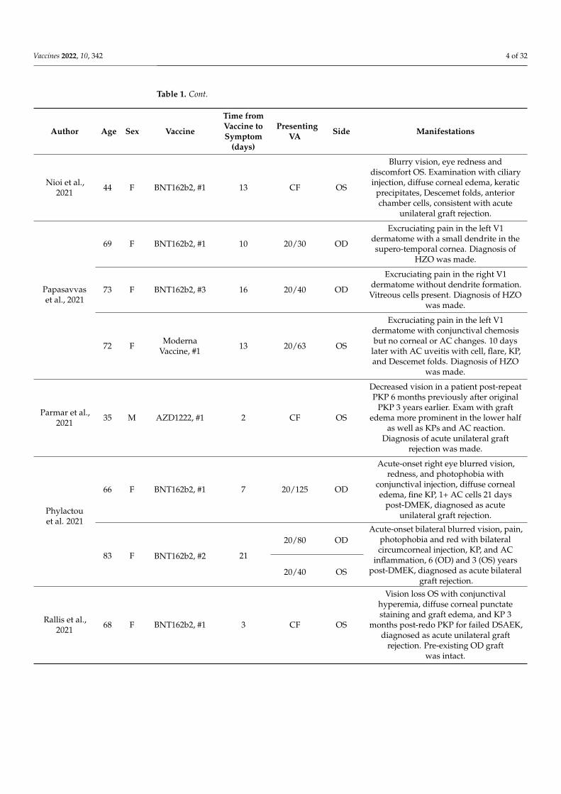
3. Adverse Ocular Events: Patient Overview
In total, 58 articles were included in our review. These findings are detailed in Table 1.Of these 58 studies, 28 (48.3%) were case reports, 5 (8.6%) were case series, 22 (37.9%)were letters to the editor, and 3 (5.2%) were photo essays. A total of 94 patients wereincluded. Of 90 patients with documented age information, the mean age at the time ofpresentation was 46.9 ± 18.4 years. Of 91 patients with documented gender, there were50 (54.9%) females and 41 (45.1%) males. Of the 87 cases in which vaccine informationwere present, BNT162b2 mRNA SARS-CoV-2 (BioNTech/Pfizer, Mainz, Germany) wasreported 55 (63.2%) times, AZD1222 ChAdO×1 nCoV-19 (AstraZeneca, Cambridge, UK,also marketed as the CoviShield Serum Institute of India vaccine) was reported 20 (22.9%)times, Moderna COVID-19 Vaccine (ModernaTX, Inc., Cambridge, MA, USA) was reported6 (6.9%) times, BBIBP-CorV (Sinopharm, Beijing, China) was reported 3 (3.4%) times,Corona Vac (Sinovac Biotech Ltd., Beijing, China) was reported 2 (2.3%) times, and Gam-COVID-Vac/Sputnik V (Gamaleya Institute, Moscow, Russia) was reported once (1.1%).Vaccine ordinal dose was reported 81 times; 45 (55.6%) cases were after the first dose,35 (43.2%) were after the second dose, and one (1.2%) was after a 3rd (booster shot) dose.
In our literature review, we found numerous ophthalmic adverse events followingCOVID-19 vaccination. Because some phenomena were reported several times and otherswere reported only once, we primarily discuss mechanisms and clinical considerationsfor phenomena which have occurred several times. Nevertheless, it is important to notethat an important limitation of this analysis is that it is a retrospective review and not acohort study. Despite the fact that we discuss mechanisms, in the absence of definitive un-derlying pathophysiologic processes, we must recognize the possibility that some adverseevents—particularly those which are especially rare—are due to random chance.
Table 1. Aggregated information on reviewed cases.
Author Age Sex Vaccine
Time fromVaccine toSymptom
(days)
PresentingVA Side Manifestations
Eyelid
Austria et al.,2021
32 F BNT162b2, #NR
1 to 2 NR NR
Unilateral upper greater than lowereyelid edema and erythema without
other systemic or ocular findingson exam.
43 F BNT162b2, #NR
43 F BNT162b2, #NR

Vaccines 2022, 10, 342 3 of 32
Table 1. Cont.
Author Age Sex Vaccine
Time fromVaccine toSymptom
(days)
PresentingVA Side Manifestations
Mazzatenaet al., 2021
67 F BNT162b2, #1 10 NR OD andOS
Ecchymotic lesions on the upper eyelids.Lesions were moderately itchy.
44 F BNT162b2, #2 21 NR OD andOS
Purpuric lesions bilaterally. Lesions werecircumscribed on the upper eyelid and
totally asymptomatic.
63 M BNT162b2, #2 21 NR OD andOS
Purpuric lesions bilaterally. Lesions werecircumscribed on the upper eyelid and
totally asymptomatic.
Orbit
Bayas et al.,2021
55 F AZD1222, #1 1020/140 OD
Bilateral conjunctival congestion,retroorbital pain, and diplopia. MRI
showed bilateral superior ophthalmicvein thrombosis.20/140 OS
Chuang et al.,2021 45 M NR 7 NR OS
Progressive ptosis and decreased visionOS, diplopia, and examination with APDand complete ophthalmoplegia. CT and
MRI with left cavernous sinusthrombosis. Pt diagnosed with
Tolosa-Hunt syndrome.
Panovska-Stavridis
et al., 202129 F AZD1222, #1 10 NR OS
Left orbital swelling, severe headache,and blurred vision OS. Labs showed
thrombocytopenia of 18 × 1019/L. MRIdemonstrated central filling defects and a
diagnosis of superior ophthalmic veinthrombosis was made.
Cornea
De la Presaet al., 2021 27 F Moderna
Vaccine, #1 15 20/20 OD
Redness and irritation with 1+conjunctival hyperemia and an irregular
temporal epithelial rejection line in apatient post LR-CLAL 4 years earlier. A
diagnosis of acute unilateral graftrejection was made.
Abousy et al.,2021
73 F BNT162b2, #2 420/200 OD
Vision loss with corneal thickening withDescemet folds bilaterally in a patient
with DSEK 8 years previously, consistentwith acute bilateral graft rejection.20/40 OS
Crnej et al.,2021 71 M BNT162b2, #1 7 20/125 OD
Painless decrease in right eye vision withconjunctival injection and diffuse cornealedema 5 months post-DMEK, diagnosed
as acute unilateral graft rejection.
Khan et al.,2021
48 M AZD1222, #1 21
LP ODVision loss, bilateral lid edema, diffuse
conjunctival and ciliary congestion,corneal melting and perforation with
diffuse corneal haze, uveal tissueprolapse, bilateral massive choroidal
detachment on B-scan ultrasonography.LP OS
Vaccines 2022, 10, 342 4 of 32
Table 1. Cont.
Author Age Sex Vaccine
Time fromVaccine toSymptom
(days)
PresentingVA Side Manifestations
Nioi et al.,2021 44 F BNT162b2, #1 13 CF OS
Blurry vision, eye redness anddiscomfort OS. Examination with ciliaryinjection, diffuse corneal edema, keratic
precipitates, Descemet folds, anteriorchamber cells, consistent with acute
unilateral graft rejection.
Papasavvaset al., 2021
69 F BNT162b2, #1 10 20/30 OD
Excruciating pain in the left V1dermatome with a small dendrite in thesupero-temporal cornea. Diagnosis of
HZO was made.
73 F BNT162b2, #3 16 20/40 OD
Excruciating pain in the right V1dermatome without dendrite formation.Vitreous cells present. Diagnosis of HZO
was made.
72 F ModernaVaccine, #1 13 20/63 OS
Excruciating pain in the left V1dermatome with conjunctival chemosisbut no corneal or AC changes. 10 days
later with AC uveitis with cell, flare, KP,and Descemet folds. Diagnosis of HZO
was made.
Parmar et al.,2021 35 M AZD1222, #1 2 CF OS
Decreased vision in a patient post-repeatPKP 6 months previously after original
PKP 3 years earlier. Exam with graftedema more prominent in the lower half
as well as KPs and AC reaction.Diagnosis of acute unilateral graft
rejection was made.
Phylactouet al. 2021
66 F BNT162b2, #1 7 20/125 OD
Acute-onset right eye blurred vision,redness, and photophobia with
conjunctival injection, diffuse cornealedema, fine KP, 1+ AC cells 21 days
post-DMEK, diagnosed as acuteunilateral graft rejection.
83 F BNT162b2, #2 21
20/80 ODAcute-onset bilateral blurred vision, pain,
photophobia and red with bilateralcircumcorneal injection, KP, and AC
inflammation, 6 (OD) and 3 (OS) yearspost-DMEK, diagnosed as acute bilateral
graft rejection.20/40 OS
Rallis et al.,2021 68 F BNT162b2, #1 3 CF OS
Vision loss OS with conjunctivalhyperemia, diffuse corneal punctatestaining and graft edema, and KP 3
months post-redo PKP for failed DSAEK,diagnosed as acute unilateral graft
rejection. Pre-existing OD graftwas intact.
Vaccines 2022, 10, 342 5 of 32
Table 1. Cont.
Author Age Sex Vaccine
Time fromVaccine toSymptom
(days)
PresentingVA Side Manifestations
Ravichandranand
Natarajan2021
62 M AZD1222, #1 21 NR NR
Right eye decreased vision andcongestion, with an advancing
Kodadoust rejection line and cornealgraft edema, 2 years post-PKP.Diagnosed as acute unilateral
graft rejection.
Wasser et al.,2021
73 M BNT162b2, #1 13 20/200 OS
Eye discomfort OS and vision loss withciliary injection, corneal edema,
Descemet folds, and KP 2 years afterre-graft for PKP performed 44 years
earlier. Diagnosed as acute unilateralgraft rejection.
56 M BNT162b2, #1 12 CF OD
Blurred vision and redness OD, withdiffuse corneal edema, KP, and AC cells25 years post-PKP, diagnosed as acute
unilateral graft rejection. Pre-existing OSgraft from PKP 7 years earlier was intact.
Uveitis
ElSheikhet al., 2021
18 F Sinopharm, #2 520/40 OD
Bilateral acute uveitis with 2+ AC flareOU and 1+ cell OU and hyperreflectivedots in the AC in a patient with juvenile
idiopathic arthritis.20/120 OS
Goyal et al.,2021
34 M AZD1222, #1 4
20/120 OD
Ocular pain followed by nasal rednessOS and a floater OD progressing to
severe vision loss. Fundus exam withmultiple bilateral oval lesions at the levelof the choroid with serous detachments,
consistent with bilateralmultifocal choroiditis.
20/20 OS
Herbort andPapasavvas
202153 M Moderna
Vaccine, #2 5 NR OD
Severe flare-up of pre-existingherpes-keratouveitis OD inactive for
18 months without treatment.Pt presented with numerous KPs,
elevated IOP to 41 mmHg.
Ishay et al.,2021 28 M BNT162b2, #1 10 NR OS
Pain, redness, and blurred vision OS in apatient with Behçet’s disease on
colchicine twice daily. Examinationrevealed severe panuveitis.
Jain andKalamkar
202127 M AZD1222, #1 2 20/20 OS
Pain, redness and severe circumcornealcongestion OS with 2+ AC cells and
non-granulomatous KP in a patient withjuvenile idiopathic arthritis and oneprevious episode of bilateral uveitis.
Acute uveitis was diagnosed.
Koong et al.,2021
54 M BNT162b2, #1 1
20/80 ODAcute bilateral, sequential blurring of
vision with bilateral areas of subretinalfluid with dot-blot hemorrhages on
examination. OCT with bilateral serousneurosensory retinal detachments. ICGA
confirmed diagnosis of VKH.20/160 OS
Vaccines 2022, 10, 342 6 of 32
Table 1. Cont.
Author Age Sex Vaccine
Time fromVaccine toSymptom
(days)
PresentingVA Side Manifestations
Maleki et al.,2021
33 F ModernaVaccine, #2
1020/20 OD
Bilateral photopsia and progressive nasalfield defect OS. OCT with outer layer
segmental disruption OS. Elevated ESRand CRP. Diagnosis of acute zonal occultouter retinopathy (AZOOR) was made.
20/20 OS
Mishra et al.,2021 71 M AZD1222, #1 10 CF OD
Reactivation of VZV presenting withpanuveitis OD, circumcorneal congestion,multiple fine keratic precipitates, anterior
chamber cells and flare, vitritis, andwidespread acute retinal necrosis.
Mudie et al.,2021
43 F BNT162b2, #2 3
20/500 ODBilateral substantial vision loss, eye pain
and redness, and photophobia, with3–4+ AC cell and 2–3+ vitreous cell. OCTwith significant choroidal thicnening, FA
with mild peripheral vascular leakage.Diagnosis of panuveitis was made.
20/500 OS
Pan et al.,2021 50 F
Unspecifiedinactivated Vero
cell-basedvaccine
approved inChina
5
20/33 OD Bilateral blurred vision with pale, blurryoptic disc, absent foveal reflex, macular
edema, and fluorescein angiographyconsistent with bilateral choroiditis.20/66 OS
Papasavvasand Herbort
202143 F BNT162b2, #2 42
20/20 OD
Reactivation of pre-existing VKH diseasewith significant anterior segment
inflammation OU, and 3–4 mutton-fat KPOD. OCT showed retinal folds and
subretinal fluid. Multiplehypofluorescent dark dots present
on ICGA.
20/20 OS
Rabinovitchet al., 2021
43 F BNT162b2, #1 2 20/25 ODRedness, pain, blurred vision. 3+ cell and1+ flare and fibrin on exam. Diagnosis of
anterior uveitis was made.
34 M BNT162b2, #1 4 20/32 ODRedness and pain. 1+ cell and
non-granulomatous KPs on exam.Diagnosis of anterior uveitis was made.
34 F BNT162b2, #1 1 20/50 OSRedness, pain, and photophobia. 2+ celland non-granulomatous KPs on exam.
Diagnosis of anterior uveitis was made.
53 M BNT162b2, #1 13 20/25 OS Pain only. 0.5+ cell on exam. Diagnosis ofanterior uveitis was made.
64 M BNT162b2, #1 15 20/25 OSRedness, pain, and photophobia.
0.5+ cell on exam. Diagnosis of anterioruveitis was made.
68 M BNT162b2, #1 5 20/200 OD Redness and pain. 1+ cell on exam.Diagnosis of anterior uveitis was made.
61 F BNT162b2, #1 12 20/25 OD Pain and photophobia. 2+ cell on exam.Diagnosis of anterior uveitis was made.
65 F BNT162b2, #1 3 20/80 ODRedness, pain, photophobia, and blurred
vision. 2+ cell and 2+ flare on exam.Diagnosis of anterior uveitis was made.
Vaccines 2022, 10, 342 7 of 32
Table 1. Cont.
Author Age Sex Vaccine
Time fromVaccine toSymptom
(days)
PresentingVA Side Manifestations
78 M BNT162b2, #2 3 20/25 OS
Redness, pain, and blurred vision. 2+ celland 2+ flare with posterior synechiae on
exam. Diagnosis of anterior uveitiswas made.
59 M BNT162b2, #2 8 20/32 OSPain, photophobia, and blurred vision.2+ cell on exam. Diagnosis of anterior
uveitis was made.
72 M BNT162b2, #2 16 20/80 OD Redness only. 1+ cell on exam. Diagnosisof anterior uveitis was made.
51 M BNT162b2, #2 2 20/50 OS Redness and pain. 2+ cell on exam.Diagnosis of anterior uveitis was made.
42 F BNT162b2, #2 2020/25 OD Pain and blurred vision bilaterally.
2+ cell on exam. Diagnosis of anterioruveitis was made.20/25 OS
74 M BNT162b2, #2 7 20/40 OD OD Pain only. 1+ cell and 2+ flare on exam.Diagnosis of anterior uveitis was made.
39 M BNT162b2, #2 5 20/32 ODBlurred vision with defect and photopsia.
Outer retinal changes on exam.Diagnosis of MEWDS was made.
64 F BNT162b2, #2 6 20/25 OD Photophobia only. 1+ flare on exam.Diagnosis of anterior uveitis was made.
50 F BNT162b2, #2 2 20/25 OS Pain only. 1+ cell on exam. Diagnosis ofanterior uveitis was made.
23 F BNT162b2, #2 220/25 OD
Redness, blurred vision, andphotophobia bilaterally. 1+ cell and
1+ flare on exam. Diagnosis of anterioruveitis was made.20/25 OS
36 M BNT162b2, #2 1 20/80 OS
Redness, photophobia, and blurredvision. 3+ cell and 3+ flare with
non-granulomatous KPs on exam.Diagnosis of anterior uveitis was made.
41 M BNT162b2, #2 2 20/50 ODRedness, photophobia, and blurredvision. 2+ cell and 2+ flare on exam.
Diagnosis of anterior uveitis was made.
28 F BNT162b2, #2 30 20/32 OSBlurred vision, visual field defect, andphotopsia. Outer retinal changes on
exam. Diagnosis of MEWDS was made.
Renisi et al.,2021 23 M BNT162b2, #2 14 20/40 OS
Pain and photophobia OS withperikeratic and conjunctival hyperemia,
posterior synechiae, AC cells, and KP.Diagnosis of anterior uveitis was made.
Saracenoet al., 2021 62 F AZD1222, #1 2
20/600 ODAcute bilateral loss of vision with mild
2+ AC cell and 1+ vitreous cell OU.Fundus examination revealed a serous
retinal detachment OU. OCT revealed thesame and subretinal hyperreflective dots.
Diagnosis of VKH was made.20/200 OS
Vaccines 2022, 10, 342 8 of 32
Table 1. Cont.
Author Age Sex Vaccine
Time fromVaccine toSymptom
(days)
PresentingVA Side Manifestations
Retina
Bøhler et al.,2021 27 F AZD1222, #1 2 20/20 OS
Left eye paracentral scotoma with ateardrop-shaped macular lesion nasal tothe fovea on ophthalmoscopy, diagnosed
as unilateral AMN.
Book et al.,2021
21 F AZD1222, #1 3
20/16 ODBilateral paracentral scotomas with
underlying circumscribed paracentraldark lesions on exam, OCT with outer
plexiform layer thickening anddiscontinuity, diagnosed as
bilateral AMN.20/16 OS
Chen et al.,2021 21 F BNT162b2, #1 3 20/20 OS
Paracentral scotomas OS with barelyvisible oval parafoveal lesions on fundus
exam. Infrared imaging revealedhypo-reflective lesions consistent with
left AMN.
Drüke et al.,2021
23 F AZD1222, #1 1
20/20 ODDevelopment of bilateral paracentral
scotomas. Fundus photography revealeda subtle brownish rimmed lesion
parafoveally OD and blurred lesion nasalto the macula OS. IR and OCT imaging
confirmed a diagnosis of AMN.20/20 OS
Fowler et al.,2021 33 M BNT162b2, #1 3 20/63 OD
Blurry vision OD with swollen macula,central foveal thickness (CFT) of 457 µmon OCT, and macular serous detachmentof the neurosensory retina on FA. OCTA
confirmed a diagnosis of centralserous retinopathy.
Khochtaliet al., 2021 24 F BNT162b2, #1 5 20/40 OS
Foveolitis with 2+ vitreous cell, diffuseretinal vascular leakage, faint foveal
hyperfluorescence and late phasehypofluorescence of the foveal lesion,
and granular hyperreflective specks inthe inner nuclear layer.
Mambrettiet al., 2021
22 F AZD1222, #1 2 20/20 OD
Acute paracentral scotoma OD withbarely visible parafoveal lesions onfundus exam. OCT was consistent
with AMN.
28 F AZD1222, #1 2 20/20 OD Acute paracentral scotoma OD with OCTconsistent with AMN.
Michel et al.,2021 21 F AZD1222, #1 2 20/20 OS
Acute-onset of 4 central scotomas OS,well-demarcated dark oval-shaped areas
surrounding the left fovea on infraredimaging. OCT with multifocal highly
reflective lesions and with ellipsoid andinterdigitation zone disruption consistent
with AMN.

Vaccines 2022, 10, 342 9 of 32
Table 1. Cont.
Author Age Sex Vaccine
Time fromVaccine toSymptom
(days)
PresentingVA Side Manifestations
Pichi et al.,2021
NR NR Sinopharm,#NR 5 20/400 OS
Acute vision loss OS, with OCT showinghyperreflectivity of the outer plexiform,Henle fiber, and outer nuclear layers. A
diagnosis of AMN was made.
NR NR Sinopharm,#NR 0 20/30 OS
Tachycardia, systolic hypertension(210 mm Hg), and inferior scotoma OS
20 min after vaccination. Fundusexamination revealed a suprafoveal dot
hemorrhage. A diagnosis of PAMMwas made.
Subramonyet al., 2021
22 F ModernaVaccine, #2
1020/70 OD
Progressive painless vision loss OD andno vision changes OS, but macula-off
inferotemporal retinal detachment ODand small macula-on temporal retinal
detachment OS.20/20 OS
Valenzuelaet al., 2021
20 F BNT162b2, #2 2
20/20 ODDevelopment of bilateral paracentral
scotomas and shimmering lights. Fundusexam was unrevealing, but OCT
demonstrated corresponding parafovealfoci of hyperreflectivity. Diagnosis of
AMN was made.20/20 OS
Vinzamuriet al., 2021
35 M AZD1222, #2 NR20/20 OD
Visual disturbance, OCT withhyperreflective lesions involving the
nerve fiber layer, ganglion cell layer andouter plexiform layer; diagnosed as
PAMM and AMN.20/20 OS
Vascular
Bialasiewiczet al., 2021
50 M BNT162b2, #2 0CF OD
Immediate bilateral retrobulbar pain, redeye, and vision loss. Examination and
OCT revealed a hemorrhagic CRVO withischemic areas and cystoid
macular edema.CF OS
Endo et al.,2021 52 M BNT162b2, #1 14 20/20 OS
Sudden blurred vision OS with minimaldot hemorrhages in the upper quadrants,dilated tortuous veins in four quadrants,and disperse exudates. FA was consistent
with non-ischemic CRVO.
Goyal et al.,2021 28 M Sputnik V, #2 11 20/30 OD
Visual disturbance with fundusexamination revealing superior
hemi-retinal vein occlusion with severecystoid macular edema.
Tanaka et al.,2021
71 F BNT162b2, #2 1 20/30 OS
Vision loss, with examination and OCTshowing superior temporal BRVO and
secondary macular edema withpreviously resolved inferior
temporal BRVO.
72 M BNT162b2, #1 1 20/25 OD
Vision loss, with examination and OCTshowing recurrence of previously
resolved superior temporal BRVO andmacular edema.

Vaccines 2022, 10, 342 10 of 32
Table 1. Cont.
Author Age Sex Vaccine
Time fromVaccine toSymptom
(days)
PresentingVA Side Manifestations
Neuro-Ophthalmology
Elnahry et al.,2021
69 F BNT162b2, #2 16
CF OD
Blurry vision OU with immediate OSclearing but persistent blurring OD.Examination with optic nerve head
edema (OD > OS) and RAPD OD onexam. RNFL imaging confirmed a
diagnosis of central nervous systeminflammatory syndrome
with neuroretinitis.
20/20 OS
32 F AZD1222, #1 4 20/30 OS
Blurred vision with superior field defectOS. Examination revealed left optic disc
swelling and RAPD with decreasedRNFL thickness. MRI was diagnostic of
left optic neuritis.
Leber et al.,2021
32 F Corona Vac, #2 020/200 OS
Rapidly progressive worsening visionand pain with EOM OS. Examination
revealed RAPD OS and disc swelling ODand OS. Labs revealed thyroiditis andMRI revealed bilateral optic neuritis.
20/20 OD
Maleki et al.,2021
79 F BNT162b2, #2 2
20/1250 OD
Bilateral sudden loss of vision, OD > OS,with 3+ afferent pupillary defect OD.
OCT, FA, and ICG consistent withgeneralized disc pallor OD and inferior
pallor OS, consistent with bilateralarteritic anterior ischemic optic
neuropathy (AAION).20/40 OS
Pawar et al.,2021 28 F NR 21 20/120 OS
Sudden vision loss OS, with examinationrevealing mild blurring of the optic disc
margin. MRI was consistent withoptic neuritis.
Ocular Motility
Eleiwa et al.,2021 46 M AZD1222, #2 3 NR OD
Torsional, binocular diplopia. Adiagnosis of right trochlear (4th cranial)
nerve palsy was made.
Kawtharaniet al., 2021 37 F AZD1222, #1 NR NR OS Left eye esotropia diagnosed as abducens
(6th cranial) nerve palsy.
Manea et al.,2021 29 M BNT162b2, #1 6 NR OS
Multiple cranial neuropathies, namelyincomplete oculomotor (3rd cranial),
abducens (6th cranial), and facial(7th cranial) nerve palsy.
Pawar et al.,2021
23 M NR 6 NR OS
Acute esotropia OS in a patient withprevious recurrent abducens (6th cranial)
nerve palsy following chickenpox.Normal fundus examination and MRI.
24 F NR 21
NR ODDiplopia and squinting bilaterally, with
examination revealing restrictedelevation of both eyes. MRI and
neurological examination were otherwisenormal. Pt was diagnosed with bilateral
vertical gaze palsy.NR OS
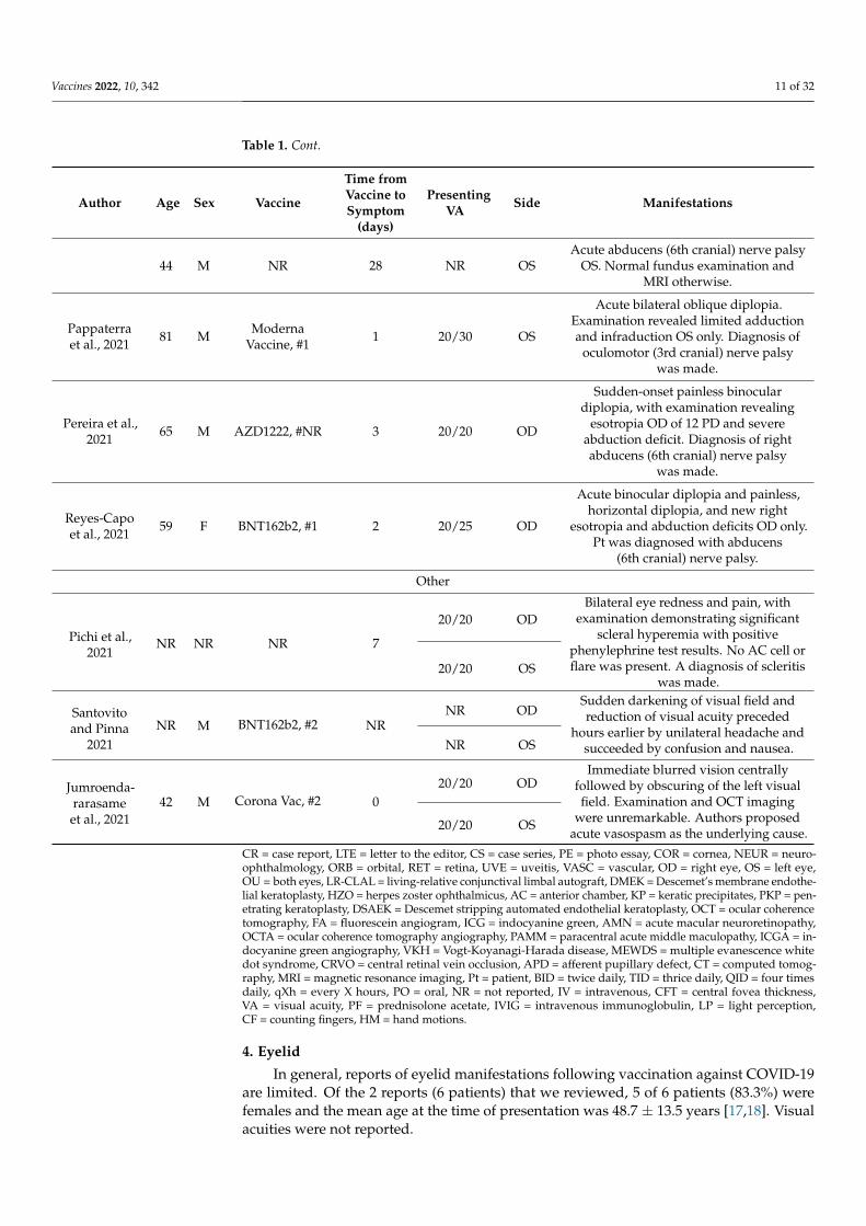
Vaccines 2022, 10, 342 11 of 32
Table 1. Cont.
Author Age Sex Vaccine
Time fromVaccine toSymptom
(days)
PresentingVA Side Manifestations
44 M NR 28 NR OSAcute abducens (6th cranial) nerve palsy
OS. Normal fundus examination andMRI otherwise.
Pappaterraet al., 2021 81 M Moderna
Vaccine, #1 1 20/30 OS
Acute bilateral oblique diplopia.Examination revealed limited adductionand infraduction OS only. Diagnosis of
oculomotor (3rd cranial) nerve palsywas made.
Pereira et al.,2021 65 M AZD1222, #NR 3 20/20 OD
Sudden-onset painless binoculardiplopia, with examination revealing
esotropia OD of 12 PD and severeabduction deficit. Diagnosis of rightabducens (6th cranial) nerve palsy
was made.
Reyes-Capoet al., 2021 59 F BNT162b2, #1 2 20/25 OD
Acute binocular diplopia and painless,horizontal diplopia, and new right
esotropia and abduction deficits OD only.Pt was diagnosed with abducens
(6th cranial) nerve palsy.
Other
Pichi et al.,2021
NR NR NR 7
20/20 ODBilateral eye redness and pain, with
examination demonstrating significantscleral hyperemia with positive
phenylephrine test results. No AC cell orflare was present. A diagnosis of scleritis
was made.20/20 OS
Santovitoand Pinna
2021NR M BNT162b2, #2 NR
NR ODSudden darkening of visual field andreduction of visual acuity preceded
hours earlier by unilateral headache andsucceeded by confusion and nausea.NR OS
Jumroenda-rarasameet al., 2021
42 M Corona Vac, #2 020/20 OD
Immediate blurred vision centrallyfollowed by obscuring of the left visualfield. Examination and OCT imaging
were unremarkable. Authors proposedacute vasospasm as the underlying cause.
20/20 OS
CR = case report, LTE = letter to the editor, CS = case series, PE = photo essay, COR = cornea, NEUR = neuro-ophthalmology, ORB = orbital, RET = retina, UVE = uveitis, VASC = vascular, OD = right eye, OS = left eye,OU = both eyes, LR-CLAL = living-relative conjunctival limbal autograft, DMEK = Descemet’s membrane endothe-lial keratoplasty, HZO = herpes zoster ophthalmicus, AC = anterior chamber, KP = keratic precipitates, PKP = pen-etrating keratoplasty, DSAEK = Descemet stripping automated endothelial keratoplasty, OCT = ocular coherencetomography, FA = fluorescein angiogram, ICG = indocyanine green, AMN = acute macular neuroretinopathy,OCTA = ocular coherence tomography angiography, PAMM = paracentral acute middle maculopathy, ICGA = in-docyanine green angiography, VKH = Vogt-Koyanagi-Harada disease, MEWDS = multiple evanescence whitedot syndrome, CRVO = central retinal vein occlusion, APD = afferent pupillary defect, CT = computed tomog-raphy, MRI = magnetic resonance imaging, Pt = patient, BID = twice daily, TID = thrice daily, QID = four timesdaily, qXh = every X hours, PO = oral, NR = not reported, IV = intravenous, CFT = central fovea thickness,VA = visual acuity, PF = prednisolone acetate, IVIG = intravenous immunoglobulin, LP = light perception,CF = counting fingers, HM = hand motions.
4. Eyelid
In general, reports of eyelid manifestations following vaccination against COVID-19are limited. Of the 2 reports (6 patients) that we reviewed, 5 of 6 patients (83.3%) werefemales and the mean age at the time of presentation was 48.7 ± 13.5 years [17,18]. Visualacuities were not reported.

Vaccines 2022, 10, 342 12 of 32
In one study, Austria et al. reported on a series of three women who each presentedwith unilateral edema more prominent in the upper eyelid following vaccination with theBNT162b2 vaccine [17]. All three patients were middle-aged women (aged 32, 43, and 43)and they were all treated differently. One patient was treated with observation, anotherwith antihistamines, and one with oral steroids. All patients had complete resolution oforbital edema within two days.
Elsewhere, Mazzatenta et al. described a case series of three patients who developedecchymotic or purpuric lesions on the upper eyelids 1 to 3 weeks following vaccinationwith the BNT162b2 vaccine [18]. In all three cases, lesions were bilateral and resolvedwithin approximately two weeks.
Regarding the mechanism of these findings, Austria et al. proposed in their report thateyelid changes may be mediated by complement activation which increased complementmediators within the tear duct via leakage of plasma [17,18]. Further investigation isrequired to support this hypothesis.
5. Orbit
A total of three reports (3 patients, 4 eyes) commented on orbital manifestationsfollowing vaccination against COVID-19. These cases are described below.
5.1. Superior Ophthalmic Vein Thrombosis
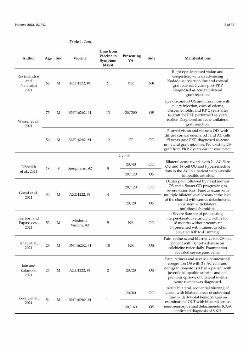
Two cases commented on superior ophthalmic vein thrombosis. Bayas et al. reporteda case of a 55-year-old woman who presented with conjunctival injection, retro-orbital pain,and diplopia seven days after getting vaccination with the AZD1222 vaccine [19]. Magneticresonance imaging (MRI) of brain and orbit with contrast showed superior ophthalmicvein thrombosis with no contrast filling and bilateral T2 signal intensity of the superiorophthalmic vein. Laboratory values revealed secondary immune thrombocytopenia. Thepatient later developed a transient right-sided hemiparesis and aphasia, and MRI testingdemonstrated a left parietal lobe ischemic stroke. Ultimately the patient was treated withanticoagulation and discharged. Elsewhere, Panovska-Stavridis et al. reported on a caseof a 29-year-old woman (Figure 1) who developed left orbital swelling, severe headache,and blurred left eye vision 10 days after receiving the AZD1222 vaccine [20]. MRI imagingdemonstrated central filling defects and a diagnosis of superior ophthalmic vein thrombosiswas made. The patient was treated with intravenous (IV) immunoglobulins 1 g/kg fortwo days, followed by an oral prednisolone taper. Concurrently, the patient was placedon rivaroxaban 15 mg twice daily for 21 days as well as broad-spectrum antibiotics. Theauthors reported excellent response within 5 days and the patient’s thrombocytopeniaalso resolved.
5.2. Tolosa-Hunt Syndrome
Chuang et al. reported on a case of a 45-year-old male who developed left eye painwith progressive ptosis, decreased vision, and binocular diplopia seven days after receivingan unspecified COVID-19 vaccine [21]. The patient had an afferent pupillary defect (APD)and complete ophthalmoplegia. Imaging with computed tomography (CT) and MRI ofthe brain were most consistent with cavernous sinus thrombosis. In the setting of thisconstellation of findings, the patient was diagnosed with Tolosa–Hunt syndrome.
Diagnostic criteria for Tolosa–Hunt syndrome require unilateral orbital affection withassociated paresis of one or more of the 3rd, 4th, and 6th cranial nerves [22]. Cavernoussinus thrombosis with Tolosa–Hunt syndrome has been reported sparingly, but a previousreport of it following hepatitis-B vaccination has been described [23]. On our review ofother vascular phenomena, we found limited reports of central retinal vein occlusion(CRVO), branch retinal vein occlusion (BRVO), and hemi-retinal vein occlusion (HRVO)(discussed later).

Vaccines 2022, 10, 342 13 of 32
Vaccines 2022, 10, x FOR PEER REVIEW 13 of 33
antibiotics. The authors reported excellent response within 5 days and the patient’s throm-bocytopenia also resolved.
Figure 1. Clinical presentation of the vaccine-induced prothrombotic immune thrombocytopenic disorder (VIPIT) and superior ophthalmic vein (SOV) thrombosis after ChAdOx1 nCoV-19 vaccina-tion. (A) patient presentation at admission with marked proptosis, (B) contrast enhanced magnetic resonance imaging (MRI) revealed SOV thrombosis (white arrow), presented with widening SOV and filling defects, (C) T2 sequence further confirmed SOV thrombosis with the enhanced signal intensity of SOV (white arrow), (D) no symptoms after five days of treatment, published with pa-tient’s permission. Adapted from Panovska-Stavridis, I.; Pivkova-Veljanovska, A.; Trajkova, S.; Laz-arevska, M.; Grozdanova, A.; Filipche, V. A Rare Case of Superior Ophthalmic Vein Thrombosis and Thrombocytopenia Following ChAdOx1 nCoV-19 Vaccine Against SARS-CoV-2. Mediterr. J. Hematol. Infect. Dis. 2021, 13, e2021048; Published 1 March 2021. https://doi.org/10.4084/MJHID.2021.048 [20]. Figure 1, Copyright (2021) with permission from Insti-tute of Hematology, Catholic University, Rome, open access article under the terms of the Creative Commons Attribution License.
5.2. Tolosa-Hunt Syndrome Chuang et al. reported on a case of a 45-year-old male who developed left eye pain
with progressive ptosis, decreased vision, and binocular diplopia seven days after receiv-ing an unspecified COVID-19 vaccine [21]. The patient had an afferent pupillary defect (APD) and complete ophthalmoplegia. Imaging with computed tomography (CT) and MRI of the brain were most consistent with cavernous sinus thrombosis. In the setting of this constellation of findings, the patient was diagnosed with Tolosa–Hunt syndrome.
Diagnostic criteria for Tolosa–Hunt syndrome require unilateral orbital affection with associated paresis of one or more of the 3rd, 4th, and 6th cranial nerves [22]. Cavern-ous sinus thrombosis with Tolosa–Hunt syndrome has been reported sparingly, but a pre-vious report of it following hepatitis-B vaccination has been described [23]. On our review of other vascular phenomena, we found limited reports of central retinal vein occlusion (CRVO), branch retinal vein occlusion (BRVO), and hemi-retinal vein occlusion (HRVO) (discussed later).
5.3. Mechanisms The mechanisms underlying a possible hypercoagulable state following vaccination
have not yet been completely elucidated. However, Schultz et al. previously reported five cases of severe venous thromboembolism—four of which were cerebral venous throm-bosis—following vaccination against COVID-19 [24]. Because all cases resolved with transfusions and had an absence of hemolysis, the authors ruled out thrombotic
Figure 1. Clinical presentation of the vaccine-induced prothrombotic immune thrombocytopenicdisorder (VIPIT) and superior ophthalmic vein (SOV) thrombosis after ChAdOx1 nCoV-19 vaccina-tion. (A) patient presentation at admission with marked proptosis, (B) contrast enhanced magneticresonance imaging (MRI) revealed SOV thrombosis (white arrow), presented with widening SOV andfilling defects, (C) T2 sequence further confirmed SOV thrombosis with the enhanced signal intensityof SOV (white arrow), (D) no symptoms after five days of treatment, published with patient’s permis-sion. Adapted from Panovska-Stavridis, I.; Pivkova-Veljanovska, A.; Trajkova, S.; Lazarevska, M.;Grozdanova, A.; Filipche, V. A Rare Case of Superior Ophthalmic Vein Thrombosis and Thrombocy-topenia Following ChAdOx1 nCoV-19 Vaccine Against SARS-CoV-2. Mediterr. J. Hematol. Infect. Dis.2021, 13, e2021048; Published 1 March 2021. https://doi.org/10.4084/MJHID.2021.048 [20]. Figure 1,Copyright (2021) with permission from Institute of Hematology, Catholic University, Rome, openaccess article under the terms of the Creative Commons Attribution License.
5.3. Mechanisms
The mechanisms underlying a possible hypercoagulable state following vaccina-tion have not yet been completely elucidated. However, Schultz et al. previously re-ported five cases of severe venous thromboembolism—four of which were cerebral venousthrombosis—following vaccination against COVID-19 [24]. Because all cases resolved withtransfusions and had an absence of hemolysis, the authors ruled out thrombotic thrombo-cytopenic purpura and immune thrombocytopenia. However, in all cases, there was a highlevel of antibodies to platelet factor-4 (PF4)-polyanion complexes, suggesting a vaccine-related variant of the phenomenon of heparin-induced thrombocytopenia termed vaccine-induced thrombotic thrombocytopenia (VITT) [24]. Indeed, thrombosis and thrombocy-topenia has previously been reported following the use of Measles-Mumps-Rubella [25–28],influenza [29], pneumococcal [30], smallpox [31], and COVID-19 vaccines [24,32–35], but itis unclear if these would all fall into the category of VITT. Thrombotic microangiopathieshave previously been reported following influenza vaccination and linked to thromboticthrombocytopenic purpura, but these reports are rare [36].
It is our clinical recommendation that any patient presenting with thrombosis—withor without ophthalmic manifestations—should be tested for thrombocytopenia, responseto platelet transfusion, and the presence of anti-PF4 complex antibodies.
6. Uveitis
Given the strong association between uveitis and immunologic phenomena, it wouldbe expected that there is some relationship between vaccination against COVID-19 anduveitis. Of the 14 reports we reviewed dealing with uveitis after COVID-19 vaccination,34 patients (44 eyes) were reported on. Of these 34, 19 (55.9%) were males, 15 (44.1%)

Vaccines 2022, 10, 342 14 of 32
were females, and the average age at the time of presentation was 47.6 ± 16.3 years. Forthe 34 patients, average time from vaccination to development of ophthalmic symptomswas 8.0 ± 8.6 days. Ten patients (29.4%) presented with bilateral manifestations. For the40 eyes which had presenting visual acuity information, the mean presenting visual acuitywas logMAR 0.421 ± 0.455 (20/52 in Snellen notation). For the 35 eyes which had bothpresenting and final visual acuity at last follow-up, these values were 0.434± 0.426 (20/54 inSnellen notation) and 0.085 ± 0.166 (20/24 in Snellen notation), respectively (p < 0.001).
6.1. Uveitis Flares
Previously, we described a case of an 18-year-old girl with a history of antinuclearantibody positive oligoarticular juvenile idiopathic arthritis (JIA) (but no prior historyof uveitis) who presented with bilateral anterior uveitis 5 days after the second dose ofthe Sinopharm COVID-19 vaccine [37]. Examination was notable for anterior uveitis,and optical coherence tomography (OCT) showed hyperreflective dots and circulatingcells in the anterior chamber (AC). Uveitis in both eyes resolved gradually after topicalsteroid treatment without recurrence. Similar to our case, Jain and Kalamkar reported ona 27-year-old man with past medical history of JIA and one previous episode of uveitiswho developed a uveitis flare-up in the left eye (OS) two days after receiving the AZD1222vaccine [38]. Similar to our previous report, the patient demonstrated resolution withtopical steroids and cycloplegic drops.
Numerous other reports have been made. Mudie et al. described a case of a 43-year-old woman who presented with eye pain, redness, and photophobia bilaterally 3 daysafter her second dose of the BNT162b2 [39]. Examination was notable for a thickenedchoroid and pronounced inflammation in the AC and the vitreous cavity. The patientresponded well to oral and topical corticosteroids with a mild recurrence after the initialattempt to taper these drugs. Renisi et al. described a similar case in a 23-year-old manwho developed pain, photophobia, and a red eye four days after receiving the second doseof the BNT162b2 vaccine [40]. Examination revealed conjunctival hyperemia, posteriorsynechiae, and AC cells with keratic precipitates (KP) in the lower quadrants. The patientdemonstrated initial improvement on topical dexamethasone and atropine drops dailyover 3 weeks, then demonstrated complete resolution at 6 weeks.
Ishay et al. reported a case of a 28-year-old male with past medical history of Behçet’sdisease on colchicine twice daily [41]. Ten days after receiving the BNT162b2 vaccine, thepatient developed left eye pain, redness, and blurred vision. Examination revealed severepanuveitis. Unlike the previous cases, the patient was successfully treated with five days ofpulse-dose IV methylprednisolone followed by oral (PO) corticosteroids and azathioprine.
In a different case, Herbort and Papasavvas reported on a 53-year-old male with pre-existing herpes keratouveitis which was inactive for 18 months without treatment [42]. Fivedays after receiving the Moderna COVID-19 vaccine, the patient presented with a severeflare-up of disease, including numerous KPs and elevated intraocular pressure to 41 mmHg.The patient was treated with PO valacyclovir 500 mg, topical dexamethasone, dorzolamide,and timolol. Over 6 days of treatment, the patient demonstrated an improvement in flare,and KPs resolved almost completely after 3 weeks.
To date, the largest and only multicenter study investigating a relationship betweenuveitis and COVID-19 vaccination was conducted by Rabinovitch et al. [43]. In their study,the authors examined 23 eyes of 21 patients (mean age of 51.3 years) who developeduveitis after vaccination against COVID-19 with the BNT162b2 vaccine. These patientspresented with uveitis an average of 7.5 ± 7.3 days after vaccination. A total of 8 of the21 patients had pre-existing uveitis, though average time since last flare was one year, andno patients had recent changes in medication regimen. Eight of 21 patients presented afterfirst dose of vaccination and 13 of 21 presented after second dose of vaccination. Six ofthe 21 patients had pre-existing uveitis-related diseases, including ankylosing spondylitis,psoriasis, Crohn’s disease, and herpes zoster (VZV) ophthalmicus. Two patients hadbilateral disease presentation. Twenty-one of 23 eyes had anterior uveitis and two eyes had
Vaccines 2022, 10, 342 15 of 32
multiple evanescent white dot syndrome. Nineteen of 21 patients were treated with steroids,most commonly prednisone or dexamethasone, and all 19 of these patients demonstratedcomplete resolution of inflammation. Two of 21 patients did not undergo treatment butdemonstrated significant improvement, nevertheless.
6.2. Choroiditis
Two reports have been made connecting choroiditis with vaccination against COVID-19.Goyal et al. reported on a 34-year-old male who developed ocular pain and nasal rednessOS as well as a floater in the right eye (OD) progressing to severe vision loss 4 days afterreceiving the AZD1222 vaccine [44]. At presentation, his visual acuity was 20/120 ODand 20/20 OS. The patient’s fundus exam demonstrated multiple bilateral oval lesions atthe level of the choroid with serous detachment, consistent with a diagnosis of bilateralmultifocal choroiditis. The patient was treated with a PO prednisolone taper to beginningat 100 mg daily and demonstrated significant improvement in inflammation and subretinalfluid after 11 days of treatment. His visual acuity at the last follow-up was 20/20 inboth eyes.
Another report by Pan et al. described a 50-year-old woman who developed bilateralblurred vision her 5 days after receiving an unspecified Vero cell-based vaccine in China [45].Her examination revealed a pale, blurry optic disc, absent foveal reflex, and macularedema. Imaging with fluorescein angiography was consistent with bilateral choroiditis.The patient’s vision and inflammation improved considerably over 5 weeks with perioculartriamcinolone acetamide and PO prednisone.
6.3. Vogt–Koyanagi–Harada Disease
Vogt–Koyanagi–Harada (VKH) disease is a T-lymphocyte mediated multi-systemdisease affecting the auditory system, skin, meninges, and eye [46,47]. Ophthalmologi-cally, it causes a granulomatous panuveitis often affecting young adults, and may alsopresent with exudative retinal detachments and a sunset glow fundus [46–49]. As it is anautoimmune disease resulting from antibodies against melanocytes-associated antigens,the robust immune response mounted by patients following vaccination against COVID-19may be of importance to patients living with VKH or other autoimmune diseases.
Papasavvas and Herbort reported on a 43-year-old woman who had a previous historyof VKH disease which was under control for 6 years using mycophenolate, cyclosporine,and intermittent infliximab infusions [50]. However, six weeks after the second dose ofthe BNT162b2 vaccine, the patient presented with severe reactivation of disease. Althoughher visual acuity remained 20/20 OD and OS, she had severe AC inflammation with3–4 small mutton-fat KPs as well as bilateral exudative retinal detachments. Severalhypofluorescent dark dots were present on indocyanine green angiography (ICGA), whichwas also observed when the patient was first diagnosed with VKH disease. This flare wasultimately controlled using infliximab. Furthermore, the authors speculated that the flarehad occurred with the second dose because the patient’s last infliximab infusion had beenperformed 3.5 weeks before her first dose of the vaccine but 7.5 weeks before the seconddose. This case in particular highlights the possibility that COVID-19 vaccination may beassociated with reactivation or exacerbation of pre-existing autoimmune disease.
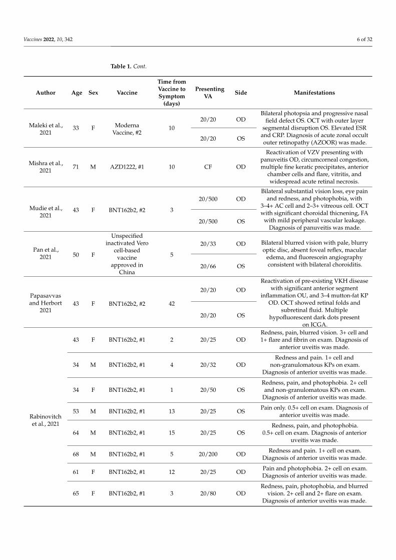
In another report, Saraceno et al. described a 62-year-old female who developedacute bilateral loss of vision two days after receiving the AZD1222 vaccine [51]. Shewas found to have visual acuity of 20/600 OD and 20/200 OS. On examination, she had2+ AC cells and 1+ vitreous cells bilaterally. Fundus examination revealed serous retinaldetachments and optic disc hyperemia bilaterally (Figure 2). OCT demonstrated subretinalhyperreflective dots. In this case also a diagnosis of VKH was made. The patient wastreated with PO prednisone. Intravenous therapy was avoided due to a restriction inavailable hospital beds. Within four days, the patient’s visual acuity improved to 20/60 ODand 20/80 OS. At a three week follow up, the patient remarkably demonstrated visualacuity of 20/20 in both eyes with no signs of inflammatory activity and resolution of
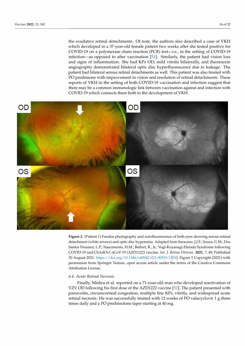
Vaccines 2022, 10, 342 16 of 32
the exudative retinal detachments. Of note, the authors also described a case of VKHwhich developed in a 37-year-old female patient two weeks after she tested positive forCOVID-19 on a polymerase chain reaction (PCR) test—i.e., in the setting of COVID-19infection—as opposed to after vaccination [51]. Similarly, the patient had vision lossand signs of inflammation: She had KPs OD, mild vitritis bilaterally, and fluoresceinangiography demonstrated bilateral optic disc hyperfluorescence due to leakage. Thepatient had bilateral serous retinal detachments as well. This patient was also treated withPO prednisone with improvement in vision and resolution of retinal detachments. Thesereports of VKH in the setting of both COVID-19 vaccination and infection suggest thatthere may be a common immunologic link between vaccination against and infection withCOVID-19 which connects them both to the development of VKH.
Vaccines 2022, 10, x FOR PEER REVIEW 17 of 33
Figure 2. (Patient 1) Fundus photography and autofluorescence of both eyes showing serous retinal detachment (white arrows) and optic disc hyperemia. Adapted from Saraceno, J.J.F.; Souza, G.M.; Dos Santos Finamor, L.P.; Nascimento, H.M.; Belfort, R., Jr.; Vogt-Koyanagi-Harada Syndrome fol-lowing COVID-19 and ChAdOx1 nCoV-19 (AZD1222) vaccine. Int. J. Retina Vitreous. 2021, 7, 49; Published 30 August 2021. https://doi.org/10.1186/s40942-021-00319-3 [51]. Figure 1 Copyright (2021) with permission from Springer Nature, open access article under the terms of the Creative Commons Attribution License.
6.4. Acute Retinal Necrosis Finally, Mishra et al. reported on a 71-year-old man who developed reactivation of
VZV OD following his first dose of the AZD1222 vaccine [52]. The patient presented with panuveitis, circumcorneal congestion, multiple fine KPs, vitritis, and widespread acute retinal necrosis. He was successfully treated with 12 weeks of PO valacyclovir 1 g three times daily and a PO prednisolone taper starting at 40 mg.
6.5. Acute Zonal Occult Outer Retinopathy Maleki et al. reported on a 33-year-old woman who developed bilateral photopsias
and a progressive nasal field defect OS 10 days after receiving the second dose of the Moderna COVID-19 vaccine [53]. Imaging with OCT was demonstrative of an outer layer segmental disruption OS. A diagnosis of acute zonal occult outer retinopathy (AZOOR) was made.
6.6. Mechanisms The primary uveitic phenomenon we encountered on our review was new uveitis or
flare-ups of pre-existing disease. Uveitis has previously been documented following nu-merous vaccines, most commonly the Bacille Calmette–Guerin, hepatitis B, human papil-lomavirus, influenza, measles-mumps-rubella, and varicella vaccines [54–58]. One review of 276 found that 199 (72.1%) cases were in women [58].
Figure 2. (Patient 1) Fundus photography and autofluorescence of both eyes showing serous retinaldetachment (white arrows) and optic disc hyperemia. Adapted from Saraceno, J.J.F.; Souza, G.M.; DosSantos Finamor, L.P.; Nascimento, H.M.; Belfort, R., Jr.; Vogt-Koyanagi-Harada Syndrome followingCOVID-19 and ChAdOx1 nCoV-19 (AZD1222) vaccine. Int. J. Retina Vitreous. 2021, 7, 49; Published30 August 2021. https://doi.org/10.1186/s40942-021-00319-3 [51]. Figure 1 Copyright (2021) withpermission from Springer Nature, open access article under the terms of the Creative CommonsAttribution License.
6.4. Acute Retinal Necrosis
Finally, Mishra et al. reported on a 71-year-old man who developed reactivation ofVZV OD following his first dose of the AZD1222 vaccine [52]. The patient presented withpanuveitis, circumcorneal congestion, multiple fine KPs, vitritis, and widespread acuteretinal necrosis. He was successfully treated with 12 weeks of PO valacyclovir 1 g threetimes daily and a PO prednisolone taper starting at 40 mg.
Vaccines 2022, 10, 342 17 of 32
6.5. Acute Zonal Occult Outer Retinopathy
Maleki et al. reported on a 33-year-old woman who developed bilateral photopsiasand a progressive nasal field defect OS 10 days after receiving the second dose of theModerna COVID-19 vaccine [53]. Imaging with OCT was demonstrative of an outer layersegmental disruption OS. A diagnosis of acute zonal occult outer retinopathy (AZOOR)was made.
6.6. Mechanisms
The primary uveitic phenomenon we encountered on our review was new uveitisor flare-ups of pre-existing disease. Uveitis has previously been documented followingnumerous vaccines, most commonly the Bacille Calmette–Guerin, hepatitis B, humanpapillomavirus, influenza, measles-mumps-rubella, and varicella vaccines [54–58]. Onereview of 276 found that 199 (72.1%) cases were in women [58].
There are several possible mechanisms underpinning the development of post-vaccinationuveitis. Fraunfelder et al. previously studied the connection between the hepatitis Bvaccine and uveitis and proposed that a delayed-type hypersensitivity reaction and im-mune complex deposition following vaccination leads to uveitis [56]. The authors alsoproposed that adjuvants play a role in this immunologic process, though this does notapply to COVID-19 vaccination. Elsewhere, Aguirre et al. reported a uveitic reaction indogs following vaccination with canine adenovirus 1, which was found to be a type IIIhypersensitivity reaction involving antigen-antibody complexes present in the aqueoushumor [59]. Given the previously established fact that SARS-CoV-2 RNA has been found inhuman aqueous humor and other ocular tissues, a similar inflammatory reaction involvingimmune complex deposition is likely [60,61].
In their major review of uveitis following COVID-19 vaccination with the BNT162b2vaccine, Rabinovitch et al. proposed that the possible causal mechanism is vaccine-inducedtype I interferon secretion [43]. The authors proposed that the vaccine mRNA activatesRNA-sensing molecules including TLR3, TLR7, MDA5, and RIG-I which drive autoimmuneprocesses in these patients. While not mutually exclusive, we favor the phenomenon ofimmune-complex deposition as the primary driver of COVID-19 vaccine-related uveitis.However, it has been reported that COVID-19 vaccines use the modified nucleobase N1-methylpseudouridine in order to dampen immunostimulatory potential [62]. Furtherinvestigation is required to evaluate the extent to which this impacts COVID-19 vaccine-related uveitis.
7. Cornea
A number of studies have reported on adverse events at the level of the ocular sur-face following vaccination to COVID-19. On our review, 11 reports (15 patients, 18 eyes)described corneal manifestations following vaccination against COVID-19. Of these15 patients, 9 (66.7%) were female and 6 (33.3%) were male. The mean age at the timeof presentation was 61.33 ± 15.5 years, and the average time from vaccination to devel-opment of ophthalmic symptoms was 11.8 ± 6.2 days. Three patients (20.0%) presentedwith bilateral involvement. For the 17 affected eyes which had reported visual acuity, themean visual acuity was logMAR 1.09 ± 0.858 (20/247 in Snellen notation) at presentation.Only 5 studies (6 patients, 7 eyes) reported on baseline, post-transplantation visual acuityof patients who underwent graft rejection after vaccination. For these patients, baselinevisual acuity was logMAR 0.204 ± 0.309 (20/32 in Snellen notation), whereas visual acuityat presentation after vaccination was logMAR 0.871 ± 0.694 (20/149 in Snellen notation).This difference was significant (p = 0.007). For the 13 eyes which had both presenting andfinal visual acuities reported, the mean visual acuities were 1.215± 0.878 (20/328 in Snellennotation) and 0.482 ± 0.793 (20/61 in Snellen notation), respectively (p < 0.001).

Vaccines 2022, 10, 342 18 of 32
7.1. Graft Rejection
Several reports have described complications involving corneal transplant rejectionfollowing vaccination to COVID-19. Phylactou et al. reported on a pair of cases [63].First, they described a 66-year-old woman with Fuchs endothelial corneal dystrophy(FECD) status-post unilateral Descemet’s membrane endothelial keratoplasty (DMEK)transplant in the right eye who received the BNT162b2 vaccine 14 days after DMEK. Sevendays after receiving her vaccination, she presented with a visual acuity of 20/120 OD.Examination revealed moderate conjunctival injection, diffuse corneal edema, fine KPs,and 1+ AC cells. Central corneal thickness (CCT) was 652 µm, significantly increased from525 µm one week after transplantation. She was diagnosed with acute unilateral graftrejection. She was treated with an increase in frequency of topical steroids and one weeklater demonstrated 20/20 vision OD with a clear cornea and decreased inflammation. Theauthors also reported on an 83-year-old woman with bilateral DMEK transplants for FECD3 and 6 years before developing acute bilateral endothelial rejection, 3 weeks after hersecond dose of the BNT162b2 vaccine. In this case also, the patient was treated with topicalsteroid drops and demonstrated significant improvement at a one-week follow-up.
Several other reports have also been made. Crnej et al. reported on a 71-year-oldpatient who underwent DMEK surgery 5 months earlier and developed acute unilateralgraft rejection 7 days after receiving his second dose of the BNT162b2 vaccination [64]. Inthat case, the patient was treated successfully with topical dexamethasone 1 mg/mL everytwo hours.
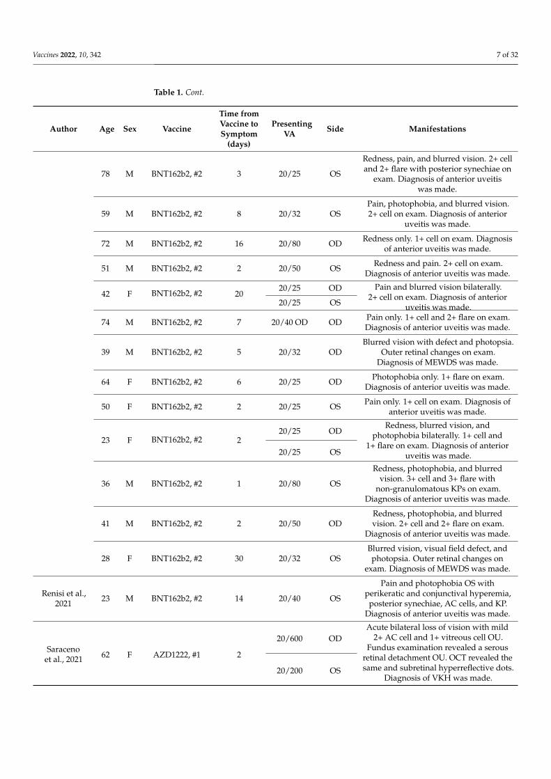
Wasser et al. reported on a pair of men, aged 56 and 73, both with a history ofpenetrating keratoplasty (PKP) due to keratoconus, who developed acute corneal graftrejection 2 weeks after receiving their first dose of the BNT162b2 vaccine [65]. Similar toother cases of graft rejection, both patients presented with vision loss, corneal edema, andKPs. Of note, the 56-year-old man had pre-existing grafts in both eyes but only his righteye (PKP done 25 years earlier) had rejection, while his left eye (PKP done 7 years earlier)remained intact. Both patients were successfully treated with hourly dexamethasone andoral prednisone 60 mg/day. Rallis et al. reported on a similar case of a 68-year-old womanwith previous bilateral lamellar Descemet stripping automated endothelial keratoplasty(DSAEK) for previous FECD and a left re-do PKP for failed DSAEK [66]. She presentedwith pain and redness and rapid vision loss OS four days after receiving her first dose ofthe BNT162b2 vaccine. Her examination demonstrated corneal punctuate straining, cornealgraft edema, Descemet’s folds, and scattered KPs, all in the left eye only (Figure 3). Similarto the case mentioned in Wasser et al., her pre-existing right eye graft was completelyunaffected. At a three-week follow-up, she demonstrated complete resolution of thesesymptoms following hourly topical dexamethasone 0.1% and a week of PO acyclovir400 mg five times daily to cover for herpes simplex keratitis.
In comparison, Abousy et al. described a case of a 73-year-old woman with previousbilateral DSEK for FECD who presented with bilateral decreased vision, ocular pain, andphotophobia four days after her second dose of the BNT162b2 vaccine [67]. Examinationrevealed decreased vision to 20/200 OD and 20/40 OS as well as corneal edema OD.Further examination revealed Descemet folds bilaterally. The patient was diagnosed withbilateral graft rejection. The patient was initiated on topical prednisolone acetate 1%four times per day. She had initially persistent and worsening symptoms on this regimenat her 28-day follow-up, so prednisolone frequency was increased to hourly, and Muroointment was added at bedtime. Prednisolone was tapered with improvement and at atwo-month follow-up, the patient’s vision had improved to 20/50 OD and 20/25 OS, withsignificantly decreased corneal edema bilaterally., Taken together, these cases suggest thatgraft rejection can be unilateral or bilateral post-COVID-19 vaccination.
Similar cases of post-PKP were reported separately by Ravichandran et al., Nioi et al.,and Parmar et al. in adult patients [68–70]. Nioi et al. uniquely found that their 44-year-oldfemale patient had severe vitamin D deficiency concurrently with rejection, so the patientwas treated with topical dexamethasone and vitamin D supplementation. After initial

Vaccines 2022, 10, 342 19 of 32
resolution at four weeks, she again had an episode of rejection concurrently with persistentvitamin D deficiency, and steroid drops were re-started with higher doses of vitamin D,which resulted in sustained resolution. Vitamin D deficiency has previously been demon-strated to play a vital role in adverse effects following solid organ transplantation, namelyallograft rejection [71–73]. It plays a vital role the expression of IL-2 and interferon-mRNA,downregulates T-cell mediated cytotoxicity, and suppresses major histocompatibility com-plexes of immunomodulators of dendritic cells [68,74,75]. The authors supported thisexplanation for their patient.
Vaccines 2022, 10, x FOR PEER REVIEW 20 of 33
Figure 3. (A,B) Slit-lamp photography demonstrating conjunctival hyperemia, corneal graft haze, diffuse corneal epithelial, and stromal oedema (within the graft), Descemet’s folds, scattered keratic precipitates (KPs), and 1+ cells in anterior chamber. An unusual distribution of fluorescein staining with coarse punctate epitheliopathy over the corneal graft was observed. The central corneal thick-ness (CCT) was 730 μm. (C,D) At 3-week post treatment, the corneal graft rejection was successfully treated with considerable improvement in the graft transparency, reduction in epithelial and stro-mal oedema, and resolution of epitheliopathy and anterior chamber inflammation. The best-cor-rected visual acuity improved to 6/12, with a CCT of 609 μm. Adapted from Rallis, K.I.; Ting, D.S.J.; Said, D.G.; et al. Corneal graft rejection following COVID-19 vaccine. Eye (2021). https://doi.org/10.1038/s41433-021-01671-2 [65]. Figure 1, Copyright (2021) with permission from Nature Publications, open access article under the terms of the Creative Commons Attribution Li-cense.
7.2. Corneal Melting Khan et al. reported on a 48-year-old man who developed profound vision loss to
light perception three weeks after receiving his first dose of the AZD1222 vaccine [76]. He was found to have diffuse conjunctival and ciliary congestion, corneal melting and perfo-ration with diffuse corneal haze, uveal tissue prolapse, and bilateral massive choroidal detachment on B-scan ultrasonography.
7.3. Mechanisms Our review primarily revealed several cases of corneal graft rejection, both unilateral
and bilateral. Corneal transplant rejection has previously been reported—albeit rarely—following influenza, hepatitis B, tetanus, and yellow fever vaccinations [77–80]. In the set-ting of this pre-existing precedent, it is not surprising that the highly immunogenic vac-cines to COVID-19 present similar risks.
There are several possible mechanisms underlying corneal graft compromise follow-ing vaccination. One hypothesis proposed by Steinemann et al. asserts that elevated vas-cular permeability following vaccination compromises the native-state immunologic priv-ilege possessed by the cornea [77]. This theory is supported by the finding of graft edema, as demonstrated on our review. In the same case series, the Steinemann et al. also pro-posed that immunization may induce expression of the major histocompatibility complex
Figure 3. (A,B) Slit-lamp photography demonstrating conjunctival hyperemia, corneal graft haze,diffuse corneal epithelial, and stromal oedema (within the graft), Descemet’s folds, scattered keraticprecipitates (KPs), and 1+ cells in anterior chamber. An unusual distribution of fluorescein stainingwith coarse punctate epitheliopathy over the corneal graft was observed. The central corneal thickness(CCT) was 730 µm. (C,D) At 3-week post treatment, the corneal graft rejection was successfullytreated with considerable improvement in the graft transparency, reduction in epithelial and stromaloedema, and resolution of epitheliopathy and anterior chamber inflammation. The best-correctedvisual acuity improved to 6/12, with a CCT of 609 µm. Adapted from Rallis, K.I.; Ting, D.S.J.; Said,D.G.; et al. Corneal graft rejection following COVID-19 vaccine. Eye (2021). https://doi.org/10.1038/s41433-021-01671-2 [65]. Figure 1, Copyright (2021) with permission from Nature Publications, openaccess article under the terms of the Creative Commons Attribution License.
7.2. Corneal Melting
Khan et al. reported on a 48-year-old man who developed profound vision loss tolight perception three weeks after receiving his first dose of the AZD1222 vaccine [76].He was found to have diffuse conjunctival and ciliary congestion, corneal melting andperforation with diffuse corneal haze, uveal tissue prolapse, and bilateral massive choroidaldetachment on B-scan ultrasonography.
7.3. Mechanisms
Our review primarily revealed several cases of corneal graft rejection, both unilateraland bilateral. Corneal transplant rejection has previously been reported—albeit rarely—

Vaccines 2022, 10, 342 20 of 32
following influenza, hepatitis B, tetanus, and yellow fever vaccinations [77–80]. In thesetting of this pre-existing precedent, it is not surprising that the highly immunogenicvaccines to COVID-19 present similar risks.
There are several possible mechanisms underlying corneal graft compromise followingvaccination. One hypothesis proposed by Steinemann et al. asserts that elevated vascularpermeability following vaccination compromises the native-state immunologic privilegepossessed by the cornea [77]. This theory is supported by the finding of graft edema, asdemonstrated on our review. In the same case series, the Steinemann et al. also proposedthat immunization may induce expression of the major histocompatibility complex (MHC)of the cornea, as various organ grafts result in enhancement of MHC antigenic expressionafter rejection [77]. Donor cells with no MHC expression are thereafter targeted by the hostimmune cells due to poor immunogenicity [77,81].
Another mechanism for corneal graft rejection proposed in the setting of vaccination toCOVID-19 by Abousy et al. revolves around the finding that SARS-CoV-2 RNA is present inthe aqueous humor of patients with asymptomatic infections [61,67]. Likewise, Sawant et al.found that SARS-CoV-2 RNA was found in the corneas of postmortem COVID-19 pa-tients [60]. In the setting of vaccination for COVID-19 during ongoing or previous asymp-tomatic infection, then, it is possible that antibody-antigen complexes would be formedin large quantities with the subsequent development of profound inflammation, againcompromising the integrity of corneal grafts.
Regardless of the mechanism, we propose that ophthalmologists consider examiningpatients with corneal grafts prior to vaccination against COVID-19 in order to evaluateunderlying inflammatory processes which may be further exacerbated by the introductionof a profound immunogenic stimulus such as a COVID-19 vaccine. Particularly if weconsider the effect of different immune processes to be additive, it may be optimal forpatients with corneal grafts to delay COVID-19 vaccination if experiencing a transientinflammatory process around the time of vaccination. However, given the fact that thecases of corneal graft rejection have been successfully managed with topical steroids,whereas COVID-19 infection presents grave individual and epidemiologic risks, we do notrecommend that patients avoid receiving the vaccine altogether.
8. Retina
Preliminary reports suggest that retinal adverse events are possible following vac-cination against COVID-19. On our review, 12 reports (14 patients, 19 eyes) commentedon retinal manifestations following vaccination against COVID-19. One report did notinclude information on the sex of two patients. Of the remaining 12 patients, 10 (83.3%)were women and 2 (16.7%) were men. The mean age at the time of presentation was24.8 ± 4.8 years, and the average time from vaccination to development of ophthalmicsymptoms was 3.1 ± 2.4 days. Five patients (35.7%) presented with bilateral involvement.For the 19 affected eyes which had presenting visual acuities, the mean visual acuitywas logMAR 0.138 ± 0.325 (20/27 in Snellen notation) at presentation. For the 6 eyeswhich had both presenting and final visual acuities reported, the mean visual acuities were0.350 ± 0.465 (20/45 in Snellen notation) and 0.030 ± 0.067 (20/21 in Snellen notation),respectively (p = 0.138).
8.1. Acute Macular Neuroretinopathy
Acute macular neuroretinopathy (AMN) is a rare disease, commonly affecting adultfemales, which frequently presents with the acute onset of paracentral scotomas affectingone or both eyes [82–85]. Fundus exam may demonstrate reddish-brown petaloid perifoveallesions with the tip pointed toward the fovea [83,86]. There are no known treatmentmodalities for AMN, and vision changes may be permanent [85].
A number of reports suggest that there is an association between vaccination forCOVID-19 and development of AMN. Bøhler et al. reported on a 27-year-old femalewith no past medical history who developed flu-like symptoms followed by a paracentral
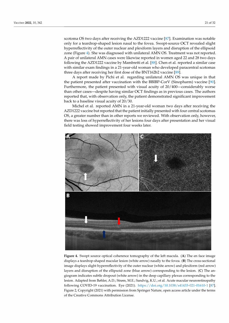
Vaccines 2022, 10, 342 21 of 32
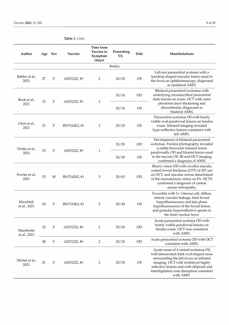
scotoma OS two days after receiving the AZD1222 vaccine [87]. Examination was notableonly for a teardrop-shaped lesion nasal to the fovea. Swept-source OCT revealed slighthyperreflectivity of the outer nuclear and plexiform layers and disruption of the ellipsoidzone (Figure 4). She was diagnosed with unilateral AMN OS. Treatment was not reported.A pair of unilateral AMN cases were likewise reported in women aged 22 and 28 two daysfollowing the AZD1222 vaccine by Mambretti et al. [88]. Chen et al. reported a similar casewith similar exam findings in a 21-year-old woman who developed paracentral scotomasthree days after receiving her first dose of the BNT162b2 vaccine [89].
A report made by Pichi et al. regarding unilateral AMN OS was unique in thatthe patient presented after vaccination with the BBIBP-CorV (Sinopharm) vaccine [90].Furthermore, the patient presented with visual acuity of 20/400—considerably worsethan other cases—despite having similar OCT findings as in previous cases. The authorsreported that, with observation only, the patient demonstrated significant improvementback to a baseline visual acuity of 20/30.
Michel et al. reported AMN in a 21-year-old woman two days after receiving theAZD1222 vaccine but reported that the patient initially presented with four central scotomasOS, a greater number than in other reports we reviewed. With observation only, however,there was loss of hyperreflectivity of her lesions four days after presentation and her visualfield testing showed improvement four weeks later.
Vaccines 2022, 10, x FOR PEER REVIEW 22 of 33
A report made by Pichi et al. regarding unilateral AMN OS was unique in that the patient presented after vaccination with the BBIBP-CorV (Sinopharm) vaccine [90]. Fur-thermore, the patient presented with visual acuity of 20/400—considerably worse than other cases—despite having similar OCT findings as in previous cases. The authors re-ported that, with observation only, the patient demonstrated significant improvement back to a baseline visual acuity of 20/30.
Michel et al. reported AMN in a 21-year-old woman two days after receiving the AZD1222 vaccine but reported that the patient initially presented with four central scoto-mas OS, a greater number than in other reports we reviewed. With observation only, how-ever, there was loss of hyperreflectivity of her lesions four days after presentation and her visual field testing showed improvement four weeks later.
In comparison, Book et al. reported a case of a 21-year-old woman with no past med-ical history who developed bilateral paracentral scotomas 3 days after receiving the AZD1222 vaccine [91]. Near-infrared imaging and OCT revealed similar lesions as the previous case, but bilaterally. She was diagnosed with bilateral AMN. Druke et al. re-ported a similar case of a 23-year-old female who developed bilateral paracentral scoto-mas one day after vaccination with the AZD1222 vaccine [92]. Fundus photography re-vealed a subtle brownish rimmed lesion parafoveal in the right eye and a blurred lesion nasal to the macula. Near-infrared imaging and OCT imaging confirmed a diagnosis of bilateral AMN. Valenzuela et al. described a similar report of bilateral AMN following vaccination with the second dose BNT162b2 vaccine [93]. In contrast to other reports, the authors reported resolution of symptoms after 7 days with observation only.
Figure 4. Swept source optical coherence tomography of the left macula. (A) The en face image displays a teardrop-shaped macular lesion (white arrow) nasally to the fovea. (B) The cross-sectional image displays slight hyperreflectivity of the outer nuclear (white arrow) and plexiform (red arrow) layers and disruption of the ellipsoid zone (blue arrow) corresponding to the lesion. (C) The angio-gram indicates subtle dropout (white arrow) in the deep capillary plexus corresponding to the le-sion. Adapted from Bøhler, A.D.; Strøm, M.E.; Sandvig, K.U.; et al. Acute macular neuroretinopathy following COVID-19 vaccination. Eye (2021). https://doi.org/10.1038/s41433-021-01610-1 [87]. Figure 2,
Figure 4. Swept source optical coherence tomography of the left macula. (A) The en face imagedisplays a teardrop-shaped macular lesion (white arrow) nasally to the fovea. (B) The cross-sectionalimage displays slight hyperreflectivity of the outer nuclear (white arrow) and plexiform (red arrow)layers and disruption of the ellipsoid zone (blue arrow) corresponding to the lesion. (C) The an-giogram indicates subtle dropout (white arrow) in the deep capillary plexus corresponding to thelesion. Adapted from Bøhler, A.D.; Strøm, M.E.; Sandvig, K.U.; et al. Acute macular neuroretinopathyfollowing COVID-19 vaccination. Eye (2021). https://doi.org/10.1038/s41433-021-01610-1 [87].Figure 2, Copyright (2021) with permission from Springer Nature, open access article under the termsof the Creative Commons Attribution License.

Vaccines 2022, 10, 342 22 of 32
In comparison, Book et al. reported a case of a 21-year-old woman with no pastmedical history who developed bilateral paracentral scotomas 3 days after receiving theAZD1222 vaccine [91]. Near-infrared imaging and OCT revealed similar lesions as theprevious case, but bilaterally. She was diagnosed with bilateral AMN. Druke et al. reporteda similar case of a 23-year-old female who developed bilateral paracentral scotomas oneday after vaccination with the AZD1222 vaccine [92]. Fundus photography revealed asubtle brownish rimmed lesion parafoveal in the right eye and a blurred lesion nasal to themacula. Near-infrared imaging and OCT imaging confirmed a diagnosis of bilateral AMN.Valenzuela et al. described a similar report of bilateral AMN following vaccination withthe second dose BNT162b2 vaccine [93]. In contrast to other reports, the authors reportedresolution of symptoms after 7 days with observation only.
8.2. Paracentral Acute Middle Maculopathy
Paracentral acute middle maculopathy (PAMM) is an entity similar yet distinct toAMN [94–96]. Rahimy et al. previously described PAMM as a more superficial variant ofAMN with similar manifestations such as acute paracentral scotomas and similar imag-ing findings of hyporeflective macular lesions [97,98]. However, PAMM predominantlyaffects middle-aged men, whereas AMN predominantly affects young women [84,85,97,98].Furthermore, PAMM lesions manifest as thinning and atrophy of the inner nerve fiber layer,whereas AMN lesions manifest at the junction of the outer plexiform and outer nerve fiberlayers [97,98].
Limited reports suggest that vaccination against COVID-19 may have an associationwith PAMM as well. Pichi et al. reported on a patient who received the BBIBP-CorV(Sinopharm) vaccine, and 20 min later developed persistent tachycardia, systolic hyper-tension, and concurrent development of an inferior scotoma OS [90]. The patient’s fundusexamination revealed a suprafoveal dot hemorrhage OS. OCTA showed an area of flowdisturbance superior to the fovea OS, and en face swept-source OCT showed a round areaof hyperreflectivity superior to the fovea. A diagnosis of PAMM was made. The authorsdid not comment on follow-up.
Another report by Vinzamuri et al. described a 35-year-old man who received thefirst dose of the AZD1222 vaccine and developed reduced brightness of vision in both eyesover four weeks [99]. After receiving his second dose, his symptoms progressed further,and he was examined by an ophthalmologist. Although his acuity was 20/20 in botheyes, OCT of his macula revealed hyperreflective lesions involving the nerve fiber layer,ganglion cell layer, and outer plexiform layer. There was focal loss of the external limitingmembrane in both eyes. He was diagnosed with bilateral PAMM and AMN simultaneously.With observation only, there was improvement in the patient’s symptoms at a three-weekfollow-up, with significant reduction in the number and size of hyperreflectivity lesions.
8.3. Other Reports
Other reports of retinal phenomena exist but have been reported infrequently. Multiplereports made mention of retinal detachments. Fowler et al. reported on a 33-year-old manwho developed blurry vision three days after receiving his first dose of the BNT162b2 vac-cine [100]. The patient was found to have a macular serous detachment of the neurosensoryretina and OCT revealed a diagnosis of central serous retinopathy. He was successfullytreated over three months with spironolactone 50 mg daily. Khochtali et al. reported a caseof foveolitis OS in a 24-year-old woman after her first dose of the BNT162b2 vaccine [101].Imaging showed diffuse retinal vascular leakage, faint foveal hyperfluorescence and latephase hypofluorescence of the foveal lesion, and granular hyperreflective specks in theinner nuclear layer. She demonstrated improvement of her lesions on a six-week POprednisolone taper and resolution at three months.

Vaccines 2022, 10, 342 23 of 32
8.4. Mechanisms
The most commonly reported retinal adverse event following COVID-19 vaccinationon our review was AMN. Previous studies have proposed ischemia of the deep capillaryplexus (DCP) in the inner nerve fiber layer as the pathologic mechanism underlyingAMN [85,86]. Risk factors for AMN include concurrent viral illness, oral contraceptiveuse, and vasoactive events such as trauma, dehydration, and shock [85]. Of note, oralcontraceptive use was described in all cases of AMN we received.
Reports connecting AMN and vaccination are limited. However, Shah et al. and Liuet al. have previously described AMN following influenza vaccination in women aged 42and 47, respectively [102,103]. In both cases, there was a demonstration of reduced DCPflow on OCTA imaging at baseline and restoration of flow at follow-up.
Mambretti et al. proposed in their reports of AMN that the pro-inflammatory state fol-lowing COVID-19 vaccination may have had a compounding effect on the pro-thromboticeffects of oral contraceptives [88]. Furthermore, they proposed that hypovolemia associatedwith the inflammatory reaction following vaccination may have led to reduced blood flowin the DCP. Only the reports by Bøhler et al. and Pichi et al. commented on imaging findingsin the retinal vasculature, but both mentioned compromised flow in the DCP [87,90].
While the exact mechanism remains unclear, Giacuzzo et al. also reported developmentof bilateral AMN in the setting of COVID-19 infection with reduced flow in the DCPand unusually large, confluent lesions bilaterally compared to the usual smaller, petaloidlesions [104]. Future investigation should examine whether there is a unifying immunologicexplanation underlying COVID-19 vaccination, infection, and development of AMN.
9. Vascular
Previous discussion around COVID-19 vaccination has raised the question of whetheror not vaccines to COVID-19 confer upon patients a hypercoagulable state [105–107]. Wereviewed 4 reports (5 patients, 6 eyes) commenting on vascular events following COVID-19vaccination. Of these 5 patients, 4 (80.0%) were male and 1 (20.0%) was female. Themean age at the time of presentation was 54.6 ± 18.1 years, and the average time fromvaccination to development of ophthalmic symptoms was 5.4 ± 6.6 days. One patient(20.0%) presented with bilateral involvement. All 6 eyes had both presenting and finalvisual acuities reported; the means, respectively, were logMAR 0.676 ± 0.797 (20/95 inSnellen notation) and 0.016 ± 0.036 (20/21) (p = 0.128).
9.1. Central and Hemi-Retinal Vein Occlusion
Two reports of central retinal vein occlusion (CRVO) have been made followingvaccination against COVID-19. Bialasiewicz et al. reported on a 50-year-old male whodeveloped immediate bilateral retrobulbar pain, red eye, and vision loss 15 min afterreceiving the BNT162B2 [108]. Fundus exam revealed a hemorrhagic CRVO with ischemicareas on fluorescein angiography and OCT showed cystoid macular edema. The patientresponded well to three days of acetylsalicylic acid 100 mg daily. Elsewhere, Endo et al.reported a similar case of a 52-year-old male developing sudden blurred vision in the left eye14 days after his first dose of the BNT162b2 vaccine [109]. His visual acuity at presentationwas 20/20 OS. On examination, he had dot hemorrhages in the upper quadrants, dilatedtortuous veins in four quadrants, and exudates. Fluorescein angiogram was consistent withnon-ischemic CRVO. In this case, the patient demonstrated improvement with intravitrealbevacizumab and PO apixaban.
Tanaka et al. also reported on a pair of cases of unilateral branch retinal vein occlusion(BRVO) exacerbation in a 71-year-old woman (OS) and 72-year-old man (OD), both after theBNT162b2 vaccine [110]. Both patients presented with visual disturbance. Ultra-wide-fieldpseudo-color and OCT imaging demonstrated recurrence of previously resolved BRVOand macular edema. In the former case, the patient was treated with one-time intravitrealaflibercept, and in the latter, the patient received two doses of intravitreal ranibizumab.Both patients demonstrated resolution of symptoms and macular edema.

Vaccines 2022, 10, 342 24 of 32
One report of hemi-retinal vein occlusion (HRVO) has been made following COVID-19vaccination. Goyal et al. reported on a 28-year-old man who developed visual disturbancesOD following his second dose of the Gam-COVID-Vac/Sputnik V vaccine [111]. Exami-nation and imaging revealed a superior HRVO with severe cystoid macular edema. Hedemonstrated significant resolution of macular edema within one week on a PO pred-nisolone taper and apixaban twice daily.
9.2. Mechanisms
For a discussion of possible mechanisms underlying hypercoagulable states followingvaccination, see Section 5.3.
10. Neuro-Ophthalmology
On our review, 4 reports (5 patients, 8 eyes) have discussed neuro-ophthalmologicalmanifestations following COVID-19 vaccination. All 5 patients were female. The mean ageat the time of presentation was 48.0 ± 21.5 years, and the average time from vaccination todevelopment of ophthalmic symptoms was 8.6± 8.3 days. Three patients (60.0%) presentedwith bilateral involvement. For the 8 affected eyes which had presenting visual acuities, themean visual acuity was logMAR 0.732 ± 0.700 at presentation (20/108 in Snellen notation).For the 4 eyes which had both presenting and final visual acuities reported, the mean visualacuities were 0.490 ± 0.412 and 0.024 ± 0.042, respectively (20/61 and 20/21 in Snellennotation; p = 0.166).
10.1. Optic Neuritis
The majority of the reports that we reviewed dealt with cases on the axis of optic neu-ritis. Elnahry et al. reported on a pair of cases [112]. A 69-year-old woman presented withblurry vision in both eyes with examination revealing optic nerve head edema bilaterally.OCT showed swelling of the retinal nerve fiber layer in both eyes with intraretinal andsubretinal fluid in the right macula. The patient was diagnosed with post-vaccination cen-tral nervous system (CNS) inflammatory syndrome leading to neuroretinitis and papillitis.They also reported on a 32-year-old female who similarly presented with left optic neuritis.Both cases demonstrated significant improvement in symptoms and examination with useof IV methylprednisolone.
Pawar et al. reported on a 28-year-old female who developed sudden vision lossOS three weeks after receiving an unspecified COVID-19 vaccination [113]. Examinationand imaging were consistent with left optic neuritis. As in the previous cases, the patientdeveloped resolution of symptoms after IV methylprednisolone followed by PO steroids.
Leber et al. reported on a 32-year-old woman who developed rapidly progressiveworsening vision and pain with extraocular movements OS [114]. Presenting visual acu-ity was 20/20 OD and 20/200 OS. Examination revealed and RAPD OS as well as discswelling OD and OS. MRI revealed bilateral optic neuritis and labs also revealed a thyroid-stimulating hormone (TSH) level of 13.2 mUI/L (reference 0.45–4.5 mUI/L). The patientdemonstrated significant improvement in symptoms and examination following five daysof IV methylprednisolone 1 g.
10.2. Other Reports
Maleki et al. reported on a neuro-ophthalmologic case as well. A 79-year-old womanpresented with bilateral sudden loss of vision 2 days after receiving the BNT162b2 vac-cine [53]. Her examination was significant for an APD OD with generalized disc pallor ODand inferior pallor OS (Figure 5). A diagnosis of arteritic anterior ischemic optic neuropathy(AAION) was made.

Vaccines 2022, 10, 342 25 of 32Vaccines 2022, 10, x FOR PEER REVIEW 26 of 33
Figure 5. The upper and lower pictures are macular optical coherence tomography of the right and left eye, respectively. Arrows show the areas of disruption and segmentation of the ellipsoid zone in the right eye and thinning of (absent in some areas) ellipsoid zone in the left eye. Adapted from Maleki A, Look-Why S, Manhapra A, Foster CS. COVID-19 Recombinant mRNA Vaccines and Se-rious Ocular Inflammatory Side Effects: Real or Coincidence? J. Ophthalmic. Vis. Res. 2021, 16, 490–501; Published 29 July 2021. https://doi.org/10.18502/jovr.v16i3.9443 [53]. Figure 4, Copyright (2021) with permission from KnE Publishing, open access article under the terms of the Creative Commons Attribution License.
10.3. Mechanisms We have previously discussed the mechanisms underlying optic neuropathy in the
setting of vaccination. While the phenomenon is poorly studied, it has been previously posited that molecular mimicry between myelin basic protein and viral particles, epitope spreading, bystander activation, and superantigen activation all may play a role in the development of autoimmune optic neuritis following vaccination [14,115–119].
11. Ocular Motility Disorders Multiple reports of gaze palsies have been made. On our review, 7 reports (9 patients,
10 eyes) discussed gaze palsies. One case was bilateral. Of these cases, six (66.67%) were male and three (33.3%) were female. The mean age at the time of presentation was 45.3 ± 18.7 years, and the mean time between vaccination and development of ophthalmic symp-toms was 8.8 ± 9.4 days. Visual acuities were discussed sparingly.
11.1. Ocular Gaze Palsies Pawar et al. reported on a series of three cases of gaze palsies [113]. They reported on
a 24-year-old woman who developed diplopia 21 days after unspecified COVID-19 vac-cination. On examination, she demonstrated restricted elevation of both eyes. MRI and neurological examination were otherwise normal. The patient was diagnosed with bilat-eral vertical gaze palsy. The patient underwent systemic steroid therapy and demon-strated resolution after 10 days.
Figure 5. The upper and lower pictures are macular optical coherence tomography of the right andleft eye, respectively. Arrows show the areas of disruption and segmentation of the ellipsoid zonein the right eye and thinning of (absent in some areas) ellipsoid zone in the left eye. Adapted fromMaleki A, Look-Why S, Manhapra A, Foster CS. COVID-19 Recombinant mRNA Vaccines and SeriousOcular Inflammatory Side Effects: Real or Coincidence? J. Ophthalmic. Vis. Res. 2021, 16, 490–501;Published 29 July 2021. https://doi.org/10.18502/jovr.v16i3.9443 [53]. Figure 4, Copyright (2021)with permission from KnE Publishing, open access article under the terms of the Creative CommonsAttribution License.
10.3. Mechanisms
We have previously discussed the mechanisms underlying optic neuropathy in thesetting of vaccination. While the phenomenon is poorly studied, it has been previouslyposited that molecular mimicry between myelin basic protein and viral particles, epitopespreading, bystander activation, and superantigen activation all may play a role in thedevelopment of autoimmune optic neuritis following vaccination [14,115–119].
11. Ocular Motility Disorders
Multiple reports of gaze palsies have been made. On our review, 7 reports (9 patients,10 eyes) discussed gaze palsies. One case was bilateral. Of these cases, six (66.67%)were male and three (33.3%) were female. The mean age at the time of presentation was45.3 ± 18.7 years, and the mean time between vaccination and development of ophthalmicsymptoms was 8.8 ± 9.4 days. Visual acuities were discussed sparingly.
11.1. Ocular Gaze Palsies
Pawar et al. reported on a series of three cases of gaze palsies [113]. They reportedon a 24-year-old woman who developed diplopia 21 days after unspecified COVID-19vaccination. On examination, she demonstrated restricted elevation of both eyes. MRI andneurological examination were otherwise normal. The patient was diagnosed with bilateralvertical gaze palsy. The patient underwent systemic steroid therapy and demonstratedresolution after 10 days.

Vaccines 2022, 10, 342 26 of 32
Pawar et al. also reported on a case of acute abducens (6th cranial) nerve palsy inthe left eye of a 44-year-old male who developed his symptoms 28 days after unspecifiedvaccination against COVID-19. The patient had normal examination and imaging findingsotherwise. The patient was treated with Botox injection into the medial rectus OS andthereafter had only minimal residual esotropia. Finally, they reported on a patient whodeveloped acute onset esotropia OS 6 days after receiving vaccination against COVID-19.The patient previously had recurrent abducens nerve palsy following a chickenpox infection.The authors did not report on treatment. Elsewhere, Reyes-Capo et al. also reported asimilar case of abducens nerve palsy in a 59-year-old woman who presented with newesotropia and abduction deficits OD after her first dose of the BNT162b2 vaccine [120].Examination remained unchanged on follow-up. Kawtharani et al. also reported on a caseof abducens nerve palsy OS after the AZD1222 vaccine which resolved with vitamin B12supplementation and eyeglasses modification, but the patient also went on to developtransverse myelitis and quadriplegia in the ensuing weeks [121].
In another report, Pappaterra et al. reported on an 81-year-old male who developedacute diplopia one day after receiving the first dose of the Moderna COVID-19 vaccine [122].Examination revealed limited adduction and infraduction OS only with normal motilityOD. Further examination revealed an exotropia of 3 prism diopters (PD) in the primaryposition, decreasing to an exotropia of 2 PD on right gaze and increasing to an exotropia of10 PD on left gaze, combined with a left hypertropia of 5 PD. Ultimately, the patient wasdiagnosed with a partial left oculomotor (3rd cranial) nerve palsy. At an 11 day follow up,the patient had full extraocular motility in both eyes with minimal residual exodeviation inthe primary position with observation only.
Manea et al. reported on a 29-year-old man who developed multiple cranial neu-ropathies six days after receiving his first dose of the BNT162b2 vaccine [123]. In particular,he developed incomplete oculomotor (3rd cranial), abducens (6th cranial), and facial(7th cranial) nerve palsies.
Eleiwa et al. reported on a 46-year-old man who developed torsional, binoculardiplopia three days after receiving the second dose of the AZD1222 vaccine [14]. Examinationwas consistent with a diagnosis of right trochlear (4th cranial) nerve palsy.
11.2. Ocular Gaze Palsies
Vaccination-induced cranial nerve palsies have previously been reported following theinfluenza, hepatitis B, smallpox, and MMR vaccines [124–130]. While the exact mechanismhas not been elucidated, vaccine-induced cranial nerve palsies are believed to be due toimmune-mediated damage resulting in demyelination or vascular compromise resulting inreduced blood flow [130,131]. Given the aforementioned links between vaccination andvascular compromise as well as autoimmune phenomena, it is likely that these underlie thedevelopment of ocular motility disorders following COVID-19 vaccination.
12. Other Reports
Only one report of scleritis was found on our review. Pichi et al. reported on a patientwith unspecified age, gender, and vaccine who presented seven days after vaccinationwith bilateral eye redness and pain [90]. The patient’s ocular exam was significant forscleral hyperemia and positive phenylephrine test results. No AC cells or flare was present.A diagnosis of scleritis was made. The patient was thereafter successfully treated over oneweek with a topical steroid taper which resulted in complete resolution.
Jumroendararasame et al. reported on a 42-year-old man who developed blurredvision ten minutes after receiving the Corona Vac [132]. The patient, who was himselfan ophthalmologist, described immediate blurred vision centrally which was followedthereafter by obscuration of the visual field OS. Examination and OCT imaging wereunrevealing. The patient experienced resolution of his symptoms approximately two hoursafter the initial event. The authors proposed that acute vasospasm in the vasculature of thepostchiasmatic visual pathway was the underlying cause of the event.

Vaccines 2022, 10, 342 27 of 32
Santovito and Pinna previously reported on a male patient who developed suddendarkening of his visual field after receiving the BNT162b2 vaccine but were unable to com-ment on an etiology [133]. We have previously discussed this case elsewhere, suggestingthat it may have been an optic neuropathy [14].
13. Conclusions
Since the introduction of COVID-19 vaccinations, numerous reports have commentedon adverse ocular events following vaccination. In this review, we sought to present thesein a systematic fashion and offer insights into the mechanisms and clinical considerationssurrounding these phenomena. Given the relatively low number of reports per specificphenomenon, more reports and clinical data are needed in order to establish better guide-lines and insights. Leading clinical bodies in ophthalmology have not provided officialguidelines on the use of COVID-19 boosters in the setting of active ophthalmic complica-tions, yet it is our belief that caution and delay may be warranted in lower-risk groupswith active complications. Nevertheless, it is the opinion of the authors that vaccination isa vital public health tool in the management of the COVID-19 pandemic.
Author Contributions: Conceptualization, A.A.H. and A.M.E.; methodology, A.A.H. and A.M.E.;formal analysis, A.A.H. and A.M.E.; investigation, O.S., M.M.A. and A.S.A.O.; resources, O.S., M.M.A.and A.S.A.O.; data curation, O.S. and A.A.H.; writing—original draft preparation, A.A.H. andM.M.A.; writing—review and editing, A.A.H. and A.M.E. All authors have read and agreed to thepublished version of the manuscript.
Funding: This research received no external funding.
Institutional Review Board Statement: Not applicable.
Informed Consent Statement: Not applicable.
Data Availability Statement: Please refer to suggested Data Availability Statements in section “MDPIResearch Data Policies” at https://www.mdpi.com/ethics (accessed on 30 December 2021).
Conflicts of Interest: The authors declare no conflict of interest.
References1. Saied, S.M.; Saied, E.M.; Kabbash, I.A.; Abdo, S.A.E. Vaccine hesitancy: Beliefs and barriers associated with COVID-19 vaccination
among Egyptian medical students. J. Med. Virol. 2021, 93, 4280–4291. [CrossRef]2. Kadali, R.A.K.; Janagama, R.; Peruru, S.R.; Racherla, S.; Tirumala, R.; Madathala, R.R.; Gajula, V. Adverse effects of COVID-19
messenger RNA vaccines among pregnant women: A cross-sectional study on healthcare workers with detailed self-reportedsymptoms. Am. J. Obs. Gynecol. 2021, 225, 458–460. [CrossRef]
3. Meo, S.A.; Fahad Al-Jassir, F.; Al-Qahtani, S.; Albarrak, R.; Usmani, A.M.; Klonoff, D.C. Effect of Pfizer/BioNTech and Ox-ford/AstraZeneca vaccines against COVID-19 morbidity and mortality in real-world settings at countrywide vaccinationcampaign in Saudi Arabia. Eur. Rev. Med. Pharm. Sci. 2021, 25, 7185–7191. [CrossRef]
4. Meo, S.A.; Bukhari, I.A.; Akram, J.; Meo, A.S.; Klonoff, D.C. COVID-19 vaccines: Comparison of biological, pharmacologicalcharacteristics and adverse effects of Pfizer/BioNTech and Moderna Vaccines. Eur. Rev. Med. Pharm. Sci. 2021, 25, 1663–1669.[CrossRef]
5. Singh, S.; Garcia, G.; Shah, R.; Kramerov, A.A.; Wright, R.E., 3rd; Spektor, T.M.; Ljubimov, A.V.; Arumugaswami, V.; Kumar, A.SARS-CoV-2 and its beta variant of concern infect human conjunctival epithelial cells and induce differential antiviral innateimmune response. Ocul. Surf. 2021, 23, 184–194. [CrossRef] [PubMed]
6. Barros, A.; Queiruga-Pineiro, J.; Lozano-Sanroma, J.; Alcalde, I.; Gallar, J.; Fernandez-Vega Cueto, L.; Alfonso, J.F.; Quiros, L.M.;Merayo-Lloves, J. Small fiber neuropathy in the cornea of Covid-19 patients associated with the generation of ocular surfacedisease. Ocul. Surf. 2021, 23, 40–48. [CrossRef] [PubMed]
7. Wang, J.; Li, Y.; Musch, D.C.; Wei, N.; Qi, X.; Ding, G.; Li, X.; Li, J.; Song, L.; Zhang, Y.; et al. Progression of Myopia in School-AgedChildren After COVID-19 Home Confinement. JAMA Ophthalmol. 2021, 139, 293–300. [CrossRef] [PubMed]
8. Rokohl, A.C.; Grajewski, R.S.; Matos, P.A.W.; Kopecky, A.; Heindl, L.M.; Cursiefen, C. Ocular Involvement in COVID-19:Conjunctivitis and More. Klin. Monbl. Augenheilkd. 2021, 238, 555–560. [CrossRef] [PubMed]
9. Loffredo, L.; Pacella, F.; Pacella, E.; Tiscione, G.; Oliva, A.; Violi, F. Conjunctivitis and COVID-19: A meta-analysis. J. Med. Virol.2020, 92, 1413–1414. [CrossRef] [PubMed]
10. Ahuja, A.S.; Farford, B.A.; Forouhi, M.; Abdin, R.; Salinas, M. The Ocular Manifestations of COVID-19 through Conjunctivitis.Cureus 2020, 12, e12218. [CrossRef]

Vaccines 2022, 10, 342 28 of 32
11. Elhusseiny, A.M.; Eleiwa, T.K.; Yacoub, M.S.; George, J.; ElSheikh, R.H.; Haseeb, A.; Kwan, J.; Elsaadani, I.A.; Abo Shanab, S.M.;Solyman, O.; et al. Relationship between screen time and dry eye symptoms in pediatric population during the COVID-19pandemic. Ocul. Surf. 2021, 22, 117–119. [CrossRef] [PubMed]
12. Eleiwa, T.K.; Elmaghrabi, A.; Helal, H.G.; Abdelrahman, S.N.; ElSheikh, R.H.; Elhusseiny, A.M. Phlyctenular Keratoconjunctivitisin a Patient with COVID-19 Infection. Cornea 2021, 40, 1502–1504. [CrossRef] [PubMed]
13. Eleiwa, T.; Abdelrahman, S.N.; ElSheikh, R.H.; Elhusseiny, A.M. Orbital inflammatory disease associated with COVID-19 infection.J. AAPOS 2021, 25, 232–234. [CrossRef] [PubMed]
14. Eleiwa, T.K.; Gaier, E.D.; Haseeb, A.; ElSheikh, R.H.; Sallam, A.B.; Elhusseiny, A.M. Adverse Ocular Events following COVID-19Vaccination. Inflamm. Res. 2021, 70, 1005–1009. [CrossRef]
15. Ng, X.L.; Betzler, B.K.; Ng, S.; Chee, S.P.; Rajamani, L.; Singhal, A.; Rousselot, A.; Pavesio, C.E.; Gupta, V.; de Smet, M.D.; et al.The Eye of the Storm: COVID-19 Vaccination and the Eye. Ophthalmol. Ther. 2021, 11, 81–100. [CrossRef] [PubMed]
16. Sen, M.; Honavar, S.G. After the Storm: Ophthalmic Manifestations of COVID-19 Vaccines. Indian J. Ophthalmol. 2021,69, 3398–3420. [CrossRef] [PubMed]
17. Austria, Q.M.; Lelli, G.J.; Segal, K.L.; Godfrey, K.J. Transient Eyelid Edema following COVID-19 Vaccination. Ophthalmic. PlastReconstr. Surg. 2021, 37, 501–502. [CrossRef]
18. Mazzatenta, C.; Piccolo, V.; Pace, G.; Romano, I.; Argenziano, G.; Bassi, A. Purpuric lesions on the eyelids developed afterBNT162b2 mRNA COVID-19 vaccine: Another piece of SARS-CoV-2 skin puzzle? J. Eur. Acad. Derm. Venereol. 2021, 35, e543–e545.[CrossRef]
19. Bayas, A.; Menacher, M.; Christ, M.; Behrens, L.; Rank, A.; Naumann, M. Bilateral superior ophthalmic vein thrombosis, ischaemicstroke, and immune thrombocytopenia after ChAdOx1 nCoV-19 vaccination. Lancet 2021, 397, e11. [CrossRef]
20. Panovska-Stavridis, I.; Pivkova-Veljanovska, A.; Trajkova, S.; Lazarevska, M.; Grozdanova, A.; Filipche, V. A Rare Case of SuperiorOphthalmic Vein Thrombosis and Thrombocytopenia following ChAdOx1 nCoV-19 Vaccine against SARS-CoV-2. Mediterr. J.Hematol. Infect Dis. 2021, 13, e2021048. [CrossRef] [PubMed]
21. Chuang, T.Y.; Burda, K.; Teklemariam, E.; Athar, K. Tolosa-Hunt Syndrome Presenting after COVID-19 Vaccination. Cureus 2021,13, e16791. [CrossRef] [PubMed]
22. Dutta, P.; Anand, K. Tolosa-Hunt Syndrome: A Review of Diagnostic Criteria and Unresolved Issues. J. Curr. Ophthalmol. 2021,33, 104–111. [CrossRef] [PubMed]
23. Hyer, R.; McGuire, D.K.; Xing, B.; Jackson, S.; Janssen, R. Safety of a two-dose investigational hepatitis B vaccine, HBsAg-1018,using a toll-like receptor 9 agonist adjuvant in adults. Vaccine 2018, 36, 2604–2611. [CrossRef] [PubMed]
24. Schultz, N.H.; Sorvoll, I.H.; Michelsen, A.E.; Munthe, L.A.; Lund-Johansen, F.; Ahlen, M.T.; Wiedmann, M.; Aamodt, A.H.;Skattor, T.H.; Tjonnfjord, G.E.; et al. Thrombosis and Thrombocytopenia after ChAdOx1 nCoV-19 Vaccination. N. Engl. J. Med.2021, 384, 2124–2130. [CrossRef]
25. Owatanapanich, S.; Wanlapakorn, N.; Tangsiri, R.; Poovorawan, Y. Measles-mumps-rubella vaccination induced thrombocytope-nia: A case report and review of the literature. Southeast Asian J. Trop. Med. Public Health 2014, 45, 1053–1057.
26. Kashyape, S.S.; Kashyape, P.S. Thrombocytopenia following MMR vaccination. Indian Pediatr. 2005, 42, 80–82.27. Forrest, J.M.; Honeyman, M.C.; Lovric, V.A. Rubella vaccination and thrombocytopenia. Aust. N. Z. J. Med. 1974, 4, 352–355.
[CrossRef]28. Bartos, H.R. Thrombocytopenia associated with rubella vaccination. N. Y. State J. Med. 1972, 72, 499.29. Yamamoto, Y.; Ohara, Y.; Iwai, A.; Hara, R.; Matsuki, T.; Fukushima, K.; Oshitani, Y.; Kagawa, H.; Tsujino, K.; Yoshimura, K.; et al.
Influenza Vaccination-associated Acute Thrombocytopenia and Diffuse Alveolar Hemorrhage. Intern. Med. 2020, 59, 1633–1637.[CrossRef]
30. Moulis, G.; Sailler, L.; Sommet, A.; Lapeyre-Mestre, M.; Adoue, D. Prevalence of pneumococcal vaccination in adult immunethrombocytopenia patients exposed to rituximab or to splenectomy. Eur. J. Haematol. 2013, 90, 438–439. [CrossRef]
31. Meindersma, T.E. Thrombocytopenia after vaccination against smallpox. Ned. Milit. Geneeskd. Tijdschr. 1961, 14, 199–205.32. McCrae, K.R. Thrombotic thrombocytopenia due to SARS-CoV-2 vaccination. Clevel. Clin. J. Med. 2021. [CrossRef]33. Lee, E.J.; Cines, D.B.; Gernsheimer, T.; Kessler, C.; Michel, M.; Tarantino, M.D.; Semple, J.W.; Arnold, D.M.; Godeau, B.;
Lambert, M.P.; et al. Thrombocytopenia following Pfizer and Moderna SARS-CoV-2 vaccination. Am. J. Hematol. 2021,96, 534–537. [CrossRef]
34. Lai, K.Y.; Au, S.Y.; Fong, K.M. Thrombotic Thrombocytopenia after ChAdOx1 nCoV-19 Vaccination. N. Engl. J. Med. 2021,385, e11. [CrossRef]
35. Greinacher, A.; Thiele, T.; Warkentin, T.E.; Weisser, K.; Kyrle, P.A.; Eichinger, S. Thrombotic Thrombocytopenia after ChAdOx1nCov-19 Vaccination. N. Engl. J. Med. 2021, 384, 2092–2101. [CrossRef]
36. Bitzan, M.; Zieg, J. Influenza-associated thrombotic microangiopathies. Pediatr. Nephrol. 2018, 33, 2009–2025. [CrossRef]37. ElSheikh, R.H.; Haseeb, A.; Eleiwa, T.K.; Elhusseiny, A.M. Acute Uveitis following COVID-19 Vaccination. Ocul. Immunol.
Inflamm. 2021, 29, 1207–1209. [CrossRef]38. Jain, A.; Kalamkar, C. COVID-19 vaccine-associated reactivation of uveitis. Indian J. Ophthalmol. 2021, 69, 2899–2900. [CrossRef]39. Mudie, L.I.; Zick, J.D.; Dacey, M.S.; Palestine, A.G. Panuveitis following Vaccination for COVID-19. Ocul. Immunol. Inflamm. 2021,
29, 1–2. [CrossRef]
Vaccines 2022, 10, 342 29 of 32
40. Renisi, G.; Lombardi, A.; Stanzione, M.; Invernizzi, A.; Bandera, A.; Gori, A. Anterior uveitis onset after bnt162b2 vaccination:Is this just a coincidence? Int. J. Infect. Dis. 2021, 110, 95–97. [CrossRef]
41. Ishay, Y.; Kenig, A.; Tsemach-Toren, T.; Amer, R.; Rubin, L.; Hershkovitz, Y.; Kharouf, F. Autoimmune phenomena followingSARS-CoV-2 vaccination. Int. Immunopharmacol. 2021, 99, 107970. [CrossRef]
42. Herbort, C.P., Jr.; Papasavvas, I. Effect of SARS-CoV-2 mRNA vaccination on ocular herpes simplex and varicella-zoster virusreactivation: Should preventive antiviral treatment be given in known herpes patients. J. Ophthalmic. Inflamm. Infect. 2021, 11, 33.[CrossRef]
43. Rabinovitch, T.; Ben-Arie-Weintrob, Y.; Hareuveni-Blum, T.; Shaer, B.; Vishnevskia-Dai, V.; Shulman, S.; Newman, H.; Biadsy, M.;Masarwa, D.; Fischer, N.; et al. Uveitis following the BNT162b2 mRNA vaccination against SARS-CoV-2 infection: A possibleassociation. Retina 2021, 41, 2462–2471. [CrossRef]
44. Goyal, M.; Murthy, S.I.; Annum, S. Bilateral Multifocal Choroiditis following COVID-19 Vaccination. Ocul. Immunol. Inflamm.2021, 29, 1–5. [CrossRef]
45. Pan, L.; Zhang, Y.; Cui, Y.; Wu, X. Bilateral uveitis after inoculation with COVID-19 vaccine: A case report. Int. J. Infect. Dis. 2021,113, 116–118. [CrossRef]
46. Read, R.W.; Rao, N.A.; Cunningham, E.T. Vogt-Koyanagi-Harada disease. Curr. Opin. Ophthalmol. 2000, 11, 437–442. [CrossRef]47. Read, R.W.; Holland, G.N.; Rao, N.A.; Tabbara, K.F.; Ohno, S.; Arellanes-Garcia, L.; Pivetti-Pezzi, P.; Tessler, H.H.; Usui, M.
Revised diagnostic criteria for Vogt-Koyanagi-Harada disease: Report of an international committee on nomenclature. Am. J.Ophthalmol. 2001, 131, 647–652. [CrossRef]
48. Rao, N.A.; Gupta, A.; Dustin, L.; Chee, S.P.; Okada, A.A.; Khairallah, M.; Bodaghi, B.; Lehoang, P.; Accorinti, M.; Mochizuki, M.; et al.Frequency of distinguishing clinical features in Vogt-Koyanagi-Harada disease. Ophthalmology 2010, 117, 591–599. [CrossRef]
49. Harada, Y.; Bhat, P.; Munk, M.R.; Goldstein, D.A. Changes in Scleral Architecture in Chronic Vogt-Koyanagi-Harada Disease.Ocul. Immunol. Inflamm. 2017, 25, 85–92. [CrossRef]
50. Papasavvas, I.; Herbort, C.P., Jr. Reactivation of Vogt-Koyanagi-Harada disease under control for more than 6 years, followinganti-SARS-CoV-2 vaccination. J. Ophthalmic. Inflamm. Infect. 2021, 11, 21. [CrossRef]
51. Saraceno, J.J.F.; Souza, G.M.; Dos Santos Finamor, L.P.; Nascimento, H.M.; Belfort, R., Jr. Vogt-Koyanagi-Harada Syndromefollowing COVID-19 and ChAdOx1 nCoV-19 (AZD1222) vaccine. Int. J. Retin. Vitr. 2021, 7, 49. [CrossRef]
52. Mishra, S.B.; Mahendradas, P.; Kawali, A.; Sanjay, S.; Shetty, R. Reactivation of varicella zoster infection presenting as acute retinalnecrosis post COVID 19 vaccination in an Asian Indian male. Eur. J. Ophthalmol. 2021. [CrossRef] [PubMed]
53. Maleki, A.; Look-Why, S.; Manhapra, A.; Foster, C.S. COVID-19 Recombinant mRNA Vaccines and Serious Ocular InflammatorySide Effects: Real or Coincidence? J. Ophthalmic. Vis. Res. 2021, 16, 490–501. [CrossRef]
54. Holt, H.D.; Hinkle, D.M.; Falk, N.S.; Fraunfelder, F.T.; Fraunfelder, F.W. Human papilloma virus vaccine associated uveitis.Curr. Drug Saf. 2014, 9, 65–68. [CrossRef] [PubMed]
55. Heydari-Kamjani, M.; Vante, I.; Uppal, P.; Demory Beckler, M.; Kesselman, M.M. Uveitis Sarcoidosis Presumably Initiated afterAdministration of Shingrix Vaccine. Cureus 2019, 11, e4920. [CrossRef] [PubMed]
56. Fraunfelder, F.W.; Suhler, E.B.; Fraunfelder, F.T. Hepatitis B vaccine and uveitis: An emerging hypothesis suggested by review of32 case reports. Cutan. Ocul. Toxicol. 2010, 29, 26–29. [CrossRef] [PubMed]
57. Esmaeli-Gutstein, B.; Winkelman, J.Z. Uveitis associated with varicella virus vaccine. Am. J. Ophthalmol. 1999, 127, 733–734.[CrossRef]
58. Benage, M.; Fraunfelder, F.W. Vaccine-Associated Uveitis. Mo. Med. 2016, 113, 48–52.59. Aguirre, G.; Carmichael, L.; Bistner, S. Corneal endothelium in viral induced anterior uveitis. Ultrastructural changes following
canine adenovirus type 1 infection. Arch. Ophthalmol. 1975, 93, 219–224. [CrossRef]60. Sawant, O.B.; Singh, S.; Wright, R.E., 3rd; Jones, K.M.; Titus, M.S.; Dennis, E.; Hicks, E.; Majmudar, P.A.; Kumar, A.; Mian, S.I.
Prevalence of SARS-CoV-2 in human post-mortem ocular tissues. Ocul. Surf. 2021, 19, 322–329. [CrossRef]61. Koo, E.H.; Eghrari, A.O.; Dzhaber, D.; Shah, A.; Fout, E.; Dubovy, S.; Maestre-Mesa, J.; Miller, D. Presence of SARS-CoV-2 Viral
RNA in Aqueous Humor of Asymptomatic Individuals. Am. J. Ophthalmol. 2021, 230, 151–155. [CrossRef] [PubMed]62. Nance, K.D.; Meier, J.L. Modifications in an Emergency: The Role of N1-Methylpseudouridine in COVID-19 Vaccines. ACS Cent.
Sci. 2021, 7, 748–756. [CrossRef] [PubMed]63. Phylactou, M.; Li, J.O.; Larkin, D.F.P. Characteristics of endothelial corneal transplant rejection following immunisation with
SARS-CoV-2 messenger RNA vaccine. Br. J. Ophthalmol. 2021, 105, 893–896. [CrossRef] [PubMed]64. Crnej, A.; Khoueir, Z.; Cherfan, G.; Saad, A. Acute corneal endothelial graft rejection following COVID-19 vaccination. J. Fr.
Ophtalmol. 2021. [CrossRef] [PubMed]65. Wasser, L.M.; Roditi, E.; Zadok, D.; Berkowitz, L.; Weill, Y. Keratoplasty Rejection After the BNT162b2 messenger RNA Vaccine.
Cornea 2021, 40, 1070–1072. [CrossRef]66. Rallis, K.I.; Ting, D.S.J.; Said, D.G.; Dua, H.S. Corneal graft rejection following COVID-19 vaccine. Eye 2021. [CrossRef]67. Abousy, M.; Bohm, K.; Prescott, C.; Bonsack, J.M.; Rowhani-Farid, A.; Eghrari, A.O. Bilateral EK Rejection After COVID-19
Vaccine. Eye Contact Lens 2021, 47, 625–628. [CrossRef]68. Nioi, M.; D’Aloja, E.; Fossarello, M.; Napoli, P.E. Dual Corneal-Graft Rejection after mRNA Vaccine (BNT162b2) for COVID-19
during the First Six Months of Follow-Up: Case Report, State of the Art and Ethical Concerns. Vaccines 2021, 9, 1274. [CrossRef][PubMed]

Vaccines 2022, 10, 342 30 of 32
69. Parmar, D.P.; Garde, P.V.; Shah, S.M.; Bhole, P.K. Acute graft rejection in a high-risk corneal transplant following COVID-19vaccination: A case report. Indian J. Ophthalmol. 2021, 69, 3757–3758. [CrossRef]
70. Ravichandran, S.; Natarajan, R. Corneal graft rejection after COVID-19 vaccination. Indian J. Ophthalmol. 2021, 69, 1953–1954.[CrossRef] [PubMed]
71. Thiem, U.; Borchhardt, K. Vitamin D in solid organ transplantation with special emphasis on kidney transplantation. Vitam.Horm. 2011, 86, 429–468. [CrossRef] [PubMed]
72. Stein, E.M.; Shane, E. Vitamin D in organ transplantation. Osteoporos. Int. 2011, 22, 2107–2118. [CrossRef]73. Courbebaisse, M.; Souberbielle, J.C.; Thervet, E. Potential nonclassical effects of vitamin D in transplant recipients. Transplantation
2010, 89, 131–137. [CrossRef]74. DeLuca, H.F.; Zierold, C. Mechanisms and functions of vitamin D. Nutr. Rev. 1998, 56, S4–S10. [CrossRef]75. Lemire, J. 1,25-Dihydroxyvitamin D3—A hormone with immunomodulatory properties. Z. Rheumatol. 2000, 59 (Suppl. 1), 24–27.
[CrossRef]76. Khan, T.A.; Sidhu, N.; Khan, L.; Sen, S.; Hussain, N.; Tandon, R.; Gupta, N. Bilateral Immune-Mediated Keratolysis after
Immunization with SARS-CoV-2 Recombinant Viral Vector Vaccine. Cornea 2021, 40, 1629–1632. [CrossRef]77. Steinemann, T.L.; Koffler, B.H.; Jennings, C.D. Corneal allograft rejection following immunization. Am. J. Ophthalmol. 1988,
106, 575–578. [CrossRef]78. Vignapiano, R.; Vicchio, L.; Favuzza, E.; Cennamo, M.; Mencucci, R. Corneal Graft Rejection after Yellow Fever Vaccine: A Case
Report. Ocul. Immunol. Inflamm. 2021, 1–4. [CrossRef] [PubMed]79. Wertheim, M.S.; Keel, M.; Cook, S.D.; Tole, D.M. Corneal transplant rejection following influenza vaccination. Br. J. Ophthalmol.
2006, 90, 925. [CrossRef] [PubMed]80. Solomon, A.; Frucht-Pery, J. Bilateral simultaneous corneal graft rejection after influenza vaccination. Am. J. Ophthalmol. 1996,
121, 708–709. [CrossRef]81. Fabre, J.W.; Milton, A.D.; Spencer, S.; Settaf, A.; Houssin, D. Regulation of alloantigen expression in different tissues. Transpl. Proc.
1987, 19, 45–49.82. Steger, B.; Curnow, E.; Cheeseman, R.; Romano, V.; Kaye, A.; Jones, M.; Kaye, S.; National Health Service, B.; Transplant Ocular
Tissue Advisory, G.; Contributing, O. Sequential Bilateral Corneal Transplantation and Graft Survival. Am. J. Ophthalmol. 2016,170, 50–57. [CrossRef] [PubMed]
83. Fawzi, A.A.; Pappuru, R.R.; Sarraf, D.; Le, P.P.; McCannel, C.A.; Sobrin, L.; Goldstein, D.A.; Honowitz, S.; Walsh, A.C.; Sadda, S.R.; et al.Acute macular neuroretinopathy: Long-term insights revealed by multimodal imaging. Retina 2012, 32, 1500–1513. [CrossRef]
84. Bos, P.J.; Deutman, A.F. Acute macular neuroretinopathy. Am. J. Ophthalmol. 1975, 80, 573–584. [CrossRef]85. Bhavsar, K.V.; Lin, S.; Rahimy, E.; Joseph, A.; Freund, K.B.; Sarraf, D.; Cunningham, E.T., Jr. Acute macular neuroretinopathy:
A comprehensive review of the literature. Surv. Ophthalmol. 2016, 61, 538–565. [CrossRef]86. Monson, B.K.; Greenberg, P.B.; Greenberg, E.; Fujimoto, J.G.; Srinivasan, V.J.; Duker, J.S. High-speed, ultra-high-resolution optical
coherence tomography of acute macular neuroretinopathy. Br. J. Ophthalmol. 2007, 91, 119–120. [CrossRef]87. Bohler, A.D.; Strom, M.E.; Sandvig, K.U.; Moe, M.C.; Jorstad, O.K. Acute macular neuroretinopathy following COVID-19
vaccination. Eye 2021, 1–2. [CrossRef]88. Mambretti, M.; Huemer, J.; Torregrossa, G.; Ullrich, M.; Findl, O.; Casalino, G. Acute Macular Neuroretinopathy following
Coronavirus Disease 2019 Vaccination. Ocul. Immunol. Inflamm. 2021, 29, 730–733. [CrossRef] [PubMed]89. Chen, S.; Hodge, C. Comment on: Acute macular neuroretinopathy following COVID-19 vaccination. Eye 2021. [CrossRef]
[PubMed]90. Pichi, F.; Aljneibi, S.; Neri, P.; Hay, S.; Dackiw, C.; Ghazi, N.G. Association of Ocular Adverse Events with Inactivated COVID-19
Vaccination in Patients in Abu Dhabi. JAMA Ophthalmol. 2021, 139, 1131–1135. [CrossRef] [PubMed]91. Book, B.A.J.; Schmidt, B.; Foerster, A.M.H. Bilateral Acute Macular Neuroretinopathy after Vaccination against SARS-CoV-2.
JAMA Ophthalmol. 2021, 139, e212471. [CrossRef]92. Druke, D.; Pleyer, U.; Hoerauf, H.; Feltgen, N.; Bemme, S. Acute macular neuroretinopathy (AMN) following COVID-19
vaccination. Am. J. Ophthalmol. Case Rep. 2021, 24, 101207. [CrossRef]93. Valenzuela, D.A.; Groth, S.; Taubenslag, K.J.; Gangaputra, S. Acute macular neuroretinopathy following Pfizer-BioNTech
COVID-19 vaccination. Am. J. Ophthalmol. Case Rep. 2021, 24, 101200. [CrossRef]94. Virgo, J.; Mohamed, M. Paracentral acute middle maculopathy and acute macular neuroretinopathy following SARS-CoV-2
infection. Eye 2020, 34, 2352–2353. [CrossRef] [PubMed]95. Nemiroff, J.; Kuehlewein, L.; Rahimy, E.; Tsui, I.; Doshi, R.; Gaudric, A.; Gorin, M.B.; Sadda, S.; Sarraf, D. Assessing Deep Retinal
Capillary Ischemia in Paracentral Acute Middle Maculopathy by Optical Coherence Tomography Angiography. Am. J. Ophthalmol.2016, 162, 121–132.e121. [CrossRef] [PubMed]
96. Sarraf, D.; Rahimy, E.; Fawzi, A.A.; Sohn, E.; Barbazetto, I.; Zacks, D.N.; Mittra, R.A.; Klancnik, J.M., Jr.; Mrejen, S.;Goldberg, N.R.; et al. Paracentral acute middle maculopathy: A new variant of acute macular neuroretinopathy associated withretinal capillary ischemia. JAMA Ophthalmol. 2013, 131, 1275–1287. [CrossRef]
97. Rahimy, E.; Sarraf, D. Paracentral acute middle maculopathy spectral-domain optical coherence tomography feature of deepcapillary ischemia. Curr. Opin. Ophthalmol. 2014, 25, 207–212. [CrossRef]

Vaccines 2022, 10, 342 31 of 32
98. Rahimy, E.; Kuehlewein, L.; Sadda, S.R.; Sarraf, D. Paracentral Acute Middle Maculopathy: What We Knew Then and What WeKnow Now. Retina 2015, 35, 1921–1930. [CrossRef]
99. Vinzamuri, S.; Pradeep, T.G.; Kotian, R. Bilateral paracentral acute middle maculopathy and acute macular neuroretinopathyfollowing COVID-19 vaccination. Indian J. Ophthalmol. 2021, 69, 2862–2864. [CrossRef]
100. Fowler, N.; Mendez Martinez, N.R.; Pallares, B.V.; Maldonado, R.S. Acute-onset central serous retinopathy after immunizationwith COVID-19 mRNA vaccine. Am. J. Ophthalmol. Case Rep. 2021, 23, 101136. [CrossRef]
101. Khochtali, S.; Krifa, H.; Zina, S.; Khairallah, M.; Jelliti, B.; Abroug, N.; Khairallah, M. Multimodal Imaging of Acute Foveolitisfollowing COVID-19 Vaccination. Ocul. Immunol. Inflamm. 2021, 1–4. [CrossRef]
102. Shah, P.; Zaveri, J.S.; Haddock, L.J. Acute Macular Neuroretinopathy following the Administration of an Influenza Vaccination.Ophthalmic. Surg. Lasers Imaging Retin. 2018, 49, e165–e168. [CrossRef]
103. Liu, J.C.; Nesper, P.L.; Fawzi, A.A.; Gill, M.K. Acute macular neuroretinopathy associated with influenza vaccination withdecreased flow at the deep capillary plexus on OCT angiography. Am. J. Ophthalmol. Case Rep. 2018, 10, 96–100. [CrossRef]
104. Giacuzzo, C.; Eandi, C.M.; Kawasaki, A. Bilateral acute macular neuroretinopathy following COVID-19 infection. Acta Ophthalmol.2021, 100, 611–612. [CrossRef]
105. Premkumar, M.; Bhujade, H.; Karki, T.; Chaluvashetty, S.B.; Kaur, H.; Duseja, A.K.; Singh, V. New Portal Vein Thrombosis inCirrhosis-is the thrombophilia exacerbated due to Vaccine or COVID-19? J. Clin. Exp. Hepatol. 2021. [CrossRef]
106. John, N.A.; John, J.; Kamble, P.; Singhal, A.; Daulatabad, V.; Vamshidhar, I.S. COVID 19 vaccine in patients of hypercoagulabledisorders: A clinical perspective. Horm. Mol. Biol. Clin. Investig. 2021. [CrossRef]
107. Tomerak, S.; Khan, S.; Almasri, M.; Hussein, R.; Abdelati, A.; Aly, A.; Salameh, M.A.; Saed Aldien, A.; Naveed, H.; Elshazly, M.B.; et al.Systemic inflammation in COVID-19 patients may induce various types of venous and arterial thrombosis: A systematic review.Scand. J. Immunol. 2021, 94, e13097. [CrossRef]
108. Bialasiewicz, A.A.; Farah-Diab, M.S.; Mebarki, H.T. Central retinal vein occlusion occurring immediately after 2nd dose of mRNASARS-CoV-2 vaccine. Int. Ophthalmol. 2021, 41, 3889–3892. [CrossRef]
109. Endo, B.; Bahamon, S.; Martinez-Pulgarin, D.F. Central retinal vein occlusion after mRNA SARS-CoV-2 vaccination: A case report.Indian J. Ophthalmol. 2021, 69, 2865–2866. [CrossRef]
110. Tanaka, H.; Nagasato, D.; Nakakura, S.; Tanabe, H.; Nagasawa, T.; Wakuda, H.; Imada, Y.; Mitamura, Y.; Tabuchi, H. Exacerbationof branch retinal vein occlusion post SARS-CoV2 vaccination: Case reports. Medicine 2021, 100, e28236. [CrossRef]
111. Goyal, M.; Murthy, S.I.; Srinivas, Y. Unilateral retinal vein occlusion in a young, healthy male following Sputnik V vaccination.Indian J. Ophthalmol. 2021, 69, 3793–3794. [CrossRef]
112. Elnahry, A.G.; Asal, Z.B.; Shaikh, N.; Dennett, K.; Abd Elmohsen, M.N.; Elnahry, G.A.; Shehab, A.; Vytopil, M.; Ghaffari, L.;Athappilly, G.K.; et al. Optic neuropathy after COVID-19 vaccination: A report of two cases. Int. J. Neurosci. 2021, 1–7. [CrossRef]
113. Pawar, N.; Maheshwari, D.; Ravindran, M.; Padmavathy, S. Ophthalmic complications of COVID-19 vaccination. Indian J.Ophthalmol. 2021, 69, 2900–2902. [CrossRef]
114. Leber, H.M.; Sant′Ana, L.; Konichi da Silva, N.R.; Raio, M.C.; Mazzeo, T.; Endo, C.M.; Nascimento, H.; de Souza, C.E. AcuteThyroiditis and Bilateral Optic Neuritis following SARS-CoV-2 Vaccination with CoronaVac: A Case Report. Ocul. Immunol.Inflamm. 2021, 29, 1–7. [CrossRef]
115. Barnaba, V. Viruses, hidden self-epitopes and autoimmunity. Immunol. Rev. 1996, 152, 47–66. [CrossRef]116. Hull, T.P.; Bates, J.H. Optic neuritis after influenza vaccination. Am. J. Ophthalmol. 1997, 124, 703–704. [CrossRef]117. McCoy, L.; Tsunoda, I.; Fujinami, R.S. Multiple sclerosis and virus induced immune responses: Autoimmunity can be primed by
molecular mimicry and augmented by bystander activation. Autoimmunity 2006, 39, 9–19. [CrossRef]118. Miller, S.D.; Vanderlugt, C.L.; Begolka, W.S.; Pao, W.; Yauch, R.L.; Neville, K.L.; Katz-Levy, Y.; Carrizosa, A.; Kim, B.S. Persistent
infection with Theiler’s virus leads to CNS autoimmunity via epitope spreading. Nat. Med. 1997, 3, 1133–1136. [CrossRef]119. Nahill, S.R.; Welsh, R.M. High frequency of cross-reactive cytotoxic T lymphocytes elicited during the virus-induced polyclonal
cytotoxic T lymphocyte response. J. Exp. Med. 1993, 177, 317–327. [CrossRef]120. Reyes-Capo, D.P.; Stevens, S.M.; Cavuoto, K.M. Acute abducens nerve palsy following COVID-19 vaccination. J. AAPOS 2021,
25, 302–303. [CrossRef]121. Kawtharani, A.A.; Fakhry, B.; Serhan, A. Longitudinal extensive transverse myelitis with sixth nerve palsy post ChAdOx1
nCov-19 vaccine: A case report and literature review. World J. Adv. Res. Rev. 2021.122. Pappaterra, M.C.; Rivera, E.J.; Oliver, A.L. Transient Oculomotor Palsy Following the Administration of the Messenger RNA-1273
Vaccine for SARS-CoV-2 Diplopia Following the COVID-19 Vaccine. J. Neuroophthalmol. 2021. [CrossRef]123. Manea, M.M.; Dragos, D.; Enache, I.; Sirbu, A.G.; Tuta, S. Multiple cranial nerve palsies following COVID-19 vaccination-Case
report. Acta Neurol. Scand. 2021, 145, 257–259. [CrossRef]124. Essrani, R.; Essrani, R.K.; Mehershahi, S.; Lohana, A.K.; Sudhakaran, A. Oculomotor Nerve Palsy after Influenza Vaccine in
Inflammatory Bowel Disease. Cureus 2018, 10, e3759. [CrossRef]125. Roger, H.; Poursines, Y.; Roger, J.; Pujol, R. Guillain-Barre syndrome with paralysis of the two external oculomotor muscles after
smallpox vaccination. Rev. Otoneuroophtalmol. 1953, 25, 425–427.126. Larmande, A.; Margaillan, A. Oculomotor paralysis & jennerian vaccination. Rev. Otoneuroophtalmol. 1956, 28, 431.127. Kim, B.Y.; Yung, Y.; Kim, G.S.; Park, H.R.; Lee, J.J.; Song, P.; Cho, J.Y. Complete Oculomotor Palsy after Influenza Vaccination in a
Young Healthy Adult: A Case Report. Case Rep. Neurol. 2021, 13, 35–39. [CrossRef]

Vaccines 2022, 10, 342 32 of 32
128. De Almeida, D.F.; Teodoro, A.T.; Radaeli Rde, F. Transient oculomotor palsy after influenza vaccination: Short report. ISRNNeurol. 2011, 2011, 849757. [CrossRef]
129. Grewal, D.S.; Zeid, J.L. Isolated abducens nerve palsy following neonatal hepatitis B vaccination. J. AAPOS 2014, 18, 75–76.[CrossRef]
130. Bourtoulamaiou, A.; Yadav, S.; Nayak, H. Benign Recurrent Sixth (Abducens) Nerve Palsy following Measles-Mumps-RubellaVaccination. Case Rep. Pediatr. 2015, 2015, 734516. [CrossRef]
131. Pereima, R.R.; Bonatti, R.; Crotti, F.; Furtado, J.M.; Lopes, M.H.; Yamamoto, J.H.; Kreuz, A.C. Ocular Adverse Events followingYellow Fever Vaccination: A Case Series. Ocul. Immunol. Inflamm. 2021, 1–5. [CrossRef] [PubMed]
132. Jumroendararasame, C.; Panyakorn, S.; Othong, R.; Jumroendararasame, A.; Srimanan, W.; Tipparut, K. Transient visual fieldloss after COVID-19 vaccination: Experienced by ophthalmologist, case report. Am. J. Ophthalmol. Case Rep. 2021, 24, 101212.[CrossRef] [PubMed]
133. Santovito, L.S.; Pinna, G. Acute reduction of visual acuity and visual field after Pfizer-BioNTech COVID-19 vaccine 2nd dose:A case report. Inflamm. Res. 2021, 70, 931–933. [CrossRef] [PubMed]