Children With Cochlear Implants Recognize Their Motherʼs Voice
Upload
khangminh22Category
view
4download
0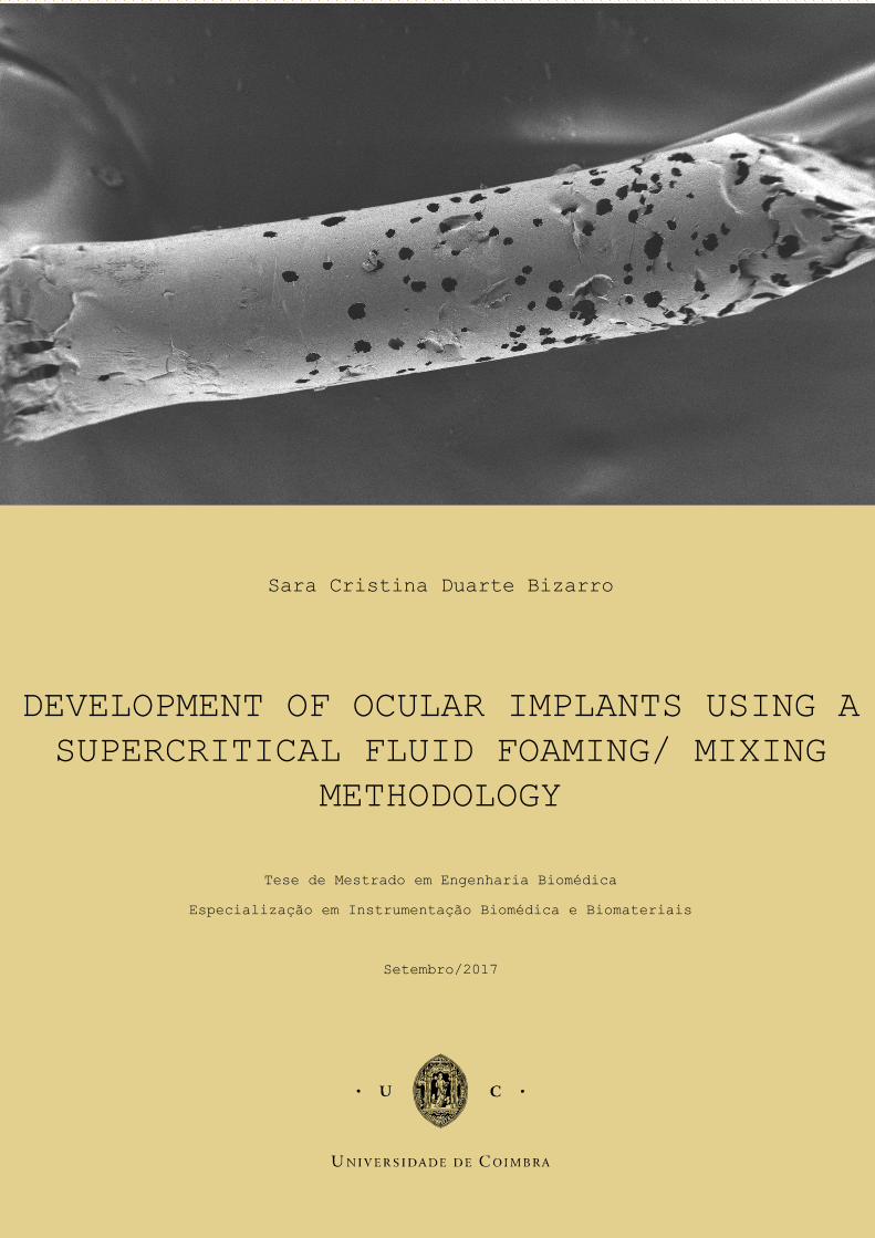
Imagem
Falta-me colocar Imagem!
Imagem
Sara Cristina Duarte Bizarro
DEVELOPMENT OF OCULAR IMPLANTS USING A
SUPERCRITICAL FLUID FOAMING/ MIXING
METHODOLOGY
Tese de Mestrado em Engenharia Biomédica
Especialização em Instrumentação Biomédica e Biomateriais
Setembro/2017


Sara Cristina Duarte Bizarro
DEVELOPMENT OF OCULAR IMPLANTS
USING A SUPERCRITICAL FLUID
FOAMING/ MIXING METHODOLOGY
Dissertation submitted to Faculty of Science and Technology of University of
Coimbra for attribution of Master’s Degree in Biomedical Engineering
Supervisors:
Prof. Dr. Hermínio José Cipriano de Sousa
Prof. Dr. Mara Elga Medeiros Braga
Coimbra
2017


Esta cópia da tese é fornecida na condição de que quem a consulta reconhece que os direitos de
autor são pertença do autor da tese e que nenhuma citação ou informação obtida a partir dela
pode ser publicada sem a referência apropriada.
This copy of the thesis has been supplied on condition that anyone who consults it is
understood to recognize that its copyright rests with its author and that no quotation from the
thesis and no information derived from it may be published without proper acknowledgement.


i
To Duarte for all the playing time we missed…
To Camila for all the basketball games I didn’t attend to…
To Bruno for all the travelling we had to hold over…

ii

iii
Acknowledgements
First of all, think you Lord… without faith I could never have done it!
It has been a long way, not always in a friendly road, but, as in life, the path became easier to
go through with the presence and support of those who, in many ways, came along with me. I
am grateful to all of them.
To Professor Mara Braga for not giving up on me, for the support, knowledge, understanding,
patience and dedication ... there is so much to thank that I miss words.
To Professor Hermínio for guidelines, patience and interest in this work.
To Professor Miguel Morgado for wise advice, insight, and for not hinder me from flying when
I needed the most to try to stretch my wings.
Especially to my parents and my brother for the confidence and serenity.
To my nephews, Camila e Duarte, for making me laugh.
To Celeste for being my support in the hospital… your caring, genuine concern and affection
were much important to me than you ever know.
The most beloved thank you to Bruno for pushing me out of bed in the days I was so tired that
I could not get myself up alone… in your love I rested and recovered strength.
From the laboratory, to Dra Ana Dias, António Rosa, Rita Chim, Patrícia Amado, Sónia
Mendes, Inês Almeida, Rui Pinho, Rui Churro, Bárbara Valente, Daniela Martins, João
Gouveia and Paulo Dias for sharing knowledge and helping me dealing with my insecurities.
To Dra Raquel Santiago, Raquel Bóia and Maria from AIBILI for the interest and support in
this project.

iv

v
Table of Contents
Acknowledgements ................................................................................................................... iii
Abstract ..................................................................................................................................... vii
Resumo ...................................................................................................................................... ix
List of Abbreviations and Nomenclature................................................................................... xi
List of Figures .......................................................................................................................... xiii
List of Tables ........................................................................................................................... xiv
List of Equations ...................................................................................................................... xiv
List of Supplementary Data ..................................................................................................... xiv
Goals and Motivation ............................................................................................................. xvii
1. Introduction ............................................................................................................................ 2
1.1 Intraocular Drug Delivery Systems ...................................................................................... 2
1.2 Implants Manufacturing Techniques .................................................................................. 11
2. Materials and Methods ......................................................................................................... 16
2.1 Chemicals ........................................................................................................................... 16
2.2. Methods ............................................................................................................................. 16
3. Results and Discussion ......................................................................................................... 22
3.1 Morphological characterization .......................................................................................... 22
3.2. Thermal Analysis of Samples: DSC .................................................................................. 32
3.3. Dexamethasone Implants: quantification and release ....................................................... 34
3.4. 2-Cl-IB-MECA Implants: quantification and release ........................................................ 35
3.5. 2-Cl-IB-MECA degradation analysis ................................................................................ 44
4. Conclusion and future remarks ............................................................................................. 48
REFERENCES ......................................................................................................................... 50
Supplementary Data ................................................................................................................. 62

vi
vii
Abstract
Despite advances in ophthalmological pharmacology, the treatment of eye diseases have
limitations since conventional methods of drug delivery are not completely effective. A possible
solution for the maintenance of therapeutic levels within the eye includes the controlled release
systems such as implants, capsules, liposomes and iontophoresis.
Supercritical carbon dioxide (scCO2) foaming/mixing methodology (SFM) is an alternative
processing method that has unique advantages over standard techniques, including porosity
control, absence of organic solvents and reduction of the melting (Tm) and glass transition
temperatures (Tg) which is an important condition in what concern the incorporation of
thermally and chemically sensitive drugs. In this type of processing, the polymer phase melts
due to the dissolution of the fluid phase, and pores are formed upon fluid release.
Thus, the main propose of this work was the development of poly (ε-caprolactone) (PCL) ocular
implants loaded with drugs of ophthalmological interest (dexamethasone (DXMT), used to
prove the concept and due to its lower price and frequent use in the treatment of
ophthalmological disorders, and 2-Chloro-N6-(3-iodobenzyl)-adenosine-5'-N-
methyluronamide (2-Cl-IB-MECA), a promising drug for future in vivo application) using SFM
process. To achieve viable implant dimensions and to evaluate the effect of material porosity
in drug release were also defined objectives. Pure PCL, PCL with glycofurol and PCL with
glycofurol and the selected drugs were processed by scCO2 at 45 ° C, 200 bar, for two hours.
Depressurization rates tested were 10, 20 and 30 bar/min. Produced implants have similar
dimensions to ophthalmologic implants currently marketed (length 2 mm and diameter ≤ 0.464
mm).
Glycofurol was shown to be important to ensure compatibilization of the polymer/drug mixture
because, when used, there was greater drug incorporation. The ideal percentage of glycofurol
determinate was 8% (w/w). SEM images show that all samples have a heterogeneous pore
distribution influenced by depressurization rate and glycofurol presence: higher
depressurization rate produces smaller pores and additive presence generates larger pores at the
same depressurization rate.
Largest pores obtained were for SFM10. There is a small difference in pore size between
SFM20 and SFM30 which suggests that, on a small scale, like the ocular implants one, this
variation in the depressurization rate may not be relevant. At same depressurization rate,
additive presence generates larger pores. DSC analysis shows that PCL thermal properties were
not significantly altered after processing.
Dexamethasone implants revealed controlled release for more than 500 hours.

viii
Comparing DXMT implants produced by hot melting and processed with supercritical carbon
dioxide, it is observed that until 200h of release, SFM implants release is more stable and higher
than HM implants. After that time, HM appears to become stable but still lower. This is due to
higher porosity in SFM implants. Implants produced by hot melting have smaller surface area
and less porosity.
Despite a similar release trend, due to porosity effect, 2-Cl-IB-MECA SFM20-G implants have
higher drug release than 2-Cl-IB-MECA SFM30-G. 2-Cl-IB-MECA implants showed sustained
release over 100 h. Release is greater in the first 60% of release profile and faster without
additive addition due to less polymer-drug compatibilization.
The amount of drug released in the first hour was eight times higher than the required on an
intravitreal injection. Preliminary in vivo tests with PCL implants drug free didn’t reveal
adverse side effects.

ix
Resumo
Apesar dos avanços na farmacologia oftalmológica, o tratamento das patologias do olho
apresenta limitações uma vez que os métodos convencionais de administração de fármacos não
são completamente eficazes. Uma possível solução para a manutenção de níveis terapêuticos
no interior do olho inclui os sistemas de libertação controlada na forma de implantes, cápsulas,
lipossomas e iontoforese.
A metodologia de foaming/ mistura com dióxido de carbono supercrítico é um método de
processamento alternativo que tem vantagens únicas relativamente às técnicas padrão,
incluindo o controlo da porosidade, a possibilidade de ausência de solventes orgânicos e a
redução da temperatura de fusão e de transição vítrea, que é uma condição importante no que
se refere à incorporação de fármacos térmica e quimicamente sensíveis. Neste tipo de
processamento ocorre fusão da fase polimérica, devido à dissolução da fase do fluido, e
formam-se poros aquando da libertação do fluido.
Assim, o principal objectivo deste trabalho é o desenvolvimento de implantes oculares de
poly(ε-caprolactona) (PCL) carregados com fármacos de interesse terapêutico a nível
oftalmológico (a dexametasona (DXMT), utilizada para provar o conceito e devido ao seu baixo
preço e à sua utilização frequente no tratamento de patologias oftalmológicas, e 2-Cloro-N6-(3-
iodobenzil)-adenosina-5′-N-metiluronamida (2-Cl-IB-MECA), um fármaco promissor para
futura aplicação in vivo) utilizando foaming/ mistura com dióxido de carbono supercrítico.
Obter implantes de dimensões viáveis e avaliar o efeito da porosidade do material na libertação
do fármaco foram também objectivos definidos. PCL pura, PCL com glicofurol e PCL com
glicofurol e os fármacos seleccionados, foram processados por dióxido de carbono supercrítico,
a 45ºC, 200 bar, durante duas horas. As taxas de despressurização testadas foram 10, 20 and 30
bar/min. Os implantes produzidos têm dimensões semelhantes aos implantes de aplicação
oftalmológica actualmente comercializados (comprimento de 2 mm e diâmetro ≤ 0.464 mm).
O glicofurol mostrou ser importante para garantir a compatibilização da mistura polímero/
fármaco porque aquando da sua utilização houve maior incorporação de fármaco. A
percentagem ideal de glicofurol a utilizar foi de 8% (m/m). As imagens de SEM mostram que
todas as amostras apresentam uma distribuição de poros heterogénea influenciada pela taxa de
despressurização e presença de glicofurol: maior taxa de despressurização gera poros mais
pequenos e a presença de aditivo gera poros maiores para uma mesma taxa de despressurização.
Os maiores poros obtidos foram para SFM10. Existe pouca diferença no tamanho de poro entre
SFM20 e SFM30 o que sugere que, numa escala pequena, esta variação da taxa de

x
despressurização poderá não ser relevante. A presença de aditivo para uma mesma taxa de
despressurização gera poros de maiores dimensões. A análise de DSC mostra que as
propriedades térmicas da PCL não foram significativamente alteradas depois do processamento.
Os implantes de Dexametasona revelaram libertação controlada durante mais de 500h.
Comparando os implantes de DXMT produzidos por hot melting e processados com dióxido de
carbono supercrítico, constata-se que até às 200h de libertação, esta é mais estável e elevada no
caso do foaming do que no caso do hot melting. Depois deste tempo a libertação dos implantes
de HM estabiliza mas continua mais baixa. Isto fica a dever-se a uma porosidade superior nos
implantes processados por tecnologia supercrítica. Os implantes produzidos por HM têm uma
área de superfície e porosidade menores.
Apesar de uma tendência de libertação semelhante, nos implantes de 2-Cl-IB-MECA com uma
taxa de despressurização de 20 bar/min a libertação de fármaco é maior do que nos implantes
com uma taxa de despressurização de 30 bar/min devido ao efeito da porosidade. Os implantes
de 2-Cl-IB-MECA apresentaram libertação sustentada mais de 100 h. A libertação foi muito
superior nos primeiros 60% do perfil de libertação e foi mais rápida na ausência de aditivo
devido a uma menor compatibilização entre o polímero e o fármaco.
A quantidade de fármaco libertada foi, na primeira hora de libertação, oito vezes superior ao
requerido numa injecção intra-vítrea. Os testes preliminares in vivo com implantes de PCL sem
fármaco não revelaram efeitos secundários adversos.

xi
List of Abbreviations and Nomenclature
AIBILI – Association for Innovation and Biomedical Research on Light
ARMD – Age-related macular degeneration
CAS – Chemical Abstracts Service
CMV - Cytomegalovirus
CO2 – Carbon Dioxide
DDS – Drug delivery systems
DME – Diabetic Macular Edema
DSC – Differential scanning calorimetry
DXMT - Dexamethasone
FDA – Food and drug administration
HPLC – High performance liquid chromatography
HM – Implant produced by hot-melting
HM-G – Implant produced by hot-melting using Glycofurol as additive
IOP – Intraocular pressure
I.V. – Intravenous
min – Minute
mL – Milliliter
mm – Millimeter
Mn – Number average molecular weight
PCL – Poly(ε-caprolactone)
PLA – Poly(lactic acid)
PLGA – Poly(lactic-co-glycolic acid)
PVA – Poly(vinyl alcohol)
scCO 2 – Supercritical carbon dioxide
SCF – Supercritical fluids
SEM – Scanning electron microscopy
SFM – Supercritical CO2 foaming/ mixing process
SFM10 – Implant processed by Supercritical CO2 foaming/ mixing processed with 10 bar/min
depressurization rate

xii
SFM20 – Implant processed by Supercritical CO2 foaming/ mixing processed with 20 bar/min
depressurization rate
SFM20-G – Implant processed by Supercritical CO2 foaming/ mixing, using Glycofurol as
additive, processed with 20 bar/min depressurization rate
SFM30 – Implant processed by Supercritical CO2 foaming/ mixing process with 30 bar/min
depressurization rate
SFM30-G – Implant processed by Supercritical CO2 foaming/ mixing, using Glycofurol as
additive, processed with 30 bar/min depressurization rate
Tg – Glass transition temperature
Td – Degradation temperature
TGA – thermogravimetric analysis
Tm – Melting temperature
UCST / LCST – Upper critical solution temperature/lower critical solution temperature
USA – United States of America
2-Cl-IB-MECA – 2-Chloro-N6-(3-iodobenzyl)-adenosine-5′-N-methyluronamide
µm – micrometer

xiii
List of Figures
Figure 1 - Basic Eye Anatomy (www.eyesightresearch.org) ..................................................... 2
Figure 2 - Evolution of Implants Size Reduction during investigation. ................................... 22
Figure 3 - SEM of implants. Magnification 118x. Scale bar 100 µm. ..................................... 23
Figure 4 - Optical Microscope images of 22% (w/w) glycofurol in PCL implant: catheter
walls that could not be removed in its entirety and large pores of the implant are visible. ..... 24
Figure 5 - Optical Microscope image of 8% (w/w) glycofurol in PCL implant: initial tests.
Implants were totally removed from catheter tube and have porosity. .................................... 25
Figure 6 - SEM: implants prepared by scCO2 foaming process for 2h, 20 MPa, 45ᵒC.
Depressurization rates were variable. At depressurization rate of 20 bar/min, samples with and
without Glycofurol were analyzed. .......................................................................................... 26
Figure 7 - SEM: implant prepared by hot melting.................................................................... 27
Figure 8 - Implants surface area and pore size in different processing conditions. ................. 28
Figure 9 - Implants porosity under different processing conditions. ........................................ 28
Figure 10 - Samples thermal analysis: DSC thermograms. ...................................................... 33
Figure 11 - Total Dexamethasone implants release comparing SFM with HG and additive. .. 34
Figure 12 - Dexamethasone release profiles in water. .................................................................
Figure 13 - 2-Cl-IB-MECA implants total release. .................................................................. 37
Figure 14 - Glycofurol effect on 2-Cl-IB-MECA implants profile release. ............................. 37
Figure 15 - Profile release of 2-Cl-IB-MECA implants with different depressurization rates.
................................................................................................................................................ 388
Figure 16 - After release SEM images of 2-Cl-IB-MECA implants. ....................................... 41
Figure 17 - 2-Cl-IB-MECA implants SEM images before and after release with body and
surface details. .......................................................................................................................... 42
Figure 18 - 2-Cl-IB-MECA degradation analysis in different medium………………………44
Figure 19 - Macroscopic view of the rat’s eyes after 7 days implantation on the left eye. RE –
Right eye; LE – Left eye. (Pictures from Raquel Bóia – AIBILI). .......................................... 46
Figure 20 - Left eye OCT 7 days after implantation (Pictures from Raquel Bóia – AIBILI). . 46

xiv
List of Tables
Table 1 - Poly (ε-caprolactone) Chemical and Mechanical Properties ...................................... 6
Table 2 - Common ocular implants commercialized.................................................................. 9
Table 3 - Experimental conditions............................................................................................ 18
Table 4 - Commercial and produced implants dimensions. ..................................................... 24
Table 5 – Morphological and Thermal characterization. ......................................................... 30
Table 6 - Diffusion Coefficients for 2-Cl-IB-MECA. .............................................................. 39
List of Equations
(1) ............................................................................................................................................. 19
(2) ............................................................................................................................................. 21
(3) and (4) ................................................................................................................................. 21
List of Supplementary Data
Appendix A – Catheters Filling Process
Figure A1 – I.V. Optiva® catheters from Smiths Medical.
Figure A2 – Catheter completion.
Appendix B – Supercritical Solvent Unit
Figure B1 – Schematic diagram of the supercritical solvent unit.
Appendix C – Dexamethasone Standard Curves
Figure C1 - Dexamethasone standard curve, in Methanol, used to determine the total amount of
drug in the implants.
Figure C2 - Dexamethasone standard curve, in Milli-Q water, used to determine implants drug
release.

xv
Appendix D – 2-Cl-IB-MECA Standard Curves
Figure D1 – 2-Cl-IB-MECA standard curve, in Methanol, used to determine the total amount
of drug in the implants.
Figure D2 – 2-Cl-IB-MECA standard curve, in Milli-Q water, used to determine implants drug
release.
Appendix E – Comparison of detection peaks, in HPLC, of 2-Cl-IB-MECA, saline
solution, cell culture medium and explants culture medium.

xvi

xvii
Goals and Motivation
Ophthalmic disorders are majorly caused by eye posterior segments diseases and they are a
limiting factor in life’s quality. Topical ocular drugs are insufficient to achieve therapeutic
levels and multiple intravitreal injections have several risks associated. Such a premise is
enough to stimulate and assign the development of ocular implants that provide a controlled
drug delivery in eye tissues. Controlled drug delivery systems allow the inclusion of a
therapeutic substance in the body, on its target release site and with a desirable release rate.
These features make these systems safer and more effective than traditional approaches.
The synthesis of such implants can be performed based on polymers with proven
biocompatibility that may be or not biodegradable. Melting, extrusion and hot molding are
traditional methods of implants processing that have limitations regarding the incorporation of
heat sensitive drugs and the need of solvents addition. In many cases, the use of such solvents
requires intermediate processing steps to guarantee their removal or neutralization in order to
avoid potential toxicity. Non-biodegradable implants involve multiple invasive techniques that
have several risks associated.
The main goal of this work is the development of PCL ocular implants for drug delivery using
a supercritical carbon dioxide (scCO2) assisted fluid foaming/mixing methodology. PCL is a
polymer biodegradable and biocompatible, known for its slow degradation rate. Supercritical
carbon dioxide is an advantageous solvent for processing polymers with desirable shape and
porosity. Porosity is especially important when we consider drug release systems.
SFM does not require the use of solvents – except the CO2 that volatilize out of the matrix – so
it may assume as a safe alternative.
Besides the mentioned main goal, this work also does a morphologic characterization and
thermal analysis of samples. Drug release assays were performed to evaluate release amount
and kinetics.
The results obtained so far in other biomedical applications, such as PCL ibuprofen
impregnation and preparation of imprinted contact-lenses for drug delivery, demonstrated the
feasibility of using scCO2 methods.
Potential targeted diseases treatable with these implants include uveitis, cytomegalovirus
retinitis, AMD and macular edema.

1

2
1. Introduction
1.1 Intraocular Drug Delivery Systems
Vision is a primary sense. Nutheti et al. and Aspinall et al. studies proved that visual impairment
is associated with a significant decrease in quality of life among population.
The eye is typically the representation of sight (Figure 1).
Figure 1 - Basic Eye Anatomy (www.eyesightresearch.org)
The treatment of eyes sicknesses back to Cleopatra times when atropine eye drops (known as
Belladonna) was used as a mydriatic. In the late 1800s, in Europe, United Kingdom, gelatin
cocaine inserts were the first polymeric system of continuous drug release; they were used for
local anesthesia. In the 1900s, several topical ocular delivery methods were developed: soluble
ophthalmic drug inserts (Amo and Urtti, 2008), liquid state delivery systems (Shedden et al.,
2001), microspheres (Yarangumeli and Kural, 2004), drug immersed hydrophilic contact lenses
and topical ocular liposomes (Amo and Urtti, 2008). None of those approaches could treat
totally capably posterior ocular tissues.
Despite drug delivery to the anterior portions of the eye is well established in the literature, in
what concern the posterior segment it remains a challenge due to its particular anatomy and
physiology. The obstacles for the adequate drug delivery are static barriers, such as different
layers of cornea, sclera and retina (blood aqueous and blood-retinal barriers) and dynamic
barriers as conjunctival and choroidal, blood flow, lymphatic clearance, and tear dilution
(Hughes et al., 2005; Gaudana et al., 2010).

3
The most frequent diseases that affect the posterior segment are age-related macular
degeneration (ARMD), diabetic macular edema (DME), endophthalmitis and proliferative
vitreoretinopathy. All this pathologies can lead to vision impairment and blindness (Hughes et
al., 2005).
In recent years there has been a marked development in scientific understanding of the
mechanisms associated with these pathologies and new treatment strategies. Nevertheless, the
success of new therapeutic approaches is dependent upon strategies which allow the drug
reaching the intended site of action. While topical treatments can achieve drug therapeutic
levels in the anterior segment, the natural barriers of the eye referred previously, make then
must less effectively to the posterior segment. This is easily confirmed if we consider that in
the case of topical instillation of ophthalmic drops – which is the most usual therapeutic method
in ocular diseases – less than 5% of the applied dose reaches the aqueous humor (Gaudana et
al., 2010).
Due to these limitations, the treatment of eye posterior segment diseases is done by
subconjunctival injections, systemic administration of drugs or intravitreal injections. All these
treatment strategies have limitations.
Subconjunctival injection is less invasive than intravitreal injection but the main problem is the
lack of control in the drug concentration that reaches the vitreous (Yasukawa et al., 2001;
Hughes et al., 2005; Amo and Urtti, 2008). Several authors refer that low bioavailability is
determinate by rapid drug elimination into systemic circulation, following subconjunctival
administration (Weijtens et al., 1999; Hosseini et al., 2008; Kim et al., 2008).
In systemic administration, parenteral and oral administration may be considered. Parenteral
route is conditioned by the blood-retinal barrier which regulates strictly the entry of therapeutic
agents from blood circulation into the retina. Hydrophobic drugs cross poorly the blood-retinal
barrier which is a significant aspect in what concern drug delivery systems. As only a small
amount of medications overcomes this barrier, several systemic doses are required to maintain
the concentration of drugs in effective therapeutic levels (Gaudana et al., 2010; Haghjou et al.,
2011).
Therapeutic efficacy is also a problem in oral administration because there is, as well, limited
accessibility to ocular tissues; it requires high dosage to observe therapeutic efficacy. After
gastrointestinal absorption, molecules in systemic circulation must also cross the blood-aqueous
and blood-retinal barriers (Gaudana et al., 2010).
In both cases – parenteral and oral administration – high drug among can lead to systemic side
effects and toxicity to the patient (Yasukawa et al., 2001; Hughes et al., 2005; Amo and Urtti,
2008).

4
Direct intraocular drug injection into the vitreous cavity provides efficient drug delivery for
several drugs, including low molecular mass drugs and macromolecules, achieving higher
concentrations in the retina and vitreous. This route of administration reduces systemic side-
effects because it is not carried in the bloodstream, acting in loco. The therapeutic effect of that
drugs depends on the retention time of the injected drug at the administration site. However,
the half-life of drugs in the vitreous is relatively short which request repeated injections to
maintain drug concentrations in operative therapeutic levels. To keep drugs in therapeutic
range, repeated injections are requested at regular intervals. Repeated injections may cause
discomfort to the patient and lead to therapy discontinuation; technically, the risk of vitreous
hemorrhage, infection, globe perforation, orbital fibrosis, ptosis, cataract and retinal detachment
are increased by repeating the procedure (Ogura et al., 2001; Yasukawa et al., 2001; Choonara
et al., 2007; Haghjou et al., 2011).
Intraocular implants are a good alternative and possible solution to the problem because they
can be controlled release systems, prepared from different biocompatible polymers,
biodegradable or non-biodegradable, that are placed after the blood-retinal barrier, releasing the
drug directly to the site of action in a controlled manner and for a long time. This mechanism,
especially important in chronic eye diseases, reduces the possibility of adverse effects that are
often associated with administration of drugs by systemic route (Yasukawa et al., 2005;
Bourges et al., 2006; Manickavasagam and Oyewumi, 2013), reduces the need of multiple
injections for in situ application, allow drug release at therapeutic levels and decrease the
amount of drug required for the treatment (Amo and Urtti, 2008; Cunha et al., 2009;
Manickavasagam and Oyewumi, 2013).
Intraocular insertion of implants must be performed by surgical procedure and, although it is
invasive, the several implants advantages cited above outweigh the inconvenience caused by
the surgical procedure.
Intraocular implants may be non-biodegradable or biodegradable according to the different kind
of biocompatible polymer used in its formulation (Dash and Cudworth II, 1998;
Manickavasagam and Oyewumi, 2013).
Non-biodegradable intraocular implants can be monolithic systems (matrix) or reservoirs. In
the matrix system, the drug is dispersed homogeneously in the polymeric matrix or absorbed
on the surface. The slow diffusion of drug through the polymer matrix provides a controlled
release. Otherwise, in the reservoir system the drug is surrounded by a non-degradable
permeable membrane. In this system, water diffuses through the membrane by dissolving the
drug and creating a saturated solution within the reservoir. Since saturation occurs, the drug
diffuses outwards into a release rate – based on Fick’s Law – conditioned by polymer coating

5
thickness, implant shape, diffusibility of the drug from the polymer coating and release area
(Yasukawa et al., 2005; Bourges et al., 2006).
Several non-biodegradable polymers have been used in implants with a long-term controlled
release of drugs. Silicones, poly(vinyl alcohol) (PVA) and poly(ethylene-co-vinyl acetate)
(EVA) are the polymers commonly used to produce this implants. Silicones and PVA have
hydrophobic character thus are permeable to various lipophilic drugs; EVA is used to cover
around the reservoir to decrease drug diffusion rate because it is impermeable to most
medications (Okabe et al., 2003; Yasukawa et al., 2005; Bourges et al., 2006; Manickavasagam
and Oyewumi, 2013).
The non-biodegradable polymeric intraocular implants allow controlling the release of the drug
in a predictable kinetics for long periods of time and they are less likely to produce burst drug
release when compared with biodegradable implants. They have the disadvantage of need to be
surgically removed from the eye after the complete release of the drug, which poses a risk to
the patient. Otherwise, prolonged intraocular location could potentially trigger immunity
responses (Bourges et al., 2006; Manickavasagam and Oyewumi, 2013).
Therefore, natural and synthetic biodegradable polymers have been widely investigated for the
development of intraocular implantation devices (Jain, 2000; Amo and Urtti, 2008). Bovine and
human albumin, collagen, gelatin and hemoglobin are types of natural protein-based polymers.
However, their utilization is limited due to the high cost and questionable purity. Polymers
often used are polyamides, polyaminoacids, polyesters, polyurethanes, polyacrylamides,
poly(glycolic acid) (PGA) and poly (D, L-lactic-glycolic acid) (PLGA) (Jain, 2000;
Manickavasagam and Oyewumi, 2013). These polymers are biocompatible, well tolerated,
consider safe for clinical use and allow the possibility of modifying their time degradation. For
instance, the PLGA degradation rate is predisposed by lactide and glycolide monomers ratio;
with more glycolide units, degradation will be faster. Extent of crystallization and polymer
molecular weight are other factors that affect drug release mechanism.
One of the most promising polymers in biomedical applications is PCL (Table 1).
Poly (ε-caprolactone) (PLC) is a hydrophobic and semi-crystalline aliphatic polyester
synthesized from e-caprolactone monomers polymerization.

6
Table 1 - Poly (ε-caprolactone) Chemical and Mechanical Properties
Melting Temperature (Tm) ≈ 60ºC (Jenkins et al., 2006; Bassi et al., 2011
using Differential Scanning Calorimetry)
Glass Transition Temperature (Tg) -60ºC (Jenkins et al., 2006; Bassi et al., 2011
using Differential Scanning Calorimetry)
Crystallization Temperature (Tc) ≈ 27ºC (Bassi et al., 2011 using Differential
Scanning Calorimetry)
Thermal Stability (Td) ≈ 350ºC (Jenkins et al., 2006 using
Differential Scanning Calorimetry)
Compressive Strengh 14MPa (Bassi et al., 2011 using the Instron
1122 mechanical tester)
Young’s Modulus 0.9 GPa (Bassi et al., 2011 using the Instron
1122 mechanical tester)
As PCL can be degraded by hydrolytic mechanisms in a physiological environment, it is
consider to be biodegradable and that is very important in what concern biomedical applications
(Jenkins, 2006; Cunha et al., 2009; Bernards et al., 2013). The hydrolytic degradation of PCL
is due to the presence of ester bonds. The hydrolysis of the PCL aliphatic ester group does not
generate toxic degradation products (Fernandez and Richa, 2011). The degradation rate of this
polymer is approximately 2 to 3 years which is considerably slow and consequently attractive
for biomedical long-term applications (Boruges et al., 2006; Nair and Laurencin, 2007; Cunha
et al., 2009; Bernards et al., 2013).
The poly-α-ester degradation occurs by random hydrolytic cleavage and enzymatic
fragmentation. The random hydrolytic cleavage it’s accelerated by carbonyl ends of polymeric
chains and starts in amorphous areas. The fragments resulted of bulk fragmentation are taken
up by macrophages and degraded inside the cells. When the remains are small enough to diffuse
through matrix, mass loss occurs (Dash and Cudworth II, 1998; Bernards et al., 2013).
The enzymatic fragmentation is based on the fact that although enzymes are programmed for
highly specific interactions with particular biological substrates, some of them are able to
recognize "non-natural" such as polymers like PCL (Labow et al., 2002). Several studies, such
as Aktusu et al., (1998), Labow et al., (2002), Dash et al., (2011), proved the importance of
enzyme hydrophobic domain to polymer surface adsorption and catalytic domain for the
hydrolysis of the ester bond.
Multiple biochemical and cellular parameters can be directly involved in biodegradation
because the in vivo environment complexity allows activation of other degradation mechanisms
furthermore to hydrolytic, enzymatic and oxidative.
The slow degradation, high permeability to hydrophobic drugs and high biocompatibility are
characteristics that make PCL and excellence polymer for the development of controlled eye
drug delivery systems (Dong et al., 2006; Fialho et al. 2003; Cunha et al., 2009) and Cunha et

7
al., 2009 and Bernards et al., 2013 studies confirmed the feasibility of PCL usage in the
production of intraocular devices.
Implants made from biodegradable polymers can also be monolithic (matrix) or reservoirs as
mentioned in the case of non-biodegradable polymers. In the matrix system, the polymer
degrades slowly in physiological conditions and the drug is released as the degradation occurs.
The drug can also be released by diffusion through the pores of the matrix. In the reservoir
system, and depending on the polymer, the membrane degrades in a slower rate than the drug
diffusion (Dash and Cudworth II, 1998; Fialho et al., 2003). Polyanhydrides degrades faster by
surface erosion than PLA and PLGA and this can lead to excess drug release (burst).
Discontinuity of the matrix is also associated to irregular drug release and final burst release.
In literature there are reported alterations that leaded to maintaining controlled polymer erosion
and drug release (Yasukawa et al., 2005; Kunou et al., 2000). Balancing polymer mass loss and
drug release is challenging for most biodegradable systems.
Ophthalmic implants have been commercialized for more than 20 years.
Vitrasert® (Bausch &Lomb, USA), the first polymeric (EVA and PVA) ganciclovir non-
biodegradable implant, was approved by FDA in 1996. It was used on the treatment of
cytomegalovirus retinitis, releasing the 4.5 mg of ganciclovir for 4 to 5 months. Complications
associated with prolonged use of the implant, such as vitreous hemorrhages, led to withdrawal
from the European market in 2002.
Retiser® (PVA and silicone) and Medidur® (Polyimine, PVA and silicone) are fluocinolone
acetonide non-biodegradable implants commercialized used for uveitis treatment. For
Retisert®, drug release is for approximately 30 months. The development of cataracts, the need
for surgery in 100% of cases and the increase in intraocular pressure were some of the adverse
effects resulting from the use of this implant (Jaffe et al., 2006). Medidur® has the advantage
of could be intravitreally inserted as an injection, instead of a surgery due its dimensions (3mm
long and 0.37 mm of diameter) (Amo and Urtti, 2008).
Ozurdex® is a PLGA and dexamethasone biodegradable implant used on the treatment of
macular edema. Drug release occurs for about 6 months and it is refered to be well tolerated by
patients (Kuppermann et al., 2007).
Surodex® is other of dexamethasone biodegradable implants. Structurally it is a 60 µg
dexamethasone pellet coated in PLGA that provides sustained release for 7-10 days after
insertion into eye anterior chamber. Its primarily pretended application is the treatment of
postcataract surgery inflammation. The use of this implant has no reported adverse events and
it showed to be safe and effective (Tan et al., 1999).

8
Implants further promoted and used in ophthalmic applications are specified in Table 2. All of
them have limitations as drug loading capacity, drug release profiles, degradation time,
biodegradability and solvents request in their manufacture.

9
Table 2 - Common ocular implants commercialized.
Registered
Name Manufacter
Active
Substance
Therapeutic
Application Dosage
Polymeric
Matrix Biodegradability Implant size
Vitrasert® Bausch &
Lomb Ganciclovir CMV retinitis 4.5 mg
EVA and
PVA
Nonbiodegradable
2.5 mm diameter by 1
mm thick tablet
Retiser® Bausch &
Lomb
Fluocinolone
acetonide
Uveitis 0.59 mg PVA and
silicone
Medidur®
(Iluvien)
Alimera
Sciences Uveitis 0.19 mg
Polyimine,
PVA and
silicone 3.5 0.37 mm
Surodex® Ocular
Pharmaceuticals
Dexamethasone
Postoperative
inflammation 60 µg
PLGA Biodegradable
1.0×0.5 mm Not available
Ozurdex® Allergan Macular
Edema 700 µg 60.46 mm

10
Beyond solid implants, there are micro and nano particles that can be colloidal carriers. The
main goal of this approach is the development of injectable formulations long term actives and
with selective actuation in specific tissues and cells (Choonara et al., 2007).
These systems have the advantage of the administration to be made by injections, as present
themselves as colloidal solutions, which avoids the potential complications of surgical
processes associated with implants application (Behar-Cohen, 2002). This colloidal may be
prepared based on synthetic polymers such as PLA, PLGA and PCL. The micro and nano
particles are polymeric structures classified by size: the micro particles have diameter over 1
µm and the nano particles below 1 µm. As the implants, these systems are also classified
structurally in monolithic (matrix) or reservoirs (Kimura and Ogura, 2001; Fialho et al., 2003;
Choonara et al., 2007). Several studies have been performed with microspheres for release of
drugs such as ganciclovir, retinoic acid and fluorescein. The release time obtained as about two
to eight weeks. Low amount of encapsulated drug, the need of repeated injections in short
period of time and the visual limitations caused by the presence of suspended particles in
vitreous cavity are reported limitations of this methodology (Fialho et al., 2003).
Liposomes are biocompatible and biodegradable layer, which surround an aqueous medium,
and can be also used in colloidal systems for intraocular applications. They may be positively,
neutral or negatively charged depending on its composition. Their size is between 0.025 µm
and 2.5 µm. According to the size and number of layers, the liposomes can be classified as
multilamellar vesicles, large unilamellar or small unilamellar. Drugs may be incorporated into
phospholipid layers or the aqueous solution which is an advantage because it allows the
simultaneous transport of hydrophilic and hydrophobic agents. The distribution of the drug is
controlled by the size of the vesicles and lipid bilayers of the liposomes. The limitations of this
approach are related to complex preparation methods, difficult due to low storage stability and
induction of constraints in vision due to its suspension in the vitreous cavity (Behar-Cohen,
2002; Fialho et al., 2003; Choonara et al., 2007).
Iontophoresis is another possibility under investigation as drug transport system in the eye. In
this process, ions are conducted in a tissue by a low intensity electrical current that modifies
cells permeability and facilitates drugs penetration. The transport depends on an electromotive
force that repels ions of an electrode of the same charge and makes them migrate to the
oppositely charged. It is a non-invasive procedure and can be repeated several times with few
adverse effects. However, drug transports by iontophoresis disadvantages are the risk of shocks
and burns caused by contact of the electrodes with the eye, overdose and damage of the
application site. Iontophoresis can enhance the intraocular administration and reach the target

11
tissues but as this technique is not a sustained-release system it is necessary to repeat the
treatment several times (Fialho et al., 2003).
Therefore, intravitreal route minimizes the occurrence of adverse systemic effects and offers
advantages as the drug is directly introduced into the vitreous. However, drug distribution in
the vitreous is heterogeneous and the blood flow in the choroid and retina promotes the half-
life reduction of the drug which causes a reduction of its concentration to sub therapeutic levels
in a short period of time. On the other hand, the increase of drug dose could prolong the
effectiveness of single injections but could also induce peak drug concentration within the eye,
exposing the patient to undesirable toxic effects.
To overcome the abovementioned limitations, several sustained-release drug delivery devices
are being developed in order to obtain a drug therapeutic level inside the eye with the smallest
possible complications associated. That will be discussed following.
1.2 Implants Manufacturing Techniques
Some of the techniques most used in the preparation of biodegradable intraocular implants
polymer based are films, molding and extrusion. The technique and technical parameters to be
used are conditioned by the type of polymer to be processed, the drug to be loaded and the
properties of the final drug/polymer mixture (Rothen-Weinhold et al., 1999; Kimura and Ogura,
2001; Breitenbach, 2002; Choonara et al., 2007).
Films preparation may be performed in two ways: adding the solution, with the solubilized
components, in a suitable solvent that is then evaporated or melting and pressure the polymer
and drug. The dry film is then removed from the surface. To dissolve the polymer, solvent-
casting using organic solvents might be involved and this could be a problem in biomolecules
loading (Kimura and Ogura, 2001; Choonara et al., 2007).
In the molding technique the polymer and drug are heated and compressed in molds of the
desired configuration. In extrusion, the polymer and drug are continuously propelled under
pressure through high temperature areas, causing melting and compacting the powder mixture
into the shape of the implant (Rothen-Weinhold et al., 1999).
Melt extrusion has an important role on pharmaceutical manufacturing granules, pellets, tablets,
stents, implants, suppositories and ophthalmic inserts. It is considered to be an efficient

12
technique with advantages over solvent procedures like co-precipitation however the influence
of heat stress and shear forces on the drug active are disadvantages related (Breitenbach, 2002).
These two processes, molding and extrusion, are temperature aided and, for that reason, might
be inadequate for drugs that are thermolabile. (Kimura and Ogura, 2001; Choonara et al., 2007).
The biological degradation kinetics of intraocular systems is affected by chemical nature
composition and molecular weight of the polymer, morphology and structure of the device and
thermal processes influence. Polymer processing techniques regulate the structure of the device
and impact its morphology, specifically its microporous structure, polymeric chain orientation
and crystallinity. Rothen-Weinhold et al. analyzed extrusion and injection-molding in the
preparation of biodegradable implants and proved that molecular weight and polydispersity
decreased after extrusion or injection-molding and this decrease was higher in injection-
molding. The crystallinity analysis verified that the crystalline structure was not destroyed in
both manufacturing approaches and, in what concern in vitro degradation, the extruded implants
degraded more rapidly than the injection-molded ones.
Fialho and Silva-Cunha studied polymeric sustained-drug release systems comparing the
impact of implant manufacturing techniques, compression and hot molding, on the in vitro
degradation of the polymeric matrices and on the release of dexamethasone acetate. Their
outcomes revealed that the manufacturing technique decidedly influences degradation and drug
release progressions. The degradation was faster in compressed systems which also allowed
one faster release of the drug.
Development of polymer-based drug controlled release systems may be performed in several
ways, however, traditional approaches have limitations such as difficulties in the incorporation
and heterogeneous distribution of the drug, use of potentially toxic solvents, drug dissolution
and photochemical and thermal degradation. The use of supercritical technology is a viable
alternative for the development of polymeric materials with loaded bioactive agents for
biomedical application because it overcomes much of these limitations.
Polymeric foams involve a solid–polymer matrix and gaseous voids derived from a blowing
agent. This materials have excellent thermal properties, flexibility to template to desired
morphologies, high strength–to–weight ratio and good energy/mass absorption (Lee et al.,
2005; Brun et al., 2011).
Due to its characteristics, there are several different applications for polymeric foams:
constrution and aerospace industry, coating, acoustic insulation controlled release systems and
scaffolding, among others (Zeng et al., 2003; Zhai et al., 2006, Bao et al., 2011).
The foaming method can be physical or chemical according to the nature of the gas formation.
Physical blowing agents are inert elements, usually liquids with low boiling points, which

13
gasify under foaming circumstances. Chemical blowing agents are substances that produce
gases due to chemical reactions, as water, sodium bicarbonate and citric acid (Ashida, 2007).
Several methods may be used to produce foams (casting and leaching, thermally induced phase
separation, extrusion with chemical blowing agent) however all of them use solvents that
contaminate the final polymeric foam. The purification of the foam is not always possible
because it often require temperature rise that may degrade thermal sensitive compounds such
as drugs (Jacobs et al., 2008).
Supercritical fluids are an alternative to traditional solvents in order to avoid this complications
(Jacobs et al., 2008; Duarte et al., 2012).
A certain substance is considered to be in a supercritical state when its temperature and pressure
are higher than its critical temperature and pressure. Supercritical fluids (SCF) have higher
density than that of a gas and higher diffusivity than that of a liquid so it may be consider that
they have intermediate properties to those of liquids and gases. Due this behavior, SCF are
useful solvents for some compounds (Knox, 2005). The solvent choice depends on several
aspects such as toxicity, cost, environmental issues and, obviously, pressures and temperatures
in the desired processing supercritical region. Supercritical carbon dioxide (scCO2) is a good
possibility for medical applications because it is non-toxic, recyclable, environmentally-
friendly, with acceptable cost, chemically inert and has reasonably low critical temperature
(304K) and pressure (7.4MPa), which allows processing of thermolabile and biologically-active
compounds (Kazarian, 2004; Woods et al., 2004; Xu et al., 2004; Jenkins et al., 2006; Tai et
al., 2007; Shieh et al., 2009; Morèrea et al., 2011).
In the scCO2 foaming process, the polymer saturation (with constant temperature and pressure)
with scCO2 lead to a homogenous mixture because the scCO2 can plasticize amorphous and
semi-crystalline polymers (Karimi et al., 2012; Liao et al., 2012; White et al., 2012). In semi-
crystalline polymers, scCO2 penetrates favorably the amorphous phase because the gas
dissolution is increased in those parts. The homogeneity occurs because the polymer glass
transition and melting temperature are reduced – caused by the interactions between polymer
and gas molecules (Gualandi et al., 2010) – so the polymer is less viscous and polymer chains
are more mobile and can rearrange themselves into an ordered configuration (Kiran, 2009; Liao
et al., 2012). This is mostly important in the case of high molecular weight polymers in which
viscosity of the bulk polymer is relatively high. The high viscosity would require huge
temperatures to processing and those high temperatures could lead to drugs thermal
degradation. The scCO2 reduce the intermolecular interactions and increase the chain separation
facilitating the process (Jenkins, 2006). The cell nucleation happens inducing a thermodynamic
instability factor in the equilibrium system. The instability may be caused by temperature

14
increase or pressure decrease. In the case of pressure decrease and in systems using CO2, as the
gas depart from the polymer, occurs nucleation of gas bubbles that lead to foams formation.
The nucleation can be homogeneous or heterogeneous, but heterogeneous nucleation is
energetically preferred since it requires a lower activation energy barrier (Jenkins et al., 2006;
Léonard et al., 2008; Tsimpliaraki et al., 2011). The number and size of pores depends on the
growth and bubble rates. With high depressurization rates, more bubbles nucleate thus the gas
available for growth is separated into more cells and foams will have more pores but with
smaller diameters. The inverse occurs with low depressurization rates; as the energy barrier for
nucleation increases, the nucleation rate decreases leading to less nucleation rate and bigger
bubbles (Jenkins, 2006; Léonard et al., 2008).
In the end, the polymeric foam results from the growth of the cells, coalescence and expansion
(Karimi et al., 2012; White et al., 2012) and its morphology and structure are determinate by
foaming conditions (temperature, pressure, saturation time and depressurization rate
(Tsimpliaraki et al., 2011; Liao et al., 2012).
Though the advantages, CO2 has difficulty to dissolve compounds with high molecular weight.
Besides that, its lack of some specific solvent-solute interaction and non-polarity could lead to
deposition yields. Adding small amounts of co-solvents improve scCO2 solvent power and
increase the solubility (Natu et al., 2008; Braga et al., 2008). Glycofurol is a greener and safer
additive that acts as porogenic agent, plasticizer and polymer compatibilizer. It is water-
miscible and frequently used on pharmaceutical formulations so it’s a good option as co-solvent
for the development of implants (Boongird et al., 2011; Allhenn and Lamprech, 2011).
The scCO2 processed materials have no residual solvent presence except when co-solvents are
used. This gains over other solvents made that scCO2 has been analyzed not only as a solvent
but also as an anti-solvent or plasticizer for polymerization, modification and extraction (Tai et
al., 2007, Kiran 2009).
This technology has proven to be effective in biomedical applications including PCL ibuprofen
impregnation (Yoganathan et al., 2010) and preparation of imprinted contact-lenses for drug
delivery (Yañez et al., 2011).

15

16
2. Materials and Methods
2.1 Chemicals
The PCL pellets (CAS [24980-41-4], Mw of 45000 g.mol-1), glycofurol (tetraglycol, CAS
[31692-85-0]), methanol (purity ≥ 99.9%, CAS [67-56-1]), acetone (purity ≥ 99.5%, CAS [67-
64-1]), acetonitrile HPLC (purity ≥ 99.9%, CAS [75-05-8]) and dexamethasone (purity ≥ 98%)
were supplied by Sigma-Aldrich.
Carbon dioxide was obtained from Praxair (purity (v/v) ≥ 99.998%).
The 2-Chloro-N6-(3-iodobenzyl)-adenosine-5′-N-methyluronamide (2-Cl-IB-MECA, Mw of
544.74 g.mol-1, CAS [163042-96-4]) was from Tocris.
Except for the PCL, all chemicals were used without other processing.
2.2. Methods
PCL preparation: from pellets to powder form
In order to allow the introduction of the PCL in the catheter used in the processing with scCO2,
the polymer was powderized and sifted. Only the particles with a diameter less than 250 µm
were used. The small diameter of the PCL power also allow an increase of the superficial area,
which favors the interaction with scCO2 molecules, and optimize the physical mixing between
the drugs and the polymer.
For powderization, 12 g of PCL were dissolved in 200 mL of acetone at environmental
temperature. This solution was precipitated adding 20 mL of methanol followed by 20 mL of
water, both dropwise, and then it was centrifuged at 5000 rpm for 10 minutes. The supernatant
was removed and the PCL powder dried at room temperature, in petri dishes, for one week.
After that, the powder was sieved to rich the desired diameter (≤ 250 µm) and stored in suitable
vials.
Implants preparation
Before the foaming process, a mixture of PCL power, glycofurol and drug – Dexamethasone or
2-Cl-IB-MECA – was made with a proportion of 8% (w/w) of glycofurol and 26% (w/w) of

17
drug. This combination was physically mixed in a glass vessel until homogenization. Some
tests were performed with pure PCL and others without glycofurol in order to compare the
glycofurol effect as compatibilizer.
Several containers with different shapes and materials were tested to reach implants with viable
dimensions and structure for biomedical application.
To achieve a realistic and sustainable ocular implant size, I.V. Optiva® catheters have been
chosen to use as a mold. They were purchased from Smiths Medical and are composed of a
needle inserted in a polyurethane tube with a maximum diameter inside of 0.464 mm.
The mixture to be processed was introduced into two thirds of the catheter polyurethane tube
and compressed with the needle therein. The tube was then removed from the remaining
catheter to be processed. This time-consuming and meticulous process was completely manual.
Detailed description of the filling procedure is in Appendix A.
Implants Processing
The filled polyurethane tubes were placed horizontally, in a supporting metallic mesh, inside
the pressure cell for foaming processing. The maximum number of simultaneously processed
tubes was 40.
Implants were set with the batch foaming technique, using scCO2 as foaming agent, in a
supercritical solvent unit (Appendix B).
The samples were placed in a high pressure cell (23 cm3) which was immersed in water with
controlled temperature (45º C; Thermoscientific, Haake AC 150).
Thereafter, CO2 was slowly introduced (nearly 0.6 bar per second) in the high pressure cell
until reaching a pressure of 200 bar (controlled with manometer – Lab DMM, REP transducer).
For two hours the system was maintained in constant conditions of temperature and pressure
under magnetic stirring (average 740 rpm) to facilitate the mixture homogenization and CO2
diffusion. After that time, the high pressure cell was depressurized at a pre-established rate of
10, 20 or 30 bar/min. All process conditions are presented in table 3.
The processed material was recovered and stored in petry dishes. The storage of dexamethasone
implants was done at room temperature and 2-Cl-IB-MECA implants in the refrigerator; both
storage containers had silica gel to ensure humidity control.
After several tests, it was concluded that the removal of the implants from polyurethane tubing
should only be done at least 24 hours after processing to ensure complete CO2 output. Prior to
that time, the implants were more adherent to the tube wall. Removal of the implants within the
tube walls was made by taking out the polyurethane structure with a scalpel (peel the implant).

18
After their removal, implants were optimized in the standard defined dimensions (2 mm length
and maximum diameter of 0.464 mm).
The operating temperature (45º) and pressure (200 bar) conditions were that defined because,
according to Leeke et al., 2006, scCO2 has the highest solubility in PCL at these operating
parameters. With high solubility, PCL swelling degree would increase drug loading.
PCL used was Mw of 45000 g.mol-1 because low molecular weight polymer disrupt after
processing (Matos et al., 2013) and high molecular weight would increase the melting point.
With the purpose of investigate porosity effect and compare methodologies of processing, some
implants were processed placing filled polyurethane catheters tubes in an oven at 62.5ºC to
ensure PCL melting.
Glycofurol presence, supercritical processing and variation of the depressurization rate
influences were tested in order to obtain proper pores, in number and size, on a resistant and
manageable implant (Table 3).
Table 3 - Experimental conditions
P (bar) T (ºC) System
stability (min)
Co-solvent
(Glycofurol)
Depressurization rate
(bar/min)
200 45
120
0 20
8% (w/w) 20
8% (w/w) 10
8% (w/w) 30
~ 1 62.5 0 -
8% (w/w) -
Implants Characterization
Implants were evaluated according to their morphology, porosity and thickness.
Samples morphology was evaluated macroscopically (using digital photographs with an
enlargement of 4x), microscopically (using Olympus BH-2 optical microscope) and with a
scanning electron microscope (Zeiss Merlin 61.50, operating at 0.1 kV and 2.0kV and 80 pA).
Implants microporosity was measured by N2 adsorption (ASAP 2000 V2.04). For each
microporosity analysis, implants from 80 processed catheters were used, for itch variation
parameter study, so that ensure reliability of results.
To calculate the surface area, Brunauer, Emmet and Teller (BET) method was used. According
to BET theory, surface area is estimate by physical adsorption of a gas on a solid surface and
by calculating the quantity of adsorbate gas matching to a monomolecular layer on the surface
(Teixeira et al., 2001). The total porosity was calculated according to equation (1).

19
𝑃 = 1 − 𝜌𝑎𝑝𝑝𝑎𝑟𝑒𝑛𝑡/𝜌𝑠𝑜𝑙𝑖𝑑 (1)
where ρapparent is the calculated as the ratio between the mass and volume of the samples and
ρsolid is the real density (Bueno et al., 2014).
In a porous material, real density relates to the volume of material not including the porous
spaces (Lowell et al., 2004). Helium picnometry (Quanta-Chrome, MPY-2) was the method
used to evaluate samples real density.
The mean pore size was determinate based on the horizontal Feret diameter of the pores and
using ImageJ software.
Implants thickness was determined with a digital micrometer (Electronic Outside Micrometer
IP 54, 0-25 mm, 0.001 mm) and it referred as an average of 5 measurements.
Drug quantification
High-Performance Liquid Chromatography (HPLC - Prominence UFLC Shimadzu coupled to
a photo diode array detector SPDM20A) was used for implants drug quantification and drug
delivery. The operation was made with appropriate schedules for Dexamethasone and 2-Cl-IB-
MECA.
In Dexamethasone analysis, the column was Eurospher 100-5C18 RP (250 × 4 mm i.d., 5 mm,
Germany) and the chromatographic conditions were based on Chim et al., 2012: mobile phase
constituted by methanol/water in a proportion of 9:1 (v/v), isocratic elution (15 min) and flow
rate of 1 mL·min−1, at 35 °C. To clean the column, runs with acetonitrile have been made.
Injected sample chromatographic profile was measured at 239 nm at least in triplicate.
The chromatographic conditions for 2-Cl-IB-MECA were defined based on Kim et al., 1996:
mobile phase with water (A) and acetonitrile (B) in a proportion of initial A=20%, increasing
to 30%, from 0 to 9 minutes, and reducing to 20%, from 9 to 12 minutes, at 35ºC. The column
used was also Eurospher 100-5C18 RP (250 × 4 mm i.d., 5 mm, Germany).
To quantify the release of Dexamethasone and 2-Cl-IB-MECA off the implants, standard curves
have been elaborated, for both drugs, in methanol and water from solutions of known
concentration (Appendix C and D). Analysis of total drug release was made using methanol as
solvent to facilitate the process since the drugs are more soluble in this solvent. Mass-known
implants were placed into glass vials with a defined volume of methanol (250 µL) and made up
HPLC readings until the drug was undetectable.
Between analyzes the samples remained stored in thermoshaker at 25° C, 100 rpm.

20
In view of forward applications, for the 2-Cl-IB-MECA, a saline solution similar to in vivo
environment, cell culture medium and explants culture medium were previously tested but the
HPLC detection peak was coincident with the drug one (Appendix E) so it was not feasible for
analysis. In drug releasing study, water was used as solvent since the vitreous humor is
essentially constituted by water. The implants were emerged in water and measurements were
made hourly during the first 6 hours and then increasing the time intervals daily. Due to PCL
hydrophobicity, some implants floated which reduced the interaction with water. This may have
affected some results. At the end of delivery process, the dry implants were weighed for
comparison of masses. All analysis were made at least in triplicate.
2-Cl-IB-MECA degradation analysis
In several tests carried out, 2-Cl-IB-MECA degradation appeared to occur. In order to confirm
this degradation and better understand this process, drug degradation analysis was carried out
using HPLC. A solution of 2-Cl-IB-MECA was exposed to thermal shocks between 25ºC and
80°C and subsequently analyzed by HPLC to detect degradation and the way it was visually
manifested on the graphs.
Modulated Differential Scanning Calorimetry (MDSC)
Implants, without glycofurol, and with different depressurization rates (SFM10 and SFM20),
implants with glycofurol addition with a depressurization rate of 20 and 30 bar/min (SFM20-G
and SFM30-G) and HM processed implants were analyzed in what concern their glass transition
temperature by MDSC (Q100, TA instruments) with nitrogen purge gas (50 mL.min-1). The
calibration process was executed with indium and measurements were made in alumina pans
equilibrated at -80ºC for 5 min, modulate ± 0.50ºC every 40s, and heating at 2ºC min-1 until
200ºC. The glass transition temperatures were determined with duplicate analysis performed to
ensure results reliability.
Diffusion coefficients
Mass transfer through porous polymeric membranes depends on several factors such as
solubility and diffusivity of the drug into the polymer, morphology and plasticization (Karimi,
2011). According to the second Fick’s law, to describe the drug release, a zero-order kinetics
equation (2) was used. In the equation, Mt represent the cumulative amount of drug released at
time t and M∞ the cumulative amount of drug released at infinite time, k is a kinetic constant
that incorporates geometric and structural characteristics of the implant and n is the release

21
exponent (Natu et al., 2008, Yañez et al., 2011). Below 60% of the amount drug released, the
drug release profiles were fitted to equation 2:
(2)
Other equations used were the following:
(3) and (4)
where l represents the thickness (~ 2 mm) of the sample and D is the diffusion coefficient,
which is presumed to be constant. Eq. (3) is only useable for the first 60% of the total release
(Mt/M∞) while Eq. (4) is valid for the last 40% of the total release (Mt/M∞) and has been
commonly used to obtain diffusion coefficients from drug release investigational data. The drug
diffusion coefficients D can be determined from the resulting slopes, after linearization and
regression analysis of these equations (Yañez et al., 2011).
In Vivo analysis
In vivo PCL implants tolerance were performed in AIBILI by Professor Raquel Santiago team.
The implants were implanted into the posterior chamber of one of rat’s eye under general
anesthesia and sterile conditions. The incision was made using a 24G needle. The other eye was
left untreated for control.
The evaluation of safety and tolerability were made by monitoring adverse events such as
inflammation, edema, cataract formation and polymer appearance and location (Myers et al.,
2016).
22
3. Results and Discussion
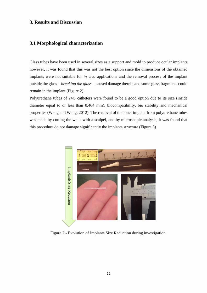
3.1 Morphological characterization
Glass tubes have been used in several sizes as a support and mold to produce ocular implants
however, it was found that this was not the best option since the dimensions of the obtained
implants were not suitable for in vivo applications and the removal process of the implant
outside the glass – breaking the glass – caused damage therein and some glass fragments could
remain in the implant (Figure 2).
Polyurethane tubes of 24G catheters were found to be a good option due to its size (inside
diameter equal to or less than 0.464 mm), biocompatibility, bio stability and mechanical
properties (Wang and Wang, 2012). The removal of the inner implant from polyurethane tubes
was made by cutting the walls with a scalpel, and by microscopic analysis, it was found that
this procedure do not damage significantly the implants structure (Figure 3).
Figure 2 - Evolution of Implants Size Reduction during investigation.

23
Figure 3 – Implants macroscopic view.
The average mass of PCL introduced into a catheter for processing was 1.16 ± 0.01 mg and the
average mass of PCL implants was 0.19 ± 0.01 mg. During processing in scCO2 unit, average
PCL mass lost was 0.068 mg ± 0.003. This mass loss is mainly caused by drag with CO2 during
the depressurizing process.
The produced implants have similar dimensions to implants currently available into market
(table 4). Implants size has a substantial role in the viability of securing implants with minimal
invasiveness.
Implants with dimensions higher than those can trigger foreign body reaction with formation
of fibrous capsule by the deposition of fibroblasts, foreign body giant cells and macrophages
around the implant. This capsule prolongs degradation rate (Kuno and Fujii, 2010;
Manickavasagam and Oyewumi, 2013).
Choonara et al. (2009) and Manickavasagam and Oyewumi (2013) refer the importance of the
development of ocular devices that are geometrically small because they are less aggressive
and well tolerated; the produced implants follow that requirements.

24
Table 4 - Commercial and produced implants dimensions.
Implant Diameter (mm) Length (mm)
Iluvien® 0.37 3.5
Ozurdex® 0.46 6
PCL implants ≤ 0.464 2
The operating conditions (temperature, depressurization rate and additive presence) were
defined based on studies already carried out in PCL processing with supercritical technology
(Yoganathan et al., 2010; Takahashi et al., 2012; Matos et al., 2013). Although there are studies
in this area, it was important to analyze the effect of the parameters on a scale as small as that
of intraocular implants.
Use of glycofurol is important to ensure homogeneity of the polymer/drug mixture and porosity.
Tests with varying amounts of glycofurol were made: 22% (w/w), 12% (w/w) and 8% (w/w).
A proportion of 22% (w/w) glycofurol is not viable because cause excessive increase of porosity
and adhesiveness of the implant to the walls of the catheter which does not allowed removal
(Figure 4). The implants pores were so large that the implant not present a tube defined
structure. Reducing the amount of glycofurol to 12% (w/w) the implant removal was still
difficult because it appears not to have a resistant structure. Desired results were achieved at
8% (w/w) glycofurol: homogenous physical mixture between PCL and the drug and porous
structured implants easily removable from catheters (Figure 5).
Figure 4 - Optical Microscope images of 22% (w/w) glycofurol in PCL implant: catheter
walls that could not be removed in its entirety and large pores of the implant are visible.

25
Figure 5 - Optical Microscope image of 8% (w/w) glycofurol in PCL implant: initial tests.
Implants were totally removed from catheter tube and have porosity.
The effects of glycofurol presence and depressurization rate on porosity were analyzed.
According to the SEM images (Figure 6), it is evident that heterogeneous pores distribution
were obtain in all samples and implants porosity is different depending on the depressurization
rate and additive presence.
As the depressurization rate increases, the pore size decreases and the pores number increases.
This differences may be caused by higher cell density and smaller pore size are achieved when
more CO2 is dissolved into a polymer (Karimi et al., 2011). As the depressurization rate
increases, the scCO2 has more contact time with the polymer and may be more dissolved on it
leading to small pores. Otherwise, glycofurol presence seems to restring the scCO2 interaction
with the polymer, reducing the contact between them and consequently increasing the pore size.
Glycofurol additive appears to act generate nucleating spots in witch contact between polymer
and gas particles is facilitated which leads to the lowering of energy barrier for cell nucleation;
this fact increases nucleation rate and origin pores with large diameter (Zhai et al., 2006; Jacobs
et al., 2008).
The SEM images shows macro and nano structures on the implants surface with morphologies
similar to spherical indentations or cavities.

26
Figure 6 - SEM: implants prepared by scCO2 foaming process for 2h, 20 MPa, 45ᵒC.
Depressurization rates were variable. At depressurization rate of 20 bar/min, samples with and
without Glycofurol were analyzed.
SFM10
SFM20
SFM20-G
SFM30

27
Comparing scCO2 foaming processed and hot melting produced implants, it is seen that hot
melting produced implants has significantly lower pores number due to the fact that no
nucleation process occurs (Figure 7).
Implants porosity is important for gradual drug release, so scCO2 appears to be a better
alternative. Moreover, temperature required for hot melting would degrade most of the drugs.
Implant physical analysis confirm the influence of depressurization rate and glycofurol in
porosity.
BET surface area, pore volume and average pore diameter were determinate by nitrogen
adsorption (Figure 8 and Table 5).
Regarding surface area, it was verified that this parameter is superior for higher depressurization
rates (20 and 30 bar/min) with an unexpected no relevant difference between them. SFM20-G
showed a reduction of the surface area compared to the sample without glycofurol (SFM20).
In theory, samples subjected to fast depressurization typically show lower average pore
diameters (Jenkins et al., 2006; Tai et al., 2007; Kiran, 2010). Adding glycofurol, CO2 solubility
in the polymer is increased and more scCO2 is accessible for pore nucleation leading to the
formation of smaller pores. Pore volume results and average pore diameter are similar to those
of BET surface area: higher pore volume and pore diameter in SFM20 and SFM30 for samples
without additive and lower values for SFM20-G relatively to SFM20 sample. Nucleation
theories were expound on section 1.2.
HM implants appear to have similar pore size to SFM20 and SFM30 but surface area is much
lower. In all analyzed cases, surface area was higher in SFM processed implants than HM.
Besides temperature degradation, smallest surface area may limit drug diffusion.
Figure 7 - SEM: implant prepared by hot melting.

28
Figure 8 - Implants surface area and pore size in different processing conditions:
■ surface area □ pore size
Analyzing porosity (Figure 9), it may be consider that SFM processed implants have similar
porosity (c.84%) which is higher than HM implants porosity (c.52%).
Depressurization rate and glycofurol seems not to affect significantly the implants density
(Table 5) since all the analyzed values are within error intervals. This fact as also been reported
by other authors that analyzed the effect of this parameters on the density of pure PCL porous
biomaterials (Matos, 2012; Rosa, 2013; Churro, 2016).
Figure 9 - Implants porosity under different processing conditions.
0
30
60
90
120
150
180
0
3
6
9
12
15
18
SFM10 SFM20 SFM30 SFM20-G HM-G
Pore
siz
e, A
Surf
ace
area
, m
2/g
45
60
75
90
SFM10 SFM20 SFM30 SFM20-G HM-G
Poro
sity
, %

29
In what concern average pore diameter calculated with ImageJ software, the results almost
underwrite the aforementioned. SFM10 presents bigger diameter pores from 120 µm – 250 µm;
SFM20 from 120 µm – 170µm; SFM30 from 90 µm – 100 µm and SFM20-G from 110 µm –
120 µm. The difference in SFM20-G may be justified by the fact that the SEM image would
not have captured the larger pores.

30
Table 5 – Morphological and Thermal characterization.
Samples
Helium Pycnometry
Nitrogen Adsorption SEM DSC
Real density (g cm-3) BET surface
area (m2 g-1)
Pore volume
(cm3. g-1)
Average pore
diameter (Å)
Average pore
diameter (µm) Tm (ºC) Enthalpy (J/g)
SFM10
1.04 ± 0.03 11.15 ± 0.28 0.04 ± 0.01 146.50 ± 5.0 185 ± 65 62.03 ± 0.25 1227 ± 65.05
SFM20
0.99 ± 0.09 15.95 ± 0.72 0.01 ± 0.01 36.54 ± 2.2 145 ± 25 62.19 ± 0.31 1260 ± 59.40
SFM30
1.12 ± 0.07 15.38 ± 0.52 0.02 ± 0.01 43.82 ± 3.6 95 ± 5.0 - -
SFM20-G
1.06 ± 0.12 11.46 ± 0.42 0.05 ± 0.01 172.55 ± 6.0 115 ± 5.0 61.82 ± 0.26 1353 ± 71.42
SFM30-G
- - - - - 61.71 ± 0.35 1360 ± 0.71
HM
- - - - - 56.60 ± 0.20 -

31

32
3.2. Thermal Analysis of Samples: DSC
Influence of processing method, depressurization rate (in SFM) and additive presence on Tm
were obtained using DSC analysis.
PCL powder melting temperature obtained was 61.00 ± 0.20 ºC which is in accordance with
the literature (Lebourg et al., 2008 and Salerno et al., 2010).
Pure processed PCL melting temperature is reported in the literature (Matos et al., 2013 and
Rosa, 2013) to decrease but this was not verified on this analysis (Tm: 61.04 ± 0.20). This
reduction on Tm value was observed on HM (56.60 ± 0.20).
Obtained values for SFM didn’t reveal important differences so it is possible to assume samples
homogeneity (Table 5).
At ordinary conditions, PCL melts around 60º C (Jenkins, 2007; Bassi et al., 2011). According
to Charoenchaitrakool et al. (2000), the Tm of PCL in scCO2 presence is around 36.6º C at 147
bar; 36.2º C at 163 bar and 34.2º C at 276 bar. Tm at 200 bar is approximately 36.6º C (Matos
et al., 2013 and Rosa, 2013). All samples were produced at 45º C and 200 bar so the PCL was
melted and scCO2 was penetrating profoundly within polymer chains.
All samples exhibited a melting temperature between 61.71º C and 62.19º C. Considering the
error range it is possible to achieve that the depressurization rate and the presence of glycofurol
do not distress PCL (Figure 10). Considering that depressurization rate determines material
porosity, it can be conclude that porosity does not affect the thermal properties of the PCL.
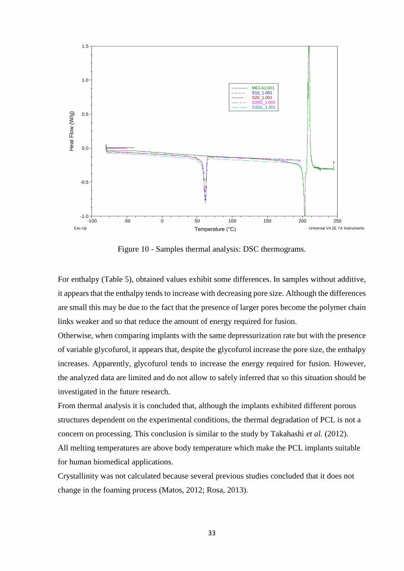
33
Figure 10 - Samples thermal analysis: DSC thermograms.
For enthalpy (Table 5), obtained values exhibit some differences. In samples without additive,
it appears that the enthalpy tends to increase with decreasing pore size. Although the differences
are small this may be due to the fact that the presence of larger pores become the polymer chain
links weaker and so that reduce the amount of energy required for fusion.
Otherwise, when comparing implants with the same depressurization rate but with the presence
of variable glycofurol, it appears that, despite the glycofurol increase the pore size, the enthalpy
increases. Apparently, glycofurol tends to increase the energy required for fusion. However,
the analyzed data are limited and do not allow to safely inferred that so this situation should be
investigated in the future research.
From thermal analysis it is concluded that, although the implants exhibited different porous
structures dependent on the experimental conditions, the thermal degradation of PCL is not a
concern on processing. This conclusion is similar to the study by Takahashi et al. (2012).
All melting temperatures are above body temperature which make the PCL implants suitable
for human biomedical applications.
Crystallinity was not calculated because several previous studies concluded that it does not
change in the foaming process (Matos, 2012; Rosa, 2013).
-1.0
-0.5
0.0
0.5
1.0
1.5
He
at
Flo
w (
W/g
)
-100 -50 0 50 100 150 200 250
Temperature (°C)
MECA2.001––––––– S10_1.001– – – – S20_1.001––––– · S20G_1.001––– – – S30G_1.001––– –––
Exo Up Universal V4.2E TA Instruments

34
3.3. Dexamethasone Implants: quantification and release
The influence of additive and processing technique were analyzed by comparing total
dexamethasone release in implants produced by foaming (with and without glycofurol) and
implants produced by temperature raising until melting (Figure 11). In the melting procedure
the system drug-polymer were exposed to 62.5ºC to ensure PCL melting.
According to the graph, there is less variability in SFM sample results. It occurs because the
technique allows a more homogenous drug distribution throughout implant body.
Analyzing the influence of additive, in samples with glycofurol, total dexamethasone release
was bigger which suggests a higher drug incorporation. Glycofurol seems to act as a polymer-
drug compatibilizer.
Considering these results the most desirable option was the obtained with SFM20-G: greater
drug incorporation and lower associated error.
Figure 11 - Total Dexamethasone implants release comparing SFM with HG and additive.
Sustained drug release is affected by factors that influence polymer degradation and drug
release such as the type of polymer and is molecular weight (which is the same in this analysis),
hydrolysis mechanism, erosion properties, shape and porosity (Manickavasagam and
Oyewumi, 2013). Dexamethasone Implants revealed sustained release over 500 h.
The foaming implants release profile is more uniform and constant because drug distribution in
the polymeric matrix is pretend to be uniform and the porosity induced by depressurization
allows a drug release phased and regular. The contact area with the environment is increased
by pores. Otherwise the implants produced by melting, are not expected to have pores in their
structure therefore the drug release occurs only by diffusion and erosion of the external surface

35
of the implant. In Figure 12 it is possible to see that until 200h of release, SFM release is more
stable and higher than HM. After that time, HM behavior release become more stable but still
lower than SFM.
Figure 12 - Dexamethasone release profiles in water.
3.4. 2-Cl-IB-MECA Implants: quantification and release
At the time this investigation was performed, there were no theoretical solubility data of 2-Cl-
IB-MECA in water or methanol.
The values obtained in the first assays were sub-quantified due to the lack of solubility data. It
was only known that release was greater in methanol than in water. Solutions where the
implants were placed, saturated and, therefore, no further release occurred. The releases
obtained at this time were around 3% of total drug amount. In later studies, 100 hours of release
was subsequently achieved by increasing the volume of solvent in order to prevent the
0
50
100
150
0 100 200 300 400 500
Rel
ease
d D
exam
eth
aso
ne,
% (
w/w
)
Time, h
SFM20-G HM-G HM-G-total SFM-G-total
0
8
15
0 100 200 300 400

36
saturation of the medium. In analyzes after this investigation, release profiles were obtained up
to 30 days.
It was found that the 2-Cl-IB-MECA was very difficult to dissolve in water to form a
homogeneous solution in order to make a standard curve of drug release in water. In several
tests performed, there was degradation of the drug after the first 24 hours with stirring (100
rpm) and thermal effect (37° C). On Galal et al. research, the preparation of 2-Cl-IB-MECA
solutions were refer to be made with an organic solvent and further dilutions with distillated
water so, based on that, the dissolution of the drug was made in Glycofurol adding subsequently
water.
With this step, it was possible to achieve the 2-Cl-IB-MECA standard curve in water (Appendix
D).
It was found that the drug and polymer particles interact in order to move away from each other
not achieving a cohesive and homogeneous mixture. This behavior difficult the delicate
methodology of to fill catheters with the mash and the yield of the products used was 1/3 lower
compared to using Glycofurol. The Glycofurol is a determining factor in the mixture and
homogeneity between the 2-Cl-IB-MECA and PCL.
As in the case of dexamethasone, the total drug amount was measured in methanol because this
drugs are very soluble in this solvent which facilitates full release process. SFM20-G and
SFM30-G implants were analyzed (Figure 13). Values obtained for the same processing
conditions are similar which allows to infer the homogeneity of samples. The release of 2-Cl-
IB-MECA in methanol occurs in a fast and sudden early stage. About 84% of drug released is
released in the first 2 hours which validates the feasibility of using methanol for this purpose.
Total drug release was found to be very close to theoretical: 78.7% in SFM20-G and 95.4% in
SFM30-G.

37
Figure 13 - 2-Cl-IB-MECA implants total release.
In what concern sustained release, the effect of glycofurol was also analyzed (Figure 14). 2-Cl-
IB-MECA SFM20 implants, with and without additive were tested. The associated errors from
the release without glycofurol are very high and it is more difficult to reduce them than in the
other samples.
This means that there is great variability when glycofurol is not used, although the release is
greater. This release is superior because as glycofurol will standardize the matrix probably take
longer to release the same amount. The glycofurol dispersed the drug inside the polymer matrix
as it was already refer.
Figure 14 - Glycofurol effect on 2-Cl-IB-MECA implants profile release:
◊ with Glycofurol □ without Glycofurol
0
95
190
285
380
475
20 bar/min 30 bar/min
2-C
l-IB
-ME
CA
//P
CL
, µ
g/m
g
0
3
6
9
12
0 15 30 45 60 75 90 105
2-C
l-IB
-ME
CA
/PC
L,
µg/m
g
Time, h

38
In the first hours of release, in free glycofurol implants seems to happen release of a large
amount of drug (Table 6) due to surface erosion. This effect may not be desirable and may be
toxic in biological environment. Otherwise, implants with glycofurol present a more controlled
drug release mainly regulated by surface degradation including inside pores.
Burst release is more marked having a hydrophilic drug in a polymer hydrophobic matrice due
to their poor drug-polymer interaction (Manickavasagam and Oyewumi, 2013).
Dexamethasone and 2-Cl-IB-MECA are hydrophobic drugs and PCL a hydrophobic polymer
and that is favorable to prevent evident burst.
Analyzing the release profile of 2-Cl-IB-MECA implants SFM20-G and SFM30-G (Figure 15
and Table 6), is seen that despite the similar tendency release, 20 bar/min implants drug release
is higher than the 30 bar/min. In implants, the drug is dispersed on the polymeric matrix and
also deposited on the surface. Thus, as stated before, SFM20-G implants have, theoretically,
larger pores than SFM30-G which facilitate contact with the surrounding environment, in
particular the phenomena of erosion and diffusion, thereby increasing the amount of released
drug.
If the study were prolong in time, SFM30-G implants release would probably be longer due to
the liberation is slower and the amount of drug per implant is similar.
2-Cl-IB-MECA implants shown sustained release over 100h.
Figure 15 - Profile release of 2-Cl-IB-MECA implants with different depressurization rates:
◊ 20 bar/min □ 30 bar/min
0
2
4
6
8
10
0 15 30 45 60 75 90 105
2-C
l-IB
-ME
CA
/P
CL
, µ
g/m
g
Time, h

39
Investigation show that release profile is made in two steps. The initial phase is resultant from
the release of a higher dose of the drug present on the surface of the polymeric matrix, and
depends on the total surface area of the implant, the percentage of drug added to the implant,
and aqueous solubility of the drug. Therefore, the larger the surface area of the system, the
greater the hydrophilicity and concentration of drug added; if this release is biologically
exacerbated it can lead to happen toxic effects (“burst”) (Yasukawa et al., 2002).
The second step is the diffusion phase characterized by gradual release of the drug due to its
dissolution on the surface of the polymeric matrix. This phase is regulated by the polymer
degradation rate, the total surface area of the implant, the percentage of drug added and the
aqueous solubility of the drug.
In our study a potential third phase, also referred to by Yasukawa and Manickavasagam et al.,
was not achieved. This final stage is characterized by the hydrolysis into the matrix and release
of a sudden high dose of drug. The release of a high drug dose may represents a possibly
problematic drawback of biodegradable systems. The produced implants seems to have a stable
and controlled release profile over time. The similar release profile suggest that the impact of
porosity affects the amount of drug release and the differences in that amount may be caused
by the time 2-Cl-IB-MECA takes to reach equilibrium across the implant structure.
Table 6 - Diffusion Coefficients for 2-Cl-IB-MECA.
Samples Diffusion Coefficients (mm2/h)
60% release 40% release
D1 R2 D2 R2
SFM20 0,0046 0,99 1,52E-07 0,82
SFM20-G 1,04E-04 0,99 6,08E-08 0,94
SFM30-G 6.46E-05 0,99 2,07E-08 0,90
Understanding that porosity influence polymer degradation and drug release is important to
achieve sustained drug release.
Implants with large surface areas tend to degrade faster than those with small areas because the
area under the effect of external factors (ocular fluids, for example) is bigger (Manickavasagam
and Oyewumi, 2013).
In achieved results, total drug quantification was higher in the SFM30-G implants but release
over analyzed time was superior in SFM20-G implants.

40
According to the AIBILI team, the amount of 2-Cl-IB_MECA provided in which intravitreal
injection is about 3 ng. In studies performed, the amount of drug released in the first hour is
eight times higher than the amount administered monthly in intra-vitreous injections. This value
reflects the potential of biomedical applicability but also the need to adjust the therapeutic doses
of the implant.
Implants mass difference before and after the release was 0.04±0.005 mg for SFM20-G and
0.07±0.039 mg for SFM30-G. After release, SEM images reveal no surface morphological
changes in the implants probably due to the fact that PCL has a degradation time much longer
than the time under review (Figure 16 and 17).

41
Figure 16 - After release SEM images of 2-Cl-IB-MECA implants.

42
Figure 17 - 2-Cl-IB-MECA implants SEM images before and after release with body and surface details.

43
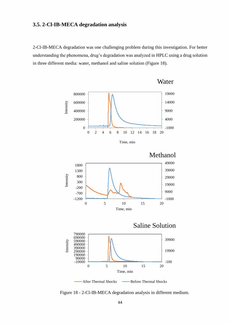
44
3.5. 2-Cl-IB-MECA degradation analysis
2-Cl-IB-MECA degradation was one challenging problem during this investigation. For better
understanding the phenomena, drug’s degradation was analyzed in HPLC using a drug solution
in three different media: water, methanol and saline solution (Figure 18).
Figure 18 - 2-Cl-IB-MECA degradation analysis in different medium.
-1000
4000
9000
14000
19000
0
200000
400000
600000
800000
0 2 4 6 8 10 12 14 16 18 20
Inte
nsi
ty
Time, min
Water
-1000
9000
19000
29000
39000
49000
-1200
-700
-200
300
800
1300
1800
0 5 10 15 20
Inte
nsi
ty
Time, min
Methanol
-100
19900
39900
-1000090000
190000290000390000490000590000690000790000
0 5 10 15 20
Inte
nsi
ty
Time, min
Saline Solution
After Thermal Shocks Before Thermal Shocks
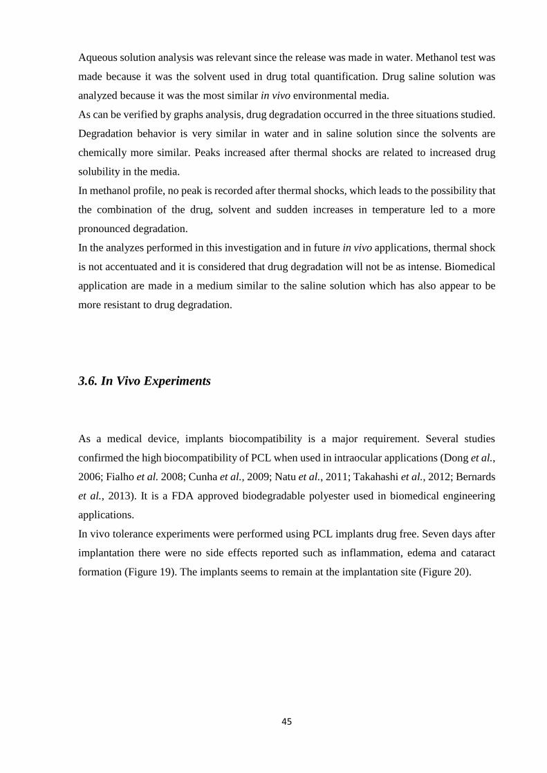
45
Aqueous solution analysis was relevant since the release was made in water. Methanol test was
made because it was the solvent used in drug total quantification. Drug saline solution was
analyzed because it was the most similar in vivo environmental media.
As can be verified by graphs analysis, drug degradation occurred in the three situations studied.
Degradation behavior is very similar in water and in saline solution since the solvents are
chemically more similar. Peaks increased after thermal shocks are related to increased drug
solubility in the media.
In methanol profile, no peak is recorded after thermal shocks, which leads to the possibility that
the combination of the drug, solvent and sudden increases in temperature led to a more
pronounced degradation.
In the analyzes performed in this investigation and in future in vivo applications, thermal shock
is not accentuated and it is considered that drug degradation will not be as intense. Biomedical
application are made in a medium similar to the saline solution which has also appear to be
more resistant to drug degradation.
3.6. In Vivo Experiments
As a medical device, implants biocompatibility is a major requirement. Several studies
confirmed the high biocompatibility of PCL when used in intraocular applications (Dong et al.,
2006; Fialho et al. 2008; Cunha et al., 2009; Natu et al., 2011; Takahashi et al., 2012; Bernards
et al., 2013). It is a FDA approved biodegradable polyester used in biomedical engineering
applications.
In vivo tolerance experiments were performed using PCL implants drug free. Seven days after
implantation there were no side effects reported such as inflammation, edema and cataract
formation (Figure 19). The implants seems to remain at the implantation site (Figure 20).
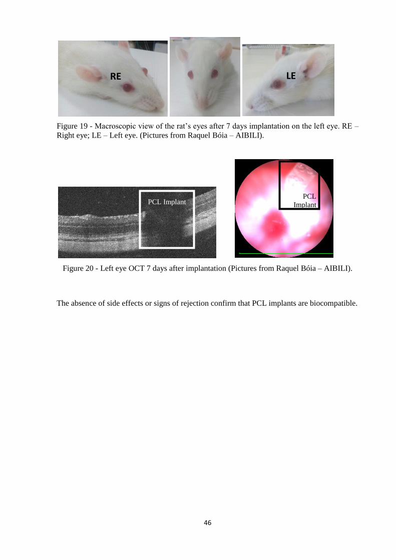
46
Figure 19 - Macroscopic view of the rat’s eyes after 7 days implantation on the left eye. RE –
Right eye; LE – Left eye. (Pictures from Raquel Bóia – AIBILI).
Figure 20 - Left eye OCT 7 days after implantation (Pictures from Raquel Bóia – AIBILI).
The absence of side effects or signs of rejection confirm that PCL implants are biocompatible.
RE LE
PCL Implant PCL
Implant

47

48
4. Conclusion and future remarks
The main goal of this work – development of PCL ocular implants for drug delivery using
supercritical carbon dioxide assisted fluid foaming/mixing methodology – was attained. PCL
Dexamethasone and 2-Cl-IB-MECA implants were successfully produced. Different working
parameters were used and diverse morphologies and profile release were obtained.
PCL thermal properties were not significantly altered after processing and are adequate for
biomedical use.
Porosity is important for controlled drug release. From SEM images it is concluded that the
implants produced have variable porosity according to the processing conditions (additive
presence and depressurization rate). Low depressurization rates and additive presence seems to
increase pore size. At a same depressurization rate, glycofurol presence increase porosity. In
SFM 2-Cl-IB-MECA implants release profiles, additive leads to greater releases. Otherwise,
hot melting produced implants have expressively less porosity and high variability. Drug
release express that differences: DXMT implants release was more stable over time in SFM
methodology.
The effect of depressurization rate on produced implants porosity was not always consistent
with the results of earlier investigations undertaken with bigger materials such as bone
substitutes. SFM20 and SFM30 implants had similar pore size: analysis repetition is suggested
because the prosperous advance in porous implants for biomedical applications depends on the
effective control of their morphology.
As in previous several works, we may also conclude that supercritical fluid technology is an
efficient approach on drug delivery devices development. The use of supercritical fluids,
particularly CO2, provides alternative pathways for processing polymers.
In the future it is recommended to optimize drug amount to incorporate in each implant
considering potential in vivo application.
Another request to new investigations are meticulous studies of 2-Cl-IB-MECA degradation
and solubility in different media because there is scarce literature on this.
Minimize losses during the process and examine the long-term behavior of the implants near
exhaustion are suggested as future work. In vitro implants degradation studies and in vivo ocular
drug bioavailability and systemic side effects at administration site must also be analyzed.
The development of an extruder applicable to the project should be a priority given the physical
and mental requirements of the manual process carried out in this research.

49

50
REFERENCES
Aktusu, Yukie; Kambe, Toshiaki Nakajima; Nomura, Nobuhiko; Nakahara, Tadaatsu. (1998).
Purification and Properties of a Polyester Polyurethane-Degrading Enzyme from Comamonas
acidovorans TB-35. Applied and Environmental Microbiology, 64 (1): 62–67.
Allhenn, Daniela; Lamprech, Alf. (2011). Microsphere Preparation Using the Untoxic Solvent
Glycofurol. Pharmaceutical Research, 28: 563-571.
Amo, E, M.; Urtti, A. (2008). Current and future ophthalmic drug delivery systems: A shift to
the posterior segment. Drug Discovery Today, 13: 135-143.
Ashida, K. (2007). Polyurethane and Related Foams: Chemistry and Technology. Boca Ranton
CRC Press Taylor & Francis.
Aspinall, Peter A.; Johnson, Zoe K.; Azuara-Blanco, Augusto; Montarzino, Alicia; Brice,
Roger; Vickers, Adrian (2008). Evaluation of Quality of Life and Priorities of Patients with
Glaucoma. Investigative Ophthalmology & Visual Science, 49:5.
Bassi, A. K.; Gough, J. E.; Zakikhani, M.; Downes, S. (2011). The Chemical and Physical
Properties of Poly (ε-caprolactone) Scaffolds Functionalised with Poly(vinyl phosphonic acid-
co-acrylic acid). Journal of Tissue Engineering, 2011, Article ID 615328.
Bao, J.-B.; Liu, T.; Zhao, L.; Hu, G.-H. (2011). A Two-Step Depressurazation Batch Process
for the Formation of Bi-modal Cell Structure Polystyrene Foams Using scCO2. Journal of
Supercritical Fluids, 55: 1104-1114.
Behar-Cohen, F. (2002). Systèmes de délivrance des médicaments pour le segment antériur:
bases fondamentales et applications cliniques. Journal Français d’Opthalmologie, 25(5): 537-
544.
Bernards, Daniel A.; Bhisitkul, Robert B.; Wynn, Paula; Steedman, Mark R.; Lee, On-Tat;
Wong, Fergus; Thoongsuwan, Somanus; Desai, Tejal A. (2013). Ocular Biocompatibility and

51
Structural Integrity of Micro and Nanostructured Poly(caprolactone) Films. Journal of Ocular
Pharmacology and Therapeutics, 29: 2.
Boongird, A.; Nasongkla, N.; Hongeng, S.; Sukdawong, N.; Sa-Nguanruang, W.;
Larbcharoensub, N. (2011). Biocompatibility Study of Glycofurol in Rat Brains. Experimental
Biology and Medicine, 236: 77-83.
Bourges, J.L.; Bloquel C.; Thomas A.; Froussart F.; Bochot A.; Azan F.; Gurny R.; Benezra
D.; Behar-Cohen F. (2006). Intraocular implants for extended drug delivery: therapeutic
applications. Advanced Drug Delivery Reviews, 58: 1182-1202.
Braga, M.E.M.; Pato, M.T. Vaz; Silva, H.S.R. Costa; Ferreira, E.I.; Gil, M. H.; Duarte, C. M.
M.; de Sousa, H. C. (2008). Supercritical solvent impregnation of ophthalmic drugs on chitosan
derivatives. Journal of Supercritical Fluids, 44: 245–257.
Breitenbach, Jorg (2002). Melt extrusion: from process to drug delivery technology. European
Journal of Pharmaceutics and Biopharmaceutics, 54(2): 107-117.
Brun, N.; Ungureanu, S.; Deleuze, H.; Backov, R. (2011). Hybrid Foams, Colloids and Beyond:
From Design to Applications. Chemical Society Reviews, 40: 771-788.
Bueno, C. Z.; Dias, A.M.A.; de Sousa, H.J.C.; Braga, M.E.M.; Moraes, A. M. (2014). Control
of the properties of porous chitosan–alginate membranes through the addition of different
proportions of Pluronic F68. Materials, Science and Engineering, 44: 117-125.
Charoenchaitrakool, M.; Dehghani, F.; Foster, N.R.; Chan, H.K. (2000). Micronization by rapid
expansion of supercritical solutions to enhance the dissolution rates of poorly water-soluble
pharmaceuticals. Industrial and Engineering Chemistry Research, 39: 4794-4802.
Choonara, Y.E.; Pillay, V.; Carmichael, T. and Danckwerts, M.P. (2007). Studies on a
noveldoughnut-shaped minitablet for intraocular drug delivery. Journal of the American
Association of Pharmaceutical Scientists PharmSciTech, 8:E118.
Choonara, Y.E.; Pillay, V.; Carmichael, T.; Danckwerts, M.P. (2009). A Review of Implantable
Intravitreal Drug Delivery Technologies for the Treatment of Posterior Segment Eye Diseases.
Journal of Pharmaceutical Sciences, 99(5).

52
Churro, R.M.M. (2016). Supercritical foaming/mixing on the preparation of PCL/mesoporous
silica based composites for hard tissue engineering applications, MSc Thesis, University of
Coimbra, Portugal.
Cunha, Armando Silva; Fialho, Silvia Ligorio; Naud, Marie-Christine; Cohen, Francine Behar.
(2009). Poly ε-Caprolactone Intravitreous Devices: An In Vivo Study. Investigative
Ophthalmology & Visual Science, 50: 5.
Dash, A. K.; Cudworth II, G. C. (1998). Therapeutic Application of Implantable Drug Delivery
Systems. Journal of Pharmacological and Toxicological Methods, 40: 1-12.
Dash T. K.; Konkimalla V. B. (2011). Poly-ε-caprolactone based formulations for drug delivery
and tissue engineering: A review. Journal of Controlled Release,
doi:10.1016/j.jconrel.2011.09.064.
Dong, X.; Shi, W.; Yuan, G.; Xie, L.; Wang, S.; Lin, P. (2006). Intravitreal implantation of the
biodegradable cyclosporine A drug delivery system for experimental chronic uveitis. Graefe's
archive for clinical and experimental ophthalmology, 244: 492-497.
Duarte, A. R. C.; Silva, S. S.; Mano J. F.; Reis, R. L. (2012). Ionic Liquids as Foaming Agents
of Semi-Crystalline Natural-Based Polymers. Green Chemistry, 14(7), 1949-1955.
Fernandez, Angel Marcos; Richa, Antonio Martinez (2011). Hydrolytic degradation of poly(ε-
caprolactone) with different end groups and poly(ε-caprolactone-co-γ-butyrolactone. Polymers
for Advanced Technologies. DOI: 10.1002/pat.1531
Fialho, S. L.; Rego, M. G. B.; Cardillo, J. A.; Siqueira, R. C.; Jorge, R.; Silva-Cunha A. J.
(2003). Implantes biodegradáveis destinados à administração intra-ocular. Arquivos Brasileiros
de Oftalmologia, 66: 891-896.
Fialho, S. L.; Silva-Cunha A. J. (2005). Manufacturing Techniques of Biodegradable Implants
Intended for Intraocular Application. Drug Delivery, 12(2): 109-116.
Gaudana, Ripal; Ananthula, Hari Krishna; Parenky, Ashwin; Mitra, Ashim K. (2010). Ocular
Drug Delivery. Journal of the American Association of Pharmaceutical Scientists: 12 (3).

53
Gualandi, C.; White, L. J.; Chen, L.; Gross, R. A.; Shakesheff, K. M.; Howdle, S. M.; Scandola,
M. (2010). Scaffolds for Tissue Engineering Fabricated by Non-Isothermal Supercritical
Carbon Dioxide Foaming of Highly Crystalline Polyester. Acta Biomaterialia, 6: 130-136.
Haghjou, Nahid; Soheilian, Masoud; Abdekhodaie, Mohammad Jafar (2011). Sustained
Release Intraocular Drug Delivery Devices for Treatment of Uveitis. Journal of Ophthalmic
and Vision Research, 6 (4).
Hosseini, K.; Matsushima, D.; Johnson, J.; Widera, G.; Nyam, K.; Cormier, M. (2008).
Pharmacokinetic study of dexamethasone disodium phosphate using intravitreal,
subconjunctival, and intravenous delivery routes in rabbits. Journal of Ocular Pharmacology
and Therapeutics, 24: 301-308.
Hughes, Patrick M.; Olejnik, Orest; Chang-Lin, Joan; Wilson, Clive G. (2005). Topical and
systemic drug delivery to the posterior segments. Advanced Drug Delivery Reviews, 57: 2010-
2032.
Jacobs, L. J. M.; Kemmer, M. F. and Keurentjes, J. T. F. (2008). Sustainable Polymer Foaming
Using High Pressure Carbon Dioxide: A Review on Fundamentals, Process and Applications.
Green Chemistry, 10(7): 721-812.
Jaffe, G.J.; Martin, D.; Callanan, D.; Pearson, P.A.; Levy, B.; Comstock, T. (2006).
“Fluocinolone acetonide implant (Retisert) for noninfectious posterior uveitis: thirty-four-week
results of a multicenter randomized clinical study. Ophthalmology, 113: 1020-1027.
Jain, R. A. (2000). The manufacturing techniques of various drug loaded biodegradable poly
(lactide-co-glycolide) (PLGA) devices. Biomaterials, 21: 2475-2490.
Jain, K. K. (2008). Drug Delivery Systems – An Overview. Drug Delivery Systems. Humana
Press, 1-50.
Jenkins M. J.; Harrison K. L.; Silva M. M. C. G.; Whitaker M. J.; Shakesheff K. M.; Howdle
S. M. (2006). Characterisation of microcellular foams produced from semicrystalline PCL
using supercritical carbon dioxide. European Polymer Journal 42: 3145–3151.

54
Karimi, Mohammad (2011). Diffusion in Polymer Solids and Solutions, Mass Transfer in
Chemical Engineering Processes. Dr. Jozef Markoš (Ed.), DOI: 10.5772/23436.
Kazarian, S.G. (2000). Polymer processing with supercritical fluids. Polymer 42: 78–101.
Kikic, I.; Vecchione, F. (2003). Supercritical impregnation of polymers. Current Opinion in
Solid State and Materials Science, 7: 399-405.
Kim, Hea O.; Hawes, Calvin; Towers, Pat; Jacobson, Kenneth A. (1996). Radiolabeling and
efficient synthesis of Tritiated 2-Chloro-N6-(3-Iodobenzyl) Adenosine-5'-N-Methyluronamide,
a potent, selective A3 adenosine receptor agonist. Journal of Labelled Compounds and
Radiopharmaceuticals, 38(6): 547-560.
Kim, S.H.; Csaky, K.G.; Wang, N.S.; Lutz, R.J. (2008). Drug elimination kinetics following
subconjunctival injection using dynamic contrast-enhanced magnetic resonance imaging.
Journal of Pharmacy Research, 25(3): 512-20.
Kimura, H; Ogura, Y. (2001). Biodegradable polymers for ocular drug delivery.
Ophalmologica, 215(3): 143-155.
Kiram E. (2009). Polymer miscibility, phase separation, morphological modifications and
polymorphic transformations in dense fluids. Journal of Supercritical Fluids, 47: 466–483.
Knox, D.E. (2005). Solubilities in supercritical fluids. Pure and Applied Chemistry, 77: 513-
530.
Kuno, N.; Fujii, S. (2010). Biodegradable intraocular therapies for retinal disorders: progress
to date. Drugs and Aging, 27(2): 117-134.
Kunou, N.; Ogura, Y; Yasukawa, T. (2000). Long-term sustained release of ganciclovir from
biodegradable scleral implant for the treatment of cytomegalovirus retinis. Journal of
Controlled Release, 68(2): 263-271.

55
Kuppermann, B.D.; Blumenkranz, M.S.; Haller, J.A.; Williams, G.A.; Weinberg, D.V.; Chou,
C.; Whitcup, S.M. (2007). Randomized controlled study of an intravitreous dexamethasone
drug delivery system in patients with persistent macular edema. Archives of Ophthalmology,
125(3): 309-317.
Labow, Rosalind S.; Tang, Yiwen; McCloskey, Christopher B.; Santerre, J. Paul (2002). The
effect of oxidation on the enzyme-catalyzed hydrolytic biodegradation of poly(urethane)s.
Journal of Biomaterials Science, Polymer Edition, 13: 651-665.
Lance, Kevin D.; Good, Samuel D.; Mendes, Thaís S.; Ishikiriyama, Mynna; Chew, Patrick;
Estes, Laurel S.; Yamada, Kazuhito; Mudumba, Sri; Bhisitkul, Robert B.; Desai, Tejal A.
(2015). In Vitro and In Vivo Sustained Zero-Order Delivery of Rapamycin (Sirolimus) from a
Biodegradable Intraocular Device. Investigative Ophthalmology & Visual Science, 56: 7331-
7337.
Lang, L.C. (1995). Ocular drug delivery: conventional ocular formulations. Advanced Drug
Delivery Reviews, 16: 39-43.
Lebourg, M., Antón, J. S.; Ribelles, J. L. G. (2008). Porous Membranes of PLLA-PCL Blend
for Tissue Engineering Applications. European Polymer Journal, 44: 2207-2218.
Lee, L. J.; Zeng, C.; Cao, X.; Han, X.; Shen, J.; Xu, G. (2005). Polymer Nanocomposite Foams.
Composites Science and Technology, 65: 2344-2363.
Leeke, Gary A.; Cai, Jiangou; Jenkins, Mike. (2006). Solubility of Supercritical Carbon
Dioxide in Polycaprolactone (CAPA 6800) at 313 and 333 K. Journal of Chemical and
Engineering Data, 51: 1877-1879.
Léonard A.; Calberg C.; Kerckhofs G.; Wevers M.; Jérôme R.; Pirard J.; Germain A.; Blacher
S. (2008). Characterization of the porous structure of biodegradable scaffolds obtained with
supercritical CO2 as foaming agent. Journal of Porous Mater, 15: 397-403.
56
Lowell, S.; Shields, J.E.; Thomas, M.A.; Thommes, M. (2004). Characterization of Porous
Solids and Powders: Surface Area, Pore Size and Density. Particle Technology Series, 16.
Kluwer Academic Publishers. ISBN: 978-90-481-6633-6
Manickavasagam, Dharani; Oyewumi, Moses O. (2013). Critical Assessment of Implantable
Drug Delivery Devices in Glaucoma Management. Journal of Drug Delivery, 2013. Article ID
895013.
Matos, M.B.C. de (2012). Drug-eluting silica/poly(α-esters) biocomposite materials prepared
by green technologies, MSc Thesis, University of Coimbra, Portugal.
Matos, M.B.C. de; Piedade, A.P; Alvarez-Lorenzo, C.; Concheiro, A.; Braga, M.E.M.; Sousa,
H.C. (2013). Dexamethasone-loaded poly(-caprolactone)/silica nanoparticles composites
prepared by supercritical CO2 foaming/mixing and deposition. International Journal of
Pharmaceutics, 456: 269-281.
Myers, J. S.; Sall, K. N., DuBiner, H.; Slomowitz, N.; McVicar, W.; Rich, C. C.; Baumgartner,
R. A. (2016). A Dose-Escalation Study to Evaluate the Safety, Tolerability, Pharmacokinetics,
and Efficacy of 2 and 4 Weeks of Twice-Daily Ocular Trabodenoson in Adults with Ocular
Hypertension or Primary Open-Angle Glaucoma. Journal of Ocular Pharmacology and
Therapeutics, 32(8), 555–562.
Monnereau L; Urbanczyk L; Thomassin J-M; Alexandre M; Jerome C; Huynen I; Bailly C;
Detrembleur C. (2014). Supercritical CO2 and polycarbonate based nanocomposites: a critical
issue for foaming. Polymer, 55: 2422–2431.
Morèrea J.; Tenorio M. J.; Torralvo M. J.; Pando C.; Renuncio J. A. R.; Cabañas A. (2011).
Deposition of Pd into mesoporous silica SBA-15 using supercritical carbon dioxide. Journal of
Supercritical Fluids, 56: 213–222.
Nair, L. S.; Laurencin, C. T. (2007). Biodegradable polymers as biomaterials. Progress in
Polymer Science, 32: 762–798.

57
Natu, M.V.; Gaspar, M.N.; Ribeiro, C.A. Fontes; Cabrita, A.M.; de Sousa, H.C.; Gil, M.H.
(2011). In vitro and in vivo evaluation of an intraocular implant for glaucoma treatment. Journal
of Pharmaceutics, 415: 73-82.
Nutheti, Rishita; Shamanna, Bindiganavale R.; Nirmalan, Praveen K.; Keeffe, Jill E.;
Krishnaiah, Sannapaneni; Rao, Gullapalli N.; Thomas, Ravi (2006). Impact of Impaired Vision
and Eye Disease on Quality of Life. Investigative Ophthalmology & Visual Science, 47: 11.
Ogura, Y. (2001). Drug delivery to the posterior segments of the eye. Advanced Drug
Delivery Reviews, 52: 1-3.
Okabe, K.; Kimura, H.; Okabe, J.; Kato, O.; Kunou, N.; Ogura, Y. (2003). Intraocular tissue
distribution of betamethasone after intrascleral administration using a non-biodegradable
sustained drug delivery device. Investigative Ophtalmology and Visual Science, 44(6): 2702-
2707.
Paz, E. de; Martin, A.; Rodríguez-Rojo, S.; Herreras, J.; Cocero, M.J. (2010). Determination of
phase equilibrium (solid-liquid-gas) in poli-(ε-caprolactone)-carbon dioxide system. Journal of
Chemical & Engineering, 50: 2781-2785.
Peyman, G. A.; Ganiban, G. J. (1996). Delivery systems for intraocular routes. Advanced Drug
Delivery Reviews, 16: 107-123.
Rosa, A.B.S. (2013). Poly (ε-caprolactone)/SBA-15 composite biomaterials plasticized with
greener additives, MSc Thesis, University of Coimbra, Portugal.
Rothen-Weinhold, A.; Besseghir, K.; Vuaridel, E.; Sublet, E.; Oudry, N.; Kubel, F.; Gurny, R.
(1999). Injection-molding versus extrusion as manufacturing technique for the preparation of
biodegradable implants. European Journal of Pharmaceutics and Biopharmaceutics, 48(2):
113-121.
Salerno, A.; Di Maio, E.; Iannace, S.; Netti, P.A. (2011). Solid-state supercritical CO2 foaming
of PCL and PCL-HA nano-composite: effect of composition, thermal history and foaming
process on foam pore structure. Journal of Supercritical Fluids, 58: 158-167.

58
Salerno, A.; Domingo, C. (2013). Effect of blowing agent composition and processing
parameters on the low temperature foaming of poly(l-lactide/caprolactone) co-polymer by
means of supercritical CO2/ethyl lactate binary mixtures. Journal of Supercritical Fluids, 84:
195-204.
Shedden, A.H.; Laurance, J.; Barrish, A.; Olah, T.V. (2001). Plasma timolol concentrations of
timolol maleate: timolol gel-forming solution (TIMOPTIC-XE) once daily versus timolol
maleate ophthalmic solution twice daily. Documenta Ophthalmologica, 103: 73–79.
Shieh Y. T.; Lai J. G.; Tang W. L.; Yang C. H.; Wang T. L. (2009). Supercritical CO2
intercalation of polycaprolactone in layered silicates. Journal of Supercritical Fluids, 49: 385-
393.
Tai H.; Popov V. K.; Shakesheff K. M.; Howdle S. M. (2007). Putting the fizz into chemistry:
applications of supercritical carbon dioxide in tissue engineering, drug delivery and synthesis
of novel block copolymers. Biochemical Society Transactions, 35: 516-521.
Tan, D. T. H.; Chee, S.P.; Lim, L.; Lim, A. S. M. (1999). Randomized clinical trial of a new
dexamethasone delivery system (Surodex) for treatment of post-cataract surgery inflammation.
Ophthalmology, 106(2): 223–231.
Takahashi, S.; Hassler, J. C.; Kiran, E. (2012). Melting behavior of biodegradable polyesters in
carbon dioxide at high pressures. Journal of Supercritical Fluids, 72: 278-287.
Tatai, L.; Moore, Tim G.; Adhikari, Raju; Malherbe, François; Jayasekara, Ranjith; Griffiths,
Ian; Gunatillake, Pathiraja A. (2007). Thermoplastic biodegradable polyurethanes: The effect
of chain extender structure on properties and in-vitro degradation. Biomaterials, 28: 5407-
5417.
Teixeira, V.G.; Coutinho, Fernanda M.B.; Gomes, A.S. (2001). Principais Métodos de
Caracterização da Porosidade de Resinas à base de Divinilbenzeno. Química Nova, 24(6): 808-
818.
Tsimpliaraki A.; Tsivintzelis I.; Marras S.I., Zuburtikudis I.; Panayiotou C. (2011). The effect
of surface chemistry and nanoclay loading on the microcellular structure of porous poly(d, l
lactic acid) nanocomposites. Journal of Supercritical Fluids, 57: 278–287.
59
Tsimpliaraki, A.; Tsivintzelis, I.; Marras, S.I.; Zuburtikudis, I.; Panayiotou, C. (2013). Foaming
of PCL/clay nanocomposites with supercritical CO2 mixtures: The effect of nanocomposite
fabrication route on the clay dispersion and the final porous structure. Journal of Supercritical
Fluids, 81: 86-91.
Wang, Wenshou; Wang, Chun (2012). Polyurethane for biomedical applications: A review of
recent developments. The design and manufacture of medical devices, Woodhead Publishing
Reviews Mechanical Engineering, 115-122.
Weijtens, O.; Feron, Eric J.; Schoemaker, Rik C.; Cohen, Adam F.; Romijn, Fred P.H.T.M.
(1999). High concentration of dexamethasone in aqueous and vitreous after subconjunctival
injection. American Journal of Ophthalmology, 7: 128:192.
White, L. J.; Hutter, V.; Tai, H.; Howdle, S. M.; Shakesheff, K. M. (2012). The Effect of
Proceesing Variables on Morphological and Mechanical Properties of Supercritical CO2
Foamed Scaffolds for Tissue Engineering. Acta Biomaterialia, 8: 61-71.
Woods H. M.; Silva M. M. C. G.; Nouvel C.; Shakesheffb K. M.; Howdle S. M. (2004).
Materials processing in supercritical carbon dioxide: surfactants, polymers and biomaterials.
Journal of Materials Chemistry, 14: 1663–1678.
Xu Q.; Ren X.; Chang Y.; Wang J.; Yu L.; Dean K. (2004). Generation of microcellular
biodegradable polycaprolactone foams in supercritical carbon dioxide. Journal of Applied
Polymer Science, 94: 593–597.
Yañez, Fernando; Martikainen, Lahja; Braga, Mara E.M.; Alvarez-Lorenzo, Carmen;
Concheiro, Angel; Duarte, Catarina M.M.; Gil, Maria H.; de Sousa, Hermínio C. (2011).
Supercritical fluid-assisted preparation of imprinted contact lenses for drug delivery. Acta
Biomaterialia, 7: 1019-1030.
Yarangumeli, A.; Kural, G. (2004). Are there any benefits of Betoptic S (betaxolol HCl
ophthalmic suspension) over other beta-blockers in the treatment of glaucoma?. Expert Opinion
on Pharmacotherapy, 5: 1071–1081.

60
Yasukawa, T.; Kimura, H.; Tabata, Y.; Ogura, Y. (2001). Biodegradable sclera plugs for
vitreoretinal drug delivery. Advanced Drug Delivery Reviews, 52(1): 25-36.
Yasukawa, T.; Ogura, Y.; Tabata, Y.; Kimura, H.; Wiedemann, P.; Honda, Y. (2004). Drug
delivery systems for vitreoretinal diseases. Progress in Retinal and Eye Research, 23: 253-281.
Yasukawa, T.; Ogura, Y.; Sakurai, E.; Tabata, Y.; Kimura, H. (2005). Intraocular sustained
drug delivery using implantable polymeric devices. Advanced Drug Delivery Reviews, 57(14):
2033-2046.
Yoganathan, Roshan; Mammucari, Raffaella; Foster, Neil R. (2010). Impregnation of Ibuprofen
into Polycaprolactone Using Supercritical Carbon Dioxide. Journal of Physics: Conference
Series 215.
Zeng, C.; Han, X.; Lee, L. J.; Koelling, K. W.; Tomasko, D. L. (2003). Polymer-Clay
Nanocomposite Foams Prepared Using Carbon Dioxide. Advanced Materials, 15(20), 1743-
1747.
Zhai, W.; Yu, J.; Wu, L.; Ma, W.; He, J. (2006). Heterogeneous Nucleation Uniformizing Cell
Size Distribution in Microcellular Nanocomposites Foams. Polymer, 47: 7580-7589.

61

62
Supplementary Data
Appendix A – Catheters Filling Process
Figure A1 – I.V. Optiva® catheters from Smiths Medical.
A small amount of the mixture (PCL, glycofurol and drug) or pure PCL was placed in the yellow
portion of the catheter (Figure A1) and it was pushed and compressed therein with the aid of two
needles (one at each end). The filling was done in about two thirds of the total area of the catheter.
After filling, the yellow part was removed with a scalpel (Figure A2).
Figure A2 – Catheter completion.

63

64
Appendix B – Supercritical Solvent Unit
Figure B1 – Schematic diagram of the supercritical solvent unit. C1: compressor; TC:
temperature controller; WB: water bath; P: purge; PT: pressure transducer; S: sample; MS:
magnetic stirrer; C: high pressure vessel; V: screw down valve; M: macrometric valve; m:
micrometric valve; GT: glass trap; F: mass flow meter (Rosa, 2013).

65

66
Appendix C – Dexamethasone Standard Curves
According to HPLC data obtained, the standard curves of dexamethasone release in methanol
and in water are on figure C1 and C2:
Figure C1 - Dexamethasone standard curve, in Methanol, used to determine the total amount
of drug in the implants.
Figure C2 - Dexamethasone standard curve, in Milli-Q water, used to determine implants
drug release.
y = 1E+07x
R² = 0,9992
0
1000000
2000000
3000000
4000000
5000000
6000000
0,0 0,2 0,4 0,6
Are
a
Concentration (mg/mL)
y = 22490x
R² = 0,9577
0
950000
1900000
2850000
3800000
0,0 25,0 50,0 75,0 100,0 125,0 150,0
Are
a
Concentration (mg/mL)

67

68
Appendix D – 2-Cl-IB-MECA Standard Curves
According to HPLC data obtained, the standard curves of 2-Cl-IB-MECA release in methanol
and in water are on figure D1 and D2:
Figure D1 – 2-Cl-IB-MECA standard curve, in Methanol, used to determine the total amount
of drug in the implants.
Figure D2 – 2-Cl-IB-MECA standard curve, in Milli-Q water, used to determine implants
drug release.
y = 10071x
R² = 0,9995
0
500000
1000000
1500000
2000000
2500000
0 50 100 150 200 250
Are
a
Concentration (ug/mL)
y = 6844,4x
R² = 0,993
0
5000
10000
15000
20000
25000
30000
35000
40000
0,0 1,0 2,0 3,0 4,0 5,0 6,0
Are
a
Concentration (ug/mL)

69

70
Appendix E – Comparison of detection peaks, in HPLC, of 2-Cl-IB-MECA, saline
solution, cell culture medium and explants culture medium.
Copyright © 2022 FDOKUMEN