Long-term Visual Outcomes of Intravitreal Bevacizumab in Inflammatory Ocular Neovascularization

CAUSES ANDMANAGEMENT OF
NEOVASCULARIZATIONPREPARED BY
MUHAMAD HAZIMIN BIN ISHAK5034
MUHAMMAD AMINUDDIN BIN KHALID5037
MUHAMMAD ARIF IMRAN BIN ZAINAL RASHID5038
MUHAMMAD BASHEER BIN YAHYA5039

MUHAMMAD FIKRI BIN ISMAIL5041
MUHAMMAD HASIF BIN KAMARUDIN5043
MUHAMMAD IKRAAM BIN ABDUL LATIF5046
SUPERVISED BY: DR. AHMED ABDULLAH
CONTENT
CONTENT PAGEPREFACE 3
INTRODUCTION 4
1.0: PROLIFERATIVE DIABETIC RETINOPATHY
5
2.0: RETINOPATHY OF PREMATURITY
8
3.0: AGE-RELATED MACULAR DEGENERATION
12
4.0: SICKLE CELL 142

RETINOPATHY5.0: CENTRAL RETINAL VEINOCCLUSION
17
6.0: OCULAR INFLAMMATIONS& NEOVASCULARISATION
19
REFERENCES 20
PREFACE
In the name of Allah the Most Gracious and the Most Merciful,
praise be to Allah for that we have finished our task for a
group assignment entitled “Causes and Management of
Neovascularization”. Special regards to our beloved and
passionate supervisor, Dr. Ahmed Abdullah for his guide in the
making of this project. Not to forget, all the staff members
of Ophthalmology Department, Kasr Al Ainy Medical School for
their generous help and support for us upon finishing this
project. We are very lucky to have them alongside us all
through this journey. Last but not least, we love
ophthalmology!
3

INTRODUCTION
1. What is neovascularization?
Neovascularization is a pathologic process consisting of the
proliferation of blood vessels in abnormal tissues or in
abnormal positions. Neovascularization within the eye
contributes to visual loss in several ocular diseases.
4

2. What are among the causes known for this phenomenon?
The most common of which are proliferative diabetic
retinopathy, neovascular age-related macular degeneration, and
retinopathy of prematurity. Together, these three diseases
afflict persons in all stages of life from birth through late
adulthood and account for most instances of legal blindness.
Retinopathy of prematurity, proliferative diabetic
retinopathy, and neovascular age-related macular degeneration
are but three of the ocular diseases which can produce visual
loss secondary to neovascularization. Others include sickle
cell retinopathy, retinal vein occlusion, and certain
inflammatory diseases of the eye. These, however, account for
a much smaller proportion of visual loss caused by ocular
neovascularization.
1.0: PROLIFERATIVE DIABETIC RETINOPATHY
5

1. Proliferative diabetic retinopathy affects insulin-dependent
diabetes mostly.
2. There is major ischemia process during the phase of non
proliferative and the severe non proliferative characterized
by the soft exudates, and maybe due to microaneurysm and
hemorrhages.
3. Fluorescein angiogram is used to detect the ischemic parts
of the retina.
4. The ischemic retina secretes a vasostimulating substance
that results in budding of the new vessels.
5. Neovascularization may occur at the optic disc (NVD). This
is dangerous that it may cause Primary Optic Nerve atrophy.
6. Neovascularization can also occur along the vessels of
retina termed new vessels elsewhere (NVE).
7. These new vessels are unhealthy and liable to leak and
hemorrhage occurs. This will further cause ischemia to the
retina as bleeding causes scar in the retinal tissue.
6

Fig. 1.0: Fundus exam showing non-proliferative diabetic retinopathy
Fig. 1.1: Fundus exam showing the neovascularization process in the
proliferative diabetic retinopathy.
MANAGEMENT
Follow up diabetic patients and regular fundus examinations
every year.
When there is leaking of vessels, focal laser
photocoagulation is indicated.
If the new vessels are developed (NVD or NVE or even
rubeosis iridis), pan retinal laser photocoagulation is
indicated.
7

o This procedure is indicated to kill the new budding
endothelial cells in the formation of the
neovascularization.
Vitrectomy.
Anti-Vascular Endothelial Growth Factor (Anti-VEGF)
o When there is a lack of nutrient in the Retinal Pigment
Epithelium, the VEGF plays its role by forming a new
budding blood vessels in order to restore the oxygen and
nutrients level of the cell
o However, the budding blood vessels are weak and liable to
rupture causing haemorrhages.
o This is dangerous as the hemorrhages can cause retinal
scars and subsequently, the patient will lose his vision.
o Thus, Anti-VEGF is used to prevent the formation of
neovascularization in the cases like:
1. Age-related macular edema
2. Proliferative diabetic retinopathy (PDR)
3. Central retinal vein occlusion (CRVO).
8

Fig. 1.3: Fundus exam showing the Pan Retinal Photocoagulation
2.0: RETINOPATHY OF PREMATURITY (ROP)
1. Doctors do not know for certain what causes ROP. But it is
known that by the 4th month of pregnancy, the unborn child's
retina has begun to develop vascularization. Such formation
of blood vessels appears to be very sensitive to the amount
of oxygen supplied, either naturally or artificially.
2. Blood vessels in the eye normally finish developing in the
last few weeks before birth. Premature infants, however,
9

leave the protective uterus before blood vessels of the eye
have had a chance to fully develop. The less developed the
retina at birth the worse ROP is likely to be. ROP occurs in
two opposite phases:
Phase I consists of delayed retinal vascular growth and
vessel loss after premature birth resulting in hypoxia.
Phase II consists of hypoxia-induced vascular
proliferation.
3. Both oxygen-regulated and non-oxygen-regulated factors
contribute to normal vascular development and retinal
neovascularization. Vascular endothelial growth factor
(VEGF) is an important oxygen-regulated factor. A critical
non-oxygen-regulated growth factor is insulin like growth
factor-I (IGF-I).
4. In premature infants, the absence of IGF-1 (normally
provided by the placenta and the amniotic fluid) inhibits
blood vessel growth. As the eye matures, it becomes oxygen
starved, sending signals to increase VEGF. As the infant’s
organs and systems then continue to mature, IGF-1 levels
rise again, suddenly allowing the VEGF signal to produce
blood vessels. This neovascular proliferation of phase II of
ROP can cause blindness.
10

Fig. 2.0: Schematic representation of IGF-I and VEGF control of blood vessel
development in ROP
5. Some risk factors have been identified that may have an
effect on ROP development include:
Low birth weight (less than 1250g).
Low gestational age (before 30 weeks of gestation).
Use of supplemental oxygen after birth.
Vitamin E deficiency.
Various types of infections (especially concurrent).
Race (Caucasians are more at risk than African-
Americans).
Anemia.
Respiratory complications & cardiac defects.
MANAGEMENT
11
The norma l growth of the bl ood vessels i s directed to relativel y low-oxyge n
ar eas of the retina, but the vessels remain in
th e plane of the retin a and do no t grow i nto
th e vitreous h umor.
If excess ox ygen is g iven,
normal bloo d ves sels
d egrade and cease to de velop.
Wh en the excess oxygen
envi ronmen t is r emoved , the b lood vessel s rapidl y begin
formin g again and grow into
the vitreous humor of the eye from the
retina

The main goal of treatment is to prevent progression of
fibrovascular proliferation and to avoid retinal detachment.
In this case, timing is critical to any intervention that will
be done.
Replacement of IGF-1 early on might promote normal blood
vessel growth; whereas late supplementation with IGF-1 in the
neovascular phase of ROP could exacerbate the disease.
Similarly, inhibition of either VEGF or IGF-1 early after
birth can prevent normal blood vessel growth and precipitate
the disease; whereas inhibition at the second neovascular
phase might prevent destructive neovascularization.
At any rate, screening at-risk preterm infants at proper
times and intervals is very important.
Although oxygen therapy has been blamed for ROP
progression in the past, maximizing the oxygen saturation
(to 95%) may induce regression in pretreshold disease.
Cryotherapy (regional retinal destruction using a probe
to freeze the desired areas) was the original mode of
treatment (since the 1970s).
While laser surgery (e.g. xenon, argon, diode) is as
effective as cryotherapy, does not require general
anesthesia, and has a lower complication rate.
12

Fig. 2.1: showing fresh laser-coagulation burns in the avascular periphery of a Stage 3
ROP
Scleral buckling surgery and/or vitrectomy are usually
performed for severe ROP (stages 4 and 5) that progresses
to retinal detachment.
Fig. 2.2: The silicone band (scleral buckle, blue) is placed around the eye. This
brings the wall of the eye into contact with the detached retina, allowing the retina to
re-attach.
Recently, intravitreal injection of bevacizumab (Avastin)
has been reported as a supportive measure in aggressive 13

posterior retinopathy of prematurity. This therapy
appears to have lower risk of very high myopia than laser
therapy (according to a 2011 clinical trial)
Fig 2.3: Some of the different preparations of Avastin®, a drug that starves cancer
cells.
3.0: AGE-RELATED MACULAR DEGENERATION (AMD)
1. Age-related macular degeneration (AMD) affects an estimated
14 million people worldwide, and is the leading cause of
severe, irreversible vision loss in individuals over the age
of 50 years in Western societies.
2. It is characterized by the aged patients having vision loss
associated with drusen and atrophy of the RPE.
14

3. Choroidal neovascularization (CNV), the hallmark of 'wet',
'exudative' or 'neovascular' AMD, is responsible for
approximately 90% of cases of severe vision loss due to AMD.
4. Vascular endothelial growth factor (VEGF) has been shown to
play a key role in the regulation of CNV and vascular
permeability.
Fig 3.0: Pathological illustration of AMD
Fig 3.1: Fundus examination of AMD
MANAGEMENT
15
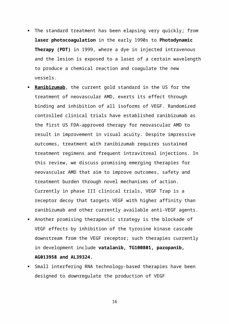
The standard treatment has been elapsing very quickly; from
laser photocoagulation in the early 1990s to Photodynamic
Therapy (PDT) in 1999, where a dye in injected intravenous
and the lesion is exposed to a laser of a certain wavelength
to produce a chemical reaction and coagulate the new
vessels.
Ranibizumab , the current gold standard in the US for the
treatment of neovascular AMD, exerts its effect through
binding and inhibition of all isoforms of VEGF. Randomized
controlled clinical trials have established ranibizumab as
the first US FDA-approved therapy for neovascular AMD to
result in improvement in visual acuity. Despite impressive
outcomes, treatment with ranibizumab requires sustained
treatment regimens and frequent intravitreal injections. In
this review, we discuss promising emerging therapies for
neovascular AMD that aim to improve outcomes, safety and
treatment burden through novel mechanisms of action.
Currently in phase III clinical trials, VEGF Trap is a
receptor decoy that targets VEGF with higher affinity than
ranibizumab and other currently available anti-VEGF agents.
Another promising therapeutic strategy is the blockade of
VEGF effects by inhibition of the tyrosine kinase cascade
downstream from the VEGF receptor; such therapies currently
in development include vatalanib, TG100801, pazopanib,
AG013958 and AL39324.
Small interfering RNA technology-based therapies have been
designed to downregulate the production of VEGF
16
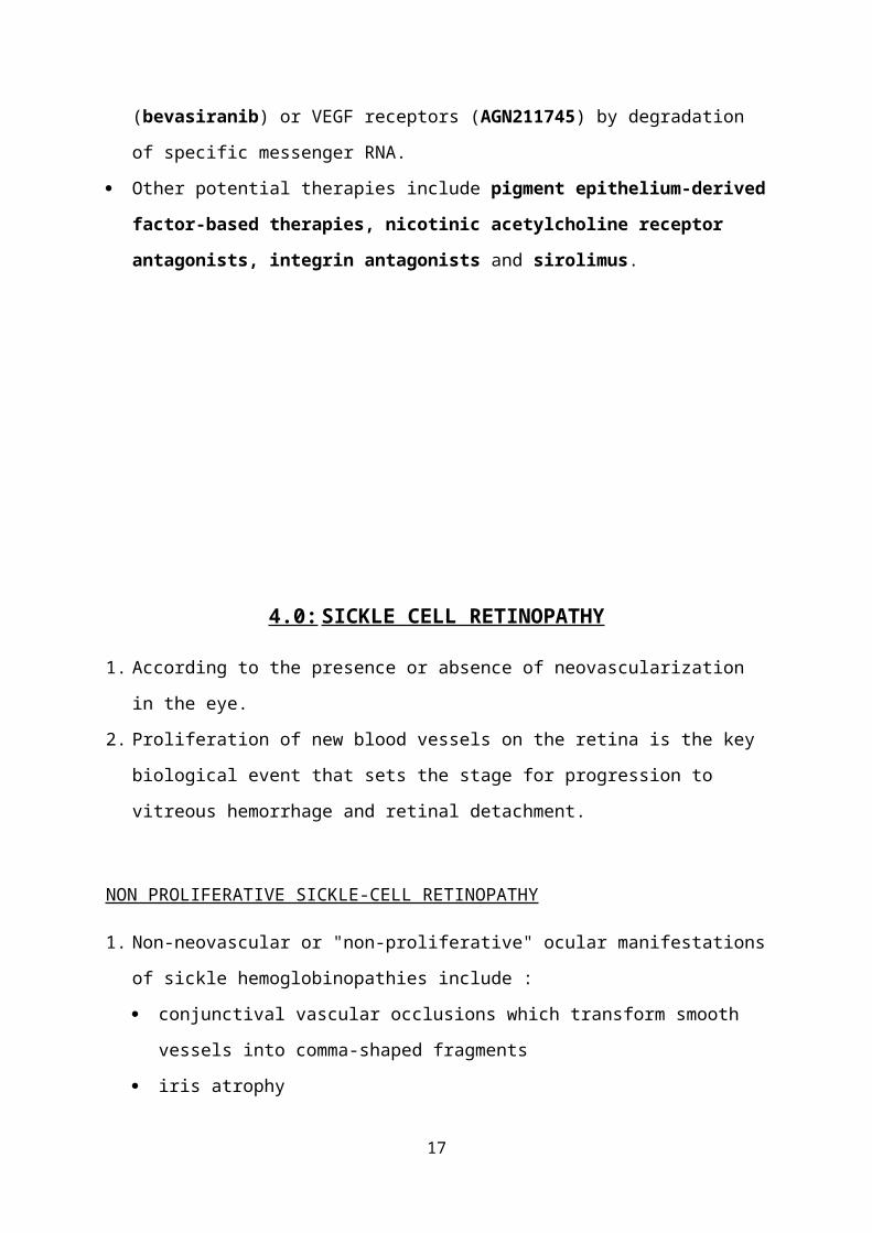
(bevasiranib) or VEGF receptors (AGN211745) by degradation
of specific messenger RNA.
Other potential therapies include pigment epithelium-derived
factor-based therapies, nicotinic acetylcholine receptor
antagonists, integrin antagonists and sirolimus.
4.0: SICKLE CELL RETINOPATHY
1. According to the presence or absence of neovascularization
in the eye.
2. Proliferation of new blood vessels on the retina is the key
biological event that sets the stage for progression to
vitreous hemorrhage and retinal detachment.
NON PROLIFERATIVE SICKLE-CELL RETINOPATHY
1. Non-neovascular or "non-proliferative" ocular manifestations
of sickle hemoglobinopathies include :
conjunctival vascular occlusions which transform smooth
vessels into comma-shaped fragments
iris atrophy
17

retinal "salmon patch" hemorrhages
retinal pigmentary changes
2. These clinical findings are readily apparent on dilated
ophthalmoscopy and all occur due to local vaso-occlusive
events but rarely have visual consequences.
PROLIFERATIVE SICKLE-CELL RETINOPATHY
1. Patients with sickle-cell hemoglobinopathy can develop
peripheral neovascularization when vascular occlusions
induce retinal ischemia.
2. The hemoglobin protein in patients with sickle-cell anemia
is altered by a single amino acid substitution of valine for
glutamine, causing polymerization of the deoxygenated
hemoglobin and sickling of the erythrocyte.
3. It is currently believed that the rigid, elongated, and
sickle-shaped erythrocyte causes mechanical microvascular
occlusions, resulting in retinal ischemia. Furthermore, the
sickled erythrocytes irritate the endothelial cells,
stimulating a cascade of inflammatory events leading to
vascular stasis and prearteriolar capillary occlusion.
4. The initiating event in the pathogenesis of proliferative
disease is thought to be peripheral retinal arteriolar
occlusions.
5. Local ischemia from repeated episodes of arteriolar closure
is presumed to trigger angiogenesis through the production
of endogenous vascular growth factors, such as vascular
endothelial growth factor (VEGF) and basic fibroblast growth
factor (bFGF)
18
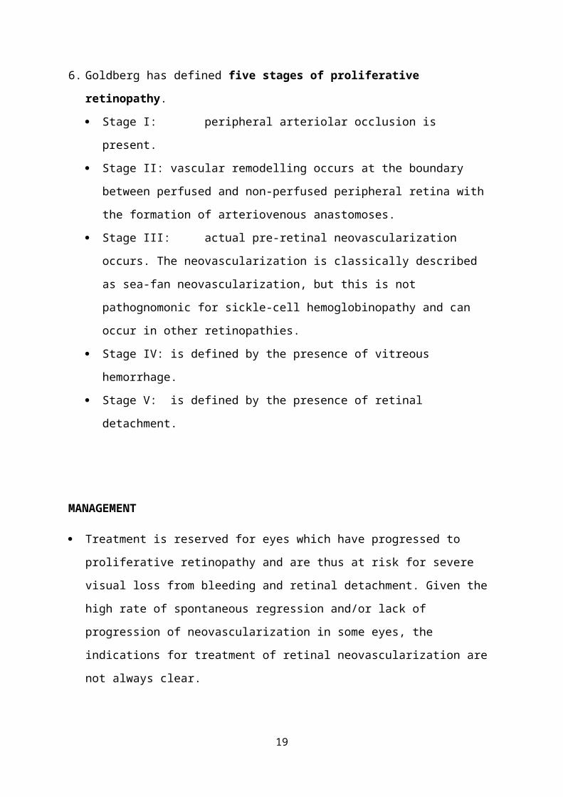
6. Goldberg has defined five stages of proliferative
retinopathy.
Stage I: peripheral arteriolar occlusion is
present.
Stage II: vascular remodelling occurs at the boundary
between perfused and non-perfused peripheral retina with
the formation of arteriovenous anastomoses.
Stage III: actual pre-retinal neovascularization
occurs. The neovascularization is classically described
as sea-fan neovascularization, but this is not
pathognomonic for sickle-cell hemoglobinopathy and can
occur in other retinopathies.
Stage IV: is defined by the presence of vitreous
hemorrhage.
Stage V: is defined by the presence of retinal
detachment.
MANAGEMENT
Treatment is reserved for eyes which have progressed to
proliferative retinopathy and are thus at risk for severe
visual loss from bleeding and retinal detachment. Given the
high rate of spontaneous regression and/or lack of
progression of neovascularization in some eyes, the
indications for treatment of retinal neovascularization are
not always clear.
19
Therapeutic intervention is usually recommended in cases of
bilateral proliferative disease, spontaneous hemorrhage,
large elevated neovascular fronds, rapid growth of
neovascularization, or cases in which the fellow eye has
already been lost to proliferative retinopathy.
The goal is early treatment aimed at inducing regression of
neovascular tissue before progression to bleeding and
retinal detachment. Proliferative sickle cell retinopathy
often spontaneously regresses due to auto-infarction of the
neovascular fronds.
Techniques such as diathermy, cryotherapy and laser
photocoagulation (least side effect) have all been used to
cause involution of neovascular lesions.
Cryotherapy is used to treat the peripheral ischemic retina
when laser treatment is not possible due to poor
visualization of the peripheral retina because of media
opacity.
Specific methods of laser application include direct
coagulation of feeder vessels, local scatter
photocoagulation with and without focal ablation of the
neovascular frond, and 360 degree peripheral scatter
delivery.
Feeder vessel photocoagulation has also been successful in
treating PSR by closing sea-fan vessels that persists
following scatter laser treatment.
Scatter laser photocoagulation of the ischemic retina and
tissue surrounding the sea-fan can cause regression of the
20

neovascular lesions and prevention of late-stage disease
complications by reducing angiogenic factors.
If retinal detachment and/or non-clearing vitreous
hemorrhage is present, surgical intervention is usually
required. Surgical techniques include vitrectomy with or
without the placement of a scleral buckle. Surgery carries a
significant risk of intraoperative and postoperative
complications, including severe ocular ischemia, recurrent
hemorrhage and elevated eye pressure.
Fig 4.0: In this picture, the left side is more peripheral and more anterior; the right is
more posterior. On the left side, the peripheral retina is completely non-perfused. The right
side shows a partially perfused retina. The brighter areas are the junction where the
neovascularization is leaking.
5.0: CENTRAL RETINAL VEIN OCCLUSION
21

1. It is the second most common retinal vascular disease after
diabetic retinopathy.
2. There are two distinct types, which are perfused or non-
ischemic and non-perfused or ischemic which are determined
according to the amount of retinal capillary ischemia seen
by the ophthalmologist on fluorescein angiography.
3. In ischemic retina, there are increased levels of the
products of hypoxia regulated genes, including vascular
endothelial growth factor (VEGF).
4. Such a distinction is relevant to the clinician, since two
thirds of patients with the ischemic type develop the
dreaded complications of macular edema, macular ischemia,
and neovascularization that lead to blindness.
5. There are some risk factors leading to occlusion:
Age
Hypertension
Hyperlipidaemia. (A total cholesterol > 6.5 mmol/l is
present in 35% of patients)
Diabetes mellitus
Oral contraceptive pill (The risk may be exacerbated by
thrombophilia)
Raised intraocular pressure
Smoking
22

Fig. 5.0: Central retinal vein occlusion showing dilation and tortuosity as well as
intraretinal hemorrhages in all four quadrants.
MANAGEMENT
No known effective medical treatment is available for either
the prevention of or the treatment of central retinal vein
occlusion (CRVO). Identifying and treating any systemic
medical problems to reduce further complications is
important. Because the exact pathogenesis of the CRVO is not
known, various medical modalities of treatment have been
advocated by multiple authors with varying success in
preventing complications and in preserving vision.
Advocated treatments are as follows:
o Anti-inflammatory agents.
o Isovolemic hemodilution.
o Systemic anticoagulation with warfarin, heparin, and
alteplase.
o Fibrinolytic agents.
o Systemic corticosteroids.
23

o Local anticoagulation with intravitreal injection of
alteplase.
o Intravitreal injection of ranibizumab.
o Intravitreal injection of bevacizumab.
Laser photocoagulation is the known treatment of choice in
the treatment of various complications associated with
retinal vascular diseases.
Chorioretinal venous anastomosis is performed by creating an
anastomosis to bypass the site of venous occlusion in the
optic disc (high risk of complications).
Radial optic neurotomy (RON) is a new surgical technique in
which a microvitreoretinal blade is used during pars plana
vitrectomy to relax the scleral ring around the optic nerve.
The central retinal artery and vein passes through the
narrow openings of the cribriform plate in the optic disc.
Vitrectomy is a technique in which the vitreous is
surgically removed along with removal of the posterior
hyaloid.
6.0: OCULAR INFLAMMATIONS & NEOVASCULARIZATION
A. INTERSTITIAL KERATITIS
Interstitial keratitis (IK) is a broad, descriptive term that
has become synonymous with syphilitic disease. Although
24

syphilis remains the leading cause of interstitial keratitis,
various bacterial, viral, parasitic, and autoimmune causes of
interstitial keratitis are known.
STAGES & SIGNS:
Progressive stage (2 weeks):
Infiltration and vascularization of cornea and it is triggered
by inflammation or surgery.
Florid stage:
Extensive infiltration and neovascularization occurs. Variable
corneal stromal neovascularization is depending on the
severity of the inflammation. Usually the neovascularization
occurs at the corneal limbus. It commonly occurs at the deep
stromal layer but it may be seen ath the superficial layer.
Stromal inflammation overlying the vessel often causes the
salmon-coloured patch due to pinkish color imparted by the
stromal vessels. Intrastromal haemorrhage may occur.
Regressive stage:
Scarring of the corneal stromal and collagen remodeling occur.
The superficial vessel resorb, snd the deeper vessel may
constrict , resulting in ghost vessels.
25

Fig. 6.0: Interstitial keratitis manifests with varying degrees of corneal opacification
and neovascularization. Neovascularization can cause orange-red areas (salmon patches).
TREATMENTS:
Topical and systemic corticosteroid.
IV Penincilin in cases of syphilis.
Keratoplasty in cases of corneal opacities.
B. UVEITIS
Uveitic neovascularization affects both uveal and retinal
tissues. It more commonly involves the retina––either through
neovascularization of the disk (NVD) or neovascularization
elsewhere (NVE)––and the choroid (choroidal
neovascularization, CNV).
Both NVD and NVE can be detected by fundoscopy and
angiography.
The new vessels tend to grow on the retinal surface. Later
these vessels will penetrate the internal limiting membrane
26

and posterior vitreous hyaloid face, and proliferate into the
vitreous. They are fragile and tend to bleed, resulting in
vitreous hemorrhage.
Retinal Neovascularization
1. Retinal neovascularization often occurs in uveal
inflammations that are associated with occlusive retinal
vasculitis, such as Behçet’s & Eales’ diseases.
2. Eales’ disease is notable for its presentation of retinal
periphlebitis, ischemia, and neovascularization. In
contrast, Behçet’s disease presents with intraocular
inflammation consisting of retinal vasculitis, oral and
mucosal ulcerations, and skin lesions.
3. Although retinal neovascularization rarely occurs in pars
planitis, both NVE and NVD leading to vitreous hemorrhage
have been reported.
4. Other uveitides reported to develop NVD and NVE include
systemic lupus erythematosus, sarcoidosis, birdshot
retinochoroidopathy and retinal vasculitis.
5. Infectious uveitis such as AIDS and others can also be
associated with retinal neovascularization.
27

Fig 6.1: Retinal vasculitis with hemorrhages and cotton wool spots.
Choroidal Neovascularization (CNV)
1. CNV includes choroidal and subchoroidal neovascularization.
CNV occurs in both infectious and noninfectious choroiditis.
2. Usually CNV presents with subretinal or intraretinal
hemorrhage, pigment epithelial detachment and a grayish-
green subretinal membrane. It is often associated with
chorioretinal scar.
3. CNV is an important feature of ocular histoplasmosis, which
manifests as the classic triad of discrete atrophic
choroidal scars in the macula or midperiphery (histo spots),
peripapillary atrophy, and choroidal neovascularization.
4. CNV has also been associated with serpiginous choroidopathy.
5. CNV is associated with a poor prognosis in Vogt-Koyanagi-
Harada syndrome (VKH).
6. Several infections are known to be complicated by CNV,
including toxoplasmosis and Candida choroidoretinitis.
28

Fig. 6.2: Retinal photograph of ocular histoplasmosis, with the clinical triad of
multiple white, atrophic choroidal scars, peripapillary pigment changes (dark spots around
optic disc of the eye)
Iris and Ciliary Neovascularization
1. Neovascularization in the iris and ciliary body is a rare
and ominous complication of chronic iridocyclitis.
2. An Iris neovascularization, or rubeosis irides, is new
vessel growth on the surface of the iris, which can result
in hyphema and/or rubeotic glaucoma.
29

Fig. 6.1: Fluorescein angiogram of the iris in case of chronic anterior uveitis.
MANAGEMENT
Currently available treatments for uveitic
neovascularization include immunotherapy, photocoagulation,
photodynamic therapy (PDT), and surgical excision of the CNV
neovascular membrane.
In uveitic neovascularization, there is always an
inflammatory component. Therefore, antiinflammatory and/or
immunosuppressive therapies should be prescribed.
Corticosteroids are antiinflammatory and antiangiogenic.
Local (including periocular) injections and vitreal
implants, as well as systemic corticosteroids, have been
reported to treat NVD effectively in sarcoidosis, multiple
sclerosis, juvenile rheumatoid arthritis, cyclitis, and
idiopathic uveitis.
Although corticosteroids alone may be less favorable for
CNV, additional immunosuppressive agents such as 30

cyclosporine and azathioprine have been used successfully to
regress neovascularization in sympathetic ophthalmia,
multifocal choroiditis, ocular histoplasmosis syndrome,
serpiginous choroiditis, and endogenous posterior uveits.
With the recent advance in understanding of angiogenesis,
antiangiogenic factors such as anti-VEGF agents are being
introduced as treatments for neovascularization, including
ocular neovascularization.
Laser photocoagulation and PDT for uveitic
neovascularization have been reported in a few small case
series. PDT appeared to stabilize or improve vision in a few
patients with subfoveal CNV secondary to multifocal
choroiditis and panuveitis.
The surgical mechanical excision of CNV associated with
ocular histoplasmosis and multifocal choroiditis produces
less damage to the photoreceptor cells than laser
photocoagulation. However, this surgical procedure is rather
difficult and requires skillful surgeons. Surgical
complications are frequent.
REFERENCES
- Ophthalmology Department, Cairo University. General
Ophthalmology for Undergraduate (6th edition) 2012-13.
31
- A.M. Joussen, T.W. Gardner, B. Kirchhof, S.J. Ryan
(Eds.). Retinal Vascular Disease; Springer-Verlag Berlin
Heidelberg New York 2007.
- Shaker A. Mousa. Angiogenesis Inhibitors and Stimulators
Potential Therapeutic Implications; Landes Bioscience &
Eurekah.com 2000.
- Chi-Chao Chan (MD) and Robert B. Nussenblatt (MD). Ocular
Inflammation and Neovascularization, Ophthalmology:
Ocular Angiogenesis: Diseases, Mechanisms, and
Therapeutics.
- Leila Laatikainen. Vascular Changes in the Iris in
Chronic Anterior Uveitis, British Journal of
Ophthalmology 1979.
- http://www.ncbi.nlm.nih.gov/pubmed/18484796
- http://www.stlukeseye.com/conditions/cnvm.html
- http://www.lucentis.com/hcp/amd/about-ranibizumab-
amd.html
- http://www.retinalphysician.com/articleviewer.aspx?
articleID=102207
- http://sickle.bwh.harvard.edu/eye.html
- http://www.medscape.com/ophthalmology
- http://www.eyecalcs.com/DWAN/pages/v4/v4c042.html
- http://shop.onjoph.com/catalog/index.php
32
Copyright © 2022 FDOKUMEN