Inflammatory choroidal neovascularization associated with ...
7
CASE REPORT Open Access Inflammatory choroidal neovascularization associated with immunoglobulin G4-related disease: a case report Chirag Jhaveri 1,2* and Giovanni Campagna 3 Abstract Background: Immunoglobulin G4-related disease is a recently recognized condition with pathologic features that are consistent across a wide range of organ systems. Orbital manifestations of this disease entity typically involve the lacrimal gland and lacrimal duct, extraocular muscles, orbital soft tissue, and sclera. Here, the authors report the first known case of inflammatory choroidal neovascular membrane associated with immunoglobulin G4-related disease and offer suggestions for clinical management of this enigmatic condition. Case presentation: A 38-year-old Caucasian man with a history of recurrent tonsillitis presented with blurry vision in his left eye of 1-week duration and was diagnosed as having inflammatory choroidal neovascular membrane. An infectious workup was negative, but his serum immunoglobulin G4 level was elevated at 248 mg/dL (reference 4–86), and a subsequent tonsillectomy for a repeat episode of tonsillitis revealed increased immunoglobulin G4 staining on histopathology, thus confirming the diagnosis of immunoglobulin G4-related disease. The inflammatory choroidal neovascular membrane was treated with intravitreal bevacizumab injections and orally administered prednisone resulting in improved visual acuity and choroidal neovascular membrane regression. He later received rituximab infusions for immunoglobulin G4-related disease. Conclusions: We report a case of choroidal neovascularization associated with immunoglobulin G4-related disease, a chronic inflammatory condition whose ophthalmic manifestations typically include dacryoadenitis, orbital myositis, or scleritis. This is the first reported instance of inflammatory choroidal neovascular membrane associated with immunoglobulin G4-related disease. Early detection of this disease is important to avoid organ damage and potential complications, so clinicians should maintain an index of suspicion for this condition when inflammatory choroidal neovascular membrane is observed. Keywords: Choroidal neovascular membrane, IgG4, Immunoglobulin G4-related disease, Immunoglobulin G4- related ophthalmic disease, Inflammatory choroidal neovascularization © The Author(s). 2020 Open Access This article is licensed under a Creative Commons Attribution 4.0 International License, which permits use, sharing, adaptation, distribution and reproduction in any medium or format, as long as you give appropriate credit to the original author(s) and the source, provide a link to the Creative Commons licence, and indicate if changes were made. The images or other third party material in this article are included in the article's Creative Commons licence, unless indicated otherwise in a credit line to the material. If material is not included in the article's Creative Commons licence and your intended use is not permitted by statutory regulation or exceeds the permitted use, you will need to obtain permission directly from the copyright holder. To view a copy of this licence, visit http://creativecommons.org/licenses/by/4.0/. The Creative Commons Public Domain Dedication waiver (http://creativecommons.org/publicdomain/zero/1.0/) applies to the data made available in this article, unless otherwise stated in a credit line to the data. * Correspondence: [email protected] 1 Retina Consultants of Austin and Retina Research Center, 3705 Medical Parkway, Suite 410, Austin, TX 78705, USA 2 Department of Ophthalmology, The University of Texas at Austin Dell Medical School, Austin, TX, USA Full list of author information is available at the end of the article Jhaveri and Campagna Journal of Medical Case Reports (2020) 14:89 https://doi.org/10.1186/s13256-020-02431-8
-
Upload
khangminh22 -
Category
Documents
-
view
1 -
download
0
Transcript of Inflammatory choroidal neovascularization associated with ...
CASE REPORT Open Access
Inflammatory choroidal neovascularizationassociated with immunoglobulin G4-relateddisease: a case reportChirag Jhaveri1,2* and Giovanni Campagna3
Abstract
Background: Immunoglobulin G4-related disease is a recently recognized condition with pathologic features thatare consistent across a wide range of organ systems. Orbital manifestations of this disease entity typically involvethe lacrimal gland and lacrimal duct, extraocular muscles, orbital soft tissue, and sclera. Here, the authors report thefirst known case of inflammatory choroidal neovascular membrane associated with immunoglobulin G4-relateddisease and offer suggestions for clinical management of this enigmatic condition.
Case presentation: A 38-year-old Caucasian man with a history of recurrent tonsillitis presented with blurry visionin his left eye of 1-week duration and was diagnosed as having inflammatory choroidal neovascular membrane. Aninfectious workup was negative, but his serum immunoglobulin G4 level was elevated at 248 mg/dL (reference4–86), and a subsequent tonsillectomy for a repeat episode of tonsillitis revealed increased immunoglobulin G4staining on histopathology, thus confirming the diagnosis of immunoglobulin G4-related disease. The inflammatorychoroidal neovascular membrane was treated with intravitreal bevacizumab injections and orally administeredprednisone resulting in improved visual acuity and choroidal neovascular membrane regression. He later receivedrituximab infusions for immunoglobulin G4-related disease.
Conclusions: We report a case of choroidal neovascularization associated with immunoglobulin G4-related disease,a chronic inflammatory condition whose ophthalmic manifestations typically include dacryoadenitis, orbital myositis,or scleritis. This is the first reported instance of inflammatory choroidal neovascular membrane associated withimmunoglobulin G4-related disease. Early detection of this disease is important to avoid organ damage andpotential complications, so clinicians should maintain an index of suspicion for this condition when inflammatorychoroidal neovascular membrane is observed.
Keywords: Choroidal neovascular membrane, IgG4, Immunoglobulin G4-related disease, Immunoglobulin G4-related ophthalmic disease, Inflammatory choroidal neovascularization
© The Author(s). 2020 Open Access This article is licensed under a Creative Commons Attribution 4.0 International License,which permits use, sharing, adaptation, distribution and reproduction in any medium or format, as long as you giveappropriate credit to the original author(s) and the source, provide a link to the Creative Commons licence, and indicate ifchanges were made. The images or other third party material in this article are included in the article's Creative Commonslicence, unless indicated otherwise in a credit line to the material. If material is not included in the article's Creative Commonslicence and your intended use is not permitted by statutory regulation or exceeds the permitted use, you will need to obtainpermission directly from the copyright holder. To view a copy of this licence, visit http://creativecommons.org/licenses/by/4.0/.The Creative Commons Public Domain Dedication waiver (http://creativecommons.org/publicdomain/zero/1.0/) applies to thedata made available in this article, unless otherwise stated in a credit line to the data.
* Correspondence: [email protected] Consultants of Austin and Retina Research Center, 3705 MedicalParkway, Suite 410, Austin, TX 78705, USA2Department of Ophthalmology, The University of Texas at Austin DellMedical School, Austin, TX, USAFull list of author information is available at the end of the article
Jhaveri and Campagna Journal of Medical Case Reports (2020) 14:89 https://doi.org/10.1186/s13256-020-02431-8

BackgroundA choroidal neovascular membrane (CNVM) is a patho-logical growth of new blood vessels that originate in thechoroid through a break in Bruch’s membrane into thesub-retinal pigment epithelium (RPE) space or sub-retinalspace. The differential diagnosis of new CNVM is broadand includes a wide range of conditions such as age-relatedmacular degeneration, myopic degeneration, and ocularinflammation [1]. When CNVM is associated with inflam-matory conditions of the posterior segment, it is referred toas inflammatory CNVM. Inflammatory CNVM can occurfrom a disruption in the interface between Bruch’s mem-brane and the RPE, from an angiogenic stimulus mediatedby local inflammation, or from a combination of both [2].Classically, inflammatory CNVM presents as new-onsetdistortion or metamorphopsia, sometimes leading to dimin-ution of vision or a scotoma. Due to a lack of randomizedcontrolled clinical trials regarding treatment of inflamma-tory CNVM, there is no consensus or set of evidence-basedguidelines for the management of this condition. Manage-ment approaches include observation, intravitreal anti-vascular endothelial growth factor (VEGF) injections,photodynamic therapy, corticosteroids and other immuno-suppressive agents, and laser photocoagulation [1].The authors report a case of a young adult man who de-
veloped inflammatory CNVM in his left eye (OS) associ-ated with immunoglobulin G4 (IgG4)-related disease(IgG4-RD), a chronic immune-mediated fibroinflamma-tory systemic disease that can affect any organ in the bodyand is characterized by focal or diffuse organ infiltrationby IgG4-bearing plasma cells. The inflammatory CNVMwas treated with intravitreal bevacizumab injections andorally administered prednisone, resulting in improvedvisual acuity and ultimately CNVM regression. He laterreceived rituximab infusions for IgG4-RD. This representsthe first reported case of CNVM associated with IgG4-RDin the English ophthalmic literature and sheds light on anotherwise uncommon etiology of inflammatory CNVM.Intraocular findings in cases of IgG4-RD are variable andhave been sparsely described until recently, and this case
augments the growing body of literature by expanding thespectrum of disease presentation. This case shouldencourage clinicians to maintain an index of suspicion forIgG4-RD when inflammatory CNVM is observed.
Case presentationA 38-year-old Caucasian man presented with worseningcentral vision OS of 1-week duration. His medical his-tory was significant for recurrent tonsillitis but otherwisenegative. He had been diagnosed as having “strep throat”twice in the last 3 months and prescribed amoxicillin500 mg twice per day for 10 days both times withoutformal rapid strep testing. He recovered from eachinstance of tonsillitis within 5–7 days. He had no historyof ophthalmic conditions or prior surgery. He deniedtaking any daily medications or using eye drops. He grewup in Kansas City, Missouri, United States of America,and owned a dog but denied recent travel or exposure toticks. He and his wife have lived in Austin, Texas, USA,for several years. He works as an information technologyspecialist. He is a former cigarette smoker who quit 8years ago, he drinks alcohol socially, and he denied anyhistory of illicit drug use. His family history is significantfor maternal diabetes and paternal hypertension.His uncorrected visual acuity was 20/20 in his right
eye (OD) and 20/50 OS. His pupils were equally roundand reactive, and intraocular pressures were normal.The ocular adnexa were normal on external examin-ation. The anterior chamber and vitreous were quiet bi-laterally. A dilated fundus examination OD wasunremarkable. A dilated fundus examination OS showeda normal optic nerve, scant hyperpigmentation at theinferotemporal border of the disc, and a whitish sub-retinal lesion temporal to the fovea with a small associ-ated hemorrhage. Ultra-widefield fundus photography isshown in Fig. 1. A general medical examination was un-remarkable. He was afebrile with a temperature of36.7 °C (98.0 °F), heart rate of 73 beats per minute, rightarm cuff blood pressure of 113/70 mmHg, respiratoryrate of 16 breaths per minute, and 98% oxygen
Fig. 1 Fundus photography of the right eye (a) and left eye (b) with an ultra-widefield retinal camera. A small cream-colored sub-retinal lesiontemporal to the fovea with a raised appearance is appreciated in the left eye
Jhaveri and Campagna Journal of Medical Case Reports (2020) 14:89 Page 2 of 7
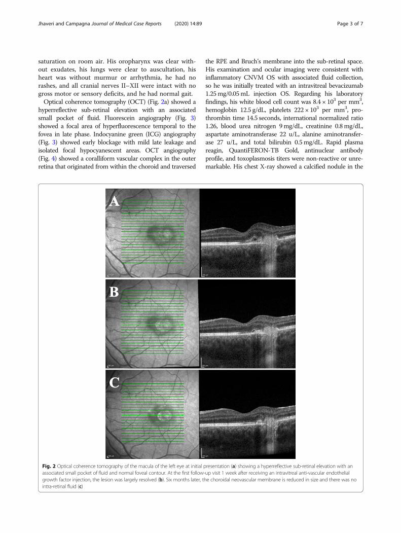
saturation on room air. His oropharynx was clear with-out exudates, his lungs were clear to auscultation, hisheart was without murmur or arrhythmia, he had norashes, and all cranial nerves II–XII were intact with nogross motor or sensory deficits, and he had normal gait.Optical coherence tomography (OCT) (Fig. 2a) showed a
hyperreflective sub-retinal elevation with an associatedsmall pocket of fluid. Fluorescein angiography (Fig. 3)showed a focal area of hyperfluorescence temporal to thefovea in late phase. Indocyanine green (ICG) angiography(Fig. 3) showed early blockage with mild late leakage andisolated focal hypocyanescent areas. OCT angiography(Fig. 4) showed a coralliform vascular complex in the outerretina that originated from within the choroid and traversed
the RPE and Bruch’s membrane into the sub-retinal space.His examination and ocular imaging were consistent withinflammatory CNVM OS with associated fluid collection,so he was initially treated with an intravitreal bevacizumab1.25mg/0.05mL injection OS. Regarding his laboratoryfindings, his white blood cell count was 8.4 × 10³ per mm3,hemoglobin 12.5 g/dL, platelets 222 × 103 per mm3, pro-thrombin time 14.5 seconds, international normalized ratio1.26, blood urea nitrogen 9mg/dL, creatinine 0.8mg/dL,aspartate aminotransferase 22 u/L, alanine aminotransfer-ase 27 u/L, and total bilirubin 0.5mg/dL. Rapid plasmareagin, QuantiFERON-TB Gold, antinuclear antibodyprofile, and toxoplasmosis titers were non-reactive or unre-markable. His chest X-ray showed a calcified nodule in the
Fig. 2 Optical coherence tomography of the macula of the left eye at initial presentation (a) showing a hyperreflective sub-retinal elevation with anassociated small pocket of fluid and normal foveal contour. At the first follow-up visit 1 week after receiving an intravitreal anti-vascular endothelialgrowth factor injection, the lesion was largely resolved (b). Six months later, the choroidal neovascular membrane is reduced in size and there was nointra-retinal fluid (c)
Jhaveri and Campagna Journal of Medical Case Reports (2020) 14:89 Page 3 of 7

upper lung field of his right lung, later confirmed with chestcomputed tomography.One week after initial presentation and treatment with
bevacizumab, his visual acuity improved to 20/25 OS,and OCT showed improvement of the sub-retinal fluid(Fig. 2b). A 2-week taper of orally administered prednis-one starting at 60 mg per day and ending at 5 mg perday was prescribed to treat the inflammatory componentof the CNVM. He was referred to pulmonology, and anadditional workup revealed elevated serum IgG4 levelsat 248mg/dL (reference 4–86). He was also referred tootolaryngology for his history of recurrent tonsillitis, forwhich he underwent bilateral tonsillectomy. The speci-men showed significant staining for IgG4 on histopath-ology. After completion of the first orally administeredprednisone course, he developed left upper eyelid bleph-aritis, which did not resolve after initial treatment withantibiotic ointment and warm compresses. During thenext 4 months, he received two more intravitreal bevaci-zumab 1.25 mg/0.05 mL injections for recurrence of sub-retinal fluid associated with inflammatory CNVM in thesetting of biopsy-proven IgG4-RD, as well as a 1-monthtaper of orally administered prednisone starting at 60 mgper day and ending at 5 mg per day, after which theblepharitis resolved. He experienced improvement in hisvisual acuity with each injection. Six months after initialpresentation, he was started on rituximab infusions1000 mg every 2 weeks in order to decrease circulatingIgG4 levels. The CNVM had decreased in size by thispoint (Fig. 2c). He is seen by his retina subspecialist (CJ)every 2 months for examination and anti-VEGF therapyas needed.
Discussion and conclusionsIn summary, we present the first reported case ofinflammatory CNVM in the setting of IgG4-RD. Treat-ment was achieved with anti-VEGF injections as well assteroid tapers in order to stop sub-retinal fluid accumu-lation as well as inflammatory components, respectively.In this discussion we provide a thorough review of thenatural history of IgG4-RD and describe how this casediffers from what has been published in the literatureregarding ophthalmic manifestations of the disease.IgG4-RD is a recently recognized condition with patho-
logic features that are consistent across a wide range oforgan systems. While the exact mechanism of the diseaseremains unknown, the prevailing contention is that IgG4-RD is mediated primarily by T helper type 2 cells, whichsecrete different interleukins that activate plasma cells toproduce IgG4, as well as regulatory T cells [3]. The firstreported instance of this condition was described by Sarleset al. [4] and involved a case of pancreatitis with hypergam-maglobulinemia, but IgG4-RD has since been discovered asthe cause of inflammatory disease in many organs including
Fig. 3 Fluorescein angiography (left column) and indocyanine greenangiography (right column) of the patient’s left eye. Fluoresceinangiography shows normal arteriovenous transit and a focal area ofhyperfluorescence temporal to the fovea particularly in late-phase,consistent with a choroidal neovascular membrane, and transmissiondefects consistent with retinal pigment epithelium changes. Indocyaninegreen angiography shows early blockage with mild late leakagecorresponding to an inflammatory lesion with a choroidal neovascularmembrane as well as some isolated focal hypocyanescent areas
Jhaveri and Campagna Journal of Medical Case Reports (2020) 14:89 Page 4 of 7

the central nervous system, orbits, ears, nose, throat,salivary glands, thyroid gland, lungs, kidneys, and others.Ophthalmic and orbital manifestations of IgG4-RD, nowreferred to as IgG4-related ophthalmic disease (IgG4-ROD), typically involve the lacrimal gland and lacrimalduct, extraocular muscles, orbital soft tissue, and sclera, aswell as the cranial nerves and their branches [5, 6]. WhereasIgG4-RD typically occurs more often in men and targetsmiddle-aged men [7], men and women tend to be affectedequally in cases of IgG4-ROD [5]. Furthermore, in a recentsingle-center retrospective review, 71% of patients withbiopsy-confirmed IgG4-ROD had bilateral ophthalmic ororbital disease, and 71% had extra-orbital involvement [8].IgG4-ROD remains a diagnostic challenge for several
reasons: (1) the epidemiology of the condition is poorlydescribed due to its recent arrival in the diagnosticlexicon, (2) the presentation is variable, and (3) a largenumber of other inflammatory conditions are known toaffect the orbit and adnexa. Ophthalmic manifestationsof pathologically high levels of IgG4 include dacryoade-nitis, blepharitis, scleritis, cranial nerve involvement, andorbital soft tissue inflammation [3, 5, 6, 8, 9]. Otherdocumented presentations of IgG4-ROD include idio-pathic orbital inflammation (IOI) and idiopathic orbital
myositis [10]. Proptosis is a common feature in thesetwo conditions. IOI can affect either the entire orbit orspecific components such as the extraocular muscles,lacrimal system, optic nerve, or sclera. Histopathologydemonstrates a benign, nonspecific, polymorphic inflam-matory infiltrate, sometimes accompanied by sclerosis[10]. Without specific IgG4 staining, it is impossible todifferentiate IOI from IgG4-RD, and it is possible that asubstantial proportion of cases of presumed IOI are, infact, IgG4-ROD. Idiopathic orbital myositis is known tocommonly present with diplopia and pain with eyemovements. In their review of 15 individuals with in-flammatory CNVM, D’Souza et al. concluded that thepresence of concurrent active inflammation is not anessential factor for development of inflammatory CNVM[11], and that CNVM by definition is merely a CNVMassociated with a condition that may manifest as poster-ior segment inflammation, which is possible in IgG4-ROD. In the first published case of choroidal lesionsassociated with IgG4-ROD, no signs of intraocularinflammation were observed [12]. Intraocular inflamma-tion in the form of anterior chamber cell and flare orvitritis was not observed in this patient, but these find-ings may be subtle, and our patient presented relatively
Fig. 4 Optical coherence tomography angiography of the patient’s left eye. This shows a coralliform vascular complex in the outer retina thatoriginated from within the choroid and traversed the retinal pigment epithelium and Bruch’s membrane into the sub-retinal space
Jhaveri and Campagna Journal of Medical Case Reports (2020) 14:89 Page 5 of 7

late in the course of his symptomatology. Consideringour patient’s history, ophthalmic examination, elevatedserum IgG4 and confirmatory biopsy, and resolution ofsymptoms with intravitreal anti-VEGF injections andorally administered steroids, the authors are confidentthat IgG4-RD is the cause of this patient’s inflammatoryCNVM and blepharitis OS. Therefore, this case reportshowcases an unusual presentation of IgG4-ROD andexpands the current spectrum of this enigmatic condition.Infectious etiologies of CNVM, such as syphilis, tubercu-
losis, and toxoplasmosis, were ruled out with a thoroughlaboratory workup. Although our patient was originallyfrom the Midwestern USA, his ophthalmic examinationwas not consistent with presumed ocular histoplasmosissyndrome, which often presents with the clinical triad of:(1) discrete atrophic choroidal scars in the macula orperiphery, (2) peripapillary atrophy, and (3) choroidal neo-vascularization. Our patient’s examination was devoid ofatrophic choroidal scars, or “histo spots.” In addition, ocularhistoplasmosis syndrome tends to produce early hypercya-nescence on ICG angiography, but this patient’s ICG angi-ography revealed early blockage with late leakage and focalhypocyanescence. While the calcified pulmonary nodulewas not biopsied, serum IgG4 levels and increased IgG4staining on subsequent tonsillectomy histopathology con-firmed the diagnosis of IgG4-RD in this patient. However,the presence of a pulmonary lesion does warrant consider-ation for histoplasmosis as well as sarcoidosis, which tendto have skin involvement (for example erythema nodosum)and bilateral pulmonary hilar adenopathy, and granuloma-tosis with polyangiitis, which frequently demonstratessecondary orbital involvement from the nearby ethmoid ormaxillary sinuses [9].Initially our patient was treated with intravitreal bevaci-
zumab injection OS to reduce the sub-retinal fluid associ-ated with inflammatory CNVM. He required additionalinjections every 2–3months throughout the course oftreatment described in this case report, and he continuesto be examined regularly by his retina specialist. Theaddition of orally administered prednisone to his treat-ment regimen after rule-out of infectious etiologies wascrucial in managing the inflammatory component of hiscondition. His inflammatory CNVM showed signs ofregression on clinical examination following immunosup-pressive therapy with tapered steroids, and the frequencyand quantity of new sub-retinal fluid decreased over time.With regard to his left upper eyelid blepharitis, his poorresponse to antibiotic ointment and warm compresses isconsistent with IgG4-ROD, as well, and he experiencedresolution of blepharitis after completion of an orallyadministered prednisone course. For patients with recur-rent or refractory IgG4-RD, B cell depletion with rituxi-mab appears to be a useful approach [3, 7, 8], and ourcase report is no exception. Rituximab infusions were
initiated shortly after tonsillectomy in order to lower hiselevated levels of circulating IgG4, and recurrence ofdisease was not observed.This is the first reported case of inflammatory CNVM
associated with IgG4-RD, a relatively new addition to thediagnostic lexicon that was perhaps previously misdiag-nosed as IOI or some other variant of inflammatory orbitaldisease prior to quantitative IgG4 serum measurementsand histopathological biopsy staining for IgG4. Despite theunusual presentation of IgG4-ROD as inflammatoryCNVM with sub-retinal fluid and later with blepharitis, thecourse of disease and response to immunosuppressivetreatment with steroids (and anti-VEGF therapy) describedin this case report shows consistency with more commonmanifestations of ophthalmic IgG4-RD. Early detection ofthis disease is important to avoid organ damage and poten-tial complications, so clinicians should maintain an index ofsuspicion for this condition when new inflammatoryCNVM is observed in an otherwise atypical setting.
AbbreviationsCNVM: Choroidal neovascular membrane; VEGF: Vascular endothelial growthfactor; IgG4: Immunoglobulin G4; IgG4-RD: Immunoglobulin G4-related dis-ease; OD: Right eye; OS: Left eye; OCT: Optical coherence tomography;ICG: Indocyanine green; IgG4-ROD: Immunoglobulin G4-related ophthalmicdisease; IOI: Idiopathic orbital inflammation; RPE: Retinal pigment epithelium
AcknowledgementsNot applicable.
Authors’ contributionsConception and design of the work: CJ. Clinical data collection: CJ and GC.Imaging data collection and analysis: CJ. Drafting the article: GC. Criticalrevision of the article: CJ and GC. Agreement to be accountable for allaspects of the work in ensuring that questions related to the accuracy orintegrity of any part of the work are appropriately investigated and resolved:CJ and GC. All authors read and approved the final manuscript.
FundingThere is no funding to declare.
Availability of data and materialsThe datasets used and/or analyzed during the current study are availablefrom the corresponding author on reasonable request.
Ethics approval and consent to participateThe authors have no ethical conflicts to disclose. Written informed consentwas obtained.
Consent for publicationWritten informed consent was obtained from the patient for publication ofthis case report and any accompanying images. A copy of the writtenconsent is available for review by the Editor-in-Chief of this journal.
Competing interestsThe authors declare that they have no competing interests.
Author details1Retina Consultants of Austin and Retina Research Center, 3705 MedicalParkway, Suite 410, Austin, TX 78705, USA. 2Department of Ophthalmology,The University of Texas at Austin Dell Medical School, Austin, TX, USA.3Transitional Year Residency Program, The University of Texas at Austin DellMedical School, Austin, TX, USA.
Jhaveri and Campagna Journal of Medical Case Reports (2020) 14:89 Page 6 of 7
Received: 27 December 2019 Accepted: 2 June 2020
References1. Agarwal A, Invernizzi A, Singh RB, Foulsham W, Aggarwal K, Handa S,
Agrawal R, Pavesio C, Gupta V. An update on choroidal inflammatoryneovascularization: epidemiology, multimodal imaging, and management.J Ophthalmic Inflamm Infect. 2018;8(1):13.
2. Dhingra N, Kelly S, Majid MA, Bailey CB, Dick AD. Inflammatory choroidalneovascular membrane in posterior uveitis-pathogenesis and treatment.Indian J Ophthalmol. 2010;58(1):3–10.
3. Obiorah IE, Velasquez AH, Ozdemirli M. The clinicopathologic spectrum ofIgG4-related disease. Balkan Med J. 2018;35(4):292–300.
4. Sarles H, Sarles JC, Muratore R, Guien C. Chronic inflammatory sclerosisof the pancreas – an autonomous pancreatic disease? Am J Dig Dis.1961;6:688–98.
5. Kubota T, Moritani S. Orbital IgG4-related disease: clinical features anddiagnosis. ISRN Rheumatol. 2012;2012:412896.
6. Kim EC, Lee SJ, Hwang HS, Kim J, Kim MS. Bilateral diffuse scleritis as a firstmanifestation of immunoglobulin G4-related sclerosing pachymeningitis.Can J Ophthalmol. 2013;48(2):e31–3.
7. Stone JH, Zen Y, Deshpande V. IgG4-related disease. N Engl J Med. 2012;366(6):539–51.
8. Wallace ZS, Deshpande V, Stone JH. Ophthalmic manifestations of IgG4-related disease: single-center experience and literature review. SeminArthritis Rheum. 2014;43(6):806–17.
9. Golnik KC. Neuro-ophthalmologic manifestations of systemic disease:rheumatologic/inflammatory. Ophthalmol Clin N Am. 2004;17(3):389–96.
10. Wallace ZS, Khosroshahi A, Jakobiec FA, Deshpande V, Hatton MP, Ritter J,Ferry JA, Stone JH. IgG4-related systemic disease as a cause of “idiopathic”orbital inflammation, including orbital myositis, and trigeminal nerveinvolvement. Surv Ophthalmol. 2012;57(1):26–33.
11. D’Souza P, Ranjan R, Babu U, Kanakath AV, Saravanan VR. Inflammatorychoroidal neovascular membrane: long-term visual and anatomicaloutcomes after intravitreal anti-vascular endothelial growth factor therapy.Retina. 2018;38(7):1307–15.
12. Gange WS, Holland SM, De Alba F. IgG4-related ophthalmic disease presentingas choroidal and orbital lesions. Retin Cases Brief Rep. 2019;13(3):283–6.
Publisher’s NoteSpringer Nature remains neutral with regard to jurisdictional claims inpublished maps and institutional affiliations.
Jhaveri and Campagna Journal of Medical Case Reports (2020) 14:89 Page 7 of 7