Intravitreal Bevacizumab for the Treatment of Chronic or Recurrent Central Serous Chorioretinopathy
Upload
independentCategory
view
1download
0
Intravitreal Bevacizumab for ChoroidalNeovascularization Caused by AMD (IBeNA Study):Results of a Phase 1 Dose-Escalation Study
Rogerio A. Costa,1,2 Rodrigo Jorge,2 Daniela Calucci,1 Jose A. Cardillo,1 Luiz A. S. Melo, Jr,1
and Ingrid U. Scott3
PURPOSE. To evaluate the safety of three dose regimens ofintravitreal bevacizumab (Avastin; Genentech, Inc., South SanFrancisco, CA) for the management of choroidal neovascular-ization (CNV) associated with age-related macular degenera-tion (AMD).
METHODS. This was a prospective, nonrandomized open-labelstudy of 45 patients with AMD and subfoveal CNV. A standard-ized ophthalmic evaluation was performed at baseline and atweeks 1, 6, and 12 (�1) after a single intravitreous injection(1.0, 1.5, or 2.0 mg) of bevacizumab. Main outcomes measuresinclude clinical evidence of toxicity and complications.Changes in best corrected visual acuity (BCVA) and lesioncharacteristics–macular morphology were also evaluated.
RESULTS. The most common adverse events were conjunctivalhyperemia and subconjunctival hemorrhage at the injectionsite. Mean BCVA improved from baseline throughout the study(P � 0.001; ANOVA with Geisser-Greenhouse correction).Compared with baseline, BCVA was improved at week 1 (P �0.001), week 6 (P � 0.001), and week 12 (P � 0.001; Dunnetttest). At week 12, the lesion area and CNV area were stable ordecreased in 79.1% (34/43) and in 74.4% (32/43) of patients,respectively, with no deterioration of macular architectureobserved in 83.7% (36/43). A dose-related change in BCVA (inEarly Treatment Diabetic Retinopathy Study [ETDRS] lines)was observed at week 12 (1.0 mg [�0.3 line]; 1.5 mg [�0.6line]; and 2.0 mg [�1.0 line]; P � 0.02; nonparametric test forordered groups).
CONCLUSIONS. A single intravitreal bevacizumab injection waswell tolerated and, except for minor transient local adverseevents, no other adverse events were observed. In the short-term, treatment was associated with vision stabilization orimprovement and no unfavorable neovascular lesion–macularchanges in most patients. (Invest Ophthalmol Vis Sci. 2006;47:4569–4578) DOI:10.1167/iovs.06-0433
Reports of the beneficial effect of photodynamic therapy(PDT) for choroidal neovascularization (CNV) associated
with age-related macular degeneration (AMD) launched thecontemporary era of management of choroidal neovasculardiseases.1–3 Contrary to the principles arising from the MacularPhotocoagulation Study (i.e., photothermal destruction of theentire neovascular lesion),4,5 it has been demonstrated that, byan intravenous injection of a photosensitizer that accumulatesin neovascular tissue and subsequent low-irradiance specificlight application, disease activity can be modulated after sev-eral PDT treatment sessions.6–13 Another step toward the im-plementation of “modulation of disease activity” rather thanlesion destruction occurred at the end of 2004 with the ap-proval of the antiangiogenic drug pegaptanib (Macugen; Eye-tech Pharmaceuticals, New York, NY), an aptamer that bindsvascular endothelial growth factor (VEGF) isoform 165, fortreatment of all forms of neovascular AMD.14,15 However,despite repeated, high-cost treatment sessions of either PDTor pegaptanib, patients generally lose vision over time be-cause the growth of neovascular lesions is slowed, but notprevented.3,6 –13,15
Even if the overall benefit of pegaptanib therapy in neovas-cular AMD can be considered modest,15 it has been of majorsignificance in providing proof of concept for the use of anti-angiogenic therapy in neovascular AMD. Ranibizumab (Lucen-tis; Genentech, Inc., South San Francisco, CA) is another anti-angiogenic agent currently under investigation for treatment ofneovascular AMD. Unlike pegaptanib, ranibizumab binds allVEGF isoforms.16–21 Preliminary 2-year data from one phase IIIstudy (the MARINA Trial), which included patients with AMDwho had “minimally classic CNV” and “occult CNV” neovascu-lar lesions, demonstrated that at least 90% of patients treatedwith ranibizumab maintained or improved vision comparedwith approximately 53% of control patients.22
Intravitreal ranibizumab is derived from the same human-ized monoclonal anti-VEGF antibody of bevacizumab (Avastin;Genentech, Inc.), which is the full-length humanized monoclo-nal anti-VEGF neutralizing antibody designed for intravenousadministration and approved for the treatment of metastaticcolorectal cancer.23–27 Although preclinical studies demon-strated lack of retinal penetration of the full-length antibodywhen injected intravitreally,28 rapid and favorable macularremodeling was observed in the clinical setting of neovascularAMD and macular edema due to central retinal vein occlusionafter a single intravitreous injection of 1.25 mg of bevaci-zumab,29,30 thus bringing to light a new perspective of intra-vitreal angiogenic pharmacomodulation by using the full-length antibody.
Extensive preclinical data suggesting an important role ofVEGF in choroidal neovascular diseases,31–54 and reported ben-efits of anti-VEGF therapy with pegaptanib and ranibizumab inhumans,14,15,20,21 coupled with the encouraging initial clinicalreports using intravitreal bevacizumab,29,30 prompted us toconduct a phase I clinical trial designed to investigate the safety
From the 1U.D.A.T. Macular Imaging and Treatment Division,Hospital de Olhos de Araraquara, Araraquara, SP, Brazil; the 2Retina andVitreous Section, Department of Ophthalmology, School of Medicineof Ribeirao Preto, University of Sao Paulo, Ribeirao Preto, SP, Brazil;and the 3Departments of Ophthalmology and Health Evaluation Sci-ences, Penn State College of Medicine, Hershey, Pennsylvania.
Presented in part at 29th Annual Meeting of The Macula Society,North San Diego, California, February 2006.
Supported in part by Fundacao de Amparo a Pesquisa do Estado deSao Paulo (FAPESP) Grant 98/14270-8.
Submitted for publication April 18, 2006; revised May 25 and 31,2006; accepted July 26, 2006.
Disclosure: R.A. Costa, None; R. Jorge, None; D. Calucci, None;J.A. Cardillo, None; L.A.S. Melo, Jr, None; I.U. Scott, None
The publication costs of this article were defrayed in part by pagecharge payment. This article must therefore be marked “advertise-ment” in accordance with 18 U.S.C. §1734 solely to indicate this fact.
Corresponding author: Rogerio A. Costa, U.D.A.T. – Hospital deOlhos de Araraquara, Rua Padre Duarte 989, apto 172., Araraquara-SP.14801-310, Brazil; [email protected].
Investigative Ophthalmology & Visual Science, October 2006, Vol. 47, No. 10Copyright © Association for Research in Vision and Ophthalmology 4569

and tolerability of one intravitreal injection of bevacizumab, atdifferent dosages, in patients with neovascular AMD.
METHODS
The study protocol adhered to the tenets of the Declaration of Helsinkiand was approved by the local Institutional Review Board. A prospec-tive, nonrandomized, open-label trial was performed to investigate thesafety and tolerability of three escalating doses of bevacizumab (1.0,1.5, and 2.0 mg) administered as a single intravitreal injection inpatients with CNV secondary to AMD. Patients were informed verballyand in writing of the potential benefits and risks of the procedure, and
all patients signed a written form stating that they understood andconsented to participation. All fluorescein angiography and opticalcoherence tomography (OCT) evaluations were performed by a certi-fied ophthalmic technician (DC) and interpreted by a single retinalspecialist (RAC) in an unmasked fashion. If there were questionsregarding interpretation of the study data, other retinal specialists (RJ,JAC, IUS) were approached in consultation.
Patient Selection and Entry Examinations
Patients were recruited from two participating centers (Hospital deOlhos de Araraquara and Department of Ophthalmology at the Univer-
TABLE 1. Definition of Dose-Limiting Toxicity (DLT) for the Study
Ophthalmic DLTsPhotographic evaluation
Diminished transparency of the lens (cataract induction/progression)Local wound-healing problems (absence of closure at the site of intravitreal injection)
Clinical examinationClinically significant inflammation that is severe (obscuring visualization of the retinal vasculature) and vision threateningOther ocular abnormalities not usually seen in patients with age-related macular degeneration, such as retinal arterial or venous occlusion,
acute retinal detachment, and diffuse retinal hemorrhageBest corrected visual acuity: doubling or worsening of the visual angle (loss of 3 ETDRS lines); transition to light perception or hand
motion for patients whose baseline visual acuity score is less than 20/500, unless the loss of vision is due to a vitreous hemorrhagerelated to the injection procedure between days 1 and 7
Tonometry: increase from baseline of intraocular pressure by 25 mmHg on two separate examinations at least 1 day apart or a sustainedpressure of 30 mmHg for more than a week, despite pharmacologic intervention
Fluorescein AngiographySignificant retinal or choroidal vascular abnormalities not seen at baseline, such as:
Choroidal nonperfusion (affecting one or more quadrants)Delay in arteriovenous transit times (�15 seconds)Retinal arterial or venous occlusion (any deviation from baseline)Diffuse retinal permeability alteration affecting retinal circulation in the absence of intraocular inflammation
Systemic DLTsThese include grade III (severe), such as hospitalization of the patient or an event that results in significant or persistent deficiency or
inability, or grade IV (life-threatening) toxicities or any significant severe toxicity deemed related to the study drug by the investigator
TABLE 2. Baseline Characteristics by Dose Regimen
Dose Regimen
Total(n � 45)
1.0 mg(n � 15)
1.5 mg(n � 15)
2.0 mg(n � 15)
Age (y)Mean (�SD) 74.1 (7.01) 73.8 (4.77) 76.0 (7.25) 74.6 (6.37)Median (Range) 73 (63–85) 72 (67–84) 77 (62–85) 74 (62–85)
GenderFemale 6 10 8 24 (53.3%)Male 9 5 7 21 (46.7%)
Blood pressureMean � SD
Systolic 127.7 (10.67) 129.3 (8.21) 125.3 (9.54) 127.4 (9.45)Diastolic 80.7 (7.29) 80.3 (6.11) 79.3 (7.04) 80.1 (6.70)
BCVA in study eyeMean � SD* 1.19 (0.23) 1.11 (0.36) 1.35 (0.19) 1.22 (0.28)Median (range)* 1.18 (0.8–1.64) 1.22 (0.36–1.6) 1.38 (0.92–1.62) 1.24 (0.36–1.64)Median† 20/320�1 20/320�1 20/500�1 20/320�2Range† 20/125�20/800�2 20/50�2–20/800 20/160�1–20/800�1 20/50�2–20/800�2
Lesion Size (DA)‡Mean � SD 7.7 (2.47) 9.6 (3.11) 8.9 (3.38) 8.7 (2.71)Median (range) 7 (4–11) 10 (4–15) 9 (6–13) 9 (4–15)
CNV CharacteristicsOccult 5 7 4 16 (35.5%)Classic and Occult 8 5 8 21 (46.7%)Classic 2 3 3 8 (17.8%)
* LogMAR.† Snellen equivalent.‡ The area of a circle equal to 2.54 mm2.
4570 Costa et al. IOVS, October 2006, Vol. 47, No. 10
sity of Sao Paulo, Ribeirao Preto) from August through November 2005.All patients who met the following inclusion criteria were offeredstudy participation: patients with neovascular AMD with logarithm ofminimum angle of resolution (logMAR) Early Treatment Diabetic Ret-inopathy Study (ETDRS) best corrected visual acuity (BCVA) between0.3 (Snellen equivalent, 20/40) and 1.64 (Snellen equivalent, 20/800�2)due to subfoveal CNV diagnosed by fluorescein angiography within theprior 96 hours. Diagnosis of neovascular (exudative) AMD was basedon the criteria of the International ARM Epidemiologic Study Group.55
For inclusion in the study, it was mandatory that the CNV caused byAMD extend under the geometric center of the foveal avascular zoneand that the neovascular complex demonstrate some occult CNVcomponent by fluorescein angiography. Lesions characterized angio-graphically by the presence of only classic CNV could also be includedif logMAR ETDRS BCVA was worse than 1.0 (Snellen equivalent, 20/200). The neovascular complex could be of any size, and no restric-tions existed with respect to the presence of associated serous pig-ment epithelial detachment and thick blood. Because of the localunavailability of pegaptanib at the time of this study, patients with theaforementioned lesion and/or BCVA criteria were not eligible forapproved treatments, including photodynamic therapy with vertepor-fin.
Exclusion criteria included: pathologic myopia, defined as a spher-ical equivalent of �6 D or more, or an ocular axial length of �26.5 mm,or retinal abnormalities consistent with pathologic myopia (such aslacquer cracks)56; angioid streaks; traumatic choroidal rupture; peri-papillary changes with atrophic or pigmented “punched out” chori-oretinal lesions; uveitis; or any other ophthalmic disorder, other thanmild cataract, that might affect visual function. Patients were alsoexcluded if they had (1) an allergy to fluorescein; (2) an ocular mediaopacity that might interfere with visual acuity, assessment of toxicity,or photographic fundus documentation of the macular area; (3) ahistory of vitrectomy; (4) any major surgery within the prior 6 months
or planned within the next 28 days; (5) any history of a thromboem-bolitic event (including myocardial infarction and/or coronary diseaseassociated with clinical symptoms or cerebral vascular accident); (6)uncontrolled systemic arterial hypertension (according to guidelines ofthe seventh report of the joint National Committee on Prevention,Detection, Evaluation, and Treatment of High Blood Pressure [JNC-7]);(7) an active infection, bleeding disorders, or peptic ulcer disease withbleeding; and (8) known coagulation abnormalities or current use ofanticoagulative medication other than aspirin. Of the 51 patients of-fered study participation, 6 (11.8%) declined because of the estimatedendophthalmitis risk of approximately 1%.
A comprehensive ophthalmic evaluation was performed at theparticipating center responsible for performing the intravitreal bevaci-zumab treatments. This evaluation included a medical history, bloodpressure measurement, ETDRS BCVA testing, color fundus photogra-phy, fluorescein angiography, and third-generation OCT evaluation.Best corrected visual acuity was measured according to a standardizedrefraction protocol using a retroilluminated Lighthouse for the Blinddistance visual acuity test chart (using modified ETDRS charts 1, 2, andR). Stereoscopic digital color fundus photography and fluoresceinangiography were performed with certified fundus camera systems(UVi-60/EyeQ Pro; Canon, Tokyo, Japan, and TRC-50IA/IMAGEnet;Topcon, Tokyo, Japan). Third–generation OCT evaluation (Stratus To-mographer, model 3000; Carl Zeiss Ophthalmic Systems Inc., Hum-phrey Division, Dublin, CA) consisted of six linear 6.00-mm high-density (512 A-scans) scans oriented at intervals of 30° and centered onthe foveal region.
Fifteen patients were enrolled per dose group, with enrollment ineach escalating dose group only if there was no evidence of anydose-limiting toxicity at the lower dose(s) (Table 1). The protocolspecified that enrollment would be stopped if three or more patientswithin a dose group experienced dose-limiting toxicity within 7 daysafter intravitreal injection of bevacizumab. No more than three patients
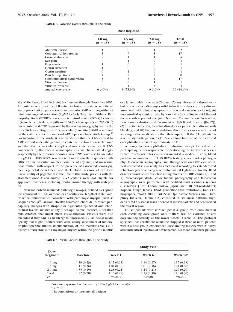
TABLE 3. Adverse Events throughout the Study
Dose Regimen
Total(n � 45)
1.0 mg(n � 15)
1.5 mg(n � 15)
2.0 mg(n � 15)
Abnormal vision 1 0 1 2Conjunctival hyperemia 5 7 5 17Corneal abrasion 2 1 1 4Eye pain 0 1 0 1Hyperemia 1 0 0 1Ocular irritation 1 0 0 1Ocular pruritus 0 1 0 1Pain (at injection) 4 2 2 8Subconjunctival hemorrhage 5 8 4 17Vitreous floaters 0 0 1 1Vitreous prolapse 0 1 3 4Any adverse event 6 (40%) 8 (53.3%) 6 (40%) 19 (44.4%)
TABLE 4. Visual Acuity throughout the Study
DoseRegimen Baseline
Study Visit
Week 1 Week 6 Week 12*
1.0 mg 1.19 (0.23) 1.15 (0.23) 1.14 (0.27) 1.17 (0.28)1.5 mg 1.11 (0.36) 1.04 (0.38) 1.01 (0.36) 1.04 (0.38)2.0 mg 1.35 (0.19) 1.28 (0.21) 1.24 (0.21) 1.26 (0.20)Total 1.22 (0.28) 1.16 (0.29) 1.13 (0.30) 1.16 (0.30)P† �0.001 �0.001 �0.001
Data are expressed as the mean (�SD) logMAR (n � 45).* n � 43.† In comparison to baseline; all patients.
IOVS, October 2006, Vol. 47, No. 10 Intravitreal Bevacizumab in CNV 4571

could be enrolled into the study on any day, and no more than sixpatients could be enrolled into the study per week.
Treatment Procedure
Patients received one intravitreal injection of 1.0, 1.5, or 2.0 mg ofbevacizumab; for these, 0.04-, 0.06-, and 0.08-mL aliquots of commer-cially available bevacizumab (25 mg/mL), respectively, were preparedfor each patient and placed in insulin syringes (BD Ultra-fine; BDBiosciences, Franklin Lakes, NJ) by a compounding pharmacy usingstandard aseptic techniques. All treatments were performed by a singleretinal specialist (RAC) using topical anesthetic (tetracaine-phenyleph-rine) drops under sterile conditions (eyelid speculum and povidoneiodine). Patients were given 250 mg of acetazolamide orally beforepupil dilation with mydriatric (tropicamide 1%) drops. Bevacizumabwas injected into the vitreous cavity with a 29.5-gauge needle insertedthrough the inferotemporal pars plana 3.0 mm (pseudophakic) or 3.5mm (phakic) posterior to the limbus. After the injection, central retinalartery perfusion was confirmed with indirect ophthalmoscopy andphotographic documentation of the fundus and of the injection sitewas performed. Patients were instructed to instill one drop of 0.3%ciprofloxacin into the injected eye four times daily for 1 week after theprocedure.
Follow-up Examinations and Outcome Measures
Follow-up examinations were performed at weeks 1, 6, and 12 (�1)after injection. The same procedures performed at baseline were per-formed at each study visit (e.g., BCVA, complete ophthalmic examina-tion, fundus photography, fluorescein angiography, and OCT). Localand systemic adverse events were monitored throughout the study.
These local ophthalmic and systemic adverse events were theprimary end points of the study. BCVA was measured primarily as anindicator of safety, not efficacy, given the absence of a control groupand the short follow-up period. However, changes in BCVA were usedto compare results among the dose groups.
Secondary measures were used to evaluate the short-term effects ofintravitreal bevacizumab treatment and to assist with comparisonsbetween dose regimens: (1) total area of the neovascular complex(lesion) and characteristics of the CNV component by fluoresceinangiography, and (2) macular remodeling (that is, retinal elevation) onOCT evaluation. Because of inherent OCT software flaws in the gen-eration of reliable quantitative tomographic data in eyes with neovas-cular AMD,57,58 changes in macular architecture were qualitatively
analyzed. Based on changes in the degree of retinal elevation due tointraretinal, subretinal, and sub-RPE fluid compared with baseline, themacular architecture was classified as having undergone “partial recov-ery” (absence of, or decrease in, retinal elevation), “no recovery”(unchanged retinal elevation), or “deterioration” (increased retinalelevation).59,60
Statistical Analysis
Repeated-measures analysis of variance (ANOVA) with the Geisser-Greenhouse correction was performed to analyze the visual acuityfrom baseline to the final study visit (week 12). The Dunnett test wasused to compare the visual acuity after injection with the baselinemeasurement. A nonparametric test for trend across ordered groupswas used to compare the changes in visual acuity from baseline amongthe three groups at weeks 1, 6, and 12.61 The significance level was setat 0.05.
RESULTS
The 45 participants included 21 (46.7%) men and 24 (53.3%)women; 43 completed all follow-up visits (one patient refusedto return after the week-6 follow-up visit, and the other patientwithdrew because of a death in the family). The mean (�SD)age was 74.6 � 6.4 years (median, 74 years; range, 62–85years). Mean (�SD) systolic and diastolic blood pressure was127.4 � 9.5 and 80.1 � 6.7 mm Hg, respectively. The mean �SD size of the neovascular complex was 12.2 � 3.9 MacularPhotocoagulation Study disc areas. Thirty-seven (82.2%) of 45eyes showed some fluorescein leakage corresponding to occultCNV, and 8 (17.8%) eyes showed purely classic CNV lesions.Baseline characteristics of the patients, stratified by dosegroup, are summarized in Table 2.
No systemic or serious drug-related adverse events wereobserved. The treatment procedure was well tolerated, and noclinical evidence of inflammation, uveitis, endophthalmitis, orobvious ocular toxicity was observed. Further, there were nosignificant changes in blood pressure, intraocular pressure, orlens status in any of the eyes during the study follow-up period.Minor local adverse events related to the treatment procedurewere observed (Table 3). Subconjunctival hemorrhage andconjunctival hyperemia were observed frequently at the intra-vitreal injection site and were most likely the result of theanesthesia technique rather than the injection procedure itself.In the first 15 procedures, four (26.6%) patients reported painat the time of the injection. Seeking to minimize this pain, inthe next 15 procedures we used a sterile cotton tip soakedwith tetracaine–phenylephrine drops, pushed gently againstthe conjunctiva/sclera for 60 to 70 seconds before the injection(for the first 15 procedures the cotton tip was left in place forapproximately 30 seconds). Only 2 (13.3%) patients reportedpain after we implemented the latter anesthetic technique, butwe did note that 8 (53.3%) patients (versus 5 [33.3%] in thefirst 15 patients) developed conjunctival hyperemia and sub-conjunctival hemorrhage within 30 minutes of the procedure.
FIGURE 1. Visual acuity throughout the study. Data points: the mean;vertical bars: 95% CI.
TABLE 5. Changes in Visual Acuity Compared with Baseline
DoseRegimen
Study Visit
Week 1 Week 6 Week 12
1.0 mg �0.05 (0.13) �0.05 (0.14) �0.03 (0.17)1.5 mg �0.07 (0.10) �0.10 (0.11) �0.06 (0.09)2.0 mg �0.07 (0.09) �0.10 (0.11) �0.10 (0.10)P * �0.49 �0.13 �0.02
Data are expressed as the mean (�SD) logMAR.* Comparison between dose regimens at each follow-up visit.
4572 Costa et al. IOVS, October 2006, Vol. 47, No. 10

Two eyes even had small subconjunctival hemorrhages beforethe injection. For the last 15 injections, the sterile cotton tipsoaked in anesthetic (tetracaine-phenylephrine) drops waspressed against the eyewall over the injection site for only 30to 35 seconds, with an additional 10 seconds of pressureapplied just before the injection; and pain was reported by onlytwo (13.3%) patients and subconjunctival hemorrhage devel-oped in four (26.6%). Independent of the anesthesia technique,subconjunctival hemorrhage was resolved and absence ofwound leakage was verified in all 45 patients by 1 week afterinjection.
The study was designed primarily as a safety trial. BCVAresults are summarized in Table 4 (Fig. 1). Mean BCVA im-proved significantly from baseline (P � 0.001; ANOVA usingthe Geisser-Greenhouse correction). Compared with baseline,BCVA improved significantly at week 1 (P � 0.001), week 6(P � 0.001), and week 12 (P � 0.001; Dunnett test; Table 4).
The mean visual acuity change (in ETDRS lines) from base-line between dose-regimens 1.0 (�0.3 line), 1.5 (�0.6 line),and 2.0 mg (�1.0 line) was significantly different at week 12,thus suggesting a dose-related response (P � 0.02; nonpara-metric test for ordered groups; Table 5; Fig. 2).
No unfavorable changes in neovascular complexes wereobserved during the study period by angiographic and OCTexaminations. At 6 weeks, 42 (93.3%) and 40 (88.9%) patientsof 45 patients exhibited stabilization (i.e., no change or de-crease) of the total lesion area and CNV area, respectively, byfluorescein angiography (Fig. 3). Unchanged or decreased le-sion area and CNV area was seen in 34 (79.1%) and in 32
(74.4%) patients, respectively, of 43 patients who completedthe week-12 follow-up evaluation. No patients in the 2.0-mgdose group demonstrated an increase in lesion area or CNVarea throughout the study period (Table 6). No patients in the1.5-mg dose group demonstrated an increase in lesion area atthe week-6 evaluation. On OCT, favorable changes in maculararchitecture (i.e., no deterioration) were observed in 41(91.1%) of the 45 patients at 6 weeks, and in 36 (83.7%) of the43 patients who completed the week-12 follow-up evaluation(Fig. 4). The most favorable macular remodeling was observedin patients in the 2.0-mg dose group at weeks 6 and 12, and atweek 6 in patients in the 1.5-mg dose group (Table 6).
DISCUSSION
This phase-I clinical trial evaluated the safety profile of threedifferent doses of a single intravitreal injection of bevacizumab,a genetically engineered humanized monoclonal antibodyagainst all active forms of human VEGF, in patients with neo-vascular AMD. In short-term, bevacizumab was well toleratedwhen administered by intravitreal injection at doses of 1.0, 1.5,and 2.0 mg using the commercially formulation of the drug atthe concentration of 25 mg/mL (volumes of 0.04, 0.06, and0.08 mL). The most common adverse events were conjunctivalhyperemia and subconjunctival hemorrhage at the injectionsite, events judged to be essentially the result of the injectionprocedure rather than the drug. No major adverse event asso-ciated with the treatment was observed during the 12-weekfollow-up period.
FIGURE 2. Profile of the mean visual acuity (VA) changes from baseline for each dose group after a single intravitreal injection of bevacizumabthrough week 12. Vertical bars, 95% CI.
IOVS, October 2006, Vol. 47, No. 10 Intravitreal Bevacizumab in CNV 4573

Before study initiation we had used intravitreal bevaci-zumab for salvage therapy in eyes with AMD and CNV refrac-tory to other treatments, and we observed some reflux of drugafter the injection procedure. Therefore, in an effort to mini-mize drug reflux, 250 mg of acetazolamide was given orally 1hour before the injection procedure. In the present study,prolapse of vitreous and/or bevacizumab at the end of theinjection procedure was observed in 1 patient who received0.06 mL and in three patients who received 0.08 mL of thedrug. Unlike triamcinolone acetonide, bevacizumab is a trans-parent, fluid drug that may reflux when higher volumes areinjected intravitreally even through a 29.5-gauge entry site.
Overall improvement in visual acuity occurred as early as 1week after the bevacizumab injection in this study. Visualacuity continued to improve between weeks 1 and 6, and thisbenefit persisted up to the week 12 evaluation. The improve-ment observed at week 6 may reflect continued inhibition ofVEGF. In conjunction with fluorescein angiographic and OCTdata, which demonstrated favorable macular remodeling with-out documented regression of the choroidal neovascular chan-nels in most of the patients (particularly in lesions with someclassic CNV component), it seems that the beneficial effects ofintravitreal bevacizumab on vision may be related to inhibitionof VEGF-associated vascular permeability rather than to aneffect on angiogenesis. Recently, complete retinal penetrationof bevacizumab was demonstrated in rabbits after a singleintravitreal injection of bevacizumab62; however, no measure-
ments at the choroidal tissues were performed, and thus onemay consider that low levels of the antiangiogenic drug mayreach the choriocapillaris–choroid complex. This hypothesis isfurther supported by the remarkable beneficial effects on cho-roidal neovascularization characteristics of systemic bevaci-zumab in humans,63,64 as well as the observed effects of intra-vitreal bevacizumab on retinal neovascularization associatedwith diabetic retinopathy65–67 and choroidal neovascular le-sions associated with chorioretinal anastomosis (Costa RA; un-published data, 2006). In all, clear regression of the neovascu-lar vessels was demonstrated, a finding not observed in thepresent study.
One limitation of the present study is the absence of serumtesting to investigate the systemic pharmacokinetics of intra-vitreally administered bevacizumab. Such evaluation, however,involves high-cost procedures that are not feasible within thecontext of an unfunded investigator-driven study. Because hy-pertension is the most common systemic adverse event asso-ciated with systemic bevacizumab therapy, changes in bloodpressure after treatment may roughly serve as a surrogatemarker for clinically relevant systemic bevacizumab levels.Herein, as in other studies involving patients with AMD,68–70
no significant change in blood pressure was seen in the short-term after single injections of bevacizumab. Two patients werelost to the final follow-up; however, it is unlikely that thisinfluenced the overall study results significantly, given theobserved findings through 6 weeks of follow-up in both pa-
FIGURE 3. Red-free fundus photog-raphy, and early- and late-phase fluo-rescein angiograms of one patient indose regimen of 1.5 mg of intravitrealbevacizumab throughout the study.Top: baseline evaluation performed24 hours before intravitreal bevaci-zumab injection demonstrated stip-pled fluorescence from occult CNVcomponent and areas of blocked flu-orescence from thick blood, as wellas a small retinal pigment epitheliumdetachment at the temporal aspect ofthe neovascular complex. Top cen-ter: 1 week after treatment fluores-cence from CNV was diminished andsubretinal hemorrhage has partiallyreabsorbed. Bottom center: at 6weeks, although choroidal neovascu-lar channels could be identified inearly phases, fluorescein leakagefrom CNV was minimal, and subreti-nal hemorrhage was further dimin-ished. Bottom: 12 weeks after treat-ment, actively leaking CNV andlesion areas were decreased in com-parison with baseline.
4574 Costa et al. IOVS, October 2006, Vol. 47, No. 10

tients. Additional limitations of the present study include thesmall number of subjects and short follow-up duration. Finally,the variability of the injection volume within each dose regi-men, which could also have interfered in the study results,might have been minimized in this study by using preloadedsyringes prepared by a compounding pharmacy.
As mentioned earlier, limitations inherent in the study’sdesign preclude extrapolation of our results. Conclusions re-garding efficacy cannot be made in the absence of a controlgroup, but differences among the dose groups may provideclues regarding drug effects. Patients who received 1.5 or 2.0mg of bevacizumab were more likely to demonstrate nochange or a reduction in lesion area as well as in CNV area atweeks 6 and 12 than were patients who received 1.0 mg.Overall, and keeping in mind the short follow-up of the studyand the variable course of the disease, at week 6 more eyesdemonstrated a smaller area of leakage from CNV with associ-ated favorable macular remodeling on angiographic and OCTevaluations, thus suggesting the effects of bevacizumab may bemaximal up to week 6 under the strict conditions of this study.Overall visual acuity improved throughout the study, and nodifferences existed among dose regimens at weeks 1 and 6.However, the change in visual acuity observed in treated pa-
tients at week 12 was related to the dose regimen, thus sug-gesting a possible longer duration of action (i.e., drug persis-tence in the eye) of the higher dosage (2.0 mg) ofbevacizumab, but not necessarily a dose dependence related tobetter initial efficacy. In light of this and the fact that beneficialeffects of bevacizumab have just been reported in neovascularAMD after intravitreal injections of 2.5 mg (0.1 mL),70 futurestudies probably should include higher dose regimens; never-theless, as discussed earlier, issues related to drug–fluid vitre-ous reflux should be cautiously monitored. One may also arguethat even lower doses should have been tested since for pe-gaptanib therapy it was the lowest dose of the drug thatappeared the most effective.15 However, in contrast to pe-gaptanib, in our study the highest dose worked best, suggest-ing that low-dose inhibition is not an issue. Furthermore, doseregimens lower than 1.0 mg are difficult to measure and re-quire dilution of the commercially available drug so that repro-ducible volumes can be injected. This would require reformu-lation of the drug with stability testing, which is outside thescope of clinical practice.
In 1971, Folkman71 hypothesized that the growth of tumorsis angiogenesis dependent and that antiangiogenic therapy mayrepresent an option for the treatment of solid tumors.72 Since
FIGURE 4. Third-generation opticalcoherence tomography evaluation(high-density [512 A-scans] 6 mm inlength B-scans, oriented obliquely at330°) of one patient in dose regimenof 1.5 mg of intravitreal bevacizumabthroughout the study (same patientillustrated in Fig. 3). Top left: focalregions of fusiform thickening of aswell as fragmentation and irregularelevation of the outer highly reflec-tive layer corresponding to the ret-inal pigment epithelium– chorio-capillaris hyperreflective complexassociated with intraretinal andsubretinal fluid thus characterizinga fibrovascular detachment of theRPE was seen at baseline. Top right:1 week after treatment, decrease inretinal elevation was noted. Bottomleft: 6 weeks after the procedure, marked decrease in retinal elevation was seen. Bottom right: by week 12, subretinal and sub-RPE fluid recurred;in comparison to baseline, no deterioration of the macular architecture was verified.
TABLE 6. Change from Baseline in Neovascular Complex Lesion Area and CNV Characteristics
Dose Regimen
Total1.0 mg 1.5 mg 2.0 mg
Week 6 Week 12 Week 6 Week 12 Week 6 Week 12 Week 6 Week 12
Fluorescein angiographyLesion area
Decrease 1 (6.7) 1 (6.7) 2 (13.3) 2 (14.3) 3 (20) 2 (14.3) 6 (13.3) 5 (11.6)No change 11 (73.3) 8 (53.3) 13 (86.7) 9 (64.3) 12 (80) 12 (85.7) 36 (80) 29 (67.5)Increase 3 (20) 6 (40) 0 3 (21.4) 0 0 3 (6.7) 9 (20.9)
CNV areaDecrease 2 (13.3) 1 (6.7) 7 (46.7) 3 (21.4) 8 (53.3) 5 (35.7) 17 (35.6) 9 (20.9)No Change 10 (66.7) 7 (46.6) 6 (40) 7 (50) 7 (46.7) 9 (64.3) 23 (53.3) 23 (53.5)Increase 3 (20) 7 (46.6) 2 (13.3) 4 (28.6) 0 0 5 (11.1) 11 (25.6)
Optical coherence tomographyMacular architecture
Partial recovery 2 (13.3) 2 (13.3) 8 (53.3) 4 (28.6) 11 (73.3) 9 (64.3) 21 (46.7) 15 (34.9)No recovery 10 (66.7) 8 (53.3) 6 (40) 8 (57.1) 4 (26.7) 5 (35.7) 20 (44.4) 21 (48.8)Deterioration 3 (20) 5 (33.3) 1 (6.7) 2 (14.3) 0 0 4 (8.9) 7 (16.3)
Data are expressed as the number of cases with the percentage of the total in parentheses.
IOVS, October 2006, Vol. 47, No. 10 Intravitreal Bevacizumab in CNV 4575

then, numerous reports have supported the crucial role ofneovascularization in malignancy and various non-neoplasticdiseases, including neovascular AMD,72 which is the leadingcause of blindness in elderly persons in developed nations.Unfortunately, currently approved treatments are character-ized by both limited clinical significance and high-cost thatrestricts access to treatment, particularly in underdevelopednations. Characterization of the various processes involved inthe pathogenesis of any disease is a prerequisite to developingeffective therapies. Vascular endothelial growth factor is aprotein involved in the onset and progression of AMD.31–54
Thus, VEGF and its signaling receptors are potential targets forpharmaceutical intervention Results from phase III clinicaltrials evaluating intravitreal anti-VEGF agents have led to theapproval of pegaptanib for the treatment of neovascular AMDand indicate favorable results associated with ranibizumab. Inthis era of pharmacomodulation for AMD management, bevaci-zumab may have a crucial role not only because of the prom-ising results reported herein and additional safety data andclinical evidence of its beneficial effects reported recently for avariety of diseases,65–67,73–77 but also because intravitreal be-vacizumab therapy, if proved safe and efficacious, may repre-sent a treatment option for neovascular AMD that could beaccessible to all patients, independent of their socioeconomiccondition.
References
1. Miller JW, Schmidt-Erfurth U, Sickenberg M, et al. Photodynamictherapy with verteporfin for choroidal neovascularization causedby age-related macular degeneration: results of a single treatmentin a phase 1 and 2 study. Arch Ophthalmol. 1999;117:1161–1173.
2. Schmidt-Erfurth U, Miller JW, Sickenberg M, et al. Photodynamictherapy with verteporfin for choroidal neovascularization causedby age-related macular degeneration: results of retreatments in aphase 1 and 2 study. Arch Ophthalmol. 1999;117:1177–1187.
3. Treatment of age-related macular degeneration with photody-namic therapy (TAP) Study Group. Photodynamic therapy of sub-foveal choroidal neovascularization in age-related macular degen-eration with verteporfin: one-year results of 2 randomized clinicaltrials. TAP report (published correction in Arch Ophthalmol 2000;118:488). Arch Ophthalmol. 1999;117:1329–1345.
4. Macular Photocoagulation Study Group. Subfoveal neovascular le-sions in age-related macular degeneration. Guidelines for evalua-tion and treatment in the macular photocoagulation study. ArchOphthalmol. 1991;109:1242–1257.
5. Macular Photocoagulation Study Group. Laser photocoagulation ofsubfoveal neovascular lesions in age-related macular degeneration;results of a randomized clinical trial. Arch Ophthalmol. 1991;109:1220–1231.
6. Bressler NM; Treatment of Age-Related Macular Degeneration withPhotodynamic Therapy (TAP) Study Group. Photodynamic therapyof subfoveal choroidal neovascularization in age-related maculardegeneration with verteporfin: two-year results of 2 randomizedclinical trials. TAP report no. 2. Arch Ophthalmol. 2001;119:198–207.
7. Verteporfin In Photodynamic Therapy Study Group. Verteporfintherapy of subfoveal choroidal neovascularization in age-relatedmacular degeneration: two-year results of a randomized clinicaltrial including lesions with occult with no classic choroidal neo-vascularization. VIP report no. 2. Am J Ophthalmol. 2001;131:541–560.
8. Bressler NM, Arnold J, Benchaboune M, et al. Verteporfin therapyof subfoveal choroidal neovascularization in patients with age-related macular degeneration: additional information regardingbaseline lesion composition’s impact on vision outcomes. TAPreport no. 3. Arch Ophthalmol. 2002;120:1443–1454.
9. Rubin GS, Bressler NM. Treatment of Age-Related Macular Degen-eration with Photodynamic therapy (TAP) study group. Effects ofverteporfin therapy on contrast on sensitivity: results from theTreatment of Age-Related Macular Degeneration with Photody-
namic Therapy (TAP) investigation. TAP report no 4. Retina.2002;22:536–544.
10. Blumenkranz MS, Bressler NM, Bressler SB, et al. Verteporfin ther-apy for subfoveal choroidal neovascularization in age-related mac-ular degeneration: three-year results of an open-label extension of2 randomized clinical trials. TAP report no. 5. Arch Ophthalmol.2002;120:1307–1314.
11. Bessler NM and the VAM Study Writing Committee. Verteporfintherapy in age-related macular degeneration (VAM): an open-labelmulticenter photodynamic therapy study of 4,435 patients (pub-lished correction in Retina. 2004;24:990). Retina 2004;24:512–520.
12. Azab M, Benchaboune M, Blinder KJ, et al. Verteporfin therapy ofsubfoveal choroidal neovascularization in age-related maculardegeneration: meta-analysis of 2-year safety results in three ran-domized clinical trials: Treatment of Age-Related Macular Degen-eration with Photodynamic Therapy and Verteporfin in Photody-namic Therapy Study Report no. 4. Retina. 2004;24:1–12.
13. Kaiser PK; Treatment of Age-related Macular Degeneration withPhotodynamic Therapy (TAP) Study Group. Verteporfin therapy ofsubfoveal choroidal neovascularization in age-related maculardegeneration: 5-year results of two randomized clinical trials withan open-label extension. TAP Report no. 8. Graefes Arch Clin ExpOphthalmol. Published online March 15, 2006.
14. The Eyetech Study Group. Preclinical and phase 1A clinical eval-uation of an anti-VEGF pegylated aptamer (EYE001) for the treat-ment of exudative age-related macular degeneration. Retina. 2002;22:143–152.
15. Gragoudas ES, Adamis AP, Cunningham ET Jr, et al. Pegaptanib forneovascular age-related macular degeneration. N Engl J Med. 2004;351:2805–2816.
16. Presta LG, Chen H, O’Connor SJ, et al. Humanization of an anti-vascular endothelial growth factor monoclonal antibody for thetherapy of solid tumors and other disorders. Cancer Res. 1997;57:4593–4599.
17. Muller YA, Chen Y, Christinger HW, et al. VEGF and the Fabfragment of a humanized neutralizing antibody: crystal structure ofthe complex at 2.4—a resolution and mutational analysis of theinterface. Structure. 1998;6:1153–1167.
18. Chen Y, Wiesmann C, Fuh G, et al. Selection and analysis of anoptimized anti-VEGF antibody: crystal structure of an affinity-ma-tured Fab in complex with antigen. J Mol Biol. 1999;293:865–881.
19. Krzystolik MG, Afshari MA, Adamis AP, et al. Prevention of exper-imental choroidal neovascularization with intravitreal anti-vascularendothelial growth factor antibody fragment. Arch Ophthalmol.2002;120:338–346.
20. Rosenfeld PJ, Schwartz SD, Blumenkranz MS, et al. Maximumtolerated dose of a humanized anti-vascular endothelial growthfactor antibody fragment for treating neovascular age-related mac-ular degeneration. Ophthalmology. 2005;112:1048–1053.
21. Heier JS, Antoszyk AN, Pavan PR, et al. Ranibizumab for treatmentof neovascular age-related macular degeneration: a phase I/II mul-ticenter, controlled, multidose study. Ophthalmology. Publishedonline February 13, 2006
22. Genentech: Press Releases—News Release February 28, 2006.Available at: http://www.gene.com/gene/news/press-releases/display.do?method�detail&id�9427. Accessed March, 2006.
23. Yang JC, Haworth L, Sherry RM, et al. A randomized trial ofbevacizumab, an anti-vascular endothelial growth factor antibody,for metastatic renal cancer. N Engl J Med. 2003;349:427–434.
24. No author listed. Adding a humanized antibody to vascular endo-thelial growth factor (bevacizumab, Avastin) to chemotherapyimproves survival in metastatic colorectal cancer. Clin ColorectalCancer. 2003;8:85–88.
25. Kabbinavar F, Hurwitz HI, Fehrenbacher L, et al. Phase II, random-ized trial comparing bevacizumab plus fluorouracil (FU)/leucov-orin (LV) with FU/LV alone in patients with metastatic colorectalcancer. J Clin Oncol. 2003;21:60–65.
26. Hurwitz H, Fehrenbacher L, Novotny W, et al. Bevacizumab plusirinotecan, fluorouracil, and leucovorin for metastatic colorectalcancer. N Engl J Med. 2004;350:2335–2342.
27. Yang JC. Bevacizumab for patients with metastatic renal cancer: anupdate. Clin Cancer Res. 2004;10:6367S–70S.
4576 Costa et al. IOVS, October 2006, Vol. 47, No. 10

28. Mordenti J, Cuthbertson RA, Ferrara N, et al. Comparisons of theintraocular tissue distribution, pharmacokinetics, and safety of125I-labeled full-length and Fab antibodies in rhesus monkeysfollowing intravitreal administration. Toxicol Pathol. 1999;27:536–544.
29. Rosenfeld PJ, Moshfeghi AA, Puliafito CA. Optical coherence to-mography findings after an intravitreal injection of bevacizumab(Avastin) for neovascular age-related macular degeneration. Oph-thalmic Surg Lasers Imaging. 2005;36:331–335.
30. Rosenfeld PJ, Fung AE, Puliafito CA. Optical coherence tomogra-phy findings after an intravitreal injection of bevacizumab (Avas-tin) for macular edema from central retinal vein occlusion. Oph-thalmic Surg Lasers Imaging. 2005;36:336–339.
31. Amin R, Puklin JE, Frank RN. Growth factor localization in choroi-dal neovascular membranes of age-related macular degeneration.Invest Ophthalmol Vis Sci. 1994;35:3178–3188.
32. D’Amore PA. Mechanisms of retinal and choroidal neovasculariza-tion. Invest Ophthalmol Vis Sci. 1994;35:3974–3979.
33. Frank RN, Amin RH, Eliott D, Puklin JE, Abrams GW. Basic fibro-blast growth factor and vascular endothelial growth factor arepresent in epiretinal and choroidal neovascular membranes. Am JOphthalmol. 1996;122:393–403.
34. Aiello LP. Vascular endothelial growth factor and the eye. Past,present and future (editorial). Arch Ophthalmol. 1996;114:1252–1254.
35. Lopez PF, Sippy BD, Lambert HM, et al. Transdifferentiated retinalpigment epithelial cells are immunoreactive for vascular endothe-lial growth factor in surgically excised age-related macular degen-eration-related choroidal neovascular membranes. Invest Ophthal-mol Vis Sci. 1996;37:855–868.
36. Kvanta A, Algvere PV, Berglin L, Seregard S. Subfoveal fibrovascu-lar membranes in age-related macular degeneration express vascu-lar endothelial growth factor. Invest Ophthalmol Vis Sci. 1996;37:1929–1934.
37. Wells JA, Murthy R, Chibber R, et al. Levels of vascular endothelialgrowth factor are elevated in the vitreous of patients with subreti-nal neovascularisation. Br J Ophthalmol. 1996;80:363–366.
38. Schlingemann RO, van Hinsbergh VWM. Role of vascular perme-ability factor/vascular endothelial growth factor in eye disease(review). Br J Ophthalmol. 1997;81:501–512.
39. Yi X, Ogata N, Komada M, et al. Vascular endothelial growth factorexpression in choroidal neovascularization in rats. Graefes ArchClin Exp Ophthalmol. 1997;235:313–319.
40. Kliffen M, Sharma HS, Mooy CM, et al. Increased expression ofangiogenic growth factors in age-related maculopathy. Br J Oph-thalmol. 1997;81:154–162.
41. Mousa SA, Lorelli W, Campochiaro PA. Role of hypoxia and extra-cellular matrix-integrin binding in the modulation of angiogenicgrowth factors secretion by retinal pigmented epithelial cells.J Cell Biochem. 1999;74:135–143.
42. Frank RN. Growth factors in age-related macular degeneration:pathogenic and therapeutic implications. Ophthalmic Res 1997;29:341–353. Comment in: Ophthalmic Res. 1999;31:243–244.
43. Campochiaro PA, Soloway P, Ryan SJ, Miller JW. The pathogenesisof choroidal neovascularization in patients with age related mac-ular degeneration. Mol Vis. 1999;5:34–39.
44. Campochiaro PA. Retinal and choroidal neovascularization. J CellPhysiol. 2000;184:301–310.
45. Spilsbury K, Garrett KL, Shen WY, Constable IJ, Rakoczy PE.Overexpression of vascular endothelial growth factor (VEGF) inthe retinal pigment epithelium leads to the development of cho-roidal neovascularization. Am J Pathol. 2000;157:135–144.
46. Lip PL, Belgore F, Blann AD, et al. Plasma VEGF and soluble VEGFreceptor FLT-1 in proliferative retinopathy: relationship to endo-thelial dysfunction and laser treatment. Invest Ophthalmol Vis Sci.2000;41:2115–2119.
47. Ohno-Matsui K, Morita I, Tombran-Tink J, et al. Novel mechanismfor age-related macular degeneration: an equilibrium shift betweenthe angiogenesis factors VEGF and PEDF. J Cell Physiol. 2001;189:323–333.
48. Schwesinger C, Yee C, Rohan RM, et al. Intrachoroidal neovascu-larization in transgenic mice overexpressing vascular endothelialgrowth factor in the retinal pigment epithelium. Am J Pathol.2001;158:1161–1172.
49. Mori K, Gehlbach P, Yamamura S, et al. AAV-mediated gene trans-fer of pigment epithelium-derived factor inhibits choroidal neovas-cularization. Invest Ophthalmol Vis Sci. 2002;43:1994–2000.
50. Ogata N, Wada M, Otsuji T, et al. Expression of pigment epithe-lium-derived factor in normal adult rat eye and experimental cho-roidal neovascularization. Invest Ophthalmol Vis Sci. 2002;43:1168–1175.
51. Renno RZ, Youssri AI, Michaud N, Gragoudas ES, Miller JW. Ex-pression of pigment epithelium-derived factor in experimentalchoroidal neovascularization. Invest Ophthalmol Vis Sci. 2002;43:1574–1580.
52. Hera R, Keramidas M, Peoc’h M, et al. Expression of VEGF andangiopoietins in subfoveal membranes from patients with age-related macular degeneration. Am J Ophthalmol. 2005;139:589–596. Comment in: Am J Ophthalmol. 2005;139:710–711.
53. Tong JP, Chan WM, Liu DT, et al. Aqueous humor levels of vascularendothelial growth factor and pigment epithelium-derived factorin polypoidal choroidal vasculopathy and choroidal neovascular-ization. Am J Ophthalmol. 2006;141:456–462.
54. Bhutto IA, McLeod DS, Hasegawa T, et al. Pigment epithelium-derived factor (PEDF) and vascular endothelial growth factor(VEGF) in aged human choroid and eyes with age-related maculardegeneration. Exp Eye Res. 2006;82:99–110.
55. Bird AC, Bressler NM, Bressler SB, et al. An international classifi-cation and grading system for age-related maculopathy and age-related macular degeneration. The International ARM Epidemio-logical Study Group. Surv Ophthalmol. 1995;39:367–374.
56. Verteporfin in Photodynamic Therapy Study Group. Photody-namic therapy of subfoveal choroidal neovascularization inpathologic myopia with verteporfin. 1-year results of a random-ized clinical trial. VIP report no. 1. Ophthalmology. 2001;108:841– 852.
57. Costa RA, Calucci D, Skaf M, et al. Optical coherence tomography3: automatic delineation of the outer neural retinal boundary andits influence on retinal thickness measurements. Invest Ophthal-mol Vis Sci. 2004;45:2399–2406.
58. Costa RA, Skaf M, Melo Jr LA, et al. Retinal assessment using opticalcoherence tomography. Prog Retin Eye Res. 2006;25:325–353.
59. Costa RA, Calucci D, Cardillo JA, et al. Selective occlusion ofsubfoveal choroidal neovascularization in angioid streaks using anew technique of ingrowth site treatment. Ophthalmology. 2003;110:1192–1203.
60. Costa RA, Teixeira LF, Calucci D, et al. Selective occlusion ofsubfoveal choroidal neovascularization in pathologic myopia usinga new technique of ingrowth site treatment. Am J Ophthalmol.2003;135:857–866.
61. Cuzick J. A Wilcoxon-type test for trend. Stat Med. 1985;4:87–90.62. Shahar J, Avery RL, Heilweil G, et al. Electrophysiologic and retinal
penetration studies following intravitreal injection of bevacizumab(Avastin). Retina. 2006;26:262–269.
63. Michels S, Rosenfeld PJ, Puliafito CA, Marcus EN, Venkatraman AS.Systemic bevacizumab (Avastin) therapy for neovascular age-re-lated macular degeneration: twelve-week results of an uncon-trolled open-label clinical study. Ophthalmology. 2005;112:1035–1047.
64. Nguyen QD, Shah S, Tatlipinar S, et al. Bevacizumab suppresseschoroidal neovascularisation caused by pathological myopia. Br JOphthalmol. 2005;89:1368–1370.
65. Spaide RF, Fisher YL. Intravitreal bevacizumab (Avastin) treatmentof proliferative diabetic retinopathy complicated by vitreous hem-orrhage. Retina. 2006;26:275–278.
66. Avery RL. Regression of retinal and iris neovascularization afterintravitreal bevacizumab (Avastin) treatment. Retina. 2006;26:352–354.
67. Jorge R, Costa RA, Calucci D, Cintra L, Scott IU. Intravitreal be-vacizumab (Avastin) for persistent new vessels in diabetic retinop-athy (IBePe Study). Retina. In press.
IOVS, October 2006, Vol. 47, No. 10 Intravitreal Bevacizumab in CNV 4577

68. Avery RL, Pieramici DJ, Rabena MD, et al. Intravitreal bevacizum-ab(Avastin) for neovascular age-related macular degeneration.Ophthalmology. 2006;113:363–372.
69. Spaide RF, Laud K, Fine H, et al. Intravitreal bevacizumab treat-ment of choroidal neovascularization secondary to age-relatedmacular degeneration. Retina. 2006;26:383–390.
70. Bashshur ZF, Bazarbachi A, Schakal A, et al. Intravitreal bevaci-zumab for the management of choroidal neovascularization inage-related macular degeneration. Am J Ophthalmol. Publishedonline April 16, 2006.
71. Folkman J. Tumor angiogenesis: therapeutic implications. N Engl JMed. 1971;285:1182–1186.
72. Witmer AN, Vrensen GFJM, Van Noorden CJF, Schlingemann RO.Vascular endothelial growth factors and angiogenesis in eye dis-ease. Prog Retin Eye Res. 2003;22:1–29.
73. Iturralde D, Spaide RF, Meyerle CB, et al. Intravitreal bevaci-zumab (Avastin) treatment of macular edema in central retinalvein occlusion: a short-term study. Retina. 2006;26:279 –284.
74. Manzano RP, Peyman GA, Khan P, Kivilcim M. Testing intravitrealtoxicity of bevacizumab (Avastin). Retina. 2006;26:257–261.
75. Maturi RK, Bleau LA, Wilson DL. Electrophysiologic findings after intra-vitreal bevacizumab (Avastin) treatment. Retina. 2006;26:270–274.
76. Oshima Y, Sakaguchi H, Gomi F, Tano Y. Regression of iris neo-vascularization after intravitreal injection of bevacizumab in pa-tients with proliferative diabetic retinopathy. Am J Ophthalmol.Published online March 20, 2006.
77. Paula JS, Jorge R, Costa RA, Rodrigues MLV, Scott IU. Short-termresults of intravitreal bevacizumab (Avastin) on anterior segmentneovascularization in neovascular glaucoma. Acta OphthalmolScand. In press.
4578 Costa et al. IOVS, October 2006, Vol. 47, No. 10
Copyright © 2022 FDOKUMEN