ARTICLE IN PRESS Automated detection of exudates and macula for grading of diabetic macular edema
Upload
independentCategory
view
0download
0
A Randomized Trial Comparing the Efficacy and Safety ofIntravitreal Triamcinolone With Standard Care to Treat Vision LossAssociated With Macular Edema Secondary to Branch RetinalVein Occlusion:The Standard Care vs Corticosteroid for Retinal Vein Occlusion (SCORE) Study Report 6
The SCORE Study Research Group*, Ingrid U. Scott, MD, MPH [chair], Michael S. Ip, MD, PaulC. VanVeldhuisen, PhD, Neal L. Oden, PhD, Barbara A. Blodi, MD, Marian Fisher, PhD,Clement K. Chan, MD, Victor H. Gonzalez, MD, Lawrence J. Singerman, MD, and MichaelTolentino, MD
AbstractObjective: To compare the efficacy and safety of 1-mg and 4-mg doses of preservative-freeintravitreal triamcinolone with standard care (grid photocoagulation in eyes without dense macularhemorrhage and deferral of photocoagulation until hemorrhage clears in eyes with dense macularhemorrhage) for eyes with vision loss associated with macular edema secondary to branch retinalvein occlusion (BRVO).
Methods: Multicenter, randomized clinical trial of 411 participants.
Main Outcome Measure: Gain in visual acuity letter score of 15 or more from baseline to month12.
Results: Twenty-nine percent, 26%, and 27% of participants achieved the primary outcome in thestandard care, 1-mg, and 4-mg groups, respectively. None of the pairwise comparisons between the3 groups was statistically significant at month 12. The rates of elevated intraocular pressure andcataract were similar for the standard care and 1-mg groups, but higher in the 4-mg group.
Conclusions: There was no difference identified in visual acuity at 12 months for the standardcare group compared with the triamcinolone groups; however, rates of adverse events (particularlyelevated intraocular pressure and cataract) were highest in the 4-mg group.
Application to Clinical Practice: Grid photocoagulation as applied in the SCORE Study remainsthe standard care for patients with vision loss associated with macular edema secondary to BRVOwho have characteristics similar to participants in the SCORE-BRVO trial. Grid photocoagulationshould remain the benchmark against which other treatments are compared in clinical trials for eyeswith vision loss associated with macular edema secondary to BRVO.
Trial Registration: clinicaltrials.gov Identifier: NCT00105027
©2009 American Medical Association. All rights reserved.Correspondence: Paul C. VanVeldhuisen, PhD, The EMMES Corporation, 401 N Washington St, Ste 700, Rockville, MD 20850([email protected])..*The Authors/Writing Committee are listed at the end of this article.Financial Disclosure: None reported.
NIH Public AccessAuthor ManuscriptArch Ophthalmol. Author manuscript; available in PMC 2010 September 1.
Published in final edited form as:Arch Ophthalmol. 2009 September ; 127(9): 1115–1128. doi:10.1001/archophthalmol.2009.233.
NIH
-PA Author Manuscript
NIH
-PA Author Manuscript
NIH
-PA Author Manuscript

Retinal vein occlusion disease is estimated to be the second most common cause of retinalvascular disease in the United States.1 The prevalence of retinal vein occlusion in the BlueMountains Eye Study was 1.6%, with 69.5%(41of59)oftheseocclusionsclassified as branchretinal vein occlusion (BRVO).2 Retinal vein occlusion was bilateral in 5.1% of cases.2 The15-year cumulative incidence of BRVO was 1.8% in the Beaver Dam Eye Study.3 In the BeaverDamcohort,centraland branch retinal vein occlusion accounted for 12% of eyes that developedsevere vision loss during a 15-year period.4
Macular edema is a frequent cause of visual acuity loss from BRVO.1,5,6 The natural historyof macular edema secondary to BRVO was delineated in the Branch Vein Occlusion Study(BVOS).1 One arm of the BVOS demonstrated a benefit with grid photocoagulation.1 Of 43treated eyes available for follow-up at the 3-year visit, 28 (65%) had gained 2 or more lines ofvisual acuity from baseline and maintained this gain for at least 8 months compared with thesame gain in 13 of 35 untreated eyes (37%). The BVOS also identified a subset of patients whoderived limited benefit from grid photocoagulation. In the BVOS, 40% of treated eyes (n=43)had worse than 20/40 visual acuity at 3 years, and 12% of treated eyes had 20/200 or worsevisual acuity at 3 years.1
During the last decade, a number of additional treatments for macular edema secondary toBRVO have been investigated. Such treatments include laser chorioretinal anastomosis,vitrectomy, arteriovenous sheathotomy, and intravitreal injection of antibodies targeted atvascular endothelial growth factor (VEGF).7-12 Numerous reports have suggested thatintravitreal injection(s) of triamcinolone acetonide (hereafter referred to as intravit-realtriamcinolone) is a potentially efficacious therapy for retinal thickening and vision loss inpatients with macular edema secondary to BRVO but that some patients may develop steroid-related complications, such as elevated intraocular pressure (IOP) and cataract.13-26 Most ofthe information concerning intravitreal triamcinolone for macular edema secondary to BRVOis derived from case series that lacked long-term follow-up and adequate numbers of studyparticipants; a randomized and controlled study has not been performed.13-26 Despite theshortcomings of these case series, intravitreal triamcinolone is currently in use as a treatmentfor BRVO.
The rationale for the use of corticosteroids to treat macular edema secondary to BRVO followsfrom the observation that the increase in retinal capillary permeability that results in macularedema may be caused by a breakdown of the blood-retina barrier mediated in part by VEGF,a 45-kDa glycoprotein.27-29 Therefore, attenuation of the effects of VEGF may reduce macularedema associated with BRVO.30,31 Corticosteroids have been demonstrated to inhibit theexpression of VEGF and therefore may be an effective therapy for macular edema.32,33
Inflammation may also play a role in the pathology of BRVO, and the anti-inflammatoryproperties of corticosteroids may contribute to the attenuation of the disease process.34
Intravitreal triamcinolone is used by ophthalmologists in the clinical setting as a readilyavailable pharmacologic agent (Kenalog 40; Bristol-Myers Squibb, Princeton, New Jersey, orTriesence; Alcon Inc, Fort Worth, Texas), though use for the treatment of macular edema isoff label. Other formulations such as compounded preservative-free triamcinolone acetonideare also used in the clinical setting.
The Standard Care vs Corticosteroid for Retinal Vein Occlusion (SCORE) Study, sponsoredby the National Eye Institute, is a clinical trial designed to compare 1-mg and 4-mg doses ofintravitreal triamcinolone with standard care for vision loss associated with macular edemasecondary to per-fused central retinal vein occlusion (CRVO) and BRVO.35,36 This articledescribes findings from the SCORE-BRVO trial. A companion article that comparesintravitreal triamcinolone with observation for vision loss associated with macular edema
et al. Page 2
Arch Ophthalmol. Author manuscript; available in PMC 2010 September 1.
NIH
-PA Author Manuscript
NIH
-PA Author Manuscript
NIH
-PA Author Manuscript

secondary to CRVO is published concurrently (the SCORE-CRVOtrial).37 The 2 primarystudy objectives of the SCORE-BRVO trial are (1) to determine whether intravit-realtriamcinolone at 1-mg and 4-mg doses produces greater visual benefit, with an acceptablesafety profile,than grid photocoagulation, when appropriate, for the treatment of vision lossassociated with macular edema secondary to BRVO; and (2) to compare the efficacy and safetyof the 1-mg and 4-mg triamcinolone doses.
METHODSDESIGN
The SCORE-BRVO trial was designed as a multicenter, prospective, randomized clinical trialand adhered to the tenets of the Declaration of Helsinki. Approval for the protocol was obtainedfrom either a central (Jaeb Center for Health Research) or local institutional review board.Health Insurance Portability and Accountability Act–compliant informed consent forms wereobtained before screening any participants. Study oversight was provided by an independentdata and safety monitoring committee. Eligibility for the SCORE-BRVO trial was determinedat the clinical sites (Table 1). For the purposes of the SCORE Study, participants identified ashaving hemiretinal vein occlusion according to the definition outlined in the protocol wereincluded in the BRVO trial. All participants were randomized within 21 days of screening orrescreening. One eye per participant was enrolled in the trial. Participants and physicians weremasked to the intravitreal triamcinolone dose (1 mg vs 4 mg) but not to the treatment assignmentof standard care vs intravitreal triamcinolone. The prespecified primary efficacy evaluationwas performed at month 12. The primary outcome measure was the proportion of participantswho experienced a gain in visual acuity letter score of 15 or more from baseline to month 12as assessed by the electronic Early Treatment Diabetic Retinopathy Study (E-ETDRS) method.38
RANDOMIZATIONWithin baseline visual acuity strata of good (visual acuity letter score, 73-59; Snellenequivalent, 20/40 to 20/63); moderate (visual acuity letter score, 58-49; Snellen equivalent,20/80 to 20/ 100); and poor (visual acuity letter score, 48-19; Snellen equivalent, 20/125 to20/400) participants were randomly assigned centrally through a Web-based data entry systemmaintained at the Data Coordinating Center (The EMMES Corporation, Rockville, Maryland),with equal probability to receive standard care (referred to as the standard care group); 1 mgof intravitreal triamcinolone (referred to as the 1-mg triamcinolone group), or 4 mg ofintravitreal triamcinolone (referred to as the 4-mg triamcinolone group) using a permutedblocks design with random block sizes. Standard care consisted of either grid photocoagulationif there was no dense macular hemorrhage or, if a dense macular hemorrhage was present,observation at 4-month intervals until the dense macular hemorrhage cleared sufficiently toallow grid photocoagulation to be performed. The decision as to what constituted a dense vsnondense hemorrhage was left to the investigator.
VISIT SCHEDULEStudy visits were planned for every 4 months through 36 months. At baseline and at eachfollow-up visit, best-corrected visual acuity letter score was measured at 3m by a masked,certified tester using the E-ETDRS method.38 A standardized refraction was performed atbaseline, month 4, and at the annual visits (months 12, 24, and 36). All participants whoreceived intravitreal triamcinolone had safety visits at 4 days (±3 days) and at 4 weeks (±1week) following each injection; visual acuity, IOP measurement, and eye examination results,including those from a dilated fundus examination, were recorded in the study eye at thesevisits. At all other study visits, participants underwent an eye examination, E-ETDRS testing,IOP measurement, and an optical coherence tomography (OCT) scan (OCT2 or Stratus OCT;
et al. Page 3
Arch Ophthalmol. Author manuscript; available in PMC 2010 September 1.
NIH
-PA Author Manuscript
NIH
-PA Author Manuscript
NIH
-PA Author Manuscript

Carl Zeiss Meditech, Dublin, California). These tests were performed on both eyes at all visits,except for visits at months 8, 16, 20, 28, and 32, when OCT was performed only on the studyeye. Stereoscopic color fundus photographs (7 fields) were taken of the study eye at baselineand at the annual visits. Three-field photographs of the study eye were taken at visits at months4, 8, 16, 20, 28, and 32 and of the fellow eye at baseline and the annual visits. Lens opacitiesfor both eyes were assessed at baseline and at the annual visits using the modified Age-RelatedEye Disease Study grading method.39 Fluorescein angiography was performed at baseline andat the month 4, 12, and 24 visits. All images were sent to the reading center (University ofWisconsin Fundus Photograph Reading Center, Madison, Wisconsin) for analysis, where theywere graded in a masked fashion. Blood pressure was measured at baseline and at the annualvisits.
Participating study personnel, such as physician-investigators and study coordinators, werecertified by the data coordinating center. Photographers and technicians who took the fundusphotographs and OCT images for this study were certified by the reading center.
INTRAVITREAL INJECTIONSThe optimal dose of intraocular corticosteroid to maximize efficacy and simultaneouslyminimize adverse effects was not known when the SCORE Study was initiated. The mostcommonly used dose of triamcinolone for treating eyes with macular edema secondary toBRVO is 4 mg, delivered in a volume of 0.1 mL. However, the rationale for using this dosehas been based on clinical feasibility (limiting volume of 0.1 mL of the first availabletriamcinolone formulation, Kenalog) rather than scientific principles. Clinical experience withother triamcinolone doses ranging from 1 to 20 mg is limited.40-42 Doses of intravitrealtriamcinolone reported to be as high as 25 mg have been used in intravitreal injections.18,43
The SCORE-BRVO trial evaluated 1-mg and 4-mg doses of triamcinolone. The 4-mg dosewas evaluated because it was the one used most commonly in clinical practice when the SCOREStudy was initiated. The 1-mg dose was evaluated because this dose is likely to exceed theconcentration necessary to saturate the glucocorticoid receptors in the cell cytoplasm.44,45 Inaddition, it was anticipated that, compared with the 4-mg dose, the 1-mg dose would have alower risk of steroid-related adverse events. Insufficient data were available at the time ofprotocol development to warrant evaluating a dose higher than 4 mg.
A preservative-free, nondispersive formulation of triamcinolone was used in the current studyin an effort to avoid the postinjection intraocular inflammation reported with Kenalog and someof the compounded triamcinolone formulations thought to be attributable to the excipients inKenalog, endotoxins, or a particle dispersion phenomenon.46,47 The study drug wasmanufactured as a sterile, preservative-free, single-use, intravitreal injectable (Trivaris;Allergan, Inc, Irvine, California; 4-mg brand name TRIVARIS) in 1-mg and 4-mg doses. Bothdoses were administered in a volume of 0.05 mL. All study eyes assigned to intravit-realtriamcinolone received a standardized ocular surface preparation procedure prior to theinjection consisting of an eyelid speculum, topical anesthetic, administration of topicalantibiotics on the day of injection, and asepsis with povidone iodine.48
Following the preparation procedure, either 1 mg or 4 mg of triamcinolone was injected intothe vitreous cavity via the pars plana, 3 to 4 mm posterior to the limbus. The eyelid speculumwas removed and indirect ophthalmoscopy was performed to confirm the intravitreal locationof the triamcinolone and to confirm a perfused central retinal artery. Participants wereinstructed to use topical antibiotics 4 times daily for 3 days following the injection.
et al. Page 4
Arch Ophthalmol. Author manuscript; available in PMC 2010 September 1.
NIH
-PA Author Manuscript
NIH
-PA Author Manuscript
NIH
-PA Author Manuscript

GRID PHOTOCOAGULATION PROCEDUREFor participants with BRVO assigned to standard care who were eligible for gridphotocoagulation (ie, no dense baseline macular hemorrhage), investigators were asked toperform grid photocoagulation to treat both focal leaks, if any, and areas of diffuse retinalthickening. If the eye was not eligible for grid photocoagulation at randomization because ofdense macular hemorrhage, the participant was reevaluated at 4-month intervals. Once themacular hemorrhage cleared, grid photocoagulation was performed. The followingphotocoagulation guidelines were provided in the study protocol:
Size: 50 to 100 μm
Exposure: 0.05 to 0.1 seconds Intensity: mild
Number: cover areas of diffuse retinal thickening and treat focal leaks if any are present
Placement: 1 to 2 burn-widths apart (500-3000 μm from center of fovea)
Wavelength: green to yellow
RETREATMENT CRITERIARetreatment criteria were identical for all 3 treatment arms. Participants were retreated at 4-month intervals (minimum, 105 days from the last treatment) according to the originaltreatment assigned at randomization, except when specific reasons not to retreat wereencountered. However, even if these reasons for not retreating were present, investigators werenot prohibited from retreating. The 3 reasons to consider deferral of retreatment were (1) if thetreatment was successful (either the investigator believed that the macula was flat with an OCT-measured central subfield thickness of ≤225 μm, visual acuity letter score was ≥79 [Snellenequivalent, 20/25 or better] or there was substantial improvement in macular edema from theprior treatment and further improvement from the prior treatment might be expected); (2) ifthe treatment was contra-indicated because, in the judgment of the investigator, either theparticipant had a significant adverse effect from prior treatment (eg, IOP rise that requiredtreatment) or maximum grid photocoagulation had already been performed; or (3) additionaltreatment was considered “apparently futile.” Treatment was considered apparently futile if aperiod of 8 or more months transpired during which there were 2 procedures (either 2 gridphotocoagulation sessions or 2 intravitreal triamcinolone injections, according to the treatmentassigned at randomization) but there was no evidence of at least borderline improvement.Borderline improvement was present if, when compared with findings at the beginning of theperiod, there was an increase in visual acuity letter score of 5 or more or there was a decreasein calculated retinal thickening (actual thickness minus mean normal thickness49) that was atleast 50 μm and represented at least a 20% reduction in retinal thickening compared with thefindings at the beginning of the period.
ALTERNATE TREATMENT CRITERIAIn the SCORE-BRVO trial, eyes could receive the alternate treatment when there was a lossfrom baseline in best-corrected visual acuity letter score of 15 or more that was present at 2consecutive 4-month interval visits. The decrease in visual acuity had to be a result of persistentor recurrent macular edema (ie, not due to cataract or other abnormality) that was documentedon OCT. If these criteria were met, eyes assigned to the macular grid photocoagulation groupcould receive (but were not required to receive) intravitreal triamcinolone (4-mg dose, studyformulation), and eyes assigned to intravitreal triamcinolone could receive (but were notrequired to receive) grid photocoagulation. Study eyes that received an alternate treatment wereanalyzed in the arm to which they were randomized.
et al. Page 5
Arch Ophthalmol. Author manuscript; available in PMC 2010 September 1.
NIH
-PA Author Manuscript
NIH
-PA Author Manuscript
NIH
-PA Author Manuscript

ASSESSMENT OF MACULAR EDEMAThe reading center graders, without knowledge of treatment assignment or participant clinicaldata, followed a standardized protocol to grade the area of macular edema and retinalhemorrhage using stereoscopic fundus photographs.50 The OCT scans were evaluated for bothquantitative data (eg, central subfield thickness) using the macular fastmap scan, consisting of6 radially oriented scans, and qualitative data (eg, presence or absence of vitreomaculartraction, subretinal fluid, and cystoid spaces) using the 2-scan cross-hair images.51 Center pointthickness was used for analysis instead of central subfield thickness, because this permittedcorrection of errors in the measurement of the inner and outer retinal boundaries. Thecorrelation between center point thickness and central subfield thickness is 0.98.52 Fluoresceinangiograms were graded for area of nonperfusion and leakage in disc areas.
STATISTICAL ANALYSISThe primary efficacy outcome measure of the SCORE-BRVO trial is the proportion ofparticipants experiencing a gain in E-ETDRS visual acuity letter score of 15 or more frombaseline to month 12. The primary outcome measure was examined for each of 3 pairwisecomparisons: (1) the 1-mg triamcinolone group vs the standard care group; (2) the 4-mgtriamcinolone group vs the standard care group; and (3) the 1-mg triamcinolone group vs the4-mg triamcinolone group.
The SCORE-BRVO trial was designed assuming efficacy of 35% in the standard care arm,estimated from the BVOS,1 and 53% in both the 1-mg and 4-mg triamcinolone arms. Theoriginal target sample size was 630 participants, to be divided equally among the 3 treatmentarms. After 10% attrition, this would yield 90% power independently at α=.025 (2-tailed) for2 of the 3 primary pairwise comparisons: (1) the 1-mg triamcinolone group vs the standardcare group; and (2) the 4-mg triamcinolone group vs the standard care group. A priori, atreatment difference between the 1-mg and the 4-mg triamcinolone group was not expected.Slow recruitment prompted a downward revision of the sample size from 630 to 486participants, granting 80% power. A common closeout date of February 28, 2009, wassubsequently established to allow at least 12 months of follow-up on all participants.
The primary analysis of the SCORE-BRVO trial is based on an observed case analysis thatanalyzed participants based on the arm to which they were randomized (consistent with theintention-to-treat principle) and treating missing 12-month observations as missing completelyat random. To be included in the primary analysis, a study participant must have had a visualacuity at 12 months within a window ranging from 2 months before the target date to 3 monthsafter the target date, with the target date defined as 12 months after the date of randomization.The statistical significance of the 3 pairwise comparisons was calculated using closed testingprocedures53 modified for sequential testing, with family-wide error controlled at no more thanα=.05. Logistic regression modeled the effects of the treatment assignment on the primaryoutcome while adjusting for the stratification factor of baseline visual acuity (good, moderate,and poor) and baseline dense macular hemorrhage used in the design of the SCORE-BRVOtrial. P values for the 3 primary treatment comparisons and for the simultaneous comparisonof all 3 arms (1-mg triamcinolone vs 4-mg triamcinolone vs standard care) were calculated.P values were then adjusted to account for simultaneous inference at a single time point andfor interim monitoring. For interim monitoring, an O'Brien-Fleming–type boundary using aLan and DeMets α-spending function was specified, so that, for all but the final comparison,family-wide error would be no more than .005, and the amount of family-wide α spent at thefinal comparison would be between .045 and .05.
Additional analyses were performed to assess the consistency of the primary efficacy resultsand included a sensitivity analysis in which outcomes were assigned to participants without
et al. Page 6
Arch Ophthalmol. Author manuscript; available in PMC 2010 September 1.
NIH
-PA Author Manuscript
NIH
-PA Author Manuscript
NIH
-PA Author Manuscript

month 12 data so as to explore extreme possible estimates of treatment effects. A per-protocolanalysis was also conducted, including only study eyes that have 12-month visual acuity dataand excluded participants who, before 12 months, received an alternative treatment (ie,treatment cross-overs) or nonprotocol treatments; who did not meet the eligibility criteria; orwho did not receive the treatment assigned at randomization.
Secondary statistical analyses were performed, with analysis of variance and Kruskal-Wallistests for continuous data and χ2 tests for categorical data. For examination of changes frombaseline in visual acuity for various subgroup analyses, 95% confidence intervals (CIs) for themean changes were provided. Presentations of continuous data included median (interquartilerange) instead of or in addition to mean (standard deviation) to give a description of thedistribution of the data. Only the primary analysis was adjusted for multiple testing. Thus, Pvalues and CIs for secondary findings are intended primarily to give a sense of the variabilityinherent in the data. Adverse events reported by the clinical centers were coded per the MedicalDictionary for Regulatory Activities coding system, version 11.0, by trained staff at the datacoordinating center. Version 9.1.3 of SAS was used to conduct all statistical analyses. Allanalyses included data available as of April 1, 2009.
RESULTSBASELINE CHARACTERISTICS
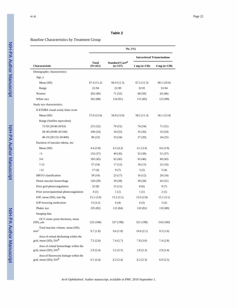
Between November 4, 2004, and February 29, 2008, 411 patients with BRVO were enrolledin the SCORE Study at 75 clinical sites (Table 2). The mean duration of macular edema (basedon patient history or ophthalmologic diagnosis) prior to enrollment was 4 months; 37% ofparticipants had macular edema for less than 3 months; and 82% had macular edema for 6months or less. The mean baseline visual acuity letter score was 57 (Snellen equivalent,approximately 20/80), and eyes had a mean center point thickness of about 523 μm based onOCT. A more detailed description of the SCORE-BRVO participant population can be foundelsewhere.48
FOLLOW-UPFigure 1 shows study follow-up of all participants at 4-month intervals through 12 months, andthen annually through 36 months. The month 12 primary outcome visit was completed by 88%,89%, and 91% of participants in the standard care, 1-mg, and 4-mg groups, respectively. Atthe time of study closeout, 58% of study participants had month 24 outcomes assessed and31% had month 36 outcomes assessed.
STUDY TREATMENTSFor eyes without a dense macular hemorrhage at baseline and randomized to the standard caregroup, the mean number of laser treatments prior to 12 months was 1.8 (95% CI, 1.6-2.0); 27%of these eyes received the maximum number of 3 treatments and 98% received at least 1 lasertreatment prior to 12 months (Table 3). For the 39 eyes in the standard care group with a densemacular hemorrhage at baseline, the mean number of grid photocoagulation treatments priorto 12 months was 0.7 (95% CI, 0.5-1.0); 49% of these eyes received at least 1 gridphotocoagulation treatment during the study period through 12 months, and 21% received 2laser treatments (at months 4 and 8) prior to 12 months. Overall, 84% of participants in thestandard care group received at least 1 grid photocoagulation treatment prior to month 12.
Prior to month 12, the mean number of injections was similar in the triamcinolone groups, with2.2 (95% CI, 2.1-2.4) in the 1-mg triamcinolone group and 2.1 in the 4-mg triamcinolone group(95% CI, 2.0-2.3). Success of the prior triamcinolone treatment was the primary reason for notadministering additional injections prior to 12 months (69%). Other reasons cited for not
et al. Page 7
Arch Ophthalmol. Author manuscript; available in PMC 2010 September 1.
NIH
-PA Author Manuscript
NIH
-PA Author Manuscript
NIH
-PA Author Manuscript

administering retreatment prior to 12 months include futility of the treatment (12%), treatmentcontraindicated (4%), participant refusal (2%), and other (13%).
Few treatment protocol deviations were noted prior to 12 months. These entailed theadministration of intravitreal injections of (1) an anti-VEGF drug in 1 participant in the 4-mgtriamcinolone group and 1 participant in the standard care group; and (2) nonstudy-formulationtriamcinolone in 1 participant in the 4-mg triamcinolone group and 3 participants in thestandard care group. These participants were included in the primary analyses but not in theper-protocol analysis.
VISUAL ACUITY OUTCOMESThe primary outcome of the SCORE-BRVO trial, the percentage of participants with a gain invisual acuity letter score of 15 or more from baseline to month 12, was similar in all 3 groups:28.9%, 25.6%, and 27.2% in the standard care, and 1-mg and 4-mg triamcinolone groups,respectively (Table 4 and Figure 2A). The odds ratios (ORs) for all 3 comparisons were closeto 1 and P>.05 (1-mg triamcinolone group compared with standard care group, OR, 0.9; 95%CI, 0.5-1.6; P=.89; and 4-mg triamcinolone group compared with standard care group, OR,0.9; 95% CI, 0.5-1.6; P=.89; 4-mg triamcinolone group compared with 1-mg triamcinolonegroup, OR, 1.0; 95% CI, 0.5-1.7; P=.89). All 3 groups had a similar gain of approximately 4to 6 in mean visual acuity letter score from baseline to month 12 (Table 4). At month 12, therewas a similar percentage of eyes in all 3 study groups with a loss in visual acuity letter scoreof 15 or more, approximately 11% to 15% (Table 4 and Figure 2B). The sensitivity of theprimary efficacy conclusions to missing data was investigated, and no pattern of outcomesamong missing participants overturns the conclusion that 1 mg or 4 mg of intravitrealtriamcinolone is not superior to standard care at 12 months. Furthermore, the per-protocolanalysis gave results that were qualitatively similar to the primary analysis.
The percentages with a gain or loss in visual acuity letter score of 15 or more from baselineand mean changes from baseline in visual acuity letter score are presented by study group andscheduled follow-up visit in Table 5 and Figure 2. The similarity of visual acuity results acrossthe 3 study groups that is evident at month 12 was not present at month 4 for the comparisonof the mean change in visual acuity letter score, which was greater in the 4-mg triamcinolonegroup compared with the other 2 groups (P=.002, based on analysis of variance). After month12, mean change from baseline in visual acuity letter score is greater in the standard care groupcompared with the 2 triamcinolone groups (P<.05 at months 16, 20, 24, and 32, based onanalysis of variance).
Analyses examining 12-month visual acuity outcomes for prespecified baseline subgroupscategorizing dense macular hemorrhage status at baseline, grid photocoagulation status priorto enrollment, duration of macular edema, visual acuity letter score, and OCT-measured centerpoint thickness demonstrated consistency of results with those observed in the overall 12-month analysis, with no sub-group showing a statistically significant treatment effect (Table6). An analysis limited to eyes that were pseudophakic at baseline also demonstrated nostatistically significant difference among the treatment groups with respect to change in visualacuity over time (P=.44, based on analysis of variance).
IMAGING OUTCOMESAll 3 study groups showed OCT-measured center point thickness decreases from baselinethroughout follow-up (Table 7 and Figure 3). At the month 4 visit, the median decrease wasgreater in the 4-mg triamcinolone group (142-μm decrease) than the 1-mg group (77-μmdecrease) and the standard care group (113-μm decrease; P=.008, Kruskal-Wallis test). At the
et al. Page 8
Arch Ophthalmol. Author manuscript; available in PMC 2010 September 1.
NIH
-PA Author Manuscript
NIH
-PA Author Manuscript
NIH
-PA Author Manuscript

month 12 visit, the median decrease from baseline in OCT-measured center point thicknesswas similar among the 3 treatment groups.
Changes in OCT-measured center point thickness from baseline in all 3 study groups showedmoderate negative correlation with changes from baseline in visual acuity letter score overtime. The Pearson correlation coefficients were −0.23, −0.28, and −0.32 at month 4 and −0.19,−0.30, and −0.10 at month 12 for the standard care, 1-mg, and 4-mg study groups, respectively.For all 3 study groups, disc areas of fluorescein leakage within the grid were similar at baselineand follow-up visits (Table 8). The percentages of eyes with greater than 10 disc areas ofcapillary nonperfusion within the eye at month 12 was about 15% in all 3 groups; 23% to 31%of eyes had greater than 5 disc areas of nonperfusion at month 12.
SAFETY OUTCOMESIntraocular pressure–lowering medication was initiated in more eyes through 12 months in the4-mg triamcinolone group (41%) compared with the 1-mg triamcinolone (7%) and standardcare (2%) groups (χ2 test adjusting for multiple testing: standard care vs 1 mg of triamcinolone,P=.03; standard care vs 4 mg of triamcinolone, P<.001; and 1 mg vs 4 mg of triamcinolone,P<.001) (Table 9). Trabeculectomy or tube shunt surgery was not performed up to 12 months.Between 12 and 24 months, 1 participant in the 4-mg group received a trabeculectomy and 1participant in the 4-mg group received a tube shunt to control IOP unrelated to neovascularglaucoma.
Among eyes that were phakic at baseline, the estimate of new-onset lens opacity or progressionof an existing opacity based on clinical assessment through month 12 in the standard care groupwas 13% compared with 25% and 35% in the 1-mg and 4-mg triamcinolone groups,respectively (χ2 test adjusting for multiple testing: standard care vs 1 mg of triamcinolone,P=.03; standard care vs 4 mg of triamcinolone, P<.001; and 1 mg vs 4 mg of triamcinolone,P=.10) (Table 9). Through month 12, cataract surgery was reported for 3 participants in thestandard care group vs none in the 1-mg triamcinolone group and 4 in the 4-mg triamcinolonegroup. Cataract surgery was more frequent between months 12 and 24 in the 4-mg group, with35 study eyes receiving such surgery, compared with 8 in the 1-mg study group and 6 in thestandard care group (log-rank test between 1 and 2 years, standard care vs 1 mg oftriamcinolone, P=.59; standard care vs 4 mg of triamcinolone, P<.001; and 1 mg vs 4 mg oftriamcinolone, P<.001).
Through month 12, there were no reports of infectious endophthalmitis in the standard caregroup or 1-mg triamcinolone group and 1 case in the 4-mg triamcinolone group 3 days afterthe third injection. This participant's vitreous culture grew coagulase-negative staphylococcus.The participant's visual acuity letter score returned to 59 five weeks after the endophthalmitisdiagnosis (the pre-endophthalmitis visual acuity letter score was 54), and the participant'svisual acuity letter score at 36 months was 65 (visual acuity letter score at randomization, 65).Surgical procedures through 12 months, including sector/ panretinal scatter photocoagulation,pars plana vitrectomy, and yttrium-aluminum-garnet capsulotomy, were uncommon.
Minor ocular adverse events related to the injection procedure were evaluated. Vitreous floatersand conjunctival hemorrhage were reported in a similar percentage of participants in bothtriamcinolone groups through month 12: 31% of the 1-mg triamcinolone group and 26% of 4-mg triamcinolone group had vitreous floaters and 30% of the 1-mg triamcinolone group and33% of the 4-mg triamcinolone group had conjunctival hemorrhage. Silicone oil droplets inthe vitreous were reported through 12 months in 33% of eyes treated with 1 mg of triamcinoloneand 12% of eyes treated with 4 mg of triamcinolone. A separate report provides more detailedinformation on the incidence of intravitreal silicone oil in the SCORE Study, which decreasedprecipitously following the introduction of a luer cone needle design in place of a staked-on
et al. Page 9
Arch Ophthalmol. Author manuscript; available in PMC 2010 September 1.
NIH
-PA Author Manuscript
NIH
-PA Author Manuscript
NIH
-PA Author Manuscript

needle design.54 No adverse events were reported as a result of treatment with gridphotocoagulation.
Reports of systemic adverse events were similar among the SCORE-BRVO trial groups. TheMedical Dictionary for Regulatory Activities system/organ class of infection and infestationshad the highest incidence through month 12, with 10%, 16%, and 15% of participants reportingat least 1 event in the standard care, 1-mg, and 4-mg groups, respectively. Seven deathsoccurred prior to 12 months of follow-up (3 in the standard care group, 2 in the 1-mgtriamcinolone group, and 2 in the 4-mg triamcinolone group), and 9 more deaths occurred after12 months of follow-up (2 in the standard care group and 7 in the 4-mg triamcinolone group).
COMMENTThe results of the SCORE-BRVO trial demonstrate no significant differences among the 3treatment groups for a gain in visual acuity letter score of 15 or more at 12 months, though anearly positive treatment response of a gain in visual acuity letter score of 15 or more wasobserved at month 4 in the 4-mg triamcinolone group compared with the 1-mg triamcinoloneand standard care groups. Additionally, at month 12 there was a similar gain of 4 to 5 in meanvisual acuity letter score from baseline and a similar likelihood of a loss in visual acuity letterscore of 15 or more in all 3 treatment groups. After month 12 and through month 36, the meanimprovement from baseline visual acuity letter score was greatest in the standard care groupcompared with the 2 triamcinolone groups.
With respect to OCT-measured center point thickness, all 3 groups showed a decrease frombaseline to month 12. Analogous to the visual acuity results, only at month 4 did the 4-mgtriamcinolone group demonstrate a greater treatment effect on center point thickness than the1-mg and standard care groups; at all other times investigated (months 8-36), the standard caregroup demonstrated the greatest overall median decrease in center point thickness frombaseline. There was only moderate correlation between changes from baseline in OCT-measured thickness and changes from baseline in visual acuity letter score, which is consistentwith previously reported observations of a modest correlation between OCT-measuredthickness and visual acuity in patients with macular edema secondary to CRVO,52 BRVO,52or diabetic retinopathy.55
The rates of adverse events were higher in the 4-mg triamcinolone group compared with the1-mg and standard care groups. There was a dose-dependent higher frequency of initiatingIOP-lowering medications in the triamcinolone groups compared with the standard care group.No participant was treated with trabeculectomy or tube shunt surgery through 12 months. Theproportion of eyes that were phakic at baseline and had new-onset lens opacity or progressionof an existing opacity through 12 months based on assessment at the clinical center was greaterin the 2 triamcinolone groups compared with the standard care group. Most cataract surgerieswere performed during the second year of the study and occurred with the highest frequencyin the 4-mg group. Intravitreal silicone oil droplets were observed in 23% of eyes treated withintravitreal triamcinolone, but no adverse effects were attributable to the silicone oil droplets.Intravitreal silicone oil associated with the use of siliconized syringes is a recognizedoccurrence.56,57
The rate of infectious endophthalmitis in the SCORE-BRVO trial was 0.1% (1 of 914) perinjection. The lack of any report of noninfectious endophthalmitis in the SCORE-BRVO trial,which included an evaluation of all participants within 1 week postinjection to evaluate forcomplications, may be due to the preservative-free, micronized, nondispersive triamcinoloneformulation (the triamcinolone crystals were suspended in a hyaluronate matrix gel) used inthis trial.
et al. Page 10
Arch Ophthalmol. Author manuscript; available in PMC 2010 September 1.
NIH
-PA Author Manuscript
NIH
-PA Author Manuscript
NIH
-PA Author Manuscript

Cataract development is unlikely to have masked a significant treatment benefit oftriamcinolone over standard care in the overall results. First, an analysis of eyes pseudophakicat baseline demonstrated no statistically significant difference among the treatment groups withrespect to change in visual acuity over time. Second, the protocol specified that cataract surgerywas to be performed at any time that such surgery was indicated clinically, a point that wasemphasized in the SCORE Study by a memorandum sent to all study investigators, encouragingthem to facilitate cataract surgery for any study participants in whom this was indicatedclinically. Third, most cataract surgeries in SCORE-BRVO participants occurred between 12and 24 months, suggesting that this is when the cataracts became clinically significant. Despitemore cataract extractions having been performed between 12 and 24 months in the 4-mgtriamcinolone group than in the other 2 groups, there was no significant difference in visualacuity outcome that showed a benefit of triamcinolone over standard care at any time otherthan month 4. Lastly, the results from the SCORE-CRVO trial demonstrate a strong treatmenteffect of intravitreal triamcinolone relative to observation. Thus, in the SCORE-BRVO trial,it is unlikely that the triamcinolone groups fared no better than the standard care group becauseof a masking of treatment effect by cataract (as there is no reason to suspect that participantswith BRVO are more susceptible to cataract than participants with CRVO) but rather as a resultof triamcinolone being compared with an equally efficacious therapy—grid photocoagulation.
It is unlikely that a triamcinolone benefit over standard care went undetected, particularlybecause the trends favored the standard care group. One sensitivity analysis shows that, evenafter imputing favorable outcomes for all the missing month 12 outcomes in the 2 triamcinolonegroups and unfavorable outcomes in the standard care group, the month 12 results would stillnot favor the triamcinolone groups, providing more evidence of no treatment difference amongthe 3 study groups. Other sensitivity analyses produced similar results. The SCORE Studyprotocol encouraged frequent retreatment (see the “Methods” section). Thus, participants inthis study were not likely to have been undertreated.
Twelve-month data are important in a disease with an acute nature, and 12 months may besufficient follow-up, but comparison of results among the study groups after 12 months isanother topic of interest; a shortcoming of the current study is the lack of definitive data atthese later scheduled visits, owing either to missed visits or to participants not having longenough follow-up to complete the 24- or 36-month visits because of the common closeout.However, the 128 participants in the SCORE-BRVO trial observed at 36 months, 82 of whomwere in the triamcinolone arms, exceed the size of the cohort of participants studied in theBVOS (group III),1 which followed 78 eyes (including 43 eyes treated with gridphotocoagulation and 35 untreated) for as long as 36 months. In the SCORE-BRVO trial, thedata available in the month 36 visit do not suggest a treatment benefit of intravitrealtriamcinolone relative to standard care; in fact, the trend in the SCORE-BRVO trial at month36 suggests that standard care may be superior to intravitreal triamcinolone (Table 5 and Figure2).
The month 12 results of the SCORE-BRVO trial demonstrate that treatment with 1 mg oftriamcinolone, 4 mg oftriamcinolone,orstandardcareishistoricallycomparable with what wasobserved in the treated arm of the BVOS (grid photocoagulation) and that all 3 treatments arelikely associated with a better visual outcome than the natural history of macular edemasecondary to BRVO as demonstrated in the untreated arm of the BVOS. The results of theSCORE-BRVO trial also underscore the importance of grid photocoagulation in managingvision loss due to macular edema associated with BRVO across a wide range of visual acuitiesand retinal thicknesses, even in eyes with prior grid photocoagulation treatment for macularedema secondary to BRVO. It is important to note that an eye with prior grid photocoagulationwas enrolled in this trial only if the investigatorjudgedtheeyetohavethepotentialtobenefitfromadditional photocoagulation. Notably, whether or not laser photocoagulation was performed
et al. Page 11
Arch Ophthalmol. Author manuscript; available in PMC 2010 September 1.
NIH
-PA Author Manuscript
NIH
-PA Author Manuscript
NIH
-PA Author Manuscript

prior to randomization, there was no difference in treatment effect among the 3 groups, thoughthe number of participants entering the trial having undergone grid photocoagulation was small(Table 6). Given the equal efficacy for visual acuity (gain, mean change, and loss), the superioradverse event profile of standard care over 4 mg of triamcinolone and the at least theoreticalsafety advantage of standard care over 1 mg of intravitreal triamcinolone, standard care isrecommended over either dose of intravitreal triamcinolone.
Despite the findings from the SCORE-BRVO trial, there is a need for improved treatments inthe future, because, at 12 months, less than one-third of study eyes in the standard care grouphad a gain in visual acuity letter score of 15 or more, and about half of study eyes in the standardcare group still had central retinal thickening. The fact that the 4-mg triamcinolone group hada greater positive treatment response on visual acuity and retinal thickness at 4 months, whilethe standard care group demonstrated a more delayed but progressive improvement in visualacuity and retinal thickness over time, raises the possibility that combining gridphotocoagulation with intravitreal triamcinolone may produce a greater benefit than either ofthese treatments alone; this could be evaluated in a future clinical trial.
In conclusion, there was no significant difference in terms of visual acuity outcomes (gain,mean change, or loss) among the groups treated with standard care, 1 mg of intravitrealtriamcinolone, or 4 mg of intravitreal triamcinolone for vision loss associated with macularedema secondary to BRVO at 12 months. The results of the SCORE-BRVO trial suggest thatstandard care and intravitreal triamcinolone in either the 1-mg or 4-mg dose are all likelyassociated with a better visual outcome than the natural history of macular edema secondaryto BRVO. The rates of adverse events were higher in the 4-mg triamcinolone group than in thestandard care group. The rates of adverse events with respect to cataract surgery and elevatedIOP were similar between the standard care and 1-mg groups, but the potential for added risksof procedure-related complications in the 1-mg group (as exemplified by the 1 case ofendophthalmitis reported in the 4-mg group) suggests a superior safety profile for the standardcare group. Thus, current study results (up to 12 months and possibly up to 36 months) supportgrid photocoagulation as the continued standard of care for patients with decreased visualacuity associated with macular edema secondary to BRVO who have similar characteristics tothe cohort in this clinical trial. The results of this trial also support the idea that gridphotocoagulation, as applied in the current study, should be the benchmark against which othertreatments for vision loss associated with macular edema secondary to BRVO are comparedin clinical trials.
AcknowledgmentsFunding/Support: This study was supported by the National Eye Institute (National Institutes of Health, Departmentof Health and Human Services)grants 5U10EY014351, 5U10EY014352, and 5U10EY014404; and in part by AllerganInc.
REFERENCES1. The Branch Vein Occlusion Study Group. Argon laser photocoagulation for macular edema in branch
vein occlusion. Am J Ophthalmol 1984;98(3):271–282. [PubMed: 6383055]2. Mitchell P, Smith W, Chang A. Prevalence and associations of retinal vein occlusion in Australia: The
Blue Mountains Eye Study. Arch Ophthalmol 1996;114(10):1243–1247. [PubMed: 8859084]3. Klein R, Moss SE, Meuer SM, Klein BEK. The 15-year cumulative incidence of retinal vein occlusion:
The Beaver Dam Eye Study. Arch Ophthalmol 2008;126(4):513–518. [PubMed: 18413521]4. Klein R, Klein BEK, Lee KE, Cruickshanks KJ, Gangnon RE. Changes in visual acuity in a population
over a 15-year period: The Beaver Dam Eye Study. Am J Ophthalmol 2006;142(4):539–549. [PubMed:17011842]
et al. Page 12
Arch Ophthalmol. Author manuscript; available in PMC 2010 September 1.
NIH
-PA Author Manuscript
NIH
-PA Author Manuscript
NIH
-PA Author Manuscript
5. Chang MA, Fine HF, Bass E, et al. Patients' preferences in choosing therapy for retinal vein occlusions.Retina 2007;27(6):789–797. [PubMed: 17621191]
6. McIntosh RL, Mohamed Q, Saw SM, Wong TY. Interventions for branch retinal vein occlusion: anevidence-based systematic review. Ophthalmology 2007;114(5):835–854. [PubMed: 17397923]
7. Kumagai K, Furukawa M, Ogino N, Uemura A, Larson E. Long-term outcomes of vitrectomy with orwithout arteriovenous sheathotomy in branch retinal vein occlusion. Retina 2007;27(1):49–54.[PubMed: 17218915]
8. Shah GK, Sharma S, Fineman MS, Federman J, Brown MM, Brown GC. Arteriovenous adventitialsheathotomy for the treatment of macular edema associated with branch retinal vein occlusion. Am JOphthalmol 2000;129(1):104–106. [PubMed: 10653427]
9. Opremcak EM, Bruce RA. Surgical decompression of branch retinal vein occlusion via arteriovenouscrossing sheathotomy: a prospective review of 15 cases. Retina 1999;19(1):1–5. [PubMed: 10048366]
10. Fekrat S, Goldberg MF, Finkelstein D. Laser-induced chorioretinal venous anastomosis fornonischemic central or branch retinal vein occlusion. Arch Ophthalmol 1998;116(1):43–52.[PubMed: 9445207]
11. Bavbek T, Yenice O, Toygar O. Problems with attempted chorioretinal venous anastomosis by laserfor nonischemic CRVO and BRVO. Ophthalmologica 2005;219(5):267–271. [PubMed: 16123551]
12. Costa RA, Jorge R, Calucci D, Melo LA Jr, Cardillo JA, Scott IU. Intravitreal bevacizumab (Avastin)for central and hemicentral retinal vein occlusions: IBeVO study. Retina 2007;27(2):141–149.[PubMed: 17290194]
13. Cekic O, Chang S, Tseng JJ, et al. Intravitreal triamcinolone treatment for macular edema associatedwith central retinal vein occlusion and hemiretinal vein occlusion. Retina 2005;25(7):846–850.[PubMed: 16205562]
14. Ozkiris A, Evereklioglu C, Erkilic K, Ilhan O. The efficacy of intravitreal triamcinolone acetonideon macular edema in branch retinal vein occlusion. Eur J Ophthalmol 2005;15(1):96–101. [PubMed:15751246]
15. Lee H, Shah GK. Intravitreal triamcinolone as primary treatment of cystoid macular edema secondaryto branch retinal vein occlusion. Retina 2005;25(5):551–555. [PubMed: 16077348]
16. Krepler K, Ergun E, Sacu S, et al. Intravitreal triamcinolone acetonide in patients with macular oedemadue to branch retinal vein occlusion: a pilot study. Acta Ophthalmol Scand 2005;83(5):600–604.[PubMed: 16188000]
17. Hayashi K, Hayashi H. Intravitreal versus retrobulbar injections of triamcinolone for macular edemaassociated with branch retinal vein occlusion. Am J Ophthalmol 2005;139(6):972–982. [PubMed:15953425]
18. Jonas JB, Akkoyun I, Kamppeter B, Kreissig I, Degenring RF. Branch retinal vein occlusion treatedby intravitreal triamcinolone acetonide. Eye 2005;19(1):65–71. [PubMed: 15105817]
19. Cekiç O, Chang S, Tseng JJ, et al. Intravitreal triamcinolone injection for treatment of macular edemasecondary to branch retinal vein occlusion. Retina 2005;25(7):851–855. [PubMed: 16205563]
20. Karacorlu M, Ozdemir H, Karacorlu SA. Resolution of serous macular detachment after intravitrealtriamcinolone acetonide treatment of patients with branch retinal vein occlusion. Retina 2005;25(7):856–860. [PubMed: 16205564]
21. Yepremyan M, Wertz FD, Tivnan T, Eversman L, Marx JL. Early treatment of cystoid macular edemasecondary to branch retinal vein occlusion with intravitreal triamcinolone acetonide. OphthalmicSurg Lasers Imaging 2005;36(1):30–36. [PubMed: 15688969]
22. Chen SDM, Sundaram V, Lochhead J, Patel CK. Intravitreal triamcinolone for the treatment ofischemic macular edema associated with branch retinal vein occlusion. Am J Ophthalmol 2006;141(5):876–883. [PubMed: 16527226]
23. Özkiris A, Evereklioglu C, Erkilic K, Dogan H. Intravitreal triamcinolone acetonide for treatment ofpersistent macular oedema in branch retinal vein occlusion. Eye 2006;20(1):13–17. [PubMed:15723039]
24. Cheng KC, Wu W. Intravitreal triamcinolone acetonide for patients with macular edema due to branchretinal vein occlusion. Kaohsiung J Med Sci 2006;22(7):321–330. [PubMed: 16849100]
et al. Page 13
Arch Ophthalmol. Author manuscript; available in PMC 2010 September 1.
NIH
-PA Author Manuscript
NIH
-PA Author Manuscript
NIH
-PA Author Manuscript

25. Oh JY, Seo JH, Ahn JK, Heo JW, Chung H. Early versus late intravitreal triamcinolone acetonide formacular edema associated with branch retinal vein occlusion. Korean J Ophthalmol 2007;21(1):18–20. [PubMed: 17460427]
26. Cakir M, Dogan M, Bayraktar Z, et al. Efficacy of intravitreal triamcinolone for the treatment ofmacular edema secondary to branch retinal vein occlusion in eyes with or without grid laserphotocoagulation. Retina 2008;28(3):465–472. [PubMed: 18327140]
27. Aiello LP, Bursell SE, Clermont A, et al. Vascular endothelial growth factor-induced retinalpermeability is mediated by protein kinase C in vivo and suppressed by an orally effective beta-isoform-selective inhibitor. Diabetes 1997;46(9):1473–1480. [PubMed: 9287049]
28. Antonetti DA, Barber AJ, Hollinger LA, Wolpert EB, Gardner TW. Vascular endothelial growthfactor induces rapid phosphorylation of tight junction proteins occludin and zonula occluden 1: apotential mechanism for vascular permeability in diabetic retinopathy and tumors. J Biol Chem1999;274(33):23463–23467. [PubMed: 10438525]
29. Senger DR, Galli SJ, Dvorak AM, Perruzzi CA, Harvey VS, Dvorak HF. Tumor cells secrete avascular permeability factor (VPF) that promotes accumulation of ascites fluid. Science 1983;219(4587):983–985. [PubMed: 6823562]
30. Vinores SA, Youssri AI, Luna JD, et al. Upregulation of vascular endothelial growth factor in ischemicand non-ischemic human and experimental retinal disease. Histol Histopathol 1997;12(1):99–109.[PubMed: 9046048]
31. Pe'er J, Folberg R, Itin A, Gnessin H, Hemo I, Keshet E. Vascular endothelial growth factorupregulation in human central retinal vein occlusion. Ophthalmology 1998;105(3):412–416.[PubMed: 9499769]
32. Zhang X, Bao S, Lai D, Rapkins RW, Gillies MC. Intravitreal triamcinolone acetonide inhibitsbreakdown of the blood-retinal barrier through differential regulation of VEGF-A and its receptorsin early diabetic rat retinas. Diabetes 2008;57(4):1026–1033. [PubMed: 18174522]
33. Wang K, Wang Y, Gao L, Li X, Li M, Guo J. Dexamethasone inhibits leukocyte accumulation andvascular permeability in retina of streptozotocin-induced diabetic rats via reducing vascularendothelial growth factor and intercellular adhesion molecule-1 expression. Biol Pharm Bull 2008;31(8):1541–1546. [PubMed: 18670086]
34. Lee HB, Pulido JS, McCannel CA, Buettner H. Role of inflammation in retinal vein occlusion. CanJ Ophthalmol 2007;42(1):131–133. [PubMed: 17361257]
35. Flynn HW Jr, Scott IU. Intravitreal triamcinolone acetonide for macular edema associated withdiabetic retinopathy and venous occlusive disease: it's time for clinical trials. Arch Ophthalmol2005;123(2):258–259. [PubMed: 15710825]
36. Blumenkranz MS. New therapy for central retinal vein occlusion: are intravitreal steroids a possibleanswer? Arch Ophthalmol 2005;123(2):259–261. [PubMed: 15710826]
37. The SCORE Study Research Group. A randomized trial comparing the efficacy and safety ofintravitreal triamcinolone with observation to treat vision loss associated with macular edemasecondary to central retinal vein occlusion: The Standard Care vs Corticosteroid for Retinal VeinOcclusion (SCORE) Study Report 5. Arch Ophthalmol 2009;127(9):1101–1114. [PubMed:19752419]
38. Beck RW, Moke PS, Turpin AH, et al. A computerized method of visual acuity testing: adaptationof the Early Treatment of Diabetic Retinopathy Study testing protocol. Am J Ophthalmol 2003;135(2):194–205. [PubMed: 12566024]
39. Age-Related Eye Disease Study Research Group. The age-related eye disease study (AREDS) systemfor classifying cataracts from photographs: AREDS Report No. 4. Am J Ophthalmol 2001;131(2):167–175. [PubMed: 11228291]
40. Chieh JJ, Roth DB, Liu M, et al. Intravitreal triamcinolone acetonide for diabetic macular edema.Retina 2005;25(7):828–834. [PubMed: 16205559]
41. Kim JE, Pollack JS, Miller DG, Mittra RA, Spaide RF, Isis Study Group. ISIS-DME: a prospective,randomized, dose-escalation intravitreal steroid injection study for refractory diabetic macularedema. Retina 2008;28(5):735–740. [PubMed: 18463518]
et al. Page 14
Arch Ophthalmol. Author manuscript; available in PMC 2010 September 1.
NIH
-PA Author Manuscript
NIH
-PA Author Manuscript
NIH
-PA Author Manuscript

42. Jonas JB, Kreissig I, Spandau UH, Harder B. Infectious and noninfectious endophthalmitis afterintravitreal high-dosage triamcinolone acetonide. Am J Ophthalmol 2006;141(3):579–580.[PubMed: 16490517]
43. Jonas JB, Kreissig I, Degenring RF. Intravitreal triamcinolone acetonide as treatment of macularedema in central retinal vein occlusion. Graefes Arch Clin Exp Ophthalmol 2002;240(9):782–783.[PubMed: 12271378]
44. Then Bergh F, Grasser A, Trenkwalder C, Backmund H, Holsboer F, Rupprecht R. Bindingcharacteristics of the glucocorticoid receptor in peripheral blood lymphocytes in multiple sclerosis.J Neurol 1999;246(4):292–298. [PubMed: 10367698]
45. Schottelius A, Wedel S, Weltrich R, et al. Higher expression of glucocorticoid receptor in peripheralmononuclear cells in inflammatory bowel disease. Am J Gastroenterol 2000;95(8):1994–1999.[PubMed: 10950048]
46. Chen SD, Lochhead J, Patel CK. Diffuse intraocular dispersion of triamcinolone particles as a causeof sterile endophthalmitis [letter]. Arch Ophthalmol 2004;122(11):1733. [PubMed: 15534152]
47. Moshfeghi DM, Kaiser PK, Bakri SJ, et al. Presumed sterile endophthalmitis following intravitrealtriamcinolone acetonide injection. Ophthalmic Surg Lasers Imaging 2005;36(1):24–29. [PubMed:15688968]
48. Ip MS, Oden NL, Scott IU, et al. SCORE Study Investigator Group. SCORE Study Report 3: studydesign and baseline characteristics. Ophthalmology. published online ahead of print July 18, 2009.
49. Massin P, Erginay A, Haouchine B, Mehidi AB, Paques M, Gaudric A. Retinal thickness in healthyand diabetic subjects measures using optical coherence tomography mapping software. Eur JOphthalmol 2002;12(2):102–108. [PubMed: 12022281]
50. Fundus Photograph Reading Center Imaging Procedures for the Standard Care vs COrticosteroid forREtinal Vein Occlusion (SCORE) Study. National Eye Institute; Bethesda, MD: 2008. NTIS orderNo. PB2008-113740
51. Domalpally A, Blodi BA, Scott IU, et al. SCORE Study Investigator Group. SCORE Study Report4: grading methodology for optical coherence tomography images. Arch Ophthalmol. 2009 In press.
52. Scott IU, VanVeldhuisen PC, Oden NL, et al. SCORE Study Investigator Group. SCORE StudyReport 1: baseline association between central retinal thickness and visual acuity in patients withretinal vein occlusion. Ophthalmology 2009;116(3):504–512. [PubMed: 19167078]
53. Marcus R, Peritz E, Gabriel KR. On closed testing procedures with special reference to orderedanalysis of variance. Biometrika 1976;63:655–660.
54. Scott IU, Oden NL, VanVeldhuisen PC, et al. SCORE Study Report 7: incidence of intravitrealsilicone oil droplets associated with staked-on versus luer cone syringe design. Am J Ophthalmol.In press.
55. Browning DJ, Glassman AR, Aiello LP, et al. Diabetic Retinopathy Clinical Research Network.Relationship between optical coherence tomography-measured central retinal thickness and visualacuity in diabetic macular edema. Ophthalmology 2007;114(3):525–536. [PubMed: 17123615]
56. Freund KB, Laud K, Eandi CM, Spaide RF. Silicone oil droplets following intravitreal injection.Retina 2006;26(6):701–703. [PubMed: 16829818]
57. Bakri SJ, Ekdawi NS. Intravitreal silicone oil droplets after intravitreal drug injections. Retina 2008;28(7):996–1001. [PubMed: 18698303]
et al. Page 15
Arch Ophthalmol. Author manuscript; available in PMC 2010 September 1.
NIH
-PA Author Manuscript
NIH
-PA Author Manuscript
NIH
-PA Author Manuscript

Figure 1.Flowchart of participants in the Standard Care vs Corticosteroid for Retinal Vein Occlusion–Branch Retinal Vein Occlusion Study. Missed visits include those who missed a visit but cameback for other visits and those who prematurely withdrew from the study.
et al. Page 16
Arch Ophthalmol. Author manuscript; available in PMC 2010 September 1.
NIH
-PA Author Manuscript
NIH
-PA Author Manuscript
NIH
-PA Author Manuscript

Figure 2.Change from baseline in electronic Early Treatment Diabetic Retinopathy Study visual acuityat each 4-month follow-up visit. The histograms show the percentages of participants with again (A) or loss (B) in visual acuity letter score of 15 or more from baseline. Error bars represent
et al. Page 17
Arch Ophthalmol. Author manuscript; available in PMC 2010 September 1.
NIH
-PA Author Manuscript
NIH
-PA Author Manuscript
NIH
-PA Author Manuscript

upper 95% confidence limits. C, Box plot with whiskers representing the fifth and 95thpercentiles; the line in the box represents the median; dots, values outside the whiskers; SC,standard care; 1 mg and 4 mg, doses of intravitreal triamcinolone acetonide.
et al. Page 18
Arch Ophthalmol. Author manuscript; available in PMC 2010 September 1.
NIH
-PA Author Manuscript
NIH
-PA Author Manuscript
NIH
-PA Author Manuscript
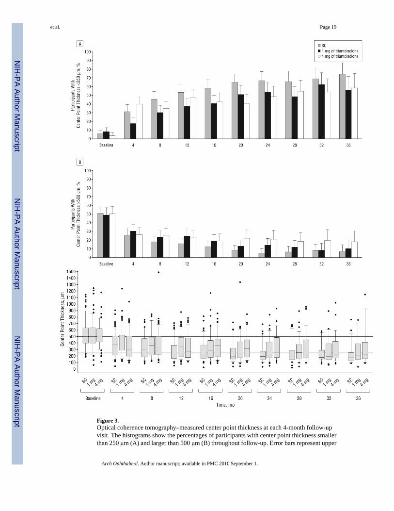
Figure 3.Optical coherence tomography–measured center point thickness at each 4-month follow-upvisit. The histograms show the percentages of participants with center point thickness smallerthan 250 μm (A) and larger than 500 μm (B) throughout follow-up. Error bars represent upper
et al. Page 19
Arch Ophthalmol. Author manuscript; available in PMC 2010 September 1.
NIH
-PA Author Manuscript
NIH
-PA Author Manuscript
NIH
-PA Author Manuscript

95% confidence limits. C, Box plots, with whiskers representing the fifth and 95th percentiles;the line in the box represents the median; dots, values outside the whiskers; SC, standard care;1 mg and 4 mg, doses of intravitreal triamcinolone acetonide. Horizontal reference lines at 250and 500 μm are presented.
et al. Page 20
Arch Ophthalmol. Author manuscript; available in PMC 2010 September 1.
NIH
-PA Author Manuscript
NIH
-PA Author Manuscript
NIH
-PA Author Manuscript

NIH
-PA Author Manuscript
NIH
-PA Author Manuscript
NIH
-PA Author Manuscript
et al. Page 21
Table 1
Study Eye Major Inclusion and Exclusion Criteria
Inclusion Criteria
Best-corrected E-ETDRS visual acuity letter score of ≤73 (approximate Snellen equivalent, 20/40 or worse) and≥19 (approximate Snellen equivalent, 20/400 or better)a
Center-involved macular edema secondary to BRVO present on clinical examination
Mean central subfield retinal thickness of 2 OCT fast macular scans ≥250 μm
Media clarity, pupillary dilation, and subject cooperation sufficient for adequate fundus photographs
Exclusion Criteria
Macular edema due to a cause other than BRVO
An ocular condition such that visual acuity would not improve from resolution of the edema (eg, foveal atrophy)
Substantial cataract estimated to have reduced visual acuity by ≥3 lines
Prior treatment with intravitreal corticosteroids at any time or peribulbar steroid injection within 6 mo prior torandomization
History of focal/grid macular photocoagulation within 15 weeks (3.5 mo) or PRP within 4 mo prior to randomizationor anticipated need for PRP within the 4 mo following randomization
Prior pars plana vitrectomy
Major ocular surgery (including cataract extraction) within prior 6 mo or anticipated within the next 6 mo followingrandomization
Yttrium aluminum garnet capsulotomy performed within 2 mo prior to randomization
IOP ≥25 mm Hg, open-angle glaucoma (either primary open-angle glaucoma or other cause of open-angleglaucoma), or steroid-induced IOP elevation that required IOP-lowering treatment or pseudoexfoliation
Aphakia
Abbreviations: BRVO, branch retinal vein occlusion; E-ETDRS, electronic Early Treatment Diabetic Retinopathy Study; IOP, intraocular pressure;OCT, optical coherence tomography; PRP, panretinal photocoagulation.
aThe original lower limit of visual acuity was expanded from 34 letters to 24 or more letters 5 months after accrual began and then from 24 or more
letters to 19 or more letters 12 months after accrual began.
Arch Ophthalmol. Author manuscript; available in PMC 2010 September 1.
NIH
-PA Author Manuscript
NIH
-PA Author Manuscript
NIH
-PA Author Manuscript
et al. Page 22
Table 2
Baseline Characteristics by Treatment Group
No. (%)
Total(N=411)
Standard Carea(n=137)
Intravitreal Triamcinolone
Characteristic 1 mg (n=136) 4 mg (n=138)
Demographic characteristics
Age, y
Mean (SD) 67.4 (11.2) 66.9 (11.5) 67.2 (11.5) 68.1 (10.6)
Range 22-94 22-90 32-91 33-94
Women 202 (49) 71 (52) 68 (50) 63 (46)
White race 362 (88) 124 (91) 115 (85) 123 (89)
Study eye characteristics
E-ETDRS visual acuity letter score
Mean (SD) 57.0 (12.6) 56.8 (13.0) 58.2 (11.3) 56.1 (13.4)
Range (Snellen equivalent)
73-59 (20/40-20/63) 215 (52) 70 (51) 74 (54) 71 (51)
58-49 (20/80-20/100) 100 (24) 34 (25) 35 (26) 33 (24)
48-19 (20/125-20/400) 96 (23) 33 (24) 27 (20) 34 (25)
Duration of macular edema, mo
Mean (SD) 4.4 (3.8) 4.5 (4.2) 4.1 (3.4) 4.6 (3.9)
<3 152 (37) 49 (36) 52 (38) 51 (37)
3-6 185 (45) 62 (45) 63 (46) 60 (43)
7-12 57 (14) 17 (12) 18 (13) 22 (16)
>12 17 (4) 9 (7) 3 (2) 5 (4)
HRVO classification 59 (14) 23 (17) 16 (12) 20 (14)
Dense macular hemorrhage 120 (29) 39 (28) 38 (28) 43 (31)
Prior grid photocoagulation 32 (8) 15 (11) 8 (6) 9 (7)
Prior sector/panretinal photocoagulation 4 (1) 1 (1) 1 (1) 2 (1)
IOP, mean (SD), mm Hg 15.1 (3.0) 15.2 (3.1) 15.0 (2.8) 15.1 (3.1)
IOP-lowering medication 13 (3.2) 6 (4) 4 (3) 3 (2)
Phakic eye 335 (82) 115 (84) 110 (81) 110 (80)
Imaging data
OCT center point thickness, mean(SD), μm 525 (186) 537 (198) 521 (198) 516 (160)
Total macular volume, mean (SD),mm3 9.7 (1.8) 9.6 (1.8) 10.0 (2.1) 9.5 (1.6)
Area of retinal thickening within thegrid, mean (SD), DAb 7.5 (2.8) 7.4 (2.7) 7.8 (3.0) 7.4 (2.8)
Area of retinal hemorrhage within thegrid, mean (SD), DAb 2.9 (2.4) 3.2 (2.5) 2.8 (2.3) 2.9 (2.4)
Area of fluorescein leakage within thegrid, mean (SD), DAb 6.1 (2.4) 6.2 (2.4) 6.2 (2.3) 6.0 (2.5)
Arch Ophthalmol. Author manuscript; available in PMC 2010 September 1.

NIH
-PA Author Manuscript
NIH
-PA Author Manuscript
NIH
-PA Author Manuscript
et al. Page 23
No. (%)
Total(N=411)
Standard Carea(n=137)
Intravitreal Triamcinolone
Characteristic 1 mg (n=136) 4 mg (n=138)
>5 DA of capillary nonperfusion in theeyec 41 (15) 12 (13) 17 (18) 12 (12)
>10 DA of capillary nonperfusion inthe eyec 26 (9) 6 (6) 13 (14) 7 (7)
Nonstudy eye E-ETDRS visual acuity letterscore, mean (SD) 82.5 (11.0) 81.9 (13.2) 83.7 (8.6) 81.9 (10.7)
Other clinical characteristics
Diabetes mellitus 59 (14) 18 (13) 17 (13) 24 (17)
Hypertension 287 (70) 99 (72) 93 (68) 95 (69)
Coronary artery disease 75 (18) 20 (15) 27 (20) 28 (20)
History of cancer 78 (19) 24 (18) 24 (18) 30 (22)
Abbreviations: DA, disc area; E-ETDRS, electronic Early Treatment Diabetic Retinopathy Study; HRVO, hemiretinal vein occlusion; IOP, intraocularpressure; OCT, optical coherence tomography.
aStandard care consisted of either grid photocoagulation if there was no dense macular hemorrhage or, if a dense macular hemorrhage was present,
observation at 4-month intervals until the dense macular hemorrhage cleared sufficiently to allow grid photocoagulation to be performed.
bThe grid is defined as the 9 ETDRS subfields centered in the macula. The grid is 16 DAs in size.
cCapillary nonperfusion in the eye was assessed in 96, 95, and 102 eyes at baseline in the standard care, 1-mg, and 4-mg groups, respectively.
Arch Ophthalmol. Author manuscript; available in PMC 2010 September 1.

NIH
-PA Author Manuscript
NIH
-PA Author Manuscript
NIH
-PA Author Manuscript
et al. Page 24
Tabl
e 3
Prot
ocol
Tre
atm
ents
in P
artic
ipan
ts
Stan
dard
Car
e, G
rid
Phot
ocoa
gula
tiona
Intr
avitr
eal T
riam
cino
lone
Cha
ract
eris
ticA
ll(n
=137
)D
MH
at B
asel
ine
(n=3
9)N
o D
MH
at B
asel
ine
(n=9
8)1
mgb
(n=1
36)
4 m
gb(n
=138
)
No.
of t
reat
men
ts fr
omba
selin
e to
prio
r to
12 m
o,m
ean
(95%
CI)
1.5
(1.3
-1.7
)0.
7 (0
.5-1
.0)
1.8
(1.6
-2.0
)2.
2 (2
.1-2
.4)
2.1
(2.0
-2.3
)
No.
of t
reat
men
ts p
rior t
o 12
mo,
No.
(%)
3
26 (1
9)0
26 (2
7)63
(46)
47 (3
4)
2
37 (2
7)8
(21)
29 (3
0)43
(32)
62 (4
5)
1
52 (3
8)11
(28)
41 (4
2)30
(22)
28 (2
0)
0
22 (1
6)20
(51)
2 (2
)0
1 (1
)c
Trea
tmen
ts re
ceiv
ed d
urin
gvi
sit,
No.
(%)
B
asel
ine
137
(69)
39 (3
)98
(96)
136
(100
)13
8 (9
9)
4
mo
127
(47)
36 (4
2)91
(49)
132
(76)
132
(60)
8
mo
125
(39)
35 (3
1)90
(42)
123
(56)
128
(60)
12
mo
124
(23)
37 (1
6)87
(25)
121
(45)
126
(31)
16
mo
111
(5)
33 (6
)78
(5)
107
(35)
105
(35)
20
mo
96 (7
)28
(7)
68 (7
)92
(32)
92 (2
3)
24
mo
82 (1
1)28
(7)
54 (1
3)84
(26)
73 (2
5)
Abb
revi
atio
ns: C
I, co
nfid
ence
inte
rval
; DM
H, d
ense
mac
ular
hem
orrh
age.
a Stan
dard
car
e co
nsis
ted
of e
ither
grid
pho
toco
agul
atio
n if
ther
e w
as n
o D
MH
or,
if a
DM
H w
as p
rese
nt, o
bser
vatio
n at
4-m
onth
inte
rval
s unt
il th
e D
MH
cle
ared
suff
icie
ntly
to a
llow
grid
pho
toco
agul
atio
n to
be p
erfo
rmed
. The
re w
ere
4 pa
rtici
pant
s who
und
erw
ent s
tand
ard
care
, all
with
out D
MH
at b
asel
ine,
who
switc
hed
grou
ps a
nd re
ceiv
ed 4
mg
of tr
iam
cino
lone
per
pro
toco
l gui
delin
es p
rior t
o 12
mon
ths.
b Ther
e w
ere
7 pa
rtici
pant
s who
rece
ived
1 m
g an
d 3
who
rece
ived
4 m
g of
tria
mci
nolo
ne w
ho sw
itche
d gr
oups
and
rece
ived
grid
pho
toco
agul
atio
n pe
r pro
toco
l gui
delin
es p
rior t
o 12
mon
ths.
c One
par
ticip
ant w
as ra
ndom
ized
to th
e 4-
mg
arm
, but
refu
sed
initi
al tr
eatm
ent a
nd w
ithdr
ew fr
om th
e st
udy.
Arch Ophthalmol. Author manuscript; available in PMC 2010 September 1.

NIH
-PA Author Manuscript
NIH
-PA Author Manuscript
NIH
-PA Author Manuscript
et al. Page 25
Table 4
Change in Visual Acuity Letter Score at Month 12
%
StandardCarea
(n=121)
Intravitreal Triamcinolone
Change in VisualAcuity Letter Score
1 mg(n=121)
4 mg(n=125)
Change at month 12, letters
≥15 Gainb 28.9 25.6 27.2
10-14 Gain 9.9 17.4 12.8
5-9 Gain 14.0 17.4 12.8
No change, ±4 22.3 20.7 23.2
5-9 Loss 5.0 4.1 5.6
10-14 Loss 5.0 3.3 6.4
≥15 Loss 14.9 11.6 12.0
Mean (95% CI)c 4.2(1.1 to 7.3)
5.7(2.8 to 8.6)
4.0(0.9 to 7.2)
Median (interquartile range) 6.0(−4.0 to 16.0)
8.0(−3.0 to 15.0)
5.0(−4.0 to 15.0)
Abbreviation: CI, confidence interval.
aStandard care consisted of either grid photocoagulation if there was no dense macular hemorrhage or, if a dense macular hemorrhage was present,
observation at 4-month intervals until the dense macular hemorrhage cleared sufficiently to allow grid photocoagulation to be performed.
bPairwise comparisons with a gain in visual acuity letter score of 15 or more: 1-mg vs standard care group, odds ratio, 0.9; 95% CI, 0.5-1.6; P=.89;
4-mg vs standard care group, odds ratio, 0.9; 95% CI, 0.5-1.6; P=.89; 4-mg vs 1-mg group, odds ratio, 1.0; 95% CI, 0.5-1.7; P=.89. The odds ratiosare adjusted for visual acuity and presence of dense macular hemorrhage at baseline. Confidence intervals are not adjusted for simultaneous testingor interim monitoring.
cAnalysis of variance comparing the means among the 3 groups at month 12, P=.70.
Arch Ophthalmol. Author manuscript; available in PMC 2010 September 1.

NIH
-PA Author Manuscript
NIH
-PA Author Manuscript
NIH
-PA Author Manuscript
et al. Page 26
Tabl
e 5
Cha
nge
From
Bas
elin
e V
isua
l Acu
ity L
ette
r Sco
re b
y Fo
llow
-up
Vis
it an
d Tr
eatm
ent G
roup
Mea
n C
hang
e Fr
om B
asel
ine
in V
isua
lA
cuity
Let
ter
Scor
e (9
5% C
I)G
ain
in V
isua
l Acu
ity L
ette
r Sc
ore
of ≥
15, %
(95%
CI)
Los
s in
Vis
ual A
cuity
Let
ter
Scor
e of
≥15
, %
Follo
w-u
pV
isit
No.
of
Part
icip
ants
,St
anda
rd C
area
/1-
mg/
4-m
gG
roup
sSt
anda
rdC
area
Intr
avitr
eal
Tri
amci
nolo
ne
Stan
dard
Car
ea
Intr
avitr
eal
Tri
amci
nolo
ne
Stan
dard
Car
ea
Intr
avitr
eal
Tri
amci
nolo
ne
1 m
g4
mg
1 m
g4
mg
1 m
g4
mg
Mon
th 4
b12
7/13
1/13
20.
7 (−
1.9
to3.
3)2.
6 (−
0.1
to 5
.2)
6.9
(4.8
to9.
0)17
(10-
23)
18 (1
2-25
)23
(16-
30)
1314
4
Mon
th 8
124/
122/
127
1.6
(−1.
6 to
4.8)
5.0
(2.2
to7.
7)4.
3 (1
.8 to
6.9)
15 (8
-21)
24 (1
6-31
)22
(15-
29)
1712
7
Mon
th 1
212
1/12
1/12
54.
2 (1
.1 to
7.3
)5.
7 (2
.8 to
8.6)
4.0
(0.9
to7.
2)29
(21-
37)
26 (1
8-33
)27
(19-
35)
1512
12
Mon
th 1
611
0/10
7/10
55.
5 (2
.4 to
8.7
)5.
9 (2
.8 to
9.1)
−0.8
(−5.
0to
3.3
)31
(22-
40)
26 (1
8-34
)24
(16-
32)
147
22
Mon
th 2
095
/92/
928.
1 (5
.0 to
11.3
)6.
4 (3
.1 to
9.8)
−0.2
(−4.
7to
4.2
)32
(22-
41)
30 (2
1-40
)22
(13-
30)
99
24
Mon
th 2
482
/84/
7211
.3 (8
.1 to
14.5
)6.
3 (2
.3 to
10.3
)4.
0 (−
0.7
to 8
.8)
38 (2
7-48
)36
(25-
46)
31 (2
0-41
)5
1417
Mon
th 2
868
/73/
6010
.9 (7
.3 to
14.4
)6.
1 (1
.9 to
10.3
)4.
4 (−
1.0
to 9
.7)
43 (3
1-54
)34
(23-
45)
33 (2
1-45
)4
815
Mon
th 3
254
/54/
5113
.4 (9
.7 to
17.0
)6.
3 (1
.5 to
11.1
)5.
1 (−
1.1
to 1
1.3)
48 (3
5-61
)30
(17-
42)
41 (2
8-55
)4
1314
Mon
th 3
646
/44/
3812
.9 (7
.4 to
18.4
)4.
4 (−
1.1
to 9
.9)
8.0
(0.9
to15
.2)
48 (3
3-62
)25
(12-
38)
50 (3
4-66
)7
1421
Abb
revi
atio
n: C
I, co
nfid
ence
inte
rval
.
a Stan
dard
car
e co
nsis
ted
of e
ither
grid
pho
toco
agul
atio
n if
ther
e w
as n
o de
nse
mac
ular
hem
orrh
age
or, i
f a d
ense
mac
ular
hem
orrh
age
was
pre
sent
, obs
erva
tion
at 4
-mon
th in
terv
als u
ntil
the
dens
e m
acul
arhe
mor
rhag
e cl
eare
d su
ffic
ient
ly to
allo
w g
rid p
hoto
coag
ulat
ion
to b
e pe
rfor
med
.
b Ana
lysi
s of v
aria
nce
com
parin
g th
e m
eans
am
ong
the
3 gr
oups
at m
onth
4: P
=.00
2.
Arch Ophthalmol. Author manuscript; available in PMC 2010 September 1.

NIH
-PA Author Manuscript
NIH
-PA Author Manuscript
NIH
-PA Author Manuscript
et al. Page 27
Tabl
e 6
Twel
ve-M
onth
Cha
nge
From
Bas
elin
e in
Vis
ual A
cuity
Let
ter S
core
Am
ong
Subg
roup
s
%
No.
of
Part
icip
ants
,St
anda
rd C
are/
1-m
g/4-
mg
Gro
ups
Mea
n C
hang
e in
Vis
ual A
cuity
Let
ter
Scor
eFr
om B
asel
ine
(95%
CI)
Gai
n in
Vis
ual A
cuity
Let
ter
Scor
e of
≥15
Los
s in
Vis
ual A
cuity
Let
ter
Scor
e of
≥15
Stan
dard
Car
ea
Intr
avitr
eal
Tri
amci
nolo
neSt
anda
rdC
area
Intr
avitr
eal
Tri
amci
nolo
neSt
anda
rdC
area
Intr
avitr
eal
Tri
amci
nolo
ne
Cha
ract
eris
tic1
mg
4 m
g1
mg
4 m
g1
mg
4 m
g
Bas
elin
e vi
sual
acu
ity le
tter
scor
e (S
nelle
n eq
uiva
lent
)
73-5
9 (2
0/40
-20/
63)
61/6
6/61
2.0
(−1.
5 to
5.5)
2.5
(−0.
9to
5.8
)−1
.7 (−
6.1
to 2
.6)
2115
1515
1518
58-4
9 (2
0/80
-20/
100)
32/3
2/30
1.5
(−6.
5 to
9.4)
8.4
(2.1
to14
.8)
4.8
(−2.
3to
12.
0)27
3230
216
13
48-1
9 (2
0/12
5-20
/400
)28
/23/
3412
.2 (5
.7 to
18.7
)11
.6 (3
.6to
19.
5)13
.6 (8
.9to
18.
3)45
4847
710
0
Bas
elin
e cen
ter p
oint
thic
knes
s,μm
<500
58/6
1/60
5.4
(1.8
to 9
.0)
7.4
(3.3
to11
.4)
6.9
(2.4
to11
.3)
2325
377
810
≥5
0063
/59/
643.
2 (−
1.9
to8.
3)3.
9 (−
0.3
to 8
.2)
1.3
(−3.
3to
5.8
)33
2719
2215
14
Den
se m
acul
ar h
emor
rhag
e at
base
line
No
84/8
7/85
2.4
(−1.
2 to
6.0)
4.7
(1.1
to8.
3)4.
8 (1
.5 to
8.2)
2223
2914
1111
Yes
37/3
4/40
8.4
(2.2
to14
.5)
8.3
(3.6
to13
.0)
2.3
(−4.
7to
9.2
)43
3223
1612
15
Prio
r grid
pho
toco
agul
atio
n
N
o10
7/11
3/11
64.
1 (0
.6 to
7.5
)6.
6 (3
.7 to
9.5)
4.3
(1.0
to7.
6)29
2728
1711
11
Y
es14
/8/9
5.4
(−0.
1 to
11.0
)
−6.6
(−20
.4 to
7.2)
0.3
(−13
.2to
13.
9)21
022
025
22
Dur
atio
n of
mac
ular
ede
ma
atba
selin
e, m
o
≤3
70/6
0/60
7.8
(3.6
to12
.0)
8.1
(4.1
to12
.0)
2.0
(−2.
3to
6.4
)38
3020
1510
13
Arch Ophthalmol. Author manuscript; available in PMC 2010 September 1.

NIH
-PA Author Manuscript
NIH
-PA Author Manuscript
NIH
-PA Author Manuscript
et al. Page 28
%
No.
of
Part
icip
ants
,St
anda
rd C
are/
1-m
g/4-
mg
Gro
ups
Mea
n C
hang
e in
Vis
ual A
cuity
Let
ter
Scor
eFr
om B
asel
ine
(95%
CI)
Gai
n in
Vis
ual A
cuity
Let
ter
Scor
e of
≥15
Los
s in
Vis
ual A
cuity
Let
ter
Scor
e of
≥15
Stan
dard
Car
ea
Intr
avitr
eal
Tri
amci
nolo
neSt
anda
rdC
area
Intr
avitr
eal
Tri
amci
nolo
neSt
anda
rdC
area
Intr
avitr
eal
Tri
amci
nolo
ne
Cha
ract
eris
tic1
mg
4 m
g1
mg
4 m
g1
mg
4 m
g
>351
/61/
65−0
.6 (−
5.1
to3.
8)3.
4 (−
0.8
to 7
.7)
5.8
(1.3
to10
.4)
1521
3413
1311
Pseu
doph
akic
at b
asel
ine
20/2
1/25
−3.2
(−14
.1 to
7.8)
2.5
(−8.
0to
13.
0)4.
8 (−
2.0
to 1
1.6)
2029
2835
244
Abb
revi
atio
n: C
I, co
nfid
ence
inte
rval
.
a Stan
dard
car
e co
nsis
ted
of e
ither
grid
pho
toco
agul
atio
n if
ther
e w
as n
o de
nse
mac
ular
hem
orrh
age
or, i
f a d
ense
mac
ular
hem
orrh
age
was
pre
sent
, obs
erva
tion
at 4
-mon
th in
terv
als u
ntil
the
dens
e m
acul
arhe
mor
rhag
e cl
eare
d su
ffic
ient
ly to
allo
w g
rid p
hoto
coag
ulat
ion
to b
e pe
rfor
med
.
Arch Ophthalmol. Author manuscript; available in PMC 2010 September 1.
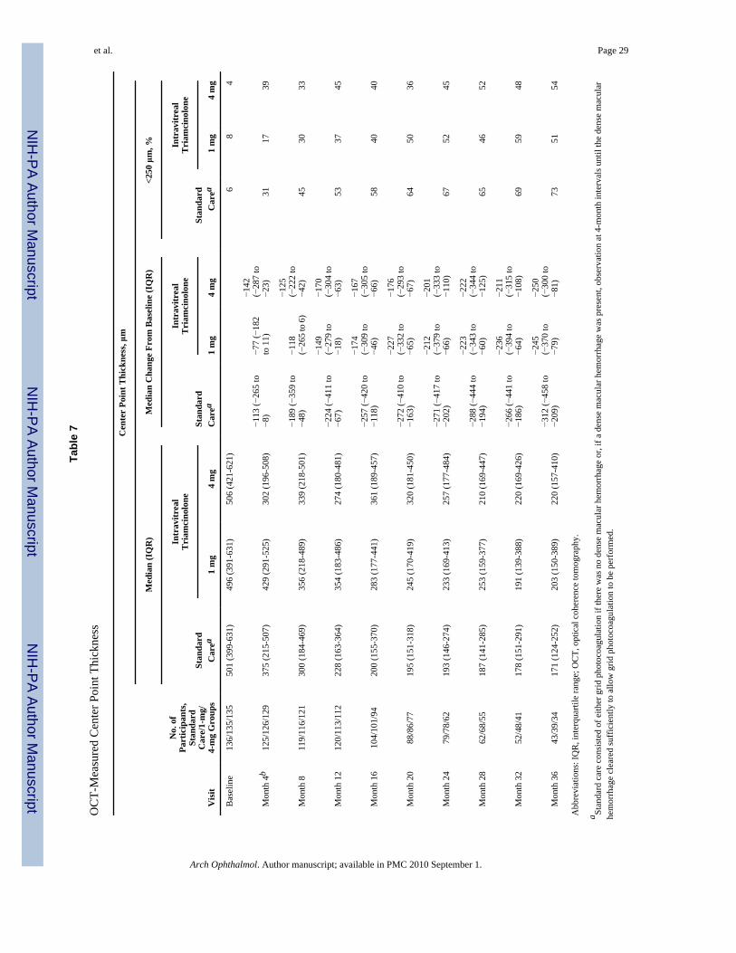
NIH
-PA Author Manuscript
NIH
-PA Author Manuscript
NIH
-PA Author Manuscript
et al. Page 29
Tabl
e 7
OC
T-M
easu
red
Cen
ter P
oint
Thi
ckne
ss
Cen
ter
Poin
t Thi
ckne
ss, μ
m
No.
of
Part
icip
ants
,St
anda
rdC
are/
1-m
g/4-
mg
Gro
ups
Med
ian
(IQ
R)
Med
ian
Cha
nge
From
Bas
elin
e (I
QR
)<2
50 μ
m, %
Stan
dard
Car
ea
Intr
avitr
eal
Tri
amci
nolo
neSt
anda
rdC
area
Intr
avitr
eal
Tri
amci
nolo
neSt
anda
rdC
area
Intr
avitr
eal
Tri
amci
nolo
ne
Vis
it1
mg
4 m
g1
mg
4 m
g1
mg
4 m
g
Bas
elin
e13
6/13
5/13
550
1 (3
99-6
31)
496
(391
-631
)50
6 (4
21-6
21)
68
4
Mon
th 4
b12
5/12
6/12
937
5 (2
15-5
07)
429
(291
-525
)30
2 (1
96-5
08)
−113
(−26
5 to
−8)
−77
(−18
2to
11)
−142
(−28
7 to
−23)
3117
39
Mon
th 8
119/
116/
121
300
(184
-469
)35
6 (2
18-4
89)
339
(218
-501
)−1
89 (−
359
to−4
8)−1
18(−
265
to 6
)
−125
(−22
2 to
−42)
4530
33
Mon
th 1
212
0/11
3/11
222
8 (1
63-3
64)
354
(183
-486
)27
4 (1
80-4
81)
−224
(−41
1 to
−67)
−149
(−27
9 to
−18)
−170
(−30
4 to
−63)
5337
45
Mon
th 1
610
4/10
1/94
200
(155
-370
)28
3 (1
77-4
41)
361
(189
-457
)−2
57 (−
420
to−1
18)
−174
(−30
9 to
−46)
−167
(−30
5 to
−66)
5840
40
Mon
th 2
088
/86/
7719
5 (1
51-3
18)
245
(170
-419
)32
0 (1
81-4
50)
−272
(−41
0 to
−163
)
−227
(−33
2 to
−65)
−176
(−29
3 to
−67)
6450
36
Mon
th 2
479
/78/
6219
3 (1
46-2
74)
233
(169
-413
)25
7 (1
77-4
84)
−271
(−41
7 to
−202
)
−212
(−37
9 to
−66)
−201
(−33
3 to
−110
)67
5245
Mon
th 2
862
/68/
5518
7 (1
41-2
85)
253
(159
-377
)21
0 (1
69-4
47)
−288
(−44
4 to
−194
)
−223
(−34
3 to
−60)
−222
(−34
4 to
−125
)65
4652
Mon
th 3
252
/48/
4117
8 (1
51-2
91)
191
(139
-388
)22
0 (1
69-4
26)
−266
(−44
1 to
−186
)
−236
(−39
4 to
−64)
−211
(−31
5 to
−108
)69
5948
Mon
th 3
643
/39/
3417
1 (1
24-2
52)
203
(150
-389
)22
0 (1
57-4
10)
−312
(−45
8 to
−209
)
−245
(−37
0 to
−79)
−250
(−30
0 to
−81)
7351
54
Abb
revi
atio
ns: I
QR
, int
erqu
artil
e ra
nge;
OC
T, o
ptic
al c
oher
ence
tom
ogra
phy.
a Stan
dard
car
e co
nsis
ted
of e
ither
grid
pho
toco
agul
atio
n if
ther
e w
as n
o de
nse
mac
ular
hem
orrh
age
or, i
f a d
ense
mac
ular
hem
orrh
age
was
pre
sent
, obs
erva
tion
at 4
-mon
th in
terv
als u
ntil
the
dens
e m
acul
arhe
mor
rhag
e cl
eare
d su
ffic
ient
ly to
allo
w g
rid p
hoto
coag
ulat
ion
to b
e pe
rfor
med
.
Arch Ophthalmol. Author manuscript; available in PMC 2010 September 1.

NIH
-PA Author Manuscript
NIH
-PA Author Manuscript
NIH
-PA Author Manuscript
et al. Page 30b K
rusk
al-W
allis
test
com
parin
g th
e di
strib
utio
n am
ong
the
3 gr
oups
at m
onth
4, P
=.00
8.
Arch Ophthalmol. Author manuscript; available in PMC 2010 September 1.

NIH
-PA Author Manuscript
NIH
-PA Author Manuscript
NIH
-PA Author Manuscript
et al. Page 31
Tabl
e 8
Are
a of
Flu
ores
cein
Lea
kage
and
Cap
illar
y N
onpe
rfus
ion
by F
luor
esce
in A
ngio
gram
Cap
illar
y N
onpe
rfus
ion
With
in th
e E
ye, D
Ab
No.
of
Part
icip
ants
,St
anda
rd C
are/
1-m
g/4-
mg
Gro
ups
Fluo
resc
ein
Lea
kage
With
inth
e G
rid,
Med
ian
(IQ
R),
DA
a
No.
of
Part
icip
ants
,St
anda
rd C
are/
1-m
g/4-
mg
Gro
ups
>5 D
A, %
>10
DA
, %
Stan
dard
Car
ec
Intr
avitr
eal
Tri
amci
nolo
neSt
anda
rdC
arec
Intr
avitr
eal
Tri
amci
nolo
neSt
anda
rdC
arec
Intr
avitr
eal
Tri
amci
nolo
ne
Vis
it1
mg
4 m
g1
mg
4 m
g1
mg
4 m
g
Bas
elin
e13
7/13
4/13
76
(5-8
)6
(5-7
)6
(4-7
)96
/95/
102
1318
126
147
Mon
th 4
121/
123/
130
6 (3
-7)
6 (4
-7)
6 (3
-7)
101/
101/
104
1825
2012
159
Mon
th 1
211
8/11
3/11
55
(2-6
)5
(3-7
)5
(2-7
)10
4/10
0/93
2931
2315
1614
Mon
th 2
479
/76/
663
(1-6
)4
(2-6
)5
(2-6
)74
/62/
5326
3426
1219
17
Abb
revi
atio
ns: D
A, d
isc
area
; IQ
R, i
nter
quar
tile
rang
e.
a The
grid
is d
efin
ed a
s the
9 E
arly
Tre
atm
ent D
iabe
tic R
etin
opat
hy S
tudy
subf
ield
s cen
tere
d in
the
mac
ula.
The
grid
is 1
6 D
As i
n si
ze.
b With
in th
e ey
e is
210
DA
s.
c Stan
dard
car
e co
nsis
ted
of e
ither
grid
pho
toco
agul
atio
n if
ther
e w
as n
o de
nse
mac
ular
hem
orrh
age
or, i
f a d
ense
mac
ular
hem
orrh
age
was
pre
sent
, obs
erva
tion
at 4
-mon
th in
terv
als u
ntil
the
dens
e m
acul
arhe
mor
rhag
e cl
eare
d su
ffic
ient
ly to
allo
w g
rid p
hoto
coag
ulat
ion
to b
e pe
rfor
med
.
Arch Ophthalmol. Author manuscript; available in PMC 2010 September 1.

NIH
-PA Author Manuscript
NIH
-PA Author Manuscript
NIH
-PA Author Manuscript
et al. Page 32
Table 9
Adverse Ocular Events
No.
StandardCarea
(n=137)
IntravitrealTriamcinolone
Characteristic1 mg
(n=136)4 mg
(n=138)
Events Through 12 Months
Elevated IOP/glaucoma
Initiation of IOP-lowering medication, No. (%)b 3 (2) 11 (8) 57 (41)
IOP>35 mm Hg 1 2 14
IOP>10 mm Hg above baseline 1 12 50
Laser peripheral iridotomy 0 0 1
Trabeculectomy 0 0 0
Tube shunt 0 0 0
Cataractc
Lens opacity onset or progression,d No. (%) 15 (13) 27 (25) 38 (35)
Cataract surgery 3 0 4
≥1 of the following other ocular adverse events: 8 4 8
Infectious endophthalmitis 0 0 1
Noninfectious endophthalmitis 0 0 0
Retinal detachment 1 1 0
HRVO to CRVO transition 0 0 1
BRVO to CRVO transition 1 0 0
Iris neovascularization or neovascular glaucoma 1 1 2
Retinal neovascularization 5 1 3
Vitreous hemorrhage 2 1 3
Other ocular surgical procedures
YAG capsulotomy 1 0 1
Sector/panretinal scatter photocoagulation 5 1 4
Pars plana vitrectomy 1 0 1
Selected Events at 12-24 Months
Glaucoma procedures
Argon laser trabeculoplasty 0 0 2
Laser peripheral iridotomy 0 1 0
Trabeculectomy 0 0 1
Tube shunt 0 0 1
Cataract surgery 6 8 35
Abbreviations: BRVO, branch retinal vein occlusion; CRVO, central retinal vein occlusion; HRVO, hemiretinal vein occlusion; IOP, intraocularpressure; YAG, yttrium-aluminum-garnet.
Arch Ophthalmol. Author manuscript; available in PMC 2010 September 1.

NIH
-PA Author Manuscript
NIH
-PA Author Manuscript
NIH
-PA Author Manuscript
et al. Page 33
aStandard care consisted of either grid photocoagulation if there was no dense macular hemorrhage or, if a dense macular hemorrhage was present,
observation at 4-month intervals until the dense macular hemorrhage cleared sufficiently to allow grid photocoagulation to be performed.
bPercents are of the total sample size. P<.001 based on an overall χ2 test. Pairwise comparisons, adjusting for multiple testing: standard care vs 1-mg
group, P=.03; standard care vs 4-mg group, P<.001; and 1-mg vs 4-mg group, P<.001.
cStandard care group, 115; 1-mg group, 110; and 4-mg group, 110 phakic eyes at baseline.
dP<.001 based on overall χ2 test. Pairwise comparison, adjusting for multiple testing: standard care vs 1-mg group, P=.03; standard care vs 4-mg
group, P<.001; and 1-mg vs 4-mg group, P=.10.
Arch Ophthalmol. Author manuscript; available in PMC 2010 September 1.
Copyright © 2022 FDOKUMEN