Macular Edema - MDPI
130
Edited by Macular Edema The Current Recommendations for Clinical Practice Gawęcki Maciej Printed Edition of the Special Issue Published in Journal of Clinical Medicine www.mdpi.com/journal/jcm
-
Upload
khangminh22 -
Category
Documents
-
view
1 -
download
0
Transcript of Macular Edema - MDPI
Edited by
Macular EdemaThe Current
Recommendations for
Clinical Practice
Gawęcki Maciej
Printed Edition of the Special Issue Published in
Journal of Clinical Medicine
www.mdpi.com/journal/jcm

Macular Edema: The CurrentRecommendations for Clinical Practice

Macular Edema: The CurrentRecommendations for Clinical Practice
Editor
Gawecki Maciej
MDPI • Basel • Beijing • Wuhan • Barcelona • Belgrade • Manchester • Tokyo • Cluj • Tianjin
Editor
Gawecki Maciej
Ophthalmology
Dobry Wzrok Centrum
Okulistyczne
Gdansk
Poland
Editorial Office
MDPI
St. Alban-Anlage 66
4052 Basel, Switzerland
This is a reprint of articles from the Special Issue published online in the open access journal
Journal of Clinical Medicine (ISSN 2077-0383) (available at: www.mdpi.com/journal/jcm/special
issues/Macular Edema).
For citation purposes, cite each article independently as indicated on the article page online and as
indicated below:
LastName, A.A.; LastName, B.B.; LastName, C.C. Article Title. Journal Name Year, Volume Number,
Page Range.
ISBN 978-3-0365-3257-8 (Hbk)
ISBN 978-3-0365-3256-1 (PDF)
© 2022 by the authors. Articles in this book are Open Access and distributed under the Creative
Commons Attribution (CC BY) license, which allows users to download, copy and build upon
published articles, as long as the author and publisher are properly credited, which ensures maximum
dissemination and a wider impact of our publications.
The book as a whole is distributed by MDPI under the terms and conditions of the Creative Commons
license CC BY-NC-ND.

Contents
About the Editor . . . . . . . . . . . . . . . . . . . . . . . . . . . . . . . . . . . . . . . . . . . . . . vii
Preface to ”Macular Edema: The Current Recommendations for Clinical Practice” . . . . . . . ix
Maciej Gawecki
Subthreshold Diode Micropulse Laser Combined with Intravitreal Therapy for MacularEdema—A Systematized Review and Critical ApproachReprinted from: J. Clin. Med. 2021, 10, 1394, doi:10.3390/jcm10071394 . . . . . . . . . . . . . . . . 1
Andrzej Grzybowski, Agne Markeviciute and Reda Zemaitiene
Treatment of Macular Edema in Vascular Retinal Diseases: A 2021 UpdateReprinted from: J. Clin. Med. 2021, 10, 5300, doi:10.3390/jcm10225300 . . . . . . . . . . . . . . . . 9
Slawomir Jan Teper
Update on the Management of Uveitic Macular EdemaReprinted from: J. Clin. Med. 2021, 10, 4133, doi:10.3390/jcm10184133 . . . . . . . . . . . . . . . . 25
Michał Orski and Maciej Gawecki
Current Management Options in Irvine–Gass Syndrome: A Systemized ReviewReprinted from: J. Clin. Med. 2021, 10, 4375, doi:10.3390/jcm10194375 . . . . . . . . . . . . . . . . 39
Izabella Karska-Basta, Weronika Pociej-Marciak, Michał Chrzaszcz, Agnieszka
Kubicka-Trzaska, Magdalena Debicka-Kumela and Maciej Gawecki et al.
Imbalance in the Levels of Angiogenic Factors in Patients with Acute and Chronic CentralSerous ChorioretinopathyReprinted from: J. Clin. Med. 2021, 10, 1087, doi:10.3390/jcm10051087 . . . . . . . . . . . . . . . . 53
Maciej Gawecki
Idiopathic Peripheral Retinal Telangiectasia in Adults: A Case Series and Literature ReviewReprinted from: J. Clin. Med. 2021, 10, 1767, doi:10.3390/jcm10081767 . . . . . . . . . . . . . . . . 67
Ewa Kosior-Jarecka, Dominika Wrobel-Dudzinska, Anna Swiech and Tomasz Zarnowski
Bleb Compressive Sutures in the Management of Hypotony Maculopathy after GlaucomaSurgeryReprinted from: J. Clin. Med. 2021, 10, 2223, doi:10.3390/jcm10112223 . . . . . . . . . . . . . . . . 83
Mateusz Winiarczyk, Dagmara Winiarczyk, Katarzyna Michalak, Kai Kaarniranta, Łukasz
Adaszek and Stanisław Winiarczyk et al.
Dysregulated Tear Film Proteins in Macular Edema Due to the Neovascular Age-RelatedMacular Degeneration Are Involved in the Regulation of Protein Clearance, Inflammation, andNeovascularizationReprinted from: J. Clin. Med. 2021, 10, 3060, doi:10.3390/jcm10143060 . . . . . . . . . . . . . . . . 95
Chiara Altana, Matthew Gavino Donadu, Stefano Dore, Giacomo Boscia, Gabriella
Carmelita and Stefania Zanetti et al.
Clinical Outcome and Drug Expenses of Intravitreal Therapy for Diabetic Macular Edema: ARetrospective Study in Sardinia, ItalyReprinted from: J. Clin. Med. 2021, 10, 5342, doi:10.3390/jcm10225342 . . . . . . . . . . . . . . . . 109
v


About the Editor
Gawecki Maciej
Maciej Gawecki, Md PhD is a graduate of Medical University in Gdansk, Poland. His PhD covers
the subject of amblyopia, however his interests since then shifted towards medical retina, including
modern diagnostics and treatment, especially application of non-damaging forms of laser. At present,
Dr Gawecki heads his own ophthalmological clinic: Dobry Wzrok in Gdansk and coordinates
Department of Ophthalmology of Specialist Hospital in Chojnice, Poland. He also takes the position
of regional consultant for pomorskie province in Poland in the field of ophthalmology.
vii

Preface to ”Macular Edema: The Current
Recommendations for Clinical Practice”
Dear readers,
We invite you to read a few articles that cover the broad subject of macular edema. Macular
edema is a common clinical entity that has variable etiopathogenic background. Advances in
ophthalmological technology, especially the advent of OCT –angiography, have made diagnostics
of that syndrome more profound and shed a different light into its classifications and therapeutic
approaches. On the other hand, introduction of intravitreal therapies to ophthalmological practice
has revolutionized treatment of macular edema and gave a different perspective for the use of
classical laser photocoagulation in such cases. It also made research redirect towards non-damaging
retinal therapies such as subthreshold laser treatment applied in pulsed mode. No matter how
we appreciate advances in the diagnostics and treatment of macular edema, there are still many
issues that remain a medical mystery. That situation, sometimes, has a consequence in the lack of
strong therapeutic recommendations supported by relevant research. This book is a presentation of
discussions and experience of authors whose efforts aim towards creating precise recommendations
for the treatment of macular edema in different ophthalmological diseases, including combination of
intravitreal injections with other forms of treatment.
Gawecki Maciej
Editor
ix

Journal of
Clinical Medicine
Review
Subthreshold Diode Micropulse Laser Combined withIntravitreal Therapy for Macular Edema—A SystematizedReview and Critical Approach
Maciej Gawecki
�����������������
Citation: Gawecki, M. Subthreshold
Diode Micropulse Laser Combined
with Intravitreal Therapy for Macular
Edema—A Systematized Review and
Critical Approach. J. Clin. Med. 2021,
10, 1394. https://doi.org/10.3390/
jcm10071394
Academic Editors: Emmanuel Andrès
and Brent Siesky
Received: 28 January 2021
Accepted: 24 March 2021
Published: 31 March 2021
Publisher’s Note: MDPI stays neutral
with regard to jurisdictional claims in
published maps and institutional affil-
iations.
Copyright: © 2021 by the author.
Licensee MDPI, Basel, Switzerland.
This article is an open access article
distributed under the terms and
conditions of the Creative Commons
Attribution (CC BY) license (https://
creativecommons.org/licenses/by/
4.0/).
Dobry Wzrok Ophthalmological Clinic, Kliniczna 1B/2, 80-402 Gdansk, Poland; [email protected];Tel.: +48-501-788-654
Abstract: Objective: intravitreal therapy for macular edema (ME) is a common clinical approach
to treating most retinal vascular diseases; however, it generates high costs and requires multiple
follow-up visits. Combining intravitreal anti–vascular endothelial growth factor (VEGF) or steroid
therapy with subthreshold diode micropulse laser (SDM) application could potentially reduce the
burden of numerous intravitreal injections. This review sought to explore whether this combination
treatment is effective in the course of ME secondary to retinal vascular disease, and in particular,
determine whether it is comparable or superior to intravitreal therapy alone. Materials and methods:
the following terms and Boolean operators were used to search the PubMed literature database:
subthreshold micropulse laser, subthreshold diode micropulse OR micropulse laser treatment AND
anti-VEGF, anti-VEGF treatment, intravitreal steroids, OR combined therapy.This analysis included all
studies discussing the combination of SDM and intravitreal anti-VEGF or steroid treatment. Results:
the search revealed nine studies that met the inclusion criteria, including five comparing combined
treatment and anti-VEGF treatment alone, four covering diabetic ME, and one covering ME secondary
to branch retinal vein occlusion. All of these five studies suggested that combination therapy
results in fewer intravitreal injections than anti-VEGF monotherapy with non-inferior functional
and morphological outcomes. The remaining four studies report functional and morphological
improvements after combined treatment; however, SDM alone was never superior to intravitreal-
alone or combined treatment. There were substantial differences in treatment protocols and inclusion
criteria between the studies. Conclusions: the available material was too scarce to provide a reliable
assessment of the effects of combined therapy and its relation to intravitreal monotherapy in the
treatment of ME secondary to retinal vascular disease. One assumption of note is that it is possible
that SDM plus anti-VEGF might require fewer intravitreal injections than anti-VEGF monotherapy
with equally good functional and morphological results. However, further randomized research is
required to confirm this thesis.
Keywords: combined treatment; subthreshold diode micropulse; anti-VEGF treatment; diabetic
macular edema; retinal vein occlusion
1. Introduction
Subthreshold diode micropulse laser (SDM) therapy has been used extensively to treatretinal disorders in recent years [1,2]. The efficacy of SDM in the treatment of central serouschorioretinopathy (CSCR) has been proven in numerous studies and accepted as a routineform of treatment by many ophthalmologists in the context of this specific disease [3–5].
However, in other retinal disorders, especially vascular ones, current recommenda-tions emphasize the application of intravitreal therapies. In this context, the use of SDMin these diseases remains an area to be explored. Functional improvements after SDMalone in the treatment of macular edema (ME) or diabetic ME (DME) secondary to retinalvein occlusion (RVO), can generally be described as moderate and not superior to gainsachieved after intravitreal therapies [6]. On the other hand, real-world studies suggest that
1

J. Clin. Med. 2021, 10, 1394
the actual visual gains achieved after intravitreal therapy are usually smaller than thosereported in the randomized clinical trials that were the basis for the drug’s approval [7,8].Additionally, the dense schedule of intravitreal therapy places a substantial burden onthe patients, contrary to when undergoing laser treatment, which is performed less fre-quently. This fact was proved by reviewing five years of results of the Protocol S studyby the Diabetic Retinopathy Clinical Research Network, which compared the efficacy ofpan-retinal photocoagulation versus intravitreal ranibizumab for proliferative diabeticretinopathy [9]. As much as one-third of patients did not complete the trial, which oftenresulted in the serious progression of diabetic retinopathy. However, deterioration wasmuch more frequent in noncompliant patients from the ranibizumab group than in thosefrom the laser group. In light of this knowledge, the question of SDM application in retinalvascular diseases could be asked in a different way: is SDM capable of reducing the numberof necessary intravitreal injections needed to maintain vision? The goal of this review wasto analyze the effects of the combination of SDM and intravitreal injections in DME andME secondary to RVO based on the available literature. In particular, the present reviewseeks to find premises in which to use SDM as a supportive therapy that would reduce thenumber of necessary intravitreal injections.
2. Materials and Methods
The following terms and Boolean operators together were used to search the PubMedliterature database: subthreshold micropulse laser, subthreshold diode micropulse ORmicropulse laser treatment AND anti-VEGF, anti-VEGF treatment, intravitreal steroids, ORcombined therapy. The present analysis included all available studies that involved thecombination of SDM and intravitreal anti-vascular endothelial growth factor (VEGF) orsteroid treatment within the years: 2000–2021 in the PubMed database. Both SDM andanti-VEGF treatment were not available before 2000.
3. Results
The search revealed nine studies altogether that involved combined SDM with intrav-itreal treatment in ME, with the oldest one indexed in 2008. Five of these compared theresults of combination treatment to those of intravitreal therapy alone. A description ofthese trials is presented in Table 1.
Table 1. Studies that compared combined SDM and anti-VEGF/intravitreal steroid therapy and intravitreal treatment alonein the management of retinal diseases.
Author/Year ofPublication
Material Study Design Results
DME
Thinda et al. 2014 [10]anti-VEGF + SDM (n = 10 eyes);
anti-VEGF (n = 10 eyes)
Retrospective; evaluation of thenumber of injections andimprovements in BCVA and CRT;follow-up of six to 18 months witha median of 12 months
Mean number of injections per month:0.27 in the combined group and 0.67 inanti-VEGF group (difference wasstatistically significant); significantimprovements in BCVA and final CRTsimilar in both groups.
Moisseiev et al. 2018 [11]IVR + SDM (n = 19 eyes);
IVR (n = 19 eyes)
Retrospective; comparison ofBCVA and number of injections inboth groups at 12 months and atthe end of the follow-up; mostpatients in the SDM group hadCRT < 400 µm; no more than threeIVRs before SDM application.
Significant BCVA improvement similarin both groups; number of requiredinjections was significantly fewer in thecombined group than in themonotherapy group: 1.7 ± 2.3 vs.5.6 ± 2.1 at 12 months and 2.6 ± 3.3 vs.9.3 ± 5.1 at the end of follow-up.
2

J. Clin. Med. 2021, 10, 1394
Table 1. Cont.
Author/Year ofPublication
Material Study Design Results
Khattab et al. 2019 [12]DME
IVA (n = 27 eyes);SDM + IVA (n = 27 eyes)
Prospective, randomized; impactof adjuvant SDM therapy ascompared with aflibercepttreatment alone on the number ofinjections; evaluation of thenumber of injections, BCVA, andCS at 18 months; SDM appliedwithin one week after the loadingphase of injections.
Number of injections in the afliberceptgroup was 7.3 vs. 4.1 in the combinedgroup (difference significant); BCVAimproved significantly by a similaramount in both groups; CS improvedsignificantly in both groups by a similardegree.
Kanar et al. 2020 [13]DME
IVA (n = 28 eyes);IVA + SDM (n = 28 eyes)
Prospective RCT; comparison ofBCVA, CRT, and number ofinjections required in both groupsat 12 months; SDM applied afterat least three loading doses of IVAand until CRT decreased below450 µm.
IVA group experienced significant BCVAimprovement from 0.38 ± 0.1 logMARto 0.20 ± 0.1 logMAR and CRTreduction from 451.28 ± 44.85 µm to328.8 ± 49.69 µm, while the combinedgroup experienced significant BCVAimprovement from 0.40 ± 0.09 logMARto 0.17 ± 0.06 logMAR and CRTreduction from 466.07 ± 71.79 µm to312.0 ± 39.29 µm—thus, no statisticallysignificant differences in BCVA and CRTchanges existed between the groups; thenumber of injections in the combinedgroup was significantly smaller than inthe monotherapy group at 3.21 ± 0.41vs. 5.39 ± 1.54.
BRVO
Terashima et al. 2019 [14]ME secondary to BRVO
IVR group (n = 24 eyes); IVR +SDM group (n = 22 eyes)
Retrospective; evaluation ofBCVA, CRT, and number ofinjections in both groups atsix months; SDM performed onemonth after initial IVR; IVRapplied in PRN fashion after thefirst initial injection in bothgroups.
BCVA and CRT improved significantlyin both groups without significantdifferences; combined group requiredstatistically fewer injections than theIVR monotherapy group (1.9 ± 0.8 vs.2.3 ± 0.9) by three months.
SDM, subthreshold diode micropulsation; IVR, intravitreal ranibizumab; IVA, intravitreal aflibercept; IVT, intravitreal triamcinolone;BCVA, best-corrected visual acuity; CRT, central retinal thickness; ME, macular edema; BRVO, branch retinal vein occlusion; DME, diabeticmacular edema; CS, contrast sensitivity; RCT, randomized clinical trial; PRN, pro re nata; VEGF, vascular endothelial growth factor.
The studies compared in Table 1 consist of four studies covering DME [10–13] andone study concerning ME secondary to branch retinal vein occlusion (BRVO) [14]. Amongthose studies, there were two randomized clinical trials on DME by Khattab et al. [12] andKanar et al. [13], respectively. The results of combined anti-VEGF plus SDM treatmentwere compared with the outcomes of anti-VEGF. These five studies reported similar best-corrected visual acuity (VA) (BCVA) and retinal morphology improvements in both groups,with significantly fewer injections required in the combined therapy cohort. Moreover, inall of these studies, SDM was performed after the loading phase of the intravitreal injection;however, the number of loading injections varied across the studies. Subsequent treatmentwith anti-VEGF medications was conducted in a pro re nata fashion.
Each of the remaining four studies had a unique design and they did not includeintravitreal therapy alone as a reference. Nevertheless, they were analyzed because theydocumented the results of combined therapy. Two trials compared the outcome of com-bination treatment versus SDM alone [15,16], and two studies presented the effects ofcombination treatment in specific cases of ME [17,18]. A description of these four studies isprovided in Table 2.
3
J. Clin. Med. 2021, 10, 1394
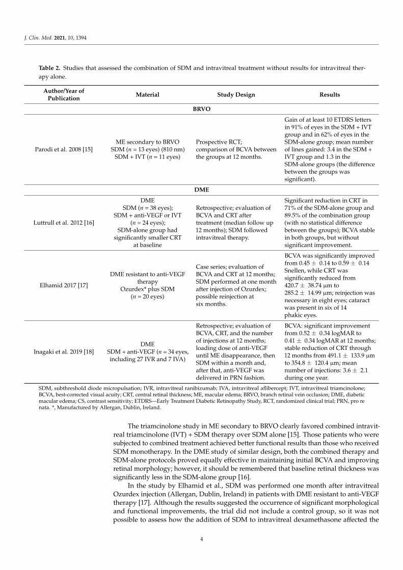
Table 2. Studies that assessed the combination of SDM and intravitreal treatment without results for intravitreal ther-apy alone.
Author/Year ofPublication
Material Study Design Results
BRVO
Parodi et al. 2008 [15]ME secondary to BRVO
SDM (n = 13 eyes) (810 nm)SDM + IVT (n = 11 eyes)
Prospective RCT;comparison of BCVA betweenthe groups at 12 months.
Gain of at least 10 ETDRS lettersin 91% of eyes in the SDM + IVTgroup and in 62% of eyes in theSDM-alone group; mean numberof lines gained: 3.4 in the SDM +IVT group and 1.3 in theSDM-alone groups (the differencebetween the groups wassignificant).
DME
Luttrull et al. 2012 [16]
DMESDM (n = 38 eyes);
SDM + anti-VEGF or IVT(n = 24 eyes);
SDM-alone group hadsignificantly smaller CRT
at baseline
Retrospective; evaluation ofBCVA and CRT aftertreatment (median follow up12 months); SDM followedintravitreal therapy.
Significant reduction in CRT in71% of the SDM-alone group and89.5% of the combination group(with no statistical differencebetween the groups); BCVA stablein both groups, but withoutsignificant improvement.
Elhamid 2017 [17]
DME resistant to anti-VEGFtherapy
Ozurdex* plus SDM(n = 20 eyes)
Case series; evaluation ofBCVA and CRT at 12 months;SDM performed at one monthafter injection of Ozurdex;possible reinjection atsix months.
BCVA was significantly improvedfrom 0.45 ± 0.14 to 0.59 ± 0.14Snellen, while CRT wassignificantly reduced from420.7 ± 38.74 µm to285.2 ± 14.99 µm; reinjection wasnecessary in eight eyes; cataractwas present in six of 14phakic eyes.
Inagaki et al. 2019 [18]DME
SDM + anti-VEGF (n = 34 eyes,including 27 IVR and 7 IVA)
Retrospective; evaluation ofBCVA, CRT, and the numberof injections at 12 months;loading dose of anti-VEGFuntil ME disappearance, thenSDM within a month and,after that, anti-VEGF wasdelivered in PRN fashion.
BCVA: significant improvementfrom 0.52 ± 0.34 logMAR to0.41 ± 0.34 logMAR at 12 months;stable reduction of CRT through12 months from 491.1 ± 133.9 µmto 354.8 ± 120.4 µm; meannumber of injections: 3.6 ± 2.1during one year.
SDM, subthreshold diode micropulsation; IVR, intravitreal ranibizumab; IVA, intravitreal aflibercept; IVT, intravitreal triamcinolone;BCVA, best-corrected visual acuity; CRT, central retinal thickness; ME, macular edema; BRVO, branch retinal vein occlusion; DME, diabeticmacular edema; CS, contrast sensitivity; ETDRS—Early Treatment Diabetic Retinopathy Study, RCT, randomized clinical trial; PRN, pro renata. *, Manufactured by Allergan, Dublin, Ireland.
The triamcinolone study in ME secondary to BRVO clearly favored combined intravit-real triamcinolone (IVT) + SDM therapy over SDM alone [15]. Those patients who weresubjected to combined treatment achieved better functional results than those who receivedSDM monotherapy. In the DME study of similar design, both the combined therapy andSDM-alone protocols proved equally effective in maintaining initial BCVA and improvingretinal morphology; however, it should be remembered that baseline retinal thickness wassignificantly less in the SDM-alone group [16].
In the study by Elhamid et al., SDM was performed one month after intravitrealOzurdex injection (Allergan, Dublin, Ireland) in patients with DME resistant to anti-VEGFtherapy [17]. Although the results suggested the occurrence of significant morphologicaland functional improvements, the trial did not include a control group, so it was notpossible to assess how the addition of SDM to intravitreal dexamethasone affected the
4

J. Clin. Med. 2021, 10, 1394
final outcome. A study by Inagaki et al., which considered SDM and anti-VEGF therapyin DME is a case series, [18] observed a moderate BCVA improvement (by 0.11 logMAR),with a relatively low number of injections required to achieve this effect during one year offollow-up (mean: 3.6 ± 2.1 injections).
4. Discussion
Literature material for the analysis of the efficacy of the combination of SDM andintravitreal treatment in DME and RVO is scarce. Following a search of PubMed, onlyfive eligible comparative studies were identified, including two randomized trials. Somecollective findings from these studies can be reported and analyzed, although caution mustbe maintained. Generally, patients subjected to combined therapy required fewer injections,especially when this number was compared with the number of anti-VEGF treatments inthe monotherapy population. If this outcome is confirmed in larger studies, SDM could beadopted in clinical practice to significantly reduce the burden of the treatment of retinalvascular diseases both financially and with respect to the patient’s comfort.
From the available material, it was determined that combined treatment was notinferior to anti-VEGF therapy alone when considering improvements in BCVA and retinalmorphology. However, SDM was usually performed in cases of minor and moderateretinal edema or following the resolution of edema after a loading dose of the intravitrealinjection was delivered. This is consistent with the results of other research correlatingSDM efficacy with the amount of baseline ME, often suggesting a central retinal thicknessof 400 µm as the threshold [19–21]. This fact implicates a strict rationale is necessary duringcombined SDM and anti-VEGF treatment in that the adjunct of SDM is only sensible incases with less severe retinal edema or following a reduction in edema prompted by initialanti-VEGF therapy.
Unfortunately, the analysis of the material does not offer us a precise answer regardingwhat should be the treatment schedule for the combined therapy. Both the number ofloading-phase injections and the moment of SDM application varied among the studies.Further research needs to address the following questions that remain: what is the optimalnumber of injections required during the loading phase of intravitreal therapy, what isthe best time point of SDM application (e.g., complete resolution of ME, reduction below400 µm, or reference to BCVA), and what is the ideal the retreatment schedule for eitheranti-VEGF or SDM? Some form of an algorithm for combined treatment in DME has alreadybeen proposed, yet it is not backed by published research [22]. SDM or anti-VEGF wassuggested as the first-line therapy for DME of less than 250 µm. For larger cases of edema,an initial loading phase of two to three anti-VEGF injections followed by three injections inthe context of a good response is recommended. Thereafter, switching to SDM is suggested.However, if the response is poor after two or three initial injections, a switch to SDM earlieron is indicated. Luttrull et al. does not use retinal thickness as a signal for deciding how totreat DME; if the VA is 20/50 or worse, an initial anti-VEGF injection is given and injectionsare continued until the VA is 20/40 or better, at which time panmacular SDM is initiated(there is no loading dose custom), while, if the VA is 20/40 or better, SDM is performedalone [23].
This review also discusses a number of non-comparative studies that do not directlyrefer the combined treatment to intravitreal therapy alone (Table 2). As the literature on thesubject was really limited, the author attempted to evaluate each study that reported effectsof combination treatment that included SDM. Two studies presented in Table 2 providesome perspective on the position of combination therapy, including SDM versus SDMalone [15,16]. It seems that SDM works well alone in mild to moderate DME; however,there is a tendency for better morphological results to be obtained with the involvementof intravitreal medication [16]. In BRVO, an additive strong anti-inflammatory effect ofintravitreal steroids provided significant improvements that were clearly superior to SDMonly [15]. The remaining two studies reported an effect of SDM added to either intravitrealsteroid or anti-VEGF therapy in the treatment of DME [17,18]. The lack of a control
5

J. Clin. Med. 2021, 10, 1394
groups in these reports makes their interpretation rather risky and, despite favorablemorphological and functional outcomes, the benefit of adding SDM to the treatmentregimen is impossible to evaluate. Moreover, it must be emphasized that intravitrealsteroid therapy in the treatment of DME and ME secondary to BRVO in most cases remainsthe second line of therapy, as does its combination with SDM.
The author realizes that the scarceness of literature on combined treatment includingboth SDM and intravitreal therapy for ME does not allow for a systematic review to beperformed nor for the presentation of concrete conclusions. However, in the author’sopinion, this limitation only means that this form of treatment should be looked at morecarefully. The common use of intravitreal injections—anti-VEGF in particular—has pushedaside other forms of treatment, some of which are potentially effective. SDM is rarelygiven attention by members of industry, who support multicenter clinical trials. Thus,designing and carrying out a large SDM investigation including numerous cases is noteasy and requires a lot of perseverance. Reviews such as this one will hopefully stimulateresearchers to pursue the subject further.
5. Conclusions
An analysis of the available research on combined SDM and anti-VEGF/intravitrealtreatment in ME does not provide an unequivocal answer at this time regarding the efficacyand benefits of this clinical approach. Existing published results suggest that combiningSDM and anti-VEGF in the treatment of cases of limited retinal edema would reduce thenumber of intravitreal injections required, with functional and morphological outcomesthat are non-inferior to those of anti-VEGF monotherapy. Larger, randomized clinical trialsare needed to confirm this thesis and provide a rational treatment algorithm.
Funding: No funding was received for this research.
Institutional Review Board Statement: Not applicable (the study is a systemized review).
Informed Consent Statement: Not applicable (the study is a systemized review).
Data Availability Statement: No new data were created or analyzed in this study. Data sharing isnot applicable to this article.
Conflicts of Interest: The author declares no conflict of interest.
Ethical Statement: This study was approved by the Dobry Wzrok Ophthalmological Clinic committee.
References
1. Gawecki, M. Micropulse Laser Treatment of Retinal Diseases. J. Clin. Med. 2019, 8, 242. [CrossRef]2. Brader, H.S.; Young, L.H.Y. Subthreshold Diode Micropulse Laser: A Review. Semin. Ophthalmol. 2016, 31, 30–39. [CrossRef]
[PubMed]3. Gawecki, M.; Jaszczuk-Maciejewska, A.; Jurska-Jasko, A.; Kneba, M.; Grzybowski, A. Transfoveal Micropulse Laser Treatment of
Central Serous Chorioretinopathy within Six Months of Disease Onset. J. Clin. Med. 2019, 8, 1398. [CrossRef]4. Luttrull, J.K. Low-intensity/high-density subthreshold diode micropulse laser for central serous chorioretinopathy. Retin. 2016,
36, 1658–1663. [CrossRef]5. Scholz, P.; Ersoy, L.; Boon, C.J.; Fauser, S. Subthreshold Micropulse Laser (577 nm) Treatment in Chronic Central Serous
Chorioretinopathy. Ophthalmologica 2015, 234, 189–194. [CrossRef]6. Scholz, P.; Altay, L.; Fauser, S. A Review of Subthreshold Micropulse Laser for Treatment of Macular Disorders. Adv. Ther. 2017,
34, 1528–1555. [CrossRef]7. Ciulla, T.A.; Bracha, P.; Pollack, J.; Williams, D.F. Real-world Outcomes of Anti–Vascular Endothelial Growth Factor Therapy in
Diabetic Macular Edema in the United States. Ophthalmol. Retin. 2018, 2, 1179–1187. [CrossRef]8. Korobelnik, J.-F.; Daien, V.; Faure, C.; Tadayoni, R.; Giocanti-Auregan, A.; Dot, C.; Kodjikian, L.; Massin, P. Real-world outcomes
following 12 months of intravitreal aflibercept monotherapy in patients with diabetic macular edema in France: Results from theAPOLLON study. Graefe Arch. Clin. Exp. Ophthalmol. 2020, 258, 521–528. [CrossRef]
9. Gross, J.G.; Glassman, A.R.; Liu, D.; Sun, J.K.; Antoszyk, A.N.; Baker, C.W.; Bressler, N.M.; Elman, M.J.; Ferris, F.L.;Gardner, T.W.; et al. Five-year outcomes of panretinal photocoagulation vs. intravitreous ranibizumab for proliferative diabeticretinopathy: A randomized clinical trial. JAMA Ophthalmol. 2018, 136, 1138–1148. [PubMed]
6
J. Clin. Med. 2021, 10, 1394
10. Thinda, S.; Patel, A.; Hunter, A.A.; Moshiri, A.; Morse, L.S. Combination therapy with subthreshold diode laser micropulsephotocoagulation and intravitreal anti-vascular endothelial growth factor injections for diabetic macular edema. Invest. Ophthalmol.
Vis. Sci. 2014, 55, 6363.11. Moisseiev, E.; Abbassi, S.; Thinda, S.; Yoon, J.; Yiu, G.; Morse, L.S. Subthreshold micropulse laser reduces anti-VEGF injection
burden in patients with diabetic macular edema. Eur. J. Ophthalmol. 2018, 28, 68–73. [CrossRef]12. Khattab, A.M.; Hagras, S.M.; Abdelhamid, A.; Torky, M.A.; Awad, E.A.; Abdelhameed, A.G. Aflibercept with adjuvant mi-
cropulsed yellow laser versus aflibercept monotherapy in diabetic macular edema. Graefe Arch. Clin. Exp. Ophthalmol. 2019, 257,1373–1380. [CrossRef]
13. Kanar, H.S.; Arsan, A.; Altun, A.; Akı, S.F.; Hacısalihoglu, A. Can subthreshold micropulse yellow laser treatment change theantivascular endothelial growth factor algorithm in diabetic macular edema? A randomized clinical trial. Indian J. Ophthalmol. 2020,68, 145–151. [PubMed]
14. Terashima, H.; Hasebe, H.; Okamoto, F.; Matsuoka, N.; Sato, Y.; Fukuchi, T. Combination therapy of intravitreal ranibizumab andsubthreshold micropulse photocoagulation for macular edema secondary to branch retinal vein occlusion: 6-month result. Retina
2019, 39, 1377–1384. [CrossRef]15. Parodi, M.B.; Iacono, P.; Ravalico, G. Intravitreal triamcinolone acetonide combined with subthreshold grid laser treatment for
macular oedema in branch retinal vein occlusion: A pilot study. Br. J. Ophthalmol. 2008, 92, 1046–1050. [CrossRef] [PubMed]16. Luttrull, J.K.; Sramek, C.; Palanker, D.; Spink, C.J.; Musch, D.C. Long-term safety, high-resolution imaging, and tissue temperature
modeling of subvisible diode micropulse photocoagulation for retinovascular macular edema. Retina 2012, 32, 375–386. [CrossRef]17. Elhamid, A.H.A. Combined intravitreal dexamethasone implant and micropulse yellow laser for treatment of anti-VEGF re-sistant
diabetic macular edema. Open Ophthalmol. J. 2017, 11, 164–172. [CrossRef]18. Inagaki, K.; Hamada, M.; Ohkoshi, K. Minimally invasive laser treatment combined with intravitreal injection of anti-vascular
endothelial growth factor for diabetic macular oedema. Sci. Rep. 2019, 9, 1–8. [CrossRef]19. Midena, E.; Bini, S.; Martini, F.; Enrica, C.; Pilotto, E.; Micera, A.; Esposito, G.; Vujosevic, S. Changes of aqueous humor müller
cells’ biomarkers in human patients affected by diabetic macular edema after subthreshold micropulse laser treatment. Retina
2020, 40, 126–134. [CrossRef] [PubMed]20. Mansouri, A.; Sampat, K.M.; Malik, K.J.; Steiner, J.N.; Glaser, B.M. Efficacy of subthreshold micropulse laser in the treatment of
diabetic macular edema is influenced by pre-treatment central foveal thickness. Eye 2014, 28, 1418–1424. [CrossRef] [PubMed]21. Vujosevic, S.; Martini, F.; Longhin, E.; Convento, E.; Cavarzeran, F.; Midena, E. Subthreshold micropulse yellow laser versus
subthreshold micropulse infrared laser in center-involving diabetic macular edema: Morphologic and functional safety. Retina
2015, 35, 1594–1603. [CrossRef] [PubMed]22. Mansour, S.; Luttrull, J. Integration of Micro Pulse Laser Therapy (MPLT) in the Management of Diabetic Retinopathy; IRIDEX
Educational Webinar. 2012. Available online: http://www.iridex.com.23. Luttrull, J.K. SDM as Modern Retinal Laser Therapy. Principles, Practice and RWD. Available online: https://www.researchgate.
net/project/SDM-as-Modern-Retinal-Laser-Therapy-Principles-Practice-and-RWD (accessed on 20 January 2021).
7

Journal of
Clinical Medicine
Review
Treatment of Macular Edema in Vascular Retinal Diseases:A 2021 Update
Andrzej Grzybowski 1,2 , Agne Markeviciute 3 and Reda Zemaitiene 3,*
�����������������
Citation: Grzybowski, A.;
Markeviciute, A.; Zemaitiene, R.
Treatment of Macular Edema in
Vascular Retinal Diseases: A 2021
Update. J. Clin. Med. 2021, 10, 5300.
https://doi.org/10.3390/
jcm10225300
Academic Editors: Gawecki Maciej
and Stephen Andrew Vernon
Received: 11 August 2021
Accepted: 11 November 2021
Published: 15 November 2021
Publisher’s Note: MDPI stays neutral
with regard to jurisdictional claims in
published maps and institutional affil-
iations.
Copyright: © 2021 by the authors.
Licensee MDPI, Basel, Switzerland.
This article is an open access article
distributed under the terms and
conditions of the Creative Commons
Attribution (CC BY) license (https://
creativecommons.org/licenses/by/
4.0/).
1 Department of Ophthalmology, University of Warmia and Mazury, 10-561 Olsztyn, Poland;[email protected]
2 Institute for Research in Ophthalmology, 60-836 Poznan, Poland3 Department of Ophthalmology, Medical Academy, Lithuanian University of Health Sciences,
50161 Kaunas, Lithuania; [email protected]* Correspondence: [email protected]
Abstract: Macular edema (ME) is associated with various conditions; however, the main causes of ME
are retinal vein occlusion (RVO) and diabetes. Laser photocoagulation, formerly the gold standard
for the treatment of ME, has been replaced by anti-vascular endothelial growth factor (anti-VEGF)
intravitreal injections. Despite its efficiency, this treatment requires frequent injections to preserve the
outcomes of anti-VEGF therapy, and as many patients do not sufficiently respond to the treatment,
ME is typically a chronic condition that can lead to permanent visual impairment. Generalized
recommendations for the treatment of ME are lacking, which highlights the importance of reviewing
treatment approaches, including recent anti-VEGFs, intravitreal steroid implants, and subthreshold
micropulse lasers. We reviewed relevant studies, emphasizing the articles published between 2019
and 2021 and using the following keywords: macular edema, diabetic macular edema, retinal vein
occlusion, laser photocoagulation, anti-VEGF, and intravitreal injections. Our results revealed that a
combination of different treatment methods may be beneficial in resistant cases. Additionally, artificial
intelligence (AI) is likely to help select the best treatment option for patients in the near future.
Keywords: macular edema; diabetic macular edema; retinal vein occlusion; laser photocoagulation;
anti-VEGF; intravitreal injections
1. Introduction
Macular edema (ME) is a disease characterized by the swelling of the macula due tothe abnormal accumulation of fluid [1]. It is associated with increased macular thicknessand significantly reduced visual acuity, and it may develop in various ocular conditions.
Postoperative cystoid macular edema (PCME) typically occurs after cataract surgery;however, it can occur after any ocular surgery [2]. The increased phacoemulsification energyand phacoemulsification time or postoperative pseudophakodonesis can significantlycontribute to PCME development [3]. It is thought that topical prostaglandin analogs usedfor glaucoma treatment may also promote PCME [3,4].
Corticosteroid eyedrops are prescribed postoperatively by most cataract surgeonsto prevent the formation of PCME [5]. Topical steroids, non-steroidal anti-inflammatoryeye drops, and ocular steroid injections (sub-tenon or intravitreal) are the main treatmentoptions for PCME [2].
ME is the most common cause of vision loss in patients with uveitis [6,7]. Althoughboth regional and systemic steroids are considered effective treatments, other treatmentoptions are available, including immunomodulatory agents and anti-vascular endothelialgrowth factor (VEGF) intravitreal injections [7,8].
Cystoid macular edema (CME) is observed in patients with various retinal patholo-gies. It is considered a complication in patients with retinitis pigmentosa (RP), whereas
9

J. Clin. Med. 2021, 10, 5300
tractional CME is associated with the persistent attachment of the vitreous at the macularregion [9,10].
However, in most eyes undergoing treatment of ME related to retinal vascular dis-ease, it is diabetic macular edema (DME) and retinal vein occlusion (RVO) that are thedriving forces.
ME affects approximately 7 million patients with diabetic retinopathy (DR) and 3 mil-lion patients with retinal vein occlusion (RVO) [11].
The role of inherited genetic polymorphisms in DME development and treatmentresponse is still poorly understood; nevertheless, possible DME risk genes have been iden-tified. Graham and colleagues did not find any significant genome-wide associations withDME risk; however, they identified the top-ranked single nucleotide polymorphism (SNP)for DME in rs1990145 on chromosome 2 [12]. A trend toward an association between DMEand DR was detected in two SNPs: rs12267418, near MALRD1 (p = 0.008), and rs16999051 inthe diabetes gene PCSK2 (p = 0.007) [12,13]. It is clear that there is a need for larger studies.
CME involves fluid accumulation in the outer plexiform layer of the retina due toabnormal perifoveal retinal capillary permeability, whereas DME is associated with theleakage of macular capillaries and is observed in patients suffering from diabetes [14].ME is also associated with an increase in VEGF and interleukin 6, which induce vascularpermeability and vasodilation [15].
Chronic ME leads to permanent visual impairment by altering the outer limitingmembrane, affecting photoreceptor segments (outer nuclear layer thinning and outersegment atrophy), and disorganization of inner retinal layers [11].
ME treatment approaches have changed substantially in recent years. Although laserphotocoagulation (LP) has long been the gold standard for the treatment of ME, it is beingreplaced by anti-VEGF intravitreal injections, which have been reported as a first-linetreatment for both DME and ME due to RVO.
This paper reviews and analyzes recent approaches to ME treatment and discussesfuture directions and perspectives in this field.
2. Methodology
A search of the medical literature was performed in PubMed and Google Scholarup to April 2021. The following keywords were used in various combinations: macularedema, diabetic macular edema, retinal vein occlusion, Laser Photocoagulation, anti-VEGF,intravitreal injections, and uveitis. Only articles with English abstracts focusing on MEcaused by retinal vascular diseases, including DME and ME due to RVO, were reviewed.Studies were critically reviewed to construct an overview and guidance for further searchesand highlight the lack of generalized recommendations. Emphasis was placed on articlespublished between 2019 and 2021.
3. Results
Intravitreal ranibizumab and aflibercept are currently approved for ME treatment,whereas bevacizumab is used off-label, and conbercept is approved and used for DMEtreatment only in China [16]. Frequent injections are required to preserve the effects of anti-VEGF therapy, and this treatment is therefore associated with repeated risk, high costs andan increasing burden on ophthalmologists and their patients. Despite the reported efficacyof anti-VEGFs, many patients do not respond well to treatment. In addition, identifyingwhich treatment regimen is optimal is a constant dilemma. The main advantage of treat-and-extend (T and E) over pro re nata (PRN) regimens is a reduction in the number of hospitalvisits and recurrences [17]. Elsebaey and colleagues compared T and E treatment regimenwith the PRN regimen in patients with DME [18]. They concluded that an individualized Tand E regimen has the potential to reduce the clinic burden and improve patient compliancewhile maintaining effectiveness and providing well-tolerated treatment for DME [18].Similar results were reported by Kim et al.: the T and E regimen of aflibercept in DME
10

J. Clin. Med. 2021, 10, 5300
maintained effectiveness in a 2-year follow-up and reduced the number of injectionscompared with fixed dosing regimens [17].
Intravitreal corticosteroid implants ensure sustained drug release for a specific periodand reduce the number of injections needed compared with anti-VEGF treatment. Steroidimplants were reported to be effective and safe both in DME and ME due to RVO; however,they are typically used as a second choice in cases resistant to anti-VEGF treatment. The in-travitreal dexamethasone (DEX) implant is approved for the treatment of DME and MEdue to RVO; in the EU, it is approved for use in patients with DME that responds poorly toother treatments and for those who are pseudophakic or ineligible for other therapies [19].The fluocinolone acetonide (FA) implant is approved for the treatment of DME and istypically used in patients who previously received a course of corticosteroids and didnot experience a significant increase in eye pressure [20]. Despite the efficacy of steroids,they may be associated with increased intraocular pressure (IOP) and cataract formation.
Resistance to anti-VEGFs and intravitreal steroids treatment methods highlights theneed for alternative treatment options.
3.1. Diabetic Macular Edema
The main DME treatment options are intravitreal injections of anti-VEGF agents andintravitreal corticosteroid injections. Formerly, macular LP was the gold standard forDME treatment; however, it is now utilized as an additional treatment. The two mostcommon techniques of LP in patients with DME are focal photocoagulation targeting focallesions (e.g., leaking microaneurysms or ischemic areas on fluorescein angiography (FA)for focal DME cases) and the grid laser technique, in which the laser is applied to diffuseleakages or nonperfusion areas; the latter is recommended for diffuse or more severe formsof DME [21,22]. According to the European Society of Retina Specialists (EURETINA)guidelines published in 2017, the focal and grid laser techniques should be utilized for non-center involving DME [23]. The laser can reportedly be applied in the vasogenic subform ofDME, which is clinically characterized by the presence of focally grouped microaneurysms(MA) and leaking capillaries [24]. The primary reason grid laser is not recommendedfurther is because of retinal scarring; however, when targeting capillary microaneurysms,a focal laser is beneficial as a second-line treatment [24,25]. In addition, it can be consideredas a combined treatment option to reduce the number of anti-VEGF injections. Paques andcolleagues performed a pilot study and reported significantly reduced macular thicknessand improved visual acuity after elective photocoagulation of capillary microaneurysms inpatients with chronic macular edema and severe hard exudates due to diabetic retinopathyor RVO [26].
Most studies found anti-VEGFs to be superior to laser treatment in DME patients.The REFINE study was conducted in Chinese patients with DME who received intravitrealranibizumab injections or LP [27]. The results revealed a significantly greater improve-ment in mean best-corrected visual acuity (BCVA) at month 12 with ranibizumab thanwith LP [27]. Singh and colleagues reported that BCVA improvement was significantlygreater with aflibercept than with laser techniques and was not influenced by any baselinefactors [28,29]. A subthreshold micropulse laser (SML) is a relatively new tissue-sparinglaser technique; it avoids protein coagulation and prevents retinal scars, allowing thepreservation of retinal anatomy and function [30].
SML helps improve or stabilize visual function and decrease macular thickness inDME [31]. Vujosevic and colleagues performed a study that evaluated the effectivenessof SML treatment in patients with DME [31]. They reported that 31 patients (83.8%)required retreatment (mean number of SML treatments over 12 months: 2.19 ± 0.7); how-ever, no eyes needed any additional treatments (anti-VEGF, steroids, and/or conventionallaser) [31]. Al-Barki et al. compared the outcomes between short-pulse continuous wave-length and infrared micropulse lasers in DME treatment [32]. The authors concludedthat the infrared micropulse system improved functional outcomes in patients with DME,whereas the short-pulse system resulted in a greater temporary reduction in edema [32].
11

J. Clin. Med. 2021, 10, 5300
Gawecki and colleagues performed a systematized review and proposed that combin-ing the SML treatment with anti-VEGFs may require fewer intravitreal injections thananti-VEGF monotherapy with equally favorable functional and morphological results inthe ME treatment. However, SML alone was not superior to intravitreal treatment aloneor combined treatment [33]. The authors noted that the studies under review varied intreatment protocols and inclusion criteria [33]. Altinel and colleagues compared the effi-cacy and safety of SML and intravitreal bevacizumab (IVB) injection combined therapywith IVB monotherapy in DME treatment [34]. They concluded that fewer IVB injectionswere needed when laser treatment was added; however, a significant increase in BCVAwas not achieved [34]. Similarly, Furashova et al. reported that patients treated withranibizumab combined with additional laser treatment experienced greater visual improve-ment and required fewer ranibizumab injections compared with patients treated only withranibizumab [35].
Valera-Cornejo et al. evaluated the effect of SML treatment in center-involved DMEin previously untreated (naïve) patients and patients who did not respond to prior treat-ment [36]. No significant changes in BCVA were observed between the groups after3 months [36]. The change in central macular thickness (CMT) at 3 months was statisticallybut not clinically significant in the treatment-naïve group only, and no adverse events werereported [36]. Passos et al. reported that SML treatment used alone was not as effectiveas it could be when combined with other treatments [37]. DME cases associated withsubretinal fluid had the best anatomical response, whereas intraretinal edema respondedpoorly to laser monotherapy [37]. The authors concluded that SML might be used in acombination treatment for ME [37]. Other authors also suggest considering laser therapyas an additional treatment in combination with intravitreal injections [21].
Anti-VEGFs utilize different molecules to achieve their effect: aptamers (pegaptanib);antibodies to VEGF (bevacizumab); antibody fragments to VEGF (ranibizumab); and fu-sion proteins, which combine a receptor for VEGF with the constant region of a humanimmunoglobulin (aflibercept and conbercept) [28]. Bevacizumab, ranibizumab, and afliber-cept are the most common anti-VEGFs, and many studies have not observed significantdifferences in outcomes between them [28,38]. However, it has been suggested that thechoice of anti-VEGF can be guided by the untreated BCVA. When it is lower, aflibercepthas been suggested as the drug of choice [28,29]. The remaining anti-VEGFs, includingbevacizumab, ranibizumab, and aflibercept, provide similar functional outcomes whenthe baseline BCVA is higher [28]. Bressler and colleagues, however, reported that after sixconsecutive injections, more patients presented with persistent ME following bevacizumabtreatment compared with ranibizumab and aflibercept [39]. On this basis, Haritoglouet al. suggested switching from bevacizumab to either aflibercept or ranibizumab if DMEpersists while using bevacizumab [40].
Zhou et al. evaluated the efficacy and safety of intravitreal conbercept for DMEtreatment [41]. Patients were treated with one to three consecutive monthly intravitrealconbercept (IVC) injections, followed by retreatment with conbercept or switch therapywith triamcinolone acetonide (TA) based on a 6-month observation of the effect of treat-ment [29]. Approximately one-third of the eyes (29 of 89 eyes involved in the study)received intravitreal triamcinolone acetonide (IVTA) injections at month 6 [41]. The resultsrevealed that the mean BCVA and CMT were significantly improved at 1 and 3 monthsafter IVC treatment in the IVC group, and they gradually improved at 9 months after IVTAtreatments in the IVC plus IVTA group [41]. Five eyes exhibited aggravated cataracts at thelast follow-up visit after IVTA injection, and this was associated with the final decline inBCVA [41]. Nonetheless, the authors concluded that conbercept is safe and efficient, and TAmay be beneficial in cases that are refractory to anti-VEGF treatment [41]. A meta-analysiscomparing the efficacies of conbercept and ranibizumab for DME treatment demonstratedthat intravitreal conbercept was significantly superior to ranibizumab in reducing CMT;however, no significant difference in visual improvement was observed [42]. The effectsand safety of conbercept and ranibizumab in DME treatment were also compared in a
12

J. Clin. Med. 2021, 10, 5300
recent meta-analysis by Sun et al., and the results demonstrated that intravitreal injectionsof conbercept were superior to ranibizumab in both reducing central retinal thickness andimproving BCVA [43].
Corticosteroids are typically used as an alternative therapy for eyes with an insufficientresponse to anti-VEGF treatment reducing inflammation, decreasing the disruption of theblood–retinal barrier, and interfering with retinal angiogenesis [44]. Although intravitrealsteroids are not used as often as anti-VEGFs, they can significantly reduce DME, and someauthors suggest them as an option for first-line treatment. The main steroids used for thetreatment of DME are TA, dexamethasone (DEX), and FA, which differ in their duration ofaction [40]. Because of the short vitreous elimination half-life of the solubilized fraction ofthese steroids, an extended duration of action can be achieved by applying sustained re-lease systems (implants) into the vitreous cavity [40]. After one intravitreal injection of TA,the treatment effect was maintained for up to 6 months [40]. However, TA elevates the riskof increased IOP, and it may be associated with the risk of pseudoendophthalmitis [45,46]and retinal toxicity [47–49]; thus, it is used less frequently than its alternatives [40]. Ad-ditionally, TA has not been approved for DME treatment [28]. Conversely, the DEX drugrelease injectable implant has higher recognition, with a pharmacological effect rangingbetween 4 and 6 months [40].
A first-line treatment algorithm and guidelines in center-involving DME have beensuggested by Kodjikian et al. [50]. The authors included a slow-release 700 µg dexametha-sone intravitreal implant as an option for first-line treatment in center-involving DME,together with three anti-VEGFs (bevacizumab, ranibizumab, and aflibercept). Augustinand colleagues reported a consensus by a group of retina experts indicating that if a patientdoes not exhibit a sufficient response after 3–6 months of anti-VEGF treatment (a visualacuity gain of <5 ETDRS letters or a reduction in the central retinal thickness of ≤20%),switching to the dexamethasone implant should be considered [51]. An implant may alsobe suitable in eyes with massive lipid exudates or as a first-line treatment in pseudophakicpatients, patients unwilling or unable to comply with tight anti-VEGF injection intervals,or patients with known vascular diseases [51].
Intravitreal DEX implants were reported to be effective in cases that were refractory toanti-VEGF treatment. Castro-Navarro and colleagues reported that the intravitreal DEXimplant was effective and safe in both previously treated and untreated patients withDME [52]. Additionally, the authors observed that 6 months after the injection of theDEX implant, patients without prior DME treatment gained significantly more letters thanpatients who were previously treated [52]. These results suggest the possibility of achievingbetter results with earlier DEX implantation. This agrees with the results of a study byMedina-Baena, which demonstrated that at month 12, naïve patients exhibited a greaterimprovement in BCVA from baseline and achieved this BCVA improvement significantlyfaster than previously treated patients [53]. Similar results were observed in a study byIglicki et al. [54]. They found that over a follow-up of 24 months, the vision in DME eyesimproved after treatment with DEX implants in eyes that were treatment-naïve and ineyes that were refractory to anti-VEGF treatment; however, a greater improvement wasobserved in naïve eyes [54].
Although most studies evaluate CMT as the target of anatomical outcomes, Altun andcolleagues evaluated the subfoveal choroidal thickness (SFCT) in vitrectomized eyes ofpatients with DME after intravitreal DEX implants [55]. The authors reported a statisticallysignificant thinning of the mean SFCT during the follow-up period after DEX implantinjection in vitrectomized eyes with DME [55].
Hong et al. performed a retrospective study to evaluate the effect of intravitrealTA injections in patients who were refractory to anti-VEGF treatment [44]. The authorsreported that the BCVA improved significantly, and CMT was significantly reduced aftera single TA intravitreal injection [44]. In addition, poorer visual acuity (VA) before theinjection was associated with visual gain 1 month after the treatment [44]. Elevated IOP
13

J. Clin. Med. 2021, 10, 5300
was observed in 17.1% of eyes, and this was observed significantly more often after IVTAinjections containing a preservative than after preservative-free injections [39].
A longer pharmacological effect lasting up to 3 years can be achieved with an in-travitreal FA sustained-release non-biodegradable device, which is inserted into the vit-reous cavity via a 25-gauge needle; it contains 0.19 mg of FA and has a release rate of0.2 µg/day [11]. Augustin and colleagues performed a retrospective study to evaluatethe results of DME treatment with FA implants [56]. They concluded that a single FAimplant could maintain reduced CMT for up to 3 years [56]. Several more studies reportedsimilar results, highlighting that FA has a favorable safety and effectiveness profile whilereducing CMT and improving BCVA [57–59]. Notably, Coelho and colleagues reportedthat FA exhibited long-term effectiveness in vitrectomized DME eyes and sustained theeffectiveness in DME eyes that did not respond to DEX therapy [60].
The correct time to switch therapy if patients do not respond to anti-VEGF treatmentremains unclear. Gonzalez et al. performed a study and reported that in eyes with poorresponses after three anti-VEGF injections, it may be beneficial to switch to other modesof therapy [61]. Baker and colleagues found that for patients with DME and excellentvisual acuity (defined as 20/25 or better), observation appeared to be a non-inferior initialmanagement strategy compared with intravitreal aflibercept or LP in terms of visualacuity outcomes after 2 years [62]. Likewise, it was reported that initial focal or grid lasersignificantly reduced the risk of requiring aflibercept injection during follow-up [62].
Martínez and colleagues evaluated the effect of early DEX implantation in eyes withDME that received three or fewer anti-VEGF injections before the switch as well as the effectof later implantation in patients who received six or more anti-VEGF injections before theswitch [63]. They reported that an early switch to DEX in patients who did not adequatelyrespond to anti-VEGF therapy provided better results: BCVA improved significantly more(compared with baseline), and CMT decreased more in the early switch group comparedwith the late switch group [63]. In addition, no difference in the incidence of increased IOPwas observed between the groups [63]. Comparable results were reported in Demir andcolleagues’ study; the authors concluded that the central retinal thickness (CRT) decreasedsignificantly more in the early switch group compared with the later switch group [64].These results agree with those of a study by Ruiz-Medrano et al. [45]. Superior functionaloutcomes were observed in eyes with insufficient responses to anti-VEGFs in patientsswitched to DEX who had been receiving three monthly anti-VEGF injections comparedwith those who had been receiving more than three monthly anti-VEGF injections [65].
Cataract surgery can induce DME progression as well as the development of DMEin patients with diabetes [28]. Several studies have reported improved functional andanatomic clinical outcomes in patients with DEX implants during cataract surgery [66–68].Furino and colleagues conducted a study to evaluate functional and anatomical outcomesafter combined phacoemulsification and intravitreal DEX implantation with standardphacoemulsification in diabetic patients with cataracts [69]. In the group with combinedphacoemulsification and intravitreal DEX implantation, BCVA improved significantly more,and central subfoveal thickness decreased more [69]. Although this group had significantlyhigher IOP during follow-up at month 3 compared with baseline, IOP remained within thenormal range [69].
Possibilities for future treatment include ziv-aflibercept, which was proposed as anew recombinant fusion protein and which has a mechanism of action similar to thatof aflibercept; however, it is available at a lower cost than the proprietary anti-VEGFdrug [70]. It was reported to be effective and safe in DME treatment and other retinaldiseases; however, further studies are needed [70,71]. Because of the longer intravitrealhalf-life of the new generation anti-VEGF-A inhibitors, including brolucizumab, abiciparpegol, and angiopoietin combination drugs, improved prolonged edema reduction andless frequent injections appear to be required [11,28]. The preliminary results of studiescurrently in progress have suggested that anti-VEGF-A may have superior effectivenesscompared with approved anti-VEGFs [11,28,72].
14
J. Clin. Med. 2021, 10, 5300
Rivera et al. reported evidence of reduction of DME through the consumption oflutein. In patients with ME who have lower levels of lutein, lutein consumption preventedand reduced possible complications [73].
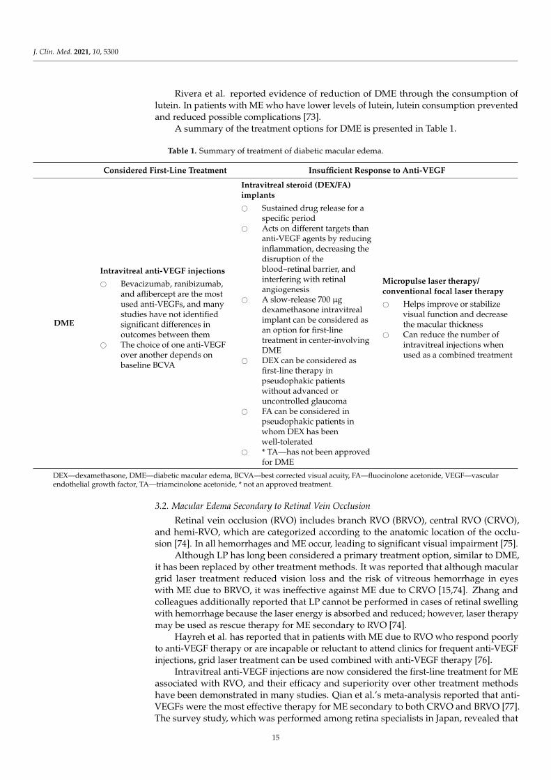
A summary of the treatment options for DME is presented in Table 1.
Table 1. Summary of treatment of diabetic macular edema.
Considered First-Line Treatment Insufficient Response to Anti-VEGF
DME
Intravitreal anti-VEGF injections
# Bevacizumab, ranibizumab,and aflibercept are the mostused anti-VEGFs, and manystudies have not identifiedsignificant differences inoutcomes between them
# The choice of one anti-VEGFover another depends onbaseline BCVA
Intravitreal steroid (DEX/FA)implants
# Sustained drug release for aspecific period
# Acts on different targets thananti-VEGF agents by reducinginflammation, decreasing thedisruption of theblood–retinal barrier, andinterfering with retinalangiogenesis
# A slow-release 700 µgdexamethasone intravitrealimplant can be considered asan option for first-linetreatment in center-involvingDME
# DEX can be considered asfirst-line therapy inpseudophakic patientswithout advanced oruncontrolled glaucoma
# FA can be considered inpseudophakic patients inwhom DEX has beenwell-tolerated
# * TA—has not been approvedfor DME
Micropulse laser therapy/conventional focal laser therapy
# Helps improve or stabilizevisual function and decreasethe macular thickness
# Can reduce the number ofintravitreal injections whenused as a combined treatment
DEX—dexamethasone, DME—diabetic macular edema, BCVA—best corrected visual acuity, FA—fluocinolone acetonide, VEGF—vascularendothelial growth factor, TA—triamcinolone acetonide, * not an approved treatment.
3.2. Macular Edema Secondary to Retinal Vein Occlusion
Retinal vein occlusion (RVO) includes branch RVO (BRVO), central RVO (CRVO),and hemi-RVO, which are categorized according to the anatomic location of the occlu-sion [74]. In all hemorrhages and ME occur, leading to significant visual impairment [75].
Although LP has long been considered a primary treatment option, similar to DME,it has been replaced by other treatment methods. It was reported that although maculargrid laser treatment reduced vision loss and the risk of vitreous hemorrhage in eyeswith ME due to BRVO, it was ineffective against ME due to CRVO [15,74]. Zhang andcolleagues additionally reported that LP cannot be performed in cases of retinal swellingwith hemorrhage because the laser energy is absorbed and reduced; however, laser therapymay be used as rescue therapy for ME secondary to RVO [74].
Hayreh et al. has reported that in patients with ME due to RVO who respond poorlyto anti-VEGF therapy or are incapable or reluctant to attend clinics for frequent anti-VEGFinjections, grid laser treatment can be used combined with anti-VEGF therapy [76].
Intravitreal anti-VEGF injections are now considered the first-line treatment for MEassociated with RVO, and their efficacy and superiority over other treatment methodshave been demonstrated in many studies. Qian et al.’s meta-analysis reported that anti-VEGFs were the most effective therapy for ME secondary to both CRVO and BRVO [77].The survey study, which was performed among retina specialists in Japan, revealed that
15
J. Clin. Med. 2021, 10, 5300
anti-VEGF therapy was chosen as the first-line treatment for ME secondary to BRVO,and most specialists (82.4%) selected initial injection followed by a pro re nata (PRN)regimen; however, the opinions about the initiation and switching therapy varied betweenspecialists [78]. As additional treatment in refractory cases, laser therapy was reported asthe most common choice (35.9%), with 25.6% selecting vitrectomy, and 15.4% chosing toadd steroid injections [78].
Anti-VEGFs used to treat ME due to RVO are similar to those used to treat DME;ranibizumab and aflibercept are used on label, whereas bevacizumab and conbercept havebeen used off label. Hykin and colleagues performed a prospective study to evaluate theeffectiveness of ranibizumab, aflibercept, and bevacizumab for the management of ME dueto CRVO [16]. They reported that mean changes in vision after 100 weeks of follow-up andtreatment were not inferior with aflibercept than with ranibizumab; however, the meannumber of injections given in the aflibercept group was lower than that in the ranibizumabgroup [16]. The mean changes in vision using bevacizumab compared with those usingranibizumab were similar, suggesting that the effectiveness of bevacizumab was neitherequal nor superior to ranibizumab [16]. Conbercept is one of the newest anti-VEGFs andprovided good treatment results in Chinese patients with RVO in a randomized clinicaltrial [79]. Xia and colleagues reported that conbercept significantly reduced retinal struc-tural remodeling, inflammation, and oxidative stress in mice as well as in patients with MEdue to RVO [75]. However, some patients with severe ME due to RVO did not experiencesignificant benefit from conbercept [75]. The authors hypothesized that this may havebeen because conbercept only inhibits downstream VEGF inflammatory mediators anddoes not affect the upstream inflammatory mediators of VEGFs, such as PGE1, PGE2,and PGF2a [75]. Costa et al. reported that intravitreal anti-VEGF injections are prioritizedover other treatment methods, including macular grid photocoagulation [80]. Comparedwith steroid injections, anti-VEGFs are superior because they have fewer side effects;as with their use in DME, steroids are associated with a higher incidence of increased IOPand cataract formation [80]. A systematic review and meta-analysis were performed by Liuand colleagues to evaluate the efficacy of conbercept and ranibizumab with or without LPin patients with ME secondary to RVO [81]. Both intravitreal conbercept and ranibizumabtherapy with or without LP were effective in improving vision function in patients with MEsecondary to RVO. The two anti-VEGFs did not differ significantly in BCVA improvementor adverse effects, and they resulted in similar visual gains [81]. However, conberceptreduced CMT more than ranibizumab with fewer injections [81]. Another systematicreview performed by Spooner and colleagues evaluated 17 studies involving 1070 eyes [15].It demonstrated that the management and outcomes of patients with CRVO varied greatly;however, anti-VEGF therapy significantly improved the anatomical and functional out-comes [15]. Although most eyes obtained a significant visual acuity gain, those treated withaflibercept and bevacizumab had significantly better outcomes than ranibizumab-treatedeyes [15]. The incidence rates of ocular complications were low, including neovascularglaucoma (3.6%), vitreous hemorrhage (<1%), glaucoma (1.2%), and neovascular glaucoma(<1%) [15].
The management of cases refractory to anti-VEGF treatment is an ongoing dilemma,and therefore, the efficacy of steroids in patients with ME due to RVO has been explored inseveral studies. One study hypothesized that inflammation could be the first key mecha-nism to mechanical injury in RVO, and VEGF up-regulation may occur as a secondary effectof this inflammatory response [75]. Corticosteroids can significantly reduce inflammation,retinal vascular permeability, and the regulation of VEGF-A expression, and thus they havebeen used for the treatment of ME due to RVO [74]. The intravitreal dexamethasone im-plant is approved for the treatment of ME due to RVO [74]. Ming and colleagues performeda meta-analysis on the efficacy and safety of intravitreal DEX implants and anti-VEGFsfor the treatment of ME due to RVO; the review included 4 randomized controlled trialsand 12 real-world studies [19]. The authors reported that DEX implantation resulted in acomparable or smaller reduction in central subfield thickness (CST) at months 6 and 12 but
16
J. Clin. Med. 2021, 10, 5300
introduced higher risks of elevated IOP and cataract induction [19]. It was concluded thatcompared with anti-VEGF agents, DEX implants required fewer injections but had inferiorfunctional efficacy and safety [19].
The management of central and branch RVO and its long-term effects were evaluatedin a 7-year follow-up study by Arrigo et al. performed in an Italian referral center [82].Contrary to the previously discussed study, the authors reported that both CRVO andBRVO eyes exhibited significant visual acuity improvements secondary to intravitrealanti-VEGF or dexamethasone treatments and a significant reduction in CMT at the end ofthe follow-up. Furthermore, the authors highlighted a result that showed that the timeat which the greatest improvement was observed differed between CRVO and BRVO;an earlier improvement was observed for CRVO (after 12 months of follow-up), and alater improvement was observed for BRVO (after 24 months of follow-up). However,after 2 years, both visual acuity and CMT remained stable until the end of follow-up.
Evidence of the value and importance of SML therapy in ME treatment is increasing.Buyru et al. compared the effects of intravitreal ranibizumab and SML treatment in twogroups of patients with ME due to BRVO [83]. They concluded that the reduction in macu-lar thickness and the increase in visual acuity were comparable for intravitreal ranibizumaband yellow SML treatment over 1 year. It was suggested that SML treatment may be usefulin the treatment of ME due to BRVO. Eng and colleagues conducted a literature reviewon the efficacy of SML treatment for ME due to BRVO and reported that SML therapyresulted in a smaller reduction in ME compared with intravitreal anti-VEGF agents [84].However, the authors concluded that SML treatment could be useful as adjuvant therapywith intravitreal anti-VEGF agents or steroids. Terashima et al. evaluated the efficacy ofthe combined therapy of intravitreal ranibizumab and 577 nm yellow laser SML photoco-agulation for ME secondary to BRVO [85]. They concluded that combination therapy withintravitreal injections and SML was effective and decreased the frequency of intravitrealinjections while maintaining good visual acuity. Similarly, a meta-analysis conducted byChen et al. concluded that laser therapy combined with intravitreal ranibizumab injectionshad a strong effect, promoting its use for the treatment of ME secondary to BRVO in clinicalpractice [86].
Nanotechnology (nanocarriers) offers multiple benefits by promoting drug deliveryacross tissue barriers, controlling the release of a topically administered drug, improvingbioavailability, and directing drugs to the target tissue [87]. An example of a nanosystemis the topical ophthalmic TA-loaded liposome formulation (TA-LF), which releases TAinto the vitreous and retina [87]. It was reported to be safe and effective in rabbits as wellas in patients with refractory pseudophakic cystoid ME. Navarro-Partida and colleaguesevaluated its safety and efficacy in patients with ME secondary to BRVO who were givena topical instillation of one drop of TA-LF (TA 0.2%) six times a day for 12 weeks [87].The results confirmed its effectiveness; a significant reduction in central foveal thicknessand a significant improvement in BCVA were observed. No adverse events, includingincreased IOP, were reported. The authors suggested that as liposomes can function asnanocarriers of TA, they could allow topical ophthalmic therapy to become the primarytreatment option instead of intravitreal drugs in patients with ME secondary to BRVO.Cheng et al.’s also showed that liposomes with TA in eye drops could be a new therapeuticapproach for the effective treatment of retinal diseases [88].
Authors have investigated factors associated with the course of the disease and theresponse to the treatment. Kida and colleagues hypothesized that increased retinal venouspressure (RVP) plays an important role in the formation of macula edema; thus, they re-cently evaluated RVP before and 1 month after intravitreal ranibizumab injection to deter-mine its effect on RVO-related ME [89]. They concluded that RVP decreased significantlyafter treatment; however, it remained significantly higher than the IOP. Rothman andcolleagues assessed the impact of age on ME due to RVO and concluded that patientsyounger than 50 years old had higher baseline and final visual acuity, a lower incidence
17
J. Clin. Med. 2021, 10, 5300
of cystoid macular edema at presentation, and received fewer intravitreal injections thanolder patients [90].
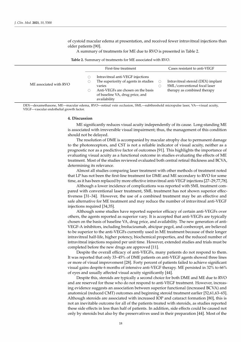
A summary of treatments for ME due to RVO is presented in Table 2.
Table 2. Summary of treatments for ME associated with RVO.
ME associated with RVO
First-line treatment Cases resistant to anti-VEGF
# Intravitreal anti-VEGF injections# The superiority of agents in studies
varies# Anti-VEGFs are chosen on the basis
of baseline VA, drug price, andavailability
# Intravitreal steroid (DEX) implant# SML/conventional focal laser
therapy as combined therapy
DEX—dexamethasone, ME—macular edema, RVO—retinal vein occlusion, SML—subthreshold micropulse laser, VA—visual acuity,VEGF—vascular endothelial growth factor.
4. Discussion
ME significantly reduces visual acuity independently of its cause. Long-standing MEis associated with irreversible visual impairment; thus, the management of this conditionshould not be delayed.
The resolution of DME is accompanied by macular atrophy due to permanent damageto the photoreceptors, and CST is not a reliable indicator of visual acuity, neither as aprognostic nor as a predictive factor of outcomes [91]. This highlights the importance ofevaluating visual acuity as a functional outcome in studies evaluating the effects of MEtreatment. Most of the studies reviewed evaluated both central retinal thickness and BCVA,determining its relevance.
Almost all studies comparing laser treatment with other methods of treatment notedthat LP has not been the first-line treatment for DME and ME secondary to RVO for sometime, as it has been replaced by more effective intravitreal anti-VEGF injections [27–29,77,78].
Although a lower incidence of complications was reported with SML treatment com-pared with conventional laser treatment, SML treatment has not shown superior effec-tiveness [31–34]. However, the use of a combined treatment may be an effective andsafe alternative for ME treatment and may reduce the number of intravitreal anti-VEGFinjections required [34,35].
Although some studies have reported superior efficacy of certain anti-VEGFs overothers, the agents reported as superior vary. It is accepted that anti-VEGFs are typicallychosen on the basis of baseline VA, drug price, and availability. The new generation of anti-VEGF-A inhibitors, including brolucizumab, abicipar pegol, and conbercept, are believedto be superior to the anti-VEGFs currently used in ME treatment because of their longerintravitreal half-life, higher potency, biochemical properties, and the reduced number ofintravitreal injections required per unit time. However, extended studies and trials must becompleted before the new drugs are approved [11].
Despite the overall efficacy of anti-VEGFs, many patients do not respond to them.It was reported that only 33–45% of DME patients on anti-VEGF agents showed three linesor more of visual improvement [28]. Forty percent of patients failed to achieve significantvisual gains despite 6 months of intensive anti-VEGF therapy. ME persisted in 32% to 66%of eyes and usually affected visual acuity significantly [44].
Despite this, steroids are typically a second choice for both DME and ME due to RVOand are reserved for those who do not respond to anti-VEGF treatment. However, increas-ing evidence suggests an association between superior functional (increased BCVA) andanatomical (reduced CMT) outcomes and beginning steroid treatment earlier [52,61,63–65].Although steroids are associated with increased IOP and cataract formation [80], this isnot an inevitable outcome for all of the patients treated with steroids, as studies reportedthese side effects in less than half of patients. In addition, side effects could be caused notonly by steroids but also by the preservatives used in their preparation [44]. Most of the
18

J. Clin. Med. 2021, 10, 5300
studies reported a significant positive effect of intravitreal steroids in the treatment of ME,thus highlighting its advantage. The intravitreal FA implant is superior to the DEX implantbecause of its longer effect (up to 36 months); however, it is usually used to treat DMEin patients who previously received a course of corticosteroids and did not experience asignificant increase in eye pressure [11]. Furthermore, intravitreal FA was approved forDME, but it has not yet been approved for ME due to RVO. We did not identify any studiesthat compared DEX and FA in terms of effectiveness.
It would appear that, as of yet, a consensus on ME treatment has not been reached,particularly in cases that are resistant to standard treatment. We assume that artificialintelligence (AI) may be beneficial in addressing this issue. It was previously reportedthat AI was able to accurately predict posttreatment central foveal thickness and BCVAafter anti-VEGF injections in DME patients; thus, it can be used to prospectively assessthe efficacy of anti-VEGF therapy in DME patients [91]. The data regarding AI propertiesand possibilities in ME diagnosis and treatment prognosis are increasing [92]. Opticalcoherence tomography (OCT) is an indispensable tool for the application of AI as well asfor determining the need for treatment and evaluating its effectiveness in patients withME [93]. Until AI is widely and effectively incorporated in clinical practice, establishedimaging biomarkers may significantly contribute to DME management. Hyperreflectiveretinal foci (HRF) appear as intraretinal hyperreflective dots on OCT in patients withDME and are reported to be an important imaging marker of retinal inflammation [94].Kim et al. suggested that patients with an increased number of HRF on OCT should bemore frequently followed up for early intervention because they observed that a highernumber of HRF on the spectral domain (SD) OCT was associated with early recurrenceof DME after steroid implants [94]. It was also reported that the presence of subretinalfluid, the absence of HRF, and the integrity of the inner segment–outer segment layer couldbe OCT biomarkers for superior functional success [55]. Larger cysts (intraretinal cystoidspaces) are associated with poor visual prognosis, and the size of the cyst is correlated withthe extent of macular ischemia [14]. An increased fundus autofluorescence (FAF) signal(hyper-autofluorescence) was associated with declining visual acuity and an increase inthe macular thickness on OCT [95]. This highlights the properties of FAF as an additionaltool that may help monitor the progression of DME and its response to treatment.
5. Perspectives
New therapies, including anti-VEGF-A inhibitors (brolucizumab and abicipar pegol),are under investigation and may be more effective in ME treatment compared with pre-vious anti-VEGFs [11]. A suprachoroidal TA delivery system in DME patients has beeninvestigated as well, and the preliminary results are promising [96]. Nanotechnology wasreported to be safe and beneficial in its ability to ensure TA delivery to the retina usingtopical drops.
The SML is absorbed by xanthophyll pigment, allowing for treatment close to thefovea [84]. It can promote the absorption of edema, hemorrhage, and exudation, and it canimprove the retinal oxygen supply and reduce vascular permeability [86]. This relativelynew laser technique is superior to a conventional laser because it does not cause structuraldamage to the retina. Although SML therapy has not shown superiority when used alone inME treatment, in most of the reviewed studies, SML therapy was reported to be an effectiveadditional treatment method when combined with intravitreal anti-VEGF injections in thetreatment of DME and ME due to RVO. Both methods have some limitations and possiblecomplications; however, when combined, they not only effectively reduce ME and increaseVA but also reduce the number of intravitreal injections. Therefore, this combined treatmentcould lower healthcare costs and the burden on patients by reducing the frequency ofclinic visits.
With the emerging era of AI, this technology may soon be beneficial in selecting themost effective and appropriate treatment in patients with ME. Promising results werereported in a recent study performed by Gallardo and colleagues [97]. They used machine
19

J. Clin. Med. 2021, 10, 5300
learning classifiers to predict low and high anti-VEGF treatment demands for patientswith DME, RVO, and neovascular age-related macular degeneration treated according toa treat-and-extend regimen. The authors highlighted the ability to predict the low andhigh treatment demands in all groups of patients with similar accuracy, along with thecapability to predict low demand at the first visit before the first injection. Further researchis needed to establish the individual treatment demands for patients and consolidate theproperties of AI in clinical practice.
Author Contributions: Conceptualization, A.G., A.M. and R.Z.; methodology, A.M.; validation, A.G.and R.Z.; formal analysis, A.M.; investigation, A.G., A.M. and R.Z.; data curation, A.G.; writing—original draft preparation, A.M.; writing—review and editing, A.G., A.M. and R.Z.; visualization,A.G.; supervision, A.G. All authors have read and agreed to the published version of the manuscript.
Funding: This research received no external funding.
Institutional Review Board Statement: Not applicable.
Informed Consent Statement: Not applicable.
Conflicts of Interest: The authors declare no conflict of interest.
References
1. Claudiu, T.S. Agents for the prevention and treatment of age-related macular degeneration and macular edema: A literature andpatent review. Expert Opin. Ther. Pat. 2019, 29, 761–767.
2. Kitazawa, K.; Sotozono, C.; Kinoshita, S. Incidence and Management of Cystoid Macular Edema after Corneal Transplantation.Int. J. Ophthalmol. 2017, 10, 1081–1087. [CrossRef]
3. Schaub, F.; Adler, W.; Enders, P.; Koenig, M.C.; Koch, K.R.; Cursiefen, C.; Kirchhof, B.; Heindl, L.M. Preexisting epiretinalmembrane is associated with pseudophakic cystoid macular edema. Graefe Arch. Clin. Exp. Ophthalmol. 2018, 256, 909–917.[CrossRef] [PubMed]
4. Aaronson, A.; Achiron, A.; Tuuminen, R. Clinical Course of Pseudophakic Cystoid Macular Edema Treated with Nepafenac. J.
Clin. Med. 2020, 9, 3034. [CrossRef]5. Walter, K.; Kauffman, L.; Hess, J. Rate of pseudophakic cystoid macular edema using intraoperative and topical nonsteroidal
antiinflammatory drugs alone without steroids. J. Cataract. Refract. Surg. 2020, 46, 350–354. [CrossRef]6. Koronis, S.; Stavrakas, P.; Balidis, M.; Kozeis, N.; Tranos, P.G. Update in treatment of uveitic macular edema. Drug Des. Dev. Ther.
2019, 13, 667–680. [CrossRef]7. Testi, I.; Rousselot, A.; Agrawal, R.; Pavesio, C. Treatment of Uveitic Macular Edema. In Complications in Uveitis; Pichi, F., Neri, P.,
Eds.; Springer: Cham, Switzerland, 2020.8. Dick, A.D.; Rosenbaum, J.T.; Al-Dhibi, H.A. Guidance on noncorticosteroid systemic immunomodulatory therapy in noninfectious
uveitis: Fundamentals of Care for UveitiS (FOCUS) initiative. Ophthalmology 2018, 125, 757–773. [CrossRef]9. Liew, G.; Strong, S.; Bradley, P.; Severn, P.; Moore, A.T.; Webster, A.R.; Mitchell, P.; Kifley, A.; Michaelides, M. Prevalence of cystoid
macular oedema, epiretinal membrane and cataract in retinitis pigmentosa. Br. J. Ophthalmol. 2018, 103, 1163–1166. [CrossRef]10. Petrou, P.; Chalkiadaki, E.; Errera, M.-H.; Liyanage, S.; Wickham, L.; Papakonstantinou, E.; Karamaounas, A.; Kanakis, M.;
Georgalas, I.; Kandarakis, S.; et al. Factors Associated with the Clinical Course of Vitreomacular Traction. J. Ophthalmol. 2020,2020, 9457670. [CrossRef]
11. Sacconi, R.; Giuffrè, C.; Corbelli, E.; Borrelli, E.; Querques, G.; Bandello, F. Emerging therapies in the management of macularedema: A review. F1000Research 2019, 8. [CrossRef]
12. Graham, P.S.; Kaidonis, G.; Abhary, S.; Gillies, M.C.; Daniell, M.; Essex, R.W.; Chang, J.H.; Lake, S.R.; Pal, B.; Jenkins, A.J.; et al.Genome-wide association studies for diabetic macular edema and proliferative diabetic retinopathy. BMC Med. Genet. 2018, 19,71. [CrossRef]
13. Grassi, M.A.; Tikhomirov, A.; Ramalingam, S.; Below, J.E.; Cox, N.J.; Nicolae, D.L. Genome-wide meta-analysis for severe diabeticretinopathy. Hum. Mol. Genet. 2011, 20, 2472–2481. [CrossRef]
14. Yalçın, N.G.; Özdek, S. The Relationship Between Macular Cyst Formation and Ischemia in Diabetic Macular Edema. Turk. J.
Ophthalmol. 2019, 49, 194–200. [CrossRef]15. Spooner, K.; Hong, T.; Fraser-Bell, S.; Chang, A. Current Outcomes of Anti-VEGF Therapy in the Treatment of Macular Edema
Secondary to Central Retinal Vein Occlusions: A Systematic Review and Meta-Analysis. Ophthalmologica 2019, 242, 163–177.[CrossRef]
16. Hykin, P.; Prevost, A.T.; Vasconcelos, J.C.; Murphy, C.; Kelly, J.; Ramu, J.; Hounsome, B.; Yang, Y.; Harding, S.P.; Lotery, A.; et al.Clinical Effectiveness of Intravitreal Therapy with Ranibizumab vs Afliber-cept vs Bevacizumab for Macular Edema Secondary toCentral Retinal Vein Occlusion: A Randomized Clinical Trial. JAMA Ophthalmol. 2019, 137, 1256–1264. [CrossRef]
20
J. Clin. Med. 2021, 10, 5300
17. Kim, Y.C.; Shin, J.P.; Pak, K.Y.; Kim, H.W.; Sagong, M.; Lee, S.J.; Chung, I.Y.; Park, S.W.; Lee, J.E. Two-year outcomes of thetreat-and-extend regimen using aflibercept for treating diabetic macular oedema. Sci. Rep. 2020, 10, 22030. [CrossRef]
18. Elsebaey, A.E.; Ibrahim, A.M.; Elshaarawy, E.A. Treat-and-extend vs pro re nata regimens of aflibercept in diabetic macular edema.Menoufia Med. J. 2020, 33, 1144–1149.
19. Ming, S.; Xie, K.; Yang, M.; He, H.; Li, Y.; Lei, B. Comparison of intravitreal dexamethasone implant and anti-VEGF drugs inthe treatment of retinal vein occlusion-induced oedema: A meta-analysis and systematic review. BMJ Open 2020, 10, e032128.[CrossRef]
20. Fonollosa, A.; Llorenç, V.; Artaraz, J.; Jimenez, B.; Ruiz-Arruza, I.; Agirrebengoa, K.; Cordero-Coma, M.; Costales-Mier, F.; Adan,A. Safety and efficacy of intravitreal dexamethasone implants in the management of macular edema secondary to infectiousuveitis. Retina 2016, 36, 1778–1785. [CrossRef]
21. Kim, E.J.; Lin, W.V.; Rodriguez, S.M.; Chen, A.; Loya, A. Treatment of Diabetic Macular Edema. Curr. Diabetes Rep. 2019, 19, 68.[CrossRef] [PubMed]
22. Romero-Aroca, P. Is Laser Photocoagulation Treatment Currently Useful in Diabetic Macular Edema? Med. Hypothesis Discov.
Innov. Ophthalmol. J. 2015, 4, 5–8.23. Schmidt-Erfurth, U.; Garcia-Arumi, J.; Bandello, F.; Berg, K.; Chakravarthy, U.; Gerendas, B.S.; Jonas, J.; Larsen, M.; Tadayoni,
R.; Loewenstein, A. Guidelines for the Management of Diabetic Macular Edema by the European Society of Retina Specialists(EURETINA). Ophthalmologica 2017, 237, 185–222. [CrossRef] [PubMed]
24. Karti, O.; Ipek, S.C.; Saatci, A.O. Multimodal Imaging Characteristics of a Large Retinal Capillary Macroaneurysm in an Eye withSevere Diabetic Macular Edema: A Case Presentation and Literature Review. Med. Hypothesis Discov. Innov. Ophthalmol. 2020, 9,33–37.
25. Castro, F.D.; Matsui, S.R.; Bianchi, G.J.; de Dios-Cuadras, U.; Sahel, J.; Graue Wiechers, F.; Dupas, B.; Paques, M. Indocyaninegreen angiography for identifying telangiectatic capillaries in diabetic macular oedema. Br. J. Ophthalmol. 2020, 104, 509–513.[CrossRef]
26. Paques, M.; Philippakis, E.; Bonnet, C.; Falah, S.; Ayello-Scheer, S.; Zwillinger, S.; Girmens, J.-F.; Dupas, B. Indocyanine-green-guided targeted laser photocoagulation of capillary macroaneurysms in macular oedema: A pilot study. Br. J. Ophthalmol. 2016,101, 170–174. [CrossRef]
27. Li, X.; Dai, H.; Li, X.; Han, M.; Li, J.; Suhner, A.; Lin, R.; Wolf, S. Efficacy and safety of ranibizumab 0.5 mg in Chinese patients withvisual impairment due to diabetic macular edema: Results from the 12-month REFINE study. Graefe Arch. Clin. Exp. Ophthalmol.
2019, 257, 529–541. [CrossRef]28. Furino, C.; Boscia, F.; Reibaldi, M.; Alessio, G. Intravitreal Therapy for Diabetic Macular Edema: An Update. J. Ophthalmol. 2021,
2021, 1–23. [CrossRef]29. Singh, R.P.; Silva, F.Q.; Gibson, A.; Thompson, D.; Vitti, R.; Berliner, A.J.; Saroj, N. Difference in Treatment Effect Between
Intravitreal Aflibercept Injection and Laser by Baseline Factors in Diabetic Macular Edema. Ophthalmic Surg. Lasers Imaging Retin.
2019, 50, 167–173. [CrossRef]30. Midena, E.; Micera, A.; Frizziero, L.; Pilotto, E.; Esposito, G.; Bini, S. Sub-threshold micropulse laser treatment reduces inflamma-
tory biomarkers in aqueous humour of diabetic patients with macular edema. Sci. Rep. 2019, 9, 10034. [CrossRef]31. Vujosevic, S.; Toma, C.; Villani, E.; Brambilla, M.; Torti, E.; Leporati, F.; Muraca, A.; Nucci, P.; De Cilla, S. Subthreshold Micropulse
Laser in Diabetic Macular Edema: 1-Year Improvement in OCT/OCT-Angiography Biomarkers. Transl. Vis. Sci. Technol. 2020, 9,31. [CrossRef]
32. Al-Barki, A.; Al-Hijji, L.; High, R.; Schatz, P.; Do, D.; Nguyen, Q.D.; Luttrull, J.K.; Kozak, I. Comparison of short-pulse subthreshold(532 nm) and infrared micropulse (810 nm) macular laser for diabetic macular edema. Sci. Rep. 2021, 11, 14. [CrossRef] [PubMed]
33. Gawecki, M. Subthreshold Diode Micropulse Laser Combined with Intravitreal Therapy for Macular Edema—A SystematizedReview and Critical Approach. J. Clin. Med. 2021, 10, 1394. [CrossRef] [PubMed]
34. Altınel, M.G.; Acikalin, B.; Alis, M.G.; Demir, G.; Mutibayraktaroglu, K.M.; Totuk, O.M.G.; Ardagil, A. Comparison of the efficacyand safety of anti-VEGF monotherapy versus anti-VEGF therapy combined with subthreshold micropulse laser therapy fordiabetic macular edema. Lasers Med. Sci. 2021, 36, 1545–1553. [CrossRef] [PubMed]
35. Furashova, O.; Strassburger, P.; Becker, K.; Engelmann, K. Efficacy of combining intravitreal injections of ranibizumab withmicro-pulse diode laser versus intravitreal injections of ranibizumab alone in diabetic macular edema (ReCaLL): A single center,randomised, controlled, non-inferiority clinical trial. BMC Ophthalmol. 2020, 20, 308. [CrossRef]
36. Valera-Cornejo, D.A.; García-Roa, M.; Quiroz-Mendoza, J.; Arias-Gómez, A.; Ramírez-Neria, P.; Villalpando-Gómez, Y.; Romero-Morales, V.; García-Franco, R. Micropulse laser in patients with refractory and treatment-naïve center–involved diabetic macularedema: Short terms visual and anatomic outcomes. Ther. Adv. Ophthalmol. 2021, 13, 2515841420979112. [CrossRef]
37. Passos, R.M.; Malerbi, F.K.; Rocha, M.; Maia, M.; Farah, M.E. Real-life outcomes of subthreshold laser therapy for diabetic macularedema. Int. J. Retin. Vitr. 2021, 7, 4. [CrossRef]
38. Wells, J.A.; Glassman, A.R.; Ayala, A.R.; Jampol, L.M.; Bressler, N.M.; Bressler, S.B.; Brucker, A.J.; Ferris, F.L.; Hampton, G.R.;Jhaveri, C.; et al. Aflibercept, Bevacizumab, or Ranibizumab for Diabetic Macular Edema: Two-Year Results from a ComparativeEffectiveness Randomized Clinical Trial. Ophthalmology 2016, 123, 1351–1359. [CrossRef]
21
J. Clin. Med. 2021, 10, 5300
39. Bressler, N.M.; Beaulieu, W.T.; Glassman, A.R.; Blinder, K.J.; Bressler, S.B.; Jampol, L.M.; Melia, M.; Wells, J.A. Persistent MacularThickening Following Intravitreous Aflibercept, Bevacizumab, or Ranibizumab for Central-Involved Diabetic Macular EdemaWith Vision Impairment: A Secondary Analysis of a Randomized Clinical Trial. JAMA Ophthalmol. 2018, 136, 257–269. [CrossRef]
40. Haritoglou, C.; Maier, M.; Neubauer, A.S.; Augustin, A.J. Current concepts of pharmacotherapy of diabetic macular edema.Expert Opin. Pharmacother. 2020, 21, 467–475. [CrossRef]
41. Zhou, Q.; Guo, C.; You, A.; Wang, D.; Wang, W.; Zhang, X. One-year outcomes of novel VEGF decoy receptor therapy withintravitreal conbercept in diabetic retinopathy-induced macular edema. Mol. Vis. 2019, 25, 636–644.
42. Liu, W.-S.; Li, Y.-J. Comparison of conbercept and ranibizumab for the treatment efficacy of diabetic macular edema: A Meta-analysis and systematic review. Int. J. Ophthalmol. 2019, 12, 1479–1486. [CrossRef]
43. Sun, X.; Zhang, J.; Tian, J.; Chen, S.; Zeng, F.; Yuan, G. Comparison of the Efficacy and Safety of Intravitreal Conbercept withIntravitreal Ranibizumab for Treatment of Diabetic Macular Edema: A Meta-Analysis. J. Ophthalmol. 2020, 5809081. [CrossRef]
44. Hong, I.H.; Choi, W.; Han, J.R. The effects of intravitreal triamcinolone acetonide in diabetic macular edema refractory toanti-VEGF treatment. Jpn. J. Ophthalmol. 2020, 64, 196–202. [CrossRef]
45. Fung, A.T.; Tran, T.; Lim, L.L.; Samarawickrama, C.; Arnold, J.; Gillies, M.; Catt, C.; Mitchell, L.; Symons, A.; Buttery, R.; et al.Local delivery of corticosteroids in clinical ophthalmology: A review. Clin. Exp. Ophthalmol. 2020, 48, 366–401. [CrossRef]
46. Mason, R.H.; Ballios, B.G.; Yan, P. Noninfectious endophthalmitis following intravitreal triamcinolone acetonide: Clinical caseand literature review. Can. J. Ophthalmol. 2020, 55, 471–479. [CrossRef]
47. Lang, Y.; Zemel, E.; Miller, B.; Perlman, I. Retinal toxicity of intravitreal kenalog in albino rabbits. Retina 2007, 27, 778–788.[CrossRef]
48. Schulze-Döbold, C.; Weber, M. Loss of visual function after repeated intravitreal injections of triamcinolone acetonide in refractoryuveitic macular oedema. Int. Ophthalmol. 2009, 29, 427–429. [CrossRef]
49. Arndt, C.; Meunier, I.; Rebollo, O.; Martinenq, C.; Hamel, C.; Hattenbach, L.-O. Electrophysiological Retinal Pigment EpitheliumChanges Observed with Indocyanine Green, Trypan Blue and Triamcinolone. Ophthalmic Res. 2010, 44, 17–23. [CrossRef]
50. Kodjikian, L.; Bellocq, D.; Bandello, F.; Loewenstein, A.; Chakravarthy, U.; Koh, A.; Augustin, A.; De Smet, M.D.; Chhablani, J.;Tufail, A.; et al. First-line treatment algorithm and guidelines in center-involving diabetic macular edema. Eur. J. Ophthalmol.
2018, 29, 573–584. [CrossRef]51. Augustin, A.J.; Feltgen, N.; Haritoglou, C.; Hoerauf, H.; Maier, M.M.; Mardin, C.Y.; Schargus, M. Klinische Entscheidungsfindung
bei der Behandlung des diabetischen Makulaödems mit DEX-Implantat: Ein Konsenspapier. Klin. Monbl. Augenheilkd. 2019, 238,73–84. [CrossRef]
52. Castro-Navarro, V.; Cervera-Taulet, E.; Navarro-Palop, C.; Monferrer-Adsuara, C.; Hernández-Bel, L.; Montero-Hernández, J.Intravitreal dexamethasone implant Ozurdex® in naïve and refractory patients with different subtypes of diabetic macular edema.BMC Ophthalmol. 2019, 19, 15. [CrossRef]
53. Medina-Baena, M.; Cejudo-Corbalán, O.; García-Pulido, J.I.; Huertos-Carrillo, M.J.; Girela-López, E. Intravitreal dexamethasoneimplant in naïve and previously treated patients with diabetic macular edema: A retrospective study. Int. J. Ophthalmol. 2020, 13,1597–1605. [CrossRef]
54. Iglicki, M.; Busch, C.; Zur, D.; Huertos-Carrillo, M.J.; Girela-López, E. Dexamethasone implant for diabetic macular edema innaive compared with refractory eyes: The international retina group real-life 24-month multicenter study. The IRGREL-DEXstudy. Retina 2019, 39, 44–51. [CrossRef]
55. Altun, A.; Hacimustafaoglu, A.M. Effect of Dexamethasone Implant on Subfoveal Choroidal Thickness in Early Period inVitrectomized Eyes with Diabetic Macular Edema. J. Ophthalmol. 2021, 2021, 8840689. [CrossRef]
56. Augustin, A.J.; Bopp, S.; Fechner, M.; Holz, F.; Sandner, D.; Winkgen, A.M.; Khoramnia, R.; Neuhann, T.; Warscher, M.; Spitzer,M.; et al. Three-year results from the Retro-IDEAL study: Real-world data from diabetic macular edema (DME) patients treatedwith ILUVIEN® (0.19 mg fluocinolone acetonide implant). Eur. J. Ophtalmol. 2020, 30, 382–391. [CrossRef]
57. Holden, S.E.; Kapik, B.; Beiderbeck, A.B.; Currie, C.J. Comparison of data characterizing the clinical effectiveness of thefluocinolone intravitreal implant (ILUVIEN) in patients with diabetic macular edema from the real world, non-interventionalICE-UK study and the FAME randomized controlled trials. Curr. Med. Res. Opin. 2019, 35, 1165–1176. [CrossRef]
58. Chakravarthy, U.; Taylor, S.R.; Koch, F.H.J.; De Sousa, J.P.C.; Bailey, C. Changes in intraocular pressure after intravitrealfluocinolone acetonide (ILUVIEN): Real-world experience in three European countries. Br. J. Ophthalmol. 2018, 103, 1072–1077.[CrossRef]
59. Panos, G.D.; Arruti, N.; Patra, S.; Panos, G.D.; Arruti, N.; Patra, S. The long-term efficacy and safety of fluocinolone acetonideintravitreal implant 190 µg (ILUVIEN®) in diabetic macular oedema in a multi-ethnic inner-city population. Eur. J. Ophthalmol.
2021, 31, 620–629. [CrossRef]60. Coelho, J.; Malheiro, L.; Beirão, J.M.; Meireles, A.; Pessoa, B. Real-world retrospective comparison of 0.19 mg fluocinolone
acetonide and 0.7 mg dexamethasone intravitreal implants for the treatment of diabetic macular edema in vitrectomized eyes.Clin. Ophthalmol. 2019, 13, 1751–1759. [CrossRef]
61. Gonzalez, V.H.; Campbell, J.; Holekamp, N.M.; Kiss, S.; Loewenstein, A.; Augustin, A.; Ma, J.; Ho, A.; Patel, V.; Whitcup, S.; et al.Early and long-term responses to anti-vascular endothelial growth factor therapy in diabetic macular edema: Analysis of protocolI data. Am. J. Ophthalmol. 2016, 172, 72–79. [CrossRef]
22

J. Clin. Med. 2021, 10, 5300
62. Baker, C.W.; Glassman, A.R.; Beaulieu, W.T.; Antoszyk, A.N.; Browning, D.J.; Chalam, K.V.; Grover, S.; Jampol, L.M.; Jhaveri, C.D.;Melia, M.; et al. Effect of initial management with aflibercept vs laser photocoagulation vs observation on vision loss amongpatients with diabetic macular edema involving the center of the macular and good visual acuity: A randomized clinical trial.JAMA 2019, 321, 1880–1894. [CrossRef] [PubMed]
63. Martínez, A.H.; Delgado, E.P.; Silva, G.S.; Mateos, L.C.; Pascual, J.L.; Villa, J.L.; Vicente, P.G.; Almeida-González, C.-V. Earlyversus late switch: How long should we extend the anti-vascular endothelial growth factor therapy in unresponsive diabeticmacular edema patients? Eur. J. Ophthalmol. 2019, 30, 1091–1098. [CrossRef] [PubMed]
64. Demir, G.; Ozkaya, A.; Yuksel, E.; Erdogan, G.; Tunc, U.; Ocal, M.C.; Göker, Y. Early and Late Switch from Ranibizumab to anIntravitreal Dexamethasone Implant in Patients with Diabetic Macular Edema in the Event of a Poor Anatomical Response. Clin.
Drug Investig. 2019, 40, 119–128. [CrossRef] [PubMed]65. Ruiz-Medrano, J.; Rodríguez-Leor, R.; Almazán, E.; Lugo, F.; Casado-Lopez, E.; Arias, L.; Ruiz-Moreno, J.M. Results of
dexamethasone intravitreal implant (Ozurdex) in diabetic macular edema patients: Early versus late switch. Eur. J. Ophthalmol.
2020, 31, 1135–1145. [CrossRef] [PubMed]66. Panozzo, G.A.; Gusson, E.; Panozzo, G.; Dalla Mura, G. Dexamethasone intravitreal implant at the time of cataract surgery in
eyes with diabetic macular edema. Eur. J. Ophthalmol. 2017, 27, 433–437. [CrossRef]67. Furino, C.; Boscia, F.; Niro, A.; Giancipoli, E.; Grassi, M.O.; Ricci, G.D.; Blasetti, F.; Reibaldi, M.; Alessio, G. Combined
Phacoemulsification and Intravitreal Dexamethasone Implant (Ozurdex®) in Diabetic Patients with Coexisting Cataract andDiabetic Macular Edema. J. Ophthalmol. 2017, 2017, 4896036. [CrossRef] [PubMed]
68. Calvo, P.; Ferreras, A.; Al Adel, F.; Dangboon, W.; Brent, M.H. Effect of an intravitreal dexamethasone implant on diabetic macularedema after cataract surgery. Retina 2018, 38, 490–496. [CrossRef]
69. Furino, C.; Boscia, F.; Niro, A.; D’Addario, M.; Grassi, M.O.; Saglimbene, V.; Reibaldi, M.; Alessio, G. Diabetic macular edemaand cataract surgery: Phacoemulsification Combined with Dexamethasone Intravitreal Implant Compared with StandardPhacoemulsification. Retina 2021, 41, 1102–1109. [CrossRef]
70. Mansour, A.M.; Stewart, M.W.; Farah, M.E.; Mansour, H.A.; Chhablani, J. Ziv-aflibercept: A cost-effective, off-label, highly potentantagonist of vascular endothelial growth factor. Acta Ophthalmol. 2019, 98. [CrossRef]
71. De Andrade, G.C.; Dias, J.R.D.O.; Maia, A.; Farah, M.E.; Meyer, C.H.; Rodrigues, E.B. Intravitreal Ziv-Aflibercept for DiabeticMacular Edema: 48-Week Outcomes. Ophthalmic Surg. Lasers Imaging Retin. 2018, 49, 245–250. [CrossRef]
72. Sahni, J.; Patel, S.S.; Dugel, P.U.; Khanani, A.; Jhaveri, C.; Wykoff, C.; Hershberger, V.; Pauly-Evers, M.; Sadikhov, S.; Szczesny, P.;et al. Simultaneous Inhibition of Angiopoietin-2 and Vascular Endothelial Growth Factor-A with Faricimab in Diabetic MacularEdema: BOULEVARD Phase 2 Randomized Trial. Ophthalmology 2019, 126, 1155–1170. [CrossRef]
73. Yanet, G.; Moreno, C.; Rivera, I.; Altamirano, G.; Padilla, E.; Heald, A.; Moreno, G. Lutein Supplementation for Diabetic MacularEdema. Food Sci. Nutr. Res. 2019, 2, 1–3.
74. Zhang, Y.; Duan, J.; Chang, T.; Li, X.; Wang, M.; Zhang, M. Comparative efficacy of intravitreal pharmacotherapy for macularedema secondary to retinal vein occlusion: A protocol for the systematic review and network meta-analysis. Medicine 2020, 99,e22267. [CrossRef]
75. Xia, J.-P.; Wang, S.; Zhang, J.-S. The anti-inflammatory and anti-oxidative effects of conbercept in treatment of macular edemasecondary to retinal vein occlusion. Biochem. Biophys. Res. Commun. 2018, 508, 1264–1270. [CrossRef]
76. Hayreh, S.S. Photocoagulation for retinal vein occlusion. Prog. Retin. Eye Res. 2021, 100964. [CrossRef]77. Qian, T.; Zhao, M.; Xu, X.; Qian, T. Comparison between anti-VEGF therapy and corticosteroid or laser therapy for macular
oedema secondary to retinal vein occlusion: A meta-analysis. J. Clin. Pharm. Ther. 2017, 42, 519–529. [CrossRef]78. Ogura, Y.; Kondo, M.; Kadonosono, K.; Shimura, M.; Kamei, M.; Tsujikawa, A. Current practice in the management of branch
retinal vein occlusion in Japan: Survey results of retina specialists in Japan. Jpn. J. Ophthalmol. 2019, 63, 365–373. [CrossRef]79. Feng, X.-X.; Li, C.; Shao, W.-W.; Yuan, Y.-G.; Qian, X.-B.; Zheng, Q.-S.; Li, Y.-J.; Gao, Q.-Y. Intravitreal anti-VEGF agents,
oral glucocorticoids, and laser photocoagulation combination therapy for macular edema secondary to retinal vein occlusion:Preliminary report. Int. J. Ophthalmol. 2018, 11, 429–437. [CrossRef]
80. Costa, J.V.; Moura-Coelho, N.; Abreu, A.C.; Neves, P.; Ornelas, M.; Furtado, M.J. Macular edema secondary to retinal veinocclusion in a real-life setting: A multicenter, nationwide, 3-year follow-up study. Graefe Arch. Clin. Exp. 2021, 259, 343–350.[CrossRef]
81. Liu, W.; Li, Y.; Cao, R.; Bai, Z.; Liu, W. A systematic review and meta-analysis to compare the efficacy of conbercept withranibizumab in patients with macular edema secondary to retinal vein occlusion. Medicine 2020, 99, e20222. [CrossRef]
82. Arrigo, A.; Crepaldi, A.; Viganò, C.; Aragona, E.; Lattanzio, R.; Scalia, G.; Resti, A.G.; Calcagno, F.; Pina, A.; Rashid, H.F.; et al.Real-Life Management of Central and Branch Retinal Vein Occlusion: A Seven-Year Follow-Up Study. Thromb. Haemost. 2021, 121,1361–1366. [CrossRef] [PubMed]
83. Buyru, Ö.Y.; Akkaya, S.; Aksoy, S.; Simsek, M.H. Comparison of ranibizumab and subthreshold micropulse laser in treatment ofmacular edema secondary to branch retinal vein occlusion. Eur. J. Ophthalmol. 2018, 28, 690–696. [CrossRef] [PubMed]
84. Eng, V.A.; Leng, T. Subthreshold laser therapy for macular oedema from branch retinal vein occlusion: Focused review. Br. J.
Ophthalmol. 2020. [CrossRef] [PubMed]
23
J. Clin. Med. 2021, 10, 5300
85. Terashima, H.; Hasebe, H.; Okamoto, F.; Matsuoka, N.; Sato, Y.; Fukuchi, T. Combination therapy of intravitreal ranibizumab andsubthreshold micropulse photocoagulation for macular edema secondary to branch retinal vein occlusion: 6-month result. Retina
2019, 39, 1377–1384. [CrossRef]86. Chen, G.; Chen, P.; Chen, X.; Wang, J.; Peng, X. The laser combined with intravitreal injection of ranibizumab for treatment of
macular edema secondary to branch retinal vein occlusion: A protocol for systematic review and meta-analysis. Medicine 2021,100, e23675. [CrossRef]
87. Navarro-Partida, J.; Altamirano-Vallejo, J.C.; Lopez-Naranjo, E.J.; la Rosa, A.G.-D.; Manzano-Ramírez, A.; Apatiga-Castro, L.M.;Armendáriz-Borunda, J.; Santos, A. Topical Triamcinolone Acetonide-Loaded Liposomes as Primary Therapy for Macular EdemaSecondary to Branch Retinal Vein Occlusion: A Pilot Study. J. Ocul. Pharmacol. Ther. 2020, 36, 393–403. [CrossRef]
88. Cheng, T.; Li, J.; Cheng, Y.; Zhang, X.; Qu, Y. Triamcinolone acetonide-chitosan coated liposomes efficiently treated retinal edemaas eye drops. Exp. Eye Res. 2019, 188, 107805. [CrossRef]
89. Kida, T.; Flammer, J.; Konieczka, K.; Ikeda, T. Retinal venous pressure is decreased after anti-VEGF therapy in patients withretinal vein occlusion-related macular edema. Graefe Arch. Clin. Exp. Ophthalmol. 2021, 259, 1853–1858. [CrossRef]
90. Rothman, A.L.; Thomas, A.S.; Khan, K.; Fekrat, S. Central retinal vein occlusion in young individuals. Retina 2019, 39, 1917–1924.[CrossRef]
91. Markan, A.; Agarwal, A.; Arora, A.; Bazgain, K.; Rana, V.; Gupta, V. Novel imaging biomarkers in diabetic retinopathy anddiabetic macular edema. Ther. Adv. Ophthalmol. 2020, 12. [CrossRef]
92. Liu, B.; Zhang, B.; Hu, Y.; Cao, D.; Yang, D.; Wu, Q.; Hu, Y.; Yang, J.; Peng, Q.; Huang, M.; et al. Automatic prediction of treatmentoutcomes in patients with diabetic macular edema using ensemble machine learning. Ann. Transl. Med. 2021, 9, 43. [CrossRef]
93. Hecht, I.; Bar, A.; Rokach, L.; Achiron, R.; Munk, M.; Huf, W.; Burgansky-Eliash, Z.; Achiron, A. Optical coherence tomographybiomarkers to distinguish diabetic macular edema from pseudophakic cystoid macular edema using machine learning algorithms.Retina 2019, 39, 2283–2291. [CrossRef]
94. Kim, K.T.; Kim, D.Y.; Chae, J.B. Association between Hyperreflective Foci on Spectral-Domain Optical Coherence Tomographyand Early Recurrence of Diabetic Macular Edema after Intravitreal Dexamethasone Implantation. J. Ophthalmol. 2019, 2019,3459164. [CrossRef]
95. Özmen, S.; Agca, S.; Dogan, E.; Aksoy, N.; Çakır, B.; Sonalcan, V.; Alagöz, G. Evaluation of fundus autofluorescence ımaging ofdiabetic patients without retinopathy. Arq. Bras. Oftalmol. 2019, 82, 412–416. [CrossRef]
96. Clearside Biomedical, Inc. Suprachoroidal CLS-TA with Intravitreal Aflibercept versus Aflibercept alone in Subject with DiabeticMacular Edema (TYBEE). Available online: https://clinicaltrials.gov/ct2/show/NCT03126786 (accessed on 27 June 2021).
97. Gallardo, M.; Munk, M.R.; Kurmann, T.; De Zanet, S.; Mosinska, A.; Karagoz, I.K.; Zinkernagel, M.S.; Wolf, S.; Sznitman, R.Machine Learning Can Predict Anti-VEGF Treatment Demand in a Treat-and-Extend Regimen for Patients with NeovascularAMD, DME, and RVO Associated Macular Edema. Ophthalmol. Retina 2021, 5, 604–624. [CrossRef]
24
Journal of
Clinical Medicine
Review
Update on the Management of Uveitic Macular Edema
Slawomir Jan Teper
�����������������
Citation: Teper, S.J. Update on the
Management of Uveitic Macular
Edema. J. Clin. Med. 2021, 10, 4133.
https://doi.org/10.3390/jcm10184133
Academic Editor: Gawecki Maciej
Received: 15 August 2021
Accepted: 11 September 2021
Published: 14 September 2021
Publisher’s Note: MDPI stays neutral
with regard to jurisdictional claims in
published maps and institutional affil-
iations.
Copyright: © 2021 by the author.
Licensee MDPI, Basel, Switzerland.
This article is an open access article
distributed under the terms and
conditions of the Creative Commons
Attribution (CC BY) license (https://
creativecommons.org/licenses/by/
4.0/).
Clinical Department of Ophthalmology, Faculty of Medical Sciences in Zabrze, Medical University of Silesia,40-760 Katowice, Poland; [email protected]
Abstract: Uveitic macular edema (ME) is a frequent complication in 8.3% of uveitis patients and
is a leading cause of serious visual impairment in about 40% of cases. Despite the numerous
available drugs for its treatment, at least a third of patients fail to achieve satisfactory improvement
in visual acuity. First-line drugs are steroids administered by various routes, but drug intolerance
or ineffectiveness occur frequently, requiring the addition of other groups of therapeutic drugs.
Immunomodulatory and biological drugs can have positive effects on inflammation and often on the
accompanying ME, but most uveitic randomized clinical trials to date have not aimed to reduce ME;
hence, there is no clear scientific evidence of their effectiveness in this regard. Before starting therapy
to reduce general or local immunity, infectious causes of inflammation should be ruled out. This
paper discusses local and systemic drugs, including steroids, biological drugs, immunomodulators,
VEGF inhibitors, and anti-infection medication.
Keywords: macular edema; uveitis; uveitic complications; non-steroidal anti-inflammatory drugs;
steroids; biologic treatment
1. Introduction
Uveitis is a common cause of blindness, especially at working age and in low- ormiddle-income countries [1,2]. There are many causes of blindness or significant visualimpairment in patients with uveitis, but the most common and important is macular edema(ME), which affects about 40% of patients with 20/60 visual acuity or less, according to [3].There are many causes of ME, but secondary edema caused by uveitis has perhaps one ofthe most complex and varied pathomechanisms [4]. ME occurs in 8.3% of non-infectiousuveitis patients [4]. It can persist without any sign of concurrent inflammation, but activeinflammation can make ME difficult to treat.
Inflammatory ME is a complication of a heterogeneous group of diseases with complexetiologies, which makes multicenter clinical trials for uveitic ME difficult and expensive,and a lack of high-quality evidence-based medical data hinders guideline development.Despite the striking increase in related studies in recent decades, many clinical decisions inuveitis cases are not supported by strong scientific evidence. This paper therefore reviewsthe available methods of treating ME, together with their advantages and disadvantages,and indicates appropriate therapies for specific clinical situations. Proper uveitis manage-ment that considers the etiology in a specific case is sometimes enough to restore normalretinal thickness, but many patients require additional treatment dedicated to ME. Infec-tious cases, in which an eradication of the infection suffices, are an exception. Thus, thispaper proposes treatment for ME following infections, focusing on the treatment of MEitself rather than etiological factors. Acute treatment usually involves the use of steroidsvia various routes of administration. Long-term treatment, however, should avoid the useof steroid drugs because of their common side effects.
25
J. Clin. Med. 2021, 10, 4133
2. Infectious Uveitis
Anti-Infection Agents
ME may be secondary to an infection, and aggressive steroid or intravitreal treatmentshould not be started until infection has been ruled out. Table 1 lists the most commonpathogens involved in ME, together with examples of treatment regimens. Besides thepathogens listed in the table, many other viruses, bacteria, fungi, and parasites can beresponsible for ME.
Table 1. Common infectious causes of uveitis and ME (in alphabetical order) along with treatment options.
Pathogen Examples of Treatment Regimens
Bartonella sp.
Treatment remains controversial.Doxycycline 100 mg bid, alone or in combination with rifampin 300 mg bid; fluoroquinolones;or macrolides + steroids (e.g., Prednisolone 60 mg/day). Treatment should continue for afew weeks [5].
Borrelia sp.Oral doxycycline 100 mg bid or intravenous ceftriaxone 1 g/day + steroids (e.g., oralprednisolone 1 mg/kg/day) [6].
Herpes sp.Oral Valacyclovir 1–3 g/day or acyclovir 5 × 800 mg/day + intravitreal foscarnet 2.4mg/0.1 mL twice weekly [7].
Mycobacterium tuberculosisMultidrug therapy with four drugs (isoniazid, rifampin, ethambutol, and pyrazinamide)according to the country’s health policy [8].
Treponema pallidum (syphilis)Intravenous aqueous penicillin G 18–24 MU/dayevery 4 h for 10–14 days + oral or intravenous steroids [9].
Toxocara sp.Poor visual outcomes are common despite treatment: albendazole + steroids or vitrectomy insevere cases [10].
Toxoplasma sp.Oral six-week course of clindamycin, pyrimethamine “ sulfadiazine, ortrimethoprim/sulfamethoxazole + a tapering course of oral prednisolone (1 mg/kg) [11].
3. Non-Infectious Uveitis
3.1. Local Treatment
The advantage of local eye treatment is usually its negligible effect on other organs.The disadvantage is that it treats only one eye, whereas inflammatory ME often affectsboth eyes. Moreover, obtaining a high drug concentration requires either a frequent useof the drug or an invasive administration route. The emergence of long-acting drugsadministered locally has contributed to the increasing popularity of such treatment.
3.1.1. Topical Steroids
Corticosteroids are first-line agents for addressing inflammation in most acute uveitiscases. However, the topical route of administration limits their effectiveness to only mildME following anterior uveitis. Dexamethasone can be used frequently. Usually, wheninitiating treatment for acute anterior uveitis, corticosteroids are given every 1 h exceptduring the night. Although application every 15 min can be even more effective, thisdosing is only possible for a brief period. As the inflammation subsides over the followingdays or weeks, the eye drops can be used less frequently, but discontinuation of treatmentbefore at least a few weeks may cause a rapid relapse. In terms of ME, it can be used onlyin cases of anterior uveitis—usually for patients with rather mild or no ME. No reportshave confirmed their efficacy for treating uveitic ME.
3.1.2. Topical Non-Steroidal Anti-Inflammatory Drugs (NSAIDs)
The activity of cyclooxygenases during inflammation promotes the increased produc-tion of prostaglandins, which in turn increases vascular permeability and contributes toME. Although NSAIDs seem to be an excellent treatment choice, pseudophakic ME doesnot share the same cytokine profile as uveitic ME, and clinical trials have not confirmed
26

J. Clin. Med. 2021, 10, 4133
the efficacy of topical drugs; hence, they are now used for pseudophakic ME or as a con-troversial adjunct to corticosteroids for uveitis. Their effectiveness for uveitic ME has notbeen clearly proven, and their effects are often of borderline statistical significance [12].Scientific publications have mentioned, for example, bromfenac and nepafenac for thisindication [13,14]. Usually, NSAIDs are administered three times daily, but bromfenacseems to be sufficient twice a day for pseudophakic ME [13].
3.1.3. Topical Interferons
Topical interferons are used off-label because there is no topical formulation on themarket, but they should be prepared by a pharmacy. In many countries, the availabilityof INF-α2a (Roferon-A®) or INF-α2b (Intron-A®) is limited, and scientific reports on theirtopical administration are also limited; consequently, they are rarely prescribed. INF-α andTNF-α have opposite effects in inflammation. Reports have shown the efficacy of topicaladministration four times a day for both uveitic and diabetic ME [15–17].
3.1.4. Periocular Steroids
Periocular drug administration can be divided into subconjunctival, peribulbar, andretrobulbar injections; injections under the Tenon capsule; and a relatively new route,suprachoroidal injections. In each of these cases, the drugs used for uveitic ME are steroids.
Subconjunctival Steroids
For inflammation in the anterior region of the eye, subconjunctival steroids are oftenused, but their use for ME is much less frequent and is associated with lower penetration intothe posterior region and a relatively short duration of action, especially for dexamethasone.
Frequent (e.g., five times a day) subconjunctival dexamethasone injections increasesteroid concentration in the eye, but are inconvenient for the patient and the ophthal-mologist, and the efficacy is transitory. Since treatment of ME is necessarily long term,dexamethasone administration is not used for this purpose.
The use of long-acting steroids seems to be the right choice for subconjunctival admin-istration. However, although further studies have been called for, especially for economicreasons, no multicenter randomized studies have compared this option with others [18].
So far, the published results for such treatment are promising—CST reduction occursin most patients, with accompanying improvement in visual acuity and relapses in onlyabout a quarter of patients six months after an injection [19]. Unfortunately, this route ofadministration can have side effects, especially in the form of an increase in IOP (25% ofparticipants), with some patients requiring surgical rinsing out of triamcinolone.
Other reported local side effects, such as conjunctival ulceration, necrosis, and infec-tious scleritis, are rare [20,21].
Subtenon/Peribulbar Steroids
Injections under the Tenon capsule have been performed for over 50 years, andalthough the first report concerned optic neuritis [22], it was quickly realized that thisroute was also useful for uveitis [23]. A complication for ophthalmologists performingthis procedure is possible perforation of the eyeball, which can be avoided by moving theneedle sideways during the injection.
As with the administration of steroids by other routes, an increase in IOP is a commonadverse effect depending on the dose and location of the drug [24]. Rearward injectionsappear to have a lower risk of significantly increasing IOP [25].
Elevated IOP (>21 mm Hg) can be expected in about 15–20% of uveitic patientsfollowing triamcinolone injections, and an increase in IOP above 5 mm Hg has beenobserved in roughly a third of patients [26]. However, some papers have reported ocularhypertension in more than 75% of patients receiving subtenon triamcinolone, with a 23%incidence of glaucoma in the follow-up period [27].
27

J. Clin. Med. 2021, 10, 4133
Suprachoroidal Route
The method of delivering medications between the choroid and the sclera was devisedto increase the concentration of drugs in the posterior pole of the eye more effectivelythan administration under the Tenon capsule or into the vitreous. This method is stillunder investigation, but the results so far are very promising, with a special triamcinoloneformula administered in this way being developed. The results of the six-month phase 3PEACHTREE study showed that the efficacy and safety profile was satisfactory, and thiswas confirmed by the MAGNOLIA study. Almost half the patients who were treated withsuprachoroidally injected triamcinolone acetonide formulation (CLS-TA)—a suspension oftriamcinolone acetonide—gained 15 or more ETDRS letters for BCVA versus 16% in thecontrol group (p < 0.001), and the mean improvement was 9.6 versus 1.3 letters. The meanreduction in CST from the baseline was 153 µm versus 18 µm (p < 0.001), and elevatedintraocular pressure occurred in 11.5% and 15.6% of the CLS-TA and control groups,respectively. Cataract AE rates were also similar (7.3% vs. 6.3%, respectively). The controlgroup received sham injections. However, this leaves the basic question unanswered:Would administering triamcinolone periocularly in a different way or directly into thevitreous achieve better results? [28]
3.1.5. Intravitreal Route
A high drug concentration can be obtained by an injection into the vitreous. Currently,intravitreal injections are the most frequently performed invasive ophthalmic procedure inthe world due to the introduction of VEGF inhibitors. Although uveitis is not one of themost common indications for their use, they have often proved effective. Uveitis can leadto the development of choroidal neovascularization, so treatment with VEGF inhibitors isthe treatment of choice, but anti-VEGF agents are often used successfully for inflammatoryME. Nevertheless, steroids are the primary medications administered intravitreally foruveitis. Unlike previous routes of drug administration, there is a possibility of infectiouspathogens entering the vitreous, causing a risk of endophthalmitis. This risk is especiallysignificant in the case of steroids (up to about 0.15–0.5% of injections) [29–31].
Steroids
Currently, some steroids are used intravitreally, differing primarily in their duration ofaction but sharing common side effects. Although the longest possible duration of action isalways crucial for treating ME, treatment should not begin with the longest-acting steroids.The initial use of shorter-acting drugs prevents a surprise increase in IOP and the potentialneed for a vitrectomy to remove the steroid from the vitreous chamber. Despite the knowndifferences between steroids, specific pharmacokinetics in particular patients have hardlybeen estimated, and there can be notable differences in the duration of action after usingthe same drug. Although triamcinolone is assumed to be shorter acting than Ozurdex®, itis impossible to predict the specific duration of action before its first use in a patient.
Triamcinolone acetonide is one of the most common intravitreally injected steroidsthat is used off-label, and there are many pharmacological triamcinolone products on themarket. For many years, it was believed that the benzyl alcohol (preservative) contentincreased the risk of sterile endophthalmitis, especially in the presence of uveitis. Althoughthis cannot be fully ruled out, studies have also suggested that the triamcinolone crystalsize is more important than the preservative composition [32]. Due to limited solubilityof triamcinolone, the desired concentration in the vitreous lasts for about three monthsafter a single injection. The most common dose is 4 mg, but 2 mg is also used. Significantimprovement in visual acuity has been observed in about 50% of patients. In most cases,repeated injections are needed to avoid ME relapses. Cataract progression relates to thenumber of injections, and 4–5 administrations almost always result in cataract formation.Increased IOP (in 20–45% of patients) is usually transient and easily managed with IOP-lowering medication.
28
J. Clin. Med. 2021, 10, 4133
The remaining drugs in this group are steroid devices, which, by slowly releasingthe active substance (dexamethasone or fluocinolone), ensure many months or years ofaction. Because the implantation of such devices is more difficult than intravitreal injections,comparatively major surgical complications can be expected. In a retrospective analysisof 1241 dexamethasone implantations, 1.69% procedures led to complications, includingdisplacement of the implant by corneal decompensation or hypotony, especially withpreexisting risk factors (e.g., in post-PPV eyes) [33].
Ozurdex® (a 700-µg dexamethasone intravitreal implant; AbbVie, Chicago, IL, USA) isa polymer-based, sustained-release corticosteroid formulation implanted into the vitreous,which has the shortest duration of action among the polymer drug group. The HURONstudy found that the proportion of eyes with a vitreous haze score of 0 at week 8 was 47%with a dexamethasone implant and 12% with a sham (p < 0.001)—a benefit that persistedthrough week 26. However, in terms of ME, a significant decrease in CST was seen at week8, which did not persist until week 26 [34].
In the POINT six-month trial, periocular triamcinolone acetonide (PTA), intravitrealtriamcinolone acetonide (ITA), and an intravitreal dexamethasone implant (IDI) weredirectly compared with randomization at 1:1:1. The study aimed to measure their influenceon ME relative to the proportion of CST at baseline and CST at eight weeks (CST at8 weeks/CST at BL) assessed with optical coherence tomography (OCT). Reductions of23%, 39%, and 46% for PTA, ITA, and IDI were observed, respectively; thus, the intravitrealroute was superior, but with no difference between drugs. The risk of an IOP of ≥24 mmHg was higher in the intravitreal treatment groups than in the periocular group (95%CI: 1.83, 0.91–3.65 and 2.52, 1.29–4.91 for ITA and IDI, respectively), with no significantdifference between the two intravitreal treatment groups [35].
Ozurdex® insertion must be repeated more often than with Retisert® or Iluvien®, so itis inconvenient to compare adverse events with those for other sustained-release devices.In a retinal vein occlusion (RVO) 24-month trial, cataract progression was observed inalmost 40% of patients and an IOP increase in about 35% [36]. Since implantations canlead to vitreous infections, they can increase the risk of endophthalmitis compared to othersteroid devices.
Iluvien® (a 0.19 mg fluocinolone acetonide intravitreal implant; Alimera Sciences Ltd.,England, UK) continuously releases 0.2 mg/day of fluocinolone to the posterior segment ofthe eye over a 36-month period. In a 36-month randomized trial, the time to first recurrenceof uveitis was substantially longer than for the sham group—657 versus 70.5 days. ME wasan additional outcome—more than two times fewer patients had investigator-determinedME in the treatment group than in the sham group (13.0% vs. 27.3%). It is worth notingthat this result was not statistically significant (p = 0.079) and was mentioned only in themain text of the paper. Adjunctive treatment was received by 95.7% of patients in the shamgroup and 57.5% in the treated group, but the impact of these additional therapies on MEcould not be determined. The lack of statistical significance could have been related tothe size of the treated group—only about 60% of people had edema at the baseline, so thesample size was not sufficient for confirming ME. IOP-lowering medications were neededfor 42.5% of the treated eyes and 33.3% of the sham group. Glaucoma surgery was neededfor 11.9% of the treated eyes and 5.7% of the sham group. Furthermore, 73.8% versus 23.8%of phakic eyes required cataract surgery, respectively, in the treated and sham groups [37].
Retisert® (a 0.59 mg fluocinolone acetonide intravitreal implant; Bausch & Lomb,Rochester, New York, NY, USA) is a sustained-release system designed to deliver corti-costeroids inside the eye for up to 30 months. ME was a secondary efficacy outcome in a36-month historically controlled clinical trial; since there was no control group, reduction ofME areas relative to ME in the fellow eyes were analyzed. Uveitis recurrence was reducedin implanted eyes from 62% (during the one-year preimplantation period) to 4%, 10%, and20% (during the one-, two-, and three-year postimplantation periods, respectively). At theone- and three-year visits, the CME area was reduced in 86% and 73% of implantation casescompared with 28% and 28% in fellow nonimplanted eyes, respectively. An 80% reduction
29
J. Clin. Med. 2021, 10, 4133
in systemic medications was needed as an adjunctive therapy during the postimplantationperiod. However, it was difficult to accurately trace the changes in concomitant treatmentand their potential impact on ME from the article text. A greater than 10 mm Hg increase inIOP was noted in 67% of patients after implantation, and 40% of patients required glaucomasurgery. Over 90% of patients required cataract surgery in the fluocinolone implanted eyesduring the study (compared to 20% in the fellow phakic eyes) [38].
VEGF Inhibitors
An increasing number of anti-VEGF agents have entered the market. Although a fewhave been registered for ophthalmology, this registration does not apply to uveitic ME;therefore, treatment, regardless of the choice of drug, is off-label. For patients who aresteroid intolerant or phakic, VEGF inhibitors could be the treatment of choice. VEGF is amajor vascular permeability factor and is heavily involved in the development of uveitic MEof various origins [39]. Given how frequently VEGF inhibitors are used in ophthalmology, itmay come as a surprise that so little scientific evidence has been published for their efficacyin uveitic ME. Unlike steroid preparations, no VEGF inhibitors have such long durations ofaction. Currently, bevacizumab, ranibizumab, and aflibercept are used for other indications,but it is worth noting that anti-VEGF drugs—particularly brolucizumab–have the potentialto cause inflammation, including occlusive retinal vasculitis [40]. No specific anti-VEGFtreatment regimen for uveitic ME has been proposed so far. It is not known whether VEGFinhibitors should be used for people with infectious uveitis and ME, in cases where the useof immunosuppressing drugs may be harmful. It is also unclear whether it is necessary towait for the inflammation to decrease or be eliminated before starting this treatment. Somestudies have reported positive effects of anti-VEGF drugs (ranibizumab, bevacizumab,and aflibercept) on uveitic ME; the administration of subsequent injections was usuallyassociated with a relapse or worsening of edema measured by OCT [41–43].
Immunomodulatory Agents
Methotrexate—an antifolate antimetabolite—can be injected into the vitreous to avoidsystemic manifestations of adverse effects when inflammation is still active. In a prospectivecase study with 15 participants who all had intermediate or posterior ME or panuveitis,400 µg/0.1 mL methotrexate was administered, and it improved ocular inflammationscores. On average, macular thickness decreased from 425 to 275 µm over the six-monthperiod of observation. One third of patients relapsed at a median time of four months, butreinjection was as effective as after first injection. One (pseudophakic) patient developedcorneal decompensation—which could be treated with topical folinic acid [44]. Althougha larger study was conducted, it provided no detailed data regarding ME [45]. Cataractsare likely to develop less frequently with methotrexate than with steroid drugs, but dueto short treatment times and small sample populations, it was impossible to identify theincidence of cataract as an adverse event in those groups.
Intravitreal sirolimus—an mTOR inhibitor—may be effective in some cases of uveiticME; however, the SAVE-2 trial failed to achieve statistical significance in reducing ME.It may be crucial to select the right patients for sirolimus treatment of ME, since someparticipants showed significant improvement and others worsened during treatment [46].
3.2. Systemic Treatment
3.2.1. Steroids
Systemic steroids are very effective for treating uveitic CME, but their use is limiteddue to their numerous systemic side effects. Discontinuation of medication often resultsin a recurrence of edema, necessitating re-treatment and associated adverse events. It isimportant to identify a personalized minimal effective dose—usually starting with 0.5–1 mgof prednisone/kg or an equivalent dose of other steroids [47]. The most used medicationsinclude oral prednisone and methylprednisolone. Systemic steroids are chosen morefrequently for bilateral cases than for unilateral cases. ME is one of the factors contributing
30
J. Clin. Med. 2021, 10, 4133
to the choice of oral steroids for uveitis, but oral corticosteroids are also one of the majorstrategies in relapse in uveitis without ME [48]. The side effects of systemic steroidsare numerous and usually dose-dependent; therefore, chronic oral use is almost alwaysdestructive to the human body, from the skeletal system to the brain [47].
3.2.2. Immunomodulatory Agents
Systemic treatment with immunosuppressive agents can effectively reduce ME associ-ated with active inflammation. However, patients should be made aware that immunomod-ulating drugs are not panaceas for ocular complications. Evaluating immunosuppressivetherapy in uveitis is very challenging. Only 19 randomized clinical trials of immunomodu-lating drugs for intermediate and posterior uveitis could be found in the medical databases,but these studies did not always present ME data, or the researchers observed positivetrends toward reducing macular thickness but without statistical significance [49]. In theabsence of relevant data, a retrospective analysis was warranted. This was conducted bythe SITE study, which examined the past use of antimetabolites, T-cell inhibitors, alkylatingagents, and other immunosuppressives based on the medical records of approximately9250 uveitis patients at five tertiary centers over 30 years [50]. Of more than 1500 eyes,52% showed improved visual acuity of at least the equivalent of two lines on an ETDRSchart [51]. Negative prognostic factors—snow banking (not snowballs), posterior synechiae,and hypotony—were also identified. The SITE study aimed to check whether the most-used immunosuppressive drugs led to increased cancer-related or overall mortality. Theresults suggested that tumor necrosis factor inhibitors could increase mortality, but this wasnot evident in patients treated with azathioprine, methotrexate, mycophenolate mofetil,ciclosporin, systemic corticosteroids, or dapsone (only cyclophosphamide was an exceptionamong immunomodulatory agents) [52].
3.2.3. Biologic Agents
No completed or ongoing clinical trials have confirmed or contradicted the efficacyof anti-TNF agents for uveitic ME [53]. However, based on some reports, it seems that, inat least some patient groups, subcutaneous TNF-alpha inhibitors may be effective for MEwhen used at standard doses [54].
3.2.4. Interferons
In a two-pronged study, either interferon beta 44 mg was administered subcutaneouslythree times weekly or 20 mg MTX was administered subcutaneously once weekly; macularthickness decreased by a mean of 206 µm in the interferon group but increased by 47 µm inthe methotrexate group (p < 0.0001) [55].
Interferon alpha2a has been used successfully to treat Behçet’s disease and other typesof uveitis (about 60% efficacy in reducing inflammation). It can be even more effectivefor treating uveitic ME—one study reported control of ME in more than 80% of patientsreceiving subcutaneous interferon alpha2a [56]. Major but rare side effects (in about 5% ofpatients) include severe depression, neutropenia, and optic neuritis [56].
Currently, in many countries, access to the abovementioned interferons is very limited.
4. Surgical Treatment—Pars Plana Vitrectomy
Surgical treatment of uveitic ME remains a third-line therapy in most cases due to thesignificant risk of complications. However, pars plana vitrectomy should be consideredfor patients for whom the accumulation of inflammatory cytokines in the vitreous playsa dominant role. Some patients withstand vitrectomy surprisingly well, such as thosewith Fuch’s syndrome and severe vitritis. Heterogeneous visual improvement after uveitistreatment applies to almost all therapeutic modalities but is especially significant for PPVpatients. It can be difficult or even impossible to distinguish the effects of haze and edemareduction. In many cases, the change in visual acuity may be attributed to a reduction ininflammation. In fact, post-PPV CST may remain near baseline with possible intravitreal
31

J. Clin. Med. 2021, 10, 4133
drug pharmacokinetic deterioration; for example, triamcinolone acetonide has an 18.6-dayhalf-life in non-vitrectomized eyes versus only 3.2 days in vitrectomized eyes [57]. A directcomparison of general treatment and PPV favored systemic medication [58].
Ophthalmologists should be aware that in some patients with uveitis, inflammationcan increase as a result of surgery, usually requiring increased doses of previously useddrugs or the initiation of more extensive therapy. The risk–benefit ratio is often low, so thedecision to perform PPV (e.g., in patients with inflammatory epiretinal membranes) shouldnot be made too hastily.
The effect of PPV on the pharmacokinetics of intravitreal drugs is not well understoodand has been investigated mainly in animal studies. However, it seems that the durationof therapeutic drug action in vitrectomized eyes is quite short, meaning that, in somecases, VEGF inhibitors may be administered as often as every two weeks [59]. Thus, PPVpotentially increases the role of long-acting drugs such as dexamethasone implants in thispatient group.
5. What to Consider When Choosing a Treatment
Many factors should be considered when choosing a treatment, since the effectivenessof treatments may vary from patient to patient. When deciding on a specific treatmentmethod, the questions in Table 2 can be used in conjunction with the short pros/cons listedin Table 3.
Table 2. Questions facilitating the choice of treatment of uveitic ME.
Questions Remarks
1. Has the patient been treated for ME? What were the effectiveness andcomplications of this treatment? Can the dose be adjusted to improvethem? Is it better to repeat them, or does the response to this treatmentsuggest a need for change?
Previous treatment effects can be crucial when choosing an appropriatetreatment regimen. Be sure to ask whether the patient has been treatedpreviously by other ophthalmologists.
2. What complications should I especially avoid in this patient; forexample, due to (a) advanced glaucoma changes, (b) age, (c) generaldiseases, (d) accompanying ocular changes, (e) the mental state of thepatient, and/or (f) the expected compliance withmedical recommendations?
ME is usually not the patient’s only problem. Chronic use of steroids orinterferons in general can be dangerous in terms of the patient’sphysical condition and mental health.
3. How often should I monitor and treat the patient—may I miss sideeffects or the need for additional treatment due to too infrequentfollow-up visits?
IOP generally increases shortly after administration ofperiocular/intravitreal steroids; hence, after 1–2 weeks, it is worthchecking the scale of this increase. VEGF inhibitors may not be effectivefor more than one month in many CME cases. This should be checkedearly in OCT, especially when starting therapy.
4. Is the patient a steroid responder for IOP?
If so, avoid local steroids unless pharmacological anti-glaucomatreatment is expected to be sufficient to normalize the IOP. In this case,do not start vitreous injections. Topical or posterior subtenonadministration will be safer, although less effective, especially for thefirst option.
5. In the event of cataract formation, will it be safe to implant anartificial intraocular lens?
If not, try to avoid periocular/intravitreal steroids or intravitrealmethotrexate. Administer oral steroids in carefully controlled doses.
6. Can a change in macular retinal morphology, especially a reduction inmacular thickness, improve visual acuity?
Often, based on previously observed values, the patient’s prognosis ofimproved vision can be estimated. Sometimes aggressive treatment willonly improve the OCT cross-section.
7. Do both eyes need ME treatment, or is there a case for focusing ontreating the eye with the best prognosis or only the one with edema?
Treatment of both eyes can often be easier with systemic medication.
8. Do the treatment results so far suggest that a combined therapy maybe needed?
Systemic and local medications can complement each other, but theyreduce the comfort of therapy.
9. Is inflammation still active? Is intense inflammation the main cause ofthe edema?
A cytokine storm is not a good time to focus on the ME itself. If theinflammation is not under control, ME treatment may be ineffective orhave only short-term effects.
32

J. Clin. Med. 2021, 10, 4133
Table 3. Advantages and disadvantages of individual therapeutic options for uveitic ME. Signs in the table: -, +/-, +, ++,+++ are ranked from least to most favorable, respectively.
Treatment Efficacy Safety Supported by EBM DataDuration of Action
(Single Administration)Cost
Topical steroids - ++ - - +++
Topical NSAIDs - +++ - - +++
Subconjunctival triamcinolone + ++ + + +++
Periocular/subtenontriamcinolone/methyloprednisolone
++ + ++ + +++
Intravitreal triamcinolone +++ + ++ ++ +++
Ozurdex® +++ + ++ ++ +
Iluvien® +++ + ++ +++ +
Retisert® +++ + ++ +++ +
Intravitreal VEGF inhibitors + ++ + + ++
Systemic steroids ++ + ++ - +++
Systemic immunomodulatory drugs ++ ++ + +/- +++
Systemic biologic treatments ++ ++ +/- +/- ++
5.1. Bilateral Versus Unilateral CME
Avoidance of general side effects from chronic medication use favors the choice oflocal treatment, especially when the edema affects only one eye. When ME is bilateral,the benefits of local treatment are less obvious, but it can still prevent many side effects.An ophthalmologist’s decision not to treat both eyes simultaneously can also reduce thepatient’s comfort, leading to more frequent visits. Although there is no conclusive evidenceof an unfavorable response to bilateral treatment with intravitreal injections in both eyes,ophthalmologists should be aware that the incidence of complications in uveitis is higherthan with routine administration of VEGF inhibitors for age-related macular degeneration(AMD) [60]; hence, the simultaneous administration of intravitreal steroids in both eyesmay not be the optimal choice.
5.2. Age
Treatment at a young age is a negative prognostic factor, although uveitic ME is morecommon at an advanced age [61]. Young people not only develop a more aggressiveform of the disease more frequently, but due to their longer potential life spans, theymay experience more frequent relapses, and their retinas, despite morphological changes,will have to function for longer periods. The treatment for young people may thereforehave to be very intensive. ME, unlike retinal neovascularization, does not cause rapidand permanent vision-threatening changes; however, especially for young patients, quickinitiation of effective treatment is highly recommended.
5.3. Phakic Status
This factor relates to the previous one. Although ME is more critical than the potentialfor cataract formation, it is important to remember that the lens removal procedure itselfmay exacerbate inflammation. Moreover, removing a young person’s lens is a greaterinjury than it would be for a person with presbyopia. The use of multifocal intraocularlenses is not a good choice during cataract surgery in patients with uveitis, mainly forretinal reasons. Both for the patient and the ophthalmologist, because of the possible needto perform a vitrectomy, both epiretinal membrane (ERM) and internal limiting membrane(ILM) peeling are technically much more difficult in the presence of such lenses.
Ocular complications of cataract surgery in uveitis can be significant, and in somegroups of patients—especially younger patients—even the use of biological drugs does
33

J. Clin. Med. 2021, 10, 4133
not significantly reduce this risk [62]; thus, intravitreal steroids should be avoided if theprevention of cataract surgery is desirable.
5.4. Economy
The choice of treatment does not always depend on strictly medical reasons. Thescope of reimbursement for particular methods, or their availability in a given country,may key factors determining the method of treatment.
5.5. Combined Treatment
In the absence of available evidence-based medicine (EBM) data on combinationtherapies for uveitic ME, there are no clear indications in this regard. Available druginteraction data should be consulted to avoid interactions that may cause side effects orshow similar mechanisms of action. One possible option is the use of a local corticosteroidalong with systemic immunomodulatory and/or biological therapy.
6. Conclusions
It is impossible to identify a single treatment regimen that covers most clinical sit-uations in uveitic ME, and regimens can be error-prone in many cases; therefore, theauthor recommends asking some basic questions for each patient in order to choose acourse of action that is both safe and effective. The questions should primarily relate to thepatient’s safety.
In many of the studies cited above, only half the patients responded well to MEtreatment. Discussion with the patient is therefore important for deciding on an optimaltreatment, including complex combined therapy, and this may take time and many attempts.If the patient is dissatisfied with the effects of a treatment and changes his doctor, previousattempts to identify an optimal treatment may be unnecessarily repeated. Cooperationbased on trust is particularly important for treating ME, as it is for other chronic diseases.Acute treatment usually involves the use of steroids via various routes of administration,but long-term treatment should avoid steroid drugs because of their common side effects.
Funding: This research received no external funding.
Conflicts of Interest: The author declares no conflict of interest.
References
1. Acharya, N.R.; Thamm, V.M.; Esterberg, E.; Borkar, D.S.; Parker, J.V.; Vinoya, A.C.; Uchida, A. Incidence and prevalence of uveitis:Results from the Pacific Ocular Inflammation Study. JAMA Ophthalmol. 2013, 131, 1405–1412. [CrossRef]
2. Rao, N.A. Uveitis in developing countries. Indian J. Ophthalmol. 2013, 61, 253. [CrossRef]3. Lardenoye, C.W.; van Kooij, B.; Rothova, A. Impact of macular edema on visual acuity in uveitis. Ophthalmology 2006, 113, 1446–1449.
[CrossRef]4. Massa, H.; Pipis, S.Y.; Adewoyin, T.; Vergados, A.; Patra, S.; Panos, G.D. Macular edema associated with non-infectious uveitis:
Pathophysiology, etiology, prevalence, impact and management challenges. Clin. Ophthalmol. 2019, 13, 1761. [CrossRef]5. Habot-Wilner, Z.; Trivizki, O.; Goldstein, M.; Kesler, A.; Shulman, S.; Horowitz, J.; Amer, R.; David, R.; Ben-Arie-Weintrob,
Y.; Bakshi, E.; et al. Cat-scratch disease: Ocular manifestations and treatment outcome. Acta Ophthalmol. 2018, 96, e524–e532.[CrossRef] [PubMed]
6. Bernard, A.; Seve, P.; Abukhashabh, A.; Roure-Sobas, C.; Boibieux, A.; Denis, P.; Broussolle, C.; Mathis, T.; Kodjikian, L.Lyme-associated uveitis: Clinical spectrum and review of literature. Eur. J. Ophthalmol. 2020, 30, 874–885. [CrossRef]
7. Debiec, M.R.; Lindeke-Myers, A.T.; Shantha, J.G.; Bergstrom, C.S.; Hubbard, G.B.; Yeh, S. Outcomes of combination systemic andintravitreal antiviral therapy for acute retinal necrosis. Ophthalmol. Retina 2021, 5, 292–300. [CrossRef] [PubMed]
8. Agrawal, R.; Betzler, B.K.; Testi, I.; Mahajan, S.; Agarwal, A.; Gunasekeran, D.V.; Raje, D.; Aggarwal, K.; Murthy, S.I.; Westcott, M.;et al. The collaborative ocular tuberculosis study (COTS)-1: A multinational review of 447 patients with tubercular intermediateuveitis and panuveitis. Ocul. Immunol. Inflamm. 2020, 1–11. [CrossRef] [PubMed]
9. Pichi, F.; Neri, P. Multimodal imaging patterns of posterior syphilitic uveitis: A review of the literature, laboratory evaluation andtreatment. Int. Ophthalmol. 2020, 40, 1319–1329. [CrossRef] [PubMed]
10. Sahu, E.S.; Pal, B.; Sharma, T.; Biswas, J. Clinical profile, treatment, and visual outcome of ocular toxocara in a tertiary eye carecentre. Ocul. Immunol. Inflamm. 2018, 26, 753–759. [CrossRef]
34

J. Clin. Med. 2021, 10, 4133
11. Yates, W.B.; Chiong, F.; Zagora, S.; Post, J.J.; Wakefield, D.; McCluskey, P. Ocular toxoplasmosis in a tertiary referral center inSydney Australia: Clinical features, treatment, and prognosis. Asia-Pac. J. Ophthalmol. 2019, 8, 280–284. [CrossRef]
12. Petrushkin, H.; Rogers, D.; Pavesio, C. The use of topical non-steroidal anti-inflammatory drugs for uveitic cystoid macularedema. Ocul. Immunol. Inflamm. 2018, 26, 795–797. [CrossRef]
13. Radwan, A.E.; Arcinue, C.A.; Yang, P.; Artornsombudh, P.; Abu Al-Fadl, E.M.; Foster, C.S. Bromfenac alone or with singleintravitreal injection of bevacizumab or triamcinolone acetonide for treatment of uveitic macular edema. Graefes Arch. Clin. Exp.
Ophthalmol. 2013, 251, 1801–1806. [CrossRef] [PubMed]14. Hariprasad, S.M.; Akduman, L.; Clever, J.A.; Ober, M.; Recchia, F.M.; Mieler, W.F. Treatment of cystoid macular edema with the
new-generation NSAID nepafenac 0.1%. Clin. Ophthalmol. 2009, 3, 147–154. [CrossRef]15. Kawali, A.; Srinivasan, S.; Mahendradas, P.; Shetty, R. Topical interferon in recurrent inflammatory macular edema following a
cat bite. Eur. J. Ophthalmol. 2021. [CrossRef]16. Maleki, A.; Meese, H.; Sahawneh, H.; Foster, C.S. Progress in the understanding and utilization of biologic response modifiers in
the treatment of uveitis. Expert Rev. Clin. Immunol. 2016, 12, 775–786. [CrossRef]17. Maleki, A.; Stephenson, A.P.; Hajizadeh, F. Topical interferon alpha 2b in the treatment of refractory diabetic macular edema.
J. Ophthalmic Vis. Res. 2020, 15, 453.18. Couret, C.; Poinas, A.; Volteau, C.; Riche, V.P.; Le Lez, M.L.; Errera, M.H.; Creuzot-Garcher, C.; Baillif, S.; Kodjikian, L.;
Ivan, C.; et al. Comparison of two techniques used in routine care for the treatment of inflammatory macular oedema, subcon-junctival triamcinolone injection and intravitreal dexamethasone implant: Medical and economic importance of this randomizedcontrolled trial. Trials 2020, 21, 1–13. [CrossRef]
19. Qu, Y.; Liu, X.-S.; Liang, A.-Y.; Xiao, J.-Y.; Zhao, C.; Gao, F.; Zhang, M.F. Subconjunctival injections of triamcinolone acetonide totreat uveitic macular edema. Int. J. Ophthalmol. 2020, 13, 1087–1091. [CrossRef]
20. Ying-Jiun, C.; Chee-Kuen, W.; Shatriah, I. Conjunctival necrosis following a subconjunctival injection of triamcinolone acetonidein a child. Middle East Afr. J. Ophthalmol. 2015, 22, 125–128.
21. Agrawal, S.; Agrawal, J.; Agrawal, T.P. Conjunctival ulceration following triamcinolone injection. Am. J. Ophthalmol. 2003, 136, 539–540.[CrossRef]
22. Smith, J.L.; McCrary, J.A.; Bird, A.C.; Kurstin, J.; Kulvin, S.M.; Skilling, F.D., Jr.; Acers, T.E.; Coston, T.O. Sub-tenon steroidinjection for optic neuritis. Trans. Am. Acad. Ophthalmol. Otolaryngol. 1970, 74, 1249–1253.
23. McLean, E.B. Inadvertent injection of corticosteroid into the choroidal vasculature. Am. J. Ophthalmol. 1975, 80, 835–837.[CrossRef]
24. Inatani, M.; Iwao, K.; Kawaji, T.; Hirano, Y.; Ogura, Y.; Hirooka, K.; Shiraga, F.; Nakanishi, Y.; Yamamoto, H.; Negi, A.; et al.Intraocular pressure elevation after injection of triamcinolone acetonide: A multicenter retrospective case-control study. Am. J.
Ophthalmol. 2008, 145, 676–681. [CrossRef]25. Yang, Y.H.; Hsu, W.C.; Hsieh, Y.T. Anterior migration of triamcinolone acetonide after posterior subtenon injection for macular
edema predisposes to intraocular pressure elevation. Curr. Eye Res. 2021, 46, 689–693. [CrossRef]26. Maeda, Y.; Ishikawa, H.; Nishikawa, H.; Shimizu, M.; Kinoshita, T.; Ogihara, R.; Kitano, S.; Yamanaka, C.; Mitamura, Y.; Sugimoto,
M.; et al. Intraocular pressure elevation after subtenon triamcinolone acetonide injection; Multicentre retrospective cohort studyin Japan. PLoS ONE 2019, 14, e0226118. [CrossRef]
27. Kuo, H.K.; Lai, I.C.; Fang, P.C.; Teng, M.C. Ocular complications after a sub-tenon injection of triamcinolone acetonide for uveitis.Chang Gung Med. J. 2005, 28, 85–89.
28. Yeh, S.; Khurana, R.N.; Shah, M.; Henry, C.R.; Wang, R.C.; Kissner, J.M.; Ciulla, T.A.; Noronha, G.; PEACHTREE StudyInvestigators. Efficacy and safety of suprachoroidal CLS-TA for macular edema secondary to noninfectious uveitis: Phase 3randomized trial. Ophthalmology 2020, 127, 948–955. [CrossRef]
29. Ozkiris, A.; Erkiliç, K. Complications of intravitreal injection of triamcinolone acetonide. Can. J. Ophthalmol. 2005, 40, 63–68.[CrossRef]
30. Mishra, C.; Lalitha, P.; Rameshkumar, G.; Agrawal, R.; Balne, P.K.; Iswarya, M.; Kannan, N.B.; Ramasamy, K. Incidence ofendophthalmitis after intravitreal injections: Risk factors, microbiology profile, and clinical outcomes. Ocul. Immunol. Inflamm.
2018, 26, 559–568. [CrossRef]31. Dhoot, D.S.; Boucher, N.; Pitcher, J.D., 3rd; Saroj, N. Rates of suspected endophthalmitis following intravitreal injections in clinical
practices in the United States. Ophthalmic Surg. Lasers Imaging Retin. 2021, 52, 312–318. [CrossRef]32. Dodwell, D.G.; Krimmel, D.A.; de Fiebre, C.M. Sterile endophthalmitis rates and particle size analyses of different formulations
of triamcinolone acetonide. Clin. Ophthalmol. 2015, 9, 1033–1040. [CrossRef]33. Celik, N.; Khoramnia, R.; Auffarth, G.U.; Sel, S.; Mayer, C.S. Complications of dexamethasone implants: Risk factors, prevention,
and clinical management. Int. J. Ophthalmol. 2020, 13, 1612–1620. [CrossRef]34. Lowder, C.; Belfort, R., Jr.; Lightman, S.; Foster, C.S.; Robinson, M.R.; Schiffman, R.M.; Li, X.Y.; Cui, H.; Whitcup, S.M.;
Ozurdex HURON Study Group. Dexamethasone intravitreal implant for noninfectious intermediate or posterior uveitis. Arch.
Ophthalmol. 2011, 129, 545–553. [CrossRef]
35

J. Clin. Med. 2021, 10, 4133
35. Thorne, J.E.; Sugar, E.A.; Holbrook, J.T.; Burke, A.E.; Altaweel, M.M.; Vitale, A.T.; Acharya, N.R.; Kempen, J.H.; Jabs, D.A.;Multicenter Uveitis Steroid Treatment Trial Research Group. Periocular triamcinolone vs. intravitreal triamcinolone vs. intravitrealdexamethasone implant for the treatment of uveitic macular edema: The PeriOcular vs. INTravitreal corticosteroids for uveiticmacular edema (POINT). Trial. Ophthalmol. 2019, 126, 283–295. [CrossRef]
36. Korobelnik, J.F.; Kodjikian, L.; Delcourt, C.; Gualino, V.; Leaback, R.; Pinchinat, S.; Velard, M.E. Two-year, prospective, multicenterstudy of the use of dexamethasone intravitreal implant for treatment of macular edema secondary to retinal vein occlusion in theclinical setting in France. Graefes Arch. Clin. Exp. Ophthalmol. 2016, 254, 2307–2318. [CrossRef]
37. Jaffe, G.J.; Pavesio, C.E.; Study Investigators. Effect of a fluocinolone acetonide insert on recurrence rates in noninfectiousintermediate, posterior, or panuveitis: Three-year results. Ophthalmology 2020, 127, 1395–1404. [CrossRef]
38. Callanan, D.G.; Jaffe, G.J.; Martin, D.F.; Pearson, P.A.; Comstock, T.L. Treatment of posterior uveitis with a fluocinolone acetonideimplant: Three-year clinical trial results. Arch. Ophthalmol. 2008, 126, 1191–1201.
39. Cho, H.; Madu, A. Etiology and treatment of the inflammatory causes of cystoid macular edema. J. Inflamm. Res. 2009, 2, 37.[CrossRef]
40. Monés, J.; Srivastava, S.K.; Jaffe, G.J.; Tadayoni, R.; Albini, T.A.; Kaiser, P.K.; Holz, F.G.; Korobelnik, J.F.; Kim, I.K.; Pruente, C.; et al.Risk of inflammation, retinal vasculitis, and retinal occlusion-related events with brolucizumab: Post hoc review of HAWK andHARRIER. Ophthalmology 2021, 128, 1050–1059. [CrossRef] [PubMed]
41. Staurenghi, G.; Lai, T.Y.Y.; Mitchell, P.; Wolf, S.; Wenzel, A.; Li, J.; Bhaumik, A.; Hykin, P.G.; PROMETHEUS Study Group. Efficacyand safety of ranibizumab 0.5 mg for the treatment of macular edema resulting from uncommon causes: Twelve-month findingsfrom PROMETHEUS. Ophthalmology 2018, 125, 850–862. [CrossRef]
42. Kozak, I.; Shoughy, S.S.; Stone, D.U. Intravitreal antiangiogenic therapy of uveitic macular edema: A review. J. Ocul. Pharmacol.
Ther. 2017, 33, 235–239. [CrossRef]43. Rothova, A.; Berge, J.C.; Vingerling, J.R. Intravitreal aflibercept for treatment of macular oedema associated with immune recovery
uveitis. Acta Ophthalmol. 2020, 98, e922. [CrossRef] [PubMed]44. Taylor, S.R.; Habot-Wilner, Z.; Pacheco, P.; Lightman, S.L. Intraocular methotrexate in the treatment of uveitis and uveitic cystoid
macular edema. Ophthalmology 2009, 116, 797–801. [CrossRef]45. Taylor, S.R.; Banker, A.; Schlaen, A.; Couto, C.; Matthe, E.; Joshi, L.; Menezo, V.; Nguyen, E.; Tomkins-Netzer, O.; Bar, A.; et al.
Intraocular methotrexate can induce extended remission in some patients in noninfectious uveitis. Retina 2013, 33, 2149–2154.[CrossRef]
46. Nguyen, Q.D.; Sadiq, M.A.; Soliman, M.K.; Agarwal, A.; Do, D.V.; Sepah, Y.J. The effect of different dosing schedules ofintravitreal sirolimus, a mammalian target of rapamycin (mTOR) inhibitor, in the treatment of non-infectious uveitis (An AmericanOphthalmological Society Thesis). Trans. Am. Ophthalmol. Soc. 2016, 114, 3–4.
47. Babu, K.; Mahendradas, P. Medical Management of Uveitis—Current Trends. Indian J. Ophthalmol. 2013, 61, 277. [CrossRef]48. Takeuchi, M.; Kanda, T.; Kaburaki, T.; Tanaka, R.; Namba, K.; Kamoi, K.; Maruyama, K.; Shibuya, E.; Mizuki, N. Real-world evidence
of treatment for relapse of noninfectious uveitis in tertiary centers in Japan: A multicenter study. Medicine 2019, 98, e14668. [CrossRef]49. Gómez-Gómez, A.; Loza, E.; Rosario, M.P.; Espinosa, G.; de Morales, J.M.G.R.; Herrera, J.M.; Muñoz-Fernández, S.; Rodríguez-
Rodríguez, L.; Cordero-Coma, M.; Spanish Society of Ocular Inflammation (SEIOC). Efficacy and safety of immunomodulatorydrugs in patients with non-infectious intermediate and posterior uveitis, panuveitis and macular edema: A systematic literaturereview. Semin. Arthritis Rheum. 2020, 50, 1299–1306. [CrossRef]
50. Kempen, J.H.; Daniel, E.; Gangaputra, S.; Dreger, K.; Jabs, D.A.; Kaçmaz, R.O.; Pujari, S.S.; Anzaar, F.; Foster, C.S.;Helzlsouer, K.J.; et al. Methods for identifying long-term adverse effects of treatment in patients with eye diseases: The systemicimmunosuppressive therapy for eye diseases (SITE) cohort study. Ophthalmic Epidemiol. 2008, 15, 47–55. [CrossRef]
51. Levin, M.H.; Pistilli., M.; Daniel, E.; Gangaputra, S.S.; Nussenblatt, R.B.; Rosenbaum, J.T.; Suhler, E.B.; Thorne, J.E.; Foster, C.S.;Jabs, D.A.; et al. Systemic Immunosuppressive Therapy for Eye Diseases Cohort Study. Incidence of visual improvement inuveitis cases with visual impairment caused by macular edema. Ophthalmology 2014, 121, 588–595. [CrossRef]
52. Kempen, J.H.; Daniel, E.; Dunn, J.P.; Foster, C.S.; Gangaputra, S.; Hanish, A.; Helzlsouer, K.J.; Jabs, D.A.; Kaçmaz, R.O.;Levy-Clarke, G.A.; et al. Overall and cancer related mortality among patients with ocular inflammation treated with immunosup-pressive drugs: Retrospective cohort study. BMJ 2009, 339, 89–92. [CrossRef] [PubMed]
53. Barry, R.J.; Tallouzi, M.O.; Bucknall, N.; Mathers, J.M.; Murray, P.I.; Calvert, M.J.; Moore, D.J.; Denniston, A.K. Anti-tumournecrosis factor biological therapies for the treatment of uveitic macular oedema (UMO) for non-infectious uveitis. Cochrane
Database Syst. Rev. 2018, 12, CD012577. [CrossRef]54. Steeples, L.R.; Spry, P.; Lee, R.W.J.; Carreño, E. Adalimumab in refractory cystoid macular edema associated with birdshot
chorioretinopathy. Int. Ophthalmol. 2018, 38, 1357–1362. [CrossRef]55. Mackensen, F.; Jakob, E.; Springer, C.; Dobner, B.C.; Wiehler, U.; Weimer, P.; Rohrschneider, K.; Fiehn, C.; Max, R.; Storch-
Hagenlocher, B.; et al. Interferon versus methotrexate in intermediate uveitis with macular edema: Results of a randomizedcontrolled clinical trial. Am. J. Ophthalmol. 2013, 156, 478–486. [CrossRef]
56. Bodaghi, B.; Gendron, G.; Wechsler, B.; Terrada, C.; Cassoux, N.; Huong, D.L.T.; Lemaitre, C.; Fradeau, C.; LeHoang, P.; Piette, J.C.Efficacy of interferon alpha in the treatment of refractory and sight threatening uveitis: A retrospective monocentric study of45 patients. Br. J. Ophthalmol. 2007, 91, 335–339. [CrossRef]
36

J. Clin. Med. 2021, 10, 4133
57. Beer, P.M.; Bakri, S.J.; Singh, R.J.; Liu, W.; Peters, G.B., 3rd; Miller, M. Intraocular concentration and pharmacokinetics oftriamcinolone acetonide after a single intravitreal injection. Ophthalmology 2003, 110, 681–686. [CrossRef]
58. Shalaby, O.; Saeed, A.; Elmohamady, M.N. Immune modulator therapy compared with vitrectomy for management of complicatedintermediate uveitis: A prospective, randomized clinical study. Arq. Bras. Oftalmol. 2020, 83, 402–409. [CrossRef]
59. Edington, M.; Connolly, J.; Chong, N.V. Pharmacokinetics of intravitreal anti-VEGF drugs in vitrectomized versus non-vitrectomized eyes. Expert Opin. Drug Metab. Toxicol. 2017, 13, 1217–1224. [CrossRef]
60. VanderBeek, B.L.; Bonaffini, S.G.; Ma, L. The association between intravitreal steroids and post-injection endophthalmitis rates.Ophthalmology 2015, 122, 2311–2315.e1. [CrossRef]
61. Prieto-Del-Cura, M.; González-Guijarro, J.J. Risk factors for ocular complications in adult patients with uveitis. Eur. J. Ophthalmol.
2020, 30, 1381–1389. [CrossRef]62. Bolletta, E.; Coassin, M.; Iannetta, D.; Mastrofilippo, V.; Aldigeri, R.; Invernizzi, A.; de Simone, L.; Gozzi, F.; De Fanti, A.;
Cappella, M.; et al. Cataract surgery with intraocular lens implantation in juvenile idiopathic arthritis-associated uveitis:Outcomes in the era of biological therapy. J. Clin. Med. 2021, 10, 2437. [CrossRef] [PubMed]
37

Journal of
Clinical Medicine
Review
Current Management Options in Irvine–Gass Syndrome:A Systemized Review
Michał Orski 1,* and Maciej Gawecki 2
�����������������
Citation: Orski, M.; Gawecki, M.
Current Management Options in
Irvine–Gass Syndrome: A Systemized
Review. J. Clin. Med. 2021, 10, 4375.
https://doi.org/10.3390/jcm10194375
Academic Editors: María Isabel
López-Gálvez and Giacinto Triolo
Received: 9 August 2021
Accepted: 18 September 2021
Published: 25 September 2021
Publisher’s Note: MDPI stays neutral
with regard to jurisdictional claims in
published maps and institutional affil-
iations.
Copyright: © 2021 by the authors.
Licensee MDPI, Basel, Switzerland.
This article is an open access article
distributed under the terms and
conditions of the Creative Commons
Attribution (CC BY) license (https://
creativecommons.org/licenses/by/
4.0/).
1 Department of Ophthalmology, Ludwik Rydygier Memorial Hospital, Zlotej Jesieni 1, 31-826 Krakow, Poland2 Dobry Wzrok Ophthalmological Clinic, Kliniczna 1B/2, 80-402 Gdansk, Poland; [email protected]* Correspondence: [email protected]
Abstract: Irvine–Gass syndrome (IGS) remains one of the most common complications following
uneventful cataract surgery. In most cases, macular edema (ME) in IGS is benign, self-limiting, and
resolves spontaneously without visual impairment; however, persistent edema and refractory cases
may occur and potentially deteriorate visual function. Despite the relatively high prevalence of
IGS, no solid management guidelines exist. We searched the PUBMED database for randomized
clinical trials (RCT) or case series of at least 10 cases published since 2000 evaluating different
treatment strategies in patients with cystoid macular edema (CME). The search revealed 28 papers
that fulfilled the inclusion criteria with only seven RCTs. The scarceness of material makes it
impossible to formulate strong recommendations for the treatment of IGS. Clinical practice and
theoretical background support topical non-steroidal anti-inflammatory drugs (NSAIDs) as the
first-line therapy. Invasive procedures, such as periocular steroids, intravitreal corticosteroids, and
anti-vascular endothelial growth factor (anti-VEGF), are usually applied in prolonged or refractory
cases. Results of novel applications of subthreshold micropulse laser (SML) are also promising and
should be studied carefully in terms of the safety profile and cost effectiveness. Early initiation of
invasive treatment for providing better functional results must be examined in further research.
Keywords: Irvine–Gass syndrome; cystoid macular edema; pseudophakic cystoid macular edema;
NSAIDs corticosteroids; anti-VEGF; subthreshold diode micropulse
1. Introduction
Postoperative cystoid macular edema (CME) remains one of the most common compli-cations of intraocular surgery. It is defined as a presence of intraretinal fluid (IF) spaces orcentral macular thickening (CMF) in optical coherence tomography (OCT) examination [1].Irvine–Gass syndrome (IGS), sometimes named pseudophakic cystoid macular edema(PCME), is a cystoid macular edema that develops following uneventful cataract surgery.It was first described in 1953 by Irvine and studied using fluorescein angiography (FA) byGass and Norton in 1966 [2,3]. Irvine–Gass syndrome remains the most common cause ofdecreased visual acuity after uneventful cataract surgery [4]. In most cases, no treatment isindicated as it resolves spontaneously, but persistent edema may also occur. Hunter et al.reported that 26.8% of eyes with pseudophakic CME did not recover 6/6 vision [5].
The incidence of Irvine–Gass syndrome varies among studies and is highly dependenton the diagnostic criteria [6]. Diagnosis is made based on clinical findings along withvisual impairment or based on the presence of FA leakage or IF on OCT scans. OCT showscystic intraretinal spaces on high-resolution cross-sectional scans of the macula that canbe accompanied by mild photoreceptors detachment [4,7]. The early phases of FA showmacular leakage, and as FA helps to rule out other causes of macular edema (ME), itremains a gold standard as a diagnostic tool [8] when used with the OCT.
Clinically significant CME impairing patients’ vision is found in 1–2% of patientswith its peak 6 weeks following surgery, but subclinical CME can be seen in about 30%
39
J. Clin. Med. 2021, 10, 4375
of patients in FA and up to 40% in OCT [4,7,9]. The risk factors include the presence ofepiretinal membrane, history of uveitis, diabetes mellitus, and use of topical medicationsfor glaucoma.
Several models have been considered, but multifactorial inflammatory origin seemsto play a major role in the pathophysiology of Irvine–Gass syndrome. Surgical manipu-lation causes significant release of inflammatory mediators, including arachidonic acid,cytokines, lysozyme, and vascular endothelial growth factor (VEGF). The inflammatorycascade impairs the blood–aqueous and blood–retinal barriers and promotes vascularpermeability [10,11]. Fluid accumulates in the outer plexiform and inner nuclear layers,creating cystic intraretinal spaces that coalesce to larger fluid cavities [6]. Prolonged CMEmay cause lamellar holes and persistent subretinal fluid.
To date, there are no uniform recommendations for the treatment of Irvine–Gasssyndrome, and variable strategies are employed. This review aims to present the mostimportant contemporary therapeutic strategies in IGS based on available modern literature.
2. Material and Methods
The PUBMED database was searched for a combination of phrases including the termsIrvin–Gass syndrome or pseudophakic cystoid edema and steroids, intravitreal steroids,periocular steroids, triamcinolone, sub-tenon triamcinolone, dexamethasone, OZURDEX®,fluocinolone, non-steroidal anti-inflammatory drugs, anti-VEGF, aflibercept, ranibizumab,bevacizumab, carbonic anhydrase inhibitors, and acetazolamide.
Only randomized clinical trials or case series of at least 10 cases published since 2000were included in the analysis and presented in the following tables. Reports using smallersamples were quoted only if larger studies were scarce or unavailable for the specifictreatment modality.
The search revealed 28 articles, including 7 RCTs on the subject, that fulfilled inclusioncriteria. Results were grouped according to the analyzed treatment modality.
If a treatment modality was not analyzed in a larger case series or RCT, results werepresented descriptively.
3. Results
3.1. Non-Steroidal Anti-Inflammatory Drugs (NSAID)
The search revealed seven studies, including two RCTs, that met inclusion criteriaanalyzing the efficacy of NSAID eye drops in the treatment of IGS. The results of thosestudies are presented in Table 1. All the studies show functional and morphologicalimprovement, although most patients still present some visual deficit at the end of thetreatment. The latest studies favor topical nepafenac compared to other NSAID eye drops.No significant adverse events associated with the use of NSAIDs were reported in any ofthe studies.
40

J. Clin. Med. 2021, 10, 4375
Table 1. Results of the studies analyzing the efficacy of NSAID in the treatment of IGS that involved at least 10 cases and were published since 2000.
No Study No of Eyes Duration of CME Study Design Results
1Giarmoukakiset al., 2020 [12]
21 eyes treated with TN 0.3%Acute (<4 months) and
chronic (>4 months)Prospective, clinic-based, non-randomized
case-series
BCVA improvement from 0.49 ± 0.36 logMAR to0.36 ± 0.42 logMAR at the last follow-up visit
(p < 0.005). CRT decreased from 450.40 ± 90.74 µm atbaseline to 354.60 ± 81.49 µm (p < 0.05)
2Guclu et al.,
2019 [13]
62The IVD group included 32 eyes,
and the TN group included 30 eyes2 months
Retrospective; two arms:IVD: 32 patients,
TN 0.1%: 30 patients; changes in BCVA, CMT atbaseline, 1 month, 3 months, 6 months
Results at 6 months:BCVA change in ETDRS letters for IVD from 25 ± 11.8
to 49.3 ± 6.8 versus 20.9 ± 9.3 to 32.9 ± 7.3 for TN;CMT reduction from 522.7 ± 120.7 µm to
266.1 ± 53.4 µm for IVD versus 501.2 ± 104.2 µm to364.9 ± 56.3 µm) for TN;
statistically significantly better improvements for IVDthan TN
3Sengupta et al.,
2018 [14]69 Acute, precise duratio not defined
Retrospective; combined topical prednisolone QIDfor 6 weeks and TN for at least 6 weeks QID;
evaluation of effect at 6 weeks; success criterion:BCVA 6/9 and CMT £300 mm; definition of any
success: anything less than success and reduction ofCMT by 150 mm
Success achieved in 37 eyes (54%) and any success in55 eyes (80%) at 6 weeks
4Yuksel et al.,
2017 [15]24 TA arm24 TN arm
Mean duration 4.8 ± 5.0 weeks forTA and 4.5 ± 3.1 weeks for TN
Prospective; two arms: TA and TN; changes in CMTand BCVA at 6 months
Significant reduction of CMT and improvement ofBCVA in both groups; BCVA change from 0.99 ± 0.62logMAR to 0.63 ± 0.74 for TA and from 0.84 ± 0.65 to
0.37 ± 0.48 for TNreduction of CMT from 513.3 to 318.9 mm in TA arm
and from 483.7 to 278.0 mm in TN arm; BCVAstatistically better improvement in the TN arm
5Warren et al.,
2010 [16]39 Chronic 6 months, mean 9.4 months
RCT; evaluation of the effect of adding topicalNSAID in IGS;
Design: IVT and IVB at study entry; IVB repeatedafter 1 month; afterward randomization to topical
diclofenac 0.1% or ketorolac 0.4% or nepafenac 0.1%or bromfenac 0.09% or placebo for 16 weeks;
evaluation at 16 weeks
Significant reduction of CMT compared with placebofor TN and topical bromfenac; improvement of BCVA
for nepafenac only (by 19%)

J. Clin. Med. 2021, 10, 4375
Table 1. Cont.
No Study No of Eyes Duration of CME Study Design Results
6Hariprasad et al.,
2009 [17]
22 eyes with pseudophakic anduveitic CME, including 13 with
chronic IGS and 3 with acute IGS(20 patients) treated with TN 0.1%)
Acute IGS < 6 monthsChronic IGS > 6 months
Retrospective multicenter review of 22 CME casestreated with TN 0.1% (six with concomitant
prednisolone acetate 1%); duration of the follow upfrom 6 weeks to 6 months
BCVA improvement in 2 acute IGS(from 0.4 logMAR to 0.18 logMAR and from
0.3 logMAR to 0.14 logMAR).CRT reduction from 448 to 211 mm and from 306to 284 mm. Morphological improvement in thethird acute case: reduction of CMT from 380 to
236 mm, but no BCVA change due to retinaldegeneration;
mean BCVA improvement in the chronic groupfrom 0.63 ± 0.33 logMAR to 0.30 ± 0.16 logMAR
and mean CMT reduction from 451 ± 145.7 to273 ± 80.8 mm
7 Rho 2003 [18]34:
Diclofenac 18Ketorolac 16
Acute:4.2 ± 1.4 months for ketorolac group
and 4.0 ± 1.4 months fordiclofenac group
Randomized prospective; evaluation of effects oftopical diclofenac sodium 0.1% versus ketorolac
tromethamine 0.5% in the treatment of IGS; evaluationat 26 weeks
BCVA changeketorolac: from 20/160 ± 75.8 to 20/58 ± 94.1diclofenac: from 20/173 ± 94 to 20/49 ± 56.8Reduction of CME at 26 weeks: diclofenac 16
(89%), ketorolac 14 (88%); elimination of CME at26 weeks: diclofenac 14 (78%), ketorolac 12 (75%);
no significant difference between the drugs
RCT: randomized controlled trial; IGS: Irvine–Gass syndrome; ME: macular edema; IVD: intravitreal dexamethasone implant; FA: fluorescein angiography; IVB: intravitreal bevacizumab; CMT: central macularthickness; TA: triamcinolone acetonide; TN: topical nepafenac; BCVA: best-corrected visual acuity; CME: cystoid macular edema; QID–quater in die.

J. Clin. Med. 2021, 10, 4375
3.2. Carbonic Anhydrase Inhibitors (CAI)
The search revealed only 2 studies that analyzed the additional effect of 250–500 mgof oral acetazolamide compared to that from topical NSAIDs or corticosteroids alone(Table 2). Both papers present better functional and morphological results of combinedNSAID with or without corticosteroid plus CAI. Both papers present better functional andmorphological results of NSAID combined with CAI. No data evaluating the potential roleof topical CAIs were found.
Table 2. Results of the studies analyzing the efficacy of CAI in the treatment of IGS that involved at least 10 cases and werepublished since 2000.
No Study No of Eyes Duration of CME Study Design Results
1 Curkovic et al.,2005 [19]
147–0.1% topical dexamethason +topical flurbiprofen (group 1)
7–0.1% topical dexamethason +topical flurbiprofen plus
acetazolamide 250 mg 3×(group 2)
Not defined
RCT, the efficacy of oralacetazolamide of
250 mg TID in additionto topical
dexamethasone andflurbiprofen
Complete resolution of CME in 86% of eyesreceiving acetazolamide (plus the topical
NSAID-steroid combination) vs. 29% in thecontrol group who received topical
dexamethasone and flurbiprofen aloneBCVA change significantly better in group 2
from 0.32 ± 0.1 to 0.67 ± 0.1 versus 0.34 ± 0.12to 0.53 ± 0.14 in group 1 (Snellen fraction)
2 Catier et al.,2005 [20] 16 5 months
Retrospective review250–500 mg of
acetazolamide per dayassociated with topical
NSAID or steroids
Mean improvement of BCVA from 20/100(0.7 ± 0.28 Log MAR) to 20/40 (+0.3 ± 0.2 Log
MAR) and reduction of CMT from599.67 ± 174.17 mm to 264.69 ± 106.59 mm;
complete resolution in 87.5% cases and in 100%of cases treated by a combination ofacetazolamide, NSAIDs and steroids
CT: randomized controlled trial; IGS: Irvine–Gass syndrome; CMT: central macular thickness; NSAID: non-steroidal anti-inflammatorydrugs; BCVA: best-corrected visual acuity; CME: cystoid macular edema; mo—month; TID—ter in die.
3.3. Corticosteroids
3.3.1. Topical Corticosteroids
The search revealed only two studies that analyzed the additional effect of topicalcorticosteroids compared to NSAIDs alone. The results are presented in Table 3 and do notprovide an unequivocal answer whether any additional effect exists: possible benefits areadvocated in the Heier et al. study [21] but not confirmed in the study by Singal et al. [22].
Table 3. Results of the studies analyzing the efficacy of the addition of topical corticosteroids to NSAID in the treatment ofIGS that involved at least 10 cases and were published since 2000.
No Study No of Eyes Duration of CME Study Design Results
1 Heier et al.,2000 [21]
28 (26 completed thestudy)
Acute: 21–90 daysafter surgery
RCT, patients randomized to topicaltherapy with ketorolac (group K),
prednisolone (group P), or ketorolac andprednisolone combination therapy (group
C) QID. Follow up, 3 months.
BCVA improvements (Snellen lines): 1.6in group K, 1.21 in group P, and 3.8 in
group C.Treatment of acute, visually significantpseudophakic CME with ketorolac and
prednisolone combination therapyappears to offer benefits over
monotherapy with either agent alone
2 Singal et al.,2004 [22]
10Ketorolac: 4
Ketorolac andtromethamine: 6
6 weeks and longer
RCT: prospective double-maskedrandomized controlled trial.
10 patients were randomly assigned toreceive either 0.5% ketorolac tromethamine
plus placebo or 0.5% ketorolactromethamine plus 1% prednisolone
acetate; follow up, 90 days
No statistically significant differencewas found in the outcome betweenpatients who received ketorolac andthose who received ketorolac plus
prednisolone for acute or chronic CME
RCT: randomized controlled trial; IGS: Irvine–Gass syndrome; CMT: central macular thickness NSAID: non-steroidal anti-inflammatorydrugs; BCVA: best-corrected visual acuity; CME: cystoid macular edema.
3.3.2. Periocular Corticosteroids
The search revealed only three papers fulfilling the search criteria. The results of thesestudies are presented in Table 4. All are retrospective analyses and present significantimprovement of both macular morphology and BCVA after sub-tenon injection of triamci-nolone acetonide (STT) in IGS patients. A study by Kuley et al. [23] compared the effects
43

J. Clin. Med. 2021, 10, 4375
of STT and IVT in a large sample but did not show a significant difference in final effectdepending on the drug administration route.
Table 4. Results of the studies analyzing the efficacy of periocular corticosteroids in the treatment of IGS that involved atleast 10 cases and were published since 2000.
No Study No of Eyes Duration of CME Study Design Results
1 Kuley et al., 2021 [23] 50 STT45 IVT Not stated
Retrospective;comparison of
resolution of IGS in twoarms: 2 mg IVT or
40 mg STT at 1, 3, and6 months
Insignificant difference in BCVA improvement: 2.3 linesin the IVT group and 2.4 lines in the STT group; CMTreduction was significantly better in the IVT group atmonth 1 (255 mm vs. 187 mm), but the difference was
not present at month 3 (214 mm vs. 212 mm) and month6 (176 mm vs. 207 mm); ocular hypertension managed bytopical therapy in 7% of eyes in the IVT group and 12%
of eyes in the STT group
2 Erden et al., 2019 [24] 21 Not stated
Retrospective; patientstreatment naïve;
injection of 40 mg ofSTT; minimum follow
up 6 months
Significant improvement of mean BCVA from 0.71 ± 0.23logMAR to 0.19 ± 0.06 logMAR and significant reductionof CMT from 431 ± 136 mm to 299 ± 66 mm at 6 months
3 Tsai et al., 2018 [25] 17 57.9 ± 50.1 days(range: 21–178 days).
Retrospective; 40 mg ofSTT; evaluation of
BCVA and CMT at 1and 3 months
Change of logMAR BCVA from baseline 0.75 ± 0.23 to0.50 ± 0.20 at month 1 and 0.40 ± 0.20 at month 3.Change of CMT from baseline 446 ± 107 mm to
354 ± 90 mm at month 1 and 300 ± 58 mm at month 3.Insignificant rise of IOP < 21 mm Hg
RCT: randomized controlled trial; IGS: Irvine–Gass syndrome; ME: macular edema; IVD: intravitreal dexamethasone implant; FA:fluorescein angiography; IVB: intravitreal bevacizumab; IVT—intravitreal triamcinolone; CMT: central macular thickness; TA: triamcinoloneacetonide; STT: sub-tenon triamcinolone; IOP: intraocular pressure; BCVA: best-corrected visual acuity.
3.3.3. Intravitreal Corticosteroid
In the search, we found eight larger reports published since 2000t that are presentedin Table 5. The search revealed only two larger studies evaluating the efficacy of IVTin IGS (listed in Table 5). However, randomized controlled trials of IVT are missing. Inaddition, transient effects and the need for repeated injections remain a challenge [26].Most high-quality studies on the use of intravitreal corticosteroids in IGS are focused onthe use of an intravitreal dexamethasone implant (IVD) of 700 micrograms, commerciallyused under the name OZURDEX® (five studies). One study associated the results of IGStreatment with either IVD, IVT, or anti-VEGF to the time point of initiation of treatment [27].Most of the patients were treated with IVD. All the listed studies demonstrated significantletter gains after intravitreal corticosteroid therapy without serious adverse events. Fewcases of intraocular pressure rise were controlled with topical anti-glaucoma medication.The study by Sharma and his group showed that early initiation of intravitreal treatment inIGS provides better functional results [27]. The use of the a fluocinolone implant was nottested on a larger sample; however, available reports confirm its efficacy in the resolutionof IGS in recurrent cases [28].
Table 5. Results of the studies analyzing the efficacy of intravitreal corticosteroids in the treatment of IGS that involved atleast 10 cases and were published since 2000.
No Study No of EyesDuration of CME
(Months)Study Design Results
1 Sharma et al., 2020 [27] 79 Less than 14 weeks
Retrospective;evaluation of the effect
of IVD or IVT oranti-VEGF in IGS;
evaluation at 12 months
IVD in 73.4% of eyes as initial therapy; switch fromanti-VEGF to dexamethasone in 54.5% of cases;
BCVA gain and CMT reduction 16.7 ± 12.9 letters and336.7 ± 191.7 mm in patients treated within 4 weeks
from diagnosis versus 5.2 ± 9.2 letters and160.1 ± 153.1 mm for patients treated after 14 weeks
from diagnosis;IOP rise in 3 patients after IVD controlled with
topical medications
2 Altintas et al., 2019 [29] 10 Minimum 3 months
Retrospective; IGSresistant to topicaltreatment and IVB;
implantation of IVD
Significant improvement of mean BCVA from0.69 ± 0.19 logMAR to 0.19 ± 0.05 logMAR and
significant reduction of mean CMT from476.13 ± 135.13 mm to 268.38 ± 31.35 mm; mean
number of IVD: 1.44 ± 0.89
44
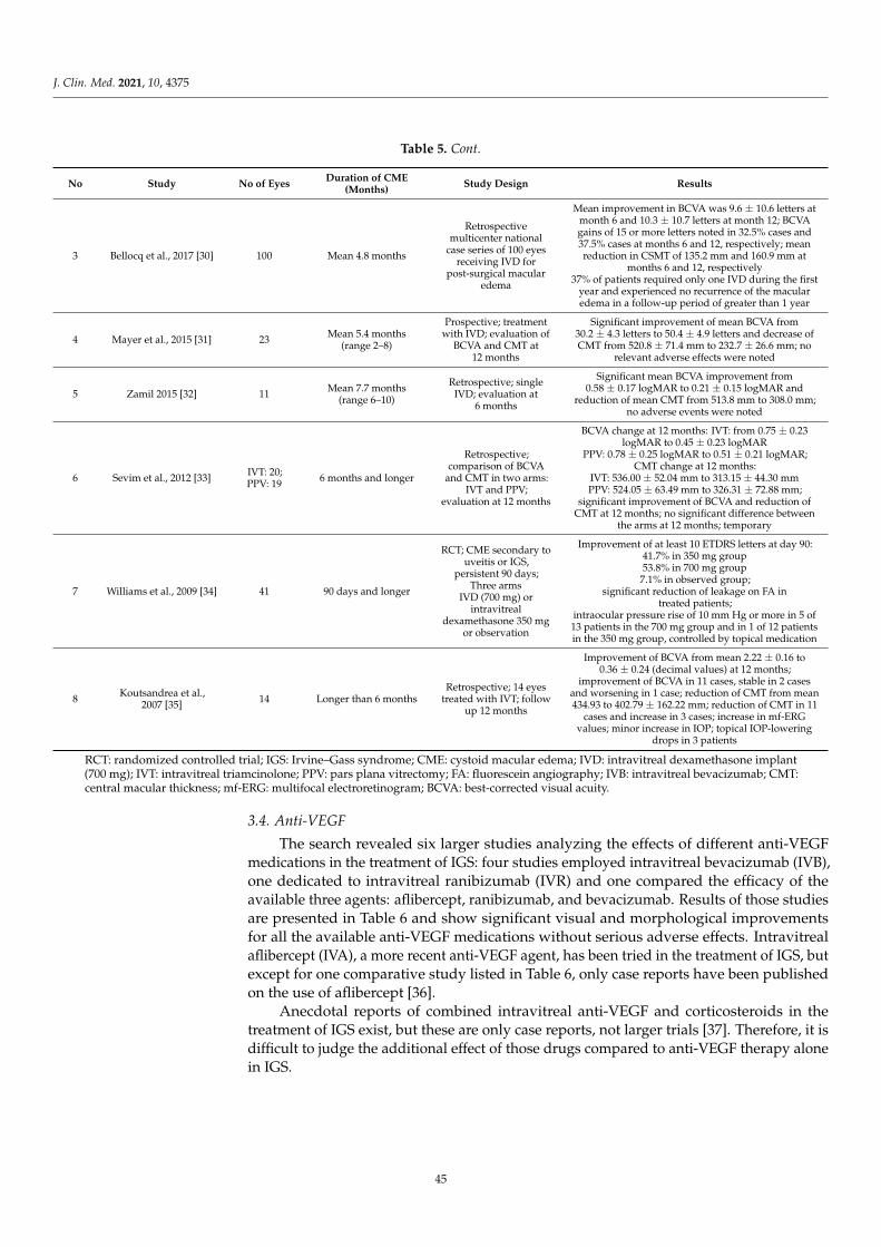
J. Clin. Med. 2021, 10, 4375
Table 5. Cont.
No Study No of EyesDuration of CME
(Months)Study Design Results
3 Bellocq et al., 2017 [30] 100 Mean 4.8 months
Retrospectivemulticenter national
case series of 100 eyesreceiving IVD for
post-surgical macularedema
Mean improvement in BCVA was 9.6 ± 10.6 letters atmonth 6 and 10.3 ± 10.7 letters at month 12; BCVAgains of 15 or more letters noted in 32.5% cases and37.5% cases at months 6 and 12, respectively; meanreduction in CSMT of 135.2 mm and 160.9 mm at
months 6 and 12, respectively37% of patients required only one IVD during the first
year and experienced no recurrence of the macularedema in a follow-up period of greater than 1 year
4 Mayer et al., 2015 [31] 23 Mean 5.4 months(range 2–8)
Prospective; treatmentwith IVD; evaluation of
BCVA and CMT at12 months
Significant improvement of mean BCVA from30.2 ± 4.3 letters to 50.4 ± 4.9 letters and decrease ofCMT from 520.8 ± 71.4 mm to 232.7 ± 26.6 mm; no
relevant adverse effects were noted
5 Zamil 2015 [32] 11 Mean 7.7 months(range 6–10)
Retrospective; singleIVD; evaluation at
6 months
Significant mean BCVA improvement from0.58 ± 0.17 logMAR to 0.21 ± 0.15 logMAR and
reduction of mean CMT from 513.8 mm to 308.0 mm;no adverse events were noted
6 Sevim et al., 2012 [33] IVT: 20;PPV: 19 6 months and longer
Retrospective;comparison of BCVA
and CMT in two arms:IVT and PPV;
evaluation at 12 months
BCVA change at 12 months: IVT: from 0.75 ± 0.23logMAR to 0.45 ± 0.23 logMAR
PPV: 0.78 ± 0.25 logMAR to 0.51 ± 0.21 logMAR;CMT change at 12 months:
IVT: 536.00 ± 52.04 mm to 313.15 ± 44.30 mmPPV: 524.05 ± 63.49 mm to 326.31 ± 72.88 mm;
significant improvement of BCVA and reduction ofCMT at 12 months; no significant difference between
the arms at 12 months; temporary
7 Williams et al., 2009 [34] 41 90 days and longer
RCT; CME secondary touveitis or IGS,
persistent 90 days;Three arms
IVD (700 mg) orintravitreal
dexamethasone 350 mgor observation
Improvement of at least 10 ETDRS letters at day 90:41.7% in 350 mg group53.8% in 700 mg group
7.1% in observed group;significant reduction of leakage on FA in
treated patients;intraocular pressure rise of 10 mm Hg or more in 5 of13 patients in the 700 mg group and in 1 of 12 patientsin the 350 mg group, controlled by topical medication
8 Koutsandrea et al.,2007 [35] 14 Longer than 6 months
Retrospective; 14 eyestreated with IVT; follow
up 12 months
Improvement of BCVA from mean 2.22 ± 0.16 to0.36 ± 0.24 (decimal values) at 12 months;
improvement of BCVA in 11 cases, stable in 2 casesand worsening in 1 case; reduction of CMT from mean434.93 to 402.79 ± 162.22 mm; reduction of CMT in 11
cases and increase in 3 cases; increase in mf-ERGvalues; minor increase in IOP; topical IOP-lowering
drops in 3 patients
RCT: randomized controlled trial; IGS: Irvine–Gass syndrome; CME: cystoid macular edema; IVD: intravitreal dexamethasone implant(700 mg); IVT: intravitreal triamcinolone; PPV: pars plana vitrectomy; FA: fluorescein angiography; IVB: intravitreal bevacizumab; CMT:central macular thickness; mf-ERG: multifocal electroretinogram; BCVA: best-corrected visual acuity.
3.4. Anti-VEGF
The search revealed six larger studies analyzing the effects of different anti-VEGFmedications in the treatment of IGS: four studies employed intravitreal bevacizumab (IVB),one dedicated to intravitreal ranibizumab (IVR) and one compared the efficacy of theavailable three agents: aflibercept, ranibizumab, and bevacizumab. Results of those studiesare presented in Table 6 and show significant visual and morphological improvementsfor all the available anti-VEGF medications without serious adverse effects. Intravitrealaflibercept (IVA), a more recent anti-VEGF agent, has been tried in the treatment of IGS, butexcept for one comparative study listed in Table 6, only case reports have been publishedon the use of aflibercept [36].
Anecdotal reports of combined intravitreal anti-VEGF and corticosteroids in thetreatment of IGS exist, but these are only case reports, not larger trials [37]. Therefore, it isdifficult to judge the additional effect of those drugs compared to anti-VEGF therapy alonein IGS.
45
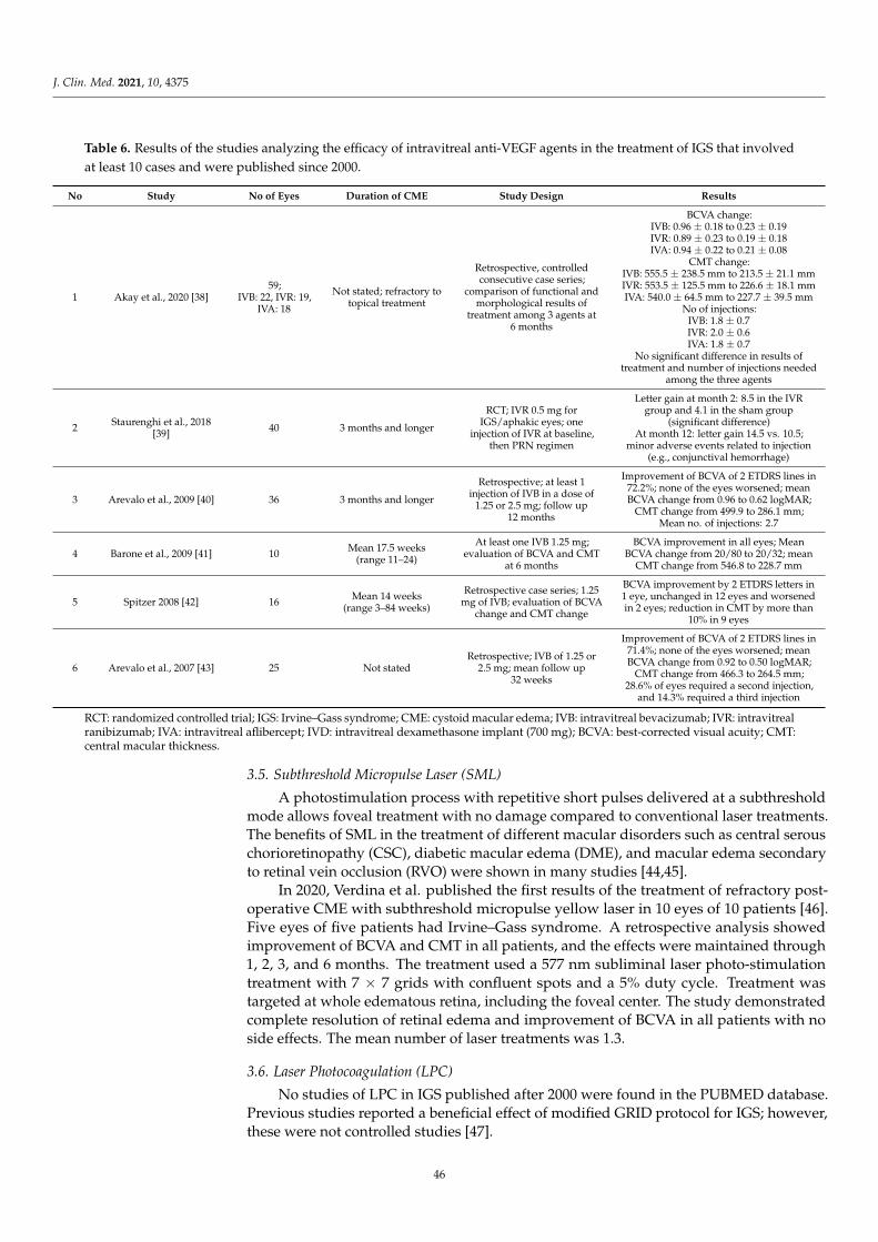
J. Clin. Med. 2021, 10, 4375
Table 6. Results of the studies analyzing the efficacy of intravitreal anti-VEGF agents in the treatment of IGS that involvedat least 10 cases and were published since 2000.
No Study No of Eyes Duration of CME Study Design Results
1 Akay et al., 2020 [38]59;
IVB: 22, IVR: 19,IVA: 18
Not stated; refractory totopical treatment
Retrospective, controlledconsecutive case series;
comparison of functional andmorphological results of
treatment among 3 agents at6 months
BCVA change:IVB: 0.96 ± 0.18 to 0.23 ± 0.19IVR: 0.89 ± 0.23 to 0.19 ± 0.18IVA: 0.94 ± 0.22 to 0.21 ± 0.08
CMT change:IVB: 555.5 ± 238.5 mm to 213.5 ± 21.1 mmIVR: 553.5 ± 125.5 mm to 226.6 ± 18.1 mmIVA: 540.0 ± 64.5 mm to 227.7 ± 39.5 mm
No of injections:IVB: 1.8 ± 0.7IVR: 2.0 ± 0.6IVA: 1.8 ± 0.7
No significant difference in results oftreatment and number of injections needed
among the three agents
2 Staurenghi et al., 2018[39] 40 3 months and longer
RCT; IVR 0.5 mg forIGS/aphakic eyes; one
injection of IVR at baseline,then PRN regimen
Letter gain at month 2: 8.5 in the IVRgroup and 4.1 in the sham group
(significant difference)At month 12: letter gain 14.5 vs. 10.5;
minor adverse events related to injection(e.g., conjunctival hemorrhage)
3 Arevalo et al., 2009 [40] 36 3 months and longer
Retrospective; at least 1injection of IVB in a dose of
1.25 or 2.5 mg; follow up12 months
Improvement of BCVA of 2 ETDRS lines in72.2%; none of the eyes worsened; meanBCVA change from 0.96 to 0.62 logMAR;
CMT change from 499.9 to 286.1 mm;Mean no. of injections: 2.7
4 Barone et al., 2009 [41] 10 Mean 17.5 weeks(range 11–24)
At least one IVB 1.25 mg;evaluation of BCVA and CMT
at 6 months
BCVA improvement in all eyes; MeanBCVA change from 20/80 to 20/32; mean
CMT change from 546.8 to 228.7 mm
5 Spitzer 2008 [42] 16 Mean 14 weeks(range 3–84 weeks)
Retrospective case series; 1.25mg of IVB; evaluation of BCVA
change and CMT change
BCVA improvement by 2 ETDRS letters in1 eye, unchanged in 12 eyes and worsenedin 2 eyes; reduction in CMT by more than
10% in 9 eyes
6 Arevalo et al., 2007 [43] 25 Not statedRetrospective; IVB of 1.25 or
2.5 mg; mean follow up32 weeks
Improvement of BCVA of 2 ETDRS lines in71.4%; none of the eyes worsened; meanBCVA change from 0.92 to 0.50 logMAR;
CMT change from 466.3 to 264.5 mm;28.6% of eyes required a second injection,
and 14.3% required a third injection
RCT: randomized controlled trial; IGS: Irvine–Gass syndrome; CME: cystoid macular edema; IVB: intravitreal bevacizumab; IVR: intravitrealranibizumab; IVA: intravitreal aflibercept; IVD: intravitreal dexamethasone implant (700 mg); BCVA: best-corrected visual acuity; CMT:central macular thickness.
3.5. Subthreshold Micropulse Laser (SML)
A photostimulation process with repetitive short pulses delivered at a subthresholdmode allows foveal treatment with no damage compared to conventional laser treatments.The benefits of SML in the treatment of different macular disorders such as central serouschorioretinopathy (CSC), diabetic macular edema (DME), and macular edema secondaryto retinal vein occlusion (RVO) were shown in many studies [44,45].
In 2020, Verdina et al. published the first results of the treatment of refractory post-operative CME with subthreshold micropulse yellow laser in 10 eyes of 10 patients [46].Five eyes of five patients had Irvine–Gass syndrome. A retrospective analysis showedimprovement of BCVA and CMT in all patients, and the effects were maintained through1, 2, 3, and 6 months. The treatment used a 577 nm subliminal laser photo-stimulationtreatment with 7 × 7 grids with confluent spots and a 5% duty cycle. Treatment wastargeted at whole edematous retina, including the foveal center. The study demonstratedcomplete resolution of retinal edema and improvement of BCVA in all patients with noside effects. The mean number of laser treatments was 1.3.
3.6. Laser Photocoagulation (LPC)
No studies of LPC in IGS published after 2000 were found in the PUBMED database.Previous studies reported a beneficial effect of modified GRID protocol for IGS; however,these were not controlled studies [47].
46
J. Clin. Med. 2021, 10, 4375
3.7. Other Treatments
Interferon alfa was administered for IGS in a small case series of four eyes refractoryto topical treatment [48]. A 3 million IU/day dose was injected subcutaneously for 4 weeksand tapered thereafter. Improvement was achieved in three cases without any side effects.Topical treatment of chronic refractory IGS with interferon alfa was also reported in a singlecase with spectacular visual improvement from 20/100 to 20/25 [49].
IGS was also treated by adalimumab (Humira). No significant improvement aftersuch therapy was achieved in a small case series of five eyes [50].
4. Discussion
The excellent results of modern cataract surgery set patient expectations very high, andpersistent CME after uneventful cataract surgery may significantly affect patient outcomesand satisfaction [51]. Irvine–Gass syndrome is a common complication of uneventfulcataract surgery, which resolves spontaneously in most cases but may persist, causingvisual deterioration and patient dissatisfaction [4,6,7]. As has been emphasized in manyprevious reviews and studies, no homogenous recommendations for the treatment ofIVG exist [52–55]. The lack of randomized controlled trials assessing the effectivenessof available therapeutic modalities results in many different approaches, often based onindividual judgment and clinical experience but not hard evidence. Our analysis focusedon the papers published in this century, as this is the time when intravitreal treatmentssuch as anti-VEGF or intravitreal corticosteroids were introduced and revolutionized themanagement of various ophthalmic diseases. Therefore, we sought to compare conservativetreatments to those modern therapeutic modalities.
Presented studies published since 2000t in general show favorable results of the treat-ment of IGS with topical NSAIDs alone or in combination with periocular or intravitrealsteroids as well as intravitreal anti-VEGF agents. Those treatments should be consid-ered, weighing both the potential for improving BCVA and the invasive character of thetreatment and the possibility of complications.
As IGS resolves spontaneously in most cases, that possibility must be consideredbefore administering invasive therapy. Therefore, the timing of the application of differentforms of treatment should be carefully considered with non-invasive therapies used asthe first line (e.g., topical treatment) and invasive procedures (e.g., intraocular injections)usually reserved for non-responsive cases.
NSAIDs administered topically such as via eye drops are FDA-approved drugs for useas anti-inflammatory, antipyretic, and analgesic agents. Their main mechanism of action isthe inhibition of the enzyme cyclooxygenase (COX). Cyclooxygenase is required to convertarachidonic acid into thromboxanes, prostaglandins, and prostacyclin. Prostaglandinsplay an important role in vasodilatation [56]. The use of NSAIDs in the postoperativemanagement of patients undergoing cataract surgery has become a standard of care [57,58].Routine use of anti-inflammatory eye drops following cataract surgery is highly effectivein reducing post-surgical inflammation and the incidence of CME [59]; however, their rolein the treatment of CME has not been studied widely. Topical NSAIDs remain a first-linetherapy of IGS, and although their use has shown to be beneficial in several studies, theyhave shown no clear effect in other studies [58]. Our search revealed only a few modernstudies that analyze the effects of NSAID in the treatment of IGS, none of which is anRCT. One older study showed significant visual and morphological improvements afteradministration of topical NSAID in acute cases, which are usually defined as lasting lessthan 3 months [18]. However, most recent studies show only moderate improvement aftertreatment of IGS with only topical NSAID [12,13].
Functional and morphological results are reported to be better after intravitreal dex-amethasone [13]. Adding the effect of nepafenac was reported in one study that analyzedthe combination of IVB and NSAID [16]. NSAIDs are also used in combined therapy withtopical corticosteroids or oral CAIs, but available data on the combined treatment of CMEare very limited. Nevertheless, the off-label use of acetazolamide, a carbonic anhydrase
47
J. Clin. Med. 2021, 10, 4375
inhibitor, in IGS is a common practice as a first- or second-line therapy. Acetazolamideincreases the retinal pigment epithelium pump function by inhibiting carbonic anhydraseand is thought to decrease intraretinal fluid [11,60]. Dosage varies among studies from250 mg once a day to TID. Many authors state that the combination of oral acetazolamidewith topical NSAIDs is shown to be highly effective [20,61,62]. Our search revealed onlytwo papers analyzing the additional effect of CAI compared to NSAID-only treatmentof IGS, both on relatively small samples (14–15 eyes). Both papers favor the use of CAIin combination therapy for IGS; however, such limited data make it impossible to buildstrong recommendations for the use of this treatment regimen.
Corticosteroids remain a viable therapeutic option in the treatment of CME, includ-ing IGS. Corticosteroids block the release of arachidonic acid, impact the production ofinterleukins and VEGF, and interrupt the inflammatory cascade. Several routes of adminis-tration, such as topical, periocular, and intravitreal, are available. At the same time, currentdata on a combination treatment of topical NSAID with topical corticosteroids are scarceand not convincing [21,22]. A conclusion on the beneficial effect of the addition of topicalcorticosteroids to the treatment of IGS cannot be made based on available research. Never-theless, topical corticosteroids are widely used in the treatment of Irvine–Gass syndrome,usually in combination with topical NSAIDs and oral CAIs. An accurate assessment of therole of topical corticosteroids alone in the treatment of IGS is not currently possible.
Periocular or intravitreal corticosteroids serve as an option in refractory cases ofIGS [63]. Sub-tenon or retrobulbar injections of corticosteroids had been used widely forpersistent CME before the advent of an officially registered intravitreal dexamethasoneimplant (OZURDEX®). Early in 1997, Thach and his group showed VA improvement after12 repeated corticosteroid injections in a series of 31 patients with chronic CME [64]. Oursearch revealed three recent studies (2018–2021) that showed significant visual improve-ment after STT in refractory cases of IGS. STT remains a cost-effective therapy, and itsapplication sub-tenon does not bear the risk of intraocular inflammation possible afterintravitreal application. A recent study by Kuley did not show an advantage of intravitrealversus sub-tenon administration of triamcinolone [23]. It must be emphasized, though,that the use of triamcinolone acetonide remains off-label. Intravitreal corticosteroids haveconsequently been used for chronic or refractory cases, lasting longer than 3 months,with significant letter gains and minor adverse effects [31–35]. Before the dexametha-sone implant was introduced, triamcinolone acetonide was tested in a few larger andsmaller studies, proving its efficacy in improving macular morphology and function inIGS [33,35,65–67]. Later studies show significant improvements after IVD administrationwithout serious side effects [27,29–32,34]. The most recent large retrospective study from2020 highlighted the benefits of early intervention and reported significantly larger visualgains when IVD was administered within 4 weeks of diagnosis [27]. This approach is nota common practice due to the invasive character of the procedure and the possibility ofeffective treatment with only topical NSAIDs. Further comparative studies are needed tosupport the results of that paper.
Vascular endothelial growth factors play central roles in the regulation of angiogenesisand lymphangiogenesis and they regulate endothelial cell proliferation, migration, vascularpermeability, secretion, and other endothelial functions. The revolutionary role of anti-VEGF in treating ophthalmic conditions such as neovascularization and macular edemadue to DME or ME in RVO was a milestone. The VEGF family plays a major role inangiogenesis, inflammation, and capillary permeability; thus, its potential in treatingCME was studied. However, the role of anti-VEGF treatment in CME remains unclear.Anti-VEGF injections remain an alternative in unresponsive cases, but their use in IGSrequires further randomized research. Our search revealed a few quality studies thatshow significant improvements after the use of anti-VEGF medication in IGS, but RCTsare missing. Despite that, clinical practice and the universality of that procedure make it asolid treatment modality in refractory IGS.
48

J. Clin. Med. 2021, 10, 4375
Non-damaging laser therapy, such as SML, remains an interesting therapeutic option.To date, just a few papers report its efficacy in IGS. Considering its non-damaging character,lack of side effects, and low cost, it may be considered as an alternative to more invasivetreatment modalities. Further studies are needed to provide treatment guidelines for SML.
Practical Considerations and Conclusions
This review aimed to provide a basis for modern recommendations for treating pseu-dophakic macular edema or Irvine–Gass syndrome. The available published material doesnot provide convincing data to build such guidelines. Therefore, theoretical background,clinical experience, and safety of the procedure must determine the choice of treatment inthis clinical entity. Common practice is to start therapy with a topical NSAID, which is asimple and non-invasive treatment modality. This approach is supported by epidemiologi-cal and clinical research that provides data on the possibility of spontaneous resolutionof CME and improvement after topical therapy [68]. Larger clinical trials have not shownthat using a combination of topical NSAID and topical corticosteroids and/or oral CAI issuperior to topical NSAID alone.
What remains unclear is the timing of application of invasive therapies—periocular orintravitreal injections—once topical treatment is not effective. Refractory pseudophakicmacular edema is not precisely defined according to its duration, but usually authorsemploy periocular or intravitreal treatment in cases lasting longer than three months.The efficacy and safety of intravitreal or periocular injections with corticosteroids or anti-VEGF agents have been confirmed in many studies. Still, its invasive nature and rare butpotentially serious complications must be considered. Patients who resist intravitreal orperiocular treatment might be offered therapy with subthreshold micropulse laser. Recentpublications on the use of SML show promise. Low complication rates, cost-effectiveness,and repeatability are clear advantages of this treatment modality.
Our search revealed publications that show possible options for the treatment of IGS.Methodology and randomization in presented trials may be discussed; what remains astheir common feature is the visual deficit reported in most cases of longstanding CME,even after successful treatment. Therefore, in view of results of a recent large studyfrom Sharma et al. [27] that proves better functional and morphological results with earlyapplication of intravitreal steroids, that therapeutic option for short-standing pseudophakicCME should be examined with care in future research.
Author Contributions: Conceptualization: M.O. and M.G.; Methodology: M.G., Investigation: M.O.and M.G., writing: M.O. and M.G., supervision: M.G. All authors have read and agreed to thepublished version of the manuscript.
Funding: This research received no external funding.
Institutional Review Board Statement: Not applicable.
Informed Consent Statement: Not applicable.
Data Availability Statement: Not applicable.
Conflicts of Interest: The authors declare no conflict of interest.
References
1. Grzybowski, A.; Sikorski, B.L.; Ascano, F.J.; Huerva, V. Pseudophakic cystoid macular edema. Clin. Interv. Aging 2016, 11,1221–1229. [CrossRef] [PubMed]
2. Irvine, S.R. A newly defined vitreous syndrome following cataract surgery. Am. J. Ophthalmol. 1953, 36, 599–619. [CrossRef]3. Gass, J.D.; Norton, E.W. Cystoid macular edema and papilledema following cataract extraction. A fluorescein fundoscopic and
angiographic study. Arch. Ophthalmol. 1966, 76, 646–661. [CrossRef] [PubMed]4. Henderson, B.A.; Kim, J.Y.; Ament, C.S.; Ferrufino-Ponce, Z.K.; Grabowska, A.; Cremers, S.L. Clinical pseudophakic cystoid
macular edema. Risk factors for development and duration after treatment. J. Cataract Refract. Surg. 2007, 33, 1550–1558.[CrossRef] [PubMed]
49

J. Clin. Med. 2021, 10, 4375
5. Hunter, A.A.; Modjtahedi, S.P.; Long, K.; Zawadzki, R.; Chin, E.K.; Caspar, J.J.; Morse, L.S.; Telander, D.G. Improving visualoutcomes by preserving outer retina morphology in eyes with resolved pseudophakic cystoid macular edema. J. Cataract Refract.
Surg. 2014, 40, 626–631. [CrossRef]6. Flach, A.J. The incidence, pathogenesis and treatment of cystoid macular edema following cataract surgery. Trans. Am. Ophthalmol.
Soc. 1998, 96, 557–634.7. Perente, I.; Utine, C.A.; Ozturker, C.; Cakir, M.; Kaya, V.; Eren, H.; Kapran, Z.; Yilmaz, O.F. Evaluation of macular changes after
uncomplicated phacoemulsification surgery by optical coherence tomography. Curr. Eye Res. 2007, 32, 241–247. [CrossRef]8. Ursell, P.G.; Spalton, D.J.; Whitcup, S.M.; Nussenblatt, R.B. Cystoid macular edema after phacoemulsification: Relationship to
blood-aqueous barrier damage and visual acuity. J. Cataract Refract. Surg. 1999, 25, 1492–1497. [CrossRef]9. Shelsta, H.N.; Jampol, L.M. Pharmacologic therapy of pseudophakic cystoid macular edema: 2010 update. Retina 2011, 31, 4–12.
[CrossRef] [PubMed]10. Smith, R.T.; Campbell, C.J.; Koester, C.J.; Trokel, S.; Anderson, A. The barrier function in extracapsular cataract surgery.
Ophthalmology 1990, 97. [CrossRef]11. Benitah, N.R.; Arroyo, J.G. Pseudophakic cystoid macular edema. Int. Ophthalmol. Clin. 2010, 50, 139–153. [CrossRef]12. Giarmoukakis, A.K.; Blazaki, S.V.; Bontzos, G.C.; Plaka, A.D.; Seliniotakis, K.N.; Ioannidi, L.D.; Tsilimbaris, M.K. Efficacy of
topical nepafenac 0.3% in the management of postoperative cystoid macular edema. Ther. Clin. Risk Manag. 2020, 16, 1067–1074.[CrossRef]
13. Guclu, H.; Gurlu, V.P. Comparison of topical nepafenac 0.1% with intravitreal dexamethasone implant for the treatment ofIrvine-Gass syndrome. Int. J. Ophthalmol. 2019, 12, 258–267. [CrossRef] [PubMed]
14. Sengupta, S.; Vasavada, D.; Pan, U.; Sindal, M. Factors predicting response of pseudophakic cystoid macular edema to topicalsteroids and nepafenac. Indian J. Ophthalmol. 2018, 66, 827–830. [CrossRef] [PubMed]
15. Yüksel, B.; Uzunel, U.D.; Kerci, S.G.; Sagban, L.; Kusbeci, T.; Örsel, T. Comparison of subtenon triamcinolone acetonide injectionwith topical nepafenac for the treatment of pseudophakic cystoid macular edema. Ocul. Immunol. Inflamm. 2016, 25, 513–519.[CrossRef]
16. Warren, K.A.; Bahrani, H.; Fox, J.E. NSAIDs in combination therapy for the treatment of chronic pseudophakic cystoid macularedema. Retina 2010, 30, 260–266. [CrossRef] [PubMed]
17. Hariprasad, S.M.; Akduman, L.; Clever, J.A.; Ober, M.; Recchia, F.M.; Mieler, W.F. Treatment of cystoid macular edema with thenew-generation NSAID nepafenac 0.1%. Clin. Ophthalmol. 2009, 3, 147–154. [CrossRef]
18. Rho, D.S. Treatment of acute pseudophakic cystoid macular edema: Diclofenac versus ketorolac. J. Cataract Refract. Surg. 2003, 29,2378–2384. [CrossRef]
19. Curkovic, T.; Vukojevic, N.; Bucan, K. Treatment of pseudophakic cystoid macular oedema. Coll. Antropol. 2005, 103–105.20. Catier, A.; Tadayoni, R.; Massin, P.; Gaudric, A. Intérêt de l’acétazolamide associé aux anti-inflammatoires dans le traitement de
l’oedème maculaire postopératoire (Advantages of acetazolamide associated with anti-inflammatory medications in postoperativetreatment of macular edema). J. Fr. Ophtalmol. 2005, 28, 1027–1031. [CrossRef]
21. Heier, J.S.; Topping, T.M.; Baumann, W.; Dirks, M.S.; Chern, S. Ketorolac versus prednisolone versus combination therapy in thetreatment of acute pseudophakic cystoid macular edema. Ophthalmology 2000, 107, 2034–2038. [CrossRef]
22. Singal, N.; Hopkins, J. Pseudophakic cystoid macular edema: Ketorolac alone vs. ketorolac plus prednisolone. Can. J. Ophthalmol.
2004, 39, 245–250. [CrossRef]23. Kuley, B.; Storey, P.P.; Wibbelsman, T.D.; Pancholy, M.; Zhang, Q.; Sharpe, J.; Bello, N.; Obeid, A.; Regillo, C.; Kaiser, R.S.; et al.
Resolution of pseudophakic cystoid macular edema: 2 mg intravitreal triamcinolone acetonide versus 40 mg posterior sub-tenontriamcinolone acetonide. Curr. Eye Res. 2021, 1–7. [CrossRef]
24. Erden, B.; Çakır, A.; Aslan, A.C.; Bölükbası, S.; Elçioglu, M.N. The efficacy of posterior subtenon triamcinolone acetonide injectionin treatment of irvine-gass syndrome. Ocul. Immunol. Inflamm. 2019, 27, 1235–1241. [CrossRef]
25. Tsai, M.-J.; Yang, C.-M.; Hsieh, Y.-T. Posterior subtenon injection of triamcinolone acetonide for pseudophakic cystoid macularoedema. Acta Ophthalmol. 2016, 96, e891–e893. [CrossRef]
26. Benhamou, N.; Massin, P.; Haouchine, B.; Audren, F.; Tadayoni, R.; Gaudric, A. Intravitreal triamcinolone for refractorypseudophakic macular edema. Am. J. Ophthalmol. 2003, 135, 246–249. [CrossRef]
27. Sharma, A.; Bandello, F.; Loewenstein, A.; Kuppermann, B.D.; Lanzetta, P.; Zur, D.; Hilely, A.; Iglicki, M.; Veritti, D.; Wang,A.; et al. Current role of intravitreal injections in Irvine Gass syndrome-CRIIG study. Int. Ophthalmol. 2020, 40, 3067–3075.[CrossRef] [PubMed]
28. Marques, J.H.; Abreu, A.C.; Silva, N.; Meireles, A.; Pessoa, B.; Melo Beirão, J. Fluocinolone acetonide 0.19 mg implant in patientswith cystoid macular edema due to Irvine-Gass syndrome. Int. Med. Case Rep. J. 2021, 26, 127–132. [CrossRef] [PubMed]
29. Altintas, A.G.K.; Ilhan, C. Intravitreal dexamethasone implantation in intravitreal bevacizumab treatment-resistant pseudophakiccystoid macular edema. Korean J. Ophthalmol. 2019, 33, 259–266. [CrossRef] [PubMed]
30. Bellocq, D.; Pierre-Kahn, V.; Matonti, F.; Burillon, C.; Voirin, N.; Dot, C.; Akesbi, J.; Milazzo, S.; Baillif, S.; Soler, V.; et al.Effectiveness and safety of dexamethasone implants for postsurgical macular oedema including Irvine–Gass syndrome: TheEPISODIC-2 study. Br. J. Ophthalmol. 2016, 101, 333–341. [CrossRef] [PubMed]
50
J. Clin. Med. 2021, 10, 4375
31. Mayer, W.J.; Kurz, S.; Wolf, A.; Kook, D.; Kreutzer, T.; Kampik, A.; Priglinger, S.; Haritoglou, C. Dexamethasone implant asan effective treatment option for macular edema due to Irvine-Gass syndrome. J. Cataract Refract. Surg. 2015, 41, 1954–1961.[CrossRef] [PubMed]
32. Al Zamil, W.M. Short-term safety and efficacy of intravitreal 0.7-mg dexamethasone implants for pseudophakic cystoid macularedema. Saudi J. Ophthalmol. 2014, 29, 130–134. [CrossRef]
33. Sevim, M.; Sanisoglu, H.; Türkyılmaz, K. Intravitreal triamcinolone acetonide versus pars plana vitrectomy for pseudophakiccystoid macular edema. Curr. Eye Res. 2012, 37, 1165–1170. [CrossRef]
34. Williams, G.A.; Haller, J.A.; Kuppermann, B.D.; Blumenkranz, M.S.; Weinberg, D.V.; Chou, C.; Whitcup, S.M. DexamethasoneDDS phase II study group. Dexamethasone posterior-segment drug delivery system in the treatment of macular edema resultingfrom uveitis or Irvine-Gass syndrome. Am. J. Ophthalmol. 2009, 147, 1048–1054. [CrossRef]
35. Koutsandrea, C.; Moschos, M.M.; Brouzas, D.; Loukianou, E.; Apostolopoulos, M.; Moschos, M. Intraocular triamcinoloneacetonide for pseudophakic cystoid macular edema: Optical coherence tomography and multifocal electroretinography study.Retina 2007, 27, 159–164. [CrossRef] [PubMed]
36. Lin, C.J.; Tsai, Y.Y. Use of aflibercept for the management of refractory pseudophakic macular edema in Irvine-Gass syndromenad literature review. Retin. Cases Brief. Rep. 2018, 12, 59–62. [CrossRef]
37. Fenicia, V.; Balestrieri, M.; Perdicchi, A.; Enrici, M.M.; Fave, M.D.; Recupero, S.M. Intravitreal injection of dexamethasone implantand ranibizumab in cystoid macular edema in the course of Irvine-Gass syndrome. Case Rep. Ophthalmol. 2014, 5, 243–248.[CrossRef]
38. Akay, F.; Isik, M.U.; Akmaz, B. Comparison of intravitreal anti-vascular endothelial growth factor agents and treatment results inIrvine-Gass syndrome. Int. J. Ophthalmol. 2020, 13, 1586–1591. [CrossRef]
39. Staurenghi, G.; Lai, T.Y.Y.; Mitchell, P.; Wolf, S.; Wenzel, A.; Li, J.; Bhaumik, A.; Hykin, P.G.; PROMETHEUS Study Group. Efficacyand safety of ranibizumab 0.5 mg for the treatment of macular edema resulting from uncommon causes: Twelve-month findingsfrom prometheus. Ophthalmology 2018, 125, 850–862. [CrossRef] [PubMed]
40. Arevalo, J.F.; Maia, M.; Garcia-Amaris, R.A.; Roca, J.A.; Sanchez, J.G.; Berrocal, M.H.; Wu, L. Intravitreal bevacizumab forrefractory pseudophakic cystoid macular edema: The Pan-American collaborative retina study group results. Ophthalmology 2009,116, 1481.e1–1487.e1. [CrossRef]
41. Barone, A.; Russo, V.; Prascina, F.; Noci, N.D. Short-term safety and efficacy of intravitreal bevacizumab for pseudophakic cystoidmacular edema. Retina 2009, 29, 33–37. [CrossRef]
42. Spitzer, M.S.; Ziemssen, F.; Yoeruek, E.; Petermeier, K.; Aisenbrey, S.; Szurman, P. Efficacy of intravitreal bevacizumab in treatingpostoperative pseudophakic cystoid macular edema. J. Cataract Refract. Surg. 2008, 34, 70–75. [CrossRef]
43. Arevalo, J.F.; Garcia-Amaris, R.A.; Roca, J.A.; Sanchez, J.G.; Wu, L.; Berrocal, M.H.; Maia, M. Primary intravitreal bevacizumabfor the management of pseudophakic cystoid macular edema: Pilot study of the Pan-American collaborative retina study Group.J. Cataract Refract. Surg. 2007, 339120, 2098–3105. [CrossRef] [PubMed]
44. Gawecki, M. Micropulse laser treatment of retinal diseases. J. Clin. Med. 2019, 8, 242. [CrossRef]45. Gawecki, M.; Jaszczuk-Maciejewska, A.; Jurska-Jasko, A.; Kneba, M.; Grzybowski, A. Transfoveal micropulse laser treatment of
central serous chorioretinopathy within six months of disease onset. J. Clin. Med. 2019, 8, 1398. [CrossRef] [PubMed]46. Verdina, T.; D’Aloisio, R.; Lazzerini, A.; Ferrari, C.; Valerio, E.; Mastropasqua, R.; Cavallini, G.M. The role of subthreshold
micropulse yellow laser as an alternative option for the treatment of refractory postoperative cystoid macular edema. J. Clin. Med.
2020, 9, 1066. [CrossRef]47. Lardenoye, C.W.; van Schooneveld, M.J.; Frits Treffers, W.; Rothova, A. Grid laser photocoagulation for macular oedema in
uveitis or the Irvine-Gass syndrome. Br. J. Ophthalmol. 1998, 82, 1013–1016. [CrossRef]48. Deuter, C.M.E.; Gelisken, F.; Stübiger, N.; Zierhut, M.; Doycheva, D. Successful treatment of chronic pseudophakic macular edema
(Irvine-Gass syndrome) with interferon alpha: A report of three cases. Ocul. Immunol. Inflamm. 2011, 19, 216–218. [CrossRef][PubMed]
49. Maleki, A.; Aghaei, H.; Lee, S. Topical interferon alpha 2b in the treatment of refractory pseudophakic cystoid macular edema.Am. J. Ophthalmol. Case Rep. 2018, 10, 203–205. [CrossRef]
50. Farvardin, M.; Namvar, E.; Sanie-Jahromi, F.; Johari, M.K. The effects of intravitreal adalimumab injection on pseudophakicmacular edema. BMC Res. Notes 2020, 13, 1–4. [CrossRef]
51. Gawecki, M.; Grzybowski, A. Diplopia as the complication of cataract surgery. J. Ophthalmol. 2016, 2016, 1–6. [CrossRef]52. Tsangaridou, M.-A.; Grzybowski, A.; Gundlach, E.; Pleyer, U. Controversies in NSAIDs use in cataract surgery. Curr. Pharm. Des.
2015, 21, 4707–4717. [CrossRef]53. Grzybowski, A.; Kanclerz, P. Controversies on the use of nonsteroidal antiinflammatory drugs and steroids in pseudophakic
cystoid macular edema prophylaxis. J. Cataract Refract. Surg. 2019, 45, 1848. [PubMed]54. Guo, S.; Patel, S.; Baumrind, B.; Johnson, K.; Levinsohn, D.; Marcus, E.; Tannen, B.; Roy, M.; Bhagat, N.; Zarbin, M. Management
of pseudophakic cystoid macular edema. Surv. Ophthalmol. 2015, 60, 123–137. [CrossRef]55. Yonekawa, Y.; Kim, I.K. Pseudophakic cystoid macular edema. Curr. Opin. Ophthalmol. 2012, 23, 26–32. [CrossRef] [PubMed]56. Vane, J.R. Inhibition of prostaglandin synthesis as a mechanism of action for aspirin-like drugs. Nat. New Biol. 1971, 231, 232–235.
[CrossRef] [PubMed]
51
J. Clin. Med. 2021, 10, 4375
57. Brandsdorfer, A.; Patel, S.H.; Chuck, R.S. The role of perioperative nonsteroidal anti-inflammatory drugs use in cataract surgery.Curr. Opin. Ophthalmol. 2019, 30, 44–49. [CrossRef] [PubMed]
58. Lim, B.X.; Lim, C.H.; Lim, D.K.; Evans, J.R.; Bunce, C.; Wormald, R. Prophylactic non-steroidal anti-inflammatory drugs for theprevention of macular oedema after cataract surgery. Cochrane Database Syst. Rev. 2016. [CrossRef] [PubMed]
59. Rossetti, L.; Chaudhuri, J.; Dickersin, K. Medical prophylaxis and treatment of cystoid macular edema after cataract surgery: Theresults of a meta-analysis. Ophthalmology 1998, 105, 397–405. [CrossRef]
60. Marmor, M.F.; Maack, T. Enhancement of retinal adhesion and sub- retinal fluid resorption by acetazolamide. Invest. Ophthalmol.
Vis. Sci. 1982, 23, 121–124.61. Cox, S.N.; Hay, E.; Bird, A.C. Treatment of chronic macular edema with acetazolamide. Arch. Ophthalmol. 1988, 106, 1190–1195.
[CrossRef]62. Weene, L. Cystoid macular edema after scleral buckling responsive to acetazolamide. Ann. Ophthalmol. 1992, 24.63. Zur, D.; Loewenstein, A. Postsurgical cystoid macular edema. Dev. Ophthalmol. 2017, 58, 178–190. [CrossRef] [PubMed]64. Thach, A.B.; Dugel, P.U.; Flindall, R.J.; Sipperley, J.O.; Sneed, S.R. A Comparison of retrobulbar versus sub-tenon’s corticosteroid
therapy for cystoid macular edema refractory to topical medications. Ophthalmology 1997, 104, 2003–2008. [CrossRef]65. Boscia, F.; Furino, C.; Dammacco, R.; Ferreri, P.; Sborgia, L. Intravitreal triamcinolone acetonide in refractory pseudophakic
cystoid macular edema: Functional and anatomic results. Eur. J. Ophthalmol. 2005, 15, 89–95. [CrossRef] [PubMed]66. Jonas, J.B.; Kreissig, I.; Degenring, R.F. Intravitreal triamcinolone acetonide for pseudophakic cystoid macular edema. Am. J.
Ophthalmol. 2003, 136, 384–386. [CrossRef]67. Conway, M.D.; Canakis, C.; Livir-Rallatos, C.; Peyman, G.A. Intravitreal triamcinolone acetonide for refractory chronic pseu-
dophakic cystoid macular edema. J. Cataract Refract. Surg. 2003, 29, 27–33. [CrossRef]68. Zur, D.; Fischer, N.; Tufail, A.; Monés, J.; Loewenstein, A. Postsurgical cystoid macular edema. Eur. J. Ophthalmol. 2011, 21, 62–68.
[CrossRef]
52
Journal of
Clinical Medicine
Article
Imbalance in the Levels of Angiogenic Factors in Patients withAcute and Chronic Central Serous Chorioretinopathy
Izabella Karska-Basta 1,*, Weronika Pociej-Marciak 1, Michał Chrzaszcz 1, Agnieszka Kubicka-Trzaska 1 ,
Magdalena Debicka-Kumela 1, Maciej Gawecki 2 , Bozena Romanowska-Dixon 1 and Marek Sanak 3
�����������������
Citation: Karska-Basta, I.;
Pociej-Marciak, W.; Chrzaszcz, M.;
Kubicka-Trzaska, A.;
Debicka-Kumela, M.; Gawecki, M.;
Romanowska-Dixon, B.; Sanak, M.
Imbalance in the Levels of
Angiogenic Factors in Patients with
Acute and Chronic Central Serous
Chorioretinopathy. J. Clin. Med. 2021,
10, 1087. https://doi.org/10.3390/
jcm10051087
Academic Editor: Fumi Gomi
Received: 21 January 2021
Accepted: 26 February 2021
Published: 5 March 2021
Publisher’s Note: MDPI stays neutral
with regard to jurisdictional claims in
published maps and institutional affil-
iations.
Copyright: © 2021 by the authors.
Licensee MDPI, Basel, Switzerland.
This article is an open access article
distributed under the terms and
conditions of the Creative Commons
Attribution (CC BY) license (https://
creativecommons.org/licenses/by/
4.0/).
1 Department of Ophthalmology, Faculty of Medicine, Clinic of Ophthalmology and Ocular Oncology,Jagiellonian University Medical College, 31-070 Krakow, Poland;[email protected] (W.P.-M.); [email protected] (M.C.);[email protected] (A.K.-T.); [email protected] (M.D.-K.);[email protected] (B.R.-D.)
2 Dobry Wzrok Ophthalmological Clinic, 80-402 Gdansk, Poland; [email protected] Molecular Biology and Clinical Genetics Unit, Department of Internal Medicine,
Jagiellonian University Medical College Faculty of Medicine, 31-066 Krakow, Poland;[email protected]
* Correspondence: [email protected]
Abstract: Background: The pathogenesis of central serous chorioretinopathy (CSC) remains a sub-
ject of intensive research. We aimed to determine correlations between plasma levels of selected
angiogenic factors and different forms of CSC. Methods: Eighty patients were enrolled in the study
including 30 with a chronic form of CSC, 30 with acute CSC, and 20 controls. Presence of active
CSC was determined by fluorescein angiography (FA), indocyanine green angiography (ICGA), and
swept-source optical coherence tomography (SS-OCT). Plasma concentrations of angiopoietin-1,
endostatin, fibroblast growth factor, placental growth factor (PlGF), platelet-derived growth factor
(PDGF-AA), thrombospondin-2, vascular endothelial growth factor (VEGF), VEGF-D, and pigment
epithelium–derived factor were measured, and the results were compared between groups. Addition-
ally, mean choroidal thickness (CT) was measured in all patients. Results: Levels of angiopoietin-1
(p = 0.008), PlGF (p = 0.045), and PDGF-AA (p = 0.033) differed significantly between the three
groups. Compared with the controls, VEGF (p = 0.024), PlGF (p = 0.013), and PDGF-AA (p = 0.012)
were downregulated in the whole CSC group, specifically PDGF-AA (p = 0.002) in acute CSC and
angiopoietin-1 (p = 0.007) in chronic CSC. An inverse correlation between mean CT and VEGF levels
was noted in CSC patients (rho = −0.27, p = 0.044). Conclusions: Downregulated angiopoietin-1,
VEGF, PDGF-AA, and PlGF levels may highlight the previously unknown role of the imbalanced
levels of proangiogenic and antiangiogenic factors in the pathogenesis of CSC. Moreover, down-
regulated VEGF levels may suggest that choroidal neovascularization in CSC is associated with
arteriogenesis rather than angiogenesis.
Keywords: angiogenesis; angiopoietin-1; central serous chorioretinopathy; proangiogenic factors
1. Introduction
Central serous chorioretinopathy (CSC) is a common disease that belongs to thepachychoroid-related disorders, characterized by serous retinal detachment, which isassociated with a local leakage from the thicker choroid through impaired retinal pigmentepithelium (RPE) [1]. For a long time, a simple classification of CSC into acute and chronicforms has been used; however, it was based solely on the duration of serous neurosensoryretinal detachment [2]. Nowadays, as new pathogenic concepts of CSC emerge and modernmultimodal imaging becomes available, various new classifications are being developed,but so far, none of them has fully reflected the complexity of this clinical entity [3,4].Daruich et al. classified CSC as acute, non-resolving, recurrent, chronic, or inactive, whichmore precisely refers to the course of the disease [5].
53
J. Clin. Med. 2021, 10, 1087
As the pathogenesis and pathophysiology of CSC have not been fully explained,the available treatment modalities are suboptimal, especially in long-standing cases [2,6].Growing evidence indicates that the pathomechanism of CSC is associated with dysfunc-tion of the thickened choroid (an important risk factor), with subsequent impairment ofthe RPE [1,7]. Some authors have also postulated that downregulation of the cell–cell adhe-sion molecules in the vascular endothelium could increase the permeability of choroidalvessels, causing fluid leakage under the neurosensory retina [8,9]. In the present study,we attempted to find an association between these phenomena in CSC and the levels ofproangiogenic and antiangiogenic factors in human plasma.
Although the role of elevated levels of vascular endothelial growth factor (VEGF) andother cytokines in the pathogenesis of the pachychoroid disease has been speculated on insome papers, they more often referred to other forms of this disease entity than CSC [10,11].Recent studies suggested that eyes with CSC may be at higher risk of age-related mac-ular degeneration (AMD) or one of its subtypes (polypoidal choroidal vasculopathy orpachychoroid neovasculopathy), or that the development of CSC and AMD/polypoidalchoroidal vasculopathy may share a common background [12–14]. Data on systemicchanges in the expression of proangiogenic and antiangiogenic factors and their role inchoroidal vascular homeostasis associated with CSC are scarce. Apart from our previousresearch, we identified only one report evaluating the plasma level of VEGF in patientswith CSC [15,16].
It is well-known that CSC may be complicated by choroidal neovascularization (CNV),a typical feature for neovascular AMD (nAMD). The prevalence of this complication wasreported to range from 15.6% to 25% [16,17]. Extensive research of AMD included thelevels of proangiogenic factors [18–20]. Impaired expression of antiangiogenic factors wasshown to play an important role in the development of CNV in the course of nAMD [21].By analogy, some of these etiological factors typical for nAMD could also be involved inthe course of different forms of pachychoroid disease such as CSC. Interestingly, whereasacute exposure to some angiogenic factors such as VEGF results in fast but self-limited hy-perpermeability of normal vessels, chronic exposure leads to profound changes in venularfunction and structure, which results in its chronic hyperpermeability and pathologicalvessel formation [22]. Angiogenesis and arteriogenesis play a crucial role in tissue develop-ment, repair, and regeneration, but also in ocular pathology [23]. Both processes dependon the intricate balance of angiogenic and inflammatory factors [24,25].
To our knowledge, no studies have reported alterations in the levels of circulatingangiogenic factors in patients with CSC, which is not an angiogenic condition in itself,but since it is a pachychoroid disease, its phenotype is characterized by attenuation ofthe choriocapillaris overlying dilated choroidal veins [7]. The purpose of our study wasto highlight the previously unknown role of the imbalanced levels of proangiogenic andantiangiogenic factors in CSC. Additionally, our findings may support the search for newtherapeutic strategies in CSC and provide new targets for development in the field.
2. Materials and Methods
This case-control study included 60 white adult patients (11 women and 49 men)diagnosed with CSC in the Department of Ophthalmology and Ocular Oncology in Kraków,Poland, between November 2017 and June 2018. The control group comprised 20 healthyvolunteers from the University Hospital in Kraków, who were matched for sex, age,smoking status, and hypertension. The diagnosis of CSC was based on characteristicfindings of indirect ophthalmoscopy, fluorescein angiography (FA), indocyanine greenangiography (ICGA, SPECTRALIS, Heidelberg Engineering, Heidelberg, Germany), and SS-OCT (DRI OCT Atlantis, Topcon, Japan). Ocular exclusion criteria were as follows: nAMD,uveitis, diabetic retinopathy, vasculitis, polypoidal choroidal vasculopathy, neovascularglaucoma, anti-VEGF treatment, and other diseases causing macular exudation. Systemicexclusion criteria included any malignancy, acute illness, rheumatoid arthritis, psoriasis,renal or hepatic dysfunction, acute myocardial infarction or stroke within the preceding six
54
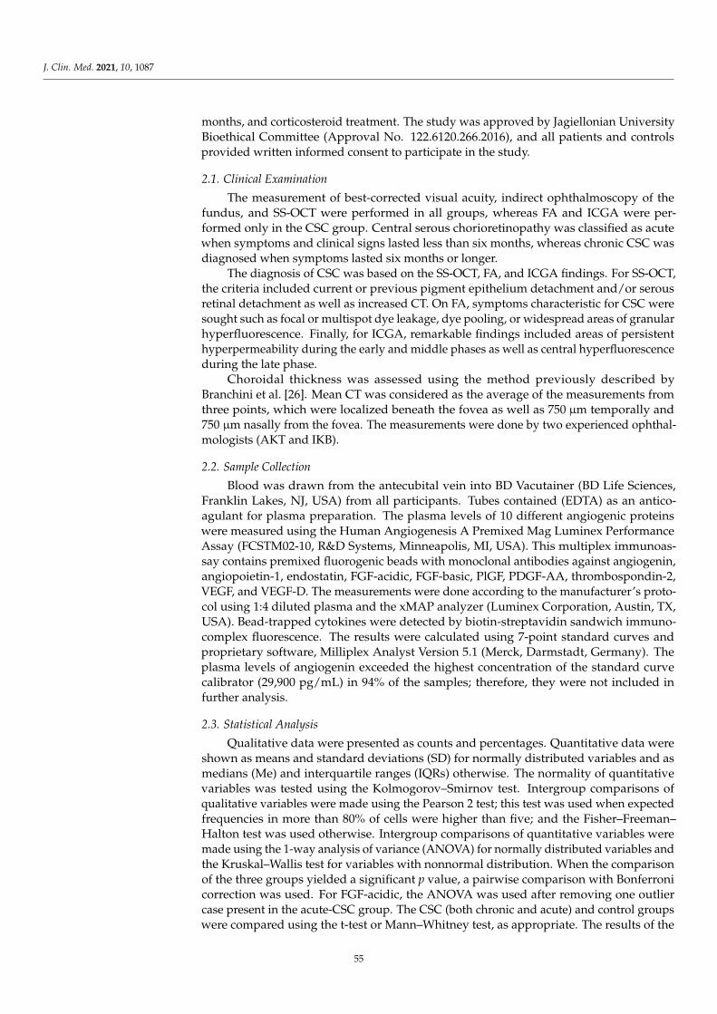
J. Clin. Med. 2021, 10, 1087
months, and corticosteroid treatment. The study was approved by Jagiellonian UniversityBioethical Committee (Approval No. 122.6120.266.2016), and all patients and controlsprovided written informed consent to participate in the study.
2.1. Clinical Examination
The measurement of best-corrected visual acuity, indirect ophthalmoscopy of thefundus, and SS-OCT were performed in all groups, whereas FA and ICGA were per-formed only in the CSC group. Central serous chorioretinopathy was classified as acutewhen symptoms and clinical signs lasted less than six months, whereas chronic CSC wasdiagnosed when symptoms lasted six months or longer.
The diagnosis of CSC was based on the SS-OCT, FA, and ICGA findings. For SS-OCT,the criteria included current or previous pigment epithelium detachment and/or serousretinal detachment as well as increased CT. On FA, symptoms characteristic for CSC weresought such as focal or multispot dye leakage, dye pooling, or widespread areas of granularhyperfluorescence. Finally, for ICGA, remarkable findings included areas of persistenthyperpermeability during the early and middle phases as well as central hyperfluorescenceduring the late phase.
Choroidal thickness was assessed using the method previously described byBranchini et al. [26]. Mean CT was considered as the average of the measurements fromthree points, which were localized beneath the fovea as well as 750 µm temporally and750 µm nasally from the fovea. The measurements were done by two experienced ophthal-mologists (AKT and IKB).
2.2. Sample Collection
Blood was drawn from the antecubital vein into BD Vacutainer (BD Life Sciences,Franklin Lakes, NJ, USA) from all participants. Tubes contained (EDTA) as an antico-agulant for plasma preparation. The plasma levels of 10 different angiogenic proteinswere measured using the Human Angiogenesis A Premixed Mag Luminex PerformanceAssay (FCSTM02-10, R&D Systems, Minneapolis, MI, USA). This multiplex immunoas-say contains premixed fluorogenic beads with monoclonal antibodies against angiogenin,angiopoietin-1, endostatin, FGF-acidic, FGF-basic, PlGF, PDGF-AA, thrombospondin-2,VEGF, and VEGF-D. The measurements were done according to the manufacturer’s proto-col using 1:4 diluted plasma and the xMAP analyzer (Luminex Corporation, Austin, TX,USA). Bead-trapped cytokines were detected by biotin-streptavidin sandwich immuno-complex fluorescence. The results were calculated using 7-point standard curves andproprietary software, Milliplex Analyst Version 5.1 (Merck, Darmstadt, Germany). Theplasma levels of angiogenin exceeded the highest concentration of the standard curvecalibrator (29,900 pg/mL) in 94% of the samples; therefore, they were not included infurther analysis.
2.3. Statistical Analysis
Qualitative data were presented as counts and percentages. Quantitative data wereshown as means and standard deviations (SD) for normally distributed variables and asmedians (Me) and interquartile ranges (IQRs) otherwise. The normality of quantitativevariables was tested using the Kolmogorov–Smirnov test. Intergroup comparisons ofqualitative variables were made using the Pearson 2 test; this test was used when expectedfrequencies in more than 80% of cells were higher than five; and the Fisher–Freeman–Halton test was used otherwise. Intergroup comparisons of quantitative variables weremade using the 1-way analysis of variance (ANOVA) for normally distributed variables andthe Kruskal–Wallis test for variables with nonnormal distribution. When the comparisonof the three groups yielded a significant p value, a pairwise comparison with Bonferronicorrection was used. For FGF-acidic, the ANOVA was used after removing one outliercase present in the acute-CSC group. The CSC (both chronic and acute) and control groupswere compared using the t-test or Mann–Whitney test, as appropriate. The results of the
55
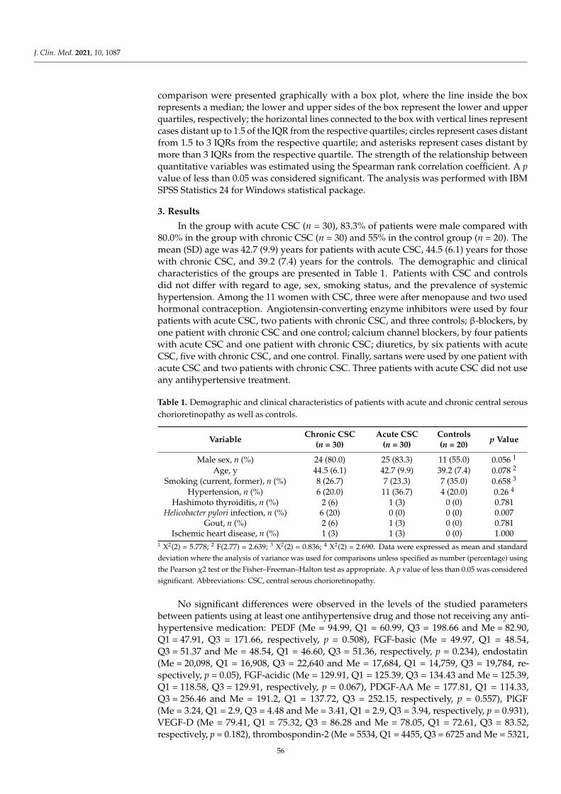
J. Clin. Med. 2021, 10, 1087
comparison were presented graphically with a box plot, where the line inside the boxrepresents a median; the lower and upper sides of the box represent the lower and upperquartiles, respectively; the horizontal lines connected to the box with vertical lines representcases distant up to 1.5 of the IQR from the respective quartiles; circles represent cases distantfrom 1.5 to 3 IQRs from the respective quartile; and asterisks represent cases distant bymore than 3 IQRs from the respective quartile. The strength of the relationship betweenquantitative variables was estimated using the Spearman rank correlation coefficient. A pvalue of less than 0.05 was considered significant. The analysis was performed with IBMSPSS Statistics 24 for Windows statistical package.
3. Results
In the group with acute CSC (n = 30), 83.3% of patients were male compared with80.0% in the group with chronic CSC (n = 30) and 55% in the control group (n = 20). Themean (SD) age was 42.7 (9.9) years for patients with acute CSC, 44.5 (6.1) years for thosewith chronic CSC, and 39.2 (7.4) years for the controls. The demographic and clinicalcharacteristics of the groups are presented in Table 1. Patients with CSC and controlsdid not differ with regard to age, sex, smoking status, and the prevalence of systemichypertension. Among the 11 women with CSC, three were after menopause and two usedhormonal contraception. Angiotensin-converting enzyme inhibitors were used by fourpatients with acute CSC, two patients with chronic CSC, and three controls; β-blockers, byone patient with chronic CSC and one control; calcium channel blockers, by four patientswith acute CSC and one patient with chronic CSC; diuretics, by six patients with acuteCSC, five with chronic CSC, and one control. Finally, sartans were used by one patient withacute CSC and two patients with chronic CSC. Three patients with acute CSC did not useany antihypertensive treatment.
Table 1. Demographic and clinical characteristics of patients with acute and chronic central serouschorioretinopathy as well as controls.
VariableChronic CSC
(n = 30)Acute CSC
(n = 30)Controls(n = 20)
p Value
Male sex, n (%) 24 (80.0) 25 (83.3) 11 (55.0) 0.056 1
Age, y 44.5 (6.1) 42.7 (9.9) 39.2 (7.4) 0.078 2
Smoking (current, former), n (%) 8 (26.7) 7 (23.3) 7 (35.0) 0.658 3
Hypertension, n (%) 6 (20.0) 11 (36.7) 4 (20.0) 0.26 4
Hashimoto thyroiditis, n (%) 2 (6) 1 (3) 0 (0) 0.781Helicobacter pylori infection, n (%) 6 (20) 0 (0) 0 (0) 0.007
Gout, n (%) 2 (6) 1 (3) 0 (0) 0.781Ischemic heart disease, n (%) 1 (3) 1 (3) 0 (0) 1.000
1 X2(2) = 5.778; 2 F(2.77) = 2.639; 3 X2(2) = 0.836; 4 X2(2) = 2.690. Data were expressed as mean and standard
deviation where the analysis of variance was used for comparisons unless specified as number (percentage) using
the Pearson χ2 test or the Fisher–Freeman–Halton test as appropriate. A p value of less than 0.05 was considered
significant. Abbreviations: CSC, central serous chorioretinopathy.
No significant differences were observed in the levels of the studied parametersbetween patients using at least one antihypertensive drug and those not receiving any anti-hypertensive medication: PEDF (Me = 94.99, Q1 = 60.99, Q3 = 198.66 and Me = 82.90,Q1 = 47.91, Q3 = 171.66, respectively, p = 0.508), FGF-basic (Me = 49.97, Q1 = 48.54,Q3 = 51.37 and Me = 48.54, Q1 = 46.60, Q3 = 51.36, respectively, p = 0.234), endostatin(Me = 20,098, Q1 = 16,908, Q3 = 22,640 and Me = 17,684, Q1 = 14,759, Q3 = 19,784, re-spectively, p = 0.05), FGF-acidic (Me = 129.91, Q1 = 125.39, Q3 = 134.43 and Me = 125.39,Q1 = 118.58, Q3 = 129.91, respectively, p = 0.067), PDGF-AA Me = 177.81, Q1 = 114.33,Q3 = 256.46 and Me = 191.2, Q1 = 137.72, Q3 = 252.15, respectively, p = 0.557), PlGF(Me = 3.24, Q1 = 2.9, Q3 = 4.48 and Me = 3.41, Q1 = 2.9, Q3 = 3.94, respectively, p = 0.931),VEGF-D (Me = 79.41, Q1 = 75.32, Q3 = 86.28 and Me = 78.05, Q1 = 72.61, Q3 = 83.52,respectively, p = 0.182), thrombospondin-2 (Me = 5534, Q1 = 4455, Q3 = 6725 and Me = 5321,
56

J. Clin. Med. 2021, 10, 1087
Q1 = 4113, Q3 = 6642, respectively, p = 0.553), angiopoetin-1 (Me = 2356, Q1 = 1129, Q3 = 3760and Me = 2926, Q1 = 1819, Q3 = 3958, respectively, p = 0.224), and VEGF (Me = 12.5,Q1 = 5.2, Q3 = 20.5 and Me = 9.8, Q1 = 6.8, Q3 = 16.8, respectively, p = 0.968).
The baseline ophthalmological characteristics of patients and controls are presentedin Table 2. The groups differed significantly in terms of best-corrected visual acuity andmean CT (Table 2).
Table 2. Ophthalmological characteristics of patients with acute and chronic central serous chori-oretinopathy as well as controls.
VariableChronic CSC
(n = 30)Acute CSC
(n = 30)Controls(n = 20)
p Value
CT, µm 406.1 (88.1) 421.5 (85.3) 317.4 (61.4) <0.001 1
Affected eye, n (%)Right 10 (33.3) 14 (46.7) -
0.225 2Left 12 (40) 13 (43.3) -Both 8 (26.7) 3 (10.0) -
BCVA(logMAR), n (%)0.3< * ≤0.0 23 (76.7) 20 (66.7) 20 (100.0)
0.017 31.0≤ * ≤0.3 7 (23.3) 10 (33.3) 0
* BCVA score. 1 F(2.76) = 10.770; 2 X2(2) = 2.979; 3 X2(2) = 8.092. Data were expressed as mean and standard
deviation where the analysis of variance was used for comparisons unless specified as number (percentage)
using the Pearson χ2 test or the Fisher–Freeman–Halton test as appropriate. A p value of less than 0.05 was
considered significant. Abbreviations: BCVA, best corrected visual acuity; CSC, central serous chorioretinopathy;
CT, choroidal thickness.
At the time of data collection, all patients were treatment naive. Different treatmentmodalities were applied after plasma samples were collected.
Data on the plasma levels of angiogenic factors in patients with acute and chronicCSC as well as controls are shown in Table 3.
Table 3. Plasma levels of angiogenic factors in patients with acute and chronic central serous chorioretinopathy as well ascontrols.
Angiogenic Factor, pg/mLAcute CSC
(n = 30)Chronic CSC
(n = 30)Controls(n = 20)
p Value
PEDF 94.99 (47.91–200.92) 82.89 (45.73–155.99) 74.12 (54.45–159.35) 0.80 1
FGF-basic 48.97 (2.74) 48.97 (4.71) 50.49 (3.11) 0.28 2
Endostatin 18,009.97 (4466.25) 18,241.17 (4524.54) 16,854.20 (3182.28) 0.49 3
FGF-acid 127.40 (7.92) 125.20 (8.11) 126.08 (8.20) 0.58 4
PDGF-AA 179.04 (87.66) 200.23 (143.25) 265.19 (97.72) 0.03 5
PlGF 3.40 (0.76) 3.44 (0.91) 3.99 (0.96) 0.045 6
VEGF-D 78.62 (5.88) 76.93 (9.66) 77.94 (6.99) 0.70 7
Trombospondin-2 4661.00 (4070.00–6002.00) 5478.00 (5420.00–6933.00) 5898.00 (4750.00–6857.00) 0.07 8
Angiopoietin-1 2894.00 (1722.00–3760.00) 2373.00 (1321.00–2943.00) 3982.00 (2411.00–5617.00) 0.01 9
VEGF 8.80 (6.00-14.40) 9.40 (6.60-13.10) 14.50 (8.60-23.00) 0.07 10
1 X2(2) = 0.447; 2 F(2.77) = 1.275; 3 F(2.77) = 0.707; 4 F(2.76) = 0.553; 5 F(2.77) = 3.558; 6 F(2.77) = 3.230; 7 F(2.77) = 0.359; 8 X2(2) = 5.248;9 X2(2) = 9.656; 10 X2(2) = 5.091. Data are shown as mean and standard deviation where the analysis of variance was used for comparisonsor as median (interquartile range) using the Kruskal–Wallis test for comparison. A p value of less than 0.05 was considered significant.Abbreviations: CSC, central serous chorioretinopathy; FGF, fibroblast growth factor; PEDF, pigment epithelium–derived factor; PDGF,platelet-derived growth factor; PlGF, placental growth factor; VEGF, vascular endothelial growth factor.
Intergroup comparisons of the plasma levels of the 10 measured angiogenic factorsrevealed significant differences between the study groups for angiopoietin-1, PlGF, andPDGF-AA (Figure 1a–c). There were no differences in the levels of the remaining antiangio-genic factors among the three groups (p > 0.05).
57
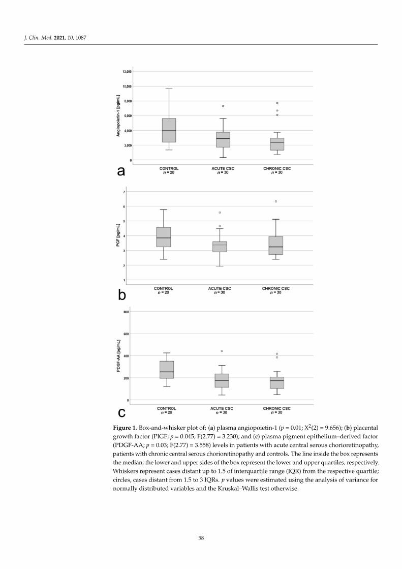
J. Clin. Med. 2021, 10, 1087
Figure 1. Box-and-whisker plot of: (a) plasma angiopoietin-1 (p = 0.01; X2(2) = 9.656); (b) placentalgrowth factor (PlGF; p = 0.045; F(2.77) = 3.230); and (c) plasma pigment epithelium–derived factor(PDGF-AA; p = 0.03; F(2.77) = 3.558) levels in patients with acute central serous chorioretinopathy,patients with chronic central serous chorioretinopathy and controls. The line inside the box representsthe median; the lower and upper sides of the box represent the lower and upper quartiles, respectively.Whiskers represent cases distant up to 1.5 of interquartile range (IQR) from the respective quartile;circles, cases distant from 1.5 to 3 IQRs. p values were estimated using the analysis of variance fornormally distributed variables and the Kruskal–Wallis test otherwise.
58
J. Clin. Med. 2021, 10, 1087
Subsequent pairwise comparisons showed that in patients with acute CSC (n = 30),plasma PDGF-AA levels were significantly lower than in the controls (n = 20) (Figure 2),whereas in patients with chronic CSC (n = 30), angiopoietin-1 levels were significantlylower than in the controls (n = 20) (Figure 3).
Figure 2. Box-and-whisker plot of plasma pigment epithelium–derived factor (PDGF-AA) levels inpatients with acute central serous chorioretinopathy and controls (p = 0.002; t(48) = 3.252). The lineinside the box represents the median; the lower and upper sides of the box represent the lower andupper quartiles, respectively. Whiskers represent cases distant up to 1.5 of interquartile range (IQR)from the respective quartile; circles, cases distant from 1.5 to 3 IQRs. p values were estimated usingthe analysis of variance post-hoc Bonferroni test.
Figure 3. Box-and-whisker plot of plasma angiopoietin-1 levels in patients with chronic centralserous chorioretinopathy and controls (p = 0.007; X2(2) = 9.656; post hoc test statistics = 20.3). Theline inside the box represents the median; the lower and upper sides of the box represent the lowerand upper quartiles, respectively. Whiskers represent cases distant up to 1.5 of interquartile range(IQR) from the respective quartile; circles, cases distant from 1.5 to 3 IQRs. p values were estimatedusing the post-hoc pairwise comparison for the Kruskal–Wallis test with Bonferroni correction.
Additionally, we performed an analysis for the whole CSC group (patients with bothchronic and acute form, n = 60) vs. controls (n = 20). Plasma VEGF, PDGF-AA, and PlGFlevels in patients with acute or chronic CSC (n = 60) were significantly lower than in thecontrols (Figure 4a–c).
59

J. Clin. Med. 2021, 10, 1087
Figure 4. Box-and-whisker plot of: (a) plasma vascular endothelial growth factor (VEGF; p = 0.024);(b) plasma placental growth factor (PlGF; p = 0.013; t(48)= −2.551); (c) platelet-derived growth factorAA (PDGF-AA; p = 0.012; t(48) = −2.577) levels in all patients with central serous chorioretinopathy(acute and chronic) and controls. The line inside the box represents the median; the lower and uppersides of the box represent the lower and upper quartiles, respectively. Whiskers represent casesdistant up to 1.5 of interquartile range (IQR) from the respective quartile; circles, cases distant from1.5 to 3 IQRs. p values were estimated using the t-test for normally distributed variables and theMann–Whitney test otherwise.
60
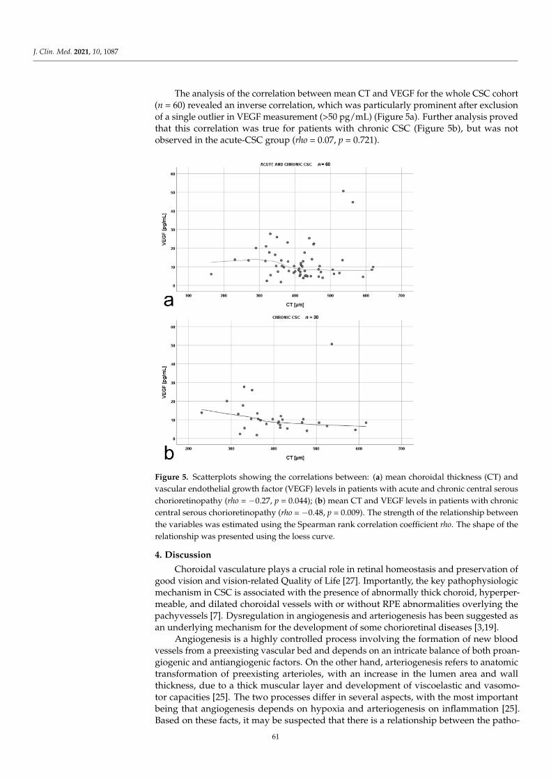
J. Clin. Med. 2021, 10, 1087
The analysis of the correlation between mean CT and VEGF for the whole CSC cohort(n = 60) revealed an inverse correlation, which was particularly prominent after exclusionof a single outlier in VEGF measurement (>50 pg/mL) (Figure 5a). Further analysis provedthat this correlation was true for patients with chronic CSC (Figure 5b), but was notobserved in the acute-CSC group (rho = 0.07, p = 0.721).
Figure 5. Scatterplots showing the correlations between: (a) mean choroidal thickness (CT) andvascular endothelial growth factor (VEGF) levels in patients with acute and chronic central serouschorioretinopathy (rho = −0.27, p = 0.044); (b) mean CT and VEGF levels in patients with chroniccentral serous chorioretinopathy (rho = −0.48, p = 0.009). The strength of the relationship betweenthe variables was estimated using the Spearman rank correlation coefficient rho. The shape of therelationship was presented using the loess curve.
4. Discussion
Choroidal vasculature plays a crucial role in retinal homeostasis and preservation ofgood vision and vision-related Quality of Life [27]. Importantly, the key pathophysiologicmechanism in CSC is associated with the presence of abnormally thick choroid, hyperper-meable, and dilated choroidal vessels with or without RPE abnormalities overlying thepachyvessels [7]. Dysregulation in angiogenesis and arteriogenesis has been suggested asan underlying mechanism for the development of some chorioretinal diseases [3,19].
Angiogenesis is a highly controlled process involving the formation of new bloodvessels from a preexisting vascular bed and depends on an intricate balance of both proan-giogenic and antiangiogenic factors. On the other hand, arteriogenesis refers to anatomictransformation of preexisting arterioles, with an increase in the lumen area and wallthickness, due to a thick muscular layer and development of viscoelastic and vasomo-tor capacities [25]. The two processes differ in several aspects, with the most importantbeing that angiogenesis depends on hypoxia and arteriogenesis on inflammation [25].Based on these facts, it may be suspected that there is a relationship between the patho-
61
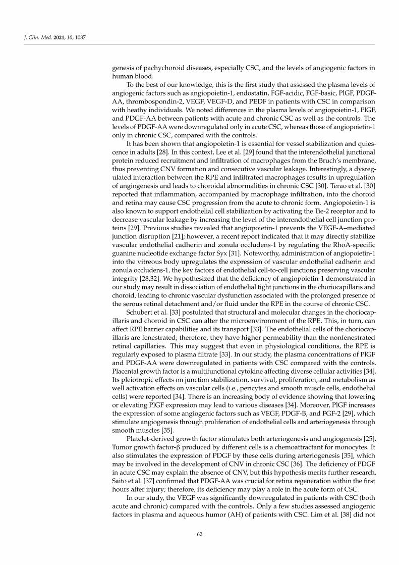
J. Clin. Med. 2021, 10, 1087
genesis of pachychoroid diseases, especially CSC, and the levels of angiogenic factors inhuman blood.
To the best of our knowledge, this is the first study that assessed the plasma levels ofangiogenic factors such as angiopoietin-1, endostatin, FGF-acidic, FGF-basic, PlGF, PDGF-AA, thrombospondin-2, VEGF, VEGF-D, and PEDF in patients with CSC in comparisonwith heathy individuals. We noted differences in the plasma levels of angiopoietin-1, PlGF,and PDGF-AA between patients with acute and chronic CSC as well as the controls. Thelevels of PDGF-AA were downregulated only in acute CSC, whereas those of angiopoietin-1only in chronic CSC, compared with the controls.
It has been shown that angiopoietin-1 is essential for vessel stabilization and quies-cence in adults [28]. In this context, Lee et al. [29] found that the interendothelial junctionalprotein reduced recruitment and infiltration of macrophages from the Bruch’s membrane,thus preventing CNV formation and consecutive vascular leakage. Interestingly, a dysreg-ulated interaction between the RPE and infiltrated macrophages results in upregulationof angiogenesis and leads to choroidal abnormalities in chronic CSC [30]. Terao et al. [30]reported that inflammation, accompanied by macrophage infiltration, into the choroidand retina may cause CSC progression from the acute to chronic form. Angiopoietin-1 isalso known to support endothelial cell stabilization by activating the Tie-2 receptor and todecrease vascular leakage by increasing the level of the interendothelial cell junction pro-teins [29]. Previous studies revealed that angiopoietin-1 prevents the VEGF-A–mediatedjunction disruption [21]; however, a recent report indicated that it may directly stabilizevascular endothelial cadherin and zonula occludens-1 by regulating the RhoA-specificguanine nucleotide exchange factor Syx [31]. Noteworthy, administration of angiopoietin-1into the vitreous body upregulates the expression of vascular endothelial cadherin andzonula occludens-1, the key factors of endothelial cell-to-cell junctions preserving vascularintegrity [28,32]. We hypothesized that the deficiency of angiopoietin-1 demonstrated inour study may result in dissociation of endothelial tight junctions in the choriocapillaris andchoroid, leading to chronic vascular dysfunction associated with the prolonged presence ofthe serous retinal detachment and/or fluid under the RPE in the course of chronic CSC.
Schubert et al. [33] postulated that structural and molecular changes in the choriocap-illaris and choroid in CSC can alter the microenvironment of the RPE. This, in turn, canaffect RPE barrier capabilities and its transport [33]. The endothelial cells of the choriocap-illaris are fenestrated; therefore, they have higher permeability than the nonfenestratedretinal capillaries. This may suggest that even in physiological conditions, the RPE isregularly exposed to plasma filtrate [33]. In our study, the plasma concentrations of PlGFand PDGF-AA were downregulated in patients with CSC compared with the controls.Placental growth factor is a multifunctional cytokine affecting diverse cellular activities [34].Its pleiotropic effects on junction stabilization, survival, proliferation, and metabolism aswell activation effects on vascular cells (i.e., pericytes and smooth muscle cells, endothelialcells) were reported [34]. There is an increasing body of evidence showing that loweringor elevating PlGF expression may lead to various diseases [34]. Moreover, PlGF increasesthe expression of some angiogenic factors such as VEGF, PDGF-B, and FGF-2 [29], whichstimulate angiogenesis through proliferation of endothelial cells and arteriogenesis throughsmooth muscles [35].
Platelet-derived growth factor stimulates both arteriogenesis and angiogenesis [25].Tumor growth factor-β produced by different cells is a chemoattractant for monocytes. Italso stimulates the expression of PDGF by these cells during arteriogenesis [35], whichmay be involved in the development of CNV in chronic CSC [36]. The deficiency of PDGFin acute CSC may explain the absence of CNV, but this hypothesis merits further research.Saito et al. [37] confirmed that PDGF-AA was crucial for retina regeneration within the firsthours after injury; therefore, its deficiency may play a role in the acute form of CSC.
In our study, the VEGF was significantly downregulated in patients with CSC (bothacute and chronic) compared with the controls. Only a few studies assessed angiogenicfactors in plasma and aqueous humor (AH) of patients with CSC. Lim et al. [38] did not
62
J. Clin. Med. 2021, 10, 1087
show any differences in plasma and AH levels of VEGF in CSC compared with the controls.However, the study was limited by a small group of patients, mostly with acute CSC, whichprecluded a definitive conclusion. Shin et al. [39] reported similar, but very low, AH levelsof VEGF in patients with CSC and the controls, but PDGF levels were lower in CSC.
Downregulated plasma VEGF levels in CSC compared with the healthy individu-als observed in our study may partially explain an unsatisfactory effect of intravitrealanti-VEGF treatment in patients with CSC [2,40]. Even though CSC is described as avascular disorder (pachychoroid), the primary exudative component leading to a maculardetachment is considered to be nonvasogenic, which means that it does not result directlyfrom the proliferation of choroidal vessels [8,41]. This is an essential difference betweenCSC and other conditions presenting with serous macular detachment and CNV such asnAMD [8,41,42].
The fact that chronic CSC can be associated with the presence of CNV [16,17] doesnot contradict the results of our study, which revealed lower plasma VEGF levels in CSCpatients compared with the controls. This is in line with the studies by Spaide et al. [36] andSacconi et al. [43]. They hypothesized that CNV in CSC occurs as a result of proliferationof new vessels during arteriogenesis, which is characterized by dilation of the existingvascular channels and is independent of VEGF (unlike angiogenesis, which is highly VEGFdependent) [44]. On the other hand, VEGF alters the junctional integrity, downregulatesthe expression of occludin and zonula occludens-1 [45–48], which results in increasedpermeability and angiogenesis [15,49]. Downregulated VEGF levels observed in our studymay suggest that the mechanism of vascular hyperpermeability in CSC is similar to thatof CNV in VEGF independent CSC and may result from altered flow in dilated choroidalvessels (pachyvessels) [7]. The increased flow leads to endothelial cell proliferation, withluminar expansion and release of platelet endothelial cell adhesion molecule-1, monocytechemoattractant protein-1, intracellular adhesion molecule-1, and vascular cell adhesionmolecule-1 [50]. As a result, increased endothelial permeability, as indicated by the leakageof plasma proteins, erythrocytes, and platelets into the vascular wall and the adherence ofmonocytes to the endothelium, was observed, along with the recruitment of circulatingmonocytes and resident macrophages [51]. This, in turn, promotes arteriogenesis by theability of monocytes and macrophages to secrete metalloproteinases, chemokines, andgrowth factors [25,52].
We hypothesized that the impaired function of RPE cells in CSC, a major source ofocular proangiogenic proteins, may result in decreased intraocular levels of VEGF, but itdoes not fully explain the downregulation of the other analyzed proangiogenic factors inthe eyes, especially in plasma. This issue merits further investigation.
In the current study, we did not find differences in the levels of antiangiogenic proteins,trombospondin-2, and endostatin between patients with CSC and the controls. It is wellknown that antihypertensive drugs affect angiogenesis [53,54]. However, in our study, wedid not observe any differences in the levels of proangiogenic and antiangiogenic factorsbetween patients using at least one antihypertensive drug and those not receiving anymedication. This finding requires further research.
In our study, an inverse correlation between mean CT and VEGF was observedin chronic CSC. It was shown that a decrease in VEGF levels in intraocular fluids as aresult of intravitreal anti-VEGF therapy leads to a decrease in CT in patients with diabeticretinopathy [55]. Furthermore, some studies reported a decrease in CT in patients withacute CSC after anti-VEGF treatment [56]. These data suggest that certain levels of VEGFare necessary to maintain choroidal stability and function, which is also in line with ourresults. Nevertheless, this is an interesting phenomenon that definitely deserves a moresubstantial discussion and more thorough research.
Our study was limited by the fact that it was performed at a single time point andonly plasma, and not AH, samples were investigated. Moreover, there were possibleconfounding factors affecting the levels of angiogenic factors such as concomitant anti-hypertensive treatment. Finally, during blood sample collection, we did not record the
63
J. Clin. Med. 2021, 10, 1087
stage of the menstruation cycle in women at reproductive age, while menstruation cycleis known to affect angiogenic markers. Although the lack of AH assessment may be animportant limitation, the significant differences in the plasma levels of angiogenic factorsare an interesting finding, indicating that CSC might be a systemic disease.
5. Conclusions
Downregulated angiopoietin-1, VEGF, PDGF-AA, and PlGF levels observed in ourstudy may highlight the previously unknown role of the imbalanced levels of proan-giogenic and antiangiogenic factors, which affect choroidal hyperpermeability and exertprofound changes in venular structure and function, thus possibly contributing to thepathogenesis of CSC. Lower plasma levels of VEGF in patients with CSC may supportthe hypothesis that CNV occurs in these patients as a result of arteriogenesis, which isless VEGF-dependent than angiogenesis. Moreover, our findings may support the searchfor new therapeutic strategies in CSC. Nevertheless, the underlying mechanisms thatcontribute to this condition remain largely unknown and require further studies.
Author Contributions: Conceptualization, I.K.-B. and M.S.; Methodology, I.K.-B. and M.S.; Software,M.G.; Validation, I.K.-B. and B.R.-D.; Formal analysis, I.K.-B. and W.P.-M.; Investigation, I.K.-B.,W.P.-M., M.C. and A.K.-T.; Resources, I.K.-B., W.P.-M. and M.D.-K.; Data curation, I.K.-B.; Writing—original draft preparation, I.K.-B., W.P.-M. and M.D.-K.; Writing—review and editing, M.G. andM.S.; Visualization, I.K.-B., W.P.-M. and M.C.; Supervision, B.R.-D. and M.S.; Project administration,W.P.-M.; Funding acquisition, I.K.-B. All authors have read and agreed to the published version ofthe manuscript.
Funding: This research was funded by Jagiellonian University Medical College, Krakow, Poland,grant no. K/ZDS/006283; to I.K.-B.). The APC was funded by the authors.
Institutional Review Board Statement: The study was conducted according to the guidelines of theDeclaration of Helsinki and approved by the Ethics Committee of Jagiellonian University in Krakow,Poland (protocol codes 122.6120.266.2016; 27 October 2016).
Informed Consent Statement: Informed consent was obtained from all subjects involved in the study.
Data Availability Statement: Not applicable.
Acknowledgments: Not applicable.
Conflicts of Interest: The authors declare no conflict of interest.
References
1. Van Rijssen, T.J.; van Dijk, E.H.C.; Yzer, S.; Ohno-Matsui, K.; Keunen, J.E.E.; Schlingemann, R.O.; Sivaprasad, S.; Querques, G.;Downes, S.M.; Fauser, S.; et al. Central serous chorioretinopathy: Towards an evidence-based treatment guideline. Prog. Retin.
Eye Res. 2019, 100770. [CrossRef]2. Chung, Y.-R.; Seo, E.J.; Lew, H.M.; Lee, K.H. Lack of positive effect of intravitreal bevacizumab in central serous chorioretinopathy:
Meta-analysis and review. Eye Lond. Engl. 2013, 27, 1339–1346. [CrossRef]3. Sartini, F.; Figus, M.; Nardi, M.; Casini, G.; Posarelli, C. Non-resolving, recurrent and chronic central serous chorioretinopathy:
Available treatment options. Eye Lond. Engl. 2019, 33, 1035–1043. [CrossRef]4. Vilela, M.; Mengue, C. Central Serous Chorioretinopathy Classification. Pharmaceuticals 2020, 14, 26. [CrossRef]5. Daruich, A.; Matet, A.; Dirani, A.; Bousquet, E.; Zhao, M.; Farman, N.; Jaisser, F.; Behar-Cohen, F. Central serous chorioretinopathy:
Recent findings and new physiopathology hypothesis. Prog. Retin. Eye Res. 2015, 48, 82–118. [CrossRef] [PubMed]6. Gawecki, M.; Jaszczuk-Maciejewska, A.; Jurska-Jasko, A.; Kneba, M.; Grzybowski, A. Impairment of visual acuity and retinal
morphology following resolved chronic central serous chorioretinopathy. BMC Ophthalmol. 2019, 19, 160. [CrossRef] [PubMed]7. Cheung, C.M.G.; Lee, W.K.; Koizumi, H.; Dansingani, K.; Lai, T.Y.Y.; Freund, K.B. Pachychoroid disease. Eye Lond. Engl. 2019, 33,
14–33. [CrossRef] [PubMed]8. Sakurada, Y.; Leong, B.C.S.; Parikh, R.; Fragiotta, S.; Freund, K.B. Association between choroidal caverns and choroidal vascular
hyperpermeability in eyes with pachychoroid diseases. Retina Phila. Pa 2018, 38, 1977–1983. [CrossRef]9. Karska-Basta, I.; Pociej-Marciak, W.; Chrzaszcz, M.; Wilanska, J.; Jager, M.J.; Markiewicz, A.; Romanowska-Dixon, B.; Sanak, M.;
Kubicka-Trzaska, A. Differences in anti-endothelial and anti-retinal antibody titers: Implications for the pathohysiology of acuteand chronic central serous chorioretinopathy. J. Physiol. Pharmacol. Off. J. Pol. Physiol. Soc. 2020, 71. [CrossRef]
64
J. Clin. Med. 2021, 10, 1087
10. Cheung, C.M.G.; Lai, T.Y.Y.; Ruamviboonsuk, P.; Chen, S.-J.; Chen, Y.; Freund, K.B.; Gomi, F.; Koh, A.H.; Lee, W.-K.; Wong, T.Y.Polypoidal Choroidal Vasculopathy: Definition, Pathogenesis, Diagnosis, and Management. Ophthalmology 2018, 125, 708–724.[CrossRef]
11. Kato, Y.; Oguchi, Y.; Omori, T.; Shintake, H.; Tomita, R.; Kasai, A.; Ogasawara, M.; Sugano, Y.; Itagaki, K.; Ojima, A.; et al.Complement Activation Products and Cytokines in Pachychoroid Neovasculopathy and Neovascular Age-Related MacularDegeneration. Invest. Ophthalmol. Vis. Sci. 2020, 61, 39. [CrossRef]
12. Hosoda, Y.; Yoshikawa, M.; Miyake, M.; Tabara, Y.; Ahn, J.; Woo, S.J.; Honda, S.; Sakurada, Y.; Shiragami, C.; Nakanishi, H.; et al.CFH and VIPR2 as susceptibility loci in choroidal thickness and pachychoroid disease central serous chorioretinopathy. Proc.
Natl. Acad. Sci. USA 2018, 115, 6261–6266. [CrossRef]13. Sartini, F.; Figus, M.; Casini, G.; Nardi, M.; Posarelli, C. Pachychoroid neovasculopathy: A type-1 choroidal neovascularization
belonging to the pachychoroid spectrum-pathogenesis, imaging and available treatment options. Int. Ophthalmol. 2020, 40,3577–3589. [CrossRef]
14. Sartini, F.; Menchini, M.; Posarelli, C.; Casini, G.; Figus, M. Bullous Central Serous Chorioretinopathy: A Rare and Atypical Formof Central Serous Chorioretinopathy. A Systematic Review. Pharmaceuticals 2020, 13, 221. [CrossRef] [PubMed]
15. Karska-Basta, I.; Pociej-Marciak, W.; Chrzaszcz, M.; Kubicka-Trzaska, A.; Romanowska-Dixon, B.; Sanak, M. Altered plasmacytokine levels in acute and chronic central serous chorioretinopathy. Acta Ophthalmol. 2020. [CrossRef] [PubMed]
16. Shiragami, C.; Takasago, Y.; Osaka, R.; Kobayashi, M.; Ono, A.; Yamashita, A.; Hirooka, K. Clinical Features of Central SerousChorioretinopathy With Type 1 Choroidal Neovascularization. Am. J. Ophthalmol. 2018, 193, 80–86. [CrossRef]
17. Mrejen, S.; Balaratnasingam, C.; Kaden, T.R.; Bottini, A.; Dansingani, K.; Bhavsar, K.V.; Yannuzzi, N.A.; Patel, S.; Chen, K.C.;Yu, S.; et al. Long-term Visual Outcomes and Causes of Vision Loss in Chronic Central Serous Chorioretinopathy. Ophthalmology
2019, 126, 576–588. [CrossRef]18. Van Bergen, T.; Etienne, I.; Cunningham, F.; Moons, L.; Schlingemann, R.O.; Feyen, J.H.M.; Stitt, A.W. The role of placental growth
factor (PlGF) and its receptor system in retinal vascular diseases. Prog. Retin. Eye Res. 2019, 69, 116–136. [CrossRef]19. Muether, P.S.; Neuhann, I.; Buhl, C.; Hermann, M.M.; Kirchhof, B.; Fauser, S. Intraocular growth factors and cytokines in patients
with dry and neovascular age-related macular degeneration. Retina Phila. Pa 2013, 33, 1809–1814. [CrossRef] [PubMed]20. Siedlecki, J.; Wertheimer, C.; Wolf, A.; Liegl, R.; Priglinger, C.; Priglinger, S.; Eibl-Lindner, K. Combined VEGF and PDGF
inhibition for neovascular AMD: Anti-angiogenic properties of axitinib on human endothelial cells and pericytes in vitro. Graefes
Arch. Clin. Exp. Ophthalmol. Albrecht Von Graefes Arch. Klin. Exp. Ophthalmol. 2017, 255, 963–972. [CrossRef] [PubMed]21. Gavard, J.; Patel, V.; Gutkind, J.S. Angiopoietin-1 prevents VEGF-induced endothelial permeability by sequestering Src through
mDia. Dev. Cell 2008, 14, 25–36. [CrossRef]22. Nagy, J.A.; Benjamin, L.; Zeng, H.; Dvorak, A.M.; Dvorak, H.F. Vascular permeability, vascular hyperpermeability and angiogene-
sis. Angiogenesis 2008, 11, 109–119. [CrossRef]23. Farnoodian, M.; Wang, S.; Dietz, J.; Nickells, R.W.; Sorenson, C.M.; Sheibani, N. Negative regulators of angiogenesis: Important
targets for treatment of exudative AMD. Clin. Sci. Lond. Engl. 1979 2017, 131, 1763–1780. [CrossRef] [PubMed]24. Kuwano, M.; Fukushi, J.; Okamoto, M.; Nishie, A.; Goto, H.; Ishibashi, T.; Ono, M. Angiogenesis factors. Intern. Med. Tokyo Jpn.
2001, 40, 565–572. [CrossRef] [PubMed]25. Rizzi, A.; Benagiano, V.; Ribatti, D. Angiogenesis versus arteriogenesis. Rom. J. Morphol. Embryol. Rev. Roum. Morphol. Embryol.
2017, 58, 15–19.26. Branchini, L.A.; Adhi, M.; Regatieri, C.V.; Nandakumar, N.; Liu, J.J.; Laver, N.; Fujimoto, J.G.; Duker, J.S. Analysis of choroidal
morphologic features and vasculature in healthy eyes using spectral-domain optical coherence tomography. Ophthalmology 2013,120, 1901–1908. [CrossRef] [PubMed]
27. Karska-Basta, I.; Pociej-Marciak, W.; Chrzaszcz, M.; Zuber-Łaskawiec, K.; Sanak, M.; Romanowska-Dixon, B. Quality of life ofpatients with central serous chorioretinopathy—A major cause of vision threat among middle-aged individuals. Arch. Med. Sci.
2020, 16. [CrossRef]28. El-Asrar, M.A.; Elbarbary, N.S.; Ismail, E.A.R.; Bakr, A.A. Circulating angiopoietin-2 levels in children and adolescents with type
1 diabetes mellitus: Relation to carotid and aortic intima-media thickness. Angiogenesis 2016, 19, 421–431. [CrossRef] [PubMed]29. Lee, J.; Park, D.-Y.; Park, D.Y.; Park, I.; Chang, W.; Nakaoka, Y.; Komuro, I.; Yoo, O.-J.; Koh, G.Y. Angiopoietin-1 suppresses
choroidal neovascularization and vascular leakage. Invest. Ophthalmol. Vis. Sci. 2014, 55, 2191–2199. [CrossRef]30. Terao, N.; Koizumi, H.; Kojima, K.; Yamagishi, T.; Nagata, K.; Kitazawa, K.; Yamamoto, Y.; Yoshii, K.; Hiraga, A.; Toda, M.; et al.
Association of Upregulated Angiogenic Cytokines With Choroidal Abnormalities in Chronic Central Serous Chorioretinopathy.Invest. Ophthalmol. Vis. Sci. 2018, 59, 5924–5931. [CrossRef]
31. Ngok, S.P.; Geyer, R.; Liu, M.; Kourtidis, A.; Agrawal, S.; Wu, C.; Seerapu, H.R.; Lewis-Tuffin, L.J.; Moodie, K.L.; Huveldt, D.; et al.VEGF and Angiopoietin-1 exert opposing effects on cell junctions by regulating the Rho GEF Syx. J. Cell Biol. 2012, 199, 1103–1115.[CrossRef]
32. Peters, S.; Cree, I.A.; Alexander, R.; Turowski, P.; Ockrim, Z.; Patel, J.; Boyd, S.R.; Joussen, A.M.; Ziemssen, F.; Hykin, P.G.; et al.Angiopoietin modulation of vascular endothelial growth factor: Effects on retinal endothelial cell permeability. Cytokine 2007, 40,144–150. [CrossRef]
65

J. Clin. Med. 2021, 10, 1087
33. Schubert, C.; Pryds, A.; Zeng, S.; Xie, Y.; Freund, K.B.; Spaide, R.F.; Merriam, J.C.; Barbazetto, I.; Slakter, J.S.; Chang, S.; et al.Cadherin 5 is regulated by corticosteroids and associated with central serous chorioretinopathy. Hum. Mutat. 2014, 35, 859–867.[CrossRef]
34. Dewerchin, M.; Carmeliet, P. PlGF: A multitasking cytokine with disease-restricted activity. Cold Spring Harb. Perspect. Med.
2012, 2. [CrossRef]35. van Royen, N.; Piek, J.J.; Buschmann, I.; Hoefer, I.; Voskuil, M.; Schaper, W. Stimulation of arteriogenesis; a new concept for the
treatment of arterial occlusive disease. Cardiovasc. Res. 2001, 49, 543–553. [CrossRef]36. Spaide, R.F. Optical Coherence Tomography Angiography Signs of Vascular Abnormalization With Antiangiogenic Therapy for
Choroidal Neovascularization. Am. J. Ophthalmol. 2015, 160, 6–16. [CrossRef]37. Saito, M.; Saito, W.; Hirooka, K.; Hashimoto, Y.; Mori, S.; Noda, K.; Ishida, S. Pulse Waveform Changes in Macular Choroidal
Hemodynamics With Regression of Acute Central Serous Chorioretinopathy. Invest. Ophthalmol. Vis. Sci. 2015, 56, 6515–6522.[CrossRef]
38. Lim, J.W.; Kim, M.U.; Shin, M.-C. Aqueous humor and plasma levels of vascular endothelial growth factor and interleukin-8 inpatients with central serous chorioretinopathy. Retina Phila. Pa 2010, 30, 1465–1471. [CrossRef]
39. Shin, M.C.; Lim, J.W. Concentration of cytokines in the aqueous humor of patients with central serous chorioretinopathy. Retina
Phila. Pa 2011, 31, 1937–1943. [CrossRef]40. Ji, S.; Wei, Y.; Chen, J.; Tang, S. Clinical efficacy of anti-VEGF medications for central serous chorioretinopathy: A meta-analysis.
Int. J. Clin. Pharm. 2017, 39, 514–521. [CrossRef]41. Spaide, R.F.; Koizumi, H.; Pozzoni, M.C.; Pozonni, M.C. Enhanced depth imaging spectral-domain optical coherence tomography.
Am. J. Ophthalmol. 2008, 146, 496–500. [CrossRef]42. Zuber-Łaskawiec, K.; Kubicka-Trzaska, A.; Karska-Basta, I.; Pociej-Marciak, W.; Romanowska-Dixon, B. Non-Responsiveness and
Tachyphylaxis to Anti-Vascular Endothelial Growth Factor Treatment in Naive Patients with Exudative Age-Related MacularDegeneration. Available online: https://pubmed.ncbi.nlm.nih.gov/32009630/ (accessed on 2 December 2020).
43. Sacconi, R.; Tomasso, L.; Corbelli, E.; Carnevali, A.; Querques, L.; Casati, S.; Bandello, F.; Querques, G. Early response to thetreatment of choroidal neovascularization complicating central serous chorioretinopathy: A OCT-angiography study. Eye Lond.
Engl. 2019, 33, 1809–1817. [CrossRef] [PubMed]44. Schierling, W.; Troidl, K.; Troidl, C.; Schmitz-Rixen, T.; Schaper, W.; Eitenmüller, I.K. The role of angiogenic growth factors in
arteriogenesis. J. Vasc. Res. 2009, 46, 365–374. [CrossRef] [PubMed]45. Behzadian, M.A.; Windsor, L.J.; Ghaly, N.; Liou, G.; Tsai, N.-T.; Caldwell, R.B. VEGF-induced paracellular permeability in cultured
endothelial cells involves urokinase and its receptor. FASEB J. Off. Publ. Fed. Am. Soc. Exp. Biol. 2003, 17, 752–754. [CrossRef][PubMed]
46. Liu, X.; Dreffs, A.; Díaz-Coránguez, M.; Runkle, E.A.; Gardner, T.W.; Chiodo, V.A.; Hauswirth, W.W.; Antonetti, D.A. OccludinS490 Phosphorylation Regulates Vascular Endothelial Growth Factor-Induced Retinal Neovascularization. Am. J. Pathol. 2016,186, 2486–2499. [CrossRef] [PubMed]
47. Murakami, T.; Frey, T.; Lin, C.; Antonetti, D.A. Protein kinase cβ phosphorylates occludin regulating tight junction trafficking invascular endothelial growth factor-induced permeability in vivo. Diabetes 2012, 61, 1573–1583. [CrossRef] [PubMed]
48. Yun, J.-H.; Park, S.W.; Kim, K.-J.; Bae, J.-S.; Lee, E.H.; Paek, S.H.; Kim, S.U.; Ye, S.; Kim, J.-H.; Cho, C.-H. Endothelial STAT3Activation Increases Vascular Leakage Through Downregulating Tight Junction Proteins: Implications for Diabetic Retinopathy. J.
Cell. Physiol. 2017, 232, 1123–1134. [CrossRef]49. Mesquida, M.; Leszczynska, A.; Llorenç, V.; Adán, A. Interleukin-6 blockade in ocular inflammatory diseases. Clin. Exp. Immunol.
2014, 176, 301–309. [CrossRef]50. Chen, Z.; Rubin, J.; Tzima, E. Role of PECAM-1 in arteriogenesis and specification of preexisting collaterals. Circ. Res. 2010, 107,
1355–1363. [CrossRef]51. Takeda, Y.; Costa, S.; Delamarre, E.; Roncal, C.; Leite de Oliveira, R.; Squadrito, M.L.; Finisguerra, V.; Deschoemaeker, S.;
Bruyère, F.; Wenes, M.; et al. Macrophage skewing by Phd2 haplodeficiency prevents ischaemia by inducing arteriogenesis.Nature 2011, 479, 122–126. [CrossRef]
52. Galis, Z.S.; Sukhova, G.K.; Libby, P. Microscopic localization of active proteases by in situ zymography: Detection of matrixmetalloproteinase activity in vascular tissue. FASEB J. Off. Publ. Fed. Am. Soc. Exp. Biol. 1995, 9, 974–980. [CrossRef]
53. Humar, R.; Zimmerli, L.; Battegay, E. Angiogenesis and hypertension: An update. J. Hum. Hypertens. 2009, 23, 773–782. [CrossRef]54. Pociej-Marciak, W.; Karska-Basta, I.; Kuzniewski, M.; Kubicka-Trzaska, A.; Romanowska-Dixon, B. Sudden visual deterioration
as a first symptom of chronic kidney failure. Case Rep Ophtalmol. 2015, 28, 394–400. [CrossRef]55. Kniggendorf, V.F.; Novais, E.A.; Kniggendorf, S.L.; Xavier, C.; Cole, E.D.; Regatieri, C.V. Effect of intravitreal anti-VEGF on
choroidal thickness in patients with diabetic macular edema using spectral domain OCT. Arq. Bras. Oftalmol. 2016, 79, 155–158.[CrossRef]
56. Kim, D.Y.; Joe, S.G.; Yang, H.S.; Lee, J.Y.; Kim, J.-G.; Yoon, Y.H. Subfoveal choroidal thickness changes in treated idiopathic centralserous chorioretinopathy and their association with recurrence. Retina Phila. Pa 2015, 35, 1867–1874. [CrossRef]
66

Journal of
Clinical Medicine
Article
Idiopathic Peripheral Retinal Telangiectasia in Adults: A CaseSeries and Literature Review
Maciej Gawecki
�����������������
Citation: Gawecki, M. Idiopathic
Peripheral Retinal Telangiectasia in
Adults: A Case Series and Literature
Review. J. Clin. Med. 2021, 10, 1767.
https://doi.org/10.3390/jcm10081767
Academic Editor: Marcel Menke
Received: 6 March 2021
Accepted: 14 April 2021
Published: 19 April 2021
Publisher’s Note: MDPI stays neutral
with regard to jurisdictional claims in
published maps and institutional affil-
iations.
Copyright: © 2021 by the author.
Licensee MDPI, Basel, Switzerland.
This article is an open access article
distributed under the terms and
conditions of the Creative Commons
Attribution (CC BY) license (https://
creativecommons.org/licenses/by/
4.0/).
Dobry Wzrok Ophthalmological Clinic, 80-822 Gdansk, Poland; [email protected]; Tel.: +48-501-788-654
Abstract: Idiopathic peripheral retinal telangiectasia (IPT), often termed as Coats disease, can present
in a milder form with the onset in adulthood. The goal of this case series study and literature
review was to describe and classify different presenting forms and treatment of this entity and to
review contemporary methods of its management. Six cases of adult onset IPT were described with
the following phenotypes based on fundus ophthalmoscopy, fluorescein angiography, and optical
coherence tomography findings: IPT without exudates or foveal involvement, IPT with peripheral
exudates without foveal involvement, IPT with peripheral exudates and cystoid macular edema,
and IPT with peripheral and macular hard exudates. Treatments applied in this series included
observation, laser photocoagulation, and anti-vascular endothelial growth factor (VEGF) treatment
with variable outcomes depending upon the extent of IPT, the aggressiveness of laser treatment, and
the stringency of follow-up. The accompanying literature review suggests that ablative therapies,
especially laser photocoagulation, remain the most effective treatment option in adult-onset IPT, with
anti-VEGF therapy serving as an adjuvant procedure. Close follow-up is necessary to achieve and
maintain reasonable good visual and morphological results.
Keywords: Coats disease; peripheral retinal telangiectasia; laser photocoagulation; anti-VEGF treatment
1. Introduction
Idiopathic peripheral retinal telangiectasia (IPT), usually referred to as Coats disease,has been well-described in the medical literature since its discovery in 1908 [1]. Coatsdisease is an idiopathic condition characterized by telangiectatic and aneurysmal retinalvessels with intraretinal and subretinal exudation and fluid without appreciable retinal orvitreal traction, frequently associated with retinal detachment [2]. The pathogenesis andprogression of that disease depends on the impairment of retinal vasculature. Alterationsof endothelium of retinal vessels lead to breaking of blood-retinal barrier and presence ofabnormal pericytes, causing formation of telangiectasias and closure of some vessels [3,4].Exudation of lipids from the damaged vessels follows and is responsible for retinal detach-ment and cyst formation as well as retinal ischemia due to vessel closure [5]. The process isdescribed as similar to pathogenesis of diabetic retinopathy, but definitely more severe andrapid [6].
Coats disease commonly presents unilaterally with strong male predominance. It hasreportedly occurred in patients ranging from 4 months to 70 years of age, with the peakincidence in the first decade of life [5]. Its severe form (referred to as Coats disease) andmilder form (sometimes termed Leber miliary aneurysms) that commonly present in adults,were initially described as separate entities [7]. Now, they are considered to be variableexpressions of the same disease This explains its adult onset, as milder forms often stayasymptomatic for a long period of time [8,9]. Less severe forms of Coats disease fall intocategory 1, 2A and 2 B of the Shields classification commonly used to describe the stagesof this disease [10]. Nevertheless, the perception of many ophthalmologists is that Coatsdisease is attributed to young children, only, and limited peripheral forms of this clinicalentity are still frequently labeled differently, which causes inconsistencies in classification of
67
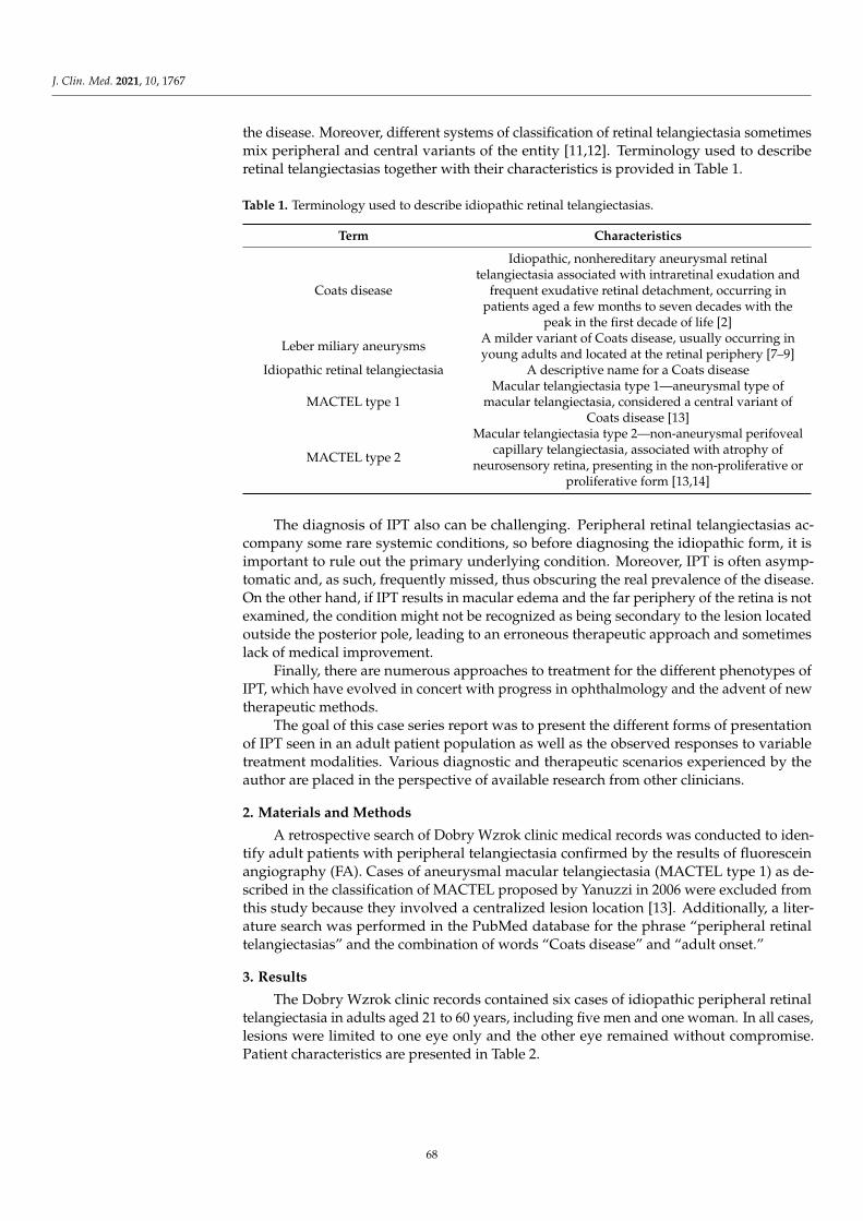
J. Clin. Med. 2021, 10, 1767
the disease. Moreover, different systems of classification of retinal telangiectasia sometimesmix peripheral and central variants of the entity [11,12]. Terminology used to describeretinal telangiectasias together with their characteristics is provided in Table 1.
Table 1. Terminology used to describe idiopathic retinal telangiectasias.
Term Characteristics
Coats disease
Idiopathic, nonhereditary aneurysmal retinaltelangiectasia associated with intraretinal exudation and
frequent exudative retinal detachment, occurring inpatients aged a few months to seven decades with the
peak in the first decade of life [2]
Leber miliary aneurysmsA milder variant of Coats disease, usually occurring inyoung adults and located at the retinal periphery [7–9]
Idiopathic retinal telangiectasia A descriptive name for a Coats disease
MACTEL type 1Macular telangiectasia type 1—aneurysmal type of
macular telangiectasia, considered a central variant ofCoats disease [13]
MACTEL type 2
Macular telangiectasia type 2—non-aneurysmal perifovealcapillary telangiectasia, associated with atrophy of
neurosensory retina, presenting in the non-proliferative orproliferative form [13,14]
The diagnosis of IPT also can be challenging. Peripheral retinal telangiectasias ac-company some rare systemic conditions, so before diagnosing the idiopathic form, it isimportant to rule out the primary underlying condition. Moreover, IPT is often asymp-tomatic and, as such, frequently missed, thus obscuring the real prevalence of the disease.On the other hand, if IPT results in macular edema and the far periphery of the retina is notexamined, the condition might not be recognized as being secondary to the lesion locatedoutside the posterior pole, leading to an erroneous therapeutic approach and sometimeslack of medical improvement.
Finally, there are numerous approaches to treatment for the different phenotypes ofIPT, which have evolved in concert with progress in ophthalmology and the advent of newtherapeutic methods.
The goal of this case series report was to present the different forms of presentationof IPT seen in an adult patient population as well as the observed responses to variabletreatment modalities. Various diagnostic and therapeutic scenarios experienced by theauthor are placed in the perspective of available research from other clinicians.
2. Materials and Methods
A retrospective search of Dobry Wzrok clinic medical records was conducted to iden-tify adult patients with peripheral telangiectasia confirmed by the results of fluoresceinangiography (FA). Cases of aneurysmal macular telangiectasia (MACTEL type 1) as de-scribed in the classification of MACTEL proposed by Yanuzzi in 2006 were excluded fromthis study because they involved a centralized lesion location [13]. Additionally, a liter-ature search was performed in the PubMed database for the phrase “peripheral retinaltelangiectasias” and the combination of words “Coats disease” and “adult onset.”
3. Results
The Dobry Wzrok clinic records contained six cases of idiopathic peripheral retinaltelangiectasia in adults aged 21 to 60 years, including five men and one woman. In all cases,lesions were limited to one eye only and the other eye remained without compromise.Patient characteristics are presented in Table 2.
68
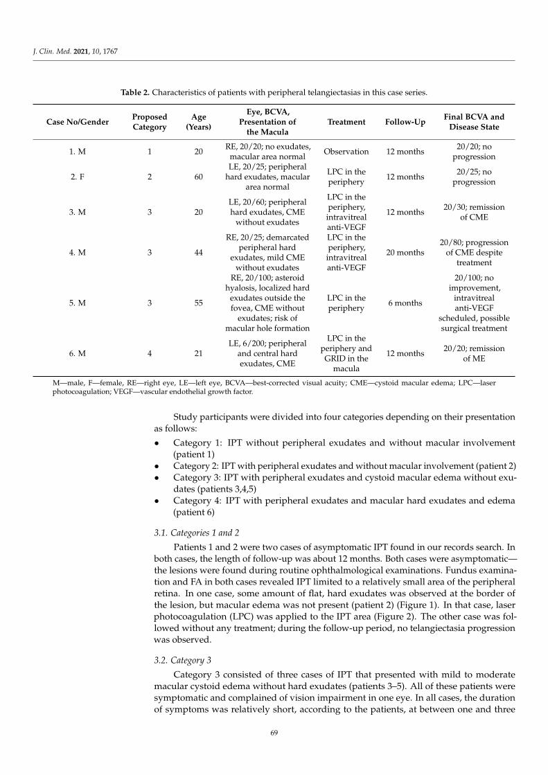
J. Clin. Med. 2021, 10, 1767
Table 2. Characteristics of patients with peripheral telangiectasias in this case series.
Case No/GenderProposedCategory
Age(Years)
Eye, BCVA,Presentation of
the MaculaTreatment Follow-Up
Final BCVA andDisease State
1. M 1 20RE, 20/20; no exudates,
macular area normalObservation 12 months
20/20; noprogression
2. F 2 60LE, 20/25; peripheral
hard exudates, maculararea normal
LPC in theperiphery
12 months20/25; no
progression
3. M 3 20LE, 20/60; peripheralhard exudates, CME
without exudates
LPC in theperiphery,
intravitrealanti-VEGF
12 months20/30; remission
of CME
4. M 3 44
RE, 20/25; demarcatedperipheral hard
exudates, mild CMEwithout exudates
LPC in theperiphery,
intravitrealanti-VEGF
20 months20/80; progression
of CME despitetreatment
5. M 3 55
RE, 20/100; asteroidhyalosis, localized hard
exudates outside thefovea, CME without
exudates; risk ofmacular hole formation
LPC in theperiphery
6 months
20/100; noimprovement,
intravitrealanti-VEGF
scheduled, possiblesurgical treatment
6. M 4 21LE, 6/200; peripheral
and central hardexudates, CME
LPC in theperiphery andGRID in the
macula
12 months20/20; remission
of ME
M—male, F—female, RE—right eye, LE—left eye, BCVA—best-corrected visual acuity; CME—cystoid macular edema; LPC—laserphotocoagulation; VEGF—vascular endothelial growth factor.
Study participants were divided into four categories depending on their presentationas follows:
• Category 1: IPT without peripheral exudates and without macular involvement(patient 1)
• Category 2: IPT with peripheral exudates and without macular involvement (patient 2)• Category 3: IPT with peripheral exudates and cystoid macular edema without exu-
dates (patients 3,4,5)• Category 4: IPT with peripheral exudates and macular hard exudates and edema
(patient 6)
3.1. Categories 1 and 2
Patients 1 and 2 were two cases of asymptomatic IPT found in our records search. Inboth cases, the length of follow-up was about 12 months. Both cases were asymptomatic—the lesions were found during routine ophthalmological examinations. Fundus examina-tion and FA in both cases revealed IPT limited to a relatively small area of the peripheralretina. In one case, some amount of flat, hard exudates was observed at the border ofthe lesion, but macular edema was not present (patient 2) (Figure 1). In that case, laserphotocoagulation (LPC) was applied to the IPT area (Figure 2). The other case was fol-lowed without any treatment; during the follow-up period, no telangiectasia progressionwas observed.
3.2. Category 3
Category 3 consisted of three cases of IPT that presented with mild to moderatemacular cystoid edema without hard exudates (patients 3–5). All of these patients weresymptomatic and complained of vision impairment in one eye. In all cases, the durationof symptoms was relatively short, according to the patients, at between one and three
69
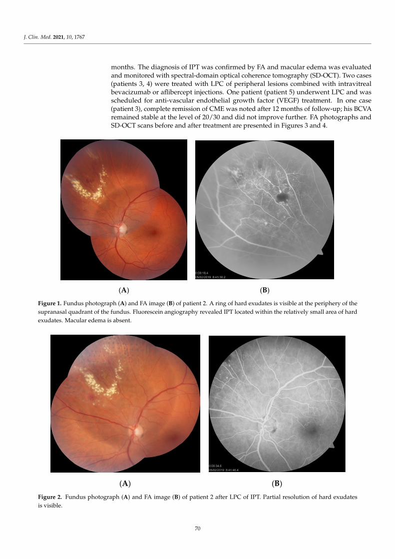
J. Clin. Med. 2021, 10, 1767
months. The diagnosis of IPT was confirmed by FA and macular edema was evaluatedand monitored with spectral-domain optical coherence tomography (SD-OCT). Two cases(patients 3, 4) were treated with LPC of peripheral lesions combined with intravitrealbevacizumab or aflibercept injections. One patient (patient 5) underwent LPC and wasscheduled for anti-vascular endothelial growth factor (VEGF) treatment. In one case(patient 3), complete remission of CME was noted after 12 months of follow-up; his BCVAremained stable at the level of 20/30 and did not improve further. FA photographs andSD-OCT scans before and after treatment are presented in Figures 3 and 4.
(A) (B)
Figure 1. Fundus photograph (A) and FA image (B) of patient 2. A ring of hard exudates is visible at the periphery of thesupranasal quadrant of the fundus. Fluorescein angiography revealed IPT located within the relatively small area of hardexudates. Macular edema is absent.
(A) (B)
Figure 2. Fundus photograph (A) and FA image (B) of patient 2 after LPC of IPT. Partial resolution of hard exudatesis visible.
70
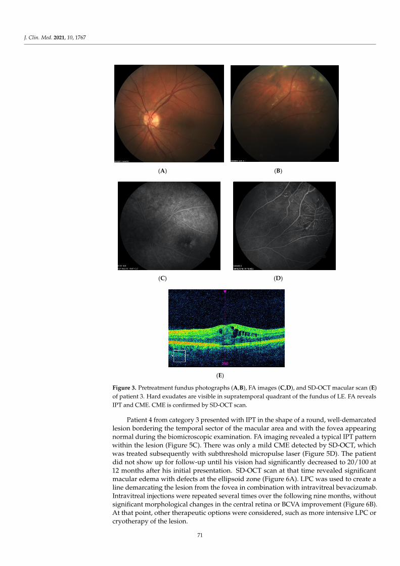
J. Clin. Med. 2021, 10, 1767
(A) (B)
(C) (D)
(E)
Figure 3. Pretreatment fundus photographs (A,B), FA images (C,D), and SD-OCT macular scan (E)of patient 3. Hard exudates are visible in supratemporal quadrant of the fundus of LE. FA revealsIPT and CME. CME is confirmed by SD-OCT scan.
Patient 4 from category 3 presented with IPT in the shape of a round, well-demarcatedlesion bordering the temporal sector of the macular area and with the fovea appearingnormal during the biomicroscopic examination. FA imaging revealed a typical IPT patternwithin the lesion (Figure 5C). There was only a mild CME detected by SD-OCT, whichwas treated subsequently with subthreshold micropulse laser (Figure 5D). The patientdid not show up for follow-up until his vision had significantly decreased to 20/100 at12 months after his initial presentation. SD-OCT scan at that time revealed significantmacular edema with defects at the ellipsoid zone (Figure 6A). LPC was used to create aline demarcating the lesion from the fovea in combination with intravitreal bevacizumab.Intravitreal injections were repeated several times over the following nine months, withoutsignificant morphological changes in the central retina or BCVA improvement (Figure 6B).At that point, other therapeutic options were considered, such as more intensive LPC orcryotherapy of the lesion.
71
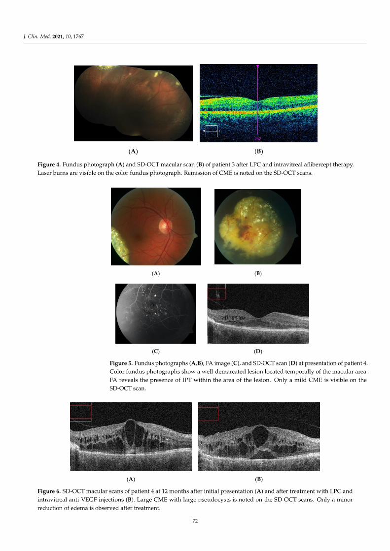
J. Clin. Med. 2021, 10, 1767
(A) (B)
Figure 4. Fundus photograph (A) and SD-OCT macular scan (B) of patient 3 after LPC and intravitreal aflibercept therapy.Laser burns are visible on the color fundus photograph. Remission of CME is noted on the SD-OCT scans.
(A) (B)
(C) (D)
Figure 5. Fundus photographs (A,B), FA image (C), and SD-OCT scan (D) at presentation of patient 4.Color fundus photographs show a well-demarcated lesion located temporally of the macular area.FA reveals the presence of IPT within the area of the lesion. Only a mild CME is visible on theSD-OCT scan.
(A) (B)
Figure 6. SD-OCT macular scans of patient 4 at 12 months after initial presentation (A) and after treatment with LPC andintravitreal anti-VEGF injections (B). Large CME with large pseudocysts is noted on the SD-OCT scans. Only a minorreduction of edema is observed after treatment.
72
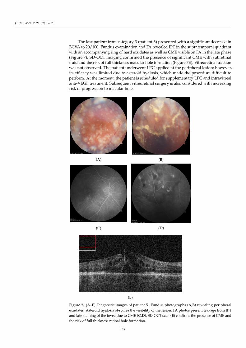
J. Clin. Med. 2021, 10, 1767
The last patient from category 3 (patient 5) presented with a significant decrease inBCVA to 20/100. Fundus examination and FA revealed IPT in the supratemporal quadrantwith an accompanying ring of hard exudates as well as CME visible on FA in the late phase(Figure 7). SD-OCT imaging confirmed the presence of significant CME with subretinalfluid and the risk of full thickness macular hole formation (Figure 7E). Vitreoretinal tractionwas not observed. The patient underwent LPC applied at the peripheral lesion; however,its efficacy was limited due to asteroid hyalosis, which made the procedure difficult toperform. At the moment, the patient is scheduled for supplementary LPC and intravitrealanti-VEGF treatment. Subsequent vitreoretinal surgery is also considered with increasingrisk of progression to macular hole.
(A) (B)
(C) (D)
(E)
Figure 7. (A–E) Diagnostic images of patient 5. Fundus photographs (A,B) revealing peripheralexudates. Asteroid hyalosis obscures the visibility of the lesion. FA photos present leakage from IPTand late staining of the fovea due to CME (C,D). SD-OCT scan (E) confirms the presence of CME andthe risk of full thickness retinal hole formation.
73
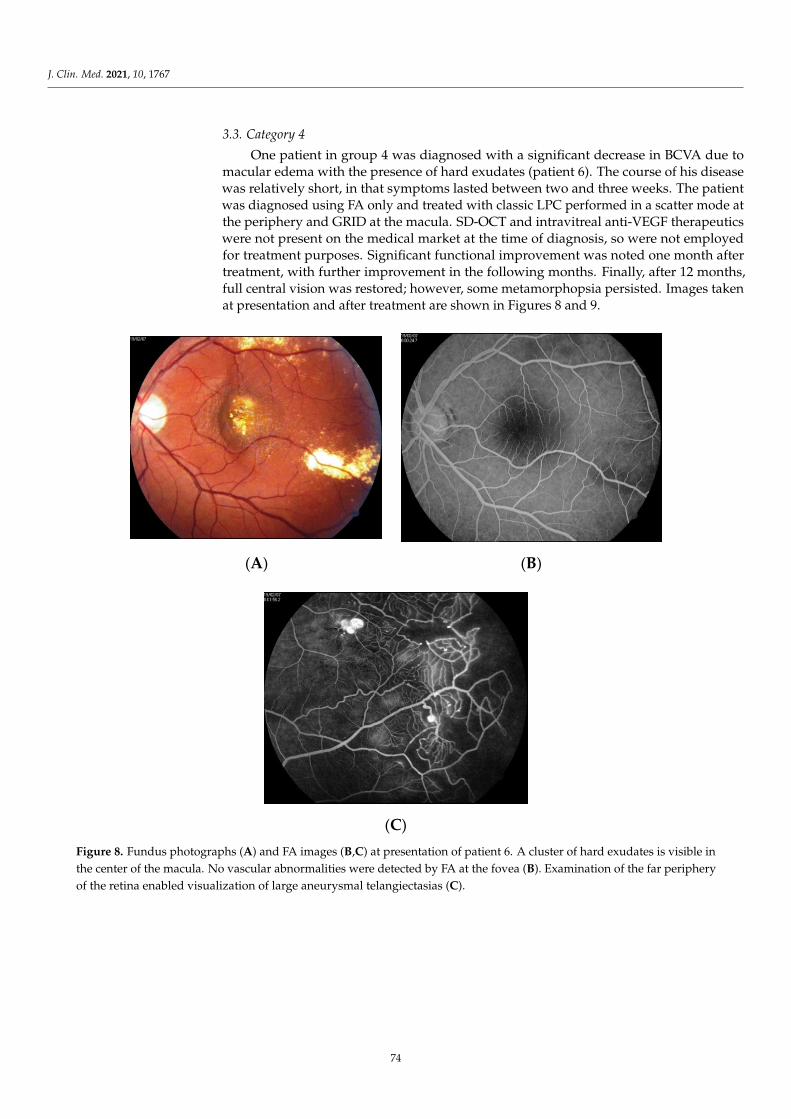
J. Clin. Med. 2021, 10, 1767
3.3. Category 4
One patient in group 4 was diagnosed with a significant decrease in BCVA due tomacular edema with the presence of hard exudates (patient 6). The course of his diseasewas relatively short, in that symptoms lasted between two and three weeks. The patientwas diagnosed using FA only and treated with classic LPC performed in a scatter mode atthe periphery and GRID at the macula. SD-OCT and intravitreal anti-VEGF therapeuticswere not present on the medical market at the time of diagnosis, so were not employedfor treatment purposes. Significant functional improvement was noted one month aftertreatment, with further improvement in the following months. Finally, after 12 months,full central vision was restored; however, some metamorphopsia persisted. Images takenat presentation and after treatment are shown in Figures 8 and 9.
(A) (B)
(C)
Figure 8. Fundus photographs (A) and FA images (B,C) at presentation of patient 6. A cluster of hard exudates is visible inthe center of the macula. No vascular abnormalities were detected by FA at the fovea (B). Examination of the far peripheryof the retina enabled visualization of large aneurysmal telangiectasias (C).
74
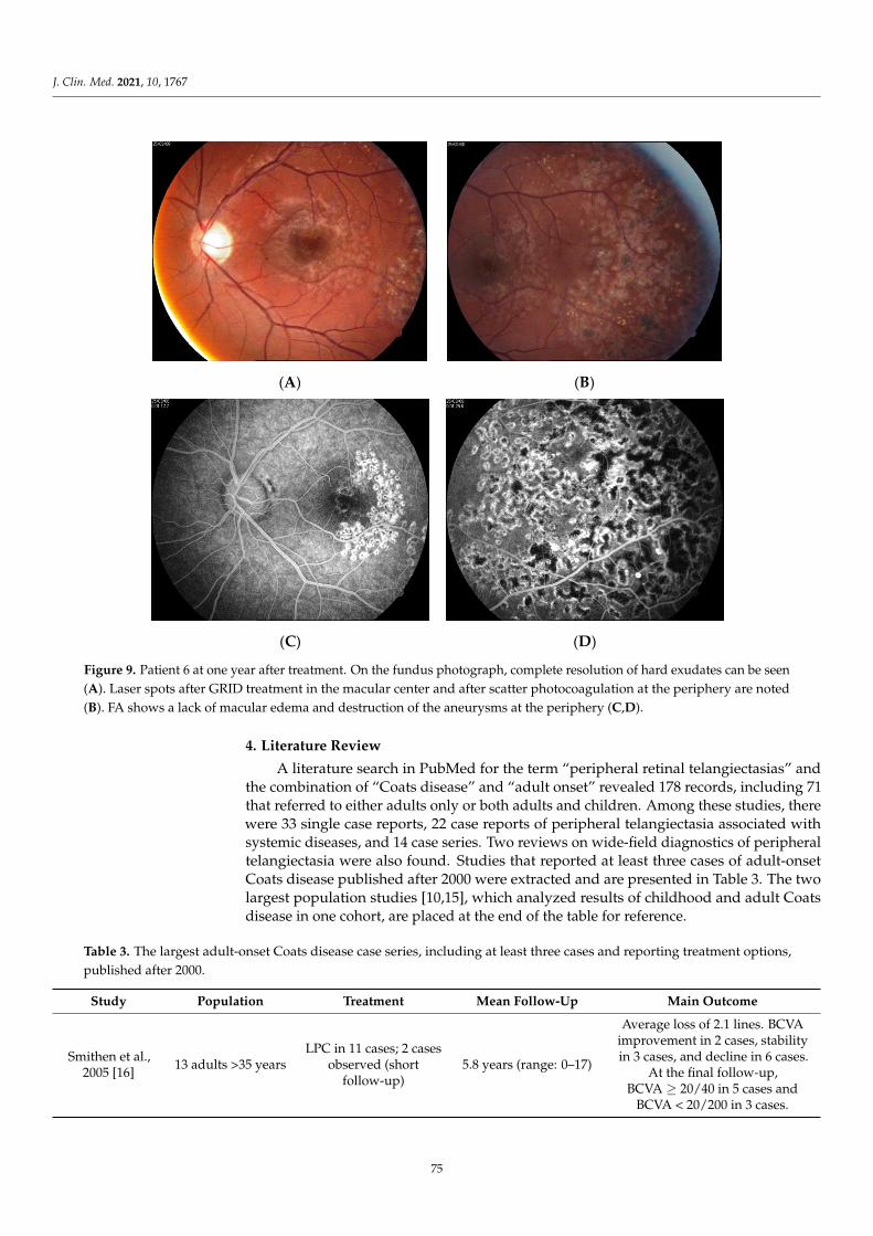
J. Clin. Med. 2021, 10, 1767
(A) (B)
(C) (D)
≥
μ
μ
Figure 9. Patient 6 at one year after treatment. On the fundus photograph, complete resolution of hard exudates can be seen(A). Laser spots after GRID treatment in the macular center and after scatter photocoagulation at the periphery are noted(B). FA shows a lack of macular edema and destruction of the aneurysms at the periphery (C,D).
4. Literature Review
A literature search in PubMed for the term “peripheral retinal telangiectasias” andthe combination of “Coats disease” and “adult onset” revealed 178 records, including 71that referred to either adults only or both adults and children. Among these studies, therewere 33 single case reports, 22 case reports of peripheral telangiectasia associated withsystemic diseases, and 14 case series. Two reviews on wide-field diagnostics of peripheraltelangiectasia were also found. Studies that reported at least three cases of adult-onsetCoats disease published after 2000 were extracted and are presented in Table 3. The twolargest population studies [10,15], which analyzed results of childhood and adult Coatsdisease in one cohort, are placed at the end of the table for reference.
Table 3. The largest adult-onset Coats disease case series, including at least three cases and reporting treatment options,published after 2000.
Study Population Treatment Mean Follow-Up Main Outcome
Smithen et al.,2005 [16]
13 adults >35 yearsLPC in 11 cases; 2 cases
observed (shortfollow-up)
5.8 years (range: 0–17)
Average loss of 2.1 lines. BCVAimprovement in 2 cases, stabilityin 3 cases, and decline in 6 cases.
At the final follow-up,BCVA ≥ 20/40 in 5 cases and
BCVA < 20/200 in 3 cases.
75

J. Clin. Med. 2021, 10, 1767
Table 3. Cont.
Study Population Treatment Mean Follow-Up Main Outcome
Goel et al.,2011 [17]
3 adultsSingle intravitreal
bevacizumab followedby LPC
9 months
Significant improvement of BCVAin all cases of from counting
fingers to 20/300, counting fingersto 20/240, and 20/240 to 20/120;regression of hard exudates from
the macula in all cases
Wang et al.,2011 [18]
3 adults2 injections of
bevacizumab followedby LPC
0.5–2 years (2, 0.5, 1years, respectively)
Significant improvement in BCVA,reduction of CRT, and regression
of telangiectasias.
• Case 1: BCVA change from6/15 to 6/6.7; CRT reductionfrom 437 to 230 µm
• Case 2: BCVA change from6/12 to 6/6, decrease in SRF(exact numbers not reported)
• Case 3: BCVA change from5/60 to 6/20; CRT reductionfrom 412 to 330 µm
Zheng et al.,2014 [19]
5 adults
Intravitrealbevacizumab followed
by LPC (3 cases) orintravitreal
triamcinolone (1 case)or subsequent
intravitrealbevacizumab (averageof 2 injections during
follow-up)
10.6 months
Resolution of subretinal fluid andtelangiectasias without significant
improvement in BCVA (range:1.42–1.25 logMAR). Vitreoretinal
fibrosis in two cases.
Park et al.,2016 [20]
13 adults
LPC combined withintravitreal
bevacizumab (mean no.of injections: 2.69 and
mean no of lasersessions: 1.68)
24.8 months
Mean BCVA change from0.72 logMAR to 0.68 logMAR
(statistically insignificant). BCVAimprovement of more than 3 linesin 3 patients (23%) and stability in7 patients (54%). Mean CRT was
significantly decreased from 473 to288 µm. Poor baseline BCVA and
subfoveal hard exudates correlatedwith poor final BCVA result.
Rishi et al.,2016 [21]
48 adults ≥ 35 years32 cases observed >
6 months
LPC (60.4%),observation (27.08%),
surgery (6.2%),cryotherapy (4%), LPCplus cryotherapy (2%)
40 months (range:1–122 months)
Patients with follow-up longerthan 6 months (32 cases):
• Treated: BCVA improvementor stabilization in 82.5%; exu-dates reduced in 56.5%, retinaattached in 95.6%
• Untreated: BCVA stabiliza-tion in 88%, no improve-ment; exudates reduced in33%, retina attached in 66%
(exact BCVA values not reported)
76
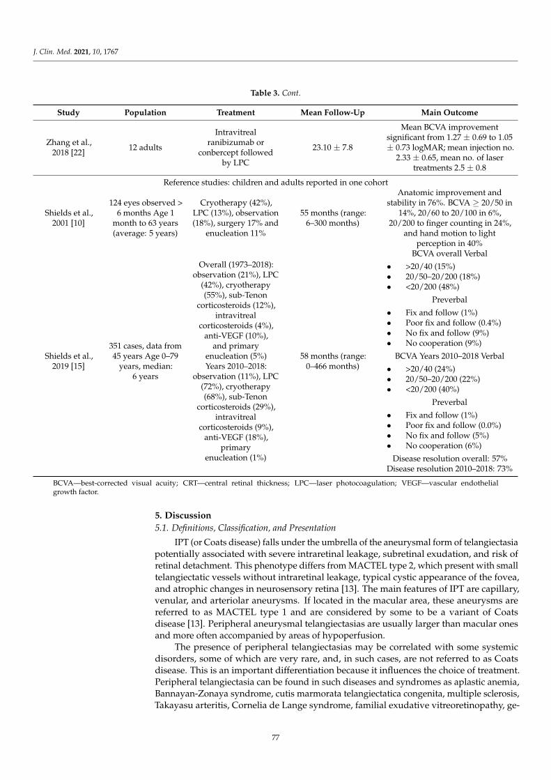
J. Clin. Med. 2021, 10, 1767
Table 3. Cont.
Study Population Treatment Mean Follow-Up Main Outcome
Zhang et al.,2018 [22]
12 adults
Intravitrealranibizumab or
conbercept followedby LPC
23.10 ± 7.8
Mean BCVA improvementsignificant from 1.27 ± 0.69 to 1.05± 0.73 logMAR; mean injection no.
2.33 ± 0.65, mean no. of lasertreatments 2.5 ± 0.8
Reference studies: children and adults reported in one cohort
Shields et al.,2001 [10]
124 eyes observed >6 months Age 1
month to 63 years(average: 5 years)
Cryotherapy (42%),LPC (13%), observation(18%), surgery 17% and
enucleation 11%
55 months (range:6–300 months)
Anatomic improvement andstability in 76%. BCVA ≥ 20/50 in
14%, 20/60 to 20/100 in 6%,20/200 to finger counting in 24%,
and hand motion to lightperception in 40%
Shields et al.,2019 [15]
351 cases, data from45 years Age 0–79
years, median:6 years
Overall (1973–2018):observation (21%), LPC
(42%), cryotherapy(55%), sub-Tenon
corticosteroids (12%),intravitreal
corticosteroids (4%),anti-VEGF (10%),
and primaryenucleation (5%)Years 2010–2018:
observation (11%), LPC(72%), cryotherapy(68%), sub-Tenon
corticosteroids (29%),intravitreal
corticosteroids (9%),anti-VEGF (18%),
primaryenucleation (1%)
58 months (range:0–466 months)
BCVA overall Verbal
• >20/40 (15%)• 20/50–20/200 (18%)• <20/200 (48%)
Preverbal
• Fix and follow (1%)• Poor fix and follow (0.4%)• No fix and follow (9%)• No cooperation (9%)
BCVA Years 2010–2018 Verbal
• >20/40 (24%)• 20/50–20/200 (22%)• <20/200 (40%)
Preverbal
• Fix and follow (1%)• Poor fix and follow (0.0%)• No fix and follow (5%)• No cooperation (6%)
Disease resolution overall: 57%Disease resolution 2010–2018: 73%
BCVA—best-corrected visual acuity; CRT—central retinal thickness; LPC—laser photocoagulation; VEGF—vascular endothelialgrowth factor.
5. Discussion
5.1. Definitions, Classification, and Presentation
IPT (or Coats disease) falls under the umbrella of the aneurysmal form of telangiectasiapotentially associated with severe intraretinal leakage, subretinal exudation, and risk ofretinal detachment. This phenotype differs from MACTEL type 2, which present with smalltelangiectatic vessels without intraretinal leakage, typical cystic appearance of the fovea,and atrophic changes in neurosensory retina [13]. The main features of IPT are capillary,venular, and arteriolar aneurysms. If located in the macular area, these aneurysms arereferred to as MACTEL type 1 and are considered by some to be a variant of Coatsdisease [13]. Peripheral aneurysmal telangiectasias are usually larger than macular onesand more often accompanied by areas of hypoperfusion.
The presence of peripheral telangiectasias may be correlated with some systemicdisorders, some of which are very rare, and, in such cases, are not referred to as Coatsdisease. This is an important differentiation because it influences the choice of treatment.Peripheral telangiectasia can be found in such diseases and syndromes as aplastic anemia,Bannayan-Zonaya syndrome, cutis marmorata telangiectatica congenita, multiple sclerosis,Takayasu arteritis, Cornelia de Lange syndrome, familial exudative vitreoretinopathy, ge-
77
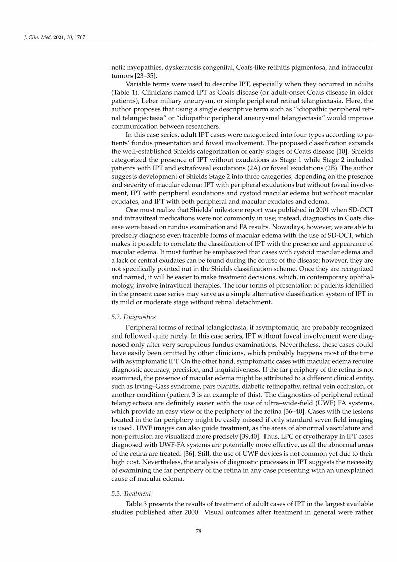
J. Clin. Med. 2021, 10, 1767
netic myopathies, dyskeratosis congenital, Coats-like retinitis pigmentosa, and intraoculartumors [23–35].
Variable terms were used to describe IPT, especially when they occurred in adults(Table 1). Clinicians named IPT as Coats disease (or adult-onset Coats disease in olderpatients), Leber miliary aneurysm, or simple peripheral retinal telangiectasia. Here, theauthor proposes that using a single descriptive term such as “idiopathic peripheral reti-nal telangiectasia” or “idiopathic peripheral aneurysmal telangiectasia” would improvecommunication between researchers.
In this case series, adult IPT cases were categorized into four types according to pa-tients’ fundus presentation and foveal involvement. The proposed classification expandsthe well-established Shields categorization of early stages of Coats disease [10]. Shieldscategorized the presence of IPT without exudations as Stage 1 while Stage 2 includedpatients with IPT and extrafoveal exudations (2A) or foveal exudations (2B). The authorsuggests development of Shields Stage 2 into three categories, depending on the presenceand severity of macular edema: IPT with peripheral exudations but without foveal involve-ment, IPT with peripheral exudations and cystoid macular edema but without macularexudates, and IPT with both peripheral and macular exudates and edema.
One must realize that Shields’ milestone report was published in 2001 when SD-OCTand intravitreal medications were not commonly in use; instead, diagnostics in Coats dis-ease were based on fundus examination and FA results. Nowadays, however, we are able toprecisely diagnose even traceable forms of macular edema with the use of SD-OCT, whichmakes it possible to correlate the classification of IPT with the presence and appearance ofmacular edema. It must further be emphasized that cases with cystoid macular edema anda lack of central exudates can be found during the course of the disease; however, they arenot specifically pointed out in the Shields classification scheme. Once they are recognizedand named, it will be easier to make treatment decisions, which, in contemporary ophthal-mology, involve intravitreal therapies. The four forms of presentation of patients identifiedin the present case series may serve as a simple alternative classification system of IPT inits mild or moderate stage without retinal detachment.
5.2. Diagnostics
Peripheral forms of retinal telangiectasia, if asymptomatic, are probably recognizedand followed quite rarely. In this case series, IPT without foveal involvement were diag-nosed only after very scrupulous fundus examinations. Nevertheless, these cases couldhave easily been omitted by other clinicians, which probably happens most of the timewith asymptomatic IPT. On the other hand, symptomatic cases with macular edema requirediagnostic accuracy, precision, and inquisitiveness. If the far periphery of the retina is notexamined, the presence of macular edema might be attributed to a different clinical entity,such as Irving–Gass syndrome, pars planitis, diabetic retinopathy, retinal vein occlusion, oranother condition (patient 3 is an example of this). The diagnostics of peripheral retinaltelangiectasia are definitely easier with the use of ultra–wide-field (UWF) FA systems,which provide an easy view of the periphery of the retina [36–40]. Cases with the lesionslocated in the far periphery might be easily missed if only standard seven field imagingis used. UWF images can also guide treatment, as the areas of abnormal vasculature andnon-perfusion are visualized more precisely [39,40]. Thus, LPC or cryotherapy in IPT casesdiagnosed with UWF-FA systems are potentially more effective, as all the abnormal areasof the retina are treated. [36]. Still, the use of UWF devices is not common yet due to theirhigh cost. Nevertheless, the analysis of diagnostic processes in IPT suggests the necessityof examining the far periphery of the retina in any case presenting with an unexplainedcause of macular edema.
5.3. Treatment
Table 3 presents the results of treatment of adult cases of IPT in the largest availablestudies published after 2000. Visual outcomes after treatment in general were rather
78
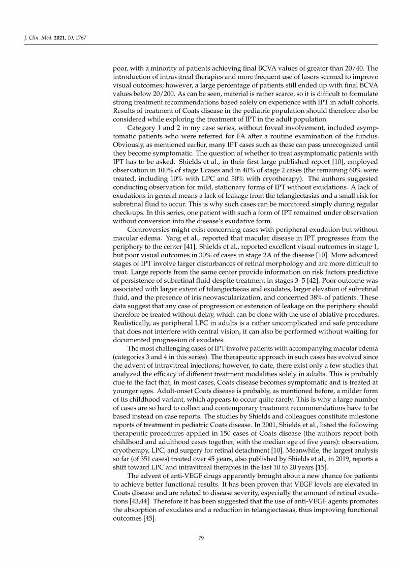
J. Clin. Med. 2021, 10, 1767
poor, with a minority of patients achieving final BCVA values of greater than 20/40. Theintroduction of intravitreal therapies and more frequent use of lasers seemed to improvevisual outcomes; however, a large percentage of patients still ended up with final BCVAvalues below 20/200. As can be seen, material is rather scarce, so it is difficult to formulatestrong treatment recommendations based solely on experience with IPT in adult cohorts.Results of treatment of Coats disease in the pediatric population should therefore also beconsidered while exploring the treatment of IPT in the adult population.
Category 1 and 2 in my case series, without foveal involvement, included asymp-tomatic patients who were referred for FA after a routine examination of the fundus.Obviously, as mentioned earlier, many IPT cases such as these can pass unrecognized untilthey become symptomatic. The question of whether to treat asymptomatic patients withIPT has to be asked. Shields et al., in their first large published report [10], employedobservation in 100% of stage 1 cases and in 40% of stage 2 cases (the remaining 60% weretreated, including 10% with LPC and 50% with cryotherapy). The authors suggestedconducting observation for mild, stationary forms of IPT without exudations. A lack ofexudations in general means a lack of leakage from the telangiectasias and a small risk forsubretinal fluid to occur. This is why such cases can be monitored simply during regularcheck-ups. In this series, one patient with such a form of IPT remained under observationwithout conversion into the disease’s exudative form.
Controversies might exist concerning cases with peripheral exudation but withoutmacular edema. Yang et al., reported that macular disease in IPT progresses from theperiphery to the center [41]. Shields et al., reported excellent visual outcomes in stage 1,but poor visual outcomes in 30% of cases in stage 2A of the disease [10]. More advancedstages of IPT involve larger disturbances of retinal morphology and are more difficult totreat. Large reports from the same center provide information on risk factors predictiveof persistence of subretinal fluid despite treatment in stages 3–5 [42]. Poor outcome wasassociated with larger extent of telangiectasias and exudates, larger elevation of subretinalfluid, and the presence of iris neovascularization, and concerned 38% of patients. Thesedata suggest that any case of progression or extension of leakage on the periphery shouldtherefore be treated without delay, which can be done with the use of ablative procedures.Realistically, as peripheral LPC in adults is a rather uncomplicated and safe procedurethat does not interfere with central vision, it can also be performed without waiting fordocumented progression of exudates.
The most challenging cases of IPT involve patients with accompanying macular edema(categories 3 and 4 in this series). The therapeutic approach in such cases has evolved sincethe advent of intravitreal injections; however, to date, there exist only a few studies thatanalyzed the efficacy of different treatment modalities solely in adults. This is probablydue to the fact that, in most cases, Coats disease becomes symptomatic and is treated atyounger ages. Adult-onset Coats disease is probably, as mentioned before, a milder formof its childhood variant, which appears to occur quite rarely. This is why a large numberof cases are so hard to collect and contemporary treatment recommendations have to bebased instead on case reports. The studies by Shields and colleagues constitute milestonereports of treatment in pediatric Coats disease. In 2001, Shields et al., listed the followingtherapeutic procedures applied in 150 cases of Coats disease (the authors report bothchildhood and adulthood cases together, with the median age of five years): observation,cryotherapy, LPC, and surgery for retinal detachment [10]. Meanwhile, the largest analysisso far (of 351 cases) treated over 45 years, also published by Shields et al., in 2019, reports ashift toward LPC and intravitreal therapies in the last 10 to 20 years [15].
The advent of anti-VEGF drugs apparently brought about a new chance for patientsto achieve better functional results. It has been proven that VEGF levels are elevated inCoats disease and are related to disease severity, especially the amount of retinal exuda-tions [43,44]. Therefore it has been suggested that the use of anti-VEGF agents promotesthe absorption of exudates and a reduction in telangiectasias, thus improving functionaloutcomes [45].
79
J. Clin. Med. 2021, 10, 1767
Different anti-VEGF medications have been used in combination with ablative thera-pies in the treatment of Coats disease. A report from Bascom Palmer Institute details thesuccessful treatment of 24 children with advanced Coats disease complicated by exudativeretinal detachment with direct laser ablation in combination with anti-VEGF treatment [46].Zhang et al., presented the results of 28 cases (including 12 adults) treated by the combina-tion of LPC and intravitreal ranibizumab or conbercept [22], where LPC was applied afterinitial anti-VEGF injection and then both treatments were used in a pro re nata (PRN) fash-ion. These authors noted significant morphological improvements; however, the changes inBCVA were not statistically significant. On the other hand, Ramasubramanian and Shieldswarn that the addition of bevacizumab to standard Coats therapy (LPC and cryotherapy)might evoke vitreoretinal fibrosis and potentially retinal detachment, rarely seen afterstandard therapy [47]. However, Daruich et al., in a large pediatric study did not findsuch a relationship [48]. To date, this question remains unanswered, although cliniciansemphasize the benefits of anti-VEGF treatment in Coats disease [17].
In this case series, two patients with IPT and macular edema were treated with thecombination of intravitreal anti-VEGF and LPC and two cases were treated with LPC only.Interestingly, patient 6, after LPC monotherapy performed almost 15 years ago, practicallyhad his BCVA completely restored, contrary to the results among patients treated afterthe advent of intravitreal therapies. I believe that this outcome is mainly due to the shortduration of macular edema and aggressive LPC that was the treatment of choice at thattime (as intravitreal injections were not available on the market yet). This is also consistentwith the report by Smithen et al., from 2005, when LPC was also the main form of treatmentof Coats disease in adults, where improvement or stabilization of BCVA was noted in 5 of11 cases [16]. A large pediatric study from 2008 also reported that aggressive diode lasertherapy proved effective in the majority of patients [49].
Among the remaining three cases of this series, one patient (patient 3) was treated withthe combination of anti-VEGF and LPC, achieving morphological and functional success.The patient underwent very close follow-up and presented relatively early after symptomsoccurred. Patient 4 after his initial visit was lost to follow-up for one year without directaggressive laser treatment of IPT. During that time, severe macular edema developedand a decline in his BCVA to 20/200 was noted, and no improvements after combinationtreatment with anti-VEGF therapy plus LPC followed. I believe that the course of this caseemphasizes the necessity of employing early ablative therapies in practically every case ofsymptomatic Coats disease. Only anecdotal reports of IPT well-controlled by anti-VEGFinjections alone exist [50].
Photodynamic therapy has been trialed in the treatment of Coats disease; however,only a few case reports are available in the literature [51–53]. Thus, it should be treated asan adjunct form of treatment in refractory cases, but not as first-line therapy.
6. Conclusions
Adult-onset Coats disease is a clinical entity of variable presentation and extent.Available data support the use of ablative therapies, especially LPC, as the main and mosteffective means of treatment in every exudative form of peripheral telangiectasias. Onlyasymptomatic cases without exudates can be observed. Anti-VEGF treatment seems tobe a useful adjunct to ablative therapies, especially in cases with macular involvement.Close follow-up is needed in every symptomatic adult-onset Coats disease case. Lossof follow-up without treatment might result in irreversible vision decline. Despite theapplication of different forms of therapies, a large percentage of adult Coats cases end upexperiencing deterioration of vision and poor morphological outcomes.
Funding: No funding was received for this research.
Institutional Review Board Statement: The study was conducted according to the guidelines of theDeclaration of Helsiniki and approved by the Dobry Wzrok Ophthalmological Clinic committee ofEA1/2021.
80
J. Clin. Med. 2021, 10, 1767
Informed Consent Statement: The study was retrospective in nature and involved standard proce-dures performed in the clinic on a daily basis; hence, informed consent for participation in the studywas waived by the Ethics Committee.
Data Availability Statement: Data is contained within the article.
Conflicts of Interest: The author declares no conflict of interest.
References
1. Coats, G. Forms of retinal diseases with massive exudation. R. Lond. Ophthalmol. Hosp. Rep. 1908, 17, 440–525.2. Shields, J.A.; Shields, C.L.; Honavar, S.G.; Demirci, H. Clinical variations and complications of Coats disease in 150 cases: The
2000 Sanford Gifford Memorial Lecture. Am. J. Ophthalmol. 2001, 131, 561–571. [CrossRef]3. Egbert, P.R.; Chan, C.C.; Winter, F.C. Flat preparations of the retinal vessels in Coats’ disease. J. Pediatr. Ophthalmol. 1976,
13, 336–339.4. Fernandes, B.F.; Odashiro, A.N.; Maloney, S.; Zajdenweber, M.E.; Lopes, A.G.; Burnier, M.N., Jr. Clini-cal-histopathological
correlation in a case of Coats’ disease. Diagn. Pathol. 2006, 1, 24. [CrossRef] [PubMed]5. Shields, C.L.; Sen, M.; Honavar, S.G.; Shields, J.A. Coats disease: An overview of classification, management and outcomes.
Indian J. Ophthalmol. 2019, 67, 763–771. [CrossRef] [PubMed]6. Jones, J.H.; Kroll, A.J.; Lou, P.L.; Ryan, E.A. Coats’ disease. Int. Ophthalmol. Clin. 2001, 41, 189–198. [CrossRef]7. Gawecki, M. Tetniaki prosówkowate Lebera—Opis przypadku [Leber miliary aneurysms—A case report. Klin Oczna. 2009,
111, 131–133.8. Reese, A.B. Telangiectasis of the Retina and Coats’ Disease. Am. J. Ophthalmol. 1956, 42, 1–8. [CrossRef]9. Alturkistany, W.; Waheeb, S. Leber’s miliary aneurysms. Oman J. Ophthalmol. 2013, 6, 119–121. [CrossRef]10. Shields, J.A.; Shields, C.L.; Honavar, S.G.; Demirci, H.; Cater, J. Classification and management of Coats disease: The 2000 Proctor
Lecture. Am. J. Ophthalmol. 2001, 131, 572–583. [CrossRef]11. Yanuzzi, A.L. The Retinal Atlas; Elsevier Ltd.: Amsterdam, The Netherlands, 2010.12. Cahill, M.; O’Keefe, M.; Acheson, R.; Mulvihill, A.; Wallace, D.; Mooney, D. Classification of the spectrum of Coats’ disease as
subtypes of idiopathic retinal telangiectasis with exudation. Acta Ophthalmol. Scand. 2001, 79, 596–602. [CrossRef] [PubMed]13. Yannuzzi, L.A.; Bardal, A.M.; Freund, K.B.; Chen, K.J.; Eandi, C.M.; Blodi, B. Idiopathic macular telangiectasia. Arch. Ophthalmol.
2006, 124, 450–460. [CrossRef] [PubMed]14. Charbel Issa, P.; Gillies, M.C.; Chew, E.Y.; Bird, A.C.; Heeren, T.F.; Peto, T.; Holz, F.G.; Scholl, H.P. Macular telangiectasia type 2.
Prog. Retin. Eye Res. 2013, 34, 49–77. [CrossRef]15. Shields, C.L.; Udyaver, S.; Dalvin, L.A.; Lim, L.S.; Atalay, H.T.; Khoo, C.T.L.; Mazloumi, M.; Shields, J.A. Coats disease in 351
eyes: Analysis of features and outcomes over 45 years (by decade) at a single center. Indian J. Ophthalmol. 2019, 67, 772–783.[CrossRef] [PubMed]
16. Smithen, L.M.; Brown, G.C.; Brucker, A.J.; Yannuzzi, L.A.; Klais, C.M.; Spaide, R.F. Coats’ Disease Diagnosed in Adulthood.Ophthalmology 2005, 112, 1072–1078. [CrossRef]
17. Goel, N.; Kumar, V.; Seth, A.; Raina, U.K.; Ghosh, B. Role of intravitreal bevacizumab in adult onset Coats’ disease. Int. Ophthalmol.
2011, 31, 183–190. [CrossRef]18. Wang, K.-Y.; Cheng, C.-K. A Combination of Intravitreal Bevacizumab Injection with Tunable Argon Yellow Laser Photocoagula-
tion as a Treatment for Adult-Onset Coats’ Disease. J. Ocul. Pharmacol. Ther. 2011, 27, 525–530. [CrossRef]19. Zheng, X.X.; Jiang, Y.R. The effect of intravitreal bevacizumab injection as the initial treatment for Coats’ disease. Graefes Arch.
Clin. Exp. Ophthalmol. 2014, 252, 35–42. [CrossRef]20. Park, S.; Cho, H.J.; Lee, D.W.; Kim, C.G.; Kim, J.W. Intravitreal bevacizumab injections combined with laser photocoagulation for
adult-onset Coats’ disease. Graefe’s Arch. Clin. Exp. Ophthalmol. 2015, 254, 1511–1517. [CrossRef]21. Rishi, P.; Rishi, E.; Appukuttan, B.; Uparkar, M.; Sharma, T.; Gopal, L. Coats′ disease of adult-onset in 48 eyes. Indian J. Ophthalmol.
2016, 64, 518–523. [CrossRef] [PubMed]22. Zhang, L.; Ke, Y.; Wang, W.; Shi, X.; Hei, K.; Li, X. The efficacy of conbercept or ranibizumab intravitreal injection combined with
laser therapy for Coats’ disease. Graefe’s Arch. Clin. Exp. Ophthalmol. 2018, 256, 1339–1346. [CrossRef]23. Metelitsina, T.I.; Sheth, V.S.; Patel, S.B.; Grassi, M.A. Peripheral retinopathy associated with aplastic anemia. Retin. Cases Brief.
Rep. 2017, 11, 108–110. [CrossRef] [PubMed]24. Klifto, M.R.; Balaratnasingam, C.; Weissman, H.H.; Yannuzzi, L.A. Bilateral coats reaction in bannayan–zonana syndrome: A
single case report. Retin. Cases Brief. Rep. 2017, 11, 286–289. [CrossRef] [PubMed]25. Taleb, E.A.; Nagpal, M.P.; Mehrotra, N.S.; Bhatt, K. Retinal findings in a case of presumed cutis marmorata telangiectatica
congenita. Retin. Cases Brief. Rep. 2018, 12, 322–325. [CrossRef] [PubMed]26. Soohoo, J.R.; McCourt, E.A.; Lenahan, D.S.; Oliver, S.C. Fluorescein angiogram findings in a case of cutis marmorata telangiectatica
congenita. Ophthalmic Surg. Lasers Imaging Retina 2013, 44, 398–400. [CrossRef] [PubMed]27. de Massougnes, S.; Borruat, F.X.; Ambresin, A. Peripheral Bilateral Telangiectasiae in Multiple Sclerosis Patients Treated with
Interferon B1a. Klin. Monbl. Augenheilkd. 2016, 233, 438–440. [CrossRef] [PubMed]
81
J. Clin. Med. 2021, 10, 1767
28. Batliwala, S.Y.; Perez, M.; Aston, W.; Chavala, S.H. Peripheral Retinal Telangiectasia and Ischemia in Takayasu Arteritis. Arthritis
Rheumatol. 2016, 68, 2350. [CrossRef]29. Stacey, A.W.; Sparagna, C.; Borri, M.; Rizzo, S.; Hadjistilianou, T. A 6-year-old boy with Cornelia de Lange syndrome and
Coats disease: Case report and review of the literature. J. Am. Assoc. Pediatr. Ophthalmol. Strabismus 2015, 19, 474–478.[CrossRef] [PubMed]
30. Ilhan, A.; Yolcu, U.; Gundogan, F.C.; Akay, F. An association between subclinical familial exudative vitreoretinopathy androd-cone dystrophy. Arq. Bras. Oftalmol. 2014, 77. [CrossRef]
31. Kashani, A.H.; Brown, K.T.; Chang, E.; Drenser, K.A.; Capone, A.; Trese, M.T. Diversity of Retinal Vascular Anomalies in Patientswith Familial Exudative Vitreoretinopathy. Ophthalmology 2014, 121, 2220–2227. [CrossRef]
32. Sacconi, S.; Baillif-Gostoli, S.; Desnuelle, C. Atteinte rétinienne et myopathies génétiques [Retinal involvement and geneticmyopathy]. Rev. Neurol. 2010, 166, 998–1009. [CrossRef]
33. Johnson, C.A.; Hatfield, M.; Pulido, J.S. Retinal vasculopathy in a family with autosomal dominant dyskeratosis congenita.Ophthalmic Genet. 2009, 30, 181–184. [CrossRef] [PubMed]
34. Kan, E.; Yilmaz, T.; Aydemir, O.; Güler, M.; Kurt, J. Coats-like retinitis pigmentosa: Reports of three cases. Clin. Ophthalmol. 2007,1, 193–198. [PubMed]
35. Char, D.H. Coats’ syndrome: Long term follow up. Br. J. Ophthalmol. 2000, 84, 37–39. [CrossRef]36. Kumar, V.; Chandra, P.; Kumar, A. Ultra-wide field imaging in the diagnosis and management of adult-onset Coats’ disease. Clin.
Exp. Optom. 2017, 100, 79–82. [CrossRef] [PubMed]37. Joussen, A.M.; Brockmann, C.; Urban, J.; Seibel, I.; Winterhalter, S.; Zeitz, O.; Müller, B. Ultraweitwinkel-Fundusfotografie und
-angiografie in der Differenzialdiagnose und zur Therapieplanung bei peripheren vaskulären Netzhauterkrankungen [Ultra-WideField Retinal Imaging and Angiography in the Differential Diagnosis and Therapeutic Decisions in Vascular Diseases of thePeripheral Retina]. Klin. Monbl. Augenheilkd. 2018, 235, 980–993.
38. Sakurada, Y.; Freund, K.B.; Yannuzzi, L.A. Multimodal Imaging in Adult-Onset Coats’ Disease. Ophthalmology 2018, 125,485. [CrossRef]
39. Temkar, S.; Azad, S.V.; Chawla, R.; Damodaran, S.; Garg, G.; Regani, H.; Nawazish, S.; Raj, N.; Venkatraman, V. Ultra-widefieldfundus fluorescein angiography in pediatric retinal vascular diseases. Indian J. Ophthalmol. 2019, 67, 788–794. [CrossRef]
40. Turczynska, M.; Brydak-Godowska, J. The role of peripheral retinal angiography in the diagnosis of adult Coats’ disease based ona case report. Klin. Ocz. 2020, 122, 171–176. [CrossRef]
41. Yang, L.-H.; Shi, X.-H.; Tian, B.; Zhou, D.; Wei, W.-B. Clinical analysis of macular disease involved by Coats disease. [Zhonghua
yan ke za zhi] Chin. J. Ophthalmol. 2009, 45.42. Khoo, C.T.L.; Dalvin, L.A.; Lim, L.S.; Mazloumi, M.; Atalay, H.T.; Udyaver, S.; Shields, J.A.; Shields, C.L. Fac-tors Predictive of
Subretinal Fluid Resolution in Coats Disease: Analysis of 177 Eyes in 177 Pa-tients at a Single Center. Asia Pac. J. Ophthalmol.
2019, 8, 290–297. [CrossRef]43. Zhao, Q.; Peng, X.-Y.; Chen, F.-H.; Zhang, Y.-P.; Wang, L.; You, Q.-S.; Jonas, J.B. Vascular endothelial growth factor in Coats’
disease. Acta Ophthalmol. 2013, 92, e225–e228. [CrossRef] [PubMed]44. Zhang, J.; Jiang, C.; Ruan, L.; Huang, X. Associations of cytokine concentrations in aqueous humour with retinal vascular
abnormalities and exudation in Coats’ disease. Acta Ophthalmol. 2019, 97, 319–324. [CrossRef]45. Yang, Q.; Wei, W.; Shi, X. Successful use of intravitreal ranibizumab injection and combined treatment in the management of
Coats’ disease. Acta Ophthalmol. 2016, 94, 401–406. [CrossRef] [PubMed]46. Gold, A.S.; Villegas, V.M.; Murray, T.G.; Berrocal, A.M. Advanced Coats’ disease treated with intravitreal bevacizumab combined
with laser vascular ablation. Clin. Ophthalmol. 2014, 8, 973–976. [CrossRef]47. Ramasubramanian, A.; Shields, C.L. Bevacizumab for Coats’ disease with exudative retinal detachment and risk of vitreoretinal
traction. Br. J. Ophthalmol. 2011, 96, 356–359. [CrossRef] [PubMed]48. Daruich, A.; Matet, A.; Tran, H.V.; Gaillard, M.-C.; Munier, F.L. Extramacular fibrosis in coats’ disease. Retina 2016, 36,
2022–2028. [CrossRef]49. Schefler, A.C.; Berrocal, A.M.; Murray, T.G. Advanced Coats’ disease. Management with repetitive aggressive laser ablation
therapy. Retina 2008, 28 (Suppl. 3), S38–S41. [CrossRef]50. Georgakopoulos, C.D.; Foteini, T.; Makri, O.E.; Vavvas, D. Two-year results of intravitreal injections of aflibercept in Coats’
Disease; a case report. Retin. Cases Brief. Rep. 2020. [CrossRef]51. Beselga, D.; Campos, A.; Mendes, S.; Carvalheira, F.; Castro, M.; Castanheira, D. Refractory Coats’ Disease of Adult Onset. Case
Rep. Ophthalmol. 2012, 3, 118–122. [CrossRef]52. Namba, M.; Shiode, Y.; Morizane, Y.; Kimura, S.; Hosokawa, M.; Doi, S.; Toshima, S.; Takahashi, K.; Hosogi, M.; Fujiwara,
A.; et al. Successful resolution of coats disease by photodynamic therapy: A case report. BMC Ophthalmol. 2018, 18, 264.[CrossRef] [PubMed]
53. Kim, J.; Park, K.H.; Woo, S.J. Combined Photodynamic Therapy and Intravitreal Bevacizumab Injection for the Treatment ofAdult Coats’ Disease: A Case Report. Korean J. Ophthalmol. 2010, 24, 374–376. [CrossRef] [PubMed]
82
Journal of
Clinical Medicine
Article
Bleb Compressive Sutures in the Management of HypotonyMaculopathy after Glaucoma Surgery
Ewa Kosior-Jarecka 1,* , Dominika Wróbel-Dudzinska 1 , Anna Swiech 2 and Tomasz Zarnowski 1
�����������������
Citation: Kosior-Jarecka, E.;
Wróbel-Dudzinska, D.; Swiech, A.;
Zarnowski, T. Bleb Compressive
Sutures in the Management of
Hypotony Maculopathy after
Glaucoma Surgery. J. Clin. Med. 2021,
10, 2223. https://doi.org/10.3390/
jcm10112223
Academic Editor: Gawecki Maciej
Received: 6 April 2021
Accepted: 18 May 2021
Published: 21 May 2021
Publisher’s Note: MDPI stays neutral
with regard to jurisdictional claims in
published maps and institutional affil-
iations.
Copyright: © 2021 by the authors.
Licensee MDPI, Basel, Switzerland.
This article is an open access article
distributed under the terms and
conditions of the Creative Commons
Attribution (CC BY) license (https://
creativecommons.org/licenses/by/
4.0/).
1 Department of Diagnostics and Microsurgery of Glaucoma, Medical University of Lublin,20-059 Lublin, Poland; [email protected] (D.W.-D.); [email protected] (T.Z.)
2 Department of Vitreoretinal Surgery, Medical University of Lublin, 20-059 Lublin, Poland;[email protected]
* Correspondence: [email protected]
Abstract: PURPOSE: The aim of the study was to assess the efficacy and safety of compressive sutures
in patients with hypotony maculopathy after glaucoma surgery. METHODS: This retrospective case
series analyzes the clinical outcomes of conjunctival compressive sutures in 17 patients with hypotony
maculopathy developed after glaucoma surgery. Compressive Nylon 10–0 single sutures were used
in all patients; in two patients, the procedure was repeated. All patients underwent ophthalmic
evaluation and macular OCT scanning before the surgery, one month, six months, and one year after
the procedure. RESULTS: Mean intraocular pressure (IOP) before suturing was 2.3 ± 1.57 mmHg
and increased to 14.2 ± 7.03 mmHg (p = 0.00065) one month after the procedure. After six months,
mean IOP was 10.2 ± 4.3 mmHg (p = 0.005), and after one year ± 4.7 mmHg (p = 0.0117). To obtain
the target pressure, the sutures had to be removed in one patient, and medical therapy was under-
taken in three patients. Mean decimal best-corrected visual acuity (BCVA) before the sutures was
0.18 ± 0.13 and increased to 0.53 ± 0.25 (p = 0.0004) after one month, to 0.46 ± 0.31 (p = 0.005) after
six months, and to 0.31 ± 0.22 (p = 0.025) after one year. In one case, leakage from the bleb was
observed after the procedure and bleb revision was required. CONCLUSIONS: transconjuctival
compressive sutures seem to be an efficient and safe technique for managing hypotony maculopathy
after glaucoma surgery.
Keywords: hypotony maculopathy; complications of antiglaucoma surgery; bleb compressive sutures
1. Introduction
Glaucoma, one of the leading causes of irreversible blindness worldwide [1], is a progres-sive optic neuropathy, and elevated intraocular pressure (IOP) is one of the main risk factorsfor the development and progression of this disease [2]. A decrease of IOP remains the onlyclinical method with confirmed efficacy for diminishing the progression rate of glaucoma,and this effect can be achieved by means of medical treatment, laser, or surgery [3].
After trabeculectomy, a standard surgical glaucoma procedure, aqueous humor flowsinto the surgically created filtering bleb [4]. The final success of trabeculectomy dependsnot only on the surgical technique but also on the possibility of slowing down the healingprocesses [5]. Therefore, the challenge faced by both surgical technique and postsurgicalcare is to find a balance between IOP low enough to obtain the target pressure and at thesame time high enough to avoid complications related to ocular hypotony [6]. Additionally,not every case of post-surgical low IOP level leads to ocular complications [7].
Clinical hypotony is defined as a level of IOP that is too low to maintain the shapeof the eyeball and results in structural and functional changes [8,9]. If left untreated, pro-longed hypotony may cause various serious complications such as bleb infection, cataractformation, synechiae, persistent choroidal detachment, or hypotony maculopathy [10].Hypotony maculopathy and bleb infection in the course of clinical hypotony are potentiallysight-threatening [11,12].
83
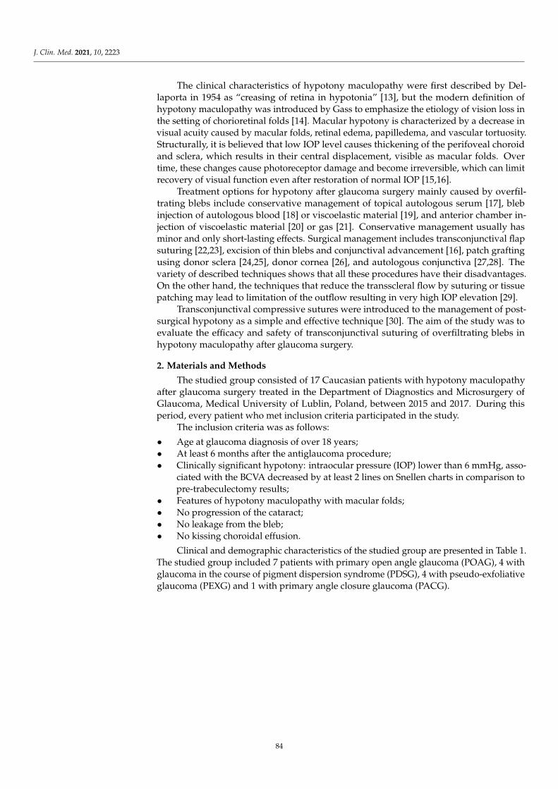
J. Clin. Med. 2021, 10, 2223
The clinical characteristics of hypotony maculopathy were first described by Del-laporta in 1954 as “creasing of retina in hypotonia” [13], but the modern definition ofhypotony maculopathy was introduced by Gass to emphasize the etiology of vision loss inthe setting of chorioretinal folds [14]. Macular hypotony is characterized by a decrease invisual acuity caused by macular folds, retinal edema, papilledema, and vascular tortuosity.Structurally, it is believed that low IOP level causes thickening of the perifoveal choroidand sclera, which results in their central displacement, visible as macular folds. Overtime, these changes cause photoreceptor damage and become irreversible, which can limitrecovery of visual function even after restoration of normal IOP [15,16].
Treatment options for hypotony after glaucoma surgery mainly caused by overfil-trating blebs include conservative management of topical autologous serum [17], blebinjection of autologous blood [18] or viscoelastic material [19], and anterior chamber in-jection of viscoelastic material [20] or gas [21]. Conservative management usually hasminor and only short-lasting effects. Surgical management includes transconjunctival flapsuturing [22,23], excision of thin blebs and conjunctival advancement [16], patch graftingusing donor sclera [24,25], donor cornea [26], and autologous conjunctiva [27,28]. Thevariety of described techniques shows that all these procedures have their disadvantages.On the other hand, the techniques that reduce the transscleral flow by suturing or tissuepatching may lead to limitation of the outflow resulting in very high IOP elevation [29].
Transconjunctival compressive sutures were introduced to the management of post-surgical hypotony as a simple and effective technique [30]. The aim of the study was toevaluate the efficacy and safety of transconjunctival suturing of overfiltrating blebs inhypotony maculopathy after glaucoma surgery.
2. Materials and Methods
The studied group consisted of 17 Caucasian patients with hypotony maculopathyafter glaucoma surgery treated in the Department of Diagnostics and Microsurgery ofGlaucoma, Medical University of Lublin, Poland, between 2015 and 2017. During thisperiod, every patient who met inclusion criteria participated in the study.
The inclusion criteria was as follows:
• Age at glaucoma diagnosis of over 18 years;• At least 6 months after the antiglaucoma procedure;• Clinically significant hypotony: intraocular pressure (IOP) lower than 6 mmHg, asso-
ciated with the BCVA decreased by at least 2 lines on Snellen charts in comparison topre-trabeculectomy results;
• Features of hypotony maculopathy with macular folds;• No progression of the cataract;• No leakage from the bleb;• No kissing choroidal effusion.
Clinical and demographic characteristics of the studied group are presented in Table 1.The studied group included 7 patients with primary open angle glaucoma (POAG), 4 withglaucoma in the course of pigment dispersion syndrome (PDSG), 4 with pseudo-exfoliativeglaucoma (PEXG) and 1 with primary angle closure glaucoma (PACG).
84

J. Clin. Med. 2021, 10, 2223
Table 1. Clinical and demographic characteristics of the studied group.
Feature n/Mean Value ± SD
Number of patients 17
Gender 8F/10 M
Age 60.5 ± 20.5
Diagnosis
POAG: 7 cases;PDSG: 4 cases:PEXG: 4 cases;PACG: 1 case;
traumatic glaucoma: 1 case.
Primary procedure
Trabeculectomy: 9 cases;Phacotrabeculectomy: 4 cases;
Needle revision: 3 cases;Deep sclerectomy: 1 case.
Mean IOP before primary procedure 33.6 ± 11.6 mmHg
Mean IOP before sutures 2.3 ± 1.57 mmHg
BCVA before primary procedure 0.55 ± 0.31
BCVA before sutures 0.18 ± 0.13
Mean MD 16.21 ± 7.45 dB
Mean time between primary procedure andcompression sutures
3.08 years
POAG, primary open angle glaucoma; PDSG, pigment dispersion syndrome; PEXG, pseudo-exfoliative glaucoma;
PACG, primary angle closure glaucoma; IOP, mean intraocular pressure; BCVA, best-corrected visual acuity; MD,
mean deviation.
The information about patients’ medical history was obtained from their clinical records.At the inclusion visit, patients underwent ophthalmic examination with BCVA (decimalSnellen charts), Goldman applanation tonometry, slit lamp examination, eye fundus assess-ment by ophthalmoscopy, and OCT (Stratus, Carl Zeiss Meditec, Dublin, Ireland) measure-ments assessing the thickness of the macula and the presence of choroidal folds.
During the surgical procedure, 5–7 single Nylon 10–0 sutures were placed transcon-junctivally in the area of the bleb as described earlier [30]. In brief, after peribulbaranesthesia, the bulb was rotated downward and single sutures were placed, starting fromthe limbus and extending posteriorly as far as possible toward the superior fornix. Duringsuturing, attempts were made to catch not only the conjunctiva but also a part of theunderlying sclera, if the height of the bleb allowed for it. The sutures were intended to beplaced on the area of existing scleral flap or in its proximity. During the procedure, afterplacing the 5th suture, paracentesis was performed and the BSS was administered into theanterior chamber to assess the increase of IOP and the reduction of the outflow. Furthersutures were added until the increase in IOP was observed (up to 7 sutures). The idea andthe surgical technique are shown in Scheme 1.
A B
Scheme 1. Transconjunctival sutures placement: A, side view; B, front view.
85

J. Clin. Med. 2021, 10, 2223
In the post-surgical period, only fixed combination of dexamethasone/tobramycinewas used in decreasing doses (starting from 4 times a day, and decreasing 1 drop per weekup to 4 weeks when the drops were stopped). Some patients needed to have installed thepreservatives free lubricant drops.
The patients were controlled one day after the procedure, as well as 7 days, 1 month,3 months, 6 months, and 12 months after the procedure. During the check-ups, ophthalmicexamination was performed including BCVA testing (decimal Snellen charts), Goldmanapplanation tonometry, slit lamp examination, eye fundus assessment by ophthalmoscopy,and OCT measurements assessing the thickness of the macula and the presence of choroidalfolds. All patients were present at all time-point visits. However, in 2 cases at 6 months,OCT was not performed because of technical problems. Additionally, the study eye wasexamined at every visit to look for possible complications.
Two criteria of success were defined:
1. IOP over 6 mmHg;2. BCVA improvement of at least 2 lines on a Snellen chart.
Changes in the assessed values (BCVA and IOP) were measured at each control visitplanned in the study by subtracting the preoperative value from the postoperative value.Statistic evaluation of the data was performed using Statistica 13.1 (Polish version, StatsoftPoland). The results were reported mainly as mean ± SD or percentage values. A p-value lowerthan 0.05 was considered statistically significant. Normal distribution was assessed with theShapiro–Wilk test. The Mann–Whitney test was used for non-normally distributed data.
3. Results
Mean IOP before suturing was 2.3 ± 1.57 mmHg and increased to 14.2 ± 7.03 mmHg(p = 0.00065) at 1 month post-op. After 6 months, mean IOP was 10.2 ± 4.3 mmHg(p = 0.005); and after one year ± 4.7 mmHg (p = 0.0117) (Figure 1). On day 7 after surgery,IOP value exceeded 21 mmHg in 4 (23.5%) patients, with a value of over 30 mmHg in onecase. To obtain the target pressure, one patient needed to have one suture removed, andmedical therapy was undertaken in three patients.
Figure 1. Changes in the intraocular pressure during the study period.
86
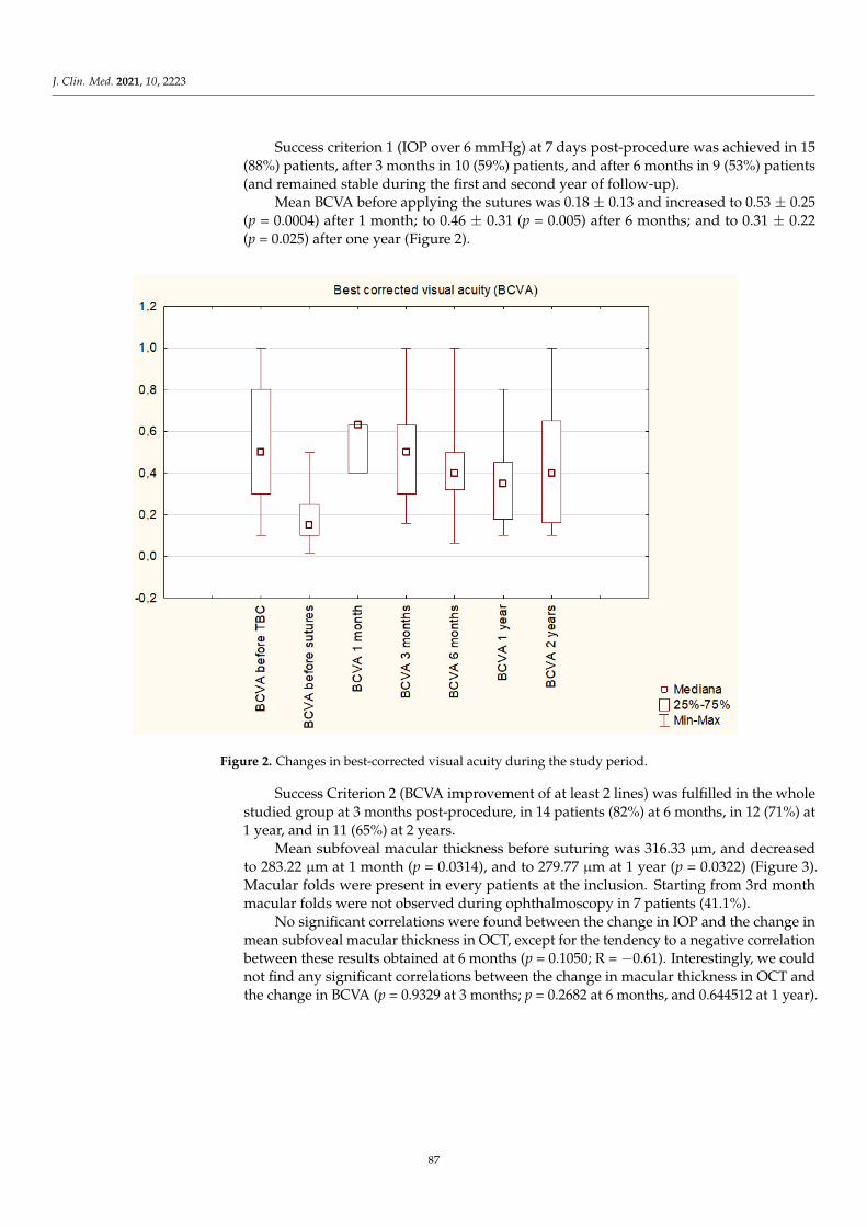
J. Clin. Med. 2021, 10, 2223
Success criterion 1 (IOP over 6 mmHg) at 7 days post-procedure was achieved in 15(88%) patients, after 3 months in 10 (59%) patients, and after 6 months in 9 (53%) patients(and remained stable during the first and second year of follow-up).
Mean BCVA before applying the sutures was 0.18 ± 0.13 and increased to 0.53 ± 0.25(p = 0.0004) after 1 month; to 0.46 ± 0.31 (p = 0.005) after 6 months; and to 0.31 ± 0.22(p = 0.025) after one year (Figure 2).
μμ μ
Figure 2. Changes in best-corrected visual acuity during the study period.
Success Criterion 2 (BCVA improvement of at least 2 lines) was fulfilled in the wholestudied group at 3 months post-procedure, in 14 patients (82%) at 6 months, in 12 (71%) at1 year, and in 11 (65%) at 2 years.
Mean subfoveal macular thickness before suturing was 316.33 µm, and decreasedto 283.22 µm at 1 month (p = 0.0314), and to 279.77 µm at 1 year (p = 0.0322) (Figure 3).Macular folds were present in every patients at the inclusion. Starting from 3rd monthmacular folds were not observed during ophthalmoscopy in 7 patients (41.1%).
No significant correlations were found between the change in IOP and the change inmean subfoveal macular thickness in OCT, except for the tendency to a negative correlationbetween these results obtained at 6 months (p = 0.1050; R = −0.61). Interestingly, we couldnot find any significant correlations between the change in macular thickness in OCT andthe change in BCVA (p = 0.9329 at 3 months; p = 0.2682 at 6 months, and 0.644512 at 1 year).
87
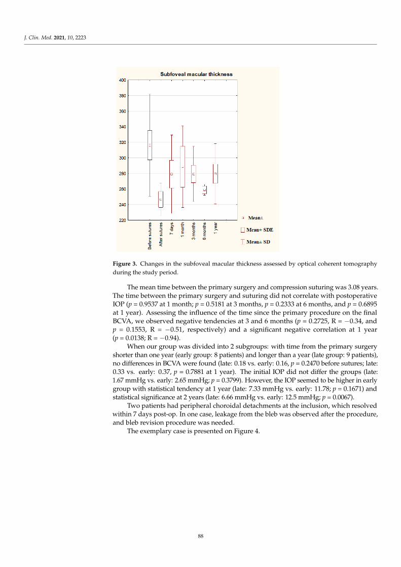
J. Clin. Med. 2021, 10, 2223
Figure 3. Changes in the subfoveal macular thickness assessed by
−
−−
−
Figure 3. Changes in the subfoveal macular thickness assessed by optical coherent tomographyduring the study period.
The mean time between the primary surgery and compression suturing was 3.08 years.The time between the primary surgery and suturing did not correlate with postoperativeIOP (p = 0.9537 at 1 month; p = 0.5181 at 3 months, p = 0.2333 at 6 months, and p = 0.6895at 1 year). Assessing the influence of the time since the primary procedure on the finalBCVA, we observed negative tendencies at 3 and 6 months (p = 0.2725, R = −0.34, andp = 0.1553, R = −0.51, respectively) and a significant negative correlation at 1 year(p = 0.0138; R = −0.94).
When our group was divided into 2 subgroups: with time from the primary surgeryshorter than one year (early group: 8 patients) and longer than a year (late group: 9 patients),no differences in BCVA were found (late: 0.18 vs. early: 0.16, p = 0.2470 before sutures; late:0.33 vs. early: 0.37, p = 0.7881 at 1 year). The initial IOP did not differ the groups (late:1.67 mmHg vs. early: 2.65 mmHg; p = 0.3799). However, the IOP seemed to be higher in earlygroup with statistical tendency at 1 year (late: 7.33 mmHg vs. early: 11.78; p = 0.1671) andstatistical significance at 2 years (late: 6.66 mmHg vs. early: 12.5 mmHg; p = 0.0067).
Two patients had peripheral choroidal detachments at the inclusion, which resolvedwithin 7 days post-op. In one case, leakage from the bleb was observed after the procedure,and bleb revision procedure was needed.
The exemplary case is presented on Figure 4.
88

J. Clin. Med. 2021, 10, 2223
(A) (B)
(C)
(D)
Figure 4. Sample case. (A) Eye fundus with hypotony maculopathy; (B) compressive sutures—1 day after the surgery (bluearrow points the sutures); (C) macular OCT before suturing; (D) macular OCT after suturing.
4. Discussion
The wide range of reported chronic hypotony after antiglaucoma procedures is dueto the lack of a standardized definition [31,32]. Additionally, some eyes require very lowIOP to stop or slow down glaucoma progression [33]. In cases when pre-surgery IOP haslower values or when glaucomatous optic neuropathy is advanced, an IOP below 6 mmHg without cataract progression, choroidal effusion, or maculopathy, and with improvedglaucoma is considered a surgical success [34]. In this study, transconjunctival suturing wasperformed when the IOP maintained under 6 mmHg and the BCVA remained significantlydecreased due to hypotony maculopathy.
The perfect time for intervention in case of hypotony after antiglaucoma procedure isalso not clear. The tendency to delay the treatment is observed because clinical experiencesuggests that most eyes with chronic hypotony following a trabeculectomy maintain goodvisual acuity without complications [11]. Moreover, spontaneous recovery from hypotonycaused by natural healing processes is frequent [34]. Additionally, a lot of procedures
89
J. Clin. Med. 2021, 10, 2223
designed to cure hypotony turn out to be unsuccessful: 5 of our patients had previouslyhad blood injected to the bleb to enhance healing without prolonged effect.
The duration of hypotony has been reported to have no correlation with final visualoutcomes [11,35]. In the case of our patients with hypotony maculopathy, we observedthat the time since the primary procedure indeed did not influence the final IOP levelafter transconjunctival suturing. If timely managed, IOP increase usually leads to therestoration of the normal smooth architecture of the retina, allowing for realignment ofphotoreceptors and visual recovery [31]. On the other hand, prolonged hypotony causesirreversible fibrosis within the retina, choroid, or sclera, maintaining the choroid in a foldedposition [31]. Scoralick et al. found resolution of hypotony maculopathy in 85% of thecases following surgical reintervention, but none of the variables investigated (includingthe time interval between trabeculectomy and flap resuturing) was significantly associatedwith postoperative maculopathy resolution [36].
Numerous risk factors for hypotony after glaucoma surgery have been identified,including myopia, young age, antimetabolite use, pre-existing inflammation, aphakia,and old age accompanied by a thin conjunctiva and thinner CCT [11,37,38]. Besides theapplication of an antifibrotic agent, male gender, high myopia, young age, and patientsreceiving primary filtering surgery have also been associated with an increased risk ofhypotony maculopathy [39,40]. In our study, five (29%) patients underwent primaryprocedure at the age below 40 because of pigmentary glaucoma or traumatic glaucoma. Itconfirms the susceptibility of younger patients to hypotony maculopathy. Additionally,all of these patients were myopic males, which suggests that care needs to be taken in thisgroup during the primary procedure to avoid hypotony, with lower titers of antimetabolitesand tight bleb suturing. Lower scleral rigidity in younger patients is believed to be relatedto the development of hypotony maculopathy [14,40]. Additionally, myopic eyes tend tohave thinner sclera, which is related to a general loss of collagen and proteoglycans [41]and makes sclera more vulnerable to collapse during hypotony. Further, males tend to havelower scleral rigidity and a correlation between male gender and hypotony maculopathyhas been ascertained [42].
In our practice, transconjunctival sutures are an effective and safe method of increasingIOP and improving visual acuity in patients with hypotony maculopathy as a complicationof antiglaucoma procedure. In a retrospective study, Letarte et al. showed a significantIOP increase and BCVA improvement six months after transconjunctival scleral flap resu-turing [43]. Eha et al. [22,44] published a prospective case study describing the outcomeof 16 patients whose mean IOP was 9.6 mmHg and mean BCVA was 20/60, six monthsfollowing the procedure. These results are similar to ours with mean IOP value of 9 mmHg1 year after suturing. We observed a gradual decrease in mean IOP in the course of thestudy, with the highest values one month postoperatively.
Although the success in IOP increase was not observed in every patient, duringthe early postoperative period we observed an improvement of the visual acuity in thewhole group, similarly to the results observed by Scoralick [36]. It shows the efficacyof transconjunctival sutures regarding BCVA improvement, and probably additionallyconfirms that numerous definitions of hypotony are not accurate, as in some patients anincrease in IOP even below 6 mmHg allowed for BCVA improvement.
In our group, but not in every case, it was possible to obtain the stable IOP increase inpost-surgical period, which may have some possible reasons. First, it may be connectedwith not enough traction obtained during placement of the sutures, which is usually con-nected with the changes of the ocular tissues during early postsurgical period (for example,the decrease in conjunctival oedema or moving of the sutures through the conjunctiva).Tight suturing with the attachment to the superficial parts of the sclera in case of extensivelyhyperfiltarating and elevated blebs may also be problematic. Additionally, it is possiblethat after MMC application during primary procedure the scleral tissues are weakened andmelted which makes transconjunctival suturing not enough for limitation of the outflow. Insuch cases, the revision with opening of conjunctiva and direct suturing of the sclera may
90
J. Clin. Med. 2021, 10, 2223
be beneficial. Finally, after antiglaucoma procedure prolonged ciliary body hypoperfusionand hyposecretion is possible.
Clinical assessment had led us to the assumption that a shorter time since the primaryprocedure may enable obtaining a more prominent increase in the IOP level, which wasnot confirmed by our results: in this study, at no time-point was IOP correlated with thetime since the primary surgery, which was also observed by other authors [36]. However,in our results a longer time since the primary surgery tended to have a negative impact onthe final BCVA. It may be related to the observation that when left untreated, hypotonymaculopathy can have long-term visual consequences [7] with irreversible chorioretinalfolds resulting from fibrosis within the retina, choroids, or sclera [31,45]. Additionally,in our study the patients with sutures placed earlier tended to have higher IOP valuesin longer observation period. This is why our results may provide an argument for anearlier intervention in maculopathy hypotony. However, in our opinion, the surprisingfinding of this study is the lack of correlation between the improvement in BCVA and thechanges in subfoveal macular thickness determined in OCT. It may be explained by the factthat during the study, intra- or sub-retinal fluid was not observed; thus, the initial BCVAdecrease after the antiglaucoma procedure was not caused by any disturbances in themorphology of the retinal layers but rather by their folding. Additionally, the improvementin BCVA may be partially explained by the restoration of the anterior segment structuresafter the increase in IOP [46–48]. The changes in the hemodynamics of the choroid affectedby the IOP increase may also be involved in BCVA improvement [49].
In general, the highest IOP during the follow-up period was measured on postop-erative Day 7. Furthermore, this spike in IOP seemed to be beneficial for expediting theresolution of any preexisting serous choroidal detachments [16] and cause quicker improve-ment in BCVA, as observed in this study. However, in such cases, the improvement ofthe visual acuity was not stable; all three patients with the decrease in BCVA observed atsix months belonged to this group. The highest values of early postoperative IOP wereobserved in the group of younger patients with pigmentary glaucoma, which may also beconnected with scleral vulnerability, as mentioned earlier. On the other hand, such highspikes in IOP may be potentially harmful in the case of severely damaged visual field inadvanced glaucoma.
Except for the observed IOP spikes, transconjunctival sutures seem to be a safe pro-cedure. In the course of the study, we observed only one patient with avascular thin blebwho suffered from bleb leakage and needed bleb revision, which involved covering it withthe mobilized conjunctiva.
To sum up, transconjunctival sutures seem to be an effective and safe method totreat hypotony maculopathy after glaucoma surgery. In the study, the improvement inBCVA started with the elevation of IOP at the early postoperative period and remainedstable during the observation time. Our results also provide an argument for an earlierintervention in case of clinical hypotony after glaucoma surgery.
Author Contributions: Conceptualization, E.K.-J. and T.Z.; methodology, E.K.-J. and T.Z.; software,D.W.-D.; formal analysis, E.K.-J. and D.W.-D.; investigation, E.K.-J., D.W.-D. and A.S.; resourcesT.Z.; data curation D.W.-D.; writing—original draft preparation E.K.-J.; writing—review and editing,E.K.-J., D.W.-D., A.S. and T.Z.; supervision, T.Z. All authors have read and agreed to the publishedversion of the manuscript.
Funding: This research received no external funding.
Institutional Review Board Statement: The study was conducted according to the guidelines of theDeclaration of Helsinki, and approved by the Local Board of Ethics Committee of Medical Universityof Lublin (Approval nr127/2015).
Informed Consent Statement: Informed consent was obtained from all subjects involved in thestudy.
Data Availability Statement: Data are available from the corresponding author on demand.
91

J. Clin. Med. 2021, 10, 2223
Conflicts of Interest: The authors declare no conflict of interest.
References
1. Quigley, H.A.; Broman, A.T. The number of people with glaucoma worldwide in 2010 and 2020. Br. J. Ophthalmol. 2006, 90,262–267. [CrossRef] [PubMed]
2. Weinreb, R.N.; Aung, T.; Medeiros, F.A. The pathophysiology and treatment of glaucoma: A review. JAMA 2014, 14, 1901–1911.[CrossRef]
3. Weinreb, R.N.; Khaw, P.T. Primary open-angle glaucoma. Lancet 2004, 22, 1711–1720. [CrossRef]4. Khaw, P.T.; Chiang, M.; Shah, P.; Sii, F.; Lockwood, A.; Khalili, A. Enhanced Trabeculectomy: The Moorfields Safer Surgery
System. Dev. Ophthalmol. 2017, 59, 15–35. [CrossRef] [PubMed]5. Joseph, J.P.; Miller, M.H.; Hitchings, R.A. Wound healing as a barrier to successful filtration surgery. Eye 1988, 2, S113–S123.
[CrossRef]6. Costa, V.P.; Arcieri, E.S. Hypotony maculopathy. Acta Ophthalmol. Scand. 2007, 85, 586–597. [CrossRef] [PubMed]7. Seah, S.K.; Prata, J.A., Jr.; Minckler, D.S.; Baerveldt, G.; Lee, P.P.; Heuer, D.K. Hypotony following trabeculectomy. J. Glaucoma
1995, 4, 73–79. [CrossRef]8. Pederson, J.E. Ocular Hypotony. In The Glaucomas; Ritch, R., Krupin, T., Shields, M.B., Eds.; Mosby: St. Louis, MO, USA, 1996; pp.
385–395.9. Rahman, A.; Mendonca, M.; Simmons, R.B.; Simmons, R.J. Hypotony after glaucoma filtration surgery. Int. Ophthalmol. Clin.
2000, 40, 127–136. [CrossRef]10. Edmunds, B.; Thompson, J.R.; Salmon, J.F.; Wormald, R.P. The National Survey of Trabeculectomy. III. Early and late complications.
Eye 2002, 16, 297–303. [CrossRef]11. Saeedi, O.J.; Jefferys, J.L.; Solus, J.F.; Jampel, H.D.; Quigley, H.A. Risk factors for adverse consequences of low intraocular pressure
after trabeculectomy. J. Glaucoma 2014, 23, e60–e68. [CrossRef]12. Francis, B.A.; Hong, B.; Winarko, J.; Kawji, S.; Dustin, L.; Chopra, V. Vision loss and recovery after trabeculectomy: Risk and
associated risk factors. Arch. Ophthalmol. 2011, 129, 1011–1017. [CrossRef] [PubMed]13. Dellaporta, A. Creasing of retina in hypotonia. Klin. Mon. Augenheilkd. Augenarztl. Fortbild. 1954, 125, 872–878.14. Gass, J.D. Hypotony Maculopathy. In Contemporary Ophthalmology: Honoring Sir Stewart Duke-Elder; Bellows, J.G., Ed.; Williams
and Wilkins: Baltimore, MD, USA, 1972; pp. 336–343.15. Oyakhire, J.O.; Moroi, S.E. Clinical and anatomical reversal of long-term hypotony maculopathy. Am. J. Ophthalmol. 2004, 137,
953–995. [CrossRef] [PubMed]16. Bitrian, E.; Song, B.J.; Caprioli, J. Bleb revision for resolution of hypotony maculopathy following primary trabeculectomy. Am. J.
Ophthalmol. 2014, 158, 597–604.e1. [CrossRef] [PubMed]17. Matsuo, H.; Tomidokoro, A.; Tomita, G.; Araie, M. Topical application of autologous serum for the treatment of lateonset aqueous
oozing or point-leak through filtering bleb. Eye 2005, 19, 23–28. [CrossRef] [PubMed]18. Smith, M.F.; Magauran, R.G., 3rd; Betchkal, J.; Doyle, J.W. Treatment of postfiltration bleb leaks with autologous blood. Ophthal-
mology 1995, 102, 868–871. [CrossRef]19. Higashide, T.; Tagawa, S.; Sugiyama, K. Intraoperative Healon5 injection into blebs for small conjunctival breaks created during
trabeculectomy. J. Cataract Refract. Surg. 2005, 31, 1279–1282. [CrossRef]20. Hosoda, S.; Yuki, K.; Ono, T.; Tsubota, K. Ophthalmic viscoelastic device injection for the treatment of flat anterior chamber after
trabeculectomy: A case series study. Clin. Ophthalmol. 2013, 7, 1781–1785.21. Kurtz, S.; Leibovitch, I. Combined perfluoropropane gas and viscoelastic material injection for anterior chamber reformation
following trabeculectomy. Br. J. Ophthalmol. 2002, 86, 1225–1227. [CrossRef]22. Eha, J.; Hoffmann, E.M.; Wahl, J.; Pfeiffer, N. Flap suture—A simple technique for the revision of hypotony maculopathy following
trabeculectomy with mitomycin C. Graefes Arch. Clin. Exp. Ophthalmol. 2008, 246, 869–874. [CrossRef]23. Shirato, S.; Maruyama, K.; Haneda, M. Resuturing the sclera flap through conjunctiva for treatment of excess filtration. Am. J.
Ophthalmol. 2004, 137, 173–174. [CrossRef] [PubMed]24. Haynes, W.L.; Alward, W.L. Rapid visual recovery and longterm intraocular pressure control after donor scleral patch grafting
for trabeculectomy-induced hypotony maculopathy. J. Glaucoma 1995, 4, 200–201. [CrossRef] [PubMed]25. Harizman, N.; Ben-Cnaan, R.; Goldenfeld, M.; Levkovitch-Verbin, H.; Melamed, S. Donor scleral patch for treating hypotony due
to leaking and/or overfiltering blebs. J. Glaucoma 2005, 14, 492–496. [CrossRef] [PubMed]26. Bochmann, F.; Kaufmann, C.; Kipfer, A.; Thiel, M.A. Corneal patch graft for the repair of late-onset hypotony or filtering bleb leak
after trabeculectomy: A new surgical technique. J. Glaucoma 2014, 23, e76–e80. [CrossRef] [PubMed]27. Panday, M.; Shantha, B.; George, R.; Boda, S.; Vijaya, L. Outcomes of bleb excision with free autologous conjunctival patch
grafting for bleb leak and hypotony after glaucoma filtering surgery. J. Glaucoma 2011, 20, 392–397. [CrossRef] [PubMed]28. Dietlein, T.S.; Lappas, A.; Rosentreter, A. Secondary subconjunctival implantation of a biodegradable collagen-glycosaminoglycan
matrix to treat ocular hypotony following trabeculectomy with mitomycin C. Br. J. Ophthalmol. 2013, 97, 985–988. [CrossRef]29. Laspas, P.; Wahl, J.; Peters, H.; Prokosch-Willing, V.; Chronopoulos, P.; Grehn, F.; Pfeiffer, N.; Hoffmann, E.M. Outcome of Bleb
Revision With Autologous Conjunctival Graft Alone or Combined With Donor Scleral Graft for Late-onset Bleb Leakage withHypotony after Standard Trabeculectomy with Mitomycin C. J. Glaucoma 2021, 1, 175–179. [CrossRef]
92
J. Clin. Med. 2021, 10, 2223
30. Maruyama, K.; Shirato, S. Efficacy and safety of transconjunctival scleral flap resuturing for hypotony after glaucoma filteringsurgery. Graefes Arch. Clin. Exp. Ophthalmol. 2008, 246, 1751–1756. [CrossRef]
31. Jampel, H.D.; Pasquale, L.R.; Dibernardo, C. Hypotony maculopathy following trabeculectomy with mitomycin C. Arch.
Ophthalmol. 1992, 110, 1049–1050. [CrossRef]32. Thomas, M.; Vajaranant, T.S.; Aref, A.A. Hypotony maculopathy: Clinical presentation and therapeutic methods. Ophthalmol.
Ther. 2015, 4, 79–88. [CrossRef]33. Higashide, T.; Ohkubo, S.; Sugimoto, Y.; Kiuchi, Y.; Sugiyama, K. Persistent hypotony after trabeculectomy: Incidence and
associated factors in the Collaborative Bleb-Related Infection Incidence and Treatment Study. Jpn. J. Ophthalmol. 2016, 60, 309–318.[CrossRef] [PubMed]
34. Yun, S.; Chua, B.; Clement, C.I. Does Chronic Hypotony following Trabeculectomy Represent Treatment Failure? J. Curr. Glaucoma
Pract. 2015, 9, 12–15. [CrossRef] [PubMed]35. Cohen, S.M.; Flynn, H.W., Jr.; Palmberg, P.F.; Gass, J.D.; Grajewski, A.L.; Parrish, R.K., 2nd. Treatment of hypotony maculopathy
after trabeculectomy. Ophthalmic Surg. Lasers 1995, 26, 435–441. [CrossRef] [PubMed]36. Scoralick, A.L.B.; Almeida, I.; Ushida, M.; Dias, D.T.; Dorairaj, S.; Prata, T.S.; Kanadani, F.N. Hypotony Management through
Transconjunctival Scleral Flap Resuturing: Analysis of Surgical Outcomes and Success Predictors. J. Curr. Glaucoma Pract. 2017,11, 58–62. [CrossRef] [PubMed]
37. Stamper, R. Bilateral chronic hypotony following trabeculectomy with mitomycin C. J. Glaucoma 2001, 10, 325–328. [CrossRef]38. Silva, R.A.; Doshi, A.; Law, S.K.; Singh, K. Postfiltration hypotony maculopathy in young chinese myopic women with glaucoma-
tous appearing optic neuropathy. J. Glaucoma 2010, 19, 105–110. [CrossRef]39. Singh, K.; Byrd, S.; Egbert, P.R.; Budenz, D. Risk of hypotony after primary trabeculectomy with antifibrotic agents in a black
West African population. J. Glaucoma 1998, 7, 82–85. [CrossRef]40. Fannin, L.A.; Schiffman, J.C.; Budenz, D.L. Risk factors for hypotony maculopathy. Ophthalmology 2003, 110, 1185–1191. [CrossRef]41. Curtin, B.J.; Iwamoto, T.; Renaldo, D.P. Normal and staphylomatous sclera of high myopia. An electron microscopic study. Arch.
Ophthalmol. 1979, 97, 912–915. [CrossRef]42. Nicolela, M.T.; Carrillo, M.M.; Yan, D.B.; Rafuse, P.E. Relationship between central corneal thickness and hypotony maculo-pathy
after trabeculectomy. Ophthalmology 2007, 114, 1266–1271. [CrossRef] [PubMed]43. Letartre, L.; Basheikh, A.; Anctil, J.-L.; Des Marchais, B.; Goyette, A.; Kasner, O.P.; Lajoie, C. Transconjunctival suturing of the
scleral flap for overfiltration with hypotony maculopathy after trabeculectomy. Can. J. Ophthalmol. 2009, 44, 567–570. [CrossRef][PubMed]
44. Eha, J.; Hoffmann, E.M.; Pfeiffer, N. Long-term results after transconjunctival resuturing of the scleral flap in hypotony followingtrabeculectomy. Am. J. Ophthalmol. 2013, 155, 864–869. [CrossRef] [PubMed]
45. Tunç, Y.; Tetikoglu, M.; Kara, N.; Sagdık, H.M.; Özarpaci, S.; Elçioglu, M.N. Management of hypotony and flat anterior chamberassociated with glaucoma filtration surgery. Int. J. Ophthalmol. 2015, 8, 950–953.
46. Cashwell, L.F.; Martin, C.A. Axial length decrease accompanying successful glaucoma filtration surgery. Ophthalmology 1999, 106,2307–2311. [CrossRef]
47. Camp, A.S.; Weinreb, R.N. Hypotony Keratopathy Following Trabeculectomy. J. Glaucoma 2020, 29, 77–80. [CrossRef] [PubMed]48. Azuma, K.; Saito, H.; Takao, M.; Araie, M. Frequency of hypotonic maculopathy observed by spectral domain optical coherence
tomography in post glaucoma filtration surgery eyes. Am. J. Ophthalmol. Case Rep. 2020, 18, 100786. [CrossRef] [PubMed]49. Saeedi, O.; Pillar, A.; Jefferys, J.; Arora, K.; Friedman, D.; Quigley, H. Change in choroidal thickness and axial length with change
in intraocular pressure after trabeculectomy. Br. J. Ophthalmol. 2014, 98, 976–979. [CrossRef]
93


Journal of
Clinical Medicine
Article
Dysregulated Tear Film Proteins in Macular Edema Due to theNeovascular Age-Related Macular Degeneration Are Involvedin the Regulation of Protein Clearance, Inflammation, andNeovascularization
Mateusz Winiarczyk 1,*, Dagmara Winiarczyk 2 , Katarzyna Michalak 2, Kai Kaarniranta 3, Łukasz Adaszek 2,
Stanisław Winiarczyk 2 and Jerzy Mackiewicz 1
�����������������
Citation: Winiarczyk, M.;
Winiarczyk, D.; Michalak, K.;
Kaarniranta, K.; Adaszek, Ł.;
Winiarczyk, S.; Mackiewicz, J.
Dysregulated Tear Film Proteins in
Macular Edema Due to the
Neovascular Age-Related Macular
Degeneration Are Involved in the
Regulation of Protein Clearance,
Inflammation, and
Neovascularization. J. Clin. Med.
2021, 10, 3060. https://doi.org/
10.3390/jcm10143060
Academic Editor: Gawecki Maciej
Received: 27 May 2021
Accepted: 6 July 2021
Published: 10 July 2021
Publisher’s Note: MDPI stays neutral
with regard to jurisdictional claims in
published maps and institutional affil-
iations.
Copyright: © 2021 by the authors.
Licensee MDPI, Basel, Switzerland.
This article is an open access article
distributed under the terms and
conditions of the Creative Commons
Attribution (CC BY) license (https://
creativecommons.org/licenses/by/
4.0/).
1 Department of Vitreoretinal Surgery, Medical University of Lublin, 20-079 Lublin, Poland;[email protected]
2 Department of Epizootiology, University of Life Sciences of Lublin, 20-400 Lublin, Poland;[email protected] (D.W.); [email protected] (K.M.); [email protected] (Ł.A.);[email protected] (S.W.)
3 Department of Ophthalmology, University of Eastern Finland and Kuopio University Hospital,70211 Kuopio, Finland; [email protected]
* Correspondence: [email protected]; Tel.: +48-692-476-464
Abstract: Macular edema and its further complications due to the leakage from the choroidal neovas-
cularization in course of the age-related macular degeneration (AMD) is a leading cause of blindness
among elderly individuals in developed countries. Changes in tear film proteomic composition
have been reported to occur in various ophthalmic and systemic diseases. There is an evidence
that the acute form of neovascular AMD may be reflected in the tear film composition. Tear film
was collected with Schirmer strips from patients with neovascular AMD and sex- and age-matched
control patients. Two-dimensional electrophoresis was performed followed by MALDI-TOF mass
spectrometry for identification of differentially expressed proteins. Quantitative analysis of the
differential electrophoretic spots was performed with Delta2D software. Altogether, 11 significantly
differentially expressed proteins were identified; of those, 8 were downregulated, and 3 were upregu-
lated in the tear film of neovascular AMD patients. The differentially expressed proteins identified
in tear film were involved in signaling pathways associated with impaired protein clearance, per-
sistent inflammation, and neovascularization. Tear film protein analysis is a novel way to screen
AMD-related biomarkers.
Keywords: age-related macular degeneration; AMD; proteomics; tear film; tear film proteome;
protein clearance; neovascularization; neovascular AMD
1. Introduction
Age-related macular degeneration (AMD) is a leading cause of blindness in elderlypatients in developed countries. The incidence of AMD is expected to increase by over50% in the next 20 years [1]. AMD affects central vision by evoking metamorphopsia,reading problems, and eventually legal blindness in its end stage. AMD can be dividedinto wet (neovascular) and dry (atrophic) forms. Usually, atrophic AMD progresses slowlyover years, while neovascular AMD with the presence of subretinal fluid and macularedema can develop in weeks due to the progressive growth of pathological choroidalvessels. Currently, there is no established treatment protocol for atrophic AMD, but anti-vascular endothelial growth factor (VEGF) intravitreal injections are a treatment of choicefor neovascular AMD. Although our awareness of AMD etiopathology has significantlyimproved in the past decade, the exact mechanisms underlying the disease are still vague.The cellular mechanisms of AMD are known to be linked to chronic oxidative stress (OS),
95
J. Clin. Med. 2021, 10, 3060
autophagy impairment, and inflammation that can ultimately lead to retinal pigmentepithelium (RPE) cell and photoreceptor death [2,3]. AMD development is also stronglyassociated with genetic variations and mutations in the complement system, as well as withmany environmental risk factors, such as smoking, hypercholesterolemia, arteriosclerosis,obesity, and unhealthy diet consumption [4].
Tear film is a mixture of lipids, water, and mucin that covers the surface of the eye.It protects against an environment-evoked irritation and smooths the corneal surface toimprove the refractive effect. Tear film is produced by lacrimal and accessory glands, aswell as by meibomian glands and goblet cells [5]. Since tear film is readily accessible, it hasbeen analyzed in many clinical studies on dry eye syndrome, diabetes, Parkinson’s disease,multiple sclerosis, and cancer [6–12].
Previous studies concerning proteomic changes that occur over the course of AMD havefocused mainly on the aqueous humor, the vitreous body, donor retinas, and blood [13–29].In each of these studies, significant differences in the expression of certain proteins havebeen discovered. The identified proteins are usually involved in metabolic pathways associ-ated with AMD. We reviewed most of the recent developments in AMD proteomic researchin our previous manuscript, in which we sought to determine whether the pathologicalprocess in the macula can result in tear film proteome changes [30]. Although we discov-ered various differentially expressed proteins, we were not able to perform quantitativeanalysis. In this study, we analyzed tear film samples from neovascular AMD patients toidentify and quantify proteins that were differentially expressed between a neovascularAMD group and a control group.
2. Materials and Methods
The study was approved by the Bioethical Committee of the Medical University ofLublin under declaration number KE-0254/238/2015. Informed consent was obtained fromevery individual enrolled in the study. The purpose and design of the study, as well as itspossible complications, were explained to every patient, and written consent was obtained.All experiments followed the provisions of the Declaration of Helsinki.
In total, 30 patients were included in the study: 15 patients in the neovascular AMDgroup, and 15 in the control group. The sex distribution was similar between the groups.All criteria-satisfying patients underwent a full ophthalmic examination by the sameophthalmologist (MW) that consisted of a visual acuity test, slit lamp examination, intraoc-ular pressure (IOP) measurement, spectral domain optical coherent tomography (SDOCT,Copernicus, Optopol Technologies, Zawiercie, Poland), and fluorescein angiography oroptical coherence tomography angiography (angio-OCT, RTVue XR 100 Avanti, Optovue,Fremont, CA, USA). The tear film break-up times (BUTs) were within normal limits (over10 s), and all the patients had Schirmer test results of greater than 15 mm in 5 min.
The inclusion criteria for the AMD group were as follows: active form of diseasefeaturing choroidal neovascularization (CNV) on fluorescein angiography or angio-OCT inat least one eye and the presence of subretinal fluid.
The exclusion criteria were any ocular surface diseases that would disturb the re-sults, e.g., dry eye syndrome, eye surface disorders, diabetic retinopathy, glaucoma, andprevious ocular surgery except for cataract extraction. Additionally, any moderately ad-vanced or advanced stage of any systemic disease, such as poorly controlled hypertension,cardiovascular disease, or autoimmune disorder, was an exclusion criterion.
The neovascular AMD group consisted of 15 patients, comprising 7 men and 8 women,with a mean age of 76.4 years (SD = 5.6). On slit lamp examination, the patients presentedwith an active form of AMD in at least one eye with subretinal fluid presence. All of themhad previously been treated with anti-VEGF therapy in one or both eyes. Environmentalrisk factors, smoking, and systemic diseases were assessed. The control subjects wererecruited from among patients who qualified for standard cataract surgery. The controlgroup consisted of 15 patients, comprising 8 men and 7 women, with a mean age of76.1 years (SD = 3.9). The mean IOP was within normal limits (8–21 mm Hg) in all patients.
96
J. Clin. Med. 2021, 10, 3060
For a statistical analysis of sex difference, a chi-square test on the contingency tablewith control and AMD on one side and male and female on the other side, making a2 × 2 table, and assuming the null hypothesis about independence of health from gender.Comparing the chi-square-calculated parameter with the tabular value, we dealt withindependent variables, meaning that the gender composition of the groups was neutral.According to the age comparison between the groups, we performed a t-test to determinethe difference in the means. In this case (group one mean: 76.4 ± 5.4; group two: 76.1 ± 3.9),the p-value equals 0.8782. By conventional criteria, this difference is not considered to bestatistically significant. To confirm that the groups were statistically similar, we preformedthe Shapiro–Wilk test of normality. We accepted that H0 assumed that the data in thecontrol and AMD groups were normally distributed. We also accepted H0 in the t-test,which meant that the average of 1′s population was considered to be equal to the averageof the 2′s population. In other words, the difference between the average of the 1 and2 populations was not big enough to be statistically significant.
Detailed information about the study and the control groups are presented in Supple-mentary Materials Tables S1 and S2.
2.1. Sample Preparation
Tear film was collected from each eye onto a Schirmer strip (TearFlo, HUB Pharmaceu-ticals LLC, Scottsdale, AZ, USA) [1,2]. Each collection was performed by the author M.W.in the morning hours between 8 and 11 a.m. If fluorescein angiography was performed,the material was always collected beforehand. Sterile gloves were always used by theinvestigator. The Schirmer strips were placed into the lower conjunctival sacs of both eyesat the one-third point of the eyelid as measured from the nasal canthus without anesthesia.There is currently no consensus about which method of collection should be used forproteomic analysis [3–6]. After the strips were held in place for 5 min, they were removed,transferred to 1.5-mL Eppendorf tubes without buffer, and immediately frozen at −80 ◦C.Next, the proteins were extracted in urea buffer for 3 h. Extraction was carried out at 4 ◦Cin the presence of protease inhibitor cocktail (P8340, Sigma Aldrich, St. Louis, MO, USA).The cocktail contained 104 mM 4-(2-aminoethyl)benzenesulfonyl fluoride hydrochloride(AEBSF), 80 µM aprotinin, 4 mM bestatin, 1.4 mM E-64, 2 mM leupeptin, and 1.5 mMpepstatin A. Each of these components has specific inhibitory properties. AEBSF and apro-tinin inhibit serine proteases, including trypsin, chymotrypsin, and plasmin, among others;bestatin inhibits aminopeptidases; E-64 inhibits cysteine proteases; leupeptin inhibits bothserine and cysteine proteases; and pepstatin A inhibits acid proteases (according to theSigma–Aldrich specification sheet). After extraction, the strips were removed, and theextracts were centrifuged at 1844× g for 10 min at 4 ◦C. The obtained supernatants werecollected and stored at −80 ◦C.
2.2. Protein Purification and Precipitation
The concentrations of the proteins were measured by a spectrophotometric method(MaestroNano Micro-Volume Spectrophotometer). Samples containing 150 µg of proteinswere transferred into 1.5-mL microcentrifuge tubes and diluted with water to a final volumeof 100 µL. Using a ReadyPrep 2-D Cleanup Kit (Bio-Rad, Hercules, CA, USA) the proteinpellets were obtained and resuspended by adding 300 µL of rehydration sample buffer(Bio-Rad). The supernatants were applied directly to immobilized pH gradient (IPG) strips(17 cm, pH 3¨C10, linear pH gradient, Bio-Rad).
After 12 h of gel rehydration the isoelectric focusing was performed at 60 kVh with acurrent limit of 50 µA per strip (Hoefer IEF100). Before second-dimension separation, theIPG strips were equilibrated in two equilibration buffers (50 mM Tris-HCl, pH 8.8, 6 M urea,30% glycerol, 2% sodium dodecyl sulfate (SDS)). The first buffer contained dithiothreitol(2%), while the second buffer contained iodoacetamide (2.5%) instead of dithiothreitol. Theduration of each equilibration step was 15 min. The second dimension of electrophoreticseparation was conducted using 12.5% polyacrylamide gels in a Bio-Rad PROTEAN II
97

J. Clin. Med. 2021, 10, 3060
xi Cell (Bio-Rad). Vertical separation was performed at 600 V/50 mA/30 W in 0.025 MTris/Gly buffer (pH 8.3). After electrophoretic separation, the proteins were silver stainedin accordance with the methods of Shevchenko et al. [7].
2.3. Preparation of Proteins for MALDI Identification
The spots of interest were excised from the gels by scalpel, transferred into microtubes,washed with H2O, and distained. After that, dithiothreitol reduction and iodoacetamidealkylation were performed. The gel pieces were covered with trypsin solution contain-ing 50 mM ammonium bicarbonate and placed in an autoclave overnight to digest at37 ◦C. Next, the peptides were extracted from the gel pieces with 50 µL of acetonitrile(ACN):H2O:trifluoroacetic acid (TFA) (50:45:5) solution. Extraction was performed in anultrasonic bath at room temperature and was repeated three times (each step lasted 15 min).The extracts were collected and concentrated in a CentriVap (Labconco, Kansas City, MO,USA). The obtained peptide pellets were dissolved in 10 µL of 0.1% TFA and purifiedwith ZipTip Sample Prep Pipette Tips (0.2 µL of C18 iod, Merck, Darmstadt, Germany) inaccordance with a standard procedure.
2.4. MALDI Analysis
Finally, 1 µL of each purified peptide sample was spotted onto an AnchorChip MALDIplate with hydrophobic coating and calibrant anchors. Next, 1 µL of alpha-cyano-4-hydroxycinnamic acid (HCCA, Bruker, Billerica, MA, USA) matrix solution was pipettedonto the dry peptide sample. A peptide calibration standard (Peptide Calibration Stan-dard II, Bruker) was spotted on the calibrant spots. The mass spectra were recorded inactive positive reflection mode by an Ultraflex III MALDI-TOF/TOF spectrometer (Bruker).All spectra were collected within the 700–4000 m/z range. The collected spectra weresmoothed (Savitsky–Golay method) and the baseline corrected (Top Hat baseline algo-rithm) in flexAnalysis 3.0 software (Bruker). A list of peaks in the range of 700–4000 m/zfor a signal-to-noise ratio greater than 3 was also generated in flexAnalysis 3.0. Afterremoval of impurities, the final peak list was transferred to BioTools 3.2 (Bruker) andcompared with Mascot 2.2 software using the Swiss–Prot database. Other parameters wereset as follows: the maximum error in both MS and MS/MS was 0.3 Da; the obligatorymodification was carbamidomethylation of cysteine; and the possible modifications weremethionine oxidation, serine, and threonine phosphorylation, methionine dioxidation,and protein N-terminal acetylation. Results with scores above 56 were considered statisti-cally significant. The peptide mass fingerprint spectra were analyzed in MS/MS mode toconfirm the amino acid sequences.
2.5. Visual and Statistical Analysis
The stained gels were scanned using a GE Image Scanner III (GE Healthcare, Warsaw,Poland) and further processed by Delta2D software (version 4.7, DECODON). The Delta2Dsoftware enabled quantitation of the spots and creation of protein expression profiles. Theutilized program uses gel image warping (correction of positional spot variations andmatching of images) to create a so-called fused image. This image is a proteome mapcontaining every protein spot obtained on every gel during the whole experiment. Afterthe fused image was created, the spots were detected. False-negative and false-positiveprotein spots were determined manually. To calculate the expression ratios (Rts; spotvolumes relative to the group means), a quantitation table was generated, and the volume-normalized values were statistically analyzed. In this experiment, the mean volume of agiven spot in the control group was the denominator of the Rt parameter.
In the case of gel statistical analysis after normalization, we used a t-test for twoanalyzed groups with p-values based on t-distribution and alpha (overall all thresholdp-value): 0.05. We took a Rt value greater than 1.5 as overexpressed and below 0.67 assuppressed.
98
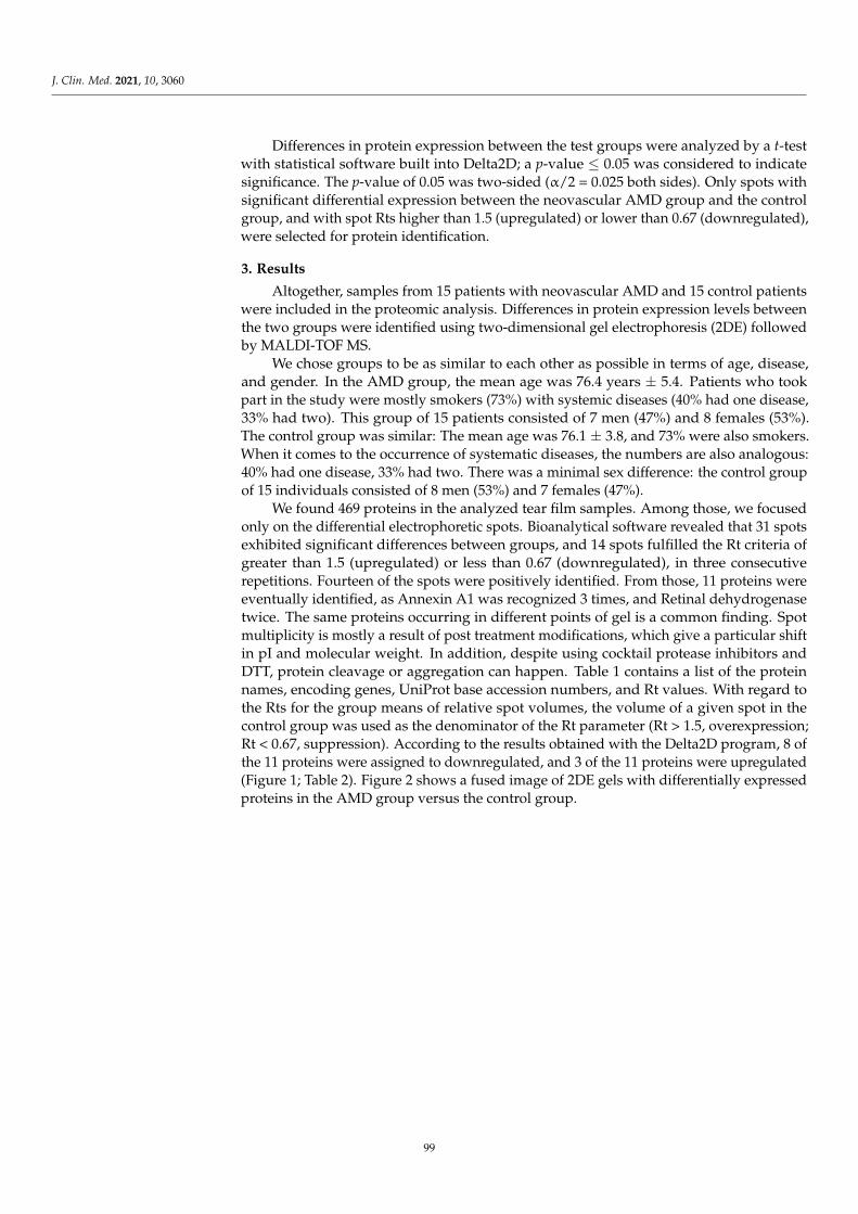
J. Clin. Med. 2021, 10, 3060
Differences in protein expression between the test groups were analyzed by a t-testwith statistical software built into Delta2D; a p-value ≤ 0.05 was considered to indicatesignificance. The p-value of 0.05 was two-sided (α/2 = 0.025 both sides). Only spots withsignificant differential expression between the neovascular AMD group and the controlgroup, and with spot Rts higher than 1.5 (upregulated) or lower than 0.67 (downregulated),were selected for protein identification.
3. Results
Altogether, samples from 15 patients with neovascular AMD and 15 control patientswere included in the proteomic analysis. Differences in protein expression levels betweenthe two groups were identified using two-dimensional gel electrophoresis (2DE) followedby MALDI-TOF MS.
We chose groups to be as similar to each other as possible in terms of age, disease,and gender. In the AMD group, the mean age was 76.4 years ± 5.4. Patients who tookpart in the study were mostly smokers (73%) with systemic diseases (40% had one disease,33% had two). This group of 15 patients consisted of 7 men (47%) and 8 females (53%).The control group was similar: The mean age was 76.1 ± 3.8, and 73% were also smokers.When it comes to the occurrence of systematic diseases, the numbers are also analogous:40% had one disease, 33% had two. There was a minimal sex difference: the control groupof 15 individuals consisted of 8 men (53%) and 7 females (47%).
We found 469 proteins in the analyzed tear film samples. Among those, we focusedonly on the differential electrophoretic spots. Bioanalytical software revealed that 31 spotsexhibited significant differences between groups, and 14 spots fulfilled the Rt criteria ofgreater than 1.5 (upregulated) or less than 0.67 (downregulated), in three consecutiverepetitions. Fourteen of the spots were positively identified. From those, 11 proteins wereeventually identified, as Annexin A1 was recognized 3 times, and Retinal dehydrogenasetwice. The same proteins occurring in different points of gel is a common finding. Spotmultiplicity is mostly a result of post treatment modifications, which give a particular shiftin pI and molecular weight. In addition, despite using cocktail protease inhibitors andDTT, protein cleavage or aggregation can happen. Table 1 contains a list of the proteinnames, encoding genes, UniProt base accession numbers, and Rt values. With regard tothe Rts for the group means of relative spot volumes, the volume of a given spot in thecontrol group was used as the denominator of the Rt parameter (Rt > 1.5, overexpression;Rt < 0.67, suppression). According to the results obtained with the Delta2D program, 8 ofthe 11 proteins were assigned to downregulated, and 3 of the 11 proteins were upregulated(Figure 1; Table 2). Figure 2 shows a fused image of 2DE gels with differentially expressedproteins in the AMD group versus the control group.
99
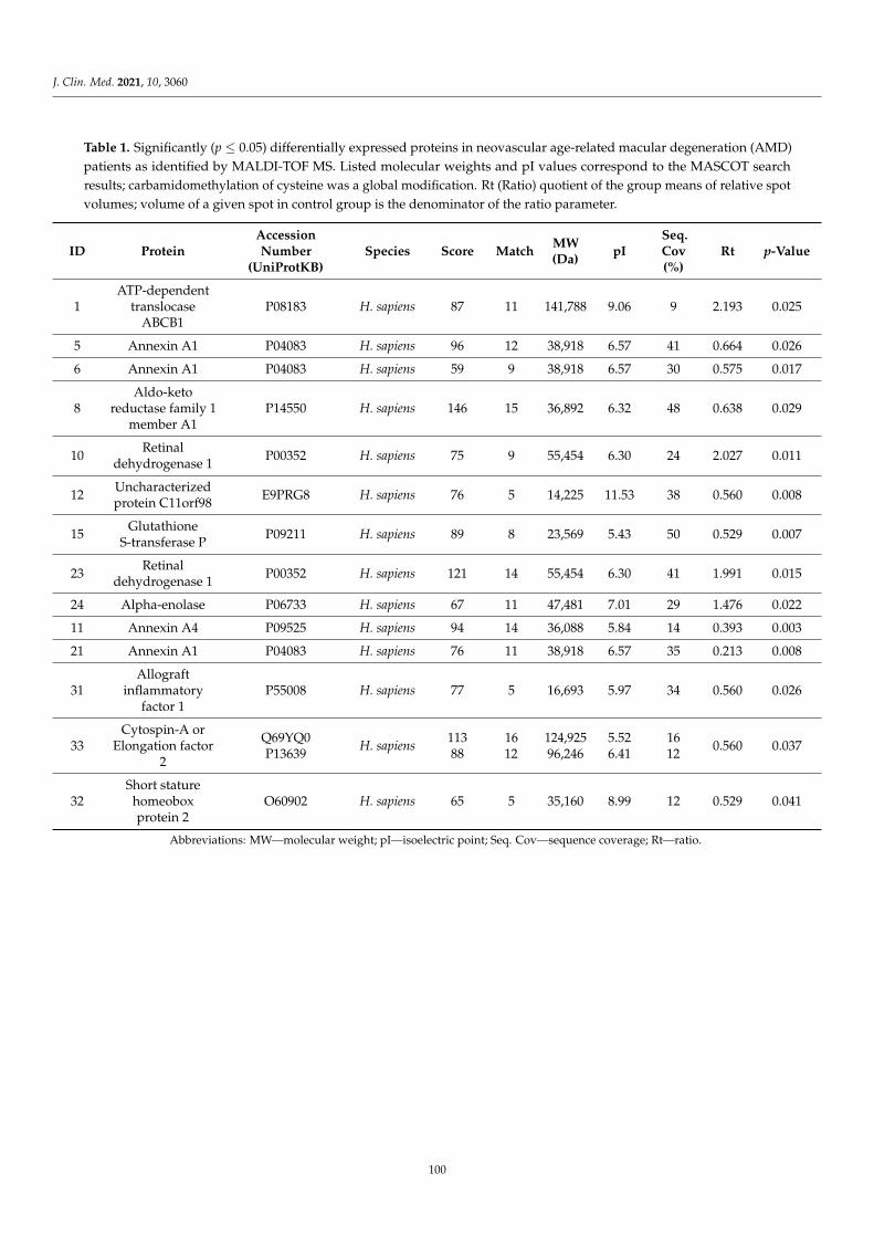
J. Clin. Med. 2021, 10, 3060
Table 1. Significantly (p ≤ 0.05) differentially expressed proteins in neovascular age-related macular degeneration (AMD)patients as identified by MALDI-TOF MS. Listed molecular weights and pI values correspond to the MASCOT searchresults; carbamidomethylation of cysteine was a global modification. Rt (Ratio) quotient of the group means of relative spotvolumes; volume of a given spot in control group is the denominator of the ratio parameter.
ID ProteinAccessionNumber
(UniProtKB)Species Score Match
MW(Da)
pISeq.Cov(%)
Rt p-Value
1ATP-dependent
translocaseABCB1
P08183 H. sapiens 87 11 141,788 9.06 9 2.193 0.025
5 Annexin A1 P04083 H. sapiens 96 12 38,918 6.57 41 0.664 0.026
6 Annexin A1 P04083 H. sapiens 59 9 38,918 6.57 30 0.575 0.017
8Aldo-keto
reductase family 1member A1
P14550 H. sapiens 146 15 36,892 6.32 48 0.638 0.029
10Retinal
dehydrogenase 1P00352 H. sapiens 75 9 55,454 6.30 24 2.027 0.011
12Uncharacterizedprotein C11orf98
E9PRG8 H. sapiens 76 5 14,225 11.53 38 0.560 0.008
15Glutathione
S-transferase PP09211 H. sapiens 89 8 23,569 5.43 50 0.529 0.007
23Retinal
dehydrogenase 1P00352 H. sapiens 121 14 55,454 6.30 41 1.991 0.015
24 Alpha-enolase P06733 H. sapiens 67 11 47,481 7.01 29 1.476 0.022
11 Annexin A4 P09525 H. sapiens 94 14 36,088 5.84 14 0.393 0.003
21 Annexin A1 P04083 H. sapiens 76 11 38,918 6.57 35 0.213 0.008
31Allograft
inflammatoryfactor 1
P55008 H. sapiens 77 5 16,693 5.97 34 0.560 0.026
33Cytospin-A or
Elongation factor2
Q69YQ0P13639
H. sapiens11388
1612
124,92596,246
5.526.41
1612
0.560 0.037
32Short stature
homeoboxprotein 2
O60902 H. sapiens 65 5 35,160 8.99 12 0.529 0.041
Abbreviations: MW—molecular weight; pI—isoelectric point; Seq. Cov—sequence coverage; Rt—ratio.
100
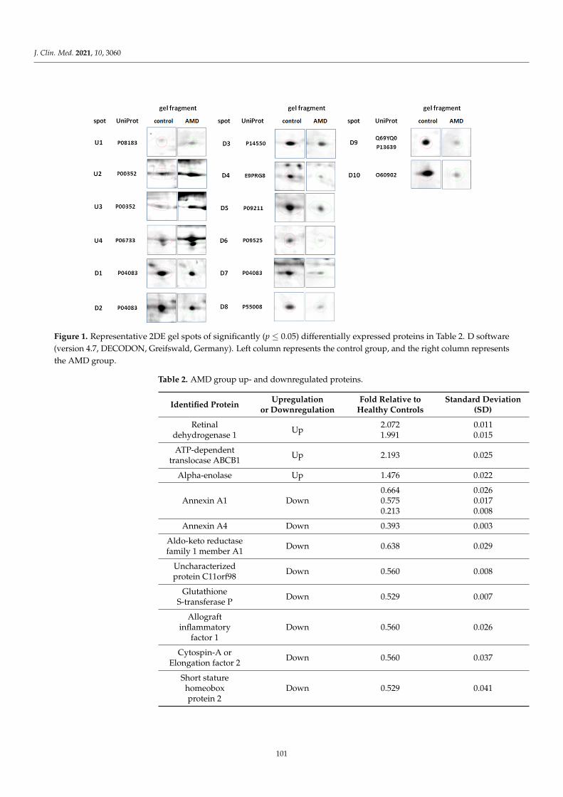
J. Clin. Med. 2021, 10, 3060
≤Figure 1. Representative 2DE gel spots of significantly (p ≤ 0.05) differentially expressed proteins in Table 2. D software(version 4.7, DECODON, Greifswald, Germany). Left column represents the control group, and the right column representsthe AMD group.
Table 2. AMD group up- and downregulated proteins.
Identified ProteinUpregulation
or DownregulationFold Relative to
Healthy ControlsStandard Deviation
(SD)
Retinaldehydrogenase 1
Up2.0721.991
0.0110.015
ATP-dependenttranslocase ABCB1
Up 2.193 0.025
Alpha-enolase Up 1.476 0.022
Annexin A1 Down0.6640.5750.213
0.0260.0170.008
Annexin A4 Down 0.393 0.003
Aldo-keto reductasefamily 1 member A1
Down 0.638 0.029
Uncharacterizedprotein C11orf98
Down 0.560 0.008
GlutathioneS-transferase P
Down 0.529 0.007
Allograftinflammatory
factor 1Down 0.560 0.026
Cytospin-A orElongation factor 2
Down 0.560 0.037
Short staturehomeoboxprotein 2
Down 0.529 0.041
101
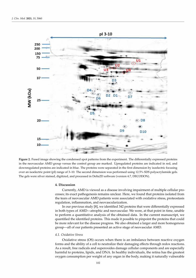
J. Clin. Med. 2021, 10, 3060
Figure 2. Fused image showing the condensed spot patterns from the experiment. The differentially expressed proteinsin the neovascular AMD group versus the control group are marked. Upregulated proteins are indicated in red, anddownregulated proteins are indicated in blue. The proteins were separated in the first dimension by isoelectric focusingover an isoelectric point (pI) range of 3–10. The second dimension was performed using 12.5% SDS polyacrylamide gels.The gels were silver stained, digitized, and processed in Delta2D software (version 4.7, DECODON).
4. Discussion
Currently, AMD is viewed as a disease involving impairment of multiple cellular pro-cesses; its exact pathogenesis remains unclear. Here, we found that proteins isolated fromthe tears of neovascular AMD patients were associated with oxidative stress, proteostasisregulation, inflammation, and neovascularization.
In our previous study [8], we identified 342 proteins that were differentially expressedin both types of AMD—atrophic and neovascular. We were, at that point in time, unableto perform a quantitative analysis of the obtained data. In the current manuscript, wequantified the identified proteins. This made it possible to pinpoint the proteins that couldbe more relevant for the disease progress. We also obtained a larger and more homogenousgroup—all of our patients presented an active stage of neovascular AMD.
4.1. Oxidative Stress
Oxidative stress (OS) occurs when there is an imbalance between reactive oxygenforms and the ability of a cell to neutralize their damaging effects through redox reactions.As a result, free radicals and superoxides damage cellular components and are especiallyharmful to proteins, lipids, and DNA. In healthy individuals, the retina has the greatestoxygen consumption per weight of any organ in the body, making it naturally vulnerable
102
J. Clin. Med. 2021, 10, 3060
to OS [3,31,32]. In our study, we identified a number of proteins involved in OS inductionand management in tear samples isolated from neovascular AMD patients. One of themost striking findings was the downregulation of glutathione S-transferase P. Glutathione(GSH) is one of the most important, ubiquitous antioxidant agents, whose role in the retinalanti-OS defense is well-established. It works by scavenging the reactive oxygen speciesand is a cofactor for GSH S-transferase P [9,10]. Its lowered concentration in the tear filmof AMD patients may suggest the impaired cell-detoxification mechanisms.
Aldo-keto reductase family 1 member A1 (AKR1A1) is yet another protein involvedin the cellular protection against OS, which was downregulated in our study, but itsconnection with the retina remains unclear [11,12].
Additionally, the identified protein retinal dehydrogenase 1 (RALDH1) is involvedin redox reactions. Its key function is to oxidize retinaldehyde into retinoic acid, whichparticipates in cell growth and differentiation and plays a critical role in the visual cycle [13].
Overall, the findings suggest that selected OS biomarkers can be found in tears fromneovascular AMD patients.
4.2. Protein Clearance
Increased OS can damage proteins, and damaged proteins must be removed to preventintracellular protein aggregation. Protein clearance impairment plays a crucial role in AMDdevelopment. Under normal conditions, retinal cells maintain proteostasis through twomajor mechanisms: proteasome-mediated degradation and lysosome-mediated autophagy.In AMD, impairment of phagocytosis leads to failure of the degradation of photoreceptorouter segments (POSs) in lysosomes, while impairment of autophagy leads to the accu-mulation of toxic protein aggregates and organelles, such as mitochondria [14]. SinceRPE cells are quiescent cells, the consequences of deficient proteostasis are potentiallydevastating [15].
Annexin A1 and A4 are a part of the calcium-dependent phospholipid-binding family.Annexins A, beside regulating the inflammatory process described above, are vitallyimportant to the autophagy process, and take an important part in the formation of thecytoskeleton, cell membrane, and in the cell signaling [16]. Annexin A1 is involved inthe autophagosome-lysosome fusion, and its upregulation seems to inhibit autophagyprocess via PI3K/AKT activation followed by Beclin-1 and ATG5-dependent autophagyinhibition [17,18]. This may lead to the pathological aggregation of debris material withinthe RPE-BM complex, called drusen, and further stimuli for the formation of the choroidalneovascularization (CNV).
Given all of the above findings, the upregulation of the Annexin A1 and A4 in thetears of patients with neovascular AMD may indicate that proteostasis is disturbed inAMD.
4.3. Chronic Inflammation and Neovascularization
Increased inflammation is well established to occur during AMD pathogenesis inresponse to chronic OS and disturbed proteostasis [14]. Short lasting inflammation is abeneficial host defense in cells, while prolonged inflammation of low intensity (parainflam-mation) can lead to CNV and cell death in the context of AMD [19,20].
Annexin A1 (ANXA1) was found in our study on three different electrophoretic spots,probably to its further post treatment modification, which suggests its strong presencein the AMD tear film. Its concentration in the samples obtained from AMD patients wasalmost 5 times higher than in the control group. Previous studies investigating the ANXA1impact on inflammation proved its significant anti-inflammatory potential [21–23]. It wasalso already found in the aqueous humor of the wet AMD patients (both Annexin A1 andA4) [24], in the drusen from the donors retina [25]. The impairment of the Annexins familyfunction is related to numerous diseases, also neurodegenerative disorders and glaucoma,although in none it seems to be a primary cause [26–28].
103
J. Clin. Med. 2021, 10, 3060
Alpha-Enolase, which was found to be upregulated in our AMD patients’ group, isanother protein that can act as an autoantigen in the autoimmune process, which wasalready connected with AMD. Elevated levels of the antibodies against α-enolase werefound in AMD patients’ serum [29,30]. It is also strongly connected with the develop-ment of cancer-associated retinopathy (CAR), Alzheimer’s disease (AD), cancer, and otherdiseases [31–33].
Another protein directly involved in inflammation and neovascularization process isallograft inflammatory factor 1 (AIF-1). In mouse models of neovascular AMD, it was highlyexpressed in an induced laser scarring spot, leading to NF-κB activation, and further CNVdevelopment [34]. It is also an established biomarker in local immune and inflammatoryresponse of the retinal cells [35–37]. The questionable aspect is the downregulation of AIF-1observed in our study, one would expect it to be upregulated.
Another hallmark of AMD is choroidal neovascularization (CNV), in which vesselssprout from the choroid and pass through the BM and the RPE, causing subretinal leakage,macular edema, and hemorrhages. In the end stage, a disciform scar is formed, withmesenchymal transition of RPE cells and general retinal disorganization. One of the keymodulators of this process is VEGF, and the treatment of choice is anti-VEGF delivered viaintravitreal injections. Although anti-VEGF administration has been a major breakthroughin AMD treatment, it has significant limitations: continued visits are necessary, macularscarring can occur, and patients can be refractory to treatment [38]. One of the proteinscrucial for the BRB development, ATP-dependent translocase ABCB1, was found to beupregulated in our study. ABCB1 is responsible for the cellular transport, being an effluxpump, and is commonly associated with various types of cancers, due to its role in themultidrug resistance (MDR) [39–41].
Scarring is the eventual effect of the CNV presence, whether due to the treatmentor the natural course of the disease. Elongation factor 2 was found to be downregulatedin our AMD group. This protein was also identified in the Müller glia in course of theproliferative vitreoretinopathy, which is essentially a scarring process [42]. Elongationfactors were also found to be downregulated in older retinas, which can partially explainits downregulation in our study [43].
Last of the identified proteins, short stature homeobox 2 has not been yet describedin the context of AMD, or retinal dysfunction, and it should probably be concerned as anaccidental finding. It was recently found that it may serve as a biomarker for bronchialsquamous cell carcinoma (SCC) [44].
All these findings suggest a strong inflammatory component that can be observedin the tear film of AMD patients, which stays in line with our current knowledge of thedisease.
4.4. Anti-VEGF Treatment
All of the patients included in our study were treated with intraocular anti-VEGFinjections over the course of a national drug program. This warranted quality patientselection and confirmation of medical history. The patients were first qualified by a localophthalmologist, and then the diagnosis was confirmed online by nationally board-certifiedretinal AMD specialists. In all cases, the samples were collected at the follow-up day beforethe anti-VEGF injection. Therefore, each patient was examined 28 to 31 days after previousanti-VEGF treatment. Since we did not include treatment-naïve patients with AMD, anti-VEGF treatment might have affected the protein expression results. This may explain whywe did not observe differential expression of certain proteins, such as VEGF or PDGF,even though differences in such proteins have been found in other studies [45,46]. On theother hand, this might have enabled the roles and differential expression of other proteinsinvolved in neovascularization. The VEGF pathway, although extremely important, iscertainly not the sole promotor of the growth of the new vessels. Preferably, a larger clinicalstudy should be conducted on samples collected from patients at different stages of AMDprogression.
104
J. Clin. Med. 2021, 10, 3060
4.5. Limitations of the Study
One major limitation of our study is the correlation between the tear film compositionand macular lesions. Although the tear film is not directly connected with the retina, it canbe altered by the partial blood–retinal barrier breakdown (BRB) in the course of AMD. BRBwas mainly described in diabetic retinopathy, and functions as a key factor in this disease,but the BRB can also be found in the neovascular AMD, where macular edema, subretinalfluid, and vitreous hemorrhages are present [47–50]. Thus, we believe that in an activephase of neovascular AMD, it is possible that the leakage from the pathological vessels canbe also detected in the tear film.
Another limitation of this study is that being a pilot study, we were not able toanalyze enough samples to reach adequate power of the tests used in statistical calculations.According to an amount of wet AMD cases in our region, we would need over 300 samplesfor each group. This will be done in the following experiments.
5. Conclusions
Tear film is a well-established material for obtaining biomarkers of various diseases.We believe that the findings of this study enhance the current understanding of AMD asa multifactorial disease with underlying persistent OS, cell clearance mechanism impair-ment, inflammation, and CNV. Although the identified proteins probably should not beconsidered verified biomarkers, the differences in their expression between groups suggestthat they are connected to ongoing pathological processes in the macula and tear film.Further studies are needed to confirm this possibility, preferably studies comparing thelevels of specific proteins in different body fluids, such as the plasma, aqueous humor, andtear film.
Supplementary Materials: Supplementary materials can be found at https://www.mdpi.com/article/10.3390/jcm10143060/s1. Supplementary Table S1: Characteristics of wet AMD patients.Supplementary, Table S2: Characteristics of control group patients.
Author Contributions: Conceptualization, M.W.; methodology, M.W., K.M., and S.W.; software,K.M.; validation, M.W., K.M., and Ł.A.; formal analysis, M.W.; investigation, M.W.; resources, M.W.,D.W., and S.W.; data curation, M.W.; writing—original draft preparation, M.W.; writing—reviewand editing, M.W., K.K., D.W., and S.W.; visualization, K.M.; supervision, K.K. and J.M.; projectadministration, M.W.; funding acquisition, M.W. All authors have read and agreed to the publishedversion of the manuscript.
Funding: This work was supported by the Polish National Science Centre (NCN). M.W. wassupported by Preludium grant number UMO-2017/25/N/NZ5/01875. D.W. was supported byPreludium grant number UMO-2016/23/N/NZ5/02576. The research materials supporting thispublication can be accessed by contacting the corresponding authors.
Institutional Review Board Statement: Ethical review and approval were waived for this study, dueto the standard procedure of tear film collection and non-invasive nature of the experiment.
Informed Consent Statement: Informed consent was obtained from all subjects involved in the study.
Data Availability Statement: Data available after contacting corresponding author: [email protected].
Conflicts of Interest: The authors declare no conflict of interest.
References
1. Li, K.; Chen, Z.; Duan, F.; Liang, J.; Wu, K. Quantification of Tear Proteins by SDS-PAGE with an Internal Standard Protein: ANew Method with Special Reference to Small Volume Tears. Graefes Arch. Clin. Exp. Ophthalmol. 2010, 248, 853–862. [CrossRef][PubMed]
2. Posa, A.; Bräuer, L.; Schicht, M.; Garreis, F.; Beileke, S.; Paulsen, F. Schirmer Strip vs. Capillary Tube Method: Non-InvasiveMethods of Obtaining Proteins from Tear Fluid. Ann. Anat. 2013, 195, 137–142. [CrossRef] [PubMed]
3. Green-Church, K.B.; Nichols, K.K.; Kleinholz, N.M.; Zhang, L.; Nichols, J.J. Investigation of the Human Tear Film Proteome UsingMultiple Proteomic Approaches. Mol. Vis. 2008, 14, 456–470.
105
J. Clin. Med. 2021, 10, 3060
4. González, N.; Iloro, I.; Durán, J.A.; Elortza, F.; Suárez, T. Evaluation of Inter-Day and Inter-Individual Variability of TearPeptide/Protein Profiles by MALDI-TOF MS Analyses. Mol. Vis. 2012, 18, 1572–1582.
5. Rentka, A.; Koroskenyi, K.; Harsfalvi, J.; Szekanecz, Z.; Szucs, G.; Szodoray, P.; Kemeny-Beke, A. Evaluation of CommonlyUsed Tear Sampling Methods and Their Relevance in Subsequent Biochemical Analysis. Ann. Clin. Biochem. 2017, 54, 521–529.[CrossRef]
6. Ablamowicz, A.F.; Nichols, J.J. Concentrations of MUC16 and MUC5AC Using Three Tear Collection Methods. Mol. Vis. 2017, 23,529–537.
7. Shevchenko, A.; Wilm, M.; Vorm, O.; Mann, M. Mass Spectrometric Sequencing of Proteins Silver-Stained Polyacrylamide Gels.Anal. Chem. 1996, 68, 850–858. [CrossRef]
8. Winiarczyk, M.; Kaarniranta, K.; Winiarczyk, S.; Adaszek, Ł.; Winiarczyk, D.; Mackiewicz, J. Tear Film Proteome in Age-RelatedMacular Degeneration. Graefe’s Arch. Clin. Exp. Ophthalmol. 2018, 256, 1127–1139. [CrossRef] [PubMed]
9. Tew, K.D. Redox in Redux: Emergent Roles for Glutathione S-Transferase P (GSTP) in Regulation of Cell Signaling and S-Glutathionylation. Biochem. Pharmacol. 2007, 73, 1257–1269. [CrossRef] [PubMed]
10. Sreekumar, P.G.; Ferrington, D.A.; Kannan, R. Glutathione Metabolism and the Novel Role of Mitochondrial GSH in RetinalDegeneration. Antioxidants 2021, 10, 661. [CrossRef]
11. Chen, W.-R.; Lan, Y.-W.; Chen, H.-L.; Chen, C.-M. AKR1A1 Deficiency Is Associated with High Risk of Alcohol-Induced FattyLiver Syndrome. FASEB J. 2018, 32, 546.6. [CrossRef]
12. Stomberski, C.T.; Anand, P.; Venetos, N.M.; Hausladen, A.; Zhou, H.-L.; Premont, R.T.; Stamler, J.S. AKR1A1 Is a NovelMammalian S-Nitroso-Glutathione Reductase. J. Biol. Chem. 2019, 294, 18285–18293. [CrossRef]
13. Morgan, C.A.; Hurley, T.D. Characterization of Two Distinct Structural Classes of Selective Aldehyde Dehydrogenase 1A1Inhibitors. J. Med. Chem. 2015, 58, 1964–1975. [CrossRef]
14. Kaarniranta, K.; Uusitalo, H.; Blasiak, J.; Felszeghy, S.; Kannan, R.; Kauppinen, A.; Salminen, A.; Sinha, D.; Ferrington, D.Mechanisms of Mitochondrial Dysfunction and Their Impact on Age-Related Macular Degeneration. Progress Retin. Eye Res.
2020, 100858. [CrossRef]15. Ferrington, D.A.; Sinha, D.; Kaarniranta, K. Defects in Retinal Pigment Epithelial Cell Proteolysis and the Pathology Associated
with Age-Related Macular Degeneration. Progress Retin. Eye Res. 2016, 51, 69–89. [CrossRef] [PubMed]16. Xi, Y.; Ju, R.; Wang, Y. Roles of Annexin A Protein Family in Autophagy Regulation and Therapy. Biomed. Pharmacother. 2020,
130, 110591. [CrossRef] [PubMed]17. Zhu, J.-F.; Huang, W.; Yi, H.-M.; Xiao, T.; Li, J.-Y.; Feng, J.; Yi, H.; Lu, S.-S.; Li, X.-H.; Lu, R.-H.; et al. Annexin A1-Suppressed
Autophagy Promotes Nasopharyngeal Carcinoma Cell Invasion and Metastasis by PI3K/AKT Signaling Activation. Cell Death
Dis. 2018, 9, 1–16. [CrossRef] [PubMed]18. White, I.J.; Bailey, L.M.; Aghakhani, M.R.; Moss, S.E.; Futter, C.E. EGF Stimulates Annexin 1-Dependent Inward Vesiculation in a
Multivesicular Endosome Subpopulation. EMBO J. 2006, 25, 1–12. [CrossRef]19. Datta, S.; Cano, M.; Ebrahimi, K.; Wang, L.; Handa, J.T. The Impact of Oxidative Stress and Inflammation on RPE Degeneration in
Non-Neovascular AMD. Progress Retin. Eye Res. 2017, 60, 201–218. [CrossRef]20. Kauppinen, A.; Paterno, J.J.; Blasiak, J.; Salminen, A.; Kaarniranta, K. Inflammation and Its Role in Age-Related Macular
Degeneration. Cell Mol. Life Sci. 2016, 73, 1765–1786. [CrossRef]21. D’Acquisto, F.; Merghani, A.; Lecona, E.; Rosignoli, G.; Raza, K.; Buckley, C.D.; Flower, R.J.; Perretti, M. Annexin-1 Modulates
T-Cell Activation and Differentiation. Blood 2007, 109, 1095–1102. [CrossRef]22. Sanches, J.M.; Correia-Silva, R.D.; Duarte, G.H.B.; Fernandes, A.M.A.P.; Sánchez-Vinces, S.; Carvalho, P.O.; Oliani, S.M.; Bortoluci,
K.R.; Moreira, V.; Gil, C.D. Role of Annexin A1 in NLRP3 Inflammasome Activation in Murine Neutrophils. Cells 2021, 10, 121.[CrossRef] [PubMed]
23. Yazid, S.; Gardner, P.J.; Carvalho, L.; Chu, C.J.; Flower, R.J.; Solito, E.; Lee, R.W.J.; Ali, R.R.; Dick, A.D. Annexin-A1 Restricts Th17Cells and Attenuates the Severity of Autoimmune Disease. J. Autoimmun. 2015, 58, 1–11. [CrossRef] [PubMed]
24. Kang, G.-Y.; Bang, J.Y.; Choi, A.J.; Yoon, J.; Lee, W.-C.; Choi, S.; Yoon, S.; Kim, H.C.; Baek, J.-H.; Park, H.S.; et al. Exosomal Proteinsin the Aqueous Humor as Novel Biomarkers in Patients with Neovascular Age-Related Macular Degeneration. J. Proteome Res.
2014, 13, 581–595. [CrossRef] [PubMed]25. Crabb, J.W.; Miyagi, M.; Gu, X.; Shadrach, K.; West, K.A.; Sakaguchi, H.; Kamei, M.; Hasan, A.; Yan, L.; Rayborn, M.E.; et al.
Drusen Proteome Analysis: An Approach to the Etiology of Age-Related Macular Degeneration. Proc. Natl. Acad. Sci. USA 2002,99, 14682–14687. [CrossRef]
26. Yang, Z.; Quigley, H.A.; Pease, M.E.; Yang, Y.; Qian, J.; Valenta, D.; Zack, D.J. Changes in Gene Expression in ExperimentalGlaucoma and Optic Nerve Transection: The Equilibrium between Protective and Detrimental Mechanisms. Investig. Ophthalmol.
Vis. Sci. 2007, 48, 5539–5548. [CrossRef] [PubMed]27. Iaccarino, L.; Ghirardello, A.; Canova, M.; Zen, M.; Bettio, S.; Nalotto, L.; Punzi, L.; Doria, A. Anti-Annexins Autoantibodies:
Their Role as Biomarkers of Autoimmune Diseases. Autoimmun. Rev. 2011, 10, 553–558. [CrossRef]28. Cañas, F.; Simonin, L.; Couturaud, F.; Renaudineau, Y. Annexin A2 Autoantibodies in Thrombosis and Autoimmune Diseases.
Thromb. Res. 2015, 135, 226–230. [CrossRef]29. Adamus, G.; Chew, E.Y.; Ferris, F.L.; Klein, M.L. Prevalence of Anti-Retinal Autoantibodies in Different Stages of Age-Related
Macular Degeneration. BMC Ophthalmol. 2014, 14, 154. [CrossRef]
106
J. Clin. Med. 2021, 10, 3060
30. Joachim, S.C.; Bruns, K.; Lackner, K.J.; Pfeiffer, N.; Grus, F.H. Analysis of IgG Antibody Patterns against Retinal Antigens andAntibodies to α-Crystallin, GFAP, and α-Enolase in Sera of Patients with “Wet” Age-Related Macular Degeneration. Graefe’s Arch.
Clin. Exp. Ophthalmol. 2006, 245, 619. [CrossRef]31. Adamus, G.; Aptsiauri, N.; Guy, J.; Heckenlively, J.; Flannery, J.; Hargrave, P.A. The Occurrence of Serum Autoantibodies against
Enolase in Cancer-Associated Retinopathy. Clin. Immunol. Immunopathol. 1996, 78, 120–129. [CrossRef]32. Dot, C.; Guigay, J.; Adamus, G. Anti-α-Enolase Antibodies in Cancer-Associated Retinopathy with Small Cell Carcinoma of the
Lung. Am. J. Ophthalmol. 2005, 139, 746–747. [CrossRef] [PubMed]33. Morohoshi, K.; Goodwin, A.M.; Ohbayashi, M.; Ono, S.J. Autoimmunity in Retinal Degeneration: Autoimmune Retinopathy and
Age-Related Macular Degeneration. J. Autoimmun. 2009, 33, 247–254. [CrossRef] [PubMed]34. Hikage, F.; Lennikov, A.; Mukwaya, A.; Lachota, M.; Ida, Y.; Utheim, T.P.; Chen, D.F.; Huang, H.; Ohguro, H. NF-KB Acti-
vation in Retinal Microglia Is Involved in the Inflammatory and Neovascularization Signaling in Laser-Induced ChoroidalNeovascularization in Mice. Exp. Cell Res. 2021, 403, 112581. [CrossRef] [PubMed]
35. Singh, R.K.; Occelli, L.M.; Binette, F.; Petersen-Jones, S.M.; Nasonkin, I.O. Transplantation of Human Embryonic Stem Cell-DerivedRetinal Tissue in the Subretinal Space of the Cat Eye. Stem Cells Dev. 2019, 28, 1151–1166. [CrossRef] [PubMed]
36. Hambright, D.; Park, K.-Y.; Brooks, M.; McKay, R.; Swaroop, A.; Nasonkin, I.O. Long-Term Survival and Differentiation of RetinalNeurons Derived from Human Embryonic Stem Cell Lines in Un-Immunosuppressed Mouse Retina. Mol. Vis. 2012, 18, 920–936.
37. Stifter, J.; Ulbrich, F.; Goebel, U.; Böhringer, D.; Lagrèze, W.A.; Biermann, J. Neuroprotection and Neuroregeneration of RetinalGanglion Cells after Intravitreal Carbon Monoxide Release. PLoS ONE 2017, 12, e0188444. [CrossRef] [PubMed]
38. Ambati, J.; Fowler, B.J. Mechanisms of Age-Related Macular Degeneration. Neuron 2012, 75, 26–39. [CrossRef]39. Gutmann, D.A.P.; Ward, A.; Urbatsch, I.L.; Chang, G.; van Veen, H.W. Understanding Polyspecificity of Multidrug ABC
Transporters: Closing in on the Gaps in ABCB1. Trends Biochem. Sci. 2010, 35, 36–42. [CrossRef]40. Hodges, L.M.; Markova, S.M.; Chinn, L.W.; Gow, J.M.; Kroetz, D.L.; Klein, T.E.; Altman, R.B. Very Important Pharmacogene
Summary: ABCB1 (MDR1, P-Glycoprotein). Pharmacogenet Genom. 2011, 21, 152–161. [CrossRef]41. Lee, W.-K.; Frank, T. Teaching an Old Dog New Tricks: Reactivated Developmental Signaling Pathways Regulate ABCB1 and
Chemoresistance in Cancer. Cancer Drug Resist. 2021, 4. [CrossRef]42. Eastlake, K.; Heywood, W.E.; Banerjee, P.; Bliss, E.; Mills, K.; Khaw, P.T.; Charteris, D.; Limb, G.A. Comparative Proteomic
Analysis of Normal and Gliotic PVR Retina and Contribution of Müller Glia to This Profile. Exp. Eye Res. 2018, 177, 197–207.[CrossRef]
43. Mirzaei, M.; Pushpitha, K.; Deng, L.; Chitranshi, N.; Gupta, V.; Rajput, R.; Mangani, A.B.; Dheer, Y.; Godinez, A.; McKay, M.J.;et al. Upregulation of Proteolytic Pathways and Altered Protein Biosynthesis Underlie Retinal Pathology in a Mouse Model ofAlzheimer’s Disease. Mol. Neurobiol. 2019, 56, 6017–6034. [CrossRef] [PubMed]
44. Ni, S.; Ye, M.; Huang, T. Short Stature Homeobox 2 Methylation as a Potential Noninvasive Biomarker in Bronchial Aspirates forLung Cancer Diagnosis. Oncotarget 2017, 8, 61253–61263. [CrossRef] [PubMed]
45. Ang, W.J.; Zunaina, E.; Norfadzillah, A.J.; Raja-Norliza, R.O.; Julieana, M.; Ab-Hamid, S.A.; Mahaneem, M. Evaluation of VascularEndothelial Growth Factor Levels in Tears and Serum among Diabetic Patients. PLoS ONE 2019, 14. [CrossRef] [PubMed]
46. Rentka, A.; Hársfalvi, J.; Berta, A.; Köröskényi, K.; Szekanecz, Z.; Szücs, G.; Szodoray, P.; Kemény-Beke, Á. Vascular EndothelialGrowth Factor in Tear Samples of Patients with Systemic Sclerosis. Mediators Inflamm. 2015, 2015. [CrossRef] [PubMed]
47. Klaassen, I.; Van Noorden, C.J.F.; Schlingemann, R.O. Molecular Basis of the Inner Blood-Retinal Barrier and Its Breakdown inDiabetic Macular Edema and Other Pathological Conditions. Progress Retin. Eye Res. 2013, 34, 19–48. [CrossRef] [PubMed]
48. Schlingemann, R.O. Role of Growth Factors and the Wound Healing Response in Age-Related Macular Degeneration. Graefes
Arch. Clin. Exp. Ophthalmol. 2004, 242, 91–101. [CrossRef]49. El-Mollayess, G.M.; Noureddine, B.N.; Bashshur, Z.F. Bevacizumab and Neovascular Age Related Macular Degeneration:
Pathogenesis and Treatment. Semin. Ophthalmol. 2011, 26, 69–76. [CrossRef]50. Gawecki, M.; Jaszczuk-Maciejewska, A.; Jurska-Jasko, A.; Kneba, M.; Grzybowski, A. Transfoveal Micropulse Laser Treatment of
Central Serous Chorioretinopathy within Six Months of Disease Onset. J. Clin. Med. 2019, 8, 1398. [CrossRef]
107


Journal of
Clinical Medicine
Article
Clinical Outcome and Drug Expenses of Intravitreal Therapyfor Diabetic Macular Edema: A Retrospective Study inSardinia, Italy
Chiara Altana 1, Matthew Gavino Donadu 1,2,* , Stefano Dore 3,4, Giacomo Boscia 4 , Gabriella Carmelita 1,
Stefania Zanetti 2, Francesco Boscia 5 and Antonio Pinna 3,4
�����������������
Citation: Altana, C.; Donadu, M.G.;
Dore, S.; Boscia, G.; Carmelita, G.;
Zanetti, S.; Boscia, F.; Pinna, A.
Clinical Outcome and Drug Expenses
of Intravitreal Therapy for Diabetic
Macular Edema: A Retrospective
Study in Sardinia, Italy. J. Clin. Med.
2021, 10, 5342. https://doi.org/
10.3390/jcm10225342
Academic Editor: Gawecki Maciej
Received: 29 October 2021
Accepted: 14 November 2021
Published: 16 November 2021
Publisher’s Note: MDPI stays neutral
with regard to jurisdictional claims in
published maps and institutional affil-
iations.
Copyright: © 2021 by the authors.
Licensee MDPI, Basel, Switzerland.
This article is an open access article
distributed under the terms and
conditions of the Creative Commons
Attribution (CC BY) license (https://
creativecommons.org/licenses/by/
4.0/).
1 Hospital Pharmacy, Azienda Ospedaliero Universitaria di Sassari, 07100 Sassari, Italy;[email protected] (C.A.); [email protected] (G.C.)
2 Department of Biomedical Sciences, University of Sassari, 07100 Sassari, Italy; [email protected] Ophthalmology Unit, Azienda Ospedaliero Universitaria di Sassari, 07100 Sassari, Italy;
[email protected] (S.D.); [email protected] (A.P.)4 Department of Medical, Surgical and Experimental Sciences, University of Sassari, 07100 Sassari, Italy;
[email protected] Section of Ophthalmology, Department of Basic Medical Science, Neuroscience and Sense Organs,
University of Bari, 70124 Bari, Italy; [email protected]* Correspondence: [email protected]
Abstract: Background: Diabetic macular edema (DME) is a leading cause of visual loss in working-
age adults. The purpose of this retrospective study was to perform an epidemiological analysis on
DME patients treated with intravitreal drugs in a tertiary hospital. The clinical outcome, adverse drug
reactions (ADRs), and intravitreal drug expenses were assessed. Methods: All DME patients treated
with Ranibizumab, Aflibercept, Dexamethasone implant, and Fluocinolone Acetonide implant at
the Sassari University Hospital, Italy, between January 2017 and June 2020 were included. Central
macular thickness (CMT) and best corrected visual acuity (BCVA) were measured. ADRs and drug
expenses were analyzed. Results: Two-hundred thirty-one DME patients (mean age: 65 years)
received intravitreal agents. Mean CMT and BCVA were 380 µm and 0.5 LogMAR at baseline,
298 µm and 0.44 logMAR after one year (p = 0.04), and 295 µm and 0.4 logMAR at the end of the
follow-up period. A total of 1501 intravitreal injections were given; no major ADRs were reported.
Treatment cost was €915,000 (€261,429/year). Twenty non-responders to Ranibizumab or Aflibercept
were switched to a Dexamethasone implant. In these patients, mean CMT and BCVA were 468 µm
and 0.5 LogMar at the time of switching and 362 µm and 0.3 LogMar at the end of the follow-up
(p = 0.00014 and p = 0.08, respectively). Conclusion: Results confirm that Ranibizumab, Aflibercept,
and Dexamethasone implant are effective and safe in DME treatment. A switch to Dexamethasone
implant for patients receiving Aflibercept or Ranibizumab with minimal/no clinical benefit should
be considered.
Keywords: diabetic macular edema; intravitreal agents; best corrected visual acuity; central retinal
thickness; adverse drug reactions; intravitreal drug expenses
1. Introduction
Diabetic macular edema (DME) is one of the most severe complications of diabeticretinopathy and the main reason for legal blindness among working-age individuals indeveloped countries [1,2].
Population-based studies have reported DME prevalence rates of 4.2% to 7.9% intype 1 diabetic patients and 1.4% to 12.8% in type 2 diabetic patients [3]. In a Cochranereview of the DME prevalence evaluated using optical coherence tomography (OCT), theprevalence rates covered a wider range (19–65%) [4].
109
J. Clin. Med. 2021, 10, 5342
Worldwide, the prevalence rate for diabetic retinopathy has been estimated at 34.6%(93 million people) [5]. In the U.S., the prevalence rate for retinopathy for all diabeticpatients aged ≥40 years has been reported to be 28.5% (4.2 million people) [5]. Theprevalence of diabetic retinopathy increases with increasing duration of disease [6].
In the Wisconsin Epidemiologic Study of Diabetic Retinopathy, the four-year incidenceof diabetic retinopathy was 59% when age at diagnosis was <30 years [7]. Conversely,when age at diagnosis was ≥30 years, the incidence rate was 47% in insulin users and 34%in nonusers of insulin [8].
In diabetic retinopathy, structural changes of the retinal vascular network can beobserved, leading to accumulation of fluids in the macular region, disruption of the blood-retinal barrier, and expression of various inflammatory factors, including the vascularendothelial growth factor (VEGF), intercellular adhesion molecules (ICAM-1), monocytechemoattractant protein (MCP-1), interleukine-6, and others [9,10]. Recently, experimentaland clinical evidence have shown that in addition to microvascular changes and inflam-mation, retinal neurodegeneration may contribute to retinal damage in the early stages ofdiabetic retinopathy [11]. In the most advanced stages, a proliferative diabetic retinopathycan occur, which may result in a vitreous hemorrhage and/or tractional retinal detach-ment [12,13].
In patients with DME, anti-VEGF agents and corticosteroids are the gold standard oftherapy. The purpose of this study was to carry out an epidemiological analysis on DMEpatients treated with intravitreal drugs (anti-VEGF agents and corticosteroid-based im-plants) between January 2017 and June 2020 at the Sassari University Hospital, NorthwestSardinia, Italy. Specifically, the clinical outcome, therapy adherence, and drug expenseswere assessed.
2. Materials and Methods
2.1. Participants
All 231 DME patients (139 men, 92 women; mean age: 65 years) treated with intravit-real drugs at the Ophthalmology Unit—Azienda Ospedaliero-Universitaria, Sassari, Italy,between January 2017 and June 2020 were included in this retrospective study.
Our unit has a catchment population of approximately 335,000 living in an area of4300 square kilometers (Sassari province).
Ethical approval was waived by the local Ethics Committee of the Azienda Ospedaliero-Universitaria di Sassari in view of the retrospective nature of the survey, which was con-ducted in full accord with the tenets of the Declaration of Helsinki. Each participantreceived detailed information and provided informed consent.
Affected eyes received a loading dose of three consecutive monthly intravitreal in-jections of Ranumizumab (0.3 mg) or five consecutive monthly injections of Aflibercept(2.0 mg), followed by a treat-and-extend regimen. This regimen incorporates elements ofboth monthly and as-needed (PRN) treatment regimens. As with a monthly regimen, theophthalmologist administers anti-VEGF intravitreal injections at each follow-up exami-nation, but instead of a fixed 4-week follow-up interval, the length of the interval variesaccording to disease activity.
In patients presenting a serous detachment of neuroepithelium and/or a poor responsein terms of improvement of best corrected visual acuity (BCVA) and central macularthickness (CMT) three months after the loading phase with Ranimizumab or Aflibercept,intravitreal therapy was switched to a Dexamethasone implant (700 µg), administeredtwice yearly, or to a Fluocinolone Acetonide implant (190 µg).
2.2. Data Analysis
Data regarding treatments were extracted from the Eye Clinic records, web-based mon-itoring records by the Italian Medicines Agency (Agenzia Italiana del Farmaco-AIFA), anddata flows included in the New Health Information System (Nuovo Sistema InformativoSanitario—NSIS).
110
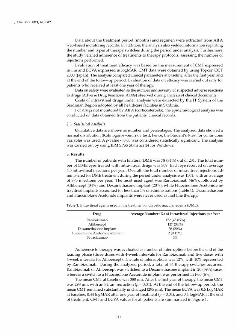
J. Clin. Med. 2021, 10, 5342
Data about the treatment period (months) and regimen were extracted from AIFAweb-based monitoring records. In addition, the analysis also yielded information regardingthe number and types of therapy switches during the period under analysis. Furthermore,the study verified adherence of treatments to therapy protocols, assessing the number ofinjections performed.
Evaluation of treatment efficacy was based on the measurement of CMT expressedin µm and BCVA expressed in logMAR. CMT data were obtained by using Topcon OCT2000 (Japan). The analysis compared clinical parameters at baseline, after the first year, andat the end of the follow-up period. Evaluation of data on efficacy was carried out only forpatients who received at least one year of therapy.
Data on safety were evaluated as the number and severity of suspected adverse reactionsto drugs (Adverse Drug Reactions, ADRs) observed during analysis of clinical documents.
Costs of intravitreal drugs under analysis were extracted by the IT System of theSardinian Region adopted by all healthcare facilities in Sardinia.
For drugs not monitored by AIFA (corticosteroids), the epidemiological analysis wasconducted on data obtained from the patients’ clinical records.
2.3. Statistical Analysis
Qualitative data are shown as number and percentages. The analyzed data showed anormal distribution (Kolmogorov–Smirnov test); hence, the Student’s t-test for continuousvariables was used. A p-value < 0.05 was considered statistically significant. The analysiswas carried out by using IBM SPSS Statistics 24 for Windows.
3. Results
The number of patients with bilateral DME was 78 (34%) out of 231. The total num-ber of DME eyes treated with intravitreal drugs was 309. Each eye received on average4.5 intravitreal injections per year. Overall, the total number of intravitreal injections ad-ministered for DME treatment during the period under analysis was 1501, with an averageof 375 injections per year. The most used agent was Ranibizumab (46%), followed byAflibercept (34%) and Dexamethasone implant (20%), while Fluocinolone Acetonide in-travitreal implants accounted for less than 1% of administrations (Table 1). Dexamethasoneand Fluocinolone Acetonide implants were never used as first line therapy.
Table 1. Intravitreal agents used in the treatment of diabetic macular edema (DME).
Drug Average Number (%) of Intravitreal Injections per Year
Ranibizumab 172 (45.85%)Aflibercept 127 (34%)
Dexamethasone implant 76 (20%)Fluocinolone Acetonide implant 2 (0.15%)
Bevacizumab 0%
Adherence to therapy was evaluated as number of interruptions before the end of theloading phase (three doses with 4-week intervals for Ranibizumab and five doses with4-week intervals for Aflibercept). The rate of interruptions was 12%, with 10% representedby Ranibizumab. During the analyzed period, a total of 34 therapy switches occurred.Ranibizumab or Aflibercept was switched to a Dexamethasone implant in 20 (59%) cases,whereas a switch to a Fluocinolone Acetonide implant was performed in two (6%).
The mean CMT at baseline was 380 µm. After the first year of therapy, the mean CMTwas 298 µm, with an 82 µm reduction (p = 0.04). At the end of the follow-up period, themean CMT remained substantially unchanged (295 µm). The mean BCVA was 0.5 LogMARat baseline, 0.44 logMAR after one year of treatment (p = 0.04), and 0.4 logMAR at the endof treatment. CMT and BCVA values for all patients are summarized in Figure 1.
111
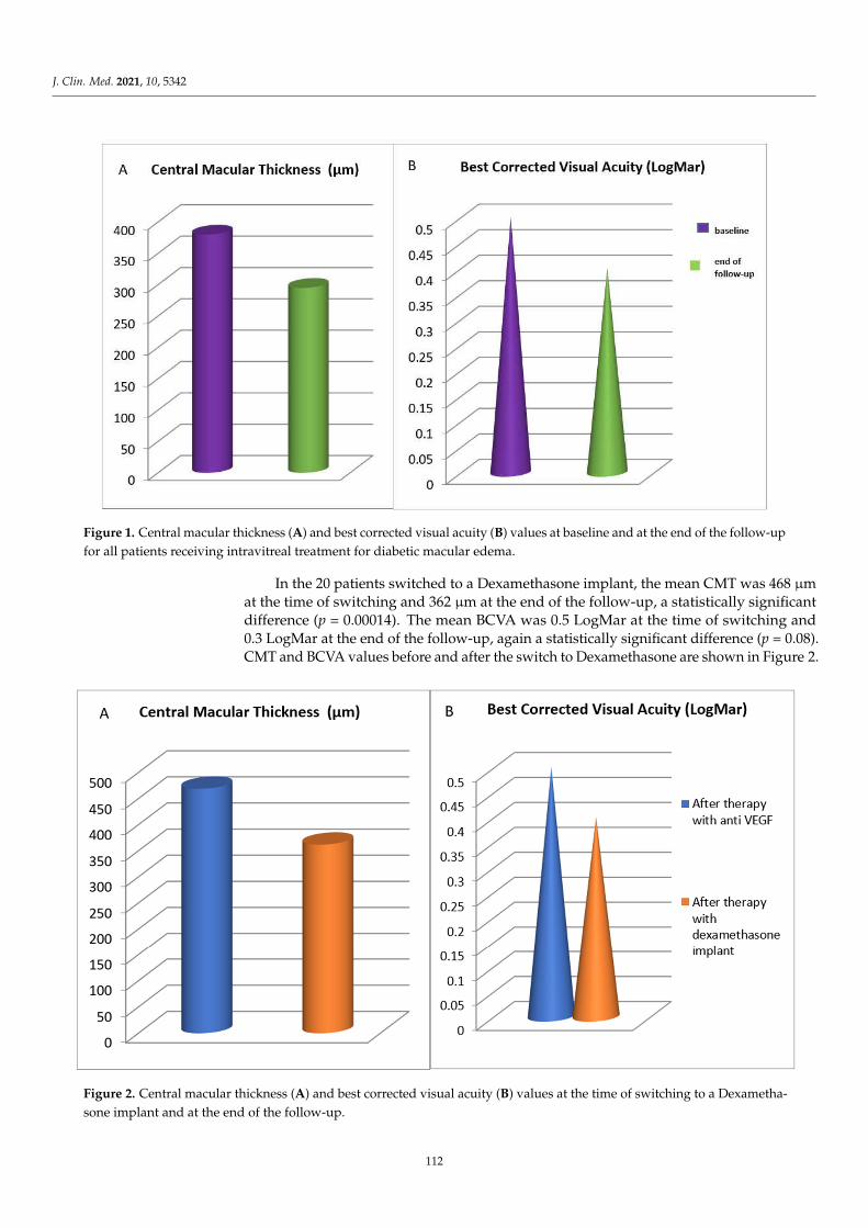
J. Clin. Med. 2021, 10, 5342
μμ μ
μ
μμ
Figure 1. Central macular thickness (A) and best corrected visual acuity (B) values at baseline and at the end of the follow-upfor all patients receiving intravitreal treatment for diabetic macular edema.
In the 20 patients switched to a Dexamethasone implant, the mean CMT was 468 µmat the time of switching and 362 µm at the end of the follow-up, a statistically significantdifference (p = 0.00014). The mean BCVA was 0.5 LogMar at the time of switching and0.3 LogMar at the end of the follow-up, again a statistically significant difference (p = 0.08).CMT and BCVA values before and after the switch to Dexamethasone are shown in Figure 2.
Figure 2. Central macular thickness (A) and best corrected visual acuity (B) values at the time of switching to a Dexametha-sone implant and at the end of the follow-up.
112

J. Clin. Med. 2021, 10, 5342
Considering all types of retinal diseases for which intravitreal drugs are used (i.e., wetage-related macular degeneration, retinal vein occlusion, myopic choroidal neovascular-ization, etc.), the total treatment cost during the analyzed period amounted to €5,780,000(Table 2), with an average cost of €1,651,429 per year. As far as DME is concerned, theglobal cost of intravitreal treatment was €915,000, with an average cost of €261,429 per year,accounting for approximately 16% of total expenses. It is important to emphasize that thetotal expense per year is subject to high variability, due to differences in drug consumptionand selling price, with the latter decreasing over the years. Ranibizumab was the drugthat mostly affected the expense, as it was the most prescribed intravitreal agent (46%),followed by Aflibercept (34%).
Table 2. Annual number and cost of intravitreal (IV) injections in the period January 2017–June 2020.
YearAll IV
Injections (n.)Global IV Cost (€)
IV Injectionsfor DME
IV Cost forDME * (€)
2017 2416 1,180,000.00€ 358 175,000.00€
2018 2720 1,750,000.00€ 388 250,000.00€
2019 2642 1,600,000.00€ 643 390,000.00€
2020 † 1395 1,250,000.00€ 112 100,000.00€
TOTAL 9173 5,780,000.00€ 1501 915,000.00€
* DME = Diabetic Macular Edema; † January–June.
Our study showed that the analyzed treatments were generally well tolerated. Indeed,during the period taken into consideration, no major ADRs (endophthalmitis, occlusivevasculitis, etc.) were reported.
4. Discussion
DME represents a social burden due to the reduction of vision and lower quality oflife of patients affected [1,2]. Our retrospective survey confirms the clinical efficacy ofRanibizumab, Aflibercept, and Dexamethasone implant in the treatment of DME [5,14,15],with significant BCVA improvement and CRT reduction, especially after the first year oftreatment. In terms of safety, the drugs analyzed showed a good risk-benefits outcome,with no systemic ADRs. Occasionally, local reactions were reported, mostly related tothe injection procedure and not connected to pharmacologic properties of the intravitrealagents. It is important to emphasize that no thromboembolic events were observed duringthe analyzed period. On the other hand, data related to adherence to therapy showed thatas much as 12% of the patients did not complete the loading phase, with a negative impacton the outcome. The greatest contribution to this value was given by Ranibizumab, themost used anti-VEGF agent in this investigation. Unfortunately, from the available data, itwas not possible to ascertain the reason of treatment interruption in most cases.
In our study, 20 patients receiving intravitreal Ranimizumab or Aflibercept withdisappointing results were switched to a Dexamethasone implant. After switching, therewas a significant improvement in the mean BCVA (from 0.5 to 0.3 LogMar) and CMT (from468 to 362 µm).
In patients presenting a serous detachment of neuroepithelium and poor responsein terms of BCVA and CMT after the loading phase with Ranimizumab or Aflibercept,intravitreal injection of a Dexamethasone implant should be considered. This can beadministered as an adjunctive treatment, without suspending the anti-VEGF agents, or asa switch treatment.
Overall, expenses for DME treatment accounted for only 16% of the global cost of allintravitreal injections for any cause. This can at least in part be explained by the fact thatabout one-third of diabetic patients are not aware of having the disease; therefore, diabeticretinopathy and DME are commonly underdiagnosed [5]. Cost analysis did not show alinear trend over time, presenting a peak in 2018. In 2020, the expenses were considerablylower, as eye examinations and intravitreal injections fell due to the hospital emergency
113

J. Clin. Med. 2021, 10, 5342
created by the first COVID-19 wave; furthermore, there was a reduction in the selling priceof all the intravitreal agents used.
Our study has important limitations, including its retrospective nature and the use ofrelatively crude estimates of hospital expenses. However, it provides some new data in anunderinvestigated topic, such as the drug expenses of intravitreal treatment for DME.
5. Conclusions
In conclusion, our results confirm that Ranibizumab, Aflibercept, and Dexamethasoneimplant are effective and safe in the treatment of DME. A therapy switch to Dexamethasoneimplant for patients receiving Aflibercept or Ranibizumab with minimal/no clinical benefitis recommended in an attempt to improve vision, reduce costs, and reduce the burden ofinjections of clinics and hospitals, especially in a pandemic era [16].
Author Contributions: Conceptualization, C.A., A.P.; methodology, C.A., M.G.D.; software, C.A.;validation, A.P., S.Z., F.B.; investigation, C.A., A.P.; writing—original draft preparation, C.A., M.G.D.,S.D., A.P.; writing—review and editing, C.A., M.G.D., S.D., A.P.; visualization, G.B., G.C.; supervision,A.P.; funding acquisition, A.P., M.G.D. All authors have read and agreed to the published version ofthe manuscript.
Funding: This research received no external funding.
Institutional Review Board Statement: The study was conducted according to the guidelines ofthe Declaration of Helsinki. Ethical review and approval were waived for this study, due to theretrospective nature of the survey.
Informed Consent Statement: Informed consent was obtained from all subjects involved in the study.
Data Availability Statement: Data will be available upon request.
Conflicts of Interest: The authors declare no conflict of interest.
References
1. American Diabetes Association (ADA). Standards of Medical Care in Diabetes—2013. Diabetes Care 2013, 36 (Suppl. 1), S11–S66.[CrossRef] [PubMed]
2. Linee Guida per lo Screening, la Diagnostica ed il Trattamento della Retinopatia Diabetica in Italia. Review and Update 2015. Avail-able online: https://www.fondazionebietti.it/sites/default/files/pdf/lg-rd-16sett2015.pdf (accessed on 15 November 2021).
3. Acan, D.; Calan, M.; Er, D.; Arkan, T.; Kocak, N.; Bayraktar, F.; Kaynak, S. The prevalence and systemic risk factors of diabeticmacular edema: A cross-sectional study from Turkey. BMC Ophthalmol. 2018, 18, 91. [CrossRef] [PubMed]
4. Virgili, G.; Menchini, F.; Murro, V.; Peluso, E.; Rosa, F.; Casazza, G. Optical coherence tomography (OCT) for detection of macularoedema in patients with diabetic retinopathy. Cochrane Database Syst. Rev. 2011, 7, CD008e081.
5. Flaxel, C.J.; Adelman, R.A.; Bailey, S.T.; Fawzi, A.; Lim, J.I.; Vemulakonda, G.A.; Ying, G.-S. Diabetic Retinopathy PreferredPractice Pattern®. Ophthalmology 2020, 127, P66–P145. [CrossRef] [PubMed]
6. Klein, R.; Klein, B.E.; Moss, S.E.; Davis, M.D.; DeMets, D.L. The Wisconsin Epidemiologic Study of diabetic retinopathy IIIPrevalence and risk of diabetic retinopathy when age at diagnosis is 30 or more years. Arch. Ophthalmol. 1984, 102, 527–533.[CrossRef] [PubMed]
7. Klein, R.; Klein, B.E.; Moss, S.E.; Davis, M.D.; DeMets, D.L. The Wisconsin epidemiologic study of diabetic retinopathy. IX.Four-year incidence and progression of diabetic retinopathy when age at diagnosis is less than 30 years. Arch. Ophthalmol. 1989,107, 237–243. [CrossRef] [PubMed]
8. Klein, R.; Klein, B.E.; Moss, S.E.; Cruickshanks, K.J. The Wisconsin epidemiologic study of diabetic retinopathy. X. Four-yearincidence and progression of diabetic retinopathy when age at diagnosis is 30 years or more. Arch. Ophthalmol. 1989, 107, 244–249.[CrossRef] [PubMed]
9. Bhagat, N.; Grigorian, R.A.; Tutela, A.; Zarbin, M.A. Diabetic Macular Edema: Pathogenesis and Treatment. Surv. Ophthalmol.
2009, 54, 1–32. [CrossRef] [PubMed]10. Nehmé, A.; Edelman, J. Dexamethasone inhibits high glucose-, TNF-alpha-, and IL-1beta-induced secretion of inflammatory
and angiogenic mediators from retinal microvascular pericytes. Investig. Ophthalmol. Vis. Sci. 2008, 49, 2030–2038. [CrossRef][PubMed]
11. Wang, W.; Lo, A.C.Y. Diabetic Retinopathy: Pathophysiology and Treatments. Int. J. Mol. Sci. 2018, 19, 1816. [CrossRef] [PubMed]12. Patelli, F.; Radice, P.; Giacomotti, E. Diabetic macular edema. Dev. Ophthalmol. 2014, 54, 164–173. [PubMed]13. Ciulla, T.A.; Amador, A.G.; Zinman, B. Diabetic Retinopathy and Diabetic Macular Edema: Pathophysiology, screening, and
novel therapies. Diabetes Care 2003, 26, 2653–2664. [CrossRef] [PubMed]
114

J. Clin. Med. 2021, 10, 5342
14. Lee, S.S.; Hughes, P.M.; Robinson, M.R. Recent advances in drug delivery systems for treating ocular complications of systemicdiseases. Curr. Opin. Ophthalmol. 2009, 20, 511–519. [CrossRef] [PubMed]
15. Boscia, F.; Giancipoli, E.; D’Amico Ricci, G.; Pinna, A. Management of macular oedema in diabetic patients undergoing cataractsurgery. Curr. Opin. Ophthalmol. 2017, 28, 23–28. [CrossRef] [PubMed]
16. Iovino, C.; Peiretti, E.; Giannaccare, G.; Scorcia, V.; Carnevali, A. Evolving Treatment Paradigm in the Management of DiabeticMacular Edema in the Era of COVID-19. Front. Pharmacol. 2021, 12, 670468. [CrossRef] [PubMed]
115


MDPI
St. Alban-Anlage 66
4052 Basel
Switzerland
Tel. +41 61 683 77 34
Fax +41 61 302 89 18
www.mdpi.com
Journal of Clinical Medicine Editorial Office
E-mail: [email protected]
www.mdpi.com/journal/jcm


MDPI
St. Alban-Anlage 66
4052 Basel
Switzerland
Tel: +41 61 683 77 34
Fax: +41 61 302 89 18
www.mdpi.com ISBN 978-3-0365-3256-1