Docslide us dyspnea-and-pulmonary-edema
44
DYSPNEA AND PULMONARY EDEMA Harrison’s 17 th edition Chapter 33
-
Upload
independent -
Category
Documents
-
view
2 -
download
0
Transcript of Docslide us dyspnea-and-pulmonary-edema

DYSPNEA AND PULMONARY EDEMAHarrison’s 17th editionChapter 33

Dyspnea

DYSPNEA American Thoracic Society
dyspnea as a “subjective experience of breathing discomfort that consists of qualitatively distinct sensations that vary in intensity experience derives from interactions among multiple physiological, psychological, social, and environmental factors, and may induce secondary physiological and behavioral responses.”

MECHANISMS OF DYSPNEA

Motor Efferents Disorders of the ventilatory pump
associated with increased work of breathing or a sense of an increased effort to breathe
The increased neural output from the motor cortex is thought to be sensed due to a corollary discharge that is sent to the sensory cortex at the same time that signals are sent to the ventilatory muscles.

Sensory Efferents Chemoreceptors in the carotid bodies and medulla
activated by hypoxemia, acute hypercapnia, and acidemia; leads to an increase in ventilation, produce a sensation of air hunger
Mechanoreceptors in the lungs stimulated by bronchospasm; lead to a sensation of chest tightness
J-receptors, sensitive to interstitial edema, and pulmonary vascular receptors activated by acute changes in pulmonary artery pressure, appear to contribute to air hunger

Sensory Efferents Hyperinflation
associated with the sensation of an inability to get a deep breath or of an unsatisfying breath
Metaboreceptors, located in skeletal muscle activated by changes in the local biochemical milieu of the tissue active during exercise
when stimulated, contribute to the breathing discomfort

Anxiety Acute anxiety may increase the severity of dyspnea altering the interpretation of sensory data
leading to patterns of breathing that heighten physiologic abnormalities in the respiratory system

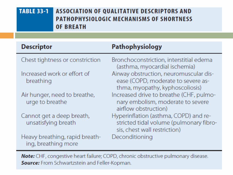
ASSESSING DYSPNEA Quality of Sensation
determination of the quality of the discomfort Sensory Intensity
modified Borg scale or visual analogue scale can be utilized to measure dyspnea at rest, immediately following exercise, or on recall of a reproducible physical task.
alternative approach is to inquire about the activities a patient can do. The Baseline Dyspnea Index and the Chronic Respiratory Disease Questionnaire are commonly used tools for this purpose.
Affective Dimension for a sensation to be reported as a symptom, it must be perceived as unpleasant and interpreted as abnormal.

DIFFERENTIAL DIAGNOSIS

Respiratory System Dyspnea Controller
Stimulated by acute hypoxemia and hypercapnia
Stimulation of pulmonary receptors: acute bronchospasm, interstitial edema, and PE
High altitude, high progesterone states (pregnancy), aspirin

Respiratory System Dyspnea Ventilatory Pump
Disorders of the airways (asthma, emphysema, chronic bronchitis, bronchiectasis) lead to increased airway resistance and work of breathing
Hyperinflation inability to get a deep breath Conditions that stiffen the chest wall (kyphoscoliosis) and that weaken ventilatory muscles (MG and GBS) associated with increased effort to breath
Large pleural effusions increases the work of breathing and stimulates pulmonary receptors if there is associated atelectasis.

Respiratory System Dyspnea Gas Exchanger
interfere with gas exchange: pneumonia, pulmonary edema, and aspiration
direct stimulation of pulmonary receptors: pulmonary vascular and interstitial lung disease and pulmonary vascular congestion
relief of hypoxemia - small impact on dyspnea

Cardiovascular System Dyspnea High Cardiac Output
Mild to moderate anemia: breathing discomfort during exercise
Left-to-right intracardiac shunts: may be complicated by the development of pulmonary hypertension
Breathlessness associated with obesity: due to multiple mechanisms, including high cardiac output and impaired ventilatory pump function

Cardiovascular System Dyspnea Normal Cardiac Output
Cardiovascular deconditioning: early development of anaerobic metabolism and stimulation of chemo- and metaboreceptors
Diastolic dysfunction: due to HPN, AS, or hypertrophic cardiomyopathy
Pericardial disease: constrictive pericarditis

Cardiovascular System Dyspnea Low Cardiac Output
Coronary artery disease and nonischemic cardiomyopathies: pulmonary receptors are stimulated

Approach to the Patient Clinical Indicators in the history
Orthopnea: CHF, mechanical impairment of the diaphragm in obesity, or asthma triggered by esophageal reflux
Nocturnal dyspnea: CHF or asthma Acute, intermittent episodes: MI, bronchospasm, PE
Chronic persistent: COPD and interstitial lung disease
Platypnea: left atrial myxoma or hepatopulmonary syndrome

Approach to the Patient Physical Examination
Inability of the patient to speak in full sentences: problem with the controller ventilatory pump
Increased work of breathing (supraclavicular retractions, use of accessory muscles, and the tripod position): ventilatory pump problem increased airway resistance or stiff lungs and chest wall

Approach to the Patient Physical Examination
vital signs, respiratory rate examination for a pulsus paradoxus >10 mmHg: COPD
signs of anemia (pale conjunctivae), cyanosis, and cirrhosis (spider angiomata, gynecomastia)

Approach to the Patient Physical Examination
Paradoxical movement of the abdomen (inward motion during inspiration): diaphragmatic weakness
Clubbing of the digits: interstitial pulmonary fibrosis
Joint swelling or deformation, change consistent with Raynaud’s disease: collagen-vascular process associated with pulmonary disease

Approach to the Patient Physical Examination of the Chest
Symmetry of movement Percussion (dullness indicative of pleural effusion, hyper-resonance a sign of emphysema)
Auscultation(wheezes, rales, rhonchi, prolonged expiratory phase, diminished breath sounds)

Approach to the Patient Physical Examination of the Heart
signs of elevated right heart pressures (jugular venous distention, edema, accentuated pulmonic component to the second heart sound)
left ventricular dysfunction (S3 and S4 gallops)
valvular disease (murmurs)

Approach to the Patient Diagnostic Exams
CXR Lung volumes
hyperinflation: obstructive lung disease
low lung volumes: interstitial edema or fibrosis, diaphragmatic dysfunction, or impaired chest wall motion
Pulmonary parenchyma - interstitial disease and emphysema
Approach to the Patient Diagnostic Exams
CXR Prominent pulmonary vasculature
in the upper zones: pulmonary venous hypertension
enlarged central pulmonary arteries: pulmonary artery hypertension
enlarged cardiac silhouette: dilated cardiomyopathy or valvular disease

Approach to the Patient Diagnostic Exams
CXR Bilateral pleural effusions: CHF and collagen vascular disease
Unilateral effusions: CA and PE

Approach to the Patient Diagnostic Exams CT scan of the chest
reserved for further evaluation of the lung parenchyma (interstitial lung disease) and possible PE
ECG Look for evidence of ventricular hypertrophy and prior myocardial infarction

Approach to the Patient Distinguishing Cardiovascular from Respiratory System Dyspnea CARDIOPULMONARY EXERCISE TEST
determine which system is responsible for the exercise limitation

Approach to the Patient Distinguishing Cardiovascular from Respiratory System Dyspnea CARDIOPULMONARY EXERCISE TEST
PULMONARY IF AT PEAK EXERCISE: achieves predicted maximal ventilation
demonstrates an increase in dead space or hypoxemia (oxygen saturation below 90%)
develops bronchospasm

Approach to the Patient Distinguishing Cardiovascular from Respiratory System Dyspnea CARDIOPULMONARY EXERCISE TEST
CARDIAC IF AT PEAK EXERCISE: heart rate is >85% of the predicted maximum
if anaerobic threshold occurs earlyif the BP becomes excessively high or drops
if the O2 pulse (O2 consumption/heart rate, an indicator of stroke volume) falls
if there are ischemic changes on the ECG


Treatment First goal: correct the underlying problem responsible for the symptom
Administration of supplemental O2
COPD patients: pulmonary rehabilitation programs have demonstrated positive effects on dyspnea, exercise capacity, and rates of hospitalization
Pulmonary Edema

MECHANISMS OF FLUID ACCUMULATION balance of hydrostatic and oncotic forces within the pulmonary capillaries
Hydrostatic pressure favors movement of fluid from the capillary into the interstitium
Oncotic pressure favors movement of fluid into the vessel

MECHANISMS OF FLUID ACCUMULATION Maintenance
tight junctions of the capillary endothelium are impermeable to proteins
lymphatics in the tissue carry away the small amounts of protein that may leak out
Pathology disruption of the endothelial barrier: allows protein to escape the capillary bed and enhances the movement of fluid into the tissue of the lung

Cardiogenic Pulmonary Edema Hydrostatic pressure is increased and fluid exits the capillary at an increased rate
Early signs of pulmonary edema: exertional dyspnea and orthopnea
CXR: peribronchial thickening, prominent vascular markings in the upper lung zones, and Kerley B lines

Noncardiogenic Pulmonary Edema Hydrostatic pressures are normal Leakage of proteins and other macromolecules into the tissue
Associated with dysfunction of the surfactant lining the alveoli, increased surface forces, and a propensity for the alveoli to collapse at low lung volumes

Noncardiogenic Pulmonary Edema Characterized by intrapulmonary shunt with hypoxemia and decreased pulmonary compliance
Causes Direct Injury to Lung Hematogenous Injury to Lung Possible Lung Injury Plus Elevated Hydrostatic Pressures


Cardiogenic vs Noncardiogenic CARDIOGENIC PULMONARY EDEMA
Physical Examination: increased intracardiac pressures (S3 gallop, elevated jugular venous pulse, peripheral edema)
rales and/or wheezes on auscultation of the chest
CXR:enlarged cardiac silhouettevascular redistributioninterstitial thickeningperihilar alveolar infiltratespleural effusions

Cardiogenic vs Noncardiogenic NONCARDIOGENIC PULMONARY EDEMA
Physical Examination: Findings may be relatively normal in the early stages
CXR:Heart size is normalUniform alveolar infiltrates Pleural effusions are uncommon

Hypoxemia CARDIOGENIC
due to ventilation-perfusion mismatch responds to the administration of supplemental oxygen
NONCARDIOGENIC due to intrapulmonary shunting persists despite high concentrations of inhaled O2