Pathology of Pulmonary Hypertension
30
Pathology of Pulmonary Hypertension Rubin M. Tuder, M.D. a,b , John C. Marecki, Ph.D. c , Amy Richter, B.S. d , Iwona Fijalkowska, Ph.D. d,e , and Sonia Flores, Ph.D. f a Professor of Pathology and Medicine, Division of Cardiopulmonary Pathology, Johns Hopkins University School of Medicine, Baltimore, Maryland b Research Associate, Division of Pulmonary and Critical Care Medicine, University of Colorado-Denver Health Sciences Center, Baltimore, Maryland c Director, Division of Cardiopulmonary Pathology, Johns Hopkins University School of Medicine, Baltimore, Maryland d Member, Division of Cardiopulmonary Pathology, Johns Hopkins University School of Medicine, Baltimore, Maryland e Research Associate, Division of Cardiopulmonary Pathology, Johns Hopkins University School of Medicine, Baltimore, Maryland f Associate Professor of Medicine, Division of Pulmonary and Critical Care Medicine, University of Colorado Health Sciences at Denver Keywords endothelial cells; smooth muscle cells; remodeling; angiogenesis; apoptosis; proliferation The focus on the pathological changes underlying pulmonary hypertension (PH) have dominated the early investigations of this disease first described late in the 19 th century. Pulmonary vascular pathology continues to play an important role in the present age of cell and molecular investigation of the pathogenesis of PH. This importance stems from the permanent quest to correlate pulmonary vascular remodeling with the altered pulmonary vascular hemodynamics, a critical advancement in the late 40’s and early 50’s with a wide impact on our present understanding of the disease. However, as it happens to most descriptive tools applied to medical sciences, the pathological insight into the extent and type of a particular form of pulmonary vascular remodeling has fallen short of establishing cause and effect relationships in the natural history of PH, and has had a limited impact on diagnosis and therapy. These limitations derive largely from the reliance of our current knowledge on studies of autopsies, as lung tissue is rarely available for histopathology during the course of the disease. The pathological diagnosis of pulmonary vascular remodeling depends on the histological assessment of the cellular composition of pulmonary vascular walls, which if abnormal, is described as pulmonary vascular ‘lesions’. Although mostly reliant on examination of histological slides stained by hematoxylin and eosin stains, the pathological interpretation of a Corresponding author for proof and reprints: Rubin M. Tuder, M.D., Division of Cardiopulmonary Pathology, Johns Hopkins University School of Medicine, Baltimore, Maryland. 720 Rutland Ave., Ross Research Building, Baltimore, MD 21217, Phone: (410) 955-2566, Fax: (410) 502-7813, Email address: [email protected] c Coauthors address: Amy Richter, B.S., Iwona Fijalkowska, Ph.D. Division of Cardiopulmonary Pathology, Johns Hopkins University School of Medicine, Baltimore, Maryland, 720 Rutland Ave., Ross Research Building, Baltimore, MD 21217, Phone: (410) 955-2573, Fax: (410) 502-7813, Email: [email protected]; [email protected] e Sonia C. Flores, Ph.D. c John Marecky, Ph.D. Box C-272 4200 East 9th Avenue, Denver, CO 80262. Phone: (303) 315-0055, Fax: Email: [email protected] Email: [email protected] This work was supported by the grant P01HL66254 to RMT and grant R01 1HL083491 to SCF, from the National Institutes of Health. Publisher's Disclaimer: This is a PDF file of an unedited manuscript that has been accepted for publication. As a service to our customers we are providing this early version of the manuscript. The manuscript will undergo copyediting, typesetting, and review of the resulting proof before it is published in its final citable form. Please note that during the production process errors may be discovered which could affect the content, and all legal disclaimers that apply to the journal pertain. NIH Public Access Author Manuscript Clin Chest Med. Author manuscript; available in PMC 2007 July 18. Published in final edited form as: Clin Chest Med. 2007 March ; 28(1): 23–vii. NIH-PA Author Manuscript NIH-PA Author Manuscript NIH-PA Author Manuscript
-
Upload
independent -
Category
Documents
-
view
0 -
download
0
Transcript of Pathology of Pulmonary Hypertension

Pathology of Pulmonary Hypertension
Rubin M. Tuder, M.D.a,b, John C. Marecki, Ph.D.c, Amy Richter, B.S.d, Iwona Fijalkowska,Ph.D.d,e, and Sonia Flores, Ph.D.f
aProfessor of Pathology and Medicine, Division of Cardiopulmonary Pathology, Johns Hopkins UniversitySchool of Medicine, Baltimore, Maryland bResearch Associate, Division of Pulmonary and Critical CareMedicine, University of Colorado-Denver Health Sciences Center, Baltimore, Maryland cDirector, Divisionof Cardiopulmonary Pathology, Johns Hopkins University School of Medicine, Baltimore, MarylanddMember, Division of Cardiopulmonary Pathology, Johns Hopkins University School of Medicine, Baltimore,Maryland eResearch Associate, Division of Cardiopulmonary Pathology, Johns Hopkins University Schoolof Medicine, Baltimore, Maryland fAssociate Professor of Medicine, Division of Pulmonary and Critical CareMedicine, University of Colorado Health Sciences at Denver
Keywordsendothelial cells; smooth muscle cells; remodeling; angiogenesis; apoptosis; proliferation
The focus on the pathological changes underlying pulmonary hypertension (PH) havedominated the early investigations of this disease first described late in the 19th century.Pulmonary vascular pathology continues to play an important role in the present age of celland molecular investigation of the pathogenesis of PH. This importance stems from thepermanent quest to correlate pulmonary vascular remodeling with the altered pulmonaryvascular hemodynamics, a critical advancement in the late 40’s and early 50’s with a wideimpact on our present understanding of the disease. However, as it happens to most descriptivetools applied to medical sciences, the pathological insight into the extent and type of a particularform of pulmonary vascular remodeling has fallen short of establishing cause and effectrelationships in the natural history of PH, and has had a limited impact on diagnosis and therapy.These limitations derive largely from the reliance of our current knowledge on studies ofautopsies, as lung tissue is rarely available for histopathology during the course of the disease.
The pathological diagnosis of pulmonary vascular remodeling depends on the histologicalassessment of the cellular composition of pulmonary vascular walls, which if abnormal, isdescribed as pulmonary vascular ‘lesions’. Although mostly reliant on examination ofhistological slides stained by hematoxylin and eosin stains, the pathological interpretation of
aCorresponding author for proof and reprints: Rubin M. Tuder, M.D., Division of Cardiopulmonary Pathology, Johns Hopkins UniversitySchool of Medicine, Baltimore, Maryland. 720 Rutland Ave., Ross Research Building, Baltimore, MD 21217, Phone: (410) 955-2566,Fax: (410) 502-7813, Email address: [email protected] address: Amy Richter, B.S., Iwona Fijalkowska, Ph.D. Division of Cardiopulmonary Pathology, Johns Hopkins UniversitySchool of Medicine, Baltimore, Maryland, 720 Rutland Ave., Ross Research Building, Baltimore, MD 21217, Phone: (410) 955-2573,Fax: (410) 502-7813, Email: [email protected]; [email protected] C. Flores, Ph.D.cJohn Marecky, Ph.D. Box C-272 4200 East 9th Avenue, Denver, CO 80262. Phone: (303) 315-0055, Fax: Email:[email protected] Email: [email protected] work was supported by the grant P01HL66254 to RMT and grant R01 1HL083491 to SCF, from the National Institutes of Health.Publisher's Disclaimer: This is a PDF file of an unedited manuscript that has been accepted for publication. As a service to our customerswe are providing this early version of the manuscript. The manuscript will undergo copyediting, typesetting, and review of the resultingproof before it is published in its final citable form. Please note that during the production process errors may be discovered which couldaffect the content, and all legal disclaimers that apply to the journal pertain.
NIH Public AccessAuthor ManuscriptClin Chest Med. Author manuscript; available in PMC 2007 July 18.
Published in final edited form as:Clin Chest Med. 2007 March ; 28(1): 23–vii.
NIH
-PA Author Manuscript
NIH
-PA Author Manuscript
NIH
-PA Author Manuscript

PH has benefited from the progressive use of cell specific immunohistochemical markers tobetter define the structure and cellular composition of the pulmonary vascular lesions. Despitethe advances in the understanding and treatments aimed at the disease, the pathology of PHclearly lags behind the comprehensive approach employed by pathologists in their assessmentof other diseases such as cancer. This approach presently includes the screening for abnormalexpression of the p53 tumor suppressor or adenomatous polyposis coli (APC) genes, abnormalexpression of cytokeratins in breast adenocarcinomas, and markers of cell proliferation insarcomas, among several tissue markers that help in the final diagnosis, staging prognosis, andtreatment. By comparison, the histopathological diagnosis of PH infrequently fulfills some orall of these important tasks of the pathological work-up aimed at clinical management.
The functional status of the pulmonary circulation and the levels of pulmonary vascularresistance and pulmonary artery pressures ultimately determine the outcome and treatment ofpatients with PH. We have previously proposed that the functional status of the hypertensivepulmonary circulation could be broadly correlated with a specific type of pulmonary vascularremodeling present at the time of pathological evaluation of the pulmonary arteries [1]. Wewill frame this review on the pathology of PH within the functional categories proposedpreviously i.e., that PH can be broadly divided into mild/moderate vs. severe based onpulmonary artery pressures, their impact on right ventricular performance, and overallmortality [1] (Tables 1 and 2). Many non neoplastic lung diseases with intima thickening ormedial hypertrophy, such as idiopathic interstitial pneumonias (IPF) [2] and chronicobstructive pulmonary diseases (COPD) [3], present with mild/moderate PH. On the otherhand, conditions associated with endothelial cell proliferative lesions (including plexiformlesions), marked intima fibrosis (such as idiopathic pulmonary arterial hypertension (IPAH)and scleroderma), or medial and intimal smooth muscle cell growth (as observed in a fractionof IPF or COPD lungs) cause severe PH. As it will become apparent in this review, our abilityto relate the mode of pulmonary vascular remodeling to the severity of the disease is ratherlimited. We will highlight the changes associated with IPAH as those paradigmatic of thepathology of severe PH.
In our goal to highlight the pathology of PH, we will follow the recommendations of the Evianmeeting on Pulmonary Hypertension in 1998, which supported a more descriptive approachto the pulmonary vascular changes in PH [4]. We will also review the recent findings relatedto the pathogenesis of human PH (Table 3). We believe that their translation into the pathologyof PH may eventually lead to a more refined and clinically useful approach towards thediagnosis and staging of the disease. The latest reorganization of the pathological nomenclatureof PH by the international meeting in Venice in 2004 provides a useful framework of referenceto the diagnosis of pulmonary vascular lesions in PH [5]. As a valuable background to thischapter, the readers are also referred to reviews on normal histology of pulmonary arteries[6;7], and a historical perspective of the research accomplishments in the last 100 years ofstudies of the pulmonary circulation [8].
Intima LesionsPathology
Intimal lesions account for most of the reduction of luminal area of small pulmonary arteriesand potentially largely influence the overall pulmonary vascular resistance. Intimal lesionsconsist of eccentric intima thickening, and fibrotic, plexiform, concentric, and dilation/angiomatoid lesions (Table 1, Figures 1–4). Focal eccentric lesions can be detected in normallungs, but these lesions are more widespread and impinge to a larger extent on the vascularlumen in PH. It is conceivable that some of these lesions results from the organization (i.e.,lysis of fibrin, recanalization by newly formed blood vessels, and/or ingrowth ofmyofibroblasts) of localized thrombi, which form the nidus for a localized growth of smooth
Tuder et al. Page 2
Clin Chest Med. Author manuscript; available in PMC 2007 July 18.
NIH
-PA Author Manuscript
NIH
-PA Author Manuscript
NIH
-PA Author Manuscript

muscle cells. More advanced lesions acquire a ‘fibrotic’ pattern, with interspersedmyofibroblasts and marked accumulation of mucopolyssacharides (Figure 3). These ‘fibrotic’lesions are widely present in explanted lungs of patients with severe PH, including lungs withIPAH or PAH associated with the CREST syndrome (acronym for calcinosis, Raynaudphenomenon, esophageal dysmotility, sclerodactily, and telengectasia) [9]. However, variabledegrees of eccentric thickening can be seen in cigarette smoker’s lungs associated withpulmonary endothelial cell dysfunction, with or without evidence of PH [10].
The organization of myofibroblasts or endothelial cells in ‘onion-skin’ layers underlies thepresentation of concentric lesions (Figures 1 and 2). These lesions are characteristic of in severePH, as they significantly reduce significantly the luminal area. We documented the presenceof concentric lesions in vascular segments proximal to plexiform lesions [11], suggesting thatthe concentric lesions arise from remodeled plexiform lesions. Concentric lesions composedof smooth muscle cells have also been described [12]. It is unclear whether endothelial- orsmooth muscle-based lesions have a similar impact on the course of PH. However, it isconceivable that these lesions may arise from similar progenitors, as endothelial cells cantransdifferentiate into smooth muscle cells when stimulated with platelet derived growth factor(PDGF)-BB [13] or transforming growth factor (TGF)-β [14]. Pulmonary arteries may alsoalmost occluded by the accumulation of acellular matrix, suggestive of a terminal scarringprocess (Figure C). Approximately 20–25% of pulmonary arteries ranging between 25 and 200um in diameter are compromised by intima occlusion or concentric lesions [15]. These twopatterns of luminal obliteration might represent temporally related lesions, which start as anabnormal proliferation of endothelial and/or smooth muscle cells, progress into an ‘onion-skin’lesion, and then become acellular with abundant extracellular matrix deposition (Figure 4).
Plexiform lesions, typically located in branching pointes of muscular arteries [11], consists ofa network of vascular channels lined up by endothelial cells [16] (Figures 1 and 2) and a coreof myofibroblastic or less well-differentiated cells (Figure 3) [17]. In our experience, theselesions are characteristically found in cases of severe PH, including IPAH, and PH associatedwith HIV infection, liver cirrhosis, CREST, congenital heart malformations, andschistosomiasis. As endothelial cells contribute significantly to both concentric and plexiformlesions, endothelial cell immunostaining with Factor VIII related antigen or CD31 may aid inthe identification of the different stages of endothelial cell proliferation [6] (Figure 1 and 2).The detection of small clusters of endothelial cells can be aided by use of these stains [18] andcell proliferation markers such as MIB-1 may contribute to the identification of proliferatingendothelial cells as well (normal pulmonary endothelial cells have a very low proliferationrate) [19]. Dilation lesions represent another potential endothelial cell lesion, with theformation of a rosary of dilated channels (also known as angiomatoid lesions), often distallyto a plexiform lesion [6] (Figure 2). As with plexiform lesion, we have observed dilation lesionsin cases of severe PH (Table 1). These lesions containing endothelial cells have abundantcollagen IV expression, a component of basement membranes, and a useful marker for theoutline of blood vessels (Figure 4).
PathobiologyThe prevailing concept is the one which dysfunctional endothelial cells plays the keypathobiological role in PH [20]. However, to the present day, the pathogenesis of intimalvascular lesions remains mostly undetermined. It is conceivable that these lesions result frominjuries to the endothelium, followed by activation of smooth muscle cell migration,extracellular matrix deposition, and endothelial cell proliferation. As mentioned, it is alsopossible that these lesions occur due to organization of microthrombi. In animal models, onlythe association of the alkaloid monocrotaline (MCT) and high shear stress due to shuntingbetween the systemic and pulmonary circulations leads to marked intima thickening by smooth
Tuder et al. Page 3
Clin Chest Med. Author manuscript; available in PMC 2007 July 18.
NIH
-PA Author Manuscript
NIH
-PA Author Manuscript
NIH
-PA Author Manuscript

muscle cells and severe PH in rats [21]. The intimal cells express smooth muscle cell markers,and fibronectin [19].
We have proposed that plexiform lesions represent a process of misguided angiogenesis basedon the findings of expression of vascular endothelial growth factor (VEGF), its receptors 1 (flt)and 2(kdr), and hypoxia inducible factor (HIF)-1α and β [22]. The finding of monoclonalityof endothelial cells in plexiform lesions in IPAH, but not in similar lesions in PH associatedwith congenital heart malformation, suggests that these lesions might arise from mutations intumor suppressor genes [23]. Somatic loss of expression of transforming growth factor βreceptor 2 ((TGFβ-R2) and the propapoptotic Bax, potentially due to microsatellite instability,is also documented in IPAH plexiform lesions [24]. Heterozygous germline mutations of bonemorphogenetic protein receptor 2 (BMPR2) (a component of the TGF-β-related family) isfound to underlie up to 60% of cases of familial IPAH [25;26]. BMPR2 expression isdocumented in plexiform lesions [12] and in remodeled pulmonary arteries in IPAH lungs,predominantly in endothelial cells. However, our studies reveal that the cells in the central coreof plexiform lesions lack the expression of TGF-β receptor 2, TGF-β receptor 1 and theirsignaling smad(s) 2,1 (which shares common epitopes with smads 5 and 8), 3 and 4, includingthe phosphorylated smad 1/5/8 and 2. The absence of expression of these phosphorylated smadsis the best indication that there is no TGF-β (via smad 2 or smads 1/5/8) or BMP (via smads1/5/8) signaling in these cells [27]. Loss of cytostatic signaling from TGF-β would allow theplexiform cells to abnormally proliferate. Notwithstanding the evidence of preserved BMPRexpression and signaling in IPAH endothelial cells, recent studies indicate that BMP signalingprotects against endothelial cell apoptosis in vitro [28]; the loss of this protection by germlinemutations in BMPR2 would thus favor enhanced susceptibility to apoptosis of lung endothelialcells.
The potential role for early endothelial cell apoptosis in the pathogenesis of uncontrolledproliferation of pulmonary endothelial cells was first documented in the rat model of severepulmonary hypertension caused by the combination of VEGF receptor blockade with SU5416and chronic hypoxia [29]. Moreover, the role of endothelial cell apoptosis in the pathogenesisof PH was also extended to the monocrotaline model [30]. Initial endothelial cell apoptosismight favor the emergence of apoptosis-resistant endothelial cells, with potential foruncontrolled proliferation [29;31].
VEGF [22], endothelin-1 [32], and survivin [33;34] are among the factors present in plexiformlesions that may enhance endothelial cell and smooth muscle cell proliferation or decreasevascular cell apoptosis. These lesions have decreased expression of anti-remodeling mediatorssuch as nitric oxide synthase [35] and prostacyclin synthase [36], and tumor suppressors, suchas caveolin-1[37]. These phenotypic characteristics lead us to propose that the plexiformlesions have characteristics in common with neoplasms [38]. As discussed below, there isemerging evidence that viral factors may play a role in endothelial cell proliferative lesions insevere PH.
A summary of the molecules that may contribute to lesion formation, their expression, and thesignaling or metabolic pathways involved, as well as their potential target in the vasculatureare shown in Table 3.
Medial Vascular RemodelingPathology
Medial smooth muscle cell hypertrophy is a characteristic pathological feature of PH thatinvolves muscularized arteries (ranging between 70 and 500 μm in diameter), and precapillaryvessels (below 70 μm in diameter) (Figure 3). The medial smooth muscle cell layer represents
Tuder et al. Page 4
Clin Chest Med. Author manuscript; available in PMC 2007 July 18.
NIH
-PA Author Manuscript
NIH
-PA Author Manuscript
NIH
-PA Author Manuscript

approximately 10–15% of the outside diameter of normal muscularized pulmonary arteries,while it approaches 30–60% of the outside diameter in vessels of IPAH lungs [15;39–41].Although careful morphometric assessments of medial remodeling have not been carried outin non-IPAH PH, it is apparent that medial thickening occurs in mild/moderate or severe PHand in cases of normal individuals exposed to cigarette smoke with no evidence of PH [42](Table 2). This diagnostic limitation of the finding of medial hypertrophy has led us to proposethat the identification of pulmonary medial remodeling warrants additional clinical evidenceof the presence of potential PH [6]. Several of these cases may have normal pulmonary arterypressure levels and thus do not have PH (Table 2). The limited number of smooth muscle cellsmarkers, identified mostly in animal models, precludes the ‘functional’ dissection ofpulmonary medial remodeling. Nevertheless, medial thickening probably plays an importantrole in the pathogenesis of PH, yet it is difficult to relate the morphological identification ofmedial remodeling to specific levels of pulmonary artery pressures, degrees of severity of PH[6], or potential for response to vasodilators.
PathobiologyThe expansion of the tunica media in pulmonary arteries in PH occurs by a combination ofhypertrophy and hyperplasia. Smooth muscle cell hypertrophy together with accumulation ofextracellular matrix, including thicker elastic laminae, accounts for most of medial thickeningin PH [43]. Smooth muscle cell hyperplasia probably plays a minor role in medial thickeningthan cell hypertrophy or extracellular matrix deposition, since it is experimentally shown thatthe proliferation index of smooth muscle cells doubles in large pulmonary arteries in hypoxicrats, while those cells lining intraacinar arteries do not show evidence of [3H]-thymidine uptake[44]. A similar finding is observed in PH caused by monocrotaline in rats [45]. The apparentpredominant role of smooth muscle cell hypertrophy over hyperplasia in medial thickening isalso supported by human PH studies [19;46;47]. Based on the hypoxia model of PH, theappearance of smooth muscle cells in nonmuscularized precapillary arteries occurs byhypertrophy and metaplasia of potential precursor cells, i.e., pericytes or intermediate cells(pericytes-smooth muscle cells) [44]. A careful examination of the smooth muscle cellphenotypes in the tunica media of neonatal bovine pulmonary arteries reveals the existence ofsubstantial cellular heterogeneity, with clusters of fully differentiated smooth muscle cellsinterspersed with cells negative for most smooth muscle cell markers [48]; such a cellularheterogeneity probably exists in the different forms of human PH as well, though a systematicdissection of these phenotypes has not been performed thus far. In spite of these data, most ofthe present day studies addressing the potential role of genetic mutations or alterations in ionchannels have focused on smooth muscle cell proliferation, as noted below.
The mechanisms underlying the thickening of the pulmonary vascular medial layer have beenlinked mostly to cell proliferation and, more recently, to inhibition of cell apoptosis. Theidentification of mutations in BMPR2 led to several in vitro studies aimed at relating smoothmuscle cell proliferation to abnormal BMPR2 signaling. IPAH smooth muscle cells isolatedfrom proximal arteries (i.e., elastic vessels, >500 μm in diameter) exhibit decreased inhibitoryeffect on cell proliferation mediated by TGF-β1 or BMP-4 when compared with smooth musclecells isolated from normal human pulmonary arteries [49]. Of note, these altered responses arenot due to abnormal expression of smad signaling or ligand binding to its receptors in IPAHsmooth muscle cells. Similar studies were extended to more peripheral smooth muscle cells(i.e., from arteries less than 2 mm in diameter-(that are still elastic arteries) obtained fromnormal pulmonary arteries, which undergo cell proliferation and protection against apoptosiswhen exposed to BMP-4, a ligand for BMPR2. Therefore, loss of function mutations in MPR2would be predicted to cause smooth muscle cell arrest and increased cell death [50], i.e.,paradoxically opposite to that predicted to occur in familial IPAH in which mutations wouldfacilitate smooth muscle cell growth and remodeling. Given these somewhat discrepant results
Tuder et al. Page 5
Clin Chest Med. Author manuscript; available in PMC 2007 July 18.
NIH
-PA Author Manuscript
NIH
-PA Author Manuscript
NIH
-PA Author Manuscript
[50], the evidence of reduced expression of BMPR2 found in IPAH alveolar septa [12], andthe finding of decreased levels of the activated form of smad 1 (the signaling smad for BMPR2)in smooth muscle cells of muscular pulmonary arteries in IPAH, it remains unclear howBMPR2 mutations either can cause or trigger the disease. Indeed, when compared with wildtype mice, heterozygous mice lacking a single copy of BMPR2 have no pulmonary vascularphenotype at baseline or under chronic hypoxia [51], but show moderately increasedpulmonary artery pressures and minimal remodeling when stressed with intratracheal deliveryof a 5-lipoxygenase expressing vector [51].
The role of serotonin (5-hydroxytryptamine) in the growth stimulation of pulmonary arterysmooth muscle cells has been intensively studied as a mechanism of medial remodeling in PHand a potential modifier gene to the familial IPAH. Serotonin is internalized in smooth musclecells after binding to serotonin transporter (5-HTT) or to its receptors 5-HT1B-R, 5-HT-2A R,5-HT7 -R, and 5-HT2B -R, when it promotes vasoconstriction, cell growth, and enhancementof hypoxia-induced remodeling and PH [52]. Vascular smooth muscle cells in IPAH lungsexpress higher levels of 5-HTT [53] or 5-HT2B R [52] by immunohistochemistry, and undergoenhanced cell proliferation when treated in vitro with serotonin compared with normal smoothmuscle cells [53]. These findings are supported by the report that mice deficient in 5HTT or5-HT-2B are protected against PH caused by hypoxia alone [52] or hypoxia combined with theanorexigen dexfenfluramine [54], respectively. Of interest is the observation that increasedserotonin levels affect signaling pathways downstream of mutated BMPR2, as serotonininfusion enhances normoxic and hypoxic pulmonary pressures and vascular remodeling inBMPR2 heterozygous mice as compared with wild type mice [50].
Despite evidence supporting a loss of function for the TGF-β family signaling, particularly inendothelial cells of IPAH, TGF-β may produce gain-of-function alterations underlying medialsmooth muscle cell growth and adventitial fibroblast activation. TGF-β isoforms 1, -2, and -3are expressed in hypertensive pulmonary arteries [55], and could signal via activation of smad2 or 3 by serine phosphorylation, whose expression was documented in pulmonary vascularsmooth muscle cells [27]. Recent evidence implicates PDGF in the pathogenesis of bothmonocrotaline- and chronic hypoxia-induced PH [56]. Monocrotaline treatment or chronichypoxia exposure leads to increased PDGF receptor (PDGFR)-β expression andphosphorylation and activation of ERK Map kinase in rats. The findings of positive responseof hypertensive animals to Gleevac™, also an inhibitor of PDGFR signaling, and the evidenceof increased expression of PDGFR-β and phosphorylated PDGFR-β in IPAH lungs, led theauthors to translate their finding by treating an IPAH patient with Gleevac™, with promisingearly results [57].
The extracellular matrix is both a component of the thickened pulmonary vascular media anda regulator of smooth muscle cell growth. Tenascin, a glycoprotein involved in lung andvascular morphogenesis, is strongly expressed in remodeled intima and medial layers of humanhypertensive pulmonary arteries [58]. Experimentally, products of enhanced elastolytic orproteolytic activity in pulmonary arteries can promote clustering of integrin receptors, whichactivate tenascin and epidermal growth factor (EGF) receptors, thus leading to smooth musclecell proliferation [59]. These findings are pertinent to the monocrotaline model of PH [60;61], while pulmonary vascular remodeling in hypoxic pulmonary hypertension is improved bythe action of metalloproteases [62]. Based on studies using cultured human smooth musclecells isolated from elastic arteries from IPAH patients, it appears that Tissue Inhibitor ofMetalloprotease-1 (TIMP-1) and metalloprotease (MMP)-2 are increased in expression; onlythe finding of MMP-2 enhanced expression is confirmed in situ in elastic pulmonary arteriesin IPAH lungs [63]. It is unclear whether a similar pattern is present in distally remodeledpulmonary arteries; however, endothelial cells express moderate/intenseimmunohistochemical expression of MMP-2, while myofibroblasts display low levels of this
Tuder et al. Page 6
Clin Chest Med. Author manuscript; available in PMC 2007 July 18.
NIH
-PA Author Manuscript
NIH
-PA Author Manuscript
NIH
-PA Author Manuscript

extracellular protease [64]. Membrane type-1-MMP was also expressed in endothelial andmyofibroblastic cells of concentric and plexiform lesions. These results and those in themonocrotaline model suggest that the inhibition of elastases or MMPs might be beneficial inIPAH as demonstrated in the monocrotaline model of PH [65], potentially leading to theapoptosis of smooth muscle cells.
It is becoming clear that the ultimate fate of vascular smooth muscle cells in PH is determinedby their resistance to apoptosis. In fact, ‘apoptosis-resistance’ might play a central role in boththe endothelial- and smooth muscle cell-based pulmonary vascular lesions [66], since IPAHlungs have lower number of apoptotic cells than normal or emphysematous lungs [67]. Asgrowth signals originated by PDGF, TGF-β, EGF, serotonin, and extracellular matrix proteinsare interrupted in animal models of PH, pulmonary arteries undergo de-remodeling associatedwith apoptosis of pulmonary artery smooth muscle cells [65]. Targeting apoptosis of thehypertrophic smooth muscle cells might represent a more viable approach towards treatment.
Recent studies of K+ channel activity have provided a novel insight of the lack of proapoptoticsignals in IPAH smooth muscle cells. An exciting and complementary paradigm based on theinterplay between K+ channels and apoptosis in pulmonary artery smooth muscle cells hasemerged based on the demonstration of activation of K+ channels causes cytochrome C releasefrom the mitochondria and water efflux from the dying cells [68]. Conversely, inhibition ofK+ channels causes cell depolarization, enhances contractility, and decreases apoptosis ofpulmonary artery smooth muscle cells. One potential mechanism linking this paradigm toBMPR2 mutations is the finding that BMPR2 activation upregulates K+ channels [69] andcauses apoptosis of normal pulmonary artery smooth muscle cells, but not of cells from patientswith IPAH [70]. McMurtry et al provide recent additional evidence that mechanisms akin tocancer operate in pulmonary vascular remodeling [34]. Survivin protects cancer cell againstapoptosis by inhibiting caspase activation and apoptosis inducing factor [71]. Not only dopulmonary artery smooth muscle cells in IPAH lungs express higher levels of the antiapoptoticsurvivin, but monocrotaline-induced pulmonary vascular remodeling requires survivinexpression. Indeed, transduction of a functionally deficient survivin in monocrotaline-treatedlungs prevented pulmonary vascular remodeling and, when administered after monocrotalinetreatment, it enhances apoptosis of pulmonary artery smooth muscle cells and reducespulmonary artery pressures. This mutant survivin increases levels of K+ channel activity andleads to depolarization of mitochondria with enhanced cytochrome C release.
Adventitial RemodelingPathology
The adventitia is mostly composed of fibroblasts. There is growing evidence that, rather thanjust a structural support to pulmonary vessels, the adventitia may also play a role in theregulation of pulmonary vascular function from the ‘outside-in’ (as comprehensively reviewedin [72]). The normal adventitia represents approximately 15% of the external diameter ofpulmonary arteries larger than 50 μm in diameter. In IPAH arteries, the adventitial thicknessincreases to 28% of artery diameter, predominantly due to collagen deposition [15]. We andothers have not noted the presence of a vasa-vasorum in the adventitia of medium sizedpulmonary arteries of PH. Whether the adventitia is thickened or presents with a heterogeneousstromal cell population in other forms of PH remains unclear. The diagnostic significance ofthe observation of the extent of adventitial thickening or its role in the differentiation of mild/moderate vs. severe PH is equally unknown. As discussed subsequently, the adventitia containsa perivascular cuff of inflammatory cells, which might modulate the growth of ortransdifferentiate themselves into vascular structural cells in the pulmonary vascular wall.
Tuder et al. Page 7
Clin Chest Med. Author manuscript; available in PMC 2007 July 18.
NIH
-PA Author Manuscript
NIH
-PA Author Manuscript
NIH
-PA Author Manuscript

PathobiologyIt is apparent that the adventitial fibroblasts react promptly to vascular stresses, notably thosecaused by hypoxia, high-perfusion flow models, and, to a lesser extent, the alkaloidmonocrotaline treatment [72]. Fibroblasts undergo a marked increase in cell proliferationreaching 11-fold increase in labelling index in hilar vessels, at day 3 of chronic hypoxiaexposure [44]. In the monocrotaline model, proliferation rate of adventitial fibroblasts reachesa 5-fold increase at day 21 [45]. These findings underscore the powerful abilities of adventitialfibroblasts to undergo proliferation, one of the most characteristic properties of these cells.Moreover, adventitial fibroblasts can: 1. either differentiate into smooth muscle cells andmigrate into the remodeled media or; 2. trigger smooth muscle cell proliferation by secretinggrowth factors; 3. allow for the recruitment of inflammatory and bone marrow progenitor cells;4. create a ‘vasculogenic and angiogenic’ niche for expansion of newly-formed vessels (vasavasorum). It is apparent that adventitial fibroblasts might represent heterogeneous clusters ofcells, like those found in the medial smooth muscle cells [72]; this biological heterogeneity isreadily apparent in the neonatal bovine model of hypoxic PH. A potentially important findingis the contribution of circulating fibrocytes (precursor cells sharing the expression of thefibroblast marker α1-collagen and peripheral leukocyte markers CD45, CD34, CD11b, andCD14) to experimental hypoxic PH [72]. Adventitial fibroblasts sense alterations in their redoxstatus, with the ensuing activation of cell growth, cytokine release, and generation of oxidantsvia activation of NADPH oxidase [72]. The redox regulation of adventitial cell function issupported by the identification of markers of oxidative stress in remodeled pulmonary arteriesof IPAH lungs [73].
Venous pathologyPathology
The pulmonary veins are primarily involved in the pathogenesis of postcapillary PH, such asthat caused by venoocclusive disease, capillary hemangiomatosis, mitral valve and other formsof left heart dysfunction, and extrinsic main pulmonary vein obstructions. Venoocclusivedisease and pulmonary capillary hemaniogiomatosis are rare causes of idiopathic PH. The firstis characterized by variable luminal obstruction by intraluminal bands or eccentrically placedfibrous tissue, which is firmly attached to the intima, potentially representing organizedthrombi. Interlobular septal veins become muscularized, and there is marked capillarydistention in periseptal alveoli, leading to a pattern suggestive of interstitial lung disease (Figure5). Pulmonary capillary hemangiomatosis is characterized by a neoplastic-like proliferation ofendothelial cells, often forming capillaries, and infiltrating alveolar septa, venous, and arterialpulmonary arteries.
PH caused by left heart failure or mitral valve dysfunction is often severe and one of the mostfrequent causes of PH. The lung vascular pathology is of limited help in its diagnosis andstudies of its pathogenesis since most patients with left heart failure show chronic passivecongestion with hemosiderin-laden macrophages. However, this finding is common to bothpatients with or without PH. Pulmonary arteries show mild medial thickening.
Pulmonary arteries also undergo remodeling in venous pulmonary hypertension, which mightreflect the effect of the progressive buid-up of pressures in the pulmonary arterial side of thepulmonary circulation. It is important to consider that pulmonary arteries undergo a smoothmuscle cell based remodeling with intima (eccentric lesions) and medial thickening. The extentand pattern of arterial remodeling is similar to that seen in arterial PH. Thus careful examinationof pulmonary veins in any potential case of PH is warranted as pulmonary veins show intimalthickening in cases of precapillary PH [15].
Tuder et al. Page 8
Clin Chest Med. Author manuscript; available in PMC 2007 July 18.
NIH
-PA Author Manuscript
NIH
-PA Author Manuscript
NIH
-PA Author Manuscript
PathobiologyLittle is known about the functional alterations of endothelial, smooth muscle, and adventitialfibroblasts in venous PH. Pulmonary veins are difficult to identify in lung sections, given thefact that they are located in the midst of the alveolated tissue. Only vein segments present ininterlobular septa (i.e., invaginations of pleural connective tissue containing veins andlymphatics) can be more easily recognized. It is apparent that the development of endothelialcell markers unique to pulmonary veins would aid in the proper assessment of the contributionof endothelial cell dysfunction in venous pulmonary hypertension. The same limitations applyto venous smooth muscle cells and fibroblasts.
Inflammation, circulating progenitor cells, and viral agentsPathology
Perivascular cuffing of remodeled pulmonary arteries is present in IPAH lungs and in severePH associated with underlying conditions such as HIV infection and CREST [16;74]) (Figure5). B- and T-cells, and macrophages infiltrate the vessel wall and are present within intimallesions [16] (Figure 6). Both CD4- and CD8- cells are present and many of these express thememory T cell marker CD45RO, which might also indicate cell activation (Figure 6).Perivascular inflammation is more frequently seen in severe PH cases than in mild/moderateand normal lungs, but it is unclear whether this finding pertains to autopsied lung specimensor it occurs during the progression of the disease.
As discussed in detail in the section below, it is important to consider that some of the so-calledinflammatory cells might represent circulating blood progenitor cells. No detailed investigationfor the lung homing of circulating blood progenitor cells has been carried out in PH. The loneevidence is provided by studies that reported AC133+ and CD45+ cells infiltration ofremodeled intima of COPD patients [75].
PathobiologyWhile perivascular inflammation has limited value in the pathological work up of cases of PH,it is presently one of the most promising areas of investigation in the disease. Inflammatorycells may interact with viral factors, which have emerged as potential etiological/pathogeneticagents in PH.
There is compelling evidence of global immunological alterations in IPAH patients [76;77]and PH occurs in the setting of profound immune deregulation underlying HIV infection andcollagen vascular diseases. The recognition of an inflammatory component in PH [16;74]supports the investigation of expression of cytokines that might potentially drive perivascularinflammation and thus contribute to the disease. Remodeled pulmonary arteries express IL-1,IL-6, and PDGF in infiltrating inflammatory cells [78;79], the chemokine RANTES (acronymfor regulated upon activation, normal T-cell expressed and secreted), an importantchemoattractant for monocytes and T cells [80], and the macrophage inflammatoryprotein-1α (MIP-1 α) [76]. Lungs of IPAH patients have increased expression of fractalkine,a chemokine involved in T cell trafficking and monocyte recruitment, and their circulatingCD4 and CD8 T-cells have higher levels of the fractalkine receptor CX3CR1 when comparedwith controls or samples of patients with thromboembolic PH [81].
Inflammatory cells infiltrating remodeled pulmonary arteries may include subpopulations ofvascular precursor or early-progenitor cells, also potential contributors to pulmonary vascularremodeling in PH. Pulmonary arteries in PH caused by chronic hypoxia contain an infiltratingsubpopulation of fibrocytes, identified by the expression mononuclear cell markers CD45,CD11b, CD14, and the fibroblast marker α1-procollagen. About 15% and 20% of these cells
Tuder et al. Page 9
Clin Chest Med. Author manuscript; available in PMC 2007 July 18.
NIH
-PA Author Manuscript
NIH
-PA Author Manuscript
NIH
-PA Author Manuscript

also undergo proliferation and express smooth muscle α-actin, respectively [82]. These studiesalso document that depletion of circulating monocytic cells alleviates pulmonary vascularremodeling caused by chronic hypoxia. Endothelial cell precursors may play a beneficial rolein PH since their administration to monocrotaline-treated rats has dramatic healing effects inremodeled pulmonary arteries, notably when transfected with the endothelial nitric oxidesynthase gene [83]. The potential for either deleterious or beneficial effects from manipulationsthat increase the progenitor cell pool underscores the need for careful and detailed studies inthe human disease, prior to embarking on clinical trials.
Viral infection may disrupt normal immunoregulatory and homeostatic cellular pathways,which result in endothelial or smooth muscle cell injury and activate inflammation. Most ofthe pathways involved in virus pathogenesis converge on either pro-survival or pro-angiogenicsignals, the same signals associated with severe PH.
The important role of inflammation is further highlighted in cases of PH where a viral etiologycan be identified. For HIV-related pulmonary hypertension, the first clinical report of anassociation between infection and the development of lesions appeared in 1987 [84], followedby other reports [85–87] but with no evidence for the presence of the virus in PH vascularlesions. As PH is frequently diagnosed when it is advanced, the incidence of PH in HIV-infectedpatients is likely underestimated, although a recent report demonstrates a high prevalence ofPH in HIV-infected children [88]. BMPR2 mutations are not required for severe PH to occurin HIV-infected patients, yet the vascular lesions in the lungs from HIV-infected [86] patientsare identical to those with familial PH and sporadic IPAH with BMPR2 mutations.
Some studies showed no correlation between viral load and right heart changes [89;90].However, a case report [91] and a recent unpublished observation showed that viral load controlwith HARRT therapy can be associated with an improved clinical outcome (M. Humbert,personal communication). Furthermore, bosentan, an endothelin receptor antagonist has beensuccessfully used in some HIV-PH patients [92], suggesting that shear stress contributes to thedisease independently of the viral load.
In the lung, HIV-1 primarily infects macrophages providing a potential reservoir not only forthe transmission of the virus to circulating T-cells, but also a source for localized viral proteinssuch as Nef, Tat,and gp120, all of which may have direct effects on innocent by-stander cells.The chronic exposure to viral products in the lung, a deficiency in regulatory T cells, and analtered production of chemokines/cytokines, may all contribute to pulmonary vasculardysfunction, with endothelial cells being particularly sensitive target.
The HIV Nef (for negative factor) protein is found in plexiform lesions of macaques infectedwith a chimeric virus containing the simian immunodeficiency virus (SIV) backbone with thehuman immunodeficiency virus Nef (in place of SIV nef) [93]. Nef is also present in endothelialcells of HIV infected patients with PH (Figure 7) [93].
Nef is also present in endothelial cells of HIV infected patients with PH. These recent studiessuggest that the viral protein may exert direct effects on cells not necessarily permissive forviral replication. Foci of mononuclear cells and ectopic lymphoid tissues characteristicallyfound in regions adjacent to the lesions may be sources of this viral protein (Figure 5).
The Nef protein appears to be dispensable for viral replication in vitro, but is a critical virulencefactor for pathogenesis and maintenance of high viral loads in vivo [94;95]. Nef is an N-terminus-myristoylated protein with a relative molecular mass of 27 kDa is found associatedwith cellular membranes and the cytoskeleton [96]. Myristoylation is essential for almost allthe functions ascribed to Nef, including membrane localization within lipid raft microdomains.The localization and adaptor functions recruit signaling proteins to discrete regions in the
Tuder et al. Page 10
Clin Chest Med. Author manuscript; available in PMC 2007 July 18.
NIH
-PA Author Manuscript
NIH
-PA Author Manuscript
NIH
-PA Author Manuscript

membrane and affect T cell signaling pathways [97]. Proteins associated with a survival andpro-angiogenic phenotype in severe PH such as PI-3 kinase, MAP kinases, and a p21 kinase-2are all recruited to the rafts by Nef [98–101]. In human monocyte-derived macrophages(MDMs), Nef activates the STAT1 pathway and the secretion of MIP-1, IL-1-α, IL-6, andTNF-α [102]. Extracellular Nef found in HIV patients (approximately 10 ng/ml) enters thevascular endothelium in vivo via CXCR4 [103]. Finally, Nef can be proapoptotic or pro-survival, depending on the context of expression and the particular cell type [104]. Thus,localization of Nef to the lipid rafts may be sufficient to trigger the changes associated withthe endothelial cell expansion characteristic of plexiform lesions. On the other hand, a secondhit such as infection with other viruses (e.g. gammaherpesviruses such as HHV8) or a geneticsusceptibility may be necessary as well.
Human herpesvirus 8 (HHV8) is a gammaherpesvirus, also known as Kaposi’s sarcoma-associated herpes virus (KSHV) [105;106]. Evidence of HHV8 is found in a large percentageof plexiform lesions of PH patients examined in Denver, USA. suggesting for the first timethat this virus was a contributing factor [107]. There are several pro-angiogenic or oncogenicgenes present in its genome, including a viral IL-8 and a viral IL-6 both shown to play a rolein IPAH. In addition, the genome encodes a seven-transmembrane-spanning G protein-coupledreceptor (GPCR) with extensive sequence similarity to cellular chemokine receptors [108].When expressed in NIH 3T3 fibroblasts, this gene increases their ability to grow in soft agarand to induce tumor formation in nude mice [109]. GPCR increases secretion of vascularendothelial growth factor and activation of the ERK1/2 (p44/42) mitogen-activated proteinkinase signaling pathway [110]. Endothelial cells that express this gene become immortalizedwith constitutive activation of the VEGF-receptor 2 (KDR) [111]. In addition, this gene cancause KS-like lesions in nude mice [109], and over-expression within hematopoietic cellsresults in angioproliferative lesions, resembling those found in KS [112]. These are yet moreexamples of viral factors with the potential of altering cellular phenotype in the absence ofviral replication. Nevertheless, in spite of their recognized angioproliferative potential and theinitial association with plexiform lesions, several groups do not reproduce these results withHHV8 [113]. Studies of patients from a San Francisco clinic along with Japanese and Germancohorts find no evidence of latent virus in the lesions or serum antibodies against viral antigens[114–117]. The discrepancy between groups may be a reflection of the methodology used todetect the virus or of regional (genetic/environmental) differences in the study population. Ofnote, latency-associated transcripts may be undetectable if the virus is going through a lyticreplication cycle. In addition, serological tests for viral antibodies are notoriously difficult andin many cases, hard to interpret. Thus, further studies are necessary to address these questions.
PH represents one of the extrahepatic complications of Hepatitis C virus infection, with aprevalence of 1–5% [118]. In the majority of patients, portal hypertension precedes pulmonaryhypertension [118;119]. The pathogenesis is also poorly understood, but the histologicalhallmarks are similar to IPAH. Whether these lesions are secondary to increased inflammatorycytokine production or to direct viral replication or to presence of viral products in the lungremain to be determined. As in HIV-mediated pulmonary hypertension, an associated immunedysregulation may trigger uncontrolled intrapulmonary angiogenesis.
Some case report studies of pulmonary hypertension patients showed infection of lungendothelial cells with other viruses like herpes simplex type 2 or cytomegalovirus. The factthat both belong to the viral family Herpesviridae raises the intriguing possibility that they mayshare some of the angioproliferative potential of HHV8. Nevertheless, since both patients wereHIV-positive, it is difficult to dissect the contribution of each virus to the angioproliferation.
Tuder et al. Page 11
Clin Chest Med. Author manuscript; available in PMC 2007 July 18.
NIH
-PA Author Manuscript
NIH
-PA Author Manuscript
NIH
-PA Author Manuscript

ConclusionsThe secondary role taken by the pathology in the present clinical management of PH reflectsto some extent the limitations of our current understanding of the disease. As apparent by thepreceding discussion, there is ample room for the diagnostic translation of the pathobiologicalstudies, aimed at improving the diagnostic and prognostic power of the pathological assessmentof pulmonary vascular remodeling. Indeed, this combined review of the pathology andpathobiology of PH seeks to show their complementarities. We forecast that the pathogeneticinsights will allow one to further reclassify the disease as angioproliferative [120] ormyofibroblastic (as in idiopathic pulmonary fibrosis), neoplastic-like, or a reaction to injury,with clear implications to biomarker discovery, disease-tailored therapies, genetic associationstudies, etc. Based solely on the advances in the past 10 years, there is the potential to refinethe diagnostic tools using genomics of peripheral blood cells of PH patients [121] and also tobreakdown the apparently unifying pathology of severe PH using genomics applied to lungtissue [122]. As illustrated in the color Figures, a lung with severe PH shows a range of ongoingvascular lesions, with distinct severities and cellular compositions. It is our hope that thischapter be rewritten in the future with a deeper insight into the role of specific pulmonaryvascular lesions, ultimately aiding novel treatments aimed at PH.
References1. Tuder RM, Cool CD, Yeager ME, Taraseviciene-Stewart L, Bull TM, Voelkel NF. The pathobiology
of pulmonary hypertension: Endothelium. Chest Clin North Am 2001;22(3):405–18.2. Strange C, Highland KB. Pulmonary hypertension in interstitial lung disease. Curr Opin Pulm Med
2005;11(5):452–5. [PubMed: 16093821]3. Kessler R, Faller M, Weitzenblum E, Chaouat A, Aykut A, Ducolone A, et al. “Natural History” of
pulmonary hypertension in a series of 131 patients with chronic obstructive lung disease. Am J RespirCrit Care Med 2001;164(2):219–24. [PubMed: 11463591]
4. Haworth, SG.; Rabinovitch, M.; Meyrick, B.; Michel, R.; Pietra, GG.; Polak, JM., et al. PrimaryPulmonary Hypertension: Executive Summary from the World Symposium-Primary PulmonaryHypertension. Rich, S., editor. World Health Organization; 1998. p. 2-5.
5. Pietra GG, Capron F, Stewart S, Leone O, Humbert M, Robbins IM, et al. Pathologic assessment ofvasculopathies in pulmonary hypertension. J Am Coll Cardiol 2004;43(12 Suppl S):25S–32S.[PubMed: 15194175]
6. Tuder, RM.; Zaiman, AL. Pathology of pulmonary vascular disease. In: Peacock, A.; Rubin, LJ.,editors. Pulmonary circulation. 2. London: Hodder Arnold, Health Sciences; 2003.
7. Wagenvoort, CA.; Wagenvoort, N. Normal Circulation of the Lungs. In: Wagenvoort, CA.;Wagenvoort, N., editors. Pathology of Pulmonary Hypertension. 1. New York: John Wiley & Sons;1977. p. 1-8.
8. Zaiman A, Fijalkowska I, Hassoun PM, Tuder RM. One hundred years of research in the pathogenesisof pulmonary hypertension. Am J Respir Cell Mol Biol 2005;33(5):425–31. [PubMed: 16234331]
9. Cool CD, Kennedy D, Voelkel NF, Tuder RM. Pathogenesis and evolution of plexiform lesions inpulmonary hypertension associated with scleroderma and human immunodeficiency virus infection.Hum Pathol 1997;28(4):434–42. [PubMed: 9104943]
10. Santos S, Peinado VI, Ramirez J, Melgosa T, Roca J, Rodriguez-Roisin R, et al. Characterization ofpulmonary vascular remodelling in smokers and patients with mild COPD. Eur Respir J 2002;19(4):632–8. [PubMed: 11998991]
11. Cool CD, Stewart JS, Werahera P, Miller GJ, Williams RL, Voelkel NF, et al. Three-dimensionalreconstruction of pulmonary arteries in plexiform pulmonary hypertension using cell specificmarkers: evidence for a dynamic and heterogeneous process of pulmonary endothelial cell growth.Am J Pathol 1999;155(2):411–9. [PubMed: 10433934]
12. Atkinson C, Stewart S, Upton PD, Machado R, Thomson JR, Trembath RC, et al. Primary pulmonaryhypertension is associated with reduced pulmonary vascular expression of type II bonemorphogenetic protein receptor. Circulation 2002;105(14):1672–8. [PubMed: 11940546]
Tuder et al. Page 12
Clin Chest Med. Author manuscript; available in PMC 2007 July 18.
NIH
-PA Author Manuscript
NIH
-PA Author Manuscript
NIH
-PA Author Manuscript
13. Yamashita J, Itoh H, Hirashima M, Ogawa M, Nishikawa S, Yurugi T, et al. Flk1-positive cells derivedfrom embryonic stem cells serve as vascular progenitors. Nature 2000;408(6808):92–6. [PubMed:11081514]
14. Frid MG, Kale VA, Stenmark KR. Mature vascular endothelium can give rise to smooth muscle cellsvia endothelial-mesenchymal transdifferentiation - In vitro analysis. Circ Res 2002;90(11):1189–96.[PubMed: 12065322]
15. Chazova I, Loyd JE, Newman JH, Belenkov Y, Meyrick B. Pulmonary artery adventitial changes andvenous involvement in primary pulmonary hypertension. Am J Pathol 199; 146(2):389–97.
16. Tuder RM, Groves BM, Badesch DB, Voelkel NF. Exuberant endothelial cell growth and elementsof inflammation are present in plexiform lesions of pulmonary hypertension. Am J Pathol 1994;144(2):275–85. [PubMed: 7508683]
17. Heath D, Smith P, Gosney J. Ultrastructure of early plexogenic pulmonary arteriopathy.Histopathology 1988;12(1):41–52. [PubMed: 3371893]
18. Voelkel, NF.; Tuder, RM.; Cool, CD.; Geraci, M.; Quaife, R.; Bristow, M. Severe chronic pulmonaryhypertension and the pressure-overloaded right ventricle. 1. Polak, J., editor. London: 2002.
19. Mitani Y, Ueda M, Komatsu R, Maruyama K, Nagai R, Matsumura M, et al. Vascular smooth musclecell phenotypes in primary pulmonary hypertension. Eur Respir J 2001;17(2):316–20. [PubMed:11334137]
20. Budhiraja R, Tuder RM, Hassoun PM. Endothelial dysfunction in pulmonary hypertension.Circulation 2004;109(2):159–65. [PubMed: 14734504]
21. Tanaka Y, Schuster DP, Davis EC, Patterson GA, Botney MD. Role of vascular injury andhemodynamics in rat pulmonary artery remodeling. J Clin Invest 1996;98(2):434–42. [PubMed:8755654]
22. Tuder RM, Chacon M, Alger LA, Wang J, Taraseviciene-Stewart L, Kasahara Y, et al. Expressionof angiogenesis-related molecules in plexiform lesions in severe pulmonary hypertension: evidencefor a process of disordered angiogenesis. J Pathol 2001;195(3):367–74. [PubMed: 11673836]
23. Lee SD, Shroyer KR, Markham NE, Cool CD, Voelkel NF, Tuder RM. Monoclonal endothelial cellproliferation is present in primary but not secondary pulmonary hypertension. J Clin Invest 1998;101(5):927–34. [PubMed: 9486960]
24. Yeager ME, Halley GR, Golpon HA, Voelkel NF, Tuder RM. Microsatellite instability of endothelialcell growth and apoptosis genes within plexiform lesions in primary pulmonary hypertension. CircRes 2001;88(1):e8–e11.
25. Lane KB, Machado RD, Pauciulo MW, Thompson JR, Philips JA III, et al. The International PPHConsortium. Heterozygous germline mutations in BMPR2 encoding a TGF-B receptor cause familiarpulmonary hypertension. Nat Genet 2000;26(1):81–4. [PubMed: 10973254]
26. Deng Z, Morse JH, Slager SL, Cuervo N, Moore KJ, Venetos G, et al. Familial primary pulmonaryhypertension (gene PPH1) Is caused by mutations in the bone morphogenetic protein receptor-IIgene. Am J Hum Genet 2000;67(3):737–44. [PubMed: 10903931]
27. Richter A, Yeager ME, Zaiman A, Cool CD, Voelkel NF, Tuder RM. Impaired transforming growthFactor β signaling in idiopathic pulmonary arterial hypertension. Am J Respir Crit Care Med2004;170(12):1340–8. [PubMed: 15361368]
28. Teichert-Kuliszewska K, Kutryk MJ, Kuliszewski MA, Karoubi G, Courtman DW, Zucco L, et al.Bone morphogenetic protein receptor-2 signaling promotes pulmonary arterial endothelial cellsurvival: implications for loss-of-function mutations in the pathogenesis of pulmonary hypertension.Circ Res 2006;98(2):209–17. [PubMed: 16357305]
29. Taraseviciene-Stewart L, Kasahara Y, Alger L, Hirth P, Mc Mahon GG, Waltenberger J, et al.Inhibition of the VEGF receptor 2 combined with chronic hypoxia causes cell death-dependentpulmonary endothelial cell proliferation and severe pulmonary hypertension. Faseb J 2001;15(2):427–38. [PubMed: 11156958]
30. Campbell AIM, Zhao YD, Sandhu R, Stewart DJ. Cell-based gene transfer of vascular endothelialgrowth factor attenuates monocrotaline-induced pulmonary hypertension. Circulation 2001;104(18):2242–8. [PubMed: 11684638]
Tuder et al. Page 13
Clin Chest Med. Author manuscript; available in PMC 2007 July 18.
NIH
-PA Author Manuscript
NIH
-PA Author Manuscript
NIH
-PA Author Manuscript

31. Sakao S, Taraseviciene-Stewart L, Lee JD, Wood K, Cool CD, Voelkel NF. Initial apoptosis isfollowed by increased proliferation of apoptosis-resistant endothelial cells. Faseb J 2005;19(9):1178–80. [PubMed: 15897232]
32. Giaid A, Yanagisawa M, Langleben D, Michel RP, Levy R, Shennib H, et al. Expression ofendothelin-1 in the lungs of patients with pulmonary hypertension. N Engl J Med 1993;328(24):1732–9. [PubMed: 8497283]
33. Bonnet S, Michelakis ED, Porter CJ, ndrade-Navarro MA, Thebaud B, Bonnet S, et al. An abnormalmitochondrial-hypoxia inducible factor-1 alpha-Kv channel pathway disrupts oxygen sensing andtriggers pulmonary arterial hypertension in fawn hooded rats - Similarities to human pulmonaryarterial hypertension. Circulation 2006;113(22):2630–41. [PubMed: 16735674]
34. McMurtry MS, Archer SL, Altieri DC, Bonnet S, Haromy A, Harry G, et al. Gene therapy targetingsurvivin selectively induces pulmonary vascular apoptosis and reverses pulmonary arterialhypertension. J Clin Invest 2005;115(6):1479–91. [PubMed: 15931388]
35. Giaid A, Saleh D. Reduced expression of endothelial nitric oxide synthase in the lungs of patientswith pulmonary hypertension. N Engl J Med 1995;333(4):214–21. [PubMed: 7540722]
36. Tuder RM, Cool CD, Geraci MW, Wang J, Abman SH, Wright L, et al. Prostacyclin synthaseexpression is decreased in lungs from patients with severe pulmonary hypertension. Am J Respir CritCare Med 1999;159(6):1925–32. [PubMed: 10351941]
37. Achcar RO, Demura Y, Rai PR, Taraseviciene-Stewart L, Kasper M, Voelkel NF, et al. Loss ofcaveolin and heme oxygenase expression in severe pulmonary hypertension. Chest 2006;129(3):696–705. [PubMed: 16537870]
38. Voelkel NF, Cool CD, Lee SD, Wright L, Geraci MW, Tuder RM. Primary pulmonary hypertensionbetween inflammation and cancer. Chest 1999;114(Suppl 3):225S–30S. [PubMed: 9741573]
39. Yamaki S, Wagenvoort CA. Comparison of primary plexogenic arteriopathy in adults and children.A morphometric study in 40 patients. Br Heart J 1985;54(4):428–34. [PubMed: 4052282]
40. Yi ES, Kim H, Ahn H, Strother J, Morris T, Masliah E, et al. Distribution of obstructive intimal lesionsand their cellular phenotypes in chronic pulmonary hypertension. A morphometric andimmunohistochemical study. Am J Respir Crit Care Med 2000;162(4):1577–86. [PubMed:11029379]
41. Palevsky HI, Schloo BL, Pietra GG, Weber KT, Janicki JS, Rubin E, et al. Primary pulmonaryhypertension, vascular structure, morphometry, and responsiveness to vasodilator agents. Circulation1989;80(1207):1221.
42. Santos S, Peinado VI, Ramirez J, Melgosa T, Roca J, Rodriguez-Roisin R, et al. Characterization ofpulmonary vascular remodelling in smokers and patients with mild COPD. Eur Resp J 2002;19(4):632–8.
43. Balk AG, Dingemans KP, Wagenvoort CA. The ultrastructure of the various forms of pulmonaryarterial intimal fibrosis. Virchows Arch A Pathol Anat Histol 1979;382(2):139–50. [PubMed:157603]
44. Meyrick B, Reid L. Hypoxia and incorporation of 3H-thymidine by cells of the rat pulmonary arteriesand alveolar wall. Am J Pathol 1979;96(1):51–70. [PubMed: 464026]
45. Meyrick BO, Reid LM. Crotalaria-induced pulmonary hypertension. Uptake of 3H-thymidine by thecells of the pulmonary circulation and alveolar walls. Am J Pathol 1982;106(1):84–94. [PubMed:7055214]
46. Reid L, Anderson G, Simon G. Comparison of primary and thromboembolic pulmonary hypertension.Thorax 1972;27(2):263–4. [PubMed: 5034616]
47. Tuder RM, Zaiman AL. Prostacyclin Analogs as the Brakes for Pulmonary Artery Smooth MuscleCell Proliferation. Is it sufficient to treat severe pulmonary hypertension? Am J Respir Cell Mol Biol2002;26(2):171–4. [PubMed: 11804866]
48. Frid MG, Aldashev AA, Dempsey EC, Stenmark KR. Smooth muscle cells isolated from discretecompartments of the mature vascular media exhibit unique phenotypes and distinct growthcapabilities. Circ Res 1997;81(6):940–52. [PubMed: 9400374]
49. Morrell NW, Yang X, Upton PD, Jourdan KB, Morgan N, Sheares KK, et al. Altered growth responsesof pulmonary artery smooth muscle cells from patients with primary pulmonary hypertension to
Tuder et al. Page 14
Clin Chest Med. Author manuscript; available in PMC 2007 July 18.
NIH
-PA Author Manuscript
NIH
-PA Author Manuscript
NIH
-PA Author Manuscript

transforming growth factor-beta(1) and bone morphogenetic proteins. Circulation 2001;104(7):790–5. [PubMed: 11502704]
50. Yang X, Long L, Southwood M, Rudarakanchana N, Upton PD, Jeffery TK, et al. DysfunctionalSmad signaling contributes to abnormal smooth muscle cell proliferation in familial pulmonaryarterial hypertension. Circ Res 2005;96(10):1053–63. [PubMed: 15845886]
51. Song YL, Jones JE, Beppu H, Keaney JF, Loscalzo J, Zhang YY. Increased susceptibility topulmonary hypertension in heterozygous BMPR2-mutant mice. Circulation 2005;112(4):553–62.[PubMed: 16027259]
52. Launay JM, Herve P, Peoc’h K, Tournois C, Callebert J, Nebigil CG, et al. Function of the serotonin5-hydroxytryptamine 2B receptor in pulmonary hypertension. Nat Med 2002;8(10):1129–35.[PubMed: 12244304]
53. Eddahibi S, Humbert M, Fadel E, Raffestin B, Darmon M, Capron F, et al. Serotonin transporteroverexpression is responsible for pulmonary artery smooth muscle hyperplasia in primary pulmonaryhypertension. J Clin Invest 2001;108(8):1141–50. [PubMed: 11602621]
54. Eddahibi S, Hanoun N, Lanfumey L, Lesch KP, Raffestin B, Hamon M, et al. Attenuated hypoxicpulmonary hypertension in mice lacking the 5- hydroxytryptamine transporter gene. J Clin Invest2000;105(11):1555–62. [PubMed: 10841514]
55. Botney MD, Bahadori L, Gold LI. Vascular remodeling in primary pulmonary hypertension. Potentialrole for transforming growth factor-beta. Am J Pathol 1994;144(2):286–95. [PubMed: 8311113]
56. Schermuly RT, Dony E, Ghofrani HA, Pullamsetti S, Savai R, Roth M, et al. Reversal of experimentalpulmonary hypertension by PDGF inhibition. J Clin Invest 2005;115(10):2811–21. [PubMed:16200212]
57. Ghofrani HA, Seeger W, Grimminger F. Imatinib for the treatment of pulmonary arterial hypertension.The New England Journal of Medicine 2005;353(13):1412–3. [PubMed: 16192491]
58. Jones PL, Cowan KN, Rabinovitch M. Tenascin-C, proliferation and subendothelial fibronectin inprogressive pulmonary vascular disease. Am J Pathol 1997;150(4):1349–60. [PubMed: 9094991]
59. Rabinovitch M. EVE and beyond, retro and prospective insights. Am J Physiol 1999;277(1 Pt 1):L5–12. [PubMed: 10409225]
60. Cowan KN, Jones PL, Rabinovitch M. Elastase and matrix metalloproteinase inhibitors induceregression, and tenascin-C antisense prevents progression, of vascular disease. J Clin Invest 2000;105(1):21–34. [PubMed: 10619858]
61. Merklinger SL, Jones PL, Martinez EC, Rabinovitch M. Epidermal growth factor receptor blockademediates smooth muscle cell apoptosis and improves survival in rats with pulmonary hypertension.Circulation 2005;112(3):423–31. [PubMed: 16027270]
62. Vieillard-Baron A, Frisdal E, Eddahibi S, Deprez I, Baker AH, Newby AC, et al. Inhibition of matrixmetalloproteinases by lung TIMP-1 gene transfer or doxycycline aggravates pulmonary hypertensionin rats. Circ Res 2000;87(5):418–25. [PubMed: 10969041]
63. Lepetit H, Eddahibi S, Fadel E, Frisdal E, Munaut C, Noel A, et al. Smooth muscle cell matrixmetalloproteinases in idiopathic pulmonary arterial hypertension. Eur Resp J 2005;25(5):834–42.
64. Matsui K, Takano Y, Yu ZX, Hi JE, Stetler-Stevenson WG, Travis WD, et al. Immunohistochemicalstudy of endothelin-1 and matrix metalloproteinases in plexogenic pulmonary arteriopathy. PatholRes Pract 2002;198(6):403–12. [PubMed: 12166897]
65. Cowan KN, Heilbut A, Humpl T, Lam C, Ito S, Rabinovitch M. Complete reversal of fatal pulmonaryhypertension in rats by a serine elastase inhibitor. Nat Med 2000;6(6):698–702. [PubMed: 10835689]
66. Tuder RM, Yeager ME, Geraci M, Golpon HA, Voelkel NF. Severe pulmonary hypertension afterthe discovery of the familial primary pulmonary hypertension gene. Eur Respir J 2001;17(6):1065–9. [PubMed: 11491145]
67. Kasahara Y, Tuder RM, Cool CD, Lynch DA, Flores SC, Voelkel NF. Endothelial cell death anddecreased expression of vascular endothelial growth factor and vascular endothelial growth factorreceptor 2 in emphysema. Am J Respir Crit Care Med 2001;163(3):737–44. [PubMed: 11254533]
68. Remillard CV, Yuan JXJ. Activation of K+ channels: an essential pathway in programmed cell death.Amer J Physiol -Lung Cell M PH 2004;286(1):L49–L67.
Tuder et al. Page 15
Clin Chest Med. Author manuscript; available in PMC 2007 July 18.
NIH
-PA Author Manuscript
NIH
-PA Author Manuscript
NIH
-PA Author Manuscript
69. Fantozzi I, Platoshyn O, Wong AH, Zhang S, Remillard CV, Furtado MR, et al. Bone morphogeneticprotein-2 upregulates expression and function of voltage-gated K+ channels in human pulmonaryartery smooth muscle cells. AJP - Lung Cellular and Molecular Physiology 2006:00191.
70. Dawson, CA. Hypoxic pulmonary vasocontriction: Heterogeneity. In: Yuan, JX., editor. Hypoxicpulmonary vasoconstriction: Cellular and Molecule Mechanisms. Norwell; Massachussets: 2002. p.15-32.
71. Altieri DC. Validating survivin as a cancer therapeutic target. Nature Reviews Cancer 2003;3(1):46–54.
72. Stenmark KR, Davie N, Frid M, Gerasimovskaya E, Das M. Role of the adventitia in pulmonaryvascular remodeling. Physiology (Bethesda) 2006;21(2):134–45. [PubMed: 16565479]
73. Bowers R, Cool C, Murphy RC, Tuder RM, Hopken MW, Flores SC, et al. Oxidative stress in severepulmonary hypertension. Am J Respir Crit Care Med 2004;169(6):764–9. [PubMed: 14701708]
74. Caslin AW, Heath D, Madden B, Yacoub M, Gosney JR, Smith P. The histopathology of 36 cases ofplexogenic pulmonary arteriopathy. Histopathology 1990;16(1):9–19. [PubMed: 2307421]
75. Peinado VI, Ramirez J, Roca J, Rodriguez-Roisin R, Barbera JA. Identification of vascular progenitorcells in pulmonary arteries of patients with chronic obstructive pulmonary disease. Am J Respir CellMol Biol 2006;34(3):257–63. [PubMed: 16239642]
76. Dorfmuller P, Perros F, Balabanian K, Humbert M. Inflammation in pulmonary arterial hypertension.Eur Resp J 2003;22(2):358–63.
77. Nicolls MR, Taraseviciene-Stewart L, Rai PR, Badesch DB, Voelkel NF. Autoimmunity andpulmonary hypertension: a perspective. Eur Respir J 2005;26(6):1110–8. [PubMed: 16319344]
78. Humbert M, Monti G, Brenot F, Sitbon O, Portier A, Grangeot-Keros L, et al. Increased interleukin-1and interleukin-6 serum concentrations in severe primary pulmonary hypertension. Am J Respir CritCare Med 1995;151(5):1628–31. [PubMed: 7735624]
79. Humbert M, Monti G, Fartoukh M, Magnan A, Brenot F, Rain B, et al. Platelet-derived growth factorexpression in primary pulmonary hypertension: comparison of HIV seropositive and HIVseronegative patients. Eur Resp J 1998;11(3):554–9.
80. Dorfmuller P, Zarka V, Durand-Gasselin I, Monti G, Balabanian K, Garcia G, et al. ChemokineRANTES in severe pulmonary arterial hypertension. Am J Respir Crit Care Med 2002;165(4):534–9. [PubMed: 11850348]
81. Balabanian K, Foussat A, Dorfmuller P, Durand-Gasselin I, Capel F, Bouchet-Delbos L, et al. CX3Cchemokine fractalkine in pulmonary arterial hypertension. Am J Respir Crit Care Med 2002;165(10):1419–25. [PubMed: 12016106]
82. Frid MG, Brunetti JA, Burke DL, Carpenter TC, Davie NJ, Reeves JT, et al. Hypoxia-inducedpulmonary vascular remodeling requires recruitment of circulating mesenchymal precursors of amonocyte/macrophage lineage. Am J Pathol 2006;168(2):659–69. [PubMed: 16436679]
83. Zhao YDD, Courtman DW, Deng YP, Kugathasan L, Zhang QW, Stewart DJ. Rescue ofmonocrotaline-induced pulmonary arterial hypertension using bone marrow-derived endothelial-likeprogenitor cells - Efficacy of combined cell and eNOS gene therapy in established disease. Circ Res2005;96(4):442–50. [PubMed: 15692087]
84. Kim KK, Factor SM. Membranoproliferative glomerulonephritis and plexogenic pulmonaryarteriopathy in a homosexual man with acquired immunodeficiency syndrome. Hum Pathol 1987;18(12):1293–6. [PubMed: 3679202]
85. Kanmogne GD, Kennedy RC, Grammas P. Is HIV involved in the pathogenesis of non-infectiouspulmonary complications in infected patients? Curr HIV Res 2003;1(4):385–93. [PubMed:15049425]
86. Mette SA, Palevsky HI, Pietra GG, Williams TM, Bruder E, Prestipino AJ, et al. Primary pulmonaryhypertension in association with human immunodeficiency virus infection. A possible viral etiologyfor some forms of hypertensive pulmonary arteriopathy. Am Rev Respir Dis 1992;145(5):1196–200.[PubMed: 1586065]
87. Pellicelli AM, Barbaro G, Palmieri F, Girardi E, D’Ambrosio C, Rianda A, et al. Primary pulmonaryhypertension in HIV patients: a systematic review. Angiology 2001;52(1):31–41. [PubMed:11205929]
Tuder et al. Page 16
Clin Chest Med. Author manuscript; available in PMC 2007 July 18.
NIH
-PA Author Manuscript
NIH
-PA Author Manuscript
NIH
-PA Author Manuscript

88. Pongprot Y, Sittiwangkul R, Silvilairat S, Sirisanthana V. Cardiac manifestations in HIV-infectedThai children. Ann Trop Paediatr 2004;24(2):153–9. [PubMed: 15186544]
89. Recusani F, Di M, Gambarin F, D’Armini A, Klersy C, Campana C. Clinical and therapeutical follow-up of HIV-associated pulmonary hypertension: prospective study of 10 patients. AIDS 2003;17(Suppl 1):S88–S95. [PubMed: 12870536]
90. Barbaro G, Lucchini A, Pellicelli AM, Grisorio B, Giancaspro G, Barbarini G. Highly activeantiretroviral therapy compared with HAART and bosentan in combination in patients with HIV-associated pulmonary hypertension. Heart 2006;92(8):1164–6. [PubMed: 16844879]
91. Speich R, Jenni R, Opravil M, Jaccard R. Regression of HIV-associated pulmonary arterialhypertension and long-term survival during antiretroviral therapy. Swiss Med Wkly 2001;131(45–46):663–5. [PubMed: 11835116]
92. Sitbon O, Gressin V, Speich R, Macdonald PS, Opravil M, Cooper DA, et al. Bosentan for thetreatment of human immunodeficiency virus-associated pulmonary arterial hypertension. Am JRespir Crit Care Med 2001;170(11):1212–7. [PubMed: 15317666]
93. Marecki JC, Cool CD, Parr JE, Beckey VE, Luciw PA, Tarantal AF, et al. HIV-1 Nef is Associatedwith Complex Pulmonary Vascular Lesions in SHIV-nef-infected Macaques. Am J Respir Crit CareMed. 2006In Press
94. Kirchhoff F, Greenough TC, Brettler DB, Sullivan JL, Desrosiers RC. Brief report: absence of intactnef sequences in a long-term survivor with nonprogressive HIV-1 infection. N Engl J Med 1995;332(4):228–32. [PubMed: 7808489]
95. Kestler HW, Ringler DJ, Mori K, Panicali DL, Sehgal PK, Daniel MD, et al. Importance of the nefgene for maintenance of high virus loads and for development of AIDS. Cell 1991;65(4):651–62.[PubMed: 2032289]
96. Harris M. The role of myristoylation in the interactions between human immunodeficiency virus typeI Nef and cellular proteins. Biochem Soc Trans 1995;23(3):557–61. [PubMed: 8566415]
97. Wang JK, Kiyokawa E, Verdin E, Trono D. The Nef protein of HIV-1 associates with rafts and primesT cells for activation. Proc Natl Acad Sci U S A 2000;97(1):394–9. [PubMed: 10618429]
98. Graziani A, Galimi F, Medico E, Cottone E, Gramaglia D, Boccaccio C, et al. The HIV-1 nef proteininterferes with phosphatidylinositol 3-kinase activation 1. J Biol Chem 1996;271(12):6590–3.[PubMed: 8636073]
99. Linnemann T, Zheng YH, Mandic R, Peterlin BM. Interaction between Nef andphosphatidylinositol-3-kinase leads to activation of p21-activated kinase and increased productionof HIV. Virology 1915;294(2):246–55. [PubMed: 12009866]
100. He JC, Husain M, Sunamoto M, D’Agati VD, Klotman ME, Iyengar R, et al. Nef stimulatesproliferation of glomerular podocytes through activation of Src-dependent Stat3 and MAPK1,2pathways. J Clin Invest 2004;114(5):643–51. [PubMed: 15343382]
101. Krautkramer E, Giese SI, Gasteier JE, Muranyi W, Fackler OT. Human immunodeficiency virustype 1 Nef activates p21-activated kinase via recruitment into lipid rafts. J Virol 2004;78(8):4085–97. [PubMed: 15047825]
102. Olivetta E, Percario Z, Fiorucci G, Mattia G, Schiavoni I, Dennis C, et al. HIV-1 Nef induces therelease of inflammatory factors from human monocyte/macrophages: involvement of Nefendocytotic signals and NF-kappa B activation. J Immunol 1915;170(4):1716–27. [PubMed:12574335]
103. James CO, Huang MB, Khan M, Garcia-Barrio M, Powell MD, Bond VC. Extracellular Nef proteintargets CD4+ T cells for apoptosis by interacting with CXCR4 surface receptors. J Virol 2004;78(6):3099–109. [PubMed: 14990729]
104. Choi HJ, Smithgall TE. HIV-1 Nef promotes survival of TF-1 macrophages by inducing Bcl-XLexpression in an extracellular signal-regulated kinase-dependent manner. J Biol Chem 2003;279(49):51688–96. [PubMed: 15459189]
105. Damania B, Desrosiers RC. Simian homologues of human herpesvirus 8. PhilosTrans R Soc LondB Biol Sci 1929;356(1408):535–43.
106. Desrosiers RC, Sasseville VG, Czajak SC, Zhang X, Mansfield KG, Kaur A, et al. A herpesvirus ofrhesus monkeys related to the human Kaposi’s sarcoma-associated herpesvirus. J Virol 1997;71(12):9764–9. [PubMed: 9371642]
Tuder et al. Page 17
Clin Chest Med. Author manuscript; available in PMC 2007 July 18.
NIH
-PA Author Manuscript
NIH
-PA Author Manuscript
NIH
-PA Author Manuscript

107. Cool CD, Rai MD, Yeager ME, Hernandez-Saavedra D, Serls AE, Bull TM, et al. Expression ofhuman herpesvirus 8 in primary pulmonary hypertension. N Engl J Med 2003;349(12):1113–22.[PubMed: 13679525]
108. Boshoff C, Endo Y, Collins PD, Takeuchi Y, Reeves JD, Schweickart VL, et al. Angiogenic andHIV-inhibitory functions of KSHV-encoded chemokines. Science 1997;278(5336):290–4.[PubMed: 9323208]
109. Guo HG, Sadowska M, Reid W, Tschachler E, Hayward G, Reitz M. Kaposi’s sarcoma-like tumorsin a human herpesvirus 8 ORF74 transgenic mouse. J Virol 2003;77(4):2631–9. [PubMed:12552002]
110. Estep RD, Axthelm MK, Wong SW. A G protein-coupled receptor encoded by rhesus rhadinovirusis similar to ORF74 of Kaposi’s sarcoma-associated herpesvirus. J Virol 2003;77(3):1738–46.[PubMed: 12525607]
111. Bais C, Van G, Eroles P, Mutlu A, Chiozzini C, Dias S, et al. Kaposi’s sarcoma associated herpesvirusG protein-coupled receptor immortalizes human endothelial cells by activation of the VEGFreceptor-2/KDR. Cancer Cell 2003;3(2):131–43. [PubMed: 12620408]
112. Yang TY, Chen SC, Leach MW, Manfra D, Homey B, Wiekowski M, et al. Transgenic expressionof the chemokine receptor encoded by human herpesvirus 8 induces an angioproliferative diseaseresembling Kaposi’s sarcoma. J Exp Med 2000;191(3):445–54. [PubMed: 10662790]
113. Galambos C, Montgomery J, Jenkins FJ. No role for kaposi sarcoma-associated herpesvirus inpediatric idiopathic pulmonary hypertension. Pediatr Pulmonol 2006;41(2):122–5. [PubMed:16369926]
114. Laney AS, De M, Peters JS, Malloy M, Teehankee C, Moore PS, et al. Kaposi sarcoma-associatedherpesvirus and primary and secondary pulmonary hypertension. Chest 2005;127(3):762–7.[PubMed: 15764755]
115. Katano H, Ito K, Shibuya K, Saji T, Sato Y, Sata T. Lack of human herpesvirus 8 infection in lungsof Japanese patients with primary pulmonary hypertension. J Infect Dis 2001;191(5):743–5.[PubMed: 15688289]
116. Nicastri E, Vizza CD, Carletti F, Cicalini S, Badagliacca R, Poscia R, et al. Human herpesvirus 8and pulmonary hypertension. Emerg Infect Dis 2005;11(9):1480–2. [PubMed: 16229789]
117. Henke-Gendo C, Mengel M, Hoeper MM, Alkharsah K, Schulz TF. Absence of Kaposi’s sarcoma-associated herpesvirus in patients with pulmonary arterial hypertension. Am J Respir Crit Care Med1915;172(12):1581–5. [PubMed: 16192453]
118. Moorman J, Saad M, Kosseifi S, Krishnaswamy G. Hepatitis C virus and the lung: implications fortherapy. Chest 2005;128(4):2882–92. [PubMed: 16236966]
119. Robalino BD, Moodie DS. Association between primary pulmonary hypertension and portalhypertension: analysis of its pathophysiology and clinical, laboratory and hemodynamicmanifestations. J Am Coll Cardiol 1991;17(2):492–8. [PubMed: 1991908]
120. Voelkel NF, Tuder RM. Severe pulmonary hypertensive diseases: a perspective. Eur Resp J 1999;14(6):1246–50.
121. Bull TM, Coldren CD, Moore M, Sotto-Santiago SM, Pham DV, Nana-Sinkam SP, et al. Genemicroarray analysis of peripheral blood cells in pulmonary arterial hypertension. Am J Respir CritCare Med 2004;170(8):911–9. [PubMed: 15215156]
122. Geraci MW, Gao B, Hoshikawa Y, Yeager ME, Tuder RM, Voelkel NF. Genomic approaches toresearch in pulmonary hypertension. Respir Res 2001;2(4):210–5. [PubMed: 11686886]
Tuder et al. Page 18
Clin Chest Med. Author manuscript; available in PMC 2007 July 18.
NIH
-PA Author Manuscript
NIH
-PA Author Manuscript
NIH
-PA Author Manuscript

Figure 1.Histopathology of endothelial cell lesions in IPAH. A. Pulmonary artery showing medialhypertrophy and lined by a single layer of endothelial cells, as outlined by Factor VIII relatedantigen immunostaining (arrow). Plexiform lesion (outlined by the rim of arrowheads) withthe proximal vascular arterial segment with marked intimal and medial thickening by smoothmuscle cells (arrow). Note the proliferation of endothelial cells with the outer edge (3–5o’clock) occupied by dilated blood vessel-like structures. C. Cross section of a plexiform lesion,outlined by arrowheads. Note perilesional inflammatory infiltrate (arrow). D. Highmagnification histology of plexiform lesions shown slit-like vascular channels lined byhyperchromatic and cuboidal endothelial cells. Cells in the core do not display distinctcytoplamic borders. E. Low magnification immunohistology with Factor VIII related antigenimmunohistochemistry of different endothelial cell based vascular lesions. This area has re-vascularized lesions (possibly an organized thrombus), with well-formed and distinct smallcapillaries/vessels (arrowhead), a plexiform lesion (arrow), and dilated/angiomatoid lesions(between arrowheads). F. High magnification immunohistology of cellular plexiform lesionstained with Factor VIII related antigen (arrowheads). G and H. Histological identification of
Tuder et al. Page 19
Clin Chest Med. Author manuscript; available in PMC 2007 July 18.
NIH
-PA Author Manuscript
NIH
-PA Author Manuscript
NIH
-PA Author Manuscript

plexiform and dilation lesions (G) is markedly improved by Factor VIII related antigenimmunohistochemistry (H) (arrowheads), while the parent vessel (arrow) shows mild medialremodeling. I. Highlight of vascular dilation/angiomatoid lesions with Factor VIII relatedantigen immunohistochemistry. J. Endothelial cells in plexiform lesion is highlighted by CD34immunohisochemistry (arrowheads). Proximal pulmonary artery with marked intima andmedial thickening is highlighted by the arrow. K and I. Endothelial cells are highlighted byCD31 immunohistochemistyr (arrowheads). Note that capillary endothelial cells express CD31as well (arrow in I),
Tuder et al. Page 20
Clin Chest Med. Author manuscript; available in PMC 2007 July 18.
NIH
-PA Author Manuscript
NIH
-PA Author Manuscript
NIH
-PA Author Manuscript
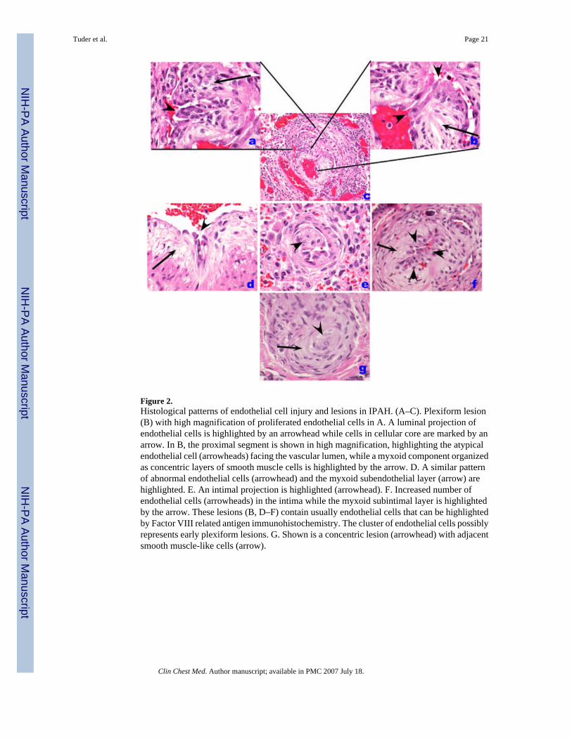
Figure 2.Histological patterns of endothelial cell injury and lesions in IPAH. (A–C). Plexiform lesion(B) with high magnification of proliferated endothelial cells in A. A luminal projection ofendothelial cells is highlighted by an arrowhead while cells in cellular core are marked by anarrow. In B, the proximal segment is shown in high magnification, highlighting the atypicalendothelial cell (arrowheads) facing the vascular lumen, while a myxoid component organizedas concentric layers of smooth muscle cells is highlighted by the arrow. D. A similar patternof abnormal endothelial cells (arrowhead) and the myxoid subendothelial layer (arrow) arehighlighted. E. An intimal projection is highlighted (arrowhead). F. Increased number ofendothelial cells (arrowheads) in the intima while the myxoid subintimal layer is highlightedby the arrow. These lesions (B, D–F) contain usually endothelial cells that can be highlightedby Factor VIII related antigen immunohistochemistry. The cluster of endothelial cells possiblyrepresents early plexiform lesions. G. Shown is a concentric lesion (arrowhead) with adjacentsmooth muscle-like cells (arrow).
Tuder et al. Page 21
Clin Chest Med. Author manuscript; available in PMC 2007 July 18.
NIH
-PA Author Manuscript
NIH
-PA Author Manuscript
NIH
-PA Author Manuscript

Figure 3.A. Fibrotic, relatively paucicellular intima thickening (outlined by arrowheads) in a pulmonaryartery with the media highlighted with the arrow. B. Marked intima remodeling with almostcomplete obliteration by fibrous tissue with a marked intravascular and perivascularinflammatory infiltrate (arrows). C. Smooth muscle cell hypertrophy, with prominentthickening of medial layer (arrow). D. Highlight of medial hypertrophy with smooth muscleα actin immunohistochemistry. E. Markedly remodeled pulmonary artery with endothelial celllayer highlighted by Factor VIII related antigen immunohistochemistry. Note that the intimaand medial smooth muscle cells are negative for Factor VIII related antigen reactivity. F.Ingrowth of smooth muscle cells in a plexiform lesions, highlighted by smooth muscle cell αactin immunohistochemistry (arrow).
Tuder et al. Page 22
Clin Chest Med. Author manuscript; available in PMC 2007 July 18.
NIH
-PA Author Manuscript
NIH
-PA Author Manuscript
NIH
-PA Author Manuscript

Figure 4.Distribution of collagen IV, a component of basement membrane of endothelial cells (arrows)in PH pulmonary arteries. A and B. Plexiform lesion. C. Pulmonary artery with medialthickening. The collagen IV basement membrane of alveolar capillaries shows the cleardemarcation of capillary basement membrane.
Tuder et al. Page 23
Clin Chest Med. Author manuscript; available in PMC 2007 July 18.
NIH
-PA Author Manuscript
NIH
-PA Author Manuscript
NIH
-PA Author Manuscript

Figure 5.Veno-occlusive PH. A. Low-power histological view of thickened pulmonary veins runninginto the lung parenchyma from the pleural surface (left edge) (arrows). Note marked vein wallthickening and decreased lumen. Adjacent alveoli are filled with blood and show septalthickening with engorged capillaries (arrowhead). B. Marked vein thickening with intimalprojection probably representing organized thrombus (arrow). Alveolar hemorrhage and septalthickening are highlighted with arrowhead. C and D. Movat stained pulmonary vein showingarterialization pattern with internal and external elastic layers (arrow). The vein shows markedintima thickening with organized thrombus (arrowheads).
Tuder et al. Page 24
Clin Chest Med. Author manuscript; available in PMC 2007 July 18.
NIH
-PA Author Manuscript
NIH
-PA Author Manuscript
NIH
-PA Author Manuscript

Figure 6.Inflammatory cell infiltrate in severe PH. A. Lymphomonuclear cells positive for CD45RO(arrow), surrounding a plexiform lesion (PA). B. Clustering of CD4 lymphocytes around apulmonary artery (PA) with concentric thickening (arrow). C. Clustering of CD8 lymphocytesaround a remodeled pulmonary artery (PA) (arrow). D. Few cells stain positively for CD45RAmarker (indicative of naïve T cell cells).
Tuder et al. Page 25
Clin Chest Med. Author manuscript; available in PMC 2007 July 18.
NIH
-PA Author Manuscript
NIH
-PA Author Manuscript
NIH
-PA Author Manuscript

Figure 7.HIV-1 Nef localization in HIV-1 associated PH. A. Normal precapillary vessels (methyl green).B. Localization of Factor VIII related antigen stained with Alexa Fluor 488 (green) and Nefstained with Vector Red. Note lack of expression of Nef in normal pulmonary arteries, resultingin a green signal in the endothelial cells (arrow). C. Markedly remodeled pulmonary arterywith concentric/plexiform lesion (arrow, methyl green). D. Co-localization of Factor VIIIrelated antigen (green) with Nef (red), resulting in a yellow signal (arrow). E. Plexiform lesionin HIV-1 associated PH (arrow, methyl green). F. Co-localization of Factor VIII related antigen(green) with Nef (red), resulting in a yellow signal (arrow) in endothelial cells of pulmonaryarteries with mild remodeling (arrow). G and H. Lymph node of an SIV infected monkey,showing extensive expression of Nef (red in G and brown in H, arrows) in a hilar lymph node.(Reproduced from Marecki, J. C., et al. “HIV-1 Nef is Associated with Complex Pulmonary
Tuder et al. Page 26
Clin Chest Med. Author manuscript; available in PMC 2007 July 18.
NIH
-PA Author Manuscript
NIH
-PA Author Manuscript
NIH
-PA Author Manuscript

Vascular Lesions in SHIV-nef-infected Macaques.” Am.J Respir Crit Care Med. 174.4 (2006):437-45, with permission.)
Tuder et al. Page 27
Clin Chest Med. Author manuscript; available in PMC 2007 July 18.
NIH
-PA Author Manuscript
NIH
-PA Author Manuscript
NIH
-PA Author Manuscript

NIH
-PA Author Manuscript
NIH
-PA Author Manuscript
NIH
-PA Author Manuscript
Tuder et al. Page 28
Table 1Intima Remodeling.
Cell Smooth muscle cells EC Matrix Endothelial cellsLesion Eccentric Concentric Fibrotic Plesiform Concentric Dilation/angiomatoid
Normal PAP Yes No No No No NoMild/Moderate PH Yes No Yes No No No
Severe PH Yes Yes Yes Yes Yes Yes
Clin Chest Med. Author manuscript; available in PMC 2007 July 18.

NIH
-PA Author Manuscript
NIH
-PA Author Manuscript
NIH
-PA Author Manuscript
Tuder et al. Page 29
Table 2Medial Remodeling.
Cell Smooth muscle cells EC MatrixNormal PAP Yes* No
Mild/Moderate PH Yes YesSevere PH Yes Yes
*Medial hypertrophy is localized and restricted to some pulmonary arteries
Clin Chest Med. Author manuscript; available in PMC 2007 July 18.

NIH
-PA Author Manuscript
NIH
-PA Author Manuscript
NIH
-PA Author Manuscript
Tuder et al. Page 30
Table 3Molecules Up or Down Pathways Affected Vascular Cells AffectedTGFβR2 Down b,e EC, SMCBMPR2 Down b,e EC, SMC
Nitric oxide Down a,c,e EC, SMCProstacyclin Down a,c EC, SMCCaveolin-1 Down a EC
BAX Down b EC[K−] channels Down b,d SMC
Thrombomodulin Down C ECEndothelin-1 Up a,bc,d,e EC, SMC,AFTissue Factor Up a,bc EC
von Willebrand Factor Up c,e EC, SMC5-lipoxygenase Up a,de EC
VEGF Up a,b,c,e EC, SMCVEGF reportors Up a,b,c,e EC, SMC
HIF-1α Up a,b EC, SMC,AFSerotonin transporter Up a,d SMC
Serotonin receptor Up a,d SMCRANTES Up e Mφ
IL-6 Up e ECMIP-1α Up e Mφ
Fractalkine UP e T cellsPDGF Up a EC, SMC, AFTGF β Up a SMC,AF
Tenascin Up a,b SMCMMP2 Up a SMCTIMP-1 Up a SMC
MT-MMP1 Up A SMCPathobiological Processes
aIncreased cell proliferation
bDecreased cell apoptosis
cIncreased clotting
dIncreased vasoconstriction
eInflammation
Abbreviations: EC, endothelial cells; SMC, smooth muscle cells; AF, adventitial fibroblast; Mφ, macrophages.
Clin Chest Med. Author manuscript; available in PMC 2007 July 18.