Diagnosis, Assessment, and Treatment of Non-Pulmonary Arterial Hypertension Pulmonary Hypertension
12
Diagnosis, Assessment, and Treatment of Non-Pulmonary Arterial Hypertension Pulmonary Hypertension Marius M. Hoeper, MD,* Joan Albert Barberà, MD,† Richard N. Channick, MD,‡ Paul M. Hassoun, MD,§ Irene M. Lang, MD, Alessandra Manes, MD, PHD,¶ Fernando J. Martinez, MD,# Robert Naeije, MD, PHD,** Horst Olschewski, MD,†† Joanna Pepke-Zaba, PHD,‡‡ Margaret M. Redfield, MD, PHD,§§ Ivan M. Robbins, MD, Rogério Souza, MD,¶¶ Adam Torbicki, MD, PHD,## Michael McGoon, MD*** Hannover, Germany; Barcelona, Spain; La Jolla, California; Baltimore, Maryland; Vienna and Graz, Austria; Bologna, Italy; Ann Arbor, Michigan; Brussels, Belgium; Cambridge, United Kingdom; Rochester, Minnesota; Nashville, Tennessee; São Paulo, Brazil; and Warsaw, Poland The 4th World Symposium on Pulmonary Hypertension was the first international meeting to focus not only on pulmonary arterial hypertension (PAH) but also on the so-called non-PAH forms of pulmonary hypertension (PH). The term “non-PAH PH” summarizes those forms of PH that are found in groups 2 to 5 of the current classifica- tion of PH, that is, those forms associated with left heart disease, chronic lung disease, recurrent venous throm- boembolism, and other diseases. Many of these forms of PH are much more common than PAH, but all of them have been less well studied, especially in terms of medical therapy. The working group on non-PAH PH focused mainly on 4 conditions: chronic obstructive lung disease, interstitial lung disease, chronic thromboembolic PH, and left heart disease. The medical literature regarding the role of PH in these diseases was reviewed, and rec- ommendations regarding diagnosis and treatment of PH in these conditions are provided. Given the lack of ro- bust clinical trials addressing PH in any of these conditions, it is important to conduct further studies to establish the role of medical therapy in non-PAH PH. (J Am Coll Cardiol 2009;54:S85–96) © 2009 by the American College of Cardiology Foundation Previous international meetings on pulmonary hyperten- sion (PH) have focused predominantly on pulmonary arterial hypertension (PAH), a form of PH that is usually severe but overall quite rare. The 4th World Symposium was the first to assign a working group to address in detail the so-called non-PAH forms of PH, that is, those forms of PH that are encountered in patients with chronic obstructive pulmonary disease (COPD), interstitial lung disease (ILD), left heart disease (LHD), venous throm- boembolism, and other conditions. It is a paradox in the field of PH that one of the less common forms, that is, PAH, has been extensively studied, whereas fewer data are available on other types of PH, many of which are far more common. At the same time, drugs with proven efficacy in PAH (1–3) are being increasingly used in other forms of PH, despite the virtual absence of clinical trials supporting this approach. Pulmonary hypertension in chronic lung disease sub- sumes COPD, ILD, and other diffuse parenchymal lung diseases such as sarcoidosis, connective tissue disease, or pulmonary Langerhans cell histiocytosis. Space limitation prevents a discussion of the rarer diseases, such as sarcoid- osis and pulmonary Langerhans cell histiocytosis, which are often associated with clinically relevant PH (4). Epidemiology, Features, and Importance of PH in COPD The prevalence of PH in COPD depends on the population under study, the definitions applied, and the tools used to evaluate patients (5). Most hemodynamic studies have been From the *Department of Respiratory Medicine, University of Hannover Medical School, Hannover, Germany; †Department of Respiratory Medicine, Hospital Clinic, CIBERES, University of Barcelona, Barcelona, Spain; ‡Division of Pulmonary and Critical Care Medicine, University of California, San Diego, La Jolla, California; §Department of Medicine, Pulmonary and Critical Care Medicine, Johns Hopkins University School of Medicine, Baltimore, Maryland; Division of Cardiology, Medical University of Vienna, Vienna, Austria; ¶Institute of Cardiology, University of Bologna, Bologna, Italy; #Division of Pulmonary and Critical Care Medicine; University of Michigan Health System, Ann Arbor, Michigan; **Department of Pathophysiology, Free University of Brussels, Brussels, Belgium; ††Department of Pulmonology, Medical University of Graz, Graz, Austria; ‡‡Pulmonary Vascular Disease Unit, Papworth Hospital, Cambridge, United Kingdom; §§Department of Cardiology, Mayo Clinic, Rochester, Minnesota; Department of Allergy, Pulmonary and Critical Care Medicine; Vanderbilt University Medical Center, Nashville, Tennessee; ¶¶Pulmo- nary Department, Heart Institute, University of São Paulo Medical School, São Paulo, Brazil; ##Department of Chest Medicine, Institute of Tuberculosis and Lung Diseases, Medical University of Warsaw, Warsaw, Poland; and the ***Department of Cardiovascular Medicine, Mayo Clinic, Rochester, Minnesota. Please see the end of this article for each author’s conflict of interest information. Manuscript received February 6, 2009; accepted April 15, 2009. Journal of the American College of Cardiology Vol. 54, No. 1, Suppl S, 2009 © 2009 by the American College of Cardiology Foundation ISSN 0735-1097/09/$36.00 Published by Elsevier Inc. doi:10.1016/j.jacc.2009.04.008
Transcript of Diagnosis, Assessment, and Treatment of Non-Pulmonary Arterial Hypertension Pulmonary Hypertension
Psaswto
FSCC§UMoUPPUMCnBDCt
Journal of the American College of Cardiology Vol. 54, No. 1, Suppl S, 2009© 2009 by the American College of Cardiology Foundation ISSN 0735-1097/09/$36.00P
Diagnosis, Assessment, and Treatment of Non-PulmonaryArterial Hypertension Pulmonary HypertensionMarius M. Hoeper, MD,* Joan Albert Barberà, MD,† Richard N. Channick, MD,‡Paul M. Hassoun, MD,§ Irene M. Lang, MD,� Alessandra Manes, MD, PHD,¶Fernando J. Martinez, MD,# Robert Naeije, MD, PHD,** Horst Olschewski, MD,††Joanna Pepke-Zaba, PHD,‡‡ Margaret M. Redfield, MD, PHD,§§ Ivan M. Robbins, MD,��Rogério Souza, MD,¶¶ Adam Torbicki, MD, PHD,## Michael McGoon, MD***
Hannover, Germany; Barcelona, Spain; La Jolla, California; Baltimore, Maryland; Vienna and Graz, Austria;Bologna, Italy; Ann Arbor, Michigan; Brussels, Belgium; Cambridge, United Kingdom; Rochester, Minnesota;Nashville, Tennessee; São Paulo, Brazil; and Warsaw, Poland
The 4th World Symposium on Pulmonary Hypertension was the first international meeting to focus not only onpulmonary arterial hypertension (PAH) but also on the so-called non-PAH forms of pulmonary hypertension (PH).The term “non-PAH PH” summarizes those forms of PH that are found in groups 2 to 5 of the current classifica-tion of PH, that is, those forms associated with left heart disease, chronic lung disease, recurrent venous throm-boembolism, and other diseases. Many of these forms of PH are much more common than PAH, but all of themhave been less well studied, especially in terms of medical therapy. The working group on non-PAH PH focusedmainly on 4 conditions: chronic obstructive lung disease, interstitial lung disease, chronic thromboembolic PH,and left heart disease. The medical literature regarding the role of PH in these diseases was reviewed, and rec-ommendations regarding diagnosis and treatment of PH in these conditions are provided. Given the lack of ro-bust clinical trials addressing PH in any of these conditions, it is important to conduct further studies to establishthe role of medical therapy in non-PAH PH. (J Am Coll Cardiol 2009;54:S85–96) © 2009 by the AmericanCollege of Cardiology Foundation
ublished by Elsevier Inc. doi:10.1016/j.jacc.2009.04.008
odbfiPamefs
sdppoo
Ea
Tu
revious international meetings on pulmonary hyperten-ion (PH) have focused predominantly on pulmonaryrterial hypertension (PAH), a form of PH that is usuallyevere but overall quite rare. The 4th World Symposiumas the first to assign a working group to address in detail
he so-called non-PAH forms of PH, that is, those formsf PH that are encountered in patients with chronic
rom the *Department of Respiratory Medicine, University of Hannover Medicalchool, Hannover, Germany; †Department of Respiratory Medicine, Hospital Clinic,IBERES, University of Barcelona, Barcelona, Spain; ‡Division of Pulmonary andritical Care Medicine, University of California, San Diego, La Jolla, California;Department of Medicine, Pulmonary and Critical Care Medicine, Johns Hopkinsniversity School of Medicine, Baltimore, Maryland; �Division of Cardiology,edical University of Vienna, Vienna, Austria; ¶Institute of Cardiology, University
f Bologna, Bologna, Italy; #Division of Pulmonary and Critical Care Medicine;niversity of Michigan Health System, Ann Arbor, Michigan; **Department ofathophysiology, Free University of Brussels, Brussels, Belgium; ††Department ofulmonology, Medical University of Graz, Graz, Austria; ‡‡Pulmonary Vascular Diseasenit, Papworth Hospital, Cambridge, United Kingdom; §§Department of Cardiology,ayo Clinic, Rochester, Minnesota; � �Department of Allergy, Pulmonary and Criticalare Medicine; Vanderbilt University Medical Center, Nashville, Tennessee; ¶¶Pulmo-ary Department, Heart Institute, University of São Paulo Medical School, São Paulo,razil; ##Department of Chest Medicine, Institute of Tuberculosis and Lungiseases, Medical University of Warsaw, Warsaw, Poland; and the ***Department ofardiovascular Medicine, Mayo Clinic, Rochester, Minnesota. Please see the end of
ehis article for each author’s conflict of interest information.
Manuscript received February 6, 2009; accepted April 15, 2009.
bstructive pulmonary disease (COPD), interstitial lungisease (ILD), left heart disease (LHD), venous throm-oembolism, and other conditions. It is a paradox in theeld of PH that one of the less common forms, that is,AH, has been extensively studied, whereas fewer datare available on other types of PH, many of which are farore common. At the same time, drugs with proven
fficacy in PAH (1–3) are being increasingly used in otherorms of PH, despite the virtual absence of clinical trialsupporting this approach.
Pulmonary hypertension in chronic lung disease sub-umes COPD, ILD, and other diffuse parenchymal lungiseases such as sarcoidosis, connective tissue disease, orulmonary Langerhans cell histiocytosis. Space limitationrevents a discussion of the rarer diseases, such as sarcoid-sis and pulmonary Langerhans cell histiocytosis, which areften associated with clinically relevant PH (4).
pidemiology, Features,nd Importance of PH in COPD
he prevalence of PH in COPD depends on the populationnder study, the definitions applied, and the tools used to
valuate patients (5). Most hemodynamic studies have been
(sf(stpp
ps�mpHaphoo
sstpmwtCaac
tm4nooowaodfePg
mAsvwoomwrwri6
S86 Hoeper et al. JACC Vol. 54, No. 1, Suppl S, 2009Diagnosis, Assessment, and Treatment of Non-PAH PH June 30, 2009:S85–96
performed in patients with ad-vanced disease. Three recent stud-ies have provided data in largeseries of patients, the majority ofthem in the Global initiative forchronic Obstructive Lung Disease(GOLD) IV stage. In 1 studyamong 120 patients with severeemphysema (mean forced expira-tory volume in 1 s [FEV1], 27%predicted) undergoing evaluationfor lung volume reduction sur-gery (6), the incidence of PH,defined as a mean pulmonaryartery pressure (mPAP) �20 mmHg, was 91%, although in themajority of patients (86%), it wasin the mild-to-moderate range(mPAP 20 to 35 mm Hg). Only5% of the patients showed anmPAP �35 mm Hg. The cor-relation between mPAP andlung function was weak (FEV1,r2 � 0.11 and PaO2, r2 � 0.03).The mPAP was more closelyrelated to pulmonary capillarywedge pressure (PCWP) (r2 �0.32), which was mildly elevatedin the majority of patients, sug-gesting the presence of diastolicleft ventricular (LV) dysfunctionin advanced COPD. However,gas trapping with elevated in-trathoracic pressures may be analternative explanation for theincreased PCWP.
In a retrospective analysis ofpulmonary hemodynamic studiesin 998 COPD patients (7), 27patients had severe PH, definedas an mPAP �40 mm Hg. Ofthe 27, 16 had alternative expla-nations for PH. Among the re-maining 11 (1.1% of the wholegroup), COPD was the onlyidentifiable cause of PH. Thissubset of patients had onlymoderate airway obstruction
FEV1 50% predicted), but at the same time, they showedevere hypoxemia, hyperventilation, and a very low dif-usion capacity of the lung for carbon monoxideDLCO). The survival time of these patients was muchhorter than in the other patients. These findings indicatehat there is a subset of COPD patients with “out ofroportion” PH sharing some clinical features with idio-
Abbreviationsand Acronyms
BNP � brain natriureticpeptide
COPD � chronicobstructive pulmonarydisease
CT � computedtomography
CTEPH � chronicthromboembolic pulmonaryhypertension
DLCO � diffusion capacityof the lung for carbonmonoxide
ERA � endothelin receptorantagonist
FEV1 � mean forcedexpiratory volume in 1 s
ILD � interstitial lungdisease
IPF � idiopathic pulmonaryfibrosis
LHD � left heart disease
LV � left ventricular
mPAP � mean pulmonaryartery pressure
PAH � pulmonary arterialhypertension
PCWP � pulmonarycapillary wedge pressure
PDE-5 �
phosphodiesterase-5
PEA � pulmonaryendarterectomy
PH � pulmonaryhypertension
PVR � pulmonary vascularresistance
RCT � randomizedcontrolled trial
RHC � right heartcatheterization
RV � right ventricular
SPAP � systolic pulmonaryarterial pressure
TR � tricuspidregurgitation
athic PAH. h
In a study of 215 patients with severe COPD (FEV1 24%redicted) who were candidates for lung volume reductionurgery or lung transplantation, PH, defined as an mPAP25 mm Hg, was present in 50%, although it was mostlyild (mPAP 26 to 35 mm Hg) (8). In 9.8% of these
atients, PH was considered moderate (mPAP 36 to 45 mmg), and in 3.7%, severe (mPAP �45 mm Hg). Cluster
nalysis identified a subset of patients with moderate im-airment of airway function, high PAP, and severe arterialypoxemia, further supporting the concept of the existencef a subgroup of COPD patients with moderate airflowbstruction and “out of proportion” PH.The overall burden of PH in patients with COPD is
ubstantial. If 1% of patients with advanced COPD haveevere PH with mPAP �40 mm Hg (7), extrapolation fromhe U.S. or French prevalence figures on COPD suggests arevalence of severe PH in COPD patients of 3 to 17 perillion, similar to the prevalence of PAH (9). However,ith an mPAP cutoff of 35 mm Hg rather than 40 mm Hg,
he prevalence of out-of-proportion PH in advancedOPD increases by a factor of approximately 5 to 10. When
ll patients with mPAP �20 mm Hg are taken intoccount, the population-based prevalence of PH in COPDould be in the range of 100 to 150 per million.
The hemodynamic features of PH in COPD differ fromhose seen in PAH. In general, the degree of PH is low tooderate in magnitude, with mPAP rarely exceeding 35 to
0 mm Hg. Both right atrial pressure and PCWP tend to beormal or mildly elevated (10–13). The rate of progressionf PH in COPD is slow, with an annual increase in mPAPf 0.4 to 0.7 mm Hg per year (14,15). Patients with COPDften develop right ventricular (RV) diastolic dysfunctionith elevated RV filling pressures resulting in fluid retention
nd edema, especially during COPD exacerbations. Cardiacutput in COPD is usually preserved and may increaseuring exacerbation episodes. Right ventricular forwardailure, that is, low cardiac output as commonly seen innd-stage PAH, is exceedingly rare in COPD-associatedH, and death from right heart failure is a rarity in thisroup of patients.
Numerous studies have shown that the presence of evenild PH is of prognostic relevance in patients with COPD.longitudinal 7-year study of 50 patients with COPD
howed that survival was inversely related to pulmonaryascular resistance (PVR) (13). In a 15-year follow-up studyith 200 patients (16), the presence or absence of PH wasne of the strongest predictors of mortality. In a 1981 studyf 175 patients with COPD (10), those with mPAP �20m Hg had shorter survival time than those in whom PAPas normal. A more recent study involving 84 patients
eceiving long-term oxygen therapy observed that mPAPas the best predictor of mortality (17). The 5-year survival
ate was 36% in patients with mPAP �25 mm Hg, whereasn patients with mPAP �25 mm Hg the survival rate was2%. In this study neither the FEV1 nor the degree of
ypoxemia or hypercapnia had prognostic value.
Ea
Tdmvdd(
urespet5da�(zrtEoSq
(pumrarb39(p�b
tdiiiRopo
A
Dasswsdp
DhCadcbgcetts
Nhlee
eTfcf
TW
IoSoPfipwcrfidt(
S87JACC Vol. 54, No. 1, Suppl S, 2009 Hoeper et al.June 30, 2009:S85–96 Diagnosis, Assessment, and Treatment of Non-PAH PH
pidemiology, Features,nd Importance of PH in ILD
he term “ILD” summarizes a heterogeneous group of lungiseases with similar clinical, radiographic, and physiologicanifestations. The prevalence of PH in patients with ILD
aries greatly as a function of the underlying disease and theiagnostic mode used to identify PH. The most extensiveata have been published in idiopathic pulmonary fibrosisIPF).
The incidence and prevalence of PAH in IPF remainnclear, with widely varying estimates. The differenceseflect varying patient populations, varying underlying dis-ase severity, and differing diagnostic modalities. In general,tudies of patients undergoing assessment for lung trans-lantation have suggested a higher prevalence of PH. In onearly study of ventricular dysfunction and tricuspid regurgi-ation (TR) in patients evaluated for lung transplantation,0 of 77 IPF patients had echocardiographic evidence of RVysfunction (18). Another study retrospectively identifiedn estimated systolic pulmonary arterial pressure (SPAP)35 mm Hg in 84% of 88 patients and �50 mm Hg in 16%
19). The combination of emphysema in the upper lungones and pulmonary fibrosis in the lower lobes on high-esolution computed tomography (CT) of the chest seemso be associated with a higher prevalence of PH (20,21).chocardiographic data are difficult to interpret because theperating characteristics of echocardiography to estimatePAP in patients with advanced lung disease seem to beuite poor (22).Recent studies have used right heart catheterization
RHC) to accurately measure pulmonary pressures in IPFatients. Two retrospective analyses of IPF patientsndergoing RHC reported PH (defined as mPAP �25m Hg) in 31.6% (23) and 33.9% (24) of the patients,
espectively. In the United Network for Organ Sharingnd the Organ Procurement and Transplant Networkegistries for IPF patients listed for lung transplantationetween January 1995 and June 2004 (25), 2,525 of the,457 patients listed had RHC results available. Of these,32 (37.0%) had an mPAP �25 mm Hg, whereas 2319.1%) had an mPAP �40 mm Hg. Among 70 IPFatients evaluated prospectively (26), PH (resting mPAP25 mm Hg) was detected in only 6 patients (8.1%) at
aseline.Several groups have emphasized the prognostic impor-
ance of PAH complicating IPF. Echocardiographicallyefined PH (SPAP �50 mm Hg) has been associated with
mpaired survival (19). Echocardiographically defined PHn IPF patients with superimposed emphysema negativelynfluences survival (20). In IPF patients undergoingHC before listing for lung transplantation, the presencef PAH correlated linearly with mortality (23). In arospective study, an mPAP �17 mm Hg was predictive
f mortality (26). issessment of PH in Chronic Lung Disease
yspnea and fatigue are symptoms of chronic lung diseases well as PH. Thus, patients with chronic lung diseaseshould be evaluated for PH when the symptoms are moreevere than one would expect from lung function data, orhen signs of right heart failure develop. Suspicion of PH
hould be high if clinical deterioration is not matched by aecline in pulmonary function. Profound hypoxemia, hy-erventilation, and a low DLCO are indicators of PH.Once PH is suspected, patients should be evaluated byoppler echocardiography. A measurable TR velocity,
owever, is less likely to be observed in patients withOPD than in patients with PAH, ranging between 24%
nd 77% (27–29). Even if a TR jet is available, echocar-iographic estimates of the PA pressure are often inac-urate, and both false-positive and -negative results haveeen reported. In 2 large series comparing echocardio-raphic data and findings from RHC in patients withhronic lung disease, the positive predictive values ofchocardiography were 32% and 68%, respectively, andhe negative predictive values were 93% and 67%, respec-ively (22,30). The results are somewhat better but stilluboptimal in patients with ILD (22).
Plasma levels of brain natriuretic peptide (BNP) or-terminal pro-brain natriuretic peptide (NT-proBNP)
ave also been evaluated as predictors of PH in patients withung disease (31,32), but these biomarkers lack sensitivity,specially for milder forms of PH, and specificity, becauselevated levels may also reflect LHD.
Given the limitations of echocardiography and biomark-rs, RHC remains the standard for the diagnosis of PH.his is of particular relevance for patients who also suffer
rom some degree of LV dysfunction, which seems to be aommon comorbidity and may contribute to the clinicaleatures of cor pulmonale.
reatment of PH in Patientsith Chronic Lung Disease
t is self-evident that the underlying lung disease should beptimally treated according to relevant guidelines (33,34).ummarizing these recommendations is beyond the scopef this article. We will focus exclusively on the use ofH-targeted medication in patients with lung disease. So
ar, no large randomized controlled trials (RCTs) address-ng the long-term effects of drugs targeting PH have beenerformed in patients with chronic lung disease. Patientsith advanced lung disease, that is, those with total lung
apacities �70% predicted and FEV1/forced vital capacityatios �50% to 60% have been excluded from RCTs in theeld of PAH. There is not sufficient evidence showing thatrugs approved for the treatment of PAH, that is, endo-helin receptor antagonists (ERAs), phosphodiesterase-5PDE-5) inhibitors, and prostanoids, are safe and effective
n patients with chronic lung disease–associated PH. This is
tmo
gewfIhosaisvbstumtt
WC
D
•
•
•
•
•
•
T
•
•
•
•
•
•
CigcewBwmcun
t
•
•
C(mbesd4
S88 Hoeper et al. JACC Vol. 54, No. 1, Suppl S, 2009Diagnosis, Assessment, and Treatment of Non-PAH PH June 30, 2009:S85–96
rue for patients with advanced chronic lung disease andild PH as well as for patients with severe PH in the setting
f chronic lung disease, independent of its severity.Any pulmonary vasodilator has the potential to worsen
as exchange in patients with chronic lung disease, and theffects of these drugs may vary substantially depending onhether the underlying disease has obstructive or restrictive
eatures (35). Short-term studies have been performed inLD patients with sildenafil (36), bosentan (37), and in-aled iloprost (38), and these drugs had no adverse effectsn oxygenation. In contrast, unpublished data suggest thatildenafil may worsen oxygenation in patients with COPDnd PH (39), and it has been shown that sildenafil cannhibit hypoxic pulmonary vasoconstriction (40). In anothertudy in COPD patients, sildenafil did not improve strokeolume at rest or during exercise (41). A small RCT withosentan in patients with COPD and mild PH found aignificant deterioration of the PaO2. At the same time,here was no improvement in exercise capacity, peak oxygenptake, and health-related quality of life (42). All in all,uch more evidence is needed before the use of PH-
argeted drugs can be recommended for certain subpopula-ions of patients with chronic lung disease.
orking Group Recommendations for PH inhronic Lung Disease (COPD, ILD, and Other Forms)
iagnosis and assessment of PH in chronic lung disease
In patients with chronic lung disease, the presence of PHshould be suspected when the symptoms are more severethan expected based on lung function data, or when signsof right heart dysfunction are present. Profound hypox-emia, hyperventilation, or low DLCO values can beindicators of PH (E/A). (Please refer to Barst et al. [43]for an explanation of the evidence-based grading system.)Doppler echocardiography remains the most useful non-invasive tool for assessing the presence of PH in patientswith chronic lung disease, but both false-positive andfalse-negative results are not uncommon (E/B).Biomarkers such as BNP or NT-proBNP need to befurther evaluated. They seem to be useful screeningtools for the presence of PH in patients with chroniclung disease, although they lack sensitivity and speci-ficity (E/B).If the presence of PH is going to affect the managementof a patient with chronic lung disease, confirmation byRHC is recommended (E/A).Patients with chronic lung disease and severe PH (i.e.,mPAP �35 mm Hg and/or signs of right heart failure)should be referred to a center with expertise in PH (E/A).The use of RHC is strongly recommended in clinicaltrials studying patients with chronic lung disease and PHto categorize the patients under study and to identifysubpopulations likely to benefit from PH-targeted drug
therapy (E/A). areatment of PH in chronic lung disease
The underlying lung disease should be optimally treatedaccording to the respective guidelines, including the useof long-term oxygen therapy in patients with chronichypoxemia (E/A).There is no sufficient evidence that the drugs currentlyused for PAH are safe and effective in patients with PHassociated with chronic lung disease (E/A).Patients with PH and chronic lung disease should betreated in the setting of clinical trials whenever possi-ble (E/A).PH in various lung diseases should be studied separately,because patients with COPD and PH may responddifferently to medical therapy than patients with ILD andPH (E/A).Registries are needed to obtain data from patients withvery rare conditions (E/A).The use of drugs currently approved for PAH in patientswith chronic lung disease is not recommended untilfurther data are available (E/B).
linical trial strategy for PH in chronic lung disease. Its unlikely that patients with end-stage lung disease areoing to derive a substantial benefit from PH treatment. Inontrast, patients with mild-to-moderate chronic lung dis-ase but severe PH may be good candidates for clinical trialsith drugs targeting the pulmonary vascular component.ecause one would not expect all subpopulations of patientsith chronic lung disease to respond similarly to PHedications, it is crucial that the patients under study be
arefully evaluated and characterized: this will include these of RHC to define the severity of PH and the hemody-amic profile.A 2-step approach is recommended to evaluate drugs
argeting PH in the setting of chronic lung disease.
Step 1. Safety, proof-of-concept, and preliminary effi-cacy: important safety parameters include vital signs andblood gases (both PaO2 and PaCO2). Preliminary efficacycan be assessed by hemodynamics, exercise capacity (i.e.,6-min walk test), peak oxygen uptake, and ventilatorefficacy measured during cardiopulmonary exercise test-ing, and improvement of oxygenation at rest and duringexercise (E/A).Step 2. Long-term safety and efficacy: prevention of clinicalworsening (i.e., morbidity and mortality), improvements inexercise capacity and quality of life (E/A).
hronic thromboembolic pulmonary hypertensionCTEPH). CTEPH results from obstruction of the pul-onary vascular bed by nonresolving thromboemboli. It has
een estimated that there are 2,500 new cases of CTEPHach year in the U.S. (44). In a prospective study followingurvivors of acute pulmonary embolism, 3.8% of patientseveloped CTEPH within 2 years (45). However, up to0% of patients with CTEPH have not had a clinically
pparent acute pulmonary embolic episode (46–48). Sple-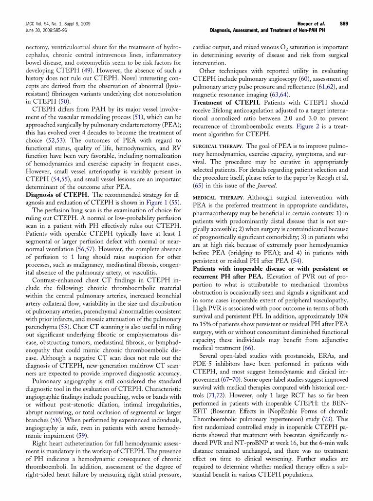
ncbdhcri
matcffoHCdDa
rsPsnopi
cwaowpoeeedn
daoaban
motr
cii
CpmTrtrm
S
nvst(
M
PppgoabpPrpoiHstscm
PCpstpETfitdder
S89JACC Vol. 54, No. 1, Suppl S, 2009 Hoeper et al.June 30, 2009:S85–96 Diagnosis, Assessment, and Treatment of Non-PAH PH
ectomy, ventriculoatrial shunt for the treatment of hydro-ephalus, chronic central intravenous lines, inflammatoryowel disease, and osteomyelitis seem to be risk factors foreveloping CTEPH (49). However, the absence of such aistory does not rule out CTEPH. Novel interesting con-epts are derived from the observation of abnormal (lysis-esistant) fibrinogen variants underlying clot nonresolutionn CTEPH (50).
CTEPH differs from PAH by its major vessel involve-ent of the vascular remodeling process (51), which can be
pproached surgically by pulmonary endarterectomy (PEA);his has evolved over 4 decades to become the treatment ofhoice (52,53). The outcomes of PEA with regard tounctional status, quality of life, hemodynamics, and RVunction have been very favorable, including normalizationf hemodynamics and exercise capacity in frequent cases.owever, small vessel arteriopathy is variably present inTEPH (54,55), and small vessel lesions are an importanteterminant of the outcome after PEA.iagnosis of CTEPH. The recommended strategy for di-
gnosis and evaluation of CTEPH is shown in Figure 1 (55).The perfusion lung scan is the examination of choice for
uling out CTEPH. A normal or low-probability perfusioncan in a patient with PH effectively rules out CTEPH.atients with operable CTEPH typically have at least 1egmental or larger perfusion defect with normal or near-ormal ventilation (56,57). However, the complete absencef perfusion to 1 lung should raise suspicion for otherrocesses, such as malignancy, mediastinal fibrosis, congen-tal absence of the pulmonary artery, or vasculitis.
Contrast-enhanced chest CT findings in CTEPH in-lude the following: chronic thromboembolic materialithin the central pulmonary arteries, increased bronchial
rtery collateral flow, variability in the size and distributionf pulmonary arteries, parenchymal abnormalities consistentith prior infarcts, and mosaic attenuation of the pulmonaryarenchyma (55). Chest CT scanning is also useful in rulingut significant underlying fibrotic or emphysematous dis-ase, obstructing tumors, mediastinal fibrosis, or lymphad-nopathy that could mimic chronic thromboembolic dis-ase. Although a negative CT scan does not rule out theiagnosis of CTEPH, new-generation multirow CT scan-ers are expected to provide improved diagnostic accuracy.Pulmonary angiography is still considered the standard
iagnostic tool in the evaluation of CTEPH. Characteristicngiographic findings include pouching, webs or bands withr without post-stenotic dilation, intimal irregularities,brupt narrowing, or total occlusion of segmental or largerranches (58). When performed by experienced individuals,ngiography is safe, even in patients with severe hemody-amic impairment (59).Right heart catheterization for full hemodynamic assess-ent is mandatory in the workup of CTEPH. The presence
f PH indicates a hemodynamic consequence of chronichromboemboli. In addition, assessment of the degree of
ight-sided heart failure by measuring right atrial pressure, sardiac output, and mixed venous O2 saturation is importantn determining severity of disease and risk from surgicalntervention.
Other techniques with reported utility in evaluatingTEPH include pulmonary angioscopy (60), assessment ofulmonary artery pulse pressure and reflectance (61,62), andagnetic resonance imaging (63,64).reatment of CTEPH. Patients with CTEPH should
eceive lifelong anticoagulation adjusted to a target interna-ional normalized ratio between 2.0 and 3.0 to preventecurrence of thromboembolic events. Figure 2 is a treat-ent algorithm for CTEPH.
URGICAL THERAPY. The goal of PEA is to improve pulmo-ary hemodynamics, exercise capacity, symptoms, and sur-ival. The procedure may be curative in appropriatelyelected patients. For details regarding patient selection andhe procedure itself, please refer to the paper by Keogh et al.65) in this issue of the Journal.
EDICAL THERAPY. Although surgical intervention withEA is the preferred treatment in appropriate candidates,harmacotherapy may be beneficial in certain contexts: 1) inatients with predominantly distal disease that is not sur-ically accessible; 2) when surgery is contraindicated becausef prognostically significant comorbidity; 3) in patients whore at high risk because of extremely poor hemodynamicsefore PEA (bridging to PEA); and 4) in patients withersistent or residual PH after PEA (54).atients with inoperable disease or with persistent or
ecurrent PH after PEA. Elevation of PVR out of pro-ortion to what is attributable to mechanical thrombusbstruction is occasionally seen and signals a significant andn some cases inoperable extent of peripheral vasculopathy.
igh PVR is associated with poor outcome in terms of bothurvival and persistent PH. In addition, approximately 10%o 15% of patients show persistent or residual PH after PEAurgery, with or without concomitant diminished functionalapacity; these individuals may benefit from adjunctiveedical treatment (66).Several open-label studies with prostanoids, ERAs, and
DE-5 inhibitors have been performed in patients withTEPH, and most suggest hemodynamic and clinical im-rovement (67–70). Some open-label studies suggest improvedurvival with medical therapies compared with historical con-rols (71,72). However, only 1 large RCT has so far beenerformed in patients with inoperable CTEPH: the BEN-FiT (Bosentan Effects in iNopErable Forms of chronIchromboembolic pulmonary hypertension) study (73). Thisrst randomized controlled study in inoperable CTEPH pa-ients showed that treatment with bosentan significantly re-uced PVR and NT-proBNP at week 16, but the 6-min walkistance remained unchanged, and there was no treatmentffect on time to clinical worsening. Further studies areequired to determine whether medical therapy offers a sub-
tantial benefit in various CTEPH populations.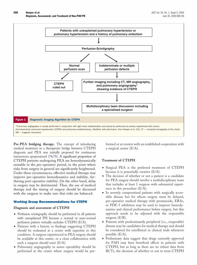
PmdiCurUititw
W
D
•
•
•
T
•
•
•
•
•
S90 Hoeper et al. JACC Vol. 54, No. 1, Suppl S, 2009Diagnosis, Assessment, and Treatment of Non-PAH PH June 30, 2009:S85–96
re-PEA bridging therapy. The concept of introducingedical treatment as a therapeutic bridge between CTEPH
iagnosis and PEA was initially proposed for continuousntravenous epoprostenol (74,75). A significant proportion of
TEPH patients undergoing PEA are hemodynamicallynstable in the pre-operative period, to the point whereisks from surgery in general are significantly heightened.nder these circumstances, effective medical therapy may
mprove pre-operative hemodynamics and stability, fur-hering post-operative stability. On the other hand, delayn surgery may be detrimental. Thus, the use of medicalherapy and the timing of surgery should be discussedith the surgeon to make sure that risks are balanced.
orking Group Recommendations for CTEPH
iagnosis and assessment of CTEPH
Perfusion scintigraphy should be performed in all patientswith unexplained PH because a normal or near-normalperfusion pattern virtually excludes CTEPH (E/A).Patients with a history or findings suggesting CTEPHshould be evaluated at a center with expertise in thiscondition. A surgeon experienced in PEA surgery shouldbe available at this center, or a close collaboration withsuch a surgeon should exist (E/A).Pulmonary angiography to assess operability should be
Patients with unexplainedpulmonary hypertension and a
Perfusion-
Normalperfusion scan
CTEPHruled out
Further im
Multidi
Figure 1 Diagnostic Imaging Algorithm for CTEPH
*Pulmonary angiography is usually performed in conjunction with right heart catheterizthromboembolic pulmonary hypertension (CTEPH) and pulmonary endarterectomy. ModMR � magnetic resonance.
performed at the center where surgery would be per-
formed or at centers with an established cooperation witha surgical center (E/A).
reatment of CTEPH
Surgical PEA is the preferred treatment of CTEPHbecause it is potentially curative (E/A).The decision of whether or not a patient is a candidatefor PEA surgery should involve a multidisciplinary teamthat includes at least 1 surgeon with substantial experi-ence in this procedure (E/A).In severely compromised patients with surgically acces-sible disease but for whom surgery must be delayed,pre-operative medical therapy with prostanoids, ERAs,or PDE-5 inhibitors may be used to improve hemody-namics and clinical performance before surgery, but thisapproach needs to be adjusted with the responsiblesurgeon (E/B).Patients with predominantly peripheral (i.e., inoperable)disease may be candidates for medical therapy and shouldbe considered for enrollment in clinical trials wheneverpossible (E/A).Preliminary data suggest that drugs currently approvedfor PAH may have beneficial effects in patients withCTEPH, but as long as there are no robust data from
monary hypertension ortory of pulmonary embolism
tigraphy
eterminate or multipleperfusion defects
ng including CT, MR angiography,pulmonary angiography*wing evidence of CTEPH
inary team discussion including specialized surgeon
nd should be performed at centers experienced with chronicwith permission, from Hoeper et al. (54). CT � computed tomography of the chest;
pul his
Scin
Ind
agiand sho
scipla
ation aified,
RCTs, the decision of whether or not to treat CTEPH
Cdtwild
•
•
P
Lbdd6utal
(rspmw1trt
pcps
S91JACC Vol. 54, No. 1, Suppl S, 2009 Hoeper et al.June 30, 2009:S85–96 Diagnosis, Assessment, and Treatment of Non-PAH PH
patients with these drugs should be restricted to centersexperienced in the management of this disease (E/B).
linical trial strategy for CTEPH. There is no doubt thatrugs used in PAH, such as prostanoids or PDE-5 inhibi-ors, improve hemodynamics in CTEPH. The question ishether administration of these drugs also improves mean-
ngful clinical end points such as exercise capacity, quality ofife, time to clinical worsening, and survival. At least 2ifferent scenarios require further investigation:
Patients with operable disease but severe hemodynamicimpairment (i.e., PVR ��1,000 dyne·s·cm�5): thesepatients have an elevated perioperative risk, and it isunclear whether a limited period of medical treatment toimprove hemodynamics also improves outcome whencompared with immediate surgery. This problem isextremely difficult to study because a blinded placebo-controlled study might be considered unethical. Thus,this question should be addressed in a carefully designedopen study involving only centers with broad experiencein the surgical and medical management of CTEPHpatients (E/B).Patients with peripheral (inoperable) disease and thosewith persistent or recurrent PH after PEA surgery aregood candidates for clinical trials. The questions thatneed to be answered are whether medical therapy im-proves exercise capacity and quality of life and whether it
Diagnosis of CTEPH witsigns of right
Antico
Surgically accessible pulmonaryarterial obstructions
Contraindicationsprecluding surgery
YesNo
PerformPEA
Clinicmedic
Persistent/recurrent symptomaticpulmonary hypertension
Figure 2 Treatment Algorithm for CTEPH
Modified, with permission, from Hoeper et al. (54). CTEPH � chronic thromboembpulmonary hypertension; PEA � pulmonary endarterectomy; PVR � pulmonary vas
delays time to clinical worsening (E/A). i
H Associated With LHD
HD is one of the most common causes of PH. It maye caused by chronic heart failure attributable to LVysfunction of systolic or diastolic origin or by valvulariseases, predominantly mitral valve disorders (76). Up to0% of patients with severe LV systolic dysfunction andp to 70% of patients with isolated LV diastolic dysfunc-ion may develop PH, and the presence of PH isssociated with a poor prognosis in these patient popu-ations (77–79).
Pulmonary hypertension with PVR �2 mm Hg/l/minWood units) has been reported in up to 50% of patientseferred to transplant clinics (79). Pulmonary hyperten-ion and RV dysfunction carry a poor prognosis foratients with chronic heart failure (80). In one study, theortality rate after 28 months was 57% in patientsith left heart failure and moderate PH, compared with7% in patients without PH (81). Patients with aranspulmonary gradient �16 mm Hg have an increasedisk of post-operative RV failure after heart transplanta-ion (82).
The pathogenesis of PH in LHD is complex. There is aassive (pulmonary venous) component in response to in-reased left atrial pressure (83). In some patients, a superim-osed active component caused by pulmonary arterial vasocon-triction and vascular remodeling may lead to a further increase
ctional limitation and/orrt dysfunction
ation
Advanced small vessel diseasePVR disproportionately elevated
Contraindicationsprecluding surgery
Yes No
ls ofrapy
Consider lungtransplantation
Persistent/recurrent symptomaticpulmonary hypertension
esistance.
h fun hea
agul
al triaal the
oliccular r
n PAP.
DIptbR
EffipfdeadtphA�csPn
ahdLcff
RD
S92 Hoeper et al. JACC Vol. 54, No. 1, Suppl S, 2009Diagnosis, Assessment, and Treatment of Non-PAH PH June 30, 2009:S85–96
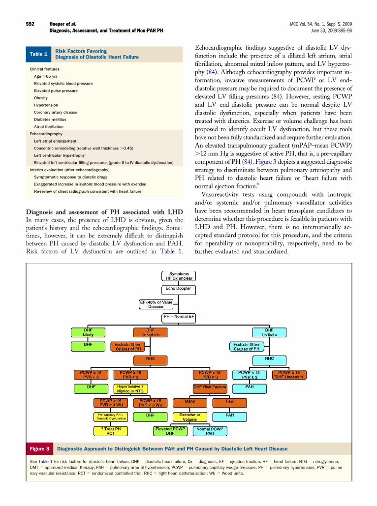
iagnosis and assessment of PH associated with LHDn many cases, the presence of LHD is obvious, given theatient’s history and the echocardiographic findings. Some-imes, however, it can be extremely difficult to distinguishetween PH caused by diastolic LV dysfunction and PAH.isk factors of LV dysfunction are outlined in Table 1.
isk Factors Favoringiagnosis of Diastolic Heart Failure
Table 1 Risk Factors FavoringDiagnosis of Diastolic Heart Failure
Clinical features
Age �65 yrs
Elevated systolic blood pressure
Elevated pulse pressure
Obesity
Hypertension
Coronary artery disease
Diabetes mellitus
Atrial fibrillation
Echocardiography
Left atrial enlargement
Concentric remodeling (relative wall thickness �0.45)
Left ventricular hypertrophy
Elevated left ventricular filling pressures (grade II to IV diastolic dysfunction)
Interim evaluation (after echocardiography)
Symptomatic response to diuretic drugs
Exaggerated increase in systolic blood pressure with exercise
Re-review of chest radiograph consistent with heart failure
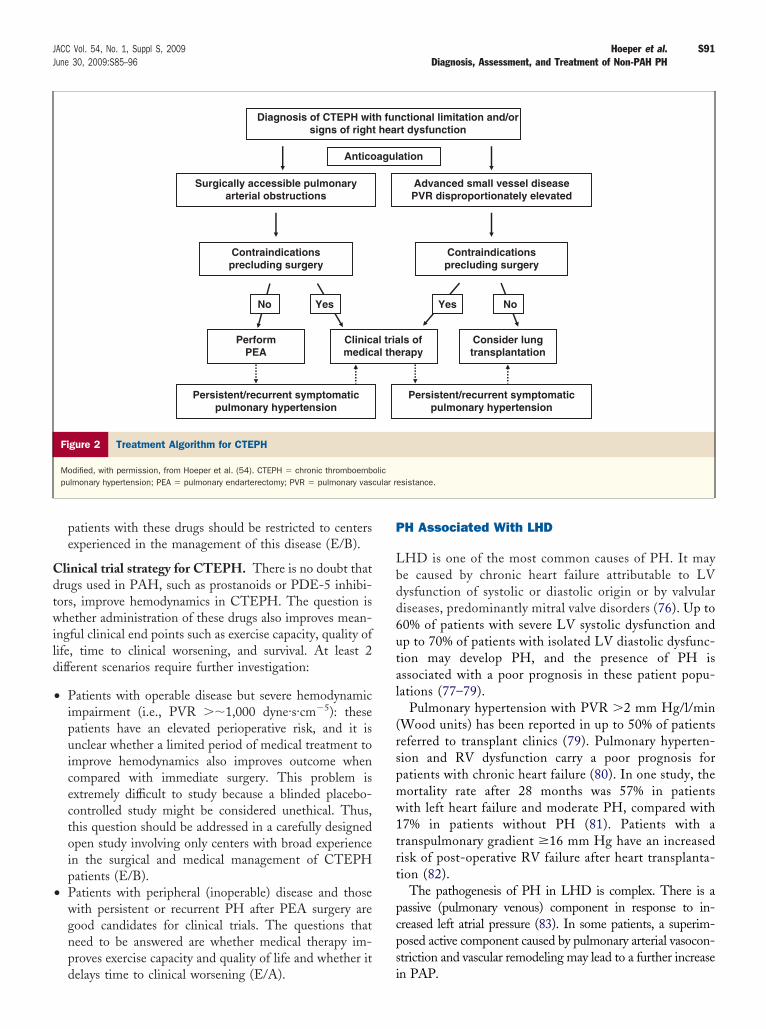
Figure 3 Diagnostic Approach to Distinguish Between PAH and
See Table 1 for risk factors for diastolic heart failure. DHF � diastolic heart failureOMT � optimized medical therapy; PAH � pulmonary arterial hypertension; PCWPnary vascular resistance; RCT � randomized controlled trial; RHC � right heart ca
chocardiographic findings suggestive of diastolic LV dys-unction include the presence of a dilated left atrium, atrialbrillation, abnormal mitral inflow pattern, and LV hypertro-hy (84). Although echocardiography provides important in-ormation, invasive measurements of PCWP or LV end-iastolic pressure may be required to document the presence oflevated LV filling pressures (84). However, resting PCWPnd LV end-diastolic pressure can be normal despite LViastolic dysfunction, especially when patients have beenreated with diuretics. Exercise or volume challenge has beenroposed to identify occult LV dysfunction, but these toolsave not been fully standardized and require further evaluation.n elevated transpulmonary gradient (mPAP–mean PCWP)12 mm Hg is suggestive of active PH, that is, a pre-capillary
omponent of PH (84). Figure 3 depicts a suggested diagnostictrategy to discriminate between pulmonary arteriopathy andH related to diastolic heart failure or “heart failure withormal ejection fraction.”Vasoreactivity tests using compounds with inotropic
nd/or systemic and/or pulmonary vasodilator activitiesave been recommended in heart transplant candidates toetermine whether this procedure is feasible in patients withHD and PH. However, there is no internationally ac-epted standard protocol for this procedure, and the criteriaor operability or nonoperability, respectively, need to beurther evaluated and standardized.
Caused by Diastolic Left Heart Disease
diagnosis; EF � ejection fraction; HF � heart failure; NTG � nitroglycerine;onary capillary wedge pressure; PH � pulmonary hypertension; PVR � pulmo-
zation; WU � Wood units.
PH
; Dx �
� pulmtheteri
TriPfimlapwctmP
upPtaeptcfi
poecs(atpiaosreccsausc
Wf
D
•
•
•
•
T
•
•
•
CLpfphsnesTp
A
DSsKcSrGcl
S93JACC Vol. 54, No. 1, Suppl S, 2009 Hoeper et al.June 30, 2009:S85–96 Diagnosis, Assessment, and Treatment of Non-PAH PH
reatment of PH associated with LHD. Optimal cor-ection of the underlying substrate is a necessary first stepn management because treatment of LHD may decreaseH (85). Careful attention must be paid to optimize LVlling pressures (86). In valvular heart disease such asitral stenosis, post-operative reduction of PCWP may
ead to a rapid reduction of PAP in both passive andctive PH (87). However, the reduction may be incom-lete, despite the normalization of the PCWP, in casesith PH caused by chronic pathological obstructive
hanges. This component may require weeks to monthso regress, even after successful valve surgery (87). In theajority of patients, an almost complete normalization ofH is expected.There is an ongoing debate regarding whether drugs
sed in PAH, that is, ERAs, PDE-5 inhibitors, orrostanoids, may be useful in patients with LHD andH, especially when PH becomes severe and dominates
he clinical picture. Theoretical concerns about thispproach include the possibility of causing pulmonarydema when pulmonary vasodilators are administered toatients with elevated LV filling pressures. This poten-ially adverse effect, however, may be offset when theoncomitant treatments reduce LV afterload and LVlling pressures.Virtually no data from long-term RCTs address the
opulation of patients with LHD and PH. Inhaled nitricxide and prostanoids exert favorable acute hemodynamicffects in these patients (88 –90). However, it is ofoncern that a study of epoprostenol in heart failurehowed a higher mortality in the active treatment group91). Large trials with ERAs in patients with LHD havelso failed to show beneficial long-term effects, althoughhese trials did not look specifically at the subset ofatients with LHD and PH (92,93). The PDE-5 inhib-tor sildenafil improves systolic and diastolic LV functions well as systemic vasoreactivity in experimental modelsf heart failure, making it a promising agent for futuretudies in patients with LHD and PH (94,95). Results ofecent small studies suggest that sildenafil may improvexercise capacity and quality of life in patients with PHaused by LHD (96 –98). However, long-term data fromarefully designed clinical studies are required beforeildenafil can be recommended for patients with LHDnd PH because there have been instances in which drugssed to treat left heart failure had positive effects onurrogate end points but decreased survival, as was thease with PDE-3 inhibitors (99,100).
orking Group Recommendationsor PH Associated With LHD
iagnosis and assessment of PH in LHD
Left heart disease is one of the most common causes ofPH. The presence of LHD is obvious in many
patients, but it may be occult in others, especially when hisolated LV diastolic dysfunction is present. Featuressuggestive of LV diastolic dysfunction or diastolic leftheart failure include an older age, atrial fibrillation,and an enlarged left atrium (E/A).Right and left heart catheterization may be required todistinguish between PAH and PH associated withLHD (E/A).In patients with elevated PCWP and an elevatedtranspulmonary gradient, reduction of LV afterload withnipride or other vasodilators is useful to identify an activepulmonary arterial component of PH (E/A).In patients with LHD and PH considered for hearttransplantation, pulmonary vasoreactivity testing withinhaled nitric oxide or prostanoids may be used toassess reversibility and operability, although the crite-ria for operability or nonoperability require furtherevaluation (E/A).
reatment of PH in LHD
Treatment of LHD according to established criteria isthe basis for a successful approach to patients with LHDand PH (E/A).Patients with end-stage heart failure and PH may becandidates for heart transplantation (in milder, largelyreversible forms of PH) or heart–lung transplantation (ifPH is severe and fixed) (E/A).The use of prostanoids, ERAs, and PDE-5 inhibitorsin patients with LHD and PH is not recommendeduntil robust data from long-term clinical trials areavailable (E/A).
linical trial strategy for PH in LHD. Drugs that reduceV afterload as well as PVR may have beneficial effects inatients with moderate or severe PH in the setting of milderorms of heart failure or LV diastolic dysfunction withreserved ejection fraction. Short-term beneficial effects onemodynamics and exercise capacity have been shown witheveral drugs, including PDE-5 inhibitors. These drugseed to be rigorously studied to assess long-term safety andfficacy in patients with LHD and PH and to identifyubpopulations of patients most likely to derive a benefit.hese trials should address morbidity and mortality endoints to yield meaningful results (E/A).
uthor Disclosures
r. Hoeper has received grants from Actelion, Bayerchering, and Encysive and travel accommodations andpeakers’ honoraria from Actelion, Encysive, GlaxoSmith-line, Lung Rx, Pfizer, and Schering, and has served as a
onsultant to Actelion, Bayer Schering, Encysive, Glaxo-mithKline, and Lung Rx. Dr. Barberà has received hono-aria and research funds from Actelion, Bayer Schering,laxoSmithKline, and Pfizer. Dr. Channick has received
onsulting and speaker fees and research grants from Acte-ion, Gilead, Pfizer, and United Therapeutics. Dr. Hassoun
as received research grants from Actelion (Cotherix), the
NascPcbKeNc&ttramTcsFAtoSSDlhrarUsGfscTaBKcADm
RD3
R
S94 Hoeper et al. JACC Vol. 54, No. 1, Suppl S, 2009Diagnosis, Assessment, and Treatment of Non-PAH PH June 30, 2009:S85–96
ational Institutes of Health, the National Heart, Lung,nd Blood Institute, and United Therapeutics. Dr. Lang haserved as a consultant to Actelion, AOP Orphan Pharma-euticals, Bayer Schering, Encysive, GlaxoSmithKline,fizer, and United Therapeutics. Dr. Manes reports noonflicts of interest. Dr. Martinez has served on speakers’ureaus for AstraZeneca, Boehringer Ingelheim, GlaxoSmith-line, and VoxMedic; advisory boards for AstraZeneca, For-
st/Almirall, Genzyme, GlaxoSmithKline, Mpex, Novartis,ycomed, Schering-Plough, Talecris, and Roche; steering
ommittees for Actelion, Gilead, GlaxoSmithKline, JohnsonJohnson (Centocor), and UBC; and as a primary investiga-
or for Actelion, Altana/Nycomed, Boehringer Ingelheim, andhe National Institutes of Health. Dr. Naeije has receivedesearch grant support from Actelion, Encysive, and Pfizer,nd has served as a consultant and/or steering committeeember for Actelion, Encysive, Lung Rx, MondoBIO-ECH, and United Therapeutics. Dr. Olschewski has re-
eived university grants from Deutsche Forschungsgemein-chaft, Österreichische Nationalbank, and European Unionramework 5 and 6; has received pharmaceutical grants fromctelion, Bayer Schering, Encysive, and Unither Pharmaceu-
icals; has received travel accommodations and speaker’s hon-raria from Actelion, Encysive, Gilead (Myogen), Pfizer,chering, and Unither; and has served as a consultant to Bayerchering, Gilead (Myogen), GlaxoSmithKline, and Unither.r. Pepke-Zaba has received speaker’s honoraria from Acte-
ion, Bayer Schering, Encysive, and United Therapeutics, andas served on an advisory board for Actelion. Dr. Redfield haseceived study drug for research from Pfizer and has served as
consultant to Novartis. Dr. Robbins has received grant/esearch support from Actelion, BioMarin, Gilead, Pfizer, andnited Therapeutics, and has served on advisory boards,
teering committees, and/or speakers’ bureaus for Actelion,ilead, and Lung Rx. Dr. Souza has received consulting fees
rom Actelion; lecture fees from Actelion and Pfizer; and travelupport from Gilead (Myogen). Dr. Torbicki has served as aonsultant for Eli Lilly, GlaxoSmithKline, and mondoBIO-ECH; has received honoraria from Bayer Schering, Eli Lilly,
nd Sanofi-Aventis; and has conducted research supported byayer Schering, Bristol-Myers Squibb, Eli Lilly, GlaxoSmith-line, mondoBIOTECH, and Pfizer. Dr. McGoon has re-
eived grant support from Gilead and consulting fees fromctelion, Gilead, Lung Rx, and Medtronic, and has served onata Safety and Management Board/Clinical Endpoint Com-ittees for Actelion and Gilead.
eprint requests and correspondence: Dr. Marius M. Hoeper,epartment of Respiratory Medicine, Hannover Medical School,
0623 Hannover, Germany. E-mail: [email protected].
EFERENCES
1. Dupuis J, Hoeper MM. Endothelin receptor antagonists in pulmo-
nary arterial hypertension. Eur Respir J 2008;31:407–14.2. Olschewski H, Gomberg-Maitland M. Prostacyclin therapies for thetreatment of pulmonary arterial hypertension. Eur Respir J 2008;31:801–901.
3. Wilkins MR, Wharton J, Grimminger F, Ghofrani HA. Phospho-diesterase inhibitors for the treatment of pulmonary hypertension.Eur Respir J 2008;32:198–209.
4. Behr J, Ryu JH. Pulmonary hypertension in interstitial lung disease.Eur Respir J 2008;31:1357–67.
5. Chaouat A, Naeije R, Weitzenblum E. Pulmonary hypertension inCOPD. Eur Respir J 2008;32:1371–85.
6. Scharf SM, Iqbal M, Keller C, Criner G, Lee S, Fessler HE.Hemodynamic characterization of patients with severe emphysema.Am J Respir Crit Care Med 2002;166:314–22.
7. Chaouat A, Bugnet AS, Kadaoui N, et al. Severe pulmonaryhypertension and chronic obstructive pulmonary disease. Am J RespirCrit Care Med 2005;172:189–94.
8. Thabut G, Dauriat G, Stern JB, et al. Pulmonary hemodynamics inadvanced COPD candidates for lung volume reduction surgery orlung transplantation. Chest 2005;127:1531–6.
9. Humbert M, Sitbon O, Chaouat A, et al. Pulmonary arterialhypertension in France: results from a national registry. Am J RespirCrit Care Med 2006;173:1023–30.
10. Weitzenblum E, Hirth C, Ducolone A, Mirhom R, Rasaholinjana-hary J, Ehrhart M. Prognostic value of pulmonary artery pressure inchronic obstructive pulmonary disease. Thorax 1981;36:752–8.
11. Naeije R, MacNee W. Pulmonary circulation. In: Calverley P, MacNeeW, Pride N, Renard S, editors. Chronic Obstructive Pulmonary Disease.2nd edition. London: Arnold Health Sciences, 2003:228–42.
12. Fishman AP. State of the art: chronic cor pulmonale. Am Rev RespirDis 1976;114:775–94.
13. Burrows B, Kettel LJ, Niden AH, Rabinowitz M, Diener CF.Patterns of cardiovascular dysfunction in chronic obstructive lungdisease. N Engl J Med 1972;286:912–8.
14. Kessler R, Faller M, Weitzenblum E, et al. “Natural history” ofpulmonary hypertension in a series of 131 patients with chronicobstructive lung disease. Am J Respir Crit Care Med 2001;164:219 –24.
15. Weitzenblum E, Sautegeau A, Ehrhart M, Mammosser M, Hirth C,Roegel E. Long-term course of pulmonary arterial pressure in chronicobstructive pulmonary disease. Am Rev Respir Dis 1984;130:993–8.
16. Traver GA, Cline MG, Burrows B. Predictors of mortality in chronicobstructive pulmonary disease. A 15-year follow-up study. Am RevRespir Dis 1979;119:895–902.
17. Oswald-Mammosser M, Weitzenblum E, Quoix E, et al. Prognosticfactors in COPD patients receiving long-term oxygen therapy.Importance of pulmonary artery pressure. Chest 1995;107:1193–8.
18. Vizza CD, Lynch JP, Ochoa LL, Richardson G, Trulock EP. Rightand left ventricular dysfunction in patients with severe pulmonarydisease. Chest 1998;113:576–83.
19. Nadrous HF, Pellikka PA, Krowka MJ, et al. Pulmonary hyperten-sion in patients with idiopathic pulmonary fibrosis. Chest 2005;128:2393–9.
20. Cottin V, Nunes H, Brillet PY, et al. Combined pulmonary fibrosisand emphysema: a distinct underrecognised entity. Eur Respir J2005;26:586–93.
21. Grubstein A, Bendayan D, Schactman I, Cohen M, Shitrit D,Kramer MR. Concomitant upper-lobe bullous emphysema, lower-lobe interstitial fibrosis and pulmonary hypertension in heavy smok-ers: report of eight cases and review of the literature. Respir Med2005;99:948–54.
22. Arcasoy SM, Christie JD, Ferrari VA, et al. Echocardiographicassessment of pulmonary hypertension in patients with advanced lungdisease. Am J Respir Crit Care Med 2003;167:735–40.
23. Lettieri CJ, Nathan SD, Barnett SD, Ahmad S, Shorr AF. Preva-lence and outcomes of pulmonary arterial hypertension in advancedidiopathic pulmonary fibrosis. Chest 2006;129:746–52.
24. Zisman DA, Ross DJ, Belperio JA, et al. Prediction of pulmonaryhypertension in idiopathic pulmonary fibrosis. Respir Med 2007;101:2153–9.
25. Shorr AF, Wainright JL, Cors CS, Lettieri CJ, Nathan SD.Pulmonary hypertension in patients with pulmonary fibrosis awaiting
lung transplant. Eur Respir J 2007;30:715–21.
S95JACC Vol. 54, No. 1, Suppl S, 2009 Hoeper et al.June 30, 2009:S85–96 Diagnosis, Assessment, and Treatment of Non-PAH PH
26. Hamada K, Nagai S, Tanaka S, et al. Significance of pulmonaryarterial pressure and diffusion capacity of the lung as prognosticator inpatients with idiopathic pulmonary fibrosis. Chest 2007;131:650–6.
27. Tramarin R, Torbicki A, Marchandise B, Laaban JP, Morpurgo M,for the Working Group on Noninvasive Evaluation of PulmonaryArtery Pressure. Doppler echocardiographic evaluation of pulmonaryartery pressure in chronic obstructive pulmonary disease. A Europeanmulticentre study. European Office of the World Health Organiza-tion, Copenhagen. Eur Heart J 1991;12:103–11.
28. Torbicki A, Skwarski K, Hawrylkiewicz I, Pasierski T, Miskiewicz Z,Zielinski J. Attempts at measuring pulmonary arterial pressure bymeans of Doppler echocardiography in patients with chronic lungdisease. Eur Respir J 1989;2:856–60.
29. Laaban JP, Diebold B, Zelinski R, Lafay M, Raffoul H, RochemaureJ. Noninvasive estimation of systolic pulmonary artery pressure usingDoppler echocardiography in patients with chronic obstructive pul-monary disease. Chest 1989;96:1258–62.
30. Fisher MR, Criner GJ, Fishman AP, et al. Estimating pulmonaryartery pressures by echocardiography in patients with emphysema.Eur Respir J 2007;30:914–21.
31. Leuchte HH, Baumgartner RA, Nounou ME, et al. Brain natriureticpeptide is a prognostic parameter in chronic lung disease. Am JRespir Crit Care Med 2006;173:744–50.
32. Leuchte HH, El Nounou M, Tuerpe JC, et al. N-terminal pro-brainnatriuretic peptide and renal insufficiency as predictors of mortality inpulmonary hypertension. Chest 2007;131:402–9.
33. Celli BR, MacNee W. Standards for the diagnosis and treatment ofpatients with COPD: a summary of the ATS/ERS position paper.Eur Respir J 2004;23:932–46.
34. Rabe KF, Hurd S, Anzueto A, et al. Global strategy for the diagnosis,management, and prevention of chronic obstructive pulmonary dis-ease: GOLD executive summary. Am J Respir Crit Care Med2007;176:532–55.
35. Barbera JA, Roger N, Roca J, Rovira I, Higenbottam TW,Rodriguez-Roisin R. Worsening of pulmonary gas exchange withnitric oxide inhalation in chronic obstructive pulmonary disease.Lancet 1996;347:436–40.
36. Ghofrani HA, Wiedemann R, Rose F, et al. Sildenafil for treatmentof lung fibrosis and pulmonary hypertension: a randomised controlledtrial. Lancet 2002;360:895–900.
37. Gunther A, Enke B, Markart P, et al. Safety and tolerability ofbosentan in idiopathic pulmonary fibrosis: an open label study. EurRespir J 2007;29:713–9.
38. Olschewski H, Ghofrani HA, Walmrath D, et al. Inhaled prostacy-clin and iloprost in severe pulmonary hypertension secondary to lungfibrosis. Am J Respir Crit Care Med 1999;160:600–7.
39. Blanco I, Gimeno E, Munoz P, et al. Acute effects of sildenafil inchronic obstructive pulmonary disease (abstr). Eur Respir J 2008;32:167s.
40. Zhao L, Mason NA, Morrell NW, et al. Sildenafil inhibits hypoxia-induced pulmonary hypertension. Circulation 2001;104:424–8.
41. Rietema H, Holverda S, Bogaard HJ, et al. Sildenafil treatment inCOPD does not affect stroke volume or exercise capacity. Eur RespirJ 2008;31:759–64.
42. Stolz D, Rasch H, Linka A, et al. A randomized, controlled trial ofbosentan in severe COPD. Eur Respir J 2008;32:619–28.
43. Barst RJ, Gibbs JSR, Ghofrani HA, et al. Updated evidence-basedtreatment algorithm in pulmonary arterial hypertension. J Am CollCardiol 2009;54 Suppl S:S78–84.
44. Fedullo PF, Auger WR, Kerr KM, Rubin LJ. Chronic thromboem-bolic pulmonary hypertension. N Engl J Med 2001;345:1465–72.
45. Pengo V, Lensing AW, Prins MH, et al. Incidence of chronicthromboembolic pulmonary hypertension after pulmonary embolism.N Engl J Med 2004;350:2257–64.
46. Lang IM. Chronic thromboembolic pulmonary hypertension—not sorare after all. N Engl J Med 2004;350:2236–8.
47. Condliffe R, Kiely DG, Gibbs JS, et al. Prognostic and aetiologicalfactors in chronic thromboembolic pulmonary hypertension. EurRespir J 2009;33:332–8.
48. Bonderman D, Wilkens H, Wakounig S, et al. Risk factors forchronic thromboembolic pulmonary hypertension. Eur Respir J
2009;33:325–31.49. Bonderman D, Skoro-Sajer N, Jakowitsch J, et al. Predictors ofoutcome in chronic thromboembolic pulmonary hypertension. Cir-culation 2007;115:2153–8.
50. Suntharalingam J, Goldsmith K, van Marion V, et al. FibrinogenAalpha Thr312Ala polymorphism is associated with chronic throm-boembolic pulmonary hypertension. Eur Respir J 2008;31:736–41.
51. Moser KM, Auger WR, Fedullo PF. Chronic major-vessel throm-boembolic pulmonary hypertension. Circulation 1990;81:1735–43.
52. Klepetko W, Mayer E, Sandoval J, et al. Interventional and surgicalmodalities of treatment for pulmonary arterial hypertension. J AmColl Cardiol 2004;43:73S–80S.
53. Jamieson SW, Kapelanski DP, Sakakibara N, et al. Pulmonaryendarterectomy: experience and lessons learned in 1,500 cases. AnnThorac Surg 2003;76:1457–62; discussion 1462–4.
54. Moser KM, Bloor CM. Pulmonary vascular lesions occurring inpatients with chronic major vessel thromboembolic pulmonary hy-pertension. Chest 1993;103:685–92.
55. Hoeper MM, Mayer E, Simonneau G, Rubin LJ. Chronic throm-boembolic pulmonary hypertension. Circulation 2006;113:2011–20.
56. Moser KM, Page GT, Ashburn WL, Fedullo PF. Perfusion lungscans provide a guide to which patients with apparent primarypulmonary hypertension merit angiography. West J Med 1988;148:167–70.
57. Fishman AJ, Moser KM, Fedullo PF. Perfusion lung scans vspulmonary angiography in evaluation of suspected primary pulmo-nary hypertension. Chest 1983;84:679–83.
58. Auger WR, Fedullo PF, Moser KM, Buchbinder M, Peterson KL.Chronic major-vessel thromboembolic pulmonary artery obstruction:appearance at angiography. Radiology 1992;182:393–8.
59. Hoeper MM, Lee SH, Voswinckel R, et al. Complications of rightheart catheterization procedures in patients with pulmonary hyper-tension in experienced centers. J Am Coll Cardiol 2006;48:2546–52.
60. Shure D, Gregoratos G, Moser KM. Fiberoptic angioscopy: role inthe diagnosis of chronic pulmonary arterial obstruction. Ann InternMed 1985;103:844–50.
61. Nakayama Y, Nakanishi N, Hayashi T, et al. Pulmonary arteryreflection for differentially diagnosing primary pulmonary hyperten-sion and chronic pulmonary thromboembolism. J Am Coll Cardiol2001;38:214–8.
62. Castelain V, Herve P, Lecarpentier Y, Duroux P, Simonneau G,Chemla D. Pulmonary artery pulse pressure and wave reflection inchronic pulmonary thromboembolism and primary pulmonary hyper-tension. J Am Coll Cardiol 2001;37:1085–92.
63. Bergin CJ, Hauschildt J, Rios G, Belezzuoli EV, Huynh T, Chan-nick RN. Accuracy of MR angiography compared with radionuclidescanning in identifying the cause of pulmonary arterial hypertension.AJR Am J Roentgenol 1997;168:1549–55.
64. Kreitner KF, Kunz RP, Ley S, et al. Chronic thromboembolicpulmonary hypertension—assessment by magnetic resonance imag-ing. Eur Radiol 2007;17:11–21.
65. Keogh A, Mayer E, Benza RL, et al. Interventional and surgicalmodalities of treatment in pulmonary hypertension. J Am CollCardiol 2009;54 Suppl S:S67–77.
66. Dartevelle P, Fadel E, Mussot S, et al. Chronic thromboembolicpulmonary hypertension. Eur Respir J 2004;23:637–48.
67. Bresser P, Fedullo PF, Auger WR, et al. Continuous intravenousepoprostenol for chronic thromboembolic pulmonary hypertension.Eur Respir J 2004;23:595–600.
68. Bonderman D, Nowotny R, Skoro-Sajer N, et al. Bosentan therapyfor inoperable chronic thromboembolic pulmonary hypertension.Chest 2005;128:2599–603.
69. Hoeper MM, Kramm T, Wilkens H, et al. Bosentan therapy forinoperable chronic thromboembolic pulmonary hypertension. Chest2005;128:2363–7.
70. Reichenberger F, Voswinckel R, Enke B, et al. Long-term treatmentwith sildenafil in chronic thromboembolic pulmonary hypertension.Eur Respir J 2007;30:922–7.
71. Condliffe R, Kiely DG, Gibbs JS, et al. Improved outcomes inmedically and surgically treated chronic thromboembolic pulmonaryhypertension. Am J Respir Crit Care Med 2008;177:1122–7.
72. Hughes RJ, Jais X, Bonderman D, et al. The efficacy of bosentan in
inoperable chronic thromboembolic pulmonary hypertension: a1-year follow-up study. Eur Respir J 2006;28:138–43.
1
K
S96 Hoeper et al. JACC Vol. 54, No. 1, Suppl S, 2009Diagnosis, Assessment, and Treatment of Non-PAH PH June 30, 2009:S85–96
73. Jais X, D’Armini AM, Jansa P, et al. Bosentan for Treatment ofInoperable Chronic Thromboembolic Pulmonary HypertensionBENEFiT (Bosentan Effects in iNopErable Forms of chronIcThromboembolic pulmonary hypertension), a randomized,placebo-controlled trial. J Am Coll Cardiol 2008;52:2127–34.
74. Kerr KM, Rubin LJ. Epoprostenol therapy as a bridge to pulmonarythromboendarterectomy for chronic thromboembolic pulmonary hy-pertension. Chest 2003;123:319–20.
75. Nagaya N, Sasaki N, Ando M, et al. Prostacyclin therapy beforepulmonary thromboendarterectomy in patients with chronic throm-boembolic pulmonary hypertension. Chest 2003;123:338–43.
76. Oudiz RJ. Pulmonary hypertension associated with left-sided heartdisease. Clin Chest Med 2007;28:233–41.
77. Ghio S, Gavazzi A, Campana C, et al. Independent and additiveprognostic value of right ventricular systolic function and pulmonaryartery pressure in patients with chronic heart failure. J Am CollCardiol 2001;37:183–8.
78. Grigioni F, Potena L, Galie N, et al. Prognostic implications of serialassessments of pulmonary hypertension in severe chronic heartfailure. J Heart Lung Transplant 2006;25:1241–6.
79. Costard-Jackle A, Fowler MB. Influence of preoperative pulmo-nary artery pressure on mortality after heart transplantation:testing of potential reversibility of pulmonary hypertension withnitroprusside is useful in defining a high risk group. J Am CollCardiol 1992;19:48 –54.
80. Di Salvo TG, Mathier M, Semigran MJ, Dec GW. Preserved rightventricular ejection fraction predicts exercise capacity and survival inadvanced heart failure. J Am Coll Cardiol 1995;25:1143–53.
81. Abramson SV, Burke JF, Kelly JJ Jr., et al. Pulmonary hypertensionpredicts mortality and morbidity in patients with dilated cardiomy-opathy. Ann Intern Med 1992;116:888–95.
82. Butler J, Stankewicz MA, Wu J, et al. Pre-transplant reversiblepulmonary hypertension predicts higher risk for mortality aftercardiac transplantation. J Heart Lung Transplant 2005;24:170–7.
83. Lipkin DP, Poole-Wilson PA. Symptoms limiting exercise inchronic heart failure. Br Med J (Clin Res Ed) 1986;292:1030–1.
84. Paulus WJ, Tschope C, Sanderson JE, et al. How to diagnosediastolic heart failure: a consensus statement on the diagnosis of heartfailure with normal left ventricular ejection fraction by the HeartFailure and Echocardiography Associations of the European Societyof Cardiology. Eur Heart J 2007;28:2539–50.
85. Silke B. Haemodynamic impact of diuretic therapy in chronic heartfailure. Cardiology 1994;84 Suppl 2:115–23.
86. Steimle AE, Stevenson LW, Chelimsky-Fallick C, et al. Sustainedhemodynamic efficacy of therapy tailored to reduce filling pressures insurvivors with advanced heart failure. Circulation 1997;96:1165–72.
87. Dalen JE, Matloff JM, Evans GL, et al. Early reduction of pulmonaryvascular resistance after mitral-valve replacement. N Engl J Med
1967;277:387–94. h88. Fojon S, Fernandez-Gonzalez C, Sanchez-Andrade J, et al. Inhalednitric oxide through a noninvasive ventilation device to assessreversibility of pulmonary hypertension in selecting recipients forheart transplant. Transplant Proc 2005;37:4028–30.
89. Loh E, Stamler JS, Hare JM, Loscalzo J, Colucci WS. Cardiovasculareffects of inhaled nitric oxide in patients with left ventriculardysfunction. Circulation 1994;90:2780–5.
90. Haraldsson A, Kieler-Jensen N, Nathorst-Westfelt U, Bergh CH,Ricksten SE. Comparison of inhaled nitric oxide and inhaledaerosolized prostacyclin in the evaluation of heart transplant candi-dates with elevated pulmonary vascular resistance. Chest 1998;114:780–6.
91. Califf RM, Adams KF, McKenna WJ, et al. A randomized controlledtrial of epoprostenol therapy for severe congestive heart failure: theFlolan International Randomized Survival Trial (FIRST). AmHeart J 1997;134:44–54.
92. Kirkby NS, Hadoke PW, Bagnall AJ, Webb DJ. The endothelinsystem as a therapeutic target in cardiovascular disease: great expec-tations or bleak house? Br J Pharmacol 2008;153:1105–19.
93. Seed A, Love MP, McMurray JJ. Clinical experience with endothelinreceptor antagonists in chronic heart failure. Heart Fail Rev 2001;6:317–23.
94. Lewis GD, Semigran MJ. The emerging role for type 5 phosphodi-esterase inhibition in heart failure. Curr Heart Fail Rep 2006;3:123–8.
95. Salloum FN, Abbate A, Das A, et al. Sildenafil (Viagra) attenuatesischemic cardiomyopathy and improves left ventricular function inmice. Am J Physiol Heart Circ Physiol 2008;294:H1398–406.
96. Lewis GD, Lachmann J, Camuso J, et al. Sildenafil improves exercisehemodynamics and oxygen uptake in patients with systolic heartfailure. Circulation 2007;115:59–66.
97. Guazzi M, Samaja M, Arena R, Vicenzi M, Guazzi MD. Long-termuse of sildenafil in the therapeutic management of heart failure. J AmColl Cardiol 2007;50:2136–44.
98. Lewis GD, Shah R, Shahzad K, et al. Sildenafil improves exercisecapacity and quality of life in patients with systolic heart failure andsecondary pulmonary hypertension. Circulation 2007;116:1555–62.
99. Packer M, Carver JR, Rodeheffer RJ, et al., for the PROMISE StudyResearch Group. Effect of oral milrinone on mortality in severechronic heart failure. N Engl J Med 1991;325:1468–75.
00. Abraham WT, Adams KF, Fonarow GC, et al. In-hospital mortalityin patients with acute decompensated heart failure requiring intrave-nous vasoactive medications: an analysis from the Acute Decompen-sated Heart Failure National Registry (ADHERE). J Am CollCardiol 2005;46:57–64.
ey Words: diagnosis y assessment y non-PAH pulmonary
ypertension.