Endothelial-to-Mesenchymal Transition in Pulmonary Hypertension
27
1006 P ulmonary arterial hypertension (PAH) is a rare disorder, with a prevalence of 15 to 50 patients per million in the pop- ulation. It is characterized by remodeling of the precapillary pulmonary arteries, with endothelial cell dysfunction contrib- uting to endothelial and pulmonary artery smooth muscle cell proliferation. This remodeling increases pulmonary vascular resistance and pulmonary arterial pressure (mean pulmonary arterial pressure ≥25 mm Hg and a pulmonary capillary wedge Background—The vascular remodeling responsible for pulmonary arterial hypertension (PAH) involves predominantly the accumulation of α-smooth muscle actin–expressing mesenchymal-like cells in obstructive pulmonary vascular lesions. Endothelial-to-mesenchymal transition (EndoMT) may be a source of those α-smooth muscle actin–expressing cells. Methods and Results—In situ evidence of EndoMT in human PAH was obtained by using confocal microscopy of multiple fluorescent stainings at the arterial level, and by using transmission electron microscopy and correlative light and electron microscopy at the ultrastructural level. Findings were confirmed by in vitro analyses of human PAH and control cultured pulmonary artery endothelial cells. In addition, the mRNA and protein signature of EndoMT was recognized at the arterial and lung level by quantitative real-time polymerase chain reaction and Western blot analyses. We confirmed our human observations in established animal models of pulmonary hypertension (monocrotaline and SuHx). After establishing the first genetically modified rat model linked to BMPR2 mutations (BMPR2 Δ140Ex1/+ rats), we demonstrated that EndoMT is linked to alterations in signaling of BMPR2, a gene that is mutated in 70% of cases of familial PAH and in 10% to 40% of cases of idiopathic PAH. We identified molecular actors of this pathological transition, including twist overexpression and vimentin phosphorylation. We demonstrated that rapamycin partially reversed the protein expression patterns of EndoMT, improved experimental PAH, and decreased the migration of human pulmonary artery endothelial cells, providing the proof of concept that EndoMT is druggable. Conclusions—EndoMT is linked to alterations in BPMR2 signaling and is involved in the occlusive vascular remodeling of PAH, findings that may have therapeutic implications. (Circulation. 2015;131:1006-1018. DOI: 10.1161/ CIRCULATIONAHA.114.008750.) Key Words: bone morphogenetic protein receptors, type II ◼ cardiovascular diseases ◼ epithelial-mesenchymal transition ◼ hypertension, pulmonary ◼ models, animal ◼ neointima ◼ sirolimus ◼ TWIST1 protein, human ◼ vascular remodeling ◼ vimentin © 2015 American Heart Association, Inc. Circulation is available at http://circ.ahajournals.org DOI: 10.1161/CIRCULATIONAHA.114.008750 Received October 16, 2013; accepted December 19, 2014. From Univ. Paris-Sud, Faculté de médecine, Kremlin-Bicêtre, France (B.R., F.A., C.R.-M., A.H., P.D., F.L., E.F., G.S., M.H., S.C.-K., F.P.); AP-HP, DHU TORINO, Centre de Référence de l’Hypertension Pulmonaire Sévère, Service de Pneumologie et Réanimation Respiratoire, Hôpital Bicêtre, Le Kremlin-Bicêtre, France (B.R., F.A., C.R.-.M., A.H., P.D., F.L., G.S., M.H., S.C.-K., F.P.); INSERM UMR-S 999, Labex LERMIT, Hypertension Artérielle Pulmonaire: Physiopathologie et Innovation Thérapeutique, Centre Chirurgical Marie Lannelongue, Le Plessis-Robinson, France (B.R., F.A., C.R.-M., A.H., P.D., F.L., E.F., G.S., M.H., S.C.-K., F.P.); INRA U1196, Génomique et Physiologie de la Lactation – Plateau de Microscopie Electronique à Transmission, Jouy-en-Josas, France (C.P., S.C.); Service de Chirurgie Thoracique, Centre Chirurgical Marie Lannelongue, Le Plessis-Robinson, France (E.F.); Service d’Anatomie Pathologique, Centre Chirurgical Marie Lannelongue, Le Plessis Robinson, France (P.D., S.P.); Department of Pulmonary Medicine, Institute for Cardiovascular Research, VU University Medical Center, Amsterdam, The Netherlands (H.J.B.); INSERM UMR 1064-Center for Research in Transplantation and Immunology-ITUN et Transgenic Rats and Immunophenomic Platform, Nantes, France (S.R., I.A.); and INSERM U955, Département de Physiologie and Service de Cardiologie, Hôpital Henri Mondor, AP-HP, Université Paris-Est Créteil (UPEC), Créteil, France (A.H., S.A.). *Drs Ranchoux and Antigny contributed equally. The online-only Data Supplement is available with this article at http://circ.ahajournals.org/lookup/suppl/doi:10.1161/CIRCULATIONAHA. 114.008750/-/DC1. Correspondence to Frédéric Perros, PhD, INSERM U999, Centre Chirurgical Marie Lannelongue, 133, Avenue de la Résistance, F-92350 Le Plessis Robinson, France. E-mail [email protected] Endothelial-to-Mesenchymal Transition in Pulmonary Hypertension Benoît Ranchoux, MSc*; Fabrice Antigny, PhD*; Catherine Rucker-Martin, PhD; Aurélie Hautefort, MSc; Christine Péchoux, PhD; Harm Jan Bogaard, MD, PhD; Peter Dorfmüller, MD, PhD; Séverine Remy, PhD; Florence Lecerf; Sylvie Planté; Sophie Chat; Elie Fadel, MD, PhD; Amal Houssaini, MSc; Ignacio Anegon, MD, PhD; Serge Adnot, PhD; Gerald Simonneau, MD; Marc Humbert, MD, PhD; Sylvia Cohen-Kaminsky, PhD; Frédéric Perros, PhD Clinical Perspective on p 1018 Vascular Medicine by guest on March 16, 2015 http://circ.ahajournals.org/ Downloaded from by guest on March 16, 2015 http://circ.ahajournals.org/ Downloaded from by guest on March 16, 2015 http://circ.ahajournals.org/ Downloaded from by guest on March 16, 2015 http://circ.ahajournals.org/ Downloaded from by guest on March 16, 2015 http://circ.ahajournals.org/ Downloaded from by guest on March 16, 2015 http://circ.ahajournals.org/ Downloaded from by guest on March 16, 2015 http://circ.ahajournals.org/ Downloaded from by guest on March 16, 2015 http://circ.ahajournals.org/ Downloaded from by guest on March 16, 2015 http://circ.ahajournals.org/ Downloaded from by guest on March 16, 2015 http://circ.ahajournals.org/ Downloaded from by guest on March 16, 2015 http://circ.ahajournals.org/ Downloaded from by guest on March 16, 2015 http://circ.ahajournals.org/ Downloaded from by guest on March 16, 2015 http://circ.ahajournals.org/ Downloaded from by guest on March 16, 2015 http://circ.ahajournals.org/ Downloaded from by guest on March 16, 2015 http://circ.ahajournals.org/ Downloaded from
-
Upload
independent -
Category
Documents
-
view
0 -
download
0
Transcript of Endothelial-to-Mesenchymal Transition in Pulmonary Hypertension

1006
Pulmonary arterial hypertension (PAH) is a rare disorder,
with a prevalence of 15 to 50 patients per million in the pop-
ulation. It is characterized by remodeling of the precapillary
pulmonary arteries, with endothelial cell dysfunction contrib-
uting to endothelial and pulmonary artery smooth muscle cell
proliferation. This remodeling increases pulmonary vascular
resistance and pulmonary arterial pressure (mean pulmonary
arterial pressure ≥25 mm Hg and a pulmonary capillary wedge
Background—The vascular remodeling responsible for pulmonary arterial hypertension (PAH) involves predominantly the
accumulation of α-smooth muscle actin–expressing mesenchymal-like cells in obstructive pulmonary vascular lesions.
Endothelial-to-mesenchymal transition (EndoMT) may be a source of those α-smooth muscle actin–expressing cells.
Methods and Results—In situ evidence of EndoMT in human PAH was obtained by using confocal microscopy of multiple
fluorescent stainings at the arterial level, and by using transmission electron microscopy and correlative light and electron
microscopy at the ultrastructural level. Findings were confirmed by in vitro analyses of human PAH and control cultured
pulmonary artery endothelial cells. In addition, the mRNA and protein signature of EndoMT was recognized at the arterial and
lung level by quantitative real-time polymerase chain reaction and Western blot analyses. We confirmed our human observations
in established animal models of pulmonary hypertension (monocrotaline and SuHx). After establishing the first genetically
modified rat model linked to BMPR2 mutations (BMPR2Δ140Ex1/+ rats), we demonstrated that EndoMT is linked to alterations in
signaling of BMPR2, a gene that is mutated in 70% of cases of familial PAH and in 10% to 40% of cases of idiopathic PAH.
We identified molecular actors of this pathological transition, including twist overexpression and vimentin phosphorylation. We
demonstrated that rapamycin partially reversed the protein expression patterns of EndoMT, improved experimental PAH, and
decreased the migration of human pulmonary artery endothelial cells, providing the proof of concept that EndoMT is druggable.
Conclusions—EndoMT is linked to alterations in BPMR2 signaling and is involved in the occlusive vas cular remodeling
of PAH, findings that may have therapeutic implications. (Circulation. 2015;131:1006-1018. DOI: 10.1161/
CIRCULATIONAHA.114.008750.)
Key Words: bone morphogenetic protein receptors, type II ◼ cardiovascular diseases
◼ epithelial-mesenchymal transition ◼ hypertension, pulmonary ◼ models, animal ◼ neointima
◼ sirolimus ◼ TWIST1 protein, human ◼ vascular remodeling ◼ vimentin
© 2015 American Heart Association, Inc.
Circulation is available at http://circ.ahajournals.org DOI: 10.1161/CIRCULATIONAHA.114.008750
Received October 16, 2013; accepted December 19, 2014.From Univ. Paris-Sud, Faculté de médecine, Kremlin-Bicêtre, France (B.R., F.A., C.R.-M., A.H., P.D., F.L., E.F., G.S., M.H., S.C.-K., F.P.); AP-HP,
DHU TORINO, Centre de Référence de l’Hypertension Pulmonaire Sévère, Service de Pneumologie et Réanimation Respiratoire, Hôpital Bicêtre, Le Kremlin-Bicêtre, France (B.R., F.A., C.R.-.M., A.H., P.D., F.L., G.S., M.H., S.C.-K., F.P.); INSERM UMR-S 999, Labex LERMIT, Hypertension Artérielle Pulmonaire: Physiopathologie et Innovation Thérapeutique, Centre Chirurgical Marie Lannelongue, Le Plessis-Robinson, France (B.R., F.A., C.R.-M., A.H., P.D., F.L., E.F., G.S., M.H., S.C.-K., F.P.); INRA U1196, Génomique et Physiologie de la Lactation – Plateau de Microscopie Electronique à Transmission, Jouy-en-Josas, France (C.P., S.C.); Service de Chirurgie Thoracique, Centre Chirurgical Marie Lannelongue, Le Plessis-Robinson, France (E.F.); Service d’Anatomie Pathologique, Centre Chirurgical Marie Lannelongue, Le Plessis Robinson, France (P.D., S.P.); Department of Pulmonary Medicine, Institute for Cardiovascular Research, VU University Medical Center, Amsterdam, The Netherlands (H.J.B.); INSERM UMR 1064-Center for Research in Transplantation and Immunology-ITUN et Transgenic Rats and Immunophenomic Platform, Nantes, France (S.R., I.A.); and INSERM U955, Département de Physiologie and Service de Cardiologie, Hôpital Henri Mondor, AP-HP, Université Paris-Est Créteil (UPEC), Créteil, France (A.H., S.A.).
*Drs Ranchoux and Antigny contributed equally.The online-only Data Supplement is available with this article at http://circ.ahajournals.org/lookup/suppl/doi:10.1161/CIRCULATIONAHA.
114.008750/-/DC1.Correspondence to Frédéric Perros, PhD, INSERM U999, Centre Chirurgical Marie Lannelongue, 133, Avenue de la Résistance, F-92350 Le Plessis
Robinson, France. E-mail [email protected]
Endothelial-to-Mesenchymal Transition in Pulmonary Hypertension
Benoît Ranchoux, MSc*; Fabrice Antigny, PhD*; Catherine Rucker-Martin, PhD; Aurélie Hautefort, MSc; Christine Péchoux, PhD; Harm Jan Bogaard, MD, PhD;
Peter Dorfmüller, MD, PhD; Séverine Remy, PhD; Florence Lecerf; Sylvie Planté; Sophie Chat; Elie Fadel, MD, PhD; Amal Houssaini, MSc; Ignacio Anegon, MD, PhD;
Serge Adnot, PhD; Gerald Simonneau, MD; Marc Humbert, MD, PhD; Sylvia Cohen-Kaminsky, PhD; Frédéric Perros, PhD
Clinical Perspective on p 1018
Vascular Medicine
by guest on March 16, 2015http://circ.ahajournals.org/Downloaded from by guest on March 16, 2015http://circ.ahajournals.org/Downloaded from by guest on March 16, 2015http://circ.ahajournals.org/Downloaded from by guest on March 16, 2015http://circ.ahajournals.org/Downloaded from by guest on March 16, 2015http://circ.ahajournals.org/Downloaded from by guest on March 16, 2015http://circ.ahajournals.org/Downloaded from by guest on March 16, 2015http://circ.ahajournals.org/Downloaded from by guest on March 16, 2015http://circ.ahajournals.org/Downloaded from by guest on March 16, 2015http://circ.ahajournals.org/Downloaded from by guest on March 16, 2015http://circ.ahajournals.org/Downloaded from by guest on March 16, 2015http://circ.ahajournals.org/Downloaded from by guest on March 16, 2015http://circ.ahajournals.org/Downloaded from by guest on March 16, 2015http://circ.ahajournals.org/Downloaded from by guest on March 16, 2015http://circ.ahajournals.org/Downloaded from by guest on March 16, 2015http://circ.ahajournals.org/Downloaded from

Ranchoux et al EndoMT in Pulmonary Hypertension 1007
pressure ≤15 mm Hg at rest), which ultimately leads to pro-
gressive right ventricular dysfunction and death.1 The nature
of the primary abnormality that triggers and perpetuates pul-
monary vascular cell proliferation in PAH is unclear.
PAH is associated with a spectrum of structural changes
in the pulmonary arteries: increased medial thickness, eccen-
tric and concentric intimal thickening, the obliteration and
recanalization of arteries, and the appearance of plexiform
and dilatation lesions.2 The plexiform lesion is a character-
istic structure of the pulmonary arteriopathy in severe PAH.
According to the consensus view, the plexiform lesion is a
complex and disorganized pulmonary arterial proliferative
lesion that consists of a network or plexus of channels lined
by endothelial cells and separated by core cells.3 However,
it has not been determined whether the core cells are myo-
fibroblasts, smooth muscle cells, endothelial cells, or undif-
ferentiated cells.3 Intimal and medial thickening are the most
consistently encountered structural changes in PAH.2 All of
these changes are characterized by increased numbers of cells
expressing α-smooth muscle actin (α-SMA).4 It is thought
that α-SMA–positive cells that accumulate in vascular lesions
are derived from the expansion of resident vascular smooth
muscle cells or adventitial fibroblasts. However, it is increas-
ingly recognized that endothelial cells (ECs) can transition
into mesenchymal cells expressing α-SMA and that this pro-
cess contributes to the accumulation of smooth muscle–like
cells in vascular pathologies.5
Endothelial-to-mesenchymal transition (EndoMT) is a
biological process in which ECs progressively change their
endothelial phenotype into a mesenchymal or myofibroblas-
tic phenotype. During this process, ECs dissociate from the
monolayer of tightly cohesive cells at the abluminal surface of
the vessel and migrate toward the inner tissue. The migration
starts with the loss of cell-cell contact mediated by membrane
proteins such as vascular endothelial cadherin (VE-cadherin)
and CD31/PECAM-1, and by the cytoplasmic scaffold
protein p120-catenin. By losing their cell-cell junction,
EndoMT-derived cells gain a migratory and invasive capac-
ity allowing them to reach the surrounding tissues. While
migrating, the cells lose specific endothelial markers such
as CD31, VE-cadherin, and CD34 and progressively express
mesenchymal or myofibroblastic markers like α-SMA and
vimentin.6 This phenomenon occurs during certain embryonic
stages of pulmonary artery development7 but also seems to be
implicated in pathological fibroblast and myofibroblast accu-
mulation in conditions such as cardiac or renal fibrosis8,9 and
chronic hypertension.5 The potential role of EndoMT in PAH
has been previously suggested4,5,10 based on analogies with
other diseases,8,9 epithelial-to-mesenchymal transition,5 and
mechanisms of embryonic vascular development,7,11 and also
on the basis of in vitro experiments.6,12,13 Here, we provide the
first in situ evidence of EndoMT in human and experimental
pulmonary hypertension (PH).
Because a reduction in peripheral arterial volume, seen as
intimal thickening and arterial obliteration, is likely to be the
major contributor to the onset and maintenance of PAH, we
analyzed in priority the phenotype of endothelial and suben-
dothelial cells in human intimal lesions. We also elucidated
the phenotype of endothelial and core cells in plexiform
lesions. We hypothesized that α-SMA+ cells building these
lesions had an endothelial origin and resulted from EndoMT.
We used explanted tissue from PAH (group 1 of the Dana
Point classification)14 and from severe models of PAH induced
in rats by exposure to the toxic alkaloid monocrotaline (MCT)
or by the combined exposure to chronic hypoxia and vascu-
lar endothelial growth factor receptor blockade with Sugen/
SU5416.15 We analyzed the phenotype of endothelial and sub-
endothelial cells at structural and ultrastructural levels by mul-
tiple immunofluorescence staining and correlative light and
electron microscopy (CLEM). The loss of endothelial cell-cell
junctions, which is essential for EndoMT, was characterized by
immunostaining and by Western blot analysis (VE-cadherin,
p120-catenin). We measured the expression of Twist-1, a
master transcription factor for EndoMT, in both human and
experimental PAH. EndoMT is stimulated by transforming
growth factor β (TGF-β) signaling,16 but, at the same time,
it is inhibited by BMPR2,17 a TGF-β receptor implicated in
human PAH.18 After establishing the first genetically modified
rat model linked to BMPR2 mutation (BMPR2Δ140Ex1/+ rats),
we searched for evidence of pulmonary vascular EndoMT
linked to BMPR2 signaling. Rapamycin was used in MCT-
induced PAH and on cultured human pulmonary endothelial
cells (PAECs) to regulate EndoMT-associated processes, like
the acquisition of a migratory phenotype.
Methods
PatientsHuman lung specimens were obtained during lung transplantation from patients with PAH and during lobectomy or pneumonectomy for localized lung cancer from control subjects (n numbers are indicated in every legend of the figures). In the lung specimens from control subjects, pulmonary arteries were studied at a distance from tumor areas. Transthoracic echocardiography was performed preoperatively in the control subjects to rule out PH. Patients studied were part of the French Network on Pulmonary Hypertension, a program approved by our institutional Ethics Committee, and had given written informed consent (Protocol N8CO-08- 003, ID RCB: 2008-A00485-50, approved on June 18, 2008).
Immunofluorescence StainingThe full description of the immunofluorescence staining procedures is available in the online-only Data Supplement (Tables I and II in the online-only Data Supplement).
Immunohistochemical Detection of Twist-1 in Paraffin-Embedded Lung TissuesAfter classical dewaxing and heat antigen retrieval at pH 6, immu-nohistochemistry was performed with a rabbit anti–Twist-1 (Ref ab50581, Abcam, UK), detected by a biotinylated goat anti-rabbit and streptavidin peroxidase (Thermo-Scientific, France) and permanent AEC kit (Ref ZUC054-200, Zytomed, Germany). Slides were coun-terstained with hematein.
Transmission Electron Microscopy and CLEMThe full description of the transmission electron microscopy and CLEM procedures are available in the online-only Data Supplement.
To quantify α-SMA and phospho-vimentin labeling, gold par-ticles localized in ECs or SECs of lung control artery from intimal or plexiform lesion of PAH lung were counted on 6 to 40 micrographs randomly taken, at the same magnification of 12 000. The particle density (number of gold particles per 10 μm2 of cytoplasm area) was
by guest on March 16, 2015http://circ.ahajournals.org/Downloaded from

1008 Circulation March 17, 2015
calculated as previously described.19 Area was measured with iTEM
software.
Rat Specimens and In Vivo Study DesignPH and control age/sex-matched pulmonary rat tissues came from
previously published studies.20–22 PH was induced in rats by MCT (60
mg/kg)20,21 or by the combined exposure to chronic hypoxia and vas-
cular endothelial growth factor receptor blockage with Sugen (SuHx
model).22 We had access to the tissues from different time points of
PH development (kinetic of MCT-induced PH development)20 and
from rats exposed to MCT and rapamycin (5 mg/kg from day 21 to
28).21 Hemodynamic data (mean pulmonary artery pressure) and right
ventricular morphology (Fulton Index) were available from animals
of the kinetic study (unpublished data that served in setting up the
experiments). The SuHx model is a severe angio-obliterative PH
model that reproduces multiple salient histological features of human
PAH23 (see the online-only Data Supplement).
Generation of BMPR2-Deficient RatsBMPR2-deficient rats were generated by using zinc-finger nucle-
ases (Sigma, St. Louis, MO) as previously described.24,25 In brief, we
microinjected into the cytoplasm of Sprague-Dawley zygotes mRNA
at 5 ng/μL encoding a pair of zinc-finger nucleases recognizing rat
BMPR2 sequences. Newborn animals were genotyped by a T7 endo-
nuclease assay and sequencing of polymerase chain reaction products
of the targeted sequence. Founders displaying a shift in the coding
reading frame with a premature stop codon were used to derive ani-
mal lines. A rat line with a heterozygous 140 base pairs deletion in
the first exon (BMPR2Δ140Ex1/+ rats) was chosen for this study because
it displayed an intense pulmonary vascular remodeling at 3 months of
life that was absent in the wild-type littermates.
Migration and Proliferation Assays on Cultured Human PAECsHuman PAECs from control and PAH patients were cultured as
previously described26 and were used between passages 3 and 5.
Proliferation was measured with the DELFIA Cell Proliferation
Kit (PerkinElmer) and migration with the CytoSelect 96-Well
Cell Migration Assay, 8 μm (cell Biolabs), following manufac-
turer instructions. Cells were pretreated 24 hours before start-
ing the experiment and treated during the time of the experiment
with rapamycin (50 ng/mL) or by the same volume of its solvent
(dimethyl sulfoxide).
Quantification of p120-Catenin, VE-Cadherin, Vimentin, Phospho-Vimentin, Twist-1, and BMPR2 Lung Expression by Western BlotThe full description of the Western blot procedure is available in the
online-only Data Supplement.
Real-Time Quantitative Polymerase Chain Reaction The full description of the real-time quantitative polymerase chain
reaction procedure is available in the online-only Data Supplement
and Figure I in the online-only Data Supplement.
Statistical AnalysisOwing to small sample sizes, we used nonparametric statistical anal-
yses: Wilcoxon rank sum test (Mann-Whitney U test) for comparing
data between 2 independent groups, Wilcoxon signed-rank test for
comparing data between paired observations, and Kruskal-Wallis test
for comparing data among 3 or more independent groups. Probability
values of <0.05 were considered as statistically significant. IBM
SPSS Statistics and XLSTAT software packages were used for analy-
ses. Data are presented as box plot with the minimum and maximum
of all of the data.
Results
Presence of Cells With a Mixed Mesenchymal/Endothelial Phenotype in Intimal and Plexiform Lesions of PAH Lungs
We analyzed the phenotype of ECs and subendothelial cells
(SECs) by multiple immunofluorescence labeling against spe-
cific endothelial (CD31, CD34, VE-cadherin) and mesenchy-
mal (α-SMA) markers in intimal and plexiform lesions from
PAH lungs in comparison with size-paired normal pulmonary
arteries from control tissues.
In control pulmonary arteries from non-PAH patients, we
observed a single thin layer of ECs expressing CD34, CD31,
and VE-cadherin at EC-EC junctions immediately adjacent to
smooth muscle cells expressing α-SMA (Figure 1A and 1D).
In intimal and plexiform lesions from PAH patient lungs, we
observed a single, generally swollen, luminal layer of cells
expressing diffuse endothelial markers (CD34, CD31, and
VE-cadherin), some of them also coexpressing the mesenchy-
mal marker α-SMA. We also noticed SECs expressing both
VE-cadherin and α-SMA and CD31 and α-SMA (Figure 1B,
1C, 1E, and 1F). VE-cadherin could be detected deep into
the neointima in cells expressing high levels of α-SMA
(Figure 1B).
Interestingly, the EndoMT process is characterized by the
loss of cell-cell junctions making possible a gain of migra-
tory and invasive capacities of ECs. In ECs, VE-cadherin and
CD31 are 2 proteins implicated in cell-cell contact. In con-
trol pulmonary arteries from non-PAH patients, VE-cadherin
and CD31 colocalized in intense spots at cell-cell junctions
(Figure 1A). In intimal and plexiform lesions from PAH
patients, we observed a diffuse staining, in part, cytoplas-
mic, for VE-cadherin and CD31 which was not specifically
localized at cell junctions (Figure 1B and 1C). We also ana-
lyzed the endothelial expression of p120-catenin, the primary
VE-cadherin binding partner that determines the stability of
adherens junctions. As expected, p120-catenin localized in
intense spots at cell-cell junctions of CD34+ EC of control
pulmonary arteries (Figure 1D). Accordingly, the loss of EC
cell junction in EndoMT was associated with a near-loss of
p120-catenin expression in intimal and plexiform lesions
(Figure 1E and 1F). Immunofluorescence performed with
appropriate species and immunoglobulin isotype control
of irrelevant specificity did not lead to a detectable and rel-
evant pattern of staining (Figure 1A through 1F, extreme right
panels).
Protein Pattern Supported EndoMT in PAH LungsTo quantify the extent of EndoMT in PAH lungs, we measured
the pulmonary protein levels of VE-cadherin, p120-catenin,
vimentin and phospho-vimentin, and Twist-1 (Figure 2). We
found a high expression of Twist-1 in PAH lungs, whereas
Twist-1 was nearly not expressed in controls. This may account
for the dramatic decrease in p120-catenin expression in PAH
lungs in comparison with controls. VE-cadherin levels were
not statistically significantly decreased, but the loss of p120-
catenin is expected to induce the internalization of VE-cadherin
from adherens junctions and subsequent loss of endothelial
barrier function.27 Interestingly, we did not find a significant
by guest on March 16, 2015http://circ.ahajournals.org/Downloaded from

Ranchoux et al EndoMT in Pulmonary Hypertension 1009
difference in total pulmonary vimentin content in human PAH
lungs but found a 39-fold increase in the phospho-vimentin
(P-vim) content. We confirmed by immunohistochemical
staining the absence of vascular Twist-1 expression in control
lungs, whereas we observed an intense Twist-1 expression in
ECs and SECs in both intimal and plexiform lesions (Figure
Figure 1. Characterization of the endothelial (EC) and subendothelial cell (SEC) phenotype and endothelial cell–cell junction by immunofluorescence labelings and confocal imaging in pulmonary arteries from control and PAH lungs. A through C, VE-cadherin (green), CD31 (red), and α-SMA (white). A, Pulmonary artery from control lung. VE-cadherin and CD31 are colocalized in intense spots at endothelial cell–cell junctions. VE-cadherin and CD31 are only expressed in ECs. α-SMA is only expressed in smooth muscle cells (SMCs). B, Pulmonary intimal lesion from PAH patient lung. Note some SECs express various levels of VE-cadherin and CD31 deep inside the neointima. Thin arrow indicates SECs expressing both α-SMA and CD31; asterisks, SEC expressing both VE-cadherin and α-SMA. C, Plexiform lesion from PAH patient lung. Note luminal ECs expressing both endothelial (VE-cadherin, CD31) and mesenchymal (α-SMA) markers. These ECs express diffuse VE-cadherin. D through F, p120-catenin (green), CD34 (red), and α-SMA (white). D, Pulmonary artery from control lung. p120-catenin is localized in intense spots at endothelial cell-cell junctions. p120-catenin and CD34 are only expressed in ECs. α-SMA is only expressed in SMCs. Pulmonary intimal lesion (E) and plexiform lesion (F) from PAH patient lung. Note the near-loss of p120-catenin endothelial expression in intimal and plexiform lesions. Some ECs express both CD34 and α-SMA. Red scale bar, 50 μm; white scale bar, 20 μm. DIC indicates differential interference contrast; PAH, pulmonary arterial hypertension; α-SMA, α-smooth muscle actin; and VE-cadherin, vascular endothelial cadherin.
by guest on March 16, 2015http://circ.ahajournals.org/Downloaded from

1010 Circulation March 17, 2015
II in the online-only Data Supplement). These findings are
compatible with an active EndoMT process ongoing in the
remodeled pulmonary arteries from PAH patients under the
control of Twist-1, with transitional ECs translocating from
the luminal layer to constitute the neointima and the core of
the plexiform lesions, after having lost their cell-cell junction
because of p120-catenin repression.
mRNA Expression Pattern Supported EndoMT in Freshly Dissected Human PAH Pulmonary ArteriesWe found a gain of mRNA expression of various mesenchy-
mal genes, including vimentin, N-cadherin, fibronectin and its
receptor ITGA5 in PAH-dissected pulmonary arteries. We also
confirmed overexpression of the EndoMT-related transcrip-
tion factor Twist in PAH pulmonary arteries. Last, we found
increased mRNA expression of 2 known actors of mesenchymal
transition, TGFBR1 and TGFB, coding, respectively, for the
TGF-β receptor 1 and its ligand, the profibrotic cytokine TGF-
β (Table III in the online-only Data Supplement).
Ultrastructural Observations Supported Ongoing EndoMT in PAHElectron microscopy was a technique of choice to ascer-
tain the endothelial origin of SECs in intimal and plexiform
lesions in PAH lungs, because ECs are unambiguously recog-
nized through their specific organelles and structures such as
the Weibel-Palade body (WPB).28 These rod-shaped cytoplas-
mic organelles are present in pulmonary artery and arteriole
ECs, yet are absent in capillary ECs.29
In control pulmonary arteries, ECs were fully differenti-
ated and were characterized in electron microscopy by a
high density of caveolae and WPBs. Subendothelial smooth
Figure 2. Protein signature of EndoMT in control and PAH lung lysates. p120-catenin (A), VE-cadherin (B), and Twist-1 (C), and P-vimentin and vimentin (D) expression were measured by Western blotting. Blots are shown on the left side and their quantification is shown on the right side as expression relative to control. Statistical analyses: Mann–Whitney U test; *P<0.05; ** P<0.01. EndoMT indicates endothelial-to-mesenchymal transition; PAH, pulmonary arterial hypertension; P-vimentin, phospho-vimentin; and VE-cadherin, vascular endothelial cadherin.
by guest on March 16, 2015http://circ.ahajournals.org/Downloaded from

Ranchoux et al EndoMT in Pulmonary Hypertension 1011
Figure 3. Ultrastructure of pulmonary artery from control (A) and PAH (B and C) lungs. A, Flat and elongated endothelial cells (ECs) are separated from smooth muscle cells (SMCs) by only a thick basement membrane, the basal lamina (BL). The elastic lamina (E) is thick. Right inset, Dense bodies (large arrows) can be observed in the cytoplasm of SMC and numerous mitochondria (Mi) are close to the nucleus (N). Lower left inset, EC exhibit numerous caveolae (thin arrows) and WPB (asterisks). B, Intimal lesion from PAH lung displaying EC invagination in the intima (empty triangle) associated to a modification of their nuclei orientation. The endothelial nature of these cells is confirmed by the presence of caveolae and WPB. Their cytoplasm contains numerous microfilaments (plain triangle). C, These invaginations and subendothelial cells exhibit a mixed phenotype demonstrated by the presence of caveolae and WPB in their cytoplasm but also a high density of fibers and dense bodies (inset). Col indicates collagen; PAH, pulmonary arterial hypertension; and WPB, Weibel-Palade body.
by guest on March 16, 2015http://circ.ahajournals.org/Downloaded from

1012 Circulation March 17, 2015
muscle cells were devoid of these EC-specific structures and
harbored typical features of the contractile phenotype, ie, a
cytoplasm displaying myofilaments anchored to dense bod-
ies, and mitochondria were close to the nucleus30 (Figure 3A).
In PAH, luminal pulmonary ECs had a mixed ultrastructural
phenotype because they possessed high-density caveolae and
WPBs but also detectable filaments (Figure 3B). Evidence
of their ongoing migration could be observed by electron
microscopy. Indeed, luminal ECs harbored cell invaginations
directed toward the intima and changes of nuclei orientation
in remodeled PAH arteries in comparison with control EC
(Figure 3B and 3C). We ascertained the endothelial nature
of these invaginations by the high density of caveolae at
their surface and the presence of WPBs in their cytoplasm
(Figure 3B and 3C). SECs harbored a higher density of fibers
and some contained WPBs, which indicated their endothelial
origin (Figure 3C).
To follow endothelial and mesenchymal markers at the
ultrastructural level, we analyzed the phenotype of endo-
thelial and subendothelial cells by using CLEM on CD31-,
α-SMA-, and P-vim–labeled explanted human PAH and
control tissues. This approach allows the treatment of large
samples and the location of rare structures within determined
regions of interest in intimal and plexiform lesions from PAH
patients. As expected, in control pulmonary arteries, the loca-
tion of CD31 was restricted to the luminal ECs as observed
in fluorescence (Figure 4A), more particularly along the
plasma membrane as attested by the presence of numerous
gold particles (Figure 4B). In contrast, only smooth muscle
cells localized under the basal lamina expressed α-SMA
visible at light and electron levels (Figure 5A and 5B). In
PAH, we observed mixed phenotypes for both luminal and
SECs in intimal and plexiform lesions. After confocal anal-
ysis, some SECs appeared to express CD31 in both lesions
(Figure 4C and 4E). Examination at the ultrastructure level
allowed the identification of CD31-laden gold particles close
to the membrane and in the cytoplasm (Figure 4D and 4F).
Moreover, in these lesions, some ECs appeared to express
α-SMA as indicated by the fluorescent labeling (Figure 5C
and 5E). The observation of this labeling at the ultrastruc-
tural level proved the presence of α-SMA–containing fibers
in PAH luminal ECs (Figure 5D and 5F). The quantification
of α-SMA expression in luminal EC, by gold particle den-
sity measurement, confirmed this neoexpression in PAH in
comparison with controls (Figure 5G). We found a 6.6- and
5.1-fold increase in P-vim density in luminal ECs and SECs,
respectively, in intimal lesions in comparison with control
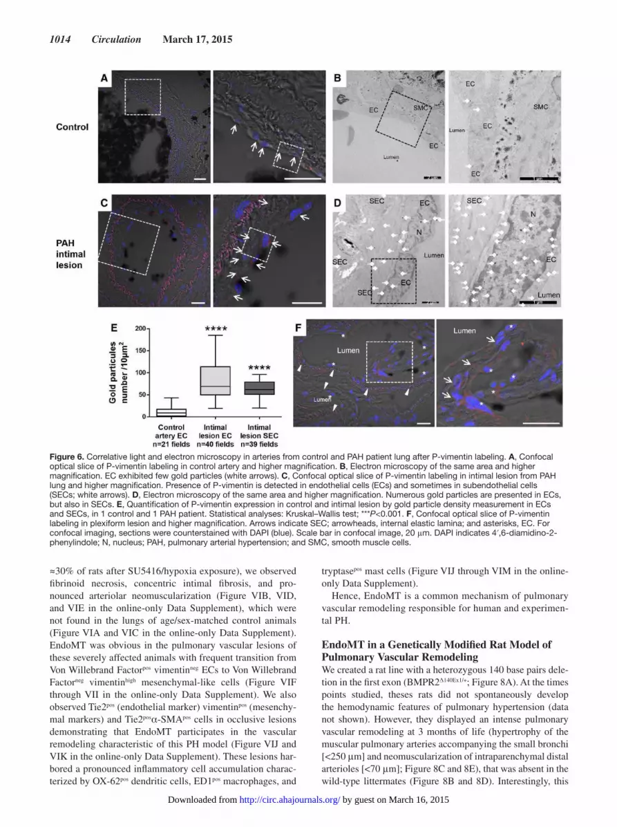
pulmonary arteries (Figure 6A through 6E). We also found
a high expression of P-vim in ECs and SECs of plexiform
lesions (Figure 6F).
Hence, these ultrastructural observations supported fur-
ther a dynamic process of EndoMT, in which transitional
ECs move from the luminal layer to constitute the neointimal
lesions.
EndoMT in PAH Animal ModelThe MCT exposure is the standard model of severe PH. The
progressive neomuscularization and obstruction of precapillary
Figure 4. Correlative light and electron microscopy in arteries from control and PAH patient lung after CD31 labeling. A, Confocal optical slice of CD31 labeling in control artery and higher magnification. B, Electron microscopy of the same area and higher magnification. Only endothelial cells (ECs) exhibited gold particles (white arrows) along the plasma membrane. Some caveolae (black arrows) in front basal lamina (BL) can be observed. C, Confocal optical slice of CD31 labeling in intimal lesion from PAH lung and higher magnification. Presence of CD31 is detected in EC and subendothelial cells (white arrows). D, Electron microscopy of the same area and higher magnification. Gold particles are presented not only in ECs, but also in the subendothelial cells (SECs) near the plasma membrane and along cytoplasmic microfilaments (plain triangle). E, Confocal slice of CD31 in plexiform lesion from PAH lung and higher magnification. F, Electron microscopy of the same area and higher magnification. For confocal imaging, sections were counterstained with DAPI (blue). Confocal images were merged with differential interference contrast images. Scale bar in confocal images, 20 μm. Col indicates collagen; DAPI, 4′,6-diamidino-2-phenylindole; E, elastic lamina; N, nucleus; PAH, pulmonary arterial hypertension; and SMC, smooth muscle cells.
by guest on March 16, 2015http://circ.ahajournals.org/Downloaded from

Ranchoux et al EndoMT in Pulmonary Hypertension 1013
resistance arteries occurring in this model is a robust mecha-
nism of total pulmonary resistance elevation responsible for
PAH. MCT induces a delayed and progressive PH that devel-
ops 14 days after MCT injection (significant increase in mean
pulmonary artery pressure and in right ventricle hypertrophy
quantified by the Fulton index) and becomes established and
severe at day 21, with progressive right heart failure and death
starting between day 21 and 28 (Figure IIIA and IIIB in the
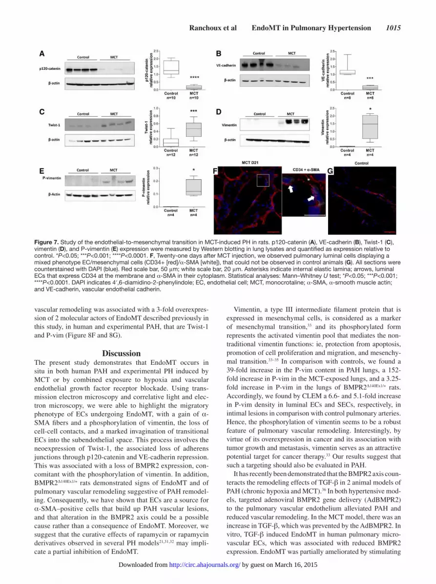
online-only Data Supplement). At day 21/28, we observed
pulmonary luminal cells displaying a mixed phenotype EC/
mesenchymal cells (CD34+αSMA+) that may account for the
progressive neomuscularization of precapillary vessels that
are normally not muscularized (Figure 7). At the pulmonary
level, we observed, at this time point, an overexpression of
Twist-1, vimentin, and P-vim associated with a strong repres-
sion of VE-cadherin and p120-catenin protein expression
(Figure 7 and Figure II in the online-only Data Supplement).
This dual Twist-1 overexpression/VE-cadherin and p120-
catenin repression becomes significant at day 14, when the
pulmonary vascular remodeling appears, inducing a raise in
mean pulmonary artery pressure and a compensatory right
ventricle hypertrophy. Interestingly, this protein signature of
EndoMT paralleled with the kinetic of BMPR2 repression
(Figure IIIC and IIID in the online-only Data Supplement).
When used between day 21 and day 28 (in a curative approach
applied on an established PH), rapamycin (5 mg/kg) reduced
mean pulmonary artery pressure and the Fulton index, and
normalized the muscularization of the pulmonary artery in
comparison with MCT alone.21 The analysis of the Twist-1,
p120-catenin, and VE-cadherin levels in the lungs of those
rats revealed that rapamycin reduced to basal the pulmonary
level of Twist-1, and increased the level of p120-catenin, with-
out significant reexpression of VE-cadherin (Figure IVA and
IVB in the online-only Data Supplement). Hence, the cura-
tive effects of rapamycin in MCT-induced PH may impli-
cate a partial inhibition of EndoMT. Accordingly, rapamycin
(50 ng/mL) inhibited the migration of human PAEC from
control and PAH lungs (Figure IVC in the online-only Data
Supplement). Interestingly, rapamycin also inhibited PAEC
proliferation. This may be relevant in the context of angiopro-
liferative lesions of human PAH (Figure V in the online-only
Data Supplement).
In the lungs of SuHx rats with severe PH (right ventricu-
lar systolic pressure in excess of 80 mm Hg, which occurs in
Figure 5. Correlative light and electron microscopy in arteries from control and PAH patient lung after α-SMA labeling. A, Confocal optical slice of α-SMA labeling in control artery and higher magnification. B, Electron microscopy of the same area and higher magnification. Only SMC exhibited gold particles (white arrows). C, Confocal optical slice of α-SMA labeling in intimal lesion from PAH lung and higher magnification. The presence of α-SMA is detected in endothelial cells (white arrows). D, Electron microscopy of the same area and higher magnification. Gold particles are presented in endothelial cells (ECs) identified by WPB (asterisk) but also in subendothelial cells (SECs). E, Confocal slice of α-SMA in plexiform lesion from PAH lung and higher magnification. F, Electron microscopy of the same area and higher magnification. G, Quantification of α-SMA expression in luminal ECs, by gold particle density measurement in 1 control and 1 PAH patient. For confocal imaging, sections were counterstained with DAPI (blue). Confocal images were merged with differential interference contrast images. Scale bar in confocal image, 20 μm. Statistical analyses: Kruskal–Wallis test; **P<0.01; ****P<0.0001. Col indicates collagen; DAPI, 4′,6-diamidino-2-phenylindole; N, nucleus; PAH, pulmonary arterial hypertension; α-SMA, α-smooth muscle actin; SMC, smooth muscle cells; and WPB, Weibel-Palade body.
by guest on March 16, 2015http://circ.ahajournals.org/Downloaded from
1014 Circulation March 17, 2015
≈30% of rats after SU5416/hypoxia exposure), we observed
fibrinoid necrosis, concentric intimal fibrosis, and pro-
nounced arteriolar neomuscularization (Figure VIB, VID,
and VIE in the online-only Data Supplement), which were
not found in the lungs of age/sex-matched control animals
(Figure VIA and VIC in the online-only Data Supplement).
EndoMT was obvious in the pulmonary vascular lesions of
these severely affected animals with frequent transition from
Von Willebrand Factorpos vimentinneg ECs to Von Willebrand
Factorneg vimentinhigh mesenchymal-like cells (Figure VIF
through VII in the online-only Data Supplement). We also
observed Tie2pos (endothelial marker) vimentinpos (mesenchy-
mal markers) and Tie2posα-SMApos cells in occlusive lesions
demonstrating that EndoMT participates in the vascular
remodeling characteristic of this PH model (Figure VIJ and
VIK in the online-only Data Supplement). These lesions har-
bored a pronounced inflammatory cell accumulation charac-
terized by OX-62pos dendritic cells, ED1pos macrophages, and
tryptasepos mast cells (Figure VIJ through VIM in the online-
only Data Supplement).
Hence, EndoMT is a common mechanism of pulmonary
vascular remodeling responsible for human and experimen-
tal PH.
EndoMT in a Genetically Modified Rat Model of Pulmonary Vascular RemodelingWe created a rat line with a heterozygous 140 base pairs dele-
tion in the first exon (BMPR2Δ140Ex1/+; Figure 8A). At the times
points studied, theses rats did not spontaneously develop
the hemodynamic features of pulmonary hypertension (data
not shown). However, they displayed an intense pulmonary
vascular remodeling at 3 months of life (hypertrophy of the
muscular pulmonary arteries accompanying the small bronchi
[<250 μm] and neomuscularization of intraparenchymal distal
arterioles [<70 μm]; Figure 8C and 8E), that was absent in the
wild-type littermates (Figure 8B and 8D). Interestingly, this
Figure 6. Correlative light and electron microscopy in arteries from control and PAH patient lung after P-vimentin labeling. A, Confocal optical slice of P-vimentin labeling in control artery and higher magnification. B, Electron microscopy of the same area and higher magnification. EC exhibited few gold particles (white arrows). C, Confocal optical slice of P-vimentin labeling in intimal lesion from PAH lung and higher magnification. Presence of P-vimentin is detected in endothelial cells (ECs) and sometimes in subendothelial cells (SECs; white arrows). D, Electron microscopy of the same area and higher magnification. Numerous gold particles are presented in ECs, but also in SECs. E, Quantification of P-vimentin expression in control and intimal lesion by gold particle density measurement in ECs and SECs, in 1 control and 1 PAH patient. Statistical analyses: Kruskal–Wallis test; ***P<0.001. F, Confocal optical slice of P-vimentin labeling in plexiform lesion and higher magnification. Arrows indicate SEC; arrowheads, internal elastic lamina; and asterisks, EC. For confocal imaging, sections were counterstained with DAPI (blue). Scale bar in confocal image, 20 μm. DAPI indicates 4′,6-diamidino-2-phenylindole; N, nucleus; PAH, pulmonary arterial hypertension; and SMC, smooth muscle cells.
by guest on March 16, 2015http://circ.ahajournals.org/Downloaded from
Ranchoux et al EndoMT in Pulmonary Hypertension 1015
vascular remodeling was associated with a 3-fold overexpres-
sion of 2 molecular actors of EndoMT described previously in
this study, in human and experimental PAH, that are Twist-1
and P-vim (Figure 8F and 8G).
DiscussionThe present study demonstrates that EndoMT occurs in
situ in both human PAH and experimental PH induced by
MCT or by combined exposure to hypoxia and vascular
endothelial growth factor receptor blockade. Using trans-
mission electron microscopy and correlative light and elec-
tron microscopy, we were able to highlight the migratory
phenotype of ECs undergoing EndoMT, with a gain of α-
SMA fibers and a phosphorylation of vimentin, the loss of
cell-cell contacts, and a marked invagination of transitional
ECs into the subendothelial space. This process involves the
neoexpression of Twist-1, the associated loss of adherens
junctions through p120-catenin and VE-cadherin repression.
This was associated with a loss of BMPR2 expression, con-
comitant with the phosphorylation of vimentin. In addition,
BMPR2Δ140Ex1/+ rats demonstrated signs of EndoMT and of
pulmonary vascular remodeling suggestive of PAH remodel-
ing. Consequently, we have shown that ECs are a source for
α-SMA–positive cells that build up PAH vascular lesions,
and that alteration in the BMPR2 axis could be a possible
cause rather than a consequence of EndoMT. Moreover, we
suggest that the curative effects of rapamycin or rapamycin
derivatives observed in several PH models21,31,32 may impli-
cate a partial inhibition of EndoMT.
Vimentin, a type III intermediate filament protein that is
expressed in mesenchymal cells, is considered as a marker
of mesenchymal transition,33 and its phosphorylated form
represents the activated vimentin pool that mediates the non-
traditional vimentin functions: ie, protection from apoptosis,
promotion of cell proliferation and migration, and mesenchy-
mal transition.33–35 In comparison with controls, we found a
39-fold increase in the P-vim content in PAH lungs, a 152-
fold increase in P-vim in the MCT-exposed lungs, and a 3.25-
fold increase in P-vim in the lungs of BMPR2Δ140Ex1/+ rats.
Accordingly, we found by CLEM a 6.6- and 5.1-fold increase
in P-vim density in luminal ECs and SECs, respectively, in
intimal lesions in comparison with control pulmonary arteries.
Hence, the phosphorylation of vimentin seems to be a robust
feature of pulmonary vascular remodeling. Interestingly, by
virtue of its overexpression in cancer and its association with
tumor growth and metastasis, vimentin serves as an attractive
potential target for cancer therapy.33 Our results suggest that
such a targeting should also be evaluated in PAH.
It has recently been demonstrated that the BMPR2 axis coun-
teracts the remodeling effects of TGF-β in 2 animal models of
PAH (chronic hypoxia and MCT).36 In both hypertensive mod-
els, targeted adenoviral BMPR2 gene delivery (AdBMPR2)
to the pulmonary vascular endothelium alleviated PAH and
reduced vascular remodeling. In the MCT model, there was an
increase in TGF-β, which was prevented by the AdBMPR2. In
vitro, TGF-β induced EndoMT in human pulmonary micro-
vascular ECs, which was associated with reduced BMPR2
expression. EndoMT was partially ameliorated by stimulating
Figure 7. Study of the endothelial-to-mesenchymal transition in MCT-induced PH in rats. p120-catenin (A), VE-cadherin (B), Twist-1 (C), vimentin (D), and P-vimentin (E) expression were measured by Western blotting in lung lysates and quantified as expression relative to control. *P<0.05; ***P<0.001; ****P<0.0001. F, Twenty-one days after MCT injection, we observed pulmonary luminal cells displaying a mixed phenotype EC/mesenchymal cells (CD34+ [red]/α-SMA [white]), that could not be observed in control animals (G). All sections were counterstained with DAPI (blue). Red scale bar, 50 μm; white scale bar, 20 μm. Asterisks indicate internal elastic lamina; arrows, luminal ECs that express CD34 at the membrane and α-SMA in their cytoplasm. Statistical analyses: Mann–Whitney U test; *P<0.05; ***P<0.001; ****P<0.0001. DAPI indicates 4′,6-diamidino-2-phenylindole; EC, endothelial cell; MCT, monocrotaline; α-SMA, α-smooth muscle actin; and VE-cadherin, vascular endothelial cadherin.
by guest on March 16, 2015http://circ.ahajournals.org/Downloaded from

1016 Circulation March 17, 2015
BMPR2 signaling with appropriate ligands even in the ongo-
ing presence of TGF-β. On the other hand, our results suggest
that alteration in the BMPR2 signaling can induce EndoMT
and subsequent vascular remodeling.
A limitation of our study results from the fact we did not
fully characterize our BMPR2Δ140Ex1/+ rat line. A great deal of
work remains to decipher the pathomechanisms responsible
for the pulmonary vascular remodeling that develops in this
model, and to determine what could be the second hit that
allows PAH occurrence in the context of BMPR2 mutation.
Rapamycin has been demonstrated to prevent EC (EA.
hy926 cell line) migration by inhibiting EndoMT through
the induction of VE-cadherin expression, and inhibition
of vimentin and Twist-1 expression, and inhibition of EC
secretion of metalloproteinase-2 and -9.37 Accordingly,
rapamycin decreased the migration of primary PAECs
derived from control and PAH lungs. It also significantly
reduced their proliferation. Both effects are particularly
relevant in the context of PAH, whose pathognomonic
plexiform lesions are angioproliferative. Besides, a dra-
matic improvement in a patient with PAH has been
reported with rapamycin,38 and, recently, a pilot study
has demonstrated that everolimus (42-O-(2-hydroxyethyl)
rapamycin) was well tolerated in 8 patients with PAH, and
led to improvement in pulmonary vascular resistances and
of the 6-minute walk distance.39 In rats, we demonstrated
that rapamycin alleviated MCT-induced PH with a con-
comitant decrease in distal artery muscularization.21 Of
note, sildenafil, a treatment for PAH, could also decrease
EndoMT and PH progression via a myocardin-dependent
mechanism.13 Hence, EndoMT could be a new target for
therapeutic approaches.
Figure 8. BMPR2-deficient rats (BMPR2Δ140Ex1/+) displayed a spontaneous pulmonary vascular remodeling. A, After PCR, amplicons were analyzed by gel electrophoresis and PCR product size (WT or Δ140 bp) is confirmed by gel electrophoresis. B through E, HES staining of control and BMPR2Δ140Ex1/+ rats. B, Lung from control rat with a normal artery accompanying the small bronchi. C, Lung from BMPR2Δ140Ex1/+ rat with hypertrophy of the muscular pulmonary arteries accompanying the small bronchi. D, Lung from control rat. Distal arterioles are not muscularized. E, Lung from BMPR2Δ140Ex1/+ rat with neomuscularization of intraparenchymal distal arterioles. F and G, Twist-1 and P-vimentin expression in control and BMPR2Δ140Ex1/+ rat lung lysates measured by Western blot. Scales, 100 μm. Statistical analyses: Mann–Whitney U test; * P<0.05. HES, hematoxylin erythrosine saffron; MW, molecular weight; PCR, polymerase chain reaction; P-vimentin, phospho-vimentin; and WT, wild-type.
by guest on March 16, 2015http://circ.ahajournals.org/Downloaded from

Ranchoux et al EndoMT in Pulmonary Hypertension 1017
In conclusion, our work demonstrates that EndoMT par-
ticipates in the vascular remodeling present in PAH, and this
finding may have therapeutic implications for PAH. The iden-
tification of the key molecular players of EndoMT in PAH,
and the mechanisms participating in the control of their
expression and of their functions, as well, will be mandatory
to achieve this goal, and will open a new field of research in
the pathophysiology of the disease.
AcknowledgmentsWe thank Dr André Capderou, the statistician from our INSERM
unit, who reviewed the present study.
Sources of FundingDr Ranchoux is supported by the LabEx LERMIT. Dr Antigny is
supported by a postdoctoral grant from Aviesan (ITMO IHP). Dr
Hautefort is supported by a PhD grant from Région Ile de France
(CORDDIM). Dr Perros receives funding from National Funding
Agency for Research (ANR; Grant ANR-13-JSV1-001). Dr Bogaard
is supported by the Netherlands Cardiovascular Research Initiative:
the Dutch Heart Foundation, Dutch Federation of University Medical
Centers, the Netherlands Organization for Health Research and
Development, and the Royal Netherlands Academy of Sciences
DisclosuresDrs Simonneau and Humbert have received speaker fees or hono-
raria for consultations from Actelion, Bayer, Bristol-Myers-Squib,
GSK, Lilly, Novartis, Pfizer and United Therapeutics. Dr Simonneau
received reimbursement from Actelion and Lilly for attending French
and international meetings and fees from Bristol-Myers-Squib and
Lilly for participating to advisory boards. Dr Bogaard received
speaker fees or honoraria for consultations from Actelion, Bayer,
Pfizer, and United Therapeutics. The other authors report no conflicts.
References 1. O’Callaghan DS, Savale L, Montani D, Jaïs X, Sitbon O, Simonneau
G, Humbert M. Treatment of pulmonary arterial hypertension with
targeted therapies. Nat Rev Cardiol. 2011;8:526–538. doi: 10.1038/
nrcardio.2011.104.
2. Pietra GG, Edwards WD, Kay JM, Rich S, Kernis J, Schloo B, Ayres
SM, Bergofsky EH, Brundage BH, Detre KM. Histopathology of primary
pulmonary hypertension. A qualitative and quantitative study of pulmo-
nary blood vessels from 58 patients in the National Heart, Lung, and
Blood Institute, Primary Pulmonary Hypertension Registry. Circulation.
1989;80:1198–1206.
3. Abe K, Toba M, Alzoubi A, Ito M, Fagan KA, Cool CD, Voelkel NF,
McMurtry IF, Oka M. Formation of plexiform lesions in experimental
severe pulmonary arterial hypertension. Circulation. 2010;121:2747–
2754. doi: 10.1161/CIRCULATIONAHA.109.927681.
4. Rabinovitch M. Molecular pathogenesis of pulmonary arterial hyperten-
sion. J Clin Invest. 2012;122:4306–4313. doi: 10.1172/JCI60658.
5. Arciniegas E, Frid MG, Douglas IS, Stenmark KR. Perspectives on
endothelial-to-mesenchymal transition: potential contribution to vascular
remodeling in chronic pulmonary hypertension. Am J Physiol Lung Cell
Mol Physiol. 2007;293:L1–L8. doi: 10.1152/ajplung.00378.2006.
6. Frid MG, Kale VA, Stenmark KR. Mature vascular endothelium can give
rise to smooth muscle cells via endothelial-mesenchymal transdifferentia-
tion: in vitro analysis. Circ Res. 2002;90:1189–1196.
7. Arciniegas E, Neves CY, Carrillo LM, Zambrano EA, Ramírez R.
Endothelial-mesenchymal transition occurs during embryonic pul-
monary artery development. Endothelium. 2005;12:193–200. doi:
10.1080/10623320500227283.
8. Zeisberg EM, Tarnavski O, Zeisberg M, Dorfman AL, McMullen JR,
Gustafsson E, Chandraker A, Yuan X, Pu WT, Roberts AB, Neilson EG,
Sayegh MH, Izumo S, Kalluri R. Endothelial-to-mesenchymal transition
contributes to cardiac fibrosis. Nat Med. 2007;13:952–961. doi: 10.1038/
nm1613.
9. He J, Xu Y, Koya D, Kanasaki K. Role of the endothelial-to-mesenchymal
transition in renal fibrosis of chronic kidney disease. Clin Exp Nephrol.
2013;17:488–497. doi: 10.1007/s10157-013-0781-0.
10. Morrell NW, Adnot S, Archer SL, Dupuis J, Jones PL, MacLean MR,
McMurtry IF, Stenmark KR, Thistlethwaite PA, Weissmann N, Yuan
JX, Weir EK. Cellular and molecular basis of pulmonary arterial hyper-
tension. J Am Coll Cardiol. 2009;54(1 suppl):S20–S31. doi: 10.1016/j.
jacc.2009.04.018.
11. DeRuiter MC, Poelmann RE, VanMunsteren JC, Mironov V, Markwald
RR, Gittenberger-de Groot AC. Embryonic endothelial cells transdifferen-
tiate into mesenchymal cells expressing smooth muscle actins in vivo and
in vitro. Circ Res. 1997;80:444–451.
12. Sakao S, Taraseviciene-Stewart L, Cool CD, Tada Y, Kasahara Y, Kurosu
K, Tanabe N, Takiguchi Y, Tatsumi K, Kuriyama T, Voelkel NF. VEGF-R
blockade causes endothelial cell apoptosis, expansion of surviving CD34+
precursor cells and transdifferentiation to smooth muscle-like and neuro-
nal-like cells. FASEB J. 2007;21:3640–3652. doi: 10.1096/fj.07-8432com.
13. Zhu P, Huang L, Ge X, Yan F, Wu R, Ao Q. Transdifferentiation
of pulmonary arteriolar endothelial cells into smooth muscle-like
cells regulated by myocardin involved in hypoxia-induced pulmo-
nary vascular remodelling. Int J Exp Pathol. 2006;87:463–474. doi:
10.1111/j.1365-2613.2006.00503.x.
14. Simonneau G, Robbins IM, Beghetti M, Channick RN, Delcroix M,
Denton CP, Elliott CG, Gaine SP, Gladwin MT, Jing ZC, Krowka MJ,
Langleben D, Nakanishi N, Souza R. Updated clinical classification of
pulmonary hypertension. J Am Coll Cardiol. 2009;54(1 suppl):S43–S54.
doi: 10.1016/j.jacc.2009.04.012.
15. Stenmark KR, Meyrick B, Galie N, Mooi WJ, McMurtry IF. Animal
models of pulmonary arterial hypertension: the hope for etiological dis-
covery and pharmacological cure. Am J Physiol Lung Cell Mol Physiol.
2009;297:L1013–L1032. doi: 10.1152/ajplung.00217.2009.
16. Díez M, Musri MM, Ferrer E, Barberà JA, Peinado VI. Endothelial pro-
genitor cells undergo an endothelial-to-mesenchymal transition-like pro-
cess mediated by TGFbetaRI. Cardiovasc Res. 2010;88:502–511. doi:
10.1093/cvr/cvq236.
17. Soubrier F, Chung WK, Machado R, Grünig E, Aldred M, Geraci M,
Loyd JE, Elliott CG, Trembath RC, Newman JH, Humbert M. Genetics
and genomics of pulmonary arterial hypertension. J Am Coll Cardiol.
2013;62(25 suppl):D13–D21. doi: 10.1016/j.jacc.2013.10.035.
18. Potenta S, Zeisberg E, Kalluri R. The role of endothelial-to-mesenchymal
transition in cancer progression. Br J Cancer. 2008;99:1375–1379. doi:
10.1038/sj.bjc.6604662.
19. Rabouille C. Quantitative aspects of immunogold labeling in embedded
and nonembedded sections. Methods Mol Biol. 1999;117:125–144. doi:
10.1385/1-59259-201-5:125.
20. Price LC, Montani D, Tcherakian C, Dorfmüller P, Souza R, Gambaryan
N, Chaumais MC, Shao DM, Simonneau G, Howard LS, Adcock IM, Wort
SJ, Humbert M, Perros F. Dexamethasone reverses monocrotaline-induced
pulmonary arterial hypertension in rats. Eur Respir J. 2011;37:813–822.
doi: 10.1183/09031936.00028310.
21. Houssaini A, Abid S, Mouraret N, Wan F, Rideau D, Saker M, Marcos E,
Tissot CM, Dubois-Randé JL, Amsellem V, Adnot S. Rapamycin reverses
pulmonary artery smooth muscle cell proliferation in pulmonary hyper-
tension. Am J Respir Cell Mol Biol. 2013;48:568–577. doi: 10.1165/
rcmb.2012-0429OC.
22. Bogaard HJ, Mizuno S, Guignabert C, Al Hussaini AA, Farkas D, Ruiter
G, Kraskauskas D, Fadel E, Allegood JC, Humbert M, Vonk Noordegraaf
A, Spiegel S, Farkas L, Voelkel NF. Copper dependence of angioprolifera-
tion in pulmonary arterial hypertension in rats and humans. Am J Respir
Cell Mol Biol. 2012;46:582–591. doi: 10.1165/rcmb.2011-0296OC.
23. Nicolls MR, Mizuno S, Taraseviciene-Stewart L, Farkas L, Drake JI,
Al Husseini A, Gomez-Arroyo JG, Voelkel NF, Bogaard HJ. New
models of pulmonary hypertension based on VEGF receptor blockade-
induced endothelial cell apoptosis. Pulm Circ. 2012;2:434–442. doi:
10.4103/2045-8932.105031.
24. Geurts AM, Cost GJ, Freyvert Y, Zeitler B, Miller JC, Choi VM, Jenkins
SS, Wood A, Cui X, Meng X, Vincent A, Lam S, Michalkiewicz M,
Schilling R, Foeckler J, Kalloway S, Weiler H, Ménoret S, Anegon I,
Davis GD, Zhang L, Rebar EJ, Gregory PD, Urnov FD, Jacob HJ, Buelow
R. Knockout rats via embryo microinjection of zinc-finger nucleases.
Science. 2009;325:433. doi: 10.1126/science.1172447.
25. Ménoret S, Iscache AL, Tesson L, Rémy S, Usal C, Osborn MJ, Cost GJ,
Brüggemann M, Buelow R, Anegon I. Characterization of immunoglobu-
lin heavy chain knockout rats. Eur J Immunol. 2010;40:2932–2941. doi:
10.1002/eji.201040939.
by guest on March 16, 2015http://circ.ahajournals.org/Downloaded from
1018 Circulation March 17, 2015
26. Eddahibi S, Guignabert C, Barlier-Mur AM, Dewachter L, Fadel E,
Dartevelle P, Humbert M, Simonneau G, Hanoun N, Saurini F, Hamon
M, Adnot S. Cross talk between endothelial and smooth muscle cells
in pulmonary hypertension: critical role for serotonin-induced smooth
muscle hyperplasia. Circulation. 2006;113:1857–1864. doi: 10.1161/
CIRCULATIONAHA.105.591321.
27. Gong H, Gao X, Feng S, Siddiqui MR, Garcia A, Bonini MG, Komarova Y,
Vogel SM, Mehta D, Malik AB. Evidence of a common mechanism of dis-
assembly of adherens junctions through Gα13 targeting of VE-cadherin. J
Exp Med. 2014;211:579–591. doi: 10.1084/jem.20131190.
28. Weibel ER. Fifty years of Weibel-Palade bodies: the discovery and early
history of an enigmatic organelle of endothelial cells. J Thromb Haemost.
2012;10:979–984. doi: 10.1111/j.1538-7836.2012.04718.x.
29. Ochoa CD, Wu S, Stevens T. New developments in lung endo-
thelial heterogeneity: Von Willebrand factor, P-selectin, and the
Weibel-Palade body. Semin Thromb Hemost. 2010;36:301–308. doi:
10.1055/s-0030-1253452.
30. Smith P, Heath D. Electron microscopy of the plexiform lesion. Thorax.
1979;34:177–186.
31. Nishimura T, Faul JL, Berry GJ, Veve I, Pearl RG, Kao PN. 40-O-(2-
hydroxyethyl)-rapamycin attenuates pulmonary arterial hypertension and
neointimal formation in rats. Am J Respir Crit Care Med. 2001;163:498–
502. doi: 10.1164/ajrccm.163.2.2006093.
32. Paddenberg R, Stieger P, von Lilien AL, Faulhammer P, Goldenberg
A, Tillmanns HH, Kummer W, Braun-Dullaeus RC. Rapamycin
attenuates hypoxia-induced pulmonary vascular remodeling and
right ventricular hypertrophy in mice. Respir Res. 2007;8:15. doi:
10.1186/1465-9921-8-15.
33. Satelli A, Li S. Vimentin in cancer and its potential as a molecular target
for cancer therapy. Cell Mol Life Sci. 2011;68:3033–3046. doi: 10.1007/
s00018-011-0735-1.
34. Snider NT, Omary MB. Post-translational modifications of intermedi-
ate filament proteins: mechanisms and functions. Nat Rev Mol Cell Biol.
2014;15:163–177. doi: 10.1038/nrm3753.
35. Chung BM, Rotty JD, Coulombe PA. Networking galore: intermediate
filaments and cell migration. Curr Opin Cell Biol. 2013;25:600–612. doi:
10.1016/j.ceb.2013.06.008.
36. Reynolds AM, Holmes MD, Danilov SM, Reynolds PN. Targeted gene
delivery of BMPR2 attenuates pulmonary hypertension. Eur Respir J.
2012;39:329–343. doi: 10.1183/09031936.00187310.
37. Gao H, Zhang J, Liu T, Shi W. Rapamycin prevents endothelial cell migra-
tion by inhibiting the endothelial-to-mesenchymal transition and matrix
metalloproteinase-2 and -9: an in vitro study. Mol Vis. 2011;17:3406–3414.
38. Wessler JD, Steingart RM, Schwartz GK, Harvey BG, Schaffer W.
Dramatic improvement in pulmonary hypertension with rapamycin.
Chest. 2010;138:991–993. doi: 10.1378/chest.09-2435.
39. Seyfarth HJ, Hammerschmidt S, Halank M, Neuhaus P, Wirtz
HR. Everolimus in patients with severe pulmonary hypertension:
a safety and efficacy pilot trial. Pulm Circ. 2013;3:632–638. doi:
10.1086/674311.
CLINICAL PERSPECTIVEPulmonary arterial hypertension (PAH) is a rare disorder characterized by progressive obliteration of small pulmonary arter-
ies that leads to elevated pulmonary arterial pressure and right heart failure. Pulmonary endothelial cells are involved in
pulmonary vascular remodeling through their ability to control vascular tone. Effective therapies have been developed that
promote vasodilation (epoprostenol and derivatives, endothelin receptor antagonists, and phosphodiesterase type 5 inhibi-
tors). Although these drugs allow clinical, functional, and hemodynamic improvements, the prognosis of PAH patients
remains poor. Another critical aspect of endothelial cell dysfunction in PAH is the excessive release of paracrine factors that
act as growth factors to induce pulmonary artery smooth muscle cell proliferation. Accordingly, antiproliferative agents, such
as tyrosine kinase inhibitors, have been investigated in PAH. Because safety concerns have curtailed the clinical application
of these drugs, the need to identify new therapeutic targets has remained. To this end, we explored an unrevealed pathogenic
mechanism in PAH, called endothelial-to-mesenchymal transition. We demonstrated that, in addition to promoting vascular
remodeling though their cross-talk with smooth muscle cells, endothelial cells can directly contribute to the myofibroblastic
core of PAH lesions by transitioning into a mesenchymal phenotype. We identified molecular actors of this pathological
transition, including twist overexpression, vimentin phosphorylation, and BMPR2 gene mutation, a gene mutated in 70%
of cases of familial PAH and in 10% to 40% of cases of idiopathic PAH. We also demonstrated that rapamycin partially
reversed the protein expression patterns of endothelial-to-mesenchymal transition and improved experimental PAH. Hence,
endothelial-to-mesenchymal transition is druggable, and this finding may have therapeutic implications.
by guest on March 16, 2015http://circ.ahajournals.org/Downloaded from

SUPPLEMENTAL MATERIAL
Endothelial-to-mesenchymal transition in pulmonary hypertension
Benoît Ranchoux*, MSc; Fabrice Antigny*, PhD; Catherine Rucker-Martin, PhD; Aurélie Hautefort, Msc;
Christine Péchoux, PhD; Harm Jan Bogaard, MD, PhD; Peter Dorfmüller, MD, PhD; Séverine Remy, PhD;
Florence Lecerf; Sylvie Planté; Sophie Chat; Elie Fadel, MD, PhD; Amal Houssaini, Msc; Ignacio Anegon, MD,
PhD; Serge Adnot, PhD; Gerald Simonneau, MD; Marc Humbert, MD, PhD; Sylvia Cohen-Kaminsky, PhD;
Frédéric Perros, PhD
Short title: EndoMT in pulmonary hypertension
Online Data Supplements
Expanded Methods
Immunofluorescence Staining
Frozen sections were cut at 6 µm and air dried. Slices were fixed with paraformaldehyde (PFA) 4% for 10 minutes
at room temperature. Free aldehyde groups from PFA fixation were quenched 3 times for 10 minutes with 50
mM NH4Cl solution. Slices were saturated with human (10%) and donkey (10%) sera in PBS for 1 hour at room
temperature. We used primary antibodies at the dilutions indicated in Supplementary table 1. Antibody binding
was detected with the secondary antibodies listed in Table 2. Slides were counterstained with 4',6'-diamidino-2-
phénylindole (DAPI). Because of technical issues, some stainings were applied on 4-µm-thick sections of paraffin
blocks. Sections were dewaxed in toluene, and antigen were retrieved in Power Universal Antigen Retrieval
buffer pH 9,5 (Leica ref PV6125 BR02-1000) for 20 minutes at 95°C. Slices were saturated with human (10%) and
donkey (10%) sera in PBS for 1 hour at room temperature. Primary and secondary antibodies were used as
reported in Supplementary table 1 and 2. Sections were viewed under a LSM 700 microscope (Carl Zeiss, Le Pecq,
France) equipped with 405-, 488-, 555-, and 639-nm lasers (Carl Zeiss). Images were recorded and analyzed with
ZEN software (Carl Zeiss).

Transmission electron microscopy (TEM) and correlative light and electron microscopy (CLEM)
For TEM morphological analysis, 3 mm3 pieces of freshly excised lung tissue were fixed for 4 hours in 2%
glutaraldehyde in 0.1 M Na cacodylate buffer pH 7.2, for 4 hours at room temperature and then postfixed with
1% osmium tetroxide containing 1.5% potassium cyanoferrate, contrasted with uranyl acetate 2% in water,
gradually dehydrated in ethanol (30% to 100%) and embedded in Epon (Delta microscopie – Labège France). Thin
sections (70 nm) were collected onto 200 mesh cooper grids, and counter stained with lead citrate before
examination with Zeiss EM902 electron microscope operated at 80kV– (MIMA2- UR1196 Génomique et
Physiologie de la Lactation, INRA, Plateau de Microscopie Electronique, Jouy-en-Josas, France).
Microphographies were acquired with a charge-coupled device camera MegaView III CCD camera and analyzed
with ITEM software (Eloïse – SARL – Roissy CDG – France).
To determine the ultrastructural localization of CD31, α-SMA and phospho-vimentin correlative light and electron
microscopy (CLEM) was used after immunolabeling. This approach allows pre-selection of areas with features of
interest for detailed ultrastructural study in transmission electron microscopy (TEM). Samples of human lung
specimens (2 cm3) were embedded and frozen after fixation in 4% paraformaldehyde and immersion in 10% then
40% sucrose solution. Indirect immunofluorescence was performed on frozen sections (10 µm) collected onto
correlative microscopy coverslips® (Delta Microscopies, France). After an overnight incubation at 4°C, mouse
monoclonal antibodies (CD31 1/200 M0823 from Dako, α-SMA 1/100 A5228 from Sigma-Aldrich, phospho-
vimentin 1/50 Ab22651 from Abcam) were revealed by the anti-mouse Alexa Fluor® 594 FluoroNanogold™
(Nanoprobes,UK), slides were examined with a confocal microscope (LSM700; Carl Zeiss, Le Pecq, France) and
images were acquired with the software Zen (Centre Chirurgical Marie Lannelongue Microscopy and Imaging
Facility, Le-Plessis-Robinson, France). Afterward, thick cryo-sections were fixed in 1% glutaraldehyde in PBS for 5
min at room temperature, quenched with glycine 50mM and rinsed before processing to gold amplification. One
drop of gold preparation (Gold enhance EM/blot – Nanoprobes – LGF Distribution Echirolles – France) was
applied onto sections for 10 min. After rinses, the sections were postfixed with 0.5% osmium tetroxide, gradually
dehydrated in ethanol (50% to 100%) and embedded in Epon. 0.5 micron sections were collected onto glass
slides, counter stained with methylen blue-Azur II. Sections were imaged on an epifluorescent microscope (DMRB
– Leica - France) with a 25x plan apochromat oil immersion lens. Acquisition was performed using a CCD camera
(Olympus DP50) and processed with adobe photoshop CS software (VWR – France). Thin sections (70 nm) were
collected onto 150 mesh cooper grids, and counter stained with lead citrate before examination under a Zeiss
EM902 electron microscope operated at 80kV– (MIMA2- UR1196, INRA, Plateau de Microscopie Electronique,
Jouy-en-Josas, France). Microphotographies were acquired with a charge-coupled device camera MegaView III
camera and analyzed using the ITEM software (Eloïse – SARL – Roissy CDG – France).
Rat specimens and in vivo study design
No animals were generated for primary use in this study. All pulmonary hypertension (PH) and controls age/sex
matched pulmonary rat tissues came from previously published studies17–19. PH was induced in rats either by
monocrotaline (MCT) (60mg/kg)17,18, either by their combined exposure to chronic hypoxia and VEGFR blockage
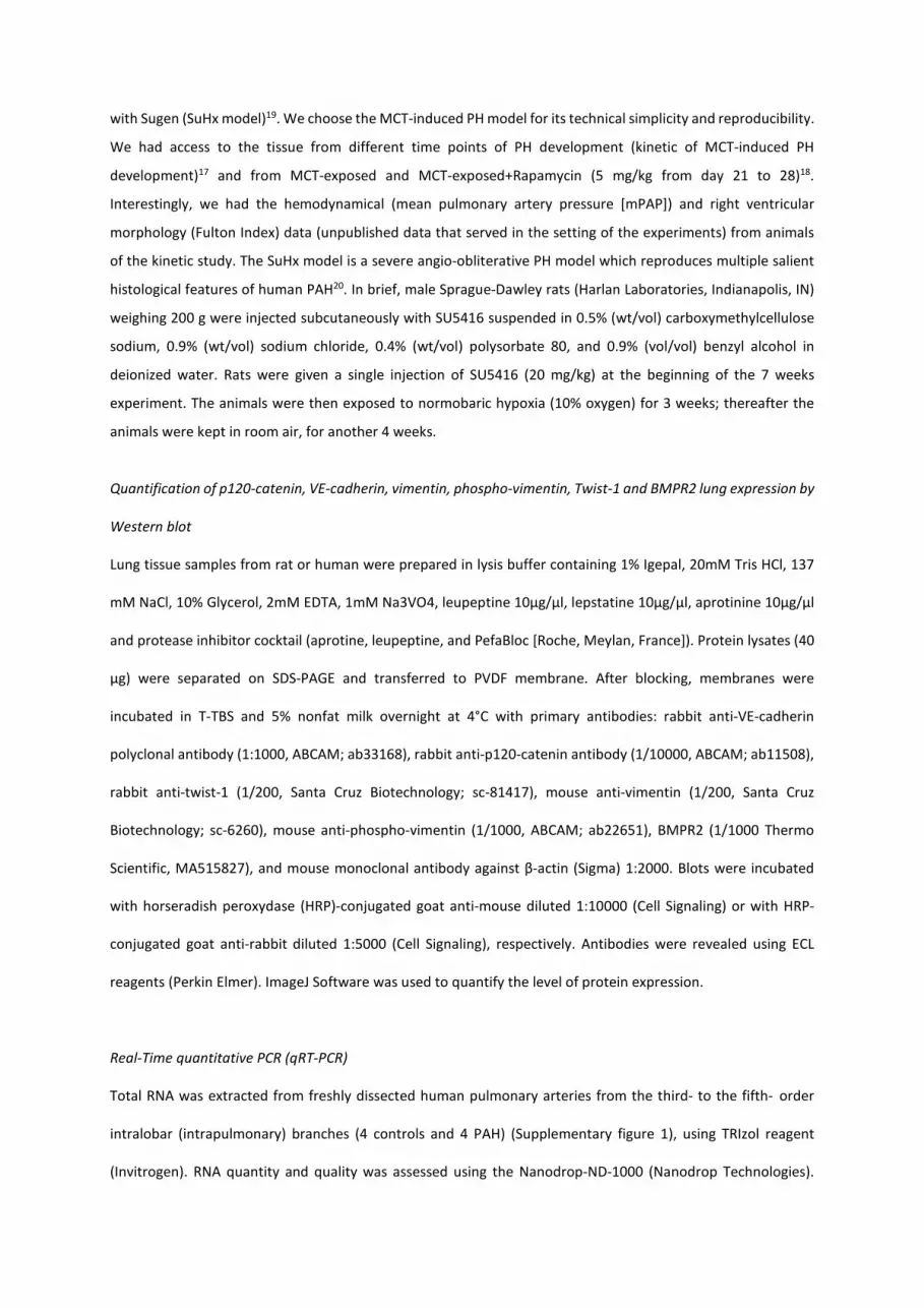
with Sugen (SuHx model)19. We choose the MCT-induced PH model for its technical simplicity and reproducibility.
We had access to the tissue from different time points of PH development (kinetic of MCT-induced PH
development)17 and from MCT-exposed and MCT-exposed+Rapamycin (5 mg/kg from day 21 to 28)18.
Interestingly, we had the hemodynamical (mean pulmonary artery pressure [mPAP]) and right ventricular
morphology (Fulton Index) data (unpublished data that served in the setting of the experiments) from animals
of the kinetic study. The SuHx model is a severe angio-obliterative PH model which reproduces multiple salient
histological features of human PAH20. In brief, male Sprague-Dawley rats (Harlan Laboratories, Indianapolis, IN)
weighing 200 g were injected subcutaneously with SU5416 suspended in 0.5% (wt/vol) carboxymethylcellulose
sodium, 0.9% (wt/vol) sodium chloride, 0.4% (wt/vol) polysorbate 80, and 0.9% (vol/vol) benzyl alcohol in
deionized water. Rats were given a single injection of SU5416 (20 mg/kg) at the beginning of the 7 weeks
experiment. The animals were then exposed to normobaric hypoxia (10% oxygen) for 3 weeks; thereafter the
animals were kept in room air, for another 4 weeks.
Quantification of p120-catenin, VE-cadherin, vimentin, phospho-vimentin, Twist-1 and BMPR2 lung expression by
Western blot
Lung tissue samples from rat or human were prepared in lysis buffer containing 1% Igepal, 20mM Tris HCl, 137
mM NaCl, 10% Glycerol, 2mM EDTA, 1mM Na3VO4, leupeptine 10µg/µl, lepstatine 10µg/µl, aprotinine 10µg/µl
and protease inhibitor cocktail (aprotine, leupeptine, and PefaBloc [Roche, Meylan, France]). Protein lysates (40
μg) were separated on SDS-PAGE and transferred to PVDF membrane. After blocking, membranes were
incubated in T-TBS and 5% nonfat milk overnight at 4°C with primary antibodies: rabbit anti-VE-cadherin
polyclonal antibody (1:1000, ABCAM; ab33168), rabbit anti-p120-catenin antibody (1/10000, ABCAM; ab11508),
rabbit anti-twist-1 (1/200, Santa Cruz Biotechnology; sc-81417), mouse anti-vimentin (1/200, Santa Cruz
Biotechnology; sc-6260), mouse anti-phospho-vimentin (1/1000, ABCAM; ab22651), BMPR2 (1/1000 Thermo
Scientific, MA515827), and mouse monoclonal antibody against β-actin (Sigma) 1:2000. Blots were incubated
with horseradish peroxydase (HRP)-conjugated goat anti-mouse diluted 1:10000 (Cell Signaling) or with HRP-
conjugated goat anti-rabbit diluted 1:5000 (Cell Signaling), respectively. Antibodies were revealed using ECL
reagents (Perkin Elmer). ImageJ Software was used to quantify the level of protein expression.
Real-Time quantitative PCR (qRT-PCR)
Total RNA was extracted from freshly dissected human pulmonary arteries from the third- to the fifth- order
intralobar (intrapulmonary) branches (4 controls and 4 PAH) (Supplementary figure 1), using TRIzol reagent
(Invitrogen). RNA quantity and quality was assessed using the Nanodrop-ND-1000 (Nanodrop Technologies).

First-strand cDNA was synthesized using Quantitec kit (Qiagen) according to the manufacturer’s protocol. Twist-
1 [6H-TWIST1], Vimentin [1H-VIM], N-Cadherin [2H-CDH2], ITGA5 [2H-ITGA5], Fibronectin [1H-FN1], TGFBR1 [3H-
TGFBR1], TGFB1 [2H-TGFB1] and β-actin [4H-ACTB] primer sets (AnyGenes ID number in brackets) were
specifically designed for each transcript and span 2 exons in order to avoid DNA contaminations and pseudogenes
(AnyGenes, Frances). Transcript levels were measured by qRT-PCR using Perfect Master Mix-Probe (AnyGenes,
France) on a StepOnePlus system (Applied Biosystems) according to the manufacturer’s protocol. The transcript
levels were normalized to the β-actin transcripts.

Supplementary tables
Supplementary table 1: Characteristics of primary antibodies used in immunofluorescence studies.
Target Host
specie
Clone Supplier Reference Concentration
CD31 Mouse JC70A Dako M0823 1/200
CD34 Rabbit EP373Y Abcam ab81289 1/200
VWF Rabbit Dako A0082 1/200
α-SMA-FITC Mouse 1A4 Sigma-Aldrich F3777 1/100
α-SMA Mouse 1A4 Sigma-Aldrich A5228 1/100
p120-catenin Mouse 6H11 Abcam ab11508 1/100
VE-cadherin Rabbit Abcam ab33168 1/100
Vimentin-
TRITC
Mouse V9 Santa Cruz
biotechnology
sc-6260 TRITC 1/100
Phospho-
vimentin
Mouse 4A4 Abcam Ab22651 1/50
CD68 Mouse ED1 AbDserotec MCA341GA 1/100
OX-62 Mouse OX-62 AbDserotec MCA1029GA 1/100
Tie-2 Rabbit Santa Cruz
biotechnology
sc-324 1/50
Tryptase Mouse 2A10-
B5
Sigma-Aldrich WH0007177M1 1/100
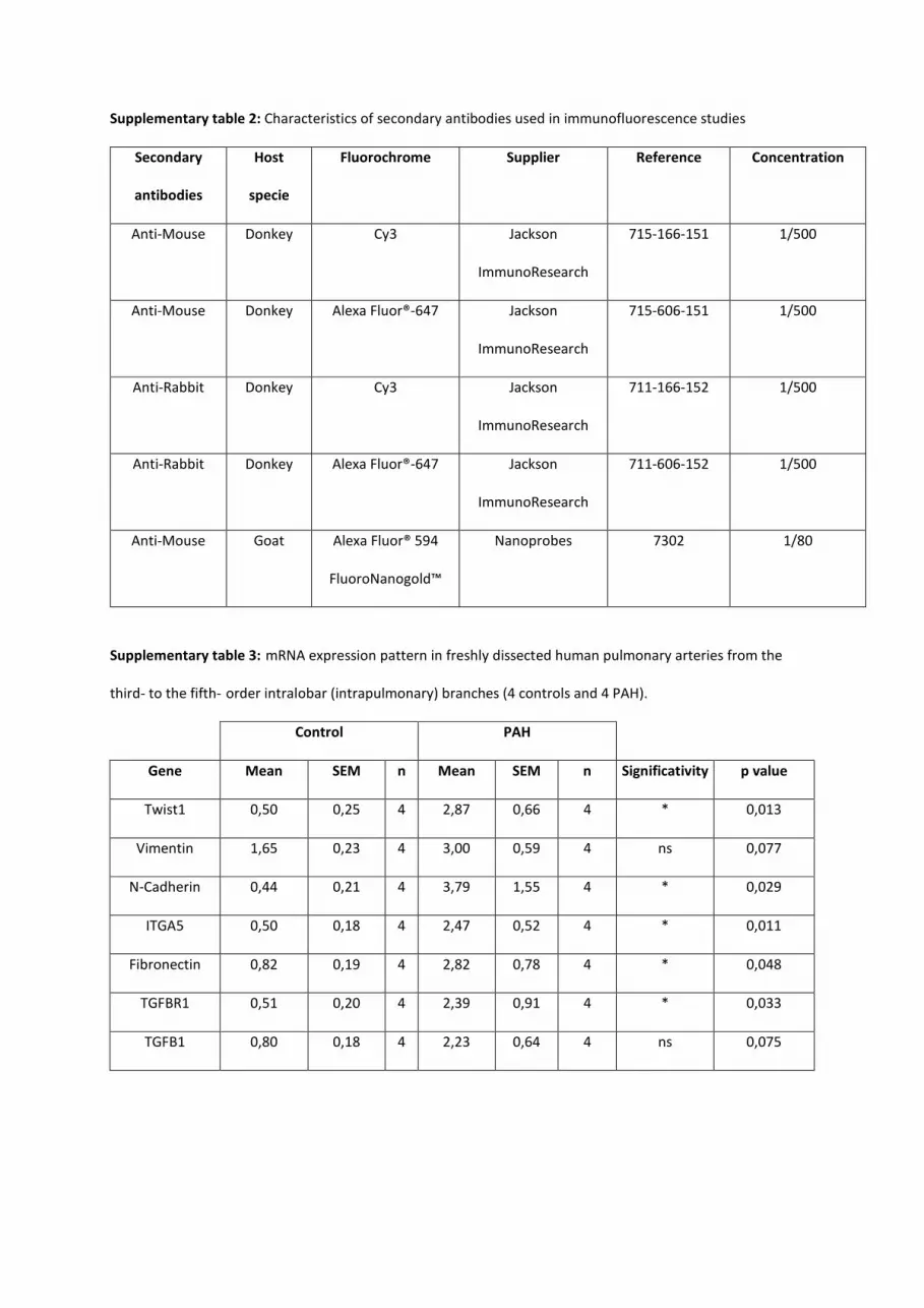
Supplementary table 2: Characteristics of secondary antibodies used in immunofluorescence studies
Secondary
antibodies
Host
specie
Fluorochrome Supplier Reference Concentration
Anti-Mouse Donkey Cy3 Jackson
ImmunoResearch
715-166-151 1/500
Anti-Mouse Donkey Alexa Fluor®-647 Jackson
ImmunoResearch
715-606-151 1/500
Anti-Rabbit Donkey Cy3 Jackson
ImmunoResearch
711-166-152 1/500
Anti-Rabbit Donkey Alexa Fluor®-647 Jackson
ImmunoResearch
711-606-152 1/500
Anti-Mouse Goat Alexa Fluor® 594
FluoroNanogold™
Nanoprobes 7302 1/80
Supplementary table 3: mRNA expression pattern in freshly dissected human pulmonary arteries from the
third- to the fifth- order intralobar (intrapulmonary) branches (4 controls and 4 PAH).
Control PAH
Gene Mean SEM n Mean SEM n Significativity p value
Twist1 0,50 0,25 4 2,87 0,66 4 * 0,013
Vimentin 1,65 0,23 4 3,00 0,59 4 ns 0,077
N-Cadherin 0,44 0,21 4 3,79 1,55 4 * 0,029
ITGA5 0,50 0,18 4 2,47 0,52 4 * 0,011
Fibronectin 0,82 0,19 4 2,82 0,78 4 * 0,048
TGFBR1 0,51 0,20 4 2,39 0,91 4 * 0,033
TGFB1 0,80 0,18 4 2,23 0,64 4 ns 0,075

Supplementary legends
Supplementary figure 1: Freshly dissected human pulmonary artery from the third- to the fifth- order intralobar
(intrapulmonary) branches. Scale bar: 10mm.

Supplementary figure 2: Localization of Twist-1 by immunohistochemistry in a) control pulmonary artery, b)
intimal and c) plexiform lesion from patient with pulmonary arterial hypertension. Left: Rabbit Immunoglobulin
G Isotype Control (pre-immune) polyclonal antibody (pAb), middle: rabbit anti-Twist-1 pAb, and right: inset from
middle pictures. Distal pulmonary arteries from control d) and from MCT-exposed e) rat. Left: Rabbit
Immunoglobulin G Isotype Control (pre-immune) pAb, and right: rabbit anti-Twist-1 pAb.
a-c): scale bar: 50µm.
d-e): scale bar: 20µm.
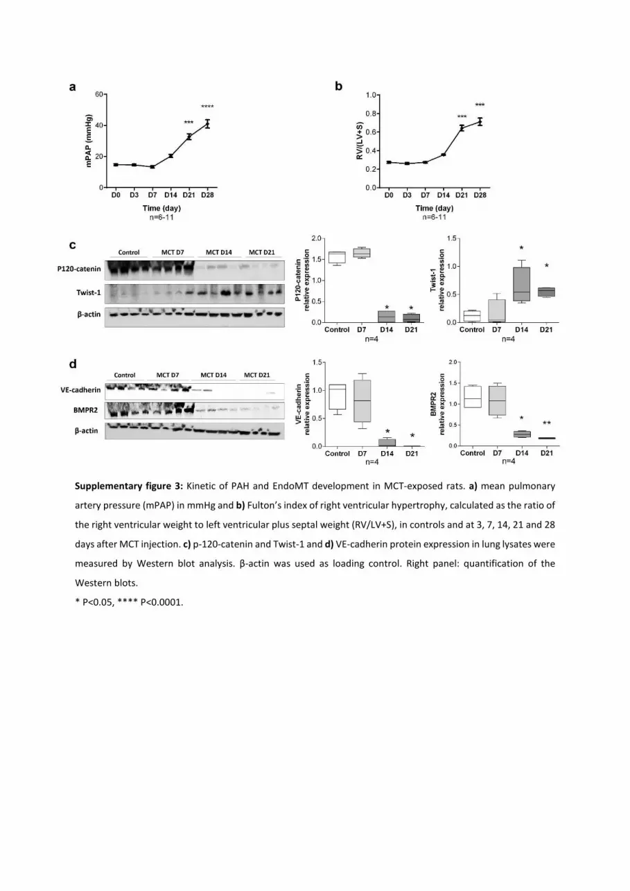
Supplementary figure 3: Kinetic of PAH and EndoMT development in MCT-exposed rats. a) mean pulmonary
artery pressure (mPAP) in mmHg and b) Fulton’s index of right ventricular hypertrophy, calculated as the ratio of
the right ventricular weight to left ventricular plus septal weight (RV/LV+S), in controls and at 3, 7, 14, 21 and 28
days after MCT injection. c) p-120-catenin and Twist-1 and d) VE-cadherin protein expression in lung lysates were
measured by Western blot analysis. β-actin was used as loading control. Right panel: quantification of the
Western blots.
* P<0.05, **** P<0.0001.

Supplementary figure 4: Rapamycin partially inhibits EndoMT protein pattern in the lungs of MCT-exposed rats
and reduces migration of human control and PAH PAEC in vitro. a) p120-catenin, and b) VE-cadherin and Twist-
1 expression were measured by Western blotting in lung lysates and quantified as expression relative to control.
c) Migratory PAEC from control and PAH PAEC were quantified by CyQuant® GR Dye (Cell Biolabs assay). Black
dots: control cells, red dots: PAH cells.
* P<0.05, *** P<0.001.
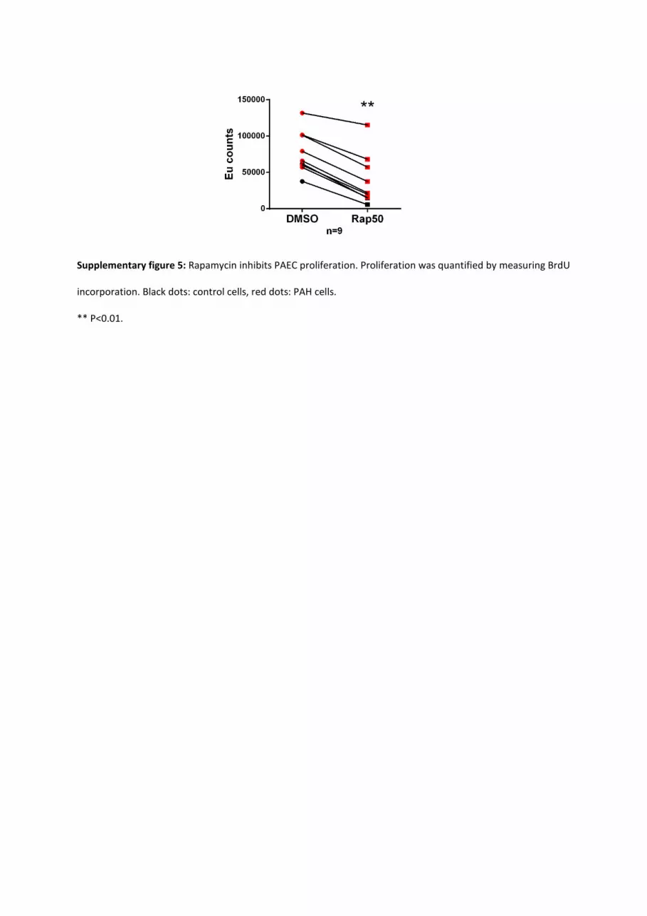
Supplementary figure 5: Rapamycin inhibits PAEC proliferation. Proliferation was quantified by measuring BrdU
incorporation. Black dots: control cells, red dots: PAH cells.
** P<0.01.

Supplementary figure 6: Histologic and immunofluorescence characterization of control and SuHx pulmonary
arteries. a-e) elastic van Gieson stains. a and c) control pulmonary arteries. b, d and e) SuHx remodeled
pulmonary arteries. b) Neomuscularized pulmonary arteriole. d) Concentric intimal fibrosis. e) Fibrinoid necrosis
(FN) in a complex lesion. Bottom left: higher magnification of the FN. Same lesion in hematoxylin and eosin stain
(Top left) and Masson’s trichrome stain (top right), note the fibrin deposit (in red) bellow the endothelium. f-m)
Multiple immunofluorescence stainings in SuHx lesions. f-i) VWF (green), vimentin (red) and α-SMA (white). f and
h: transition from VWFpos vimentinneg endothelial cells (luminal green cells) to VWFneg vimentinhigh mesenchymal-
like cells (luminal red cells). g and i) higher magnification of the transitional cells, some co-express vimentin and
α-SMA and harbor a spindle fibroblast-like shape as genuine mesenchymal cells. j) ED1/CD68 (green), Tie2 (red)
and α-SMA (white). Note the ED1pos macrophages infiltrating the lesion and the Tie2posα-SMApos transitional cells
(in the inset). k) OX62 (green), Tie2 (red) and vimentin (white). Note that the OX62pos dendritic cells infiltrating
the lesion coexpress Tie2 (yellow cells) and the Tie2posvimentinpos transitional cells (in the inset). l and m) α-SMA

(green), tryptase (red): note the tryptasepos mast cells surrounding a complex lesion (L) and a neomuscularized
arteriole (M). All sections were counterstained with DAPI (blue).
Scale bar: 50µm.
Marc Humbert, Sylvia Cohen-Kaminsky and Frédéric PerrosSophie Chat, Elie Fadel, Amal Houssaini, Ignacio Anegon, Serge Adnot, Gerald Simonneau,
Péchoux, Harm Jan Bogaard, Peter Dorfmüller, Séverine Remy, Florence Lecerf, Sylvie Planté, Benoît Ranchoux, Fabrice Antigny, Catherine Rucker-Martin, Aurélie Hautefort, Christine
Endothelial-to-Mesenchymal Transition in Pulmonary Hypertension
Print ISSN: 0009-7322. Online ISSN: 1524-4539 Copyright © 2015 American Heart Association, Inc. All rights reserved.
is published by the American Heart Association, 7272 Greenville Avenue, Dallas, TX 75231Circulation doi: 10.1161/CIRCULATIONAHA.114.008750
2015;131:1006-1018; originally published online January 15, 2015;Circulation.
http://circ.ahajournals.org/content/131/11/1006World Wide Web at:
The online version of this article, along with updated information and services, is located on the
http://circ.ahajournals.org/content/suppl/2015/01/15/CIRCULATIONAHA.114.008750.DC1.htmlData Supplement (unedited) at:
http://circ.ahajournals.org//subscriptions/
is online at: Circulation Information about subscribing to Subscriptions:
http://www.lww.com/reprints Information about reprints can be found online at: Reprints:
document. Permissions and Rights Question and Answer this process is available in the
click Request Permissions in the middle column of the Web page under Services. Further information aboutOffice. Once the online version of the published article for which permission is being requested is located,
can be obtained via RightsLink, a service of the Copyright Clearance Center, not the EditorialCirculationin Requests for permissions to reproduce figures, tables, or portions of articles originally publishedPermissions:
by guest on March 16, 2015http://circ.ahajournals.org/Downloaded from