High-altitude pulmonary hypertension is associated with a free radical-mediated reduction in...
12
See discussions, stats, and author profiles for this publication at: https://www.researchgate.net/publication/46578387 High-altitude pulmonary hypertension is associated with a free radical-mediated reduction in pulmonary nitric oxide... Article in The Journal of Physiology · September 2010 DOI: 10.1113/jphysiol.2010.194704 · Source: PubMed CITATIONS 44 READS 85 17 authors, including: Some of the authors of this publication are also working on these related projects: Documenting simultaneous transvascular free radical exchange during exercise and hypoxia View project Sarah Taudorf Rigshospitalet 26 PUBLICATIONS 2,148 CITATIONS SEE PROFILE Jane Mceneny Queen's University Belfast 139 PUBLICATIONS 2,286 CITATIONS SEE PROFILE Peter Bärtsch Universität Heidelberg 294 PUBLICATIONS 9,179 CITATIONS SEE PROFILE Marc Moritz Berger Universität Heidelberg 28 PUBLICATIONS 497 CITATIONS SEE PROFILE All content following this page was uploaded by Jane Mceneny on 02 December 2016. The user has requested enhancement of the downloaded file. All in-text references underlined in blue are linked to publications on ResearchGate, letting you access and read them immediately.
Transcript of High-altitude pulmonary hypertension is associated with a free radical-mediated reduction in...
Seediscussions,stats,andauthorprofilesforthispublicationat:https://www.researchgate.net/publication/46578387
High-altitudepulmonaryhypertensionisassociatedwithafreeradical-mediatedreductioninpulmonarynitricoxide...
ArticleinTheJournalofPhysiology·September2010
DOI:10.1113/jphysiol.2010.194704·Source:PubMed
CITATIONS
44
READS
85
17authors,including:
Someoftheauthorsofthispublicationarealsoworkingontheserelatedprojects:
DocumentingsimultaneoustransvascularfreeradicalexchangeduringexerciseandhypoxiaView
project
SarahTaudorf
Rigshospitalet
26PUBLICATIONS2,148CITATIONS
SEEPROFILE
JaneMceneny
Queen'sUniversityBelfast
139PUBLICATIONS2,286CITATIONS
SEEPROFILE
PeterBärtsch
UniversitätHeidelberg
294PUBLICATIONS9,179CITATIONS
SEEPROFILE
MarcMoritzBerger
UniversitätHeidelberg
28PUBLICATIONS497CITATIONS
SEEPROFILE
AllcontentfollowingthispagewasuploadedbyJaneMcenenyon02December2016.
Theuserhasrequestedenhancementofthedownloadedfile.Allin-textreferencesunderlinedinblue
arelinkedtopublicationsonResearchGate,lettingyouaccessandreadthemimmediately.
J Physiol 588.23 (2010) pp 4837–4847 4837
High-altitude pulmonary hypertension is associated with afree radical-mediated reduction in pulmonary nitric oxidebioavailability
Damian M. Bailey1, Christoph Dehnert2, Andrew M. Luks3, Elmar Menold2, Christian Castell2,Guido Schendler2, Vitalie Faoro4, Mariusz Gutowski1, Kevin A. Evans1, Sarah Taudorf5, Philip E. James6,J. McEneny7, Ian S. Young7, Erik R. Swenson3,8, Heimo Mairbaurl2, Peter Bartsch2 and Marc M. Berger2,9
1Neurovascular Research Laboratory, Faculty of Health, Science and Sport, University of Glamorgan, Mid-Glamorgan, UK2Department of Internal Medicine VII (Sports Medicine), University of Heidelberg, Heidelberg, Germany3Department of Medicine, Division of Pulmonary and Critical Care Medicine, University of Washington, Seattle, WA, USA4Department of Pathophysiology, University of Brussels, Brussels, Belgium5Centre of Inflammation and Metabolism, Department of Infectious Diseases, Rigshospitalet, University of Copenhagen, Copenhagen, Denmark6 Wales Heart Research Institute, Department of Cardiology, Cardiff University, Cardiff, UK7 Centre for Public Health, Queen’s University Belfast, Belfast, UK8Medical Service, Puget Sound Veterans Affairs Health Care System, Seattle, WA, USA9Department of Anaesthesiology, University of Heidelberg, Heidelberg, Germany
High altitude (HA)-induced pulmonary hypertension may be due to a free radical-mediatedreduction in pulmonary nitric oxide (NO) bioavailability. We hypothesised that the increasein pulmonary artery systolic pressure (PASP) at HA would be associated with a net trans-pulmonary output of free radicals and corresponding loss of bioactive NO metabolites.Twenty-six mountaineers provided central venous and radial arterial samples at low altitude (LA)and following active ascent to 4559 m (HA). PASP was determined by Doppler echocardiography,pulmonary blood flow by inert gas re-breathing, and vasoactive exchange via the Fickprinciple. Acute mountain sickness (AMS) and high-altitude pulmonary oedema (HAPE) werediagnosed using clinical questionnaires and chest radiography. Electron paramagnetic resonancespectroscopy, ozone-based chemiluminescence and ELISA were employed for plasma detectionof the ascorbate free radical (A•−), NO metabolites and 3-nitrotyrosine (3-NT). Fourteensubjects were diagnosed with AMS and three of four HAPE-susceptible subjects developedHAPE. Ascent decreased the arterio-central venous concentration difference (a-cvD) resulting ina net transpulmonary loss of ascorbate, α-tocopherol and bioactive NO metabolites (P < 0.05 vs.LA). This was accompanied by an increased a-cvD and net output of A•− and lipid hydroperoxides(P < 0.05 vs. sea level, SL) that correlated against the rise in PASP (r = 0.56–0.62, P < 0.05) andarterial 3-NT (r = 0.48–0.63, P < 0.05) that was more pronounced in HAPE. These findingssuggest that increased PASP and vascular resistance observed at HA are associated with a freeradical-mediated reduction in pulmonary NO bioavailability.
(Received 13 June 2010; accepted after revision 24 September 2010; first published online 27 September 2010)Corresponding author D. M. Bailey: Neurovascular Research Laboratory, Faculty of Health, Science and Sport,University of Glamorgan, South Wales CF37 1DL, UK. Email: [email protected]
Abbreviations AMS, acute mountain sickness; HA, high altitude; HAPE, high-altitude pulmonary oedema; HAPE-S,susceptible to high-altitude pulmonary oedema; HPV, hypoxic pulmonary vasoconstriction; LA, low altitude; PASP,pulmonary artery systolic pressure; PBF, pulmonary blood flow; PPF, pulmonary plasma flow; SL, sea level.
Introduction
Hypoxic pulmonary vasoconstriction (HPV) is a highlyconserved adaptive response to alveolar hypoxia that
D. M. Bailey, P. Bartsch and M. M. Berger are equal contributors.
serves to optimise ventilation–perfusion matching andgas exchange (Von Euler & Liljestrand, 1946). However,the prolonged systemic and alveolar hypoxia associatedwith high-altitude (HA) exposure can result in apotentially detrimental increase in pulmonary arterypressure and vascular resistance. When excessive, thiscan lead to high-altitude pulmonary oedema (HAPE),
C© 2010 The Authors. Journal compilation C© 2010 The Physiological Society DOI: 10.1113/jphysiol.2010.194704
4838 D. M. Bailey and others J Physiol 588.23
with potentially fatal consequences (Bartsch et al. 1991;Swenson et al. 2002).
Despite extensive research, the precise mechanismsunderlying HA-induced pulmonary hypertension are notcompletely understood. Human studies suggest that thismay be due to a functional imbalance favouring vaso-constriction through increased sympathetic activationof α-adrenergic efferent pathways (Duplain et al. 1999)with additional contributions from endothelin (Bergeret al. 2009) and angiotensin II (Bartsch et al. 1988)in combination with reduced bioavailability of the end-ogenous vasodilator nitric oxide (NO) (Scherrer et al.1996; Busch et al. 2001; Swenson et al. 2002; Berger et al.2005; Ingram et al. 2010). At the cellular level, emergingevidence implicates the mitochondrial formation ofreactive oxygen species (ROS) as the primary stimulusunderlying HPV (Guzy & Schumacker, 2006; Waypa &Schumacker, 2008).
In support, there is now compelling evidence to suggestthat hypoxia promotes systemic (Bailey et al. 2001, 2004,2006b, 2009a) and focal cerebral (Bailey et al. 2009b)oxidative–nitrosative stress in humans. However, to whatextent if indeed at all the pulmonary circulation throughwhich the entire cardiac output must pass contributesto the intravascular accumulation of free radicals andsubsequent implications for high-altitude pulmonaryhypertension remains to be examined.
Therefore, our primary aim was to determinethe temporal effects of hypoxia on the systemicand transpulmonary exchange of oxidative–nitrosative–inflammatory stress biomarkers during ascent to HA.The secondary aim was to examine if any subsequentrelationships existed between exchange across the lungsand pulmonary hypertension. We hypothesised thatcompared to the sea level (SL) control condition, HAwould promote the pulmonary formation and subsequentnet output of free radicals (oxidative stress) and associatedreactants (inflammatory stress) as indicated by a positivearterial–central venous concentration difference (a-cvD).This would be accompanied by a net loss of bioactiveNO metabolites and output of 3-nitrotyrosine (3-NT),reflecting oxidative inactivation of NO (Pacher et al. 2007)(nitrosative stress). Finally, we hypothesised that exchangewould be directly proportional to both the absolute valuesand the increase in pulmonary artery systolic pressure(PASP) and subsequently more marked in those subjectswho developed HAPE.
Methods
Subjects
Thirty-eight (32 male, 6 female) healthy non-acclimatisednative lowlanders aged 37 (mean) ± 10 (S.D.) years oldvolunteered for the study, which had been approved
by the Medical Faculty of the University of Heidelberg.Thirty subjects had previously climbed to altitudesabove 3000 m. Of these, eight had previously developedmoderate to severe acute mountain sickness (AMS)and eight had prior radiographic evidence of HAPE(classified as HAPE susceptible; HAPE-S). No subjectspent more than four nights above 2500 m within30 days before the study and there was no history ofchronic daily headache, habitual antioxidant vitamin oranti-inflammatory supplementation. All subjects wereencouraged to follow a low nitrate/nitrite diet prior toand throughout the duration of the study with specificinstructions to avoid fruits, salads and cured meats (Wanget al. 1997).
Compliance
Four subjects withdrew following baseline controlmeasurements, two of whom were HAPE-S. A furthereight subjects were excluded (two HAPE-S, one of whomdeveloped HAPE) due to failure to measure pulmonaryblood flow (PBF) and/or obtain blood samples at all timepoints. Thus, the current data are based on full data setsfor 26 subjects (aged 36 ± 10 years) with four classified asHAPE-S.
Experimental design
Baseline control measurements were conducted at sealevel (SL) in Heidelberg, Germany (110 m). Between 2and 4 weeks later, subjects travelled to Alagna, Valsesia,Italy. They were transported by cable car to 3200 m beforetrekking to 3611 m where they spent the night (the GnifettiHut). In the morning they were accompanied by a certifiedmountain guide while they ascended to 4559 m (CapannaRegina Margherita) within 4–6 h where they remained forthe following 2 days.
Blood sampling
At SL subjects attended the laboratory following a 12 hovernight fast. A radial arterial catheter (RA 04020, ArrowInternational, Inc., Reading, PA, USA) was placed underlocal anaesthesia (2% lidocaine) and a central venouscatheter (Cavafix Certodyn 375, B. Braun Melsungen AG,Melsungen, Germany) was inserted via an antecubitalvein and advanced under electrocardiographic guidanceinto the superior vena cava (Corsten et al. 1994). Mixedvenous samples would have been preferred, thoughthis was not an option in light of the risks associatedwith pulmonary artery catheterisation. At HA, catheterswere placed within 1 h of arrival to the MargheritaHut. Samples were obtained 3 h thereafter (HA-3h)to ensure that the baseline blood samples were not
C© 2010 The Authors. Journal compilation C© 2010 The Physiological Society
J Physiol 588.23 Redox regulation of hypoxic pulmonary vasoconstriction 4839
contaminated by the combined stresses of catheterisation,exercise and/or post-prandial elevations in blood-bornebiomarkers of oxidative–nitrosative–inflammatory stress.We were confident that given the short half-lives ofthe biomarkers employed that this ‘recovery period’ wassufficient to ensure an accurate baseline at HA. Catheterswere kept patent overnight and the final sample wasobtained within 1 h of waking at 07.00 h which equated toa 20 h exposure to 4559 m (HA-20h). With the exceptionof blood gas measurements, samples were centrifugedat 600 g (4◦C) for 10 min and serum/plasma/red bloodcells (RBC) immediately snap-frozen and stored in liquidnitrogen prior to analysis. Subjects remained at HA fora further 24 h having received medical treatment whennecessary prior to guided descent.
Rescue medication
Subjects with clinical and radiographic signs of HAPEwere treated with oral nifedipine (2 × 8 mg day−1) andsupplemental oxygen. Those subjects who developed AMSreceived symptomatic treatment with acetaminophen(500 mg) and/or ibuprofen (400 mg). All measurementswere performed prior to pharmacological interventionand no further assessments were made thereafter. A moredetailed description of the safety aspects and clinicalmanagement of subjects has been described by Swensonet al. (2002) and more recently in two separate publicationsthat report further data from the same study described here(Berger et al. 2009; Dehnert et al. 2010).
Pulmonary haemodynamics
Echocardiography. Doppler echocardiography (AccusonCypress, Siemens, Germany) was employed for themeasurement of PASP according to the modified Bernoulliequation (Allemann et al. 2000). Measurements wereperformed on three cardiac cycles (mean recorded) byan experienced sonographer and subsequently evaluatedoff-line in random order by a separate sonographerblinded to the study. The inter- and intra-observerco-efficients of variation (C.V.) were both <15%.
Pulmonary flow. Pulmonary blood flow (PBF) wasdetermined non-invasively (Innocor, Innovision A/S,Odense, Denmark) using an oxygen-enriched mixture of0.5% nitrous oxide and 0.1% sulfur hexafluoride measuredwith photo-acoustic analysers over a 5-breath interval(Lang et al. 2007). Pulmonary plasma flow (PPF) wascalculated as PBF × (1 – Hct). The potential limitations ofthis technique have been discussed by Berger et al. (2009)who performed identical measurements in a sub-setof subjects enrolled in the same study. The inter andintra-observer C.V. were both <5%.
Blood gases. Samples were immediately measuredwith a blood gas analyser (ABL 5, Radiometer,Copenhagen, Denmark) for the measurement of pH,haemoglobin (Hb), partial pressures of oxygen/carbondioxide (PO2 /PCO2 ) and oxyhaemoglobin saturation (SO2 ).Haematocrit (Hct) was determined manually followingmicrocentrifugation. Calculation of the alveolar to arterialPO2 difference [[A-a]O2diff ] (Fenn et al. 1946) assumed thatPACO2 was equal to PaCO2 and the respiratory quotient was0.85.
Metabolic analyses
Oxidative stress. Antioxidants. For ascorbic acid, 900 μlof 5% metaphosphoric acid was added to 100 μl plasmaand assayed via fluorimetry based on the condensationof dehydroascorbic acid with 1,2-phenylenediamine(Vuilleumier & Keck, 1993). Plasma α-tocopherol, wasdetermined by HPLC (Catignani et al. 1983; Thurnhamet al. 1988). The intra- and inter-assay C.V. for both assayswere <10%.
Ascorbate free radical (A•−). Plasma (1 ml) was injectedinto a high-sensitivity multiple-bore sample cell (AquaX,Bruker Instruments Inc., Billerica, MA, USA) housedwithin a TM110 cavity of an electron paramagneticresonance (EPR) spectrometer operating at X-band (9.87GHz) (Bailey et al. 2009a). Samples were analysed using amodulation frequency of 100 kHz, modulation amplitudeof 0.65 gauss (G), microwave power of 10 mW, receivergain of 2 × 10⁵, time constant of 41 ms, magnetic fieldcentre of 3477 G and scan width of ±50 G for threeincremental scans (Buettner & Kiminyo, 1992). Afteridentical baseline correction and filtering, each of thespectral peak-to-trough line heights were normalisedrelative to
√X line width and the mean considered a
measure of the relative concentration of A•−. The intra-and inter-assay C.V. were both <5%.
Lipid hydroperoxides. Serum lipid hydroperoxides(LOOH) were determined using the ferrous iron/xylenolorange (FOX) assay (Wolff, 1994) with modification. Theintra- and inter-assay C.V. were both <4%.
Inflammatory stress. Cytokines. Plasma tumour necrosisfactor (TNF)-α and interleukin (IL)-6 were measuredby ELISA (R&D Systems, Minneapolis, MN, USA). Theintra/inter-assay C.V. were both <10%.
Nitrosative stress. NO metabolites. Plasma (200 μl) wasinjected into tri-iodide reagent for the measurement ofnitrite (NO2
−)+S-nitrosothiols (RSNOs) by ozone-basedchemiluminescence (Model 280i, NOA, Sievers, Boulder,CO, USA) (Rassaf et al. 2004). Acidified sulphanilamide(5%) was added to a separate sample and left toincubate in the dark at 21◦C for 15 min to permit
C© 2010 The Authors. Journal compilation C© 2010 The Physiological Society
4840 D. M. Bailey and others J Physiol 588.23
differentiation of NO2− from RSNOs. RBC (100 μl) were
injected into modified tri-iodide (Rogers et al. 2005)for the measurement of total RBC-bound NO (NO2
− +nitrosyl haemoglobin (HbNO) + S-nitrosohemoglobin(HbSNO)). All calculations were performed using OriginPeak Analysis software (OriginLab Corp., Northampton,MA, USA). The intra- and inter-assay C.V. for all NOmetabolites were <10%.
3-Nitrotyrosine. Plasma 3-nitrotyrosine (3-NT) wasmeasured by ELISA (Hycult Biotechnology, b.v., Uden,The Netherlands) and the intra- and inter-assay C.V. wereboth <10%.
Transpulmonary exchange kinetics. Transpulmonaryexchange kinetics was determined according to the FickPrinciple:
Exchange = PBF (for RBC − bound NO) or PPF
(plasma/serum − borne metabolites)
×a-cvD
By convention, a positive value reflects net output or gainand a negative value indicates net uptake or loss across thepulmonary vascular bed.
Altitude illness
HAPE was suspected clinically and confirmed by chestradiography (Vock et al. 1989) if at least one lung quadrantdemonstrated patchy opacities compatible with inter-stitial or alveolar oedema. AMS was evaluated usingthe Lake Louise (LL-cumulative Self Report + ClinicalScores) (Roach et al. 1993) and Environmental SymptomsQuestionnaire Cerebral (ESQ-C) (Sampson et al. 1983)scoring systems. Clinical AMS (moderate to severe) wasconfirmed by a combined total LL score of ≥5 points andESQ-C score ≥0.7 points (Maggiorini et al. 1998) uponwaking after the first night at HA (HA-20h).
Statistical analysis
Distribution normality was assessed using Shapiro–WilkW tests (SPSS Version 17.0; SPSS Inc., Chicago, IL,USA). Exchange and a-cvD data were analysed usinga combination of one-factor (Location: SL vs. HA-3hvs. HA-20h) and two-factor (Location: SL vs. HA-3hvs. HA-20h × Sample Site: Radial Artery vs. CentralVenous) repeated measures ANOVA with post hocBonferroni-corrected paired samples t test. Relationshipswere examined using a Pearson product momentcorrelation. Non-parametric equivalents were applied toTNF-α, IL-6 and ESQ-C data only. Significance wasestablished at P < 0.05 and data presented as mean ± S.D.Where indicated, a main effect refers to a pooled (Arterial+ Venous) difference between SL vs. HA-3h vs. HA-20h
(P < 0.05) or pooled (SL + HA-3h + HA-20h) differencebetween arterial vs. venous (P < 0.05).
Results
Altitude illness
LL and ESQ-C scores increased from 1 ± 1 and 0.1 ± 0.2points respectively at SL to 4 ± 3 and 0.8 ± 0.7 points atHA-3h (P < 0.05 vs. SL) to 6 ± 2 and 1.0 ± 0.8 pointsat HA-20h (P < 0.05 vs. SL). Fourteen subjects werediagnosed with AMS (AMS+) by HA-20h, three of whomsubsequently developed HAPE after 27 h, 37 h and 44 hat HA with radiographic scores of 7, 12 and 5 points,respectively, at the time of diagnosis. Three of the foursubjects originally identified as HAPE-S at SL developedHAPE. The remaining 12 subjects reported only mildsymptoms of AMS.
Pulmonary gas exchange and haemodynamics
As anticipated, HA was associated with marked hypo-xaemia and respiratory alkalosis (Table 1) whereas PBFor PPF did not change relative to SL. PASP was elevatedthroughout the duration of HA and the increase (valueat HA-20h minus the SL control value) was greater(P < 0.05) in HAPE subjects (n = 3, +27 ± 12 mmHg)whose individual PASP values were 66, 52 and 41 mmHgcompared to the remaining (i.e. non-HAPE) controls(n = 23, +13 ± 6 mmHg).
Oxidative stress
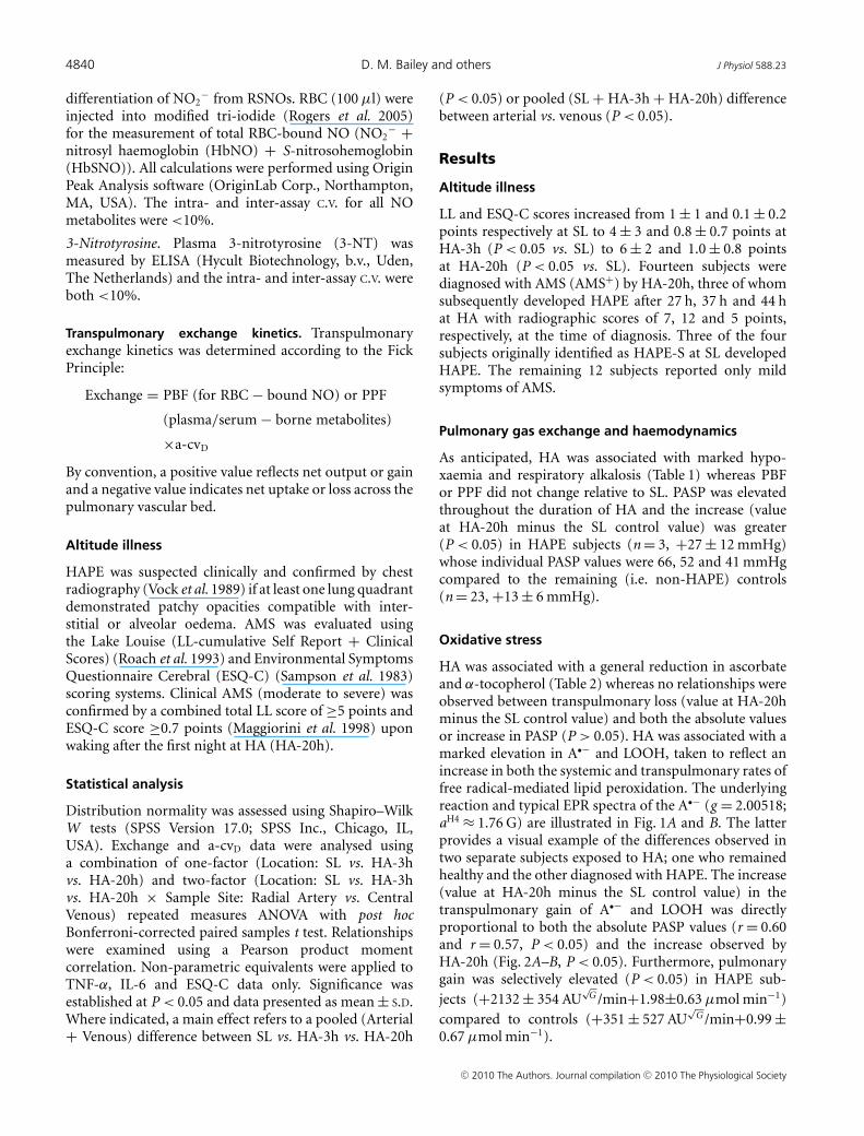
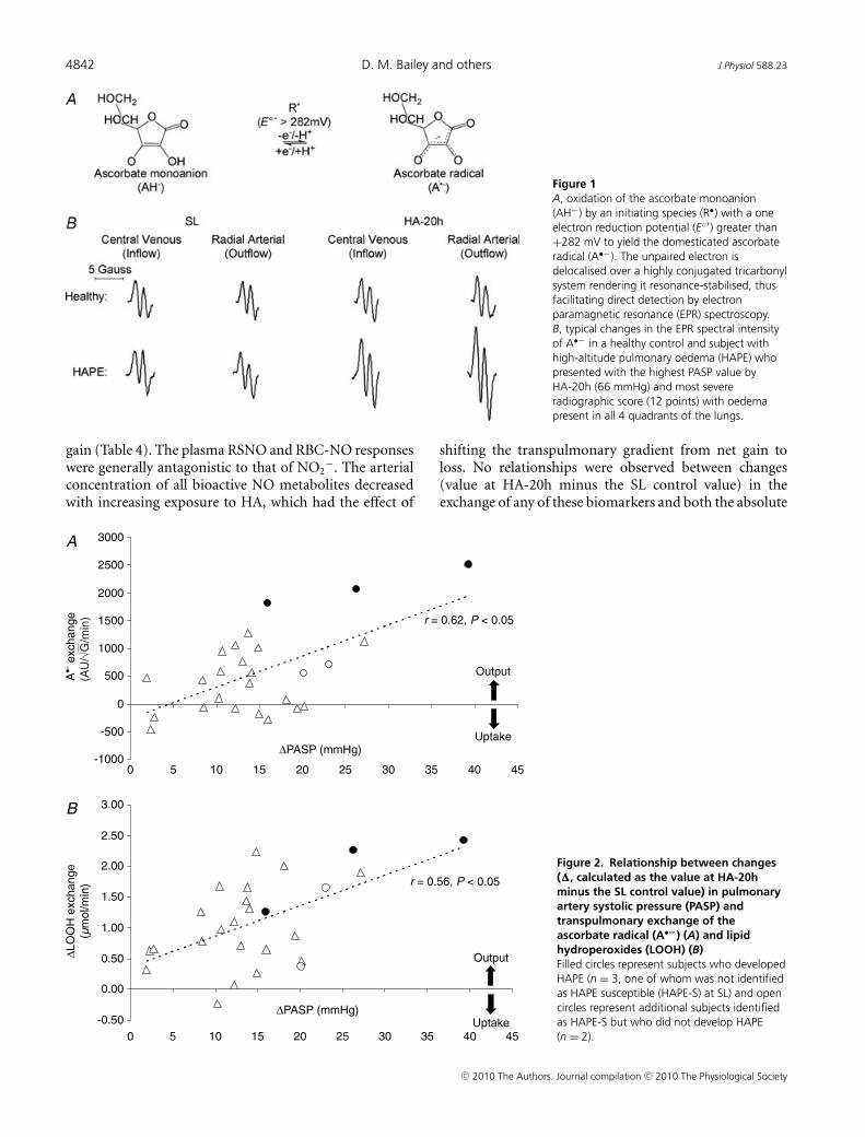
HA was associated with a general reduction in ascorbateand α-tocopherol (Table 2) whereas no relationships wereobserved between transpulmonary loss (value at HA-20hminus the SL control value) and both the absolute valuesor increase in PASP (P > 0.05). HA was associated with amarked elevation in A•− and LOOH, taken to reflect anincrease in both the systemic and transpulmonary rates offree radical-mediated lipid peroxidation. The underlyingreaction and typical EPR spectra of the A•− (g = 2.00518;aH4 ≈ 1.76 G) are illustrated in Fig. 1A and B. The latterprovides a visual example of the differences observed intwo separate subjects exposed to HA; one who remainedhealthy and the other diagnosed with HAPE. The increase(value at HA-20h minus the SL control value) in thetranspulmonary gain of A•− and LOOH was directlyproportional to both the absolute PASP values (r = 0.60and r = 0.57, P < 0.05) and the increase observed byHA-20h (Fig. 2A–B, P < 0.05). Furthermore, pulmonarygain was selectively elevated (P < 0.05) in HAPE sub-jects (+2132 ± 354 AU
√G /min+1.98±0.63 μmol min−1)
compared to controls (+351 ± 527 AU√
G /min+0.99 ±0.67 μmol min−1).
C© 2010 The Authors. Journal compilation C© 2010 The Physiological Society

J Physiol 588.23 Redox regulation of hypoxic pulmonary vasoconstriction 4841
Table 1. Pulmonary gas exchange and haemodynamics
Location: SL HA-3h HA-20h
Sample site Radial arterial Central venous Radial arterial Central venous Radial arterial Central venous
PO2 (mmHg) 95 ± 11 38 ± 5∗ 38 ± 3† 28 ± 2∗† 41 ± 4†‡ 29 ± 3∗†Main effects (P < 0.05): HA < SL + Arterial > Venous; interaction effect (P < 0.05)PCO2 (mmHg) 40 ± 3 48 ± 4∗ 29 ± 3† 33 ± 4∗† 28 ± 2† 32 ± 2∗†Main effects (P < 0.05) : HA < SL + Arterial < Venous; interaction effect (P < 0.05)SaO2 (%) 97 ± 1 70 ± 6∗ 72 ± 5† 52 ± 5∗† 74 ± 5† 50 ± 6∗†Main effects (P < 0.05): HA < SL + Arterial > Venous; interaction effect (P < 0.05)pH (units) 7.41 ± 0.03 7.36 ± 0.01∗ 7.48 ± 0.02† 7.46 ± 0.02∗† 7.47 ± 0.02†‡ 7.44 ± 0.02∗†‡Main effects (P < 0.05): HA > SL + Arterial > Venous; interaction effect (P < 0.05)[A-a]O2diff (mmHg) 13 ± 7 10 ± 4 9 ± 5PBF (l min−1) 6.4 ± 1.4 6.9 ± 1.2 6.2 ± 1.0‡PPF (l min−1) 3.7 ± 0.8 3.9 ± 0.7 3.5 ± 0.5‡PASP (mmHg) 23 ± 4 39 ± 10† 38 ± 10†Values are means ± S.D.; SL, sea level; HA, high-altitude; PO2 /PCO2 partial pressure of oxygen/carbon dioxide; SO2 , oxyhaemoglobinsaturation; [A-a]O2diff, alveolar to arterial PO2 difference; PBF/PPF, pulmonary blood/plasma flow; PASP, pulmonary artery systolicpressure. ∗P < 0.05 vs. radial arterial for given location; †P < 0.05 vs. SL for given sample site; ‡P < 0.05 vs. HA-3h for given sample site.
Table 2. Oxidative stress
Location SL HA-3h HA-20h
Sample site Radial arterial Central venous Radial arterial Central venous Radial arterial Central venous
Ascorbate (μmol l−1) 63.3 ± 9.7 62.2 ± 8.8 57.5 ± 7.7 60.2 ± 7.7 52.7 ± 5.5 55.2 ± 10.9
Main effect (P < 0.05): HA < SLa-cvD (μmol l−1) +1.1 ± 4.9 −2.7 ± 8.0† −2.5 ± 8.3Exchange (μmol min−1) +4.3 ± 16.9 −11.7 ± 25.8† −10.1 ± 31.1α-Tocopherol (μmol l−1) 27.8 ± 8.0 28.4 ± 7.5 26.0 ± 6.6 27.4 ± 6.7 25.7 ± 7.6 26.6 ± 7.9
Main effect (P < 0.05): HA < SLa-cvD (μmol l−1) −0.6 ± 2.9 −1.3 ± 6.0 −0.9 ± 3.6Exchange (μmol min−1) −2.4 ± 10.9 −5.3 ± 24.2 −3.9 ± 12.5
A•− (AU√
G ) 1572 ± 237 1536 ± 214 1982 ± 352† 1740 ± 257∗† 1934 ± 387† 1736 ± 272∗†Main effects (P < 0.05): HA > SL + Arterial > Venous; interaction effect (P < 0.05)
a-cvD (AU√
G ) +36 ± 111 +242 ± 270† +198 ± 222†Exchange (AU
√G min−1) +137 ± 398 +988 ± 960† +715 ± 852†
LOOH (μmol l−1) 0.49 ± 0.08 0.54 ± 0.09∗ 0.84 ± 0.29† 0.64 ± 0.15∗† 0.95 ± 0.29†‡ 0.69 ± 0.14∗†Main effects (P < 0.05): HA > SL + Arterial > Venous; interaction effect (P < 0.05)a-cvD (μmol l−1) −0.06 ± 0.07 +0.19 ± 0.20† +0.26 ± 0.22†‡Exchange (μmol min−1) −0.20 ± 0.21 +0.77 ± 0.77† +0.91 ± 0.81†
A•−, ascorbate radical expressed in arbitrary units (AU)√
linewidth in gauss (G); LOOH, lipid hydroperoxides; a-cvD, radial arterial minuscentral venous concentration difference. ∗P < 0.05 vs. radial arterial for given location; †P < 0.05 vs. SL for given sample site; ‡P < 0.05vs. HA-3h for given sample site.
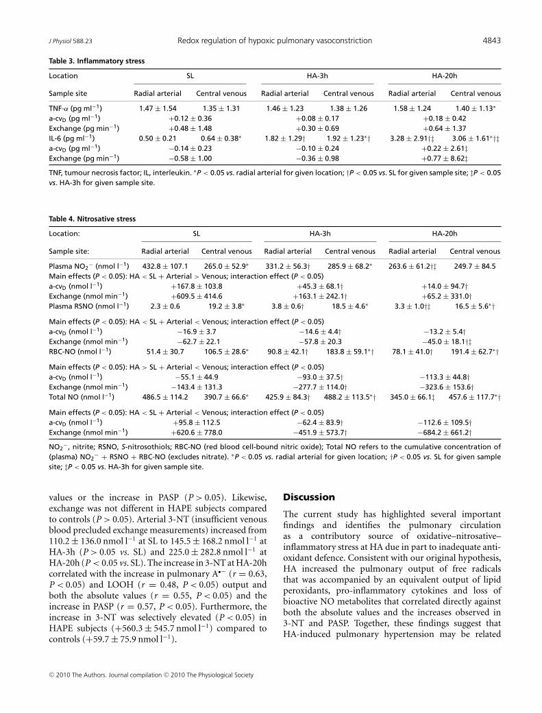
Inflammatory stress
An increased transpulmonary gain of TNF-α was observedby HA-20h and the transpulmonary gradient of IL-6shifted from net loss to gain by HA-20h (Table 3). Norelationships were observed between changes (value atHA-20h minus the SL control value) in the output ofany of these biomarkers and both the absolute values or
increase in PASP (P > 0.05). Exchange was not differentin HAPE subjects compared to controls (P > 0.05).
Nitrosative stress
A progressive decrease in arterial NO2− was observed at
HA, which had the effect of suppressing transpulmonary
C© 2010 The Authors. Journal compilation C© 2010 The Physiological Society
4842 D. M. Bailey and others J Physiol 588.23
Figure 1A, oxidation of the ascorbate monoanion(AH−) by an initiating species (R•) with a oneelectron reduction potential (E◦′) greater than+282 mV to yield the domesticated ascorbateradical (A•−). The unpaired electron isdelocalised over a highly conjugated tricarbonylsystem rendering it resonance-stabilised, thusfacilitating direct detection by electronparamagnetic resonance (EPR) spectroscopy.B, typical changes in the EPR spectral intensityof A•− in a healthy control and subject withhigh-altitude pulmonary oedema (HAPE) whopresented with the highest PASP value byHA-20h (66 mmHg) and most severeradiographic score (12 points) with oedemapresent in all 4 quadrants of the lungs.
gain (Table 4). The plasma RSNO and RBC-NO responseswere generally antagonistic to that of NO2
−. The arterialconcentration of all bioactive NO metabolites decreasedwith increasing exposure to HA, which had the effect of
shifting the transpulmonary gradient from net gain toloss. No relationships were observed between changes(value at HA-20h minus the SL control value) in theexchange of any of these biomarkers and both the absolute
Figure 2. Relationship between changes(�, calculated as the value at HA-20hminus the SL control value) in pulmonaryartery systolic pressure (PASP) andtranspulmonary exchange of theascorbate radical (A•−) (A) and lipidhydroperoxides (LOOH) (B)Filled circles represent subjects who developedHAPE (n = 3, one of whom was not identifiedas HAPE susceptible (HAPE-S) at SL) and opencircles represent additional subjects identifiedas HAPE-S but who did not develop HAPE(n = 2).
C© 2010 The Authors. Journal compilation C© 2010 The Physiological Society
J Physiol 588.23 Redox regulation of hypoxic pulmonary vasoconstriction 4843
Table 3. Inflammatory stress
Location SL HA-3h HA-20h
Sample site Radial arterial Central venous Radial arterial Central venous Radial arterial Central venous
TNF-α (pg ml−1) 1.47 ± 1.54 1.35 ± 1.31 1.46 ± 1.23 1.38 ± 1.26 1.58 ± 1.24 1.40 ± 1.13∗
a-cvD (pg ml−1) +0.12 ± 0.36 +0.08 ± 0.17 +0.18 ± 0.42Exchange (pg min−1) +0.48 ± 1.48 +0.30 ± 0.69 +0.64 ± 1.37IL-6 (pg ml−1) 0.50 ± 0.21 0.64 ± 0.38∗ 1.82 ± 1.29† 1.92 ± 1.23∗† 3.28 ± 2.91†‡ 3.06 ± 1.61∗†‡a-cvD (pg ml−1) −0.14 ± 0.23 −0.10 ± 0.24 +0.22 ± 2.61‡Exchange (pg min−1) −0.58 ± 1.00 −0.36 ± 0.98 +0.77 ± 8.62‡TNF, tumour necrosis factor; IL, interleukin. ∗P < 0.05 vs. radial arterial for given location; †P < 0.05 vs. SL for given sample site; ‡P < 0.05vs. HA-3h for given sample site.
Table 4. Nitrosative stress
Location: SL HA-3h HA-20h
Sample site: Radial arterial Central venous Radial arterial Central venous Radial arterial Central venous
Plasma NO2− (nmol l−1) 432.8 ± 107.1 265.0 ± 52.9∗ 331.2 ± 56.3† 285.9 ± 68.2∗ 263.6 ± 61.2†‡ 249.7 ± 84.5
Main effects (P < 0.05): HA < SL + Arterial > Venous; interaction effect (P < 0.05)a-cvD (nmol l−1) +167.8 ± 103.8 +45.3 ± 68.1† +14.0 ± 94.7†Exchange (nmol min−1) +609.5 ± 414.6 +163.1 ± 242.1† +65.2 ± 331.0†Plasma RSNO (nmol l−1) 2.3 ± 0.6 19.2 ± 3.8∗ 3.8 ± 0.6† 18.5 ± 4.6∗ 3.3 ± 1.0†‡ 16.5 ± 5.6∗†Main effects (P < 0.05): HA < SL + Arterial < Venous; interaction effect (P < 0.05)a-cvD (nmol l−1) −16.9 ± 3.7 −14.6 ± 4.4† −13.2 ± 5.4†Exchange (nmol min−1) −62.7 ± 22.1 −57.8 ± 20.3 −45.0 ± 18.1†‡RBC-NO (nmol l−1) 51.4 ± 30.7 106.5 ± 28.6∗ 90.8 ± 42.1† 183.8 ± 59.1∗† 78.1 ± 41.0† 191.4 ± 62.7∗†Main effects (P < 0.05): HA > SL + Arterial < Venous; interaction effect (P < 0.05)a-cvD (nmol l−1) −55.1 ± 44.9 −93.0 ± 37.5† −113.3 ± 44.8†Exchange (nmol min−1) −143.4 ± 131.3 −277.7 ± 114.0† −323.6 ± 153.6†Total NO (nmol l−1) 486.5 ± 114.2 390.7 ± 66.6∗ 425.9 ± 84.3† 488.2 ± 113.5∗† 345.0 ± 66.1‡ 457.6 ± 117.7∗†Main effects (P < 0.05): HA < SL + Arterial < Venous; interaction effect (P < 0.05)a-cvD (nmol l−1) +95.8 ± 112.5 −62.4 ± 83.9† −112.6 ± 109.5†Exchange (nmol min−1) +620.6 ± 778.0 −451.9 ± 573.7† −684.2 ± 661.2†NO2
−, nitrite; RSNO, S-nitrosothiols; RBC-NO (red blood cell-bound nitric oxide); Total NO refers to the cumulative concentration of(plasma) NO2
− + RSNO + RBC-NO (excludes nitrate). ∗P < 0.05 vs. radial arterial for given location; †P < 0.05 vs. SL for given samplesite; ‡P < 0.05 vs. HA-3h for given sample site.
values or the increase in PASP (P > 0.05). Likewise,exchange was not different in HAPE subjects comparedto controls (P > 0.05). Arterial 3-NT (insufficient venousblood precluded exchange measurements) increased from110.2 ± 136.0 nmol l−1 at SL to 145.5 ± 168.2 nmol l−1 atHA-3h (P > 0.05 vs. SL) and 225.0 ± 282.8 nmol l−1 atHA-20h (P < 0.05 vs. SL). The increase in 3-NT at HA-20hcorrelated with the increase in pulmonary A•− (r = 0.63,P < 0.05) and LOOH (r = 0.48, P < 0.05) output andboth the absolute values (r = 0.55, P < 0.05) and theincrease in PASP (r = 0.57, P < 0.05). Furthermore, theincrease in 3-NT was selectively elevated (P < 0.05) inHAPE subjects (+560.3 ± 545.7 nmol l−1) compared tocontrols (+59.7 ± 75.9 nmol l−1).
Discussion
The current study has highlighted several importantfindings and identifies the pulmonary circulationas a contributory source of oxidative–nitrosative–inflammatory stress at HA due in part to inadequate anti-oxidant defence. Consistent with our original hypothesis,HA increased the pulmonary output of free radicalsthat was accompanied by an equivalent output of lipidperoxidants, pro-inflammatory cytokines and loss ofbioactive NO metabolites that correlated directly againstboth the absolute values and the increases observed in3-NT and PASP. Together, these findings suggest thatHA-induced pulmonary hypertension may be related
C© 2010 The Authors. Journal compilation C© 2010 The Physiological Society
4844 D. M. Bailey and others J Physiol 588.23
to a free radical-mediated reduction in pulmonary NObioavailability.
Given the low reduction potential of the A•−/ascorbatemonanion (AH−) couple (E◦′ = 282 mV) (Williams& Yandell, 1982), virtually every oxidising free radicalthat could arise within the pulmonary circulationwill react with AH− to form A•− (R• + AH− → A•−
+ R-H) (Buettner, 1993). Thus, the EPR detectionof A•− and concomitant loss of ascorbate in thecurrent study provides direct evidence for an increasedtranspulmonary flux of free radicals during hypo-xia. The loss of α-tocopherol would have also beenexpected to constrain peroxidative stress since thephenolic-OH group located in its chromanol ringscavenges lipid-derived peroxyl radicals (LOO•) fasterthan the equivalent reaction with lipid side-chains
(α − TOH + LOO• �E ◦′≈+500mV——————→
k≈8×10⁴M−1s−1LOOH + a−TO• vs.
LOO• + L − H�E ◦′≈+400mV
——————→k≈10 M−1s−1
LOOH + L•) (Wang &
Quinn, 1999). However, consumption of thesechain-breaking donor antioxidants ultimately failedto terminate chain propagation as indicated by theincreased pulmonary output of LOOH, one of the majorreactants of lipid peroxidation.
Our data provide unique insight into the correspondingrates of pulmonary free radical-mediated lipidperoxidation, albeit within the constraints of a singlearterio-venous transit. The arterial concentration ofA•− and LOOH increased by 363 ± 356 AU
√G and
0.46 ± 0.28 μmol l−1 respectively at HA-20h. Thus,assuming steady-state kinetics and dilution within theextracellular space (∼12 litres) (Bailey et al. 2006a),the total amount of A•− and LOOH formed during thesojourn at HA would have been substantial, equating to4356 AU
√G and 9.20 μmol l−1 respectively implying that
net pulmonary ‘output’ was ∼197- and 119-fold greaterthan the respective rates of ‘accumulation’.
These calculations emphasise the extraordinarily highturnover of free radicals across the hypoxic humanpulmonary circulation, which is not surprising given themultiple potential sites of ROS formation and inherentsusceptibility to peroxidation (Connolly & Aaronson,2010). Likewise, similar kinetics have been observedacross the hypoxic human brain (Bailey et al. 2009b)suggesting that transvascular free radical output is likelyto be common to ‘all’ organ systems when challengedby hypoxaemia. While not disassociating cause fromeffect, the linear, albeit modest, relationships observedbetween oxidant output and rise in PASP are consistentwith recent observations in vitro. Current evidence(Ward & McMurtry, 2009) suggests that cellular hypo-xia triggers mitochondrial O2
•− formation at complexIII of the electron transport chain subsequent toincreased electron transfer from UQ•− to O2 (Guzy
& Schumacker, 2006). The resultant increase in ROSsignalling is thought to initiate pulmonary arterialsmooth muscle contraction through Ca2+ release fromryanodine-sensitive sarcoplasmic reticulum stores (Waypa& Schumacker, 2008).
Our findings suggest that a free radical-mediatedreduction in pulmonary NO bioavailability mayprove of equal significance by potentiating HPV.The metal-catalysed reductive decompositionof LOOH can yield secondary LO• (Fe2+ +LOOH
k≈1.5×10³M−1s−1
———————→ Fe3+ + OH− + LO•) thathave previously been detected in hypoxic humanblood (Bailey et al. 2004, 2006b, 2009b). Incombination with O2
•−, these radicals have thepotential to deplete pulmonary NO stores through adiffusion-controlled reaction that yields peroxynitrite
(LO•/O•−2 + NO• k≈6−20×109M−1s−1
———————→ ONOO−) (Nauser &Koppenol, 2002) which can promote nitration of tyrosineresidues to form the stable reactant 3-NT, our molecular‘footprint’ of ROS-mediated NO inactivation (Pacheret al. 2007).
Thus, the blunted systemic and pulmonary outputof NO2
− combined with the systemic rise in 3-NTin the current study suggests that NO bioavailabilityis likely to have been decreased at HA subsequent tooxidative inactivation. This finding agrees with pre-vious reports documenting a reduction of NO inexpired air (Duplain et al. 2000), loss of NO2
−
and nitrate in broncho-alveolar lavage fluid (Swensonet al. 2002) and impaired endothelium-dependent vaso-dilatation in the systemic circulation during hypo-xia, especially in HAPE susceptible individuals (Bergeret al. 2005). Additional NO2
− loss may be attributableto reduced NO synthesis subsequent to limited O2
substrate bioavailability (Le Cras & McMurtry, 2001)though this has recently been questioned (Steiner et al.2002). Alternatively, it may reflect increased consumptioncatalysed by deoxyHb/deoxyMb and/or xanthineoxidoreductase/aldehyde oxidase-mediated reductionto NO and subsequent re-apportionment towardslonger-lived species notably Hb-NO and Hb-SNO drivenby the quite substantial differences in transpulmonaryO2 gradients (van Faassen et al. 2009). Whether thedynamic interchange of NO from the plasma to the RBCcompartment serves to improve pulmonary oxygenationduring hypoxia remains to be determined.
It remains to be established whether pulmonaryoxidative–nitrosative stress was the cause or simplythe consequence of HPV and HAPE. The early andprogressive accumulation of oxidants–nitrosants lendstentative support for a causal role, which is notunreasonable given that these molecules have the thermo-dynamic potential to promote autocatalytic destructionof the pulmonary lipid membrane structure through
C© 2010 The Authors. Journal compilation C© 2010 The Physiological Society
J Physiol 588.23 Redox regulation of hypoxic pulmonary vasoconstriction 4845
cross-linking polymerisation and breakage of fatty acidside chains (Roubal & Tappel, 1966). These speciesmay be responsible for the degradation of proteogylcansand subsequent disruption of the endothelial basementmembrane and pulmonary extracellular matrix observedin hypoxia (Miserocchi et al. 2001). When combinedwith elevated intravascular hydrostatic pressure, this canresult in capillary stress failure, destruction of tissuematrix structures, increased alveolar–capillary membranepermeability and alveolar haemorrhage (Bartsch & Gibbs,2007). Localised inflammation is likely to be a secondaryevent as suggested by the delayed pulmonary outputof cytokines, which agrees with previous observations(Swenson et al. 2002).
Alternatively, these biomolecules have the capacity toserve as integral components of the signal transductioncascade that are physiologically essential in controlledthough as of yet undefined amounts, capable of initiatingprotective adaptation in the face of hypoxic stress forthe maintenance of homeostasis (Bailey et al. 2001;Guzy & Schumacker, 2006; Pryor et al. 2006). Thoughspeculative, the oxidative-nitrosative stress response thatmay ultimately predispose to pulmonary hypertensionat HA may represent a physiological attempt to regainhomeostasis, a concept that warrants investigation infuture studies.
In conclusion, these findings are the first to demonstratea net pulmonary output and systemic accumulationof oxidative–nitrosative–inflammatory stress biomarkersat HA due in part to inadequate antioxidant defence.A free radical-mediated reduction in pulmonary NObioavailability may prove the unifying mechanismunderlying HA-induced pulmonary hypertension andsubsequent susceptibility to HAPE which has broaderimplications for other human pulmonary diseasescharacterised by arterial hypoxaemia such as chronicobstructive pulmonary disorder (COPD). The improvedclinical outcome of COPD patients given supplementalO2 may be due to the suppression of hypoxia-mediatedpulmonary ROS formation (Zouaoui Boudjeltia et al.2009). Follow-up interventional studies need to considercombined ascorbate and α-tocopherol prophylaxis inorder to optimise the chain-breaking antioxidant capacityof the pulmonary circulation at HA.
References
Allemann Y, Sartori C, Lepori M, Pierre S, Melot C, Naeije R,Scherrer U & Maggiorini M (2000). Echocardiographic andinvasive measurements of pulmonary artery pressurecorrelate closely at high altitude. Am J Physiol Heart CircPhysiol 279, H2013–2016.
Bailey D, Ainslie P, Jackson S, Richardson R & Ghatei M (2004).Evidence against redox regulation of energy homoeostasis inhumans at high altitude. Clin Sci (Lond) 107, 589–600.
Bailey D, Raman S, McEneny J, Young I, Parham K, Hullin D,Davies B, McKeeman G, McCord J & Lewis M (2006a).Vitamin C prophylaxis promotes oxidative lipid damageduring surgical ischemia-reperfusion. Free Radic Biol Med40, 591–600.
Bailey DM, Davies B & Young IS (2001). Intermittent hypoxictraining: implications for lipid peroxidation induced byacute normoxic exercise in active men. Clin Sci (Lond) 101,465–475.
Bailey DM, Evans KA, James PE, McEneny J, Young IS, Fall L,Gutowski M, Kewley E, McCord JM, Moller K & Ainslie PN(2009a). Altered free radical metabolism in acute mountainsickness: implications for dynamic cerebral autoregulationand blood–brain barrier function. J Physiol 587,73–85.
Bailey DM, Roukens R, Knauth M, Kallenberg K, Christ S,Mohr A, Genius J, Storch-Hagenlocher B, Meisel F, McEnenyJ, Young IS, Steiner T, Hess K & Bartsch P (2006b). Freeradical-mediated damage to barrier function is notassociated with altered brain morphology in high-altitude headache. J Cereb Blood Flow Metab 26,99–111.
Bailey DM, Taudorf S, Berg RMG, Lundby C, McEneny J,Young IS, Evans KA, James PE, Shore A, Hullin DA, McCordJM, Pedersen BK & Moller K (2009b). Increased cerebraloutput of free radicals during hypoxia: implications for acutemountain sickness? Am J Physiol Regul Integr Comp Physiol297, R1283–1292.
Bartsch P & Gibbs JS (2007). Effect of altitude on the heart andthe lungs. Circulation 116, 2191–2202.
Bartsch P, Maggiorini M, Ritter M, Noti C, Vock P & Oelz O(1991). Prevention of high-altitude pulmonary edema bynifedipine. N Engl J Med 325, 1284–1289.
Bartsch P, Shaw S, Franciolli M, Gnadinger MP & Weidmann P(1988). Atrial natriuretic peptide in acute mountain sickness.J Appl Physiol 65, 1929–1937.
Berger MM, Dehnert C, Bailey DM, Luks AM, Menold E,Castell C, Schendler G, Faoro V, Mairbaurl H, Bartsch P &Swenson ER (2009). Transpulmonary plasma ET-1 andnitrite differences in high altitude pulmonary hypertension.High Alt Med Biol 10, 17–24.
Berger MM, Hesse C, Dehnert C, Siedler H, Kleinbongard P,Bardenheuer HJ, Kelm M, Bartsch P & Haefeli WE (2005).Hypoxia impairs systemic endothelial function in individualsprone to high-altitude pulmonary edema. Am J Respir CritCare Med 172, 763–767.
Buettner GR (1993). The pecking order of free radicals andantioxidants: lipid peroxidation, α-tocopherol, andascorbate. Arch Biochem Biophys 300, 535–543.
Buettner GR & Kiminyo KP (1992). Optimal EPR detection ofweak nitroxide spin adduct and ascorbyl free radical signals.J Biochem Biophys Methods 24, 147–151.
Busch T, Bartsch P, Pappert D, Grunig E, Hildebrandt W, ElserH, Falke KJ & Swenson ER (2001). Hypoxia decreases exhalednitric oxide in mountaineers susceptible to high-altitudepulmonary edema. Am J Respir Crit Care Med 163,368–373.
Catignani GL & Bieri JG (1983). Simultaneous determinationof retinol and Alpha-tocopherol in serum or plasma byliquid chromatography. Clin Chem 29, 708–712.
C© 2010 The Authors. Journal compilation C© 2010 The Physiological Society
4846 D. M. Bailey and others J Physiol 588.23
Connolly MJ & Aaronson PI (2010). Cell redox state andhypoxic pulmonary vasoconstriction: Recent evidence andpossible mechanisms. Respir Physiol Neurobiol (in press).
Corsten SA, van Dijk B, Bakker NC, de Lange JJ & Scheffer GJ(1994). Central venous catheter placement using theECG-guided Cavafix-Certodyn SD catheter. J Clin Anesth 6,469–472.
Dehnert C, Luks AM, Schendler G, Menold E, Berger MM,Mairbaurl H, Faoro V, Bailey DM, Castell C, Hahn G, VockP, Swenson ER & Bartsch P (2010). No evidence forinterstitial lung oedema by extensive pulmonary functiontesting at 4,559 m. Eur Respir J 35, 812–820.
Duplain H, Sartori C, Lepori M, Egli M, Allemann Y, Nicod P& Scherrer U (2000). Exhaled nitric oxide in high-altitudepulmonary edema: role in the regulation of pulmonaryvascular tone and evidence for a role against inflammation.Am J Respir Crit Care Med 162, 221–224.
Duplain H, Vollenweider L, Delabays A, Nicod P, Bartsch P &Scherrer U (1999). Augmented sympathetic activationduring short-term hypoxia and high-altitude exposure insubjects susceptible to high-altitude pulmonary edema.Circulation 99, 1713–1718.
Fenn WO, Rahn H & Otis AB (1946). A theoretical study of thecomposition of the alveolar air at altitude. Am J Physiol 146,637–653.
Guzy RD & Schumacker PT (2006). Oxygen sensing bymitochondria at complex III: the paradox of increasedreactive oxygen species during hypoxia. Exp Physiol 91,807–819.
Ingram TE, Pinder AG, Bailey DM, Fraser AG & James PE(2010). Low-dose sodium nitrite vasodilates hypoxic humanpulmonary vasculature by a means which is not dependentupon a simultaneous elevation in plasma nitrite. Am JPhysiol Heart Circ Physiol 298, H331–H339.
Lang CC, Karlin P, Haythe J, Tsao L & Mancini DM (2007).Ease of noninvasive measurement of cardiac output coupledwith peak VO2 determination at rest and during exercise inpatients with heart failure. Am J Cardiol 99,404–405.
Le Cras TD & McMurtry IF (2001). Nitric oxide production inthe hypoxic lung. Am J Physiol Lung Cell Mol Physiol 280,L575–582.
Maggiorini M, Muller A, Hofstetter D, Bartsch P & Oelz O(1998). Assessment of acute mountain sickness by differentscore protocols in the Swiss Alps. Aviat Space Environ Med69, 1186–1192.
Miserocchi G, Negrini D, Passi A & De Luca G (2001).Development of lung edema: interstitial fluid dynamics andmolecular structure. News Physiol Sci 16, 66–71.
Nauser T & Koppenol WH (2002). The rate constant of thereaction of superoxide with nitrogen monoxide: approachingthe diffusion limit. J Phys Chem A 106,4084–4086.
Pacher P, Beckman JS & Liaudet L (2007). Nitric oxide andperoxynitrite in health and disease. Physiol Rev 87,315–424.
Pryor WA, Houk KN, Foote CS, Fukuto JM, Ignarro LJ,Squadrito GL & Davies KJA (2006). Free radical biology andmedicine: it’s a gas, man! Am J Physiol Regul Integr CompPhysiol 291, R491–511.
Rassaf T, Feelisch M & Kelm M (2004). Circulating NO pool:assessment of nitrite and nitroso species in blood and tissues.Free Radic Biol Med 36, 413–422.
Roach RC, Bartsch P, Hackett PH & Oelz O (1993). The LakeLouise acute mountain sickness scoring system. In Hypoxiaand Molecular Medicine, ed. Sutton JR, Coates J & HoustonCS, pp. 272–274. Queen City Printers, Burlington.
Rogers SC, Khalatbari A, Gapper PW, Frenneaux MP & JamesPE (2005). Detection of human red blood cell-bound nitricoxide. J Biol Chem 280, 26720–26728.
Roubal WT & Tappel AL (1966). Polymerization of proteinsinduced by free-radical lipid peroxidation. Arch BiochemBiophys 113, 150–155.
Sampson JB, Cymerman A, Burse RL, Maher JT & Rock PB(1983). Procedures for the measurement of acute mountainsickness. Aviat Space Environ Med 54, 1063–1073.
Scherrer U, Vollenweider L, Delabays A, Savcic M,Eichenberger U, Kleger GR, Fikrle A, Ballmer PE, Nicod P &Bartsch P (1996). Inhaled nitric oxide for high-altitudepulmonary edema. N Engl J Med 334,624–629.
Steiner DRS, Gonzalez NC & Wood JG (2002). Interactionbetween reactive oxygen species and nitric oxide in themicrovascular response to systemic hypoxia. J Appl Physiol93, 1411–1418.
Swenson ER, Maggiorini M, Mongovin S, Gibbs JSR, Greve I,Mairbaurl H & Bartsch P (2002). Pathogenesis ofhigh-altitude pulmonary edema: inflammation is not anetiologic factor. JAMA 287, 2228–2235.
Thurnham DI, Smith E & Flora PS (1988). Concurrentliquid-chromatographic assay of retinol, alpha-tocopherol,beta-carotene, alpha- carotene, lycopene, and beta-crytoxanthin in plasma, with tocopherol acetate as internalstandard. Clin Chem 34, 377–381.
van Faassen EE, Bahrami S, Feelisch M, Hogg N, Kelm M,Kim-Shapiro DB, Kozlov AV, Li H, Lundberg JO, Mason R,Nohl H, Rassaf T, Samouilov A, Slama-Schwok A, Shiva S,Vanin AF, Weitzberg E, Zweier J & Gladwin MT (2009).Nitrite as regulator of hypoxic signaling in mammalianphysiology. Med Res Rev 29, 683–741.
Vock P, Fretz C, Franciolli M & Bartsch P (1989). High-altitudepulmonary edema: findings at high-altitude chestradiography and physical examination. Radiology 170,661–666.
Von Euler U & Liljestrand G (1946). Observations on thepulmonary arterial blood pressure in the cat. Acta PhysiolScand 12, 301–320.
Vuilleumier JP & Keck E (1993). Fluorimetric assay of vitaminC in biological materials using a centrifugal analyser withfluorescence attachment. J Micronutr Anal 5,25–34.
Wang J, Brown MA, Tam SH, Chan MC & Whitworth JA(1997). Effects of diet on measurement of nitric oxidemetabolites. Clin Exp Pharmacol Physiol 24, 418–420.
Wang X & Quinn PJ (1999). Vitamin E and its function inmembranes. Prog Lipid Res 38, 309–336.
Ward JP & McMurtry IF (2009). Mechanisms of hypoxicpulmonary vasoconstriction and their roles in pulmonaryhypertension: new findings for an old problem. Curr OpinPharmacol 9, 287–296.
C© 2010 The Authors. Journal compilation C© 2010 The Physiological Society
J Physiol 588.23 Redox regulation of hypoxic pulmonary vasoconstriction 4847
Waypa GB & Schumacker PT (2008). Oxygen sensing inhypoxic pulmonary vasoconstriction: using new tools toanswer an age-old question. Exp Physiol 93, 133–138.
Williams NH & Yandell JK (1982). Outer-sphereelectron-transfer reactions of ascorbic anions. Aust J Chem35, 1133–1144.
Wolff SP (1994). Ferrous ion oxidation in presence of ferric ionindicator xylenol orange for measurement ofhydroperoxides. Methods Enzymol 233, 183–189.
Zouaoui Boudjeltia K, Tragas G, Babar S, Moscariello A,Nuyens V, Van Antwerpen P, Gilbert O, Ducobu J, BroheeD, Vanhaeverbeek M & Van Meerhaeghe A (2009). Effects ofoxygen therapy on systemic inflammation andmyeloperoxidase modified LDL in hypoxemic COPDpatients. Atherosclerosis 205, 360–362.
Author contributions
Study conception and design: D.M.B., C.D., A.M.L., E.M., E.R.S.,H.B., P.B., M.M.B.; data analysis, collection and interpretation:
D.M.B., C.D., A.M.L., E.M., M.G., K.A.E., S.T., P.E.J., J.M., I.S.Y.,E.R.S., H.B., P.B., M.M.B.; drafting of the article: D.M.B., E.R.S.,H.B., P.B., M.M.B.; critical revision of the article: D.M.B., C.D.,A.M.L., E.R.S., H.B., P.B., M.M.B. All authors approved the finalversion for publication.
Acknowledgements
We acknowledge all subjects for their kind participation; the hutkeepers and the Sezione Varallo of the Club Alpino Italianofor supporting research at the Capanna Regina Margherita;Sonja Engelhardt, Christiane Herth, Verena Faulkner, AnetteFrank and Kathy Pogue for skilled technical assistance and PeterVock, Guido Robotti, Semanta Greppi, Chantal Imesch for theirexpertise taking chest radiographs and VIASYS Healthcare (nowCareFusion) for financial support.
C© 2010 The Authors. Journal compilation C© 2010 The Physiological Society