hexagonal pattern - BMC Pulmonary Medicine
8
Okabayashi et al. BMC Pulmonary Medicine (2022) 22:76 https://doi.org/10.1186/s12890-022-01869-4 RESEARCH The new useful high-resolution computed tomography finding for diagnosing fibrotic hypersensitivity pneumonitis: “hexagonal pattern”: a single-center retrospective study Hiroko Okabayashi 1,4* , Taiki Fukuda 2,5 , Tae Iwasawa 2 , Tsuneyuki Oda 1 , Hideya Kitamura 1 , Tomohisa Baba 1 , Tamiko Takemura 3 , Takuro Sakagami 4 and Takashi Ogura 1 Abstract Background: Centrilobular nodules, ground-glass opacity (GGO), mosaic attenuation, air trapping, and three-density pattern were reported as high-resolution computed tomography (HRCT) findings characteristic of fibrotic hypersen- sitivity pneumonitis (HP). However, it is often difficult to differentiate fibrotic HP from idiopathic pulmonary fibrosis (IPF). In fibrotic HP, the HRCT sometimes shows tortoiseshell-like interlobular septal thickening that extends from the subpleural lesion to the inner layers. This finding is called “hexagonal pattern,” and this study is focused on the possibil- ity that such finding is useful for differentiating fibrotic HP from IPF. Methods: This study included patients with multidisciplinary discussion (MDD) diagnosis of fibrotic HP or IPF under- going surgical lung biopsy between January 2015 and December 2017 in Kanagawa Cardiovascular and Respiratory Center. Two radiologists have evaluated the HRCT findings without clinical and pathological information. Results: A total of 23 patients were diagnosed with fibrotic HP by MDD and 48 with IPF. Extensive GGO, centrilobular nodules, and hexagonal pattern were more frequent findings in fibrotic HP than in IPF. No significant difference was observed between the two groups in the presence or absence of mosaic attenuation, air trapping, or three-density pattern. In the multivariate logistic regression, the presence of extensive GGO and hexagonal pattern was associated with increased odds ratio of fibrotic HP. The sensitivity and specificity of the diagnosis of fibrotic HP in the presence of the hexagonal pattern were 69.6% and 87.5%, respectively. Conclusion: Hexagonal pattern is a useful finding for differentiating fibrotic HP from IPF. Keywords: Fibrotic hypersensitivity pneumonitis, Idiopathic pulmonary pneumonitis, HRCT, Interlobular septal thickening, Hexagonal pattern © The Author(s) 2022. Open Access This article is licensed under a Creative Commons Attribution 4.0 International License, which permits use, sharing, adaptation, distribution and reproduction in any medium or format, as long as you give appropriate credit to the original author(s) and the source, provide a link to the Creative Commons licence, and indicate if changes were made. The images or other third party material in this article are included in the article’s Creative Commons licence, unless indicated otherwise in a credit line to the material. If material is not included in the article’s Creative Commons licence and your intended use is not permitted by statutory regulation or exceeds the permitted use, you will need to obtain permission directly from the copyright holder. To view a copy of this licence, visit http://creativecommons.org/licenses/by/4.0/. The Creative Commons Public Domain Dedication waiver (http://creativeco mmons.org/publicdomain/zero/1.0/) applies to the data made available in this article, unless otherwise stated in a credit line to the data. Background Two guidelines have recently been proposed for diag- nosing hypersensitivity pneumonitis (HP), which state that lung biopsy is not necessary for the diagnosis of HP if antigen exposure, typical high-resolution computed tomography (HRCT) findings, and bronchoalveolar lav- age (BAL) lymphocytosis have been identified [1, 2]. However, in fibrotic HP, the antigen is often unknown, Open Access *Correspondence: [email protected] 1 Department of Respiratory Medicine, Kanagawa Cardiovascular and Respiratory Center, 6-16-1 Tomioka-Higashi, Kanazawa-ku, Yokohama City, Kanagawa 236-0051, Japan Full list of author information is available at the end of the article
-
Upload
khangminh22 -
Category
Documents
-
view
0 -
download
0
Transcript of hexagonal pattern - BMC Pulmonary Medicine

Okabayashi et al. BMC Pulmonary Medicine (2022) 22:76 https://doi.org/10.1186/s12890-022-01869-4
RESEARCH
The new useful high-resolution computed tomography finding for diagnosing fibrotic hypersensitivity pneumonitis: “hexagonal pattern”: a single-center retrospective studyHiroko Okabayashi1,4*, Taiki Fukuda2,5, Tae Iwasawa2, Tsuneyuki Oda1, Hideya Kitamura1, Tomohisa Baba1, Tamiko Takemura3, Takuro Sakagami4 and Takashi Ogura1
Abstract
Background: Centrilobular nodules, ground-glass opacity (GGO), mosaic attenuation, air trapping, and three-density pattern were reported as high-resolution computed tomography (HRCT) findings characteristic of fibrotic hypersen-sitivity pneumonitis (HP). However, it is often difficult to differentiate fibrotic HP from idiopathic pulmonary fibrosis (IPF). In fibrotic HP, the HRCT sometimes shows tortoiseshell-like interlobular septal thickening that extends from the subpleural lesion to the inner layers. This finding is called “hexagonal pattern,” and this study is focused on the possibil-ity that such finding is useful for differentiating fibrotic HP from IPF.
Methods: This study included patients with multidisciplinary discussion (MDD) diagnosis of fibrotic HP or IPF under-going surgical lung biopsy between January 2015 and December 2017 in Kanagawa Cardiovascular and Respiratory Center. Two radiologists have evaluated the HRCT findings without clinical and pathological information.
Results: A total of 23 patients were diagnosed with fibrotic HP by MDD and 48 with IPF. Extensive GGO, centrilobular nodules, and hexagonal pattern were more frequent findings in fibrotic HP than in IPF. No significant difference was observed between the two groups in the presence or absence of mosaic attenuation, air trapping, or three-density pattern. In the multivariate logistic regression, the presence of extensive GGO and hexagonal pattern was associated with increased odds ratio of fibrotic HP. The sensitivity and specificity of the diagnosis of fibrotic HP in the presence of the hexagonal pattern were 69.6% and 87.5%, respectively.
Conclusion: Hexagonal pattern is a useful finding for differentiating fibrotic HP from IPF.
Keywords: Fibrotic hypersensitivity pneumonitis, Idiopathic pulmonary pneumonitis, HRCT , Interlobular septal thickening, Hexagonal pattern
© The Author(s) 2022. Open Access This article is licensed under a Creative Commons Attribution 4.0 International License, which permits use, sharing, adaptation, distribution and reproduction in any medium or format, as long as you give appropriate credit to the original author(s) and the source, provide a link to the Creative Commons licence, and indicate if changes were made. The images or other third party material in this article are included in the article’s Creative Commons licence, unless indicated otherwise in a credit line to the material. If material is not included in the article’s Creative Commons licence and your intended use is not permitted by statutory regulation or exceeds the permitted use, you will need to obtain permission directly from the copyright holder. To view a copy of this licence, visit http:// creat iveco mmons. org/ licen ses/ by/4. 0/. The Creative Commons Public Domain Dedication waiver (http:// creat iveco mmons. org/ publi cdoma in/ zero/1. 0/) applies to the data made available in this article, unless otherwise stated in a credit line to the data.
BackgroundTwo guidelines have recently been proposed for diag-nosing hypersensitivity pneumonitis (HP), which state that lung biopsy is not necessary for the diagnosis of HP if antigen exposure, typical high-resolution computed tomography (HRCT) findings, and bronchoalveolar lav-age (BAL) lymphocytosis have been identified [1, 2]. However, in fibrotic HP, the antigen is often unknown,
Open Access
*Correspondence: [email protected] Department of Respiratory Medicine, Kanagawa Cardiovascular and Respiratory Center, 6-16-1 Tomioka-Higashi, Kanazawa-ku, Yokohama City, Kanagawa 236-0051, JapanFull list of author information is available at the end of the article
Page 2 of 8Okabayashi et al. BMC Pulmonary Medicine (2022) 22:76
and most cases need lung biopsy. In addition, specimens collected via transbronchial forceps biopsies or trans-bronchial lung cryobiopsy (TBLC) are insufficient in size for the diagnosis of fibrotic HP, and surgical lung biopsy (SLB) is often required. Conversely, SLB is often not fea-sible due to the patient’s background or facility issues. Therefore, HRCT imaging plays a significant role in the diagnosis of fibrotic HP.
HRCT findings characteristic of fibrotic HP include centrilobular nodules, ground-glass opacity (GGO), mosaic attenuation, air trapping, and three-density pat-tern in previous reports [1–8]. These findings are not specific to fibrotic HP only, and many cases of fibrotic HP were found without typical HRCT findings.
Approximately half of chronic HP has been reported to have histological usual interstitial pneumonia (UIP)-like pattern and extensive fibrosis with poor prognosis [9]. Fibrotic HP with UIP pattern is common and often difficult to distinguish from idiopathic pulmonary fibro-sis (IPF) in clinical practice. However, differentiating between fibrotic HP and IPF is important as they differ in their treatment strategies.
In fibrotic HP, the HRCT sometimes shows tortoise-shell-like interlobular septal thickening that extends from the subpleural region to the inner layers. This finding is called “hexagonal pattern,” and this study focused on the possibility that such finding is useful for differentiating fibrotic HP from IPF (Fig. 1).
MethodsPatientsThis study included patients with multidisciplinary dis-cussion (MDD) diagnosis of fibrotic HP or IPF undergo-ing SLB between January 2015 and December 2017 in Kanagawa Cardiovascular and Respiratory Center. The diagnosis of IPF and fibrotic HP was based on consensus using previously reported criteria [5, 10–12]. The exclu-sion criteria of this study were as follows: (1) patients that had collagen vascular diseases or pulmonary alveolar proteinosis, (2) patients that met the interstitial pneumo-nia with autoimmune features diagnostic criteria [13], (3) patients that developed collagen vascular disease follow-ing MDD, and (4) patients that have already been given medication, including steroids or immunosuppressant, before biopsy.
The patient’s baseline characteristics, including age, gender, smoking status, peripheral blood counts, blood biochemistry, pulmonary function tests, and BAL find-ings at the time of MDD, were retrospectively obtained from the medical records. This study was performed in accordance with the Declaration of Helsinki and approved by the Institutional Review Board of Kanagawa Cardiovascular and Respiratory Center (permission
number: KCRC-20-0023, permission date: August 18, 2020). The opt-out consent was adopted for patient enrollment in this study.
The definition of hexagonal patternHexagonal pattern is defined as an interlobular sep-tal thickening expanding from the subpleural region to two or more inner layers of secondary pulmonary lob-ules (Fig. 1a, b). When there is subpleural collapse such that the structure of secondary pulmonary lobules is no longer recognizable, hexagonal pattern was defined as interlobular septal thickening that expands to two or more inner layers of secondary pulmonary lobules from the collapsed area (Fig. 1c).
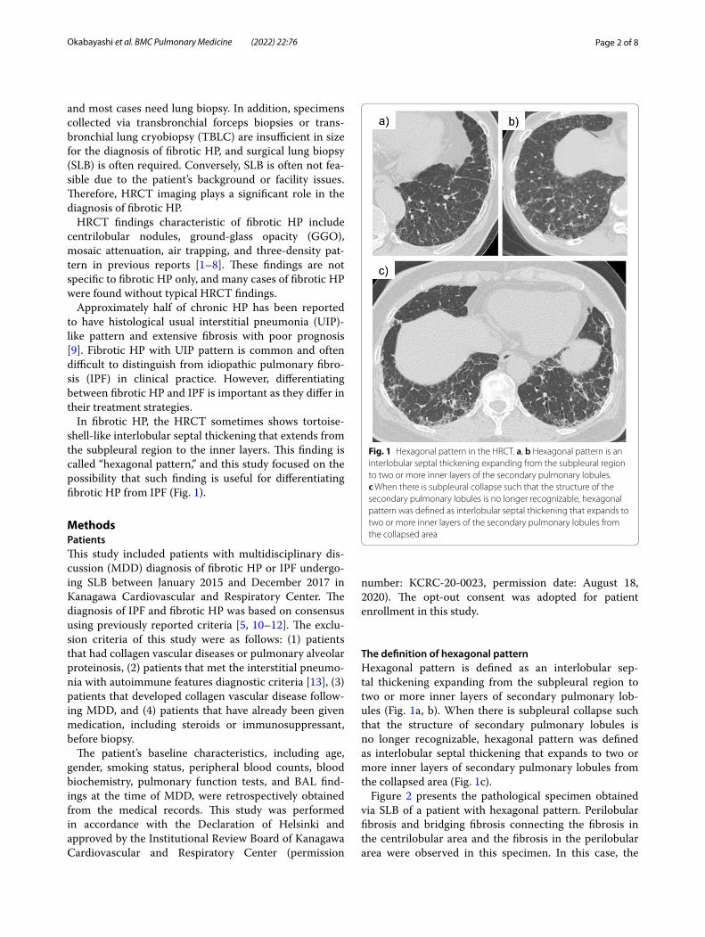
Figure 2 presents the pathological specimen obtained via SLB of a patient with hexagonal pattern. Perilobular fibrosis and bridging fibrosis connecting the fibrosis in the centrilobular area and the fibrosis in the perilobular area were observed in this specimen. In this case, the
Fig. 1 Hexagonal pattern in the HRCT. a, b Hexagonal pattern is an interlobular septal thickening expanding from the subpleural region to two or more inner layers of the secondary pulmonary lobules. c When there is subpleural collapse such that the structure of the secondary pulmonary lobules is no longer recognizable, hexagonal pattern was defined as interlobular septal thickening that expands to two or more inner layers of the secondary pulmonary lobules from the collapsed area
Page 3 of 8Okabayashi et al. BMC Pulmonary Medicine (2022) 22:76
hexagonal pattern observed in the HRCT corresponded to that of perilobular fibrosis.
Radiological evaluationThe radiological data were reviewed by two chest radi-ologists (T.I. and D.F.). HRCT images were indepen-dently evaluated, and the final findings were agreed upon by consensus between the two radiologists. The radi-ologists knew that only patients with fibrotic HP or IPF were included in the study, but they evaluated the HRCT findings without clinical and pathological information. The two radiologists have retrospectively evaluated the shadow distribution and the following findings: hex-agonal pattern, extensive GGO, centrilobular nodules, mosaic attenuation, air trapping, three-density pattern, reticulation, traction bronchiectasis, honeycombing, consolidation, emphysema, cyst, and pleuroparenchy-mal fibroelastosis. The HRCT findings were interpreted based on the recommendations of the Nomenclature
Committee of the Fleischner Society [14]. In this study, mosaic attenuation, air trapping, and three-density pat-tern were defined as being present when they were found in three or more lobules bilaterally. Before the radio-logical evaluation, two teacher images were shown to confirm the definition of hexagonal pattern. The classi-fication of cases as UIP, probable UIP, indeterminate for UIP, or alternative diagnosis using the American Tho-racic Society (ATS)/European Respiratory Society (ERS)/Japanese Respiratory Society (JRS)/Latin American Tho-racic Association (ALAT) guidelines for diagnosing IPF was performed retrospectively [12].
Statistical analysisContinuous variables are expressed as median values. The Mann–Whitney U test, chi-squared test, or Fisher’s exact probability test was employed, as appropriate, for between-group comparisons. The interobserver variation between two radiologists for the diagnosis of fibrotic HP
Fig. 2 The pathological specimen of the patient with hexagonal pattern in HRCT. a, b The HRCT shows interlobular septal thickening expanding from the subpleural region to two or more inner layers of the secondary pulmonary lobules. c, d The specimen of right S9 obtained via surgical lung biopsy. Perilobular fibrosis and bridging fibrosis connecting the fibrosis in the centrilobular area and the fibrosis in the perilobular area were observed. The arrows indicate bridging fibrosis. In this case, the hexagonal pattern observed in the HRCT corresponded to perilobular fibrosis. ILS, interlobular septum; V, vein; A, artery; RB, respiratory bronchiole; HE, hematoxylin and eosin; EvG, Elastica van Gieson
Page 4 of 8Okabayashi et al. BMC Pulmonary Medicine (2022) 22:76
or IPF was analyzed using κ statistic. A κ coefficient equal to or less than 0.20 indicated poor agreement; 0.21–0.40, fair agreement; 0.41–0.60, moderate agreement; 0.61–0.80, good agreement; and 0.81–1.00, excellent agree-ment. Univariate and multivariate logistic analyses were employed to identify the useful HRCT findings for dis-tinguishing fibrotic HP from IPF. A p-value of < 0.05 was considered to be significant. All statistical analyses have been conducted using the Statistical Package for the Social Sciences software version 24.0 (IBM; Armonk, NY, USA).
ResultsPatient characteristicsDuring the study period, 216 patients diagnosed with interstitial lung disease underwent SLB at our hospital. A total of 28 patients were diagnosed with fibrotic HP by MDD at the time of SLB and 51 with IPF. Among the fibrotic HP patients, one had pulmonary alveolar pro-teinosis, and one had Sjögren’s syndrome. Among the IPF patients, one met the diagnosis criteria of interstitial pneumonia with autoimmune features. One patient each with IPF and fibrotic HP received steroids prior to SLB. One patient with IPF developed rheumatoid arthritis after biopsy. Excluding cases that met the exclusion crite-ria, 23 cases of fibrotic HP and 48 cases of IPF were con-sidered in this study (Fig. 3). The ATS/JRS/ALAT Clinical Practice Guideline of the diagnosis of HP was published in 2020 [1], but the diagnosis in this study was made before this guideline was reported. The fibrotic HP diag-nosis was again reviewed according to this guideline, and all cases of fibrotic HP diagnosed by MDD were found to meet the criteria for definite or high-confidence HP.
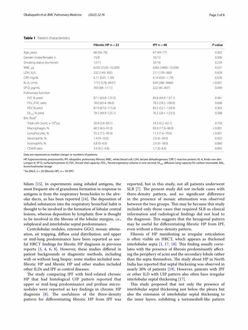
Table 1 presents the characteristic of the patients with fibrotic HP and IPF at the MDD. The serum Krebs von den Lungen-6 and surfactant protein-D levels were higher in fibrotic HP than in IPF patients. The percent predicted forced vital capacity (FVC), percent predicted forced expiratory volume in one second (FEV1), FEV1/FVC ratio, and percent predicted diffusion lung capac-ity for carbon monoxide (DLCO) at diagnosis were not significantly different between the two groups. The lym-phocyte ratio and CD4/8 ratio in BAL were significantly higher among fibrotic HP patients.
HRCT assessmentInterobserver agreement across the two radiologists in the HRCT findings other than pleuroparenchymal fibroelastosis was fair to good. The kappa coefficient of hexagonal pattern was 0.44, indicating moderate agree-ment (Table 2). Extensive GGO, centrilobular nodules, and hexagonal pattern were more frequently observed in fibrotic HP than in IPF. A significant difference in axial distribution was also found between fibrotic HP and IPF.
Mosaic attenuation, air trapping, or three-density pat-tern were reported as findings suggestive of fibrotic HP [1, 2, 6, 7], but no significant difference was observed in this study (Table 2). The logistic regression for predict-ing fibrotic HP is presented in Table 3. In the univariate logistic regression, the presence of extensive GGO, cen-trilobular nodules, and hexagonal pattern was associated with increased odds ratio of fibrotic HP. Extensive GGO and hexagonal pattern were the remaining variables in multivariate logistic regression. The presence of hex-agonal pattern was highly specific (87.5%) for diagnos-ing fibrotic HP. The sensitivity for fibrotic HP diagnosis when hexagonal pattern was present was 69.6%. The pos-itive predictive value of hexagonal pattern was 72.7% for fibrotic HP diagnosis.
DiscussionThis study has demonstrated that hexagonal pattern can be a useful finding for differentiating fibrotic HP from IPF.
HP is a diffuse interstitial pneumonia caused by immune response to inhaled antigen. Inhaled substances are usually most likely deposited at the level of the res-piratory bronchioles, and small particles of 2.5 μm or less reach the alveolar region via diffusion, are phagocytosed by alveolar macrophages, and then enter the lymphat-ics. The distribution of lymphatic vessels in the lungs is largely divided into two routes. One lymphatic flow fol-lows the bronchovascular bundles to the hilum in the inner layers. The other route starts at the perivenular area in the secondary lobule, runs through the interlobu-lar septa or subpleural lymphatic vessels, and ends at the
Fig. 3 Patient flow diagram. MDD, multidisciplinary discussion; SLB, surgical lung biopsy; HP, hypersensitivity pneumonitis; IPF, idiopathic pulmonary fibrosis; IPAF, interstitial pneumonia with autoimmune features
Page 5 of 8Okabayashi et al. BMC Pulmonary Medicine (2022) 22:76
hilum [15]. In experiments using inhaled antigens, the most frequent site of granuloma formation in response to antigens is from the respiratory bronchioles to the alve-olar ducts, as has been reported [16]. The deposition of inhaled substances into the respiratory bronchial habit is thought to be involved in the formation of lobular central lesions, whereas deposition by lymphatic flow is thought to be involved in the fibrosis of the lobular margins, i.e., subpleural and interlobular septal predominance.
Centrilobular nodules, extensive GGO, mosaic attenu-ation, air trapping, diffuse axial distribution, and upper or mid-lung predominance have been reported as use-ful HRCT findings for fibrotic HP diagnosis in previous reports [3, 4, 6, 8]. However, these studies differed in patient backgrounds or diagnostic methods, including with or without lung biopsy: some studies included non-fibrotic HP and fibrotic HP and other studies included other ILDs and IPF as control diseases.
The study comparing IPF with bird-related chronic HP that had histological UIP pattern reported that upper or mid-lung predominance and profuse micro-nodules were reported as key findings in chronic HP diagnosis [8]. The usefulness of the three-density pattern for differentiating fibrotic HP from IPF was
reported, but in this study, not all patients underwent SLB [7]. The present study did not include cases with three-density pattern, and no significant difference in the presence of mosaic attenuation was observed between the two groups. This may be because this study included only those cases that required SLB as clinical information and radiological findings did not lead to the diagnosis. This suggests that the hexagonal pattern may be useful for differentiating fibrotic HP from IPF, even without a three-density pattern.
Fibrosis of HP manifesting as irregular reticulation is often visible on HRCT, which appears as thickened interlobular septa [1, 17, 18]. This finding usually corre-lates with the presence of fibrosis predominantly affect-ing the periphery of acini and the secondary lobule rather than the septa themselves. The study about HP in North India has reported that septal thickening was observed in nearly 30% of patients [19]. However, patients with IPF or other ILD with UIP pattern also often have irregular interlobular septal thickening [17].
This study proposed that not only the presence of interlobular septal thickening just below the pleura but also the extension of interlobular septal thickening to the inner layers, exhibiting a tortoiseshell-like pattern
Table 1 Patient characteristics
Data are expressed as median (range) or numbers of patients
HP, hypersensitivity pneumonitis; IPF, idiopathic pulmonary fibrosis; WBC, white blood cell; LDH, lactate dehydrogenase; CRP, C-reactive protein; KL-6, Krebs von den Lungen-6; SP-D, surfactant protein-D; FVC, forced vital capacity; FEV1, forced expiratory volume in one second; DLCO, diffusion lung capacity for carbon monoxide; BAL, bronchoalveolar lavagea for BALF, n = 20 (fibrotic HP), n = 39 (IPF)
Fibrotic HP n = 23 IPF n = 48 P-value
Age, years 68 (56–76) 67 (49–77) 0.362
Gender (male/female), n 15/8 35/13 0.506
Smoking status (ex/never) 12/11 32/16 0.239
WBC, μL 6250 (2530–10,300) 6365 (3400–13,500) 0.531
LDH, IU/L 232 (149–305) 211 (139–360) 0.428
CRP, mg/dL 0.11 (0.01–1.50) 0.14 (0.01–1.79) 0.526
KL-6, U/mL 1772 (578–8437) 834 (286–4666) < 0.001
SP-D, pg/mL 393 (84–1111) 222 (45–897) 0.049
Pulmonary function
FVC % pred 87.1 (63.8–125.5) 85.8 (44.9–137.1) 0.461
FEV1/FVC ratio 78.0 (65.4–96.6) 78.3 (59.5–100.0) 0.606
FEV1% pred 87.9 (67.0–115.4) 83.5 (52.1–129.9) 0.363
DLCO % pred 78.5 (49.9–125.1) 76.5 (29.1–123.5) 0.588
BAL fluida
Total cell count, × 104/μL 26.9 (2.6–69.1) 24.3 (0.2–62.1) 0.750
Macrophages, % 40.5 (4.0–91.0) 83.0 (17.0–96.0) < 0.001
Lymphocytes, % 55.2 (7.5–95.0) 11.3 (1.6–70.0) < 0.001
Neutrophils, % 1.4 (0–10.0) 2.0 (0–39.0) 0.425
Eosinophils, % 0.8 (0–6.0) 2.0 (0–18.0) 0.060
CD4/8 ratio 3.9 (0.2–9.8) 1.7 (0–8.4) 0.005
Page 6 of 8Okabayashi et al. BMC Pulmonary Medicine (2022) 22:76
(hexagonal pattern), may be more characteristics of fibrotic HP than IPF.
In the case presented in Fig. 2, the area showing a hex-agonal pattern was biopsied, and the hexagonal pattern in the HRCT was thought to correspond to perilobular fibrosis. However, in this study, SLB was not performed at selected sites with hexagonal pattern; thus, the HRCT findings and pathology in all patients enrolled have not been compared in this study. Since hexagonal pattern is a shadow that extends to the inner layers, in some cases, the shadow is found in the inner layers beyond the area that can be sampled by SLB.
Recently, the utility of TBLC has been reported in the diagnosis of diffuse lung disease [20–23]. Cryo-probe-retrieved specimens are larger than those of
transbronchial forceps biopsies and less crush. TBLC tends to sample more proximal portion of the lung apart from the pleura compared with SLB. In the future, TBLC and SLB may be useful for comparing the imaging and pathology of the cases with hexagonal pattern in HRCT.
This study has several limitations. First, this was a sin-gle-center retrospective study, which may be subjected to various biases. External validation studies are warranted in the future. Second, this study included only patients who underwent SLB. Fibrotic HP and IPF, which can be diagnosed by clinical information, HRCT findings, and histological findings by transbronchial forceps biopsies or TBLC, were not included. However, the problem in clini-cal practice is the differentiation between IPF and fibrotic HP, which requires SLB. It is noteworthy that in this
Table 2 HRCT findings
Data are expressed as numbers of patients
HP, hypersensitivity pneumonitis; IPF, idiopathic pulmonary fibrosis; GGO, ground-glass opacity; PPFE, pleuroparenchymal fibroelastosis; HRCT, high-resolution computed tomography; UIP, usual interstitial pneumoniaa One patient of IPF had no expiratory CT available
Fibrotic HPn = 23
IPFn = 48
P-value Interobserver agreement
κ 95%CI
Distribution (zonal), n 0.611 0.22 − 0.02−0.47
Upper/middle predominant 0 1 (2.1%)
Lower predominant 19 (82.6%) 43 (89.6%)
Diffuse 4 (17.4%) 4 (8.3%)
Distribution (axial), n 0.001 0.36 0.16–0.55
Peribronchovascular 2 (8.7%) 1 (2.1%)
Peripheral 12 (52.2%) 44 (91.7%)
Peripheral with subpleural sparing 4 (17.4%) 2 (4.2%)
Diffuse 5 (21.7%) 1 (2.1%)
Extensive GGO, n 19 (82.6%) 19 (39.6%) 0.001 0.38 0.16–0.59
Centrilobular nodules, n 20 (87.0%) 27 (56.3%) 0.010 0.27 0.07–0.47
Mosaic attenuation, n 5 (21.7%) 3 (6.3%) 0.102 0.21 − 0.12–0.55
Air trapping, na 15 (65.2%) 25 (52.1%) 0.623 0.46 0.26–0.67
Three-density pattern, n 0 0 -
Reticulation, n 23 (100%) 48 (100%) -
Honeycombing, n 3 (13.0%) 14 (29.2%) 0.136 0.52 0.24–0.79
Traction bronchiectasis, n 23 (100%) 48 (100%) -
Consolidation, n 1 (4.3%) 7 (14.6%) 0.261 0.48 0.11–0.85
Emphysema, n 7 (30.4%) 21 (43.8%) 0.283 0.59 0.39–0.79
PPFE, n 1 (4.3%) 10 (20.8%) 0.090 0.14 − 0.35−0.64
Cyst, n 8 (34.8%) 27 (56.3%) 0.090 0.69 0.52–0.86
Hexagonal pattern, n 16 (69.6%) 6 (12.5%) < 0.001 0.44 0.19–0.69
HRCT patterns, n 0.000 0.37 0.22–0.53
UIP 0 4 (8.3%)
Probable UIP 2 (8.7%) 26 (54.2%)
Indeterminate for UIP 12 (52.2%) 14 (29.2%)
Alternative diagnosis 9 (39.1%) 4 (8.3%)

Page 7 of 8Okabayashi et al. BMC Pulmonary Medicine (2022) 22:76
study, the hexagonal pattern was useful for differentiat-ing between the two diseases, even in cases that the diag-nosis could not be made based on clinical and imaging findings. Future studies including a cohort comprising of patients with fibrotic HP diagnosed without SLB should be conducted. Third, the interobserver agreement for hexagonal pattern was not high. However, the problem is not limited to hexagonal patterns only, but it is also seen with findings such as GGO and centrilobular nodules, which have been reported to be characteristics of fibrotic HP. Previous studies on HRCT findings in hypersensitiv-ity pneumonitis have also shown only fair to moderate agreement among radiologists for these finding at rates comparable to this study [6, 8]. In this study, the interob-server agreement for hexagonal pattern was higher than that of GGO and centrilobular nodules. Therefore, we believe that the interobserver agreement in this study is not abnormally low, and it is simply necessary to examine more cases in the future. It is thought that a more accu-rate diagnosis of fibrotic HP can be made by combining multiple findings suggestive of fibrotic HP, rather than a single finding, and we hope that hexagonal pattern will
be one of them. Finally, we did not examine whether the hexagonal pattern is useful in differentiating fibrotic HP from diffuse lung diseases other than IPF. In the future, we are planning to compare the HRCT findings of fibrotic HP with other diffuse lung diseases.
ConclusionHexagonal pattern is a useful finding for differentiating fibrotic HP from IPF. Further external validation studies should be conducted to evaluate the utility of hexagonal pattern for diagnosing fibrotic HP.
AbbreviationsHRCT : High-resolution computed tomography; HP: Hypersensitivity pneumo-nitis; IPF: Idiopathic pulmonary fibrosis; MDD: Multidisciplinary discussion; BAL: Bronchoalveolar lavage; TBLC: Transbronchial lung cryobiopsy; SLB: Surgical lung biopsy; GGO: Ground-glass opacity; UIP: Usual interstitial pneumonia.
AcknowledgementsThe authors would like to acknowledge Toshihiro Misumi, PhD who supported the statistical analysis.
Authors’ contributionsHO takes responsibility for the content of the manuscript, including the integrity of the data and data analysis. DF and TI was involved in the analysis and interpretation of the HRCT findings. TT was involved in the analysis and interpretation of the pathological findings. HO, TI, TsuO, HK, TB, and TaO were involved in the analysis and interpretation of the clinical data. HO, DF, TI, TsuO, HK, TB, TT, TS, and TaO were involved in revising the manuscript. All authors read and approved the final manuscript.
FundingThis research did not receive any specific grant from funding agencies in the public, commercial, or not-for-profit sectors.
Availability of data and materialsThe dataset supporting the conclusions of this article is presented within the article. The detailed clinical data is not available because of patients’ confidentiality.
Declarations
Ethics approval and consent to participateThis study protocol was reviewed and approved by the Institutional Review Board of Kanagawa Cardiovascular and Respiratory Center, approval number KCRC-20-0023. Consent to participate statement: Because this study was a retrospective study, written informed consent was not obtained from partici-pants. The opt-out consent was adopted for patient enrollment in this study (approved by the Institutional Review Board of Kanagawa Cardiovascular and Respiratory Center).
Consent for publicationNot applicable.
Competing interestsThe authors declare that they have no competing interests.
Author details1 Department of Respiratory Medicine, Kanagawa Cardiovascular and Respira-tory Center, 6-16-1 Tomioka-Higashi, Kanazawa-ku, Yokohama City, Kanagawa 236-0051, Japan. 2 Department of Radiology, Kanagawa Cardiovascular and Respiratory Center, 6-16-1 Tomioka-Higashi, Kanazawa-ku, Yokohama City, Kanagawa 236-0051, Japan. 3 Department of Pathology, Kanagawa Cardiovascular and Respiratory Center, 6-16-1 Tomioka-Higashi, Kanazawa-ku, Yokohama City, Kanagawa 236-0051, Japan. 4 Department of Respiratory
Table 3 Logistic regression predicting fibrotic HP
HP, hypersensitivity pneumonitis; GGO, ground-glass opacity; PPFE, pleuroparenchymal fibroelastosis
Odds ratio 95% CI P-value
Univariate logistic regressionDistribution (zonal)
Upper/middle 0.00 0.00–Inf 0.991
Lower 0.44 0.10–1.96 0.282
Diffuse Reference
Distribution (axial)
Peripheral 0.05 0.01–0.51 0.011
Peripheral with subpleural sparing
0.40 0.03–6.18 0.512
Peribronchovascular 0.40 0.02–10.00 0.577
Diffuse Reference
Extensive GGO 7.25 2.13–24.65 0.002
Centrilobular nodules 5.19 1.36–19.82 0.016
Mosaic attenuation 4.17 0.90–19.28 0.068
Air trapping 1.65 0.59–4.63 0.341
Honeycombing 0.36 0.09–1.43 0.147
Consolidation 0.27 0.03–2.31 0.229
Emphysema 0.56 0.20–1.62 0.285
PPFE 0.17 0.02–1.44 0.105
Cyst 0.42 0.15–1.16 0.094
Hexagonal pattern 16.00 4.66–54.91 < 0.001
Multivariate logistic regressionExtensive GGO 4.86 1.20–19.70 0.027
Centrilobular nodules 1.45 0.30–7.08 0.647
Hexagonal pattern 11.00 2.70–44.70 0.001

Page 8 of 8Okabayashi et al. BMC Pulmonary Medicine (2022) 22:76
• fast, convenient online submission
•
thorough peer review by experienced researchers in your field
• rapid publication on acceptance
• support for research data, including large and complex data types
•
gold Open Access which fosters wider collaboration and increased citations
maximum visibility for your research: over 100M website views per year •
At BMC, research is always in progress.
Learn more biomedcentral.com/submissions
Ready to submit your researchReady to submit your research ? Choose BMC and benefit from: ? Choose BMC and benefit from:
Medicine, Kumamoto University Hospital, Faculty of Life Sciences, Kumamoto University, 1-1-1 Honjo, Chuo-ku, Kumamoto City, Kumamoto 860-8556, Japan. 5 Department of Radiology, The Jikei University School of Medicine, 3-19-18 Nishishinbashi, Minato-ku, Tokyo 105-8471, Japan.
Received: 13 November 2021 Accepted: 24 February 2022
References 1. Raghu G, Remy-Jardin M, Ryerson CJ, et al. Diagnosis of hypersensitivity
pneumonitis in adults. An official ATS/JRS/ALAT clinical practice guide-line. Am J Respir Crit Care Med. 2020;202(3):e36–69.
2. Fernandez Perez ER, Travis WD, Lynch DA, et al. Diagnosis and evalua-tion of hypersensitivity pneumonitis: CHEST guideline and expert panel report. Chest. 2021;160(2):e97–156.
3. Lynch DA, Newell JD, Logan PM, King TE, Müller NL. Can CT distinguish hypersensitivity pneumonitis from idiopathic pulmonary fibrosis? AJR Am J Roentgenol. 1995;165(4):807–11.
4. Silva CIS, Müller NL, Lynch DA, et al. Chronic hypersensitivity pneumo-nitis: differentiation from idiopathic pulmonary fibrosis and non-specific interstitial pneumonia by using thin-section CT. Radiology. 2008;246(1):288–97.
5. Salisbury ML, Myers JL, Belloli EA, Kazerooni EA, Martinez FJ, Flaherty KR. Diagnosis and treatment of fibrotic hypersensitivity pneumonia. Where we stand and where we need to go. Am J Respir Crit Care Med. 2017;196(6):690–9.
6. Salisbury ML, Gross BH, Chughtai A, et al. Development and validation of a radiological diagnosis model for hypersensitivity pneumonitis. Eur Respir J. 2018;52(2):1800443.
7. Barnett J, Molyneaux PL, Rawal B, et al. Variable utility of mosaic attenua-tion to distinguish fibrotic hypersensitivity pneumonitis from idiopathic pulmonary fibrosis. Eur Respir J. 2019;54(1):1900531.
8. Tateishi T, Johkoh T, Sakai F, et al. High-resolution CT features distinguish-ing usual interstitial pneumonia pattern in chronic hypersensitivity pneumonitis from those with idiopathic pulmonary fibrosis. Jpn J Radiol. 2020;38(6):524–32.
9. Ohtani Y, Saiki S, Kitaichi M, et al. Chronic bird fancier’s lung: histopatho-logical and clinical correlation. An application of the 2002 ATS/ERS consensus classification of the idiopathic interstitial pneumonias. Thorax. 2005;60(8):665–71.
10. Lacasse Y, Selman M, Costabel U, et al. Clinical diagnosis of hypersensitiv-ity pneumonitis. Am J Respir Crit Care Med. 2003;168(8):952–8.
11. Vasakova M, Morell F, Walsh S, Leslie K, Raghu G. Hypersensitivity pneu-monitis: perspectives in diagnosis and management. Am J Respir Crit Care Med. 2017;196(6):680–9.
12. Raghu G, Remy-Jardin M, Myers JL, et al. Diagnosis of idiopathic pulmo-nary fibrosis. An official ATS/ERS/JRS/ALAT clinical practice guideline. Am J Respir Crit Care Med. 2018;198(5):e44–68.
13. Fischer A, Antoniou KM, Brown KK, et al. An official European Respira-tory Society/American Thoracic Society research statement: interstitial pneumonia with autoimmune features. Eur Respir J. 2015;46(4):976–87.
14. Hansell DM, Bankier AA, MacMahon H, McLoud TC, Müller NL, Remy J. Fleischner Society: glossary of terms for thoracic imaging. Radiology. 2008;246(3):697–722.
15. Okada Y. Pulmonary lymphatic system and lung cancer. 1st ed. Kinpodo; 1989.
16. Mitaka K, Miyazaki Y, Yasui M, et al. Th2-biased immune responses are important in a murine model of chronic hypersensitivity pneumonitis. Int Arch Allergy Immunol. 2011;154(3):264–74.
17. Webb WR, Muller NL, Naidich DP. High-resolution CT of the lung. 5th ed. Wolters Kluwer; 2014.
18. Giorgia-Dalpiaz AC. Atlas of diffuse lung diseases: a multidisciplinary approach. Springer; 2016.
19. Kumar R, Spalgais S, Ranga V. Hypersensitivity pneumonitis: clinical, radio-logical and pathological profile of 103 patients from North India. Monaldi Arch Chest Dis. 2020;90(3):1307.
20. Casoni GL, Tomassetti S, Cavazza A, et al. Transbronchial lung cryobi-opsy in the diagnosis of fibrotic interstitial lung diseases. PLoS ONE. 2014;9(2):e86716.
21. Iftikhar IH, Alghothani L, Sardi A, Berkowitz D, Musani AI. Transbronchial lung cryobiopsy and video-assisted thoracoscopic lung biopsy in the diagnosis of diffuse parenchymal lung disease. A meta-analysis of diag-nostic test accuracy. Ann Am Thorac Soc. 2017;14(7):1197–211.
22. Lentz RJ, Argento AC, Colby TV, Rickman OB, Maldonado F. Transbron-chial cryobiopsy for diffuse parenchymal lung disease: a state-of-the-art review of procedural techniques, current evidence, and future chal-lenges. J Thorac Dis. 2017;9(7):2186–203.
23. Hetzel J, Maldonado F, Ravaglia C, et al. Transbronchial cryobiopsies for the diagnosis of diffuse parenchymal lung diseases: expert statement from the cryobiopsy working group on safety and utility and a call for standardization of the procedure. Respiration. 2018;95(3):188–200.
Publisher’s NoteSpringer Nature remains neutral with regard to jurisdictional claims in pub-lished maps and institutional affiliations.