Comparative transcription analysis and ... - BMC Microbiology
13
RESEARCH ARTICLE Open Access Comparative transcription analysis and toxin production of two fluoroquinolone-resistant mutants of Clostridium perfringens Sunny Park 1,2 , Miseon Park 1 and Fatemeh Rafii 1* Abstract Background: Fluoroquinolone use has been listed as a risk factor for the emergence of virulent clinical strains of some bacteria. The aim of our study was to evaluate the effect of fluoroquinolone (gatifloxacin) resistance selection on differential gene expression, including the toxin genes involved in virulence, in two fluoroquinolone-resistant strains of Clostridium perfringens by comparison with their wild-type isogenic strains. Results: DNA microarray analyses were used to compare the gene transcription of two wild types, NCTR and ATCC 13124, with their gatifloxacin-resistant mutants, NCTR R and 13124 R . Transcription of a variety of genes involved in bacterial metabolism was either higher or lower in the mutants than in the wild types. Some genes, including genes for toxins and regulatory genes, were upregulated in NCTR R and downregulated in 13124 R . Transcription analysis by quantitative real-time PCR (qRT-PCR) confirmed the altered expression of many of the genes that were affected differently in the fluoroquinolone-resistant mutants and wild types. The levels of gene expression and enzyme production for the toxins phospholipase C, perfringolysin O, collagenase and clostripain had decreased in 13124 R and increased in NCTR R in comparison with the wild types. After centrifugation, the cytotoxicity of the supernatants of NCTR R and 13224 R cultures for mouse peritoneal macrophages confirmed the increased cytotoxicity of NCTR R and the decreased cytotoxicity of 13124 R in comparison with the respective wild types. Fluoroquinolone resistance selection also affected cell shape and colony morphology in both strains. Conclusion: Our results indicate that gatifloxacin resistance selection was associated with altered gene expression in two C. perfringens strains and that the effect was strain-specific. This study clearly demonstrates that bacterial exposure to fluoroquinolones may affect virulence (toxin production) in addition to drug resistance. Keywords: Clostridium perfringens, Fluoroquinolone, Resistance, Toxin, Virulence Background Clostridium perfringens is commonly found in the gastro- intestinal (GI) tract of humans, animals, soils, freshwater sediments and sewage. It can cause various diseases in humans, including food poisoning, antibiotic-associated diarrhea, sporadic diarrhea, internal abscesses, and gas gan- grene and also various animal diseases [1-5]. C. perfringens strains all are prolific toxin producers and are classified based on their toxin formation. Various C. perfringens toxins denature cellular components of mammalian cells and are implicated in virulence and pathogenicity. Among these toxins are α-toxin (phospholipase C, PLC) and θ-toxin (perfringolysin O, PFO), which are essential for gas gangrene pathogenesis. Other toxins or hydrolytic enzymes may be involved in destruction of connective tissue or spread of bacteria in infected tissues [4,6,7]. C. perfringens, although a commensal, can cause life threatening infections and is implicated in inflammatory bowel diseases [8-10]. In a survey of Clostridium species bacteremia, in a Canadian hospital between 2000–2006, C. perfringens was shown to have caused 42% of the * Correspondence: [email protected] 1 Division of Microbiology, National Center for Toxicological Research, US Food and Drug Administration, Jefferson, AR, USA Full list of author information is available at the end of the article © 2013 Park et al.; licensee BioMed Central Ltd. This is an Open Access article distributed under the terms of the Creative Commons Attribution License (http://creativecommons.org/licenses/by/2.0), which permits unrestricted use, distribution, and reproduction in any medium, provided the original work is properly cited. Park et al. BMC Microbiology 2013, 13:50 http://www.biomedcentral.com/1471-2180/13/50
-
Upload
khangminh22 -
Category
Documents
-
view
1 -
download
0
Transcript of Comparative transcription analysis and ... - BMC Microbiology

Park et al. BMC Microbiology 2013, 13:50http://www.biomedcentral.com/1471-2180/13/50
RESEARCH ARTICLE Open Access
Comparative transcription analysis and toxinproduction of two fluoroquinolone-resistantmutants of Clostridium perfringensSunny Park1,2, Miseon Park1 and Fatemeh Rafii1*
Abstract
Background: Fluoroquinolone use has been listed as a risk factor for the emergence of virulent clinical strains ofsome bacteria. The aim of our study was to evaluate the effect of fluoroquinolone (gatifloxacin) resistance selectionon differential gene expression, including the toxin genes involved in virulence, in two fluoroquinolone-resistantstrains of Clostridium perfringens by comparison with their wild-type isogenic strains.
Results: DNA microarray analyses were used to compare the gene transcription of two wild types, NCTR andATCC 13124, with their gatifloxacin-resistant mutants, NCTRR and 13124R. Transcription of a variety of genesinvolved in bacterial metabolism was either higher or lower in the mutants than in the wild types. Somegenes, including genes for toxins and regulatory genes, were upregulated in NCTRR and downregulated in13124R. Transcription analysis by quantitative real-time PCR (qRT-PCR) confirmed the altered expression of manyof the genes that were affected differently in the fluoroquinolone-resistant mutants and wild types. The levelsof gene expression and enzyme production for the toxins phospholipase C, perfringolysin O, collagenase andclostripain had decreased in 13124R and increased in NCTRR in comparison with the wild types. Aftercentrifugation, the cytotoxicity of the supernatants of NCTRR and 13224R cultures for mouse peritonealmacrophages confirmed the increased cytotoxicity of NCTRR and the decreased cytotoxicity of 13124R incomparison with the respective wild types. Fluoroquinolone resistance selection also affected cell shape andcolony morphology in both strains.
Conclusion: Our results indicate that gatifloxacin resistance selection was associated with altered geneexpression in two C. perfringens strains and that the effect was strain-specific. This study clearly demonstratesthat bacterial exposure to fluoroquinolones may affect virulence (toxin production) in addition to drugresistance.
Keywords: Clostridium perfringens, Fluoroquinolone, Resistance, Toxin, Virulence
BackgroundClostridium perfringens is commonly found in the gastro-intestinal (GI) tract of humans, animals, soils, freshwatersediments and sewage. It can cause various diseases inhumans, including food poisoning, antibiotic-associateddiarrhea, sporadic diarrhea, internal abscesses, and gas gan-grene and also various animal diseases [1-5]. C. perfringensstrains all are prolific toxin producers and are classified
* Correspondence: [email protected] of Microbiology, National Center for Toxicological Research, USFood and Drug Administration, Jefferson, AR, USAFull list of author information is available at the end of the article
© 2013 Park et al.; licensee BioMed Central LtdCommons Attribution License (http://creativecreproduction in any medium, provided the or
based on their toxin formation. Various C. perfringenstoxins denature cellular components of mammalian cellsand are implicated in virulence and pathogenicity. Amongthese toxins are α-toxin (phospholipase C, PLC) andθ-toxin (perfringolysin O, PFO), which are essential forgas gangrene pathogenesis. Other toxins or hydrolyticenzymes may be involved in destruction of connectivetissue or spread of bacteria in infected tissues [4,6,7].C. perfringens, although a commensal, can cause lifethreatening infections and is implicated in inflammatorybowel diseases [8-10]. In a survey of Clostridium speciesbacteremia, in a Canadian hospital between 2000–2006,C. perfringens was shown to have caused 42% of the
. This is an Open Access article distributed under the terms of the Creativeommons.org/licenses/by/2.0), which permits unrestricted use, distribution, andiginal work is properly cited.

Park et al. BMC Microbiology 2013, 13:50 Page 2 of 13http://www.biomedcentral.com/1471-2180/13/50
cases, which was more than any other Clostridiumspecies [11]. It causes nearly a million cases of foodborne illness each year in the United States [1]. Bac-teria from the GI tract, including C. perfringens, maybecome resistant to fluoroquinolones used for treat-ment or prophylaxis of bacterial infections. Surveys offluoroquinolone-resistant-anaerobes found ciprofloxacin-resistant C. perfringens as early as 1992 among clinicalisolates [12]. Although similar surveys have not beenconducted in recent years, Gionchetti et al. [10] showedthat treatment of patients with chronic treatment-resistant pouchitis with 1 g of ciprofloxacin for 15 daysdid not result in a statistically significant reduction inC. perfringens. One reason for fluoroquinolone resist-ance development is mutation in the fluoroquinolonetarget genes, gyrase (gyrA and gyrB) and topoisomeraseIV (parC and parE) [13]. Because fluoroquinolones areDNA-damaging agents, they may also induce the SOSresponse [14-16] that results in expression of DNA re-pair genes, which may lead to phenotypic changes influoroquinolone-resistant strains [17-20]. Excessive useof fluoroquinolones has been attributed to the emergenceof virulent strains of bacteria [21-24]. Clostridium difficilestrain NAP1/027, which emerged in 2002 in Canadaand the USA, now has spread to most parts of Europe[22]. In a gut model, higher rates of spore germinationand levels of toxin production were observed in tworibotypes of C. difficile that were exposed to three dif-ferent fluoroquinolones [24]. Wide dissemination ofvirulent fluoroquinolone-resistant strains of Escherichiacoli has been reported in East Asia [25]. Other reports,sometimes conflicting, show either increased or de-creased virulence in fluoroquinolone-resistant clinicalisolates of bacteria [26-28]. Previously we showed thatdifferent C. perfringens strains rapidly developed resist-ance, even to high potency fluoroquinolones, and thatresistant strains had various mutations in the fluoro-quinolone target genes [29]. In addition, the productionof some enzymes was altered in some resistant mutants[30,31]. One gatifloxacin-resistant strain, NCTR, hadincreased levels of α-toxin (phospholipase C, PLC) andθ-toxin (perfringolysin O, PFO) [30]. These resultspoint to global changes in the expression of variousgenes in gatifloxacin- resistant strains and to the needfor further study. In this study, we have used genomicanalysis (microarray and QRT-PCR) to compare thechanges in gene expression in two gatifloxacin-resistantstrains of C. perfringens following fluoroquinolone re-sistance selection, and have compared the toxin pro-duction and cytotoxicity of the strains. Strain NCTRwas selected because of enhanced production of PLCand PFO by its gatifloxacin resistant mutant and wascompared with strain ATCC 13124, which is a gangreneisolate whose genomic sequence is known, and its
gatifloxacin resistant mutant 13124R has the samemutation in gyrA as NCTRR.
MethodsGrowth of bacterial strainsWild types and gatifloxacin-resistant mutants of C.perfringens strains ATCC 13124 and NCTR [29] wereused in this study. The development of these mutants(Gat-13124-10 and Gat-NCTR- 10) was described previ-ously [29]. Both mutants have stable mutations in targetgenes and will be referred to as 13124R and NCTRR inthis study. Both mutants had a mutation in gyrA (G81C,D87Y), 13124R had mutation in gyrB (A431S) and parC(S89I), and NCTRR had a mutation in parE (E486K).The bacteria were grown anaerobically under an atmos-phere of 85% N2, 10% CO2, and 5% H2 at 37°C in brainheart infusion (BHI) broth (Remel, Lenexa, KS) withvitamin K (1 μg/ml), hemin (5 μg/ml), and L-cysteine(5 μg/ml) (Sigma Chemical Co., St. Louis, MO) [29]. Noantibiotics were added.
Preparation of RNAEarly exponential (2.5-3.0 h) growth phase cultures of allfour strains, grown in BHI under identical anaerobicconditions, were used to isolate RNA for microarrays.Cells from 100-ml cultures were harvested by centrifuga-tion (15,000 × g, 10 min, 4°C), washed with 10 mM Trisand 1 mM EDTA (pH 8.0), and suspended in 1 ml ofbuffer containing 10 mg/ml of lysozyme (Sigma). The mix-tures were incubated for 10 min at room temperature andcentrifuged (15,000 × g, 10 min, 4°C). The samples weresuspended in 0.5 ml TE (10 mM Tris, 1 mM EDTA)and mixed with 5 ml of RNA-Bee isolation reagentfrom TEL-TEST, Inc. (Friendship, TX). After additionof 1 ml chloroform to the mixture, the samples were in-cubated on ice for 30 min and centrifuged (15,000 × g,30 min, 4°C). The clear phases were harvested, addedto an equal volume of isopropanol and centrifuged topellet the RNA. The RNA was further purified using anRNeasyR Mini Kit (50) from QIAGEN, Inc. (Valencia,CA), according to the instructions provided with thekit. After RNA extraction and purification, contaminat-ing DNA was removed using 10 U of RNase-free DNase1 (Boehringer Mannheim, Ingelheim, Germany). Thequantity and quality of total RNA was determined usinga Nanodrop ND-1000 spectrophotometer (NanoDropTechnology, Wilmington, DE). RNA purification stepsfor real-time PCR (qRT- PCR) were essentially the same.The RNA was stored at −80°C and used within a week toavoid degradation of RNA. RNA was extracted from threedifferent cultures of each strain for microarray analysisand qRT-PCR.

Park et al. BMC Microbiology 2013, 13:50 Page 3 of 13http://www.biomedcentral.com/1471-2180/13/50
Probe design for microarraysThe probes were designed by Biodiscovery LLC, Ann Arbor,MI (http://www.mycroarray.com/) from the sequencesof C. perfringens strains 13 (CPE) and ATCC 13124(CPF in http://www.ncbi.nlm.nih.gov), using OligoArray v3.1 (http://berry.engin.umich.edu). The designs of micro-arrays were submitted to MIAMExpress and can beaccessed at the following links: for strain 13124, [http://www.ebi.ac.uk/arrayexpress/arrays/A-MEXP-2008], and forstrain NCTR, at [http://www.ebi.ac.uk/arrayexpress/arrays/A-MEXP-2027].
Microarray hybridizationThe microarrays were hybridized by Biodiscovery LLCto fluor-labeled RNA at 60°C for at least 16 h in 2-gasket slides and commercial hybridization chambers(Agilent, Santa Clara, CA) while being rotated (~4 rpm)in a hybridization incubator. The hybridization solutioncontained 6 × SSPE (1 M NaCl, 50 mM NaH2PO4,50 mM Na2HPO4, 3 mM EDTA, pH 6.7), 1 μg/μl acety-lated BSA, 1 μg/μl herring sperm DNA (Promega,Madison,WI), 0.01% Tween 20 (Sigma) and 10 μg tem-plate RNA per array. The hybridized arrays werewashed twice in 6 × SSPE for 5 min at 60°C, once in 1 ×SSPE for 5 min at 20°C, and once in 0.25 × SSPE at 20°Cfor 1 min, and then were spun dry in a microarray high-speed centrifuge (ArrayIT, model MHC). The arrays werescanned in an Axon 4000B scanner (Molecular DevicesSunnyvale, California), controlled by GenePixPro software(v 6.1.0.4). The resulting images were quantified withthe same software and the results were archived in thegpr file format. The mean expression of each gene forthe mutant was divided by the mean expression of thesame gene for the wild type. Those genes for whichthe values were ≥ 1.5 were considered upregulated inthe mutant, and the genes for which this value was≤0.6 were considered downregulated in the mutant.The genes that were upregulated or downregulatedwere selected for further RT-PCR analysis.
Quantitative real-time PCR (qRT-PCR)Primers used for qRT-PCR are listed in Additional file 1.The genes that were upregulated in one mutant anddownregulated in the other mutant, in comparison withtheir respective wild types, by microarray analysis wereselected to design primers. Some genes involved in regu-lation of transcription were also selected. The sequenceof C. perfringens ATCC 13124 (http://www.ncbi.nlm.nih.gov/nuccore/CP000246.1) was used to design primersthat generated PCR amplicons of 100–150 bp in lengthvia the default setting of “Primer 3 Input software”(http://frodo.wi.mit.edu/primer3). For cDNA templatesynthesis, SuperScriptTM III First-Strand SynthesisSuperMix (Invitrogen, Carlsbad, CA) was used. For
qRT-PCR, SYBRW GreenERTM qPCR SuperMix (Invitrogen)was used. The reaction mixtures were prepared on iceaccording to the manufacturer’s instructions. Each reac-tion contained 2 × Express SYBR Green ER qRT-PCRuniversal mix, 25, 2.5, or 0.25 ng of the cDNA template,and 2 μM each of the forward and reverse primers. Theamplification was performed using a CFX96 Real-TimePCR detection system (Bio-Rad, Hercules, CA) and thefollowing protocol: 50°C for 10 min, 95°C for 8.5 minto inactivate uracil DNA glycosylase and activate DNApolymerase, followed by 40 cycles of 95°C for 15 s and60°C for 1 min to amplify cDNA. Melting curves weremonitored at 65-95°C (1°C per 5 s) to detect anynonspecific amplification. Either 25, 2.5, or 0.25 ng ofeach 16S rRNA gene was amplified as a referenceRNA of equivalent size for normalization [32]. Reactionmixtures without reverse transcriptase, for detectinggenomic DNA contamination, and reaction mixtureswithout templates, for detecting nucleic acid contamin-ation of reagents and tubes, were included as controls.Each PCR reaction was run in triplicate for each typeof RNA isolated from three different cultures of eachwild type or mutant. The relative level of mRNA expres-sion was calculated by the 2-ΔΔCT method according toReal-Time PCR Application Guide (Additional file 2).
Detection of phospholipase C (PLC) and perfringolysinO (PFO)PLC and PFO activities were measured according to themethods previously described [7,30,33]. The hemoglobin re-lease from red blood cells in the presence of perfringolysinbuffer was measured to detect perfringolysin O (PFO)according to the method of O'Brien and Melville [33]. Theincrease in turbidity of lecithin in egg yolk emulsion or therelease of nitrophenol from O-(4-nitrophenyl-phosphoryl)choline as the result of hydrolysis by PLC was used tomeasure phospholipase C (PLC) activity [7,30].
Collagenase assayThe amounts of collagenase in the mutants and wildtypes were calculated by the method of Awad et al. [34]by measuring the amount of dye released from Azo DyeImpregnated Collagen (azocoll) (Sigma). Azocoll powderwas washed and resuspended in 0.2 M of borate buffer(pH 7.2) containing 0.15 M NaCl, 20 μM ZnCl2 and5 mM CaCl2 to a final concentration of 5 mg azocollper ml. Next, 100 μl of the filter-sterilized supernatantsof centrifuged wild types and mutants were added to400 μl of azocoll solution and the mixtures were incu-bated for 2 h at 37°C. Following centrifugation at16,100 × g, the released dye was measured by the ab-sorbance at 550 nm.

Park et al. BMC Microbiology 2013, 13:50 Page 4 of 13http://www.biomedcentral.com/1471-2180/13/50
Assay for clostripainA clostripain substrate, N-carbobenzoxy-L-arginine p-nitroanilide (Z-Arg-pNA) (Bachem Americas, Torrance,CA), was used for measuring the amounts of clostripainin the supernatants of wild types and mutants [35]. Thefilter-sterilized supernatant from each centrifuged strainwas incubated overnight at 4°C in a buffer containingdithiothreitol to reduce the thiol group of the cysteineresidues of clostripain. Next, 20 μl of the sample wasadded to the 300 μl buffer containing 2 mM CaCl2 and260 mM of Z-Arg-pNA. The kinetics software of thespectrophotometer was programmed to measure theabsorbance at 410 nm every min for 30 min. The amount ofcleavage of Z-Arg-pNA was measured and the enzyme unitswere calculated. One unit was defined as the amount of en-zyme that hydrolyzed 1.0 μmol of Z-Arg-pNA per min [35].
Detection of sialidaseSialidase activity was measured in filter-sterilized superna-tants of centrifuged cultures of mutants and wild types, using4 mM 5-bromo-4-chloro-3-indolyl-α-D-N-acetylneuraminicacid, sodium salt [36]. The assay reaction was performedin 96-well plates by addition of the supernatant to wellscontaining the substrates, according to a procedurerecommended by Sigma for measuring recombinant C.perfringens neuraminidase. The kinetics software was pro-grammed to measure the absorbance at 595 nm.
Hyaluronidase detectionThe amounts of hyaluronidase in the filter-sterilized su-pernatants of centfifuged wild types and mutants werequantified by measuring the degradation of hyaluronicacid. Bovine hyaluronic acid (Sigma) was dissolved inacetate buffer (200 mM sodium acetate, 150 mM NaCl,pH 6.0) to a final concentration of 1 mg/ml. 100 μl ofthe hyaluronic acid solution was incubated with 400 μlof the filter-sterilized supernatants of the wild types and mu-tants for 30 min at 37°C. One ml of a solution containing2% NaOH and 2.5% cetramide (cetyltrimethylammoniumbromide, Sigma) was added to the reaction mixture. Theturbidity of the insoluble complex formed betweencetramide and hyaluronic acid was measured at 400 nm[37]. The reduction in turbidity, reflecting the decrease inhyaluronic acid because of the activity of hyaluronidase,was calculated by comparing the turbidities of samplescontaining the supernatant of each culture with controlscontaining BHI alone. The enzyme assays for all the en-zymes were performed three times from three differentcultures of each strains.
Cytotoxicity of C. perfringens supernatants formacrophagesMacrophages were obtained from C57BL/6 male mice,4–6 weeks old, which had ad libitum access to food and
water. The maintenance, handling and sacrifice of micewere according to procedures approved by the NCTRInstitutional Animal Care and Use Committee. Residentmouse peritoneal macrophages were harvested by peri-toneal lavage, using 4 ml of supplemented DMEMmedium, containing 5% heat-inactivated fetal bovineserum, 100 μg/ml streptomycin sulfate, 100 units/mlpenicillin G, 110 mg/L sodium pyruvate, and 2 mMglutamine. Red blood cells were removed by hypotoniclysis. The peritoneal exudate cells were washed oncewith DMEM, plated and incubated at 37°C in a humidi-fied atmosphere of 5% CO2 [33]. Floating cells were re-moved and the macrophages were incubated in DMEM,containing 10% BHI or filter-sterilized supernatants ofovernight cultures of wild types and mutants, for18 h at 37°C in a CO2 incubator. A CytoTox 96W
Non-Radioactive Cytotoxicity Assay Kit (Promega) wasused to measure the toxicity of the mutants and wild typecultures for macrophages. The cytotoxicity of eachabsorbance unit of the cells of different strains wascalculated by the amount of lactate dehydrogenase (LDH)released from the macrophages. The differences in cyto-toxicity due to the mutants and wild types were assessedusing Student’s t-test.
Morphological examinationColony morphology of the strains was compared afterovernight growth on BHI plates. For cellular morph-ology, log phase grown cells were Gram stained and ex-amined under the light microscope.
DNA sequencingSeveral regulatory and toxin genes and enzymes fromwild types and mutants were amplified and sequenced aspreviously described [29].
ResultsTranscriptional analysis by DNA microarrayUsing the genome sequences of C. perfringens strain13 and strain ATCC 13124, microarray probes weredesigned for genome-wide transcriptional analysis oftwo fluoroquinolone-resistant C. perfringens strains, NCTRR
and 13124R, and their wild types. Microarray analysisshowed that a variety of genes were upregulated (≥ 1.5fold) or downregulated (≤ 1.5 fold) in the fluoroquinolone-resistant strains. The altered genes with known functionsthat were affected in both strains as the results of fluoro-quinolone resistance selection are grouped in Tables 1, 2, 3according to the classification used by the Institute forGenomic Research (http://www.jcvi.org/). In addition, themicroarray detected alterations of many genes, for whichthe function is not known, which are listed as hypotheticalproteins in the GenBank. Some of these were upregulatedmanyfold in both resistant strains, especially in 13124R.

Table 1 Microarray and qRT-PCR analysis of the genes that were differentially affected in the gatifloxacin resistantmutants, NCTRR and 13124R
Gene ID and name Function Microarray qRT-PCR
mt/wt mt/wt
NCTR ATCC 13124 NCTR ATCC 13124
Cell envelope
CPE1089 CPF_1345 putative membrane protein 4.3 −2.1 7.3 −2.8
CPE0162 CPF_0155 (pfoR) putative membrane protein 2.6 −4.0 3.3 −3.5
CPE0251 CPF_0244 putative lipoprotein 5.0 −2.4 2.0 −3.5
CPE0278 CPF_0274 (sagA) sagA protein 1.1 −2.4 4.7 −2.6
CPE0714 CPF_0710 putative monogalactosyl-diacylglycerol synthase 2.4 −2.4 7.6 6.3
Cellular processes
CPE0036 CPF_0042 (plc) phospholipase C 4.8 −6.8 1.9 −3.3
CPE0846 CPF_0840 (cloS1) α-clostripain 17.3 −15.6 8.3 −1143
CPE1474 CPF_1725 (hlyC) hemolysin III 3.2 −1.8 15.1 −2.6
CPE0163 CPF_0156 (pfoA) perfringolysin O 3.6 −71.4 6.4 −462
CPE0782 CPF_0784 (ahpC) alkyl hydroperoxide reductase-C subunit 10.3 −2.6 13.4 −12.6
CPE1092 CPF_1348 (pac) choloylglycine hydrolase family protein 1.7 −2.5 25.7 −1.7
Energy metabolism
CPE0778 CPF_0780 oxidoreductase, FDA-binding 4.8 −2.8 85 2.6
CPE1299 CPF_1505 (eno) enolase 3.5 −1.6 11.9 −1.9
CPE2058 CPF_2315 (gadB) glutamate decarboxylase 31.9 −3.5 20.0 −3.4
CPE2437 CPF_2747 (nrdH) glutaredoxin-like protein, YruB-family 3.8 −2.5 4.8 −11.0
CPE2551 CPF_2875 (glpA) probable glycerol-3-phosphate dehydrogenase 0.8 −2.5 1.3 −0.1
Purines, pyrimidines, nucleotides, and nucleosides
CPE2276 CPF_2558 (guaB) inosine-5’-monophosphate dehydrogenase 9.2 −3.6 30.3 −1.5
CPE2622 CPF_2958 (purA) adenylosuccinate synthetase 4.3 −1.9 14.8 −0.8
Protein fate
CPE0173 CPF_0166 (colA) collagenase 9.9 −4.7 8.5 −2.7
CPE2323 CPF_2632 (pepF) probable oligoendopeptidase F 2.7 -2.0 11.6 4.3
CPE1205 CPF_1002 (abgB) amidohydrolase family protein 1.9 −4.3 67.4 −1.6
Regulatory functions
CPE0073 CPF_0069 transcription antiterminator 2.1 −5.0 1.9 −2.6
CPE0759 CPF_0753 putative regulatory protein 1.5 −5.4 3.3 0.6
CPE1533 CPF_1784 (scrR) sucrose operon repressor 1.7 −2.8 132 −1.5
CPE2035 CPF_2292 (hrcA) heat-inducible transcription repressor HrcA 2.3 −2.9 9.5 5.5
CPE2363 CPF_2673 two-component sensor histidine kinase 2.1 −3.0 16.1 2.7
Transport and binding proteins
CPE1240 CPF_1450 (mgtE) magnesium transporter 8.6 −1.7 5.2 −2.6
CPE1300 CPF_1507 (gadC) glutamate:γ-aminobutyrate antiporter family protein 9.6 −2.7 17.1 −7.3
CPE1505 CPF_1756 (uraA) uracil transporter 3.8 −2.7 3.9 −4.6
CPE0075 CPF_0070 N-acetyl glucosamine-specific 1.4 −14.3 1 .8 ND
CPE0707 CPF_0703 ABC transporter, ATP-binding protein 1.5 −3.2 5.2 2.9
CPE0761 CPF_0756 (gltP) proton/sodium-glutamate symporter 1.5 −4.2 4.6 0.9
CPE1371 CPF_1621 sodium:neurotransmitter symporter family protein 1.8 −4.0 15.2 2.7
CPE2084 CPF_2341 (modB) molybdate ABC transporter, permease protein 1.8 −2.5 10.8 2.0
CPE2343 CPF_2652 (malE) putative maltose/maltodextrin ABC transporter 2.9 1.3 3.8 −2.1
Unknown functions
CPE0183 CPF_0176 nitroreductase family protein 1.0 −4.8 2.9 −1.1
Park et al. BMC Microbiology 2013, 13:50 Page 5 of 13http://www.biomedcentral.com/1471-2180/13/50

Table 1 Microarray and qRT-PCR analysis of the genes that were differentially affected in the gatifloxacin resistantmutants, NCTRR and 13124R (Continued)
CPE1172 CPF_1375 haloacid dehalogenase 2.1 −2.4 20.6 −1.7
CPE1784 CPF_2038 (nifU) NifU family protein 1.3 −2.5 6.4 −1.5
CPE2448 CPF_2758 PSP1 domain-containing protein 1.0 −2.4 5.5 −1.9All of the data are the means of three different experiments.
Park et al. BMC Microbiology 2013, 13:50 Page 6 of 13http://www.biomedcentral.com/1471-2180/13/50
The genes that were differentially affected in the resistantstrains are shown in Table 1. Many of these genes weregenerally upregulated in NCTRR and downregulated in13124R. The common genes that were upregulated in oneor both mutants are in Table 2 and those that weredownregulated in both are in Table 3. Some genes involvedin amino acid biosynthesis, protein synthesis, fatty acidsynthesis, and phospholipid metabolism were mostlyupregulated in 13124R. Some genes for putative mem-brane proteins were upregulated in either one (Table 1)or both mutants (Table 2). The ATP synthase and potas-sium transporter genes were upregulated in both mutants(Table 2). Some of the genes involved in purine, pyrimi-dine, nucleotide, and nucleoside transport and metabolismwere upregulated in both mutants and some weredownregulated in both mutants (Tables 2 and 3). Sev-eral transcriptional regulators, transporters and kinasesalso were downregulated in one or both mutants(Tables 1 and 3). Resistance selection also affected theexpression of genes involved in virulence (phospholip-ase C, perfringolysin O, collagenase, hemolysin III andα-clostripain). Surprisingly, these genes were upregulatedin strain NCTRR and downregulated in strain 13124R.
Validation of DNA microarray data by qRT-PCRTo verify that fluoroquinolone resistance selection in-deed had different effects on the expression of some ofthe genes in C. perfringens, the transcription of the genesthat were generally upregulated or unchanged in NCTRR
and downregulated in 13124R was measured by qRT-PCR (Table 1). Real-time PCR verified the upregulationof all of the genes that were tested in NCTRR anddownregulation of a majority of the genes that weredownregulated in 13124R. qRT-PCR was also performedon the genes that are reported to have regulatoryfunctions (Table 4). virR, virS, vrr, virX and others wereall upregulated in NCTRR by at least twofold. In strain13124R, virX was downregulated more than twofold,but vrr also was substantially downregulated. Amongthe genes whose expression was altered by fluoroquino-lone resistance selection were phospholipase C (PLC),perfringolysin O (PFO), α-clostripain, hemolysin III,and collagenase. Both microarray analysis and qRT-PCRshowed upregulation of these genes in NCTRR anddownregulation in 13124R. Both microarray and qRT-PCR
showed downregulation of the sialidase gene, nanI, inNCTRR and upregulation of this gene in 13124R.
Toxin production in the mutants and wild typesThe quantities of several enzymes that are implicated inbacterial virulence were measured for each absorbanceunit of cells of wild types and mutants of both strains(Figures 1 and 2). The production of phospholipase C(PLC), perfringolysin O (PFO), collagenase, clostripain,and sialidase were all affected in the resistant mutant.Strain 13124R produced less PLC and PFO than the wildtype. In contrast, as previously reported [30], the pro-duction of both enzymes increased in NCTRR. Collage-nase and clostripain production also were similarlyaffected by fluoroquinolone resistance selection, but themost dramatic effect was for perfringolysin O (PFO) inATCC 13214, which was totally inhibited in 13124R.However, sialidase had increased in 13124R but de-creased in NCTRR. Hyaluronidase was not significantlyaffected. The alterations in the production of PLC, PFO,collagenase and clostripain in both strains reflected thealteration of gene expression for these enzymes, asshown by microarray and qRT-PCR.
Cytotoxic effects on mouse peritoneal macrophagesTo investigate if the changes in the expression levelsof toxin genes in the fluoroquinolone resistant mu-tants affected cytotoxicity for phagocytes, cytotoxi-city assays were performed by incubating mouseperitoneal macrophages with cell-free filtrates of thecentrifuged bacterial cultures. The levels of cytotoxicitywere compared by measuring the amount of lactate de-hydrogenase (LDH) released from the lysed macrophages.The relative cytotoxicity was about threefold lower (P=0.0131) in 13124R than in ATCC 13124 (Figure 3). Thesupernatant of NCTRR showed about 1.4-fold higher cyto-toxicity than that of NCTR. Microscopic observation alsoindicated that macrophages treated with bacterial culturemedia from ATCC 13124 and NCTRR were rounded offand detached from the surface (Additional file 3).
Morphological examinationGram staining of log phase cultures showed that gatif-loxacin resistance selection affected the shape of cells (Add-itional file 4). As expected, the Gram reaction was positive
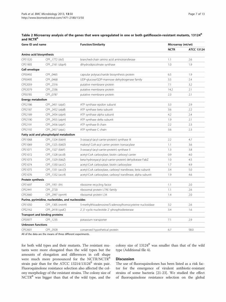
Table 2 Microarray analysis of the genes that were upregulated in one or both gatifloxacin-resistant mutants, 13124R
and NCTRR
Gene ID and name Function/Similarity Microarray (mt/wt)
NCTR ATCC 13124
Amino acid biosynthesis
CPE1520 CPF_1772 (ilvE) branched-chain amino acid aminotransferase 1.1 2.6
CPE1905 CPF_2161 (dapA) dihydrodipicolinate synthase 1.0 1.9
Cell envelope
CPE0492 CPF_0465 capsular polysaccharide biosynthesis protein 6.5 1.9
CPE0495 CPF_0468 UDP-glucose/GDP-mannose dehydrogenase family 3.5 2.4
CPE2059 CPF_2316 putative membrane protein 7.1 3.2
CPE2079 CPF_2336 putative membrane protein 14.2 2.1
CPE0785 CPF_0787 putative membrane protein 2.3 2.1
Energy metabolism
CPE2186 CPF_2451 (atpE) ATP synthase epsilon subunit 3.3 2.9
CPE2187 CPF_2452 (atpB) ATP synthase beta subunit 3.6 2.2
CPE2189 CPF_2454 (atpA) ATP synthase alpha subunit 4.2 2.4
CPE2190 CPF_2455 (atpH) ATP synthase delta subunit 1.9 2.1
CPE2191 CPF_2456 (atpF) ATP synthase B chain 2.2 2.3
CPE2192 CPF_2457 (atpL) ATP synthase C chain 3.6 2.3
Fatty acid and phospholipid metabolism
CPE1068 CPF_1324 (fabH) 3-oxoacyl-(acyl-carrier-protein) synthase III 2.2 4.7
CPE1069 CPF_1325 (fabD) malonyl CoA-acyl carrier protein transacylase 1.1 3.6
CPE1071 CPF_1327 (fabF) 3-oxoacyl-(acyl-carrier-protein) synthase II 1.3 3.8
CPE1072 CPF_1328 (accB) acetyl-CoA carboxylase, biotin carboxyl carrier 0.9 4.0
CPE1073 CPF_1329 (fabZ) beta-hydroxyacyl-(acyl-carrier-protein) dehydratase FabZ 1.0 4.5
CPE1074 CPF_1330 (accC) acetyl-CoA carboxylase, biotin carboxylase 1.7 4.9
CPE1075 CPF_1331 (accD) acetyl-CoA carboxylase, carboxyl transferase, beta subunit 3.4 5.0
CPE1076 CPF_1332 (accA) acetyl-CoA carboxylase, carboxyl transferase, alpha subunit 1.9 4.6
Protein synthesis
CPE1697 CPF_1951 (frr) ribosome recycling factor 1.1 2.0
CPE2441 CPF_2720 ribosomal protein L7AE family 1.1 2.6
CPE2660 CPF_2997 (rpmH) ribosomal protein L34 1.4 2.0
Purine, pyrimidine, nucleotides, and nucleosides
CPE1050 CPF_1305 (mtnH) 5-methylthioadenosine/S-adenosylhomocysteine nuclosidase 3.2 2.6
CPE2162 CPF_2418 (cpdC) 2`,3`-cyclic-nucleotide 2`-phosphodiesterase 3.4 1.6
Transport and binding proteins
CPE0977 CPF_1235 potassium transporter 7.1 2.9
Unknown functions
CPE2601 CPF_2928 conserved hypothetical protein 6.7 58.0
All of the data are the means of three different experiments.
Park et al. BMC Microbiology 2013, 13:50 Page 7 of 13http://www.biomedcentral.com/1471-2180/13/50
for both wild types and their mutants. The resistant mu-tants were more elongated than the wild types but theamounts of elongation and differences in cell shapewere much more pronounced for the NCTR/NCTRR
strain pair than for the ATCC 13214/13124R strain pair.Fluoroquinolone resistance selection also affected the col-ony morphology of the resistant strains. The colony size ofNCTRR was bigger than that of the wild type, and the
colony size of 13124R was smaller than that of the wildtype (Additional file 4).
DiscussionThe use of fluoroquinolones has been listed as a risk fac-tor for the emergence of virulent antibiotic-resistantstrains of some bacteria [21-23]. We studied the effectof fluoroquinolone resistance selection on the global

Table 3 Microarray analysis of the genes that were downregulated in both gatifloxacin-resistant strains, 13124R
and NCTRR
Gene ID (name) Function/Similarity Microarray (mt/wt)
NCTR ATCC 13124
Biosynthesis of cofactors, prosthetic groups, and carriers
CPE1085 CPF_1341 (ispH) 4-hydroxy-3-methylbut-2-enyl diphosphate reductase −2.4 −2.2
Energy metabolism
CPE0292 CPF_0288 carbohydrate kinase family protein −3.1 −2.5
CPE1185 CPF_1389 (pfk) 6-phosphofructokinase −1.7 −2.7
CPE0585 CPF_0565 (fruB) fructose-1-phosphate kinase −5.2 −2.3
CPE0692 CPF_0684 transaldolase −2.8 −2.3
CPE0725 CPF_0721 (nanI) * exo-alpha-sialidase −3.5 1.5
CPE0894 CPF_0887 (eutP) ethanolamine utilization protein, EutP −1.9 −2.0
CPE2348 CPF_2657 (ptb) phosphate butyryltransferase −2.3 −1.6
Purine, pyrimidine, nucleotides, and nucleosides
CPE1398 CPF_1652 (deoD) purine nucleoside phosphorylase −1.7 −3.4
Regulatory functions
CPE0586 CPF_0566 (fruR) transcriptional regulator, DeoR family −3.6 −2.6
CPE0631 CPF_0612 probable PBP5 synthesis regulator protein −2 −2.5
CPE1077 CPF_1333 transcriptional regulator, PadR family −3.1 −3.2
CPE2510 CPF_2833 transcriptional regulator, PadR family −2.6 −2.7
CPE1305 CPF_1512 probable transcriptional regulator −2 −1.6
Transport and binding proteins
CPE0600 CPF_0581 amino acid ABC transporter −4.8 −3.4
CPE1534 CPF_1785 PTS system, sucrose-specific IIBC component −3.1 −14.3
CPE2345 CPF_2654 putative maltose/maltodextrin ABC transporter −2.0 −1.8
Unknown functions
CPE2509 CPF_2832 degV family protein −3.6 −3.3
CPE1171 CPF_1374 mutator mutT protein homolog −6.4 −2.0
CPE2592 CPF_2917 phnA family protein −2. 8 −2.0* Decrease in the expression of nanI in NCTRR and increase of its expression in 13124R was confirmed by qRT-PCR.All of the data are the means of three different experiments.
Park et al. BMC Microbiology 2013, 13:50 Page 8 of 13http://www.biomedcentral.com/1471-2180/13/50
transcriptional response in gatifloxacin-resistant C.perfringens strains 13124R and NCTRR by microarray ana-lysis. The fluoroquinolone resistance selection resulted inalteration of transcription levels of a significant number ofgenes involved in almost every aspect of metabolism inthe resistant mutants of both strains in comparison with
Table 4 Results of qRT-PCR for the C. perfringens regulatory g
Gene ID and name Regulat
CPE_1501 CPF_1752 (virR) DNA binding response re
CPE_1500 CPF_1751 (virS) sensor histidine kinase, V
CPE_0646 CPF_0627 (virX) conserved hypothetical p
CPE_0957 CPF_1204 (vrr) VR-RNA
CPE_1701 CPF_1955 (codY) GTP-sensing transcription
CPE_0073 CPF_0069 Transcription antitermina
CPE_0642 CPF_0623 (RevR) DNA binding response re
their wild types. Many genes with similar functions were ei-ther upregulated or downregulated in the resistant mutants.However, some genes that were downregulated in 13124R
were upregulated in NCTRR. qRT-PCR analysis confirmedthat the transcription of these genes, which included toxingenes for phospholipase C (PLC), perfringolysin O (PFO),
enes in the wild types and mutants
ory function qRT-PCR fold
change (mt/wt)
NCTR ATCC13124
gulator, VirR 7.4 1.3
irS 9.7 0.3
rotein 2.2 −3.0
2.0 −158.5
al pleiotropic repressor CodY 6.9 −1.8
tor 1.5 −116.5
gulator 2 −2

A
B
0
0.5
1
1.5
2
2.5
3
3.5
4
4.5
5
13124 NCTRStrains
A54
0/A
600
of
cell
den
sity
w
m
0
0.1
0.2
0.3
0.4
0.5
0.6
0.7
0.8
13124 NCTRStrains
A41
0/A
600
of
cell
den
sity
w
m
Figure 1 Comparison of phospholipase C (A) and perfringolysin O (B) activities of the wild type strains of C. perfringens, ATCC 13124and NCTR, with their respective mutants, 13124R and NCTRR. W: wild type, M: mutant.
Park et al. BMC Microbiology 2013, 13:50 Page 9 of 13http://www.biomedcentral.com/1471-2180/13/50
collagenase and clostripain, were affected differently in thetwo mutants. Similarly, the production of these enzymesand the toxicity of the culture supernatants decreased in13124R and increased in NCTRR. It appears that gati-floxacin resistance selection resulted in alteration of globalgene transcription in C. perfringens and that the effect wasstrain-specific.The changes in the levels of global gene expression due
to the response to fluoroquinolone exposure may begoverned by complex regulatory processes. Both resistantstrains harbored some common and some unique muta-tions in fluoroquinolone target genes. These enzymes areinvolved in the DNA supercoiling process that plays an es-sential role during gene transcription [38,39]. Althoughneither of the resistant strains was a clinical isolate, someof the mutations found in the resistant strains were thesame as those found in fluoroquinolone-resistant mutantsof E. coli obtained from clinical samples, which were alsothe same as those found in fluoroquinolone-resistant mu-tants of E. coli generated in the laboratory [29,40].The expression of a number of genes is affected by
supercoiling [19] and aberrant expression of these genesoccurs when DNA supercoiling has been altered by gyrase
mutation(s). Alleles of gyrA that reduce DNA supercoilinghave been shown to generate metabolic defects and re-duce fitness of gyrase mutant strains [38,41]. Furthermore,because fluoroquinolones are DNA-damaging agents, inaddition to inducing mutation in target genes, changingDNA superhelicity, they may also induce the expressionof DNA repair genes via the SOS response, which maylead to phenotypic changes [15,17-20]. Cirz et al. [15]characterized the global transcription response of S.aureus to ciprofloxacin and, among other changes, foundinduction of the SOS response, upregulation of the TCAcycle and downregulation of α-hemolysin and a leukocidinfamily toxin. The positive regulators of transcriptional re-sponses for stress and toxin genes were also downregulated[15]. In C. perfringens, although the expression of severalvirulence genes decreased in one resistant mutant (13124R),it increased in another (NCTRR). The transcription of vari-ous genes, including toxin genes, is regulated by virR andvirS [32,42,43]. VirS is a sensor histidine kinase, which au-tophosphorylates in response to extracellular signals, andVirR is a response regulator [32,42,43]. These two genes,along with vrr (which is an RNA regulator virR-RNA), areimplicated in controlling gene transcription [44] and were

0
20
40
60
80
100
13124 13124R NCTR NCTRR
% o
f LD
H r
elea
se
Figure 3 Comparison of cytotoxicity of two gatifloxacin-resistant C. perfringens mutant strains, 13124R and NCTRR, withtheir wild type parents, strains ATCC 13124 and NCTR, forperitoneal macrophages, as measured by LDH (lactatedehydrogenase) released.
B
A
0
0.05
0.1
0.15
0.2
0.25
0.3
0.35
0.4
13124 NCTR
A55
0/A
600
of
cell
den
sity
w
m
0
0.005
0.01
0.015
0.02
0.025
0.03
0.035
0.04
0.045
13124 NCTR
A25
3/A
600
of
cell
den
sity
w
m
C
0
0.05
0.1
0.15
0.2
0.25
0.3
0.35
0.4
0.45
0.5
NCTR13124
A59
5/A
600
of
cell
den
sity
w
m
Figure 2 Comparison of collagenase (A), clostripain (B) andsialidase (C) activities of the wild type strains of C. perfringens,ATCC 13124 and NCTR, with their respective mutants, 13124R
and NCTRR. W: wild type, M: mutant.
Park et al. BMC Microbiology 2013, 13:50 Page 10 of 13http://www.biomedcentral.com/1471-2180/13/50
upregulated in NCTRR. In 13124R, transcription of VirRdid not change, and virS and vrr were downregulated. Thegene vrr is directly regulated by VirR/VirS, and as a regula-tory RNA, controls transcription of 147 genes, includinghousekeeping and toxin genes, in C. perfringens [32,44].Obana et al. [45] showed that VR-RNA regulates the stabil-ity of colA mRNA by cleaving the transcript. The processedshorter colA transcript was more stable than the longer in-tact colA transcript. It is possible that among other fac-tors, downregulation of vrr in 13124R (−158) may have
contributed to a decrease in the level of transcription ofgenes. The vrr in NCTRR was upregulated twofold. virX isanother regulatory gene that, even in the absence of theVirR/VirS regulatory system, activates the transcription ofthe pfoA, plc and colA genes, and its overexpression resultsin the increased expression of toxin genes [44,46]. qRT-PCR results showed that the expression of this gene in-creased at least 2.2 times in NCTRR and decreased by −3.0in 13124R.Another regulatory gene whose expression was altered
in the mutants was revR, which was downregulated in13124R and upregulated in NCTRR. revR is a responseregulator that alters the transcription of 100 genes,including those for potential virulence factors, whichalso are regulated by (VirR/VirS), and those for cell wallmetabolism [47]. Hiscox et al. [47] found that a revRmutant of C. perfringens 13 was filamented. Gram stain-ing of the wild types and mutants of ATCC 13124 andNCTR showed that cells of both mutants werefilamented and longer than those of the wild types.Microarray and qRT-PCR analysis (Table 1) showed thatsome putative membrane protein genes were differen-tially expressed in the mutants and wild types of bothstrains.The amino acid sequences of the toxin genes and the
regulatory genes (virR/virS) in the mutants and wildtypes of both strains were identical, except that therewere two silent mutations in virR/virS in NCTRR, so theexpression of toxin genes and their regulators was notthe result of gene mutation. The sequence of vrr wasidentical in the mutants and wild types of both strains,and the sequence of revR in ATCC 13124 and 13124R
was also identical. Obana and Nakamura [48] alsodetected other regulatory genes, CPE_1446-CPE_1447,which appear to regulate the transcription of plc, pfoA,

Park et al. BMC Microbiology 2013, 13:50 Page 11 of 13http://www.biomedcentral.com/1471-2180/13/50
nanI and nagHIJK at transcription level. Microarray ana-lysis showed that CPE_1447 was downregulated in NCTRR,but this gene was not detected in the microarray datafrom ATCC 13124. qRT-PCR confirmed that nanI wasdownregulated and sialidase was decreased in NCTRR;however, the role of CPE_1447 in the regulation of thisgene is not clear.Another global regulatory protein, CodY, has been
shown to regulate expression of many genes in Bacillussubtilis and Clostridium difficile [49,50]. It appears torepress genes whose products are not needed duringgrowth in high nutrient medium. qRT-PCR showed thatCodY was upregulated (6.9 times) in NCTRR anddownregulated (−1.89 times) in 13124R. The sequenceof codY was identical in both ATCC 13124 and 13124R.Since both mutants and wild types were grown in arich medium, the effect of CodY on alteration of geneexpression in our strains is not known.In addition, microarray analysis also detected some
regulatory genes that were downregulated in both mu-tants (Table 3) and some that were upregulated inNCTRR and downregulated in 13124R (Table 1). Amongthose genes that were affected differently was CPF_0069,which is a transcription antiterminator similar to theBglG-type regulators in other bacteria (http://www.ncbi.nlm.nih.gov/). This gene was downregulated in 13124R
and upregulated in NCTRR. At this point, the roles thatthis gene and others play in altering the transcription oftoxin genes in resistant strains are not known. Nor isthere a reason known for the contradictory effects offluoroquinolone resistance selection on the expressionof regulatory genes, including those that regulate toxinproduction, and it needs to be investigated further.Autoinducers (AI-2) also have been implicated in theregulation of some toxin genes [51]. However, in ourstrains, the production of AI-2 per cell unit, measuredby the indicator Vibrio harveyi, was higher for 13124R
than for ATCC 13124 and lower for NCTRR than forNCTR. The ratio of AI-2 production per OD unit in anovernight culture of the mutant to that of the wild typewas 1.5 for ATCC 13124 and 0.14 for NCTR. Thecontradictory results observed in the transcription ofvarious toxin genes in two resistant strains were ac-companied by changes in the levels of toxins and otherenzymes. The most dramatic changes were observedfor phospholipase C (PLC) and perfringolysin O (PFO).These two toxins were substantially decreased in13124R and increased in NCTRR. The alterations in theproduction of enzymes were accompanied by changesin cytotoxicity for macrophages. The cytotoxicities ofcell-free culture supernatants of the wild type ATCC13124 and NCTR, for the macrophages were comparable.However, the cell-free culture supernatant of 13124R exhi-bited significantly lower cytotoxicity for macrophages than
ATCC 13124, but that of NCTRR had higher cytotoxicitythan NCTR. These data were consistent with the alter-ations in the transcription patterns of toxin genes andenzyme assays that were observed by DNA microarrayanalysis, qRT-PCR assay and toxin production. Thecytotoxic effects were correlated with the transcriptionpattern of toxins and virulence-associated genes andenzymatic activities, confirming that the effect of fluoro-quinolones on C. perfringens was strain-specific. O’Brienand Melville [33] reported that perfringolysin O (PFO)plays a more prominent role than α-toxin (PLC) in cytotox-icity for macrophages. Since we used the crude extract,which contains various factors including PFO and PLC, ourresults only show the alteration in the overall cytotoxicitiesof the mutants in comparison with their wild types and thecontributing factors and their affinities for macrophage re-ceptors are not known. Fluoroquinolone resistance selec-tion decreased the toxicity of 13124R and increased thetoxicity of NCTRR.
ConclusionsOur study demonstrates that gatifloxacin resistance selec-tion in C. perfringens was associated with upregulation ordownregulation of different genes involved in various as-pects of metabolism and that the effect was strain-specific.The genes involved in transcription regulation, virulenceand cell toxicity were among those that were upregulatedin one resistant strain and downregulated in another.Hiscox et al. [47] surmised that “the regulation of viru-lence in C. perfringens was a complex process” and wefound that the nature of each strain adds yet anotherlevel of complexity to gene regulation in C. perfringens.Myer et al. [52] found widely variable large genomicislands in a large collection of C. perfringens strains andstated that considerable variation exists among thegenomes of C. perfringens strains. It appears that thisvariation in gene structure of different C. perfringensstrains also affects gene regulation and interaction ofbacteria with fluoroquinolones. Fluoroquinolones havebeen implied to have a role in the development of C.difficile associated diarrhea [53]. Since virulent, drug-resistant clinical isolates of pathogenic bacteria have an un-defined genetic basis for their resistance and virulence, weused two wild types and otherwise isogenic resistant mu-tants, which are difficult to obtain in a clinical setting, toassess fluoroquinolone effects. Our results reflect clinicalobservations of finding fluoroquinolone-resistant strains ofbacteria that are more or less virulent than the susceptiblestrains. They underscore the role of fluoroquinolones inchanging bacterial virulence and the importance of prudentuse of fluoroquinolones. Further study is needed on theeffect of fluoroquinolones on a larger number of C.perfringens strains, along with genomic analysis of the re-sistant mutants.

Park et al. BMC Microbiology 2013, 13:50 Page 12 of 13http://www.biomedcentral.com/1471-2180/13/50
Additional files
Additional file 1: Primers used for qRT-PCR.
Additional file 2: Analysis of mRNA quality and expression.
Additional file 3: Cytotoxicities of C. perfringens supernatants formacrophages.
Additional file 4: Morphological examination of C. perfringens strains.
AbbreviationsPLC: Phospholipase C; PFO: Perfringolysin O; gyrA and gyrB: Gyrase; parC andparE: Topoisomerase IV; BHI: Brain Heart Infusion; VirS, VirR, virX, vrr(VR-RNA),Cody RevR: Regulatory genes.
Competing interestsThe authors declare that they have no competing interests.
Authors’ contributionsTechnical experiments and statistical analysis were performed by MP and SP.SP performed those on RT-PCR and cytotoxicity, morphological analysis andMP performed the rest of the experiments. SP wrote the first draft of themanuscript sections on RT-PCR analysis, cytotoxicity and cell morphology. FRplanned the experiments, analyzed the data, and wrote the manuscript. Allauthors have read and approved the final manuscript.
AcknowledgmentsWe thank Drs. Mark Hart and John B. Sutherland for their helpful commentson the manuscript, Dr. Carl E. Cerniglia for support of research and Drs.Donald Schwartz and Jean-Marie Rouillard for DNA microarray experiments.S.P. was supported by the FDA Commissioner’s Fellowship Program. Theviews presented in this article do not necessarily reflect those of the USFood and Drug Administration.
Author details1Division of Microbiology, National Center for Toxicological Research, USFood and Drug Administration, Jefferson, AR, USA. 2Current Address; WO66RM2467, 10903 New Hampshire Ave, Silver Spring, MD 20993-0002, USA.
Received: 31 August 2012 Accepted: 18 February 2013Published: 1 March 2013
References1. Scallan E, Hoekstra RM, Angulo FJ, Tauxe RV, Widdowson MA, Roy SL, et al:
Foodborne illness acquired in the United States—major pathogens.Emerg Infect Dis 2011, 17:7–15.
2. Heimesaat MM, Granzow K, Leidinger H, Liesenfeld O: Prevalence ofClostridium difficile toxins A and B and Clostridium perfringensenterotoxin A in stool samples of patients with antibiotic-associateddiarrhea. Infection 2005, 33:340–344.
3. Meyns E, Vermeersch N, Ilsen B, Hoste W, Delooz H, Hubloue I:Spontaneous intrahepatic gas gangrene and fatal septic shock. Acta ChirBelg 2009, 109:400–404.
4. Rood JI: Virulence genes of Clostridium perfringens. Annu Rev Microbiol1998, 52:333–360.
5. Sparks SG, Carman RJ, Sarker MR, McClane BA: Genotyping ofenterotoxigenic Clostridium perfringens fecal isolates associated withantibiotic-associated diarrhea and food poisoning in North America.J Clin Microbiol 2001, 39:883–888.
6. Petit L, Gibert M, Popoff MR: Clostridium perfringens: toxinotype andgenotype. Trends Microbiol 1997, 179:104–110.
7. Titball RW, Hunter SE, Martin KL, Morris BC, Shuttleworth AD, Rubidge T, et al:Molecular cloning and nucleotide sequence of the alpha-toxin(phospholipase C) of Clostridium perfringens. Infect Immun 1989, 57:367–376.
8. Lazarescu C, Kimmoun A, Blatt A, Bastien C, Levy B: Clostridium perfringensgangrenous cystitis with septic shock and bone marrow necrosis.Intensive Care Med 2012, 38:1906–1907.
9. Gosselink MP, Schouten WR, van Lieshout LM, Hop WC, Laman JD, et al:Eradication of pathogenic bacteria and restoration of normal pouch
flora: comparison of metronidazole and ciprofloxacin in the treatment ofpouchitis. Dis Colon Rectum 2004, 47:1519–1525.
10. Gionchetti P, Rizzello F, Venturi A, Ugolini F, Rossi M, Brigidi P, et al:Antibiotic combination therapy in patients with chronic, treatment-resistant pouchitis. Aliment Pharmacol Ther 1999, 13:713–718.
11. Leal J, Gregson DB, Ross T, Church DL, Laupland KB: Epidemiology ofClostridium species bacteremia in Calgary, Canada, 2000–2006. J Infect2008, 5:198–203.
12. Wexler HM, Molitoris E, Finegold SM: In vitro activities of three of thenewer quinolones against anaerobic bacteria. Antimicrob AgentsChemother 1992, 36:239–234.
13. Ferrero L, Cameron B, Crouzet J: Analysis of gyrA and grlA mutations instepwise-selected ciprofloxacin-resistant mutants of Staphylococcusaureus. Antimicrob Agents Chemother 1995, 39:1554–1558.
14. Tamayo M, Santiso R, Gosalvez J, Bou G, Fernandez JL: Rapid assessment ofthe effect of ciprofloxacin on chromosomal DNA from Escherichia coliusing an in situ DNA fragmentation assay. BMC Microbiol 2009, 9:69.
15. Cirz RT, Jones MB, Gingles NA, Minogue TD, Jarrahi B, Peterson SN, et al:Complete and SOS-mediated response of Staphylococcus aureus to theantibiotic ciprofloxacin. J Bacteriol 2007, 189:531–539.
16. Dorr T, Lewis K, Vulic M: SOS response induces persistence tofluoroquinolones in Escherichia coli. PLoS Genet 2009, 5:e1000760.
17. Dorr T, Vulic M, Lewis K: Ciprofloxacin causes persister formation byinducing the TisB toxin in Escherichia coli. PLoS Biol 2010, 8:e1000317.
18. Dal Sasso M, Culici M, Bovio C, Braga PC: Gemifloxacin: effects of sub-inhibitory concentrations on various factors affecting bacterial virulence.Int J Antimicrob Agents 2003, 21:325–333.
19. Dorman CJ, Ni Bhriain N, Higgins CF: DNA supercoiling and environmentalregulation of virulence gene expression in Shigella flexneri. Nature 1990,344:789–792.
20. Mesak LR, Davies J: Phenotypic changes in ciprofloxacin-resistantStaphylococcus aureus. Res Microbiol 2009, 160:785–791.
21. Muto CA, Pokrywka M, Shutt K, Mendelsohn AB, Nouri K, Posey K, et al: Alarge outbreak of Clostridium difficile-associated disease with anunexpected proportion of deaths and colectomies at a teaching hospitalfollowing increased fluoroquinolone use. Infect Control Hosp Epidemiol2005, 26:273–280.
22. Noren T: Clostridium difficile and the disease it causes. Methods Mol Biol2010, 646:9–35.
23. Pawlowski SW, Archbald-Pannone L, Carman RJ, Alcantara-Warren C, LyerlyD, Genheimer CW, et al: Elevated levels of intestinal inflammation inClostridium difficile infection associated with fluoroquinolone-resistant C.difficile. J Hosp Infect 2009, 73:185–187.
24. Saxton K, Baines SD, Freeman J, O'Connor R, Wilcox MH: Effects ofexposure of Clostridium difficile PCR ribotypes 027 and 001 tofluoroquinolones in a human gut model. Antimicrob Agents Chemother2009, 53:412–420.
25. Uchida Y, Mochimaru T, Morokuma Y, Kiyosuke M, Fujise M, Eto F, et al:Clonal spread in Eastern Asia of ciprofloxacin-resistant Escherichia coliserogroup O25 strains, and associated virulence factors. Int J AntimicrobAgents 2010, 35:444–450.
26. Drews SJ, Poutanen SM, Mazzulli T, McGeer AJ, Sarabia A, Pong-Porter S,et al: Decreased prevalence of virulence factors among ciprofloxacin-resistant uropathogenic Escherichia coli isolates. J Clin Microbiol 2005,43:4218–4220.
27. Ferjani S, Saidani M, Ennigrou S, Hsairi M, Ben Redjeb S: Virulencedeterminants, phylogenetic groups and fluoroquinolone resistance inEscherichia coli isolated from cystitis and pyelonephritis. Pathol Biol (Paris)2012, 60:270–274.
28. Sun J, Hu J, Peng H, Shi J, Dong Z: Molecular and physiologicalcharacterization of fluoroquinolone resistance in relation touropathogenicity among Escherichia coli isolates isolated from WenyuRiver, China. Chemosphere 2012, 87:37–42.
29. Rafii F, Park M, Novak JS: Alterations in DNA gyrase and topoisomerase IV inresistant mutants of Clostridium perfringens found after in vitro treatmentwith fluoroquinolones. Antimicrob Agents Chemother 2005, 49:488–492.
30. Rafii F, Park M, Bryant AE, Johnson SJ, Wagner RD: Enhanced production ofphospholipase C and perfringolysin O (alpha and theta toxins) in agatifloxacin-resistant strain of Clostridium perfringens. Antimicrob AgentsChemother 2008, 52:895–900.
Park et al. BMC Microbiology 2013, 13:50 Page 13 of 13http://www.biomedcentral.com/1471-2180/13/50
31. Rafii F, Park M, Gamboa da Costa G, Camacho L: Comparison of themetabolic activities of four wild-type Clostridium perfringens strains withtheir gatifloxacin-selected resistant mutants. Arch Microbiol 2009,191:895–902.
32. Ohtani K, Hirakawa H, Tashiro K, Yoshizawa S, Kuhara S, Shimizu T:Identification of a two-component VirR/VirS regulon in Clostridiumperfringens. Anaerobe 2010, 16:258–264.
33. O'Brien DK, Melville SB: Effects of Clostridium perfringens alpha-toxin (PLC)and perfringolysin O (PFO) on cytotoxicity to macrophages, on escapefrom the phagosomes of macrophages, and on persistence of C.perfringens in host tissues. Infect Immun 2004, 72:5204–5215.
34. Awad MM, Ellemor DM, Bryant AE, Matsushita O, Boyd RL, Stevens DL, et al:Construction and virulence testing of a collagenase mutant ofClostridium perfringens. Microb Pathog 2000, 28:107–117.
35. Dargatz H, Diefenthal T, Witte V, Reipen G, von Wettstein D: Theheterodimeric protease clostripain from Clostridium histolyticum isencoded by a single gene. Mol Gen Genet 1993, 240:140–145.
36. Li J, Sayeed S, Robertson S, Chen J, McClane BA: Sialidases affect the hostcell adherence and epsilon toxin-induced cytotoxicity of Clostridiumperfringens type D strain CN3718. PLoS Pathog 2011, 7:e1002429.
37. Song JM, Im JH, Hoon JH, Kang JD, Kang DJ: A simple method for hyaluronicacid quantification in culture broth. Carbohydr Polym 2009, 78:633–634.
38. Kugelberg E, Lofmark S, Wretlind B, Andersson DI: Reduction of the fitnessburden of quinolone resistance in Pseudomonas aeruginosa. J AntimicrobChemother 2005, 55:22–30.
39. Marcusson LL, Frimodt-Moller N, Hughes D: Interplay in the selection offluoroquinolone resistance and bacterial fitness. PLoS Pathog 2009, 5:e1000541.
40. Bachoual R, Tankovic J, Soussy CJ: Analysis of the mutations involved influoroquinolone resistance of in vivo and in vitro mutants of Escherichiacoli. Microb Drug Resist 1998, 4:271–276.
41. Smani Y, Lopez-Rojas R, Dominguez-Herrera J, Docobo-Perez F, Marti S, VilaJ, et al: In vitro and in vivo reduced fitness and virulence in ciprofloxacin-resistant Acinetobacter baumannii. Clin Microbiol Infect 2012, 18:1–4.
42. Shimizu T, Shima K, Yoshino K, Yonezawa K, Hayashi H: Proteome andtranscriptome analysis of the virulence genes regulated by the VirR/VirSsystem in Clostridium perfringens. J Bacteriol 2002, 184:2587–2594.
43. Shimizu T, Yaguchi H, Ohtani K, Banu S, Hayashi H: Clostridial VirR/VirSregulon involves a regulatory RNA molecule for expression of toxins.Mol Microbiol 2002, 43:257–265.
44. Okumura K, Ohtani K, Hayashi H, Shimizu T: Characterization of genesregulated directly by the VirR/VirS system in Clostridium perfringens.J Bacteriol 2008, 190:7719–7727.
45. Obana N, Shirahama Y, Abe K, Nakamura K: Stabilization of Clostridiumperfringens collagenase mRNA by VR-RNA-dependent cleavage in 5'leader sequence. Mol Microbiol 2010, 77:1416–1428.
46. Ohtani K, Bhowmik SK, Hayashi H, Shimizu T: Identification of a novel locusthat regulates expression of toxin genes in Clostridium perfringens. FEMSMicrobiol Lett 2002, 209:113–118.
47. Hiscox TJ, Chakravorty A, Choo JM, Ohtani K, Shimizu T, Cheung JK, et al:Regulation of virulence by the RevR response regulator in Clostridiumperfringens. Infect Immun 2011, 79:2145–2153.
48. Obana N, Nakamura K: A novel toxin regulator, the CPE1446-CPE1447protein heteromeric complex, controls toxin genes in Clostridiumperfringens. J Bacteriol 2011, 193:4417–4424.
49. Brinsmade SR, Sonenshein AL: Dissecting complex metabolic integrationprovides direct genetic evidence for CodY activation by guaninenucleotides. J Bacteriol 2011, 193:5637–5648.
50. Dineen SS, McBride SM, Sonenshein AL: Integration of metabolism andvirulence by Clostridium difficile CodY. J Bacteriol 2010, 192:5350–5362.
51. Ohtani K, Yuan Y, Hassan S, Wang R, Wang Y, Shimizu T: Virulence generegulation by the agr system in Clostridium perfringens. J Bacteriol 2009,191:3919–3927.
52. Myers GS, Rasko DA, Cheung JK, Ravel J, Seshadri R, DeBoy RT, et al:Skewed genomic variability in strains of the toxigenic bacterialpathogen, Clostridium perfringens. Genome Res 2006, 16:1031–1040.
53. Deshpande A, Pant C, Jain A, Fraser TG, Rolston DD: Do fluoroquinolonespredispose patients to Clostridium difficile associated disease? A reviewof the evidence. Curr Med Res Opin 2008, 24:329–333.
doi:10.1186/1471-2180-13-50Cite this article as: Park et al.: Comparative transcription analysis andtoxin production of two fluoroquinolone-resistant mutants ofClostridium perfringens. BMC Microbiology 2013 13:50.
Submit your next manuscript to BioMed Centraland take full advantage of:
• Convenient online submission
• Thorough peer review
• No space constraints or color figure charges
• Immediate publication on acceptance
• Inclusion in PubMed, CAS, Scopus and Google Scholar
• Research which is freely available for redistribution
Submit your manuscript at www.biomedcentral.com/submit