NASA Microbiology Workshop - CORE
174
NASA/CP—2012–217460 NASA Microbiology Workshop M.C. Roman, Chair Marshall Space Flight Center, Huntsville, Alabama D.L. Jan, Vice-Chair Jet Propulsion Laboratory, Pasadena, California K.T. Konstantinidis, Facilitator Georgia Institute of Technology, Atlanta, Georgia M.W. Mittelman, Facilitator Harvard University, Cambridge, Massachusetts June 2012 Proceedings of a Workshop sponsored by the National Aeronautics and Space Administration held at Johnson Space Center, Houston, Texas, April 19, 2011 https://ntrs.nasa.gov/search.jsp?R=20130009194 2019-08-30T23:55:25+00:00Z
-
Upload
khangminh22 -
Category
Documents
-
view
1 -
download
0
Transcript of NASA Microbiology Workshop - CORE
NASA/CP—2012–217460
NASA Microbiology WorkshopM.C. Roman, ChairMarshall Space Flight Center, Huntsville, Alabama
D.L. Jan, Vice-ChairJet Propulsion Laboratory, Pasadena, California
K.T. Konstantinidis, FacilitatorGeorgia Institute of Technology, Atlanta, Georgia
M.W. Mittelman, FacilitatorHarvard University, Cambridge, Massachusetts
June 2012
National Aeronautics andSpace AdministrationIS20George C. Marshall Space Flight CenterHuntsville, Alabama 35812
Proceedings of a Workshop sponsored by theNational Aeronautics and Space Administrationheld at Johnson Space Center, Houston, Texas,April 19, 2011
https://ntrs.nasa.gov/search.jsp?R=20130009194 2019-08-30T23:55:25+00:00Z

The NASA STI Program…in Profile
Since its founding, NASA has been dedicated tothe advancement of aeronautics and spacescience. The NASA Scientific and Technical Information (STI) Program Office plays a keypart in helping NASA maintain this importantrole.
The NASA STI Program Office is operated by Langley Research Center, the lead center for NASA’s scientific and technical information. The NASA STI Program Office provides access to the NASA STI Database, the largest collection of aeronautical and space science STI in the world. The Program Office is also NASA’s institutional mechanism for disseminating the results of its research and development activities. These results are published by NASA in the NASA STI Report Series, which includes the following report types:
• TECHNICAL PUBLICATION. Reports of completed research or a major significant phase of research that present the results of NASA programs and include extensive data or theoretical analysis. Includes compilations of significant scientific and technical data and information deemed to be of continuing reference value. NASA’s counterpart of peer-reviewed formal professional papers but has less stringent limitations on manuscript length and extent of graphic presentations.
• TECHNICAL MEMORANDUM. Scientific and technical findings that are preliminary or of specialized interest, e.g., quick release reports, working papers, and bibliographies that contain minimal annotation. Does not contain extensive analysis.
• CONTRACTOR REPORT. Scientific and technical findings by NASA-sponsored contractors and grantees.
• CONFERENCE PUBLICATION. Collected papers from scientific and technical conferences, symposia, seminars, or other meetings sponsored or cosponsored by NASA.
• SPECIAL PUBLICATION. Scientific, technical, or historical information from NASA programs, projects, and mission, often concerned with subjects having substantial public interest.
• TECHNICAL TRANSLATION. English-language translations of foreign
scientific and technical material pertinent to NASA’s mission.
Specialized services that complement the STI Program Office’s diverse offerings include creating custom thesauri, building customized databases, organizing and publishing research results…even providing videos.
For more information about the NASA STI Program Office, see the following:
• Access the NASA STI program home page at <http://www.sti.nasa.gov>
• E-mail your question via the Internet to <[email protected]>
• Fax your question to the NASA STI Help Desk at 443 –757–5803
• Phone the NASA STI Help Desk at 443 –757–5802
• Write to: NASA STI Help Desk NASA Center for AeroSpace Information 7115 Standard Drive Hanover, MD 21076–1320

i
NASA/CP—2012–217460
NASA Microbiology WorkshopM.C. Roman, ChairMarshall Space Flight Center, Huntsville, Alabama
D.L. Jan, Vice-ChairJet Propulsion Laboratory, Pasadena, California
K.T. Konstantinidis, FacilitatorGeorgia Institute of Technology, Atlanta, Georgia
M.W. Mittelman, FacilitatorHarvard University, Cambridge, Massachusetts
June 2012
National Aeronautics andSpace Administration
Marshall Space Flight Center • Huntsville, Alabama 35812
Proceedings of a Workshop sponsored by theNational Aeronautics and Space Administrationheld at Johnson Space Center, Houston, Texas,April 19, 2011

ii
Available from:
NASA Center for AeroSpace Information7115 Standard Drive
Hanover, MD 21076 –1320443 –757– 5802
This report is also available in electronic form at<https://www2.sti.nasa.gov/login/wt/>
TRADEMARKS
Trade names and trademarks are used in this report for identification only. This usage does not constitute an official endorsement, either expressed or implied, by the National Aeronautics and Space Administration.

iii
EXECUTIVE SUMMARY
Long-term spaceflight is characterized by extraordinary challenges to maintain the life- supporting instrumentation free from microbial contamination and the crew healthy. The method-ology currently employed for microbial monitoring in space stations or short spaceflights within the orbit of Earth have been instrumental in safeguarding the success of the missions, but suffers certain shortcomings that are critical for long spaceflights. To discuss alternative methodologies and technologies suitable for microbial monitoring in long-term missions, a workshop was orga-nized at the Johnson Space Center by Monserrate Roman (NASA) with help from Dr. Marc Mittelman (Exponent and Harvard University) and Dr. Kostas Konstantinidis (Georgia Tech).
Invited speakers with expertise in environmental microbiology, infectious diseases, pathogen tracking and monitoring, food safety, and industry discussed the available cutting-edge technolo-gies that hold promise for NASA missions. This Conference Publication aims at summarizing the discussions and findings of the workshop. Although it appears that no technology from those cur-rently available represents a “silver bullet solution” to the needs of long-term spaceflights, several technologies offer significant advantages over the current practice. At least some of the technolo-gies, when optimized for the special needs and conditions of the spacecraft such as microgravity conditions, can represent robust and cost-effective means to maintain the health of the crew and the spacecraft environment. In particular, it is proposed that traditional culture-based approaches, which dominate the current practice, should be replaced or at least supplemented with modern molecular approaches, which provide both greater accuracy and sensitivity. The modern molecu-lar methods should be validated using the current culture-based practice as a baseline metric. The validation protocols established by the food industry, which lead the development of new monitor-ing techniques, may be useful in this regard. These amendments to current practice are expected to have significant benefits for the crew and cost savings for NASA.

iv
v
TABLE OF CONTENTS
1. INTRODUCTION ............................................................................................................. 1
2. CURRENT PRACTICE AND CHALLENGES ............................................................... 4
3. PANEL MEMBERS, TOPICS DISCUSSED, AND RECOMMENDATIONS ................ 6
3.1 Panel Members and Topics Discussed ........................................................................... 6 3.2 Summary of Panel Recommendations .......................................................................... 7
4. TECHNOLOGIES DISCUSSED ....................................................................................... 8
4.1 Culture-Independent Nucleic Acid Technologies (Polymerase Chain Reaction-Based) ............................................................................................................ 8 4.2 Quantitative Biochemical Methods: Adenosine Triphosphate Bioluminescence ........... 9 4.3 Biosensors, Direct Laser-Based Detection .................................................................... 9 4.4 Flow Cytometry Methods ............................................................................................. 11 4.5 Matrix-Assisted Laser Desorption/IonizationTime of Flight ........................................ 11 4.6 Microscopic Methods ................................................................................................... 11 4.7 Protocols for Validating and Comparing Technologies ................................................. 12
5. CONCLUDING REMARKS ............................................................................................ 14
APPENDIX A—SURVEY OF MICROBIAL MONITORING NEEDS ............................... 15
APPENDIX B—MICROBILOGICAL MONITORING IN SPACE (DR. MITTELMAN’S REPORT) ................................................................ 19
APPENDIX C—BIOGRAPHY AND ABSTRACT OF WORKSHOP PRESENTERS ....... 51
APPENDIX D—WORKSHOP ATTENDEES, AGENDA, AND PRESENTATIONS ........ 65
REFERENCES ....................................................................................................................... 161

vi
LIST OF FIGURES
1. The SmartCycler® system from Cepheid. The system allows up to 96 independently programmable reactions to take place simultaneously, using different protocols. Multiple experimental runs can be started at different times, allowing several operators to use the system concurrently ............... 9
2. The BioVigilant IMD-A® 220-4 system from Azbil. With an air sampling capacity of 28.3 L/min and stainless steel, chemically resistant case, the IMD-A 220-4 is suitable for the most demanding cleanroom environments and larger testing areas ................................................................................................ 10
LIST OF TABLES
1. Suggested properties/traits that technologies should be evaluated for .......................... 12
2. Preliminary evaluations of modern technologies for long-term spaceflight .................. 13

vii
LIST OF ACRONYMS AND ABBREVIATIONS
AOAC Association of Analytical Communities
ATP adenosine triphosphate
CFU colony-forming units
CP Conference Publication
ECLSS Environmental Control Life Support System
ISS International Space Station
MALDI matrix-assisted laser desorption/ionization
MS mass spectrometry
PCR polymerase chain reaction
QMRA Quantitative Microbial Risk Assessment
RT qPCR real-time quantitative polymerase chain reaction
SOA state of the art
TOF time of flight

viii

1
CONFERENCE PUBLICATION
NASA MICROBIOLOGY WORKSHOPAPRIL 19, 2011
1. INTRODUCTION
Humans have been exploring space for more than 40 years. For all those years, microorgan-isms have accompanied both unmanned spacecraft/cargo and manned vessels. Microorganisms are everywhere on Earth, could easily adapt to new environments, and/or can rapidly mutate to survive in very harsh conditions. Their presence in spacecraft and cargo have caused a few inconveniences over the years of human spaceflight, ranging from crew health, life support systems challenges, and material degradation. The sterilization of spacecraft that will host humans in long-duration missions would be a costly operation that will not provide a long-term solution to the microbial colonization of the vessels. As soon as a human is exposed to the spacecraft, microorganisms start populating the new environment during the mission. As the human presence in space increases in length, the risk from the microbial load to hardware and crew will also increase. Mitigation of this risk involves several different strategies that will include minimizing the microbial load (in numbers and diversity) and monitoring.
The ability to produce and maintain spacecraft and habitats with environments suitable for human habitation has been established with data from over 50 years of human spaceflight mis-sions. More than 100 missions aboard the Space Shuttle have provided NASA with an extensive microbial database for short-term (<20 days) space flights. There is no question that microorgan-isms will survive, adapt, and flourish, even during short-term missions, in the closed environment of a spacecraft. Data collected from the inside of the Space Shuttle after landing (air, water, and surfaces) show that there is an increase of microorganisms, compared to samples before launch. Short-term missions, like the Space Shuttle, provide us with the opportunity to characterize the microbial population in the vehicle with minimal in-flight equipment. Due to the short duration of the missions, samples can be archived and returned to Earth for analysis in well-equipped laborato-ries. Extensive analysis of the inside of the vehicle can be performed, if needed, after landing. This information has given NASA a “peek” at the microbial population inside a closed environmental system in microgravity, but it provides limited information about how microorganisms will behave in long-duration missions.
The NASA Mir Program presented a first opportunity for observation that provided data for long-duration missions. From this experience, we know that the major bacteria and fungal species found in the Mir (after more than 15 years in service), were similar to those found in the Space Shuttle. It was reported that infection from the crew’s normal microbiological flora has been

2
a problem in the past. For example, staphylococcal and streptococcal skin infections and urinary tract infections with Echerichia coli etiology have been documented. Microbial growth caused performance problems in the Mir life support systems, including clogging in the tube that transported cabin humidity condensate from where it is condensed to where it was stored for later processing. Severe material degradation caused by uncontrolled microbial growth was also documented in the Mir.
Over the past several years with the permanent presence of humans in space, the Interna-tional Space Station (ISS) has provided additional opportunities to study microbial growth and related affects on Environmental Control Life Support Systems (ECLSSs) and astronaut crews for long-duration missions. Such opportunities have highlighted the importance and need for the development of automated biological in-flight monitoring methods and techniques that do not rely on potentially toxic chemicals and/or time-consuming steps and/or power-consuming hard-ware. The desirable capabilities that a real-time effective microbial monitor must have include quantification and identification of viruses, bacteria, and fungi. Also, recognition is given to the fact that, presently, there are many challenges that a microbial monitor developer will have to address in order for the technology to be useful in a vehicle/habitat environment and to provide feedback on the performance of the ECLSS. Another area of primary importance is Quantitative Microbial Risk Assessment (QMRA), a framework that has been used by the water (and food) industry to predict the health consequences from environmental exposures to pathogens. QMRA has been extremely useful in developing guidelines for microbial water quality, evaluate, and com-pare the public health impacts of different water treatment technologies. QMRA can be applied in a variety of settings, including the ISS to determine the likelihood of microbial exposure and the consequence(s) to the flight crew.
Considerable progress has been made in the monitoring and control of microorganisms, thus improving the quality of life for onboard astronauts who are tasked with accomplishing important scientific research and other technical mission-related functions. However, findings to date have indicated the need for continuous improvement in many applicable areas of microbial control including monitoring, detection, and risk mitigation, especially in support of long-duration space missions. As a precursor to this workshop, an Environmental Monitoring and Controls team, lead by JPL/Darrell Jan, funded by the Life Support and Habitation Systems Domain, asked the Marshall Space Flight Center to lead an effort to assess the state of the art (SOA) in microbial monitoring technologies currently in use within NASA. This work required that NASA, as an Agency, thoroughly understand the microbial monitoring needs inherent within the different NASA projects/programs. Two independent groups, with expertise in microbial monitoring, were asked to provide the Agency a list of technologies currently available, their Technology Readiness Level, and the probability that they can meet NASA’s needs. Accordingly, a survey form to assess the SOA of microbial monitoring technologies currently in use and customer needs was developed and distributed with responses requested. The customer survey results (see app. A) were very helpful in understanding diverse Agency requirements and needs and will be used to prioritize the technologies in preparation for future funding. Customers were chosen to complete this survey based on direct or indirect needs for microbial monitoring technologies, short term or long term. The responses are of utmost important to NASA and participants were invited to join the rest of the NASA microbial monitoring customers in the workshop in which the

3
independent groups (Harvard University and Georgia Tech) presented NASA their findings. This workshop appropriately focused on a cursory review of current practices including state-of-the-art microbiology methods, and instrumentation, environmental, and clinical microbiology needs. Related discussions, findings, and recommendations by experts from across the field including representatives within government, industry, and academia are adequately addressed and presented in this Conference Publication (CP).

4
2. CURRENT PRACTICE AND CHALLENGES
The challenges associated with long-term spaceflight and the advantages and limitations of the current technologies for microbial monitoring are discussed extensively in the report prepared by Dr. Mittelman (Exponent and Harvard University) prior to the workshop. Dr. Mittelman’s report is provided in appendix B and, where appropriate, the reader is directed to this appendix for more information. Accordingly, this CP touches on these issues only briefly, and instead, focuses more on the issues discussed during the workshop and the recommendations of the experts that participated in the workshop. See appendix C for a short biography and abstract.
Long-term spaceflight, on the order of months or even years of flight time, imposes several extraordinary challenges, most importantly, for the purposes of this CP, keeping the life-sustaining equipment, the air, and the water in the spacecraft free of microbial contamination and the crew free of microbial infections. In such long flights, however, it is expected that microbial contamina-tions and/or infections will unavoidably occur, resulting primarily from the microbes brought into the spacecraft with the crew (skin-associated but also gut-associated microflora) and the supplies/equipment. Hence, a system to robustly monitor microbial load and identify action in those cases where the load exceeds acceptable levels are necessary. Microbial contaminations frequently occur in space stations but the means available in the space stations and the immediate contact with the Earth (e.g., space station shuttles) render these contaminations relatively easy to treat and eradi-cate. Spacecraft does not have the same equipment as space stations and/or require lighter, less energy-demanding, and easier to use equipment. Therefore, the ideal microbial monitoring system for long-term spaceflights should also be autonomous, as simple and durable as possible, and user friendly, particularly with respect to reading and interpreting its output.
Currently, microbial monitoring in space stations primarily involves enumerating total bacterial and fungi cells and total coliform bacteria by culturing cells on broad specificity and coliform-specific media, respectively (D. Pierson, Personal Communication, and presentation in app. D). Samples are typically taken from air, water, and surfaces of the space station at regular intervals (e.g., every 3 months) and if microbial cell counts exceed specific limits (e.g., 50 colony-forming units (CFU) per milliliter for total bacteria and zero CFU for coliform bacteria), then specific decontamination actions are taken. These may include application of disinfectants (e.g., quaternary ammonia compounds for surfaces; hydrogen peroxide for water). Although the culture-based approaches provide valuable information for microbial contamination, they are character-ized by several limitations that are critical for long-term spaceflights. The experts in the workshop highlighted a number of limitations. Perhaps most importantly, the great majority of microbial cells in any natural environment, and the spacecraft should not represent an exception to this rule,1 are resistant to laboratory cultivation (the “unculturable majority”), and thus are missed by the culture-based approaches mentioned above.2,3 Even microorganisms that are typically easily cul-tured, such as Escherichia coli, lose “culturability” after prolonged incubation under different con-ditions than the culture conditions or when growing in natural habitats.4 Furthermore, a 3-month
5
sampling interval (current practice in space stations), although optimum from a practical per-spective, may represent a too long period of time for successful intervention in cases of contami-nation or crew infection; an online (real time) system is clearly preferable. It is also important to mention that the current methods do not provide any information about which microorganisms are responsible for contamination, since species identification is taking place in the laboratory facilities on Earth. Thus, the intervention actions on board are typically delayed or limited to general, non-specific antimicrobial measures, which may not be efficient or even necessary. Identifying the specific microbial culprits on board, particularly those causing crew infections, is important to decide the best treatment or antibiotic to use.

6
3. PANEL MEMBERS, TOPICS DISCUSSED, AND RECOMMENDATIONS
3.1 Panel Members and Topics Discussed
The experts that participated in the workshop were as follows; a short biography and an abstract of their presentation is provided in appendix C, and their presentations in appendix D:
Dr. Duane L. Pierson, Chief Microbiologist, NASA, Houston, TX. Dr. Pierson talked about the current practice of microbiological monitoring at the ISS and the additional challenges associ-ated with long-term spaceflights.
Dr. Kostas Konstantinidis, Assistant Professor, Georgia Institute of Technology, Atlanta, GA. Dr. Konstantinidis presented the cutting-edge molecular methods for microbial monitoring of environmental samples such as metagenomics and 16S rRNA gene amplicon sequencing. Sev-eral of these methods have been validated for research purposes only and are not commercially available yet.
Dr. Stephen A. Morse, Associate Director, Environmental Microbiology Laboratory, Cen-ters for Disease Control and Prevention, Atlanta, GA. Dr. Morse discussed the major challenges in sampling environmental microbes such as what media to use and what the best practices for sampling are.
Dr. Richard Levy, Senior Vice President, Scientific and Regulatory Affairs, Parenteral Drug Association, Bethesda, MD. Dr. Levi presented the state-of-the-art microbiological monitoring in the pharmaceutical industry and translational opportunities for NASA missions. His presentation included summaries of different monitoring technologies available, including brief discussions of the advantages and disadvantages of each technology.
Charles Deibel, President, Deibel Laboratories, Lincolnwood, IL. Mr. Deibel provided his perspective on what to consider in terms of rapid microbial testing technologies and presented protocols for how to validate and compare technologies based on established practices from the food industry.
Dr. Marc W. Mittelman, Senior Managing Scientist, Exponent/Harvard, Engineering and Scientific Consulting, Natick, MA. Dr. Mittelman presented an overview of recent microbiologi-cal monitoring approaches that may be adaptable for use in long-term space travel. His lecture and ensuing discussions contributed to the development of rationale for selecting candidate microbio-logical technologies for further evaluation.
Dr. Leonard Mermel, Professor of Medicine, Brown University and Medical Director, Rhode Island Hospital, Providence, RI. Dr. Mermel presented guidelines to detect and treat

7
microbial infections during space travel and provided recommendations for preventing acquisitions of microbial infections, drawn from his experience in the clinical settings.
Dr. Timothy E. Ford, Professor, Dean, and Vice President of Research, University of New England, Portland, ME. Dr. Ford discussed issues related to emerging pathogens and biofilms in microgravity environments, focusing on which microbial species represent the major problems and how to detect these microbes using molecular and non-molecular methods.
Dr. Rodney M. Donlan, Director, Biofilm Laboratory, Centers for Disease Control and Prevention, Atlanta, GA. Dr. Donlan discussed issues related to bacterial biofilms and cutting-edge methods for detecting and eradicating biofilms in the spacecraft environment and elsewhere.
3.2 Summary of Panel Recommendations
The experts in the panel provided the following specific recommendations for long-term spaceflight:
• Hygiene practices that are commonly employed in hospital settings and have been successful in restricting the spreading of microbial infections such as cleaning common areas (e.g., toilet-ing devices) on a regular basis with germicidal wipes, daily bath with chlorhexidine cloths, hand hygiene, etc., should be employed by the crew. The crew should be trained to perform these practices routinely and appropriately.
• The current in-flight, culture-based microbial enumeration practices should be replaced or at least supplemented with advanced culture-independent molecular methodologies. These can provide semi-quantification plus microbial identification. The most promising methodologies discussed during the workshop are mentioned in section 4.1.
• Although several technologies are promising for long-term spaceflight, no technology “off-the-shelf” could be flight ready at the present time. Therefore, a follow-up workshop, where specific technologies will be presented and the necessary optimization(s) for spaceflight missions will be discussed, is highly recommended.
• The ideal microbial monitoring system for long-term spaceflights should be easy to use, auto-mated, real-time, online, compact, multipurpose (i.e., work with air and water samples and identify different types of microbes, including pathogenic microbes) and provide modes of action depending on the results obtained and the microorganisms present in the sample analyzed.
In the remaining text, the most promising technologies discussed in the workshop, including their advantages and limitations, are presented. Whenever possible, specific examples of commer-cially available systems are provided as representative examples rather than as the systems of choice. Additional information for each technique, as well as techniques not discussed exten-sively during the workshop such as culture-based and impedance techniques, can be found in appendix D.
8
4. TECHNOLOGIES DISCUSSED
A synopsis of the technologies discussed at the workshop are given in sections 4.1 through 4.7.
4.1 Culture-Independent Nucleic Acid Technologies (Polymerase Chain Reaction-Based)
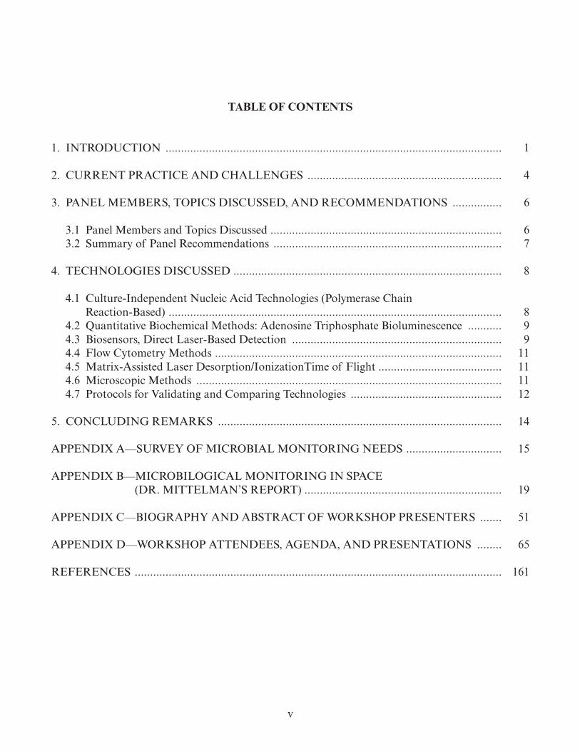
There are a lot of variations of nucleic acid technologies such as hybridization based (microarrays), real-time quantitative polymerase chain reaction (RT qPCR) based, and those based on nucleic acid probes coupled with fluorescent labels, to name a few. Among those, the RT qPCR appears to be the most promising because of its high accuracy, high reproducibility, low detection limit; the fact that it can analyze unculturable in addition to culturable organisms; and, perhaps most importantly, because of recent “lab-on-chip” optimizations allowing the technology to be implemented in very small portable devices and provide real-time monitoring on site.5 RT qPCR assays typically utilize two primers to replicate and hence, amplify, DNA based on a specific target sequence. In addition to these two primers, an additional nucleic acid probe is utilized. For each probe molecule consumed, one fluorescent dye molecule is released and detected. Therefore, as the RT qPCR reaction proceeds, if the target is present in the sample, fluorescence will increase. Such a RT qPCR assay and an associated devise to house the assay are, for instance, commercially avail-able by Cepheid and used by the Department of Homeland Security to detect biothreat agents (e.g., Bacillus anthracis, or anthrax) in the air of large cities in the United States. The anthrax test of Cepheid (fig. 1) amplifies gene sequences specific to B. anthracis and returns a positive signal with as few as 30 cells in the sample; it provides results within an hour (as opposed to 1–2 days for culture-based systems) and can be easily run onsite, by non-expert personnel. It was suggested that a similar PCR-based system that can perform three assays—one for total bacterial counts, one for total fungi, and one for total enterobacteria, which are typically the main agents of microbial infections in the space stations—will have major advantages over the current practice for microbial monitoring in the spacecraft and may represent a powerful solution for long-term spaceflights.
9
Figure 1. The SmartCycler® system from Cepheid. The system allows up to 96 independently programmable reactions to take place simultaneously, using different protocols. Multiple experimental runs can be started at different times, allowing several operators to use the system concurrently.
4.2 Quantitative Biochemical Methods: Adenosine Triphosphate Bioluminescence
Adenosine triphosphate (ATP) bioluminescence can be used to assess the level of total microbial content in a sample, including unculturable microorganisms. The main principle behind ATP bioluminescence is that ATP, a key intracellular energy source and ubiquitous marker indicat-ing cellular viability, increases as the amount of biological material (including microorganisms) increases. Measuring ATP bioluminescence relies on detection of photons emitted during the oxidative dephosphorylation of ATP by the luciferin-luciferase substrate/enzyme system. Photon emission is proportional to the amount of ATP in a sample. Currently, several portable and easy-to-use commercial systems to perform ATP bioluminescence measurements are available and it was suggested that such a system, with minor modifications to account for microgravity conditions, could find useful applications in monitoring the quality of the drinking water or biofilm formation in long-term spaceflights. The main drawback of ATP bioluminescence is that it cannot distinguish the types (e.g., bacterial vs. fungal vs. human/animal cells) or the species of the microorganisms that are active in the sample analyzed.
4.3 Biosensors, Direct Laser-Based Detection
There was significant discussion among the participants of the workshop about biosensors, as biosensors represent an emerging technology that provides great flexibility in design and can be easily adjusted for the needs of in-flight microbial monitoring. Although no system currently avail-able has been designed with the specifications required for long-term spaceflight in mind, several systems hold great potential for spaceflight purposes. It is not possible to provide an exhaustive list

10
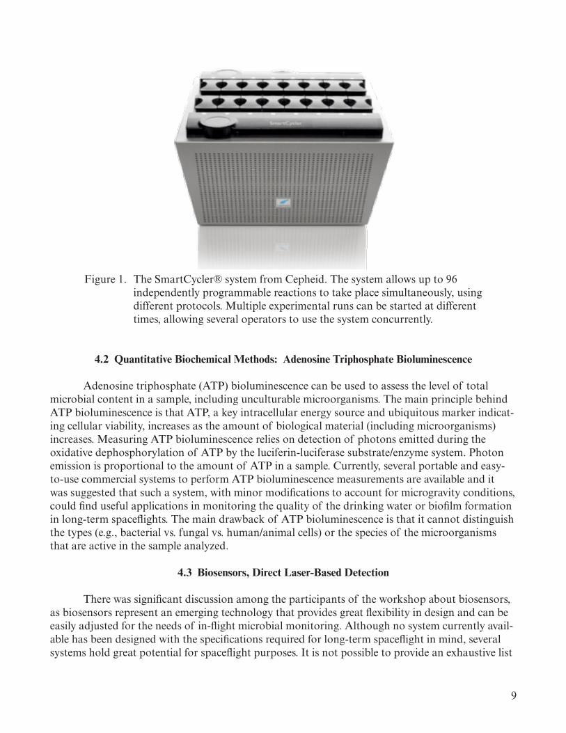
of potentially useful biosensors due to the great diversity of systems available and their underlying principles. The most promising systems, however, typically employ laser-based detection methodol-ogy, which utilizes direct interaction between a light source (a laser with suitable wavelength) and the biochemical molecules inside the microbial cellular structures to detect the presence of the microbes. Typically, in an instrument based on this detection scheme, an ultraviolet laser gener-ates an intrinsic fluorescence signal from certain metabolites (e.g., NADH, riboflavin) inside the microbe, and this fluorescence signal is used as a biological marker to differentiate the microbes from inert particles or even dead cells.
As a representative example of this technology, the BioVigilant IMD-A system (BioVigilant Systems, Inc.; see fig. 2) was discussed, which is characterized by several attractive properties. The BioVigilant IMD-A provides a way to quantitatively assess and instantly visualize the number of biologic events as they occur in the environment based on the intrinsic autofluorescence of specific biologic markers (NADH, riboflavin, dipicolinicacid) when excited with a laser at a wavelength of 405 nm. No consumables are necessary for the operation of this instrument, and the instrument offers real-time detection as well as cleaning and disinfection activity support. The main drawback of such biosensor systems is the (relatively) high-energy demand for the laser, but engineering solu-tions that can go around this problem may be within reach.
Figure 2. The BioVigilant IMD-A® 220-4 system from Azbil. With an air sampling capacity of 28.3 L/min and stainless steel, chemically resistant case, the IMD-A 220-4 is suitable for the most demanding cleanroom environments and larger testing areas.
11
4.4 Flow Cytometry Methods
Flow cytometry is a technique for counting and examining microscopic particles, such as microbial cells, by suspending them in a stream of fluid and passing them by an electronic detec-tion apparatus. It allows simultaneous multiparametric analysis of the physical and/or chemical characteristics of up to thousands of particles per second.6 Flow cytometry is routinely used in the diagnostics laboratories. However, the required infrastructure of modern flow cytometers that are capable of detecting microbial cells is probably prohibiting for deployment on the spacecraft in terms of energy required and weight. Miniaturized, automated flow cytometers, employing the lab-on-chip idea and microfluidics, are possible, at least in theory, although it appears that no such system currently is commercially available. Companies that might be able to produce custom-made, miniaturized flow cytometers include, but are not limited to, BD (Becton, Dickinson and Company, who recently bought Cytopeia, a start-up company that is specialized on flow cytometers) and Beckman Coulter.
4.5 Matrix-Assisted Laser Desorption/Ionization Time of Flight
The introduction of matrix-assisted laser desorption/ionization (MALDI) time-of-flight (TOF) mass spectrometry (MS) into the routine microbiology laboratory at the end of the 1990s has been a breakthrough in the rapid characterization of bacteria at the strain, species, and genus levels.7 As little as about 5 × 10³ CFU is necessary for reliable MALDI-TOF analysis.8 The remark-able reproducibility of the MALDI-TOF approach is due to the fact that many of the individual single-charged proteins of size 2,000 to 20,000 m/z (mass/charge; daltons) present in high abun-dance in the cell and measured by the MALDI-TOF approach (underlying principle) include many ribosomal proteins. Being part of the cellular translational machinery, MALDI protein fingerprints are therefore not significantly influenced by variability in environmental or growth conditions. Several commercial systems are currently available (e.g., BiotyperTM from Bruker Daltonics or SARAMIS from bioMérieux). The size of the infrastructure required is currently prohibitive for deployment on the spacecraft, but new miniaturized models and lab-on-chip versions are possible, making this technology a promising one for real-time monitoring. The limitations of the MALDI-TOF and similar approaches are the need to have the organism growing (either in culture or in the natural sample) in substantial numbers, which is not acceptable for coliform bacteria, and the inability to resolve robustly multispecies samples.
4.6 Microscopic Methods
Although microscopy represents one of the oldest techniques available for visualizing and monitoring microbial content, it still finds applications in the modern microbial monitoring labo-ratory. Furthermore, recent developments such as epifluorescence technology, which allows the identification of distinct microbial species even in cases that the species possess similar cell mor-phologies and can distinguish between live and dead cells,9 make microscopy a potentially useful technology for long-term spaceflights. However, some of the reagents currently used in epifluores-cence technology (e.g., acridine orange) are toxic and so cannot be used in the spacecraft or must be strictly contained within self-contained devices/containers. Thus, alternative protocols for epi-fluorescence must be developed. In addition, microscopy is typically not user friendly and requires
12
well-trained crew, which makes its application in the spacecraft challenging. Nonetheless, micros-copy has several advantages, such as, it can be quantitative, distinguish between different types of microbes, and can work with known as well as unknown microbial species. Recent developments such as the LUCAS (Lensless Ultra-wide-field, Cell Monitoring Array platform) microscope devel-oped by UCLA researchers, which represents a miniaturized cell phone size, shade-based micro-scope that can be used by non-experts, represent technologies that should be considered for the needs of long-term spaceflight missions.
4.7 Protocols for Validating and Comparing Technologies
The experts participating in the workshop discussed the technologies mentioned above and evaluated them based on the criteria that seemed more important for the purposes of long-term spaceflights. It was not possible to evaluate all technologies for all criteria presented in table 1 due to the lack of enough time and the fact that the commercially available products for each technol-ogy require different degrees of optimizations to be appropriate for NASA purposes; hence, it was not always fair to evaluate all technologies for the same criterion. The summary of the evaluations is presented in table 2.
Table 1. Suggested properties/traits that technologies should be evaluated for.
1. What the target organisms are (i.e., bacteria, viruses, fungi; all or only a pathogenic group) 2. What the detection limit is (e.g., cells in the sample; copies of DNA/RNA) 3. What samples can be analyzed (e.g., water, air filtrates, soils/surfaces, human samples) 4. Need optimization for the sample or method is general/robust? (e.g., PCR-based methods usually do not work with all samples equally well) 5. Time to obtain results since sample acquisition 6. Is it high throughput (e.g., how many samples can be analyzed in a day) 7. Any special requirements for sample processing (e.g., for sequencing methods, it is necessary to perform DNA extraction) 8. Can work in microgravity environment 9. What the method of detection is (e.g., DNA sequencing, ATP/lipid detection, etc.)10. What infrastructure is required11. Cost per sample/cost of infrastructure (approximately)12. What is the level of phylogenetic resolution (e.g., genus, species or strain level)13. Can distinguish live from dead cells14. Is it quantitative

13
Table 2. Preliminary evaluations of modern technologies for long-term spaceflight.
Criterion PCR-Based ATP BiosensorsFlow
CytometryMass
Spectrometry MicroscopyVersatility in microbes detected (e.g., bacteria and fungi)
+++ ++ ++ + ++ ++
Versatility in types of samples (water, air, surfaces, etc.)
++ +++ +++ + + ++
Easy to use and obtain results quickly
++ +++ +++ + ++ ++
Results easy to interpret ++ +++ +++ ++ ++ ++Creation of no biohazard waste
++ +++ +++ + ++ +
Low energy requirement ++ + + + + ++++++ Very Good ++ Good + Fair
The panel of experts also discussed the procedures to validate and compare the technolo-gies for the purposes of long-term spaceflights. The consensus was that the protocols used in food industry, which leads the development of new microbial monitoring technologies, could be adopted, especially the protocols established by the Association of Analytical Communities (AOAC) International. These protocols allow direct comparisons of different technologies among themselves and against the current culture-based practices at space stations, which should consti-tute the reference point in the comparisons. More details about the protocols and established pro-cedures can be found through the Web site of AOAC International and in Mr. Deibel’s presentation in appendix D. Even though the validation process involves some extra cost upfront, it provides assurance that a chosen rapid method will perform as expected and thus, it is deemed necessary.
14
5. CONCLUDING REMARKS
Regardless of the technology chosen for the needs of long-term spaceflights, it will be important to engineer and optimize the technology for the special conditions in the spacecraft. The spacecraft represents a unique environment that does not resemble any other environment available on Earth (e.g., microgravity conditions); hence, no microbial monitoring technology from those currently available has been designed with the unique conditions of the spacecraft in mind. The most promising technologies should be evaluated against the current practice before deployed on the spacecraft. The participants of the workshop offered several examples of how testing and comparisons of the different technologies should be done, derived primarily from the food industry where technology development represents a continuously evolving field. More detailed information and established protocols for technology testing can be found in the presentation of Mr. Diebel (of Deibel Labs) in appendix D. Furthermore, table 1 provides a list of characteristics/traits that each technology needs to be evaluated for; these traits represent important properties in general and/or specifically for the spacecraft environment.
Microbial growth in natural environments is typically limited by nutrients such as carbon and environmental conditions such as temperature.1 Maintaining a low carbon load in the water or the air circulated in the spacecraft and performing treatment of circulated air/water under as cold temperatures as is possible will significantly contribute towards controlling microbial growth and infections. Educating the crew along these lines to maintain low carbon load and clean surfaces routinely will be instrumental for the success of the above practices and for preventing incidences of microbial contaminations.

15
APPENDIX A—SURVEY OF MICROBIAL MONITORING NEEDS
Appendix A contains the results of a NASA customer survey to assess the SOA of microbial technologies currently in use and future related needs.

16

Survey Results/ NASA-org /Q-01 /Q-02 /Q-03 /Q-04 /Q-05 /Q-06 /Q-07 /Q-08 /Q-09 /Q-10 /Q-11 /Q-12 /Q-13 /Q-14 /Q-15 /Q-16 /Q-17 replied on
1. How will a rapid microbiology monitoring system be employed by your team? 2. Will the viability of the microorganisms detected important for your application?
3. Do you have a list of target organisms that you would like identified? (If so, please provide)
4. What is most important to you, identification, characterization, or enumeration?
5. What detection limits are required for your application (air, water, wastewater, urine, surfaces, clinical specimens, etc.)?
6. What are your requirements for analytical sensitivity and specificity? 7. What is the expected sample bioburden for your application?
8. What are the indicator micro-organisms for your applications (e.g., MRSA, E. coli, etc.)?
9. Do samples need to be archived after testing?
10. What is the sample amount that you can provide for the analysis?
11. Is there a specific tool needed to aseptically collect your sample? Is it available? Do we need to improve it? Do we need to design one?
12. What is the required frequency of analysis? 13. What is the assay turnaround time required? 14. Is there a require-ment for anaerobe monitoring?
15. Is there a requirement for viral monitoring?
16. Is there a require-ment for antibiotic susceptibility testing?
17. What is the chemical and/or biological composition of your test media (water, air, sur-faces, crew)? Please provide all the information that might be needed to select a microbial monitoring systems that fits your needs.
Jet Propulsion Labo-ratory, California Institute of Technology
a. ATP-based microbial monitoring system to differentiate dead from viable cells, enumerate total cells. b. Q-PCR based analysis to measure total microbial burden (bacteria, archaea, and eukarya). c. PMA-DNA Microarray analysis to measure viable microbial burden (bacteria and archaea).
yes Viable spores for present missions; total genetic inventory for future planetary protection missions; Viable problematic microbes for future human habitation missions Viable microbes for Department of Homeland Security issues
enum 300 viable spores per sq meter (less than one viable spore per 25 cm2) for surface 100 to 10,000 viable cells for 100 ml of water 100 to 1,000 viable cells for one cubic foot of air
The question is not clear. Some information given in #5 are applicable here. If I need to elaborate sensitivity and specificity there is no space. Contact me for details. We have several publications pertaining to this.
See Q#5. Spores 1 Any where from 100 uL (purified DNA) to 10 mL of unprocessed sample
Yes. Need to improve for some application. Depends on the mission and application. Real time to not more than 24 hours (8-h human work) No No No Water Air Surfaces Crew
12/17/2010
KSC / DYN-3 Our team supports LSHS Water Recovery Systems research and technology development for microbial control in potable water and environmental control and life support (ECLS) systems. A rapid microbiology monitoring system would be used to quantify disinfection efficacy among candidate technologies in labora-tory and relevant environments.
yes No. We have a short list of challenge microorganisms used for testing water disinfection technologies but the list is not comprehensive. Although the identification and enumeration of specific “indicator” microorganisms can be a useful measurement guide for water quality monitoring and process control dur-ing water treatment, too much reliance upon a single target can provide a false sense of security, i.e. reliance upon E. coli as a measure of potable water quality ignores protozoan and viral sources of potential contamination.
enum 1 CFU / 100 mL bacteria or fungi (required) and 1 PFU / 100 mL virus (desired) suspended in water or attached to wetted surfaces (e.g., biofilms) 10 - 100 cells per square cm for material surfaces
Our test objectives do not require high analytical sensitivity since we typically seek disinfection technologies that yield >1-log reduction given an initial added biological burden ≥1E6. However, low biomass samples like potable water or spacecraft surfaces would require both high analytical sensitivity and specificity. For water qual-ity monitoring during water treatment, it would be useful to have an analytical method capable of differentiating between viable and non-viable microorganisms in both high-biomass (e.g., wastewater) and low-biomass (e.g., potable water) samples.
We typically challenge with 1E6 - 1E9 per mL challenge bacteria and desire microbial monitoring technologies that can quantify microbial burden down to 1-10 CFU per mL, i.e. >5-log reduction.
Burkholderia cepacia, Cupriavidus metallidurans, Esch-erichia coli, Methylobacterium fujisawaense, Pseudomonas aeruginosa, Ralstonia pickettii, Sphingomonas paucimobilis, Wautersia paucula
yes 10 - 100 mL Sample by pipet or by material coupon. Depends on frequency of testing and availability of funding. It would be useful to have a real-time assay, but reports returned in 3-5 days would be acceptable.
No, not at present. Not currently within scope of experiments due to budget limitations but highly desir-able for future water quality studies.
Not currently within scope of experiments due to budget limita-tions but desirable for future water quality studies.
We typically focus on testing potable water disinfection technologies. The test medium is water that meets or exceeds spacecraft drinking water quality microbiological specification prior to the addition of challenge bacteria required for the test objective.
12/20/2010
KSC / NE-F3 It will be used to assess the potable water sampled from ground support equipment and spacecraft, to help ensure that the water quality is suitable for long duration missions.
yes No enum 1 CFU/mL (for water) HSIR limits for potable water < 50 CFU/mL Unknown no 1 Liter Samples are currently collected via sterilized tubing and bottles, and this method has been adequate for the task.
Once pre-servicing for the ground support equipment, and once post servicing for the spacecraft.
48 hours Unknown Unknown Unknown Water that meets HSIR limits 12/21/2010
MSFC / ES62 For monitoring potable water quality on ISS or for a future manned mission. no enum <100 CFU/ml same as standard analytical requirements. Typically <1 CFU/ml, but may exceed 1000 CFU/ml as a result of a system failure.
water-borne bacteria. Monsi can answer this question for me.
no Limited product water is available on ISS. Typically 100 ml is provided for microbial analysis.
Microbial samples are collected in sterile sample bags per standard ISS procedure.
Monthly Not sure, but I think it’s 1 week. N/A No No WPA product water is best described as “ultrapure” water. It typically has an organic content of ~200 ug/L, and no detectable bacteria. It does contain 1 - 4 mg/L of Iodine in the WPA effluent, though this is removed to <0.2 mg/L at the potable dispenser. There is no other detectable inorganic species.
12/21/2010
JSC / SF3 We could use it for testing harvested fruits and vegetables on a surface mission. We could also use it to test foods that are processed on a surface mission. Both test would provide confirmation (or non-confirmation) of food safety.
yes Total aerobic count, coliform, coagulase postive staphylococci, salmonella, yeasts and mold
char Depends on microbe - Combined AEH/AFT research task is underway to determine limits
Depends on microbe - Combined AEH/AFT research task is underway to determine limits
e coli no grams n/a Depends on how often crops are harvested and/or ingredients are processed
Hours due to perishability of food No Not that I know of No water, food surfaces 1/5/2011
KSC / MESC (Innovative Health Applications, LLC)
a. testing drinking and D.I. waters for bacterial load and for specific bacteria b. monitoring space craft components for bacterial load c. monitoring indoor air quality of laboratory, shop, and cleanrooms
yes E. coli, human pathogens char D.I. water - sterility - 1cfu/ Liter Potable water - 1cfu/100ml Wastewater - 1 cfu/100ml
95% sensitivity and specificity Sterile to 10x5 cfu/mL E. coli Spore forming bacteria
yes 100mL to 1Liter For current applications, there is equipment available to collect samples.
Weekly for potable water applications sporadic for mission related sampling
ASAP for most mission related samples. 2 days to 10 days for routine samples
No, we do not have a current requirement for anaerobic monitoring.
No, there is not a current requirement, however, there is considerable interest to collecting baseline informa-tion on the presence and numbers of viruses.
No, we do not have a requirement for suscep-tibility testing.
Water - potable, wastewater, and D.I. waters Air - breathing air Surfaces - space craft materials - metals, composites, plastics
1/6/2011
KSC / Dynamac To assess the microbiological quality of edible crops grown in vegetable production units (VPU) designed for closed environments such as the Lada VPU housed on the international space station, to ensure qual-ity and safety for the consumer. The monitoring of the number of microorganisms on the plant and growing surfaces as well as the detection of foodborne pathogens would be the desired outcome.
yes E. coli, Coliforms, Salmonella, Staphylococcus aureus,Aspergillis flavus (in accordance with ISS food requirements set by NASA)
enum For :Total aerobic count: ≥ 10,000 CFU/g, Coliform: 10 CFU/g, Coagulase pos. Staphylococcus: 10CFU/g, Aspergillis flavus: 10CFU/g, and total yeast and molds: 100 CFU/g. E.coli and Salmonella: presence/absence.
Total aerobic plate counts: 10,000-1E8 CFU/g Specific organisms mentioned would likely be below 10 CFU/g
Total coliforms, E.coli, Salmonella
no From ground control studies-5-25 grams Standard asceptic technique The frequency of the samples would be at harvest of any crops grown in a VPU before consumption. This would probably vary from 1 to 4 weeks.
As quickly as possible-within 24hrs. No Possibly enteric viruses. No Analysis would be on plant tissue, either destructively sampled or surfaces. Ideally plants growing in a VPU could be tested to ensure microbiologically safe edible product in ac-cordance with NASA food requirements before consumption, requiring a rapid turn around time. Safe disposal and destruction of microbial samples after analysis must be considered so as not to introduce a new hazard.
1/11/2011
KSC / DYN-3 (Dynamac Corporation)
Our research goal is microbial characterization of space mission solid wastes before, during?, and after treatment by technologies under development by the Waste Management Element of Life Support and Habitation Systems. Solid wastes include food wastes, personal hygiene items, and urine and fecal waste contaminated items such as EVA ‘diapers’ and space toilet wipes (‘Elbow’ packs). We run conventional plate counts using a couple of generalist heterotrophic media and some selective media for a couple of pathogens of interest. We also do total direct counts (AODC). We select some colonies that grow on the media and run the purified isolates through some sort of ID system - Biolog usually, or microSEQ for those that the Biology doesn’t want to identify. The rapid microbiology monitoring system would be employed by our team to run in tandem with these methods for comparison, then we would decide whether to incorpo-rate this system into our routine tests or, perhaps, replace some or all of them?
yes Selected food pathogens - G+: Staphylococcus aureus, Bacillus sp. (typical sporeformer that can survive high heat and desiccation); G-: E. coli; Pseudomo-nas aeruginosa, Salmonella enterica serovar typhimurium; a typical mold, e.g. Aspergillus niger. Crew fecal contaminated wastes: E. coli / coliforms
enum Quite low, ~ 10 per mL for liquid wastes (e.g., food drinks); ~ 100 per g fresh weight for food items and fecal solid wastes - but we typically disperse these solids in a diluent, then de-termine numbers. Surfaces: Solid waste processing hardware - ~ 10 per sq. cm, inside/outside plastic film solid waste bags - 1 per sq. cm. Pathogenic microbes: presence / absence.
I don’t know how to answer this. Food wastes: Cultivatable counts: 1 x 10^6 to 1 x 10^9 per g fresh weight, drink pouches: 1 x 10^6 to 1 x 10^9 per g fresh weight. Total direct counts: 1 x 10^7 to 1 x 10^9 per g fw Personal hygiene wastes: 1 x 10^6 to 1 x 10^9 per g fw. Total direct counts: 1 x 10^7 to 1 x 10^10 per g fw. Solid waste plastic film bag surfaces: Cultivatable counts: 1 x 10^1 to 1 x 10^4 per sq. cm. Total direct counts: 1 x 10^6 per sq. cm.
Food pathogens, fecal patho-gens / indicator m.o.’s.
yes Solid and liquid wastes - many grams fresh weight. Hardware and plastic film surfaces - a few sq. cm (hardware) to many sq. cm (plastic film encasing food and other solid wastes)
Surface sampling ‘sponge’ and sterile swabbing template (usually 5 cm x 5 cm). These are available. Solid and liquid wastes - the current sampling methods are to asepti-cally acquire samples (sterile gloves) and conduct a dilution series into sterile diluent.
As yet, unknown. Studies of the fate of microbes during storage of solid wastes after application of the (various) treatment technologies is still to be done. Some may be stored for months / years/ forever. Planetary quarantine requirements may prevent these long duration storage periods, however.
I don’t know of any NASA requirements for assay turnaround time. I’d personally like to see test results faster than it takes to incubate a plate of agar media 3 days to 7 days or more
Yes. Don’t know. No. Space trash solid wastes, including: food waste, personal hygiene waste (wipes, EVA diapers, Elbow packs (toilet wipes, etc.) with crew urine and some fecal waste, paper,plastic film, drink pouches. Commode waste / crew fecal material has NOT been part of our micro-bial characterization samples. Hardware surfaces of the candidate solid waste treatment technologies. Microbiological characterization of waste and hardware before, during, and after treatment / hardware operation. Among the goals of solid waste treatment are: (1) sterilize the waste (usually by heat) to make it (microbiologically) safe for crew and (2) water recovery from the waste, thus, dehydrating the waste to levels that will prevent further microbial growth.
1/11/2011
JSC / Space Life Science
The initial needs for the rapid system will be for in-flight environmental monitoring (air, surfaces, potable water), crew health monitoring, and spaceflight research. Depending on the source of food supply for a mission, the system may also be needed for food monitoring.
yes No official list exists; however, future requirements are likely to include specific medically important organisms, including both obligate and opportunistic pathogens.
1 While culture based requirements (including detection limits) have been established for current NASA missions, no require-ments have been established for other technologies. Current research studies are underway to provide recommendations for future requirements.
No requirements for analytical sensitivity and specificity have been established for environmental and food samples beyond the inherent characteristics of culture based methodology.
Environmental samples have historically had extremely low levels nominally. However, these levels have dramati-cally increased during contamination events. Both food and clinical samples have the potential for very high levels.
The only indicator organisms currently used are coliform bacteria used in the analysis of potable water. Other indicators may be used for future require-ments.
1 This volume has been historically dependent on factors such as storage capacity of consumables, availability (as with potable water), or the expected concentration of the sample (a contaminated sample requires much less than a nominal environ-mental sample).
Depending on the type of sample, many collection tools have been developed (swabs, teflon bags, etc). Some modification may be nec-essary depending on the type of technology used for analysis (ex: im-pinger rather than impactor for air samples). Much more work needs to be done on sample processing from collection to sample analysis. This would include an adequate work area, such as a glove box.
No requirements have been established for sampling frequency for all technologies. This value will be dependent on vehicle design and operational activities
Certain assays, such as those for clinical applications, may require near immediate results. Other samples such as environmental samples or food analysis can take longer times. Historically, periods of 24 hours to 5 days have been experienced. In general, a shorter time frame is preferred.
None at this time. Depending on future missions and medical needs, viral analysis may be required. However, it is cur-rently not a requirement.
No requirement cur-rently exists, though it may have future op-erational and research benefits.
1/11/2011
JSC / SF2 To ascertain microbial populations in the air, surface, and water of the ISS. Characterization capabilities will also be required. This will help to determine the overall environmental health of the habitable volume of ISS and other vehicles.
yes List of microorganisms typically found in ISS. id Detection limits prescribed by the MORD. As prescribed by JSC and MSFC Microbiology Groups. As prescribed by JSC and MSFC Microbiology Groups. As prescribed by JSC and MSFC Microbiology Groups.
yes As prescribed by JSC and MSFC Microbiology Groups.
As prescribed by JSC and MSFC Microbiology Groups. As prescribed by JSC and MSFC Microbiology Groups. Most likely. Most likely. Most likely. 1/14/2011
Marshall Space Flight Center / ES62
Surface fouling and filter loading assessment for those surfaces and equipment in contact with cabin atmosphere.
no Nonspecific but need to include bacteria, molds/fungi enum Air: <500 CFU/m3; Surfaces: <100 CFM/cm2 Undefined. Undefined. Undefined. no Undefined. Undefined. Monthly. Undefined. No. No. No. Typical spacecraft cabin atmosphere at 1 atm - 20% O2/79% N2 with trace Ar, 0.5% CO2, trace non-methane volatile organic loading averaging between 10 mg/m3 and 20 mg/m3, relative humidity ranging from 35% to 45%.
1/18/2011
NASA Ames Research Center / Code SCB
Our team needs to be able to detect microbes on hardware, processed solid and liquid waste, and process offgases. There is application to the research and development of hardware and to final spacecraft implementation of the hardware.
yes Human pathogens. Also indicator organisms such as E. Coli. / coliforms. Examples include Staphylococcus aureus, Bacillus sp. , Psedudomonas aerugi-nosa, Salmonella enterica serovar typhimunium, Aspergillus niger.
id Quesstimates: 100 per ml for liquid wastes. 100 per g solid wastes such as food, 10 per sq. cm on surfaces.
species and maybe strain examples: food wastes and personal hygiene wastes: counts up to 1x10^9 per g
E. coli and others not yet determined.
no Swab samples to grams to maybe kilograms. Swabs for some samples. For some of the plastic tiles produced by the heat melt compactor a coring device is needed to obtain samples from the inside.
For development efforts – a few per week. For flight – Unknown, maybe a couple of times a week.
For the research – days to weeks. for Space application – day.
maybe for anaerobic pathogens.
yes, pathogenic viruses. probably not. space solid wastes such as food waste, hygiene waste (wipes, diapers, etc.), crew urine and feces, paper, plastic, tape, vomit, gloves, clothing
1/21/2011
JSC / EC6 Periodic checks on the efficacy of the antimicrobial and control of organisms within the ISS internal thermal control system coolant.
yes Identification is secondary to enumeration. Typical water borne organisms would be sufficient.
enum water -- up to 10^6 CFU/100 ml no Up to 30 ml if needed. Yes; yes, sample tools are installed in each coolant loop. No further hardware, or improvements are needed.
Not more than once per year in each of 7 coolant loops, provided there’s no change in stability of the present level of microbial control. Should a loop exhibit an upward trend in organism population, additional analysis would be needed to characterize the trend.
The critical time parameter for ITCS monitoring is crew time as opposed to assay turnaround.
No No Maybe deionized water with buffer additives, as well as ortho-pthalaldehyde as the antimicrobial 2/3/2011
NASA Ames Re-search Center / SGE
organisms in extreme environments, water and air yes no id 1-10 cells/cc in air (a bit higher on a filter), 500+/mL in water sequence analysis (e.g., rRNA) na none yes for air a matter of cells, for water 100 µL to 1 mL no for water, we are working on one for air but are not as aware of ISS and shuttle systems already available
variable for air, ideally 20-60 minutes; for water a matter of days only if the samples occur naturally
always a good idea no 2/3/2011
Headquarters / SOMD
It will be used for rapid accurate identification and characterization of the microbial environment on board the ISS and any future human space craft.
yes E. coli, salmonella, fungi, staphylococcus and streptococcus char This is not my field, thus I am not prepared to state exact limits. However, I am most concerned that the limits provide us to the ability to accurately determine what microorganisms are present and are they at levels that could possibly impact the health of crew members.
I do not have the technical expertise to define these requirements and would defer to our toxicology experts at JSC.
I do not have the technical expertise in this area and would defer to our toxicology experts at JSC.
E. coli, salmonella, fungi, staphylococcus and streptococ-cus, MRSA
yes I do not have the technical expertise in this area and would defer to our toxicology experts at JSC.
Yes tools currently exist but can always be improved (e.g. smaller, more dependable, fewer consumables).
This is dependent on whether the samples are collected for research or operations. Research criteria would drive frequency. Operations measures would be frequent enough to adequately characterize the environment. This would be defined by flight rules.
Naturally, as soon as possible. This would depend again on the intent. If for measuring human samples for medical man-agement, within minutes to a few hours. If for environmental characterization, it would not need to be as rapid, but ideally within 24 hours.
I do not have the techni-cal expertise in this area and would defer to our toxicology experts at JSC.
In my opinion, yes, as this could have a bearing on crew health monitoring and treatment.
At present, I cannot see the need, but if used for long duration/planetary activities, there may be.
At present, with the confined spaces of space craft, it is important to monitor all media included above. Water is now largely in a closed loop system and its purity must be assured. The “cabin” air is constantly recycled through filters and monitored but
2/22/2011
17

18

19
APPENDIX B—MICROBIOLOGICAL MONITORING IN SPACE(DR. MITTELMAN’S REPORT)

20
Occupational & Environmental Health Microbiological Monitoring in Space Microbiological Microbiological Monitoring in Space

21
[Type text] [Type text] [Type text]
Microbiological Monitoring in Space Exponent Project No. 1006535.000 Prepared for NASA Contract No. GS-23F0390K Marshall Space Flight Center Huntsville, AL Prepared by Marc W. Mittelman, Ph.D. Exponent 9 Strathmore Road Natick, MA 01760 March 25, 2011 Exponent, Inc.

22
iii 1006535.000-A0F0-0311-R020
Contents
Page
List of Figures xxii
List of Tables xxiii
Executive Summary xxiv
Background 1
The NASA 2011 Microbiology Workshop 3
Literature Survey of Testing Methods 5 Culture-Based 6 Biochemical 7 Molecular 8 Spectroscopic 9 Flow Cytometry 10 Impedance 10 Other 11
Development of Selection Criteria 12
Literature Cited 15
Appendix NASA-generated survey of NASA microbiology customers

23
List of Figures
Page
Figure 1. Summary of draft microbiology assay weightings. 14

24
List of Tables
Page
Table 1: Microbiology test methods and test environments. 2
Table 2: Draft microbiology assay weightings 13
25
Executive Summary
This communication presents an overview of recent microbiological monitoring approaches that may be adaptable for use in long-term space travel. There are a number of challenges associated with the enumeration and identification of environmental and clinical microorganisms (bacteria, fungi, viruses) in space. These include weight and energy limitations, risks of crew exposures to test reagents and microorganisms, waste disposal issues, and problems associated with operating in a microgravity environment. Additionally, the growth, virulence, and antimicrobial susceptibility of some microorganisms appear to be influenced by microgravity, which could present problems in characterizing isolates. Traditionally, microbiological monitoring of environments and crew has focused on bacteria and (less frequently) fungi using culture-based techniques. However, there are a number of molecular, biochemical, and physicochemical test systems that may be adaptable for use in a space environment. This review has been prepared as part of an effort to develop a rationale basis for selecting candidate microbiological technologies for further evaluation. A NASA-sponsored Workshop has also been organized to develop selection criteria and to define key attributes required for environmental and crew monitoring of microorganisms.
26
1 1006535.000-A0F0-0311-R020
Background Microorganisms, including bacteria, fungi, and protozoa, are ubiquitous in spaceflight operations. A number of studies have shown that personnel, fluid handling systems (water, wastewater, etc.), air handling systems (filters, etc.), and various surfaces can harbor bioburden. Some of the environmental bioburden isolates have been associated with both human and animal diseases, as well as biological fouling activities. Microbial contamination of space vehicle environments can result in a number of deleterious outcomes for crew health, and can adversely affect operations of critical fluid- and air- handling subsystems (Horneck, et al., 2010). The ability to rapidly enumerate and identify microbial contaminants is key to controlling the impact of microorganisms in a confined spacecraft environment. A variety of rapid test systems are currently available, and others are under development in academic and commercial laboratories. The selection of appropriate methods is dependent on the type of data required as well as the sample type. The early space missions (i.e., Apollo) and short duration mission (space shuttles) did not monitor the microbial population during flight; they relied on the return of samples after missions for analysis. To date, technologies for monitoring microorganisms aboard the International Space Station (ISS) have primarily relied on traditional, culture-based approaches. These techniques are often laborious and require extended processing times, and are difficult to standardize and to interpret, and do not always provide identification of the microbial flora. Newer techniques for microbial classification and identification have focused on chemotaxonomic and molecular-based techniques. The techniques allow for more detailed analysis of the microorganisms present, including viable but non-culturable organisms. Rapid, sensitive, and selective microbial detection and identification methods would help differentiate between pathogenic and nonpathogenic microbial species. Bioburden analyses could also help the crew better assess risks to the various operating systems and payloads (Table 1).

27
Table 1: Microbiology test methods and test environments.
Test Environment Viable-Count Methods
Direct-Count Methods
Indirect-Count (Biochemical) Methods
Indirect-Count (Physicochemical & Spectroscopic) Methods
Molecular-Based Identification
Chemical-Based Identifica-tion
Air √ √ √ √ √ √
Crew (blood, urine, CSF, other)
√ √ √ √ √ √
Food √ √ √ √ √ √
Lab Animals √ √ √ √ √ √
Other Fluids √ √ √ √ √ √
Plants/greenhouse √ √ √ √ √ √
Potable Water √ √ √ √ √ √
Surfaces √ √ √ √ √ √
Wastewater √ √ √ √ √ √
Perhaps the most significant adaptive mechanism used by bacteria is adhesion to surfaces; indeed, the majority of bacteria in nutrient-limited environments (such as the internal active thermal control system, IATCS) are attached to surfaces. Recognition of this important growth characteristic is a key consideration in developing effective monitoring programs. Sampling of planktonic environments can only recover a small fraction of the total system bioburden. Monitoring of microorganisms associated with biological fouling activities is an important part of an overall control strategy. Sample acquisition and the selection of sampling locations are critical for obtaining accurate, useful data. Many of the biofouling microorganisms are sensitive to oxygen, temperature, and the effects of drying. Whenever possible, microbiological samples should be taken before the start of system maintenance or repair activities. While bulk-phase samples can provide useful information
Test Environment
Viable-Count
Methods
Direct-Count
Methods
Indirect-Count
(Biochemical)Methods
Indirect-Count(Physiocochemical& Spectroscopic)
Methods
Molecular-Based
Identification
Chemical-Based
IdentificationAir √ √ √ √ √ √Crew (blood, urine, CSF, other
√ √ √ √ √ √
Food √ √ √ √ √ √Lab Animals √ √ √ √ √ √Other Fluids √ √ √ √ √ √Plants/greenhouse √ √ √ √ √ √Potable Water √ √ √ √ √ √Surfaces √ √ √ √ √ √Wastewater √ √ √ √ √ √
Table 1. Microbiology test methods and test enviroments.
28
on the overall system condition, surface samples provide the best evidence for microbiological assessments.
The environmental control and life support systems (ECLSS) provide a number of challenges for microbiological testing that are somewhat unique to the spaceflight environment (Roman and Mittelman, 2010). Sample collection volumes, sample preservation reagents, and sample storage containers must conform to existing requirements for compatible materials. Test reagents and test equipment selections may be constrained by environmental health and safety requirements. Significant constraints may also be imposed by energy and weight limitations, and by the requirement for operations under microgravity conditions. It is essential that appropriate selection criteria be applied to test systems intended for the ECLSS. It is likely that many of the considerations for environmental microbiology test system requirements will also apply to clinical testing systems used in disease diagnosis and treatment.
The NASA 2011 Microbiology Workshop This report has been prepared in conjunction with development of a NASA Workshop scheduled for April 19, 2011 at Johnson Space Center, Houston. Fundamental issues to be addressed include the sources and risks from microorganisms in the air, water supply, waste recycling, food, laboratory animals, plants and soils, and on surfaces aboard spacecraft. The goal of the Workshop is to identify the most important microorganisms that should be measured, the level of specificity and frequency of measurement, and the areas of the spacecraft that should be monitored. Consideration will be given to the impact of bioburden on the altered physiological conditions of astronauts, the impacts of microgravity on the microorganisms themselves, and the influence of life support systems (for example food and water sources). Consideration will also be given to risk management and control practices that might influence microbial populations. In addition, the Workshop will consider the type of sampling technologies needed for detecting and analyzing the microorganisms.
29
The Workshop will focus on fundamental questions surrounding the sources and risks from microorganisms in spacecraft. The presentations and discussions will include consideration of the following issues:
1) What is the historical context and practice within NASA of microbiological monitoring?
2) Which microorganisms should be measured?
3) What areas of the spacecraft should be sampled?
4) What level of specificity and frequency of measurement is required?
5) Which methods provide the greatest amount of useful information consistent with the unique operational environment?
Comments regarding the selection criteria components have been solicited from NASA customers (in a survey conducted by NASA in late 2010 and early 2011), and the discrete data are summarized in Appendix 1. The complete survey, which includes all of the responses, may be found in a separate electronic file (provided under separate cover). The survey findings will be incorporated into the Workshop discussions.

30
Literature Survey of Testing Methods A literature survey, primarily focusing on English language, peer-reviewed publications from 2000 - March, 2011, was conducted for different types of microbiological techniques with potential applications for the space program. This survey is intended as an overview of several types of candidate technologies, but is not an exhaustive review of microbiological enumeration and identification methods. A number of methods, which may be adaptable for space applications, used in the pharmaceutical industry have been described (Marino, et al., 2000; Jimenez, 2001; Jimenez, 2004). In addition to existing culture-based methods (Haberer and Mittelman, 2003), a number of rapid testing methods have been described (Easter, 2003). While this literature survey is focused on testing methods, it is important that sampling methods be addressed in any comprehensive evaluation program. For example, recovery of airborne microorganisms for testing can involve specialized equipment with limitations that could limit direct transfer of techniques to the space environment (Kuske, 2006; Fykse,
et al., 2008; Obeloer and Schwanke, 2009). Similarly, the recovery of biofilm bacteria and fungi from fluid-contacting surfaces poses a number of challenges, particularly in a microgravity environment. While a number of scientific studies on the effects of microgravity on biofilm microorganisms have been conducted (Lynch, et al., 2006; Chen, et
al., 2008; Mauclaire and Egli, 2010; Rosenzweig, et al., 2010; Van Mulders, et al., 2011), sampling of spacecraft fluid-handling systems for biofilm microorganisms remains problematic. Several reviews of biofilm sampling and detection methods have been published (Amaral, et al., 1991; Mittelman, 1998; Denkhaus, et al., 2007; Nivens, et al., 2009). Each of these groups of technologies has common advantages and disadvantages that impact their utility for space microbiology applications. While ease-of-use and short assay turnaround times are obvious considerations, these attributes are less important than those that may influence crew safety. For example, any assay protocols that involve the

31
growth of microorganisms need to consider inactivation of what must be considered as biohazardous waste. Similarly, biochemical or molecular methods often require the use of chemical reagents that must also be safely handled and disposed. Size, weight, energy usage, and heat generation are of significantly more concern in space operations than in a typical laboratory environment. Finally, the effects of microgravity on fluid transport—and on some reactions—present challenges for all of the technologies described herein. It is important to note that there are opportunities for combining technology platforms to provide improved sensitivity and specificity. Much as analytical chemistry has benefited from combinations of, for example, infrared spectroscopy and mass spectrometry in trace analysis applications, microbiological monitoring using multiplex systems is evolving. A number of the cited research communications presented herein include such multi-technology platforms. Many of these platforms involve advanced biosensor technologies incorporating biochemical and molecular diagnostic tools (Turner and Magan, 2004; Pohn,
et al., 2007; Settanni and Corsetti, 2007; Miller and Tang, 2009; Grossi, et al., 2010).
Culture-Based Culture-based techniques for enumerating and identifying viable microorganisms—bacteria and fungi—have been the primary means for monitoring onboard various spacecraft. A number of publications have described methods employed for the recovery of aerobic and anaerobic microorganisms from fluids (Roman, et al., 2001; La Duc, et al., 2004; Bobe, et al., 2007), air (Ott, et al., 2004), surfaces (Castro, et al., 2004; La Duc, et al., 2004), and crew (Novikova, et al., 2006; Frey, 2010). A number of studies have shown that microgravity influences both the growth and the virulence of bacteria (Wilson, et al., 2008; Rosenzweig, et al., 2010). Therefore, the morphological and physicochemical properties of cultured microorganisms may be different from “textbook” descriptions. This finding should be a consideration in the selection of commercial test systems for use in space. There are a number of commercially available self-contained test systems currently available in the commercial marketplace. These include the 3M PetriFilm system (Chain

32
and Fung, 1991), various culture-based systems from Millipore Corporation (Marino, et al., 2000; Massa, et al., 2001; Smith, et al., 2004; Mainelis and Tabayoyong, 2010), IDEXX Corporation (Noble, et al., 2010), and products from a number of other companies (Horman and Hanninen, 2006). In all cases, samples must be incubated under controlled conditions until visible evidence of growth occurs—or some growth-induced change in a reagent is apparent.
Biochemical Biochemical analyses focus on signature compounds that may or may not be unique to microbial constituents. In some cases, viable cultures are required; in others, bulk phase or surface samples can be analyzed directly, circumventing the need for culturing. Orenga (Orenga, et al., 2009) has reviewed various substrates that are candidates for different substrate-based reactions. Test systems based on physiological reactions of viable microorganisms include Biolog (Tokajian and Hashwa, 2004; Stefanowicz, 2006; Bultmann, et al., 2009; Morgan, et al., 2009), API (Dalton, et al., 1993; Tokajian and Hashwa, 2004; Song and Leff, 2005), Vitek (Vuksanovic, 2007; Chen, et al., 2008; Mittman, et al., 2009; Mittman, et al., 2010), Microscan (Tritz, et al., 1990; Chen, et al., 1998), and BD-Phoenix (Menozzi, et al., 2006; Brigante, et al., 2007; Snyder, et al., 2008). Most of these types of systems can process various types of specimens on an automated basis, and many also provide antimicrobial susceptibility information for clinical decision-making. ATP assays are widely used to detect total bioburden in industrial operations (Passman, et
al., 2009; Lee, et al., 2010), pharmaceutical operations (Jimenez, 2004; Kramer, et al., 2008), drinking water (Berney, et al., 2008) and food and dairy sanitation surveys (Labots and Stekelenburg, 1985; Poulis, et al., 1993; Kottferova, et al., 2003). A number of applications have also been described for aerospace and spacecraft operations (Fajardo-Cavazos, et al., 2008; Newcombe, et al., 2008; Osman, et al., 2008; Morris, et al., 2010).

33
Antibodies, including monoclonal antibody assays, are highly specific for specific microoganisms (i.e., pathogens). When combined with immunomagnetic capture technologies, these types of assays can be very sensitive and robust. As with the non-culture based genomic assays, antibody based test systems cannot distinguish between viable and non-viable (or non-culturable) microorganisms. Potentially useful techniques have been described in a number of communications, including several that address spaceflight applications (Yu, 1998; Stevens and Jaykus, 2004; Anon., 2006). Novitsky and Hochstein (Novitsky and Hochstein, 2003) have reviewed pharmaceutical and clinical applications for the Limulus amoebocyte lysate (LAL) assay. This biological assay has been employed to detect and enumerate Gram negative bacteria in water; however, the sensitivity and specificity of the assay may not be adequate for all drinking water/wastewater applications. Gram negative bacteria—and their associated endotoxins—are often associated with sepsis and septic complications (Munford, 2006). Bates et al. (Bates, et al., 1998) suggest that the LAL assay may be useful in rapid diagnosis of sepsis; however, the assay is subject to interferences in blood that can mask the presence of endotoxin. Additionally, endotoxins are not associated with Gram positive bacteremia, and therefore would not be detected by the LAL assay. Novitsky (Novitsky and Hochstein, 2003) noted, however, that there are potential applications for the assay in the rapid diagnosis of Gram negative spinal meningitis and urinary tract infections.
Molecular Over the past 20 years, there has been a tremendous increase in the types and numbers of molecular-based diagnostic tools for the detection, identification and enumeration of microorganisms in various milieus. These tools have been applied to the diagnosis of human/animal diseases, detection of bioburden in liquids and gases, epidemiological investigations of food and waterborne outbreaks, and the characterization of microorganisms from a variety of environments. PCR-based assays have been used for identification and semi-quantitation of bacteria, fungi, and viruses, including non-culturable microorganisms. A number of methods have already

34
been described for spaceflight applications (Larios-Sanz, et al., 2007; Moissl, et al., 2007; Vesper, et al., 2008; Maule, et al., 2009; Probst, et al., 2010; Trevors and Masson, 2010). Some of the home-based infectious disease diagnostic systems (Bissonnette and Bergeron, 2010) may be relatively easy to adapt for space flight. Similarly, emerging biodefense detection technology may be transferable (Chang, et al., 2001; Bravata, et al., 2004; Bromberg, et al., 2009). Unlike culture-based and some biochemical assays, PCR-based assays are unable to distinguish viable from non-viable (or non-culturable) microorganisms. However, specific gene-probes and gene chip technologies can be used for rapid identification of cultured microorganisms. Gene probe, microarray, and other “lab on a chip” technologies have been described in a number of communications, including several that describe spaceflight applications (Procop, 2007; Mikhailovich, et al., 2008; La Duc, et al., 2009; Miller and Tang, 2009; Probst, et al., 2010; Roepman, 2010; Schwarz, et al., 2010). Some of these systems have microfluidic components, which may be particularly susceptible to microgravity influences (Culbertson, et al., 2005).
Spectroscopic The use of spectroscopic analyses for the detection, enumeration, and identification of microorganisms has a number of potential benefits for space microbiology applications. In many cases, culturing of samples is not required. Hazardous reagents are usually not required, although some techniques require application of fluorescent dyes. Finally, many of these systems are amenable for use in detecting biofilm populations on animate and inanimate surfaces, as was noted above. A number of spectroscopic analyses have been developed that have potential spacecraft applications. These include FTIR (Nivens, et al., 1995), ultraviolet fluorescence spectroscopy (Veal, et al., 2000; Chang, et al., 2001; McHugh and Tucker, 2007; Jun, et al., 2010), and Raman spectroscopy (Beier, et al., 2010; Guicheteau, et al., 2010; Huang, et al., 2010; Ramya, et al., 2010). These techniques rely upon signature biochemical constituents (e.g., NADPH, aromatic amino acids, etc.) to detect bioburden.

35
Naumann and his collaborators (Naumann, et al., 1992; Beekes, et al., 2007; Bosch, et al., 2008) have utilized FTIR for identifying bacteria and fungi in clinical and other samples. Raman spectroscopy, however, may have better sensitivity for some applications (Thygesen, et al., 2003). One of the benefits of these spectroscopic techniques is that they can also be utilized for characterizing other analytes with both clinical (Kazarian and Chan, 2006) and environmental relevance (Xiao, et al., 1990). Due to sensitivity limitations, microorganisms must first be cultured or developed as biofilms for FTIR analysis.
Flow Cytometry Flow cytometry combines elements of microfluidics and spectroscopic detection for real-time analysis of bulk phase populations. In addition to clinical applications (Karo, et al., 2008; Kadkhoda, et al., 2011), flow cytometry has been used to characterize various microbial populations in food (Gunasekera, et al., 2003; Jasson, et al., 2010), drinking water (Berney, et al., 2008), and pharmaceuticals (Jimenez, 2001). Khan et al. (Khan, et al., 2010) have used specialized staining reagents to differentiate viable (non-culturable) bacteria from non-viable cells. Applications for airborne monitoring of bioburden using flow cytometry have also been described (Vanhee, et al., 2008; Vanhee, et al., 2009). Manti et al. (Manti, et al., 2008) used flow cytometry for monitoring microbial populations in wastewater, which could also be useful for monitoring waste streams (urine, shower, personal hygiene, etc.) onboard the ISS or other spacecraft (Roman and Mittelman, 2010). Impedance The use of impedance-based technologies for detecting and enumerating microbial populations in bulk phase fluids has primarily been employed by the food industry (Grossi, et al., 2009). Jimenez (Jimenez, 2001) has described an application for detecting bacterial growth in pharmaceutical and cosmetic preparations, and Ramalho et al. (Ramalho, et al., 2001) used an impedance-based method combined with direct-viable count methods to estimate bacterial populations in bottled water. Estimates of microbial population numbers may be obtained within hours, depending upon the suspending milieu and population

36
growth characteristics (e.g., drinking water, food, etc.). Grossi et al. (Grossi, et al., 2010) have described a rapid, on-line detection system based on a electrode-type sensor. Culture of test sample populations is required, and similar considerations to those associated with viable-count techniques also apply to impedance-based technologies. Other
There are several other technologies that have shown promise for use in long-term duration space travel. Molina et al. (Molina, et al., 1990) described a rapid Gram-staining method that is suitable for microgravity conditions, which may be useful in rapid disease detection during space flight (Summers, et al., 2005). Quartz-crystal microbalance (QCM) technologies have been employed to monitor microbial biofilm development and biofilm thickness in water/wastewater (Reipa, et al., 2006; Sprung, et al., 2009). Miecinskas et al. (Miecinskas, et al., 2007) showed that a QCM device could detect fungal deposition on metallic surfaces.
37
Development of Selection Criteria As was noted above, there are a number of unique features associated with microbiological monitoring in a space environment. The constraints associated with the crew environment are multifaceted—and interrelated. A preliminary assay selection matrix has been developed to facilitate a ranking of candidate technologies for further evaluation (Table 2, page 13). As a start, weighting factors were selected for each of the selection criteria, and a numeric scoring system was developed. The results of this preliminary matrix have been summarized graphically in Figure 1 (page 14). The matrix is intended as a framework for the development of rationally-based test system selection process. One of the goals of the NASA Workshop (scheduled for April 19, 2011) is to further develop this preliminary matrix to facilitate selection of candidate test systems for further evaluation.

38
13
1006
535.
000-
A0F0
-031
1-R
020
Tab
le 2
: Dra
ft m
icro
biol
ogy
assa
y w
eigh
tings
Tes
t Sys
tem
Cre
w S
afet
yH
azar
dous
R
eage
nts
Rel
iabi
lity
Wei
ght
Ope
ratin
g C
ost (
Ene
rgy)
Ass
ay S
ensi
tivity
(e
num
erat
ion)
Ass
ay
Spec
ifici
ty
(ID
)
Ass
ay
Inte
rfer
ence
sT
urna
roun
d T
ime
Eas
e-of
-Ope
ratio
n (U
ser
Inte
rfac
e)C
apita
l C
ost
Oth
er-A
Oth
er-B
Max
imum
Sco
re*
2013
128
87
77
77
4
Idea
l Sys
tem
W
eigh
ting
Fact
or1
11
11
11
11
11
Wei
ghtin
g Fa
ctor
s* cultu
re-b
ased
0.7
0.6
0.7
0.6
0.6
0.9
0.8
0.8
0.6
0.7
0.7
ATP
0.8
0.8
0.7
0.6
0.6
0.6
0.1
0.8
0.9
0.8
0.4
lase
r-ba
sed
coun
ting
0.8
0.9
0.7
0.5
0.5
0.6
0.2
0.7
10.
90.
2
16S
rRN
A0.
70.
60.
60.
70.
60.
30.
90.
70.
70.
60.
6im
peda
nce-
base
d0.
70.
50.
70.
50.
50.
60.
30.
50.
80.
80.
6di
rect
mic
rosc
opic
0.7
0.7
0.8
0.5
0.6
0.7
0.6
0.8
0.8
0.5
0.6
mA
b0.
70.
70.
60.
60.
60.
30.
80.
70.
80.
60.
7sp
ectro
scop
ic0.
80.
90.
70.
60.
50.
50.
50.
50.
80.
80.
5m
icro
arra
ys0.
70.
60.
50.
70.
80.
30.
80.
50.
70.
70.
5ca
rbon
sou
rce
reac
tions
(Bio
log)
0.7
0.7
0.7
0.6
0.6
0.4
0.7
0.8
0.6
0.7
0.7
bioc
hem
ical
re
actio
ns (A
PI, e
tc.)
0.7
0.6
0.7
0.6
0.6
0.5
0.7
0.8
0.7
0.7
0.7
othe
r-1
othe
r-2
Tes
t Sys
tem
Cre
w S
afet
yH
azar
dous
R
eage
nts
Rel
iabi
lity
Wei
ght
Ope
ratin
g C
ost (
Ene
rgy)
Ass
ay S
ensi
tivity
(e
num
erat
ion)
Ass
ay
Spec
ifici
ty
(ID
)
Ass
ay
Inte
rfer
ence
sT
urna
roun
d T
ime
Eas
e-of
-Ope
ratio
n (U
ser
Inte
rfac
e)C
apita
l C
ost
Oth
er-A
Oth
er-B
Tot
al
Scor
e
Idea
l Sys
tem
2013
128
87
77
77
40
010
0
Scor
escu
lture
-bas
ed14
7.8
8.4
4.8
4.8
6.3
5.6
5.6
4.2
4.9
2.8
00
69.2
ATP
1610
.48.
44.
84.
84.
20.
75.
66.
35.
61.
60
068
.4
lase
r-ba
sed
coun
ting
1611
.78.
44
44.
21.
44.
97
6.3
0.8
00
68.7
16S
rRN
A14
7.8
7.2
5.6
4.8
2.1
6.3
4.9
4.9
4.2
2.4
00
64.2
impe
danc
e-ba
sed
146.
58.
44
44.
22.
13.
55.
65.
62.
40
060
.3
dire
ct m
icro
scop
ic14
9.1
9.6
44.
84.
94.
25.
65.
63.
52.
40
067
.7
mA
b14
9.1
7.2
4.8
4.8
2.1
5.6
4.9
5.6
4.2
2.8
00
65.1
spec
trosc
opic
1611
.78.
44.
84
3.5
3.5
3.5
5.6
5.6
20
068
.6m
icro
arra
ys14
7.8
65.
66.
42.
15.
63.
54.
94.
92
00
62.8
carb
on s
ourc
e re
actio
ns (B
iolo
g)14
9.1
8.4
4.8
4.8
2.8
4.9
5.6
4.2
4.9
2.8
00
66.3
bioc
hem
ical
re
actio
ns (A
PI, e
tc.)
147.
88.
44.
84.
83.
54.
95.
64.
94.
92.
80
066
.4
othe
r-1
00
00
00
00
00
00
00
othe
r-2
00
00
00
00
00
00
00
*hig
her s
core
s &
wei
ghtin
g fa
ctor
s ar
e as
soci
ated
with
mor
e fa
vora
ble
attri
bute
sTab
le 2
. Dra
ft m
icro
biol
ogy
assa
y w
eigh
ting
s.

39
14 1006535.000-A0F0-0311-R020
Figure 1. Summary of draft microbiology assay weightings.
69.2 68.7 68.6 68.4 67.766.4 66.3
65.1 64.262.8
60.3
54
56
58
60
62
64
66
68
70
Wei
ghte
d Sc
ore
Assay Type
I
II
III

40
15 1006535.000-A0F0-0311-R020
Literature Cited Amaral, M. C., R. A. Silva, C. G. Canales and H. A. Videla. 1991. Field and laboratory
evaluation of a new multipurpose sampling device for monitoring microbially induced corrosion and biofilms in recirculating cooling water systems. Biodeterioration and Biodegradation 8.: 592-594.
Anon. 2006. Antibody microarrays for real-time monitoring of microbial environment and astronaut health. Astrobiology 6(1): 211-212.
Bates, D. W., J. Parsonnet, et al. 1998. Limulus amebocyte lysate assay for detection of endotoxin in patients with sepsis syndrome. Clin. Infect. Dis. 27(3): 582-591.
Beekes, M., P. Lasch and D. Naumann. 2007. Analytical applications of Fourier transform-infrared (FT-IR) spectroscopy in microbiology and prion research. Veterinary Microbiology 123(4): 305-319.
Beier, B. D., R. G. Quivey and A. J. Berger. 2010. Identification of different bacterial species in biofilms using confocal Raman microscopy. Journal of Biomedical Optics 15(6): 1-5.
Berney, M., M. Vital, et al. 2008. Rapid, cultivation-independent assessment of microbial viability in drinking water. Water Research 42(14): 4010-4018.
Bissonnette, L. and M. G. Bergeron. 2010. Diagnosing infections--current and anticipated technologies for point-of-care diagnostics and home-based testing. Clinical Microbiology and Infection 16(8): 1044-1053.
Bobe, L., N. Samsonov, et al. 2007. Regenerative water supply for an interplanetary space station: The experience gained on the space stations "Salut", "Mir" ISS and development prospects. Acta Astronaut. 61(1-6): 8-15.
Bosch, A., A. Minan, et al. 2008. Fourier transform infrared spectroscopy for rapid identification of nonfermenting gram-negative bacteria isolated from sputum samples from cystic fibrosis patients. Journal of Clinical Microbiology 46(8): 2535-2546.
Bravata, D. M., K. M. McDonald, et al. 2004. Systematic review: Surveillance systems for early detection of bioterrorism-related diseases. Ann. Intern. Med. 140(11): 910-922.
Brigante, G. R., F. A. Luzzaro, et al. 2007. Drug susceptibility testing of clinical isolates of streptococci and enterococci by the Phoenix automated microbiology system. BMC Microbiology 7: 46-54.
Bromberg, L., S. Raduyk and T. A. Hatton. 2009. Functional magnetic nanoparticles for biodefense and biological threat monitoring and surveillance. Analytical Chemistry 81(14): 5637-5645.
Bultmann, R., G. Knop, A. Wille and A. Sammann. 2009. Six years experience with the Biolog MicroStation System (R) in the differential diagnosis of filamentous fungi. Mycoses 52(5): 423-423.
Castro, V. A., A. N. Thrasher, M. Healy, C. M. Ott and D. L. Pierson. 2004. Microbial characterization during the early habitation of the International Space Station. Microbial Ecology 47(2): 119-126.

41
Chain, V. S. and D. Y. C. Fung. 1991. Comparison of redigel, petrifilm, spiral plate system, isogrid, and aerobic plate-count fo determining the numbers of aerobic bacteria in selected foods. Journal of Food Protection 54(3): 208-211.
Chang, A. C., J. B. Gillespie and M. B. Tabacco. 2001. Enhanced detection of live bacteria using a dendrimer thin film in an optical biosensor. Analytical Chemistry 73(3): 467-470.
Chen, J. R., S. Y. Lee, B. H. Yang and J. J. Lu. 2008. Rapid identification and susceptibility testing using the VITEK (R) 2 system using culture fluids from positive BacT/ALERT (R) blood cultures. Journal of Microbiology Immunology and Infection 41(3): 259-264.
Chen, R. D., M. J. Semmens and T. M. LaPara. 2008. Biological treatment of a synthetic space mission wastewater using a membrane-aerated, membrane-coupled bioreactor (M2BR). Journal of Industrial Microbiology & Biotechnology 35(6): 465-473.
Chen, Y. S., S. A. Marshall, et al. 1998. Use of molecular and reference susceptibility testing methods in a multicenter evaluation of MicroScan dried overnight gram-positive MIC panels for detection of vancomycin and high-level aminoglycoside resistances in enterococci. J Clin Microbiol 36(10): 2996-3001.
Culbertson, C. T., Y. Tugnawat, et al. 2005. Microchip separations in reduced-gravity and hypergravity environments. Analytical Chemistry 77(24): 7933-7940.
Dalton, M. T., S. Comeau, B. Rainnie, K. Lambert and K. R. Forward. 1993. A comparison of the API Uriscreen with the Vitek urine identification-3 and the leukocyte esterase or nitrite strip as a screening test for bacteriuria. Diag. Microbiol. Infect. Dis. 16: 93-97.
Denkhaus, E., S. Meisen, U. Telgheder and J. Wingender. 2007. Chemical and physical methods for characterisation of biofilms. Microchimica Acta 158(1-2): 1-27.
Easter, M. C. 2003. Rapid microbiological methods in the pharmaceutical industry. New York, Interpharm/CRC. 277 pp.
Fajardo-Cavazos, P., A. C. Schuerger and W. L. Nicholson. 2008. Persistence of biomarker ATP and ATP-generating capability in bacterial cells and spores contaminating spacecraft materials under earth conditions and in a simulated martian environment. Applied and Environmental Microbiology 74(16): 5159-5167.
Frey, M. A. 2010. Protecting Astronauts from Infectious Disease During Long-duration Space Missions. Aviation Space And Environmental Medicine 81(4): 433-434.
Fykse, E. M., B. Langseth, J. S. Olsen, G. Skogan and J. M. Blatny. 2008. Detection of bioterror agents in air samples using real-time PCR. Journal of Applied Microbiology 105(2): 351-358.
Grossi, M., M. Lanzoni, et al. 2010. An embedded portable biosensor system for bacterial concentration detection. Biosensors & Bioelectronics 26(3): 983-990.
Grossi, M., A. Pompei, et al. 2009. Total bacterial count in soft-frozen dairy products by impedance biosensor system. Ieee Sensors Journal 9(10): 1270-1276.
Guicheteau, J., S. Christesen, D. Emge and A. Tripathi. 2010. Bacterial mixture identification using Raman and surface-enhanced Raman chemical imaging. Journal of Raman Spectroscopy 41(12): 1342-1347.
Gunasekera, T. S., D. A. Veal and P. V. Attfield. 2003. Potential for broad applications of flow cytometry and fluorescence techniques in microbiological and somatic cell analyses of milk. International Journal of Food Microbiology 85(3): 269-279.

42
Haberer, K. and M. W. Mittelman. 2003. Microbiological methods of the pharmacopoeia: growth and recovery of microorganisms from pharmaceutical manufacturing environments. Rapid microbiological methods in the pharmaceutical industry. M. Easter ed. Boca Raon, Interpharm/CRC: 19-40.
Horman, A. and M. L. Hanninen. 2006. Evaluation of the lactose Tergitol-7, m-Endo LES, Colilert 18, Readycult Coliforms 100, Water-Check-100, 3M Petrifilm EC and DryCult Coliform test methods for detection of total coliforms and Escherichia coli in water samples. Water Research 40(17): 3249-3256.
Horneck, G., D. M. Klaus and R. L. Mancinelli. 2010. Space microbiology. Microbiology and Molecular Biology Reviews 74(1): 121-+.
Huang, W. E., M. Q. Li, R. M. Jarvis, R. Goodacre and S. A. Banwart. 2010. Shining light on the microbial world: The application of raman microspectroscopy. Advances in Applied Microbiology, Vol 70ed. 70: 153-186.
Jasson, V., L. Jacxsens, P. Luning, A. Rajkovic and M. Uyttendaele. 2010. Alternative microbial methods: An overview and selection criteria. Food Microbiology 27(6): 710-730.
Jimenez, L. 2001. Rapid methods for the microbiological surveillance of pharmaceuticals. PDA Journal of Pharmaceutical Science and Technology 55(5): 278-285.
Jimenez, L. 2004. Adenosine triphosphate bioluminescence analysis for rapid screening of microbial contamination in non-sterile pharmaceutical samples. PDA Journal of Pharmaceutical Science and Technology 58(3): 159-168.
Jun, W., M. S. Kim, et al. 2010. Microbial biofilm detection on food contact surfaces by macro-scale fluorescence imaging. Journal of Food Engineering 99(3): 314-322.
Kadkhoda, K., K. Manickam, et al. 2011. UF-1000i (TM) flow cytometry is an effective screening method for urine specimens. Diagnostic Microbiology and Infectious Disease 69(2): 130-136.
Karo, O., A. Wahl, et al. 2008. Bacteria detection by flow cytometry. Clinical Chemistry and Laboratory Medicine 46(7): 947-953.
Kazarian, S. G. and K. L. A. Chan. 2006. Applications of ATR-FTIR spectroscopic imaging to biomedical samples. Biochimica Et Biophysica Acta-Biomembranes 1758(7): 858-867.
Khan, M. M. T., B. H. Pyle and A. K. Camper. 2010. Specific and rapid enumeration of viable but nonculturable and viable-culturable Gram-negative bacteria by using flow cytometry. Applied and Environmental Microbiology 76(15): 5088-5096.
Kottferova, J., M. Pipova, M. Ondrasovic, J. Nagy and M. Vargova. 2003. Practical validation of the ATP bioluminescence method for determination of the extent of microbial contamination. Milchwissenschaft-Milk Science International 58(5-6): 264-266.
Kramer, M., H. Suklje-Debeljak and V. Kmetec. 2008. Preservative efficacy screening of pharmaceutical formulations using ATP bioluminescence. Drug Development and Industrial Pharmacy 34(5): 547-557.
Kuske, C. R. 2006. Current and emerging technologies for the study of bacteria in the outdoor air. Current Opinion in Biotechnology 17(3): 291-296.
La Duc, M. T., R. Kern and K. Venkateswaran. 2004. Microbial monitoring of spacecraft and associated environments. Microbial Ecology 47(2): 150-158.

43
La Duc, M. T., S. Osman, et al. 2009. Comprehensive census of bacteria in clean rooms by using DNA microarray and cloning methods. Applied and Environmental Microbiology 75(20): 6559-6567.
Labots, H. and F. K. Stekelenburg. 1985. ATP-bioluminescence--a rapid method for the estimation of the microbial-contamination of meat and meat-products. Antonie Van Leeuwenhoek Journal of Microbiology 51(5-6): 606-606.
Larios-Sanz, M., K. D. Kourentzi, et al. 2007. 16S rRNA beacons for bacterial monitoring during human space missions. Aviation Space And Environmental Medicine 78(4): A43-A47.
Lee, H. J., M. R. Ho, et al. 2010. Enhancing ATP-based bacteria and biofilm detection by enzymatic pyrophosphate regeneration. Anal. Biochem. 399(2): 168-173.
Lynch, S. V., K. Mukundakrishnan, M. R. Benoit, P. S. Ayyaswamy and A. Matin. 2006. Escherichia coli biofilms formed under low-shear modeled microgravity in a ground-based system. Applied and Environmental Microbiology 72(12): 7701-7710.
Mainelis, G. and M. Tabayoyong. 2010. The effect of sampling time on the overall performance of portable microbial impactors. Aerosol Science and Technology 44(1): 75-82.
Manti, A., P. Boi, et al. 2008. Bacterial cell monitoring in wastewater treatment plants by flow cytometry. Water Environment Research 80(4): 346-354.
Marino, G., C. Maier and A. M. Cundell. 2000. A comparison of the MicroCount (TM) Digital System to plate count and membrane filtration methods for the enumeration of microorganisms in water for pharmaceutical purposes. Pda Journal of Pharmaceutical Science and Technology 54(3): 172-192.
Massa, S., G. F. Brocchi, G. Peri, C. Altieri and C. Mammina. 2001. Evaluation of recovery methods to detect faecal streptococci in polluted waters. Letters in Applied Microbiology 32(5): 298-302.
Mauclaire, L. and M. Egli. 2010. Effect of simulated microgravity on growth and production of exopolymeric substances of Micrococcus luteus space and earth isolates. FEMS Immunology and Medical Microbiology 59(3): 350-356.
Maule, J., N. Wainwright, et al. 2009. Rapid culture-independent microbial analysis aboard the international space station (ISS). Astrobiology 9(8): 759-775.
McHugh, I. O. L. and A. L. Tucker. 2007. Flow cytometry for the rapid detection of bacteria in cell culture production medium. Cytometry Part A 71A(12): 1019-1026.
Menozzi, M. G., U. Eigner, et al. 2006. Two-center collaborative evaluation of performance of the BD phoenix automated microbiology system for identification and antimicrobial susceptibility testing of gram-negative bacteria. Journal of Clinical Microbiology 44(11): 4085-4094.
Miecinskas, P., K. Leinartas, V. Uksiene and E. Juzeliunas. 2007. QCM study of microbiological activity during long-term exposure to atmosphere - aluminium colonisation by Aspergillus Niger. Journal of Solid State Electrochemistry 11(7): 909-913.
Mikhailovich, V., D. Gryadunov, A. Kolchinsky, A. A. Makarov and A. Zasedatelev. 2008. DNA microarrays in the clinic: infectious diseases. Bioessays 30(7): 673-682.
Miller, M. B. and Y. W. Tang. 2009. Basic concepts of microarrays and potential applications in clinical microbiology. Clinical Microbiology Reviews 22(4): 611-+.

44
Miller, M. B. and Y. W. Tang. 2009. Basic concepts of microarrays and potential applications in clinical microbiology. Clin. Microbiol. Rev. 22(4): 611-633.
Mittelman, M. W. 1998. Laboratory studies of bacterial biofilms. Techniques in microbial ecology. R. Burlage ed. London, Oxford University Press: 337-353.
Mittman, S. A., R. C. Huard, P. Della-Latta and S. Whittier. 2009. Comparison of BD Phoenix to Vitek 2, MicroScan MICroSTREP, and Etest for Antimicrobial Susceptibility Testing of Streptococcus pneumoniae. Journal of Clinical Microbiology 47(11): 3557-3561.
Mittman, S. A., R. C. Huard, P. Della-Latta and S. Whittier. 2010. Comparison of the automated Phoenix with the Vitek 2 for the identification of Streptococcus pneumoniae. Canadian Journal of Microbiology 56(4): 326-332.
Moissl, C., N. Hosoya, et al. 2007. Molecular microbial community structure of the Regenerative Enclosed Life Support Module Simulator air system. International Journal of Astrobiology 6(2): 131-145.
Molina, T. C., H. D. Brown, R. M. Irbe and D. L. Pierson. 1990. Gram staining apparatus for space station applications. Applied and Environmental Microbiology 56(3): 601-606.
Morgan, M. C., M. Boyette, C. Goforth, K. V. Sperry and S. R. Greene. 2009. Comparison of the Biolog OmniLog Identification System and 16S ribosomal RNA gene sequencing for accuracy in identification of atypical bacteria of clinical origin. Journal of Microbiological Methods 79(3): 336-343.
Morris, H. C., L. A. Monaco, A. Steele and N. Wainwright. 2010. Setting a standard: the Limulus amebocyte lysate assay and the assessment of microbial contamination on spacecraft surfaces. Astrobiology 10(8): 845-852.
Munford, R. S. 2006. Severe sepsis and septic shock: The role of gram-negative bacteremia. Annual Review of Pathology-Mechanisms of Disease 1(1): 467-496.
Naumann, D., D. Helm, H. Labischinski and P. Giesbrecht. 1992. The characterization of microorganisms by Fourier-transform infrared spectroscopy (FTIR). Modern techniques for rapid microbiological analysis. W. H. Nelson ed. New York., VCH Publishers: 43-96.
Newcombe, D. A., M. T. La Duc, P. Vaishampayan and K. Venkateswaran. 2008. Impact of assembly, testing and launch operations on the airborne bacterial diversity within a spacecraft assembly facility clean-room. International Journal of Astrobiology 7(3-4): 223-236.
Nivens, D. E., B. M. Co and M. J. Franklin. 2009. Sampling and quantification of biofilms in food processing and other environments. Biofilms in the food and beverage industries. C. G. Fratamico, A. T. and T. Gunther ed. Salt Lake City, UT, Woodland Publishing 539-568.
Nivens, D. E., R. J. Palmer and D. C. White. 1995. Continuous nondestructive monitoring of microbial biofilms-- review of analytical techniques. Journal of Industrial Microbiology 15(4): 263-276.
Noble, R. T., A. D. Blackwood, J. F. Griffith, C. D. McGee and S. B. Weisberg. 2010. Comparison of rapid quantitative PCR-based and conventional culture-based methods for enumeration of Enterococcus spp. and Escherichia coli in recreational waters. Applied and Environmental Microbiology 76(22): 7437-7443.
Novikova, N., P. De Boever, et al. 2006. Survey of environmental biocontamination on board the International Space Station. Res Microbiol 157(1): 5-12.

45
Novitsky, T. J. and H. D. Hochstein. 2003. Limulus endotoxin test. Rapid microbiological methods in the pharmaceutical industry. M. Easter ed. Boca Raon, Interpharm/CRC: 187-210.
Obeloer, M. and B. Schwanke. 2009. Detection methods for biological indoor pollution. Allergologie 32(3): 110-+.
Orenga, S., A. L. James, M. Manafi, J. D. Perry and D. H. Pincus. 2009. Enzymatic substrates in microbiology. Journal of Microbiological Methods 79(2): 139-155.
Osman, S., M. T. La Duc, A. Dekas, D. Newcombe and K. Venkateswaran. 2008. Microbial burden and diversity of commercial airline cabin air during short and long durations of travel. ISME Journal 2(5): 482-497.
Ott, C. M., R. J. Bruce and D. L. Pierson. 2004. Microbial characterization of free floating condensate aboard the Mir space station. Microbial Ecology 47(2): 133-136.
Passman, F. J., G. L. Egger, S. Hallahan, B. W. Skinner and M. Deschepper. 2009. Real-time testing of bioburdens in metalworking fluids using adenosine triphosphate as a biomass indicator. Tribology Transactions 52(6): 788-792.
Pohn, B., J. Gerlach, et al. 2007. Micro-colony array based high throughput platform for enzyme library screening. Journal of Biotechnology 129(1): 162-170.
Poulis, J. A., M. Depijper, D. A. A. Mossel and P. P. A. Dekkers. 1993. Assessment of cleaning and disinfection in the food-industry with the rapid ATP-bioluminescence technique combined with the tissue-fluid contamination test and a conventional microbiological method. International Journal of Food Microbiology 20(2): 109-116.
Probst, A., P. Vaishampayan, et al. 2010. Diversity of anaerobic microbes in spacecraft assembly clean rooms. Applied and Environmental Microbiology 76(9): 2837-2845.
Procop, G. W. 2007. Molecular diagnostics for the detection and characterization of microbial pathogens. Clin. Infect. Dis. 45: S99-S111.
Ramalho, R., J. Cunha, P. Teixeira and P. A. Gibbs. 2001. Improved methods for the enumeration of heterotrophic bacteria in bottled mineral waters. Journal of Microbiological Methods 44(2): 97-103.
Ramya, S., R. P. George, R. V. S. Rao and R. K. Dayal. 2010. Detection of algae and bacterial biofilms formed on titanium surfaces using micro-Raman analysis. Applied Surface Science 256(16): 5108-5115.
Reipa, V., J. Almeida and K. D. Cole. 2006. Long-term monitoring of biofilm growth and disinfection using a quartz crystal microbalance and reflectance measurements. Journal of Microbiological Methods 66(3): 449-459.
Roepman, P. 2010. The future of diagnostic gene-expression microarrays: bridging the gap between bench and bedside. Bioanalysis 2(2): 249-262.
Roman, M. C. and M. W. Mittelman. 2010. Microbiological tests performed during the design of the international space station environmental control and life support systems. Part 1, Bulk phase and wastewater. NASA Sci Tech Aerospace Rep 2010 48: 1-15.
Roman, M. C., O. J. v. d. Schijff, P. Macuch and M. W. Mittelman. 2001. Preliminary assessment of microbial adhesion on the surface of materials from the ISS internal thermal control system: results of an accelerated 60-d study. Soc. Automotive Eng. J. 2337: 1-11.
Rosenzweig, J. A., O. Abogunde, et al. 2010. Spaceflight and modeled microgravity effects on microbial growth and virulence. Applied Microbiology and Biotechnology 85(4): 885-891.

46
Schwarz, R., B. Joseph, et al. 2010. Evaluation of one- and two-color gene expression arrays for microbial comparative genome hybridization analyses in routine applications. Journal of Clinical Microbiology 48(9): 3105-3110.
Settanni, L. and A. Corsetti. 2007. The use of multiplex PCR to detect and differentiate food- and beverage-associated microorganisms: A review. Journal of Microbiological Methods 69(1): 1-22.
Smith, R. S., S. A. Pineiro, et al. 2004. Discrepancies in bacterial recovery from dental unit water samples on R2A medium and a commercial sampling device. Current Microbiology 48(4): 243-246.
Snyder, J. W., G. K. Munier and C. L. Johnson. 2008. Direct comparison of the BD Phoenix system with the MicroScan WalkAway system for identification and antimicrobial susceptibility testing of Enterobacteriaceae and nonfermentative gram-negative organisms. Journal of Clinical Microbiology 46(7): 2327-2333.
Song, B. and L. G. Leff. 2005. Identification and characterization of bacterial isolates from the Mir space station. Microbiological Research 160(2): 111-117.
Sprung, C., D. Wahlisch, et al. 2009. Detection and monitoring of biofilm formation in water treatment systems by quartz crystal microbalance sensors. Water Science and Technology 59(3): 543-548.
Stefanowicz, A. 2006. The biolog plates technique as a tool in ecological studies of microbial communities. Polish Journal of Environmental Studies 15(5): 669-676.
Stevens, K. A. and L. A. Jaykus. 2004. Bacterial separation and concentration from complex sample matrices: a review. Crit Rev Microbiol 30(1): 7-24.
Summers, R. L., S. L. Johnston, T. H. Marshburn and D. R. Williams. 2005. Emergencies in space. Annals of Emergency Medicine 46(2): 177-184.
Thygesen, L. G., M. M. Lokke, E. Micklander and S. B. Engelsen. 2003. Vibrational microspectroscopy of food. Raman vs. FT-IR. Trends in Food Science & Technology 14(1): 50-57.
Tokajian, S. and F. Hashwa. 2004. Incidence of antibiotic resistance in coliforms from drinking water and their identification using the biolog and the API identification systems. Journal of Chemotherapy 16(1): 45-50.
Trevors, J. T. and L. Masson. 2010. DNA technologies: what's next applied to microbiology research? Antonie Van Leeuwenhoek International Journal of General and Molecular Microbiology 98(3): 249-262.
Tritz, D. M., P. C. Iwen and G. L. Woods. 1990. Evaluation of MicroScan for identification of Enterococcus species. J Clin Microbiol 28(6): 1477-1478.
Turner, A. P. F. and N. Magan. 2004. Electronic noses and disease diagnostics. Nature Reviews Microbiology 2(2): 161-166.
Van Mulders, S. E., C. Stassen, et al. 2011. The influence of mcrogravity on invasive growth in Saccharomyces cerevisiae. Astrobiology 11(1): 45-55.
Vanhee, L. M. E., H. J. Nelis and T. Coenye. 2008. Enumeration of airborne bacteria and fungi using solid phase cytometry. Journal of Microbiological Methods 72(1): 12-19.
Vanhee, L. M. E., H. J. Nelis and T. Coenye. 2009. Detection and quantification of viable airborne bacteria and fungi using solid-phase cytometry. Nat. Protoc. 4(2): 224-231.
Veal, D. A., D. Deere, B. Ferrari, J. Piper and P. V. Attfield. 2000. Fluorescence staining and flow cytometry for monitoring microbial cells. Journal of Immunological Methods 243(1-2): 191-210.

47
Vesper, S. J., W. Wong, C. M. Kuo and D. L. Pierson. 2008. Mold species in dust from the International Space Station identified and quantified by mold-specific quantitative PCR. Research in Microbiology 159(6): 432-435.
Vuksanovic, V. 2007. VITEK diagnostics of Enterococcus species in urine samples. Int. J. Antimicrob. Agents 29: S168-S168.
Wilson, J. W., C. M. Ott, et al. 2008. Media ion composition controls regulatory and virulence response of salmonella in spaceflight. PLoS One 3(12).
Xiao, H. K., S. P. Levine, G. Kinnes and D. Almaguer. 1990. Evaluation of the fourier-transform infrared (FTIR) spectrophotometer for analysis of trichloroethylene (TCE) in the presence of freon-113 in carbon-disulfide eluates of charcoal air sampling tubes. American Industrial Hygiene Association Journal 51(7): 402-404.
Yu, H. 1998. Use of an immunomagnetic separation-fluorescent immunoassay (IMS-FIA) for rapid and high throughput analysis of environmental water samples. Anal Chim Acta 376(1): 77-81.

48
Appendix to the Mittelman Report NASA-generated survey of NASA microbiology customers
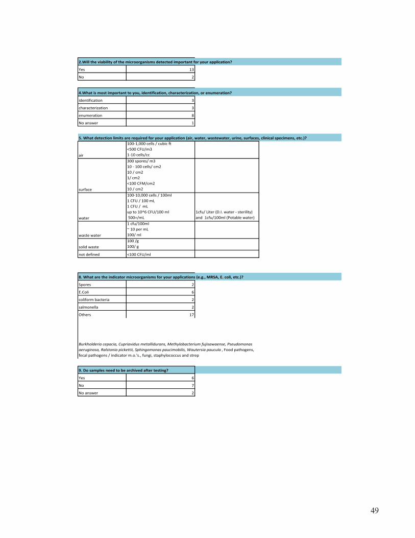
49
2.Will the viability of the microorganisms detected important for your application?
Yes 13
No 2
4.What is most important to you, identification, characterization, or enumeration?
identification 3
characterization 3
enumeration 8
No answer 1
5. What detection limits are required for your application (air, water, wastewater, urine, surfaces, clinical specimens, etc.)?
air
100-1,000 cells / cubic ft<500 CFU/m31-10 cells/cc
surface
300 spores/ m310 - 100 cells/ cm210 / cm21/ cm2<100 CFM/cm210 / cm2
water
100-10,000 cells / 100ml1 CFU / 100 mL 1 CFU / mL up to 10^6 CFU/100 ml 500+/mL
1cfu/ Liter (D.I. water - sterility) and 1cfu/100ml (Potable water)
waste water
1 cfu/100ml~ 10 per mL 100/ ml
solid waste100 /g100/ g
not defined <100 CFU/ml
8. What are the indicator microorganisms for your applications (e.g., MRSA, E. coli, etc.)?
Spores 2
E.Coli 6
coliform bacteria 2
salmonella 2
Others 17
9. Do samples need to be archived after testing?
Yes 6
No 7
No answer 2
Burkholderia cepacia, Cupriavidus metallidurans, Methylobacterium fujisawaense, Pseudomonas aeruginosa, Ralstonia pickettii, Sphingomonas paucimobilis, Wautersia paucula , Food pathogens, fecal pathogens / indicator m.o.'s., fungi, staphylococcus and strep
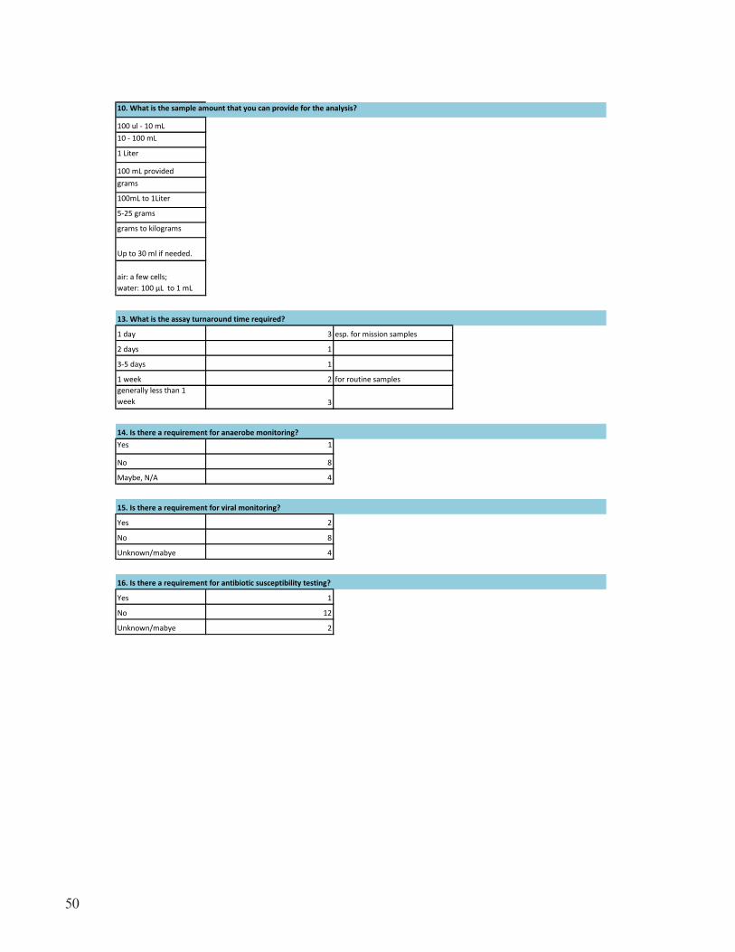
50
10. What is the sample amount that you can provide for the analysis?
100 ul - 10 mL10 - 100 mL
1 Liter
100 mL providedgrams
100mL to 1Liter
5-25 grams
grams to kilograms
Up to 30 ml if needed.
air: a few cells; water: 100 µL to 1 mL
13. What is the assay turnaround time required?
1 day 3 esp. for mission samples
2 days 1
3-5 days 1
1 week 2 for routine samplesgenerally less than 1 week 3
14. Is there a requirement for anaerobe monitoring?Yes 1
No 8
Maybe, N/A 4
15. Is there a requirement for viral monitoring?
Yes 2
No 8
Unknown/mabye 4
16. Is there a requirement for antibiotic susceptibility testing?
Yes 1
No 12
Unknown/mabye 2

51
APPENDIX C—BIOGRAPHY AND ABSTRACT OF WORKSHOP PRESENTERS
Appendix C contains the biographies and an abstract for each presenter at the workshop.

52
Microbiological Monitoring of the ISSDuane L. Pierson, Ph.D.Chief MicrobiologistNASA, Houston, TX
The current microbial monitoring plan of the International Space Station (ISS) resulted from lessons learned from the Space Shuttle Program and the NASA-Mir Program. The major sources of microbiological risks to the crew are associated with the food, water, air, surfaces, payloads, animals, and crewmembers. Adverse effects of microorganisms are many and can jeopardize crew health and safety, the integrity of the ISS, and ultimately mission success. Acceptability limits for microorganisms in food, water, air, and surfaces were established, and monitoring is conducted to ensure compliance with requirements. Monitoring begins during the preflight period and includes the air and surfaces of spacecraft and modules destined for the ISS. Potable water, food, and hard-ware are monitored to ensure safety. In-flight monitoring of the breathing air, exposed surfaces, and potable water is conducted at scheduled times. Monitoring results are compared with estab-lished acceptability limits for microorganisms. In addition to sensitivity and specificity, monitor-ing technology and in-flight procedures are subject to restrictions on the following parameters: power, mass, volume, microgravity compatibility, expertise required for operation, expendables and resupply requirements, and many others. Current monitoring equipment onboard the ISS are inexpensive, small, portable with no or low power requirements, culture based technologies devel-oped during the NASA-Mir Program. These technologies may not be suitable for long exploration missions. Crews are major providers of bacteria and along with common environmental bacteria and fungi are the most commonly isolated microorganisms from the air and surfaces of the ISS. Gram negative bacteria commonly associated with drinking water are the most commonly isolated bacteria from the US and Russian water systems. With some exceptions, the levels of bacteria and fungi isolated from the air, surfaces, and water are typically within the internationally agreed upon acceptability limits. However, contamination levels above the acceptability limits have occurred and these anomalies were addressed on a case by case basis to remediate the unacceptable contamina-tion. Lessons learned from the first 10 years of ISS operations should be applied to future monitor-ing plans for spacecraft and space habitats.
Dr. Pierson, a Fellow in the American Academy of Microbiology, serves as NASA’s Chief Microbi-ologist. He obtained his Ph.D. in Biochemistry from Oklahoma State University and following an NIH Postdoctoral fellowship was on faculty at Baylor College of Medicine (Dept. of Microbiology and Immunology) for 10 years before joining NASA in 1980. He is responsible for formulating, developing, and implementing NASA’s microbiology program for current and future human explo-ration of space. His major responsibilities include both operational and research activities to ensure the health, safety, and optimum performance of the astronauts. His broad experience results in his participation in activities ranging from planetary protection and astrobiology to environmental health. He has been actively involved in microbiological and biochemical research for 40 years at Baylor College of Medicine in Houston and at NASA’s Johnson Space Center. He has published over 150 manuscripts in a wide variety of peer-reviewed journals. He also has 20 book chapters, 15 NASA Tech Briefs, and 2 patents. His leadership in space microbiology has made Dr. Pierson a well-recognized figure throughout NASA, the academic community, and the aerospace indus-try. He has directed a highly productive research program with strong collaborations with many
53
U.S. and international scientists. His research interests focuses on host-microbe interactions in the space environment. Specific interest is the reactivation of herpes viruses in astronauts, and his findings have been extended to treatment of shingles patients and are currently used in medical diagnoses. His studies include ground-based analogs of space flight including the Antarctic, the Aquarius undersea habitat, human-rated closed-chambers, and others; he is also experienced with flight investigations on the Space Shuttle, Soyuz, Russian Space Station Mir, and the International Space Station. Over three decades NASA has recognized Dr. Pierson’s accomplishments through many awards including the Medal for Exceptional Scientific Achievement and the Certificate of Commendation. The astronauts recognized his contributions in environmental health with the highly coveted Silver Snoopy Award. He maintains academic appointments with Baylor College of Medicine (Houston), the University of Houston, and the University of Texas Medical Branch (Galveston). At the international level, he was elected in 1998 by his academic peers to Fellow in the American Academy of Microbiology.
54
Cutting-Edge Genomic Approaches for Microbial DetectionKostas Konstantinidis, Ph.D.Assistant Professor of Civil & Environmental Engineering and of Biology (Adjunct)Center for Computational Genomics, Georgia Institute of Technology, Atlanta, GA
The great majority of microbial cells in natural settings, >98-99% in some habitats, are resistant to cultivation in the laboratory (the so called “uncultivated majority”), severely limiting the usefulness of culture-based approaches for microbial monitoring. Culture-independent approaches provide means to detect and characterize the uncultivated majority but they also have their own inherent limitations such as the requirement for optimization depending on the type of sample, lack of sensitivity or resolution at the species level, and frequently suffer from increased experimental error or noise. In this presentation, I will provide several representative examples from our own research where we have employed the most promising culture-independent technologies such as qPCR, 16S rRNA gene pyrosequencing, and shotgun metagenomics, to detect microbes in air, water and soil samples. Although each technology is characterized by its own, usually technology-specific, limitations, a combination of selected technologies may represent reliable means for robust and quantitative microbial monitoring.
Dr. Kostas Konstantinidis joined the School of Civil and Environmental Engineering, at Georgia Institute of Technology as an Assistant Professor in November, 2007. He also holds a courtesy appointment in the School of Biology and is program faculty in the Center for Bioinformatics and Computational Genomics and in the Bioengineering Graduate Program. Prior, he was a Post-doctoral Fellow in the Department of Civil and Environmental Engineering at the Massachusetts Institute of Technology, Cambridge, MA. Dr. Konstantinidis received his BS in Agriculture Sci-ences from the Aristotle University of Thessaloniki (Greece) in 1999. He continued his studies at the Center for Microbial Ecology at Michigan State University (East Lansing, MI) under the supervision of Prof. James M. Tiedje, where he obtained a PhD in 2004. His PhD studies were fully supported by the Bouyoukos Fellowship program and were devoted in advancing our under-standing of the ecology and physiology of soil bacteria through the comparative analysis of their whole-genome sequences. Dr. Konstantinidis education and research interests are at the interface of environmental microbiology with engineering, genomics and computational biology, with the overarching goal to broaden understanding of the genetic and metabolic diversity of the smallest organisms on the planet, the bacteria and the archaea, and to explore this biodiversity for biotech-nological applications. Dr. Konstantinidis has already received several national and international distinctions and awards for his work such as the 2010 International Skerman Award of the World Federation for Culture Collections. He is a member of the American Society for Microbiology (ASM), the International Society for Microbial Ecology (ISME), the Association of Environmen-tal Engineering and Science Professors (AEESP), and the editorial board of the journal of Applied and Environmental Microbiology.

55
Challenges in Sampling Environmental MicrobesStephen A. Morse, MSPH, Ph.D.Centers for Disease Control and Prevention, Atlanta, Georgia
Microbial contamination of space vehicle environments can result in a number of deleterious effects on crew health, and can adversely affect the operations of critical subsystems. The ability to rapidly enumerate microbial contaminants may be the key to controlling their impact in the confined environment of a spacecraft. While culture has been traditionally used for the character-ization of environmental samples, there have been tremendous advances in non-culture methods such as sequenced-based technologies that can be used to characterize and identify the microorgan-isms in a particular environment. In spite of these advances, the data generated from such methods only reflects what was present in the initial sample and may not be representative of the particular environment as a whole. Sampling is important but probably underappreciated. Environmental samples are collected for a number of purposes including: to determine the presence and viability of an agent; to determine the extent and degree of contamination; to support medical treatment and clean-up decisions; and, ascertainment of risk. There are a number of technical challenges that must be addressed when collecting a sample. Examples of these are: location of the micro-organisms (sampling approach); surface characteristics (non-porous or porous); collection device (swab, wipe, vacuum); controls; storage and transport of samples; and detection methods (culture versus non-culture, semi-quantitative versus qualitative, viable versus non-viable). Other issues that must be considered when evaluating the results are the collection efficiency of the sampling device, losses during transport, recovery efficiency during sample processing, and limit of detection in the analysis phase. Consideration of these issues is especially important for evaluating negative results and differentiating between those that are true negatives and those that are false negatives. Physico-chemical characteristics of the sampling device are also important considerations. For example, the composition of the sampling device (cotton, polyester, rayon, or macro foam swab), whether it is used dry or moist as well as the moistening agent, absorbance capacity, and charge will influence its recovery efficiency. Organism characteristics can also affect recovery. Some swab materials are bet-ter at collecting gram negative microorganisms while others are better for gram positive bacteria or spores. It is difficult to determine risk from the presence of a particular environmental microorgan-ism without knowing whether the sample that was collected and analyzed was truly representative of the contribution of the microbe to the total microbial population.
Stephen A. Morse graduated from San Jose State University in 1964 with a B.A. in microbiology. He attended graduate school at the University of North Carolina at Chapel Hill where he received his M.S.P.H. (1966) in environmental chemistry and biology and a Ph.D. (1969) in microbiology. After postdoctoral training in microbial genetics at the University of Georgia, Dr. Morse joined the faculties of the Harvard School of Public Health and Medical School as an assistant professor. In 1974, he joined the faculty of the Department of Microbiology and Immunology at Oregon Health Sciences University where he subsequently attained the rank of Professor of Micro-biology. In 1984, he joined the CDC as Director of the Sexually Transmitted Diseases (STDs) Research Program, National Center for Infectious Diseases (NCID); and in 1996, became the Associate Director for Science of the newly created Division of AIDS, STDs and Tuberculosis Laboratory Research. From 1999 - 2007, he served as the Associate Director for Science, Division of Bioterrorism Preparedness and Response where he has worked on national and international

56
bioterrorism-related issues. In 2008, he became the Associate Director for Environmental Microbi-ology, National Center for Preparedness, Detection, and Control of Infectious Diseases, CDC. He also has an appointment to the Senior Biomedical Research Service of the U.S. Public Health Ser-vice and has published over 280 articles, books and chapters. He has received numerous awards and other forms of recognition for his achievements including: the Mary Poston Award from the North Carolina Chapter of the American Society for Microbiology; the CDC, U.S. Public Health Ser-vice, and Department of Health and Human Services EEO Achievement Awards; the McLaughlin Award from the University of Texas Medical School at Galveston; the Harriet Hylton Barr Out-standing Alumnus Award from the School of Public Health of the University of North Carolina at Chapel Hill; the Lea and Harrison Latta Lectureship in the Department of Clinical Pathology, University of California at Los Angeles; the Molecular Virology and Microbiology Distinguished Lectureship at the University of Pittsburgh; and a Distinguished Lectureship at Hanover College. He is currently an Adjunct Professor at Emory University School of Medicine, a past member of the Board of Governors of the American Academy of Microbiology, and has served on several Scientific Advisory Boards as well as the FBI Scientific Working Group for the forensic analysis of chemical, biological, radiological and nuclear terrorism (SWGCBRN).
57
State of the Art Microbiological Monitoring in the Pharmaceutical Industry: Translational OpportunitiesRichard Levy, Ph.D.Senior Vice President, Scientific and Regulatory AffairsParenteral Drug Association, Bethesda, MD
Microbiological testing plays an ever increasing role in delivering high quality drug products to patients, whether it’s practiced in the pharmaceutical laboratory and in the manufacturing envi-ronment. In response to the drive for continuous improvement and further economies, a variety of new methodologies have emerged in recent years which automate existing methods, make use of surrogate markers for growth, or are based on wholly new technologies. These new methodolo-gies offer significant improvements in terms of the speed, accuracy, precision and specificity with which testing can be performed. However, in spite of the limitations of current culture methods, acceptance of new and potentially superior methods has only started to gain momentum within the pharmaceutical, biotechnology and medical device industries. We believe this continues to be due in part to a lack of clear guidance regarding the demonstration of their equivalence to existing methods acceptable to regulatory agencies and validation of the equipment associated with the new methods. In any case, many of these new rapid methods may have a role to play in other applica-tions including missions in space where we want to know the impact of microorganisms on space travel, as well as what microorganisms might be waiting to be discovered.
Richard Levy is currently Senior Vice President of Scientific and Regulatory Affairs at the Paren-teral Drug Association (PDA) (www.pda.org) in Bethesda Maryland. In this capacity, he is respon-sible for directing and managing the scientific, technical, regulatory affairs and quality activities of a 9,500 member association focused on pharmaceutical and biotechnological manufacturing. He is also responsible for the Association publications: the PDA Journal of Pharmaceutical Science and Technology, PDA Technical Reports and the PDA Letter (association magazine). Dr. Levy’s other key activities include working with PDA members to prepare and write consensus positions on proposed international regulations and guidance documents, and developing the scientific content of PDA’s global meetings and forums. In this capacity, Dr. Levy works directly with global indus-try associations such as A3P, AAMI, ASTM, IABS, ISPE, PhRMA and R3-Nordic to coordinate and harmonize scientific and regulatory activities involving FDA and other international regula-tory authorities (EMA, FDA, MHRA, MHLW, SHFDA) and standard setting organizations (e.g., AAMI, EDQM, ICH, ISO, and USP). Prior to joining PDA in 2005, Dr. Levy was Corporate Vice President and General Manager of PAREXEL Consulting (www.parexel.com) , a newly formed PAREXEL INTERNATIONAL business unit created by the merger of KMI, Barnett, and World-wide Regulatory Affairs of PAREXEL. Dr. Levy joined KMI/PAREXEL International in January of 2001 as Vice President of Consulting Services. Prior to joining KMI, Dr. Levy was with MIL-LIPORE Corporation (www.millipore.com) for 16 years in a variety of Business, R&D, Regula-tory and Quality Systems senior management positions. He was Chair-elect of the Parenteral Drug Association (PDA) Board of Directors, and served on that Board from 1999-2005. Dr. Levy is active in industry programs and task forces on aseptic processing, process validation, microbial and viral clearance, regulatory affairs and quality systems and has made more than 100 presentations at various national/international industry symposia. He has published articles on biotechnology, asep-tic processing, filter validation, sterile filtration, microbial retention testing, and viral clearance in

58
American Pharmaceutical Review, BioPharm, BioProcess International, Pharmaceutical Technol-ogy, PDA J. Parenteral Science and Technology, J. American Water Works Association, Blow-Fill-Seal Society Journal, and BioProcess International. He has also authored chapters in textbooks on these subjects. Dr. Levy was Chairman of the 2007 Committee of Revision for Technical Report No. 1, Validation of Moist Heat Sterilization and was a member of the PDA committee and co-author of “Sterilizing Filtration,” Technical Report No. 26 (1998 and 2008 Revisions). Dr. Levy is a member of the American Association of Pharmaceutical Scientists (AAPS), the International Society of Pharmaceutical Engineering (ISPE), the Parenteral Drug Association (PDA), the Regu-latory Affairs Professional Society (RAPS), the International Association for Biologicals (IABs), and the American Society for Microbiology (ASM). In 2006, Rich received the Frederick Char-leton Award for his work on the PDA Board of Directors, and in 2009 he received the first PDA Special Recognition Award for his work as a staff member. Dr. Levy received his B.A. in Biology from the Colby College (Waterville, ME), and an M.A. in Biology from Clark University located in Worcester, Massachusetts. He received his Ph.D. in Environmental Health Sciences from Worcester Polytechnic Institute in Worcester, Massachusetts.

59
Rapid Testing Methodologies - What to Consider?Charles DeibelPresident, Deibel LaboratoriesLincolnwood, IL
This presentation will focus on current Food Safety testing methodologies for Pathogenic bacteria and toxins, potentially found in Ready To Eat (RTE) foods and environmental samples. We will review state-of-the-art methods for NASA, concerning detection of microorganisms in the environ-ment, water and in foods. During the presentation we will also examine sample preparations, detec-tion limits, relative false negatives rates, complex matrices (spices, chocolates, etc) and the pro’s and con’s of each technology. Food microbiology is important for crew health and our space missions must be free of food borne illnesses.
Charles Deibel is President of Deibel Laboratories, Inc., an internationally-recognized firm provid-ing food safety testing, quality control evaluations, scientific consulting and training for industry leading food manufacturers, as well as family run operations. He is an industry advocate, having testified in front of the House Energy and Commerce Committee on important food safety con-cerns. Charles is a HACCP Certified Lead Instructor, Process Authority, and an expert in pathogen remediation in plants. He conducts microbial challenge studies, process validations, shelf life deter-minations and GFSI system development for Deibel clients. He has always been very hands-on in assisting clients with their food safety concerns, spending a significant portion of his time in a variety of food plants.

60
Environment and Clinical Microbiology NeedsMarc W. Mittelman, Ph.D.Senior Managing ScientistExponent/Harvard UniversityNatick, MA
There are numerous microbiological monitoring approaches that may be adaptable for use in long-term space travel. However, there are also a number of challenges associated with the enumera-tion and identification of environmental and clinical microorganisms (bacteria, fungi, viruses) in space. These include weight and energy limitations, risks of crew exposures to test reagents and microorganisms, waste disposal issues, and problems associated with operating in a microgravity environment. Additionally, the growth, virulence, and antimicrobial susceptibility of some micro-organisms appear to be influenced by microgravity, which could present problems in characterizing isolates. Traditionally, microbiological monitoring of environments and crew has focused on bac-teria and (less frequently) fungi using culture-based techniques. Fortunately, there are a number of molecular, biochemical, and physicochemical test systems that may be adaptable for use in a space environment. A review of environmental and clinical microbiological needs was conducted to serve as the basis for establishing the rationale for selecting candidate microbiological technologies for further evaluation. A NASA-sponsored Workshop has also been organized to develop selection criteria and to define key attributes required for environmental and crew monitoring of microor-ganisms.
Dr. Mittelman has over 25 years experience in academia, industry, and consulting. The majority of his research and consulting work has been in the area of microbiological contamination detec-tion and mitigation for pharmaceutical/biotechnology, clinical, and industrial applications. Dr. Mittelman has developed test systems and analytical methods for monitoring biofilm development on engineered materials, and has conducted a number of studies on the contamination of criti-cal components from the International Space Station for NASA. He provides consulting services in the fields of biological fouling (biofouling), microbially influenced corrosion (MIC), purified water system contamination control, medical device-related infections, and antimicrobial coatings development. He has conducted contamination control investigations for industrial and biophar-maceutical applications, focusing on process- and product-compatible solutions. He has designed preventative and remedial strategies for controlling microbiological contamination in products and process operations ranging from high purity water systems to marine structures. He has also pro-vided new product development support to large and small medical device companies, with a focus on biofilm prevention and infection control strategies. Dr. Mittelman was previously an Associate Professor in the Faculties of Medicine and Dentistry, and director of the Centre for Infection and Biomaterials Research (CIBR), at the University of Toronto. He has also worked as a microbiolo-gist in the pharmaceutical industry, directed microbiology and medical device engineering consult-ing practices, and has served as an expert witness in product liability cases. Dr. Mittelman is the author of 80 scientific papers and books, and has lectured extensively on microbiological contami-nation control. He has numerous patents in the microbiological monitoring and related fields. In addition to his consulting work at Exponent, he is a visiting scientist at Harvard University, School of Engineering and Applied Sciences.

61
Infection Control Challenges in Space TravelLeonard Mermel, DO, ScM, AM (Hon), FSHEA, FIDSA, FACP Professor of Medicine, Warren Alpert Medical School of Brown University Medical Director, Department of Epidemiology & Infection Control, Rhode Island Hospital, Providence, RI
The conditions of space travel create a unique challenge to infection prevention. Ground-based and in-flight research has demonstrated multiple alterations in the immune system that likely increase the risk of infection caused by intracellular and extracellular pathogens. Additionally, changes in microbial flora occur reflected by increased S. aureus colonization of the skin and upper airway and an increase in the bioburden of aerobic gram-negative in the GI tract. At the same time, bacteria undergo changes in microgravity leading to increased virulence, biofilm formation, and resistance to antimicrobial agents. Countermeasures that may mitigate risk of infection involve pre-travel interventions such as vaccination and screening for: S. aureus and group A streptococcal car-riage; latent infections; acquired or inherited immunodeficiencies; decolonization of S. aureus and group A Streptococcus; gamma irradiation of food and deliverables. Preparation of the contain-ment vessel includes: filtration and/or decontamination of air & water; differential pressures of air from bathroom to other areas and docking vessels to main vessel; and antimicrobial surface treat-ment of high-touch inanimate objects. Interventions during travel include hand hygiene enhance-ments, proper mask use for containment of respiratory infections, vitamin D supplementation, and possibly microbial interference. If illness or injury requires intravenous medications or surgery, application of evidence-based preventative strategies will minimize the risk of infectious complications.
Leonard A. Mermel, D.O., Sc.M., AM (Hon), FACP, FIDSA, FSHEA is Professor of Medicine, Warren Alpert Medical School of Brown University and Medical Director, Dept. of Epidemiology & Infection Control, Rhode Island Hospital. Dr. Mermel was a Technical Expert Panel Member of the Medicare Patient Safety Monitoring System, US Dept. of Health and Human Services. He was the 2005 President of the Society for Healthcare Epidemiology of America (SHEA). He is a past recipient of the Ralph A. Kinsella, Sr. Memorial Tribute Award from St. Louis University Hospitals for outstanding qualities of work, leadership, and ability as a house staff officer, the SHEA Young Investigator Award, and the Brown Medical School Department of Medicine Chair-man’s Award for Outstanding Teaching. Dr. Mermel is also an elected member of Delta Omega, the Honorary Public Health Society. He has been repeatedly selected by his peers to be included as one of The Best Doctors in America, America’s Top Doctors, The Best of Rhode Island - Infec-tious Diseases Physician, Castle Connolly Top Doctor and he’s listed in the Guide to America’s Top Physicians, Who’s Who in America and Who’s Who in Science & Engineering. Dr. Mermel has co-authored two US guidelines dealing with prevention and management of intravascular catheter infections and he has authored or co-authored over 200 articles, textbook chapters, and abstracts dealing with infection control and infectious diseases. He has lectured at the National Institutes of Health, Institute of medicine, and internationally on infectious disease issues. His research interest is the prevention of hospital-acquired infections.
62
Environmental Pathogens, Emerging Pathogens and Biofilms in Microgravity?Timothy E. Ford, Ph.D.Professor, Dean, and Vice President ResearchUniversity of New England, Portland, ME
Over the last couple of decades, those of us interested in drinking water microbiology have been intrigued by the environmental pathogens that not only survive, but are often thought to prolif-erate in drinking water, often associated with biofilms. These pathogens are often protected by those biofilms from most forms of water treatment. While the environmental pathogens primarily cause a health burden to the immunologically compromised, what happens in micro gravity is not fully understood. Is the astronaut’s immunity sufficiently compromised that these pathogens may become a significant health risk? The other, not unconnected question is what constitutes an emerg-ing waterborne pathogen. Are environmental pathogens likely to increase in virulence under micro-gravity? At what point does a non-pathogenic microbe become pathogenic? Will rates of transfer of virulence factors increase? Using Earth-based examples, this presentation will raise more questions than answers, but highlight the fact that there is more to risk management of human health than simply monitoring and control of known pathogens.
Tim Ford is Vice President for Research and Dean of Graduate Studies at the University of New England. He obtained his PhD in aquatic microbiology from the University College of North Wales. After completing a postdoctoral fellowship at Harvard University, he joined the faculty of the Harvard School of Public Health where he both founded and directed the School’s Program in Water and Health. In 2002, he moved to Montana State University as Professor and Department Head of Microbiology, where he directed Montana’s NIH-funded Idea Networks for Biomedi-cal Research Excellence. He has authored or co-authored ~160 peer-reviewed publications, books, book chapters and reports, and has both directed and participated in water quality related projects in the US, Canada, the UK, Mexico, India, Russia and the Philippines. He holds a Concurrent Professorship at Nanjing University, PR China, and was the first recipient of the Gen-Probe Joseph Award for exemplary leadership and service in the field of public health (2006).
63
Monitoring for Microorganisms Important in Healthcare-Associated Infections: Translational Opportunities for the Space ProgramRodney M. Donlan, Ph.D.Division of Healthcare Quality PromotionCenters for Disease Control and Prevention, Atlanta, GA
Healthcare-associated infections (HAIs) are infections that patients acquire while receiving treat-ment for medical or surgical conditions in all settings of care, and may be associated with use of medical devices, complications following surgery, transmission between healthcare workers and patients, or the result of antibiotic overuse. Microorganisms associated with HAIs may originate from the native microbial communities of human skin or other body sites, or from environmen-tal sources including potable water systems. The most common pathogens, accounting for >80% of any HAIs are coagulase-negative Staphylococcus species, Staphylococcus aureus, Enterococ-cus species, Candida species, Escherichia coli, Pseudomonas aeruginosa, Klebsiella pneumoniae, Enterobacter species, Acinetobacter baumanii, and Klebsiella oxytoca; in some cases these organ-isms may also be multi-drug resistant. The association between biofilm formation and HAIs is well established. Biofilms are ubiquitous in nature and consist of surface-associated microbial commu-nities that are enclosed in an extracellular polymeric substance matrix which often contains noncel-lular materials. Biofilms may form on a wide variety of surfaces including living tissues, indwelling medical devices, industrial or potable water system piping, or soil and aquatic systems. The process of biofilm formation is complex and depends upon the physical/chemical properties of the sur-face, presence of conditioning films, hydrodynamics, physical and chemical properties of the liquid milieu, and properties of the individual microbial cells. Once established, biofilms are difficult to eradicate, and the associated microorganisms exhibit tolerance to a wide spectrum of antimicrobial agents. The biofilms in potable water systems may be an important source of contamination in the healthcare environment. Potable water distribution systems contain diverse microbial communities, and may provide a niche for the survival and dissemination of opportunistic pathogens that have been associated with healthcare associated infections in certain patient populations. Free-living protozoa (FLP) may also associate with potable water biofilms, and a number of opportunistic pathogens may infect and amplify within protozoa. Association with FLP may provide a mecha-nism for increased tolerance to disinfectants and dissemination of these organisms within the water supply. It can be expected that organisms from biofilms in potable or process water systems could contaminate the spacecraft environment, and potentially impact human health. Characterizing spacecraft water system microbial communities and determining those key variables affecting bio-film formation and growth will require reliable devices for monitoring these systems, and reproduc-ible protocols for recovery and analysis of the biofilm. Molecular, microscopic, and in some cases culture-based methods could then be utilized to characterize and quantify the biofilms in these systems.
Dr. Rodney Donlan leads the Biofilm Laboratory in the Division of Healthcare Quality Promo-tion at the Centers for Disease Control and Prevention in Atlanta, GA, a position he has held since joining the CDC in 1998. He has been involved in research on microbial biofilms for over 30 years, and has collaborated successfully with researchers from a number of academic centers and private industry. Current projects in the Biofilm Lab are investigating the formation and control of bio-
64
films on central venous catheter needleless access devices, use of bacteriophage to prevent biofilms on indwelling medical devices, and the role of biofilms in the survival and disinfection of opportu-nistic pathogens in potable water systems. He received his B.S. and M.S. degrees from Virginia Tech and his Ph.D. from Drexel University.

65
APPENDIX D—WORKSHOP ATTENDEES, AGENDA, AND PRESENTATIONS
2011 NASA Microbiology Workshop AttendeesApril 19, 2011
Johnson Space Center, Houston
Name AffiliationRebekah (Bekki) Bruce NASA JSC Microbiology LaboratoryVictoria Castro NASA JSC Microbiology LaboratoryRodney M. Donlan Centers for Disease Control and PreventionTodd Elliott NASA/Wyle/JSC Microbiology LaboratoryJohn W. Fisher NASA Ames Research CenterDarrell Jan JPLAnna Kallay Orion Project/Lockheed MartinAriel V. Macatangay NASA/JSC/SF2/Environmental Factors BranchStephen A. Morse Centers for Disease Control and PreventionC. Mark Ott NASA JSC Microbiology LaboratoryCherie Oubre Wyle/NASA JSCDuane L. Pierson NASA JSCHank Rotter NASA/NESC/JSCMelanie J. Smith EASI/Wyle LaboratoriesRichard F. Strayer KSC-ESC – Team QNAWing C. Wong NASA/WyleMarc W. Mittelman ExponentKostas Konstantinidis GA TechTim Ford Univ. New EnglandLeonard Mermel Brown University/Rhode Island HospitalRichard Levy Parenteral Drug AssociationCharles Deibel Deibel LaboratoriesMonsi Roman NASA MSFCPatricia Catauro Lockheed MartinMichael S. Roberts QNALeticia (Letty) Vega NASA JSC (ESCG)
Daniel J. BartaLife Support and Habitability Systems Branch, Mail Code EC3
Jeff McQuillan Life Support & Habitation Systems Project OfficeTorin McCoy NASA JSCLynn J. Rothschild NASA Ames Research Center
Chantel Whatley ESCG/GeoControls

66

67
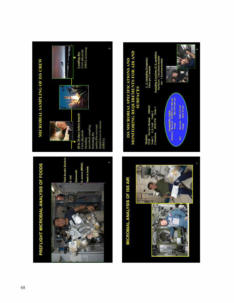
68

69

70
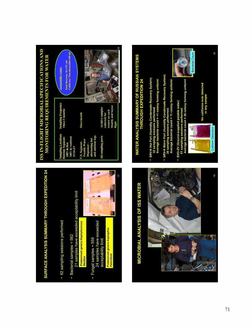
71

72

73

74

75

76

77

78

79
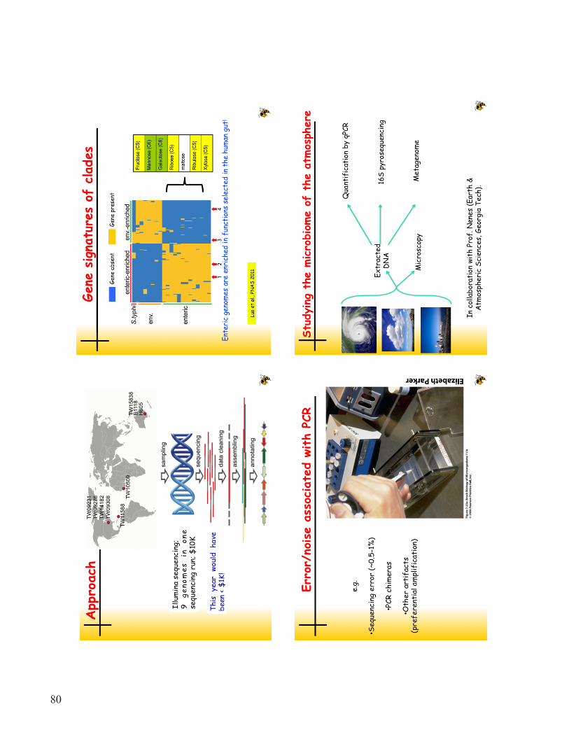
80

81

82

83

84

85
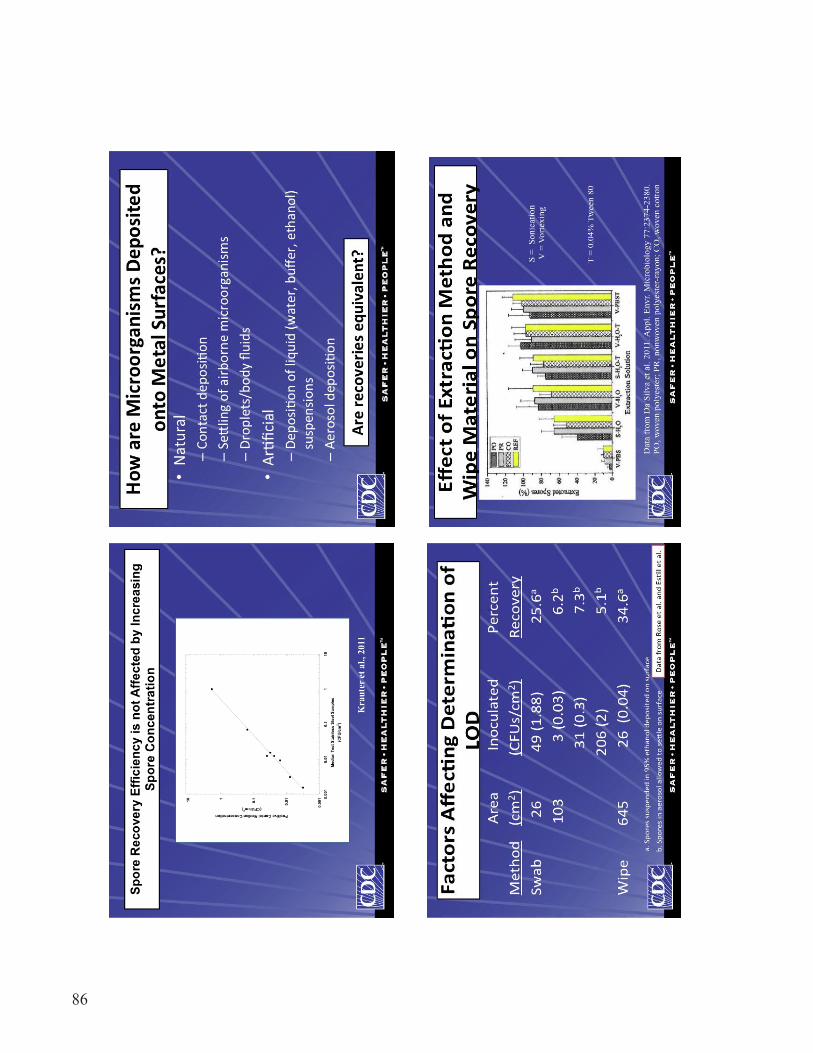
86

87

88

89

90

91
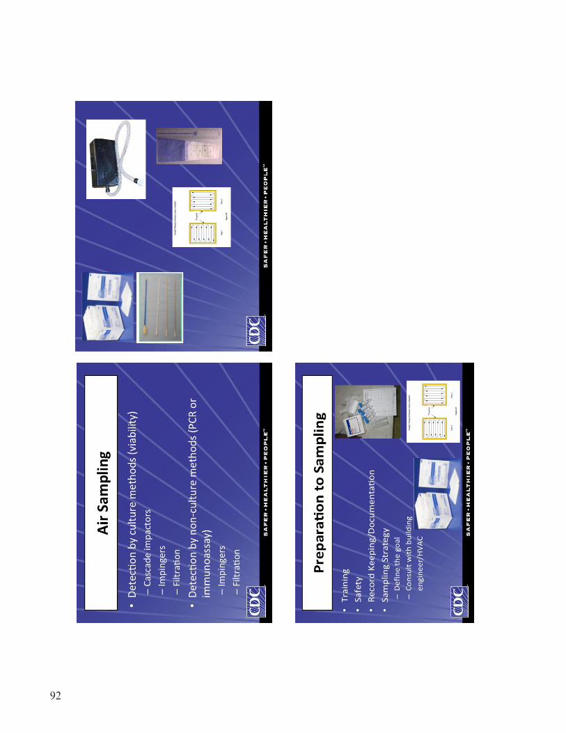
92

93

94
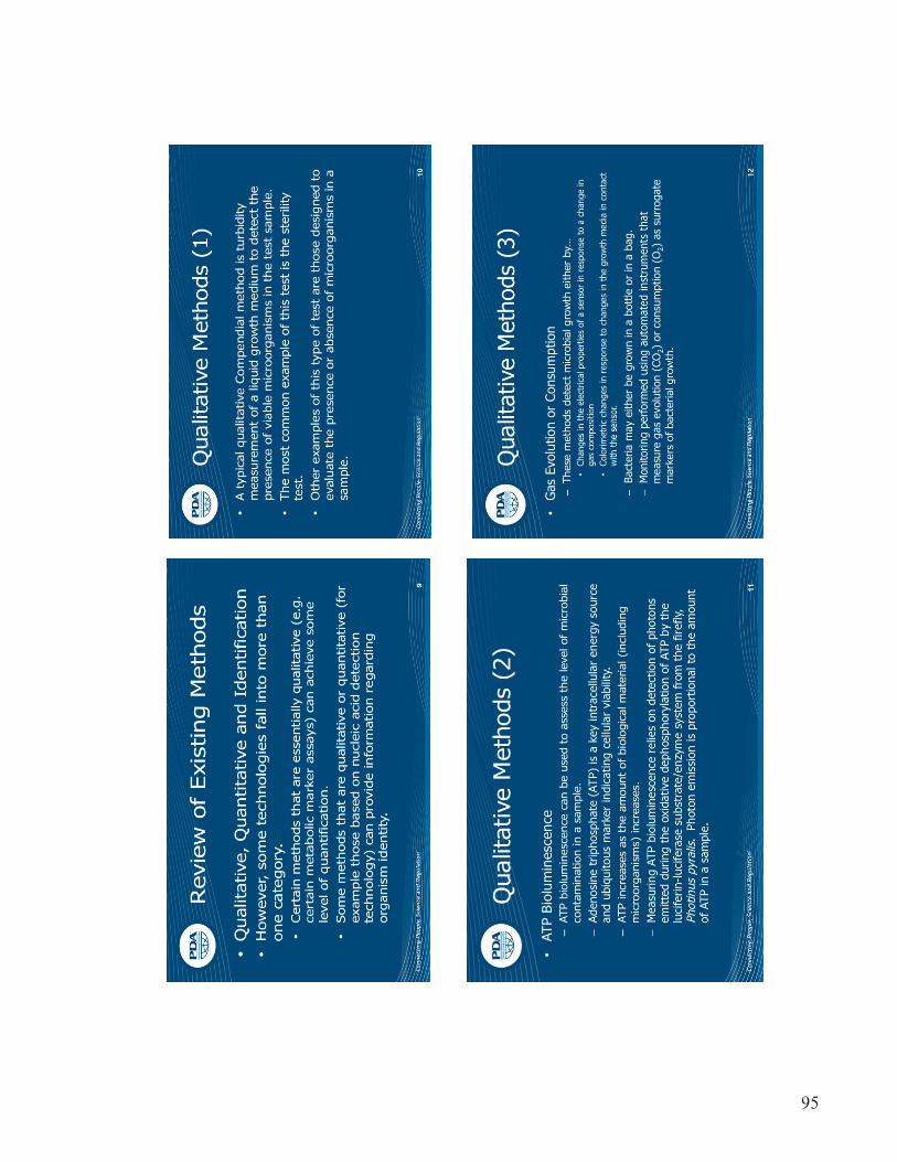
95

96

97

98

99

100

101

102

103
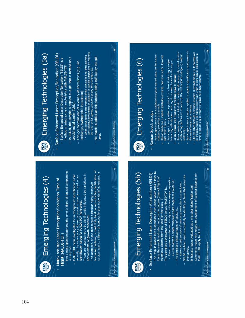
104

105

106

107

108

109

110

111

112

113
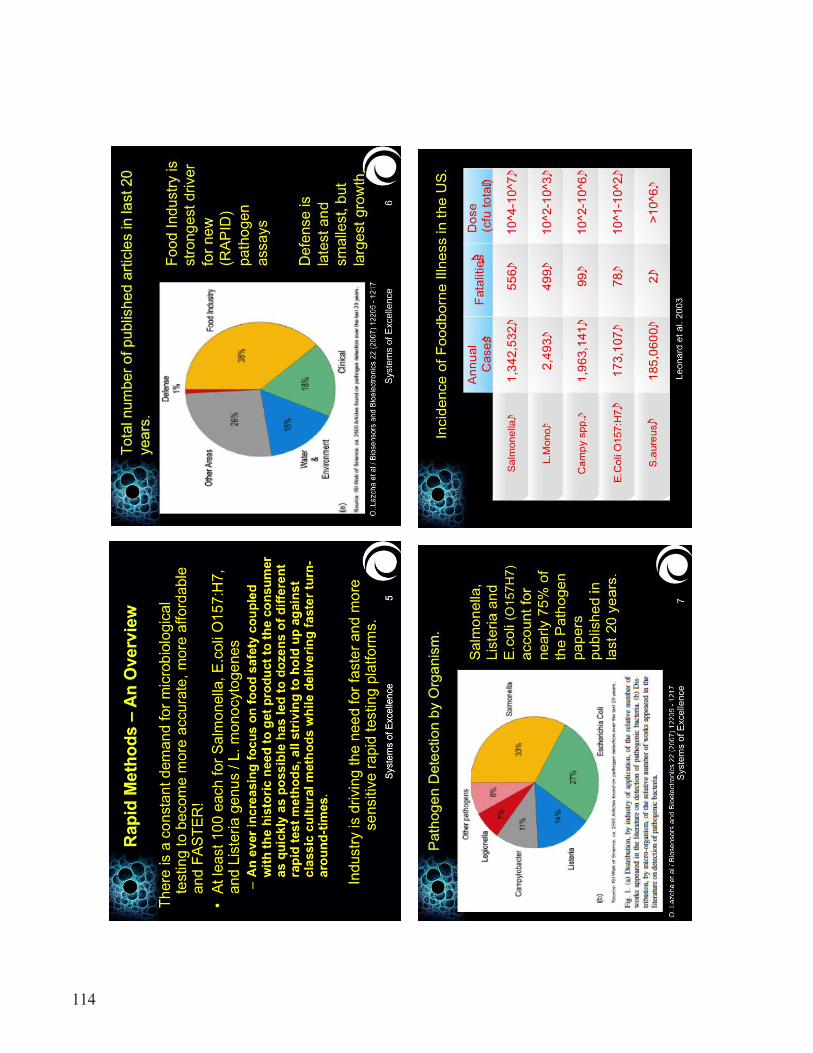
114

115

116

117
118
119

120

121
122
123

124

125

126
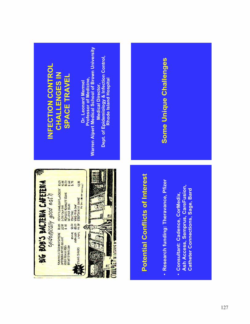
127

128
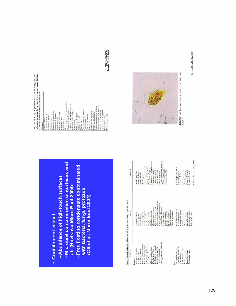
129
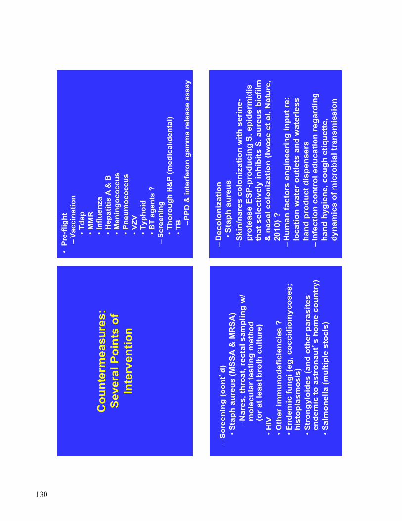
130

131

132

133

134

135
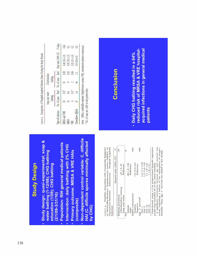
136

137

138
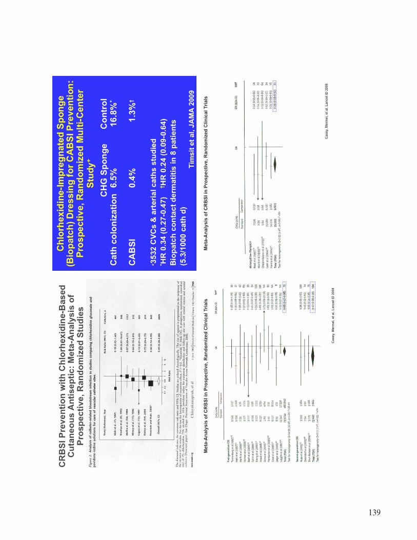
139
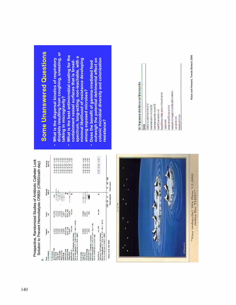
140

141

142

143
144

145

146

147

148

149

150

151

152

153

154

155

156

157
158

159

160

161
REFERENCES
1. Horneck, G.; Klaus, D.M.; and Mancinelli, R.L.: “Space Microbiology,” Microbiol. Mol. Biol., Rev., Vol. 74, No. 1, pp. 121–56, 2010.
2. Staley, J.T.; and Konopka, A.: “Measurement of In Situ Activities of Nonphotosynthetic Microorganisms in Aquatic and Terrestrial Habitats,” Annu. Rev. Microbiol., Vol. 39: pp. 321–346, 1985.
3. Amann, R.I.; Ludwig, W.; and Schleifer, K.H.: “Phylogenetic Identification and In Situ Detec-tion of Individual Microbial Cells Without Cultivation, Microbiol. Rev., Vol. 59, pp. 143–169, 1995.
4. Mizunoe, Y.; Wai, S.N.; Takade, A.; and Yoshida, S.: “Restoration of Culturability of Starvation-Stressed and Low-Temperature-Stressed Escherichia Coli O157 Cells by using H2O2-Degrading Compounds, Arch. Microbiol., Vol. 172, pp. 63–67, 1999.
5. Stenuit, B.; Eyers, L.; Schuler, L.; et al.: “Emerging High-Throughput Approaches to Analyze Bioremediation of Sites Contaminated With Hazardous and/or Recalcitrant Wastes,” Biotechnol. Adv., Vol. 26, pp. 561–575, 2008.
6. Nebe-von-Caron, G.; Stephens, P.J.; Hewitt, C.J.; et al.: “Analysis of Bacterial Function by Multi-Colour Fluorescence Flow Cytometry And Single Cell Sorting,” J. Microbiol. Methods, Vol. 42, pp. 97–114, 2000.
7. Shah, H.N.; Keys, C.J.; Schmid, O.; and Gharbia, S.E.: “Matrix-Assisted Laser Desorption/ Ionization Time-of-Flight Mass Spectrometry and Proteomics: A New Era in Anaerobic Microbiology,” Clin. Infect. Dis., Vol. 35, pp. S58–S64, 2002.
8. Hsieh, S.Y.; Tseng, C.L.; Lee, Y.S.; et al.: “Highly Efficient Classification and Identification of Human Pathogenic Bacteria by MALDI-TOF MS,” Mol. Cell. Proteomics, Vol. 7, pp. 448–456, 2008.
9. Amann, R.; and Fuchs, B.M.: “Single-Cell Identification in Microbial Communities by Improved Fluorescence In Situ Hybridization Techniques, Nat. Rev. Microbiol., Vol. 6, pp. 339–348, 2008.

162
REPORT DOCUMENTATION PAGE Form ApprovedOMB No. 0704-0188
The public reporting burden for this collection of information is estimated to average 1 hour per response, including the time for reviewing instructions, searching existing data sources, gathering and maintaining the data needed, and completing and reviewing the collection of information. Send comments regarding this burden estimate or any other aspect of this collection of information, including suggestions for reducing this burden, to Department of Defense, Washington Headquarters Services, Directorate for Information Operation and Reports (0704-0188), 1215 Jefferson Davis Highway, Suite 1204, Arlington, VA 22202-4302. Respondents should be aware that notwithstanding any other provision of law, no person shall be subject to any penalty for failing to comply with a collection of information if it does not display a currently valid OMB control number.PLEASE DO NOT RETURN YOUR FORM TO THE ABOVE ADDRESS.
1. REPORT DATE (DD-MM-YYYY) 2. REPORT TYPE 3. DATES COVERED (From - To)
4. TITLE AND SUBTITLE 5a. CONTRACT NUMBER
5b. GRANT NUMBER
5c. PROGRAM ELEMENT NUMBER
6. AUTHOR(S) 5d. PROJECT NUMBER
5e. TASK NUMBER
5f. WORK UNIT NUMBER
7. PERFORMING ORGANIZATION NAME(S) AND ADDRESS(ES) 8. PERFORMING ORGANIZATION REPORT NUMBER
9. SPONSORING/MONITORING AGENCY NAME(S) AND ADDRESS(ES) 10. SPONSORING/MONITOR’S ACRONYM(S)
11. SPONSORING/MONITORING REPORT NUMBER
12. DISTRIBUTION/AVAILABILITY STATEMENT
13. SUPPLEMENTARY NOTES
14. ABSTRACT
15. SUBJECT TERMS
16. SECURITY CLASSIFICATION OF:a. REPORT b. ABSTRACT c. THIS PAGE
17. LIMITATION OF ABSTRACT 18. NUMBER OF PAGES
19a. NAME OF RESPONSIBLE PERSON
19b. TELEPHONE NUMBER (Include area code)
Standard Form 298 (Rev. 8-98)Prescribed by ANSI Std. Z39-18
NASA Microbiology Workshop
M.C. Roman, Chair; D.L. Jan,* Vice-Chair; K.T. Konstantinidis,** Facilitator; and M.W. Mittelman,*** Facilitator
George C. Marshall Space Flight Center, Huntsville, AL 35812Georgia Institute of Technology, 311 Ferst Dr., ES&T, Rm. 3224, Atlanta, GA 30332–0512
National Aeronautics and Space AdministrationWashington, DC 20546–0001
Unclassified-UnlimitedSubject Category 54Availability: NASA CASI (443–757–5802)
Prepared for the Game Changing Development Office, Science & Technology Office*Jet Propulsion Laboratory, **Georgia Institute of Technology, ***Harvard University
M–1336
Conference Publication
NASA/CP—2012–217460
microbial monitoring, long-term space flight, pathogen tracking and monitoring, crew healthspacecraft environment, environmental monitoring
01–06–2012
UU 174U U U
STI Help Desk at email: [email protected]
STI Help Desk at: 443–757–5802
Long-term spaceflight is characterized by extraordinary challenges to maintain the life-supporting instrumentation free from microbial contamination and the crew healthy. The methodology currently employed for microbial monitoring in space stations or short spaceflights within the orbit of Earth have been instrumental in safeguarding the success of the missions, but suffers certain shortcomings that are critical for long spaceflights. This workshop addressed current practices and methodologies for microbial monitoring in space systems, and identified and discussed promising alternative methodologies and cutting-edge technologies for pursuit in the microbial monitoring that hold promise for supporting future NASA long-duration space missions.

The NASA STI Program…in Profile
Since its founding, NASA has been dedicated tothe advancement of aeronautics and spacescience. The NASA Scientific and Technical Information (STI) Program Office plays a keypart in helping NASA maintain this importantrole.
The NASA STI Program Office is operated by Langley Research Center, the lead center for NASA’s scientific and technical information. The NASA STI Program Office provides access to the NASA STI Database, the largest collection of aeronautical and space science STI in the world. The Program Office is also NASA’s institutional mechanism for disseminating the results of its research and development activities. These results are published by NASA in the NASA STI Report Series, which includes the following report types:
• TECHNICAL PUBLICATION. Reports of completed research or a major significant phase of research that present the results of NASA programs and include extensive data or theoretical analysis. Includes compilations of significant scientific and technical data and information deemed to be of continuing reference value. NASA’s counterpart of peer-reviewed formal professional papers but has less stringent limitations on manuscript length and extent of graphic presentations.
• TECHNICAL MEMORANDUM. Scientific and technical findings that are preliminary or of specialized interest, e.g., quick release reports, working papers, and bibliographies that contain minimal annotation. Does not contain extensive analysis.
• CONTRACTOR REPORT. Scientific and technical findings by NASA-sponsored contractors and grantees.
• CONFERENCE PUBLICATION. Collected papers from scientific and technical conferences, symposia, seminars, or other meetings sponsored or cosponsored by NASA.
• SPECIAL PUBLICATION. Scientific, technical, or historical information from NASA programs, projects, and mission, often concerned with subjects having substantial public interest.
• TECHNICAL TRANSLATION. English-language translations of foreign
scientific and technical material pertinent to NASA’s mission.
Specialized services that complement the STI Program Office’s diverse offerings include creating custom thesauri, building customized databases, organizing and publishing research results…even providing videos.
For more information about the NASA STI Program Office, see the following:
• Access the NASA STI program home page at <http://www.sti.nasa.gov>
• E-mail your question via the Internet to <[email protected]>
• Fax your question to the NASA STI Help Desk at 443 –757–5803
• Phone the NASA STI Help Desk at 443 –757–5802
• Write to: NASA STI Help Desk NASA Center for AeroSpace Information 7115 Standard Drive Hanover, MD 21076–1320

NASA/CP—2012–
NASA Microbiology Workshop,April 19, 2011M.C. Roman, ChairpersonMarshall Space Flight Center, Huntsville, Alabama
D.L. Jan, Co-ChairJet Propulsion Laboratory, Pasadena, California
K.T. Konstantinidis, FacilitatorGeorgia Institute of Technology, Atlanta, Georgia
M.W. Mittelman, FacilitatorHarvard University, Cambridge, Massachusetts
February 2012
National Aeronautics andSpace AdministrationIS20George C. Marshall Space Flight CenterHuntsville, Alabama 35812
Proceedings of a Workshop sponsored by theNational Aeronautics and Space Administrationheld at Johnson Space Center, Houston, Texas,April 19, 2011