Microbiology Research - Academic Journals
65
African Journal of Microbiology Research Volume 8 Number 41, 8 October, 2014 ISSN 1996-0808
-
Upload
khangminh22 -
Category
Documents
-
view
2 -
download
0
Transcript of Microbiology Research - Academic Journals

African Journal of
Microbiology Research
Volume 8 Number 41, 8 October, 2014
ISSN 1996-0808

ABOUT AJMR
The African Journal of Microbiology Research (AJMR) (ISSN 1996-0808) is published Weekly (one volume per year) by Academic Journals.
African Journal of Microbiology Research (AJMR) provides rapid publication (weekly) of articles in all areas of Microbiology such as: Environmental Microbiology, Clinical Microbiology, Immunology, Virology, Bacteriology, Phycology, Mycology and Parasitology, Protozoology, Microbial Ecology, Probiotics and Prebiotics, Molecular Microbiology, Biotechnology, Food Microbiology, Industrial Microbiology, Cell Physiology, Environmental Biotechnology, Genetics, Enzymology, Molecular and Cellular Biology, Plant Pathology, Entomology, Biomedical Sciences, Botany and Plant Sciences, Soil and Environmental Sciences, Zoology, Endocrinology, Toxicology. The Journal welcomes the submission of manuscripts that meet the general criteria of significance and scientific excellence. Papers will be published shortly after acceptance. All articles are peer-reviewed.
Submission of Manuscript
Please read the Instructions for Authors before submitting your manuscript. The manuscript files should be given the last name of the first author Click here to Submit manuscripts online If you have any difficulty using the online submission system, kindly submit via this email [email protected]. With questions or concerns, please contact the Editorial Office at [email protected].

Editors Prof. Dr. Stefan Schmidt, Applied and Environmental Microbiology School of Biochemistry, Genetics and Microbiology University of KwaZulu-Natal Private Bag X01 Scottsville, Pietermaritzburg 3209 South Africa. Prof. Fukai Bao Department of Microbiology and Immunology Kunming Medical University Kunming 650031, China Dr. Jianfeng Wu Dept. of Environmental Health Sciences, School of Public Health, University of Michigan USA Dr. Ahmet Yilmaz Coban OMU Medical School, Department of Medical Microbiology, Samsun, Turkey Dr. Seyed Davar Siadat Pasteur Institute of Iran, Pasteur Square, Pasteur Avenue, Tehran, Iran. Dr. J. Stefan Rokem The Hebrew University of Jerusalem Department of Microbiology and Molecular Genetics, P.O.B. 12272, IL-91120 Jerusalem, Israel Prof. Long-Liu Lin National Chiayi University 300 Syuefu Road, Chiayi, Taiwan N. John Tonukari, Ph.D Department of Biochemistry Delta State University PMB 1 Abraka, Nigeria
Dr. Thaddeus Ezeji Assistant Professor Fermentation and Biotechnology Unit Department of Animal Sciences The Ohio State University 1680 Madison Avenue USA.
Associate Editors Dr. Mamadou Gueye MIRCEN/ Laboratoire commun de microbiologie IRD-ISRA-UCAD, BP 1386, DAKAR, Senegal. Dr. Caroline Mary Knox Department of Biochemistry, Microbiology and Biotechnology Rhodes University Grahamstown 6140 South Africa. Dr. Hesham Elsayed Mostafa Genetic Engineering and Biotechnology Research Institute (GEBRI) Mubarak City For Scientific Research, Research Area, New Borg El-Arab City, Post Code 21934, Alexandria, Egypt. Dr. Wael Abbas El-Naggar Head of Microbiology Department, Faculty of Pharmacy, Mansoura University, Mansoura 35516, Egypt. Dr. Abdel Nasser A. El-Moghazy Microbiology, Molecular Biology, Genetics Engineering and Biotechnology Dept of Microbiology and Immunology Faculty of Pharmacy Al-Azhar University Nasr city, Cairo, Egypt

Dr. Barakat S.M. Mahmoud Food Safety/Microbiology Experimental Seafood Processing Laboratory Costal Research and Extension Center Mississippi State University 3411 Frederic Street Pascagoula, MS 39567 USA Prof. Mohamed Mahrous Amer Poultry Disease (Viral Diseases of poultry) Faculty of Veterinary Medicine, Department of Poultry Diseases Cairo university Giza, Egypt Dr. Xiaohui Zhou Molecular Microbiology, Industrial Microbiology, Environmental Microbiology, Pathogenesis, Antibiotic resistance, Microbial Ecology Washington State University Bustad Hall 402 Department of Veterinary Microbiology and Pathology, Pullman, USA Dr. R. Balaji Raja Department of Biotechnology, School of Bioengineering, SRM University, Chennai India Dr. Aly E Abo-Amer Division of Microbiology, Botany Department, Faculty of Science, Sohag University. Egypt.
Editorial Board
Dr. Haoyu Mao Department of Molecular Genetics and Microbiology College of Medicine University of Florida Florida, Gainesville USA. Dr. Rachna Chandra Environmental Impact Assessment Division Environmental Sciences Sálim Ali Center for Ornithology and Natural History (SACON), Anaikatty (PO), Coimbatore-641108, India Dr. Yongxu Sun Department of Medicinal Chemistry and Biomacromolecules Qiqihar Medical University, Qiqihar 161006 Heilongjiang Province P.R. China Dr. Ramesh Chand Kasana Institute of Himalayan Bioresource Technology Palampur, Distt. Kangra (HP), India Dr. S. Meena Kumari Department of Biosciences Faculty of Science University of Mauritius Reduit Dr. T. Ramesh Assistant Professor Marine Microbiology CAS in Marine Biology Faculty of Marine Sciences Annamalai University Parangipettai - 608 502 Cuddalore Dist. Tamilnadu, India Dr. Pagano Marcela Claudia Post doctoral fellowship at Department of Biology, Federal University of Ceará - UFC, Brazil.

Dr. EL-Sayed E. Habib Associate Professor, Dept. of Microbiology, Faculty of Pharmacy, Mansoura University, Egypt. Dr. Pongsak Rattanachaikunsopon Department of Biological Science, Faculty of Science, Ubon Ratchathani University, Warin Chamrap, Ubon Ratchathani 34190, Thailand Dr. Gokul Shankar Sabesan Microbiology Unit, Faculty of Medicine, AIMST University Jalan Bedong, Semeling 08100, Kedah, Malaysia Dr. Kwang Young Song Department of Biological Engineering, School of Biological and Chemical Engineering, Yanbian Universityof Science and Technology, Yanji, China. Dr. Kamel Belhamel Faculty of Technology, University of Bejaia Algeria Dr. Sladjana Jevremovic Institute for Biological Research Sinisa Stankovic, Belgrade, Serbia Dr. Tamer Edirne Dept. of Family Medicine, Univ. of Pamukkale Turkey Dr. R. Balaji Raja M.Tech (Ph.D) Assistant Professor, Department of Biotechnology, School of Bioengineering, SRM University, Chennai. India Dr. Minglei Wang University of Illinois at Urbana-Champaign,USA
Dr. Mohd Fuat ABD Razak Institute for Medical Research Malaysia Dr. Davide Pacifico Istituto di Virologia Vegetale – CNR Italy Prof. Dr. Akrum Hamdy Faculty of Agriculture, Minia University, Egypt Egypt Dr. Ntobeko A. B. Ntusi Cardiac Clinic, Department of Medicine, University of Cape Town and Department of Cardiovascular Medicine, University of Oxford South Africa and United Kingdom Prof. N. S. Alzoreky Food Science & Nutrition Department, College of Agricultural Sciences & Food, King Faisal University, Saudi Arabia Dr. Chen Ding College of Material Science and Engineering, Hunan University, China Dr Svetlana Nikolić Faculty of Technology and Metallurgy, University of Belgrade, Serbia Dr. Sivakumar Swaminathan Department of Agronomy, College of Agriculture and Life Sciences, Iowa State University, Ames, Iowa 50011 USA Dr. Alfredo J. Anceno School of Environment, Resources and Development (SERD), Asian Institute of Technology, Thailand Dr. Iqbal Ahmad Aligarh Muslim University, Aligrah India

Dr. Josephine Nketsia-Tabiri Ghana Atomic Energy Commission Ghana Dr. Juliane Elisa Welke UFRGS – Universidade Federal do Rio Grande do Sul Brazil Dr. Mohammad Nazrul Islam NIMR; IPH-Bangalore & NIUM Bangladesh Dr. Okonko, Iheanyi Omezuruike Department of Virology, Faculty of Basic Medical Sciences, College of Medicine, University of Ibadan, University College Hospital, Ibadan, Nigeria Dr. Giuliana Noratto Texas A&M University USA Dr. Phanikanth Venkata Turlapati Washington State University USA Dr. Khaleel I. Z. Jawasreh National Centre for Agricultural Research and Extension, NCARE Jordan Dr. Babak Mostafazadeh, MD Shaheed Beheshty University of Medical Sciences Iran Dr. S. Meena Kumari Department of Biosciences Faculty of Science University of Mauritius Reduit Mauritius Dr. S. Anju Department of Biotechnology, SRM University, Chennai-603203 India Dr. Mustafa Maroufpor Iran
Prof. Dong Zhichun Professor, Department of Animal Sciences and Veterinary Medicine, Yunnan Agriculture University, China Dr. Mehdi Azami Parasitology & Mycology Dept, Baghaeei Lab., Shams Abadi St. Isfahan Iran Dr. Anderson de Souza Sant’Ana University of São Paulo. Brazil. Dr. Juliane Elisa Welke UFRGS – Universidade Federal do Rio Grande do Sul Brazil Dr. Paul Shapshak USF Health, Depts. Medicine (Div. Infect. Disease & Internat Med) and Psychiatry & Beh Med. USA Dr. Jorge Reinheimer Universidad Nacional del Litoral (Santa Fe) Argentina Dr. Qin Liu East China University of Science and Technology China Dr. Xiao-Qing Hu State Key Lab of Food Science and Technology Jiangnan University P. R. China Prof. Branislava Kocic Specaialist of Microbiology and Parasitology University of Nis, School of Medicine Institute for Public Health Nis, Bul. Z. Djindjica 50, 18000 Nis Serbia Dr. Rafel Socias CITA de Aragón, Spain

Prof. Kamal I. Mohamed State University of New York at Oswego USA Dr. Adriano Cruz Faculty of Food Engineering-FEA University of Campinas (UNICAMP) Brazil Dr. Mike Agenbag (Michael Hermanus Albertus) Manager Municipal Health Services, Joe Gqabi District Municipality South Africa Dr. D. V. L. Sarada Department of Biotechnology, SRM University, Chennai-603203 India. Dr. Samuel K Ameyaw Civista Medical Center United States of America Prof. Huaizhi Wang Institute of Hepatopancreatobiliary Surgery of PLA Southwest Hospital, Third Military Medical University Chongqing400038 P. R. China Prof. Bakhiet AO College of Veterinary Medicine, Sudan University of Science and Technology Sudan Dr. Saba F. Hussain Community, Orthodontics and Peadiatric Dentistry Department Faculty of Dentistry Universiti Teknologi MARA 40450 Shah Alam, Selangor Malaysia Prof. Dr. Zohair I.F.Rahemo State Key Lab of Food Science and Technology Jiangnan University P. R. China Dr. Afework Kassu University of Gondar Ethiopia
Prof. Isidro A. T. Savillo ISCOF Philippines Dr. How-Yee Lai Taylor’s University College Malaysia Dr. Nidheesh Dadheech MS. University of Baroda, Vadodara, Gujarat, India. India Dr. Omitoyin Siyanbola Bowen University, Iwo Nigeria Dr. Franco Mutinelli Istituto Zooprofilattico Sperimentale delle Venezie Italy Dr. Chanpen Chanchao Department of Biology, Faculty of Science, Chulalongkorn University Thailand Dr. Tsuyoshi Kasama Division of Rheumatology, Showa University Japan Dr. Kuender D. Yang, MD. Chang Gung Memorial Hospital Taiwan Dr. Liane Raluca Stan University Politehnica of Bucharest, Department of Organic Chemistry “C.Nenitzescu” Romania Dr. Muhamed Osman Senior Lecturer of Pathology & Consultant Immunopathologist Department of Pathology, Faculty of Medicine, Universiti Teknologi MARA, 40450 Shah Alam, Selangor Malaysia Dr. Mohammad Feizabadi Tehran University of medical Sciences Iran

Prof. Ahmed H Mitwalli State Key Lab of Food Science and Technology Jiangnan University P. R. China Dr. Mazyar Yazdani Department of Biology, University of Oslo, Blindern, Oslo, Norway Dr. Ms. Jemimah Gesare Onsare Ministry of Higher, Education Science and Technology Kenya Dr. Babak Khalili Hadad Department of Biological Sciences, Roudehen Branch, Islamic Azad University, Roudehen Iran Dr. Ehsan Sari Department of Plan Pathology, Iranian Research Institute of Plant Protection, Tehran, Iran. Dr. Snjezana Zidovec Lepej University Hospital for Infectious Diseases Zagreb, Croatia Dr. Dilshad Ahmad King Saud University Saudi Arabia Dr. Adriano Gomes da Cruz University of Campinas (UNICAMP) Brazil Dr. Hsin-Mei Ku Agronomy Dept. NCHU 250 Kuo Kuang Rd, Taichung, Taiwan Dr. Fereshteh Naderi Physical chemist, Islamic Azad University, Shahre Ghods Branch Iran
Dr. Adibe Maxwell Ogochukwu Department of Clinical Pharmacy and Pharmacy Management, University of Nigeria, Nsukka. Nigeria Dr. William M. Shafer Emory University School of Medicine USA Dr. Michelle Bull CSIRO Food and Nutritional Sciences Australia Prof. Dr. Márcio Garcia Ribeiro (DVM, PhD) School of Veterinary Medicine and Animal Science- UNESP, Dept. Veterinary Hygiene and Public Health, State of Sao Paulo Brazil Prof. Dr. Sheila Nathan National University of Malaysia (UKM) Malaysia Prof. Ebiamadon Andi Brisibe University of Calabar, Calabar, Nigeria Dr. Julie Wang Burnet Institute Australia Dr. Jean-Marc Chobert INRA- BIA, FIPL France Dr. Zhilong Yang, PhD Laboratory of Viral Diseases National Institute of Allergy and Infectious Diseases, National Institutes of Health Dr. Dele Raheem University of Helsinki Finland Dr. Li Sun PLA Centre for the treatment of infectious diseases, Tangdu Hospital, Fourth Military Medical University China

Dr. Biljana Miljkovic-Selimovic School of Medicine, University in Nis, Serbia; Referent laboratory for Campylobacter and Helicobacter, Center for Microbiology, Institute for Public Health, Nis Serbia Dr. Xinan Jiao Yangzhou University China Dr. Endang Sri Lestari, MD. Department of Clinical Microbiology, Medical Faculty, Diponegoro University/Dr. Kariadi Teaching Hospital, Semarang Indonesia Dr. Hojin Shin Pusan National University Hospital South Korea Dr. Yi Wang Center for Vector Biology, 180 Jones Avenue Rutgers University, New Brunswick, NJ 08901-8536 USA Dr. Heping Zhang The Key Laboratory of Dairy Biotechnology and Engineering, Ministry of Education, Inner Mongolia Agricultural University. China Prof. Natasha Potgieter University of Venda South Africa Dr. Alemzadeh Sharif University Iran Dr. Sonia Arriaga Instituto Potosino de Investigación Científicay Tecnológica/División de Ciencias Ambientales Mexico Dr. Armando Gonzalez-Sanchez Universidad Autonoma Metropolitana Cuajimalpa Mexico
Dr. Pradeep Parihar Lovely Professional University, Phagwara, Punjab. India Dr. William H Roldán Department of Medical Microbiology, Faculty of Medicine, Peru Dr. Kanzaki, L I B Laboratory of Bioprospection. University of Brasilia Brazil Prof. Philippe Dorchies Laboratory of Bioprospection. University of Brasilia Brazil Dr. C. Ganesh Kumar Indian Institute of Chemical Technology, Hyderabad India Dr. Farid Che Ghazali Universiti Sains Malaysia (USM) Malaysia Dr. Samira Bouhdid Abdelmalek Essaadi University, Tetouan, Morocco Dr. Zainab Z. Ismail Department of Environmental Engineering, University of Baghdad. Iraq Dr. Ary Fernandes Junior Universidade Estadual Paulista (UNESP) Brasil Dr. Papaevangelou Vassiliki Athens University Medical School Greece Dr. Fangyou Yu The first Affiliated Hospital of Wenzhou Medical College China Dr. Galba Maria de Campos Takaki Catholic University of Pernambuco Brazil

Dr. Kwabena Ofori-Kwakye Department of Pharmaceutics, Kwame Nkrumah University of Science & Technology, KUMASI Ghana Prof. Dr. Liesel Brenda Gende Arthropods Laboratory, School of Natural and Exact Sciences, National University of Mar del Plata Buenos Aires, Argentina. Dr. Adeshina Gbonjubola Ahmadu Bello University, Zaria. Nigeria Prof. Dr. Stylianos Chatzipanagiotou University of Athens – Medical School Greec Dr. Dongqing BAI Department of Fishery Science, Tianjin Agricultural College, Tianjin 300384 P. R. China Dr. Dingqiang Lu Nanjing University of Technology P.R. China Dr. L. B. Sukla Scientist –G & Head, Biominerals Department, IMMT, Bhubaneswar India Dr. Hakan Parlakpinar MD. Inonu University, Medical Faculty, Department of Pharmacology, Malatya Turkey Dr Pak-Lam Yu Massey University New Zealand Dr Percy Chimwamurombe University of Namibia Namibia Dr. Euclésio Simionatto State University of Mato Grosso do Sul-UEMS Brazil
Dr. Hans-Jürg Monstein Clinical Microbiology, Molecular Biology Laboratory, University Hospital, Faculty of Health Sciences, S-581 85 Linköping Sweden Dr. Ajith, T. A Associate Professor Biochemistry, Amala Institute of Medical Sciences, Amala Nagar, Thrissur, Kerala-680 555 India Dr. Feng-Chia Hsieh Biopesticides Division, Taiwan Agricultural Chemicals and Toxic Substances Research Institute, Council of Agriculture Taiwan Prof. Dra. Suzan Pantaroto de Vasconcellos Universidade Federal de São Paulo Rua Prof. Artur Riedel, 275 Jd. Eldorado, Diadema, SP CEP 09972-270 Brasil Dr. Maria Leonor Ribeiro Casimiro Lopes Assad Universidade Federal de São Carlos - Centro de Ciências Agrárias - CCA/UFSCar Departamento de Recursos Naturais e Proteção Ambiental Rodovia Anhanguera, km 174 - SP-330 Araras - São Paulo Brasil Dr. Pierangeli G. Vital Institute of Biology, College of Science, University of the Philippines Philippines Prof. Roland Ndip University of Fort Hare, Alice South Africa Dr. Shawn Carraher University of Fort Hare, Alice South Africa Dr. José Eduardo Marques Pessanha Observatório de Saúde Urbana de Belo Horizonte/Faculdade de Medicina da Universidade Federal de Minas Gerais Brasil

Dr. Yuanshu Qian Department of Pharmacology, Shantou University Medical College China Dr. Helen Treichel URI-Campus de Erechim Brazil Dr. Xiao-Qing Hu State Key Lab of Food Science and Technology Jiangnan University P. R. China Dr. Olli H. Tuovinen Ohio State University, Columbus, Ohio USA Prof. Stoyan Groudev University of Mining and Geology “Saint Ivan Rilski” Sofia Bulgaria Dr. G. Thirumurugan Research lab, GIET School of Pharmacy, NH-5, Chaitanya nagar, Rajahmundry-533294. India Dr. Charu Gomber Thapar University India Dr. Jan Kuever Bremen Institute for Materials Testing, Department of Microbiology, Paul-Feller-Str. 1, 28199 Bremen Germany Dr. Nicola S. Flanagan Universidad Javeriana, Cali Colombia Dr. André Luiz C. M. de A. Santiago Universidade Federal Rural de Pernambuco Brazil Dr. Dhruva Kumar Jha Microbial Ecology Laboratory, Department of Botany, Gauhati University, Guwahati 781 014, Assam India
Dr. N Saleem Basha M. Pharm (Pharmaceutical Biotechnology) Eritrea (North East Africa) Prof. Dr. João Lúcio de Azevedo Dept. Genetics-University of São Paulo-Faculty of Agriculture- Piracicaba, 13400-970 Brasil Dr. Julia Inés Fariña PROIMI-CONICET Argentina Dr. Yutaka Ito Kyoto University Japan Dr. Cheruiyot K. Ronald Biomedical Laboratory Technologist Kenya Prof. Dr. Ata Akcil S. D. University Turkey Dr. Adhar Manna The University of South Dakota USA Dr. Cícero Flávio Soares Aragão Federal University of Rio Grande do Norte Brazil Dr. Gunnar Dahlen Institute of odontology, Sahlgrenska Academy at University of Gothenburg Sweden Dr. Pankaj Kumar Mishra Vivekananda Institute of Hill Agriculture, (I.C.A.R.), ALMORA-263601, Uttarakhand India Dr. Benjamas W. Thanomsub Srinakharinwirot University Thailand Dr. Maria José Borrego National Institute of Health – Department of Infectious Diseases Portugal

Dr. Catherine Carrillo Health Canada, Bureau of Microbial Hazards Canada Dr. Marcotty Tanguy Institute of Tropical Medicine Belgium Dr. Han-Bo Zhang Laboratory of Conservation and Utilization for Bio-resources Key Laboratory for Microbial Resources of the Ministry of Education, Yunnan University, Kunming 650091. School of Life Science, Yunnan University, Kunming, Yunnan Province 650091. China Dr. Ali Mohammed Somily King Saud University Saudi Arabia Dr. Nicole Wolter National Institute for Communicable Diseases and University of the Witwatersrand, Johannesburg South Africa Dr. Marco Antonio Nogueira Universidade Estadual de Londrina CCB/Depto. De microbiologia Laboratório de Microbiologia Ambiental Caixa Postal 6001 86051-980 Londrina. Brazil Dr. Bruno Pavoni Department of Environmental Sciences University of Venice Italy Dr. Shih-Chieh Lee Da-Yeh University Taiwan Dr. Satoru Shimizu Horonobe Research Institute for the Subsurface Environment, Northern Advancement Center for Science & Technology Japan
Dr. Tang Ming College of Forestry, Northwest A&F University, Yangling China Dr. Olga Gortzi Department of Food Technology, T.E.I. of Larissa Greece Dr. Mark Tarnopolsky Mcmaster University Canada Dr. Sami A. Zabin Al Baha University Saudi Arabia Dr. Julia W. Pridgeon Aquatic Animal Health Research Unit, USDA, ARS USA Dr. Lim Yau Yan Monash University Sunway Campus Malaysia Prof. Rosemeire C. L. R. Pietro Faculdade de Ciências Farmacêuticas de Araraquara, Univ Estadual Paulista, UNESP Brazil Dr. Nazime Mercan Dogan PAU Faculty of Arts and Science, Denizli Turkey Dr Ian Edwin Cock Biomolecular and Physical Sciences Griffith University Australia Prof. N K Dubey Banaras Hindu University India Dr. S. Hemalatha Department of Pharmaceutics, Institute of Technology, Banaras Hindu University, Varanasi. 221005 India Dr. J. Santos Garcia A. Universidad A. de Nuevo Leon Mexico India

Dr. Somboon Tanasupawat Department of Biochemistry and Microbiology, Faculty of Pharmaceutical Sciences, Chulalongkorn University, Bangkok 10330 Thailand Dr. Vivekananda Mandal Post Graduate Department of Botany, Darjeeling Government College, Darjeeling – 734101. India Dr. Shihua Wang College of Life Sciences, Fujian Agriculture and Forestry University China Dr. Victor Manuel Fernandes Galhano CITAB-Centre for Research and Technology of Agro-Environment and Biological Sciences, Integrative Biology and Quality Research Group, University of Trás-os-Montes and Alto Douro, Apartado 1013, 5001-801 Vila Real Portugal Dr. Maria Cristina Maldonado Instituto de Biotecnologia. Universidad Nacional de Tucuman Argentina Dr. Alex Soltermann Institute for Surgical Pathology, University Hospital Zürich Switzerland Dr. Dagmara Sirova Department of Ecosystem Biology, Faculty Of Science, University of South Bohemia, Branisovska 37, Ceske Budejovice, 37001 Czech Republic Dr. E. O Igbinosa Department of Microbiology, Ambrose Alli University, Ekpoma, Edo State, Nigeria. Dr. Hodaka Suzuki National Institute of Health Sciences Japan
Dr. Mick Bosilevac US Meat Animal Research Center USA Dr. Nora Lía Padola Imunoquímica y Biotecnología- Fac Cs Vet-UNCPBA Argentina Dr. Maria Madalena Vieira-Pinto Universidade de Trás-os-Montes e Alto Douro Portugal Dr. Stefano Morandi CNR-Istituto di Scienze delle Produzioni Alimentari (ISPA), Sez. Milano Italy Dr Line Thorsen Copenhagen University, Faculty of Life Sciences Denmark Dr. Ana Lucia Falavigna-Guilherme Universidade Estadual de Maringá Brazil Dr. Baoqiang Liao Dept. of Chem. Eng., Lakehead University, 955 Oliver Road, Thunder Bay, Ontario Canada Dr. Ouyang Jinping Patho-Physiology department, Faculty of Medicine of Wuhan University China Dr. John Sorensen University of Manitoba Canada Dr. Andrew Williams University of Oxford United Kingdom Dr. Chi-Chiang Yang Chung Shan Medical University Taiwan, R.O.C. Dr. Quanming Zou Department of Clinical Microbiology and Immunology, College of Medical Laboratory, Third Military Medical University China

Prof. Ashok Kumar School of Biotechnology, Banaras Hindu University, Varanasi India Dr. Chung-Ming Chen Department of Pediatrics, Taipei Medical University Hospital, Taipei Taiwan Dr. Jennifer Furin Harvard Medical School USA Dr. Julia W. Pridgeon Aquatic Animal Health Research Unit, USDA, ARS USA Dr Alireza Seidavi Islamic Azad University, Rasht Branch Iran Dr. Thore Rohwerder Helmholtz Centre for Environmental Research UFZ Germany Dr. Daniela Billi University of Rome Tor Vergat Italy Dr. Ivana Karabegovic Faculty of Technology, Leskovac, University of Nis Serbia Dr. Flaviana Andrade Faria IBILCE/UNESP Brazil Prof. Margareth Linde Athayde Federal University of Santa Maria Brazil Dr. Guadalupe Virginia Nevarez Moorillon Universidad Autonoma de Chihuahua Mexico Dr. Tatiana de Sousa Fiuza Federal University of Goias Brazil Dr. Indrani B. Das Sarma Jhulelal Institute of Technology, Nagpur India
Dr. Guanghua Wang Northeast Institute of Geography and Agroecology, Chinese Academy of Sciences China Dr. Renata Vadkertiova Institute of Chemistry, Slovak Academy of Science Slovakia Dr. Charles Hocart The Australian National University Australia Dr. Guoqiang Zhu University of Yangzhou College of Veterinary Medicine China Dr. Guilherme Augusto Marietto Gonçalves São Paulo State University Brazil Dr. Mohammad Ali Faramarzi Tehran University of Medical Sciences Iran Dr. Suppasil Maneerat Department of Industrial Biotechnology, Faculty of Agro-Industry, Prince of Songkla University, Hat Yai 90112 Thailand Dr. Francisco Javier Las heras Vazquez Almeria University Spain Dr. Cheng-Hsun Chiu Chang Gung memorial Hospital, Chang Gung University Taiwan Dr. Ajay Singh DDU Gorakhpur University, Gorakhpur-273009 (U.P.) India Dr. Karabo Shale Central University of Technology, Free State South Africa Dr. Lourdes Zélia Zanoni Department of Pediatrics, School of Medicine, Federal University of Mato Grosso do Sul, Campo Grande, Mato Grosso do Sul Brazil

Dr. Tulin Askun Balikesir University Turkey Dr. Marija Stankovic Institute of Molecular Genetics and Genetic Engineering Republic of Serbia Dr. Scott Weese University of Guelph Dept of Pathobiology, Ontario Veterinary College, University of Guelph, Guelph, Ontario, N1G2W1, Canada Dr. Sabiha Essack School of Health Sciences South African Committee of Health Sciences University of KwaZulu-Natal Private Bag X54001 Durban 4000 South Africa Dr. Hare Krishna Central Institute for Arid Horticulture, Beechwal, Bikaner-334 006, Rajasthan, India Dr. Anna Mensuali Dept. of Life Science, Scuola Superiore Sant’Anna Dr. Ghada Sameh Hafez Hassan Pharmaceutical Chemistry Department, Faculty of Pharmacy, Mansoura University, Egypt Dr. Kátia Flávia Fernandes Biochemistry and Molecular Biology Universidade Federal de Goiás Brasil Dr. Abdel-Hady El-Gilany Public Health & Community Medicine Faculty of Medicine, Mansoura University Egypt
Dr. Hongxiong Guo STD and HIV/AIDS Control and Prevention, Jiangsu provincial CDC, China Dr. Konstantina Tsaousi Life and Health Sciences, School of Biomedical Sciences, University of Ulster Dr. Bhavnaben Gowan Gordhan DST/NRF Centre of Excellence for Biomedical TB Research University of the Witwatersrand and National Health Laboratory Service P.O. Box 1038, Johannesburg 2000, South Africa Dr. Ernest Kuchar Pediatric Infectious Diseases, Wroclaw Medical University, Wroclaw Teaching Hospital, Poland Dr. Hongxiong Guo STD and HIV/AIDS Control and Prevention, Jiangsu provincial CDC, China Dr. Mar Rodriguez Jovita Food Hygiene and Safety, Faculty of Veterinary Science. University of Extremadura, Spain Dr. Jes Gitz Holler Hospital Pharmacy, Aalesund. Central Norway Pharmaceutical Trust Professor Brochs gt. 6. 7030 Trondheim, Norway Prof. Chengxiang FANG College of Life Sciences, Wuhan University Wuhan 430072, P.R.China Dr. Anchalee Tungtrongchitr Siriraj Dust Mite Center for Services and Research Department of Parasitology, Faculty of Medicine Siriraj Hospital, Mahidol University 2 Prannok Road, Bangkok Noi, Bangkok, 10700, Thailand
Instructions for Author
Electronic submission of manuscripts is strongly encouraged, provided that the text, tables, and figures are included in a single Microsoft Word file (preferably in Arial font).
The cover letter should include the corresponding author's full address and telephone/fax numbers and should be in an e-mail message sent to the Editor, with the file, whose name should begin with the first author's surname, as an attachment.
Article Types Three types of manuscripts may be submitted:
Regular articles: These should describe new and carefully confirmed findings, and experimental procedures should be given in sufficient detail for others to verify the work. The length of a full paper should be the minimum required to describe and interpret the work clearly. Short Communications: A Short Communication is suitable for recording the results of complete small investigations or giving details of new models or hypotheses, innovative methods, techniques or apparatus. The style of main sections need not conform to that of full-length papers. Short communications are 2 to 4 printed pages (about 6 to 12 manuscript pages) in length.
Reviews: Submissions of reviews and perspectives covering topics of current interest are welcome and encouraged. Reviews should be concise and no longer than 4-6 printed pages (about 12 to 18 manuscript pages). Reviews are also peer-reviewed.
Review Process
All manuscripts are reviewed by an editor and members of the Editorial Board or qualified outside reviewers. Authors cannot nominate reviewers. Only reviewers randomly selected from our database with specialization in the subject area will be contacted to evaluate the manuscripts. The process will be blind review. Decisions will be made as rapidly as possible, and the Journal strives to return reviewers’ comments to authors as fast as possible. The editorial board will re-review manuscripts that are accepted pending revision. It is the goal of the AJMR to publish manuscripts within weeks after submission.
Regular articles
All portions of the manuscript must be typed double- spaced and all pages numbered starting from the title page.
The Title should be a brief phrase describing the contents of the paper. The Title Page should include the authors' full names and affiliations, the name of the corresponding author along with phone, fax and E-mail information. Present addresses of authors should appear as a footnote.
The Abstract should be informative and completely self- explanatory, briefly present the topic, state the scope of the experiments, indicate significant data, and point out major findings and conclusions. The Abstract should be 100 to 200 words in length.. Complete sentences, active verbs, and the third person should be used, and the abstract should be written in the past tense. Standard nomenclature should be used and abbreviations should be avoided. No literature should be cited. Following the abstract, about 3 to 10 key words that will provide indexing references should be listed.
A list of non-standard Abbreviations should be added. In general, non-standard abbreviations should be used only when the full term is very long and used often. Each abbreviation should be spelled out and introduced in parentheses the first time it is used in the text. Only recommended SI units should be used. Authors should use the solidus presentation (mg/ml). Standard abbreviations (such as ATP and DNA) need not be defined.
The Introduction should provide a clear statement of the problem, the relevant literature on the subject, and the proposed approach or solution. It should be understandable to colleagues from a broad range of scientific disciplines.
Materials and methods should be complete enough to allow experiments to be reproduced. However, only truly new procedures should be described in detail; previously published procedures should be cited, and important modifications of published procedures should be mentioned briefly. Capitalize trade names and include the manufacturer's name and address. Subheadings should be used. Methods in general use need not be described in detail.

Results should be presented with clarity and precision. The results should be written in the past tense when describing findings in the authors' experiments. Previously published findings should be written in the present tense. Results should be explained, but largely without referring to the literature. Discussion, speculation and detailed interpretation of data should not be included in the Results but should be put into the Discussion section.
The Discussion should interpret the findings in view of the results obtained in this and in past studies on this topic. State the conclusions in a few sentences at the end of the paper. The Results and Discussion sections can include subheadings, and when appropriate, both sections can be combined.
The Acknowledgments of people, grants, funds, etc should be brief.
Tables should be kept to a minimum and be designed to be as simple as possible. Tables are to be typed double- spaced throughout, including headings and footnotes. Each table should be on a separate page, numbered consecutively in Arabic numerals and supplied with a heading and a legend. Tables should be self-explanatory without reference to the text. The details of the methods used in the experiments should preferably be described in the legend instead of in the text. The same data should not be presented in both table and graph form or repeated in the text.
Figure legends should be typed in numerical order on a separate sheet. Graphics should be prepared using applications capable of generating high resolution GIF, TIFF, JPEG or Powerpoint before pasting in the Microsoft Word manuscript file. Tables should be prepared in Microsoft Word. Use Arabic numerals to designate figures and upper case letters for their parts (Figure 1). Begin each legend with a title and include sufficient description so that the figure is understandable without reading the text of the manuscript. Information given in legends should not be repeated in the text.
References: In the text, a reference identified by means of an author‘s name should be followed by the date of the reference in parentheses. When there are more than two authors, only the first author‘s name should be mentioned, followed by ’et al‘. In the event that an author cited has had two or more works published during the same year, the reference, both in the text and in the reference list, should be identified by a lower case letter like ’a‘ and ’b‘ after the date to distinguish the works.
Examples:
Abayomi (2000), Agindotan et al. (2003), (Kelebeni, 1983), (Usman and Smith, 1992), (Chege, 1998;
1987a,b; Tijani, 1993,1995), (Kumasi et al., 2001) References should be listed at the end of the paper in alphabetical order. Articles in preparation or articles submitted for publication, unpublished observations, personal communications, etc. should not be included in the reference list but should only be mentioned in the article text (e.g., A. Kingori, University of Nairobi, Kenya, personal communication). Journal names are abbreviated according to Chemical Abstracts. Authors are fully responsible for the accuracy of the references.
Examples:
Chikere CB, Omoni VT and Chikere BO (2008). Distribution of potential nosocomial pathogens in a hospital environment. Afr. J. Biotechnol. 7: 3535-3539.
Moran GJ, Amii RN, Abrahamian FM, Talan DA (2005). Methicillinresistant Staphylococcus aureus in community-acquired skin infections. Emerg. Infect. Dis. 11: 928-930.
Pitout JDD, Church DL, Gregson DB, Chow BL, McCracken M, Mulvey M, Laupland KB (2007). Molecular epidemiology of CTXM-producing Escherichia coli in the Calgary Health Region: emergence of CTX-M-15-producing isolates. Antimicrob. Agents Chemother. 51: 1281-1286.
Pelczar JR, Harley JP, Klein DA (1993). Microbiology: Concepts and Applications. McGraw-Hill Inc., New York, pp. 591-603.
Short Communications
Short Communications are limited to a maximum of two figures and one table. They should present a complete study that is more limited in scope than is found in full-length papers. The items of manuscript preparation listed above apply to Short Communications with the following differences: (1) Abstracts are limited to 100 words; (2) instead of a separate Materials and Methods section, experimental procedures may be incorporated into Figure Legends and Table footnotes; (3) Results and Discussion should be combined into a single section. Proofs and Reprints: Electronic proofs will be sent (e- mail attachment) to the corresponding author as a PDF file. Page proofs are considered to be the final version of the manuscript. With the exception of typographical or minor clerical errors, no changes will be made in the manuscript at the proof stage.

Fees and Charges: Authors are required to pay a $550 handling fee. Publication of an article in the African Journal of Microbiology Research is not contingent upon the author's ability to pay the charges. Neither is acceptance to pay the handling fee a guarantee that the paper will be accepted for publication. Authors may still request (in advance) that the editorial office waive some of the handling fee under special circumstances
Copyright: © 2014, Academic Journals. All rights Reserved. In accessing this journal, you agree that you will access the contents for your own personal use but not for any commercial use. Any use and or copies of this Journal in whole or in part must include the customary bibliographic citation, including author attribution, date and article title.
Submission of a manuscript implies: that the work described has not been published before (except in the form of an abstract or as part of a published lecture, or thesis) that it is not under consideration for publication elsewhere; that if and when the manuscript is accepted for publication, the authors agree to automatic transfer of the copyright to the publisher.
Disclaimer of Warranties
In no event shall Academic Journals be liable for any special, incidental, indirect, or consequential damages of any kind arising out of or in connection with the use of the articles or other material derived from the AJMR, whether or not advised of the possibility of damage, and on any theory of liability. This publication is provided "as is" without warranty of any kind, either expressed or implied, including, but not limited to, the implied warranties of merchantability, fitness for a particular purpose, or non-infringement. Descriptions of, or references to, products or publications does not imply endorsement of that product or publication. While every effort is made by Academic Journals to see that no inaccurate or misleading data, opinion or statements appear in this publication, they wish to make it clear that the data and opinions appearing in the articles and advertisements herein are the responsibility of the contributor or advertiser concerned. Academic Journals makes no warranty of any kind, either express or implied, regarding the quality, accuracy, availability, or validity of the data or information in this publication or of any other publication to which it may be linked.
International Journal of Medicine and Medical Sciences
African Journal of Microbiology Research
Table of Content: Volume 8 Number 41, 8 October, 2014
ARTICLES
Brucellosis in human and domestic animals in Bangladesh: A review Md. Siddiqur Rahman, Roma Rani Sarker, Falk Melzer, Lisa D. Sprague and Heinrich Neubauer Evaluation of Streptomyces as probiotics against vibriosis and health management of prawn larvae Macrobrachium rosenbergii Sridevi, K. and Dhevendaran, K. Isolation and characterization of two T4-like bacteriophages against pathogenic Escherichia coli of piglet Nguyen Xuan Hoa, Fang Tang, Qinqin Bai, Wei Zhang and Chengping Lu In vitro antimicrobial evaluation of two indigenous functional food-plants (Chenopodium album and Solanum nigrum) used in the Oliver Reginald (o.r.) Tambo district municipality of South Africa Collise Njume, Bomkazi M. Gqaza, Grace George and Nomalungelo I. Goduka Chemical characterization and bioactivity evaluation of bacteriocin from Marine biofilm-forming bacteria Sengol Jenifer S., V. Balasubramanian and R. Rajaram

Vol. 8(41), pp. 3580-3594, 8 October, 2014
DOI: 10.5897/AJMR2014.7074
Article Number: 4382C0E48107
ISSN 1996-0808
Copyright © 2014
Author(s) retain the copyright of this article http://www.academicjournals.org/AJMR
African Journal of Microbiology Research
Review
Brucellosis in human and domestic animals in Bangladesh: A review
Md. Siddiqur Rahman1,2*, Roma Rani Sarker2, Falk Melzer1, Lisa D. Sprague1 and Heinrich Neubauer1
1OIE Reference Laboratory for Brucellosis, Federal Research Institute for Animal Health, Friedrich-Loeffler-Institut,
Naumburger Str. 96a, 07743 Jena, Germany. 2Department of Medicine, Faculty of Veterinary Science, Bangladesh Agricultural University, Mymensingh 2202,
Bangladesh.
Received 18 August, 2014; Accepted 22 September, 2014
According to the Food and Agriculture Organization (FAO), the World Health Organization (WHO) and the World Organization of Animal Health (OIE), brucellosis is considered to be the most widespread zoonosis throughout the world. It is a neglected bacterial zoonotic disease in many countries including Bangladesh. The aim of this study was to review published reports of the brucellosis in humans and domestic animals (cattle, buffalo, sheep, goats, pigs and dogs) in Bangladesh. The prevalence studies are based primarily on the following serological tests: Rose Bengal test (RBT), plate agglutination test (PAT), tube agglutination test (TAT), mercaptoethanol agglutination test (MET), standard tube agglutination test (STAT), slow agglutination test (SAT), milk ring test (MRT), indirect enzyme-linked immunosorbant assay (I-ELISA), competitive ELISA (C-ELISA), complement fixation test (CFT), fluorescent polarization assay (FPA); genus specific and species specific real time PCR. Seroprevalences of brucellosis were found to be affected by the sensitivity and specificity of serological tests employed. Brucellosis prevalence varied based on occupations of people (2.5-18.6%) and species of domestic animals (3.7% in cattle, 4.0% in buffalo, 3.6% in goats and 7.3% in sheep, 4.8% in pigs, 4% in dogs). The prevalence of brucellosis in humans was reported in farmers (2.6-21.6%), milkers (18.6%), butchers (2.5%) and veterinarians (5.3-11.1%) who have direct contact with domestic animals and their products or who consume raw milk. According to published reports, brucellosis does affect people and domestic animals of Bangladesh and there is only one published reports available on the characterization of the Brucella isolates of animals in Bangladesh at the species level. There is an immediate need for a concerted effort to control and eradicate brucellosis from domesticated animals in Bangladesh. Key words: Bangladesh, brucellosis, domestic animals, prevalence.
INTRODUCTION Early indications of brucellosis date back to the Crimean War (1853-1856) in which Brucella spp. was shown as the causative agent of human disease. It was first described in 1859 on the island of Malta by Marston. The
first identification of Brucella spp. was performed by Dr. Bruce in 1887 and in 1897 Dr. Bang identified Brucella abortus. Because of its global expansion, B. abortus infection takes different names as Bang's disease, Malta

Figure 1. Aborted fetus from a cow in Bangladesh. The fetus
delivered dead at 8 months of pregnancy (c.f. Dey et al., 2013).
fever or undulant fever (OIE, 2014). According to the Food and Agriculture Organization
(FAO), the World Health Organization (WHO) and the World Organization of Animal Health (OIE), brucellosis is considered to be the most widespread zoonosis through-out the world (Mustafa and Nicoletti, 1995). This highly contagious zoonotic disease is caused by different species of the genus Brucella. These small, Gram negative, non-motile, facultative intracellular, non-spore forming, rod shaped coccobacilli (Baek et al., 2003; Kakoma et al., 2003) are pathogenic for a wide variety of domestic ani-mals including cattle, buffalo, sheep, goat, dog, pig and man (Mathur, 1971). Species of Brucella that cause
disease in domesticated animals are: B. abortus (cattle and buffalo), Brucella ovis (sheep), Brucella melitensis (sheep and goats), Brucella suis (swine) and Brucella canis (dog). Brucellosis is primarily a disease of the reproductive tract of domestic animals. The mammary gland is a very important source for transmission of Brucella because of its predilection for supramammary lymph nodes and associated shedding in milk. In animals, brucellosis mainly affects reproduction and fertility, with abortion or birth of weak offspring, retention of placenta
Rahman et al. 3581 (Figures 1, 2, 3 and 4) and reduced milk yield. Clinical signs of mastitis are seldom detectable in naturally infected cattle and goats. In man, the clinical picture resembles many other febrile diseases, but sacroiliitis and hepato-splenomegaly are the most prominent symp-toms. Severe complications are endocarditis and neuro-logical disorders (Colmenero et al., 1996).
Transmission of Brucella to humans results from direct contact with the infected domestic animal, consumption of unpasteurized milk and milk products (Corbel, 2006). Human brucellosis is mainly an occupational disease affecting animal caretakers, farmers, artificial inseminators, abattoir workers, meat inspectors and veterinarians due to frequent exposure to infected domestic animals (Corbel, 2006). Close contact with domestic animals may occur when humans assist animals during parturition or abortion or handling of stillbirth. Farmers and people working in abattoirs frequently have small lesions on their hands that could be the entry point for Brucella from infected tissues. Inhalation of Brucella has been reported in slaughterhouse workers where the concentration of Brucella can be high due to aerosol generation (Sammartino et al., 2005). Dairy farmers who milk animals by hand have a greater chance of becoming infected by the Brucella infected animals (Sammartino et al., 2005). Meat inspectors and artificial inseminators who do not take adequate biosafety precautions while performing their jobs are at risk of contracting Brucella from the infected animals (Sammartino et al., 2005). Transmission of brucellosis in domestic animal results from ingestion of contaminated feeds and water, inhalation of aerosolized bacteria, sexual intercourse and direct contact with infected placenta and uterine discharges (Corbel, 2006; Radostits et al., 2007). Vertical transmission of Brucella is also reported from infected cattle or dam to calf, lambs or kids and other animals (Rahman, 2004; Baek et al., 2005, Rahman and Baek, 2008a). There are two main factors associated with an animal’s susceptibility to Brucella infection. First, brucellosis primarily affects sexually mature animals (Sammartino et al., 2005). Second,
suscep-tibility dramatically increases during pregnancy (Sammartino et al., 2005). Uterine discharge and placenta expelled from infected animals are the main sources of transmission of Brucella to humans and animals. Understanding the mode of transmission of Brucella is important because it plays a key role in the disease epidemiology. Major risk factors of animal infection are the husbandry practices, local habits and management of the herd/flock. Environmental factors that affect the ability of Brucella to survive outside the mammalian hosts are to be considered in the epide-miology of brucellosis. High humidity, low temperature and absence of direct sun light may favor survival of
*Corresponding author. E-mail: [email protected]. Author(s) agree that this article remain permanently open access under the terms of the Creative Commons Attribution License 4.0International License

3582 Afr. J. Microbiol. Res.
Figure 2. The placenta of the cow failed to
discharge after 24 hours of abortion in Bangladesh (c.f. Dey et al., 2013).
Figure 3. Abortion in black Bengal goat from Bangladesh.
Brucella for several months in water, aborted fetuses, placental membranes, liquid manure, hay, buildings, equipment and clothes (Sammartino et al., 2005).
Figure 4. Abortion in sheep from Bangladesh.
Human brucellosis poses major economic and public health challenges in affected countries especially in the Mediterranean countries of Europe, northern and eastern Africa, Near East countries, India, Central Asia, Mexico, and Central and South America (Pappas et al., 2006). Human brucellosis remains the most common zoonotic disease worldwide, with more than 500,000 new cases reported annually (Pappas et al., 2006). Globally this disease is woefully under-reported because of its vague clinical flu like symptoms, difficulty in laboratory diagnosis and lack of familiarity by medical professionals (Corbel, 2006). Therefore, the true incidence of human brucellosis is unknown for most developing countries of the world including Bangladesh. Data documenting human
brucellosis are very meager in Bangladesh. Published reports indicate that it is an occupational disease among farmers, milkers, butcher and veterinary practitioners in Bangladesh (Nahar and Ahmed, 2009; Muhammad et al., 2010; Rahman 2011a).
Diagnosis of brucellosis in humans and domestic animals is mainly based on detection of Brucella lipopolysaccharide (LPS) specific antibodies in milk and serum samples using serological tests. Serological tests are commonly used for Brucella diagnosis in cattle and small ruminants at herd level. The sensitivity of RBT fulfills the requirements for surveillance at flock level (European Commission Regulation, 2002). The SAT is an easy to use screening method with a good sensitivity but lower specificity. CFT is an OIE mandatory test for international trade (Nielsen, 2002). None of the above

mentioned tests can distinguish between antibodies pro-duced after vaccination and those due to field infection (Nielsen et al., 1989). Different enzyme-linked immune-sorbent assays (ELISA) have been developed to overcome these problems and are capable of detecting Brucella carriers being seronegative by RBT, SAT and CFT (Van Aert et al., 1984). Nowadays, real time (RT) PCR methods are used to amend serological diagnostics. DNA of Brucellae can readily be detected from serum of infected animals even if blood culture fails. Additionally, species differentiation out of serum using IS711 species specific RT PCR is possible (Rahman et al., 2013a). The genomes of several Brucella species have been sequenced and different molecular methods including multiple locus variable number repeat analysis (MLVA) has been deve-loped for species identification and subspecies geno-typing (Le Fleche et al., 2006). Genotyping may be used to study the diversity of genotypes and to trace source of infection (Kattar et al., 2008).
In this review, we summarized the published literatures on human and domestic animal brucellosis in Bangladesh and we recommended strategies to control brucellosis in Bangladesh. BRUCELLOSIS IN HUMAN Approximately 2.4 billion people are at risk of getting brucellosis every year in the world. Bangladesh is situated in the northern part of south Asia between 20°38
´ and
26°38´ north latitude and between 88°01
´ and 92°41
´ east
longitude. Bangladesh has one of the highest popu-lation densities in the world (1015 per sq km). It has 147,570 km
2 area of land with seven divisions.
Rahman (1983) conducted the first sero-prevalence study of brucellosis in humans in Bangladesh. This study recorded 12.8% prevalence of brucellosis in dairy and agricultural workers and 21.6% prevalence among goat farmers.
Nahar and Ahmed (2009) carried out a seroprevalence study using RBT and STAT on 50 human sera. The study recorded Brucella positive specimen in animal owners (1 of 7), animal attendants (1 of 13) and veterinary students (1 of 26). Muhammad et al. (2010) analyzed 210 human sera of people at risk in the Mymensingh district using a variety of Brucella serological tests. Seroprevalences among occupational groups were 11.1% in veterinary personnel, 6.5% in dairy workers and 4.7% in animal farmers.
Rahman et al. (2012a) conducted a study to determine the seroprevalence of brucellosis in a high-risk exposure group of individuals (n = 500). The prevalence of brucellosis was 2.6% in farmers, 18.6% in milkers, 2.5% in butchers and 5.3% in veterinary practitioners. The prevalence was higher in males (5.6%) than females (0.8%). The highest prevalence was recorded in Dhaka district (24%) followed by Mymensingh district (2.9%). Higher prevalence was recorded in farmers handling
Rahman et al. 3583 goats (8.5%) as compared to farmers handling cattle and goats (4.7%) or cattle only (3.5%). The prevalence was higher in individuals with the history of drinking raw milk (11.4%) than individuals not drinking raw milk (3.9%). The highest prevalence was recorded in individuals (16.2%) having contact with animals for more than 26 years. The prevalence was higher in 41-80 years age group (6.2%) followed by 21-40 years group (3%) and 14-20 years age group (2.3%), respectively. The study emphasized that contact especially with goats, is a significant risk factor for infection with Brucella of individuals in high-risk group.
The results of all seroprevalence studies indicated that brucellosis is an occupational health hazards in Bangladesh among milkers, farmers and veterinarians. The type of animals owned or handled, and duration of contact with domestic animals and consumption of raw milk are the risk factors associated with brucellosis in humans in Bangladesh (Rahman et al., 2012a). BRUCELLOSIS IN CATTLE Cattle constitute the major domestic animal in Bangladesh. Most of the households in the villages of Bangladesh rear cattle and Bangladesh has 23.4 million cattle. Cattle reared in Bangladesh are mainly indigenous zebu, some exotic breeds and their crosses predomi-nantly Holstein-Friesian, Jersey, Sahiwal and Sindhi. Dairying is part of the mixed farming systems and a predominant source of income, nutrition and jobs and a strong tool to develop a village micro economy of Bangladesh in order to improve rural livelihoods and to alleviate rural poverty. One of the infectious diseases, which are a major constraint for dairy animal productivity, is brucellosis. Brucellosis in dairy cattle is caused by B. abortus (Rahman, 2011a, b)
Brucellosis in cattle in Bangladesh was first reported by Mia and Islam (1967). Prevalence of brucellosis in cattle was demonstrated as 18.4% (Rahman and Mia, 1970). Prevalence was also reported from milk samples in dairy farms as 11.4, 11.7 and 4.2% in Savar, Tangail and BAU dairy farms, respectively (Rahman et al., 1978). Milk
samples of cattle provided 5.5 and 11.4% prevalence rates of brucellosis in BAU dairy farm and central cattle breeding and dairy farm (CCBDF) Savar, respectively (Rahman and Rahman, 1981). Prevalence of brucellosis in cows on dairy farms of Pabna, Faridpur and Bogra districts were 11.5, 2.9 and 2.0%, respectively (Rahman and Rahman, 1982). The annual economic loss in Bangladesh due to bovine brucellosis in indigenous cows was 720,000 EUR (total) and 12000 EUR per 1000 cross-bred cows and a total of 276000000 EUR in cross-bred cows in Bangladesh (Islam et al., 1983).
Islam et al. (1992) recorded 15% prevalence of brucellosis in exotic breed of cows and 9% in local cattle breed after screening 760 sera of cows from Avoynagar, Puthia, Hazirhat, Comilla, Manikgonj and Moshurikhola of

3584 Afr. J. Microbiol. Res.
Figure 5. A piece of placenta obtained from the cow with a history of abortion and seropositivity to RBPT and I-ELISA. There was diffuse fibrosis around the placental epithelium and in placental tissues (H & E, 10x) (c.f. Dey et al., 2013).
Bangladesh by rapid screening test and tube agglutination test (TAT). Ahmed et al. (1992) reported 5% prevalence of brucellosis in dairy farms and 2.8% prevalence in rural cows by plate agglutination test (PAT) and TAT. This study recorded 3.2% prevalence of brucellosis in pregnant cow and 3.1% in non-pregnant cows. Prevalence of brucellosis was higher in cows above 3 years age (4.8%) than cows less than 3 years (0.7%). The prevalence of brucellosis was 9.1% in cows with a history of previous abortion.
Rahman et al. (2006) reported the prevalence of brucellosis and its association with reproductive problems in cows in Bangladesh. The prevalence of the disease among the 260 rural cows was 3·08% by the RBT and the PAT, and 1·92% by the TAT. The difference between the two groups was not statistically significant. In cows with a history of retained placenta, the prevalence of brucellosis was 13·04% by the RBT and PAT, and 8·70% by the TAT. The prevalence of brucellosis in repeat breeding cases was 1·45% by all three serological tests. There was a statistically significant difference in the prevalence of the disease between cows with a history of retained placenta and repeat breeding cases (P<0·05). The prevalence of brucellosis in cows that had mastitis was 4·76% by the RBT and PAT, but was not demon-strated by the TAT.
During the period of 2004-2012, a total of 1487 serum or milk samples were obtained from cattle in six districts of Bangladesh and overall prevalence of brucellosis 4.2% was found in Mymensingh, 8% in Dinajpur, 1.1% in Bagherhat, 5% in Chittagong and 0% both in Bogra and Gaibandha districts (Amin et al., 2004; Nahar and Ahmed, 2009; Ahasan et al., 2010; Rahman et al., 2012b; Sikder
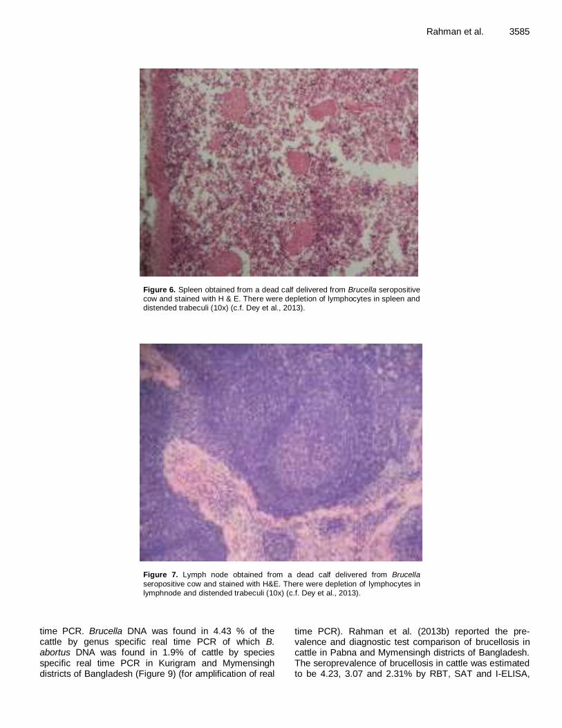
et al., 2012). Dey et al. (2013) recorded serological and pathological investigations of brucellosis in dairy cows in Bangladesh. Out of 190 randomly sera sample tested, prevalence was 2.63% by RBPT and 1.05% by I-ELISA. Histopathological study of placenta from an aborted cow and spleen and lymphnode and liver from an aborted fetus were performed. During histopathological study there was depletion of lymphocytes in spleen and lymphnodes which was characterized by reducing densities of lymphocytes. The smooth muscular trabeculi in spleen and fibromuscular trabeculi in lymphnode were distended. In placenta, there was diffuse fibrosis around the placental epithelium. The liver of aborted fetus showed multifocal necroses in hepatic parenchyma and necrosed tissue was replaced by fibrous connective tissue and reactive cells (Figures 5, 6, 7 and 8).
Rahman et al. (2013a) recorded 5.29% prevalence of brucellosis in 700 cattle sera by RBT. RBT positive samples were retested by CFT, SAT, ELISA and real
Rahman et al. 3585
Figure 6. Spleen obtained from a dead calf delivered from Brucella seropositive cow and stained with H & E. There were depletion of lymphocytes in spleen and distended trabeculi (10x) (c.f. Dey et al., 2013).
Figure 7. Lymph node obtained from a dead calf delivered from Brucella
seropositive cow and stained with H&E. There were depletion of lymphocytes in lymphnode and distended trabeculi (10x) (c.f. Dey et al., 2013).
time PCR. Brucella DNA was found in 4.43 % of the cattle by genus specific real time PCR of which B. abortus DNA was found in 1.9% of cattle by species specific real time PCR in Kurigram and Mymensingh districts of Bangladesh (Figure 9) (for amplification of real
time PCR). Rahman et al. (2013b) reported the pre-valence and diagnostic test comparison of brucellosis in cattle in Pabna and Mymensingh districts of Bangladesh. The seroprevalence of brucellosis in cattle was estimated to be 4.23, 3.07 and 2.31% by RBT, SAT and I-ELISA,

3586 Afr. J. Microbiol. Res.
Figure 8. Section of a liver collected from a dead calf suspected to BE infectED
with Brucella. There were multifocal necrosis in hepatic parenchyma and necrosed tissue was replaced by fibrous connective tissue (H & E, 40x) (c.f. Dey et al., 2013).
Figure 9. Amplification plots in real time PCR with the cattle and buffalo sera of Bangladesh.

Rahman et al. 3587
Table 1. Prevalence of brucellosis in cattle in seven districts of Bangladesh.
District No. tested No. positive Positive (%) Reference
Mymensingh 250 5 2.00 Amin et al. (2004)
Mymensingh 200 9 4.50 Nahar and Ahmed (2009)
Mymensingh
Mymensingh
Mymensingh
132
100
190
14
7
5
10.60
7.00
2.63
Ahasan et al. (2010)
Rahman et al., (2013a)
Dey et al. (2013)
Dinajpur 50 4 8.00 Ahasan et al. (2010)
Mymensingh 135 2 1.50 Rahman et al. (2012b)
Bogra 60 0 0.00 Rahman et al. (2012b)
Gaibhandha 70 0 0.00 Rahman et al. (2012b)
Bagherhat 90 1 1.10 Rahman et al. (2012b)
Chittagong
Kurigram
500
600
25
30
5.00
5.00
Sikder et al. (2012)
Rahman et al., (2013a)
respectively. The comparison of the serological tests result revealed the highest prevalence in RBT than SAT and I-ELISA. The prevalence of Brucella was 2.5% in Pabna and 2.14% in Mymensingh. It was observed that, a higher prevalence of Brucella was found in female (2.67%) than in male (1.82%), natural breeding (2.67%) than artificial breeding (1.81%), in aged animals (3.33%) than young (1.25%). But these differences were not statistically significant. There exists significant difference between prevalence of brucellosis in cattle with history of abortion than without history of abortion. See Table 1 for preva-lence of brucellosis in cattle in different districts of Bangladesh.
BRUCELLOSIS IN BUFFALO
Asia is the native home of the water buffalo, with 95% of the world population, with about half of the total in India and Bangladesh. It is valuable for its meat and milk, as well as the labour it performs. It is often referred to as “the living tractor of the East”, as it is relied upon for plowing and transportation in many parts of Asia including Bangladesh (Rahman, 2012a).
Buffaloes are known to be affected with B. abortus and less frequently with B. melitensis (Munir et al., 2008; Ahmed et al., 2010). Similar to cattle, Brucella infections are known to result in late gestation (6-9 months) abortions (Sanjrani et al., 2013), infertility (Sukumar et al., 2012) and latent infection of mammary gland lymph nodes with shedding of organisms in the milk (Ahmed et al., 2010), yet abortions are less common in buffaloes (The Center for Food Security and Public Health Iowa State University, 2009) with the disease being endemic in most buffalo raising countries. Shedding of Brucella in milk creates a potential threat to human health particularly for consumers using unpasteurized milk and milk products (Ahmed et al., 2010). A slightly lower incidence of brucellosis has been recorded in buffaloes as compared to cattle in studies that simultaneously evaluated the
serologic presence of brucellosis in these two species (Hussain et al., 2008), however, in other studies, a higher incidence of the disease was recorded in buffaloes as compared to cattle (Nasir et al., 2004). Thus, it can be presumed that buffaloes are differentially affected with B. abortus.
The first report on the occurrence of brucellosis in buffaloes appears to have originated in India in 1918 at the Indian Veterinary Research Institute, Mukteshwar
(Anonymous, 1918). The first seroprevalence study of brucellosis in buffalo in Bangladesh was conducted by Rahman et al. (1997) in selected areas of Bangladesh. The overall seroprevalence in buffalo was 6.9% by PAT and 2.4% by TAT. The prevalence of brucellosis was 7.1 and 1.2% in buffalo with history of retained placenta and repeat breeding, respectively.
Rahman et al. (2012b) screened 135 sera of buffaloes from five districts of Bangladesh and found prevalence of brucellosis in Bagherhat, Mymensingh and Sirajgonj districts as 2.9, 8.3 and 5.3%, respectively. No preva-lence of brucellosis was recorded in buffalo both in Bogra and Gaibandha districts. Age and sex as potential risk factors for brucellosis in buffalo was analyzed by Rahman et al. (2011a). Recently Rahman et al. (2013a) recorded prevalence of brucellosis in 99 buffaloes sera of Bagerhat and Mymensingh using RBT, SAT, CFT, I-ELISA, genus specific and species specific real time PCR (see Figure 9 for amplification of real time PCR). The presence of Brucella DNA was found in 7.1 % of the buffaloes investigated and B. abortus DNA was found in 6.1% of the buffaloes. Prevalence of brucellosis in buffaloes in different districts of Bangladesh is shown in Table 2.
BRUCELLOSIS IN GOAT
Economically and culturally, the goat has played an important role in traditional Bengali society. Among the Asiatic countries, Bangladesh, a tropical agro-based developing country, possess the third largest repository

3588 Afr. J. Microbiol. Res.
Table 2. Prevalence of brucellosis in buffalo in five districts of Bangladesh.
District No. tested No. positive Positive (%) Reference
Bagherhat
Bagherhat
70
80
2
5
2.85
6.50
Rahman et al. (2012b)
Rahman et al. (2013b)
Bogra 20 0 0.00 Rahman et al. (2012b)
Gaibandha 14 0 0.00 Rahman et al. (2012b)
Mymensingh
Mymensingh
12
19
1
2
8.33
10.52
Rahman et al. (2012b)
Rahman et al. (2013a)
Sirajgonj 19 1 5.26 Rahman et al. (2012b)
Table 3. Prevalence of brucellosis in goats in eight districts of Bangladesh.
District No. tested No. positive Positive (%) Reference
Mymensingh and Dhaka 300 5 1.67 Uddin et al. (2007b)
Dhaka and Lalmonirhat 20 0 0.00 Das et al. (2008)
Mymensingh 208 8 3.85 Islam et al. (2010)
Bogra and Mymensingh 120 7 5.83 Rahman et al. (2011b)
Bagherhat 15 1 6.67 Rahman et al. (2012b)
Bogra 30 0 0.00 Rahman et al. (2012b)
Gaibandha 50 2 4.00 Rahman et al. (2012b)
Mymensingh 100 4 4.00 Rahman et al. (2012b)
Sirajgonj
Nilphamari
35
154
1
5
2.86
3.24
Rahman et al. (2012b)
Rahman et al. (2012c)
of goats, with a population of more than 34 million heads, according to the FAO (WHO, 2006). This figure represents more than 57% of total livestock in Bangladesh. More than 90% of the goats of the country are of the Black Bengal breed. Each year goat production provides 127,000 MT meat, which accounts for 25% of total red meat in Bangladesh (Bangladesh Economic Review, 2012). As goats come in very close contact with humans, the risk of transmitting this zoonosis is very high (Rahman, 2012b).
Serological prevalence (14.5%) of brucellosis in goats in Bangladesh was first reported by Rahman (1983). A higher incidence of the disease was observed in goats with reproductive disorders (Rahman et al., 1988). Overall prevalence of brucellosis in goats were 1.7% in Mymensingh and Dhaka districts, 0% in Dhaka and Lalmonirhat districts, 3.9% in Mymensingh district, 5.8% in Bogra and Mymensingh districts, 6.7% in Bagherhat districts, 0% in Bogra district, 4% in Gaibandha district and 2.9% in Sirajgonj district (Uddin et al., 2007a, b; Das et al., 2008; Islam et al., 2010; Rahman et al., 2011b, 2012b). Potential risk factors for brucellosis in goats included age, sex, pregnancy status, management system (concrete floor versus mud floor; flock rearing vs. individual rearing; non grazing versus free grazing; mixed with cattle versus without cattle; and rural versus farm goats) and reproductive disorders (abortion vs. retained placenta).
Prevalence odds of brucellosis in goats that are pregnant were 7 times greater than the prevalence odds of brucellosis for goats that are not pregnant. Rahman et al. (2012c) found overall seroprevalence of brucellosis 59% in Black Bengal goats in Nilphamari district of Bangladesh. A significantly (p<0.01) higher prevalence of brucellosis was found in Black Bengal goats with the history of previous abortion (33.33%). An insignificant (p>0.05) but higher prevalence of brucellosis was found in adult Black Bengal goats (>24 months) than young ones. The prevalence was relatively higher in cross-bred than pure Black Bengal goats, in female than male and in pregnant than non-pregnant Black Bengal goats. Prevalence of brucellosis in goats in different districts of Bangladesh is shown in Table 3. BRUCELLOSIS IN SHEEP Among the livestock populations, sheep still occupies the third position and about 80% sheep is reared by rural farmers in Bangladesh. The sheep in Bangladesh are mainly indigenous and utilized for meat purposes but also important for good quality leathers and source of income to rural people. In Bangladesh, sheep and goats are a very valuable asset especially for poor people. Most cases of brucellosis infection in sheep are inapparent and lack

Rahman et al. 3589
Table 4. Prevalence of brucellosis in sheep in six districts of Bangladesh.
District No. tested No. positive Positive (%) Reference
Mymensingh and Dhaka 62 3 4.84 Uddin et al. (2007a)
Bogra and Mymensingh 80 3 3.75 Rahman et al. (2011b)
Bagherhat 27 3 11.11 Rahman et al. (2012b)
Bogra 30 1 3.33 Rahman et al. (2012b)
Gaibandha
Gaibandha
35
206
2
7
5.71
3.39
Rahman et al. (2012b)
Rahman et al. (2012d)
Mymensingh 40 8 20.00 Rahman et al. (2012b)
Sirajgonj 38 2 5.26 Rahman et al. (2012b)
Table 5. Prevalence of brucellosis in pigs in 2 districts of Bangladesh.
District No. tested No. positive Positive (%) Reference
Bogra
Bogra
62
63
3
4
4.80
6.60
Rahman (2011c)
Rahman et al. (2012e)
Sirajgonj
Sirajgonj
41
42
2
3
4.80
7.10
Rahman (2011c)
Rahman et al. (2012e)
clinical signs. Serological evidence of brucellosis in sheep in Bangladesh was first reported by Uddin et al. (2007b). Brucella antibodies were prevalent in 8.84% sheep.
The overall prevalence of brucellosis in sheep (n = 312) reported by Rahman et al. (2011b, 2012b) from Mymensingh and Dhaka, Bogra and Mymensingh, Bagherhat, Bogra, Gaibandha, Mymensingh and Sirajgonj districts was 4.8, 3.8, 11.1, 3.3, 5.7, 20 and 5.3%, respectively. Prevalence odds of brucellosis in sheep that are greater than 2 year of age were 90 times greater than the prevalence odds of brucellosis for sheep that are less than or equal to 2 years (Rahman et al., 2011a, b). Further investigation by Rahman et al. (2012d) recorded seroprevalence of brucellosis in sheep in the Gaibandha districts of Bangladesh as 3.39% by RBPT and 2.91% by i-ELISA. The prevalence of brucellosis was higher in female sheep (3.41%) than male (3.33%) and in sheep with history of abortion (4.34%) than without history of abortion (3.08%). The higher rate (4.59%) of Brucella antibody was recorded in sheep of 1-2 years of age. Prevalence of brucellosis in goats in different districts of Bangladesh is shown in Table 4. BRUCELLOSIS IN PIGS Brucellosis in pigs is caused by B. suis. The capability of B. suis to colonize the bovine udder with subsequent shedding in milk means that it has the potential to be a serious human health risk. Outbreaks in slaughter houses have been caused by inhalation of B. suis. Most cases occur in people employed in meat processing industry and animal rearing (Radostits et al., 2007). Though, out
of 590 million pigs in the world, about 34% are raised in tropical countries. From to the religious point of view and for the limited number of pork consumers, the pig popula-tion is not large as compared to other ruminants and birds in Bangladesh. Furthermore, it is difficult to get the exact number of pigs in Bangladesh. But the pig popu-lation is increasing in the tribal areas. The pig rearing continues to be primitive scavenging in nature because they are raised by certain rural people who are educa-tionally, economically and socially most backward. Sero-logical evidence of brucellosis in pig in Bangladesh was first reported by Rahman (2011c).
Further serological status of brucellosis in pigs was diagnosed by Rahman et al. (2012e) in Bangladesh using RBT and SAT. Overall seroprevalence was 6.7 and 4.8% by RBT and SAT, respectively. It was observed that, insignificantly higher prevalence of brucellosis based on SAT was found in female (5.6%) than male (2.9%) in aged animal (8.1%) than young (0.0%) and in pregnant animal (12.5%) than non pregnant animal (2.1%) (p>0.05). Prevalence of brucellosis was 42.9% in aborted pigs and 1.6% in non aborted pigs. The association between abortion status and prevalence of brucellosis was statistically highly significant (p<0.01). Prevalence of brucellosis in pigs in different districts of Bangladesh is shown in Table 5 BRUCELLOSIS IN DOGS Dogs fill a variety of roles in human society and are often trained as working dogs. The most important role of dogs is as companion. Dogs have lived with and worked with humans in so many roles that their loyalty has earned

3590 Afr. J. Microbiol. Res.
Table 6. Prevalence of brucellosis in dogs in 2 districts of Bangladesh.
District No. tested No. positive Positive (%) Reference
Mymensingh
Dhaka
30
50
4
2
13.33
4.00
Talukder et al. (2011)
Rahman (2014b)
them the sobriquet man's best friend. Dog population in Bangladesh may be considered as a carrier of Brucella infection and might act as a risk for food animal and human health (Rahman, 2014a). Dogs may become
infected through ingestion of infected bovine placental tissue. Brucella infected dogs may abort and vaginal discharges have a potential for transmitting Brucella to susceptible animals. Both B. abortus and B. melitensis infection have been reported in dogs kept on farms (Baek et al., 2003).
The first report on the sero-prevalence of brucellosis in stray dogs of Bangladesh by using four commercial sero-diagnostic kits was conducted by Talukder et al. (2011). The overall sero-prevalence of canine brucellosis was recorded as 13.33, 6.67, 6.67 and 10.0% with RBPT, SAT, STAT and ELISA, respectively. Significantly (p<0.01) higher sero-prevalence rate of canine brucellosis was recorded in stray dogs aged between 7 and 36 months (14.81, 7.40, 7.40 and 11.11%) in comparison with aged group up to 6 months (0, 0, 0 and 0% ) with RBPT, SAT, STAT and ELISA, respectively. The sero-prevalence rate of canine brucellosis was found significantly (p <0.01) higher in female dogs (15.78, 10.52, 10.52 and 15.78%) in comparison with male (9.09, 0, 0 and 0%) with RBPT, SAT, STAT and ELISA, respectively.
Rahman (2014b) conducted a serological study for a total of 50 pet dog’s serum samples collected from Dhaka, Bangladesh. The overall seroprevalence of brucellosis in pet dogs was found to be 4.00%. Statistically significant higher seroprevalence of brucellosis (RBPT and ELISA, 6.06% respectively) was found in dog aged 1.5 to 2.5 years. Higher seroprevalence (15.38%) was found in female pet dogs and no response in male pet dogs. Prevalence of brucellosis in dogs in different districts of Bangladesh is shown in Table 6. Recommended strategy to control brucellosis in Bangladesh It is important to remember that brucellosis is an important zoonosis and nearly every case of human brucellosis has an animal origin and, therefore, control is primarily a veterinary responsibility (Nicoletti, 1992). The Brucellae are 'survivors' in both extracellular and intra-cellular environments. Compatible relationships with the hosts including variable incubation periods, asymptomatic carriers and resistance to treatments are the important problems. The animal husbandry factors such as com-merce, nomadism, commingling and increasing population
sizes assure difficulties in control of diseases. The serosurveillance studies of brucellosis in humans
and animals suggest that brucellosis is endemic in the surveyed areas of Bangladesh. Without control measures, the infected domestic animals will continue to serve as reservoirs for the spread of the disease to uninfected domestic animals and humans.
CONTROL OF BRUCELLOSIS IN HUMANS
Public health education
Efforts should be focused on the public health education regarding the disease and its risk factors. The duration of contact with animals and the type of animal handled appeared to be the most significant risk factors for human brucellosis in Bangladesh (Rahman et al., 2012a). Expo-sure could be minimized by educating individuals within the high risk group (Rahman et al., 2012a).
Food safety
Brucella spp. are readily killed by pasteurization or heating of raw milk. Pasteurization process is not available in all parts of Bangladesh. Boiling or heating of milk at 80-85°C (176-185 8°F) for several minutes will kill the Brucella (Corbel, 2006).
Personal hygiene Protective clothes such as overalls, rubber gloves and rubber boot should be used during handling of domestic animals. If gloves are not available, washing of hands with soap and water immediately after examination is recom-mended. Consuming of food and smoking must be forbid-den in the abattoirs while handling domestic animals (Sammartino et al., 2005).
Improved diagnostic and treatment facilities
Brucellosis in humans is under-reported globally (Corbel, 2006) and likely under-reported in Bangladesh as well. Due to the scarcity of diagnostic and medical tools, treat-ment of brucellosis is often not possible. Appropriate test facilities for early and accurate diagnosis of brucellosis and prescription of effective antimicrobial treatment regimen must be included in the human health care system of Bangladesh.

Collaboration between human and veterinary medicine Control of brucellosis in domestic animals is the key to decreasing human cases since it is transmitted to humans from infected domestic animals and their products (Jiang and Baldwin, 1993). Collaboration between the department of health and department of livestock services are impor-tant to control brucellosis in domestic animals and thereby eliminate transmission to humans. Veterinary medicine must implement methods to control/eradicate brucellosis in domestic animals while human medicine must develop complementary methods to prevent transmission and develop effective treatment of human patients. So it is critical that physicians and veterinarians cooperate in these efforts.
CONTROL OF BRUCELLOSIS IN DOMESTIC ANIMALS Surveillance program Surveillance is important for determining prevalence and thereby allow for the development of preventive and control measures and eventual eradication of brucellosis in domestic animals. Brucellosis is primarily diagnosed by serological tests and rapid screening tests can be done by either RBT or PAT in the field. Conventional serological tests like rivanol, 2-MET and complement fixation tests (CFT), I-ELISA, C-ELISA and FPA are used as confirmatory tests. An excellent surveillance option is testing bulk tank milk samples among dairy herds by MRT (Sarker et al., 2014). The appropriate places for testing animals are slaughterhouses, livestock markets or any livestock sale station. This surveillance will help trace-back the infected animals to the herd or flock of origin. The polymerase chain reaction (PCR) can be used for identification of Brucella species or biovars and would be useful for epidemiological trace-back in a brucellosis control program (Rahman et al., 2013a).
Control of unrestricted animal movements
The initial introduction of disease into a herd or flock is often due to replacement animals introduced from an infected herd or flock of unknown disease status (Crawford et al., 1990). Implementation of quarantine and serosur-veillance of the new replacement animals before they enter the farms and checking the imported animals at border check points before entering into the country are required to ensure that these animals are free from brucellosis.
Epidemiological investigations
Animal age, sex, gestation stage, virulence of the pathogen, environmental conditions affect exposure to infection
Rahman et al. 3591 (Nicoletti, 1984). A detailed epidemiological investigation focusing on host, agent and environment factors needs to be performed throughout the country in order to identify the risk factors associated with transmission and maintenance of brucellosis in animals. Investigation of causes of abortion Making animal abortion notifiable and investigations into the causes of abortion help identify not only Brucella but also allow for the identification of other causative agents. This method of detection relies on compliance by farmers and veterinarians provided enough resources are available to conduct investigations following a report of an abortion (Crawford et al., 1990). Improved animal management practices The practice of mixing of cattle, either through grazing or sharing of watering points, is a significant risk factor for brucellosis (Crawford et al., 1990). Avoiding mixing of replacement cattle without screening for brucellosis and promoting the use self-contained units instead of shared facilities could help control brucellosis. In case of abortion, the aborted fetus must be properly disposed under bio-safety precautions. Avoid burying infected fetuses because dogs and other wild animals may dig them up and dis-seminate the disease. Any entrance where the animals are located must use step in tanks on the floor filled with disinfectant. Training of farmers Training of the livestock farmers on the effective imple-mentation of sanitary and hygienic livestock manage-ment practice following abortion helps reduce spreading the disease amongst animals as well as to the humans. Education of the farmers and animal care workers on the basic hygiene and sanitary procedures and techniques as well as practical demonstration on the use of disinfection and personal protection methods are important (Sammartino et al., 2005). Use of vaccines The use of vaccines is one of the important measures for prevention and control of brucellosis. In areas with endemic brucellosis only vaccination will control brucellosis. Brucella vaccines in use for livestock are the B. melitensis Rev 1, live B. abortus strain 19, and B. abortus strain RB51. The Rev 1 vaccine is a modified live B. melitensis vaccine used in small ruminants between the ages of three and four months that confers immunity for three to five years

3592 Afr. J. Microbiol. Res. (Blasco and Molina-Flores, 2011). Use of strain RB51 vaccine in cattle could be a good choice for control of brucellosis in Bangladesh. The use of strain RB51 has been shown to help prevent, control and eradicate cattle brucellosis in the countries where it has been adapted (Luna-Martinez and Mejia-Teran, 2002; Rahman and Baek, 2008b; Rahman 2011d). Test and slaughter In order to be a cost-effective disease control measure, test and slaughter is best implemented in areas where there is a less than two percent prevalence of brucellosis in the flocks and herds (Corbel, 2006). In developing countries, test and slaughter can be difficult to carry out due to enormous cost involved in the indemnification paid to the farmers for slaughtered animals (Blasco and Molina-Flores, 2011). Enhanced biomedical research Biomedical research focusing on epidemiology, isolation
and characterization of field isolates, development of the best diagnostic method and more effective vaccines against brucellosis in non-bovine species should be undertaken. To date, only one published reports is available on the characterization of the Brucella isolates of animals in Bangladesh at the species level (Rahman et al., 2013a). Government commitment Regulations and adequate monetary support (political will) from the local and national government organizations
strengthen the collaboration among farmers, veterinarians and regulators that are essential for effective implement-tation of a country-wide brucellosis control and eradica-tion program.
CONCLUSIONS Brucellosis is considered as a neglected bacterial zoono-tic disease in Bangladesh and it is present in the domestic-cated animal species and humans in surveyed regions of Bangladesh. The differences in seroprevalence of bru-cellosis in the study areas may be linked to ecological factors, differences of animal’s density and husbandry practices and type of serological tests. The variation of prevalence of brucellosis between animals on farms and domestic holding are likely to be attributed to certain risk factors such as cattle management practices, population dynamics and biological features (for example, host immunity) that largely influence the epidemiology of Brucella spp. Prevalence of brucellosis is higher in sexually mature and pregnant animals, in female than male, in
animals with history of abortion than without history of
abortion. A detailed and statistically valid surveillance study of brucellosis in high-risk group of people and
domesticated animals throughout the country is necessary
to know actual disease burden. Public health education for
target groups of people, understanding the risk factors of brucellosis, hygienic animal management practice (bio-safety), early diagnosis, collaboration among veterinarians, medical doctors and farmers and vaccination of animals are necessary for control of brucellosis in Bangladesh.
Conflict of Interest The author(s) have not declared any conflict of interests.
ACKNOWLEDGEMENTS Dr. Md. Siddiqur Rahman thanks the DAAD (Deutscher Akademischer Austausch Dienst-German Academic
Exchange Service) Programme, Germany for supporting his research stay at OIE Reference Laboratory for Brucellosis, Federal Research Institute for Animal Health, Friedrich-Loeffler-Institut, Naumburger Str. 96a, 07743 Jena, Germany. REFERENCES
Ahasan MS, Rahman MS, Song HJ (2010). A sero-surveillance of
Brucella spp. antibodies and individual risk factors of infection in
cattle of Bangladesh. Kor. J. Vet. Serv. 33:121-128. Ahmed JU, Alam MGS, Rahman MM, Hossain M,
(1992).Seroprevalence of brucellosis in indigenous zebu cows of
Bangladesh. Bangl. J. Microbiol. 9:17-21. Ahmed YF, Sokkar SM, Desouky SM (2010). Pathological and
molecular studies on mammary glands and supramammary lymph nodes of naturally Brucella infected buffalo cows. J. Reprod. Infertil.
1:33-40. Amin KMR, Rahman MB, Kabir SML, Sarkar SK, Akand SKI (2004).
Serological epidemiology of brucellosis in cattle in Mymensingh district of Bangladesh. J. Anim. Vet. Adv. 3:773-775.
Anonymous. Annual Report (1917-1918) (1918). Imperial Veterinary
Research Institute, Mukteswar, Uttar Pradesh, India. p.16. Baek BK, Lim CW, Rahman MS, Kim CH, Oluoch A, Kakoma I (2003).
Brucella abortus infection in indigenous Korean dogs. Can. J. Vet.
Res. 67:312-314. Baek BK, Lee BO, Hur J, Rahman MS, Lee SI, KakomaI (2005).
Evaluation of the Sprague-Dawley rat as a model for vertical transmission of Brucella abortus . Can. J. Vet. Res. 69:305-308.
Bangladesh Economic Review (2012). Economic Advisors wing Finance Division. Ministry of Finance, People’s Republic of Bangladesh.
Blasco JM, Molina-Flores B (2011).Control and eradication of Brucella melitensis in sheep and goats. Vet. Clin. North Am. Food Anim.
Pract. 27:95-104.
Center for Food Security and Public Health Iowa State University (2009). Bovine brucellosis: Brucellaabortus. Fact
Sheets.www.cfsph.iastate.edu/Factsheets/es/Brucella-abortus.pdf; 1-
6. Colmenero JD, Reguera JM, Martos F, Sánchez-De-Mora D, Delgado
M, Causse M, Martín-Farfán A, Juárez C (1996). Complications associated with Brucellamelitensis infection: a study of 530 cases.
Medicine (Baltimore) 75(4):195-211. Corbel M (2006). Brucellosis in Humans and Animals. World Health
Organization in collaboration with the Food and Agriculture

Organization of the United Nations and the World Organization for Animal Health.
Crawford RP, Huber JD, Adams BS (1990). Epidemiology and
surveillance. In: Nielson K, Duncan JR (Eds.), Animal Brucellosis. CRC Press, Boca Raton, FL. pp. 131-152.
Das T, Ershaduzzaman M, Islam KK, Haque MM, Rahman MM, Islam KBMS (2008). Surveillance of Brucella melitensis and Brucella abortus from aborted Bengal goats in Bangladesh. Res. J. Vet. Sci.
1:28-36.
Dey SK, Rahman MS, Rima UK, Hossain MZ, Chowdhury GA, Pervin M, Habib MA, Khan MAHNA (2013). Serological and pathological investigation of brucellosis in dairy cows of
Mymensingh districts, Bangladesh. Bangl. J. Vet. Med. 11(2):107-113.
European Commission Regulation (EC) No 535/2002 (2002). Official
Journal of the European Communities O J E C; L 80:22-28. Hussain I, Arshad MI, Mahmood MS (2008). Seroprevalence of
brucellosis in human, cattle, and buffalo populations in Pakistan.
Turk. J. Vet. Anim. Sci. 32:315-318. Islam A, Haque M, Rahman A, Rahman MM, Rahman A, Haque F,
(1983). Economic losses due to brucellosis in cattle in Bangladesh.
Bangl. Vet. J. 17:57-62. Islam MA, Samad MA, Rahman AKMA (2010). Risk factors associated
with prevalence of brucellosis in black Bengal goats in Bangladesh.
Bangl. J. Vet. Med. 8:141-147. Islam MS, Rahman MF, Hossain MA, Jahan S, (1992).Sero-prevalence
of brucellosis in cows sampled from six different areas of
Bangladesh. Bangl. J. Microbiol. 9:75-77. Jiang X, Baldwin CL (1993). Effects of cytokines on intracellular growth
of Brucella abortus. Infect. Immun. 61:124-134.
Kakoma I, Oluoch AO, Baek BK, Rahman MS, Kiku M (2003). More attention warranted on Brucella abortus in animals. J. Am. Vet. Med.
Assoc. 222(3):284.
Kattar MM, Jaafar RF, ArajGF, Le Flèche P, Matar GM (2008). Evaluation of a multilocus variable-number tandem-repeat analysis scheme for typing human Brucella isolates in a region of brucellosis
endemicity. J. Clin. Microbiol.46:3935-940. Le Flèche P, Jacques I, Grayon M, Al Dahouk S, Bouchon P (2006).
Evaluation and selection of tandem repeat loci for a Brucella MLVA
typing assay. BMC Microbiol. 6:9. Luna-Martinez JE, Mejia-Teran C (2002). Brucellosis in Mexico: current
status and trends. Vet. Microbiol. 90:19-30.
Mathur TNM (1971). Brucellosis and farm management. Indian Vet. J. 48:219-228.
Mia AS, Islam H, (1967).A preliminary study on the incidence of bovine
infertility and economic loss caused by it. Pak. Vet. J. 1:5-10. Muhammad N, Hossain MA, Musa AK, Mahmud MC, Paul SK, Rahman
MA, Haque N, Islam MT, Parvin US, Khan SI, Nasreen SA, Mahmud NU (2010). Seroprevalence of human brucellosis among the
population at risk in rural area. Mymensingh Med. J. 19:1-4. Munir R, Rehman ST, Kausar R (2008). Indirect Enzyme Linked
Immunosorbent Assay for diagnosis of brucellosis in buffaloes. Acta.
Vet. Brno. 77:401-406. Mustafa AH, Nicoletti P (1995). FAO, WHO, OIE, guidelines for a
regional brucellosis control programme for the Middle East.Workshop
of Amman, Jordan, Ammended at the Round-Table. Nahar A, Ahmed MU (2009). Sero-prevalence study of brucellosis in
cattle and contact human in Mymensingh district.Bangl. J. Vet. Med.
7:269-274. Nasir A, Parveen Z, Shah MA (2004). Seroprevalence of brucellosis in
animals at government and private livestock farms in Punjab. Pak.
Vet. J. 24:144-146. Nicoletti P (1984). The epidemiology of brucellosis in animals. Dev. Biol.
Stand. 56:23-628.
Nicoletti P (1992). The control of brucellosis-a veterinary responsibility. Saudi Med. J. 13: 10- 133.
Nielsen K (2002). Diagnosis of brucellosis by serology. Vet.
Microbiol.90:447- 459. Nielsen K, Cherwonogrodzky JW, Duncan JR, Bundle DR (1989).
Enzyme-linked immunosorbent assay for differentiation of the antibody response of cattle naturally infected with Brucella abortus or
vaccinated with strain 19. Am. J. Vet. Res. 50:5-9.
Rahman et al. 3593 OIE (2014). Sheets summary information on animal diseases.
Brucellosis. [Online]. www.oie.int/es/sanidad-animal-en-el-mundo/sintesis-de-informacion-de-enfermedades/
Pappas G, Papadimitriou P, Akritidis N, Christou L, Tsianos EV(2006). The new global map of human brucellosis. Lancet Infect. Dis. 6:91-99.
Radostits OM, Gay CC, Hinchcliff KW, Constable PD (2007). Veterinary Medicine: A Textbook of the Diseases of Cattle, Horses, Sheep, Pigs and Goats. Saunders Elsevier, Edinburgh. pp. 963-984.
Rahman MS, Rahman MN, Islam MT, Sarker RR, Sarker MAS, Sarabontuhura M, Chakrabartty A, Akther L, Uddin MJ (2012d).
Seroprevalence of brucellosis in sheep in the Gaibandha district of Bangladesh. Prog. Agric. 23(1-2):25-32.
Rahman AK, Dirk B, Fretin D, Saegerman C, Ahmed MU, Muhammad N, Hossain A, Abatih E (2012a). Seroprevalence and risk factors for
brucellosis in a high-risk group of individuals in Bangladesh. Foodborne Pathog. Dis. 9:190-197.
Rahman M (1983). Seroprevalence of human and animal brucellosis in
Bangladesh. Indian Vet. J. 60:165-168. Rahman MA, Islam MS, Alam MGS, Shamsuddin M
(1997).Seroprevalence of brucellosis in the buffalo (Bubalus bubalis)
of a selected area in Bangladesh. Buffalo J. 2:209-214. Rahman MA, Mia AS (1970). A study of brucellosis in Bangladesh. J.
Agric. Sci. 3:39-44.
Rahman MM, Chowdhury TIMF, Chowdhury MUA (1978). Investigation of brucellosis among cattle. Bangl. Vet. J. 12:12-15.
Rahman MM, Haque M, Rahman MA (1988). Seroprevalence of caprine
and human brucellosis in some selected areas of Bangladesh. Bangl. Vet. J. 22:85-92.
Rahman MM, Rahman MA (1981). Incidence of Brucella infection in
subclinical mastitic udder. Bangl. Vet. J. 15:39-42. Rahman MM, Rahman MS (1982). Study on the prevalence of
brucellosis in cows in organized farms and domestic holdings in
Bangladesh. Bangl. Vet. J. 16:53-58. Rahman MS (2004). Polymerase chain reaction assay for the diagnosis
of experimentally infected pregnant Sprague-Dawley rats with Brucella abortus biotype 1. Vet. Med. Czech 49 (7):253-258.
Rahman MS (2011a). Brucellosis in Livestock in Bangladesh. ISBN 978-3-8465-0121-4, LAP LAMBERT Academic Publishing
GmbH & Co. KG, Dudweiler Landstraße 99, 66123 Saarbrücken, Germany.
Rahman MS (2011b). Brucellosis in Dairy Cattle.ISBN 978-3-8473-
1583-4, LAP LAMBERT Academic Publishing GmbH & Co. KG, DudweilerLandstraße 99, 66123 Saarbrücken, Germany.
Rahman MS (2011c). Brucellosis in pigs of northern region of
Bangladesh. ISBN 978-3-8465-3939-2, LAP LAMBERT Academic Publishing GmbH & Co. KG, Dudweiler Landstraße 99, 66123 Saarbrücken, Germany.
Rahman MS (2011d). Brucellosis and Protective Immunity.ISBN
978-3-639-35725-7, VDM Verlag Dr. Muller GmbH & Co. KG, Dudweiler Landstr. 99, 66123 Saarbruecken, Germany.
Rahman MS (2012a). Brucellosis in Water Buffalo.ISBN 978-3-
8484-1581-6, LAP LAMBERT Academic Publishing GmbH & Co. KG, Dudweiler Landstraße 99, 66123 Saarbrücken, Germany.
Rahman MS (2012b). Brucellosis in Black Bengal goat. ISBN 978-
3-8473-3894-9, LAP LAMBERT Academic Publishing GmbH & Co. KG, Dudweiler Landstraße 99, 66123 Saarbrücken, Germany.
Rahman MS (2012c). Brucellosis in Sheep.ISBN 978-3-8465-8328-9, LAP LAMBERT Academic Publishing GmbH & Co. KG, Dudweiler Landstraße 99, 66123 Saarbrücken, Germany.
Rahman MS (2014a). Small and Laboratory Animal Medicine, a textbook of Small and Laboratory Animal Medicine. Third Edition, ISBN 978-984-33-6081-6, Copy Right Registration Number 13054-COPR, Prithul and Piya Publication, 17/12A1
Pallabi, Mirpur 12, Dhaka 1216, Bangladesh. Rahman MS, Baek BK (2008a). Transplacentally transmitted
congenital brucellosis due to Brucella abortus biotype
1 in Sprague-Dawley rats. Pak. J. Sci. Ind. Res. 51 (2):89-92. Rahman MS, Baek BK (2008b). Diagnostic efficacy of Brucella
abortus strain RB51 in experimentally inoculated Sprague- Dawley rats using western blot assay. J. Infect. Dev. Ctries 2
3594 Afr. J. Microbiol. Res.
(5):384-388. Rahman MS, Faruk MO, Her M, Kim JY, Kang SI, Jung SC (2011a).
Prevalence of brucellosis in ruminants in Bangladesh. Vet. Med.
Czech. 56:379-385. Rahman MS, Hahsin MFA, Ahasan MS, Her M, Kim JY, Kang SI, Jung
SC (2011b). Brucellosis in sheep and goat of Bogra and Mymensingh
districts of Bangladesh. Kor. J. Vet. Serv. 51:277-280. Rahman MS, Han JC, Park J, Lee JH, Eo SK, Chae JS (2006).
Prevalence of brucellosis and its association with reproductive
problems in cows in Bangladesh. Vet. Rec. 159:180-182. Rahman MS, Her M, Kim JY, Kang SI, Lee K, Uddin MJ, Chakrabartty
A, Jung SC (2012b). Brucellosis among ruminants in some districts of
Bangladesh using four conventional serological assays. Afr. J. Microbiol. Res. 6:4775-4781.
Rahman MS, Melzer F, Sprague LD, Neubauer H (2013a). Real time
PCR for diagnosis of brucellosis in livestock in Bangladesh. Proceedings of the 66th Annual Brucellosis Research Conference, Marriot, Downtown Magnificent Mile, Chicago,
Illinois, USA, 7-8 December, 2013, Oral Abstract No. 3. Rahman MS, Mithu S, Islam MT, Uddin MJ, Sarker R R, Sarker MAS,
Akther L (2012c). Prevalence of brucellosis in black Bengal goats in Bangladesh. Bangl. J. Vet. Med.10:(1-2):51-56.
Rahman MS, Nuruzzaman M, Ahasan MS, Sarker RR, Chakrabarty, A, Nahar A, Uddin MJ, Sarker MAS, Akther L (2012e). Prevalence of brucellosis in pigs: the first report in Bangladesh. Bangl. J. Vet. Med.
10 (1-2): 75-80. Rahman MS, Rahman MN, Islam M, Chakrabartty A, Sarker RR, Akther
L, Uddin MJ (2013b). Prevalence and diagnostic test comparison of
brucellosis in cattle of Pabna and Mymensingh districts of Bangladesh. P. J. Sci. Ind. Res. Series B 56(3):147-153.
Rahman S (2014b). Seroprevalence of canine brucellosis in Dhaka city corporation area, Bangladesh.MS thesis, Department of Microbiology, Bangladesh Agricultural University, Mymensingh
2202, Bangladesh. Sammartino LE, Gil A, Elzer P (2005). Capacity building for surveillance
and control of bovine and caprine brucellosis.Food and Agriculture
Organisation (FAO), Expert and Technical Consultation, 14-16 June, Rome.FAO, Rome Appendix 3, 55-56 ftp://ftp.fao.org/docrep/fao/009/a0083e/A0083E01.pdf (accessed on
15.11.12).
Sanjrani SN, Mirbahar KB, Soomro H (2013). Prevalence of abortion in
Kundhi buffalo in district Hyderabad, Sindh - Pakistan. Herald J. Food Agric. Food Sci. Res. 2:70-77.
Sarker MAS, Rahman MS, Islam MT, Rahman AKMA, Rahman MB, Akter L, Chaki A (2014). The use of milk ring test for the control and eradication of brucellosis in Bangladesh.
Proceedings of the seminar, 2nd
international exhibition on dairy, aqua and pet animal, Animal Health Companies Association of Bangladesh (AHACAB), 27 February-01 March, 2014, BICC,
Dhaka, Bangladesh. pp. 144-147. Sikder S, Rahman AKMA, Faruque MR, Alim MA, Das S, Gupta AD,
Das BC, Uddin MI, Prodhan MAM, (2012). Bovine brucellosis: an
epidemiological study at Chittagong, Bangladesh. Pak. Vet. J. 32:499-502.
Sukumar K, Tamilselvan P, Dorairajan N (2012). Studies on infertility in cattle and buffaloes caused by Brucella abortus. Tamil Nadu J. Vet.
Anim. Sci. 8:235-237. Talukder BC, Samad MA, Rahman AKMA (2011). Comparative
evaluation of commercial serodiagnostic tests for the seroprevalence study of brucellosis in stray dogs in Bangladesh. Bangl. J. Vet. Med. 9:79-83.
Uddin MS, Rahman MS, Hossain MA, Akter SH, Majumder S, Park JH, Song HJ (2007a). Relation between brucellosis and husbandry practices in goats in Bangladesh. Kor. J. Vet. Serv. 30:259-267.
Uddin MS, Rahman MS, Akter SH, Hossain MA, Islam MT, Islam MA, Park JH, Song HJ (2007b). Seroprevalence of brucellosis in small ruminants in selected area of Bangladesh. Kor. J. Vet. Serv. 30:511-
525. Van Aert A, Brioen P, Dekeyser P, Uytterhaegen L, Sijens RJ, Boeye A
(1984). A comparative study of ELISA and other methods for the detection of Brucella antibodies in bovine sera. Vet. Microbiol. 10:13-
21. WHO (2006). Brucellosis in human and animals. Joint report of WHO,
FAO and OIE.
Vol. 8(41), pp. 3595-3603, 8 October, 2014
DOI: 10.5897/AJMR2014.6705
Article Number: D3A12C748110
ISSN 1996-0808
Copyright © 2014
Author(s) retain the copyright of this article
http://www.academicjournals.org/AJMR
African Journal of Microbiology Research
Full Length Research Paper
Evaluation of Streptomyces as probiotics against vibriosis and health management of prawn larvae
Macrobrachium rosenbergii
Sridevi, K.1* and Dhevendaran, K.2
1Department of Aquatic Biology and Fisheries, University of Kerala, Kariavattom Campus,
Trivandrum - 695 581, Kerala, India. 2School of Chemical and Biotechnology, Sastra University, Thanjavur, India.
Received 11 February, 2014; Accepted 22 September, 2014
Nearly forty-five isolates of Streptomyces were found to be associated with 16 different seaweeds. Of these, only six strains that antagonized Vibrio vulnificus were selected to study the probiotic efficiency. Six strains were characterized based on the phenotypic characteristics. The toxicity of different Vibrio species (V. harveyji, V. parahemolyticus, V. alginolyticus, V. vulnificus) towards prawn larvae were studied, among them, V.vulnificus was more pathogenic and mass mortality (88%) was observed within three days. Hence, Streptomyces that antagonized V. vulnificus with maximum inhibition zone was selected for the probiotic study. In vitro and in vivo evaluation of probiotic efficacy of six selected Streptomyces were made using prawn larvae Macrobrachium rosenbergii challenged with V. vulnificus at 10
5Cfu/ml. The best growth-rate, reduced mortality, highest glucose and protein-level, reduced chole-
sterol level and increased enzymatic activities were noticed with Streptomyces AQB.SKKU8, AQB.SKKU18, AQB.SKKU25 treated groups. It is recommended to use strain AQB.SKKU 8, Strain AQB.SKKU 18, strain AQB.SKKU 25, and strain AQB.SKKU37, as probiotics in-vivo, while strain AQB.SKKU10, and strain AQB.SKKU20 showed probiotic effects in-vitro only. The results of the present investigation reveal that the seaweed associated Streptomyces were found to be a promising source of probiotic and biocontrol agents against vibriosis disease in aquaculture. Key words: Streptomyces spp., physiological activities, biocontrol agent, vibriosis, disease resistance.
INTRODUCTION Probiotics are live microbes, which confer health benefit to the host and have been used in aquaculture as biocontrol agents against various fish and shellfish diseases. Bacterial disease, especially vibriosis is a
major disease problem in shrimp aquaculture causing high mortality, and severe economic loss in all producing countries. The major species causing vibriosis in shrimp are Vibrio vulnificus, Vibrio anguillarum, Vibrio harveyji,
*Corresponding author. E-mail: [email protected] or [email protected]. Author(s) agree that this article remain permanently open access under the terms of the Creative Commons Attribution License 4.0International License

3596 Afr. J. Microbiol. Res. Vibrio alginolyticus and Vibrio parahaemolyticus (Goarant et al., 1999). Lin et al. (1989) showed that seven Vibrio species associated with vibriosis in silver sea bream (Sparus sarba) and of these species, V. alginolyticus, V. vulnificus, and V. parahaemolyticus are dominant. Among the vibrios, much attention focuses towards V. vulnificus during the last decade for its role both in human (Hoyer et al., 1995) and in fish pathogen (Tison et al., 1982; Biosca et al., 1991; Arias et al., 1997). Several strategies have been proposed to control vibriosis. For instance, the use of antimicrobial in disease prevention and growth promo-tion can bring about the emergence of drug-resistant microorganism and leave antibiotic residues in the prawn and the environment. Moreover, the chemotherapy may kill or inhibit intestinal microflora in the digestive tract, which are beneficial to the host (Sugita et al., 1999). Therefore, developing alternative technology for the health management of prawn larvae in the hatchery from vibrio-sis is the use of probiotic technology, which is an application of microbial ecology (Moriarty, 1999). Probiotics are applied to the feed or added to the culture tank or in a pond as a biocontrol agent against infection by pathogenic Vibrios. This approach to disease control is not much used in the prawn larval culture. Most proposed pro-biotics are Lactic acid bacteria, Bacillus strains and Pseudomonas strains (Verschuere et al., 2000), but few studies on probiotics consisting of Streptomyces have been reported as biocontrol agents against vibriosis disease. Many studies on probiotics in aquaculture used in vitro models of specific bacteria as antagonists of pathogens (You et al., 2007; Selvakumar and Dhevendaran, 2010; Ngo Thi Tuong et al., 2011). Other studies have focused on growth promotion and improvement of water quality by probiotic Streptomyces supplements (Das et al., 2006; Selvakumar and Dhevendaran, 2010). There are reports on the Streptomyces used as single cell protein feed on the growth of juveniles of Macrobrachium (Manju and Dhevendaran, 2002). Streptomyces culture extracted using fermentation media also used to evaluate the survival of black tiger shrimp against White spot syndrome virus (WSSV) infection has shown significant survival. Streptomyces cell mass was supplemented with feed at 1% dosage to black tiger shrimp for 15days in three treatments with two treatments of commercial probiotic that resulted better growth, survival and mortality of P. monodon after exposure to virulent V. harveyji at 10
7 CFU ml
− 1. The significant reduction in the
percentage mortality of Artemia was observed after 24, 48 and 72 h immersion in different concentrations of Streptomyces cell mass (Surajit et al., 2006). Jayasudha et al. (2011) studied in vitro inhibitory activity of ethyl acetate extract of actinobacteria, which showed 33.56% mortality and did notice any relative percentage inhibition in the petroleum ether extract. Giant freshwater prawn (Macrobrachium rosenbergii de Man, 1879) or scampi used in this study is an important commercial candidate
species due to increased protein demand in domestic and export markets. Considering above facts, the present study attempted to develop the marine streptomycetes into probiotic to control vibriosis disease in aquaculture. The Streptomyces was isolated from the seaweeds of marine coast and it’s in vitro and in vivo probiotic potential were tested against Vibrio spp., especially against Vibrio vulnificus have been surveyed in this study. In addition, an attempt was made for the first time to study the efficacy of Streptomyces as probiotics on promoting physiological activities of prawn, M. rosenbergii thereby to improve the health. MATERIALS AND METHODS Isolation of Streptomyces from seaweeds The Seaweed samples were collected during September 2006 from Muttom coast, Tamil Nadu, India (8° 7’ 15” N: 77°
1’E) at monthly
interval by random sampling method. One-gram mantle of live specimen was weighed, washed with distilled water and treated with 0.5% phenol for 5 min to eliminate the bacterial and fungal colonies present as contaminants. After the distilled water wash, the mantle was macerated and aliquots of 1 ml was serially diluted with sterile seawater and plated each dilution on three different selective media. The total number of Streptomyces colonies on the plates was enumerated (Okazaki and Okami, 1976; Lakshmanaperumalsamy, 1978). They were incubated at room
temperature (28±2C) for seven days. The colonies were counted and expressed as CFU/gm dry wt. of visceral mass. The isolated strains were stored in Glycerol asparagine agar (ISP5) medium as
agar slant cultures at 28±2C (Shirling and Gottlieb, 1966).
Determination of in vitro probiotic activity The pure isolates were examined for its antagonistic effects against the four shrimp pathogenic Vibrio species (V.harveyji, V.alginolyticus, V.parahaemolyticus and V. vulnificus). The in vitro probiotic activity was assessed using double layer method. The Streptomyces isolates were inoculated on petridish containing 15ml GsB agar medium (soluble starch 5 g, soybean meal 5 g, NaCl 0.5 g, K2HPO4 0.5 g, MgSO4 0.5 g, FeSO4 20 µM, seawater to 1 L) and incubated at 28±2°C for 3 days. The chloroform vapour was applied over the colonies. Sloppy (0.75% TCBS) agar containing Vibrio spp. was seeded onto the basal layer of GsB agar plate. The zone of inhibition was measured after incubation at 28°±2C for 24 h.
Identification of probiotic bacteria
Probiotic bacterial strain, Streptomycetes used in the study was characterized according to traditional morphological, cultural, biochemical characteristics as described in Shrilling and Gottlieb (1966).
Morphological characters Aerial mass color determination The cultures were inoculated on starch-casein agar and glycerol asparagine agar, incubated at 28±2°C for seven days and the

Sridevi and Dhevendaran 3597
Table 1. Percentage composition of probiotic and control feed.
Ingredient Control feed (w / %) Probiotic feed (w/ %)
Prawn Meal 25 25
Fish Meal 15 15
Soybean Meal 10 10
Groundnut oil cake 15 15
Wheat flour 15 15
Tapioca 13 13
Vitamins 1 1
Minerals 1.5 1.5
Cholesterol 0.5 0.5
Streptomyces biomass 0 7×10⁵ cells/g/ml
Oil 4 4
Total 100 100
mature sporulating aerial mycelium was observed for their colorations. The cultures were classified into white, grey, red, green, blue, yellow and violet series, depending on the aerial mycelial color. Micromorphological characteristics The Streptomyces isolates was streaked on Petri dish containing starch-casein agar medium and incubated at room temperature (28±2ºC) for 7-10 days. The growth on the surface of the medium was observed under Phase contrast microscope (magnification 400X). Depending upon the number of spores and sporulating pattern, strains are categorized into different species. Substrate mycelium color The color on the reverse side of Streptomyces colonies was observed using starch-casein agar and glycerol asparagine agar. The strains are classified into pale yellow, olive, yellowish brown or cream depending on the substrate mycelial color. Soluble pigments The isolates were cultured on salts starch-casein agar and observed for soluble colors other than melanoids (brown or black). The strains were divided into two groups viz: positive (+) and negative based on their ability to produce soluble pigments. The color was recorded as red, orange, green, yellow, blue and violet. Melanin production The cultures were streaked on peptone-iron agar slants
48 h. Culture-forming greenish-brown to brown and then to black diffusible pigments were recorded as positive (+) melanoid production, whereas the absence of these colors was recorded as negative (-) for melanoid production. Carbon utilization The Streptomyces colonies were inoculated into10 ml of basal minerals to which sterilized carbon sources (Xylose, arabinose, rhamnose, fructose, galactose, raffinose, mannitol, inositol, sucrose and glucose) wer added to a final concentration of 1%. The tubes
and categorized into positive and negative. The growth was compared with two controls; growth without carbon source was used as negative control and growth on D-glucose was used as positive control. Species level identification of Streptomyces isolates Streptomyces isolates were identified at the species level by com-paring the phenotypic properties with the representative species found in the key of Nonomura (1974) and Bergey’s manual of determinative bacteriology (Buchanan and Gibbons, 1974). Preparation of probiotic bacterial cell suspension Fresh cultures of each of the selected probionts were inoculated
10,000 rpm. The supernatant was discarded and the pellet was resuspended in 5ml of PBS (Phosphate buffer saline). Five millilitres of fish oil was added to each fraction and mixed thoroughly in a vortex mixture. The bacterial cell suspension was mixed with the formulated feed ingredients. Preparation of probiotic feed Seven different feeds were prepared, namely the probiotic (6) and control feed (without bacterial suspension). One hundred grams compound feed were prepared (Hardy, 1980) as per the formulation given in Table 1. The dough was prepared by adding the required amount of water of these ingredients; it was sterilized by steam
(autoclave at 121C for 15 min) and incorporated with a commercial vitamin and mineral mix at 2.5 (v/w) and pelletized using hand pelletizer to obtain 1 mm pellets. The pellets were initially sun dried
and further dried in an oven at 60±5C for 12 h. They were manually broken into smaller bits and stored in airtight sterile polypropylene containers at room temperature.
The probiotic feeds (PS1, PS2, PS3, PS4, PS5 and PS6) were prepared by mixing each probiotic cell suspension of the compound feed in order to obtain evenly coated pellets of each feed containing 7-×10
5cells g-1of diet. The control feed contains only PBS and oil.
The feeds were stored in sterile airtight containers at 4°C. The viability of the bacterial cells was determined periodically.

3598 Afr. J. Microbiol. Res.
Table 2. Proximate composition of formulated probiotic and control feeds.
Formulated feeds
Proximate composition (% dry matter basis)
Moisture Crude protein
Crude fat
Ash Crude fibre
Nitrogen free
Extract (NFE)
Control 6.95 37.84 5.5 12.14 1.6 35.96
PS1 6.73 47.91 3.9 10.14 3.78 31.12
PS2 6.78 47.84 3.98 10.3 3.56 30.83
PS3 6.74 47.96 3.86 10 3.8 31.1
PS4 6.76 47.88 3.92 10.28 3.58 31.52
PS5 6.68 48.12 3.8 9.72 3.87 31.02
PS6 6.7 48.08 3.82 9.74 3.85 31.06
DC, basal diet without bacteria (Control feed); PS1,basal diet containing probiotic strain AQB.SKKU8; PS2 , basal diet containing probiotic strain AQB.SKKU10; PS3 , basal diet containing probiotic strain AQB.SKKU18; PS4 , basal diet containing probiotic strain AQB.SKKU20; PS5, basal diet containing probiotic strain AQB.SKKU25; PS6, basal diet containing probiotic strain AQB.SKKU37.
Proximate chemical analysis of formulated feeds
The proximate composition of the feed was analyzed by moisture, crude protein, crude fat, crude fibre, ash and nitrogen-free extract according to the standard methods of AOAC (1990). The proximate compositions of the formulated feeds are displayed in Table 2.
In-vivo evaluation probionts
The isolated probiotic bacteria tested for its in vivo role against pathogenic V. vulnificus was evaluated by using 50 healthy M. rosenbergii larvae to each group. The larvae were acclimatized for two weeks in indoor tanks. The shrimp larvae were divided into seven equal groups with three replicates per each group. Six groups (1-6) were fed with probiotic diet and a group (7) was fed with control diet for two weeks. The percentage survival was noticed prior to challenge. Six experimental groups and control group were further challenged with V. vulnificus to the rearing water to get 10
5 CFU/ml. All groups were kept under observation for more
than two weeks and the mortality was recorded. For microbiological analysis, water sample was taken after 30 days of the experiment. Total Vibrio count during the rearing was recorded using Thiosulphate citrate agar medium. During this period, no water exchange was provided and the water quality parameters were maintained as recommended by the Correia et al. (2000).
Physiological analyses
The healthy post-larvae M. rosenbergii used for physiological studies were maintained in indoor tanks. At the end of the experimental period, haemolymph was collected from the animal. Plasma was obtained by centrifugation of heamolymph at 3000 rpm for 15 min and non-haemolyzed plasma was stored in deep freezer for further biochemical analysis.
The heamolymph glucose, protein and cholesterol were determined calorimetrically by using available commercial kits purchased from Erba Mannheim Diagnostic Company, TRANSASIA Biomedical Ltd. The Activities of aspartate aminotransferase (AST) and alanine aminotransferase (ALT) were determined calori-metrically according to Reitman’s and Frankel (1957).
Statistical analysis
The obtained results were analyzed statistically using One-way
analysis of variance (ANOVA). Turkey HSP multiple comparison test was used to evaluate the mean ratio among the different treatments at 5% probability level. All the statistical analyses were performed using the SAS program (2005).
RESULTS Isolation and enumeration of streptomycetes from seaweeds
The occurrence, distribution and identification of different kinds of seaweeds were surveyed in the Muttom (marine) coast during the study period. During the study, 16 different types of seaweeds were obtained. Enumeration of Streptomyces associated with seaweeds also exhibits Sargassum weight and Enteromorpha intestinalis are being the source of the highest number of pigment pro-ducing strains. The maximum streptomycetes population recorded in glycerol asparagine agar in the month of October followed by June (73 ×10
2cfu/ml) and February
(68 ×102cfu/ml). The minimum streptomycetes population
was recorded in the month of November (5X10²cfu/ml)
followed by the month of July (14 ×102cfu/ml); was
recorded on the coast (Figure 1).
The invitro probiotic activity In the present study, a total of 45 streptomycetes colonies were isolated from seaweeds of marine coast and tested for its in vitro probiotic activity against four pathogenic Vibrio spp. It was found that out of the 45 isolates, only 25 isolates of Streptomyces showed anta-gonistic activity against any one of four tested Vibrio spp. (V. harveyji, V. alginolyticus, V. parahemolyticus and V. vulnificus). Between 25 probiotic isolate, 23 (92%) isolates inhibited V. harveyji, 18 (72%) isolates were active against V. alginolyticus, 13 (52%) of isolates were antagonistic to V. vulnificus, and 15 (60%) isolates inhibited V. parahaemolyticus (Table 3).

Sridevi and Dhevendaran 3599
Figure 1. Streptomycetes population recorded in the different months.
Table 3 .Number of Streptomyces isolates antagonized tested pathogenic strains.
Parameter Tested pathogenic strains
V. harveyji V. alginolyticus V. vulnificus V. parahaemolyticus
No. of Streptomyces species against Vibrio spp. 23/25 18/25 13/25 15/25
Percentage 92 72 52 60
Identification of streptomycetes from seaweeds The isolates of streptomycetes were identified using pheno-typic characteristics using Nonomura Key and Bergey’s manual of determinative bacteriology (1974). The isolates were identified as closely related to Streptomyces coelicolor (AQB.SKKU 8), Streptomyces autotrophicus (AQB.SKKU 10), Streptomyces pedanensis (AQB.SKKU 18), Streptomyces deccanensis (AQB.SKKU 20), Streptomyces vinaceus (AQB.SKKU 25) and Streptomyces Nov spp. (AQB.SKKU 37). Their mor-phology, pigmentation and carbon utilization charac-teristics were depicted in Table 4. In vivo effect of the isolated probiotics
The rate of survival was 100% in pre-challenged larvae fed with probiotic feed, compared to the control (85.42 ± 3.60). The strain AQB.SKKU.8 (PS1), strain AQB.SKKU.18 (PS3), strain AQB.SKKU.25 (PS5) and strain AQB.SKKU.37 (PS6), of the probiotics were harmless to the shrimp M. rosenbergii larvae, as there were no clinical signs and reduced mortality was noticed than control. The percentage mortality of 0-18% was observed in probiotic treated groups than control (Table 5) which showed a mortality rate of 79.2%. The total Vibrio count determined subsequent to the addition of inoculum was significantly lesser (p < 0.05) than that of the control.
Physiological parameters
The glucose and protein level was significantly increased in the heamolymph of prawn fed with probiotic diet PS1
(containing strain AQB.SKKU8), PS5 (containing strain AQB.SKKU25) and PS6 (containing strain AQB.SKKU37) (P>0.5) compared to the control diet (feed devoid of Streptomyces). The glucose level was lower in control and PS2 diet (containing strain AQB.SKKU10), while the low protein levels were obtained in the PS2 (containing strain AQB.SKKU10) and PS4 diet (containing strain AQB.SKKU20). The data obtained are given in Table 6.
The AST and ALT activities in plasma of M. rosenbergii increased in the probiotic treated groups such as PS1, PS3 and PS5 than in the control. The lowest values were obtained in the control, PS2 and PS4 diet. The average levels of plasma AST and ALT activities with M. rosenbergii in the control group was 38.73±6.28 and 19.25±2.18 IU/L respectively and the results are presented in Table 6.
Growth performance
Experiments were performed to study the effect of probiotic isolates on growth enhancement of freshwater prawn, Macrobrachium rosenbergii. During the 30 days feeding trial, the prawn consumed well and acceptability of all diets was similar. Among the probionts, PS5 diet produced elevated values in weight gain followed by PS6

3600 Afr. J. Microbiol. Res.
Table 4. Phenotypic characteristics of Streptomyces spp. Isolated from seaweeds.
Strain number Seaweed source
Spore chain morphology
Spore surface
Aerial mass color
Reverse side color M
elan
oid
Pig
men
t
So
lub
le
Pig
men
t
Carbon utilization
Glu
Ara
Xyl
Ino
Man
Fru
Rh
a
Su
c
Raf
AQB.SKKU8
Gracillaria corticata
RF
Smooth
Yellow/
Green
Brown /lavender
-
-
+
+
+
-
+
+
±
±
-
AQB.SKKU10
Chnoospora minima
RF
Smooth
Pale yellow
ND/ cream
-
-
+
+
+
+
+
+
+
+
-
AQB.SKKU18 Sargassum weightii
RF Smooth White Brown - - + + + ± + - ± + -
AQB.SKKU20 Spyridia hypnoides
SC Hairy Grey Yellow + + + + + + + + + + +
AQB.SKKU25
Enteromorpha intestinalis
RF
Smooth
Yellowish brown
Reddish
brown
-
-
+
-
+
-
+
+
-
+
-
AQB.SKKU37
Hypnea valentiae
RF
Smooth
Bluish green
Reddish
Brown/red
-
-
+
±
+/
+
+/
+/
+
+
±
RF, Rectiflexibles; SC, Straight chains; ND-not distinctive; Glc, D-glucose; Ara, L-arabinose, Fru,-D-fructose; Ino, iinositol; Ma, D-Mannitol; Raf, raffinose, Rha, L-rhamnose. Su, sucrose; Xyl, D-xylose, “+”: well utilized;” +/_”: poorly utilized; “_”, not utilized; NA, not available.
Table 5. Post- challenge percentage mortality of shrimp larvae M. rosenbergii fed with control and probiotic feed.
Parameter DC PS1 PS2 PS3 PS4 PS5 PS6
%Mortality after 7days of infection (Mean± S.D)
64.6±3.60 14.2±0.0a 54.2±3.60 18.8± 0.0
a 31.3 ±6.3 0 ± 0 10.4±3.6
a
%Mortality after 2weeks of infection (Mean± S.D)
79.2 ± 3.6 14.2±0.0a 58.3 ± 7.2 18.8± 0.0
a 33.3 ±3.6 2.1±3.6
b 10.4±3.6
ab
Means having the same letter in the same row are not significant (P < 0.05). DC, basal diet without bacteria (Control feed); PS1,basal diet containing probiotic strain AQB.SKKU8; PS2, basal diet containing probiotic strain AQB.SKKU10; PS3, basal diet containing probiotic strain AQB.SKKU18; PS4, basal diet containing probiotic strain AQB.SKKU20; PS5, basal diet containing probiotic strain AQB.SKKU25; PS6, basal diet containing probiotic strain AQB.SKKU37nov Sp.
Table 6. Physiological parameters of shrimp M. rosenbergii fed with different probiotic and control feeds.
Parameter DC (control) PS1 PS2 PS3 PS4 PS5
PS6
Glucose 8.75±2.75a 20±5.88
b 5±1.82
a 12±1.41
ab 11±0.81
ab 29.75±0.70
c
17.75±0.95
b
Protein 2.7±2.63a 7.4±0.08b 4.39±0.36a 6.89±0.36ab 1.25±0.18a 10.9±0.50c
7.69±0.69b
Cholesterol 17.5±1.29a 4±0.3b 15±1.81a 7±0.81c 9±0.81d 2 ±1.41b
3±0.81b
AST (IU L-1
) 38.75±6.29a 42 ±6.48
a 8±2.16
c 39.25±2.06
d 37.75±1.70
a 56.25±2.62
e
46±6.48
f
ALT (IU L-1
) 19.25±2.18a 23±2.44
a 12±2.82
a 22.75±2.44
a 19±1.41
a 30.5±3.69
a
32.5±6.24
a
Means having the same letter in the same row are not significant (P < 0.05). DC, basal diet without bacteria (Control feed); PS1, basal diet containing probiotic strain AQB.SKKU8; a PS2, basal diet containing probiotic strain AQB.SKKU10; PS3, basal diet containing probiotic strain AQB.SKKU18; PS4, basal diet containing probiotic strain AQB.SKKU20; PS5, basal diet containing probiotic strain AQB.SKKU25; PS6, basal diet containing probiotic strain AQB.SKKU37, AST, aspartate aminotransferase; ALT, alanine aminotransferase; (IU L
,1) , International Unit/Liter.
diet compared to the control. The lowest growth rate was obtained with the control diet, which exhibited the highest mortality (Table 7).
DISCUSSION The phenotypic characteristics of the isolated strains from

Sridevi and Dhevendaran 3601
Table 7. Growth performance of M. rosenbergii fed with control and putative probionts.
Parameter Control (PS1) (PS3) (PS5) (PS6)
Average Initial length (cm) 43.70±0.01a 47.73±0.03
a 43.72±0.01
a 43.74±0.02
a 43.70±0.02
a
Average Initial weight (g) 25.57±0.01a 25.59±0.03
a 25.57±0.02
a 25.26±0.02
a 25.58±0.01
a
Average Final length (cm) 47.03±0.01ab
47.74±0.01ab
47.38±0.02ab
53.0±0.01ab
52.70±0.03ab
Average final weight (g) 28.4±0.02 33.35±0.06a 32.44±0.03
b 33.75±0.04
ab 35.58±0.02
Specific growth rate (SGR) 0.094±0.06 0.26±0.02a 0.229±0.03
b 0.273±0.02
ab 0.27±0.04
Length gain 7.62±0.02a 9.31±0.01
b 8.47±0.02
ab 21.35±0.06
ab 20.59±0.04
Weight gain 11.07±0.03 30.50±0.02a 26.87±0.01
a 32.04±0.02
ab 31.27±0.01
b
Means having the same letter in the same row are not significant (P < 0.05). DC, basal diet without bacteria (Control feed); PS1,basal diet containing probiotic strain AQB.SKKU8; PS2 , basal diet containing probiotic strain AQB.SKKU10; PS3 , basal diet containing probiotic strain AQB.SKKU18; PS4, basal diet containing probiotic strain AQB.SKKU20; PS5, basal diet containing probiotic strain AQB.SKKU25; PS6, basal diet containing probiotic strain AQB.SKKU37, g,gram; cm,centimeter; SGR,Specific growth rate; S.D , Standard deviation.
the seaweeds are pointed out in different Streptomyces species; further molecular characterization is needed for their taxonomic position. Streptomyces isolated from sea-weed exhibited maximum antagonism of 6.1 against V.
vulnificus in vitro. Vanajakumar (1981), Gomathinayagam (1995) and Mathew et al. (1994) were observed differen-tial antagonistic property of microbes from different source against test pathogen. Gildberg et al. (1995) and Nogami and Maeda (1992) reported in vitro antagonism test are the common way to screen the candidate probiotics against finfish and shellfish pathogens.
In the present study, higher percentage survival with no external disease manifestations was detected in pro-bionts fed M. rosenbergii juveniles challenged with V. vulnificus infections, whereas 79.2% mortality was obtained in animals maintained on feed devoid of Streptomyces probiotic. Earlier studies of Sridevi and Dhevendaran (2007) stated that V. vulnificus cause mass mortality in M. rosenbergii during challenge with four different Vibrio spp. The studies of Garriques and Arevelo (1995) well correlated with the present findings and recommended the use of probiotics for increasing the disease resistance of farm animals. Limited studies were carried on the aspect of in vivo probiotic effect of Streptomyces against Vibrio species, especially against V. vulnificus. The present study is also in agreement with those of Austin et al. (1995), who challenged Atlantic salmon with V. alginolyticus, feed with a probiotic feed for 7 days, and reported 66% mortality in the control group as compared to 24% mortality in the probiotic fed growth. The larvae fed with probiotic Bacillus survived for 100 days (100%) challenged with V. harveyji in comparison to only 26% survival in the group not fed with probiotic (Rengpipat et al., 1998). The result of the present study also clearly demonstrated that Streptomyces act as potent biocontrol agent, which destroys the Vibrio spp. thereby improving the survival during the challenge.
The biochemical analyses often provide vital infor-mation on health-assessment and management of culture fish (Blaxhall, 1972; Cnaai et al., 2004). The present
study reveals that haemolymph protein and glucose con-tents were enhanced by the supplement of Streptomyces probionts. Similar results were obtained in White shrimp (Litopenaeus vannamei) fed six different formulations of medicinal herbs (MH) and Bacillus in a feeding trial (Ming-Chaoyu et al., 2008). The results show similar trends to those reported in O. niloticus (Mohamed, 2007; Mehrim, 2008). Contrary results were observed with Abd El-Rhman et al. (2009), who found the various bioche-mical parameters reduced in fish diets supple-mented with Pseudomonas species. This mean, the species
might be changed to pathogenic-version. Abdelhamid et al. (2009) also reported that the low total protein in fish blood while treated with probiotics due to insufficient amount of protein in the diet might lead to low protein of blood. This is in the case of starvation, because catfish is generally fed once daily, which commonly called starve-tion.
In the present study, plasma cholesterol content decreased significantly in the treated group than the control. These results correlated with the findings of Lin et al. (1989), who found that probiotics and therapeutic properties of probiotic control the serum cholesterol levels. Maan and Sproeig (1974) also reported that drinking yogurt fermented with wild strains of Lactobacillus species reduced the values of blood cholesterol. Nutri-tional status is considered as one of the important factors that determine the ability of animals to withstand infec-tions. Hence, nutritional status is considered a good health indicator of shrimp (Merchie et al., 1998; Bachere, 2000). These findings mean that the addition of probiotic Streptomyces isolates led to significant increases in the plasma protein, which indicates the improvement of the nutritional value of the diet. The results of the present study also demonstrated that blood glucose and cholesterol together with blood proteins are good indicators of nutritional health.
The present study indicated that the AST, ALT activities in plasma of M. rosenbergii were significantly increased In shrimp fed with diets containing strain AQB.SKKU8
3602 Afr. J. Microbiol. Res. and strain AQB.SKKU18 (PS3 and PS4). Tahovonen and Avikainen (1987) and Austin et al. (1995), who found probiotic bacteria, improve the physiological state of the crab larvae (Portunus trituberculatus). Khattab et al. (2004) and Palikova et al. (2004), who indicated a decrease of enzyme activities (AST, ALT and LDH) in Cyprinus carpio after exposure to extract of Cyanobacterium, observed the contrast results. The enzymatic activity was decreased in the control group due to the severe damage of some organs, liver, spleen, muscle and kidney (Cruz et al., 1989). The present study concluded that probiotic Streptomyces might improve the physiological state of the M. rosenbergii by serving as a nutrient source during its growth.
During the probiotic feeding, the final weight and survival rate were increased among prawn fed with diet containing PS5 (strain AQB.SKKU25), so, it may be considered as a growth promoter in confined systems. These results are well correlated with those of Manju and Dhevendaran (1997), they reported that the marine streptomycetes as single cell protein was incorporated into the artificial diet increased growth, food conversion efficiency and protein increment of the juvenile prawn, Macrobrachium idella and fish, Oreochromus
mossombicus. The present study also supported by Abdelhamid et al. (2009) noted that the tested probiotic reflected better results than the control. The growth and metabolism of fish were associated with the microflora present in the alimentary tract and the use of artificial feed can meet the nutritional requirements of fish to a considerable extent (Maya et al., 1990). The present study was supported by those of Irianto and Austin (2002). Streptomyces used in this study as probiotics, is also well known for producing many secondary meta-bolites. The present study concluded that probiotics might stimulate the appetite and improve nutrition by the production of secondary metabolites such as pigments, vitamins, amino acids, peptides, and detoxification of compounds in the diet and by the breakdown of indigestible components. Probiotics are considered the best to promote growth as well as health of organisms by inhibiting the proliferation of pathogenic bacteria in the same habitat (Bogut et al., 2000). This has led to a new strategy for prevention of disease outbreak and fish health management in aquaculture operations. Conclusion The investigations in the form of challenge studies of these probionts in reared animals with common shrimp pathogen V.vulnificus clearly demonstrated that anta-gonistic Streptomyces as a potent probiotic against vibrios, especially against V. vulnificus and can be used for other aquaculture practices. To the best of our knowledge, this is the first report on the combined probiotic and biocontrol role against Vibrio vulnificus. This
study proved that the Streptomyces probiotics improved the survival, growth, physiological activities and disease resistance against vibriosis. All the beneficial influence of probiotics discussed here indicates that Streptomyces probiont are going to play an important role in the near future, especially against frequently causing disease. Conflict of interest The author(s) have not declared any conflict of interests. REFERENCES Abd El-Rahman M, Yassir A, Khattab E, Abdel Shalaby ME (2009).
Micrococcus luteus and Pseudomonas species as probiotics for promoting the growth performance and health of Nile tilapia, Oreochromis niloticus. Fish Shellfish Immunol. 27:175-180.
Abdelhamid AM, Mehrim AI, El-Barbay MI, Ibrahim SM, and Abd-El.Wahab AI (2009).Evaluation of New Egyptian Probiotics by African Catfish Fingerlings. J. Environ Sci Technol. Asian Network for Scientific Information.1-13.
AOAC (1990). Official methods of analysis 15th edn Washington Dc.
Association of Official Analytical Chemists. Arias CR, Verdonck L, Swings J, Aznar R, Garay E (1997). Apolyphasic
approach to study the intraspecific diversity amongst Vibrio vulnificus isolates. Syst. Appl. Microbiol. 20:622-633.
Austin B, Stuckey LE, Robertson PAW, Effendi I, Griffith DRW (1995). A probiotic strain of Vibrio alginolyticus effective in reducing disease caused by Aeromonas Salmonicida, Vibrio anguillarum and Vibrio Ordalli. J. Fish Dis. 18:93-96.
Bachere E (2000). Shrimp immunity and disease control. Aquaculture 191:3-11.
Biosca EG, Amaro C, Esteve C, Alcaide E, Garay E (1991). First record of Vibriovulnificus biotype 2 from diseased European eel, Anguilla anguilla L. J. Fish Dis. 14:I 03-109.
Blaxhall PC (1972). The hematological assessment of the health of freshwater fish. J. Fish Biol. 4:593-604.
Bogut I, Milakovic Z, Brkic S, Novoselic D, Bukvic Z(2000). Effects of Enterococcus faecium on the growth rate and content of intestinal microflora in sheat fish (Silurus glanis). Vet Med-Czech. 45:107-109.
Buchanan RE, Gibbons NE, eds.(1974).Bergey’s Manual of Deter-minative Bacteriology.8
th ed. Williams & Wilkins Co., Baltimore,
Md.21202.xxvi+1246pp. Cnaai A, Tinman S, Avidar Y, Ron M, Hulata G (2004). Comparative
study of biochemical parameters in response to stress in Oreochromis aureus, O. mossambicus and two strains of O. niloticus. Aquac. Res. 35:1434-140.
Correia ES, Suwannatous S, New MB (2000) Flow-through hatchery systems and management. In: Freshwater prawn culture (Ed. New MB, Valenti WC), Blackwell, Oxford. pp. 52-68.
Cruz MC, De-La, Mroga K (1989). The effect of Vibrio angullarum extracellular products on Japanese eels. Aquaculture 80(3-4):201-210.
Garriques D, Arevalo G (1995). An evaluation of the production and use of a live bacterial isolate to manipulate the microbial flora in the commercial production of Penaeus Íannamei postlarvae in Ecuador. In: Browdy CL, Hopkins JS. Eds, Swimming Through Troubled Water. Proceedings of the special session on shrimp farming, Aquaculture ’95.World Aquaculture Society, Baton Rouge. pp.53–59.
Gildberg A, Johansen A, Bogwald JM (1995). Growth and Survival of Atlantic salmon (Salmon salar) fry give diet supplemented with fish protein hydrolysate and lactic acid bacterial during a challenge trial with Aeromonas salmonicidae. Aquaculture 138:23-24.
Goarant C, Merien F, Berthe F, Mermoud I, Perolat P (1999).Arbitrarily primed PCR to type Vibrio spp. pathogenic for shrimp. Appl. Environ. Microbiol. 65:1145-1151.
Gomathinayagam P (1995). Studies on antibiotic producing
actinomycetes isolated from Bhavani river. Ph.D thesis. Bharathiar University, India.
Hardy R (1980). Fish feed formulation. In: Fish feed technology, FAO/UNDP Training course, Univ. of Washington, Oct– Dec 1978. ADCP/REP/80/11. pp. 233-240.
Hoyer J, Engelmann E, Liehr RM, Distler A, Hahn H, Shimada T (1995) Septic shock due to Vibrio vulnificus serogroup 04 wound infection acquired from the Baltic Sea. Eur. J. Clin. Microbiol. Infect. Dis. 14:1016-1018.
Irianto A, Austin B (2002).Use of probiotics to control furunculosis in rainbow trout, Oncorhynchus mykiss (Walbaum). Fish Dis. 25:333-342.
Jayasudha J, Kumar G, Karthika L, Bhaskara Rao KV (2011). Biological control of vibriosis by antagonistic actinobacteria -an in vitro study. J. Agric. Technol. 7 (2): 271-280.
Khattab YAE, Shalaby AME, Sharaf SM, El. Marakby HI, Rizkallan EH (2004).The physiological changes and growth performance of the Nile tilapia Oreochromis niloticus after feeding with Biogen ® as growth promoter. Egypt. J. Aquat. Biol. Fish. 8:145-158.
Lakshmanaperumalsamy P (1978). Studies on Actinomycetes with special reference to antagonistic Streptomycetes from sediments of Porto Novo coastal zone. PhD Thesis, Annamalai University.
Lin SY, Ayres JW, Winkler, W, Sandine WE (1989). Lactobacillus effects on cholesterol: in vitro and in vivo results. J Dairy Res. 72:2885-2889.
Manju KG, Dhevendaran K (1997). Effect of bacteria and actinomycetes as single cell protein fed on growth of juveniles of Macrobrachium Idella (Hilgendrof). Indian J. Exp. Biol. 35: 53-55.
Manju KG, Dhevndaran K (2002). Influence of Azolla spp. incorporated feeds on the growth conversion efficiency and gut flora of Oreochromis mossambicus (peters). J. Aqua. Trop. 17:221-230.
Mathew A, Dhevendaran K, Georgekutty MI, Natarajan P (1994). L-Asparaginase activity in antagonistic streptomycetes associated with Villorita cyprinoides (Hanley). Indian J. Mar. Sci. 23:204-208.
Mehrim AI (2008). Effect of dietary supplementation of Biogen® (commercial probiotics) on Mono –Sex-Nile tilapia Oreochromis niloticus under different stocking densities. J Fish. Aquat. Sci. 4(6): 261-273.
Merchie G, Kontara EKM, Lavens P, Robles R, Kurmaly K, Sogeloos P (1998). Effect of Vitamin C and astaxantahin on stress and disease resistance of postlarvae tiger shrimpPenaeus monodon (Fabricius). Aquat. Res. 29:579-585.
Ming-ChaoYu, Zhuo-Jia Li, Hei-Zhao Lin, Guo-Liang Wen, Shen Ma (2008). Effects of dietary Bacillus and medicinal herbs on the growth, digestive enzyme activity, and serum biochemical parameters of the shrimp Litopenaeus vannamei. Aquac. Int. 16 (5):471-480.
Mohamed KA (2007). Effect of using probiotic and yeast as growth promoters in commercial diet of tilapia (Oreochromis niloticus) fingerlings. Agric. Res. J. Suez Canal Univ. 7:41-47.
Moriarty DJW (1999). Disease control in shrimp aquaculture with Probiotic bacteria, Microbial Interactions in Aquaculture, Proc.8
th
Int.sym.Microbial Ecol. Atlantic society of Microbial Ecology, Canada. Ngo Thi Tuong Chau, Nguyen Xuan Hieu, Le Thi Nam Thuan, Masaru
Matsumoto, Ikuo Miyajima (2011). Identification and Characterization of Actinomycetes Antagonistic to Pathogenic Vibrio spp. Isolated from Shrimp Culture Pond Sediments in Thua Thien Hue–Viet Nam. J. Fac. Agric. Kyushu Univ. 56 (1):15–22.
Nogami K, Maeda M (1992). Bacteria as biocontrol agents for rearing larvae of the crab Portunus trituberculatus. Can. J. Fish Aquac. Sci. 49:2373-2376.
Sridevi and Dhevendaran 3603 Nonomura H (1974). Key for classification and identification of 458
species of the Streptomycetes included in the ISP. J. Ferment.
Technol. 52(2):78‐92. Okazaki T, Okami Y (1976). Studies on actinomycetes isolated from
shallow sea and their antibiotic substances. In Actinomycetes.The boundary of microorganisms (ed.), T. Arabi, Toppan Co Ltd. pp. 23-16.
Palikova M, Mavratil S, Krejcf R , Sterba F, Tichy F, Kubala L (2004). Outcomes of repeated exposure of carp(Cyprinus carpio L) to Cyanobacteria extract. Acta Vet. Brno. 73:259-265.
Reitman S, Frankel S (1957).Colorimetric determination of glutamic oxaloacetic and glutamic pyruvic transaminase. Amer. J. Clin. Pathol. 28:53-6.
Rengpipat S, Phianphak W, Piyatiratitivorakul S, Manasveta P (1998). Effects of probiotic bacterium on black tiger shrimp Penaeus monodon survival and growth. Aquaculture 167:301-313.
SAS (2005). Statistical Analysis System. Users guide. North carolina. SAS Institute Cary.
Selvakumar DR, Kandasamy D (2010). Evaluation of Streptomyces as a Probiotic Feed for the Growth of Ornamental Fish Xiphophorus helleri. Food Technol. Biotechnol. 48 (4): 497–504.
Shirling EB, Gottlieb D (1996).Co-operative descriptive of type cultures of Streptomyces. The International Streptomyces Project. Int. J. System. Bacteriol.17:315.
Sridevi K, Dhevendaran K (2007). Isolation and Characterization of Vibrio species associated with Vibriosis affected shrimp and their pathogenicity towards Macrobrachium rosenbergii. Preceding of Fourth All India Conference of KAAS. Scott Christian College, India, pp. 14-21.
Sugita H, Hirose Y, Matsuo N, Deguchi Y (1999). Production of the antibacterial substance by Bacuillus species strain MM12, an intestinal bacterium of Japanese coastal Fish. Aquaculture 165:269-280.
Surajit D, Lyla PS, Ajmal KS (2006). Application of Streptomyces as a Probiotic in the Laboratory Culture of Penaeus monodon (Fabricius). Isr. J. Aquac. Bamidgeh 58(3):198-204.
Tahovonen R, Avikainen H (1987). The biological control of seed-borne Alternaria brassicola of Cruciferous plants with a powdered preparation of Streptomyces spp. J. Agric. Sci. Finland 59: 199-208.
Tison DL, Nishibushi M, Greemrood ID, Scidler RJ (1982).Vibrio vulnificus biotype2: new biogroup pathogen for eels. Appl. Environ. Microbiol. 44:640-646.
Vanajakumar S (1981). Studies on actinomycetes associated with mollusks from Porto Novo coastal water. Ph.D Thesis. Annamalai University, India
Verschuere L, Heang H, Criel G, Dafnis S, Sorgeloos P, Verstraete W (2000). Protection of Artemia against the pathogenic effects of Vibrio proteolyticus CW8T2 by selected bacterial strains. Appl. Environ. Microbiol. 66:1139-1146.
You J, Xue X, Cao LX, Lu J, Wang L, Zhang SN (2007). Zhou, Inhibition of Vibrio biofilm formation by a marine actinomycetes strain A66. Appl. Microbiol. Biotechnol. 76:1137-1144.

Vol. 8(41), pp. 3604-3611, 8 October, 2014
DOI: 10.5897/AJMR2013.5763 Article Number: 03E9FBA48127
ISSN 1996-0808
Copyright © 2014
Author(s) retain the copyright of this article
http://www.academicjournals.org/AJMR
African Journal of Microbiology Research
Full Length Research Paper
Isolation and characterization of two T4-like bacteriophages against pathogenic Escherichia coli of
piglet
Nguyen Xuan Hoa1,2, Fang Tang1, Qinqin Bai1, Wei Zhang1 and Chengping Lu1*
1Department of Microbiology and Immunology, College of Veterinary Medicine, Nanjing Agricultural University, Nanjing
210095, China. 2Faculty of Animal Science and Veterinary Medicine, Hue University of Agriculture and Forestry, 102 Phung Hung St.,
Hue, Vietnam.
Received 17 May, 2013; Accepted 22 September, 2014
This study, has described the isolation of two T4-like JH11 and JH14 phages. All bacteriophages produced clear plaques in double Luria Bertani (LB) agar, but JH11 produced the larger plaques. These phages lysed Escherichia coli O141: F18 (ETEC), they have icosahedral heads, necks and contractile tails, and belonged to the Myoviridae family. The genomes of these phages were a strand DNA, predicted to be larger than 2 kb. Titer of the two phages were unaffected by exposure to pH 5 - 9 for 16 h. Among the phages, JH11 and JH14 were resistance to enterotoxigenic E. coli O141: F18 (ETEC). Both phages contain three abundant protein bands with approximate molecular sizes of 80, 75 and 50 kDa for JH11, and 50, 38, and 36 kDa for JH14. According to the blast results, the phage JH11 random clones are in close relation with phage T4 gene 23 for major capsid protein MVSS (98%), ECML-134 (98%), T6(97%) and T4 (96% identity) identity. Phage JH14 random clones are in close relation with HX01 (96%), CEV1 (95%) and T4 (93%) identity. The two strains belong to T4-like phage genus. Gene of JH11 and JH14 were (93%) identical. Key words: Bacteriophage, enterotoxigenic, Escherichia coli, T4-like phage.
INTRODUCTION Bacteriophages or phages are enteric viruses that can replicate only inside susceptible bacteria, There are diverse forms of organisms in the world and exert a major influence on the microbial world (Hendrix et al., 1999). While phages have been proposed for the treatment of
bacterial diseases (Barrow et al., 1998), the nature of phage-host interactions, poor understanding of mecha- nisms of bacterial pathogenesis and introduction of antibiotics have hampered the investigations of their role in therapy (Soothill, 1992). Enterotoxigenic Escherichia
*Corresponding author. E-mail: [email protected]. Tel: +86 25 84396517. Fax: +86 25 84396517. Author(s) agree that this article remain permanently open access under the terms of the Creative Commons Attribution License 4.0International License
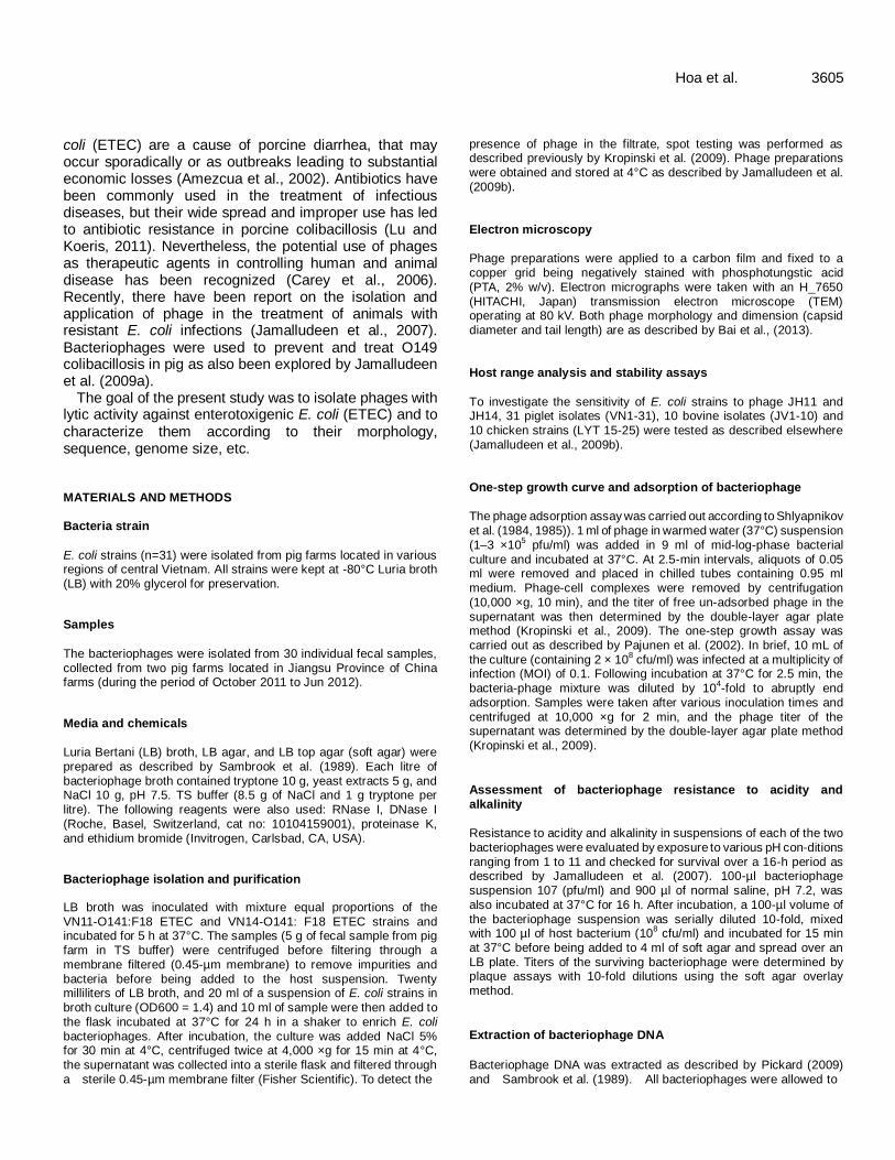
coli (ETEC) are a cause of porcine diarrhea, that may occur sporadically or as outbreaks leading to substantial economic losses (Amezcua et al., 2002). Antibiotics have been commonly used in the treatment of infectious diseases, but their wide spread and improper use has led to antibiotic resistance in porcine colibacillosis (Lu and Koeris, 2011). Nevertheless, the potential use of phages as therapeutic agents in controlling human and animal disease has been recognized (Carey et al., 2006). Recently, there have been report on the isolation and application of phage in the treatment of animals with resistant E. coli infections (Jamalludeen et al., 2007). Bacteriophages were used to prevent and treat O149 colibacillosis in pig as also been explored by Jamalludeen et al. (2009a).
The goal of the present study was to isolate phages with lytic activity against enterotoxigenic E. coli (ETEC) and to characterize them according to their morphology, sequence, genome size, etc. MATERIALS AND METHODS Bacteria strain
E. coli strains (n=31) were isolated from pig farms located in various regions of central Vietnam. All strains were kept at -80°C Luria broth (LB) with 20% glycerol for preservation. Samples
The bacteriophages were isolated from 30 individual fecal samples, collected from two pig farms located in Jiangsu Province of China farms (during the period of October 2011 to Jun 2012).
Media and chemicals Luria Bertani (LB) broth, LB agar, and LB top agar (soft agar) were prepared as described by Sambrook et al. (1989). Each litre of bacteriophage broth contained tryptone 10 g, yeast extracts 5 g, and NaCl 10 g, pH 7.5. TS buffer (8.5 g of NaCl and 1 g tryptone per litre). The following reagents were also used: RNase I, DNase I (Roche, Basel, Switzerland, cat no: 10104159001), proteinase K, and ethidium bromide (Invitrogen, Carlsbad, CA, USA).
Bacteriophage isolation and purification
LB broth was inoculated with mixture equal proportions of the
VN11-O141:F18 ETEC and VN14-O141: F18 ETEC strains and incubated for 5 h at 37°C. The samples (5 g of fecal sample from pig farm in TS buffer) were centrifuged before filtering through a membrane filtered (0.45-µm membrane) to remove impurities and bacteria before being added to the host suspension. Twenty milliliters of LB broth, and 20 ml of a suspension of E. coli strains in broth culture (OD600 = 1.4) and 10 ml of sample were then added to the flask incubated at 37°C for 24 h in a shaker to enrich E. coli bacteriophages. After incubation, the culture was added NaCl 5% for 30 min at 4°C, centrifuged twice at 4,000 ×g for 15 min at 4°C, the supernatant was collected into a sterile flask and filtered through a sterile 0.45-µm membrane filter (Fisher Scientific). To detect the
Hoa et al. 3605 presence of phage in the filtrate, spot testing was performed as described previously by Kropinski et al. (2009). Phage preparations were obtained and stored at 4°C as described by Jamalludeen et al.
(2009b). Electron microscopy
Phage preparations were applied to a carbon film and fixed to a copper grid being negatively stained with phosphotungstic acid (PTA, 2% w/v). Electron micrographs were taken with an H_7650 (HITACHI, Japan) transmission electron microscope (TEM) operating at 80 kV. Both phage morphology and dimension (capsid diameter and tail length) are as described by Bai et al., (2013). Host range analysis and stability assays
To investigate the sensitivity of E. coli strains to phage JH11 and JH14, 31 piglet isolates (VN1-31), 10 bovine isolates (JV1-10) and
10 chicken strains (LYT 15-25) were tested as described elsewhere (Jamalludeen et al., 2009b). One-step growth curve and adsorption of bacteriophage
The phage adsorption assay was carried out according to Shlyapnikov et al. (1984, 1985)). 1 ml of phage in warmed water (37°C) suspension (1–3 ×10
5 pfu/ml) was added in 9 ml of mid-log-phase bacterial
culture and incubated at 37°C. At 2.5-min intervals, aliquots of 0.05 ml were removed and placed in chilled tubes containing 0.95 ml medium. Phage-cell complexes were removed by centrifugation (10,000 ×g, 10 min), and the titer of free un-adsorbed phage in the supernatant was then determined by the double-layer agar plate method (Kropinski et al., 2009). The one-step growth assay was carried out as described by Pajunen et al. (2002). In brief, 10 mL of the culture (containing 2 × 10
8 cfu/ml) was infected at a multiplicity of
infection (MOI) of 0.1. Following incubation at 37°C for 2.5 min, the bacteria-phage mixture was diluted by 10
4-fold to abruptly end
adsorption. Samples were taken after various inoculation times and centrifuged at 10,000 ×g for 2 min, and the phage titer of the supernatant was determined by the double-layer agar plate method (Kropinski et al., 2009).
Assessment of bacteriophage resistance to acidity and
alkalinity Resistance to acidity and alkalinity in suspensions of each of the two bacteriophages were evaluated by exposure to various pH con-ditions ranging from 1 to 11 and checked for survival over a 16-h period as described by Jamalludeen et al. (2007). 100-µl bacteriophage suspension 107 (pfu/ml) and 900 µl of normal saline, pH 7.2, was also incubated at 37°C for 16 h. After incubation, a 100-µl volume of
the bacteriophage suspension was serially diluted 10-fold, mixed with 100 µl of host bacterium (10
8 cfu/ml) and incubated for 15 min
at 37°C before being added to 4 ml of soft agar and spread over an LB plate. Titers of the surviving bacteriophage were determined by plaque assays with 10-fold dilutions using the soft agar overlay method.
Extraction of bacteriophage DNA
Bacteriophage DNA was extracted as described by Pickard (2009) and Sambrook et al. (1989). All bacteriophages were allowed to

3606 Afr. J. Microbiol. Res. completely lyse their host E. coli strains in a soft agar overlay. The overlay was added to SM buffer, and bacteriophages were allowed to diffuse into the buffer at 4°C for 3-4 h with gentle shaking as
previously described. After the suspension was centrifuged at 4000 ×g for 15 min, the supernatant was collected. Solid NaCl was added to a final concentration of 1 M and dissolved by swirling. Following incubation on ice for 1 h, the suspension was centrifuged at 11,000 ×g for 10 min at 4°C (Beckman Coulter, J2-MC Centrifuge). The supernatant was collected, solid polyethylene glycol (PEG 8000) was added to a final concentration of 10% (w/v), and the mixture was stirred slowly at room temperature. After cooling in ice water and standing for 1 h on ice, the mixture was centrifuged at 14,000 ×g for 10 min at 4°C. The bacteriophage pellet was then resuspended in 1 mL of SM buffer. An equal volume of chloroform was added to the phage suspension and mixed by vortexing for 30 s. The phases were separated by centrifugation at 3000 ×g for 15 min at 4°C and the aqueous phase was recovered. Pancreatic DNase I and RNase I were added to a final concentration of 5 and 1 µg/mL, respectively, and allowed to digest substrates for 30 min at 37°C. EDTA, pH 8.0 was added to a final concentration of 20 mM.
Proteinase K was added to a final concentration of 50 µg/mL, then sodium dodecyl sulfate (SDS, 10%) was added to a final concentration of 0.5% and the mixture was inverted several times prior to incubation at 56°C for 2 h. An equal volume of phenol : chloroform : isoamyl alcohol (25:24:1, v/v/v) was mixed in the sample. The aqueous phase was collected after centrifugation at 10,000 ×g for 10 min and extracted with an equal volume of chloroform : isoamyl alcohol (24:1, v/v). Centrifugation was repeated and the aqueous phase was collected. Two volumes of ice-cold 95%
ethanol were added and the mixture was kept at room temperature for 20 min. The precipitate was collected by centrifugation at 10,000 ×g for 10 min at 4°C and the pellet was washed with cold 70% ethanol. Following centrifugation at 10,000 ×g and 4°C for 30 min, the pellet was air dried and dissolved in 20-35 µL TE buffer (10 mM Tris, 1 mM EDTA, pH 8). Agarose gel electrophoresis analysis
Genome sizes of undigested or digested bacteriophage were determined by electrophoresis (Sambrook and Russell, 2001). The bands were stained with ethidium bromide (EB) and compared against a λ phage DNA digested with Hind III marker and restriction enzyme Dra I before comparison by gel electrophoresis(Chakrabarti et al., 2000).
Protein of purified particles of phage Bacteriophages particles were purified by centrifugation through a glycerol step gradient as determined by Sambrook and Russell (2001) after purified particles were subjected to SDS-PAGE on precast 4-15% gradient TRIS acrylamidegels (BioRad) along with protein molecular weight markers (Kropinski et al., 2012). The
phage suspensions (approximately 1010 pfu/ml) were boiled for 5 min and separated by SDS-PAGE. Denaturation, annealing and extension The PCR and oligonucleotide primers were as described by Olsetetart et al. (2001). The consensus primers that were used to amplify the central portion of gene gp23 of the various T4-type phages were: Mzia1 (5´-TGTTATATIGGTATGGTICGICGTGCTAT-3´) and (5´-TGAAGT TACCTTCACCACGACCGG-3´). The primers that were initially used
to amplify the gens gp18 analogue of the T4-type phage were: FT18-N2 (5´-GGTAAATTCCAATGGGGTCCAGCT T-3´) and (5´-TATCAGCAGCCAACGGAACC CAA-3´).
PCR sequencing
The PCR products were purified (Casjens et al., 2004) and sequenced with an Amersham Life Science Thermo Sequenase kit. The gene 18 and 23 nucleotide sequences of the various T4-type phages were determined by Mzia1 and FT18-N2 primer.
RESULTS Bacteriophage isolation and morphology Two bacteriophages were isolated from fecal sample and, named JH11 and JH14. All the bacteriophage produced similar plaques that were clear and medium sized 3.0-3.5 mm in diameter (Figure 1).
Electron microscopy confirmed that phages JH11 and JH14 belong to the Myoviridae family. Phages possessed icosahedral heads, necks and contractile tails, with tail fibers. JH11 and JH14 belong to the order Caudovirales. The head dimensions for JH11 and JH14 were 120 × 70 and 100 × 80 nm, and tail dimensions were 100 × 25 and 130 × 30 nm, respectively (Figure 2).
Host range analysis and stability assays Phage JH11 and JH14 specifically lysed piglet clinical isolates of E. coli, whereas lysed neither bovine clinical isolates, nor other chicken. A high proportion (58.06%, n=31) of E. coli piglet isolates was sensitive to phage JH11 and JH14, thus confirming its broad host range (data not show). Both phages were found to be heat sensivitive (Figure 3) as more than 50% of phage particles were killed after 30 min of incubation at 60°C, and only 10% of the phage particles were still alive after 120 min of incubation. Less than 10% phage particles survived after 30 min of incubation at 70°C.
Bacteriophage resistance to acidity and alkalinity
The two bacteriophages were highly susceptible to acidity at pH 1 - 2 and susceptible in varying degrees to overnight exposure to pH 3-4. All the bacteriophages were resistant to the range of pH 5-9. Phage JH11 appeared to be a lightly more acid resistant than the JH14 (data not show).
Nucleic acid of bacteriophages
Nucleic acid of all the two bacteriophages was a DNA. All the bacteriophages’ DNA samples were digested with restriction enzyme Dra I (Figures 4 and 5).

Hoa et al. 3607
Figure 1. Plaques formed by phages JH11 and JH14 on O141 E. coli.
Figure 2. Appearance of phages JH11 and JH14 under electron microscopes.
Figure 3. Thermostability of bacteriophages JH11 and JH14.
0
20
40
60
80
100
0 30 60 90 120
Time (min)
Ph
ages s
urviv
al
rate (
%)
JH11 (70 °C)
JH14(70 °C)
JH11(60°C)
JH14(60°C)

3608 Afr. J. Microbiol. Res.
Figure 4. Electrophoresis of
DNA of phages JH1 and JH14 on 0.7% agarose gels M = λ DNA marker.
Figure 5. Analysis of
phages genetic by enzyme DraI on 0.7% agarose gels M = λ DNA marker.
PCR sequencing of bacteriophage JH11 and JH14 of gp18, gp23 gene Gene gp23 on the capsid surface structure and gene gp18 were structure protein of the T4 phage. The phage JH11 was gp23 positive, also the phage JH14 gp18 was gp23 positive, this is shown in Figure 6.
Figure 6. Agarose electrophoresis of PCR
products, M: 5000 DNA marker, A: phage JH11 gene 23 PCR production, B: phage JHu14 gene gp23 PCR production, C: phage JH14 gene gp18 PCR production.
Blast analysis of the JH11, JH14 random clones PCR products of gene 23 that resembled major capsid protein of JH11 and JH14 were dideoxynucleotide sequencing. Blast analysis results with the determined sequence on the NCBI website are demonstrated in Tables 1 and 2. According to the blast results, the gp23 of phage JH11 random clones were homologous with gene 23 of phage T4 MVSS (98%), ECML-134 (98%), T6 (97%) and T4 (96%) identity. Phage JH14 random clones are in close relation with HX01 (96%), CEV1 (95%) and T4 (93%). The two strains belong to T4-like phage genus. Gene of JH11 and JH14 were 93% identity (Figure 7). Protein of purified particles of phage Phage particles were purified and used to determine the structural protein content of each phage by SDS-PAGE analysis. Distinct profiles were observed among the two phages tested (Figure 8). Phage JH11 consisted of three abundant proteins (A, B and C) bands with approximate molecular sizes of 80, 75 and 50 kDa. Phage JH14 consisted of three abundant proteins (D, E and F) with estimated molecular size of 50, 38 and 36 kDa. DISCUSSION Bacteriophages are ubiquitous in our world and extremely diverse. Although recent research on bacteriophage is

Hoa et al. 3609
Table 1. Blast analysis of bacteriophage JH11 random clones.
Accession Random clone Query coverage (%) Max ident(%)
JX128259.1 Escherichia phage ECML-134, complete genome 96 98
Z78095.1 Bacteriophage T6 DNA (1506 bp) 96 97
JN202312.1 Enterobacteria phage ime09, complete genome 96 96
DQ485345.1 Enterobacteria phage MV SS major capsid protein gene, partial cds 90 98
HM137666.1 Enterobacteria phage T4T, complete genome 96 96
AF158101.6 Enterobacteria phage T4, complete genome 96 96
X01774.1 Bacteriophage T4 gene 23 for major capsid protein 96 96
K01765.1 Bacteriophage T4 genes 22 (partial) and 23 (complete cds) 96 96
DQ904452.1 Bacteriophage RB32, complete genome 96 96
Table 2. Blast analysis of bacteriophage JH14 random clones.
Accession Random clone Query coverage
(%)
Max ident
(%)
JX536493.1 Enterobacteria phage HX01, complete genome 94 96
AY331985.1 Bacteriophage CEV1 nonfunctional major capsid protein gp23 91 95
JX128259.1 Escherichia phage ECML-134, complete genome 94 93
HM137666.1 Enterobacteria phage T4T, complete genome 94 93
AF158101.6 Enterobacteria phage T4, complete genome 94 93
AY303349.1 Enterobacteria phage RB69, complete genome 94 93
Figure 7. Dendrogram showing relationship among phage JH11, JH14, and with the
determined sequence on the NCBI website.
mushrooming, it is still limited in some respects. The isolation and characterization of more bacteriophage will facilitate the utilization of bacteriophage’s resources. In this study, two novel bacteriophages named JH11 and JH14 were isolated from faecal pig farm. To identify the bacteriophages taxa, PCR method was applied with the sequencing blast analysis of bacteriophage. Preliminary study with this method revealed that bacteriophage JH11
and JH14 belonged to T4-like. The goal of this study was to isolate and characterize phages against the O141 E. coli with special emphasis on O141: F18 ETEC, which were reported as the most common sources of coliphages from sewage samples of domestic animal drainage (pigs). Pig sewage is a collection of waste drainage for the entire pig farms and was considered to be a good source from which to isolate phages against O141: F18 ETEC,
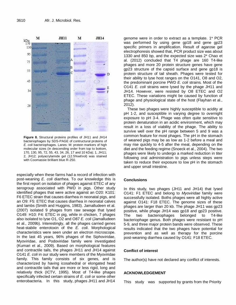
3610 Afr. J. Microbiol. Res.
Figure 8. Structural proteins profiles of JH11 and JH14
bacteriophages by SDS-PAGE of contructural proteins of E. coli bacteriophages. Lanes: M: protein markers of high molecular sizes (in descending order from top to bottom, 170, 130, 95, 72, 55, 43, 34, 26, 17 and 10 kDa); 1, JH11; 2, JH12; polyacrylamide gel (12.5%wt/vol) was stained with Coomassie brilliant blue R-250.
especially when these farms had a record of infection with post-weaning E. coli diarrhea. To our knowledge this is the first report on isolation of phages against ETEC of any serogroup associated with PWD in pigs. Other study identified phages that were active against an O20: K101: F6 ETEC strain that causes diarrhea in neonatal pigs, and an O9: F5: ETEC that causes diarrhea in neonatal calves and lambs (Smith and Huggins, 1983). Jamalludeen et al. (2007) isolated 9 phages from raw sewage that lysed O149: H10: F4: ETEC in pig, while in chicken, 7 phages also isolated to lyse O1, O2 and O87 E. coli (Jamalludeen et al., 2009b). Interestingly, all the phages carried genes heat-stabile enterotoxin of the E. coli. Morphological characteristics were seen under an electron microscope. In the last 45 years, 96% phages of the Siphoviridae, Myoviridae, and Podoviridae family were investigated (Kumari et al., 2009). Based on morphological features and contractile tails, the phages JH11 and JH14 against O141 E. coli in our study were members of the Myoviridae family. This family consists of six genes, and is characterized by having icosahedral or elongated head and contractile tails that are more or less rigid, long and relatively thick (ICTV, 1995). Most of T4-like phages specifically infected certain strains of E. coli or some other enterobacteria. In this study, phages JH11 and JH14
genome were in order to extract as a template. 1* PCR was performed by using gene gp18 and gene gp23 specific primers in amplification. Result of agarose gel electrophoresis showed that, PCR product size was about 1330 and 850 bp, and the expected size was 2* Chao et al. (2012) concluded that T4 phage are 160 T4-like phages and more 20 protein structure genes have gene gp23 structure of the capsid surface and gene gp18 is protein structure of tail sheath. Phages were tested for their ability to lyse host ranges on the O141, O8 and O2, the predominant porcine PWD E. coli strains. Most of the O141 E. coli strains were lysed by the phage JH11 and JH14. However, were resisted by O8 ETEC and O2 ETEC. These variations might be caused by function of phage and physiological state of the host (Flayhan et al., 2012).
These two phages were highly susceptible to acidity at pH 1-2, and susceptible in varying degree to overnight exposure to pH 3-4. Phage was often quite sensitive to protein denaturation in an acidic environment, which may result in a loss of viability of the phage. The ability to survive well over the pH range between 5 and 9 was a common feature for most phages. The pH in the stomach of weaned pigs may be as low as 1-2 before a meal and may rise quickly to 4-5 after the meal, depending on the diet and the feeding regime (Snoeck et al., 2004). The two phages were likely to undergo a marked reduction in titer following oral administration to pigs unless steps were taken to reduce their exposure to low pH in the stomach and upper small intestine. Conclusions In this study, two phages (JH11 and JH14) that lysed O141: F1 ETEC and belong to Myoviridae family were successfully isolated. Boths phages were all highly active against O141: F18 ETEC. The genome sizes of these phages are larger than 20 kb. The phage JH11 was gp23 positive, while phage JH14 was gp18 and gp23 positive. The two bacteriophages belonged to T4-like bacteriophage genus. Both phages were resistant to pH 5-9, and three major protein bands were observed. These results indicated that the two phages have potential for prevention and as well as therapy for the porcine post-weaning diarrhea caused by O141: F18 ETEC. Conflict of interest The author(s) have not declared any conflict of interests. ACKNOWLEDGEMENT This study was supported by grants from the Priority

Academic Program Development of Jiangsu Higher Education Institutions, Chaina. REFERENCES
Amezcua R, Friendship Robert M, Dewey Catherine E, Gyles C,
Fairbrother JM (2002). Presentation of postweaning Escherichia coli
diarrhea in southern Ontario, prevalence of haemolytic E. coli
serogroups involved, and their antimicrobial resistance patterns. Can. J. Vet. 66:73-78.
Bai Q, Zhang W, Yang Y, Tang F, XuanHoa N, Liu G, Lu C (2013). Characterization and genome sequencing of a novel bacteriophage infecting Streptococcus agalactiae with high similarity to a phage from
Streptococcus pyogenes. Arch Virol. 158(8):1733-1741.
Barrow P, Lovell M, Berchieri A (1998). Use of lytic bacteriophage for control of experimental Escherichia coli septicemia and meningitis in
chickens and calves. Clin. Diagn. Lab. Immunol. 70 (6):294–298 Carey S, Gwyneth V, Billington C, Cornelius AJ, Hudson A, Heinemann
JA (2006). Isolation and characterization of bacteriophages infecting Salmonella spp. Microbiol Lett. 258:182-186.
Casjens S, Winn-Stapley DA, Gilcrease EB, Morona R, Kühlewein C, Chua JEH, Manning PA, Inwood W, Clark AJ (2004). The chromosome of Shigella flexneri bacteriophage Sf6: complete
nucleotide sequence, genetic mosaicism and DNA packaging. J. Mol. Biol. 339(2):379-394.
Chakrabarti AK, Ghosh AN, Nair GB, Niyogi SK, Bhattacharya SK,
Sarkar BL (2000). Development and evaluation of a phage typing scheme for Vibrio cholerae O139. J. Clin. Microbiol. 38:44-49
Chao L, Wailap VN, Lin I, Syu W, Liu T, Chang C (2012). T4-like genome organization of the Escherichia coli O157:H7 lytic phage
AR1. J. Virol. 85 (13):6567–6578. Flayhan A, Wien F, Paternostre M, Boulanger P, Breyton C (2012). New
insights into pb5, the receptor binding protein of bacteriophage T5, and its interaction with its Escherichia coli receptor FhuA.
Biochemistry 94:1989-1994.
Hendrix RW, Smith MC, Burns RN (1999). Evolutionary relationships among diverse bacteriophages and prophages: all the worldÿs a phage. Proc. Natl. Acad. Sci. USA. 96(5):2192-2197.
ICTV (1995). International Committee on Taxonomy of Viruses. Virus taxonomy, classification and nomenclature of virus. Sixth report of the international committee on taxonomy of viruses. 49-54.
Jamalludeen N, Johnson RP, Friendship R, Kropinski AM, Lingohr EJ, Gyles CL (2007). Isolation and characterization of nine bacteriophages that lyse O149 enterotoxigenic Escherichia coli. Vet.
Microbiol. 124:47-57. Jamalludeen N, Johnson RP, Shewen PE, Gyles CL (2009a). Evaluation
of bacteriophages for prevention and treatment of diarrhea due to experimental enterotoxigenic Escherichia coli O149 infection of pigs.
Vet. Microbiol. 136:135-141.
Hoa et al. 3611 Jamalludeen N, She Y-M, Lingohr EJ, Griffiths M (2009b). Isolation and
characterization of virulent bacteriophages against Escherichia coli
serogroups O1, O2, and O78. Poult. Sci. 88:1694-1702.
Kropinski AM, Lingohr EJ, Moyles DM, Ojha S, Mazzocco A, She Y-M, Bach SJ, Rozema EA, Stanford K, McAllister TA, Johnson RP (2012). Endemic bacteriophages: a cautionary tale for evaluation of
bacteriophage therapy and other interventions for infection control in animals. Virol J. 9 (209):1-9.
Kropinski AM, Mazzocco A, Waddell T, E., Lingohr E, Johnson Roger P
(2009). Enumeration of bacteriophages by double agar overlay. Methods Mol. Biol. 1 (501):69-80.
Kumari S, Harjai K, Chhibber S (2009). Characterization of Pseudomonas aeruginosa PAO specific bacteriophages isolated from
sewage samples Am. J. Biomed. 1 (2):91-102 Lu TK, Koeris MS (2011). The next generation of bacteriophage therapy.
Microbiology 14:524-531. Olsetetart F, Desplats C, Kutateladze M, Monod C, Ackermann H-W,
Krisch HM (2001). Phylogeny of the major head and tail genes of the
wide-ranging T4-type bacteriophages. J. Bacteriol. 183 (1):358-366 Pajunen MI, Elizondo MR, Skurnik M, Kieleczawa J, Molineux IJ (2002).
Complete Nucleotide Sequence and Likely Recombinatorial Origin of Bacteriophage T3. J. Mol. Biol. 319(5):1115-1132.
Pickard DJJ (2009). Preparation of bacteriophage lysates and pure DNA. Methods Mol. Biol. 2(502):3-9
Sambrook J, Fritsch EF, Maniatis T (1989). Molecular Cloning: A
Laboratory Manual, second ed. Cold Spring Harbour Press, Cold Spring Harbour, NY.
Sambrook J, Russell DW (2001). Molecular Cloning: A Laboratory
Manual, 2nd ed., In Molecular cloning: a laboratory manual, 3rd ed. CSHL Press, New York. 1:132-134.
Shlyapnikov MG, Kazantsev SI, Kryukov VM, Bayev AA (1985). The
nucleotide sequence of bacteriophage T 5 leucine tRNA. FEBS Lett. 192 (2):299-302.
Shlyapnikov MG, Kaliman AV, Kazantsev SI, Kryukov VM, Bayev AA
(1984). The nucleotide sequence of bacteriophage T5 glutamine transfer RNA. Biochim. Biophys. Acta 782(3):313-319.
Smith HW, Huggins MB (1983). Effectiveness of phages in treating experimental Escherichia coli diarrhoea in calves, piglets and lambs.
J. Gen. Microbiol. 129:2659-2675. Snoeck V, Cox E, Verdonck F, Joensuu JJ, Goddeeris BM (2004).
Influence of porcine intestinal pH and gastric digestion on antigenicity of F4 fimbriae for oral immunisation. Vet. Microbiol. 98(1):45-53
Soothill JS (1992). Treatment of experimental infections of mice by
bacteriophage. J. Med. Microb. 37:258-261.

Vol. 8(41), pp. 3612-3616, 8 October, 2014
DOI: 10.5897/AJMR2014.7019
Article Number: 1959EAF48130
ISSN 1996-0808
Copyright © 2014
Author(s) retain the copyright of this article
http://www.academicjournals.org/AJMR
African Journal of Microbiology Research
Full Length Research Paper
In vitro antimicrobial evaluation of two indigenous functional food-plants (Chenopodium album and
Solanum nigrum) used in the Oliver Reginald (O.R.) Tambo district municipality of South Africa
Collise Njume1,2*, Bomkazi M. Gqaza3, Grace George3 and Nomalungelo I. Goduka1
1Centre for Rural Development, Walter Sisulu University, Enkululekweni, Mthatha, 5117, South Africa.
2Department of Medical Microbiology, Walter Sisulu University, Mthatha 5117, South Africa.
3Department of Medical Biochemistry, Walter Sisulu University, Mthatha 5117, South Africa.
Received 17 July, 2014; Accepted 8 September, 2014
Plants have long been the source of important products with nutritional and therapeutic properties. African indigenous functional food-plants such as Chenopodium album and Solanum nigrum may constitute important sources of phytochemical constituents for the synthesis of antimicrobial compounds against infectious organisms. The objective of this study was to determine the antimicrobial properties of C. album and S. nigrum leaves used as functional food-plants in the O.R. Tambo district municipality of South Africa. Organic and aqueous solvent-extracts of C. album and S. nigrum were tested against Staphylococcus aureus (ATCC 29213), Pseudomonas aeruginosa (ATCC 27853), Bacillus subtilis (ATCC 6051), Escherichia coli (25922) and Enterococcus faecalis (51299) using standard microbiological techniques. Ciprofloxacin was included in all the experimental runs as positive control antibiotic. The aqueous extracts of both plants were the most active with zones of inhibition diameters ranging from 0 - 20 mm and minimum inhibitory concentration (MIC50) values ranging from 0.63 - 10 mg/mL. The positive control antibiotic was highly active with zones of inhibition diameters ranging from 17 - 31 mm and MIC50 values from 0.0003 - 0.0005 mg/mL for all the bacteria tested. Both extracts were bactericidal with minimum bactericidal concentration (MBC) ranging from 2.5 - 20 mg/mL. From the results, it can be concluded that both plants possess compounds with antimicrobial properties, thus validating scientifically their use in traditional medicine. However, more studies to document the respective plant-principles responsible for antimicrobial activity of these plants would shed more light on their pharmacological properties. Key words: Antimicrobial resistance, sensitivity tests, functional plants, Eastern Cape Province, South Africa.
INTRODUCTION Bacterial antimicrobial resistance against commonly used antibiotics is distressingly on the rise (Falagas et al., 2013).
Patients infected with resistant bacteria are more likely to have longer, more expensive hospital stays (Matsuura
*Corresponding author. E-mail: [email protected] or [email protected]. Tel: +27(0)475022710 or +27(0)732481673. Author(s) agree that this article remain permanently open access under the terms of the Creative Commons Attribution License 4.0International License

et al., 2013). The modification of chemotherapeutic agents to limit this problem has been greatly successful. How-ever, many reports also indicate that many of the drugs are being rendered obsolete by microbial drug-resistance (Balsalobre et al., 2014). As a result, the treatment of microbial infection is becoming increasingly complicated. Physicians have now resorted to the use of combination therapy, increasing the cost of treatment even more. Reports on Escherichia coli, Pseudomonas aeruginosa, Staphylococcus aureus, Bacillus subtilis and Enterococcus faecalis infections and antibacterial resis-tance reveal the need for a constant search of new drugs against these organisms (Gupta et al., 2001; Tamma et al., 2012). E. coli is a major cause of travellers’ diarrhoea, one of the most common forms of diarrhoea worldwide (Mittal et al., 2009; Canizalez-Roman et al., 2013). Both E. coli and P. aeruginosa are also major causes of urinary tract infections while S. aureus and E. faecalis are common causes of nosocomial infections (Sood et al., 2008; Oli et al., 2012). B. subtilis infections are not common but few cases have been reported in the literature in patients with oesophageal perforations (Jeon et al., 2012).
The use of medicinal plants in the treatment of human infections is a common practice in many remote areas of Africa with inadequate health care facilities. Chenopodium album and Solanum nigrum are functional food-plants with wide nutritional and medicinal importance among rural communities in the O.R Tambo District Municipality of South Africa (Njume et al., 2014). They are jointly referred to as imifino ezikhulelayo in isiXhosa, meaning indigenous vegetable. C. album is locally known as imbikicane while S. nigrum is known as umsobo (Gqaza et al., 2013a, b). Both plants grow wildly in bushes, barren land and roadside paths from where they are harvested either for nutritional or medicinal purposes. Even though they are considered as weeds in the urban areas, many rural dwellers find them tasteful and believe in their ability to alleviate problems associated with gastrointestinal discomfort. In some parts of India, C. album is also used in ayurveda for treating anorexia, cough, dysentery, diarrhoea, oedema, piles and worm infestations (Yadav et al., 2007). Despite their medicinal uses, very little information is available in the literature on their pharma-cological potential. This is surprising considering the ever-increasing rate of antimicrobial resistance of human infectious organisms to currently used drugs. The aim of this study therefore was to investigate the antimicrobial properties of these plants in an attempt to identify cheap sources of compounds for the synthesis of new drugs against medically important bacteria.
MATERIALS AND METHODS
Bacterial strains
Standard bacterial strains including Pseudomonas aeruginosa ATCC 127853, Escherichia coli ATCC 25922, Staphylococcus
Njume et al. 3613 aureus ATCC 29213, Enterococcus faecalis ATCC 51299 and Bacillus subtilis ATCC 6051 (American Type Culture Collection, Rockville, MD) obtained from the stock culture of the National Health Laboratory Services (NHLS), Nelson Mandela Academic Hospital, Mthatha were used in this study. Ethical clearance was obtained from the Eastern Cape Department of Health and the Ethics Committee of the Faculty of Health Sciences, Walter Sisulu University (WSU). The bacteria were separately inoculated on Nutrient agar (Oxoid Llt., Basingstoke, UK) plates consisting of peptone (5 g/L), beef extract (3 g/L), sodium chloride (8 g/L), agar number 2 (12 g/L) at pH (7.3±0.2). The plates were incubated at 37°C for 24-48 h and examined for growth. Collection and preparation of plant material Mature plant-leaves of C. album and S. nigrum were harvested around the stems from home gardens and along bush paths in the vicinity of WSU main campus in Mthatha in October 2012. The plants were identified by Dr. Kathleen Immelman of the Department of Botany at WSU and voucher specimens were prepared and deposited in the Kei herbarium (CN01 and CN02). The plant-leaves were washed with tap water to remove dirt and soil particles. The plant-leaves were placed on cardboards and dried at 50°C for 24 h in a hot air oven (Heraeus, Schutzart). The plant material was powdered (ATO Mix, Cambridge) and stored in air-tight containers at 5°C for further analysis. Preparation of plant extracts
Approximately 400 g of dried powdered plant material was exhaustively extracted in different solvents. The plant material was separately soaked in 700 mL of concentrated hexane, acetone, ethanol, methanol and water in 2 L volumetric flasks (Schott, Durban). The flasks were placed in an orbital shaker incubator (labcon, Maraisburg) for 48 h (Kuete et al., 2011). The plant material was centrifuged at 1006.2 xg for 5 min and filtered through a fritted filter funnel of pore size 60Å. The procedure was repeated twice and the three extracts combined and concentrated to dryness under vacuum (Büchi, Switzerland). The dried crude extract was collected in porcelain evaporating dish (Haldenwanger, Berlin) and left open in a biosafety class 2 cabinet (Durban, South Africa) for complete evaporation of residual solvents. The aqueous extracts were lyophilized (Castillo-Juarez et al., 2009). A 2-g sample of each extract was used for the preliminary bioassay, and where possible, another 2 g or more was put in universal bottles and kept in the extract bank. Stock solutions were prepared by dissolving the extracts in 80% acetone (a concentration we found to be non-inhibitory to any of the bacterial strains tested).
Screening of crude extracts for antibacterial activity
The agar-well diffusion method was used for this analysis (Boyanova et al., 2005). Briefly, each bacterial suspension prepared in 0.9% saline (McFarland turbidity standard 0.5) was inoculated by spreading on Mueller Hinton agar (Oxoid Llt., Basingstoke, UK) plates and allowed to dry for 15 min. Wells (6 mm in diameter) were punched into the agar using a sterile stainless steel borer and filled with 70 μL of the extract at 100 mg/mL. Seventy microliters of 0.005 mg/mL ciprofloxacin and 80% acetone were included in all experiments as positive and negative controls, respectively. The plates were incubated at 37°C for 24 h, after which the diameters of zones of inhibition were measured in mm. The experiment was repeated twice, and means for zones were recorded.
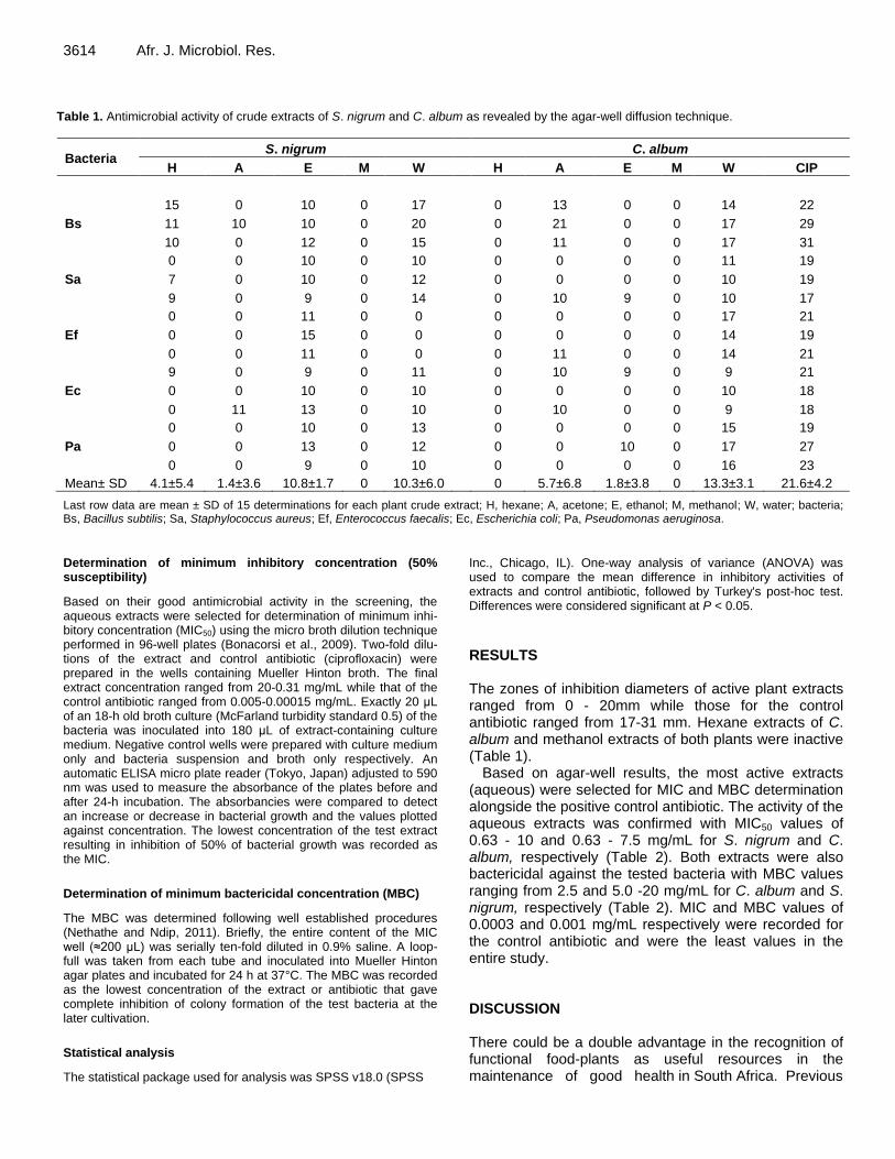
3614 Afr. J. Microbiol. Res.
Table 1. Antimicrobial activity of crude extracts of S. nigrum and C. album as revealed by the agar-well diffusion technique.
Bacteria S. nigrum C. album
H A E M W H A E M W CIP
Bs
15 0 10 0 17 0 13 0 0 14 22
11 10 10 0 20 0 21 0 0 17 29
10 0 12 0 15 0 11 0 0 17 31
Sa
0 0 10 0 10 0 0 0 0 11 19
7 0 10 0 12 0 0 0 0 10 19
9 0 9 0 14 0 10 9 0 10 17
Ef
0 0 11 0 0 0 0 0 0 17 21
0 0 15 0 0 0 0 0 0 14 19
0 0 11 0 0 0 11 0 0 14 21
Ec
9 0 9 0 11 0 10 9 0 9 21
0 0 10 0 10 0 0 0 0 10 18
0 11 13 0 10 0 10 0 0 9 18
Pa
0 0 10 0 13 0 0 0 0 15 19
0 0 13 0 12 0 0 10 0 17 27
0 0 9 0 10 0 0 0 0 16 23
Mean± SD 4.1±5.4 1.4±3.6 10.8±1.7 0 10.3±6.0 0 5.7±6.8 1.8±3.8 0 13.3±3.1 21.6±4.2
Last row data are mean ± SD of 15 determinations for each plant crude extract; H, hexane; A, acetone; E, ethanol; M, methanol; W, water; bacteria; Bs, Bacillus subtilis; Sa, Staphylococcus aureus; Ef, Enterococcus faecalis; Ec, Escherichia coli; Pa, Pseudomonas aeruginosa.
Determination of minimum inhibitory concentration (50% susceptibility)
Based on their good antimicrobial activity in the screening, the aqueous extracts were selected for determination of minimum inhi-bitory concentration (MIC50) using the micro broth dilution technique performed in 96-well plates (Bonacorsi et al., 2009). Two-fold dilu-tions of the extract and control antibiotic (ciprofloxacin) were prepared in the wells containing Mueller Hinton broth. The final extract concentration ranged from 20-0.31 mg/mL while that of the control antibiotic ranged from 0.005-0.00015 mg/mL. Exactly 20 μL of an 18-h old broth culture (McFarland turbidity standard 0.5) of the bacteria was inoculated into 180 μL of extract-containing culture medium. Negative control wells were prepared with culture medium only and bacteria suspension and broth only respectively. An automatic ELISA micro plate reader (Tokyo, Japan) adjusted to 590 nm was used to measure the absorbance of the plates before and after 24-h incubation. The absorbancies were compared to detect an increase or decrease in bacterial growth and the values plotted against concentration. The lowest concentration of the test extract resulting in inhibition of 50% of bacterial growth was recorded as the MIC.
Determination of minimum bactericidal concentration (MBC)
The MBC was determined following well established procedures (Nethathe and Ndip, 2011). Briefly, the entire content of the MIC well (≈200 μL) was serially ten-fold diluted in 0.9% saline. A loop-full was taken from each tube and inoculated into Mueller Hinton agar plates and incubated for 24 h at 37°C. The MBC was recorded as the lowest concentration of the extract or antibiotic that gave complete inhibition of colony formation of the test bacteria at the later cultivation.
Statistical analysis
The statistical package used for analysis was SPSS v18.0 (SPSS
Inc., Chicago, IL). One-way analysis of variance (ANOVA) was used to compare the mean difference in inhibitory activities of extracts and control antibiotic, followed by Turkey's post-hoc test. Differences were considered significant at P < 0.05. RESULTS The zones of inhibition diameters of active plant extracts ranged from 0 - 20mm while those for the control antibiotic ranged from 17-31 mm. Hexane extracts of C. album and methanol extracts of both plants were inactive (Table 1).
Based on agar-well results, the most active extracts (aqueous) were selected for MIC and MBC determination alongside the positive control antibiotic. The activity of the aqueous extracts was confirmed with MIC50 values of 0.63 - 10 and 0.63 - 7.5 mg/mL for S. nigrum and C. album, respectively (Table 2). Both extracts were also bactericidal against the tested bacteria with MBC values ranging from 2.5 and 5.0 -20 mg/mL for C. album and S. nigrum, respectively (Table 2). MIC and MBC values of 0.0003 and 0.001 mg/mL respectively were recorded for the control antibiotic and were the least values in the entire study. DISCUSSION There could be a double advantage in the recognition of functional food-plants as useful resources in the maintenance of good health in South Africa. Previous

Table 2. Minimum Inhibitory and bactericidal concentration (mg/mL) values of plant crude extracts and control antibiotic tested against medically important bacteria.
Bacteria species S. nigrum C. album Ciprofloxacin
MIC
B. subtilis 2.5 0.63 0.0005
S. aureus 0.63 0.63 0.0003
E. faecalis 2.5 1.3 0.0005
E. coli 5 7.5 0.0005
P. aeruginosa 10 5 0.0003
Total 4.1±3.6 3.0±3.1 0.0004±0.0001
MBC
B. subtilis 5 2.5 0.001
S. aureus 10 2.5 0.001
E. faecalis 20 10 0.002
E. coli 10 20 0.003
P. aeruginosa 10 10 0.002
Total 11±5.5 9±7.2 0.002±0.0008
The total results shown are representative of mean ± SD value of five determinations for each plant crude extract or control antibiotic.
studies on some of these plants indicate them to be useful sources of important dietary components such as vitamins (A, B2, C, folic acid), iron, selenium, zinc, calcium and magnesium (Akubugwo et al., 2007; van der Walt et al., 2008; Gqaza et al., 2013a,b). Some of these nutrients occur in high enough quantities to meet the recommended daily allowance in children, preventing hidden hunger, its effects (stunted growth, anaemia, overweight, obesity) and promoting optimal growth and development (van Jaarsveld et al., 2014). Their medicinal properties indi-cate that they may also constitute an important source of antioxidants and therapeutic compounds against human infectious organisms. Many plants have been reported to contain flavonoids, alkaloids, tannins, phenols, saponins or other secondary metabolites which serve as defense mechanisms against microorganisms, insects and ani-mals (Madduluri et al., 2013). These compounds are known to act in different ways to exert antimicrobial action. Some, such as tannins do so by binding to proline-rich proteins and interfering with protein synthesis while others such as saponins interact with bacterial cell walls to cause leakage of vital cell proteins (Shimada et al., 2006; Madduluri et al., 2013). However, this study did not evaluate the mechanism of action of the crude extracts against the tested bacteria. Nevertheless, it indicates that crude extracts of C. album and S. nigrum, especially aqueous extracts have the potential for further evaluation as antibacterial agents. The crude extracts inhibited the test bacteria, sometimes at low concentrations of 0.63 mg/mL. However, higher concentrations, at times up to 20 mg/mL were needed to completely kill the bacteria (Table 2).
Njume et al. 3615
Gram-positive organisms: S. aureus, E. faecalis and B. subtilis were the most susceptible in the entire study while Gram-negative bacteria such as E. coli and P. aeruginosa were less susceptible (Tables 1 and 2). The difference in susceptibility between Gram-negative and positive bacteria to antimicrobial agents has been reported by other researchers (Cock, 2007; Madduluri et al., 2013) and may be attributed to structural differences in the cell wall of both organisms. Gram-negative bacteria have a lipid protective sheath around their cell walls which seems to shield them from the effects of antimi-crobial agents (Silhavy et al., 2010). All the bacteria tested were highly susceptible to ciprofloxacin, the control antibiotic (Tables 1 and 2). The plant crude extracts were relatively less active when compared to ciprofloxacin (P<0.05). This was expected as the control antibiotic is a purified compound which may also contain excipients to facilitate activity. The crude extracts on the other hand are made of numerous compounds; some of which may be synergistic or antagonistic against each other. Equally important is the fact that the quantity of the active ingre-dient in the crude extracts may be in minute quantities, not enough to exhibit the type of activity demonstrated by the control antibiotic. Of all the bacteria tested, B. subtilis and S. aureus were most susceptible to aqueous extracts of the plants, producing large zones of inhibition dia-meters (Table 1), low MIC and MBC values (Table 2). However, there were no significant differences in antibacterial activity between the aqueous extracts of C. album and S. nigrum (P>0.05). Conclusion The current study illustrates the antibacterial properties of crude extracts of C. album and S. nigrum against some selected bacteria of medical importance. The study shows that aqueous extracts of both plants are inhibitory and bactericidal to S. aureus, B. subtilis, P. aeruginosa, E. coli and E. faecalis. These findings are consistent with their folkloric use in the treatment of stomach-related morbidities in the O.R. Tambo District Municipality of South Africa. However, more studies to document the plants active ingredients will shed more light on their pharmacological relevance as antibacterial agents. Conflict of interest The author(s) have not declared any conflict of interests.
ACKNOWLEDGEMENTS The authors are grateful to the Department of Science and Technology (DST) and the National Research
Foundation (NRF), South Africa for funding this study through a grant awarded to the Research Chair for

3616 Afr. J. Microbiol. Res. Indigenous Knowledge System at Walter Sisulu University, South Africa. REFERENCES Akubugwo IE, Obasi AN, Ginika SC (2007). Nutritional potential of
theleaves and seeds of blacknightshade-Solanumnigrum L. varvirginicum from Afikpo-Nigeria. Pak. J. Nutr. 6:323-326.
Balsalobre LC, Dropa M, Matte MH (2014). Anoverview of Antimicrobial resistance and its public health significance. Braz. J. Microbiol. 45(1):1-5.
Bonacorsi C, Raddi MSG, Iracilda ZC, Sannomiya M, Vilegas W (2009). Anti-Helicobacter pylori activity and immunostimulatory effect of extracts from ByrsonimacrassaNied. (Malpighiaceae). Complement. Altern. Med. 9:1472-6882.
Boyanova L, Gergova G, Nikolov R, Derejian S, Lazarova E, Katsarov N, Mitov I, Krastev Z (2005). Activity of Bulgarian propolis against 94 Helicobacter pylori strains in vitro by agar-well diffusion, agar dilution and disc diffusion methods. J. Med. Microbiol. 54: 481-483.
Canizalez-Roman A, Gonzalez-Nunez E, Vidal JE, Flores-Villasenor H, Leon-Sicairos N (2013). Prevalence and antibiotic resistant profiles of diarrheagenic Escherichia coli strains isolated from food items in North Western Mexico. Int. J. Food. Microbiol. 164(1):36-45.
Castillo-Juárez I, González V, Aime-Aguilar H, Martínez G, Linares E, Bye R, Romero I (2009). Anti-Helicobacter pylori activity of plants used in Mexican traditional medicine for gastrointestinal disorders. J. Ethnopharmacol. 122(2):402-405.
Cock I (2007). Antibacterial activity of selected Australian native plant extracts. Int. J. Microbiol. 4(2):76-81.
Falagas ME, Karageorgopoulos DE, Leptidis J, Korbila IP (2013). MRSA in Africa: Filing the global map of antimicrobial resistance. PLoS One 8(7) e68024:1-12.
Gqaza BM, Njume C, Goduka NI, George G (2013a). Nutritional assessment of Chenopodium album L. (Imbikicane) young shoots and mature plant-leaves consumed in the Eastern Cape Province of South Africa. Int. Conf. Nutr. Food Sci. DOI: 10.7763/IPCBEE.
Gqaza MB, Njume C, Goduka IN, Grace G (2013b). The proximate composition of S. nigrum plant-leaves consumed in the Eastern Cape Province of South Africa. Int. Conf. Nutr. Food Sci. DOI: 10.7763/IPCBEE.
Gupta K, Hooton TM, Stamm WE (2001).Increasing antimicrobial resistance and the management of uncomplicated community-acquired urinary tract infections. Ann. Int. Med. 135(1):45-50.
Jeon YL, Yang JJ, Kim MJ, Lim G, Cho SY, Park TS, Suh JT, Park YH, Lee MS, Kim SC, Lee HJ (2012). Combined Bacillus licheniformis and Bacillus subtilis infection in a patient with oesophageal perforation. J. Med. Microbiol. 61(12):1766-1769.
Kuete V, Ango PY, Fotso GW, Kapche GD, Dzoyem JP, Wouking AG, Ngadjui BT, Abegaz BM (2011). Antimicrobial activities of the methanol extract and compounds from Artocarpus communis (Moraceae). BMC Complement. Altern. Med. 11(42):1-5.
Madduluri S, Rao KB, Sitaram B (2013). In vitro evaluation of
antibacterial activity of five indigenous plants extracts against five bacteria pathogens of humans. Int. J. Pharm. Pharm. Sci. 5(4):679-684.
Matsuura GT, Pharm D, Barg N (2013). Update on the antimicrobial management of foot infections in patients with diabetes. Clin. Diabetes 31(2):59-65.
Mittal R, Aggarwal S, Sharma S, Chhibber S, Harjai K (2009). Urinary tract infections caused by Pseudomonas aeruginosa: a minireview. J. Infect. Public Health 2:101-111.
Nethathe BB, Ndip RN (2011). Bioactivity of Hydnora africana on selected bacteria pathogens: Preliminary phytochemical screening. Afr. J. Microbiol. Res. 5(18):2820-2826.
Njume C, Goduka NI, George G (2014) Indigenous leafy vegetables (imifino, morogo, muhuro) in South Africa: a rich and unexplored source of nutrients and antioxidants. Afr. J. Biotechnol. 13(19):1933-1942.
Oli AK, Sungar R, Shivshetty N, Hosamani R, Rivansiddappa KC (2012). A study of scanning electron microscopy of vancomycin resistant Enterococcus faecalis from clinical isolates. Adv. Microbiol. 2:93-97.
Shimada T (2006). Salivary proteins as a defense against dietary tannins. J. Chem. Ecol. 32(6):1149-1163.
Silhavy TJ, Kahne D, Walker S (2010). The bacterial cell envelope. Cold Spring Harb. Perspect. Biol. 2(5):1-17.
Sood S, Malhotra M, Das BK, Kapil A (2008). Enterococcal infections and antimicrobial resistance. Indian J. Med. Res. 128:111-121.
Tamma PD, Cosgrove SE, Maragakis LL (2012). Combination therapy for treatment of infections with gram negative bacteria. Clin. Microbiol. Rev. 25(3):450-470.
van der Walt AM, Ibrahim MI, Benzuidenhout CC, Loots DT (2008). Linolenic acid and folate in wild-growing African dark leafy vegetables (Morogo). Public Health Nutr. 12:525-530.
van Jaarsveld P, Faber M, van Heerden I, Wenhold F, van Rensburg WJ, van Averbeke W (2014). Nutrient content of eight African leafy vegetables and their potential contribution to dietary reference intakes. J. Food Compost. Anal. 33:77-84.
Yadav N, Vasudeva N, Singh S, Sharma SK (2007). Medicinal properties of genus Chenopodium Linn. Nat. Prod. Rad. 6(2):131-134.

Vol. 8(41), pp. 3617-3624, 8 October, 2014
DOI: 10.5897/AJMR2014.7022
Article Number: 8A209F348132
ISSN 1996-0808
Copyright © 2014
Author(s) retain the copyright of this article
http://www.academicjournals.org/AJMR
AfricanJournalofMicrobiology Research
Full Length Research Paper
Chemical characterization and bioactivity evaluation of bacteriocin from marine biofilm-forming bacteria
Sengol Jenifer S.1, V. Balasubramanian2 and R. Rajaram2*
1Department of Biotechnology, SeethalakshmiRamaswami College, Tiruchirappalli-620 002, Tamilnadu, India.
2Department of Marine Science, Bharathidasan University, Tiruchirappalli-620 024, Tamilnadu, India.
Received 18 July, 2014; Accepted 22 September, 2014
The marine biofilm bacteria were isolated on polyvinyl chloride (PVC) sheet immersed from offshore platform of Central Electrochemical Research Center (CECRI), Tuticorin, Tami Nadu. The isolated marine biofilm bacteria such as Pseudomonas sp., Aeromonas sp., Enterobacter sp., Bacillus spp., Flavobacterium sp., Microccocus sp. and Cytophaga sp were identified up to generic level. The Pseudomonas sp. was the dominant, primary biofilm forming bacteria. The cell free crude extract of single (Aeromonas sp.) and co-cultured (Aeromonas sp. + Enterobacter sp.) system showed the significant repellent activity against Pseudomonas sp. The crude fractions were characterized by FTIR for functional group identification and GC-MS for compound identification. The results of FTIR shows different peaks like 3302.16, 2816.56, 1227.44 and 2724.02 cm
–1 indicating the presence of bromine, amine, and phenolic, hydroxyl, amino,
carbonyl and phosphoryl functional groups. It was also noted that mixed culture system produced increased level of bacteriocin as compared to single culture system. The chemical characterizations of the inhibitory molecules such as nonane, is common to both single as well as co-culture system. The higher inhibitory activity observed in co-culture system is due to the presence of nonadecane, heneicosane and cholestane compounds. The natural biogenic compounds from the marine bacteria studied have the potential to be used as a substitute to commercial biocides for anti-microfouling. Key words: Anti-biofilm activity, bacteriocin, nonane, bioactivity, bioremediation.
INTRODUCTION Marine structures such as ships, marine platforms, offshore rigs and jetties are under constant attack from the marine environment by fouling organisms. These structures need to be protected from the influences of the key elements of the marine environment such as saltwater, temperature
fluctuations and biofouling (Chambers et al., 2006). Bio-fouling is one of the major significant problems and ubi-quitous in the marine environment. It is a natural process of colonization of submerged surfaces, either living or arti-ficial, involving a wide range of micro and macro organisms
*Corresponding author. E-mail: [email protected]. Tel: 0091-9842874661. Fax: 0091-431-240745. Author(s) agree that this article remain permanently open access under the terms of the Creative Commons Attribution License 4.0International License

3618 Afr. J. Microbiol. Res.
Figure 1. The Biofilm development rack.
(Briand, 2009). The attachment of microorganisms like bac-teria, algae, diatoms and fungi are called microfouling (bio-film), which initiates the macrofouling. The problem of biofouling is so severe that worldwide the expenditure incurred on antifouling measures alone is approximately
US$ 6.5 billion a year (Bhadury and Wright, 2004). Appli-cation of antifouling coatings like self- polishing copoly-mer antifouling paints (SPCs) with tributyl tin (TBT) as biocide and copper-based paints were widely employed for controlling fouling on marine structures (Chamberset al., 2006; Claisse and Alzieu, 1993; Batley et al., 1994). However, the toxicity of TBT and copper to marine organisms cause bioaccumulation of metal, imposex, etc. Nowadays, natural products from marine organisms can be used as replacements for the chemicals commonly used in antifouling coatings (Clare, 1996). Bacteriocins are antimicrobial compounds produced by bacteria that can kill closely related strains of the same species. Marine bacteria, primarily belonging to the Pseudoalteromonas genus, the Vibrionaceae family, and the Roseobacter clade, excrete compounds that can reduce bacterial biofilm formation and settlement by microorganisms on surfaces suggesting their suitability as antifouling bacteria (Bowman, 2007; Dobretsov and Qian, 2004). The chemotaxis activity of the bacteriocins toward the microfouling bacteria to prevent the initial biofilm formation,
accordingly the prevention of biofilm would be a safe and effective way to prevent the macrofouling through eco-friendly (Armstrong et al., 2000). The main purpose of the present study was to screen the antifouling potential of marine bacterial metabolites against primary biofilm forming bacteria. Finally, chemical characterization of bacteriocin obtained from the co-culture of Aeromonas sp. and Enterobacter sp. was done. MATERIALS AND METHODS Marine biofilm bacterial strain Polyvinyl chloride (PVC) sheet was cut into the dimension of 12″ ×12″ and the sheets were degreased using acetone and mounted on a wooden rack having the total size of 75″ × 15″, using brass bolt and nut. The rack was immersed at 2 m depth from the mean surface seawater below the offshore platform of Central Electro Chemical Research Institute (CECRI) at Tuticorin unit, Tamilnadu, India for 24 h (Figure 1). After 24 h, the biofilm was scrapped from PVC sheets using sterile brush and transferred to glass tube containing sterile seawater. Further the scrubbed biofilm samples was serially diluted and spread on Zobell Marine Agar and the plates were incubate up to 24 h at 37°C. The isolated biofilm bacteria were characterized using various morphological and biochemical tests (Allegrucci and Sauer, 2007; Balasubramanian et al., 2012). Bacteriocin activity For bacteriocin production, the isolates were inoculated by means of (i) single cell culture by Aeromonas sp. and co-culture by Aeromonassp + Enterobacter sp. were prepared in Zobellmarine broth and incubated at 37°C for 24-48 h in orbital shaker at 120 rpm over night. The growth kinetics of the single as wells co-culture systems has been monitored by taking the OD600 starting from 0, 1, 2, 3, 4, 5, 10, 15, 20 and 24 h respectively (Sher et al, 2011). The bacteriocins were extracted after 48 h of culture, because the production rate is high during the exponential phase and late stationary (Hammami et al., 2009).
The cultures were centrifuged at 6000g and the supernatant was precipitated with ammonium sulfate at 80% saturation under chilled conditions for 18-24 h. The precipitated bacteriocins were collected by centrifugation (10,000 g, 30 min) and the pellet was dissolved in 50 mMTris-HCl (Lili et al., 2006). The agar well diffusion method was used for detection of bacteriocin activity in crude cell-free supernatants from single and co-culture systems against primary biofilm forming bacteria Pseudomonas sp. All the plates were incubated at 37°C for 24 h; the zone of inhibition was observed (Todorov and Dicks, 2005).
Analysis of natural products
The qualitative analysis by FTIR (Model RX) spectrometry has enabled us to make an identification of functional groups present in the sample of the crude extracts. The crude extracts were subjected to column chromatography over silica gel (Merck, mesh size 100-200µm) and eluted with an n-hexane/ethyl acetate combination (8:2). The major bioactive fractions of n-hexane-ethyl acetate were repeatedly run over the silica gel and these crude fractions were separated and subjected to GC-MS analysis. Analysis of natural

Sengol et al. 3619
Table 1.The biochemical characteristics of microbial isolates.
Biochemical parameters
Suggested Genera Gram Staining
Motility Indole Oxidase CH2O Penicillin sensitivity
Pigmentation
G-
+ - - - - - ***
Pseudomonas sp.
G-
+ + + AG - - **Aeromonas sp.
G-
+ + - AG - - **Enterobacter sp.
G+
+ + - - - - *Bacillus sp.
G+
- - - - - - *Micrococcus sp.
G-
+ - - - - Orange *Flavobacterium sp.
G-
+ - - - + Yellow *Cytophaga sp.
*** Dominated groups; ** Moderate; * presence; AG – acid and gas.
Table 2. The seawater physicochemical parameters for the study area.
Parameter Tuticorin - Open sea
Salinity (‰) 35.00
pH 7.8
Dissolved oxygen (mg/l) 4.05
Inorganic phosphate (µmol/l) 0.725
Nitrite (µmol/) 0.0150
Nitrate (µmol/) 3.37
Ammonia (µmol/) 2.25
Calcium (mg/l) 400
Magnesium (mg/l) 1275
Total phosphorus (µmol/l) 3.27
products was carried out with a GC-MS (SHIMADZU, QP 2010) for identification of the mass proportion of active fractions (Bhattarai et al., 2007).
Crystal violet binding assay
The overnight culture of biofilm bacteria Pseudomonas sp. was diluted to about 1×10
6CFU/ml with fresh sterile medium. The
antifouling potential of each metabolite from single and co-culture system was tested against Pseudomonas sp. using 96-well microtiter, the plates were gently shaken to permit growth and encourage biofilm formation. After an incubation period of 24 h, the plates were washed with PBS buffer, air dried and stained with 0.2% crystal violet solution. The plates were then again washed thoroughly to remove any unbound crystal violet dye. Ethyl alcoholwas (99.0%) then added to the wells in order to elute the crystal violet dye adhering to the biofilm (Thenmozhi et al., 2009).
RESULTS AND DISCUSSION Seawater characteristics and bacterial identification The isolated and identified marine bacterial isolates are
Pseudomonas sp., Aeromonas sp., Enterobacter sp., Cytophaga sp., Micrococcus sp., Bacillus sp., Flavobacterium sp. Among the isolates, Pseudomonas sp. is found in dominant and initial biofilm forming bacteria followed by Aeromonas sp. and Enterobacter sp. (Table 1). The physico-chemical characters of the Tuticorin coastal water samples were found in normal (Table 2). Growth kinetics of the bacteriocin producing biofilm bacteria The growth pattern among the single and co-culture system, the mixed cultures of Aeromonas sp. and Enterobacter sp. showed increased growth rate. In both culture, bac-teriocin production starts high during the exponential phase (12 h) and increased bacterial growth rate was observed in stationary phase (Boe, 1996; Bizani and Brandelli, 2002).
The high bacteriocin production in exponential phase is due to increased bacterial growth rate as observed in sta-tionary phase (Boe, 1996). It was also noted that co-cul-tures system shows significant increased growth rate where as single culture system shows the decrease level of growth rate. The bacteriocin from mixed culture system shows significant antagonistic activity against marine biofilm forming Pseudomonas sp. (Figure 2) is due to presence of more antibiofilm compounds in comparison with single cell culture system (Table 3). The prevention of the bac-terial adhesion at the very initial stage, can considerably reduce the risk of further biofilm formation. Bacteriocin activity Bacteria from marine environment are known for their rich source of bioactive molecules, but the reports are scanty for their antibiofilm metabolites (Thenmozh et al., 2011; Bakkiyaraj and Pandian, 2010; Nithya et al., 2010). Moreover, several studies suggest many marine bacteria are capable of producing novel antibiofilm compound(s)

3620 Afr. J. Microbiol. Res.
Figure 2. The growth kinetics of bacteriocin producing marine bacteria.
Table 3. FTIR spectrum of the single and co culture extracts.
S/N Frequency (cm-1
) Functional group assignment References
Single culture
1 3302.16 H, Free NH, CH stretching vibrations,=C–H Dumas and Miller
34
2 2816.56 C–H of CH3,CH2,CH
3 2084.11 C–C=C–C=CH stretching
vibrations of –CH3>CH2
Guo36
4 1354.57 Stuart35
Co culture
1 3428.16 O–H AND N–H stretching vibration Guo
36
2 2094.80 C–C=C–C=CH
3 2811.70 C–H stretching vibrations of –CH3>CH2 Stuart35
4 1601.07 C–N stretching Guo36
5 1353.34 C-H of CH3, CH2 groups and עs C-O of COO−
groups Wolkers
37
6 1227.44 C–N stretching Yee et al38
Figure 3. The bacteriocin activity against Pseudomonas sp.
which have not been tapped from terrestrial environment (El-Gendy et al., 2008; Selvin et al. 2009). In the present study, bacteriocin extracts obtained from marine biofilm bacteria were used to test against primary biofilm forming bacteria Pseudomonas sp. by using agar well method. The zone of inhibition was high in bacteriocin obtained from co-culture system as compared to single culture (Figure 3). A few anti-biofilm metabolites secreted by bacteria and devoid of antibacterial activity against free living cells were previously reported as in E. coli strains producing group II capsules that release a soluble polysaccharide in their environment, preventing biofilm formation by a wide range of Gram-positive and negative bacteria (Valleet al., 2006).

Figure 4. Biofilm stained with crystal violet.
Crystal violet binding assay The 96-well polystyrene microtiter plate assays is a simple means of testing bacterial biofilm formation; it is a rapid and quantitative method which permits the direct quantification of adhered marine bacteria in a microtiter plate. This basic assay can be adapted readily to study several aspects of biofilm formation (Jianget al., 2011). The result of the 96-well reveals the bacteriocins from both culture systems shows significant decreased biofilm formation (Figure 4), the dark well indicates biofilm posi-tive and light well shows biofilm negative. The well con-tains the culture of Pseudomonas sp. along with bac-teriocin showing light colour when dye with crystal violet is due to the presence of anti-adherent compound in bacteriocin. The culture of Pseudomonas sp. alone in the well shows dark colour when dye with crystal violet indi-cate the biofilm formation in 96-well (Thenmozhi et al., 2011).
FTIR spectrum of the bacterial metabolites FT-IR spectroscopy defines the chemical composition of single as well as co-culture system of the marine bac-terial isolates exhibited various active metabolites (Figure 5). The functional groups spectral region (4000-400cm
-1)
obtained from single culture systems exhibits four major peaks whereas six major peaks are observed in co-culture systems (Table3).The higher antibiofilm activity against Pseudomonas sp. observed in the bacteriocin of co-culture system is due to the presence of additional functional peaks like 1227.44 and 2724.02 cm
-1 could
have been active against the marine biofilm bacteria.
GC-MS analysis of the crude cell extract The results pertaining to GC-MS analysis leads to theidentification of number of compounds from hexane
Sengol et al. 3621 and ethyl acetate extract (8:2) of single culture (Aeromonas sp.) and co-culture strains (Aeromonas sp. + Enterobacter sp.) clearly revealed the presence of several organic metabolites acts as a anti-biofilm in nature. Chemical characteristics of active fraction on the basis of spectral data by GC-MS were found to be a mixture of various biogenic compounds. The characterization of the inhibitory molecules exhibited in secondary metabolites such as nonane, pentadecane and hexadecane is common to both single as well as co-culture system. The compounds 3-eicosanone, cyclohexane, undecane, octadecane and docosane were exhibited in single culture system likewise iron, nonadecane, heneicosane, cholestane were exhibited in co-culture system (Table 4 and Figure 6). The higher antibiofim activity associated with co-culture’s bacteriocin is due to the presence of iron, nonadecane, heneicosane and cholestane.
Sponge associated marine bacteria produced octa-decaneand their antibacterial and antilarval- settlement activity was for possible new sources of less toxic bioactive antifoulants (Dash et al., 2009). The chloroform extract of Andrographis paniculata exhibits the octa-decane, hexacosane, eicosane, heptadecane observed to be active against the opportunistic and pathogenic Gram negative bacteria (Roy et al., 2010). Previous report showed antimicrobial activity of nonane extracted from individual and mixed fractions of dill, cilantro, coriander and eucalyptus essential oils (Pascalet al., 2002).
In the present study, the bacteriocin produced in the co-culture system shows good anti-biofilm activity against Gram negative pioneering marine biofilm forming bacteria Pseudomonas sp. Accordingly, the Gram negative bac-terium Pseudomonas sp. was found to be the pioneer bacteria to colonize the marine structure. Marine bac-terium Pseudomonas rhizosphaerae could produce potent antibacterial and antilarval secondary metabolites. The antibacterial compounds including cyclo-(Tyr-Pro), cyclo-(Tyr-Ile), cyclo-(Phe-Pro), cyclo-(Val-Pro), 3-phenyl-2-pro-penoic acid and uracil had various antibacterial activities towards five marine fouling bacteria (Qi et al., 2009). Our prime purpose was not only the identification of antibacterial activity, but also to screen the anti-biofilm activity expressed by marine bacteria producing bac-teriocins. The present study suggests that marine bacterial isolates are the potential source for isolation of anti-biofilm agents as they have broad spectrum of activity against the target organisms. These metabolites can be further exploited for the use of antifouling compounds against biofouling through eco-friendly manner. Conflict of interest The author(s) have not declared any conflict of interests.

3622 Afr. J. Microbiol. Res.
Figure 5. The FTIR spectrum of crude metabolites.
Figure 6a. The GC–MS spectrum of antibiofilm compounds in single culture systems.
T (%
) T
(%)

Sengol et al. 3623
Figure 6b. The GC–MS spectrum of antibiofilm compounds in co-culture systems.
Table 4. Chemical compounds characterized by GC–MS (QP 2010 SHIMADZU).
Peak R. Time Area Height Compound Base (m/z)
Single culture
1 7.607 9849940 7375197 Nonane 57.05
2 8.478 599376 325846 3-Eicosanone 85.05
3 10.157 552440 368021 Undecane 57.05
4 13.651 545566 333792 Octadecane 57.00
5 16.641 515295 323444 Docosane 57.05
7 17.033 406779 216232 Pentadecane 57.10
7 19.261 490355 304833 Hexadecane 57.10
8 19.808 551578 328270 Docosane 57.00
9 30.392 3012664 684368 4-Bromo-2,6-bis(3,5-di-tert-butly-4-hydroxy) 57.05
Co-culture
1 7.607 25707907 19331280 Nonane 57.05
2 17.027 4597973 3069066 Pentadecane 57.05
3 18.314 6196065 4086885 Hexadecane 57.05
4 19.407 2213676 1078919 Pentadecane 57.05
5 19.532 6331828 4343250 Iron 57.05
6 20.686 6321679 4070423 Nonadecane 57.05
7 21.783 5025673 3586753 Nonadecane 57.05
8 22.827 4050007 25662719 Nonadecane 57.05
9 23.824 2445399 1773641 Heneicosane 57.05
10 31.769 2561988 1202010 Cholestane 81.05
ACKNOWLEDGEMENTS The authors are also thankful to Dr. G. Subramanian and S. Palanichamy, CECRI (OPMEC), Tuticorin Unit – 628
004, Tamil Nadu for providing facilities to conduct the biofilm formation studies in OPMEC, CECRI, Tuticorin and Dr. R. BabuRajendran, Department of Environmental Biotechnology, Bharathidasan University for GC-MS

3624 Afr. J. Microbiol. Res. analysis of bacteriocin samples. REFERENCES Allegrucci M, Sauer K (2007). Characterization of colony morphology
variants isolated from Streptococcus pneumoniae biofilms. J. Bacteriol. 189:2030-2038.
Armstrong E, Boyd KG, Pisacane A, Peppiatt CJ, Burgess JG (2000). Marine microbial natural products in antifouling coatings. Biofouling 16:215-224.
Bakkiyaraj D, Pandian SK (2010). In vitro and in vivo antibiofilm activity of a coral associated actinomycete against drug resistant Staphylococcus aureus biofilms. Biofouling 26:711-717.
Balasubramanian V, Palanichamy S, Rajaram R (2012). Development of polyvinyl chloride biofilms for succession of selected marine bacterial populations. J. Environ. Biol. 33:57-60.
Batley GE, Chapman JC, Wilson SP (1994). Environmental impacts of antifouling technology. In: Kjelleberg S, Steinberg P (eds) Biofouling: Problems and Solutions. UNSW, Sydney. pp. 49-53.
Bhadury P, Wright PC (2004). Exploitation of marine algae: biogenic compounds for potential antifouling applications. Planta 219:561-578.
Bhattarai HD, Ganti VS, Paudel B, Lee YK, Lee HK, Hong Y-K, Shin HW (2007). Isolation of antifouling compounds from the marine bacterium Shewanella oneidensis. World J. Microbiol. Biotechnol. 23:243-249.
Bizani D, Brandelli A (2002). Characterization of a bacteriocin produced by a newly isolated Bacillus sp. strain 8 A. J. Appl. Microbiol. 93:512-519
Boe J (1996). Evaluation of optimum production for bacteriocin from Lactobacillus sp. JB 42 isolation from Kimichi. J. Microbiol. Biotechnol. 6:63-67.
Bowman JP (2007). Bioactive compound synthetic capacity and ecological significance of marine bacterial genus Pseudoalteromonas. Mar. Drugs 5:220-241.
Briand JF (2009). Marine antifouling laboratory bioassays: an overview of their diversity. Biofouling 25:297-311
Chambers LD, Stokes KR, Walsh FC, Wood RJK (2006). Modern approaches to marine antifouling coatings. Surf. Coat. Technol. 201:3642-3652.
Claisse D, Alzieu C (1993). Copper contamination as a result of antifouling paint regulations?. Mar. Pollut. Bull. 26: 395-397.
Clare AS (1996). Marine natural product antifoulants: status and potential. Biofouling 9:211-229.
Dash S, Jin C, Lee OO, Xu Y, Qian PY (2009). Antibacterial and antilarval-settlement potential and metabolite profile of novel sponge-associated marine bacteria. J. Ind. Microbiol. Biotechnol. 36:1047-1056.
Dobretsov S, Qian P-Y (2004). The role of epibotic bacteria from the surface of the soft coral Dendronephthya sp. in the inhibition of larval settlement. J. Exp. Mar. Biol. Ecol. 299:35-50.
El-Gendy MMA, HawasUW, Jaspars M (2008). Novel bioactive
metabolites from a marine derived bacterium Nocardia sp. ALAA 2000. J. Antibiot. 61:379-386.
Hammami I, Rhouma A, Jaouadi B, Rebai A, Nesme X (2009). Optimization and biochemical characterization of a bacteriocin from a newly isolated Bacillus subtilisstrain 14B for bio-control of Agrobacterium sp. Strains. Lett. Appl. Microbiol. 48:253-260.
Jiang P, Li J, Han F, Duan G, Lu X (2011). Antibiofilm Activity of an Exopolysaccharide from Marine Bacterium Vibrio sp. QY101. PLoS ONE 6: e18514.
Lili H, Weiliang C,Yang L (2006). Production and Partial Characterization of bacteriocin-like peptides by Bacillus licheniforms ZJU123.Microbiol. Res. 161:321-326.
Nithya C, AravindrajaC,PandianSK (2010). Bacillus pumilusof Palk Bay origin inhibits quorum-sensing-mediated virulence factors in Gram-negative bacteria. Res. Microbiol. 161:293-304.
Pascal JD, Kareen S, Benoit G,MazzaG (2002). Antimicrobial activity of individual and mixed fractions of dill, cilantro, coriander and eucalyptus essential oils.Int.J. Food Microb. 74:101-109.
Qi S-H, Xu Y, Gao J, Qian P-Y, Zhang S (2009). Antibacterial and antilarval compounds from marine bacterium Pseudomonas rhizosphaerae. Annals Microbiol. 59(2):229-233.
Roy S, Rao K, Bhuvaneswari C, Giri A, Mangamoori LN (2010). Phytochemical analysis of Andrographispaniculata extract and its antimicrobial activity. World J. Microbiol. Biotechnol. 26:85–91.
Selvin J, ShanmughapriyaS, R Gandhimathi (2009). Optimization and production of novel antimicrobial agents from sponge associated marine actinomycetesNocardiopsisdassonvilleiMAD08. Appl. Microbiol. Biotechnol. 83:435-445.
Sher D, Thompson JW, Kashtan N, Croal L, Chisholm SW (2011). Response of Prochlorococcus ecotypes to co-culture with diverse marine bacteria. The ISME J. 5:1125-1132.
Thenmozhi R, Balaji K, Kumar R, Rao TS, Pandian SK (2011). Characterization of biofilms in different clinical M serotypes of Streptococcus pyogenes. J. Basic Microbiology. 51:196–204.
Thenmozhi R, Nithyanand P, Rathna J, Pandian SK (2009). Antibiofilm activity of coral-associated bacteria against different clinical M serotypes of Streptococcus pyogenes. FEMS Immunol. Medical Microbiol. 57:284-294.
Todorov SD, Dicks LMT (2005). Lactobacillus plantarum isolated from molasses produces bacteriocins active against gram negative bacteria. Enzyme Microb. Technol. 36:318-326.
Valle JR, Henry DS, Fontaine N, Balestrino T, Latour-Lambert P (2006). Broad-spectrum biofilm inhibition by a secreted bacterial polysaccharide. Proc. Natl. Acad. Sci. USA.103:12558-12563.

African Journal of
Microbiology Research
Related Journals Published by Academic Journals African Journal of Biotechnology African Journal of Biochemistry Research Journal of Bacteriology Research Journal of Evolutionary Biology Research Journal of Yeast and Fungal Research Journal of Brewing and Distilling