Romanian Neurosurgery - Journals. London Academic ...
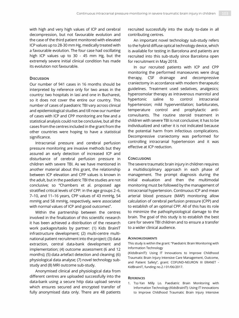
122
Romanian Neurosurgery
-
Upload
khangminh22 -
Category
Documents
-
view
3 -
download
0
Transcript of Romanian Neurosurgery - Journals. London Academic ...

Romanian Neurosurgery

EDITORIAL AND ADVISORY BOARD
EDITOR-IN-CHIEF Dr St. M. Iencean, MD, PhD
EXECUTIVE EDITOR
Al. Chiriac, PhD
ASSISTANT EDITORS
B. Costachescu
A. Iordache
ADVISORY BOARD - ROMANIA
Professor D. Adam, Romania
Dr. Fl. Exergian, Romania
Professor St.I. Florian, Romania
Professor R.M. Gorgan, Romania
Professor G.B.I. Iacob, Romania
Dr. Al. Lupsa, Romania
Professor I. Poeata, Romania
Dr. Al. Tascu, Romania
ADVISORY BOARD - INTERNATIONAL Professor M.A. Arraez, Spain
Professor V. Astarastoae, Romania
Professor H. Bertalanffy, Germany
Professor J. Brotchi, Belgium
Professor P. Courtheoux, France
Professor J.P. Houtteville, France
Professor Y. Kato, Japan
Professor U. Kehler, Germany
Professor Christopher M. Loftus, USA
Dr. M.R. Mahmud, Nigeria
Professor J.Cl. Marchal, France
Professor P. Mertens, France
Professor B.K. Misra, India
Professor D.F. Muresanu, Romania
Professor L. Pendefunda, Romania
Professor S.C. Robertson, USA
Professor M. Samii, Germany
Professor J. Schramm, Germany
Professor M. Sindou, France
Professor B. Sutter, Austria
Professor F. Umansky, Israel
Professor T.T. Wong, Taiwan
EMERITUS EDITORIAL BOARD FOUNDING EDITOR Professor A.V. Ciurea, Romania
Dr. H. Ples, Romania, Formed Editor
_Professor Al. Constantinovici, Former Editor_
_Professor Constantin Arseni_

ROMANIAN
NEUROSURGERY
Vol. XXXIII | No. 2 June 2019

Copyright © 2019 Romanian Society of Neurosurgery &
London Academic Publishing
All rights reserved. This book or any portion thereof may not be
reproduced or used in any manner whatsoever without the express
written permission of the Romanian Society of Neurosurgery or the
publisher except for the use of brief quotations in a book review or
scholarly journal.
ISSN 1220-8841 (Print)
ISSN 2344-4959 (Online)
First Printing June 2019
London Academic Publishing Ltd.
27, Old Gloucester Street
WC1N 3AX
London, United Kingdom
Email: [email protected]
www.lapub.co.uk
www.journals.lapub.co.uk
www.journals.lapub.co.uk/index.php/roneurosurgery
Company Reg. No. 10941794
Registered in England and Wales
The opinions expressed in the published articles are the sole
responsibility of the authors and do not reflect the opinion of the editors
or members of the editorial board.

CONTENTS
101 Continuous intracranial pressure monitoring in severe traumatic brain injury in children
St.M. Iencean, A. Tascu, C.A. Apetrei, C. Gheorghita, T.Y.M. Lo, I. Piper, A.St. Iencean
105 Tailored approach for the resection of planum sphenoidale
meningiomas
Oana-Mihaela Punga, Cristiana-Elena Moisescu, D. Iftimie, D. Adam
110 Biomarkers of the brain injuries - the future diagnosis standard
in head trauma? Brief literature review
A.E. Bîrlescu, B. Hanganu, A.A. Hleșcu, I.S. Manoilescu, B.G. Ioan
116 Dual microcatheter technique for the treatment of a ruptured
wide neck basilar tip aneurysm
Rares Filep, Dorin N. Gherasim, Septimiu Popescu, Botond Tokes, Lucian Marginean
122 New technologies for low-grade glioma surgery
Nicolae-Ștefan Bogaciu, Daniel Teleanu, A.V. Ciurea
127 Initial single centre experience with Barrel VRD stent in large
neck aneurisms
C. Mihalea, F.O. Humulescu, H. Abdelkhalek, S. Pescariu, B.V. Popa, H. Ples.
135 Correlations between clinical, imaging and histological findings
in a patient with neurofibromatosis type 1 (von Recklinghausen's disease)
G.F. Dumitrescu, A. Sava, I. Poeată, D. Haba, B. Dobrovat, N. Dumitrescu, C.M. Bogdanici, C.F. Costea
144 Experience of choroid plexus papilloma in children at Mansoura
University Hospital
Hatem Badr, Ahmad Zaher, Mohamed State, A.F. Khalil
150 Hydatid cyst of the quadrigeminal cistern.
A case report for unusual location with literature review
S.S. Abdelrazaq, A.H. Al Ramadan, A.A. Dolachee, M.M. AbdulAzeez, A.S. Abdulrazzaq, A.S. Rashid, S.S. Hoz

156 Cerebral pilocytic astrocytoma with spontaneous intratumoral haemorrhage in the elderly - a rare entity. A case report and review of the literature
A. Narang, V. Aggarwal, D. Kavita, C. Maheshwari, P. Bansal
160 Traumatic posterior fossa extradural hematoma.
A comprehensive analysis of cases from a tertiary care centre in Southwestern Rajasthan
V.S. Parashar, V.K. Kankane, G. Jaiswal, T.K. Gupta
166 Secondary (Duret) brainstem haemorrhage may not always
represent a fatal event. Review of literature and report of four cases
M. Hanko, B. Kolarovszki, K. Varga, R. Opšenák, P. Snopko, R. Hanzel, K. Zeleňák
174 Spinal extradural meningioma en plaque with nerve root
attachment and extracanal (intrathoracic) extension. Review of literature on management and case report
Morgan E., Hakkou M., Mellaoui A, Poluyi E., El Ouahabi A.
178 The evaluation of long-term screw pull-out rates following
posterior thoracolumbar fusion surgery with short and thin pedicle screws
Umit Kocaman, Hakan Yilmaz
183 Traumatic complete transection of dorsal spinal cord un-associated with spinal fracture or subluxation. Management review
Guru Dutta Satyarthee, Satyajit Panda
188 Post traumatic isolated ipsilateral oculomotor nerve palsy. An uncommon presentation
P. Kumar, S. Pandey, K. Singh, M. Sharma, P. Saxena
191 Endoscopic management of intraventricular shunt-related cystic compartment in paediatric patients
Ahmed Zaher, Amr Farid Khalil, Mohamed State, Hatem Badr
200 The use of folic acid in the prevention of spinal bifida.
Knowledge, attitude, and practice of women of childbearing age in low income rural communities
E. Morgan, N. Akpede, E.F. Osagiede, V. Ajekweneh, F. Erah, V.A. Momoh, M.I. Momoh, E.A. Morgan, E.T. Osagiede
215 Guidelines for authors

Romanian Neurosurgery (2019) XXXIII (2): pp. 101-104 DOI: 10.33962/roneuro-2019-020 www.journals.lapub.co.uk/index.php/roneurosurgery
Continuous intracranial pressure monitoring in severe traumatic brain injury in children
St.M. Iencean1,2, A. Tascu3,4, C.A. Apetrei2, C. Gheorghita5,
Tsz-Yan Milly Lo6, Ian Piper7, A.St. Iencean2
1 Neurosurgery, “Grigore T. Popa” University of Medicine and
Pharmacy, Iasi, ROMANIA 2 Neurosurgery, “Prof. Dr. N. Oblu” Clinical Emergency Hospital,
Iasi, ROMANIA 3 Neurosurgery, “Bagdasar-Arseni” Clinical Emergency Hospital,
Bucharest, ROMANIA 4 Neurosurgery, “Carol Davila” University of Medicine and Pharmacy,
Bucharest, ROMANIA 5 Neurosurgery, “Sf. Maria” Children Clinical Emergency Hospital,
Iasi, ROMANIA 6 University of Edinburgh (Child Life & Health) / Royal Hospital for
Sick Children (Paediatric Critical Care Medicine), UK 7 BrainIT Group Coordinator, Principal Health Care Scientist, Neuro-
Intensive Care Monitoring Research, UK
ABSTRACT We present the results of the Romanian team for the multi-center grant “Paediatric
Brain Monitoring with Information Technology (KidsBrainIT). Using IT Innovations to
Improve Childhood Traumatic Brain Injury Intensive Care Management, Outcome,
and Patient Safety”, acronym KidsBrainIT. Children aged 2 to 16 years who require
intensive care management after sustaining traumatic severe brain injury are
included in this study in three neurosurgical hospital: "Prof. Dr. N. Oblu" Clinical
Emergency Hospital Iasi, "Sf. Maria" Children Clinical Emergency Hospital Iasi and
"Bagdasar-Arseni" Clinical Emergency Hospital Bucharest. Continuous real-time
intracranial pressure monitoring became a "gold standard" in TBI intensive-care
management and ICP-lowering therapy is recommended when ICP is elevated above
20 mmHg or more. Continuous ICP and mean arterial blood pressure (MAP)
monitoring allow calculation of cerebral perfusion pressure (CPP) and to establish of
an optimal CPP. This study aims to improve the treatments and the outcomes in
severe traumatic brain injury in children.
INTRODUCTION Annually, over 50,000 new cases of cranio-cerebral trauma (TBI) occur
in Romania; road accidents are the main cause of cranial traumas,
which often cause cognitive, affective and behavioural disorders, with a
particular impact on families and society. World Health Organization
Keywords cerebral perfusion pressure,
traumatic coma, intracranial pressure,
paediatric brain monitoring, severe children brain injury
Corresponding author: A. Tascu
"Carol Davila" University of Medicine and Pharmacy,
Bucharest, Romania
Scan to access the online version

102 St.M. Iencean, A. Tascu, C.A. Apetrei et al.
estimated that up to 90% of head injuries that
receive treatment are mild, of which moderate and
severe injuries represent 10%. In the European
Union the yearly aggregate incidence of TBI
hospitalizations and fatalities is estimated at 235 per
100,000.
Fortunately, TBI in children is much lower,
without being able to make a reliable estimate
because of research differences, but partial studies
have shown that TBI in children represents about
14% of the total TBI. Also, children have a higher
incidence of increased intracranial pressure (ICP)
following TBI than adults (80% vs. 50%) and it is a
major cause of morbidity and mortality in the
paediatric age group.
In this report of the multi-center grant “Paediatric
Brain Monitoring with Information Technology
(KidsBrainIT). Using IT Innovations to Improve
Childhood Traumatic Brain Injury Intensive Care
Management, Outcome, and Patient Safety”,
acronym KidsBrainIT we present the results of our
study for almost the past two years.
MATERIAL AND METHODS
Three neurosurgical hospital: "Prof. Dr. N. Oblu"
Clinical Emergency Hospital Iasi, "Sf. Maria" Children
Clinical Emergency Hospital Iasi and "Bagdasar-
Arseni" Clinical Emergency Hospital Bucharest
participated in this study that included children aged
2 to 16 years who require intensive care mana-
gement after sustaining traumatic severe brain
injury. A total of 941 children with traumatic brain
injury received medical care during 16 months in
these three neurosurgical departments, including
minor, medium and severe brain traumas. Thirty-
one patients needed intensive care and 9 children
have been ICP and blood pressure monitored, but
only four patients were included in this scientific
project. As presented in the previous report in two
cases the values of ICP were high and very high and
cerebral decompression was performed;
unfortunately, the initial clinical condition was
extremely severe and evolution was not favourable
in these cases. The third and fourth patients
monitored showed elevated ICP values up to 28-30
mm Hg and 30 - 40 mm Hg, which were medically
treated.
FIGURE 1: Case of children with severe TBI and continuous
intracranial pressure monitoring
RESULTS
During 16 months in our three neurosurgical
departments there were a total of 941 children with
traumatic brain injury and 31 patients needed
intensive care and 9 children have been ICP and
blood pressure monitored, but only four patients
were included in this scientific project. The scientific
report that on the mid-term results of this multi-
centre grant presented the three cases: two children
103 Continuous intracranial pressure monitoring in severe traumatic brain injury in children
with high and very high values of ICP and cerebral
decompression, but not favourable evolution and
the case of the third patient monitored with elevated
ICP values up to 28-30 mm Hg, medically treated with
a favourable evolution. The four case had oscillating
high ICP values up to 30 - 45 mm Hg, but the
extremely severe initial clinical condition has made
its evolution not favourable.
DISCUSSION
Our number of 941 cases in 16 months should be
interpreted by reference only for two areas in the
country: two hospitals in Iasi and one in Bucharest,
so it does not cover the entire our country. This
number of cases of paediatric TBI vary across clinical
and epidemiological studies and till now our number
of cases with ICP and CPP monitoring are few and a
statistical analysis could not be conclusive, but all the
cases from the centres included in the grant from the
other countries were hoping to have a statistical
significance.
Intracranial pressure and cerebral perfusion
pressure monitoring are invasive methods but they
assured an early detection of increased ICP and
disturbance of cerebral perfusion pressure in
children with severe TBI. As we have mentioned in
another material about this grant, the relationship
between ICP elevation and CPP values is known in
the adult, but in the paediatric TBI the studies are not
conclusive; so “Chambers et al. proposed age
stratified critical levels of CPP: in the age groups 2–6,
7–10, and 11–16 years, CPP values of 43 mmHg, 54
mmHg and 58 mmHg, respectively, were associated
with normal values of ICP and good outcomes”.
Within the partnership between the centres
involved in the finalization of this scientific research
it has been achieved a distribution of the research
work packages/tasks by partner: (1) Kids BrainIT
infrastructure development; (2) multi-centre multi-
national patient recruitment into the project; (3) data
extraction, central data-bank development and
implementation; (4) outcome assessment (6 and 12
months); (5) data artefact detection and cleaning; (6)
physiological data analyse; (7) novel technology sub-
study and (8) MRI outcome sub-study.
Anonymised clinical and physiological data from
different centres are uploaded successfully into the
data-bank using a secure http data upload service
which ensures secured and encrypted transfer of
fully anonymised data only. There are 48 patients
recruited successfully into the study to-date in all
contributing centres.
An important novel technology sub-study refers
to the hybrid diffuse optical technology device, which
is available for testing in Barcelona and patients are
recruited into this sub-study since Barcelona open
for recruitment in May 2018.
In our recruited patients with ICP and CPP
monitoring the performed manoeuvres were drug
therapy, CSF drainage and decompressive
craniectomy in accordance with modern therapeutic
guidelines. Treatment used sedatives, analgesics;
hyperosmolar therapy as intravenous mannitol and
hypertonic saline to control intracranial
hypertension; mild hyperventilation; barbiturates,
temperature control and prophylactic anti-
convulsants. The routine steroid treatment in
children with severe TBI is not conclusive; it has to be
individualized and rather it is not indicated because
the potential harm from infectious complications.
Decompressive craniectomy was performed for
controlling intracranial hypertension and it was
effective at ICP reduction.
CONCLUSIONS
The severe traumatic brain injury in children requires
a multidisciplinary approach in each phase of
management. The prompt diagnosis during the
initial evaluation and then the multimodal
monitoring must be followed by the management of
intracranial hypertension. Continuous ICP and mean
arterial blood pressure (MAP) monitoring allow
calculation of cerebral perfusion pressure (CPP) and
to establish of an optimal CPP. All of this has its role
to minimize the pathophysiological damage to the
brain. The goal of this study is to establish the best
care for severe TBI children and to ensure a transfer
to a wider clinical audience.
ACKNOWLEDGMENTS
This study is within the grant: “Paediatric Brain Monitoring with
Information Technology
(KIdsBrainIT): Using IT Innovations to Improve Childhood
Traumatic Brain Injury Intensive Care Management, Outcome,
and Patient Safety”, grant: COFUND-NEURON III ERANET -
KidBrainIT, funding no.2 / 01/06/2017.
REFERENCES
1. Tsz-Yan Milly Lo. Paediatric Brain Monitoring with
Information Technology (KIdsBrainIT): Using IT Innovations
to Improve Childhood Traumatic Brain Injury Intensive

104 St.M. Iencean, A. Tascu, C.A. Apetrei et al.
Care Management, Outcome, and Patient Safety. Proposal
Application Form - ERA-NET NEURON, 2016.
2. Iencean St M, Tascu A, Apetrei CA, Gheorghita C,Iencean A
St. Continuous intracranial pressure monitoring in severe
traumatic brain injury in children. Romanian Neurosurgery,
Vol XXXII, Sept 2018, Supplement pp.73.
3. Kannan, N., Ramaiah, R., & Vavilala, M. S. (2014). Pediatric
neurotrauma. International journal of critical illness and
injury science, 4(2), 131–137. DOI: 10.4103/2229-5151.
134152.
4. C.A. Apetrei, C. Gheorghita, A. Tascu, A.St. Iencean, Tsz-Yan
Milly Lo, Ian Pipe, St.M. Iencean Paediatric Brain Monitoring
with Information Technology (KidsBrainIT) - ERA-NET
NEURON Grant. Romanian Neurosurgery (2018) XXXII 2: 183 -
186 DOI: 10.2478/romneu-2018-0024.
5. St.M. Iencean, A. Tascu, C.A. Apetrei, C. Gheorghita, Tsz-Yan
Milly Lo, Ian Piper, A.St. Iencean Mid-term results in
continuous intracranial pressure monitoring in severe
traumatic brain injury in children - ERA-NET NEURON Grant
Romanian Neurosurgery (2018) XXXII 4: 547 - 551 DOI:
10.2478/romneu-2018-0070.
6. Tsz-Yan Milly Lo. Paediatric Brain Monitoring with
Information Technology (KIdsBrainIT): Using IT Innovations
to Improve Childhood Traumatic Brain Injury Intensive
Care Management, Outcome, and Patient Safety. Annual
Scientific Progress Report ERA-NET NEURON, April 2019.
7. Guidelines for the acute medical management of severe
traumatic brain injury in infants, children, and adolescents
(Second Edition). Pediatr Crit Care Med 2012. 13, No 1
(Suppl.).
8. Chambers IR, Jones PA, Lo TYM et al. Critical thresholds of
intracranial pressure and cerebral perfusion pressure
related to age in pediatric head injury. J Neurol Neurosurg
Psychiatry 2006. 77(2): 234-240.
9. Depreitere B, Güiza F, Van den Berghe G, Schuhmann M,
Maier G, Piper I, Meyfroidt G. Pressure autoregulation
monitoring and cerebral perfusion pressure target
recommendation in severe traumatic brain injury patients
based on minute-by-minute monitoring data. J.
Neurosurgery 2014 Jun; 120(6): 1451-1457.
10. Güiza F, Meyfroidt G, Lo TYM, Jones PA, Greet Van den B,
Depreitere B. Continuous optimal CPP based on minute-
by-minute monitoring data: a study on a pediatric
population. Acta Neurochir 2015.
11. Guiza F, Depreitere B, Piper I et al. Visualizing the pressure
and time burden of intracranial hypertension in adult and
paediatric traumatic brain injury. Intensive Care Medicine
2015. 41(6): 1067-1076.
12. Hutchison JS, Frndova H, Lo TYM et al. Impact of
hypotension and low cerebral perfusion pressure on
outcomes in children treated with hypothermia therapy
following severe traumatic brain injury: a post hoc analysis
of the Hypothermia Pediatric Head Injury Trial. Dev
Neurosci. 2010; 32(5-6): 406-12.
13. Suttipongkaset P, Chaikittisilpa N, Vavilala MS, Lele AV,
Watanitanon A, Chandee T, Krishnamoorthy V . Blood
Pressure Thresholds and Mortality in Pediatric Traumatic
Brain Injury. Pediatrics. 2018;142(2).
14. Centers for Disease Control and Prevention. Report to
congress: the management of traumatic brain injury in
children. National Center for Injury Prevention and Control;
Division of Unintentional Injury Prevention, Atlanta, GA;
2018.
15. Olsen, Mari et al. Incidence and mortality of moderate and
severe traumatic brain injury in children: A ten-year
population-based cohort study in Norway European Journal
of Paediatric Neurology, Online May 2019. In Press.
16. Manfiotto M et al. Decompressive craniectomy in children
with severe traumatic brain injury: a multicentre
retrospective study and literature review. World
Neurosurgery. Available online 1 May 2019, In Press.

Romanian Neurosurgery (2019) XXXIII (2): pp. 105-109 DOI: 10.33962/roneuro-2019-021 www.journals.lapub.co.uk/index.php/roneurosurgery
Tailored approach for the resection of planum sphenoidale meningiomas
Oana-Mihaela Punga1, Cristiana-Elena Moisescu1,
D. Iftimie1, D. Adam1,2
1 Department of Neurosurgery, “Saint Pantelimon” Clinical
Emergency Hospital, Bucharest, ROMANIA 2 “Carol Davila” University of Medicine and Pharmacy, Bucharest,
ROMANIA
ABSTRACT Background and importance. Planum sphenoidale meningiomas are relatively rare
tumours that can grow to a considerable size before determining noticeable
symptoms. Modern imaging techniques can detect these tumours of varying size.
Surgical resection of planum sphenoidale meningiomas can be performed by
adapting the approach to the size of the tumour.
Clinical presentation. A 56-year-old woman presented with a small (2 cm in diameter)
planum sphenoidale meningioma that was resected through a frontal craniotomy
performed with a 4,5 cm trephine at the level of the frontal sinus. The second case is
that of a 55-year-old woman that presented with a large planum sphenoidale
meningioma (5,6 cm in the antero-posterior plane and 5,5 cm cranio-caudally)
extending to the tuberculum sellae and sellar diaphragm, reaching the anterior wall
of the third ventricle. In this case, a bifrontal craniotomy was performed with frontal
sinus cranialization and resection of falx cerebri, achieving a Simpson II resection.
Both cases presented a favourable postoperative evolution, without any deficits and
an excellent cosmetic result.
Conclusion. The approach for tumours of the anterior skull base must be tailored to
the size of the tumour. A minimally invasive approach through the frontal sinus
should not be avoided in cases with small tumours.
INTRODUCTION Meningiomas are the most common primary intracranial tumours,
arising from arachnoidal cells. They are benign, slow-growing tumours
and the cognitive impairment as well as behavioural changes they can
induce can easily be mistaken for dementia or depression1.
Planum sphenoidale meningiomas are relatively rare tumours that
originate from the flat surface of the sphenoid bone, anterior to the
optic chiasm. They are closely related to tuberculum sellae tumours but
with a different clinical presentation. Tuberculum sellae tumours
determine early visual deficits even when lesions are small, due to their
proximity to the optic chiasm. Therefore, planum sphenoidale
meningiomas can grow to a considerable size before determining
Keywords minimally invasive approach,
planum sphenoidale meningiomas
Corresponding author: Dragos Iftimie
“Saint Pantelimon” Clinical
Emergency Hospital, Bucharest, Romania
Scan to access the online version

106 Oana-Mihaela Punga, Cristiana-Elena Moisescu, D. Iftimie, D. Adam
noticeable symptoms2.
Modern imaging techniques can detect these
tumours of varying size and surgical treatment is still
the most commonly used treatment option.
Approaches that are used may vary depending upon
tumour size and location, adjacent neurovascular
structures and surgeon’s experience as well as
preference3.
CLINICAL PRESENTATIONS
Case 1
A 55-year-old female, known with chronic viral
hepatitis C and hypermetropic astigmatism with
retinal angiosclerosis, was admitted in our
department for headaches that appeared 3 months
prior to presentation. The neurological examination
was otherwise normal. A contrast MRI examination
revealed a 2 cm in diameter intracranial extra-axial
tumour at the level of the planum sphenoidale, the
radiological aspect suggesting a meningioma (FIGURE
1).
FIGURE 1. Axial (A),
sagittal (B) and
coronal (C) contrast
MRI images revealing
a planum sphenoidale
meningioma
The approach was performed with a 4.5 cm trephine
at the level of the frontal sinus (FIGURE 2) and the
tumour was completely resected, while preserving
the integrity of the olfactory tract. Postoperatively,
the patient presented a favourable outcome with
remission of headaches and no new neurological
deficits. Postoperative control MRI confirmed total
resection of the tumour (FIGURE 3).
FIGURE 2. Postoperative X-ray revealing the location of the
craniotomy
A
B
C
A B

107 Tailored approach for the resection of planum sphenoidale meningiomas
FIGURE 3.
Postoperative axial
(A), sagittal (B) and
coronal (C) images of
T1 with contrast MRI
scan confirming the
total resection of the
tumour
Case 2
A 56-year-old woman was brought to the Emergency
Department of our hospital for drowsiness and
cognitive deterioration that began 3 months prior to
presentation and progressively worsened.
Neurological examination revealed right anisocoria,
no motor deficits, positive Babinski on the left side
and a GCS of 12 points.
The emergency native CT scan showed an
isodense frontal tumor with bilateral extension and
significant perilesional edema. The contrast MRI
subsequently performed revealed a large extra-axial,
isodense tumor with intense, homogenous
enhancement, originating at the planum
sphenoidale, imagistic features suggestive for a
meningioma (FIGURE 4).
FIGURE 4. T1 weighted with
contrast MRI in axial (A),
sagittal (B) and coronal (C)
planes that revealed a large
planum sphenoidal
meningiomas, 5.6 cm in the
antero-posterior plane and
5.5 cm cranio-caudally
This tumour was resected through a large bifrontal
craniotomy (FIGURE 5). After bone flap elevation and
cranialization of frontal sinus, the dura mater was
opened bilaterally and the anterior third portion of
the superior sagittal sinus was ligated and resected.
The tumour was completely removed with
coagulation of dural insertion (Simpson II resection).
FIGURE 5.
Bifrontal
craniotomy
performed for
the resection of
the large planum
sphenoidale
meningioma
Postoperatively, the patient presented a favorable
evolution with no new neurological deficits. The
control CT showed complete removal of the tumor
(FIGURE 6) and the histopathological examination
revealed a transitional meningioma (WHO grade I).
FIGURE 6: Postoperative CT scans that confirm a total resection
of the tumor
DISCUSSIONS
We presented two cases of planum sphenoidale
meningiomas of different sizes, both operated
through a bifrontal approach, with the extent of
craniotomy adapted to tumor size. In both cases, a
total tumor resection was achieved with no surgical
morbidity.
Planum sphenoidale meningiomas can be
resected using different surgical routes, each with its
advantages and disadvantages, allowing the
A
C
B C

108 Oana-Mihaela Punga, Cristiana-Elena Moisescu, D. Iftimie, D. Adam
neurosurgeon to make decisions regarding the
surgical strategy. The factors influencing it are: tumor
size and its relationship to adjacent neurovascular
structures, the patient’s symptoms and the
neurosurgeon’s experience. There are various
transcranial approaches to resect planum
sphenoidale meningiomas: bicoronal subfrontal,
unilateral subfrontal, pterional transsylvian, anterior
interhemispheric, extended bifrontal, skull base
techniques and fronto-temporal orbito-zygomatic4,5.
The bifrontal craniotomy is generally used for
most midline anterior cranial fossa lesions mainly
because of the flexible operative working angles that
it provides and for its generous exposure of the
tumor6.
When compared with bifrontal craniotomy, the
pterional approach avoids the frontal sinuses,
averting the necessity to sacrifice the anterior
superior sagittal sinus. It does not imply the necessity
to manipulate both frontal lobes. This approach also
allows early identification of the optic apparatus,
therefore facilitating its protection during tumor
resection7,8.
Nakamura et al. compared in a series of patients,
the bifrontal approach with frontolateral
approaches, concluding that they prefer the
frontolateral approaches that offer an adequate
access to the tumor with less brain exposure while
allowing a total tumor removal with a low morbidity
rate9.
Also, a minimally invasive approach via a
supraorbital incision and bone opening is also
reportedly used quite frequently in removing these
tumors10. Another option is the endoscopic
endonasal approach. In one study, Ajlan et al.
compared transcranial with endoscopic transnasal
resection for anterior fossa tumors. While the
transnasal endoscopic access associated fewer
complications, the tumor resection rates were much
lower compared to the transcranial approaches11.
Also, in a small single institution study, the
endoscopic approach resulted in equal rates of
resection with better outcomes and less trauma to
the brain.12
However, in 2012 Komotar et al. published a
meta-analysis of 60 studies including over 1,000
patients with tuberculum sellae, planum
sphenoidale or olfactory groove meningiomas
resected either via an endoscopic or transcranial
approach. The results indicated that patients had
similar outcomes regardless of the approach with a
higher rate of CSF leaks associated to the endoscopic
approach13.
The transcranial approaches may be better suited
for planum sphenoidale or tuberculum sellae
meningiomas that are large, with significant lateral
extension or vascular involvement. They offer better
control and thus better tools to deal with vascular
complications. Ultimately, the optimal approach is
predicated by the experience of the surgeon and the
patient’s characteristics and should be determined
on a case by case basis14.
CONCLUSIONS
The approach for meningiomas of the anterior skull
base must be tailored to the size of the tumor. A
minimally invasive approach through the frontal
sinus should not be avoided in cases with small
tumors.
REFERENCES
1. Chiang GSH, Goh LG. Olfactory groove and planum
sphenoidale meningioma: Dementia masquerade. Can
Fam Physician. 2017;63(4):288-291. http://www.ncbi.nlm.
nih.gov/pubmed/28404703. Accessed April 14, 2019.
2. Fox D, Khurana VG, Spetzler RF. Olfactory Groove/Planum
Sphenoidale Meningiomas. In: Meningiomas. London:
Springer London; 2009:327-332. doi:10.1007/978-1-84628-
784-8_34
3. Schroeder HWS. Indications and Limitations of the
Endoscopic Endonasal Approach for Anterior Cranial Base
Meningiomas. World Neurosurg. 2014;82(6):S81-S85.
doi:10.1016/j.wneu.2014.07.030
4. Estevão IA, Camporeze B, Matricardi G, et al. Tuberculum
sellae meningioma: Is there an ideal approach? Med
Express. 2017;4(4). doi:10.5935/MedicalExpress.2017.04.03
5. Lynch JC, Gonçalves MB, Pereira CE, Melo W, Temponi GF.
The extended pterional approach allows excellent results
for removal of anterior cranial fossa meningiomas. Arq
Neuropsiquiatr. 2016;74(5):382-387. doi:10.1590/0004-
282X20160058
6. Rhoton AL. The Anterior and Middle Cranial Base.
Neurosurgery. 2002;51(suppl_4):S1-273-S1-302. doi:10.1097
/00006123-200210001-00007
7. Mathiesen T, Lindquist C, Kihlström L, Karlsson B.
Recurrence of cranial base meningiomas. Neurosurgery.
1996;39(1):2-7; discussion 8-9. http://www.ncbi.nlm.nih.
gov/pubmed/8805134. Accessed April 14, 2019.
8. Poppen JL. Operative techniques for removal of olfactory
groove and suprasellar meningiomas. Clin Neurosurg.
1964;11:1-7.
http://www.ncbi.nlm.nih.gov/pubmed/5854772. Accessed
April 14, 2019.

109 Tailored approach for the resection of planum sphenoidale meningiomas
9. Nakamura M, Struck M, Roser F, Vorkapic P, Samii M.
Olfactory Groove Meningiomas: Clinical Outcome and
Recurrence Rates after Tumor Removal Through the
Frontolateral and Bifrontal Approach. Neurosurgery.
2007;60(5):844-852.
doi:10.1227/01.NEU.0000255453.20602.80
10. Arai H, Sato K, Okuda O, et al. Transcranial Transsphenoidal
Approach for Tuberculum Sellae Meningiomas. Acta
Neurochir (Wien). 2000;142(7):751-757. doi:10.1007/s00701
0070089
11. Ajlan AM, Choudhri O, Hwang P, Harsh G. Meningiomas of
the tuberculum and diaphragma sellae. J Neurol Surg B Skull
Base. 2015;76(1):74-79. doi:10.1055/s-0034-1390400
12. Bander ED, Singh H, Ogilvie CB, et al. Endoscopic endonasal
versus transcranial approach to tuberculum sellae and
planum sphenoidale meningiomas in a similar cohort of
patients. J Neurosurg. 2018;128(1):40-48. doi:10.3171/
2016.9.JNS16823
13. Komotar RJ, Starke RM, Raper DMS, Anand VK, Schwartz TH.
Endoscopic Endonasal versus Open Transcranial Resection
of Anterior Midline Skull Base Meningiomas. World
Neurosurg. 2012;77(5-6):713-724. doi:10.1016/j.wneu.2011.
08.025
14. Koutourousiou M, Fernandez-Miranda JC, Stefko ST, Wang
EW, Snyderman CH, Gardner PA. Endoscopic endonasal
surgery for suprasellar meningiomas: experience with 75
patients. J Neurosurg. 2014;120(6):1326-1339. doi:10.3171/
2014.2.JNS13767.

Romanian Neurosurgery (2019) XXXIII (2): pp. 110-115 DOI: 10.33962/roneuro-2019-022 www.journals.lapub.co.uk/index.php/roneurosurgery
Biomarkers of the brain injuries - the future diagnosis standard in head trauma? Brief literature review
Andreea Elena Bîrlescu1,2, Bianca Hanganu1,
Andreea Alexandra Hleșcu1,2, Irina Smaranda
Manoilescu1,2, Beatrice Gabriela Ioan1,2
1 “Grigore T. Popa” University of Medicine and Pharmacy, Dept. of
Legal Medicine, Iași, ROMANIA 2 Institute of Legal Medicine, Iasi, ROMANIA
ABSTRACT Acute head trauma is often a clinical challenge in diagnosing the brain damage,
assessing its severity and prognosis, and establishing the optimal treatment.
Different patients, with brain damage of apparent comparable severity according to
the imaging examination, may have different neurological evolution or different
response to therapy.
Minor traumatic brain injuries can induce a brief loss of consciousness or confusion,
are usually benign, but sometimes they cause persistent and progressive brain
symptoms in the long run. However, at present, there are no reliable methods that
can diagnose properly minor traumatic brain injuries.
Biomarkers of the brain injuries allow the monitoring of both physiological and
pathological processes. The identification of such biomarkers could allow a better
understanding of the pathological processes involved in traumatic brain injuries, their
diagnosis, prognosis and may facilitate the establishment of a better treatment
regimen for these patients.
In this article, the authors make a brief review of the literature in which they analyse
the biomarkers of the lesions of the various brain structures identified so far, which
can be detected in biological fluids (blood, cerebrospinal fluid) and the advantages
and limitations of their use in the current medical practice.
INTRODUCTION Brain injury may be graded by Glasgow Coma Scale score (GCS) in:
severe trauma, characterized by a GCS score of less than or equal to 8-
the cerebral coma equivalent; moderate trauma, characterized by a
GCS score of 9-12 [1], of which 10% of patients will experience
neurological deterioration and cerebral coma and minor trauma, where
the GCS score is between 13 and 15 [2].
Minor brain trauma can be characterized by the loss of
consciousness of short duration- up to 30 minutes, or confusion,
retrograde amnesia to the traumatic event of up to 24 hours, headache,
Keywords biomarkers,
brain injuries, head trauma,
advantages, limitations
Corresponding author: Andreea Alexandra Hleșcu
“Grigore T. Popa” University of
Medicine and Pharmacy, Dept. of Legal Medicine, Iași, Romania
Scan to access the online version

111 Biomarkers of the brain injuries - the future diagnosis standard in head trauma?
vomiting (unrelated to intracranial hypertension)
and/ or transient focal neurological signs or
convulsions [1]. Most patients with minor brain
trauma show favourable progression, but about 3%
of cases have an unfavourable progression, with
increased risk for intracranial haemorrhage and
diffuse axonal injuries, the promoters of cognitive,
motor and psychosocial deficits [2]. The morbidity
associated with cerebral traumatic injuries (even
minor) is considerable. Studies have shown that
between 1 and 20% of patients with minor traumatic
brain injuries develop persistent physical, cognitive
and behavioural disorders [3], such as chronic
dizziness, fatigue, headache, and amnesia. It is also
important that in the clinic, minor brain injuries are
more common than stroke, dementia and epilepsy,
indicating their high prevalence and justifying the
efforts to diagnose and treat them as accurately as
possible.
Despite substantial efforts to clarify and improve
the diagnostic criteria for minor traumatic brain
injuries, compared to moderate and severe brain
injuries, the former often remain a diagnostic
challenge. This is largely due to the rapid resolution
of acute signs and symptoms after a simple rest and
the absence, in many cases, of objective
neuroimaging evidence.
Current diagnosis regimens for minor traumatic
brain injuries often face the difficulty of
differentiating them from non-traumatic pathologies
that may exhibit a similar symptomatology.
Currently, the gold standard for diagnosing and
establishing the therapeutic management of
traumatic brain injuries is the computer tomography
(CT) exam. It allows the detection of various
traumatic head injuries, such as cranial fractures,
extra- and subdural hematomas, subarachnoid
haemorrhage, cerebral contusion and laceration,
cerebral edema, etc. With increased sensitivity and
specificity and by using it in the clinic as a routine
exam, the head CT scan surpassed the simple head
radiography [1]. However, the head radiography
retains its importance in the initial classification of
traumatic brain injuries as complicated injuries
(radiographically proven) or uncomplicated injuries
(negative radiography), and thus contributes to
establishing the necessary further investigations,
such as CT scan or MRI, and the therapeutic
management (hospital admission with or without
surgery). Despite the superior results from classical
X-rays, modern imaging modalities such as CT scan
and MRI are costly and entail a number of risks,
including the risk of irradiation and the risk
associated with the administration of the contrast
substance [3]. Also, in many cases, minor traumatic
brain injuries cannot be detected by CT scan. Under
these circumstances, an additional diagnostic tool is
necessary to detect patients at risk of developing
further complications.
BIOMARKERS OF CEREBRAL LESIONS -
PATHOPHYSIOLOGICAL BACKGROUND
Research on biomarkers of neuronal lesions began
after the 1950s, and their interest has increased
significantly over the past 25 years.
Biomarkers, also called biological markers, are
natural characteristics that can be measured and
interpreted objectively as indicators of biological
processes or responses to therapeutic interventions
[4]. Biomarkers are indicators of physiological,
pathological or pharmacological processes. Each
organ system has more or less specific biomarkers,
and their analysis, either isolated or joined to other
clinical investigations, allows monitoring of an
individual's health status [2].
From the pathophysiological point of view, due to
the brain injuries, the neuronal and astroglial
network loses its structural integrity, cellular
membranes are affected and secondary to these
events, biomarkers are released in the cerebrospinal
fluid and in blood, allowing for the diagnosis and
prognosis of brain injuries [5].
Traumatic mechanical forces can determine cell
damage due to shear, rupture and stretching of
neurons, axons, glial cells and blood vessels, and the
lesion will induce biochemical changes such as
excitotoxicity, necrosis and apoptosis, oxidative
stress and inflammation. Similar pathophysiological
changes can also be seen in disorders induced by
acute pathological brain injury such as stroke.
Sensitive and specific biomarkers that reflect the
brain damage can provide important information
about the pathophysiology of traumatic brain
injuries and can predict abnormal CT results and/ or
the development of residual deficits in patients
suffering from minor traumatic brain injuries.
Biomarkers could be diagnostic criteria for traumatic
brain injuries and could be a valuable adjuvant to
clinical and routine imaging. In particular, the
possibility of using biomarkers in patients with minor

112 Andreea Elena Bîrlescu, Bianca Hanganu, Andreea Alexandra Hleșcu et al.
traumatic brain injuries could provide a rapid,
differential, non-invasive and cost-effective
diagnostic test to guide appropriate patients’ triage
and their early management [6].
BIOMARKERS OF BRAIN INJURIES –
PROMISING RESULTS
A wide range of proteins, of different origins and
resulting from various pathways, have been studied
as biomarkers for diagnosis and prognosis of brain
injuries. However, the performance of many of
these biomarkers has not been studied in the case
of minor traumatic brain injuries [7].
At the level of the central nervous system, the
lesion biomarkers studied to date are S100beta, Glial
Fibrillary Acidic Protein (GFAP), Neuron- Specific
Enolase (NSE), Alpha II Spectrin, Tau protein,
Ubiquitin C-Terminal Hydrolase L1 (UCH-L1), Fatty-
Acid-Binding Proteins (B-FABP, H-FABP) and Il-10.
Among the listed biomarkers, the most studied
are S100 beta and Neuron- Specific Enolase (NSE),
the values of which increase in hypoxic conditions,
starting on the 2nd post-traumatic day and
normalize at about 4 days after the trauma [2].
S100 beta is a dimer that binds cell calcium, is
involved in cellular differentiation and neuronal
proliferation and has a life span of about 2 hours.
There are 19 types of such dimers, of which S100A1
(in skeletal muscles, heart, and kidneys), S100A1B (in
astrocytes), S100B (in astrocytes and Schwan cells)
and S100BB (in astrocytes). The low molecular
weight of 21kDa allows the S100beta dimers to easily
cross the blood-brain barrier, so that in brain injuries
high levels of S100beta are found in the blood. Unlike
the NSE, the plasma level of S100beta is not affected
by hemolysis, 21.2 micrograms/ liter suggesting the
installation of anoxic coma, and 15.2 micrograms/
liter indicates neuronal recovery.
Neuron- Specific Enolase (NSE) is an isoform dimer
involved in glucose metabolism, which is normally
not found in the peripheral blood. In patients with
stroke, the NSE value increases, with higher values
for patients with irrecoverable traumatic brain
injuries compared to patients with favourable
progression. Decreasing NSE values at 24-48 hours
after the trauma usually indicates a good prognosis,
while a value greater than or equal to 30
micrograms/ liter, 48 hours post-trauma, predicted
death in 100% of the cases. As mentioned above, NSE
values are influenced by hemolysis, which does not
allow its determination in peripheral blood. Apart
from brain lesions, other sources of NSE may also be
small cell carcinomas, neuroblastoma, haemorrhagic
shock, femoral fracture, ischemia and local
reperfusion.
Glial Fibrillary Acidic Protein (GFAP) is a monomer,
being an intermediate protein derived from
astroglial cells. GFAP has increased specificity for
neuronal tissue, with high values in degenerative
brain diseases, cerebral infarction, severe brain
injury, and axonal injuries. GFAP is a predictive
indicator for the recovery of anoxic cerebral coma,
but studies conducted so far on this monomer are
contradictory, and further research is needed on
post-mortem biological products.
Ubiquitin C-Terminal Hydrolase L1 (UCH-L1) is a
compound that plays a role in the elimination of
oxidized neuronal proteins under both normal and
pathological conditions. Initially, it was used as a
histological marker for neurons. Recently, UCH-L1
has been found to have elevated values in the
cerebrospinal fluid after a traumatic brain injury,
which can be immediately detected post-
traumatically, with elevated values lasting for about
one week [2].
Both S100B and the combination of GFAP and
UCH-L1 were promising in screening for CT positivity/
negativity among patients with acute traumatic brain
injury [8].
Alpha II Spectrin is a major component of the
cortical membrane of the cytoskeleton, being
present in axons and presynaptic terminations. It is
a marker for apoptosis and necrosis in the post-
traumatic initial stages and has high values in
moderate and severe brain injuries [2].
Tau is a microtubule associated protein, which is
necessary to maintain the structural integrity of the
axons. Tau proteins have also other functions, such
as nerve impulse transmission, synaptic activity,
cellular proliferation, neurobiological development
and neuroplasticity. Phosphorylation of Tau proteins
is a normal metabolic process, while in both aging
and neurodegenerative diseases, Tau proteins
undergo hyper-phosphorylation, which determines
their aggregation as fibrillar deposits. Post mortem
studies on human corpses described different
patterns of taunting, depending on the pathological
phenotype. Recent studies also highlight the
uniqueness of pathological models, including a
model attributed to repetitive cerebral trauma,

113 Biomarkers of the brain injuries - the future diagnosis standard in head trauma?
although clinical correlations were relative [9].
Fatty-Acid-Binding Proteins (FABPs) are non-
enzymatic cytoplasmic proteins involved in
intracellular buffering and transport of fatty acids.
These are 9 distinct protein types, each named after
the tissue in which it was first detected. FABPs are
rapidly released into circulation from the injured
cells and are eliminated by the kidney, with a half-life
of about 20 minutes. B-FABP was first identified in
the rodent brain where it has a variable
concentration depending on the animal’s age (stage
of development). Thus, in adult mice, B-FABP is
usually produced at low concentrations and is
detected only in glial cells of the white matter. Unlike
B-FABP, H-FABP is also detected in neurons of the
gray matter. B-FABP and H-FABP proteins have
different brain tissue distribution, with the highest
concentrations in the frontal lobe. However, in all
brain structures it was observed that the level of H-
FABP concentration is about 10 times higher than the
B-FABP concentration. Studies show that these two
proteins have greater susceptibility to minor cerebral
lesions than the currently used markers, S100B and
NSE respectively [10].
DIAGNOSTIC RELEVANCE OF BIOMARKERS OF CEREBRAL
INJURIES
To date, biomarkers of cerebral injuries have been
detected in cerebrospinal fluid and in peripheral
blood. It has been found that in cases where the
blood-brain barrier is intact, cerebral proteins are
only present in small amounts in blood. The
condition of the blood-brain barrier has, therefore,
an important influence on the concentration of those
proteins in the blood, which should be considered
for the interpretation of the cerebral lesion-specific
biomarkers [7].
The cerebrospinal fluid is in direct contact with
the extracellular matrix of the brain, and its
composition reflects the biochemical changes
occurring in this organ. For these reasons, the
cerebrospinal fluid could be an optimal source of
brain damage biomarkers. Several cerebral lesion-
specific biomarkers have already been described,
including proteins that indicate the integrity of the
blood-brain barrier and neuro-inflammation, as well
as axonal, neuronal and astrogial lesions. Some
proteins that are expressed in the central nervous
system are also detectable in peripheral blood, albeit
at very low concentrations due to their dilution in the
much larger volume of the extracellular plasma and
matrix of peripheral tissues. Because peripheral
blood sampling is much easier in practice than the
collection of the cerebrospinal fluid, a series of
cerebrospinal fluid biomarkers specific for minor
traumatic brain injuries have also been evaluated in
the peripheral blood. The low concentration of
potential biomarkers in the peripheral blood is,
however, a technical limitation on the use of most
standard immunological tests. However, the number
of potential biomarkers of cerebral lesions in the
peripheral blood studied is steadily increasing as the
analytical tools for detecting them become more and
more sensitive [11].
Studies have shown that unique biomarkers do
not have the specificity and sensitivity required for
their use as diagnostic tools. For a biomarker to be
useful its sensitivity and specificity should be very
high to ensure diagnosis and prognosis assessment
without the need for a CT brain exam.
So far, most research on biomarkers of minor
traumatic brain injuries has been performed with
unique biomarkers. The combination of different
biomarkers has been suggested to enhance the
diagnostic performance. Several studies have shown
that combinations of biomarkers significantly
increase diagnostic performance in various
pathologies, such as sleep disorders, post-stroke
subarachnoid haemorrhage, lung cancer or
differentiation of post-traumatic brain injuries from
other types of lesions. Furthermore, it has been
suggested that some combinations of different
clinical parameters, such as the age and types of
biomarkers, e.g. inflammatory proteins, can improve
the classification of lesions [7].
In a multicentre study, 13 cerebral biomarkers, all
previously investigated in patients with stroke, were
evaluated for their ability to correctly classify patients
with minor traumatic brain injury, CT positive and CT
negative, with a GCS score of 15 and showing at least
one clinical symptom. Of the 13 biomarkers, the H-
FABP and IL 10 proteins were the best single
markers. These were further compared and
combined with the better-studied S100B and GFAP
markers. H-FABP was the best single marker, but
when combined with GFAP, the overall performance
increased from 32% to 46%, with a sensitivity of
100%. Proteins have been shown to be released
from various types of injured cells. S100B and GFAP

114 Andreea Elena Bîrlescu, Bianca Hanganu, Andreea Alexandra Hleșcu et al.
were derived from astrocytic lesions, H-FABP from
endothelial cells and neuronal cellular bodies, while
IL10 is expressed by monocytes and macrophages
[7].
Detection of cerebral lesions by the serum
biomarkers is not a standard procedure in current
clinical practice, although several proteins, such as
S100B, NSE, myelin basic protein and GFAP show
promising results [10]. Some biomarkers, such as
S100B and GFAP, have been extensively studied in
the blood of patients with minor traumatic brain
injuries, but so far none seem to provide sufficient
information [7].
DIFFICULTIES AND LIMITATIONS IN THE STUDY OF
BIOMARKERS OF BRAIN INJURIES
The main difficulty facing biomarkers for brain
damage is to know whether the measured proteins
really come from the brain injuries. As shown above,
regardless of their origin, single biomarkers do not
have sufficient performance to be transformed into
diagnostic tools. Biomarker combinations, however,
have been shown to enhance diagnostic
performance when proteins of different origins and
pathways are combined, due to the complexity of the
nervous system and the heterogeneity of the
traumatic brain injuries [7, 8].
There are also other obstacles to the
development of a series of blood biomarkers for
minor traumatic brain injuries. The blood-brain
barrier prevents the evaluation of the biochemical
changes in the brain by using biomarkers in the
blood, but this is possible, however, in the case of
loss of blood-brain barrier integrity, which occurs in
severe brain lesions. In addition, some potential
biomarkers suffer a proteolytic degradation in the
blood, and their levels may be affected by clearance
in the blood through the liver or kidneys. The
accuracy of immunoassays may also be affected by
the binding of biomarkers to carrier proteins and
extra-cerebral sources of biomarkers [12].
Biomarkers of the cerebral injuries have different
delivery patterns, and this has limitations on their
practical use. As a result, clinical applicability may be
limited by the type of brain injury (traumatic, stroke,
hypoxia-ischemia). Another important limitation in
the analysis of cerebral biomarkers is their ambiguity
in multiple lesions.
Despite the current limitations in the study and
application of biomarkers of cerebral lesions in the
current medical practice, biomarkers could be used
in the future as an adjuvant, supplementing the
traditional and neuro-imaging examination in the
diagnosis and prognosis of patients with traumatic
brain injuries [4].
CONCLUSIONS
Traumatic brain injuries may raise clinical challenges
due to the diagnostic difficulties and the lack of
specific prognostic tools. A special place in the
traumatic pathology of the brain is occupied by
minor traumatic brain injuries that, although
characterized by immediate mild signs and
symptoms, can induce long-term brain pathology
with increased disability potential.
Biomarkers of cerebral injuries may be a new
diagnostic standard for traumatic brain injuries, and
in particular, minor ones that often cannot be
detected by cerebral CT.
However, further studies are needed to identify
the biomarkers or combinations of biomarkers with
the highest sensitivity and specificity for cerebral
injuries.
REFERENCES
1. Popescu I, Florian IS, Poeată I. Tratat de neurochirurgie.
București: Editura Academiei Române, 2014.
2. Chirica VI. Useful markers to assess traumatic and hypoxic
brain injury. Rom J Leg Med. 2017; 25: 146-151.
3. Sharma R, Rosenberg A, Bennet ER, Laskowitz DT, Acheson
SK. A blood-based biomarker panel to risk stratify in mild
traumatic brain injury. PLoS One. 2017; 12(3): e0173798.
4. Toman E, Harrisson S, Belli T. Biomarkers in traumatic brain
injury: a review. Journal of the Royal Army Medical
Corps. 2016; 162(2):103-108.
5. Mondello S, Schmid K, Berger RP, Kobeissy F, Jeromin A,
Italiano D, Buki A. The challenge of mild traumatic brain
injury: role of biochemical markers in diagnosis of brain
damage. Med Res Rev. 2014 May; 34(3):503-531.
6. http://www.traumaticbraininjury.com/understanding-
tbi/what-are-the-causes-of-tbi/
7. Mrozek S, Dumurgier J, Citerio G, Mebazaa A, Geeraerts T.
Biomarkers and acute brain injury: interest and limits. Crit
Care. 2014; 18:220.
8. Pelsers MMAL, Hanhoff T, Van Der Voort D et all. Brain and
heart type fatty acid binding proteins in the brain: tissue
distribution and clinical utility. Clin Chem. 2004; 50(9): 1568-
1575.
9. Posti JP, Takala RSK, Lagerstedt L et all. Correlation of blood
biomarkers and biomarker panels with traumatic findings

115 Biomarkers of the brain injuries - the future diagnosis standard in head trauma?
on computed tomography after traumatic brain injury. J
Neurotrauma. 2019, Apr 5. [Epub ahead of print].
10. Lagerstedt L, Egea-Guerrero JJ, Bustamenate A et all.
Combining H-FABP and GFAP increases the capacity to
differentiate between CT-positive and CT-negative patients
with mild traumatic brain injury. PLoS One. 2018, Jul 9;
13(7):e0200394.
11. Zetterberg H, Smith DH, Blennow K. Biomarkers of mild
traumatic brain injury in cerebrospinal fluid and blood. Nat
Rev Neurol. 2013 Apr; 9(4): 201–210.
12. Castellani RJ, Perry G. Tau biology, taupathy, traumatic
brain injury and diagnostic challenges. J Alzheimers Dis.
2019; 67(2): 447–467.

Romanian Neurosurgery (2019) XXXIII (2): pp. 116-121 DOI: 10.33962/roneuro-2019-023 www.journals.lapub.co.uk/index.php/roneurosurgery
Dual microcatheter technique for the treatment of a ruptured wide neck basilar tip aneurysm
Rares Filep, Dorin Nicolae Gherasim, Septimiu Popescu,
Botond Tokes, Lucian Marginean
* Department of Interventional Radiology & Department of
Neurosurgery, Emergency County Hospital, Targu Mures, ROMANIA
ABSTRACT Endovascular treatment is a safe and efficient therapy for intracranial aneurysms
with lower complication and mortality rates compared to surgical clipping. Wide-neck
aneurysms still represent a challenge to complete and safe aneurysm occlusion in
spite of techniques such as stent-assisted or balloon-assisted coiling, developed in
order to achieve better occlusion rates. These techniques themselves may lead to
further complications, so alternative methods such as the dual microcatheter
technique were developed. This technique assumes that, via two microcatheters
inserted into an aneurysm, simultaneous deployment of two coils achieves a stable
coil frame without the use of adjunctive devices. The aim of this paper is to present a
successfully treated basilar tip wide-neck aneurysm treated with the dual
microcatheter technique.
Case report. A 46-year-old male patient with acute onset of severe headache
presented in the emergency room with altered state of consciousness. Non-
enhanced CT scan showed subarachnoid and intraventricular haemorrhage. CT
angiography revealed a wide-neck basilar tip aneurysm. Digital subtraction
angiography confirmed the presence of an aneurysm with a wide, 4.9 mm neck.
Dual microcatheter technique was chosen as the first treatment option, while a
hypercompliant balloon was kept as backup. Two microcatheters were placed inside
de aneurysm and two coils were introduced in order to form a stable framing coil
mass that served as a support for further coils deployed in an alternately manner
through each microcatheter. No procedural complication occurred, and the patient’s
evolution was uneventful with no neurological deficits at discharge.
Conclusion. The dual microcatheter technique is a safe and effective therapeutic
option for wide-neck ruptured or unruptured intracranial aneurysms. Periprocedural
complication rates are similar to simple coiling or balloon-assisted coiling, but lower
than for stent-assisted coiling.
INTRODUCTION Endovascular treatment has become the therapy of choice for
intracranial aneurysms owing to its lower complication and mortality
rates, as compared to surgical clipping (5). However wide-neck
aneurysms with a neck diameter greater than 4 mm or a dome-to-neck
Keywords subarachnoid haemorrhage,
cerebral aneurysm, basilar artery,
endovascular treatment
Corresponding author: Dorin Nicolae Gherasim
Department of Neurosurgery, Emergency County Hospital,
Targu Mures, Romania
Scan to access the online version

117 Dual microcatheter technique for the treatment of a ruptured wide neck basilar tip aneurysm
ratio less than 2, still represent a procedural
challenge attributable to the risk of intraprocedural
coil herniation, a higher number of thromboembolic
complications and higher rate of long-term
recanalization (6).
Different techniques have been developed to
assist wide-neck aneurysm coiling by covering the
neck during coil deployment, thus allowing denser
packing and better long-term occlusion rates. Stents
and balloons have been used for this purpose, but
both techniques have their limitations. Stent
placement might be difficult in tortuous vessel
anatomy and is associated with delayed
thromboembolic events and in-stent stenosis.
Balloon-assisted coiling (BAC) requires temporary
flow arrest and may be associated with increased
risk of vessel rupture and thromboembolic
complications (7).
One alternative to the above-mentioned
procedures is a less used and under-reported
method, the dual microcatheter technique (DMT),
which requires simultaneous placement of two
microcatheters in the aneurysm sac and deployment
of coils through both of them with the aim of
achieving a stable coil frame without the use of
adjunctive devices (1).
The aim of this paper is to present the case of a
patient with a wide-neck basilar tip ruptured
aneurysm, successfully treated using the DMT and to
discuss its advantages and disadvantages compared
to other techniques used for wide-neck aneurysm
coiling.
CASE REPORT
A 46-year-old male patient presented in the
emergency department with abrupt-onset severe
headache. Clinical examination revealed a drowsy
and confused patient (Hunt-Hess 3). Non-enhanced
CT and CT angiography of the head performed on 64-
channel machine (Siemens, Erlangen, Germany)
showed a subarachnoid and intraventricular
hemorrhage (mFisher 4) and a wide-neck basilar tip
aneurysm. (FIGURE 1: a, b, c)
FIGURE 1. a. Sagittal NCCT image reveals a small amount of subarachnoid blood in the interpeduncular cistern and the 3rd ventricle
(white arrows); b. Axial NCCT slice shows the blood clot in the 3rd ventricle (white arrow); c. Coronal CTA image reveals the basilar
bifurcation aneurysm (white arrow)
NCCT = non-contrast computed tomography; CTA = computed tomography angiography
Digital subtraction angiography (DSA) and 3D
rotational angiogram obtained on a Siemens biplane
system (Siemens, Erlangen, Germany) showed the
basilar tip aneurysm measuring 7.1/5.1/5.4 mm
(CC/LL/AP diameters) with a neck of 4.9 mm (dome-
to-neck ratio of 1.4), and a small secondary lobule
along the left superior border of the aneurysm,
thought to be the site of bleeding (FIGURE 2: a, b).
Under general anesthesia the right common femoral
artery was accessed with a 6F Cordis sheath (Cordis,
Freemont, California) and a 6F Chaperon guiding
catheter (Terumo, Tokyo, Japan) was placed in the
right vertebral artery (VA). A Headway 17
microcatheter (Terumo, Tokyo, Japan) was then
advanced into the aneurysm and a Target 360
standard 5 mm x 15 cm coil (Stryker, Michigan, USA)
a. b. c.

118 Rares Filep, Dorin Nicolae Gherasim, Septimiu Popescu et al.
was deployed. At this point an intravenous bolus of
2500 units of heparin was administered. The coil was
quite stable in the aneurysm sac without
impingement on the basilar artery. (FIGURE 2: c.) We
kept it attached to its introducer wire nevertheless,
with the idea to keep it as support for further coils
that will be inserted through a second microcatheter.
A 5F Cordis femoral sheath was inserted on the
opposite side and a second Headway 17
microcatheter was introduced into the aneurysm via
a 5F Navien support catheter (Medtronic,
Minneapolis, USA) placed in the left VA. A 3D Axium
Prime 5 mm x 10 cm coil (Medtronic, Minneapolis,
USA) was introduced through the second
microcatheter, and by intertwining with the first coil,
formed a secure, stable coil mass throughout the
entire volume of the aneurysm. (FIGURE 2: d, e) The
second coil was detached, and an additional bolus of
2500 IU of heparin was administered. A third
HydroCoil 3 mm x 8 cm coil (Microvention, California,
USA) was introduced through the second
microcatheter, followed by the careful detachment
of the very first coil. Three further hydrogel-coated
coils were introduced through both microcatheters,
alternately, resulting in satisfactory occlusion of the
aneurysm judged by the Raymond-Roy occlusion
classification as 1 (FIGURE 2: f). The patient had an
uneventful postoperative course and was discharged
2 weeks later without neurological deficits, with a
modified Rankin scale 0.
FIGURE 2. a. AP DSA image from a right VA injection shows a basilar tip aneurysm; b. 3D rotational angiographic reconstruction; c.
AP unsubtracted image shows the first microcatheter and the first coil inside the aneurysm (white arrow); d. AP unsubtracted image
a. b. c.
d. e. f.

119 Dual microcatheter technique for the treatment of a ruptured wide neck basilar tip aneurysm
reveals the second microcatheter (white arrows) and a further coil inserted into the aneurysm; e. Further coils are deployed
simultaneous through both of the microcatheters; f. Final control DSA angiogram shows the complete obliteration of the aneurysm.
AP = anteroposterior, DSA = digital subtraction angiography
DISCUSSIONS
Since its introduction in 1998 by Baxter’s group (4),
DMT has been used as an alternative to balloon or
stent-assisted coiling (SAC) for wide-neck intracranial
aneurysms. It involves the use of two microcatheters
inserted in the aneurysm sac followed by
simultaneous or sequential deployment of coils. The
general principle behind this technique is the
stability obtained by the “entanglement” of the first
two framing coils inserted simultaneously, thus
creating a larger coil frame that prevents
impingement on or herniation of the coil loops into
the parent artery. One of the coils is usually detached
at the end of the procedure thus conferring
additional stability for further coils.
The technique is aimed at wide-neck saccular
aneurysms with a dome-to-neck ratio less than 2,
irregular aneurysms with multiple daughter sacs and
complex configurations and aneurysms with a vessel
emerging from the neck (5). We considered DMT as
the first therapeutic option for our case while a
balloon was kept as back-up in case the coils were
not stable inside the aneurysm. SAC was excluded
because the aneurysm was ruptured, and dual
antiplatelet therapy was necessary. For a more
detailed description of the technique we propose a
step-by-step guide on how to approach aneurysms
that are amenable to treatment with this procedure.
1. General procedural considerations
Endovascular treatment of intracranial aneurysms
should be performed with the patient under general
anesthesia. In our institution all catheters are
continuously flushed with saline containing heparin
(2500 IU heparin in 500 ml of saline). Our protocol is
to administer a bolus of 5000 IU heparin
intravenously after the deployment of the first coil,
although during this procedure, 2500 IU were
administered after insertion of both guiding
catheters in order to prevent thromboembolic
events, and 2500 IU following the deployment of the
first coil.
2. Femoral artery access and guiding-catheter
(GC) choice.
Uni- or bilateral access depends on how many GC’s
are needed. Generally, the dual microcatheter
technique can be safely performed through one GC.
In the majority of cases the internal carotid arteries
(ICA) or at least one of the vertebral arteries (VA) can
easily accommodate one larger device, although
cases with small VA’s may require the use of two GC’s
in cases with posterior circulation aneurysms (4).
Advantages of using a unilateral approach are the
smaller complication rates related to femoral artery
puncture and large vessel access, like arterial spasm,
dissection and embolic events. Theoretically, using
two GC’s can lead to a two-fold increase in such
complications. The use of two GC’s can be avoided
because the majority of 6 or 7 French (F) devices have
a large enough lumen to contain both
microcatheters. Disadvantages of using only one
device are the difficulty of obtaining quality
roadmaps or control angiograms with two
microcatheters that occupy a large luminal area of a
6F GC, although this can be overcome with the use of
a 7F device. Another disadvantage of two
microcatheters inserted through the same GC is
related to the risk of inadvertent forward/backward
movement while manipulating them, which can lead
to aneurysm perforation or dislodgement of the coil
mass (4,3). Due to the limited experience with this
technique we felt safer with a bilateral approach in
order to avoid inadvertent microcatheter movement
and to have access with a hypercompliant balloon if
the coils were not stable or if rupture of the
aneurysm occurred.
3. Aneurysm access
The first option is to try coiling the aneurysm with
one microcatheter. If this fails due to coil herniation
or impingement on the parent artery, the second
microcatheter is positioned as follows. To anticipate
the deployment behavior of the coils it is useful to
divide the aneurysm in two imaginary
compartments, each one occupied by one of the first
two coils, a simple example is to have a proximal and
a distal compartment, and to position the tip of the
two microcatheters in the corresponding
compartment (3).
4. Coil selection, deployment and detachment
Coil choice depends on the configuration of the
aneurysm. For irregular aneurysms with multiple

120 Rares Filep, Dorin Nicolae Gherasim, Septimiu Popescu et al.
daughter sacs soft coils are recommended to avoid
perforation. If this is not the case, standard, complex
coils are preferred due to their larger radial force
providing a better “anchoring” inside the aneurysm.
The first coils should be longer, thus minimizing the
number of subsequent coils necessary for a dense
final packing. After achieving a stable frame with the
first two coils, one of them is detached and further
coils are inserted until complete obliteration is
obtained, while the other is left attached to its
pusher-wire. The decision which coil to detach first is
usually based on its stability: the more stable looking
coil is to be detached first, because it provides a
better scaffold for further coils. Another
consideration to which coil should be detached first
is the position of the microcatheter tip. It is safer to
insert coils through the catheter sitting closer to the
neck of the aneurysm than through the one
positioned deeper inside due to the risk posed by the
deep deposition of coils that might “push” the whole
coil construct outside and into the parent vessel,
consequently the one sitting deeper should be
detached earlier (1,3,4).
5. Microcatheter removal
A potential risk of retrieving the microcatheters at
the end of the procedure is the extraction of the coil
mass outside the aneurysm. The best way to avoid
this if the microcatheter’s tip is deep inside the
aneurysm, is to push gently on the pusher-wire of the
last coil until the tip protrudes outside, and only
afterwards to detach the coil (6).
DMT is a relatively under-used method for
intracranial aneurysms compared to balloon or
stent-assisted coiling, with only few case reports,
series and retrospective studies published in the
literature so far. The safety and efficiency of this
approach has been assessed by Durst et al in their
retrospective analysis of 100 wide-necked
aneurysms (2). They reported a morbidity and
mortality of 1% and 2% respectively. Intraprocedural
rupture occurred in 3 patients (3%), 2 were
successfully stopped immediately and 1 patient died
due to massive hemorrhage and hydrocephalus.
One other patient died as a consequence of the coils
herniating in the parent vessel, obliterating the
lumen and subsequent ischemic stroke. DMT was
successfully carried out in 91 cases (91%). The
remaining 9 failed due to the impossibility of
stabilizing the coils inside the aneurysm.
Retreatment was necessary in 18% of cases after
recanalization (2).
The relatively low major complication rate of DMT
(3%) is similar to that reported for simple coiling (0.6-
5.1%) or BAC (0.9-3.8%), but much lower than for
stent-assisted coiling (SAC) (9.4-12.2%) (7). Two
common downsides of BAC and SAC are the greater
experience needed for device manipulation and the
specific risks imparted by these devices on the
procedure. Balloons add the risk of intraprocedural
vessel rupture while stents are permanent
intraluminal devices prone to in-stent stenosis and
thrombosis, requiring long-term antiplatelet therapy
with an additional hemorrhagic risk. Conversely, no
special training is needed for DMT since the only
devices used are those required for simple coiling,
although special care is mandatory during aneurysm
access, because two catheterization procedures
mean an increased risk of wall perforation with
either the guidewire or the microcatheters.
One major issue of DMT is the retreatment rate
of 18% (7), that compared to BAC, between 5.7% and
15.6%, and SAC, between 4.3% and 13.3% (8) is
relatively high. This is especially important for young
patients due to their longer life expectancy,
therefore a judicious use of DMT is necessary in this
patient population.
CONCLUSIONS
The dual microcatheter technique is a safe and
effective therapeutic option for wide-neck ruptured
or unruptured intracranial aneurysms.
Periprocedural complication rates are similar to
simple coiling or balloon-assisted coiling, but lower
than for stent-assisted coiling.
REFERENCES
1. Baxter BW, Rosso D, Lownie SP. Double microcatheter
technique for detachable coil treatment of large, wide-
necked intracranial aneurysms. AJNR Am J Neuroradiol.
1998 Jun;19(6):1176–8.
2. Durst CR, Starke RM, Gaughen JR, Geraghty S, Kreitel KD,
Medel R, et al. Single-center experience with a dual
microcatheter technique for the endovascular treatment of
wide-necked aneurysms. J Neurosurg. American
Association of Neurological Surgeons; 2014 Nov. 121 (5):
1093–101.
3. Horowitz M, Gupta R, Jovin T. The dual catheter technique
for coiling of wide-necked cerebral aneurysms. An under-
reported method. Interv Neuroradiol. 2005 Jun 30; 11
(2):155–60.

121 Dual microcatheter technique for the treatment of a ruptured wide neck basilar tip aneurysm
4. Kwon O-K, Kim SH, Kwon BJ, Kang H-S, Kim JH, Oh CW, et al.
Endovascular treatment of wide-necked aneurysms by
using two microcatheters: techniques and outcomes in 25
patients. AJNR Am J Neuroradiol. 2005 Apr;26(4):894–900.
5. Molyneux AJ, Kerr RS, Yu L-M, Clarke M, Sneade M, Yarnold
JA, et al. International subarachnoid aneurysm trial (ISAT) of
neurosurgical clipping versus endovascular coiling in 2143
patients with ruptured intracranial aneurysms: a
randomised comparison of effects on survival,
dependency, seizures, rebleeding, subgroups, and
aneurysm occlusion. The Lancet. 2005 Sep;366(9488):809–
17.
6. Pierot L, Biondi A. Endovascular techniques for the
management of wide-neck intracranial bifurcation
aneurysms: A critical review of the literature. Journal of
Neuroradiology. Elsevier Masson SAS; 2016 Mar 11;:1–9.
7. Piotin M, Blanc R. Balloons and stents in the endovascular
treatment of cerebral aneurysms: vascular anatomy
remodeled. Front Neurol. Frontiers; 2014;5(Suppl 3):41.
8. Wang F, Chen X, Wang Y., Bai P, Wang H-Z, Sun T, et al.
Stent-assisted coiling and balloon-assisted coiling in the
management of intracranial aneurysms: A systematic
review & meta-analysis. J Neurol Sci. 2016 May 15; 364:
160–6.

Romanian Neurosurgery (2019) XXXIII (2): pp. 122-126 DOI: 10.33962/roneuro-2019-024 www.journals.lapub.co.uk/index.php/roneurosurgery
New technologies for low-grade glioma surgery
Nicolae-Ștefan Bogaciu1,3, Daniel Teleanu1,2,3, A.V. Ciurea2,3
1 University Emergency Hospital, Bucharest, ROMANIA 2 Sanador Clinical Centre Hospital, Bucharest, ROMANIA 3 “Carol Davila” University of Medicine and Pharmacy, Bucharest,
ROMANIA
ABSTRACT Brain surgery has come far from the primitive methods used thousands of years ago.
In the last hundred years alone, we have come across countless breakthroughs like
the invention of bipolar coagulation, the surgical microscope, microsurgical
techniques and a large variety of surgical instruments. The latest years have brought
us neuro-navigation, intra-operative imaging techniques and brain activity
monitoring. Low-grade glioma surgery, in particular, has taken advantage a lot from
all these new methods, bringing great benefits for the patients: the safe extent of
resection has grown progressively and tumours located in eloquent areas that were
thought until recently to be inoperable, have started to be removed in safe
conditions. The purpose of this paper is to present these new technologies as a recap
for neurosurgical professionals.
INTRODUCTION Endovascular Brain surgery is one of the oldest procedures to ever be
performed on a human being. Beginning as trephining, it has known a
long period of slow development, only to become one of the top-edge
sciences of modern time. It developed from somewhat barbaric
procedures with little if any supporting scientific knowledge, to life-
saving procedures performed on a macroscopic scale, to microscopic
interventions with progressively smaller „collateral damage” and in the
future it might turn into something we would call „molecular surgery”.
The purpose of this paper is to highlight the technologies we nowadays
use in low-grade glioma surgery, presenting their pearls and pitfalls,
benefits and uses.
MATERIALS Computer assisted neuro-navigation. The purpose of this tool in brain
surgery is to integrate imaging studies obtained preoperatively with
intra-op anatomy in order to localize more accurately the lesions
subdue to surgery (PICTURE 1). A neuro-navigation system has the
following main parts: a computer-station with built-in software, a a
receiver and trackers/pointers (PICTURE 2).
Keywords intraoperative, imaging,
neuro-navigation, low-grade glioma,
brain monitoring
Corresponding author: Nicolae-Ștefan Bogaciu
University Emergency Hospital,
Bucharest, Romania
Scan to access the online version

123 New technologies for low-grade glioma surgery
PICTURE 1
PICTURE 2
The initial step is to upload the preoperative images
in the computer. Patient is positioned and a patient-
tracker is placed in the proximity of the patient’s
head. Landmark registration is performed and
checked for accuracy and surgery can commence.
Intra-operatively, the surgeon may navigate with
different tools: pointers, microscope, standard
neuro-navigated instruments (aspirator, ultrasonic
aspirator, retractors etc.). Some systems allow
injection of images directly into the microscope for
compatible devices (PICTURE 3).
PICTURE 3
Neuro-navigation is not only useful for accurately
localizing a lesion, but also for localizing vasculature
and other normal anatomical structures that need to
be avoided in glioma surgery. Image series used for
neuro-navigation can be fused for better describing
the anatomy and functionality of different brain
areas: tractography, PET, MRI, CT, contrast-enhanced
imagery etc. The advantages of using neuro-
navigation in LGG (low-grade glioma) surgery consist
of, but are not limited to: smaller scalp incisions,
cranial flaps, dural incisions and corticotomies, safer
dissection and tumour excision, reduced surgery
time.
All these imply fewer post-operative complications
from surgery, faster discharge, lower costs for
patient care. The main disadvantage of neuro-
navigation systems is that the position of the patient
124 Nicolae-Ștefan Bogaciu, Daniel Teleanu, A.V. Ciurea
when obtaining images and the position of the
patient during surgery are almost never the same so
the brain will have a shift during surgery compared
to imaging. Another thing is that after beginning the
excision of tumoral tissue, the landscape changes,
but the images stay the same. Some solutions were
developed for these problems. Other imaging
studies may be performed intraoperatively to
complete the preoperative study and to increase the
accuracy of the neuro-navigation system.
Intraoperative MRI is a known tool which performs
the task well, but it is highly inconvenient. Its more
readily available counterpart is intraoperative
ultrasound. Tips for successfully removing tumours
using neuro-navigation include: removing tumour
tissue closer to eloquent areas first, when accuracy is
better, performing en-bloc excisions as much as
possible, avoiding cysts especially when they are
large, until good accuracy is no longer an issue,
„rebuilding” volume by placing cottonoids in
resulting cavities. In spite of all issues, a surgeon will
always find this tool useful and he will always have in
mind the fact that he can never rely on solely this in
performing his tasks.1,2,3,4 (PICTURE 4).
Intra-operative ultrasound. Ultrasonography (US) is
a great mean of obtaining fast, cheap real-time
images during brain surgery. Images are usually
obtained by using a probe of 4-8 MHz which is
sufficient for acquiring images in a 2-6 cm distance
from it. There are a few ways to use this tool: regular
gray-scale brightness mode (B-mode), Contrast
Enhanced Ultrasound (CEUS), elastosonography. US
is of aid in all stages of brain tumour surgery, from
the opening of the dura up to the resection of
peripheral extensions of the tumour. As opposed to
cortical lesions, subcortical lesions are more difficult
to identify, and the site of the corticotomy may be
tricky to choose. By using US, the surgeon can
identify the subcortical tumour and choose an entry
site as found suitable. During resection, the surgeon
may use US to identify the depth and extent of the
tumour and adapt the aggressivity of his resection
accordingly. In the final stages of the surgery, one
may use US for identifying residual tumoral tissue in
the periphery of the mass. Not only US may be used
for identifying the tumour, but also to characterize it.
It can deliver information about the consistency of
the tumour. For example, the surgeon may use this
information to adapt the power of an ultrasonic
aspirator, or to differentiate between low-grade
glioma (LGG) and high-grade glioma (HGG): LGG are
firmer than HGG (as assessed by elastosonography).
CEUS brings even more information about the
vascularization of the tumour, by real-time angio-
sonography so that the surgeon may adapt his
resection strategy and have even more information
about the possible histological diagnosis (LGG are
poorly vascularized as opposed to HGG). A common
use for intraoperative US in brain surgery is
enhancing accuracy of neuro-navigation systems by
updating the pre-operative acquired images used for
registration and navigation, with real-time images
provided by intra-operative US. By doing so, the
effects of brain shift and selling may be partly
counteracted.1,4–8
Intraoperative CT. Although it is less preferred by
neurosurgeons compared to other imagistic tools,
intraoperative CT-scans may be found useful in
glioma surgery. As the other intraoperative imaging
studies (US, MRI), it allows a greater extent of surgical
resection by providing real-time intra-operative
information regarding tumour mass, as well as
updating pre-operative neuro-navigation planning.
We may consider some advantages regarding the
cost (relatively low compared to intraoperative MRI),
speed of the procedure and general benefits such as
improving accuracy and extent of the resection.9,10
125 New technologies for low-grade glioma surgery
Intraoperative MRI (ioMRI). Intraoperative magnetic
resonance imaging is the preferred intraoperative
imaging method for low-grade glioma surgery. It is
also the most laborious, the most expensive, but
more importantly the most accurate. IoMRI comes in
two shapes: low field (≤0,5 Tesla) and high field (≥1,5
Tesla). Regardless of the power of the magnet, ioMRI
aids the surgeon in extending tumour resection in
both enhancing and non-enhancing lesions, with
greater benefit in the latter. The use of intraoperative
MRI associates with higher extent of resection which
implies longer overall survival and free of symptoms
survival in patients with gliomas. Setting up an
operating room with an MRI device can get pretty
challenging. Although there are tools that can be
used within the area closest to the device (an area
limited by the 5-gauss border, which is a line beyond
which ferromagnetic instruments can be used), most
instruments used for microsurgical resection,
including microscope and ultrasonic aspirator, have
to be used further away from the magnet. Residual
tumour is observed with high fidelity on images
acquired from intraoperative MRI and can be readily
excised if feasible. The whole range of sequences can
be studied: functional, connective or pathological
areas can be identified. Newly acquired images are
integrated into navigation systems making surgeon
orientation precise.3,11–13
Intraoperative brain mapping and awake
craniotomy. Although neuroimaging techniques
have a great contribution to identifying functional
cerebral structures and their relationship to tumor
masses, intraoperative brain mapping is the gold
standard for establishing tumour surgical strategy
for each individual case, especially in the case of low
grade glioma (LGG) where it is commonly accepted
that the extent of resection (EOR) is linked to increase
in survival. Intraoperative electric stimulation is
performed by using a bipolar probe with an electric
intensity starting from 2mA, with increments of 1mA
up to 6 mA in patients operated with local
anaesthesia and up to 16 mA in patients operated
with general anaesthesia. During the procedure,
when a certain area of the brain is stimulated, it will
produce a neurological disturbance which returns to
normal after stimulation is stopped. Thereby the
interested function is noted and the neurosurgeon
can make a surgical decision of stopping the
resection. Areas which when stimulated do not
generate a disturbance are considered safe for
continuing resection. LGG are slow-growing and
rarely present with major neurological deficits, so the
benefit of intraoperative brain mapping is large in
LGG surgery because you need to preserve
functionality for the patient. Even more, tumour
mass resection is considered to be linked to
postoperative seizure control so the possibility of
safely increasing the EOR with this procedure
increases the chance of good seizure control.
Intraoperative brain mapping even offers the
opportunity for safe tumour resection in eloquent
areas otherwise considered inoperable. An
important factor that contributes to this fact is brain
plasticity. Brain plasticity also offers the opportunity
for re-intervention in LGG of eloquent areas where
initial total resection could not be performed, but
timing of such a re-intervention must be carefully
planned in order not to be too late and face
anaplastic transformation.10,14–16
CONCLUSIONS
Low-grade glioma is a challenging pathology in the
field of neurosurgery due to its infiltrative nature,
especially when concerning eloquent areas of the
brain. Safe resection of such lesions can be
performed using a variety of instruments and
procedures, but better results can be obtained by
combining them. The goal is obtaining an increased
extent of resection which is beneficial for the patient,
improving survival and quality of life.
REFERENCES
1. Khoshnevisan A, Allahabadi NS. Neuronavigation:
principles, clinical applications and potential pitfalls. Iran J
Psychiatry. 2012;7(2):97-103. https://www.ncbi.nlm.nih.gov
/pubmed/22952553.
2. Ricciardi L, Della Pepa GM, Izzo A, et al. Use of
Neuronavigation System for Superficial Vein Identification:
Safe and Quick Method to Avoid Intraoperative Bleeding
and Vein Closure: Technical Note. World Neurosurg.
2018;117:92-96. doi:10.1016/j.wneu.2018.06.018
3. Lu C-Y, Chen X-L, Chen X-L, Fang X-J, Zhao Y-L. Clinical
application of 3.0 T intraoperative magnetic resonance
combined with multimodal neuronavigation in resection of
cerebral eloquent area glioma. Medicine (Baltimore).
2018;97(34):e11702. doi:10.1097/MD.0000000000011702
4. Unsgaard G, Ommedal S, Muller T, Gronningsaeter A,
Nagelhus Hernes TA. Neuronavigation by intraoperative
three-dimensional ultrasound: initial experience during
brain tumor resection. Neurosurgery. 2002;50(4):804-12;
discussion 812.
126 Nicolae-Ștefan Bogaciu, Daniel Teleanu, A.V. Ciurea
5. Pino MA, Imperato A, Musca I, et al. New Hope in Brain
Glioma Surgery: The Role of Intraoperative Ultrasound. A
Review. Brain Sci. 2018;8(11). doi:10.3390/brainsci8110202
6. Del Bene M, Perin A, Casali C, et al. Advanced Ultrasound
Imaging in Glioma Surgery: Beyond Gray-Scale B-mode.
Front Oncol. 2018;8:576. doi:10.3389/fonc.2018.00576
7. Prada F, Del Bene M, Rampini A, et al. Intraoperative Strain
Elastosonography in Brain Tumor Surgery. Oper Neurosurg
(Hagerstown, Md). November 2018. doi:10.1093/ons
/opy323
8. Altieri R, Melcarne A, Di Perna G, et al. Intra-Operative
Ultrasound: Tips and Tricks for Making the Most in
Neurosurgery. Surg Technol Int. 2018;33:353-360.
9. Coburger J, Nabavi A, Konig R, Wirtz CR, Pala A.
Contemporary use of intraoperative imaging in glioma
surgery: A survey among EANS members. Clin Neurol
Neurosurg. 2017;163:133-141. doi:10.1016/j.clineuro.2017.
10.033
10. Barbagallo GM V, Morrone A, Certo F. Intraoperative
Computed Tomography and Awake Craniotomy: A Useful
and Safe Combination in Brain Surgery. World Neurosurg.
2018;119:e159-e166. doi:10.1016/j.wneu.2018.07.078
11. Arya N, Stark AM, Lutz D, Mehdorn HM. Surgical Navigation
with Intraoperative Imaging: Special Operating Room
Concepts. In: Quinones-Hinojosa A, ed. Schmidek & Sweet
Operative Neurosurgical Techniques: Indications, Methods,
and Results. 6th ed. Philadelphia PA: Elsevier/Saunders;
2012:14-20.
12. Whiting BB, Lee BS, Mahadev V, et al. Combined use of
minimal access craniotomy, intraoperative magnetic
resonance imaging, and awake functional mapping for the
resection of gliomas in 61 patients. J Neurosurg. January
2019:1-9. doi:10.3171/2018.9.JNS181802
13. Mohammadi AM, Sullivan TB, Barnett GH, et al. Use of high-
field intraoperative magnetic resonance imaging to
enhance the extent of resection of enhancing and
nonenhancing gliomas. Neurosurgery. 2014;74(4):339-350.
doi:10.1227/NEU.0000000000000278
14. Gravesteijn BY, Keizer ME, Vincent AJPE, Schouten JW,
Stolker RJ, Klimek M. Awake craniotomy versus craniotomy
under general anesthesia for the surgical treatment of
insular glioma: choices and outcomes. Neurol Res.
2018;40(2):87-96. doi:10.1080/01616412.2017.1402147
15. Frati A, Pesce A, Palmieri M, et al. Hypnosis-Aided Awake
Surgery for the Management of Intrinsic Brain Tumors
versus Standard Awake-Asleep-Awake Protocol: A
Preliminary, Promising Experience. World Neurosurg.
2019;121:e882-e891. doi:10.1016/j.wneu.2018.10.004
16. Duffau H. Cortical and Subcortical Brain Mapping. In:
Quinones-Hinojosa A, ed. Schmidek & Sweet Operative
Neurosurgical Techniques: Indications, Methods, and Results.
6th ed. Philadelphia PA: Elsevier/Saunders; 2012:80-92.

Romanian Neurosurgery (2019) XXXIII (2): pp. 127-134 DOI: 10.33962/roneuro-2019-025 www.journals.lapub.co.uk/index.php/roneurosurgery
Initial single centre experience with Barrel VRD stent in large neck aneurisms
Cristian Mihalea1,2, Felicia Otilia Humulescu3,4, Hazem
Abdelkhalek1,5, Sorin Pescariu6, Bogdan Valeriu Popa7, Horia Ples1
1 Department of Neurosurgery, “Victor Babes” University of Medicine
and Pharmacy, Timisoara, ROMANIA 2 Department of Interventional Neuroradiology, NEURI Brain
Vascular Center, Bicêtre Hospital, Le Kremlin-Bicêtre, FRANCE 3 Bucharest University of Economic Studies, Faculty of Cybernetics,
Statistics and Economic Informatics, ROMANIA 4 Bucharest University of Medicine and Pharmacy Faculty, Faculty of
Dental Medicine, ROMANIA 5 Department of Neuropsychiatry, Tanta University Hospital, Tanta,
EGYPT 6 Cardiology Department, “Victor-Babes” University of Medicine and
Pharmacy, Timisoara, ROMANIA 7 Department of Radiology and Medical Imaging, “Floreasca” Clinical
Emergency Hospital, “Carol Davila” University of Medicine and
Pharmacy, Bucharest, ROMANIA
ABSTRACT Introduction. Despite the use of new techniques, such as Y-stenting, the waffle-cone
technique and intrasaccular flow disrupters the treatment of wide-neck bifurcation
aneurysms is still challenging, especially for those where adjacent branches are
arising at the neck level. Moreover, the use of flow diverter stents in bifurcation
aneurysms has been proposed by several teams, although the results remain
controversial.
This study is reflecting initial experience in our department with a relatively new
device available on the market: Barrel VRD stent. The unique design feature of the
device is the “belly-like” central part of the stent which protects the adjacent branches.
Methods. We retrospectively reviewed all patients in whom stenting with braided or
laser-cut stents had been performed in our center. Three patients were identified and
analyzed. Technical success, complications, immediate angiographic outcomes,
procedural data, are reported here.
Results. One MCA bifurcation and two basilar tip large neck aneurysms with one
branch arising from the neck level have been identified. Technical success was
achieved in all procedures. Overall procedure-related morbidity and mortality was
0%. In the immediate post-treatment angiography, adequate occlusion (neck
remnant or total occlusion) was observed in all patients. Short- and mild-term follow-
up angiography showed adequate occlusion of the aneurysms.
Conclusions. In this small case series, retrospective single-center analysis we showed
that Barrel VRD - stent assisted coiling is a safe and feasible technique.
Keywords cerebral aneurysms,
Barrel stent, endovascular treatment
Corresponding author: Cristian Mihalea
Department of Neurosurgery, “Victor
Babes” University of Medicine and Pharmacy, Timisoara,
Romania
Scan to access the online version

128 Cristian Mihalea, Felicia Otilia Humulescu, Hazem Abdelkhalek
Moreover, it offers an elegant and effective endovascular
solution for large neck basilar tip aneurysms on which the
neurosurgical clipping remains challenging.
INTRODUCTION
Endovascular treatment is the standard first-line
therapy for both ruptured and unruptured
intracranial aneurysms [1-3]. Unfavorable anatomy
as wide-neck aneurysms also require adjuvant
therapy to be treated safety and efficiently [4, 5]
knowing the fact that simple coiling or balloon-
assisted coiling has a high risk of coil protrusion in to
the parent vessel. The use of stent-assisted coil
embolization for wide-necked intracranial
aneurysms has significantly expanded the range of
aneurysms that are candidates for endovascular
treatment [6-8]. However, single stent-assisted
coiling in particular situations does not provide
sufficient support when the neck of the aneurysm is
centered on the bifurcation branches. In these cases,
the use of two stents or Y-stenting has been
proposed [9-12]. From technical point of view this
technique remain challenging but it has high
immediate and long-term occlusion rates [11].The
waffle-cone technique is a feasible alternative to Y-
stenting and consists of deploying the distal end of a
dedicated stent, such as pCONus (Phenox GmbH,
Bochum, Germany) and PulseRider (Pulsar Vascular,
San Jose, California, USA), inside the aneurysm sac
and the proximal end in the parent artery. Coiling is
performed through the expanded distal end of the
stent. The main advantage of this technique is that
both branches of the bifurcation are not involved in
endovascular treatment. Small retrospective studies
have evaluated and reported its safety and efficacy
[13-15]. In asymmetric bifurcations where two or
even three branches are arising from the neck level
those techniques described above offer limited
solutions and unpredictable branch protection
during coiling. The use of flow diverters in wide-neck
bifurcation aneurysms (WNBA) remains
controversial owing to inconsistent patency of
bifurcation branches covered by the stent and poor
occlusion of the aneurysm at follow-up angiograms
[16]. Targeted, large prospective multicenter studies
are needed to evaluate the safety and efficacy of this
treatment specifically for WNBA. Intrasaccular flow
disruption devices, such as WEB (Woven EndoBridge;
Microvention, Tustin, California, USA) are presently a
feasible and efficient tool of WNBA treatment;
moreover, retrospective and prospective studies
confirmed the high safety level and low morbidity
and mortality rates [17,18]. Despite the advantages
of flow diversion in the case of complex irregular
shapes and bi-lobed aneurysms or asymmetrical
bifurcations, placing the WEB device proved to be
unfeasible or at least very challenging even in
experienced hands. In this context a single stent with
“belly-like” central part which protects the adjacent
branches in WNBA seems to be a promising solution
for this type of aneurysms. Here, we report our
single-center experience with Barrel VRD - stent
assisted coiling, with clinical and angiographic peri-
procedural results, in order to evaluate the safety
and efficiency of this technique.
MATERIALS AND METHODS
Population
We retrospectively reviewed interventional
neuroradiology database records to identify patients
in whom stenting with laser-cut or braided stents
had been performed. We identified three patients
treated with this technique in our institution
between April 2015 and August 2018. Demographic
data, aneurysm location and size are summarized in
Table 1. All aneurysms were unruptured; all of them
were primary endovascular treatments. In all cases,
the aneurysm neck was wide, with an average dome
to neck ratio of 1.49.
The Barrel vascular reconstruction device
The Barrel device (Medtronic/Covidien, Irvine,
California, USA) is a nitinol self-expandable, closed-
cell laser cut micro-stent with electrolytic
detachment system. Two main characteristics offer
two this type of stent the feasibility for WNBA’s
treatments. Main advantage is that due to “barrel”
design that is represented by the “belly-like” central
part of the stent protects the adjacent branches.
Secondly due to the property to be re-sheathable up
to three times.
Endovascular technique
Dual antiplatelet therapy was prescribed 2 days
before the procedure as in all stent-assisted coiling
treatment. In all patients, endovascular treatment
was performed using a monoplane angiography unit
with three-dimensional rotational capability (Allura
Monoplan; Philips, Best, The Netherlands) under
general anesthesia and systemic heparinization.
After the procedure, dual antiplatelet therapy

129 Initial single centre experience with Barrel VRD stent in large neck aneurisms
(Ticagrelor 2 × 90 mg/day and Aspirin 150 mg/day)
was administered for the first 3 months, and aspirin
160 mg/day alone was administered for the following
9 months. In our department clopidogrel (Plavix 75
mg) was replaced by ticagrelor (Brilique 90 mg) due
to multiples advantages of the last one and also
knowing that 20-25% of the subjects are naturally
resistant to clopidogrel. A baseline activated clotting
time (ACT) was obtained prior to the bolus infusion
of heparin (30 to 50 IU/kg body weight), and hourly
thereafter. The bolus infusion of heparin was
followed by a continuous drip (1000 to 1500 IU/h),
with the purpose of doubling the baseline ACT. From
the femoral arterial access, a 6 Fr 100 cm distal-
access catheter (Envoy/Codman, USA) was placed
into the internal carotid artery or in the dominant
vertebral artery. In all procedures, the most
challenging branch of the bifurcation was first
catheterized in order to place the distal tip of the
stent. Then the simple deploying by retrieving the
0.021-inch microcatheter (Rebar 18, Medtronic,
Irvine, CA, USA) was performed similar to Solitaire AB
stent. The convenient placement of the “belly-like”
central part of the stent was analyzed during the
deployment in subtracted and non-subtracted
angiograms. In all procedures the Barrel stent used
was BV 4065 (“belly” diameter was 6,5mm and 4 mm
proximal end diameter and 3.5 mm distal diameter)
and in case 1 and 3 the satisfactory positioning of the
stent was achieved after a first deployment. The
precise placement of the “belly-like” central part of
the stent is relatively simple to archive by the first
attempt (2 out of our 3 cases). In one case the good
positioning was achieved after third attempt due to
acute angle between left PCA basilar trunk and
consecutive drop of the microcatheter whine
deploying the device. Each time the Barrel stent was
safely re-sheeted inside Rebar 18 microcatheter and
a new catheterization of the left PCA was done. In all
three cases the aneurismal sac was micro-
catheterized easily through Barrel interstices after
stent placement and detachment (no jailing
technique was used). In all procedures, passing
through both stents studs with 0.014-inch micro-
guidewire was achieved without any technical
incident. Finally, the aneurysm was coiled using
detachable coils. After coiling in all three cases no
collapse of the stent has been noticed neither
thromboembolic event was noticed in the protected
neck-sided branch.
Complications
No intra-procedural or delayed thromboembolic or
hemorrhagic complication were noticed for all three
cases treated in our department.
Follow-up protocol
At the end of the procedure, immediate control
angiograms were obtained from all patients.
Modified Raymond–Roy occlusion classification
(RROC) was used for grading the occlusion status of
coiled aneurysms. Patients underwent Angio-MRI at
3 months to reassess filling status of the coiled
aneurysm, in-stent stenosis, and thrombosis. The
modified Rankin scale was used for scoring
neurologic status at discharge and follow-up.
RESULTS
All three treated aneurysms were un-ruptured.
Procedural details and follow-up angiographic
results are shown in Table 1. Treatment was
successfully performed in all three cases.
Angiographic results were categorized according to
the revised RROC into one of the following groups:
complete occlusion (RR1), remnant neck (RR2), or
residual aneurysm (RR3) by two senior
neuroradiologysts in consensus. There was no
evidence of in-stent stenosis or thrombosis during
follow-up Angio-MRI.
DISCUSSION
Stent-assisted coiling represents a wide world
accepted solution for the treatment of wide-neck
cerebral aneurysms over treatment with
conventional coiling targeting higher rates of
complete occlusion and lower rates of recurrence at
long-term follow-up. However, treatment of WNBA
and fusiform aneurysms is requiring a higher level of
interventional neuro-endovascular skills and
frequently requires a second stent (that is, a Y-, X- or
waffle-cone-stenting configuration [19–20]) or
dedicated endovascular stents such as pCONus,
PulseRider, and Barrel stent [21]. There are currently
little clinical data on these last three devices
available.
Despite the potential benefit of single stent-assisted
coiling, complex aneurysms can pose a significant
challenge for achieving immediate and durable
130 Cristian Mihalea, Felicia Otilia Humulescu, Hazem Abdelkhalek
embolization. Specifically, for wide-necked
aneurysms located at shallow angle arterial
bifurcations, single stent-mediated coiling may
provide insufficient support of the coil mass to
prevent coil herniation through the aneurysm neck.
A special situation is represented by the aneurysms
with branches incorporated in the aneurysm base;
Kim et al. [22] published a study with 68 patients with
78 intracranial aneurysms treated with multiple
endovascular techniques: single-catheter coiling,
multi-catheter coiling, balloon-assisted coiling, stent-
assisted coiling, and combined techniques. 5.8%
morbidity have been reported in this study and no
mortality. 10 out of 55 (18%) intracranial aneurysms
had a recanalization from which six were retreated.
It represents a significant proportion and, moreover,
retreatments are even more challenging in this
situation. In 2004, Chow et al detailed the Y-stent
technique [12], whereby double open-cell
Neuroform stents (Boston Scientific, Fremont, CA,
USA) were deployed sequentially — first, proximal to
the arterial bifurcation, and then into each arterial
branch followed by coiling of the aneurysm sac. This
represented an efficient endovascular solution for
bifurcation lesions that are too complex for coiling
alone, remodeling technique, or single-stent assisted
coiling. Several authors have evaluated this
technique in retrospective single-center studies,
mostly including unruptured aneurysms [9-11]. The
safety was relatively limited: Bartolini et al. reported
10.0% procedure-related permanent neurologic
deficits and 1.0% death [10]. However, in other
studies, the rate of intra-procedural complications
was lower, with 4.2% in the study by Limbucci et al.
and 2.7% in the study by Yavuz et al. with mortality
rates of 2.1% and 0.5%, respectively [9,11]. At long-
term follow-up, the rate of complete aneurysm
occlusion was high with 85.8% in the Bartolini study,
95.7% in the Yavuz study, and 93.6% in the Limbucci
study. Several types of stents can be used for Y-
stenting; mainly open-cell stents (Neuroform, Stryker
Neurovascular, Fremont, California, USA) or closed-
cell stents (Enterprise, Codman, Miami Lakes, Florida,
USA) have been reported. Intrasaccular flow
disruption devices, such as the WEB device, are
specially designed for the treatment of WNBA and
have shown a high level of safety, with morbidity and
mortality at 1-month post-procedure of 2.7% and
0.0%, respectively [23]. The efficacy of this technique
also proved to be higher than single-stent assisted
coiling, and adequate occlusion was reported in
82.0% of cases at 12 months follow-up (complete
occlusion: 56.0% and neck remnant: 26.0%). Despite
progressive technological improvements, such as
the replacement of dual layer by single layer
versions, and progressive decreases in the diameter
of the micro-catheter used to deliver the device,
there are still bifurcation aneurysms with challenging
anatomy which are not suitable for this technique.
More recently new WEB-balloon remodeling
techniques have been described by Mihalea et al [24]
where the WEB device can be deployed and tilted in
the aneurysm sac using so called “balloon
remodeling technique” and it may represent a
feasible solution for this type of wide neck
bifurcation aneurysms. In this category of wide neck
aneurysms with branches incorporated at the neck
level are technically particular and we strongly
believe that Barrel stents represents a feasible and
efficient solution. Moreover, it offers a solution for
those anatomies where Y-stenting is not a suitable
solution as in recurrent branch anatomies like in the
cases presented in this report (FIGURE 1, 2 and 3).
Being a nitinol self-expandable, closed-cell laser cut
microstent Barrel device is definitely less
thrombogenic then braided stents that can be used
for Y-stenting configuration. In our experience Y-
stenting demands higher technical skills and
prolonged duration of treatment and from this point
of view the solution to protect the both side
branches of an WBNA with only one stent is more
elegant and particularly less susceptible to per-
operative thromboembolic complications. Several
particular anatomical conditions are feasible for this
technique: a dome to neck ratio below 1,5; branches
incorporated at the neck level usually with a smaller
diameter (not suitable for stent deployment); poly-
lobed bifurcation aneurysms; and asymmetry
between the aneurysm plan and the bifurcation
plan.
From a technical point of view, one of the main
advantages over Y-stenting is represented by the fact
that the stent micro-catheter (0.021-inch internal
diameter) Rebar 18 in our cases, has to be placed
only once at the level of desired branch and no re-
catheterization of the stent and second stent
placement is needed.

131 Initial single centre experience with Barrel VRD stent in large neck aneurisms
FIGURE 1. Right MCA bifurcation non-ruptured aneurysm with superior division of MCA arising from the neck level (yellow arrow).
Barrel VRD was deployed in the inferior branch of the inferior division, the red arrow on Intra OP middle row (upper) indicates the
radio-opaque markers protruding below the aneurysm neck. Post-Op pictures are demonstrating total occlusion of the aneurism
sac and patency of the superior division of MCA.
FIGURE 2. Basilar tip non-ruptured aneurysm with both PCA’s arising from the neck level; Barrel VRD was deployed between the P1-
P2 segment of left PCA and the middle third of basilar artery. Post-Op pictures (3DRA and subtracted 2D images) are demonstrating
total occlusion of the aneurism sac and patency of the both PCA’s and SCA’s arteries.

132 Cristian Mihalea, Felicia Otilia Humulescu, Hazem Abdelkhalek
Fig 3. Giant basilar tip non-ruptured aneurysm with both PCA’s arising from the neck level; Barrel VRD was deployed between the
P1 segment of right PCA and the middle third of basilar artery. Red arrow indicating adequate apposition of the “belly” of the stent
at the neck level with protection of both origins of PCA’s. Post-Op pictures (3DRA and subtracted 2D images) are demonstrating
adequate occlusion of the aneurism sac and patency of the both PCA’s and SCA’s arteries.
There is high visibility of the markers and positioning
of the “barrel” at the neck level is clearly controlled
under Road Map in conjunction with non-subtracted
fluoroscopy (FIGURE 1 and 3; Intra-Op – middle row,
upper image). In the same time the operator has to
be aware that in angulated anatomies the stent
positioning can be challenging and it requires re-
shiting of the stent. Is always preferable the more
distal start of deploying than more proximal one. In
this way we adjusted permanently the “barrel” part
till the best matching with the neck is achieved. In all
cases the stent has been detached once the
deployment was proper and in the second time
micro-catheterism through the stent struts was done
without any technical difficulties with an 0,017 coiling
microcatheter (Echelon 10/Medtronic,USA) . Coiling
of the aneurysm sac was achieved using coils that
were 1 mm smaller than aneurysm maximal
diameter. This strategy allowed safe coiling of the sac
and also avoided the micro-catheter expulsion from
the aneurysm. In the current study, the initial
immediate angiographic result was complete
occlusion in all cases, and no recanalization occurred
during follow-up. We found that this type of
technique is useful in cases of asymmetrical WNBA,
in which a WEB device is not a feasible solution and
is less challenging than Y-stenting technique. In the
same time, we are aware of the strong limitation of
our small number of aneurysms treated by this
technique and certainly further prospective studies
comparing this technique with Y-stenting or WEB
technique is needed in order to quantify the
potential complication rates, long term angiographic
outcomes.
CONCLUSION
WNBA remains challenging to endovascular
treatment, although new devices that are dedicated

133 Initial single centre experience with Barrel VRD stent in large neck aneurisms
to this type of anatomy are available. Barrel stent
seems to be a promising device and difficult
anatomies where Y-stenting or WEB device were only
technical solution are now suitable for this
technique. Main advantage of the device is the
possibility to protect complex bifurcations or even
trifurcations and branches incorporated at the neck
level. However, further study using this technique in
a prospective study with a large population and a
long follow-up period is warranted.
FUNDING STATEMENT
This research received no specific grant from any funding
agency in the public, commercial or not-for-profit sectors.
COMPETING INTERESTS STATEMENT
No Competing Interests to declare.
REFERENCES
1. Molyneux A, Kerr R, Stratton I, et al. International Subarach-
noid Aneurysm Trial (ISAT) of neurosurgical clipping
versusendovascular coiling in 2143 patients with ruptured
intracranialaneurysms: a randomised trial. Lancet
2002;360:1267—74.
2. Cognard C, Pierot L, Anxionnat R, Ricolfi F, Clarity
Studygroup. Results of embolization used as the first
treatmentchoice in a consecutive nonselected population
of rupturedaneurysms: clinical results of the Clarity GDC
study. Neuro-surgery 2011;69:837-41.
3. Pierot L, Spelle L, Vitry F, for the ATENA investigators. Clin-
ical outcome of patients harbouring unruptured
intracranialaneurysms treated by endovascular approach:
results of theATENA trial. Stroke 2008;39:2497—504.
4. Pierot L, Cognard C, Spelle L, Moret J. Safety and efficacy
ofballoon remodeling technique during endovascular
treatmentof intracranial aneurysms: critical review of the
literature.AJNR Am J Neuroradiol 2012;33:12—5.
5. Pierot L, Spelle L, Leclerc X, Cognard C, Bonafé A, Moret
J.Endovascular treatment of unruptured intracranial
aneurysms:comparison of safety of remodeling technique
and standardtreatment with coils. Radiology
2009;251:846—55.
6. Gentric JC, Biondi A, Piotin M, et al. Balloon remodeling
mayimprove angiographic results of stent-assisted coiling
of unrup-tured intracranial aneurysms. Neurosurgery
2015;76:441—5.
7. Piotin M, Blanc R, Spelle L, et al. Stent-assisted coiling of
intracranial aneurysms: clinical and angiographic results in
216consecutive aneurysms. Stroke 2010;41:110—5.
8. Hetts SW, Turk A, English JD, et al. Stent-assisted coiling ver-
sus coiling in unruptured intracranial aneurysms in the
Matrixand Platinum Science trial: safety, efficacy, and mid-
term out-comes. AJNR Am J Neuroradiol 2014;35:698—705.
9. Yavuz K, Geyik S, Cekirge S, Saatchi I. Double stent-assisted
coilembolization treatment for bifurcation aneurysms:
immediatetreatment results and long-term angiographic
outcome. AJNRAm J Neuroradiol 2013;34:1778—84.
10. Bartolini B, Blanc R, Pistocchi S, Redjem H, Piotin M. « Y »
and « X » stent-assisted coiling of complex and wide-
neckintracranial bifurcation aneurysms. AJNR Am J
Neuroradiol 2014;35:2153—8.
11. Limbucci N, Renieri L, Nappini S, et al. Y-stent assisted coil-
ing of bifurcation aneurysms with Enterprise stent: long-
termfollow-up. J Neurointerv Surg 2016;8:158—62,
12. Chow MM, Woo HH, Masaryk TJ, et al. A novel endovascular
treatment of a wide-necked basilar apex aneurysm by
using a Y-configuration, double-stenttechnique. AJNR Am J
Neuroradiol 2004;25:509–12.
13. Aguilar-Pérez M, Kurre W, Fischer S, Bäzner H, Henkes H.
Coil occlusion of wide-neck bifurcation aneurysms
assistedby a novel intra- to extra-aneurysmatic neck-
bridgingdevice (pCONus): Initial experience. AJNR Am J
Neuroradiol2014;35:965—71.
14. Gory B, Aguilar-Pérez M, Pomero E, et al. pCONus devicefor
the endovascular treatment of wide-neck middle
cerebralartery aneurysms. AJNR Am J Neuroradiol
2015;36:1735—40,http://dx.doi.org/10.3174/ajnr.A4392.
15. Spiotta AM, Chaudry MI, Turk AS, Turner RD. Initial experi-
ence with the PulseRider for the treatment of
bifurcationaneurysms: report of first three cases in the
USA. JNeurointerv Surg 2016;8:186—9,
http://dx.doi.org/10.1136/neurintsurg-2014-011531.
16. Gawlitza M, Januel AC, Tall P, Bonneville F, Cognard C.Flow
diversion treatment of complex bifurcation
aneurysmsbeyond the circle of Willis: a single center series
with spe-cial emphasis on covered cortical branches and
perforatingarteries. J Neuro Intervent Surg 2015,
http://dx.doi.org/10.1136/neurintsurg-2015-011682.
17. Pierot L, Moret J, Turjman F, et al. WEB treatment of
intracra-nial aneurysms: clinical and anatomical results in
the FrenchObservatory. AJNR Am J Neuroradiol [Epub
ahead of print].
18. Pierot L, Spelle L, Molyneux A, Byrne J, WEBCAST and
FrenchObservatory investigators. Clinical and anatomical
follow-up inpatients with aneurysms treated with the WEB
device: 1-yearfollow-up report in the cumulated population
of 2 prospec-tive, multicenter series (WEBCAST and French
Observatory). Neurosurgery 2016;78:133—41
19. Bartolini B, Blanc R, Pistocchi S, et al. “Y” and “X” stent-
assisted coiling of complex and wide-neck intracranial
bifurcation aneurysms. AJNR Am J
Neuroradiol2014;35:2153–8.
20. Saatci I, Geyik S, Yavuz K, et al. X-configured stent-assisted
coiling in the endovascular treatment of complex anterior

134 Cristian Mihalea, Felicia Otilia Humulescu, Hazem Abdelkhalek
communicating artery aneurysms: a novel reconstructive
technique. AJNR Am J Neuroradiol 2011;32:E113–17.
21. Piotin M, Blanc R, Berge J, et al. The Barrel VRDTM vascular
reconstruction device for the treatment of wide-neck
bifurcation aneurysms. Stroke 2015;46(Suppl 1): AWP86.
22. Kim BM, Park SI, Kim DJ et al (2010) Endovascular coil
embolization of aneurysms with a branch incorporated
into the sac. AJNR Am J Neuroradiol 31:145–151
23. Pierot L, Spelle L, Molyneux A, Byrne J, WEBCAST and
FrenchObservatory investigators. Clinical and anatomical
follow-up inpatients with aneurysms treated with the WEB
device: 1-yearfollow-up report in the cumulated population
of 2 prospective, multicenter series (WEBCAST and French
Observatory). Neurosurgery 2016;78:133—41.
24. Cristian Mihalea, Simon Escalard, Jildaz Caroff, Léon Ikka,
Aymeric Rouchaud, Valerio Da Ros, Igor Pagiola, Joaquin
Jose Marenco de la Torre1, Thomas Yasuda1, Bogdan
Valeriu Popa, Horia Ples, Nidhal Benachour, Augustin
Ozanne, Jacques Moret1, Laurent Spelle; Balloon
remodeling-assisted Woven EndoBridge technique:
description and feasibility for complex bifurcation
aneurysms; J Neurointerv Surg. 2018 Oct 5. pii: neurintsurg-
2018-014104. doi: 10.1136/neurintsurg-2018-014104.

Romanian Neurosurgery (2019) XXXIII (2): pp. 135-143 DOI: 10.33962/roneuro-2019-026 www.journals.lapub.co.uk/index.php/roneurosurgery
Correlations between clinical, imaging and histological findings in a patient with neurofibromatosis type 1 (von Recklinghausen's disease)
Gabriela Florența Dumitrescu1, Anca Sava1,2,
Ion Poeată1,2, Danisia Haba1,2, Bogdan
Dobrovăț1,2, Nicoleta Dumitrescu1, Camelia
Margareta Bogdănici2,3, Claudia Florida Costea1,2
1 “Prof. Dr. N. Oblu” Emergency Clinical Hospital, Iași, ROMANIA 2 “Grigore T. Popa” University of Medicine and Pharmacy, Iași,
ROMANIA 3 “Saint Spiridon” Emergency County Hospital, Iași, ROMANIA
ABSTRACT Neurofibromatosis type 1 (NF1) or von Recklinghausen disease is one of the most
common genetic diseases, affecting 1/4,000 individuals. It is transmitted by
autosomal dominant inheritance and the gene NF1, which is responsible for the
disease, is located on the long arm of chromosome 17. NF1 is characterized by varied
expressions of the disease, even within the same family.
We present the case of a 22-year-old patient with NF1 admitted in the Department of
Neurosurgery for a two months history of diffuse intercostal nevralgias that did not
respond to treatment and discuss the histopathological and immunohistochemical
features of her cutaneous and spinal neurofibromas.
Our case adds new data to the knowledge of the diverse biological behaviour of NF1,
highlighting the fact that this condition is a complex disease even in the same
individual. We report here a highly variability among neurofibromas in the same
patient from a histopathological point of view. Our data are also important as they
demonstrate the fact that the management of a patient with NF1, due to the various
and complex manifestations of the disease, requires a multidisciplinary approach,
including neurologist, neurosurgeon, ophthalmologist, plastic surgeon, derma-
tologist, radiologist and pathologist.
INTRODUCTION
Neurofibromatosis type I (NF1) is a complex neuro-cutaneous disease
with dominant autosomal transmission, whose NFI gene is located on
Chromosome 17. The definitive diagnosis needs the existence in the
same individual of two or more criteria from the following seven main
elements: a). six or more café-au-lait spots, larger than 5 mm in puberty
and larger than 15 mm after puberty, disseminated on the whole body;
Keywords neurofibromatosis type 1, cutaneous neurofibroma,
spinal neurofibroma, pseudomeissnerian
corpuscle, ganglia cell, immunohistochemistry
Corresponding author Anca Sava
“Grigore T. Popa” University of Medicine and Pharmacy, Iași,
Romania
Scan to access the online version

136 Gabriela Florența Dumitrescu, Anca Sava, Ion Poeată et al.
b). axillary and inguinal freckling (Crowe sign); c). two
or more cutaneous or spinal nerve neurofibromas of
any type or at least 1 plexiform neurofibroma; d).
optic nerve glioma; e). two or more Lisch nodules (iris
hamartomas); f). characteristic skeletal dysplasia; g).
at least one first-degree relative affected by NF1
(parent, sibling, child) (1).
However, NF1 is characterized by varied
expressions of the disease, even within the same
family.
We present the case of a 22-year-old patient with
NF1 fulfilling five main criteria of the disease and
discuss the histopathological features of her
cutaneous and spinal neurofibromas highlighting
the fact that NF1 is a complex disease even in the
same individual.
CASE PRESENTATION
A 22-year-old female patient was referred to
Neurosurgery Department, „Prof. Dr. N. Oblu”
Emergency Clinical Hospital Iaşi, with a two months
history of diffuse intercostal nevralgias that did not
respond to treatment.
The patient had a positive family history for von
Recklinghausen’s disease. She had an uncle (44
years-old), her father's brother, with NF1, who was
diagnosed five months ago with plexiform
neurofibromas of the spinal nerves (from D12 nerve
roots to the sacral roots), the terminal segments
showing the appearance of pelvic "masses".
Our patient's past medical history revealed a
diagnosis of NF1 that was established when she was
8 years old as she has had already café-au-lait spots,
disseminated on her limbs and trunk, and cutaneous
soft, painless tumours, one of them located in her
left retroauricular area.
The tumour was incompletely excised when the
patient was 12 years old and the diagnosis of a
cutaneous diffuse neurofibroma was
histopathologically established, thus confirming the
diagnosis of von Recklinghausen's (NF1) disease.
As the tumour slowly increased in size, after 9
years a head Magnetic Resonance Imaging (MRI) was
performed, identifying the presence of a lesion that
developed in the dermis and distorted the left
temporo-occipital region (FIGURE 1).
The tumour was completely excised and the
specimen was sent to the Pathology Department
where it was processed using the standard
histological technique. The immunohistochemical
stainings were performed using anti-S-100 protein
antibody, anti-Vimentin antibody, and a two-step
staining technique (EnVision+ Dual Link System-HRP,
Dako). On histological examination, an intact
epidermis covered a relatively ill-defined dermal
tumoural proliferation of Schwann cells with
hyperchromatic wavy nuclei, few aggregates of
pseudomeissnerian corpuscles, associated with
fibroblasts and normal appearing nerve bundles.
Tumour mass infiltrated the hypoderm, entrapping
adipocytes and distorting serous gland acini.
Immunopositivity for Vimentin in randomly arranged
fibroblasts and intense immunopositivity for S-100
protein in randomly arranged Schwann cells, but also
in the cells of pseudomeissnerian corpuscles,
established the final diagnosis: cutaneous
neurofibroma with pseudomeissnerian corpuscles
(FIGURES 2, 3, and 4).
FIGURE 1. MRI T2 SE scan revealed a cutaneous tumour in the
left temporo-coccipital region.

137 Correlations between clinical, imaging and histological findings in a patient with neurofibromatosis type 1
FIGURE 2. Cutaneous neurofibroma: a). Tumoral proliferation of
Schwann cells with hyperchromatic wavy nuclei, which were
intimately associated with fibroblasts and fine collagen strands.
In some areas, there were aggregates of pseudomeissnerian
corpuscles (HE, x100); b). tactile-like/pseudomeissnerian
corpuscles in tumoral mass (HE, x200). c). Small scattered
fascicles of nerve bundles entrapped into the tumour mass (HE,
x100); d). tumour mass infiltrated the hypoderm, entrapping
adipocytes and distorting serous gland (HE, 1200).
Figure 3. Intense immunopositivity for S100 protein in
randomly arranged Schwann cells, but also in all cells located
in the pseudomeissnerian corpuscles (anti – S100 protein, x
200).
FIGURE 4. Immunopositivity for vimentin in randomly arranged
fibroblasts (anti - Vimentin, x 100).
One year later, on admission in our hospital due to
diffuse intercostals nevralgias, the patient's physical
examination revealed: axillary and inguinal freckling,
six café-au-lait spots with diameters ranging from
1,5cm to 6 cm, located on the trunk, six cutaneous
tumours that were soft, pink, painless, ranging from
a few millimeters to 2 centimeters, which were
located on the dorsal trunk and right shoulder
(FIGURE 5).
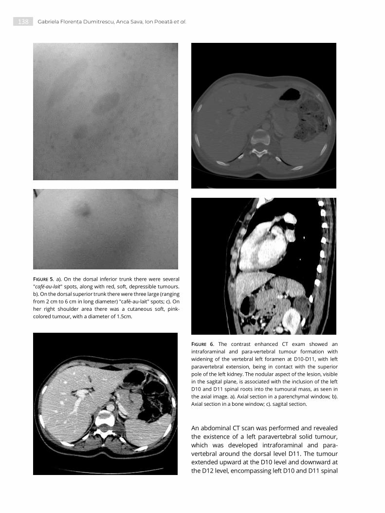
138 Gabriela Florența Dumitrescu, Anca Sava, Ion Poeată et al.
FIGURE 5. a). On the dorsal inferior trunk there were several
"café-au-lait" spots, along with red, soft, depressible tumours.
b). On the dorsal superior trunk there were three large (ranging
from 2 cm to 6 cm in long diameter) "café-au-lait" spots; c). On
her right shoulder area there was a cutaneous soft, pink-
colored tumour, with a diameter of 1.5cm.
FIGURE 6. The contrast enhanced CT exam showed an
intraforaminal and para-vertebral tumour formation with
widening of the vertebral left foramen at D10-D11, with left
paravertebral extension, being in contact with the superior
pole of the left kidney. The nodular aspect of the lesion, visible
in the sagital plane, is associated with the inclusion of the left
D10 and D11 spinal roots into the tumoural mass, as seen in
the axial image. a). Axial section in a parenchymal window; b).
Axial section in a bone window; c). sagital section.
An abdominal CT scan was performed and revealed
the existence of a left paravertebral solid tumour,
which was developed intraforaminal and para-
vertebral around the dorsal level D11. The tumour
extended upward at the D10 level and downward at
the D12 level, encompassing left D10 and D11 spinal

139 Correlations between clinical, imaging and histological findings in a patient with neurofibromatosis type 1
roots. The tumoural formation was solid, oval, well
defined, with dimensions of 35/16 / 65mm (FIGURE 6).
The standard laboratory tests values were in the
normal range.
Surgical intervention was performed by external
approach at D11-D12 lateral level, with microsurgical
total ablation of the tumour. The surgical specimen
was sent to the Pathology Department of the same
hospital for histopathological analysis.
The specimen was fixed in formalin, embedded in
paraffin, cut at 5 μm and stained with hematoxylin-
eosin (HE). On representative sections, immune-
histochemical stainings were performed using anti-
S-100 protein antibody, anti-Vimentin antibody, and
a two-step staining technique (EnVision+ Dual Link
System-HRP, Dako). Histological sections stained
with HE showed a non-encapsulated tumour,
consisting of neoplastic wavy Schwann cells,
entrapped ganglion cells as the tumour extended to
associated ganglia, a myxoid stroma, and delicate
collagen fibers (FIGURE 7). S100 protein immune-
staining highlighted neoplastic Schwann cell
component and ganglia cells of the spinal root that
were entrapped into the tumour (Figure 8). Vimentin
immunostaining revealed numerous fibroblasts
(FIGURE 9). The final pathological diagnosis was spinal
neurofibroma with tumoural infiltration of the
associated ganglia.
The ophthalmologic examination revealed
myopia in both eyes (best corrected visual acuity in
right eye was 1 with -1 D and in the left eye was 1 with
– 0,5 D). The ophthalmoscopic examination showed
normal appearance of the ocular fundus in both
eyes. Direct photomotor reflex was bilaterally
present. Intraocular pressure was within normal
range: right eye = 12 mmHg and left eye = 13 mmHg.
Both eyes presented many small, bright brown, oval,
and dome-shaped irian nodules (Lisch nodules)
(FIGURE 10).
FIGURE 7. Spinal neurofibroma: a). non-encapsulated tumour
made up of neoplastic wavy Schwann cells, a myxoid stroma,
and delicate collagen (H-E, x100); Numerous ganglia cells
entrapped into the tumour mass (HE, x100).
FIGURE 8. Immunopositivity for S100 protein in neoplastic
Schwann cells and ganglia cells (anti – S100 protein, x 100).
FIGURE 9. Immunopositivity for vimentin in randomly arranged
fibroblasts (anti - Vimentin, x 100).

140 Gabriela Florența Dumitrescu, Anca Sava, Ion Poeată et al.
FIGURE 10. Numerous small Lisch nodules at both eyes [a). right
eye; b). left eye].
DISCUSSION
There are suppositions that Ebers Papyrus case #873
can represent the first mention of a case of
neurofibromatosis type 1 (NF1) (2). The important
landmarks in the history of the disease are the Irish
neurosurgeon Robert William Smith, who, in 1849,
realized a systematic review of clinical cases (3), and
the German pathologist Friedrich Daniel von
Recklinghausen (1833-1910), who fully described this
condition in 1882. Von Recklinghausen introduced
the term “neurofibroma” into clinical practice to
define the benign tumours that accompany this
disease. He was the first who found that these
tumours develop from the peripheral nerve sheath,
consisting of a mixture of Schwann cells and
fibroblasts (4). In recognition of his discovery, the
disease received his name.
However, there are different forms of
neurofibromatosis that VM Riccardi, the director of
the Baylor NF Program (1978-1990), attempted to
classify the disease in 1982 (5). He considered that
there were eight types, but later, in 1986, Carey et al.
proposed a new classification into only five types, as
follows: NF1 - classical, NF2 - acoustic, NF3 -
segmental, NF4 - CALM - familial and NF5 - NF-
Noonan phenotype (6).
NF1 represents 90% of all cases with NF, being
characterized mainly by multiple café-au-lait spots
and neurofibromas along peripheral nerves.
Even though NF1 has been recognized as a
clinical entity for more than a century, its
etiopathogenesis was better understand only in
1990 when the NF1 gene was identified on
chromosome 17. NF1 gene is a tumour suppressor
gene that encodes a protein named neurofibromin
(7), which is a negative regulator of the ‘‘rat sarcoma
viral oncogene homologue’’ (RAS) (8).
Approximately 50% of the cases with NF1 disease
represent new mutations and the expression of the
disease is highly variable, both between and within
families (9).
The most important hypothesis regarding the
neurofibromas was that these tumours can develop
only after both NF1 alleles have been lost in the
Schwann cell lineage. When Schwann cells begin to
proliferate, they recruit some other cells from their
environment (10). As such, neurofibromas are
complex tumours, consisting in a mixture of cell
types: Schwann cells, fibroblasts, perineurial cells,
and mast cells (7, 10).
It was also hypothesized that these different
cellular phenotypes can represent divergent cellular
differentiation pathways of multipotent precursor
cells (11).
It was considered that the development of
neurofibromas include the presence of a clonal
population of NF1-/- Schwann cells in a
microenvironment harbouring other cell types with
an NF1+/- genotype. It was also hypothesised that
the multipotent NF1+/- cells are the major source of
different cell types found in the neurofibromas (11).
Neurofibromas developing in a NF1-affected
individual can be classified according to their
anatomical location into: cutaneous, subcutaneous,
intraneural, and spinal nerve roots tumours (12).
Cutaneous neurofibromas generally appear
during preadolescence. They are soft, pink coloured
tumours, sessile or dome-shaped, being most
numerous on the trunk and limbs. The tumour
diameter usually varies between a few millimetres

141 Correlations between clinical, imaging and histological findings in a patient with neurofibromatosis type 1
and approximately 2 centimetres (11). It is
considered that these benign tumours developed
from small nerve tributaries of the skin (13).
Histologically, cutaneous neurofibromas are
nonencapsulated, loosely textured dermal mixed
tumours, consisting of Schwann cells, neurons,
perineurial cells, fibroblasts, and mast cells, but also
adipocytes, epithelial cells, and axonal processes
(11). All of these cellular elements are embedded in
an abundant collagenous extracellular matrix.
Megahed (14) described ten histopathological
variants of neurofibroma: classic, cellular, myxoid,
hyalinized, epithelioid, plexiform, diffuse, pigmented,
granular cell, pacinian. Subsequently, some other
variants such as dendritic cell neurofibroma with
pseudorosettes (15), lipomatous neurofibroma (16),
and angioneurofibroma (17) have been reported.
Pseudomeissnerian corpuscles can be seen in
cutaneous neurofibromas, but not so frequently.
Using traditional histological staining as well as
immunohistochemical staining (anti-S100 protein
antibody and anti-Vimentin antibody) can be the
proofs that the neoplastic component is made of
Schwann cell. Neurofilament protein immune-
reactivity (NFP) revealed the occurrence of residual
axons within tumour nodules. Some authors
highlighted the fact that, using factor XIIIa as an
immunohistochemical marker, the pathologist can
differentiate cutaneous neurofibromas from
neurotized nevi and cutaneous schwannomas (18.)
Aggregates of pseudo-meissnerian corpuscles are
frequent in diffuse cutaneous neurofibromas (19). As
these structures consist entirely of Schwann cells,
they can be can be highlighted with anti-S100 protein
antibody immunostaining (20).
Our patient presented at least one cutaneous
neurofibroma since he was 8 years-old, but the
definitive diagnosis of NF1 was realized only after the
surgical excision of the tumour (when patient was 12
years-old). Histological and immunohistochemical
examinations identified diffuse cutaneous
neurofibroma with clusters of organoid structures
resembling Meissner corpuscles (pseudo-
meissnerian structures), thus being in line with other
researchers' findings.
The incidence of spinal involvement in NF1
patients varies among authors. Mautner et al.
reported that spinal neurofibromas are found in up
to 38% of NF1 patients but very few of them are
symptomatic (21).
In a study of patients with NF1 made by Thakkar
et al., 6% of patients had intramedullary tumours,
57% had intraforaminal tumours (dumbbell), and
33% had extradural tumours (22).
At the spinal level, patients with NF1 may have
either one or more tumours that can develop in one
or more spinal nerve roots. Usually, spinal tumours
are diffuse or plexiform neurofibromas. Some
authors found out that more frequently these spinal
tumours are solitary neurofibromas (23), but others
reported plexiform neurofibromas, which grow
involving multiple nerve fascicles, branches and
plexuses (24). Our patient had a solitary spinal
neurofibroma, but her uncle had multiple plexiform
spinal neurofibromas. Our article prove the fact that
neurofibromas developing in a patient with NF1 can
have different histological features, each of them
displaying microscopical appearances more closely
related to the histology of the region in which the
tumour occurs, i.e., cutaneous neurofibroma
exhibited pseudomeissnerian corpuscle, and spinal
neurofibroma revealed Schwann cells arranged in
bundles, reminding of spinal nerve histology.
The age at onset of symptoms caused by spinal
neurofibromas can vary from 11 to 49 years (mean
32.8 years), but most cases present symptoms at
adult ages (22 to 43 years) (25).
Spinal neurofibromas cause neurological
symptoms in only 2% of NF1 patients, provoking
both sensory and motor deficits due to compression
of the spinal cord or nerve root (22). In a recent
article, Mauda-Havakuk et al. analyzed the
radiological findings of a series of thirty-four patients
with NF1 with spinal neurofibromas. They classified
spinal involvement into four types according to the
anatomic location of the tumours along the spine
and to their type of involvement of the spinal canal
and foramina: 1. foraminal tumour; 2. “kissing”
tumours; 3. paraspinal tumour; 4. intradural tumour
(26). These authors reported that most spinal
neurofibromas developed in the lumbo-sacral area
and fell into group 1 (foraminal).
Our patient was diagnosed as having a spinal
foraminal neurofibroma at the age of 22. As her left
D10 and D11 spinal roots were entrapped into the
tumour mass, the patient presented a two months
history of diffuse intercostal nevralgias that did not
respond to the treatment, thus imposing the surgical
intervention.
Lisch nodules are hamartomas of the iris that are

142 Gabriela Florența Dumitrescu, Anca Sava, Ion Poeată et al.
also characteristic for NF1. They have variable
dimensions and dome-shaped configuration. Some
researchers reported that patients affected by NF1
and older than twenty could have an incidence of
Lisch nodules of 100% (27). Lewis et al. found out that
92% of subjects aged 6 years and older had Lisch
nodules, but their presence was not correlated to
number of café-au-lait spots, number of
neurofibromas, or severity of disease (28). Our
patient, being in her third decade of life, also
presented Lisch nodules in both her eyes.
Due to varied and complex manifestations of the
disease, the management of a patient with NF1
requires a multidisciplinary approach. Once the
diagnosis is established, clinical monitoring is
needed to identify possible complications. As a
result, annual assessment is required to reduce
morbidity and improve the quality of life. This
assessment should include: a) dermatological
examination to analyze the progression of the
existing neurofibromas and to identify possible new
lesions; b). head and spine MRI, chest and abdomen
imaging to detect the majority of complications that
could be associated with this disease; c). full
ophthalmological examination for early detection of
optic nerve lesions; d). neurological examination for
early detection of paresthesia, radiculopathy, muscle
fatigue or muscle atrophy; e). removal of skin
tumours for cosmetic or therapeutic purposes; f).
education and psychological support for NF1 patient.
CONCLUSION
The patient described in this article is a very typical
case of NF1, having six ”café-au-lait” spots, six
neurofibromas, axillary or inguinal freckling, Lisch
nodules, and a first degree relative (her father's
brother) with NF1.Our case presents a considerable
interest because of a complete description of the
natural evolution of NF1, highlighting the histological
pictures of her cutaneous and spinal neurofibromas.
Because the expression of the disease is highly
variable, both between and within families, our study
data are especially valuable as they also showed a
highly variability among neurofibromas in the same
patient from a histopathological point of view. These
data are also important as they demonstrate the fact
that the management of a patient with NF1, due to
the various and complex manifestations of the
disease, requires a multidisciplinary approach,
including neurologist, neurosurgeon, ophthalmol-
ogist, plastic surgeon, dermatologist, radiologist and
pathologist.
REFERENCES
1. Stumpf D, Alksne J, Annegers J, Brown S, Conneally P,
Housman D, Leppert M, Miller J, Moss M, Pileggi A, Rapin I.
Strohman R, Swanson L, Zimmerman A.
Neurofibromatosis. Conference statement. National
Institutes of Health Consensus Development Conference.
Arch Neurol. 1988;45:575–578
2. Sanchez GM, Siuda T. Ebers Papyrus case #873: a probable
case of neurofibromatosis 1. S D J Med. 2002;55(12):529-35
3. Kobrin JL, Blodi FC, Weingeist TA. Ocular and orbital
manifestations of neurofibromatosis. Surv Ophthalmol,
1979, 24:45
4. von Recklinghausen FD. Ueber die multiplen Fibrome der
Haut und ihre Beziehung zu den multiplen Neuromen.
Hirschwald, Berlin, 1882
5. Riccardi VM. Neurofibromatosis: Clinical heterogeneity.
Curr Probl Cancer, 1982, 7:1
6. Carey JC, Baty BJ, Johnson JP, Morrison T, Skolnik M, Kivlin J.
The genetic aspects of neurofibromatosis. Ann NY Acad Sci.
1986;486:45–46
7. Theos A, Korf BR. Pathophysiology of Neurofibromatosis
Type 1. Ann Intern Med. 2006;144:842-849
8. Martin GA, Viskochil D, Bollag G, McCabe PC, Crosier WJ,
Haubruck H, Conroy L, Clark R, O’Connell P, Cawthon RM,
Innis MA, McCornick F. The GAP-related domain of the
neurofibromatosis type 1 gene product interacts with ras
p21. Cell. 1990;63:843–849
9. Huson SM, Hughes RAC. The Neurofibromatoses: A
Pathogenetic and Clinical Overview. Chapman and Hall
Medical, London, UK, 1994.
10. Zhu Y, Ghosh P, Charnay P, Burns DK, Parada LF.
NeuroÞbromas in NF1: Schwann Cell Origin and Role of
Tumour Environment. Science. 2002; 296:920-922
11. Jouhilahtilow EM, Peltonen S, Heape AM, Peltonenlow J. The
Pathoetiology of Neurofibromatosis 1. Am J Pathol. 2011;
178(5):1932-1939
12. Evans D, Komminoth P, Scheihauer B, Peltonen J.
Neurofibromatosis type 1. Pathology and Genetics of
Tumours of Endocrine Organs: World Health Organization
Classification of Tumours. In: DeLellis R, Lioyd R, Heitz P,
Eng C. (editors). IARC Press, Lyon, France, 2004, pp. 243–
248
13. Jouhilahti EM, Peltonen S, Callens T, Jokinen E, Heape AM,
Messiaen L, Peltonen J. The Development of Cutaneous
Neurofibromas. Am J Pathol. 2011; 178(2): 500–505
14. Megahed M. Histopathological variants of neurofibroma. A
study of 114 lesions. Am J Dermatopathol. 1994; 16:486–
495
143 Correlations between clinical, imaging and histological findings in a patient with neurofibromatosis type 1
15. Michal M, Fanburg-Smith JC, Mentzel T, Kutzner H, Requena
L, Zamecnik M, Miettinen M. Dendritic cell neurofibroma
with pseudorosettes: a report of 18 cases of a distinct and
hitherto unrecognized neurofibroma variant. Am J Surg
Pathol. 2001;25:587–594.
16. Val-Bernal JF, de sa Dehesa J, Garijo MF, Val D. Cutaneous
lipomatous neurofibroma. Am J Dermatopathol.
2002;24:246–250
17. Saxer-Sekulic N, Kaya G. Cutaneous Angioneurofibroma: A
New Histopathological Variant of Neurofibroma,
Dermatopathol. 2014;1:7–10
18. Gray MH, Smoller BR, McNutt NS, Hsu A.
Immunohistochemical demonstration of factor XIIIa
expression in neurofibromas. A practical means of
differentiating these tumours from neurotized melanocytic
nevi and schwannomas. Arch Dermatol. 1990;126(4):472-6
19. Zhang ML, Suarez MJ, Bosley TM, Rodriguez FJ.
Clinicopathologic Features of Peripheral Nerve Sheath
Tumours Involving the Eye and Ocular Adnexa. Hum
Pathol. 2017; 63:70–78.
20. Shiurba RA, Eng LF, Urich H. The structure of
pseudomeissnerian corpuscles. An immunohistochemical
study. Acta Neuropathol. 1984;63(2):174-6.
21. Mautner VF, Asuagbor FA, Dombi E, Funsterer C, Kluwe L,
Wenzel R, et al. Assessment of benign tumour burden by
whole-body MRI in patients with neurofibromatosis 1.
Neurol Oncol 2008;10:593-8
22. Thakkar SD, Feigen U, Mautner V-F. Spinal tumours in
neurofibromatosis type 1: a study of frequency, multiplicity
and variety. Neuroradiol. 1999; 41: 625-629
23. Khong PL, Goh WHS, Wong VCN, Fung CW, Ooi GC. MR
Imaging of Spinal Tumours in Children with
Neurofibromatosis 1, AJR. 2003; 180: 413-417
24. Acharya S, Shukla S, Banode P. Multiple spinal
neurofibromatosis. Astrocyte. 2014;1(3):250-251
25. Kluwe L, Tatagiba M, Fünsterer C, Mautner V-F. NF1
mutations and clinical spectrum in patients with spinal
neurofibromas. J Med Genet. 2003;40:368–371
26. Mauda-Havakuk M, Shofty B, Ben-Shachar S, Ben-Sira L,
Constantini S, Bokstein F. Spinal and Paraspinal Plexiform
Neurofibromas in Patients with Neurofibromatosis Type 1:
A Novel Scoring System for Radiological-Clinical
Correlation. AJNR Am J Neuroradiol. 2017. www.ajnr.org
27. Lubs ME, Bauer MS, Formas ME, Djokic B: Lisch nodules in
neurofibromatosis type 1. N Engl J Med. 1991; 324:1264.
28. Lewis RA, Riccardi VM. Von Recklinghausen
neurofibromatosis. Incidence of iris hamartomata.
Ophthalmology. 1981;88(4):348.

Romanian Neurosurgery (2019) XXXIII (2): pp. 144-149 DOI: 10.33962/roneuro-2019-027 www.journals.lapub.co.uk/index.php/roneurosurgery
Experience of choroid plexus papilloma in children at Mansoura University Hospital
Hatem Badr, Ahmad Zaher, Mohamed State, Amr Farid Khalil
* Mansoura University, EGYPT
ABSTRACT Choroid plexus papilloma (CPP) are rare, benign tumours of neuroectodermal origin;
they represent 1-3% of central nervous system (CNS) tumours in paediatric patients.
Authors present their experience in the management of such lesion in Mansoura
University Hospitals.
Methods. For children with Choroid plexus papilloma who were treated via
microsurgical excision over 4 years from January 2012 to January 2016 in Mansoura
University Hospitals, a retrospective analysis was done for age, sex, clinical
manifestations, surgical treatment and follow up.
Results. Twenty-three paediatrics were treated over 4 years. Age ranged from (7
months - 8 years). There were 13 female and10 males. Tumours were located in the
lateral ventricle in all cases. Features of raised intracranial pressure were the
predominant presentation. Total microsurgical excision was achieved in all cases. All
cases had intraoperative blood loss < 100 ml with mean Haematocrit 28%. Follow up
period (7-53 months). Complete relief of all symptoms was reported in all cases with
no mortality. One of our patients needs postoperative ventriculoperitoneal shunt for
persistent CSF leakage and another one required subdural peritoneal shunt for
persistent subdural CSF collection.
Conclusion. Total excision of Choroid plexus papilloma is usually the rule with an
excellent outcome. Routine external ventricular drainage for at least 3 days is
effective in lowering shunt-dependent cases as it allows the release of bloody CSF
and small tumour residue. With proper microsurgical technique through superior
parietal lobule to access lateral ventricle then tumour coagulation at the same time
of irrigation that helps the tumour to shrink and thus, decrease the incidence of
bleeding. The tumour vascular pedicle should be resected with the last part of the
tumour to avoid pedicle retraction that may lead to ventricular haemorrhage.
INTRODUCTION Choroid plexus papilloma (CPPs) are benign neoplasms of the choroid
plexus, a structure made from tufts of villi within the ventricular system
that produces cerebrospinal fluid (CSF) [13, 24]. Lateral ventricle of the
brain is the commonest site of CPPs in paediatrics, but they can occur
in adults too. Although the major percent of these tumours are
considered benign in nature, a small percent is classified to be
malignant [1, 3].
CPPs represent about 1% of intracranial neoplasms but this
percentage tends to be higher in children (2-4%). The most common
Keywords choroid plexus papilloma,
surgical resection, complications
Corresponding author: Hatem Badr
Mansoura University, Egypt
Scan to access the online version

145 Experience of choroid plexus papilloma in children at Mansoura University Hospital
location is the atrium of the lateral ventricle in
paediatric population while 4th ventricle represents
the commonest location in adults. Cerebellopontine
angle (CPA), 3rd ventricle, and parenchyma of the
cerebrum are considered rare locations of these
tumours [20].
Clinically infants and children with CPP presented
with; headache, vomiting, progressive head
enlargement and excessive crying, as reported by
the patients mothers, usually caused by
hydrocephalus that results from; CSF pathway
obstruction, CSF overproduction, or arachnoidal
fibrosis due to recurrent occult bleeding from
tumour [8].
Computed tomography (CT) and magnetic
resonance imaging (MRI) show iso- or hyperdense,
T1 isointense, and T2 hyperintense masses inside the
ventricles that enhance with contrast, generally
associated with hydrocephalus [17].
Grossly, these tumours arise as well-
circumscribed cauliflower-like exophytic growths
from the ventricular wall. Histologically, choroid
plexus papilloma appears as papillary proliferation
with a fibrovascular core lined by a single layer of
monotonous cuboidal to columnar epithelium with a
basement membrane [5].
Gross total excision is considered the main line of
treatment of these tumours as it is associated with
excellent outcomes after surgery. Moreover,
adjuvant radiotherapy can be used for progressing
tumours that were treated by subtotal resection. A
meta-analysis of 566 choroid plexus tumours found
no difference in survival when comparing patients
with recurrent CPPs and those with stable disease
[10].
Choroid plexus tumours can be distinguished
from each other via mouth intestinal bacteria (MIB-
1) labelling, status of p53, and histology into CPP,
atypical papilloma, and carcinoma, and this
differentiation can direct follow-up and adjuvant
treatment plans later on [22].
In management of choroid plexus papilloma,
there are two main problems: treatment of
associated hydrocephalus, rich tumour vascular
supply that is medially located in these young
patients, can lead to a hazardous intraoperative
blood loss. Preoperative embolization, to minimize
risk of intraoperative loss, is associated with
exposure to radiation and also to vessel injury and
stroke [21].
We present our experience in management of such
lesions in the neurosurgical department located at
Mansoura University Hospitals.
FIGURE 1. A 2-year-old female child with left lateral ventricle
choroid plexus papilloma. A: preoperative MRI brain with
contrast; B: 3-month follow up MRI brain with subdural CSF
effusion and total tumour excision.
FIGURE 2. A 6-year-old female child with right lateral ventricle
choroid papilloma. A: preoperative MRI brain with contrast, B:
1-month follow up CT brain with contrast showing total tumour
excision.

146 Hatem Badr, Ahmad Zaher, Mohamed State et al.
FIGURE 3. Right lateral ventricle choroid papilloma which was
managed by microsurgical resection. A: preoperative MRI
brain, B: 3-month follow up MRI brain showing no residual or
tumour recurrence.
METHODS
Retrospective analysis of children with choroid
plexus papilloma who were treated by microsurgical
resection over 4 years from January 2012 to January
2016 in Mansoura University Hospitals.
Each patient was subjected to complete medical
history taking, through physical examination, routine
pre-operative laboratory tests, and radiological
studies (CT and MRI brain). The tumour size was
calculated as the maximum diameter of the lesion
measured by CT and MRI.
All research activities were approved by
Mansoura University local ethical committee.
Post-operative complications were classified into
two categories; major or minor. Denovo permanent
neurological deficits and functional status worsening
were classified as major complications. Minor
complications included temporary post-operative
problems that faded with time like transient cranial
neuropathy, temporary postoperative seizures, deep
venous thrombosis (DVT), or wound healing
problems such as infection, cerebrospinal fluid (CSF)
leak, and pseudo meningocele.
The collected data were coded, processed and
analysed using the SPSS (Statistical Package for
Social Sciences) version 22 for Windows® (SPSS Inc,
Chicago, IL, USA). Qualitative data was presented as
number (frequency) and Percent. Quantitative data
was expressed as median (range).
RESULTS TABLE 1: Demographic, clinical, operative, and follow up data.
Variable Range or number
Age (month) 39 (7 – 96)
Sex
Male
Female
10 (43.47%)
13 (56.52%)
Variable Range or number
Symptoms
-Headache
-Vomiting
-Progressive head enlargement
-Excessive crying
13 (56.52%)
8 (34.78%)
9 (39.13%)
6 (26.08%)
Tumour size (cm) 3.1 (2.4 – 4.2)
Calcification 3 (13.04%)
Major complications 0 (0%)
Mortality 0 (0%)
Minor complications
-CSF leakage
-Persistent subdural effusion
2 (8.69%)
1 (4.34%)
1 (4.34%)
Follow up (month) 20 (7-53)
Need for shunt 2 (8.69%)
Residual in follow up CT 0 (0%)
Twenty-three paediatrics were treated via micro-
surgical resection over 7 years. Age ranged from (7
months - 8 years). There were 13 females (56.52%)
and 10 males (43.47%). Features of increased
intracranial pressure were present in all our cases
with varying combination like headache, vomiting,
excessive crying, and progressive head enlargement.
All cases were evaluated preoperatively via CT
and MRI brain. Lateral ventricle of the brain was the
site of the CCP in all our paediatric cases. The CT
scans showed a mass that was lobulated, globular,
and brightly enhancing in the lateral ventricle.
Median tumour diameter measured by CT or MRI
was 3.1 cm. Calcification was diagnosed radio-
logically in 3 cases (13.04%), whereas all the cases
had varying degrees of hydrocephalus. In all
patients, MRI showed an enhancing mass in the
lateral ventricle that was isointense on T1- and
hyperintense on T2-weighted images.
Total microsurgical excision was performed via
the transcortical approach through the superior
parietal lobule in the all patients who had the tumour
located the lateral ventricle trigone. The tumour was
identified and then, its surface was coagulated.
Moreover, it was excised after shrinking its size via
combined coagulation and irrigation. Coagulation,
then division of the vascular tumour pedicle was
achieved on removal of the last tumour part.
Routine closed ventricular drainage was applied
in all patients to allow CSF drainage for three days
post-operatively. That ventricular drain was removed
after exclusion of ventricular hematoma by CT scan
and absence of red colour in the draining CSF fluid.
In one case (4.34%), CSF leakage from the operative

147 Experience of choroid plexus papilloma in children at Mansoura University Hospital
incision occurred and that child needed ventriculo-
peritoneal shunt for persisting hydrocephalus.
Another patient (4.34%) needed subduro-peritoneal
shunt because of persistent, subdural effusion.
The follow-up ranged from 7–53 months (median:
20 months). All patients experienced no symptoms
at follow-up as well as the post-operative CT and MRI
showed no residual lesions.
DISCUSSION
Choroid plexus papilloma are considered as WHO
grade I tumours and are among the most common
tumours in the paediatric group less than 2 years.
They are of neuroectodermal origin representing 1-
3% of central nervous system (CNS) tumours in
paediatric patients [23].
Choroid plexus papilloma has been reported to
be associated with Aicardi syndrome [4, 25, 28] and
Li-Fraumeni syndrome [6]. Leptomeningeal
dissemination may occur [7].
Usually, CPPs occur in the lateral ventricles in
paediatric population, while the 4th ventricle is the
commonest location in adults; other rare sites
included 3rd ventricle and the cerebellopontine angle
(CPA). CPPs have also been reported in other regions
like the suprasellar area, frontal lobe, and
cerebellum [17]. In our study, all tumours were
located in the lateral ventricle (23 cases).
In children, before closure of the sutures and
fontanelles, the sequel of raised intracranial
pressure such as blindness will be difficult to
manifest. Seizures or mental changes may be the
only presentations of these lesions while tumours
involving the 4th ventricle may manifest itself with
cerebellar signs. Cerebellopontine angle CPPs have
been reported to cause cranial nerve deficits [8]. The
predominant clinical features in our study can all be
traced back to raised intracranial pressure that as
consistent in all of our patients.
Clinical picture of CPP in paediatrics includes;
headache, vomiting, progressive head enlargement
and excessive crying. These are usually caused by
hydrocephalus that results from CSF pathway
obstruction, excessive CSF production, and recurrent
occult bleeding from the tumour that results in
arachnoidal fibrosis and adhesions [8].
In our study, headache was present in 56.52 % of
cases while vomiting was experienced in 34.78 % of
cases. Moreover, progressive head enlargement and
excessive crying were reported by the mothers of in
39.13 and 26.08% of our cases respectively.
The mean value of maximum tumour diameter in
our study was 3.1 cm, while the study conducted by
safaee and his colleagues included cases with
median diameter of 3 cm in the total resection group
[21].
Calcification was detected in CT in 13.04% (3
cases) in our study cases. This is consistent with the
findings reported in the literature stating that
choroid plexus tumours calcification can be detected
radiologically in around 4–20% of cases [9, 19].
Surgical gross total excision of the tumour should
be attempted in all cases of choroid plexus papilloma
[2]. As these tumours are highly vascular, great care
should be taken during surgery to avoid excessive
blood loss that may lead to mortality, as reported in
some series, especially in young children. Securing
the main arterial supply of the tumour, which is
usually a branch of choroidal artery, is of crucial
importance to avoid hazardous intraoperative
bleeding followed by coagulation of the tumour and
en bloc or piecemeal removal [15].
Regarding surgical excision for paediatric CPPs,
there is a surgical opinion stating that there is some
sort of benefit from delaying surgery till
hydrocephalus develops. That is because this delay
lessens the length of the corridor to the ventricles
and widens the space surrounding the lesion making
it more surgically accessible. The drawback to this
delay is that hydrocephalus may not be the
presenting symptom, leading to more tumour
growing that may lead to neurological deficit that
may not resolve after surgical resection [11, 16].
In addition, the drawbacks to a watchful waiting
on dealing with such tumours are many. initially,
repeated general anaesthesia will be needed for MRI
for follow up which will be burdensome and not
devoid of risks. Moreover, although CT scan may
alleviate the need for MRI< but excess radiation
exposure will carry risk to the child as well [11].
We reported that blood loss in this study was less
than 100 ml in every patient. No major complications
or mortality were reported in our study. Major
complications reported in another series were rare,
that may be attributed to the fact that these modern
studies were conducted at a tertiary care centres
[21].
Compared to published series from the modern
microsurgical era, our observed morbidity was very
minimal, once again confirming that these tutors can

148 Hatem Badr, Ahmad Zaher, Mohamed State et al.
be safely resected in both children and adults [14, 27,
29].
Whereas complete surgical excision is the ideal
treatment, it could be impossible to accomplish in
some cases, due to excessive blood loss or due to
tumour infiltration into the surrounding vital
structures. If significant residual tumour is left or
recurrence is diagnosed, redo-surgery may be an
option after sometime. Moreover, adjuvant therapy
has been reported to be another option [18].
Pneumocephalus, pneumoventricle, subdural
effusions, persistence of hydrocephalus (in those not
previously shunted), and new onset hydrocephalus
are the most common reported complications after
surgical intervention [21, 26]. Total surgical removal
of these tumours is associated with excellent survival
that reaches 100% at 10-year follow up as reported
in the literature [14].
In our series, only two cases required post-
operative shunts; one needed ventriculoperitoneal
shunt due to persistent CSF leakage, and the other
needed subduroperitoneal shunt for persistent
subdural CSF collection.
Although some studies have described
neurocognitive sequelae related to surgery for CPP
[12, 16], our study is not large enough to make any
significant conclusions with respect to differential
outcomes in children compared to adults.
Safaee and his associates also reported a 22 %
incidence of temporary swallowing dysfunction that
required percutaneous endoscopic gastrostomy
tubes, tracheostomy, or both, with no difference in
the incidence when comparing patients who
received gross total resection with the subtotal
resection group [21].
The prognosis for CPP patients treated with gross
total resection (GTR) is excellent, with a report of
100% survival at 5 years after surgical resection in
many series including ours, and adjuvant therapy is
not indicated in these patients. Radiotherapy is not
necessary after GTR, and its usefulness should be
reserved for recurrent disease. Unfortunately, the
prognosis for patients with choroid plexus
carcinoma CPC is guarded, with an overall 5-year
survival rate of 26-50%. GTR is generally considered
the most important prognostic factor for CPC as was
confirmed in our analysis [14].
Several limitations can be noted in this current
study. Small sample size limits the statistical power
of our analysis, but is an unfortunate and inevitable
consequence of studying such a rare tumour.
Moreover, this study is a single centre study that
could affect the validity of our results as it serves a
localized population.
CONCLUSION
Total excision of Choroid plexus papilloma is usually
the rule with excellent outcome. Routine external
ventricular drainage for at least 3 days is effective in
lowering shunt dependent cases as it allows release
of bloody CSF and small tumour residue. With proper
microsurgical technique through superior parietal
lobule to access lateral ventricle then tumour
coagulation at the same time of irrigation that helps
the tumour to shrink and thus, decrease the
incidence of bleeding. The tumour vascular pedicle
should be resected with last part of the tumour to
avoid pedicle retraction that may lead to ventricular
haemorrhage.
AUTHORS CONTRIBUTIONS
This work was carried out in collaboration between all authors.
Author Amr Farid, designed the study, Author Mohamed State
wrote the protocol, Author Ahmed Zaher managed the
literature research, Author Hatem Badr performed the
statistical analysis and revised the final manuscript. All surgical
procedures were carried out by the same surgical team
including the four authors. All authors read and approved the
final manuscript.
ABBREVIATIONS
CPC Choroid plexus carcinoma.
CPP Choroid plexus papilloma.
CPA Cerebellopontine angle.
GTR Gross total resection.
MIB-1 Mouse intestinal bacteria.
MRI Magnetic resonance imaging.
REFERENCES
1. Abdulkader M M, Mansour N H, Van Gompel J J, Bosh G A,
Dropcho E J, Bonnin J M, and Cohen-Gadol A A:
Disseminated choroid plexus papillomas in adults: A case
series and review of the literature. Journal of Clinical
Neuroscience 2016, 32, 148-154.
2. Bettegowda C, Adogwa O, Mehta V, Chaichana K L,
Weingart J, Carson B S, Jallo G I, and Ahn E S: Treatment of
choroid plexus tumors: a 20-year single institutional
experience. Journal of Neurosurgery: Pediatrics 2012, 10(5),
398-405.
3. Dudley R W, Torok M R, Gallegos D, Liu A K, Handler M H,
and Hankinson T C: Pediatric choroid plexus tumors:
epidemiology, treatments, and outcome analysis on 202
149 Experience of choroid plexus papilloma in children at Mansoura University Hospital
children from the SEER database. Journal of neuro-
oncology 2015, 121(1), 201-207.
4. Frye R E, Polling J S, and Ma L C: Choroid plexus papilloma
expansion over 7 years in Aicardi syndrome. Journal of
child neurology 2007, 22(4), 484-487.
5. Fung K-M, Yu Z, and Petropoulou K; Tumors of the central
nervous system, in Pediatric Malignancies: Pathology and
Imaging. 2015, Springer. p. 151-201.
6. Gozali A E, Britt B, Shane L, Gonzalez I, Gilles F, McComb J
G, Krieger M D, Lavey R S, Shlien A, and Villablanca J G:
Choroid plexus tumors; management, outcome, and
association with the Li–Fraumeni syndrome: The Children's
Hospital Los Angeles (CHLA) experience, 1991–2010.
Pediatric blood & cancer 2012, 58(6), 905-909.
7. Irsutti M, Thorn-Kany M, Arrue P, Richaud J, Sol J, Delisle M,
and Manelfe C: Suprasellar seeding of a benign choroid
plexus papilloma of the fourth ventricle with local
recurrence. Neuroradiology 2000, 42(9), 657-661.
8. Jaiswal A K, Jaiswal S, Sahu R N, Das K, Jain V K, and Behari
S: Choroid plexus papilloma in children: Diagnostic and
surgical considerations. Journal of pediatric neurosciences
2009, 4(1), 10.
9. Koeller K K and Sandberg G D: From the archives of the
AFIP: cerebral intraventricular neoplasms: radiologic-
pathologic correlation. Radiographics 2002, 22(6), 1473-
1505.
10. Krishnan S, Brown P D, Scheithauer B W, Ebersold M J,
Hammack J E, and Buckner J C: Choroid plexus papillomas:
a single institutional experience. Journal of neuro-oncology
2004, 68(1), 49-55.
11. Laarakker A S, Nakhla J, Kobets A, and Abbott R: Incidental
choroid plexus papilloma in a child: A difficult decision.
Surgical neurology international 2017, 8.
12. Levy M L, Goldfarb A, Hyder D J, Gonzales-Gomez I, Nelson
M, Gilles F H, and McComb J G: Choroid plexus tumors in
children: significance of stromal invasion. Neurosurgery
2001, 48(2), 303-309.
13. Lun M P, Monuki E S, and Lehtinen M K: Development and
functions of the choroid plexus–cerebrospinal fluid system.
Nature Reviews Neuroscience 2015, 16(8), 445.
14. Menon G, Nair S N, Baldawa S S, Rao R B, Krishnakumar K,
and Gopalakrishnan C: Choroid plexus tumors: an
institutional series of 25 patients. Neurology India 2010,
58(3), 429.
15. Mishra A, Ojha B, Chandra A, Singh S, Chandra N, and
Srivastava C: Choroid plexus papilloma of posterior third
ventricle: A case report and review of literature. Asian
journal of neurosurgery 2014, 9(4), 238.
16. Nagib M G and O’Fallon M T: Lateral ventricle choroid
plexus papilloma in childhood: management and
complications. Surgical neurology 2000, 54(5), 366-372.
17. Ogiwara H, Dipatri Jr A J, Alden T D, Bowman R M, and
Tomita T: Choroid plexus tumors in pediatric patients.
British journal of neurosurgery 2012, 26(1), 32-37.
18. Pawar S, Sharma R, Mahapatra A, Lad S, and Musa M:
Choroid plexus papilloma of the posterior third ventricle
during infancy & childhood: report of two cases with
management morbidities. Neurology India 2003, 51(3),
379.
19. Pencalet P, Sainte-Rose C, Lellouch-Tubiana A, Kalifa C,
Brunelle F, Sgouros S, Meyer P, Cinalli G, Zerah M, and
Pierre-Kahn A: Papillomas and carcinomas of the choroid
plexus in children. Journal of neurosurgery 1998, 88(3),
521-528.
20. Prasad G L and Mahapatra A K: Case series of choroid
plexus papilloma in children at uncommon locations and
review of the literature. Surgical neurology international
2015, 6.
21. Safaee M, Clark A J, Bloch O, Oh M C, Singh A, Auguste K I,
Gupta N, McDermott M W, Aghi M K, and Berger M S:
Surgical outcomes in choroid plexus papillomas: an
institutional experience. Journal of neuro-oncology 2013,
113(1), 117-125.
22. Safaee M, Oh M C, Bloch O, Sun M Z, Kaur G, Auguste K I,
Tihan T, and Parsa A T: Choroid plexus papillomas:
advances in molecular biology and understanding of
tumorigenesis. Neuro-oncology 2012, 15(3), 255-267.
23. Sethi D, Arora R, Garg K, and Tanwar P: Choroid plexus
papilloma. Asian journal of neurosurgery 2017, 12(1), 139.
24. Spector R, Keep R F, Snodgrass S R, Smith Q R, and
Johanson C E: A balanced view of choroid plexus structure
and function: focus on adult humans. Experimental
neurology 2015, 267, 78-86.
25. Taggard D A and Menezes A H: Three choroid plexus
papillomas in a patient with Aicardi syndrome. Pediatric
neurosurgery 2000, 33(4), 219-223.
26. Talacchi A, De Micheli E, Lombardo C, Turazzi S, and Bricolo
A: Choroid plexus papilloma of the cerebellopontine angle:
a twelve patient series. Surgical neurology 1999, 51(6), 621-
629.
27. Tena-Suck M L, Salinas-Lara C, Rembao-Bojórquez D, and
Castillejos M: Clinicopathologic and immunohistochemical
study of choroid plexus tumors: single-institution
experience in Mexican population. Journal of neuro-
oncology 2010, 98(3), 357-365.
28. Trifiletti R R, Incorpora G, Polizzi A, Cocuzza M D, Bolan E A,
and Parano E: Aicardi syndrome with multiple tumors: a
case report with literature review. Brain and Development
1995, 17(4), 283-285.
29. Wolff J, Sajedi M, Brant R, Coppes M, and Egeler R: Choroid
plexus tumours. British Journal of Cancer 2002, 87(10),
1086.

Romanian Neurosurgery (2019) XXXIII (2): pp. 150-155 DOI: 10.33962/roneuro-2019-028 www.journals.lapub.co.uk/index.php/roneurosurgery
Hydatid cyst of the quadrigeminal cistern. A case report for unusual location with literature review
Saif Saood Abdelrazaq1, Abdullah H. Al Ramadan2,
Ali Adnan Dolachee3, Mohammed Maan
AbdulAzeez4, Ali Saud Abdulrazzaq5, Amar Saeed
Rashid6, Samer S. Hoz5 1 Neurosurgery Department, Al Sadar Teaching Hospital, Najaf, IRAQ 2 Department of Neurosurgery, King Faisal Specialist Hospital and
Research Centre, Riyadh, SAUDI ARABIA 3 Department of Neurosurgery, College of Medicine, Al Qadisiyah
University, IRAQ 4 College of Medicine, University of Baghdad, Baghdad, IRAQ 5 Neurosurgery Department, Neurosurgery Teaching Hospital,
Baghdad, IRAQ 6 Department of Neurosurgery, College of Medicine, Kufa University,
Baghdad, IRAQ
ABSTRACT Intracranial hydatid cyst involves supratentorial area and mainly affecting the middle
cerebral artery territory with the predilection of the partial lobe. It can be single -
which is the most common - or multiple up to 35 cysts. They tend to be huge at the
time of symptomatic presentation especially when they are presented as a solitary
lesion with a slow growth rate around 1.5 cm/year, however, it is variable and it can
be up to 10 cm/year. Surgical treatment is mandatory for all patients once the correct
diagnosis is made, except for patients with multiple organ involvement in poor
general conditions and deep-located cysts. The existence of hydatidosis in the
cisternal spaces must not be neglected given the capacity of E. granulosus larvae to
disseminate via the CSF. In this case report; two and half years’ male child presented
with a history of 2 attacks of generalized seizure for the last 72 hours with the head
circumference at the upper normal limit for his age. This paper presents the first case
report demonstrating a primary single hydatid cyst located in the quadrigeminal
cistern in a child. We concluded that in spite of the feasibility of the imaging and the
high suspension of cerebral hydatid cyst, still, the reports show more locations which
can be described as unusual although for a head to toe suspected distribution of
hydatid disease is already understood. An eminent medical and surgical (if indicated)
treatment of the primary cerebral hydatid cyst are always effective and
recommended.
INTRODUCTION Choroid Echinococcosis could be characterized as a zoonotic infes-
tation caused by cestode species of the genus Echinococcus (1).
Keywords neurosurgery,
hydatid, quadrigeminal cistern,
case report
Corresponding author: Mohammed Maan AbdulAzeez
College of Medicine, University of
Baghdad, Baghdad University, Iraq
Scan to access the online version

151 Hydatid cyst of the quadrigeminal cistern
It is most commonly involving the liver then lungs. (2)
Intracranial involvement occurs in 1-2% of Echino-
coccosis and 50-70% of these cases affect the
paediatric age group. (3) It showed slightly male
preference in most of the case series. (4,5,6,7,8)
Geographically endemic area of hydatid disease
includes some of South America countries like
Argentina, Brazil, Chile, Peru and Uruguay, France,
Italy and Greece in Europe, Turkey, Israel, Lebanon,
Syria and Jordan in the Middle East, China, Iran, and
India in Southeast Asia and Tunisia in Africa. (9)
Intracranial hydatid cyst involves supratentorial
area and mainly affecting the middle cerebral artery
territory with the predilection of the partial lobe. It
can be single - which is the most common - or
multiple up to 35 cysts. (10,11,12) They tend to be
huge at the time of symptomatic presentation
especially when they are presented as a solitary
lesion with a slow growth rate around 1.5 cm/year,
however, it is variable and it can be up to 10 cm/year.
(13) Surgical treatment is mandatory for all patients
once the correct diagnosis is made, except for
patients with multiple organ involvement in poor
general conditions and deep-located cysts. (14) The
existence of hydatidosis in the cisternal spaces must
not be neglected given the capacity of E. granulosus
larvae to disseminate via the CSF. (15)
This paper presents the first case report
demonstrating a primary single hydatid cyst located
in the quadrigeminal cistern in a child.
CASE SCENARIO
Two and half years’ male child presented with history
of 2 attacks of generalized seizure for the last 72
hours with head circumference at the upper normal
limit for his age.
Initial lab workup was done to exclude metabolic
derangements the patient was sent for
neuroimaging. The general lab tests were within
normal ranges and the brain MRI showed a midline
rounded cystic lesion, 5x3 cm in size, located in extra-
axially in the quadrigeminal cistern and extend infra-
tentorially to compress the cerebellum downward
along with bilateral dilated lateral ventricles (FIGURE
1).
FIGURE 1. Pre-operative brain MRI showing
a midline rounded cystic lesion, 5x3 cm in
size, located in extra-axially in the
quadrigeminal cistern and extend infra-
tentorially to compress the cerebellum
downward along with bilateral dilated
lateral ventricles. The lesion was hypo-
intense and homogenous in T1 with
intensity higher than CSF, hyper-intense in
T2 and showed no enhancement in
contrasted image, also there was some
vivid inclusions within the upper part of
the cyst.
FIGURE 2. the delivered intact hydatid cyst.

152 Saif Saood Abdelrazaq, Abdullah H. Al Ramadan, Ali Adnan Dolachee et al.
The lesion was hypo-intense and homogenous in the
T1 sequence of the MRI with intensity higher than the
CSF, hyperintense in the T2, and showed no
enhancement in the contrasted image, also there
were some vivid inclusions within the upper part of
the cyst.
At that point, the top differential diagnosis
included were epidermoid, dermoid and arachnoid
cysts only, because that location was an unusual
location for a hydatid cyst and the age of the patient
was too young for a hydatid cyst.
The decision was to do a surgical resection or
fenestration according to the intra-operative
findings. we planned to delay the CSF shunting
option as the sulci were not effaced, the young age
of the patient and the surgery would remove the
cause of the obstruction of the CSF pathways.
The surgery conducted at the next day, an intra-
tentorial supra-cerebellar approach was done via
midline occipital craniotomy. The cyst was a typical
hydatid cyst. The cyst resected as one piece via initial
dissection and then delivered using Dowling’s hydro-
dissection technique (FIGURE 2,3).
The postoperative course went uneventfully. The
histopathology confirms the diagnosis, further
workup excluded other organs involvement.
FIGURE 3. Early postoperative axial brain CT scan showing resection cavity
TABLE 1. Intracranial Cisternal Hydatid cysts: Case Report in English Literature
No. Authors Age/Gender Complain Diameter Location Treatment
1 Rumboldt et
al (27)
50 Year/
Male
Headache 30 mm Premedullary Cisten Surgery
2 Beskonakli et
al (28)
33 Year/
Male
Headache and impair
vision
N/A Interpeduncular
Cistern
Surgery
3 Sanli et al (29) 7 Year/ Male Symptoms of high
Intracranial pressure
25 x 25 mm Ambient Cistern Surgery
4 Kizilca et al
(37)
45 Year/
Female
Symptoms of high
Intracranial pressure
N/A Premedullary Cisten Surgery
5 The authors
of this case
report
2.5 Year/
Male
Seizure 50 x 30 mm Quadrigeminal
Cistern
Surgery

153 Hydatid cyst of the quadrigeminal cistern
DISCUSSION
Variant locations of hydatid cyst were reported in the
literature including aqueduct of Sylvius (16), Frontal
lobe associated with massive edema and raise in the
intracranial pressure (17), temporal lobe with
midbrain herniation. (18)
Thalamic hydatid cyst was managed surgically
through the transcallosal approach in a child who
presented with left-sided weakness. (19)
An interosseous lesion involving the skull
estimated to represent 3-4% of the lesion involving
the skeleton which evident in 2% of all hydatid cyst.
(20)
Spinal hydatid cysts are estimated at less than
1%, they are usually extradural and tend to rupture
intraoperative more than other location. (21)
Meckel’s cave can be involved with extension to the
Cerebellopontine angle and the middle fossa. (22)
Brainstem hydatid cyst was reported in middle
age woman presented with a headache and
progressive hemiparesis. (23) both cases of Meckel’s
cave and Brainstem hydatid cysts were treated with
decompression first prior to the removal to decrease
the chances of intraoperative rupture. (22,23)
Cerebellar lesion with mass effect causing
obstructive hydrocephalus was encountered in a
child with a progressive headache followed by signs
and symptoms of high intracranial pressure. (24)
Intraventricular involvement was estimated
between 1.3 - 16.6% of multiple case series for
intracranial hydatid disease with overall good
outcome after surgical excision. (25) Other unusual
locations include the seller and the para-sellar
regions were reported. (26)
Cisternal hydatid cysts were reported to involve
the pre-medullary, the interpeduncular and the
ambient cisterns. (27,28,29) (Table 1)
An intermittent headache can be the only
presenting symptoms even with a huge cyst. (30) In a
Large series from China, headache and vomiting
were the most common symptoms, this finding was
supported in the literature as well.
Seizure attacks, focal neurological deficits, and
skull deformities were reported. Papilledema was
frequent in the ophthalmological examination
especially in patients with high intracranial pressure,
optic atrophy was seen as well. (4,31,32)
The Magnetic Resonance Imaging (MRI) thought
to be more sensitive than the computed tomography
(CT) to identify the pericystic layer and to
differentiated the hydatid cyst from the other similar
cystic lesions like the epidermoid cyst, however, CT
scan is more sensitive to identify the calcified lesions.
(33)
Serious complications related to the rupture of
the hydatid cyst include recurrence, cerebral
vasospasm and infarction, hydrocephalus,
anaphylactic shock, chemical meningitis, and even
death. (34)
In two series of long-term follow up for cerebral
hydatidosis in children, it can be concluded that the
improvements of the imaging facilities lead to early
recognition and improve the outcome. Poor
prognostic factors can be identified including delay
in the treatment, the rupture of the cyst at the
presentation or intraoperatively, and poor control of
the systemic disease. Single versus multiple cyst and
age at the presentation was not identified as
prognostic factors, however, some authors consider
the multiplicity as a malignant form of the disease.
(35,36)
Review of all the available literature reveals that
our case is the first reported case of hydatid cyst
located in the quadrigeminal cistern which was
removed successfully.
CONCLUSION
In spite of the feasibility of the imaging and the high
suspension of cerebral hydatid cyst, still, the reports
show more locations which can be described as
unusual although for a head to toe suspected
distribution of hydatid disease is already
understood. An eminent medical and surgical (if
indicated) treatment of the primary cerebral hydatid
cyst are always effective and recommended.
ABBREVIATIONS
CT: computed tomography
MRI: magnetic resonance imaging
CSF: cerebrospinal fluid
REFERENCES
1. Eckert J, Deplazes P. Biological, epidemiological, and clinical
aspects of echinococcosis, a zoonosis of increasing
concern. Clin Microbiol Rev. 2004 Jan;17(1):107-35. Review.
PubMed PMID: 14726458; PubMed Central PMCID:
PMC321468.
2. Madeo J, Zheng X, Ahmed S, Ramos De Oleo R. Primary
cerebral echinococcosis presenting as long-standing
generalized weakness. Germs. 2013 Jun 1;3(2):63-6. doi:
10.11599/germs.2013.1038. eCollection 2013 Jun 1.
154 Saif Saood Abdelrazaq, Abdullah H. Al Ramadan, Ali Adnan Dolachee et al.
PubMed PMID: 24432288; PubMed Central PMCID:
PMC3882828.
3. Erşahin Y, Mutluer S, Güzelbağ E. Intracranial hydatid cysts
in children. Neurosurgery. 1993 Aug; 33(2): 219-24;
discussion 224-5. PubMed PMID: 8367043.
4. Turgut M. Intracranial hydatidosis in Turkey: its clinical
presentation, diagnostic studies, surgical management,
and outcome. A review of 276 cases. Neurosurg Rev. 2001
Dec;24(4):200-8. Review. PubMed PMID: 11778827.
5. Khaldi M, Mohamed S, Kallel J, Khouja N. Brain hydatidosis:
report on 117 cases. Childs Nerv Syst. 2000 Nov;16(10-
11):765-9. PubMed PMID: 11151730.
6. Kocaman S, Ersahin Y, Mutluer S. Cerebral hydatid cysts in
children. J Neurosci Nurs. 1999 Oct;31(5):270-7. PubMed
PMID: 10633303.
7. Duishanbai S, Jiafu D, Guo H, Liu C, Liu B, Aishalong M, Mijiti
M, Wen H. Intracranial hydatid cyst in children: report of 30
cases. Childs Nerv Syst. 2010 Jun;26(6):821-7. doi:
10.1007/s00381-009-1008-2. Epub 2009 Dec 16. PubMed
PMID: 20013283.
8. Limaiem F, Bellil S, Bellil K, Chelly I, Mekni A, Khaldi M,
Haouet S, Zitouna M, Kchir N. Primary hydatidosis of the
central nervous system: a retrospective study of 39
Tunisian cases. Clin Neurol Neurosurg. 2010 Jan;112(1):23-
8. doi: 10.1016/j.clineuro.2009.09.001. Epub 2009 Sep 24.
PubMed PMID: 19781849.
9. Nourbakhsh A, Vannemreddy P, Minagar A, Toledo EG,
Palacios E, Nanda A. Hydatid disease of the central nervous
system: a review of literature with an emphasis on Latin
American countries. Neurol Res. 2010 Apr;32(3):245-51. doi
10.1179/016164110X12644252260673. Review. PubMed
PMID: 20406602.
10. Ali M, Mahmood K, Khan P. Hydatid cysts of the brain. J
Ayub Med Coll Abbottabad. 2009 Jul-Sep;21(3):152-4.
PubMed PMID: 20929036.
11. Sen N, Laha D, Gangopadhyay PK, Mohanty BC. Young girl
with multiple intracranial hydatid cyst. Ann Neurosci. 2012
Apr;19(2):96-8. doi: 10.5214/ans.0972.7531.12190212.
PubMed PMID: 25205976; PubMed Central PMCID:
PMC4117048.
12. Binesh F, Mehrabanian M, Navabii H. Primary brain
hydatosis. BMJ Case Rep. 2011 Mar 8;2011. pii:
bcr0620103099. doi: 10.1136/bcr.06.2010.3099. PubMed
PMID: 22707660; PubMed Central PMCID: PMC3063272.
13. Thakur SH, Joshi PC, Kelkar AB, Seth N. Unusual presen-
tation of hydatid cyst - ruptured intraventricular hydatid.
Indian J Radiol Imaging. 2017 Jul-Sep;27(3):282-285. doi:
10.4103/ijri.IJRI_70_16. PubMed PMID: 29089673; PubMed
Central PMCID: PMC5644318.
14. Yurt A, Avci M, Selçuki M, Ozer F, Camlar M, Uçar K, Taşli F,
Altinörs N. Multiple cerebral hydatid cysts. Report of a case
with 24 pieces. Clin Neurol Neurosurg. 2007 Nov; 109(9):
821-6. Epub 2007 Aug PubMed PMID: 17765392.
15. Andronikou S, Welman CJ, Kader E. Classic and unusual
appearances of hydatid disease in children. Pediatr Radiol.
2002 Nov;32(11):817-28. Epub 2002 Aug 16. PubMed PMID:
12389111.
16. Gökalp HZ, Erdoğan A. Hydatid cyst of the aqueduct of
Sylvius. Case report.Clin Neurol Neurosurg. 1988;90(1):83-
5. PubMed PMID: 3359737.
17. Bahloul K, Ouerchefani N, Kammoun B, Boudouara MZ.
Unusual brain edema caused by an intracranial hydatid
cyst: c ase report and literature review. Neurochirurgie.
2009 Feb;55(1):53-6. doi: 10.1016/j.neuchi.2008.03.008.
Epub 2008 Jun 30. Review. PubMed PMID: 18586282.
18. Duransoy YK, Mete M, Barutçuoğlu M, Unsal UÜ, Selçuki M.
Intracranial hydatid cyst is a rare cause of midbrain
herniation: A case report and literature review. J Pediatr
Neurosci. 2013 Sep 8(3): 224-7. doi: 10.4103/1817-1745.
123683. PubMed PMID: 24470819; PubMed Central PMCID:
PMC3888042.
19. Erşahin Y, Mutluer S, Dermirtaş E, Yurtseven T. A case of
thalamic hydatid cyst. Clin Neurol Neurosurg. 1995 Nov; 97
(4): 321-3. PubMed PMID: 8599900.
20. Erman T, Tuna M, Göçer I, Ildan F, Zeren M, Cetinalp E.
Intracranial intraosseous hydatid cyst. Case report and
review of literature. Neurosurg Focus.2001 Jul 15;11(1):
ECP1. Review. PubMed PMID: 16724818.
21. Luo K, Luo DH, Zhang TR, Wen H. Primary intracranial and
spinal hydatidosis: a retrospective study of 21 cases.
Pathog Glob Health. 2013 Mar;107(2):47-51. doi:
10.1179/2047773213Y.0000000072. PubMed PMID: 23683
329; PubMed Central PMCID: PMC4001477.
22. Taghipour M, Saffarrian A, Ghaffarpasand F, Azarpira N.
Dumbbell-Shape Hydatid Cyst of Meckel Cave Extending to
Cerebellopontine Angle and Middle Fossa; Surgical
Technique and Outcome of Rare Case. World Neurosurg.
2017 Aug;104 :1049.e7-1049.e10. doi: 10.1016/j.wneu.
2017.05.091. Epub 2017 May 25. PubMed PMID: 28552738.
23. Muthusubramanian V, Pande A, Vasudevan MC, Ravi R.
Surgical management of brainstem hydatid cyst--an
unusual site. Surg Neurol. 2009 Jan;71(1):103-6; discussion
106. doi: 10.1016/j.surneu.2007.06.077. Epub 2008 Feb 11.
PubMed PMID: 18262629.
24. Fakhouri F, Ghajar A, Mahli N, Shoumal N. Giant hydatid
cyst in the posterior fossa of a child. Asian J Neurosurg.
2015 Oct-Dec;10(4):322-4. doi: 10.4103/1793-5482.162719.
PubMed PMID: 26425166; PubMed Central PMCID:
PMC4558813.
25. Guzel A, Tatli M, Maciaczyk J, Altinors N. Primary cerebral
intraventricular hydatid cyst: a case report and review of
the literature. J Child Neurol. 2008 May;23(5):585-8. doi:
10.1177/0883073807309791. Epub 2008 Jan 11. Review.
PubMed PMID: 18192651.
26. Turgut M. Hydatidosis of central nervous system and its
coverings in the pediatric and adolescent age groups in
Turkey during the last century: a critical review of 137
cases. Childs Nerv Syst. 2002 Dec;18(12):670-83. Epub 2002
Oct 25. Review. PubMed PMID: 12483350.
27. Rumboldt Z, Jednacak H, Talan-Hranilović J, Rumboldt T,
Kalousek M. Unusual appearance of a cisternal hydatid
cyst. AJNR Am J Neuroradiol. 2003 Jan;24(1):112-4. PubMed
PMID: 12533337

155 Hydatid cyst of the quadrigeminal cistern
28. Beskonakli E, Solaroglu I, Tun K, Albayrak L. Primary
intracranial hydatid cyst in the interpeduncular cistern.
Acta Neurochir (Wien). 2005 Jul;147(7):781-3; discussion
783. Epub 2005 May 20. PubMed PMID: 15900396.
29. Sanlı AM, Türkoğlu E, Kertmen H, Gürer B. Hydatid cyst of
the ambient cistern radiologically mimicking an arachnoid
cyst. J Neurosurg Pediatr. 2012 Sep;10(3):186-8. doi:
10.3171/2012.6. PEDS 11562. Epub 2012 Jul 13. PubMed
PMID:22793161.
30. Basarslan SK, Gocmez C, Kamasak K, Ceviz A. The Gigant
primary cerebral hydatid cyst with no marked
manifestation: a case report and review of literature. Eur
Rev Med Pharmacol Sci. 2015 Apr;19(8): 1327-9. Review.
PubMed PMID: 25967703.
31. Erşahin Y, Mutluer S, Güzelbağ E. Intracranial hydatid cysts
in children. Neurosurgery. 1993 Aug;33(2): 219-24;
discussion 224-5. PubMed PMID: 8367043.
32. Duishanbai S, Geng D, Liu C, Guo HR, Hao YJ, Liu B, Wang
YX, Luo K, Zhou K, Wen H; Research Group of Hydatid
Diseases. Treatment of intracranial hydatid cysts. Chin Med
J (Engl). 2011 Sep;124 (18): 2954-8. PubMed PMID:
22040508.
33. Tsitouridis J, Dimitriadis AS, Kazana E. MR in cisternal
hydatid cysts. AJNRAm J Neuroradiol. 1997 Sep;18(8): 1586-
7. PubMed PMID: 9296204.
34. Salunke P, Patra DP, Mukherjee KK. Delayed cerebral
vasospasm and systemic inflammatory response
syndrome following intraoperative rupture of cerebral
hydatid cyst. Acta Neurochir (Wien). 2014 Mar;156(3):613-
4. doi: 10.1007/s00701-013-1675-3. Epub 2013 Mar 10.
PubMed PMID: 23474734.
35. Lunardi P, Missori P, Di Lorenzo N, Fortuna A. Cerebral
hydatidosis in childhood: a retrospective survey with
emphasis on long-term follow-up. Neurosurgery. 1991
Oct;29(4): 515-7; discussion 517-8. PubMed PMID:
1944831.
36. Onal C, Unal F, Barlas O, Izgi N, Hepgul K, Turantan MI,
Canbolat A, Turker K, Bayindir C, Gokay HK, Kaya U. Long-
term follow-up and results of thirty paediatric intracranial
hydatid cysts: half a century of experience in the
Department of Neurosurgery of the School of Medicine at
the University of Istanbul (1952-2001). Pediatr Neurosurg.
2001 Aug;35(2): 72-81. PubMed PMID: 11549917.
37. Kızılca O, Altaş M, Senol U, Oztek MA. Hydatid disease
located in the cerebellomedullary cistern. Case Rep Med.
2014: 271365. doi: 10.1155/2014/271365. Epub 2014 Mar
24. PubMed PMID: 24782895; PubMed Central PMCID:
PMC3982244.

Romanian Neurosurgery (2019) XXXIII (2): pp. 156-159 DOI: 10.33962/roneuro-2019-029 www.journals.lapub.co.uk/index.php/roneurosurgery
Cerebral pilocytic astrocytoma with spontaneous intratumoral haemorrhage in the elderly - a rare entity. A case report and review of the literature
Amit Narang, Varun Aggarwal, Divya Kavita,
Chandni Maheshwari, Prajjwal Bansal
* Baba Farid University of Health Sciences. Faridkot. Punjab, INDIA
ABSTRACT Pilocytic astrocytomas (PA) are histologically are low-grade tumours, commonly
found in the paediatric and young adult population. Although cases of adult and
elderly pilocytic astrocytomas are described in the literature, they are quite
uncommon. The rate of PAs with spontaneous bleeding is very less, with a high
occurrence in children than in adults, moreover, that is in cerebellar PAs. Cerebral
PAs with intratumoral haemorrhage in the older age group is extremely rare. We
present a case of a 60-year female presenting with acute neurological compromise
resulting from an acutely haemorrhagic Left temporal pilocytic astrocytoma. She was
managed surgically with evacuation of tumoral haemorrhages, as well as resection of
the tumour. Postoperatively the patient made a remarkable recovery.
INTRODUCTION Pilocytic astrocytoma (PA) is commonly a paediatric central nervous
system glial tumour. Adult occurrence is rare, especially in elderly
population. [6,2] The most common sites of PAs origin are cerebellum,
optic chiasma, hypothalamus, and to a lesser extent, cerebral
hemispheres, brain stem and spinal cord.[6] Reports of tumours with
intratumoral haemorrhage in elderly population are extremely rare.
Here we present a 60 years old female who presented with left
temporal intratumoral bleed mimicing Intracerebral haemorrhage.
CASE SUMMARY
Ours is a 60-year-old female who was a known case of hypertension
and was poorly compliant to anti-hypertensive medications. She had
complaint of sudden onset of altered sensorium and slurring of speech.
There was no history suggestive of Trauma. Patient was taken to a
nearest clinic and was managed conservatively on the line of Intra
Cerebral Hemorhage (ICH), and was discharched after seven days.
Three days after the discharge patients attendants brought the patient
Keywords astrocytoma,
cerebral pilocytic astrocytoma,
elderly population, intratumoral hemorrhage,
tumor excision
Corresponding author: Varun Aggarwal
Guru Gobind Singh Medical College
and Hospital Faridkot, Punjab, India
Scan to access the online version

157 Cerebral pilocytic astrocytoma with spontaneous intratumoral haemorrhage in the elderly - a rare entity
to our hospital for second opinion. On presentation
her GCS was E3V3M5. Contrast MRI Brain was done
which was suggestive 48 x 34 mm altered signal
intensity lesion in left temporoparietal cortex with
vasogenic edema and with mass effect in the form of
midline shift of 6.4 mm towards right. The lesion was
hypointens on T2, T1 with marginal T1 hyper-
intensities, hyperintens on flair and intens blooming
in gradient images. In post contrast studies subtle
marginal enhancement noted. By enlarge, radio-
logical features were suggestive of left Temporal ICH
with mass effect. [Fig-1]. Patient was taken up for
surgery. Left frontotemporal craniotomy and eva-
cuation of intracereberal hematoma was done.
Intraoperatively brain was bulging and there was
large hematoma in Rt temporal lobe, brain tissue all
around the hematoma was greyish, soft and
suckable this tissue was excised all around and was
sent for biopsy. Post operatively NCCT was done
suggestive of complete evacuation of ICH with
reduced mass effect [Fig-2]. Histopathology was
suggestive of pilocytic astrocytoma (WHO grade I).
Patient improved significantly and discharged on 5th
Post op day with GCS E4V4M6.
FIGURE 1. A -T1, B -T2, C- DW, D- post contrast MRI showing
lesion in left temporal lobe

158 Amit Narang, Varun Aggarwal, Divya Kavita et al.
FIGURE 2. First post op day NCCT head showing Complete evacuation of hematoma and reduced mass effect
FIGURE 3. Histopathology was suggestive of low grade astrocytic
tumor with abundant eosinophilic granular bodies- pilocytic
astrocytoma (WHO grade I)
DISCUSSION
PA is a histological subtype of astrocytomas, which
accounted for approximately 15% of central nervous
system (CNS) tumours. PAs are considered World
Health Organization (WHO) grade I tumours, which
have relatively distinct histological appearance and
well demarcated boundaries. [1,5]. This tumour has
much higher occurrence among children and
adolescents than elderly. Common age group for PAs
is 5 to 14 years, followed by 0 to 4 years and 15 to 19
years, however, pilocytic astrocytoma rarely occur
over the age of 60. [2]
Patients having a pilocytic astrocytoma may
present with sensory motor deficit, gait disturbance,
headaches, cranial nerve deficits and hydrocephalus,
depending on the exact location. The most common
sites of these tumour origin are cerebellum, optic
chiasma, hypothalamus, and to a lesser extent
cerebral hemispheres, brain stem and spinal cord.
[1] Most of glial tumours, such as high-grade
astrocytomas, oligodendrocytes, and mixed
astrocytomas, have propensity for intratumoral
bleeding. However, intratumoral hemorrhage is very
rare among patients with PA. [6]
The rate of PAs with spontaneous bleeding is
approximately 8%, with a high occurrence in children
than in adults, which is mostly in cerebellar PA [6].

159 Cerebral pilocytic astrocytoma with spontaneous intratumoral haemorrhage in the elderly - a rare entity
Relatively limited amount of literature is available on
PAs with intratumoral haemorrhage in older age
group. Although vascular necrosis and tumour
invasion are the primary causes of bleeding in high-
grade gliomas, the mechanisms underlying low-
grade gliomas remains unclear. [3] Previous
literature have demonstrated that spontaneous
bleeding in PAs is due to interaction of various
factors, which includes specific pathological changes
in vascular structures of tumour, such as
proliferation of vascular endothelial cells, hyaline
degeneration and interstitial degeneration. Sun S. et
al [6] suggested in their study, the vascular histology
of bleeding tumour reveals a poorly developed
capillary bed and thin vessel walls. Bleeding tumour
blood vessels, although rich in collagen have fewer
elastic fibres and poor compliance, which can
escalates the chances of tumour rupture. As
reported previously the reticular vessels are more
prone to rupture, which suggests an association
between tumour capillary type and bleeding
tendency.[4] In addition, intratumoral bleeding is
associated with tumour growth, intratumoral
vascular invasion, rupture of an encased aneurysm
and coagulopathies. Notably, higher arterial
pressure and acute intracranial hypertension are
also the predisposing factors. Previous studies also
suggest that calcification may contribute to
intratumoral bleeding. [6,4]. It has been found that
the spontaneous bleeding of PAs is also associated
with tumour VEGF expression.[7]
The patient in our case report developed
spontaneous intratumoral hemorrhage, but the
cause of bleeding is still not clear. Therefore, we
speculate that intratumoral bleeding may be
attributed to tumour vascular abnormalities and
hypertension.
PAs patients with intratumoral bleed leading to
raised intracranial tension requirs emergency
neurosurgical care. Surgery is the preferred the
treatment for PAs with hemorrhage, which permits
simultaneous removal of tumor and hematoma.
Tumors that occurred in the brain stem or visual
pathway usually cannot be completely removed by
surgery, though the residual tumors may cease to
grow, or grow slowly, or even regress. Therefore, a
long-term follow-up is required for patients with
complete and partial resection. The patients treated
with subtotal resection may require post op
chemotherapy and radiotherapy, but it is still a
matter of debate. The prognosis of patients with PAs
is generally good. The location of the tumour and the
extent of the resection can play an important role in
the prognosis of PAs patients. Reoperation may be
considered for most patients with recurrent PAs.
Patients with partial resection of tumour can have a
higher risk of recurrence. [8] literature suggests that
a longer follow-up period is required for PAs patients
with intratumoral haemorrhage.
CONCLUSION
Based on the collective results from the present
clinical experience and other related studies, we
suggest that cerebral PAs with spontaneous
intratumoral haemorrhage is extremely rare,
especially in elderly patients over the age of 60 years.
We agree with the literature that to completely
remove the tumour and hematoma; surgery is
probably the most effective treatment option. Long-
term follow-up after surgical treatment of PAs with
intratumoral haemorrhage is crucial to identify and
treat the recurrence if any.
REFERENCES
1. Bornhorst M, Frappaz D, Packer RJ. Pilocytic
astrocytomas. Handbook Clin Neurol 2016;134:329–44.
2. Cyrine S, Sonia Z, Mounir T, et al. Pilocytic astrocytoma: a
retrospective study of 32 cases. Clin Neurol
Neurosurg 2013;115:1220–5.
3. Kapoor A, Savardekar A, Tewari MK, et al. Spontaneous
hemorrhages in pediatric supratentorial pilocytic
astrocytomas. Malignant presentation of a benign
entity. Childs Nerv Syst 2015;31:1617–20.
4. Kim MS, Kim SW, Chang CH, et al. Cerebellar pilocytic
astrocytomas with spontaneous intratumoral hemorrhage
in adult. Korean Neurosurg 2011;49:363–6.
5. Ostrom QT, Gittleman H, Liao P, et al. CBTRUS statistical
report: primary brain and central nervous system tumors
diagnosed in the United States in 2007–2011. Neuro
Oncol2014;16:1–63.
6. Sun S, Zhou H, Ding ZZ, Shi H.Cerebellar pilocytic
astrocytomas with spontaneous intratumoral hemorrhage
in the elderly: A case report and review of the
literature. Medicine (Baltimore). 2018 Aug;97(31).
7. Wilson MP, Johnson ES, Hawkins C, et al. Hemorrhagic
presentations of cerebellar pilocytic astrocytomas in
children resulting in death: report of 2 cases. Neurosurg
Pediatr2016;17:446–52.
8. Ye JM, Ye MJ, Kranz S, et al. A 10 year retrospective study of
surgical outcomes of adult intracranial pilocytic
astrocytoma. J Clin Neurosci 2014;21:2160–4.

Romanian Neurosurgery (2019) XXXIII (2): pp. 160-165 DOI: 10.33962/roneuro-2019-030 www.journals.lapub.co.uk/index.php/roneurosurgery
Traumatic posterior fossa extradural hematoma. A comprehensive analysis of cases from a tertiary care centre in Southwestern Rajasthan
Vibhu Shankar Parashar, Vivek Kumar Kankane,
Gaurav Jaiswal, Tarun Kumar Gupta
* Department of Neurosurgery, Rabindra Nath Tagore Medical
College & M.B. Groups of Hospital, Udaipur, Rajasthan, INDIA
ABSTRACT Background. Extradural hematoma of posterior fossa (PFEDH) is less common and
there are not many articles about PFEDH. These patients can deteriorate very rapidly
due to compression over brainstem. Thus, early identification and immediate
intervention can save the lives of these patients.
Objective. This study aims to conduct a comprehensive analysis of patients with
PFEDH and evaluate the postoperative outcome which may be of help to make
further preventive strategies.
Methods and Materials. The study included 16 patients admitted with traumatic
PFEDH from July 2016 to July 2018 at R.N.T. Medical College & M.B. Groups of Hospital
Udaipur, southwestern Rajasthan, India. We have retrospectively reviewed the data.
Analysed factors were gender, age, Glasgow Coma Scale (GCS), Noncontrast CT scan
findings, associated brain injury, type of intervention, Glasgow Outcome Scale (GOS).
GOS was assessed at discharge, at 3 months and 6 months follow-up.
Results. Out of a total of 16 patients, 11 were male and 5 were female with age
ranging from 05-46 years. 12 patients had GCS 13 -15 at admission and only one of
them had GCS < 8. 15 patients underwent surgical intervention. At 6 months follow-
up, 12 patients had good recovery GOS is 5.
Conclusion. Early detection and immediate evacuation of PFEDHs should be done if
causing fourth ventricle, basal cistern or brain stem compression. It may be rapidly
fatal due to the expansion of hematoma leading to brainstem compression, tonsillar
herniation, and/or obstructive hydrocephalus. Early detection and immediate
evacuation lead to a better outcome in these patients.
INTRODUCTION PFEDH is an uncommon trauma sequel accounting for only 4% to 12.9%
of all EDHs [1,2]. In PFEDH clinical progress may be silent and slow, but
sudden deterioration may occur without significant warning signs.
Because of limited space in posterior fossa, comparatively small volume
can cause clinical deterioration. The patient may deteriorate very
rapidly due to compression over the brainstem usually without
Keywords posterior fossa,
extradural hematoma, traumatic,
Glasgow outcome Scale
Corresponding author: Vivek Kumar Kankane
Rabindra Nath Tagore Medical
College & M.B. Groups of Hospital, Udaipur, Rajasthan,
India
Scan to access the online version
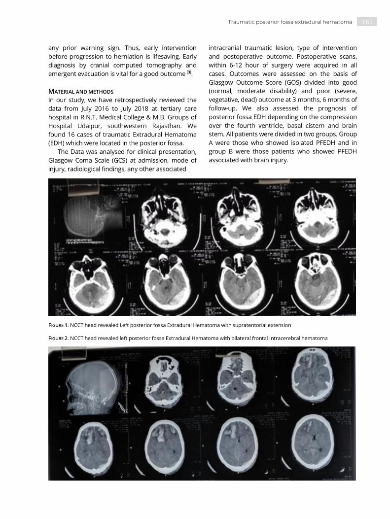
161 Traumatic posterior fossa extradural hematoma
any prior warning sign. Thus, early intervention
before progression to herniation is lifesaving. Early
diagnosis by cranial computed tomography and
emergent evacuation is vital for a good outcome [3].
MATERIAL AND METHODS
In our study, we have retrospectively reviewed the
data from July 2016 to July 2018 at tertiary care
hospital in R.N.T. Medical College & M.B. Groups of
Hospital Udaipur, southwestern Rajasthan. We
found 16 cases of traumatic Extradural Hematoma
(EDH) which were located in the posterior fossa.
The Data was analysed for clinical presentation,
Glasgow Coma Scale (GCS) at admission, mode of
injury, radiological findings, any other associated
intracranial traumatic lesion, type of intervention
and postoperative outcome. Postoperative scans,
within 6-12 hour of surgery were acquired in all
cases. Outcomes were assessed on the basis of
Glasgow Outcome Score (GOS) divided into good
(normal, moderate disability) and poor (severe,
vegetative, dead) outcome at 3 months, 6 months of
follow-up. We also assessed the prognosis of
posterior fossa EDH depending on the compression
over the fourth ventricle, basal cistern and brain
stem. All patients were divided in two groups. Group
A were those who showed isolated PFEDH and in
group B were those patients who showed PFEDH
associated with brain injury.
FIGURE 1. NCCT head revealed Left posterior fossa Extradural Hematoma with supratentorial extension
FIGURE 2. NCCT head revealed left posterior fossa Extradural Hematoma with bilateral frontal intracerebral hematoma

162 Vibhu Shankar Parashar, Vivek Kumar Kankane, Gaurav Jaiswal et al.
RESULTS
A total of 16 cases of PFEDHs were admitted to our
tertiary care centre during 24 months from July2016-
July 2018.The mean age of patients was 28.6 years
(range 5-46years). 3 (18.75%) of them were below 18
years. 5(31.25%) of them were Females (TABLE 1).
Outcome was better in paediatric patients.
Most common mode of injury was road traffic
accident (n= 11, 68.75%), rest were either fall from
height (n = 04, 25%) or assault (n =1, 6.25%) (TABLE -
2). Most common associated radiological finding is
occipital bone fracture followed by supratentorial
extension of EDH and frontal contusions (TABLE 3).
15 patients underwent surgical evacuation. At 6
months follow-up, 12 (75%) patients had good
recovery and one patient died (6.25%) (TABLE 4).
Patients with PFEDH with associated brain injury had
lesser GCS Score on admission with increased
volume of EDH, increased hospital stay. Outcome
was poor in PFEDH patients with associated
intracranial injury as compared to those with isolated
PFEDH. (TABLE 5)
TABLE 1. Correlation of Gender and Age and outcome analysis
Total cases
Number. of
patients
(N=16)
AGE<18 Yrs AGE>18 yrs Percentage
(%)
Good
outcome
(GOS 3-5)
Poor
outcome
(GOS1-2)
Gender
Male 11 03 08 68.75% 10 01
Female 05 00 05 31.25% 03 02
Age
Pediatric
patients (<18)
03
18.75 % 03 00
Adults 13
81.25% 10 03
Total 16
100% 13 03
TABLE 2. Correlation of mode of injury, GCS at admission and outcome analysis
No. of patients Percentage Good outcome
(GOS 3-5)
Poor outcome
(GOS 1-2)
Mode of injury
Road traffic accident 11 68.75% 09 02
Fall from height 04 25% 03 01
Assault 01 6.25% 01 00
GCS at admission
13-15 12 75% 12 00
9-12 03 18.75% 01 02
3-8 01 6.25% 00 01
Total 16 100% 13 03

163 Traumatic posterior fossa extradural hematoma
TABLE 3. Analysis of clinical presentation and radiological findings
Number of patients Percentage
Clinical presentation
Headache 10 62.5%
Altered sensorium 06 37.5%
Vomiting 08 50%
Radiological findings
Occipital bone fracture 12 75%
Frontal contusions 01 6.25%
Supratentorial extension of EDH 02 12.5%
Hydrocephalus 01 6.25%
Intraventricular hemorrhage 01 6.25%
TABLE 4. Outcome based on GOS (Glasgow outcome score)
Outcome based on GOS Discharge At 3 months At 6 months
GOS 5 11 12 (75%) 12 (81.25%)
GOS 4 03 01 (6.25%) No follow up
GOS 3 00 01 (6.25%) 01 (6.25%)
GOS 2 01 01 (6.25%) 01 (6.25%)
GOS 1 01 - -
TABLE 5. Analysis of patients with isolated PFEDH and those with associated brain injuries
Parameters Isolated PFEDH
(Group A)
PFEDH and associated
brain injury (Group B)
Total patients 13(81.25%) 03(18.75%)
Radiological findings
Occipital bone fracture 10(76.9%) 02(66.7%)
Frontal contusions - 01(33.3%)
Supratentorial extension of EDH - 02(66.7%)
Hydrocephalus - 01(33.3%)
Intraventricular hemorrhage - 01(33.3%)
GCS (admission)
15-13 11 01
12-9 02 01
8-3 00 01
Management
Surgical evacuation 11 03
Conservative 02 00
Failed conservative and operated 01 00
GCS (discharge)
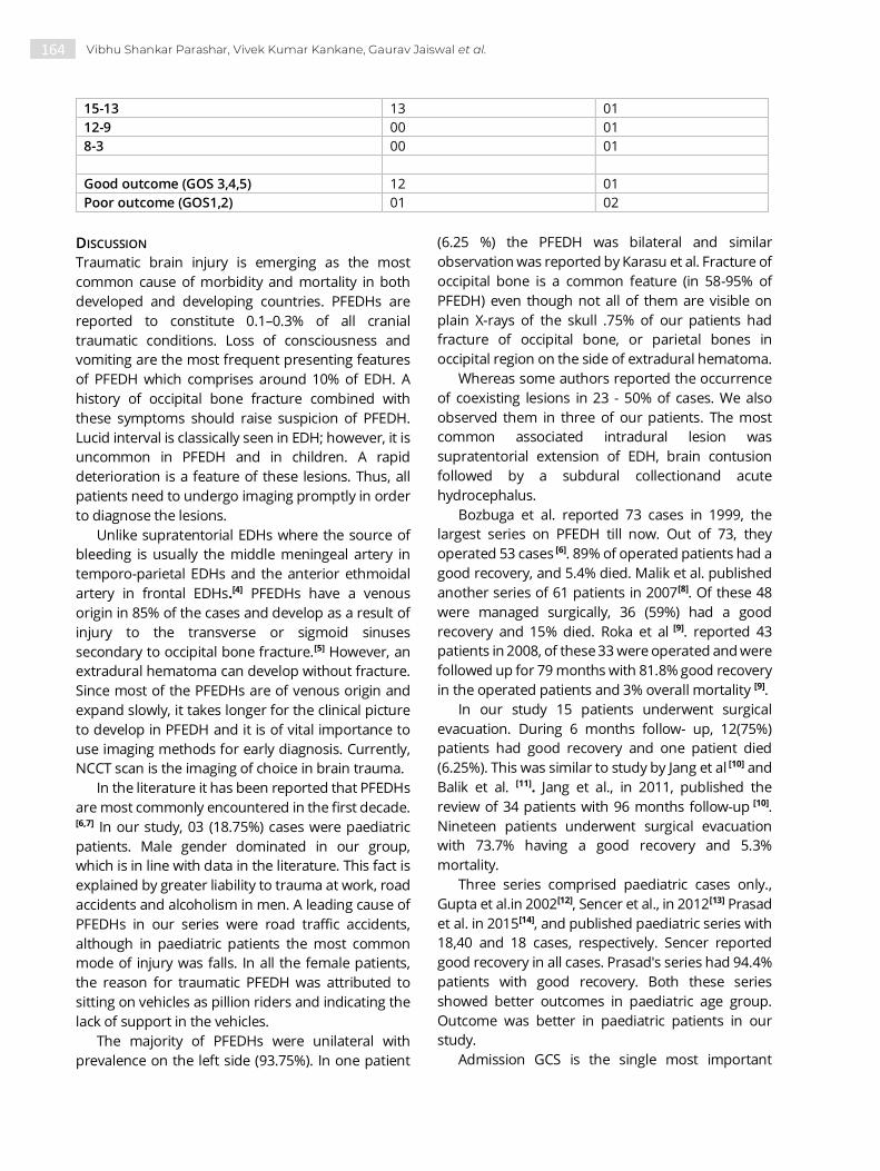
164 Vibhu Shankar Parashar, Vivek Kumar Kankane, Gaurav Jaiswal et al.
15-13 13 01
12-9 00 01
8-3 00 01
Good outcome (GOS 3,4,5) 12 01
Poor outcome (GOS1,2) 01 02
DISCUSSION
Traumatic brain injury is emerging as the most
common cause of morbidity and mortality in both
developed and developing countries. PFEDHs are
reported to constitute 0.1–0.3% of all cranial
traumatic conditions. Loss of consciousness and
vomiting are the most frequent presenting features
of PFEDH which comprises around 10% of EDH. A
history of occipital bone fracture combined with
these symptoms should raise suspicion of PFEDH.
Lucid interval is classically seen in EDH; however, it is
uncommon in PFEDH and in children. A rapid
deterioration is a feature of these lesions. Thus, all
patients need to undergo imaging promptly in order
to diagnose the lesions.
Unlike supratentorial EDHs where the source of
bleeding is usually the middle meningeal artery in
temporo-parietal EDHs and the anterior ethmoidal
artery in frontal EDHs.[4] PFEDHs have a venous
origin in 85% of the cases and develop as a result of
injury to the transverse or sigmoid sinuses
secondary to occipital bone fracture.[5] However, an
extradural hematoma can develop without fracture.
Since most of the PFEDHs are of venous origin and
expand slowly, it takes longer for the clinical picture
to develop in PFEDH and it is of vital importance to
use imaging methods for early diagnosis. Currently,
NCCT scan is the imaging of choice in brain trauma.
In the literature it has been reported that PFEDHs
are most commonly encountered in the first decade.
[6,7] In our study, 03 (18.75%) cases were paediatric
patients. Male gender dominated in our group,
which is in line with data in the literature. This fact is
explained by greater liability to trauma at work, road
accidents and alcoholism in men. A leading cause of
PFEDHs in our series were road traffic accidents,
although in paediatric patients the most common
mode of injury was falls. In all the female patients,
the reason for traumatic PFEDH was attributed to
sitting on vehicles as pillion riders and indicating the
lack of support in the vehicles.
The majority of PFEDHs were unilateral with
prevalence on the left side (93.75%). In one patient
(6.25 %) the PFEDH was bilateral and similar
observation was reported by Karasu et al. Fracture of
occipital bone is a common feature (in 58-95% of
PFEDH) even though not all of them are visible on
plain X-rays of the skull .75% of our patients had
fracture of occipital bone, or parietal bones in
occipital region on the side of extradural hematoma.
Whereas some authors reported the occurrence
of coexisting lesions in 23 - 50% of cases. We also
observed them in three of our patients. The most
common associated intradural lesion was
supratentorial extension of EDH, brain contusion
followed by a subdural collectionand acute
hydrocephalus.
Bozbuga et al. reported 73 cases in 1999, the
largest series on PFEDH till now. Out of 73, they
operated 53 cases [6]. 89% of operated patients had a
good recovery, and 5.4% died. Malik et al. published
another series of 61 patients in 2007[8]. Of these 48
were managed surgically, 36 (59%) had a good
recovery and 15% died. Roka et al [9]. reported 43
patients in 2008, of these 33 were operated and were
followed up for 79 months with 81.8% good recovery
in the operated patients and 3% overall mortality [9].
In our study 15 patients underwent surgical
evacuation. During 6 months follow up, 12(75%)
patients had good recovery and one patient died
(6.25%). This was similar to study by Jang et al [10] and
Balik et al. [11]. Jang et al., in 2011, published the
review of 34 patients with 96 months follow-up [10].
Nineteen patients underwent surgical evacuation
with 73.7% having a good recovery and 5.3%
mortality.
Three series comprised paediatric cases only.,
Gupta et al.in 2002[12], Sencer et al., in 2012[13] Prasad
et al. in 2015[14], and published paediatric series with
18,40 and 18 cases, respectively. Sencer reported
good recovery in all cases. Prasad's series had 94.4%
patients with good recovery. Both these series
showed better outcomes in paediatric age group.
Outcome was better in paediatric patients in our
study.
Admission GCS is the single most important

165 Traumatic posterior fossa extradural hematoma
factor that determined the immediate and long-term
outcomes. Patients with additional intracranial
findings had relatively poor GCS at admission and
categorically much poor outcomes. Patients with
mass effect over brainstem had lesser GCS Score on
admission with increased volume of EDH, increased
hospital stay and increased mortality [15]. Nonsurgical
management is a viable option in select patients with
low EDH volumes, but option should be kept for
surgical evacuation in such patients for better
outcome.
CONCLUSION
PFEDH is a rare entity and posterior fossa is an
unfavorable location. They are usually associated
with occipital bone fractures. Early diagnosis and
emergent evacuation lead to good outcome.
PFEDH may be rapidly fatal due to the expansion
of hematoma and compromise of the posterior
cranial fossa space leading to brainstem
compression, tonsillar herniation, and/or obstructive
hydrocephalus which are associated with worse
outcome.
REFERENCES
1. Ammirati M, Tomita T. Posterior fossa epidural hematoma
during childhood. Neurosurgery 1984; 14: 541–44.
2. Asanin B. Traumatic epidural hematomas in posterior
cranial fossa. Acta Clin Croat. 2009;48:27–30.
3. Berker M, Cataltepe O, Ozcan OE. Traumatic epidural
haematoma of the posterior fossa in childhood: 16 new
cases and a review of the literature. Br J
Neurosurg.2003;17:226–9.
4. Samudrala S, Cooper PR. Traumatic intracranial
hematomas. In: Wilkins RH, Rengachary SS,
editorsNeurosurgery. 2nd ed. New York: McGrawHill;
1996. pp. 2797–807.
5. GarzaMercado R. Extradural hematoma of the posterior
cranial fossa. Report of seven cases with survival. J
Neurosurg. 1983;59:664–72.
6. Bozbuga M, Izgi N, Polat G, Gürel I. Posterior fossa epidural
hematomas: Observations on a series of73 cases.
Neurosurg Rev.1999;22:34-40
7. Peter JC, Domingo Z. Subacute traumatic extradural
haematomas of the posterior fossa: A clinicopathological
entity of the 5 to 10yearold child. Childs Nerv Syst.
1990;6:135–8.
8. Malik NK, Makhdoomi R, Indira B, Shankar S, Sastry K.
Posterior fossa extradural hematoma: Our experience and
review of the literature. Surg Neurol. 2007;68:155–8.
9. Roka YB, Kumar P, Bista P, Sharma GR, Adhikari P.
Traumatic posterior fossa extradural hematoma. JNMA J
Nepal Med Assoc. 2008;47:174–8.
10. Jang JW, Lee JK, Seo BR, Kim SH. Traumatic epidural
haematoma of the posterior cranial fossa. Br J Neurosurg.
2011;25:55–61
11. Balik V, Lehto H, Hoza D, Sulla I, Hernesniemi J. Posterior
fossa extradural haematomas. Cent EurNeurosurg.
2010;71:167-72.
12. Gupta PK, Mahapatra AK, Lad SD. Posterior fossa
extradural hematoma. Indian J Pediatr.2002;69:489–94.
13. Sencer A, Aras Y, Akcakaya MO, Goker B, Kiris T, Canbolat
AT. Posterior fossa epidural hematomas in children: Clinical
experience with 40 cases. J NeurosurgPediatr. 2012;9:139–
43.
14. Prasad GL, Gupta DK, Sharma BS, Mahapatra AK. Traumatic
pediatric posterior fossa extradural hematomas: A
tertiarycare trauma center experience from India.
PediatrNeurosurg. 2015;50:250–6.
15. Pateriya A, Bansal R, Mittal R. Factors affecting outcome in
posterior fossa EDH: an analytical study at tertiary referral
hospital. Romanian Neurosurgery .2016; XXX 2: 267-71.

Romanian Neurosurgery (2019) XXXIII (2): pp. 166-173 DOI: 10.33962/roneuro-2019-031 www.journals.lapub.co.uk/index.php/roneurosurgery
Secondary (Duret) brainstem haemorrhage may not always represent a fatal event. Review of literature and report of four cases
Martin Hanko1, Branislav Kolarovszki1,
Kristián Varga1, René Opšenák1, Pavol Snopko1,
Radoslav Hanzel1, Kamil Zeleňák2
1 Clinic of Neurosurgery, University Hospital in Martin, Jessenius
Faculty of Medicine in Martin, Comenius University in Bratislava,
SLOVAKIA 2 Clinic of Radiology, University Hospital in Martin, Jessenius Faculty
of Medicine in Martin, Comenius University in Bratislava, SLOVAKIA
ABSTRACT Background. Secondary brainstem haemorrhage (eponymously called Duret
haemorrhage) is a well-known complication of transtentorial brain herniation or of
rapid decompression of intracranial space. It is considered to be a consequence of
arterial rupture, venous infarction or ischemia-reperfusion injury and it is regarded
as a harbinger of an unfavourable outcome for the patient. Despite this, several case
reports describing good outcome after Duret haemorrhage preceded by evacuation
of an expansive traumatic intracranial mass lesion, an episode of intracranial
hypotension or lumbar drainage have been published.
Case description. We present four cases of patients with secondary brainstem
haemorrhage linked to an episode of intracranial hypertension due to various
reasons who were treated at our clinic. The first patient suffered a small brainstem
haemorrhage that was described on his initial CT scan presumably as a result of
massive intracranial expansion caused by an acute subdural haematoma and this
Duret haemorrhage markedly expanded after the subdural haematoma was
evacuated by means of a decompressive craniectomy. The next two patients
developed Duret haemorrhage after the evacuation of intracranial haematomas. The
fourth patient presented with posttraumatic cerebral oedema complicated by a
subtle Duret haemorrhage displayed on his initial CT scan and this bleeding remained
stable even after a bilateral decompressive craniectomy. One patient passed away,
one remained in a persistent coma and two survived with a light neurological deficit.
Conclusions. However ominous a newly discovered Duret haemorrhage may be, it
alone should not discourage us from the further intensive treatment of our patients
as their outcome may considerably vary. The extent of this bleeding, type and severity
of underlying brain injury and complete clinical status and history of our patients
should all be taken into account when deciding about patients’ prognosis.
INTRODUCTION
Originally described by a French surgeon Henri Duret[6], secondary
Keywords Duret haemorrhage,
intracranial hypertension, prognosis, secondary
brainstem haemorrhage
Corresponding author: Branislav Kolarovszki
Jessenius Faculty of Medicine in Martin, Comenius University in
Bratislava, Clinic of Neurosurgery, University Hospital in Martin Kollárova 2, 036 59 Martin,
Slovakia
Scan to access the online version
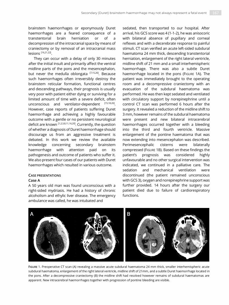
167 Secondary (Duret) brainstem haemorrhage may not always represent a fatal event
brainstem haemorrhages or eponymously Duret
haemorrhages are a feared consequence of a
transtentorial brain herniation or of a
decompression of the intracranial space by means of
craniectomy or by removal of an intracranial mass
lesions [16,21,22].
They can occur with a delay of only 30 minutes
after the initial insult and primarily affect the ventral
midline parts of the pons and the mesencephalon,
but never the medulla oblongata [7,15,26]. Because
such haemorrhages often irreversibly destroy the
brainstem reticular formation, functional centres
and descending pathways, their prognosis is usually
very poor with patient either dying or surviving for a
limited amount of time with a severe deficit, often
unconscious and ventilator-dependent [15,18,26].
However, case reports of patients suffering Duret
haemorrhage and achieving a highly favourable
outcome with a gentle or no persistent neurological
deficit are known [1,2,3,8,11,16,20]. Currently, the question
of whether a diagnosis of Duret haemorrhage should
discourage us from an aggressive treatment is
debated. In this work we review the available
knowledge concerning secondary brainstem
haemorrhage with attention paid on its
pathogenesis and outcome of patients who suffer it.
We also present four cases of our patients with Duret
haemorrhages which resulted in various outcome.
CASE PRESENTATIONS
Case A
A 50 years old man was found unconscious with a
right-sided mydriasis. He had a history of chronic
alcoholism and ethylic liver disease. The emergency
ambulance was called, he was intubated and
sedated, then transported to our hospital. After
arrival, his GCS score was 4 (1-1-2), he was anisocoric
with bilateral absence of pupillary and corneal
reflexes and with a decerebrate response to painful
stimuli. CT scan verified an acute left-sided subdural
haematoma 24 mm thick, descending transtentorial
herniation, enlargement of the right lateral ventricle,
midline shift of 21 mm and a small interhemispheric
haemorrhage. There was also a subtle Duret
haemorrhage located in the pons (FIGURE 1A). The
patient was immediately brought to the operating
room and a decompressive craniectomy with an
evacuation of the subdural haematoma was
performed. He was then kept sedated and ventilated
with circulatory support by norepinephrine until a
control CT scan was performed 6 hours after the
surgery. It revealed a reduction of the midline shift to
3 mm, however remains of the subdural haematoma
were present and new bilateral intracerebral
haemorrhages occurred together with a bleeding
into the third and fourth ventricle. Massive
enlargement of the pontine haematoma that was
now extending into mesencephalon was described.
Perimesencephalic cisterns were bilaterally
compressed (FIGURE 1B). Based on these findings the
patient’s prognosis was considered highly
unfavourable and no other surgical intervention was
indicated, we continued in a palliative care. The
sedation and mechanical ventilation were
discontinued (the patient remained unconscious
with GCS 3), oxygen and norepinephrine support was
further provided. 14 hours after the surgery our
patient died due to failure of cardiorespiratory
functions.
FIGURE 1. Preoperative CT scan (A) revealing a massive acute subdural haematoma 24 mm thick, smaller interhemispheric acute
subdural haematoma, enlargement of the right lateral ventricle, midline shift of 21mm, and a subtle Duret haemorrhage located in
the pons. After a decompressive craniectomy (B) the midline shift had resolved however remains of subdural haematomas are
apparent. New intracerebral haemorrhages together with progression of pontine bleeding are visible.

168 Martin Hanko, Branislav Kolarovszki, Kristián Varga et al.
Case B
This 64 years old man with a history of arterial
hypertension, liver steatosis, lung resection due to
tuberculosis and chronic obstructive pulmonary
disease underwent a right-sided decompressive
craniectomy with an evacuation of an acute
posttraumatic haemorrhage into a chronic subdural
haematoma. Two months later he was admitted
again for a planned cranioplastic surgery usong an
autologous bone flap. At that time, he was fully
conscious, well-oriented and communicating with a
moderate left-sided hemiparesis persistent since the
event of his traumatic brain injury. He underwent the
cranioplasty without any serious complications and
without any changes in his neurological status
immediately after surgery. Approximately 8 hours
later the patient had an epileptic seizure that
terminated after a 5 mg intravenous dose of
diazepam, he however remained non-responsive, his
right-sided pupil was enlarged. The attending
neurosurgeon indicated an immediate CT scan
which revealed an epidural haematoma 47 mm thick
at the site of reimplanted bone flap, midline shift of
19 mm, compression of lateral ventricles and
perimesencephalic cisterns and a transtentorial
herniation of the right-sided temporal lobe. The
patient was transported to the operating room with
pupils already bilaterally mydriatic and non-reactive.
Emergent evacuation of the epidural haematoma
was performed and the bone flap was returned with
drainage tubes placed into the epidural space. The
patient remained sedated and ventilated until the
next CT scan 8 hours later which revealed a recent
haemorrhage into right-sided basal ganglia, all four
ventricles and a Duret haemorrhage into the upper
pons and mesencephalon. 24 hours later the
sedation and mechanical ventilation were
discontinued and a tracheostomy was performed. At
that time the patient was unconscious with GCS 3, he
had irregular pupils with a slight right-sided
mydriasis, absent pupillary reflex and present
corneal reflex, spontaneously ventilating. He was
later transferred to a nursing home and remained in
persistent coma with his neurological status
unimproved (as reported from his last known clinical
check 2 months after the surgery).
Case C
A 63 years old woman pedestrian was struck by a car
and subsequently transported to a peripheral
hospital, unconscious with GCS 7 (1-1-5), present
pupillary reflex, sedated and intubated. CT scan was
performed revealing a skull base fracture,
pneumocephalus, traumatic subarachnoid
haemorrhage, small subdural haematoma over the
right hemisphere and a small haemorrhagic
contusion in right temporal lobe. She also suffered
numerous smaller lung contusions. 24 hours later
she was transferred to our institution due to a
progressive oedema of the right hemisphere with an
enlargement of the haemorrhagic contusion in the
right temporal lobe. Decompressive craniectomy
and evacuation of the haemorrhagic contusion was
subsequently performed and the patient remained
sedated for following 24 hours. A control CT scan
revealed two subtle Duret haemorrhages (one
located in the pons and one in the mesencephalon)
both with a diameter of approximately 1 cm (FIGURE
2). No further surgical intervention was indicated.
Two months later she underwent autologous bone
flap replantation and a ventriculo-peritoneal shunt
insertion due to development of a posttraumatic
hydrocephalus. After the surgery she remained
conscious with GCS 14 (4-4-6), disoriented with signs
of organic psychosyndrome - slightly bradypsychic,
occasionally agitated and verbally aggressive. Her
only persistent motoric deficit was a right sided
ptosis.
FIGURE 2. Postoperative CT scan verifying basally located
remains of haemorrhagic contents and a subtle Duret
haemorrhage in the pons.

169 Secondary (Duret) brainstem haemorrhage may not always represent a fatal event
Case D
A 27 years old patient who suffered a motorcycle
accident was immediately sedated and transferred
to our clinic due to a CT verified posttraumatic
cerebral oedema, subarachnoid haemorrhage, non-
expansive right-sided acute subdural haematoma,
intracerebral haematomas in both frontal lobes and
a small Duret haemorrhage located in his ventral
mesencephalon. Prior to sedation he had GCS 8 (1-2-
5), he was isocoric with pupillary reflex bilaterally
preserved. Due to a presence of non-expansive
haemorrhagic intracranial lesions only an
intraparenchymal intracranial pressure sensor was
initially inserted but 12 hours later the patient
developed a refractory intracranial hypertension
with pressure values exceeding 40 mmHg and a
bilateral decompressive craniectomy was
performed. However, because of a CT verified Duret
haemorrhage the patient’s prognosis was
considered unfavourable. The postoperative CT
revealed stabilized intracranial findings with an
adequate decompressive effect of the bilateral
craniectomy with the brainstem haemorrhage being
stable and non-expanding. The sedation was
discontinued and the patient was later discharged to
a peripheral hospital surprisingly achieving a highly
favourable outcome: GCS 15 and a slight right sided
hemiparesis and facial nerve palsy.
DISCUSSION
Henri Duret (1849-1921), a French surgeon and a
pioneer of neuroscience focused his aim in
neurological research on localising of functional
areas in the brain, describing the vascular supply of
the central nervous system and investigating
pathomechanisms of traumatic brain injuries [27]. In
experimental conditions he had described
mechanisms of intracranial hypertension by
simulating intracranial mass lesions and correctly
assumed brainstem (especially medulla oblongata)
as a centre of cardiorespiratory functions. He had
also noted a disturbance in the cerebral tissue
perfusion during periods of increased intracranial
pressure and had observed microscopic
haemorrhages located in the floor of the fourth
ventricle which were associated with episodes of
increased intracranial pressure [6,27]. Despite of
having inaccurately linked these bleedings to an
increased cerebrospinal fluid pressure at the
moment of primary brain injury and despite a fact
that these haemorrhages might not even be true
secondary brainstem bleedings, Duret haemor-
rhages have remained an eponymous term for
secondary brainstem haemorrhagic lesions caused
mostly by intracranial hypertension and following
the transtentorial herniation [21,27].
Causes of Duret haemorrhage
Duret haemorrhage is typically a result of a
descending transtentorial brain herniation. Cases of
a brainstem haemorrhage preceded by an
occurrence of an intracerebral haematoma [19,21],
acute subdural haematoma [1,10,17,20,21], cerebral
infarction [19] or diffuse cerebral oedema caused by
hyponatremia [11] are known. On the other hand,
several cases of the Duret haemorrhage following an
episode of intracranial hypotension which can also
provoke a descending transtentorial herniation have
been reported. Cardinale et al. have published a case
of the Duret haemorrhage in a patient after a
decompressive craniectomy complicated by an
episode of intracranial hypotension and a
paradoxical brain herniation presenting as the
sinking skin flap syndrome [4]. Similarly, Yuan et al.
have described a case of the Duret haemorrhage in
a patient with lumbar drainage following an
operation of a thalamic tumour [28]. There is also a
report of a patient who suffered a brainstem
haemorrhage caused by a postoperative lumbar
pseudomeningocele and associated intracranial
hypotension [3]. Interestingly, even a rapid
decompression of the intracranial space by means of
evacuation of an expansive intracranial
haemorrhage [2,8,13,22] or by means of a
decompressive craniectomy [16,17,25] can result in the
Duret bleeding.
Incidence
In their autoptic research of haemorrhagic stroke
Nedergaard et al. have observed an incidence of the
Duret haemorrhage in 45% of intracerebral
haemorrhages, 15% of cerebral infarctions and 36%
of aneurysm haemorrhages [19]. In a clinico-
pathological report of Klintworth, Duret
haemorrhage was typically associated with
intracranial lesions complicated by a rapidly
expanding intracranial haemorrhage or brain
oedema or with a surgical decompression of
intracranial space even in slowly growing lesions with
overall incidence of 16.5%, specifically 31.5% for

170 Martin Hanko, Branislav Kolarovszki, Kristián Varga et al.
intracerebral haemorrhages, 21.5% for subdural
haematomas and 11.6% for cerebral infarctions [13].
In a non-missile brain injury, Graham et al. have
reported an incidence of the Duret haemorrhage in
51% of autopsies [9]. However, almost 20% of these
haemorrhages could only be discovered during
microscopic examination [9] and this fact leads us to
expect that radiological examination will
underestimate the incidence of secondary brainstem
haemorrhages in surviving patients.
Pathogenesis
Unlike primary brainstem haemorrhage which could
be a result of a spontaneous arterial rupture, direct
traumatic injury to the brainstem or of a rupture of
vascular malformation, secondary brainstem
haemorrhage typically presents with a delay and is
mostly a consequence of the descending
transtentorial herniation or follows a rapid
decompression of the intracranial space [16,21]. There
are several main explanations for pathogenesis of
the Duret haemorrhage. The theory of an arterial
origin of the Duret haemorrhage assumes that a
descending transtentorial herniation causes a
displacement and angulation of the brainstem with
an elongation and straining of the perforating
arteries extending from the basilar artery which is
fixed in its upper end by the posterior
communicating arteries and the posterior cerebral
arteries. This tension results in either vasospasm or
rupture of the perforating arteries and causes the
Duret haemorrhage [12,15,21,26]. The very appearance
of the Duret haemorrhage is highly dependent on
the volume and rate of expansion of the underlying
intracranial mass lesions and also on variations of
systemic blood pressure and blood inflow into the
perforating arteries during the episodes of
transtentorial herniation. Slowly expanding
intracranial mass lesions do not usually cause the
Duret haemorrhage [7,12]. An extremely rapid
descending transtentorial herniation causing acute
brainstem ischaemia results in early death of the
patient without an episode of the secondary
brainstem bleeding (which is prevented by the
disturbed blood flow in perforating arteries) [12].
Based on experimental research on animal model,
Klintworth has postulated that the Duret
haemorrhage arises during two possible scenarios:
The transtentorial herniation can extensively
displace the perforating branches of the basilar
artery. If sufficient blood flow in these vessels is
retained, they are prone to rupture and cause
secondary brainstem haemorrhage [12]. Evacuation
of the intracranial mass lesion causing transtentorial
herniation and perforating vessel damage (mechanic
or ischaemic) can also provoke Duret haemorrhage
in a situation when the blood flow into these
damaged arteries is restored causing them to bleed
even more extensively than during transtentorial
herniation only, especially in situation of elevated
systemic blood pressure [12,14,15,24]. It is possible that
all types of blood vessels in the ventral brainstem are
damaged in a manner of an ischaemia-reperfusion
injury and are later prone to cause a massive
secondary brainstem haemorrhage [14]. The theory of
reperfusion injury is supported by a report of Sim et
al. who have observed an enhancement of ventral
pontomesencephalic area on CT angiography in a
patient with a transtentorial herniation due to
bilateral chronic subdural haematoma and acute
tentorial subdural haematoma. After a burr-hole
drainage of chronic haematomas was performed, a
massive Duret haemorrhage in the area of previous
parenchymal enhancement occurred. The contrast
extravasation probably demonstrated damaged
perforating arteries [24]. Arterial origin of Duret
haemorrhage is also supposed when based on a
report by Chew et al. who have performed a post-
mortem CT angiography on a patient who died due
to a middle cerebral artery aneurysm rupture
associated with a transtentorial herniation and a
secondary brainstem haemorrhage. Multiple linear
enhancing foci corresponding to ruptured
paramedian perforating arteries found in autopsy
were detected in the central pons [5]. Theory of a
venous infarction arises from an observation of
venous congestion in the brainstem in patients with
a supratentorial mass lesions, perivenous
localisation of several Duret haemorrhages and a
fact that the veins are thin-walled vessels easily
compressed by an external pressure, specifically at
the site of the tentorial notch. Compression can
cause the veins to thrombose and result in venous
brainstem haemorrhages [12,15,21,23]. This explanation
has been however questioned because secondary
brainstem haemorrhages often extend anatomical
regions of the venous drainage and the evacuation
of intracranial mass lesions can rather provoke Duret
haemorrhage than prevent it [12]. Observations of a
brainstem ischaemia with no secondary

171 Secondary (Duret) brainstem haemorrhage may not always represent a fatal event
haemorrhage in patients with the transtentorial
herniation also challenges this theory of
pathogenesis [26].
Favourable outcome of patients suffering a Duret
haemorrhage
Despite being considered a predictor of infaust
prognosis, several reports describing a favourable
outcome of patients with the Duret haemorrhage
caused by various types of intracranial pathologies
have been published. Dramatic recovery has been
reported by Ishizaka et al.: A patient with an
idiopathic subdural haematoma and preoperatively
described Duret haemorrhage underwent
evacuation of the subdural haematoma and despite
of a severe preoperative neurological deficit his
status resolved to a persistent oculomotor palsy only [10]. Similarly, Nguyen et al. have described a patient
with an acute subdural haematoma and a Duret
haemorrhage achieving a favourable outcome
(dysarthria and no motor deficit) after an evacuation
of a subdural bleeding [20]. Stiver et al. have also
reported a favourable outcome of a young woman
who developed a Duret haemorrhage after an
evacuation of an epidural haematoma by means of
decompressive craniectomy [25] and another case of
favourable outcome after an evacuation an epidural
haematoma in a patient with a Duret haemorrhage
has been published by Fujimoto et al. [8]. A report by
Park et al. describes a patient who suffered a Duret
haemorrhage after a burr-hole drainage of bilateral
chronic subdural haematomas and survived with his
only persistent deficit being a gentle gait disturbance [22]. Lonjaret et al. have published a case report of
patient with a Duret haemorrhage triggered by a
decompressive craniectomy for an acute subdural
haematoma resulting in a favourable outcome with
no neurological deficit [16]. Kamijo et al. have reported
a favourable outcome (gentle hemiparesis) in a
patient with a severe hyponatremia causing a
transtentorial herniation and a Duret haemorrhage [11]. A favourable outcome with persistent
internuclear ophtalmoplegia only in a patient with a
Duret haemorrhage due to an intracranial
hypotension has been published by Bonow et al. [3].
In paediatric patients, Beier and Dirks have reported
a favourable outcome in two patients with Duret
haemorrhage preceded by a traumatic brain injury [1]. When summarized, most of patients surviving
with favourable outcome have suffered a brainstem
haemorrhage of a limited extent only [1,2,3,8,20],
however exceptions exist [11,16,22]. Younger age of
these patients could be a factor predicting a
favourable outcome [1,11,16,20]. A typically reported
persisting neurological deficit in patients with the
favourable outcome is often a cranial nerve
weakness [1,3,8,10,11,16], or various subtle forms of the
organic psychosyndrome and a cognitive deficit [1,25].
Analysis of our case series
The first three patients (A,B,C) described in our work
represent cases of Duret haemorrhage provoked or
worsened by a rapid decompression of intracranial
space combined with a decompressive craniectomy
in 2 of 3 patients. The explanation of this
phenomenon could be based on the theory of
reperfusion into the damaged pontine perforating
vessels as initially postulated by Klintworth [12,13,14,24].
Alleviation of a significant pressure on the brainstem
had most likely increased the blood inflow into
mechanically and metabolically injured vessels in the
pontomesencephalic region and resulted in the
secondary brainstem haemorrhage (or its significant
expansion in one case). The patient D probably
suffered a Duret haemorrhage due to a massive
cerebral oedema with a mass effect that displaced
the brainstem and distorted the mesencephalic
perforating arteries thus causing them to rupture
because of a combination of metabolic and
mechanic damage [12,15,21,26]. It is possible that due to
patient’s younger age and absence of complicating
diagnoses the perforators remained relatively stable
even after the alleviation of the intracranial
hypertension by means of decompressive
craniectomy and the secondary brainstem
haemorrhage did not expand. Two of our four
patients had achieved a good outcome and this was
probably caused by a smaller extent of the pontine
haemorrhage and a younger age of patient D.
Chronic liver disease present in two patients with
unfavourable outcome could play a role as a
potential negative prognostic factor as it can
negatively affect the function of the blood
coagulating system, however a definite conclusion
can only be made when relating to a larger group of
patients.
CONCLUSIONS
Massive elevation of intracranial pressure,
decompression of the intracranial space by means of

172 Martin Hanko, Branislav Kolarovszki, Kristián Varga et al.
craniectomy or intracranial bleeding evacuation can
make the patient susceptible to the secondary
brainstem (Duret) haemorrhage. Such bleeding,
typically localised in the midbrain and pons has been
considered a significant predictor of poor prognosis.
Despite of this, outcome of patients suffering the
Duret haemorrhage can considerably vary as
presented in our report. When deciding about the
prognosis of a patient with the Duret haemorrhage,
attention should be paid to the type and severity of
the underlying brain injury, patient’s clinical status
and history and at last but not least to the extent of
the very brainstem bleeding.
ABBREVIATIONS
CT: computed tomography
GCS : Glasgow Coma Scale
REFERENCES
1. Beier AD, Dirks PB. Pediatric brainstem hemorrhages after
traumatic brain injury. J Neurosurg Pediatrics. 2014; 14(4),
421-4.
2. Bermudéz G, Areitio E. Duret haemorrhage with full
recovery. Acta Neurochir (Wien). 2015; 157(8), 1337-8.
3. Bonow R, Bales JW, Morton RP, Levitt MR, Zhang F.
Reversible coma and Duret haemorrhage after intracranial
hypotension from remote lumbar spine surgery: case
report. J Neurosurg Spine. 2016; 24(3): 389-93.
4. Cardinale M, Meaudre E, Cunqi PQ, Nguyen C, Esnault P,
Joubert C. Duret hemorrhage complicating a sinking skin
flap syndrome. Br J Neurosurg. 2017; Sep 8,1-2.
5. Chew KL, Baber Y, Iles L, O’Donnell C. Duret hemorrhage:
demonstration of ruptured paramedian pontine branches
of the basilar artery of minimally invasive, whole body post-
mortem CT angiography. Forensic Sci Med Pathol. 2012;
8(4), 436-40.
6. Duret H. Études Expérimentales et Cliniques sur les
Traumatismes Craniocérébraux. Paris: Thése; 1878.
7. Fields WS, Halpert B. Pontine Hemorrhage in Intracranial
Hypertension. Am J Pathol. 1953; 29(4), 677-87.
8. Fujimoto Y, Aquiar PH, Freitar AB, de Andrade AF, Marino
Júnior R. Recovery from Duret Hemorrhage: A Rare
Complication after Craniotomy. Neurol Med Chir (Tokyo).
2000; 40(10), 508-10.
9. Graham DI, Lawrence AE, Adams JH, Doyle D, McLellan DR.
Brain damage in non-missile head injury secondary to high
intracranial pressure. Neuropathol Appl Neurobiol. 1987;
13(3), 209-17.
10. Ishizaka S, Shimizu T, Ryu N. Dramatic recovery after severe
descending transtentorial herniation-induced Duret
haemorrhage: A case report and review of literature. Brain
Inj. 2014; 28(3), 374-7.
11. Kamijo Y, Kishita R, Hamanaka S, Soma K. Duret
haemorrhage is not always suggestive of poor prognosis: a
case of acute severe hyponatremia. Am J Emerg Med. 2005;
23(7), 908-10.
12. Klintworth GK. The pathogenesis of Secondary Brainstem
Hemorrhages as Studied in an Experimental Model. Am J
Pathol. 1965; 47(4), 525-36.
13. Klintworth GK. Evaluation of the role of neurosurgical
procedures in the pathogenesis of secondary brain-stem
hemorrhages. J Neurol Neurosurg Psychiatry. 1966; 29(5),
423-5.
14. Klintworth GK. Paratentorial Groowing of Human Brains
with Particulatr Reference to Transtentorial Herniation and
the Pathogenesis of Secondary Brain-stem Hemorrhages.
Am J Pathol. 1968; 53(3) 391-408.
15. Leestma JE. Forensic Neuropathology. Boca Raton:
Taylor&Francis; 2014.
16. Lonjaret L, Ros M, Boetto S, Fourcade O, Geeraerts T.
Brainstem haemorrhage following decompressive
craniectomy. J Clin Neurosci. 2012; 19(9), 1293-5.
17. Martins WA, Teixeira AB, Frigeri TM, Paglioli E. Spontaneous
subdural hematoma associated to Duret haemorrhage.
Interdisciplinary Neurosurgery. 2015; 2(1), 13-15.
18. Moore AS, Mittal MK, Rabinstein AA. Duret Hemorrhage
Following Subdural Hematoma Evacuation. JAMA Neurol.
2013; 70(4), 518.
19. Nedergaard M, Klinken L, Paulson OB. Secondary Brain
Stem Hemorrhage in Stroke. Stroke. 1983; 14(4), 501-5.
20. Nguyen HS, Doan MB, Gelsomino MJ, Shabani S, Mueller
WM. Good outcomes in a patient with a Duret
haemorrhage from an acute subdural hematoma. Int Med
Case Rep J. 2016; 9, 15-18.
21. Parizel PM, Makkat S, Jorens PG, Ozsarlak O, Cras P, Van
Goethem JW, et al. Brainstem hemorrhage in descending
transtentorial herniation (Duret haemorrhage). Intensive
Care Med. 2002; 28(1), 85-8.
22. Park KJ, Kang SH, Lee HK, Chung YG. Brain Stem
Hemorrhage Following Burr Hole Drainage for Chronic
Subdural Hematoma. Neurol Med Chir (Tokyo). 2009;
49(12), 594-7.
23. Scheinker IM. Transtentorial herniation of the brain stem.
A characteristic clinicopathologic syndrome; pathogenesis
of hemorrhages in the brain stem. Arch NeurPsych. 1945;
53(4), 289-298.
24. Sim KB, Na DG, Park JK. Precedence of Parenchymal
Enhancement on CT to a Fatal Duret Hemorrhage. J Korean
Neurosurg Soc. 2013; 53(6), 380-2.
25. Stiver SI, Gean AD, Manley GT. Survival with good outcome
after cerebral herniation and Duret hemorrhage caused by
traumatic brain injury. J Neurosurg. 2009; 110(6): 1242-6.

173 Secondary (Duret) brainstem haemorrhage may not always represent a fatal event
26. Tomliston BE, Brain-stem lesions after head injury. J Clin
Pathol Suppl (R Coll Pathol). 1970; 4, 154-65.
27. Walusinski O, Cou.rivaud P. Henri Duret (1849-1921): A
Surgeon and Forgotten Neurologist. Eur Neurol. 2014;
72(3-4), 193-202.
28. Yuan X, Zhao H, Zhao C, Zhao H, Xiaho H, Sun G, et al. Duret
Hemorrhage after Lumbar Drainage. J Craniofac Surg.
2014; 25(4), 1551-2.

Romanian Neurosurgery (2019) XXXIII (2): pp. 174-177 DOI: 10.33962/roneuro-2019-032 www.journals.lapub.co.uk/index.php/roneurosurgery
Spinal extradural meningioma en plaque with nerve root attachment and extracanal (intrathoracic) extension. Review of literature on management and case report
Morgan E.1, Hakkou M.2, Mellaoui A.2, Poluyi E1,
El Ouahabi A.2
1 Lagos University Teaching Hospital, Idi-Araba, Lagos, NIGERIA 2 Faculte de Medicine et de Pharmacie, Mohamed V. University,
Souissi Rabat, MOROCCO
ABSTRACT Meningiomas are relatively common primary spinal tumours, being the second most
common intraspinal tumours probably after vertebral haemangioma.
It constitutes about 25% of all intraspinal tumours; however, in the presence of
extradural spinal lesions, the diagnosis of meningioma is uncommon and often not
among the first two considerations. Purely extradural spinal meningioma, especially
of the “En plaque” variety, usually simulate malignant disease (metastatic diseases
and lymphoma) and may result in inadequate therapy, however, the presence of
nerve root attachment is even rarer.
Our case report is that of an entirely cervicothoracic extradural en‑plaque
meningioma (WHO grade 1) with a nerve root attachment (right C7) and intrathoracic
extension. We highlighted the issues in diagnosis, operative intervention and long-
term follow-up.
INTRODUCTION
Meningiomas account for about a quarter of all intraspinal tumours
and literature has shown that it is the second commonest primary
intraspinal tumour [1]. The majority of spinal meningiomas are entirely
intradural and this is seen in about 10% of cases.
There is an extradural spinal extension, but attachment to spinal
nerve root and/or intrathoracic extension are not a frequent finding [2].
Meningiomas located purely in the extradural space are very rare and,
therefore, are not often listed in the differential diagnosis of spinal
extradural lesions [2].
We present a case report of purely extradural meningioma “en
plaque” of the spine that was noticed to be attached to right C7 nerve
root which create a bias for preoperative diagnosis of spinal
schwannoma (intradural extramedullary) with extradural extension.
Keywords spinal extradural
meningioma, intrathoracic extension,
case report
Corresponding author Morgan Eghosa
Lagos University Teaching Hospital,
Idi-Araba Lagos, Nigeria
Scan to access the online version

175 Spinal extradural meningioma en plaque with nerve root attachment and extracanal (intrathoracic) extension
CASE REPORT
We report a case of a 28-year old woman with no
previous significant medical history presented with
2-month history of progressive spastic tetraparesis
(with muscle power in most muscle groups being 4)
associates with right brachio-cervical hyperesthesia
involving C6/C7 distribution. Cervico-thoracic
magnetic resonance imaging (MRI) done was
reported as intradural extramedullary tumour
(schwannoma) extending from C4-T1 vertebral level
by the radiologist with attachment to right C7 nerve
root. Lesion was iso-hypointense on TIWI and
enhances on T2WI as shown in IMAGE 1 and IMAGE 2
perioperatively, the tumour was accessed via C4 –T1
laminectomy, and it was entirely extradural
extending from C4-T1 with right C7 nerve root
attachment and extending to the intrathoracic
region anteriorly as shown in intra-operative images
1 and 2. The tumour was firm, fibrous, highly
vascular and dissectible, tumour resected with
coagulation of the attachment to right C7 nerve root
but the intrathoracic component left in-situ.
Both frozen section and definitive
histopathological/immunohistochemical analyses
confirmed mengioma “en plaque” (WHO grade 1) as
shown in histology slides below.

176 Morgan E., Hakkou M., Mellaoui A et al.
Post operatively, there was an initial loss of sensory
modality of right C7 nerve root distribution which
improved subsequently in conjunction with motor
functions (preoperative tetraplegia) after six (6)
weeks of function education and rehabilitation.
Follow up is been done regularly using both clinical
and radiological tools and there has not been change
in improvement achieved postoperatively and also
the size of the intrathoracic component.
DISCUSSION
Meningioma is one of the commonest central
nervous system (CNS) tumours which is essentially a
benign tumour. Majority are intracranial, with about
90% supratentorial, but spinal meningioma account
for between 1.2%-12,5% of all meningiomas [3,4,5.
Meningioma arises from arachnoid cap ceil
(meningiothelial cell in the arachnoid villi just like any
meningioma elsewhere).
The prevalence of extradural meningioma in
literature ranges from 3.3% and 21.4% of all cases of
spinal meningioma [6].
Tumours occur more in female to male of ratio
4:1 with a peak incidence between 5thand 6th
decades of life. Almost about 80% occur in the
thoracic spinal region [6]. Majority of these lesions
are intradural extramedullary and are placed ventral
or ventrolaterally to the dura, However, about 10%
do extend to the extradural space [2]. The presence
of a “sole” extradural spine meningioma is entirely
rare [2]. Common tumours located in the spinal
extradural space are metastatic spinal tumour and
lymphoma, hence the dilemma in preoperative
diagnosis which was also emphasis by Saryedekar et
al in the two cases reported by them [7].
Our patient reported above had purely
extradural spinal “en plaque” meningioma with right
C7 nerve root attachment and intrathoracic
extension. The occurrence of “en plaque” variety of
meningioma is rarer. Moreso, the presence of nerve
root attachment with or without intrathoracic
extension is even much more a rarity. Only 16 cases
in literature have shown extradural spinal
meningioma adhering/proximity to spinal nerve
root.
Tuli et al in their case report of a 42-year old lady
with T4-T6 extradural spinal meningioma revealed
attachment to left T5 spinal nerve root. The presence
of extradural meningioma with or without nerve root
attachment remain a puzzle and been described as a
paradox in view of absence of arachnoid cap cell in
the extradural space.
Several theories have been ascribed to the
occurrence of an entirely extradural spinal
meningioma and possibility of nerve root adherence.
Reasons adduced for these in literatures include
migration of arachnoid tissue into the extradural
space, or extradural spinal meningioma could arise
from ectopic or separated arachnoid tissue around
the periradicular nerve root sleeve, which is the point
of contact of the spinal leptomeninges directly into
the dura. This probably explains the attachment to
nerve root [2].
Another reason alluded to the occurrence of
spinal extradural meningioma is that the
periradicular dura which is said to be less thick, may
have vestigial remnants of the superficial layer of the
embryonal arachnoid mater and villi [6]
Also, it has been suggested that islands of
arachnoid tissue that may have migrated into the
extradural space can be the source of the
meningioma [2,6].
An entirely spinal extradural meningioma
especially of the ‘en plaque; variety poses a dilemma
in pre-operative diagnosis [2, 7]. In this index case, a
pre-operative diagnosis of C4-T1 schwannoma of the
spine in view of its location and C7 nerve root
attachment.
Intraoperative frozen section histopathological
analysis is a necessity in view of other possibility such
as metastatic spine tumour and lymphoma [8]. In
this index case, the frozen section was done which
showed a meningioma en plaque. This was done for
decision making as regards extend of tumour
resection.
The extradural tumour with its attachment to the
right C7 nerve root was resected and point of C7
nerve root adhesion was coagulated. However, the
thoracic extension was left in-situ. Saryedekar et al in
their work shows the importance of intraoperative
frozen section with near total resection of spinal
extradural meningioma en plaque [7].
This strategy will give the best postoperative
outcome, as prognosis depends on the extent of
tumour resection [8],
Another consideration in the operation is to
decide whether to open the dura or not. However, in
consideration of the pathogenesis of the lesion from
the dura nerve root sleeve and not from the external
part of the spinal dura. It may be necessary to only

177 Spinal extradural meningioma en plaque with nerve root attachment and extracanal (intrathoracic) extension
peeled off the tumour from the dura. Saryadekar et
al in the two cases reported shows that the tumour
was stripped off the dura with no durotomy done [7].
Though there is essentially no consensus on whether
the dura should be excised or not, However, it is
necessary that the dura be opened to rule out
intradural extension of the meningioma which is
seen in literature to account for about 10% of cases
[2].
Tuli et al. reported 12 cases out of 47 supposedly
pure extradural spinal meningioma who had
durotomy, three (3) of these 12 patients was noticed
to have intradural extension. This buttresses the
importance of durotomy in hitherto purely
extradural spinal meningioma. We had to open the
dura to be very sure that there was not intradural
component of the tumour as done in standard
procedure [6].
Issues relating to long term outcome still remain
an item of debate. Issues been debated such as gross
total resection versus incomplete resection, benign
versus malignant meningioma remain an important
factor in considering long-term outcome following
operation for extradural spinal meningioma.
Literature has revealed that this tumour may
have a local malignant potential despite been a
relatively benign tumour. Also, worse prognosis is
seen with incompletely tumour resection due to
bony involvement or paraspinal extension [2, 3, 8]. In
the index case been reviewed, all except the thoracic
extension was excised. Tumour has remained static
after a regular follow-up period of over 2 years.
CONCLUSION
Spinal extradural meningioma is a rare tumour, even
more with the ‘en plaque’ variety with spinal nerve
root attachment. In view of the prevalence of
between 3.3% to 21.4%, it is essential that cases of
extradural spinal meningioma should be entertained
in conjunction with common tumours such as
metastatic spine disease and lymphoma and in
doubtful cases Intraoperative frozen section and
durotomy are essential in operative decision making
either to do gross total resection or partial. For the
incompletely resected tumours, long-term follow-up
period using both clinical and radiological
monitoring tools is essential.
REFERENCES
1. Santiago BM, Rodeia P, Cunha E Sa M. Extradural thoracic
spinal meningioma. Neurol India 2009; 57:98.
2. Zevgaridis D, Thomé C. Purely epidural spinal meningioma
mimicking metastatic tumor: Case report and review of
literature. Spine (Phila Pa 1976) 2002;27: E403‑5.
3. Osborn AG: Diagnostic Neuroradiology. St. Louis: Mosby;
1994.
4. Solero CL, Fornari M, Giombini S, Lasio G, Oliveri G, Cimino
C, Pluchino F: Spinal meningiomas: review of 174 operated
cases. Neurosurgery 25:153-160,1989.
5. Yamashita J, Handa H, Iwaki K, Abe M: Recurrence of
intracranial meningiomas, with special reference to
radiotherapy. Surg Neurol 14:33-40, 1980.
6. Tuli J, Drzymalski DM, Lidov H, Tuli S. Extradural en‑plaque
spinal meningioma with intraneural invasion. World
Neurosurg 2012;77: 202.e5‑13.
7. Savardekar A, Chatterjee D, Chatterjee D, Dhandapani S,
Mohindra S, Salunke P. Totally extradural spinal en plaque
meningiomas - Diagnostic dilemmas and treatment
strategies. Surg Neurol Int 2014;5: S291-4.
8. Frank BL, Harrop JS, Hanna A, Ratliff J. Cervical extradural
meningioma: Case report and literature review. J Spinal
Cord Med 2008; 31:302‑5.
Romanian Neurosurgery (2019) XXXIII (2): pp. 178-182 DOI: 10.33962/roneuro-2019-033 www.journals.lapub.co.uk/index.php/roneurosurgery
The evaluation of long-term screw pull-out rates following posterior thoracolumbar fusion surgery with short and thin pedicle screws
Umit Kocaman1, Hakan Yilmaz2
1 Izmir Cigli Region Education and Research Hospital, Department of
Neurosurgery, Izmir, TURKEY 2 University of Healt Sciences Izmir Bozyaka Education and Research
Hospital, Department of Neurosurgery, Izmir, TURKEY
ABSTRACT Background. The aim of this study was to evaluate screw pull-out rates after fusion
operations with short and thin pedicle screws.
Methods. A total of 200 posterior lumbar and thoracolumbar fusion operations
performed at our clinic with short and thin pedicle screws (5.5x35 mm) were
retrospectively evaluated. The patients were assessed with computed tomography
postoperatively on the day of surgery and at the 6th month. Single groove retraction
of the transpedicular screw was evaluated as pull-out. The results were evaluated by
the 'number of pull-out cases / total number of cases' and also the 'total number of
pull-out screws / total number of screws used' ratios.
Results. There were 112 (56%) female and 88 (44%) male patients with a mean age of
58 years. The total number of screws used in the 200 cases was 1188. There were 88
(7.4%) thoracic pedicle screws, 1056 (88.9%) lumbar pedicle screws and 44 (3.7%)
sacral pedicle screws used. No pull-out was found in the control CTs taken
postoperatively. Left side T11 and T12 pull-out was observed in one case and left L4
pull-out was observed in another case in the control CTs taken at the postoperative
6th month. Pull-out was observed in 2 (1%) of the 200 cases and 3 (0.25%) of the 1188
screws.
Conclusions. All the short and thin pedicle screws used had passed the pedicle length
and neurocentral junction. The use of a 5.5x35 mm screws in fusion operations is less
invasive than using longer and thicker screws while the pull-out rates may be similar.
INTRODUCTION
Posterior lumbar and thoracolumbar fusion operations constitute an
important part of neurosurgery practice. The major problems after
these operations are screw pull-out, adjacent segment degeneration,
deterioration of instrumentation integrity, and fusion insufficiency.
Various preventive measures such as post-cement screw application
and porous coated screw application are rarely used to prevent screw
pull-out, while the tendency is to use long and thick screws. The general
Keywords fusion,
instrumentation, pedicle,
screw
Corresponding author Hakan Yilmaz
University of Healt Sciences Izmir Bozyaka Education and Research
Hospital, Department of Neurosurgery, Izmir, Turkey
Scan to access the online version

179 The evaluation of long-term screw pull-out rates following posterior thoracolumbar fusion surgery
preference is to use 6.5x45 mm, 6.5x50 mm, 6.0x45
mm, 6.0x50 mm screws for the lumbar and
thoracolumbar junction. Screws 7.0 mm thick or 55
mm long are also in use for the lumbar region.
Although long and thick screw applications increase
spine stability, they also create surgical risk [2,4,12].
We investigated the pull-out rates in the cases
where we used screws that were shorter and thinner
than those generally used in thoracolumbar fusion
operations. We would like to share our experiences
on surgical safety in posterior lumbar and
thoracolumbar fusion surgeries in this regard.
METHODS
A total of 200 posterior lumbar and thoracolumbar
fusion operations performed with short and thin
pedicle screws between 2016 and 2018 at the Izmir
Cigli Regional Training Hospital's Neurosurgery
Department were evaluated retrospectively. This
evaluation was performed only in patients who
underwent posterior segmental instrumentation
and fusion with 5.5×35 mm transpedicular screws.
Cases where screws of a different size were used as
necessary and those that required revision for
postoperative screw malposition were not included
in the study. An evaluation was made with
preoperative CT in all cases and a screw length of 35
mm was used after it was deemed to be adequate to
reach the neurocentral junction. The patients were
evaluated with computed tomography post-
operatively on the day of surgery and at the 6th
month. Single groove retraction of the
transpedicular screw was evaluated as pull-out. The
results were evaluated by the 'number of pull-out
cases / number of cases' and the 'total number of
pull-out screws / total number of screws' ratios.
STATISTICAL ANALYSIS
Descriptive statistical methods were used in the
evaluation of the data. The results were expressed as
mean ± standard deviation and frequency (%).
RESULTS
The 200 patients included in the study consisted of
112 (56%) females and 88 (44%) males. The mean age
was 58 years and the age range was 22-78 years. The
primary pathology was narrow lumbar canal in 100
cases, lumbar spondylolisthesis (grade 1) in 26 cases,
degenerative type lumbar spondylolisthesis (grade 1)
in 24 cases, post-traumatic burst fracture requiring
posterior thoracolumbar fusion in 22 cases, post-
traumatic burst fracture requiring posterior lumbar
fusion in 6 cases and primary or recurrent lumbar
disc hernia with segmental instability in 22 cases
(TABLE 1). The number of screws used in the 200 cases
was 1188. These consisted of 88 (7.4%) thoracic
pedicle screws, 1056 (88.9%) lumbar pedicle screws
and 44 (3.7%) sacral pedicle screws (TABLE 2). No pull-
out was found in any patient on the control CTs taken
the same day postoperatively (FIGURE 1,2). Left T11
and T12pull-out was observed in control CTs
performed at the postoperative 6th month in 1 case
who had undergone thoracolumbar fusion (FIGURE 3).
Left L4 pull-out was observed in 1 patient who had
undergone surgery due to narrow lumbar canal.
Both patients underwent revision surgery. Pull-out
was therefore observed in 2 (1%) of the 200 cases
and 3 (%0.25) of the 1188 screws.
Pathology Female Male Total
narrow lumbar canal 59 41 100
isthmic type lumbar
spondylolisthesis
(grade 1)
13 13 26
degenerative type
lumbar
spondylolisthesis
(grade 1)
13 11 24
burst fracture
requiring
thoracolumbar fusion
12 10 22
lumbar disc herniation
with segmental
instability
12 10 22
burst fracture
requiring posterior
lumbar fusion
3 3 6
Total 112 88 200
TABLE 1. The distribution of the cases where short and thin
pedicle screws were applied according to the primary
pathology and gender.
Pathology Lumbar
pedicle
screw
Thoracic
pedicle
screw
Sacral
pedicle
screw
narrow lumbar
canal
558 0 12
isthmic type
lumbar
spondylolisthesis
(grade 1)
66 4 14

180 Umit Kocaman, Hakan Yilmaz
degenerative
type lumbar
spondylolisthesis
(grade 1)
206 6 8
burst fracture
requiring
thoracolumbar
fusion
130 78 0
lumbar disc
herniation with
segmental
instability
54 0 6
burst fracture
requiring
posterior lumbar
fusion
42 0 4
Total 1056 88 44
TABLE 2. The distribution of the screws used.
FIGURE 1,2. Spinal CT sagittal and axial images of the posterior
lumbar fusion operation where short and thin pedicle screws
(5.5 mm-35 mm) was used.
FIGURE 3. Left side T12 screw pull-out of a patient on the spinal
CT axial image who was inserted a short and thin pedicle screw.
181 The evaluation of long-term screw pull-out rates following posterior thoracolumbar fusion surgery
DISCUSSION
The mechanisms preventing pull-out are thought to
be large screw outer diameter, low screw inner
diameter, short intergroove distance, and a strong
bone structure. The most important of these has
been shown to a large screw outer diameter. It is
believed that a length that goes past the
neurocentral junction is sufficient in terms of
stability. However, pericortical and even bicortical
screw lengths that markedly go past the
neurocentral junction are used in practice [2,4].
No significant correlation was found between the
screw length and the force needed to create pull-out
in the cadaver study of Diaremes et al. Bone density
seemed to be a more important factor in their study
[5].
Karami et al. compared the force needed to
create pull-out in mid-body, pericortical and
bicortical screws in their cadaver study. The force
needed to create pull-out increased as the screw
depth increased. However, a statistically significant
difference was only found between mid-body and
bicortical screws [7].
There are also studies reporting opposite results
in the literature [5]. Our study provides the results of
the pull-out evaluation of screws with a thickness of
5.5 mm and invasion depth of 35 mm that were used
to go past the neurocentral junction.
There was no significant difference between our
pull-out rates during 6-month follow-up after 5.5x35
mm short and thin pedicle screws were inserted and
the results of long and thick pedicle screw use in the
literature [4,5,6]. It is possible that long and thick
screws will be more effective in terms of spine
stability according to Denis' three-column theory
[3,8]. However, all screws went past the pedicle
length and neurocentral junction with short and thin
pedicle screw use. The importance of short pedicle
screws in terms of surgical safety increases when it is
considered that spinal surgery makes up a significant
part of neurosurgical practice and a large number of
surgeries are performed by surgeons with various
levels of experience. The anterior, antero-lateral and
lateral areas of the pedicle screw application area are
surrounded by arterial and venous structures with
very high flow. Although rare, damage to these
structures during the screw implantation stage
carries a high mortality rate [1,9,10,11]. Most of these
surgeries are elective and concern the spine and any
mortality creates a difficult situation.
Vascular damage can occur at 3 different stages:
pedicle probe implantation, tapping, and screw
implantation [10,11]. We believe that using a 35 mm
screw at the pedicle probe placement stage will
improve surgical safety and may decrease the
probability of invading the main vascular structures
in case of lateral malposition.
According to the results of this study, we
recommend short pedicle screw use in appropriate
cases after considering the risk-benefit balance.
Every patient needs to evaluated separately.
Biomechanical studies can evaluate the stability of
short pedicle screws more clearly. Our results
indicate that the pull-out rates of short pedicle
screws that reach the neurocentral junction are
similar to those of long screws.
The critical point here is that each patient should
be evaluated according to his/her spine
characteristics and the length of the screw to be used
should be chosen so as to reach the neurocentral
junction during preoperative planning.
Using a 5.5 mm screw instead of a 6.0 / 6.5 mm
screw makes it easier to stay within the pedicle
boundaries. It decreases the risk of pedicle burst
during screw implantation and allows the use of a
thicker screw when revision is required. Our results
indicate that the pull-out rate for 5.5 mm screws is
similar to thicker screws. Specific planning may be
required for each patient in the presence of
comorbid situations such as advanced osteoporosis,
scoliotic deformity and rheumatic diseases.
CONCLUSION
The use of a 5.5x35 mm screw during posterior
lumbar and thoracolumbar instrumentation and
fusion operations is less invasive than using longer
and thicker screws and the pull-out rates may be
similar.
REFERENCES
1. Akinrinlola A, Brinster DR. Endovascular treatment of a
malpositioned screw in the thoracic aorta after anterior
spinal instrumentation: the screwed aorta. Vasc
Endovascular Surg. 2013;47:555–557.
2. Chatzistergos P. E., Magnissalis E. A., Kourkoulis S. K. A
parametric study of cylindrical pedicle screw design
implications on the pullout performance using an
experimentally validated finite-element model. Medical
Engineering & Physics.2010;32(2):145–154

182 Umit Kocaman, Hakan Yilmaz
3. Cho W, Cho SK, Wu C. The biomechanics of pediclescrew-
based instrumentation.J Bone Joint Surg Br. 2010
Aug;92(8):1061-5.
4. Daftari TK, Horton WC, Hutton WC. Correlations between
screw hole preparation, torque of insertion, and pullout
strength for spinal screws. J Spinal Disord. 1994;7:139–45
5. Diaremes P, Kokkinakis MC, Kurth AA, Kafchitsas K. The role
of insertion length of pedicle screws on their pullout
strength. A cadaveric study.Orthopaedic Proceedings
201193-B:SUPP_III, 366-366
6. Galbusera F, Volkheimer D, Reitmaier S, Berger-Roscher N,
Kienle A, WilkeHJ. Pedicle screw loosening: a clinically
relevant complication? Eur Spine J.2015May;24(5):1005-16.
doi: 10.1007/s00586-015-3768-6.
7. Karami KJ, Buckenmeyer LE, Kiapour AM, Kelkar PS, GoelVK,
DemetropoulosCK, Soo TM. Biomechanical evaluation of
the pedicle screw insertion depth effect on screw stability
under cyclic loading and subsequent pullout. J Spinal
Disord Tech. 2015 Apr;28(3):E133-9.
8. Mehmanparast H, Petit Y, Mac-Thiong JM. Comparison of
pedicle screw loosening mechanisms and the effect on
fixation strength. J Biomech Eng. 2015;137:121003
9. Mirza AK, Alvi MA, Naylor RM, Kerezoudis P, Krauss WE,
Clarke MJ, Shepherd DL, Nassr A, DeMartino RR, Bydon M.
Management of major vascular injury during pedicle screw
instrumentation of thoracolumbar spine. ClinNeurol
Neurosurg 2017;163:53-59
10. Wegener B, Birkenmaier C, Fottner A, et al. Delayed
perforation of the aorta by a thoracic pedicle screw. Eur
Spine J. 2008;17:S351–S354.
11. Zeiller SC, Lee J, Lim M, et al. Posterior thoracic segmental
pedicle screw instrumentation: evolving methods of safe
and effective placement. Neurol India. 2005;53:458–465.
12. Zhang QH, Tan SH, Chou SMM. Investigation of fixation
screw pull-out strength on human spine. Journal of
Biomechanics. 2004;37(4):479–485. doi: 10.1016/j.jbiomec
h.2003.09.005.

Romanian Neurosurgery (2019) XXXIII (2): pp. 183-187 DOI: 10.33962/roneuro-2019-034 www.journals.lapub.co.uk/index.php/roneurosurgery
Traumatic complete transection of dorsal spinal cord un-associated with spinal fracture or subluxation. Management review
Guru Dutta Satyarthee, Satyajit Panda
* Department of Neurosurgery, Neurosciences Centre, AIIMS New
Delhi, INDIA
ABSTRACT Complete transection of spinal cord is an extremely rare occurrence, and usually
associated with spinal instability, fracture or spondylolisthesis and also have
associated systemic polytrauma involving multiple organs injury. However, a
complete transaction of spinal cord unassociated with fracture or dislocation or
without abnormality on X-ray and CT scan imaging is extremely uncommon. Kalfas et
al. observed 11.2 % of cases had cord transection in the study of sixty-two cases spinal
cord-injured patients, who underwent spinal MRI, were additionally had evidence
vertebral injury. In 2010, Cha et al reported the first case of transection of cord in the
cervical region in a 34- year- adult male, as the first case in literature, which was not
associated with any vertebral body fracture or dislocation. Authors report an
interesting case, 15-year boy, who had complete transection of the spinal cord
associated with polytrauma following a motor vehicle accident, spinal X-ray and CT
scan imaging was unremarkable, however spinal MRI scan established the diagnosis.
Briefly, imaging, management and pertinent literature are reviewed.
INTRODUCTION
Acute spinal injury can present with a spectrum of pathology including
vertebral body fracture, dislocation, cord contusion, but spinal cord
transection represents very uncommon occurrence. [1-8] However
mostly such cord transection is reported with spinal column injury in
form of fracture, dislocation or spondyloptosis. However, unassociated
with fracture dislocation is uncommon. In 2010, Cha et al was first to
report a case of cervical cord transection of cord in a 34- year- male,
which was not associated with fracture or dislocation. [1,2] However, in a
detailed radiological review, Kalfas et al. reported his observation of
sixty-two spinal cord-injured patients, who underwent spinal MRI
evaluation within the first thirty six hours of sustaining spinal injury,
seven cases had evidence of anatomical cord transection with various
types of fracture and dislocations. [3] Spinal cord injury is usually
associated with severe traumatic injury involving spine and other body
Keywords complete spinal cord transection, magnetic
resonance imaging study, associated injury,
management
Corresponding author Guru Dutta Satyarthee
Department of Neurosurgery,
Neurosciences Centre, AIIMS New Delhi, India
Scan to access the online version

184 Guru Dutta Satyarthee, Satyajit Panda
organs. However, paediatric population can sustain
various grades of Spinal cord injury without evidence
of a radiographic abnormality, and acronym
SCIWORA (spinal cord Injury without radiographic
abnormality) was coined in the year 1982 by Pang
and Wilberger for describing cases sustaining spinal
cord injury and presenting with clinical symptoms of
traumatic myelopathy, however, no radiographic or
computed tomographic scan shows evidence of
spinal fracture or instability. [1] SCIWORA is
considered rare in adults, but relatively commoner in
children. In the paediatric cases, SCIWORA
represents about 8 to 19% of all paediatric spinal
cord injuries [4]. However, MRI can show presence of
in cases of SCIWORA. SCIWORA lesions have
predilection for cervical spine commonly but can
affect the thoracic or lumbar spine although much
less frequently. SCIWORA in adults is rare, but is of
considerable important because of the potential
problems of management inherent in the diagnosis.
Authors report a case, who sustained complete
transaction of spinal cord but without association of
vertebral column fracture or dislocation. Kalfas et al.
reported observation of sixty-two spinal cord-injured
patients, who underwent spinal MRI evaluation
within the first thirty-six hours of sustaining injury,
showed presence of anatomical cord transection in
7 cases. [3]
CASE ILLUSTRATION
A 15- year old male pillion rider presented to
emergency services with the complaints of inability
to move associate with numbness involving both
lower limbs following a high-speed road traffic
accident. He wore helmet and received initial first aid
was given at primary health centre, evaluation on
arrival to our emergency services, he had history of
loss of consciousness for ten minutes, when
regained consciousness, complained of severe pain
in the abdomen. On arrival at hospital his pulse rate
was 160 per minute and blood pressure was
70/40/mm Hg. Immediately resuscitation started
with colloid and blood transfusion started, with
momentary recovery of blood pressure followed by
fall. GCS was E3VTM5 and pupils were normal size
briskly reacting to light with motor evaluation
showed flaccid paraplegia with absent deep tendon
jerks.
The X –ray chest and cervical spine was normal.
Non-contrast computed tomography of cervical
spine showed was essentially normal with no
evidence of fracture, subluxation or compromise of
spinal canal. (FIGURE 1) cranial computed tomography
scan head showed presence of multiple contusions
with diffuse cerebral edema with absence of midline
shift. Ultrasound abdomen revealed presence of
hemoperitoneum, CT scan abdomen revealed
presence of hepatic tear with hemoperitoneum.
FIGURE 1.
Computed
tomography scan
spine of 15-year
male, sagittal
section image of
spine showing no
evidence of
fracture or
dislocation.
He was planned for emergency surgical exploration
by trauma surgery team. He suffered cardiac arrest
in casualty while shifting to operation theatre, but
revived and immediately shifted to operation theatre
and underwent emergency laparotomy under
general anaesthesia. A midline laparotomy done;
two litters of blood mixed collection were drained.
Intraoperative, solid organs were examined, showed
presence of grade -3 liver laceration on anterolateral
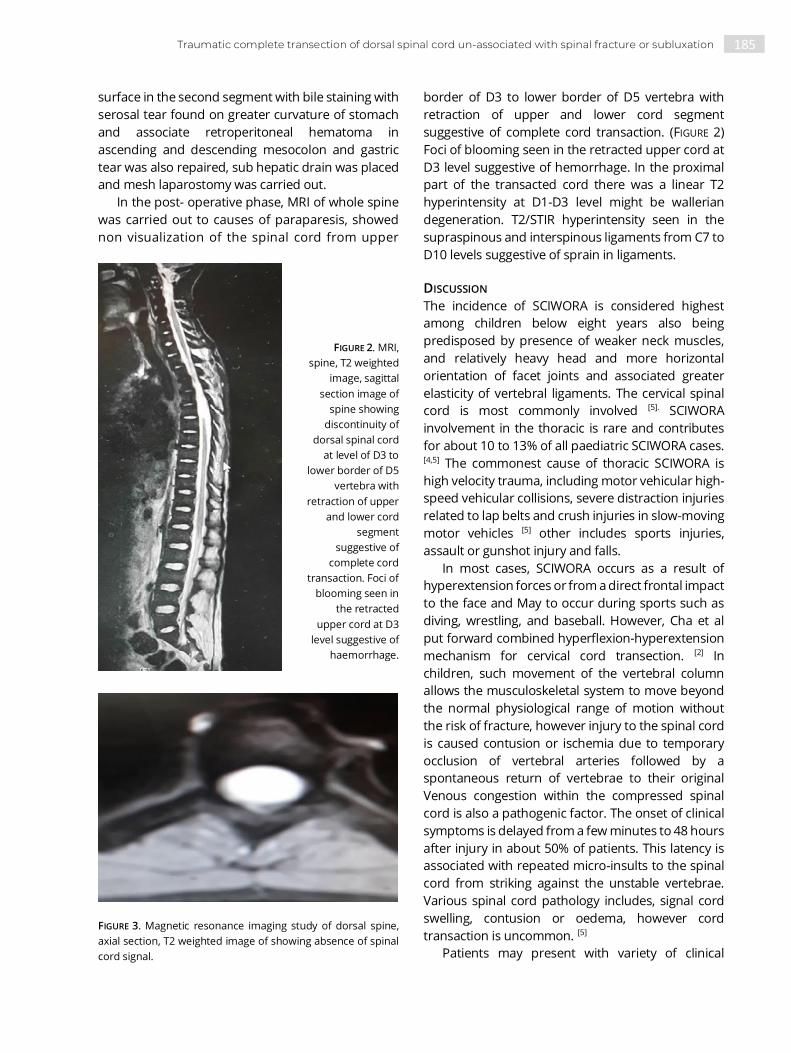
185 Traumatic complete transection of dorsal spinal cord un-associated with spinal fracture or subluxation
surface in the second segment with bile staining with
serosal tear found on greater curvature of stomach
and associate retroperitoneal hematoma in
ascending and descending mesocolon and gastric
tear was also repaired, sub hepatic drain was placed
and mesh laparostomy was carried out.
In the post- operative phase, MRI of whole spine
was carried out to causes of paraparesis, showed
non visualization of the spinal cord from upper
border of D3 to lower border of D5 vertebra with
retraction of upper and lower cord segment
suggestive of complete cord transaction. (FIGURE 2)
Foci of blooming seen in the retracted upper cord at
D3 level suggestive of hemorrhage. In the proximal
part of the transacted cord there was a linear T2
hyperintensity at D1-D3 level might be wallerian
degeneration. T2/STIR hyperintensity seen in the
supraspinous and interspinous ligaments from C7 to
D10 levels suggestive of sprain in ligaments.
FIGURE 2. MRI,
spine, T2 weighted
image, sagittal
section image of
spine showing
discontinuity of
dorsal spinal cord
at level of D3 to
lower border of D5
vertebra with
retraction of upper
and lower cord
segment
suggestive of
complete cord
transaction. Foci of
blooming seen in
the retracted
upper cord at D3
level suggestive of
haemorrhage.
FIGURE 3. Magnetic resonance imaging study of dorsal spine,
axial section, T2 weighted image of showing absence of spinal
cord signal.
DISCUSSION
The incidence of SCIWORA is considered highest
among children below eight years also being
predisposed by presence of weaker neck muscles,
and relatively heavy head and more horizontal
orientation of facet joints and associated greater
elasticity of vertebral ligaments. The cervical spinal
cord is most commonly involved [5]. SCIWORA
involvement in the thoracic is rare and contributes
for about 10 to 13% of all paediatric SCIWORA cases. [4,5] The commonest cause of thoracic SCIWORA is
high velocity trauma, including motor vehicular high-
speed vehicular collisions, severe distraction injuries
related to lap belts and crush injuries in slow-moving
motor vehicles [5] other includes sports injuries,
assault or gunshot injury and falls.
In most cases, SCIWORA occurs as a result of
hyperextension forces or from a direct frontal impact
to the face and May to occur during sports such as
diving, wrestling, and baseball. However, Cha et al
put forward combined hyperflexion-hyperextension
mechanism for cervical cord transection. [2] In
children, such movement of the vertebral column
allows the musculoskeletal system to move beyond
the normal physiological range of motion without
the risk of fracture, however injury to the spinal cord
is caused contusion or ischemia due to temporary
occlusion of vertebral arteries followed by a
spontaneous return of vertebrae to their original
Venous congestion within the compressed spinal
cord is also a pathogenic factor. The onset of clinical
symptoms is delayed from a few minutes to 48 hours
after injury in about 50% of patients. This latency is
associated with repeated micro-insults to the spinal
cord from striking against the unstable vertebrae.
Various spinal cord pathology includes, signal cord
swelling, contusion or oedema, however cord
transaction is uncommon. [5]
Patients may present with variety of clinical

186 Guru Dutta Satyarthee, Satyajit Panda
presentation depending on severity of injury and
location of involvement of spinal cord includes
transient weakness, paraesthesia, numbness,
paraparesis, quadriplegia, complete cord transection
syndrome and Brown–Sequard syndrome. [17]
CT scan spine is best for assessing the associated
bony injuries which may need concomitant
treatment consideration but does not assess the
cord itself. MRI is considered as investigation of
choice and is often performed acute phase in the
context of recent traumatic spinal injury and picks up
spinal cord pathology, associated extramural
hematoma, or hematomyelia, or ligament injury,
which that may or may not be confirmed by trauma
CT. The role of acute MRI is usually to assess for the
presence of acute pathologies treatable such as an
epidural haematoma or cord compression, which
can be targeted by surgical decompression. The
identification of significant cord trans section can
potentially contraindicate surgery. Kalfas et al
analyzed cases had spectrum of lesions including
anatomical spinal cord transection in seven cases,
other finding were 28 cases had spinal cord
deformity secondary to extrinsic compression, 21
had focal cord enlargement/swelling, hyperintensity
intramedullary lesions noted in 17 cases, and rest
two cases had disc herniations. [3]
Boese et al. analyzed 114 patients, on admission,
neurologic deficit assessed by the American Spinal
Injury Association impairment scale was an in 28%, B
in 17%, C in 31%, and D in 25%. On basis of MRI, they
categorize finding. Type I noted in 43%cases with no
MRI abnormalities and 57% exhibited abnormal scan
results (Type II): 6% revealed extra neural (Type Imia),
38% intramural (Type Ibis) and 13% combined
abnormalities (Type Sic). [15]
Pang classified MRI finding into five common
patterns, first type is complete cord disruption
represents most severe injury predominantly
involving upper cervical and thoracic spine in
younger children as a result of flexion injury. second
being major cord haemorrhage with axial MRI
showing haemorrhagic involvement of greater than
50% of cord, next is a minor cord haemorrhage,
fourth is isolated cord edema and fifth pattern is
groups of patients presenting with acute clinical
neurologic deficit consistent with SCIWORA and with
no evidence of injury in the cord on MRI study. [17]
Prognosis depends on spinal cord pathology and
associated spinal injury. For isolated spinal cord
injury case with only cord oedema have most
favorable prognosis but intermediate for cord
oedema and contusion and remains worse for cord
contusion and associated intramedullary
hemorrhage. The prognosis for spinal cord
transaction still remains very poor. However, in cases
with SCIWORA, associated extrinsic compression
caused by vertebral body fragment and disc must be
surgically corrected. In cases with fracture may need
spinal fixation and fusion. Management of spinal
cord transection is still a mystery, and no clear
solution seems in the pipeline in near future. [6-9]
Satyarthee advocated ideally care management
acute traumatic spinal cord injury should commence
immediately at the moment of traumatic impact,
continue during safe evacuation, immediate
resuscitation by trained paramedics, adequate
immobilization, and proper prehospital care, and
emergence transportation to a specialized hospital
after prompt resuscitation, a detailed neurologic
evaluation, pertinent neuroimaging studies, medical
intervention including maintenance of airway and
circulation, augmentation and maintenance of target
level mean arterial pressure, avoidance of any
hypotensive episodes, assessment and appropriate
care of associated systemic injury, early surgical
spinal decompression as judiciously needed. [8]
However, these are effective in minor cord injury, but
for complete teransection of spinal cord, situation
remains gloomy. [8-10]
Tabakow et al observed management of cases
suffering complete spinal cord injury remains an
unsolved clinical problem because of the lack of
spontaneous regeneration of injured central axons.
[13] Exact management is still not available. [10-16]
Various suggested method are suggested mostly on
experimental basis. Olfactory ensheathing cells
support axon outgrowth in the olfactory system, and
transplantaion after spinal cord injury lead to axon
regeneration and improvement in recovery.
Thornton et al. observed rats with mid-thoracic
transection and olfactory ensheathing cells
transplantation, and for assessing connectivity
across the transection, pseudorabies virus injected
into muscles of hind-limb and finally noted presence
of large number of serotonergic axons crossed the
rostral scar border and the area of neurofilament-
positive axons in the injury site. [11] Authors
concluded olfactory ensheathing cells
transplantation can increase axonal growth across

187 Traumatic complete transection of dorsal spinal cord un-associated with spinal fracture or subluxation
the injury site and may promote recovery of
propioception circuit.
Tabakow et al reported 38-year-old with ASIA A
injury at 21 months. The olfactory bulbs were
removed to obtain a culture containing olfactory
ensheathing cells and olfactory nerve fibroblasts.
The cultured cells were transplanted following
resection of the glial scar. He improved to ASIA C with
improved trunk stability, partial recovery of the
voluntary movements of the lower extremities, and
an increase of the muscle mass in the left thigh.
Neuroimaging confirmed grafts producing bridge
over left side of the spinal cord with majority of the
nerve grafts implant, and further neurophysiological
examinations confirmed the restitution of the
integrity of the corticospinal tracts. [ 13]
CONCLUSION
Every cases with suspected head injury sustaining
can presenting with shock and paraplegia, a high
index of suspicion for cord transaction should also
be considered, although rarer. It is highly imperative
that paediatrician, neurosurgeons, orthopaedician
and emergency physician should be aware for entity
total spinal cord transection.
REFERENCES
1. Pang D, Wilberger JE Jr. Spinal cord injury without
radiographic abnormalities in children. J Neurosurg 1982;
57(1):114–129.
2. Cha YH , Cho TH, Suh JK. Traumatic cervical cord transection
without facet dislocations--a proposal of combined
hyperflexion-hyperextension mechanism: a case report. J
Korean Med Sci. 2010 Aug;25(8):1247-50.
3. Kalfas I, Wilberger J, Goldberg A, Prostko ER. Magnetic
resonance imaging in acute spinal cord trauma.
Neurosurgery. 1988 Sep;23(3):295-9.
4. Carroll T, Smith CD, Liu X et al Spinal cord injuries without
radiologic abnormality in children: a systematic review.
Spinal Cord 2015; 53(12):842–848.
5. Parizel PM, van der Zijden T, Gaudino S et al. Trauma of the
spine and spinal cord: imaging strategies. Eur Spine J.
2010;19 Suppl 1 (S1): S8-17.
6. Satyarthee GD, Sangani M, Sinha S, Agrawal D.
Management and Outcome Analysis of Pediatric Unstable
Thoracolumbar Spine Injury: Large Surgical Series with
Literature Review. J Pediatr Neurosci. 2017 Jul-Sep;
12(3):209-214. Doi: 10.4103/Jpn
7. Farrell CA, Hannon M, Lee LK. Pediatric spinal cord injury
without radiographic abnormality in the era of advanced
imaging. Curr Opin Pediatr. 2017 ;29(3):286-290.
8. Satyarthee GD. Ways to Improve Outcomes of Traumatic
Acute Spinal Cord Injury: Integrated Approaches of
Improved Prehospital Care, the Adoption of Synergistic
Medical and Surgical Intervention, Along with Care for
Associated Systemic Injury and Rehabilitation and Social
Inclusion. World Neurosurg. 2017;101:786-787
9. Lukovic D, Moreno-Manzano V, Lopez-Mocholi E,
Rodriguez-Jiménez FJ, Jendelova P, Sykova E, Oria M,
Stojkovic M, Complete rat spinal cord transection as a
faithful model of spinal cord injury for translational cell
transplantation. Erceg S. Sci Rep. 2015 Apr 10;5:9640.
10. Verma SK, Singh PK, Agrawal D, Sinha S, Gupta D,
Satyarthee GD, Sharma BS. O-arm with navigation versus
C-arm: a review of screw placement over 3 years at a major
trauma center. Br J Neurosurg. 2016 Dec;30(6):658-661.
11. Thornton MA , Mehta MD , Morad TT , Ingraham KL ,
Khankan RR , Griffis KG ,et al. Evidence of axon connectivity
across a spinal cord transection in rats treated with
epidural stimulation and motor training combined with
olfactory ensheathing cell transplantation. Exp Neurol.
2018 Jul 26. pii: S0014-4886(18)30263-2. doi:
10.1016/j.expneurol.2018.07.015.
12. Ahmann PA, Smith SA, Schwartz JF, et al. Spinal cord
infarction due to minor trauma in children. Neurology.
1975;25:301–7.
13. Tabakow P , Raisman G, Fortuna W, Czyz M, Huber J, Li D,
Szewczyk P, et al. Functional regeneration of supraspinal
connections in a patient with transected spinal
cordfollowing transplantation of bulbar olfactory
ensheathing cells with peripheral nerve bridging. Cell
Transplant. 2014;23(12):1631-55.
14. Chandrashekhara SH, Kumar A, Gamanagatti S, Kapoor K,
Mukund A, Aggarwal D, Sinha S. Unusual traumatic
spondyloptosis causing complete transection of spinal
cord. Unusual traumatic spondyloptosis causing complete
transection of spinal cord. Int Orthop. 2011
Nov;35(11):1671-5.
15. Boese CK , Oppermann J, Siewe J, Eysel P, Scheyerer MJ,
Lechler P. Spinal cord injury without radiologic abnormality
in children: a systematic review and meta-analysis. J
Trauma Acute Care Surg. 2015 Apr;78(4):874-82.
16. Szwedowski D, Walecki J Spinal cord injury without
radiographic abnormality (SCIWORA)—clinical and
radiological aspects. Pol J Radiol. 2014; 79:461–464.
17. Pang D. Spinal cord injury without radiographic
abnormality in children, 2 decades later. Neurosurgery
2004; 55:1325–1342.

Romanian Neurosurgery (2019) XXXIII (2): pp. 188-190 DOI: 10.33962/roneuro-2019-035 www.journals.lapub.co.uk/index.php/roneurosurgery
Post traumatic isolated ipsilateral oculomotor nerve palsy. An uncommon presentation
Praveen Kumar1, Sharad Pandey2, Kulwant Singh1,
Mukesh Sharma1, Prarthana Saxena2
1 Department of Neurosurgery, Sir Sunder Lal Hospital, IMS, BHU,
Varanasi, Uttar Pradesh, INDIA 2 Department of Neurosurgery, P.G.I.M.E.R. Dr Ram Manohar Lohia
Hospital, New Delhi, INDIA
ABSTRACT The common causes of isolated third nerve palsy are microvascular infarction,
intracranial aneurysm, diabetes, hypertension and atherosclerosis. Here we are
presenting a case of 26-year female presenting with a history of head injury two
months back. She presented with ptosis on the left side. On computed tomography,
a large left-sided chronic subdural hematoma with significant midline shift was found.
Isolated ipsilateral third nerve palsy is a rare presentation with unilateral chronic
subdural hematoma. Improvement in ptosis after surgery indicate a good
neurological outcome.
INTRODUCTION
Chronic subdural Hematoma is a collection of liquefied old blood
between the duramater and arachnoid membrane of brain. It is first
described by Virchow as “Pachymeningitis hemorrhagic interna “in
1857. Later Trotter put forward the theory of traumatic rupture of
bridging veins as a cause of what he named “subdural hemorrhagic
cyst” Chronic SDH is commonly present in elderly patients After a trivial
Trauma [1,2]. Unilateral chronic SDH is more common in comparison
to bilateral SDH [3]. Chronic SDH is a common presentation in people
taking anticoagulant or antiplatelet drugs. Commonly Chronic SDH
Patients present with Headache, Vomiting, Hemiparesis, Ataxia, Altered
Consciousness, Seizures, Urinary incontinence [4]. Patients with
Chronic SDH rarely present with vertigo and nystagmus upward gaze
palsy, and isolated third nerve palsy [5,6,7]. Isolated third nerve palsy
presented with ptosis is rare presentation in post-traumatic chronic
SDH patients.
CASE REPORT
We are presenting a 26-year-old female patient, non-hypertensive,
Keywords ptosis,
third nerve palsy, chronic subdural hematoma
(chronic SDH)
Corresponding author Sharad Pandey
Department of Neurosurgery,
P.G.I.M.E.R. Dr Ram Manohar Lohia Hospital, New Delhi. India
Scan to access the online version
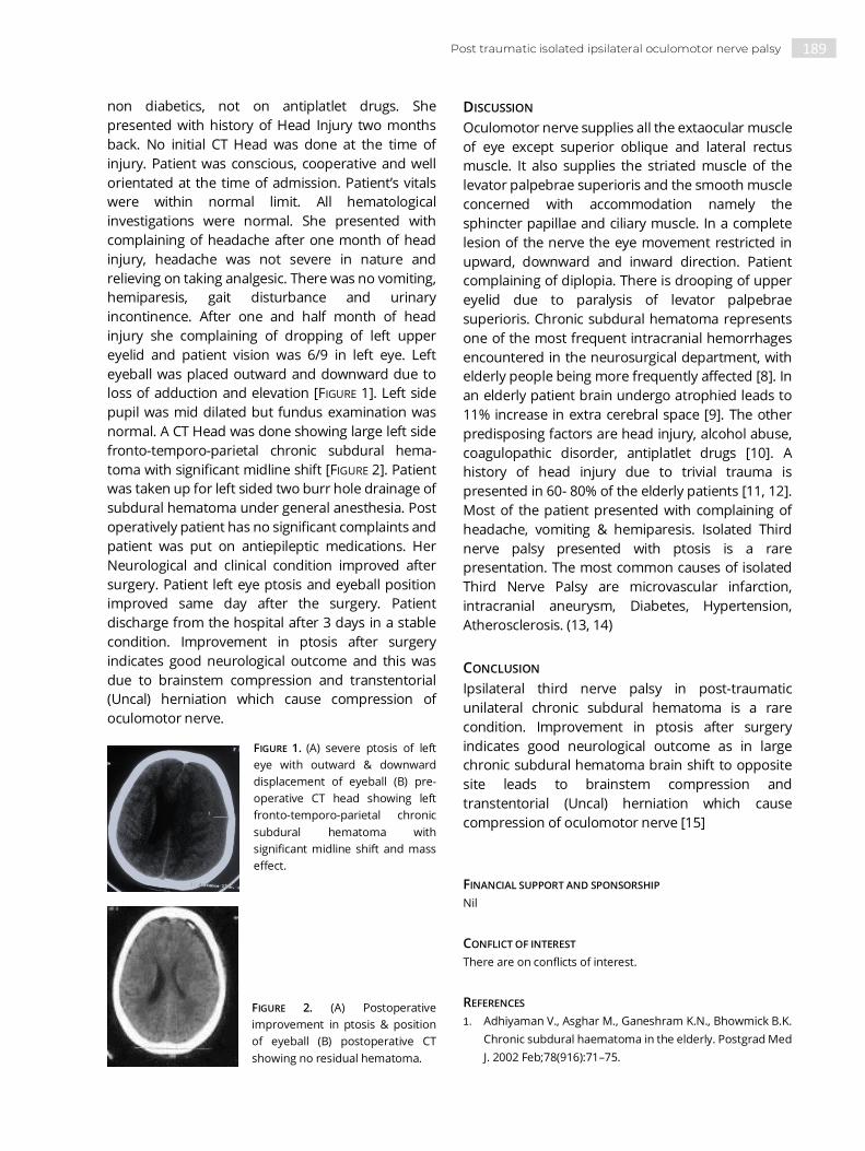
189 Post traumatic isolated ipsilateral oculomotor nerve palsy
non diabetics, not on antiplatlet drugs. She
presented with history of Head Injury two months
back. No initial CT Head was done at the time of
injury. Patient was conscious, cooperative and well
orientated at the time of admission. Patient’s vitals
were within normal limit. All hematological
investigations were normal. She presented with
complaining of headache after one month of head
injury, headache was not severe in nature and
relieving on taking analgesic. There was no vomiting,
hemiparesis, gait disturbance and urinary
incontinence. After one and half month of head
injury she complaining of dropping of left upper
eyelid and patient vision was 6/9 in left eye. Left
eyeball was placed outward and downward due to
loss of adduction and elevation [FIGURE 1]. Left side
pupil was mid dilated but fundus examination was
normal. A CT Head was done showing large left side
fronto-temporo-parietal chronic subdural hema-
toma with significant midline shift [FIGURE 2]. Patient
was taken up for left sided two burr hole drainage of
subdural hematoma under general anesthesia. Post
operatively patient has no significant complaints and
patient was put on antiepileptic medications. Her
Neurological and clinical condition improved after
surgery. Patient left eye ptosis and eyeball position
improved same day after the surgery. Patient
discharge from the hospital after 3 days in a stable
condition. Improvement in ptosis after surgery
indicates good neurological outcome and this was
due to brainstem compression and transtentorial
(Uncal) herniation which cause compression of
oculomotor nerve.
FIGURE 1. (A) severe ptosis of left
eye with outward & downward
displacement of eyeball (B) pre-
operative CT head showing left
fronto-temporo-parietal chronic
subdural hematoma with
significant midline shift and mass
effect.
FIGURE 2. (A) Postoperative
improvement in ptosis & position
of eyeball (B) postoperative CT
showing no residual hematoma.
DISCUSSION
Oculomotor nerve supplies all the extaocular muscle
of eye except superior oblique and lateral rectus
muscle. It also supplies the striated muscle of the
levator palpebrae superioris and the smooth muscle
concerned with accommodation namely the
sphincter papillae and ciliary muscle. In a complete
lesion of the nerve the eye movement restricted in
upward, downward and inward direction. Patient
complaining of diplopia. There is drooping of upper
eyelid due to paralysis of levator palpebrae
superioris. Chronic subdural hematoma represents
one of the most frequent intracranial hemorrhages
encountered in the neurosurgical department, with
elderly people being more frequently affected [8]. In
an elderly patient brain undergo atrophied leads to
11% increase in extra cerebral space [9]. The other
predisposing factors are head injury, alcohol abuse,
coagulopathic disorder, antiplatlet drugs [10]. A
history of head injury due to trivial trauma is
presented in 60- 80% of the elderly patients [11, 12].
Most of the patient presented with complaining of
headache, vomiting & hemiparesis. Isolated Third
nerve palsy presented with ptosis is a rare
presentation. The most common causes of isolated
Third Nerve Palsy are microvascular infarction,
intracranial aneurysm, Diabetes, Hypertension,
Atherosclerosis. (13, 14)
CONCLUSION
Ipsilateral third nerve palsy in post-traumatic
unilateral chronic subdural hematoma is a rare
condition. Improvement in ptosis after surgery
indicates good neurological outcome as in large
chronic subdural hematoma brain shift to opposite
site leads to brainstem compression and
transtentorial (Uncal) herniation which cause
compression of oculomotor nerve [15]
FINANCIAL SUPPORT AND SPONSORSHIP
Nil
CONFLICT OF INTEREST
There are on conflicts of interest.
REFERENCES
1. Adhiyaman V., Asghar M., Ganeshram K.N., Bhowmick B.K.
Chronic subdural haematoma in the elderly. Postgrad Med
J. 2002 Feb;78(916):71–75.

190 Praveen Kumar, Sharad Pandey, Kulwant Singh et al.
2. Arpino L., Gravina M., Basile D., Franco A. Spontaneous
chronic subdural hematoma in a young adult. J Neurosurg
Sci. 2009 Jun;53(2):55–57.
3. Nayil K, Ramzan A, Sajad A, Zahoor S, Wani A, et al. (2012)
Subdural Hematomas: An Analysis of 1181 Kashmiri
Patients. World Neuro¬surg 77(1): 103-110.
4. Mulholland C., Knox F.A. Subacute subdural haematoma
presenting with oculomotor nerve palsy, reduced vision,
and hallucinations. Eye (Lond) 2006 Jan;20(1):125–126.
5. Ashkenazi E, Pomeranz S (1994) Nystagmus as the
presentation of tentorial incisure subdural haematoma. J
Neurol Neurosurg Psychi¬atry 57(7): 830-831.
6. Phookan G, Cameron M (1994) Bilateral chronic subdural
haemato¬ma: an unusual presentation with isolated
oculomotor nerve palsy (letter). J Neurol Neurosurg
Psychiatry 57(9): 1146.
7. Sandyk R (1982) Isolated failure of upward gaze as a sign of
chronic subdural haematoma (letter). S Afr Med J 61(2): 32.
8. Kim J.H., Kang D.S., Kim J.H., Kong M.H., Song K.Y. Chronic
subdural hematoma treated by small or large craniotomy
with membranectomy as the initial treatment. J Korean
Neurosurg Soc. 2011 Aug; 50(2):103–108.
9. Stanisic M, Lund-Johnsen M, Mahesparan R (2005)
Treatment of chronic subdural haematoma by burr-hole
craniostomy in adults: in¬fluence of some factors on
postoperative recurrence. Acta Neurochir (Wien) 147(12):
1249-1256
10. Stroobandt G, Fransen P, Thauvoy C, Menard E (1995)
Pathogenic factors in chronic subdural haematoma and
causes of recurrence af¬ter drainage. Acta Neurochir
(Wien) 137(1-2): 6-14.
11. Markwalder TM, Steinsiepe KF, Rohner M, Reichenbach W,
Mark¬walder H (1981) The course of chronic subdural
haematomas after burr-hole craniostomy and closed-
system drainage. J Neurosurg 55(3): 390-396.
12. Matsumoto K, Akagi K, Abekura M, Ryujin H, Ohkawa M, et
al. (1999) Recurrence factor for chronic subdural
haematoma after burr-hole craniostomy and closed
system drainage. Neurol Res 21(3): 277-280.
13. Ortega-Martínez M., Fernández-Portales I., Cabezudo J.M.,
Rodríguez-Sánchez J.A., Gómez-Perals L.F., Giménez-Pando
J. Isolated oculomotor palsy. An unusual presentation of
chronic subural hematoma. Neurocirugia (Astur) 2003
Oct;14(5):423–425.
14. Ko J.H., Kim Y.J. Oculomotor nerve palsy caused by
posterior communicating artery aneurysm: evaluation of
symptoms after endovascular treatment. Interv
Neuroradiol. 2011 Dec17(4):415–419.
15. Cameron MM (1978) Chronic subdural haematoma: a
review of 114 cases. J Neurol Neurosurg Psychiatry 41(9):
834-839.

Romanian Neurosurgery (2019) XXXIII (2): pp. 191-199 DOI: 10.33962/roneuro-2019-036 www.journals.lapub.co.uk/index.php/roneurosurgery
Endoscopic management of intraventricular shunt-related cystic compartment in paediatric patients
Ahmed Zaher, Amr Farid Khalil, Mohamed State, Hatem Badr
* Department of Neurosurgery, Faculty of Medicine, Mansoura
University, EGYPT
ABSTRACT Background. Intraventricular uniloculated compartments are relatively seen
frequently in children previously treated with ventriculoperitoneal shunts for
hydrocephalus. These compartments may present before shunt insertion and even
as an end result to the shunt gliosis. Although the availability of different surgical
modalities for the management of such loculations but till now there is no consensus
about a single therapeutic approach for its management. Objective: The aim of this
study is to assess the role of neuroendoscopy in management of uniloculated
compartments, in pediatric cases who underwent shunting for hydrocephalus, to
avoid multiple shunt insertion and even to eliminate the need for the preexisting
shunt system.
Patients and Methods. We retrospectively studied 44 consecutive previously
shunted patients with isolated intraventricular compartment who underwent 58
endoscopic procedures by a single surgeon during the period between February 2015
and February 2018. These procedures included endoscopic cyst fenestration,
intraventricular septum pellucidotomy, foraminoplasty and additionally third
ventriculostomy in some cases was added to the previous procedures. Multiloculated
hydrocephalus and cysts related to the tumours were excluded. Clinical, radiological
and surgical data were assessed.
Results. The most common cause of a single loculation in those patients was
neuroepithelial cyst (20 cases) followed by post shunt isolated compartment (12
cases), meningitis (8 cases) and four cases of intraventricular haemorrhage. The
patient's age varied from two months up to seven years (mean = 3.2 years). The
follow-up period was ranged from (2-49 months) with the mean of 12.3 months. The
overall success of restoration of communication between the uniloculated
compartment and the ventricular system was achieved in 36 patients (81.8%), with 28
(63.6%) patient needed only one shunt and eight (18.2%) patients became
independent of their previous shunts. Fourteen repeated endoscopic procedures
were considered necessary in ten patients for either reopening of a closed stoma
and/or shunt revision and finally, eight patient (18%) required additional shunt
insertion during follow up period. The complications encountered were minor and
reversible (CSF leak in two cases, infection in two cases, subdural fluid collection in
four cases) with no mortality.
Conclusion. Minimal invasive endoscopic technique is a useful operative alternative
to control uniloculated compartments in patients with previous ventriculoperitoneal
shunts. It does not only simplify the existing shunt system by restoration of the
communication between the loculated compartment and the draining shunt but it
may eliminate the need for external shunt in some cases as well.
Keywords loculated hydrocephalus,
endoscopy, fenestration
Corresponding author Ahmad Zaher
Department of Neurosurgery,
Faculty of Medicine, Mansoura University, Egypt
Scan to access the online version

192 Ahmed Zaher, Amr Farid Khalil, Mohamed State et al.
INTRODUCTION
Intraventricular septation may lead to formation of
an isolated CSF cystic component which may become
enlarged even if the shunt system is functioning.
Such enlarging intraventricular loculation may be
attributed to altered CSF dynamics as result of
separation between the ventricular catheter tip and
the site of CSF production [14].
Variable anatomical and radiological forms of
loculations were described according to the site of
obstruction and consequent CSF accumulation
including either uniloculated or multiloculated.
Identification and distinction between the two forms
is crucial because everyone has a particular different
pathogenesis, treatment and prognosis [7].
Loculated hydrocephalus usually develops after
episodes of neonatal meningitis or germinal matrix
hemorrhage. Occlusion of the foramen of Monro,
the aqueduct, and the outlet foramina of the fourth
ventricle may result from shunt infection and its
consequences like ventriculitis and ventricular
septations. However, isolated intraventricular
compartment may evolve after insertion of
ventriculoperitoneal shunt via other mechanisms
rather than shunt infection [11, 14].
Direct ependymal trauma during shunt insertion,
collapse of shunted ventricular compartment with
subsequent obstruction of the catheter tip
perforations by proliferating fibrillary astrocytes and
over drainage of existing shunt may play important
roles that may stimulate the enlargement of an
isolated shunt related compartment [14].
The management of such complex condition is
challenging to most neurosurgeons. The main goal
of treatment is to elevate the manifestation of
increased intracranial pressure by creating a free
connection between the isolated compartment and
the ventricular system cavity via converting them
into a solitary component that will be drained by a
single ventricular catheter with little morbidity and
mortality [8, 19].
Multiple treatment strategies for intraventricular
loculation are available but the best choice of the
proper procedure remains controversial. This group
of patients presented with intraventricular
compartments related to ventriculoperitoneal
shunts often require multiple and repeated
operations. These patients usually necessitate either
traditional shunt revisions alone or in combination
with other procedures like insertion of multiple
perforated catheter, craniotomy with transcallosal
fenestration, stereotactic aspiration, endoscopic cyst
fenestration, septostomy, foraminoplasty or third
ventriculostomy [18, 20].
Recent advancement of neuroendoscopy and the
higher incidence of complication associated with
traditional shunt revision or open craniotomy have
shifted many neurosurgeons to the different
endoscopic techniques as simple and minimal
invasive alternatives to solve this complex condition
[9].
PATIENTS AND METHODS
From February 2015 to February 2018, forty-four
consecutive paediatric cases presented with shunt
related cystic compartments and a preexisting
ventriculoperitoneal shunts were operated at
“Neurosurgical Department of Mansoura University
Hospitals” by a single surgeon using
neuroendoscopic techniques. Our study included 28
boys and 16 girls; their ages ranged from two
months to seven years (mean 3.2 years). The follow
up period renged from (2-49 months) with the mean
of 12.3 months. Cases of multiloculated
hydrocephalus or uniloculated hydrocephalus
without previous shunt insertion were excluded
from our study. Tumour related cysts were also
excluded.
We retrospectively reviewed the patient's data
retrieved from the patient charts which included:
patient history, clinical examination, radiological
investigations, operative data, patient outcome,
shunt independency, the need for additional shunts
and the prerequisites to repeat endoscopic
procedures.
Plain x-ray to the shunt system, CT scan and MRI
brain with or without gadolinium were done to all
patients preoperatively. CSF sample was obtained
and examined to exclude CSF or shunt infection prior
to endoscopic procedures.
Endoscopic surgery was indicated when the
patients had clinical deterioration in the form of
progressive enlargement of head size, development
of manifestations of intracranial hypertension,
worsening of the focal neurological deficits, gait
disturbance or refractory epilepsy. The previous
clinical manifestations had been correlated with
radiological signs of the cyst enlargement and/or
shunt malfunctioning. Cognitive dysfunction was not

193 Endoscopic management of intraventricular shunt-related cystic compartment in paediatric patients
considered as an indication for surgical
management.
Our aim of endoscopic surgery was to connect
the isolated compartment to the ventricular cavity in
order to drain both of them by the same shunt in
order to simply the pre-existing shunt device and to
avoid implanting another device whenever possible.
In all patients, surgery was done under general
anaesthesia. The entry point was tailored individually
in every case according to the cyst location and the
location of the previous shunt entry point. A proper
burr-hole location and the direct surgical corridor
were the most critical points for proper cyst
fenestration and shunt revision simultaneously.
Different angles rigid lens endoscopes (0o, 30o,
70o Karl-Storz rigid Gaab System) were used. The
endoscopic sheath has an outer diameter of 6 mm
and working channel of 2 mm. After endoscopic
introduction, continuous irrigation was essential for
clear identification of key anatomical landmarks and
proper visualization of cyst wall as well as the
proximal end of the pre-existing ventricular catheter.
Initial cyst fenestration was performed in a thin,
translucent and relatively a vascular segment of the
cyst wall by the use of bipolar electrocoagulation.
Dilation and enlargement of the initial stoma was
continued by with the aid of Fogarty balloon
catheter, punches, and sharp scissor to fashion a
wide stoma that secure a free CSF passage and
minimize the incidence of reclosure of the stoma.
Coagulation and devascularization of the cyst wall
were done in order shrink the cyst wall and hold the
cyst growth.
Other endoscopic procedures like endoscopic
revision of the shunt, septum pellucidotomy,
foramioplasty and third ventriculostomy were done
in combination with endoscopic cyst fenestration to
advance the communication of CSF isolated
compartments.
Any bleeding point was secured with continuous
irrigation, bipolar electrocoagulation and
tamponading with embolectomy balloon catheter to
achieve a non-vascular surgical field as well as the
site of fenestration. At the end of the surgery, the
endoscopic sheath was retrieved under continuous
irrigation to clear out the surgical field from any
blood clots or any surgical debris.
Clinical outcome was defined as improved,
unchanged or worsened. Developmental delay was
not considered in post-operative clinical outcome
assessment. Epilepsy was defined to be improved if
the attacks were controlled after withdrawal or even
reduction of the dose of antiepileptic drugs. If the
head circumference remained stable or slightly
reduced in size, this was considered as a
postoperative sign of improvement.
Routine postoperative CT brain was done to all
patients twenty-four hours after surgery. Follow up
CT and MRI brain were done routinely 3 months after
surgery. The procedure was considered to be
successful when endoscopic procedure was able to
restore the continuity between the cyst and the
ventricular system cavity without the need to insert
additional shunt devices. Repeated endoscopic
procedures were considered when the patient
experienced recurrence or progression of his clinical
manifestations that were concomitant with
progressive cyst and/or ventricular enlargement on
follow up CT and/or MRI images.
RESULTS
Progressive enlargement of the head size was the
most common presenting symptom in our patients
(28 cases, 63.6%) followed by seizures (14 cases,
31.8%), headache (10 cases, 22.72%), focal
neurological deficits (10 cases, 22.72%) and gait
disturbance in 6 patients (13.6%) (TABLE 1).
The non-colloidal neuroepithelial cysts were the
most prominent underlying cause of intraventricular
unilateral compartments with preexisting shunts in
our study (Twenty cases, 45.5%) while uniloculated
cysts related to either previous shunt insertion or
shunt infection represented the second most
common cause (Twelve cases, 27.3%) followed by
meningitis (eight cases,18.2%) and neonatal
intraventricular haemorrhage (four cases, 9%) (TABLE
2) .
Endoscopic fenestration of cyst wall was done in
all patients. Other additional endoscopic procedures
that were required to be done simultaneously with
the endoscopic cyst fenestration included either
endoscopic third ventriculostomy (six patients),
septum pellucidotomy (ten patients) or
foraminoplasty (six patients).
During the previous endoscopic procedures,
shunt revisions were indicated in only six patients
(33%) who had non-colloidal neuroepithelial cysts
whenever 20 out of 24 patients (83%) of shunt
related cysts, postmeningitic cysts and post-
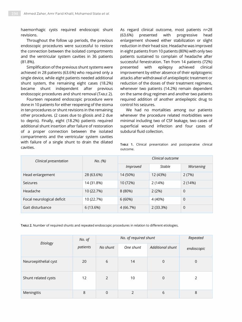
194 Ahmed Zaher, Amr Farid Khalil, Mohamed State et al.
haemorrhagic cysts required endoscopic shunt
revisions.
Throughout the follow up periods, the previous
endoscopic procedures were successful to restore
the connection between the isolated compartments
and the ventricular system cavities in 36 patients
(81.8%).
Simplification of the previous shunt systems were
achieved in 28 patients (63.6%) who required only a
single device, while eight patients needed additional
shunt system, the remaining eight cases (18.2%)
became shunt independent after previous
endoscopic procedures and shunt removal (TABLE 2).
Fourteen repeated endoscopic procedure were
done in 10 patients for either reopening of the stoma
in ten procedures or shunt revisions in the remaining
other procedures. (2 cases due to gliosis and 2 due
to depris). Finally, eight (18.2%) patients required
additional shunt insertion after failure of restoration
of a proper connection between the isolated
compartments and the ventricular system cavities
with failure of a single shunt to drain the dilated
cavities.
As regard clinical outcome, most patients n=28
(63.6%) presented with progressive head
enlargement showed either stabilization or slight
reduction in their head size. Headache was improved
in eight patients from 10 patients (80%) with only two
patients sustained to complain of headache after
successful fenestration. Ten from 14 patients (72%)
presented with epilepsy achieved clinical
improvement by either absence of their epilptogenic
attacks after withdrawal of antiepileptic treatment or
reduction of the doses of their treatment regimens,
whenever two patients (14.2%) remain dependent
on the same drug regimen and another two patients
required addition of another antiepileptic drug to
control his seizures.
We had no mortalities among our patients
whenever the procedure related morbidities were
minimal including two of CSF leakage, two cases of
superficial wound infection and four cases of
subdural fluid collection.
TABLE 1. Clinical presentation and postoperative clinical
outcome.
Clinical presentation No. (%) Clinical outcome
Improved Stable Worsening
Head enlargement 28 (63.6%) 14 (50%) 12 (43%) 2 (7%)
Seizures 14 (31.8%) 10 (72%) 2 (14%) 2 (14%)
Headache 10 (22.7%) 8 (80%) 2 (2%) 0
Focal neurological deficit 10 (22.7%) 6 (60%) 4 (40%) 0
Gait disturbance 6 (13.6%) 4 (66.7%) 2 (33.3%) 0
TABLE 2. Number of required shunts and repeated endoscopic procedures in relation to different etiologies.
Etiology No. of
patients
No. of required shunt Repeated
endoscopic
procedures
No shunt One shunt Additional shunt
Neuroepithelial cyst 20 6 14 0 0
Shunt related cysts 12 2 10 0 2
Meningitis 8 0 2 6 8

195 Endoscopic management of intraventricular shunt-related cystic compartment in paediatric patients
TABLE 3. Repeated endoscopic procedure in ten patients.
Type of the procedure No. %
Closed fenestration 10 71.4
Shunt revision
4 28.6
Gliosis 2 14.3
Obstruction form debris 2 14.3
FIGURE 1. a-l: Radiological and intraoperative endoscopic photographs of a case of shunt related cyst. a) Preoperative CT. b) CT after
insertion of a shunt. c) Three months follow up CT after shunt insertion with development of shunt related cyst. d) CT after
Intraventricular age 4 0 2 2 4
Total no. and % 44 (100%) 8 (18%) 28 (64%) 8 (18%) 14 (24%)
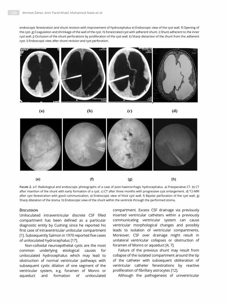
196 Ahmed Zaher, Amr Farid Khalil, Mohamed State et al.
endoscopic fenestration and shunt revision with improvement of hydrocephalus e) Endoscopic view of the cyst wall. f) Opening of
the cyst. g) Coagulation and shrinkage of the wall of the cyst. h) Fenestrated cyst with adherent shunt. i) Shunt adherent to the inner
cyst wall. j) Occlusion of the shunt perforations by proliferation of the cyst wall. k) Sharp dissection of the shunt from the adherent
cyst. l) Endoscopic view after shunt revision and cyst perforation.
(e) (f) (g) (h)
FIGURE 2. a-f: Radiological and endoscopic photographs of a case of post-haemorrhagic hydrocephalus. a) Preoperative CT. b) CT
after insertion of the shunt with early formation of a cyst. c) CT after three months with progressive cyst enlargement. d) T2-MRI
after cyst fenestration with good communication. e) Endoscopic view of thick cyst wall. f) Bipolar perforation of the cyst wall. g)
Sharp dilatation of the stoma. h) Endoscopic view of the shunt within the ventricle through the performed stoma.
DISCUSSION
Uniloculated intraventricular discrete CSF filled
compartment has been defined as a particular
diagnostic entity by Cushing since he reported his
first case of intraventricular unilocular compartment
[1]. Subsequently Salmon in 1970 reported five cases
of uniloculated hydrocephalus [17].
Non-colloidal neuroepithelial cysts are the most
common underlying etiological causes for
uniloculated hydrocephalus which may lead to
obstruction of normal ventricular pathways with
subsequent cystic dilation of one segment of the
ventricular system, e.g. foramen of Monro or
aqueduct and formation of uniloculated
compartment. Excess CSF drainage via previously
inserted ventricular catheters within a previously
communicating ventricular system can cause
ventricular morphological changes and possibly
leads to isolation of ventricular compartments.
Moreover, CSF over drainage might result in
unilateral ventricular collapses or obstruction of
foramen of Monro or aqueduct [4, 7].
Failure of the previous shunt may result from
collapse of the isolated compartment around the tip
of the catheter with subsequent obliteration of
ventricular catheter fenestrations by reactive
proliferation of fibrillary astrocytes [12].
Although the pathogenesis of univentricular

197 Endoscopic management of intraventricular shunt-related cystic compartment in paediatric patients
loculation is not well known but the most accepted
theory is that the inflammatory reaction related to
previous shunt insertion, infection and
intraventricular hemorrhage are the triggering
factors for subendymal gliosis that may enhance the
formation of glial tufts which occlude the holes of the
ventricular system and act as a nidus for ventricular
septations [11, 18]. Regardless of the accurate
mechanism, the problem progress steadily toward
greater complexity with time and traditional
treatment ultimately require repeated surgeries with
increasingly complex shunt devices [1].
The synonyms and classification of complex
hydrocephalus are still interchangeable and unclear
in the literatures. Spennato et al. published a
classification for variable types of ventricular
loculation defined by the location and number of
compartments [19]. According to the previous
proposed scheme "uniloculated" and
"multiloculated" are broadly defined based on
anatomical factors only [1]. These categories should
be further subdivided into physiologically simple or
physiologically complex based on whether CSF
absorption is normal or impaired [2]. Differentiation
between the previous entities is of great importence
as their pathogenesis, surgical outcome and
prognosis are quite different [6].
The clinical features of loculated hydrocephalus
are not specific and are difficult to recognize because
they often arise in a neurologically compromised
infant or child with a previous history of
hydrocephalus, neonatal meningitis or
intraventricular haemorrhage. In our study the
progressive head enlargement was the most
common presenting symptom in 28 cases (63.6%)
followed by seizures in 14 cases (31.8%) headache in
10 cases (22.7%), focal neurological deficit in 10 cases
(22.7%) and gait disturbance in 6 cases (13.6%).
Intraventricular loculation with previous
ventriculoperitoneal shunt is a complex disorder that
is difficult to treat. Those patients usually require
repeated shunt revisions. Furthermore, those
patients may need division and lysis of
intraventricular septations either surgically through
transcallosal craniotomy or through endoscopic
techniques [13, 16].
The best advantages of the endoscopic
procedures over transcallosal craniotomy are limited
invasiveness and short postoperative recovery time.
Nowadays the recent technical improvement in
endoscopic techniques and high morbidity
associated with multiple shunt revision or
transcallosal craniotomy has shifted neurosurgeon
toward the minimally invasive endoscopic
techniques for lysis of intraventricular septae
simultaneously with endoscopic shunt revisions [5].
Our objective in management of a single
intraventricular loculation in a previously shunted
patient was to restore the continuity between the
isolated compartment and the ventricular system
cavity to create a single cavity with the intent of
simplifying or removal of the previous shunt system
and reducing the need for repeated procedure. The
proposed goal has been achieved in thirty-six
patients (81.8%) with simplification of the previous
shunts in 28 patients (63.6%); out of them eight
patients (18.2%) became shunt independent after
endoscopic cyst fenestration and additional
endoscopic foraminoplasty, septum pellucidotomy
or third ventriculostomy.
Shunt malfunctioning was found in most cases of
our series (34 cases, 77%) due to either collapse and
adhesive scarring of the cyst around the catheter tip
or occlusion of the catheter perforations by debris or
proliferating epithelial tufts. We used bipolar
coagulation for safe dissection of the catheter tip
from the adherent collapsed cyst wall to avoid
catastrophic intraventricular bleeding which may
happen in absence of endoscopic control.
In his series of 31 pediatric patients with
uniloculated hydrocephalus, El-Ghandour treated 9
cases of uniloculated hydrocephalus with preexisting
shunt system demonstrating reduction of shunt
revision rate from 2.7 per year before fenestration to
0.25 per year after fenestration with simplification of
four previous complex shunt systems and all
patients required single repeated endoscopic
procedures [6]. Lewis et al. reported 34 cases of both
multiloculated and uniloculated hydrocephalus and
shunt revision rate dropped from 3.04% prior to
endoscopy to 0.25 after the procedure and 50% of
the patients shunted prior to endoscopy required
repeat endoscopic procedures [12].
In our series 90 revisions were required in 32
patients prior to endoscopy with shunt revision rate
was 2.98/year which have been reduced to 0.27 after
endoscopic fenestration as only four patients
required endoscopic shunt revision. Ten patients
(22.7%) of our series required fourteen repeated
endoscopic procedure for opening of a closed

198 Ahmed Zaher, Amr Farid Khalil, Mohamed State et al.
fenestrae or endoscopic shunt revision. The
explanation of small percentage of repeated
endoscopic procedure, in our cases compared by
previously mentioned series is that we have 20 cases
of neuroepithelial cysts presented to us with
malfunctioning shunts and none of them required to
repeat surgery. Moreover, we have only 12 cases of
post meningitis and post haemorrhagic uniloculated
hydrocephalus in which 8 cases required 12
repeated endoscopic surgery and only two patients
with shunt related cysts had a single repeated
endoscopic surgery
During repeated endoscopic procedures, ten
cysts required reopening of closed stoma and four
ventricular catheters required reopening of the
closed stoma either due to gliosis in two cases or
obstruction of the catheter openings by a debris in
the other cases. Ultimately, eight patients (18.2%)
"Six with post-meningitis loculations and two with
post-haemorrhagic loculations" required insertion of
additional ventricular catheters after failure of
repeated endoscopic procedures to achieve proper
communication with persistent undrained
intraventricular loculated compartments.
In the context with the experience of other
endoscopic surgeons, we found that sharp
dissection, employment of wide fenestrations of at
least one cm and coagulation of the cysts wall
ensured the success of the procedure and reduced
the chance of recurrence [10, 15].
Previous series of endoscopic fenestration of
loculated hydrocephalus highlighted the
combination of endoscopic procedures to achieve
better success and moreover may help shunt
independence [6, 20].The obvious advantages of
leaving one shunt in complex hydrocephalus are
fewer opportunities for obstruction, rupture,
infection and gliosis [3].
Septum pellucidotomy, foraminoplasty and third
ventriculostomy were employed in our cases in
combination with endoscopic cyst fenestration and
shunt revision that have been succeed to achieve
simplification of the previous shunt systems in
(63.6%) and elimination of shunt dependency in
eight patients (18%). The immaturity and deficient
absorptive capabilities of scared subarachnoid space
by a previous inflammatory process may be
responsible for the lower shunt independent
incidence in our study in line with others series [2,
12].
Nida and Haines who advocate transcallosal
craniotomy for division and lysis of intraventricular
septation criticized the endoscopic approaches by
their limited ability to control intraoperative bleeding
which may obscure the operative field [13]. However,
intraoperative haemorrhage presented no great
problem and did not result in termination of any
procedure in this series. Haemorrhage was
controlled by continuous irrigation with warm
lactated ringer solution and occasionally monopolar
and bipolar coagulation were helpful to control any
bleeding point. Moreover, proper coagulation of site
of fenestration may help to minimize the incidence
of intraoperative bleeding.
As regard clinical outcome 28 patients (63.6%)
who presented with head enlargement had
stabilization or decline in their head circumference.
Epilepsy was controlled in ten patients (72%) out of
fourteen patients presented with seizures. Focal
neurological deficient was improved in six patients
(60%) and gait disturbance was improved in four
patients (67%). Development delay was excluded
from postoperative assessment as developmental
improvement is not correlated with the change of
the cyst size. Our results are going with the previous
series which assessed the postoperative clinical
outcome, in addition to radiological improvement of
hydrocephalus [6].
Our morbid complications were minimal and
reversible including CSF leakage in two cases which
stopped within few days after medical therapy, four
cases of subdural hygroma that resolved
spontaneously during follow up and only two cases
of superficial wound infection which improved with
antibiotic and repeated dressing.
CONCLUSION
Intraventricular loculations in previously shunted
patients is among the most challenging scenarios
encountered by a neurosurgeon. This complex
disorder has a relatively evolving course in the form
of ongoing worsening with time that requires
increasingly difficult surgical procedures. The main
objective in management of such cases is to reduce
the number of surgical procedures more than
improving the quality of life of the patient.
Endoscopic surgical techniques are attractive
minimal invasive alternatives that may help to
achieve treatment objectives with minimal
postoperative morbidity.
199 Endoscopic management of intraventricular shunt-related cystic compartment in paediatric patients
AUTHORS CONTRIBUTIONS
This work was carried out in collaboration between all authors.
Author Amr Farid, designed the study, Author Mohamed State
wrote the protocol, Author Ahmed Zaher managed the
literature research, Author Hatem Badr revised the final
manuscript. All surgical procedures were carried out by single
surgeon and assisted by the same surgical team including the
four authors. All authors read and approved the final
manuscript.
REFERENCES
1. Akbari S H A, Holekamp T F, Murphy T M, Mercer D, Leonard
J R, Smyth M D, Park T, and Limbrick D D: Surgical
management of complex multiloculated hydrocephalus in
infants and children. Child's Nervous System 2015, 31(2),
243-249.
2. Andresen M and Juhler M: Multiloculated hydrocephalus: a
review of current problems in classification and treatment.
Child's Nervous System 2012, 28(3), 357-362.
3. Cipri S, Gambardella G, and Gangemi M: Neuroendoscopic
approach to complex hydrocephalus: Personal experience
and preliminary report. Journal of neurosurgical sciences
2001, 45(2), 92.
4. Cushing H: Surgery of the head. Surgery: its principles and
practice 1908, 3, 255-256.
5. El-Ghandour N M: Endoscopic cyst fenestration in the
treatment of multiloculated hydrocephalus in children.
Journal of Neurosurgery: Pediatrics 2008, 1(3), 217-222.
6. El-Ghandour N M: Endoscopic cyst fenestration in the
treatment of uniloculated hydrocephalus in children.
Journal of Neurosurgery: Pediatrics 2013, 11(4), 402-409.
7. Eller T W and Pasternak J F: Isolated ventricles following
intraventricular hemorrhage. Journal of neurosurgery
1985, 62(3), 357-362.
8. Gangemi M, Maiuri F, Donati P, and Sigona L:
Neuroendoscopy: Personal experience, indications and
limits. Journal of neurosurgical sciences 1998, 42(1), 1.
9. Greenfield J P and Souweidane M M: Endoscopic
management of intracranial cysts. Neurosurgical focus
2005, 19(6), 1-9.
10. Heilman C B and Cohen A R: Endoscopic ventricular
fenestration using a “saline torch”. Journal of neurosurgery
1991, 74(2), 224-229.
11. Jamjoom A, Mohammed A, Al-Boukai A, Jamjoom Z,
Rahman N, and Jamjoom H: Multiloculated hydrocephalus
related to cerebrospinal fluid shunt infection. Acta
neurochirurgica 1996, 138(6), 714-719.
12. Lewis A I, Keiper G L, and Crone K R: Endoscopic treatment
of loculated hydrocephalus. Journal of neurosurgery 1995,
82(5), 780-785.
13. Nida T Y and Haines S J: Multiloculated hydrocephalus:
craniotomy and fenestration of intraventricular septations.
Journal of neurosurgery 1993, 78(1), 70-76.
14. Oi S and Abbott R: Loculated ventricles and isolated
compartments in hydrocephalus: their pathophysiology
and the efficacy of neuroendoscopic surgery.
Neurosurgery clinics of North America 2004, 15(1), 77-87.
15. Oi S, Hidaka M, Honda Y, Togo K, Shinoda M, Shimoda M,
Tsugane R, and Sato O: Neuroendoscopic surgery for
specific forms of hydrocephalus. Child's Nervous System
1999, 15(1), 56-68.
16. Powers S; Fenestration of intraventricular cysts using a
flexible, steerable endoscope, in Minimally Invasive
Neurosurgery I. 1992, Springer. p. 42-46.
17. Salmon J H: Isolated unilateral hydrocephalus following
ventriculoatrial shunt. Journal of neurosurgery 1970, 32(2),
219-226.
18. Schulz M, Bohner G, Knaus H, Haberl H, and Thomale U-W:
Navigated endoscopic surgery for multiloculated
hydrocephalus in children. Journal of Neurosurgery:
Pediatrics 2010, 5(5), 434-442.
19. Spennato P, Cinalli G, Ruggiero C, Aliberti F, Trischitta V,
Cianciulli E, and Maggi G: Neuroendoscopic treatment of
multiloculated hydrocephalus in children. Journal of
Neurosurgery: Pediatrics 2007, 106(1), 29-35.
20. Teo C, Kadrian D, and Hayhurst C: Endoscopic
management of complex hydrocephalus. World
neurosurgery 2013, 79(2), S21. e1-S21. e7.

Romanian Neurosurgery (2019) XXXIII (2): pp. 200-214 DOI: 10.33962/roneuro-2019-037 www.journals.lapub.co.uk/index.php/roneurosurgery
The use of folic acid in the prevention of spinal bifida. Knowledge, attitude, and practice of women of childbearing age in low income rural communities
Eghosa Morgan1, Nosa Akpede2, Emmanuel
Friday Osagiede2, Vivian Ajekweneh2, Francis
Erah2, Victoria Aishetu Momoh3, Mohammed
Irumhekhai Momoh3, Ezemwenghian Aikomien
Morgan1, Ese Tracy Osagiede4
1 Department of Surgery, Ambrose Alli University, Ekpoma, NIGERIA 2 Department of Community Medicine, Irrua Specialist Teaching
Hospital, Irrua, NIGERIA 3 Department of Community Medicine, Ambrose Alli University,
Ekpoma, NIGERIA 4 Accident and Emergency Unit, Irrua Specialist Teaching Hospital,
Irrua, NIGERIA
ABSTRACT Background. Spinal Bifida is a congenital malformation of the spine that typified
defect of the neural tube with devastating neurological, psychosocial and
developmental burden to the growing child with associated huge financial burden to
the parents, community and the country. Past and present studies have shown
strong evidence to indicate that folic acid supplementation during the
periconceptional period reduces the occurrence of spinal bifida in children.
Aim. To assess the knowledge, attitude, and practice of use of folic acid during the
periconceptional period among women of childbearing age in two rural communities
in Edo State, Nigeria.
Materials and methods. A prospective cross-sectional study of 170 women between
the ages of 15 and 49 years recruited through a multi-stage sampling technique. The
survey instrument was a structured interviewer-administered questionnaire. The
data was analysed using Statistical Package for Social Sciences (SPSS) version 21 and
presented as charts, tables, and associations tested with Chi-square at a statistical
level of significance set at p<0.05.
Results. One hundred and twenty-two (71.8%) of the respondents showed good
knowledge, 147(86.5%) had a positive attitude, and 106 (62.4%) had a good practice
of use of folic acid. There was a statistically significant association between
respondents’ age, marital status, level of education, occupation, and their knowledge
of the use of folic acid as well as with their attitude towards the use of folic acid.
However, the practice was mainly associated with the socio-demographic variable of
each household.
Conclusion. The use of folic acid during the perinatal period for the prevention of
Keywords folic acid,
spinal bifida, childbearing age,
prevention
Corresponding author Eghosa Morgan
Department of Surgery, Ambrose Alli
University Ekpoma and Irrua Specialist Teaching Hospital, Irrua,
Nigeria
Scan to access the online version

201 The use of folic acid in the prevention of spinal bifida
spinal bifida is found to be absent in about two-fifths of the
study population, a number found to be alarming despite the
high level of good knowledge and attitude towards the use of
folic acid. There is, therefore, an urgent need to step up more
advocacy and health education to women of childbearing age
to increase the uptake of folic acid for effective reduction of the
incidence of spinal bifida.
INTRODUCTION
Spinal bifida is part of a group of birth defects called
neural tube defects (NTDs) which are caused by the
abnormal closure of the embryonic neural tube
between 22 and 28 days after conception.1 The
neural tube is the embryonic structure that
eventually develops into the baby’s brain and spinal
cord as well as adjacent tissues. There are two major
types of spinal bifida: spinal bifida occulta as seen
when the vertebral defect or malformation is
covered by skin and hence hidden, and spinal bifida
cystic characterized by a cystic protrusion. Spinal
bifida cystic is further subdivided into meningocele
in which the protrusion contains meninges and
cerebrospinal fluid, and may or may not be covered
by skin or myelomeningocele is the most severe
form where the protrusion which contains spinal
cord and nerves is exposed to the exterior.2 The
worldwide incidence of spinal bifida varies between
0.17 and 6.39 per 1000 live-births.3 In Nigeria,
however, the general incidence of neural tube
defects has been reported at rates of 2.75 to 7 per
1000 livebirths.4,5
Past and ongoing studies have shown that the
use of folic acid has significantly reduced the
occurrence of spinal bifida and other neural tube
defects. The relationship between apparent folate
deficiency and spinal bifida was hypothesized as
early as 1965. This prompted the conduction of
randomized control trials by the medical research
council where they found that women with the
previous history of NTD-related pregnancies
reduced the risk of occurrence by 70% by taking 4000
micrograms of folic acid daily. In 1984, Hungarian
randomized control trial found 100% reduction in
risk of having the first occurrence of NTD-affected
pregnancy among women who took 800 micrograms
of folic acid daily 6,7 although the exact mechanism by
which folic acid prevents spina bifida, and other
NTDs is still not known, its role in the synthesis,
repair, and methylation of DNA especially in rapidly
growing cells is instructive. The world health
organization recommends that all women of
childbearing age consume 400 micrograms of folic
daily and that women with pregnancies previously
affected by NTDs consume 5000 micrograms of folic
acid daily.8 Women should consume these amounts
in the periconceptional period as it takes eight weeks
to reach the optimal serum level of folic acid.9
METHODOLOGY
This is an eight-month (June 2017 to January 2018)
prospective cross-sectional study involving 170
women of child bearing age attending the Primary
Health Care Facility at Usugbenu and Opoji, Edo
Central Senatorial District, Edo State, Nigeria. This
study was carried out by the use of an interviewer-
administered questionnaire with emphasis on the
following areas: Knowledge of use of folic acid in the
prevention of spinal bifida among women of child
bearing age, compliance to use of folic acid among
women of childbearing age, and factors that
influence use of folic acid among women of child
bearing age.
Data were analyzed using SPSS version 21.
Descriptive and inferential statistics were used to
describe and interpret the data with the level of
statistical significance set at p<0.05.
RESULTS
Majority of the respondents 90 (52.9%) were
between age 30-39, while only 5 (2.9%) were less
than 20 years. All the respondents 170 (100%) were
females. Those married were 131 (77.1%) while 4
(2.4%) were cohabiting. A total of 84 (49.4%)
respondents had secondary education, while 34
(20%) has primary education. Most of the
respondents 79(46.5%) were self-employed while
6(3.5%) were students. Most of the respondents had
been pregnant 70(41.2%) while a total of 19(11.2%)
had not been pregnant before. (TABLE 1).
Majority 127 (74.7%) have not had Spinal bifida
child, while 43 (25.3%) have had a Spinal bifida child
in the past (FIGURE 1). Seventy-nine (46.5%) said they
had had a Miscarriage while 91(53.5%) said they had
not had any Miscarriage (FIGURE 2).
In scoring for “KNOWLEDGE,” 11 points were
considered from the questionnaire, and a score of >
7 points was graded as “GOOD,” a score of 5 -7 was
graded as “FAIR,” while a score of < 5 was graded as
“POOR.” In scoring for “ATTITUDE,” 8 points were
considered from the questionnaire, and a score of >4
was graded as “POSITIVE,” while a score of ≤ 4 was

202 Eghosa Morgan, Nosa Akpede, Emmanuel Friday Osagiede et al.
graded as “NEGATIVE.” In scoring for “PRACTICE,” 6
points were considered from the questionnaire, and
a score of >3 was graded as “GOOD,” a score 2 -3 was
graded as “FAIR” while a score of < 2 was graded as
“POOR.” In scoring for “DETERMINANTS,” 5 points
were considered from the questionnaire, and if > 2
“NO” were ticked, it was graded as “STRONG” while if
≤ 2 “NO” were ticked it was graded as “WEAK” (TABLE
2).
There was a statistically significant relationship
between age group, marital status, level of
education, occupation, number of pregnancies had
and spinal bifida. However, there was no statistically
significant relationship with the number of
miscarriage (TABLE 3A). There was a statistically
significant relationship between, level of education
and occupation. However, there was no statistically
significant relationship with age group, spinal bifida
child, pregnancies had, marital status and
miscarriage (TABLE 3B). There was a statistically
significant relationship between age-group, marital
status, level of education and number of pregnancies
had. However, there was no statistically significant
relationship with having spinal bifida child (TABLE 5).
There was a statistically significant relationship
between age group and occupation, but there is no
statistically marital status, level of education,
number of pregnancies had, spinal bifida child and
miscarriages (TABLE 7).
TABLE 1. Socio-demographic characteristics of respondents.
VARIABLE FREQUENCY (n=170) PERCENT (%)
Age group(year)
< 20 5 2.9
20-29 46 27.1
30-39 90 52.9
40-49 29 17.1
Mean ± SD = 28.41±0.73
Marital status
Single 21 12.4
Married 131 77.1
Widowed 14 8.2
Co-habiting 4 2.4
Level of education
Primary 34 20.0
Secondary 84 49.4
Tertiary 52 30.6
Occupation

203 The use of folic acid in the prevention of spinal bifida
FIGURE 1. Prevalence of spinal bifida.
FIGURE 2. Prevalence of Miscarriages.
43(25.3%)
127( 74.7%)
Percentage
Aware Not aware
79 (46.5%)
91(53.5%)
Had Miscarriage
Yes No
Employed 58 34.1
Unemployed 27 15.9
Self-employed 79 46.5
Student 6 3.5
Pregnancies had
None 19 11.2
1-3 39 22.9
4-6 70 41.2
7-9 35 20.6
>9 7 4.1
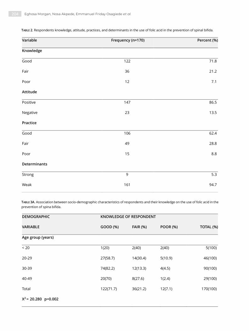
204 Eghosa Morgan, Nosa Akpede, Emmanuel Friday Osagiede et al.
TABLE 2. Respondents knowledge, attitude, practices, and determinants in the use of folic acid in the prevention of spinal bifida.
TABLE 3A. Association between socio-demographic characteristics of respondents and their knowledge on the use of folic acid in the
prevention of spina bifida.
Variable Frequency (n=170) Percent (%)
Knowledge
Good 122 71.8
Fair 36 21.2
Poor 12 7.1
Attitude
Positive 147 86.5
Negative 23 13.5
Practice
Good 106 62.4
Fair 49 28.8
Poor 15 8.8
Determinants
Strong 9 5.3
Weak 161 94.7
DEMOGRAPHIC KNOWLEDGE OF RESPONDENT
VARIABLE GOOD (%) FAIR (%) POOR (%) TOTAL (%)
Age group (years)
< 20 1(20) 2(40) 2(40) 5(100)
20-29 27(58.7) 14(30.4) 5(10.9) 46(100)
30-39 74(82.2) 12(13.3) 4(4.5) 90(100)
40-49 20(70) 8(27.6) 1(2.4) 29(100)
Total 122(71.7) 36(21.2) 12(7.1) 170(100)
X2 = 20.280 p=0.002

205 The use of folic acid in the prevention of spinal bifida
TABLE 3B. Socio-demographic characteristics of respondents and knowledge on the use of folic acid in the prevention of spinal bifida.
Marital status
Single 8(38.1) 6(28.6) 7(33.3) 21(100)
Married 98(74.8) 28(21.4) 5(3.8) 131(100)
Widowed 12(85.7) 2(14.3) 0(0) 14(100)
Cohabiting 4(100) 0(0) 0(0) 4(100)
Total 122(71.7) 36(21.2) 12(7.1) 170(100)
X2 = 29.775 p<0.001
Level of education
Primary 22(64.6) 4(11.8) 8(23.6) 34(100)
Secondary 56(66.6) 26(31) 2(2.4) 84(100)
Tertiary 44(84.7) 6(11.5) 2(3.8) 52(100)
Total 122(71.7) 36(21.2) 12(7.1) 170(100)
X2 = 25.662 p<0.0001
Occupation
Employed 48(82.8) 8(13.8) 2(3.4) 58(100)
Unemployed 13(48.2) 8(29.6) 6(22.2) 27(100)
Self-employed 58(73.4) 18(22.8) 3(3.8) 79(100)
Student 3(50.1) 2(33.3) 1(16.6) 6(100)
Total 122(71.7) 36(21.2) 12(7.1) 170(100)
X2 =18.262 p= 0.006
DEMOGRAPHIC VARIABLES KNOWLEDGE OF RESPONDENT
Pregnancies had GOOD (%) FAIR (%) POOR (%) TOTAL (%)
None 11(57.9) 3(15.8) 5(26.3) 19(100)
1-3 24(61.5) 11(28.2) 4(10.3) 39(100)
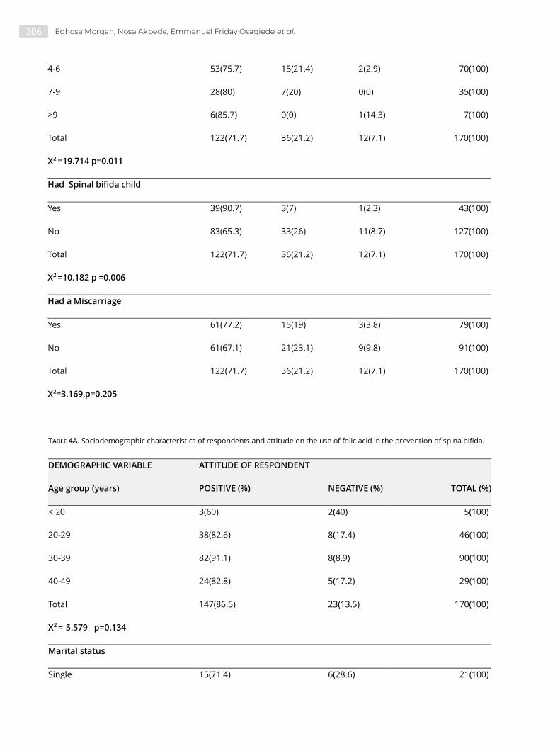
206 Eghosa Morgan, Nosa Akpede, Emmanuel Friday Osagiede et al.
TABLE 4A. Sociodemographic characteristics of respondents and attitude on the use of folic acid in the prevention of spina bifida.
4-6 53(75.7) 15(21.4) 2(2.9) 70(100)
7-9 28(80) 7(20) 0(0) 35(100)
>9 6(85.7) 0(0) 1(14.3) 7(100)
Total 122(71.7) 36(21.2) 12(7.1) 170(100)
X2 =19.714 p=0.011
Had Spinal bifida child
Yes 39(90.7) 3(7) 1(2.3) 43(100)
No 83(65.3) 33(26) 11(8.7) 127(100)
Total 122(71.7) 36(21.2) 12(7.1) 170(100)
X2 =10.182 p =0.006
Had a Miscarriage
Yes 61(77.2) 15(19) 3(3.8) 79(100)
No 61(67.1) 21(23.1) 9(9.8) 91(100)
Total 122(71.7) 36(21.2) 12(7.1) 170(100)
X2=3.169,p=0.205
DEMOGRAPHIC VARIABLE ATTITUDE OF RESPONDENT
Age group (years) POSITIVE (%) NEGATIVE (%) TOTAL (%)
< 20 3(60) 2(40) 5(100)
20-29 38(82.6) 8(17.4) 46(100)
30-39 82(91.1) 8(8.9) 90(100)
40-49 24(82.8) 5(17.2) 29(100)
Total 147(86.5) 23(13.5) 170(100)
X2 = 5.579 p=0.134
Marital status
Single 15(71.4) 6(28.6) 21(100)
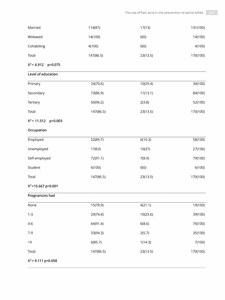
207 The use of folic acid in the prevention of spinal bifida
Married 114(87) 17(13) 131(100)
Widowed 14(100) 0(0) 14(100)
Cohabiting 4(100) 0(0) 4(100)
Total 147(86.5) 23(13.5) 170(100)
X2 = 6.912 p=0.075
Level of education
Primary 24(70.6) 10(29.4) 34(100)
Secondary 73(86.9) 11(13.1) 84(100)
Tertiary 50(96.2) 2(3.8) 52(100)
Total 147(86.5) 23(13.5) 170(100)
X2 = 11.512 p=0.003
Occupation
Employed 52(89.7) 6(10.3) 58(100)
Unemployed 17(63) 10(37) 27(100)
Self-employed 72(91.1) 7(8.9) 79(100)
Student 6(100) 0(0) 6(100)
Total 147(86.5) 23(13.5) 170(100)
X2 =15.667 p=0.001
Pregnancies had
None 15(78.9) 4(21.1) 19(100)
1-3 29(74.4) 10(25.6) 39(100)
4-6 64(91.4) 6(8.6) 70(100)
7-9 33(94.3) 2(5.7) 35(100)
>9 6(85.7) 1(14.3) 7(100)
Total 147(86.5) 23(13.5) 170(100)
X2 = 9.111 p=0.058

208 Eghosa Morgan, Nosa Akpede, Emmanuel Friday Osagiede et al.
TABLE 4B. Sociodemographic characteristics of respondents and attitude on the use of folic acid in the prevention of spinal bifida.
TABLE 5A. Socio-demographic characteristics of respondents and practice on the use of folic acid in the prevention of spinal bifida.
DEMOGRAPHIC VARIABLE ATTITUDE OF RESPONDENT
Had Spinal bifida child POSITIVE (%) NEGATIVE (%) TOTAL (%)
Yes 38(88.4) 5(11.6) 43(100)
No 109(85.8) 18(14.2) 127(100)
Total 147(86.5) 23(13.5) 170(100)
X2 =0.178, p=0.673
Had a Miscarriage
Yes 70(88.6) 9(11.4) 79(100)
No 77(85.6) 14(15.4) 91(100)
Total 147(86.5) 23(13.5) 170(100)
X2=0.576, p=0.448
DEMOGRAPHIC VARIABLE PRACTICE OF RESPONDENT
GOOD (%) FAIR (%) POOR (%) TOTAL (%)
Age group (years)
< 20 1(20) 2(40) 2(40) 5(100)
20-29 22(47.8) 16(34.8) 8(17.4) 46(100)
30-39 64(71.1) 22(24.4) 4(4.4) 90(100)
40-49 19(65.6) 9(31) 1(3.4) 29(100)
Total 106(62.4) 49(28.8) 15(8.8) 170(100)
X2 = 17.821 p=0.007
Marital status
Single 8(38.1) 5(23.8) 8(38.1) 21(100)
Married 82(62.6) 44(33.6) 5(3.8) 131(100)

209 The use of folic acid in the prevention of spinal bifida
Widowed 12(85.7) 0(0) 2(14.3) 14(100)
Cohabiting 4(100) 0(0) 0(0) 4(100)
Total 106(62.4) 49(28.8) 15(8.8) 170(100)
X2 = 35.461 p<0.001
Level of education
Primary 13(38.2) 17(50) 4(11.8) 34(100)
Secondary 53(63) 26(31) 5(6) 84(100)
Tertiary 40(77) 6(11.5) 6(11.5) 52(100)
Total 106(62.4) 49(28.8) 15(8.8) 170(100)
X2 =17.314, p=0.002
Occupation
Employed 43(74.2) 9(15.5) 6(10.3) 58(100)
Unemployed 10(37) 15(55.6) 2(7.4) 27(100)
Self-employed 49(62) 23(29.1) 7(8.9) 79(100)
Student 4(66.7) 2(33.3) 0(0) 6(100)
Total 106(62.4) 49(28.8) 15(8.8) 170(100)
X2 =15.131 , p=0.019
Pregnancies had
None 8(42.1) 3(15.8) 8(42.1) 19(100)
1-3 25(64.1) 10(25.6) 4(10.3) 39(100)
4-6 41(58.5) 27(38.6) 2(2.9) 70(100)
7-9 28(80) 7(20) 0(0) 35(100)
>9 4(57.1) 2(28.6) 1(14.3) 7(100)
Total 106(62.4) 49(28.8) 15(8.8) 170(100)
X2 = 37.809 p=0.000
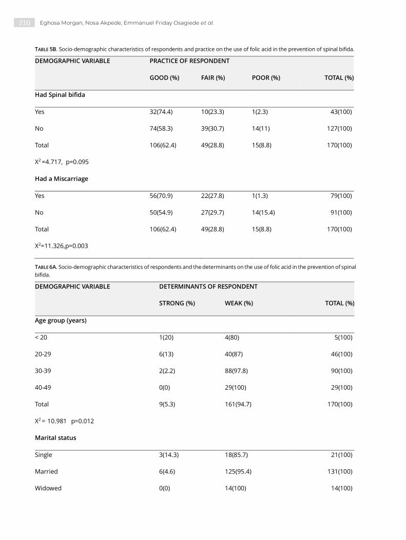
210 Eghosa Morgan, Nosa Akpede, Emmanuel Friday Osagiede et al.
TABLE 5B. Socio-demographic characteristics of respondents and practice on the use of folic acid in the prevention of spinal bifida.
DEMOGRAPHIC VARIABLE PRACTICE OF RESPONDENT
GOOD (%) FAIR (%) POOR (%) TOTAL (%)
Had Spinal bifida
Yes 32(74.4) 10(23.3) 1(2.3) 43(100)
No 74(58.3) 39(30.7) 14(11) 127(100)
Total 106(62.4) 49(28.8) 15(8.8) 170(100)
X2 =4.717, p=0.095
Had a Miscarriage
Yes 56(70.9) 22(27.8) 1(1.3) 79(100)
No 50(54.9) 27(29.7) 14(15.4) 91(100)
Total 106(62.4) 49(28.8) 15(8.8) 170(100)
X2=11.326,p=0.003
TABLE 6A. Socio-demographic characteristics of respondents and the determinants on the use of folic acid in the prevention of spinal
bifida.
DEMOGRAPHIC VARIABLE DETERMINANTS OF RESPONDENT
STRONG (%) WEAK (%) TOTAL (%)
Age group (years)
< 20 1(20) 4(80) 5(100)
20-29 6(13) 40(87) 46(100)
30-39 2(2.2) 88(97.8) 90(100)
40-49 0(0) 29(100) 29(100)
Total 9(5.3) 161(94.7) 170(100)
X2 = 10.981 p=0.012
Marital status
Single 3(14.3) 18(85.7) 21(100)
Married 6(4.6) 125(95.4) 131(100)
Widowed 0(0) 14(100) 14(100)

211 The use of folic acid in the prevention of spinal bifida
TABLE 6B. Socio-demographic characteristics of respondents and the determinants on the use of folic acid in the prevention of spinal
bifida.
Cohabiting 0(0) 4(100) 4(100)
Total 9(5.3) 161(94.7) 170(100)
X2 = 4.526, p=0.210
Level of education
Primary 2(5.9) 32(94.1) 34(100)
Secondary 3(3.6) 81(96.4) 84(100)
Tertiary 4(7.7) 48(92.3) 52(100)
Total 9(5.3) 161(94.7) 170(100)
X2 =1.117 p= 0.572
Occupation
Employed 2(3.4) 56(96.6) 58(100)
Unemployed 4(14.8) 23(85.2) 27(100)
Self-employed 2(2.5) 77(97.5) 79(100)
Student 1(16.7) 5(83.3) 6(100)
Total 9(5.3) 161(94.7) 170(100)
X2 =80.26 p=0.045
DEMOGRAPHIC VARIABLE DETERMINANTS OF RESPONDENT
STRONG (%) WEAK (%) TOTAL (%)
Pregnancies had
None 3(15.8) 16(84.2) 19(100)
1-3 2(5.1) 37(94.9) 39(100)
4-6 4(5.7) 66(94.3) 70(100)
7-9 0(0) 35(100) 35(100)
>9 0(0) 7(100) 7(100)

212 Eghosa Morgan, Nosa Akpede, Emmanuel Friday Osagiede et al.
DISCUSSION
This research was carried out to assess the
knowledge, attitude, and practice of use of folic acid
in the prevention of spinal bifida among women of
childbearing age in ECLGA of Edo State. The research
also assessed the relationship between socio-
demographic factors and knowledge and use of folic
acid in the prevention of spinal bifida. A total of 170
women, all of childbearing age, were interviewed.
The mean age is 32 years with the highest proportion
of participants 90(52.9%) consisting of those within
the age group 30-39 years.
Majority of the participants were married
131(77.1%) as compared to singles 21(12.4%),
widows 14(8.2%), cohabiting (2.4%). Up to 84(49.4%)
had secondary education, 52(30.6%) had tertiary
education, and 34(20.0%) had primary education. We
observed that the majority of the respondents 71.8%
had good knowledge about the use of folic acid in the
prevention of spinal bifida. This is in sharp contrast
to a study done on awareness of mothers about birth
defects in Ibadan, in which only 15.8% knew that
birth defects could be prevented.10 In another study
in Ibadan, only 11.8% knew that folic acid could
prevent birth defects although as much as 98.3%
were using folic acid in the current pregnancy.11
64.6% awareness of folic acid was reported in Jos.12
In Port Harcourt, 94.4% knew about folic acid, but
only 24.1% knew folic acid could prevent birth
defects.13 The high awareness in this study is quite
encouraging. It could be explained by the proximity
of the respondents to a tertiary health facility (Irrua
Specialist Teaching Hospital) which gives them
access to health education through campaigns or
interactions with resident health personnel. Also,
there is routine education of women during
antenatal visits, as most respondents stated.
We also observed that certain socio-demographic
factors had a demonstrable influence on knowledge
of folic acid use in the prevention of spinal bifida.
Age, marital status, level of education, occupation,
previous pregnancy and having babies with spinal
bifida in the past were significantly associated with
knowledge of the use of folic acid whereas factors
like religion and number of miscarriages were not.
These observations are by earlier observations.11, 14-
18 On the aspect of attitude towards the use of folic
acid, most of the respondents 86.5%. Showed a
positive attitude. Sociodemographic factors
significantly associated with this positive attitude
were level of education (p=0.003), occupation
(p=0.001).
Total 9(5.3) 161(94.7) 170(100)
X2 =6.549, p=0.162
Had Spinal bifida child
Yes 0(0) 43(100) 43(100)
No 9(7.1) 118(92.9) 127(100)
Total 9(5.3) 161(94.7) 170(100)
X2 =3.218, p=0.073
Had Miscarriage
Yes 4(5.1) 75(94.9) 79(100)
No 5(5.9) 86(94.1) 91(100)
Total 9(5.3) 161(94.7) 170(100)
X2=0.016, p=0.900

213 The use of folic acid in the prevention of spinal bifida
More than half of the respondents 62.4 had a good
practice which compares favorably with the result of
the study done in Jakarta, Indonesia where 64% of
respondents claimed to have been compliant with
the use of folic acid.33 In another study done in
Canada, 71% were compliant with the use of folic
acid,35 while only 22.3% of the women in a study in
Denmark showed compliance19. The differences
between our observation and between the results in
previous reports could be due to the difficulty in
measuring the actual practice of use of folic acid by
women of childbearing age, for example in the
Jakarta study20; two parallel results were obtained
from the same respondents using qualitative and
quantitative tools.
There was a statistically significant relationship
between age (p=0.007), number of miscarriages
(p=0.003), marital status (p=0.000), level of education
(p=0.002), number of pregnancies had (0.000). A
number of pregnancies had the highest statistical
significance.
Determinants of use of folic acid showed
statistically significant relationship with certain
sociodemographic factors such as with age group
such as women between 30-39 years had strong
score while women less than 20 years had weak
score reason for which may be due to teenage
unplanned pregnancy, ignorance and late antenatal
booking which is in keeping with the study done in
South-western Nigeria.17 In our study, the occupation
had the highest statistical significant relationship
(p=0.045). This shows that employment empowers
these women making them better placed to use folic
acid. Also, employment is also related to an older
age.
CONCLUSION
We concluded that
1. The knowledge of the use of folic acid among
women of childbearing age in ECLGA is good.
Furthermore, attitude and practice of use of folic acid
match the knowledge and compares unfavourably
with the levels reported from other places.
2. The level of knowledge, attitude, and practice on
the use of folic acid among women of childbearing
age in ECLGA are dependent on age, marital status,
level of education, occupation, number of
pregnancies, previous child with spinal bifida.
RECOMMENDATION
We recommend that:
1. The government and health institution should
work closely with stakeholders in the health sector to
create more public enlightenment campaigns to
increase awareness about spinal bifida and use of
folic acid either in their diet or as medication in its
prevention especially among women of childbearing
age
2. Communities should organize forums where a
health worker enlightened member about the
benefits of the use of folic acid by women of
childbearing age while also answering any questions
they may have.
REFERENCES
1. Botto LD, et al. Neural tube defects N Engl J Med. 1999 Nov.
11; 341 (20): 1509-19
2. Molloy, AM. The role of folic acid in the prevention of NTDs.
Trends Food Sci Technol. 2005; 16 (6-7): 241-5.
3. Bowman RM, Boshnjaku V. and Mclone DG (2009). The
Changing incidence of myelomeningocele and its impact
on paediatric neurosurgery: a review from the children’s
memorial hospital child’s nerv syst July 2009 25 (7): 801-6
4. Anyanwu LC, Danborno B, Hamman W.O. The prevalence
of Neural Tube Defects in live born neonates in Kano,
North-Western Nigeria. Afr J Med 2015; 2:105-9
5. Airede KI. Neural Tube Defects in the middle belt of Nigeria.
J Trop Pediatr 1992; 38:27-30
6. MRC Vitamin Study Research Group. Prevention of neural
tube defects: results of the medical research council
vitamin study. Lancet 1991; 338:131-7
7. Ezeizel AE, Dudas I. Prevention of the first occurrence of
neural tube defects by periconceptional vitamin
supplementation, N Engl J Med 1992; 327: 1832-5
8. WHO e-Library of Evidence for Nutrition Actions (eLENA).
www.who.int/elena/title/folate_periconceptional/en/ Last
accessed: 9.00pm, 25th July 2017.
9. Crider KS, Devine O, HaoLing, Dowling NF, Lis, Molly AM et
al. population red blood cell folate concentrations for
prevention of neural tube defects: a Bayesian model. BMJ
2014; 349: g4554
10. Lawal TA, et al. "Knowledge of birth defects among nursing
mothers in a developing country" African Health Science
2015 Mar; 15(1):180-187. www.ncbi.nlm.nih.gov/pmc/arti
cles /PMC4370152/
11. Adebo OO, et al. "Knowledge and uptake of folic acid
among pregnant women attending a secondary health
facility in Nigeria" British Journal of Midwifery 2017 June;
25(6): 358-364

214 Eghosa Morgan, Nosa Akpede, Emmanuel Friday Osagiede et al.
12. Anzaku SA. "Assessing folic acid awareness and its usage
for the prevention of neural tube defects among pregnant
women in Jos, Nigeria. Journal of Basic and Clinical
Reproductive Science Jan-June 2013; 2(1): 13-17
13. Eghwrudjakpor PO, et al. " Evaluation of the level of
awareness of the role of folic acid in the prevention of
neural tube defects amongst women of reproductive age
in a tertiary health institution." Niger J Med 2011 Apr-Jun.
14. Dessie MA, Zeleke EG, et al. "Folic acid usage and associated
factors in the prevention of neural tube defects among
pregnant women in Ethiopia: a cross-sectional study" BMC
Pregnancy Childbirth v. 17(2017)
15. Sadore AA, et al. "Compliance with iron-folate supplement
and associated factors among antenatal care attendant
mothers in Misha District, South Ethiopia: community-
based cross-sectional study" Journal of Environmental and
Public Health 2015(2):1-7
16. Gathigi LN. "Factors influencing utilization of iron and folic
acid supplementation services among women attending
antenatal clinic at Nyeri Provincial Hospital, Kenya"
17. Lawal TA and Adeleye A. O. "Determinants of folic acid
intake during preconception and in early pregnancy by
mothers in Ibadan, Nigeria" The Pan African Medical
Journal
18. Ezegwui HU, et al. "Preconception care in South Eastern
Nigeria" Japan Obstet Gynaecol. 2008
19. Knudsen VK, Oroziva-Bekkevold I, Rasmussen LB,
Mikkelsen TB. Low compliance with recommendations on
folic acid use in relation to pregnancy: is there a need for
fortification? DOI: 10. 1079?PHN2004630. Public heal
nutrition 2004: 7(7); 843-850
20. Schultink Well, et al. (1993). "Low compliance with an iron
supplementation program: a study among pregnant
women in Jakarta, Indonesia." Am J Clin Nutr 57(2):135-139.

215
Guidelines for authors
1. ETHICS
The publication of an article in Romanian Neurosurgery is a
direct reflection of the quality of the work of the authors. The
prevention of publication malpractice is first the responsibility
of every author and also of our editorial board.
Authors must submit accurate information and sufficient
details, presenting its objective significance; unethical
behaviour is unacceptable.
Plagiarism in all its forms constitutes unethical publishing
behaviour and is unacceptable.
For Romanian Neurosurgery the publication ethics and
publication malpractice statement are consistent with the
recommendations and guidelines of the Committee on
Publication Ethics, the World Association of Medical Editors,
the International Committee of Medical Journal Editors and
Consolidated Standards Of Reporting Trials.
Links:
Committee on Publication Ethics
(COPE): http://www.publicationethics.org
World Association of Medical Editors
(WAME): http://www.wame.org
International Committee of Medical Journal Editors
(ICMJE): http://www.icmje.org
2. ENCLOSED LETTER
In addition to the manuscript, the Editorial Board should
receive an enclosed letter containing the exclusive reservation
of copyright guaranteed by all authors whose manuscripts
have already been accepted. If the paper was completely or
partially published or exposed previously, a copy or a
photocopy of it should be also sent. The technical reports
should contain a declaration concerning the financial sources
that cover the costs necessary for instruments and
methodology acquisition.
In order to illustrate different cases, photos of identifiable
patients will not be published without their legal consent or
that of their legal representative. The letter containing this
consent together with the manuscripts should be sent to the
editorial office.
If the author wishes his unpublished manuscripts returned,
please note this in the enclosed letter.
3. SENDING OF MANUSCRIPTS
Authors shall ensure that the article has been "spell and
grammar checked" prior to the submission.
The manuscript sent for publishing must be submitted in
English. The manuscript will be typed without formatting, with
a 1-line space. Please enclose 1 copy of the manuscript, tables,
graphics and photos. After publishing, the paper and pictures
become the property of Romanian Neurosurgery.
There are no article processing charges (NO APCs) and no
article submission charges.
4. MANUSCRIPT ELABORATION
Paper sent for publishing should be in accordance with
international standards of manuscript submittance. These
standards are mentioned in “British Medical Journal” 1988;
296: 404-405 or in “Annals of Internal Medicine” 1988; 108:258-
265. The authors are responsible for the accuracy of the
information contained in the essay.
4.1. The title page should contain the whole title of the essay
and complete names of authors with their academical
degrees. If it is necessary, the department, the hospital or the
institution where the search has been undertaken, should be
also mentioned.
4.2. Please include an additional page containing the title of
the essay and the author responsible for correcting of any and
of all mistakes and for maintaining correspondence. The
address, phone and fax number should be included (e-mail be
available).
4.3. A summary, no longer than 300 words, should be written
on a separate page. Key-words, no more than 7, should be
listed in alphabetical order on the same page. The use of
keywords should be approved by the “Index Medicus”.
4.4. Text. The introduction should specify the purpose of the
paper. The content and the method should give a minute
account of the work methodology so that the experiment
conclusion could be reproduced and checked up on in other
centres. The experiments and medical studies performed on
human beings should respect the principles specified in “The
Declaration of Helsinki”, whereas the experiments done on
animals should be in accordance with “The Principles Charta
of Animal’s Care and Use”. The results should not contain
references to previous studies. The discussions should reflect
the main features of the research.
5. REFERENCES
- typed on 1 line;
- quoted in alphabetical order;
- explanatory footnotes are not accepted;
- unpublished data and personal papers should be quoted
inside the text and not in the bibliography;
- entries to the bibliography should appear in the following
order: the authors, the title of the essay, the title of the

216
periodical (abbreviated according to the list of abbreviation
specified in the “Index Medicus”), the volume number, the
page number and the data publication.
6. TABLES AND PICTURES
There should be 1 copy sent. Each table with its own title
should be submitted on separate pages. The photos,
radiographies, CT scans should be labelled on the back with
the number of each picture, corresponding with the number
included inside the text, and the author’s name. The label will
be placed on the top of the picture. The drawings may be sent
on vellum paper, tracing paper or transparent paper. The use
of pictures belonging to other publications is accepted, only if
mentioning the original source. Further, the partial use of any
previously published text is accepted with the approval of its
author and editor. All pages should be consecutively
numbered, starting with the title page.
7. ABBREVIATIONS
Abbreviations should be consistently used throughout the text
and established in a fixed form from the beginning.
8. SUBMISSION
Manuscripts should be sent to the Editor:
Dr St.M. Iencean,
Romanian Neurosurgery
"Gr.T. Popa" University of Medicine and Pharmacy
Universitatii Street, No. 16, 700115, Iasi, Romania
or by e-mail to [email protected].
9. DISCLAIMER
The editors disclaim any responsibility for opinions expressed
in the papers.
10. THE REVIEW PROCESS
After the manuscript submission, the peer review process is
broken down into the following steps:
1. The Editor assigns Reviewers to the manuscript.
2. The Reviewers review the manuscript.
3. The Editor drafts a decision to be sent to the author/authors.
The review process takes between three weeks and two
months.