ROMANIAN JOURNAL OF BIOLOGY PLANT BIOLOGY
62
ROM. J. BIOL. – PLANT BIOL., VOLUME 62, No. 2, P. 59–119, BUCHAREST, 2017 ROMANIAN JOURNAL OF BIOLOGY PLANT BIOLOGY VOLUME 62, No. 2 2017 CONTENTS V.S. HARIKUMAR, Varietal difference in natural colonization by arbuscular mycorrhizal fungi and its influence on plant characters and phosphorus nutrition in sesame ......................................................................................... 61 I. VICOL, Red listed lichen species within old growth and young growth forests from Romania ................................................................................................ 67 D. VOICU, A. BREZEANU, N. TOMA, Lichens – some relevant aspects of the in vitro culture and ultrastructural peculiarities ............................................. 77 A.J.S. RAJU, K.V. RAMANA, Pollination ecology of Rhynchosia suaveolens (L.F.) Dc. (Fabaceae), a perennial erect shrub in the southern eastern Ghats, Andhra Pradesh, India ................................................................................................ 89 K.A. ABDULKAREEM, T. GARUBA, E. O. AKANDE, O.T. MUSTAPHA, Effect of sodium azide on morphological characters of three tomato accesssions (Solanum lycopersicon L.) .......................................................... 109 BOOK REVIEW I.I. ARDELEAN, Nanotechnologies in Food and Agriculture ................................... 119
-
Upload
khangminh22 -
Category
Documents
-
view
1 -
download
0
Transcript of ROMANIAN JOURNAL OF BIOLOGY PLANT BIOLOGY

ROM. J. BIOL. – PLANT BIOL., VOLUME 62, No. 2, P. 59–119, BUCHAREST, 2017
ROMANIAN JOURNAL OF BIOLOGY
PLANT BIOLOGY
VOLUME 62, No. 2 2017
CONTENTS
V.S. HARIKUMAR, Varietal difference in natural colonization by arbuscular
mycorrhizal fungi and its influence on plant characters and phosphorus
nutrition in sesame ......................................................................................... 61
I. VICOL, Red listed lichen species within old growth and young growth forests
from Romania ................................................................................................ 67
D. VOICU, A. BREZEANU, N. TOMA, Lichens – some relevant aspects of the
in vitro culture and ultrastructural peculiarities ............................................. 77
A.J.S. RAJU, K.V. RAMANA, Pollination ecology of Rhynchosia suaveolens (L.F.)
Dc. (Fabaceae), a perennial erect shrub in the southern eastern Ghats, Andhra
Pradesh, India ................................................................................................ 89
K.A. ABDULKAREEM, T. GARUBA, E. O. AKANDE, O.T. MUSTAPHA,
Effect of sodium azide on morphological characters of three tomato
accesssions (Solanum lycopersicon L.) .......................................................... 109
BOOK REVIEW
I.I. ARDELEAN, Nanotechnologies in Food and Agriculture ................................... 119


ROM. J. BIOL. – PLANT BIOL., VOLUME 62, No. 2, P. 61–66, BUCHAREST, 2017
VARIETAL DIFFERENCE IN NATURAL COLONIZATION
BY ARBUSCULAR MYCORRHIZAL FUNGI AND ITS INFLUENCE ON PLANT CHARACTERS
AND PHOSPHORUS NUTRITION IN SESAME
V.S. HARIKUMAR1
Abstract. Twenty accessions of sesame (Sesamum indicum L.) plants were evaluated for arbuscular mycorrhizal (AM) colonization and its relationship with growth, yield and tissue P content. Sesame accessions exhibited variations in both frequency (%F) and intensity (%M) of colonization by AM fungi. Among the accessions, 30% had a comparatively higher %F whereas in other ones the %F was more or less on par. The %M was comparatively more in 40% of the accessions while two accessions had a very low %M. AM fungi which occurred in the rhizosphere of sesame accessions belonged to two species of Acaulospora and three species of Glomus. Glomus spp was found dominant in the rhizosphere soils of sesame. Correlation studies revealed a significant positive relationship between fungal variables as well as between fungal variables and plant characters such as growth, yield and tissue P content.
Keywords: AMF, host variety, plant characters, sesame.
INTRODUCTION
The ubiquitous arbuscular mycorrhizal (AM) fungi are an integral component of any soil system where they form obligate symbiosis with the roots of over 80% terrestrial plant species (van der Heijden & Sanders, 2002). Genetic variation within plant species can influence both the degree of root colonization by mycorrhizal fungi and the response of the plant to mycorrhizal symbiosis (Peterson & Bradbury, 1995). For example, Harikumar & Potty (2002) screened 257 genetic stocks of field grown sweet potato and found that 20% of them responded to natural colonization to the tune of 25% whereas, another 20% had 50% colonization in their root system and the remaining 60% had a very high level of colonization. Selection of host plant germplasm to maximize productivity and pest resistance or responsiveness to fertilization can affect the ability of the host to sustain or benefit from mycorrhizas (Kaeppler et al., 2000; Tawaraya, 2003).
Sesame (Sesamum indicum L., Fam. Pedaliaceae) is cultivated in tropical, subtropical and southern temperate regions of the world for its seeds which are a
1 Department of Post Graduate Studies & Research in Botany, Sanatana Dharma College,
Alappuzha-688 003, Kerala, e mail: [email protected]

V.S. Harikumar 2 62
rich source of edible oil. Incidence of AM colonization in sesame roots and infective propagules in the rhizosphere of the crop have been reported earlier (Sulochana et al., 2000; Harikumar, 2015). The evaluation of plant varieties for AM colonization and endophyte specificity helps to identify the most compatible symbiont with the host (Dhillion, 1992). The present paper reports the varietal differences and species specificity in AM colonization in 20 sesame varieties and its influence on plant characters.
MATERIALS AND METHODS
Twenty accessions of sesame (courtesy KAU (RS) Kayamkulam) collected from different parts of the country were evaluated for AM colonization and its relationship with growth, yield and P content of the plants. The study was conducted in a farmer’s field located at Alappuzha, Kerala. The soil was sampled for analysis of physico-chemical properties (Jackson, 1973) and AM fungal population prior to the initiation of the experiment. The soil was a sandy Entisol with a pH 5.5 (1: 2:5; soil: water) and organic carbon 1.17%. Soil nutrient determinations included 108 kg N h
-1, 25 kg P h
-1
and 19.40 kg K h-1
. The total indigenous AM fungal spore density prior to the start of the study was 103 spores per 50 ml soil.
Seeds were hand sown in rows at a distance of 1 ft with three replicate pits for each accession. After emergence of seedlings, the plant number in each pit was thinned to three to avoid overcrowding. Plants did not receive any fertilization or irrigation throughout the growth period. Weeds were controlled by hand as required.
Plants were harvested at 75 days after emergence. Three replicated plants for each accession were dug out with almost the entire root system intact. Rhizosphere soil samples were collected from 10–20 cm depth. The harvested plants were utilized for monitoring mycorrhizal colonization, growth and yield. The tissue P content was determined as per Jackson (1973).
The undamaged fine roots of test plants were cut into 1 cm root segments from these about 50 segments were selected at random. The root segments were thoroughly washed and stained with cotton blue (Phillips & Hayman, 1970). The root segments were mounted on clean microscopic slides in a mixture of glycerol and lactic acid (v/v). The root segments were gently squashed and covered by a glass cover slip and observed under a compound microscope (Nikon Eclipse E 400) using different magnifications. Mycorrhizal variables, frequency (%F) and intensity (%M) of AM colonization were calculated following the method of Trouvelot et al., 1986). Spores were extracted from a sub-sample (50 ml) of each rhizosphere soil sample by wet-sieving and sucrose density gradient centrifugation (Daniels and Skipper, 1982) and examined using a Zeiss Stemi-DV4 stereomicroscope. Spores of each morphotype were mounted on slides in polyvinyl alcohol-lactic acid-glycerol (PVLG) (Koske & Tessier, 1983) and PVLG mixed 1:1 (v/v) with Melzer’s reagent. Spores were examined using a Nikon Eclipse 400 research microscope and identified up to species level using the manual for the identification of VA mycorrhizal fungi by

3 Host variety and AM colonization in sesame 63
Schenck & Pérez (1990). Spore density was determined as the number of healthy appearing spores per 50 ml soil. Isolation frequency was calculated as the percentage of samples in which the particular genus or species was present.
RESULTS AND DISCUSSION
AM colonization in sesame accessions exhibited variation in both %F and %M. Among the 20 accessions screened, 30% had a comparatively higher (>40%)
%F whereas in other ones the %F was more or less on par. %M was comparatively more (>8%) in 40% of the accessions while two accessions (SI 68, SI 69) had a
very low %M (Table 1). While AM fungi are known to vary in their ability to colonize and transfer P to the plant and confer other beneficial effects, little is known to the
exact role of the host genotype in the expression of AM fungi. In the present study, though the accessions were grown in the same field under identical environmental
condition, there was significant variation in root colonization by AM fungi which
could be ascribable at least in part to genetic and physiological factors controlling host/fungus compatibility. Recently discovered mycorrhizal mutants (myc
-) of pea
offer great promise for the study in this direction (Graham & Eissenstat, 1994). Similar genotype dependent variation for AM fungal colonization has been reported in
wheat (Azcón & Ocampo, 1980) sorghum (Clark, 1983) pearl millet (Krishna et al., 1985) coconut (Thomas & Ghai, 1987) and cowpea (Mercy et al., 1990).
AM fungi which occurred in the rhizosphere of sesame accessions belonged to two species of Acaulospora and three species of Glomus (Table 1). Glomus spp
was found dominant in the rhizosphere soils. The results revealed that G. mosseae associated with most accessions with the highest frequency (Figure 1). Glomus species
are capable of colonizing roots via fragments of mycelium or mycorrhizal root pieces (Daniell et al., 2001). This may be the possible reason for the predominance of
the genera over other genera in most agricultural soils (Morton, 1988). Furthermore, they are widely adaptable to the varied soil conditions and survive in acidic as well
as alkaline soils (Pande & Tarafdar, 2004). Correlation studies between fungal variables (%F, %M) revealed that there
exists a significant positive relationship between these variables. A similar significant positive correlation was observed between fungal variables and plant characters (Table 2). %F was significantly correlated with measured variables such as growth, yield and P content. %M also followed the same pattern of relationship. Harikumar & Potty (2002) in a field study with 257 genetic stocks of sweet potato for mycorrhizal colonization and its response on growth of the crop observed that fungal characters showed a positive relationship with underground plant characters (root and tuber) only while the above ground portion had no direct bearing on the interaction. By contrast, in the present study all the parameters examined showed a significant correlation with the fungal variables further confirming the importance of indigenous AM endophytes on the growth and nutrition of sesame.

V.S. Harikumar 4 64
Table 1
Frequency (%F) and intensity (%M) of root colonization and AMF species associated with sesame accessions
Accession no. Colonization*
AM Species** % F %M
SI 2 43.33 bcd 8.70c 3, 4, 5
SI 7 43.33bcd 9.17 3, 4, 5
SI I5 26.67ef 5.66c 2, 4
SI I7 23.33ef 4.50ef 2, 4
SI 25 26.67 ef 5.90de 2, 3
SI 30 56.67ab 17.70a 1, 3, 4, 5
SI 32 46.67bc 8.17cd 1, 4, 5
SI 37 28.33def 4.00ef 2, 4
SI 42 28.33 def 4.93ef 3, 5
SI 47 68.33a 19.30a 1, 3, 4, 5
SI 48 50.00b 14.50 b 1, 3, 4
SI 56 33.33cde 8.90c 4, 5
SI 57 33.33cde 8.60c 2, 4
SI 58 26.67ef 5.43ef 3, 5
SI 59 26.00ef 5.27ef 3, 4
SI 63 23.33ef 4.50ef 2, 3
SI 66 23.33ef 5.07ef 2, 3
SI 68 16.67f 3.07f 3
SI 69 28.33def 3.63ef 4, 5
SI 70 28.33def 4.13ef 4, 5
* Means in each column with different letters are significantly different (P<O.05) by Tukey’s HSD ** 1. A. delicata 2. A. lacunosa 3. G. dimorphicum 4. G. mosseae 5. G. versiformae
Fig. 1. Frequency of AM species in the rhizosphere of sesame accessions.

5 Host variety and AM colonization in sesame 65
Table 2
Frequency (%F) and intensity (%M) of root colonization and AMF species associated with sesame accessions
Parameters Mycorrhizal parameters
% F % M
Mycorrhizal parameters % F % M
1.000 0.897***
1.000
Growth characters Root length (cm) Shoot length (cm) Leaf Number plant _1
0.479*** 0.499*** 0.707***
0.547*** 0.528*** 0.781***
Biomass production Root fresh weight (g) Shoot fresh weight (g) Total biomass (g)
0.593*** 0.493*** 0.538***
0.669*** 0.563*** 0.614***
Dry matter production Root dry weight (g) Shoot dry weight (g) Total dry weight (g)
0.573*** 0.526*** 0.544***
0.667*** 0.621*** 0.641***
Yield Pod Number (plant-1)
0.404***
0.358***
P efficiency P content (mg g-1)
0.546***
0.480***
***P<0.001.
CONCLUSION
Sesame accessions revealed AM colonization attributable to the species of Acaulospora and Glomus which varied with varieties. In general, both %F and %M were low in sesame accessions. Nevertheless, the crop is greatly dependent on indigenous AM fungi for growth and nutrition as a significant positive relationship between plant and fungal characters could be drawn out through correlation analysis.
REFERENCES
1. Azcón R. and N.A. Ocampo, 1980, Factors affecting the vesicular – arbuscular infection and mycorrhizal dependency of thirteen wheat cultivars. New Phytol, 87, pp. 677–685.
2. Clark R.B., 1983, Plant genotype differences in the uptake, translocation, accumulation and use of mineral elements required for plant growth. Plant Soil, 72, pp. 175–196.
3. Daniell T.J., R. Husband, A.H. Fitter, and J.P.W. Young, 2001, Molecular diversity of arbuscular mycorrhizal fungi colonizing arable crops. FEMS Microbiol Ecol, 36, pp. 203–209.
4. Daniels B.A. and H.D. Skipper, 1982, Methods for the recovery and quantitative estimation of propagates from soil In: N.C. Schenck, Editor, Methods and Principles of Mycorrhizal Research. American Phytopathological Society, St. Paul, Minn, pp. 29–35.
5. Dhillion S.S., 1992, Evidence for host-mycorrhizal preference in native grassland species. Mycol Res, 94, pp. 359–362.
V.S. Harikumar 6 66
6. Graham J. H. and D.M. Eissenstat, 1994, Host genotype and the formation and function of VA
mycorrhizae. Plant Soil, 159, pp. 179–185.
7. Harikumar V.S, 2015, Arbuscular mycorrhizal associations in sesame under low-input cropping
systems. Archives Agron Soil Sci, 61, pp. 347–359.
8. Harikumar V.S. and V.P. Potty, 2002, Genotype dependent variation in arbuscular mycorrhizal
colonization and its response on the growth of sweet potato. Bulgarian J Agric Sci, 8, pp. 161–166.
9. Jackson M.L., 1973, Soil Chemical Analysis, Prentice Hall, New Delhi.
10. Kaeppler S.M., J.L. Parke, S.M. Mueller, L. Senior, C. Stuber, and W.E. Tracy, 2000, Variation
among maize inbred lines and detection of quantitative trait loci for growth at low phosphorus and
responsiveness to arbuscular mycorrhizal fungi. Crop Sci, 40, pp. 358–364.
11. Koske R.E. and B. Tessier, 1983, A convenient permanent slide mounting medium. Mycol Soc
Am Newsl, 34, pp. 59.
12. Krishna K.R., K.G. Shetty, P.J. Dart, and D.J. Andrews, 1985, Genotype dependent variation in
mycorrhizal colonization and response to inoculation in pearl millet. Plant Soil, 86, pp. 113–125.
13. Mercy M.A., G. Shivashankar, and D.J. Bagyaraj, 1990, Mycorrhizal colonization in cowpea is
host dependent and heritable. Plant Soil, 121, pp. 292–294.
14. Morton J.B., 1988, Taxonomy of VAM fungi, classification, nomenclature and identification.
Mycotaxon, 32, pp. 267–324.
15. Pande M. and J.F. Tarafdar, 2004, Arbuscular Mycorrhizal fungal diversity in Neem based
Agroforestry Systems in Rajastan. Appl Soil Ecol, 26, pp. 233–241.
16. Peterson R.L. and S.M. Bradbury, 1995, Use of plant mutants, intraspecific variants and non-hosts
in studying mycorrhiza formation and function. In: A.K. Varma, and B. Hock, Editors, Mycorrhiza:
Structure, Function, Molecular Biology and Biotechnology. Springer, Berlin, Germany.
17. Phillips J.M. and D.S. Hayman, 1970, Improved procedure for clearing roots and staining
parasitic and vesicular arbuscular mycorrhizal fungi for rapid assessment of infection. Trans Br
Mycol Soc, 55, pp. 158–161.
18. Schenck N.C. and Y. Pérez, 1990, Manual for the identification of VA mycorrhizal fungi (3rd edn),
Synergistic Publication, Gainesville F L, U.S.A.
19. Sulochana T., P. Reddy, Jagan Mohan and C. Manoharachary, 2000, Impact of season on the
dynamics of arbuscular mycorrhizal fungi in sesamum. J Indian Bot Soc, 79, pp. 179–183.
20. Tawaraya K., 2003, Arbuscular mycorrhizal dependency of different plant species and cultivars.
J Soil Sci Plant Nutr, 49, pp. 655–668.
21. Thomas G.V. and S.K. Ghai, 1987, Genotype dependent variation in vesicular – arbuscular
mycorrhizal colonization of coconut seedlings. Proc Indian Acad Sci (Plant Sci.), 97, pp. 289–294.
22. Trouvelot A., J. Kough, and V. Gianinazzi – Pearson, 1986, Evaluation of VA infection levels in
root systems. Research for estimation methods having a functional significance. In: V. Gianinazzi –
Pearson and S. Gianinazzi S, Editors, Physiological and Genetical Aspects of Mycorrhizae, INRA –
Press, Paris, pp. 217–221.
23. Van der Heijden M.G.A. and I.R. Sanders, 2002, Mycorrhizal Ecology. Ecological Studies,
Springer-Verlag, New York, USA.

ROM. J. BIOL. – PLANT BIOL., VOLUME 62, No. 2, P. 67–76, BUCHAREST, 2017
RED LISTED LICHEN SPECIES WITHIN OLD GROWTH
AND YOUNG GROWTH FORESTS FROM ROMANIA
I. VICOL1
Abstract. In the studied areas of a total of 19 red listed lichen species from Romania
there were observed 4 red listed lichen species, for instant: Cetraria islandica (L.) Ach.
(1803), Cetraria saepincola (Ehrh.) Ach. (1803), Hypotrachyna sinuosa (Sm.) Hale
(1975), and Lobaria pulmonaria (L.) Hoffm. (1796). The red listed lichen species were
predominantly identified in mountainous areas within old growth forests well conserved, the
most of them being protected areas. As regards young growth forests, only one red listed
lichen species was found in a mixed forest represented in a great deal by ancient oaks
from the Romanian Plain. In the field it was observed that red listed lichen species have
been found especially on old trees, such as: oak and beech. In the lowland areas, within
mixed forests represented especially by oaks Hypotrachyna sinuosa has been identified
while within montane forests well represented by beech, fir and spruce the presence of
L. pulmonaria has been observed. In addition to forest habitats, C. islandica and
C. saepincola were found on mugo pine and common juniper in subalpine and alpine
belts with undetectable anthropogenic pressure.
Keywords: forest, lichen, mountainous areas, oak, beech.
INTRODUCTION
At European level both old growth forests and their characteristic lichen
species are nowadays relictual (Scheidegger & Goward, 2002). The mixed forests
from Europe are predominantly represented by oak, elm and beech which have been
harversted and converted into secondary forests. The vast forestlands are threatened
because of increasing of agricultural, urbanization and industrial activities; in this
regard to avoid the loss of the forest biodiversity protected areas have been
designed (Nascimbene et al., 2013).
It has been estimated that 0.2% of mixed forests from Europe are old-growth,
the other being intensively managed for economical development. Nevertheless,
Romania is very well represented by deciduous forests compared with western
European countries (Nascimbene et al., 2013).
Old oaks host a rich lichen species diversity due to their favourable
microhabitats, but in young growth forests the lichen diversity is subjected to a
1 Institute of Biology of the Romanian Academy, 296 Splaiul Independenţei, 060031, Bucharest,
Romania, e-mail: [email protected]

I. Vicol 2 68
progressive loss depending on the scale of the shade degree (Paltto et al., 2011).
In Europe, during forest management, the old oaks were retained constituting a
network of the individual patches whose elements act an important role both to
dispersal diaspores and to lead further a spatial model for conserving lichen species
diversity including many red listed species (Ranius et al., 2008). Within forest
habitats from Swedish, on deciduous trees older than 150 years were found lichen
species associated to old-growth forests, for instance: Calicium salicinum, Lobaria
pulmonaria and Mycobilimbia sabuletorum. These lichen species are considered
“late-successional species” probably favoured by texture and chemical composition
of the tree bark. Forest continuity includes besides old trees other old growth forest
elements such as “number of tree layers” and “stand age”; therefore to conserve the
old forest attributes is necessary to retain groups of old trees during clear-cutting
(Gustafsson et al., 1992). Although a great attention must be paid to the anthropogenic
impact that is responsible to decreasing the red listed lichen species in protected
areas, considered nowadays as major refuge habitats (Motiejūnaitė, 2011).
Within young-growth forests there are predominant common lichen species
which belong to Xanthorion group unlike to sensitive lichen species found in old-
growth forests. The sensitive lichen species are affected by forest management
including clearing and cutting, the density of young trees and the presence of shrubs;
therefore “higher habitat quality requirements” as characteristic environment for them
are necessary (Aragón et al., 2010).
The aim of this study consists in highlighting the importance of the presence
of red listed lichen species within young-growth and old-growth forests. The
objectives of the study are the following: (1) finding out the presence of red listed
lichen species within young-growth and old-growth forests and (2) revealing the
presence of red listed lichen species within forest habitats situated in geomorphological
units such as: plain, hills and mountains.
MATERIALS AND METHODS
The field researches were performed during 2009–2015 within old growth
and young growth forests from Alba, Bihor, Botoșani, Brașov, Caraș-Severin, Cluj,
Gorj, Hunedoara, Ilfov, Neamț, Prahova, Sibiu and Vaslui counties (Fig. 1).
The specimens collected from the field were determined in laboratory using
keys (Moruzi & Toma, 1971; Purvis et al., 1994; Ciurchea, 2004), stereomicroscope
(Zeiss Stereo CL 1500 ECO), optical microscope (Zeiss Scope A1) and chemical
reagents such as IIK (iodine-potassium iodide) and KOH (potassium hydroxide).
Identification and nomenclature of the tree species is according to
www.uk.ipni.org, whilst for lichen nomenclature it was used www.mycobank.org.
The identified material is preserved within the Mycological Herbarium, Lichen
Collection (BUCM L) Institute of Biology, Romanian Academy, Bucharest, Romania.

3 Red listed lichen species within forests from Romania
69
Fig. 1. The studied sites within red listed lichen species were identified.
RESULTS
Within the investigated forests there were identified 4 lichen species which are found on the Romanian Red List of lichens (Sârbu et al., 2007; Ardelean et al., 2013), for instance: Cetraria islandica (L.) Ach. (1803), Cetraria saepincola (Ehrh.) Ach. (1803), Hypotrachyna sinuosa (Sm.) Hale (1975), and Lobaria pulmonaria (L.) Hoffm. (1796).
The chorology of the identifed red listed lichen species is the following: Cetraria islandica (L.) Ach. (1803) Fig. 2. Alba County: Șureanu Mountain, at the end of the ski lifts, found on terricolous
substrata, in a meadow, leg. Vicol Ioana, 24.10.2012, det. Vicol Ioana, 31.10.2012, BUCM L1749; found within shrubs represented by Juniperus communis Thunb. (1784), leg. Vicol Ioana, 24.10.2012, det. Vicol Ioana, 08.11.2012, BUCM L1758.
Brașov County: Piatra Craiului Mountains, Turnu Peak, found on terricolous substrata, leg. Vicol Ioana, 27.06.2012, det. Vicol Ioana, 03.07.2012, BUCM L1748.
Caraș-Severin County: Țarcu Mountains, Țarcu Peak, northern exhibition, alt. 1900 m, found on saxicolous substrata, within shrubs represented by Vaccinium sp., leg. Ion Roxana Georgiana, 06.08.2013, det. Vicol Ioana, 09.05.2014, BUCM L2292.
Gorj County: Parâng Mountains, near Rânca locality, found on terricolous substrata, in a meadow, leg. Vicol Ioana, 23.10.2012, det. Vicol Ioana, 02.11.2012,

I. Vicol 4 70
BUCM L1750, BUCM L1753, BUCM L1754, BUCM L1755, BUCM L1757; Parâng Mountains, between Rânca and Obârșia Lotrului, found on terricolous substrata, in a meadow, leg. Vicol Ioana, 23.10.2012, det. Vicol Ioana, 01.11.2012, BUCM L1751, BUCM L1752; leg. Vicol Ioana, 23.10.2012, det. Vicol Ioana, 31.10.2012, BUCM L1756.
Fig. 2. The chorology of the red listed Cetraria islandica in Romania.
Hunedoara County: Retezat Mountains, Retezat National Park, found on
saxicolous substrata, leg. Vicol Ioana, 29.05.2013, det. Vicol Ioana, 10.06.2013,
BUCM L1946; Culmea Lolaia, found on terricolous substrata, leg. Vicol Ioana,
29.05.2013, det. Vicol Ioana, 03.06.2013, BUCM L1963; Râușor, Colții Prelucelor,
found on terricolous substrata, leg. Ion Roxana Georgiana, det. Vicol Ioana,
23.09.2013, BUCM L2009; Culmea Prelucelor, found on terricolous substrata, leg.
Vicol Ioana, 21.08.2014, det. Vicol Ioana, 26.08.2014, BUCM L2375; Ștevia Valley,
eastern exhibition, alt. 1847 m, found on terricolous substrata, leg. Vicol Ioana,
20.08.2014, det. Vicol Ioana, 26.08.2014, BUCM L2488; towards Ștevia Valley, in
a beech and spruce forest, lat. 45.39756ºN, long. 22.84426ºE, alt. 1775 m, found on
saxicolous substrata, leg. Vicol Ioana, 20.08.2014, det. Vicol Ioana, 15.09.2014,
BUCM L2499. Neamț County: Ceahlău National Park, Ceahlău Mountain, near Toaca Peak,
found on terricolous substrata, in subalpine plateau within juniper shrubs, leg. Vicol Ioana, 13.10.2011, det. Vicol Ioana, 18.10.2011, BUCM L1561, BUCM

5 Red listed lichen species within forests from Romania
71
L1562; near Toaca Peak, found on terricolous substrata, leg. Vicol Ioana, 25.07.2014, det. Vicol Ioana, 08.08.2014, BUCM L2380; alpine plateau, found on terricolous substrata, leg. Vicol Ioana, 26.06.2014, det. Vicol Ioana, 26.06.2014, BUCM L2351; tourist route Durău - Duruitoarea Waterfall - Dochia Chalet, Polița cu ariniș, alt. 1624 m, found on terricolous substrata, leg. 26.06.2014, BUCM L2365; La Pălărie, found on terricolous substrata, leg. Vicol Ioana, 23.07.2014, det. Vicol Ioana, 07.08.2014, BUCM L2425.
Prahova County: Bucegi Mountains, found on terricolous substrata, leg.
Mogâldea Daniela, 11.07.2011, det. Vicol Ioana, 26.09.2011, BUCM L1560.
Sibiu County: Făgăraș Mountains, Bâlea Lake, alt. 2044 m, lat. 45º36’11.13’’N,
long. 24º36’45.95’’E, found on saxicolous substrata, in an alpine grassland, leg.
Fiera Cristina, 05.09.2015, det. Vicol Ioana, 24.09.2015, BUCM L2652.
Cetraria saepincola (Ehrh.) Ach. (1803) Fig. 3.
Hunedoara County: Retezat Mountains, Retezat National Park, Culmea
Prelucele, alt. 1849, found on Pinus mugo Turra (1764), leg. Vicol Ioana,
21.08.2014, det. Vicol Ioana, 07.08.2014, BUCM L2492.
Fig. 3. The chorology of the red listed Cetraria saepincola in Romania.
Neamț County: Ceahlău Mountain, Ceahlău National Park, La Pălărie, alt.
1743 m, on Juniperus communis Thunb. (1784), leg. Vicol Ioana, 23.07.2014, det.
Vicol Ioana, 07.08.2014, BUCM L2449 (Vicol, 2015b).

I. Vicol 6 72
Hypotrachyna sinuosa (Sm.) Hale (1975) Fig. 4.
Botoșani County: Ciornohal-Călărași Nature Reserve Forest, found on Quercus
robur L. (1753), leg. Vicol Ioana, 16.07.2013, det. Vicol Ioana, 17.09.2013, BUCM
L2081 (Vicol, 2016).
Cluj County: along a trail between Turzii Gorges Chalet towards Turzii
Gorges, found on Quercus sp., leg. Vicol Ioan, 18.04.2013, det. Vicol Ioana,
13.05.2013, BUCM L1930.
Ilfov County: Pustnicul Forest, found on Quercus cerris L. (1753), leg. Vicol
Ioan, 27.03.2009, det. Vicol Ioana, 22.06.2009, BUCM L1363; leg. Vicol Ioan,
30.04.2009, det. 23.06.2009, BUCM L1389 (Vicol, 2010).
Sibiu County: Tălmaciu Forest, on saxicolous substrata, leg. Vicol Ioana,
28.03.2011, det. Vicol Ioana, 04.04.2011, BUCM L1616.
Vaslui County: Seaca-Movileni Nature Reserve Forest, found on Quercus
pedunculiflora K. Koch., leg. Vicol Ioan, 17.05.2012, det. Vicol Ioana, 30.05.2012,
BUCM L1810 (Vicol, 2015a).
Fig. 4. The chorology of the red listed Hypotrachyna sinuosa in Romania.
Lobaria pulmonaria (L.) Hoffm. (1796)
Bihor County: Apuseni Natural Park, Pădurea Craiului Mountains, between
Izvorul Minunilor (Stâna de Vale) and Culmea Muncelu, found on Fagus sylvatica L.
(1753), leg. Vicol Ioana, 30.08.2014, det. Vicol Ioana, 02.09.2014 BUCM L2477.

7 Red listed lichen species within forests from Romania
73
Neamț County: Ceahlău Mountain, Ceahlău National Park, near Izvorul Muntelui
locality, found on Fagus sylvatica L., 13.08.2014, alt. 817 m, lat. 46.95374ºN,
long. 25.98634ºE (Vicol, 2015b).
Prahova County: Glodeasa Nature Reserve, found on corticolous substrata,
leg. Ion Constanța Mihaela, 13.05.2009, det. Vicol Ioana, 16.07.2099, BUCM
L1414; Bucegi Mountains, tourist route Căminul alpin – Munticel – Poiana Coștilei –
Pichetul Roșu – Cabana Mălăiești, alt. 1288 m, lat. 45.43060ºN, long. 25.50908ºE,
found on Fagus sylvatica L. (1753), leg. Vicol Ioana, 04.06.2014, det. Vicol Ioana,
19.06.2014, BUCM L2312; Bușteni, Poteca Munticelului, found on Fagus
sylvatica L., 04.06.2014, alt. 1288 m, lat. 45.43060ºN, long. 25.50908ºE, exbibition
N-V.
Fig. 5. The chorology of the red listed Lobaria pulmonaria in Romania.
Based on the above chorology, the red listed lichen species were identified in
both plain, hilly and mountainous areas. Thus, the red listed lichen species were
found to a great deal in mountainous areas represented by old growth forests (Table 1).
These forests are predominantly represented by beech accompanied by fir and
spruce.

I. Vicol 8 74
Table 1
The presence of red listed lichen species in old growth forests
plain area hilly area mountainous area
– Hypotrachyna sinuosa Cetraria islandica
– – Cetraria saepincola
– – Lobaria pulmonaria
In the case of young growth forests situated in plain areas from the Romanian
Plain, near the eastern part of the Bucharest Municipality only one of the red listed lichen species has been found, namely H. sinuosa. Regarding the young growth forests from hilly areas none of the lichen species has been found (Table 2). The young growth forests are mainly characterised by the predominance of the oak.
Table 2
The presence of red listed lichen species in young growth forests
plain area hilly area mountainous area
Hypotrachyna sinuosa – –
– – –
– – –
At the upper limit of forest, in subalpine and alpine areas where anthropogenic
impact is low, within meadows and shrubs represented by mugo pine and common juniper were found out C. islandica and C. saepincola. Also, these species were found within old coniferous forests from mountainous areas.
DISCUSSION
As regards the research theme there are least concerns on red listed lichen species and their associated forest habitats in Romania. A larger number of the red listed lichen species have been found in mountainous areas due to a lowest anthropogenic impact. In both inferior, subalpine and alpine belts especially within protected areas the number of red listed lichen species is greater due to conservation of their specific microhabitats. These microhabitats are represented among other characters by large circumferences of the tested trees. Thus, in Neamț County, Ceahlău National Park, one of the red listed lichen species (L. pulmonaria) has been identified on F. sylvatica with a circumference of 3.20 m. This is one of the most relevant particularities of old growth forests because they highlight the ancient forest continuity (Nascimbene et al., 2007). Lobaria pulmonaria is related to old growth forests especially mountainous beech forests where this species is more frequently due to suitable environmental conditions (Svoboda et al., 2011). The absence of cyanolichens from young-growth forests is caused by intensive management of forestry practices (Nascimbene et al., 2007). In other European countries, L. pulmonaria is found in Red List of Estonia (Randlane et al., 2008).

9 Red listed lichen species within forests from Romania
75
According to field observations, Pustnicul Forest is a matrix with old trees
and planted trees. The old ones were integrant elements of the old growth forests;
therefore, one of the red listed lichen species has been found on an old oak (a
circumference of 0.91 m), namely, H. sinuosa (Vicol, 2010). In the case of this
species the vegetation layers of forest act as biotic barrier against atmospheric
pollution. The Pustnicul Forest is situated in the eastern part of Bucharest,
intensively polluted by industrial and car traffic activities (Vicol, 2014). The lichen
species which are rare have a long lifetime and a deficient dispersal; therefore these
species depend on their specifical habitats as are old growth forests with a low
intensity of the human impact (Scheidegger & Goward, 2002). As regards the trees
pattern from Pustnicul Forest, which is an intensively managed forest, due to its
ecological continuity the red listed lichen species are favoured. Otherwise, the
presence of the one red listed lichen species within this forest is mainly caused by
its fragmentation (Svoboda et al., 2011). The ecological performance of red listed
lichen species in a young growth forest mixed with relictual oaks consists in a
selective cutting of trees, so that to create a matrix with young and old trees, in
which the dispersal of the propagules to be efficient (Nascimbene et al., 2007;
Morley & Gibson, 2010). The lichen species are affected by attributes of old
growth forests, such as circumferences of host trees, tree species diversity (Mežaka
et al., 2008; Vicol, 2016) and rhytidome crevice depth (Vicol, 2015b).
It is important to retain that Lobaria pulmonaria is a “flagship species“ and a
“signal species for rapidly assessing the conservation importance of sites”
(Nascimbene et al., 2007) whilst oaks are “keystone” (Ranius et al., 2008) with a
great significance for native forests conservation in Romania (Negrean & Ciortan,
2014).
REFERENCES
1. Aragón G., Martínez I., Izquierdo P., Belinchón R., Escudero A., 2010, Effects of forest management
on epiphytic lichen diversity in Mediterranean forests, Appl Veg Sci 13, pp. 183–194.
2. Ardelean I. V., Keller C., Scheidegger C, 2013, Lichen flora of Rodnei Mountains National Park
(Eastern Carpathians, Romania) including new records for the Romanian mycoflora, Folia
Cryptog Estonica 50, pp. 101–115.
3. Ciurchea M, 2004, Determinatorul lichenilor din România, Iaşi, Editura Bit.
4. Gustafsson L., Fiskesjö A., Ingelög T., Pettersson B., Thor G., 1992, Factors of importance to
some lichen species of deciduous broad-leaved woods in southern Sweden, Lichenologist 24 (3),
pp. 255–266.
5. Mežaka A., Brūmelis G., Piterāns A., 2008, The distribution of epiphytic bryophyte and lichen
species in relation to phorophyte characters in Latvian natural old-growth broad leaved forests.
Folia Cryptog Estonica 44, pp. 89–99.
6. Morley S.E., Gibson M., 2010. Successional changes in epiphytic rainforest lichens: implications
for the management of rainforest communities. Lichenologist. 42 (3): 311–321.
7. Moruzi C. and N. Toma, 1971, Lichenii. Determinator de plante inferioare, Bucureşti, Editura
Didactică şi Pedagogică.

I. Vicol 10 76
8. Motiejūnaitė J., 2011, Lichens and allied fungi from Kamanos State strict Nature Reserve
(northern Lithuania), Bot Lithuan 17 (2–3), pp. 109–116.
9. Nascimbene J., Marini L., Nimis P.L., 2007, Influence of forest management on epiphytic lichens
in a temperate beech forest of northern Italy, Forest Ecol Managem 247, pp. 43–47.
10. Nascimbene J., Göran T., Nimis P.L., 2013, Effects of forest management on epiphytic lichens in
temperate deciduous forests of Europe – A review, Forest Ecol Managem 298, pp. 27–38.
11. Negrean G. and I. Ciortan, 2014, Nemoral habitats from Geopark Plateau Mehedinți (Romania),
Journal of Horticulture, Forestry and Biotechnology 18 (1), pp. 75–83.
12. Paltto H., Nordberg A., Nordén B., Snäll T., 2011, Development of secondary woodland in oak
wood pastures reduces the richness of rare epiphytic lichens, PLoS ONE. 6 (9), e24675.
13. Purvis O.W., Coppins B.J., Hacksworth D.L., James P.W., Moore D.M., 1994, The lichen flora of
Great Britain and Ireland, London, UK: Natural History Museum Publications in association
with The British Lichen Society.
14. Randlane T., Jüriado I., Suija A., Lõhmus P., Leppik E., 2008, Lichens in the new Red List of
Estonia, Folia Cryptog Estonica 44, pp. 113–120.
15. Ranius T., Johansson P., Berg N., Niklasson M., 2008, The influence of tree age and microhabitat
quality on the occurrence of crustose lichens associated with old oaks. J Veg Sci 19, pp. 653–662.
16. Sârbu A., Sârbu I., Oprea A., Negrean G., Cristea V., Gheorghe C., Cristurean I., Popescu Ghe.,
Oroian S., Tănase C., Bartók K., Gafta D., Anastasiu P., Crișan F., Costache I., Goia I., Marușca
T., Oțel V., Sămărghițan M., Hențea S., Pascale G., Răduțoiu D., Baz A., Boruz V., Pușcaș M.,
Hirițiu M., Stan I., Frink J., 2007, Arii speciale pentru protecția și conservarea plantelor în
România, Victor B Victor, Bucharest.
17. Scheidegger C. and T. Goward, 2002, Monitoring lichens for conservation: red lists and conservation
action plans. In Nimis P.L., Scheidegger C., Wolseley P. A., Editors, Monitoring with lichens-
Monitoring Lichens, Kluwer Academic Publisher, Netherlands, pp. 163–181.
18. Svoboda D., Peksa O., Veselá J., 2011, Analysis of the species composition of epiphytic lichens
in Central European oak forests, Preslia 83, pp. 129–144.
19. Vicol I., 2010, Preliminary study on epiphytic lichens as an indicator of environmental quality in
forests from around Bucharest Municipality (Romania), Analele Universităţii din Oradea.
Fascicula Biologie. Oradea, 17 (1), pp. 200–207.
20. Vicol I., 2014, Environmental quality in forests from Bucharest metropolitan area, Romania.
Environmental Engineering and Management Journal, 13 (12), pp. 2989–2997.
21. Vicol I., 2015a, Synecological structure of the lichen synusiae within forest natural reserves from
the Moldavian Plateau (Romania), Turk J Bot 39, pp. 189–197.
22. Vicol I., 2015b, Effect of old-growth forest attributes on lichen species abundances: a study
performed within Ceahlău National Park (Romania), Cryptogamie Mycol 36 (4), pp. 399–407.
23. Vicol I., 2016, Ecological patterns of lichen species abundance in mixed forests of Eastern
Romania. Ann For Res 59 (2), pp. 237–248.

ROM. J. BIOL. – PLANT BIOL., VOLUME 62, No. 2, P. 77–88, BUCHAREST, 2017
LICHENS – SOME RELEVANT ASPECTS OF THE IN VITRO
CULTURE AND ULTRASTRUCTURAL PECULIARITIES
D. VOICU1, A. BREZEANU1, N. TOMA2
Abstract. This paper summarises “in vitro” reactivity and evolution of Xanthoria
parietina Beltr. and Cetraria islandica explants, biotechnological potential assessment
of in situ and in vitro symbiotrophic formations of Cetraria islandica (L), the main
aspects of lichen symbionts ultrastructure for the Xanthoria parietina Beltr., Pseudoevernia
furfuracea species, and Usnea barbata L. Mott. The investigations presented in this paper focused mainly on the posibility to elaborate
alternative efficient methodologies for lichen species culture and artificial biomass
resynthesis (symbiotrophic formations), using “in vitro” systems.
Keywords: mycobiont, ascospore isolation, lichen culture, symbiotrophic formations,
resynthesis, ascospore isolation.
INTRODUCTION
As we know from the literature, lichens are the expression of the union between
a photosynthetic partner and a heterotrophic one, the photobiont and mycobiont,
respectively.
Their close interaction creates a new organism with distinct features that
allows them to survive in inappropriate environments for each other (Zarnea &
Popescu, 2011). As a result of symbiotic relation, the structure of the partners
undergoes a series of changes ( Moya et al. 2015).
The controversial nature of lichens stimulated the interest of many researchers
for their study, on multiple levels. In this context, a history and classification of the
experimental approaches in lichenology is exposed by Lobakova S. and Smirnov A.
(2012).
Many bioactive compounds are unique for lichens and exert an antimicrobian
and/or antitumoral (usnic acid, for example) action (Grujicic et al. 2014. ) On the
other hand, a considerable number of species became endemic, a good reason for
their bioconservation and repopulation strategies.
1 Institute of Biology, Romanian Academy, Bucharest, Spaiul Independenţei, nr. 296, Romania. 2 University of Bucharest, Faculty of Biology, Splaiul Independenţei 91–95, Romania.

D. Voicu, A. Brezeanu, N. Toma 2 78
At the same time the sensibility of some lichen species to the environmental
changes recommends them as indicators of life medium quality (Vicol 2011 a ).
The aim of our researches was the development of some alternative
methodologies of bioconservation, resynthesis and biomass obtaining with a
significant content of bioactive substances, using the advantages offered by “in
vitro” systems and ultrastructural analysis of symbiosis partners.
MATERIALS AND METHODS
As biological material for study we used the following species: Usnea barbata (L.)
Weber ex Wigg., Cetraria islandica (L.) Ach., Xanthoria parietina (L.) and
Pseudoevernia furfuracea L.
Some characteristics of them like pharmacological value, the abundance in
Romania lichenobiota besides the few data existing in the national scientific
literature are arguments for our studies.
The protocols were adapted according to the literature (Ahmadjian, 1993),
(Honegger, 1993), (Stocker – Worgotter, 2002), (Yamamoto et al., 2002) and
(Yoshimura et al., 2002) to our objectives, considering the culture medium, the
inoculum type and incubation conditions.
We tested a wide range of culture media (liquid and solid, organic and mineral,
synthetic and naturals) water – agar 2% (WA2) (Yamamoto et al., 1985); Murashige &
Skoog (Ahmadjian, 1961), Malt – Yeast extract (MY) (Ahmadjian, 1961); nutritive
organic medium, Trebouxia (Tonm) (Ahmadjian, 1967); Honegger (Honegger, 1993);
Honegger with Gelrite; Knop; Knop with bark extract; Knop with soil extract,
BBM, BBM – modified.
The grinding paste and derived from this, the “whole suspension” and
micropipettes method was used for algal green isolation (Friedl et al., 2008) and
for resynthesis experiments.
For mycobiont isolation, two sources have been used, namely thallus explants,
for Cetraria islandica and Xanthoria parietina, and spores from apothecia,
dispersed directly on the surface of the MEYE (Ahmadjian, 1967) or MLB (Lilly &
Barnett, 1951) for Usnea barbata and Xanthoria parietina species.
All parameters have been modified according to the pursued objectives.
Cryopreservation was a less used method for lichens. Banciu and Cristian,
2015, performed this method for P. furfuracea. The cryoprotection treatment used
a solution of liquid half mineral concentration MS medium (Murashige & Skoog,
1962) supplemented with 6% (w/v) sucrose, and two variants of cryoprotectants:
5% Dimethyl sulfoxide (DMSO) and 5% glycerol, respectively 10% dimethyl
sulfoxide (DMSO) and 10% glycerol, applied each for 30 minutes to 10 samples of
lichens (Withers & Williams, 1985). A controlled cooling rate machine (from
CryoLogic) was used for different cooling processes, with 2° C/min to 0° C, then

3 Lichens –ultrastructural aspects of in vitro culture
79
with 1° C/min to – 6° C, 0.3° C/min to – 32°, with 0.5° C/min to – 42° C, foolowed
by immersion in liquid nitrogen (at – 196° C). Also, we performed antioxidant activity (DPPH radical scavenging activity of
extracts) and extraction of phenolic compounds for Cetraria islandica, after the method proposed by Marxen et al., 2007.
The biotechnological potential using Folin – Ciocalteu reagent (Cristian D., Mitoi M., Brezeanu A., 2013), of in vitro cultures was exploited by determining flavonoids concentration.
Cultures evolution was monitored by macroscopic and microscopic analyses. The sections were examined in an TEM electron microscope – Philips – 200 (Toma N., Voicu D., Toma F.A., 2007) and EM – 125 (Selmi-Ucraina) electron microscope (Brezeanu A., Voicu D., 2008).
The symbiotrophic formations were cytologically analysed on squash samples by phase contrast as well as on semifine sections (after inclusion in synthetic resins – Epon 812) by photonic microscope (Scope A1, Zeiss model). For TEM electron microscopical analysis ultrathin technique which involves fixation in 3% glutaraldehyde and 1% OsO4 at 4° C over night and inclusion in synthetic resine Epon 812 using the method proposed by Mascoro and Bozola, 2007, modified by us in proper formulas (Brezeanu, Voicu, 2008).
RESULTS AND DISCUSSION
Grinding paste methodology was used for all the following research directions. The culture media which sustained the objectives were diverse. Mainly the synthetic balanced culture media (BBM, the modified BBM ,
modified MS, Honegger) sustained artificial lichen thallus resynthesis experiments (Voicu D., Brezeanu A., 2008).
These satisfied nutritional requirements of both partners – the heterotroph acidophilic mycobiont and photoautotrophic and neutro – basophil photobiont; all three species investigated had a propitious development.
Guzow-Krzeminska & Stocker-Wörgötter E. (2013) also used modified BBM medium (enriched with mannitol), this enabling the balanced growth of both bionts.
The use of soil substratum was sustained, too, by the most of the lichenologists (Bubrick P., 1988b, Stocker – Wörgötter E., Wörgötter E. 2001), allowing us to conclude that a slightly enriched BBM medium could be used as a general medium for resynthesis experiments. The organic substances from the cortex stimulate algal cells proliferations; this fact is supported by the heterotrophic nature of Trebouxia in some cases (Ahmadjian, 1993).
For mycobiont lichens isolation the best source are the spore from apothecia
released directly on the surface of the MEYE culture medium (Ahmadjian, 1967)
for Xanthoria parietina or liquid BBM for Usnea barbata (L.) Mott. (Figs. 1, 2).
D. Voicu, A. Brezeanu, N. Toma 4 80
Fig. 1. The 6 days white column of the hyphae and 9 days mycobiont
(Cristian D., Brezeanu A., 2013).
Fig. 2. Usnea barbata mycobiont culture in BBM-liqhid, blue-cotton coloured (orig.).
The cultures evolution is stimulated by an alternative regime of light and dark
with equal periods (12 hours) while continuous light has an inhibitory character.
Increasing the period of darkness stimulates the mycobiont development and
thus decreases symbiotic interactions.
High temperatures (20–26C) inhibit the symbiotic evolution of cultures
while the low ones (12–15C) stimulate symbiotic interaction between mycobiont
and photobiont.
5 Lichens –ultrastructural aspects of in vitro culture
81
The explanation lies in the inhibitory partial character of low temperatures upon the nutrition capacity of mycobiont and photosynthetic yield that is forced to establish symbiotrophic relations without which it is doomed.
The periods necessary for symbiotrophic interactions differed according to species, photobionts density and culture medium used. The tendency to form aggregates represents an important step for symbiosis.
The interactions are sufficient to form a dense texture similar to nativ thallus, known as soredial – like stage only after 90 or even 120 days for Usnea barbata species (Voicu D., Brezeanu A., 2008, Yamamoto et al., 1985). A gradient density is useful for symbiosis establishment of powerful binding sites between symbionts.
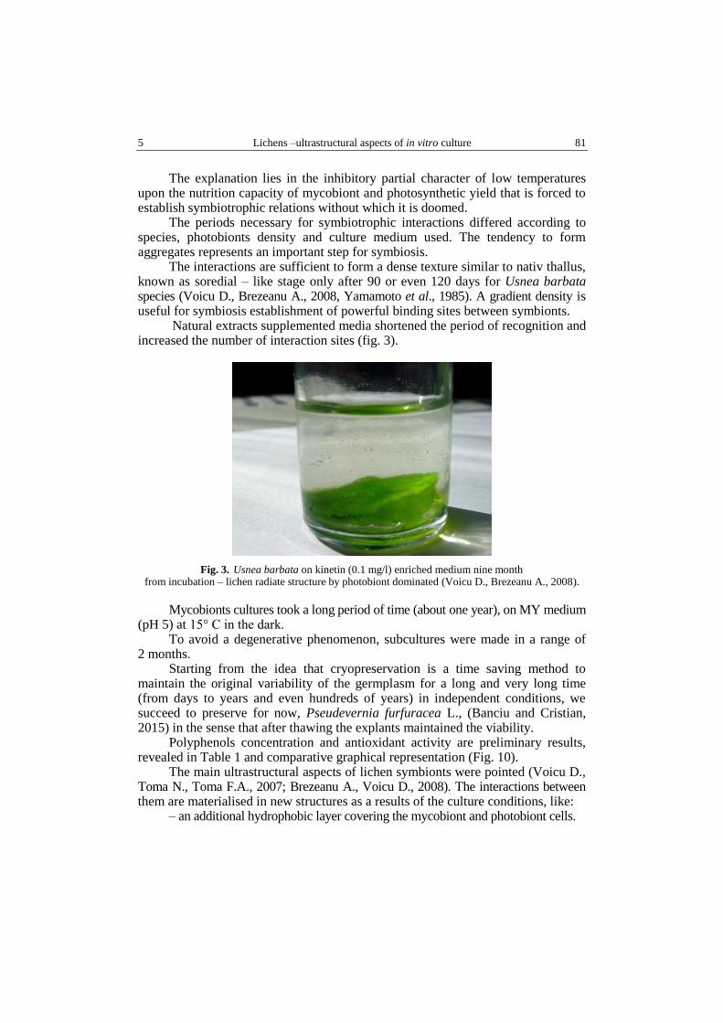
Natural extracts supplemented media shortened the period of recognition and increased the number of interaction sites (fig. 3).
Fig. 3. Usnea barbata on kinetin (0.1 mg/l) enriched medium nine month from incubation – lichen radiate structure by photobiont dominated (Voicu D., Brezeanu A., 2008).
Mycobionts cultures took a long period of time (about one year), on MY medium (pH 5) at 15° C in the dark.
To avoid a degenerative phenomenon, subcultures were made in a range of 2 months.
Starting from the idea that cryopreservation is a time saving method to maintain the original variability of the germplasm for a long and very long time (from days to years and even hundreds of years) in independent conditions, we succeed to preserve for now, Pseudevernia furfuracea L., (Banciu and Cristian, 2015) in the sense that after thawing the explants maintained the viability.
Polyphenols concentration and antioxidant activity are preliminary results, revealed in Table 1 and comparative graphical representation (Fig. 10).
The main ultrastructural aspects of lichen symbionts were pointed (Voicu D., Toma N., Toma F.A., 2007; Brezeanu A., Voicu D., 2008). The interactions between them are materialised in new structures as a results of the culture conditions, like:
– an additional hydrophobic layer covering the mycobiont and photobiont cells.

D. Voicu, A. Brezeanu, N. Toma 6 82
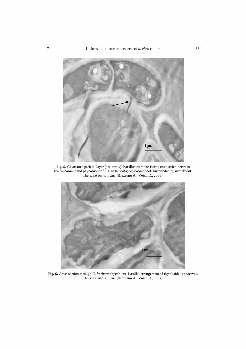
– a neostructure, named – the interface (Fig. 5), between the mycobiont and
phycobiont – cooperating the both partners to the biogenesis. By interface differentiating,
the two partners cell walls lose their individuality, forming a tripartite structure. Beside
this, mycobionts of the investigated lichens have a three-layered structure (Fig. 8).
Regarding the peculiarities of organelles, the chloroplast from “in vitro”
aposymbiotically cultured photobionts present a primitive, lamellar, parallel-
arranged system of thylakoinds, with pyrenoglobules (Figs. 6, 7).
On the other hand, mycobionts from native thalli contain concentric bodies –
a type of particular inclusion with important diagnostical feature; they are not
found “in vitro” conditions. Mycobiont plasmallema is strongly folded. Paramural
bodies are also specific for mycobionts from native thallus.
Some modifications “in vitro” conditions like the decrease in number and
amplitude of invaginations and concentric bodies (Fig. 9) is another important aspect
and also the enhanced number of mitochondria, suggesting a possible nutritional
stress for Cetraria islandica mycobionts “in vitro” conditions.
Fig. 4. Mycobiont – phycobiont interactions (oc. 10, ob. 40)
(Voicu D., Brezeanu A., 2008).
Table 1
Polyphenols concentration and antioxidant activity in methanol extracts
(Cristian D., Mitoi M., Brezeanu A., 2013)
Sample Polyphenols
(µg GAE/mg fresh weight)
Antioxidant activity
(µg Trolox equivalents/mg fresh weight)
In vitro culture 21.022 90.529
Native thalli 99.296 194.758
7 Lichens –ultrastructural aspects of in vitro culture
83
Fig. 5. Gelatinous parietal layer (see arrow) that illustrates the intime connection between
the mycobiont and phycobiont of Usnea barbata; phycobiont cell surrounded by mycobiont.
The scale bar is 1 µm. (Brezeanu A., Voicu D., 2008).
Fig. 6. Cross section through U. barbata phycobiont. Parallel arrangement of thylakoids is observed.
The scale bar is 1 µm. (Brezeanu A., Voicu D., 2008).

D. Voicu, A. Brezeanu, N. Toma 8 84
Fig. 7. Pyrenoglobules arranged along the chloroplast thylakoids (see arrangement).
The scale bar is 1 µm. (Brezeanu A., Voicu D., 2008).
Fig. 8. Electronomicroscopical image of mycobiont cells in U. barbata in vitro. Three layered
structure of cell wall. The scale bar is 1 µm. (Brezeanu A., Voicu D, 2008).

9 Lichens –ultrastructural aspects of in vitro culture
85
Fig. 9. Cross section through mycobiont cell. In vitro, concentric or ellipsoidal bodies
are observed (see arrow). The scale bar is 1 µm. (Brezeanu A., Voicu D., 2008).
0
20
40
60
80
100
120
140
160
180
200
in vitro in situ
polyphenols concentration mg GAE/g
flavonoids concentration µg ERU/g
antioxidant capacity µM Trolox/g
Fig. 10. Polyphenols concentration and antioxidant activity
in methanol extracts in vitro and in situ.

D. Voicu, A. Brezeanu, N. Toma 10 86
CONCLUSIONS
Our studies regarding the possibility of using the “in vitro” systems for lichen species revealed merely the perspectives offered by them allowing the understanding of the cellular and molecular mechanisms involved in the establishment of this unique type of symbiosis, of cell recognition process and especially biotechnological, biosynthesis of bioactive compounds.
One noteworthy aspect was the highlighting of differentiated reactivity besides the “in vitro” conditions of lichen species analyzed, namely Usnea barbata (L.) Weber ex Wigg., Cetraria islandica (L.) Ach. and Xanthoria parietina, followed by Pseudoevernia furfuracea. We showed that the processes involved in “in vitro” cultures initiation and artificial resynthesis of symbiotrophic formations of lichen species are very complex, laborious, species specific.
So, every species requires special conditions both in term of type and size of the inoculum used for initiation, culture medium (composition, physical condition, hormones addition and incubation).
This is explained by the fact that each species presents morpho-anatomical and physiological different features and distinct ecological requirements.
Grinding paste methodology was useful for algae isolation and resynthesis experiments.
For mycobiont isolation, good results were obtained using apothecia like explants for Xanthoria parietina (L.) Th.Fr., Usnea barbata (L.) Weber ex Wigg, Cetraria islandica (L.) Ach.
Our findings showed that the methodology developed by us can be a promising alternative for lichen biomass obtaining of biotechnological interest and could be successfully adapted to other important species presenting a socio-economic interest.
Although they are not synthesized in great amount and the in vitro culture does not affect their biosynthesis, phenolic compounds are important for experimental systems use for that purpose.
Cryoconservation, frequently used for numerous higher plants species but not so frequently for lichens, proved to be a propitious method to maintain the cultures for long periods of time.
Also, our experiments allowed us to appreciate that the experimental system used by us specific for each species does not induce severe alterations on the inner structure and biosynthetic pathway of the cells and could represent a feasible alternative methodology for lichen biomass obtaining of biotechnological interest as well as for bioconservation of endemic or critically endangered species.
In perspective, for the future researches, we are interested in studying the elicitation involving the receptors from the mycobiont cell walls and the ligands from the photobiontic cell walls.
Acknowledgements This paper was supported by project RO1567 - IBB06/2017 from the Institute
of Biology Bucharest of the Romanian Academy.
11 Lichens –ultrastructural aspects of in vitro culture
87
REFERENCES
1. Ahmadjian V., 1961, Studies on lichenized fungi, Bryologist 64, pp. 168–179.
2. Ahmadjian V., 1967, The Lichen Symbiosis, Blaisdell Publishing Company, Massachusetts, 152 pp.
3. AhmadjianV., and J.B. Jacobs, 1983, Algal – fungal relationship in lichens: Recognition, synthesis and
development. In, Algal Symbiosis. L.J. Goff, ed., Cambridge, Cambridge University Press,
pp. 147–172.
4. Ahmadjian V., 1993, The Lichen Symbiosis, John Wiley & Sons, Inc, New York, 248 pp.
5. Ascaso & Galvan, 1976, The ultrastructure of the symbionts of Rhizocarpon geographicum,
Parmelia conspersa, and Umbilicaria pustulata growing under dryness conditions. Protoplasma,
87, pp. 409–418.
6. Cristian D., Brezeanu A., 2013, Xanthoria parietina (L). Th. Fr. mycobiont isolation by ascospore
discharge, germination and development in “in vitro” culture. Muzeul Olteniei Craiova.Oltenia.
Stud. Comunic. Şt. Nat. 29 (2), pp. 58–63.
7. Banciu C., Cristian D., 2015. Cryoconservation of Pseudevernia furfuracea L. species and
assessing the viability after thawing (Crioconservarea speciei Pseudevernia furfuracea și
evidențierea viabilității după dezgheț). Muzeul Olteniei Craiova. Oltenia. Studii și comunicări.
Științele naturii 31 (1), pp. 57–60.
8. Brezeanu A., Voicu D., 2008, Ultrastructural peculiarities of Usnea barbata (L.) Mott. mycobiont
and phycobiont cells “in vivo” and “in vitro” culture, Romanian Journal of Biology-Plant Biology,
53(2):63–69.
9. Bubrick P., 1988, Methods for cultivating lichens and isolated bionts. In : Galun M. (ed)
Handbook of Lichenology, vol. 3. CRC Press, pp. 127–138.
10. Cristian D., Mitoi M., Brezeanu A., 2013, In vitro systems, a facile tool for Cetraria islandica
(L.) (Lichenophyta) artificial resynthesis and biotechnological potential exploit – preliminary
results, Romanian Journal of Biology-Plant Biology, 58 (2): 95–105.
11. Friedl T., Budel B., 2008, Photobionts. In: Nash T. H. III, Lichen Biology, 2nd edn, Cambridge
University Press, Cambridge, pp. 9–26.
12. Grujicic D. et al., 2014, Evaluation of in vitro antioxidant, antimicrobial, genotoxic and anticancer
activities of lichen Cetraria islandica. Cytotechnology, 66:803–813.
13. Guzov Kreminska B., Stocker-Wörgötter E., 2013, In vitro culturing and resynthesis of the
mycobiont Protoparmeliopsis muralis with algalbionts. In Lichenologist 45(1):65–76.
14. Honegger, R., 1993a, Developmental biology of lichens. New Phytologist, 125, pp. 659–77.
15. Jacobs, J. B. & Ahmadjian, V., 1969, The ultrastructure of lichens. I. A general survey. Journal of
Phycology. 5, pp. 227–240.
16. Lilly VG, Barnett HL, 1951, In Physiology of Fungi. McGraw-Hill, New York.
17. Lobakova S., Smirnov A., 2012, Experimental Lichenology. In: Advances in Applied Biotechnology,
Marian Petre Eds.
18. Marxen K, Vanselow KH, Lippemeier S, Hintze R, Ruser A, Hensen UP, 2007, Determination of
DPPH radical oxidation caused by methanolic extract of some microalgal species by linear
regression analysis of spectrophotometric measurements.Sensors, 10(VII): 2080–2095.
19. Mascorro, A.J., Bozzola, J.J. 2007, Processing Biological Tissues for Ultra structural study, Electron
microscopy, Methods and Protocols. Second Edition, Ed. By J. Kuo, Humana Press, 19–35.
20. Moya P. et al., 2015, Molecular phylogeny and ultrastructure of the lichen microalga Asterochloris
mediterranea sp. nov. from Mediterranean and Canary Islands ecosystems. International Journal
of Systematic and Evolutionary Microbiology (2015), 65, 1838–1854.
21. Murashige T. & Skoog F., 1962, A revised medium for rapid growth and bioassay with tobacco
tissue cultures. Physiologia plantarum. Edited by Scandinavian Plant Physiology Society. Copenhagen,
Denmark.15: 473–497.
22. Paunescu, A., 2009, Biotechnology for endangered plant conservation: a critical overview. Rom.
Biotech. Lett., 14 (1), pp. 4095–4103.

D. Voicu, A. Brezeanu, N. Toma 12 88
23. Peveling, E., 1976, Investigations into the ultrastructure of lichens. In: Lichenology: Progress and
Problems. D. H. Brown, D. L. Hawksworth, and R. H. Bailey, eds., London, Academic Press,
pp. 17–26.
24. Stocker-Wörgötter, 2001, Experimental lichenology and microbiology of lichens: culture experiments,
secondary chemistry of cultured mycobionts, resynthesis, and thallus morphogenesis. Bryologist
104, pp. 576–576.
25. Stocker-Wörgötter, E., 2002. Investigating the production of secondary compounds in cultured
lichen mycobionts. In Protocols in lichenology. Edited by I. Kranner, R. Beckett, Varma, A.
Springer Verlag, Heidelberg, 296–306.
26. Toma N., Voicu D. and Toma F.A., 2007, Aspecte ale organizării ultrastructurale a talurilor de
Pseudoevernia furfuracea (L.) Zopf. şi Xanthoria parietina (L.) Th. Fr., An. Grad. Bot. Univ.
Macea, 1, 78–93.
27. Vicol I., 2011a, Preliminary study using lichen species diversity as an indicator of local
environmental quality within two nature reserves from Romania, Analele Universității din
Oradea-Fascicula Biologie 18, pp. 53–58. 96.
28. Voicu D., Brezeanu A., 2008, Comportamentul in cultura in vitro a speciilor lichenice Xanthoria
parietina Beltr. şi Cetraria islandica Ach., (In vitro reactivity of lichen species Xanthoria
parietina Beltr. and Cetraria islandica Ach.) Risoprint, 101–112.
29. Voicu D., Brezeanu A., 2008, In vitro reactivity of Usnea barbata (L.) Mott., Rom. J. Biol. –
Plant Biol., 53 (2), pp. 83–90.
30. Yamamoto, Y., Mizuguchi, R. & Yamada, Y., 1985, Tissue cultures of Usnea rubescens and
Ramalina yasudae and production of usnic acid in their cultures, Agricultural and Biological
Chemistry, 49, 3347 – 8.
31. Yamamoto Y. et al., 1993, Using lichen tissue cultures in modern biology, The Bryologist 96 (3),
pp. 384–393.
32. Yamamoto, Y. et al., 2002, Isofuranonaphthoquinone derivatives from cultures of the lichen
Arthonia cinnabarina (DC.) Wallr. Phytochemistry 60, pp. 741–745.
33. Yoshimura I. et al., 2002, Isolation and culture of Lichen Photobionts and Mycobionts. In: Protocols in
Lichenology. Edited by I. Kromer, R. Beckett, A. Varma, Springer Verlag, Heidelberg, 3–33.
34. Zarnea G., Popescu O.V., 2011, Dicționar de microbiologie generală și biologie moleculară.
Editura Academiei Române, București, 1331 pp.

ROM. J. BIOL. – PLANT BIOL., VOLUME 62, No. 2, P. 89–108, BUCHAREST, 2017
POLLINATION ECOLOGY OF RHYNCHOSIA SUAVEOLENS
(L.F.) DC. (FABACEAE), A PERENNIAL ERECT SHRUB IN THE
SOUTHERN EASTERN GHATS, ANDHRA PRADESH, INDIA
A.J.S. RAJU1, K.V. RAMANA2
Abstract. Rhynchosia suaveolens is a perennial erect shrub. It flowers during October–
January with peak flowering during November. The flowers are hermaphroditic,
nectariferous, self-compatible and have an explosive pollination mechanism adapted for
pollination by bees. They do not fruit through autonomous selfing but fruit through
manipulated selfing, geitonogamy and xenogamy mediated by pollen vectoring bees.
Lycaenid butterflies also visit, especially during peak flowering season, but they
principally act as nectar robbers. The flowers not visited by bees fall off while those
visited and pollinated by them set fruit. Seed dispersal occurs by explosive pod
dehiscence. Perennial root stock produces new foliage during rainy season. Seeds also
germinate at the same time but their continued growth is subject to the availability of
soil moisture content. Therefore, R. suaveolens expands its population size in areas
where soil is sufficiently moist and also in areas where trees or woody shrubs provide
shade. Since the plant is not widely distributed, its occurrence is almost unknown to
locals and hence is not used for any purpose.
Keywords: Rhynchosia suaveolens, hermaphroditism, explosive pollination mechanism,
melittophily, explosive pod dehiscence.
INTRODUCTION
Legume seeds are important sources of nutrients that meet our high quality
dietary protein requirements (Perumal et al., 2001; Escudero et al., 2006). The
under-utilized legumes, which have tremendous potential for commercial exploitation
but remain ignored, offer a good scope in this context (Bhag Mal, 1992). Accounts
of important under-exploited pulses which await exploration for food, fodder, energy
and industrial purposes have been given (Siddhuraju et al., 2000; Kalidass & Mohan,
2012). Rhynchosia is such a legume which is under-utilized despite its potential to
provide high protein requirements. In this context, some work has been carried out
on the biochemical and nutritional composition of R. venulosa, R. hirta, R. cana,
1 Department of Environmental Sciences, Andhra University, Visakhapatnam 530 003, India. 2 Department of Botany, Andhra University, Visakhapatnam 530 003, India.
Corresponding author: [email protected].

A.J.S. Raju, K.V. Ramana 2 90
R. filipes, R. rufescens and R. suaveolens by different workers (Lovelace, 1977;
Murthy & Kandimalla, 2007; Kalidass & Mohan, 2012). Their work showed that
these legumes are potential sources of protein, minerals and energy supplements
for livestock and humans. The high protein value of R. suaveolens is in tune with
its consumption by the tribes of Kani and Kannikars of southern Western Ghats of
Tamil Nadu, India and hence it has potential for human nutrition (Kalidass &
Mohan, 2012). This species has been reported to be distributed only in India and
Sri Lanka (Manjunatha et al., 2004). The knowledge of its reproductive ecology is
essential to understand its sexual mode of reproduction and develop it as an edible
legume crop. But, this information is totally lacking and hence the present study is
contemplated to provide details of floral biology, pollination mechanism, pollinators
and seed dispersal.
MATERIALS AND METHODS
STUDY SITE
The study region is an integral part of Southern Eastern Ghats of Andhra
Pradesh in Peninsular India. The area is located at 13°40’N latitude and 79°19’E
longitude. The exact study area is the forest cover of Tirumala Hills, a constituent
of Seshachalam Hill Range in Chittoor District, Andhra Pradesh. The entire region
represents the deciduous forest ecosystem. The site is characterized by a combination
of rocky, undulating and steep terrain with some litter content formed from grass
and other herbaceous plants. In this area, Rhynchosia suaveolens grows as scattered
individuals or here and there as small populations in moist soils or shaded areas.
FLOWERING AND FLORAL BIOLOGY
Flowering season was defined based on regular field trips made. Twenty
inflorescences were tagged and followed to record the length of flowering and the
number of flowers produced. Anthesis was initially recorded by observing twenty
five marked mature buds in the field. Later, the observations were repeated five
times on different days in order to provide an accurate anthesis schedule. Similarly,
the mature buds were followed for recording the time of anther dehiscence. The
presentation pattern of pollen was also investigated by recording how anthers
dehisced and confirmed by observing the anthers under a 10× hand lens. The
details of flower morphology such as flower sex, shape, size, colour, odour, sepals,
petals, stamens and ovary were described based on twenty five flowers randomly
collected from five plants. Observations regarding the position and spatial relationships
of stamens and stigma in mature bud, at anthesis and during the flower-life with
reference to self and/or cross-pollination, were made very carefully.

3 Pollination ecology of Rhynchosia suaveolens
91
POLLEN OUTPUT
Thirty mature but un-dehisced anthers from five different plants were collected and placed in a Petri dish. Later, each time a single anther was taken out and placed on a clean microscope slide (75 × 25 mm) and dabbed with a needle in a drop of lactophenol-aniline-blue. The anther tissue was then observed under the microscope for pollen, if any, and if pollen grains were not there, the tissue was removed from the slide. The pollen mass was drawn into a band, and the total number of pollen grains was counted under a compound microscope (40x objective, 10x eye piece). This procedure was followed for counting the number of pollen grains in each anther collected. Based on these counts, the mean number of pollen produced per anther was determined. The mean pollen output per anther was multiplied by the number of anthers in the flower for obtaining the mean number of pollen grains per flower. The characteristics of pollen grains were also recorded.
POLLEN-OVULE RATIO
The pollen-ovule ratio was determined by dividing the average number of pollen grains per flower by the number of ovules per flower. The value thus obtained was taken as pollen-ovule ratio (Cruden, 1977).
NECTAR CHARACTERS
The presence of nectar was determined by observing the mature buds and open flowers. The average volume of nectar per flower was determined and expressed in µL; for this ten flowers were used. The flowers used for this purpose were bagged at mature bud stage, opened after cessation of nectar secretion and squeezed nectar into micropipette for measuring the volume of nectar. Nectar sugar concentration was determined using a Hand Sugar Refractometer (Erma, Japan). Ten samples were used for examining the range of sugar concentration in the nectar. For the analysis of sugar types, paper chromatography method described by Harborne (1973) was followed. Nectar was placed on Whatman No. 1 of filter paper along with standard samples of glucose, fructose and sucrose. The paper was run ascendingly for 24 hours with a solvent system of n-butanol-acetone-water (4:5:1), sprayed with aniline oxalate spray reagent and dried at 120
oC in an electric oven
for 20 minutes for the development of spots from the nectar and the standard sugars. Then, the sugar types present and also the most dominant sugar type were recorded based on the area and colour intensity of the spot. The sugar content/ flower is expressed as the product of nectar volume and sugar concentration per unit volume, mg/µL. This is done by first noting the conversion value for the recorded sugar concentration on the refractometer scale and then by multiplying it with the volume of nectar/flower. Table 5.6 given in Dafni et al. (2005) was followed for recording the conversion value to mg of sugars present in one µL of nectar.
A.J.S. Raju, K.V. Ramana 4 92
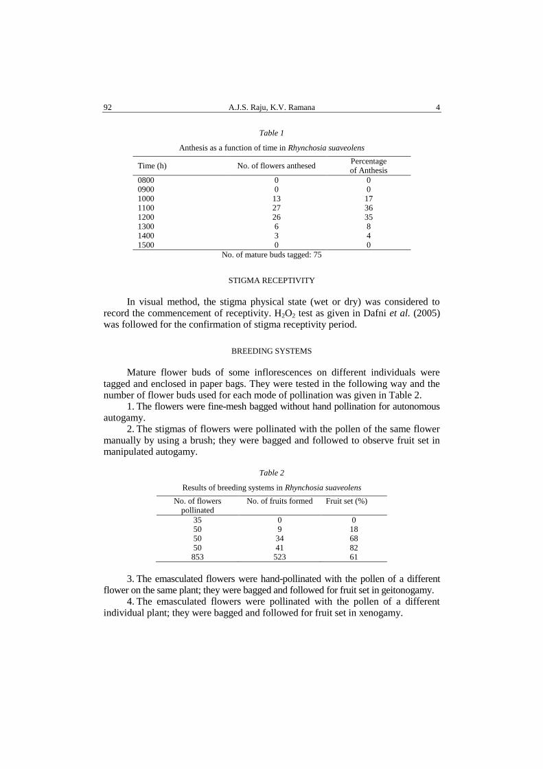
Table 1
Anthesis as a function of time in Rhynchosia suaveolens
Time (h) No. of flowers anthesed Percentage of Anthesis
0800 0 0 0900 0 0 1000 13 17 1100 27 36 1200 26 35 1300 6 8 1400 3 4 1500 0 0
No. of mature buds tagged: 75
STIGMA RECEPTIVITY
In visual method, the stigma physical state (wet or dry) was considered to record the commencement of receptivity. H2O2 test as given in Dafni et al. (2005) was followed for the confirmation of stigma receptivity period.
BREEDING SYSTEMS
Mature flower buds of some inflorescences on different individuals were tagged and enclosed in paper bags. They were tested in the following way and the number of flower buds used for each mode of pollination was given in Table 2.
1. The flowers were fine-mesh bagged without hand pollination for autonomous autogamy.
2. The stigmas of flowers were pollinated with the pollen of the same flower manually by using a brush; they were bagged and followed to observe fruit set in manipulated autogamy.
Table 2
Results of breeding systems in Rhynchosia suaveolens
No. of flowers pollinated
No. of fruits formed Fruit set (%)
35 0 0 50 9 18 50 34 68 50 41 82
853 523 61
3. The emasculated flowers were hand-pollinated with the pollen of a different flower on the same plant; they were bagged and followed for fruit set in geitonogamy.
4. The emasculated flowers were pollinated with the pollen of a different individual plant; they were bagged and followed for fruit set in xenogamy.

5 Pollination ecology of Rhynchosia suaveolens
93
All these categories of flower pollinations were followed for fruit set. If fruit
set is there, the percentage of fruit set was calculated for each mode.
FLOWER-VISITORS
The flower foragers included only bees and butterflies. The hourly foraging
visits of insect species were recorded on 3 or 4 occasions depending on the possibility
and the data was tabulated to use the same for further analysis. Fully blooming
plants were selected to record the foraging visits of insects. The data obtained was used
to calculate the percentage of foraging visits made by each species per day and also
to calculate the percentage of foraging visits of each species per day in order to
understand the relative importance of each species and category of insects. Their
foraging behaviour was observed on a number of occasions for the mode of approach,
landing, probing behaviour, the type of forage collected, contact with essential organs
to result in pollination, inter-plant foraging activity in terms of cross-pollination.
DETERMINATION OF POLLEN CARRYOVER EFFICIENCY OF INSECT FORAGERS
Ten specimens of each insect species were captured from flowers and
brought to the laboratory. Each specimen was washed first in ethyl alcohol and the
contents stained with aniline-blue on a glass slide and observed under microscope
to count the number of pollen grains present. From this, the average number of
pollen grains carried by each species was calculated to know the pollen carryover
efficiency of different species.
NATURAL FRUIT SET, SEED DISPERSAL AND SEEDLING ECOLOGY
A sample of flowers on twenty five plants were tagged on different plants
prior to anthesis and followed for fruit set rate in open-pollinations. Fruit maturation
period, fruit dehiscence and seed dispersal aspects were observed to the extent
possible. Field observations were also made on fruit and seed dispersal modes, seed
germination and seedling establishment to the extent possible.
PHOTOGRAPHY
Plant habitat, flowering inflorescences, and flower and fruit details were
photographed with Nikon D40X Digital SLR (10.1 pixel) and TZ240 Stereo Zoom
Microscope with SP-350 Olympus Digital Camera (8.1 pixel). Olympus Binoculars
(PX35 DPSR Model) was also used to make field observations. Magnus Compound
Microscope – 5×, 10×, 40× and 100× magnification was used for studying the pollen
characteristics.

A.J.S. Raju, K.V. Ramana 6 94
RESULTS
PHENOLOGY
It is a sweet-scented perennial erect hairy shrub with slender stem that grows
moist, shaded areas (Figure 4a); it is also found to grow nearby streams where the
soil is sufficiently moist. The plant re-grows from below ground perennial root stock
and from the seed during June–September during which growth and leaf flushing
occurs. The leaves are trifoliate with reticulate venation. The leaflets are petiolate,
ovate-acuminate, and puberulous, especially beneath. The flowering occurs during
October–January with peak flowering in November (Figure 4b,c). The plants
wither and disappear in April. The flower stalks arise in leaf-axils and each stalk is
2-flowered.
FLOWER MORPHOLOGY
The flowers are pedicellate, small (9.1 ± 0.5 mm long and 8.3 ± 0.4 mm
wide), green with strong scarlet tinge, odorless, papilionaceous, zygomorphic and
bisexual. The calyx is green with yellow tinge and consists of 5 free, lanceolate,
acuminate, pubescent, 5.4 ± 0.6 mm long sepals. The corolla is red outside, yellow
inside, pubescent, specialized and consists of upper standard petal, two wing petals and
two keel petals. The standard petal is large (7.4 ± 0.6 mm long and 6.7 ± 0.4mm
wide), red lines inside at the bottom which serves as nectar guide; the petal base is
clawed and consists of two inflexed fingernail auricles. The standard petal envelops
the rest of the petals in bud but reflexes when the flower blooms. The two adjacent
petals (7.3 ± 0.4 mm long and 2.8 ± 0.4 mm wide), called wing petals, surround the
two bottom petals, called keel petals (7.2± 0.3 mm long and 2.8 ± 0.2 mm wide).
The keel petals form a proximal cylindrical part and a distal part consisting of a
pressed angular pouch, with an acute porate tip in which the stamens and stigma
are housed. The keel and the wing petals are attached by means of two notched
folds. The wing petals serve as a lighting platform for insects visiting the flowers.
The stamens are ten, 5.7±0.4 mm long, diadelphous; nine filaments are fused by
the basal part into a sheath open along the upper side while the tenth filament is
free and lies on the others (Figure 4i). The distal parts of the filaments are free and
contain 1.2±0.3 mm long uniform dithecous anthers. The ovary is sessile, green,
villous, 2.5±0.5 mm long (Figure 4l) and lies in the sheath of the filaments along
the cylindrical part of the keel. It is monocarpellary and monolocular with two
ovules arranged on marginal placentation (Figure 4n). It has a long filiform style with a
capitate wet shiny stigma, both together account for a length of 6.5 ± 0.6 mm
(Figure 4m). The stigma is situated slightly beyond the length of the anthers. The
distal portion of free filaments and style and stigma are incurved and clamped into
the keel petals.

7 Pollination ecology of Rhynchosia suaveolens
95
FLORAL BIOLOGY
Mature buds open during 1000–1400 h with peak anthesis during 1100–1200 h (Table 1). Unfolding of the standard petal and wing petals indicates flowering
opening. The keel petals do not unfold and remain in their original position as in mature bud stage (Figure 4d–h). All the ten anthers in a flower dehisce at the same
time by longitudinal slits in mature bud stage (Figure 4j). The number of pollen grains per anther is 773.4±42.34 and per flower is 7,734. The pollen-ovule ratio is
3,867:1. The pollen grains are monads, spheroidal, 24.86±5.24 µm in size, powdery and tricolporate, angulaperturate with reticulate exine (Figure 4k). A nectariferous
disc is present at the base of the ovary. The initiation of nectar secretion occurs during mature bud stage and its cessation occurs an hour after anthesis. Individual
flowers produce 1.4±0.21 µL of nectar with 0.45 mg of sugar. The nectar sugar
concentration is 29% (Range 27–31%) consisting of sucrose, glucose and fructose with the first as dominant. Nectar is deeply concealed and it is open through two
windows between the joined and the free filaments at the flower base. These windows allow access to the nectar. The stigma attains receptivity during anthesis
and remains receptive for about three hours. After three hours of anthesis, the standard, wing and keel petals gradually move close to each other enclosing the
reproductive organs. The closed flowers remain so even during most part of the fruit development. The calyx initially encloses the ovary and subsequently turns
light brown and discloses the ovary since the latter gradually bulges and develops into a seeded pod.
POLLINATION MECHANISM
The reproductive column is held under pressure within the keel part in open flowers and it is exposed when the pollinator presses against the wing and the keel petals. When insects land on the wing petals, the latter causes the keel petals to release the reproductive column explosively. Consequently, the reproductive column snaps forward against the standard petal causing most of the pollen to be instantly released and the pollen thus released comes into contact with the ventral side of the insect body. Since the incurved stigma is situated above the height of the anthers, it strikes the insect body first due to which cross-pollination occurs if the insect visited the other flowers previously and carried pollen on its ventral side and also then the pollen ejected from the anthers powders the ventral side of the insect instantly. If it is the first visit for the insect to the flower, then it effects self-pollination upon explosive release of reproductive column from the keel boat. With the departure of the insect from the flower, the reproductive column does not return back to its former position but the keel moves forward partly covering the stamens and stigma. The downward movement of keel petals occurs in each subsequent foraging visits by appropriate insects. Tripping of keel boat can also occur due to heavy rain or high temperature that weaken turgidity of the restraining keel tissues.

A.J.S. Raju, K.V. Ramana 8 96
But, the tripping due to these two factors is ruled out since the plant flowers during winter season when heavy rains do not normally occur and the temperature usually stands low. If the flower is untouched or tripping to keel did not occur, the reproductive column is never exposed and remains enclosed in the keel boat. Such flowers fall off subsequently upon withering without fruit set.
BREEDING SYSTEMS
In mature buds, anthers dehisce but autonomous autogamy does not occur. Fruit set is absent in un-manipulated autogamy, 18% in hand-pollinated autogamy, 68% in geitonogamy, 82% in xenogamy and 61% in open-pollination (Table 3).
Table 3
Natural fruit/seed set rate in Rhynchosia suaveolens
No. of flowers sampled
No. of flowers set fruit
Fruit set (%)
Seed set (%)
Rate of seed set/fruit No. of 1-seeded fruits
1-seeded fruit set
(%)
No. of 2-seeded
fruits
2-seeded fruit set
(%)
No. of seedless
fruits
Seedless fruits (%)
853 523 61 58 42 8 476 91 5 1
BEE POLLINATORS AND POLLINATION
The flowers were exclusively foraged by bees for pollen and nectar and by butterflies for nectar. The bees showed foraging activity during 1040–1630 h with peak
activity during 1230–1330 h (Figure 1). The bees included Apis florea (Figure 5a),
Xylocopa latipes (Figure 5b), Ceratina sp. (Figure 5c–f) and Nomia sp. (Figure 5g) (Apidae) (Table 4); they were regular and consistent foragers throughout the flowering
season. The butterflies included only Lycaenids, Castalius rosimon (Figure 5h), Freyeria trochylus, Lampides boeticus and Chilades pandava (Figure 5i) (Table 4);
they foraged during 1130–1530 h with peak activity during 1230–1330 h (Figure 2). Bee foraging visits constituted 84% and butterflies 16% of total foraging visits made in
a given day during peak flowering phase (Figure 3). All bee species were regular and consistent foragers throughout the flowering season. In case of butterflies, they
were occasional in their visits during initial and final phase of flowering while they were regular and consistent during peak phase of flowering. The bees were quite
effective to trip the flowers while foraging for nectar and effect pollination. The bees landed on the wing petals and the keel, with their head near the standard. They
then exerted a certain pressure with legs on the wing petals until these and the keel bent downwards, and then proceeded to collect nectar during which the bee’s
abdomen appeared pollen smothered (sternotribic pollen deposition). To collect pollen, the bees took “U” turn after nectar collection and proceeded to the stamens
to collect pollen. The butterflies were in-effective to trip the flowers but foraged for
nectar from both un-tripped as well as tripped flowers. In case of un-tripped
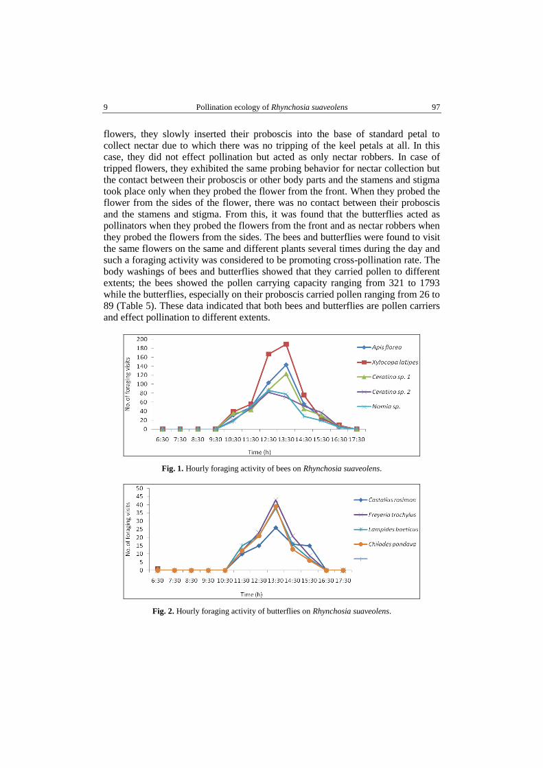
9 Pollination ecology of Rhynchosia suaveolens
97
flowers, they slowly inserted their proboscis into the base of standard petal to
collect nectar due to which there was no tripping of the keel petals at all. In this case, they did not effect pollination but acted as only nectar robbers. In case of
tripped flowers, they exhibited the same probing behavior for nectar collection but the contact between their proboscis or other body parts and the stamens and stigma
took place only when they probed the flower from the front. When they probed the flower from the sides of the flower, there was no contact between their proboscis
and the stamens and stigma. From this, it was found that the butterflies acted as pollinators when they probed the flowers from the front and as nectar robbers when
they probed the flowers from the sides. The bees and butterflies were found to visit
the same flowers on the same and different plants several times during the day and such a foraging activity was considered to be promoting cross-pollination rate. The
body washings of bees and butterflies showed that they carried pollen to different extents; the bees showed the pollen carrying capacity ranging from 321 to 1793
while the butterflies, especially on their proboscis carried pollen ranging from 26 to 89 (Table 5). These data indicated that both bees and butterflies are pollen carriers
and effect pollination to different extents.
Fig. 1. Hourly foraging activity of bees on Rhynchosia suaveolens.
Fig. 2. Hourly foraging activity of butterflies on Rhynchosia suaveolens.
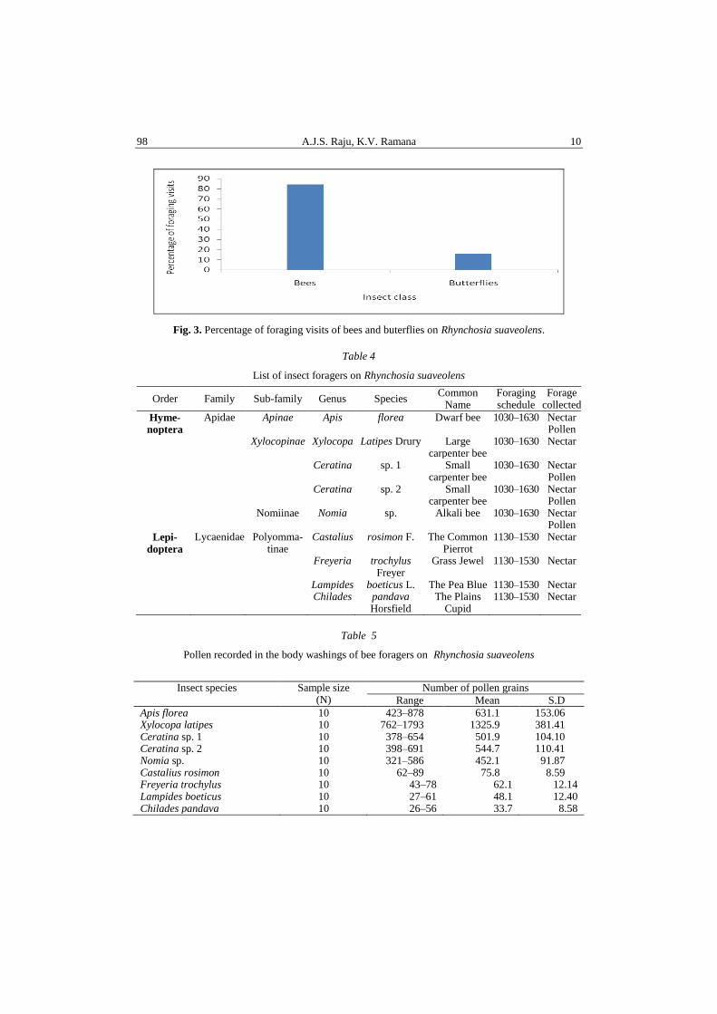
A.J.S. Raju, K.V. Ramana 10 98
Fig. 3. Percentage of foraging visits of bees and buterflies on Rhynchosia suaveolens.
Table 4
List of insect foragers on Rhynchosia suaveolens
Order Family Sub-family Genus Species Common
Name Foraging schedule
Forage collected
Hyme-noptera
Apidae Apinae Apis florea Dwarf bee 1030–1630 Nectar Pollen
Xylocopinae Xylocopa Latipes Drury Large carpenter bee
1030–1630 Nectar
Ceratina sp. 1 Small carpenter bee
1030–1630 Nectar Pollen
Ceratina sp. 2 Small carpenter bee
1030–1630 Nectar Pollen
Nomiinae Nomia sp. Alkali bee 1030–1630 Nectar Pollen
Lepi-doptera
Lycaenidae Polyomma-tinae
Castalius rosimon F. The Common Pierrot
1130–1530 Nectar
Freyeria trochylus Freyer
Grass Jewel 1130–1530 Nectar
Lampides boeticus L. The Pea Blue 1130–1530 Nectar Chilades pandava
Horsfield The Plains
Cupid 1130–1530 Nectar
Table 5
Pollen recorded in the body washings of bee foragers on Rhynchosia suaveolens
Insect species Sample size
(N) Number of pollen grains
Range Mean S.D
Apis florea 10 423–878 631.1 153.06 Xylocopa latipes 10 762–1793 1325.9 381.41 Ceratina sp. 1 10 378–654 501.9 104.10 Ceratina sp. 2 10 398–691 544.7 110.41 Nomia sp. 10 321–586 452.1 91.87 Castalius rosimon 10 62–89 75.8 8.59 Freyeria trochylus 10 43–78 62.1 12.14 Lampides boeticus 10 27–61 48.1 12.40 Chilades pandava 10 26–56 33.7 8.58

11 Pollination ecology of Rhynchosia suaveolens
99
Figure 4. Rhynchosia suaveolens: a. Habitat, b. Budding phase, c. Flowering,d-g. Different stages of anthesis, h. Keel and wing petals withstamens and stigma, i. Diadelphous stamens, j Dehisced anthers,k. Pollen grain, l. Gynoecium, m. Capitate stigma, n. Ovule.
d
i j k l m
c
n
b
a
e f g h
Fig. 4. Rhynchosia suaveolens: a. Habitat, b. Budding phase, c. Flowering, d–g. Different stages
of anthesis, h. Keel and wing petals with stamens and stigma, i. Diadelphous stamens, j. Dehisced
anthers, k. Pollen grain, l. Gynoecium, m. Capitate stigma, n. ovule.

A.J.S. Raju, K.V. Ramana 12 100
Figure 5. Rhynchosia suaveolens: a. Apis florea collecting nectar, b. Xylocopalatipes collecting nectar, c. & d. Ceratina sp. 1 collecting pollen, e.Ceratina sp. 1 collecting nectar, f. Ceratina sp. 2 collecting pollen, g.Nomia sp. collecting nectar, h-i. Lycaenids – h. Castalius rosimoncollecting nectar, i. Chilades pandava collecting nectar, j. Maturingpods, k. Mature, dry pod, l. 2-seeded pod, m. Seeds, n.-p. Explosivepod dehiscence, q. Seedling.
gf
j k l m
n o p q
ih
ba edc
Fig. 5. Rhynchosia suaveolens: a. Apis florea collecting nectar, b. Xylocopa latipes collecting nectar,
c. & d. Ceratina sp. 1 collecting pollen, e. Ceratina sp. 1 collecting nectar, f. Ceratina sp. 2 collecting
pollen, g. Nomia sp. collecting nectar, h–i. Lycaenids – h. Castalius rosimon collecting nectar,
i. Chilades pandava collecting nectar, j. Maturing pods, k. Mature, dry pod, 1. 2–seeded pod,
m. Seeds, n.–p. Explosive pod dehiscence, q. Seedling.

13 Pollination ecology of Rhynchosia suaveolens
101
FRUITING BEHAVIOR
The fruit growth and development begins immediately after pollination and fertilization. The fruits mature within three weeks. The sepals enclose the growing
fruit initially and the fruit emerges out of the sepals gradually with its gradual growth and development. Fruit is green initially with scarlet red tinge and brown
when ripe and dry (Figure 5 j,k). It is a non-fleshy, hairy, oblong, 11.2±1.5 mm long, 5.1±0.4 mm wide, compressed, rounded and apiculated pod. The pods produced
mostly two seeds (Figure 5l) while a few produce one seed. Fruit set without seeds also exists but it is negligible. The fruit set rate in open-pollination mode is 61%
out of which 2-seeded pod set rate is 91%, 1-seeded pod set rate is 8% and seedless pod set rate is 1%. Total seed set rate is 58% (Table 3).
SEED ECOLOGY
Mature and dry fruits display explosive dehiscence to disperse seeds (Figure 5n–p). The pod with bi-valvate configuration dehisce to eject the seed. The seed is greyish
brown, striate, reniform, finely pubescent, 4.6 ± 0.4 mm long, 3.6 ± 0.3 mm wide, shiny and strophiolate (Figure 5m). Seeds germinate during rainy season which starts
from June onwards. Seedlings grow continually but their growth rate is subject to the availability of moisture status of the soil (Figure 5q). In areas where soil is
sufficiently moist, seeds continue growth and produce mature plants within two months and subsequently commence flowering and fruiting.
DISCUSSION
Distribution range of Rhynchosia suaveolens is largely unknown as there is almost no information regarding its distribution and habitats of occurrence. Manjunatha et al. (2004) mentioned that it is distributed in India and Sri Lanka only. The present study shows that R. suaveolens occurs in moist soils or shaded areas in dry deciduous forest areas of Eastern Ghats of Andhra Pradesh. It usually occurs as scattered individuals or as small populations here and there. As a perennial erect shrub, it grows from seed as well as from perennial root stock during rainy season. The flowering season is well defined and is confined to winter season. Individual plants produce a small number of flowers during their lifetime due to production of a few inflorescences and each of which produces two flowers. Individual flowers are prominent only by their red to yellow coloured corolla but not by their position since they occur within the foliage.
In R. suaveolens, hermaphroditic sexual system is functional due to production of fertile pollen grains and functional ovary. The flowers display the near synchronous hermaphroditism or homogamy due to the occurrence of anther dehiscence in

A.J.S. Raju, K.V. Ramana 14 102
mature bud stage and receptivity of stigma during anthesis. The entire reproductive column stays inside the keel petals even after anthesis; in this situation, there is a likelihood of the occurrence autonomous autogamy. But, hand-pollination tests indicated that autonomous autogamy does not occur despite self-compatibility but it is functional when this mode of pollination is manipulated by brushing the stigma with its own pollen. Such a situation suggests that the flowers are essentially dependent on flower foragers for fruit set through self – as well as cross-pollination. It appears that the stigma although receptive blocks the germination of the self-pollen while it is in keel petals and hence, it essentially requires the rupture of its surface by a pollinator to allow the self – or cross pollen to germinate. Such a stigmatic regulatory function appears to have evolved to discourage selfing and promote out-crossing. Shivanna & Owens (1989) stated that the rupture of the stigmatic surface by pollinator permits the pollen to germinate in the flowers of Phaseoleae members with thick stigmatic cuticle. On the contrary, Castro & Agullo (1998) reported that in Vigna, a member of the tribe Phaseoleae, autonomous self-pollination may occur by spontaneous rupture of the stigmatic membrane. Similar stigmatic surface that prevents self-fertilization has also been reported in Vicia faba (tribe Vicieae) (Lord & Heslop-Harrison, 1984) and in Medicago scutellata (tribe Trifolieae) (Krietner & Sorensen, 1985); however, in these species auto-fertile lines have been reported to have thin stigmatic cuticles allowing spontaneous disruption and self-fertilization. In R. suaveolens, the stigmatic surface appears to have thick cuticle and does not have the mechanism of causing spontaneous rupture to facilitate autonomous self-pollination. In effect, the tripping of keel petals appears to be essential to cause rupture on the stigmatic surface by the tripping agent due to which there is more likelihood of the occurrence of either geitonogamy or xenogamy. The fruit set rates recorded in hand-pollinated geitonogamy and xenogamy also substantiate that the plant is facultative xenogamous, a breeding system that is flexible and keeps the options open for both selfing and out-crossing mediated by pollen vectors.
Schrire (1989) stated that the ecological and evolutionary success of
Leguminosae has been related to biotic pollination mechanisms. The three sub-
families within this family have achieved a characteristic floral architecture, in
which plants within the sub-family Papilionoideae have developed the most
complex floral mechanisms. Plants within the Papilionoideae have zygomorphic
flowers that are mainly bee-pollinated (Westerkamp, 1997); although bird pollination
and bat pollination have also been recorded within the subfamily (Ortega-Olivencia
et al., 2005). In bee-pollinated flowers of Papilionoideae, it is assumed that each
part of the corolla is specialized for a particular role in pollinator attraction and the
success of pollination. The flag or standard petal attracts pollinators; the keel
protects androecium and gynoecium and, together with the wings, provides a
platform for the insects to land on. The wings also operate as levers that raise or
lower the keel (Stirton, 1981). The flowers typical of pollination by the bee family
Apidae are zygomorphic, bright yellow or blue with nectar guides, and frequently
with hidden rewards such as those in the Lamiaceae, Scrophulariaceae, Fabaceae

15 Pollination ecology of Rhynchosia suaveolens
103
and Orchidaceae (Faegri & van der Pijl, 1979). In the present study, the Fabaceae
member, R. suaveolens has papilionaceous corolla with flag, wing and keel petals;
the flag petal serves as a visual attractant, wing petals provide landing platform and
keel petals protect the entire reproductive column. The flowers are typical of
pollination by bees since they are zygomorphic, standard petal with nectar guide,
hidden nectar at the corolla base and hidden pollen in keel petals. Within the sub-family Papilionoideae, primary and secondary pollen
presentations have been reported. In plants with primary pollen presentation, pollen is delivered directly from the anthers to the vector's body. In plants with secondary pollen presentation, pollen grains are delivered first on a floral part such as the keel petals in Papilionoideae and then on the body of the vector implying an accurate delivery of pollen on the vector’s body (Howell et al., 1993). These two pollen presentation patterns are associated with the four types of basic pollination mechanisms – valvular, pump, explosive and brush, all of them are associated with a particular floral architecture and kinetics. In the valvular type, pollen presentation is primary, whereas in the other three mechanisms, it is secondary (Yeo, 1993). In the explosive mechanism, commonly only one pollination event occurs and it has evolved independently in several tribes (Small, 1988), while in the other three mechanisms, repeated visitation is possible (Westerkamp, 1997). In the present study, R. suaveolens flowers have explosive pollination mechanism and deliver pollen directly from the anthers to the bee's body when keel petals are tripped by the foraging bee; this type pollen delivery is the representative of primary pollen presentation associated with explosive pollination mechanism. In the flowers, the staminal column is held under pressure within the keel, and when the tension is released by the forager, the same column snaps forward against the standard petal causing all the pollen to be instantly released. The reproductive column remains exposed and does not return back to its original state but the keel petals return back partially covering the stamens and stigma. The efficiency of explosive pollination mechanism depends on the ambient weather conditions, especially temperature and relative humidity. Since R. suaveolens flowers during winter season, it accordingly commences anthesis from late morning onwards by which time the ambient air will be relatively dry and hence is conducive for the efficient functioning of the explosive pollination mechanism. Further, the bees also commence their foraging activity as soon as the flowers are open and continue forage collection until the flowers close back. The concealment of the stamens within the keel petals until it is tripped is an advantage for the plant to secure pollen from unusual rains and ambient moisture conditions during the flowering season of this plant (Peter et al., 2004).
Percival (1961) stated that plants with deep-tubed flowers tend to produce sucrose-rich nectar, whereas those with open or shallow-tubed flowers tend to be hexose-rich. Baker & Baker (1983) stated that flowers with long corolla tube possess more sucrose in their nectar while those with short tubes possess more hexoses in their nectar. In the present study, R. suaveolens with short corolla tube presents sucrose-rich nectar because the nectar is perfectly concealed and hence is

A.J.S. Raju, K.V. Ramana 16 104
not exposed for the breakdown of sucrose into hexoses. Concealment of nectar in this species is adaptive to protect against microorganisms, particularly yeasts, whose metabolic activities dramatically change nectar chemistry and the plant gains a benefit from keeping the nectar as sterile as possible to maintain control over its chemical composition in order to maximize pollination rate by attracting appropriate pollinators (Herrera et al., 2008). Honey bees prefer the flowers with sucrose as chief constituent of nectar (Kevan, 1995). The flowers pollinated by long-tongued bees produce sucrose-rich nectar (Baker & Baker, 1990). In line with this, R. suaveolens with melittophilous pollination syndrome also produces sucrose-rich nectar which is utilized exclusively by long-tongued bees. Apis, Ceratina, Nomia and Megachile bees recorded on this herb have been documented as long-tongued bees (Cruden et al., 1983; Roubik, 1992; Roubik, 2006). Bee-flowers tend to produce small volume of nectar with higher sugar concentration than the nectar of flowers pollinated by other animals (Opler, 1983; Cruden et al., 1983). Honey bees prefer sugar concentration of 20 to 40% in the nectar (Waller, 1972). On the contrary, Baker & Baker (1983) noted that honey bees prefer sugar concentration of 30 and 50% in the nectar. The honey bees have the ability to regurgitate liquid onto concentrated or even crystallized nectar, in this way, reduce its concentration so that it may be imbibed. The preferred sugar concentrations of nectar by other categories of bees have not been found in the literature. But, Pyke & Waser (1981) stated that the nectar sugar concentration of flowers pollinated by bees is generally higher than that of those pollinated by butterflies and hummingbirds; bee-pollinated flowers tend to produce nectar with sugar concentration more than 35% while butterfly or hummingbird pollinated flowers tend to produce nectar with sugar concentration ranged between 20 and 25%. In line with these reports, the present study shows that the flowers of R. suaveolens produce a small volume of nectar with 29% sugar concentration. Further, the energy yield from nectar appears to be in tune with the requirement of energy by bees. Therefore, R. suaveolens flowers with explosive pollination mechanism, primary pollen presentation, and hidden nectar and pollen have evolved to discourage other foragers from visiting the flowers and to ensure that the bees get the floral rewards. Accordingly, the flowers were pollinated principally by bees. Apart from bees, lycaenid butterflies also visit the flowers for nectar especially during peak flowering season but their probing behavior indicates that they do not cause tripping of flowers to result in pollination but collect nectar by slowly inserting their proboscis into the floral base. They collect nectar from tripped flowers with great ease with or without any contact between their proboscis and floral sex organs. Therefore, butterflies are principally nectar robbers and in effect, standing crop of nectar gets reduced. In this situation, the pollinating bees make multiple visits to the same flowers in quest of nectar and enhance pollination rate. Therefore, butterfly foraging activity is indirectly contributing to the enhanced natural fruit set rate. In line with this, natural fruit set rate recorded for R. suaveolens is the highest.
Cruden (1977) used the pollen-ovule (P/O) ratios as indicators of breeding
systems of plants. He provided P/O ratios for different breeding systems – 168.5 +

17 Pollination ecology of Rhynchosia suaveolens
105
22.1 for facultative autogamy, 798.6 + 87.7 for facultative xenogamy and 5859.2 +
936.5 for xenogamy. Several workers followed these P/O ratios to classify breeding
systems of the plant species studied by them. Arroyo (1981) stated that the P/O
varies according to the pollination mechanism within Papilionoideae. These authors
suggested that the plants with explosive mechanism have a low P/O because a
single pollinator visit is needed for efficient transference of pollen; this low P/O is
a consequence of the highly specialized, irreversible pollination mechanism, which
allows only one effective exchange of pollen with pollinators. Small (1988) stated
that Medicago species of the tribe Trifolieae with explosive pollination mechanism
displays the lowest pollen-ovule ratios. Lopez et al. (1999) recorded an explosive
pollination mechanism with the highest pollen-ovule ratios in certain genera of the
Fabaceae such as Cytisus, Pterospartum, Teline, Ulex, Stauracanthus and Cytisophyllum.
Etcheverry et al. (2011) stated that the Fabaceae plants which they studied with an
explosive pollination mechanism had intermediate pollen-ovule ratios. These
authors mentioned that Rhynchosia edulis and R. senna var. texana have a valvular
pollination mechanism with primary pollen presentation. Both the species are classified
as obligate xenogamous based on P/O ratio but R. edulis has been found to be
facultative xenogamous in hand-pollination tests. Craufurd & Prins (1979) reported
that R. sublobata is self-compatible and facultative xenogamous in hand-pollination
tests; it is pollinated by Xylocopa bees. In the present study, R. suaveolens shows
the highest P/O ratio when compared to that of facultative xenogamy used by
Cruden (1977). The highest P/O ratio in this plant species appears to be a consequence
of pollen collection activity by bees. Therefore, it is inevitable for R. suaveolens to
produce high P/O to compensate the pollen loss caused by pollen collectors and
ensure the function of its vector-dependent facultative xenogamous breeding system. Tran & Cavanagh (1984) reported that in Leguminosae, seeds of many taxa
exhibit physical dormancy due to the presence of a water impermeable seed coat. With this dormancy, they remain viable for a long period of time. Ali et al. (2012) reported such physical dormancy in Rhynchosia capitata due to which this species is successful as a weed. Shaukat & Burhan (2000) described seed characteristics and the factors regulating germination of R. minima in Pakistan; it exhibits differential success in different habitats with different micro-climates. Rangaswamy and Nandakumar (1985) reported that the seed coat of Rhynchosia minima is composed of three gradative barriers to water uptake – a surface deposit of waxy material interfused with a lipoidal substance, β-sitosterol; a subjacent 3-μm adcrustation of hemicellulose-cellulose complex; and a layer of palisade cells in which the secondary walls are impregnated with arabinan and the lumen contains tannin and phenolic compounds. The micropyle and hilum function as hygroscopic valves when seed coat breaks open. In R. suaveolens, seeds exhibit physical dormancy during dry season and respond to rainfall during wet season. Many seeds germinate in the vicinity of or away from the parental plants during rainy season but their continued growth is related to soil moisture and nutrient status. Since the plant is a perennial, its underground root stock also produces new growth during rainy season, flowers

A.J.S. Raju, K.V. Ramana 18 106
and fruits during winter season. In R. suaveolens, the explosive pod dehiscence does not spread the seeds far away from parental sites but rain water causes the seeds to disperse to new places. Therefore, R. suaveolens is not a very widespread species and is primarily an inhabitant of moist soils or shaded areas, the preference of which makes it a typical mesophyte.
R. suaveolens is traditionally used as food by the tribes of Kani and kannikars of southern Western Ghats of Tamil Nadu, India (Kalidass & Mohan, 2012). A study on the biochemical and nutritional composition of this species showed it is an important source of protein, minerals and energy supplements for livestock and humans (Lovelace, 1977; Murthy & Kandimalla, 2007). Keeping this in view, further studies are suggested to evaluate the potentiality of this plant as a commercial legume crop to meet a part of nutritional requirements of livestock as well as humans.
Remanandan (1981) stated that Rhynchosia, being closely related to the genus Cajanus, some of its species can be used to provide substantial contributions towards crop improvement in pigeon pea. Furthermore, some species of Rhynchosia have been experimented in India to provide physiological resistance against insect pests such as pod-borer and pod-fly in pigeon pea. In this study, the seeds of R. suaveolens have not been infested by any pod-borer or pod-fly both in water-saturated and water-stress habitats suggesting that it has physiological resistance against insect pests. Therefore, intensive and extensive research is suggested to identify and select desirable genotypes of R. suaveolens that give physiological resistance against pod or seed pests in order to use them for crop improvement in pigeon pea or other legumes.
REFERENCES
1. Ali H.H., Tanveer, A., Nadeem, 2012, Evaluation of some seed dormancy breaking methods on germination of Rhynchosia capitata (Roth DC)., Pakistan Journal of Weed Science Research, 18, pp. 423–432.
2. Arroyo, M.T.K., 1981, Breeding systems and pollination biology in Leguminosae, In R.M. Polhill, P.H. Raven, Editors, Advances in legume systematics, Part 2, , Royal Botanical Gardens, Kew, London, pp. 723–769.
3. Baker, H.G. and I. Baker, 1983, Floral nectar sugar constituents in relation to pollinator type. In C.E. Jones, R.J. Little, Editors, Handbook of Experimental Pollination Biology, Scientific and Academic Editions, New York, pp. 117–140.
4. Baker, H.G. and I. Baker, 1990, The predictive value of nectar chemistry to the recognition of
pollinator types, Israel Journal of Botany, 39, pp. 157–166. 5. Bhag Mal, 1992, Biodiversity utilization and conservation in under-utilized plants: Indian
perspective, Indian Journal of Plant Genetics Research, 5, pp. 1–12. 6. Castro, M.A. and M.A. Agullo, 1998, Anatomy of the stigma of Vigna adenantha (G.F. Mayer)
Marechal. Mascherpa and Stainer (Leguminosae, Papilionoideae), Biocell, 22, pp. 9–18. 7. Craufurd, R.Q. and W.H. Prins, 1979, Munkolo (Rhynchosia sublobata), a promising pasture
legume for Zambia, Tropical Grasslands, 13, pp. 45–52. 8. Cruden, R.W., 1977, Pollen-ovule ratios: a conservative indicator of breeding systems in
flowering plants. Evolution, 31, pp. 32–46.

19 Pollination ecology of Rhynchosia suaveolens
107
9. Cruden, R.W., Hermann, H.M., Peterson, S., 1983, Patterns of nectar production and plant-pollinator co-evolution, In B. Bentley, T. Elias, The Biology of Nectaries, Columbia University Press, New York, pp. 80–125.
10. Dafni, A., Kevan, P.G., Husband, B.C., 2005, Practical Pollination Biology, Enviroquest Ltd.,
Canada, pp. 315.
11. Escudero, N.L., Zirulnik, F., Gomez, N.N., Mucciarelli, S.I., Gimenez, M.S., 2006, Influence of a
protein concentrate from Amaranthus cruentus seeds on lipid metabolism, Experimental Biology
and Medicine, 231, pp. 50–59.
12. Etcheverry, A.V., Aleman, M.M., Figueroa-Fleming, T., Lopez-Sphar, D., Gomez, C.A., Yanez,
C., Figueroa-Castro, D.M., Ortega-Baes, P., 2011, Pollen: ovule ratio and its relationship with
other floral traits in Papilionoideae (Leguminosae): an evaluation with Argentine species, Plant
Biology (Stuttgart), 14, pp. 171–178.
13. Faegri, K. and L. van der Pijl, 1979, The Principles of Pollination Ecology, Pergamon Press,
Oxford, pp. 244.
14. Harborne, J.B., 1973, Phytochemical methods, Chapman and Hall, London, pp. 288.
15. Herrera, C.M., Garcia, I.M., Perez, R., 2008, Invisible floral larcenies: microbial communities
degrade floral nectar of bumble bee-pollinated plants, Ecology, 89, 2369–2376.
16. Howell, G.J., Slater, A.T., Knox, R.B., 1993, Secondary pollen presentation in angiosperms and
its biological significance. Australian Journal of Botany, 41, pp. 417–438.
17. Kalidass, C and V.R. Mohan, 2012, Biochemical composition and nutritional assessment of
selected under-utilized food legume of the genus Rhynchosia. International Food Research
Journal, 19, 977–984.
18. Kamble, S.Y., Patil, S.R., Sawant, P.S., Sawant, S., Pawar, S.G., Singh, E.A., 2010, Studies on
plants used in traditional medicine by Bhilla tribe of Maharashtra, Indian Journal of Traditional
Knowledge, 9, pp. 591–598.
19. Kevan, P.G., 1995, Bee botany, pollination, foraging and floral calendars, In P.G. Kevan, Editor,
The Asiatic hive bee: Apiculture, Biology and role in Sustainable Development in Tropical and
Subtropical Asia, Enviroquest Ltd., Ontario, pp. 113–116.
20. Kreitner, G.L. and E.L. Sorensen, 1985, Stigma development and the stigmatic cuticle of
Medicago scutellata, Canadian Journal of Botany, 63, pp. 813–818.
21. Lopez, J., Rodriguez-Riano, T., Ortega-Olivencia, A., Devesa, J.A., Ruiz, T., 1999, Pollination
mechanisms and pollen-ovule ratios in some Genisteae (Fabaceae) from Southwestern Europe,
Plant Systematics and Evolution, 216, pp. 23–47.
22. Lord, E. and Y. Heslop-Harrison, 1984, Pollen and stigma organization in Leguminosae: stigma
organization and the breeding system of Vicia faba, Annals of Botany, 54, pp. 827–836.
23. Lovelace, C., 1977, Zambian opaque maize beer and munkoyo. In K.H. Steinkraus, Editor,
Handbook of Indigenous Fermented Foods, Marcel Dekker, New York, pp. 435–439.
24. Manjunatha, B.K., Krishna, V., Pullaiah, T., 2004, Flora of Davanagare District, Karnataka,
India, Regency Publications, New Delhi, pp. 388.
25. Murthy, K.S.R. and V.B. Kandimalla, 2007, Biochemical and nutritional assessment of Rhynchosia
hirta (Andr.) Meikle and Verdc (Papilionaceae), Journal of Plant Sciences 2, pp. 433–439.
26. Opler, P. A. 1983. Nectar production in a tropical ecosystem, In B. Bentley, T. Elias, The Biology
of Nectaries, Editors, Columbia University Press, Columbia, pp. 30–79.
27. Ortega-Olivencia, A., Rodriguez-Riano, T., Valtuena, F.J., Lopez, J., Devesa, J.A., 2005, First
confirmation of a native bird-pollinated plant in Europe, Oikos, 110, pp. 578–590.
28. Percival, M.S., 1961, Types of nectars in angiosperms, New Phytologist, 60, pp. 235–281.
29. Perumal, S., Klaus, B., Harinder, P.S.M., 2001. Chemical composition, protein fractionation,
essential amino acid potential and antimetabolic constituents of an unconventional legume, Gila
bean (Entada phaseoloides Merrill) seed kernel. Journal of Science of Food and Agriculture, 82,
pp. 192–202.

A.J.S. Raju, K.V. Ramana 20 108
30. Peter, C.I., Dold, A.P., Barker, N.P., Ripley, B.S., 2004, Pollination biology of Bergeranthus
multiceps (Aizoaceae) with preliminary observations of repeated flower opening and closure,
South African Journal of Science, 100, 624–628.
31. Pyke, G.H. and N.M. Waser, 1981, The production of dilute nectars by hummingbird and honeyeater
flowers, Biotropica, 13, 260–270.
32. Rangaswamy, N.S. and L. Nandakumar, 1985, Correlative studies on seed coat structure,
chemical composition, and impermeability in the legume Rhynchosia minima, Botanical Gazette,
146, 501–509.
33. Remanandan, P., 1981, The wild gene pool of Cajanus at ICRISAT, Present and Future, Proceedings
of International Workshop on Pigeon-peas, Vol. 2, 1980, Patencheru, Andhra Pradesh, India,
pp. 15–19.
34. Roubik, D.W., 1992, Ecology and natural history of tropical bees, Cambridge University Press,
UK, pp.526.
35. Roubik, D.W., 2006, Pollination ecology and the rain forest: Sarawak studies, Springer Science
& Business Media, pp. 308.
36. Schrire, B.D., 1989, A multidisciplinary approach to pollination biology in the Leguminosae, In
C.H. Stirton, J.L. Zarucchi, Editors, Advances in legume biology, Monographs in Systematic Botany
from the Missouri Botanical Garden 29, Missouri Botanical Garden Press, St. Louis, pp. 183–242.
37. Shaukat, S.S. and N. Burhan, 2000, Fecundity, seed characteristics and factors regulating germination
of Rhynchosia minima (L.) D.C. Pakistan Journal of Botany, 32, 211–226.
38. Shivanna, K.R. and S.J. Owens, 1989, Pollen-pistil interactions (Papilionoideae). In C.H. Stirton,
J.L. Zarucchi, Editors, Advances in legume biology, Monographs in Systematic Botany from the
Missouri Botanical Gardens, 29, pp. 157–182.
39. Siddhuraju, P., Vijayakumari, K., Janardhanan, K., 2000, Chemical composition and protein quality of
the little-known legume, velvet bean (Mucuna pruriens (L.) DC)., Journal of Agriculture and Food
Chemistry, 44, pp. 2636–2641.
40. Small, E., 1988, Pollen-ovule patterns in tribe Trifoliae (leguminosae), Plant Systematics and
Evolution, 160, pp. 195–205.
41. Stirton, C.H., 1981, Petal sculpturing in Papilionoid legumes. In: R.M Polhill, R.H. Raven,
Editors, Advances in legume systematics, Royal Botanical Gardens, Kew, UK, pp. 771–788.
42. Tran, V.N. and A.K. Cavanagh, 1984, Structural aspects of dormancy. In D.R. Murray, Editor,
Seed Physiology, Germination and Reserve Mobilization, Vol. 2, Academic Press, Sydney, pp. 1–44.
43. Waller, G.D., 1972, Evaluating responses of honey bees to sugar solutions using an artificial
flower feeder, Annals of Entomological Society of America, 65, pp. 857–862.
44. Westerkamp, C., 1997, Keel blossoms: bee flowers with adaptations against bees, Flora, 192,
pp. 125–132.
45. Yeo, P.F., 1993, Secondary pollen presentation: Form, function and evolution, Springer, New
York, USA, pp. 152.
ROM. J. BIOL. – PLANT BIOL., VOLUME 62, No. 2, P. 109–117, BUCHAREST, 2017
EFFECT OF SODIUM AZIDE ON MORPHOLOGICAL
CHARACTERS OF THREE TOMATO ACCESSIONS (SOLANUM LYCOPERSICON L.)
K.A. ABDULKAREEM1,*, T. GARUBA1, E.O. AKANDE1, O.T. MUSTAPHA1
Abstract. The effect of chemical mutagen (Sodium azide) was used to study the morphological characters of three accessions of tomato (Solanum lycopersicon) and to estimate the heritability of treated plants. Dry seeds of one accession (NGB01302) were collected from National Centre for Genetic Resources (NACGRAB) and two accessions (Tropimech and Tima) were collected from National Horticultural Research Institute (NIGHORT), Ibadan, Nigeria. The seeds were presoaked in water to test for viability. The seeds were soaked in sodium azide of concentrations 1mM, 2mM and 4mM in phosphate buffer of pH 4 for 4 hours and rinsed under a running tap to remove excess mutagen. The result of the study showed that the control of Tropimech had the highest germination (100%) while the lowest was recorded in treatment with 1mM. At four weeks of planting (4WAP), there was no significant difference between the heights of treated seedlings of Tropimech while the untreated seedlings were significantly different from the treated seeds. No significant difference was observed in both treated and untreated seedlings of Tima and NGB01302. Highly significant differences (P<0.05) were observed in the number of leaves, number of branches, number of flowers, number of fruit and fruit weight in the three accessions. The heritability of the yield parameters (number of flower and fruit weight) were moderate and the genetic advance was high in the number or leaves, number of flowers and fruit weight. There was general reduction in all parameters under study with increasing concentration of sodium azide except in NGB01302 which showed an increase.
Keywords: Sodium azide, mutagen, Solanum lycopersicon, accession.
INTRODUCTION
Chemical mutagenesis is regarded as an effective and important tool in improving the yield and quality characters of crop plants. Sodium azide has also proved its worth as a chemical mutagen used to induce genetic variability (Srivastava et al., 2011). Successful mutant isolation largely relies on the use of efficient mutagens. The role of mutation breeding in increasing the variability for quantitative traits in various crop plants has been proved beyond doubt by a
1 Department of Plant Biology, Faculty of Life Sciences, University of Ilorin, P.M.B. 1515,
Ilorin 240003, Kwara State, Nigeria.
* Corresponding author: K.A. ABDULKAREEM [email protected];

K.A. Abdulkareem et al. 2 110
number of scientists (Khan et al., 1998; Das and Chakraborty, 1998; Rachovska and Dimova, 2000; Erdem and Oldacay, 2004; Khan and Goyal, 2009; Adamu and Aliyu, 2007; Siddiquis et al, 2007; Nilan et al., 1977).
Mutation induction offers the possibility of inducing desired attributes that either cannot be found in nature or have been lost during evaluation. Treatment of plant with mutagens alters genes or breaks chromosomes.
The main advantage of mutation breeding is the possibility of improving one or two characters without changing the genotype. Induced mutation has great potential and serves as a complimentary approach in the genetic improvement of crops (Novak et al., 1992).
Tomato (Solanum lycopersicon) is an economically important plant, therefore the need to improve its yield is on increase. The plant is a perennial herb but is grown as an annual plant. It is a branching, herbaceous, hairy plant with weak trailing stems. The leaves are hairy and vary in size. It bears yellow flowers in clusters. The fruits are round to lobed and they vary in size and colour ranging from red, pink or yellow when ripe. The shape of the fruit differs per cultivar. Flat, slightly curved, hairy, light brown seeds are produced (Tigchelaar, 1978).
It is a succulent and fleshy vegetable which can reach a height of over two metres. In some regions, e.g. South America, however, the same plants can be harvested for several years in succession. The first harvest is possible 45–55 days after flowering, or 90–120 days after sowing (Tindall, 1968). Tomato is a warm-season crop that is sensitive to frost. An average daily mean of 20° to 24°C is optimum for growth, yield and fruit quality. Fruit set and quality are poor at temperatures below 12°C and 35°C (Simon and Sobulo, 1974). As it is a relatively short duration crop and gives a high yield, it is economically attractive and the area under cultivation is increasing daily (Varela et al., 2003). Nigeria is the second largest producer of tomato in Africa second only to Egypt and 13th in the world, and produces 6 million tonnes of tomato annually prior to 1990 (Erinle, 1989). Tomato is cultivated almost all over Nigeria (Olanrewaju and Swamp, 1980). This work elucidates the mutagenic effects of the chemical mutagen (sodium azide) on three accessions of Solanum lycopersicon with the aim to ascertain the effects of this mutagen on the morphology and yield.
MATERIALS AND METHODS
Three accessions of tomato were used, accession (NGB01302) was obtained from National Centre for Genetic Resources and Biotechnology (NACGRAB) Ibadan, Nigeria. Two accessions (Tropimech and Tima) were obtained from National Horticultural Research Institute (NIHORT) Ibadan, Nigeria. The M1 seeds were then subjected to treatment with sodium azide.
Three concentrations of Sodium azide (1mM, 2mM and4mM) were prepared in 1.0M phosphate buffer of pH 4. The seeds of each accession were soaked in the solutions of the mutagen separately for 4 hours at room temperature. The container

3 Effect of sodium azide on morphological characters of tomato accessions
111
was agitated periodically to ensure even take up of mutagen by the seeds. After the holding period, the seeds were rinsed under tap water to remove excess mutagen. The seeds of both the treated and control were then sown in pots for twenty (20) weeks. Each treatment was replicated three times using completely randomized design (CRD) (Adamu et. al., 2007).
DATA COLLECTION
Data were collected on the following growth and yield parameters: seedling
germination, plant height, number of leaves per plant, number of branches, root
length, number of flowers, number of fruit and fruit weight.
RESULTS AND DISCUSSION
RESULTS
Germination of seeds
Germination percentage at day 8 which is the estimate of variability of a
population of seeds was 100% in the control; however, variations were observed in the
treated. Tropimech had 40% in the treatment with 1mM, while 50% germination rate
was observed in both 2mM and 4mM respectively. NGB01302 had an 85%
germination rate in treatment with 1mM and 65% in 4mM. TIMA accession had 65%
germination rate in 4mM and 55% in 2mM treatments respectively (Table 1).
MORPHOLOGICAL EFFECTS
Plant height
Measurements of some quantitative effects of the mutagen on the plants
began at 4WAP and were terminated at 20WAP. At 4WAP, Tropimech control had
the highest plant height (9.75cm) followed by NGB01303 at 4mM (8.07cm) while
the lowest was observed in TIMA, 2mM (3.10cm). At 20WAP, the highest plant
height was recorded in 1mM of the Tropimech accession (40.10cm) while the least
was observed in TIMA at 2mM (8.93cm) (Table 1).
Number of leaves
At 4WAP, the highest number of leaves was observed in the control of
Tropimech (12.00) followed by NGB01302 with 4mM and 1mM (9.50). The least
number of leaves was recorded in TIMA at 4mM (4.00). The highest number of
leaves at 20WAP was recorded in Tropimech (113.25) while the least was in TIMA
(27.00) (Table 1).

K.A. Abdulkareem et al. 4 112
Number of branches The highest number of branches was recorded in Tropimech 1mM (8.75)
followed by the treatment at 4mM (8.00) in the NGB0I302 accession. The lowest was recorded in the 2mM treatment (3.00) of the TIMA accession. At 20WAP, Tropimech had the highest number of branches (17.50) while TIMA had the least number of branches (5.25) (Table 1).
Flower number Tropimech (10.00) produced the highest number of flowers in treatment with
1mM in the study. Treatment with 2mM did not produce flower while the control produced 2.00. In NGB1302, 4mM had the highest number of flowers (3.50). The highest number of flowers in TIMA was recorded in the control (3.00) (Table 1).
Root length The highest root length (10.38) was recorded in the Tropimech accession
with 1mM treatment followed by the NGB01302 (7.53) while the least was obtained in TIMA (2.90) with treatment of 4mM at the point of termination. In the control, Tropimech had the highest (5.40) while NGB01302 had the least (Table 1).
Fruit number The highest number of fruit was found in the control of Tropimech, treatment
with 2mM did not bear fruit. The control of Tropimech, NGB01302 and Tima were the highest in terms of number of fruit (12.00), (8.00) and (6.00) respectively. Between the accessions treated with 1mM, Tropimech was the only one with fruit (3.5) while the other accessions (NGB01302 and Tima) did not bear fruit (Table 1).
Fruit weight The highest fruit weight was recorded in the Control of Tropimech (12.86g)
followed by Control of NGB01302, NGB01302 treated with 1mM Sodium azide was the least. It was impossible to measure the fruit weight of Tropimech (2mM), NGB01302 (1mM), Tima (1mM, 2mM, 4mM) as they did not produce fruit (Table 1).
Heritability (h
2B) and Genetic Advance
The genotypic variance, phenotypic variance, heritability in broad sense, expected genotypic advance (GA) for all parameters studied are presented in table 5.
The plant height showed a low heritability of 9.68% and a low expected genetic advance of 3.48%.
The leaf number of the three accessions had a low heritability of 6.91% but exhibited high genetic advance of 21.91%.
Branch number and root length both had low heritability and exhibited low genetic advance as well.
Number of flower, number of fruit and fruit weight had moderate heritability of 55.86%, 37.50%, 56.44% respectively. The genetic advance estimated for the number of flowers and fruit weight was moderate while that of fruit number was low.
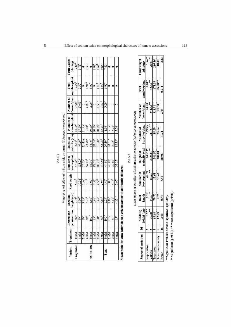
5 Effect of sodium azide on morphological characters of tomato accessions
113

K.A. Abdulkareem et al. 6 114
7 Effect of sodium azide on morphological characters of tomato accessions
115
DISCUSSION
Treatment with sodium azide exhibited a general reduction and lateness in
the germination of seeds when compared with the control. The control germinated
4 days after planting while seeds treated with sodium azide germinated after day 5
and this was observed in all the accessions. In the Tropimech, there was 100% seed
germination in the control, there was reduction in the seed germination in the
treatments. Treatment with 1mM had 40% germination, 2mM had 50% germination
and 50% germination in treatment with 4mM and this showed a great reduction in
germination. NGB01302 also had a 100% germination in the control, 1mM (85%),
2mM (80%), 4mM (65%) which are also less than the control. More so in the third
accession (Tima), the control had 100% germination, 1mM had 57.5% germination,
2mM (55%) and 4mM (65%) which are also lower than the control. Earlier work
by Pande and Khetmalas (2012) reported that germination of seeds and survival of
seedlings is strongly inhibited with increasing concentration and treatment duration. He
explained that there was reduction in the germination and survival percentage with
increasing concentrations for both chemicals in the first generation. Blixt (1970)
opined that the inhibition in seedling growth might be due to the gross injury
caused at cellular level either due to gene controlled biochemical processes or acute
chromosomal aberrations or both. Evans and Sparrow (1961) suggested that the
chromosomal damage and inhibition of cell division are the chief causes of reduced
seedling growth. Khan and Goyal, 2009; Chowdhury and Tah (2011), explained
that the decrease in seed germination induced by mutagenic treatments may be the
result of damage of cell constituents at molecular level or altered enzyme activity.
Micco et al., (2011) have correlated seed germination with abnormalities in mitotic
cycles and in metabolic pathways of the cells.
Decrease in plant height, number of leaves and number of branches were
observed with increase in sodium azide concentration in the Tropimech and Tima
accessions at the point of termination. This agrees with the work of Adamu and
Aliyu (2007), Pande and Khetmalas (2012), Shagufta et al. (2013). Ignacimuthu
and Babu (1988), Cherry et al. (1962) also reported that change in the specific activity
of quite a few enzymes and physiological injury induced in the seeds and seedlings
may be responsible for the decrease in height. Adamu and Aliyu (2007) showed
that there was general decrease in germination percentage, seedling height, root length,
number of leaves per seedling, seedling survival, height at maturity, number of fruit
with increase in mutagenic concentration. The results obtained in NGB01302 in this
study however differ from the above assertion. An increase in all the parameters
was observed with increasing concentration of sodium azide at the point of
termination. Krishna et. al (1984) opined that the greater sensitivity at higher
concentration level was attributed to various factors such as changes in the
metabolic activity of the cells, inhibitory effects of mutagens and disturbance of the
balance between promoter and inhibitors of growth regulators.
K.A. Abdulkareem et al. 8 116
The flower produced by Tropimech treated with 1mM sodium azide developed
into fruit at week 15. However, flower senescence was observed in the treated
accessions which consequently led to less production of fruit, this was in accordance to
the work of Adamu et. al (2002) and Adamu and Aliyu (2007), the result of their
work showed that all the traits studied were affected by sodium azide treatment.
The decrease in seedling emergence, seedling height, root length, seedling survival,
height at maturity and fruit yield per plant with increasing mutagen concentration
has been reported by Aliyu et al., (2002) in their mutagenesis studies on groundnut.
In all growth parameters studied, Tropimech and Tima had a general decrease with
increasing concentration of sodium azide. However, NGB01302 had an increase in
all parameters studied with increasing concentration.
CONCLUSION
The treatment of the three tomato accessions with sodium azide exhibited a
general increase in all parameters studied. M1 seeds showed heritable changes which
can be used for selection of beneficial mutant leading to variability in tomato, the
estimate of heritability has proved that these tomato accessions will perform well
when selected. Low concentration of sodium azide can therefore be utilized to
trigger traits that can help increase farmers’ turnover which can ultimately lead to
the improvement of tomato accessions.
REFERENCES
1. Adamu A.K. and H. Aliyu, 2007, Morphological effect of Sodium azide on tomato
(Lycopersicon esculentum).Sci. World J. 2 (4), pp. 9–12
2. Adamu A.K., Oluranju P.E., Bate J.A., O.T. Ogunlade, 2002, Radio sensitivity and effective dose
determination in groundnut (Arachis hypogaea L.) irradiated with gamma-rays. Journal of Agriculture
and Environment 3(1), pp. 17–84.
3. Blixt S., 1970, Studies of induced mutations in peas. XXV. Genetically controlled differences in
radiation sensitivity. Agri. Hort. Genet. 28, pp. 55–116.
4. Cherry J.H., Hageman R.H., J. B. Hanson, 1962, Effect of X-Irradiation on nucleic acids in Zea mays:
II. On the level of ribonuclease activity in growing seedlings. Radiation Research 17, pp. 740–751.
5. Chowdhury R. and J. Tah, 2011, Assessment of chemical mutagenic effects in mutation breeding
programme for M1 generation of carnation (Dianthu caryophyllus). Research in Plant Biology
1(4), pp. 23–32.
6. Cutler K.D., 1998, From Wolf peach to outer Space: Tomato history. lore. www.bbg.org/gardening/
kitchen/tomatoes /cutler.html/. (accessed 14th December, 2013).
7. Das S.V. and S. Chakraborty, 1998, Genetic variation for seed yield and its components in green
grain (Vigna radiate (L.) Wilczek). Plant Sci. 11, pp.271–273.
8. Erinle I.D., 1989, Present status and prospect for increased production of tomato and pepper in
Northern Nigeria. Procedure of International Symposium. Integrated management practices.
AVRD, Tainan, Taiwan.
9. Erdem G. and S. Oldacays, 2004, Employment of RAOD technique to assess the genetic stability
of Helianthus annus treated with different mutagenic agents. J. Applied Sci. 4, pp. 277–281.

9 Effect of sodium azide on morphological characters of tomato accessions
117
10. Evans H.J. and A.H. Sparrow, 1961, Nuclear Factors Affecting Radiosensitivity. In: Fundamental
Aspects of Radio sensitivity. Brookhaven Symposia in Biology, 14, pp. 101–124.
11. Green S.R., Blackmore R. and A. Monchich, 1989, Comment on tomato production.
www.och.greehoof.ord/rt15.org(accessed on 16th February 2014).
12. Ignacimuthu S. and C.R. Babu, 1988, Radio Sensitivity of the wild and cultivated urd and
mungbeans. Indian J. Genet. Plant Breed. 48(3), pp. 331–342.
13. Johnson H.W, Robinson H.F. and R.E. Comstock, 1955, Estimate of genetic and environmental
variability in soybeans. Agronomy Journal, 47, pp. 311–318.
14. Khan S. and S. Goyal, 2009, Improvement of Mungbean varieties through induced mutations.
African Journal of Plant Science, 3, pp. 174–180.
15. Micco V.D., Arena C., Pignalosa D., M. Durante, 2011, Effects of sparsely and densely ionizing
radiation on plants. Radiat. Environ. Biophys 50, pp. 1–19.
16. Nilan R.A., Kleinhofs A. and C.F. Konzak, 1977, The role of induced mutation in supplementing
natural genetic variability. Ann. N.Y. Acad. Sci. 287, pp. 367–384.
17. Novak F.J. and Brunner B.K., 1992, Plant breeding: Induced mutation technology for crop
improvement. IAEA Bull. 4, pp. 25–32.
18. Olanrewaju D. and V. Swamp, 1980, Tomato cultivation and its potential in Nigeria. National
Horticulture Research Institute Bulletin. Ibadan, Nigeria, pp. 20.
19. Pande S. and M. Khetmalas, 2012, Biological effect of Sodium Azide and Colchicine in Stevia
rebaudiana. Asian J. Exp. Biol. Sci. 3(1), pp. 93–98.
20. Reddy P.A., Sekhar M.R., Rangnatha A.R.G and A. Dhanrat, 2001, Genetic variability and
heritability for seed yield and its components in Sesame. Journal of Oil seed Resources, 18,
pp. 173–175
21. Shagufta B., Aijaz A.W. and A. Nawchoo Irshad, 2013, Mutagenic sensitivity of Gamma rays,
EMS and Sodium azide in Trigonella foenum-graecum L. Sci. Res. Rept, 3(1), pp. 20–26.
22. Sobulo R.A. and J.H. Simon, 1974, Methods for higher tomato yield in Western State of Nigeria.
Paper presented at Institute of Agric. Research Conference Ibadan, Nigeria.
23. Tigchelaar, E.C., 1978, Tomato ripening mutants. Horticultural Science, 13, pp. 502.
24. Tindall H.D., 1968, Commercial Vegetable Growing. Oxford University Press, London, Ibadan,
Nairobi, pp. 300.
25. Varela A.M., Self A. and B. Lohr, 2003, A guide to IPM in tomato production in Eastern and
Southern Africa. The International Centre of Insect Physiology and Ecology (ICIPE), pp. 144.

K.A. Abdulkareem et al. 10 118

ROM. J. BIOL. – PLANT BIOL., VOLUME 62, No. 2, P. 119, BUCHAREST, 2017
Nanotechnologies in Food and Agriculture (eds. Rai M., Ribeiro C, Mattoso L.,
Duran L), 2015, Softcover ISBN 978-3-319-38244-9, 23 b/w illustrations,
19 illustrations in color, Springer International Publishing, p. 347.
The book Nanotechnologies in Food and Agriculture, first edition, contains
14 chapters dealing with fundamental and applicative overviews on new and
emerging nanotechnologies. Topics include nanoagrochemicals (3 chapters: Nano-
fertilizers for Balanced Crop Nutrition; Nano-fertilizers and Their Smart Delivery
System; Perspectives in Nanocomposites for the Slow and Controlled Release of
Agrochemicals: Fertilizers and Pesticides), nanotechnologies for food processing,
packaging, and storage, crop improvement and plant disease control (6 chapters:
Strategic Role of Nanotechnology in Fertilizers: Potential and Limitations;
Nanotechnology Applied in Agriculture: Controlled Release of Agrochemicals;
Nanobiotechnology Strategies for Delivery of Antimicrobials in Agriculture and
Food; Nano-developments for Food Packaging and Labeling Applications;
Emerging Role of Nanocarriers in Delivery of Nitric Oxide for Sustainable
Agriculture; Nanotechnology in Foods), Nanoecotoxicology (2 chapters) as well as
individual chapters on nanobiosensors (Strategic Role of Nanobiosensor in Food:
Benefits and Bottlenecks), plant genetic transformation (Nanoparticles-Based
Delivery Systems in Plant Genetic Transformation) and waste treatment (Nano-
enhanced Biological Treatment of Agricultural Wastewater). Each chapter contains
very rich and useful information, including comprehensive references. Each chapter is
very well written by well known scientist(s) in their fields. The book is not very
rich in illustrations but the quality of the text partly overcomes this aspect. The
book, useful and needed both by PhD students and already experienced scientists,
should be found accessible in every (significant) faculty or research institute dealing
with this still emerging field.
Ioan I. Ardelean

K.A. Abdulkareem et al. 2 110