
The Eukaryotes of Microbiology
208
Chapter 5 The Eukaryotes of Microbiology Figure 5.1 Malaria is a disease caused by a eukaryotic parasite transmitted to humans by mosquitos. Micrographs (left and center) show a sporozoite life stage, trophozoites, and a schizont in a blood smear. On the right is depicted a primary defense against mosquito-borne illnesses like malaria—mosquito netting. (credit left: modification of work by Ute Frevert; credit middle: modification of work by Centers for Disease Control and Prevention; credit right: modification of work by Tjeerd Wiersma) Chapter Outline 5.1 Unicellular Eukaryotic Parasites 5.2 Parasitic Helminths 5.3 Fungi 5.4 Algae 5.5 Lichens Introduction Although bacteria and viruses account for a large number of the infectious diseases that afflict humans, many serious illnesses are caused by eukaryotic organisms. One example is malaria, which is caused by Plasmodium, a eukaryotic organism transmitted through mosquito bites. Malaria is a major cause of morbidity (illness) and mortality (death) that threatens 3.4 billion people worldwide. [1] In severe cases, organ failure and blood or metabolic abnormalities contribute to medical emergencies and sometimes death. Even after initial recovery, relapses may occur years later. In countries where malaria is endemic, the disease represents a major public health challenge that can place a tremendous strain on developing economies. Worldwide, major efforts are underway to reduce malaria infections. Efforts include the distribution of insecticide- treated bed nets and the spraying of pesticides. Researchers are also making progress in their efforts to develop effective vaccines. [2] The President’s Malaria Initiative, started in 2005, supports prevention and treatment. The Bill and Melinda Gates Foundation has a large initiative to eliminate malaria. Despite these efforts, malaria continues to cause long-term morbidity (such as intellectual disabilities in children) and mortality (especially in children younger than 5 years), so we still have far to go. 1. Centers for Disease Control and Prevention. “Impact of Malaria.” September 22, 2015. http://www.cdc.gov/malaria/malaria_worldwide/ impact.html. Accessed January 18, 2016. 2. RTS, S Clinical Trials Partnership. “Efficacy and safety of RTS,S/AS01 malaria vaccine with or without a booster dose in infants and children in Africa: final results of a phase 3, individually randomised, controlled trial.” The Lancet 23 April 2015. DOI: http://dx.doi.org/ 10.1016/S0140-6736(15)60721-8. Chapter 5 | The Eukaryotes of Microbiology 197
-
Upload
khangminh22 -
Category
Documents
-
view
0 -
download
0
Transcript of The Eukaryotes of Microbiology

Chapter 5
The Eukaryotes of Microbiology
Figure 5.1 Malaria is a disease caused by a eukaryotic parasite transmitted to humans by mosquitos. Micrographs(left and center) show a sporozoite life stage, trophozoites, and a schizont in a blood smear. On the right is depicted aprimary defense against mosquito-borne illnesses like malaria—mosquito netting. (credit left: modification of work byUte Frevert; credit middle: modification of work by Centers for Disease Control and Prevention; credit right:modification of work by Tjeerd Wiersma)
Chapter Outline
5.1 Unicellular Eukaryotic Parasites
5.2 Parasitic Helminths
5.3 Fungi
5.4 Algae
5.5 Lichens
IntroductionAlthough bacteria and viruses account for a large number of the infectious diseases that afflict humans, many seriousillnesses are caused by eukaryotic organisms. One example is malaria, which is caused by Plasmodium, a eukaryoticorganism transmitted through mosquito bites. Malaria is a major cause of morbidity (illness) and mortality (death)that threatens 3.4 billion people worldwide.[1] In severe cases, organ failure and blood or metabolic abnormalitiescontribute to medical emergencies and sometimes death. Even after initial recovery, relapses may occur years later. Incountries where malaria is endemic, the disease represents a major public health challenge that can place a tremendousstrain on developing economies.
Worldwide, major efforts are underway to reduce malaria infections. Efforts include the distribution of insecticide-treated bed nets and the spraying of pesticides. Researchers are also making progress in their efforts to developeffective vaccines.[2] The President’s Malaria Initiative, started in 2005, supports prevention and treatment. The Billand Melinda Gates Foundation has a large initiative to eliminate malaria. Despite these efforts, malaria continues tocause long-term morbidity (such as intellectual disabilities in children) and mortality (especially in children youngerthan 5 years), so we still have far to go.
1. Centers for Disease Control and Prevention. “Impact of Malaria.” September 22, 2015. http://www.cdc.gov/malaria/malaria_worldwide/
impact.html. Accessed January 18, 2016.
2. RTS, S Clinical Trials Partnership. “Efficacy and safety of RTS,S/AS01 malaria vaccine with or without a booster dose in infants and
children in Africa: final results of a phase 3, individually randomised, controlled trial.” The Lancet 23 April 2015. DOI: http://dx.doi.org/
10.1016/S0140-6736(15)60721-8.
Chapter 5 | The Eukaryotes of Microbiology 197

5.1 Unicellular Eukaryotic Parasites
Learning Objectives• Summarize the general characteristics of unicellular eukaryotic parasites
• Describe the general life cycles and modes of reproduction in unicellular eukaryotic parasites
• Identify challenges associated with classifying unicellular eukaryotes
• Explain the taxonomic scheme used for unicellular eukaryotes
• Give examples of infections caused by unicellular eukaryotes
Eukaryotic microbes are an extraordinarily diverse group, including species with a wide range of life cycles,morphological specializations, and nutritional needs. Although more diseases are caused by viruses and bacteria thanby microscopic eukaryotes, these eukaryotes are responsible for some diseases of great public health importance.For example, the protozoal disease malaria was responsible for 584,000 deaths worldwide (primarily children inAfrica) in 2013, according to the World Health Organization (WHO). The protist parasite Giardia causes a diarrhealillness (giardiasis) that is easily transmitted through contaminated water supplies. In the United States, Giardia isthe most common human intestinal parasite (Figure 5.3). Although it may seem surprising, parasitic worms areincluded within the study of microbiology because identification depends on observation of microscopic adult wormsor eggs. Even in developed countries, these worms are important parasites of humans and of domestic animals. Thereare fewer fungal pathogens, but these are important causes of illness, as well. On the other hand, fungi have been
Part 1
Upon arriving home from school, 7-year-old Sarah complains that a large spot on her arm will not stop itching.She keeps scratching at it, drawing the attention of her parents. Looking more closely, they see that it is ared circular spot with a raised red edge (Figure 5.2). The next day, Sarah’s parents take her to their doctor,who examines the spot using a Wood’s lamp. A Wood’s lamp produces ultraviolet light that causes the spot onSarah’s arm to fluoresce, which confirms what the doctor already suspected: Sarah has a case of ringworm.
Sarah’s mother is mortified to hear that her daughter has a “worm.” How could this happen?
• What are some likely ways that Sarah might have contracted ringworm?
Figure 5.2 Ringworm presents as a raised, red ring on the skin. (credit: Centers for Disease Control andPrevention)
Jump to the next Clinical Focus box.
Clinical Focus
198 Chapter 5 | The Eukaryotes of Microbiology
This OpenStax book is available for free at http://cnx.org/content/col12087/1.5

important in producing antimicrobial substances such as penicillin. In this chapter, we will examine characteristics ofprotists, worms, and fungi while considering their roles in causing disease.
Figure 5.3 (a) A scanning electron micrograph shows many Giardia parasites in the trophozoite, or feeding stage, ina gerbil intestine. (b) An individual trophozoite of G. lamblia, visualized here in a scanning electron micrograph. Thiswaterborne protist causes severe diarrhea when ingested. (credit a, b: modification of work by Centers for DiseaseControl and Prevention)
Characteristics of Protists
The word protist is a historical term that is now used informally to refer to a diverse group of microscopic eukaryoticorganisms. It is not considered a formal taxonomic term because the organisms it describes do not have a sharedevolutionary origin. Historically, the protists were informally grouped into the “animal-like” protozoans, the “plant-like” algae, and the “fungus-like” protists such as water molds. These three groups of protists differ greatly in termsof their basic characteristics. For example, algae are photosynthetic organisms that can be unicellular or multicellular.Protozoa, on the other hand, are nonphotosynthetic, motile organisms that are always unicellular. Other informalterms may also be used to describe various groups of protists. For example, microorganisms that drift or float inwater, moved by currents, are referred to as plankton. Types of plankton include zooplankton, which are motile andnonphotosynthetic, and phytoplankton, which are photosynthetic.
Protozoans inhabit a wide variety of habitats, both aquatic and terrestrial. Many are free-living, while others areparasitic, carrying out a life cycle within a host or hosts and potentially causing illness. There are also beneficialsymbionts that provide metabolic services to their hosts. During the feeding and growth part of their life cycle, theyare called trophozoites; these feed on small particulate food sources such as bacteria. While some types of protozoaexist exclusively in the trophozoite form, others can develop from trophozoite to an encapsulated cyst stage whenenvironmental conditions are too harsh for the trophozoite. A cyst is a cell with a protective wall, and the process bywhich a trophozoite becomes a cyst is called encystment. When conditions become more favorable, these cysts aretriggered by environmental cues to become active again through excystment.
One protozoan genus capable of encystment is Eimeria, which includes some human and animal pathogens. Figure5.4 illustrates the life cycle of Eimeria.
Chapter 5 | The Eukaryotes of Microbiology 199

Figure 5.4 In the sexual/asexual life cycle of Eimeria, oocysts (inset) are shed in feces and may cause diseasewhen ingested by a new host. (credit “life cycle,” “micrograph”: modification of work by USDA)
Protozoans have a variety of reproductive mechanisms. Some protozoans reproduce asexually and others reproducesexually; still others are capable of both sexual and asexual reproduction. In protozoans, asexual reproduction occursby binary fission, budding, or schizogony. In schizogony, the nucleus of a cell divides multiple times before the celldivides into many smaller cells. The products of schizogony are called merozoites and they are stored in structuresknown as schizonts. Protozoans may also reproduce sexually, which increases genetic diversity and can lead tocomplex life cycles. Protozoans can produce haploid gametes that fuse through syngamy. However, they can alsoexchange genetic material by joining to exchange DNA in a process called conjugation. This is a different processthan the conjugation that occurs in bacteria. The term protist conjugation refers to a true form of eukaryotic sexualreproduction between two cells of different mating types. It is found in ciliates, a group of protozoans, and isdescribed later in this subsection.
All protozoans have a plasma membrane, or plasmalemma, and some have bands of protein just inside the membranethat add rigidity, forming a structure called the pellicle. Some protists, including protozoans, have distinct layersof cytoplasm under the membrane. In these protists, the outer gel layer (with microfilaments of actin) is called theectoplasm. Inside this layer is a sol (fluid) region of cytoplasm called the endoplasm. These structures contribute tocomplex cell shapes in some protozoans, whereas others (such as amoebas) have more flexible shapes (Figure 5.5).
Different groups of protozoans have specialized feeding structures. They may have a specialized structure for takingin food through phagocytosis, called a cytostome, and a specialized structure for the exocytosis of wastes called acytoproct. Oral grooves leading to cytostomes are lined with hair-like cilia to sweep in food particles. Protozoansare heterotrophic. Protozoans that are holozoic ingest whole food particles through phagocytosis. Forms that are
200 Chapter 5 | The Eukaryotes of Microbiology
This OpenStax book is available for free at http://cnx.org/content/col12087/1.5

saprozoic ingest small, soluble food molecules.
Many protists have whip-like flagella or hair-like cilia made of microtubules that can be used for locomotion (Figure5.5). Other protists use cytoplasmic extensions known as pseudopodia (“false feet”) to attach the cell to a surface;they then allow cytoplasm to flow into the extension, thus moving themselves forward.
Protozoans have a variety of unique organelles and sometimes lack organelles found in other cells. Some havecontractile vacuoles, organelles that can be used to move water out of the cell for osmotic regulation (salt and waterbalance) (Figure 5.5). Mitochondria may be absent in parasites or altered to kinetoplastids (modified mitochondria)or hydrogenosomes (see Unique Characteristics of Prokaryotic Cells for more discussion of these structures).
Figure 5.5 (a) Paramecium spp. have hair-like appendages called cilia for locomotion. (b) Amoeba spp. use lobe-like pseudopodia to anchor the cell to a solid surface and pull forward. (c) Euglena spp. use a whip-like structurecalled a flagellum to propel the cell.
• What is the sequence of events in reproduction by schizogony and what are the cells produced called?
Taxonomy of Protists
The protists are a polyphyletic group, meaning they lack a shared evolutionary origin. Since the current taxonomy isbased on evolutionary history (as determined by biochemistry, morphology, and genetics), protists are scattered acrossmany different taxonomic groups within the domain Eukarya. Eukarya is currently divided into six supergroups thatare further divided into subgroups, as illustrated in (Figure 5.6). In this section, we will primarily be concernedwith the supergroups Amoebozoa, Excavata, and Chromalveolata; these supergroups include many protozoans ofclinical significance. The supergroups Opisthokonta and Rhizaria also include some protozoans, but few of clinicalsignificance. In addition to protozoans, Opisthokonta also includes animals and fungi, some of which we will discussin Parasitic Helminths and Fungi. Some examples of the Archaeplastida will be discussed in Algae. Figure 5.7and Figure 5.8 summarize the characteristics of each supergroup and subgroup and list representatives of each.
Chapter 5 | The Eukaryotes of Microbiology 201

Figure 5.6 This tree shows a proposed classification of the domain Eukarya based on evolutionary relationships.Currently, the domain Eukarya is divided into six supergroups. Within each supergroup are multiple kingdoms. Dottedlines indicate suggested evolutionary relationships that remain under debate.
202 Chapter 5 | The Eukaryotes of Microbiology
This OpenStax book is available for free at http://cnx.org/content/col12087/1.5

Figure 5.7
Chapter 5 | The Eukaryotes of Microbiology 203

Figure 5.8
• Which supergroups contain the clinically significant protists?
204 Chapter 5 | The Eukaryotes of Microbiology
This OpenStax book is available for free at http://cnx.org/content/col12087/1.5

Amoebozoa
The supergroup Amoebozoa includes protozoans that use amoeboid movement. Actin microfilaments producepseudopodia, into which the remainder of the protoplasm flows, thereby moving the organism. The genus Entamoebaincludes commensal or parasitic species, including the medically important E. histolytica, which is transmitted bycysts in feces and is the primary cause of amoebic dysentery. The notorious “brain-eating amoeba,” Naegleria fowleri,is also classified within the Amoebozoa. This deadly parasite is found in warm, fresh water and causes primaryamoebic meningoencephalitis (PAM). Another member of this group is Acanthamoeba, which can cause keratitis(corneal inflammation) and blindness.
The Eumycetozoa are an unusual group of organisms called slime molds, which have previously been classifiedas animals, fungi, and plants (Figure 5.9). Slime molds can be divided into two types: cellular slime molds andplasmodial slime molds. The cellular slime molds exist as individual amoeboid cells that periodically aggregateinto a mobile slug. The aggregate then forms a fruiting body that produces haploid spores. Plasmodial slime moldsexist as large, multinucleate amoeboid cells that form reproductive stalks to produce spores that divide into gametes.One cellular slime mold, Dictyostelium discoideum, has been an important study organism for understanding celldifferentiation, because it has both single-celled and multicelled life stages, with the cells showing some degreeof differentiation in the multicelled form. Figure 5.10 and Figure 5.11 illustrate the life cycles of cellular andplasmodial slime molds, respectively.
Figure 5.9 (a) The cellular slime mold Dictyostelium discoideum can be grown on agar in a Petri dish. In this image,individual amoeboid cells (visible as small spheres) are streaming together to form an aggregation that is beginning torise in the upper right corner of the image. The primitively multicellular aggregation consists of individual cells thateach have their own nucleus. (b) Fuligo septica is a plasmodial slime mold. This brightly colored organism consists ofa large cell with many nuclei.
Chapter 5 | The Eukaryotes of Microbiology 205

Figure 5.10 The life cycle of the cellular slime mold Dictyostelium discoideum primarily involves individual amoebasbut includes the formation of a multinucleate plasmodium formed from a uninucleate zygote (the result of the fusion oftwo individual amoeboid cells). The plasmodium is able to move and forms a fruiting body that generates haploidspores. (credit “photo”: modification of work by “thatredhead4”/Flickr)
206 Chapter 5 | The Eukaryotes of Microbiology
This OpenStax book is available for free at http://cnx.org/content/col12087/1.5

Figure 5.11 Plasmodial slime molds exist as large multinucleate amoeboid cells that form reproductive stalks toproduce spores that divide into gametes.
Chromalveolata
The supergroup Chromalveolata is united by similar origins of its members’ plastids and includes the apicomplexans,ciliates, diatoms, and dinoflagellates, among other groups (we will cover the diatoms and dinoflagellates in Algae).The apicomplexans are intra- or extracellular parasites that have an apical complex at one end of the cell. Theapical complex is a concentration of organelles, vacuoles, and microtubules that allows the parasite to enter hostcells (Figure 5.12). Apicomplexans have complex life cycles that include an infective sporozoite that undergoesschizogony to make many merozoites (see the example in Figure 5.4). Many are capable of infecting a variety ofanimal cells, from insects to livestock to humans, and their life cycles often depend on transmission between multiplehosts. The genus Plasmodium is an example of this group.
Chapter 5 | The Eukaryotes of Microbiology 207

Figure 5.12 (a) Apicomplexans are parasitic protists. They have a characteristic apical complex that enables themto infect host cells. (b) A colorized electron microscope image of a Plasmodium sporozoite. (credit b: modification ofwork by Ute Frevert)
Other apicomplexans are also medically important. Cryptosporidium parvum causes intestinal symptoms and cancause epidemic diarrhea when the cysts contaminate drinking water. Theileria (Babesia) microti, transmitted by thetick Ixodes scapularis, causes recurring fever that can be fatal and is becoming a common transfusion-transmittedpathogen in the United States (Theileria and Babesia are closely related genera and there is some debate aboutthe best classification). Finally, Toxoplasma gondii causes toxoplasmosis and can be transmitted from cat feces,unwashed fruit and vegetables, or from undercooked meat. Because toxoplasmosis can be associated with seriousbirth defects, pregnant women need to be aware of this risk and use caution if they are exposed to the feces ofpotentially infected cats. A national survey found the frequency of individuals with antibodies for toxoplasmosis (andthus who presumably have a current latent infection) in the United States to be 11%. Rates are much higher in othercountries, including some developed countries.[3] There is also evidence and a good deal of theorizing that the parasitemay be responsible for altering infected humans’ behavior and personality traits.[4]
The ciliates (Ciliaphora), also within the Chromalveolata, are a large, very diverse group characterized by thepresence of cilia on their cell surface. Although the cilia may be used for locomotion, they are often used forfeeding, as well, and some forms are nonmotile. Balantidium coli (Figure 5.13) is the only parasitic ciliatethat affects humans by causing intestinal illness, although it rarely causes serious medical issues except in theimmunocompromised (those having a weakened immune system). Perhaps the most familiar ciliate is Paramecium, amotile organism with a clearly visible cytostome and cytoproct that is often studied in biology laboratories (Figure5.14). Another ciliate, Stentor, is sessile and uses its cilia for feeding (Figure 5.15). Generally, these organismshave a micronucleus that is diploid, somatic, and used for sexual reproduction by conjugation. They also have amacronucleus that is derived from the micronucleus; the macronucleus becomes polyploid (multiple sets of duplicatechromosomes), and has a reduced set of metabolic genes.
Ciliates are able to reproduce through conjugation, in which two cells attach to each other. In each cell, the diploidmicronuclei undergo meiosis, producing eight haploid nuclei each. Then, all but one of the haploid micronuclei andthe macronucleus disintegrate; the remaining (haploid) micronucleus undergoes mitosis. The two cells then exchangeone micronucleus each, which fuses with the remaining micronucleus present to form a new, genetically different,
3. J. Flegr et al. “Toxoplasmosis—A Global Threat. Correlation of Latent Toxoplasmosis With Specific Disease Burden in a Set of 88
Countries.” PloS ONE 9 no. 3 (2014):e90203.
4. J. Flegr. “Effects of Toxoplasma on Human Behavior.” Schizophrenia Bull 33, no. 3 (2007):757–760.
208 Chapter 5 | The Eukaryotes of Microbiology
This OpenStax book is available for free at http://cnx.org/content/col12087/1.5

diploid micronucleus. The diploid micronucleus undergoes two mitotic divisions, so each cell has four micronuclei,and two of the four combine to form a new macronucleus. The chromosomes in the macronucleus then replicaterepeatedly, the macronucleus reaches its polyploid state, and the two cells separate. The two cells are now geneticallydifferent from each other and from their previous versions.
Figure 5.13 This specimen of the ciliate Balantidium coli is a trophozoite form isolated from the gut of a primate. B.coli is the only ciliate capable of parasitizing humans. (credit: modification of work by Kouassi RYW, McGraw SW, YaoPK, Abou-Bacar A, Brunet J, Pesson B, Bonfoh B, N’goran EK & Candolfi E)
Figure 5.14 Paramecium has a primitive mouth (called an oral groove) to ingest food, and an anal pore to excrete it.Contractile vacuoles allow the organism to excrete excess water. Cilia enable the organism to move.
Chapter 5 | The Eukaryotes of Microbiology 209

Figure 5.15 This differential interference contrast micrograph (magnification: ×65) of Stentor roeselie shows ciliapresent on the margins of the structure surrounding the cytostome; the cilia move food particles. (credit: modificationof work by “picturepest”/Flickr)
Öomycetes have similarities to fungi and were once classified with them. They are also called water molds. However,they differ from fungi in several important ways. Öomycetes have cell walls of cellulose (unlike the chitinous cellwalls of fungi) and they are generally diploid, whereas the dominant life forms of fungi are typically haploid.Phytophthora, the plant pathogen found in the soil that caused the Irish potato famine, is classified within this group(Figure 5.16).
Figure 5.16 A saprobic oomycete, or water mold, engulfs a dead insect. (credit: modification of work by ThomasBresson)
210 Chapter 5 | The Eukaryotes of Microbiology
This OpenStax book is available for free at http://cnx.org/content/col12087/1.5

Explore the procedures for detecting the presence of an apicomplexan in apublic water supply, at this (https://openstax.org/l/22detpreapicom) website.
This video (https://openstax.org/l/22feedstentor) shows the feeding ofStentor.
Excavata
The third and final supergroup to be considered in this section is the Excavata, which includes primitive eukaryotesand many parasites with limited metabolic abilities. These organisms have complex cell shapes and structures, oftenincluding a depression on the surface of the cell called an excavate. The group Excavata includes the subgroupsFornicata, Parabasalia, and Euglenozoa. The Fornicata lack mitochondria but have flagella. This group includesGiardia lamblia (also known as G. intestinalis or G. duodenalis), a widespread pathogen that causes diarrhealillness and can be spread through cysts from feces that contaminate water supplies (Figure 5.3). Parabasalia arefrequent animal endosymbionts; they live in the guts of animals like termites and cockroaches. They have basalbodies and modified mitochondria (kinetoplastids). They also have a large, complex cell structure with an undulatingmembrane and often have many flagella. The trichomonads (a subgroup of the Parabasalia) include pathogens suchas Trichomonas vaginalis, which causes the human sexually transmitted disease trichomoniasis. Trichomoniasis oftendoes not cause symptoms in men, but men are able to transmit the infection. In women, it causes vaginal discomfortand discharge and may cause complications in pregnancy if left untreated.
The Euglenozoa are common in the environment and include photosynthetic and nonphotosynthetic species. Membersof the genus Euglena are typically not pathogenic. Their cells have two flagella, a pellicle, a stigma (eyespot) to senselight, and chloroplasts for photosynthesis (Figure 5.17). The pellicle of Euglena is made of a series of protein bandssurrounding the cell; it supports the cell membrane and gives the cell shape.
The Euglenozoa also include the trypanosomes, which are parasitic pathogens. The genus Trypanosoma includesT. brucei, which causes African trypanosomiasis (African sleeping sickness and T. cruzi, which causes Americantrypanosomiasis (Chagas disease). These tropical diseases are spread by insect bites. In African sleeping sickness, T.brucei colonizes the blood and the brain after being transmitted via the bite of a tsetse fly (Glossina spp.) (Figure5.18). The early symptoms include confusion, difficulty sleeping, and lack of coordination. Left untreated, it is fatal.
Figure 5.17 (a) This illustration of a Euglena shows the characteristic structures, such as the stigma and flagellum.(b) The pellicle, under the cell membrane, gives the cell its distinctive shape and is visible in this image as delicateparallel striations over the surface of the entire cell (especially visible over the grey contractile vacuole). (credit a:modification of work by Claudio Miklos; credit b: modification of work by David Shykind)
Link to Learning
Chapter 5 | The Eukaryotes of Microbiology 211

Figure 5.18 Trypanosoma brucei, the causative agent of African trypanosomiasis, spends part of its life cycle in thetsetse fly and part in humans. (credit “illustration”: modification of work by Centers for Disease Control andPrevention; credit “photo”: DPDx/Centers for Disease Control and Prevention)
Chagas’ disease originated and is most common in Latin America. The disease is transmitted by Triatoma spp., insectsoften called “kissing bugs,” and affects either the heart tissue or tissues of the digestive system. Untreated cases caneventually lead to heart failure or significant digestive or neurological disorders.
The genus Leishmania includes trypanosomes that cause disfiguring skin disease and sometimes systemic illness aswell.
Neglected Parasites
The Centers for Disease Control and Prevention (CDC) is responsible for identifying public health prioritiesin the United States and developing strategies to address areas of concern. As part of this mandate, the
Eye on Ethics
212 Chapter 5 | The Eukaryotes of Microbiology
This OpenStax book is available for free at http://cnx.org/content/col12087/1.5

CDC has officially identified five parasitic diseases it considers to have been neglected (i.e., not adequatelystudied). These neglected parasitic infections (NPIs) include toxoplasmosis, Chagas disease, toxocariasis(a nematode infection transmitted primarily by infected dogs), cysticercosis (a disease caused by a tissueinfection of the tapeworm Taenia solium), and trichomoniasis (a sexually transmitted disease caused by theparabasalid Trichomonas vaginalis).
The decision to name these specific diseases as NPIs means that the CDC will devote resources towardimproving awareness and developing better diagnostic testing and treatment through studies of available data.The CDC may also advise on treatment of these diseases and assist in the distribution of medications thatmight otherwise be difficult to obtain.[5]
Of course, the CDC does not have unlimited resources, so by prioritizing these five diseases, it is effectivelydeprioritizing others. Given that many Americans have never heard of many of these NPIs, it is fair to askwhat criteria the CDC used in prioritizing diseases. According to the CDC, the factors considered were thenumber of people infected, the severity of the illness, and whether the illness can be treated or prevented.Although several of these NPIs may seem to be more common outside the United States, the CDC argues thatmany cases in the United States likely go undiagnosed and untreated because so little is known about thesediseases.[6]
What criteria should be considered when prioritizing diseases for purposes of funding or research? Are thoseidentified by the CDC reasonable? What other factors could be considered? Should government agencies likethe CDC have the same criteria as private pharmaceutical research labs? What are the ethical implications ofdeprioritizing other potentially neglected parasitic diseases such as leishmaniasis?
5.2 Parasitic Helminths
Learning Objectives• Explain why we include the study of parasitic worms within the discipline of microbiology
• Compare the basic morphology of the major groups of parasitic helminthes
• Describe the characteristics of parasitic nematodes, and give an example of infective eggs and infective larvae
• Describe the characteristics of parasitic trematodes and cestodes, and give examples of each
• Identify examples of the primary causes of infections due to nematodes, trematodes, and cestodes
• Classify parasitic worms according to major groups
Parasitic helminths are animals that are often included within the study of microbiology because many species ofthese worms are identified by their microscopic eggs and larvae. There are two major groups of parasitic helminths:the roundworms (Nematoda) and flatworms (Platyhelminthes). Of the many species that exist in these groups, abouthalf are parasitic and some are important human pathogens. As animals, they are multicellular and have organsystems. However, the parasitic species often have limited digestive tracts, nervous systems, and locomotor abilities.Parasitic forms may have complex reproductive cycles with several different life stages and more than one type ofhost. Some are monoecious, having both male and female reproductive organs in a single individual, while others aredioecious, each having either male or female reproductive organs.
5. Centers for Disease Control and Prevention. “Neglected Parasitic Infections (NPIs) in the United States.” http://www.cdc.gov/parasites/
npi/. Last updated July 10, 2014.
6. Centers for Disease Control and Prevention. “Fact Sheet: Neglected Parasitic Infections in the United States.” http://www.cdc.gov/
parasites/resources/pdf/npi_factsheet.pdf
Chapter 5 | The Eukaryotes of Microbiology 213

Nematoda (Roundworms)
Phylum Nematoda (the roundworms) is a diverse group containing more than 15,000 species, of which severalare important human parasites (Figure 5.19). These unsegmented worms have a full digestive system even whenparasitic. Some are common intestinal parasites, and their eggs can sometimes be identified in feces or around theanus of infected individuals. Ascaris lumbricoides is the largest nematode intestinal parasite found in humans; femalesmay reach lengths greater than 1 meter. A. lumbricoides is also very widespread, even in developed nations, althoughit is now a relatively uncommon problem in the United States. It may cause symptoms ranging from relatively mild(such as a cough and mild abdominal pain) to severe (such as intestinal blockage and impaired growth).
Figure 5.19 A micrograph of the nematode Enterobius vermicularis, also known as the pinworm. (credit:modification of work by Centers for Disease Control and Prevention)
Of all nematode infections in the United States, pinworm (caused by Enterobius vermicularis) is the most common.Pinworm causes sleeplessness and itching around the anus, where the female worms lay their eggs during the night.Toxocara canis and T. cati are nematodes found in dogs and cats, respectively, that can be transmitted to humans,causing toxocariasis. Antibodies to these parasites have been found in approximately 13.9% of the U.S. population,suggesting that exposure is common.[7] Infection can cause larval migrans, which can result in vision loss and eyeinflammation, or fever, fatigue, coughing, and abdominal pain, depending on whether the organism infects the eyeor the viscera. Another common nematode infection is hookworm, which is caused by Necator americanus (theNew World or North American hookworm) and Ancylostoma duodenale (the Old World hookworm). Symptoms ofhookworm infection can include abdominal pain, diarrhea, loss of appetite, weight loss, fatigue, and anemia.
Trichinellosis, also called trichinosis, caused by Trichinella spiralis, is contracted by consuming undercooked meat,which releases the larvae and allows them to encyst in muscles. Infection can cause fever, muscle pains, and digestivesystem problems; severe infections can lead to lack of coordination, breathing and heart problems, and even death.Finally, heartworm in dogs and other animals is caused by the nematode Dirofilaria immitis, which is transmitted bymosquitoes. Symptoms include fatigue and cough; when left untreated, death may result.
Part 2
The physician explains to Sarah’s mother that ringworm can be transferred between people through touch.
Clinical Focus
7. Won K, Kruszon-Moran D, Schantz P, Jones J. “National seroprevalence and risk factors for zoonotic Toxocara spp. infection.” In:
Abstracts of the 56th American Society of Tropical Medicine and Hygiene; Philadelphia, Pennsylvania; 2007 Nov 4-8.
214 Chapter 5 | The Eukaryotes of Microbiology
This OpenStax book is available for free at http://cnx.org/content/col12087/1.5

“It’s common in school children, because they often come in close contact with each other, but anyone canbecome infected,” he adds. “Because you can transfer it through objects, locker rooms and public pools arealso a potential source of infection. It’s very common among wrestlers and athletes in other contact sports.”
Looking very uncomfortable, Sarah says to her mother “I want this worm out of me.”
The doctor laughs and says, “Sarah, you’re in luck because ringworm is just a name; it is not an actual worm.You have nothing wriggling around under your skin.”
“Then what is it?” asks Sarah.
• What type of pathogen causes ringworm?
Jump to the next Clinical Focus box. Go back to the previous Clinical Focus box.
• What is the most common nematode infection in the United States?
Platyhelminths (Flatworms)
Phylum Platyhelminthes (the platyhelminths) are flatworms. This group includes the flukes, tapeworms, and theturbellarians, which include planarians. The flukes and tapeworms are medically important parasites (Figure 5.20).
The flukes (trematodes) are nonsegmented flatworms that have an oral sucker (Figure 5.21) (and sometimes asecond ventral sucker) and attach to the inner walls of intestines, lungs, large blood vessels, or the liver. Trematodeshave complex life cycles, often with multiple hosts. Several important examples are the liver flukes (Clonorchisand Opisthorchis), the intestinal fluke (Fasciolopsis buski), and the oriental lung fluke (Paragonimus westermani).Schistosomiasis is a serious parasitic disease, considered second in the scale of its impact on human populations onlyto malaria. The parasites Schistosoma mansoni, S. haematobium, and S. japonicum, which are found in freshwatersnails, are responsible for schistosomiasis (Figure 5.22). Immature forms burrow through the skin into the blood.They migrate to the lungs, then to the liver and, later, other organs. Symptoms include anemia, malnutrition, fever,abdominal pain, fluid buildup, and sometimes death.
Chapter 5 | The Eukaryotes of Microbiology 215

Figure 5.20 Phylum Platyhelminthes is divided into four classes. (a) Class Turbellaria includes the Bedford’sflatworm (Pseudobiceros bedfordi), which is about 8–10 cm long. (b) The parasitic class Monogenea includesDactylogyrus spp. Worms in this genus are commonly called gill flukes. The specimen pictured here is about 0.2 mmlong and has two anchors, indicated by arrows, that it uses to latch onto the gills of host fish. (c) The Trematoda classincludes the common liver fluke Fasciola hepatica and the giant liver fluke Fascioloides magna (right). The F. magnaspecimen shown here is about 7 cm long. (d) Class Cestoda includes tapeworms such as Taenia saginata, whichinfects both cattle and humans and can reach lengths of 4–10 meters; the specimen shown here is about 4 meterslong. (credit c: modification of work by “Flukeman”/Wikimedia Commons)
216 Chapter 5 | The Eukaryotes of Microbiology
This OpenStax book is available for free at http://cnx.org/content/col12087/1.5

Figure 5.21 (a) The oral sucker is visible on the anterior end of this liver fluke, Opisthorchis viverrini. (b) Thismicrograph shows the scolex of the cestode Taenia solium, also known as the pork tapeworm. The visible suckersand hooks allow the worm to attach itself to the inner wall of the intestine. (credit a: modification of work by Sripa B,Kaewkes S, Sithithaworn P, Mairiang E, Laha T, and Smout M; credit b: modification of work by Centers for DiseaseControl and Prevention)
The other medically important group of platyhelminths are commonly known as tapeworms (cestodes) and aresegmented flatworms that may have suckers or hooks at the scolex (head region) (Figure 5.21). Tapeworms usethese suckers or hooks to attach to the wall of the small intestine. The body of the worm is made up of segmentscalled proglottids that contain reproductive structures; these detach when the gametes are fertilized, releasing gravidproglottids with eggs. Tapeworms often have an intermediate host that consumes the eggs, which then hatch into alarval form called an oncosphere. The oncosphere migrates to a particular tissue or organ in the intermediate host,where it forms cysticerci. After being eaten by the definitive host, the cysticerci develop into adult tapeworms in thehost's digestive system (Figure 5.23). Taenia saginata (the beef tapeworm) and T. solium (the pork tapeworm) enterhumans through ingestion of undercooked, contaminated meat. The adult worms develop and reside in the intestine,but the larval stage may migrate and be found in other body locations such as skeletal and smooth muscle. The beeftapeworm is relatively benign, although it can cause digestive problems and, occasionally, allergic reactions. The porktapeworm can cause more serious problems when the larvae leave the intestine and colonize other tissues, includingthose of the central nervous system. Diphylobothrium latum is the largest human tapeworm and can be ingested inundercooked fish. It can grow to a length of 15 meters. Echinococcus granulosus, the dog tapeworm, can parasitizehumans and uses dogs as an important host.
Chapter 5 | The Eukaryotes of Microbiology 217

Figure 5.22 The life cycle of Schistosoma spp. includes several species of water snails, which serve as secondaryhosts. The parasite is transmitted to humans through contact with contaminated water and takes up residence in theveins of the digestive system. Eggs escape the host in the urine or feces and infect a snail to complete the life cycle.(credit “illustration”: modification of work by Centers for Disease Control and Prevention; credit “step 3 photo”:modification of work by Fred A. Lewis, Yung-san Liang, Nithya Raghavan & Matty Knight)
218 Chapter 5 | The Eukaryotes of Microbiology
This OpenStax book is available for free at http://cnx.org/content/col12087/1.5

Figure 5.23 Life cycle of a tapeworm. (credit “illustration”: modification of work by Centers for Disease Control andPrevention; credit “step 3 micrographs”: modification of work by American Society for Microbiology)
• What group of medically important flatworms is segmented and what group is unsegmented?
Chapter 5 | The Eukaryotes of Microbiology 219

Food for Worms?
For residents of temperate, developed countries, it may be difficult to imagine just how common helminthinfections are in the human population. In fact, they are quite common and even occur frequently in the UnitedStates. Worldwide, approximately 807–1,221 million people are infected with Ascaris lumbricoides (perhapsone-sixth of the human population) and far more are infected if all nematode species are considered.[8] Ratesof infection are relatively high even in industrialized nations. Approximately 604–795 million people are infectedwith whipworm (Trichuris) worldwide (Trichuris can also infect dogs), and 576–740 million people are infectedwith hookworm (Necator americanus and Ancylostoma duodenale).[9] Toxocara, a nematode parasite of dogsand cats, is also able to infect humans. It is widespread in the United States, with about 10,000 symptomaticcases annually. However, one study found 14% of the population (more than 40 million Americans) wasseropositive, meaning they had been exposed to the parasite at one time. More than 200 million people haveschistosomiasis worldwide. Most of the World Health Organization (WHO) neglected tropical diseases arehelminths. In some cases, helminths may cause subclinical illnesses, meaning the symptoms are so mild thatthat they go unnoticed. In other cases, the effects may be more severe or chronic, leading to fluid accumulationand organ damage. With so many people affected, these parasites constitute a major global public healthconcern.
Eradicating the Guinea Worm
Dracunculiasis, or Guinea worm disease, is caused by a nematode called Dracunculus medinensis. Whenpeople consume contaminated water, water fleas (small crustaceans) containing the nematode larvae may beingested. These larvae migrate out of the intestine, mate, and move through the body until females eventuallyemerge (generally through the feet). While Guinea worm disease is rarely fatal, it is extremely painful and canbe accompanied by secondary infections and edema (Figure 5.24).
Figure 5.24 The Guinea worm can be removed from a leg vein of an infected person by gradually winding itaround a stick, like this matchstick. (credit: Centers for Disease Control and Prevention)
An eradication campaign led by WHO, the CDC, the United Nations Children’s Fund (UNICEF), and the CarterCenter (founded by former U.S. president Jimmy Carter) has been extremely successful in reducing cases ofdracunculiasis. This has been possible because diagnosis is straightforward, there is an inexpensive methodof control, there is no animal reservoir, the water fleas are not airborne (they are restricted to still water),
Micro Connections
Micro Connections
8. Fenwick, A. “The global burden of neglected tropical diseases.” Public health 126 no.3 (Mar 2012): 233–6.
9. de Silva, N., et. al. (2003). “Soil-transmitted helminth infections: updating the global picture”. Trends in Parasitology 19 (December
2003): 547–51.
220 Chapter 5 | The Eukaryotes of Microbiology
This OpenStax book is available for free at http://cnx.org/content/col12087/1.5

the disease is geographically limited, and there has been a commitment from the governments involved.Additionally, no vaccines or medication are required for treatment and prevention. In 1986, 3.5 million peoplewere estimated to be affected. After the eradication campaign, which included helping people in affected areaslearn to filter water with cloth, only four countries continue to report the disease (Chad, Mali, South Sudan, andEthiopia) with a total of 126 cases reported to WHO in 2014.[10]
5.3 Fungi
Learning Objectives• Explain why the study of fungi such as yeast and molds is within the discipline of microbiology
• Describe the unique characteristics of fungi
• Describe examples of asexual and sexual reproduction of fungi
• Compare the major groups of fungi in this chapter, and give examples of each
• Identify examples of the primary causes of infections due to yeasts and molds
• Identify examples of toxin-producing fungi
• Classify fungal organisms according to major groups
The fungi comprise a diverse group of organisms that are heterotrophic and typically saprozoic. In addition to thewell-known macroscopic fungi (such as mushrooms and molds), many unicellular yeasts and spores of macroscopicfungi are microscopic. For this reason, fungi are included within the field of microbiology.
Fungi are important to humans in a variety of ways. Both microscopic and macroscopic fungi have medical relevance,with some pathogenic species that can cause mycoses (illnesses caused by fungi). Some pathogenic fungi areopportunistic, meaning that they mainly cause infections when the host’s immune defenses are compromised and donot normally cause illness in healthy individuals. Fungi are important in other ways. They act as decomposers in theenvironment, and they are critical for the production of certain foods such as cheeses. Fungi are also major sources ofantibiotics, such as penicillin from the fungus Penicillium.
Characteristics of Fungi
Fungi have well-defined characteristics that set them apart from other organisms. Most multicellular fungal bodies,commonly called molds, are made up of filaments called hyphae. Hyphae can form a tangled network called amycelium and form the thallus (body) of fleshy fungi. Hyphae that have walls between the cells are called septatehyphae; hyphae that lack walls and cell membranes between the cells are called nonseptate or coenocytic hyphae).(Figure 5.25).
10. World Health Organization. “South Sudan Reports Zero Cases of Guinea-Worm Disease for Seventh Consecutive Month.” 2016.
http://www.who.int/dracunculiasis/no_new_case_for_seventh_consecutive_months/en/. Accessed May 2, 2016.
Chapter 5 | The Eukaryotes of Microbiology 221

Figure 5.25 Multicellular fungi (molds) form hyphae, which may be septate or nonseptate. Unicellular fungi (yeasts)cells form pseudohyphae from individual yeast cells.
In contrast to molds, yeasts are unicellular fungi. The budding yeasts reproduce asexually by budding off a smallerdaughter cell; the resulting cells may sometimes stick together as a short chain or pseudohypha (Figure 5.25).Candida albicans is a common yeast that forms pseudohyphae; it is associated with various infections in humans,including vaginal yeast infections, oral thrush, and candidiasis of the skin.
Some fungi are dimorphic, having more than one appearance during their life cycle. These dimorphic fungi maybe able to appear as yeasts or molds, which can be important for infectivity. They are capable of changing theirappearance in response to environmental changes such as nutrient availability or fluctuations in temperature, growingas a mold, for example, at 25 °C (77 °F), and as yeast cells at 37 °C (98.6 °F). This ability helps dimorphic fungi tosurvive in diverse environments. Histoplasma capsulatum, the pathogen that causes histoplasmosis, a lung infection,is an example of a dimorphic fungus (Figure 5.26).
222 Chapter 5 | The Eukaryotes of Microbiology
This OpenStax book is available for free at http://cnx.org/content/col12087/1.5

Figure 5.26 Histoplasma capsulatum is a dimorphic fungus that grows in soil exposed to bird feces or bat feces(guano) (top left). It can change forms to survive at different temperatures. In the outdoors, it typically grows as amycelium (as shown in the micrograph, bottom left), but when the spores are inhaled (right), it responds to the highinternal temperature of the body (37 °C [98.6 °F]) by turning into a yeast that can multiply in the lungs, causing thechronic lung disease histoplasmosis. (credit: modification of work by Centers for Disease Control and Prevention)
There are notable unique features in fungal cell walls and membranes. Fungal cell walls contain chitin, as opposedto the cellulose found in the cell walls of plants and many protists. Additionally, whereas animals have cholesterol intheir cell membranes, fungal cell membranes have different sterols called ergosterols. Ergosterols are often exploitedas targets for antifungal drugs.
Fungal life cycles are unique and complex. Fungi reproduce sexually either through cross- or self-fertilization.Haploid fungi form hyphae that have gametes at the tips. Two different mating types (represented as “+ type” and “–type”) are involved. The cytoplasms of the + and – type gametes fuse (in an event called plasmogamy), producinga cell with two distinct nuclei (a dikaryotic cell). Later, the nuclei fuse (in an event called karyogamy) to createa diploid zygote. The zygote undergoes meiosis to form spores that germinate to start the haploid stage, whicheventually creates more haploid mycelia (Figure 5.27). Depending on the taxonomic group, these sexually producedspores are known as zygospores (in Zygomycota), ascospores (in Ascomycota), or basidiospores (in Basidiomycota)(Figure 5.28).
Fungi may also exhibit asexual reproduction by mitosis, mitosis with budding, fragmentation of hyphae, andformation of asexual spores by mitosis. These spores are specialized cells that, depending on the organism, may haveunique characteristics for survival, reproduction, and dispersal. Fungi exhibit several types of asexual spores and thesecan be important in classification.
Chapter 5 | The Eukaryotes of Microbiology 223

Figure 5.27 Zygomycetes have sexual and asexual life cycles. In the sexual life cycle, + and – mating typesconjugate to form a zygosporangium.
224 Chapter 5 | The Eukaryotes of Microbiology
This OpenStax book is available for free at http://cnx.org/content/col12087/1.5

Figure 5.28 These images show asexually produced spores. (a) This brightfield micrograph shows the release ofspores from a sporangium at the end of a hypha called a sporangiophore. The organism is a Mucor sp. fungus, amold often found indoors. (b) Sporangia grow at the ends of stalks, which appear as the white fuzz seen on this breadmold, Rhizopus stolonifer. The tips of bread mold are the dark, spore-containing sporangia. (credit a: modification ofwork by Centers for Disease Control and Prevention; credit b right: modification of work by “Andrew”/Flickr)
• Is a dimorphic fungus a yeast or a mold? Explain.
Fungal Diversity
The fungi are very diverse, comprising seven major groups. Not all of the seven groups contain pathogens. Someof these groups are generally associated with plants and include plant pathogens. For example, Urediniomycetes andUstilagomycetes include the plant rusts and smuts, respectively. These form reddish or dark masses, respectively, onplants as rusts (red) or smuts (dark). Some species have substantial economic impact because of their ability to reducecrop yields. Glomeromycota includes the mycorrhizal fungi, important symbionts with plant roots that can promoteplant growth by acting like an extended root system. The Glomeromycota are obligate symbionts, meaning that theycan only survive when associated with plant roots; the fungi receive carbohydrates from the plant and the plantbenefits from the increased ability to take up nutrients and minerals from the soil. The Chytridiomycetes (chytrids)are small fungi, but are extremely ecologically important. Chytrids are generally aquatic and have flagellated, motilegametes; specific types are implicated in amphibian declines around the world. Because of their medical importance,we will focus on Zygomycota, Ascomycota, Basidiomycota, and Microsporidia. Figure 5.33 summarizes thecharacteristics of these medically important groups of fungi.
The Zygomycota (zygomycetes) are mainly saprophytes with coenocytic hyphae and haploid nuclei. They usesporangiospores for asexual reproduction. The group name comes from the zygospores that they use for sexualreproduction (Figure 5.27), which have hard walls formed from the fusion of reproductive cells from twoindividuals. Zygomycetes are important for food science and as crop pathogens. One example is Rhizopus stolonifer(Figure 5.28), an important bread mold that also causes rice seedling blight. Mucor is a genus of fungi that canpotentially cause necrotizing infections in humans, although most species are intolerant of temperatures found inmammalian bodies (Figure 5.28).
The Ascomycota include fungi that are used as food (edible mushrooms, morels, and truffles), others that are commoncauses of food spoilage (bread molds and plant pathogens), and still others that are human pathogens. Ascomycotamay have septate hyphae and cup-shaped fruiting bodies called ascocarps. Some genera of Ascomycota use sexuallyproduced ascospores as well as asexual spores called conidia, but sexual phases have not been discovered ordescribed for others. Some produce an ascus containing ascospores within an ascocarp (Figure 5.29).
Examples of the Ascomycota include several bread molds and minor pathogens, as well as species capable of causing
Chapter 5 | The Eukaryotes of Microbiology 225

more serious mycoses. Species in the genus Aspergillus are important causes of allergy and infection, and are useful inresearch and in the production of certain fermented alcoholic beverages such as Japanese sake. The fungus Aspergillusflavus, a contaminant of nuts and stored grains, produces an aflatoxin that is both a toxin and the most potentknown natural carcinogen. Neurospora crassa is of particular use in genetics research because the spores producedby meiosis are kept inside the ascus in a row that reflects the cell divisions that produced them, giving a direct viewof segregation and assortment of genes (Figure 5.30). Penicillium produces the antibiotic penicillin (Figure 5.29).
Many species of ascomycetes are medically important. A large number of species in the genera Trichophyton,Microsporum, and Epidermophyton are dermatophytes, pathogenic fungi capable of causing skin infections such asathlete’s foot, jock itch, and ringworm. Blastomyces dermatitidis is a dimorphic fungus that can cause blastomycosis,a respiratory infection that, if left untreated, can become disseminated to other body sites, sometimes leading to death.Another important respiratory pathogen is the dimorphic fungus Histoplasma capsulatum (Figure 5.26), which isassociated with birds and bats in the Ohio and Mississippi river valleys. Coccidioides immitis causes the serious lungdisease Valley fever. Candida albicans, the most common cause of vaginal and other yeast infections, is also anascomycete fungus; it is a part of the normal microbiota of the skin, intestine, genital tract, and ear (Figure 5.29).Ascomycetes also cause plant diseases, including ergot infections, Dutch elm disease, and powdery mildews.
Saccharomyces yeasts, including the baker’s yeast S. cerevisiae, are unicellular ascomycetes with haploid and diploidstages (Figure 5.31). This and other Saccharomyces species are used for brewing beer.
Figure 5.29 (a) This brightfield micrograph shows ascospores being released from asci in the fungus Talaromycesflavus var. flavus. (b) This electron micrograph shows the conidia (spores) borne on the conidiophore of Aspergillus, atype of toxic fungus found mostly in soil and plants. (c) This brightfield micrograph shows the yeast Candida albicans,the causative agent of candidiasis and thrush. (credit a, b, c: modification of work by Centers for Disease Control andPrevention)
Figure 5.30 These ascospores, lined up within an ascus, are produced sexually. (credit: Peter G. Werner)
226 Chapter 5 | The Eukaryotes of Microbiology
This OpenStax book is available for free at http://cnx.org/content/col12087/1.5

Figure 5.31 The life cycle of an ascomycete is characterized by the production of asci during the sexual phase. Thehaploid phase is the predominant phase of the life cycle.
The Basidiomycota (basidiomycetes) are fungi that have basidia (club-shaped structures) that produce basidiospores(spores produced through budding) within fruiting bodies called basidiocarps (Figure 5.32). They are importantas decomposers and as food. This group includes rusts, stinkhorns, puffballs, and mushrooms. Several species are ofparticular importance. Cryptococcus neoformans, a fungus commonly found as a yeast in the environment, can causeserious lung infections when inhaled by individuals with weakened immune systems. The edible meadow mushroom,Agricus campestris, is a basidiomycete, as is the poisonous mushroom Amanita phalloides, known as the death cap.The deadly toxins produced by A. phalloides have been used to study transcription.
Chapter 5 | The Eukaryotes of Microbiology 227

Figure 5.32 The life cycle of a basidiomycete alternates a haploid generation with a prolonged stage in which twonuclei (dikaryon) are present in the hyphae.
Finally, the Microsporidia are unicellular fungi that are obligate intracellular parasites. They lack mitochondria,peroxisomes, and centrioles, but their spores release a unique polar tubule that pierces the host cell membrane toallow the fungus to gain entry into the cell. A number of microsporidia are human pathogens, and infections withmicrosporidia are called microsporidiosis. One pathogenic species is Enterocystozoan bieneusi, which can causesymptoms such as diarrhea, cholecystitis (inflammation of the gall bladder), and in rare cases, respiratory illness.
228 Chapter 5 | The Eukaryotes of Microbiology
This OpenStax book is available for free at http://cnx.org/content/col12087/1.5

Figure 5.33 (credit “Ascomycota”: modification of work by Dr. Lucille Georg, Centers for Disease Control andPrevention; credit “Microsporidia”: modification of work by Centers for Disease Control and Prevention)
Chapter 5 | The Eukaryotes of Microbiology 229

• Which group of fungi appears to be associated with the greatest number of human diseases?
Eukaryotic Pathogens in Eukaryotic Hosts
When we think about antimicrobial medications, antibiotics such as penicillin often come to mind. Penicillinand related antibiotics interfere with the synthesis of peptidoglycan cell walls, which effectively targets bacterialcells. These antibiotics are useful because humans (like all eukaryotes) do not have peptidoglycan cell walls.
Developing medications that are effective against eukaryotic cells but not harmful to human cells is moredifficult. Despite huge morphological differences, the cells of humans, fungi, and protists are similar in terms oftheir ribosomes, cytoskeletons, and cell membranes. As a result, it is more challenging to develop medicationsthat target protozoans and fungi in the same way that antibiotics target prokaryotes.
Fungicides have relatively limited modes of action. Because fungi have ergosterols (instead of cholesterol) intheir cell membranes, the different enzymes involved in sterol production can be a target of some medications.The azole and morpholine fungicides interfere with the synthesis of membrane sterols. These are used widelyin agriculture (fenpropimorph) and clinically (e.g., miconazole). Some antifungal medications target the chitincell walls of fungi. Despite the success of these compounds in targeting fungi, antifungal medications forsystemic infections still tend to have more toxic side effects than antibiotics for bacteria.
Part 3
Sarah is relieved the ringworm is not an actual worm, but wants to know what it really is. The physician explainsthat ringworm is a fungus. He tells her that she will not see mushrooms popping out of her skin, because thisfungus is more like the invisible part of a mushroom that hides in the soil. He reassures her that they are goingto get the fungus out of her too.
The doctor cleans and then carefully scrapes the lesion to place a specimen on a slide. By looking at it under amicroscope, the physician is able to confirm that a fungal infection is responsible for Sarah’s lesion. In Figure5.34, it is possible to see macro- and microconidia in Trichophyton rubrum. Cell walls are also visible. Evenif the pathogen resembled a helminth under the microscope, the presence of cell walls would rule out thepossibility because animal cells lack cell walls.
The doctor prescribes an antifungal cream for Sarah’s mother to apply to the ringworm. Sarah’s mother asks,“What should we do if it doesn’t go away?”
• Can all forms of ringworm be treated with the same antifungal medication?
Micro Connections
Clinical Focus
230 Chapter 5 | The Eukaryotes of Microbiology
This OpenStax book is available for free at http://cnx.org/content/col12087/1.5
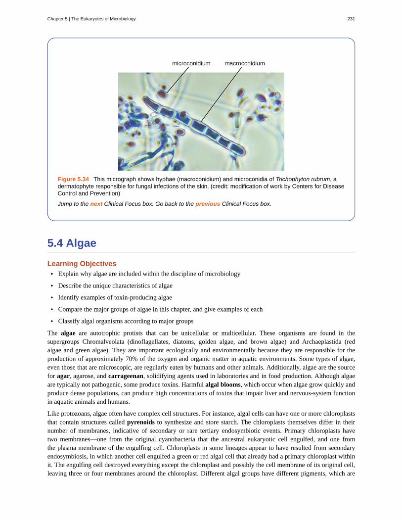
Figure 5.34 This micrograph shows hyphae (macroconidium) and microconidia of Trichophyton rubrum, adermatophyte responsible for fungal infections of the skin. (credit: modification of work by Centers for DiseaseControl and Prevention)
Jump to the next Clinical Focus box. Go back to the previous Clinical Focus box.
5.4 Algae
Learning Objectives• Explain why algae are included within the discipline of microbiology
• Describe the unique characteristics of algae
• Identify examples of toxin-producing algae
• Compare the major groups of algae in this chapter, and give examples of each
• Classify algal organisms according to major groups
The algae are autotrophic protists that can be unicellular or multicellular. These organisms are found in thesupergroups Chromalveolata (dinoflagellates, diatoms, golden algae, and brown algae) and Archaeplastida (redalgae and green algae). They are important ecologically and environmentally because they are responsible for theproduction of approximately 70% of the oxygen and organic matter in aquatic environments. Some types of algae,even those that are microscopic, are regularly eaten by humans and other animals. Additionally, algae are the sourcefor agar, agarose, and carrageenan, solidifying agents used in laboratories and in food production. Although algaeare typically not pathogenic, some produce toxins. Harmful algal blooms, which occur when algae grow quickly andproduce dense populations, can produce high concentrations of toxins that impair liver and nervous-system functionin aquatic animals and humans.
Like protozoans, algae often have complex cell structures. For instance, algal cells can have one or more chloroplaststhat contain structures called pyrenoids to synthesize and store starch. The chloroplasts themselves differ in theirnumber of membranes, indicative of secondary or rare tertiary endosymbiotic events. Primary chloroplasts havetwo membranes—one from the original cyanobacteria that the ancestral eukaryotic cell engulfed, and one fromthe plasma membrane of the engulfing cell. Chloroplasts in some lineages appear to have resulted from secondaryendosymbiosis, in which another cell engulfed a green or red algal cell that already had a primary chloroplast withinit. The engulfing cell destroyed everything except the chloroplast and possibly the cell membrane of its original cell,leaving three or four membranes around the chloroplast. Different algal groups have different pigments, which are
Chapter 5 | The Eukaryotes of Microbiology 231
reflected in common names such as red algae, brown algae, and green algae.
Some algae, the seaweeds, are macroscopic and may be confused with plants. Seaweeds can be red, brown, or green,depending on their photosynthetic pigments. Green algae, in particular, share some important similarities with landplants; however, there are also important distinctions. For example, seaweeds do not have true tissues or organs likeplants do. Additionally, seaweeds do not have a waxy cuticle to prevent desiccation. Algae can also be confused withcyanobacteria, photosynthetic bacteria that bear a resemblance to algae; however, cyanobacteria are prokaryotes (seeNonproteobacteria Gram-negative Bacteria and Phototrophic Bacteria).
Algae have a variety of life cycles. Reproduction may be asexual by mitosis or sexual using gametes.
Algal Diversity
Although the algae and protozoa were formerly separated taxonomically, they are now mixed into supergroups.The algae are classified within the Chromalveolata and the Archaeplastida. Although the Euglenozoa (within thesupergroup Excavata) include photosynthetic organisms, these are not considered algae because they feed and aremotile.
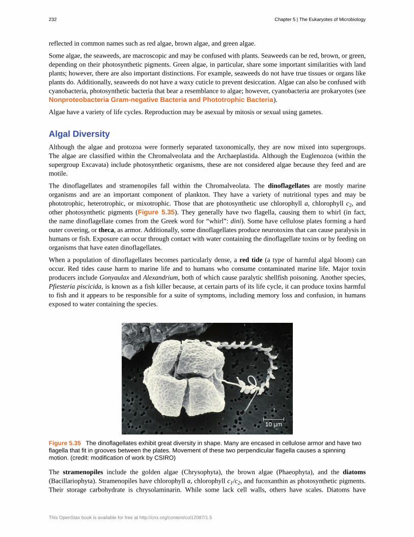
The dinoflagellates and stramenopiles fall within the Chromalveolata. The dinoflagellates are mostly marineorganisms and are an important component of plankton. They have a variety of nutritional types and may bephototrophic, heterotrophic, or mixotrophic. Those that are photosynthetic use chlorophyll a, chlorophyll c2, andother photosynthetic pigments (Figure 5.35). They generally have two flagella, causing them to whirl (in fact,the name dinoflagellate comes from the Greek word for “whirl”: dini). Some have cellulose plates forming a hardouter covering, or theca, as armor. Additionally, some dinoflagellates produce neurotoxins that can cause paralysis inhumans or fish. Exposure can occur through contact with water containing the dinoflagellate toxins or by feeding onorganisms that have eaten dinoflagellates.
When a population of dinoflagellates becomes particularly dense, a red tide (a type of harmful algal bloom) canoccur. Red tides cause harm to marine life and to humans who consume contaminated marine life. Major toxinproducers include Gonyaulax and Alexandrium, both of which cause paralytic shellfish poisoning. Another species,Pfiesteria piscicida, is known as a fish killer because, at certain parts of its life cycle, it can produce toxins harmfulto fish and it appears to be responsible for a suite of symptoms, including memory loss and confusion, in humansexposed to water containing the species.
Figure 5.35 The dinoflagellates exhibit great diversity in shape. Many are encased in cellulose armor and have twoflagella that fit in grooves between the plates. Movement of these two perpendicular flagella causes a spinningmotion. (credit: modification of work by CSIRO)
The stramenopiles include the golden algae (Chrysophyta), the brown algae (Phaeophyta), and the diatoms(Bacillariophyta). Stramenopiles have chlorophyll a, chlorophyll c1/c2, and fucoxanthin as photosynthetic pigments.Their storage carbohydrate is chrysolaminarin. While some lack cell walls, others have scales. Diatoms have
232 Chapter 5 | The Eukaryotes of Microbiology
This OpenStax book is available for free at http://cnx.org/content/col12087/1.5

flagella and frustules, which are outer cell walls of crystallized silica; their fossilized remains are used to producediatomaceous earth, which has a range of uses such as filtration and insulation. Additionally, diatoms can reproducesexually or asexually. One diatom genus, Pseudo-nitzschia, is known to be associated with harmful algal blooms.
Brown algae (Phaeophyta) are multicellular marine seaweeds. Some can be extremely large, such as the giant kelp(Laminaria). They have leaf-like blades, stalks, and structures called holdfasts that are used to attach to substrate.However, these are not true leaves, stems, or roots (Figure 5.36). Their photosynthetic pigments are chlorophyll a,chlorophyll c, β-carotene, and fucoxanthine. They use laminarin as a storage carbohydrate.
The Archaeplastids include the green algae (Chlorophyta), the red algae (Rhodophyta), another group of green algae(Charophyta), and the land plants. The Charaphyta are the most similar to land plants because they share a mechanismof cell division and an important biochemical pathway, among other traits that the other groups do not have. Like landplants, the Charophyta and Chlorophyta have chlorophyll a and chlorophyll b as photosynthetic pigments, cellulosecell walls, and starch as a carbohydrate storage molecule. Chlamydomonas is a green alga that has a single largechloroplast, two flagella, and a stigma (eyespot); it is important in molecular biology research (Figure 5.37).
Chlorella is a nonmotile, large, unicellular alga, and Acetabularia is an even larger unicellular green alga. The size ofthese organisms challenges the idea that all cells are small, and they have been used in genetics research since JoachimHämmerling (1901–1980) began to work with them in 1943. Volvox is a colonial, unicellular alga (Figure 5.37).A larger, multicellular green alga is Ulva, also known as the sea lettuce because of its large, edible, green blades.The range of life forms within the Chlorophyta—from unicellular to various levels of coloniality to multicellularforms—has been a useful research model for understanding the evolution of multicellularity. The red algae are mainlymulticellular but include some unicellular forms. They have rigid cell walls containing agar or carrageenan, whichare useful as food solidifying agents and as a solidifier added to growth media for microbes.
Chapter 5 | The Eukaryotes of Microbiology 233

Figure 5.36 (a) These large multicellular kelps are members of the brown algae. Note the “leaves” and “stems” thatmake them appear similar to green plants. (b) This is a species of red algae that is also multicellular. (c) The greenalga Halimeda incrassata, shown here growing on the sea floor in shallow water, appears to have plant-likestructures, but is not a true plant. (d) Bioluminesence, visible in the cresting wave in this picture, is a phenomenon ofcertain dinoflagellates. (e) Diatoms (pictured in this micrograph) produce silicaceous tests (skeletons) that formdiatomaceous earths. (f) Colonial green algae, like volvox in these three micrographs, exhibit simple cooperativeassociations of cells. (credit a, e: modification of work by NOAA; credit b: modification of work by Ed Bierman; creditc: modification of work by James St. John; credit d: modification of work by “catalano82”/Flickr; credit f: modificationof work by Dr. Ralf Wagner)
234 Chapter 5 | The Eukaryotes of Microbiology
This OpenStax book is available for free at http://cnx.org/content/col12087/1.5

Figure 5.37 Chlamydomonas is a unicellular green alga.
• Which groups of algae are associated with harmful algal blooms?
5.5 Lichens
Learning Objectives• Explain why lichens are included in the study of microbiology
• Describe the unique characteristics of a lichen and the role of each partner in the symbiotic relationship of alichen
• Describe ways in which lichens are beneficial to the environment
No one has to worry about getting sick from a lichen infection, but lichens are interesting from a microbiologicalperspective and they are an important component of most terrestrial ecosystems. Lichens provide opportunities forstudy of close relationships between unrelated microorganisms. Lichens contribute to soil production by breakingdown rock, and they are early colonizers in soilless environments such as lava flows. The cyanobacteria in somelichens can fix nitrogen and act as a nitrogen source in some environments. Lichens are also important soil stabilizersin some desert environments and they are an important winter food source for caribou and reindeer. Finally, lichensproduce compounds that have antibacterial effects, and further research may discover compounds that are medicallyuseful to humans.
Characteristics
A lichen is a combination of two organisms, a green alga or cyanobacterium and an ascomycete fungus, living ina symbiotic relationship. Whereas algae normally grow only in aquatic or extremely moist environments, lichenscan potentially be found on almost any surface (especially rocks) or as epiphytes (meaning that they grow on otherplants).
In some ways, the symbiotic relationship between lichens and algae seems like a mutualism (a relationship in whichboth organisms benefit). The fungus can obtain photosynthates from the algae or cyanobacterium and the algae or
Chapter 5 | The Eukaryotes of Microbiology 235

cyanobacterium can grow in a drier environment than it could otherwise tolerate. However, most scientists considerthis symbiotic relationship to be a controlled parasitism (a relationship in which one organism benefits and the otheris harmed) because the photosynthetic organism grows less well than it would without the fungus. It is important tonote that such symbiotic interactions fall along a continuum between conflict and cooperation.
Lichens are slow growing and can live for centuries. They have been used in foods and to extract chemicals as dyesor antimicrobial substances. Some are very sensitive to pollution and have been used as environmental indicators.
Lichens have a body called a thallus, an outer, tightly packed fungal layer called a cortex, and an inner, loosely packedfungal layer called a medulla (Figure 5.38). Lichens use hyphal bundles called rhizines to attach to the substrate.
Figure 5.38 This cross-section of a lichen thallus shows its various components. The upper cortex of fungal hyphaeprovides protection. Photosynthesis occurs in the algal zone. The medulla consists of fungal hyphae. The lowercortex also provides protection. The rhizines anchor the thallus to the substrate.
Lichen Diversity
Lichens are classified as fungi and the fungal partners belong to the Ascomycota and Basidiomycota. Lichens can alsobe grouped into types based on their morphology. There are three major types of lichens, although other types exist aswell. Lichens that are tightly attached to the substrate, giving them a crusty appearance, are called crustose lichens.Those that have leaf-like lobes are foliose lichens; they may only be attached at one point in the growth form, andthey also have a second cortex below the medulla. Finally, fruticose lichens have rounded structures and an overallbranched appearance. Figure 5.39 shows an example of each of the forms of lichens.
236 Chapter 5 | The Eukaryotes of Microbiology
This OpenStax book is available for free at http://cnx.org/content/col12087/1.5

Figure 5.39 Examples of the three types of lichens are shown here. (a) This is a crustose lichen found mostly onmarine rocks, Caloplaca marina. (b) This is a foliose lichen, Flavoparmelia caperata. (c) This is a fruticose lichen,Letharia vulpina, which is sufficiently poisonous that it was once used to make arrowheads. (credit b, c: modificationof work by Jason Hollinger)
• What types of organisms are found in lichens?
• What are the three growth forms of lichens?
Resolution
Sarah’s mother asks the doctor what she should do if the cream prescribed for Sarah’s ringworm does notwork. The doctor explains that ringworm is a general term for a condition caused by multiple species. Thefirst step is to take a scraping for examination under the microscope, which the doctor has already done. Heexplains that he has identified the infection as a fungus, and that the antifungal cream works against the mostcommon fungi associated with ringworm. However, the cream may not work against some species of fungus.If the cream is not working after a couple of weeks, Sarah should come in for another visit, at which time thedoctor will take steps to identify the species of the fungus.
Positive identification of dermatophytes requires culturing. For this purpose, Sabouraud’s agar may be used. Inthe case of Sarah’s infection, which cleared up within 2 weeks of treatment, the culture would have a granulartexture and would appear pale pink on top and red underneath. These features suggest that the fungus isTrichophyton rubrum, a common cause of ringworm.
Go back to the previous Clinical Focus box.
Summary
5.1 Unicellular Eukaryotic Parasites• Protists are a diverse, polyphyletic group of eukaryotic organisms.
• Protists may be unicellular or multicellular. They vary in how they get their nutrition, morphology, method oflocomotion, and mode of reproduction.
Clinical Focus
Chapter 5 | The Eukaryotes of Microbiology 237

• Important structures of protists include contractile vacuoles, cilia, flagella, pellicles, and pseudopodia; somelack organelles such as mitochondria.
• Taxonomy of protists is changing rapidly as relationships are reassessed using newer techniques.
• The protists include important pathogens and parasites.
5.2 Parasitic Helminths• Helminth parasites are included within the study of microbiology because they are often identified by looking
for microscopic eggs and larvae.
• The two major groups of helminth parasites are the roundworms (Nematoda) and the flatworms(Platyhelminthes).
• Nematodes are common intestinal parasites often transmitted through undercooked foods, although they arealso found in other environments.
• Platyhelminths include tapeworms and flukes, which are often transmitted through undercooked meat.
5.3 Fungi• The fungi include diverse saprotrophic eukaryotic organisms with chitin cell walls
• Fungi can be unicellular or multicellular; some (like yeast) and fungal spores are microscopic, whereas someare large and conspicuous
• Reproductive types are important in distinguishing fungal groups
• Medically important species exist in the four fungal groups Zygomycota, Ascomycota, Basidiomycota, andMicrosporidia
• Members of Zygomycota, Ascomycota, and Basidiomycota produce deadly toxins
• Important differences in fungal cells, such as ergosterols in fungal membranes, can be targets for antifungalmedications, but similarities between human and fungal cells make it difficult to find targets for medicationsand these medications often have toxic adverse effects
5.4 Algae• Algae are a diverse group of photosynthetic eukaryotic protists
• Algae may be unicellular or multicellular
• Large, multicellular algae are called seaweeds but are not plants and lack plant-like tissues and organs
• Although algae have little pathogenicity, they may be associated with toxic algal blooms that can and aquaticwildlife and contaminate seafood with toxins that cause paralysis
• Algae are important for producing agar, which is used as a solidifying agent in microbiological media, andcarrageenan, which is used as a solidifying agent
5.5 Lichens• Lichens are a symbiotic association between a fungus and an algae or a cyanobacterium
• The symbiotic association found in lichens is currently considered to be a controlled parasitism, in which thefungus benefits and the algae or cyanobacterium is harmed
• Lichens are slow growing and can live for centuries in a variety of habitats
• Lichens are environmentally important, helping to create soil, providing food, and acting as indicators of airpollution
238 Chapter 5 | The Eukaryotes of Microbiology
This OpenStax book is available for free at http://cnx.org/content/col12087/1.5

Multiple Choice
1. Which genus includes the causative agent formalaria?
a. Euglenab. Parameciumc. Plasmodiumd. Trypanosoma
2. Which protist is a concern because of its ability tocontaminate water supplies and cause diarrheal illness?
a. Plasmodium vivaxb. Toxoplasma gondiic. Giardia lambliad. Trichomonas vaginalis
3. A fluke is classified within which of the following?a. Nematodab. Rotiferac. Platyhelminthesd. Annelida
4. A nonsegmented worm is found during a routinecolonoscopy of an individual who reported havingabdominal cramps, nausea, and vomiting. This worm islikely which of the following?
a. nematodeb. flukec. trematoded. annelid
5. A segmented worm has male and femalereproductive organs in each segment. Some use hooks toattach to the intestinal wall. Which type of worm is this?
a. flukeb. nematodec. cestoded. annelid
6. Mushrooms are a type of which of the following?a. conidiab. ascusc. polar tubuled. basidiocarp
7. Which of the following is the most common cause ofhuman yeast infections?
a. Candida albicansb. Blastomyces dermatitidisc. Cryptococcus neoformansd. Aspergillus fumigatus
8. Which of the following is an ascomycete fungusassociated with bat droppings that can cause arespiratory infection if inhaled?
a. Candida albicansb. Histoplasma capsulatumc. Rhizopus stoloniferad. Trichophyton rubrum
9. Which polysaccharide found in red algal cell walls isa useful solidifying agent?
a. chitinb. cellulosec. phycoerythrind. agar
10. Which is the term for the hard outer covering ofsome dinoflagellates?
a. thecab. thallusc. myceliumd. shell
11. Which protists are associated with red tides?a. red algaeb. brown algaec. dinoflagellatesd. green algae
12. You encounter a lichen with leafy structures. Whichterm describes this lichen?
a. crustoseb. foliosec. fruticosed. agarose
13. Which of the following is the term for the outerlayer of a lichen?
a. the cortexb. the medullac. the thallusd. the theca
Review Questions
Chapter 5 | The Eukaryotes of Microbiology 239

14. The fungus in a lichen is which of the following?a. a basidiomyceteb. an ascomycetec. a zygomyceted. an apicomplexan
Fill in the Blank
15. The plasma membrane of a protist is called the __________.
16. Animals belong to the same supergroup as the kingdom __________.
17. Flukes are in class _________.
18. A species of worm in which there are distinct male and female individuals is described as _________.
19. Nonseptate hyphae are also called _________.
20. Unicellular fungi are called _________.
21. Some fungi have proven medically useful because they can be used to produce _________.
22. Structures in chloroplasts used to synthesize and store starch are called ________.
23. Algae with chloroplasts with three or four membranes are a result of ________ ________.
Short Answer
24. What are kinetoplastids?
25. Aside from a risk of birth defects, what other effect might a toxoplasmosis infection have?
26. What is the function of the ciliate macronucleus?
27. What is the best defense against tapeworm infection?
28. Which genera of fungi are common dermatophytes (fungi that cause skin infections)?
29. What is a dikaryotic cell?
30. What is a distinctive feature of diatoms?
31. Why are algae not considered parasitic?
32. Which groups contain the multicellular algae?
33. What are three ways that lichens are environmentally valuable?
240 Chapter 5 | The Eukaryotes of Microbiology
This OpenStax book is available for free at http://cnx.org/content/col12087/1.5

Critical Thinking
34. The protist shown has which of the following?a. pseudopodiab. flagellac. a shelld. cilia
Figure 5.40 (credit: modification of work by Richard Robinson)
35. Protist taxonomy has changed greatly in recent years as relationships have been re-examined using newerapproaches. How do newer approaches differ from older approaches?
36. What characteristics might make you think a protist could be pathogenic? Are certain nutritional characteristics,methods of locomotion, or morphological differences likely to be associated with the ability to cause disease?
37. Given the life cycle of the Schistosoma parasite, suggest a method of prevention of the disease.
38. Which of the drawings shows septate hyphae?
39. Explain the benefit of research into the pathways involved in the synthesis of chitin in fungi.
Chapter 5 | The Eukaryotes of Microbiology 241

242 Chapter 5 | The Eukaryotes of Microbiology
This OpenStax book is available for free at http://cnx.org/content/col12087/1.5
Chapter 6
Acellular Pathogens
Figure 6.1 The year 2014 saw the first large-scale outbreak of Ebola virus (electron micrograph, left) in humanpopulations in West Africa (right). Such epidemics are now widely reported and documented, but viral epidemics aresure to have plagued human populations since the origin of our species. (credit left: modification of work by ThomasW. Geisbert)
Chapter Outline
6.1 Viruses
6.2 The Viral Life Cycle
6.3 Isolation, Culture, and Identification of Viruses
6.4 Viroids, Virusoids, and Prions
IntroductionPublic health measures in the developed world have dramatically reduced mortality from viral epidemics. But whenepidemics do occur, they can spread quickly with global air travel. In 2009, an outbreak of H1N1 influenza spreadacross various continents. In early 2014, cases of Ebola in Guinea led to a massive epidemic in western Africa. Thisincluded the case of an infected man who traveled to the United States, sparking fears the epidemic might spreadbeyond Africa.
Until the late 1930s and the advent of the electron microscope, no one had seen a virus. Yet treatments for preventingor curing viral infections were used and developed long before that. Historical records suggest that by the 17thcentury, and perhaps earlier, inoculation (also known as variolation) was being used to prevent the viral diseasesmallpox in various parts of the world. By the late 18th century, Englishman Edward Jenner was inoculating patientswith cowpox to prevent smallpox, a technique he coined vaccination.[1]
Today, the structure and genetics of viruses are well defined, yet new discoveries continue to reveal their complexities.In this chapter, we will learn about the structure, classification, and cultivation of viruses, and how they impact theirhosts. In addition, we will learn about other infective particles such as viroids and prions.
1. S. Riedel “Edward Jenner and the History of Smallpox and Vaccination.” Baylor University Medical Center Proceedings 18, no. 1
(January 2005): 21–25.
Chapter 6 | Acellular Pathogens 243

6.1 Viruses
Learning Objectives• Describe the general characteristics of viruses as pathogens
• Describe viral genomes
• Describe the general characteristics of viral life cycles
• Differentiate among bacteriophages, plant viruses, and animal viruses
• Describe the characteristics used to identify viruses as obligate intracellular parasites
Despite their small size, which prevented them from being seen with light microscopes, the discovery of a filterablecomponent smaller than a bacterium that causes tobacco mosaic disease (TMD) dates back to 1892.[2] At that time,Dmitri Ivanovski, a Russian botanist, discovered the source of TMD by using a porcelain filtering device firstinvented by Charles Chamberland and Louis Pasteur in Paris in 1884. Porcelain Chamberland filters have a pore sizeof 0.1 µm, which is small enough to remove all bacteria ≥0.2 µm from any liquids passed through the device. Anextract obtained from TMD-infected tobacco plants was made to determine the cause of the disease. Initially, thesource of the disease was thought to be bacterial. It was surprising to everyone when Ivanovski, using a Chamberlandfilter, found that the cause of TMD was not removed after passing the extract through the porcelain filter. So if abacterium was not the cause of TMD, what could be causing the disease? Ivanovski concluded the cause of TMDmust be an extremely small bacterium or bacterial spore. Other scientists, including Martinus Beijerinck, continuedinvestigating the cause of TMD. It was Beijerinck, in 1899, who eventually concluded the causative agent was not abacterium but, instead, possibly a chemical, like a biological poison we would describe today as a toxin. As a result,the word virus, Latin for poison, was used to describe the cause of TMD a few years after Ivanovski’s initial discovery.Even though he was not able to see the virus that caused TMD, and did not realize the cause was not a bacterium,Ivanovski is credited as the original discoverer of viruses and a founder of the field of virology.
Today, we can see viruses using electron microscopes (Figure 6.2) and we know much more about them. Viruses aredistinct biological entities; however, their evolutionary origin is still a matter of speculation. In terms of taxonomy,they are not included in the tree of life because they are acellular (not consisting of cells). In order to survive andreproduce, viruses must infect a cellular host, making them obligate intracellular parasites. The genome of a virus
Part 1
David, a 45-year-old journalist, has just returned to the U.S. from travels in Russia, China, and Africa. He isnot feeling well, so he goes to his general practitioner complaining of weakness in his arms and legs, fever,headache, noticeable agitation, and minor discomfort. He thinks it may be related to a dog bite he sufferedwhile interviewing a Chinese farmer. He is experiencing some prickling and itching sensations at the site of thebite wound, but he tells the doctor that the dog seemed healthy and that he had not been concerned until now.The doctor ordered a culture and sensitivity test to rule out bacterial infection of the wound, and the resultscame back negative for any possible pathogenic bacteria.
• Based on this information, what additional tests should be performed on the patient?
• What type of treatment should the doctor recommend?
Jump to the next Clinical Focus box.
Clinical Focus
2. H. Lecoq. “[Discovery of the First Virus, the Tobacco Mosaic Virus: 1892 or 1898?].” Comptes Rendus de l’Academie des Sciences –
Serie III – Sciences de la Vie 324, no. 10 (2001): 929–933.
244 Chapter 6 | Acellular Pathogens
This OpenStax book is available for free at http://cnx.org/content/col12087/1.5

enters a host cell and directs the production of the viral components, proteins and nucleic acids, needed to form newvirus particles called virions. New virions are made in the host cell by assembly of viral components. The new virionstransport the viral genome to another host cell to carry out another round of infection. Table 6.1 summarizes theproperties of viruses.
Characteristics of Viruses
Infectious, acellular pathogens
Obligate intracellular parasites with host and cell-type specificity
DNA or RNA genome (never both)
Genome is surrounded by a protein capsid and, in some cases, a phospholipid membrane studded with viralglycoproteins
Lack genes for many products needed for successful reproduction, requiring exploitation of host-cell genomes toreproduce
Table 6.1
Figure 6.2 (a) Tobacco mosaic virus (TMV) viewed with transmission electron microscope. (b) Plants infected withtobacco mosaic disease (TMD), caused by TMV. (credit a: modification of work by USDA Agricultural ResearchService—scale-bar data from Matt Russell; credit b: modification of work by USDA Forest Service, Department ofPlant Pathology Archive North Carolina State University)
• Why was the first virus investigated mistaken for a toxin?
Hosts and Viral Transmission
Viruses can infect every type of host cell, including those of plants, animals, fungi, protists, bacteria, and archaea.Most viruses will only be able to infect the cells of one or a few species of organism. This is called the host range.However, having a wide host range is not common and viruses will typically only infect specific hosts and onlyspecific cell types within those hosts. The viruses that infect bacteria are called bacteriophages, or simply phages.The word phage comes from the Greek word for devour. Other viruses are just identified by their host group, suchas animal or plant viruses. Once a cell is infected, the effects of the virus can vary depending on the type of virus.
Chapter 6 | Acellular Pathogens 245

Viruses may cause abnormal growth of the cell or cell death, alter the cell’s genome, or cause little noticeable effectin the cell.
Viruses can be transmitted through direct contact, indirect contact with fomites, or through a vector: an animal thattransmits a pathogen from one host to another. Arthropods such as mosquitoes, ticks, and flies, are typical vectors forviral diseases, and they may act as mechanical vectors or biological vectors. Mechanical transmission occurs whenthe arthropod carries a viral pathogen on the outside of its body and transmits it to a new host by physical contact.Biological transmission occurs when the arthropod carries the viral pathogen inside its body and transmits it to thenew host through biting.
In humans, a wide variety of viruses are capable of causing various infections and diseases. Some of the deadliestemerging pathogens in humans are viruses, yet we have few treatments or drugs to deal with viral infections, makingthem difficult to eradicate.
Viruses that can be transmitted from an animal host to a human host can cause zoonoses. For example, the avianinfluenza virus originates in birds, but can cause disease in humans. Reverse zoonoses are caused by infection of ananimal by a virus that originated in a human.
Fighting Bacteria with Viruses
The emergence of superbugs, or multidrug resistant bacteria, has become a major challenge forpharmaceutical companies and a serious health-care problem. According to a 2013 report by the US Centersfor Disease Control and Prevention (CDC), more than 2 million people are infected with drug-resistant bacteriain the US annually, resulting in at least 23,000 deaths.[3] The continued use and overuse of antibiotics will likelylead to the evolution of even more drug-resistant strains.
One potential solution is the use of phage therapy, a procedure that uses bacteria-killing viruses(bacteriophages) to treat bacterial infections. Phage therapy is not a new idea. The discovery ofbacteriophages dates back to the early 20th century, and phage therapy was first used in Europe in 1915 by theEnglish bacteriologist Frederick Twort.[4] However, the subsequent discovery of penicillin and other antibioticsled to the near abandonment of this form of therapy, except in the former Soviet Union and a few countries inEastern Europe. Interest in phage therapy outside of the countries of the former Soviet Union is only recentlyre-emerging because of the rise in antibiotic-resistant bacteria.[5]
Phage therapy has some advantages over antibiotics in that phages kill only one specific bacterium, whereasantibiotics kill not only the pathogen but also beneficial bacteria of the normal microbiota. Development of newantibiotics is also expensive for drug companies and for patients, especially for those who live in countries withhigh poverty rates.
Phages have also been used to prevent food spoilage. In 2006, the US Food and Drug Administrationapproved the use of a solution containing six bacteriophages that can be sprayed on lunch meats such asbologna, ham, and turkey to kill Listeria monocytogenes, a bacterium responsible for listeriosis, a form offood poisoning. Some consumers have concerns about the use of phages on foods, however, especiallygiven the rising popularity of organic products. Foods that have been treated with phages must declare“bacteriophage preparation” in the list of ingredients or include a label declaring that the meat has been “treatedwith antimicrobial solution to reduce microorganisms.”[6]
Micro Connections
3. US Department of Health and Human Services, Centers for Disease Control and Prevention. “Antibiotic Resistance Threats in the United
States, 2013.” http://www.cdc.gov/drugresistance/pdf/ar-threats-2013-508.pdf (accessed September 22, 2015).
4. M. Clokie et al. “Phages in Nature.” Bacteriophage 1, no. 1 (2011): 31–45.
5. A. Sulakvelidze et al. “Bacteriophage Therapy.” Antimicrobial Agents and Chemotherapy 45, no. 3 (2001): 649–659.
6. US Food and Drug Administration. “FDA Approval of Listeria-specific Bacteriophage Preparation on Ready-to-Eat (RTE) Meat and
Poultry Products.” http://www.fda.gov/food/ingredientspackaginglabeling/ucm083572.htm (accessed September 22, 2015).
246 Chapter 6 | Acellular Pathogens
This OpenStax book is available for free at http://cnx.org/content/col12087/1.5

• Why do humans not have to be concerned about the presence of bacteriophages in their food?
• What are three ways that viruses can be transmitted between hosts?
Viral Structures
In general, virions (viral particles) are small and cannot be observed using a regular light microscope. They are muchsmaller than prokaryotic and eukaryotic cells; this is an adaptation allowing viruses to infect these larger cells (seeFigure 6.3). The size of a virion can range from 20 nm for small viruses up to 900 nm for typical, large viruses (seeFigure 6.4). Recent discoveries, however, have identified new giant viral species, such as Pandoravirus salinus andPithovirus sibericum, with sizes approaching that of a bacterial cell.[7]
Figure 6.3 (a) In this transmission electron micrograph, a bacteriophage (a virus that infects bacteria) is dwarfed bythe bacterial cell it infects. (b) An illustration of the bacteriophage in the micrograph. (credit a: modification of work byU.S. Department of Energy, Office of Science, LBL, PBD)
7. N. Philippe et al. “Pandoraviruses: Amoeba Viruses with Genomes up to 2.5 Mb Reaching that of Parasitic Eukaryotes.” Science 341,
no. 6143 (2013): 281–286.
Chapter 6 | Acellular Pathogens 247

Figure 6.4 The size of a virus is small relative to the size of most bacterial and eukaryotic cells and their organelles.
In 1935, after the development of the electron microscope, Wendell Stanley was the first scientist to crystallize thestructure of the tobacco mosaic virus and discovered that it is composed of RNA and protein. In 1943, he isolatedInfluenza B virus, which contributed to the development of an influenza (flu) vaccine. Stanley’s discoveries unlockedthe mystery of the nature of viruses that had been puzzling scientists for over 40 years and his contributions to thefield of virology led to him being awarded the Nobel Prize in 1946.
As a result of continuing research into the nature of viruses, we now know they consist of a nucleic acid (either RNAor DNA, but never both) surrounded by a protein coat called a capsid (see Figure 6.5). The interior of the capsid isnot filled with cytosol, as in a cell, but instead it contains the bare necessities in terms of genome and enzymes neededto direct the synthesis of new virions. Each capsid is composed of protein subunits called capsomeres made of one ormore different types of capsomere proteins that interlock to form the closely packed capsid.
There are two categories of viruses based on general composition. Viruses formed from only a nucleic acid and capsidare called naked viruses or nonenveloped viruses. Viruses formed with a nucleic-acid packed capsid surrounded bya lipid layer are called enveloped viruses (see Figure 6.5). The viral envelope is a small portion of phospholipidmembrane obtained as the virion buds from a host cell. The viral envelope may either be intracellular or cytoplasmicin origin.
Extending outward and away from the capsid on some naked viruses and enveloped viruses are protein structurescalled spikes. At the tips of these spikes are structures that allow the virus to attach and enter a cell, like the influenzavirus hemagglutinin spikes (H) or enzymes like the neuraminidase (N) influenza virus spikes that allow the virus todetach from the cell surface during release of new virions. Influenza viruses are often identified by their H and Nspikes. For example, H1N1 influenza viruses were responsible for the pandemics in 1918 and 2009,[8] H2N2 for thepandemic in 1957, and H3N2 for the pandemic in 1968.
8. J. Cohen. “What’s Old Is New: 1918 Virus Matches 2009 H1N1 Strain. Science 327, no. 5973 (2010): 1563–1564.
248 Chapter 6 | Acellular Pathogens
This OpenStax book is available for free at http://cnx.org/content/col12087/1.5

Figure 6.5 (a) The naked atadenovirus uses spikes made of glycoproteins from its capsid to bind to host cells. (b)The enveloped human immunodeficiency virus uses spikes made of glycoproteins embedded in its envelope to bindto host cells (credit a “micrograph”: modification of work by NIAID; credit b “micrograph”: modification of work byCenters for Disease Control and Prevention)
Viruses vary in the shape of their capsids, which can be either helical, polyhedral, or complex. A helical capsidforms the shape of tobacco mosaic virus (TMV), a naked helical virus, and Ebola virus, an enveloped helical virus.The capsid is cylindrical or rod shaped, with the genome fitting just inside the length of the capsid. Polyhedralcapsids form the shapes of poliovirus and rhinovirus, and consist of a nucleic acid surrounded by a polyhedral (many-sided) capsid in the form of an icosahedron. An icosahedral capsid is a three-dimensional, 20-sided structure with12 vertices. These capsids somewhat resemble a soccer ball. Both helical and polyhedral viruses can have envelopes.Viral shapes seen in certain types of bacteriophages, such as T4 phage, and poxviruses, like vaccinia virus, may havefeatures of both polyhedral and helical viruses so they are described as a complex viral shape (see Figure 6.6). Inthe bacteriophage complex form, the genome is located within the polyhedral head and the sheath connects the headto the tail fibers and tail pins that help the virus attach to receptors on the host cell’s surface. Poxviruses that havecomplex shapes are often brick shaped, with intricate surface characteristics not seen in the other categories of capsid.
Chapter 6 | Acellular Pathogens 249

Figure 6.6 Viral capsids can be (a) helical, (b) polyhedral, or (c) have a complex shape. (credit a “micrograph”:modification of work by USDA ARS; credit b “micrograph”: modification of work by U.S. Department of Energy)
• Which types of viruses have spikes?
Classification and Taxonomy of Viruses
Although viruses are not classified in the three domains of life, their numbers are great enough to requireclassification. Since 1971, the International Union of Microbiological Societies Virology Division has given the taskof developing, refining, and maintaining a universal virus taxonomy to the International Committee on Taxonomy ofViruses (ICTV). Since viruses can mutate so quickly, it can be difficult to classify them into a genus and a speciesepithet using the binomial nomenclature system. Thus, the ICTV’s viral nomenclature system classifies viruses intofamilies and genera based on viral genetics, chemistry, morphology, and mechanism of multiplication. To date, theICTV has classified known viruses in seven orders, 96 families, and 350 genera. Viral family names end in -viridae(e.g, Parvoviridae) and genus names end in −virus (e.g., Parvovirus). The names of viral orders, families, and generaare all italicized. When referring to a viral species, we often use a genus and species epithet such as Pandoravirusdulcis or Pandoravirus salinus.
The Baltimore classification system is an alternative to ICTV nomenclature. The Baltimore system classifies viruses
250 Chapter 6 | Acellular Pathogens
This OpenStax book is available for free at http://cnx.org/content/col12087/1.5

according to their genomes (DNA or RNA, single versus double stranded, and mode of replication). This system thuscreates seven groups of viruses that have common genetics and biology.
Explore the latest virus taxonomy (https://www.openstax.org/l/22virustaxon)at the ICTV website.
Aside from formal systems of nomenclature, viruses are often informally grouped into categories based on chemistry,morphology, or other characteristics they share in common. Categories may include naked or enveloped structure,single-stranded (ss) or double-stranded (ds) DNA or ss or ds RNA genomes, segmented or nonsegmented genomes,and positive-strand (+) or negative-strand (−) RNA. For example, herpes viruses can be classified as a dsDNAenveloped virus; human immunodeficiency virus (HIV) is a +ssRNA enveloped virus, and tobacco mosaic virus isa +ssRNA virus. Other characteristics such as host specificity, tissue specificity, capsid shape, and special genes orenzymes may also be used to describe groups of similar viruses. Table 6.2 lists some of the most common virusesthat are human pathogens by genome type.
Common Pathogenic Viruses
Genome Family Example Virus Clinical Features
Poxviridae Orthopoxvirus Skin papules, pustules, lesions
Poxviridae Parapoxvirus Skin lesionsdsDNA,enveloped
Herpesviridae SimplexvirusCold sores, genital herpes, sexuallytransmitted disease
Adenoviridae Atadenovirus Respiratory infection (common cold)
Papillomaviridae PapillomavirusGenital warts, cervical, vulvar, or vaginalcancer
dsDNA,naked
Reoviridae ReovirusGastroenteritis severe diarrhea(stomach flu)
ParvoviridaeAdeno-associateddependoparvovirus A
Respiratory tract infectionssDNA,naked
ParvoviridaeAdeno-associateddependoparvovirus B
Respiratory tract infection
dsRNA,naked
Reoviridae Rotavirus Gastroenteritis
Picornaviridae Enterovirus C Poliomyelitis
Picornaviridae RhinovirusUpper respiratory tract infection(common cold)
+ssRNA,naked
Picornaviridae Hepatovirus Hepatitis
+ssRNA,enveloped
Togaviridae Alphavirus Encephalitis, hemorrhagic fever
Table 6.2
Link to Learning
Chapter 6 | Acellular Pathogens 251

Common Pathogenic Viruses
Genome Family Example Virus Clinical Features
Togaviridae Rubivirus Rubella
Retroviridae LentivirusAcquired immune deficiency syndrome(AIDS)
Filoviridae Zaire Ebolavirus Hemorrhagic fever
Orthomyxoviridae Influenzavirus A, B, C Flu−ssRNA,enveloped
Rhabdoviridae Lyssavirus Rabies
Table 6.2
• What are the types of virus genomes?
Classification of Viral Diseases
While the ICTV has been tasked with the biological classification of viruses, it has also played an important role inthe classification of diseases caused by viruses. To facilitate the tracking of virus-related human diseases, the ICTVhas created classifications that link to the International Classification of Diseases (ICD), the standard taxonomy ofdisease that is maintained and updated by the World Health Organization (WHO). The ICD assigns an alphanumericcode of up to six characters to every type of viral infection, as well as all other types of diseases, medical conditions,and causes of death. This ICD code is used in conjunction with two other coding systems (the Current ProceduralTerminology, and the Healthcare Common Procedure Coding System) to categorize patient conditions for treatmentand insurance reimbursement.
For example, when a patient seeks treatment for a viral infection, ICD codes are routinely used by clinicians toorder laboratory tests and prescribe treatments specific to the virus suspected of causing the illness. This ICD codeis then used by medical laboratories to identify tests that must be performed to confirm the diagnosis. The ICDcode is used by the health-care management system to verify that all treatments and laboratory work performed areappropriate for the given virus. Medical coders use ICD codes to assign the proper code for procedures performed,and medical billers, in turn, use this information to process claims for reimbursement by insurance companies. Vital-records keepers use ICD codes to record cause of death on death certificates, and epidemiologists used ICD codes tocalculate morbidity and mortality statistics.
• Identify two locations where you would likely find an ICD code.
252 Chapter 6 | Acellular Pathogens
This OpenStax book is available for free at http://cnx.org/content/col12087/1.5
Part 2
David’s doctor was concerned that his symptoms included prickling and itching at the site of the dog bite;these sensations could be early symptoms of rabies. Several tests are available to diagnose rabies in livepatients, but no single antemortem test is adequate. The doctor decided to take samples of David’s blood,saliva, and skin for testing. The skin sample was taken from the nape of the neck (posterior side of the necknear the hairline). It was about 6-mm long and contained at least 10 hair follicles, including the superficialcutaneous nerve. An immunofluorescent staining technique was used on the skin biopsy specimen to detectrabies antibodies in the cutaneous nerves at the base of the hair follicles. A test was also performed on aserum sample from David’s blood to determine whether any antibodies for the rabies virus had been produced.
Meanwhile, the saliva sample was used for reverse transcriptase-polymerase chain reaction (RT-PCR)analysis, a test that can detect the presence of viral nucleic acid (RNA). The blood tests came back positivefor the presence of rabies virus antigen, prompting David’s doctor to prescribe prophylactic treatment. Davidis given a series of intramuscular injections of human rabies immunoglobulin along with a series of rabiesvaccines.
• Why does the immunofluorescent technique look for rabies antibodies rather than the rabies virusitself?
• If David has contracted rabies, what is his prognosis?
Jump to the next Clinical Focus box. Go back to the previous Clinical Focus box.
6.2 The Viral Life Cycle
Learning Objectives• Describe the lytic and lysogenic life cycles
• Describe the replication process of animal viruses
• Describe unique characteristics of retroviruses and latent viruses
• Discuss human viruses and their virus-host cell interactions
• Explain the process of transduction
• Describe the replication process of plant viruses
All viruses depend on cells for reproduction and metabolic processes. By themselves, viruses do not encode for allof the enzymes necessary for viral replication. But within a host cell, a virus can commandeer cellular machineryto produce more viral particles. Bacteriophages replicate only in the cytoplasm, since prokaryotic cells do not havea nucleus or organelles. In eukaryotic cells, most DNA viruses can replicate inside the nucleus, with an exceptionobserved in the large DNA viruses, such as the poxviruses, that can replicate in the cytoplasm. RNA viruses thatinfect animal cells often replicate in the cytoplasm.
The Life Cycle of Viruses with Prokaryote Hosts
The life cycle of bacteriophages has been a good model for understanding how viruses affect the cells they infect,since similar processes have been observed for eukaryotic viruses, which can cause immediate death of the cell orestablish a latent or chronic infection. Virulent phages typically lead to the death of the cell through cell lysis.Temperate phages, on the other hand, can become part of a host chromosome and are replicated with the cell genomeuntil such time as they are induced to make newly assembled viruses, or progeny viruses.
Clinical Focus
Chapter 6 | Acellular Pathogens 253
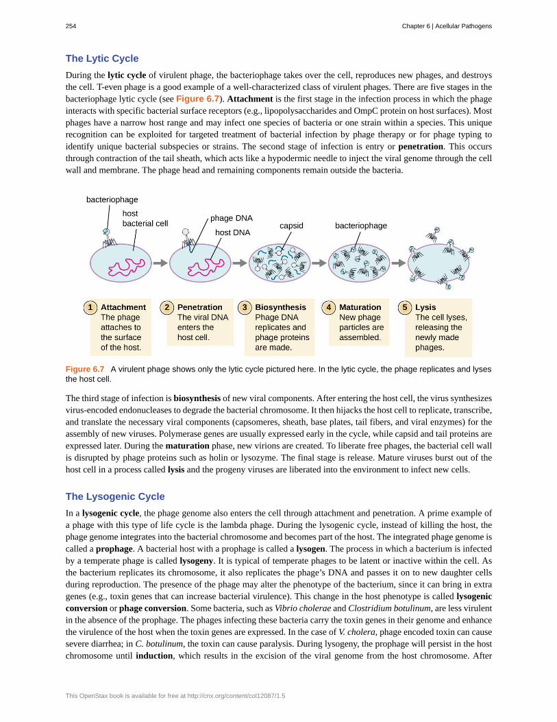
The Lytic Cycle
During the lytic cycle of virulent phage, the bacteriophage takes over the cell, reproduces new phages, and destroysthe cell. T-even phage is a good example of a well-characterized class of virulent phages. There are five stages in thebacteriophage lytic cycle (see Figure 6.7). Attachment is the first stage in the infection process in which the phageinteracts with specific bacterial surface receptors (e.g., lipopolysaccharides and OmpC protein on host surfaces). Mostphages have a narrow host range and may infect one species of bacteria or one strain within a species. This uniquerecognition can be exploited for targeted treatment of bacterial infection by phage therapy or for phage typing toidentify unique bacterial subspecies or strains. The second stage of infection is entry or penetration. This occursthrough contraction of the tail sheath, which acts like a hypodermic needle to inject the viral genome through the cellwall and membrane. The phage head and remaining components remain outside the bacteria.
Figure 6.7 A virulent phage shows only the lytic cycle pictured here. In the lytic cycle, the phage replicates and lysesthe host cell.
The third stage of infection is biosynthesis of new viral components. After entering the host cell, the virus synthesizesvirus-encoded endonucleases to degrade the bacterial chromosome. It then hijacks the host cell to replicate, transcribe,and translate the necessary viral components (capsomeres, sheath, base plates, tail fibers, and viral enzymes) for theassembly of new viruses. Polymerase genes are usually expressed early in the cycle, while capsid and tail proteins areexpressed later. During the maturation phase, new virions are created. To liberate free phages, the bacterial cell wallis disrupted by phage proteins such as holin or lysozyme. The final stage is release. Mature viruses burst out of thehost cell in a process called lysis and the progeny viruses are liberated into the environment to infect new cells.
The Lysogenic Cycle
In a lysogenic cycle, the phage genome also enters the cell through attachment and penetration. A prime example ofa phage with this type of life cycle is the lambda phage. During the lysogenic cycle, instead of killing the host, thephage genome integrates into the bacterial chromosome and becomes part of the host. The integrated phage genome iscalled a prophage. A bacterial host with a prophage is called a lysogen. The process in which a bacterium is infectedby a temperate phage is called lysogeny. It is typical of temperate phages to be latent or inactive within the cell. Asthe bacterium replicates its chromosome, it also replicates the phage’s DNA and passes it on to new daughter cellsduring reproduction. The presence of the phage may alter the phenotype of the bacterium, since it can bring in extragenes (e.g., toxin genes that can increase bacterial virulence). This change in the host phenotype is called lysogenicconversion or phage conversion. Some bacteria, such as Vibrio cholerae and Clostridium botulinum, are less virulentin the absence of the prophage. The phages infecting these bacteria carry the toxin genes in their genome and enhancethe virulence of the host when the toxin genes are expressed. In the case of V. cholera, phage encoded toxin can causesevere diarrhea; in C. botulinum, the toxin can cause paralysis. During lysogeny, the prophage will persist in the hostchromosome until induction, which results in the excision of the viral genome from the host chromosome. After
254 Chapter 6 | Acellular Pathogens
This OpenStax book is available for free at http://cnx.org/content/col12087/1.5

induction has occurred the temperate phage can proceed through a lytic cycle and then undergo lysogeny in a newlyinfected cell (see Figure 6.8).
Figure 6.8 A temperate bacteriophage has both lytic and lysogenic cycles. In the lysogenic cycle, phage DNA isincorporated into the host genome, forming a prophage, which is passed on to subsequent generations of cells.Environmental stressors such as starvation or exposure to toxic chemicals may cause the prophage to be excisedand enter the lytic cycle.
This video (https://www.openstax.org/l/22lysogeniclife) illustrates the stagesof the lysogenic life cycle of a bacteriophage and the transition to a lytic phase.
• Is a latent phage undetectable in a bacterium?
Transduction
Transduction occurs when a bacteriophage transfers bacterial DNA from one bacterium to another during sequentialinfections. There are two types of transduction: generalized and specialized transduction. During the lytic cycle ofviral replication, the virus hijacks the host cell, degrades the host chromosome, and makes more viral genomes. Asit assembles and packages DNA into the phage head, packaging occasionally makes a mistake. Instead of packagingviral DNA, it takes a random piece of host DNA and inserts it into the capsid. Once released, this virion will then
Link to Learning
Chapter 6 | Acellular Pathogens 255

inject the former host’s DNA into a newly infected host. The asexual transfer of genetic information can allow forDNA recombination to occur, thus providing the new host with new genes (e.g., an antibiotic-resistance gene, or asugar-metabolizing gene). Generalized transduction occurs when a random piece of bacterial chromosomal DNAis transferred by the phage during the lytic cycle. Specialized transduction occurs at the end of the lysogenic cycle,when the prophage is excised and the bacteriophage enters the lytic cycle. Since the phage is integrated into the hostgenome, the prophage can replicate as part of the host. However, some conditions (e.g., ultraviolet light exposureor chemical exposure) stimulate the prophage to undergo induction, causing the phage to excise from the genome,enter the lytic cycle, and produce new phages to leave host cells. During the process of excision from the hostchromosome, a phage may occasionally remove some bacterial DNA near the site of viral integration. The phage andhost DNA from one end or both ends of the integration site are packaged within the capsid and are transferred to thenew, infected host. Since the DNA transferred by the phage is not randomly packaged but is instead a specific pieceof DNA near the site of integration, this mechanism of gene transfer is referred to as specialized transduction (seeFigure 6.9). The DNA can then recombine with host chromosome, giving the latter new characteristics. Transductionseems to play an important role in the evolutionary process of bacteria, giving them a mechanism for asexualexchange of genetic information.
Figure 6.9 This flowchart illustrates the mechanism of specialized transduction. An integrated phage excises,bringing with it a piece of the DNA adjacent to its insertion point. On reinfection of a new bacterium, the phage DNAintegrates along with the genetic material acquired from the previous host.
• Which phage life cycle is associated with which forms of transduction?
Life Cycle of Viruses with Animal Hosts
Lytic animal viruses follow similar infection stages to bacteriophages: attachment, penetration, biosynthesis,maturation, and release (see Figure 6.10). However, the mechanisms of penetration, nucleic-acid biosynthesis, andrelease differ between bacterial and animal viruses. After binding to host receptors, animal viruses enter throughendocytosis (engulfment by the host cell) or through membrane fusion (viral envelope with the host cell membrane).Many viruses are host specific, meaning they only infect a certain type of host; and most viruses only infect certaintypes of cells within tissues. This specificity is called a tissue tropism. Examples of this are demonstrated by thepoliovirus, which exhibits tropism for the tissues of the brain and spinal cord, or the influenza virus, which has aprimary tropism for the respiratory tract.
256 Chapter 6 | Acellular Pathogens
This OpenStax book is available for free at http://cnx.org/content/col12087/1.5

Figure 6.10 In influenza virus infection, viral glycoproteins attach the virus to a host epithelial cell. As a result, thevirus is engulfed. Viral RNA and viral proteins are made and assembled into new virions that are released bybudding.
Animal viruses do not always express their genes using the normal flow of genetic information—from DNA to RNAto protein. Some viruses have a dsDNA genome like cellular organisms and can follow the normal flow. However,others may have ssDNA, dsRNA, or ssRNA genomes. The nature of the genome determines how the genome isreplicated and expressed as viral proteins. If a genome is ssDNA, host enzymes will be used to synthesize a secondstrand that is complementary to the genome strand, thus producing dsDNA. The dsDNA can now be replicated,transcribed, and translated similar to host DNA.
If the viral genome is RNA, a different mechanism must be used. There are three types of RNA genome: dsRNA,positive (+) single-strand (+ssRNA) or negative (−) single-strand RNA (−ssRNA). If a virus has a +ssRNAgenome, it can be translated directly to make viral proteins. Viral genomic +ssRNA acts like cellular mRNA.However, if a virus contains a −ssRNA genome, the host ribosomes cannot translate it until the −ssRNA is replicatedinto +ssRNA by viral RNA-dependent RNA polymerase (RdRP) (see Figure 6.11). The RdRP is brought in by thevirus and can be used to make +ssRNA from the original −ssRNA genome. The RdRP is also an important enzymefor the replication of dsRNA viruses, because it uses the negative strand of the double-stranded genome as a templateto create +ssRNA. The newly synthesized +ssRNA copies can then be translated by cellular ribosomes.
Chapter 6 | Acellular Pathogens 257

Figure 6.11 RNA viruses can contain +ssRNA that can be directly read by the ribosomes to synthesize viralproteins. Viruses containing −ssRNA must first use the −ssRNA as a template for the synthesis of +ssRNA beforeviral proteins can be synthesized.
An alternative mechanism for viral nucleic acid synthesis is observed in the retroviruses, which are +ssRNA viruses(see Figure 6.12). Single-stranded RNA viruses such as HIV carry a special enzyme called reverse transcriptasewithin the capsid that synthesizes a complementary ssDNA (cDNA) copy using the +ssRNA genome as a template.The ssDNA is then made into dsDNA, which can integrate into the host chromosome and become a permanent partof the host. The integrated viral genome is called a provirus. The virus now can remain in the host for a long timeto establish a chronic infection. The provirus stage is similar to the prophage stage in a bacterial infection during thelysogenic cycle. However, unlike prophage, the provirus does not undergo excision after splicing into the genome.
258 Chapter 6 | Acellular Pathogens
This OpenStax book is available for free at http://cnx.org/content/col12087/1.5
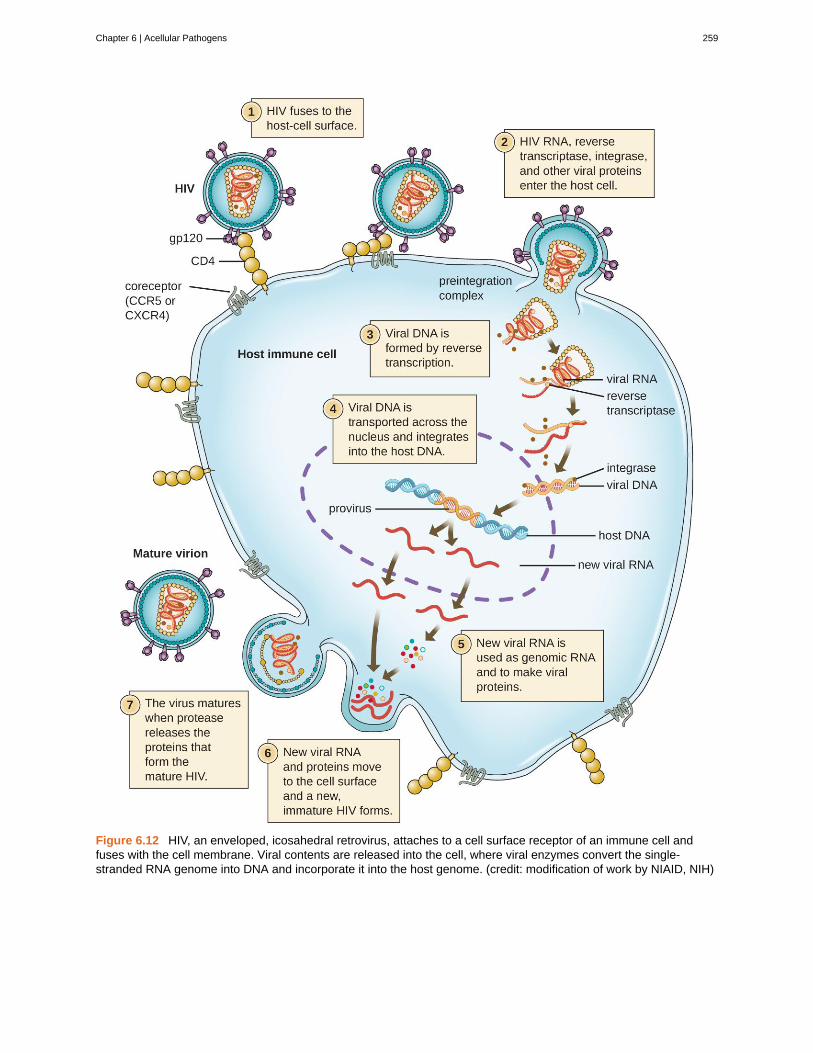
Figure 6.12 HIV, an enveloped, icosahedral retrovirus, attaches to a cell surface receptor of an immune cell andfuses with the cell membrane. Viral contents are released into the cell, where viral enzymes convert the single-stranded RNA genome into DNA and incorporate it into the host genome. (credit: modification of work by NIAID, NIH)
Chapter 6 | Acellular Pathogens 259

• Is RNA-dependent RNA polymerase made from a viral gene or a host gene?
Persistent Infections
Persistent infection occurs when a virus is not completely cleared from the system of the host but stays in certaintissues or organs of the infected person. The virus may remain silent or undergo productive infection without seriouslyharming or killing the host. Mechanisms of persistent infection may involve the regulation of the viral or host geneexpressions or the alteration of the host immune response. The two primary categories of persistent infections arelatent infection and chronic infection. Examples of viruses that cause latent infections include herpes simplex virus(oral and genital herpes), varicella-zoster virus (chickenpox and shingles), and Epstein-Barr virus (mononucleosis).Hepatitis C virus and HIV are two examples of viruses that cause long-term chronic infections.
Latent Infection
Not all animal viruses undergo replication by the lytic cycle. There are viruses that are capable of remaining hiddenor dormant inside the cell in a process called latency. These types of viruses are known as latent viruses and maycause latent infections. Viruses capable of latency may initially cause an acute infection before becoming dormant.
For example, the varicella-zoster virus infects many cells throughout the body and causes chickenpox, characterizedby a rash of blisters covering the skin. About 10 to 12 days postinfection, the disease resolves and the virus goesdormant, living within nerve-cell ganglia for years. During this time, the virus does not kill the nerve cells or continuereplicating. It is not clear why the virus stops replicating within the nerve cells and expresses few viral proteins but, insome cases, typically after many years of dormancy, the virus is reactivated and causes a new disease called shingles(Figure 6.13). Whereas chickenpox affects many areas throughout the body, shingles is a nerve cell-specific diseaseemerging from the ganglia in which the virus was dormant.
Figure 6.13 (a) Varicella-zoster, the virus that causes chickenpox, has an enveloped icosahedral capsid visible inthis transmission electron micrograph. Its double-stranded DNA genome becomes incorporated in the host DNA. (b)After a period of latency, the virus can reactivate in the form of shingles, usually manifesting as a painful, localizedrash on one side of the body. (credit a: modification of work by Erskine Palmer and B.G. Partin—scale-bar data fromMatt Russell; credit b: modification of work by Rosmarie Voegtli)
Latent viruses may remain dormant by existing as circular viral genome molecules outside of the host chromosome.Others become proviruses by integrating into the host genome. During dormancy, viruses do not cause any symptomsof disease and may be difficult to detect. A patient may be unaware that he or she is carrying the virus unless a viraldiagnostic test has been performed.
260 Chapter 6 | Acellular Pathogens
This OpenStax book is available for free at http://cnx.org/content/col12087/1.5

Chronic Infection
A chronic infection is a disease with symptoms that are recurrent or persistent over a long time. Some viral infectionscan be chronic if the body is unable to eliminate the virus. HIV is an example of a virus that produces a chronicinfection, often after a long period of latency. Once a person becomes infected with HIV, the virus can be detectedin tissues continuously thereafter, but untreated patients often experience no symptoms for years. However, thevirus maintains chronic persistence through several mechanisms that interfere with immune function, includingpreventing expression of viral antigens on the surface of infected cells, altering immune cells themselves, restrictingexpression of viral genes, and rapidly changing viral antigens through mutation. Eventually, the damage to theimmune system results in progression of the disease leading to acquired immunodeficiency syndrome (AIDS). Thevarious mechanisms that HIV uses to avoid being cleared by the immune system are also used by other chronicallyinfecting viruses, including the hepatitis C virus.
• In what two ways can a virus manage to maintain a persistent infection?
Life Cycle of Viruses with Plant Hosts
Plant viruses are more similar to animal viruses than they are to bacteriophages. Plant viruses may be envelopedor non-enveloped. Like many animal viruses, plant viruses can have either a DNA or RNA genome and be singlestranded or double stranded. However, most plant viruses do not have a DNA genome; the majority have a +ssRNAgenome, which acts like messenger RNA (mRNA). Only a minority of plant viruses have other types of genomes.
Plant viruses may have a narrow or broad host range. For example, the citrus tristeza virus infects only a few plantsof the Citrus genus, whereas the cucumber mosaic virus infects thousands of plants of various plant families. Mostplant viruses are transmitted by contact between plants, or by fungi, nematodes, insects, or other arthropods that act asmechanical vectors. However, some viruses can only be transferred by a specific type of insect vector; for example, aparticular virus might be transmitted by aphids but not whiteflies. In some cases, viruses may also enter healthy plantsthrough wounds, as might occur due to pruning or weather damage.
Viruses that infect plants are considered biotrophic parasites, which means that they can establish an infection withoutkilling the host, similar to what is observed in the lysogenic life cycles of bacteriophages. Viral infection can beasymptomatic (latent) or can lead to cell death (lytic infection). The life cycle begins with the penetration of the virusinto the host cell. Next, the virus is uncoated within the cytoplasm of the cell when the capsid is removed. Dependingon the type of nucleic acid, cellular components are used to replicate the viral genome and synthesize viral proteinsfor assembly of new virions. To establish a systemic infection, the virus must enter a part of the vascular system ofthe plant, such as the phloem. The time required for systemic infection may vary from a few days to a few weeksdepending on the virus, the plant species, and the environmental conditions. The virus life cycle is complete when itis transmitted from an infected plant to a healthy plant.
• What is the structure and genome of a typical plant virus?
Viral Growth Curve
Unlike the growth curve for a bacterial population, the growth curve for a virus population over its life cycle does notfollow a sigmoidal curve. During the initial stage, an inoculum of virus causes infection. In the eclipse phase, virusesbind and penetrate the cells with no virions detected in the medium. The chief difference that next appears in the viral
Chapter 6 | Acellular Pathogens 261

growth curve compared to a bacterial growth curve occurs when virions are released from the lysed host cell at thesame time. Such an occurrence is called a burst, and the number of virions per bacterium released is described as theburst size. In a one-step multiplication curve for bacteriophage, the host cells lyse, releasing many viral particles tothe medium, which leads to a very steep rise in viral titer (the number of virions per unit volume). If no viable hostcells remain, the viral particles begin to degrade during the decline of the culture (see Figure 6.14).
Figure 6.14 The one-step multiplication curve for a bacteriophage population follows three steps: 1) inoculation,during which the virions attach to host cells; 2) eclipse, during which entry of the viral genome occurs; and 3) burst,when sufficient numbers of new virions are produced and emerge from the host cell. The burst size is the maximumnumber of virions produced per bacterium.
• What aspect of the life cycle of a virus leads to the sudden increase in the growth curve?
Unregistered Treatments
Ebola is incurable and deadly. The outbreak in West Africa in 2014 was unprecedented, dwarfing other humanEbola epidemics in the level of mortality. Of 24,666 suspected or confirmed cases reported, 10,179 peopledied.[9]
No approved treatments or vaccines for Ebola are available. While some drugs have shown potential inlaboratory studies and animal models, they have not been tested in humans for safety and effectiveness. Notonly are these drugs untested or unregistered but they are also in short supply.
Given the great suffering and high mortality rates, it is fair to ask whether unregistered and untestedmedications are better than none at all. Should such drugs be dispensed and, if so, who should receive them,
Eye on Ethics
262 Chapter 6 | Acellular Pathogens
This OpenStax book is available for free at http://cnx.org/content/col12087/1.5

in light of their extremely limited supplies? Is it ethical to treat untested drugs on patients with Ebola? Onthe other hand, is it ethical to withhold potentially life-saving drugs from dying patients? Or should the drugsperhaps be reserved for health-care providers working to contain the disease?
In August 2014, two infected US aid workers and a Spanish priest were treated with ZMapp, an unregistereddrug that had been tested in monkeys but not in humans. The two American aid workers recovered, but thepriest died. Later that month, the WHO released a report on the ethics of treating patients with the drug. SinceEbola is often fatal, the panel reasoned that it is ethical to give the unregistered drugs and unethical to withholdthem for safety concerns. This situation is an example of “compassionate use” outside the well-establishedsystem of regulation and governance of therapies.
Ebola in the US
On September 24, 2014, Thomas Eric Duncan arrived at the Texas Health Presbyterian Hospital in Dallascomplaining of a fever, headache, vomiting, and diarrhea—symptoms commonly observed in patients with thecold or the flu. After examination, an emergency department doctor diagnosed him with sinusitis, prescribedsome antibiotics, and sent him home. Two days later, Duncan returned to the hospital by ambulance. Hiscondition had deteriorated and additional blood tests confirmed that he has been infected with the Ebola virus.
Further investigations revealed that Duncan had just returned from Liberia, one of the countries in the midstof a severe Ebola epidemic. On September 15, nine days before he showed up at the hospital in Dallas,Duncan had helped transport an Ebola-stricken neighbor to a hospital in Liberia. The hospital continued to treatDuncan, but he died several days after being admitted.
The timeline of the Duncan case is indicative of the life cycle of the Ebola virus. The incubation time forEbola ranges from 2 days to 21 days. Nine days passed between Duncan’s exposure to the virus infectionand the appearance of his symptoms. This corresponds, in part, to the eclipse period in the growth of thevirus population. During the eclipse phase, Duncan would have been unable to transmit the disease to others.However, once an infected individual begins exhibiting symptoms, the disease becomes very contagious.Ebola virus is transmitted through direct contact with droplets of bodily fluids such as saliva, blood, and vomit.Duncan could conceivably have transmitted the disease to others at any time after he began having symptoms,presumably some time before his arrival at the hospital in Dallas. Once a hospital realizes a patient like Duncanis infected with Ebola virus, the patient is immediately quarantined, and public health officials initiate a backtrace to identify everyone with whom a patient like Duncan might have interacted during the period in which hewas showing symptoms.
Public health officials were able to track down 10 high-risk individuals (family members of Duncan) and 50low-risk individuals to monitor them for signs of infection. None contracted the disease. However, one of thenurses charged with Duncan’s care did become infected. This, along with Duncan’s initial misdiagnosis, madeit clear that US hospitals needed to provide additional training to medical personnel to prevent a possible Ebolaoutbreak in the US.
• What types of training can prepare health professionals to contain emerging epidemics like the Ebolaoutbreak of 2014?
• What is the difference between a contagious pathogen and an infectious pathogen?
Case in Point
9. World Health Organization. “WHO Ebola Data and Statistics.” March 18, 2005. http://apps.who.int/gho/data/view.ebola-sitrep.ebola-
summary-20150318?lang=en
Chapter 6 | Acellular Pathogens 263

Figure 6.15 Researchers working with Ebola virus use layers of defenses against accidental infection,including protective clothing, breathing systems, and negative air-pressure cabinets for bench work. (credit:modification of work by Randal J. Schoepp)
For additional information about Ebola, please visit the CDC(https://www.openstax.org/l/22ebolacdc) website.
6.3 Isolation, Culture, and Identification of Viruses
Learning Objectives• Discuss why viruses were originally described as filterable agents
• Describe the cultivation of viruses and specimen collection and handling
• Compare in vivo and in vitro techniques used to cultivate viruses
At the beginning of this chapter, we described how porcelain Chamberland filters with pores small enough to allowviruses to pass through were used to discover TMV. Today, porcelain filters have been replaced with membrane filtersand other devices used to isolate and identify viruses.
Isolation of Viruses
Unlike bacteria, many of which can be grown on an artificial nutrient medium, viruses require a living host cell forreplication. Infected host cells (eukaryotic or prokaryotic) can be cultured and grown, and then the growth mediumcan be harvested as a source of virus. Virions in the liquid medium can be separated from the host cells by eithercentrifugation or filtration. Filters can physically remove anything present in the solution that is larger than thevirions; the viruses can then be collected in the filtrate (see Figure 6.16).
Link to Learning
264 Chapter 6 | Acellular Pathogens
This OpenStax book is available for free at http://cnx.org/content/col12087/1.5

Figure 6.16 Membrane filters can be used to remove cells or viruses from a solution. (a) This scanning electronmicrograph shows rod-shaped bacterial cells captured on the surface of a membrane filter. Note differences in thecomparative size of the membrane pores and bacteria. Viruses will pass through this filter. (b) The size of the pores inthe filter determines what is captured on the surface of the filter (animal [red] and bacteria [blue]) and removed fromliquid passing through. Note the viruses (green) pass through the finer filter. (credit a: modification of work by U.S.Department of Energy)
• What size filter pore is needed to collect a virus?
Cultivation of Viruses
Viruses can be grown in vivo (within a whole living organism, plant, or animal) or in vitro (outside a living organismin cells in an artificial environment, such as a test tube, cell culture flask, or agar plate). Bacteriophages can be grownin the presence of a dense layer of bacteria (also called a bacterial lawn) grown in a 0.7 % soft agar in a Petri dish orflat (horizontal) flask (see Figure 6.17). The agar concentration is decreased from the 1.5% usually used in culturingbacteria. The soft 0.7% agar allows the bacteriophages to easily diffuse through the medium. For lytic bacteriophages,lysing of the bacterial hosts can then be readily observed when a clear zone called a plaque is detected (see Figure6.17). As the phage kills the bacteria, many plaques are observed among the cloudy bacterial lawn.
Chapter 6 | Acellular Pathogens 265

Figure 6.17 (a) Flasks like this may be used to culture human or animal cells for viral culturing. (b) These platescontain bacteriophage T4 grown on an Escherichia coli lawn. Clear plaques are visible where host bacterial cellshave been lysed. Viral titers increase on the plates to the left. (credit a: modification of work by National Institutes ofHealth; credit b: modification of work by American Society for Microbiology)
Animal viruses require cells within a host animal or tissue-culture cells derived from an animal. Animal viruscultivation is important for 1) identification and diagnosis of pathogenic viruses in clinical specimens, 2) productionof vaccines, and 3) basic research studies. In vivo host sources can be a developing embryo in an embryonated bird’segg (e.g., chicken, turkey) or a whole animal. For example, most of the influenza vaccine manufactured for annual fluvaccination programs is cultured in hens’ eggs.
The embryo or host animal serves as an incubator for viral replication (see Figure 6.18). Location within the embryoor host animal is important. Many viruses have a tissue tropism, and must therefore be introduced into a specificsite for growth. Within an embryo, target sites include the amniotic cavity, the chorioallantoic membrane, or the yolksac. Viral infection may damage tissue membranes, producing lesions called pox; disrupt embryonic development; orcause the death of the embryo.
Figure 6.18 (a) The cells within chicken eggs are used to culture different types of viruses. (b) Viruses can bereplicated in various locations within the egg, including the chorioallantoic membrane, the amniotic cavity, and theyolk sac. (credit a: modification of work by “Chung Hoang”/YouTube)
For in vitro studies, various types of cells can be used to support the growth of viruses. A primary cell culture isfreshly prepared from animal organs or tissues. Cells are extracted from tissues by mechanical scraping or mincing torelease cells or by an enzymatic method using trypsin or collagenase to break up tissue and release single cells intosuspension. Because of anchorage-dependence requirements, primary cell cultures require a liquid culture mediumin a Petri dish or tissue-culture flask so cells have a solid surface such as glass or plastic for attachment and growth.Primary cultures usually have a limited life span. When cells in a primary culture undergo mitosis and a sufficientdensity of cells is produced, cells come in contact with other cells. When this cell-to-cell-contact occurs, mitosis is
266 Chapter 6 | Acellular Pathogens
This OpenStax book is available for free at http://cnx.org/content/col12087/1.5
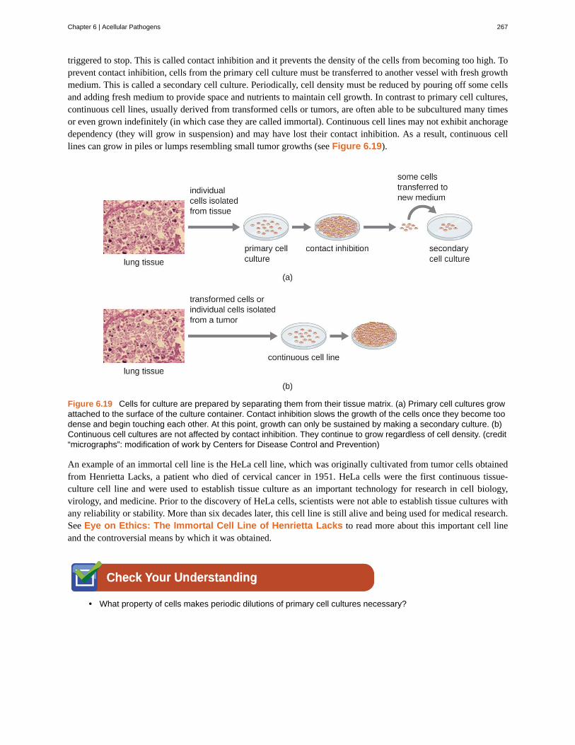
triggered to stop. This is called contact inhibition and it prevents the density of the cells from becoming too high. Toprevent contact inhibition, cells from the primary cell culture must be transferred to another vessel with fresh growthmedium. This is called a secondary cell culture. Periodically, cell density must be reduced by pouring off some cellsand adding fresh medium to provide space and nutrients to maintain cell growth. In contrast to primary cell cultures,continuous cell lines, usually derived from transformed cells or tumors, are often able to be subcultured many timesor even grown indefinitely (in which case they are called immortal). Continuous cell lines may not exhibit anchoragedependency (they will grow in suspension) and may have lost their contact inhibition. As a result, continuous celllines can grow in piles or lumps resembling small tumor growths (see Figure 6.19).
Figure 6.19 Cells for culture are prepared by separating them from their tissue matrix. (a) Primary cell cultures growattached to the surface of the culture container. Contact inhibition slows the growth of the cells once they become toodense and begin touching each other. At this point, growth can only be sustained by making a secondary culture. (b)Continuous cell cultures are not affected by contact inhibition. They continue to grow regardless of cell density. (credit“micrographs”: modification of work by Centers for Disease Control and Prevention)
An example of an immortal cell line is the HeLa cell line, which was originally cultivated from tumor cells obtainedfrom Henrietta Lacks, a patient who died of cervical cancer in 1951. HeLa cells were the first continuous tissue-culture cell line and were used to establish tissue culture as an important technology for research in cell biology,virology, and medicine. Prior to the discovery of HeLa cells, scientists were not able to establish tissue cultures withany reliability or stability. More than six decades later, this cell line is still alive and being used for medical research.See Eye on Ethics: The Immortal Cell Line of Henrietta Lacks to read more about this important cell lineand the controversial means by which it was obtained.
• What property of cells makes periodic dilutions of primary cell cultures necessary?
Chapter 6 | Acellular Pathogens 267

The Immortal Cell Line of Henrietta Lacks
In January 1951, Henrietta Lacks, a 30-year-old African American woman from Baltimore, was diagnosed withcervical cancer at John Hopkins Hospital. We now know her cancer was caused by the human papillomavirus(HPV). Cytopathic effects of the virus altered the characteristics of her cells in a process called transformation,which gives the cells the ability to divide continuously. This ability, of course, resulted in a cancerous tumor thateventually killed Mrs. Lacks in October at age 31. Before her death, samples of her cancerous cells were takenwithout her knowledge or permission. The samples eventually ended up in the possession of Dr. George Gey,a biomedical researcher at Johns Hopkins University. Gey was able to grow some of the cells from Lacks’ssample, creating what is known today as the immortal HeLa cell line. These cells have the ability to live andgrow indefinitely and, even today, are still widely used in many areas of research.
According to Lacks’s husband, neither Henrietta nor the family gave the hospital permission to collect hertissue specimen. Indeed, the family was not aware until 20 years after Lacks’s death that her cells were stillalive and actively being used for commercial and research purposes. Yet HeLa cells have been pivotal innumerous research discoveries related to polio, cancer, and AIDS, among other diseases. The cells have alsobeen commercialized, although they have never themselves been patented. Despite this, Henrietta Lacks’sestate has never benefited from the use of the cells, although, in 2013, the Lacks family was given control overthe publication of the genetic sequence of her cells.
This case raises several bioethical issues surrounding patients’ informed consent and the right to know. At thetime Lacks’s tissues were taken, there were no laws or guidelines about informed consent. Does that meanshe was treated fairly at the time? Certainly by today’s standards, the answer would be no. Harvesting tissue ororgans from a dying patient without consent is not only considered unethical but illegal, regardless of whethersuch an act could save other patients’ lives. Is it ethical, then, for scientists to continue to use Lacks’s tissuesfor research, even though they were obtained illegally by today’s standards?
Ethical or not, Lacks’s cells are widely used today for so many applications that it is impossible to list them all.Is this a case in which the ends justify the means? Would Lacks be pleased to know about her contribution toscience and the millions of people who have benefited? Would she want her family to be compensated for thecommercial products that have been developed using her cells? Or would she feel violated and exploited bythe researchers who took part of her body without her consent? Because she was never asked, we will neverknow.
Figure 6.20 A multiphoton fluorescence image of HeLa cells in culture. Various fluorescent stains have beenused to show the DNA (cyan), microtubules (green), and Golgi apparatus (orange). (credit: modification ofwork by National Institutes of Health)
Detection of a Virus
Regardless of the method of cultivation, once a virus has been introduced into a whole host organism, embryo, or
Eye on Ethics
268 Chapter 6 | Acellular Pathogens
This OpenStax book is available for free at http://cnx.org/content/col12087/1.5

tissue-culture cell, a sample can be prepared from the infected host, embryo, or cell line for further analysis under abrightfield, electron, or fluorescent microscope. Cytopathic effects (CPEs) are distinct observable cell abnormalitiesdue to viral infection. CPEs can include loss of adherence to the surface of the container, changes in cell shape fromflat to round, shrinkage of the nucleus, vacuoles in the cytoplasm, fusion of cytoplasmic membranes and the formationof multinucleated syncytia, inclusion bodies in the nucleus or cytoplasm, and complete cell lysis (see Figure 6.21).
Further pathological changes include viral disruption of the host genome and altering normal cells into transformedcells, which are the types of cells associated with carcinomas and sarcomas. The type or severity of the CPE dependson the type of virus involved. Figure 6.21 lists CPEs for specific viruses.
Chapter 6 | Acellular Pathogens 269
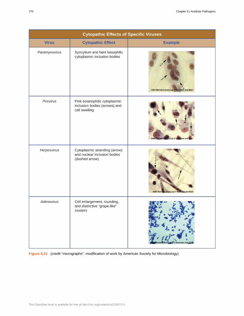
Figure 6.21 (credit “micrographs”: modification of work by American Society for Microbiology)
270 Chapter 6 | Acellular Pathogens
This OpenStax book is available for free at http://cnx.org/content/col12087/1.5

Watch this video (https://www.openstax.org/l/22virusesoncell) to learn aboutthe effects of viruses on cells.
Hemagglutination Assay
A serological assay is used to detect the presence of certain types of viruses in patient serum. Serum is the straw-colored liquid fraction of blood plasma from which clotting factors have been removed. Serum can be used in a directassay called a hemagglutination assay to detect specific types of viruses in the patient’s sample. Hemagglutinationis the agglutination (clumping) together of erythrocytes (red blood cells). Many viruses produce surface proteinsor spikes called hemagglutinins that can bind to receptors on the membranes of erythrocytes and cause the cellsto agglutinate. Hemagglutination is observable without using the microscope, but this method does not alwaysdifferentiate between infectious and noninfectious viral particles, since both can agglutinate erythrocytes.
To identify a specific pathogenic virus using hemagglutination, we must use an indirect approach. Proteins calledantibodies, generated by the patient’s immune system to fight a specific virus, can be used to bind to componentssuch as hemagglutinins that are uniquely associated with specific types of viruses. The binding of the antibodies withthe hemagglutinins found on the virus subsequently prevent erythrocytes from directly interacting with the virus. Sowhen erythrocytes are added to the antibody-coated viruses, there is no appearance of agglutination; agglutination hasbeen inhibited. We call these types of indirect assays for virus-specific antibodies hemagglutination inhibition (HAI)assays. HAI can be used to detect the presence of antibodies specific to many types of viruses that may be causing orhave caused an infection in a patient even months or years after infection (see Figure 6.22). This assay is describedin greater detail in Agglutination Assays.
Link to Learning
Chapter 6 | Acellular Pathogens 271

Figure 6.22 This chart shows the possible outcomes of a hemagglutination test. Row A: Erythrocytes do not bindtogether and will sink to the bottom of the well plate; this becomes visible as a red dot in the center of the well. RowB: Many viruses have hemagglutinins that causes agglutination of erythrocytes; the resulting hemagglutination formsa lattice structure that results in red color throughout the well. Row C: Virus-specific antibody, the viruses, and theerythrocytes are added to the well plate. The virus-specific antibodies inhibit agglutination, as can be seen as a reddot in the bottom of the well. (credit: modification of work by Centers for Disease Control and Prevention)
• What is the outcome of a positive HIA test?
Nucleic Acid Amplification Test
Nucleic acid amplification tests (NAAT) are used in molecular biology to detect unique nucleic acid sequences ofviruses in patient samples. Polymerase chain reaction (PCR) is an NAAT used to detect the presence of viral DNAin a patient’s tissue or body fluid sample. PCR is a technique that amplifies (i.e., synthesizes many copies) of a viralDNA segment of interest. Using PCR, short nucleotide sequences called primers bind to specific sequences of viralDNA, enabling identification of the virus.
Reverse transcriptase-PCR (RT-PCR) is an NAAT used to detect the presence of RNA viruses. RT-PCR differs fromPCR in that the enzyme reverse transcriptase (RT) is used to make a cDNA from the small amount of viral RNA inthe specimen. The cDNA can then be amplified by PCR. Both PCR and RT-PCR are used to detect and confirm thepresence of the viral nucleic acid in patient specimens.
272 Chapter 6 | Acellular Pathogens
This OpenStax book is available for free at http://cnx.org/content/col12087/1.5

HPV Scare
Michelle, a 21-year-old nursing student, came to the university clinic worried that she might have been exposedto a sexually transmitted disease (STD). Her sexual partner had recently developed several bumps on the baseof his penis. He had put off going to the doctor, but Michelle suspects they are genital warts caused by HPV.She is especially concerned because she knows that HPV not only causes warts but is a prominent cause ofcervical cancer. She and her partner always use condoms for contraception, but she is not confident that thisprecaution will protect her from HPV.
Michelle’s physician finds no physical signs of genital warts or any other STDs, but recommends that Michelleget a Pap smear along with an HPV test. The Pap smear will screen for abnormal cervical cells and the CPEsassociated with HPV; the HPV test will test for the presence of the virus. If both tests are negative, Michellecan be more assured that she most likely has not become infected with HPV. However, her doctor suggests itmight be wise for Michelle to get vaccinated against HPV to protect herself from possible future exposure.
• Why does Michelle’s physician order two different tests instead of relying on one or the other?
Enzyme Immunoassay
Enzyme immunoassays (EIAs) rely on the ability of antibodies to detect and attach to specific biomolecules calledantigens. The detecting antibody attaches to the target antigen with a high degree of specificity in what might be acomplex mixture of biomolecules. Also included in this type of assay is a colorless enzyme attached to the detectingantibody. The enzyme acts as a tag on the detecting antibody and can interact with a colorless substrate, leading tothe production of a colored end product. EIAs often rely on layers of antibodies to capture and react with antigens, allof which are attached to a membrane filter (see Figure 6.23). EIAs for viral antigens are often used as preliminaryscreening tests. If the results are positive, further confirmation will require tests with even greater sensitivity, such asa western blot or an NAAT. EIAs are discussed in more detail in EIAs and ELISAs.
Case in Point
Chapter 6 | Acellular Pathogens 273

Figure 6.23 Similar to rapid, over-the-counter pregnancy tests, EIAs for viral antigens require a few drops of dilutedpatient serum or plasma applied to a membrane filter. The membrane filter has been previously modified andembedded with antibody to viral antigen and internal controls. Antibody conjugate is added to the filter, with thetargeted antibody attached to the antigen (in the case of a positive test). Excess conjugate is washed off the filter.Substrate is added to activate the enzyme-mediated reaction to reveal the color change of a positive test. (credit:modification of work by “Cavitri”/Wikimedia Commons)
274 Chapter 6 | Acellular Pathogens
This OpenStax book is available for free at http://cnx.org/content/col12087/1.5

• What typically indicates a positive EIA test?
Part 3
Along with the RT/PCR analysis, David’s saliva was also collected for viral cultivation. In general, no singlediagnostic test is sufficient for antemortem diagnosis, since the results will depend on the sensitivity of theassay, the quantity of virions present at the time of testing, and the timing of the assay, since release ofvirions in the saliva can vary. As it turns out, the result was negative for viral cultivation from the saliva. Thisis not surprising to David’s doctor, because one negative result is not an absolute indication of the absence ofinfection. It may be that the number of virions in the saliva is low at the time of sampling. It is not unusual torepeat the test at intervals to enhance the chance of detecting higher virus loads.
• Should David’s doctor modify his course of treatment based on these test results?
Jump to the next Clinical Focus box. Go back to the previous Clinical Focus box.
6.4 Viroids, Virusoids, and Prions
Learning Objectives• Describe viroids and their unique characteristics
• Describe virusoids and their unique characteristics
• Describe prions and their unique characteristics
Research attempts to discover the causative agents of previously uninvestigated diseases have led to the discoveryof nonliving disease agents quite different from viruses. These include particles consisting only of RNA or only ofprotein that, nonetheless, are able to self-propagate at the expense of a host—a key similarity to viruses that allowsthem to cause disease conditions. To date, these discoveries include viroids, virusoids, and the proteinaceous prions.
Viroids
In 1971, Theodor Diener, a pathologist working at the Agriculture Research Service, discovered an acellular particlethat he named a viroid, meaning “virus-like.” Viroids consist only of a short strand of circular RNA capable of self-replication. The first viroid discovered was found to cause potato tuber spindle disease, which causes slower sproutingand various deformities in potato plants (see Figure 6.24). Like viruses, potato spindle tuber viroids (PSTVs) takecontrol of the host machinery to replicate their RNA genome. Unlike viruses, viroids do not have a protein coat toprotect their genetic information.
Clinical Focus
Chapter 6 | Acellular Pathogens 275

Figure 6.24 These potatoes have been infected by the potato spindle tuber viroid (PSTV), which is typically spreadwhen infected knives are used to cut healthy potatoes, which are then planted. (credit: Pamela Roberts, University ofFlorida Institute of Food and Agricultural Sciences, USDA ARS)
Viroids can result in devastating losses of commercially important agricultural food crops grown in fields andorchards. Since the discovery of PSTV, other viroids have been discovered that cause diseases in plants. Tomatoplanta macho viroid (TPMVd) infects tomato plants, which causes loss of chlorophyll, disfigured and brittle leaves,and very small tomatoes, resulting in loss of productivity in this field crop. Avocado sunblotch viroid (ASBVd)results in lower yields and poorer-quality fruit. ASBVd is the smallest viroid discovered thus far that infects plants.Peach latent mosaic viroid (PLMVd) can cause necrosis of flower buds and branches, and wounding of ripened fruit,which leads to fungal and bacterial growth in the fruit. PLMVd can also cause similar pathological changes in plums,nectarines, apricots, and cherries, resulting in decreased productivity in these orchards, as well. Viroids, in general,can be dispersed mechanically during crop maintenance or harvesting, vegetative reproduction, and possibly via seedsand insects, resulting in a severe drop in food availability and devastating economic consequences.
• What is the genome of a viroid made of?
Virusoids
A second type of pathogenic RNA that can infect commercially important agricultural crops are the virusoids, whichare subviral particles best described as non–self-replicating ssRNAs. RNA replication of virusoids is similar to thatof viroids but, unlike viroids, virusoids require that the cell also be infected with a specific “helper” virus. Thereare currently only five described types of virusoids and their associated helper viruses. The helper viruses are allfrom the family of Sobemoviruses. An example of a helper virus is the subterranean clover mottle virus, whichhas an associated virusoid packaged inside the viral capsid. Once the helper virus enters the host cell, the virusoidsare released and can be found free in plant cell cytoplasm, where they possess ribozyme activity. The helper virusundergoes typical viral replication independent of the activity of the virusoid. The virusoid genomes are small, only220 to 388 nucleotides long. A virusoid genome does not code for any proteins, but instead serves only to replicatevirusoid RNA.
Virusoids belong to a larger group of infectious agents called satellite RNAs, which are similar pathogenic RNAsfound in animals. Unlike the plant virusoids, satellite RNAs may encode for proteins; however, like plant virusoids,satellite RNAs must coinfect with a helper virus to replicate. One satellite RNA that infects humans and that has beendescribed by some scientists as a virusoid is the hepatitis delta virus (HDV), which, by some reports, is also calledhepatitis delta virusoid. Much larger than a plant virusoid, HDV has a circular, ssRNA genome of 1,700 nucleotidesand can direct the biosynthesis of HDV-associated proteins. The HDV helper virus is the hepatitis B virus (HBV).Coinfection with HBV and HDV results in more severe pathological changes in the liver during infection, which is
276 Chapter 6 | Acellular Pathogens
This OpenStax book is available for free at http://cnx.org/content/col12087/1.5
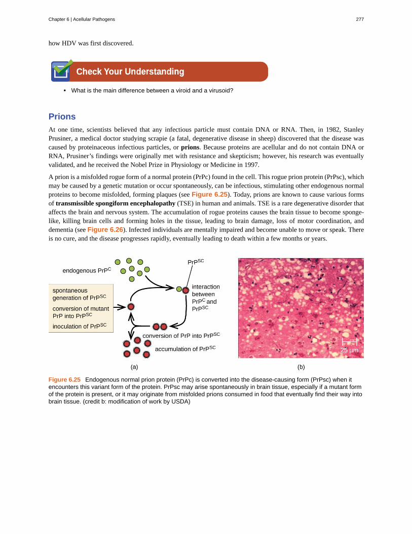
how HDV was first discovered.
• What is the main difference between a viroid and a virusoid?
Prions
At one time, scientists believed that any infectious particle must contain DNA or RNA. Then, in 1982, StanleyPrusiner, a medical doctor studying scrapie (a fatal, degenerative disease in sheep) discovered that the disease wascaused by proteinaceous infectious particles, or prions. Because proteins are acellular and do not contain DNA orRNA, Prusiner’s findings were originally met with resistance and skepticism; however, his research was eventuallyvalidated, and he received the Nobel Prize in Physiology or Medicine in 1997.
A prion is a misfolded rogue form of a normal protein (PrPc) found in the cell. This rogue prion protein (PrPsc), whichmay be caused by a genetic mutation or occur spontaneously, can be infectious, stimulating other endogenous normalproteins to become misfolded, forming plaques (see Figure 6.25). Today, prions are known to cause various formsof transmissible spongiform encephalopathy (TSE) in human and animals. TSE is a rare degenerative disorder thataffects the brain and nervous system. The accumulation of rogue proteins causes the brain tissue to become sponge-like, killing brain cells and forming holes in the tissue, leading to brain damage, loss of motor coordination, anddementia (see Figure 6.26). Infected individuals are mentally impaired and become unable to move or speak. Thereis no cure, and the disease progresses rapidly, eventually leading to death within a few months or years.
Figure 6.25 Endogenous normal prion protein (PrPc) is converted into the disease-causing form (PrPsc) when itencounters this variant form of the protein. PrPsc may arise spontaneously in brain tissue, especially if a mutant formof the protein is present, or it may originate from misfolded prions consumed in food that eventually find their way intobrain tissue. (credit b: modification of work by USDA)
Chapter 6 | Acellular Pathogens 277

Figure 6.26 Creutzfeldt-Jakob disease (CJD) is a fatal disease that causes degeneration of neural tissue. (a) Thesebrain scans compare a normal brain to one with CJD. (b) Compared to a normal brain, the brain tissue of a CJDpatient is full of sponge-like lesions, which result from abnormal formations of prion protein. (credit a (right):modification of work by Dr. Laughlin Dawes; credit b (top): modification of work by Suzanne Wakim; credit b (bottom):modification of work by Centers for Disease Control and Prevention)
TSEs in humans include kuru, fatal familial insomnia, Gerstmann-Straussler-Scheinker disease, and Creutzfeldt-Jakob disease (see Figure 6.26). TSEs in animals include mad cow disease, scrapie (in sheep and goats), and chronicwasting disease (in elk and deer). TSEs can be transmitted between animals and from animals to humans by eatingcontaminated meat or animal feed. Transmission between humans can occur through heredity (as is often the case withGSS and CJD) or by contact with contaminated tissue, as might occur during a blood transfusion or organ transplant.There is no evidence for transmission via casual contact with an infected person. Table 6.3 lists TSEs that affecthumans and their modes of transmission.
Transmissible Spongiform Encephalopathies (TSEs) in Humans
Disease Mechanism(s) of Transmission[10]
Sporadic CJD (sCJD) Not known; possibly by alteration of normal prior protein (PrP) to rogue formdue to somatic mutation
Variant CJD (vCJD) Eating contaminated cattle products and by secondary bloodbornetransmission
Familial CJD (fCJD) Mutation in germline PrP gene
Iatrogenic CJD (iCJD) Contaminated neurosurgical instruments, corneal graft, gonadotrophichormone, and, secondarily, by blood transfusion
Kuru Eating infected meat through ritualistic cannibalism
Gerstmann-Straussler-Scheinker disease (GSS)
Mutation in germline PrP gene
Table 6.3
10. National Institute of Neurological Disorders and Stroke. “Creutzfeldt-Jakob Disease Fact Sheet.” http://www.ninds.nih.gov/disorders/cjd/detail_cjd.htm (accessed December 31, 2015).
278 Chapter 6 | Acellular Pathogens
This OpenStax book is available for free at http://cnx.org/content/col12087/1.5

Transmissible Spongiform Encephalopathies (TSEs) in Humans
Disease Mechanism(s) of Transmission
Fatal familial insomnia (FFI) Mutation in germline PrP gene
Table 6.3
Prions are extremely difficult to destroy because they are resistant to heat, chemicals, and radiation. Even standardsterilization procedures do not ensure the destruction of these particles. Currently, there is no treatment or cure forTSE disease, and contaminated meats or infected animals must be handled according to federal guidelines to preventtransmission.
• Does a prion have a genome?
For more information on the handling of animals and prion-contaminatedmaterials, visit the guidelines published on the CDC(https://www.openstax.org/l/22cdccontaminat) and WHO(https://www.openstax.org/l/22whocontaminat) websites.
Resolution
A few days later, David’s doctor receives the results of the immunofluorescence test on his skin sample. Thetest is negative for rabies antigen. A second viral antigen test on his saliva sample also comes back negative.Despite these results, the doctor decides to continue David’s current course of treatment. Given the positiveRT-PCR test, it is best not to rule out a possible rabies infection.
Near the site of the bite, David receives an injection of rabies immunoglobulin, which attaches to andinactivates any rabies virus that may be present in his tissues. Over the next 14 days, he receives a series offour rabies-specific vaccinations in the arm. These vaccines activate David’s immune response and help hisbody recognize and fight the virus. Thankfully, with treatment, David symptoms improve and he makes a fullrecovery.
Not all rabies cases have such a fortunate outcome. In fact, rabies is usually fatal once the patient starts toexhibit symptoms, and postbite treatments are mainly palliative (i.e., sedation and pain management).
Go back to the previous Clinical Focus box.
Link to Learning
Clinical Focus
Chapter 6 | Acellular Pathogens 279

Summary
6.1 Viruses• Viruses are generally ultramicroscopic, typically from 20 nm to 900 nm in length. Some large viruses have
been found.
• Virions are acellular and consist of a nucleic acid, DNA or RNA, but not both, surrounded by a protein capsid.There may also be a phospholipid membrane surrounding the capsid.
• Viruses are obligate intracellular parasites.
• Viruses are known to infect various types of cells found in plants, animals, fungi, protists, bacteria, andarchaea. Viruses typically have limited host ranges and infect specific cell types.
• Viruses may have helical, polyhedral, or complex shapes.
• Classification of viruses is based on morphology, type of nucleic acid, host range, cell specificity, and enzymescarried within the virion.
• Like other diseases, viral diseases are classified using ICD codes.
6.2 The Viral Life Cycle• Many viruses target specific hosts or tissues. Some may have more than one host.
• Many viruses follow several stages to infect host cells. These stages include attachment, penetration,uncoating, biosynthesis, maturation, and release.
• Bacteriophages have a lytic or lysogenic cycle. The lytic cycle leads to the death of the host, whereas thelysogenic cycle leads to integration of phage into the host genome.
• Bacteriophages inject DNA into the host cell, whereas animal viruses enter by endocytosis or membranefusion.
• Animal viruses can undergo latency, similar to lysogeny for a bacteriophage.
• The majority of plant viruses are positive-strand ssRNA and can undergo latency, chronic, or lytic infection,as observed for animal viruses.
• The growth curve of bacteriophage populations is a one-step multiplication curve and not a sigmoidal curve,as compared to the bacterial growth curve.
• Bacteriophages transfer genetic information between hosts using either generalized or specializedtransduction.
6.3 Isolation, Culture, and Identification of Viruses• Viral cultivation requires the presence of some form of host cell (whole organism, embryo, or cell culture).
• Viruses can be isolated from samples by filtration.
• Viral filtrate is a rich source of released virions.
• Bacteriophages are detected by presence of clear plaques on bacterial lawn.
• Animal and plant viruses are detected by cytopathic effects, molecular techniques (PCR, RT-PCR), enzymeimmunoassays, and serological assays (hemagglutination assay, hemagglutination inhibition assay).
6.4 Viroids, Virusoids, and Prions• Other acellular agents such as viroids, virusoids, and prions also cause diseases. Viroids consist of small,
naked ssRNAs that cause diseases in plants. Virusoids are ssRNAs that require other helper viruses toestablish an infection. Prions are proteinaceous infectious particles that cause transmissible spongiformencephalopathies.
• Prions are extremely resistant to chemicals, heat, and radiation.
• There are no treatments for prion infection.
280 Chapter 6 | Acellular Pathogens
This OpenStax book is available for free at http://cnx.org/content/col12087/1.5

Multiple Choice
1. The component(s) of a virus that is/are extendedfrom the envelope for attachment is/are the:
a. capsomeresb. spikesc. nucleic acidd. viral whiskers
2. Which of the following does a virus lack? Select allthat apply.
a. ribosomesb. metabolic processesc. nucleic acidd. glycoprotein
3. The envelope of a virus is derived from the host’sa. nucleic acidsb. membrane structuresc. cytoplasmd. genome
4. In naming viruses, the family name ends with________ and genus name ends with _________.
a. −virus; −viridaeb. −viridae; −virusc. −virion; virusd. −virus; virion
5. What is another name for a nonenveloped virus?a. enveloped virusb. provirusc. naked virusd. latent virus
6. Which of the following leads to the destruction ofthe host cells?
a. lysogenic cycleb. lytic cyclec. prophaged. temperate phage
7. A virus obtains its envelope during which of thefollowing phases?
a. attachmentb. penetrationc. assemblyd. release
8. Which of the following components is brought intoa cell by HIV?
a. a DNA-dependent DNA polymeraseb. RNA polymerasec. ribosomed. reverse transcriptase
9. A positive-strand RNA virus:a. must first be converted to a mRNA before it can
be translated.b. can be used directly to translate viral proteins.c. will be degraded by host enzymes.d. is not recognized by host ribosomes.
10. What is the name for the transfer of geneticinformation from one bacterium to another bacterium bya phage?
a. transductionb. penetrationc. excisiond. translation
11. Which of the followings cannot be used to cultureviruses?
a. tissue cultureb. liquid medium onlyc. embryod. animal host
12. Which of the following tests can be used to detectthe presence of a specific virus?
a. EIAb. RT-PCRc. PCRd. all of the above
13. Which of the following is NOT a cytopathic effect?
a. transformationb. cell fusionc. mononucleated celld. inclusion bodies
14. Which of these infectious agents do not havenucleic acid?
a. viroidsb. virusesc. bacteriad. prions
Review Questions
Chapter 6 | Acellular Pathogens 281

15. Which of the following is true of prions?a. They can be inactivated by boiling at 100 °C.b. They contain a capsid.c. They are a rogue form of protein, PrP.d. They can be reliably inactivated by an autoclave.
True/False
16. True or False: Scientists have identified viruses that are able to infect fungal cells.
Fill in the Blank
17. A virus that infects a bacterium is called a/an ___________________.
18. A/an __________ virus possesses characteristics of both a polyhedral and helical virus.
19. A virus containing only nucleic acid and a capsid is called a/an ___________________ virus or__________________ virus.
20. The ____________ _____________ on the bacteriophage allow for binding to the bacterial cell.
21. An enzyme from HIV that can make a copy of DNA from RNA is called _______________________.
22. For lytic viruses, _________________ is a phase during a viral growth curve when the virus is not detected.
23. Viruses can be diagnosed and observed using a(n) _____________ microscope.
24. Cell abnormalities resulting from a viral infection are called ____________ _____________.
25. Both viroids and virusoids have a(n) _________ genome, but virusoids require a(n) _________ to reproduce.
Short Answer
26. Discuss the geometric differences among helical, polyhedral, and complex viruses.
27. What was the meaning of the word “virus” in the 1880s and why was it used to describe the cause of tobaccomosaic disease?
28. Briefly explain the difference between the mechanism of entry of a T-even bacteriophage and an animal virus.
29. Discuss the difference between generalized and specialized transduction.
30. Differentiate between lytic and lysogenic cycles.
31. Briefly explain the various methods of culturing viruses.
32. Describe the disease symptoms observed in animals infected with prions.
282 Chapter 6 | Acellular Pathogens
This OpenStax book is available for free at http://cnx.org/content/col12087/1.5

Critical Thinking
33. Name each labeled part of the illustrated bacteriophage.
34. In terms of evolution, which do you think arises first? The virus or the host? Explain your answer.
35. Do you think it is possible to create a virus in the lab? Imagine that you are a mad scientist. Describe how youwould go about creating a new virus.
36. Label the five stages of a bacteriophage infection in the figure:
37. Bacteriophages have lytic and lysogenic cycles. Discuss the advantages and disadvantages for the phage.
38. How does reverse transcriptase aid a retrovirus in establishing a chronic infection?
39. Discuss some methods by which plant viruses are transmitted from a diseased plant to a healthy one.
40. Label the components indicated by arrows.
Figure 6.27 (credit: modification of work by American Society for Microbiology)
Chapter 6 | Acellular Pathogens 283

41. What are some characteristics of the viruses that are similar to a computer virus?
42. Does a prion replicate? Explain.
284 Chapter 6 | Acellular Pathogens
This OpenStax book is available for free at http://cnx.org/content/col12087/1.5
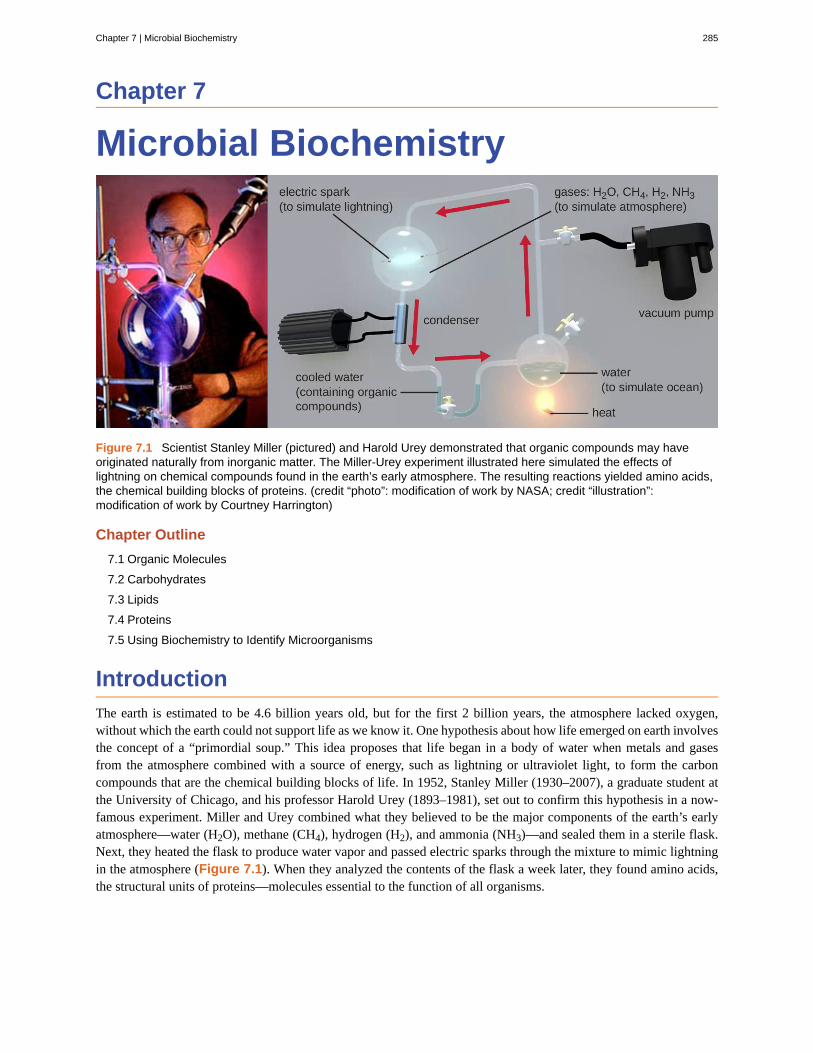
Chapter 7
Microbial Biochemistry
Figure 7.1 Scientist Stanley Miller (pictured) and Harold Urey demonstrated that organic compounds may haveoriginated naturally from inorganic matter. The Miller-Urey experiment illustrated here simulated the effects oflightning on chemical compounds found in the earth’s early atmosphere. The resulting reactions yielded amino acids,the chemical building blocks of proteins. (credit “photo”: modification of work by NASA; credit “illustration”:modification of work by Courtney Harrington)
Chapter Outline
7.1 Organic Molecules
7.2 Carbohydrates
7.3 Lipids
7.4 Proteins
7.5 Using Biochemistry to Identify Microorganisms
IntroductionThe earth is estimated to be 4.6 billion years old, but for the first 2 billion years, the atmosphere lacked oxygen,without which the earth could not support life as we know it. One hypothesis about how life emerged on earth involvesthe concept of a “primordial soup.” This idea proposes that life began in a body of water when metals and gasesfrom the atmosphere combined with a source of energy, such as lightning or ultraviolet light, to form the carboncompounds that are the chemical building blocks of life. In 1952, Stanley Miller (1930–2007), a graduate student atthe University of Chicago, and his professor Harold Urey (1893–1981), set out to confirm this hypothesis in a now-famous experiment. Miller and Urey combined what they believed to be the major components of the earth’s earlyatmosphere—water (H2O), methane (CH4), hydrogen (H2), and ammonia (NH3)—and sealed them in a sterile flask.Next, they heated the flask to produce water vapor and passed electric sparks through the mixture to mimic lightningin the atmosphere (Figure 7.1). When they analyzed the contents of the flask a week later, they found amino acids,the structural units of proteins—molecules essential to the function of all organisms.
Chapter 7 | Microbial Biochemistry 285

7.1 Organic Molecules
Learning Objectives• Identify common elements and structures found in organic molecules
• Explain the concept of isomerism
• Identify examples of functional groups
• Describe the role of functional groups in synthesizing polymers
Biochemistry is the discipline that studies the chemistry of life, and its objective is to explain form and function basedon chemical principles. Organic chemistry is the discipline devoted to the study of carbon-based chemistry, whichis the foundation for the study of biomolecules and the discipline of biochemistry. Both biochemistry and organicchemistry are based on the concepts of general chemistry, some of which are presented in Appendix A.
Elements in Living Cells
The most abundant element in cells is hydrogen (H), followed by carbon (C), oxygen (O), nitrogen (N), phosphorous(P), and sulfur (S). We call these elements macronutrients, and they account for about 99% of the dry weight ofcells. Some elements, such as sodium (Na), potassium (K), magnesium (Mg), zinc (Zn), iron (Fe), calcium (Ca),molybdenum (Mo), copper (Cu), cobalt (Co), manganese (Mn), or vanadium (Va), are required by some cells in verysmall amounts and are called micronutrients or trace elements. All of these elements are essential to the function ofmany biochemical reactions, and, therefore, are essential to life.
The four most abundant elements in living matter (C, N, O, and H) have low atomic numbers and are thus lightelements capable of forming strong bonds with other atoms to produce molecules (Figure 7.2). Carbon forms fourchemical bonds, whereas nitrogen forms three, oxygen forms two, and hydrogen forms one. When bonded togetherwithin molecules, oxygen, sulfur, and nitrogen often have one or more “lone pairs” of electrons that play importantroles in determining many of the molecules’ physical and chemical properties (see Appendix A). These traits incombination permit the formation of a vast number of diverse molecular species necessary to form the structures andenable the functions of living organisms.
Part 1
Penny is a 16-year-old student who visited her doctor, complaining about an itchy skin rash. She had a historyof allergic episodes. The doctor looked at her sun-tanned skin and asked her if she switched to a differentsunscreen. She said she had, so the doctor diagnosed an allergic eczema. The symptoms were mild so thedoctor told Penny to avoid using the sunscreen that caused the reaction and prescribed an over-the-countermoisturizing cream to keep her skin hydrated and to help with itching.
• What kinds of substances would you expect to find in a moisturizing cream?
• What physical or chemical properties of these substances would help alleviate itching and inflammationof the skin?
Jump to the next Clinical Focus box.
Clinical Focus
286 Chapter 7 | Microbial Biochemistry
This OpenStax book is available for free at http://cnx.org/content/col12087/1.5

Figure 7.2 Some common molecules include carbon dioxide, ammonia, and oxygen, which consist of combinationsof oxygen atoms (red spheres), carbon atoms (gray spheres), hydrogen atoms (white spheres), or nitrogen atoms(blue spheres).
Living organisms contain inorganic compounds (mainly water and salts; see Appendix A) and organic molecules.Organic molecules contain carbon; inorganic compounds do not. Carbon oxides and carbonates are exceptions; theycontain carbon but are considered inorganic because they do not contain hydrogen. The atoms of an organic moleculeare typically organized around chains of carbon atoms.
Inorganic compounds make up 1%–1.5% of a living cell’s mass. They are small, simple compounds that playimportant roles in the cell, although they do not form cell structures. Most of the carbon found in organic moleculesoriginates from inorganic carbon sources such as carbon dioxide captured via carbon fixation by microorganisms.
• Describe the most abundant elements in nature.
• Describe the most abundant elements in natureWhat are the differences between organic and inorganicmolecules?
Organic Molecules and Isomerism
Organic molecules in organisms are generally larger and more complex than inorganic molecules. Their carbonskeletons are held together by covalent bonds. They form the cells of an organism and perform the chemical reactionsthat facilitate life. All of these molecules, called biomolecules because they are part of living matter, contain carbon,which is the building block of life. Carbon is a very unique element in that it has four valence electrons in its outerorbitals and can form four single covalent bonds with up to four other atoms at the same time (see Appendix A).These atoms are usually oxygen, hydrogen, nitrogen, sulfur, phosphorous, and carbon itself; the simplest organiccompound is methane, in which carbon binds only to hydrogen (Figure 7.3).
As a result of carbon’s unique combination of size and bonding properties, carbon atoms can bind together in largenumbers, thus producing a chain or carbon skeleton. The carbon skeleton of organic molecules can be straight,branched, or ring shaped (cyclic). Organic molecules are built on chains of carbon atoms of varying lengths; most aretypically very long, which allows for a huge number and variety of compounds. No other element has the ability toform so many different molecules of so many different sizes and shapes.
Chapter 7 | Microbial Biochemistry 287

Figure 7.3 A carbon atom can bond with up to four other atoms. The simplest organic molecule is methane (CH4),depicted here.
Molecules with the same atomic makeup but different structural arrangement of atoms are called isomers. Theconcept of isomerism is very important in chemistry because the structure of a molecule is always directly relatedto its function. Slight changes in the structural arrangements of atoms in a molecule may lead to very differentproperties. Chemists represent molecules by their structural formula, which is a graphic representation of themolecular structure, showing how the atoms are arranged. Compounds that have identical molecular formulas butdiffer in the bonding sequence of the atoms are called structural isomers. The monosaccharides glucose, galactose,and fructose all have the same molecular formula, C6H12O6, but we can see from Figure 7.4 that the atoms arebonded together differently.
Figure 7.4 Glucose, galactose, and fructose have the same chemical formula (C6H12O6), but these structuralisomers differ in their physical and chemical properties.
Isomers that differ in the spatial arrangements of atoms are called stereoisomers; one unique type is enantiomers.The properties of enantiomers were originally discovered by Louis Pasteur in 1848 while using a microscopeto analyze crystallized fermentation products of wine. Enantiomers are molecules that have the characteristic ofchirality, in which their structures are nonsuperimposable mirror images of each other. Chirality is an importantcharacteristic in many biologically important molecules, as illustrated by the examples of structural differences in theenantiomeric forms of the monosaccharide glucose or the amino acid alanine (Figure 7.5).
Many organisms are only able to use one enantiomeric form of certain types of molecules as nutrients and as buildingblocks to make structures within a cell. Some enantiomeric forms of amino acids have distinctly different tastes andsmells when consumed as food. For example, L-aspartame, commonly called aspartame, tastes sweet, whereas D-aspartame is tasteless. Drug enantiomers can have very different pharmacologic affects. For example, the compound
288 Chapter 7 | Microbial Biochemistry
This OpenStax book is available for free at http://cnx.org/content/col12087/1.5

methorphan exists as two enantiomers, one of which acts as an antitussive (dextromethorphan, a cough suppressant),whereas the other acts as an analgesic (levomethorphan, a drug similar in effect to codeine).
Figure 7.5 Enantiomers are stereoisomers that exhibit chirality. Their chemical structures are nonsuperimposablemirror images of each other. (a) D-glucose and L-glucose are monosaccharides that are enantiomers. (b) Theenantiomers D-alanine and L-alanine are enantiomers found in bacterial cell walls and human cells, respectively.
Enantiomers are also called optical isomers because they can rotate the plane of polarized light. Some of the crystalsPasteur observed from wine fermentation rotated light clockwise whereas others rotated the light counterclockwise.Today, we denote enantiomers that rotate polarized light clockwise (+) as d forms, and the mirror image of the samemolecule that rotates polarized light counterclockwise (−) as the l form. The d and l labels are derived from theLatin words dexter (on the right) and laevus (on the left), respectively. These two different optical isomers often havevery different biological properties and activities. Certain species of molds, yeast, and bacteria, such as Rhizopus,Yarrowia, and Lactobacillus spp., respectively, can only metabolize one type of optical isomer; the opposite isomeris not suitable as a source of nutrients. Another important reason to be aware of optical isomers is the therapeutic useof these types of chemicals for drug treatment, because some microorganisms can only be affected by one specificoptical isomer.
• We say that life is carbon based. What makes carbon so suitable to be part of all the macromolecules of livingorganisms?
Biologically Significant Functional Groups
In addition to containing carbon atoms, biomolecules also contain functional groups—groups of atoms withinmolecules that are categorized by their specific chemical composition and the chemical reactions they perform,regardless of the molecule in which the group is found. Some of the most common functional groups are listed inFigure 7.6. In the formulas, the symbol R stands for “residue” and represents the remainder of the molecule. Rmight symbolize just a single hydrogen atom or it may represent a group of many atoms. Notice that some functionalgroups are relatively simple, consisting of just one or two atoms, while some comprise two of these simpler functionalgroups. For example, a carbonyl group is a functional group composed of a carbon atom double bonded to an oxygenatom: C=O. It is present in several classes of organic compounds as part of larger functional groups such as ketones,aldehydes, carboxylic acids, and amides. In ketones, the carbonyl is present as an internal group, whereas in aldehydesit is a terminal group.
Chapter 7 | Microbial Biochemistry 289

Figure 7.6
Macromolecules
Carbon chains form the skeletons of most organic molecules. Functional groups combine with the chain to form
290 Chapter 7 | Microbial Biochemistry
This OpenStax book is available for free at http://cnx.org/content/col12087/1.5

biomolecules. Because these biomolecules are typically large, we call them macromolecules. Many biologicallyrelevant macromolecules are formed by linking together a great number of identical, or very similar, smaller organicmolecules. The smaller molecules act as building blocks and are called monomers, and the macromolecules thatresult from their linkage are called polymers. Cells and cell structures include four main groups of carbon-containingmacromolecules: polysaccharides, proteins, lipids, and nucleic acids. The first three groups of molecules will bestudied throughout this chapter. The biochemistry of nucleic acids will be discussed in Biochemistry of theGenome.
Of the many possible ways that monomers may be combined to yield polymers, one common approach encountered inthe formation of biological macromolecules is dehydration synthesis. In this chemical reaction, monomer moleculesbind end to end in a process that results in the formation of water molecules as a byproduct:
H—monomer—OH + H—monomer—OH ⟶ H—monomer—monomer—OH + H2 O
Figure 7.7 shows dehydration synthesis of glucose binding together to form maltose and a water molecule. Table7.1 summarizes macromolecules and some of their functions.
Figure 7.7 In this dehydration synthesis reaction, two molecules of glucose are linked together to form maltose. Inthe process, a water molecule is formed.
Some Functions of Macromolecules
Macromolecule Functions
Carbohydrates Energy storage, receptors, food, structural role in plants, fungal cell walls, exoskeletons ofinsects
Lipids Energy storage, membrane structure, insulation, hormones, pigments
Nucleic acids Storage and transfer of genetic information
Proteins Enzymes, structure, receptors, transport, structural role in the cytoskeleton of a cell and theextracellular matrix
Table 7.1
• What is the byproduct of a dehydration synthesis reaction?
Chapter 7 | Microbial Biochemistry 291

7.2 Carbohydrates
Learning Objectives• Give examples of monosaccharides and polysaccharides
• Describe the function of monosaccharides and polysaccharides within a cell
The most abundant biomolecules on earth are carbohydrates. From a chemical viewpoint, carbohydrates areprimarily a combination of carbon and water, and many of them have the empirical formula (CH2O)n, where n is thenumber of repeated units. This view represents these molecules simply as “hydrated” carbon atom chains in whichwater molecules attach to each carbon atom, leading to the term “carbohydrates.” Although all carbohydrates containcarbon, hydrogen, and oxygen, there are some that also contain nitrogen, phosphorus, and/or sulfur. Carbohydrateshave myriad different functions. They are abundant in terrestrial ecosystems, many forms of which we use as foodsources. These molecules are also vital parts of macromolecular structures that store and transmit genetic information(i.e., DNA and RNA). They are the basis of biological polymers that impart strength to various structural componentsof organisms (e.g., cellulose and chitin), and they are the primary source of energy storage in the form of starch andglycogen.
Monosaccharides: The Sweet Ones
In biochemistry, carbohydrates are often called saccharides, from the Greek sakcharon, meaning sugar, although notall the saccharides are sweet. The simplest carbohydrates are called monosaccharides, or simple sugars. They arethe building blocks (monomers) for the synthesis of polymers or complex carbohydrates, as will be discussed furtherin this section. Monosaccharides are classified based on the number of carbons in the molecule. General categoriesare identified using a prefix that indicates the number of carbons and the suffix –ose, which indicates a saccharide;for example, triose (three carbons), tetrose (four carbons), pentose (five carbons), and hexose (six carbons) (Figure7.8). The hexose D-glucose is the most abundant monosaccharide in nature. Other very common and abundant hexosemonosaccharides are galactose, used to make the disaccharide milk sugar lactose, and the fruit sugar fructose.
292 Chapter 7 | Microbial Biochemistry
This OpenStax book is available for free at http://cnx.org/content/col12087/1.5

Figure 7.8 Monosaccharides are classified based on the position of the carbonyl group and the number of carbonsin the backbone.
Monosaccharides of four or more carbon atoms are typically more stable when they adopt cyclic, or ring, structures.These ring structures result from a chemical reaction between functional groups on opposite ends of the sugar’sflexible carbon chain, namely the carbonyl group and a relatively distant hydroxyl group. Glucose, for example, formsa six-membered ring (Figure 7.9).
Figure 7.9 (a) A linear monosaccharide (glucose in this case) forms a cyclic structure. (b) This illustration shows amore realistic depiction of the cyclic monosaccharide structure. Note in these cyclic structural diagrams, the carbonatoms composing the ring are not explicitly shown.
Chapter 7 | Microbial Biochemistry 293

• Why do monosaccharides form ring structures?
Disaccharides
Two monosaccharide molecules may chemically bond to form a disaccharide. The name given to the covalent bondbetween the two monosaccharides is a glycosidic bond. Glycosidic bonds form between hydroxyl groups of the twosaccharide molecules, an example of the dehydration synthesis described in the previous section of this chapter:
monosaccharide—OH + HO—monosaccharide ⟶ monosaccharide—O—monosaccharide⎤⎦ ⎥ ⎥ ⎥ ⎥ ⎥ ⎥ ⎥ ⎥ ⎥ ⎥ ⎥ ⎥ ⎥ ⎥
disaccharide
Common disaccharides are the grain sugar maltose, made of two glucose molecules; the milk sugar lactose, made ofa galactose and a glucose molecule; and the table sugar sucrose, made of a glucose and a fructose molecule (Figure7.10).
Figure 7.10 Common disaccharides include maltose, lactose, and sucrose.
Polysaccharides
Polysaccharides, also called glycans, are large polymers composed of hundreds of monosaccharide monomers.Unlike mono- and disaccharides, polysaccharides are not sweet and, in general, they are not soluble in water. Likedisaccharides, the monomeric units of polysaccharides are linked together by glycosidic bonds.
Polysaccharides are very diverse in their structure. Three of the most biologically important polysaccharides—starch,glycogen, and cellulose—are all composed of repetitive glucose units, although they differ in their structure (Figure7.11). Cellulose consists of a linear chain of glucose molecules and is a common structural component of cell wallsin plants and other organisms. Glycogen and starch are branched polymers; glycogen is the primary energy-storage
294 Chapter 7 | Microbial Biochemistry
This OpenStax book is available for free at http://cnx.org/content/col12087/1.5

molecule in animals and bacteria, whereas plants primarily store energy in starch. The orientation of the glycosidiclinkages in these three polymers is different as well and, as a consequence, linear and branched macromolecules havedifferent properties.
Modified glucose molecules can be fundamental components of other structural polysaccharides. Examples of thesetypes of structural polysaccharides are N-acetyl glucosamine (NAG) and N-acetyl muramic acid (NAM) found inbacterial cell wall peptidoglycan. Polymers of NAG form chitin, which is found in fungal cell walls and in theexoskeleton of insects.
Figure 7.11 Starch, glycogen, and cellulose are three of the most important polysaccharides. In the top row,hexagons represent individual glucose molecules. Micrographs (bottom row) show wheat starch granules stained withiodine (left), glycogen granules (G) inside the cell of a cyanobacterium (middle), and bacterial cellulose fibers (right).(credit “iodine granules”: modification of work by Kiselov Yuri; credit “glycogen granules”: modification of work byStöckel J, Elvitigala TR, Liberton M, Pakrasi HB; credit “cellulose”: modification of work by American Society forMicrobiology)
• What are the most biologically important polysaccharides and why are they important?
Chapter 7 | Microbial Biochemistry 295

7.3 Lipids
Learning Objectives
Describe the chemical composition of lipids
• Describe the unique characteristics and diverse structures of lipids
• Compare and contrast triacylglycerides (triglycerides) and phospholipids.
• Describe how phospholipids are used to construct biological membranes.
Although they are composed primarily of carbon and hydrogen, lipid molecules may also contain oxygen, nitrogen,sulfur, and phosphorous. Lipids serve numerous and diverse purposes in the structure and functions of organisms.They can be a source of nutrients, a storage form for carbon, energy-storage molecules, or structural components ofmembranes and hormones. Lipids comprise a broad class of many chemically distinct compounds, the most commonof which are discussed in this section.
Fatty Acids and Triacylglycerides
The fatty acids are lipids that contain long-chain hydrocarbons terminated with a carboxylic acid functional group.Because the long hydrocarbon chain, fatty acids are hydrophobic (“water fearing”) or nonpolar. Fatty acids withhydrocarbon chains that contain only single bonds are called saturated fatty acids because they have the greatestnumber of hydrogen atoms possible and are, therefore, “saturated” with hydrogen. Fatty acids with hydrocarbonchains containing at least one double bond are called unsaturated fatty acids because they have fewer hydrogenatoms. Saturated fatty acids have a straight, flexible carbon backbone, whereas unsaturated fatty acids have “kinks”in their carbon skeleton because each double bond causes a rigid bend of the carbon skeleton. These differences insaturated versus unsaturated fatty acid structure result in different properties for the corresponding lipids in whichthe fatty acids are incorporated. For example, lipids containing saturated fatty acids are solids at room temperature,whereas lipids containing unsaturated fatty acids are liquids.
A triacylglycerol, or triglyceride, is formed when three fatty acids are chemically linked to a glycerol molecule(Figure 7.12). Triglycerides are the primary components of adipose tissue (body fat), and are major constituentsof sebum (skin oils). They play an important metabolic role, serving as efficient energy-storage molecules that canprovide more than double the caloric content of both carbohydrates and proteins.
Figure 7.12 Triglycerides are composed of a glycerol molecule attached to three fatty acids by a dehydrationsynthesis reaction.
296 Chapter 7 | Microbial Biochemistry
This OpenStax book is available for free at http://cnx.org/content/col12087/1.5

• Explain why fatty acids with hydrocarbon chains that contain only single bonds are called saturated fattyacids.
Phospholipids and Biological Membranes
Triglycerides are classified as simple lipids because they are formed from just two types of compounds: glycerol andfatty acids. In contrast, complex lipids contain at least one additional component, for example, a phosphate group(phospholipids) or a carbohydrate moiety (glycolipids). Figure 7.13 depicts a typical phospholipid composed oftwo fatty acids linked to glycerol (a diglyceride). The two fatty acid carbon chains may be both saturated, bothunsaturated, or one of each. Instead of another fatty acid molecule (as for triglycerides), the third binding position onthe glycerol molecule is occupied by a modified phosphate group.
Figure 7.13 This illustration shows a phospholipid with two different fatty acids, one saturated and one unsaturated,bonded to the glycerol molecule. The unsaturated fatty acid has a slight kink in its structure due to the double bond.
The molecular structure of lipids results in unique behavior in aqueous environments. Figure 7.12 depicts thestructure of a triglyceride. Because all three substituents on the glycerol backbone are long hydrocarbon chains, thesecompounds are nonpolar and not significantly attracted to polar water molecules—they are hydrophobic. Conversely,phospholipids such as the one shown in Figure 7.13 have a negatively charged phosphate group. Because thephosphate is charged, it is capable of strong attraction to water molecules and thus is hydrophilic, or “water loving.”The hydrophilic portion of the phospholipid is often referred to as a polar “head,” and the long hydrocarbon chains asnonpolar “tails.” A molecule presenting a hydrophobic portion and a hydrophilic moiety is said to be amphipathic.Notice the “R” designation within the hydrophilic head depicted in Figure 7.13, indicating that a polar head groupcan be more complex than a simple phosphate moiety. Glycolipids are examples in which carbohydrates are bondedto the lipids’ head groups.
Chapter 7 | Microbial Biochemistry 297

The amphipathic nature of phospholipids enables them to form uniquely functional structures in aqueousenvironments. As mentioned, the polar heads of these molecules are strongly attracted to water molecules, andthe nonpolar tails are not. Because of their considerable lengths, these tails are, in fact, strongly attracted to oneanother. As a result, energetically stable, large-scale assemblies of phospholipid molecules are formed in whichthe hydrophobic tails congregate within enclosed regions, shielded from contact with water by the polar heads(Figure 7.14). The simplest of these structures are micelles, spherical assemblies containing a hydrophobic interiorof phospholipid tails and an outer surface of polar head groups. Larger and more complex structures are created fromlipid-bilayer sheets, or unit membranes, which are large, two-dimensional assemblies of phospholipids congregatedtail to tail. The cell membranes of nearly all organisms are made from lipid-bilayer sheets, as are the membranesof many intracellular components. These sheets may also form lipid-bilayer spheres that are the structural basis ofvesicles and liposomes, subcellular components that play a role in numerous physiological functions.
Figure 7.14 Phospholipids tend to arrange themselves in aqueous solution forming liposomes, micelles, or lipidbilayer sheets. (credit: modification of work by Mariana Ruiz Villarreal)
• How is the amphipathic nature of phospholipids significant?
Isoprenoids and Sterols
The isoprenoids are branched lipids, also referred to as terpenoids, that are formed by chemical modifications ofthe isoprene molecule (Figure 7.15). These lipids play a wide variety of physiological roles in plants and animals,with many technological uses as pharmaceuticals (capsaicin), pigments (e.g., orange beta carotene, xanthophylls),and fragrances (e.g., menthol, camphor, limonene [lemon fragrance], and pinene [pine fragrance]). Long-chainisoprenoids are also found in hydrophobic oils and waxes. Waxes are typically water resistant and hard at roomtemperature, but they soften when heated and liquefy if warmed adequately. In humans, the main wax productionoccurs within the sebaceous glands of hair follicles in the skin, resulting in a secreted material called sebum,which consists mainly of triacylglycerol, wax esters, and the hydrocarbon squalene. There are many bacteria inthe microbiota on the skin that feed on these lipids. One of the most prominent bacteria that feed on lipids isPropionibacterium acnes, which uses the skin’s lipids to generate short-chain fatty acids and is involved in theproduction of acne.
298 Chapter 7 | Microbial Biochemistry
This OpenStax book is available for free at http://cnx.org/content/col12087/1.5

Figure 7.15 Five-carbon isoprene molecules are chemically modified in various ways to yield isoprenoids.
Another type of lipids are steroids, complex, ringed structures that are found in cell membranes; some function ashormones. The most common types of steroids are sterols, which are steroids containing an OH group. These aremainly hydrophobic molecules, but also have hydrophilic hydroxyl groups. The most common sterol found in animaltissues is cholesterol. Its structure consists of four rings with a double bond in one of the rings, and a hydroxyl group atthe sterol-defining position. The function of cholesterol is to strengthen cell membranes in eukaryotes and in bacteriawithout cell walls, such as Mycoplasma. Prokaryotes generally do not produce cholesterol, although bacteria producesimilar compounds called hopanoids, which are also multiringed structures that strengthen bacterial membranes(Figure 7.16). Fungi and some protozoa produce a similar compound called ergosterol, which strengthens the cellmembranes of these organisms.
Figure 7.16 Cholesterol and hopene (a hopanoid compound) are molecules that reinforce the structure of the cellmembranes in eukaryotes and prokaryotes, respectively.
Chapter 7 | Microbial Biochemistry 299

Liposomes
This video (https://openstax.org/l/22liposomes) provides additionalinformation about phospholipids and liposomes.
• How are isoprenoids used in technology?
Part 2
The moisturizing cream prescribed by Penny’s doctor was a topical corticosteroid cream containinghydrocortisone. Hydrocortisone is a synthetic form of cortisol, a corticosteroid hormone produced in the adrenalglands, from cholesterol. When applied directly to the skin, it can reduce inflammation and temporarily relieveminor skin irritations, itching, and rashes by reducing the secretion of histamine, a compound producedby cells of the immune system in response to the presence of pathogens or other foreign substances.Because histamine triggers the body’s inflammatory response, the ability of hydrocortisone to reduce the localproduction of histamine in the skin effectively suppresses the immune system and helps limit inflammation andaccompanying symptoms such as pruritus (itching) and rashes.
• Does the corticosteroid cream treat the cause of Penny’s rash, or just the symptoms?
Jump to the next Clinical Focus box. Go back to the previous Clinical Focus box.
7.4 Proteins
Learning Objectives• Describe the fundamental structure of an amino acid
• Describe the chemical structures of proteins
• Summarize the unique characteristics of proteins
At the beginning of this chapter, a famous experiment was described in which scientists synthesized amino acidsunder conditions simulating those present on earth long before the evolution of life as we know it. These compoundsare capable of bonding together in essentially any number, yielding molecules of essentially any size that possess awide array of physical and chemical properties and perform numerous functions vital to all organisms. The moleculesderived from amino acids can function as structural components of cells and subcellular entities, as sources ofnutrients, as atom- and energy-storage reservoirs, and as functional species such as hormones, enzymes, receptors,and transport molecules.
Link to Learning
Clinical Focus
300 Chapter 7 | Microbial Biochemistry
This OpenStax book is available for free at http://cnx.org/content/col12087/1.5

Amino Acids and Peptide Bonds
An amino acid is an organic molecule in which a hydrogen atom, a carboxyl group (–COOH), and an amino group(–NH2) are all bonded to the same carbon atom, the so-called α carbon. The fourth group bonded to the α carbonvaries among the different amino acids and is called a residue or a side chain, represented in structural formulas bythe letter R. A residue is a monomer that results when two or more amino acids combine and remove water molecules.The primary structure of a protein, a peptide chain, is made of amino acid residues. The unique characteristics of thefunctional groups and R groups allow these components of the amino acids to form hydrogen, ionic, and disulfidebonds, along with polar/nonpolar interactions needed to form secondary, tertiary, and quaternary protein structures.These groups are composed primarily of carbon, hydrogen, oxygen, nitrogen, and sulfur, in the form of hydrocarbons,acids, amides, alcohols, and amines. A few examples illustrating these possibilities are provided in Figure 7.17.
Figure 7.17
Amino acids may chemically bond together by reaction of the carboxylic acid group of one molecule with theamine group of another. This reaction forms a peptide bond and a water molecule and is another example ofdehydration synthesis (Figure 7.18). Molecules formed by chemically linking relatively modest numbers of aminoacids (approximately 50 or fewer) are called peptides, and prefixes are often used to specify these numbers: dipeptides(two amino acids), tripeptides (three amino acids), and so forth. More generally, the approximate number of aminoacids is designated: oligopeptides are formed by joining up to approximately 20 amino acids, whereas polypeptidesare synthesized from up to approximately 50 amino acids. When the number of amino acids linked together becomesvery large, or when multiple polypeptides are used as building subunits, the macromolecules that result are calledproteins. The continuously variable length (the number of monomers) of these biopolymers, along with the varietyof possible R groups on each amino acid, allows for a nearly unlimited diversity in the types of proteins that may beformed.
Chapter 7 | Microbial Biochemistry 301

Figure 7.18 Peptide bond formation is a dehydration synthesis reaction. The carboxyl group of the first amino acid(alanine) is linked to the amino group of the incoming second amino acid (alanine). In the process, a molecule ofwater is released.
• How many amino acids are in polypeptides?
Protein Structure
The size (length) and specific amino acid sequence of a protein are major determinants of its shape, and the shape ofa protein is critical to its function. For example, in the process of biological nitrogen fixation (see BiogeochemicalCycles), soil microorganisms collectively known as rhizobia symbiotically interact with roots of legume plants suchas soybeans, peanuts, or beans to form a novel structure called a nodule on the plant roots. The plant then producesa carrier protein called leghemoglobin, a protein that carries nitrogen or oxygen. Leghemoglobin binds with a veryhigh affinity to its substrate oxygen at a specific region of the protein where the shape and amino acid sequenceare appropriate (the active site). If the shape or chemical environment of the active site is altered, even slightly, thesubstrate may not be able to bind as strongly, or it may not bind at all. Thus, for the protein to be fully active, it musthave the appropriate shape for its function.
Protein structure is categorized in terms of four levels: primary, secondary, tertiary, and quaternary. The primarystructure is simply the sequence of amino acids that make up the polypeptide chain. Figure 7.19 depicts the primarystructure of a protein.
The chain of amino acids that defines a protein’s primary structure is not rigid, but instead is flexible because ofthe nature of the bonds that hold the amino acids together. When the chain is sufficiently long, hydrogen bondingmay occur between amine and carbonyl functional groups within the peptide backbone (excluding the R side group),resulting in localized folding of the polypeptide chain into helices and sheets. These shapes constitute a protein’ssecondary structure. The most common secondary structures are the α-helix and β-pleated sheet. In the α-helixstructure, the helix is held by hydrogen bonds between the oxygen atom in a carbonyl group of one amino acid and thehydrogen atom of the amino group that is just four amino acid units farther along the chain. In the β-pleated sheet,the pleats are formed by similar hydrogen bonds between continuous sequences of carbonyl and amino groups thatare further separated on the backbone of the polypeptide chain (Figure 7.20).
The next level of protein organization is the tertiary structure, which is the large-scale three-dimensional shape ofa single polypeptide chain. Tertiary structure is determined by interactions between amino acid residues that are farapart in the chain. A variety of interactions give rise to protein tertiary structure, such as disulfide bridges, which arebonds between the sulfhydryl (–SH) functional groups on amino acid side groups; hydrogen bonds; ionic bonds; andhydrophobic interactions between nonpolar side chains. All these interactions, weak and strong, combine to determinethe final three-dimensional shape of the protein and its function (Figure 7.21).
The process by which a polypeptide chain assumes a large-scale, three-dimensional shape is called protein folding.Folded proteins that are fully functional in their normal biological role are said to possess a native structure. When
302 Chapter 7 | Microbial Biochemistry
This OpenStax book is available for free at http://cnx.org/content/col12087/1.5

a protein loses its three-dimensional shape, it may no longer be functional. These unfolded proteins are denatured.Denaturation implies the loss of the secondary structure and tertiary structure (and, if present, the quaternarystructure) without the loss of the primary structure.
Some proteins are assemblies of several separate polypeptides, also known as protein subunits. These proteinsfunction adequately only when all subunits are present and appropriately configured. The interactions that hold thesesubunits together constitute the quaternary structure of the protein. The overall quaternary structure is stabilized byrelatively weak interactions. Hemoglobin, for example, has a quaternary structure of four globular protein subunits:two α and two β polypeptides, each one containing an iron-based heme (Figure 7.22).
Another important class of proteins is the conjugated proteins that have a nonprotein portion. If the conjugatedprotein has a carbohydrate attached, it is called a glycoprotein. If it has a lipid attached, it is called a lipoprotein.These proteins are important components of membranes. Figure 7.23 summarizes the four levels of proteinstructure.
Figure 7.19 The primary structure of a protein is the sequence of amino acids. (credit: modification of work byNational Human Genome Research Institute)
Chapter 7 | Microbial Biochemistry 303

Figure 7.20 The secondary structure of a protein may be an α-helix or a β-pleated sheet, or both.
Figure 7.21 The tertiary structure of proteins is determined by a variety of attractive forces, including hydrophobicinteractions, ionic bonding, hydrogen bonding, and disulfide linkages.
304 Chapter 7 | Microbial Biochemistry
This OpenStax book is available for free at http://cnx.org/content/col12087/1.5

Figure 7.22 A hemoglobin molecule has two α and two β polypeptides together with four heme groups.
Figure 7.23 Protein structure has four levels of organization. (credit: modification of work by National HumanGenome Research Institute)
• What can happen if a protein’s primary, secondary, tertiary, or quaternary structure is changed?
Primary Structure, Dysfunctional Proteins, and Cystic Fibrosis
Proteins associated with biological membranes are classified as extrinsic or intrinsic. Extrinsic proteins, alsocalled peripheral proteins, are loosely associated with one side of the membrane. Intrinsic proteins, or integralproteins, are embedded in the membrane and often function as part of transport systems as transmembrane
Micro Connections
Chapter 7 | Microbial Biochemistry 305

proteins. Cystic fibrosis (CF) is a human genetic disorder caused by a change in the transmembrane protein.It affects mostly the lungs but may also affect the pancreas, liver, kidneys, and intestine. CF is caused by aloss of the amino acid phenylalanine in a cystic fibrosis transmembrane protein (CFTR). The loss of one aminoacid changes the primary structure of a protein that normally helps transport salt and water in and out of cells(Figure 7.24).
The change in the primary structure prevents the protein from functioning properly, which causes the body toproduce unusually thick mucus that clogs the lungs and leads to the accumulation of sticky mucus. The mucusobstructs the pancreas and stops natural enzymes from helping the body break down food and absorb vitalnutrients.
In the lungs of individuals with cystic fibrosis, the altered mucus provides an environment where bacteria canthrive. This colonization leads to the formation of biofilms in the small airways of the lungs. The most commonpathogens found in the lungs of patients with cystic fibrosis are Pseudomonas aeruginosa (Figure 7.25) andBurkholderia cepacia. Pseudomonas differentiates within the biofilm in the lung and forms large colonies,called “mucoid” Pseudomonas. The colonies have a unique pigmentation that shows up in laboratory tests(Figure 7.25) and provides physicians with the first clue that the patient has CF (such colonies are rare inhealthy individuals).
Figure 7.24 The normal CFTR protein is a channel protein that helps salt (sodium chloride) move in and outof cells.
306 Chapter 7 | Microbial Biochemistry
This OpenStax book is available for free at http://cnx.org/content/col12087/1.5

Figure 7.25 (a) A scanning electron micrograph shows the opportunistic bacterium Pseudomonasaeruginosa. (b) Pigment-producing P. aeruginosa on cetrimide agar shows the green pigment calledpyocyanin. (credit a: modification of work by the Centers for Disease Control and Prevention)
For more information about cystic fibrosis, visit the Cystic Fibrosis Foundation(https://openstax.org/l/22cystfibrofoun) website.
7.5 Using Biochemistry to Identify Microorganisms
Learning Objectives• Describe examples of biosynthesis products within a cell that can be detected to identify bacteria
Accurate identification of bacterial isolates is essential in a clinical microbiology laboratory because the results ofteninform decisions about treatment that directly affect patient outcomes. For example, cases of food poisoning requireaccurate identification of the causative agent so that physicians can prescribe appropriate treatment. Likewise, it isimportant to accurately identify the causative pathogen during an outbreak of disease so that appropriate strategiescan be employed to contain the epidemic.
There are many ways to detect, characterize, and identify microorganisms. Some methods rely on phenotypicbiochemical characteristics, while others use genotypic identification. The biochemical characteristics of a bacteriumprovide many traits that are useful for classification and identification. Analyzing the nutritional and metaboliccapabilities of the bacterial isolate is a common approach for determining the genus and the species of the bacterium.Some of the most important metabolic pathways that bacteria use to survive will be discussed in MicrobialMetabolism. In this section, we will discuss a few methods that use biochemical characteristics to identifymicroorganisms.
Some microorganisms store certain compounds as granules within their cytoplasm, and the contents of these granulescan be used for identification purposes. For example, poly-β-hydroxybutyrate (PHB) is a carbon- and energy-storage
Link to Learning
Chapter 7 | Microbial Biochemistry 307

compound found in some nonfluorescent bacteria of the genus Pseudomonas. Different species within this genus canbe classified by the presence or the absence of PHB and fluorescent pigments. The human pathogen P. aeruginosaand the plant pathogen P. syringae are two examples of fluorescent Pseudomonas species that do not accumulate PHBgranules.
Other systems rely on biochemical characteristics to identify microorganisms by their biochemical reactions, suchas carbon utilization and other metabolic tests. In small laboratory settings or in teaching laboratories, thoseassays are carried out using a limited number of test tubes. However, more modern systems, such as the onedeveloped by Biolog, Inc., are based on panels of biochemical reactions performed simultaneously and analyzedby software. Biolog’s system identifies cells based on their ability to metabolize certain biochemicals and on theirphysiological properties, including pH and chemical sensitivity. It uses all major classes of biochemicals in itsanalysis. Identifications can be performed manually or with the semi- or fully automated instruments.
Another automated system identifies microorganisms by determining the specimen’s mass spectrum and thencomparing it to a database that contains known mass spectra for thousands of microorganisms. This method is basedon matrix-assisted laser desorption/ionization time-of-flight mass spectrometry (MALDI-TOF) and uses disposableMALDI plates on which the microorganism is mixed with a specialized matrix reagent (Figure 7.26). The sample/reagent mixture is irradiated with a high-intensity pulsed ultraviolet laser, resulting in the ejection of gaseousions generated from the various chemical constituents of the microorganism. These gaseous ions are collected andaccelerated through the mass spectrometer, with ions traveling at a velocity determined by their mass-to-charge ratio(m/z), thus, reaching the detector at different times. A plot of detector signal versus m/z yields a mass spectrum forthe organism that is uniquely related to its biochemical composition. Comparison of the mass spectrum to a library ofreference spectra obtained from identical analyses of known microorganisms permits identification of the unknownmicrobe.
Figure 7.26 MALDI-TOF methods are now routinely used for diagnostic procedures in clinical microbiologylaboratories. This technology is able to rapidly identify some microorganisms that cannot be readily identified by moretraditional methods. (credit “MALDI plate photo”: modification of work by Chen Q, Liu T, Chen G; credit “graphs”:modification of work by Bailes J, Vidal L, Ivanov DA, Soloviev M)
Microbes can also be identified by measuring their unique lipid profiles. As we have learned, fatty acids of lipidscan vary in chain length, presence or absence of double bonds, and number of double bonds, hydroxyl groups,branches, and rings. To identify a microbe by its lipid composition, the fatty acids present in their membranesare analyzed. A common biochemical analysis used for this purpose is a technique used in clinical, public health,and food laboratories. It relies on detecting unique differences in fatty acids and is called fatty acid methyl ester(FAME) analysis. In a FAME analysis, fatty acids are extracted from the membranes of microorganisms, chemicallyaltered to form volatile methyl esters, and analyzed by gas chromatography (GC). The resulting GC chromatogram iscompared with reference chromatograms in a database containing data for thousands of bacterial isolates to identifythe unknown microorganism (Figure 7.27).
308 Chapter 7 | Microbial Biochemistry
This OpenStax book is available for free at http://cnx.org/content/col12087/1.5

Figure 7.27 Fatty acid methyl ester (FAME) analysis in bacterial identification results in a chromatogram unique toeach bacterium. Each peak in the gas chromatogram corresponds to a particular fatty acid methyl ester and its heightis proportional to the amount present in the cell. (credit “culture”: modification of work by the Centers for DiseaseControl and Prevention; credit “graph”: modification of work by Zhang P. and Liu P.)
A related method for microorganism identification is called phospholipid-derived fatty acids (PLFA) analysis.Membranes are mostly composed of phospholipids, which can be saponified (hydrolyzed with alkali) to release thefatty acids. The resulting fatty acid mixture is then subjected to FAME analysis, and the measured lipid profiles canbe compared with those of known microorganisms to identify the unknown microorganism.
Bacterial identification can also be based on the proteins produced under specific growth conditions within the humanbody. These types of identification procedures are called proteomic analysis. To perform proteomic analysis, proteinsfrom the pathogen are first separated by high-pressure liquid chromatography (HPLC), and the collected fractions arethen digested to yield smaller peptide fragments. These peptides are identified by mass spectrometry and comparedwith those of known microorganisms to identify the unknown microorganism in the original specimen.
Microorganisms can also be identified by the carbohydrates attached to proteins (glycoproteins) in the plasmamembrane or cell wall. Antibodies and other carbohydrate-binding proteins can attach to specific carbohydrates oncell surfaces, causing the cells to clump together. Serological tests (e.g., the Lancefield groups tests, which are usedfor identification of Streptococcus species) are performed to detect the unique carbohydrates located on the surface ofthe cell.
Resolution
Penny stopped using her new sunscreen and applied the corticosteroid cream to her rash as directed.However, after several days, her rash had not improved and actually seemed to be getting worse. She madea follow-up appointment with her doctor, who observed a bumpy red rash and pus-filled blisters around hairfollicles (Figure 7.28). The rash was especially concentrated in areas that would have been covered by aswimsuit. After some questioning, Penny told the physician that she had recently attended a pool party andspent some time in a hot tub. In light of this new information, the doctor suspected a case of hot tub rash, aninfection frequently caused by the bacterium Pseudomonas aeruginosa, an opportunistic pathogen that canthrive in hot tubs and swimming pools, especially when the water is not sufficiently chlorinated. P. aeruginosais the same bacterium that is associated with infections in the lungs of patients with cystic fibrosis.
The doctor collected a specimen from Penny’s rash to be sent to the clinical microbiology lab. Confirmatorytests were carried out to distinguish P. aeruginosa from enteric pathogens that can also be present in pool andhot-tub water. The test included the production of the blue-green pigment pyocyanin on cetrimide agar and
Clinical Focus
Chapter 7 | Microbial Biochemistry 309

growth at 42 °C. Cetrimide is a selective agent that inhibits the growth of other species of microbial flora andalso enhances the production of P. aeruginosa pigments pyocyanin and fluorescein, which are a characteristicblue-green and yellow-green, respectively.
Tests confirmed the presence of P. aeruginosa in Penny’s skin sample, but the doctor decided not to prescribean antibiotic. Even though P. aeruginosa is a bacterium, Pseudomonas species are generally resistant to manyantibiotics. Luckily, skin infections like Penny’s are usually self-limiting; the rash typically lasts about 2 weeksand resolves on its own, with or without medical treatment. The doctor advised Penny to wait it out and keepusing the corticosteroid cream. The cream will not kill the P. aeruginosa on Penny’s skin, but it should calm herrash and minimize the itching by suppressing her body’s inflammatory response to the bacteria.
Figure 7.28 Exposure to Pseudomonas aeruginosa in the water of a pool or hot tub can sometimes cause askin infection that manifests as “hot tub rash.” (credit: modification of work by “Lsupellmel”/WikimediaCommons)
Go back to the previous Clinical Focus box.
Summary
7.1 Organic Molecules• The most abundant elements in cells are hydrogen, carbon, oxygen, nitrogen, phosphorus, and sulfur.
• Life is carbon based. Each carbon atom can bind to another one producing a carbon skeleton that can bestraight, branched, or ring shaped.
• The same numbers and types of atoms may bond together in different ways to yield different moleculescalled isomers. Isomers may differ in the bonding sequence of their atoms (structural isomers) or in thespatial arrangement of atoms whose bonding sequences are the same (stereoisomers), and their physical andchemical properties may vary slightly or drastically.
• Functional groups confer specific chemical properties to molecules bearing them. Common functionalgroups in biomolecules are hydroxyl, methyl, carbonyl, carboxyl, amino, phosphate, and sulfhydryl.
• Macromolecules are polymers assembled from individual units, the monomers, which bind together likebuilding blocks. Many biologically significant macromolecules are formed by dehydration synthesis, aprocess in which monomers bind together by combining their functional groups and generating watermolecules as byproducts.
7.2 Carbohydrates• Carbohydrates, the most abundant biomolecules on earth, are widely used by organisms for structural and
energy-storage purposes.
• Carbohydrates include individual sugar molecules (monosaccharides) as well as two or more moleculeschemically linked by glycosidic bonds. Monosaccharides are classified based on the number of carbons the
310 Chapter 7 | Microbial Biochemistry
This OpenStax book is available for free at http://cnx.org/content/col12087/1.5
molecule as trioses (3 C), tetroses (4 C), pentoses (5 C), and hexoses (6 C). They are the building blocks forthe synthesis of polymers or complex carbohydrates.
• Disaccharides such as sucrose, lactose, and maltose are molecules composed of two monosaccharides linkedtogether by a glycosidic bond.
• Polysaccharides, or glycans, are polymers composed of hundreds of monosaccharide monomers linkedtogether by glycosidic bonds. The energy-storage polymers starch and glycogen are examples ofpolysaccharides and are all composed of branched chains of glucose molecules.
• The polysaccharide cellulose is a common structural component of the cell walls of organisms. Otherstructural polysaccharides, such as N-acetyl glucosamine (NAG) and N-acetyl muramic acid (NAM),incorporate modified glucose molecules and are used in the construction of peptidoglycan or chitin.
7.3 Lipids• Lipids are composed mainly of carbon and hydrogen, but they can also contain oxygen, nitrogen, sulfur,
and phosphorous. They provide nutrients for organisms, store carbon and energy, play structural roles inmembranes, and function as hormones, pharmaceuticals, fragrances, and pigments.
• Fatty acids are long-chain hydrocarbons with a carboxylic acid functional group. Their relatively longnonpolar hydrocarbon chains make them hydrophobic. Fatty acids with no double bonds are saturated; thosewith double bonds are unsaturated.
• Fatty acids chemically bond to glycerol to form structurally essential lipids such as triglycerides andphospholipids. Triglycerides comprise three fatty acids bonded to glycerol, yielding a hydrophobic molecule.Phospholipids contain both hydrophobic hydrocarbon chains and polar head groups, making themamphipathic and capable of forming uniquely functional large scale structures.
• Biological membranes are large-scale structures based on phospholipid bilayers that provide hydrophilicexterior and interior surfaces suitable for aqueous environments, separated by an intervening hydrophobiclayer. These bilayers are the structural basis for cell membranes in most organisms, as well as subcellularcomponents such as vesicles.
• Isoprenoids are lipids derived from isoprene molecules that have many physiological roles and a variety ofcommercial applications.
• A wax is a long-chain isoprenoid that is typically water resistant; an example of a wax-containing substanceis sebum, produced by sebaceous glands in the skin. Steroids are lipids with complex, ringed structures thatfunction as structural components of cell membranes and as hormones. Sterols are a subclass of steroidscontaining a hydroxyl group at a specific location on one of the molecule’s rings; one example is cholesterol.
• Bacteria produce hopanoids, structurally similar to cholesterol, to strengthen bacterial membranes. Fungi andprotozoa produce a strengthening agent called ergosterol.
7.4 Proteins• Amino acids are small molecules essential to all life. Each has an α carbon to which a hydrogen atom,
carboxyl group, and amine group are bonded. The fourth bonded group, represented by R, varies in chemicalcomposition, size, polarity, and charge among different amino acids, providing variation in properties.
• Peptides are polymers formed by the linkage of amino acids via dehydration synthesis. The bonds betweenthe linked amino acids are called peptide bonds. The number of amino acids linked together may vary froma few to many.
• Proteins are polymers formed by the linkage of a very large number of amino acids. They perform manyimportant functions in a cell, serving as nutrients and enzymes; storage molecules for carbon, nitrogen, andenergy; and structural components.
• The structure of a protein is a critical determinant of its function and is described by a graduated classification:primary, secondary, tertiary, and quaternary. The native structure of a protein may be disrupted bydenaturation, resulting in loss of its higher-order structure and its biological function.
• Some proteins are formed by several separate protein subunits, the interaction of these subunits composingthe quaternary structure of the protein complex.
Chapter 7 | Microbial Biochemistry 311

• Conjugated proteins have a nonpolypeptide portion that can be a carbohydrate (forming a glycoprotein) ora lipid fraction (forming a lipoprotein). These proteins are important components of membranes.
7.5 Using Biochemistry to Identify Microorganisms• Accurate identification of bacteria is essential in a clinical laboratory for diagnostic and management of
epidemics, pandemics, and food poisoning caused by bacterial outbreaks.
• The phenotypic identification of microorganisms involves using observable traits, including profiles ofstructural components such as lipids, biosynthetic products such as sugars or amino acids, or storagecompounds such as poly-β-hydroxybutyrate.
• An unknown microbe may be identified from the unique mass spectrum produced when it is analyzed bymatrix assisted laser desorption/ionization time of flight mass spectrometry (MALDI-TOF).
• Microbes can be identified by determining their lipid compositions, using fatty acid methyl esters (FAME)or phospholipid-derived fatty acids (PLFA) analysis.
• Proteomic analysis, the study of all accumulated proteins of an organism; can also be used for bacterialidentification.
• Glycoproteins in the plasma membrane or cell wall structures can bind to lectins or antibodies and can be usedfor identification.
Multiple Choice
1. Which of these elements is not a micronutrient?a. Cb. Cac. Cod. Cu
2. Which of the following is the name for moleculeswhose structures are nonsuperimposable mirror images?
a. structural isomersb. monomersc. polymersd. enantiomers
3. By definition, carbohydrates contain whichelements?
a. carbon and hydrogenb. carbon, hydrogen, and nitrogenc. carbon, hydrogen, and oxygend. carbon and oxygen
4. Monosaccharides may link together to formpolysaccharides by forming which type of bond?
a. hydrogenb. peptidec. ionicd. glycosidic
5. Which of the following describes lipids?a. a source of nutrients for organismsb. energy-storage moleculesc. molecules having structural role in membranesd. molecules that are part of hormones and
pigmentse. all of the above
6. Molecules bearing both polar and nonpolar groupsare said to be which of the following?
a. hydrophilicb. amphipathicc. hydrophobicd. polyfunctional
7. Which of the following groups varies amongdifferent amino acids?
a. hydrogen atomb. carboxyl groupc. R groupd. amino group
8. The amino acids present in proteins differ in whichof the following?
a. sizeb. shapec. side groupsd. all of the above
Review Questions
312 Chapter 7 | Microbial Biochemistry
This OpenStax book is available for free at http://cnx.org/content/col12087/1.5

9. Which of the following bonds are not involved intertiary structure?
a. peptide bondsb. ionic bondsc. hydrophobic interactionsd. hydrogen bonds
10. Which of the following characteristics/compoundsis not considered to be a phenotypic biochemicalcharacteristic used of microbial identification?
a. poly-β-hydroxybutyrateb. small-subunit (16S) rRNA genec. carbon utilizationd. lipid composition
11. Proteomic analysis is a methodology that deals withwhich of the following?
a. the analysis of proteins functioning as enzymeswithin the cell
b. analysis of transport proteins in the cellc. the analysis of integral proteins of the cell
membraned. the study of all accumulated proteins of an
organism
12. Which method involves the generation of gas phaseions from intact microorganisms?
a. FAMEb. PLFAc. MALDI-TOFd. Lancefield group testing
13. Which method involves the analysis of membrane-bound carbohydrates?
a. FAMEb. PLFAc. MALDI-TOFd. Lancefield group testing
14. Which method involves conversion of a microbe’slipids to volatile compounds for analysis by gaschromatography?
a. FAMEb. proteomic analysisc. MALDI-TOFd. Lancefield group testing
True/False
15. Aldehydes, amides, carboxylic acids, esters, and ketones all contain carbonyl groups.
Chapter 7 | Microbial Biochemistry 313

16. Two molecules containing the same types and numbers of atoms but different bonding sequences are calledenantiomers.
17. Lipids are a naturally occurring group of substances that are not soluble in water but are freely soluble in organicsolvents.
18. Fatty acids having no double bonds are called “unsaturated.”
19. A triglyceride is formed by joining three glycerol molecules to a fatty acid backbone in a dehydration reaction.
20. A change in one amino acid in a protein sequence always results in a loss of function.
21. MALDI-TOF relies on obtaining a unique mass spectrum for the bacteria tested and then checking the acquiredmass spectrum against the spectrum databases registered in the analysis software to identify the microorganism.
22. Lancefield group tests can identify microbes using antibodies that specifically bind cell-surface proteins.
Matching
23. Match each polysaccharide with its description.
___chitin A. energy storage polymer in plants
___glycogen B. structural polymer found in plants
___starch C. structural polymer found in cell walls of fungi and exoskeletons of some animals
___cellulose D. energy storage polymer found in animal cells and bacteria
Fill in the Blank
24. Waxes contain esters formed from long-chain __________ and saturated __________, and they may also containsubstituted hydrocarbons.
25. Cholesterol is the most common member of the __________ group, found in animal tissues; it has a tetracycliccarbon ring system with a __________ bond in one of the rings and one free __________group.
26. The sequence of amino acids in a protein is called its __________.
27. Denaturation implies the loss of the __________ and __________ structures without the loss of the __________structure.
28. A FAME analysis involves the conversion of _______ to more volatile _____ for analysis using ____________.
Short Answer
29. Why are carbon, nitrogen, oxygen, and hydrogen the most abundant elements in living matter and, therefore,considered macronutrients?
30. Identify the functional group in each of the depicted structural formulas.
31. What are monosaccharides, disaccharides, and polysaccharides?
32. Describe the structure of a typical phospholipid. Are these molecules polar or nonpolar?
33. Compare MALDI-TOF, FAME, and PLFA, and explain how each technique would be used to identifypathogens.
314 Chapter 7 | Microbial Biochemistry
This OpenStax book is available for free at http://cnx.org/content/col12087/1.5

Critical Thinking
34. The structural formula shown corresponds to penicillin G, a narrow-spectrum antibiotic that is givenintravenously or intramuscularly as a treatment for several bacterial diseases. The antibiotic is produced by fungi ofthe genus Penicillium. (a) Identify three major functional groups in this molecule that each comprise two simplerfunctional groups. (b) Name the two simpler functional groups composing each of the major functional groupsidentified in (a).
35. The figure depicts the structural formulas of glucose, galactose, and fructose. (a) Circle the functional groups thatclassify the sugars either an aldose or a ketose, and identify each sugar as one or the other. (b) The chemical formulaof these compounds is the same, although the structural formula is different. What are such compounds called?
36. Structural diagrams for the linear and cyclic forms of a monosaccharide are shown. (a) What is the molecularformula for this monosaccharide? (Count the C, H and O atoms in each to confirm that these two molecules havethe same formula, and report this formula.) (b) Identify which hydroxyl group in the linear structure undergoes thering-forming reaction with the carbonyl group.
37. The term “dextrose” is commonly used in medical settings when referring to the biologically relevant isomer ofthe monosaccharide glucose. Explain the logic of this alternative name.
Chapter 7 | Microbial Biochemistry 315

38. Microorganisms can thrive under many different conditions, including high-temperature environments such ashot springs. To function properly, cell membranes have to be in a fluid state. How do you expect the fatty acidcontent (saturated versus unsaturated) of bacteria living in high-temperature environments might compare with thatof bacteria living in more moderate temperatures?
39. Heating a protein sufficiently may cause it to denature. Considering the definition of denaturation, what doesthis statement say about the strengths of peptide bonds in comparison to hydrogen bonds?
40. The image shown represents a tetrapeptide. (a) How many peptide bonds are in this molecule? (b) Identify theside groups of the four amino acids composing this peptide.
316 Chapter 7 | Microbial Biochemistry
This OpenStax book is available for free at http://cnx.org/content/col12087/1.5

Chapter 8
Microbial Metabolism
Figure 8.1 Prokaryotes have great metabolic diversity with important consequences to other forms of life. Acidicmine drainage (left) is a serious environmental problem resulting from the introduction of water and oxygen to sulfide-oxidizing bacteria during mining processes. These bacteria produce large amounts of sulfuric acid as a byproduct oftheir metabolism, resulting in a low-pH environment that can kill many aquatic plants and animals. On the other hand,some prokaryotes are essential to other life forms. Root nodules of many plants (right) house nitrogen-fixing bacteriathat convert atmospheric nitrogen into ammonia, providing a usable nitrogen source for these plants. (credit left:modification of work by D. Hardesty, USGS Columbia Environment Research Center; credit right: modification of workby Celmow SR, Clairmont L, Madsen LH, and Guinel FC)
Chapter Outline
8.1 Energy, Matter, and Enzymes
8.2 Catabolism of Carbohydrates
8.3 Cellular Respiration
8.4 Fermentation
8.5 Catabolism of Lipids and Proteins
8.6 Photosynthesis
8.7 Biogeochemical Cycles
IntroductionThroughout earth’s history, microbial metabolism has been a driving force behind the development and maintenanceof the planet’s biosphere. Eukaryotic organisms such as plants and animals typically depend on organic molecules forenergy, growth, and reproduction. Prokaryotes, on the other hand, can metabolize a wide range of organic as well asinorganic matter, from complex organic molecules like cellulose to inorganic molecules and ions such as atmosphericnitrogen (N2), molecular hydrogen (H2), sulfide (S2−), manganese (II) ions (Mn2+), ferrous iron (Fe2+), and ferriciron (Fe3+), to name a few. By metabolizing such substances, microbes chemically convert them to other forms. Insome cases, microbial metabolism produces chemicals that can be harmful to other organisms; in others, it producessubstances that are essential to the metabolism and survival of other life forms (Figure 8.1).
Chapter 8 | Microbial Metabolism 317

8.1 Energy, Matter, and Enzymes
Learning Objectives• Define and describe metabolism
• Compare and contrast autotrophs and heterotrophs
• Describe the importance of oxidation-reduction reactions in metabolism
• Describe why ATP, FAD, NAD+, and NADP+ are important in a cell
• Identify the structure and structural components of an enzyme
• Describe the differences between competitive and noncompetitive enzyme inhibitors
The term used to describe all of the chemical reactions inside a cell is metabolism (Figure 8.2). Cellular processessuch as the building or breaking down of complex molecules occur through series of stepwise, interconnectedchemical reactions called metabolic pathways. Reactions that are spontaneous and release energy are exergonicreactions, whereas endergonic reactions require energy to proceed. The term anabolism refers to those endergonicmetabolic pathways involved in biosynthesis, converting simple molecular building blocks into more complexmolecules, and fueled by the use of cellular energy. Conversely, the term catabolism refers to exergonic pathwaysthat break down complex molecules into simpler ones. Molecular energy stored in the bonds of complex moleculesis released in catabolic pathways and harvested in such a way that it can be used to produce high-energy molecules,which are used to drive anabolic pathways. Thus, in terms of energy and molecules, cells are continually balancingcatabolism with anabolism.
Part 1
Hannah is a 15-month-old girl from Washington state. She is spending the summer in Gambia, where herparents are working for a nongovernmental organization. About 3 weeks after her arrival in Gambia, Hannah’sappetite began to diminish and her parents noticed that she seemed unusually sluggish, fatigued, andconfused. She also seemed very irritable when she was outdoors, especially during the day. When she beganvomiting, her parents figured she had caught a 24-hour virus, but when her symptoms persisted, they took herto a clinic. The local physician noticed that Hannah’s reflexes seemed abnormally slow, and when he examinedher eyes with a light, she seemed unusually light sensitive. She also seemed to be experiencing a stiff neck.
• What are some possible causes of Hannah’s symptoms?
Jump to the next Clinical Focus box.
Clinical Focus
318 Chapter 8 | Microbial Metabolism
This OpenStax book is available for free at http://cnx.org/content/col12087/1.5

Figure 8.2 Metabolism includes catabolism and anabolism. Anabolic pathways require energy to synthesize largermolecules. Catabolic pathways generate energy by breaking down larger molecules. Both types of pathways arerequired for maintaining the cell’s energy balance.
Classification by Carbon and Energy Source
Organisms can be identified according to the source of carbon they use for metabolism as well as their energy source.The prefixes auto- (“self”) and hetero- (“other”) refer to the origins of the carbon sources various organisms canuse. Organisms that convert inorganic carbon dioxide (CO2) into organic carbon compounds are autotrophs. Plantsand cyanobacteria are well-known examples of autotrophs. Conversely, heterotrophs rely on more complex organiccarbon compounds as nutrients; these are provided to them initially by autotrophs. Many organisms, ranging fromhumans to many prokaryotes, including the well-studied Escherichia coli, are heterotrophic.
Organisms can also be identified by the energy source they use. All energy is derived from the transfer of electrons,but the source of electrons differs between various types of organisms. The prefixes photo- (“light”) and chemo-(“chemical”) refer to the energy sources that various organisms use. Those that get their energy for electron transferfrom light are phototrophs, whereas chemotrophs obtain energy for electron transfer by breaking chemical bonds.There are two types of chemotrophs: organotrophs and lithotrophs. Organotrophs, including humans, fungi, andmany prokaryotes, are chemotrophs that obtain energy from organic compounds. Lithotrophs (“litho” means “rock”)are chemotrophs that get energy from inorganic compounds, including hydrogen sulfide (H2S) and reduced iron.Lithotrophy is unique to the microbial world.
The strategies used to obtain both carbon and energy can be combined for the classification of organisms accordingto nutritional type. Most organisms are chemoheterotrophs because they use organic molecules as both their electronand carbon sources. Table 8.1 summarizes this and the other classifications.
Classifications of Organisms by Energy and Carbon Source
Classifications EnergySource
CarbonSource
Examples
Chemoautotrophs Chemical InorganicHydrogen-, sulfur-, iron-, nitrogen-, andcarbon monoxide-oxidizing bacteria
Chemotrophs
Chemoheterotrophs ChemicalOrganiccompounds
All animals, most fungi, protozoa, andbacteria
Photoautotrophs Light InorganicAll plants, algae, cyanobacteria, and greenand purple sulfur bacteria
Phototrophs
Photoheterotrophs LightOrganiccompounds
Green and purple nonsulfur bacteria,heliobacteria
Table 8.1
Chapter 8 | Microbial Metabolism 319

• Explain the difference between catabolism and anabolism.
• Explain the difference between autotrophs and heterotrophs.
Oxidation and Reduction in Metabolism
The transfer of electrons between molecules is important because most of the energy stored in atoms and used to fuelcell functions is in the form of high-energy electrons. The transfer of energy in the form of electrons allows the cell totransfer and use energy incrementally; that is, in small packages rather than a single, destructive burst. Reactions thatremove electrons from donor molecules, leaving them oxidized, are oxidation reactions; those that add electrons toacceptor molecules, leaving them reduced, are reduction reactions. Because electrons can move from one moleculeto another, oxidation and reduction occur in tandem. These pairs of reactions are called oxidation-reduction reactions,or redox reactions.
Energy Carriers: NAD+, NADP+, FAD, and ATP
The energy released from the breakdown of the chemical bonds within nutrients can be stored either through thereduction of electron carriers or in the bonds of adenosine triphosphate (ATP). In living systems, a small classof compounds functions as mobile electron carriers, molecules that bind to and shuttle high-energy electronsbetween compounds in pathways. The principal electron carriers we will consider originate from the B vitamin groupand are derivatives of nucleotides; they are nicotinamide adenine dinucleotide, nicotine adenine dinucleotidephosphate, and flavin adenine dinucleotide. These compounds can be easily reduced or oxidized. Nicotinamideadenine dinucleotide (NAD+/NADH) is the most common mobile electron carrier used in catabolism. NAD+ is theoxidized form of the molecule; NADH is the reduced form of the molecule. Nicotine adenine dinucleotide phosphate(NADP+), the oxidized form of an NAD+ variant that contains an extra phosphate group, is another importantelectron carrier; it forms NADPH when reduced. The oxidized form of flavin adenine dinucleotide is FAD, and itsreduced form is FADH2. Both NAD+/NADH and FAD/FADH2 are extensively used in energy extraction from sugarsduring catabolism in chemoheterotrophs, whereas NADP+/NADPH plays an important role in anabolic reactions andphotosynthesis. Collectively, FADH2, NADH, and NADPH are often referred to as having reducing power due totheir ability to donate electrons to various chemical reactions.
A living cell must be able to handle the energy released during catabolism in a way that enables the cell to storeenergy safely and release it for use only as needed. Living cells accomplish this by using the compound adenosinetriphosphate (ATP). ATP is often called the “energy currency” of the cell, and, like currency, this versatile compoundcan be used to fill any energy need of the cell. At the heart of ATP is a molecule of adenosine monophosphate(AMP), which is composed of an adenine molecule bonded to a ribose molecule and a single phosphate group. Riboseis a five-carbon sugar found in RNA, and AMP is one of the nucleotides in RNA. The addition of a second phosphategroup to this core molecule results in the formation of adenosine diphosphate (ADP); the addition of a thirdphosphate group forms ATP (Figure 8.3). Adding a phosphate group to a molecule, a process called phosphorylation,requires energy. Phosphate groups are negatively charged and thus repel one another when they are arranged in series,as they are in ADP and ATP. This repulsion makes the ADP and ATP molecules inherently unstable. Thus, the bondsbetween phosphate groups (one in ADP and two in ATP) are called high-energy phosphate bonds. When these high-energy bonds are broken to release one phosphate (called inorganic phosphate [Pi]) or two connected phosphategroups (called pyrophosphate [PPi]) from ATP through a process called dephosphorylation, energy is released todrive endergonic reactions (Figure 8.4).
320 Chapter 8 | Microbial Metabolism
This OpenStax book is available for free at http://cnx.org/content/col12087/1.5

Figure 8.3 The energy released from dephosphorylation of ATP is used to drive cellular work, including anabolicpathways. ATP is regenerated through phosphorylation, harnessing the energy found in chemicals or from sunlight.(credit: modification of work by Robert Bear, David Rintoul)
Figure 8.4 Exergonic reactions are coupled to endergonic ones, making the combination favorable. Here, theendergonic reaction of ATP phosphorylation is coupled to the exergonic reactions of catabolism. Similarly, theexergonic reaction of ATP dephosphorylation is coupled to the endergonic reaction of polypeptide formation, anexample of anabolism.
• What is the function of an electron carrier?
Enzyme Structure and Function
A substance that helps speed up a chemical reaction is a catalyst. Catalysts are not used or changed during chemicalreactions and, therefore, are reusable. Whereas inorganic molecules may serve as catalysts for a wide range ofchemical reactions, proteins called enzymes serve as catalysts for biochemical reactions inside cells. Enzymes thusplay an important role in controlling cellular metabolism.
An enzyme functions by lowering the activation energy of a chemical reaction inside the cell. Activation energy isthe energy needed to form or break chemical bonds and convert reactants to products (Figure 8.5). Enzymes lowerthe activation energy by binding to the reactant molecules and holding them in such a way as to speed up the reaction.
Chapter 8 | Microbial Metabolism 321

The chemical reactants to which an enzyme binds are called substrates, and the location within the enzyme wherethe substrate binds is called the enzyme’s active site. The characteristics of the amino acids near the active site createa very specific chemical environment within the active site that induces suitability to binding, albeit briefly, to aspecific substrate (or substrates). Due to this jigsaw puzzle-like match between an enzyme and its substrates, enzymesare known for their specificity. In fact, as an enzyme binds to its substrate(s), the enzyme structure changes slightlyto find the best fit between the transition state (a structural intermediate between the substrate and product) and theactive site, just as a rubber glove molds to a hand inserted into it. This active-site modification in the presence ofsubstrate, along with the simultaneous formation of the transition state, is called induced fit (Figure 8.6). Overall,there is a specifically matched enzyme for each substrate and, thus, for each chemical reaction; however, there is someflexibility as well. Some enzymes have the ability to act on several different structurally related substrates.
Figure 8.5 Enzymes lower the activation energy of a chemical reaction.
Figure 8.6 According to the induced-fit model, the active site of the enzyme undergoes conformational changesupon binding with the substrate.
Enzymes are subject to influences by local environmental conditions such as pH, substrate concentration, andtemperature. Although increasing the environmental temperature generally increases reaction rates, enzyme catalyzedor otherwise, increasing or decreasing the temperature outside of an optimal range can affect chemical bonds withinthe active site, making them less well suited to bind substrates. High temperatures will eventually cause enzymes,like other biological molecules, to denature, losing their three-dimensional structure and function. Enzymes are alsosuited to function best within a certain pH range, and, as with temperature, extreme environmental pH values (acidicor basic) can cause enzymes to denature. Active-site amino-acid side chains have their own acidic or basic propertiesthat are optimal for catalysis and, therefore, are sensitive to changes in pH.
Another factor that influences enzyme activity is substrate concentration: Enzyme activity is increased at higher
322 Chapter 8 | Microbial Metabolism
This OpenStax book is available for free at http://cnx.org/content/col12087/1.5

concentrations of substrate until it reaches a saturation point at which the enzyme can bind no additional substrate.Overall, enzymes are optimized to work best under the environmental conditions in which the organisms that producethem live. For example, while microbes that inhabit hot springs have enzymes that work best at high temperatures,human pathogens have enzymes that work best at 37°C. Similarly, while enzymes produced by most organismswork best at a neutral pH, microbes growing in acidic environments make enzymes optimized to low pH conditions,allowing for their growth at those conditions.
Many enzymes do not work optimally, or even at all, unless bound to other specific nonprotein helper molecules,either temporarily through ionic or hydrogen bonds or permanently through stronger covalent bonds. Binding to thesemolecules promotes optimal conformation and function for their respective enzymes. Two types of helper moleculesare cofactors and coenzymes. Cofactors are inorganic ions such as iron (Fe2+) and magnesium (Mg2+) that helpstabilize enzyme conformation and function. One example of an enzyme that requires a metal ion as a cofactor is theenzyme that builds DNA molecules, DNA polymerase, which requires a bound zinc ion (Zn2+) to function.
Coenzymes are organic helper molecules that are required for enzyme action. Like enzymes, they are not consumedand, hence, are reusable. The most common sources of coenzymes are dietary vitamins. Some vitamins are precursorsto coenzymes and others act directly as coenzymes.
Some cofactors and coenzymes, like coenzyme A (CoA), often bind to the enzyme’s active site, aiding in thechemistry of the transition of a substrate to a product (Figure 8.7). In such cases, an enzyme lacking a necessarycofactor or coenzyme is called an apoenzyme and is inactive. Conversely, an enzyme with the necessary associatedcofactor or coenzyme is called a holoenzyme and is active. NADH and ATP are also both examples of commonlyused coenzymes that provide high-energy electrons or phosphate groups, respectively, which bind to enzymes, therebyactivating them.
Figure 8.7 The binding of a coenzyme or cofactor to an apoenzyme is often required to form an active holoenzyme.
• What role do enzymes play in a chemical reaction?
Enzyme Inhibitors
Enzymes can be regulated in ways that either promote or reduce their activity. There are many different kinds ofmolecules that inhibit or promote enzyme function, and various mechanisms exist for doing so (Figure 8.8). Acompetitive inhibitor is a molecule similar enough to a substrate that it can compete with the substrate for binding to
Chapter 8 | Microbial Metabolism 323

the active site by simply blocking the substrate from binding. For a competitive inhibitor to be effective, the inhibitorconcentration needs to be approximately equal to the substrate concentration. Sulfa drugs provide a good example ofcompetitive competition. They are used to treat bacterial infections because they bind to the active site of an enzymewithin the bacterial folic acid synthesis pathway. When present in a sufficient dose, a sulfa drug prevents folic acidsynthesis, and bacteria are unable to grow because they cannot synthesize DNA, RNA, and proteins. Humans areunaffected because we obtain folic acid from our diets.
On the other hand, a noncompetitive (allosteric) inhibitor binds to the enzyme at an allosteric site, a location otherthan the active site, and still manages to block substrate binding to the active site by inducing a conformational changethat reduces the affinity of the enzyme for its substrate (Figure 8.9). Because only one inhibitor molecule is neededper enzyme for effective inhibition, the concentration of inhibitors needed for noncompetitive inhibition is typicallymuch lower than the substrate concentration.
In addition to allosteric inhibitors, there are allosteric activators that bind to locations on an enzyme away fromthe active site, inducing a conformational change that increases the affinity of the enzyme’s active site(s) for itssubstrate(s).
Allosteric control is an important mechanism of regulation of metabolic pathways involved in both catabolism andanabolism. In a most efficient and elegant way, cells have evolved also to use the products of their own metabolicreactions for feedback inhibition of enzyme activity. Feedback inhibition involves the use of a pathway product toregulate its own further production. The cell responds to the abundance of specific products by slowing productionduring anabolic or catabolic reactions (Figure 8.9).
Figure 8.8 Enzyme activity can be regulated by either competitive inhibitors, which bind to the active site, ornoncompetitive inhibitors, which bind to an allosteric site.
324 Chapter 8 | Microbial Metabolism
This OpenStax book is available for free at http://cnx.org/content/col12087/1.5

Figure 8.9 (a) Binding of an allosteric inhibitor reduces enzyme activity, but binding of an allosteric activatorincreases enzyme activity. (b) Feedback inhibition, where the end product of the pathway serves as a noncompetitiveinhibitor to an enzyme early in the pathway, is an important mechanism of allosteric regulation in cells.
• Explain the difference between a competitive inhibitor and a noncompetitive inhibitor.
Chapter 8 | Microbial Metabolism 325

8.2 Catabolism of Carbohydrates
Learning Objectives• Describe why glycolysis is not oxygen dependent
• Define and describe the net yield of three-carbon molecules, ATP, and NADH from glycolysis
• Explain how three-carbon pyruvate molecules are converted into two-carbon acetyl groups that can be funneledinto the Krebs cycle.
• Define and describe the net yield of CO2, GTP/ATP, FADH2, and NADH from the Krebs cycle
• Explain how intermediate carbon molecules of the Krebs cycle can be used in a cell
Extensive enzyme pathways exist for breaking down carbohydrates to capture energy in ATP bonds. In addition,many catabolic pathways produce intermediate molecules that are also used as building blocks for anabolism.Understanding these processes is important for several reasons. First, because the main metabolic processes involvedare common to a wide range of chemoheterotrophic organisms, we can learn a great deal about human metabolismby studying metabolism in more easily manipulated bacteria like E. coli. Second, because animal and humanpathogens are also chemoheterotrophs, learning about the details of metabolism in these bacteria, including possibledifferences between bacterial and human pathways, is useful for the diagnosis of pathogens as well as for thediscovery of antimicrobial therapies targeting specific pathogens. Last, learning specifically about the pathwaysinvolved in chemoheterotrophic metabolism also serves as a basis for comparing other more unusual metabolicstrategies used by microbes. Although the chemical source of electrons initiating electron transfer is different betweenchemoheterorophs and chemoautotrophs, many similar processes are used in both types of organisms.
The typical example used to introduce concepts of metabolism to students is carbohydrate catabolism. Forchemoheterotrophs, our examples of metabolism start with the catabolism of polysaccharides such as glycogen,starch, or cellulose. Enzymes such as amylase, which breaks down glycogen or starch, and cellulases, which breakdown cellulose, can cause the hydrolysis of glycosidic bonds between the glucose monomers in these polymers,releasing glucose for further catabolism.
Glycolysis
For bacteria, eukaryotes, and most archaea, glycolysis is the most common pathway for the catabolism of glucose; itproduces energy, reduced electron carriers, and precursor molecules for cellular metabolism. Every living organismcarries out some form of glycolysis, suggesting this mechanism is an ancient universal metabolic process. The processitself does not use oxygen; however, glycolysis can be coupled with additional metabolic processes that are eitheraerobic or anaerobic. Glycolysis takes place in the cytoplasm of prokaryotic and eukaryotic cells. It begins with asingle six-carbon glucose molecule and ends with two molecules of a three-carbon sugar called pyruvate. Pyruvatemay be broken down further after glycolysis to harness more energy through aerobic or anaerobic respiration, butmany organisms, including many microbes, may be unable to respire; for these organisms, glycolysis may be theironly source of generating ATP.
The type of glycolysis found in animals and that is most common in microbes is the Embden-Meyerhof-Parnas(EMP) pathway, named after Gustav Embden (1874–1933), Otto Meyerhof (1884–1951), and Jakub Parnas(1884–1949). Glycolysis using the EMP pathway consists of two distinct phases (Figure 8.10). The first part of thepathway, called the energy investment phase, uses energy from two ATP molecules to modify a glucose moleculeso that the six-carbon sugar molecule can be split evenly into two phosphorylated three-carbon molecules calledglyceraldehyde 3-phosphate (G3P). The second part of the pathway, called the energy payoff phase, extracts energyby oxidizing G3P to pyruvate, producing four ATP molecules and reducing two molecules of NAD+ to two moleculesof NADH, using electrons that originated from glucose. (A discussion and illustration of the full EMP pathway withchemical structures and enzyme names appear in Appendix C.)
The ATP molecules produced during the energy payoff phase of glycolysis are formed by substrate-level
326 Chapter 8 | Microbial Metabolism
This OpenStax book is available for free at http://cnx.org/content/col12087/1.5

phosphorylation (Figure 8.11), one of two mechanisms for producing ATP. In substrate-level phosphorylation, aphosphate group is removed from an organic molecule and is directly transferred to an available ADP molecule,producing ATP. During glycolysis, high-energy phosphate groups from the intermediate molecules are added to ADPto make ATP.
Overall, in this process of glycolysis, the net gain from the breakdown of a single glucose molecule is:
• two ATP molecules
• two NADH molecule, and
• two pyruvate molecules.
Figure 8.10 The energy investment phase of the Embden-Meyerhof-Parnas glycolysis pathway uses two ATPmolecules to phosphorylate glucose, forming two glyceraldehyde 3-phosphate (G3P) molecules. The energy payoffphase harnesses the energy in the G3P molecules, producing four ATP molecules, two NADH molecules, and twopyruvates.
Chapter 8 | Microbial Metabolism 327

Figure 8.11 The ATP made during glycolysis is a result of substrate-level phosphorylation. One of the two enzymaticreactions in the energy payoff phase of Embden Meyerhof-Parnas glycolysis that produce ATP in this way is shownhere.
Other Glycolytic Pathways
When we refer to glycolysis, unless otherwise indicated, we are referring to the EMP pathway used by animalsand many bacteria. However, some prokaryotes use alternative glycolytic pathways. One important alternative is theEntner-Doudoroff (ED) pathway, named after its discoverers Nathan Entner and Michael Doudoroff (1911–1975).Although some bacteria, including the opportunistic gram-negative pathogen Pseudomonas aeruginosa, contain onlythe ED pathway for glycolysis, other bacteria, like E. coli, have the ability to use either the ED pathway or the EMPpathway.
A third type of glycolytic pathway that occurs in all cells, which is quite different from the previous two pathways, isthe pentose phosphate pathway (PPP) also called the phosphogluconate pathway or the hexose monophosphateshunt. Evidence suggests that the PPP may be the most ancient universal glycolytic pathway. The intermediates fromthe PPP are used for the biosynthesis of nucleotides and amino acids. Therefore, this glycolytic pathway may befavored when the cell has need for nucleic acid and/or protein synthesis, respectively. A discussion and illustration ofthe complete ED pathway and PPP with chemical structures and enzyme names appear in Appendix C.
• When might an organism use the ED pathway or the PPP for glycolysis?
Transition Reaction, Coenzyme A, and the Krebs Cycle
Glycolysis produces pyruvate, which can be further oxidized to capture more energy. For pyruvate to enter the nextoxidative pathway, it must first be decarboxylated by the enzyme complex pyruvate dehydrogenase to a two-carbonacetyl group in the transition reaction, also called the bridge reaction (see Appendix C and Figure 8.12). Inthe transition reaction, electrons are also transferred to NAD+ to form NADH. To proceed to the next phase of thismetabolic process, the comparatively tiny two-carbon acetyl must be attached to a very large carrier compound calledcoenzyme A (CoA). The transition reaction occurs in the mitochondrial matrix of eukaryotes; in prokaryotes, it occursin the cytoplasm because prokaryotes lack membrane-enclosed organelles.
328 Chapter 8 | Microbial Metabolism
This OpenStax book is available for free at http://cnx.org/content/col12087/1.5

Figure 8.12 (a) Coenzyme A is shown here without an attached acetyl group. (b) Coenzyme A is shown here withan attached acetyl group.
The Krebs cycle transfers remaining electrons from the acetyl group produced during the transition reaction toelectron carrier molecules, thus reducing them. The Krebs cycle also occurs in the cytoplasm of prokaryotes alongwith glycolysis and the transition reaction, but it takes place in the mitochondrial matrix of eukaryotic cells wherethe transition reaction also occurs. The Krebs cycle is named after its discoverer, British scientist Hans Adolf Krebs(1900–1981) and is also called the citric acid cycle, or the tricarboxylic acid cycle (TCA) because citric acid hasthree carboxyl groups in its structure. Unlike glycolysis, the Krebs cycle is a closed loop: The last part of the pathwayregenerates the compound used in the first step (Figure 8.13). The eight steps of the cycle are a series of chemicalreactions that capture the two-carbon acetyl group (the CoA carrier does not enter the Krebs cycle) from the transitionreaction, which is added to a four-carbon intermediate in the Krebs cycle, producing the six-carbon intermediatecitric acid (giving the alternate name for this cycle). As one turn of the cycle returns to the starting point of the four-carbon intermediate, the cycle produces two CO2 molecules, one ATP molecule (or an equivalent, such as guanosinetriphosphate [GTP]) produced by substrate-level phosphorylation, and three molecules of NADH and one of FADH2.(A discussion and detailed illustration of the full Krebs cycle appear in Appendix C.)
Although many organisms use the Krebs cycle as described as part of glucose metabolism, several of the intermediatecompounds in the Krebs cycle can be used in synthesizing a wide variety of important cellular molecules, includingamino acids, chlorophylls, fatty acids, and nucleotides; therefore, the cycle is both anabolic and catabolic (Figure8.14).
Chapter 8 | Microbial Metabolism 329

Figure 8.13 The Krebs cycle, also known as the citric acid cycle, is summarized here. Note incoming two-carbonacetyl results in the main outputs per turn of two CO2, three NADH, one FADH2, and one ATP (or GTP) moleculesmade by substrate-level phosphorylation. Two turns of the Krebs cycle are required to process all of the carbon fromone glucose molecule.
Figure 8.14 Many organisms use intermediates from the Krebs cycle, such as amino acids, fatty acids, andnucleotides, as building blocks for biosynthesis.
330 Chapter 8 | Microbial Metabolism
This OpenStax book is available for free at http://cnx.org/content/col12087/1.5

8.3 Cellular Respiration
Learning Objectives• Compare and contrast the electron transport system location and function in a prokaryotic cell and a eukaryotic
cell
• Compare and contrast the differences between substrate-level and oxidative phosphorylation
• Explain the relationship between chemiosmosis and proton motive force
• Describe the function and location of ATP synthase in a prokaryotic versus eukaryotic cell
• Compare and contrast aerobic and anaerobic respiration
We have just discussed two pathways in glucose catabolism—glycolysis and the Krebs cycle—that generate ATPby substrate-level phosphorylation. Most ATP, however, is generated during a separate process called oxidativephosphorylation, which occurs during cellular respiration. Cellular respiration begins when electrons are transferredfrom NADH and FADH2—made in glycolysis, the transition reaction, and the Krebs cycle—through a series ofchemical reactions to a final inorganic electron acceptor (either oxygen in aerobic respiration or non-oxygen inorganicmolecules in anaerobic respiration). These electron transfers take place on the inner part of the cell membrane ofprokaryotic cells or in specialized protein complexes in the inner membrane of the mitochondria of eukaryotic cells.The energy of the electrons is harvested to generate an electrochemical gradient across the membrane, which is usedto make ATP by oxidative phosphorylation.
Electron Transport System
The electron transport system (ETS) is the last component involved in the process of cellular respiration; itcomprises a series of membrane-associated protein complexes and associated mobile accessory electron carriers(Figure 8.15). Electron transport is a series of chemical reactions that resembles a bucket brigade in that electronsfrom NADH and FADH2 are passed rapidly from one ETS electron carrier to the next. These carriers can passelectrons along in the ETS because of their redox potential. For a protein or chemical to accept electrons, it musthave a more positive redox potential than the electron donor. Therefore, electrons move from electron carriers withmore negative redox potential to those with more positive redox potential. The four major classes of electron carriersinvolved in both eukaryotic and prokaryotic electron transport systems are the cytochromes, flavoproteins, iron-sulfurproteins, and the quinones.
In aerobic respiration, the final electron acceptor (i.e., the one having the most positive redox potential) at the endof the ETS is an oxygen molecule (O2) that becomes reduced to water (H2O) by the final ETS carrier. This electroncarrier, cytochrome oxidase, differs between bacterial types and can be used to differentiate closely related bacteriafor diagnoses. For example, the gram-negative opportunist Pseudomonas aeruginosa and the gram-negative cholera-causing Vibrio cholerae use cytochrome c oxidase, which can be detected by the oxidase test, whereas other gram-negative Enterobacteriaceae, like E. coli, are negative for this test because they produce different cytochrome oxidasetypes.
There are many circumstances under which aerobic respiration is not possible, including any one or more of thefollowing:
• The cell lacks genes encoding an appropriate cytochrome oxidase for transferring electrons to oxygen at theend of the electron transport system.
• The cell lacks genes encoding enzymes to minimize the severely damaging effects of dangerous oxygenradicals produced during aerobic respiration, such as hydrogen peroxide (H2O2) or superoxide ⎛
⎝O2– ⎞
⎠.
• The cell lacks a sufficient amount of oxygen to carry out aerobic respiration.
One possible alternative to aerobic respiration is anaerobic respiration, using an inorganic molecule other thanoxygen as a final electron acceptor. There are many types of anaerobic respiration found in bacteria and archaea.
Chapter 8 | Microbial Metabolism 331
Denitrifiers are important soil bacteria that use nitrate ⎛⎝NO3
– ⎞⎠ and nitrite ⎛
⎝NO2– ⎞
⎠ as final electron acceptors,
producing nitrogen gas (N2). Many aerobically respiring bacteria, including E. coli, switch to using nitrate as a finalelectron acceptor and producing nitrite when oxygen levels have been depleted.
Microbes using anaerobic respiration commonly have an intact Krebs cycle, so these organisms can access the energyof the NADH and FADH2 molecules formed. However, anaerobic respirers use altered ETS carriers encoded by theirgenomes, including distinct complexes for electron transfer to their final electron acceptors. Smaller electrochemicalgradients are generated from these electron transfer systems, so less ATP is formed through anaerobic respiration.
• Do both aerobic respiration and anaerobic respiration use an electron transport chain?
Chemiosmosis, Proton Motive Force, and Oxidative Phosphorylation
In each transfer of an electron through the ETS, the electron loses energy, but with some transfers, the energy isstored as potential energy by using it to pump hydrogen ions (H+) across a membrane. In prokaryotic cells, H+ ispumped to the outside of the cytoplasmic membrane (called the periplasmic space in gram-negative and gram-positivebacteria), and in eukaryotic cells, they are pumped from the mitochondrial matrix across the inner mitochondrialmembrane into the intermembrane space. There is an uneven distribution of H+ across the membrane that establishesan electrochemical gradient because H+ ions are positively charged (electrical) and there is a higher concentration(chemical) on one side of the membrane. This electrochemical gradient formed by the accumulation of H+ (alsoknown as a proton) on one side of the membrane compared with the other is referred to as the proton motive force(PMF). Because the ions involved are H+, a pH gradient is also established, with the side of the membrane having thehigher concentration of H+ being more acidic. Beyond the use of the PMF to make ATP, as discussed in this chapter,the PMF can also be used to drive other energetically unfavorable processes, including nutrient transport and flagellarotation for motility.
The potential energy of this electrochemical gradient generated by the ETS causes the H+ to diffuse across amembrane (the plasma membrane in prokaryotic cells and the inner membrane in mitochondria in eukaryoticcells). This flow of hydrogen ions across the membrane, called chemiosmosis, must occur through a channel inthe membrane via a membrane-bound enzyme complex called ATP synthase (Figure 8.15). The tendency formovement in this way is much like water accumulated on one side of a dam, moving through the dam when opened.ATP synthase (like a combination of the intake and generator of a hydroelectric dam) is a complex protein that actsas a tiny generator, turning by the force of the H+ diffusing through the enzyme, down their electrochemical gradientfrom where there are many mutually repelling H+ to where there are fewer H+. In prokaryotic cells, H+ flows fromthe outside of the cytoplasmic membrane into the cytoplasm, whereas in eukaryotic mitochondria, H+ flows fromthe intermembrane space to the mitochondrial matrix. The turning of the parts of this molecular machine regeneratesATP from ADP and inorganic phosphate (Pi) by oxidative phosphorylation, a second mechanism for making ATP thatharvests the potential energy stored within an electrochemical gradient.
332 Chapter 8 | Microbial Metabolism
This OpenStax book is available for free at http://cnx.org/content/col12087/1.5

Figure 8.15 The bacterial electron transport chain is a series of protein complexes, electron carriers, and ion pumpsthat is used to pump H+ out of the bacterial cytoplasm into the extracellular space. H+ flows back down theelectrochemical gradient into the bacterial cytoplasm through ATP synthase, providing the energy for ATP productionby oxidative phosphorylation.(credit: modification of work by Klaus Hoffmeier)
The number of ATP molecules generated from the catabolism of glucose varies. For example, the number of hydrogenions that the electron transport system complexes can pump through the membrane varies between different speciesof organisms. In aerobic respiration in mitochondria, the passage of electrons from one molecule of NADH generatesenough proton motive force to make three ATP molecules by oxidative phosphorylation, whereas the passage ofelectrons from one molecule of FADH2 generates enough proton motive force to make only two ATP molecules. Thus,the 10 NADH molecules made per glucose during glycolysis, the transition reaction, and the Krebs cycle carry enoughenergy to make 30 ATP molecules, whereas the two FADH2 molecules made per glucose during these processesprovide enough energy to make four ATP molecules. Overall, the theoretical maximum yield of ATP made during thecomplete aerobic respiration of glucose is 38 molecules, with four being made by substrate-level phosphorylation and34 being made by oxidative phosphorylation (Figure 8.16). In reality, the total ATP yield is usually less, rangingfrom one to 34 ATP molecules, depending on whether the cell is using aerobic respiration or anaerobic respiration;in eukaryotic cells, some energy is expended to transport intermediates from the cytoplasm into the mitochondria,affecting ATP yield.
Figure 8.16 summarizes the theoretical maximum yields of ATP from various processes during the complete aerobicrespiration of one glucose molecule.
Chapter 8 | Microbial Metabolism 333

Figure 8.16
• What are the functions of the proton motive force?
8.4 Fermentation
Learning Objectives• Define fermentation and explain why it does not require oxygen
• Describe the fermentation pathways and their end products and give examples of microorganisms that use thesepathways
• Compare and contrast fermentation and anaerobic respiration
Many cells are unable to carry out respiration because of one or more of the following circumstances:
1. The cell lacks a sufficient amount of any appropriate, inorganic, final electron acceptor to carry out cellularrespiration.
2. The cell lacks genes to make appropriate complexes and electron carriers in the electron transport system.
3. The cell lacks genes to make one or more enzymes in the Krebs cycle.
Whereas lack of an appropriate inorganic final electron acceptor is environmentally dependent, the other twoconditions are genetically determined. Thus, many prokaryotes, including members of the clinically important
334 Chapter 8 | Microbial Metabolism
This OpenStax book is available for free at http://cnx.org/content/col12087/1.5

genus Streptococcus, are permanently incapable of respiration, even in the presence of oxygen. Conversely, manyprokaryotes are facultative, meaning that, should the environmental conditions change to provide an appropriateinorganic final electron acceptor for respiration, organisms containing all the genes required to do so will switch tocellular respiration for glucose metabolism because respiration allows for much greater ATP production per glucosemolecule.
If respiration does not occur, NADH must be reoxidized to NAD+ for reuse as an electron carrier for glycolysis, thecell’s only mechanism for producing any ATP, to continue. Some living systems use an organic molecule (commonlypyruvate) as a final electron acceptor through a process called fermentation. Fermentation does not involve anelectron transport system and does not directly produce any additional ATP beyond that produced during glycolysisby substrate-level phosphorylation. Organisms carrying out fermentation, called fermenters, produce a maximum oftwo ATP molecules per glucose during glycolysis. Table 8.2 compares the final electron acceptors and methods ofATP synthesis in aerobic respiration, anaerobic respiration, and fermentation. Note that the number of ATP moleculesshown for glycolysis assumes the Embden-Meyerhof-Parnas pathway. The number of ATP molecules made bysubstrate-level phosphorylation (SLP) versus oxidative phosphorylation (OP) are indicated.
Comparison of Respiration Versus Fermentation
Type ofMetabolism
Example Final Electron Acceptor Pathways Involved inATP Synthesis (Type of
Phosphorylation)
MaximumYield of
ATPMolecules
EMP glycolysis (SLP)Krebs cycle (SLP)
Electron transport andchemiosmosis (OP):
22
34
Aerobicrespiration
Pseudomonasaeruginosa
O2
Total 38
EMP glycolysis (SLP)Krebs cycle (SLP)
Electron transport andchemiosmosis (OP):
22
1–32
Anaerobicrespiration
Paracoccusdenitrificans
NO3− , SO4
−2, Fe+3, CO2,other inorganics
Total 5–36
EMP glycolysis (SLP)Fermentation
20
Fermentation Candidaalbicans
Organics(usually pyruvate)
Total 2
Table 8.2
Microbial fermentation processes have been manipulated by humans and are used extensively in the production ofvarious foods and other commercial products, including pharmaceuticals. Microbial fermentation can also be usefulfor identifying microbes for diagnostic purposes.
Fermentation by some bacteria, like those in yogurt and other soured food products, and by animals in muscles duringoxygen depletion, is lactic acid fermentation. The chemical reaction of lactic acid fermentation is as follows:
Pyruvate + NADH ↔ lactic acid + NAD+
Bacteria of several gram-positive genera, including Lactobacillus, Leuconostoc, and Streptococcus, are collectivelyknown as the lactic acid bacteria (LAB), and various strains are important in food production. During yogurt andcheese production, the highly acidic environment generated by lactic acid fermentation denatures proteins containedin milk, causing it to solidify. When lactic acid is the only fermentation product, the process is said to be homolacticfermentation; such is the case for Lactobacillus delbrueckii and S. thermophiles used in yogurt production. However,many bacteria perform heterolactic fermentation, producing a mixture of lactic acid, ethanol and/or acetic acid, and
Chapter 8 | Microbial Metabolism 335

CO2 as a result, because of their use of the branched pentose phosphate pathway instead of the EMP pathway forglycolysis. One important heterolactic fermenter is Leuconostoc mesenteroides, which is used for souring vegetableslike cucumbers and cabbage, producing pickles and sauerkraut, respectively.
Lactic acid bacteria are also important medically. The production of low pH environments within the body inhibitsthe establishment and growth of pathogens in these areas. For example, the vaginal microbiota is composed largely oflactic acid bacteria, but when these bacteria are reduced, yeast can proliferate, causing a yeast infection. Additionally,lactic acid bacteria are important in maintaining the health of the gastrointestinal tract and, as such, are the primarycomponent of probiotics.
Another familiar fermentation process is alcohol fermentation, which produces ethanol. The ethanol fermentationreaction is shown in Figure 8.17. In the first reaction, the enzyme pyruvate decarboxylase removes a carboxylgroup from pyruvate, releasing CO2 gas while producing the two-carbon molecule acetaldehyde. The second reaction,catalyzed by the enzyme alcohol dehydrogenase, transfers an electron from NADH to acetaldehyde, producingethanol and NAD+. The ethanol fermentation of pyruvate by the yeast Saccharomyces cerevisiae is used in theproduction of alcoholic beverages and also makes bread products rise due to CO2 production. Outside of the foodindustry, ethanol fermentation of plant products is important in biofuel production.
Figure 8.17 The chemical reactions of alcohol fermentation are shown here. Ethanol fermentation is important in theproduction of alcoholic beverages and bread.
Beyond lactic acid fermentation and alcohol fermentation, many other fermentation methods occur in prokaryotes,all for the purpose of ensuring an adequate supply of NAD+ for glycolysis (Table 8.3). Without these pathways,glycolysis would not occur and no ATP would be harvested from the breakdown of glucose. It should be noted thatmost forms of fermentation besides homolactic fermentation produce gas, commonly CO2 and/or hydrogen gas. Manyof these different types of fermentation pathways are also used in food production and each results in the productionof different organic acids, contributing to the unique flavor of a particular fermented food product. The propionic acidproduced during propionic acid fermentation contributes to the distinctive flavor of Swiss cheese, for example.
Several fermentation products are important commercially outside of the food industry. For example, chemicalsolvents such as acetone and butanol are produced during acetone-butanol-ethanol fermentation. Complex organicpharmaceutical compounds used in antibiotics (e.g., penicillin), vaccines, and vitamins are produced through mixedacid fermentation. Fermentation products are used in the laboratory to differentiate various bacteria for diagnosticpurposes. For example, enteric bacteria are known for their ability to perform mixed acid fermentation, reducingthe pH, which can be detected using a pH indicator. Similarly, the bacterial production of acetoin during butanediolfermentation can also be detected. Gas production from fermentation can also be seen in an inverted Durham tubethat traps produced gas in a broth culture.
Microbes can also be differentiated according to the substrates they can ferment. For example, E. coli can fermentlactose, forming gas, whereas some of its close gram-negative relatives cannot. The ability to ferment the sugaralcohol sorbitol is used to identify the pathogenic enterohemorrhagic O157:H7 strain of E. coli because, unlike otherE. coli strains, it is unable to ferment sorbitol. Last, mannitol fermentation differentiates the mannitol-fermentingStaphylococcus aureus from other non–mannitol-fermenting staphylococci.
336 Chapter 8 | Microbial Metabolism
This OpenStax book is available for free at http://cnx.org/content/col12087/1.5
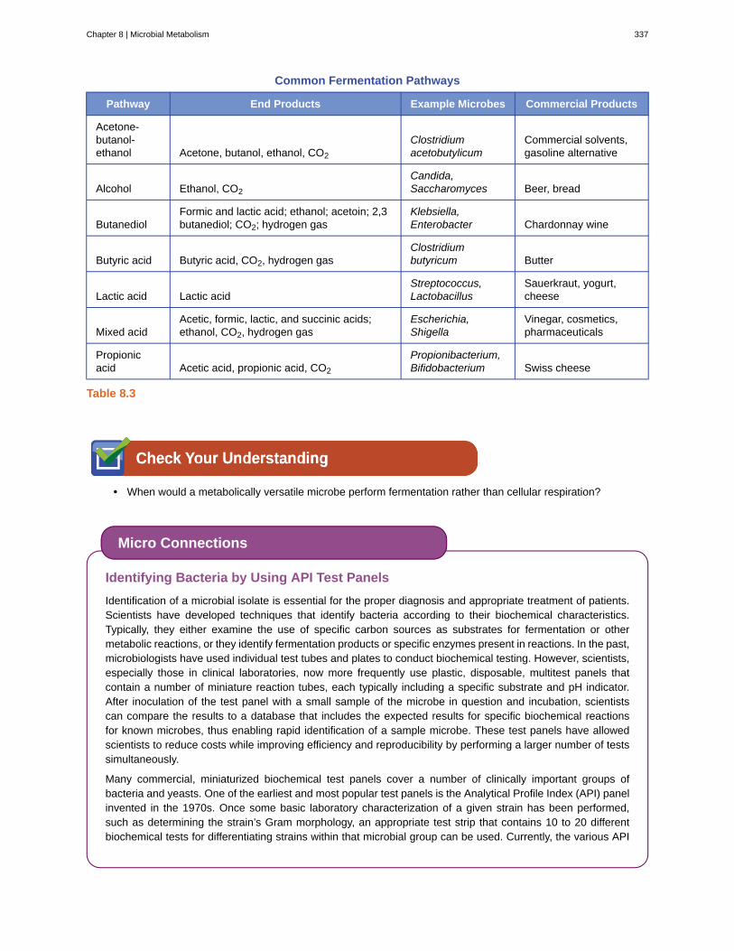
Common Fermentation Pathways
Pathway End Products Example Microbes Commercial Products
Acetone-butanol-ethanol Acetone, butanol, ethanol, CO2
Clostridiumacetobutylicum
Commercial solvents,gasoline alternative
Alcohol Ethanol, CO2
Candida,Saccharomyces Beer, bread
ButanediolFormic and lactic acid; ethanol; acetoin; 2,3butanediol; CO2; hydrogen gas
Klebsiella,Enterobacter Chardonnay wine
Butyric acid Butyric acid, CO2, hydrogen gasClostridiumbutyricum Butter
Lactic acid Lactic acidStreptococcus,Lactobacillus
Sauerkraut, yogurt,cheese
Mixed acidAcetic, formic, lactic, and succinic acids;ethanol, CO2, hydrogen gas
Escherichia,Shigella
Vinegar, cosmetics,pharmaceuticals
Propionicacid Acetic acid, propionic acid, CO2
Propionibacterium,Bifidobacterium Swiss cheese
Table 8.3
• When would a metabolically versatile microbe perform fermentation rather than cellular respiration?
Identifying Bacteria by Using API Test Panels
Identification of a microbial isolate is essential for the proper diagnosis and appropriate treatment of patients.Scientists have developed techniques that identify bacteria according to their biochemical characteristics.Typically, they either examine the use of specific carbon sources as substrates for fermentation or othermetabolic reactions, or they identify fermentation products or specific enzymes present in reactions. In the past,microbiologists have used individual test tubes and plates to conduct biochemical testing. However, scientists,especially those in clinical laboratories, now more frequently use plastic, disposable, multitest panels thatcontain a number of miniature reaction tubes, each typically including a specific substrate and pH indicator.After inoculation of the test panel with a small sample of the microbe in question and incubation, scientistscan compare the results to a database that includes the expected results for specific biochemical reactionsfor known microbes, thus enabling rapid identification of a sample microbe. These test panels have allowedscientists to reduce costs while improving efficiency and reproducibility by performing a larger number of testssimultaneously.
Many commercial, miniaturized biochemical test panels cover a number of clinically important groups ofbacteria and yeasts. One of the earliest and most popular test panels is the Analytical Profile Index (API) panelinvented in the 1970s. Once some basic laboratory characterization of a given strain has been performed,such as determining the strain’s Gram morphology, an appropriate test strip that contains 10 to 20 differentbiochemical tests for differentiating strains within that microbial group can be used. Currently, the various API
Micro Connections
Chapter 8 | Microbial Metabolism 337

strips can be used to quickly and easily identify more than 600 species of bacteria, both aerobic and anaerobic,and approximately 100 different types of yeasts. Based on the colors of the reactions when metabolic endproducts are present, due to the presence of pH indicators, a metabolic profile is created from the results(Figure 8.18). Microbiologists can then compare the sample’s profile to the database to identify the specificmicrobe.
Figure 8.18 The API 20NE test strip is used to identify specific strains of gram-negative bacteria outside theEnterobacteriaceae. Here is an API 20NE test strip result for Photobacterium damselae ssp. piscicida.
Part 2
Many of Hannah’s symptoms are consistent with several different infections, including influenza andpneumonia. However, her sluggish reflexes along with her light sensitivity and stiff neck suggest somepossible involvement of the central nervous system, perhaps indicating meningitis. Meningitis is an infectionof the cerebrospinal fluid (CSF) around the brain and spinal cord that causes inflammation of the meninges,the protective layers covering the brain. Meningitis can be caused by viruses, bacteria, or fungi. Althoughall forms of meningitis are serious, bacterial meningitis is particularly serious. Bacterial meningitis may becaused by several different bacteria, but the bacterium Neisseria meningitidis, a gram-negative, bean-shapeddiplococcus, is a common cause and leads to death within 1 to 2 days in 5% to 10% of patients.
Given the potential seriousness of Hannah’s conditions, her physician advised her parents to take her to thehospital in the Gambian capital of Banjul and there have her tested and treated for possible meningitis. After a3-hour drive to the hospital, Hannah was immediately admitted. Physicians took a blood sample and performeda lumbar puncture to test her CSF. They also immediately started her on a course of the antibiotic ceftriaxone,the drug of choice for treatment of meningitis caused by N. meningitidis, without waiting for laboratory testresults.
• How might biochemical testing be used to confirm the identity of N. meningitidis?
• Why did Hannah’s doctors decide to administer antibiotics without waiting for the test results?
Jump to the next Clinical Focus box. Go back to the previous Clinical Focus box.
8.5 Catabolism of Lipids and Proteins
Learning Objectives• Describe how lipids are catabolized
• Describe how lipid catabolism can be used to identify microbes
• Describe how proteins are catabolized
• Describe how protein catabolism can be used to identify bacteria
Previous sections have discussed the catabolism of glucose, which provides energy to living cells, as well as how
Clinical Focus
338 Chapter 8 | Microbial Metabolism
This OpenStax book is available for free at http://cnx.org/content/col12087/1.5

polysaccharides like glycogen, starch, and cellulose are degraded to glucose monomers. But microbes consumemore than just carbohydrates for food. In fact, the microbial world is known for its ability to degrade a wide rangeof molecules, both naturally occurring and those made by human processes, for use as carbon sources. In thissection, we will see that the pathways for both lipid and protein catabolism connect to those used for carbohydratecatabolism, eventually leading into glycolysis, the transition reaction, and the Krebs cycle pathways. Metabolicpathways should be considered to be porous—that is, substances enter from other pathways, and intermediates leavefor other pathways. These pathways are not closed systems. Many of the substrates, intermediates, and products in aparticular pathway are reactants in other pathways.
Lipid Catabolism
Triglycerides are a form of long-term energy storage in animals. They are made of glycerol and three fatty acids(see Figure 7.12). Phospholipids compose the cell and organelle membranes of all organisms except the archaea.Phospholipid structure is similar to triglycerides except that one of the fatty acids is replaced by a phosphorylatedhead group (see Figure 7.13). Triglycerides and phospholipids are broken down first by releasing fatty acid chains(and/or the phosphorylated head group, in the case of phospholipids) from the three-carbon glycerol backbone.The reactions breaking down triglycerides are catalyzed by lipases and those involving phospholipids are catalyzedby phospholipases. These enzymes contribute to the virulence of certain microbes, such as the bacteriumStaphylococcus aureus and the fungus Cryptococcus neoformans. These microbes use phospholipases to destroylipids and phospholipids in host cells and then use the catabolic products for energy (see Virulence Factors ofBacterial and Viral Pathogens).
The resulting products of lipid catabolism, glycerol and fatty acids, can be further degraded. Glycerol can bephosphorylated to glycerol-3-phosphate and easily converted to glyceraldehyde 3-phosphate, which continuesthrough glycolysis. The released fatty acids are catabolized in a process called β-oxidation, which sequentiallyremoves two-carbon acetyl groups from the ends of fatty acid chains, reducing NAD+ and FAD to produce NADHand FADH2, respectively, whose electrons can be used to make ATP by oxidative phosphorylation. The acetyl groupsproduced during β-oxidation are carried by coenzyme A to the Krebs cycle, and their movement through this cycleresults in their degradation to CO2, producing ATP by substrate-level phosphorylation and additional NADH andFADH2 molecules (see Appendix C for a detailed illustration of β-oxidation).
Other types of lipids can also be degraded by certain microbes. For example, the ability of certain pathogens, likeMycobacterium tuberculosis, to degrade cholesterol contributes to their virulence. The side chains of cholesterolcan be easily removed enzymatically, but degradation of the remaining fused rings is more problematic. The fourfused rings are sequentially broken in a multistep process facilitated by specific enzymes, and the resulting products,including pyruvate, can be further catabolized in the Krebs cycle.
• How can lipases and phospholipases contribute to virulence in microbes?
Protein Catabolism
Proteins are degraded through the concerted action of a variety of microbial protease enzymes. Extracellularproteases cut proteins internally at specific amino acid sequences, breaking them down into smaller peptides that canthen be taken up by cells. Some clinically important pathogens can be identified by their ability to produce a specifictype of extracellular protease. For example, the production of the extracellular protease gelatinase by members of thegenera Proteus and Serratia can be used to distinguish them from other gram-negative enteric bacteria. Followinginoculation and growth of microbes in gelatin broth, degradation of the gelatin protein due to gelatinase productionprevents solidification of gelatin when refrigerated. Other pathogens can be distinguished by their ability to degradecasein, the main protein found in milk. When grown on skim milk agar, production of the extracellular protease
Chapter 8 | Microbial Metabolism 339

caseinase causes degradation of casein, which appears as a zone of clearing around the microbial growth. Caseinaseproduction by the opportunist pathogen Pseudomonas aeruginosa can be used to distinguish it from other relatedgram-negative bacteria.
After extracellular protease degradation and uptake of peptides in the cell, the peptides can then be broken downfurther into individual amino acids by additional intracellular proteases, and each amino acid can be enzymaticallydeaminated to remove the amino group. The remaining molecules can then enter the transition reaction or the Krebscycle.
• How can protein catabolism help identify microbes?
Part 3
Because bacterial meningitis progresses so rapidly, Hannah’s doctors had decided to treat her aggressivelywith antibiotics, based on empirical observation of her symptoms. However, laboratory testing to confirm thecause of Hannah’s meningitis was still important for several reasons. N. meningitidis is an infectious pathogenthat can be spread from person to person through close contact; therefore, if tests confirm N. meningitidis asthe cause of Hannah’s symptoms, Hannah’s parents and others who came into close contact with her mightneed to be vaccinated or receive prophylactic antibiotics to lower their risk of contracting the disease. On theother hand, if it turns out that N. meningitidis is not the cause, Hannah’s doctors might need to change hertreatment.
The clinical laboratory performed a Gram stain on Hannah’s blood and CSF samples. The Gram stainshowed the presence of a bean-shaped gram-negative diplococcus. The technician in the hospital lab culturedHannah’s blood sample on both blood agar and chocolate agar, and the bacterium that grew on both mediaformed gray, nonhemolytic colonies. Next, he performed an oxidase test on this bacterium and determined thatit was oxidase positive. Last, he examined the repertoire of sugars that the bacterium could use as a carbonsource and found that the bacterium was positive for glucose and maltose use but negative for lactose andsucrose use. All of these test results are consistent with characteristics of N. meningitidis.
• What do these test results tell us about the metabolic pathways of N. meningitidis?
• Why do you think that the hospital used these biochemical tests for identification in lieu of molecularanalysis by DNA testing?
Jump to the next Clinical Focus box. Go back to the previous Clinical Focus box.
8.6 Photosynthesis
Learning Objectives• Describe the function and locations of photosynthetic pigments in eukaryotes and prokaryotes
• Describe the major products of the light-dependent and light-independent reactions
• Describe the reactions that produce glucose in a photosynthetic cell
• Compare and contrast cyclic and noncyclic photophosphorylation
Heterotrophic organisms ranging from E. coli to humans rely on the chemical energy found mainly in carbohydrate
Clinical Focus
340 Chapter 8 | Microbial Metabolism
This OpenStax book is available for free at http://cnx.org/content/col12087/1.5

molecules. Many of these carbohydrates are produced by photosynthesis, the biochemical process by whichphototrophic organisms convert solar energy (sunlight) into chemical energy. Although photosynthesis is mostcommonly associated with plants, microbial photosynthesis is also a significant supplier of chemical energy, fuelingmany diverse ecosystems. In this section, we will focus on microbial photosynthesis.
Photosynthesis takes place in two sequential stages: the light-dependent reactions and the light-independent reactions(Figure 8.19). In the light-dependent reactions, energy from sunlight is absorbed by pigment molecules inphotosynthetic membranes and converted into stored chemical energy. In the light-independent reactions, thechemical energy produced by the light-dependent reactions is used to drive the assembly of sugar moleculesusing CO2; however, these reactions are still light dependent because the products of the light-dependent reactionsnecessary for driving them are short-lived. The light-dependent reactions produce ATP and either NADPH or NADHto temporarily store energy. These energy carriers are used in the light-independent reactions to drive the energeticallyunfavorable process of “fixing” inorganic CO2 in an organic form, sugar.
Figure 8.19 The light-dependent reactions of photosynthesis (left) convert light energy into chemical energy, formingATP and NADPH. These products are used by the light-independent reactions to fix CO2, producing organic carbonmolecules.
Photosynthetic Structures in Eukaryotes and Prokaryotes
In all phototrophic eukaryotes, photosynthesis takes place inside a chloroplast, an organelle that arose in eukaryotesby endosymbiosis of a photosynthetic bacterium (see Unique Characteristics of Eukaryotic Cells). Thesechloroplasts are enclosed by a double membrane with inner and outer layers. Within the chloroplast is a thirdmembrane that forms stacked, disc-shaped photosynthetic structures called thylakoids (Figure 8.20). A stack ofthylakoids is called a granum, and the space surrounding the granum within the chloroplast is called stroma.
Photosynthetic membranes in prokaryotes, by contrast, are not organized into distinct membrane-enclosed organelles;rather, they are infolded regions of the plasma membrane. In cyanobacteria, for example, these infolded regions arealso referred to as thylakoids. In either case, embedded within the thylakoid membranes or other photosyntheticbacterial membranes are photosynthetic pigment molecules organized into one or more photosystems, where lightenergy is actually converted into chemical energy.
Photosynthetic pigments within the photosynthetic membranes are organized into photosystems, each of which iscomposed of a light-harvesting (antennae) complex and a reaction center. The light-harvesting complex consists of
Chapter 8 | Microbial Metabolism 341

multiple proteins and associated pigments that each may absorb light energy and, thus, become excited. This energy istransferred from one pigment molecule to another until eventually (after about a millionth of a second) it is deliveredto the reaction center. Up to this point, only energy—not electrons—has been transferred between molecules. Thereaction center contains a pigment molecule that can undergo oxidation upon excitation, actually giving up anelectron. It is at this step in photosynthesis that light energy is converted into an excited electron.
Different kinds of light-harvesting pigments absorb unique patterns of wavelengths (colors) of visible light. Pigmentsreflect or transmit the wavelengths they cannot absorb, making them appear the corresponding color. Examples ofphotosynthetic pigments (molecules used to absorb solar energy) are bacteriochlorophylls (green, purple, or red),carotenoids (orange, red, or yellow), chlorophylls (green), phycocyanins (blue), and phycoerythrins (red). By havingmixtures of pigments, an organism can absorb energy from more wavelengths. Because photosynthetic bacteriacommonly grow in competition for sunlight, each type of photosynthetic bacteria is optimized for harvesting thewavelengths of light to which it is commonly exposed, leading to stratification of microbial communities in aquaticand soil ecosystems by light quality and penetration.
Once the light harvesting complex transfers the energy to the reaction center, the reaction center delivers itshigh-energy electrons, one by one, to an electron carrier in an electron transport system, and electron transferthrough the ETS is initiated. The ETS is similar to that used in cellular respiration and is embedded within thephotosynthetic membrane. Ultimately, the electron is used to produce NADH or NADPH. The electrochemicalgradient that forms across the photosynthetic membrane is used to generate ATP by chemiosmosis through the processof photophosphorylation, another example of oxidative phosphorylation (Figure 8.21).
Figure 8.20 (a) Photosynthesis in eukaryotes takes place in chloroplasts, which contain thylakoids stacked intograna. (b) A photosynthetic prokaryote has infolded regions of the plasma membrane that function like thylakoids.(credit: scale bar data from Matt Russell.)
342 Chapter 8 | Microbial Metabolism
This OpenStax book is available for free at http://cnx.org/content/col12087/1.5

Figure 8.21 This figure summarizes how a photosystem works. Light harvesting (LH) pigments absorb light energy,converting it to chemical energy. The energy is passed from one LH pigment to another until it reaches a reactioncenter (RC) pigment, exciting an electron. This high-energy electron is lost from the RC pigment and passed throughan electron transport system (ETS), ultimately producing NADH or NADPH and ATP. A reduced molecule (H2A)donates an electron, replacing electrons to the electron-deficient RC pigment.
• In a phototrophic eukaryote, where does photosynthesis take place?
Oxygenic and Anoxygenic Photosynthesis
For photosynthesis to continue, the electron lost from the reaction center pigment must be replaced. The sourceof this electron (H2A) differentiates the oxygenic photosynthesis of plants and cyanobacteria from anoxygenicphotosynthesis carried out by other types of bacterial phototrophs (Figure 8.22). In oxygenic photosynthesis, H2Ois split and supplies the electron to the reaction center. Because oxygen is generated as a byproduct and is released,this type of photosynthesis is referred to as oxygenic photosynthesis. However, when other reduced compounds serveas the electron donor, oxygen is not generated; these types of photosynthesis are called anoxygenic photosynthesis.
Hydrogen sulfide (H2S) or thiosulfate ⎛⎝S2 O3
2−⎞⎠ can serve as the electron donor, generating elemental sulfur and
sulfate ⎛⎝SO4
2−⎞⎠ ions, respectively, as a result.
Photosystems have been classified into two types: photosystem I (PSI) and photosystem II (PSII) (Figure 8.23).Cyanobacteria and plant chloroplasts have both photosystems, whereas anoxygenic photosynthetic bacteria use onlyone of the photosystems. Both photosystems are excited by light energy simultaneously. If the cell requires bothATP and NADPH for biosynthesis, then it will carry out noncyclic photophosphorylation. Upon passing of the PSIIreaction center electron to the ETS that connects PSII and PSI, the lost electron from the PSII reaction center isreplaced by the splitting of water. The excited PSI reaction center electron is used to reduce NADP+ to NADPH andis replaced by the electron exiting the ETS. The flow of electrons in this way is called the Z-scheme.
If a cell’s need for ATP is significantly greater than its need for NADPH, it may bypass the production of reducingpower through cyclic photophosphorylation. Only PSI is used during cyclic photophosphorylation; the high-energyelectron of the PSI reaction center is passed to an ETS carrier and then ultimately returns to the oxidized PSI reactioncenter pigment, thereby reducing it.
Chapter 8 | Microbial Metabolism 343

Figure 8.22 Eukaryotes and cyanobacteria carry out oxygenic photosynthesis, producing oxygen, whereas otherbacteria carry out anoxygenic photosynthesis, which does not produce oxygen.
344 Chapter 8 | Microbial Metabolism
This OpenStax book is available for free at http://cnx.org/content/col12087/1.5

Figure 8.23 (a) PSI and PSII are found on the thylakoid membrane. The high-energy electron from PSII is passed toan ETS, which generates a proton motive force for ATP synthesis by chemiosmosis, and ultimately replaces theelectron lost by the PSI reaction center. The PSI reaction center electron is used to make NADPH. (b) When bothATP and NADPH are required, noncyclic photophosphorylation (in cyanobacteria and plants) provides both. Theelectron flow described here is referred to as the Z-scheme (shown in yellow in [a]). When the cell’s ATP needsoutweigh those for NADPH, cyanobacteria and plants will use only PSI, and its reaction center electron is passed tothe ETS to generate a proton motive force used for ATP synthesis.
Chapter 8 | Microbial Metabolism 345

• Why would a photosynthetic bacterium have different pigments?
Light-Independent Reactions
After the energy from the sun is converted into chemical energy and temporarily stored in ATP and NADPHmolecules (having lifespans of millionths of a second), photoautotrophs have the fuel needed to build multicarboncarbohydrate molecules, which can survive for hundreds of millions of years, for long-term energy storage. Thecarbon comes from CO2, the gas that is a waste product of cellular respiration.
The Calvin-Benson cycle (named for Melvin Calvin [1911–1997] and Andrew Benson [1917–2015]), thebiochemical pathway used for fixation of CO2, is located within the cytoplasm of photosynthetic bacteria and in thestroma of eukaryotic chloroplasts. The light-independent reactions of the Calvin cycle can be organized into threebasic stages: fixation, reduction, and regeneration (see Appendix C for a detailed illustration of the Calvin cycle).
• Fixation: The enzyme ribulose bisphosphate carboxylase (RuBisCO) catalyzes the addition of a CO2 toribulose bisphosphate (RuBP). This results in the production of 3-phosphoglycerate (3-PGA).
• Reduction: Six molecules of both ATP and NADPH (from the light-dependent reactions) are used to convert3-PGA into glyceraldehyde 3-phosphate (G3P). Some G3P is then used to build glucose.
• Regeneration: The remaining G3P not used to synthesize glucose is used to regenerate RuBP, enabling thesystem to continue CO2 fixation. Three more molecules of ATP are used in these regeneration reactions.
The Calvin cycle is used extensively by plants and photoautotrophic bacteria, and the enzyme RuBisCO is said tobe the most plentiful enzyme on earth, composing 30%–50% of the total soluble protein in plant chloroplasts.[1]
However, besides its prevalent use in photoautotrophs, the Calvin cycle is also used by many nonphotosyntheticchemoautotrophs to fix CO2. Additionally, other bacteria and archaea use alternative systems for CO2 fixation.Although most bacteria using Calvin cycle alternatives are chemoautotrophic, certain green sulfur photoautotrophicbacteria have been also shown to use an alternative CO2 fixation pathway.
• Describe the three stages of the Calvin cycle.
8.7 Biogeochemical Cycles
Learning Objectives• Define and describe the importance of microorganisms in the biogeochemical cycles of carbon, nitrogen, and
sulfur
• Define and give an example of bioremediation
Energy flows directionally through ecosystems, entering as sunlight for phototrophs or as inorganic molecules forchemoautotrophs. The six most common elements associated with organic molecules—carbon, hydrogen, nitrogen,oxygen, phosphorus, and sulfur—take a variety of chemical forms and may exist for long periods in the atmosphere,
1. A. Dhingra et al. “Enhanced Translation of a Chloroplast-Expressed RbcS Gene Restores Small Subunit Levels and Photosynthesis in
Nuclear RbcS Antisense Plants.” Proceedings of the National Academy of Sciences of the United States of America 101 no. 16
(2004):6315–6320.
346 Chapter 8 | Microbial Metabolism
This OpenStax book is available for free at http://cnx.org/content/col12087/1.5

on land, in water, or beneath earth’s surface. Geologic processes, such as erosion, water drainage, the movementof the continental plates, and weathering, all are involved in the cycling of elements on earth. Because geologyand chemistry have major roles in the study of this process, the recycling of inorganic matter between livingorganisms and their nonliving environment is called a biogeochemical cycle. Here, we will focus on the function ofmicroorganisms in these cycles, which play roles at each step, most frequently interconverting oxidized versions ofmolecules with reduced ones.
Carbon Cycle
Carbon is one of the most important elements to living organisms, as shown by its abundance and presence inall organic molecules. The carbon cycle exemplifies the connection between organisms in various ecosystems.Carbon is exchanged between heterotrophs and autotrophs within and between ecosystems primarily by way ofatmospheric CO2, a fully oxidized version of carbon that serves as the basic building block that autotrophs use to buildmulticarbon, high-energy organic molecules such as glucose. Photoautotrophs and chemoautotrophs harness energyfrom the sun and from inorganic chemical compounds, respectively, to covalently bond carbon atoms together intoreduced organic compounds whose energy can be later accessed through the processes of respiration and fermentation(Figure 8.24).
Overall, there is a constant exchange of CO2 between the heterotrophs (which produce CO2 as a result of respirationor fermentation) and the autotrophs (which use the CO2 for fixation). Autotrophs also respire or ferment, consumingthe organic molecules they form; they do not fix carbon for heterotrophs, but rather use it for their own metabolicneeds.
Bacteria and archaea that use methane as their carbon source are called methanotrophs. Reduced one-carboncompounds like methane accumulate in certain anaerobic environments when CO2 is used as a terminal electronacceptor in anaerobic respiration by archaea called methanogens. Some methanogens also ferment acetate (twocarbons) to produce methane and CO2. Methane accumulation due to methanogenesis occurs in both natural anaerobicsoil and aquatic environments; methane accumulation also occurs as a result of animal husbandry becausemethanogens are members of the normal microbiota of ruminants. Environmental methane accumulation due tomethanogenesis is of consequence because it is a strong greenhouse gas, and methanotrophs help to reduceatmospheric methane levels.
Chapter 8 | Microbial Metabolism 347

Figure 8.24 This figure summarizes the carbon cycle. Eukaryotes participate in aerobic respiration, fermentation,and oxygenic photosynthesis. Prokaryotes participate in all the steps shown. (credit: modification of work by NOAA)
• Describe the interaction between heterotrophs and autotrophs in the carbon cycle.
Nitrogen Cycle
Many biological macromolecules, including proteins and nucleic acids, contain nitrogen; however, getting nitrogeninto living organisms is difficult. Prokaryotes play essential roles in the nitrogen cycle (Figure 8.25), transformingnitrogen between various forms for their own needs, benefiting other organisms indirectly. Plants and phytoplanktoncannot incorporate nitrogen from the atmosphere (where it exists as tightly bonded, triple covalent N2), even thoughthis molecule composes approximately 78% of the atmosphere. Nitrogen enters the living world through free-living and symbiotic bacteria, which incorporate nitrogen into their macromolecules through specialized biochemicalpathways called nitrogen fixation. Cyanobacteria in aquatic ecosystems fix inorganic nitrogen (from nitrogen gas)into ammonia (NH3) that can be easily incorporated into biological macromolecules. Rhizobium bacteria (Figure8.1) also fix nitrogen and live symbiotically in the root nodules of legumes (such as beans, peanuts, and peas),providing them with needed organic nitrogen while receiving fixed carbon as sugar in exchange. Free-living bacteria,such as members of the genus Azotobacter, are also able to fix nitrogen.
The nitrogen that enters living systems by nitrogen fixation is eventually converted from organic nitrogen back intonitrogen gas by microbes through three steps: ammonification, nitrification, and denitrification. In terrestrial systems,
348 Chapter 8 | Microbial Metabolism
This OpenStax book is available for free at http://cnx.org/content/col12087/1.5

the first step is the ammonification process, in which certain bacteria and fungi convert nitrogenous waste fromliving animals or from the remains of dead organisms into ammonia (NH3). This ammonia is then oxidized to nitrite⎛⎝NO2
− ⎞⎠, then to nitrate ⎛
⎝NO3− ⎞
⎠, by nitrifying soil bacteria such as members of the genus Nitrosomonas, through
the process of nitrification. Last, the process of denitrification occurs, whereby soil bacteria, such as members of thegenera Pseudomonas and Clostridium, use nitrate as a terminal electron acceptor in anaerobic respiration, convertingit into nitrogen gas that reenters the atmosphere. A similar process occurs in the marine nitrogen cycle, where thesethree processes are performed by marine bacteria and archaea.
Human activity releases nitrogen into the environment by the use of artificial fertilizers that contain nitrogen andphosphorus compounds, which are then washed into lakes, rivers, and streams by surface runoff. A major effectfrom fertilizer runoff is saltwater and freshwater eutrophication, in which nutrient runoff causes the overgrowthand subsequent death of aquatic algae, making water sources anaerobic and inhospitable for the survival of aquaticorganisms.
Figure 8.25 This figure summarizes the nitrogen cycle. Note that specific groups of prokaryotes each participate inevery step in the cycle. (credit: modification of work by NOAA)
• What are the three steps of the nitrogen cycle?
Chapter 8 | Microbial Metabolism 349

To learn more about the nitrogen cycle, visit the PBS (https://openstax.org/l/22nitrogencycle) website.
Sulfur Cycle
Sulfur is an essential element for the macromolecules of living organisms. As part of the amino acids cysteineand methionine, it is involved in the formation of proteins. It is also found in several vitamins necessary forthe synthesis of important biological molecules like coenzyme A. Several groups of microbes are responsible forcarrying out processes involved in the sulfur cycle (Figure 8.26). Anoxygenic photosynthetic bacteria as well aschemoautotrophic archaea and bacteria use hydrogen sulfide as an electron donor, oxidizing it first to elemental sulfur
(S0), then to sulfate ⎛⎝SO4
2 − ⎞⎠. This leads to stratification of hydrogen sulfide in soil, with levels increasing at deeper,
more anaerobic depths.
Many bacteria and plants can use sulfate as a sulfur source. Decomposition dead organisms by fungi and bacteriaremove sulfur groups from amino acids, producing hydrogen sulfide, returning inorganic sulfur to the environment.
Figure 8.26 This figure summarizes the sulfur cycle. Note that specific groups of prokaryotes each may participatein every step in the cycle. (credit: modification of work by NOAA)
Link to Learning
350 Chapter 8 | Microbial Metabolism
This OpenStax book is available for free at http://cnx.org/content/col12087/1.5

• Which groups of microbes carry out the sulfur cycle?
Other Biogeochemical Cycles
Beyond their involvement in the carbon, nitrogen, and sulfur cycles, prokaryotes are involved in other biogeochemicalcycles as well. Like the carbon, nitrogen, and sulfur cycles, several of these additional biogeochemical cycles, suchas the iron (Fe), manganese (Mn), and chromium (Cr) cycles, also involve redox chemistry, with prokaryotes playingroles in both oxidation and reduction. Several other elements undergo chemical cycles that do not involve redoxchemistry. Examples of these are phosphorus (P), calcium (Ca), and silica (Si) cycles. The cycling of these elementsis particularly important in oceans because large quantities of these elements are incorporated into the exoskeletonsof marine organisms. These biogeochemical cycles do not involve redox chemistry but instead involve fluctuationsin the solubility of compounds containing calcium, phosphorous, and silica. The overgrowth of naturally occurringmicrobial communities is typically limited by the availability of nitrogen (as previously mentioned), phosphorus, andiron. Human activities introducing excessive amounts of iron, nitrogen, or phosphorus (typically from detergents)may lead to eutrophication.
Bioremediation
Microbial bioremediation leverages microbial metabolism to remove xenobiotics or other pollutants. Xenobioticsare compounds synthesized by humans and introduced into the environment in much higher concentrations thanwould naturally occur. Such environmental contamination may involve adhesives, dyes, flame retardants, lubricants,oil and petroleum products, organic solvents, pesticides, and products of the combustion of gasoline and oil. Manyxenobiotics resist breakdown, and some accumulate in the food chain after being consumed or absorbed by fishand wildlife, which, in turn, may be eaten by humans. Of particular concern are contaminants like polycyclicaromatic hydrocarbon (PAH), a carcinogenic xenobiotic found in crude oil, and trichloroethylene (TCE), a commongroundwater contaminant.
Bioremediation processes can be categorized as in situ or ex situ. Bioremediation conducted at the site ofcontamination is called in situ bioremediation and does not involve movement of contaminated material. In contrast,ex situ bioremediation involves the removal of contaminated material from the original site so that it can be treatedelsewhere, typically in a large, lined pit where conditions are optimized for degradation of the contaminant.
Some bioremediation processes rely on microorganisms that are indigenous to the contaminated site or material.Enhanced bioremediation techniques, which may be applied to either in situ or ex situ processing, involve theaddition of nutrients and/or air to encourage the growth of pollution-degrading microbes; they may also involve theaddition of non-native microbes known for their ability to degrade contaminants. For example, certain bacteria ofthe genera Rhodococcus and Pseudomonas are known for their ability to degrade many environmental contaminants,including aromatic compounds like those found in oil, down to CO2. The genes encoding their degradatory enzymesare commonly found on plasmids. Others, like Alcanivorax borkumensis, produce surfactants that are useful inthe solubilization of the hydrophobic molecules found in oil, making them more accessible to other microbes fordegradation.
• Compare and contrast the benefits of in situ and ex situ bioremediation.
Chapter 8 | Microbial Metabolism 351
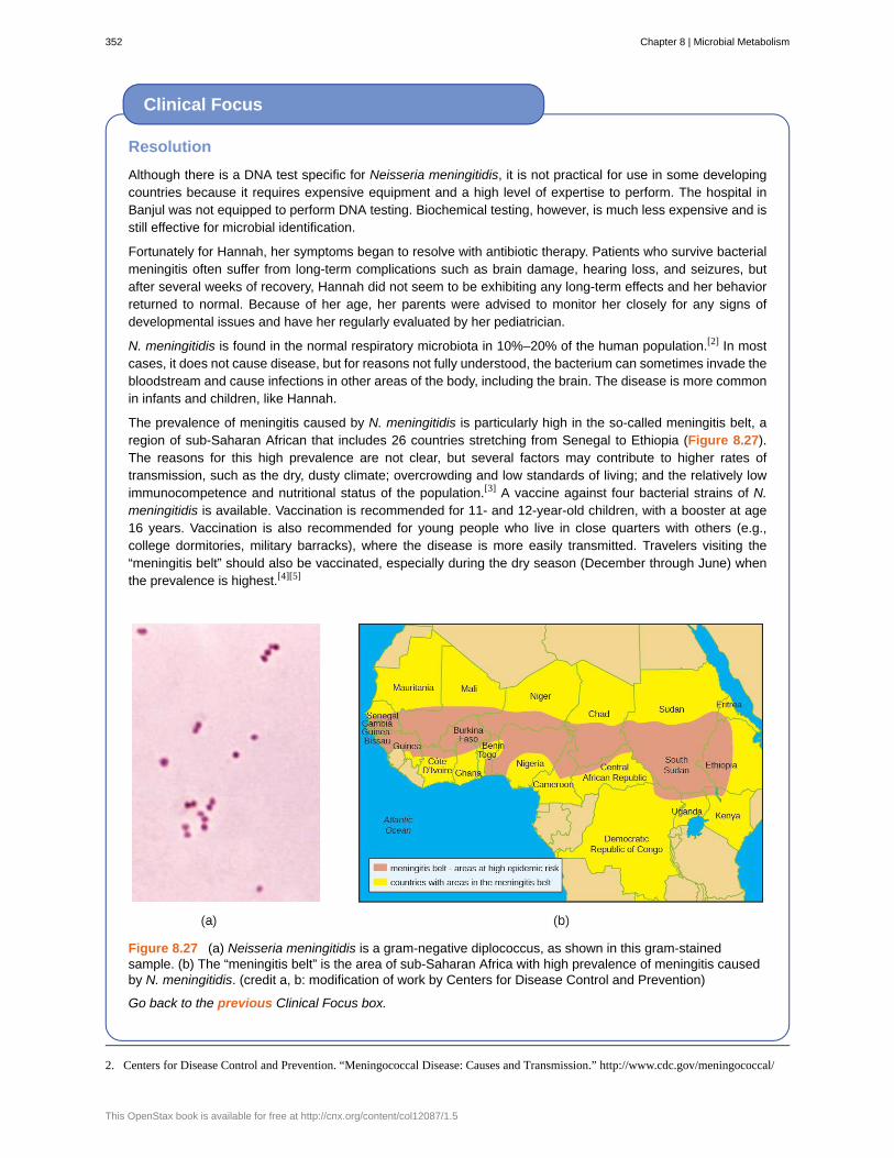
Resolution
Although there is a DNA test specific for Neisseria meningitidis, it is not practical for use in some developingcountries because it requires expensive equipment and a high level of expertise to perform. The hospital inBanjul was not equipped to perform DNA testing. Biochemical testing, however, is much less expensive and isstill effective for microbial identification.
Fortunately for Hannah, her symptoms began to resolve with antibiotic therapy. Patients who survive bacterialmeningitis often suffer from long-term complications such as brain damage, hearing loss, and seizures, butafter several weeks of recovery, Hannah did not seem to be exhibiting any long-term effects and her behaviorreturned to normal. Because of her age, her parents were advised to monitor her closely for any signs ofdevelopmental issues and have her regularly evaluated by her pediatrician.
N. meningitidis is found in the normal respiratory microbiota in 10%–20% of the human population.[2] In mostcases, it does not cause disease, but for reasons not fully understood, the bacterium can sometimes invade thebloodstream and cause infections in other areas of the body, including the brain. The disease is more commonin infants and children, like Hannah.
The prevalence of meningitis caused by N. meningitidis is particularly high in the so-called meningitis belt, aregion of sub-Saharan African that includes 26 countries stretching from Senegal to Ethiopia (Figure 8.27).The reasons for this high prevalence are not clear, but several factors may contribute to higher rates oftransmission, such as the dry, dusty climate; overcrowding and low standards of living; and the relatively lowimmunocompetence and nutritional status of the population.[3] A vaccine against four bacterial strains of N.meningitidis is available. Vaccination is recommended for 11- and 12-year-old children, with a booster at age16 years. Vaccination is also recommended for young people who live in close quarters with others (e.g.,college dormitories, military barracks), where the disease is more easily transmitted. Travelers visiting the“meningitis belt” should also be vaccinated, especially during the dry season (December through June) whenthe prevalence is highest.[4][5]
Figure 8.27 (a) Neisseria meningitidis is a gram-negative diplococcus, as shown in this gram-stainedsample. (b) The “meningitis belt” is the area of sub-Saharan Africa with high prevalence of meningitis causedby N. meningitidis. (credit a, b: modification of work by Centers for Disease Control and Prevention)
Go back to the previous Clinical Focus box.
Clinical Focus
2. Centers for Disease Control and Prevention. “Meningococcal Disease: Causes and Transmission.” http://www.cdc.gov/meningococcal/
352 Chapter 8 | Microbial Metabolism
This OpenStax book is available for free at http://cnx.org/content/col12087/1.5

Summary
8.1 Energy, Matter, and Enzymes• Metabolism includes chemical reactions that break down complex molecules (catabolism) and those that
build complex molecules (anabolism).
• Organisms may be classified according to their source of carbon. Autotrophs convert inorganic carbondioxide into organic carbon; heterotrophs use fixed organic carbon compounds.
• Organisms may also be classified according to their energy source. Phototrophs obtain their energy fromlight. Chemotrophs get their energy from chemical compounds. Organotrophs use organic molecules, andlithotrophs use inorganic chemicals.
• Cellular electron carriers accept high-energy electrons from foods and later serve as electron donors insubsequent redox reactions. FAD/FADH2, NAD+/NADH, and NADP+/NADPH are important electroncarriers.
• Adenosine triphosphate (ATP) serves as the energy currency of the cell, safely storing chemical energy inits two high-energy phosphate bonds for later use to drive processes requiring energy.
• Enzymes are biological catalysts that increase the rate of chemical reactions inside cells by lowering theactivation energy required for the reaction to proceed.
• In nature, exergonic reactions do not require energy beyond activation energy to proceed, and they releaseenergy. They may proceed without enzymes, but at a slow rate. Conversely, endergonic reactions requireenergy beyond activation energy to occur. In cells, endergonic reactions are coupled to exergonic reactions,making the combination energetically favorable.
• Substrates bind to the enzyme’s active site. This process typically alters the structures of both the active siteand the substrate, favoring transition-state formation; this is known as induced fit.
• Cofactors are inorganic ions that stabilize enzyme conformation and function. Coenzymes are organicmolecules required for proper enzyme function and are often derived from vitamins. An enzyme lacking acofactor or coenzyme is an apoenzyme; an enzyme with a bound cofactor or coenzyme is a holoenzyme.
• Competitive inhibitors regulate enzymes by binding to an enzyme’s active site, preventing substrate binding.Noncompetitive (allosteric) inhibitors bind to allosteric sites, inducing a conformational change in theenzyme that prevents it from functioning. Feedback inhibition occurs when the product of a metabolicpathway noncompetitively binds to an enzyme early on in the pathway, ultimately preventing the synthesis ofthe product.
8.2 Catabolism of Carbohydrates• Glycolysis is the first step in the breakdown of glucose, resulting in the formation of ATP, which is produced
by substrate-level phosphorylation; NADH; and two pyruvate molecules. Glycolysis does not use oxygenand is not oxygen dependent.
• After glycolysis, a three-carbon pyruvate is decarboxylated to form a two-carbon acetyl group, coupled withthe formation of NADH. The acetyl group is attached to a large carrier compound called coenzyme A.
• After the transition step, coenzyme A transports the two-carbon acetyl to the Krebs cycle, where the twocarbons enter the cycle. Per turn of the cycle, one acetyl group derived from glycolysis is further oxidized,producing three NADH molecules, one FADH2, and one ATP by substrate-level phosphorylation, andreleasing two CO2 molecules.
• The Krebs cycle may be used for other purposes. Many of the intermediates are used to synthesize important
about/causes-transmission.html. Accessed September 12, 2016.
3. Centers for Disease Control and Prevention. “Meningococcal Disease in Other Countries.” http://www.cdc.gov/meningococcal/
global.html. Accessed September 12, 2016.
4. Centers for Disease Control and Prevention. “Health Information for Travelers to the Gambia: Traveler View.” http://wwwnc.cdc.gov/
travel/destinations/traveler/none/the-gambia. Accessed September 12, 2016.
5. Centers for Disease Control and Prevention. “Meningococcal: Who Needs to Be Vaccinated?” http://www.cdc.gov/vaccines/vpd-vac/
mening/who-vaccinate.htm. Accessed September 12, 2016.
Chapter 8 | Microbial Metabolism 353

cellular molecules, including amino acids, chlorophylls, fatty acids, and nucleotides.
8.3 Cellular Respiration• Most ATP generated during the cellular respiration of glucose is made by oxidative phosphorylation.
• An electron transport system (ETS) is composed of a series of membrane-associated protein complexesand associated mobile accessory electron carriers. The ETS is embedded in the cytoplasmic membrane ofprokaryotes and the inner mitochondrial membrane of eukaryotes.
• Each ETS complex has a different redox potential, and electrons move from electron carriers with morenegative redox potential to those with more positive redox potential.
• To carry out aerobic respiration, a cell requires oxygen as the final electron acceptor. A cell also needs acomplete Krebs cycle, an appropriate cytochrome oxidase, and oxygen detoxification enzymes to prevent theharmful effects of oxygen radicals produced during aerobic respiration.
• Organisms performing anaerobic respiration use alternative electron transport system carriers for theultimate transfer of electrons to the final non-oxygen electron acceptors.
• Microbes show great variation in the composition of their electron transport systems, which can be used fordiagnostic purposes to help identify certain pathogens.
• As electrons are passed from NADH and FADH2 through an ETS, the electron loses energy. This energy isstored through the pumping of H+ across the membrane, generating a proton motive force.
• The energy of this proton motive force can be harnessed by allowing hydrogen ions to diffuse back throughthe membrane by chemiosmosis using ATP synthase. As hydrogen ions diffuse through down theirelectrochemical gradient, components of ATP synthase spin, making ATP from ADP and Pi by oxidativephosphorylation.
• Aerobic respiration forms more ATP (a maximum of 34 ATP molecules) during oxidative phosphorylationthan does anaerobic respiration (between one and 32 ATP molecules).
8.4 Fermentation• Fermentation uses an organic molecule as a final electron acceptor to regenerate NAD+ from NADH so that
glycolysis can continue.
• Fermentation does not involve an electron transport system, and no ATP is made by the fermentation processdirectly. Fermenters make very little ATP—only two ATP molecules per glucose molecule during glycolysis.
• Microbial fermentation processes have been used for the production of foods and pharmaceuticals, and for theidentification of microbes.
• During lactic acid fermentation, pyruvate accepts electrons from NADH and is reduced to lactic acid.Microbes performing homolactic fermentation produce only lactic acid as the fermentation product;microbes performing heterolactic fermentation produce a mixture of lactic acid, ethanol and/or acetic acid,and CO2.
• Lactic acid production by the normal microbiota prevents growth of pathogens in certain body regions and isimportant for the health of the gastrointestinal tract.
• During ethanol fermentation, pyruvate is first decarboxylated (releasing CO2) to acetaldehyde, which thenaccepts electrons from NADH, reducing acetaldehyde to ethanol. Ethanol fermentation is used for theproduction of alcoholic beverages, for making bread products rise, and for biofuel production.
• Fermentation products of pathways (e.g., propionic acid fermentation) provide distinctive flavors to foodproducts. Fermentation is used to produce chemical solvents (acetone-butanol-ethanol fermentation) andpharmaceuticals (mixed acid fermentation).
• Specific types of microbes may be distinguished by their fermentation pathways and products. Microbes mayalso be differentiated according to the substrates they are able to ferment.
8.5 Catabolism of Lipids and Proteins• Collectively, microbes have the ability to degrade a wide variety of carbon sources besides carbohydrates,
including lipids and proteins. The catabolic pathways for all of these molecules eventually connect into
354 Chapter 8 | Microbial Metabolism
This OpenStax book is available for free at http://cnx.org/content/col12087/1.5

glycolysis and the Krebs cycle.
• Several types of lipids can be microbially degraded. Triglycerides are degraded by extracellular lipases,releasing fatty acids from the glycerol backbone. Phospholipids are degraded by phospholipases, releasingfatty acids and the phosphorylated head group from the glycerol backbone. Lipases and phospholipases act asvirulence factors for certain pathogenic microbes.
• Fatty acids can be further degraded inside the cell through β-oxidation, which sequentially removes two-carbon acetyl groups from the ends of fatty acid chains.
• Protein degradation involves extracellular proteases that degrade large proteins into smaller peptides.Detection of the extracellular proteases gelatinase and caseinase can be used to differentiate clinically relevantbacteria.
8.6 Photosynthesis• Heterotrophs depend on the carbohydrates produced by autotrophs, many of which are photosynthetic,
converting solar energy into chemical energy.
• Different photosynthetic organisms use different mixtures of photosynthetic pigments, which increase therange of the wavelengths of light an organism can absorb.
• Photosystems (PSI and PSII) each contain a light-harvesting complex, composed of multiple proteins andassociated pigments that absorb light energy. The light-dependent reactions of photosynthesis convert solarenergy into chemical energy, producing ATP and NADPH or NADH to temporarily store this energy.
• In oxygenic photosynthesis, H2O serves as the electron donor to replace the reaction center electron, andoxygen is formed as a byproduct. In anoxygenic photosynthesis, other reduced molecules like H2S orthiosulfate may be used as the electron donor; as such, oxygen is not formed as a byproduct.
• Noncyclic photophosphorylation is used in oxygenic photosynthesis when there is a need for both ATP andNADPH production. If a cell’s needs for ATP outweigh its needs for NADPH, then it may carry out cyclicphotophosphorylation instead, producing only ATP.
• The light-independent reactions of photosynthesis use the ATP and NADPH from the light-dependentreactions to fix CO2 into organic sugar molecules.
8.7 Biogeochemical Cycles• The recycling of inorganic matter between living organisms and their nonliving environment is called a
biogeochemical cycle. Microbes play significant roles in these cycles.
• In the carbon cycle, heterotrophs degrade reduced organic molecule to produce carbon dioxide, whereasautotrophs fix carbon dioxide to produce organics. Methanogens typically form methane by using CO2 asa final electron acceptor during anaerobic respiration; methanotrophs oxidize the methane, using it as theircarbon source.
• In the nitrogen cycle, nitrogen-fixing bacteria convert atmospheric nitrogen into ammonia (ammonification).The ammonia can then be oxidized to nitrite and nitrate (nitrification). Nitrates can then be assimilated byplants. Soil bacteria convert nitrate back to nitrogen gas (denitrification).
• In sulfur cycling, many anoxygenic photosynthesizers and chemoautotrophs use hydrogen sulfide as anelectron donor, producing elemental sulfur and then sulfate; sulfate-reducing bacteria and archaea then usesulfate as a final electron acceptor in anaerobic respiration, converting it back to hydrogen sulfide.
• Human activities that introduce excessive amounts of naturally limited nutrients (like iron, nitrogen, orphosphorus) to aquatic systems may lead to eutrophication.
• Microbial bioremediation is the use of microbial metabolism to remove or degrade xenobiotics and otherenvironmental contaminants and pollutants. Enhanced bioremediation techniques may involve theintroduction of non-native microbes specifically chosen or engineered for their ability to degradecontaminants.
Chapter 8 | Microbial Metabolism 355

Multiple Choice
1. Which of the following is an organism that obtainsits energy from the transfer of electrons originating fromchemical compounds and its carbon from an inorganicsource?
a. chemoautotrophb. chemoheterotrophc. photoheterotrophd. photoautotroph
2. Which of the following molecules is reduced?a. NAD+
b. FADc. O2
d. NADPH
3. Enzymes work by which of the following?a. increasing the activation energyb. reducing the activation energyc. making exergonic reactions endergonicd. making endergonic reactions exergonic
4. To which of the following does a competitiveinhibitor most structurally resemble?
a. the active siteb. the allosteric sitec. the substrated. a coenzyme
5. Which of the following are organic molecules thathelp enzymes work correctly?
a. cofactorsb. coenzymesc. holoenzymesd. apoenzymes
6. During which of the following is ATP not made bysubstrate-level phosphorylation?
a. Embden-Meyerhof pathwayb. Transition reactionc. Krebs cycled. Entner-Doudoroff pathway
7. Which of the following products is made duringEmbden-Meyerhof glycolysis?
a. NAD+b. pyruvatec. CO2
d. two-carbon acetyl
8. During the catabolism of glucose, which of thefollowing is produced only in the Krebs cycle?
a. ATPb. NADHc. NADPHd. FADH2
9. Which of the following is not a name for the cycleresulting in the conversion of a two-carbon acetyl toone ATP, two CO2, one FADH2, and three NADHmolecules?
a. Krebs cycleb. tricarboxylic acid cyclec. Calvin cycled. citric acid cycle
10. Which is the location of electron transports systemsin prokaryotes?
a. the outer mitochondrial membraneb. the cytoplasmc. the inner mitochondrial membraned. the cytoplasmic membrane
11. Which is the source of the energy used to make ATPby oxidative phosphorylation?
a. oxygenb. high-energy phosphate bondsc. the proton motive forced. Pi
12. A cell might perform anaerobic respiration forwhich of the following reasons?
a. It lacks glucose for degradation.b. It lacks the transition reaction to convert
pyruvate to acetyl-CoA.c. It lacks Krebs cycle enzymes for processing
acetyl-CoA to CO2.d. It lacks a cytochrome oxidase for passing
electrons to oxygen.
13. In prokaryotes, which of the following is true?a. As electrons are transferred through an ETS, H+
is pumped out of the cell.b. As electrons are transferred through an ETS, H+
is pumped into the cell.c. As protons are transferred through an ETS,
electrons are pumped out of the cell.d. As protons are transferred through an ETS,
electrons are pumped into the cell.
Review Questions
356 Chapter 8 | Microbial Metabolism
This OpenStax book is available for free at http://cnx.org/content/col12087/1.5

14. Which of the following is not an electron carrierwithin an electron transport system?
a. flavoproteinb. ATP synthasec. ubiquinoned. cytochrome oxidase
15. Which of the following is the purpose offermentation?
a. to make ATPb. to make carbon molecule intermediates for
anabolismc. to make NADHd. to make NAD+
16. Which molecule typically serves as the finalelectron acceptor during fermentation?
a. oxygenb. NAD+
c. pyruvated. CO2
17. Which fermentation product is important formaking bread rise?
a. ethanolb. CO2
c. lactic acidd. hydrogen gas
18. Which of the following is not a commerciallyimportant fermentation product?
a. ethanolb. pyruvatec. butanold. penicillin
19. Which of the following molecules is not producedduring the breakdown of phospholipids?
a. glucoseb. glycerolc. acetyl groupsd. fatty acids
20. Caseinase is which type of enzyme?a. phospholipaseb. lipasec. extracellular proteased. intracellular protease
21. Which of the following is the first step intriglyceride degradation?
a. removal of fatty acidsb. β-oxidationc. breakage of fused ringsd. formation of smaller peptides
22. During the light-dependent reactions, whichmolecule loses an electron?
a. a light-harvesting pigment moleculeb. a reaction center pigment moleculec. NADPHd. 3-phosphoglycerate
23. In prokaryotes, in which direction are hydrogenions pumped by the electron transport system ofphotosynthetic membranes?
a. to the outside of the plasma membraneb. to the inside (cytoplasm) of the cellc. to the stromad. to the intermembrane space of the chloroplast
24. Which of the following does not occur duringcyclic photophosphorylation in cyanobacteria?
a. electron transport through an ETSb. photosystem I usec. ATP synthesisd. NADPH formation
25. Which of the following are two products of thelight-dependent reactions?
a. glucose and NADPHb. NADPH and ATPc. glyceraldehyde 3-phosphate and CO2
d. glucose and oxygen
26. Which of the following is the group of archaeathat can use CO2 as their final electron acceptor duringanaerobic respiration, producing CH4?
a. methylotrophsb. methanotrophsc. methanogensd. anoxygenic photosynthesizers
27. Which of the following processes is not involved inthe conversion of organic nitrogen to nitrogen gas?
a. nitrogen fixationb. ammonificationc. nitrificationd. denitrification
Chapter 8 | Microbial Metabolism 357

28. Which of the following processes produceshydrogen sulfide?
a. anoxygenic photosynthesisb. oxygenic photosynthesisc. anaerobic respirationd. chemoautrophy
29. The biogeochemical cycle of which of thefollowing elements is based on changes in solubilityrather than redox chemistry?
a. carbonb. sulfurc. nitrogend. phosphorus
True/False
30. Competitive inhibitors bind to allosteric sites.
31. Glycolysis requires oxygen or another inorganic final electron acceptor to proceed.
32. All organisms that use aerobic cellular respiration have cytochrome oxidase.
33. Photosynthesis always results in the formation of oxygen.
34. There are many naturally occurring microbes that have the ability to degrade several of the compounds found inoil.
Matching
35. Match the fermentation pathway with the correct commercial product it is used to produce:
___acetone-butanol-ethanol fermentation a. bread
___alcohol fermentation b. pharmaceuticals
___lactic acid fermentation c. Swiss cheese
___mixed acid fermentation d. yogurt
___propionic acid fermentation e. industrial solvents
Fill in the Blank
36. Processes in which cellular energy is used to make complex molecules from simpler ones are described as________.
37. The loss of an electron from a molecule is called ________.
38. The part of an enzyme to which a substrate binds is called the ________.
39. Per turn of the Krebs cycle, one acetyl is oxidized, forming ____ CO2, ____ ATP, ____ NADH, and ____ FADH2
molecules.
40. Most commonly, glycolysis occurs by the ________ pathway.
41. The final ETS complex used in aerobic respiration that transfers energy-depleted electrons to oxygen to formH2O is called ________.
358 Chapter 8 | Microbial Metabolism
This OpenStax book is available for free at http://cnx.org/content/col12087/1.5

42. The passage of hydrogen ions through ________ down their electrochemical gradient harnesses the energyneeded for ATP synthesis by oxidative phosphorylation.
43. The microbe responsible for ethanol fermentation for the purpose of producing alcoholic beverages is ________.
44. ________ results in the production of a mixture of fermentation products, including lactic acid, ethanol and/oracetic acid, and CO2.
45. Fermenting organisms make ATP through the process of ________.
46. The process by which two-carbon units are sequentially removed from fatty acids, producing acetyl-CoA,FADH2, and NADH is called ________.
47. The NADH and FADH2 produced during β-oxidation are used to make ________.
48. ________ is a type of medium used to detect the production of an extracellular protease called caseinase.
49. The enzyme responsible for CO2 fixation during the Calvin cycle is called ________.
50. The types of pigment molecules found in plants, algae, and cyanobacteria are ________ and ________.
51. The molecule central to the carbon cycle that is exchanged within and between ecosystems, being produced byheterotrophs and used by autotrophs, is ________.
52. The use of microbes to remove pollutants from a contaminated system is called ________.
Short Answer
53. In cells, can an oxidation reaction happen in the absence of a reduction reaction? Explain.
54. What is the function of molecules like NAD+/NADH and FAD/FADH2 in cells?
55. What is substrate-level phosphorylation? When does it occur during the breakdown of glucose to CO2?
56. Why is the Krebs cycle important in both catabolism and anabolism?
57. What is the relationship between chemiosmosis and the proton motive force?
58. How does oxidative phosphorylation differ from substrate-level phosphorylation?
59. How does the location of ATP synthase differ between prokaryotes and eukaryotes? Where do protonsaccumulate as a result of the ETS in each cell type?
60. Why are some microbes, including Streptococcus spp., unable to perform aerobic respiration, even in thepresence of oxygen?
61. How can fermentation be used to differentiate various types of microbes?
62. How are the products of lipid and protein degradation connected to glucose metabolism pathways?
63. What is the general strategy used by microbes for the degradation of macromolecules?
64. Why would an organism perform cyclic phosphorylation instead of noncyclic phosphorylation?
65. What is the function of photosynthetic pigments in the light-harvesting complex?
66. Why must autotrophic organisms also respire or ferment in addition to fixing CO2?
67. How can human activity lead to eutrophication?
Critical Thinking
68. What would be the consequences to a cell of having a mutation that knocks out coenzyme A synthesis?
Chapter 8 | Microbial Metabolism 359

69. The bacterium E. coli is capable of performing aerobic respiration, anaerobic respiration, and fermentation.When would it perform each process and why? How is ATP made in each case?
70. Do you think that β-oxidation can occur in an organism incapable of cellular respiration? Why or why not?
71. Is life dependent on the carbon fixation that occurs during the light-independent reactions of photosynthesis?Explain.
72. In considering the symbiotic relationship between Rhizobium species and their plant hosts, what metabolicactivity does each organism perform that benefits the other member of the pair?
360 Chapter 8 | Microbial Metabolism
This OpenStax book is available for free at http://cnx.org/content/col12087/1.5

Chapter 9
Microbial Growth
Figure 9.1 Medical devices that are inserted into a patient’s body often become contaminated with a thin biofilm ofmicroorganisms enmeshed in the sticky material they secrete. The electron micrograph (left) shows the inside wallsof an in-dwelling catheter. Arrows point to the round cells of Staphylococcus aureus bacteria attached to the layers ofextracellular substrate. The garbage can (right) served as a rain collector. The arrow points to a green biofilm on thesides of the container. (credit left: modification of work by Centers for Disease Control and Prevention; credit right:modification of work by NASA)
Chapter Outline
9.1 How Microbes Grow
9.2 Oxygen Requirements for Microbial Growth
9.3 The Effects of pH on Microbial Growth
9.4 Temperature and Microbial Growth
9.5 Other Environmental Conditions that Affect Growth
9.6 Media Used for Bacterial Growth
IntroductionWe are all familiar with the slimy layer on a pond surface or that makes rocks slippery. These are examples ofbiofilms—microorganisms embedded in thin layers of matrix material (Figure 9.1). Biofilms were long consideredrandom assemblages of cells and had little attention from researchers. Recently, progress in visualization andbiochemical methods has revealed that biofilms are an organized ecosystem within which many cells, usually ofdifferent species of bacteria, fungi, and algae, interact through cell signaling and coordinated responses. The biofilmprovides a protected environment in harsh conditions and aids colonization by microorganisms. Biofilms also haveclinical importance. They form on medical devices, resist routine cleaning and sterilization, and cause health-acquiredinfections. Within the body, biofilms form on the teeth as plaque, in the lungs of patients with cystic fibrosis, and onthe cardiac tissue of patients with endocarditis. The slime layer helps protect the cells from host immune defenses andantibiotic treatments.
Studying biofilms requires new approaches. Because of the cells’ adhesion properties, many of the methods forculturing and counting cells that are explored in this chapter are not easily applied to biofilms. This is the beginningof a new era of challenges and rewarding insight into the ways that microorganisms grow and thrive in nature.
Chapter 9 | Microbial Growth 361

9.1 How Microbes Grow
Learning Objectives• Define the generation time for growth based on binary fission
• Identify and describe the activities of microorganisms undergoing typical phases of binary fission (simple celldivision) in a growth curve
• Explain several laboratory methods used to determine viable and total cell counts in populations undergoingexponential growth
• Describe examples of cell division not involving binary fission, such as budding or fragmentation
• Describe the formation and characteristics of biofilms
• Identify health risks associated with biofilms and how they are addressed
• Describe quorum sensing and its role in cell-to-cell communication and coordination of cellular activities
The bacterial cell cycle involves the formation of new cells through the replication of DNA and partitioning of cellularcomponents into two daughter cells. In prokaryotes, reproduction is always asexual, although extensive geneticrecombination in the form of horizontal gene transfer takes place, as will be explored in a different chapter. Mostbacteria have a single circular chromosome; however, some exceptions exist. For example, Borrelia burgdorferi, thecausative agent of Lyme disease, has a linear chromosome.
Binary Fission
The most common mechanism of cell replication in bacteria is a process called binary fission, which is depicted inFigure 9.2. Before dividing, the cell grows and increases its number of cellular components. Next, the replicationof DNA starts at a location on the circular chromosome called the origin of replication, where the chromosome isattached to the inner cell membrane. Replication continues in opposite directions along the chromosome until theterminus is reached.
The center of the enlarged cell constricts until two daughter cells are formed, each offspring receiving a complete copyof the parental genome and a division of the cytoplasm (cytokinesis). This process of cytokinesis and cell division isdirected by a protein called FtsZ. FtsZ assembles into a Z ring on the cytoplasmic membrane (Figure 9.3). The Zring is anchored by FtsZ-binding proteins and defines the division plane between the two daughter cells. Additionalproteins required for cell division are added to the Z ring to form a structure called the divisome. The divisomeactivates to produce a peptidoglycan cell wall and build a septum that divides the two daughter cells. The daughtercells are separated by the division septum, where all of the cells’ outer layers (the cell wall and outer membranes, if
Part 1
Jeni, a 24-year-old pregnant woman in her second trimester, visits a clinic with complaints of high fever, 38.9 °C(102 °F), fatigue, and muscle aches—typical flu-like signs and symptoms. Jeni exercises regularly and followsa nutritious diet with emphasis on organic foods, including raw milk that she purchases from a local farmer’smarket. All of her immunizations are up to date. However, the health-care provider who sees Jeni is concernedand orders a blood sample to be sent for testing by the microbiology laboratory.
• Why is the health-care provider concerned about Jeni’s signs and symptoms?
Jump to the next Clinical Focus box
Clinical Focus
362 Chapter 9 | Microbial Growth
This OpenStax book is available for free at http://cnx.org/content/col12087/1.5

present) must be remodeled to complete division. For example, we know that specific enzymes break bonds betweenthe monomers in peptidoglycans and allow addition of new subunits along the division septum.
Figure 9.2 (a) The electron micrograph depicts two cells of Salmonella typhimurium after a binary fission event. (b)Binary fission in bacteria starts with the replication of DNA as the cell elongates. A division septum forms in the centerof the cell. Two daughter cells of similar size form and separate, each receiving a copy of the original chromosome.(credit a: modification of work by Centers for Disease Control and Prevention)
Figure 9.3 FtsZ proteins assemble to form a Z ring that is anchored to the plasma membrane. The Z ring pinchesthe cell envelope to separate the cytoplasm of the new cells.
Chapter 9 | Microbial Growth 363

• What is the name of the protein that assembles into a Z ring to initiate cytokinesis and cell division?
Generation Time
In eukaryotic organisms, the generation time is the time between the same points of the life cycle in two successivegenerations. For example, the typical generation time for the human population is 25 years. This definition isnot practical for bacteria, which may reproduce rapidly or remain dormant for thousands of years. In prokaryotes(Bacteria and Archaea), the generation time is also called the doubling time and is defined as the time it takes forthe population to double through one round of binary fission. Bacterial doubling times vary enormously. WhereasEscherichia coli can double in as little as 20 minutes under optimal growth conditions in the laboratory, bacteria of thesame species may need several days to double in especially harsh environments. Most pathogens grow rapidly, likeE. coli, but there are exceptions. For example, Mycobacterium tuberculosis, the causative agent of tuberculosis, has ageneration time of between 15 and 20 hours. On the other hand, M. leprae, which causes Hansen’s disease (leprosy),grows much more slowly, with a doubling time of 14 days.
Calculating Number of Cells
It is possible to predict the number of cells in a population when they divide by binary fission at a constant rate.As an example, consider what happens if a single cell divides every 30 minutes for 24 hours. The diagram inFigure 9.4 shows the increase in cell numbers for the first three generations.
The number of cells increases exponentially and can be expressed as 2n, where n is the number ofgenerations. If cells divide every 30 minutes, after 24 hours, 48 divisions would have taken place. If we applythe formula 2n, where n is equal to 48, the single cell would give rise to 248 or 281,474,976,710,656 cells at 48generations (24 hours). When dealing with such huge numbers, it is more practical to use scientific notation.Therefore, we express the number of cells as 2.8 × 10¹⁴ cells.
In our example, we used one cell as the initial number of cells. For any number of starting cells, the formula isadapted as follows:
Nn = N0 2n
Nn is the number of cells at any generation n, N0 is the initial number of cells, and n is the number ofgenerations.
Micro Connections
364 Chapter 9 | Microbial Growth
This OpenStax book is available for free at http://cnx.org/content/col12087/1.5

Figure 9.4 The parental cell divides and gives rise to two daughter cells. Each of the daughter cells, in turn,divides, giving a total of four cells in the second generation and eight cells in the third generation. Eachdivision doubles the number of cells.
• With a doubling time of 30 minutes and a starting population size of 1 × 10⁵ cells, how many cells will bepresent after 2 hours, assuming no cell death?
The Growth Curve
Microorganisms grown in closed culture (also known as a batch culture), in which no nutrients are added and mostwaste is not removed, follow a reproducible growth pattern referred to as the growth curve. An example of a batchculture in nature is a pond in which a small number of cells grow in a closed environment. The culture density isdefined as the number of cells per unit volume. In a closed environment, the culture density is also a measure of thenumber of cells in the population. Infections of the body do not always follow the growth curve, but correlations canexist depending upon the site and type of infection. When the number of live cells is plotted against time, distinctphases can be observed in the curve (Figure 9.5).
Chapter 9 | Microbial Growth 365
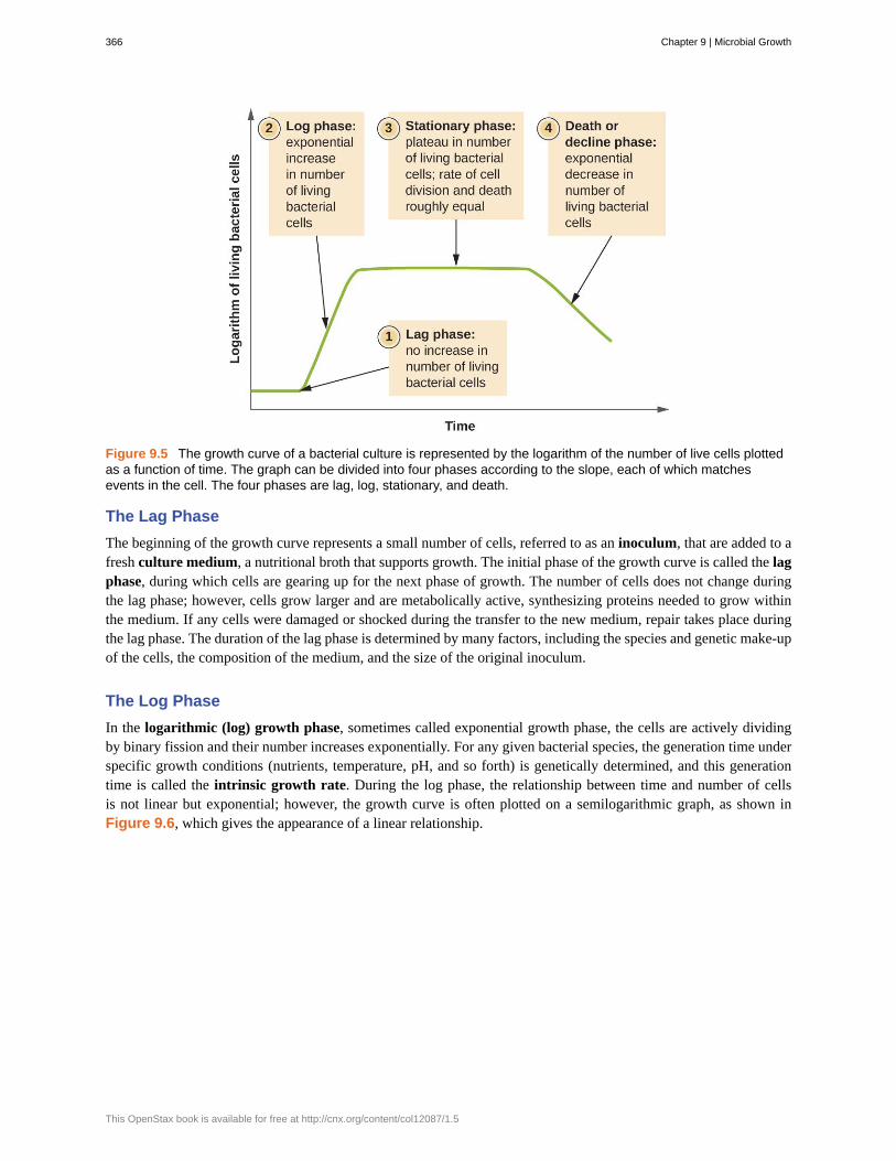
Figure 9.5 The growth curve of a bacterial culture is represented by the logarithm of the number of live cells plottedas a function of time. The graph can be divided into four phases according to the slope, each of which matchesevents in the cell. The four phases are lag, log, stationary, and death.
The Lag Phase
The beginning of the growth curve represents a small number of cells, referred to as an inoculum, that are added to afresh culture medium, a nutritional broth that supports growth. The initial phase of the growth curve is called the lagphase, during which cells are gearing up for the next phase of growth. The number of cells does not change duringthe lag phase; however, cells grow larger and are metabolically active, synthesizing proteins needed to grow withinthe medium. If any cells were damaged or shocked during the transfer to the new medium, repair takes place duringthe lag phase. The duration of the lag phase is determined by many factors, including the species and genetic make-upof the cells, the composition of the medium, and the size of the original inoculum.
The Log Phase
In the logarithmic (log) growth phase, sometimes called exponential growth phase, the cells are actively dividingby binary fission and their number increases exponentially. For any given bacterial species, the generation time underspecific growth conditions (nutrients, temperature, pH, and so forth) is genetically determined, and this generationtime is called the intrinsic growth rate. During the log phase, the relationship between time and number of cellsis not linear but exponential; however, the growth curve is often plotted on a semilogarithmic graph, as shown inFigure 9.6, which gives the appearance of a linear relationship.
366 Chapter 9 | Microbial Growth
This OpenStax book is available for free at http://cnx.org/content/col12087/1.5

Figure 9.6 Both graphs illustrate population growth during the log phase for a bacterial sample with an initialpopulation of one cell and a doubling time of 1 hour. (a) When plotted on an arithmetic scale, the growth rateresembles a curve. (b) When plotted on a semilogarithmic scale (meaning the values on the y-axis are logarithmic),the growth rate appears linear.
Cells in the log phase show constant growth rate and uniform metabolic activity. For this reason, cells in the log phaseare preferentially used for industrial applications and research work. The log phase is also the stage where bacteriaare the most susceptible to the action of disinfectants and common antibiotics that affect protein, DNA, and cell-wallsynthesis.
Stationary Phase
As the number of cells increases through the log phase, several factors contribute to a slowing of the growth rate.Waste products accumulate and nutrients are gradually used up. In addition, gradual depletion of oxygen begins tolimit aerobic cell growth. This combination of unfavorable conditions slows and finally stalls population growth. Thetotal number of live cells reaches a plateau referred to as the stationary phase (Figure 9.5). In this phase, the numberof new cells created by cell division is now equivalent to the number of cells dying; thus, the total population of livingcells is relatively stagnant. The culture density in a stationary culture is constant. The culture’s carrying capacity, ormaximum culture density, depends on the types of microorganisms in the culture and the specific conditions of theculture; however, carrying capacity is constant for a given organism grown under the same conditions.
During the stationary phase, cells switch to a survival mode of metabolism. As growth slows, so too does the synthesisof peptidoglycans, proteins, and nucleic-acids; thus, stationary cultures are less susceptible to antibiotics that disruptthese processes. In bacteria capable of producing endospores, many cells undergo sporulation during the stationaryphase. Secondary metabolites, including antibiotics, are synthesized in the stationary phase. In certain pathogenicbacteria, the stationary phase is also associated with the expression of virulence factors, products that contribute toa microbe’s ability to survive, reproduce, and cause disease in a host organism. For example, quorum sensing inStaphylococcus aureus initiates the production of enzymes that can break down human tissue and cellular debris,clearing the way for bacteria to spread to new tissue where nutrients are more plentiful.
The Death Phase
As a culture medium accumulates toxic waste and nutrients are exhausted, cells die in greater and greater numbers.Soon, the number of dying cells exceeds the number of dividing cells, leading to an exponential decrease in thenumber of cells (Figure 9.5). This is the aptly named death phase, sometimes called the decline phase. Many cellslyse and release nutrients into the medium, allowing surviving cells to maintain viability and form endospores. A
Chapter 9 | Microbial Growth 367
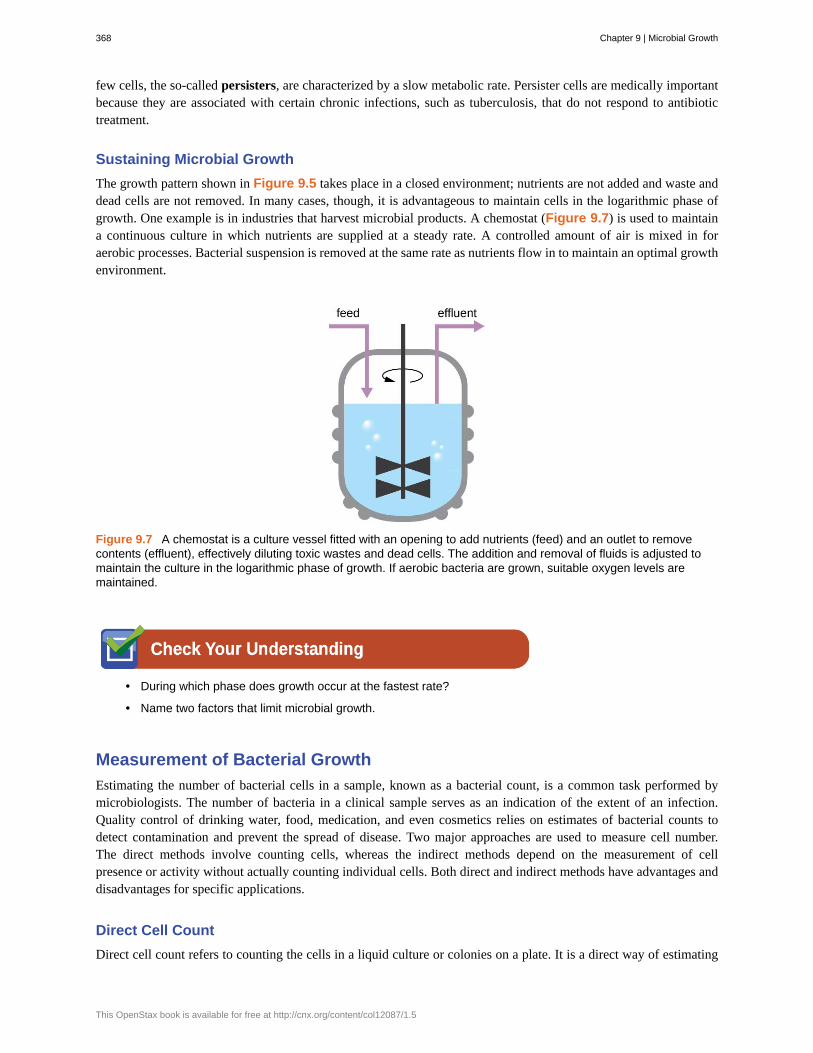
few cells, the so-called persisters, are characterized by a slow metabolic rate. Persister cells are medically importantbecause they are associated with certain chronic infections, such as tuberculosis, that do not respond to antibiotictreatment.
Sustaining Microbial Growth
The growth pattern shown in Figure 9.5 takes place in a closed environment; nutrients are not added and waste anddead cells are not removed. In many cases, though, it is advantageous to maintain cells in the logarithmic phase ofgrowth. One example is in industries that harvest microbial products. A chemostat (Figure 9.7) is used to maintaina continuous culture in which nutrients are supplied at a steady rate. A controlled amount of air is mixed in foraerobic processes. Bacterial suspension is removed at the same rate as nutrients flow in to maintain an optimal growthenvironment.
Figure 9.7 A chemostat is a culture vessel fitted with an opening to add nutrients (feed) and an outlet to removecontents (effluent), effectively diluting toxic wastes and dead cells. The addition and removal of fluids is adjusted tomaintain the culture in the logarithmic phase of growth. If aerobic bacteria are grown, suitable oxygen levels aremaintained.
• During which phase does growth occur at the fastest rate?
• Name two factors that limit microbial growth.
Measurement of Bacterial Growth
Estimating the number of bacterial cells in a sample, known as a bacterial count, is a common task performed bymicrobiologists. The number of bacteria in a clinical sample serves as an indication of the extent of an infection.Quality control of drinking water, food, medication, and even cosmetics relies on estimates of bacterial counts todetect contamination and prevent the spread of disease. Two major approaches are used to measure cell number.The direct methods involve counting cells, whereas the indirect methods depend on the measurement of cellpresence or activity without actually counting individual cells. Both direct and indirect methods have advantages anddisadvantages for specific applications.
Direct Cell Count
Direct cell count refers to counting the cells in a liquid culture or colonies on a plate. It is a direct way of estimating
368 Chapter 9 | Microbial Growth
This OpenStax book is available for free at http://cnx.org/content/col12087/1.5

how many organisms are present in a sample. Let’s look first at a simple and fast method that requires only aspecialized slide and a compound microscope.
The simplest way to count bacteria is called the direct microscopic cell count, which involves transferring a knownvolume of a culture to a calibrated slide and counting the cells under a light microscope. The calibrated slide is calleda Petroff-Hausser chamber (Figure 9.8) and is similar to a hemocytometer used to count red blood cells. Thecentral area of the counting chamber is etched into squares of various sizes. A sample of the culture suspension isadded to the chamber under a coverslip that is placed at a specific height from the surface of the grid. It is possibleto estimate the concentration of cells in the original sample by counting individual cells in a number of squares anddetermining the volume of the sample observed. The area of the squares and the height at which the coverslip ispositioned are specified for the chamber. The concentration must be corrected for dilution if the sample was dilutedbefore enumeration.
Figure 9.8 (a) A Petroff-Hausser chamber is a special slide designed for counting the bacterial cells in a measuredvolume of a sample. A grid is etched on the slide to facilitate precision in counting. (b) This diagram illustrates the gridof a Petroff-Hausser chamber, which is made up of squares of known areas. The enlarged view shows the squarewithin which bacteria (red cells) are counted. If the coverslip is 0.2 mm above the grid and the square has an area of0.04 mm2, then the volume is 0.008 mm3, or 0.000008 mL. Since there are 10 cells inside the square, the density ofbacteria is 10 cells/0.000008 mL, which equates to 1,250,000 cells/mL. (credit a: modification of work by Jeffrey M.Vinocur)
Cells in several small squares must be counted and the average taken to obtain a reliable measurement. Theadvantages of the chamber are that the method is easy to use, relatively fast, and inexpensive. On the downside, thecounting chamber does not work well with dilute cultures because there may not be enough cells to count.
Using a counting chamber does not necessarily yield an accurate count of the number of live cells because it isnot always possible to distinguish between live cells, dead cells, and debris of the same size under the microscope.However, newly developed fluorescence staining techniques make it possible to distinguish viable and dead bacteria.These viability stains (or live stains) bind to nucleic acids, but the primary and secondary stains differ in their abilityto cross the cytoplasmic membrane. The primary stain, which fluoresces green, can penetrate intact cytoplasmicmembranes, staining both live and dead cells. The secondary stain, which fluoresces red, can stain a cell only if thecytoplasmic membrane is considerably damaged. Thus, live cells fluoresce green because they only absorb the greenstain, whereas dead cells appear red because the red stain displaces the green stain on their nucleic acids (Figure9.9).
Chapter 9 | Microbial Growth 369

Figure 9.9 Fluorescence staining can be used to differentiate between viable and dead bacterial cells in a samplefor purposes of counting. Viable cells are stained green, whereas dead cells are stained red. (credit: modification ofwork by Panseri S, Cunha C, D’Alessandro T, Sandri M, Giavaresi G, Maracci M, Hung CT, Tampieri A)
Another technique uses an electronic cell counting device (Coulter counter) to detect and count the changes inelectrical resistance in a saline solution. A glass tube with a small opening is immersed in an electrolyte solution. Afirst electrode is suspended in the glass tube. A second electrode is located outside of the tube. As cells are drawnthrough the small aperture in the glass tube, they briefly change the resistance measured between the two electrodesand the change is recorded by an electronic sensor (Figure 9.10); each resistance change represents a cell. Themethod is rapid and accurate within a range of concentrations; however, if the culture is too concentrated, more thanone cell may pass through the aperture at any given time and skew the results. This method also does not differentiatebetween live and dead cells.
Direct counts provide an estimate of the total number of cells in a sample. However, in many situations, it is importantto know the number of live, or viable, cells. Counts of live cells are needed when assessing the extent of an infection,the effectiveness of antimicrobial compounds and medication, or contamination of food and water.
Figure 9.10 A Coulter counter is an electronic device that counts cells. It measures the change in resistance in anelectrolyte solution that takes place when a cell passes through a small opening in the inside container wall. Adetector automatically counts the number of cells passing through the opening. (credit b: modification of work byNational Institutes of Health)
370 Chapter 9 | Microbial Growth
This OpenStax book is available for free at http://cnx.org/content/col12087/1.5

• Why would you count the number of cells in more than one square in the Petroff-Hausser chamber toestimate cell numbers?
• In the viability staining method, why do dead cells appear red?
Plate Count
The viable plate count, or simply plate count, is a count of viable or live cells. It is based on the principle that viablecells replicate and give rise to visible colonies when incubated under suitable conditions for the specimen. The resultsare usually expressed as colony-forming units per milliliter (CFU/mL) rather than cells per milliliter because morethan one cell may have landed on the same spot to give rise to a single colony. Furthermore, samples of bacteriathat grow in clusters or chains are difficult to disperse and a single colony may represent several cells. Some cellsare described as viable but nonculturable and will not form colonies on solid media. For all these reasons, the viableplate count is considered a low estimate of the actual number of live cells. These limitations do not detract from theusefulness of the method, which provides estimates of live bacterial numbers.
Microbiologists typically count plates with 30–300 colonies. Samples with too few colonies (<30) do not givestatistically reliable numbers, and overcrowded plates (>300 colonies) make it difficult to accurately count individualcolonies. Also, counts in this range minimize occurrences of more than one bacterial cell forming a single colony.Thus, the calculated CFU is closer to the true number of live bacteria in the population.
There are two common approaches to inoculating plates for viable counts: the pour plate and the spread plate methods.Although the final inoculation procedure differs between these two methods, they both start with a serial dilution ofthe culture.
Serial Dilution
The serial dilution of a culture is an important first step before proceeding to either the pour plate or spread platemethod. The goal of the serial dilution process is to obtain plates with CFUs in the range of 30–300, and the processusually involves several dilutions in multiples of 10 to simplify calculation. The number of serial dilutions is chosenaccording to a preliminary estimate of the culture density. Figure 9.11 illustrates the serial dilution method.
A fixed volume of the original culture, 1.0 mL, is added to and thoroughly mixed with the first dilution tube solution,which contains 9.0 mL of sterile broth. This step represents a dilution factor of 10, or 1:10, compared with the originalculture. From this first dilution, the same volume, 1.0 mL, is withdrawn and mixed with a fresh tube of 9.0 mL ofdilution solution. The dilution factor is now 1:100 compared with the original culture. This process continues until aseries of dilutions is produced that will bracket the desired cell concentration for accurate counting. From each tube,a sample is plated on solid medium using either the pour plate method (Figure 9.12) or the spread plate method(Figure 9.13). The plates are incubated until colonies appear. Two to three plates are usually prepared from eachdilution and the numbers of colonies counted on each plate are averaged. In all cases, thorough mixing of sampleswith the dilution medium (to ensure the cell distribution in the tube is random) is paramount to obtaining reliableresults.
Chapter 9 | Microbial Growth 371
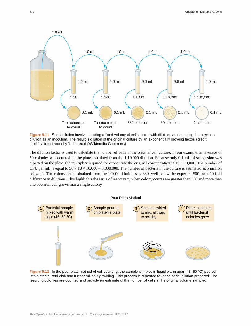
Figure 9.11 Serial dilution involves diluting a fixed volume of cells mixed with dilution solution using the previousdilution as an inoculum. The result is dilution of the original culture by an exponentially growing factor. (credit:modification of work by “Leberechtc”/Wikimedia Commons)
The dilution factor is used to calculate the number of cells in the original cell culture. In our example, an average of50 colonies was counted on the plates obtained from the 1:10,000 dilution. Because only 0.1 mL of suspension waspipetted on the plate, the multiplier required to reconstitute the original concentration is 10 × 10,000. The number ofCFU per mL is equal to 50 × 10 × 10,000 = 5,000,000. The number of bacteria in the culture is estimated as 5 millioncells/mL. The colony count obtained from the 1:1000 dilution was 389, well below the expected 500 for a 10-folddifference in dilutions. This highlights the issue of inaccuracy when colony counts are greater than 300 and more thanone bacterial cell grows into a single colony.
Figure 9.12 In the pour plate method of cell counting, the sample is mixed in liquid warm agar (45–50 °C) pouredinto a sterile Petri dish and further mixed by swirling. This process is repeated for each serial dilution prepared. Theresulting colonies are counted and provide an estimate of the number of cells in the original volume sampled.
372 Chapter 9 | Microbial Growth
This OpenStax book is available for free at http://cnx.org/content/col12087/1.5
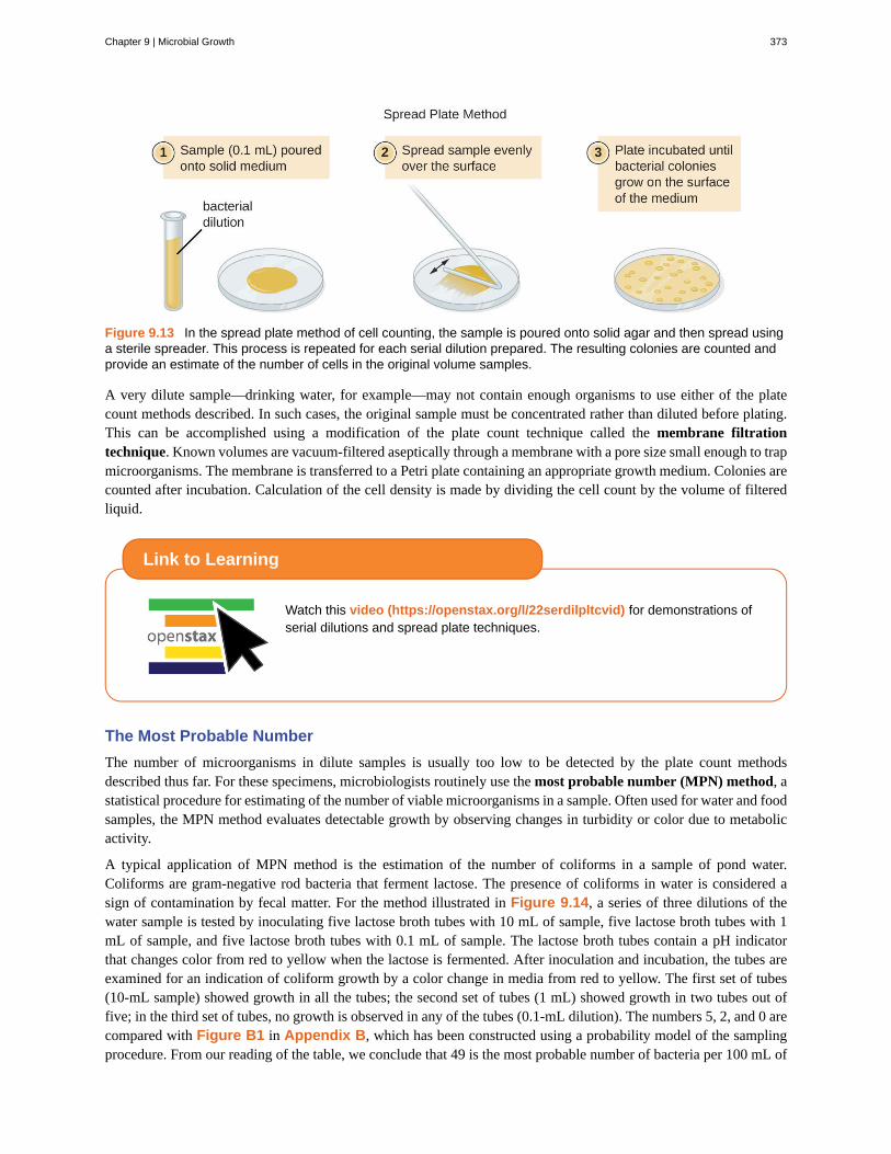
Figure 9.13 In the spread plate method of cell counting, the sample is poured onto solid agar and then spread usinga sterile spreader. This process is repeated for each serial dilution prepared. The resulting colonies are counted andprovide an estimate of the number of cells in the original volume samples.
A very dilute sample—drinking water, for example—may not contain enough organisms to use either of the platecount methods described. In such cases, the original sample must be concentrated rather than diluted before plating.This can be accomplished using a modification of the plate count technique called the membrane filtrationtechnique. Known volumes are vacuum-filtered aseptically through a membrane with a pore size small enough to trapmicroorganisms. The membrane is transferred to a Petri plate containing an appropriate growth medium. Colonies arecounted after incubation. Calculation of the cell density is made by dividing the cell count by the volume of filteredliquid.
Watch this video (https://openstax.org/l/22serdilpltcvid) for demonstrations ofserial dilutions and spread plate techniques.
The Most Probable Number
The number of microorganisms in dilute samples is usually too low to be detected by the plate count methodsdescribed thus far. For these specimens, microbiologists routinely use the most probable number (MPN) method, astatistical procedure for estimating of the number of viable microorganisms in a sample. Often used for water and foodsamples, the MPN method evaluates detectable growth by observing changes in turbidity or color due to metabolicactivity.
A typical application of MPN method is the estimation of the number of coliforms in a sample of pond water.Coliforms are gram-negative rod bacteria that ferment lactose. The presence of coliforms in water is considered asign of contamination by fecal matter. For the method illustrated in Figure 9.14, a series of three dilutions of thewater sample is tested by inoculating five lactose broth tubes with 10 mL of sample, five lactose broth tubes with 1mL of sample, and five lactose broth tubes with 0.1 mL of sample. The lactose broth tubes contain a pH indicatorthat changes color from red to yellow when the lactose is fermented. After inoculation and incubation, the tubes areexamined for an indication of coliform growth by a color change in media from red to yellow. The first set of tubes(10-mL sample) showed growth in all the tubes; the second set of tubes (1 mL) showed growth in two tubes out offive; in the third set of tubes, no growth is observed in any of the tubes (0.1-mL dilution). The numbers 5, 2, and 0 arecompared with Figure B1 in Appendix B, which has been constructed using a probability model of the samplingprocedure. From our reading of the table, we conclude that 49 is the most probable number of bacteria per 100 mL of
Link to Learning
Chapter 9 | Microbial Growth 373

pond water.no lo
Figure 9.14 In the most probable number method, sets of five lactose broth tubes are inoculated with three differentvolumes of pond water: 10 mL, 1 mL, and 0.1mL. Bacterial growth is assessed through a change in the color of thebroth from red to yellow as lactose is fermented.
• What is a colony-forming unit?
• What two methods are frequently used to estimate bacterial numbers in water samples?
Indirect Cell Counts
Besides direct methods of counting cells, other methods, based on an indirect detection of cell density, are commonlyused to estimate and compare cell densities in a culture. The foremost approach is to measure the turbidity(cloudiness) of a sample of bacteria in a liquid suspension. The laboratory instrument used to measure turbidity iscalled a spectrophotometer (Figure 9.15). In a spectrophotometer, a light beam is transmitted through a bacterialsuspension, the light passing through the suspension is measured by a detector, and the amount of light passingthrough the sample and reaching the detector is converted to either percent transmission or a logarithmic value calledabsorbance (optical density). As the numbers of bacteria in a suspension increase, the turbidity also increases andcauses less light to reach the detector. The decrease in light passing through the sample and reaching the detector isassociated with a decrease in percent transmission and increase in absorbance measured by the spectrophotometer.
Measuring turbidity is a fast method to estimate cell density as long as there are enough cells in a sample to produceturbidity. It is possible to correlate turbidity readings to the actual number of cells by performing a viable platecount of samples taken from cultures having a range of absorbance values. Using these values, a calibration curve isgenerated by plotting turbidity as a function of cell density. Once the calibration curve has been produced, it can be
374 Chapter 9 | Microbial Growth
This OpenStax book is available for free at http://cnx.org/content/col12087/1.5

used to estimate cell counts for all samples obtained or cultured under similar conditions and with densities within therange of values used to construct the curve.
Figure 9.15 (a) A spectrophotometer is commonly used to measure the turbidity of a bacterial cell suspension as anindirect measure of cell density. (b) A spectrophotometer works by splitting white light from a source into a spectrum.The spectrophotometer allows choice of the wavelength of light to use for the measurement. The optical density(turbidity) of the sample will depend on the wavelength, so once one wavelength is chosen, it must be usedconsistently. The filtered light passes through the sample (or a control with only medium) and the light intensity ismeasured by a detector. The light passing into a suspension of bacteria is scattered by the cells in such a way thatsome fraction of it never reaches the detector. This scattering happens to a far lesser degree in the control tube withonly the medium. (credit a: modification of work by Hwang HS, Kim MS; credit b “test tube photos”: modification ofwork by Suzanne Wakim)
Measuring dry weight of a culture sample is another indirect method of evaluating culture density without directlymeasuring cell counts. The cell suspension used for weighing must be concentrated by filtration or centrifugation,washed, and then dried before the measurements are taken. The degree of drying must be standardized to accountfor residual water content. This method is especially useful for filamentous microorganisms, which are difficult toenumerate by direct or viable plate count.
As we have seen, methods to estimate viable cell numbers can be labor intensive and take time because cells must begrown. Recently, indirect ways of measuring live cells have been developed that are both fast and easy to implement.These methods measure cell activity by following the production of metabolic products or disappearance of reactants.Adenosine triphosphate (ATP) formation, biosynthesis of proteins and nucleic acids, and consumption of oxygen canall be monitored to estimate the number of cells.
• What is the purpose of a calibration curve when estimating cell count from turbidity measurements?
• What are the newer indirect methods of counting live cells?
Alternative Patterns of Cell Division
Binary fission is the most common pattern of cell division in prokaryotes, but it is not the only one. Other mechanismsusually involve asymmetrical division (as in budding) or production of spores in aerial filaments.
In some cyanobacteria, many nucleoids may accumulate in an enlarged round cell or along a filament, leading tothe generation of many new cells at once. The new cells often split from the parent filament and float away in a
Chapter 9 | Microbial Growth 375

process called fragmentation (Figure 9.16). Fragmentation is commonly observed in the Actinomycetes, a group ofgram-positive, anaerobic bacteria commonly found in soil. Another curious example of cell division in prokaryotes,reminiscent of live birth in animals, is exhibited by the giant bacterium Epulopiscium. Several daughter cells growfully in the parent cell, which eventually disintegrates, releasing the new cells to the environment. Other species mayform a long narrow extension at one pole in a process called budding. The tip of the extension swells and forms asmaller cell, the bud that eventually detaches from the parent cell. Budding is most common in yeast (Figure 9.16),but it is also observed in prosthecate bacteria and some cyanobacteria.
Figure 9.16 (a) Filamentous cyanobacteria, like those pictured here, replicate by fragmentation. (b) In this electronmicrograph, cells of the bacterium Gemmata obscuriglobus are budding. The larger cell is the mother cell. Labelsindicate the nucleoids (N) and the still-forming nuclear envelope (NE) of the daughter cell. (credit a: modification ofwork by CSIRO; credit b: modification of work by Kuo-Chang Lee, Rick I Webb and John A Fuerst)
The soil bacteria Actinomyces grow in long filaments divided by septa, similar to the mycelia seen in fungi, resultingin long cells with multiple nucleoids. Environmental signals, probably related to low nutrient availability, lead to theformation of aerial filaments. Within these aerial filaments, elongated cells divide simultaneously. The new cells,which contain a single nucleoid, develop into spores that give rise to new colonies.
• Identify at least one difference between fragmentation and budding.
Biofilms
In nature, microorganisms grow mainly in biofilms, complex and dynamic ecosystems that form on a variety ofenvironmental surfaces, from industrial conduits and water treatment pipelines to rocks in river beds. Biofilms arenot restricted to solid surface substrates, however. Almost any surface in a liquid environment containing someminimal nutrients will eventually develop a biofilm. Microbial mats that float on water, for example, are biofilmsthat contain large populations of photosynthetic microorganisms. Biofilms found in the human mouth may containhundreds of bacterial species. Regardless of the environment where they occur, biofilms are not random collections ofmicroorganisms; rather, they are highly structured communities that provide a selective advantage to their constituentmicroorganisms.
Biofilm Structure
Observations using confocal microscopy have shown that environmental conditions influence the overall structure ofbiofilms. Filamentous biofilms called streamers form in rapidly flowing water, such as freshwater streams, eddies,and specially designed laboratory flow cells that replicate growth conditions in fast-moving fluids. The streamers are
376 Chapter 9 | Microbial Growth
This OpenStax book is available for free at http://cnx.org/content/col12087/1.5

anchored to the substrate by a “head” and the “tail” floats downstream in the current. In still or slow-moving water,biofilms mainly assume a mushroom-like shape. The structure of biofilms may also change with other environmentalconditions such as nutrient availability.
Detailed observations of biofilms under confocal laser and scanning electron microscopes reveal clusters ofmicroorganisms embedded in a matrix interspersed with open water channels. The extracellular matrix consistsof extracellular polymeric substances (EPS) secreted by the organisms in the biofilm. The extracellular matrixrepresents a large fraction of the biofilm, accounting for 50%–90% of the total dry mass. The properties of the EPSvary according to the resident organisms and environmental conditions.
EPS is a hydrated gel composed primarily of polysaccharides and containing other macromolecules such as proteins,nucleic acids, and lipids. It plays a key role in maintaining the integrity and function of the biofilm. Channels in theEPS allow movement of nutrients, waste, and gases throughout the biofilm. This keeps the cells hydrated, preventingdesiccation. EPS also shelters organisms in the biofilm from predation by other microbes or cells (e.g., protozoans,white blood cells in the human body).
Biofilm Formation
Free-floating microbial cells that live in an aquatic environment are called planktonic cells. The formation of abiofilm essentially involves the attachment of planktonic cells to a substrate, where they become sessile (attached toa surface). This occurs in stages, as depicted in Figure 9.17. The first stage involves the attachment of planktoniccells to a surface coated with a conditioning film of organic material. At this point, attachment to the substrate isreversible, but as cells express new phenotypes that facilitate the formation of EPS, they transition from a planktonicto a sessile lifestyle. The biofilm develops characteristic structures, including an extensive matrix and water channels.Appendages such as fimbriae, pili, and flagella interact with the EPS, and microscopy and genetic analysis suggestthat such structures are required for the establishment of a mature biofilm. In the last stage of the biofilm life cycle,cells on the periphery of the biofilm revert to a planktonic lifestyle, sloughing off the mature biofilm to colonize newsites. This stage is referred to as dispersal.
Figure 9.17 Stages in the formation and life cycle of a biofilm. (credit: modification of work by Public Library ofScience and American Society for Microbiology)
Within a biofilm, different species of microorganisms establish metabolic collaborations in which the waste productof one organism becomes the nutrient for another. For example, aerobic microorganisms consume oxygen, creating
Chapter 9 | Microbial Growth 377

anaerobic regions that promote the growth of anaerobes. This occurs in many polymicrobial infections that involveboth aerobic and anaerobic pathogens.
The mechanism by which cells in a biofilm coordinate their activities in response to environmental stimuli is calledquorum sensing. Quorum sensing—which can occur between cells of different species within a biofilm—enablesmicroorganisms to detect their cell density through the release and binding of small, diffusible molecules calledautoinducers. When the cell population reaches a critical threshold (a quorum), these autoinducers initiate a cascadeof reactions that activate genes associated with cellular functions that are beneficial only when the population reachesa critical density. For example, in some pathogens, synthesis of virulence factors only begins when enough cells arepresent to overwhelm the immune defenses of the host. Although mostly studied in bacterial populations, quorumsensing takes place between bacteria and eukaryotes and between eukaryotic cells such as the fungus Candidaalbicans, a common member of the human microbiota that can cause infections in immunocompromised individuals.
The signaling molecules in quorum sensing belong to two major classes. Gram-negative bacteria communicate mainlyusing N-acylated homoserine lactones, whereas gram-positive bacteria mostly use small peptides (Figure 9.18). Inall cases, the first step in quorum sensing consists of the binding of the autoinducer to its specific receptor only whena threshold concentration of signaling molecules is reached. Once binding to the receptor takes place, a cascade ofsignaling events leads to changes in gene expression. The result is the activation of biological responses linked toquorum sensing, notably an increase in the production of signaling molecules themselves, hence the term autoinducer.
Figure 9.18 Short peptides in gram-positive bacteria and N-acetylated homoserine lactones in gram-negativebacteria act as autoinducers in quorum sensing and mediate the coordinated response of bacterial cells. The R sidechain of the N-acetylated homoserine lactone is specific for the species of gram-negative bacteria. Some secretedhomoserine lactones are recognized by more than one species.
Biofilms and Human Health
The human body harbors many types of biofilms, some beneficial and some harmful. For example, the layers ofnormal microbiota lining the intestinal and respiratory mucosa play a role in warding off infections by pathogens.However, other biofilms in the body can have a detrimental effect on health. For example, the plaque that forms onteeth is a biofilm that can contribute to dental and periodontal disease. Biofilms can also form in wounds, sometimescausing serious infections that can spread. The bacterium Pseudomonas aeruginosa often colonizes biofilms in theairways of patients with cystic fibrosis, causing chronic and sometimes fatal infections of the lungs. Biofilms can alsoform on medical devices used in or on the body, causing infections in patients with in-dwelling catheters, artificialjoints, or contact lenses.
Pathogens embedded within biofilms exhibit a higher resistance to antibiotics than their free-floating counterparts.Several hypotheses have been proposed to explain why. Cells in the deep layers of a biofilm are metabolically inactiveand may be less susceptible to the action of antibiotics that disrupt metabolic activities. The EPS may also slowthe diffusion of antibiotics and antiseptics, preventing them from reaching cells in the deeper layers of the biofilm.Phenotypic changes may also contribute to the increased resistance exhibited by bacterial cells in biofilms. Forexample, the increased production of efflux pumps, membrane-embedded proteins that actively extrude antibiotics outof bacterial cells, have been shown to be an important mechanism of antibiotic resistance among biofilm-associatedbacteria. Finally, biofilms provide an ideal environment for the exchange of extrachromosomal DNA, which often
378 Chapter 9 | Microbial Growth
This OpenStax book is available for free at http://cnx.org/content/col12087/1.5

includes genes that confer antibiotic resistance.
• What is the matrix of a biofilm composed of?
• What is the role of quorum sensing in a biofilm?
9.2 Oxygen Requirements for Microbial Growth
Learning Objectives• Interpret visual data demonstrating minimum, optimum, and maximum oxygen or carbon dioxide requirements
for growth
• Identify and describe different categories of microbes with requirements for growth with or without oxygen:obligate aerobe, obligate anaerobe, facultative anaerobe, aerotolerant anaerobe, microaerophile, and capnophile
• Give examples of microorganisms for each category of growth requirements
Ask most people “What are the major requirements for life?” and the answers are likely to include water and oxygen.Few would argue about the need for water, but what about oxygen? Can there be life without oxygen?
The answer is that molecular oxygen (O2) is not always needed. The earliest signs of life are dated to a period whenconditions on earth were highly reducing and free oxygen gas was essentially nonexistent. Only after cyanobacteriastarted releasing oxygen as a byproduct of photosynthesis and the capacity of iron in the oceans for taking up oxygenwas exhausted did oxygen levels increase in the atmosphere. This event, often referred to as the Great OxygenationEvent or the Oxygen Revolution, caused a massive extinction. Most organisms could not survive the powerfuloxidative properties of reactive oxygen species (ROS), highly unstable ions and molecules derived from partialreduction of oxygen that can damage virtually any macromolecule or structure with which they come in contact.Singlet oxygen (O2•), superoxide ⎛
⎝O2−⎞
⎠, peroxides (H2O2), hydroxyl radical (OH•), and hypochlorite ion (OCl−),
the active ingredient of household bleach, are all examples of ROS. The organisms that were able to detoxify reactiveoxygen species harnessed the high electronegativity of oxygen to produce free energy for their metabolism and thrivedin the new environment.
Oxygen Requirements of Microorganisms
Many ecosystems are still free of molecular oxygen. Some are found in extreme locations, such as deep in the oceanor in earth’s crust; others are part of our everyday landscape, such as marshes, bogs, and sewers. Within the bodiesof humans and other animals, regions with little or no oxygen provide an anaerobic environment for microorganisms.(Figure 9.19).
Chapter 9 | Microbial Growth 379

Figure 9.19 Anaerobic environments are still common on earth. They include environments like (a) a bog whereundisturbed dense sediments are virtually devoid of oxygen, and (b) the rumen (the first compartment of a cow’sstomach), which provides an oxygen-free incubator for methanogens and other obligate anaerobic bacteria. (credit a:modification of work by National Park Service; credit b: modification of work by US Department of Agriculture)
We can easily observe different requirements for molecular oxygen by growing bacteria in thioglycolate tubecultures. A test-tube culture starts with autoclaved thioglycolate medium containing a low percentage of agar toallow motile bacteria to move throughout the medium. Thioglycolate has strong reducing properties and autoclavingflushes out most of the oxygen. The tubes are inoculated with the bacterial cultures to be tested and incubated at anappropriate temperature. Over time, oxygen slowly diffuses throughout the thioglycolate tube culture from the top.Bacterial density increases in the area where oxygen concentration is best suited for the growth of that particularorganism.
The growth of bacteria with varying oxygen requirements in thioglycolate tubes is illustrated in Figure 9.20. In tubeA, all the growth is seen at the top of the tube. The bacteria are obligate (strict) aerobes that cannot grow withoutan abundant supply of oxygen. Tube B looks like the opposite of tube A. Bacteria grow at the bottom of tube B.Those are obligate anaerobes, which are killed by oxygen. Tube C shows heavy growth at the top of the tube andgrowth throughout the tube, a typical result with facultative anaerobes. Facultative anaerobes are organisms thatthrive in the presence of oxygen but also grow in its absence by relying on fermentation or anaerobic respiration, ifthere is a suitable electron acceptor other than oxygen and the organism is able to perform anaerobic respiration. Theaerotolerant anaerobes in tube D are indifferent to the presence of oxygen. They do not use oxygen because theyusually have a fermentative metabolism, but they are not harmed by the presence of oxygen as obligate anaerobes are.Tube E on the right shows a “Goldilocks” culture. The oxygen level has to be just right for growth, not too much andnot too little. These microaerophiles are bacteria that require a minimum level of oxygen for growth, about 1%–10%,well below the 21% found in the atmosphere.
Examples of obligate aerobes are Mycobacterium tuberculosis, the causative agent of tuberculosis and Micrococcusluteus, a gram-positive bacterium that colonizes the skin. Neisseria meningitidis, the causative agent of severebacterial meningitis, and N. gonorrhoeae, the causative agent of sexually transmitted gonorrhea, are also obligateaerobes.
380 Chapter 9 | Microbial Growth
This OpenStax book is available for free at http://cnx.org/content/col12087/1.5

Figure 9.20 Diagram of bacterial cell distribution in thioglycolate tubes.
Many obligate anaerobes are found in the environment where anaerobic conditions exist, such as in deep sediments ofsoil, still waters, and at the bottom of the deep ocean where there is no photosynthetic life. Anaerobic conditions alsoexist naturally in the intestinal tract of animals. Obligate anaerobes, mainly Bacteroidetes, represent a large fractionof the microbes in the human gut. Transient anaerobic conditions exist when tissues are not supplied with bloodcirculation; they die and become an ideal breeding ground for obligate anaerobes. Another type of obligate anaerobeencountered in the human body is the gram-positive, rod-shaped Clostridium spp. Their ability to form endosporesallows them to survive in the presence of oxygen. One of the major causes of health-acquired infections is C. difficile,known as C. diff. Prolonged use of antibiotics for other infections increases the probability of a patient developinga secondary C. difficile infection. Antibiotic treatment disrupts the balance of microorganisms in the intestine andallows the colonization of the gut by C. difficile, causing a significant inflammation of the colon.
Other clostridia responsible for serious infections include C. tetani, the agent of tetanus, and C. perfringens, whichcauses gas gangrene. In both cases, the infection starts in necrotic tissue (dead tissue that is not supplied with oxygenby blood circulation). This is the reason that deep puncture wounds are associated with tetanus. When tissue death isaccompanied by lack of circulation, gangrene is always a danger.
The study of obligate anaerobes requires special equipment. Obligate anaerobic bacteria must be grown underconditions devoid of oxygen. The most common approach is culture in an anaerobic jar (Figure 9.21). Anaerobicjars include chemical packs that remove oxygen and release carbon dioxide (CO2). An anaerobic chamber is anenclosed box from which all oxygen is removed. Gloves sealed to openings in the box allow handling of the cultureswithout exposing the culture to air (Figure 9.21).
Chapter 9 | Microbial Growth 381

Figure 9.21 (a) An anaerobic jar is pictured that is holding nine Petri plates supporting cultures. (b) Openings in theside of an anaerobic box are sealed by glove-like sleeves that allow for the handling of cultures inside the box. (credita: modification of work by Centers for Disease Control and Prevention; credit b: modification of work by NIST)
Staphylococci and Enterobacteriaceae are examples of facultative anaerobes. Staphylococci are found on the skinand upper respiratory tract. Enterobacteriaceae are found primarily in the gut and upper respiratory tract but cansometimes spread to the urinary tract, where they are capable of causing infections. It is not unusual to see mixedbacterial infections in which the facultative anaerobes use up the oxygen, creating an environment for the obligateanaerobes to flourish.
Examples of aerotolerant anaerobes include lactobacilli and streptococci, both found in the oral microbiota.Campylobacter jejuni, which causes gastrointestinal infections, is an example of a microaerophile and is grown underlow-oxygen conditions.
The optimum oxygen concentration, as the name implies, is the ideal concentration of oxygen for a particularmicroorganism. The lowest concentration of oxygen that allows growth is called the minimum permissive oxygenconcentration. The highest tolerated concentration of oxygen is the maximum permissive oxygen concentration.The organism will not grow outside the range of oxygen levels found between the minimum and maximum permissiveoxygen concentrations.
• Would you expect the oldest bacterial lineages to be aerobic or anaerobic?
• Which bacteria grow at the top of a thioglycolate tube, and which grow at the bottom of the tube?
An Unwelcome Anaerobe
Charles is a retired bus driver who developed type 2 diabetes over 10 years ago. Since his retirement, hislifestyle has become very sedentary and he has put on a substantial amount of weight. Although he hasfelt tingling and numbness in his left foot for a while, he has not been worried because he thought his footwas simply “falling asleep.” Recently, a scratch on his foot does not seem to be healing and is becomingincreasingly ugly. Because the sore did not bother him much, Charles figured it could not be serious until hisdaughter noticed a purplish discoloration spreading on the skin and oozing (Figure 9.22). When he was finallyseen by his physician, Charles was rushed to the operating room. His open sore, or ulcer, is the result of adiabetic foot.
Case in Point
382 Chapter 9 | Microbial Growth
This OpenStax book is available for free at http://cnx.org/content/col12087/1.5

The concern here is that gas gangrene may have taken hold in the dead tissue. The most likely agent ofgas gangrene is Clostridium perfringens, an endospore-forming, gram-positive bacterium. It is an obligateanaerobe that grows in tissue devoid of oxygen. Since dead tissue is no longer supplied with oxygen by thecirculatory system, the dead tissue provides pockets of ideal environment for the growth of C. perfringens.
A surgeon examines the ulcer and radiographs of Charles’s foot and determines that the bone is not yetinfected. The wound will have to be surgically debrided (debridement refers to the removal of dead and infectedtissue) and a sample sent for microbiological lab analysis, but Charles will not have to have his foot amputated.Many diabetic patients are not so lucky. In 2008, nearly 70,000 diabetic patients in the United States lost a footor limb to amputation, according to statistics from the Centers for Disease Control and Prevention.[1]
• Which growth conditions would you recommend for the detection of C. perfringens?
Figure 9.22 This clinical photo depicts ulcers on the foot of a diabetic patient. Dead tissue accumulating inulcers can provide an ideal growth environment for the anaerobe C. perfringens, a causative agent of gasgangrene. (credit: Shigeo Kono, Reiko Nakagawachi, Jun Arata, Benjamin A Lipsky)
Detoxification of Reactive Oxygen Species
Aerobic respiration constantly generates reactive oxygen species (ROS), byproducts that must be detoxified. Evenorganisms that do not use aerobic respiration need some way to break down some of the ROS that may form fromatmospheric oxygen. Three main enzymes break down those toxic byproducts: superoxide dismutase, peroxidase, andcatalase. Each one catalyzes a different reaction. Reactions of type seen in Reaction 1 are catalyzed by peroxidases.
(1) X − (2H+) + H2 O2 → oxidized-X + 2H2 O
In these reactions, an electron donor (reduced compound; e.g., reduced nicotinamide adenine dinucleotide [NADH])oxidizes hydrogen peroxide, or other peroxides, to water. The enzymes play an important role by limiting the damagecaused by peroxidation of membrane lipids. Reaction 2 is mediated by the enzyme superoxide dismutase (SOD) andbreaks down the powerful superoxide anions generated by aerobic metabolism:
(2) 2O2− + 2H+ → H2 O2 + O2
The enzyme catalase converts hydrogen peroxide to water and oxygen as shown in Reaction 3.
(3) 2H2 O2 → 2H2 O + O2
Obligate anaerobes usually lack all three enzymes. Aerotolerant anaerobes do have SOD but no catalase. Reaction
1. Centers for Disease Control and Prevention. “Living With Diabetes: Keep Your Feet Healthy.” http://www.cdc.gov/Features/
DiabetesFootHealth/
Chapter 9 | Microbial Growth 383

3, shown occurring in Figure 9.23, is the basis of a useful and rapid test to distinguish streptococci, which areaerotolerant and do not possess catalase, from staphylococci, which are facultative anaerobes. A sample of culturerapidly mixed in a drop of 3% hydrogen peroxide will release bubbles if the culture is catalase positive.
Figure 9.23 The catalase test detects the presence of the enzyme catalase by noting whether bubbles are releasedwhen hydrogen peroxide is added to a culture sample. Compare the positive result (right) with the negative result(left). (credit: Centers for Disease Control and Prevention)
Bacteria that grow best in a higher concentration of CO2 and a lower concentration of oxygen than present in theatmosphere are called capnophiles. One common approach to grow capnophiles is to use a candle jar. A candle jarconsists of a jar with a tight-fitting lid that can accommodate the cultures and a candle. After the cultures are added tothe jar, the candle is lit and the lid closed. As the candle burns, it consumes most of the oxygen present and releasesCO2.
• What substance is added to a sample to detect catalase?
• What is the function of the candle in a candle jar?
Part 2
The health-care provider who saw Jeni was concerned primarily because of her pregnancy. Her conditionenhances the risk for infections and makes her more vulnerable to those infections. The immune systemis downregulated during pregnancy, and pathogens that cross the placenta can be very dangerous for thefetus. A note on the provider’s order to the microbiology lab mentions a suspicion of infection by Listeriamonocytogenes, based on the signs and symptoms exhibited by the patient.
Jeni’s blood samples are streaked directly on sheep blood agar, a medium containing tryptic soy agarenriched with 5% sheep blood. (Blood is considered sterile; therefore, competing microorganisms are notexpected in the medium.) The inoculated plates are incubated at 37 °C for 24 to 48 hours. Small grayishcolonies surrounded by a clear zone emerge. Such colonies are typical of Listeria and other pathogens suchas streptococci; the clear zone surrounding the colonies indicates complete lysis of blood in the medium,referred to as beta-hemolysis (Figure 9.24). When tested for the presence of catalase, the colonies give apositive response, eliminating Streptococcus as a possible cause. Furthermore, a Gram stain shows shortgram-positive bacilli. Cells from a broth culture grown at room temperature displayed the tumbling motility
Clinical Focus
384 Chapter 9 | Microbial Growth
This OpenStax book is available for free at http://cnx.org/content/col12087/1.5

characteristic of Listeria (Figure 9.24). All of these clues lead the lab to positively confirm the presence ofListeria in Jeni’s blood samples.
• How serious is Jeni’s condition and what is the appropriate treatment?
Figure 9.24 (a) A sample blood agar test showing beta-hemolysis. (b) A sample motility test showing bothpositive and negative results. (credit a: modification of work by Centers for Disease Control and Prevention;credit b: modification of work by “VeeDunn”/Flickr)
Jump to the next Clinical Focus box. Go back to the previous Clinical Focus box.
9.3 The Effects of pH on Microbial Growth
Learning Objectives• Illustrate and briefly describe minimum, optimum, and maximum pH requirements for growth
• Identify and describe the different categories of microbes with pH requirements for growth: acidophiles,neutrophiles, and alkaliphiles
• Give examples of microorganisms for each category of pH requirement
Yogurt, pickles, sauerkraut, and lime-seasoned dishes all owe their tangy taste to a high acid content (Figure 9.25).Recall that acidity is a function of the concentration of hydrogen ions [H+] and is measured as pH. Environments withpH values below 7.0 are considered acidic, whereas those with pH values above 7.0 are considered basic. ExtremepH affects the structure of all macromolecules. The hydrogen bonds holding together strands of DNA break up athigh pH. Lipids are hydrolyzed by an extremely basic pH. The proton motive force responsible for production ofATP in cellular respiration depends on the concentration gradient of H+ across the plasma membrane (see CellularRespiration). If H+ ions are neutralized by hydroxide ions, the concentration gradient collapses and impairs energyproduction. But the component most sensitive to pH in the cell is its workhorse, the protein. Moderate changes inpH modify the ionization of amino-acid functional groups and disrupt hydrogen bonding, which, in turn, promoteschanges in the folding of the molecule, promoting denaturation and destroying activity.
Chapter 9 | Microbial Growth 385

Figure 9.25 Lactic acid bacteria that ferment milk into yogurt or transform vegetables in pickles thrive at a pH closeto 4.0. Sauerkraut and dishes such as pico de gallo owe their tangy flavor to their acidity. Acidic foods have been amainstay of the human diet for centuries, partly because most microbes that cause food spoilage grow best at a nearneutral pH and do not tolerate acidity well. (credit “yogurt”: modification of work by “nina.jsc”/Flickr; credit “pickles”:modification of work by Noah Sussman; credit “sauerkraut”: modification of work by Jesse LaBuff; credit “pico degallo”: modification of work by “regan76”/Flickr)
The optimum growth pH is the most favorable pH for the growth of an organism. The lowest pH value that anorganism can tolerate is called the minimum growth pH and the highest pH is the maximum growth pH. Thesevalues can cover a wide range, which is important for the preservation of food and to microorganisms’ survival in thestomach. For example, the optimum growth pH of Salmonella spp. is 7.0–7.5, but the minimum growth pH is closerto 4.2.
Most bacteria are neutrophiles, meaning they grow optimally at a pH within one or two pH units of the neutralpH of 7 (see Figure 9.26). Most familiar bacteria, like Escherichia coli, staphylococci, and Salmonella spp. areneutrophiles and do not fare well in the acidic pH of the stomach. However, there are pathogenic strains of E. coli,S. typhi, and other species of intestinal pathogens that are much more resistant to stomach acid. In comparison, fungithrive at slightly acidic pH values of 5.0–6.0.
Microorganisms that grow optimally at pH less than 5.55 are called acidophiles. For example, the sulfur-oxidizingSulfolobus spp. isolated from sulfur mud fields and hot springs in Yellowstone National Park are extreme acidophiles.These archaea survive at pH values of 2.5–3.5. Species of the archaean genus Ferroplasma live in acid mine drainageat pH values of 0–2.9. Lactobacillus bacteria, which are an important part of the normal microbiota of the vagina, cantolerate acidic environments at pH values 3.5–6.8 and also contribute to the acidity of the vagina (pH of 4, except atthe onset of menstruation) through their metabolic production of lactic acid. The vagina’s acidity plays an importantrole in inhibiting other microbes that are less tolerant of acidity. Acidophilic microorganisms display a number ofadaptations to survive in strong acidic environments. For example, proteins show increased negative surface chargethat stabilizes them at low pH. Pumps actively eject H+ ions out of the cells. The changes in the composition ofmembrane phospholipids probably reflect the need to maintain membrane fluidity at low pH.
386 Chapter 9 | Microbial Growth
This OpenStax book is available for free at http://cnx.org/content/col12087/1.5

Figure 9.26 The curves show the approximate pH ranges for the growth of the different classes of pH-specificprokaryotes. Each curve has an optimal pH and extreme pH values at which growth is much reduced. Most bacteriaare neutrophiles and grow best at near-neutral pH (center curve). Acidophiles have optimal growth at pH values near3 and alkaliphiles have optimal growth at pH values above 9.
At the other end of the spectrum are alkaliphiles, microorganisms that grow best at pH between 8.0 and 10.5. Vibriocholerae, the pathogenic agent of cholera, grows best at the slightly basic pH of 8.0; it can survive pH values of11.0 but is inactivated by the acid of the stomach. When it comes to survival at high pH, the bright pink archaeanNatronobacterium, found in the soda lakes of the African Rift Valley, may hold the record at a pH of 10.5 (Figure9.27). Extreme alkaliphiles have adapted to their harsh environment through evolutionary modification of lipid andprotein structure and compensatory mechanisms to maintain the proton motive force in an alkaline environment. Forexample, the alkaliphile Bacillus firmus derives the energy for transport reactions and motility from a Na+ ion gradientrather than a proton motive force. Many enzymes from alkaliphiles have a higher isoelectric point, due to an increasein the number of basic amino acids, than homologous enzymes from neutrophiles.
Figure 9.27 View from space of Lake Natron in Tanzania. The pink color is due to the pigmentation of the extremealkaliphilic and halophilic microbes that colonize the lake. (credit: NASA)
Chapter 9 | Microbial Growth 387

Survival at the Low pH of the Stomach
Peptic ulcers (or stomach ulcers) are painful sores on the stomach lining. Until the 1980s, they were believed tobe caused by spicy foods, stress, or a combination of both. Patients were typically advised to eat bland foods,take anti-acid medications, and avoid stress. These remedies were not particularly effective, and the conditionoften recurred. This all changed dramatically when the real cause of most peptic ulcers was discovered to bea slim, corkscrew-shaped bacterium, Helicobacter pylori. This organism was identified and isolated by BarryMarshall and Robin Warren, whose discovery earned them the Nobel Prize in Medicine in 2005.
The ability of H. pylori to survive the low pH of the stomach would seem to suggest that it is an extremeacidophile. As it turns out, this is not the case. In fact, H. pylori is a neutrophile. So, how does it survive inthe stomach? Remarkably, H. pylori creates a microenvironment in which the pH is nearly neutral. It achievesthis by producing large amounts of the enzyme urease, which breaks down urea to form NH4
+ and CO2. Theammonium ion raises the pH of the immediate environment.
This metabolic capability of H. pylori is the basis of an accurate, noninvasive test for infection. The patient isgiven a solution of urea containing radioactively labeled carbon atoms. If H. pylori is present in the stomach,it will rapidly break down the urea, producing radioactive CO2 that can be detected in the patient’s breath.Because peptic ulcers may lead to gastric cancer, patients who are determined to have H. pylori infections aretreated with antibiotics.
• What effect do extremes of pH have on proteins?
• What pH-adaptive type of bacteria would most human pathogens be?
9.4 Temperature and Microbial Growth
Learning Objectives• Illustrate and briefly describe minimum, optimum, and maximum temperature requirements for growth
• Identify and describe different categories of microbes with temperature requirements for growth: psychrophile,psychrotrophs, mesophile, thermophile, hyperthermophile
• Give examples of microorganisms in each category of temperature tolerance
When the exploration of Lake Whillans started in Antarctica, researchers did not expect to find much life. Constantsubzero temperatures and lack of obvious sources of nutrients did not seem to be conditions that would supporta thriving ecosystem. To their surprise, the samples retrieved from the lake showed abundant microbial life. In adifferent but equally harsh setting, bacteria grow at the bottom of the ocean in sea vents (Figure 9.28), wheretemperatures can reach 340 °C (700 °F).
Microbes can be roughly classified according to the range of temperature at which they can grow. The growth ratesare the highest at the optimum growth temperature for the organism. The lowest temperature at which the organismcan survive and replicate is its minimum growth temperature. The highest temperature at which growth can occur isits maximum growth temperature. The following ranges of permissive growth temperatures are approximate onlyand can vary according to other environmental factors.
Organisms categorized as mesophiles (“middle loving”) are adapted to moderate temperatures, with optimal growth
Micro Connections
388 Chapter 9 | Microbial Growth
This OpenStax book is available for free at http://cnx.org/content/col12087/1.5

temperatures ranging from room temperature (about 20 °C) to about 45 °C. As would be expected from the coretemperature of the human body, 37 °C (98.6 °F), normal human microbiota and pathogens (e.g., E. coli, Salmonellaspp., and Lactobacillus spp.) are mesophiles.
Organisms called psychrotrophs, also known as psychrotolerant, prefer cooler environments, from a hightemperature of 25 °C to refrigeration temperature about 4 °C. They are found in many natural environments intemperate climates. They are also responsible for the spoilage of refrigerated food.
Resolution
The presence of Listeria in Jeni’s blood suggests that her symptoms are due to listeriosis, an infection causedby L. monocytogenes. Listeriosis is a serious infection with a 20% mortality rate and is a particular risk to Jeni’sfetus. A sample from the amniotic fluid cultured for the presence of Listeria gave negative results. Becausethe absence of organisms does not rule out the possibility of infection, a molecular test based on the nucleicacid amplification of the 16S ribosomal RNA of Listeria was performed to confirm that no bacteria crossed theplacenta. Fortunately, the results from the molecular test were also negative.
Jeni was admitted to the hospital for treatment and recovery. She received a high dose of two antibioticsintravenously for 2 weeks. The preferred drugs for the treatment of listeriosis are ampicillin or penicillin G withan aminoglycoside antibiotic. Resistance to common antibiotics is still rare in Listeria and antibiotic treatmentis usually successful. She was released to home care after a week and fully recovered from her infection.
L. monocytogenes is a gram-positive short rod found in soil, water, and food. It is classified as a psychrophileand is halotolerant. Its ability to multiply at refrigeration temperatures (4–10 °C) and its tolerance for highconcentrations of salt (up to 10% sodium chloride [NaCl]) make it a frequent source of food poisoning. BecauseListeria can infect animals, it often contaminates food such as meat, fish, or dairy products. Contamination ofcommercial foods can often be traced to persistent biofilms that form on manufacturing equipment that is notsufficiently cleaned.
Listeria infection is relatively common among pregnant women because the elevated levels of progesteronedownregulate the immune system, making them more vulnerable to infection. The pathogen can cross theplacenta and infect the fetus, often resulting in miscarriage, stillbirth, or fatal neonatal infection. Pregnantwomen are thus advised to avoid consumption of soft cheeses, refrigerated cold cuts, smoked seafood, andunpasteurized dairy products. Because Listeria bacteria can easily be confused with diphtheroids, anothercommon group of gram-positive rods, it is important to alert the laboratory when listeriosis is suspected.
Go back to the previous Clinical Focus box.
The organisms retrieved from arctic lakes such as Lake Whillans are considered extreme psychrophiles (cold loving).Psychrophiles are microorganisms that can grow at 0 °C and below, have an optimum growth temperature close to15 °C, and usually do not survive at temperatures above 20 °C. They are found in permanently cold environmentssuch as the deep waters of the oceans. Because they are active at low temperature, psychrophiles and psychrotrophsare important decomposers in cold climates.
Organisms that grow at optimum temperatures of 50 °C to a maximum of 80 °C are called thermophiles (“heatloving”). They do not multiply at room temperature. Thermophiles are widely distributed in hot springs, geothermalsoils, and manmade environments such as garden compost piles where the microbes break down kitchen scrapsand vegetal material. Examples of thermophiles include Thermus aquaticus and Geobacillus spp. Higher up on theextreme temperature scale we find the hyperthermophiles, which are characterized by growth ranges from 80 °C toa maximum of 110 °C, with some extreme examples that survive temperatures above 121 °C, the average temperatureof an autoclave. The hydrothermal vents at the bottom of the ocean are a prime example of extreme environments,with temperatures reaching an estimated 340 °C (Figure 9.28). Microbes isolated from the vents achieve optimalgrowth at temperatures higher than 100 °C. Noteworthy examples are Pyrobolus and Pyrodictium, archaea that grow
Clinical Focus
Chapter 9 | Microbial Growth 389

at 105 °C and survive autoclaving. Figure 9.29 shows the typical skewed curves of temperature-dependent growthfor the categories of microorganisms we have discussed.
Figure 9.28 A black smoker at the bottom of the ocean belches hot, chemical-rich water, and heats the surroundingwaters. Sea vents provide an extreme environment that is nonetheless teeming with macroscopic life (the redtubeworms) supported by an abundant microbial ecosystem. (credit: NOAA)
Figure 9.29 The graph shows growth rate of bacteria as a function of temperature. Notice that the curves areskewed toward the optimum temperature. The skewing of the growth curve is thought to reflect the rapid denaturationof proteins as the temperature rises past the optimum for growth of the microorganism.
Life in extreme environments raises fascinating questions about the adaptation of macromolecules and metabolicprocesses. Very low temperatures affect cells in many ways. Membranes lose their fluidity and are damaged byice crystal formation. Chemical reactions and diffusion slow considerably. Proteins become too rigid to catalyzereactions and may undergo denaturation. At the opposite end of the temperature spectrum, heat denatures proteinsand nucleic acids. Increased fluidity impairs metabolic processes in membranes. Some of the practical applications ofthe destructive effects of heat on microbes are sterilization by steam, pasteurization, and incineration of inoculatingloops. Proteins in psychrophiles are, in general, rich in hydrophobic residues, display an increase in flexibility, andhave a lower number of secondary stabilizing bonds when compared with homologous proteins from mesophiles.Antifreeze proteins and solutes that decrease the freezing temperature of the cytoplasm are common. The lipids inthe membranes tend to be unsaturated to increase fluidity. Growth rates are much slower than those encounteredat moderate temperatures. Under appropriate conditions, mesophiles and even thermophiles can survive freezing.
390 Chapter 9 | Microbial Growth
This OpenStax book is available for free at http://cnx.org/content/col12087/1.5

Liquid cultures of bacteria are mixed with sterile glycerol solutions and frozen to −80 °C for long-term storage asstocks. Cultures can withstand freeze drying (lyophilization) and then be stored as powders in sealed ampules to bereconstituted with broth when needed.
Macromolecules in thermophiles and hyperthermophiles show some notable structural differences from what isobserved in the mesophiles. The ratio of saturated to polyunsaturated lipids increases to limit the fluidity of thecell membranes. Their DNA sequences show a higher proportion of guanine–cytosine nitrogenous bases, whichare held together by three hydrogen bonds in contrast to adenine and thymine, which are connected in the doublehelix by two hydrogen bonds. Additional secondary ionic and covalent bonds, as well as the replacement of keyamino acids to stabilize folding, contribute to the resistance of proteins to denaturation. The so-called thermoenzymespurified from thermophiles have important practical applications. For example, amplification of nucleic acids in thepolymerase chain reaction (PCR) depends on the thermal stability of Taq polymerase, an enzyme isolated from T.aquaticus. Degradation enzymes from thermophiles are added as ingredients in hot-water detergents, increasing theireffectiveness.
• What temperature requirements do most bacterial human pathogens have?
• What DNA adaptation do thermophiles exhibit?
Feeding the World…and the World’s Algae
Artificial fertilizers have become an important tool in food production around the world. They are responsiblefor many of the gains of the so-called green revolution of the 20th century, which has allowed the planet tofeed many of its more than 7 billion people. Artificial fertilizers provide nitrogen and phosphorus, key limitingnutrients, to crop plants, removing the normal barriers that would otherwise limit the rate of growth. Thus,fertilized crops grow much faster, and farms that use fertilizer produce higher crop yields.
However, careless use and overuse of artificial fertilizers have been demonstrated to have significant negativeimpacts on aquatic ecosystems, both freshwater and marine. Fertilizers that are applied at inappropriate timesor in too-large quantities allow nitrogen and phosphorus compounds to escape use by crop plants and enterdrainage systems. Inappropriate use of fertilizers in residential settings can also contribute to nutrient loads,which find their way to lakes and coastal marine ecosystems. As water warms and nutrients are plentiful,microscopic algae bloom, often changing the color of the water because of the high cell density.
Most algal blooms are not directly harmful to humans or wildlife; however, they can cause harm indirectly. Asthe algal population expands and then dies, it provides a large increase in organic matter to the bacteria thatlive in deep water. With this large supply of nutrients, the population of nonphotosynthetic microorganismsexplodes, consuming available oxygen and creating “dead zones” where animal life has virtually disappeared.
Depletion of oxygen in the water is not the only damaging consequence of some algal blooms. The algaethat produce red tides in the Gulf of Mexico, Karenia brevis, secrete potent toxins that can kill fish andother organisms and also accumulate in shellfish. Consumption of contaminated shellfish can cause severeneurological and gastrointestinal symptoms in humans. Shellfish beds must be regularly monitored for thepresence of the toxins, and harvests are often shut down when it is present, incurring economic costs to thefishery. Cyanobacteria, which can form blooms in marine and freshwater ecosystems, produce toxins called
Eye on Ethics
Chapter 9 | Microbial Growth 391

microcystins, which can cause allergic reactions and liver damage when ingested in drinking water or duringswimming. Recurring cyanobacterial algal blooms in Lake Erie (Figure 9.30) have forced municipalities toissue drinking water bans for days at a time because of unacceptable toxin levels.
This is just a small sampling of the negative consequences of algal blooms, red tides, and dead zones. Yet thebenefits of crop fertilizer—the main cause of such blooms—are difficult to dispute. There is no easy solutionto this dilemma, as a ban on fertilizers is not politically or economically feasible. In lieu of this, we mustadvocate for responsible use and regulation in agricultural and residential contexts, as well as the restorationof wetlands, which can absorb excess fertilizers before they reach lakes and oceans.
Figure 9.30 Heavy rains cause runoff of fertilizers into Lake Erie, triggering extensive algal blooms, whichcan be observed along the shoreline. Notice the brown unplanted and green planted agricultural land on theshore. (credit: NASA)
This video (https://openstax.org/l/22algaebloomvid) discusses algal bloomsand dead zones in more depth.
9.5 Other Environmental Conditions that Affect Growth
Learning Objectives• Identify and describe different categories of microbes with specific growth requirements other than oxygen, pH,
and temperature, such as altered barometric pressure, osmotic pressure, humidity, and light
• Give at least one example microorganism for each category of growth requirement
Microorganisms interact with their environment along more dimensions than pH, temperature, and free oxygen levels,
Link to Learning
392 Chapter 9 | Microbial Growth
This OpenStax book is available for free at http://cnx.org/content/col12087/1.5

although these factors require significant adaptations. We also find microorganisms adapted to varying levels ofsalinity, barometric pressure, humidity, and light.
Osmotic and Barometric Pressure
Most natural environments tend to have lower solute concentrations than the cytoplasm of most microorganisms.Rigid cell walls protect the cells from bursting in a dilute environment. Not much protection is available againsthigh osmotic pressure. In this case, water, following its concentration gradient, flows out of the cell. This resultsin plasmolysis (the shrinking of the protoplasm away from the intact cell wall) and cell death. This fact explainswhy brines and layering meat and fish in salt are time-honored methods of preserving food. Microorganisms calledhalophiles (“salt loving”) actually require high salt concentrations for growth. These organisms are found in marineenvironments where salt concentrations hover at 3.5%. Extreme halophilic microorganisms, such as the red algaDunaliella salina and the archaeal species Halobacterium in Figure 9.31, grow in hypersaline lakes such as theGreat Salt Lake, which is 3.5–8 times saltier than the ocean, and the Dead Sea, which is 10 times saltier than theocean.
Figure 9.31 Photograph taken from space of the Great Salt Lake in Utah. The purple color is caused by high densityof the alga Dunaliella and the archaean Halobacterium spp. (credit: NASA)
Dunaliella spp. counters the tremendous osmotic pressure of the environment with a high cytoplasmic concentrationof glycerol and by actively pumping out salt ions. Halobacterium spp. accumulates large concentrations of K+ andother ions in its cytoplasm. Its proteins are designed for high salt concentrations and lose activity at salt concentrationsbelow 1–2 M. Although most halotolerant organisms, for example Halomonas spp. in salt marshes, do not needhigh concentrations of salt for growth, they will survive and divide in the presence of high salt. Not surprisingly, thestaphylococci, micrococci, and corynebacteria that colonize our skin tolerate salt in their environment. Halotolerantpathogens are an important cause of food-borne illnesses because they survive and multiply in salty food. Forexample, the halotolerant bacteria S. aureus, Bacillus cereus, and V. cholerae produce dangerous enterotoxins and aremajor causes of food poisoning.
Microorganisms depend on available water to grow. Available moisture is measured as water activity (aw), which isthe ratio of the vapor pressure of the medium of interest to the vapor pressure of pure distilled water; therefore, theaw of water is equal to 1.0. Bacteria require high aw (0.97–0.99), whereas fungi can tolerate drier environments; forexample, the range of aw for growth of Aspergillus spp. is 0.8–0.75. Decreasing the water content of foods by drying,as in jerky, or through freeze-drying or by increasing osmotic pressure, as in brine and jams, are common methods ofpreventing spoilage.
Chapter 9 | Microbial Growth 393

Microorganisms that require high atmospheric pressure for growth are called barophiles. The bacteria that live at thebottom of the ocean must be able to withstand great pressures. Because it is difficult to retrieve intact specimens andreproduce such growth conditions in the laboratory, the characteristics of these microorganisms are largely unknown.
Light
Photoautotrophs, such as cyanobacteria or green sulfur bacteria, and photoheterotrophs, such as purple nonsulfurbacteria, depend on sufficient light intensity at the wavelengths absorbed by their pigments to grow and multiply.Energy from light is captured by pigments and converted into chemical energy that drives carbon fixation and othermetabolic processes. The portion of the electromagnetic spectrum that is absorbed by these organisms is definedas photosynthetically active radiation (PAR). It lies within the visible light spectrum ranging from 400 to 700nanometers (nm) and extends in the near infrared for some photosynthetic bacteria. A number of accessory pigments,such as fucoxanthin in brown algae and phycobilins in cyanobacteria, widen the useful range of wavelengths forphotosynthesis and compensate for the low light levels available at greater depths of water. Other microorganisms,such as the archaea of the class Halobacteria, use light energy to drive their proton and sodium pumps. The lightis absorbed by a pigment protein complex called bacteriorhodopsin, which is similar to the eye pigment rhodopsin.Photosynthetic bacteria are present not only in aquatic environments but also in soil and in symbiosis with fungiin lichens. The peculiar watermelon snow is caused by a microalga Chlamydomonas nivalis, a green alga rich in asecondary red carotenoid pigment (astaxanthin) which gives the pink hue to the snow where the alga grows.
• Which photosynthetic pigments were described in this section?
• What is the fundamental stress of a hypersaline environment for a cell?
9.6 Media Used for Bacterial Growth
Learning Objectives• Identify and describe culture media for the growth of bacteria, including examples of all-purpose media,
enriched, selective, differential, defined, and enrichment media
The study of microorganisms is greatly facilitated if we are able to culture them, that is, to keep reproducingpopulations alive under laboratory conditions. Culturing many microorganisms is challenging because of highlyspecific nutritional and environmental requirements and the diversity of these requirements among different species.
Nutritional Requirements
The number of available media to grow bacteria is considerable. Some media are considered general all-purposemedia and support growth of a large variety of organisms. A prime example of an all-purpose medium is trypticsoy broth (TSB). Specialized media are used in the identification of bacteria and are supplemented with dyes, pHindicators, or antibiotics. One type, enriched media, contains growth factors, vitamins, and other essential nutrientsto promote the growth of fastidious organisms, organisms that cannot make certain nutrients and require them tobe added to the medium. When the complete chemical composition of a medium is known, it is called a chemicallydefined medium. For example, in EZ medium, all individual chemical components are identified and the exactamounts of each is known. In complex media, which contain extracts and digests of yeasts, meat, or plants, theprecise chemical composition of the medium is not known. Amounts of individual components are undetermined andvariable. Nutrient broth, tryptic soy broth, and brain heart infusion, are all examples of complex media.
Media that inhibit the growth of unwanted microorganisms and support the growth of the organism of interest by
394 Chapter 9 | Microbial Growth
This OpenStax book is available for free at http://cnx.org/content/col12087/1.5

supplying nutrients and reducing competition are called selective media. An example of a selective medium isMacConkey agar. It contains bile salts and crystal violet, which interfere with the growth of many gram-positivebacteria and favor the growth of gram-negative bacteria, particularly the Enterobacteriaceae. These species arecommonly named enterics, reside in the intestine, and are adapted to the presence of bile salts. The enrichmentcultures foster the preferential growth of a desired microorganism that represents a fraction of the organisms presentin an inoculum. For example, if we want to isolate bacteria that break down crude oil, hydrocarbonoclastic bacteria,sequential subculturing in a medium that supplies carbon only in the form of crude oil will enrich the cultures withoil-eating bacteria. The differential media make it easy to distinguish colonies of different bacteria by a changein the color of the colonies or the color of the medium. Color changes are the result of end products created byinteraction of bacterial enzymes with differential substrates in the medium or, in the case of hemolytic reactions, thelysis of red blood cells in the medium. In Figure 9.32, the differential fermentation of lactose can be observed onMacConkey agar. The lactose fermenters produce acid, which turns the medium and the colonies of strong fermentershot pink. The medium is supplemented with the pH indicator neutral red, which turns to hot pink at low pH. Selectiveand differential media can be combined and play an important role in the identification of bacteria by biochemicalmethods.
Figure 9.32 On this MacConkey agar plate, the lactose-fermenter E. coli colonies are bright pink. Serratiamarcescens, which does not ferment lactose, forms a cream-colored streak on the tan medium. (credit: AmericanSociety for Microbiology)
• Distinguish complex and chemically defined media.
• Distinguish selective and enrichment media.
Compare the compositions of EZ medium (https://openstax.org/l/22EZMedium) and sheep blood (https://openstax.org/l/22bloodagar) agar.
Link to Learning
Chapter 9 | Microbial Growth 395

The End-of-Year Picnic
The microbiology department is celebrating the end of the school year in May by holding its traditional picnicon the green. The speeches drag on for a couple of hours, but finally all the faculty and students can dig intothe food: chicken salad, tomatoes, onions, salad, and custard pie. By evening, the whole department, exceptfor two vegetarian students who did not eat the chicken salad, is stricken with nausea, vomiting, retching, andabdominal cramping. Several individuals complain of diarrhea. One patient shows signs of shock (low bloodpressure). Blood and stool samples are collected from patients, and an analysis of all foods served at the mealis conducted.
Bacteria can cause gastroenteritis (inflammation of the stomach and intestinal tract) either by colonizing andreplicating in the host, which is considered an infection, or by secreting toxins, which is considered intoxication.Signs and symptoms of infections are typically delayed, whereas intoxication manifests within hours, ashappened after the picnic.
Blood samples from the patients showed no signs of bacterial infection, which further suggests that this wasa case of intoxication. Since intoxication is due to secreted toxins, bacteria are not usually detected in bloodor stool samples. MacConkey agar and sorbitol-MacConkey agar plates and xylose-lysine-deoxycholate (XLD)plates were inoculated with stool samples and did not reveal any unusually colored colonies, and no blackcolonies or white colonies were observed on XLD. All lactose fermenters on MacConkey agar also fermentsorbitol. These results ruled out common agents of food-borne illnesses: E. coli, Salmonella spp., and Shigellaspp.
Analysis of the chicken salad revealed an abnormal number of gram-positive cocci arranged in clusters (Figure9.33). A culture of the gram-positive cocci releases bubbles when mixed with hydrogen peroxide. The cultureturned mannitol salt agar yellow after a 24-hour incubation.
All the tests point to Staphylococcus aureus as the organism that secreted the toxin. Samples from the saladshowed the presence of gram-positive cocci bacteria in clusters. The colonies were positive for catalase. Thebacteria grew on mannitol salt agar fermenting mannitol, as shown by the change to yellow of the medium.The pH indicator in mannitol salt agar is phenol red, which turns to yellow when the medium is acidified by theproducts of fermentation.
The toxin secreted by S. aureus is known to cause severe gastroenteritis. The organism was probablyintroduced into the salad during preparation by the food handler and multiplied while the salad was kept in thewarm ambient temperature during the speeches.
• What are some other factors that might have contributed to rapid growth of S. aureus in the chickensalad?
• Why would S. aureus not be inhibited by the presence of salt in the chicken salad?
Case in Point
396 Chapter 9 | Microbial Growth
This OpenStax book is available for free at http://cnx.org/content/col12087/1.5

Figure 9.33 Gram-positive cocci in clusters. (credit: Centers for Disease Control and Prevention)
Summary
9.1 How Microbes Grow• Most bacterial cells divide by binary fission. Generation time in bacterial growth is defined as the doubling
time of the population.
• Cells in a closed system follow a pattern of growth with four phases: lag, logarithmic (exponential),stationary, and death.
• Cells can be counted by direct viable cell count. The pour plate and spread plate methods are used toplate serial dilutions into or onto, respectively, agar to allow counting of viable cells that give rise to colony-forming units. Membrane filtration is used to count live cells in dilute solutions. The most probable cellnumber (MPN) method allows estimation of cell numbers in cultures without using solid media.
• Indirect methods can be used to estimate culture density by measuring turbidity of a culture or live celldensity by measuring metabolic activity.
• Other patterns of cell division include multiple nucleoid formation in cells; asymmetric division, as inbudding; and the formation of hyphae and terminal spores.
• Biofilms are communities of microorganisms enmeshed in a matrix of extracellular polymeric substance.The formation of a biofilm occurs when planktonic cells attach to a substrate and become sessile. Cells inbiofilms coordinate their activity by communicating through quorum sensing.
• Biofilms are commonly found on surfaces in nature and in the human body, where they may be beneficialor cause severe infections. Pathogens associated with biofilms are often more resistant to antibiotics anddisinfectants.
9.2 Oxygen Requirements for Microbial Growth• Aerobic and anaerobic environments can be found in diverse niches throughout nature, including different
sites within and on the human body.
• Microorganisms vary in their requirements for molecular oxygen. Obligate aerobes depend on aerobicrespiration and use oxygen as a terminal electron acceptor. They cannot grow without oxygen.
• Obligate anaerobes cannot grow in the presence of oxygen. They depend on fermentation and anaerobicrespiration using a final electron acceptor other than oxygen.
• Facultative anaerobes show better growth in the presence of oxygen but will also grow without it.
Chapter 9 | Microbial Growth 397

• Although aerotolerant anaerobes do not perform aerobic respiration, they can grow in the presence ofoxygen. Most aerotolerant anaerobes test negative for the enzyme catalase.
• Microaerophiles need oxygen to grow, albeit at a lower concentration than 21% oxygen in air.
• Optimum oxygen concentration for an organism is the oxygen level that promotes the fastest growth rate.The minimum permissive oxygen concentration and the maximum permissive oxygen concentration are,respectively, the lowest and the highest oxygen levels that the organism will tolerate.
• Peroxidase, superoxide dismutase, and catalase are the main enzymes involved in the detoxification of thereactive oxygen species. Superoxide dismutase is usually present in a cell that can tolerate oxygen. All threeenzymes are usually detectable in cells that perform aerobic respiration and produce more ROS.
• A capnophile is an organism that requires a higher than atmospheric concentration of CO2 to grow.
9.3 The Effects of pH on Microbial Growth• Bacteria are generally neutrophiles. They grow best at neutral pH close to 7.0.
• Acidophiles grow optimally at a pH near 3.0. Alkaliphiles are organisms that grow optimally between a pHof 8 and 10.5. Extreme acidophiles and alkaliphiles grow slowly or not at all near neutral pH.
• Microorganisms grow best at their optimum growth pH. Growth occurs slowly or not at all below theminimum growth pH and above the maximum growth pH.
9.4 Temperature and Microbial Growth• Microorganisms thrive at a wide range of temperatures; they have colonized different natural environments
and have adapted to extreme temperatures. Both extreme cold and hot temperatures require evolutionaryadjustments to macromolecules and biological processes.
• Psychrophiles grow best in the temperature range of 0–15 °C whereas psychrotrophs thrive between 4°Cand 25 °C.
• Mesophiles grow best at moderate temperatures in the range of 20 °C to about 45 °C. Pathogens are usuallymesophiles.
• Thermophiles and hyperthemophiles are adapted to life at temperatures above 50 °C.
• Adaptations to cold and hot temperatures require changes in the composition of membrane lipids and proteins.
9.5 Other Environmental Conditions that Affect Growth• Halophiles require high salt concentration in the medium, whereas halotolerant organisms can grow and
multiply in the presence of high salt but do not require it for growth.
• Halotolerant pathogens are an important source of foodborne illnesses because they contaminate foodspreserved in salt.
• Photosynthetic bacteria depend on visible light for energy.
• Most bacteria, with few exceptions, require high moisture to grow.
9.6 Media Used for Bacterial Growth• Chemically defined media contain only chemically known components.
• Selective media favor the growth of some microorganisms while inhibiting others.
• Enriched media contain added essential nutrients a specific organism needs to grow
• Differential media help distinguish bacteria by the color of the colonies or the change in the medium.
398 Chapter 9 | Microbial Growth
This OpenStax book is available for free at http://cnx.org/content/col12087/1.5

Multiple Choice
1. Which of the following methods would be used tomeasure the concentration of bacterial contamination inprocessed peanut butter?
a. turbidity measurementb. total plate countc. dry weight measurementd. direct counting of bacteria on a calibrated slide
under the microscope
2. In which phase would you expect to observe themost endospores in a Bacillus cell culture?
a. death phaseb. lag phasec. log phased. log, lag, and death phases would all have roughly
the same number of endospores.
3. During which phase would penicillin, an antibioticthat inhibits cell-wall synthesis, be most effective?
a. death phaseb. lag phasec. log phased. stationary phase
4. Which of the following is the best definition ofgeneration time in a bacterium?
a. the length of time it takes to reach the log phaseb. the length of time it takes for a population of
cells to doublec. the time it takes to reach stationary phased. the length of time of the exponential phase
5. What is the function of the Z ring in binary fission?a. It controls the replication of DNA.b. It forms a contractile ring at the septum.c. It separates the newly synthesized DNA
molecules.d. It mediates the addition of new peptidoglycan
subunits.
6. If a culture starts with 50 cells, how many cells willbe present after five generations with no cell death?
a. 200b. 400c. 1600d. 3200
7. Filamentous cyanobacteria often divide by which ofthe following?
a. buddingb. mitosisc. fragmentationd. formation of endospores
8. Which is a reason for antimicrobial resistance beinghigher in a biofilm than in free-floating bacterial cells?
a. The EPS allows faster diffusion of chemicals inthe biofilm.
b. Cells are more metabolically active at the base ofa biofilm.
c. Cells are metabolically inactive at the base of abiofilm.
d. The structure of a biofilm favors the survival ofantibiotic resistant cells.
9. Quorum sensing is used by bacterial cells todetermine which of the following?
a. the size of the populationb. the availability of nutrientsc. the speed of water flowd. the density of the population
10. Which of the following statements aboutautoinducers is incorrect?
a. They bind directly to DNA to activatetranscription.
b. They can activate the cell that secreted them.c. N-acylated homoserine lactones are autoinducers
in gram-negative cells.d. Autoinducers may stimulate the production of
virulence factors.
11. An inoculated thioglycolate medium culture tubeshows dense growth at the surface and turbiditythroughout the rest of the tube. What is your conclusion?
a. The organisms die in the presence of oxygenb. The organisms are facultative anaerobes.c. The organisms should be grown in an anaerobic
chamber.d. The organisms are obligate aerobes.
Review Questions
Chapter 9 | Microbial Growth 399

12. An inoculated thioglycolate medium culture tube isclear throughout the tube except for dense growth at thebottom of the tube. What is your conclusion?
a. The organisms are obligate anaerobes.b. The organisms are facultative anaerobes.c. The organisms are aerotolerant.d. The organisms are obligate aerobes.
13. Pseudomonas aeruginosa is a common pathogenthat infects the airways of patients with cystic fibrosis. Itdoes not grow in the absence of oxygen. The bacteriumis probably which of the following?
a. an aerotolerant anaerobeb. an obligate aerobec. an obligate anaerobed. a facultative anaerobe
14. Streptococcus mutans is a major cause of cavities.It resides in the gum pockets, does not have catalaseactivity, and can be grown outside of an anaerobicchamber. The bacterium is probably which of thefollowing?
a. a facultative anaerobeb. an obligate aerobec. an obligate anaerobed. an aerotolerant anaerobe
15. Why do the instructions for the growth of Neisseriagonorrhoeae recommend a CO2-enriched atmosphere?
a. It uses CO2 as a final electron acceptor inrespiration.
b. It is an obligate anaerobe.c. It is a capnophile.d. It fixes CO2 through photosynthesis.
16. Bacteria that grow in mine drainage at pH 1–2 areprobably which of the following?
a. alkaliphilesb. acidophilesc. neutrophilesd. obligate anaerobes
17. Bacteria isolated from Lake Natron, where thewater pH is close to 10, are which of the following?
a. alkaliphilesb. facultative anaerobesc. neutrophilesd. obligate anaerobes
18. In which environment are you most likely toencounter an acidophile?
a. human blood at pH 7.2b. a hot vent at pH 1.5c. human intestine at pH 8.5d. milk at pH 6.5
19. A soup container was forgotten in the refrigeratorand shows contamination. The contaminants areprobably which of the following?
a. thermophilesb. acidophilesc. mesophilesd. psychrotrophs
20. Bacteria isolated from a hot tub at 39 °C areprobably which of the following?
a. thermophilesb. psychrotrophsc. mesophilesd. hyperthermophiles
21. In which environment are you most likely toencounter a hyperthermophile?
a. hot tubb. warm ocean water in Floridac. hydrothermal vent at the bottom of the oceand. human body
22. Which of the following environments would harborpsychrophiles?
a. mountain lake with a water temperature of 12 °Cb. contaminated plates left in a 35 °C incubatorc. yogurt cultured at room temperatured. salt pond in the desert with a daytime
temperature of 34 °C
23. Which of the following is the reason jams anddried meats often do not require refrigeration to preventspoilage?
a. low pHb. toxic alkaline chemicalsc. naturally occurring antibioticsd. low water activity
24. Bacteria living in salt marshes are most likelywhich of the following?
a. acidophilesb. barophilesc. halotolerantd. thermophiles
400 Chapter 9 | Microbial Growth
This OpenStax book is available for free at http://cnx.org/content/col12087/1.5

25. EMB agar is a medium used in the identificationand isolation of pathogenic bacteria. It contains digestedmeat proteins as a source of organic nutrients. Twoindicator dyes, eosin and methylene blue, inhibit thegrowth of gram-positive bacteria and distinguishbetween lactose fermenting and nonlactose fermentingorganisms. Lactose fermenters form metallic green ordeep purple colonies, whereas the nonlactose fermentersform completely colorless colonies. EMB agar is anexample of which of the following?
a. a selective medium onlyb. a differential medium onlyc. a selective medium and a chemically defined
mediumd. a selective medium, a differential medium, and a
complex medium
26. Haemophilus influenzae must be grown onchocolate agar, which is blood agar treated with heat torelease growth factors in the medium. H. influenzae isdescribed as ________.
a. an acidophileb. a thermophilec. an obligate anaerobed. fastidious
Matching
27. Match the definition with the name of the growth phase in the growth curve.
___Number of dying cells is higher than the number of cells dividing A. Lag phase
___Number of new cells equal to number of dying cells B. Log phase
___New enzymes to use available nutrients are induced C. Stationary phase
___Binary fission is occurring at maximum rate D. Death phase
Chapter 9 | Microbial Growth 401

28. Four tubes are illustrated with cultures grown in a medium that slows oxygen diffusion. Match the culture tubewith the correct type of bacteria from the following list: facultative anaerobe, obligate anaerobe, microaerophile,aerotolerant anaerobe, obligate aerobe.
29. Match the type of bacterium with its environment. Each choice may be used once, more than once, or not at all.Put the appropriate letter beside the environment.
___psychotroph A. food spoiling in refrigerator
___mesophile B. hydrothermal vent
___thermophile C. deep ocean waters
___hyperthermophile D. human pathogen
___psychrophile E. garden compost
Fill in the Blank
30. Direct count of total cells can be performed using a ________ or a ________.
31. The ________ method allows direct count of total cells growing on solid medium.
32. A statistical estimate of the number of live cells in a liquid is usually done by ________.
33. For this indirect method of estimating the growth of a culture, you measure ________ using aspectrophotometer.
34. Active growth of a culture may be estimated indirectly by measuring the following products of cell metabolism:________ or ________.
35. A bacterium that thrives in a soda lake where the average pH is 10.5 can be classified as a(n) ________.
36. Lactobacillus acidophilus grows best at pH 4.5. It is considered a(n) ________.
37. A bacterium that thrives in the Great Salt Lake but not in fresh water is probably a ________.
38. Bacteria isolated from the bottom of the ocean need high atmospheric pressures to survive. They are ________.
39. Staphylococcus aureus can be grown on multipurpose growth medium or on mannitol salt agar that contains7.5% NaCl. The bacterium is ________.
40. Blood agar contains many unspecified nutrients, supports the growth of a large number of bacteria, and allowsdifferentiation of bacteria according to hemolysis (breakdown of blood). The medium is ________ and ________.
41. Rogosa agar contains yeast extract. The pH is adjusted to 5.2 and discourages the growth of manymicroorganisms; however, all the colonies look similar. The medium is ________ and ________.
402 Chapter 9 | Microbial Growth
This OpenStax book is available for free at http://cnx.org/content/col12087/1.5

Short Answer
42. Why is it important to measure the transmission of light through a control tube with only broth in it when makingturbidity measures of bacterial cultures?
43. In terms of counting cells, what does a plating method accomplish that an electronic cell counting method doesnot?
44. Order the following stages of the development of a biofilm from the earliest to the last step.a. secretion of EPSb. reversible attachmentc. dispersald. formation of water channelse. irreversible attachment
45. Infections among hospitalized patients are often related to the presence of a medical device in the patient. Whichconditions favor the formation of biofilms on in-dwelling catheters and prostheses?
46. Why are some obligate anaerobes able to grow in tissues (e.g., gum pockets) that are not completely free ofoxygen?
47. Why should Haemophilus influenzae be grown in a candle jar?
48. In terms of oxygen requirements, what type of organism would most likely be responsible for a foodborne illnessassociated with canned foods?
49. Which macromolecule in the cell is most sensitive to changes in pH?
50. Which metabolic process in the bacterial cell is particularly challenging at high pH?
51. How are hyperthermophile’s proteins adapted to the high temperatures of their environment?
52. Why would NASA be funding microbiology research in Antarctica?
53. Fish sauce is a salty condiment produced using fermentation. What type of organism is likely responsible for thefermentation of the fish sauce?
54. What is the major difference between an enrichment culture and a selective culture?
Critical Thinking
55. A patient in the hospital has an intravenous catheter inserted to allow for the delivery of medications, fluids,and electrolytes. Four days after the catheter is inserted, the patient develops a fever and an infection in the skinaround the catheter. Blood cultures reveal that the patient has a blood-borne infection. Tests in the clinical laboratoryidentify the blood-borne pathogen as Staphylococcus epidermidis, and antibiotic susceptibility tests are performedto provide doctors with essential information for selecting the best drug for treatment of the infection. Antibacterialchemotherapy is initiated and delivered through the intravenous catheter that was originally inserted into the patient.Within 7 days, the skin infection is gone, blood cultures are negative for S. epidermidis, and the antibacterialchemotherapy is discontinued. However, 2 days after discontinuing the antibacterial chemotherapy, the patientdevelops another fever and skin infection and the blood cultures are positive for the same strain of S. epidermidisthat had been isolated the previous week. This time, doctors remove the intravenous catheter and administer oralantibiotics, which successfully treat both the skin and blood-borne infection caused by S. epidermidis. Furthermore,the infection does not return after discontinuing the oral antibacterial chemotherapy. What are some possible reasonswhy intravenous chemotherapy failed to completely cure the patient despite laboratory tests showing the bacterialstrain was susceptible to the prescribed antibiotic? Why might the second round of antibiotic therapy have been moresuccessful? Justify your answers.
56. Why are autoinducers small molecules?
Chapter 9 | Microbial Growth 403

57. Refer to Figure B1 in Appendix B. If the results from a pond water sample were recorded as 3, 2, 1, whatwould be the MPN of bacteria in 100 mL of pond water?
58. Refer to Figure 9.15. Why does turbidity lose reliability at high cell concentrations when the culture reachesthe stationary phase?
59. A microbiology instructor prepares cultures for a gram-staining practical laboratory by inoculating growthmedium with a gram-positive coccus (nonmotile) and a gram-negative rod (motile). The goal is to demonstratestaining of a mixed culture. The flask is incubated at 35 °C for 24 hours without aeration. A sample is stained andreveals only gram-negative rods. Both cultures are known facultative anaerobes. Give a likely reason for success ofthe gram-negative rod. Assume that the cultures have comparable intrinsic growth rates.
60. People who use proton pumps inhibitors or antacids are more prone to infections of the gastrointestinal tract.Can you explain the observation in light of what you have learned?
61. The bacterium that causes Hansen’s disease (leprosy), Mycobacterium leprae, infects mostly the extremities ofthe body: hands, feet, and nose. Can you make an educated guess as to its optimum temperature of growth?
62. Refer to Figure 9.29. Some hyperthermophiles can survive autoclaving temperatures. Are they a concern inhealth care?
63. Haemophilus, influenzae grows best at 35–37 °C with ~5% CO2 (or in a candle-jar) and requires hemin (X factor)and nicotinamide-adenine-dinucleotide (NAD, also known as V factor) for growth.[2] Using the vocabulary learned inthis chapter, describe H. influenzae.
2. Centers for Disease Control and Prevention, World Health Organization. “CDC Laboratory Methods for the Diagnosis of Meningitis
Caused by Neisseria meningitidis, Streptococcus pneumoniae, and Haemophilus influenza. WHO Manual, 2nd edition.” 2011.
http://www.cdc.gov/meningitis/lab-manual/full-manual.pdf
404 Chapter 9 | Microbial Growth
This OpenStax book is available for free at http://cnx.org/content/col12087/1.5