Alcamo Microbiology
264
-
Upload
independent -
Category
Documents
-
view
1 -
download
0
Transcript of Alcamo Microbiology


Cliffs Quick Review
Microbiology
byI. Edward Alcamo, Ph.D.
Wiley Publishing, Inc.


Cliffs Quick Review
Microbiology
byI. Edward Alcamo, Ph.D.
Wiley Publishing, Inc.

Copyright © 1996 Wiley Publishing, Inc., New York, New York
ISBN: 0-8220-5333-0Printed in the United States of America
10 9 8 71B/RV/QY/QS/INPublished simultaneously in CanadaNo part of this publication may be reproduced, stored in a retrieval system, or transmitted in any form or by anymeans, electronic, mechanical, photocopying, recording, scanning, or otherwise, except as permitted under Sections107 or 108 of the 1976 United States Copyright Act, without either the prior written permission of the Publisher, orauthorization through payment of the appropriate per-copy fee to the Copyright Clearance Center, 222 RosewoodDrive, Danvers, MA 01923, 978-750-8400, fax 978-750-4744. Requests to the Publisher for permission should beaddressed to the Legal Department, Wiley Publishing, Inc., 10475 Crosspoint Blvd., Indianapolis, IN 46256,317-572-3447, fax 317-572-4447, or e-mail permcoordinator@wiley. com
LIMIT OF LIABILITY/DISCLAIMER OF WARRANTY: THE PUBLISHER AND AUTHOR HAVE USEDTHEIR BEST EFFORTS IN PREPARING THIS BOOK THE PUBLISHER AND AUTHOR MAKE NO REP-RESENTATIONS OR WARRANTIES WITH RESPECT TO THE ACCURACY OR COMPLETENESS OF THECONTENTS OF THIS BOOK AND SPECIFICALLY DISCLAIM ANY IMPLIED WARRANTIES OF MER-CHANTABILITY OR FITNESS FOR A PARTICULAR PURPOSE. THERE ARE NO WARRANTIES WHICHEXTEND BEYOND THE DESCRIPTIONS CONTAINED IN THIS PARAGRAPH. NO WARRANTY MAY BECREATED OR EXTENDED BY SALES REPRESENTATIVES OR WRITTEN SALES MATERIALS. THE ACCU-RACY AND COMPLETENESS OF THE INFORMATION PROVIDED HEREIN AND THE OPINIONSSTATED HEREIN ARE NOT GUARANTEED OR WARRANTED TO PRODUCE ANY PARTICULARRESULTS, AND THE ADVICE AND STRATEGIES CONTAINED HEREIN MAY NOT BE SUITABLE FOREVERY INDIVIDUAL. NEITHER THE PUBLISHER NOR AUTHOR SHALL BE LIABLE FOR ANY LOSS OFPROFIT OR ANY OTHER COMMERCIAL DAMAGES, INCLUDING BUT NOT LIMITED TO SPECIAL,
INCIDENTAL, CONSEQUENTIAL, OR OTHER DAMAGES.
Trademarks: Wiley, the Wiley Publishing logo, Cliffs, CliffsNotes, the CliffsNotes logo, CliffsAP, CliffsComplete,CliffsTestPrep, CliffsQuickReview, CliffsNote-a-Day and all related logos and trade dress are registered trademarks ortrademarks of Wiley Publishing, Inc., in the United States and other countries. All other trademarks are property of
their respective owners. Wiley Publishing, Inc., is not associated with any product or vendor mentioned in this book.
For general information on our other products and services or to obtain technical support, please contact our CustomerCare Department within the U.S. at 800-762-2974, outside the U.S. at 317-572-3993, or fax 317-572-4002.
Wiley also publishes its books in a variety of electronic formats. Some content that appears in print may not be avail-
able in electronic books.

Preface
Microorganisms surround us on all sides and make their pres-ence felt throughout our lives. The useful species outnumber the harm-ful ones by thousands to one and are so valuable that life as we knowit would be impossible without them. Microorganisms break downthe remains of all that die and recycle such elements as carbon, nitro-gen, and sulfur; they produce numerous foods, including cheeses,dairy products, and fermented beverages; they are essential decom-posers in sewage treatment; and they manufacture numerous indus-trial products such as drugs, insecticides, hormones, and enzymes.
Unfortunately, microorganisms also cause human disease. Mul-tiplying in their countless numbers in the tissues, they produce toxinsthat affect body systems and destroy the body by their sheer force ofnumbers. Microorganisms have been involved in the great plagues ofhistory, and their effects have captured the imaginations of scientists,writers, and chroniclers of society. Indeed, untold research dollarshave been spent searching for ways to stop epidemics in the environ-ment and enhance the body's natural defenses.
The microbial world is fascinating to explore. It includes the mi-croscopic bacteria, fungi, and protozoa; the submicroscopic rickett-siae and chlamydiae; and the ultramicroscopic viruses. In Cliffs QuickReview Microbiology, we summarize the characteristics of these mi-croorganisms and examine their myriad activities in the spectrum oflife. A major theme is microbial disease and the means available toprotect against it both inside and outside the body.
Cliffs Quick Review Microbiology contains the foundation mate-rial for microbiology courses taken by health and allied health sci-ence students. Students preparing for careers in nursing, dentalhygiene, medical technology, food and nutrition, and pharmacy willfind the book useful. Premedical and medical students can use it asan adjunct in their professional courses. And advanced high schoolstudents will benefit from its direct approach to the microorganisms.
We have presented the content material of Cliffs Quick ReviewMicrobiology in clear, concise detail that can be read easily and un-

Preface
derstood immediately. The paragraphs are brief, and the essentialvocabulary and key topic areas have been included. Indeed, the 29chapters conform to the chapters of the major microbiology texts avail-able today.
Cliffs Quick Review Microbiology has benefited from the talentsof many individuals, and I would like to pause and thank them. I ampleased to acknowledge the expert guidance of editor Michele Spenceduring the development of this book. I also express my gratitude toBarbara Dunleavy, who typed the manuscript carefully and efficiently.And I am happy to thank the many professionals who lent their ex-pertise during the production phase of this book. Readers who wishto reach me with their suggestions and comments may write me incare of Cliffs Notes, Inc.
In closing, I want to extend to you, the student, my best wishesfor a successful experience in microbiology.
E. AlcamoState University of New YorkFarmingdale, NY 11735

CONTENTS
INTRODUCTION TO MICROBIOLOGY.................1The Spectrum of Microbiology ..................................................... 1
Classification schemes 2Prokaryotes and eukaryotes 2The five kingdoms 2Brief descriptions of microorganisms 4Nomenclature of microorganisms 5
A Brief History of Microbiology.................................................. 5Early history of microbiology 5Louis Pasteur and the germ theory 6The development of microbiology 8Modern microbiology 8
THE CHEMICAL BASIS OF MICROBIOLOGY ......11Chemical Principles......................................................................11
Elements and atoms ...............................................................11Molecules...............................................................................12Acids and bases ..................................................................... 12
Organic Compounds.....................................................................14Carbohydrates........................................................................14Lipids ......................................................................................15Proteins...................................................................................16Nucleic acids......................................................................... 17
MICROSCOPY................................................... 19Types of Microscopes.................................................................. 19
The light microscope............................................................20Other light microscopes ....................................................... 22Electron microscopy.............................................................23
Staining Techniques .....................................................................23Simple stain techniques........................................................24Differential stain techniques................................................ 24

CONTENTS
MICROORGANISMS AS PROKARYOTES ANDEUKARYOTES................................................... 27
Prokaryotic Cells..........................................................................27Size and shape.......................................................................27The cell wall and cell membrane .........................................29The cytoplasm.......................................................................30External cellular structures.................................................. 30Endospores ............................................................................ 31
Eukaryotic Cells...........................................................................32Nucleus .................................................................................. 33Cellular organelles................................................................33The cell wall..........................................................................34The cell membrane ............................................................... 35
MICROBIAL METABOLISM............................... 37Chemical Reactions and Energy.................................................37
Enzymes................................................................................ 37Adenosine triphosphate (ATP).............................................39ATP production.....................................................................40
Cellular Respiration .....................................................................41Glycolysis ..............................................................................42The Krebs cycle ....................................................................43The electron transport system ..............................................45Chemiosmosis .......................................................................46Fermentation......................................................................... 46
Photosynthesis..............................................................................47The energy-fixing reaction ...................................................48The carbon-fixing reaction ...................................................49
MICROBIAL CULTIVATION AND GROWTH........ 51Growth Requirements ..................................................................51
Chemical requirements .........................................................51Physical requirements...........................................................52

CONTENTS
Microbial Cultivation ...................................................................54General microbial media...................................................... 54Special microbial media .......................................................55Isolation methods..................................................................56
Microbial Reproduction and Growth ..........................................58Reproduction patterns...........................................................58The growth curve.................................................................. 58Microbial measurements...................................................... 60
CONTROL OF MICROBIAL GROWTH ...................... 63Physical Methods of Control .......................................................64
Dry heat................................................................................. 64Moist heat.............................................................................. 64Nonheat methods.................................................................. 66
Chemical Methods of Control .....................................................67Evaluation methods...............................................................68Phenol .....................................................................................68Halogens................................................................................ 68Alcohols.................................................................................70Heavy metals .........................................................................70Soaps and detergents .............................................................70Aldehydes..............................................................................71Ethylene oxide.......................................................................71Oxidizing agents................................................................... 71Food preservatives ................................................................ 71
Antibiotics.....................................................................................72Penicillin................................................................................72Cephalosporin antibiotics..................................................... 72Aminoglycoside antibiotics..................................................73Tetracycline antibiotics.........................................................73Other antibacterial antibiotics .............................................. 73Antifungal drugs................................................................... 74Antiviral drugs .......................................................................75Antiprotozoal drugs ..............................................................75Drug resistance......................................................................75

CONTENTS
MICROBIAL GENETICS..................................... 77The Bacterial Chromosome and Plasmid...................................77
Transposable elements ..........................................................78Bacterial Recombinations ............................................................79
Bacterial conjugation............................................................79Bacterial transformation .......................................................81Bacterial transduction ...........................................................81
Mutation........................................................................................84Types of mutations................................................................84Mutagens ............................................................................... 84
DNA AND GENE EXPRESSION ............................ 87DNA Structure ..............................................................................87
DNA replication....................................................................88Protein Synthesis..........................................................................88
The genetic code...................................................................88Types of RNA........................................................................89Transcription ......................................................................... 89Translation .............................................................................90Gene control .......................................................................... 91
Recombinant DNA and Biotechnology ......................................93Tools of biotechnology .........................................................94Pharmaceutical products...................................................... 94Diagnostic testing................................................................. 96Gene therapy......................................................................... 97DNA fingerprinting.............................................................. 97DNA and agriculture............................................................ 98The human genome .............................................................. 98
THE BACTERIA................................................101Spirochetes and Spirilla............................................................. 101Gram-Negative Rods and Cocci ................................................102
Bdellovibrios.......................................................................102Pseudomonads.....................................................................102Azotobacter and Rhizobium................................................102

CONTENTS
Enterobacteria......................................................................102Vibrios 103Pasteurellas 103Sulfur bacteria 103Bacteroides 103Veillonella 104Gliding bacteria 104Sheathed bacteria 104Photoautotrophic bacteria 105Chemoautotrophic bacteria 105
Gram-Positive Bacteria ..............................................................106Streptococci 106Staphylococci 106Lactobacilli 106Bacillus and Clostridium species 106Corynebacteria 107Actinomyces and Arthrobacter 107
Acid-Fast Bacilli.........................................................................107Mycobacteria 107Nocardioforms 108
Archaebacteria ............................................................................ 108Submicroscopic Bacteria........................................................... 109
Rickettsiae 109Chlamydiae 109Mycoplasmas 109
THE VIRUSES.............................................................................111Viral Structure and Replication................................................. 111
Viral structure...................................................................... 111Viral replication ...................................................................112Lysogeny.............................................................................. 116
Viral Cultivation and Physiology .............................................. 116Viral measurements ............................................................. 117Antiviral agents................................................................... 117Viral vaccines.......................................................................118Viral inactivation................................................................. 118

CONTENTS
THE FUNGI.......................................................119Structure and Physiology ...........................................................119
Nutrition...............................................................................120Reproduction....................................................................... 120
Classification of Fungi ...............................................................122Division Zygomycota......................................................... 122Division Ascomycota..........................................................122Division Basidiomycota.....................................................122Division Deuteromycota.....................................................124
Slime Molds................................................................................124Water Molds................................................................................124
THE UNICELLULAR ALGAE............................... 125General Characteristics 125Divisions of Unicellular Algae 126
Division Chlorophyta 126Division Charophyta 126Division Euglenophyta 126Division Chrysophyta 127Division Pyrrophyta 127
THE PROTOZOA...............................................129General Characteristics 129
Size and shape 129Nutrition and locomotion 130
Classification of Protozoa 130Mastigophora 130Sarcodina 132Ciliophora 132Apicomplexa........................................................................133

CONTENTS
THE HOST PARASITE RELATIONSHIP .............. 135Infection and Disease.................................................................135
The normal flora ..................................................................135Opportunistic organisms.....................................................136
The Development of Infectious Disease...................................137Symptoms and signs............................................................137Transmission and incidence................................................137Types of disease...................................................................138Modes of disease transmission ...........................................138Disease patterns...................................................................140
THE DEVELOPMENT OFINFECTIOUS DISEASE ...................................... 141
Contributing Factors...................................................................141Portals of entry .................................................................... 141Dose......................................................................................141Invasiveness.........................................................................141Capsules...............................................................................142
Enzymes and Toxins ...................................................................142Enzymes ...............................................................................142Toxins ...................................................................................143
Pathogenic Viruses ..................................................................... 144
NONSPECIFIC BODY DEFENSE .........................145Nonspecific Mechanisms of Defense ....................................... 145
Mechanical barriers .............................................................145Chemical defenses...............................................................145Genetic barriers................................................................... 146Inflammation....................................................................... 146Fever.....................................................................................146Interferon............................................................................. 147
Phagocytosis............................................................................... 147Phagocytes...........................................................................147The complement system..................................................... 149
CONTENTS
THE IMMUNE SYSTEM.....................................151Antigens......................................................................................151
Antigenic determinants.......................................................152Types of antigens.................................................................152
Cells of the Immune System..................................................... 152B-lymphocytes and T-lymphocytes ................................... 153Clonal selection...................................................................153
Antibody-Mediated (Humoral) Immunity (AMI) ....................155Structure of antibodies ........................................................155Classes of antibodies ...........................................................156Types of immunity .............................................................. 158
Cell-Mediated Immunity (CMI) ................................................159Activity of cytotoxic T-lymphocytes .................................159Helper and suppressor T -lymphocytes ...............................160
Laboratory Tests.........................................................................160The agglutination and precipitation tests .......................... 161The complement fixation test.............................................162The neutralization test.........................................................162The fluorescent antibody test............................................. 162Enzyme immunoassays .......................................................162
DISORDERS OF THE IMMUNE SYSTEM............ 165Hypersensitivity Reactions........................................................165
I mmediate hypersensitivity................................................ 165Anaphylaxis .........................................................................166Allergic reactions................................................................ 168Cytotoxic reactions............................................................. 168Immune complex disease................................................... 169Delayed hypersensitivity .................................................... 170
Immunodeficiency Diseases ......................................................171

CONTENTS
DISEASES OF THE SKIN AND EYES ........................173Bacterial Skin Diseases..............................................................173
Staphylococcal infections...................................................173Scarlet fever.........................................................................173Erysipelas .............................................................................174Impetigo contagiosum........................................................ 174Madura foot.........................................................................174Gas gangrene....................................................................... 174Cat scratch fever..................................................................175Rat bite fever....................................................................... 175
Viral Skin Diseases.................................................................... 175Rubella.................................................................................175Measles................................................................................ 175Chickenpox..........................................................................176Smallpox.............................................................................. 176Cowpox................................................................................176Molluscum contagiosum .................................................... 177Warts.....................................................................................177
Fungal and Parasitic Skin Diseases...........................................177Athlete's foot and ringworm...............................................177Sporotrichosis ......................................................................177Blastomycosis......................................................................178Candidiasis (yeast disease).................................................178Swimmer ' s itch....................................................................178Dracunculiasis .....................................................................179
Eye Diseases...............................................................................179Conjunctivitis ...................................................................... 179Trachoma............................................................................. 179Secondary eye infections ....................................................179Herpes keratitis ....................................................................180Adenoviral keratoconjunctivitis .........................................180Loaiasis ................................................................................ 180

CONTENTS
DISEASES OF THE NERVOUS SYSTEM .............. 181Bacterial Diseases...................................................................... 181
Meningococcal meningitis................................................. 181
Haemophilus meningitis.....................................................181
Listeriosis .............................................................................182
Leprosy................................................................................ 182
Tetanus .................................................................................182
Botulism...............................................................................182
Viral Diseases.............................................................................183
Rabies...................................................................................183Encephalitis .........................................................................183Poliomyelitis ........................................................................183
Other Diseases............................................................................184Sleeping sickness (trypanosomiasis)................................. 184Slow-developing diseases .................................................. 184
DISEASES OF THE CARDIOVASCULAR ANDLYMPHATIC SYSTEMS..................................... 185
Bacterial Diseases ...................................................................... 185Streptococcal septicemia....................................................185Tularemia.............................................................................186Plague...................................................................................186
Brucellosis...........................................................................186
Anthrax................................................................................ 186Relapsing fever...................................................................187Lyme disease....................................................................... 187Rocky Mountain spotted fever...........................................187
Epidemic typhus..................................................................188
Endemic typhus ...................................................................188Other rickettsial diseases .................................................... 188
Viral Diseases............................................................................. 189Yellow fever........................................................................ 189Dengue fever....................................................................... 189Infectious mononucleosis ...................................................189Acquired immune deficiency syndrome (AIDS) ..............190

CONTENTS
Protozoal and Parasitic Diseases...............................................192Toxoplasmosis .....................................................................192Malaria.................................................................................192Schistosomiasis................................................................... 193
DISEASES OF THE RESPIRATORY SYSTEM .........195Bacterial Diseases ...................................................................... 195
Strep throat .......................................................................... 195Scarlet fever.........................................................................195Diphtheria............................................................................ 196Otitis media..........................................................................196Pertussis (whooping cough)............................................... 196Tuberculosis.........................................................................196Pneumococcal pneumonia ..................................................197Mycoplasmal pneumonia ....................................................197Legionnaires ' disease..........................................................198Psittacosis............................................................................ 198Chlamydial pneumonia .......................................................198Q fever................................................................................. 198
Viral Diseases............................................................................. 199Common cold ...................................................................... 199Influenza.............................................................................. 199RS virus disease...................................................................199
Fungal and Protozoal Diseases................................................. 200Histoplasmosis....................................................................200Blastomycosis..................................................................... 200Coccidioidomycosis ........................................................... 200Aspergillosis........................................................................200Pneumocystis pneumonia...................................................200

CONTENTS
DISEASES OF THE DIGESTIVE SYSTEM ............... 203Bacterial Diseases......................................................................203
Dental caries........................................................................203Periodontal disease .............................................................203Shigellosis........................................................................... 203Salmonellosis ......................................................................204Typhoid fever......................................................................204Cholera................................................................................ 204Escherichia coli infections .................................................205Campylobacteriosis............................................................ 205Gastric ulcers disease.........................................................205Staphylococcal food poisoning ......................................... 205Clostridial food poisoning ................................................. 206Leptospirosis.......................................................................206Other bacterial diseases......................................................206
Viral Diseases.............................................................................207
Mumps.................................................................................207Hepatitis A...........................................................................207Hepatitis B...........................................................................207Other forms of hepatitis ..................................................... 208Viral gastroenteritis ............................................................ 208
Protozoal Diseases .....................................................................208Amoebic dysentery .............................................................208Giardiasis.............................................................................209Balantidiasis........................................................................209Cryptosporidiosis ................................................................209
Parasitic Diseases .......................................................................210Pinworm disease.................................................................210Roundworm disease............................................................210Hookworm disease ............................................................. 210Strongyloidiasis .................................................................. 211Whipworm disease ..............................................................211Trichinosis........................................................................... 211Taenisias.............................................................................. 211Hydatid disease...................................................................212Liver fluke disease..............................................................212

CONTENTS
DISEASES OF THE REPRODUCTIVE SYSTEM 213Bacterial Diseases......................................................................213
Gonorrhea............................................................................213Chlamydia........................................................................... 213Mycoplasmal and ureaplasmal urethritis.......................... 214Syphilis................................................................................214Chancroid............................................................................215Vaginitis ...............................................................................215Lymphogranuloma venereum............................................ 215
Viral Diseases.............................................................................215Genital herpes.....................................................................215Genital warts .......................................................................216
Fungal and Protozoal Diseases .................................................216Candidiasis (yeast disease)................................................ 216Trichomoniasis....................................................................216
AQUATIC MICROBIOLOGY..............................217Sewage and Wastewater Treatment ..........................................217
Water purification............................................................... 217Sewage treatment................................................................218
Water Quality Tests....................................................................220Gene probe tests..................................................................220The membrane filter technique ..........................................221The standard plate count .................................................... 221The most probable number test......................................... 223The biochemical oxygen demand (BOD)......................... 223
SOIL MICROBIOLOGY.....................................225The Nitrogen Cycle....................................................................225
Ammonification..................................................................225Nitrogen fixation .................................................................226Nitrification .........................................................................226Denitrification .....................................................................228
CONTENTS
Other Biogeochemical Cycles .................................................. 228The phosphorus cycle .........................................................228The sulfur cycle .................................................................. 228The carbon cycle.................................................................229The oxygen cycle................................................................229
FOOD MICROBIOLOGY ................................... 231Food Spoilage............................................................................ 231
Types of spoilage................................................................ 231Sources of microorganisms................................................232
Food Preservation...................................................................... 233Heat......................................................................................233Low temperatures............................................................... 234Chemicals............................................................................234Drying..................................................................................234Radiations............................................................................234
Microbial Determinations ......................................................... 235The standard plate count .................................................... 235Coliform and fungal determinations................................. 235The phosphatase test ...........................................................236
Foods from Microorganisms .....................................................236Dairy foods..........................................................................236Other fermented foods........................................................238Bread....................................................................................238
INDUSTRIAL MICROBIOLOGY.........................239Microbial Products .....................................................................239
Enzymes.............................................................................. 239Polysaccharides...................................................................239Nutrients..............................................................................240Chemotherapeutic agents ...................................................240
Insecticides and Leaching Agents ............................................ 240Fermentations and Biotechnology............................................241

INTRODUCTION TO MICROBIOLOGY
Microorganisms are a collection of organisms that share the char-acteristic of being visible only with a microscope. They constitute thesubject matter of microbiology.
Members of the microbial world are very diverse and include thebacteria, cyanobacteria, rickettsiae, chlamydiae, fungi, unicellular(single-celled) algae, protozoa, and viruses. The majority of microor-ganisms contribute to the quality of human life by doing such thingsas maintaining the balance of chemical elements in the natural envi-ronment, by breaking down the remains of all that dies, and by recy-cling carbon, nitrogen, sulfur, phosphorus, and other elements.
Some species of microorganisms cause infectious disease. Theyoverwhelm body systems by sheer force of numbers, or they producepowerful toxins that interfere with body physiology. Viruses inflictdamage by replicating within tissue cells, thereby causing tissue de-generation.
The Spectrum of Microbiology
Like all other living things, microorganisms are placed into a systemofclassification. Classification highlights characteristics that are com-mon among certain groups while providing order to the variety ofliving things. The science of classification is known as taxonomy,and taxon is an alternative expression for a classification category.Taxonomy displays the unity and diversity among living things, in-cluding microorganisms. Among the first taxonomists was CarolusLinnaeus. In the 1750s and 1760s, Linnaeus classified all knownplants and animals of that period and set down the rules for nomen-clature.

Classification schemes. The fundamental rank of the classificationas set down by Linnaeus is the species. For organisms such as ani-mals and plants, a species is defined as a population of individualsthat breed among themselves. For microorganisms, a species is de-fined as a group of organisms that are 70 percent similar from a bio-chemical standpoint.
In the classification scheme, various species are grouped togetherto form a genus. Among the bacteria, for example, the species Shi-gella boydii and Shigella flexneri are in the genus Shigella becausethe organisms are at least 70 percent similar. Various genera are thengrouped as a family because of similarities, and various families areplaced together in an order. Continuing the classification scheme, anumber of orders are grouped as a class, and several classes are cat-egorized in a single phylum or division. The various phyla or divi-sions are placed in the broadest classification entry, the kingdom.
Numerous criteria are used in establishing a species and in plac-ing species together in broader classification categories. Morphology(form) and structure are considered, as well as cellular features, bio-chemical properties, and genetic characteristics. In addition, the anti-bodies that an organism elicits in the human body are a definingproperty. The nutritional format is considered, as are staining charac-teristics.
Prokaryotes and eukaryotes. Because of their characteristics, mi-croorganisms join all other living organisms in two major groups oforganisms: prokaryotes and eukaryotes. Bacteria are prokaryotes(simple organisms having no nucleus or organelles) because of theircellular properties, while other microorganisms such as fungi, proto-zoa, and unicellular algae are eukaryotes (more complex organismswhose cells have a nucleus and organelles). Viruses are neitherprokaryotes nor eukaryotes because of their simplicity and uniquecharacteristics.
The five kingdoms. The generally accepted classification of livingthings was devised by Robert Whittaker of Cornell University in1969. Whittaker suggested a five-kingdom classification.

The first of the five kingdoms is Monera (in some books,Prokaryotae). Prokaryotes, such as bacteria and cyanobacteria (for-merly, blue-green algae), are in this kingdom; the second kingdom,Protista, includes protozoa, unicellular algae, and slime molds, all ofwhich are eukaryotes and single-celled; in the third kingdom, Fungi,are the molds, mushrooms, and yeasts. These organisms are eukary-otes that absorb simple nutrients from the soil (Figure 1). The re-maining two kingdoms are Plantae (plants) and Animalia (animals).
PLANTAE FUNGI ANIMALIA( Multicellular Eukaryotes)
The five kingdoms of living things showing the position ofmicroorganisms relative to other organisms.
■ Figure 1 ■

Brief descriptions of microorganisms. Bacteria are relativelysimple, prokaryotic organisms whose cells lack a nucleus or nuclearmembrane. The bacteria may appear as rods (bacilli), spheres (cocci),or spirals (spirilla or spirochetes). Bacteria reproduce by binary fis-sion, have unique constituents in their cell walls, and exist in mostenvironments on earth. For instance, they live at temperatures rang-ing from 0° to 100°C and in conditions that are oxygen rich or oxy-gen free. A microscope is necessary to see and study them.
Fungi are eukaryotic microorganisms that include multicellularmolds and unicellular (single-celled) yeasts. The yeasts are slightlylarger than bacteria and are used in alcoholic fermentations and breadmaking. Certain yeasts such as Candida albicans are pathogenic (dis-ease causing). Molds are filamentous, branched fungi that use sporesfor reproduction. The fungi prefer acidic environments, and most liveat room temperature under oxygen-rich conditions. The commonmushroom is a fungus.
Protozoa are eukaryotic, unicellular organisms. Motion is a char-acteristic associated with many species, and the protozoa can be clas-sified according to how they move: Some protozoa use flagella, othersuse cilia, and others use pseudopodia. Certain species are nonmotile.Protozoa exist in an infinite variety of shapes because they have nocell walls. Many species cause such human diseases as malaria, sleep-ing sickness, dysentery, and toxoplasmosis.
The term algae implies a variety of plantlike organisms. In mi-crobiology, several types of single-celled algae are important. Ex-amples are the diatoms and dinoflagellates that inhabit the oceansand are found at the bases of marine food chains. Most algae capturesunlight and transform it to the chemical energy of carbohydrates inthe process of photosynthesis.
Viruses are ultramicroscopic bits of genetic material (DNA orRNA) enclosed in a protein shell and, sometimes, a membranous en-velope. Viruses have no metabolism; therefore, it is difficult to usedrugs to interfere with their structures or activities. Viruses multiplyin living cells and use the chemical machinery of the cells for theirown purpose. Often, they destroy the cells in the process of replicat-ing.

Nomenclature of microorganisms. The system for naming all liv-ing things, established by Linnaeus, is also applied to microorgan-isms. In this system, all organisms are placed into a classificationsystem, and each organism is given a binomial name. The binomialname consists of two names. The first name is the genus to which theorganism belongs. The second name is a modifying adjective calledthe species modifier.
In writing the binomial name, the first letter of the genus name iscapitalized, and the remainder of the genus name and the completespecies modifier are written in lowercase letters. The entire binomialname is either italicized or underlined. It can be abbreviated by usingthe first letter of the genus name and the full species modifier. Anexample of a microbial name is Escherichia coli, the bacterial rodfound in the human intestine. The name is abbreviated E. coli.
A Brief History of Microbiology
Microbiology has had a long, rich history, initially centered in thecauses of infectious diseases but now including practical applicationsof the science. Many individuals have made significant contributionsto the development of microbiology.
Early history of microbiology. Historians are unsure who made thefirst observations of microorganisms, but the microscope was avail-able during the mid-1600s, and an English scientist named RobertHooke made key observations. He is reputed to have observed strandsof fungi among the specimens of cells he viewed. In the 1670s andthe decades thereafter, a Dutch merchant named Anton vanLeeuwenhoek made careful observations of microscopic organisms,which he called animalcules. Until his death in 1723, vanLeeuwenhoek revealed the microscopic world to scientists of the dayand is regarded as one of the first to provide accurate descriptions ofprotozoa, fungi, and bacteria.

After van Leeuwenhoek died, the study of microbiology did notdevelop rapidly because microscopes were rare and the interest inmicroorganisms was not high. In those years, scientists debated thetheory of spontaneous generation, which stated that microorgan-isms arise from lifeless matter such as beef broth. This theory wasdisputed by Francesco Redi, who showed that fly maggots do notarise from decaying meat (as others believed) if the meat is coveredto prevent the entry of flies. An English cleric named John Needhamadvanced spontaneous generation, but Lazzaro Spallanzani disputedthe theory by showing that boiled broth would not give rise to micro-scopic forms of life.
Louis Pasteur and the germ theory. Louis Pasteur worked in themiddle and late 1800s. He performed numerous experiments to dis-cover why wine and dairy products became sour, and he found thatbacteria were to blame. Pasteur called attention to the importance ofmicroorganisms in everyday life and stirred scientists to think that ifbacteria could make the wine "sick," then perhaps they could causehuman illness.
Pasteur had to disprove spontaneous generation to sustain histheory, and he therefore devised a series of swan-necked flasks filledwith broth. He left the flasks of broth open to the air, but the flaskshad a curve in the neck so that microorganisms would fall into theneck, not the broth. The flasks did not become contaminated (as hepredicted they would not), and Pasteur's experiments put to rest thenotion of spontaneous generation. His work also encouraged the be-lief that microorganisms were in the air and could cause disease.Pasteur postulated the germ theory of disease, which states that mi-croorganisms are the causes of infectious disease.
Pasteur's attempts to prove the germ theory were unsuccessful.However, the German scientist Robert Koch provided the proof bycultivating anthrax bacteria apart from any other type of organism.He then injected pure cultures of the bacilli into mice and showedthat the bacilli invariably caused anthrax. The procedures used byKoch came to be known as Koch's postulates (Figure 2). They pro-vided a set of principles whereby other microorganisms could be re-lated to other diseases.

The steps of Koch 's postulates used to relate a specificmicroorganism to a specific disease. (a) Microorganisms areobserved in a sick animal and (b) cultivated in the lab. (c) Theorganisms are injected into a healthy animal, and (d) the animaldevelops the disease. (e) The organisms are observed in the sickanimal and (0 reisolated in the lab.
■ Figure 2 ■

The development of microbiology. In the late 1800s and for thefirst decade of the 1900s, scientists seized the opportunity to furtherdevelop the germ theory of disease as enunciated by Pasteur andproved by Koch. There emerged a Golden Age of Microbiology dur-ing which many agents of different infectious diseases were identi-fied. Many of the etiologic agents of microbial disease were discoveredduring that period, leading to the ability to halt epidemics by inter-rupting the spread of microorganisms.
Despite the advances in microbiology, it was rarely possible torender life-saving therapy to an infected patient. Then, after WorldWar II, the antibiotics were introduced to medicine. The incidenceof pneumonia, tuberculosis, meningitis, syphilis, and many other dis-eases declined with the use of antibiotics.
Work with viruses could not be effectively performed until in-struments were developed to help scientists see these disease agents.In the 1940s, the electron microscope was developed and perfected.In that decade, cultivation methods for viruses were also introduced,and the knowledge of viruses developed rapidly. With the develop-ment of vaccines in the 1950s and 1960s, such viral diseases as polio,measles, mumps, and rubella came under control.
Modern microbiology. Modem microbiology reaches into manyfields of human endeavor, including the development of pharmaceu-tical products, the use of quality-control methods in food and dairyproduct production, the control of disease-causing microorganismsin consumable waters, and the industrial applications of microorgan-isms. Microorganisms are used to produce vitamins, amino acids, en-zymes, and growth supplements. They manufacture many foods,including fermented dairy products (sour cream, yogurt, and butter-milk), as well as other fermented foods such as pickles, sauerkraut,breads, and alcoholic beverages.
One of the major areas of applied microbiology is biotechnol-ogy. In this discipline, microorganisms are used as living factories toproduce pharmaceuticals that otherwise could not be manufactured.These substances include the human hormone insulin, the antiviralsubstance interferon, numerous blood-clotting factors and clot-

dissolving enzymes, and a number of vaccines. Bacteria can bereengineered to increase plant resistance to insects and frost, and bio-technology will represent a major application of microorganisms inthe next century.


THE CHEMICAL BASIS OF MICROBIOLOGY
In the 1700s, scientists discovered the chemical and physical basis ofliving things, and soon they realized that the chemical organizationof all living things is remarkably similar. Microorganisms, as formsof living things, conform to this principle and have a chemical basisthat underlies their metabolism.
Chemical Principles
Elements and atoms. All living things on earth, including microor-ganisms, are composed of fundamental building blocks of matter calledelements. Over 100 elements are known to exist, including certainones synthesized by scientists. An element is a substance that cannotbe decomposed by chemical means. Such things as oxygen, iron, cal-cium, sodium, hydrogen, carbon, and nitrogen are elements.
Each element is composed of one particular kind of atom. Anatom is the smallest part of an element that can enter into combina-tions with atoms of other elements.
Atoms consist of positively charged particles called protons sur-rounded by negatively charged particles called electrons. A thirdparticle called the neutron has no electrical charge; it has the sameweight as a proton. Protons and neutrons adhere tightly to form thedense, positively charged nucleus of the atom. Electrons spin aroundthe nucleus in orbits, or shells.
The arrangement of electrons in an atom plays an essential rolein the chemistry of the atom. Atoms are most stable when their outershell of electrons has a full quota, which may be two electrons oreight electrons. Atoms tend to gain or lose electrons until their outershells have this stable arrangement. The gaining or losing of elec-trons contributes to the chemical reactions in which an atom partici-pates.

Molecules. Most of the microbial compounds of interest to biolo-gists are composed of units called molecules. A molecule is a precisearrangement of atoms from different elements; a compound is a massof molecules . The arrangements of the atoms in a molecule accountfor the properties of a compound. The molecular weight is equal tothe atomic weights of the atoms in the molecule. For example, themolecular weight of water is 18.
The atoms in molecules may be joined to one another by variouslinkages called bonds. One example of a bond is anionic bond, whichis formed when the electrons of one atom transfer to a second atom,creating electrically charged atoms called ions. The electrical chargesattract the ions to one another; the attraction creates the ionic bond.Sodium chloride consists of sodium ions and chloride ions joined byionic bonds (Figure 3a).
A second type of linkage is called a covalent bond (Figure 3b),which forms when two atoms share one or more electrons with oneanother. For example, carbon shares its electrons with four hydrogenatoms, and the resulting molecule is methane (CH 4). If one pair ofelectrons is shared, the bond is a single bond; if two pairs are shared,then it is a double bond. Covalent bonds are present in organic mol-ecules such as proteins, lipids, and carbohydrates.
Acids and bases. Certain chemical compounds release hydrogenions when the compounds are placed in water. These compounds arecalled acids. For example, when hydrogen chloride is placed in wa-ter, it releases its hydrogen ions, and the solution becomes hydro-chloric acid.
Certain chemical compounds attract hydrogen ions when theyare placed in water. These substances are called bases. An exampleof a base is sodium hydroxide (NaOH). When this substance is placedin water, it attracts hydrogen ions, and a basic (or alkaline) solutionresults.
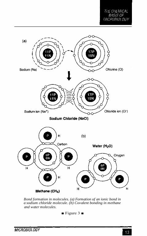
Bond formation in molecules. (a) Formation of an ionic bond ina sodium chloride molecule. (b) Covalent bonding in methaneand water molecules.
■ Figure 3 ■

Organic Compounds
The chemical compounds of living things such as microorganismsare known as organic compounds because of their association with
organisms. The organic compounds, the subject matter of organicchemistry, are the compounds associated with life processes in mi-croorganisms.
Carbohydrates. Four major categories of organic compounds arefound in all microorganisms. The first category is the carbohydrates.
Carbohydrates are used by microorganisms as sources of en-ergy. In addition, carbohydrates serve as structural materials such asin the construction of the microbial cell wall. Carbohydrates are mol-ecules composed of carbon, hydrogen, and oxygen; the ratio of hy-drogen atoms to oxygen atoms is 2:1.
The simple carbohydrates are commonly referred to as sugars.Sugars are monosaccharides if they are composed of single mol-ecules and disaccharides if they are composed of two molecules. Themost important monosaccharide is glucose, a carbohydrate with themolecular formula C6 H 1206 . Glucose is the basic form of fuel formany species of microorganisms. It is soluble and is transported bybody fluids to all cells, where it is metabolized to release its energy.Glucose is the starting material for cellular respiration, and it is themain product of photosynthesis in microorganisms.
Three important disaccharides are also found in living things.One disaccharide is maltose, a combination of two glucose units co-valently linked. Yeast cells break down the maltose from grain starchin the process of alcoholic fermentation. Another disaccharide is su-crose, the table sugar formed by linking glucose to another monosac-charide called fructose. A third disaccharide is lactose, composed ofglucose and galactose units. Lactose, the major carbohydrate in milk,is digested to acid by microorganisms when they sour milk and formsour-milk products such as yogurt and sour cream.
Complex carbohydrates are known as polysaccharides. Polysac-charides are formed by linking eight or more monosaccharide mol-

ecules.Among the most important polysaccharides are starches, whichare composed of hundreds or thousands of glucose units linked to oneanother. Starches serve as a storage form for carbohydrates. Microor-ganisms break down starch to use the glucose it contains for theirenergy needs.
Another important polysaccharide is glycogen, which is relatedto starch. Many bacteria have glycogen in thier cytoplasm. Still an-other is cellulose. Cellulose is also composed of glucose units, butthe units cannot be released from one another except by a few speciesof microorganisms, especially those in the stomach of the cow andother ruminants. The cell walls of algae contain cellulose, and certainfungi have this polysaccharide. Another polysaccharide called chitinis a primary constituent in the fungal cell wall.
Lipids. Lipids are organic molecules composed of carbon, hydro-gen, and oxygen atoms. In contrast to carbohydrates, the ratio of hy-drogen atoms to oxygen atoms is much higher. Lipids include steroids,waxes, and the most familiar lipids, fats.
Fat molecules are composed of a glycerol molecule and one,two, or three molecules of fatty acids. A fatty acid is a long chain ofcarbon atoms with associated hydroxyl (–OH) groups. At one end ofthe fatty acid is an organic acid (–COOH) group . The fatty acids in afat may be all alike or all different. They are bound to the glycerolmolecule during dehydration synthesis, a process that involves theremoval of water (Figure 4a). The number of carbon atoms in a fattyacid may be as few as four or as many as 24.
Certain fatty acids have one or more double bonds in their mol-ecules. Fats that include these molecules are called unsaturated fats.Other fatty acids have no double bonds. Fats that include these fattyacids are called saturated fats.
Some microbial species use fats as energy sources. They pro-duce the enzyme lipase, which breaks down fats to fatty acids andglycerol. An important type of phosphorus-containing lipid, the phos-pholipid, is a major constituent of the cell membranes of all micro-organisms.

Syntheses in organic molecules. (a) Bonding of two fatty acidsto a glycerol molecule in the . formation of a fat. (b) Bonding oftwo amino acids via a peptide bond in the ,formation of a protein.
■ Figure 4 ■
Proteins. Proteins are among the most complex of all organic com-pounds. They are composed of units called amino acids, which con-tain carbon, hydrogen, oxygen, and nitrogen atoms. Certain aminoacids also have sulfur atoms, phosphorus, or other trace elements suchas iron or copper.
Many proteins are immense and complex as compared to carbo-hydrates or fats. However, all are composed of folded, long chains ofthe relatively simple amino acids. There are 20 kinds of amino acids,each with an amino (—NH 2) group and an organic acid (-COOH) group.The amino acids differ with respect to the nature of the chemical groupthat is attached to the base structure. Examples of amino acids arealanine, valine, glutamic acid, tryptophan, tyrosine, and histidine.
Amino acids are linked to form a protein by the removal of watermolecules (Figure 4b). The links forged between the amino acids arecalled peptide bonds, and small proteins are often called peptides.
All living things, including microorganisms, depend upon pro-teins for their existence. Proteins are the major molecules from whichmicroorganisms are constructed. Certain proteins are dissolved orsuspended in the watery substance of the cells, while others are in-

corporated into various structures of the cells, such as the cell mem-brane. Bacterial toxins (metabolic poisons) and microbial flagella andpili are usually composed of proteins.
An essential use for proteins is in the construction of enzymes.Enzymes catalyze the chemical reactions that take place within mi-croorganisms. The enzymes are not used up in the reaction, but re-main available to catalyze succeeding reactions. Without enzymes,the metabolic activity of the microorganism could not take place.
Every species manufactures proteins unique to that species. Theinformation for synthesizing these unique proteins is found in thenucleus of the cell. The so-called genetic code specifies the sequenceof amino acids in the protein and thereby regulates the chemical ac-tivity taking place within the cell. Proteins also can serve as a reservesource of energy for the microorganism. When the amino group isremoved from an amino acid, the resulting compound is energy rich.
Nucleic acids. Like proteins, nucleic acids are very large molecules.The nucleic acids are composed of smaller units called nucleotides.Each nucleotide contains a five-carbon carbohydrate molecule, a phos-phate group, and a nitrogen-containing molecule that has basic prop-erties and is called a nitrogenous base.
Microorganisms contain two important kinds of nucleic acids.One type is called deoxyribonucleic acid, or DNA. The other isknown as ribonucleic acid, or RNA. DNA is found primarily in thenucleus of eukaryotic microorganisms (which have nuclei) and sus-pended in the cytoplasm of prokaryotic microorganisms (which lacknuclei). DNA is also located in plasmids, the tiny loops of DNA foundin bacterial cytoplasm. RNA is found in both the nucleus (if present)and the cytoplasm of the microorganism.
DNA and RNA differ from one another in their components. DNAcontains the carbohydrate deoxyribose, while RNA has ribose. Inaddition, DNA contains the bases adenine, cytosine, guanine, andthymine, while RNA has adenine, cytosine, guanine, and uracil.


MICROSCOPY
S ince microorganisms are invisible to the unaided eye, the essentialtool in microbiology is the microscope. One of the first to use a mi-croscope to observe microorganisms was Robert Hooke, the Englishbiologist who observed algae and fungi in the 1660s. In the 1670s,Anton van Leeuwenhoek, a Dutch merchant, constructed a numberof simple microscopes and observed details of numerous forms ofprotozoa, fungi, and bacteria. During the 1700s, microscopes wereused to further elaborate on the microbial world, and by the late 1800s,the sophisticated light microscopes had been developed. The electronmicroscope was developed in the 1940s, thus making the viruses andthe smallest bacteria (for example, rickettsiae and chlamydiae) vis-ible.
Microscopes permit extremely small objects to be seen, objectsmeasured in the metric system in micrometers and nanometers. Amicrometer (urn) is equivalent to a millionth of a meter, while ananometer (nm) is a billionth of a meter. Bacteria, fungi, protozoa,and unicellular algae are normally measured in micrometers, whileviruses are commonly measured in nanometers. A typical bacteriumsuch as Escherichia coli measures about two micrometers in lengthand about one micrometer in width.
Types of Microscopes
Various types of microscopes are available for use in the microbiol-ogy laboratory. The microscopes have varied applications and modi-fications that contribute to their usefulness.

The light microscope. The common light microscope used in the
laboratory is called a compound microscope because it contains twotypes of lenses that function to magnify an object. The lens closest tothe eye is called the ocular, while the lens closest to the object is
called the objective. Most microscopes have on their base an appara-
tus called a condenser, which condenses light rays to a strong beam.
A diaphragm located on the condenser controls the amount of lightcoming through it. Both coarse and fine adjustments are found on thelight microscope (Figure 5a).
To magnify an object, light is projected through an opening inthe stage, where it hits the object and then enters the objective. Animage is created, and this image becomes an object for the ocularlens, which remagnifies the image. Thus, the total magnificationpossible with the microscope is the magnification achieved by theobjective multiplied by the magnification achieved by the ocular lens.
A compound light microscope often contains four objectivelenses: the scanning lens (4 X ), the low-power lens (10x), the high-power lens (40 X ), and the oil-immersion lens (100 X). With an ocu-lar lens that magnifies 10 times, the total magnifications possible willbe 40 x with the scanning lens, 100 X with the low-power lens, 400 xwith the high-power lens, and 1000 X with the oil-immersion lens.
Most microscopes are parfocal. This term means that the microscoperemains in focus when one switches from one objective to the nextobjective.
The ability to see clearly two items as separate objects under themicroscope is called the resolution of the microscope. The resolu-tion is determined in part by the wavelength of the light used forobserving. Visible light has a wavelength of about 550 nm, whileultraviolet light has a wavelength of about 400 nm or less. The reso-lution of a microscope increases as the wavelength decreases, so ul-traviolet light allows one to detect objects not seen with visible light.The resolving power of a lens refers to the size of the smallest objectthat can be seen with that lens. The resolving power is based on thewavelength of the light used and the numerical aperture of the lens.The numerical aperture (NA) refers to the widest cone of light thatcan enter the lens; the NA is engraved on the side of the objectivelens.

Light microscopy. (a) The important parts of a common lightmicroscope. (b) How immersion oil gathers more light for use inthe microscope.
■ Figure 5 ■

If the user is to see objects clearly, sufficient light must enter theobjective lens. With modem microscopes, entry to the objective isnot a problem for scanning, low-power, and high-power lenses. How-ever, the oil-immersion lens is exceedingly narrow, and most lightmisses it. Therefore, the object is seen poorly and without resolution.To increase the resolution with the oil-immersion lens, a drop of im-mersion oil is placed between the lens and the glass slide (Figure5b). Immersion oil has the same light-bending ability (index of re-fraction) as the glass slide, so it keeps light in a straight line as itpasses through the glass slide to the oil and on to the glass of theobjective, the oil-immersion lens. With the increased amount of lightentering the objective, the resolution of the object increases, and onecan observe objects as small as bacteria. Resolution is important inother types of microscopy as well.
Other light microscopes. In addition to the familiar compound mi-croscope, microbiologists use other types of microscopes for specificpurposes. These microscopes permit viewing of objects not other-wise seen with the light microscope.
An alternative microscope is the dark-field microscope, whichis used to observe live spirochetes, such as those that cause syphilis.This microscope contains a special condenser that scatters light andcauses it to reflect off the specimen at an angle. A light object is seenon a dark background.
A second alternative microscope is the phase-contrast micro-scope. This microscope also contains special condensers that throwlight "out of phase" and cause it to pass through the object at differentspeeds. Live, unstained organisms are seen clearly with this micro-scope, and internal cell parts such as mitochondria, lysosomes, andthe Golgi body can be seen with this instrument.
The fluorescent microscope uses ultraviolet light as its lightsource. When ultraviolet light hits an object, it excites the electronsof the object, and they give off light in various shades of color. Sinceultraviolet light is used, the resolution of the object increases. A labo-ratory technique called the fluorescent-antibody technique employsfluorescent dyes and antibodies to help identify unknown bacteria.

Electron microscopy. The energy source used in the electron mi-croscope is a beam of electrons. Since the beam has an exceptionallyshort wavelength, it strikes most objects in its path and increases theresolution of the microscope significantly. Viruses and some largemolecules can be seen with this instrument. The electrons travel in avacuum to avoid contact with deflecting air molecules, and magnetsfocus the beam on the object to be viewed. An image is created on amonitor and viewed by the technologist.
The more traditional form of electron microscope is the trans-mission electron microscope (TEM). To use this instrument, oneplaces ultrathin slices of microorganisms or viruses on a wire gridand then stains them with gold or palladium before viewing. Thedensely coated parts of the specimen deflect the electron beam, andboth dark and light areas show up on the image.
The scanning electron microscope (SEM) is the more contem-porary form of electron microscope. Although this microscope giveslower magnifications than the TEM, the SEM permits three-dimensional views of microorganisms and other objects. Wholeobjects are used, and gold or palladium staining is employed.
Staining Techniques
Because microbial cytoplasm is usually transparent, it is necessary tostain microorganisms before they can be viewed with the light micro-scope. In some cases, staining is unnecessary, for example when mi-croorganisms are very large or when motility is to be studied, and adrop of the microorganisms can be placed directly on the slide andobserved. A preparation such as this is called a wet mount. A wetmount can also be prepared by placing a drop of culture on a cover-slip (a glass cover for a slide) and then inverting it over a hollowed-out slide. This procedure is called the hanging drop.
In preparation for staining, a small sample of microorganisms isplaced on a slide and permitted to air dry. The smear is heat fixed byquickly passing it over a flame. Heat fixing kills the organisms, makesthem adhere to the slide, and permits them to accept the stain.
Simple stain techniques. Staining can be performed with basic dyessuch as crystal violet or methylene blue, positively charged dyes thatare attracted to the negatively charged materials of the microbial cy-toplasm. Such a procedure is the simple stain procedure. An alter-native is to use a dye such as nigrosin or Congo red, acidic, negativelycharged dyes. They are repelled by the negatively charged cytoplasmand gather around the cells, leaving the cells clear and unstained.This technique is called the negative stain technique.
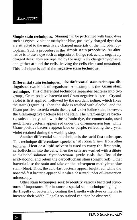
Differential stain techniques. The differential stain technique dis-tinguishes two kinds of organisms. An example is the Gram staintechnique. This differential technique separates bacteria into twogroups, Gram-positive bacteria and Gram-negative bacteria. Crystalviolet is first applied, followed by the mordant iodine, which fixesthe stain (Figure 6). Then the slide is washed with alcohol, and theGram-positive bacteria retain the crystal-violet iodine stain; however,the Gram-negative bacteria lose the stain. The Gram-negative bacte-ria subsequently stain with the safranin dye, the counterstain, usednext. These bacteria appear red under the oil-immersion lens, whileGram-positive bacteria appear blue or purple, reflecting the crystalviolet retained during the washing step.
Another differential stain technique is the acid-fast technique.This technique differentiates species of Mycobacterium from otherbacteria. Heat or a lipid solvent is used to carry the first stain,carbolfuchsin, into the cells. Then the cells are washed with a diluteacid-alcohol solution. Mycobacterium species resist the effect of theacid-alcohol and retain the carbolfuchsin stain (bright red). Otherbacteria lose the stain and take on the subsequent methylene bluestain (blue). Thus, the acid-fast bacteria appear bright red, while thenonacid-fast bacteria appear blue when observed under oil-immersionmicroscopy.
Other stain techniques seek to identify various bacterial struc-tures of importance. For instance, a special stain technique highlightsthe flagella of bacteria by coating the flagella with dyes or metals toincrease their width. Flagella so stained can then be observed.

The Gram stain procedure used for differentiating bacteria intotwo groups.
■ Figure 6 ■

A special stain technique is used to examine bacterial spores.Malachite green is used with heat to force the stain into the cells andgive them color. A counterstain, safranin, is then used to give color tothe nonsporeforming bacteria. At the end of the procedure, sporesstain green and other cells stain red.

MICROORGANISMS ASPROKARYOTES AND EUKARYOTES
Microorganisms and all other living organisms are classified asprokaryotes or eukaryotes. Prokaryotes and eukaryotes are distin-guished on the basis of their cellular characteristics. For example,prokaryotic cells lack a nucleus and other membrane-bound struc-tures known as organelles, while eukaryotic cells have both a nucleusand organelles (Figure 7).
Prokaryotic and eukaryotic cells are similar in several ways. Bothtypes of cells are enclosed by cell membranes (plasma membranes),and both use DNA for their genetic information.
Prokaryotes include several kinds of microorganisms, such asbacteria and cyanobacteria. Eukaryotes include such microorganismsas fungi, protozoa, and simple algae. Viruses are considered neitherprokaryotes nor eukaryotes because they lack the characteristics ofliving things, except the ability to replicate (which they accomplishonly in living cells).
Prokaryotic Cells
The characteristics of prokaryotic cells apply to the bacteria andcyanobacteria (formerly known as blue-green algae), as well as to therickettsiae, chlamydiae, and mycoplasmas.
Size and shape. Prokaryotes are probably the smallest living organ-isms, ranging in size from 0.151.um (mycoplasmas) to 0.25 µm (chlamy-diae) to 0.45 µm (rickettsiae) to about 2.0 µm (many of the bacteria).Certain prokaryotes, such as bacteria, occur in spherical forms calledcocci (singular, coccus) or in rodlike forms called bacilli (singular,bacillus). Some bacteria have a comma shape (vibrio), or a flexible,wavy shape (spirochete), or a corkscrew shape (spirillum).

The important cellular features of (a) a prokaryotic cell (abacterium) and (b) a eukaryotic cell.
■ Figure 7 ■

Some prokaryotes have a variety of shapes and sizes and are saidto be pleomorphic. Rickettsiae and mycoplasmas are examples ofpleomorphic microorganisms.
When certain prokaryotes divide, they cling to each other in adistinct arrangement. A diplococcus, for example, consists of a pairof cocci, while a streptococcus consists of a chain of cocci, and atetracoccus consists of four cocci arranged in a cube. A grapelikecluster of cocci is called a staphylococcus. Bacilli sometimes formlong chains called streptobacilli.
The cell wall and cell membrane. With the exception of mycoplas-mas, all bacteria have a semirigid cell wall. The cell wall gives shapeto the organisms and prevents them from bursting, especially sincematerials in the cytoplasm exert osmotic pressures.
The chief component of the prokaryotic cell wall is peptidogly-can, a large polymer composed of N-acetylglucosamine and N-acetylmuramic acid. Gram-positive bacteria have more peptidoglycanin their cell wall, which may account for their ability to retain thestain in the Gram stain procedure. Gram-negative bacteria have morelipids in their cell wall. Polymers of teichoic acid are commonly as-sociated with the peptidoglycan in Gram-positive bacteria.
In addition to the cell wall, Gram-negative bacteria have a verythin surrounding layer called the outer membrane. Lipopolysaccha-rides known as endotoxins are part of this outer membrane. A spacecalled the periplasmic space separates the cell wall from the outermembrane and contains a substance called periplasm.
All prokaryotes have cytoplasm surrounded by a cell membrane,also known as the plasma membrane. The cell membrane conformsto the fluid mosaic model, which means that its proteins float within adouble layer of phospholipids. Respiratory enzymes are located atthe cell membrane of prokaryotes, and the membrane assists DNAreplication and has attachment points for bacterial flagella.

The cytoplasm. The cytoplasm of prokaryotic cells contains ribo-somes and various other granules used by the organism. The DNA iscontained in the nuclear region (the nucleoid) and has no histoneprotein to support it. Prokaryotic cells have in their cytoplasm a single,
looped chromosome, as well as numerous small loops of DNA called
plasmids. Genetic information in the plasmids is apparently not es-sential for the continued survival of the organism.
Prokaryotic ribosomes contain protein and ribonucleic acid(RNA) and are the locations where protein is synthesized. Prokary-otic ribosomes have a sedimentation rate of 70S, and are thereforeknown as 70S ribosomes. (Eukaryotic cells have 80S ribosomes.)Certain antibiotics bind to these ribosomes and inhibit protein syn-thesis.
Some prokaryotic cells that engage in photosynthesis have inter-nal membranes called thylakoids where their chlorophyll pigmentsare located. These membranes are also the sites of enzymes for pho-tosynthesis. Certain bacteria have granules of phosphorus, starch, orglycogen. Granules called metachromatic granules stain with me-thylene blue and are used in diagnostic circumstances. Some bacte-rial species also have magnetosomes, which contain magneticsubstances to help orient the organisms to hospitable environments.
External cellular structures. Many prokaryotic cells have at theirsurface a number of external structures that assist their functions.Among these structures are flagella. Flagella are found primarily inbacterial rods and are used for motility. A bacterium may have a singleflagellum (a monotrichous bacterium), or flagella at both ends of thecell (an amphitrichous bacterium), or two or more flagella at one endof the cell (a lophotrichous bacterium), or it may be surrounded byflagella (a peritrichous bacterium).
Flagella are long, ultrathin structures, many times the length ofthe cell. They are composed of the protein flagellin arranged in long
fibers. A hooklike structure and basal body connect the flagellum tothe cell membrane. Flagella rotate and propel the bacteria.

Spirochetes lack flagella, but they possess axial filaments. Theaxial filaments extend beyond the cell wall and cause the spirocheteto rotate in a corkscrew fashion and thereby move.
Some bacterial species have projections called pill (singular, pi-lus). Pili are used for attachments to surfaces such as tissues. Manypathogens possess pili, which are composed of the protein pilin. Cer-tain pili, known as conjugation pili, unite prokaryotic cells to oneanother and permit the passage of DNA between the cells. The termfimbriae is often used for the attachment pili.
Many bacteria, especially pathogens, are enclosed at their sur-face by a layer of polysaccharides and proteins called the glycocalyx.The glycocalyx, composed of a thick, gummy material, serves as areservoir for nutrients and protects the organism from changes in theenvironment. When the glycocalyx is a tightly bound structure, it isknown as a capsule. When it is a poorly bound structure that flowseasily, it is known as a slime layer. The material in dental plaque iscomposed largely of the material from the slime layer.
Endospores. Bacteria of the genera Bacillus and Clostridium areable to form highly resistant internal structures called endospores, orsimply spores. Spores are formed during the normal life cycle whenthe environment becomes too harsh (Figure 8).
One vegetative (multiplying) cell produces one spore. Spores areable to withstand extremely high temperatures, long periods of dry-ing, and other harsh environments. When conditions are favorable,the spore germinates and releases a new vegetative cell, which multi-plies and reforms the colony. Sporeformers include the agents of an-thrax, tetanus, botulism, and gas gangrene. Spores contain dipicolinicacid and calcium ions, both of which contribute to their resistance.

The process of spore formation as it occurs in species of Bacillusand Clostridium.
■ Figure 8 ■
Eukaryotic Cells
Eukaryotic cells are generally larger and more complex than prokary-otic cells. They also contain a variety of cellular bodies called or-ganelles. The organelles function in the activities of the cell and arecompartments for localizing metabolic function. Microscopic proto-zoa, unicellular algae, and fungi have eukaryotic cells.

Nucleus. Eukaryotic cells have a distinctive nucleus, composed pri-marily of protein and deoxyribonucleic acid, or DNA. The DNA isorganized into linear units called chromosomes, also known as chro-matin when the linear units are not obvious. Functional segments ofthe chromosomes are referred to as genes.
The nuclear proteins belong to a class of proteins called histones.Histones provide a supportive framework for the DNA in chromo-somes. The DNA replicates in eukaryotic cells during the process ofmitosis.
The nucleus of eukaryotic cells is surrounded by an outer mem-brane called the nuclear envelope, which is a double-membrane struc-ture consisting of two lipid layers similar to the cell membrane. Poresexist in the nuclear membrane, and the internal nuclear environmentcan therefore communicate with the cytoplasm of the cell.
Within the nucleus are two or more dense masses referred to asnucleoli (singular, nucleolus). The nucleolus is an RNA-rich areawhere ribosomes are assembled before passing out of the nucleusinto the cytoplasm.
Cellular organelles. Within the cytoplasm (also known as the cyto-sol) of eukaryotic cells are a number of microscopic bodies calledorganelles ("little organs"). Various functions of the cell go on withinthese organelles.
An example of an organelle is the endoplasmic reticulum (ER),a series of membranes that extend throughout the cytoplasm of eu-karyotic cells. In some places the ER is studded with submicroscopicbodies called ribosomes. This type of ER is referred to as rough ER.In other places there are no ribosomes, and the ER is called smoothER. The endoplasmic reticulum is the site of protein synthesis in thecell. Eukaryotic ribosomes are 80S bodies where the amino acids arebound together to form proteins. The spaces within the ER mem-branes are known as cisternae.
Another organelle is the Golgi body (also called the Golgi appa-ratus). The Golgi body is a series of flattened sacs, usually curled atthe edges. The outermost sac often bulges away to form droplike

vesicles known as secretory vesicles. It is in the Golgi body that thecell's proteins and lipids are processed and packaged before beingsent to their final destination.
Another organelle, the lysosome, is derived from the Golgi body.It is a somewhat circular, droplike sac of enzymes within the cyto-plasm. These enzymes are used for digestion in the cell. They breakdown the particles of food taken into the cell and make the productsavailable to the cell. Enzymes are also contained in a cytoplasmicbody called the peroxisome.
The organelle where much energy is released in the eukaryoticcell is the mitochondrion (plural, mitochondria). The energy re-leased is used to form adenosine triphosphate (ATP). Because theyare involved in energy release and storage, the mitochondria are calledthe "powerhouses of the cells."
An organelle found in certain protozoa is a large, fluid-filled,contractile vacuole. The vacuole may occupy over 75 percent of thecell interior and is used for eliminating water. Water pressure build-ing up within the vacuole may cause the cell to swell.
Still another organelle within the cell is the cytoskeleton, an in-terconnected system of fibers, threads, and interwoven molecules thatgive structure to the cell. The main components of the cytoskeletonare microtubules, microfilaments, and intermediate filaments. Allare assembled from subunits of protein.
Many eukaryotic cells contain flagella and cilia. Eukaryotic fla-gella, like prokaryotic flagella, are long, hairlike organelles that ex-tend from the cell. Eukaryotic flagella whip about and propel the cell(as in protozoa) and are composed of nine pairs of microfilamentsarranged about a central pair. Cilia are shorter and more numerousthan flagella. In moving cells, they wave in synchrony and move thecell. Paramecium is a well-known ciliated protozoan.
The cell wall. Many species of eukaryotes, such as fungi, contain acell wall outside the cell membrane. In fungi, the cell wall contains acomplex polysaccharide called chitin as well as some cellulose. Al-gal cells, by contrast, have no chitin; rather, their cell walls are com-posed exclusively of the polysaccharide cellulose.

Cell walls provide support for eukaryotic cells and help the cellsresist mechanical pressures while giving them a boxlike appearance.The cell walls are not selective devices, as are the cell membranes.
The cell membrane. The eukaryotic cell membrane conforms tothe fluid mosaic model found in the prokaryotic membrane. In eu-karyotes, the membrane is a dynamic structure governing passage ofdissolved molecules and particles into and out from the cytoplasm.However, it neither contains the enzymes found in the prokaryoticcell nor functions in DNA replication.
In order for the cytoplasm of prokaryotic and eukaryotic cells tocommunicate with the external environment, materials must be ableto move through the cell membrane. There are several mechanismsby which movement can occur. One method, called diffusion, is themovement of molecules from a region of high concentration to one oflow concentration. This movement occurs because the molecules areconstantly colliding with one another, and the net movement of themolecules is away from the region of high concentration. Diffusion isa random movement of molecules, and the pathway the moleculestake is called the concentration gradient. Molecules are said to movedown the concentration gradient in diffusion.
Another method of movement across the membrane is osmosis,the movement of water from a region of high concentration to one oflow concentration. Osmosis occurs across a membrane that is semi-permeable, meaning that the membrane lets only certain moleculespass through while keeping other molecules out. Osmosis is a type ofdiffusion involving only water.
A third mechanism for movement across the membrane is facili-tated diffusion, a type of diffusion assisted by certain proteins in themembrane. The proteins permit only certain molecules to pass acrossthe membrane and encourage movement from a region of high con-centration of molecules to one of low concentration.
A fourth method for passing across the membrane is active trans-port. When active transport is taking place, a protein moves a certainmaterial across the membrane from a region of low concentration toone of high concentration. Because this movement is happeningagainst the concentration gradient, it requires that energy be expended,energy usually derived from ATP.
The final mechanism for movement across the cell membrane isendocytosis, a process in which a small patch of cell membrane en-closes particles or tiny volumes of fluid at or near the cell surface.The membrane enclosure then sinks into the cytoplasm and pinchesoff from the membrane. When the vesicle contains particulate matter,the process is called phagocytosis; when it contains droplets of fluid,the process is called pinocytosis.

MICROBIAL METABOLISM
Microbial life can exist only where molecules and cells remain or-ganized, and energy is needed by all microorganisms to maintain or-ganization.
Every activity taking place in microbial cells involves both a shiftof energy and a measurable loss of energy. Although the second lawof thermodynamics says that energy cannot be created or destroyed,but only transferred within a system, unfortunately, the transfers ofenergy in living systems are never completely efficient. For this rea-son, considerably more energy must be taken into the system than isnecessary to simply carry out the actions of microbial life.
Chemical Reactions and Energy
In microorganisms, most chemical compounds neither combine withone another automatically nor break apart automatically. A spark calledthe energy of activation is needed. The activation energy needed tospark an exergonic (energy-yielding) reaction or endergonic (energy-requiring) reaction can be heat energy or chemical energy. Reactionsthat require activation energy can also proceed in the presence ofbiological catalysts. Catalysts are substances that speed up chemicalreactions but remain unchanged during the reactions. Catalysts workby lowering the required amount of activation energy for the chemi-cal reaction. In microorganisms, the catalysts are enzymes.
Enzymes. Chemical reactions in microorganisms operate in the pres-ence of enzymes. A particular enzyme catalyzes only one reaction,and thousands of different enzymes exist in a microbial cell to cata-lyze thousands of different chemical reactions. The substance actedon by an enzyme is called its substrate. The products of an enzyme-catalyzed chemical reaction are called end products.

All enzymes are composed of proteins. When an enzyme func-tions, a key portion of the enzyme called the active site interacts withthe substrate. The active site closely matches the molecular configu-ration of the substrate, and after this interaction has taken place, ashape change at the active site places a physical stress on the sub-strate. This physical stress aids the alteration of the substrate and pro-duces the end products. After the enzyme has performed its work, theproduct or products drift away. The enzyme is then free to function inthe next chemical reaction. Enzyme-catalyzed reactions occur ex-tremely fast.
With some exceptions, enzyme names end in "-ase. " For example,the microbial enzyme that breaks down hydrogen peroxide to waterand hydrogen is called catalase. Other well-known enzymes are amy-lase, hydrolase, peptidase, and kinase.
The rate of an enzyme-catalyzed reaction depends on a numberof factors, including the concentration of the substrate, the acidity ofthe environment, the presence of other chemicals, and the tempera-ture of the environment. For example, at higher temperatures, en-zyme reactions occur more rapidly. Since enzymes are proteins,however, excessive amounts of heat may cause the protein to changeits structure and become inactive. An enzyme altered by heat is saidto be denatured.
Enzymes work together in metabolic pathways. A metabolicpathway is a sequence of chemical reactions occurring in a cell. Asingle enzyme-catalyzed reaction may be one of multiple reactions inthe metabolic pathway. Metabolic pathways may be of two generaltypes: Some involve the breakdown or digestion of large, complexmolecules in the process of catabolism. Others involve a synthesis,generally by joining smaller molecules in the process of anabolism.
Many enzymes are assisted by chemical substances called cofac-tors. Cofactors may be ions or molecules associated with an enzymeand required in order for a chemical reaction to take place. Ions thatmight operate as cofactors include those of iron, manganese, or zinc.Organic molecules acting as cofactors are referred to as coenzymes.Examples of coenzymes are NAD and FAD (to be discussed shortly).

Adenosine triphosphate (ATP). Adenosine triphosphate (ATP) isthe chemical substance that serves as the currency of energy in themicrobial cell. It is referred to as currency because it can be "spent"in order to make chemical reactions occur.
ATP, used by virtually all microorganisms, is a nearly universalmolecule of energy transfer. The energy released during the reactionsof catabolism is stored in ATP molecules. In addition, the energytrapped in anabolic reactions such as photosynthesis is also trappedin ATP.
An ATP molecule consists of three parts (Figure 9a). One part isa double ring of carbon and nitrogen atoms called adenine. Attachedto the adenine molecule is a small five-carbon carbohydrate calledribose. Attached to the ribose molecule are three phosphate groups,which are linked by covalent bonds.
The covalent bonds that unite the phosphate units in ATP arehigh-energy bonds. When an ATP molecule is broken down by anenzyme, the third (terminal) phosphate unit is released as a phos-phate group, which is a phosphate ion (Figure 9b). With the release,
The adenosine triphosphate (ATP) molecule that serves as animmediate energy source in the cell.
■ Figure 9 ■

approximately 7.3 kilocalories of energy (a kilocalorie is 1000 calo-ries) are made available to do the work of the microorganism.
The breakdown of an ATP molecule is accomplished by an en-zyme called adenosine triphosphatase. The products of ATP break-down are adenosine diphosphate (ADP) and, as noted, a phosphateion. Adenosine diphosphate and the phosphate ion can be reconsti-tuted to form ATP, much as a battery can be recharged. To accom-plish this ATP formation, energy necessary for the synthesis can bemade available in the microorganism through two extremely impor-tant processes: photosynthesis and cellular respiration. A process calledfermentation may also be involved.
ATP production. ATP is generated from ADP and phosphate ionsby a complex set of processes occurring in the cell, processes thatdepend upon the activities of a special group of cofactors called co-enzymes. Three important coenzymes are nicotinamide adenine di-nucleotide (NAD), nicotinamide adenine dinucleotide phosphate(NADP), and flavin adenine dinucleotide (FAD). All are structurallysimilar to ATP.
All coenzymes perform essentially the same work. During thechemical reactions of metabolism, coenzymes accept electrons andpass them on to other coenzymes or other molecules. The removal ofelectrons or protons from a coenzyme is called oxidation. The addi-tion of electrons or protons to a coenzyme is called reduction. There-fore, the chemical reactions performed by coenzymes are calledoxidation-reduction reactions.
The oxidation-reduction reactions performed by the coenzymesand other molecules are essential to the energy metabolism of thecell. Other molecules participating in this energy reaction are calledcytochromes. Together with the enzymes, cytochromes accept andrelease electrons in a system referred to as the electron transportsystem. The passage of energy-rich electrons among cytochromesand coenzymes drains the energy from the electrons. This is the en-ergy used to form ATP from ADP and phosphate ions.
The actual formation of ATP molecules requires a complex pro-cess referred to as chemiosmosis. Chemiosmosis involves the cre-
ation of a steep proton gradient, which occurs between the membrane-bound areas. In prokaryotic cells (for example, bacteria), it is the areaof the cell membrane; in eukaryotic cells, it is the membranes of themitochondria. A gradient is formed when large numbers of protons(hydrogen ions ) are pumped into membrane-bound compaitments.The protons build up dramatically within the compartment, finallyreaching an enormous number. The energy used to pump the protonsis energy released from the electrons during the electron transportsystem.
After large numbers of protons have gathered at one side of themembrane, they suddenly reverse their directions and move backacross the membranes. The protons release their energy in this mo-tion, and the energy is used by enzymes to unite ADP with phosphateions to form ATP. The energy is trapped in the high-energy bond ofATP by this process, and the ATP molecules are made available toperform cell work.
Cellular Respiration
Microorganisms such as cyanobacteria can trap the energy in sun-light through the process of photosynthesis and store it in the chemi-cal bonds of carbohydrate molecules. The principal carbohydrateformed in photosynthesis is glucose. Other types of microorganismssuch as nonphotosynthetic bacteria, fungi, and protozoa are unable toperform this process. Therefore, these organisms must rely upon pre-formed carbohydrates in the environment to obtain the energy neces-sary for their metabolic processes.
Cellular respiration is the process by which microorganismsobtain the energy available in carbohydrates. They take the carbohy-drates into their cytoplasm, and through a complex series of meta-bolic processes, they break down the carbohydrate and release theenergy. The energy is generally not needed immediately, so it is usedto combine ADP with phosphate ions to form ATP molecules. Dur-ing the process of cellular respiration, carbon dioxide is given off asa waste product. This carbon dioxide can be used by photosynthesiz-

ing cells to form new carbohydrates. Also in the process of cellularrespiration, oxygen gas is required to serve as an acceptor of elec-trons. This oxygen gas is identical to the oxygen gas given off inphotosynthesis.
The overall mechanism of cellular respiration involves four sub-divisions: glycolysis, in which glucose molecules are broken downto form pyruvic acid molecules; the Krebs cycle, in which pyruvicacid is further broken down and the energy in its molecule is used toform high-energy compounds such as NADH; the electron trans-port system, in which electrons are transported along a series of co-enzymes and cytochromes and the energy in the electrons is released;and chemiosmosis, in which the energy given off by electrons is usedto pump protons across a membrane and provide the energy for ATPsynthesis.
Glycolysis. The process of glycolysis is a multistep metabolic path-way that occurs in the cytoplasm of microbial cells and the cells ofother organisms. At least six enzymes operate in the metabolic path-way.
In the first and third steps of the pathway, ATP is used to ener-gize the molecules. Thus, two molecules of ATP must be expendedin the process. Further along in the process, the six-carbon glucosemolecule is converted into intermediary compounds and then is splitinto two three-carbon compounds. The latter undergo additional con-versions and eventually form pyruvic acid at the conclusion of theprocess.
During the latter stages of glycolysis, four ATP molecules aresynthesized using the energy given off during the chemical reactions.Thus, four ATP molecules are synthesized and two ATP moleculesare inserted into the process for a net gain of two ATP molecules inglycolysis.
Also during glycolysis, another of the reactions yields enoughenergy to convert NAD to NADH. The reduced coenzyme (NADH)will later be used in the electron transport system, and its energy willbe released. During glycolysis, two NADH molecules are produced.
As glycolysis does not use oxygen, the process is considered to

be anaerobic. For certain anaerobic organisms, such as certain bac-teria and fermentation yeasts, glycolysis is the sole source of energy.It is a somewhat inefficient process because much of the cellular en-ergy remains in the two molecules of pyruvic acid.
The Krebs cycle. Following glycolysis, the mechanism of cellularrespiration then involves another multistep process called the Krebscycle, also called the citric acid cycle and the tricarboxylic acid cycle.The Krebs cycle uses the two molecules of pyruvic acid formed inglycolysis and yields high-energy molecules of NADH and FADHand some ATP and carbon dioxide (Figure 10).
The Krebs cycle occurs at the cell membrane of bacterial cellsand in the mitochondria of eukaryotic cells. Each of these sausage-shaped organelles of eukaryotic microorganisms possesses inner andouter membranes, and therefore an inner and outer compartment. Theinner membrane is folded over itself many times; the folds are calledcristae. Along the cristae are the important enzymes necessary forthe proton pump and for ATP production.
Prior to entering the Krebs cycle, the pyruvic acid molecules areprocessed. Each three-carbon molecule of pyruvic acid undergoesconversion to a substance called acetyl-coenzyme A, or acetyl-CoA.In the process, the pyruvic acid molecule is broken down by an en-zyme, one carbon atom is released in the form of carbon dioxide, andthe remaining two carbon atoms are combined with a coenzyme calledcoenzyme A. This combination forms acetyl-CoA. In the process,electrons and a hydrogen ion are transferred to NAD to form high-energy NADH.
Acetyl-CoA now enters the Krebs cycle by combining with afour-carbon acid called oxaloacetic acid. The combination forms thesix-carbon acid called citric acid. Citric acid undergoes a series ofenzyme-catalyzed conversions. The conversions, which involve upto 10 chemical reactions, are all brought about by enzymes. In manyof the steps, high-energy electrons are released to NAD. The NADmolecule also acquires a hydrogen ion and becomes NADH. In oneof the steps, FAD serves as the electron acceptor, and it acquires twohydrogen' ions to become FADH 2 . Also, in one of the reactions, enough

An overview of the processes of cellular respiration showing themajor pathways and the places where ATP is synthesized.
■ Figure 10 ■

energy is released to synthesize a molecule of ATP. Since there aretwo pyruvic acid molecules entering the system, two ATP moleculesare formed.
Also during the Krebs cycle, the two carbon atoms of acetyl-CoA are released and each forms a carbon dioxide molecule. Thus,for each acetyl-CoA entering the cycle, two carbon dioxide moleculesare formed. Since two acetyl-CoA molecules enter the cycle, and eachhas two carbon atoms, four carbon dioxide molecules will form. Addthese four molecules to the two carbon dioxide molecules formed inthe conversion of pyruvic acid to acetyl-CoA, and the total is sixcarbon dioxide molecules. These six CO 2 molecules are given off aswaste gas in the Krebs cycle. They represent the six carbons of glu-cose that originally entered the process of glycolysis.
At the end of the Krebs cycle, the final product formed is oxalo-acetic acid, identical to the oxaloacetic acid which begins the cycle.The molecule is now ready to accept another acetyl-CoA molecule tobegin another turn of the cycle. All told, the Krebs cycle forms (pertwo molecules of pyruvic acid) two ATP molecules, a large numberof NADH molecules, and some FADH2 molecules. The NADH andthe FADH2 will be used in the electron transport system.
The electron transport system. The electron transport system oc-curs at the bacterial cell membrane and in the cristae of the mitochon-dria in eukaryotic cells. Here, a series ofcytochromes (cell pigments)and coenzymes exist. These cytochromes and coenzymes act as car-rier molecules and transfer molecules. They accept high-energy elec-trons and pass the electrons to the next molecule in the system. Atkey proton-pumping sites, the energy of the electrons is used to trans-port protons across the cell membrane or into the outer compartmentof the mitochondrion.
Each NADH molecule is highly energetic. It accounts for thetransfer of six protons across the membrane. Each FADH 2 moleculeaccounts for the transfer of four protons. Electrons pass from NAD toFAD, to other cytochromes and coenzymes, and eventually they losemuch of their energy. The final electron acceptor is an oxygen atom.The electron-oxygen combination then takes on two protons to form

a molecule of water (H20). As a final electron receptor, oxygen isresponsible for removing electrons from the system. If oxygen werenot available, electrons could not be passed among the coenzymes,the energy in electrons could not be released, the proton pump couldnot be established, and ATP could not be produced.
Chemiosmosis. The actual production of ATP in cellular respirationtakes place during chemiosmosis. As previously noted, chemiosmosisinvolves the pumping of protons through special channels in the mem-branes of mitochondria from the inner to the outer compartment. Inbacteria, the punping occurs at the cell membrane. The pumping es-tablishes a proton gradient. Once the gradient is established, protonspass down the gradient through molecular particles. In these particles,the energy of the protons is used to generate ATP, using ADP andphosphate ions as the starting points.
The energy production in cellular respiration during chemiosmosisis substantial. Most biochemists agree that in prokaryotic microor-ganisms, a total of 36 molecules of ATP can be produced during cel-lular respiration. In eukaryotic cells, the number is 34 molecules ofATP. Two molecules of ATP are produced as the net gain of glycoly-sis, so the grand total is 38 molecules of ATP (36 in eukaryotes).These ATP molecules may then be used in the cell for its needs.
Fermentation. Fermentation is an anaerobic process in which en-ergy can be released from glucose even though oxygen is not avail-able. Fermentation occurs in yeast cells, and a form of fermentationtakes place in bacteria.
In yeast cells, glucose can be metabolized through cellular respi-ration, as in other cells. When oxygen is lacking, however, glucose isstill changed to pyruvic acid via glycolysis. The pyruvic acid is firstconverted to acetaldehyde and then to ethyl alcohol. The net gain ofATP to the yeast cell is two molecules—the two molecules of ATPnormally produced in glycolysis.
Yeasts are able to participate in fermentation because they havethe necessary enzyme to convert pyruvic acid to ethyl alcohol. This

process is essential because it removes electrons and hydrogen ionsfrom NADH during glycolysis. The effect is to free the NAD so thatit can participate in future reactions of glycolysis. Yeasts are there-fore used in both bread and alcohol production. Alcohol fermenta-tion is the process that yields beer, wine, and other spirits. The carbondioxide given off supplements the carbon dioxide given off duringthe Krebs cycle and causes bread to rise.
Photosynthesis
A great variety of living things on earth, including all photosyntheticmicroorganisms, synthesize their foods from simple molecules suchas carbon dioxide and water. In microorganisms, photosynthesis oc-curs in unicellular algae and in photosynthesizing bacteria such ascyanobacteria and green and purple sulfur bacteria.
Photosynthesis is actually two processes. In the first process,energy-rich electrons flow through a series of coenzymes and othermolecules, and this electron energy is trapped. During the trappingprocess, ATP molecules and molecules of nicotinamide adenine di-nucleotide phosphate hydrogen (NADPH) are formed, both rich inenergy. These molecules are then used in the second half of the pro-cess, where carbon dioxide molecules are bound into carbohydratesto form organic substances such as glucose.
Photosynthesis occurs along the thylakoid membranes of eukary-otic organisms. The thylakoids are somewhat similar to the cristae ofmitochondria. Sunlight is captured in the thylakoid by pigment mol-ecules organized into photosystems. The coenzyme NADP functionsin the system. The photosystem includes the pigment molecules, aswell as proton pumps, and coenzymes and molecules of electrontransport systems. In prokaryotic microorganisms, the chlorophyllmolecules are dissolved in the cell's cytoplasm and are called bac-teriochlorophylls.
The process of photosynthesis is conveniently divided into twoparts: the energy-fixing reaction (also called the light reaction) andthe carbon-fixing reaction (also called the light-independent reaction,or the dark reaction).

The energy-fixing reaction. The energy-fixing reaction of photo-synthesis begins when light is absorbed in a photosystem. The en-ergy activates electrons to jump out of chlorophyll molecules in thereaction center. These electrons pass through a series of cytochromesin the nearby electron transport system. Some of the energy of theelectrons is lost as they move along the chain of acceptors, but aportion of the energy is used to pump protons across a membrane,setting up the potential for chemiosmosis.
After passing through the electron transport system, the energy-rich electrons enter another photosystem. Light now activates the elec-trons, and they receive a second boost out of the chlorophyll molecules.The electrons progress through a second electron transport systemand enter a molecule of NADP. Since NADP has acquired two nega-tively charged electrons, it attracts two positively charged protonsfrom a water molecule to balance the charges, and the molecule isreduced to NADPH. This molecule contains much energy.
Because electrons have flowed out of the chlorophyll molecules,the latter are left without a certain number of electrons. These elec-trons are replaced by electrons secured from water molecules. Thethird product of the disrupted water molecules is oxygen. Two oxy-gen atoms combine with one another to form molecular oxygen. Thisoxygen is given off by cyanobacteria as the waste product of photo-synthesis. It is the oxygen that fills the atmosphere and is used by alloxygen-breathing organisms.
What has been described above are the noncyclic energy-fixingreactions. Certain microorganisms are also known to participate incyclic energy-fixing reactions. Excited electrons leave the chloro-phyll molecules, pass through coenzymes of the electron transportsystem, and then follow a special pathway back to the chlorophyllmolecules. Each electron powers the proton pump and encouragesthe transport of a proton across the membrane. This process enrichesthe proton gradient and eventually leads to the generation of ATP.
ATP production in the energy-fixing reactions of photosynthesisoccurs by the process of chemiosmosis. Essentially, it consists of arush of proteins across a membrane (the microbial membrane, in thiscase) accompanied by the synthesis of ATP molecules. It has beencalculated that the proton concentration on one side of the membraneis 10,000 times that on the opposite side of the membrane.
In photosynthesis, the protons pass back across the membranesthrough channels that lie alongside sites where enzymes are located.As the protons pass through the channels, the energy of the protons isreleased to form high-energy ATP bonds. ATP is formed in the energy-fixing reactions along with NADPH formed in the main reactions.Both ATP and NADPH provide the energy necessary for the synthesisof carbohydrates that occurs in the second major set of events inphotosynthesis.
The carbon-fixing reaction. In the carbon-fixing reaction of pho-tosynthesis, glucose and other carbohydrates are synthesized. Thisphase of photosynthesis occurs in the cytoplasm of the microbial cell.
In the carbon-fixing stage, an essential material, carbon diox-ide, is obtained from the atmosphere. The carbon dioxide is attachedto a five-carbon compound called ribulose biphosphate (RuBP) toform a six-carbon product. This product immediately breaks into twothree-carbon molecules (Figure 11).
The three-carbon molecule is called phosphoglycerate (PGA).Each phosphoglycerate molecule is converted to phosphoglycer-aldehyde (PGAL) using the ATP and NADPH synthesized in theenergy-fixing reaction. The organic compounds that result have threecarbon atoms. They interact with one another and eventually join toform a single molecule of six-carbon glucose. The process alsogenerates more molecules of ribulose biphosphate to participate infurther carbon-fixing reactions.
The carbon-fixing reaction is often referred to as the Calvin cycle,for Melvin Calvin, who performed much of the biochemical research.The product of the reaction is glucose, a carbohydrate containing theenergy of sunlight, which began the reactions in the chlorophyll mol-ecule. This energy has passed through ATP and NADPH and is nowpresent in the high-energy glucose molecules. Photosynthesizing mi-croorganisms use the glucose to obtain the energy for their activities.Nonphotosynthesizing organisms use this same glucose by consum-ing the carbohydrate.

The carbon-fixing reaction of photosynthesis. The A TP andNADPH used in this process are synthesized in the energy fixingphase.
■ Figure 11 ■

MICROBIAL CULTIVATION AND GROWTH
A characteristic of microorganisms is their ability to grow and forma population of organisms. One of the results of microbial metabo-lism is an increase in the size of the cell. The many requirements forsuccessful growth include those both chemical and physical.
Growth Requirements
Chemical requirements. In order to grow successfully, microor-ganisms must have a supply of water as well as numerous other sub-stances including mineral elements, growth factors, and gas, such asoxygen. Virtually all chemical substances in microorganisms containcarbon in some form, whether they be proteins, fats, carbohydrates,or lipids. Perhaps 50 percent of a bacterium's dry weight is carbon.Carbon can be obtained from organic materials in the environment,or it may be derived from carbon dioxide. Both chemoautotrophicand photoautotrophic microorganisms obtain their energy and pro-duce their nutrients from simple inorganic compounds such as car-bon dioxide. Chemoautotrophs do so through chemical reactions,while photoautotrophs use photosynthesis.
Among the other elements required by microorganisms are ni-trogen and phosphorous. Nitrogen is used for the synthesis of pro-teins, amino acids, DNA, and RNA. Bacteria that obtain nitrogendirectly from the atmosphere are called nitrogen-fixing bacteria. Theyinclude species ofRhizobium and Azotobacter, both found in the soil.Phosphorus is an essential element for nucleic acid synthesis and forthe construction of phospholipids.
Oxygen is used by aerobic bacteria during the process of cellularrespiration as a final electron acceptor. For aerobic organisms, oxy-gen is an absolute requirement for their energy-yielding properties.Certain microorganisms grow in oxygen-free environments and aredescribed as anaerobic. Organisms such as these produce odorifer-

ous gases in their metabolism, including hydrogen sulfide gas andmethane. Certain pathogenic species, such as Clostridium species,
are anaerobic.Certain species of microorganisms are said to be facultative.
These species grow in either the presence or absence of oxygen. Somebacteria species are microaerophilic, meaning that they grow in lowconcentrations of oxygen. In some cases, these organisms must havean environment rich in carbon dioxide. Organisms such as these aresaid to be capnophilic.
Other chemical requirements for microbial growth include suchtrace elements as iron, copper, and zinc. These elements often areused for the synthesis of enzymes. Organic growth factors such asvitamins may also be required by certain bacteria. Amino acids, pu-rines, and pyrimidines should also be available.
Physical requirements. Certain physical conditions affect the typeand amount of microbial growth. For example, enzyme activity de-pends on the temperature of the environment, and microorganismsare classified in three groups according to their temperature prefer-ences: psychrophilic organisms (psychrophiles) prefer cold tempera-tures of about 0°C to 20°C; mesophilic organisms (mesophiles) prefertemperatures at 20°C to 40°C; thermophilic organisms (thermophiles)prefer temperatures higher than 40°C (Figure 12). A minimum and amaximum growth temperature range exist for each species. The tem-perature at which best growth occurs is the optimum growth tem-perature.
Another physical requirement is the extent of acidity or alkalin-ity, referred to as the pH of a solution. For most bacteria, the opti-mum pH is between 6.5 and 7.5. Since the pH of most human tissue is7.0 to 7.2, these neutrophilic bacteria usually grow well in the body.Certain bacteria, such as those in sauerkraut and yogurt, prefer acidicenvironments of 6.0 or below. These bacteria are said to be acido-philic. Molds and yeasts are among other common acidophilic mi-croorganisms.
Microbial growth proceeds best when the osmotic pressure isideal. Normally, the salt concentration of microbial cytoplasm is about1 percent. When the external environment also has a 1 percent salt

Three types of bacteria and the temperature environments inwhich they thrive.
■ Figure 12 ■

concentration, then the osmotic pressure is optimum. Should the ex-ternal salt concentration rise, as when food is salted, water will flowout of the microbial cytoplasm by osmosis through the cell mem-brane into the environment, thereby causing the microorganisms toshrink and die. By comparison, if exterior water is free of salt, it willflow through the cell membrane into the cytoplasm of the cell, caus-ing the organism to swell and burst.
Microorganisms that live in marine environments can toleratehigh salt concentrations. These organisms are said to be halophilic.They include diatoms and dinoflagellates, two types of unicellularalgae that lie at the base of oceanic food chains. There are many otherspecies of halophilic bacteria, fungi, protozoa, and algae.
Microbial Cultivation
When microorganisms are cultivated in the laboratory, a growth en-vironment called a medium is used. The medium may be purelychemical (a chemically defined medium), or it may contain organicmaterials, or it may consist of living organisms such as fertilized eggs.Microorganisms growing in or on such a medium form a culture. A
culture is considered a pure culture if only one type of organism ispresent and a mixed culture if populations of different organisms arepresent. When first used, the culture medium should be sterile, mean-ing that no form of life is present before inoculation with the micro-organism.
General microbial media. For the cultivation of bacteria, a com-monly used medium is nutrient broth, a liquid containing proteins,salts, and growth enhancers that will support many bacteria. To so-lidify the medium, an agent such as agar is added. Agar is a polysac-charide that adds no nutrients to a medium, but merely solidifies it.The medium that results is nutrient agar.
Many media for microorganisms are complex, reflecting thegrowth requirements of the microorganisms. For instance, most fungirequire extra carbohydrate and an acidic environment for optimal

growth. The medium employed for these organisms is potato dex-trose agar, also known as Sabouraud dextrose agar. For protozoa,liquid media are generally required, and for rickettsiae and viruses,living tissue cells must be provided for best cultivation.
For anaerobic microorganisms, the atmosphere must be oxygenfree. To eliminate the oxygen, the culture media can be placed withincontainers where carbon dioxide and hydrogen gas are generated andoxygen is removed from the atmosphere. Commercially available prod-ucts achieve these conditions. Anaerobic chambers can also be usedwithin closed compartments, and technicians can manipulate culturemedia within these chambers. To encourage carbon dioxide forma-tion, a candle can be burned to use up oxygen and replace it withcarbon dioxide.
Special microbial media. Certain microorganisms are cultivated inselective media. These media retard the growth of unwanted organ-isms while encouraging the growth of the organisms desired. For ex-ample, mannitol salt agar is selective for staphylococci because mostother bacteria cannot grow in its high-salt environment. Another se-lective medium is brilliant green agar, a medium that inhibits Gram-positive bacteria while permitting Gram-negative organisms such asSalmonella species to grow.
Still other culture media are differential media. These mediaprovide environments in which different bacteria can be distinguishedfrom one another. For instance, violet red bile agar is used to distin-guish coliform bacteria such as Escherichia coli from noncoliformorganisms. The coliform bacteria appear as bright pink colonies inthis media, while noncoliforms appear a light pink or clear.
Certain media are both selective and differential. For instance,MacConkey agar differentiates lactose-fermenting bacteria fromnonlactose-fermenting bacteria while inhibiting the growth of Gram-positive bacteria. Since lactose-fermenting bacteria are often involvedin water pollution, they can be distinguished by adding samples ofwater to MacConkey agar and waiting for growth to appear.
In some cases, it is necessary to formulate an enriched medium.Such a medium provides specific nutrients that encourage selectedspecies of microorganisms to flourish in a mixed sample. When at-

tempting to isolate Salmonella species from fecal samples, for in-stance, it is helpful to place a sample of the material in an enrichedmedium to encourage Salmonella species to multiply before the iso-lation techniques begin.
In order to work with microorganisms in the laboratory, it is de-sirable to obtain them in pure cultures. Pure cultures of bacteria canbe obtained by spreading bacteria out and permitting the individualcells to form masses of growth called colonies. One can then pick asample from the colony and be assured that it contains only one kindof bacteria. Cultivating these bacteria on a separate medium will yielda pure culture.
To preserve microbial cultures, they may be placed in the refrig-erator to slow down the metabolism taking place. Two other methodsare deep-freezing and freeze-drying. For deep-freezing, the microor-ganisms are placed in a liquid and frozen quickly at temperaturesbelow -50°C. Freeze-drying (lyophilization) is performed in an ap-paratus that uses a vacuum to draw water off after the microbial sus-pension has been frozen. The culture resembles a powder, and themicroorganisms can be preserved for long periods in this condition.
Isolation methods. To obtain separated colonies from a mixed cul-
ture, various isolation methods can be used. One is the streak platemethod, in which a sample of mixed bacteria is streaked several timesalong one edge of a Petri dish containing a medium such as nutrientagar. A loop is flamed and then touched to the first area to retrieve asample of bacteria. This sample is then streaked several times in thesecond area of the medium. The loop is then reflamed, touched to thesecond area, and streaked once again in the third area. The processcan be repeated in a fourth and fifth area if desired. During incuba-tion, the bacteria will multiply rapidly and form colonies (Figure 13a).
A second isolation method is the pour plate method. In thismethod, a sample of bacteria is diluted in several tubes of meltedmedium such as nutrient agar. After dilution, the melted agar is pouredinto separate Petri dishes and allowed to harden. Since the bacteriahave been diluted in the various tubes, the plates will contain variousdilutions of bacteria, and where the bacteria are most diluted, theywill form isolated colonies (Figure 13b).

Two processes for isolating bacteria from a mixed culture.(a) The streak plate technique. (b) The pour plate technique.
■ Figure 13 ■

MICROBIALCULTIVATIONAND GROWTH
Microbial Reproduction and Growth
Reproduction patterns. During their growth cycles, microorgan-isms undergo reproduction many times, causing the numbers in thepopulation to increase dramatically.
In fungi, unicellular algae, and protozoa, reproduction involvesa duplication of the nucleus through the asexual process of mitosisand a splitting of the cell in cytokinesis. Reproduction can also occurby a sexual process in which haploid nuclei unite to form a diploidcell having two sets of chromosomes. Various changes then follow toyield a sexually produced offspring. Sexual reproduction has the ad-vantage of mixing chromosomes to obtain genetic variations not pos-sible with asexual reproduction. However, fewer individuals normallyresult from sexual reproduction than from asexual reproduction. Moredetails on these methods are provided in the chapters on fungi andprotozoa.
Bacteria reproduce by the asexual process of binary fission. Inthis process, the chromosomal DNA duplicates, after which the bac-terial membrane and cell wall grow inward to meet one another anddivide the cell in two. The two cells separate and the process is com-plete.
One of the remarkable attributes of bacteria is the relatively shortgeneration time, the time required for a microbial population todouble in numbers. The generation time varies among bacteria andoften ranges between 30 minutes and three hours. Certain bacteriahave very brief generation times. Escherichia coli, for example, has ageneration time of about 20 minutes when it is dividing under opti-mal conditions.
The growth curve. The growth of a bacterial population can be ex-pressed in various phases of a growth curve. The logarithms of theactual numbers in the population are plotted in the growth curve alongthe side axis, and the time is plotted at the base. Four phases of growthare recognized in the growth curve.
In the first phase, called the lag phase, the population remains atthe same number as the bacteria become accustomed to their new

environment. Metabolic activity is taking place, and new cells arebeing produced to offset those that are dying.
In the logarithmic phase, or log phase, bacterial growth occursat its optimal level and the population doubles rapidly. This phase isrepresented by a straight line, and the population is at its metabolicpeak. Research experiments are often performed at this time.
During the next phase, the stationary phase, the reproduction ofbacterial cells is offset by their death, and the population reaches aplateau. The reasons for bacterial death include the accumulation ofwaste, the lack of nutrients, and the unfavorable environmental con-ditions that may have developed. If the conditions are not altered, thepopulation will enter its decline, or death phase (Figure 14). Thebacteria die off rapidly, the curve turns downward, and the last cell inthe population soon dies.
A growth curve of a bacterial population showing the four majorphases of the curve.
■ Figure 14 ■

Microbial measurements. In order to measure the number of bacte-ria in a population, various methods are available. In one method,known as the plate count method, a sample of bacteria is diluted insaline solution, distilled water, or other holding fluid. Samples of thedilutions are then placed in Petri dishes with a growth medium andset aside to incubate. Following incubation, the count of colonies istaken and multiplied by the dilution factor represented by that plate.Generally, plates with between 30 and 300 colonies are selected fordetermining the final count, which is expressed as the number of bac-teria per original ml of sample.
Another measuring method is to determine the most probablenumber. This technique is often used to determine the number ofbacteria in a sample of contaminated water. Water samples are addedto numerous tubes of single-strength and double-strength lactose broth.If coliform bacteria (such as E. coli) are present, they will ferment thelactose and produce gas. Judging by the number of tubes that containgas at the end of the test, one may approximate the original numberof bacteria in the water sample.
Another evaluative method is by a direct microscopic count. Aspecially designed counting chamber called a Petroff-Hausser counteris used. A measured sample of the bacterial suspension is placed onthe counter, and the actual number of organisms is counted in onesection of the chamber. Multiplying by an established reference fig-ure gives a number of bacteria in the entire chamber and in the samplecounted. The disadvantage of this method is that both live and deadbacteria are counted.
Turbidity methods can also be used to assess bacterial growth.As bacteria multiply in liquid media, they make the media cloudy.Placing the culture tube in a beam of light and noting the amount oflight transmitted gives an idea of the turbidity of the culture and therelative number of bacteria it contains.
The dry weight of a culture can also be used to determine micro-bial numbers. The liquid culture is dried out, and the amount of mi-crobial mass is weighed on a scale. It is also possible to measure theoxygen uptake of a culture of bacteria. If more oxygen is used byculture A than by culture B and all other things are equal, then it maybe deduced that more microorganisms are present in culture A. A

variation of this method called the biochemical oxygen demand(BOD) is used to measure the extent of contamination in a watersample.


CONTROL OF MICROBIAL GROWTH
The control of microbial growth may involve sterilization, disinfec-tion, antisepsis, sanitization, or degerming. Sterilization is the de-struction of all forms of microbial life, with particular attention tobacterial spores. Disinfection and antisepsis both refer to destructionof microbial pathogens, although some organisms, such as bacterialspores, may remain alive. Disinfection refers to the destruction ofpathogenic organisms on an inanimate (lifeless) object, such as a table-top, while antisepsis refers to that destruction on a living object, suchas the skin surface.
Sanitization refers to the reduction in the number of pathogensto a level deemed safe by public health guidelines. Degerming is thephysical removal of microorganisms by using such things as soaps ordetergents.
Any chemical agent that kills microorganisms is known as a ger-micide. An agent that destroys bacteria is called a bactericide, onethat kills fungi is a fungicide, and one that kills viruses is a viricide.A bacteriostatic agent prevents the further multiplication of bacte-ria without necessarily killing all that are present.
Among the conditions affecting the use of a germicide are tem-perature, the type of microorganism, and the environment. Germi-cides are more effective at high temperatures because the chemicalbreaks down at lower temperatures. Microorganisms vary in theirsusceptibility depending on such things as the composition of theircell wall, the presence or absence of a capsule, and the ability to formspores or cysts. The environment can affect the activity of a germi-cide, as, for example, when organic matter is present. This materialshields microorganisms from germicides and often reacts with thegermicide.

Physical Methods of Control
Physical methods for controlling the growth of microorganisms canbe divided into heat methods and nonheat methods. The lowest tem-perature at which all microorganisms are killed in 10 minutes is thethermal death point, while the minimum amount of time required tokill microorganisms at a given temperature is known as the thermaldeath time. The time for destruction of 90 percent of the microbialpopulation is the decimal reduction time.
Dry heat. Dry heat kills microorganisms by reacting with and oxi-dizing their proteins. Dry heat can be used in incineration devices,such as the Bunsen burner or the hot-air oven. In the hot-air oven, atemperature of about 170°C for two hours will bring about steriliza-
tion.
Moist heat. Moist heat is used to kill microorganisms in such things
as boiling water. Most vegetating microorganisms are killed withintwo or three minutes, but over two or three hours may be required fordestruction of bacterial spores. In moist heat, the microbial proteinsundergo denaturation, a process in which the three-dimensional formof the protein reverts to a two-dimensional form, and the protein breaksdown.
Moist heat is used in the autoclave, a high-pressure device inwhich steam is superheated (Figure 15). Steam at 100°C is placedunder a pressure of 15 pounds per square inch, increasing the tem-perature to 121°C. At this temperature, the time required to achievesterilization is about 15 minutes. The autoclave is the standard instru-ment for preparing microbial media and for sterilizing instrumentssuch as syringes, hospital garb, blankets, intravenous solutions, andmyriad other items.
Although pasteurization is used to lower the bacterial contentof milk and dairy products, it does not achieve sterilization. The con-ditions of pasteurization are set up to eliminate the tuberculosis bacillus

The autoclave, a pressurized steam generator used .forsterilization processes.
■ Figure 15 ■

and the rickettsia that causes Q fever. Milk is pasteurized for 30minutes at about 62°C or for 15 to 17 seconds at about 72°C. The firstmethod is known as the holding method, the second method as theflash method. Dairy products can be pasteurized at 82°C for threeseconds, a process known as ultrapasteurization.
An alternative heating method is tyndallization, also called in-termittent sterilization. Liquids and other items are subjected to free-flowing steam for 30 minutes on each of three successive days. Duringthe first day, all vegetating microorganisms, except spores, are killed.In the overnight period, the spores germinate, and they are killed bythe steam on the second day. The last few remaining spores germi-nate on the second evening and are killed on the third day.
Nonheat methods. A number of nonheat methods are also avail-able to control the growth and presence of microorganisms. Amongthese is filtration, a process in which a liquid or gas passes through aseries of pores small enough to retain microorganisms. A vacuumcan be created to help pull the liquid or gas through the filter. A filteris often used when heat-sensitive materials such as vaccines are to besterilized.
Filter materials can be of various types. For example, certain fil-ters consist of diatomaceous earth, the skeletal remains of diatoms.Membrane filters composed of nitrocellulose can also be used. Theeffectiveness of the filter depends upon the pore size, which can beestablished to trap the microorganisms desired. For instance, if bac-teria are to be removed, the pore size would be about 0.15 gm, whileif viruses are to be removed, the pores size should be about 0.01 pm.
Drying can be used to control the growth of microorganismsbecause when water is removed from cells, they shrivel and die. Todry foods, they are mixed with salt or sugar. Either draws water outof microbial cells by osmosis, and they quickly die. One method forachieving drying is lyophilization, a process in which liquids arequick-frozen and then subjected to evacuation, which dries the mate-rial. Salted meat and sugared fruits are preserved this way.
Cold temperatures are used in the refrigerator to control micro-bial growth. At low temperatures, microbial metabolism slows con-

siderably, and the reproductive rate is reduced. However, cold tem-peratures do not necessarily kill microorganisms. At freezing tem-peratures, ice crystals kill many microorganisms present.
Radiations are also used to control microorganisms when foodor other materials are subjected to gamma rays or X rays. The radia-tions change the chemical composition of microorganisms by form-ing ions in the organic materials of the cytoplasm. Highly reactivetoxic radicals also form.
Nonionizing radiations are typified by ultraviolet light. Ultra-violet light affects the nucleic acids of microorganisms, inducing ad-jacent thymine residues in DNA molecules to bind to one anotherforming dimers. This binding changes the character of the DNA,making it unable to function in protein synthesis. Cell death soonfollows.
Although microwaves are a form of radiation, their direct effecton microorganisms is minimal. Microwaves induce water moleculesto vibrate at high rates, creating heat. The heat is the killing agentrather than the microwaves.
Chemical Methods of Control
Chemical agents are generally not intended to achieve sterilization.Most reduce the microbial populations to safe levels or remove patho-gens from objects. An ideal disinfectant or antiseptic (chemical agent)kills microorganisms in the shortest possible time without damagingthe material treated.
Among the important criteria for selecting an antiseptic or disin-fectant are the concentration of disinfectant to be used, whether theagent is bactericidal or bacteriostatic, the nature of the material to betreated, whether organic matter will be present, the temperature andpH at which the chemical agent will be used, and the time availablein which the chemical agent will be left in contact with the surfacetested.

Evaluation methods. To evaluate an antiseptic or disinfectant, thephenol coefficient test is used. In this test, various dilutions of thechemical agent are prepared and tested against equivalent dilutionsof phenol with such bacteria as Staphylococcus aureus and Salmo-nella typhi. A phenol coefficient (PC) greater than one indicates thatthe chemical agent is more effective than phenol and less than onethat it is less effective.
An alternative test is the in-use test. Various dilutions of thechemical agent are made and tested against a standardized prepara-tion of test bacteria on the type of material later to be disinfected innormal use.
Phenol. One of the first chemicals to be used for disinfection wasphenol. First used by Joseph Lister in the 1860s, it is the standard formost other antiseptics and disinfectants. Phenol derivatives calledphenolics contain altered molecules of phenol useful as antisepticsand disinfectants. The phenolics damage cell membranes and inacti-vate enzymes of microorganisms, while denaturing their proteins. Theyinclude cresols, such as Lysol, as well as several bisphenols, such ashexachlorophene, which is particularly effective against staphylococci(Figure 16a).
A chemical agent resembling the phenols is chlorhexidine(Hibiclens), which is used for skin disinfection as an alternative tohexachlorophene. It persists on the skin and is effective against veg-etating bacteria, but not spores.
Halogens. Among the halogen antiseptics and disinfectants are chlo-rine and iodine. Iodine is used as a tincture of iodine, an alcoholsolution. Combinations of iodine and organic molecules are callediodophors. They include Betadine and Isodyne, both of which con-tain a surface active agent called povidone. Iodine combines withmicrobial proteins and inhibits their function.
Chlorine also combines with microbial proteins. It is used assodium hypochlorite (bleach). As calcium hypochlorite, chlorine is

A selection of chemical disinfectants and antiseptics.
■ Figure 16 ■

available to disinfect equipment in dairies, slaughterhouses, and res-taurants. The chloramines contain chlorine together with ammonia.They are used to sanitize glassware and eating utensils and are effec-tive in the presence of organic matter. Chlorine is also used as a gasto maintain a low microbial count in drinking water.
Alcohols. Alcohols are useful chemical agents when employed againstbacteria and fungi, but they have no effect on bacterial spores. Thetype of alcohol most widely used is 70 percent ethyl alcohol (etha-nol). Isopropyl alcohol (rubbing alcohol) is also useful as an anti-septic and disinfectant. Because alcohols evaporate quickly, they leaveno residue and are useful in degerming the skin before injections (Fig-ure 16b).
Heavy metals. A number of heavy metals have antimicrobial abil-ity. For example, silver is used as silver nitrate in the eyes of new-borns to guard against infection by Neisseria gonorrheae. It is alsoused to cauterize wounds. Copper is used as copper sulfate to retardthe growth of algae in swimming pools, fish tanks, and reservoirs.Zinc is useful as zinc chloride in mouthwashes and as zinc oxide asan antifungal agent in paints. The heavy metals are believed to act bycombining with sulfhydryl groups on cellular proteins.
Soaps and detergents. Soaps and detergents decrease the surfacetension between microorganisms and surfaces, and thereby helpcleanse the surface. Soaps emulsify the oily film on the body surface,carrying the oils, debris, and microorganisms away in a degermingaction. The cationic detergents are quaternary ammonium com-pounds. They solubilize the cell membranes of microorganisms.Among the popular compounds are Zephiran (benzalkonium chlo-ride) and Cepacol (cetylpyridinium chloride) (Figure 16c).

Aldehydes. Two aldehydes, formaldehyde and glutaraldehyde, in-activate microbial proteins by crosslinking the functional groups inthe proteins. Formaldehyde gas is commonly used as formalin, a 37percent solution of formaldehyde gas. It is widely employed for em-balming purposes. Glutaraldehyde is used as a liquid to sterilizehospital equipment. However, several hours are required to destroybacterial spores (Figure 16d).
Ethylene oxide. Sterilization can be achieved with a chemical knownas ethylene oxide (ETO). This chemical denatures proteins and de-stroys all microorganisms, including bacterial spores. It is used atwarm temperatures in an ethylene oxide chamber. Several hours areneeded for exposure and flushing out the gas, which can be toxic tohumans. ETO is widely used for plastic instruments such as Petridishes, syringes, and artificial heart valves (Figure 16e). Propyleneoxide, a similar compound, is also valuable as a sterilant.
Oxidizing agents. Oxidizing agents such as hydrogen peroxide killmicroorganisms by releasing large amounts of oxygen, which con-tributes to the alteration of microbial enzymes. Hydrogen peroxide isuseful on inanimate objects and in foods, but on the skin surface, it isquickly broken down by the enzyme catalase, liberating oxygen. Thisoxygen causes a wound to bubble and thereby removes microorgan-isms present. However, the chemical activity on the skin is limitedcompared to that on inanimate surfaces. Contact lenses can be disin-fected with hydrogen peroxide.
Two other oxidizing agents are benzoyl peroxide and ozone.Benzoyl peroxide is applied to the skin to treat acne due to anaerobicbacteria. The oxygen released by the compound inhibits anaerobicgrowth. Ozone can be used to disinfect water, where it oxidizes thecellular components of contaminating microbes.
Food preservatives. Foods can be preserved by using a number oforganic acids to maintain a low microbial population. Sorbic acid is

used in a number of acidic foods, including cheese, to prevent micro-bial growth. Benzoic acid also inhibits fungi and is used in acidicfoods and soft drinks. Calcium propionic acid prevents the growth ofmold in breads and bakery products.
Antibiotics
Various families of antibiotics are used for various types of microor-ganisms to achieve control and assist body defenses during times ofinfection. Antibiotics are products of microorganisms that react withand inhibit the growth of other microorganisms. An antibiotic shouldbe selectively toxic to pathogenic microorganisms, should not incitean allergic response in the body, should not upset the normal micro-bial population of various body sites, and should not foster the devel-opment of drug resistance.
Penicillin. Penicillin prevents Gram-positive bacteria from formingpeptidoglycan, the major component of the cell wall. Without pepti-doglycan, internal pressures cause the bacterium to swell and burst.
Penicillin is not one antibiotic, but a family of antibiotics. Thefamily includes penicillin F, penicillin G, and penicillin X, as well asampicillin, amoxicillin, nafcillin, and ticarcillin. The first penicillinwas derived from the green mold Penicillium, but most penicillinsare now produced by synthetic means. A few are used against Gram-negative bacteria.
People allergic to penicillin may suffer localized allergy reac-tions or whole body reactions known as anaphylaxis. Long-term useof penicillin encourages the emergence of penicillin-resistant bacte-ria because these bacteria produce penicillinase, an enzyme that con-verts penicillin to penicilloic acid.
Cephalosporin antibiotics. Cephalosporin antibiotics includecefazolin, cefoxitin, cefotaxime, cefuroxime, and moxalactam. The

antibiotics were first produced by the mold Cephalosporium. Theyprevent synthesis of bacterial cell walls, and most are useful againstGram-positive bacteria; the newer cephalosporin antibiotics are alsoeffective against Gram-negative bacteria. Cephalosporins are espe-cially useful against penicillin-resistant bacteria and are often used assubstitutes for penicillin.
Aminoglycoside antibiotics. The aminoglycoside antibiotics in-hibit protein synthesis in Gram-negative bacteria. Members of thisantibiotic group include gentamicin, kanamycin, tobramycin, andstreptomycin. Originally isolated from members of the bacterial ge-nus Streptomyces, the aminoglycosides are now produced syntheti-cally or semisynthetically. Streptomycin is effective against thetuberculosis bacterium. Unfortunately, many aminoglycosides havea deleterious effect on the ear and impair hearing.
Tetracycline antibiotics. Tetracycline antibiotics are broad-spectrum drugs that inhibit the growth of Gram-negative bacteria,rickettsiae, chlamydiae, and certain Gram-positive bacteria. Theyaccomplish this by inhibiting protein synthesis. Compared to otherantibiotics, tetracyclines have relatively mild side effects, but theyare known to destroy helpful bacteria in the body. Also, they interferewith calcium deposit in the body, so they should not be used in veryyoung children. Originally isolated from members of the genusStreptomyces, the tetracyclines include such antibiotics as minocycline,doxycycline, and tetracycline.
Other antibacterial antibiotics. The antibiotic erythromycin maybe used as a substitute for penicillin when penicillin sensitivity orpenicillin allergy exists. Erythromycin is useful against Gram-posi-tive bacteria and has been found effective against the organisms thatcause Legionnaires' disease and mycoplasmal pneumonia. It inhibitsprotein synthesis.

Tuberculosis is a difficult disease to treat because the etiologicagent is the extremely resistant bacterium Mycobacterium tuberculo-sis. Five drugs are currently useful for treating tuberculosis: rifampin,ethambutol, streptomycin, para-aminosalicylic acid, and isoniazid.Rifampin is also used to treat bacterial meningitis.
Bacitracin is used for the treatment of skin infections due toGram-positive bacteria. This antibiotic inhibits cell wall synthesis inbacteria and can be used internally, but it may cause kidney damage.
Vancomycin is currently used against bacteria displaying resis-tance to penicillin, cephalosporin, and other antibiotics. Vancomycinis a very expensive antibiotic with numerous side effects, and it isused only in life-threatening situations. It interferes with cell wallformation in bacteria.
Chloramphenicol is effective against a broad range of bacteriaincluding Gram-positive and Gram-negative bacteria, rickettsiae, andchlamydiae. However, it has serious side effects such as aplastic ane-mia (blood cells without hemoglobin), and it may induce the graysyndrome (a type of cardiovascular collapse) in babies. Therefore, itis used for only the most serious bacterial infections such as typhoidfever and meningitis.
Sulfa drugs such as sulfamethoxazole and sulfisoxazole are ef-fective against Gram-positive bacteria. These bacteria produce folicacid, and the sulfa drugs interfere with its production by replacingpara-aminobenzoic acid (PABA) in the folic acid molecule. This ac-tion typifies how an antibiotic can interfere with a metabolic pathwayin bacteria.
Antifungal drugs. Several antifungal antibiotics are currently avail-able for treating infectious disease. One example is griseofulvin,which is used against the fungi of ringworm and athlete's foot. Otherexamples are nystatin, clotrimazole, ketoconazole, and miconazole,all of which are used against vaginal infections due to Candidaalbicans. For systemic fungal infections, the antibiotic amphotericinB is available, although it has serious side effects.

Antiviral drugs. Antiviral drugs are not widely available becauseviruses have few functions or structures with which drugs can inter-fere. Nevertheless, certain drugs are available to interfere with viralreplication. One example is azidothymidine (AZT), which is used tointerrupt the replication of human immunodeficiency virus. Other ex-amples are acyclovir, which is used against herpes viruses andchickenpox viruses; ganciclovir, which is used against cytomega-lovirus; amantadine, which is prescribed against influenza viruses;and interferon, which has been used against rabies viruses and cer-tain cancer viruses.
Antiprotozoal drugs. Many antibiotics used against bacteria, forexample, tetracycline, are also useful against protozoa. Among thedrugs used widely as antiprotozoal agents are metronidazole (Flagyl),which is used against Trichomonas vaginalis; quinine, which is usedagainst malaria; and pentamidine isethionate, which is valuable againstPneumocystis carinii.
Drug resistance. Over the past decades, drug-resistant strains havedeveloped in bacteria. These strains probably existed in the micro-bial population, but their resistance mechanisms were not neededbecause the organisms were not confronted with the antibiotic. Withwidespread antibiotic use, the susceptible bacteria died off, and theresistant bacteria emerged. They multiplied to form populations ofdrug-resistant microorganisms.
Microorganisms can exhibit their resistance in various ways. Forexample, they can release enzymes (such as penicillinase) to inacti-vate the antibiotic before the antibiotic kills the microorganism; orthey can stop producing the drug-sensitive structure or modify thestructure so that it is no longer sensitive to the drug; or they can changethe structure of the plasma membrane so that the antibiotic cannotpass to the cytoplasm.


MICROBIAL GENETICS
Microorganisms have the ability to acquire genes and thereby un-dergo the process of recombination. In recombination, a new chro-mosome with a genotype different from that of the parent results fromthe combination of genetic material from two organisms. This newarrangement of genes is usually accompanied by new chemical orphysical properties.
In microorganisms, several kinds of recombination are known tooccur. The most common form isgeneral recombination, which usu-ally involves a reciprocal exchange of DNA between a pair of DNAsequences. It occurs anywhere on the microbial chromosome and istypified by the exchanges occurring in bacterial transformation, bac-terial recombination, and bacterial transduction.
A second type of recombination, called site-specific recombi-nation, involves the integration of a viral genome into the bacterialchromosome. A third type is replicative recombination, which isdue to the movement of genetic elements as they switch position fromone place on the chromosome to another.
The principles of recombination apply to prokaryotic microor-ganisms but not to eukaryotic microorganisms. Eukaryotes exhibit acomplete sexual life cycle, including meiosis. In this process, newcombinations of a particular gene form during the process of crossingover. This process occurs between homologous chromosomes and isnot seen in bacteria, where only a single chromosome exists. Muchof the work in microbial genetics has been performed with bacteria,and the unique features of microbial genetics are usually those asso-ciated with prokaryotes such as bacteria.
The Bacterial Chromosome and Plasmid
While eukaryotes have two or more chromosomes, prokaryotes suchas bacteria possess a single chromosome composed of double-stranded DNA in a loop. The DNA is located in the nucleoid of the

cell and is not associated with protein. In Escherichia coli, the lengthof the chromosome, when open, is many times the length of the cell.
Many bacteria (and some yeasts or other fungi) also possesslooped bits of DNA known as plasmids, which exist and replicateindependently of the chromosome. Plasmids have relatively few genes(fewer than 30). The genetic information of the plasmid is usually notessential to survival of the host bacteria.
Plasmids can be removed from the host cell in the process ofcuring. Curing may occur spontaneously or may be induced by treat-ments such as ultraviolet light. Certain plasmids, called episomes,may be integrated into the bacterial chromosome. Others contain genesfor certain types of pili and are able to transfer copies of themselvesto other bacteria. Such plasmids are referred to as conjugative plas-mids.
A special plasmid called a fertility (F) factor plays an importantrole in conjugation. The F factor contains genes that encourage cellu-lar attachment during conjugation and accelerate plasmid transferbetween conjugating bacterial cells. Those cells contributing DNAare called F + (donor) cells, while those receiving DNA are the F -
(recipient) cells. The F factor can exist outside the bacterial chromo-some or may be integrated into the chromosome.
Plasmids contain genes that impart antibiotic resistance. Up toeight genes for resisting eight different antibiotics may be found on asingle plasmid. Genes that encode a series of bacteriocins are alsofound on plasmids. Bacteriocins are bacterial proteins capable of de-stroying other bacteria. Still other plasmids increase the pathogenic-ity of their host bacteria because the plasmid contains genes for toxinsynthesis.
Transposable elements. Transposable elements, also known astransposons, are segments of DNA that move about within the chro-mosome and establish new genetic sequences. First discovered byBarbara McClintock in the 1940s, transposons behave somewhat likelysogenic viruses except that they cannot exist apart from the chro-mosome or reproduce themselves.

MICROBIALGENETICS
The simplest transposons, insertion sequences, are short se-quences of DNA bounded at both ends by identical sequences of nucle-otides in reverse orientation (inverted repeats). Insertion sequencescan insert within a gene and cause a rearrangement mutation of thegenetic material. If the sequence carries a stop codon, it may blocktranscription of the DNA during protein synthesis. Insertion sequencesmay also encourage the movement of drug-resistance genes betweenplasmids and chromosomes.
Bacterial Recombinations
Three types of bacterial recombination result in a change in the DNAof recipient organisms. The proteins expressed by the new geneslead to new physiological characteristics in the bacteria.
Bacterial conjugation. Bacterial conjugation was first postulatedin the 1940s by Joshua Lederberg and Edward Tatum. The essentialfeature of the process is that two bacterial cells come together andmate such that a gene transfer occurs between them. One cell, thedonor cell (F+), gives up DNA; and another cell, the recipient cell(F-), receives the DNA. The transfer is nonreciprocal, and a specialpilus called the sex pilus joins the donor and recipient during thetransfer. The DNA most often transferred is a copy of the F factorplasmid. The factor moves to the recipient, and when it enters therecipient, it is copied to produce a double-stranded DNA for integra-tion. The channel for transfer is usually a special conjugation tubeformed during contact between the two cells (Figure 17).
Certain donor strains of bacteria transfer genes with high effi-ciency. In this case, the F factor acts as an episome and integratesitself into the bacterial chromosome. Under these conditions, chro-mosomal genes are transferred to the recipient cell, and the donor iscalled a high frequency of recombination (Hfr) donor. During nor-mal conjugation, the donor cell can become a recipient cell if the Ffactor is transferred during the conjugation. However, during Hfrconjugation, the F factor is rarely transmitted, and the recipient cell

MICROBIALGENETICS
does not become a donor cell. The exception occurs if the completechromosome is transferred, a process requiring about 100 minutes inE. coli. In this case, the F factor is transferred and the recipient be-comes a donor cell.

During some instances of conjugation, the F plasmid leaves thebacterial chromosome carrying an excised piece of chromosomal DNAThe plasmid carrying the chromosomal DNA is called an F ' plas-mid. If the F' plasmid is transferred to a recipient gene during conju-gation, the donor bacterial genes will also be transferred. This type ofconjugation is important because it accounts for the spread of certainbacterial genes through a bacterial population. The process is calledsexduction.
Bacterial transformation. Bacterial transformation was discov-ered by Frederick Griffith in 1928. Griffith worked with the pneu-mococci that cause bacterial pneumonia. He discovered that if hemixed fragments of dead pathogenic pneumococci with specimens oflive harmless pneumococci, the harmless bacteria took on genes ofthe bacterial fragments and became pathogenic. Griffith's work withpneumococci was among the first demonstrating that bacteria couldundergo genetic changes.
Scientists now recognize that when bacteria undergo lysis, theyrelease considerable amounts of DNA into the environment. This DNAmay be picked up by a competent cell, that is, one capable of takingup the DNA and undergoing a transformation. To be competent, bac-teria must be in the logarithmic stage of growth, and a competencefactor needed for the transformation must be present.
During transformation, a competent cell takes up DNA and de-stroys one strand of the double helix. A single-stranded fragment thenreplaces a similar but not identical fragment in the recipient organ-ism, and the transformation is complete. Transformation has beenstudied in detail in Streptococcus pneumoniae and Haemophilusinfluenzae. It can be encouraged in the laboratory by treating cellswith heat and calcium chloride, a process that increases the perme-ability of the cell membrane to DNA.
Bacterial transduction. The third important kind of bacterial re-combination is transduction. In transduction, bacterial viruses (alsoknown as bacteriophages) transfer DNA fragments from one bacte-

rium (the donor) to another bacterium (the recipient). The virusesinvolved contain a strand of DNA enclosed in an outer coat of pro-tein.
After a bacteriophage (or phage, in brief) enters a bacterium, itmay encourage the bacterium to make copies of the phage. At theconclusion of the process, the host bacterium undergoes lysis andreleases new phages. This cycle is called the lytic cycle. Under othercircumstances, the virus may attach to the bacterial chromosome andintegrate its DNA into the bacterial DNA. It may remain here for aperiod of time before detaching and continuing its replicative pro-cess. This cycle is known as the lysogenic cycle. Under these condi-tions, the virus does not destroy the host bacterium, but remains in alysogenic condition with it. The virus is called a temperate phage,also known as a prophage. At a later time, the virus can detach, andthe lytic cycle will ensue.
During generalized transduction, a phage assumes a lysogeniccondition with a bacterium, and the phage DNA remains with thechromosomal DNA. When the phage replicates, however, randomfragments of the bacterial DNA are packaged in error by new phagesduring their production. The result is numerous phages containinggenes from the bacterium in addition to their own genes. When thesephages enter a new host bacterium and incorporate their DNA to thebacterial chromosome, then they will also incorporate the DNA fromthe previous bacterium, and the recipient bacterium will be trans-duced (Figure 18). It will express not only its genes, but also thegenes acquired from the donor bacterium.
A second type of transduction is called specialized transduc-tion. In this case, the lysogenic cycle ensues as before. When thephage DNA breaks away from the bacterial DNA, however, it maytake with it a small amount of the bacterial DNA (perhaps 5 percent).When the phage DNA is used as a template for the synthesis of newphage DNA particles, the bacterial genes are also reproduced. Whenthe phages enter new bacterial cells, they carry the bacterial genesalong with them. In the recipient bacterium, the phage and donorgenes integrate into the bacterial chromosome and transduce the re-cipient organism. Specialized transduction is an extremely rare eventin comparison to generalized transduction because genes do not eas-ily break free from the bacterial chromosome.

Generalized transduction involving a bacterial virus(bacteriophage) and a donor bacterium.
■ Figure 18 ■

Mutation
A mutation is a permanent alteration in the sequence of nitrogenousbases of a DNA molecule. The result of a mutation is generally achange in the end-product specified by that gene. In some cases, amutation can be beneficial if a new metabolic activity arises in a mi-croorganism, or it can be detrimental if a metabolic activity is lost.
Types of mutations. The most common type of mutation involves asingle base pair in the DNA molecule and is known as a point muta-tion. In this case, a different base is substituted for the normal base,thus altering the genetic code. Should a new amino acid be substi-tuted in the final protein, the mutation is known as missense muta-tion. Certain mutations change the genetic code and destroy the in-formation it contains. Such a mutation is referred to as a nonsensemutation.
In another type of cell mutation, a frameshift mutation, pairs ofnucleotides are either added to or deleted from the DNA molecule,with the result that the "reading frame" is shifted. The amino acidsequence in the resulting protein changes as a result of this frame-shift. If a mutation occurs without laboratory intervention, it is a spon-taneous mutation; if it occurs as a result of laboratory intervention,it is an induced mutation.
Mutagens. Physical and chemical agents capable of bringing aboutmutations are called mutagens. Chemical mutagens include nitrousacid. This substance converts adenine to hypoxanthine, a moleculethat will not pair with thymine, and thus interrupts the genetic code.A base analog is a chemical mutagen that resembles a nitrogenousbase and is incorporated by error into a DNA molecule. Such a DNAmolecule cannot function in protein synthesis. Certain dyes and fun-gal toxins (for example, aflatoxin) are known to be mutagens.
Physical mutagens include X rays, gamma rays, and ultravioletlight. X rays and gamma rays break the covalent bonds in DNA mol-

ecules, thereby producing fragments. Ultraviolet light binds togetheradjacent thymine bases, forming dimers. These dimers cannot func-tion in protein synthesis, and the genetic code is thereby interrupted.Radiation damage can be repaired by certain bacterial enzymes, aprocess known as photoreactivation.
The probability of a mutation occurring during cellular divisionis known as the mutation rate. In bacteria, the spontaneous mutationrate is about one in a billion reproductions. This factor implies that inevery population of a billion cells, there is at least one mutant. Thismutant organism may never express its mutation. However, for ex-ample, if the mutation renders antibiotic resistance, then the mutantswill survive when an antibiotic is applied to the population, and anew colony of antibiotic-resistant bacteria will emerge.


DNA AND GENE EXPRESSION
During the 1950s, a tremendous explosion in biological researchoccurred, and the methods of gene expression were elucidated. Theknowledge generated during this period helped explain how genesfunction in microorganisms and gave rise to the science of moleculargenetics. This science is concerned with the activity of deoxyribo-nucleic acid (DNA) and how that activity brings about the productionof proteins in microbial and other cells.
DNA Structure
As proposed originally in 1953 by Watson and Crick, deoxyribo-nucleic acid (DNA) consists of two long chains of nucleotides. Thetwo nucleotide chains twist around one another to form a double he-lix, which resembles a spiral staircase. The two chains of nucleotidesare held to one another by weak hydrogen bonds between bases ofthe chains.
A nucleotide in the DNA chain consists of three parts: a nitrog-enous base, a phosphate group, and a molecule of deoxyribose. Thenitrogenous bases of each nucleotide chain are of two major types:purines and pyrimidines. Purines have two fused rings of carbon andnitrogen atoms, while pyrimidines have only one ring. The two pu-rine bases in DNA are adenine (A) and guanine (G). The pyrimidinebases in DNA are cytosine (C) and thymine (T). Purine and pyrimi-dine bases are found in both strands of the double helix.
The phosphate group of DNA is derived from a molecule ofphosphoric acid and connects the deoxyribose molecules to one an-other in the nucleotide chain. Deoxyribose is a five-carbon carbohy-drate. The purine and pyrimidine bases are attached to the deoxyribosemolecules and stand opposite one another on the two nucleotide chains.Adenine always stands opposite and binds to thymine. Guanine al-ways stands opposite and binds to cytosine. Adenine and thymine are

said to be complementary, as are guanine and cytosine. This is knownas the principle of complementary base pairing.
DNA replication. Before a cell enters the process of binary fissionor mitosis, the DNA replicates itself to ensure that the daughter cellscan function independently. In the process of DNA replication, spe-cialized enzymes pull apart, or "unzip," the DNA double helix.
As the two strands separate, the purine and pyrimidine bases oneach strand are exposed. The exposed bases then attract their comple-mentary bases and induce the complementary bases to stand oppo-site. Deoxyribose molecules and phosphate groups are brought intothe environment, and the enzyme DNA polymerase unites all the nucle-otide components to one another and forms a long strand of nucle-otides. Thus, the old strand of DNA directs the synthesis of a newstrand of DNA through complementary base pairing.
After the synthesis has occurred, one old strand of DNA uniteswith a new strand to reform a double helix. This process is calledsemiconservative replication because one of the old strands is con-served in the new DNA double helix.
Protein Synthesis
During the 1950s and 1960s it became apparent that DNA is essentialin the synthesis of proteins. Proteins are used as structural materialsin the cells and function as enzymes. In addition, many specializedproteins function in cellular activities. For example, in bacteria, fla-gella and pili are composed of protein.
The genetic code. The key element of a protein molecule is how theamino acids are linked. The sequences of amino acids, determined bygenetic codes in DNA, distinguish one protein from another. The ge-netic code consists of the sequence of nitrogenous bases in the DNA.How the nitrogenous base code is translated to an amino acid se-quence in a protein is the basis for protein synthesis.

In order for protein synthesis to occur, several essential materialsmust be present. One is a supply of the 20 amino acids which makeup most proteins. Another essential element is a series of enzymesthat will function in the process. DNA and another form of nucleicacid called ribonucleic acid (RNA) are also essential. RNA carriesinstructions from the nuclear DNA into the cytoplasm, where proteinis synthesized. RNA is similar to DNA, with three exceptions. First,the carbohydrate in RNA is ribose rather than deoxyribose. Second,RNA nucleotides contain the pyrimidine uracil rather than thymine.And third, RNA is usually single-stranded.
Types of RNA. In the synthesis of protein, three types of RNA arerequired. The first is called ribosomal RNA (rRNA) and is used tomanufacture ribosomes. Ribosomes are ultramicroscopic particles ofrRNA and protein where amino acids are linked to one another dur-ing the synthesis of proteins. Ribosomes may exist along the mem-branes of the endoplasmic reticulum in eukaryotic cells or free in thecytoplasm of prokaryotic cells .
A second important type of RNA is transfer RNA (tRNA), whichis used to carry amino acids to the ribosomes for protein synthesis.Molecules of tRNA exist free in the cytoplasm of cells. When proteinsynthesis is taking place, enzymes link tRNA to amino acids in ahighly specific manner.
The third form of RNA is messenger RNA (mRNA), which re-ceives the genetic code from DNA and carries it into the cytoplasmwhere protein synthesis takes place. In this way, a genetic code in theDNA can be used to synthesize a protein at a distant location at theribosome. The synthesis of mRNA, tRNA, and rRNA is accomplishedby an enzyme called RNA polymerase.
Transcription. Transcription is one of the first processes in theoverall process of protein synthesis. In transcription, a strand of mRNAis synthesized using the genetic code of DNA. RNA polymerase bindsto an area of a DNA molecule in the double helix (the other strandremains unused). The enzyme moves along the DNA strand and se-
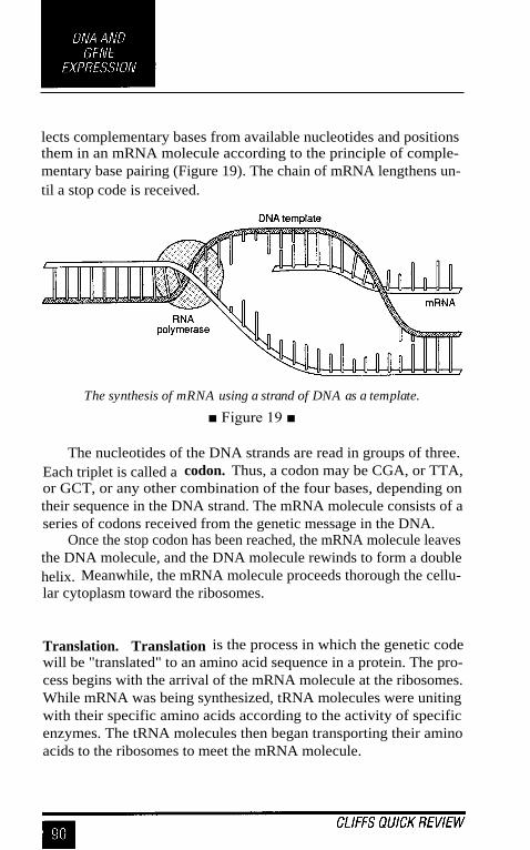
lects complementary bases from available nucleotides and positionsthem in an mRNA molecule according to the principle of comple-mentary base pairing (Figure 19). The chain of mRNA lengthens un-til a stop code is received.
The synthesis of mRNA using a strand of DNA as a template.
■ Figure 19 ■
The nucleotides of the DNA strands are read in groups of three.Each triplet is called a codon. Thus, a codon may be CGA, or TTA,or GCT, or any other combination of the four bases, depending ontheir sequence in the DNA strand. The mRNA molecule consists of aseries of codons received from the genetic message in the DNA.
Once the stop codon has been reached, the mRNA molecule leavesthe DNA molecule, and the DNA molecule rewinds to form a doublehelix. Meanwhile, the mRNA molecule proceeds thorough the cellu-lar cytoplasm toward the ribosomes.
Translation. Translation is the process in which the genetic codewill be "translated" to an amino acid sequence in a protein. The pro-cess begins with the arrival of the mRNA molecule at the ribosomes.While mRNA was being synthesized, tRNA molecules were unitingwith their specific amino acids according to the activity of specificenzymes. The tRNA molecules then began transporting their aminoacids to the ribosomes to meet the mRNA molecule.

After it arrives at the ribosomes, the mRNA molecule exposes itsbases in sets of three, the codons. Each codon has a complementarycodon called an anticodon on a tRNA molecule. When the codon ofthe mRNA molecule complements the anticodon on a tRNA mol-ecule, the latter places the particular amino acid in that position. Thenthe next codon of the mRNA is exposed, and the complementary an-ticodon of a tRNA molecule matches with it. The amino acid carriedby the second tRNA molecule is thus positioned next to the first aminoacid, and the two are linked. At this point, the tRNA molecules re-lease their amino acids and return to the cytoplasm to link up withnew molecules of amino acid.
The ribosome then moves farther down the mRNA molecule andexposes another codon which attracts another tRNA molecule withits anticodon. Another amino acid is brought into position. In thisway, amino acids continue to be added to the growing chain until theribosome has moved down to the end of the mRNA molecule. Thesequence of codons on the mRNA molecule thus determines the se-quence of amino acids in the protein being constructed (Figure 20).
Once the protein has been completely synthesized, it is removedfrom the ribosome for further processing. For example, the proteinmay be stored in the Golgi body of a eukaryotic cell before release, ora bacterium may release it as a toxin. The mRNA molecule is brokenup and the nucleotides are returned to the nucleus. The tRNA mol-ecules return to the cytoplasm to unite with fresh molecules of aminoacids, and the ribosome awaits the arrival of a new mRNA molecule.
Gene control. The process of protein synthesis does not occur con-stantly in the cell, but rather at intervals followed by periods of ge-netic "silence." Thus, the process of gene expression is regulated andcontrolled by the cell.
The control of gene expression can occur at several levels in thecell. For example, genes rarely operate during mitosis. Other levelsof gene control can occur at transcription, when certain segments ofDNA increase and accelerate the activity of nearby genes. After tran-scription has taken place, the mRNA molecule can be altered to regu-late gene activity. For example, it has been found that eukaryotic

Steps in the synthesis ofprotein beginning with the genetic codein DNA and ending with the finished polypeptide chain.
■ Figure 20 ■

mRNA contains many useless bits of RNA that are removed in theproduction of the final mRNA molecule. These useless bits of nucleicacid are called introns. The remaining pieces of mRNA, called ex-ons, are then spliced to form the final mRNA molecule. BacterialmRNA lacks introns.
The concept of gene control has been researched thoroughly inbacteria. In these microorganisms, genes have been identified as struc-tural genes, regulator genes, and control regions. The three units forma functional unit called the operon.
The operon has been examined in close detail in certain bacteria.It has been found that certain carbohydrates can induce the presenceof the enzymes needed to digest those carbohydrates. For example,when lactose is present, bacteria synthesize the enzymes needed tobreak it down. Lactose acts as an inducer molecule in the followingway: In the absence of lactose, a regulator gene produces a repressorprotein, which binds to a control region called the operator site. Thisbinding prevents the structural genes from encoding the enzyme forlactose digestion. When lactose is present, however, it binds to therepressor protein and thereby removes the repressor at the operatorsite. With the operator site free, the structural genes are released toproduce their lactose-digesting enzyme.
Recombinant DNA and Biotechnology
Biotechnology is an industrial process that uses the scientific researchon DNA for practical benefits. Biotechnology is synonymous withgenetic engineering because the genes of an organism are changedduring the process and the DNA of the organism is recombined. Re-combinant DNA and biotechnology can be used to form proteins notnormally produced in a cell. In addition, bacteria that carry recombi-nant DNA can be released into the environment to increase the fertil-ity of the soil, serve as an insecticide, or relieve pollution.

Tools of biotechnology. The basic process of recombinant DNA tech-nology revolves around the activity of DNA in the synthesis of pro-tein. By intervening in this process, scientists can change the natureof the DNA and of the gene make-up of an organism. By insertinggenes into the genome of an organism, the scientist can induce theorganism to produce a protein it does not normally produce.
The technology of recombinant DNA has been made possible inpart by extensive research on microorganisms during the last cen-tury. One important microorganism in recombinant DNA research isEscherichia coli (E. coli). The biochemistry and genetics of E. coliare well known, and its DNA has been isolated and made to acceptnew genes. The DNA can then be forced into fresh cells of E. coli,and the bacteria will begin to produce the proteins specified by theforeign genes. Such altered bacteria are said to have been transformed.
Interest in recombinant DNA and biotechnology heightened con-siderably in the 1960s and 1970s with the discovery of restrictionenzymes. These enzymes catalyze the opening of a DNA molecule ata "restricted" point, regardless of the DNA's source. Moreover, cer-tain restriction enzymes leave dangling ends of DNA molecules atthe point where the DNA is open. (The most commonly used restric-tion enzyme is named EcoRI.) Foreign DNA can then be combinedwith the carrier DNA at this point. An enzyme called DNA ligase isused to form a permanent link between the dangling ends of the DNAmolecules at the point of union (Figure 21).
The genes used in DNA technology are commonly obtained fromhost cells or organisms called gene libraries. A gene library is a col-lection of cells identified as harboring a specific gene. For example,E. coli cells can be stored with the genes for human insulin in theirchromosomes.
Pharmaceutical products. Gene defects in humans can lead to defi-ciencies in proteins such as insulin, human growth hormone, and Fac-tor VIII. These protein deficiencies may lead to problems such asdiabetes, dwarfism, and impaired blood clotting, respectively. Miss-ing proteins can now be replaced by proteins manufactured throughbiotechnology. For insulin production, two protein chains are encoded

The production of a a recombined bacterium using a gene froma foreign donor and the synthesis of protein encoded by therecombinant DNA molecule.
■ Figure 21 ■

by separate genes in plasmids inserted into bacteria. The protein chainsare then chemically joined to form the final insulin product. Humangrowth hormone is also produced within bacteria, but special tech-niques are used because the bacteria do not usually produce humanproteins. Therapeutic proteins produced by biotechnology include aclot-dissolving protein called tissue plasminogen activator (TPA)and interferon. This antiviral protein is produced within E. coli cells.Interferon is currently used against certain types of cancers and forcertain skin conditions.
Vaccines represent another application of recombinant DNA tech-nology. For instance, the hepatitis B vaccine now in use is composedof viral protein manufactured by yeast cells, which have been recom-bined with viral genes. The vaccine is safe because it contains noviral particles. Experimental vaccines against AIDS are being pro-duced in the same way.
Diagnostic testing. Recombinant DNA and biotechnology haveopened a new era of diagnostic testing and have made detecting manygenetic diseases possible. The basic tool of DNA analyses is a frag-ment of DNA called the DNA probe. A DNA probe is a relativelysmall, single-stranded fragment of DNA that recognizes and binds toa complementary section of DNA in a complex mixture of DNAmolecules. The probe mingles with the mixture of DNA and uniteswith the target DNA much like a left hand unites with the right. Oncethe probe unites with its target, it emits a signal such as radioactivityto indicate that a reaction has occurred.
To work effectively, a sufficiently large amount of target DNAmust be available. To increase the amount of available DNA, a pro-cess called thepolymerase chain reaction (PCR) is used. In a highlyautomated machine, the target DNA is combined with enzymes, nucle-otides, and a primer DNA. In geometric fashion, the enzymes synthe-size copies of the target DNA, so that in a few hours billions ofmolecules of DNA exist where only a few were before.
Using DNA probes and PCR, scientists are now able to detect theDNA associated with HIV (and AIDS), Lyme disease, and geneticdiseases such as cystic fibrosis, muscular dystrophy, Huntington'sdisease, and fragile X syndrome.

Gene therapy. Gene therapy is a recombinant DNA process in whichcells are taken from the patient, altered by adding genes, and replacedin the patient, where the genes provide the genetic codes for proteinsthe patient is lacking.
In the early 1990s, gene therapy was used to correct a deficiencyof the enzyme adenosine deaminase (ADA). Blood cells called lym-phocytes were removed from the bone marrow of two children; thengenes for ADA production were inserted into the cells using virusesas vectors. Finally, the cells were reinfused to the bodies of the twochildren. Once established in the bodies, the gene-altered cells begansynthesizing the enzyme ADA and alleviated the deficiency.
Gene therapy has also been performed with patients with mela-noma (a virulent skin cancer). In this case, lymphocytes that nor-mally attack tumors are isolated in the patients and treated with genesfor an anticancer protein called tumor necrosis factor. The gene-altered lymphocytes are then reinfused to the patients, where theyproduce the new protein which helps destroy cancer cells. Approxi-mately 2000 single-gene defects are believed to exist, and patientswith these defects may be candidates for gene therapy.
DNA fingerprinting. The use of DNA probes and the developmentof retrieval techniques have made it possible to match DNA mol-ecules to one another for identification purposes. This process hasbeen used in a forensic procedure called DNA fingerprinting.
The use of DNA fingerprinting depends upon the presence ofrepeating base sequences that exist in the human genome. The re-peating sequences are called restriction fragment length polymor-phisms (RFLPs). As the pattern of RFLPs is unique for everyindividual, it can be used as a molecular fingerprint. To perform DNAfingerprinting, DNA is obtained from an individual's blood cells, hairfibers, skin fragments, or other tissue. The DNA is extracted from thecells and digested with enzymes. The resulting fragments are sepa-rated by a process called electrophoresis. These separated DNA frag-ments are tested for characteristic RFLPs using DNA probes. Astatistical evaluation enables the forensic pathologist to compare asuspect's DNA with the DNA recovered at a crime scene and to assert

with a degree of certainty (usually 99 percent) that the suspect was atthe crime scene.
DNA and agriculture. Although plants are more difficult to workwith than bacteria, gene insertions can be made into single plant cells,and the cells can then be cultivated to form a mature plant. The majormethod for inserting genes is through the plasmids of a bacteriumcalledAgrobacterium tumefaciens. This bacterium invades plant cells,and its plasmids insert into plant chromosomes carrying the genes fortumor induction. Scientists remove the tumor-inducing genes andobtain a plasmid that unites with the plant cell without causing anyharm.
Recombinant DNA and biotechnology have been used to increasethe efficiency of plant growth by increasing the efficiency of the plant'sability to fix nitrogen. Scientists have obtained the genes for nitrogenfixation from bacteria and have incorporated those genes into plantcells. By obtaining nitrogen directly from the atmosphere, the plantscan synthesize their own proteins without intervention of bacteria asnormally needed.
DNA technology has also been used to increase plant resistanceto disease. The genes for an insecticide have been obtained from thebacterium Bacillus thuringiensis and inserted into plants to allow themto resist caterpillars and other pests. In addition, plants have beenreengineered to produce the capsid protein that encloses viruses. Theseproteins lend resistance to the plants against viral disease.
The human genome. One of the most ambitious scientific endeav-ors of this century is the effort to sequence the nitrogenous bases inthe human genome. Begun in 1990, the effort is expected to encom-pass 15 years of work at a cost of approximately $3 billion. Knowingthe content of the human genome will help researchers devise newdiagnostics and treatments for genetic diseases and will also be ofvalue to developmental biologists, evolutionary biologists, and com-parative biologists.

In addition to seeking to learn the genome of humans, the projecthas also studied numerous bacteria. By 1995, the genomes of twobacteria had been completely deciphered (Haemophilus influenzaeand Mycoplasma genitalium), and by 1996, the genome of the yeastSaccharomyces cerevisiae was known. The Human Genome Projectis one of colossal magnitude that will have an impact on many branchesof science for decades to come. The project remains the crowningachievement of DNA research in the twentieth century and the bed-rock for research in the twenty-first.


THE BACTERIA
Over 400 recognized genera of bacteria are known to exist. Bacte-rial species are listed in Bergey 's Manual of Systematic Bacteriology.The entire kingdom of bacteria, including cyanobacteria, is entitledProkaryotae. Four divisions of bacteria based on their cell wall char-acteristics are included in the Prokaryotae kingdom. Not all bacteriaare assigned to a division, but all are assigned to one of 33 "sections."
Spirochetes and Spirilla
Spirochetes have a spiral shape, a flexible cell wall, and motilitymechanisms based on structures called axial filaments. Each axialfilament is composed of fibrils extending toward each other betweentwo layers of the cell wall.
Spirochetes are very slender and difficult to see under the lightmicroscope. They are cultivated with great difficulty (some cannotbe cultivated), and their classification is based on their morphologyand pathogenicity. Certain species inhabit water environments, whileothers are parasites of arthropods (such as ticks and lice) as well aswarm-blooded animals. Spirochetes include Borrelia burgdorferi, theagent of Lyme disease, Treponema pallidum, the cause of syphilis,and Leptospira interrogans, the agent of leptospirosis.
Spirilla have a spiral shape, a rigid cell wall, and motility mecha-nisms based on polar flagella. The genera Spirillum, Aquaspirillum,and Azospirillum are widely dispersed among and readily isolatedfrom numerous environments. These organisms are aerobic bacteriawound like helices. Species S. minor is a cause of rat bite fever inhumans. The genus Campylobacter contains several pathogenic spe-cies, including C. jejuni, which causes campylobacteriosis, an intes-tinal infection accompanied by diarrhea.

Gram-Negative Rods and Cocci
Bdellovibrios. Bdellovibrios are aerobic Gram-negative, curved rodsthat prey on other bacteria. The organism attaches to the surface of abacterium, rotates, and bores a hole through the host cell wall. It thentakes biochemical control of the host cell and grows in the space be-tween the cell wall and plasma membrane. The host bacterium is killedin the process. The comma-shaped Bdellovibrio bacteriovorus is themost thoroughly studied species of the group.
Pseudomonads. Pseudomonads are aerobic, Gram-negative rodsthat are motile with polar flagella. Over 30 species are found in thegroup, and Pseudomonas fluorescens is a well-known producer of ayellow-green pigment. Another species, P. aeruginosa, causes uri-nary tract infections and infections of burned tissue.
Azotobacter andRhizobium. Species ofAzotobacter andRhizobiumare extremely important for their ability to fix nitrogen in the envi-ronment. These Gram-negative rods live free in the soil (Azotobacter)or on the roots of legume plants (Rhizobium) and use their enzymesto convert atmospheric nitrogen into organic molecules useful to theplant. The plants then use the nitrogen compounds for the synthesisof amino acids and proteins, which serve as an extremely valuablefood source for animals and humans. Members of the genus Azoto-bacter form a resting cell called a cyst, which withstands drying andenvironmental stresses.
Enterobacteria. Enterobacteria are facultatively anaerobic, Gram-negative rods that inhabit the human intestine. Members of the en-terobacteria group are members of the family Enterobacteriacae clas-sified in section 5 of Bergey's Manual.
Over 25 genera ofenterobacteria are recognized, many with patho-genic importance. Among the medically important enterobacteria are

Salmonella species that cause intestinal disease known as salmonel-losis; Yersinia pestis, the cause of plague; Klebsiella species, the causesof pneumonia, intestinal disease, and other infections; and species ofSerratia and Proteus. The well-known organism Escherichia coli isalso a member of this group. All enterobacteria have peritrichous fla-gella.
Vibrios. Vibrios are curved, Gram-negative, facultatively anaerobicrods. They belong to the family Vibrionaceae. One species, kibriocholerae, is the cause of cholera in humans. Members of the genusAeromonas and Plesiomonas are involved in human intestinal dis-ease. Species of Photobacterium are marine organisms known fortheir ability to produce light as a result of chemical actions stimu-lated by the enzyme luciferase. This production of light is known asbioluminescence.
Pasteurellas. The pasteurellas belong to the family Pasteurellaceae.They are distinguished from vibrios and enterobacteria by their smallsize and inability to move. The genera Pasteurella, Haemophilus, andActinobacillus are among the important members of the group. Thespecies H. influenzae is a cause of meningitis in children, while P.multocida causes cholera in fowl.
Sulfur bacteria. The sulfur bacteria use sulfur or sulfur compoundsas electron acceptors in their metabolism. These bacteria produce largeamounts of hydrogen sulfide during their growth, and therefore, theyproduce foul odors in water and mud. Members of the genusDesulfovibrio are particularly important in the sulfur cycle for theirability to use sulfur and convert it to other compounds that can beused by plants to synthesize sulfur-containing amino acids.
Bacteroides. The bacteroides are genera of anaerobic bacteria hav-ing unique motility and flagellation patterns. Several species digest

cellulose in the rumen of the cow and thereby break down plants.Human feces contains large numbers of bacteria belonging to the ge-nus Bacteroides, which may be helpful in digestive processes. Onespecies, B..fragilis, is a possible cause of human blood infections.
Veillonella. Among the Gram-negative cocci are a group of anaero-bic diplococci belonging to the genus Veillonella. Veillonella speciesare part of the normal flora of the mouth and gastrointestinal tract,and they are found in dental plaque. They are anaerobic organismsthat may also cause infections of the female genital tract.
Gliding bacteria. Certain bacterial species are able to move by glid-ing in a layer of slime, which they produce. Wavelike contractions ofthe outer membranes help the bacteria propel themselves. Membersof the group include species of Cytophaga and Simonsiella.
Two important genera of gliding bacteria are Beggiatoa andThiothrix. Species of these organisms live in sulfur environments andbreak down hydrogen sulfide to release sulfur in the form of sulfurgranules. For this reason, the bacteria are very important in the recy-cling of sulfur in water and soil. The bacteria are Gram-negative.
Myxobacteria are gliding bacteria that are Gram-negative, aero-bic rods. They are nonphotosynthetic species and have a unique de-velopmental cycle that involves the formation of fruiting bodies. Whennutrients are exhausted, the bacteria congregate and produce a stalk,at the top of which is a mass of cells. These cells differentiate intospherical cells, similar to cysts, which are resistant to environmentalextremes.
Sheathed bacteria. Sheathed bacteria are filamentous bacteria withcell walls enclosed in a sheath of polysaccharides and lipoproteins.The sheath assists attachment mechanisms and imparts protection tothe bacteria. The genus Sphaerotilus is in this group.

Photoautotrophic bacteria. Photoautotrophic bacteria are Gram-negative rods which obtain their energy from sunlight through theprocesses of photosynthesis. In this process, sunlight energy is usedin the synthesis of carbohydrates. Certain photoautotrophs calledanoxygenic photoautotrophs grow only under anaerobic conditionsand neither use water as a source of hydrogen nor produce oxygenfrom photosynthesis. Other photoautotrophic bacteria are oxygenicphotoautotrophs. These bacteria are cyanobacteria. They use chlo-rophyll pigments and photosynthesis in photosynthetic processes re-sembling those in algae and complex plants. During the process,they use water as a source of hydrogen and produce oxygen as a prod-uct of photosynthesis.
Cyanobacteria include various types of bacterial rods and cocci,as well as certain filamentous forms. The cells contain thylakoids,which are cytoplasmic, platelike membranes containing chlorophyll.The organisms produce heterocysts, which are specialized cells be-lieved to function in the fixation of nitrogen compounds.
Chemoautotrophic bacteria. Chemoautotrophic (or chemolitho-trophic) bacteria are a group of Gram-negative bacteria deriving theirenergy from chemical reactions involving inorganic material. Cer-tain chemoautotrophic bacteria use carbon dioxide as a carbon sourceand grow in a medium containing inorganic substances. By compari-son, members of the genus Thiobacillus metabolize sulfur compounds,and members of the genera Nitrosomonas and Nitrobacter metabo-lize nitrogen compounds. Certain chemoautotrophic bacteria use hy-drogen gas in their chemical reactions, and others use metals such asiron and manganese in their energy metabolism. These unusual typesof biochemistry are characteristic of organisms found outside the bodyin the soil and water environment.

Gram-Positive Bacteria
Streptococci. Streptococci are spherical bacteria that divide in par-allel planes to produce chains. The bacteria are Gram-positive, andcertain species are aerobic, while others are anaerobic. On blood agar,certain species partly destroy the red blood cells and are said to bealpha-hemolytic. Other species completely destroy the blood cells
and are beta-hemolytic. Those streptococci producing no blood celldestruction are gamma-hemolytic.
One species of streptococcus (Streptococcus pneumoniae) is thecause of secondary bacterial pneumonias, while another species (Strep-
tococcus pyogenes) causes strep throat and rheumatic fever. Otherspecies are associated with dental caries. Harmless strains of strepto-cocci are used in the production of yogurt, buttermilk, and cheese.
Staphylococci. Staphylococci are Gram-positive bacteria that di-vide in planes to produce clusters or packets. Normally associatedwith the skin and mucous membranes, certain species of staphylo-cocci are involved in skin boils, abscesses, and carbuncles, especiallyif they produce the enzyme coagulase, which causes blood clotting.Staphylococcus aureus is involved in cases of food poisoning, toxicshock syndrome, pneumonia, and staphylococcal meningitis.
Lactobacilli. Lactobacilli are Gram-positive, rod-shaped bacteriaoccurring as single cells or chains. They produce lactic acid in theirmetabolism and are associated with the flora of the mouth and thevagina. Certain species are associated with the production of dairyproducts such as yogurt, sour cream, and buttermilk.
Bacillus and Clostridium species. Species of Bacillus and Clos-tridium are Gram-positive, rod-shaped bacteria able to produce highlyresistant endospores (spores). The spores are found in the soil, air,and all environments of the body. Species of Bacillus grow aerobi-

cally, and Bacillus anthracis is the cause of anthrax. Clostridium spe-cies grow anaerobically, and different species cause tetanus, botu-lism, and gas gangrene.
Bacillus and Clostridium species are also used for industrial pur-poses. Bacillus thuringiensis forms an insecticide useful against vari-ous forms of caterpillars, and Clostridium species are used to producevarious types of chemicals, such as butanol.
Corynebacteria. Corynebacteria are pleomorphic members of thegenus Corynebacterium, which are Gram-positive rods found in vari-ous environments, including the soil. The bacteria contain cytoplas-mic phosphate granules that stain as characteristic metachromaticgranules. One species, Corynebacterium diphtheriae, causes humandiphtheria.
Actinomyces and Arthrobacter. Actinomyces species are Gram-positive rods that assume many shapes and usually form branchingfilaments. Most species are anaerobic, and one species is responsiblefor a human and cattle infection called lumpy jaw.
Arthrobacter species live primarily in the soil. These Gram-positive rods assume many shapes during their life cycles, includingbranching rods and spherical forms. Arthrobacter species are widelyfound in the soil, and many degrade herbicides and pesticides.
Acid-Fast Bacilli
Mycobacteria. The mycobacteria include species in the genus My-cobacterium. This group of rod-shaped bacteria possesses largeamounts of mycolic acid in the cell wall. The presence of mycolicacid makes the bacteria very difficult to stain, but when heat or otheragents are used to force carbolfuchsin into the cytoplasm, the bacte-ria resist decolorization with a dilute acid-alcohol solution. There-fore, they are said to be acid-fast.

Many mycobacteria are free-living, but two notable pathogensexist in the group: M. tuberculosis, the cause of tuberculosis in hu-mans and cattle; and M. leprae, which causes leprosy.
Nocardioforms. Nocardioforms include nine genera of aerobic, acid-fast rods, including members of the genus Nocardia. Nocardioformshave aerial hyphae which project above the surface of their growthmedium as branching filaments. The hyphae fragment into rods andcocci. Nocardioforms are found throughout nature in many types ofsoil and aquatic environments. One species, N. asteroides, causes in-
fection of the human lung.
Archaebacteria
Archaebacteria differ from all other bacteria (which are sometimescalled eubacteria). Archaebacteria are so named because biochemi-cal evidence indicates that they evolved before the eubacteria andhave not undergone significant change since then. The archaebacteriagenerally grow in extreme environments and have unusual lipids intheir cell membranes and distinctive RNA molecules in their cyto-plasm.
One group of archaebacteria are the methanogens, anaerobicbacteria found in swamps, sewage, and other areas of decomposingmatter. The methanogens reduce carbon dioxide to methane gas intheir metabolism. A second group are the halobacteria, a group ofrods that live in high-salt environments. These bacteria have the abil-ity to obtain energy from light by a mechanism different from theusual process of photosynthesis. The third type of archaebacteria arethe extreme thermophiles. These bacteria live at extremely high tem-peratures, such as in hot springs, and are associated with extremeacid environments. Like the other archaebacteria, the extreme ther-mophiles lack peptidoglycan in their cell walls. Many depend on sul-fur in their metabolism, and many produce sulfuric acid as anend-product.

Submicroscopic Bacteria
Rickettsiae. Rickettsiae are rod-shaped and coccoid bacteria be-longing to the order Rickettsiales. These bacteria cannot be seen withthe light microscope, and therefore the Gram stain is not used foridentification. However, their walls have the characteristics of Gram-negative cell walls. Rickettsiae are obligate intracellular parasites thatinfect humans as well as arthropods such as ticks, mites, and lice.They are cultivated only with great difficulty in the laboratory andgenerally do not grow on cell-free media. Tissue cultures and fertil-ized eggs are used instead.
Rickettsiae are very important as human pathogens. Various spe-cies cause Rocky Mountain spotted fever, epidemic typhus, endemictyphus, scrub typhus, Q fever, and ehrlichiosis.
Chlamydiae. Chlamydiae are extremely tiny bacteria, below theresolving power of the light microscope. Although the Gram stain isnot used for identification, the bacteria have cell walls resemblingthose in Gram-negative bacteria.
Chlamydiae display a growth cycle that takes place within hostcells. The bacteria invade the cells and differentiate into dense bodiescalled reticulate bodies. The reticulate bodies reproduce and eventu-ally form new chlamydiae in the host cell called elementary bodies.Chlamydiae cause several diseases in humans, such as psittacosis, adisease of the lung tissues; trachoma, a disease of the eye; and chlamy-dia, an infection of the reproductive tract.
Mycoplasmas. Mycoplasmas are extremely small bacteria, belowthe resolving power of the light microscope. They lack cell walls andare surrounded by only an outer plasma membrane. Without the rigidcell wall, the mycoplasmas vary in shape and are said to be pleomor-phic. Certain species cause a type of mild pneumonia in humans aswell as respiratory tract and urinary tract diseases.


THE VIRUSES
Viruses are noncellular genetic elements that use a living cell fortheir replication and have an extracellular state. Viruses are ultrami-croscopic particles containing nucleic acid surrounded by protein,and in some cases, other macromolecular components such as amembranelike envelope.
Outside the host cell, the virus particle is also known as a virion.The virion is metabolically inert and does not grow or carry on respi-ratory or biosynthetic functions.
At present, there are no technical names for viruses. Internationalcommittees have recommended genus and family names for certainviruses, but the process is still in a developmental stage.
Viral Structure and Replication
Viruses vary considerably in size and shape. The smallest viruses areabout 0.02 gm (20 nanometers), while the large viruses measure about0.3 gm (300 nanometers). Smallpox viruses are among the largestviruses; polio viruses are among the smallest.
Viral structure. Certain viruses contain ribonucleic acid (RNA),while other viruses have deoxyribonucleic acid (DNA). The nucleicacid portion of the viruses is known as the genome. The nucleic acidmay be single-stranded or double-stranded; it may be linear or a closedloop; it may be continuous or occur in segments.
The genome of the virus is surrounded by a protein coat knownas a capsid, which is formed from a number of individual proteinmolecules called capsomeres. Capsomeres are arranged in a preciseand highly repetitive pattern around the nucleic acid. A single type ofcapsomere or several chemically distinct types may make up thecapsid. The combination of genome and capsid is called the viralnucleocapsid.

A number of kinds of viruses contain envelopes. An envelope isa membranelike structure that encloses the nucleocapsid and is ob-tained from a host cell during the replication process. The envelopecontains viral-specified proteins that make it unique. Among the en-velope viruses are those of herpes simplex, chickenpox, and infec-tious mononucleosis.
The nucleocapsids of viruses are constructed according to cer-tain symmetrical patterns. The virus that causes tobacco mosaic dis-ease, for example, has helical symmetry. In this case, the nucleocapsid
is wound like a tightly coiled spiral. The rabies virus also has helicalsymmetry. Other viruses take the shape of an icosahedron, and theyare said to have icosahedral symmetry. In an icosahedron, the capsidis composed of 20 faces, each shaped as an equilateral triangle (Fig-ure 22). Among the icosahedral viruses are those that cause yellowfever, polio, and head colds.
The envelope of certain viruses is a lipid bilayer containing gly-coproteins embedded in the lipid. The envelope gives a somewhatcircular appearance to the virus and does not contribute to the sym-metry of the nucleocapsid. Projections from the envelope are knownas spikes. The spikes sometimes contain essential elements for at-tachment of the virus to the host cell. The virus of AIDS, the humanimmunodeficiency virus, uses its spikes for this purpose.
Bacteriophages are viruses that multiply within bacteria. Theseviruses are among the more complex viruses. They often have icosa-hedral heads and helical tails. The virus that attacks and replicates inEscherichia coil has 20 different proteins in its helical tail and a set ofnumerous fibers and "pins." Bacteriophages contain DNA and areimportant tools for viral research.
Viral replication. During the process of viral replication, a virusinduces a living host cell to synthesize the essential components forthe synthesis of new viral particles. The particles are then assembledinto the correct structure, and the newly formed virions escape fromthe cell to infect other cells.

An array of viruses. (a) The helical virus of rabies. (b) Thesegmented helical virus of influenza. (c) A bacteriophage withan icosahedral head and helical tail. (d) An envelopedicosahedral herpes simplex virus. (e) The unenveloped poliovirus. (f) The icosahedral human immunodeficiency virus withspikes on its envelope.
■ Figure 22 ■
The first step in the replication process is attachment. In thisstep, the virus adsorbs to a susceptible host cell. High specificity ex-ists between virus and cell, and the envelope spikes may unite withcell surface receptors. Receptors may exist on bacterial pili or fla-gella or on the host cell membrane.
The next step is penetration of the virus or the viral genome intothe cell. This step may occur by phagocytosis; or the envelope of thevirus may blend with the cell membrane; or the virus may "inject" itsgenome into the host cell. The latter situation occurs with the bacte-riophage when the tail of the phage unites with the bacterial cell wall

and enzymes open a hole in the wall. The DNA of the phage pen-etrates through this hole.
The replication steps of the process occur next. The protein capsidis stripped away from the genome, and the genome is freed in the cellcytoplasm. If the genome consists of RNA, the genome acts as a mes-senger RNA molecule and provides the genetic codes for the synthe-sis of enzymes. The enzymes are used for the synthesis of viralgenomes and capsomeres and the assembly of these components intonew viruses. If the viral genome consists of DNA, it provides thegenetic code for the synthesis of messenger RNA molecules, and theprocess proceeds.
In some cases, such as in HIV infection (as discussed below), theRNA of the virus serves as a template for the synthesis of a DNAmolecule. The enzyme reverse transcriptase catalyzes the DNA's pro-duction. The DNA molecule then remains as part of the host cell'schromosome for an unspecified period. From this location, it encodesmessenger RNA molecules for the synthesis of enzymes and viralcomponents.
Once the viral genomes and capsomeres have been synthesized,they are assembled to form new virions. This assembly may takeplace in the cytoplasm or in the nucleus of the host cell. After theassembly is complete, the virions are ready to be released into theenvironment (Figure 23).
For the release of new viral particles, any of a number of pro-cesses may occur. For example, the host cell may be "biochemicallyexhausted," and it may disintegrate, thereby releasing the virions.For enveloped viruses, the nucleocapsids move toward the membraneof the host cell, where they force themselves through that membranein a process called budding. During budding, a portion of cell mem-brane pinches off and surrounds the nucleocapsid as an envelope.The replication process in which the host cell experiences death iscalled the lytic cycle of reproduction. The viruses so produced arefree to infect and replicate in other host cells in the area.

A generalized representation of the replication of two viruses.Replication of a DNA virus is shown in (1); replication of anRNA virus is displayed in (2).
■ Figure 23 ■
Lysogeny. Not all viruses multiply by the lytic cycle of reproduc-tion. Certain viruses remain active within their host cells for a longperiod without replicating. This cycle is called the lysogenic cycle.The viruses are called temperate viruses, or proviruses, becausethey do not bring death to the host cell immediately.
In lysogeny, the temperate virus exists in a latent form within thehost cell and is usually integrated into the chromosome. Bacterio-phages that remain latent within their bacterial host cell are calledprophages. This process is a key element in the recombination processknown as transduction.
An example of lysogeny occurs in HIV infection. In this case,the human immunodeficiency virus remains latent within the host T-lymphocyte. An individual whose infection is at this stage will notexperience the symptoms of AIDS until a later date.
Viral Cultivation and Physiology
Viruses can be cultivated within suitable hosts, such as a living cell.To study bacteriophages, for example, bacteria are grown in a suit-able growth medium; then bacteriophages are added. The bacterio-phages multiply within the bacteria and increase their numberssubstantially.
Animal and plant viruses are cultivated in cell cultures. A cellculture is prepared by encouraging cell growth outside the animal orplant source. The cells are kept alive in a suspension of growth fac-tors within a Petri dish. A thin layer of cells, or monolayer, is theninoculated with viruses, and replication takes place. Fertilized eggsand living animals can also be used to cultivate viruses.
For research study, viruses can be cultivated in large volumes byinoculations to tissue culture systems. After a time, the cells are de-generated, and viruses are harvested. The viral particles are concen-trated by precipitation methods and purified by repeatedcentrifugations. Highly purified viruses can be obtained by crystalli-zation and concentration under established conditions.

Viral measurements. Viruses are generally too small to be seenunder the light microscope, and an electron microscope is usuallynecessary to make them visible. Although viruses can be quantifiedby observation, it is also possible to determine their number in termsofvirus infectious units, each of which is the smallest unit that causesa detectable effect when viruses infect a susceptible host. Virus in-fectious units are expressed per volume of fluid.
One method for determining virus infectious units is by theplaqueassay. The plaque assay is performed by cultivating viruses on a"lawn" of host cells and noting the presence of clear areas whereviruses have replicated and destroyed the cells.
Another way of determining virus infectious units is by cultivat-ing viruses in living animals and determining which dilution of virusis lethal to the animals. The end-point dilution can be calculated bythis method.
Antiviral agents. The antibiotics normally used to treat bacterialdisease cannot be used to inactivate viruses because viruses do notperform the biochemical functions that antibiotics interfere with. Forexample, penicillin is used to interrupt the synthesis of the bacterialcell wall, but viruses have no cell wall.
However, there are several nucleotide analog drugs that interferewith viral replication. Acyclovir, for example, is used against herpesviruses because this drug prevents the synthesis of DNA during viralreplication. A drug called azidothymidine (AZT) is used for patientswith HIV infection because this drug also prevents the synthesis ofDNA. A drug called ganciclovir is used against cytomegaloviruses,and amantadine is useful against influenza viruses.
Interferon, a naturally produced antiviral agent approved forcertain uses, is a group of proteins produced by host cells after theyhave been infected by viruses. The interferons do not protect the hostcell, but they do provide protection to neighboring cells against viralreplication. Interferons can be produced by genetic engineering meth-ods.

Viral vaccines. Protection against viral disease can be rendered byusing a viral vaccine. Viral vaccines can be composed of inactivatedor attenuated viruses. Inactivated viruses ("dead viruses") are un-able to replicate in host cells because of some chemical or physicaltreatment. The Salk vaccine against polio and the yellow fever vac-cine are examples.
Attenuated viruses ("live viruses") are weakened viruses thatreplicate at a very slow rate in host cells and generally do not produceany symptoms of disease when inoculated to humans. Attenuated vi-ruses are used in the Sabin polio vaccine and in the vaccines againstmeasles and rubella. The most contemporary vaccines are composedof viral proteins produced by genetic engineering methods. The vac-cine for hepatitis B is an example of this type of vaccine.
Viral inactivation. Virus particles are composed of nucleic acid,protein, and in some cases, a lipid envelope. As such, the viruses aresusceptible to normal inactivation by chemical substances that reactwith any of these organic compounds. Such things as chlorine, io-dine, phenol, detergents, and heavy metals rapidly inactivate viruses.In addition, viruses are destroyed by heating methods used for othermicroorganisms, and they are very susceptible to the effects of ultra-violet light. Filters can be used to remove viruses from fluids so longas the filter pores are small enough to trap viral particles.

THE FUNGI
The fungi (singular, fungus) include several thousand species ofeukaryotic, sporebearing organisms that obtain simple organic com-pounds by absorption. The organisms have no chlorophyll and repro-duce by both sexual and asexual means. The fungi are usuallyfilamentous, and their cell walls have chitin. The study of fungi iscalled mycology, and fungal diseases are called mycoses.
Together with bacteria, fungi are the major decomposers of or-ganic materials in the soil. They degrade complex organic matter intosimple organic and inorganic compounds. In doing so, they help re-cycle carbon, nitrogen, phosphorous, and other elements for reuse byother organisms. Fungi also cause many plant diseases and severalhuman diseases.
Two major groups of organisms make up the fungi. The filamen-tous fungi are called molds, while the unicellular fungi are calledyeasts. The fungi are classified in the kingdom Fungi in the Whittakerfive-kingdom system of classification.
Structure and Physiology
There is considerable variation in the structure, size, and complexityof various fungal species. For example, fungi include the microscopicyeasts, the molds seen on contaminated bread, and the common mush-rooms.
Molds consist of long, branching filaments of cells called hy-phae (singular, hypha). A tangled mass of hyphae visible to the un-aided eye is a mycelium (plural, mycelia). In some molds, thecytoplasm passes through and among cells of the hypha uninterruptedby cross walls. These fungi are said to be coenocytic fungi. Thosefungi that have cross walls are called septate fungi, since the crosswalls are called septa.
Yeasts are microscopic, unicellular fungi with a single nucleusand eukaryotic organelles. They reproduce asexually by a process of
budding. In this process, a new cell forms at the surface of the origi-nal cell, enlarges, and then breaks free to assume an independent ex-istence.
Some species of fungi have the ability to shift from the yeastform to the mold form and vice versa. These fungi are dimorphic.Many fungal pathogens exist in the body in the yeast form but revertto the mold form in the laboratory when cultivated.
Reproduction in yeasts usually involves spores. Spores are pro-duced by either sexual or asexual means. Asexual spores may be freeand unprotected at the tips of hyphae, where they are called conidia(Figure 24). Asexual spores may also be formed within a sac, in whichcase they are called sporangiospores.
Nutrition. Fungi grow best where there is a rich supply of organicmatter. Most fungi are saprobic (obtaining nutrients from dead or-ganic matter). Since they lack photosynthetic pigments, fungi cannotperform photosynthesis and must obtain their nutrients from preformedorganic matter. They are therefore chemoheterotrophic organisms.
Most fungi grow at an acidic pH of about 5.0, although somespecies grow at lower and higher pH levels. Most fungi grow at about25°C (room temperature) except for pathogens, which grow at 37°C(body temperature). Fungi store glycogen for their energy needs anduse glucose and maltose for immediate energy metabolism. Most spe-cies are aerobic, except for the fermentation yeasts that grow in bothaerobic and anaerobic environments.
Reproduction. Asexual reproduction occurs in the fungi whenspores form by mitosis. These spores can be conidia, sporangiospores,arthrospores (fragments of hyphae), or chlamydospores (spores withthick walls).
During sexual reproduction, compatible nuclei unite within themycelium and form sexual spores. Sexually opposite cells may unitewithin a single mycelium, or different mycelia may be required. Whenthe cells unite, the nuclei fuse and form a diploid nucleus. Severaldivisions follow, and the haploid state is reestablished.

The microscopic structures of a septate fungus showing asexuallyproduced conidia that leave the fungus and germinate to producea new mycelium.
■ Figure 24 ■
Fungal spores are important in the identification of the fungus,since the spores are unique in shape, color, and size. A single spore iscapable of germinating and reestablishing the entire mycelium. Sporesare also the method for spreading fungi in the environment. Finally,the nature of the sexual spores is used for classifying fungi into nu-merous divisions.

Classification of Fungi
Division Zygomycota. Members of the division Zygomycota areknown as zygomycetes. Zygomycetes produce sexual spores knownas zygospores (Figure 25), as well as asexual sporangiospores.
A familiar member of the division is Rhizopus stolonifer, a fun-gus found on fruits, vegetables, and breads. It is the familiar breadmold. It anchors itself to the substratum with special hyphae knownas rhizoids. Rhizopus is used in the industrial production of steroids,meat tenderizers, industrial chemicals, and certain coloring agents.
Division Ascomycota. Members of the division Ascomycota are re-
ferred to as ascomycetes. After sexual fusion of cells has taken place,these organisms form their sexual spores within a sac called an as-cus. Therefore, they are called sac fungi.
Ascomycetes include the powdery mildews and the fungi thatcause Dutch elm disease and chestnut blight disease. The researchorganism Neurospora crassa is found within this group. Asexual re-production in the ascomycetes involves conidia.
Many yeasts are classified in the division Ascomycota. Of par-ticular interest is the fermentation yeast Saccharomyces. This yeastis used in the production of alcoholic drinks, in bread making, and asa source of growth factors in yeast tablets. It is an extremely impor-tant research organism as well.
Division Basidiomycota. Members of the division Basidiomycotaare referred to as basidiomycetes and are called club fungi. After thesexual cells have united, they undergo division and produce a club-shaped structure called a basidium. Sexually produced basidiosporesform at the tips of the basidia. Basidia are often found on huge, vis-ible, fruiting bodies called basidiocarps. The typical mushroom is abasidiocarp.

Sexual reproduction in the mold Rhizopus stolonifer. Plus andminus mycelia produce sexually opposite hyphae that fuse andgive rise to zygospores, which germinate to .form new mycelia.
■ Figure 25 ■
Basidiomycetes are used as food (for example, mushrooms), butsome basidiomycetes are pathogens. One of the organisms of menin-gitis is the basidiomycete Cryptococcus neoformans. The mushroomAmanita is poisonous to humans.

Division Deuteromycota. Members of the Deuteromycota divisionare called deuteromycetes. These fungi lack a known sexual cycle ofreproduction and are said to be "imperfect." When its sexual cycle isdiscovered, a fungus from this division is usually reclassified in oneof the other divisions. Among the imperfect fungi are the organismsof athlete's foot and ringworm.
Slime Molds
Slime molds have characteristics of both molds and protozoa. Undercertain conditions, the slime mold exists as masses of cytoplasm, simi-lar to amoebae. It moves over rotting logs or leaves and feeds byphagocytosis. The amoeba stage is called the plasmodium, whichhas many nuclei.
The amoeba stage ends when the plasmodium matures or en-counters a harsh environment. At this point, it moves to a light areaand develops fruiting bodies that form spores at the ends of stalks.The spores are resistant to environmental excesses. They germinatewhen conditions are suitable to form flagellatedswarm cells, or amoe-boid cells, which later fuse to again form a multinucleate plasmo-dium.
Water Molds
Water molds belong to the group known as oomycetes. The watermolds resemble other fungi because they have branched filamentsand form spores. However, the water molds have cellulose in theircell walls, while other fungi have chitin.
Oomycetes have a complex reproductive cycle which includesflagella-bearing zoospores. Certain water molds are parasites of fish.Others cause disease in plants such as tobacco, grapes, and potatoes.

THE UNICELLULAR ALGAE
Algae are eukaryotic organisms that have no roots, stems, or leavesbut do have chlorophyll and other pigments for carrying out photo-synthesis. Algae can be multicellular or unicellular.
General Characteristics
Unicellular algae occur most frequently in water, especially in plank-ton. Phytoplankton is the population of free-floating microorgan-isms composed primarily of unicellular algae. In addition, algae mayoccur in moist soil or on the surface of moist rocks and wood. Algaelive with fungi in lichens.
According to the Whittaker scheme, algae are classified in sevendivisions, of which five are considered to be in the Protista kingdomand two in the Plantae kingdom. The cell of an alga has eukaryoticproperties, and some species have flagella with the "9-plus-2" pat-tern of microtubules. A nucleus is present, and multiple chromosomesare observed in mitosis. The chlorophyll and other pigments occur inchloroplasts, which contain membranes known as thylakoids.
Most algae are photoautotrophic and carry on photosynthesis.Some forms, however, are chemoheterotrophic and obtain energyfrom chemical reactions and nutrients from preformed organic mat-ter. Most species are saprobes, and some are parasites.
Reproduction in algae occurs in both asexual and sexual forms.Asexual reproduction occurs through the fragmentation of colonialand filamentous algae or by spore formation (as in fungi). Spore for-mation takes place by mitosis. Binary fission also takes place (as inbacteria).
During sexual reproduction, algae form differentiated sex cellsthat fuse to produce a diploid zygote with two sets of chromosomes.The zygote develops into a sexual spore, which germinates when con-ditions are favorable to reproduce and reform the haploid organism

having a single set of chromosomes. This pattern of reproduction iscalled alternation of generations.
Divisions of Unicellular Algae
Five divisions of unicellular algae are considered in microbiologybecause of their microscopic form and their unicellular characteris-tic. These organisms are classified in the kingdom Protista.
Division Chlorophyta. Algae of the division Chlorophyta possessgreen chlorophyll pigments and carotenoid pigments. A representa-tive member is Chlamydomonas, which is often used in research andas a laboratory specimen. Chlamydomonas produces zoospores, whichare flagellated. Organisms such as Chlamydomonas are believed tobe evolutionary ancestors of other species. Other organisms in thedivision are Volvox and Spirogyra.
Division Charophyta. Members of the division Charophyta arestoneworts. Stoneworts cover the bottoms of ponds and may be asource of limestone.
Division Euglenophyta. Members of the division Euglenophyta in-clude the common organism Euglena. These organisms have chloro-phyll and carotenoid pigments for photosynthesis and flagella formovement. They share many characteristics with both plants and ani-mals and are believed to be a basic stock of evolution.
A typical Euglena cell has a large nucleus and nucleolus. Con-tractile vacuoles help empty water from the organism, and two fla-gella arise at one end of the cell. Reproduction occurs by binary fissionin the longitudinal plane.

Division Chrysophyta. Members of the division Chrysophyta arebrown and yellow-green algae. These organisms contain chlorophyllpigments as well as special carotenoid pigments called fucoxanthins.Fucoxanthins give the golden-brown color to members of the divi-sion. Members of the division include the diatoms, oceanic photo-synthetic algae found at the bases of many food chains. Diatoms con-tribute immense amounts of oxygen to the atmosphere and occupykey places in the spectrum of living things because they convert thesun's energy into the energy in carbohydrates.
Division Pyrrophyta. Members of the division Pyrrophyta are pig-mented marine forms that include the dinoflagellates, amoeboid cellswith flagella as well as protective cellulose plates that surround thecells. They have chlorophyll, carotenoid, and xanthophyll pigments.Dinoflagellates often have a brown or yellow color, and they repro-duce by longitudinal division through mitosis. Dinoflagellates makeup a large portion of marine plankton and are essential to many of theocean food chains. Certain species are luminescent. Others have redor orange pigments; when these organisms multiply at abnormallyhigh rates, they cause the "red tides."


THE PROTOZOA
Protozoa are eukaryotic microorganisms. Although they are oftenstudied in zoology courses, they are considered part of the microbialworld because they are unicellular and microscopic.
General Characteristics
Protozoa are notable for their ability to move independently, a char-acteristic found in the majority of species. They usually lack the ca-pability for photosynthesis, although the genus Euglena is renownedfor motility as well as photosynthesis (and is therefore consideredboth an alga and a protozoan). Although most protozoa reproduce byasexual methods, sexual reproduction has been observed in severalspecies. Most protozoal species are aerobic, but some anaerobic spe-cies have been found in the human intestine and animal rumen.
Protozoa are located in most moist habitats. Free-living speciesinhabit freshwater and marine environments, and terrestrial speciesinhabit decaying organic matter. Some species are parasites of plantsand animals.
Protozoa play an important role as zooplankton, the free-floatingaquatic organisms of the oceans. Here, they are found at the bases ofmany food chains, and they participate in many food webs.
Size and shape. Protozoa vary substantially in size and shape. Smallerspecies may be the size of fungal cells; larger species may be visibleto the unaided eye. Protozoal cells have no cell walls and thereforecan assume an infinite variety of shapes. Some genera have cells sur-rounded by hard shells, while the cells of other genera are enclosedonly in a cell membrane.
Many protozoa alternate between a free-living vegetative formknown as a trophozoite and a resting form called a cyst. The proto-zoal cyst is somewhat analogous to the bacterial spore, since it resists

harsh conditions in the environment. Many protozoal parasites aretaken into the body in the cyst form.
Most protozoa have a single nucleus, but some have both a ma-cronucleus and one or more micronuclei. Contractile vacuoles maybe present in protozoa to remove excess water, and food vacuoles areoften observed.
Nutrition and locomotion. Protozoa are heterotrophic microorgan-isms, and most species obtain large food particles by phagocytosis.The food particle is ingested into a food vacuole. Lysosomal enzymesthen digest the nutrients in the particle, and the products of digestionare distributed throughout the cell. Some species have specializedstructures called cytostomes, through which particles pass in phago-cytosis.
Many protozoal species move independently by one of three typesof locomotor organelles: flagella, cilia, and pseudopodia. Flagellaand cilia are structurally similar, having a "9-plus-2" system of mi-crotubules, the same type of structure found in the tail of animal spermcells and certain cells of unicellular algae. How a protozoan moves isan important consideration in assigning it to a group.
Classification of Protozoa
All protozoal species are assigned to the kingdom Protista in theWhittaker classification. The protozoa are then placed into variousgroups primarily on the basis of how they move. The groups are calledphyla (singular, phylum) by some microbiologists, and classes byothers. Members of the four major groups are illustrated in Figure 26.
Mastigophora. The Mastigophora are protozoa having one or moreflagella. Reproduction in these protozoa generally occurs by fission,although sexual reproduction is observed in some species. Membersof the group include the organisms that cause intestinal distress

An array ofprotozoa showing representatives of the four majorgroups.
■ Figure 26 ■
(Giardia lamblia) and sleeping sickness (Trypanosoma brucei) inhumans. Some of these protozoa have only a single flagellum (forexample, Trypanosoma), but others may have up to eight (for ex-ample, Giardia). Flagellar motion tends to be jerky, so flagellates canbe identified in live specimens by this characteristic movement.
Also in this group is Euglena gracilis, a flagellated organism thatalso has chlorophyll pigments for photosynthesis. For this reason, theorganism is considered both a unicellular alga and a protozoan. Since
it has the characteristics of both animals (motion) and plants (photo-synthesis), many evolutionists consider Euglena to be a source of allcomplex organisms.
Flagellates are very common in nature, and numerous speciesmay be observed in pond and swamp water, aquarium water, andbirdbath water. The flagellum may either push the protozoan aheador lead the cell along. A complex basal structure at one end of the cellanchors the flagellum to the cell.
Sarcodina. Members of the group Sarcodina are amoebas. Theseorganisms move by pseudopodia, although flagella may be presentin the reproductive stages. Cytoplasmic streaming assists movement.Asexual reproduction occurs by fission of the cell. Sarcodina includestwo marine groups known as foraminiferans and radiolarians. Bothgroups were present on earth when the oil fields were in formativestages, and marine geologists use them as potential markers for oilfields.
Another member, Entamoeba histolytica, is the cause of amoe-bic dysentery. This organism can cause painful lesions of the intes-tine if it is contracted from contaminated water.
Ciliophora. The Ciliophora group includes protozoa that move bymeans of cilia. Two types of nuclei, the macronucleus and the micro-nucleus, are often found in the cells, and a contractile vacuole is usu-ally present. Conjugation may be used for sexual reproduction, andbinary fission also occurs. The distinctive rows of cilia vibrate insynchrony and propel the organism in one direction.
Paramecium is a member of this group, as are many other free-living ciliates such as Tetrahymena. Paramecium cells are known fortheir ability to conjugate and exchange genetic material, thereby ac-quiring new genetic characteristics. The cells also have kappa fac-tors, which are chemical substances known to destroy other protozoalcells, and they produce trichocysts, which are submicroscopic sting-ing particles. Indeed, the physiology of a single Paramecium cell issimilar to that of a multicellular organism.
Apicomplexa. Members of the group Apicomplexa form spores atone stage in their life cycle. For this reason, the group is also knownas Sporozoa. Reproduction is a complex phenomenon in this group,and all species are parasites. Members of the group display no meansof locomotion in the adult form. The agents of malaria andtoxoplasmosis are classified here. Another important pathogen isPneumocystis carinii, a cause of sometimes-lethal pneumonia in AIDSpatients; over half the deaths known to be associated with AIDS havebeen caused by P carinii.


THE HOST-PARASITE RELATIONSHIP
A delicate relationship exists between pathogenic microorganismsand body defenses. When the defenses resist the pathogens, the bodyremains healthy. But when the pathogens overcome the defenses, theresult is disease. Once disease has been established, the infected indi-vidual may suffer temporary or permanent damage or may experi-ence death. The outcome depends upon many factors attending theepisode of disease.
Infection and Disease
The scientific study of disease is called pathology, from the Greek"pathos" meaning suffering. Pathology is concerned with the causeof disease, called the etiology (the agent of disease is the etiologicagent). It also deals with pathogenesis, the manner in which a dis-ease develops. Pathology is also concerned with the structural andfunctional changes brought about by the disease in tissues.
The terms infection and disease do not have identical meaning.Infection refers to an invasion of body tissues by microorganisms;disease is a change from the state of good health resulting from amicrobial population living in the tissues (Figure 27). Infection mayoccur without disease. For example, the flora of microorganisms al-ways present on the body's skin is a type of infection but not disease.
The normal flora. The normal flora is the population of microor-ganisms found where the body tissues interface with the environment.Much of the normal flora is permanent, but some portions are tran-sient. The transient flora is present for a time and then disappears.
Various types of relationships exist between the normal flora andthe body. The general name of a relationship is symbiosis, a term thatmeans living together. In some cases, the symbiosis is further identi-

The balance between health and disease. The condition of thehuman body depends on interactions between host (the body)and parasite (the infectious microorganism). When the humanbody wins the battle for supremacy, the result is health and therejection of disease.
■ Figure 27 ■
feed as a commensalism, when one organism benefits and the otherremains unaffected. A type of symbiosis called mutualism exists whenboth organisms benefit one another. A symbiosis called parasitismdevelops when one organism damages the other.
Opportunistic organisms. Certain organisms of the normal floraare opportunistic. Opportunistic organisms are potentially patho-genic organisms that normally do not cause disease. However, in acompromised host, the organisms may see "opportunity" to invadethe tissues. An example occurs in individuals who have AIDS. Op-portunistic organisms such as Pneumocystis carinii invade the lungtissues and cause a lethal pneumonia.

The Development of Infectious Disease
Infectious diseases are those caused by microorganisms. In order torelate a particular organism to a particular disease, Koch's postu-lates must be fulfilled. First devised by Robert Koch in the 1870s,Koch's postulates are a series of procedures for identifying the causeof a particular disease. They are described in the first chapter of thisbook.
Symptoms and signs. Infectious diseases are usually characterizedby changes in body function known as symptoms. Symptoms aresubjective changes not always apparent to the observer. The patientmay also exhibit signs, which are objective changes that can be mea-sured. Fever and a skin rash are examples of signs. When a specificgroup of symptoms or signs accompanies a disease, the group is calleda syndrome.
Transmission and incidence. Infectious diseases may be classifiedaccording to their transmissibility. A disease that spreads from onehost to another is a communicable disease. Those communicablediseases transmitted with particular ease are said to be contagious.Diseases not spread between hosts are noncommunicable. Staphy-lococcal food poisoning is an example.
The incidence of a disease refers to the percent of a populationthat contracts it over a particular period. The prevalence of a disease,by contrast, is the percentage of a population having the disease at aparticular time.
When a disease occurs only occasionally, it is called a sporadicdisease. A disease present in a population at all times is an endemicdisease. A disease that breaks out in a population in a short period isan epidemic disease, and an epidemic disease occurring throughoutthe world is a pandemic disease.

Types of disease. Diseases can be defined in terms of their severityand duration. An acute disease occurs rapidly and lasts a short time,while a chronic disease develops slowly and lasts a long time. Influ-enza is an acute disease, while tuberculosis is a chronic disease. Asubacute disease is a disease that has vague symptoms and lasts arelatively long time. A latent disease remains inactive in a host for atime and then becomes active.
Infections can be described as local infections if they are re-stricted to a small area of the body and systemic infections if theyspread throughout the body systems. The presence of multiplyingmicroorganisms in the blood is septicemia. Toxins present in the bloodconstitute toxemia.
Infectious diseases may also be described as primary diseases orsecondary diseases. A primary disease is the first illness that occurs,and a secondary disease is due to an opportunistic microorganism,often a normal resident, after the body's defenses have weakened.
Modes of disease transmission. When a disease remains in a popu-lation, a source of pathogens called a reservoir of infection existswithin the population. The reservoir can be human, animal, or non-living, such as the soil'. A human reservoir who has had the diseaseand recovered but continues to shed infectious organisms is called acarrier. Animal diseases spread to humans are called zoonoses.
Among the principal routes of transmission of disease are con-tact, vectors, and vehicles. Contact can be direct or indirect. Directtransmission occurs from person to person by such things as touch-ing, kissing, and sexual intercourse (Figure 28). Indirect transmis-sion occurs when a nonliving object is intermediary between twohumans.
A lifeless object known as a fomite is often involved in diseasetransmission. The object may be a towel, cup, or eating utensil. Trans-mission can also be effected by droplet nuclei, bits of mucus andsaliva that spread between individuals.
Vectors are living things. Arthropods such as mosquitoes, flies,and ticks may carry pathogens on their body parts, in which case theyare mechanical vectors. If the arthropod is infected and transmits theorganism in its saliva or feces, it is a biological vector.

Some modes of transmission of microorganisms ,from therespiratory and oral tract.
■ Figure 28 ■
Vehicles are lifeless objects such as food, water, and air. Watermay be contaminated by human feces, while food is often contami-nated by pathogens from the soil. Air can be a vehicle for transmis-sion for droplet nuclei in such diseases as tuberculosis and commoncolds.
In order for infection to be transmitted, microorganisms mustleave the body through a portal of exit, which can be the intestine,mouth, or skin surface. Generally, the portal of exit is the same as the

infected body part. Organisms enter the new individual through aportal of entry.
Infections acquired during a hospital stay are called nosocomialinfections. These infections often occur in compromised hosts whoare being treated for other conditions such as cancer, nutritional defi-ciency, burns, or other forms of stress.
Disease patterns. When a disease develops in an individual, a rec-ognized set of periods can be identified. The first period is the periodof incubation, the time between the entry of the parasite into the hostand the appearance of symptoms.
The next period is the prodrome period. This period is accom-panied by mild symptoms such as aches, fever, and early signs ofdisease.
Next comes the period of illness, when the disease is most acute.Signs and symptoms are most apparent, and each disease has its owncharacteristic appearance. The body's immune system is activatedduring this period, and specific defense is critical to recovery.
The final periods are the periods of decline and convalescence.The period of decline is one in which the signs and symptoms sub-side, and during the period of convalescence, the person returns tonormal. After the disease has abated, the immune system continuesto produce antimicrobial factors that will ensure long-term immu-nity.
THE DEVELOPMENT OF INFECTIOUS DISEASE
Infectious disease results from a competition for supremacy betweenthe parasite and the host. If the parasite overcomes the host, there is achange in the general state of good health and disease develops.
Several contributing factors are involved in the establishment ofinfectious disease. These factors determine whether the infecting or-ganism will survive in the body.
Contributing Factors
Portals of entry. In order for a pathogen to gain access to the host,the pathogen must pass through a portal of entry. One of the mostcommon portals of entry is the mucous membranes, especially thoseof the respiratory, gastrointestinal, and urogenital tracts. Another im-portant portal of entry is the skin. Penetration of the skin occurs dur-ing a wound or by a hair follicle. When microorganisms penetratebelow the skin, the portal of entry is said to be the parenteral route.
Dose. The dose of an organism refers to the number of microorgan-isms required to establish an infection. For some diseases, such astyphoid fever, the dose is a few hundred bacteria. For other diseases,such as cholera, the dose may be several million bacteria. The dosemay be expressed as the LD50 , which refers to the dose of microor-ganisms that will kill 50 percent of the hosts it enters.
Invasiveness. Invasiveness is a property that encourages disease be-cause it refers to the ability of pathogens to penetrate into the tissues.Those organisms that cause intestinal ulcers, such as Entamoebahistolytica, penetrate the tissue effectively. Tissue invasion often be-gins with adherence, the ability of pathogens to attach to the tissue

by using structures such as pili. The presence of a capsule or glyco-calyx encourages adherence because they are composed of stickymaterials.
Capsules. Microorganisms that possess capsules are able to resisthost defenses by interfering with phagocytosis. Normally, the bodyuses white blood cells to engulf and destroy pathogens. However,toxic substances in the capsule are able to destroy the white bloodcells before the white blood cells perform phagocytosis. The organ-ism of pneumonia Streptococcus pneumoniae is well known for thetoxic materials in its capsule. Many other pathogens also possess cap-sules.
Enzymes and Toxins
Enzymes. Many pathogens produce a series of enzymes to help over-come body defenses and establish themselves in the host. One ex-ample is leukocidins, a group of enzymes that destroy white bloodcells. This destruction lessens the body's ability to perform phagocy-tosis.
Other bacterial enzymes are hemolysins. These enzymes destroyred blood cells. Streptococci, staphylococci, and certain Clostridiumspecies produce hemolysins.
Coagulases are bacterial enzymes that clot the blood. These en-zymes convert fibrinogen into fibrin, which forms the threads of ablood clot. The clot helps staphylococci avoid the body's phagocytesand contributes to its pathogenicity.
Other important enzymes are streptokinase and hyaluronidase.Streptokinase is a streptococcal enzyme that dissolves blood clots.This activity helps the organism escape the body's attempt to wall offan infection. Hyaluronidase destroys hyaluronic acid, a polysaccha-ride that "cements" cells together in a tissue. Hyaluronidase thus per-mits organisms to spread through tissues and establish themselves atsites distant from that of the intial infection. Another enzyme, called

collagenase, breaks down collagen in the connective tissues of mus-cles. It thereby encourages the spread of infection.
Toxins. Many bacteria are able to produce poisonous substances calledtoxins. Toxins act on the body's cells, tissues, and organs and inter-fere with important body processes, thereby interrupting normal bodyfunctions. Those microorganisms that produce toxins are said to betoxigenic. The condition in which toxins are produced is called tox-emia.
Two important types of toxins are exotoxins and endotoxins.Exotoxins are proteins produced by bacteria during their growth andliberated into their surrounding environment. Exotoxins are producedchiefly by Gram-positive bacteria, and the genes for this productionare carried primarily on the plasmids.
Various types of exotoxins exist. Neurotoxins interfere with thenervous system, while enterotoxins interfere with activities of thegastrointestinal tract. In response to toxins, the body produces spe-cial antibodies called antitoxins, which unite with and neutralize thetoxins, providing defense against disease.
It is possible to immunize against the effects of exotoxins byinjecting toxoids into individuals. Toxoids are preparations of exo-toxins chemically treated to destroy their toxigenicity but retain theirability to elicit antibody formation in the body. Toxoids are currentlyavailable to protect against diphtheria and tetanus (the DT injection).
Endotoxins are portions of the cell wall of Gram-negative bacte-ria. They consist primarily of lipopolysaccharides and are releasedwhen bacteria break apart during the process of lysis. Since lysis oc-curs during antibiotic therapy, the effects of endotoxins can bringabout a worsening of symptoms during the recovery period. This con-dition is called endotoxin shock. It is accompanied by fever, chills,aches, and cardiovascular collapse.

Pathogenic Viruses
Because viruses lack metabolic capabilities, they rely on other meansfor overcoming body defenses and causing disease. Viruses avoidbody defenses by multiplying within host cells, where antibodies andother components of the immune system cannot reach them.
The effect occurring in host cells during viral invasion is referredto as the cytopathic effect. The cytopathic effect can develop whenthe virus alters the metabolism of the cell and prevents it from pro-ducing essential cellular components. Alternatively, the virus mayinduce cells to cling together in a large mass called a syncytium. Insome cases, the virus causes the cell's lysosomes to release enzymeswhich then destroy the cell.

NONSPECIFIC BODY DEFENSE
The host-parasite relationship may result in an episode of disease.In this case, the body will mount two general forms of defense: non-specific defense and specific defense centered in the immune system.This chapter is concerned with the nonspecific types of defense.
Nonspecific Mechanisms of Defense
The body possesses many mechanisms that impart nonspecific de-fense. The objectives of these mechanisms are to prevent microor-ganisms from gaining a foothold in the body and to destroy them ifthey penetrate to the deeper tissues.
Mechanical barriers. Mechanical barriers at the portal of entryrepresent the first line of defense for the body. These defenses arenormally part of the body's anatomy and physiology. The skin is arepresentative example. The outermost layers of skin consist of com-pacted, cemented cells impregnated with the insoluble protein kera-tin. The thick top layer is impervious to infection and water. In theunbroken state, it usually is not penetrated by pathogens.
The mucous membranes of the urinary, respiratory, and diges-tive tracts are another example. They are moist and permeable, buttheir fluids, such as tears, mucus, and saliva, rid the membrane ofirritants. Nasal hairs trap particles in the respiratory tract, and thefluids exert a flushing action. Cilia on the cells sweep and trap par-ticles in the respiratory tract, and coughing ejects irritants.
Chemical defenses. Among the nonspecific chemical defenses ofthe body are the secretions of lubricating glands. The tears and salivacontain the enzyme lysozyme, which breaks down the peptidoglycan

of the cell wall of Gram-positive bacteria. The lactic acid of the va-gina imparts defense, and the extremely caustic hydrochloric acid ofthe stomach is a barrier to the intestine. Semen contains the antimi-crobial substance spermine that inhibits bacteria in the male uro-genital tract.
Genetic barriers. The hereditary characteristics of an individual area deterrent to disease as well. For example, humans suffer HIV infec-tion because their T-lymphocytes have the receptor sites for the humanimmunodeficiency virus. Dogs, cats, and other animals are immuneto this disease because they do not possess the genes for producingthe receptor sites. Conversely, humans do not suffer canine distemperbecause humans lack the appropriate receptor sites for the virus thatcauses the disease.
Inflammation. Inflammation is a nonspecific response to any traumaoccurring to tissues. It is accompanied by signs and symptoms thatinclude heat, swelling, redness, and pain. Inflammation mobilizescomponents of the immune system, sets into motion repair mecha-nisms, and encourages phagocytes to come to the area and destroyany microorganisms present.
Inflammation can be controlled by nervous stimulation and chemi-cal substances called cytokines. These chemical products of tissuecells and blood cells are responsible for many of the actions of in-flammation. The loss of fluid leads to a local swelling called edema.In some types of inflammation, phagocytes accumulate in the whitishmass of cells, bacteria, and debris called pus.
Fever. Fever is considered a nonspecific defense mechanism becauseit develops in response to numerous traumas. Fever is initiated bycirculating substances called pyrogens, which affect the brain'shypothalamus and cause the latter to raise the temperature. Althoughexcessive fever can be dangerous, fever is believed to have a benefi-cial role because it retards the growth of temperature-sensitive

microorganisms (for example, leprosy bacilli), and it increases themetabolism of body cells while stimulating the immune reaction andthe process of phagocytosis.
Interferon. Interferon is a group of antiviral substances producedby body cells in response to the presence of viruses. Lymphocytesand macrophages produce alpha-interferon, epithelial cells producebeta-interferon, and I-lymphocytes produce gamma-interferon. Theinterferons do not directly inhibit viruses. Instead, they stimulate ad-jacent cells to produce substances that inhibit the replication of vi-ruses in those cells. Interferons produced in response to one viruswill protect against many other types of viruses, and for this reason,interferon is considered a nonspecific form of defense.
Phagocytosis
Phagocytosis is a nonspecific defense mechanism in which variousphagocytes engulf and destroy the microorganisms of disease.
Phagocytes. Among the important phagocytes are the circulatingwhite blood cells called neutrophils and monocytes. In the tissues,the monocytes are transformed into phagocytic cells called macro-phages. The macrophages move through the tissues of the body per-forming phagocytosis and destroying parasites. They are part of thereticuloendothelial system. Phagocytes also initiate the processesof the immune system.
The process of phagocytosis begins with attachment and inges-tion of microbial particles (Figure 29) into a bubblelike organellecalled a phagosome. Once inside the phagocyte, the phagosome con-taining the microorganism joins with a lysosome, which contributesenzymes. The fusion of phagosome and lysosome results in aphagolysosome. Microorganisms are destroyed within minutes, and

The process of phagocytosis, a type of nonspecific defense todisease.
■ Figure 29 ■
the microbial debris is eliminated from the cell in the process ofeges-tion. In the immune process, chemical portions of the microorganismcalled antigenic determinants are displayed on the surface of thephagocyte to stimulate the immune process.
Phagocytosis is enhanced by products of the immune systemcalled antibodies. These protein molecules bind to microorganismsand encourage engulfing by phagocytosis.
The complement system. The complement system is a series ofproteins that circulate in the blood and encourage phagocytosis orotherwise "complete" the defensive process. Many immune reactionsstimulate the complement system.
The complement system operates in a cascade of reactions. Inthe pathway, certain complement components react with one anotherand produce new substances that induce other components to react.The results of the myriad reactions are substances that induce othercomplement components into action. The overall result is a numberof substances toxic to microorganisms. The substances encouragephagocytosis or bring about destruction of microbial membranes.
Two general pathways for complement activity exist. The classi-cal pathway operates with the highly specific immune system and isinitiated when certain antibodies unite with antigens and stimulatethe complement system into action. The alternative pathway is non-specific and is initiated by tumors, cell wall components of bacteria,and various microorganisms. It is sometimes called the properdinpathway because properdin is one of the proteins operating in it. Thealternative pathway invokes a slower and less specific method forridding the body of parasites, particularly Gram-negative bacteria andviruses.


Immunity is a state of specific resistance to infection. Specific resis-tance is directed against a particular type of microorganism and is thesingle most important characteristic of immunity.
The immune system enables the body to recognize a foreignagent as nonself, which is something other than a person's own sub-stances (self). The immune system takes a specific action for neutral-izing, killing, and eliminating that agent. The action involvesnonspecific resistance as well. On occasion, the immune system ac-tivity may lead to tissue damage as seen in allergic disorders andother states of hypersensitivity.
The immune system's activity is based on its ability to distin-guish characteristic proteins or protein-linked components associatedwith alien substances. Once this distinction has been made, certainlymphocytes are provoked to produce antibodies directed against theforeign matter, while other lymphocytes are sensitized to the invad-ing agent and react with it directly. Thus, there are two major branchesof the immune system: antibody-mediated immunity (also known ashumoral immunity) and cell-mediated immunity.
Antigens
Immune responses are directed at a series of foreign substances knownas antigens, also referred to as immunogens. Most antigens are highmolecular weight substances, but low molecular weight substanceswill also act as antigens if they bind to proteins in the body. The lowmolecular weight compound is referred to as a hapten. The resultingconjugate may induce an immune response directed against the anti-gen.
The uptake and processing of antigens by macrophages in thetissue is an initial, critical step in most immune responses. The simpleact of taking foreign substances into the body does not necessarily
THE IMMUNE SYSTEM

invoke an immune response because the substances may be brokendown before they are ingested by macrophages.
Antigenic determinants. The chemical groups on the antigen mol-ecules that determine their immunogenicity are called antigenic de-terminants, also known as epitopes. Antigenic determinants mayconsist of several amino acids of a protein molecule or severalmonosaccharide units of a polysaccharide. Each species of living thingis chemically and antigenically unique because of differences in itsproteins, carbohydrates, and other organic substances.
Types of antigens. Certain types of antigens are distinctive.Autoantigens, for example, are a person's own self antigens. Alloan-tigens are antigens found in different members of the same species(the red blood cell antigens A and B are examples). Heterophile an-tigens are identical antigens found in the cells of different species.
A single organism such as a bacterium may contain a variety ofproteins, carbohydrates, and other materials that provoke immuneresponses. Antigens found on the body cell are called somatic anti-gens. Antigens in the bacterial capsule are capsular antigens. Anti-gens of an organism's flagella are known as flagellar antigens (Hantigens). Protein substances such as exotoxins are also antigenic.
Cells of the Immune System
Cells of the immune system are associated with the lymphatic systemof the body and its specialized cells. Lymphocytes of the lymphaticsystem are derived from stem cells of the bone marrow. These undif-ferentiated precursor cells proliferate throughout life and replenishthe mature cells of the immune system.

B-lymphocytes and T -lymphocytes. There are two major pathwaysfor the differentiation of stem cells into immune cells. Certain of thestem cells produce B-lymphocytes (B-cells) while other stem cellsform T -lymphocytes (T-cells). B-lymphocytes are so named becausein birds, they are formed in the bursa of Fabricius. The equivalent sitein humans has not been identified but is believed to be the bone mar-row. T-lymphocytes undergo their conversion in the thymus gland,an organ in the neck tissues near the trachea and thyroid gland (Fig-ure 30).
The transformation of stem cells into B-lymphocytes and T-lymphocytes begins about the fifth month after fertilization, and afull set is complete a few months after birth. These cells then migrateto the lymphoid organs in the lymph nodes, spleen, tonsils, adenoids,and other organs of the lymphatic system.
To initiate the immune response, microorganisms are phagocy-tized and their antigens are processed in phagocytic cells such asmacrophages. The antigenic determinants are displayed on the surfaceof the phagocytic cells and presented to the appropriate B-lymphocytesand T-lymphocytes to provoke an immune response.
Clonal selection. The clonal selection theory helps explain howlymphocytes recognize antigenic determinants and respond. Accord-ing to this theory, small populations (clones) of lymphocytes bearreceptors on their cell membranes. Production of these receptors isgenetically determined. On B-lymphocytes, the receptors consist ofantibody molecules, while on T-lymphocytes, they are clusters ofamino acids. When lymphocytes encounter an antigenic determinanton the surface of a macrophage, their receptors match with the anti-genic determinant and a stimulation follows. A match is also madebetween a set of molecules called major histocompatibility (MHC)molecules and their receptors.
The clonal selection theory suggests that B-lymphocytes and T-lymphocytes exist for all antigenic determinants even before contactwith an antigen is made. The theory also says that antigenic determi-nants stimulate the lymphocytes to endow their progeny with identi-cal specificity. The B-lymphocytes and T-lymphocytes that might

An overview of the human immune system. T-lymphocytes andB-lymphocytes originate in the bone marrow and then areprocessed in different body organs before proceeding to the lymphnodes, where they provide the underpinnings of the two types ofimmune responses.
• Figure 30

potentially react with the body's own cells are deleted or in someway inactivated to ensure that an immune response to the host organ-ism does not develop.
Two general types of immunity exist for specific resistance todisease. They are antibody-mediated (humoral) immunity, cen-tered in B-lymphocytes, and cell-mediated immunity, centered in T-lymphocytes.
Antibody-Mediated (Humoral) Immunity (AMI)
On exposure to antigenic determinants in lymphatic organs, B-lymphocytes are activated and differentiated to form plasma cells.Plasma cells are specialized, differentiated cells that synthesize andsecrete antibodies specific for an antigen. Other activated B-lymphocytes form memory cells. These cells can be activated laterto differentiate to plasma cells for rapid antibody production. Thisantibody production will occur on future reentry of the antigen to thebody and is the basis of long-term immunity.
The products of plasma cells are antibodies. An antibody is aspecialized protein substance produced by the host cells in responseto an antigen in the host's tissues. Antibodies are capable of reactingspecifically with the antigen that provoked their production. Anti-bodies are often referred to as immunoglobulins. They circulate inthe blood and are associated with the gamma globulin fraction of theplasma.
Structure of antibodies. Structurally, the antibodies (immunoglo-bulins) are composed of four protein chains arranged in a distinctivepattern. Each molecule has two long chains of heavy molecular weightprotein (H chains) and two short chains of light molecular weightprotein (L chains). The chains are linked by sulfur bonds.
At the outer end of each arm of the antibody molecule, a specificamino acid sequence exists. This is where the antibody molecule re-acts with the antigenic determinant that provoked its production. The

combining site is known as the Fab region. The most common anti-body molecules have two Fab regions and are said to be bivalent(having two combining sites). The remaining portion of the antibodymolecule is called the Fe region because it can be crystallized. Itsamino acid content is relatively constant and characteristic for its class.This portion of the molecule activates the complement system andencourages phagocytosis.
When the antibody molecule reacts with the antigen, the two sur-faces fit together like the pieces of a jigsaw puzzle. This "recogni-tion" is exquisitely accurate and accounts for the extreme specificityof antibody molecules.
Classes of antibodies. Five classes of antibodies (immunoglobu-lins) are produced by the plasma cells. The first class, called IgM, isthe major component of the primary antibody response in adult hu-mans and is the first antibody to appear in the immune reaction. It iscomposed of five units joined by a J (joining) chain (Figure 31). IgMcannot diffuse through cell membranes and is found almost exclu-sively in the blood. Because of its many binding sites, it is more reac-tive with antigens than are other kinds of antibodies. IgM is also formedby the fetus during uterine development and is active against the Aand B factors of the red blood cells. Many antitoxins formed againstbacterial toxins are composed of IgM.
The principal antibody of the secondary immune response is IgG.This antibody is the most common in the bloodstream and is found inmany secretions, such as spinal, synovial, lymph, and peritoneal flu-ids. IgG crosses the placenta and protects the fetus and newborn. IgGtherefore provides a natural type of passive immunity. IgG also formsin the primary antibody reaction after a large amount of IgM has al-ready formed. IgG has the "typical" antibody structure of four pro-tein chains.
The third class of antibody is IgA. This antibody is found in ex-ternal secretions such as those at the mucosal surfaces of the respira-tory, gastrointestinal, and urogenital tracts. It is also present in thetears, saliva, bile, urine, and colostrum, and it is transferred in the

Details of an antibody molecule and the structures of the fivetypes of antibody molecules produced by the human body.
■ Figure 31 ■
breast milk. IgA consists of two antibody units held together by a Jchain and secretory component. IgA resists infections at the bodysurface.
The fourth class of antibody, IgD, is found in extremely smallquantities in the serum. IgD is found at the surface of B-lymphocytesand is believed to be an antigen receptor at this location. IgM is alsobelieved to be a receptor.
The final kind of antibody, IgE, occurs in minute concentrationsin the serum and is important in hypersensitivity reactions, especiallythe anaphylactic reaction. A localized reaction is called allergy.

The reaction of antibodies with antigens helps neutralize the an-tigen and restrict the spread of infection. Certain antibodies react withthe bacterial surface, while others react with the flagella, pili, or cap-sules. These reactions encourage phagocytosis. When antibodies re-act with a virus, viruses cannot attach to host cells and replicate.Antibody reaction with antigens also sets off the complement sys-tem, which results in the formation of an attack complex along withincreased phagocytosis. Antibody reactions with toxin molecules neu-tralize the toxins and prevent further damage to body tissues.
Types of immunity. Immunity may be broadly classified as innateor acquired. Innate immunity is present from birth. It consists ofnumerous types of nonspecific factors that operate during times ofdisease. Acquired immunity is derived from activity of the immunesystem. The term generally refers to antibodies and is subdivided intotwo parts: active immunity and passive immunity.
Active immunity is acquired when the body produces antibod-ies. The immunity is usually long lasting because the immune systemhas been stimulated into action. However, it takes several hours todevelop. Active immunity can be natural or artificial.
Naturally acquired active immunity develops when a personproduces antibodies during a bout of illness or on exposure to a mi-croorganism even though disease does not occur. The B-lymphocytesand plasma cells function, and this immunity occurs during the "natu-ral" scheme of events.
Active immunity can also occur by artificial means. Artificiallyacquired active immunity occurs when a person produces antibod-ies after exposure to a vaccine. A vaccine consists of bacteria, vi-ruses, or fragments of these. A vaccine may also contain toxoids,which are chemically treated bacterial toxins. Toxoid vaccines areavailable against diphtheria and tetanus. Viral vaccines are availableagainst measles, mumps, rubella, polio, rabies, hepatitis A, hepatitisB, and yellow fever. Because vaccine exposures do not happen in thenatural scheme of events, the immunity is said to be artificial.
Passive immunity comes about when the body receives antibod-ies from an outside source. In passive immunity, the immune system

does not operate and the immunity is not long lasting. However, itprotects instantaneously. Passive immunity can be either natural orartificial.
Naturally aquired passive immunity develops when antibod-ies pass from mother to child across the placental lining. The new-born is born with IgG, and it receives maternal IgA if it is breast-fed.These antibodies will remain for approximately six months after birthbefore fading away.
For artificially acquired passive immunity, a person is givenan injection of antibodies. Usually these antibodies are derived fromthe blood of another individual who has produced antibodies whenconfronted with a certain disease. The antibody injected is usuallyIgG. The antibodies will remain for a period of several days or weeksand then disappear. This type of immunity is given to protect peoplewho have been exposed to tetanus, diphtheria, or botulism. It is alsoused to protect against certain serious viral diseases. Allergic reac-tions to the serum proteins (serum sickness) limit the use of the prepa-rations.
Cell-Mediated Immunity (CMI)
Cell-mediated immunity depends upon the activity of T-lymphocytes.T-lymphocytes have a longer life span than B-lymphocytes and arefound in the same lymphatic tissues as the B-lymphocytes. The T-lymphocytes react with certain antigenic determinants and becomeimmunologically "committed." Part of this commitment is the con-version to a subset of cells called cytotoxic T-lymphocytes.
Activity of cytotoxic T -lymphocytes. Cytotoxic T -lymphocytes donot produce antibody molecules. Rather, they leave the lymphatictissues and enter the circulation. They circulate through the bloodvessels and gather at the infection site. Here they interact directlywith organisms such as fungi, protozoa, cancer cells, and transplantcells. They also interact with virus-infected cells and bacteria-infected
cells (such as lung cells infected with tuberculosis). The T-lymphocytes exert a "lethal hit" on the cells and secrete substancesinto them that lead to cellular destruction.
In addition to their direct interaction, T-lymphocytes also secretesubstances called lymphokines. Lymphokines attract phagocytes tothe area and encourage them to perform phagocytosis on fungi, pro-tozoa, and infected cells. This activity helps relieve the infection.Lymphokines are also known as cytokines. An important cytokine isinterleukin-1, which activates T-lymphocytes, causing them to pro-liferate further and form clones.
Helper and suppressor T -lymphocytes. Helper T -lymphocytesalso function in the immune system by encouraging the activity ofB-lymphocytes in the production of antibodies. Suppressor Tlymphocytes regulate or suppress the activity of the immune systemso that it is not excessive. Natural killer (NK) cells are T-lymphocytesthat recognize and destroy many types of target cells withoutbeing exposed to antigens. Technically, these are not part of thespecific immune response. Finally, the delayed hypersensitivity Tlymphocytes function in hypersensitivity reactions and encouragelocal tissue inflammations.
Laboratory Tests
Certain laboratory tests are available to detect the presence of anti-bodies in an individual. These laboratory tests are commonly used indiagnostic procedures because the presence of a certain antibody in-dicates the presence of a certain disease. The study of serum for itsantibody content is known as serology.
One of the objects of serology is to determine the titer of anti-body present in the individual. The titer is an estimate of the anti-body level in a unit volume of serum. It is determined as a reactivedilution of the serum and is usually expressed in a ratio such as 1:100.

The agglutination and precipitation tests. Serological tests thatinvolve clumping of an antigen are called agglutination tests. Anti-bodies involved in agglutination tests are called agglutinins. Whenthe antibody molecules unite with antigen molecules on the surfaceof bacteria, red blood cells, or particles, they cause the cells to sticktogether and form large clumps (Figure 32).
Another form of serological test is the precipitation test. In this
test, antibodies are called precipitins. They react with dissolved anti-gens and form large complexes that become visible as a fine precipi-tate. Tests such as these can be performed in fluid or gel.
The agglutination reaction taking place when antibody moleculesunite with the antigen molecules on the surfaces of cells andinvolve the entire cells in the reaction.
■ Figure 32 ■

The complement fixation test. The complement fixation test isanother application of a serological reaction. In this test, antibodiesare combined with antigens in the presence of complement, a seriesof blood proteins. If the antibodies are specific for that antigen, theywill react and use up ("fix") the complement. When a subsequentcombination of blood cells and their antibodies are added to the tube,no complement will be available and no reaction will take place.However, if the first antibodies were not specific for their antigen,the complement is not fixed, and it is available to react with the bloodcells and their antibodies. The blood cells will undergo lysis as aresult of the complement and antibody activity.
The neutralization test. Serological tests that determine the pres-ence of antitoxins are called neutralization reactions. Antitoxins areantibodies formed against toxins. When these antibodies are unitedwith toxin molecules, they neutralize the toxins and will prevent theirtoxic effects in an animal. However, if the antitoxin is not specific forthe toxin, the toxin is not neutralized and it will exert a lethal effecton the animal.
The fluorescent antibody test. The fluorescent antibody techniqueis a serological test that helps to make visible an antigen-antibodyreaction. Antibody solution is combined with cells that contain anti-gens. If an antibody-antigen reaction occurs, a fluorescent dye accu-mulates on the surface, thereby signaling a positive reaction.
Enzyme immunoassays. In the enzyme immunoassay, an antigen-antibody reaction is encouraged on the surface of a particle, and anenzyme accumulates there. When a substrate is added, the enzymemolecules change the color of the substrate, and the reaction can bedesignated positive. This test is sometimes called the enzyme-linkedimmunosorbentassay (ELISA).
A contemporary development in immunology is the developmentof monoclonal antibodies. Monoclonal antibodies are produced by

a single colony of immortal cells, that is, cells with the ability to liveand multiply for extremely long periods. The cells producing the an-tibodies are hybrids formed by fusing antibody-producing plasma cellswith tumor cells (the immortal cells). Monoclonal antibodies can bepurified and used for diagnostic purposes as well as carriers for toxicchemical compounds . The process can possibly be used to kill tumorcells in the body if antitumor drugs are carried.


DISORDERS OF THE IMMUNE SYSTEM
Certain human disorders are attributed to activity of the immunesystem. These disorders are commonly known as hypersensitivities,states of increased immune sensitivity that are mediated by antibodyor cellular factors. The disorders may also involve immunodeficien-cies in which failures of antibody-mediated or cell-mediated immu-nity take place.
Hypersensitivity Reactions
Normally the immune system plays an important role in protectingthe body from microorganisms and other foreign substances. If theactivity of the immune system is excessive or overreactive, a hyper-sensitivity reaction develops. The consequences of a hypersensitiv-ity reaction may be injury to the body or death.
Most injury resulting from hypersensitivities develops after aninteraction has taken place between antigens and antibodies or be-tween antigens and sensitized T-lymphocytes. The general nature ofand symptoms accompanying the reaction depend upon whether an-tibodies or sensitized T-lymphocytes are involved. When antibodiesare involved, the reactions fall under the heading of immediate hy-persensitivity. When T-lymphocytes are involved, the reactions arecharacterized as delayed hypersensitivity. Immediate hypersensitiv-ity reactions include anaphylaxis, allergic reactions, cytotoxic reac-tions, and immune complex reactions. Delayed hypersensitivityreactions are generally characterized as contact dermatitis or infec-tion allergies.
Immediate hypersensitivity. The reactions accompanying immedi-ate hypersensitivity depend upon the nature of the antigen, the fre-quency and route of antigen contact, and the type of antibody reactingwith the antigen. The initial dose of antigen is referred to as the sen-

sitizing dose. This exposure is followed by a latent period and thena later dose of the same antigen, called the eliciting dose or shockingdose. The shocking dose sets off the hypersensitivity reaction, result-ing in tissue damage.
Immediate reactions begin within minutes of contact with theeliciting dose of antigen. If antigens are introduced directly into thetissues, such as by insect sting or injection, the result is a systemicreaction such as anaphylactic shock. When the contact is a superfi-cial one involving the epithelial tissues, the reaction is more local-ized, as occurs in asthma or allergic rhinitis (hay fever). These localreactions are commonly referred to as allergy. Another term used isatopy.
The antigens eliciting an immediate hypersensitivity are calledallergens, particularly when they are involved in local allergic reac-tions. Hapten molecules such as penicillin molecules may be involvedwhen they are bound to larger protein molecules. Foods, feathers,pollen grains, animal dander, and dust may be allergens. Animal sera,bee venoms, and wasp venoms are also allergens.
The antibodies involved in anaphylaxis reactions are of the typeIgE. In cytotoxic and immune complex reactions, IgG and IgM areinvolved.
Anaphylaxis. Anaphylaxis, or type I hypersensitivity, is a whole-body, immediate hypersensitivity also known as anaphylactic shock.The allergens are introduced to the body directly to the tissues in aconcentrated form (intramuscular or intravenous injection, for ex-ample).
After the sensitizing dose has been administered, IgE is producedby the plasma cells. The antibodies circulate in the blood and attachat the Fc end to mast cells of the tissues and basophils in the blood-stream (Figure 33). This activity occurs during the latent period. Whenthe eliciting dose of allergen is later administered, the antigens com-bine with antibodies on the surface of the mast cells and basophils.
After the antigen-antibody combination has taken place, the cellsrelease a number of physiologically active substances including his-tamine and serotonin. These substances are derived from granules

The process of anaphylaxis. (a) Allergens stimulate theproduction IgE antibodies, which (b) fix themselves to thesurfaces ofmast cells. (c) On second exposure to the allergens,a reaction occurs on the mast cell surface, and (d) the cellulargranules release histamine and other stimulators of smoothmuscle contraction.
■ Figure 33 ■

within the cell. The histamine, serotonin, and other mediators inducespasms of the smooth muscle, such as in the bronchioles, small arter-ies, and gastrointestinal tract lining. A sudden drop in blood pressureoccurs, followed by circulatory collapse and shock. Bronchospasmsand edema cause constriction of the respiratory passageways, andbreathing is very difficult. Facial edema occurs, and the heart rateincreases due to constriction of the arteries. Swellings called "hives"develop at the site of injection and other areas of the skin. In severecases, anaphylactic shock may result in death within several minutesto an hour. To relieve the symptoms, epinephrine is administered to-gether with a smooth muscle relaxer, a drug such as cortisone to re-duce swelling, and other drugs as appropriate.
Allergic reactions. Allergic reactions (allergy) are a milder, local-ized form of anaphylaxis. As noted, such things as foods, pollen grains,and animal dander can induce these localized reactions. IgE, baso-phils, and mast cells are involved, but much less than in anaphylaxis.There appears to be a genetic basis for allergic reactions, as evidencedby their distribution in families.
Cytotoxic reactions. Cytotoxic reactions are a form of immediatehypersensitivity, sometimes referred to as type II hypersensitivity.In these reactions, IgE and IgM are produced in response to stimula-tion by antigens. The antibodies unite with the antigens in the blood-stream, but they also unite with analogous antigens on the surface ofthe human body's cells. This union sets off the complement system,and destruction of the local tissue cells ensues.
An example of a cytotoxic reaction is thrombocytopenia. In thisdisease, antibody molecules are elicited by certain drug molecules.The antibodies unite with antigens on the surface of thrombocytes(platelets), and with complement activation, the thrombocytes aredestroyed. The result is an impaired blood-clotting mechanism.
Another example of the cytotoxic reaction is agranulocytosis.In this immune disorder, antibodies unite with antigens on the sur-face of neutrophils. As these cells are destroyed with complementactivation, the capacity for phagocytosis is reduced.

Cytotoxic reactions are also manifested by the transfusion reac-tion occurring when improper blood transfusions are performed.Another consequence is erythroblastosis fetalis, also known ashemolytic disease of the newborn, or Rh disease. In this condition,a pregnant woman produces Rh antibodies against the developingfetus, and when the Rh antibodies unite with Rh antigens on the sur-face of fetal red blood cells in a succeeding pregnancy, the red bloodcells are destroyed (Figure 34).
The cytotoxic reaction in erythroblastosis fetalis.
■ Figure 34 ■
Immune complex disease. Immune complexes are combinationsof antigen and antibody that have the ability to fix complement. The

antibodies involved are IgM or IgG, and the antigens exist in fluid assoluble antigens. Proteins or nucleic acids may be involved.
An example of immune complex hypersensitivity is serum sick-ness. In this condition, animal serum is administered to humans, andits proteins elicit antibody production. When the antibodies and anti-gens unite, they form immune complexes, which activate the comple-ment system and cause local tissue damage. The patient may displayedema of the hands, face, and feet, as well as swelling of the upperrespiratory tissues and impairment of normal respiration. An inflam-matory response results.
Formation of immune complexes is also involved with numer-ous diseases including systemic lupus erythematosus, rheumatoidarthritis, and glomerulonephritis. Immune complex hypersensitiv-ity is often called type III hypersensitivity.
Delayed hypersensitivity. T -lymphocytes rather than antibodies func-tion in cases of delayed hypersensitivity, also called type IV hyper-sensitivity. Normally these are the T-lymphocytes involved incell-mediated immunity. The T-lymphocytes produce lymphokines,which stimulate an influx of macrophages to perform phagocytosis.In delayed hypersensitivity, the result is an exaggeration of the im-mune response, and the phagocytes bring about the destruction of thelocal tissue.
Delayed hypersensitivity (also called cellular hypersensitivity)is so named because the reaction requires a day or more to develop.One manifestation of the reaction is infection allergy, as in the tuber-culin skin test. A purified protein derivative (PPD) of Mycobacte-rium tuberculosis is applied to the skin superficially, and a skin reaction(swelling and redness) occurs 24 to 48 hours later if the person hashad a previous exposure to the antigens of Mycobacterium tuberculo-sis, possibly during an episode of tuberculosis.
A second manifestation of delayed hypersensitivity is contactdermatitis. In many cases, the reaction is accompanied by large,blisterlike lesions in which vesicles are surrounded by a zone oferythema (redness). Usually, the vesicles itch intensely.
Antigens involved in contact dermatitis include metals such as
nickel and mercury, cosmetics, disinfectants, and plant substancessuch as the resins of poison ivy, poison oak, and poison sumac. Theindividual can be tested to determine which antigen is the cause ofallergy by performing a patch test. In this procedure, a patch con-taining a specific antigen is attached to the skin and left in place for48 hours to determine if a reaction will take place.
Immunodeficiency Diseases
Disorders of the immune system may also be due to deficiencies ofthe system. These deficiencies may be congenital or iatrogenic (in-duced by immunosuppressive drugs), or they may result from malig-nancies occurring in the lymphatic system.
Abnormalities of the B-lymphocytes result in an immunodefi-ciency accompanied by abnormal production of antibodies. Theseabnormalities may result from infections such as diseases of the liver,from immune complex disorders such as systemic lupus erythema-tosus, or from malignancies. Multiple myeloma is a malignant dis-ease of plasma cells in which certain clones of B-lymphocytes producean overabundance of antibodies. The antibodies are excreted into theurine, where they are known as Bence Jones proteins.
Decreased ability of the B-lymphocytes to produce antibodies iscalled hypogammaglobulinemia. Such a condition may result froma genetic defect or the failure of the body to produce B-lymphocytes.Bone marrow disorders may also be a cause.
Abnormalities of the T-lymphocytes result in reduced capacityfor cell-mediated immunity. This condition, called DiGeorge's syn-drome, often develops from malformation or absence of the thymusgland. An individual with this syndrome is highly susceptible to in-fection by fungi, protozoa, and viruses.
When both B-lymphocytes and T-lymphocytes are deficient, thecondition is called severe combined immunodeficiency. Recent pro-cedures employing transplantation of bone marrow tissue have at-tempted to relieve this condition.


DISEASES OF THE SKIN AND EYES
Microbial diseases of the skin are usually transmitted by contactwith an infected individual. Although the skin normally provides abarrier to infection, when it is penetrated by microorganisms, infec-tion develops. Diseases of the eye are considered with the skin dis-eases because both occur at the surface of the body.
Bacterial Skin Diseases
Staphylococcal infections. Staphylococci are Gram-positive coccioccurring in clusters. The best known pathogen in this group is Sta-phylococcus aureus. This organism invades the hair follicles andcauses folliculitis, also referred to as pustules. A deeper infection ofthe skin tissues is referred to as a boil, abscess, or furuncle. Theselesions are usually filled with pus. A large lesion progressing from aboil is known as a carbuncle. Infections such as these are easily trans-mitted by skin contact as well as by fomites.
Toxin-producing strains of S. aureus cause scalded skin syn-drome. Usually found in young children and babies, this disease ischaracterized by vesicles on the body surface, which cause the skinto peel and give a scalded appearance. Penicillin or erythromycinantibiotics are used to treat this and other staphylococcal skin dis-eases.
Scarlet fever. Scarlet fever is caused by Streptococcus pyogenes, aGram-positive bacterium occurring in encapsulated chains. Most casesof scarlet fever begin as infections of the respiratory tract, followedby spread of the bacteria to the blood. The bacteria produce anerythrogenic toxin that causes the typical skin rash. Penicillin is usedfor therapy. Complications include damage to the heart valves knownas rheumatic heart disease or damage to the joints, which is calledrheumatic fever.

Erysipelas. Erysipelas is a skin disease caused by Streptococcuspyogenes and other pathogenic streptococci. Small, bright, raised le-sions develop at the site of streptococcal entry to the skin and growwith sharply defined borders. Penicillin therapy is employed.
Impetigo contagiosum. Impetigo contagiosum is a contagious skininfection accompanied by pus. It is caused by species of streptococci,staphylococci, and others. The disease commonly occurs in childrenand is easily transmitted among them. Penicillin therapy is often rec-ommended.
Madura foot. Madura foot is a general name for infections of thefeet due to many microorganisms. Among the causes are species ofsoil bacteria belonging to the genera Nocardia, Actinomyces, andStreptomyces. These and other bacteria enter the tissues and causegranular lesions that spread and eventually invade the bone and muscle.Sulfurlike granules represent accumulations of microorganisms inthe pus, and antibiotic therapy is necessary to prevent spread of thedisease.
Gas gangrene. Gas gangrene is a disease of the deep skin and woundsas well as the blood. Several species of Clostridium cause gas gan-grene, including Clostridium perfringens, C. novyi, and C. septicum.These anaerobic rods are transferred to the wound in their spore form.They germinate and grow in the dead, anaerobic tissue of a wound,putrefying the proteins and fermenting the carbohydrates to producegas. The gas causes the tissue to expand, and as the cells die fromlack of oxygen, gangrene begins. Bacterial toxins pass through thebloodstream to cause illness throughout the body, and degenerationof the muscle fibers occurs. Aggressive antibiotic therapy and re-moval of dead tissue are useful therapies.

Cat scratch fever. Cat scratch fever may accompany a skin woundfollowing a cat scratch. Although the causative agent has not beenisolated with certainty, it is believed to be a species of RochalimaeaorAfipia. Patients display a pustule at the skin site of entry and swol-len lymph nodes on one side of the body. Treatment with antibioticsmay or may not be successful. Mild fever and conjunctivitis oftenaccompany the disease.
Rat bite fever. Rat bite fever may be caused by either Spirillumminor or by Streptobacillus moniliformis. The former is a flagel-lated spiral bacterium; the latter is a Gram-negative rod in chains.Both species are transmitted during a bite by a rat, either wild orlaboratory. Rat bite fever is associated with skin lesions, intermittentfever, and a skin rash. Arthritis may also be present.
Viral Skin Diseases
Rubella. Rubella (German measles) is a viral disease of numerousorgans caused by an RNA virus and accompanied by a mild skin rashcalled an exanthem. First appearing on the body trunk, the rash spreadsto other areas. Pregnant women may transmit the virus across theplacenta to the developing embryo or fetus, and congenital rubellasyndrome may develop in the newborn. Damage to the eyes, ears,and heart often result. Immunity can be rendered by an injection ofattenuated rubella virus in the MMR vaccine.
Measles. Measles is also called rubeola. It is caused by an RNAvirus normally transmitted by respiratory droplets during the cough-ing stage. Red spots with white centers occur on the cheeks, gums,and lips and are a diagnostic sign for the disease. These spots arecalled Koplik spots. The measles skin rash appears as a blush first onthe forehead, then on the upper extremities, trunk, and lower extremi-ties. Prevention is rendered by inoculation with attenuated measles

viruses in the MMR vaccine. Complications of the disease may in-clude measles encephalitis or subacute sclerosing panencephalitis(SSPE).
Chickenpox. Chickenpox is also called varicella. The disease isclosely related to an adult disease called herpes zoster (shingles).The responsible virus is a DNA-containing virus of the herpesvirusgroup. It is also known as the VZ virus.
Chickenpox is a highly contagious disease. Transmitted primar-ily by respiratory droplets, the disease is accompanied by teardrop-shaped lesions filled with fluid. The lesions begin on the scalp andtrunk and then spread to the face and limbs. Prevention is possiblewith injections of inactivated VZ virus in the chickenpox vaccine.
Shingles occurs in adults and is believed to be a recurrence ofinfection by the virus that causes chickenpox. Presumably, the virushas remained latent in ganglia of the nervous system until it is reacti-vated. The disease is characterized by painful lesions surroundingthe body trunk. The disease is highly contagious. Acyclovir may berecommended for therapy.
Smallpox. Smallpox is a viral disease caused by a large, boxlike,DNA-containing virus having a complex shape. At one time, small-pox was a major cause of death in the world. It was accompanied bypus-filled lesions covering the body surface, and usually it resulted indeath. Immunity was rendered by an injection of cowpox (vaccinia)viruses, as first recommended by Edward Jenner in 1798. Smallpoxhas apparently been eradicated on the earth and has not appeared inhumans since October 26, 1977. It is the first infectious disease everto be eradicated.
Cowpox. Cowpox, also known as vaccinia, is caused by a DNAvirus similar in shape to the smallpox virus. In barnyard animals, thevirus causes a disease accompanied by lesions of the skin. These le-sions also occur when humans are infected. Immunizations with cow-pox viruses for smallpox protection are no longer given.

Molluscum contagiosum. Molluscum contagiosum is a skin dis-ease caused by a DNA-containing poxvirus. The disease is accompa-nied by flesh-colored, painless lesions scattered over the skin surface.The disease is transmitted by skin contact.
Warts. Warts are considered an infectious disease caused by a num-ber ofpapilloma viruses, which contain DNA. Warts vary in appear-ance, and are generally benign. However, certain types of warts canbe forerunners of malignancies. Cases of genital warts are very wide-spread, and certain strains of virus are related to cervical cancers.Genital warts are transmitted by sexual skin contact. Other kinds ofwarts, such as dermal warts, occur in the epithelial cells of the skintissues.
Fungal and Parasitic Skin Diseases
Athlete's foot and ringworm. Both athlete's foot and ringwormare caused by various species of fungi belonging to the generaTrichophyton, Microsporum, and Epidermophyton. These fungi areoften called dermatophytes, and their diseases are referred to asdermatomycoses. Both diseases are accompanied by fluid-filled le-sions occurring on the body surface. The diseases are spread by frag-ments of fungal hyphae. Athlete's foot is also called tinea pedis, whileringworm may be called tinea corporis (ringworm of the body), tineacruris (ringworm of the groin), or tinea capitis (ringworm of thescalp). Many pharmaceutical ointments are available to prevent spreadof the disease, and the antibiotic griseofulvin is available by prescrip-tion.
Sporotrichosis. Sporotrichosis is caused by the fungus Sporothrixschenckii. The fungus is transmitted during skin wounds associatedwith thorns of rose or barberry bushes, as well as by contact withsphagnum moss. The disease is accompanied by a nodular mass at

the site of entry; then it spreads to the lymphatic vessels and swelling(edema) follows. Hard, knotlike growths are found beneath the bodysurface. Potassium iodide and amphotericin B may be used for therapy.
Blastomycosis. Blastomycosis is a fungal disease due to Blastomycesdermatitidis. This fungus is transmitted from the lungs of an infectedpatient or from a wound. In a wound it causes pus-filled lesions andmultiple abscesses. A systemic form of blastomycosis may develop,with involvement of other organs. Amphotericin B is used for severecases.
Candidiasis (yeast disease). The fungus Candida albicans is com-monly found in the normal flora of numerous body tracts, but in com-promised individuals, it may cause a superficial infection known ascandidiasis or yeast disease. Yeast disease occurs in the vaginal tractand is accompanied by internal discomfort, pruritis (itching sensa-tions), and sometimes, a discharge. Yeast disease often follows thedestruction of lactobacilli in the vaginal tract. It can be treated withsuch drugs as miconazole, ketoconazole, and itraconazole.
Candida albicans may also cause infection in other skin loca-tions. For example, thrush is a form of candidiasis in which patchesof inflammation occur on the tongue and mucous membranes of themouth. A skin infection called onchyosis occurs in individuals whosehands are in contact with water for long periods.
Swimmer's itch. Swimmer's itch is a skin infection due to tissueinvasion by species of the flatworm Schistosoma. The schistosomesare not pathogenic of themselves, but they induce an allergic reactionthat brings on the skin irritation and itching associated with the dis-ease. Transmission occurs during swimming in contaminated waters.

Dracunculiasis. Dracunculiasis is a skin disease caused by the round-wormDracunculus medinensis. In this disease, the roundworms livein skin lesions and emerge through the lesions. In tropical countries,dracunculiasis is widespread, and relief from the disease consists ofremoving the roundworms through openings made in the lesions.
Eye Diseases
Conjunctivitis. Conjunctivitis is a general term for infection of themembrane covering the inner eyelid and pupil of the eye. This mem-brane is called the conjunctiva. Bacterial conjunctivitis is also knownas pinkeye. It is caused by numerous bacteria, most commonly theGram-negative rod Haemophilus aegyptius. The disease is charac-terized by red, itchy eyes with an exudate. It is highly contagious andis transmitted by droplets and contact to other individuals. Variousointments and fluids containing neomycin are used for therapy.
Trachoma. Trachoma is a bacterial infection of the eye caused byChlamydia trachomatis. This organism is an extremely tiny chlamy-dia. It causes an infection of the cornea in which rough, sandy,pebblelike growths occur and interfere with vision. Tetracycline andother antibiotics are used for treatment. Transmission usually occursby contact.
Secondary eye infections. Many sexually transmitted diseases re-sult in secondary eye infections of the newborn when the bacteria arecontacted during the birth process. One example of an infection isgonococcal ophthalmia, caused by Neisseria gonorrheae, the or-ganism of gonorrhea. Inflammation of the cornea in the newborn canlead to blindness. Another possibility is chlamydial ophthalmia, dueto infection with Chlamydia trachomatis, the organism that causeschlamydia. Antibiotics are used to treat these infections, and the eyesof newborns are routinely treated with antibiotic to prevent their oc-currence.

Herpes keratitis. Herpes keratitis is caused by the herpes simplexvirus, which has DNA. Transmitted by contact, this virus causes le-sions of the cornea and other eye structure and may cause blindness.Acyclovir is used for therapy.
Adenoviral keratoconjunctivitis. Adenoviral keratoconjunctivitisis caused by a DNA virus called the adenovirus. This virus normallycauses the common cold syndrome, but it can also be transmitted tothe eye, where it may cause corneal opaqueness. When transmittedby water, the infection is called shipyard eye.
Loaiasis. Loaiasis is caused by the eyeworm Loa loa. This round-worm is transmitted among humans by deerflies. The worms live inthe skin tissues and concentrate in the conjunctiva and cornea of theeye. They can be removed with optical instruments.

DISEASES OF THE NERVOUS SYSTEM
Microbial diseases affecting the nervous system tend to be seriousbecause of the critical functions performed by the brain, spinal cord,and peripheral and cranial nerves. Infections can occur in the nervoustissue or in the covering membranes called meninges. Diagnostic testsfor diseases of the nervous system often involve examination of thecerebrospinal fluid, and antibiotic therapy must use drugs that passthe blood-brain barrier.
Bacterial Diseases
Meningococcal meningitis. Meningococcal meningitis is causedby the Gram-negative diplococcus Neisseria meningitidis. This or-ganism is called the meningococcus. It is transmitted by respiratorydroplets and often inhabits the nasopharynx without evidence of dis-ease. The organism is believed to possess endotoxins that account forthe symptoms associated with meningitis. Patients suffer severe anddebilitating headache, as well as fever, chills, and blue-black skinspots. The neck is stiff, and seizures are possible. Examination of thecerebrospinal fluid reveals Gram-negative diplococci. The adrenalglands may by involved (Waterhouse-Friderichsen syndrome). Ag-gressive therapy with penicillin and other drugs is required.
Haemophilus meningitis. Haemophilus meningitis is caused byHaemophilus influenzae type b. The organism is a Gram-negative,small rod that usually affects children during the first year or two oflife. Nerve disorder, fever, and possible mental retardation result fromthe disease. The Hib vaccine is used to provide immunity, andrifampin and other antibiotics are used in therapy.

Listeriosis. Listeriosis is caused by a small, Gram-positive bacte-rium called Listeria monocytogenes. Also a blood disease, listeriosiscan affect the meninges (listeric meningitis). The disease is trans-mitted by unpasteurized or improperly pasteurized milk and cheeseproducts, as well as from animals. In a pregnant woman, the bacillusmay affect the fetus and cause miscarriage.
Leprosy. Leprosy is considered a disease of the nervous system be-cause the bacilli destroy the peripheral nerves in the skin. Thus af-fected, the patient cannot sense environmental changes, and injury tothe skin tissues results. Deformed hands and feet and eroded bones,fingers, and toes are seen in the disease. In tuberculoid leprosy, skinpigments are lost. In lepromatous leprosy, skin nodules calledlepromas disfigure the skin. The incubation time is roughly three tosix years.
Leprosy is caused by an acid-fast bacillus called Mycobacteriumleprae. The organism is cultivated with great difficulty in the labora-tory. The disease is known by its preferred name Hansen's disease.Dapsone is used for therapy.
Tetanus. Tetanus is caused by the soilborne, anaerobic, Gram-positiverod Clostridium tetani. Spores of this organism enter a wound, wherethey germinate to vegetative cells. The organisms produce a powerfulexotoxin that interferes with the removal of acetylcholine from thesynapses in the nervous system. This inhibition results in spasmsaffecting the muscles and causing clenched jaws and fists, paralysisof the respiratory muscles, disturbance of heart function, and death.The disease is prevented with immunizations of tetanus toxoid in theDPT vaccine. Established cases are treated with tetanus antitoxin(antibodies) and large doses of antibiotic such as penicillin.
Botulism. Botulism is caused by the anaerobic, Gram-positive,sporeforming rod known as Clostridium botulinum. The organism'sspores enter food in vacuum-sealed, anaerobic environments, and they

germinate to reproducing cells, which produce powerful exotoxinsingested with the food. The toxin interferes with the release of acetyl-choline in the synapse between nerve and muscle cells. Without ace-tylcholine, nerve impulses cannot be transmitted, and paralysis soonbegins. Respiratory arrest leads to death. No vaccine is available, buttreatment with large doses of botulism antitoxin may prevent death.Infant botulism and wound botulism are also possible.
Viral Diseases
Rabies. Rabies is a viral disease of the brain that has a mortality rateapproaching 100 percent. The agent is an RNA virus of the familyRhabdoviridae. Transmitted from warm-blooded animals, the rabiesvirus affects the brain, causing neurological distress and paralysis inmuscles. Paralysis of the swallowing muscles results in hydropho-bia, the fear of water. Immunization with inactivated viruses may berendered after the virus has been transmitted in a bite. Four or fiveinoculations in the shoulder muscle are sufficient to induce immunityand prevent the development of symptoms. Pre-exposure vaccinationis also possible.
Encephalitis. Encephalitis is an inflammation of the brain tissue,usually due to any of a variety of RNA-containing viruses. Amongthe kinds of encephalitis are eastern equine encephalitis (EEE),western equine encephalitis (WEE), and Venezuelan eastern equineencephalitis (VEEE). All are transmitted from horses by arthropodssuch as mosquitoes. Other forms of encephalitis include St. Louisencephalitis, California encephalitis, and La Crosse encephalitis.Patients suffer fever and severe headache, and fatalities are common.Control consists of killing the arthropods that transmit the viruses.
Poliomyelitis. Poliomyelitis (or polio) is a nervous system diseasecaused by an RNA virus belonging to the Picornaviridae family. The

virus is usually transmitted by contaminated food and water and causesintestinal distress. Viruses then reach the central nervous system andmay cause meningitis or paralysis if they reach the spinal cord. Pre-vention is available with the inactivated virus vaccine (Salk) or theattenuated virus vaccine (Sabin). Both vaccines contain the threeknown strains of polio virus. Postpolio syndrome may occur in pa-tients who experienced polio many years before. Weakened musclesand local paralysis characterize this condition.
Other Diseases
Sleeping sickness (trypanosomiasis). Sleeping sickness is alsoknown as trypanosomiasis because the etiologic agent is a proto-zoan belonging to the genus Trypanosoma. The species responsiblefor African sleeping sickness is T. brucei, which is transmitted bytsetse flies and infects the blood of patients. Headache, lassitude, trem-ors, and uncoordinated movements characterize infection of the ner-vous system. Blood smears reveal the trypanosomes, and drug therapyis available with pentamidine and suramin.
The South American form of sleeping sickness is also known asChagas' disease. The etiologic agent is T. cruzi. This trypanosome istransmitted by triatomid bugs. The organisms affect the nervous sys-tem of patients as well as the heart tissue. Often they destroy thenerve ganglia of the heart and cause severe heart disease.
Slow-developing diseases. Other diseases of the nervous system arebelieved due to viruses that have not yet been isolated. An example iskuru, a slow-developing disease observed in South Pacific peoples.Kuru is called a "slow virus disease" because the symptoms, whichinclude nervous tremors, take over a year to appear. Similar diseasesare Creutzfeldt-Jakob disease, scrapie (in goats and sheep), bovinespongiform encephalopathy (the "mad cow disease"), and a num-ber of other diseases possibly caused by prions. Prions are proteinparticles that do not appear to have nucleic acid associated with them.

DISEASES OF THE CARDIOVASCULARAND LYMPHATIC SYSTEMS
The infectious diseases of the cardiovascular system infect the blood,blood vessels, and heart. In many cases, the infections remain in theseareas, but in others, the infections are spread to secondary organs.The diseases of the lymphatic system affect the lymph, lymph ves-sels, lymph nodes, and lymphoid organs, such as the spleen, tonsils,and thymus.
Bacterial Diseases
Streptococcal septicemia. Septicemia is a general expression formicrobial infection of the blood and blood vessels. In previous gen-erations, this condition was known as blood poisoning. A commoncause of streptococcal septicemia is the Gram-positive streptococcusnamed Streptococcus pyogenes. This beta-hemolytic streptococcuscauses severe fever, malaise, and dropping blood pressure. Shock mayaccompany the infection, and antibiotic therapy with penicillin is usedaggressively. Septicemia may also be caused by a number of Gram-negative rods that release endotoxins.
An important complication of streptococcal septicemia isendocarditis, an infection of the heart valves. This is usually an im-mune system problem caused by antigen-antibody reactions takingplace at the heart valves. Heart valve replacement is sometimes re-quired. The subacute form due to Streptococcus pyogenes is accom-panied by fever, weakness, and heart murmur. The acute form isgenerally due to infection by Staphylococcus aureus and is accom-panied by rapid destruction of the heart valves.
Rheumatic fever is an immune reaction taking place in the hearttissues and is usually stimulated by antigens derived from Strepto-coccus pyogenes. Inflammation of the heart tissues is often accompa-nied by inflammation and arthritis of the joints, a condition calledrheumatoid arthritis. A streptococcal sore throat may precede thiscondition.

Tularemia. Tularemia is due to a Gram-negative rod called Francis-ella tularensis. The bacteria enter the body by contact, inhalation,ingestion of contaminated rabbit meat, and the bite of ticks and otherarthropods. Patients experience a blood disorder accompanied by fe-ver, malaise, and numerous nonspecific symptoms. Antibiotics suchas gentamicin are used in therapy.
Plague. Plague is caused by the Gram-negative rod Yersinia pestis.This organism is similar to the agent of tularemia and is transmittedby its rodent reservoir, the rat flea. The organism enters the lym-phatic system and causes swelling of the lymph nodes called buboes.This stage is called bubonic plague. When the bacteria enter the blood,the condition is referred to as septicemic plague, and when the bac-teria enter the lungs, the disease is called pneumonic plague. Trans-mission by airborne droplets is possible at this time. Aggressiveantibiotic therapy is necessary to prevent death. The bacteria displaya safety-pin appearance due to the accumulation of dye at the poles ofthe cells. This characteristic is called bipolar staining.
Brucellosis. Brucellosis is also known as undulant fever because itis characterized by alternating periods of high fever and relief. Thebacterial agents belong to the genus Brucella. They are small, Gram-negative rods and include B. abortus, B. suis, B. melitensis, and B.canis. In animals, these bacteria cause abortion of the young (conta-gious abortion) and sterility of the female. They are transmitted tohumans by unpasteurized milk and contaminated meat. On enteringthe bloodstream, the bacteria cause fever, chills, and malaise. Pro-longed treatment is required with tetracycline, and vaccines are avail-able for immunizing herds of animals.
Anthrax. Anthrax is due to the Gram-positive, aerobic, sporeformingrod Bacillus anthracis. Spores from this organism are inhaled fromthe air, or they are acquired during contact with contaminated soil oranimals such as sheep and cattle. In the bloodstream, B. anthracis

causes severe hemorrhaging, and the spleen, kidneys, and other blood-rich organs become engorged with blood. In the lungs, anthrax iscalled woolsorter's disease and is accompanied by pneumonia. Ag-gressive antibiotic therapy is necessary to prevent death.
Relapsing fever. Relapsing fever is so named because of the recur-rent periods of fever. The etiologic agent isBorrelia recurrentis, whichis a spirochete. The organism is transmitted by lice, which are naturalparasites of humans. It may also be transmitted among humans byticks. Jaundice and rose-colored skin spots accompany the infection,which may be treated by antibiotics.
Lyme disease. Lyme disease is caused byBorrelia burgdorferi. Thisorganism is a spirochete transmitted by ticks of the genus Ixodes.First observed in Lyme, Connecticut, Lyme disease is now foundthroughout the United States.
Among the first symptoms of Lyme disease is a bull's-eye rashoccurring on the skin. The rash is called erythema chronicummigrans. It occurs at the site of the tick bite and has a red center andexpands over a period of several days. After the rash fades and spiro-chetes enter the blood, fever and other symptoms appear. In addition,the heart is affected and irregular heartbeat may be observed. On oc-casion, there is paralysis of the face and meningitis. Some monthslater, patients display arthritis of the large joints such as hips, ankles,elbows, and knees.
Lyme disease may be treated with a number of antibiotics, in-cluding penicillin and tetracycline. A vaccine is currently availablefor dogs. Diagnosis of the disease depends upon the observance ofsymptoms and awareness of exposure to ticks.
Rocky Mountain spotted fever. Rocky Mountain spotted fever iscaused by the rickettsia Rickettsia rickettsii. This submicroscopicbacterium is transmitted by ticks of the genus Dermacentor. The dis-ease is characterized by a maculopapular skin rash (a "spotted rash")

occurring on the appendages and then spreading to the trunk. Thefever is very high, and headaches accompany the disease. Antibioticssuch as tetracycline are effective for therapy.
Epidemic typhus. Epidemic typhus is caused by Rickettsia pro-wazekii, a rickettsia transmitted by the body louse of the genusPediculus. The organism invades the bloodstream and causes amaculopapular skin rash beginning on the body trunk and spread-ing to the appendages. The fever is extremely high, and the death rateis significant. Tetracycline antibiotics are effective for therapy, andelimination of lice is essential to stem the spread of the epidemic.
Endemic typhus. Endemic typhus is also called murine typhusbecause it occurs in mice and other rodents. It is transmitted by therat flea and is caused by Rickettsia typhi, a submicroscopic rickett-sia. The symptoms are similar to those of epidemic typhus but aremuch milder, and the mortality rate is much lower.
Other rickettsial diseases. Several other rickettsiae are known tocause diseases in humans. One example is rickettsialpox, caused byRickettsia akari. This organism is transmitted by mites and causes askin rash that resembles chickenpox. Another disease is tsutsuga-mushi, also called scrub typhus. This disease is also transmitted bymites. It occurs in Pacific regions and is characterized by a fever andskin rash.
Another rickettsial disease is trench fever, caused by Rocha-limaea quintana. This disease is transmitted by lice and was com-mon during World War I, when it affected soldiers in the trenches.Ehrlichiosis is a rickettsial disease due to Ehrlichia canis. Patientssuffer headache and fever, but there is no skin rash associated withthe disease. A similar disease is human granulocytic ehrlichiosis(HGE), which is also caused by a species of Ehrlichia. Ehrlichiaspecies are transmitted by ticks. The diseases can be treated withtetracycline and other antibiotics.

Viral Diseases
Yellow fever. Yellow fever is a viral disease of the bloodstream trans-mitted by the mosquito Aedes aegypti. The virus is an RNA-containingparticle that is icosahedral. After injection by the mosquito, the virusspreads to the lymph nodes and blood, where it persists in the blood-rich organs such as the liver. Very high fever, nausea, and jaundiceaccompany the disease. The mortality rate is high. Two vaccines areavailable for preventing yellow fever.
Dengue fever. Dengue fever is transmitted by the Aedes aegyptimosquito and caused by an RNA virus. The viruses enter the blood-stream, where they cause fever and severe muscle, bone, and jointpains, leading to breakbone fever. Successive exposures to the virusmay result in dengue hemorrhagic fever, in which extensive hemor-rhaging occurs in the blood-rich organs.
Infectious mononucleosis. Infectious mononucleosis is caused bya herpesvirus believed to be the Epstein-Barr virus. This virus hasDNA and an envelope and the ability to remain latent in the B-lymphocytes. Symptoms of infectious mononucleosis include sorethroat, mild fever, enlarged spleen, and an elevation of infected B-lymphocytes known as Downey cells. The viruses are often transmittedby saliva. Treatment usually consists of extensive bed rest, and recur-rences are possible.
The virus of infectious mononucleosis is related to a type of tu-mor of the jaw tissues known as Burkitt's lymphoma. Most oftenseen in Africa, the condition is related to mononucleosis because ofits etiologic agent. The Epstein-Barr virus is also related to cases ofEpstein-Barr virus disease, known on occasion as chronic fatiguesyndrome.

Acquired immune deficiency syndrome (AIDS). The AIDS epi-demic was first recognized in the United States in 1981, when physi-cians in Los Angeles and other cities noted an unusually large numberof opportunistic microbial infections. Destruction of T-lymphocytesof the immune system was associated with these infections. By 1984,the responsible virus had been identified, and in 1986, it was giventhe name human immunodeficiency virus (HIV).
HIV is a very fragile virus, and for this reason, it does not survivelong periods of exposure outside the body. Most cases are transmit-ted directly from person to person via transfer of blood or semen. Thedisease is associated with intravenous drug users who use contami-nated needles and with individuals who perform anal intercourse, sincebleeding is often associated with this practice. Heterosexual inter-course can also be a mode of transmission, especially if lesions occuron the reproductive organs.
In the infected individual, HIV infects T-lymphocytes by com-bining its spike glycoproteins with the CD4 receptor sites of Tlymphocytes. The nucleocapsid enters the cytoplasm of the T-lymphocyte, and the viral enzyme reverse transcriptase synthesizesDNA molecules using the RNA of HIV as a template (for this reason,the virus is called a retrovirus).
The DNA molecule, known as a provirus, assumes a relation-ship with the DNA of the T-lymphocyte and enters the state of lysog-eny. From this point, the provirus encodes new HIV particles. Thehuman body attempts to keep up with the mass of new viral particles,but eventually, the newly emerging strains of HIV overwhelm thebody defenses and the T-lymphocyte count begins to drop. Normally,the count is approximately 800 T-lymphocytes per cubic millimeterof blood, but as the disease progresses, it drops into the low hundredsand tens. This drop may occur as soon as six months after infection oras long as 12 years or longer after infection.
While the T-lymphocytes are infected, and so long as the T-lymphocyte level remains close to normal, the patient is said to haveHIV infection. The patient occasionally will suffer swollen lymphnodes, mild prolonged fever, diarrhea, malaise, or other nonspecificsymptoms. AIDS is the end stage of the disease. It is signaled by theappearance ofopportunistic infections such as candidiasis, an exces-

sively low T-lymphocyte count, a wasting syndrome, or deteriorationof the mental faculties.
When a person has progressed to AIDS, an opportunistic infec-tion is usually present. This infection may be Pneumocystis cariniipneumonia; Cryptosporidium diarrhea; encephalitis due to Toxoplasmagondii; severe eye infection and blindness due to cytomegalovirus;candidiasis of the mucous membranes and esophagus due to Candidaalbicans; meningitis due to Cryptococcus neoformans; or herpes sim-plex, tuberculosis, or cancer of the skin known as Kaposi's sarcoma.These opportunistic infections are treatable with various drugs, butthe AIDS patient is constantly fighting one or the other, and it isdifficult to retain the will to continue resisting. As of 1996, close to600,000 cases of AIDS had been recognized in the United States, andapproximately 400,000 patients had died.
Also as of 1996, two types of drugs were available to inhibit themultiplication of HIV. One group is the chain terminators, such asazidothymidine (AZT), dideoxycytidine (ddC), and dideoxyinosine(ddI). These drugs interfere with the synthesis of the DNA moleculeusing the viral RNA as a template. They effectively interfere with theactivity of the reverse transcriptase. The second group consists ofprotease inhibitors. These drugs include saquinivir and indivir. Theyprevent the synthesis of the viral capsid by interfering with the laststeps in preparation of the protein.
Diagnostic tests for AIDS are usually antibody-based tests. Thesetests seek to determine the presence of antibodies produced by thebody on entry of HIV. It takes approximately six weeks for the bodyto produce sufficient antibodies for a positive test. Other tests calledantigen-based tests are designed to detect the virus itself. Thesetests use gene probes that unite with and signal the presence of theviral DNA if it is present in the T-lymphocytes. Counts of the T-lymphocytes are performed by a process called flow cytometry.
Thus far, vaccines are not available against HIV. There is ques-tion, for example, whether whole viruses or viral fragments are pre-ferred for the vaccine. Two glycoproteins called gp120 and gp4l fromthe envelope spikes are being investigated as possible vaccines. Testsare hampered however, since animal models are not available forvaccine testing, and it is difficult to find volunteers, who would then

be antibody-positive and could suffer discrimination as a result. Nev-ertheless, candidate vaccines have been prepared not only with gp 120and gp41, but also with simian immunodeficiency virus (SIV), whichinfects primates, and viruses mutated so as to have no envelopes.Many candidate vaccines are now in the testing stage, and it is hopedthat one will soon be available for the general population.
Protozoal and Parasitic Diseases
Toxoplasmosis. Toxoplasmosis is a protozoal disease caused by thesporozoan Toxoplasma gondii. This protozoan is transmitted fromdomestic house cats, usually by contact with their urine or feces. Inhumans, the protozoa multiply in the bloodstream and undergo a com-plex reproductive cycle. Patients experience fever, with other consti-tutional abnormalities, but symptoms are generally mild. However,in a pregnant woman, the protozoa may pass to the unborn fetus andcause tissue destruction. Also, in AIDS patients, toxoplasmosis canresult in seizures and then brain inflammation, and it may be a causeof death.
Malaria. Malaria is a blood disease due to many species of the ge-nus Plasmodium. Plasmodia are a group of protozoa of the Sporozoa(Apicomplexa) group. The parasites are transmitted by mosquitoesbelonging to the genus Anopheles. When they infect individuals, theyinvade the red blood cells in the merozoite form. Within the red bloodcells, the protozoa undergo various stages of their life cycle, and even-tually the red blood cells rupture to release large numbers of para-sites. The toxic compounds released during the rupture cause theparoxysms of chills and fever that characterize malaria. Severe ane-mia results, and renewed infections take place in new red blood cells.Treatment is effective with drugs such as quinine, chloroquine, andprimaquine. The mortality rate remains high, however, and malariainfects approximately 300 million people each year.

Schistosomiasis. Schistosomiasis is caused by a multicellular, para-sitic flatworm known as a fluke. The responsible flukes includeSchistosoma mansoni and other species. In water, these parasites livein snails, and they enter the body through the skin of an individualwho walks or swims in the infected water. The parasites multiply andlive within the bloodstream, where they interfere with the flow ofblood and lymph and cause local tissue damage. Various chemothera-peutic drugs are available to treat the disease.


DISEASES OF THE RESPIRATORY SYSTEM
Microbial diseases of the respiratory system may occur in the upperor lower regions. The upper region consists of the nose, pharynx, andother structures such as the middle ear and sinuses. Although manydefensive mechanisms exist in this part of the body, such as ciliatedhairs and mucous membranes, infections are common because of theproximity to the external environment. The lower portion of the sys-tem consists of the respiratory tubes and alveoli of the lungs. Infec-tion occurs here because of the excessive moisture and rich supply ofnutrients.
Bacterial Diseases
Strep throat. The common strep throat is due to a group A beta-hemolytic streptococcus known as Streptococcus pyogenes. ThisGram-positive organism is encapsulated and produces streptokinase,which breaks down fibrin clots and permits the organism to spread toother tissues. The disease is accompanied by enlarged lymph nodes,inflamed tissues, and pus found on the tonsils. Diagnosis can be per-formed by obtaining throat swabs and combining the bacteria presentwith specific antibodies coated to beads. If the beads clump together,then S. pyogenes is presumably present. Cases of strep throat are treatedwith penicillin antibiotics.
Scarlet fever. Scarlet fever is caused by Streptococcus pyogenes,the same organism that causes strep throat. In scarlet fever, the beta-hemolytic streptococci produce an erythrogenic toxin, which causesa skin rash. The fever is usually high, the throat tissues are inflamed,and the tongue exhibits a strawberrylike appearance ("strawberrytongue"). Penicillin antibiotics are normally used in therapy.

Diphtheria. Diphtheria is caused by a club-shaped, Gram-positiverod called Corynebacterium diphtheriae. The disease is character-ized by sore throat, neck swelling, and blockage of the respiratorypassageways with membranelike accumulations. These accumulationsare due to the effects of a bacterial exotoxin, which destroys cells ofthe epithelial lining. Antibiotic therapy is augmented by administra-tion of antitoxins to neutralize the toxins. Immunization is impartedin the DPT vaccine, in which diphtheria toxoid is employed.
Otitis media. Otitis media is infection of the middle ear accompa-nied by earache. Numerous bacteria may cause this problem includ-ing Streptococcus pneumoniae, Haemophilus influenzae, andStaphylococcus aureus. Antibiotic therapy is usually indicated.
Pertussis (whooping cough). Pertussis is caused by the Gram-negative bacterium Bordetella pertussis. Transmitted by airbornedroplets, this organism multiplies in the trachea and bronchi and causesparoxysms of cough. A rapid inrush of air following a paroxysm resultsin the high-pitched whooping sound. Treatment is rendered witherythromycin and other antibiotics, and immunization may be per-formed with killed pertussis bacilli in the DPT vaccine or acellularbacterial fragments in the DTaP vaccine.
Tuberculosis. Tuberculosis is caused by Mycobacterium tubercu-losis, an acid-fast rod. The bacteria have large amounts of mycolicacid in their cell walls, which permits them to retain carbolfuchsinstain despite a washing with acid-alcohol. The bacteria are acquiredin respiratory droplets and infect the lung tissues.
Tuberculosis is accompanied by the formation of tubercles, whichare nodules on the lung tissue. The tubercle has a soft, cheeselikecenter and is surrounded by layers of macrophages and T-lymphocytes.When the lesions heal as calcified bodies, they are called Ghoncomplexes. In some individuals, the tubercles continue to grow, andthe lesion may rupture to release microorganisms into the bloodstream

for spread to other body organs. This condition is called miliary tu-berculosis. Sometimes the disease is called consumption.
Tuberculosis may be treated over a period of months with sev-eral drugs, including isoniazid (INH), rifampin, streptomycin,pyrazinamide, ethambutol, and others. The tuberculin skin test isbased on a type of cellular (delayed) hypersensitivity and is used todetermine whether a person has had a previous exposure to tubercu-losis antigens. One variation, the Mantoux test, uses dilutions of anti-gen called PPD (purified protein derivative), which are injectedsuperficially to the skin to induce a reaction. A vaccine called BCG(bacille Calmette Guerin) is prepared from bovine tubercle bacilliand is available for immunization.
Pneumococcal pneumonia. Pneumococcal pneumonia is causedby Streptococcus pneumoniae, the pneumococcus. This organism isa Gram-positive pair of cocci occurring in chains. There are almost100 serological types of the organism, and the vaccine currently avail-able provides protection against approximately 25 of them. The dis-ease involves the lung tissues and is accompanied by fever,consolidation of the lung (filling of the air spaces with bacteria, fluid,and debris), and severe chest pains, with blood in the sputum. Ag-gressive penicillin therapy is used in treatment. Many individuals arehealthy carriers of the bacterium.
Mycoplasmal pneumonia. Mycoplasmal pneumonia is caused byMycoplasma pneumoniae, a species of mycoplasma. Mycoplasmasare exceptionally small, submicroscopic bacteria (about 0.15 gm) thathave no cell walls. Penicillin is therefore useless as a therapeutic agent.Most cases are accompanied by a mild pneumonia, and erythromycinis generally recommended for therapy. The disease is sometimes calledprimary atypical pneumonia and is often described as walkingpneumonia.

Legionnaires' disease. Legionnaires' disease, or legionellosis, wasfirst recognized in 1976 when an outbreak occurred among Ameri-can Legion members attending a convention in Philadelphia. The caus-ative agent is a Gram-negative rod called Legionella pneumophila.The organism exists where water collects and is airborne in windgusts. Cases of Legionnaires' disease are accompanied by high fever,lung consolidation, and pneumonia. Erythromycin is used for therapy.A closely associated disease known as Pontiac fever is caused by thesame organism.
Psittacosis. Psittacosis is caused by a species of chlamydia calledChlamydia psittaci. Chlamydiae are extremely tiny bacteria (0.25 µm)that cannot be seen with a light microscope. Psittacosis occurs in par-rots, parakeets, and other psittacine birds, and when it is transferredto humans in airborne droplets, it manifests itself as a type of pneu-monia with fever, headache, and lung consolidation. When the dis-ease occurs in birds other than psittacines, it is known as ornithosis.Tetracyclines are effective drugs for therapy.
Chlamydial pneumonia. Chlamydial pneumonia is a recently rec-ognized infection due to a species of chlamydia called Chlamydiapneumoniae. The infection resembles influenza and is treated suc-cessfully with tetracycline therapy.
Q fever. Q fever is due to a rickettsia known as Coxiella burnetii.The organism is transmitted by airborne droplets as well as byarthropods such as ticks. The infection resembles a form of pneumo-nia and is treated with tetracycline. Some cases are transmitted bycontaminated or unpasteurized dairy products.

Viral Diseases
Common cold. Numerous viruses are capable of causing the syn-drome known as the common cold. Among these are rhinoviruses,coronaviruses, and hundreds of strains of adenoviruses. Most casesare associated with sneezing, nasal discharge, congestion, coughing,and in some cases, middle ear infection. Therapies are directed atlessening the symptoms, and antiviral therapies are generally not avail-able.
Influenza. The influenza virus consists of eight RNA strands, heli-cally wound and enclosed in a capsid. The virus has an envelope withspikes containing hemagglutinin (H) and neuraminidase (N). Varia-tions in the chemical character of the spikes account for the differentforms and strains of influenza virus. The viruses are grouped as typesA, B, and C and have names such as A(H2N4).
Influenza is accompanied by characteristic respiratory symptomsand muscle aches. Although the disease is rarely fatal, secondary bac-terial infections may be a cause of death, and antibiotics may be givenas precautionary measures. The drug amantadine has been found tolessen the symptoms of influenza, especially when used early in theinfection.
RS virus disease. A serious form of viral pneumonia can be causedby the respiratory syncytial virus. This RNA virus causes cell cul-tures to fuse and form clusters called syncytia (singular, syncytium).In the human body, the virus causes severe coughing and wheezing,especially in children. Ribavirin may be administered to lessen theseverity of disease.

Fungal and Protozoal Diseases
Histoplasmosis. Histoplasmosis is a fungal disease caused by theyeast Histoplasma capsulatum. In the body, the infection is similarto tuberculosis, especially in immunocompromised individuals. Insevere cases, it may be a progressive disease that spreads to the otherorgans. Most cases are associated with bird and bat droppings.Amphotericin B is useful for therapy.
Blastomycosis. Blastomycosis is due to Blastomyces dermatitidis,a yeastlike fungus. The disease is found in regions of the MississippiValley and is spread in dust. Tuberculosislike lesions of the lung oc-cur, and spread to other organs is possible. Amphotericin B is usedfor therapy.
Coccidioidomycosis. Coccidioidomycosis is due to Coccidioidesimmitis, a fungus found in the soil of the southwest United States.The disease is particularly prevalent in the San Joaquin Valley ofCalifornia and is sometimes called valley fever. It is transmitted indust, and its symptoms include fever, coughing, and general malaise.Progressive disease sometimes occurs. Farm workers are particularlydisposed to the disease. Amphotericin B is used for therapy.
Aspergillosis. Aspergillosis is caused by Aspergillus fumigatus, afungus. The fungus grows in the lung tissues and forms a compactball of fungal mycelium, which blocks the respiratory passageways.Surgery is often needed to remove the mass of fungi.
Pneumocystis pneumonia. Pneumocystis pneumonia is caused byPneumocystis carinii. Although the organism is usually considered aprotozoan, there is biochemical evidence that it may be a fungus.Pneumocystis pneumonia is associated with AIDS patients. The or-

ganisms grow in the lungs of immunocompromised individuals andcause severe consolidation, which may lead to death. A drug calledpentamidine isethionate is valuable for therapy. Approximately halfof the deaths associated with AIDS are due to Pneumocystis pneumo-nia. Pneumocystis carinii is a very complex organism with a life cycleinvolving mature cysts and highly resistant forms. It is present in thelungs of most individuals but does not invade the tissues unless theimmune system has been compromised.


DISEASES OF THE DIGESTIVE SYSTEM
The digestive system consists of the gastrointestinal tract, which in-cludes the oral cavity, pharynx, esophagus, stomach, and intestines,and a number of associated structures and glands such as the teeth,salivary glands, liver, and pancreas. These organs consume food, di-gest it, absorb nutrients, and eliminate waste that is not absorbed.
Bacterial Diseases
Dental caries. Dental caries, or cavities, is a universal microbio-logical problem. Most cases are caused by Streptococcus mutans,which adheres to the tooth enamel and produces glucans, which are ameshwork of glucose molecules. Together with bacteria and debris,glucans make up the dental plaque. The bacteria ferment carbohy-drates in the diet and produce lactic acid, acetic acid, butyric acid,and other acids that damage the enamel. The susceptibility to toothdecay can be lessened by thorough brushing and flossing to removeS. mutans and by reducing the consumption of sugar.
Periodontal disease. Periodontal disease involves damage to thetissues surrounding and supporting the teeth. The gingiva, or gums,are also involved, as is the bony socket in which the tooth is embed-ded. Among the many causes of periodontal disease is Bacteroidesgingivalis, an anaerobic, Gram-negative rod. Spirochetes such as spe-cies of Treponema also play a role.
Shigellosis. Shigellosis is also known as bacillary dysentery. It iscaused by four species of the Gram-negative rod Shigella: S.dysenteriae, S. boydii, S. sonnei, and S.flexneri. Most cases occur inyoung children, and transmission takes place by an oral-fecal route.The disease is highly communicable and is initiated by a low number

of bacteria as compared to other infections. The bacteria produce apowerful toxin (the shigalike toxin) that causes lesions and inflam-mation of the intestinal lining and stools streaked with blood andmucus. Dehydration is a threat, and rehydration is necessary to pre-vent death. Antimicrobial therapy is also available with a number ofantibiotics, including quinolones.
Salmonellosis. Salmonellosis refers to a number of foodborne andwaterborne infections due to species of Salmonella. The organismsare Gram-negative rods and includeS. enteritidisandS. choleraesuis.They are transmitted by a fecal-oral route, and patients experienceextensive diarrhea with fever, abdominal cramps, and nausea. Theinfection usually limits itself, and antibiotic therapy is not used un-less severe complications exist. Chicken, egg, and poultry productsare often involved because Salmonella strains live in domestic fowl.
Typhoid fever. Typhoid fever is caused by the Gram-negative, aero-bic rod Salmonella typhi. The disease is transmitted by contaminatedfood and water and begins with a high fever lasting several days orweeks. A skin rash called rose spots is associated with the disease.Patients are tired, confused, and delirious, and the mortality rate with-out antibiotic therapy is high. Intestinal bleeding and wall perfora-tion may occur. Chloramphenicol is used in therapy. The carrier stateexists in people who have recovered. These people shed the bacteriain their feces and are a source of infection to other individuals.
Cholera. Cholera, caused by lrbrio cholerae, is a disease transmit-ted primarily by contaminated water. The etiologic agent is a short,curved, Gram-negative rod having a single polar flagellum. Its exo-toxin binds to host cells, and the host epithelial cells secrete largequantities of chloride into the intestinal lumen followed by largeamounts of water and sodium and other electrolytes. Massive diar-rhea accompanies the disease, and dehydration often leads to death.The only effective treatment is rehydration accomplished by intrave-nous and oral rehydrating solutions.

Escherichia coli infections. Escherichia coli is the Gram-negativerod routinely used in research and industrial microbiology because itis generally harmless. However, certain strains produce toxins or havethe capability of invading tissue, and these strains can cause infec-tions in humans. One disease attributed to E. coli is traveler's diar-rhea, an infection developing in travelers to Caribbean and CentralAmerican countries, among others. Infant diarrhea and urinary tractinfections are also caused byE. coli. E. coli 0157:H7 has been impli-cated in recent years in numerous foodborne outbreaks. Patients suf-fer hemorrhaging, especially in the kidneys, and infections can beserious.
Campylobacteriosis. Campylobacteriosis is caused by Campy-lobacter jejuni, a curved, Gram-negative rod often transmitted bycontaminated milk. Patients experience bloody diarrhea, as well asabdominal pain and fever. Most infections limit themselves, but anti-biotic therapy with erythromycin hastens recovery.
Gastric ulcers disease. In recent years, gastric ulcers have beenrelated to the Gram-negative rod Helicobacter pylori. This organismsurvives in the lining of the stomach by producing enzymes to con-vert urea to ammonia, thereby raising the pH. Penetration of the stom-ach wall's mucosa follows. Antibiotics such as tetracycline have beenused to limit the bacterium's proliferation.
Staphylococcal food poisoning. Staphylococcal food poisoning isthe most frequently reported type of food poisoning in the UnitedStates. It is caused by toxin-producing strains of Staphylococcusaureus. The toxin, an enterotoxin, is produced in food and affectsthe gastrointestinal tract causing vomiting, diarrhea, and abdominalcramps. The incubation period is a short few hours, and the illnesslimits itself after a brief but intense period. Antibiotic therapy is notused. Fluid replacement may be necessary if severe diarrhea has takenplace. Careful handling of foods, especially leftover foods, is para-mount in preventing this disease.

Clostridial food poisoning. Clostridial food poisoning is due toClostridium perfringens, a sporeforming, anaerobic rod. This organ-ism produces its toxin in meat, and consumption of contaminatedmeat leads to mild gastroenteritis, with diarrhea. The infection is self-limiting and rarely requires antibiotic therapy. Clostridium botuli-num also is transmitted in contaminated food. Its toxin affects thenervous system (botulism is discussed on page 182).
Leptospirosis. Leptospirosis is a disease of animals (such as dogs)as well as humans, where it causes damage to the liver and kidney.The etiologic agent is Leptospira interrogans, a spirochete. Humansusually become infected by contact with urine of the animals as thespirochete enters abrasions in the skin. Patients suffer muscle aches,fever, and infection of the liver. Kidney failure may also occur. Peni-cillin antibiotics are used for therapy.
Other bacterial diseases. A mild form of gastrointestinal illness iscaused by l'ibrio parahaemolyticus. This Gram-negative, curved rodoften contaminates fish, and the diarrhea it causes may be mild orexplosive. Low-grade fever, cramps, and vomiting accompany theillness. The organism lives in salt-water environments, especially inthe region near Japan.
A type of colitis is caused by Yersinia enterocolitica, a Gram-negative rod that displays bipolar staining. This organism adheres tothe epithelium of the intestine and produces an enterotoxin. Intenseabdominal pain accompanies the infection. The organism is associ-ated with leftover foods, especially those held in the refrigerator. Milkand animal products transmit the bacteria to humans.
A type of food poisoning is caused by Bacillus cereus, an aero-bic, sporeforming rod. This organism's spores often survive the cook-ing process, and its toxins accumulate in vegetable and rice dishes.The infection is accompanied by vomiting or diarrhea or both.

Viral Diseases
Mumps. The virus that causes mumps contains RNA. Transmittedin saliva and respiratory secretions, it replicates in the host's respira-tory tract and causes swelling of one or both of the parotid glandsbelow the ear and near the angle of the jaw. Fever is sometimes present,and in adult males, complications may occur if the virus infects thetestis. Inflammation of the testis is called orchitis. Immunity to mumpsis rendered by an injection of the MMR vaccine, using attenuatedmumps virus.
Hepatitis A. Hepatitis A is caused by an RNA virus usually placedin the Picornaviridae family. The virus passes among individuals bythe fecal-oral route, and the disease is sometimes called infectioushepatitis. Individuals are contagious before they display symptomsand after symptoms have lessened. Contaminated food and water areoften involved.
The hepatitis A virus affects the liver. Tissue damage is accom-panied by vomiting, nausea, dark urine, and jaundice (a yellow dis-coloration of the skin and the whites of the eyes). Immunization maybe rendered with an injection of the hepatitis A vaccine containinginactivated viruses. Prevention of symptoms is possible with hepati-tis gamma globulin, a preparation of serum rich in hepatitis A anti-bodies. The hepatitis A virus is extremely resistant and remains activeoutside the body in the environment.
Hepatitis B. Hepatitis B, also called serum hepatitis, is caused bya DNA virus that is classified in the Hepadnaviridae. The virus isextremely fragile and passes directly from person to person, primar-ily in blood and semen. Hepatitis B is accompanied by liver infec-tion, and in some cases, liver failure. Symptoms are similar to thosein hepatitis A but tend to be more severe. Liver cancer (hepatocar-cinoma) is a possible long-range complication of hepatitis B. Immu-nization may be rendered with an injection of genetically engineered

hepatitis B vaccine prepared in yeasts. Injections of gamma globulincontaining hepatitis B antibodies are used for passive immunizationin those infected by the virus.
Other forms of hepatitis. In addition to hepatitis A and hepatitis B,other forms of hepatitis are now known to exist. Hepatitis C is causedby an RNA virus transmitted by blood and semen. Most cases areassociated with transfusions.
Delta hepatitis is related to an antigen called the delta antigen,which is a part of an RNA virus called the delta virus. Infection withthis type of hepatitis accompanies infection with hepatitis B virusbecause the delta antigen relies on hepatitis B virus for its replica-tion. This hepatitis is sometimes called hepatitis D.
Hepatitis E is also known to exist. The responsible virus is anRNA virus. Cases appear to be restricted to Asia, Africa, and India.Types of hepatitis such as these are often considered non-A non-Bhepatitis.
Viral gastroenteritis. Viral gastroenteritis is a general expressionfor viral infection of the intestine. A major cause is the rotavirus, avirus transmitted by the fecal-oral route and capable of causing se-vere diarrhea. Dehydration may be a problem in patients, and antivi-ral therapies are generally inadequate.
Another possible cause of viral gastroenteritis is the Norwalkagent, probably a virus but not yet identified with certainty. TheCoxsackie virus is an RNA virus also capable of causing intestinalinfection. Contaminated food and water transmit this virus. Anotherpossible cause of gastroenteritis is the echovirus, also an RNA virus.
Protozoal Diseases
Amoebic dysentery. Amoebic dysentery is caused by the amoebaEntamoeba histolytica. This protozoan exists in nature in the cyst

form and is transmitted by contaminated food and water. In patients,the amoebas revert to trophozoites (feeding forms) and invade theintestinal lining. Then they enter the bloodstream and move to distantorgans, such as the liver and lung. Infected individuals pass the cystsin stools and remain carriers for long periods. A drug calledmetronidazole is used for therapy.
Giardiasis. Giardiasis is a protozoal disease caused by the flagel-late Giardia lamblia. The organism is taken into the body in its cystform in contaminated food and water. In the intestine, the trophozo-ites emerge from the cysts and multiply along the walls of the intes-tine. A foul-smelling, watery discharge accompanies the infection,followed by abdominal pain and diarrhea. Hikers, backpackers, andcampers are particularly susceptible, since mountain streams oftencontain the cysts from wild animals. Metronidazole is used in therapy.
Balantidiasis. Balantidiasis is caused by the protozoal ciliateBalantidium coll. This protozoan enters the body as a cyst, and thetrophozoite form emerges in the intestine. Tissue invasion may oc-cur, and diarrhea is accompanied by blood and pus in the stools. Symp-toms tend to last for long periods. Patients become carriers.Metronidazole can be used in therapy.
Cryptosporidiosis. Cryptosporidiosis is caused by species ofCryptosporidium such as C. parvum and C. coccidi. The organisminvades the intestinal epithelium and induces mild gastroenteritis withabdominal pain and watery diarrhea. Symptoms tend to be very se-vere in AIDS patients, and the massive diarrhea can be lethal. Notreatments are known as of this writing. Water is believed to be themain mode of transmission. Many methods for purifying water per-mit this organism to pass, and modifications of these treatment meth-ods are now being considered.

Parasitic Diseases
Parasitic diseases of the digestive system usually involve worms, alsoknown as helminths. In most cases, the worms multiply in the sys-tem, and when the worm burden becomes high, the symptoms of dis-ease ensue. Poor sanitation contributes to the occurrence of parasitic(helminthic) infections.
Pinworm disease. Pinworm disease is caused by the small round-worm Enterobius vermicularis. Infections occur after ingestion ofpinworm eggs. The eggs hatch, and adult females lay their eggs nearthe body surface, particularly near the anus. Young children are usu-ally those infected. Several drugs are available for treating pinworminfection.
Roundworm disease. Roundworm disease is due to Ascarislumbricoides. The infection begins with the ingestion of roundwormeggs, which yield roundworms that burrow through the intestinal wallto the bloodstream, ultimately reaching the lungs. The worms reenterthe digestive system when they are coughed up from the lungs andswallowed. A large number of eggs cause respiratory distress, andintestinal obstruction may also develop due to heavy worm burdens.
Hookworm disease. Hookworm disease may be caused by either oftwo species of roundworms: Ancylostoma duodenale (the Old-Worldhookworm) orNecator americanus (the New-World hookworm). Thelarvae of the hookworm penetrate the human skin, usually throughthe foot, and the hookworms pass through the bloodstream to thelungs, from where they are coughed up and swallowed to the diges-tive system. The worms use their hooks to hold fast to the intestinallining. Then they suck the blood and multiply. Infestations lead toanemia, with much fatigue and weakness. Hookworm disease is com-mon where people go barefoot.

Strongyloidiasis. Strongyloidiasis is caused by the roundwormStrongyloides stercoralis. The worms penetrate the human skin andpass from the blood to the lungs, and eventually to the digestive sys-tem. Infestations result in high worm burdens and intestinal block-ages. Invasion of the intestinal wall may accompany the disease,especially in immunocompromised individuals.
Whipworm disease. Whipworm disease is caused by the round-worm Trichuris trichiura, called the whipworm because its body re-sembles a whip. Eggs are ingested in food and water, and they hatchin the digestive tract to become adults. The adults lay their eggs, whichare passed in the feces. Heavy worm burdens in the intestine causeirritation, inflammation, and other symptoms of obstruction.
Trichinosis. Trichinosis is due to the roundworm Trichinella spiralis.This parasite infects the muscle tissues of pigs and is usually passedto humans by improperly cooked pork products. The worms enter thehuman bloodstream from the intestine and form cysts in the muscles.Heavy worm burdens in the rib muscles cause severe pain. The wormsalso migrate to the heart muscle, diaphragm, and lungs. Proper cook-ing of pork products is paramount in preventing infection.
Taenisias. Taenisias is caused by a tapeworm, which is a type offlatworm. Two tapeworms are important in humans: Taenia solium,the pork tapeworm, and Taenia saginata, the beef tapeworm. Hu-mans are infected when they eat contaminated pork or beef, respec-tively. Adult worms attach to the intestinal lining using their suckerdevices and hooks. As the tapeworm lengthens, it adds segments calledproglottids. Eventually the worm may be several feet long. Proglot-tids break free and are released in the feces to infect pigs or cattle thatfeed in the soil. Heavy worm burdens may cause intestinal blockageand abdominal pain.
Hydatid disease. Hydatid disease is caused by a type of small flat-worm called a tapeworm. The tapeworm involved is Echinococcusgranulosus. Humans are infected by contact with animal feces (espe-cially, that of canines), and the worms form hydatid cysts in the tis-sues. The large cysts cause damage to organs such as the liver orlung.
Liver fluke disease. The liver can be infected by a leaflike flatwormknown as a fluke. Liver fluke disease is due to the sheep liver flukeknown as Fasciola hepatica, or it may be caused by the Chineseliver fluke referred to as Clonorchis sinensis. The flukes are ingestedwith water plants such as watercress. The worm larvae migrate to theliver where they develop into adults. Liver damage and jaundice ac-company the disease. Outside the body, the flukes live in snails, theintermediary hosts.

DISEASES OF THE REPRODUCTIVE SYSTEM
The reproductive systems of males and females open to the externalenvironment, and therefore, the organs can be easily reached by in-fectious organisms. The diseases may then spread to deeper organsof the human body.
Bacterial Diseases
Gonorrhea. At this writing, gonorrhea is the most-reported infec-tious disease in the United States. The etiologic agent is the Gram-negative diplococcus Neisseria gonorrhoeae. The organism attachesto the epithelial cells of the male and female urethra causing urethritis.Transmission occurs during sexual contact, and males exhibit moreextensive symptoms than do females, with pain on urination and awhitish discharge from the urethra. Treatment with tetracycline, peni-cillin, and other antibiotics is usually successful.
Complications of gonorrhea may involve many organs. For ex-ample, in females, the Fallopian tubes may be blocked with scar tis-sue, thereby preventing passage of the egg cells and resulting insterility. A similar complication may occur when the epididymis andvas deferens are blocked in males. Many females suffer pelvic in-flammatory disease (PID), an inflammation of organs of the pelviccavity such as the uterus, cervix, and ovaries. Infection may also oc-cur in the rectum, pharynx, meninges, and joints. Newborns subjectedto N. gonorrhoeae during passage through the birth canal may suffereye infection called gonococcal ophthalmia. Treatment with silvernitrate and/or erythromycin shortly after birth prevents infection.
Chlamydia. A gonorrhealike infection called chlamydia is causedby Chlamydia trachomatis, a member of the chlamydia group of bac-teria. The disease is often referred to as nongonococcal urethritis to

distinguish it from gonorrhea. It is accompanied by pain during uri-nation, a frequent desire to urinate, and a watery discharge. Severalmillion people are believed to suffer from it annually. Tetracycline isused in therapy. Pelvic inflammatory disease may complicate the con-dition. Sterility is also a long-term complication. Chlamydial ophthal-mia may occur in the eyes of newborns.
Mycoplasmal and ureaplasmal urethritis. Mycoplasmal urethritisis caused by a mycoplasma known as Mycoplasma hominis, while
ureaplasmal urethritis is due to a mycoplasma known as Ureaplasmaurealyticum. Both organisms cause infection of the urethra, with symp-toms similar to those of gonorrhea and chlamydia. Tetracycline isused to treat both conditions, and PID may complicate the condition.
Syphilis. Syphilis has been known to exist for many centuries andwas once known as the Great Pox. It is caused by the spirocheteTreponema pallidum. Transmitted by sexual contact, the etiologicagent causes a disease occurring in three stages. The primary stageis accompanied by the chancre, a raised, hard, dry, crusty sore occur-ring at the site of infection. Spirochetes observed from the chancreconstitute diagnosis. Penicillin therapy at this stage is successful.
The secondary stage of syphilis occurs several weeks after thechancre disappears. This stage is accompanied by an influenzalikesyndrome of the respiratory system, a skin rash over the body surfacewith spirochete-laden lesions (pox), loss of hair, and mild fever. Treat-ment continues to be successful at this stage. A latent period fol-lows, and in a small percentage of cases, the disease recurs in thetertiary stage. This stage is probably an immunological reaction. Itis characterized by gummy, rubbery masses of damaged tissues calledgummas occurring in the nervous and cardiovascular systems. In themost severe cases, aneurysms and paralysis may develop and mentaldeficiencies may become severe. Treatment at this stage is not al-ways successful.
Congenital syphilis may occur if spirochetes pass between a preg-nant woman and her fetus. Numerous diagnostic tests exist for the

detection of both spirochetes and antibodies produced against thespirochetes.
Chancroid. Infection of the reproductive tract may be due toHaemo-philus ducreyi. This small, Gram-negative rod causes an STD calledchancroid. The disease is characterized by a swollen, painful ulceron the genital organs, with infection of the lymph nodes called buboes.It is referred to as soft chancre and is treated with tetracycline. Sexualcontact is the mode of transmission.
Vaginitis. Another disease of bacterial origin is vaginitis due to Gard-nerella vaginalis. The bacterium is a Gram-negative rod commonlyfound in the vagina as an opportunistic organism. Often the infectionis associated with the destruction of lactobacilli normally found inthe vaginal tract (such as by excessive antibiotic use). The drugmetronidazole is used in therapy.
Lymphogranuloma venereum. Lymphogranuloma venereum iscaused by a strain of Chaamydia trachomatis, the organism that alsocauses chlamydia. The disease is characterized by lesions at the in-fection site followed by swollen lymph nodes. Transmission occursduring sexual contact. Tetracycline is used for therapy.
Viral Diseases
Genital herpes. The herpes simplex virus is responsible for casesof genital herpes. The virus is a DNA icosahedral virion, the samevirus that causes cold sores of the mouth. However, the strain of virusis usually type II in genital herpes (type I in cold sores). Painful uri-nation accompanies the disease, and fluid-filled vesicles occur on thegenital organs. Recurrences occur many times, but their frequencyand severity can be limited by treatment with acyclovir. Transmis-

lion to the newborn can occur during birth. The virus is also capableof crossing the placenta and affecting the fetus before birth.
Genital warts. Genital warts is considered a viral disease. Mostcases are due to papillomaviruses, which have DNA. Warts may besmooth and flat or large with fingerlike projections. Often the condi-tion is calledcondyloma acuminatum. Cases ofcervical cancer havebeen related to genital warts in women. Treatments often consist ofexcision of the wart.
Fungal and Protozoal Diseases
Candidiasis (yeast disease). Candidiasis is a fungal disease of thereproductive tract caused by the yeast Candida albicans. Infectionsusually accompany destruction of the local population of bacteria,often related to overuse of antibiotics. Cases of candidiasis are ac-companied by lesions similar to those in thrush, as well as severepruritis, and a yellowish, cheesy discharge. Diagnosis is performedby visual observation of the yeasts. Treatment with a number of anti-fungal drugs is recommended, including nystatin, clotrimazole, andmiconazole.
Trichomoniasis. The only protozoal disease of the reproductive tractis trichomoniasis, due to the flagellate Trichomonas vaginalis. Thisorganism grows along the mucosa of the reproductive tract, causinginternal discomfort and a profuse, green-yellow discharge with a foulodor. The organism is observed in urine and discharge specimens.Metronidazole is used for successful therapy.

AQUATIC MICROBIOLOGY
Human activities generate a tremendous volume of sewage and waste-water that require treatment before discharge into waterways. Oftenthis wastewater contains excessive amounts of nitrogen, phosphorus,and metal compounds, as well as organic pollutants that would over-whelm waterways with an unreasonable burden. Wastewater alsocontains chemical wastes that are not biodegradable, as well as patho-genic microorganisms that can cause infectious disease.
Sewage and Wastewater Treatment
The chemical and biological waste in sewage and water must be bro-ken down before it is deposited to the soil and environment. Thisbreakdown can effectively be controlled by managing the microbialpopulation in waters and encouraging microorganisms to digest theorganic matter. The water must then be purified before it is consid-ered fit to drink. Water taken from ground sources must also be treatedbefore consumption.
Water purification. To purify water for drinking, a number of pro-cesses are conducted to reduce the microbial population and main-tain that population at a safe level. First, the solid matter is allowed tosettle out in a sedimentation tank. Flocculating materials such asalum are used to drag microorganisms to the bottom of the tank.
Then the filtration process is begun. Water is filtered througheither a slow sand filter or a rapid sand filter. These processes re-move 99 percent of the microorganisms. The slow sand filter is com-posed of finer grains of sand, and the filtration process takes longerthan in the rapid sand filter, where larger grains are used.
Many communities then purify the water by chlorination. Whenadded to water, chlorine maintains the low microbial count and en-

sures that the water remains safe for drinking purposes. Chlorine gasor hypochlorite (NaOCI) is used for chlorination purposes. The wateris chlorinated until a slight residue of chlorine remains.
Sewage treatment. Sewage treatment involves a more complex setof procedures than are needed for water purification because the vol-ume of organic matter and the variety of microorganisms are muchgreater.
The first treatment, or primary treatment, of sewage and waste-water involves the removal in settling tanks of particulate matter suchas plant waste. The solids that sediment are strained off, and the sludgeis collected to be burned or buried in landfills. Alternatively, it can betreated in an anaerobic sludge-digesting tank, as follows.
During the secondary treatment of wastewater and sewage, themicrobial population of liquid and sludge waste is reduced. In theanaerobic sludge digester, microorganisms break down the organicmatter of proteins, lipids, and cellulose into smaller substances formetabolism by other organisms. Results of these breakdowns includeorganic acids, alcohols, and simple compounds. Methane gas is pro-duced in the sludge tank, and it can be burned as a fuel to operate thewaste treatment facility. The remaining sludge is incinerated or bur-ied in a landfill, and its fluid is recycled and purified (Figure 35).
In aerobic secondary sewage treatment, the fluid waste is aer-ated and then passed through a trickling filter. In this process, theliquid waste is sprayed over a bed of crushed rocks, tree bark, orother filtering material. Colonies of bacteria, fungi, and protozoa growin the bed and act as secondary filters to remove organic materials.The microorganisms metabolize organic compounds and convert themto carbon dioxide, sulfate, phosphates, nitrates, and other ions. Thematerial that comes through the filter has been 99 percent cleansed ofmicroorganisms.
Liquid waste can also be treated in an activated digester after ithas been vigorously aerated. Slime-forming bacteria form masses thattrap other microorganisms to remove them from the water. Treatmentfor several hours reduces the microbial population significantly, andthe clear fluid is removed for purification. The sludge is placed in alandfill or at sea.

A view of the methods used in sewage treatment in a largemunicipality. Primary treatment is represented by the stepspreceding secondary treatment, and tertiary treatment isperformed in the chlorination tank at the conclusion of theprocess.
■ Figure 35 ■

In the tertiary treatment of sewage, the fluid from the second-ary treatment process is cleansed of phosphate and nitrate ions thatmight cause pollution. The ions are precipitated as solids, often bycombining them with calcium or iron, and the ammonia is releasedby oxidizing it to nitrate in the nitrification process. Adsorption toactivated charcoal removes many organic compounds such as poly-chlorinated biphenyls (PCBs), a chemical pollutant.
The home septic system is a waste treatment facility on a smallscale. In a septic tank, household sewage is digested by anaerobicbacteria, and solids settle to the bottom of the tank. Solid waste iscarried out of the outflow apparatus into the septic field beneath theground. The water seeps out through holes in tiles and enters the soil,where bacteria complete the breakdown processes. A similar processoccurs in cesspools, except that sludge enters the ground at the bot-tom of the pool and liquids flow out through the sides of the pool.
Water Quality Tests
Various water quality tests are available to detect the number andtypes of microorganisms in waters and assist communities in keepingthe microbial content of water supplies at a low level. These testsvary from the more sophisticated tests to the standard procedures thathave been used for decades.
Gene probe tests. Among the most sophisticated tests for water bac-teriology are those that employ gene probes. Gene probes are frag-ments of DNA that seek out and combine with complementary DNAfragments. Often the test is designed to test for the presence ofEscherichia coil in water. This Gram-negative rod, usually found inthe human intestine, is used as an indicator organism. If it is present,then it is likely that the water has been contaminated with humanfeces. The feces may contain microbial pathogens.
To use a gene probe test for E. coli in water, the water is treatedto disrupt any bacteria present and release their nucleic acid. Then a

specific E. coli probe is added to the water. Like a left hand seeking aright hand, the probe searches through all the nucleic acid in the wa-ter and unites with the E. coli DNA, if present. A radioactive signalindicates that a match has been made. If no radioactivity is emitted,then the gene probe has been unable to locate its matching DNA, andE. coli is probably absent from the water.
The membrane filter technique. The membrane filter techniqueuses a filtration apparatus and a cellulose filter called a membranefilter. A 100-m1 sample of water is passed through the filter, and thefilter pad is then transferred to a bacteriological growth medium.Bacteria trapped in the filter grow on the medium and form colonies.By counting the colonies, an estimate can be made of the number ofbacteria in the original 100-m1 sample.
The standard plate count. It is generally impractical to test for allpathogenic organisms, but the total number of bacteria can be calcu-lated. One test is the standard plate count. In this test, samples ofwater are diluted in jars containing 99-m1 sterile water, and samplesare placed in Petri dishes with nutrient agar or other nutritious me-dium (Figure 36). After incubation, the colony count is taken andmultiplied by the dilution factor to obtain the total number of bacteriaper ml of sample.
Indicator bacteria can be detected to give an estimate of patho-gens. The most common indicator organisms in water bacteriologyare the coliform bacteria. These are Gram-negative rods normallyfound in the intestine and typified by Escherichia coli. To test for thepresence of coliforms, a standard plate count can be performed, withviolet red bile agar used as the growth medium to encourage prolif-eration of the coliform bacteria.

The standard plate count procedure. A 1-mL water sample isdiluted in buffer solution, and various amounts are placed withnutrient medium into Petri dishes to encourage bacterial coloniesto form. The colony count is multiplied by the dilution factor toyield the total plate count.
■ Figure 36 ■

The most probable number test. In the most probable number(MPN) test, tubes of lactose broth are inoculated with water samplesmeasuring 10 ml, 1 ml, and 0.1 ml. During incubation, coliform or-ganisms produce gas. Depending upon which tubes from which wa-ter samples display gas, an MPN table is consulted and a statisticalrange of the number of coliform bacteria is determined. The MPNtest is very easy to perform and interpret, but it does not determinethe exact number of bacteria as the standard plate count does.
In order to test for the presence of E. coil in water, a mediumcalled eosin methylene blue (EMB) agar is used. On this medium,E. coli colonies become green with a metallic fluorescent sheen.
The biochemical oxygen demand (BOD). The extent of pollutionin wastewater can be determined by measuring the biochemical oxy-gen demand (BOD). The BOD is the amount of oxygen required bythe microorganisms during their growth in wastewater.
The BOD test is begun by noting the oxygen concentration in asample of water before incubation. The water is then incubated in anair-tight, stoppered bottle for a period of about five days. A tempera-ture of between 5° and 20°C is used. The oxygen concentration in thewater is then noted again, and the difference in the dissolved oxygenis the BOD. A higher BOD indicates presence of a higher amount oforganic matter. High BOD values are found in wastewater from agri-cultural communities, food processing plants, and certain industries.


SOIL MICROBIOLOGY
In the biosphere, microorganisms play a key role in maintaining thechemical balance of available nutrients and metabolic waste prod-ucts. In this way, they help preserve the natural environment. In addi-tion, microorganisms are important in the elemental cycles occurringin the soil.
An ecosystem is the community of organisms found in a physi-cally defined space. In the soil ecosystem, microorganisms are thelargest contributors of organic matter. This organic matter is derivedfrom the metabolism of animal and plant waste. The decay-resistantorganic matter not recycled combines with mineral particles to formthe dark-colored material of soil called humus. Humus increases thesoil's ability to retain air and water.
The Nitrogen Cycle
Renewable resources can be recycled for reuse through the interac-tions of natural processes of metabolism. Microorganisms are essen-tial in the webs of metabolic activities that renew the earth's naturalresources. Among the most important biogeochemical cycles is thenitrogen cycle.
Nitrogen is a key cellular element of amino acids, purines, pyri-midines, and certain coenzymes. The element accounts for about 9 to15 percent of the dry weight of a cell. Proteins and other organiccompounds of life could not be formed without nitrogen.
Ammonification. In the nitrogen cycle, many organisms obtain theirnitrogen from organic sources such as amino acids or purines, whileothers obtain it from inorganic compounds such as nitrogen gas (N 2),ammonia (NH3), or nitrate (NO3-'). Before nitrate or nitrogen gas canbe used, however, the nitrogen in the compounds must be changedinto ammonia, a process called ammonification. The ammonia is

then brought into the living system by an enzyme-catalyzed pathwayin which glutamic acid and glutamine form. These amino acids arethen used to synthesize other nitrogen compounds in the cell (Figure37).
Nitrogen fixation. The principal reservoir of nitrogen on earth is theatmosphere, which contains about 80 percent nitrogen. In the processof nitrogen fixation, nitrogen gas from the atmosphere is used toform ammonia by the chemical process of reduction. Nitrogen fixa-tion is performed by free-living bacteria as well as by bacteria grow-ing in symbiosis with leguminous plants (plants that bear their seedsin pods, such as peas, beans, alfalfa, clover, and soybeans).
Nitrogen fixation is accomplished by species of Rhizobium in-habiting the roots of leguminous plants in a mutually beneficial (sym-biotic) relationship. These Gram-negative bacteria penetrate the roothairs and form an infection thread that becomes a root nodule. Herethe bacteria fix atmospheric nitrogen, while deriving nutrients fromthe plant.
There are many genera of free-living bacteria that exist apart fromlegumes and fix nitrogen in the soil. Among the important ones arespecies of Azotobacter, Azospirillum, Bacillus, BeUerinckia, and nu-merous species of cyanobacteria.
Once nitrogen has been incorporated into ammonia, the ammo-nia is used for various organic substances. Later, when plants, ani-mals, and microorganisms die, the nitrogen is recycled by formingammonia once again in the process of ammonification. For example,proteins and nucleic acids are broken down first to amino acids andpurines and then to acids, gases, and ammonia. Ammonification alsooccurs from animal excretory products such as urea, the major com-ponent of the urine. The urea is broken down by urea-digesting bac-teria, and ammonia is released.
Nitrification. The conversion of ammonia to nitrate (NO3- 1 ) is theprocess of nitrification. Nitrifying bacteria, such as species ofNitrosomonas and Nitrosococcus, are involved. Nitrosomonas spe-

The complex interactions of the nitrogen cycle as it occurs in thesoil.
■ Figure 37 ■
cies convert ammonia to nitrite (NO3- '); then Nitrosococcus speciesconvert the nitrite to nitrate (NO3-'). Nitrification occurs in soils,fresh water, and marine environments. The nitrate that results servesas an important nitrogen source for plants.

Denitrification. Denitrification is the process in which the nitrogenof nitrate is released as gaseous nitrogen. This process makes nitro-gen available to bacteria that use it for nitrogen fixation. Denitrifica-tion is accomplished by numerous bacteria that reduce nitrite (NO2-
I )to nitrous oxide (N20) and then to atmospheric nitrogen (N 2 ).
Other Biogeochemical Cycles
In addition to being a site for the nitrogen cycle, the soil is the envi-ronment in which several other biogeochemical cycles take place.Among these are the cycles of phosphorus, sulfur, carbon, and oxy-gen.
The phosphorus cycle. Living things use phosphorus compoundsin the synthesis of nucleotides, phospholipids, and phosphorylatedproteins. Phosphorus enters the soil and water as phosphate ions, suchas calcium phosphate, during the breakdown of crops, decaying gar-bage, leaf litter, and other sources.
In the phosphorus cycle, microorganisms use phosphorus in theform of calcium phosphate, magnesium phosphate, and iron phos-phate. They release the phosphorus from these complexes and as-similate the phosphorus as the phosphate ion (PO 4). This ion isincorporated into DNA, RNA, and other organic compounds usingphosphate, including phospholipids. When the organisms are used asfoods by larger organisms, the phosphorus enters and is concentratedin the food chain.
The sulfur cycle. Sulfur makes up a small percentage of the dryweight of a cell (approximately 1 percent), but it is an important ele-ment in the formation of certain amino acids such as cystine, me-thionine, and glutathione. It is also used in the formation of manyenzymes.

Many bacteria have an important place in the sulfur cycle in thesoil. Sulfate-reducing bacteria grow in mud and anaerobic water en-vironments, where they reduce sulfur-containing amino acids to hy-drogen sulfide (H2 S). Hydrogen sulfide accumulates in the mud of aswamp and gives the environment an odor of rotten eggs.
In the cycle's next step, photosynthetic sulfur bacteria metabo-lize the hydrogen sulfide anaerobically. They oxidize the H2 S, therebyreleasing the sulfur as elemental sulfur (S). Elemental sulfur accu-mulates in the soil. Species of colorless sulfur bacteria, includingmembers of the genera Thiobacillus, Beggiatoa, and Thiothrix, alsometabolize the hydrogen sulfide, converting it to sulfate ions, whichare then made available to plants for amino acid formation.
The carbon cycle. Most of the organic matter present in soil origi-nates in plant material from dead leaves, rotting trees, decaying roots,and other plant tissues. Animal tissues enter the soil after death. Inthe carbon cycle, soil bacteria and fungi recycle the carbon of pro -
teins, fats, and carbohydrates by using the organic plant and animalmatter in their metabolism. Without the recycling of carbon, life wouldsuffer an irreversible decline as nutrients essential for life were boundin complex molecules.
The organic matter of organisms is digested by extracellular mi-crobial enzymes into soluble products. Fungi and bacteria then me-tabolize the soluble organic products to simpler products such ascarbon dioxide and acetic, propionic, and other small acids, as wellas other materials available for plant growth. These elements are madeavailable to the root systems of plants. Undigested plant and animalmatter becomes part of the humus.
The oxygen cycle. In the oxygen cycle, oxygen is a key element forthe chemical reactions of cellular respiration (glycolysis, Krebs cycle,electron transport, chemiosmosis). The atmosphere is the chief reser-voir of oxygen available for these processes. Oxygen is returned tothe atmosphere for use in metabolism by photosynthetic green plantsand photosynthetic microorganisms such as cyanobacteria. During

the process of photosynthesis, these organisms liberate oxygen fromwater and release it to the atmosphere. The oxygen is then availableto heterotrophic organisms for use in their metabolism.

FOOD MICROBIOLOGY
Food is considered contaminated when unwanted microorganismsare present. Most of the time the contamination is natural, but some-times it is artificial. Natural contamination occurs when microor-ganisms attach themselves to foods while the foods are in their growingstages. For instance, fruits are often contaminated with yeasts be-cause yeasts ferment the carbohydrates in fruits. Artificial contami-nation occurs when food is handled or processed, such as when fecalbacteria enter food through improper handling procedures.
Food Spoilage
Food spoilage is a disagreeable change or departure from the food'snormal state. Such a change can be detected with the senses of smell,taste, touch, or vision. Changes occurring in food depend upon thecomposition of food and the microorganisms present in it and resultfrom chemical reactions relating to the metabolic activities of micro-organisms as they grow in the food.
Types of spoilage. Various physical, chemical, and biological fac-tors play contributing roles in spoilage. For instance, microorgan-isms that break down fats grow in sweet butter (unsalted butter) andcause a type of spoilage called rancidity. Certain types of fungi andbacteria fall into this category. Species of the Gram-negative bacte-rial rod Pseudomonas are major causes of rancidity. The microorgan-isms break down the fats in butter to produce glycerol and acids, bothof which are responsible for the smell and taste of rancid butter.
Another example occurs in meat, which is primarily protein.Bacteria able to digest protein (proteolytic bacteria) break down theprotein in meat and release odoriferous products such as putrescineand cadaverine. Chemical products such as these result from the in-complete utilization of the amino acids in the protein.

Food spoilage can also result in a sour taste. If milk is kept toolong, for example, it will sour. In this case, bacteria that have sur-vived pasteurization grow in the milk and produce acid from the car-bohydrate lactose in it. The spoilage will occur more rapidly if themilk is held at room temperature than if refrigerated. The sour taste isdue to the presence of lactic acid, acetic acid, butyric acid, and otherfood acids.
Sources of microorganisms. The general sources of food spoilagemicroorganisms are the air, soil, sewage, and animal wastes. Micro-organisms clinging to foods grown in the ground are potential spoil-ers of the food. Meats and fish products are contaminated by bacteriafrom the animal's internal organs, skin, and feet. Meat is rapidly con-taminated when it is ground for hamburger or sausage because thebacteria normally present on the outside of the meat move into thechopped meat where there are many air pockets and a rich supply ofmoisture. Fish tissues are contaminated more readily than meat be-cause they are of a looser consistency and are easily penetrated.
Canned foods are sterilized before being placed on the groceryshelf, but if the sterilization has been unsuccessful, contamination orfood spoilage may occur. Swollen cans usually contain gas producedby members of the genus Clostridium. Sour spoilage without gas iscommonly due to members of the genus Bacillus. This type of spoil-age is called flat-sour spoilage. Lactobacilli are responsible for acidspoilage when they break down the carbohydrates in foods and pro-duce detectable amounts of acid.
Among the important criteria determining the type of spoilageare the nature of the food preserved, the length of time before it isconsumed, and the handling methods needed to process the foods.Various criteria determine which preservation methods are used.

Food Preservation
Food preservation methods are intended to keep microorganismsout of foods, remove microorganisms from contaminated foods, andhinder the growth and activity of microorganisms already in foods.
To keep microorganisms out of food, contamination is minimizedduring the entire food preparation process by sterilizing equipment,sanitizing it, and sealing products in wrapping materials. Microor-ganisms may be removed from liquid foods by filtering andsedimenting them or by washing and trimming them. Washing is par-ticularly valuable for vegetables and fruits, and trimming is usefulfor meats and poultry products.
Heat. When heat is used to preserve foods, the number of microor-ganisms present, the microbial load, is an important consideration.Various types of microorganisms must also be considered becausedifferent levels of resistance exist. For example, bacterial spores aremuch more difficult to kill than vegetative bacilli. In addition, in-creasing acidity enhances the killing process in food preservation.
Three basic heat treatments are used in food preservation: pas-teurization, in which foods are treated at about 62°C for 30 minutesor 72°C for 15 to 17 seconds; hot filling, in which liquid foods andjuices are boiled before being placed into containers; and steam treat-ment under pressure, such as used in the canning method. Each foodpreserved must be studied to determine how long it takes to kill themost resistant organisms present. The heat resistance of microorgan-isms is usually expressed as the thermal death time, the time neces-sary at a certain temperature to kill a stated number of particularmicroorganisms under specified conditions.
In the canning process, the product is washed to remove soil. Itis then blanched by a short period of exposure to hot water to deacti-vate enzymes in the food. Diseased sections in the food are removed,and the food is placed into cans by a filling machine. Sealed cans arethen placed into a sterilizing machine called a retort, and the food isprocessed for a designated time and temperature.

Low temperatures. Low temperatures are used to preserve food bylowering microbial activity through the reduction of microbial en-zymes. However, psychrophilic bacteria are known to grow even atcold refrigerator temperatures. These bacteria include members ofthe genera Pseudomonas, Alcaligenes, Micrococcus, and Flavobac-terium. Fungi also grow at refrigeration temperatures.
Slow freezing and quick freezing are used for long-term preser-vation. Freezing reduces the number of microorganisms in foods butdoes not kill them all. In microorganisms, cell proteins undergo de-naturation due to increasing concentrations of solutes in the unfrozenwater in foods, and damage is caused by ice crystals.
Chemicals. Several kinds of chemicals can be used for food preser-vation, including propionic acid, sorbic acid, benzoic acid, and sul-fur dioxide. These acids are acceptable because they can be metabo-lized by the human body. Some antibiotics can also be used, depend-ing upon local laws and ordinances. Tetracycline, for example, is of-ten used to preserve meats. Storage and cooking normally eliminatesthe last remnants of antibiotic.
In many foods, the natural acids act as preservatives. Insauerkraut, for example, lactic acid and acetic acid prevent contami-nation, while in fermented milks (yogurt, sour cream), acids performthe same function. For centuries, foods were prepared in this manneras a way of preventing microbial spoilage.
Drying. Drying is used to preserve food by placing foods in the sunand permitting the water to evaporate. Belt, tunnel, and cabinet dry-ers are used in industry for such things as instant coffee and cocoa.Freeze-drying, a process called lyophilization, is also valuable forproducing a product free of moisture and very light.
Radiations. Ultraviolet radiation is valuable for reducing surfacecontamination on several foods. This short-wavelength light has beenused in the cold storage units of meat processing plants. Ionizing ra-

diations such as gamma rays can be used to preserve certain types ofvegetables, fruits, and spices, according to state and U.S. federal regu-lations.
Microbial Determinations
To assess the presence and extent of microbial contamination in food,it is standard practice to perform several types ofmicrobial determi-nations. These determinations are important because microorganismsfrom foods can cause such diseases as staphylococcal food poison-ing, salmonellosis, typhoid fever, cholera, and gastroenteritis.
The standard plate count. One method for determining the numberof bacteria in foods is the standard plate count. The procedure isperformed by taking a gram of food sample and diluting it in 99-m1bottles of sterile water or buffer in the method described in the chap-ter on aquatic microbiology. The number of bacterial colonies ismultiplied by the dilution factor to determine the number of bacteriaper gram of food.
Coliform and fungal determinations. In food, it is valuable to as-sess the number of coliform bacteria. These Gram-negative intesti-nal rods do not cause disease but are valuable indicators of fecal con-tamination. Presumably, when coliform bacteria are present, the foodhas been contaminated with fecal matter and is unfit to consume. Thecoliform most commonly sought is Escherichia coll. Various typesof bacteriological media, such as violet red bile agar, are availablefor cultivating this bacteria, and the standard plate count techniquecan be performed to assess the number of coliform bacteria per gramof food.
Fungi can be assessed in foods by using a medium such asSabouraud dextrose agar. This medium encourages fungal spores togerminate and form visible masses of filaments called mycelia. A

count of the resulting mycelia gives an estimate of the fungal con-tamination of the food.
The phosphatase test. To determine contamination in milk, the platecount technique can be used, but a more rapid test is the phosphatasetest (Figure 38). Phosphatase is an enzyme destroyed by the pasteur-ization process. However, if the test for phosphatase shows that it ispresent after the milk has been treated, then the pasteurization hasbeen unsuccessful. Testing for phosphatase is a more rapid and effi-cient method for determining contamination than the plate count tech-nique.
Foods from Microorganisms
Microorganisms are widely used in the food industry to produce vari-ous types of foods that are both nutritious and preserved from spoil-age because of their acid content.
Dairy foods. In the dairy industry, many products result from fer-mentation by microorganisms in milk and the products of milk. Forexample, buttermilk results from the souring of low-fat milk by lac-tic acid. The flavor is due to substances such as diacetyl and acetalde-hyde, which are produced by species of Streptococcus, Leuconostoc,and Lactobacillus as they grow.
A fermented milk product with a puddinglike consistency is yo-gurt. Two bacteria, Streptococcus thermophilus and Lactobacillusbulgaricus, are essential to its production. After the milk has beenheated to achieve evaporation, the bacteria are added, and the con-densed milk is set aside at a warm temperature to produce the yogurt.Sour cream is produced in a similar way, using cream as a startermaterial.

The phosphatase test. The substrate sodium diphenyl phosphateis added to a pasteurized milk sample, and the tube is incubated.Then the dye reagent is added. If phosphatase is present, thetube contents turn blue. However, they remain clear ifphosphatase was destroyed during the pasteurization process.
■ Figure 38 ■
The protein portion of the milk, the casein, is used to producecheese and cheese products. Precipitated from the milk, the proteincurd is an unripened cheese such as cottage cheese. The leftoverliquid, the whey, can be used to make cheese foods.

When the cheese is allowed to ripen through the activity of vari-ous microorganisms, various cheeses are produced. Soft cheeses, suchas Camembert, do not spoil rapidly. Camembert cheese is a productof the growth of the fungus Penicillium camemberti. Hard cheeseshave less water and are ripened with bacteria or fungi. Swiss cheeseis ripened by various bacteria, including species of Propionibacterium,which produces gas holes in the cheese. Bleu cheese is produced byPenicillium roqueforti, which produces veins within the cheese as itgrows.
Other fermented foods. Other fermented foods are also the productof microbial action. Sauerkraut, for example is produced byLeucono-stoc and Lactobacillus species growing within shredded cabbage.Cucumbers are fermented by these same microorganisms to producepickles.
Bread. Bread is still another product of microbial action. Flour, wa-ter, salt, and yeast are used to make the dough. The yeast most oftenused is Saccharomyces cerevisiae. This organism ferments the carbo-hydrates in the dough and produces carbon dioxide, which causes thedough to rise and creates the soft texture of bread. Unleavened breadis bread that contains no yeast. Sourdough bread can be made byusing lactic acid bacteria to contribute a sour flavor to the dough.

INDUSTRIAL MICROBIOLOGY
The term industrial microbiology refers to the use of microorgan-isms for industrial purposes. Such things as anticoagulants, antide-pressants, vasodilators, herbicides, insecticides, plant hormones,enzymes, and vitamins have been isolated from microorganisms orproduced in large quantities by genetically engineering the organ-isms with foreign genes.
Microbial Products
In commercial industrial plants, microorganisms are widely used toproduce numerous organic materials that have far-reaching value andapplication.
Enzymes. Among the enzymes industrially produced by bacteriaare amylases, which break down starches to smaller carbohydratesfor commercial use. Amylases are also used in brewing, baking, andtextile production. Bacteria have been used to produce proteases,which break down proteins and are used for tenderizing meats, pre-paring leathers, and making detergents and cheese.
Polysaccharides. The food, petroleum, cosmetic, and pharmaceuti-cal industries use microorganisms to manufacture polysaccharides.For example, the bacterium Xanthomonas campestris, produces apolysaccharide called xanthan, which is used to stabilize and thickenfoods and as a base for cosmetics. It is also a binding agent in manypharmaceuticals and is used in textile printing and dyeing. Anotherpolysaccharide of microbial origin is dextran. The bacteriumLeuconostoc mesenteroides produces this polysaccharide when itgrows on sucrose. Dextran is used to extend blood plasma.

Nutrients. Amino acids, nucleotides, vitamins, and organic acidsare produced by the ton by microorganisms. Various types of researchand health laboratories use these products, and health-food stores sellthem as nutritional supplements. For example, the lysine prescribedby some doctors to treat herpes simplex infections is a product of thebacterium Corynebacterium glutamicum. Vitamin B12 (cyanocobal-amine) and vitamin B 2 (riboflavin) are produced by a bacterium anda mold, respectively.
Chemotherapeutic agents. Another valuable use of microorgan-isms in industry is in the production of chemotherapeutic agents.Almost two billion dollars worth of drugs are produced in the UnitedStates, mainly by the use of microorganisms. Antibiotics are producedby fungi such as Penicllium and Cephalosporium and by species ofthe bacterium Streptomyces. Many of these drugs are natural, but sev-eral are synthetic or semisynthetic drugs that begin with the naturallyoccurring molecule, which is then modified.
Insecticides and Leaching Agents
One of the more important uses of microorganisms is as insecticides.The bacterium Bacillus thuringiensis produces in its cytoplasm a toxiccrystal that kills caterpillars such as gypsy moth larvae and tomatohornworms. The toxic crystal damages the digestive tract of the in-sect and allows local bacteria to invade and destroy the tissues. Thebacillus is sprayed on plants during the growing season and is re-garded highly for its insecticide quality.
Another bacterium that can be used as an insecticide is Bacilluspopillae. This organism causes milky spore disease when it infectscaterpillars.
In the mining industry, microorganisms are widely used to leachlow-grade ores to extract their valuable metals. For example, copperand uranium can be leached from low-grade ores by species ofThiobacillus. This organism frees the iron ions from ferrous sulfide

by oxidizing ferrous ions to ferric ions. The ferric ions then oxidizecopper sulfate to a soluble form which can easily be collected.
Fermentations and Biotechnology
The fermentation of carbohydrates by yeasts to produce ethyl alcoholis used by the alcoholic beverage industry. Wine is the aged productof alcoholic fermentation of fruits. The wine-making process beginswith crushing and stemming the grapes to produce a product calledmust (Figure 39). Sulfur dioxide is used to kill wild yeast and otherorganisms, and the must is then combined with species ofSaccharo-myces to ferment under carefully controlled conditions. The skinsand seeds of grapes contribute to the color and flavor of the wine.Fermentation takes several days and results in a wine having an alco-holic content of about 15 percent. In dry wines, most of the availablecarbohydrate has been used, while in sweet wines, some carbohy-drate remains.
An aging process follows in which the wine is placed in barrelsto develop its full flavor, aroma, and bouquet. The wine extracts chemi-cals from the wood. Then it is filtered, pasteurized, and bottled formarket, unless secondary fermentations are intended. For example,sparkling wines are inoculated with sugar and permitted to continueto ferment. The carbon dioxide produced accounts for the bubbles ina sparkling wine such as champagne.
For the production of beer, a type of grain, usually barley grain,is used. It is mashed, soaked, and filtered to produce a liquid calledwort. The wort is inoculated with Saccharomyces and permitted toferment. The final alcoholic content of the beer is approximately 7 to8 percent. The beer is pasteurized or filtered before it is placed incans or bottles for sale.
One of the most elegant expressions of industrial microbiologyin the modern era is the science of biotechnology. In biotechnology,microorganisms are used to produce medically or industrially impor-tant products such as drugs, medicines, and pharmaceutical agents.Bacteria, yeasts, and algae are commonly used for this purpose. Gene

The process used in the commercial production of wine.
■ Figure 39 ■

manipulations are made, and the genetic constitutions of organismsare changed in the process of genetic engineering. Organisms arecultivated in huge volumes in tanks called fermenters. The desiredsubstances are produced within these tanks.
Genetic engineering applications began when scientists discov-ered that they could open DNA molecules at desired points, insertforeign gene fragments, reclose the DNA segments, and introducethem to recipient cells. The cells would respond by encoding the pro-teins specified by the foreign genes. One of the first products so pro-duced was insulin, first licensed for use in the 1980s. Human growthhormone, human interferon, and other substances such as clottingfactors were subsequently developed by genetic engineering meth-ods. These products are highlighted in earlier chapters.