Introduction to Clinical Microbiology
16
Introduction to Clinical Microbiology CHAPTER OUTLINE Classification and Taxonomy Characteristics of Eukaryotes and Prokaryotes The Role of Clinical Microbiology The Infectious Process KEY TERMS Acquired immunity Antibody Antigen Asymptomatic carrier Cell-mediated immunity (CMI) Colonization Endotoxin Exotoxin Humoral immunity Immunoglobulin Immunosuppressive Infection Infectious disease Inflammatory response Innate immunity Normal flora Nosocomial Pathogen Phagocytosis Pili Superinfection LEARNING OBJECTIVES 1. Discuss the purpose of clinical microbiology. 2. Describe the binomial system of taxonomy and discuss how phenotypic and molecular characteristics are used to classify bacteria. 3. Identify and give the function of the bacterial cell components. 4. Differentiate the gram-positive cell wall from the gram-negative cell wall. 5. State the function of pili, fimbriae, flagella, and the capsule. 6. Describe the important metabolic activities of the bacterial cell. 7. Define the following terms: a. Infection b. Infectious disease c. True pathogen d. Opportunistic pathogen e. Nosocomial infection f. Endogenous infection g. Exogenous infection h. Asymptomatic carriage (carriers) i. Colonization 8. Define and contrast: a. endemic and epidemic b. disease prevalence and incidence © Alex011973/Shutterstock, Inc. CHAPTER 1 9781284032314_CH01_001_016.indd 1 13/02/14 11:53 AM
-
Upload
khangminh22 -
Category
Documents
-
view
0 -
download
0
Transcript of Introduction to Clinical Microbiology

Introduction to Clinical Microbiology
Chapter OutlIne
Classification and TaxonomyCharacteristics of Eukaryotes and Prokaryotes
The Role of Clinical MicrobiologyThe Infectious Process
Key terMs
Acquired immunityAntibodyAntigenAsymptomatic carrierCell-mediated immunity (CMI)ColonizationEndotoxinExotoxinHumoral immunityImmunoglobulinImmunosuppressive
InfectionInfectious diseaseInflammatory responseInnate immunityNormal floraNosocomialPathogenPhagocytosisPiliSuperinfection
learnIng ObjeCtIves
1. Discuss the purpose of clinical microbiology.2. Describe the binomial system of taxonomy
and discuss how phenotypic and molecular characteristics are used to classify bacteria.
3. Identify and give the function of the bacterial cell components.
4. Differentiate the gram-positive cell wall from the gram-negative cell wall.
5. State the function of pili, fimbriae, flagella, and the capsule.
6. Describe the important metabolic activities of the bacterial cell.
7. Define the following terms:a. Infectionb. Infectious diseasec. True pathogend. Opportunistic pathogene. Nosocomial infectionf. Endogenous infection
g. Exogenous infectionh. Asymptomatic carriage (carriers)i. Colonization
8. Define and contrast:a. endemic and epidemicb. disease prevalence and incidence
© Alex011973/Shutterstock, Inc.
C h a p t e r 1
9781284032314_CH01_001_016.indd 1 13/02/14 11:53 AM

9. Define normal flora and discuss its role in each of the following sites:a. Mouth and oral cavityb. Nasopharynxc. Stomach and small intestined. Colon
10. List and describe the major routes of infection.11. Describe the following host defense mechanisms:
a. Innate (natural) immunityb. Inflammatory responsec. Acquired immunity
d. Humoral immunitye. Cell-mediated immunity
12. Describe the function of B and T cells in the immune response:a. List and summarize the characteristics of the
human immunoglobulin classes.b. List and state the function of four populations of
T cells.13. Define and describe endotoxins and exotoxins.14. List the signs of microbial infection.15. List the laboratory procedures that might be
requested to identify infectious disease.
The purpose of clinical microbiology is to isolate and iden-tify pathogenic microorganisms. Clinical microbiologists work with clinicians and other personnel to assist in the diagnosis, management, and treatment of infectious dis-ease. The microbiology laboratory can provide the phy-sician with information from direct smears and stains, cultures, molecular analysis, serological testing, and an-tibiotic susceptibility testing. The physician also relies on the patient’s medical history; physical examination; and results of X-rays, laboratory tests, and epidemiological in-formation (such as previous infections, travel, and illness in the family) to aid in the diagnosis of infectious disease.
This chapter provides an introduction to clinical mi-crobiology, including a review on taxonomy, bacterial structure, and metabolism. Also discussed are the con-cepts of pathogens and normal flora and the infectious process, including symptoms and routes of infection. A summary of the inflammatory process and immunity is discussed and important definitions provided.
Classification and taxonomyThe classification of organisms into categories based on genotypic and phenotypic characteristics is known as tax-onomy. Historically, classification has been based mostly on observable properties such as morphology, biochemical characteristics, and antigenic relationships. Examples of phenotypical characteristics used to classify microorgan-isms are shown in Box 1-1.
This phenotypical classification is being replaced with systems based on genetic homology. Although these sys-tems are more precise, at times, they do not conform to classification based on phenotypic characteristics. Genetic homology includes classification based on DNA base com-position and ratio. The cytosine and guanine content (CG) to total base content is used as an indicator of relatedness. Nucleic acid sequence analysis uses the order of bases along the DNA or RNA sequence and determines similar se-quences between two organisms.
2 Chapter 1 Introduction to Clinical Microbiology
Box 1-1 phenotypical Classification Characteristics
Macroscopic morphology: Size, texture, color, elevation
Microscopic morphology: Size, shape (cocci, bacilli), arrangement (pairs, chains, clusters)
Staining characteristics: Gram-stain reaction (positive/negative), acid fastness
Environmental requirements: Temperature optimum, oxygen needs, pH needs, carbon dioxide needs, need/able to withstand NaCl
Nutritional requirements: Use carbon or nitrogen substrates
Resistance profiles: Inherent resistance to antibiotics, chemicals
Antigenic properties: Serological or immunological methods (Lancefield groups of Streptococcus, properties of capsules)
9781284032314_CH01_001_016.indd 2 13/02/14 11:53 AM

When identifying microorganisms, the key features are outlined based on genotypic characteristics, including genes and nucleic acids and phenotypic characteristics, which are observable. The hierarchy for classification is summarized below, beginning with the largest division, or kingdom, and ending with the smallest division, or species.Kingdom Division Class Order Family Genus SpeciesThe species is the most basic taxonomic group and encom-passes bacterial strains with common genetic, physiologic, and phenotypic characteristics. There may be subgroups within the species, which are known as subspecies. Below the subspecies level, there may be microorganisms that share specific minor characteristics; these are known as biotypes, subtypes, or strains or genotypes. Strains or sub-types are genetic variants of the microorganism. Differ-ent species with many important features in common are known as a genus (genera). Genera are based on genetic and phenotypic characteristics among several species. It is usually not practical in microbiology to classify similar genera into higher taxonomic levels. However, at times, grouping into families may be helpful.
In the binomial system of nomenclature, two names, the genus and species, are used. These are generally derived from the Latin or Greek language. Both the genus and species names should be italicized or underlined; the genus name is always capitalized and the species name is never capitalized. Accepted abbreviations include the uppercase form of the first letter of the genus with a period. Informal names are written in lower case without italics or underlining.
Proposed changes in nomenclature are examined by the International Journal for Systematic Bacteriology. New
information on the organism is investigated, and the or-ganism may or may not be reclassified or renamed. When a new name is accepted, the written format is “new name (old name)” until sufficient time has elapsed to recognize the change. For example, Enterococcus faecalis was for-merly classified in the genus Streptococcus; when it was re-classified, Enterococcus (Streptococcus) faecalis was written for clarification. Box 1-2 gives an example of nomenclature.
Characteristics of eukaryotes and prokaryotesEukaryotic cells contain membrane-enclosed structures, which have specific functions. Fungi and parasites are cat-egorized as eukaryotes. Eukaryotic cells have a cytoskel-eton that supports the cell and also various organelles, such as the nucleus, mitochondria, endoplasmic reticu-lum, Golgi bodies, and lysosomes. Bacterial cells are pro-karyotic, which means that they do not contain organelles enclosed in membranes. Prokaryotes are unicellular or-ganisms without a nuclear membrane, mitochondria, en-doplasmic reticulum, or Golgi bodies. Bacteria multiply asexually, and all cellular functions occur in either the cytoplasm or cytoplasmic membrane of the bacterial cell.
TaBle 1-1 summarizes the characteristics of eukaryotic and prokaryotic organisms.
Characteristics of Eukaryotes and Prokaryotes 3
Box 1-2 example of Classification
Family: Microcococceae
Genus: Staphylococcus
Species: aureus
Accepted abbreviation: S. aureus
Informal: staphylococci
TaBle 1-1 Comparing the Characteristics of Eukaryotic and Prokaryotic Cells
eukaryotes Prokaryotes
Microorganisms included Algae, fungi, protozoa Bacteria
Nucleus Enclosed in nuclear membrane No nuclear membrane
Mitochondria Present Absent
Golgi bodies Present Absent
Endoplasmic reticulum Present Absent
Ribosomes 80S (60S + 40S) 70S (50S + 30S)
9781284032314_CH01_001_016.indd 3 13/02/14 11:53 AM
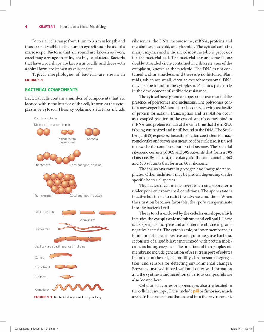
Bacterial cells range from 1 μm to 3 μm in length and thus are not visible to the human eye without the aid of a microscope. Bacteria that are round are known as cocci; cocci may arrange in pairs, chains, or clusters. Bacteria that have a rod shape are known as bacilli, and those with a spiral form are known as spirochetes.
Typical morphologies of bacteria are shown in Figure 1-1.
baCterIal COMpOnents
Bacterial cells contain a number of components that are located within the interior of the cell, known as the cyto-plasm or cytosol. These cytoplasmic structures include
ribosomes, the DNA chromosome, mRNA, proteins and metabolites, nucleoid, and plasmids. The cytosol contains many enzymes and is the site of most metabolic processes for the bacterial cell. The bacterial chromosome is one double-stranded circle contained in a discrete area of the cytoplasm, known as the nucleoid. The DNA is not con-tained within a nucleus, and there are no histones. Plas-mids, which are small, circular extrachromosomal DNA may also be found in the cytoplasm. Plasmids play a role in the development of antibiotic resistance.
The cytosol has a granular appearance as a result of the presence of polysomes and inclusions. The polysomes con-tain messenger RNA bound to ribosomes, serving as the site of protein formation. Transcription and translation occur as a coupled reaction in the cytoplasm; ribosomes bind to mRNA, and protein is made at the same time that the mRNA is being synthesized and is still bound to the DNA. The Sved-berg unit (S) expresses the sedimentation coefficient for mac-romolecules and serves as a measure of particle size. It is used to describe the complex subunits of ribosomes. The bacterial ribosome consists of 30S and 50S subunits that form a 70S ribosome. By contrast, the eukaryotic ribosome contains 40S and 60S subunits that form an 80S ribosome.
The inclusions contain glycogen and inorganic phos-phates. Other inclusions may be present depending on the specific bacterial species.
The bacterial cell may convert to an endospore form under poor environmental conditions. The spore state is inactive but is able to resist the adverse conditions. When the situation becomes favorable, the spore can germinate into the bacterial cell.
The cytosol is enclosed by the cellular envelope, which includes the cytoplasmic membrane and cell wall. There is also periplasmic space and an outer membrane in gram-negative bacteria. The cytoplasmic, or inner membrane, is found in both gram-positive and gram-negative bacteria. It consists of a lipid bilayer intermixed with protein mole-cules including enzymes. The functions of the cytoplasmic membrane include generation of ATP, transport of solutes in and out of the cell, cell motility, chromosomal segrega-tion, and sensors for detecting environmental changes. Enzymes involved in cell-wall and outer-wall formation and the synthesis and secretion of various compounds are also located here.
Cellular structures or appendages also are located in the cellular envelope. These include pili or fimbriae, which are hair-like extensions that extend into the environment. Figure 1-1 Bacterial shapes and morphology
4 Chapter 1 Introduction to Clinical Microbiology
Bacillus or rods
Bacillus - large bacilli arranged in chains
Curved
Coccobacilli
Fusiform
Various sizes
Filamentous
Coccus or spheres
Diplococci - arranged in pairs
Streptococcuspneumoniae
Neisseria
Streptococci Cocci arranged in chains
Cocci arranged in clustersStaphylococci
Spirochete
9781284032314_CH01_001_016.indd 4 13/02/14 11:53 AM

There are common pili, which permit the organism to at-tach to the host cells, and sex pili, which are involved in conjugation. Flagella are connected to the cellular enve-lope and found in those bacteria that are motile. Monot-richous flagella are located at one end of the cell, while lophotrichous flagella are located on both ends of the cell. Peritrichous flagella cover the entire bacterial surface. The presence or absence and location of flagella are important identification characteristics.
Other bacteria have capsules, which are composed of polysaccharide or protein layers. When these materials are more loosely arranged, it is known as a slime layer. Cap-sules are poor antigens and are antiphagocytic and impor-tant virulence factors for bacteria. Capsules also may serve as barriers to hydrophobic compounds such as detergents and can enable the bacteria to adhere to other bacteria or to host surfaces.
The periplasmic space contains the murein layer and a gel-like structure that assists the bacteria in obtaining nu-trients. There are also enzymes that can break down large molecules in this area. It is located between the inner part of the outer membrane and the outer external membrane and is found only in gram-negative bacteria.
The cell wall, or murein layer, is more commonly known as peptidoglycan; it serves as the external wall of most bacteria. This layer provides stability and strength to the bacterial cell and blocks the passage of some mac-romolecules. It is composed of the disaccharides N-acetylglucosamine and N-acetylmuramic acid, which are cross-linked to form peptidoglycan sheets when they are bound by peptide molecules. These peptidoglycan sheets further cross-link to form a multilayered structure. The peptidoglycan is much thicker in the gram-positive cell wall when compared to that of gram-negative bacteria. In addition, there are also techoic acids linked to the cellular membrane in the gram-positive cell wall. Techoic acids are water-soluble polymers of polyol phosphates bound to peptidoglycan and essential for the viability of the cell. Lipoteichoic acids contain a fatty acid and are important as surface antigens to differentiate bacterial serotypes. Lipoteichoic acids also aid the attachment of the organism to host receptors. Techoic acids also play a role in viru-lence. The mycobacteria contain a waxy substance, mycolic acid, in their cell walls, which enables them to resist the actions of acid. Other compounds that contribute to the acid- resistance and waxy character of the mycobacterial cell wall are cord factor, wax D, and sulfolipids.
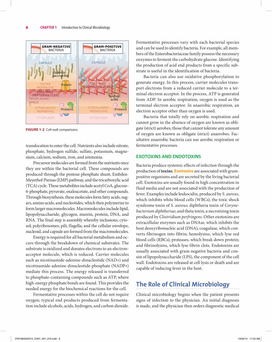
Gram-negative bacteria also have an outer membrane, which acts as a barrier for the cell against the external en-vironment and contains important enzymes and proteins. This outer membrane is external to the cytoplasmic mem-brane and is made up of lipopolysaccharide (LPS). LPS, also known as endotoxin, has several biological functions important in the disease process. When LPS is released upon cell lysis into the environment of the host, B cells are activated, which stimulates macrophage and other cells to release interleukin-1, interleukin-2, tumor necrosis factor, and other cytokines. LPS can also induce fever and cause shock; disseminated intravascular coagulation (DIC) is one severe consequence of large amounts of LPS released into the blood. DIC is characterized by systemic activation of blood coagulation with the formation of fibrin clots, which may result in thrombi, or clots, within the blood and organs of the body. This may ultimately result in multiple organ failure and bleeding from consumption of the co-agulation proteins and platelets. There are several causes of DIC that include severe infection and sepsis, trauma, malignancy, hepatic failure, and transfusion reactions.
LPS consists of three sections: lipid A, core polysaccha-ride, and O antigen. Lipid A is associated with endotoxin activity; its structure is similar for closely related bacteria. Core polysaccharide is important structurally and for the viability of the bacterial cell. The O antigen is important in differentiating serotypes or strains of a bacterial spe-cies. For example, there are over 150 types of O antigen for E. coli; one particularly important type is O157. Porins present in the outer membrane contain water and help regulate the passage of nutrients, antibiotics, and other hydrophilic compounds into the cell.
The cell walls of gram-positive and gram-negative bac-teria are compared in Figure 1-2.
Cellular MetabOlIsM
Cellular metabolism of bacteria is important when deter-mining how bacteria cause disease and also for the biochem-ical identification of bacteria. Bacteria must obtain nutrients from the environment through the cell envelope; water, oxy-gen, and carbon dioxide diffuse across the cell membrane. Active transport is needed to move other molecules such as organic acids, amino acids, and inorganic ions into the bacterial cell. These processes are facilitated through carrier molecules. Other compounds such as sugars, fatty acids, and nucleotide bases are chemically modified through group
Characteristics of Eukaryotes and Prokaryotes 5
9781284032314_CH01_001_016.indd 5 13/02/14 11:53 AM
translocation to enter the cell. Nutrients also include nitrate, phosphate, hydrogen sulfide, sulfate, potassium, magne-sium, calcium, sodium, iron, and ammonia.
Precursor molecules are formed from the nutrients once they are within the bacterial cell. These compounds are produced through the pentose phosphate shunt, Embden-Meyerhof-Parnas (EMP) pathway, and the tricarboxylic acid (TCA) cycle. These metabolites include acetyl CoA, glucose-6-phosphate, pyruvate, oxaloacetate, and other compounds. Through biosynthesis, these molecules form fatty acids, sug-ars, amino acids, and nucleotides, which then polymerize to form larger macromolecules. Macromolecules include lipid, lipopolysaccharide, glycogen, murein, protein, DNA, and RNA. The final step is assembly whereby inclusions; cyto-sol; polyribosomes; pili; flagella; and the cellular envelope, nucleoid, and capsule are formed from the macromolecules.
Energy is required for all bacterial metabolism and oc-curs through the breakdown of chemical substrates. The substrate is oxidized and donates electrons to an electron-acceptor molecule, which is reduced. Carrier molecules such as nicotinamide-adenine-dinucleotide (NAD+) and nicotinamide-adenine-dinucleotide-phosphate (NADP+) mediate this process. The energy released is transferred to phosphate-containing compounds such as ATP, where high-energy phosphate bonds are found. This provides the needed energy for the biochemical reactions for the cell.
Fermentative processes within the cell do not require oxygen; typical end products produced from fermenta-tion include alcohols, acids, hydrogen, and carbon dioxide.
Fermentative processes vary with each bacterial species and can be used to identify bacteria. For example, all mem-bers of the Enterobacteriaceae family possess the necessary enzymes to ferment the carbohydrate glucose. Identifying the production of acid end products from a specific sub-strate is useful in the identification of bacteria.
Bacteria can also use oxidative phosphorylation to generate energy. In this process, carrier molecules trans-port electrons from a reduced carrier molecule to a ter-minal electron acceptor. In the process, ATP is generated from ADP. In aerobic respiration, oxygen is used as the terminal electron acceptor. In anaerobic respiration, an electron acceptor other than oxygen is used.
Bacteria that totally rely on aerobic respiration and cannot grow in the absence of oxygen are known as obli-gate (strict) aerobes; those that cannot tolerate any amount of oxygen are known as obligate (strict) anaerobes. Fac-ultative anaerobic bacteria can use aerobic respiration or fermentative processes.
exOtOxIns and endOtOxIns
Bacteria produce systemic effects of infection through the production of toxins. exotoxins are associated with gram-positive organisms and are secreted by the living bacterial cell. Exotoxins are usually found in high concentration in fluid media and are not associated with the production of fever. Examples include leukocidin, produced by S. aureus, which inhibits white blood cells (WBCs); the toxic shock syndrome toxin of S. aureus; diphtheria toxin of Coryne-bacterium diphtheriae; and theta toxin, a necrotizing toxin produced by Clostridium perfringens. Other exotoxins are extracellular enzymes such as DNAse, which inhibits the host deoxyribonucleic acid (DNA); coagulase, which con-verts fibrinogen into fibrin; hemolysins, which lyse red blood cells (RBCs); proteases, which break down protein; and fibrinolysins, which lyse fibrin clots. Endotoxins are usually associated with gram-negative bacteria and con-sist of lipopolysaccharide (LPS), the component of the cell wall. Endotoxins are released at cell lysis or death and are capable of inducing fever in the host.
the role of Clinical MicrobiologyClinical microbiology begins when the patient presents signs of infection to the physician. An initial diagnosis is made, and the physician then orders diagnostic medical
Figure 1-2 Cell wall comparisons
6 Chapter 1 Introduction to Clinical Microbiology
GRAM-NEGATIVEBACTERIA
GRAM-POSITIVEBACTERIA
OUTERMEMBRANE
CAPSULE CAPSULE
CYTOPLASMICMEMBRANE
CYTOPLASM
CYTOPLASMICMEMBRANE
CYTOPLASM
CELL WALL(PEPTIDOGLYCAN)
CELL WALL(PEPTIDOGLYCAN)
PERIPLASMICSPACE
OUTOOUUUTTMEMBMMMMMMMMMMEEEEEEEEEMMMMMMMMMMMMMMMMBBBBBBBBBRBRRRRRRR
CAPSULE
ELL WCCEECCEECCECECECEECECECCCELLL WLL WWWWWLLLL WWWWWWWWW(P(P(P((P(P(((((((((
WWWWAAAAAAAAALLLLLLLLLLLLLLLLLLLLLGI
TTEEEEEEERRRRRRRRRRRRRRRRRRRRARARAARAAAAAAAAAAANAAAAAAAAANANNNNNNNNNNNNNNNNNNNNNNNNENNNEEEEEEEEEWWWWWWWWWWWWWWWWWWWWWWWWWWWWWWWWAWWWAAAAAAAAAALLLLLLLLLLLLLLLLLLLLLLLLLLLLLLLLLL
ASMICAAASSMMICCRANERRRRRRRRRAAAAAAAAANANNANNNNNNNNEEEEEEEEE
LASM
))))GGGGLLYCLYLYYCYYYYYYCCCAN)CAN)))))NN)IICCCCCCCC
LASM
Lipopoly-saccharide
OPLAOOOOPPPPLLLALALAMBRMMMMBBBRRR
LL WALLLLLL WAWAWWAWAWAWALLLLLLLLL L LLLLLL WL WLLLLLLLLLLLLL WLL LALALALALLLLLALLLLLLLLLLLLLLLLLALLLAAAAAAAAAAAAALLALA)))))N))))N))))))))DOGLYCADOGOGLGLOGDOGLDOGLLYCACAACAYCCAYCADDODODODODOGLYOGGGGOGO YYYYYCACAAAACACANNANAN
ASMICAAASASSSSMMMMMIMIICICCCCRANERRARAAAANNNEEE
Teichoicacids
Protein
Proteins Proteins
9781284032314_CH01_001_016.indd 6 13/02/14 11:53 AM

and laboratory procedures. A direct stain, a culture, and an antibiotic susceptibility test are typical tests that may involve the microbiology laboratory. The appropriate lab-oratory specimens are collected, labeled, and sent to the laboratory with a requisition or laboratory order form. The laboratory performs direct stains, plates the specimen on ap-propriate culture media, and incubates the plates at the suit-able temperature and atmosphere. The plates are examined and interpreted for the presence of pathogens, most often at 24 and 48 hours. Subcultures are performed as needed, and any biochemical, serological, molecular, antibiotic suscepti-bility, and automated procedures are performed. These tests are interpreted and the organisms are identified. The final report of the identification and antibiotic susceptibility tests is sent to the physician. The physician interprets the report and treats the patient appropriately.
Early diagnosis is associated with early treatment and a better prognosis for the patient. Testing must be per-formed in a timely, yet accurate, manner. Often, a pre-sumptive identification can be sent to the physician so that antibiotic therapy can be initiated. A final, definitive identification is then sent to update the report.
the Infectious processAn infection is the entrance and multiplication of a mi-croorganism in or on a host. The microorganism may enter the host through many routes including the respiratory tract, gastrointestinal tract, or breaks in the skin. The mi-croorganism next establishes itself and multiplies. Infec-tions can spread directly through tissues or the lymphatic system into the blood. infectious disease refers to an infection with functional and structural harm to the host that usually is accompanied by signs and symptoms.
In this process, the pathogen first must come in con-tact with and colonize the host. Colonization refers to the state when the microbe has established itself in a par-ticular niche or body site. This generally corresponds with the incubation period, and the host has no signs of infection. Next, the pathogen breaks the host’s protec-tive barrier and multiplies. This coincides with the first signs of clinical infection, known as the prodromal stage, during which time the host is particularly infective. The clinical symptoms peak during the clinical sage of in-fection. At this time the pathogen continues to multiply, attacking deeper tissues and eventually disseminating
through the blood to other organs. The pathogen is met by the host’s inflammatory and immune system, which have been stimulated by the microbe and its products. Depending on the immune status of the host and severity of the infection, there may be various outcomes. The host may destroy the pathogen, or the pathogen may destroy the host; in some cases, the pathogen may remain latent within the host. If the host recovers, the signs of infection decline, and the host eventually enters the convalescent phase of infection. Consequences of infection may re-main as a result of toxins or other compounds produced by the microbe. If the host continues to deteriorate, the symptoms may worsen, eventually resulting in perma-nent damage or even death.
All pathogens in a specimen must be identified and reported to the physician. A pathogen is a microorganism, including bacteria, viruses, fungi, and parasites, that is ca-pable of causing infectious disease. The identification of pathogens may be difficult because many microorganisms are present in the normal microbial flora. Normal flora re-fers to those microorganisms normally residing in a partic-ular body site or niche that do not generally cause infection. Normal flora may also be known as indigenous or resident flora or microbiota; these bacteria may perform important functions and compete with pathogenic microorganisms.
nOrMal FlOra
A limited number of organisms can be categorized as nor-mal flora. The slightly acidic pH of the skin (5.5 to 6.0) re-sults from the presence of acids produced by a number of bacteria. For example, Propionibacterium acnes produces large amounts of propionic acid. Other normal skin flora include Staphylococcus epidermidis, viridans streptococ-cus, and enterococcus. In addition, a number of contami-nating organisms may be found transiently on the skin, such as intestinal tract and soil contaminants.
In the mouth and oral cavity, the major normal flora is the viridans streptococcus, a collection of streptococcal species exhibiting alpha hemolysis or greening of sheep blood agar. In addition, S. epidermidis, nonpathogenic Neisseria species, Moraxella catarrhalis, lactobacilli, and diphtheroids may be present. Members of the anaerobic normal flora in this area include Actinomyces, Veillonella, and Bacteroides species.
The nasopharynx may serve as a site for asympto- matic carriage of several microorganisms. asymptomatic
The Infectious Process 7
9781284032314_CH01_001_016.indd 7 13/02/14 11:53 AM
carriers maintain a reservoir for the microorganism but do not have an infectious disease. The carrier state may be tran-sient or permanent, and these individuals may serve as an infectious source to transmit the pathogen to others. Bacteria that may be carried asymptomatically in the nasopharynx include Staphylococcus aureus and Neisseria meningitidis.
The stomach and upper small intestine are usually sterile, containing less than 1,000 organisms per milliliter. Organisms entering the stomach are usually killed by the hydrochloric acid and resulting low pH of the stomach, as well as by gastric enzymes. Other organisms are passed to the small intestine, where they may be destroyed by bile and pancreatic enzymes. When the gastric pH increases over 5.0, colonization from bacteria of oral, nasopharyn-geal, or colon origin may occur.
The colon is heavily colonized and serves as a reser-voir for infection for numerous body sites, including the urinary tract and peritoneal cavity. Major components of the normal bowel flora include Bacteroides, Lactobacillus, Clostridium, Eubacterium, coliforms such as Escherichia coli, aerobic and anaerobic streptococci, and yeast.
The normal flora of the distal urethra in both males and females may contain diphtheroids, alpha and non-hemolytic streptococci, Peptococcus, S. epidermidis, and Bacteroides.
Those sites considered to be sterile in the body, con-taining no normal flora, include the blood, cerebrospi-nal fluid, and urinary bladder. Normal flora can become pathogenic if they are moved to another site. Thus normal flora E. coli of the colon is an important cause of urinary tract infection (UTI) once it enters the bladder or kidneys. Likewise, although viridans streptococcus is considered to be normal flora of the oral cavity, the organism is a significant cause of subacute bacterial endocarditis when established in the heart.
MOdes OF transMIssIOn—rOutes OF InFeCtIOn
Infectious disease can be transmitted by several routes, which can be categorized as direct or indirect modes of transmission. The etiologic agent is the microorganism that has caused the infection. The reservoir of infection refers to the origin of the etiologic agent or its location such as contaminated food, another human, or an infected animal. Types of direct transmission include congenital contact, sexual contact, hand-to-hand contact, and droplet infection.
Congenital contact may occur across the placenta or as the baby passes through the vaginal canal during delivery. Rubella virus and syphilis may be acquired dur-ing pregnancy, whereas Streptococcus agalactiae and Neis-seria gonorrhoeae are examples of bacteria that may be transmitted to the infant during delivery.
Sexual contact may be the route of infection for sev-eral sexually transmittable diseases, including gonorrhea (N. gonorrhoeae); syphilis (Treponema pallidum); chla-mydia (Chlamydia trachomatis); acquired immunodefi-ciency syndrome, or AIDS (human immunodeficiency virus, or HIV); and herpes (herpes simplex virus).
Hand-to-hand transmission is the mode of direct contact seen with the spread of the common cold from rhinovirus. This route is also involved in the transmission of various GI infections when the hands are not properly washed and are fecally contaminated.
Infectious respiratory secretion or droplet infection serves as a route for several respiratory viruses as well as for bacterial pathogens including Streptococcus pyo-genes (the agent in streptococcal pharyngitis throat) and N. meningitidis. Infectious secretions include coughing, sneezing, kissing, and nasal drainage. Respiratory secre-tions can become dried on clothing, bedding, or floors and converted to dust, which may serve as a route of in-direct transmission.
Indirect routes of infection include fomites, ingestion of contaminated food and water, airborne routes, and ani-mal or arthropod vectors.
Fomites are inanimate objects such as eating uten-sils, drinking devices (water bottles and cups), hospital instruments, clothing, money, doorknobs, and tampons. Sometimes, these inanimate objects may serve as a route of nosocomial (hospital-acquired) infection. Fomites may also be referred to as vehicles of infection, which refers to nonliving objects that have been contaminted with the infectious agent.
Water may be contaminated as a result of improper sanitary measures or after it has been treated. Microor-ganisms that may be associated with contaminated water include Shigella, Salmonella, enteropathogenic E. coli, and hepatitis A virus (HAV). Improperly prepared, processed, preserved, or stored food may become contaminated with various microorganisms, including Salmonella, Listeria, E. coli O157:H7, and S. aureus. Milk and milk products may be contaminated through improper or lack of pasteuriza-tion, while undercooked meats may serve as sources of
8 Chapter 1 Introduction to Clinical Microbiology
9781284032314_CH01_001_016.indd 8 13/02/14 11:53 AM

contamination. Ingestion of the microorganisms or of pre-formed toxins plays a role in infections acquired through contaminated foods.
Infections may be incidentally transmitted to humans through infected animals or insect or arthropod vectors. Rabies, pasteurellosis (Pasteurella multocida), and tulare-mia (Francisella tularensis) are examples of infections that can be acquired through the bite or scratch of an infected animal. Arthropod vectors such as flies, mites, lice, ticks, and mosquitoes may transmit microorganisms from an infected animal to a human host. Malaria (Plasmodium) is transmitted by mosquitoes, whereas Lyme disease (Borrelia burgdorferi) is transmitted by the bite of an infected tick.
Airborne routes of infection include the inhalation of infectious particles that may be suspended in the air. Infectious aerosols can be formed in the laboratory that can serve as a route for laboratory-acquired infection. My-cobacterium tuberculosis, the agent in tuberculosis, can remain suspended in droplets that serve as infectious aero-sols. Dimorphic fungi, which cause systemic infections such as Coccidioides immitis, can cause airborne infections through the inhalation of infectious spores.
Figure 1-3 summarizes routes of infection for microorganisms.
Once established in a particular area, the microorgan-ism may multiply and spread from its original site of entry to contiguous tissue or disseminate through the blood to other, distant sites. Microorganisms spread and multiply when host defense mechanisms are overcome.
The incidence of a disease refers to the number of those individuals infected in a population, whereas the
disease prevalence describes the percentage of infected individuals in a given population at a given time. An endemic is when a disease consistently occurs at a con-stant rate in a given location. An epidemic occurs when there are larger numbers of cases of a disease in a given location. Pandemics are epidemics that occur around the world. Outbreaks generally refer to instances where there is a disproportionately larger number of infected individu-als in a fairly short amount of time.
true versus OppOrtunIstIC pathOgens
A true pathogen has the ability to cause an infection under all conditions in all types of individuals. By contrast, an op-portunistic pathogen refers to an organism that is patho-genic only under particular, favorable conditions. Under other conditions, opportunisms are nonpathogenic and generally harmless and are often members of the human normal flora. Opportunistic infections are infections that occur when there are preexisting conditions that increase the susceptibility of the host to infection. Generally, oppor-tunistic pathogens do not cause disease in individuals with a normal immune system. These infections are increasing as a result of a number of factors including the widespread use of broad-spectrum antibiotics that can alter the normal flora, the increased use of immunosuppressive drugs to pre-vent organ transplant rejection, the use of chemotherapeutic agents to treat cancer, and the increased and prolonged use of urethral catheters. Thus an opportunistic pathogen is one that attacks an already debilitated host but usually presents no danger to an individual with an intact immune system.
Figure 1-3 Routes of infection
The Infectious Process 9
(A) Direct contact
Horizontaltransmission
Sexualintercourse
Respiratorydroplets
Human oranimal contact
GastroenteritisMumpsRabies
Ringworm
PertussisCommon cold
In�uenza
Verticaltransmission
Mother tofetus
Mother tonewborn
ListeriosisCongenital
syphilisAIDS
Neonatalconjunctivitis(gonorrhea,chlamydia)
can occur by
through from
with diseases such as
with diseases such asthat can carry
involves person-to-person by
GastroenteritisHepatitis AAmoebiasis
CholeraLeptospirosis
Cryptosporidiosis
TBMeasles
Valley fever
(B) Indirect contact
Vehicletransmission
Water Aerosols
Fomites
TetanusHepatitis B
Athlete’s foot
Vectortransmission
Mechanicalvector
Biologicalvector
Typhoid feverTrachomaShigellosis
Lyme diseaseWest NileMalaria
Foods
involving
via
which canharbor
which caninvolve a
carrying diseaseagents for
carrying disease agents for
DISEASE TRANSMISSION
GonorrheaSyphilis
ChlamydiaAIDS
9781284032314_CH01_001_016.indd 9 13/02/14 11:53 AM

In immunosuppressive conditions the host immune system is unable to effectively battle those microorgan-isms considered to be normal flora for the general popula-tion. Other individuals at risk for opportunistic pathogens include dialysis patients, those with HIV-AIDS, burn victims, diabetics, and any individual who has chronic medical problems or is undergoing invasive medical tech-niques. Those individuals with foreign body implants in-cluding heart valves, prosthetic devices, and indwelling intravenous catheters; alcoholics; and intravenous drug users are also at increased risk of opportunistic pathogens.
By contrast, a true pathogen has the ability to infect those individuals with a healthy immune system as well as those with an immunosuppressed state.
hOst deFense MeChanIsMs
Host defense mechanisms include innate, or natural, im-munity; acquired immunity; and phagocytosis. Immu-nity refers to host properties that confer resistance of the host to foreign substances. It is the sum of all mechanisms used by the body as protection against environmental agents that are not normally present in the body.
Innate or natural ImmunityInnate, or natural, immunity is inborn, mainly genetically determined, and nonspecific. It is not acquired through previous contact with an infectious agent. The skin acts as a protective barrier, and a limited number of microorgan-isms can penetrate the intact skin barrier. Many microor-ganisms, however, can enter the body through breaks in the skin resulting from bites, wounds, cuts, or needles. In addition, microorganisms can enter through the sweat and fat glands. The normal flora bacteria of the skin produce free fatty acids from oil, which will produce low pH on the skin and thus prevent the establishment of infection.
Mucus covers the surfaces of the respiratory tract. The mucus can trap bacteria and, with the assistance of cilia, which are also present in the nasal cavity, can trap many microorganisms. The bacteria are either swallowed, entering the stomach where they are destroyed by the acidic environment, or removed through external open-ings such as the nose and mouth by sneezing or coughing. Lysozyme, an enzyme that lyses bacterial cell walls, is also present in respiratory secretions. Some types of immu-noglobulin A (IgA) have a secretory component that can prevent attachment of the microorganisms to the mucous
membranes of the respiratory tract. Those bacteria that are phagocytized are transported through the lymphatic system and eventually removed from the body.
The respiratory tract is a common portal of entry for several microorganisms, including Streptococcus pneu-moniae, M. tuberculosis, Mycoplasma pneumoniae, and several respiratory viruses. Those microorganisms pos-sessing pili, appendages for attachment, are able to attach to the mucous membranes.
Saliva in the oral cavity possesses various hydrolytic enzymes that can break down bacteria. The low pH of the stomach and the presence of gastric enzymes result in a limited amount of microorganisms that can sustain life in the stomach. The rapid peristalsis in the small intestine, together with the presence of enzymes and bile, keeps the microbial population in this site at very low levels. The large intestine has a large amount of normal flora that competes with pathogens, thus decreasing the chance for establishment of infection. The anaerobic normal flora of the large intestine produces fatty acids, while facultative anaerobes produce bacteriocins, which also hinder the multiplication of pathogens.
The constant flushing action and low pH of urine prevent pathogens from establishing in the urinary system. Females, who have shorter urethras, are more prone to UTIs than are males, who have longer urethras. Bacteria have easier access to the female bladder from the GI and vaginal tracts.
Lactobacilli, a major normal flora in the female vaginal tract, produce large amounts of lactic acid, which results in a low pH. This acidic environment decreases the amount of other microorganisms that can cause infection in this area.
The eyes are protected through the constant flushing provided by tears as well as the presence of lysozyme in tears.
Other elements of innate immunity include fever, in-terferon, phagocytosis, and various serum proteins. These components are described with the inflammatory response.
Natural immunity can be affected by a number of fac-tors. For example, very young and very old persons are more susceptible to infection than are other age groups. Hormonal changes such as those occurring during preg-nancy, diabetes mellitus, and Addison’s and Cushing’s diseases can alter the metabolism and immune response, which results in increased susceptibility to infection. The use of broad-spectrum antibiotics, which alter the host’s normal flora, can also promote the growth of pathogens. The normal flora is no longer present in large amounts to compete with the pathogenic organisms. Such infections
10 Chapter 1 Introduction to Clinical Microbiology
9781284032314_CH01_001_016.indd 10 13/02/14 11:53 AM

are termed secondary infections or superinfections be-cause the infection results from the use of an antibiotic.
Inflammatory responseThe inflammatory response can be activated by trauma or tissue injury. Inflammation is the total of the changes occurring in tissue factors upon injury. There are four hallmark signs of inflammation: redness, heat, swelling, and pain. These are described in Box 1-3. Hemodynamic changes, such as increased vascular permeability, dilation of arterioles and capillaries, and increased blood flow to the injured area, occur. Plasma proteins, such as comple-ment, interferon, and antibodies, are also released. Edema may occur as a result of vasodilation, while there is also an influx of red blood cells to the area.
Phagocytic white blood cells, which include neutro-phils (PMNs) and macrophages, have four functions: mi-gration, chemotaxis, ingestion, and killing. These cells may adhere to the vascular endothelium or migrate from the blood to the affected tissues in a process known as dia-pedesis. Chemotaxis refers to the attraction of the phago-cytes to the affected area by the microorganisms or its products from the blood to the injured site. Neutrophils and monocytes are attracted by bacterial products such as endotoxins and enzymes. Attachment initiates the phago-cytic process. The microorganism must be coated with opsonins such as IgG or complement units for effective phagocytosis. Those microbes that have a capsule are re-sistant to the effects of phagocytosis. The next step is in-gestion, which involves the formation of a phagosome that undergoes a respiratory burst. The respiratory burst results in the release of superoxide anion and peroxide, both of which are toxic to microorganisms. Lysosomes within the cell combine with phagosomes to form phagolysosomes, which eventually release hydrolytic enzymes.
Neutrophils are formed in the bone marrow and enter the blood and tissue. Monocytes, also formed in the bone marrow, are known as macrophages once the cells have en-tered the tissue. Macrophages are present in tissues but are also fixed to the blood vessels of the liver, spleen, and lymph nodes. Macrophages can become activated by microorgan-isms, endotoxins, and antigen–antibody complexes. Once activated, macrophages produce an increased number of lyso-somes that produce interleukin-1. Interleukin-1 is associated with the stimulation of fever and activation of phagocytosis.
Inflammation caused by microorganisms may be ini-tiated through activation of the complement system or the blood coagulation cascade. These systems initiate the release of several chemical mediators of inflammation, which are summarized in TaBle 1-2.
acquired Immunityacquired immunity can be either passive or active. Pas-sively acquired immunity refers to the temporary resis-tance to infectious agents by administration of antibodies preformed in another host. Examples include gamma glob-ulin and antitoxin to Clostridium botulinum toxin. Passive acquisition is also seen in the fetus, who acquires antibod-ies from the mother’s blood during pregnancy. These anti-bodies remain active in the newborn until 4 to 6 months of age. Newborns may also acquire immunity passively from their mothers through antibodies carried in breast milk.
Actively acquired immunity is a state of resistance built in an individual following contact with foreign an-tigens such as microorganisms. The immunity may result from clinical or subclinical infection, through injection of live or inactivated microorganisms, their antigens, or nucleic acids, or through the absorption of bacterial prod-ucts such as toxins or toxoids. In active immunity the host actively produces antibody in reaction to a foreign antigen.
The Infectious Process 11
Box 1-3 hallmark signs of Inflammation
Sign Latin Derivation Description
Redness Rubor Dilation of small blood vessels in area of injury
Heat Calor Increased blood flow through the inflamed area in peripheral parts of body; fever as a result of chemical mediators of inflammation
Swelling Edema Fluid accumulates outside of blood vessels
Pain Dolar As a result of edema because tissues are inflamed; effects of serotonin, bradykinin, and prostaglandins
9781284032314_CH01_001_016.indd 11 13/02/14 11:53 AM
Acquired immunity involves contact with a foreign agent or antigen. This process is known as immunization, and it initiates a series of reactions that lead to the activa-tion of lymphocytes and the synthesis of specialized serum proteins known as antibodies. Thus antigens are substances foreign to the body that initiate the production of antibodies.
Three major cell types are involved in the immune re-sponse: T lymphocytes, B lymphocytes, and macrophages. T and B lymphocytes arise from a common lymphoid precursor cell but mature in different areas of the body. T cells mature in the thymus, while B cells, named for the bursa of Fabricius (the area in the chicken where the cells were first isolated), mature in the bone marrow. Although B and T cells have different functions, both play a role in the recognition of antigen and reaction to it. The third type of cells, macrophages, are phagocytic cells that ingest, process, and present antigens to T cells.
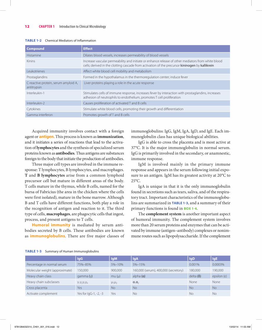
Humoral immunity is mediated by serum anti-bodies secreted by B cells. These antibodies are known as immunoglobulins. There are five major classes of
immunoglobulins: IgG, IgM, IgA, IgD, and IgE. Each im-munoglobulin class has unique biological abilities.
IgG is able to cross the placenta and is most active at 37°C. It is the major immunoglobulin in normal serum. IgG is primarily involved in the secondary, or anamnestic, immune response.
IgM is involved mainly in the primary immune response and appears in the serum following initial expo-sure to an antigen. IgM has its greatest activity at 20°C to 25°C.
IgA is unique in that it is the only immunoglobulin found in secretions such as tears, saliva, and of the respira-tory tract. Important characteristics of the immunoglobu-lins are summarized in TaBle 1-3, and a summary of their primary functions is found in Box 1-4.
The complement system is another important aspect of humoral immunity. The complement system involves more than 20 serum proteins and enzymes that can be acti-vated by immune (antigen–antibody) complexes or nonim-mune routes such as lipopolysaccharide. If the complement
12 Chapter 1 Introduction to Clinical Microbiology
TaBle 1-3 Summary of Human Immunoglobulins
igg igM iga igD ige
Percentage in normal serum 75%–85% 5%–10% 5%–15% 0.001% 0.0003%
Molecular weight (approximate) 150,000 900,000 160,000 (serum), 400,000 (secretory) 180,000 190,000
Heavy chain class gamma (γ) mu (μ) alpha (α) delta (δ) epsilon (ε)
Heavy chain subclasses γ1γ2γ3γ4 μ1μ2 α1α2 None None
Cross placenta Yes No No No No
Activate complement Yes for IgG-1, -2, -3 Yes No No No
TaBle 1-2 Chemical Mediators of Inflammation
Compound effect
Histamine Dilates blood vessels, increases permeability of blood vessels
Kinins Increase vascular permeability and initiate or enhance release of other mediators from white blood cells; derived in the clotting cascade from activation of the precursor kininogen by kallikrein
Leukotrienes Affect white blood cell mobility and metabolism
Prostaglandins Formed in the hypothalamus in the thermoregulation center; induce fever
C-reactive protein, serum amyloid A, antitrypsin
Liver proteins playing a role in the acute response
Interleukin-1 Stimulates cells of immune response, increases fever by interaction with prostaglandins, increases adhesion of neutrophils to endothelium, promotes T cell proliferation
Interleukin-2 Causes proliferation of activated T and B cells
Cytokines Stimulate white blood cells, promoting their growth and differentiation
Gamma interferon Promotes growth of T and B cells
9781284032314_CH01_001_016.indd 12 13/02/14 11:53 AM
cascade is activated, the target cell may be lysed, or phago-cytic cells may be stimulated.
Cell-Mediated ImmunityCell-mediated immunity (CMi) involves the T lympho-cytes, which circulate to the antigen to perform their func-tion. There are several populations of T cells including:
7 T helper (inducer) cells 7 Enhance proliferation and differentiation of B cells
and precursors to cytotoxic T cells 7 Increase ability of macrophages to ingest and de-
stroy pathogens 7 Enhance the production of antibody by B cells 7 Release lymphokines, including interleukin-1 (IL-1)
and interleukin-2 (IL-2) and B cell–stimulating fac-tor, which helps to activate B cells
7 Cytotoxic T cells—destroy targets on direct contact through the recognition and destruction of antigen-bearing cells
7 T suppressor cells—suppress or regulate the response of T and B cells
7 Null cells (natural killer [NK] and killer [K] cells)—kill tumor or viral-infected cells, although not with the specificity of cytotoxic T cells
Although presented as separate functions, the humoral and cell-mediated immune systems interact in the im-mune response.
sIgns OF InFeCtIOn
The need for clinical microbiology begins with a patient who is exhibiting one or more signs of infection. Some common general or systemic signs of acute infection
include a high-grade, spiking fever; chills; vasodilation with flushing; and an increased pulse rate. Chronic or subacute infections may be accompanied by the following systemic signs: intermittent, low-grade fever; weight loss; and fatigue. Local signs of infection include pain, heat, redness, and swelling. The hallmark signs of inflamma-tion are found in Box 1-3.
In the laboratory, specific procedures are used to diag-nose infection. These include the leukocyte count, which is elevated for most infectious processes, and the differ-ential white blood cell count, which enables the clinician to determine the type of infection. In general, but not al-ways, bacterial infections are associated with an elevated white blood cell count and an increased percentage of neu-trophils. By contrast, lymphocytes are the predominant WBC in most viral infections. The erythrocyte sedimen-tation rate (ESR) is a nonspecific indicator of inflamma-tion and is frequently increased in infectious disease and numerous other inflammatory states. C-reactive protein is another plasma protein that is present during infectious disease. Finally, the presence of type-specific antibodies in a patient’s serum can be used to identify the presence of a particular pathogen. On exposure to a bacterial or viral pathogen, the patient produces antibodies against the anti-gens of the organism. The antibodies then can be detected through use of antigenic markers.
Radiographic signs of infectious disease that a clini-cian would note include pulmonary infiltrates, gas and swelling in the tissues, and the accumulation of fluid in a body cavity.
Gastrointestinal signs such as nausea, vomiting, and diarrhea, as well as various neuromuscular and cardiopul-monary signs, are also noted by the clinician.
The Infectious Process 13
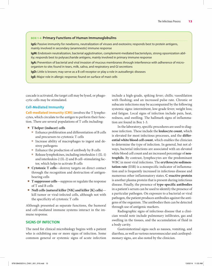
Box 1-4 primary Functions of human Immunoglobulins
igg: Passive immunity for newborns, neutralization of viruses and exotoxins; responds best to protein antigens, mainly involved in secondary (anamnestic) immune response
igM: Endotoxin neutralization, bacterial agglutination, complement-mediated bacteriolysis, strong opsonization abil-ity; responds best to polysaccharide antigens, mainly involved in primary immune response
iga: Prevention of bacterial and viral invasion of mucous membranes through interference with adherence of micro-organism to site; found in tears, milk, saliva, and respiratory and GI secretions
igD: Little is known; may serve as a B cell receptor or play a role in autoallergic diseases
ige: Major role in allergic response; found on surface of mast cells
9781284032314_CH01_001_016.indd 13 13/02/14 11:53 AM

nOsOCOMIal and health Care–assOCIated InFeCtIOns
A nosocomial, or health care–associated, infection is acquired in a hospital or other health care setting. The organism is not present and not incubating in the pa-tient on entry or admission into the health care facility. A community-acquired infection is present or incubat-ing at the time of admission into the health care facil-ity. Community-acquired infections also are those that are acquired within an individual’s community such as her or his school, workplace, or athletic or social setting. As with other infections, nosocomial and community-acquired infections can be categorized as endogenous or exogenous. Endogenous infections result from organisms that are a part of the patient’s normal flora, whereas ex-ogenous infections result from organisms from external sources. These sources may include contaminated medi-cal instruments or equipment or inanimate objects in the health care setting or from contact with health care per-sonnel. Individuals, including health care providers, may be colonized with an organism. Colonization is defined as the presence and multiplication of a microorganism in a host, with no clinical signs of infection. Such individu-als may serve as a reservoir of infection and transmit the organism to susceptible individuals.
The most common types of nosocomial infections are urinary tract infections (35% to 40%), surgical wound in-fections (20%), lower respiratory tract infections (15%), and bacteremia (5%–10%). These percentages may vary with each health care setting. Those bacteria most often associated with nosocomial infections include S. aureus, E. coli, Enterococ-cus, and Pseudomonas aeruginosa. Many nosocomial patho-gens are resistant to multiple antimicrobial agents.
Nosocomial urinary tract infections may be the result of catheterization or the presence of indwelling catheters or other urological techniques such as cystoscopy. The organ-ism is frequently of endogenous origin, as with E. coli, which is a member of the normal flora of the large intestine. Ex-ogenous sources include the contaminated hands of health care providers or contaminated equipment or solutions.
Nosocomial surgical wound infections usually involve S. aureus, Enterococcus, or gram-negative bacilli. These infections may be endogenous or exogenous.
Nosocomial pneumonia may result from aspiration of the organisms from the stomach or upper respiratory tract. The airways or stomach may become colonized with bacteria including S. aureus, P. aeruginosa, and Klebsiella pneumoniae. Respiratory care procedures, such as endo-tracheal suctioning and inhalation therapy, also present a greater risk for nosocomial pneumonia. A high mortality rate is associated with nosocomial infections of the lower respiratory tract.
Bacteremia may result from the patient’s own flora as well as that of the health care provider. In addition, intra-venous devices or solutions may be contaminated.
Host factors that lead to increased susceptibility to nosocomial infections include a compromised immune system, underlying medical disease or diseases, age, trauma, burns, poor nutritional status, anatomical abnor-malities, use of medical instrumentation, and diagnostic procedures.
Some, but not all, nosocomial infections can be pre-vented. The universal use of gloves and practice of aseptic techniques, including thorough hand washing, can de-crease the incidence of nosocomial infections. The rou-tine disinfection of inanimate surfaces and prevention of aerosols are also important factors.
review QuestionsMatchingMatch the following terms with the correct definition:
1. Infection 2. Infectious disease 3. Opportunistic infection 4. Nosocomial infection 5. Colonization
a. Condition associated with functional and structural harm to the host, accompanied by signs and symptoms
b. Infection in an immunocompromised host that does not cause infection in an immunocompetent individual
c. Infection acquired in a health care settingd. Presence and multiplication of a microorganism in a
host with no clinical signs of infectione. Entrance and multiplication of a microorganism in a
host
14 Chapter 1 Introduction to Clinical Microbiology
9781284032314_CH01_001_016.indd 14 13/02/14 11:53 AM

c. T lymphocytesd. B lymphocytes
14. Which of the following cells play a major role in cell-mediated immunity?a. Macrophagesb. Neutrophilsc. T lymphocytesd. B lymphocytes
15. The immunoglobulin found in the highest concentration in normal serum is:a. IgAb. IgDc. IgEd. IgGe. IgM
16. Which of the following immunoglobulins is involved mainly in the primary immune response?a. IgAb. IgDc. IgEd. IgGe. IgM
17. Gram-negative bacteria contain , which are not found in gram-positive bacteria.a. Capsulesb. Periplasmic space and outer membranec. Teichoic acidsd. Cross-linked peptidoglycan
18. Which of the following is true for bacterial cells?a. The DNA is contained within a nuclear membrane.b. Their mitochondria, Golgi bodies, and endoplasmic
reticulum are present in the cytoplasm.c. The DNA is found in the nucleoid.d. The ribosomes are 80S.
19. The are important for motlity of the bacterial cell.a. pilib. capsulesc. flagellad. LPS
20. Phenotypic properties used to classify bacteria include all of the following except:a. DNA relatednessb. Colonial morphologyc. Biochemical propertiesd. Antibiotic resistance patterns
Multiple Choice
6. All the following sites contain normal flora except:a. Oral cavityb. Skinc. Colond. Cerebrospinal fluid
7. Which of the following is not classified as a direct route of infection?a. Ingestion of contaminated food or waterb. Sexual contactc. Hand-to-hand contactd. Congenital contact
8. Droplet infection through contact with infectious respiratory secretions may be described as:a. Inhalation of infectious aerosols during laboratory
proceduresb. Transmission of rhinovirus through failing to wash
handsc. Spread of respiratory viruses and Streptococcus
pyogenes through coughing or sneezingd. Inhalation of bacteria or viruses that have dried on
bedding or clothing9. Which of the following organisms are typically spread
through the ingestion of contaminated food or water?a. Neisseria meningitidis and S. pyogenesb. Salmonella and Shigellac. Herpes simplex virus and Treponema pallidumd. Plasmodium and Borrelia
10. Which of the following organisms are spread through arthropod vectors?a. N. meningitidis and S. pyogenesb. Salmonella and Shigellac. Herpes simplex virus and T. pallidumd. Plasmodium and Borrelia
11. Innate, or natural, immunity involves which of the following mechanisms?a. Mucus and cilia in the respiratory tract that help to
trap and clear microorganismsb. Humoral immunityc. Cell-mediated immunityd. Immunity resulting from vaccination
12. The movement of neutrophils and monocytes from the blood to injured tissue is known as:a. Diapedesisb. Chemotaxisc. Ingestiond. Hematopoiesis
13. Antibody-producing white blood cells are:a. Macrophagesb. Neutrophils
Review Questions 15
9781284032314_CH01_001_016.indd 15 13/02/14 11:53 AM

bibliographyBower, S., & Rosenthal, K. S. (2006). Bacterial cell walls: The
armor, artillery and Achilles heel. Infectious Diseases in Clinical Practice, 15, 309–316.
Casadevall, A., & Pirofski, L. (2001). Host-pathogen interac-tions: The attributes of virulence. The Journal of Infectious Diseases, 184, 337–344.
Diekema, D. J., & Pfaller, M. A. (2007). Infection control epi-demiology and clinical microbiology. In P. R. Murray, E. J. Baron, J. H. Jorgensen, M. L. Landry, & M. A. Pfaller (Eds.), Manual of clinical microbiology, 9th ed. (pp. 118–128). Washington, DC: American Society for Microbiology.
Finlay, B. B., & Falkow, S. (1989). Common themes in microbial pathogenicity. Microbiology and Molecular Biology Reviews, 53, 210.
Forbes, B. A., Sahm, D. F., & Weissfeld, A. S. (2007). Bacterial genetics, metabolism and structure. In Bailey and Scott’s diagnostic microbiology, 12th ed. St. Louis, MO: Mosby.
Klevins, R. M., Edwards, J. R., Richards Jr., C. L., Horan T. C., Gaynes, R. P., Pollock, D. A., & Cardo, D. M. (2007). Es-timating health care-associated infections and deaths in U.S. hospitals, 2002. Public Health Reports, 122, 160–166.
Murray, P. R., Rosenthal, K. S., & Pfaller, M. A. (2009). Mecha-nisms of bacterial pathogenesis. In Medical microbiology, 6th ed. (pp. 179–187). Philadelphia, PA: Mosby.
Parham, P. (2009). Antibody structure and the generation of B-cell diversity. In The immune system, 3rd ed (pp. 1–31). New York, NY: Garland Science Taylor & Francis Group.
Parham, P. (2009). Elements of the immune system and their roles in defense. In The immune system, 3rd ed (pp. 1–31). New York, NY: Garland Science Taylor & Francis Group.
Schaechter, M. (2012). Biology of infectious diseases. In N. C. Engleberg, T. Dermody, & V. DiRita (Eds.), Schaechter’s mechanisms of microbial disease, 5th ed. (pp. 18–37). Phila-delphia, PA: Lippincott Williams & Wilkins.
Soule, B. M., & LaRocco, M. T. (1993). Nosocomial infections: An overview. In B. J. Howard, J. F. Keiser, A. S. Weissfeld, & F. Thomas (Eds.), Clinical and pathogenic microbiology, 2nd ed. (pp. 83–99). St. Louis, MO: Mosby.
Turgeon ML (2003). Antigens and antibodies. In Immunology and serology in laboratory medicine, 3rd ed. (pp. 15–35). St. Louis, MO: Mosby.
16 Chapter 1 Introduction to Clinical Microbiology
9781284032314_CH01_001_016.indd 16 13/02/14 11:53 AM