Dissertation submitted for MD MICROBIOLOGY BRANCH
150
A STUDY ON CURRENT ANTIMICROBIAL SUSCEPTIBILITY PATTERN OF TYPHOIDAL SALMONELLAE CAUSING ENTERIC FEVER IN SCHOOL GOING CHILDREN AND YOUNG ADULTS Dissertation submitted for M.D. MICROBIOLOGY BRANCH – 1V DEGREE EXAMINATION THE TAMILNADU DR.M.G.R.MEDICAL UNIVERSITY CHENNAI – 600 032 TAMILNADU MAY 2018
-
Upload
khangminh22 -
Category
Documents
-
view
1 -
download
0
Transcript of Dissertation submitted for MD MICROBIOLOGY BRANCH

A STUDY ON CURRENT ANTIMICROBIAL SUSCEPTIBILITY
PATTERN OF TYPHOIDAL SALMONELLAE CAUSING ENTERIC
FEVER IN SCHOOL GOING CHILDREN AND YOUNG ADULTS
Dissertation submitted for
M.D. MICROBIOLOGY BRANCH – 1V
DEGREE EXAMINATION
THE TAMILNADU DR.M.G.R.MEDICAL UNIVERSITY
CHENNAI – 600 032
TAMILNADU
MAY 2018

CERTIFICATE
This is to certify that this dissertation titled “A STUDY ON CURRENT
ANTIMICROBIAL SUSCEPTIBILITY PATTERN OF TYPHOIDAL
SALMONELLAE CAUSING ENTERIC FEVER IN SCHOOL GOING
CHILDREN AND YOUNG ADULTS ” is a bonafide record of work done
by Dr. ANITHA.M, during the period of April 2016 to March 2017 under the
guidance of Prof.Dr.MANGALA ADISESH, M.D., Professor of Microbiology,
Institute of Microbiology , Madras Medical College and Rajiv Gandhi
Government General Hospital, Chennai - 600003, in partial fulfillment of the
requirement of M.D. MICROBIOLOGY Degree Examination of The Tamilnadu
Dr.M.G.R. Medical University to be held in May 2018.
Dr.R.NARAYANA BABU, MD.,DCH Dr.ROSY VENNILA., M.D., Dean, Director, Madras Medical College & Institute of Microbiology, Rajiv Gandhi Government Madras Medical College & General Hospital, Rajiv Gandhi Government Chennai – 600003 General Hospital, Chennai – 600003
DECLARATION
I, Dr.ANITHA.M, Post Graduate , Institute of Microbiology, Madras
Medical College, solemnly declare that the dissertation titled “A STUDY ON
CURRENT ANTIMICROBIAL SUSCEPTIBILITY PATTERN OF
TYPHOIDAL SALMONELLAE CAUSING ENTERIC FEVER IN
SCHOOL GOING CHILDREN AND YOUNG ADULTS ”is the bonafide
work done by me at Institute of Microbiology, Madras Medical College under the
expert guidance and supervision of Prof.Dr. MANGALA ADISESH M.D.,
Professor, Institute of Microbiology, Madras Medical College. The dissertation is
submitted to the Tamil Nadu Dr.M.G.R Medical University towards partial
fulfillment of requirement for the award of M.D., Degree (Branch IV) in
Microbiology.
Place: Chennai
Date: Dr. ANITHA.M
Signature of the Guide
Prof. Dr.MANGALA ADISESH, MD., Professor,
Institute of Microbiology Madras Medical College, Chennai-600 003.

ACKNOWLEDGEMENT
I wish to express my sincere thanks to the Honourable Dean
Dr.R.NARAYANA BABU, MD, D.Ch ., Madras Medical College & RGGGH,
Chennai for permitting me to use the resources of this Institution for my study.
I express my sincere thanks to Dr. ROSY VENNILA , M.D., Director,
Institute of Microbiology, Madras Medical College & RGGGH, Chennai.
I feel fortunate and indebted to be under the guidance of Prof.Dr.
MANGALA ADISESH , M.D., Institute of Microbiology, for suggesting the
topic for my dissertation and for her valuable advice, guidance in preparing and
compilation of my work, throughout my study period. She is a source of
inspiration in my endeavours.
I specially thank to our Prof.Dr.U.UMADEVI M.D., for her valuable
guidance ,and co-ordinating me in doing Molecular workup for my study at
Christian Medical College, Vellore.
I extend my sincere thanks to our Professors Dr. S.THASNEEM BANU
M.D., Dr.R.VANAJA M.D., Dr.C.P.RAMANI M.D., for their support,
guidance and valuable advice.
I extend my sincere thanks to Prof. Dr.N.DEVASENA M.D., Department
of Microbiology, Institute of Child Health and Hospital for Children for her
support and guidance .

I extend my whole hearted gratitude and special thanks to my co-guide
Dr.R.DEEPA., M.D., Senior Assistant Professor for her expert guidance and
support in doing my study.
I express my sincere thanks to our Assistant Professors
Dr.N.RATHNAPRIYA M.D., Dr.K.USHAKRISHNAN M.D.,
Dr.K.G.VENKATESH M.D., Dr.C.S.SRIPRIYA M.D.,
Dr.N.LAKSHMIPRIYA,M.D.D.Ch., Dr. DAVID AGATHA M.D., and
Dr.B.NATESAN M.D., for their support and guidance in my study.
My sincere thanks to Prof.Dr.T.RAVICHANDRAN.,M.D.,D.Ch.,
Director and Professor , Institute of Child Health and Hospital for Children for his
support and guidance. My sincere thanks to Prof.Dr.K.SRINIVASAGALU
M.D., former Director and Professor., Institute of Internal Medicine for his
guidance during my study period .
I thank Dr. Evangeline Mary, M.D.,M.B.A.,Department of Community
Medicine , Govt. Stanley Medical College, Chennai, for guiding me in analyzing
the results statistically .
I would like to thank all my colleagues, and all our technicians and staffs
in Institute of Microbiology, Madras Medical College for their support and
cooperation.
I would like to thank the Institutional Ethics Committee for approving my
study.

I feel indebted to my parents who had been an everlasting support and
encouragement and for their heartful blessings.
I thank my husband Dr. K. Chandrasekaran M.S, for taking great care,
support and encouragement without which this work would not have been
possible.
I thank all my patients without whom this study would not have been
completed.
Above all I thank the Almighty for his blessings and grace by giving me
this opportunity to acquire knowledge .

TABLE OF CONTENTS
SI. NO TITLE PAGE
No.
1 INTRODUCTION 1
2 AIMS & OBJECTIVES 4
3 REVIEW OF LITERATURE 5
4 MATERIALS & METHODS 50
5 RESULTS 68
6 DISCUSSION 79
7 LIMITATIONS OF THE STUDY 90
7 SUMMARY 91
8 CONCLUSION 93
9 BIBLIOGRAPHY
ANNEXURE-I ABBREVATIONS
ANNEXURE-II CERTIFICATE OF APPROVAL
ANNEXURE-III PROFORMA
ANNEXURE-IV CONSENT FORM
ANNEXURE-V MASTER CHART

LIST OF TABLES
S. NO TITLE PAGE
NO.
1 DISTRIBUTION OF PATIENTS IN THE STUDY POPULATION 68
2 AGE GROUP AND SEX DISTRIBUTION IN THE STUDY POPULATION 69
3 DISTRIBUTION OF PATIENTS BASED ON HOSPITAL ADMISSION 70
4 CARDINAL SYMPTOMS AMONG STUDY POPULATION 70
5 CARDINAL SIGNS AMONG STUDY POPULATION 71
6 DISTRIBUTION BASED ON FOOD HABITS 71
7 DISTRIBUTION OF BACTERIAL GROWTH IN BLOOD CULTURE 72
8 DISTRIBUTION OF PATHOGENS ISOLATED FROM BLOOD CULTURE 72
9 MONTHWISE DISTRIBUTION OF SALMONELLA ISOLATES 73
10 DISTRIBUTION BASED ON FOOD HABITS AND TYPHOID FEVER 74
11 ANTIMICROBIAL SUSCEPTIBILITY PATTERN OF S.TYPHI AND S.PARATYPHI A BY DISC DIFFUSION METHOD 74
12 DISTRIBUTION OF MIC OF CIPROFLOXACIN BY MICROBROTH DILUTION METHOD –CLSI AND EUCAST INTERPRETATIVE CRITERIA
76
13 ANTIMICROBIAL SUSCEPTIBILITY TESTING PROFILE OF TYPHOIDAL SALMONELLA ISOLATES , MOLECULAR CHARACTERISATION & CLSI AND EUCAST GUIDELINES
77
14 RESULTS OF SEQUENCE ANALYSIS OF SALMONELLA ISOLATES 78

LIST OF FIGURES
S. NO TITLE PAGE
NO
1 DISTRIBUTION OF PATIENTS IN THE STUDY POPULATION 68
2 AGE GROUP AND SEX DISTRIBUTION IN THE STUDY POPULATION 69
3 SEASONAL DISTRIBUTION OF ENTERIC FEVER 73
4 ANTIMICROBIAL SUSCEPTIBILITY PATTERN OF S.TYPHI AND S.PARATYPHI A BY DISC DIFFUSION METHOD
75


CERTIFICATE – II
This is to certify that this dissertation work titled “A STUDY ON
CURRENT ANTIMICROBIAL SUSCEPTIBILITY PATTERN OF
TYPHOIDAL SALMONELLAE CAUSING ENTERIC FEVER IN
SCHOOL GOING CHILDREN AND YOUNG ADULTS” of the
candidate DR.ANITHA .M with Registration Number 201514002 for the award
of M.D. in the branch of MICROBIOLOGY. I personally verified the
urkund.com website for the purpose of plagiarism Check. I found that the
uploaded thesis file contains from introduction to conclusion pages and result
shows 3 percentage of plagiarism in the dissertation.
Guide & Supervisor sign with Seal.

Introduction

1
INTRODUCTION
Enteric fever is a systemic infection caused by the human adapted
athogens Salmonella enterica serovar Typhi (S.Typhi). A similar but often
less severe disease is caused by S.Paratyphi A, B, and sometimes
S.Paratyphi C. These organisms are the important cause of febrile illness in
crowded and impoverished populations with poor sanitation that are
exposed to unsafe water and food [1].
Advances in public health and hygiene have led to the virtual
disappearance of enteric fever from developed countries , but the disease
remains endemic in many developing countries and become a major public
health problem [1,2] .The global annual incidence of enteric fever was
estimated between 11.9 million and 26.9 million cases , in the year 2010
[3,4] . Case fatality rate remains 1% ranging between 1,29,000 and 1,61,000
typhoid deaths annually.[4]
The incidence of typhoid and paratyphoid varies geographically , with
South-Central and South-East Asia having the highest incidence-typically
exceeding 100 cases per 1,00,000 persons-years for typhoid and with lower,
variable rates for paratyphoid fever.
Infants ,children and adolescents experience the greatest burden of
illness[1]. Chronic carriage occurs occurs following primary infection in
2
approximately 2-5% of cases in the absence of appropriate antimicrobial
therapy and is strongly dependant on age and sex. [5]
Enteric fever carries a mortality rate of 30 % , if not treated properly,
whilst appropriate antimicrobial therapy reduces the mortality rate to as low
as 0.5% .[6] Hence, timely treatment with appropriate antimicrobial agents is
important for reducing the mortality and morbidity. But the resistance to
antimicrobial agents and its changing trends becomes a major challenge in
the management of both S.Typhi and S.Paratyphi .
After the first reported outbreak of chloramphenicol resistant S. Typhi
in 1972, there has been a steady increase in the number of multidrug
resistant (MDR) strains of S.Typhi-resistance to Ampicillin,
Chloramphenicol, Trimethoprim-Sulphamethoxazole - over the next two
decades [7,8,9]. But the rate of MDR strains was at a lower range among
S.Paratyphi A.[9]
Ciprofloxacin and Ceftriaxone (III generation cephalosporin)
become the drug of choice for treating MDR strains. With the increasing
use of Fluroquinolones since 1990s , there was a gradual decrease in MDR
strains with emergence of Nalidixic acid resistant strains (NARST).
Meanwhile, this switch to Ciprofloxacin and selective pressure exerted by
the irrational use of ciprofloxacin in human and veterinary therapeutics
3
leads to emergence of resistance , resulting in clinical failure and delayed
treatment response .
The regular revisions of Clinical Laboratory Standards Institute
(CLSI) guidelines in the interpretive criteria in 2011 and addition of new
fluroquinolones in 2015 and 2016 indicate the urgency and need to revise
breakpoints to optimise the dose of fluroquinolones and use of this drug
effectively in susceptible clinical isolates[10,11,12]
The most common cause of resistance to Nalidixic acid and
decreased susceptibility to fluoroquinolones in serovar Typhi is
chromosomal mutation in the quinolone resistance determining
region(QRDR) of the DNA gyrase subunit gyrA.[13,14 ]
With the changing patterns in antibiogram, it is necessary to
continually monitor the antibiotic resistance pattern and understand the
mechanism involved. Hence, this study was undertaken to characterise the
prevalent serotypes and their resistance patterns and analyse the molecular
mechanisms involved, so that appropriate strategies can be adopted in the
management of enteric fever.

Aims & Objectives

4
AIMS AND OBJECTIVES
AIM :
To determine the current trend of Antimicrobial Susceptibility Pattern
of Typhoidal Salmonellae in School going children & Young adults.
OBJECTIVES :
To isolate and identify the Salmonella species causing Enteric fever
through blood cultures.
To perform the Antimicrobial susceptibility testing for the isolates.
To do Molecular characterisation of isolates with reduced
susceptibility to fluroquinolone.

Review of Literature

5
REVIEW OF LITERATURE
HISTORY
Before the 19th century, typhus and typhoid fever thought to be same
disease. Many clinical distinctions were proposed,but none reliably
distinguished these syndromes. In 1829 in Paris, P. Ch. A. Louis
distinguished typhoid fever from other fevers on the basis of intestinal
lymph node and spleen pathology [15]. He described the clinical phenomena
of rose spots, intestinal perforation, and hemorrhage.
William Jenner in 1850 settled the dispute between typhus and
typhoid fever [16]. He differentiated typhoid fever based on the pathologic
evidence of enlargement of the Peyer’s patches and mesenteric lymph
nodes. He also noted that prior attacks of typhoid protected against
subsequent attacks, this was not the case in fever due to typhus. In 1869,
Wilson proposed the term “Enteric fever”, given the anatomic site of
infection[17].
In 1873,William Budd demonstrated that food, water, and fomites
could be the reason for transmitting typhoid fever [18]. Karl Joseph Eberth
(1879) first observed the typhoid bacillus in mesenteric lymph nodes and
spleen ; Gaffkey(1884) in Germany isolated the bacillus [19]. Hence,typhoid
bacillus was then called Eberth-Gaffky bacillus or Eberthella typhi.

6
The genus “Salmonella” was named after Daniel Elmer Salmon, a
veterinary pathologist, following the isolation of American-hog-cholera
bacillus (S.choleraesuis) in 1885.
In 1896, Pfeiffer and Kalle made the first typhoid vaccine using heat
killed organisms [20]. In the same year, Georges Fernand Isidore Widal , a
French physician and others demonstrated that convalescent sera from
typhoid patients caused the organisms to “stick together in large balls and
lose their motility”. He coined the term ‘agglutinin’ to describe this
observation .This technique provided a clinical tool for the identification of
Salmonella and was used extensively by Kauffman and White during the
1920s to1930s , for classification of over 2000 serotypes. [21].
In 1948, Theodore Woodward et al, reported the successful treatment
of Malaysian typhoid patients with Chloromycetin, and thus the modern
age of antimicrobial therapy for typhoid fever began.
A small animal model for typhoid fever was developed in the early
1950s, when animal experiments illustrated the susceptibility of in bred
mouse strains to infection by Salmonella Typhimurium. In1952, Zinder and
Lederberg , discovered genetic transduction, the transfer of genetic
information from one cell to another by a virus particle (bacteriophage P22)
using S.Typhimurium strains [22]. The combination of a convenient animal
7
model and powerful genetic techniques available for the study of
S.Typhimurium resulted in wide spread study of this model system of
Salmonella pathogenesis.
The healthy carrier state was realised with the typhoid Mary episode.
Typhoid Mary, a New York city cook, born as Mary Mallon, worked as a
cook for a number of establishment in the United States. Several outbreaks
of typhoid fever were attributed to her from 1900-1907 , leads to 47 cases
and 3 deaths[23]. Incidence rate was decreased in parallel with improved
sanitation, safety measures in food and water supplies, identification and
treatment of chronic carriers and use of Typhoid vaccines that reduced the
susceptibility of hosts to infection.

8
ENTERIC FEVER
Based on the clinical patterns in Human Salmonellosis , Salmonella
serptypes can be grouped into Typhoidal Salmonella and Non-typhoidal
Salmonella (NTS) . In human infections , the four different clinical
manifestations are Enteric fever, Gastroenteritis , Bacteraemia and other
Extra-intestinal complications and Chronic carrier state.
Enteric fever is a severe systemic illness characterized by fever and
abdominal symptoms that is caused by dissemination of Salmonella Typhi
and Salmonella Paratyphi. In endemic areas, most patients presenting to
hospitals with enteric fever are between 5 and 25 years of age. Those
younger than 4 years of age are more likely to have a nonspecific febrile
illness not recognized as typhoid. When children younger than 1 year of age
acquire typhoid, the disease is often more severe and is associated with a
higher rate of complications. The incubation period averages 10-14 days but
ranges from 5-21 days depending on the inoculum ingested and the immune
status of the person. In addition, patients with immunosuppression, biliary
and urinary tract abnormalities, and reticuloedothelial system defects, such
as hemoglobinopathies, malaria, schistosomiasis, bartonellosis, and
histoplasmosis,are at increased risk of severe disease.[24,25]

9
Epidemiology of Enteric Fever
In contrast to other Salmonella serotypes,the etiologic agents of
Enteric fever -Salmonella Typhi and Salmonella Paratyphi A,B, and C
serotypes –have no known hosts other than humans [26].
Burden of illness :
Enteric fever continues to be a major public health problem, with very
high social and economic impact because of hospitalization of patients with
acute disease and the complications and loss of income attributable to the
duration of clinical illness [25] .
Global Scenario :
In 2000, it was estimated that nearly 21.7 million illnesses caused
by S.Typhi and 5.5 million illnesses caused by S.Paratyphi A, B,and C
annually and an incidence ranging from 25-1000 cases per 1,00,000
population in endemic regions. The crude and laboratory adjusted estimate
was 10.8 and 21.7 million cases of typhoid fever. [1] .
Regions with high incidence of typhoid fever (>100/1,00,000
cases/year) include South-Central Asia and South-East Asia.
Infants,children and adolescents experience the greatestburden of illness in
these regions. Regions of medium incidence (10-100/1,00,000 cases/year)
include the rest of Asia, Africa, Latin America and the Caribbean and

10
Oceania, except for Australia and New Zealand and low in the other parts of
the world (10 cases/10,00,000) [1] .
The recent analysis of global typhoid fever morbidity , by Buckle et
al in 2010 reported , crude and adjusted estimate counting for low sensitivity
of blood culture for isolation of bacteria was of 13.5 and 26.9 million cases.
Indian Scenario :
The estimated prevalence of laboratory confirmed typhoid and
paratyphoid was 9.7% and 0.9% respectively. Pooled estimates of incidence
were 377 (170-801) and 105 (74-148) per 1,00,000 person years
respectively. Typhoid fever showed a significant decline in prevalence over
a period of time and was detected among clinically suspected febrile cases
or during outbreaks. But Paratyphoid fever did not show any trend over time
and there was no clear association with the risk factors involved.Children in
the age group of 2-4 years old had the high incidence rate [5] .
Typhoid and Paratyphoid fevers were included in Global Burden of
Disease 2010 (GBD 2010) project, when they estimated to account for 12.2
million disability-adjusted life years [27] .

11
Case definition : [25]
Confirmed case of typhoid fever
A patient with fever (38°C and above) that has lasted for at least three
days, with a laboratory-confirmed positive culture (blood, bone marrow,
bowel fluid) of S. typhi.
Probable case of typhoid fever
A patient with fever (38°C and above) that has lasted for at least three
days, with a positive serodiagnosis or antigen detection test but without S.
typhi isolation.
Chronic carrier
Excretion of S. typhi in stools or urine (or repeated positive bile or
duodenal string cultures) for longer than one year after the onset of acute
typhoid fever. Short-term carriers also exist but their epidemiological role is
not as important as that of chronic carriers. Some patients excreting S. typhi
have no history of typhoid fever.
Mode of transmission : [25,26]
Humans are the only natural host and reservoir. The infection is
transmitted by ingestion of food or water contaminated with faeces. Sexual
transmission has also been reported. Established risk factors are
contaminated water supply, ice cream, flavoured iced drinks or food from

12
street vendors. Shellfish taken from contaminated water, and raw fruit and
vegetables washed with sewage contaminated water , have been sources of
past outbreaks .The highest incidence occurs where water supplies serving
large populations are contaminated with faeces.
Epidemiological data suggest that waterborne transmission of S. typhi
usually involves small inocula, whereas foodborne transmission is
associated with large inocula and high attack rates over short periods. The
inoculum size (103 to 106 ) and the type of vehicle in which the organisms
are ingested greatly influence both the attack rate and the incubation period
(highly variable 1- 6 weeks) .
Small number of patients (1-5%) with acute infection develop a
chronic carrier state,which has allowed the disease to persist during inter-
epidemic periods. Levine et al. (1982) studied the role of chronic carriers as
a reservoir of infection in Santiago, Chile, where the crude rate of 694
carriers per 1,00,000 inhabitants was found.
Risk factors
Environmental factors
Housing in close proximity to open sewers and highly contaminated
water bodies , residence in low elevation areas , and rainy season are the
important environmental risk factors for transmission of typhoid fever [18,25]

13
Host factors
Typhoidal Salmonella are able to survive at low gastric pH 1.5.
Conditions that decrease stomach acidity like antacids, histamine-2 receptor
antagonists (H2 blockers), proton pump inhibitors, past infection with
Helicobacter pylori, gastrectomy, and achlorhydria facilitate infection [26] .
Genetic polymorphisms in regulatory region of PARK2 and
PACGR, protein aggregate that is essential for breaking down the bacterial
signalling molecules that dampen the macrophage response , are found
disproportionately in persons infected with S.Typhi, and Mycobacterium
lepra e [22].
Role of HLA- linked genes in susceptibility or resistance to this
infection has been studied. HLADRB1*0301/6/8, HLA-DQB180201-3, and
Tumour Necrosis Factor-α (TNFα*2-308) are belived to be associated with
susceptibility to typhoid fever. HLA-DRB1*12 is associated with protection
against complicated typhoid fever [28 ] .

14
CLASSIFICATION AND TAXONOMY
Salmonella is a genus of family of Enterobacteriaceae. Existence of
multiple Salmonella speices was taxonomically accepted before
1983.Currently, the genus Salmonella is divided into two species:
Salmonella enterica and Salmonella bongori, as a result of experiments
indicating a high degree of DNA similarity. Salmonella enterica which
contains six species (I,II, IIIa,IIIb,IV and VI) and Salmonella bongori,
which was formerly subspecies V. Almost all the serotypes of S.enterica
subspecies I are pathogenic for humans, except for rare human infections
with subspecies IIIa and IIIb,that were previously designated as genus
Arizonae.
Members of the seven Salmonella subspecies can be serotyped into
more than 2500 serotypes (serovars) based on antigenically diverse surface
structures : somatic(O) antigens, the carbohydrate component of
lipopolysaccharide, the surface Vi antigen (restricted to S.Typhi and
S.ParatyphiC), and flagellar (H) antigen [29,30,31].The name usually refers to
the location where the Salmonella serotype was first isolated.
According to the current Salmonella nomenclature system in use at
the U.S. Centre for Disease Control and Prevention and World Health
Organisation laboratories the full taxonomic designation Salmonella
enterica subspecies enterica serotype Typhi can be shortened to Salmonella
serotype Typhi or Salmonella Typhi.29

15
Classification of Salmonella species and subspecies [32]
Subspecies No of serotypes within subspecies
Salmonella enterica subspecies enterica(I) 1531
Salmonella enterica subspecies salmae(II) 505
Salmonella enterica subspecies arizonae(IIIa) 99
Salmonella enterica subspecies diarizonae(IIIB) 336
Salmonella enterica subspecies houtenae(IV) 73
Salmonella enterica subspecies indica(VI) 13
Salmonella bongori(Formerly subgenera V) 22
Total (genus salmonella) 2579
Biochemical differentiation of subspecies of Salmonella [30]
Subspecies Enterica Salamae Arizonae Diarizonae Houtenae Indica Dulcitol + + - - - d ONPG (2h) - - + + - d Malonate - + + + - - Gelatinase - + + + + + Sorbitol + + + + + - d-Tartarate + - - - - - Galacturonate - + - + + + Mucate + + + - (70%) - + Salicin - - - - + - Growth in KCN - - - - + - Beta glucoronidase d d - + - d
Lysed by phage O1 + + - + - +
+,>90 % strains of positive ; - ,>90% of strains negative ; d,some strains
positive ,others negative.

16
In 2003, a total of 2555 serovars were identified in Kaufmann-White
scheme. In 2007, since L.Le Minor described most of the presently known
serovars, “Kauffmann-White scheme” was redesignated as “White –
Kauffmann-Le minor” scheme.[32]
HABITAT
Salmonellae are primarily intestinal pathogens of human and animals
including wild birds, domestic pets, and rodents. They are found frequently
in sewage, rivers, and in soil in which they do not multiply significantly.
Under suitable environmental conditions, they may survive for weeks in
waters and for years in soil.
In contrast to other Salmonella serotypes, the etiologic agents of
Enteric fever- S.Typhi and S.Paratyphi A,B, and C – have no known host
other than humans [30] .
PHENOTYPE
Morphology
Salmonellae are gram negative, non -spore forming, facultative
anaerobic bacilli that measures about 2 to 3 by 0.4 to 0.6 micrometer in
size. Like other Enterobacteriaceae, they produce acid on glucose
fermentation , reduce nitrates, and do not produce cytochrome oxidase. All
organisms are motile as a result of peritrichous flagella, except

17
S.Gallinarum- Pullorum which is non-motile. Most serotypes do not ferment
lactose, but nearly 1% of organisms are able to ferment lactose. This
property of differential metabolism of sugars can be used to distinguish
many Salmonella serotypes; Salmonella Typhi is the only organism that
does not produce gas on sugar fermentation.[30]
Culture Characters and Growth Requirements [30,31]
Salmonella grow over a wide temperature range from 15–45o C,
optimally at 37o C at pH 4-8. Under special conditions they may proliferate
at <48 o C and withstand pH<4. In aerobic and anaerobic conditions they
grow readily on ordinary media. Most are prototropic,i.e capable of growing
on a glucose-ammonium minimal medium , but some strains are
auxotrophic and require enrichment with one or more amino acids or
vitamins, e.g. cysteine or nicotinamide ; most Typhi strains require
tryptophan.
After 24 hours of incubation at 370C,most strains of S.Typhi produce
moderately large (2- 3 mm in diameter), grey white, moist, circular colonies
, with smooth convex surface and entire edge, resembling the colonies of
other enterobacteria in Nutrient agar plate. Non-haemolytic grey white
colonies in Blood agar plate. Lactose non fermenting colonies in Mac
Conkey agar plate.

18
Paratyphi A produce relatively small size colonies. ‘Rough’, non-
virulent strains (S →R variation) form opaque granular colonies with an
irregular surface and indented edge.
Paratyphi B produce large mucoid colonies,or colonies surrounded
by thick mucoid ‘slime wall’ made up of ‘M’antigen of Kauffmann ,best
developed at low temperature, low humidity and high osmolarity (Anderson
and Rogers 1963). The “mucoid wall test” is positive with most strains of
Paratyphi B (Kauffmann 1966) [31] .
Most of the strains grow abundantly and give uniform turbidity in
liquid medium like peptone water and nutrient broth.On prolonged
incubation thin surface pellicle will be seen. ‘ Rough’ (R) variants tends to
autoagglutinate , producing granular deposits and sometimes a thick pellicle
formation [30].
Various Differential and Selective media are available for the
isolation of salmonellae from faeces and other samples that are heavily
contaminated with other bacteria [26] .
Low- selective media, such as MacConkey agar and Leifson’s
deoxycholate citrate agar (DCA) , and intermediate –selective media, such
as Taylor’s xylose lysine deoxycholate agar (XLD), Salmonella-Shigella
agar, or Hektoen enteric agar are widely used for screening .

19
Selective chromogenic medium , such as CHROMagar are more
specific than other selective medium. Tetrathionate and Selenite based
enrichment broths are often used to facilitate the recovery of low numbers
of organisms.
Highly –selective media, such as selenite with brilliant green ,
reserved in outbreak situations for carrier detection .Wilson & Blair’s
brilliant- green bismuth sulphite agar (BBSA) is a valuable isolation
medium for S.Typhi.
BIOCHEMICAL ACTIVITIES [30]
Salmonella enterica subspecies enterica can be phenotypically
identified by the following biochemical reactions include
1. Fermentation of glucose, maltose, mannitol and sorbitol with the
production of acid and gas.(S.Typhi, Gallinarium-anaerogenic).
2. Absence of fermentation of sucrose, lactose, salicin, and adonitol.
3. Failure to produce indole, hydrolyse urea, deaminate phenylalanine.
4. Positive methyl red reaction and a negative Voges-Proskauer reaction

20
There a few exceptions to these.
Biochemical Reactions Of Salmonella enterica subspecies enterica[31 ,33]
BIOCHEMICAL REACTIONS
INTERPRETATION
Cytochrome oxidase Negative Catalase Positive Nitrate reduction Reduces nitrates to nitrites Phenylalanine deaminase test Fails to deaminate phenylalanine
Hugh Leifsons OF media Shows both oxidative and fermentative pattern Fermentation of glucose Produces acid only or acid and gas KCN Sensitive Indole Not produced Methyl red Positive Vogue proskaeur Acetoin not produced Simmons citrate Utilized (except S.Typhi and S.Paratyphi A) Urease Not produced
Triple sugar iron agar(TSI)
Alkaline/acid with speck of H2S - S.Typhi Alkaline/acid with gas and no H2S - S.Paratyphi A Alkaline/acid with plenty of H2S - S.Paratyphi B
Biochemical differences between S.Typhi and S.Paratyphi A [31,33]
Biochemical Test S.Typhi S.Paratyphi A Glucose fermentation Production of acid only Acid with gas Xylose fermentation ± - Arabinose fermentation - + Mucate fermentation ± Does not ferment Dulcitol fermentation - + Rhamnose fermentation - + d-tartarate Acid only Does not ferment Lysine + - Arginine + + Ornithine - +

21
ANTIGENIC STRUCTURE [30]
The antigens used to define the serological types of Salmonella include:
1. The O antigens, heat stable polysaccharides that form part of cell wall
polysaccharide (LPS).
2. The H antigen, heat labile proteins of the flagella with diphasic
variation
3. Surface Vi antigen, surface polysaccharide that inhibits agglutinabilty
of organism by homologous ‘O’ antisera of which Vi antigen of
Typhi is most important.
Vi ANTIGEN
Daniels et al (1989) demonstrated that Vi antigen is a capsular
polysaccharide of α-(1→4) linked N-acetyl-D-galactosaminouronic acid
variably acetylated at C2/C3positions. It prevents immune serum mediated
killing, is antiphagocytic and increases resistance to peroxide. Properties of
Vi are determined by structural (via B) and functional (via A) elements at
distinct chromosomal sites (Makela & Stocker 1969).
Felix and Pitt (1934) demonstrated that Typhi strains cultured from
the blood of typhoid fever patients were inagglutinable in O9 serum
showed greater virulence in mice than O-agglutinable strains and proposed
the name ‘Vi’ (virulence) antigen for the surface structure.

22
Vi is produced by 3 strains of Salmonella serotypes Typhi, Paratyphi
C and Dublin, that are genetically distant. The presence of ‘via’ genes in all
but a few (1%) strains of Typhi and Paratyphi C suggest Vi is an
established property of these serotypes.
Continued laboratory culture of Vi+ strains may lead to loss of Vi
production. Daniels et al , in 1989, demonstrated that isolates of Paratyphi
C produce less Vi per cell than Typhi , release it more rapidly in the
medium and show higher frequency of reversion from Vi+ to Vi- than
Typhi.
Vi vaccine gives excellent protection in controlled trials in areas with
high attack rates of Typhoid fever. (Felix & Pitt 1934; Robbins and Robbins
1984; Tacker et al.1986; Klugman et al.1987; Daniels et al. 1989) [34,35,36] .
PATHOGENESIS
Salmonella infections begin with ingestion of bacteria in
contaminated food or water. Waterborne transmission involves the ingestion
of fewer bacilli and has a long incubation period and lower attack rate
compared with food borne transmission [37,38].
Median infectious dose to produce disease is approximately 106
bacilli. Gastric acidity represents the initial barrier to salmonella
colonization. On exposure to acid in vitro, Salmonella display an adaptive
23
tolerance response that probably facilitates bacterial survival in the stomach
and passage to the small intestine [39] .
Interactions with intestinal epithelium and induction of enteritis
Salmonella must evade host antimicrobial factors secreted into the
intestinal lumen,including antimicrobial peptides, bile salts, IgA, and
traverse a protective mucous barrier before encountering intestinal epithelial
cells [40,41] .
Salmonella express an array of distinct fimbriae that contribute to
tight adherence to intestinal epithelial cells. Salmonella preferentially adhere
to and enter the specialized microfold cells (M cells) that overlie lymphoid
tissue within payer’s patches. The bacteria remain in an endocytic vacuole,
where they replicate or be transported across the cytoplasm through
“bacteria-mediated endocytosis” and released in to the blood or lymphatic
circulation.
Salmonella pathogenicity island I (SPI I) encodes salmonella-
secreted invasion proteins (ssps) and a type III secretion system(T3SS).
T3SS is required for bacterial mediated endocytosis and intestinal epithelial
invasion. Salmonella mutants laking a functional SPI-I, T3SS cannot invade
epithelial cells as observed in tissue culture and animal models SPI-1 codes
for two protein SipC and SipA, induce membrane ruffling and
macropinocytosis [43].
24
Interactions with Macrophages & Systemic infection
Salmonella sense the acidic environment of the Salmonella-
containing vacuole (SCV) and activate various regulatory proteins PhoP
and PhoQ, required for adaptation in intracellular environment of host cells.
These proteins regulates transcription of over 200 genes which is required
for survival within macrophages. PhoQ is the sensor protein for the
phagosome environment by sensing acidic pH and antimicrobial peptides to
activate gene expression [44-46]
Encoded on SPI-2 is an additional T3SS , which directly delivers the
bacterial proteins in to the macrophage cytoplasm favours the survival and
promotes virulence.
Host response and immunity
The innate immune system senses invasive Salmonella infections by
recognition of lipopolysaccharide by toll-like receptor 4 (Tlr-4), bacterial
lipoproteins by Tlr-2 , and flagellin by Tlr-5 by a signalling system Ipaf and
peptidoglycan by Nod1,Nod2 [ 47] . Activation of these receptors leads to
synthesis of cytokines that orchestrate the inflammatory response and
instruct the subsequent antigen-specific immune response.
Macrophage activation and efficient killing of salmonella is
associated with production of specific antibody by B cells [48] .The infected

25
macrophages carry the bacteria to the mesenteric lymph nodes, multiply
there and reach blood stream via thoracic duct resulting in primary
bacteraemia. Primary bacteraemia being transient , seeds the liver, spleen,
lymph node and bone marrow where the bacteria continue to multiply.
Following multiplication in large numbers, the bacteria are released into the
bloodstream resulting in secondary bacteraemia and leads to the onset of
clinical disease.
CLINICAL MANIFESTATION [26]
Salmonella serotypes most often produce characteristic clinical
manifestations that have been given the syndrome designations such as
gastroenteritis, enteric fever, bacteremia and vascular infection, localized
infections and chronic carrier state.
ENTERIC FEVER
Enteric fever is classically described as an acute illness with fever
and abdominal tenderness. The symptoms are nonspecific and may be
insidious in onset.The differential diagnosis of gradual onset of fever and
abdominal pain with hepatosplenomegaly also includes malaria, amoebic
liver abscess, visceral leishmaniasis and viral syndromes such as dengue
fever.

26
The incubation period is typically 10 -14 days, but ranges fron 5 - 21
days based on the inoculum ingested and the health and immune status of
the person. Following ingestion of the organism, persons may develop
enterocolitis with diarrhoeal illness lasting for several days; these
symptoms usually resolve before the onset of fever.
Diarrhoea is common among children under 1 year of age .
Constipation is present in 10% to 38% of patients. Fever and abdominal
pain are the cardinal manifestations, but only 75% of patients presented with
fever and only 30% to 40% of patients will have abdominal pain at
presentation [49,50] . Nonspecific symptoms , such as dull frontal headache,
chills, diaphoresis, anoexia, cough, weakness, sore throat, muscle pain and
dizziness are frequent before fever onset [ 51] .
Initially fever is low grade and rises by the second week of illness to
39º to 40º. Patients with typhoid fever usually appears acutely ill. Relative
bradycardia is neither a sensitive nor a specific sign of typhoid
fever,occurring in less than 50% of patients. Approximately 30% of
patients will have rose spots on the trunk at the end of first week [52].
Organisms can be cultured from punch biopsies of these lesions and the
pathology is characterized by a perivascular mononuclear infiltrate.

27
Approximately 20-50% of patients have hepatosplenomegaly, 3% of
adults develop necrotizing cholecystitis with localized right upper quadrant
pain. Sometimes patient may present as pancreatitis .
2-40 % patients presents with neurologic manifestations, which
includes meningitis, neuritis, Guillain-Barre syndrome, and 5% to 10%
patients presents as neuropsychiatric illness including apathy, psychosis,
and confusion.This so called typhoid state has been described as ‘coma
vigil ’, picking at the bed clothes and muscle twitching are characteristic.
Nearly 10%-15% of patients develop severe disease , which depends
on host immune status, strain virulence, inoculums and choice of
antimicrobial therapy.Most common complications include Gastrointestinal
bleeding (10-20%) and intestinal perforation (1- 3%) occur in third and
fourth weeks of illness and results from hyperplasia, ulceration and necrosis
of the Peyer’s patches.
Rare complications include endocarditis, pericarditis, orchitis, and
focal abscesses and granuloma,arthritis,osteomyelitis,pancreatitis .
Hematologic abnormalities associated with typhoid include include
leukopenia, anemia, and disseminated intravascular coagulation.
Upto 10%of patients develop mild relapse, usually within 2 to 3
weeks of fever resolution and associated with the same strain type and
susceptibility profile. Reinfection may be distinguished from relapse using
molecular typing.

28
CHRONIC CARRIER STATE
0.2% to 0.6% of patients with non typhoidal salmonellosis
develop chronic carrier state . Up to 10% of untreated patients excrete
S.Typhi in feces upto 3 months and 1% to 4% develop chronic carriage [53]
. The frequency of chronic carriage is higher in infants, women, in persons
with biliary abnormalities or concurrent bladder infection with Schistosoma
haematobium, and in infants [54,55].
Chronic carriage of S.typhi and S.Paratyphi A has been associated
with an increased incidence of carcinoma of the gallbladder and of other
gastrointestinal malignancies [56] .
LABORATORY DIAGNOSIS OF ENTERIC FEVER
Clinical diagnosis of typhoid fever is difficult because of lack of
specific clinical signs and also due to altered clinical course of the disease
due to empirical treatment.
BACTERIAL CULTURE
Isolation of S.Typhi or S.Paratyphi by culturing blood, bone
marrow, another sterile sites ,stool or other intestinal secretions and rose
spots becomes the definitive diagnosis. Culture confirms the diagnosis and
provides an isolate to perform antimicrobial susceptibility testing ,
epidemiological typing and molecular characterisation [2,57] .

29
BLOOD CULTURE :
Blood culture processing done by conventional methods or in the
recent years automatic blood culture systems are also available.
Conventional blood culture makes use of BHI broth or bile broth or
sometimes biphasic media for optimal recovery of salmonella. For the
optimum yield of the organism the volume of blood to culture broth in
traditional systems should be 1:10 or even more. This dilutes antibacterial
substances present in the blood. Commercial blood culture system contains
resins , which allows higher volume of blood to be tested in a lesser volume
of broth [ 25] .
Conventional blood culture are incubated at 37ºC aerobically. The
bottles are examined visually for evidence of growth (hemolysis, turbidity)
during 6 -18 hrs. Blind subcultures from BHI broth has to be done on
Blood and Mac Conkey agar plates on day 1, even if doesn’t show any
signs of growth, there after serial subculturing done on alternative days till
7 days. The growth if any is identified by standard biochemical reactions
and confirmed. On day 7, all the blood culture bottles are subcultured before
discarded as negative.
Sensitivity of blood culture is upto 80% in patients who was not on
prior antibiotics, but sensitivity drops down to as low as 40 % in areas of

30
endemicity, where antimicrobials are taken very often before correct
evaluation [ 57,58 ] .
Culture positivity rate is high in first and second week of illness, but
in the absence of antimicrobial exposure cultures will still remain positive in
third week [ 57,60] .
Sensitivity is further reduced if only small quantities of S.Typhi (<10
organisms/ml ) are present in patients’s blood, frequently it is less than one
or less . Hence, volume of blood sample collected for culture enhances the
recovery of organism [ 59-63 ]. Increase of yield by 3.2% for each 1 ml of
blood sampled [ 64] .
Due to the higher levels of bacteremia in children compared to that in
adults, at least 10-15 ml of blood from schoolchildren and adults, and 2-4 ml
from toddlers and preschool children should be taken to achieve optimal
isolation rates [25] .
It is unsafe to collect large blood samples from children, especially in
infants. Baron and colleagues have determined the recommendations for
volumes of blood to be collected from infants and children [ 64] .

31
SUGGESTED BLOOD VOLUMES FOR CULTURES FROM
INFANTS AND CHILDREN [64]
Weight of patient(kg)
Total Blood
Volume (ml)
Blood Volume(ml) (Culture 1)
Blood Volume(ml) (Culture 2)
Total volume for
culture (ml)
% of total blood
volume
≤1 50-99 2 2 4
1.1-2 100-200 2 2 4 4
2.1-12.7 >200 4 2 6 ³
12.8-36.3 >800 10 10 20 2.5
>36.3 >2200 20-30 20-30 40-60 1.8-2.7
LYSIS CENTRIFUGATION SYSTEM [33,64]
Is an isolator system that has a special tube which contains saponin
which lyses both RBC & WBS. The system also contains polypropelene
glycol which decreases foaming, SPS as anticoagulant, EDTA to chelate
ions and inhibit complement cascade and coagulation, inert flurochemicals
to cushion and concentrate the organism during centrifugation. The tube is
centrifuged at 3000 rpm for 15 min and sediment is subcultured onto
appropriate media. This method increases the rapid recovery of intracellular
microorganisms and allow for quantification.

32
BONE MARROW CULTURE :
Higher colony counts are present in bone marrow compared to blood
and counts not reduced even with 5 days of prior antimicrobial therapy [64,65].
The sencitivity of bone marrow culture is variable 55-90% and specificity
upto 30% . Inspite of greater sensitivity, bone marrow culture is of less
clinical value because of invasive procedure, pain and expensive when
compared to blood culture.
Because amost all S.Typhi and S.Paratyphi are associated with the
mononuclear cell- platelet fraction , blood clot culture, centrifuge of blood
and culture of the buffy coat fraction, or the lysis direct plating –lysis
centrifugation method can substantially reduce the time o isolation of the
organism and variably improve sensitivity [66,67] .
SEROLOGICAL TESTS:
ANTIBODY DETECTION
1.WIDAL TEST
Widal test measures agglutinating antibodies against LPS (O),
flagellar (H) antigens of S.Typhi, and flagellar (AH, BH) antigens of
S.Paratyphi A & B respectively in sera of individuals with suspected
Enteric fever. It is simple and inexpensive procedure but lacks sensitivity
(47-77 %) and specificity (50-92 %). Acute and convalescent –phase serum

33
samples taken approximately 10 days apart is required to perform the test ;
positive result is determined by a 4-fold increase in antibody titre.
However, in infected patients antibody titres often rise before the
clinical onset,making it difficult to determine the 4-fold rise in antibody
titre.False negative and false positive results are common when single acute
phase serum is used for detection. Knowledge of the background levels of
antibodies in local population may aid in better interpretation of the test
when performed among patients with high prior probability of infection[67-70]
2.ELISA
Enzyme –linked immunosorbent assays (ELISA) have been use dto
study the normal antibody response to LPS, flagella, Vi capsular
polysaccharide, or outer membrane protein antigens. Anti-LPS antibodies
and Antiflagellum antibodies are more sensitive than Widal “O” and “H”
antigen based test [71] .
3. SDS-PAGE
Sodium dodecyl sulphate-polyacrylamide gel electrophoresis ( SDS-
PAGE) immunoblotting used to detect antibodies against LPS and flagellar
antigens of S.Typhi and S.Paratyphi [72].

34
4. RAPID SEROLOGICAL TEST [73].
There are a number of commercially available point-of-care rapid
serologic tests for enteric fever .
TUBEX TF TEST :
Detects antibody against S.Typhi LPS with an inhibition assay format
and a visual result readout, with 56-100% sensitivity and 58-100 %
specificity .
TYPHIDOT :
Measures IgM and IgG antibodies against a 50-kDa outer membrane
protein of S.Typhi in a immunodot test format, with 67-98 % sensitivity and
58-100 % specificity.
TYPHIDOT M :
Measures IgM antibodies , after removal of IgG antibodies, against a
50-kDa outer membrane protein oh S.Typhi in a dot blot format, with
slightly higher of sensitivity 47-98% and specificity of 65-93 %.
ANTIGEN DETECTION
Rapid latex agglutination test (LAT ) detects specific antigens in
culture supernatants. With a sensitivity of 100%, specificity of 97.6%, and
positive and negative predictive values of 90.9% and 100%, respectively.
LAT can be used for the presumptive diagnosis of enteric fever in remote

35
health centers . LAT could detect the antigen in 100% of the sera of patients
with negative blood culture and positive Widal, indicating better sensitivity
as compared to blood culture.
COAGGLUTINATION TEST:
It is a slide agglutination method that uses killed staphylococci
(Cowan I strain) bearing protein A which binds with Fc fragment of IgG
specific against somatic O antigen of S.enteritidis.The test will be positive
in first week of fever with sensitivity of 86.67% and specificity of 88.83 %.
It will become negative after the first week of illness [ 76 ] .
MOLECULAR METHODS
Nuclei acid amplification test, including conventional and real - time
PCR, have been developed for the detection of both S.Typhi and
S.Paratyphi A mainly in blood sample. Targets include Hd flagellin fliC-d ,
the Vi capsular gene viaB , the tyvelose epimerase gene ( tyv), the paratose
synthase gene (prt), the 16sRNA gene , hilA ( a regulatory gene in
Salmonella pathogenicity island [SPI-1] , and the gene encoding 50 kDa
outer membrane protein ST50 .
OTHER LABORATORY TESTS
Hematologic abnormalities associated with typhoid include
leukopenia, anaemia, and subclinical disseminated intravascular
36
coagulopathy and elevated creatitine kinase and liver function tests (
aspartate transaminase and alanine transaminase ) . Liver biopsies
demonstrated focal Kupffer cell hyperplasia and mononuclear cell
infiltration of the portal space [26] .
DIAGNOSIS OF TYPHOID CARRIERS
Detection of carrier is important public health measure. It is useful
for screening food handlers and cooks to detect carrier state. Carrier state
can be determined by isolating the organism in stool, bile or urine. The
frequency and intensity of bacillary shedding vary widely and it is essential,
therefore, to test repeated samples. For the detection of urinary carriers,
repeated urine cultures should be carried out.
STOOL CULTURE
The sensitivity of stool culture increases from about 10 % in a single
sample to about 30 % by testing multiple samples during 3 rd – 5 th week of
illness . Faeces can be cultured on selective media, both directly and after
preliminary culture in a liquid enrichment medium. The common media
used are Selenite F broth, DCA,XLD agar. Wilson and Blair’s medium is a
good selective medium.

37
BILE CULTURE
Culture of bile obtained from a overnight duodenal string capsule
provides a sensitivity similar to that of blood culture and helpful in isolating
the infectious agent both from patients and carriers.
DETECTION OF Vi ANTIGEN :
Demonstration of Vi antigens has been used as a screening test for the
carrier state.The test was found to be 70% sensitive and it still increase with
multiple number of samples.
Carrier tracing in cities can be done by ‘sewer swab technique’.
TABLE : DETECTION METHODS AND POSITIVITY RATE
DURING DIFFERENT STAGES OF ILLNESS
STAGE OF ILLNESS METHODS RESULT
(% POSITIVITY )
1 st week Blood culture 95
2 nd week Blood culture 40-50
Widal Test Low antibody titre
3 rd week Blood culture 15-20
Stool and Urine culture 80
Widal Test 100
4 th week Blood culture 5-10
Stool and Urine culture 90
Widal Test 100

38
VARIOUS TYPING METHODS :
BACTERIOPHAGE TYPING [30]
The underlying principle of phage typing is the host specificity of the
bacteriophages. Several phage-typing schemes have been developed for
serotypes of clinical and epidemiological importance.
Typhi phage types
In 1938, Craigie and Yen , developed the first phage typing scheme
based on the principle of phage adaptation for differentiation of Typhi .
Progressive adaptations in this scheme were made of Vi phage II, which is
specific for Vi (capsular) antigen of Typhi ( Felix and Pitt 1934), is highly
adaptable and shows high degree of specificity. The adaptation is due in
part to the selection of spontaneously occurring host-range phage mutants
by the bacterium and in part to a non mutational phenotypic modification of
the phage by the host strain.
In 1947, Craige and Felix, standardized the method of phage typing
and with further adaptations the internationally recognized total number of
phage type is 106. The scheme is now used in specialised WHO approved
reference centers world wide.
The types most widespread around the world are E1 and A, followed
by B2, C1, D1 and F1. A serious limitation of the usefulness of Vi phage

39
typing is that A or E1 may be so common in a country as to limit the
epidemiological information. These can now be overcome by further
discrimination using battery of biochemical test and more advanced
molecular typing methods.
BIOTYPING [30]
Subdividing common salmonella serotypes according to their
biochemical characters is of value in epidemiological investigations.
Anderson et al., in 1978 and Barker et al. ,in 1980 explained about the
usefulness of combined phage type-biotype studies.
Combined phage type-biotype studies help:
to determine with greater confidence the fine relationships among
strains
to characterize variants that arise from a strain in the course of its
epidemic spread and
to indicate likely phage type interconversions.
MOLECULAR TYPING METHODS [30]
A range of typing methods based on characterization of the genotype
of the organism by analysis of plasmid and chromosomal DNA have now
been developed. Typing methods based on characterisation of Plasmid

40
DNA include Plasmid profile typing, Plasmid fingerprinting and
Identification of plasmid mediated virulence genes.
Chromosomally based techniques include Ribotyping, Random
cloned chromosomal sequence (RCCS) , Insertion sequence (IS) 200,
Pulsed field gel electrophoresis and PCR based methods such as Random
amplified polymorphic DNA typing (RAPD), Repetitive extragenic
palindromic element typing (REP-PCR), Variable number of tandem repeats
finger printing (VNTR) The methods most extensively used for
epidemiological investigations are plasmid typing, RCCS typing, IS200
fingerprinting. PFGE and AFLP are more recently used methods.
ANTIMICROBIAL RESISTANCE
DEVELOPMENT OF ANTIMICROBIAL RESISTANCE AMONG
TYPHOIDAL SALMOELLA STRAINS :
CHLORAMPHENICOL RESISTANCE :
Chloramphenicol, the first successful therapeutic drug of typhoid
fever since 1948. Chloromphenicol binds to the 50S subunit of bacterial
ribosomes, which inhibit peptide chain elongation.
Anderson et al., in 1950 first reported resistance to chloromphenicol
in Salmonella Typhi isolates from England. The first epidemic caused by
Chloromphenicol-resistant strain was that which occurred in Mexico in

41
1972 [77]. At about the same time there was a first substantial outbreak in
Calicut Kerala, India in which 7 of 13 strains were resistant to
chloromphenicol and invitro transmissibility to E.coli was demonstrated by
Paniker and Vimala 1972, and in both outbreaks mortality was high [78]. In
the succeeding 5 years, outbreaks occurred in several other countries,
notably,Vietnam, Indonesia, Korea, Chile, Bangladesh.
Resistance mechanism to chloramphenicol include enzyme
inactivation by acetylation of the drugs through chloramphenicol acetyl
transferases (CATs).. Resistance to Chloramphenicol was mediated by self
transmissible plasmids of the HII incompatibility type (IncHI) during the
outbreak . In addition to chloramphenicol resistance,these plasmids often
carried genes conferring resistance to other drugs, such as streptomycin,
sulphonamides and tetracyclines [79] .Other mechanisms include inactivation
by phosphotransferases efflux systems and mutation at target sites and loss
of OMP.
MULTI DRUG RESISTANCE
With greater number of reports of chloromphenicol resistant S.Typhi
and S.Paratyphi A emerging worldwide, Ampicillin, Cotrimoxazole, and
Tetracycline became the drug of choice. There was a good response to these
drugs until resistance developed.

42
By the late 1980s, Multiple- drug resistance (MDR), defined as
resistance to ampicillin, chloramphenicol, and trimethoprim-
sulfamethoxazole, was reported from multiple countries [ 80-82] .
Resistance to Ampicillin is mostly by production of β-lactamase
enzymes that hydrolyse the β-lactam ring and sometimes by impaired
penetration of drug to target PBP( penicillin binding protein) in the bacteria.
Resistance to Co-trimoxazole also emerged after few years of wide
spread use. Low level resistance to trimethoprim is due to drug resistant
variants of the chromosomal folA gene encoding the bacterial DHFR . High
level resistance is achieved by a bypass mechanism through genes that are
plasmid mediated. Some of these genes are dfr1, dfrA 3, dfrA10, dfrB6.
Sulphonamide resistance is by mutations in the gene folP that encodes for
DHPS. Acquired resistance is by plasmid mediated genes such as sul1 and
sul2.
MDR- THE GLOBAL PICTURE
One of the first report of MDR was in 1988, when there was an
outbreak of typhoid fever in the Kashmir valley involving 230 cases, 11
isolates of S.Typhi were multidrug-resistant(MDR); resistance to all first
line drugs simultaneously . In Shangai 22/142(15%) S.Typhi isolated
between 1988 and 1989 were MDR and this was shown to be carried on a

43
98MDa plamid which could be transferred to E.coli (Zhang,1991). In recent
reports during 2002 and 2003 isolates of S.Typhi from Malaysia and
Indonesia were found to be susceptible to all antibiotics, including Nalidixic
acid.
MDR - IN INDIA
After the 1988 outbreak in Kashmir valley including 230 cases, a
number of reports of MDR salmonella were reported from different parts of
the country. The maximum number of MDR isolates was seen in Central
India(71%) whereas it was least in the South(55%) [83].
In 1989 MDR S.typhi was present in Eastern India associated with a
120 MDa plasmid [84]. In 1996, Harish et al reported 33% MDR isolates of
S.Typhi, in Pondicherry. Madhulika et al., in 2003 reported 38.8% MDRST
from Pondicherry. Padma Krishnan et al., in Chennai, reported 12% of
S.Typhi isolates were found to be MDR out of a total of 50 isolates showing
a significant decline [85]. .In contrast a study in Kerala by Ayana et al in
2007, showed no isolates of MDRST [86] .
FLUROQUINOLONES [87]
The answer to MDR came with the discovery of Nalidixic acid, the
prototype of the quinolones in 1962.The second generation quionolones

44
with the addition of a fluoride atom solved the problem with a broadened
spectrum of activity including Gram positives and a good systemic action.
Mechanism of action
The important targets of fluoroquinolones are bacterial enzymes
DNA gyrase and DNA topoisomerase IV with 2 pairs of subunit. DNA
gyrase is a tetramer with 2 subunit, gyr A and gyrB (A2B2) encoded by gyr
A and gyr B genes respectively. DNA gyrase is responsible for introducing
negative supercoils into DNA and for relieving topological stress during
replication. The active site of the enzyme is located at the 122nd aminoacid,
which is usually tyrosine. Subunits of topoisomerase IV and parC and par E
(A2B2), encoded by parC and pare genes respectively, associated with
decatenating the daughter replions.
Quinolones bind to DNA gyrase or topoisomerase IV and induce a
conformational change in the enzyme causing it to break the DNA and
prevent relegation by forming a quinolone-DNA-enzyme complex.
Fluoroquinolones are generally bactericidal but at higher concentrations,
they are bacteriostatic attributed to a number of factors, including reduced
RNA synthesis. At concentrations below 0.5 µg/ml in vivo, Ciprofloxacin
leaves a residual population of Ciprofloxacin susceptible cells whose growth
is inhibited by the drug are not killed. Adaptive mutations may occur in vivo
under conditions of reduced ciprofloxacin concentrations, which are likely

45
to occur following cessation of treatment, when the drug is eliminated
slowly over a few weeks.
Mechanism of resistance
Resistance to FQs is by two mechanisms, target and non target.
Alteration in the target enzymes, DNA gyrase and topoisomerase IV is an
important mechanism of resistance. Other non target mechanisms include
decreased accumulation either by efflux or by decrease uptake of drug and
plasmid mediated quinolone resistance.
Quinolone resistance in Salmonella is usually associated with
mutation in DNA gyrase mostly in the QRDR region of the A subunit.
Plasmid mediated resistance genes of qnr (qnr A, qnr B, qnr S, qnr D) and
qnr S1 have been described. Point mutations in QRDR region spanning
amino acids 67 to 106 confers resistance to Nalidixic acid and reduced
susceptibility to FQs. Most of the mutation occur at the position 83 where
the aminoacid is usually serine designated Ser 83, through threonine maybe
found in some; Thr83. Some mutation also occur at position 87. Substitution
of the aminoacids would alter the affinity and therefore the susceptibility to
quinolones. Substitutions to Tyr, Phe or Ala have been identified at codon
83, and to Asp, Gly, TYR, or Lys at codon 87. These show different levels
of decreased susceptibility to quinolones.
46
Topoisomerase IV is considered a secondary target for quinolones in
salmonella. Quinolone resistance mutations in parC occur at codon Ser 80
or less frequently at codon Glu84 and are invariably associated with gyrA
mutation that give phenotypic expression for parC mutation [88] .
In 2003, parC mutations were reported in Salmonella isolates which
were both sensitive or had reduced susceptibility to ciprofloxacin. Therefore
all parC mutations do not appear to play a role in resistance. Isolates with a
single gyrA mutation were less resistant to fluroquinolones than those with
an additional par C mutation (Tyr57 Ser or Ser80 Arg), while those with
double gyrA mutations were more resistant [ 89] .
Efflux pump decreases the accumulation of drug inside the bacterial
cell due to increased efflux , caused by mutation leading to over-expression
of AcrAB-TolC efflux pump.
NARST AND CIPROFLOXACIN
A few years after the introduction of fluoroquinolones as therapy for
typhoid fever, treatment failure was reported with ciprofloxacin. In
1992,from UK, Rowe et al reported that decreased susceptibility to
fluoroquinolones , often associated with Nalidixic acid resistant ( NAR
).Isolates fully susceptible to ciprofloxacin by disc testing typically have a
ciprofloxacin MIC 0.03 µg/ml and are susceptible to nalidixic acid. It was

47
soon observed that a number of isolates with a MIC of 0.125-1.0µg/ml that
were susceptible to ciprofloxacin by disc diffusion were associated clinical
failure i.e no remittance after 7 days of treatment. These were resistant to
nalidixic acid both by disc diffusion and MIC (32µg/ml). they were termed
NAR or NARST [7,8] .
NAR IN INDIA
Kadhiravan et al (2005) did a study on 60 blood culture –proven
typhoid fever patients. All the 60 isolates were sensitive to ciprofloxacin by
disc diffusion testing and had MIC of 0.016 to 2 mcg/ml. 37% of isolates
were MDRST and 78% showd resistance to NA. Study showed Nalidixic
acid resistance was 82% sensitive and 100% specific for identifying isolates
low level resistance to ciprofloxacin. Authors concluded that fluroquinolone
breakpoints of NCCLS guidelines need to be redefined and quinolones
cannot be used as a first line drugs against S.Typhi infection.
Harish et al (2006) in a prospective study obtained 51 Salmonella
isolates from blood samples of 629 clinically suspected enteric fever cases
between 2004-2005. Of the 51 salmonella, 27(53%) were S.typhi and 24
(47%) were S.paratyphi A. Among 27 isolates of S.typhi only 2were
sensitive to Nalidixic acid and rest were resistant to nalidixic acid. All the
NARST strains had MIC of ≤1µg/ml for ciprofloxacin, ofloxacin and
gatifloxacin. Among the 24 NAR S.paratyphi A serotypes, 20 had MIC of

48
≤1µg/ml for all the three quinolones,while 4 had ciprofloxacin MIC
between 8-32µg/ml. Authors concluded because of increased resistance to
fluoroquinolones and high prevalence S.Paratyphi may emerge as the main
cause of enteric fever in India.
PEFLOXACIN - AS A SURROGATE MARKER FOR QUINOLONE
RESISTANCE
Earlier in 2012 the interpretative breakpoints for ciprofloxacin had
been revised, where the susceptibility cut off using disc diffusion was raised
from 21 to 31 mm and the MIC value was lowered from 1 to 0.06 µg/ml. In
2013, the disc diffusion interpretative criterion of levofloxacin and
ofloxacin for S.Typhi was removed . Meanwhile , the MIC interpretative
criteria for levofloxacin and ofloxacin have been lowered to ≤ 0.12 µg/ml
susceptible, 0.25-1 µg/ml intermediate and ≥ 2 µg/ml resistant.
Various studies globally reported false negative results of
ciprofloxacin susceptibity while using Nalidixic acid as a surrogate
marker[90,91]. Nalidixic acid does not detect plasmid mediated reistance of
fluoroquinolones.
In 2015, CLSI and The European Committee on Antimicrobial
Susceptibility Testing (EUCAST) have recommended the use of 5 µg
Pefloxacin disc diffusion test as reliable surrogate marker to identify

49
fluroquinolone susceptibility to S.Typhi [ 92,93] . Pefloxacin is understood to
identify chromosomal (gyrA, gyrB, parC and pare ) ; plasmid ( qnrA, qnrB,
qnrS) mediated fluoroquinolone resistance better than Nalidixic acid and
Ciprofloxacin by disc diffusion. In addition , using pefloxacin can avoid
the testing of Ciprofloxacin by disc diffusion and MIC distribution of
levofloxacin , ofloxacin and ciprofloxacin [92].

Materials & Methods

50
MATERIALS AND METHODS
This study was conducted at the Institute of Microbiology and
Institute of Child Health and Hospital for Children , Egmore, Chennai- in
association with Institute of Internal Medicine, Madras Medical College &
Rajiv Gandhi Government General Hospital . Molecular characterisation
was done at Department of Clinical Microbiology, Christian Medical
College ,Vellore .
Study Design : Cross-sectional study.
Study Duration : One year (June 2016-May 2017).
Ethical Consideration :
All patients satisfying the inclusion criteria will be documented , and
taken up for the study after obtaining informed written consent in both
regional language and English. This study was reviewed and approved by
Institutional Ethics Committee .
Sample size : 257 patients.
Sample size ( n) calculated using the formula,
n = Z2 x P(1-P ) / d2
where, Z = statistics for a level of confidence [ 1.96 ], P = expected
prevalence [ 9 %], d=absolute precision [ 3.5].

51
Statistical Analysis : Descriptive statistics , SPSS version 21.
INCLUSION CRITERIA :
Febrile children and young adults in the age group of 5-24 years
admitted as inpatients and outpatients with one or more of the following
criteria will be included.
Fever more than 3 days duration
Symptoms of Enteric fever such as abdominal pain, altered bowel
habits, nausea, vomiting, and signs such as pyrexia,toxic look, coated
tongue with sparing of margins, splenomegaly, hepatomegaly,
hepatosplenomegaly.
EXCLUSION CRITERIA:
Patients who were on antibiotics for the past one week .
Patients with fever accompanied by other localising signs
&symptoms.
SAMPLE COLLECTION AND TRANSPORT :
Venous blood was collected under aseptic precautions from patients
and clinically evaluated for Enteric fever.10 ml of blood sample collected
from adult patients and in children blood sample collected based on their
body weight in Brain Heart Infusion broth ( BHI ) transported safely and
rapidly to the laboratory for conventional blood culture [ 25,64] .
52
Preparation of site :
1. Peripheral vein to be drawn was chosen and disinfected using 70%
alcohol.
2. Skin over the venipuncture site was cleansed with 70 % alcohol in a
circular fashion, approximately 5 cm in diameter , rubbed vigorously
and allowed to air-dry.
3. Starting in the centre of the circle , 2% tincture of iodine was applied
in ever-widening circles until the entire circle has been saturated with
iodine, it was then allowed to dry for 1minute.
4. Sterile needle was inserted into the vein and blood was drawn ,
transferred to 50 ml of BHI broth, making 1: 5 or 1:10 dilution of
blood in broth.
5. After the sample was collected , the site should be cleansed again
with 70% alcohol.
Time of collection : [24]
Blood was collected from patients during febrile episodes, and before
the antibiotic therapy was initiated.
53
SAMPLE PROCESSING :
Incubation Conditions :
Blood culture bottles was incubated aerobically at 370 C for 18-24
hours. All the blood culture bottles were examined for evidence of growth
(hemolysis , turbidity ) during 6 - 18 hours of incubation. Blind subcultures
were done on Nutrient agar plate, Mac Conkey agar plate,5% Sheep Blood
agar plate, after 24 hours of incubation .Then further subcultures were
done after 48 hours, 72 hours and 1 week of incubation .
IDENTIFICATION OF SALMONELLA : [31,33]
COLONY MORPHOLOGY :
On Nutrient agar plate colonies of Salmonella species were large
2- 3 mm , moist, translucent, low convex, discrete colonies with smooth
surface with entire edges.
On Blood agar, Salmonella species form moist greyish non
haemolytic colonies and on MacConkey agar plate produce lactose non
fermenting colonies.
Colonies morphologically resembling Salmonella species were
subjected to preliminary test- Gram stain , Motility by Hanging drop
method, Catalase and Oxidase test.

54
GRAM STAIN :
Salmonella was gram negative bacilli measuring approximately 2-4 x
0.6 µm, uniformly stained with parallel sides and rounded ends, non-
capsulated, non-sporing.
Presumptive identification of the isolates were done using standard
biochemical tests such as Hugh Leifson’s oxidative fermentative test,
Nitrate reduction test, Indole production, Methyl red and Voges Proskaeur
reaction, Citrate utilization ,Urease production, Phenylpyruvic acid test, 1 %
carbohydrate fermentation test for Glucose, Lactose, Xylose, Arabinose and
Moeller’s Decarboxylation test [Table 3,4].
SLIDE AGGLUTINATION TEST : [31]
Confirmation of the isolates were done by Slide agglutination test
using specific antisera- Polyvalent ‘O’ antisera, and Salmonella Typhi ‘H’ ,
Salmonella Paratyphi ‘AH’ and ‘BH’ ( Institute of Preventive Medicine,
Guindy ).
Procedure :
1) A sterile, grease free glass slide was taken and an identification
linewas drawn in the upper surface of the slide, and two circles
measuring 2 x 1 cm were drawn underneath, and labelled as Control
and Test.

55
2) Using a sterile inoculating loop portion of pure growth from a fresh
subculture in a non-selective medium (Nutrient agar plate) was taken
and emulsified in a drop of physiological saline (20 µL) and mixed
thoroughly in a ‘Control’ ring.
3) Rocked the slide back and forth and observed for any
autoagglutination under a bright light and over a black background .
4) The saline suspension was carefully examined to ensure that it is
even and does not show any clumping . If still autoagglutination
occurs ,the culture is cannot be serotyped.
5) Test was further proceed if there is no autoagglutination .
6) Emulsified a portion of pure growth in a drop of physiological saline
in ‘Test’ ring and a drop of (equal volume ) of Polyvalent ‘O’
antisera was added and mixed well.
7) Tilt the slide back and forth and observed for agglutination
.Clumping was seen within 30 seconds to 1 minute, if the reaction
was positive, .
8) Similarly the test was proceeded for flagellar antigens , using
polyvalent ‘H’ antisera for S.Typhi isolates, ‘AH’ for S.Paratyphi A
and ‘BH’ for S.Paratyphi B.

56
ANTIBIOTIC SUSCEPTIBILITY TESTING : [94]
KIRBY-BAUER DISC DIFFUSION METHOD :
Antibiotic susceptibility testing of the isolates was done by Kirby-
Bauer disc diffusion method according to Clinical Laboratory Standards
Institute (CLSI -2016) guidelines for the following drugs - Ampicillin
(10 µg), Chloramphenicol (30µg), Cotrimoxazole (1.25/23.75 µg).
Ciprofloxacin (5µg), Nalidixic acid(30 µg),Pefloxacin(5 µg ),Cefotaxime
(30 µg),Ceftriaxone (30 µg), Azithromycin(15 µg).
3-4 well isolated , morphologically similar colonies were taken with
a sterile loop and inoculated into peptone water and incubated at 370 C for 2
hours. Turbidity was adjusted to 0.5 McFarland standards and a lawn
culture was made on Muller-Hinton agar and appropriate antibiotic discs
were placed. Plates were incubated at 370 C for 16-18 hours.The zones of
inhibition were measured and interpreted according to CLSI 2016
guidelines-M100- S26 document. Quality control was done using ATCC
E.coli 25922 strain.

57
TABLE : KIRBY-BAUER DISC DIFFUSION METHOD
Antibiotic
Disc content (in
µg )
Diameter of Zone of inhibition (in mm)
Susceptible Intermediate Resistant
Ampicillin 10 17 14-16 13
Chloramphenicol 30 18 13-17 12
Cotrimoxazole 1.25/23.75 16 11-15 10
Nalidixic acid 30 19 14-18 13
Ciprofloxacin 5 31 21-30 20
Pefloxacin 5 24 - 23
Cefotaxime 30 26 23-25 22
Ceftriaxone 30 23 20-22 19
Azithromycin 15 13 - 12
MINIMUM INHIBITORY CONCENTRATION ( MIC ) : [ 94,95,96]
Determination of Ciprofloxacin minimum inhibitory concentration
(MIC) by using broth-micro dilution method (BMD) :
Broth- microbroth dilution (BMD) is a technique in which a bacterial
suspension at a predetermined concentration is tested against various
concentrations of antimicrobial agent in a liquid medium with a
predetermined formulation as per Clinical Laboratory Standards Institute
(CLSI) guidelines –M07-A10 document. MIC of the Salmonella isolates
for the drug Ciprofloxacin monohydrate(HiMedia) by Broth Micro Dilution
method.

58
WEIGHING ANTIMICROBIAL POWDER :
1000 X Volume (ml) X Concentration (µg/ml) Weight (mg) = ------------------------------------------------------------ Potency (µg/mg) P = Potency of the antibiotic base, 980 µg/mg
V = Volume of distilled water required, 10 ml
C = Final concentration of the solution, 1024 µg/ml
W = Weight of the antibiotic substance to be dissolved in distilled
water,10.44 mg.
PREPARATION OF ANTIBIOTIC STOCK SOLUTIONS :
A stock solution containing 1024 ug/ml of the antibiotic to be tested
was prepared i.e., 10.44 mg of antibiotic powder was weighed and dissolved
in 10 ml of distilled water. The required dilutions of the antibiotics were
made by doubling dilutions from 1024 µg /ml to 128 µg/ml . Range of
concentration of antibiotics was decided by breakpoints determined by CLSI
2016 guidelines .
QUALITY CONTROL STRAINS :
Positive control-.ATCC E.coli 25922 ( QC range 0.25- 1 µg/ml)

59
INOCULUM PREPARATION :
1. Inoculate 2- 3morphologically similar colonies of 24 hours pure
growth from nutrient agar plate into 1.5 ml nutrient broth. Incubate at
370 C for 2 hours and then adjust to 0.5 McFarland standards.
2. 100 µl of 0.5 McFarland adjusted inoculum is transferred to 2 ml of
physiological saline and used as final inoculums for performance of
MIC testing.
PROCEDURE :
1. 96 well microtitre plate was chosen and labelled for Quality control
strain ( Positive control- E.coli ATCC), Test isolates and Sterility
control.
2. 100 µl of Cation Adjusted Muller Hinton Broth (CAMHB ) was
added in all the wells .
3. First column in microtitre plate was the Growth control for the
isolates containing 100 µl of sterile drug free CAMHB, and 10 µl of
corresponding inoculum suspension.
4. Last row was the sterility control of medium containing 100 µl of
CAMHB.
5. 100 µl of 128 µg/ml of stock solution was added in second column
containing 100 µl of CAMHB, and serially diluting to further wells

60
from higher to lower concentrations (labeled as 128 µg/ml t0 0.06
µg/ml).
6. 10 µl of the final inoculum was added to the well of microtitre plate.
7. 10 µl of final inoculums was streaked on nutrient agar plates for
purity check.
8. Nutrient agar plate and sealed microtitre plates were incubated at 37 0
c for 16-18 hours
INTERPRETATION OF RESULTS :
1. Purity of inoculum suspension in Nutrient agar plate was checked .
2. The growth in positive control well was checked .
3. The growth ( turbidity ) in Growth control well of the corresponding
isolates were confirmed.
4. Minimum inhibitory concentration (MIC) was read as the lowest
concentration of the antibiotic without visible growth / turbidity.
MOLECULAR ANALYSIS OF CIPROFLOXACIN RESISTANCE
GENES: [ 97,98 ]
Mutational analysis in Quinolone –resistance –determining regions
(QRDR) of the DNA gyrase subunit II genes gyrA,gyrB and parC gene of
DNA topoisomerase subunit IV from Salmonellae isolates with decreased
susceptibility or resistant to ciprofloxacin were studied by performing
Polymerase chain reaction (PCR) and DNA sequencing.
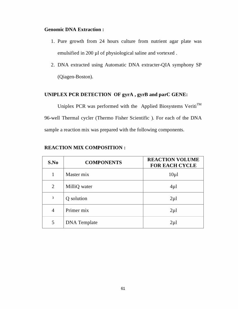
61
Genomic DNA Extraction :
1. Pure growth from 24 hours culture from nutrient agar plate was
emulsified in 200 µl of physiological saline and vortexed .
2. DNA extracted using Automatic DNA extracter-QIA symphony SP
(Qiagen-Boston).
UNIPLEX PCR DETECTION OF gyrA , gyrB and parC GENE:
Uniplex PCR was performed with the Applied Biosystems VeritiTM
96-well Thermal cycler (Thermo Fisher Scientific ). For each of the DNA
sample a reaction mix was prepared with the following components.
REACTION MIX COMPOSITION :
S.No COMPONENTS REACTION VOLUME FOR EACH CYCLE
1 Master mix 10µl
2 MilliQ water 4µl
³ Q solution 2µl
4 Primer mix 2µl
5 DNA Template 2µl

62
CYCLING CONDITIONS :
Cycling conditions for 30 cycles run- gyrA,gyrB,and parC -
1 Initial Denaturation 920 C 5 min
2 Denaturation 920 C 1min
3 Annealing 620 C 1min
4 Extension 740 C 2min
5 Final Extension 740 C 1min
PRIMERS : [99]
Antimicrobial Resistance gene sequence for Salmonella species.
S.No Oligo Name 5 < -----------Sequence ----------- >3 Base Pair
1 gyrA F TGTCCGAGATGGCCTGAAGC 347 bp
2 gyrA R TACCGTCATAGTTATCCACG
3 gyrB F CAAACTGGCGGACTGTCAGG 345 bp
4 gyrB F TTCCGGCATCTGACGATAGA
5 parC F CTATGCGATGTCAGAGCTGG 270 bp
6 parC R TAACAGCAGCTCGGCGTATT
AGAROSE GEL ELECTROPHORESIS :
PCR products were analysed using gel electrophoresis. 1% (w/v)
Agarose gel was prepared using 1 x TAE buffer (Sigma-Aldrich Pvt.
Ltd.,India) and 6µL of Ethidium bromide was added. The molten agarose
gel was cast in the electrophoresis tray with combs in place.

63
After solidifying, the cast was placed in the electrophoresis tank with
the buffer. 5µl DNA molecular weight marker (100 bp DNA ladder) was
loaded in one slot, and positive control ( Salmonella Typhi ATCC® 19430
TM ) in another slot . 5µl of PCR products were loaded in the respective
slots. Electrodes were connected and set a voltage of 100 V ; 400 mA for 1
hour.
On Gel documentation in (Bio-Rad,Hercules,California,USA)
presence of bands were observed at 347 bp, 345 bp, and 270 bp indicates
the presence of gyrA, gyrB and parC genes respectively in all the isolates
and in positive control strain .
DNA SEQUENCING :
Principle:
Sequencing of the amplicons was carried out by the
dideoxynucleotide chain-termination method, using an automated DNA
sequencer ABI PRISM 3500 Genetic Analyser (Applied Biosystems, USA ).
Pre-Clean Up Procedure :
Pre-clean up of the PCR products were done using QIA Quick PCR
Purification kit.
1. Add 50 µl of Buffer PB to 10 µl of PCR product and mix in a
QIA quick column.

64
2. Centrifuge – 30,000 rpm for 1 minute and discard the supernatant .
3. Wash with 750 µl Buffer PE and then centrifuge -30,000 rpm for 1
minute and discard the supernatant.
4. To elute the DNA add 30 µl of Elution buffer, let the column stands
for 1 minute and then centrifuge.
DNA TEMPLATE QUALITY:
Poor template quality was the most common cause of sequencing
problem.
Examining DNA Quality :
1. Spectrophotometric method :
Checked DNA template concentration and purity using Nano drop
method . This method reveals any protein contamination.1 µl of PCR
product was loaded in Thermo Scientific µDropTM plate and analysed
using SkanIT- software tool ).16 samples can be analysed at a time. If the
concentration was more than 15 µl, it was diluted with equal volumes of
MilliQ water accordingly.
2. Agarose gel electrophoresis and documentation :
Agarose gel electrophoresis reveals contaminating DNAs and
RNAs. DNA templates were subjected Agarose gel electrophoresis and
documentation along with a positive control ( Salmonella enterica

65
subspecies enterica serovar Typhi ATCC® 19430 TM ) and a molecular
ladder (100 bp ) which showed single band at 347 bp, 345 bp, and 270 bp
indicates the presence of gyrA, gyrB and parC genes respectively in all the
isolates and in positive control strain .
SEQUENCE PCR :
SEQUENCING REACTION DILUTIONS :
S.No COMPONENTS REACTION
VOLUME FOR FULL CYCLE
REACTION VOLUME FOR
16th CYCLE
1 Ready Reaction Mix 8 µl 0.5 µl
2 Dilution Buffer - 1.75 µl 3 Template(200 ng/µl) 1 µl 1 µl 4 Primer(2 pmol/µl) 8 µl 0.5 µl
5 MilliQ water 7 µl 4.75 µl
6 Total 20 µl 10 µl
CYCLING CONDITIONS :
1 Initial Denaturation 96 22 sec
2 Denaturation 96 10 sec
3 Annealing 50 5 sec
4 Extension 60 4 min
5 Final Extension 4 4 min
Amplicons obtained were subjected to Post-clean up procedure .

66
POST-CLEAN UP PROCEDURE ( TUBE METHOD ) :
Using Big dye terminator Vs 3.1 clean up –Tube method post clean
up were done .
The reaction product were transferred to 1.5 ml tube, 12 µl of Master
mix I was added to 10 µl of amplicons per reaction. Then 52 µl of Master
mix II was added and the contents were mixed well and incubated at room
temperature for 15 minutes. Spin at 12000 rpm for 20 minutes at room
temperature. The supernatant were decanted .
Then 250 µl of 70 % freshly prepared ethanol was added and spin at
12000 g for 10 minutes at room temperature. Supernatent discarded and 12
µl of elution buffer ( Hi –Di formamide ) was added . The sample tubes
were covered with septa , denatured , snap chilled and proceeded with
Capillary electrophoresis.
After the completion of the procedure, electrophorogram pattern
obtained and analysed with the reference sequence .
Reference sequences of S.Typhi for all QRDR genes (gyrA,gyrB and
parC) were obtained from http://www.ncbi.nlm.nih.gov/gene ., with
following accession numbers : gyrA- CAD07504, gyrB-NC _003198,parC-
NC_003198.1.
67
Sequence chromatograph files were analysed using software tool.
Forward and reverse DNA sequences were assembled using EMBOSS
merger software, nucleotide ambiguities were resolved by NUCLEOTIDE
massager tool and alignment of sequences were done by CLUSTAL
OMEGA software tool. Nucleotides were translated to proteins using
ExPASY Protein software and analysed for mutation by comparing with the
references.

Results

68
RESULTS
TABLE 1 : DISTRIBUTION OF PATIENTS IN THE STUDY
POPULATION (n=257)
STUDY SITE No.of Patients Percentage (%)
RGGGH 157 61.1
ICH 100 38.9
Total 257 100.0
61.1% were young adults and 38.9% were children
FIGURE: 1 DISTRIBUTION OF PATIENTS IN THE STUDY
POPULATION ( n=257)
RGGGH61%
ICH39%
Study Site Distribution

69
TABLE 2 : AGE GROUP AND SEX DISTRIBUTION
IN THE STUDY POPULATION
STUDY POPULATION
AGE GROUP (n=257)
GENDER TOTAL
MALE FEMALE
CHILDREN 5-7 22 29 51 8-10 18 18 36
11-12 11 4 15
YOUNG ADULTS
13-17 16 14 30 18-24 73 52 125
TOTAL 140 117 257
Mean age group is 15.27 , Standard deviation is 6.66.
Both genders were equally affected among children.
Male preponderance was observed among young adult population.
FIGURE: 2 AGE GROUP AND SEX DISTRIBUTION IN THE
STUDY POPULATION
2218
1116
73
29
18
4
14
52
5 - 7years 8 - 10years 11 -12years 13 - 17years 18- 24years
Age Group and Gender DistributionMale Female

70
TABLE 3 : DISTRIBUTION OF PATIENTS BASED ON HOSPITAL
ADMISSION
INPATIENTS/ OUTPATIENTS No.of patients Percentage (%)
INPATIENTS 177 68.9
OUTPATIENTS 80 31.1
Total 257 100.0
68.9% were admitted and treated as inpatients.
TABLE 4 : CARDINAL SYMPTOMS AMONG STUDY
POPULATION
SYMPTOMS Young adults (n = 157)
Percentage (%)
Children ( n=100)
Percentage (%)
Fever with abdominal pain 27 17.2 7 7
Fever with abdominal pain and Diarrhoea 52 33.1 68 68
Fever with abdominal pain, Diarrhoea,
Nausea and vomiting 49 31.2 25 25
Fever with constipation 13 8.3 0 0
Fever with nonspecific symptoms 16 10.2 0 0
The predominant symptom among Enteric fever suspected febrile
patients were fever, abdominal pain and diarrhoea .

71
TABLE 5 : CARDINAL SIGNS AMONG STUDY POPULATION
CARDINAL SIGNS Young Adults
Percentage (%) Children Percentage
(%)
Pyrexia,toxic look , pallor 61 38.9 47 47
Pyrexia, toxic look, coated tongue 74 47 44 44
Pyrexia, toxic look, coated tongue,hepatomegaly 0 0 2 2
Pyrexia, toxic look, coated tongue,Splenomegaly 5 3.2 2 2
Pyrexia, toxic look, coated tongue,hepatosplenomegaly. 3 1.9 3 3
Pyrexia,pallor 14 8.9 2 2
The predominant sign among the study population were pyrexia,
toxic look and coated tongue.
TABLE 6 : DISTRIBUTION BASED ON FOOD HABITS ( n=257)
H/O EATING FOOD
OUTSIDE Young adults (n=157) Children (n= 100)
YES 76 (48.4%) 25 ( 25 %)
NO 81 (51.6% 75 ( 75%)

72
TABLE 7 : DISTRIBUTION OF BACTERIAL GROWTH IN
BLOOD CULTURE
BACTERIAL GROWTH
YOUNG ADULTS (n=157)
CHILDREN (n=100)
TOTAL (n=257)
Growth 10 5 15 (5.9 %)
No growth 147 95 242 (94.1 %)
The isolation rate by blood culture method was 5.9%
TABLE 8 : DISTRIBUTION OF PATHOGENS ISOLATED FROM
BLOOD CULTURE
PATHOGENS ISOLATED
YOUNG ADULTS
(n =157) n(%)
CHILDREN ( n=100) n(%)
TOTAL (n=257) n(%)
S.Typhi 6 (3.82 %) 5 (5%) 11 (4.3%)
S.Paratyphi 1 (0.64%) - 1 (0.4%)
E.coli 2 (1.27%) - 2 (0.77%)
Klebsiella pneumonia 1 (0.64%) - 1 (0.4%)
Salmonella species isolated from conventional blood culture in 12
cases (4.7%). Among this ,7 (4.45 %) isolated from young adult population
and 5 isolates (5 % ) from children.
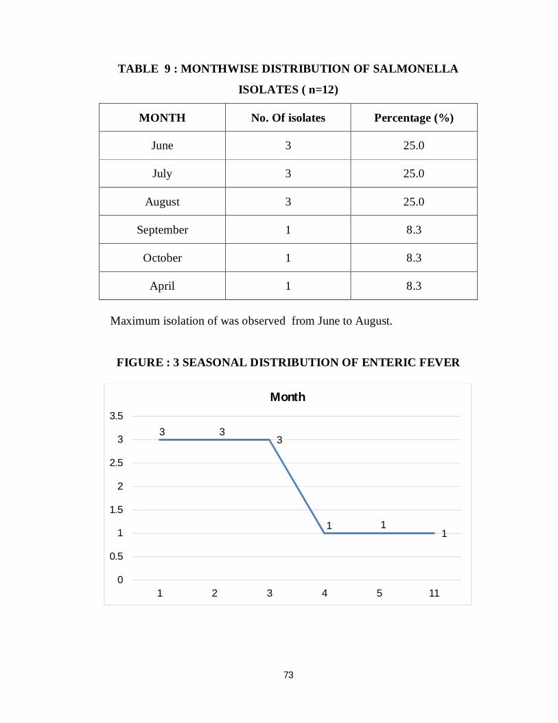
73
TABLE 9 : MONTHWISE DISTRIBUTION OF SALMONELLA
ISOLATES ( n=12)
MONTH No. Of isolates Percentage (%)
June 3 25.0
July 3 25.0
August 3 25.0
September 1 8.3
October 1 8.3
April 1 8.3
Maximum isolation of was observed from June to August.
FIGURE : 3 SEASONAL DISTRIBUTION OF ENTERIC FEVER
3 33
1 11
0
0.5
1
1.5
2
2.5
3
3.5
1 2 3 4 5 11
Month

74
TABLE 10 : DISTRIBUTION BASED ON FOOD HABITS AND
TYPHOID FEVER
STUDY SITE Outside food habits S.Typhi S.Paratyphi
A TOTAL
RGGGH Yes 6 0 6
No 0 1 1
ICH Yes 1 0 1
No 4 0 4
TOTAL 11 1 12
Among Young adults, eating outside food was significantly
associated with Typhoid fever due to S.Typhi and S.Paratyphi A with ‘ p’
value 0.021 (p < 0.05- significant ) by Fisher’s Exact Test . But this
association was not statistically significant among children.
TABLE 11 : ANTIMICROBIAL SUSCEPTIBILITY PATTERN OF
S.TYPHI AND S.PARATYPHI A BY DISC DIFFUSION METHOD
( % of Susceptibility )
SALMONELLA SPECIES
AMPI 10 µg
COT 25 µg
CK 30 µg
CTX 30 µg
CFT 30 µg
AZIT 15 µg
NA 30 µg
PEF 5 µg
CIP 5 µg
S.TYPHI ( n= 11) 100 100 100 100 100 100 0 0 0
S.PARATYPHI A (n=1 ) 100 100 100 100 100 100 0 0 0

75
FIGURE 4 : ANTIMICROBIAL SUSCEPTIBILITY PATTERN OF
S.TYPHI (n=11) AND S.PARATYPHI A (n=1) BY DISC DIFFUSION
METHOD
Abbrevation: AMPI – ampicillin, COT- cotrimoxazole, CK-
chloramphenicol , CTX-cefotaxime , CFT-ceftriaxone , AZIT-
azithromycin, NA- nalidixic acid, PEF- pefloxacin , CIP-ciprofloxacin.
Antimicrobial susceptibity pattern of both S.Typhi and S.Paratyphi A
showed similar pattern .
All the 12 isolates were resistant to Nalidixic acid ( NAR) and
Pefloxacin. None of the isolates were MDR - based on their susceptibility
to Ampicillin , Cotrimoxazole , Chloramphenicol. All the isolates were
susceptible to Third generation Cephalosporins and Azithromycin.
12 12 12 12 12 12
8
12 12
4
0
2
4
6
8
10
12
14
AMPI COT CK CTX CFT AZIT NA PEF CIPRO
S
I
R

76
TABLE 12 : DISTRIBUTION OF MIC OF CIPROFLOXACIN BY
MICROBROTH DILUTION METHOD
MIC CLSI interpretative criteria EUCAST interpretative criteria
CIPROFLOXACIN SUSCEP TIBLE
≤0.06 µg/ml
INTER MEDIATE 0.125-0.5
µg/ml
RESIS TANT
≥ 1 µg/ml
SUSCEP TIBLE
≤0.06 µg/ml
RESIS TANT
≥0.06 µg/ml
S.Typhi (n=11) 0 3 8 0 11
S.Paratyphi A ( n=1) 0 1 0 0 1
TOTAL (n=12) 0 4 (33.3%)
8 (66.7%) 0 12
(100%)
All the 12 Pefloxacin resistant isolates ( Surrogate marker of
Ciprofloxacin resistance ) were subjected to Ciprofloxacin MIC testing
by Brothmicro dilution method and was found to be 66.7 % resistant and
33.3 % intermediate using CLSI interpretative criteria , while all the 12
isolates were found to be resistant by interpretative criteria of
EUCAST .

77
TABLE 13 : RESULTS OF MOLECULAR CHARACTERISATION
OF TYPHOIDAL SALMONELLAE AND COMPARISON WITH
ANTIMICROBIAL SUCEPTIBILTY PATTERN (n=12)
Isolate ID
RESISTANT PHENOTYPE
[ NAL-PEF-CIP ] DISC
DIFFUSION
CIPRO MIC
(µg/ml)
MUTATION ANALYSIS OF QRDR GENES
Ciprofloxacin MIC
Interpretation
gyrA gyrB parC CLSI EUCAST
ST 1 NAL-PEF 0.125 + - - I R
ST 2 NAL-PEF 2 + - - R R
ST 3 NAL-PEF 4 + - + R R
ST 4 NAL-PEF 2 + - - R R
ST 5 NAL-PEF 2 + - + R R
ST 6 NAL-PEF 1 + - - R R
ST 7 NAL-PEF-CIP 0.125 + - - I R
ST 8 NAL-PEF-CIP 4 + - + R R
ST 9 NAL-PEF 0.125 + - - I R
ST 10 NAL-PEF 2 + - + R R
ST 11 NAL-PEF-CIP 1 + - - R R
ST 12 NAL-PEF 0.125 + - - I R
All the Pefloxacin resistant Salmonella isolates were found to be
Ciprofloxacin resistant by MIC according to EUCAST interpretative
criteria, which was further confirmed by molecular characterisation of
QRDR gene mutation.
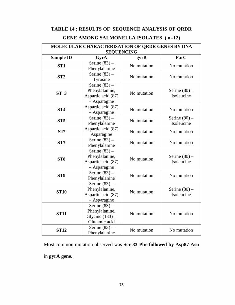
78
TABLE 14 : RESULTS OF SEQUENCE ANALYSIS OF QRDR
GENE AMONG SALMONELLA ISOLATES ( n=12)
MOLECULAR CHARACTERISATION OF QRDR GENES BY DNA SEQUENCING
Sample ID GyrA gyrB ParC
ST1 Serine (83) – Phenylalanine No mutation No mutation
ST2 Serine (83) – Tyrosine No mutation No mutation
ST 3
Serine (83) – Phenylalanine,
Aspartic acid (87) – Asparagine
No mutation Serine (80) – Isoleucine
ST4 Aspartic acid (87) – Asparagine No mutation No mutation
ST5 Serine (83) – Phenylalanine No mutation Serine (80) –
Isoleucine
ST³ Aspartic acid (87) Asparagine No mutation No mutation
ST7 Serine (83) – Phenylalanine No mutation No mutation
ST8
Serine (83) – Phenylalanine,
Aspartic acid (87) – Asparagine
No mutation Serine (80) – Isoleucine
ST9 Serine (83) – Phenylalanine No mutation No mutation
ST10
Serine (83) – Phenylalanine,
Aspartic acid (87) – Asparagine
No mutation Serine (80) – Isoleucine
ST11
Serine (83) – Phenylalanine, Glycine (133) – Glutamic acid
No mutation No mutation
ST12 Serine (83) – Phenylalanine No mutation No mutation
Most common mutation observed was Ser 83-Phe followed by Asp87-Asn
in gyrA gene.

Discussion

79
DISCUSSION
This study was performed on 257 Enteric fever suspected febrile
patients with an objective to isolate and determine the antimicrobial
susceptibility pattern of Typhoidal Salmonellae among school aged
children and young adults. Among 257 patients , 157 (61%) young adults
and 100 (39%) children were included in the study [ Table 1] .
Among those febrile patients 177 ( 68.9%) were admitted and treated
as inpatients and the remaining 80 patients ( 31.1%) were treated as
outpatients.[ Table 3].
The predominant symptoms observed were fever, abdominal pain
and diarrhoea in children (68 %) and adult population (33 %) [Table 4].
The predominant signs were pyrexia, toxic look and coated tongue in
children (44%) and adults (47%) [59,60] [Table 5] .
Authors from various parts of the world have done similar studies
among enteric fever patients . Stuart et al., found that school aged children
or young adults between 5-25 years of age were the most commonly
affected age group in the areas of endemicity [ 101,102,103] . He also observed
that the predominant symptom was fever (75%) followed by abdominal
pain ( 40%). Scragg et al., observed that diarrhoea was the predominant
symptom in children .

80
Crump et al.,observed that poor sanitation and lack of access to safe
water and food is the major risk factor for transmission of Typhoidal
Salmonella [ 1 ] . Oslen et al., observed asymptomatic carriage infood
handlers plays a major role in transmission of typhoid bacillus [106] .
In this study 48.4 % of adults and 25 % of children with febrile
illness had a history of eating outside foods such as ice cream , fruit salads ,
soft drinks , flavoured milks , sweets and food from local street vendors in
unsanitary conditions using water of questionable quality [Table 7 ]. None
of the patients included in the study were vaccinated against Typhoid fever
Conventional blood culture was done for 257 patients suspected to
patients to isolate and perform antimicrobial susceptibility testing .
Among these, eleven Salmonella Typhi ( 4.3 % ) and one Salmonella
Paratyphi A ( 0.4 % ) were isolated [ Table 9 ]. In India, varying prevalence
of typhoid fever has been reported over a period of time and geographical
distribution. The systematic review and meta analysis study done in 2016
reported the prevalence of Salmonella Typhi to be about 9.7 % and that of
Salmonella Paratyphi A was 0.9 %. [5] .
The World Health Organisation , in 2008 , reported the prevalence of
typhoid fever as 28.1 per 1000 febrile episodes in India.

81
Blood culture positivity is usually high during the first week of illness
but can also be isolated from subsequent weeks also if there is no prior
antibiotic therapy [2,25] . The low isolation rate of Salmonella species from
blood culture could be attributed to many factors- prior antibiotic therapy,
low bacterial count ( sometimes as low as one ) ,poor selection of cases,
volume of blood sampled [25] .
In this study ,the isolation rate in blood culture of Salmonella species
was 4.7 % ( n=12), with isolation of S.Typhi about 4.3 % (n=11)and that
of S.Paratyphi A (n= 1) being 0.4 % .
Among the 12 Salmonella isolates , 4.45 % ( n=7) were isolated from
young adults and 5 % ( n= 5) were isolated from children [Table 8 ].
Typhoid fever affects all age group and both the sexes are equally
affected.In the areas of endemicity the predominant age group affected are
school-aged children or young adults, within the age group of 5-25 years of
age.
Among children 5-7 (19.9%) years of age group were most
commonly affected with female preponderance ; in adult population studied
18-24 (48.6% )years of age were most commonly affected with male
preponderance, with mean age group was 15.27 [Table 2]. Mohanty et al,
observed that the most common age group affected were 5-19 years [9].

82
Typhoid fever is known to show seasonal variation and is mainly
associated with hot summer months. Many studies worldwide and Indian
studies showed peak incidence around the end of dry season .Mohanty et
al,observed peak incidence of enteric fever occurs between April and June
followed by July- September . In this study the peak isolation was between
June-August ( 75%) and there after decline in frequency to 8.3% for each
month, in September, October and March [9] .
The food habits of the patients with enteric fever was studied and
found that 58.3% of laboratory confirmed typhoid fever patients had the
history of having food items from outside street vendors. Eating food
prepared in unhygienic conditions and with untreated water is the major risk
factor for transmission. Also Levine et al, studied the role of chronic carriers
as a reservoir of infection in Santiago,where a crude rate of 694 carriers per
1,00,000 inhabitants was found [1,25] .
Similar to these observations , this study showed significant
association ( p <0.05 ) between H/O eating food outside and Salmonella
isolation among adult population.
MDR –defined as resistance to ampicillin , chloramphenicol and
trimethoprim-sulphamethoxazole., was reported from many countries
.Studies globally have found that 80% of S.Typhi isolates ( 1993-1996)

83
were MDR [107,108]. Currently in India, the MDRST varies from
7-55%[110,111] .MDR in Central India declined from 91% in 1991 to 22% in
2002[107,108].
Multicentric study in India, conducted by Dahiya.S et al,in 2014-
2015, observed the overall MDR prevalence was less than 3%. However ,in
each tertiary care hospital included in this study the MDR frequency were
found to be 6 % [109].
In this present study, all the 12 isolates were susceptible to
Ampicillin (100%), Chloramphenicol (100%)and Trimethoprim-
sulphamethoxazole (100%), and none of the isolates were Multidrug
resistant [ Table 12].
Overall there is a general trend of decline in MDR strains . The
reason could be due to discontinuation of these drugs in the treatment of
typhoid fever, thus relieving the selection pressure. Another reason could be
the loss of plasmids such as the R-plasmid that confer multi drug resitance ,
or emergence of de novo susceptible strains may also contribute for this
decline.
Ciprofloxacin has remained the drug of choice of enteric fever for the
last many years. However, isolates of S.Typhi and S.Paratyphi A with
reduced susceptibity to fluoroquinolones have now appeared in the

84
subcontinent , Vietnam and Tajikistan , and the treatment failures with
fluoroquinolones has also been noted.
In vivo adaptive mutations have been reported to occur on continuous
sub-inhibitory exposure to ciprofloxacin [ 112] . Also, the frequent use of
Ciprofloxacin for trivial infections is likely to promote resistance to these
agents.
In this present study,75% (9 /12) isolates were in the intermediate
category, 25% (3/12) isolates were resistant, and none of the isolates were
susceptible to ciprofloxacin by Kirby-Bauer’s Disc Diffusion Method
[Table 12 ]
When the isolates were subjected to MIC determination by Broth
micro dilution method , 33.3% of isolates (4/12) were in the intermediate
category, and 66.7% (8/12) became resistant. None of the isolates were in
the susceptible range of MIC based on CLSI interpretation [ Table 13 ].
This decreased fluroquinolone susceptibility was detected by using
Nalidixic acid in earlier days, since it was considered to be the surrogate
marker of fluroquinolone resistance. But, according to recent changes in
CLSI and EUCAST guidelines, Pefloxacin (5µg) disc has been suggested as
the best surrogate marker of fluoroquinolone susceptibility rather than
Nalidixic acid and Ciprofloxacin disc diffusion itself . Nalidixic acid disc

85
diffusion method does not detect all the possible mechanisms of
fluroquinolone resistance. But , Pefloxacin can detect all the possible
mechanisms of resistance except the one mediated by the plasmid aac(6´)-
lb-cr.
Fang et al., studied the fluoroquinolone resistance in Salmonella and
the utility of Pefloxacin disc diffusion in 2015, reported that Pefloxacin
disc diffusion provides better separation of ciprofloxacin susceptible and
non-susceptible strains than other disk diffusion, including ciprofloxacin
itself [121]. In this study all isolates were resistant to Nalidixic acid and
Pefloxacin , both showed 100% resistant pattern to all the 12 isolates
[Table 13].
Nine isolates ( 75% ) which were intermediate susceptibility to
ciprofloxacin were found to be resistant by Pefloxacin disc diffusion. This
was further confirmed by molecular characterisation of QRDR genes
(gyrA,gyrB and parC) [ Table 13 and14 ] [122] .
According to CLSI breakpoints, 33.3% (4/12) of isolates showed
intermediate susceptibilityto ciprofloxacin by MIC which were resistant by
Pefloxacin disc diffusion. This observation correlates well with
Ciprofloxacin MIC breakpoints of EUCAST guidelines .It is simple and

86
easy to perform Pefloxacin disc diffusion over MIC of fluroquinolones for
detecting its susceptibility [ 91,92,93 ] .
Quinolone resistance is mediated by chromosomal mutations in
QRDR genes- gyrA , gyrB and parC , parE. All the 12 isolates S.Typhi ( n
=11) and S.Paratyphi A ( n= 1) isolates which were Pefloxacin resistant
were subjected to PCR followed by Sangers sequencing of QRDR genes-
gyrA, gyrB and parC [ 113,114] .
Mutation in gyrA gene was identified in all the 12 isolates (100%) .
Four isolates (33%) harboured mutation in parC gene. None of the isolates
showed mutation in gyrB gene [83] .
Menezes et al, reported in a study conducted from 2005-2009 at
Pondicherry, out of 11 isolates tested with ciprofloxacin MIC ranging from
≥1 to ≤ 4 µg/ml , showed three mutations, two mutations within the QRDR
of gyrA (at codon 83 and 87) and a single point mutation at parC at codon
80 .No mutations were observed in gyrB and par E of any of the isolates
tested [11] .
Similarly ,in the present study, most common mutations observed
were at codon 83 where a C →T transition led to the substitution of
phenylalanine for serine .This single point mutation leads to low level
fluoroquinolone resistance , and didn’t cause considerable rise of MIC, and

87
the isolates were in the intermediate category (MIC -0.125µg/ml). While
treating this patient with higher dose of ciprofloxacin it might translate into
possible/probable clinical failure [114].
Renuka et al.,in 2004 reported that double or triple mutation in gyrA
or parC genes ,or even double mutations in gyrA cause high level
ciprofloxacin resistance in a study conducted in North India [113] .
Another mutation observed at codon 83 was substitution of Tyrosine
for Serine where C→A transition occured. But, this mutation caused
significant rise in MIC value and the isolate was resistant .
Five out of twelve isolates ( 41.7%) in this study had amino acid
substitution , Asparagine to Aspartic acid where G →A transition
occurs.This single point mutation cause significant elevation of MIC upto
1-2 µg/ml .When an isolate harbours double mutation, MIC values further
rise from 4 - >32 µg/ml .This observation is consistent with the reported in
studies of Balaji et al [114] .
Balaji et al, in 2015,studied 33 isolates with Ciprofloxacin
intermediate susceptibility range by doing molecular characterisation and
observed that both double and single amino acid substitutions were seen at
positions 83 and 87 in all the isolates for gyrA .No mutations were detected
for gyrB , while parC mutations were observed at 80 and 84 positions.
88
Double or triple mutation in gyrA cause significant increase in MIC value
rather than single point mutation .
Mutation at codon 133, has been rarely reported in literature.In this
study, single isolate of S.Typhi had a mutation at codon 133 ,where
Glutamic acid substitution for Glycine occured and was associated with
resistant range of MIC. Hamidian et al reported this mutation for the first
time ,in a study conducted at Iran in 2010 [115].
No mutation was observed in gyrB in all the 12 isolates ,consistent
with previous studies conducted in Pondicherry from 2005-2009 by Harish
et al.
Song et al ,in 2010 , repoted mutations in gyrB gene , mutation at
Phe-464, Leu-465, causing rise in MIC value [116] .
Mutation at codon 80 of parC gene was observed in 33% (4) of
isolates, where isoleucine substitution for serine occured. When this
mutation occurs along with another single point mutation in gyrA will lead
on to higher range of ciprofloxacin MIC value.
All the 12 isolates were subjected to Ceftriaxone and Cefotaxime
disc diffusion, showed 100% susceptibitity pattern, making as an effective
alternative for treating fluroquinolone resistance Salmonella isolates.

89
All the 12 culture proven Enteric fever cases were treated with
parenteral ceftriaxone according to their standard dosing regime and fever
defervescence was observed on day 3 or 4 following antimicrobial
therapy. Patient followed up and complete recovery was reported.
This is in contrary to other studies, where high level resistance to
ceftriaxone was observed due to the presence of CTX-M-15 and SHV-12
extended spectrum beta lactamases ( ESBLs) [11] . Recent reports of clinical
failure ,relapse to ceftriaxone and ESBL detection limits its use . None of
the isolates in the present study were ESBL producers.
Azithromycin shows excellent tissue penetration and achieves
intracellular concentration well. Hence it is equivalent or superior to other
alternative drugs in the management of uncomplicated typhoid fever with
prompt resolution of symptoms and low prevalence of relapse and carrier
state [117,118] .
All the 12 (100 %) isolates showed susceptibility to Azithromycin by
disc diffusion method . Several studies have reported Azithromycin MIC
distributions for typhoidal and non-typhoidal Salmonella strains [119].
Clinical breakpoints for disc diffusion and MIC testing have been adopted
by CLSI for Salmonella Typhi but not yet for Salmonella Paratyphi A .
Some sporadic cases of treatment failure with Azithromycin was reported
in literature [ 120]. Hence judicious use and continuous monitoring of creep
in MIC for azithromycin is essential to combat resistance.

Limitations of the study

90
LIMITATIONS OF THE STUDY
1. Our study had a limitation in the number of isolates studied.
Molecular analysis of large number of isolates for various resistance
mechanisms and its correlation with fluoroquinolone resistance might
help in devising methodology for detection of Ciprofloxacin
resistance in Salmonella isolates which is imperative for the
management of Typhoid fever.
2. Serotyping and Phage typing of the isolates were not done, that could
have thrown light on the pattern of strains in the population of
patients attending this tertiary care centre.

Summary

91
SUMMARY
One Hundred children (39 %) and one fifty seven (61 %) young
adults patients with clinical suspicion of Enteric fever were subjected to
conventional blood culture.
The isolation rate of Salmonella species was 4.7% . Among children the
isolation rate was 5% and 4.45% in young adults.
Predominant isolate among the Salmonella species was S.Typhi (91.6%)
followed by S.Paratyphi A (8.3%).
Predominant age group affected 5-7 years (19.9%) with female
preponderance, mean age 7.2 years.
Among young adult patients, 18-24 years old were most commonly
affected with male preponderance, mean age 19.9 years.
Peak incidence of Typhoid fever was observed from June-August. (75%-
isolation )
58.3% affected patients had H/O eating food ouside home with
significant association between Salmonella species isolation among adult
patients.
None of the individuals had Typhoid vaccination.
Analysis of Antimicrobial susceptibility pattern in this study revealed
re-emergence of susceptibility to all the first line antimicrobials-
ampicillin, chloramphenicol and co-trimoxazole in all the 12 isolates. No
MDR Salmonella species have been isolated.

92
100% resistance to Nalidixic acid & Pefloxacin by disc diffusion
method was observed.
Nine ( 75%) isolates were intermediate and three (25% ) isolates were
resistant to Ciprofloxacin by Disc diffusion method.
All 12 (100% ) isolates were susceptible to Ceftriaxone , Cefotaxime,and
Azithromycin by disc diffusion method. Enteric fever cases in this study
were treated with injection ceftriaxone according to standard dosing
regime and complete recovery was observed , with fever defervescence
by day 4 following antimicrobial therapy .
Four ( 33.3%) isolates were intermediate (0.125µg/ml) and eight
(66.7%) isolates were resistant (1-4µg/ml) to Ciprofloxacin by
Brothmicro dilution method of MIC determination.
All the Pefloxacin resistant isolates harboured point mutation in genes
encoding QRDR region when subjected to PCR and Sequencing.
Mutation analysis revealed that , all the 12 (100%) isolates showed
mutation in gyrA , with most common mutation observed were
Ser83→Phe ,followed by Asp87 →Asn ; no mutation observed in gyrB
gene ; Ser80→Isoleucine mutation in parC gene were observed among 4
( 33.3%) isolates.
Interpretative criteria of EUCAST guidelines for Ciprofloxacin MIC
correlates well with Pefloxacin resistance in all the 12 isolates , which
were confirmed by molecular characterisation .

Conclusion

93
CONCLUSION
This study demonstrates that children and young adults were the
predominant population affected with typhoid fever and had a strong
association between eating food outside and isolation of Salmonella
species.The predominant isolation was S.Typhi, and followed by
S.Paratyphi A.
Antimicrobial susceptibility pattern was similar in both the species,
with re-emergence of susceptibility to ampicillin, chloramphenicol and co-
trimoxazole. Both the species showed high resistance to Nalidixic acid and
Pefloxacin.The use of Pefloxacin as a surrogate marker for fluoroquinolone
susceptibility is promising , since it detects all the mechanisms for
fluoroquinolone resistance in this study , and clearly separates ciprofloxacin
susceptible and resistant strains .
Pefloxacin disc diffusion was found to be the best in detecting
fluoroquinolone resistance in centres where routine MIC testing and
molecular characterisation was not possible .
Interpretative criteria for Ciprofloxacin MIC by Microbroth dilution
method in EUCAST guidelines have a better correlation with molecular
characterisation , with gyrA mutation at codon 83 was predominant
followed by gyrA mutation at codon 87.
Ceftriaxone and Azithromycin seem to be the effective therapeutic
option for fluroquinolone resistant Salmonella isolates.

COLOUR PLATES
Gram stain of Salmonella species showing gram negative bacilli
Mac Conkey – Lactose non fermenting colonies
Blood Agar- Non hemolytic grey white colonies

BIOCHEMICAL REACTIONS OF SALMONELLA TYPHI
BIOCHEMICAL REACTIONS OF SALMONELLA PARATYPHI A

SLIDE AGGLUTINATION TEST – SALMONELLA TYPHI
SLIDE AGGLUTINATION TEST – SALMONELLA PARATYPHI A

ANTIMICROBIAL SUSCEPTIBILITY PATTERN - DISC DIFFUSION METHOD

CIPROFLOXACIN MIC-BROTH MICRODILUTION METHOD
g/ml
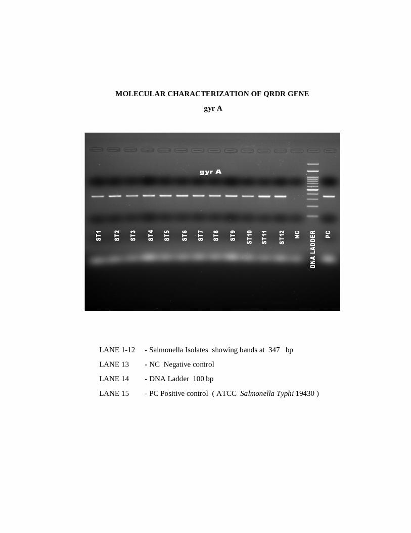
MOLECULAR CHARACTERIZATION OF QRDR GENE
gyr A
LANE 1-12 - Salmonella Isolates showing bands at 347 bp
LANE 13 - NC Negative control
LANE 14 - DNA Ladder 100 bp
LANE 15 - PC Positive control ( ATCC Salmonella Typhi 19430 )

MOLECULAR CHARACTERIZATION OF QRDR GENE
gyr B
LANE 1-12 - Salmonella Isolates showing bands at 345 bp
LANE 13 - NC Negative control
LANE 14 - DNA Ladder 100 bp
LANE 15 - PC Positive control ( ATCC Salmonella Typhi 19430 )

MOLECULAR CHARACTERIZATION OF QRDR GENE
parC
LANE 1-12 - Salmonella Isolates showing bands at 270 bp
LANE 13 - NC Negative control
LANE 14 - DNA Ladder 100 bp
LANE 15 - PC Positive control ( ATCC Salmonella Typhi 19430 )

SEQUENCE ANALYSIS

Bibliography
BIBLIOGRAPHY 1. Crump JA, Luby SP,Mintz ED.2004.The global burden of typhoid fever.Bull
World Health 82: 346-353.
2. Bhutta ZA. Current concepts in the diagnosis and treatment of typhoid fever.
BMJ 2006 ; 333:78-82.
3. Buckle GC,Walker CL,Black RE (2012)Typhoid fever and paratyphoid fever:
Systematic review to estimate global morbidity and mortality for 2010.J Glob
Health 2: 010401.PMID :23198130
4. Mogasale V,Maskery B,Ochiai RL,Lee JS,Mogasale VV,et al.(2014) Burden of
typhoid fever inlow-income and middle-income countries: a systematic,literature-
based update with risk-factor adjustment.Lancet Global Health 2:E570-
E580.PMID:253046633
5. John J,Van Aart CJC,GrasslyNC(2016).The Burden of Typhoid and Paratyphoid
in India: Systematic review and Meta-analysis.PLoS Negl Trop Dis
10(4):10.e0004616.
6. Cooke FJ,Wain J.The emergence of antibiotic resistance in typhoid fever.Travel
Med Infect Dis 2004;2:67-74.
7. Butt T,Ahmad RN,Mahmood A,Zaidi S.Ciprofloxacin treatment failure in
typhoid fever case,Pakistan.Emerg Infect Dis 2003;9:1621-1622.
8. Rowe B,Ward LR,Threlfall EJ.Multidrug-resistant Salmonella typhi :a worldwide
epidemic .Clin Infect Dis.1997;24:106-9.
9. Mohanty S,Renuka K,Sood S,Das BK,Kapil A.Antibiogram pattern and
seasonality of Salmonella serotypes in a North Indian tertiary care
hospital.Epidemiol Infect.2006;134:961-3.
10. Verma S, Thakur S, Kanga A, Singh G, Gupta P.Emerging Salmonella
Paratyphi A enteric fever and changing trends in antimicrobial resistance pattern
of salmonella in Shimla. Indian J Med Microbiol. 2010;28(1):51–3.
11. Menezes GA, Harish BN, Khan MA, Goessens WHF, Hays JP. Antimicrobial
resistance trends in blood culture positive Salmonella Typhi isolates from
Pondicherry, India, 2005–2009. Clin Microbiol Infect. 2012;18(3):239–45.

12. Dutta S, Das S, Mitra U, Jain P, Roy I, Ganguly SS, et al.Antimicrobial
resistance, virulence profiles and molecular subtypes of Salmonella enterica
serovars Typhi and Paratyphi A blood isolates from Kolkata, India during 2009–
2013. PLoS One.2014;9(8):e101347.
13. Chau TT,Campbell JI,Galindo CM et al.Antimicrobial drug resistance of
Salmonella enteric serovar Typhi in Asia and molecular mechanism of reduced
susceptibility to the fluroquinolones .Antimicrob Agents Chemother
2007;51:4315-23.
14. RoumagnacP,Weill F-X,Dolecek C et al.Evolutionary history of Salmonella
Typhi .Science 2006;314:1301-4.
15. Louis PCA. Recherches anatomiques, pathologiques, et thérapeutiques sur la
maladie connue sous les noms de gastroenterite, fièvre putride, adymanique,
typhoide, comparée avec les maladies aigues les plus ordinaires. Paris: J-B
Ballière; 1829.
16. Jenner W. On the identity or non-identity of typhoid and typhus fevers. London:
C. & J. Adlard; 1850.
17. Wilson JC. A treatise on the continued fevers. New York: Wood Pub; 1881.
18. Budd W. Typhoid fever: Its Nature, Mode of Spreading, and Prevention. London:
Longmans Publishing; 1873.
19. Schroeter J. In: Cohn F, ed. Kryptogamenflora von Schlesien Bd 3. Breslau: J. U.
Kern; 1885:1-814.
20. Pfeiffer R, Kalle W. Experimentelle untersuchunger zur Frage der
Schutzimphung des Menschen geger typhus addominalis. Dtsch Med
Wocheschn. 1896;22:735.
21. Kauffman F. The Diagnosis of Salmonella Types. Springfield, IL: Charles C.
Thomas; 1950.
22. Zinder ND, Lederberg J. Genetic exchange in Salmonella. J Bacteriol.
1952;64:679-699.

23. Lee TP, Hoffman SL,Typhoid fever.In:Strickland GT,editor.Hunters tropical
medicine and emerging infectious disease .8th Edition.Philadelphia:WB Saunders
Co; 2000.p.471-84.
24. Parry CM. Epidemiology and clinical aspects of human typhoid fever. In :
Mastroeni P and Maskell D. Salmonella Infections : Clinical , Immunological and
Molecular Aspects . Cambridge University Press ; 2006:1-25.
25. World Health Organisation Department of Vaccines and Biologicals Background
Document : The diagnosis, prevention and treatment of typhoid fever. Geneva :
WHO,2003:19-23.
26. Pegues DA, Ohl ME, Miller SI. Salmonella species, including Salmonella Typhi.
In: Mandell GL, Bennett JE, Dolin R,editors.Principles and practice of infectious
diseases. 8 th Edition, Churchill Livingstone : New York;2005.pg 2636-54.
27. Murray CJ, Vos T, Lozano R, et al. Disability-adjusted life years (DALYs) for
291 diseases and injuries in 21 regions, 1990–2010:a systematic analysis for the
Global Burden of Disease Study 2010. Lancet 2012; 380: 2197–223.
28. Dunstan SJ, Stephens HA, Blackwell JM et al.Genes of the class II and class III
major histocompatibility complex are associated with typhoid fever in Vietnam. J
Infect Dis 2001;183:261-68.
29. B renner FW, Villar RG, Angulo FJ et al : Salmonella nomenclature .J Clin
Microbiol 2000;38:2465-2467.
30. Old DC, Threlfall EJ. Salmonella. In:Balows A,Duerden BI,editors.Topley
&Wilson’s Microbiology and Microbial Infections.9 th ed.Arnold : London.p.969-
98.
31. Old DC. Salmonella .In: Collee JG, Fraser AG, Marimon BP, Simmons A.
Salmonella. Editors: Mackie & McCartney. Practical Medical Microbiology. 14 th
ed. Edinburg: Churchill Livingston 2006: 385-404.
32. Grimont, Patrick & Weill , Francis –Xavier (2007). Antigenic Formulae of the
Salmonella serovars, ( 9 th ed). Paris: WHO Collaboration Centre for Reference
and Research on Salmonella, Institute Pasteur 1-166.

33. Washington C .Winn, Jr., et al. Koneman’s Color Atlas and Textbook of
Diagnostic Microbiology ,ed 6,Lippincott Williams & Wilkins 2006 : pg 251-57.
34. Daniels E.M.,Schneeerson, R.,et al.1989.Characterisation of Salmonella
Paratyphi C Vi polysaccharide . Infect Immun , 57 , 3159-64.
35. Felix A . and Callow .B.R.1951.Paratpoid –B-Vi-phage typing.Lancet,2,10-14.
36. Felix.A. and Pitt.R.M.1934.A new antigen of B. Typhosus.Its relation to
virulence and to active and passive immunization.
37. Blaser MJ, Neuman LS. A review of human salmonellosis: I. Infective dose. Rev
Infect Dis. 1982;4:1096.
38. Mintz ED, Cartter ML, Hadler JL, et al. Dose-response effects in an outbreak of
Salmonella enteritidis. Epidemiol Infect. 1994;112:13-23.
39. Foster JW, Hall HK. Adaptive acidification tolerance response of Salmonella
typhimurium. J Bacteriol. 1990;172:771-778.
40. Michetti P, Mahan MJ, Slauch JM, et al. Monoclonal secretory immunoglobulin
A protects mice against oral challenge with the invasive pathogen Salmonella
typhimurium. Infect Immun. 1992;60:1786-1792.
41. Selsted ME, Miller SI, Henschen AH, et al. Enteric defensins: antibiotic peptide
components of intestinal host defense. J Cell Biol. 1992;118:929-936.
42. Francis CL, Starnbach MN, Falkow S. Morphological and cytoskeletal changes in
epithelial cells occur immediately upon interaction with Salmonella typhimurium
grown under low-oxygen conditions. Mol Microbiol. 1992;6:3077-3087.
43. Watson PR, Paulin SM, Bland AP, et al. Characterization of intestinal invasion
by Salmonella typhimurium and Salmonella Dublin and effect of a mutation in
the invH gene. Infect Immun. 1995;63:2743-2754.
44. Bader MW, Sanowar S, Daley ME, et al. Recognition of antimicrobial peptides
by a bacterial sensor kinase. Cell. 2005;122:461-472.
45. Prost LR, Miller SI. The Salmonellae PhoQ sensor: mechanisms of detection of
phagosome signals. Cell Microbiol. 2008;10:576-582.

46. Prost LR, Daley ME, Bader MW, et al. The PhoQ histidine kinases of Salmonella
and Pseudomonas spp. are structurally and functionally different: evidence that
pH and antimicrobial peptide sensing contribute to mammalian pathogenesis. Mol
Microbiol. 2008;69:503-519.
47. Akira S. Mammalian Toll-like receptors. Curr Opin Immunol.2003;15:5-11.
48. Hess J, Ladel C, Miko D, et al. Salmonella typhimurium aroAinfection in gene-
targeted immunodeficient mice: major role of CD4+ TCR-alpha beta cells and
IFN-gamma in bacterial clearance independent of intracellular location. J
Immunol.1996;156:3321-3326.
49. Butler T, Islam A, Kabir I, et al. Patterns of morbidity and mortality in typhoid
fever dependent on age and gender: review of 552 hospitalized patients with
diarrhea. Rev Infect Dis.1991;13:85-90.
50. Stuart BM, Pullen RL. Typhoid: clinical analysis of three hundred and sixty
cases. Arch Intern Med. 1946;78:629-661.
51. Roland HAK. The complications of typhoid fever. J Trop Med Hyg. 1961;64:143.
52. Rubin RH, Weinstein L. Salmonellosis: Microbiologic, pathologic,and clinical
features. New York: Stratton Intercontinental;1977.
53. Lanata CF,Levine MM,Ristori C, et al.Vi serology in detection of chronic
Salmonella Typhi carriers in an endemic area.Lancet.1983;2:441-443.
54. Neves J, Raso P, Marinko PP. Prolonged septicemic salmonellosis intercurrent
with Schistosomiasis mansoni infection. J Trop Med Hyg. 1971;74:9.
55. Balfour AE, Lewis R, Ahmed S. Convalescent excretion of Salmonella enteritidis
in infants. J Infect. 1999;38:24-25.
56. Nath G, Singh H, Shukla VK. Chronic typhoid carriage and carcinoma of the
gallbladder. Eur J Cancer Prev. 1997;6:557-559.
57. Shaw AB, MacKay HA. 1951. Factors influencing the results of blood culture in
enteric fever. J Hyg (London) 49:315–323. http://dx.doi.org/10
.1017/S0022172400044181.

58. Watson KC, Laurie W. 1956. The laboratory diagnosis of typhoid fever in areas
of endemicity. Am J Trop Med Hyg 5:1051–1057.
59. Gordon MA, Kankwatira AMK, Mwafulirwa G, Walsh AL, Hopkins MJ, Parry
CM, Faragher EB, Zijlstra EE, Heyderman RS, Molyneux ME. 2010. Invasive
non-typhoid salmonellae establish systemic intracellular infection in HIV-
infected adults: an emerging disease pathogenesis. Clin Infect Dis 50:953–962.
http://dx.doi.org/10.1086/651080.
60. Wang SK, Chu CJ, Sun PS, Shan DS, Kong FL, Liu HY, Wu Q, Yang RS, Yao
YB. 2009. Study on blood cultures and bacteria counts in the blood of
paratyphoid fever A patients. Eur J Clin Microbiol Infect Dis 28:1259–1261.
http://dx.doi.org/10.1007/s10096-009-0766-9.
61. Watson KC. 1955. Isolation of Salmonella Typhi from the bloodstream. J Lab
Clin Med 46:128–134.
62. Butler T, Bell WR, Levin J, Linh NN, Arnold K. 1978. Typhoid fever: studies of
blood coagulation, bacteremia, and endotoxemia. Arch Intern Med 138:407–410.
63. Rubin FA, McWhirter PD, Burr D, Punjabi NH, Lane E, Kumala S, Sudarmono
P, Pulungsih SP, Lesmana M, Tjaniadi P, Sukri N, Hoffman SL. 1990. Rapid
diagnosis of typhoid fever through identification of Salmonella Typhi within 18
hours of specimen acquisition by culture of the mononuclear cell-platelet fraction
of blood. J Clin Microbiol 28:825–827.
64. Betty A,Daniel F, Alice S. Bailey & Scotts Diagnostic Microbiology
,International edition,ed 12,2007,Mosby Elsevier , Chapter 52 : 778-797.
65. Gilman RH, Terminel M, Levine MM, Hernandez-Mendoza P, Hornick RB.
1975. Relative efficacy of blood, urine, rectal swab, bonemarrow, and rose-spot
cultures for recovery of Salmonella Typhi in typhoid fever. Lancet i:1211–1213.
66. Wain J, Diep TS, Ho VA, et al. Quantitation of bacteria in blood of typhoid fever
patients and relationship between counts and clinical features, transmissibility,
and antibiotic resistance. J Clin Microbiol. 1998;36:1683-1687.

67. Mantur BG, Bidari LH, Akki AS, et al. Diagnostic yield of blood clot culture in
the accurate diagnosis of enteric fever and human brucellosis. Clin Lab.
2007;53:57-61.
68. Lunguya O, Phoba MF, Mundeke SA, Bonebe E, Mukadi P, Muyembe JJ,
Verhaegen J, Jacobs J. 2012. The diagnosis of typhoid fever in the Democratic
Republic of the Congo. TransRSoc TropMedHyg 106:348–
http://dx.doi.org/10.1016/j.trstmh.2012.03.006.
69. House D, Chinh NT, Diep TS, Parry CM, Wain J, Dougan G, White NJ, Hien TT,
Farrar JJ. 2005. Use of paired serum samples for serodiagnosis of typhoid fever. J
Clin Microbiol 43:4889–4890. http://dx.doi .org/10.1128/JCM.43.9.4889-
4890.2005.
70. Buck RL, Escamilla J, Sangalang RP, Cabanban AB, Santiago LT, Ranoa CP,
Cross JH. 1987. Diagnostic value of a single, pretreatment Widal test in
suspected enteric fever cases in the Philippines.Trans R Soc Trop Med Hyg
81:871– 873. http://dx.doi.org/10.1016/0035-9203(87)90056-3.
71. Nardiello S, Pizzella T, Russo M, Galanti B. 1984. Serodiagnosis of typhoid
fever by enzyme-linked immunosorbent assay determination of anti-Salmonella
typhi lipopolysaccharide antibodies. J Clin Microbiol 20:718–721.
72. Chart H, Cheasty T, de Pinna E, Siorvanes L, Wain J, Alam D, Nizami Q, Bhutta
Z, Threlfall EJ. 2007. Serodiagnosis of Salmonella enteric serovar Typhi and S.
enterica serovars Paratyphi A, B and C human infections. J Med Microbiol
56:1161–1166. http://dx.doi.org/10.1099/jmm.0.47197-0.
73. Bhutta ZA, Mansurali N. 1999. Rapid serologic diagnosis of paediatric typhoid
fever in an endemic area: a prospective comparative evaluation of two dot-
enzyme immunoassays and the Widal test. Am J Trop Med Hyg 61:654–657.
74. Tantivanich S, Chongsanguan M,Sangpetchsong V, Tharavanij S. A simple and
rapid diagnostic test for typhoid fever.Southeast Asian J Trop Med Public Health
1984; 15:317-22.
75. Kaur I, Talwar V, Gupta H. Latex agglutination test for rapid diagnosis of
typhoid fever. Indian J Med Microbiol 1990; 8:78¬83.

76. Rao PS,Prasad VS, Arunkumar G,Shivananda PG.Salmoella Typhi Vi antigen –
co agglutination test for rapid diagnosus of typhoid fever.
77. Olarte J, Galindo E. 1973. Salmonella Typhi resistant to chloramphenicol,
ampicillin, and other antimicrobial agents: strains isolated during an extensive
typhoid fever epidemic in Mexico. Antimicrob Agents Chemother 4:597–601.
http://dx.doi.org/10.1128/AAC.4.6.597.
78. Paniker CKJ, Vilma KN. 1972. Transferable chloramphenicol resistance in
Salmonella Typhi. Nature 239:109–110.
79. Rowe B, Ward LR, Threlfall EJ. 1997. Multidrug-resistant Salmonella Typhi: a
worldwide epidemic. Clin Infect Dis 24:S106–S109. http://dx
.doi.org/10.1093/clinids/24.Supplement_1.S106.
80. Roe B, Ward LR, Threlfall EJ. 1991. Treatment of multiresistant typhoid fever.
Lancet 337:1422.
81. Wang F, Gu XJ, Zhang MF, Tai TY. 1989. Treatment of typhoid fever with
ofloxacin. J Antimicrob Chemother 23:785–788. http://dx.doi.org
/10.1093/jac/23.5.785.
82. Mirza SH, Beeching NJ, Hart CA. 1996. Multi-drug resistant typhoid: a global
problem. J Med Microbiol 44:317–319. http://dx.doi.org/10.1099 /00222615-44-
5-317.
83. Harish BN, Menezes GA. Antimicrobial resistance in typhoidal
salmonella.IJMM.2011 ;29:223-29.
84. Anand.A.C.,1993.Anatomy of an epidemic ( the final report on an epidemic of
multidrug resistant enteric fever in eastern India ) .Trop.Gastroenterol.14,21-27.
85. Padma Krishnan M,Stalin , S.Balasubramanian: Changing trends in antimicrobial
resistance of Salmonella enterica serovar typhi and Salmonella enterica serovar
ParatyphiA in Chennai : Indian journal of pathology and microbiology-52(4),oct-
dec.2009.
86. Ayana N,Surekha K.Antimicrobial susceptibility pattern and characterisation of
Ciprofloxacin resistant Salmonella enterica serovar typhi isolates in
Kerala.Reasearch J of Microbiology 2008 , 3 (11);654-660.

87. Hooper DC.Quinolone mode of action .Drugs 1995 ; 49 suppl 2 :10-5.
88. Zing JM,Chan EW,Lan AW,Cheng AF.Mutations in topoisomerase genes of
fluoroquinolone-resistant salmonellae in Hong Kong. Antimicrob Agents
Chemotherapy 2003 : 47(11) :3567-73.
89. Suggest a counter selection of highly fluoroquinolone resistant strains in the field
.Antimicrobial agents Chemotherapy 1999; 43 (9):2131-7.
90. Balaji V, Sharma A,Ranjan P,KapilA .Revised ciprofloxacin breakpoints for
Salmonella typhi :Its implications in India .Indian J Med Microbiol .2014;32:161-
63.
91. Deak E, Skov R, Ahman J,Karlsson M,Petersen A,Torpdahl M,Ketal.Eucast disk
diffusion with pefloxacin 5 ug as screen for fluoroquinolone resistance in
Salmonella spp.Variation between media , disks and testing sites, ECCMID
2014,Barcelona, Spain.Pp 279.
92. Clinical and Laboratory Standards Institute. Performance Standards for
Antimicrobial Susceptibility Testing: Twenty-Fourth Informational Supplement
M100-S25. Wayne: Clinical and Laboratory Standards Institute; 2015.
93. The European Committee on Antimicrobial Susceptibility Testing (2015).
Breakpoint tables for interpretation of MICs and zone diameters. Version 5.0,
2015. Available at: http://www.eucast.org.
94. Clinical and Laboratory Standards Institute. Performance Standards for
Antimicrobial Susceptibility Testing: Twenty-Fourth Informational Supplement
M100-S26. Wayne: Clinical and Laboratory Standards Institute; 2016.
95. Clinical and Laboratory Standards Institute-Methods for Dilution Antimicrobial
Susceptibility Tests for bacteria that grow aerobically ; approved standard- Tenth
edition-M07-A10 ;volume 35,No.2.
96. Laboratory protocols -Global Salm-Surv-A global Salmonella surveillanceand
laboratory support project of the World Health Organisation.ed 4;2003.
97. Balaji V, Shalini Anandan, Molecular characterisation of Intermediate
susceptible Typhoidal Salmonella to Ciprofloxacin, and its Impact. Mol
Diagnostic Ther 2016; 20:213-9.

98. Harish BN,Menezes GA,Sarangapani K,Parija SC.A case report and review of
the literature : ciprofloxacin resistant salmonella enteric serovar Typhi in India.J
Infect Dev Ctries 2008 ; 2: 324-327.
99. Afzal A,Sarwar Y,Ali A,Maqbool A, et alMplecular evaluation of drug resistance
in clinical isolates of Salmonella enteric serovar Typhi in Pakistan .J Infect Dev
Ctries, 2013;12:929-40
100. http://www.ncbi.nlm.nih.gov/gene
101. Osler W. 1912. The principles and practice of medicine: designed for the use of
practitioners and students of medicine, 8th ed D. Appleton and Company, New
York, NY.
102. Stuart BM, Pullen RL. 1946. Typhoid: clinical analysis of 360 cases. Arch Intern
Med 78:629 – 661. http://dx.doi.org/10.1001/archinte .1946.00220060002001.
103. Huckstep RL. 1962. Typhoid fever and other Salmonella infections. E & S
Livingstone, Edinburgh, Scotland.
104. Bhutta ZA. 1996. Impact of age and drug resistance on mortality in typhoid fever.
Arch Dis Child 75:214–217. http://dx.doi.org/10.1136 /adc.75.3.214.
105. Scragg J, Rubidge C, Wallace HL. 1969. Typhoid fever in African and Indian
children in Durban. Arch Dis Child 44:18–28. http://dx.doi.org
/10.1136/adc.44.233.18.
106. Olsen SJ, Bleasdale SC, Magnano AR, Landrigan C, Holland BH, Tauxe RV,
Mintz ED, Luby SP. 2003. Outbreaks of typhoid fever in the United States, 1960-
1999. Epidemiol Infect 130:13–21. http://dx.doi.org /10.1017/
S0950268802007598.
107. Rowe B, Ward LR, Threlfall EJ. 1991. Treatment of multiresistant typhoid fever.
Lancet 337:1422.
108. Wang F, Gu XJ, Zhang MF, Tai TY. 1989. Treatment of typhoid fever with
ofloxacin. J Antimicrob Chemother 23:785–788. http://dx.doi.org
/10.1093/jac/23.5.785

109. Dahiya S, Kapil A,Lodha R,Kumar R,Das BK,Sood S et al.,Induction of resistant
mutants of Salmonella enterica serotype Typhi under ciprofloxacin selective
pressure.Indian J Med Res2014; 139: 746-53.
110. Renuka K,Mohanty S,Sood S,Das BK,Kapil A.Antibiogram pattern and
seasonality of Salmonella serotypes in a North Indian tertiary care
hospital.Epidemiol Infect 2006 ; 14;1- 6.
111. Walia M,Gaind R,Paul P,Mehta R,Aggarwal P,Kalaivani M.Age related clinical
and microbiological characteristics of enteric fever in India.Trans R Soc Trop
Med Hyg 2006; 100:942-948.
112. Harish BN,Menezes GA,Sarangapani K,Parija SC.Acase report and review of the
literature : ciprofloxacin resistant salmonella enteric serovar Typhi in India.J
Infect Dev Ctries 2008 ; 2: 324-327.
113. Renuka K,Kapil A,Kabra SK et al,Reduced susceptibility to ciprofloxacin and
gyrA gene mutation in North Indian strains of Salmonella enteric serotype Typhi
and serotype Paratyphi A .Microb Drug Resist 2004;10;146-154.
114. Sharma P Dahiya S,Balaji V, Kanga A, Panda P,Das R et al.Typhoidal
salmonellae :Use of multi-locus sequence typing to determine population
structure.PLoS One 2016;11:e0162530.
115. Hamidian.M,Tajbakhsh,Tohidpour A,Rahbar M et al.,Detection of novel
gyrAmutations in nalidixic acid-resistant isolates of Salmonella enterica from
patients with diarrhoea.International Journal of Antimicrobial Agents
2010;12.013
116. Song Y, Roumagnac P,Weil Francis.,A multiplex single nucleotide
polymorphism typing assay for detecting mutations that result in decreased
fluroquinolone susceptibility in Salmonella enteric serovars Typhi and Paratyphi
A.
117. Butler T, Sridhar CB, Daga MK, Pathak K, Pandit RB, Khakhria R, Potkar CN,
Zelasky MT, Johnson RB. 1999. Treatment of typhoid fever with azithromycin
versus chloramphenicol in a randomized multicentre trial in India. J Antimicrob
Chemother 44:243–250.

118. Frenck RW, Nakhla IA, Sultan Y, Bassily SB, Girgis FY, David J, Butler TC,
Girgis NI, Morsy M. 2000. Azithromycin versus ceftriaxone for the treatment of
uncomplicated typhoid fever in children. Clin Infect Dis31:1134–1138.
119. Chinh NT, Parry CM, Ly NT, Ha HD, Thong MX, Diep TS, Wain J, White NJ,
Farrar JJ. 2000. A randomized controlled comparison of azithromycin and
ofloxacin for treatment of multidrug-resistant or na lidixic acid-resistant enteric
fever. Antimicrob Agents Chemother 44: 1855–1859.
120. Fernando S,Molland JG ,Gottlieb T .2012.Failure of oral antibiotic therapy,
including azithromycin , in the treatment of a recurrent breast abscess due to
Salmonella enteric serotype Paratyphi A, Pathog GlobHealth 106 : 366-369.
121. Fang FC,Fluroquinolone resistance in Salmonella and the utility of Pefloxacin
Disk Diffusion.J Clin Microbiol, 2015;53:3401-04.
122. Veeraraghavan B,Anandan S,Sethuvel DP,Ragupathi NK.Pefloxacin as a
surrogate marker for fluroquinolone susceptibility for Salmonella Typhi :
Problems and prospects .J Clin Diagn Res 2016; 10: DLO1-2.

ANNEXURE-I
ABBREVATIONS
Arg - Arginine
Asn - Asparagine
Asp - Aspartic acid
AST - Antimicrobial susceptibility testing
AMPI - Ampicillin
AZIT - Azithromycin
BMD - Broth microdilution
Bla - β lactamase
bp - base pair
CTX - Cefotaxime
CFT - Ceftriaxone
Ck - Chloramphenicol
CIP - Ciprofloxacin
COTRI - Cotrimoxazole
CFU - Colony Forming Units
CMI - Cell mediated immunity
CLSI - Clinical Laboratory Standards Institute
DNA - Deoxynucleotide
ESBL - Extended spectrum beta lactamase
EUCAST - The European Committee on Antimicrobial Susceptibility
Testing
Gly - Glycine
Glu - Glutamic acid
Gyr - Gyrase
H - Flagella
IFN γ - Interferon gamma
Ile - Isoleucine
Inc - Incompatibility group

LAT - Latex agglutination test
µg - Microgram
µL - Microlitre
MDR - Multi-drug resistant
MHA - Muller-Hinton agar
MHC - Major Histo Compatibility
MIC - Minimum inhibitory concentration
NA - Nalidixic acid
NAR - Nalidixic acid resistant
Par - Topoisomerase
PF - Pefloacin
PCR - Polymerase Chain Reaction
Phe - Phenylalanine
QRDR - Quinolone Resistant Determining Region
SCV - Salmonellae containing vacuole
Ser - Serine
SPI-1 - Salmonella pathogenicity island-1
SPI-2 - Salmonella pathogenicity island-2
Tyr - Tyrosine
TTSS - Type III secretion system

ANNEXURE-II

ANNEXURE-III
PROFORMA
Name : IP NO: OP NO : Age: Ward: UNIT : Sex: Address & Contact No :
School area details :
Parents’ details :
H/O Fever in family members :
Presenting Symptoms :
H/O Vaccination :
Clinical Evaluation & Provisional Diagnosis :
MICROBIOLOGICAL ANALYSIS
Sample collection ( Date & Time ) : Details of Cultre :
Final Culture Report:
Antimicrobial Susceptibility Pattern : (Disc Diffusion ) :
Ciprofloxacin MIC :
Molecular characterization of QRDR genes:

ANNEXURE-IV


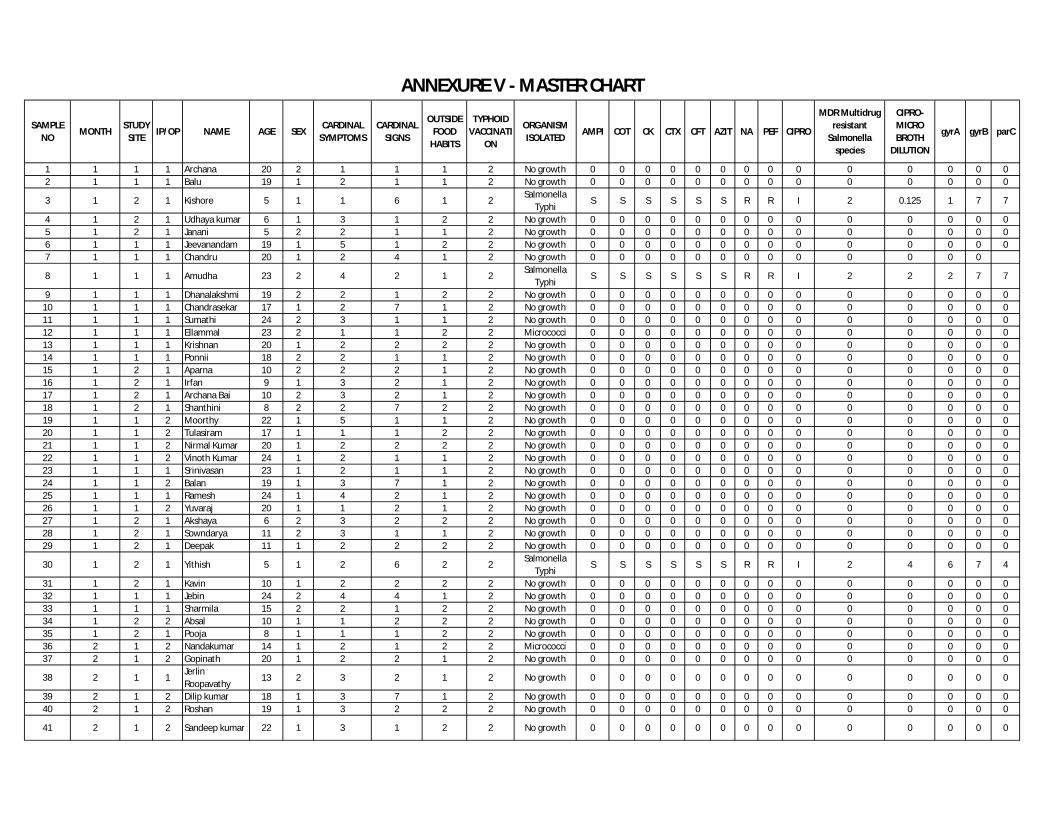
SAMPLE NO
MONTHSTUDY
SITEIP/OP NAME AGE SEX
CARDINAL SYMPTOMS
CARDINAL SIGNS
OUTSIDE FOOD
HABITS
TYPHOID VACCINATI
ON
ORGANISM ISOLATED
AMPI COT CK CTX CFT AZIT NA PEF CIPRO
MDR Multidrug resistant
Salmonella species
CIPRO- MICRO BROTH
DILUTION
gyrA gyrB parC
1 1 1 1 Archana 20 2 1 1 1 2 No growth 0 0 0 0 0 0 0 0 0 0 0 0 0 02 1 1 1 Balu 19 1 2 1 1 2 No growth 0 0 0 0 0 0 0 0 0 0 0 0 0 0
3 1 2 1 Kishore 5 1 1 6 1 2Salmonella
TyphiS S S S S S R R I 2 0.125 1 7 7
4 1 2 1 Udhaya kumar 6 1 3 1 2 2 No growth 0 0 0 0 0 0 0 0 0 0 0 0 0 05 1 2 1 Janani 5 2 2 1 1 2 No growth 0 0 0 0 0 0 0 0 0 0 0 0 0 06 1 1 1 Jeevanandam 19 1 5 1 2 2 No growth 0 0 0 0 0 0 0 0 0 0 0 0 0 07 1 1 1 Chandru 20 1 2 4 1 2 No growth 0 0 0 0 0 0 0 0 0 0 0 0 0
8 1 1 1 Amudha 23 2 4 2 1 2Salmonella
TyphiS S S S S S R R I 2 2 2 7 7
9 1 1 1 Dhanalakshmi 19 2 2 1 2 2 No growth 0 0 0 0 0 0 0 0 0 0 0 0 0 010 1 1 1 Chandrasekar 17 1 2 7 1 2 No growth 0 0 0 0 0 0 0 0 0 0 0 0 0 011 1 1 1 Sumathi 24 2 3 1 1 2 No growth 0 0 0 0 0 0 0 0 0 0 0 0 0 012 1 1 1 Ellammal 23 2 1 1 2 2 Micrococci 0 0 0 0 0 0 0 0 0 0 0 0 0 013 1 1 1 Krishnan 20 1 2 2 2 2 No growth 0 0 0 0 0 0 0 0 0 0 0 0 0 014 1 1 1 Ponnii 18 2 2 1 1 2 No growth 0 0 0 0 0 0 0 0 0 0 0 0 0 015 1 2 1 Aparna 10 2 2 2 1 2 No growth 0 0 0 0 0 0 0 0 0 0 0 0 0 016 1 2 1 Irfan 9 1 3 2 1 2 No growth 0 0 0 0 0 0 0 0 0 0 0 0 0 017 1 2 1 Archana Bai 10 2 3 2 1 2 No growth 0 0 0 0 0 0 0 0 0 0 0 0 0 018 1 2 1 Shanthini 8 2 2 7 2 2 No growth 0 0 0 0 0 0 0 0 0 0 0 0 0 019 1 1 2 Moorthy 22 1 5 1 1 2 No growth 0 0 0 0 0 0 0 0 0 0 0 0 0 020 1 1 2 Tulasiram 17 1 1 1 2 2 No growth 0 0 0 0 0 0 0 0 0 0 0 0 0 021 1 1 2 Nirmal Kumar 20 1 2 2 2 2 No growth 0 0 0 0 0 0 0 0 0 0 0 0 0 022 1 1 2 Vinoth Kumar 24 1 2 1 1 2 No growth 0 0 0 0 0 0 0 0 0 0 0 0 0 023 1 1 1 Srinivasan 23 1 2 1 1 2 No growth 0 0 0 0 0 0 0 0 0 0 0 0 0 024 1 1 2 Balan 19 1 3 7 1 2 No growth 0 0 0 0 0 0 0 0 0 0 0 0 0 025 1 1 1 Ramesh 24 1 4 2 1 2 No growth 0 0 0 0 0 0 0 0 0 0 0 0 0 026 1 1 2 Yuvaraj 20 1 1 2 1 2 No growth 0 0 0 0 0 0 0 0 0 0 0 0 0 027 1 2 1 Akshaya 6 2 3 2 2 2 No growth 0 0 0 0 0 0 0 0 0 0 0 0 0 028 1 2 1 Sowndarya 11 2 3 1 1 2 No growth 0 0 0 0 0 0 0 0 0 0 0 0 0 029 1 2 1 Deepak 11 1 2 2 2 2 No growth 0 0 0 0 0 0 0 0 0 0 0 0 0 0
30 1 2 1 Yithish 5 1 2 6 2 2Salmonella
TyphiS S S S S S R R I 2 4 6 7 4
31 1 2 1 Kavin 10 1 2 2 2 2 No growth 0 0 0 0 0 0 0 0 0 0 0 0 0 032 1 1 1 Jebin 24 2 4 4 1 2 No growth 0 0 0 0 0 0 0 0 0 0 0 0 0 033 1 1 1 Sharmila 15 2 2 1 2 2 No growth 0 0 0 0 0 0 0 0 0 0 0 0 0 034 1 2 2 Absal 10 1 1 2 2 2 No growth 0 0 0 0 0 0 0 0 0 0 0 0 0 035 1 2 1 Pooja 8 1 1 1 2 2 No growth 0 0 0 0 0 0 0 0 0 0 0 0 0 036 2 1 2 Nandakumar 14 1 2 1 2 2 Micrococci 0 0 0 0 0 0 0 0 0 0 0 0 0 037 2 1 2 Gopinath 20 1 2 2 1 2 No growth 0 0 0 0 0 0 0 0 0 0 0 0 0 0
38 2 1 1Jerlin Roopavathy
13 2 3 2 1 2 No growth 0 0 0 0 0 0 0 0 0 0 0 0 0 0
39 2 1 2 Dilip kumar 18 1 3 7 1 2 No growth 0 0 0 0 0 0 0 0 0 0 0 0 0 040 2 1 2 Roshan 19 1 3 2 2 2 No growth 0 0 0 0 0 0 0 0 0 0 0 0 0 0
41 2 1 2 Sandeep kumar 22 1 3 1 2 2 No growth 0 0 0 0 0 0 0 0 0 0 0 0 0 0
ANNEXURE V - MASTER CHART
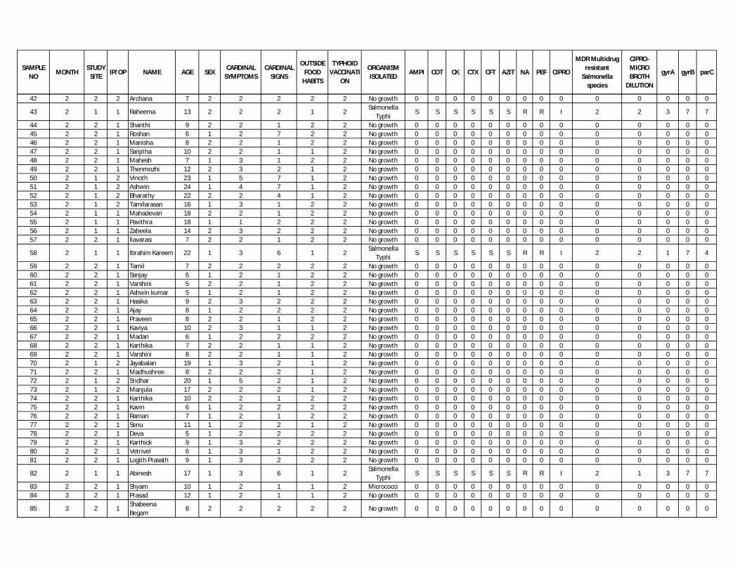
SAMPLE NO
MONTHSTUDY
SITEIP/OP NAME AGE SEX
CARDINAL SYMPTOMS
CARDINAL SIGNS
OUTSIDE FOOD
HABITS
TYPHOID VACCINATI
ON
ORGANISM ISOLATED
AMPI COT CK CTX CFT AZIT NA PEF CIPRO
MDR Multidrug resistant
Salmonella species
CIPRO- MICRO BROTH
DILUTION
gyrA gyrB parC
42 2 2 2 Archana 7 2 2 2 2 2 No growth 0 0 0 0 0 0 0 0 0 0 0 0 0 0
43 2 1 1 Raheema 13 2 2 2 1 2Salmonella
TyphiS S S S S S R R I 2 2 3 7 7
44 2 2 1 Shanthi 9 2 2 1 2 2 No growth 0 0 0 0 0 0 0 0 0 0 0 0 0 045 2 2 1 Roshan 6 1 2 7 2 2 No growth 0 0 0 0 0 0 0 0 0 0 0 0 0 046 2 2 1 Manisha 8 2 2 1 2 2 No growth 0 0 0 0 0 0 0 0 0 0 0 0 0 047 2 2 1 Sanjitha 10 2 2 1 1 2 No growth 0 0 0 0 0 0 0 0 0 0 0 0 0 048 2 2 1 Mahesh 7 1 3 1 2 2 No growth 0 0 0 0 0 0 0 0 0 0 0 0 0 049 2 2 1 Thenmozhi 12 2 3 2 1 2 No growth 0 0 0 0 0 0 0 0 0 0 0 0 0 050 2 1 2 Vinoth 23 1 5 7 1 2 No growth 0 0 0 0 0 0 0 0 0 0 0 0 0 051 2 1 2 Ashwin 24 1 4 7 1 2 No growth 0 0 0 0 0 0 0 0 0 0 0 0 0 052 2 1 2 Bharathy 22 2 2 4 1 2 No growth 0 0 0 0 0 0 0 0 0 0 0 0 0 053 2 1 2 Tamilarasan 16 1 3 1 2 2 No growth 0 0 0 0 0 0 0 0 0 0 0 0 0 054 2 1 1 Mahadevan 18 2 2 1 2 2 No growth 0 0 0 0 0 0 0 0 0 0 0 0 0 055 2 1 1 Pavithra 18 1 1 2 2 2 No growth 0 0 0 0 0 0 0 0 0 0 0 0 0 056 2 1 1 Zaheela 14 2 3 2 2 2 No growth 0 0 0 0 0 0 0 0 0 0 0 0 0 057 2 2 1 Ilavarasi 7 2 2 1 2 2 No growth 0 0 0 0 0 0 0 0 0 0 0 0 0 0
58 2 1 1 Ibrahim Kareem 22 1 3 6 1 2Salmonella
TyphiS S S S S S R R I 2 2 1 7 4
59 2 2 1 Tamil 7 2 2 2 2 2 No growth 0 0 0 0 0 0 0 0 0 0 0 0 0 060 2 2 1 Sanjay 6 1 2 1 2 2 No growth 0 0 0 0 0 0 0 0 0 0 0 0 0 061 2 2 1 Varshini 5 2 2 1 2 2 No growth 0 0 0 0 0 0 0 0 0 0 0 0 0 062 2 2 1 Ashwin kumar 5 1 2 1 2 2 No growth 0 0 0 0 0 0 0 0 0 0 0 0 0 063 2 2 1 Hasika 9 2 3 2 2 2 No growth 0 0 0 0 0 0 0 0 0 0 0 0 0 064 2 2 1 Ajay 8 1 2 2 2 2 No growth 0 0 0 0 0 0 0 0 0 0 0 0 0 065 2 2 1 Praveen 8 2 2 1 2 2 No growth 0 0 0 0 0 0 0 0 0 0 0 0 0 066 2 2 1 Kaviya 10 2 3 1 1 2 No growth 0 0 0 0 0 0 0 0 0 0 0 0 0 067 2 2 1 Madan 6 1 2 2 2 2 No growth 0 0 0 0 0 0 0 0 0 0 0 0 0 068 2 2 1 Karthika 7 2 2 1 1 2 No growth 0 0 0 0 0 0 0 0 0 0 0 0 0 069 2 2 1 Varshini 8 2 2 1 1 2 No growth 0 0 0 0 0 0 0 0 0 0 0 0 0 070 2 1 2 Jayabalan 19 1 3 2 1 2 No growth 0 0 0 0 0 0 0 0 0 0 0 0 0 071 2 2 1 Madhushree 8 2 2 2 1 2 No growth 0 0 0 0 0 0 0 0 0 0 0 0 0 072 2 1 2 Sridhar 20 1 5 2 1 2 No growth 0 0 0 0 0 0 0 0 0 0 0 0 0 073 2 1 2 Manjula 17 2 2 2 1 2 No growth 0 0 0 0 0 0 0 0 0 0 0 0 0 074 2 2 1 Karthika 10 2 2 1 2 2 No growth 0 0 0 0 0 0 0 0 0 0 0 0 0 075 2 2 1 Kavin 6 1 2 2 2 2 No growth 0 0 0 0 0 0 0 0 0 0 0 0 0 076 2 2 1 Raman 7 1 2 1 2 2 No growth 0 0 0 0 0 0 0 0 0 0 0 0 0 077 2 2 1 Senu 11 1 2 2 1 2 No growth 0 0 0 0 0 0 0 0 0 0 0 0 0 078 2 2 1 Deva 5 1 2 2 2 2 No growth 0 0 0 0 0 0 0 0 0 0 0 0 0 079 2 2 1 Karthick 9 1 3 2 2 2 No growth 0 0 0 0 0 0 0 0 0 0 0 0 0 080 2 2 1 Vetrivel 6 1 3 1 2 2 No growth 0 0 0 0 0 0 0 0 0 0 0 0 0 081 2 2 1 Logith Prasath 9 1 3 2 2 2 No growth 0 0 0 0 0 0 0 0 0 0 0 0 0 0
82 2 1 1 Abinesh 17 1 3 6 1 2Salmonella
TyphiS S S S S S R R I 2 1 3 7 7
83 2 2 1 Shyam 10 1 2 1 1 2 Micrococci 0 0 0 0 0 0 0 0 0 0 0 0 0 084 3 2 1 Prasad 12 1 2 1 1 2 No growth 0 0 0 0 0 0 0 0 0 0 0 0 0 0
85 3 2 1Shabeena Begam
8 2 2 2 2 2 No growth 0 0 0 0 0 0 0 0 0 0 0 0 0 0
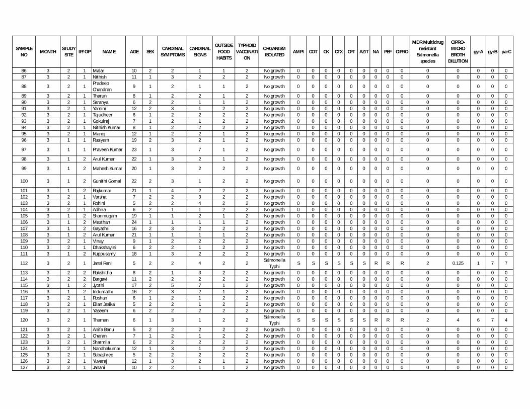
SAMPLE NO
MONTHSTUDY
SITEIP/OP NAME AGE SEX
CARDINAL SYMPTOMS
CARDINAL SIGNS
OUTSIDE FOOD
HABITS
TYPHOID VACCINATI
ON
ORGANISM ISOLATED
AMPI COT CK CTX CFT AZIT NA PEF CIPRO
MDR Multidrug resistant
Salmonella species
CIPRO- MICRO BROTH
DILUTION
gyrA gyrB parC
86 3 2 1 Malar 10 2 2 1 1 2 No growth 0 0 0 0 0 0 0 0 0 0 0 0 0 087 3 2 1 Nithish 11 1 3 2 2 2 No growth 0 0 0 0 0 0 0 0 0 0 0 0 0 0
88 3 2 1Pradeep Chandran
9 1 2 1 1 2 No growth 0 0 0 0 0 0 0 0 0 0 0 0 0 0
89 3 2 1 Tharun 8 1 2 2 1 2 No growth 0 0 0 0 0 0 0 0 0 0 0 0 0 090 3 2 1 Saranya 6 2 2 1 1 2 No growth 0 0 0 0 0 0 0 0 0 0 0 0 0 091 3 2 1 Yamini 12 2 3 1 2 2 No growth 0 0 0 0 0 0 0 0 0 0 0 0 0 092 3 2 1 Tajudheen 6 1 2 2 2 2 No growth 0 0 0 0 0 0 0 0 0 0 0 0 0 093 3 2 1 Gokulraj 7 1 2 1 2 2 No growth 0 0 0 0 0 0 0 0 0 0 0 0 0 094 3 2 1 Nithish Kumar 8 1 2 2 2 2 No growth 0 0 0 0 0 0 0 0 0 0 0 0 0 095 3 2 1 Manoj 12 1 2 2 1 2 No growth 0 0 0 0 0 0 0 0 0 0 0 0 0 096 3 1 1 Rasiyam 19 2 3 2 1 2 No growth 0 0 0 0 0 0 0 0 0 0 0 0 0 0
97 3 1 1 Praveen Kumar 23 1 3 7 1 2 No growth 0 0 0 0 0 0 0 0 0 0 0 0 0 0
98 3 1 2 Arul Kumar 22 1 3 2 1 2 No growth 0 0 0 0 0 0 0 0 0 0 0 0 0 0
99 3 1 2 Mahesh Kumar 20 1 3 2 2 2 No growth 0 0 0 0 0 0 0 0 0 0 0 0 0 0
100 3 1 2 Gunithi Gomal 22 2 3 1 2 2 No growth 0 0 0 0 0 0 0 0 0 0 0 0 0 0
101 3 1 2 Rajkumar 21 1 4 2 2 2 No growth 0 0 0 0 0 0 0 0 0 0 0 0 0 0102 3 2 1 Varsha 7 2 2 3 2 2 No growth 0 0 0 0 0 0 0 0 0 0 0 0 0 0103 3 2 1 Rohini 5 2 2 4 2 2 No growth 0 0 0 0 0 0 0 0 0 0 0 0 0 0104 3 2 1 Adhira 6 2 1 1 2 2 No growth 0 0 0 0 0 0 0 0 0 0 0 0 0 0105 3 1 2 Shanmugam 19 1 1 2 1 2 No growth 0 0 0 0 0 0 0 0 0 0 0 0 0 0106 3 1 2 Masthan 24 1 1 1 1 2 No growth 0 0 0 0 0 0 0 0 0 0 0 0 0 0107 3 1 2 Gayathri 16 2 3 2 2 2 No growth 0 0 0 0 0 0 0 0 0 0 0 0 0 0108 3 1 2 Arul Kumar 21 1 1 1 1 2 No growth 0 0 0 0 0 0 0 0 0 0 0 0 0 0109 3 2 1 Vinay 9 1 2 2 2 2 No growth 0 0 0 0 0 0 0 0 0 0 0 0 0 0110 3 2 1 Dhakshayini 6 2 2 1 2 2 No growth 0 0 0 0 0 0 0 0 0 0 0 0 0 0111 3 1 2 Kuppusamy 18 1 3 2 2 2 No growth 0 0 0 0 0 0 0 0 0 0 0 0 0 0
112 3 2 1 Jansi Rani 5 2 2 4 2 2Salmonella
TyphiS S S S S S R R R 2 0.125 1 7 7
113 3 2 2 Rakshitha 8 2 1 3 2 2 No growth 0 0 0 0 0 0 0 0 0 0 0 0 0 0114 3 2 2 Bargavi 11 2 2 2 2 2 No growth 0 0 0 0 0 0 0 0 0 0 0 0 0 0115 3 1 2 Jyothi 17 2 5 7 1 2 No growth 0 0 0 0 0 0 0 0 0 0 0 0 0 0116 3 1 2 Indumathi 16 2 3 2 1 2 No growth 0 0 0 0 0 0 0 0 0 0 0 0 0 0117 3 2 1 Roshan 6 1 2 1 2 2 No growth 0 0 0 0 0 0 0 0 0 0 0 0 0 0118 3 2 1 Ellan Jesika 5 2 2 1 2 2 No growth 0 0 0 0 0 0 0 0 0 0 0 0 0 0119 3 2 1 Yaseem 6 2 2 2 2 2 No growth 0 0 0 0 0 0 0 0 0 0 0 0 0 0
120 3 2 1 Thaman 6 1 3 1 2 2Salmonella
TyphiS S S S S S R R R 2 4 6 7 4
121 3 2 1 Anifa Banu 5 2 2 2 2 2 No growth 0 0 0 0 0 0 0 0 0 0 0 0 0 0122 3 2 1 Charan 7 1 2 1 2 2 No growth 0 0 0 0 0 0 0 0 0 0 0 0 0 0123 3 2 1 Sharmila 6 2 2 2 2 2 No growth 0 0 0 0 0 0 0 0 0 0 0 0 0 0124 3 2 1 Nandhakumar 12 1 3 1 2 2 No growth 0 0 0 0 0 0 0 0 0 0 0 0 0 0125 3 2 1 Subashree 5 2 2 2 2 2 No growth 0 0 0 0 0 0 0 0 0 0 0 0 0 0126 3 2 1 Yuvaraj 12 1 3 2 1 2 No growth 0 0 0 0 0 0 0 0 0 0 0 0 0 0127 3 2 1 Janani 10 2 2 1 1 2 No growth 0 0 0 0 0 0 0 0 0 0 0 0 0 0

SAMPLE NO
MONTHSTUDY
SITEIP/OP NAME AGE SEX
CARDINAL SYMPTOMS
CARDINAL SIGNS
OUTSIDE FOOD
HABITS
TYPHOID VACCINATI
ON
ORGANISM ISOLATED
AMPI COT CK CTX CFT AZIT NA PEF CIPRO
MDR Multidrug resistant
Salmonella species
CIPRO- MICRO BROTH
DILUTION
gyrA gyrB parC
128 3 2 1 Munna 6 1 2 1 2 2 No growth 0 0 0 0 0 0 0 0 0 0 0 0 0 0129 3 2 1 Vijaya kumar 12 1 3 2 1 2 No growth 0 0 0 0 0 0 0 0 0 0 0 0 0 0130 3 2 1 Sana Farhath 5 2 2 2 2 2 No growth 0 0 0 0 0 0 0 0 0 0 0 0 0 0131 3 2 1 Saidharshan 5 1 2 1 2 2 No growth 0 0 0 0 0 0 0 0 0 0 0 0 0 0132 3 2 1 Udhayakumar 8 1 2 1 2 2 No growth 0 0 0 0 0 0 0 0 0 0 0 0 0 0133 3 2 1 Meganathan 5 1 2 2 2 2 No growth 0 0 0 0 0 0 0 0 0 0 0 0 0 0134 3 1 1 Goutham 20 1 1 2 2 2 S.Paratyphi A S S S S S S R R I 2 0.125 1 7 7135 4 2 1 Muheeb 9 1 2 2 2 2 No growth 0 0 0 0 0 0 0 0 0 0 0 0 0 0136 4 2 1 Harini 5 2 2 2 2 2 No growth 0 0 0 0 0 0 0 0 0 0 0 0 0 0137 4 2 1 Niveditha 5 2 2 2 2 2 No growth 0 0 0 0 0 0 0 0 0 0 0 0 0 0138 4 2 1 Sachin 6 1 2 1 2 2 No growth 0 0 0 0 0 0 0 0 0 0 0 0 0 0139 4 2 1 Nida Fathima 7 2 2 1 2 2 No growth 0 0 0 0 0 0 0 0 0 0 0 0 0 0140 4 2 1 Kowsikar 8 1 1 1 2 2 No growth 0 0 0 0 0 0 0 0 0 0 0 0 0 0
141 4 2 1 Kishore kumar 6 1 3 1 2 2 No growth 0 0 0 0 0 0 0 0 0 0 0 0 0 0
142 4 2 1 Krithika 6 2 2 6 2 2Salmonella
TyphiS S S S S S R R I 2 2 6 7 4
143 4 2 1 Kishore 11 1 3 2 2 2 No growth 0 0 0 0 0 0 0 0 0 0 0 0 0 0144 4 2 1 Udaya kumar 8 1 2 2 1 2 No growth 0 0 0 0 0 0 0 0 0 0 0 0 0 0145 4 2 1 Niveditha 5 2 2 2 2 2 No growth 0 0 0 0 0 0 0 0 0 0 0 0 0 0146 4 2 1 Umalakshmi 6 2 3 1 2 2 No growth 0 0 0 0 0 0 0 0 0 0 0 0 0 0147 5 2 1 Santhosh 10 1 2 1 2 2 No growth 0 0 0 0 0 0 0 0 0 0 0 0 0 0148 5 2 1 Porkodi 7 2 3 1 2 2 No growth 0 0 0 0 0 0 0 0 0 0 0 0 0 0149 5 2 1 Archana 7 2 1 2 1 2 No growth 0 0 0 0 0 0 0 0 0 0 0 0 0 0150 5 2 1 Vanashri 6 2 2 1 2 2 No growth 0 0 0 0 0 0 0 0 0 0 0 0 0 0151 5 2 1 Akshaya 5 2 2 2 2 2 No growth 0 0 0 0 0 0 0 0 0 0 0 0 0 0152 5 2 1 Rani 8 2 3 2 2 2 No growth 0 0 0 0 0 0 0 0 0 0 0 0 0 0153 5 2 1 Pradeep 9 1 3 1 2 2 No growth 0 0 0 0 0 0 0 0 0 0 0 0 0 0154 5 1 1 Chandiran 23 1 1 7 1 2 No growth 0 0 0 0 0 0 0 0 0 0 0 0 0 0155 5 1 1 Dhanalakshmi 22 2 5 1 1 2 No growth 0 0 0 0 0 0 0 0 0 0 0 0 0 0156 5 1 1 Sandhiya 19 2 5 1 2 2 No growth 0 0 0 0 0 0 0 0 0 0 0 0 0 0157 5 1 1 Beijili 24 2 4 7 1 2 No growth 0 0 0 0 0 0 0 0 0 0 0 0 0 0
158 5 1 1Ushman Hameed
23 1 3 6 1 2Salmonella
TyphiS S S S S S R R R 2 1 5 7 7
159 5 1 1 Palani 22 1 1 2 1 2 No growth 0 0 0 0 0 0 0 0 0 0 0 0 0 0160 5 1 1 Tamilarasi 23 2 3 2 1 2 No growth 0 0 0 0 0 0 0 0 0 0 0 0 0 0161 6 1 1 Asha 19 2 2 1 2 2 E.coli 0 0 0 0 0 0 0 0 0 0 0 0 0 0162 6 1 1 Vignesh 16 1 3 1 2 2 No growth 0 0 0 0 0 0 0 0 0 0 0 0 0 0163 6 1 1 Arul 24 1 2 2 2 2 No growth 0 0 0 0 0 0 0 0 0 0 0 0 0 0164 6 1 1 Krishnan 19 1 2 7 2 2 No growth 0 0 0 0 0 0 0 0 0 0 0 0 0 0165 6 1 1 Ponni 22 2 3 2 1 2 No growth 0 0 0 0 0 0 0 0 0 0 0 0 0 0166 6 1 1 Jothy 19 2 3 1 2 2 No growth 0 0 0 0 0 0 0 0 0 0 0 0 0 0167 6 1 2 Abdul kareem 16 1 2 2 1 2 No growth 0 0 0 0 0 0 0 0 0 0 0 0 0 0168 6 1 2 Rajeshwari 23 2 3 1 2 2 No growth 0 0 0 0 0 0 0 0 0 0 0 0 0 0169 6 1 2 Hariharan 22 1 1 2 1 2 No growth 0 0 0 0 0 0 0 0 0 0 0 0 0 0
170 6 1 2 Mohana 24 2 4 2 1 2 K.pneumoniae 0 0 0 0 0 0 0 0 0 0 0 0 0 0
171 6 1 2 Vijaya 24 2 5 1 2 2 No growth 0 0 0 0 0 0 0 0 0 0 0 0 0 0

SAMPLE NO
MONTHSTUDY
SITEIP/OP NAME AGE SEX
CARDINAL SYMPTOMS
CARDINAL SIGNS
OUTSIDE FOOD
HABITS
TYPHOID VACCINATI
ON
ORGANISM ISOLATED
AMPI COT CK CTX CFT AZIT NA PEF CIPRO
MDR Multidrug resistant
Salmonella species
CIPRO- MICRO BROTH
DILUTION
gyrA gyrB parC
172 6 1 1 Kokila 20 2 2 1 2 2 No growth 0 0 0 0 0 0 0 0 0 0 0 0 0 0173 7 1 1 Vishnupriya 18 2 3 1 2 2 No growth 0 0 0 0 0 0 0 0 0 0 0 0 0 0174 7 1 1 Deepa 18 2 1 1 2 2 No growth 0 0 0 0 0 0 0 0 0 0 0 0 0 0175 7 1 2 Kaviyarasan 22 1 2 1 1 2 No growth 0 0 0 0 0 0 0 0 0 0 0 0 0 0176 7 1 1 Raveendiran 24 1 2 7 1 2 No growth 0 0 0 0 0 0 0 0 0 0 0 0 0 0177 7 1 1 Muthukumar 23 1 2 1 1 2 No growth 0 0 0 0 0 0 0 0 0 0 0 0 0 0178 7 1 1 Indumathy 20 2 3 1 1 2 No growth 0 0 0 0 0 0 0 0 0 0 0 0 0 0179 7 1 1 Dinesh 17 1 1 2 1 2 No growth 0 0 0 0 0 0 0 0 0 0 0 0 0 0180 7 1 1 Muthupriya 19 2 2 2 2 2 No growth 0 0 0 0 0 0 0 0 0 0 0 0 0 0181 7 1 1 Malar 18 1 2 2 2 2 No growth 0 0 0 0 0 0 0 0 0 0 0 0 0 0182 7 1 1 Gopi 24 2 3 2 1 2 No growth 0 0 0 0 0 0 0 0 0 0 0 0 0 0183 7 1 1 Sangeetha 21 2 3 2 1 2 No growth 0 0 0 0 0 0 0 0 0 0 0 0 0 0184 8 1 1 Jayakumar 18 1 3 2 2 2 No growth 0 0 0 0 0 0 0 0 0 0 0 0 0 0185 8 1 1 Jithan 15 1 3 7 2 2 No growth 0 0 0 0 0 0 0 0 0 0 0 0 0 0186 8 1 1 Sheela 24 2 2 2 1 2 No growth 0 0 0 0 0 0 0 0 0 0 0 0 0 0187 8 1 2 Tharun 18 2 2 1 1 2 No growth 0 0 0 0 0 0 0 0 0 0 0 0 0 0188 8 1 2 Rathika 24 2 2 2 2 2 No growth 0 0 0 0 0 0 0 0 0 0 0 0 0 0189 8 1 2 Stephen Raj 18 1 1 2 1 2 No growth 0 0 0 0 0 0 0 0 0 0 0 0 0 0190 8 1 1 Srinivasan 23 1 2 1 1 2 No growth 0 0 0 0 0 0 0 0 0 0 0 0 0 0191 8 1 1 Dhanamani 23 2 3 1 2 2 No growth 0 0 0 0 0 0 0 0 0 0 0 0 0 0192 8 1 1 Sivaraman 11 1 1 1 2 2 No growth 0 0 0 0 0 0 0 0 0 0 0 0 0 0
193 8 1 2 Naveen kumar 16 1 4 1 2 2 No growth 0 0 0 0 0 0 0 0 0 0 0 0 0 0
194 8 1 2 Fathima 21 2 5 2 2 2 No growth 0 0 0 0 0 0 0 0 0 0 0 0 0 0195 8 1 2 Rajammmal 23 2 5 1 1 2 No growth 0 0 0 0 0 0 0 0 0 0 0 0 0 0196 9 1 2 Nithish 19 1 2 1 2 2 No growth 0 0 0 0 0 0 0 0 0 0 0 0 0 0
197 9 1 2 Suriya Prakash 20 1 2 2 2 2 No growth 0 0 0 0 0 0 0 0 0 0 0 0 0 0
198 9 1 2 Suresh 23 1 2 1 2 2 No growth 0 0 0 0 0 0 0 0 0 0 0 0 0 0199 9 1 2 Sangeetha 20 2 3 2 1 2 No growth 0 0 0 0 0 0 0 0 0 0 0 0 0 0200 9 1 2 David 19 1 3 1 1 2 No growth 0 0 0 0 0 0 0 0 0 0 0 0 0 0201 9 1 2 Priya 23 2 3 2 2 2 E.coli 0 0 0 0 0 0 0 0 0 0 0 0 0 0202 9 1 2 Ajay kumar 24 1 4 1 1 2 No growth 0 0 0 0 0 0 0 0 0 0 0 0 0 0203 9 1 2 Rajasekar 19 1 5 2 2 2 No growth 0 0 0 0 0 0 0 0 0 0 0 0 0 0204 9 1 2 Jothi 24 2 3 2 2 2 No growth 0 0 0 0 0 0 0 0 0 0 0 0 0 0205 9 1 2 Ramesh 21 1 2 2 1 2 No growth 0 0 0 0 0 0 0 0 0 0 0 0 0 0206 9 1 2 Elbin Jacob 22 2 2 2 1 2 No growth 0 0 0 0 0 0 0 0 0 0 0 0 0 0207 9 1 2 Monika 20 2 2 2 1 2 No growth 0 0 0 0 0 0 0 0 0 0 0 0 0 0208 10 1 2 Brijesh Kumar 20 1 3 2 1 2 No growth 0 0 0 0 0 0 0 0 0 0 0 0 0 0209 10 1 1 Kumudha 20 2 3 2 2 2 No growth 0 0 0 0 0 0 0 0 0 0 0 0 0 0210 10 1 1 Ranjani 17 2 4 2 2 2 No growth 0 0 0 0 0 0 0 0 0 0 0 0 0 0211 10 1 1 Vijayan 20 1 5 1 2 2 No growth 0 0 0 0 0 0 0 0 0 0 0 0 0 0212 10 1 1 Monish 13 1 3 1 2 2 No growth 0 0 0 0 0 0 0 0 0 0 0 0 0 0213 10 1 1 Mohana 14 2 2 2 2 2 No growth 0 0 0 0 0 0 0 0 0 0 0 0 0 0214 10 1 1 Preety 18 1 1 1 2 2 No growth 0 0 0 0 0 0 0 0 0 0 0 0 0 0215 10 1 1 Ganthi 19 1 1 1 2 2 No growth 0 0 0 0 0 0 0 0 0 0 0 0 0 0216 10 1 1 Yasmin 18 2 2 1 2 2 No growth 0 0 0 0 0 0 0 0 0 0 0 0 0 0217 10 1 1 Ahmed 13 1 1 2 2 2 No growth 0 0 0 0 0 0 0 0 0 0 0 0 0 0

SAMPLE NO
MONTHSTUDY
SITEIP/OP NAME AGE SEX
CARDINAL SYMPTOMS
CARDINAL SIGNS
OUTSIDE FOOD
HABITS
TYPHOID VACCINATI
ON
ORGANISM ISOLATED
AMPI COT CK CTX CFT AZIT NA PEF CIPRO
MDR Multidrug resistant
Salmonella species
CIPRO- MICRO BROTH
DILUTION
gyrA gyrB parC
218 10 1 2 Sakthivel 15 1 1 2 2 2 No growth 0 0 0 0 0 0 0 0 0 0 0 0 0 0219 10 1 1 Dinesh 23 1 4 2 2 2 No growth 0 0 0 0 0 0 0 0 0 0 0 0 0 0220 10 1 1 Ruby Stella 20 2 3 2 2 2 No growth 0 0 0 0 0 0 0 0 0 0 0 0 0 0221 10 1 1 Priyanka 16 2 2 1 2 2 No growth 0 0 0 0 0 0 0 0 0 0 0 0 0 0222 10 1 2 Vijayalakshmi 20 2 2 1 1 2 No growth 0 0 0 0 0 0 0 0 0 0 0 0 0 0223 10 1 2 Shanmugaraj 22 1 3 1 1 2 No growth 0 0 0 0 0 0 0 0 0 0 0 0 0 0224 10 1 2 Suja 22 2 4 7 2 2 No growth 0 0 0 0 0 0 0 0 0 0 0 0 0 0225 11 1 2 Femina 24 2 5 7 2 2 No growth 0 0 0 0 0 0 0 0 0 0 0 0 0 0226 11 1 2 Devaraj 19 1 5 2 1 2 No growth 0 0 0 0 0 0 0 0 0 0 0 0 0 0227 11 1 2 Vidhya 24 2 5 2 1 2 No growth 0 0 0 0 0 0 0 0 0 0 0 0 0 0228 11 1 1 Parthiban 23 1 1 1 1 2 No growth 0 0 0 0 0 0 0 0 0 0 0 0 0 0229 11 1 1 Ramadoss 21 1 1 1 1 2 No growth 0 0 0 0 0 0 0 0 0 0 0 0 0 0230 11 1 1 Ilavarasan 21 1 2 2 2 2 No growth 0 0 0 0 0 0 0 0 0 0 0 0 0 0231 11 1 1 Divya 22 2 3 1 2 2 No growth 0 0 0 0 0 0 0 0 0 0 0 0 0 0232 11 1 1 Saravanan 20 1 1 2 2 2 No growth 0 0 0 0 0 0 0 0 0 0 0 0 0 0233 11 1 1 Sharmila 17 2 2 1 2 2 No growth 0 0 0 0 0 0 0 0 0 0 0 0 0 0234 11 1 1 Ajithkumar 20 1 4 2 1 2 No growth 0 0 0 0 0 0 0 0 0 0 0 0 0 0
235 11 1 1 Mani 23 1 2 4 1 2Salmonella
TyphiS S S S S S R R I 2 0.125 1 7 7
236 11 1 2 Raja 24 1 5 2 1 2 No growth 0 0 0 0 0 0 0 0 0 0 0 0 0 0237 11 1 2 Hariharan 15 1 2 2 2 2 No growth 0 0 0 0 0 0 0 0 0 0 0 0 0 0238 11 1 1 Sanjay 23 1 3 2 2 2 No growth 0 0 0 0 0 0 0 0 0 0 0 0 0 0239 11 1 1 Manjula 14 2 2 2 2 2 No growth 0 0 0 0 0 0 0 0 0 0 0 0 0 0240 11 1 1 Subbulakshmi 23 2 3 1 1 2 No growth 0 0 0 0 0 0 0 0 0 0 0 0 0 0241 11 1 2 Naseer 24 1 3 2 1 2 No growth 0 0 0 0 0 0 0 0 0 0 0 0 0 0242 11 2 1 Harikrishnan 11 1 3 1 2 2 No growth 0 0 0 0 0 0 0 0 0 0 0 0 0 0243 11 1 2 Sujeetha 10 2 2 2 2 2 No growth 0 0 0 0 0 0 0 0 0 0 0 0 0 0244 12 1 1 Vijay 23 1 1 1 2 2 No growth 0 0 0 0 0 0 0 0 0 0 0 0 0 0245 12 1 1 Charles 24 1 1 2 1 2 No growth 0 0 0 0 0 0 0 0 0 0 0 0 0 0246 12 1 2 Venkatesan 24 1 3 1 2 2 No growth 0 0 0 0 0 0 0 0 0 0 0 0 0 0247 12 1 2 Diwakar 22 1 2 2 2 2 No growth 0 0 0 0 0 0 0 0 0 0 0 0 0 0248 12 1 2 Devi 24 2 3 1 2 2 No growth 0 0 0 0 0 0 0 0 0 0 0 0 0 0249 12 1 2 Nithish 13 1 3 2 1 2 No growth 0 0 0 0 0 0 0 0 0 0 0 0 0 0250 12 1 1 Yuvaraj 15 1 2 1 1 2 No growth 0 0 0 0 0 0 0 0 0 0 0 0 0 0251 12 1 2 Dhavania 23 2 1 2 2 2 No growth 0 0 0 0 0 0 0 0 0 0 0 0 0 0252 12 1 2 Gnanodhaya 15 2 2 2 2 2 No growth 0 0 0 0 0 0 0 0 0 0 0 0 0 0253 12 1 1 Sakthivel 23 1 3 2 2 2 No growth 0 0 0 0 0 0 0 0 0 0 0 0 0 0254 12 1 2 Vinothkumar 22 1 3 1 2 2 No growth 0 0 0 0 0 0 0 0 0 0 0 0 0 0255 12 1 2 Ranjitham 20 2 3 2 1 2 No growth 0 0 0 0 0 0 0 0 0 0 0 0 0 0256 12 1 2 Inayathula 19 1 1 1 2 2 No growth 0 0 0 0 0 0 0 0 0 0 0 0 0 0257 12 1 2 Jamuna 21 2 3 2 2 2 No growth 0 0 0 0 0 0 0 0 0 0 0 0 0 0

Month codeStudy Site
Code IP/OP Code Sex CodeOutside
food habits
Code ASTPattern CodeSymptoms
CodeCode Signs Code MDR Code
Antibiotics
AbbrevationCipro-MIC
gyrA CODE gyrB CODE parC CODE
June 1 RGGGH 1 IP 1 Male 1 Yes 1 Suceptibile SFever,Abdomi
nal pain1
Pyrexia,Toxic look,Pallor
1MDR
isolate1AMPI ampicillin ≤0.06 Sensitive
ser(83) Phenylala
nine1
No mutation
7ser (80)-isoleucin
e4
July 2 ICH 2 OP 2 Female 2 No 2 Intermediate I
Fever, Abdominal
pain, Diarrhoea
2Pyrexia,Toxic look,Coated
tongue2
Susceptible isolate
2 COT cotrimoxazole 0.125-0.5Intermed
iateser(83)
Tyrosine2
August 3 Resistant R
Fever, Abdominal
pain, Diarrhoea, Vomitting
3
Pyrexia, Toxic look, Coated
tongue, Hepatomegaly
3 CKchloramphenic
ol≥1 Resistant
Asp(87)-Asn
3
September 4 Not tested 0Fever,
Constipation4
Pyrexia, Toxic look, Coated
tongue, Splenomagaly
4 CFT ceftriaxone
ser(83) Phe,Gly(1
33)-Glutamic
acid
5
October 5Fever,Non
specific symptoms
5Pyrexia,Rose spots,Coated
tongue5 AZIT azithromycin
ser(83)Phe,Asp(87)
Asn6
November 6
Pyrexia,Toxic look,Coated
tongue,Hepatosplenomegaly
6 NA nalidixic acid
December 7 Pyrexia,Pallor 7 PEF pefloxacin
January 8 CIPRO ciprofloxacin
February 9 CTX cefotaxime
March 10
April 11
May 12