diagnostic microbiology
151
Islamic University-Gaza Medical Technology Department Lecture Notes: Diagnostic Medical Microbiology MEDI 3313 Dr. Abdelraouf A. Elmanama Ph. D Microbiology January 2012
-
Upload
independent -
Category
Documents
-
view
0 -
download
0
Transcript of diagnostic microbiology

Islamic University-Gaza
Medical Technology Department
Lecture Notes:
Diagnostic Medical Microbiology MEDI 3313
Dr. Abdelraouf A. Elmanama Ph. D Microbiology
January 2012
Islamic University-Gaza Medical Laboratory Science Department
_____________________________________________________________________ Diagnostic medical microbiology [2] Dr. Abdelraouf A. Elmanama
Introduction • Koch's Postulates • Basic Definitions • Factors Controlling Growth of Organisms • Endotoxin • Bacterial Exotoxins • Comparison of Bacterial Exotoxins with Endotoxin • Summary of Host-Parasite Interactions • Pathogen Virulence
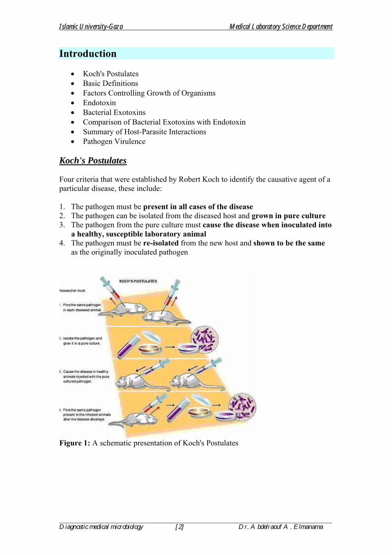
Koch's Postulates Four criteria that were established by Robert Koch to identify the causative agent of a particular disease, these include: 1. The pathogen must be present in all cases of the disease 2. The pathogen can be isolated from the diseased host and grown in pure culture 3. The pathogen from the pure culture must cause the disease when inoculated into
a healthy, susceptible laboratory animal 4. The pathogen must be re-isolated from the new host and shown to be the same
as the originally inoculated pathogen
Figure 1: A schematic presentation of Koch's Postulates

Islamic University-Gaza Medical Laboratory Science Department
_____________________________________________________________________ Diagnostic medical microbiology [3] Dr. Abdelraouf A. Elmanama
Host-Parasite Interaction Basic Definitions
INDEPENDENCE: Living free from the influence, guidance, or control by another organism SYMBIOSIS: A relationship in which two dissimilar organisms (SYMBIOTES, SYMBIONTS) live in close association with one another MUTUALISM: Mutually beneficial relationship between two species COMMENSALISM: A relationship between two species in which one is benefited and the other is not affected, neither negatively nor positively PARASITISM: A relationship between two species in which one benefits (parasite) from the other (host); usually involves some detriment to the host BENIGN: Referring to a non-life or non-health threatening condition = COMMENSALISM MALIGNANT: A disease tending to become progressively worse (MORBIDITY=illness) and potentially result in death (MORTALITY=death) CARRIER: A symptomless individual who is host to a pathogenic microorganims and who has the potential to pass the pathogen to others PATHOGENICITY: The quality of producing or the ability to produce pathologic changes or disease VIRULENCE: A measure of pathogenicity; a measurement of the degree of disease producing ability of a microorganism as indicated by the severity of the disease produced; a measure of the dosage required to caused a specific degree of pathogenicity. DOSAGE: The number of pathogenic microorganisms entering the host LD50: The number of microorganisms required to cause lethality in 50% of the test host TRUE PATHOGEN: Any microorganism capable of causing disease; an infecting agent OPPORTUNISTIC PATHOGEN: A usually harmless microorganism that becomes pathogenic under favorable conditions INFECTION: The colonization and/or invasion and multiplication of pathogenic microorganisms in the host with or without the manifestation of disease COLONIZATION: The successful occupation of a new habitat by a species not normally found in this niche

Islamic University-Gaza Medical Laboratory Science Department
_____________________________________________________________________ Diagnostic medical microbiology [4] Dr. Abdelraouf A. Elmanama
MULTIPLICATION: The ability of a microorganism to reproduce during an infection DISEASE: An abnormal condition of body function(s) or structure that is considered to be harmful to the affected individual (host); any deviation from or interruption of the normal structure or function of any part, organ, or system of the body.
Factors Controlling Growth of Microorganisms 1. NUTRIENT AVAILABILITY: The accessibility of a necessary resource, substance or compound providing nourishment to maintain life, i.e. capable of conversion to energy and structural building blocks Fastidious: Having complex nutritional or cultural requirements that make isolation and culture more difficult MAJOR ESSENTIAL ELEMENTS:
C, O, H, N, S, P, K, Mg, Ca, Fe, Na, Cl MINOR ESSENTIAL ELEMENTS:
Zn, Mn, Mo, Se, Co, Cu, Ni, W 2. PHYSICO/ENVIRONMENTAL PARAMETERS: 2.1 WATER ACTIVITY/OSMOTIC PRESSURE: Water activity (aw): represents the available water Osmotic pressure: expressed in atmospheres; reflects the concentration of solute in an aqueous solution 2.2 OXYGEN: pathogenic microorganisms may have metabolic oxygen requirements that are: 1. OBLIGATE AEROBES, 2.OBLIGATE 3. FACULTATIVE 4. AEROTOLERANT ANAEROBIC and 5. MICROAEROPHILIC 2.3 pH: "power of hydrogen"; a measurement of the amount of hydrogen ion in solution; the logarithm of the reciprocal of the hydrogen ion concentration in an aqueous solution used to express its acidity or alkalinity (0-14) 2.4 TEMPERATURE:
Psycrophile(psychrophilic): liking cold temperatures; optimal growth at 15o to 20oC Mesophile (mesophilic): liking moderate temperatures; optimal growth at 20o to 45oC Thermophile (thermophilic): liking elevated temperatures; optimal growth at 50o to 70oC
3. COMPETITION: the simultaneous demand by two or more organisms or species for a necessary, common resource or physical space that is in limited or potentially limited supply, resulting in a struggle for survival

Islamic University-Gaza Medical Laboratory Science Department
_____________________________________________________________________ Diagnostic medical microbiology [5] Dr. Abdelraouf A. Elmanama
Niche: the place of an organism within its community or ecosystem 4. HOST IMMUNE SYSTEM: the cells and tissues involved in recognizing and attacking foreign substances in the body
Bacterial Endotoxin
Endotoxin: Complex bacterial toxin; lipopolysaccharide (LPS) component of Gram-negative cell walls is composed of Lipid A + Core Polysaccharide + O Antigen (O polysaccharide side chain) and is released upon lysis of the cell during infection ; Lipid A component is responsible for endotoxin activity effects on the host; O side chain is the antigenic portion of the LPS molecule Septic shock (sepsis): Associated with overwhelming infection resulting in vascular system failure with sequestration of large volumes of blood in capillaries and veins; Activation of the complement and kinin systems and the release of histamines, prostaglandins, and other mediators may be involved. Endotoxemia: Endotoxin in the blood "More detailed discussion of endotoxin effects can be found later in the text"
Bacterial Exotoxins
Two Broad Classes of Bacterial Exotoxins 1. Intracellular Targets: A-B dimeric (two domain) exotoxins: conform to general structural model (prototype is diphtheria toxin of Corynebacterium diphtheriae): Bipartite structure (B, binding; A, active): One component is a binding domain (B) associated with absorption to target cell surface and transfer of active component (A) across cell membrane; once internalized, domain (A) enzymatically disrupts cell function
Receptor-mediated endocytosis (host cell uptake and internalization of exotoxin) ADP-ribosylation of intracellular target host molecule

Islamic University-Gaza Medical Laboratory Science Department
_____________________________________________________________________ Diagnostic medical microbiology [6] Dr. Abdelraouf A. Elmanama
2.Cellular Targets: Cytolytic exotoxins (usually degradative enzymes) or cytolysins: hemolysis, tissue necrosis, may be lethal when administered intravenously
Three Major Types of Bacterial Cytolysins Based on Mechanism of Action 1. Hydrolyze membrane phospholipids (phospholipases); e.g., Clostridium; Staphylococcus 2. Thiol(-SH)-activated cytolysins (oxygen-labile) alter membrane permeability by binding to cholesterol; e.g., streptolysin O of Streptococcus; tetanolysin of Clostridium 3. Detergent-like activity on cell membranes; rapid rate of lysis; e.g. Staphylococcus
Examples of Two-Component (A-B) Exotoxins with Intracellular Targets 1. Adenylate cyclase toxin (Bordetella spp.): • Chromosomally-encoded • Activated by intracellular calmodulin and, like pertussis toxin, catalyzes
conversion of ATP to cAMP • Inhibits leukocyte chemotaxis and activity 2. Anthrax toxin (Bacillus anthracis): • Plasmid-encoded • Three separate proteins: Protective antigen (PA); Edema factor (EF); Lethal factor
(LF) • EF + PA = increase in cAMP level resulting in edema (fluid accumulation) • LF + PA = death of host cells and ultimately death of host 3. Botulinum toxins (7 antigenically distinct toxins (A-G)) (Clostridium botulinum): • Phage-encoded neurotoxins • Among most potent of all biological toxins • Binding domain (B-subunit) binds to neuroreceptor gangliosides on cholinergic
neurons • A-subunit irreversibly inhibits release of the stimulatory neurotransmitter,
acetylcholine, at myoneural (muscle-nerve) junctions (peripheral cholinergic synapses) resulting in a flaccid paralysis and death
4. Cholera toxin (A-5B) (Vibrio cholerae): • Chromosomally-encoded • B-subunit binds to GM1 ganglioside receptors in small intestine • Reduction of disulfide bond in A-subunit activates A1 fragment that ADP-
ribosylates guanosine triphosphate (GTP)-binding protein (Gs) by transferring ADP-ribose from nicotinamide adenine dinucleotide (NAD); the ADP-ribosylated GTP-binding protein activates adenyl cyclase resulting in an increased cyclic AMP (cAMP) level and a profound life-threatening diarrhea with profuse outpouring of fluids and electrolytes (sodium, potassium, bicarbonate) while blocking the uptake of any further sodium and chloride from the lumen of the small intestine and ultimately resulting in hypovolemic shock and death in the absence of fluid and electrolyte replacement therapy
Islamic University-Gaza Medical Laboratory Science Department
_____________________________________________________________________ Diagnostic medical microbiology [7] Dr. Abdelraouf A. Elmanama
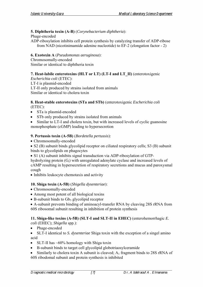
5. Diphtheria toxin (A-B) (Corynebacterium diphtheria): Phage-encoded ADP-ribosylation inhibits cell protein synthesis by catalyzing transfer of ADP-ribose
from NAD (nicotinimamide adenine nucleotide) to EF-2 (elongation factor - 2) 6. Exotoxin A (Pseudomonas aeruginosa): Chromosomally-encoded Similar or identical to diphtheria toxin 7. Heat-labile enterotoxins (HLT or LT) (LT-I and LT_II) (enterotoxigenic Escherichia coli (ETEC): LT-I is plasmid-encoded LT-II only produced by strains isolated from animals Similar or identical to cholera toxin 8. Heat-stable enterotoxins (STa and STb) (enterotoxigenic Escherichia coli (ETEC): • STa is plasmid-encoded • STb only produced by strains isolated from animals • Similar to LT-I and cholera toxin, but with increased levels of cyclic guanosine monophosphate (cGMP) leading to hypersecretion 9. Pertussis toxin (A-5B) (Bordetella pertussis): • Chromosomally-encoded • S2 (B) subunit binds glycolipid receptor on ciliated respiratory cells; S3 (B) subunit binds to glycolipids on phagocytes • S1 (A) subunit inhibits signal transduction via ADP-ribosylation of GTP-hydrolyzing protein (Gi) with unregulated adenylate cyclase and increased levels of cAMP resulting in hypersecretion of respiratory secretions and mucus and paroxysmal cough • Inhibits leukocyte chemotaxis and activity 10. Shiga toxin (A-5B) (Shigella dysenteriae): • Chromosomally-encoded • Among most potent of all biological toxins • B-subunit binds to Gb3 glycolipid receptor • A-subunit prevents binding of aminoacyl-transfer RNA by cleaving 28S rRNA from 60S ribosomal subunit resulting in inhibition of protein synthesis 11. Shiga-like toxins (A-5B) (SLT-I and SLT-II in EHEC) (enterohemorrhagic E. coli (EHEC); Shigella spp.): • Phage-encoded • SLT-I identical to S. dysenteriae Shiga toxin with the exception of a singel amino acid • SLT-II has ~60% homology with Shiga toxin • B-subunit binds to target cell glycolipid globotriaosylceramide • Similarly to cholera toxin A subunit is cleaved; A1 fragment binds to 28S rRNA of 60S ribodomal subunit and protein synthesis is inhibited
Islamic University-Gaza Medical Laboratory Science Department
_____________________________________________________________________ Diagnostic medical microbiology [8] Dr. Abdelraouf A. Elmanama
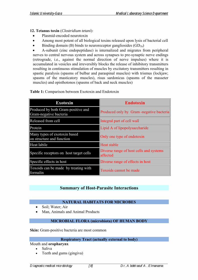
12. Tetanus toxin (Clostridium tetani):
• Plasmid-encoded neurotoxin • Among most potent of all biological toxins released upon lysis of bacterial cell • Binding domain (B) binds to neuroreceptor gangliosides (GD1b) • A-subunit (zinc endopeptidase) is internalized and migrates from peripheral nerves to central nervous system and across synapses to pre-synaptic nerve endings (retrograde, i.e., against the normal direction of nerve impulses) where it is accumulated in vesicles and irreversibly blocks the release of inhibitory transmitters resulting in continuous stimulation of muscles by excitatory transmitters resulting in spastic paralysis (spasms of bulbar and paraspinal muscles) with trismus (lockjaw; spasms of the masticatory muscles), risus sardonicus (spasms of the masseter muscles) and opisthotonos (spasms of back and neck muscles)
Table 1: Comparison between Exotoxin and Endotoxin
Exotoxin Endotoxin Produced by both Gram-positive and Gram-negative bacteria Produced only by Gram -negative bacteria
Released from cell Integral part of cell wall
Protein Lipid A of lipopolysaccharide Many types of exotoxin based on structure and function Only one type of endotoxin
Heat labile Heat stable
Specific receptors on host target cells Diverse range of host cells and systems affected
Specific effects in host Diverse range of effects in host Toxoids can be made by treating with formalin Toxoids cannot be made
Summary of Host-Parasite Interactions
NATURAL HABITATS FOR MICROBES • Soil; Water; Air • Man, Animals and Animal Products
MICROBIAL FLORA (microbiota) OF HUMAN BODY
Skin: Gram-positive bacteria are most common
Respiratory Tract (actually external to body) Mouth and oropharynx
• Saliva • Teeth and gums (gingiva)
Islamic University-Gaza Medical Laboratory Science Department
_____________________________________________________________________ Diagnostic medical microbiology [9] Dr. Abdelraouf A. Elmanama
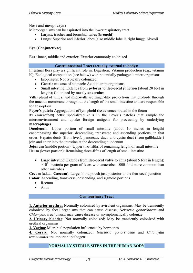
Nose and nasopharynx Microorganisms can be aspirated into the lower respiratory tract
• Larynx, trachea and bronchial tubes (bronchi) • Lungs: Superior and inferior lobes (also middle lobe in right lung); Alveoli
Eye (Conjunctivae) Ear: Inner, middle and exterior; Exterior commonly colonized
Gastrointestinal Tract (actually external to body): Intestinal flora play a significant role in: Digestion; Vitamin production (e.g., vitamin K); Ecological competition (see below) with potentially pathogenic microorganisms
• Esophagus: Not typically colonized • Gastric mucosa of stomach: Acid tolerant organisms • Small intestine: Extends from pylorus to ileo-cecal junction (about 20 feet in
length); Colonized by mostly anaerobes Villi (plural of villus) and microvilli are finger-like projections that protrude through the mucous membrane throughout the length of the small intestine and are responsible for absorption Peyer’s patch: Aggregations of lymphoid tissue concentrated in the ileum M (microfold) cells: specialized cells in the Peyer’s patches that sample the microenvironment and uptake foreign antigens for processing by underlying macrophages Duodenum: Upper portion of small intestine (about 10 inches in length) encompassing the superior, descending, transverse and ascending portions, in that order; Hepatic ducts (from liver), pancreatic duct, and cystic duct (from gallbladder) join and enter into the intestine at the descending duodenum Jejunum (middle portion): Upper two-fifths of remaining length of small intestine Ileum (lower portion): Remaining three-fifths of length of small intestine
• Large intestine: Extends from ileo-cecal valve to anus (about 5 feet in length);
>1011 bacteria per gram of feces with anaerobes 1000-fold more common than other microbes
Cecum (a.k.a., Caecum): Large, blind pouch just posterior to the ileo-cecal junction Colon: Ascending, transverse, descending, and sigmoid portions
• Rectum • Anus
Genitourinary Tract
1. Anterior urethra: Normally colonized by avirulent organisms; May be transiently colonized by fecal organisms that can cause disease; Neisseria gonorrhoeae and Chlamydia trachomatis may cause disease or asymptomatically colonize 2. Urinary bladder: Not normally colonized; May be transiently colonized with urethral organisms 3. Vagina: Microbial population influenced by hormones 4. Cervix: Not normally colonized; Neisseria gonorrhoeae and Chlamydia trachomatis are important pathogens
NORMALLY STERILE SITES IN THE HUMAN BODY
Islamic University-Gaza Medical Laboratory Science Department
_____________________________________________________________________ Diagnostic medical microbiology [10] Dr. Abdelraouf A. Elmanama
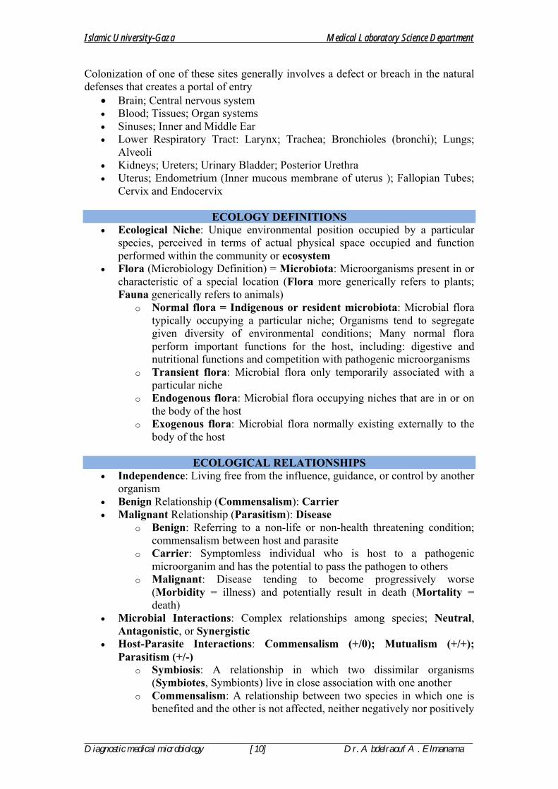
Colonization of one of these sites generally involves a defect or breach in the natural defenses that creates a portal of entry
• Brain; Central nervous system • Blood; Tissues; Organ systems • Sinuses; Inner and Middle Ear • Lower Respiratory Tract: Larynx; Trachea; Bronchioles (bronchi); Lungs;
Alveoli • Kidneys; Ureters; Urinary Bladder; Posterior Urethra • Uterus; Endometrium (Inner mucous membrane of uterus ); Fallopian Tubes;
Cervix and Endocervix
ECOLOGY DEFINITIONS • Ecological Niche: Unique environmental position occupied by a particular
species, perceived in terms of actual physical space occupied and function performed within the community or ecosystem
• Flora (Microbiology Definition) = Microbiota: Microorganisms present in or characteristic of a special location (Flora more generically refers to plants; Fauna generically refers to animals)
o Normal flora = Indigenous or resident microbiota: Microbial flora typically occupying a particular niche; Organisms tend to segregate given diversity of environmental conditions; Many normal flora perform important functions for the host, including: digestive and nutritional functions and competition with pathogenic microorganisms
o Transient flora: Microbial flora only temporarily associated with a particular niche
o Endogenous flora: Microbial flora occupying niches that are in or on the body of the host
o Exogenous flora: Microbial flora normally existing externally to the body of the host
ECOLOGICAL RELATIONSHIPS
• Independence: Living free from the influence, guidance, or control by another organism
• Benign Relationship (Commensalism): Carrier • Malignant Relationship (Parasitism): Disease
o Benign: Referring to a non-life or non-health threatening condition; commensalism between host and parasite
o Carrier: Symptomless individual who is host to a pathogenic microorganim and has the potential to pass the pathogen to others
o Malignant: Disease tending to become progressively worse (Morbidity = illness) and potentially result in death (Mortality = death)
• Microbial Interactions: Complex relationships among species; Neutral, Antagonistic, or Synergistic
• Host-Parasite Interactions: Commensalism (+/0); Mutualism (+/+); Parasitism (+/-)
o Symbiosis: A relationship in which two dissimilar organisms (Symbiotes, Symbionts) live in close association with one another
o Commensalism: A relationship between two species in which one is benefited and the other is not affected, neither negatively nor positively

Islamic University-Gaza Medical Laboratory Science Department
_____________________________________________________________________ Diagnostic medical microbiology [11] Dr. Abdelraouf A. Elmanama
o Mutualism: Mutually beneficial relationship between two species o Parasitism: A relationship between two species in which one benefits
(Parasite) from the other (Host); Usually involves some detriment to the host
o True pathogen (Strict pathogen): Any microorganism capable of causing disease; An infecting agent
o Opportunistic pathogen: A usually harmless microorganism that becomes pathogenic under favorable conditions; Often a member of the normal microbial flora
EPIDEMIOLOGY
Study of factors influencing occurrence, transmission, distribution, prevention and control of disease
o Epidemic: Occurring suddenly in numbers clearly in access of normal expectancy
o Endemic: Present or usually prevalent in a population or geographic area at all times
o Pandemic: Widespread epidemic distributed or occurring widely throughout a region, country, continent, or globally
• Acquiring Infectious Agents o Portals (Routes) of entry: Ingestion, inhalation, direct penetration o Carrier state: Symptomless individual (host) that is colonized by a
pathogenic microorganism and who has the potential to pass the pathogen to others; Carriage may be transient or (semi-) permanent
o Nosocomial infections: Infection acquired in a hospital setting that was not present in the host prior to admission, generally occurring within 72 hours of admission
o Opportunistic infections: Infection caused by a normally harmless microorganism when certain predisposing conditions (disease or conditions that increase host susceptibility) exist
• Transmission of Disease o Portals of entry/exit o Vector: Living carrier, especially the animal that transfers an
infectious agent from one host to another; Commonly an Arthropod o Fomite: Inanimate object capable of transmitting microbes from one
host to another, e.g., soiled bed linens, diapers, tissues and handkerchiefs, hospital respiratory equipment, etc.
VIRULENCE FACTORS 1. Colonization Factors 1.1 Attachment/Adherence: Close association of bacterial cells and host cells
generally characterized by receptors and target sites 1.2 Surface Receptors/Target Sites: Receptor sites present on both host (Receptor)
and bacterial surfaces (Adhesins)

Islamic University-Gaza Medical Laboratory Science Department
_____________________________________________________________________ Diagnostic medical microbiology [12] Dr. Abdelraouf A. Elmanama
1.3 Adhesins: Bind Specific Host Receptors; Often involve fimbriae as structural cell component; Host cell receptors are often sugar moieties; Lectin: Adhesin specific for polysaccharide target receptor (sugar residues)
1.3.1 Fimbriae (plural): Modern term for short, hair-like, protein (pilin) appendages extending outward from the surface of certain bacteria (formerly and a.k.a., pili) 1.3.2 Pili (plural); Pilus (singular): Short, hair-like protein (pilin) appendages extending outward from the surface of certain bacteria; Term more properly applied to those organelles (F-pilus) responsible for bacterial conjugation (transfer of nucleic acids between closely related strains or species = "bacterial sex")
2. Invasive Factors: 2.1 Invasins enable a pathogenic microorganism to enter and spread throughout the cells and/or tissues of the host body; Specific recognition of receptor sites on target cells enhances pathogenic advantage 2.2 Degradative Enzymes: Class of protein capable of catalytic reactions. Bacterial
growth requires food and energy: Growth is achieved by enzymatic catalysis of catabolic (breakdown) reactions of host tissues (resulting in tissue damage) linked to catalysis of anabolic (buildup) reactions in the bacterial cell. Bacterial and host enzymes both play roles in the disease process
3. Toxigenicity
The ability of a microorganism to cause disease as determined by the toxin it produces which partly determines its virulence.
3.1 Toxin-like pyrogenic (fever inducing) cell components (e.g., peptidoglycan and peptidoglycan fragments; Teichoic acid or lipoteichoic acid of Gram-positive cell wall)
3.2 Endotoxin: Complex bacterial toxin; Lipid A portion of lipopolysaccharide from
Gram-negative cell walls; LPS is composed of Lipid A + Core Polysaccharide + O Antigen (a.k.a., O polysaccharide side chain) and is released upon lysis of the cell during infection; Lipid A component is responsible for endotoxin activity effects on the host; O side chain is the antigenic portion of the LPS molecule
Effects of endotoxin: Binds to specific receptors on macrophages, B lymphocytes (a.k.a., B cells) and other cells stimulating production and secretion of acute phase immunoreactants and lymphokines (e.g., IFN-gamma, IL-1, TNF-alpha, IL-6, histamine, prostaglandins); Stimulates growth of B cells (mitogenic). • Lymphocyte: Agranular leukocyte that is concentrated in lymphoid tissue and is
active in immunological responses in the body, including the production of lymphokines (cytokines) and antibodies
Islamic University-Gaza Medical Laboratory Science Department
_____________________________________________________________________ Diagnostic medical microbiology [13] Dr. Abdelraouf A. Elmanama
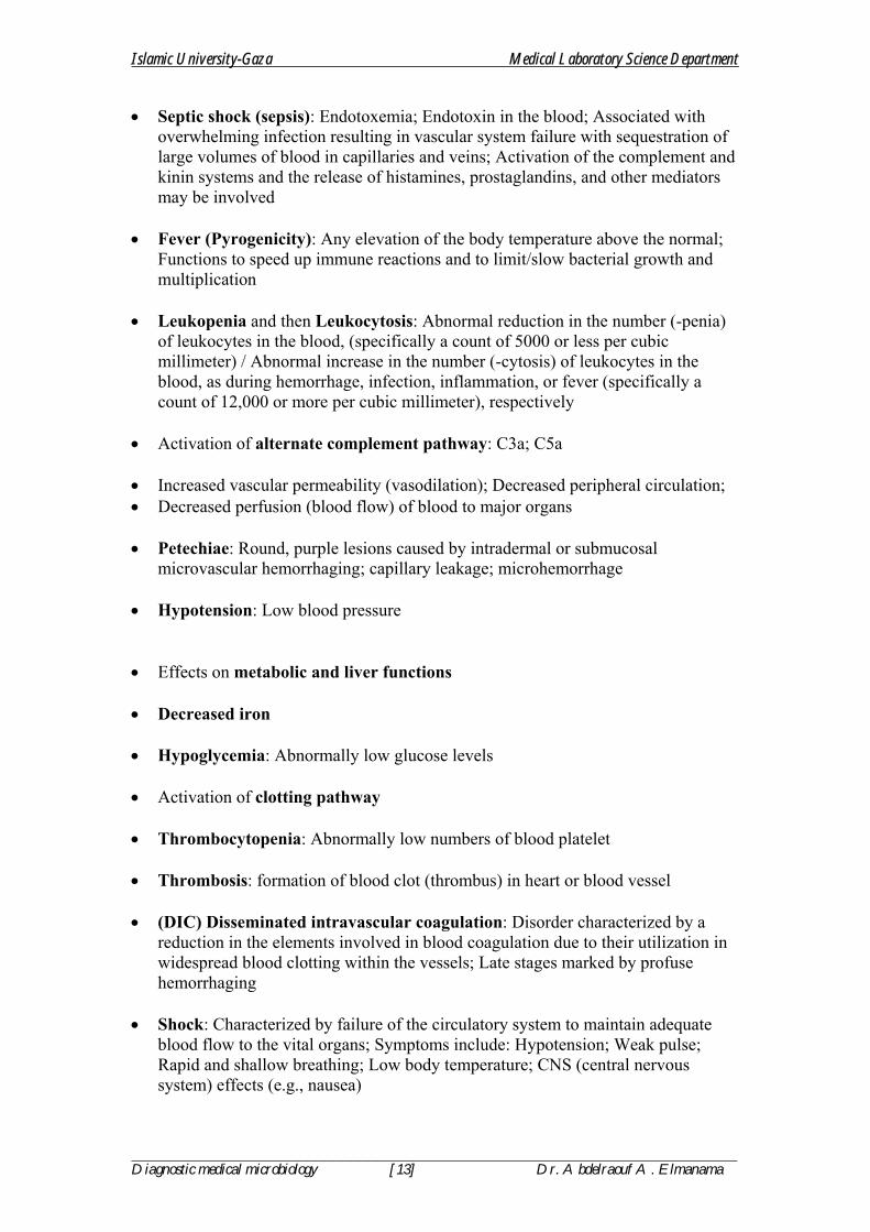
• Septic shock (sepsis): Endotoxemia; Endotoxin in the blood; Associated with overwhelming infection resulting in vascular system failure with sequestration of large volumes of blood in capillaries and veins; Activation of the complement and kinin systems and the release of histamines, prostaglandins, and other mediators may be involved
• Fever (Pyrogenicity): Any elevation of the body temperature above the normal;
Functions to speed up immune reactions and to limit/slow bacterial growth and multiplication
• Leukopenia and then Leukocytosis: Abnormal reduction in the number (-penia)
of leukocytes in the blood, (specifically a count of 5000 or less per cubic millimeter) / Abnormal increase in the number (-cytosis) of leukocytes in the blood, as during hemorrhage, infection, inflammation, or fever (specifically a count of 12,000 or more per cubic millimeter), respectively
• Activation of alternate complement pathway: C3a; C5a • Increased vascular permeability (vasodilation); Decreased peripheral circulation; • Decreased perfusion (blood flow) of blood to major organs • Petechiae: Round, purple lesions caused by intradermal or submucosal
microvascular hemorrhaging; capillary leakage; microhemorrhage • Hypotension: Low blood pressure • Effects on metabolic and liver functions • Decreased iron • Hypoglycemia: Abnormally low glucose levels • Activation of clotting pathway • Thrombocytopenia: Abnormally low numbers of blood platelet • Thrombosis: formation of blood clot (thrombus) in heart or blood vessel • (DIC) Disseminated intravascular coagulation: Disorder characterized by a
reduction in the elements involved in blood coagulation due to their utilization in widespread blood clotting within the vessels; Late stages marked by profuse hemorrhaging
• Shock: Characterized by failure of the circulatory system to maintain adequate
blood flow to the vital organs; Symptoms include: Hypotension; Weak pulse; Rapid and shallow breathing; Low body temperature; CNS (central nervous system) effects (e.g., nausea)
Islamic University-Gaza Medical Laboratory Science Department
_____________________________________________________________________ Diagnostic medical microbiology [14] Dr. Abdelraouf A. Elmanama
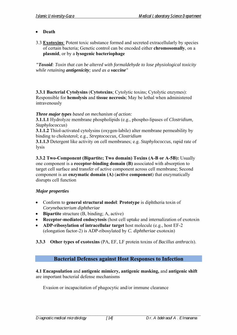
• Death
3.3 Exotoxins: Potent toxic substance formed and secreted extracellularly by species of certain bacteria; Genetic control can be encoded either chromosomally, on a plasmid, or by a lysogenic bacteriophage
"Toxoid: Toxin that can be altered with formaldehyde to lose physiological toxicity while retaining antigenicity; used as a vaccine" 3.3.1 Bacterial Cytolysins (Cytotoxins; Cytolytic toxins; Cytolytic enzymes): Responsible for hemolysis and tissue necrosis; May be lethal when administered intravenously Three major types based on mechanism of action: 3.1.1.1 Hydrolyze membrane phospholipids (e.g., phospho-lipases of Clostridium, Staphylococcus) 3.1.1.2 Thiol-activated cytolysins (oxygen-labile) alter membrane permeability by binding to cholesterol; e.g., Streptococcus, Clostridium 3.1.1.3 Detergent like activity on cell membranes; e.g. Staphylococcus, rapid rate of lysis
3.3.2 Two-Component (Bipartite; Two domain) Toxins (A-B or A-5B): Usually one component is a receptor-binding domain (B) associated with absorption to target cell surface and transfer of active component across cell membrane; Second component is an enzymatic domain (A) (active component) that enzymatically disrupts cell function Major properties • Conform to general structural model: Prototype is diphtheria toxin of
Corynebacterium diphtheriae • Bipartite structure (B, binding; A, active) • Receptor-mediated endocytosis (host cell uptake and internalization of exotoxin • ADP-ribosylation of intracellular target host molecule (e.g., host EF-2
(elongation factor-2) is ADP-ribosylated by C. diphtheriae exotoxin) 3.3.3 Other types of exotoxins (PA, EF, LF protein toxins of Bacillus anthracis).
Bacterial Defenses against Host Responses to Infection
4.1 Encapsulation and antigenic mimicry, antigenic masking, and antigenic shift are important bacterial defense mechanisms Evasion or incapacitation of phagocytic and/or immune clearance
Islamic University-Gaza Medical Laboratory Science Department
_____________________________________________________________________ Diagnostic medical microbiology [15] Dr. Abdelraouf A. Elmanama
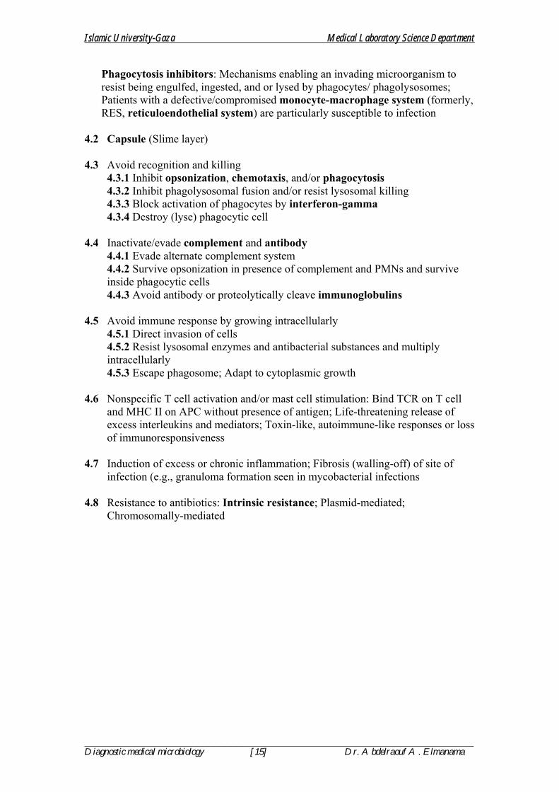
Phagocytosis inhibitors: Mechanisms enabling an invading microorganism to resist being engulfed, ingested, and or lysed by phagocytes/ phagolysosomes; Patients with a defective/compromised monocyte-macrophage system (formerly, RES, reticuloendothelial system) are particularly susceptible to infection
4.2 Capsule (Slime layer) 4.3 Avoid recognition and killing
4.3.1 Inhibit opsonization, chemotaxis, and/or phagocytosis 4.3.2 Inhibit phagolysosomal fusion and/or resist lysosomal killing 4.3.3 Block activation of phagocytes by interferon-gamma 4.3.4 Destroy (lyse) phagocytic cell
4.4 Inactivate/evade complement and antibody
4.4.1 Evade alternate complement system 4.4.2 Survive opsonization in presence of complement and PMNs and survive inside phagocytic cells 4.4.3 Avoid antibody or proteolytically cleave immunoglobulins
4.5 Avoid immune response by growing intracellularly
4.5.1 Direct invasion of cells 4.5.2 Resist lysosomal enzymes and antibacterial substances and multiply intracellularly 4.5.3 Escape phagosome; Adapt to cytoplasmic growth
4.6 Nonspecific T cell activation and/or mast cell stimulation: Bind TCR on T cell
and MHC II on APC without presence of antigen; Life-threatening release of excess interleukins and mediators; Toxin-like, autoimmune-like responses or loss of immunoresponsiveness
4.7 Induction of excess or chronic inflammation; Fibrosis (walling-off) of site of
infection (e.g., granuloma formation seen in mycobacterial infections 4.8 Resistance to antibiotics: Intrinsic resistance; Plasmid-mediated;
Chromosomally-mediated
Islamic University-Gaza Medical Laboratory Science Department
_____________________________________________________________________ Diagnostic medical microbiology [16] Dr. Abdelraouf A. Elmanama
Islamic University-Gaza Medical Laboratory Science Department
_____________________________________________________________________ Diagnostic medical microbiology [17] Dr. Abdelraouf A. Elmanama
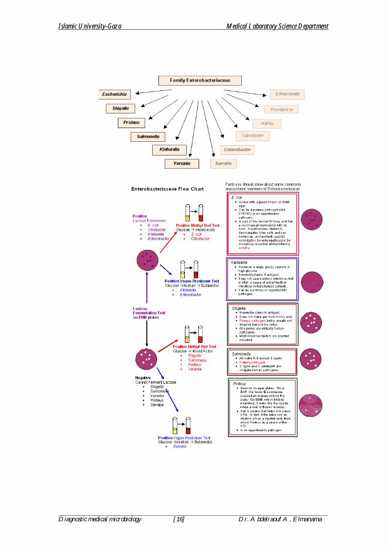
The Gram Negative Bacilli Family Enterobacteriaceae
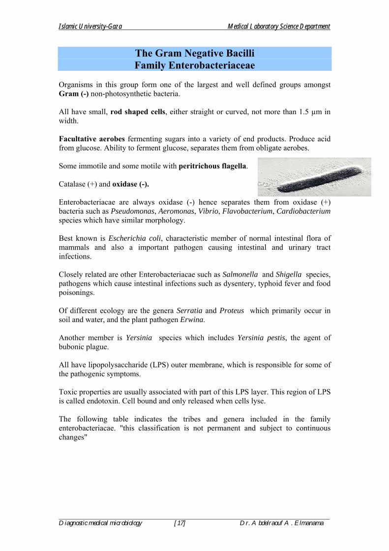
Organisms in this group form one of the largest and well defined groups amongst Gram (-) non-photosynthetic bacteria. All have small, rod shaped cells, either straight or curved, not more than 1.5 µm in width. Facultative aerobes fermenting sugars into a variety of end products. Produce acid from glucose. Ability to ferment glucose, separates them from obligate aerobes. Some immotile and some motile with peritrichous flagella. Catalase (+) and oxidase (-). Enterobacteriacae are always oxidase (-) hence separates them from oxidase (+) bacteria such as Pseudomonas, Aeromonas, Vibrio, Flavobacterium, Cardiobacterium species which have similar morphology. Best known is Escherichia coli, characteristic member of normal intestinal flora of mammals and also a important pathogen causing intestinal and urinary tract infections. Closely related are other Enterobacteriacae such as Salmonella and Shigella species, pathogens which cause intestinal infections such as dysentery, typhoid fever and food poisonings. Of different ecology are the genera Serratia and Proteus which primarily occur in soil and water, and the plant pathogen Erwina. Another member is Yersinia species which includes Yersinia pestis, the agent of bubonic plague. All have lipopolysaccharide (LPS) outer membrane, which is responsible for some of the pathogenic symptoms. Toxic properties are usually associated with part of this LPS layer. This region of LPS is called endotoxin. Cell bound and only released when cells lyse. The following table indicates the tribes and genera included in the family enterobacteriacae. "this classification is not permanent and subject to continuous changes"

Islamic University-Gaza Medical Laboratory Science Department
_____________________________________________________________________ Diagnostic medical microbiology [18] Dr. Abdelraouf A. Elmanama
Table 2: Family enterobacteriacae Tribes Genera
1. Escherichiaceae 1. Escherichia 2. Edwardsiella 3. Citrobacter 4. Salmonella 5. Shigella
2. Klebsiellae 6. Klebsiella 7. Enterobacter 8. Hafnia 9. Serratia
3. Proteae 10. Proteus 11. Providencia 12. Morganella
4. Yersiniae 13. Yersinia 5. Erwiniae 14. Erwina
This group is further subdivided by lactose fermentation into lactose fermenter and non lactose fermenters. This could be easily done by growing the bacterium on a differential medium "e.g., MacConkey agar plates" wherein lactose fermenting enterobacteriacae produces acid that will change the color of the medium into red while non-lactose fermenters will remain colorless. Some may show delayed lactose fermentation "e.g., after 48 to 72 hours of incubation". Table 3: Classification of enterobacteriacae according to their ability to ferment lactose Lactose fermenters (LF)
Non-lactose fermenter (NLF)
Delayed lactose fermenter (DLF)
1. Escherchia 1. Salmonella 1. Morganella 2. Enterobacter 2. Shigella 2. Providencia 3. Klebsiella 3. Proteus 3. Serratia 4. Citrobacter 4. Yersinia 4. Edwardsiella 5. Erwinia 6. Hafnia
ANTIGENIC PROPERTIES OF ENTEROBACTERIACAE
This group of organisms has a complex antigenic structure, and strains are divergent in their serologic behavior. 1. O Antigen: Somatic antigen. Heat stable antigen. O antigens are lipopolysaccharides and are found in the cell wall of most of gram-negative bacilli. With sera containing anti-O antibodies, such antigens agglutinate slowly in granular masses. Antibodies to O antigens are predominantly IgM.

Islamic University-Gaza Medical Laboratory Science Department
_____________________________________________________________________ Diagnostic medical microbiology [19] Dr. Abdelraouf A. Elmanama
2. H Antigen: Flagellar Antigen: This antigen is heat labile and could be inactivated by heating over 60 oC. With sera containing anti-H antibodies, such antigens agglutinate rapidly. Within a single Salmonella species, flagellar antigens may occur in either or both of 2 forms, called phase I and phase II. The organism tends to change from one phase to other; this is called phase variation. Antibodies to H antigen are predominantly IgG. 3. The “Vi” Antigen: Capsular (K) antigens that are present at the extreme periphery of the bacteria. Often interfere with agglutination of freshly isolated strains by antisera containing mainly anti-O agglutinins. Vi antigens are destroyed by heating for 1 hour at 60 oC. Example of antigenic designation of Salmonella: Salmonella typhi O 1,2,(Vi): a: 1 = 1,2 are O antigens = (Vi) if present = a phase one H antigen. = 1 phase two H antigen.
Enterobacteriaceae have the following distinctive characteristics: 1. Gram negative bacilli 2. Facultative anaerobes (grow with or without oxygen). 3. Glucose fermenters. 4. Oxidase negative 5. Nitrate positive

Islamic University-Gaza Medical Laboratory Science Department
_____________________________________________________________________ Diagnostic medical microbiology [20] Dr. Abdelraouf A. Elmanama
Pathogenic Determinants
1. Endotoxin: It is a lipopolysaccharide in structure and is derived from bacterial cell wall during lysis which may produce the following conditions:
A. Fever B. Lethal shock C. Diarrhea D. Abortion
2. Colicins: Bacteriostacins with antibiotic like substance produced by certain strains of E. coli and other related members resulting in the death and lysis of other sensitive cells releasing the endotoxin.
The Cultivation, Isolation and Differentiation of Members of Enterobacteriacae
Media of Choice:
1. Eosin Methylene Blue (EMB) 2. MacConkey Agar (the most commonly used media) 3. Hektoen enteric 4. Endo agar 5. Kligler`s Iron Agar (KIA) For initial differentiation 6. Triple Sugar Iron Agar (TSIA)
A primary differentiation is obtained by transferring colorless colonies to TSIA slants. Degradation of sugar and accompanying acid production are detected by pH indicator, phenol red which changes its color from red orange to yellow. Thiosulfate is reduced by H2S which reacts with iron salt to give black iron sulfide.
ESCHERICHIAE
Escherichia coli is a bacterium, which inhabits the intestinal tract of humans and other warm-blooded mammals. It constitutes approximately 0.1% of the total bacteria in the adult intestinal tract. Its name comes from the name of the person, Escherich, who in 1885 first isolated and characterized the bacteria.
Species:
Escherichia coli = Most frequently isolated E. fergusonii E. hermannii E. vulneris
Islamic University-Gaza Medical Laboratory Science Department
_____________________________________________________________________ Diagnostic medical microbiology [21] Dr. Abdelraouf A. Elmanama
E. coli is a normal inhabitant of the intestinal tract and may cause a wide variety of diseases. Infants diarrhea, meningitis, wound infection, urinary tract infection......etc. The presence of this organism in water is used as an indicator of recent fecal contamination because this organism does not survive in water for long. Strains of E.coli which are capable of causing disease, possess one or more virulence factor:
1. Enterotoxigenic E. coli (ETEC): Produces a heat-labile toxin (LT) an a heat-stable toxin (ST): LT action is identical to cholera toxin. LT causes diarrheae by stimulating the activity of a membrane-bound adenylate cyclase, ATP is converted into cAMP. cAMP induces the active secretion of Cl- and inhibit the absorption of NaCl, creating an electrolyte imbalance across the intestinal mucosa, resulting in the loss of large amounts of fluid and electrolytes from the intestine.
2. Enteropathogenic E.coli (EPEC): Produces diarrhea by several poorly understood mechanisms. One mechanism is the production of adhesion factor which causes the adherence to the cells of the small intestine.
3. Enterohaemorrhagic E.coli (EHEC): Produces toxin similar to Shigella dysenteria (Shiga-like toxin SLT-1 and SLT-2). The toxin inhibits protein synthesis in the affected cells. The disease is known as hemolytic colitis.
Examples of serological designations of E. coli
• E. coli O111a: H2, • E. coli O111b: H2 • E. coli O6: • E. coli O157: H7
PATHOGENICITY: Cystitis, meningitis; non-pathogenic when found in the alimentary tract of man. Epidemic diarrhea of the newborn.
NOTE: It is now widely accepted that certain serological types of E. coli are responsible for outbreaks of infantile diarrhea in nurseries.
1. Antigenic structure: O-antigen, H-antigen and K antigen
K antigen: Capsule antigen that enables the organism to resist killing by both human neutrophils and normal serum In-Vitro assays. Because K antigens is frequently found in isolates from patients with bacteriemia and neonatal meningitis, some authors feel that it plays an important role in the dissemination of organisms from primary infection sites. 2. Fimbria: Used for attachment 3. Enterotoxin (See Enterotoxigenic E. coli) 4. Verotoxin: Termed as such because of their irreversible cytotoxic effect on Vero tissue culture cells, a cell line developed from African green monkey kidney cells. Verotoxin producing E. coli (VTEC) is associated with diarrhea, hemorrhagic colitis and hemolytic-uremic-syndrome (HUS).

Islamic University-Gaza Medical Laboratory Science Department
_____________________________________________________________________ Diagnostic medical microbiology [22] Dr. Abdelraouf A. Elmanama
DIAGNOSIS:
• Stained smears reveal gram negative rods • Triple Sugar Iron Agar A/AG • IMVIC reaction + + - - • On EMB : Green metallic sheen • Lysine + • Acetate + • Lactose +
SEROLOGY: Since not all strains are enteropathogenic, agglutination with specific antisera must be performed before issuing a report of isolating EPEC.
TREATMENT: Antibiotic sensitivity testing must be performed. Aminoglycosides are effective most especially Amikacin. Treating E. coli infections with antibiotics may actually place the patient in severe shock which could possibly lead to death. This is because more of the bacterium's toxin is released when the cell dies.
ENTEROBACTER
Although this bacterium is part of the normal flora of the human intestinal tract, several species cause opportunistic infections of the urinary tract as well as other parts of the body. E. aerogenes and E. cloacae are two such pathogens that do not cause diarrhea, but that are sometimes associated with urinary tract and respiratory tract infections.
SPECIES:
1. Enterobnacter aerogenes (Type species) 2. E. cloacae 3. E. liquefaciens 4. E. sakazakii (cause of diarrhea; associated with infant formula)
GENERAL PROPERTIES:
• Formerly known as aerobacter • Often confused with Klebsiella • Motile and ornithine decaroboxylase (ODC) positive . These tests are used to
differentiate it from the similar genus of Klebsiella.
IDENTIFICATION: A. Morphology: Short and plump rods occurring singly in pairs or in short chains; non-spore forming, motile with a peritrichous flagella.
Islamic University-Gaza Medical Laboratory Science Department
_____________________________________________________________________ Diagnostic medical microbiology [23] Dr. Abdelraouf A. Elmanama
B. Culture Characteristics: Culture medium: EMB, ENDO, Mac.: On EMB, E. aerogenes appear as pink, while on Mac., colonies appear as pink usually not surrounded by lines of precipitated bile. C. Biochemical characteristics:
• The organism is facultative anaerobe, reduces nitrates to nitrite • Does not produces indole from tryptophan • Produce acetyle methyl carbinol (Voges-Proskauer) and no H2S • Utilize Citrate as the source of carbon • IMVIC reaction - - + + • Produces A/AG (Yellow slant over yellow butt with gas). • Positive Motility on a semi-solid medium. • Lysine + (except E. cloacae) • Ornithine +
KLEBSIELLA
The most clinically important speciies of this genus is Klebsiella pneumoniae. K. pneumoniae infections are common in hospitals where they cause pneumonia (characterized by emission of bloody sputum) and urinary tract infections in catheterized patients. In fact, K. pneumoniae is second only to E. coli as a urinary tract pathogen. Klebsiella infections are encountered far more often now than in the past. This is probably due to the bacterium's antibiotic resistance properties. Klebsiella species may contain resistance plasmids(R-plasmids) which confer resistance to such antibiotics as ampicillin and carbenicillin. To make matters worse, the R-plasmids can be transferred to other enteric bacteria not necessarily of the same species
SPECIES:
1. Klebsiella pneumonia (Type species) 2. Klebsiella oxytoca 3. Klebsiella ornithinolytica
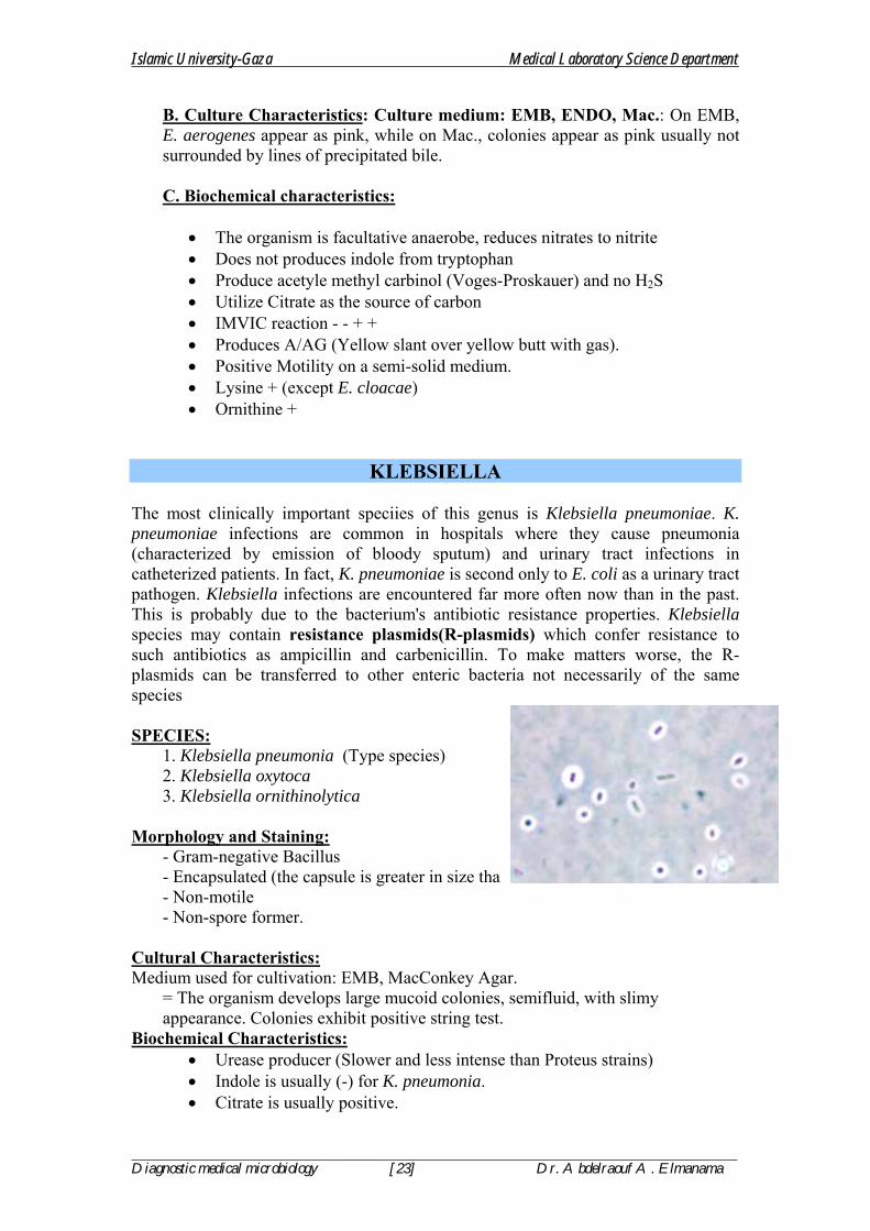
Morphology and Staining: - Gram-negative Bacillus - Encapsulated (the capsule is greater in size than the cell itself). - Non-motile - Non-spore former.
Cultural Characteristics: Medium used for cultivation: EMB, MacConkey Agar.
= The organism develops large mucoid colonies, semifluid, with slimy appearance. Colonies exhibit positive string test.
Biochemical Characteristics: • Urease producer (Slower and less intense than Proteus strains) • Indole is usually (-) for K. pneumonia. • Citrate is usually positive.

Islamic University-Gaza Medical Laboratory Science Department
_____________________________________________________________________ Diagnostic medical microbiology [24] Dr. Abdelraouf A. Elmanama
• TSIA A/AG • IMVIC reaction - - + + • Does not liquefies Gelatin. • Lysine + • Non-motile • Ornithine -
Antigenic Structure:
= Klebsiella species possess O and K antigens = The capsule enables the organism to resist phagocytosis. = Endotoxin and enterotoxin are also produced.
Pathogenicity: Primary community-acquired pneumonia, nosocomial pneumonia, urinary tract and wound infection, bacteremia and meningitis.
Differentiation From Enterobacter aerogenes
Reaction K. pneumonia E. aerogenes IMVIC - - + + - - + + Motility Non-motile Motile String Test Positive Negative Gelatine liquefaction Positive Negative Ornithine decaroboxylase Negative Positive
CITROBACTER
Citrobacter is not considered to be an enteric pathogen because it is normal gut flora. When plated, Citrobacter colonies bare a strong resemblance to E. coli colonies. This group of bacteria is of small clinical interest. C. freundii is suspected to cause diarrhea and possibly extra-intestinal infections. C. diversus has been linked to a few cases of meningitis in newborns SPECIES:
1. Citrobacter freundii 2. Citrobacter diversus
Characteristics:
• Morphologically appear similar to Escherichia coli. • Biochemically resembles Salmonella. • TSI: A/A+ • IMVIC - + - + • Urease ± • Lysine – • Hydrogen sulfide + (C. freundii) • Citrate +

Islamic University-Gaza Medical Laboratory Science Department
_____________________________________________________________________ Diagnostic medical microbiology [25] Dr. Abdelraouf A. Elmanama
SERRATIA
Members of the Serratia genus were once known as harmless organisms that produced a characteristic red pigment. Today, Serratia marcescens is considered a harmful human pathogen which has been known to cause urinary tract infections, wound infections, pneumonia and diarrhea. Serratia bacteria also have many antibiotic resistance properties which may become important if the incidence of Serratia infections dramatically increases. Serratia can be distinguished from other genera belonging to Enterobacteriaceae by its production of three special enzymes: DNase, lipase, and gelatinase.
SPECIES:
1. S. marcescens 2. S. rubiddea 3. S. liquefaciens
Biochemical Characteristics: • Non lactose fermenter (may show delayed lactose fermentation) • Citrate: positive. • V-P: positive • ODC: positive • Lysine: positive • Indole: Negative • TSI A/A: (NO gas) • DNase: positive
Specimen: Sputum, urine, and stool
Culture: A red pigment is produced by colonies of Serratia on routine laboratory media which is more clear when the organism is grown at 22 oC in the dark.
= Some strains are hemolytic.
PROTEUS GROUP
SPECIES: 1. Proteus mirabilis 2. Proteus penneri 3. Proteus vulgaris 4. Morganella morganii 5. Providencia alcalifaciens 6. Providencia stuartii 7. Providencia rettgeri

Islamic University-Gaza Medical Laboratory Science Department
_____________________________________________________________________ Diagnostic medical microbiology [26] Dr. Abdelraouf A. Elmanama
MORPHOLOGY: - Gram negative Bacilli - Actively motile - No capsule - No spores
CULTIVATION: On EMB, Endo and MacConkey Agar The colonies usually exhibit swarming. Non-lactose fermenting (Colorless).
Pathogenicity: Proteus, like almost every other bacterium in this family, can cause urinary tract infections and hospital-acquired infections. Proteus is unique, however, because it is highly motile and does not form regular colonies. Instead, Proteus forms what are known as "swarming colonies" when plated on non-inhibitory media. The most important member of this genus is considered to be P. mirabilis, a cause of wound and urinary tract infections. Fortunately, most strains of P. mirabilis are sensitive to ampicillin and cephalosporin. Unlike its relative, P. vulgaris is not sensitive to these antibiotics. However, this organism is isolated less often in the laboratory and usually only targets immunosuppressed individuals. P. mirabilis and P. vulgaris can be differentiated by an indole test for which only P. vulgaris tests positive.
Diagnosis:
• Smear: Gram-negative bacilli • TSIA K/AG+ • IMVIC V + - - • Swarming and “Gun-powder-like odor” • Strong urease positive. • Phenylalanine Deaminase test = Positive • Lysine – • Hydrogen sulfide + • Motile
NOTE: Proteus strains are used in the Weil-Felix test for Rickettsial diseases: 1. Proteus OX K 2. Proteus OX 19 3. Proteus OX 2 Weil and Felix isolated strains of Proteus vulgaris from patients with typhus fever and found that the sera of the patients agglutinated particular Proteus strains designated OX19 and OX2. The Proteus organism is not the cause of Typhus fever. The cross agglutination seems to be caused by the presence of an alkali-stable polysaccharide which is also present in R. prowazeki. The agglutination of these particular strains by sera of patients with Rickettsial infection is known as the “Weil-Felix reaction” .
Therefore Proteus does not cause Typhus fever nor acts as a secondary invaders .

Islamic University-Gaza Medical Laboratory Science Department
_____________________________________________________________________ Diagnostic medical microbiology [27] Dr. Abdelraouf A. Elmanama
Morganella and Providencia
Both are closely related to the Proteus group because both elaborate urease and produce indole. They however, don`t produce hydrogen sulfide.
Morganella morganii
Morganella morganii is the only important species of this genus. It can cause urinary tract and wound infections, as well as diarrhea. Chloramphenicol is a good choice for treating Morganella infections.
• Ornithine + • TSIA: A/A • IMVIC reaction : + + - -
Providencia alcalifaciens
Although rare, Providencia species have been associated with nosocomial (hospital acquired) urinary tract infections. One species, P. alcalifaciens, has been associated with some cases of diarrhea in children. Since infection is so rare, other genera of Enterobacteriaceae should be considered before Providencia as being causative.
• Hydrogen sulfide - • Lysine - • Lactose - • TSIA: K/A • IMVIC reaction: + + - +
ERWINIA
Not medically important since they are only pathogenic to plants.

Islamic University-Gaza Medical Laboratory Science Department
_____________________________________________________________________ Diagnostic medical microbiology [28] Dr. Abdelraouf A. Elmanama
SALMONELLA SPECIES OF MEDICAL IMPORTANCE: 1. S. typhi (The typhoid bacilli) 2. S. paratyphi A (Produce paratyphoid fever) 3. S. paratyphi B (Produce paratyphoid fever) 4. S. paratyphi C (Bacteremia without intestinal involvement) 5. S. typhimurium (Food poisoning) 6. S. enteritidis (Food infection) 7. Salmonella cholerasuis (Hog cholera bacillus) 8. Salmonella pullorum (White diarrhea in children) 9. Salmonella gallinarum (Fowl typhoid bacillus) MORPHOLOGY AND STAINING: • Short rods (bacilli) (indistinguishable from other Enterobacteriacae) • Motile with peritrichous flagella except S. pullorum and S. gallinarum. • Non-encapsulated • Gram-negative HABITAT AND TRANSMISSION: Most Salmonella species are found in the intestine of animals especially pigs, cows, goats, sheep, rodents, hens, ducks and other poultry. S. typhi and S. paratyphi, however ,are usually found only in human., both of which are excreted in the feces and urine of infected patients. Infection occurs via ingesting contaminated food or drinks. PATHOGENICITY: Salmonella can cause any one of three types of Salmonellosis: 1. Acute gastroenteritis or food infection type. 2. Septicemia or acute sepsis similar to pyogenic infection. 3. Enteric fevers: 1. Typhoid fever 2. Paratyphoid fever. Typhoid fever is an acute infectious disease characterized by continuous fever, skin eruptions, bowel disturbances and profound toxemia. Following entrance of the typhoid bacilli into the human body through the mouth there is always an incubation period of 7 to 14 days, before symptoms appear. During this time, the organism penetrates the wall of the upper intestine and causes and inflammation. Then reaches the blood via the lymphatic system were they circulate and may be localized in many internal organs especially in spleen, bone marrow, and gallbladder. Serious complication of typhoid fever may be produced as a result of multiplication of the bacilli in organs other than the intestine.

Islamic University-Gaza Medical Laboratory Science Department
_____________________________________________________________________ Diagnostic medical microbiology [29] Dr. Abdelraouf A. Elmanama
The organism begin to disappear from the blood during the first week of the illness, and especially after the second week. The disappearance of the organism from the blood is clearly associated with the development of specific antibodies. NEPHROTYPHOID: This condition is an immune complex disorder of the kidneys and is characterized by fever, edema, marked albuminuria and hematuria. OSTEOMYELITIS : Especially in children with sickle cell disease and thalassamia. Typhoid in children can be found in the bone marrow. Inflammation of the joints (Typhoid arthritis) may also occur. ABSCESSES: of the spleen and elsewhere. MENINGITIS: And rarely pneumonia and endocarditis.
DETERMINANTS OF PATHOGENICITY: 1. Surface antigens: The ability of Salmonella to attach to host receptor cells and to survive intracellularly may be due to the O antigenic side chain or in case of typhi serotypes, the presence of Vi antigen. Organisms containing Vi antigen are clearly more virulent than those lacking this antigen. It may function as a capsule and prevent phagocytosis or intracellular destruction of the organism. 2. Invasiveness: Virulent Salmonella penetrate the epithelial lining of the small bowel, thus the brush border begins to degenerate. After penetration the organism multiply and may spread to other body sites. 3. Endotoxins: Presumably, endotoxin is responsible for the fever production. Endotoxin activation of the chemotactic properties of the complement system may cause the localization of leukocytes in the classic lesions seen in typhoid fever. 4. Other factors: Enterotoxin: Affects the small intestine. Cytotoxin: Associated with outer bacterial membrane which may mean that the toxin may be important in cellular invasion and cellular destruction. CLASSIFICATION OF SALMONELLA: The Kauffman-White system used to classify Salmonella is based on identifying the O (somatic) and H (flagellar) antigens possessed by the different serovars. The detection of Vi antigen is also used in the identification of S. typhi and some other Salmonella. 1. O Antigen: Salmonella are grouped by their O antigens as groups A to Z, 51-61 and 64-66. Many of the medically important salmonella belongs to groups A to G. Each group has what is called a group factor. This is an O antigen, common to all members of the groups. For example, the factor for group B is O antigen is 4. This means that all the salmonella belonging to this group possess antigen 4 as one of their O antigen.
Islamic University-Gaza Medical Laboratory Science Department
_____________________________________________________________________ Diagnostic medical microbiology [30] Dr. Abdelraouf A. Elmanama
2. H Antigen: Many Salmonella are diphasic, that is, they can occur in two antigenic forms referred as phase I and Phase II. Phase I antigens are given alphabetical letters and phase II antigens either numbered or given a letter if known to occur in both phases. Phase I antigens are more specific and therefore, an organism can be identified if it is in phase I. 3. Vi Antigen: This surface (K) antigen can be found in S. typhi and S. paratyphi C and few other Salmonella. It is associated with virulence and can be detected using Vi antiserum. SALMONELLA O H I H II Group A: S. paratyphi A 1, 2*,12 a - Group B: S. paratyphi B 1,4*,5,12 b 1,2 S. derby 1,4*,5,12 f,g 1,2 S. typhimrium 1,4*,5,12 i 1,2 S. heidlberg 1,4*,5,(12) r 1,2 Group C: S. cholerae-Suis *6, 7 c 1,5 S. paratyphi C 6*, 7 (Vi) c 1,5 LABORATORY DIAGNOSIS: 1. SPECIMEN: For the diagnosis of enteric fever, specimens include, blood, feces and urine for culture may be used depending on the course of illness. A. Blood: Organisms can usually be detected in 75-90% of patients during the first 10 days of infection and in about 30% of patients during the 3rd. week. B. Feces: Organisms can usually be isolated from 40-50% of patients during the third week. C. Urine: Organism, can usually be detected from about 25% of patients after the second week of infection. D. Serum: Is used for the detection of serum antibodies (Widal test).
GROUPING AND SEROTYPING OF SALMONELLA
Due to the high cost of antisera, it will not be possible for most laboratories to stock wide range of Salmonella antisera, however, laboratories try to stock a salmonella polyvalent O antiserum which covers the locally important group and also specific O, H, and Vi antisera to identify S. typhi. The following antisera are required to identify S. typhi 1. Salmonella antiserum Factor 9 (Group D) 2. Salmonella H antiserum d 3. Salmonella Vi antiserum. If testing for paratyphoid the following antigen suspensions are required: 1. S. paratyphi A O1, 2, 12 2. S. paratyphi A H,2 3. S. paratyphi B O1,4,5,12 4. S. paratyphi H,b, phase I 5. S. paratyphi C O6,7 6. S. paratyphi C H, c, phase I

Islamic University-Gaza Medical Laboratory Science Department
_____________________________________________________________________ Diagnostic medical microbiology [31] Dr. Abdelraouf A. Elmanama
2. ISOLATION AND IDENTIFICATION: A. ENRICHMENT & SELECTIVE MEDIA: Various enrichment and selective media are used to isolate Salmonella from stool and other specimens. The use of Selinite-F and XLD, DCA and SSA. Salmonella produce non-lactose fermenting colonies on MacConkey medium. Most strains (especially food-poisoning serovars) show a blackening of the colonies due to H2S production. = Enrichment medium: Contains substances which have a growth stimulating of Salmonella and Shigella and inhibitory to the other contaminants. Ex. Selenite-F Broth Tetrathionate broth. = Selective media: Solid media containing lactose as differential sugar; an indicator to produce color changes when the pH of the colony becomes acid as a result of lactose fermentation and an inhibitor for gram positive bacteria and most gram-negative bacteria other than Salmonella and Shigella. 1. Bismuth Sulfide Agar (BSA): Considered by many as the best medium for the isolation of Salmonella typhi. 2. Brilliant Green Agar (BGA): This medium is good for isolating Salmonella species other than S. typhi. 3. Salmonella Shigella Agar (SSA): Colorless colonies . 4. Desoxycholate Citrate Agar (DCA). 5. Xylose Lysine DextroseAgar (XLD). B. GROWTH CHARACTERISTICS 1. On MacConkey and SSA : Colorless colonies 2. BGA : Slightly pink colonies 3. BSA and XLD : Develops black colonies
C. BIOCHEMICAL CHARACTERISTICS
A. TSIA: Alkaline slant, Acid butt with small amount of H2S B. IMVIC reaction: - + - + C. Motility (+) D. Urease (-) D. SEROLOGICAL TESTING: 1. On clinical isolates: All clinical suspected Salmonella isolate must be subjected to serotyping by polyvalent and monovalent antisera. 2. On patients: Serological test (Widal test) must be performed after 10 days of infection . TREATMENT:
ENTEROCOLITIS • Rehydration • Antimicrobials not indicated
Islamic University-Gaza Medical Laboratory Science Department
_____________________________________________________________________ Diagnostic medical microbiology [32] Dr. Abdelraouf A. Elmanama
ENTERIC FEVER • Chloramphenicol was firstly used in 1948 for typhoid fever - Now resistant • Ampicillin, sulphamethoxazole-trimethoprim still used in developing
countries. • Ciprofloxacin – drug of choice • Rehydration if needed
CHRONIC CARRIER • Prolonged treatment with Ampicillin or Ciprofloxacin • Cholecystectomy may be needed
Prevention • Public health = personal hygiene measures • Proper sewage treatment • Chlorination of water supplies • Detection of carriers • Pasteurization of milk and proper cooking of food
Vaccination (50-80%) protection) – two vaccines: • Acetone-killed – given I/M • Live-attenuated - Oral
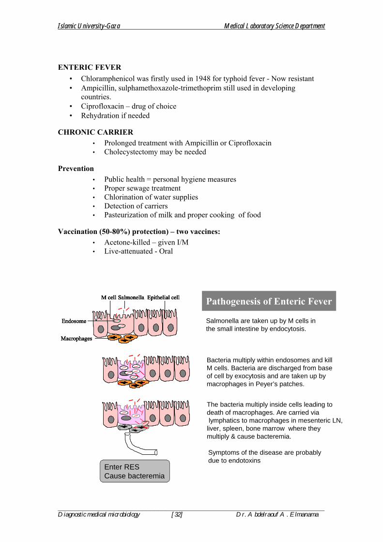
Salmonella are taken up by M cells in the small intestine by endocytosis.
Bacteria multiply within endosomes and kill M cells. Bacteria are discharged from base of cell by exocytosis and are taken up by macrophages in Peyer’s patches.
The bacteria multiply inside cells leading to death of macrophages. Are carried vialymphatics to macrophages in mesenteric LN,
liver, spleen, bone marrow where they multiply & cause bacteremia.
Symptoms of the disease are probably due to endotoxins
Pathogenesis of Enteric FeverEpithelial cellM cell Salmonella
Endosome
Macrophages
Epithelial cellM cell Salmonella
Endosome
Macrophages
Epithelial cellM cell Salmonella
Endosome
Macrophages
Epithelial cellM cell Salmonella
Endosome
Macrophages
Enter RES Cause bacteremia

Islamic University-Gaza Medical Laboratory Science Department
_____________________________________________________________________ Diagnostic medical microbiology [33] Dr. Abdelraouf A. Elmanama
Ingestion of Salmonellae
Multiplication inMesenteric LNMultiplication inMesenteric LN
PrimaryBacteremia
PrimaryBacteremia
SecondaryBacteremia(heavier)
SecondaryBacteremia(heavier)
Symptomsof diseasesSymptomsof diseases
Attachment & penetration
of ileum
Attachment & penetration
of ileum
Infection &multiplication in liver, G. bladder, spleen, kidney, bone marrow
Infection &multiplication in liver, G. bladder, spleen, kidney, bone marrow
Gall bladderGall bladder
Inflammation,Necrosis &
typhoid ulcersin ileum
Inflammation,Necrosis &
typhoid ulcersin ileum
PATHOGENESISOF
ENTERIC FEVER
SHIGELLAE
• The genus Shigella contains fewer species than the genus Salmonella and is antigenically less complex.
• Clinical dysentery may be caused by Shigella, Salmonella, Entamoeba histolytica, Proteus morganii, and viruses.
• Shigella dysenteria was isolated by Shiga in Japan in 1898. SPECIES: 1. Shigella dysenteriae 2. Shigella flexneri 3. Shigella sonnei 4. Shigella boydii
Islamic University-Gaza Medical Laboratory Science Department
_____________________________________________________________________ Diagnostic medical microbiology [34] Dr. Abdelraouf A. Elmanama
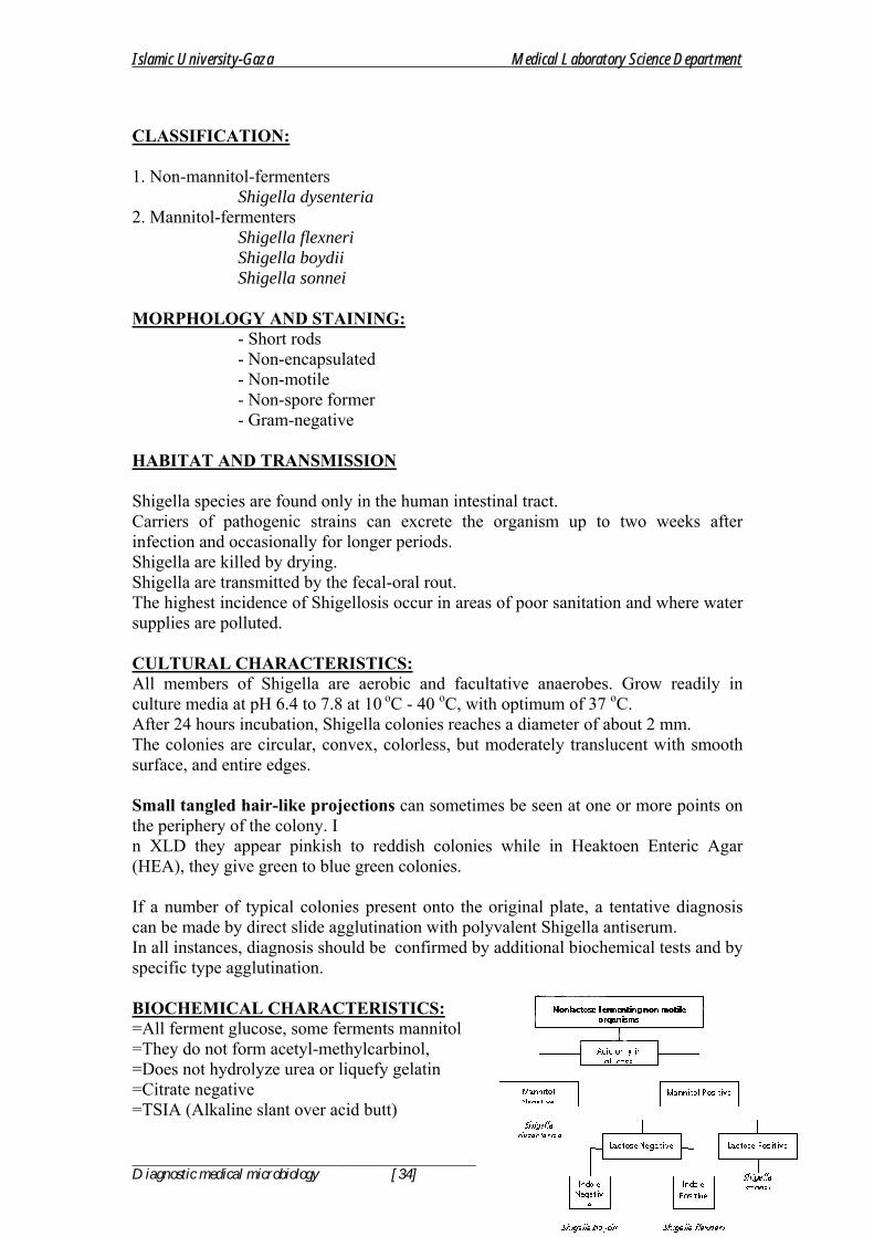
CLASSIFICATION: 1. Non-mannitol-fermenters Shigella dysenteria 2. Mannitol-fermenters Shigella flexneri Shigella boydii Shigella sonnei MORPHOLOGY AND STAINING: - Short rods - Non-encapsulated - Non-motile - Non-spore former - Gram-negative HABITAT AND TRANSMISSION Shigella species are found only in the human intestinal tract. Carriers of pathogenic strains can excrete the organism up to two weeks after infection and occasionally for longer periods. Shigella are killed by drying. Shigella are transmitted by the fecal-oral rout. The highest incidence of Shigellosis occur in areas of poor sanitation and where water supplies are polluted. CULTURAL CHARACTERISTICS: All members of Shigella are aerobic and facultative anaerobes. Grow readily in culture media at pH 6.4 to 7.8 at 10 oC - 40 oC, with optimum of 37 oC. After 24 hours incubation, Shigella colonies reaches a diameter of about 2 mm. The colonies are circular, convex, colorless, but moderately translucent with smooth surface, and entire edges. Small tangled hair-like projections can sometimes be seen at one or more points on the periphery of the colony. I n XLD they appear pinkish to reddish colonies while in Heaktoen Enteric Agar (HEA), they give green to blue green colonies. If a number of typical colonies present onto the original plate, a tentative diagnosis can be made by direct slide agglutination with polyvalent Shigella antiserum. In all instances, diagnosis should be confirmed by additional biochemical tests and by specific type agglutination. BIOCHEMICAL CHARACTERISTICS: =All ferment glucose, some ferments mannitol =They do not form acetyl-methylcarbinol, =Does not hydrolyze urea or liquefy gelatin =Citrate negative =TSIA (Alkaline slant over acid butt)
Islamic University-Gaza Medical Laboratory Science Department
_____________________________________________________________________ Diagnostic medical microbiology [35] Dr. Abdelraouf A. Elmanama
=IMVIC V + - - PATHOGENIC DETERMINANTS: 1. O antigen: The ability to survive the passage through the host defenses may be due to O antigen. 2. Invasiveness: Virulent shigella penetrate the mucosa and epithelial cells of the colon in an uneven manner.
Intracellular multiplication leads to invasion of adjacent cells, inflammation and cell death. Cell death is probably due to cytotoxic properties of shiga toxin that interfere with protein synthesis. The cellular death and resulting phagocytosis response by the host accounts for the bloody discharge of mucus and pus and shallow ulcers characteristic of the disease.
3. Other toxins: It has a protein toxin which may be neurotoxic, cytotoxic, and enterotoxic. The enterotoxic property is responsible for watery diarrhea. PATHOGENICITY: Shigella dysentery’s is set apart from other dysentery bacilli by its capacity to form a powerful exotoxin, it is associated with epidemics of bacillary dysentery. It is the only dysentery bacillus that is pathogenic to laboratory animals. In man, shigellosis begins with symptoms of acute gastro-enteritis which is accompanied by abdominal pain and diarrhea. As it progresses, diarrhea becomes more frequent and is usually accompanied colicky pain. Later diarrhea losses its fecal characteristic and is followed by mucus with pus and blood. The disease is usually accompanied by fever and marked prostration. It is also known that children are more frequently attacked than adult persons and the symptoms are more severe.

Islamic University-Gaza Medical Laboratory Science Department
_____________________________________________________________________ Diagnostic medical microbiology [36] Dr. Abdelraouf A. Elmanama
Shigella are taken up by M cells in the large intestine by endocytosis.
Bacteria are quickly released from endosomes and leaving shigella free in the cytoplasm. The bacteria multiply & enter the inferior and lateral aspects of the epithelial cells
Actin filaments quickly form a tail andpush the bacteria into next cell where
they multiply. Macrophages that take up shigella are killed and release organism.
Infected cells die and slough off. Acute inflammatory respose occurs with with bleeding and abscess formation.
Pathogenesis of ShigellaEpithelial cellM cell Shigella
Endosome
Macrophages
Epithelial cellM cell Shigella
Endosome
Macrophages
LABORATORY DIAGNOSIS: The only satisfactory method of laboratory diagnosis is to cultivate the bacilli from the patient. In the early stages of acute shigellosis, isolation of the causative organism from the feces is usually accomplished without difficulties by using the same special media and methods employed for salmonella. 1. Cultivation of the bacilli from stool specimen during the first 4-5 days of the disease. 2. Smears: Gram-negative bacilli appearing singly TSIA = Alkaline/acid (No gas no H2S) IMVIC reaction : V + - - 3. Serological examination with polyvalent and monovalent anti-sera. METHODS FOR THE IDENTIFICATION OF SALMONELLA AND SHIGELLA 1. Shigella are rarely encountered except in feces, Salmonella are also found in cultures of bile, blood, urine, abscesses and cerebrospinal fluid. 2. Microscopic examination of stained smears is of no use for the identification of the bacilli except the fact that it is a gram negative bacilli. 3. On general and selective media colonies are similar. On Blood agar they are smooth, gray and opaque. Most salmonella species produces H2S and therefore will be black on Bismuth Sulfite Agar. POINTS OF IDENTIFICATIONS: FEATURE SALMONELLA SHIGELLA Disease Typhoid fever Bacillary dysentery Motility Motile Non-motile H2S Positive Negative Indole formation Positive Negative
Islamic University-Gaza Medical Laboratory Science Department
_____________________________________________________________________ Diagnostic medical microbiology [37] Dr. Abdelraouf A. Elmanama
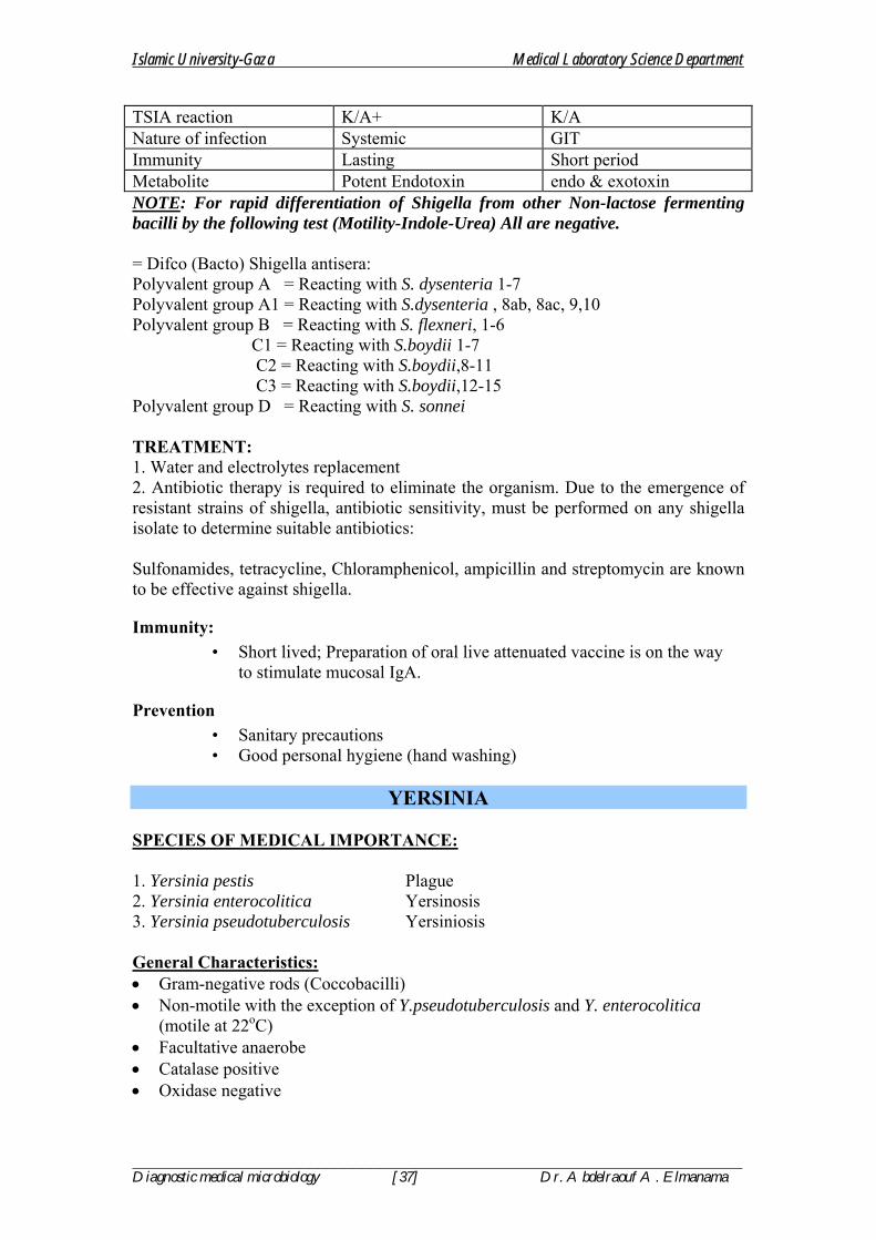
TSIA reaction K/A+ K/A Nature of infection Systemic GIT Immunity Lasting Short period Metabolite Potent Endotoxin endo & exotoxin NOTE: For rapid differentiation of Shigella from other Non-lactose fermenting bacilli by the following test (Motility-Indole-Urea) All are negative. = Difco (Bacto) Shigella antisera: Polyvalent group A = Reacting with S. dysenteria 1-7 Polyvalent group A1 = Reacting with S.dysenteria , 8ab, 8ac, 9,10 Polyvalent group B = Reacting with S. flexneri, 1-6 C1 = Reacting with S.boydii 1-7 C2 = Reacting with S.boydii,8-11 C3 = Reacting with S.boydii,12-15 Polyvalent group D = Reacting with S. sonnei TREATMENT: 1. Water and electrolytes replacement 2. Antibiotic therapy is required to eliminate the organism. Due to the emergence of resistant strains of shigella, antibiotic sensitivity, must be performed on any shigella isolate to determine suitable antibiotics: Sulfonamides, tetracycline, Chloramphenicol, ampicillin and streptomycin are known to be effective against shigella.
Immunity: • Short lived; Preparation of oral live attenuated vaccine is on the way
to stimulate mucosal IgA.
Prevention • Sanitary precautions • Good personal hygiene (hand washing)
YERSINIA
SPECIES OF MEDICAL IMPORTANCE: 1. Yersinia pestis Plague 2. Yersinia enterocolitica Yersinosis 3. Yersinia pseudotuberculosis Yersiniosis General Characteristics: • Gram-negative rods (Coccobacilli) • Non-motile with the exception of Y.pseudotuberculosis and Y. enterocolitica
(motile at 22oC) • Facultative anaerobe • Catalase positive • Oxidase negative

Islamic University-Gaza Medical Laboratory Science Department
_____________________________________________________________________ Diagnostic medical microbiology [38] Dr. Abdelraouf A. Elmanama
Yersinia pestis Clinical manifestation: Plague is primarily a disease or rodents that could be transmitted to man. Plague occurs in three forms: BUBONIC, SEPTICEMIC and PNEUMONIC. Bubonic plague is the common form resulting from the cutaneous inoculation of Y. pestis by the bite of an infective flea. The incubation period is 2-7 days after exposure. The disease is characterized by sudden fever, shaking chills, headache and pain in the area of involved lymph nodes. Untreated, bubonic plague can result in coma and death. Classic bubonic plague entails an inflammatory response in the regional lymph nodes which, when enlarged, is referred to as bubo. Untreated bubonic plague may become septicemic plague. Virtually, 100% of the untreated septicemic plague infections are followed by pulmonary involvement and death. At this stage human to human transmission may occur.
EPIZOOTICIN RATS
EPIDEMICBUBONICPLAGUE
HUMAN SPREAD
Rats
Infected rat flea
Infected rat flea
RatsRats
Infected rat flea
Infected rat flea
Humans
Wildrodent
Infected rat flea
Wildrodent
Infected rat flea
Humans
Wildrodent
Infected rat flea
Wildrodent
Infected rat flea
Wildrodent
Infected rat flea
Wildrodent
Infected rat flea
Humans
Lymph nodes(Bubonic plague)
Humans
Lymph nodes(Bubonic plague)
BacteremiaBacteremiaLungs
(Pneumonicplague)
Lungs(Pneumonic
plague)
Respiratorydroplets
Respiratorydroplets
Inhaled byother persons
Inhaled byother persons
URBAN CYCLE
SYLVATIC CYCLE
URBAN CYCLE
SYLVATIC CYCLE
Mode of Transmission
NB: Plague is an internationally reportable disease, and when suspected, must be reported to the local public health authorities. Collection of Specimen: An aspirate from buboes is a good specimen if handled properly using a sterile syringe and needle and aseptic technique in collecting. Special precautions must be taken to avoid the spread of infection. Face masks, gloves and gowns must be routinly worn when in contact with the patient. Blood drawn aseptically is also good specimen for isolation of Y.pestis is septicemic patients. Early morning sputum obtained from deep couph, avoiding contamination with saliva. The patient must be instructed to rinse the oral cavity three to four times. Biopsy from infected lymph nodes. Staining & Culture Media: = Smears are stained with WAYSON to be examined for bipolar stained rods.
Islamic University-Gaza Medical Laboratory Science Department
_____________________________________________________________________ Diagnostic medical microbiology [39] Dr. Abdelraouf A. Elmanama
= Routine media, selective for gram negative enteric bacteria are good for isolation. BHIB, Trypticase Soy Broth, Blood Agar and Nutrient Agar are also used. Identification by Biochemical Tests: = Yersinia pestis is differentiated from other Yersinia species by a negative urease negative test and carbohydrate fermentaion tests. Motility at 37oC and 22oC is also used. Serologic identification: 1. Identification of suspected Y. pestis isolate: Commercially avialable antiplague serum is used to agglutinate cells. 2. Patient serum: (Detection of antibodies): Formalinized suspension of known Y. pestis cells is used as antigen. One drop of diluted antigen is added to one drop of patient`s serum. This mixture is shaken gently for 15-30 minutes and examined for agglutination under low power magnification. Antimicrobial Sensitivity testing: Routine sensitivity testing is unreliable because In-Vitro results commonly differ significantly from In-Vivo efficacy. For example, penicillin may show slight to wide zone of inhibition of Y. pestis, although penicillin has no In-Vivo activity against this organism. Tetracycline, Streptomycin or Chloramphenicol and sulfonamides are effective in treatment of plague.
II Y. enterocolitica and Y. pseudotuberculosis Clinical manifestation: Both agents causes yersinosis which appears as enteric disorders, e.g., diarrhea, enteritis, terminal ileitis. Clinical symptoms can not be distinguished from those caused by other enteric pathogens such as salmonella and shigella. Faecal-oral rout is the common mode of transmission, although may be transmitted by bites of infective arthropods. Human to human transmission is well documented. Collection & processing of Specimen: = Stool, blood, excised mesenteric nodes, or appendices obtained during surgery. = Yersinia Selective Agar: is an excellent selective medium for the isolation of Y. enterocolitica from stool or sputum. = Cold Enrichment technique: Specimens that are grossly contaminated, are inoculated into buffered saline (pH 7.4-7.6) and stored at 4-7 oC. Both organisms tolerate and even grow at these temperatures, other organisms gradually dies. Biochemical Identification: 1. Y. enterocolitica = Growth at 4 oC and on NA/Mac. Agars = Motile at 22 oC. = Indole production= Variable = Urease positive = Ornithine decarboxylase Positive 2. Y. pseudotuberculosis = The same as Y.enterocolitica and could be differentiated from it by a sorbitol negative test and negative ornithine decarboxylase. Serology: = Specific antigens prepared from each organism is useful in detecting the presence of specific antibodies in patient`s serum. = Specific antibodies are used to agglutinate suspected cultures.

Islamic University-Gaza Medical Laboratory Science Department
_____________________________________________________________________ Diagnostic medical microbiology [40] Dr. Abdelraouf A. Elmanama
Treatment: Streptomycin, Chloramphenicol, Tetracyclines. Gram Negative Bacterial Reactions
Triple Sugar Iron Agar
Ure
ase
Citr
ate
SIMS
MR
VP
Nitr
ate
Lysine Iron Agar
Slan
t
But
t
Gas
H2S
Indo
le
H2S
Slan
t D
eam
inat
ion
But
t Dec
arb-
ox
ylat
ion
Escherichia coli A A + - - - + O+ - + - +
Enterobacter aerogenes A A + - - + - - + + - +
Aeromonas hydrophila A A + - - + + - - + - + Klebsiella oxytoca A A + - +S + + - + + - +
Klebsiella pneumoniae A A - - - + - O+ - + - +
Citrobacter freundii A A + + - + - O+ - + - -
Proteus mirabilis Alk A + + + + - - - + + - Proteus vulgaris A A + + + - + - - + + -
Morganella morganii Alk A + - + - + - - + + -
Pseudomonas aeruginosa Alk Alk - - - + - - - +Z - +
Psueduomonas putida Alk Alk - - - + - - - -Z - +
Bergeyella zoophlecum Alk Alk - - + + - - - + - + Acinetobacter lwoffii Alk Alk - - - - - - - -Z - +
Providencia rettgeri Alk A - - + + + O+ - + + -
Providencia alcalifaciens Alk A - - - + - - - + + -
Shigella boydii Alk A - - - - - + - + - - Salmonella cholerasuis Alk A + + - + - + - + - +
Salmonella typhimurium Alk A + + - + - O+ - + - +
Key S = slow O = orange/red Z = zinc
Islamic University-Gaza Medical Laboratory Science Department
_____________________________________________________________________ Diagnostic medical microbiology [41] Dr. Abdelraouf A. Elmanama
PSEUDOMONAS AND MISCELLANEOUS NON-FERMENTERS
The genus Pseudomonas is rather a heterogeneous group of microorganisms, with few phenotypic characteristics to clearly distinguish it from other genera.
I. Pseudomonas: Pathogenic species: 1. Pseudomonas aeruginosa 2. Ps. pseudomallei 3. Ps. mallei
1. Pseudomonas aeruginosa May grow in:
• Disinfectants • Contact lens solutions • Medicinal solutions • Humidifiers of respirators
GENERAL CHARACTERISTICS: 1. Obligate aerobic, gram negative bacilli indistinguishable from Enterobacteriaceae. 2. Non-lactose fermenter 3. Oxidase positive 4. Glucose oxidizer 5. Catalase positive 6. Citrate positive 7. Motile by a polar flagella. 8. Produces exotoxin A = Inhibits protein synthesis and produces tissue necrosis. 9. Produces collagenase which hydrolysis collagen 10. produces elastase which hydrolysis elastin and is used by the organism for invasion. 11. It produces pigments 11.1 pyocyanin = blue green pigment 11.2 pyoveridin = Yellow- fluorescence. NB: These pigments could be detected by wood's Ultraviolet Light. PATHOGENICITY: Ps. aeruginosa is an opportunistic pathogen that infect immunocompromized patient. Usually causes hard to treat nosocomial infections. It show resistance to most antibiotics. DISEASES caused by Ps. aeruginosa include: 1. Urinary tract infection (UTI) 6. Sepsis 2. Otitis media 7. Burn infection 3. Wound infection 8. Meningitis 4. Sinus infection 9. Endocarditis 5. Bronchopneumonia

Islamic University-Gaza Medical Laboratory Science Department
_____________________________________________________________________ Diagnostic medical microbiology [42] Dr. Abdelraouf A. Elmanama
IDENTIFICATION: 1. Gram staining: Ps. aeruginosa is indistinguishable from other gram negative bacilli. Therefore there is a little significance for gram staining. 2.Culture: Ps. aeruginosa grow well on ordinary media such as Blood Agar, Nutrient Agar and MacConkey Agar. = Colonies are 2-4 mm in diameter, slightly convex, grayish the smell like grape and are often hemolytic. = On Nutrient Agar, Ps. aeruginosa is easily recognized by the diffused blue-green pigmentation. Biochemical tests: See table one for distinguishing tests and also refer to the general characteristics. Table 1. Differential Characteristics of the Common Non-Fermenting Gram-Negative Bacilli. GENUS
OXIDASE
GLUCOSE OX.
FLAGELLA
Pseudomonas V V Polar Achromobacter + + Peritrichous Flavobacterium + + None Kingella + + None Brucella + V None Alcaligenes + - Peritrichous Bordetella V - (V) peritrichous Moraxella + - None Acinetobacter - V None
+ Positive, - Negative, V Variable 4. Serological typing: This is performed through an agglutination test based on the reactivity of somatic O antigens of intact cell with specific antisera. Cross reactivity may occur in 10-11% of cases. 5. Pyocin typing: Pyocin is an antibiotic produced by Pseudomonas aeruginosa that can inhibit the growth of other bacterial species. This test is less commonly used. ANTIBIOTIC SUSCEPTIBILITY: Ps. aeruginosa is typically resistant to commonly used antibiotics.
= Aminoglycosides (Amikacin, gentamicin), polymyxins and colistin are used in the treatment of this organism. 3rd & 4th generation cephalosporins
= Mg++ and Ca++ must be added to the growth medium used in the sensitivity testing.

Islamic University-Gaza Medical Laboratory Science Department
_____________________________________________________________________ Diagnostic medical microbiology [43] Dr. Abdelraouf A. Elmanama
2. Pseudomonas pseudomallei DISEASE: Melioidosis: Uncommon tropical disease = Pulmonary manifestation of the disease which is usually confused with tuberculosis. Characteristics: 1. Non pyocyanin producer 2. Esculin hydrolysis = Positive 3. Lactose oxidation = Positive NB: These characteristics are used to differentiate it from Ps. aeruginosa.
3. Pseudomonas mallei DISEASE: Glander: Ulcerating, tubercle-like nodules forms in the lungs, superficial lymph nodes and mucus membrane. = This organism could be differentiated from other Psudomonas species by carbohydrate oxidation test.
II. Acinetobacter Pathogenic species: 1. Acinetobacter calcoaceticus DISEASE: It is an opportunistic pathogen. In patients with burns or with immunologic deficiency, they can produce sepsis or pneumonia. Diagnostic tests: On gram stained smears it mimics the appearance of Niesseria and thus sometimes called (mima), but it is rapidly differentiated from Niesseria by negative Oxidase test. 1. Non motile 2. Resistant to penicillin 3. Grow well on MacConkey Agar. TREATMENT: It is resistant to penicillin G but usually respond well to gentamicin, amikacin and minocycline.
III. Achromobacter Pathogenic species: 1. Achromobacter xylosoxidans now known as Alcaligenes denitrificans DISEASES: 1. Septicemia 2. Otitis media 3. Meningitis 4. Wound infection 5. Urinary tract infection.

Islamic University-Gaza Medical Laboratory Science Department
_____________________________________________________________________ Diagnostic medical microbiology [44] Dr. Abdelraouf A. Elmanama
CHARACTERISTICS: 1. It grow well on MacConkey Agar 2. Oxidase positive 3. Motile by peritrichous flagella 4. Acidification of Oxidation-Fermentation (OF) glucose and xylose.
IV. Alcaligenes Pathogenic species: 1. A. faecalis = recovered from blood, sputum and urine 2. A. odorans = recovered from urine 3. A. denitrificans CHARACTERISTICS: 1. Motile by peritrichous flagella 2. Oxidase positive 3. Hydrolysis urea 4. Citrate positive 5. Inability to utilize OF carbohydrates Table.2 Discriminating Characteristics of Members of the Genus Alcaligenes Characteristics A. faecalis A. odorans A. denitrificans Nitrate reduction + - + Nitrite reduction V + + Denitrification - - + Growth in 6.5% NaCl - + + Fruity odor - + -
Treatment: Susceptible to cephalosporins, such as carbenicillin. Also to minocycline, doxycycline and polymyxin B.

Islamic University-Gaza Medical Laboratory Science Department
_____________________________________________________________________ Diagnostic medical microbiology [45] Dr. Abdelraouf A. Elmanama
GENUS: VIBRIO SPECIES: 1. Vibrio cholera O1 ( The main species of medical importance) 2. Vibrio mimicus (Very similar to V. cholera) 3. Vibrio parahemolyticus 4. V. vulnificus General Characteristics: The genus Vibrio is composed of a morphologically related species characterized by: 1. Small gram negative curved rods 2. Motile by polar flagella 3. Aerobic and facultative anaerobes PATHOGENICITY: V. cholera adheres to the walls of the intestine without invading it. The organism produces a powerful enterotoxin (Exotoxin) that activate the enzyme adenylcyclase. This leads to the secretion of large volumes of fluids and electrolytes into the leumen of the intestine. Watery diarrhea is produced. Rice water stool containing vibrios, epithelial cells and mucous are passed. In acute choleral cases, the rapid loss of fluids and electrolytes leads to muscular cramps and severe dehydration which if not treated may be fatal.
Activation of adenyl cyclaseActivation of
adenyl cyclase
Pathogenesis of Cholera Toxin
INTESTINALCELL
ReceptorReceptor
+ NAD G-2-ADP-ribose + NicotinamideProtein G + NAD G-2-ADP-ribose + Nicotinamide+ NAD G-2-ADP-ribose + NicotinamideProtein G
ADP-ribosyulation of Protein G• Subunits B facilitate entry of subunit A (Active subunit) into cell• Subunit A cleaves nicotinamide from NAD and transfers the
remaining ADP-ribose to protein G (locked protein G)• Activates adenyl cyclase to convert ATP to cAMP - secretions
AA
AA
ATP cAMP secretions ATP cAMP secretions
Locked G proteinLocked G protein
AB
BB
BB B
Cholera toxin AB
BB
BB B
Cholera toxin

Islamic University-Gaza Medical Laboratory Science Department
_____________________________________________________________________ Diagnostic medical microbiology [46] Dr. Abdelraouf A. Elmanama
THE ORGANISM: • Best growth in alkaline conditions (pH 8-9.5) • Alkaline peptone water (pH8.6) - rapid isolation from feces • Selective medium : TCBS • Oxidase positive • Gram-negative curved rods • Single flagellum at one end (actively motile) • Carbole fuchsin is recommended as a counter stain in the gram staining technique
when staining vibrio species. SPECIMEN & MEDIA: =Stool or fecal swab = Media A: Enrichment = Alkaline peptone water (pH 8.6) B: Selective = TCBS : Thiosulfate Citrate Bile Salt Sucrose Agar = In alkaline peptone water: Growth occur within within 4-6 hours. = In TCBS: Yellow colonies are produced as a results of sucrose fermentation. Test Result
Oxidase test Positive Catalase test Positive Esculin Negative Motility test Positive ONPG Positive Indole Positive Voges-Proskauer + or - Sucrose fermentation Positive Lactose Negative Mannitol Positive
STOOL or Fecal swab
Alkaline peptone water TCBS
Incubate at 37 oC for 4-6 hours
Subculture onto TCBS
Pick typical yellow colonies and perform the following tests

Islamic University-Gaza Medical Laboratory Science Department
_____________________________________________________________________ Diagnostic medical microbiology [47] Dr. Abdelraouf A. Elmanama
PRESUMPTIVE IDENTIFICATION OF VIBRIO CHOLERA: 1. Isolation of sucrose fermenting colonies (yellow) colonies on TCBS. 2. Motility positive and gram negative rods 3. Positive oxidase test 4. Positive string test: A typical yellow colony is rubbed to a microscopic slide, and 2 drops of 0.5 sodium dexoycholate are added. Try to make uniform suspension. Negative result is indicated by the formation of a uniform suspension that persist more than one minute. If the organism lyse so that little or any turbidity develops and the fluid becomes mucoid, such that a "string" is seen when the loop is slowly raised, the test is considered positive.
BIOTYPE OF VIBRIO CHOLERA Most outbreaks of cholera are now caused by V. cholera biovar Eltor. In Bangladesh, however, cholera outbreaks due to Classical biovar. The following tests can be used to distinguish the 2 biovars: 1. Voges-Proskauer test 2. Haemagglutination of chicken or sheep cells 3. Polymyxin B sensitivity test. Test Eltor Classical
1. Voges-Proskauer + _ 2. Heamagglutination test + _
3. Polymyxin B Sens. Resistant Sensitive Serological Tests: Vibrio species posses O and H antigens. Vibrio cholera is now divided into 84 serovars. Serovar O1 contains those strains that causes cholera and are agglutinated by V. cholera O group polyvalent antiserum. There are subtypes of O1 group, they are differentiated based on the basis of specific antigenic structures. Subtype Anti-Inaba Anti-Ogawa Inaba + _ Ogawa _ + Hikogima + +
Treatment & Antibiotic Sensitivity: The essential treatment of cholera consist of: 1. Fluid and electrolytes replacement 2. Antibiotic therapy to irradiate the organism.

Islamic University-Gaza Medical Laboratory Science Department
_____________________________________________________________________ Diagnostic medical microbiology [48] Dr. Abdelraouf A. Elmanama
Tetracycline is the drug of choice for the treatment of V. cholera infections. However, in recent years, tetracycline-resistant variants have emerged. For this, sensitivity tests must be performed on all clinical isolates. Among the known effective antibiotics; tetracycline group, sulfonamides, ampicillin, kanamycin, streptomycin and trimethoprim.
PREVENTION OF CHOLERA • Good sanitation & personal hygiene • Immunization with live-attenuated vaccine - stimulates local IgA production • Chemoprophylaxis with tetracycline to close contacts • Prompt detection & treatment of carriers

Islamic University-Gaza Medical Laboratory Science Department
_____________________________________________________________________ Diagnostic medical microbiology [49] Dr. Abdelraouf A. Elmanama
CAMPYLOBACTER SPECIES: 1. Camplylobacter fetus subsp. fetus Non pathogenic 2. Campylobacter fetus subsp. intestinalis 3. Campylobacter fetus subsp. jejuni 4. Campylobacter pylori or Helicobacter pylori (Peptic ulcer)
SOURCES (GIT & reproductive organs) of:•Chicken, sheeps, cattles, dogs, cats etc.•60% of chickens are contaminated
Direct occupational contactFarmers, butchers &poultry processors
Direct domestic contactDogs & cats with campylobacter diarrhoea
Indirect transmissionUntreated waterRaw milkUndercooked meat & poultry
Direct domestic contactChildren to children
DISEASES: I. Disseminated infections: 1. meningitis and meiningoencephalitis in infants: Mortality rate is 50% despite intensive treatment. Specimens include blood, CSF and stool Mostly caused by C. intestinalis. 2. Bacteremia: In children: caused by both C. intestinalis and jejuni. Specimen is blood. 3. Disseminated infections in adults: Usually in debilitated patients with one or more of the following conditions: 1. Cardiovascular disease

Islamic University-Gaza Medical Laboratory Science Department
_____________________________________________________________________ Diagnostic medical microbiology [50] Dr. Abdelraouf A. Elmanama
2. Hepatorenal 3. Endocrinological disorders 4. Chronic alcoholism 5. Malignancies Causative organism: Mostly C. intestinalis = Specimen: Blood, pericardium, pleura, joints and stool. II. Enteric Infections: Species of the genus campylobacter, are one of the major etiologic agents of bacterial enteritis. Campylobacter enteritis is accompanied by fever, headache, muscular pain, nausea and pain. 24 hours following this acute phase, diarrhea develops which may be bloody, mucoid and watery. Isolation of Campylobacter from Stool: 1. Filtration Technique: 1. One gram of stool specimen is suspended in 20 ml saline 2. Agitate vigorously (on Vortex for 10-15 seconds). 3. Centrifuge at 650-800 rpm for 10 minutes. 4. Take four to five milliliters into a syringe 5. Pass the liquid through two 25 µm filter chamber: =The upper non-sterile chamber is fitted with an 8.0 and 1.2 µm Millipore membrane filter. = The lower steam-sterilized chamber contains a 0.65 um membrane. 6. 2-4 drops of the filtrate are spread onto chocolate agar and incubated. 7. Both C. jejuni and intestinalis grow well on this medium and are very characteristics. = Incubation conditions: 1. Microaerophilic environment: 5% O2 10% CO2 85% N2 2. To increase the size of C. jejuni, incubate at 42 oC. II. Selective media: The introduction of selective media made the isolation of Campylobacter species from a stool sample possible: EXAMPLE: Blood Agar + Antibiotic solution. 1. Skirrow solution 2. Butzler =Vancomycin =Bacitracin =Polymyxin B =Cyclohexamide =Trimethoprim =Colistine B sulfate =Cephazolin sodium =Novobiocin.

Islamic University-Gaza Medical Laboratory Science Department
_____________________________________________________________________ Diagnostic medical microbiology [51] Dr. Abdelraouf A. Elmanama
GENERAL CHARACTERISTICS OF CAMPYLOBACTER = Small, delicate, spirally or curved. = Gram-negative bacteria = Oxidase positive = Fastidious = Microaerophilic = Motile by polar flagella Advantages of filtration technique over the selective media 1. All non-campylobacter organisms are retained by the filters. 2. Both C. intestinalis and jejuni will grow on Chocolate agar unlike the selective media which allows only one of the two. 3. You do not need to worry about the decay of antibiotics as in the selective media. Differential Reactions & Characteristics of Species of the Genus Campylobacter Species Catalase Nitrate H2S Urease Growth at
1. C. fetus 2. C. intestinalis 3. C. jejuni 4. H. pylori
+ + + +
+ + + V
- + - +
- - - +
25 37 42 N C+ (+) - -
+ + + +
- - + +
R R R R
S S S S
(+) Most strains are positive N = Nalidixic acid C= Cephalothin SEROLOGICAL TESTS: Serological tests are not suitable for routine investigation due to the lack of standardized antigen suspensions and reference sera. ANTIBIOTIC SENSITIVITY: Erythromycin is the drug of choice. Chloramphenicol, aminoglycosides, carbenicillin, clindamycin and tetracycline are also effective.
HELICOBACTER PYLORI • Previous name : Campylobacter pylori • First isolated in 1983 from human stomach • Gram-negative bacilli • Curved, spiral or seagull-shaped • Motile with multiple polar flagella
CULTURAL CHARACTERISTICS • Grow best at 42-43 oC • Microaerophile (7% oxygen)

Islamic University-Gaza Medical Laboratory Science Department
_____________________________________________________________________ Diagnostic medical microbiology [52] Dr. Abdelraouf A. Elmanama
• Strongly urease positive (different from Campylobacter) • Grow on enriched & selective media Modified Skirrow’s agar (Blood agar with
antibiotics)
HABITAT • Human gastric mucosa (world-wide distribution) • Approx. 50% of adults >60 years are infected
TRANSMISSION (person to person) • Oral-oral • Fecal-oral • There are clusters of infection in families
DISEASES BY H. PYLORI • Gastritis • Gastric & peptic ulcer • Gastric cancer • Most individuals tolerate the presence of H. pylori for decades
CLINICAL FEATURES • Incubation period : few days • Nausea, flatulence & bad breath • Recurrent epigastric pain & dyspepsia • Epigastric burning sensation • Bleeding from ulcer • No dissemination TREATMENT
Triple therapy • Metronidazole + • Clarythromycin or amoxycillin + • Omeprazole
Vaccine development : under trial
LAB IDENTIFICATION Specimen : gastric biopsy Direct gram-stained smear of crushed biopsy Direct urease test
Place a piece of biopsy in urea broth- red color change in few min to 2 hrs
• Culture : On Skirrow’s medium ° Oxidase +ve, Urease +ve, catalase +v
• Serodiagnosis ° ELISA to detect IgG (not established to differentiate active vs past infection)

Islamic University-Gaza Medical Laboratory Science Department
_____________________________________________________________________ Diagnostic medical microbiology [53] Dr. Abdelraouf A. Elmanama
° H. pylori Stool Antigen (HpSA) test
Urea Breath Test • Patient ingests radio-labeled 14C urea
If H. pylori infection present : • Urease produced by the organism, hydrolyses urea to NH3 and radio-labeled -HCO3
that is exhaled as CO2 which is detected by spectrometer • Has good sensitivity & specificity.
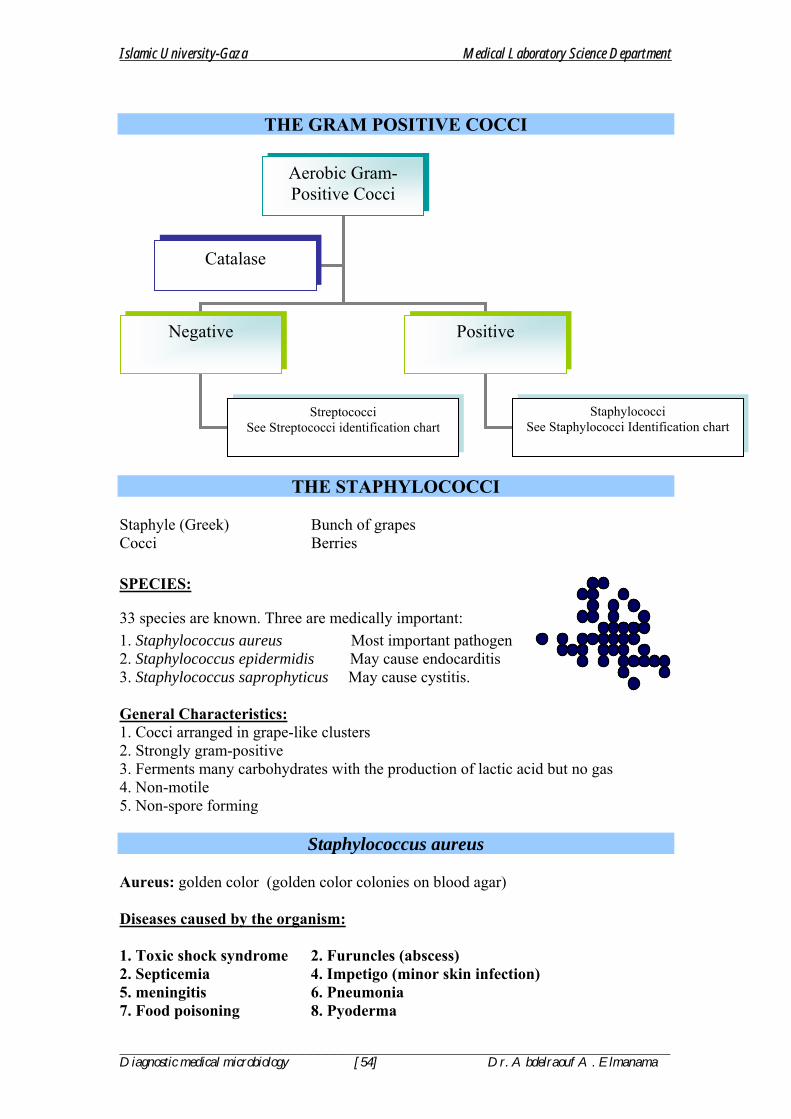
Islamic University-Gaza Medical Laboratory Science Department
_____________________________________________________________________ Diagnostic medical microbiology [54] Dr. Abdelraouf A. Elmanama
THE GRAM POSITIVE COCCI
THE STAPHYLOCOCCI Staphyle (Greek) Bunch of grapes Cocci Berries SPECIES:
33 species are known. Three are medically important: 1. Staphylococcus aureus Most important pathogen 2. Staphylococcus epidermidis May cause endocarditis 3. Staphylococcus saprophyticus May cause cystitis. General Characteristics: 1. Cocci arranged in grape-like clusters 2. Strongly gram-positive 3. Ferments many carbohydrates with the production of lactic acid but no gas 4. Non-motile 5. Non-spore forming
Staphylococcus aureus Aureus: golden color (golden color colonies on blood agar) Diseases caused by the organism: 1. Toxic shock syndrome 2. Furuncles (abscess) 2. Septicemia 4. Impetigo (minor skin infection) 5. meningitis 6. Pneumonia 7. Food poisoning 8. Pyoderma
Aerobic Gram-Positive Cocci
Negative Positive
Staphylococci See Staphylococci Identification chart
Streptococci See Streptococci identification chart
Catalase

Islamic University-Gaza Medical Laboratory Science Department
_____________________________________________________________________ Diagnostic medical microbiology [55] Dr. Abdelraouf A. Elmanama
9. Carbuncles
Diseases by S. aureus
PyogenicPyogenicDue to toxins
Due to toxinsSuperficialSuperficial DeepDeep
Cultural and Morphological Characteristics: 1. Media for Primary Isolation: S. aureus grow well in most routine media e.g, Blood Agar, Nutrient Agar. 2. Media for Selective Isolation: Mannitol Salt Agar (MSA), is an excellent medium which contains 7.5% sodium chloride which is considered as a high percentage and inhibitory to most medically important bacteria. In addition it contains mannitol as the only carbon source and a pH indicator to detect mannitol fermentation by S. aureus. 3. Incubation: After streaking the specimen on one of the common media, incubate the plates at 35-37 oC for 24 hours. 4. Colony Morphology: 4.1 On blood agar plates: colonies are 2-4 mm in diameter, rounded and slightly elevated. Most pathogenic strains produces a zone of β-hemolysis. Another distinguishing character is the production of a golden yellow pigment. 4.2 On Mannitol Salt Agar: The colonies are surrounded by a yellow zone indicative of acid production resulting from the fermentation of Mannitol. 5. Gram Stained Smears: Gram-positive cocci arranged in clusters. Single cells, diplococci, and short chains may also appear. It is usually simple to identify the morphology in stained film from sputum or pus but one can be certain by performing simple biochemical tests for the isolate e.g., catalase test to differentiate it from Streptococci and Coagulase or DNase to differentiate it from non-pathogenic staphylococci.. 6. Biochemical Characteristics: 1. Catalase positive 2. Mannitol fermenter 3. Grow well in 7.5% NaCl 4. Coagulase positive 5. DNase positive

Islamic University-Gaza Medical Laboratory Science Department
_____________________________________________________________________ Diagnostic medical microbiology [56] Dr. Abdelraouf A. Elmanama
6. Glucose fermenter. IDENTIFICATION: 1. Based on gram-staining 2. To differentiate it from other gram-positive cocci (Streptococci) perform catalase test 3. To differentiate it from other non-pathogenic staphylococci a group of biochemical tests are performed. a. Coagulase test b. Mannitol fermentation c. Growth in 7.5% NaCl d. Glucose OF e. DNase
NB: For epidemiological reasons, phage typing of S. aureus is necessary. PHAGE TYPING: A Staphylococcus aureus-specific phage is added to a plate that is inoculated with S. aureus. The plates are incubated at 37 oC for 24 hours. Positive identification: Formation of plaques Negative results: S. aureus grow over the whole area.
Gram-positive cocci
Staphylococci
Streptococci
CATALASE
Positive Negative
1. Coagulase 2. Mannitol 3. Glucose OF
1. Positive 2. Positive 3. F
1. Negative 2. Positive 3. O
1. Negative 2. Negative 3. F
S. aureus Micrococcus spp S. epidermidis
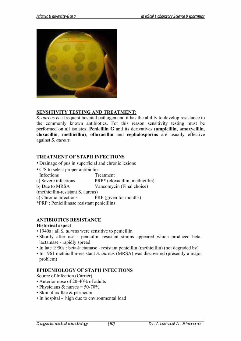
Islamic University-Gaza Medical Laboratory Science Department
_____________________________________________________________________ Diagnostic medical microbiology [57] Dr. Abdelraouf A. Elmanama
SENSITIVITY TESTING AND TREATMENT: S. aureus is a frequent hospital pathogen and it has the ability to develop resistance to the commonly known antibiotics. For this reason sensitivity testing must be performed on all isolates. Penicillin G and its derivatives (ampicillin, amoxycillin, cloxacillin, methicillin), ofloxacillin and cephalosporins are usually effective against S. aureus. TREATMENT OF STAPH INFECTIONS • Drainage of pus in superficial and chronic lesions • C/S to select proper antibiotics Infections Treatment a) Severe infections PRP* (cloxacillin, methicillin) b) Due to MRSA Vancomycin (Final choice) (methicillin-resistant S. aureus) c) Chronic infections PRP (given for months) *PRP : Penicillinase resistant penicillins ANTIBIOTICS RESISTANCE Historical aspect • 1940s : all S. aureus were sensitive to penicillin • Shortly after use : penicillin resistant strains appeared which produced beta-
lactamase - rapidly spread • In late 1950s : beta-lactamase - resistant penicillin (methicillin) (not degraded by) • In 1961 methicillin-resistant S. aureus (MRSA) was discovered (presently a major
problem) EPIDEMIOLOGY OF STAPH INFECTIONS Source of Infection (Carrier) • Anterior nose of 20-40% of adults • Physicians & nurses = 50-70% • Skin of axillae & perineum • In hospital - high due to environmental load

Islamic University-Gaza Medical Laboratory Science Department
_____________________________________________________________________ Diagnostic medical microbiology [58] Dr. Abdelraouf A. Elmanama
MRSA • Low carriage rate in community • High in tertiary care hospitals Mode of Transmission • Fomites • Direct from hospital staff or attendants : contaminated hands PREVENTION OF STAPH INFECTIONS Control of Carrier and reinfection • Wash clothes in hot water (>70oC) • Use antiseptic soap (Dettol soap) • Antimicrobial nasal cream (Gentamicin, Mupirocin) • Oral antibiotics that are concentrated in nasal secretions (ciprofloxacin and
rifampicin) Chemoprophylaxis • Antibiotics before and at time of surgical operation
COAGULASE-NEGATIVE STAPHYLOCOCCI (CNS) • Medically important species:
1. S. epidermidis 2. S. saprophyticus
Staphylococcus epidermidis • Normal flora in
° Skin, ° Anterior nose & ° External ear canal
• Cell wall contains teichoic acid (glycerol type) • White, non-haemolytic colonies on blood agar • Sensitive to novobiocin; (S. saprophyticus is resistant) DISEASES BY S. EPIDERMIDIS • Most infections are hospital acquired • Opportunistic pathogen in immuno-suppressed • Strongly associated with presence of foreign bodies ° Prosthetic heart valves (endocarditis) ° IV catheters (bacteremia) ° Urinary catheter (UTI in elderly) ° CSF shunts (meningitis) ° Peritoneal dialysis catheter (peritonitis)
Staphylococus saprophyticus • Saprophytic life • Resistant to novobiocin • Most infections are community-acquired ° Primary UTI in 10-20% of young adult women – hormonal factors may be
involved. • Resistant to antibiotics – penicillins & cephalosporins
Islamic University-Gaza Medical Laboratory Science Department
_____________________________________________________________________ Diagnostic medical microbiology [59] Dr. Abdelraouf A. Elmanama
THE GRAM POSITIVE COCCI THE STREPTOCOCCI
PATHOGENIC SPECIES: 1. Streptococcus pyogenes 2. Streptococcus agalactia 3. Streptococcus faecalis 4. Streptococcus pneumonia 5. The viridans group (Streptococcus viridans) General Characteristics: 1. Gram positive cocci arranged in chains 2. Catalse Negative 3. Colonies are small (1-2 mm), gray to white in color 4. Streptococci could be classified based on their heomlytic activity into: A. Alpha hemolytic: That produces incomplete hemolysis on blood agar as indicated by greenish discoloration of the medium (Greenish zone around colonies due to degradation of haemoglobin to biliverdin (green) ,e.g., S. pneumonia and Viridans streptococci B. Beta hemolytic: That produces complete hemolysis which results in clear zone around the colonies. e.g., S. pyogenes and S. agalactiae C. Gamma hemolytic (Non hemolytic): That produces no hemolysis on blood agar plates. e.g., S. faecalis (Enterococcus faecalis) 5. Lancefield grouping (on the basis of C-carbohydrate antigen in cell wall of
streptococci) • Groups A-U (group A,B & D are of great pathogenic significance. ° Group A = S. pyogenes ° Group B = S. agalactiae ° Group D = Enterococci • Basically beta-haemolytic are grouped by this method • Some alpha and non-haemolytic streptococci contain Lancefield group antigens NB: Many textbooks use a combination of the Lancefield and the hemolysis classification. e.g., group A Beta hemolytic streptococci
Streptococcus pyogenes Group A beta-hemolytic Streptococci
Diseases: 1. Erysipelas (red skin) 7. Streptococcal sore throat 2. Otitis media 8. Osteomyelitis 3. Puerperal fever 9. Endocarditis 4. Wound infection 10. Meningitis 5. Sepsis (Scarlet fever) 11. Acute glomerulnephritis 6. Sinusitis 12. Rheumatic fever NB: Streptococci causes a wider variety of clinical infections than any other genus of bacteria.

Islamic University-Gaza Medical Laboratory Science Department
_____________________________________________________________________ Diagnostic medical microbiology [60] Dr. Abdelraouf A. Elmanama
Specimens for Culture: 1. Throat swab: A tongue depressor should be used to expose the posterior pharynx. A sterile swab is then rubbed firmly over the tonsil and posterior pharynx, touching any exudates present. 2. Nasal swab: May be obtained by swabbing the anterior nares at a depth of 1-2 cm with a cotton swab. 3. Sputum culture: Are useful for detecting lung infection due to pneumococci and other streptococci. Sputum usually is contaminated with saliva containing normal flora, hence, special precaution should be observed. Specimens obtained by transtracheal aspiration are more reliable. 4. Blood: Blood cultures should be obtained from all patients with possible bacteremia or endocarditis. DIAGNOSTIC PROCEDURES: 1. Gram stain: The presence of infections involving streptococci can frequently be determined both quickly and easily by microscopic examination of gram stained smears of certain types of specimens. 2. Culture of specimen: Blood agar plates are the most commonly used for primary isolation of streptococci. In mixed infections: Selective media should be used such as blood agar containing gentamicin, colistin and nalidixic acid. = Colony morphology is not a distinguishing characteristic. 3. Biochemical and Other tests: 1. Catalase test (negative) 2. Bacitracin sensitivity test: a. Select a colony from the culture to be tested; streak it evenly over the surface of a half blood agar plate. b. Place bacitracin disk in the center of the streaked area. c. Incubate overnight d. RESULT: An inhibition zone of 5mm or greater is considered sensitive. NB: Streptococcus pyogenes is sensitive to bacitracin Serological tests: Detection of antibody in patient serum ASOT: Anti-Streptolysin O Titer: Streptolysin O is an antigen carried on the cell wall of S. pyogenes that is usually anticipated by specific antibodies produced by the host. This test is simple and reliable. The test is done by performing a serum serial dilutions and reacting these dilutions with the specific antigen suspension and determining the lowest concentration or the highest dilution (Titer) that produces a positive results.

Islamic University-Gaza Medical Laboratory Science Department
_____________________________________________________________________ Diagnostic medical microbiology [61] Dr. Abdelraouf A. Elmanama
Treatment: Penicillin G is used in the treatment. In penicillin sensitive patients Erythromycin is used.
Streptococcus agalactia Group B, beta-hemolytic streptococci
General Characteristic: • It is the only species that represents the group Streptococci • Hemolysis on Sheep Blood Agar is mostly beta-hemolysis • Catalase negative • The group antigen is a cell wall polysaccharide composed of N-
acetylglucoseamine, galactose, and rhamanose. • It hydrolysis hippurate to benzoic acid and glycine. • It is resistant to bacitracin antibiotic disk (and could be differentiated from S.
pyogenes which is a beta-hemolytic and sensitive to this antibiotic). Normal Human Habitat: It is a part of the normal oral and vaginal flora. Approximately 5-15% of the healthy population carry S. pyogenes or S. agalactia in the nasopharynx. It can be found in pharynx, vagina, gastrointestinal tract. In addition, in the newborn it could be present in various sites. Pathogenicity: 1. Puerperal sepsis 2. Endocarditis 3. Pneumonia 4. Neonatal infection (Pneumonia, septicemia and meningitis). 5. Bovine mastitis Culture of the Organism: 1. On general media: e.g., Blood Agar; S. agalactia produces larger colonies and more translucent to opaque colonies surrounded by a zone of Beta-hemolysis. 2. Selective media: (For both S. pyogenes and S. agalactia): Streptococcal Selective Agar (SSA): Contains the following inhibitory chemicals:
• Crystal violet in low concentration, • Colistin, • Trimethoprim-sulphamethoxazole in 5% sheep blood agar.

Islamic University-Gaza Medical Laboratory Science Department
_____________________________________________________________________ Diagnostic medical microbiology [62] Dr. Abdelraouf A. Elmanama
Laboratory Diagnosis: 1. Lancefield grouping 2. Hippurate hydrolysis test 3. CAMP test 4. Bile Esculin test 5. Latex agglutination. NOTE: The sodium hippurate hydrolysis and CAMP tests can be used to confirm the identification of group B streptococci. 1. Hippurate hydrolysis test: Group B streptococci contains the enzyme hippuricase which can hydrolyze hippuric acid. The products of the hydrolysis of sodium hippurate are sodium benzoate and glycine. Glycine could be detected by the addition of ninhydrin which is an oxidizing agent, which alos gives a purple color with glycine. 2. CAMP test The hemolytic activity of Staphylococcal Beta lysin on RBCs is increased by an extracellular factor produced by S. agalactia called the CAMP factor. This test is don by making a single streak of Streptococcus (to be identified) on sheep blood agar perpendicular to a strain of Staphylococcus aureus known to produce beta-lysin. The two streak lines must not touch one another. The plate is incubated for 24 hours . The positive result is expressed by a zone of increased lysis assuming the shape of an arrow-head at the junction of the two streak lines.
Fig. Showing the arrow-head appearance of a positive strain 3. Bile Esculin This test detects the ability of the organism to grow in the presence of bile and its ability to hydrolyze esculin and the production of glucose and aglycone esculetin. Esculetin reacts with iron salts to for a dark brown or black complex. This test is performed in an appropriate medium containing bile, esculine, ferric citrate as a source of ferric ions, and sodium azide to inhibit the growth of gram negative bacteria. This test used to differentiate Group D streptococci (Enterococci) POSITIVE, and Streptococcus agalactia (NEGATIVE)

Islamic University-Gaza Medical Laboratory Science Department
_____________________________________________________________________ Diagnostic medical microbiology [63] Dr. Abdelraouf A. Elmanama
3. Latex agglutination: A. Antibody coated latex particles serves as the basis for several commercially available systems for direct detection of bacterial and other microbial antigens in body fluids. For example, Streptococcal antigens in throat swab samples can be now detected within 10-60 minutes depending in the system being used. B. Latex agglutination tests are also available to detect antibodies that develops during certain bacterial infections. The advantages of the latex agglutination tests is rapidity and its relative sensitivity. Treatment: Group B streptococci is often resistant to tetracycline and kanamycin but remain sensitive to the clinically achievable blood levels of penicillin G.
β - hemolytic streptococci
Bacitracin
Resistant Sensitive
S. pyogenes
Other groups of β hemolytic Strepto.
Negative Positive
CAMP test
S. agalactia
Islamic University-Gaza Medical Laboratory Science Department
_____________________________________________________________________ Diagnostic medical microbiology [64] Dr. Abdelraouf A. Elmanama
Streptococcus pneumoniae This species was isolated by Pasteur in France in 1881 and was found to be the major cause of lobar pneumonia in human. Two variants has been isolated; one with capsule and was demonstrated to be pathogenic for both man an mice and the other is non-capsulated was found to be non-pathogenic. Normal Habitat: It is normally found as an oral flora and in the nasopharynx. In general 15% of children and 5% of adult healthy individuals whose considered to be carriers, do not suffer pneumonia. Diseases: 1.Pneumonia (Pneumoncoccal pneumonia): This type of pneumonia usually occur most frequently with the following conditions: a. Viral infections of the upper respiratory tract. b. Persons whose respiratory drainage is impaired e.g., heavy smokers and persons who have inhaled toxic irritants c. Immunocompramized patients 2. Bronchitis 3. Sinusitis 4. Otitis media 5. Septicemia 6. Meningitis Antigenic Structure: 1. Capsular Antigen : The capsular polysaccharide is highly antigenic and one could classify S. pneumoniae into 84 serotypes. Capsule is considered to be the major virulence factor by which it resist the process of phagocytosis by PMNLs and macrophages. 2. Somatic Antigen (O): C polysaccharide in the cell wall comosed of teichoic acid polymer. This antigen react with Beta-globulin refered to as C-Reactive Protein (CRP). 3. M protein (Somatic): protein which neither antiphagocytic no protective. Virulence: 1. Polysaccharide capsule 2. Adherence 3.Enzymes a. Neuroaminidase: Degrades surface structures of the host tissue b. Protease: degrades immunoglobulin 4. Pneumlysin O: Oxygen labile toxin which binds to cholesterol molecule in the host cell membrane and lyses the cell. 5. Autolysin: Lysis the cell in the presence of surface active substance such as bile salt. This lysis facilitate the release of pneumolysin and other toxic materials from the cells.

Islamic University-Gaza Medical Laboratory Science Department
_____________________________________________________________________ Diagnostic medical microbiology [65] Dr. Abdelraouf A. Elmanama
Culture Media: - Blood agar is usually an excellent medium for the primary isolation of this organism. Chocolate agar is also used. - 5-10% CO2 enhances growth (CO2 incubator). In order to visualize alpha hemolysis, stab the inoculating loop into the agar several times to allow the action of the oxygen labile streptolysin to take place. - Capsulated strains; on blood agar, produces white circular mucoid colonies surrounded by a zone of alpha hemolysis as indicated by a greenish discoloration. Gram staining: -Gram-positive diplococci (lancet-shape). May show gram negative reaction due to the action of antibiotics or autolysin and especially in old cultures. Laboratory Diagnosis: - Gram stained smears from clinical specimen and from culture should revail the typical lancet shaped gram positive diplococci. - Biochemical & Serological Identification: 1. Quellung reaction: The reaction is performed by mixing equal amounts of specimen with type specific pneumococcal antiserum and waiting 15-30 minutes to allow the reaction to occur, then examine by the oil immersion objective. This preparation is compared with a saline perpetration to detect any swelling (Enlargement) of the capsule. 2. Capsule Negative Staining: Capsule could be demonstrated by staining cultures by INDIA INK. 3. Catalase test = Negative 4. Bile solubility test: 10% sodium deoxycholates lyse colonies of S. pneumoniae. This test is done by adding a small drop of the bile salt over suspected colony and is observed for lysis. 5. Optochin susceptibility test: Optochin has a detergent-like action and cause selective lysis of pneumococci. Zone of inhibition of 14 mm (6 mm disks). This test should be performed in 5-10% CO2. 6. Detection of pneumococci antibodies: By the radioimmunoassay which is very sensitive in detecting the specific capsular antibodies.
alpha-hemolytic streptococci
S. pneumonia Viridans group
Positive Negative
Optochin Bile solubility

Islamic University-Gaza Medical Laboratory Science Department
_____________________________________________________________________ Diagnostic medical microbiology [66] Dr. Abdelraouf A. Elmanama
Type specificantisera+ Type specificantisera+
Capsulated pneumococcusCapsulated pneumococcus
Capsule swells &refractile under microscope
Capsule swells &refractile under microscope
CAPSULAR SWELLING (QUELLUNG) REACTION• Capsulated pneumococci are mixed with type specific antisera
• The capsule absorbs water and swells• Refractile under light microscope • Polyvalent antiserum containing antibodies against 80 capsular types is available
GROUP D STREPTOCOCCI
Non-haemolytic or alpha-haemolytic colonies: Two types
1. Enterococci (E. faecalis) • Normal flora of colon • Can grow in presence of 7.5% NaCl and bile salts
Diseases ° Opportunistic UTI ° Wound infections
2. Non-Enterococci (S. bovis) • Can grow in presence of bile salts (not NaCl) • May cause endocarditis

Islamic University-Gaza Medical Laboratory Science Department
_____________________________________________________________________ Diagnostic medical microbiology [67] Dr. Abdelraouf A. Elmanama
THE GRAM NEGATIVE COCCI
SPECIES: 1. Neisseria gonorrhea 2. Neisseria meningitidis 3. Neisseria catarahlis 4. Neisseria sicca 5. Neisseria flavescens 6. Neisseria flava 7. Neisseria mucosa 8. Neisseria lacatamica
1. Neisseria gonorrhea
The name gonorrhea was introduced by Galen 130 A.D. Neiser 1879 described diplococci which he found in the purulent secretions of acute cases of urethritis and vaginitis and in smears made from acute conjunctivitis of the new born. Gonorrhea is the most prevalent venereal disease and is second only to tuberculosis among infectious diseases which give rise to serious complications. The primary infection with N. gonorrhea although uncomfortable gives no alarming symptoms and too frequently ignored by the patient. Man is the only known host for the organism.
MORPHOLOGY AND STAINING Coffee-bean shaped, kidney-bean shaped, in pairs, gram-negative diplococci. In smears form urethral discharges, the gonococcus is seen as an oval; or spherical coccus. Found frequently in pairs with the adjacent sides flattened. Usually intracellular, especially in acute cases. In smears there is irregularity in the distribution in the phagocytic cells, many polymorphonuclears containing no organism while a few cells may contain as many as 20 to 50 or more cocci per cell.
In acute cases, intracellular cells predominate over the extracellular forms while in chronic cases , the extracellular form predominate over the intracellular forms.
Non-motile, nonencapsulated except in its mucoid variant phase. Stain readily with aniline dyes.
The presence of gram-negative intracellular diplococci in smears of pus from the male urethra is presumptive evidence of gonorrhea.
CULTURAL CHARACTERISTICS: In exudates from the vagina or from the eye, however, the morphologic picture is less reliable since the other gram negative cocci present frequently in these regions.
Delicate aerobes which usually needs an atmosphere containing 5-10% CO2 and an optimum temperature of 35-36 oC. The pH of the medium should be adjusted to 7.2 to 7.6.

Islamic University-Gaza Medical Laboratory Science Department
_____________________________________________________________________ Diagnostic medical microbiology [68] Dr. Abdelraouf A. Elmanama
MEDIA OF CHOICE: 1.CHOCOLATE AGAR 2.TRANSGROW: A transport medium to maintain viability of the organism during transportation. 3. Thyer-Martin Medium: Very similar to the chocolate agar to which antibiotics has been added: a. Vancomycin: To inhibit the growth of gram-positive cocci. b. Colistine: To inhibit growth of gram negative organism c. Nystatin: To inhibit growth of fungi. Trimethoprim may be added to prevent the growth of swarming organisms. On Chocolate agar, after 48 hours of incubation, round, convex smooth grayish colonies appear. On further incubation, the colonies may increase in size and develop a roughened surface with crenated edges. The colonies are soft and somewhat slimy when touched by a platinum loop. In broth medium, the growth is chiefly on the surface with some flaky sediment at the bottom of the tube. Dextrose is fermented producing acid but not gas. On Thayer-Martin Agar (Medium of choice): After 20 hours of incubation at 35-36 oC in a candle jar, typical colonies appear small translucent, raised, moist gray white colonies with entire to lobate margins. Usually they are mucoid and tends to come off as whole colonies when fished from the agar surface.
FERMENTATION REACTIONS OF NEISSERIA
Organism Dextrose Maltose Sucrose Growth
on NA Growth at 37 oC
oxidase
N. meningitidis + + - - - + N. gonorrhea + - - - - + N. catarrhalis - - - + + + N. sicca + + + + + + N. flavescens - - - + + +
PATHOGENICITY: 1. GONORRHEA: This infection is transmitted directly from individual to individual. It attacks chiefly the urethra of both males and females.The difficulty in growing gonorrhea in the laboratory as well as their failure to infect laboratory animals, makes it clear how very closely these organisms are adapted to growth in the human body. They will survive only for brief periods outside the body. This is the reason why gonorrhea is almost always transmitted by direct body contact, and principally by sexual intercourse. Gonorrhea is a venereal disease accompanied by excessive purulent discharge from the genital tract. Incubation period is 3-5 days. if left untreated it may cause sterility in both males and females. Manifestation are urethritis, cervico-vaginitis, rectal involvement, pharyngeal involvement. 2. OPTHALMIA NEONATORUM Less frequent site of primary infection is in the conjuctiva, usually seen in the newborn babies. This conjunctivitis in the newborn which result from infection during

Islamic University-Gaza Medical Laboratory Science Department
_____________________________________________________________________ Diagnostic medical microbiology [69] Dr. Abdelraouf A. Elmanama
birth when the fetus passes through the birth canal. If untreated this may lead to blindness. This disease could be prevented by the application of 2% silver nitrate solution in the eyes of all newborn. 3. PELVIC INFLAMMATORY DISEASE (PID)
DIAGNOSIS: 1.Specimen: Vaginal swab and material obtained from males by prostatic massage or urethral swab. 2.Stained smears: Smears from suspected cases will reveal gram negative diplococci, coffee-bean shaped.
Recovery of gram-negative diplococci extracellular or intracellular may be of a diagnostic value for male patient but usually are not in case the patient is female. This must be confirmed by culture and biochemical identification of the species.
3.Cultivation: on chocolate agar or Thayer-Martin agar with increased CO2 tension at a 35 oC. 4. Oxidase Reaction: This depends on the formation of indophenol oxidase, which is common to all Neisseria. 5. Confirmatory tests:
A. Fluorescent antibody tagging test: (FAT) B. Degradation of carbohydrates. The recommended base medium is Cystine Trypticase Agar (CTA). Neisseria gonorrhea ferments only glucose with acid but no gas.
RESISTANCE: Easily killed by drying for 2 hours. Moist heat kills them at 55oC in less than 5 minutes. Very susceptible to antiseptics, especially silver nitrate which kills the gonococci in 2 minutes using 1:4000 dilution. Cultures maintained at room temperature dies in 1 to 2 days and after 4 to 6 days at 36 oC.
Gonococci are susceptible to sulfonamides and penicillines although resistant strains to those antibiotics has emerged. Sensitivity tests must be performed on all isolates to determine their susceptibility.
ANTIGENIC STRUCTURE/ DETERMEINANTS OF PATHOGENICITY 1. Pilus antigen 2. Lipoligosccharide (LOS) 3. Outer membrane proteins constituents
P I. Antigenically variable and used as the basis of Enzyme-linked immunosorbent assay (ELISA) and coagulation assay for serotyping gonococci. P II. Associated with adhesion properties.

Islamic University-Gaza Medical Laboratory Science Department
_____________________________________________________________________ Diagnostic medical microbiology [70] Dr. Abdelraouf A. Elmanama
NEISSERIA MENINGITIDIS MENINGOCOCCI
Neisseria meningitidis causes dramatic and explosive epidemics of cerebrospinal meningitis. Man is the only natural host. The organism live in the nasopharynx of apparently healthy individuals and are passed from man to man via reparatory tract.
MORPHOLOGY AND STAINING: In cerebrospinal fluid (CSF) the organism appear as intracellular diplococci and sometimes in tetrads or even in large aggregates. In the early stages of the disease many of the organisms are found extracellularly. Both intra and extracellular forms are flattened on their opposite sides and resembles pairs of coffee-beans. Non-motile, non-sporeformers. Most of the isolated strains of Group I and Group II type have capsules. Gram negative CULTURAL CHARACTERSTICS: Rich medium with blood, serum, ascitic fluid and dextrose favors the growth. In blood agar plates: Small glistening, slightly convex colonies are formed. Growth occur at aerobic conditions at 37 oC and 5-10% CO2. PATHOGENICITY: = Causes epidemics meningococcal meningitis or cerebrospinal fever. = this disease is transmitted through the inhalation of air borne droplets or by close contact with carriers. Invasion of the body occurs in three steps:
1. Implantation of meningococcus in the nasopharynx. 2. Entrance to the blood stream with resulting septicemia. 3. Locoalization in the meninges.
Meningococcalcarrier
Droplet infection
Attach to nasopharynxof new person Meninges
Droplet infectionDroplet infection BacteraemiaBacteraemia
SkinSkinRelease
endotoxinsRelease
endotoxinsPetechiaePetechiae
ShockShock
Cribriform plateCribriform plate
Cross blood brain barrierCross blood brain barrier
Meningitis:Headache, fever, stiff neck,nausea & vomiting, petechiae, shock, coma, death
Meningitis:Headache, fever, stiff neck,nausea & vomiting, petechiae, shock, coma, death
Pathogenesis of meningococcal meningitis

Islamic University-Gaza Medical Laboratory Science Department
_____________________________________________________________________ Diagnostic medical microbiology [71] Dr. Abdelraouf A. Elmanama
The nasopharynx is the portal of entry of this organism. There, the organism may form part of the transient flora without producing or may produce an exudative pharyngitis. From the nasopharynx, organisms may reach the blood stream, producing bacteremia (meningococcemia) with high fever and a hemorrhagic rash. There may be fulminant sepsis, disseminated intravascular coagulation, and circulatory collapse. Meningitis is the commonest complication of meningococcemia. It usually begins very suddenly, with intense headache, vomiting, and stiff neck and progress to coma within a few hours. VIRULENCE FACTORS: Lipopolysaccharide endotoxin complex which activates the clotting cascade producing disseminated intravascular coagulation.
IMMUNITY: Moderate degree of immunity follows an attack of meningitis. Immunity increases with age because the disease is more prevalent in children than in adults. Infants have passive immunity through IgG antibodies transferred from the mothers. ANTIGENIC STRUCTURE AND GROUPING • Nine groups (A,B,C,D,E, W135. X,Y,Z) on the basis of polysaccharide capsular
antigen • Groups A, B & C are responsible for epidemics of meningitis • Polysaccharides of groups A,C,W135 are immunogenic – used as vaccines • Group B is not immunogenic.
DIAGNOSIS: 1.SPECIMEN: Blood, nasopharyngeal swabs, CSF, joints fluids. 2.Smears: Reveal gram-negative cocci in pairs which are coffee bean shaped. 3.Culture: Inoculate specimen on Chocolate agar plates; or Thayer-Martin Agar and incubate at 36 oC under 5-10% CO2 tension. 4. Oxidase test: same as N. gonorrhea Because meningococci may autolyse rapidly, the fluid must be examined fresh because in CSF, when out of the body, meningococci are quickly lyses. TREATMENT
• Penicillin is drug of choice : • penetrates well in inflammed meninges • In penicillin hypersensitivity Ceftriaxone Effective against other two pathogens also i.e H. influenzae & S. pneumoniae • At the end of therapy with penicillin (in adults): Give ciprofloxacin to
eradicate meningococci because penicillin does not eradicate from nasopharynx: otherwise patient will become a carrier
PREVENTION 1. Treatment of carriers •Rifampicin / ciprofloxacin •Penicillin does not eradicate carrier state ° Due to inadequate penetration of uninflammed nasopharyngeal mucosa

Islamic University-Gaza Medical Laboratory Science Department
_____________________________________________________________________ Diagnostic medical microbiology [72] Dr. Abdelraouf A. Elmanama
° Poor secretion in saliva 2. Chemoprophylaxis to close contacts
•Ciprofloxacin 3. Meningococcal vaccine
•Divalent (gp A & C) & •Quadrivalent (A,C,Y, W135) •Not available against group B (not immunogenic)
Given to: °Military camps and pilgrims °During epidemic
DIFFERENCES BETWEEN GONOCOCCUS AND MENINGOCOCCUS 1. Gonococcus grow more slowly, forms smaller colonies. 2.Gonococcus produces acid in glucose only, while meningococcus produces acid in both glucose and maltose. 3. Gonococcus is less toxic to mice and guinea pigs than meningococcus.
SIMILARITIES BETWEEN GONOCOCCUS AND MENINGOCOCCUS 1. Both are strict parasite and causes diseases only for man 2. They may show little differences in resistance to injurious agents. 3. Their distribution in the inflammatory exudates is the same. 4. They grow on artificial media with a little differences.
BRANHAMELLA
Branhamella catarrhalis (formerly called Neisseria catarrhalis). This is similar to N. gonorrhea and N. meningitidis morphologically and culturally. It is a part of the normal flora of the throat. It is normal inhabitant of mucus membrane especially the respiratory tract. Non-pathogenic characterized by its inability to ferment any sugar and by the ability to grow on routine laboratory media at room temperature. This was excluded from the genus Neisseria based on differences between the DNA structure.

Islamic University-Gaza Medical Laboratory Science Department
_____________________________________________________________________ Diagnostic medical microbiology [73] Dr. Abdelraouf A. Elmanama
Gram Positive Bacilli
MEDICALLY IMPORTANT GRAM-POSITIVE BACILLI
AEROBESCorynebacteriumListeriaBacillusGardnerellaNocardia
ANAEROBESClostridiaActinomyces

Islamic University-Gaza Medical Laboratory Science Department
_____________________________________________________________________ Diagnostic medical microbiology [74] Dr. Abdelraouf A. Elmanama
Gram Positive, Endospore-Forming Bacilli
THE GENUS BACILLUS There are 48 species included in the genus bacillus with the following general characteristics: 1. Gram-positive bacilli 2. Spore former 3. Aerobic and facultative anaerobic THE MOST IMPORTANT PATHOGENS: 1. Bacillus anthracis 2. Bacillus cereus
I. BACILLUS ANTHRACIS Morphology: 1. Large gram positive bacilli 2. Non-motile 3. Found singly, in pairs or in long chains 4. Capsule could be demonstrated during growth in infected animals. 5. Spores are formed in culture, dead animal's tissue but not in the blood of infected animals. 6. Spores are oval and centrally located. Survival in Soil • Spores remain viable in soil for decades. • In World War II in Scotland spores were exploded. • Survived for >40 years and were eradicated in 1987 • Changing environmental conditions (temp. rain etc.) help in survival and multiplication. Cultural Characteristics: = Blood Agar and Nutrient Agar are commonly used to cultivate the bacilli. Plates are incubated aerobically at 37 oC. = On blood agar plates, colonies have irregular borders and are non-hemolytic. = On nutrient agar: They are described as "Medusa head" or "Comet tail". Specimen Collection and Laboratory Diagnosis: CAUTION: Laboratory safety is very important when working with any materials suspected of containing Bacillus anthracis.
Samples are collected depending on the site affected: 1. Swab samples from cutaneous lesions and blood cultures. 2. Sputum and blood for pulmonary anthrax 3. Gastric aspirate, feces and blood for enteric anthrax.

Islamic University-Gaza Medical Laboratory Science Department
_____________________________________________________________________ Diagnostic medical microbiology [75] Dr. Abdelraouf A. Elmanama
= Gram stained smears: Made from clinical samples, show large gram positive bacilli in long chains "Bamboo-like appearance". = Giemsa stained smears: Purple bacilli with red capsule. = Culture See Cultural characteristics above. =Animal inoculation test: Experimental animals are injected intraperitoneally by a suspension of the test organism "Suspected B. anthracis culture". The animal dies in 48-96 hours due to respiratory failure. Large number of typical bacilli can be found in the blood and tissue of spleen of the infected animal. = Biochemical Identification: 1. Carbohydrate fermentation test: 2. Gelatine liqefaction test: Negative after 7 days. Growth has a characteristic appearance of an inverted pine tree. 3. Nitrate reduction test: Positive 4. Starch hydrolysis test: Positive 5. Voges-Proskauer test: Positive 6. Sensitivity to penicillin. Sensitive 7. Lysis by gamma phages: Positive. This test accurately differentiate B.anthracis from other bacilllus species. Table 1. Shows some distinguishing characteristics of B. anthracis
Feature B. anthracis B. cereus and other Bacillus spp.
Hemolysis on sheep blood agar Negative Positive Motility Negative Usually positive Salicin Fermentation Negative Positive Growth on PEA Negative Positive
PEA: PhenylEthyl Alcohol + Brain Heart Infusion Agar. PATHOLOGY; There are different clinical forms of anthrax: 1. CUTANEOUS ANTHRAX: 95-98% of anthrax cases are of this type. Infection occur through wounds, burns, which may progress to toxaemia and septicemia. The site of entry often produces a painless blister referred to as Malignant pustule. 2. ENTERIC "INTESTINAL" ANTHRAX: Caused by the ingestion of infected meat. This form of the disease is severe and fatal. 3. PULMONARY ANTHRAX: Caused by the inhalation of large number of B. anthracis spores. It is usually fetal. This clinical form is commonly known as "wool sorter disease". ANTIGENIC STRUCTURE AND PATHOGENIC DETERMINANTS: 1. The Capsular Polypeptide: Composed of poly peptide of a high molecular weight consisting of D-glutamic acid. 2. Polysaccharide Somatic Antigen: Composed of N-acetulglucoseamine and D-galactose 3. Complex Protein Toxin: This toxin appear to be responsible for signs and symptoms characteristic of anthrax. Accumulation of the toxin in tissue and its effect on the central nervous system results in death by respiratory failure and anoxia.
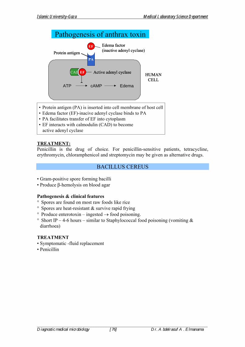
Islamic University-Gaza Medical Laboratory Science Department
_____________________________________________________________________ Diagnostic medical microbiology [76] Dr. Abdelraouf A. Elmanama
Pathogenesis of anthrax toxin
HUMANCELL
HUMANCELL
• Protein antigen (PA) is inserted into cell membrane of host cell• Edema factor (EF)-inacive adenyl cyclase binds to PA• PA facilitates transfer of EF into cytoplasm• EF interacts with calmodulin (CAD) to become
active adenyl cyclase
EFCAD EFCAD
PA
EF Edema factor(inactive adenyl cyclase)Protein antigen
PA
EF Edema factor(inactive adenyl cyclase)Protein antigen
Active adenyl cyclaseActive adenyl cyclase
ATP cAMP EdemaATP cAMP Edema
TREATMENT: Penicillin is the drug of choice. For penicillin-sensitive patients, tetracycline, erythromycin, chloramphenicol and streptomycin may be given as alternative drugs.
BACILLUS CEREUS
• Gram-positive spore forming bacilli • Produce β-hemolysis on blood agar Pathogenesis & clinical features ° Spores are found on most raw foods like rice ° Spores are heat-resistant & survive rapid frying ° Produce enterotoxin – ingested → food poisoning. ° Short IP – 4-6 hours – similar to Staphylococcal food poisoning (vomiting &
diarrhoea) TREATMENT • Symptomatic -fluid replacement • Penicillin

Islamic University-Gaza Medical Laboratory Science Department
_____________________________________________________________________ Diagnostic medical microbiology [77] Dr. Abdelraouf A. Elmanama
CLOSTRIDIA GENERAL CHARACTERISTICS: 1. Anaerobic gram-positive bacilli 2. Spore formers 3. They decompose proteins 4. Exotoxin producers 5. Natural habitat; Soil or the intestinal tract of animals and man. 6. Some motile, others are non-motile. 7. Catalase negative SPECIES OF MEDICAL IMPORTANCE: 1. Clostridium tetani 2. Cl. perfringens 3. Cl. botulinum 4. Cl. septicum 5. Cl. dificilli 6. Cl. novyi
I. Clostridium tetani Disease: Tetanus or "lock jaw" Cl. tetani is not an invasive organism but rather remains strictly localized in the area of the affected tissue into which the spores has been introduced. The disease is transmitted to man through infected wounds. Spores enter into wounds with soil or material contaminated with soil. The infection is localized in the area of entry. Growth and toxin production is aided by the followings: 1. Necrotic tissue 2. Calcium salts 3. Associated pyogenic infection. These conditions aids in providing anaerobic conditions. The toxin "tetanospasmin" is heat-labile protein, inactivated by heating for 5 minutes at 65 oC. The toxin may reach the central nervous system. It blocks the release of inhibitory mediators of the motor neurons. This results in extreme hyper-reflexia and violent spasm of skeletal muscles in response to any stimuli. This toxin is inactivated by the proteolytic enzymes of the digestive system. 1 mg pure toxin has 6 million lethal mice doses. Morphology: 1. Gram-positive anaerobic 2. Motile with peritrichous flagella 3. Terminal rounded spores (Drumstick appearance). Media of Choice: Any medium with thioglycollate under anaerobic condition most especially Cooked Meat Media.

Islamic University-Gaza Medical Laboratory Science Department
_____________________________________________________________________ Diagnostic medical microbiology [78] Dr. Abdelraouf A. Elmanama
W oundscontam inated w ith so il
Pathogenesis of TetanusO ccurs in traum atic open w ounds contam inated w ith soil
Spores germ inate in dead tissues
Spores germ inate in dead tissues
N eurotoxinR eaches retrogradeto sp inal cord
N eurotoxinR eaches retrogradeto sp inal cord
B inds to ganglioside receptors of anteriorhorn ce lls
B inds to ganglioside receptors of anteriorhorn ce lls
Inh ib its re lease of inh ib itorym ediators
Inhib its re lease of inh ib itorym ediators
C ontinuous excitem ent ofm otor neurons
C ontinuous excitem ent ofm otor neurons
M uscle spasmM uscle spasm
• O rganism s m ultiplylocally only
• N o tissue dam age• N o invasion of
adjacent tissues
II. Clostridium perfringens Disease: Gas gangrene It causes a variety of infection to man: 1. Wound infection 2. Gas gangrene 3. Necrotizing jejunitis 4. Food poisoning 5. Biliary tract infection 6. Meningitis Four types of toxins are produced: 1. Alpha = It is a phospholipase C which hydrolysis lecithin. Disrupts cell membranes of RBCs, WBCs & muscle cells 2. Beta = Is necrotic for the intestinal mucosa and lethal to the CNS. 3. Epsilon = Inactive which is converted to the active form by trypsin in small intestine. 4. Iota = Similar to epsilon but differ in structure.

Islamic University-Gaza Medical Laboratory Science Department
_____________________________________________________________________ Diagnostic medical microbiology [79] Dr. Abdelraouf A. Elmanama
O p en w o u n d scon tam in a ted
w ith so il
S p o res g erm in a te
S p o res g erm in a te
O T o x inIncreases cap illa ry p erm eab ility & m u sc le d ea th
O T o x inIncreases cap illa ry p erm eab ility & m u sc le d ea th
E n zy m esB reak d o w n su g ars w ith g as
E n zy m esB reak d o w n su g ars w ith g as
O rg a n ism s m u ltip ly
O rg a n ism s m u ltip ly
L arg e am o u n t o f g as in su b cu tan eo u s tissu es
L arg e am o u n t o f g as in su b cu tan eo u s tissu es
A lp h a T o x ince ll d ea th & tissu e n ecro sis
A lp h a T o x ince ll d ea th & tissu e n ecro sis
R ed u ced O x y g en d u e totissu e n ecro sis
R ed u ced O x y g en d u e totissu e n ecro sis
P a th o g e n e s is o f G a s G a n g re n e
D IS E A S E S B y C . P E R F R IN G E N S1 . G as G a n gren e (M yo n ecro sis)O ccu rs in trau m a tic o p en w o u n d s co n tam in a ted w ith so il
Morphological Characteristics: = Spores are usually oval and central or subterminal. = Not strictly anaerobe and can survive exposure to oxygen for short periods of time and sometimes referred to as aerotolerant anaerobe. = Non-motile = They form capsule in infected patients and animals. Cultural Characteristics: On blood agar plates: Colonies are round, domed and grayish white, surrounded by a zone of hemolysis.
III. Clostridium botulinum BOTULISM: It is a disease caused by the ingestion of food containing the neurotoxin produced by C. botulinum. The earliest symptoms usually are an acute digestive disturbances followed by nausea and vomiting and possibly diarrhea, together with fatigue, dizziness and headache. Later there is constipation. Double vision may be evident early and difficulty in swallowing and speaking may be noted. Patient may complain of dryness of the mouth and constriction of the throat. Involuntary muscles become paralyzed, paralysis spread to the respiratory system and heart, the death usually results from respiratory failure.
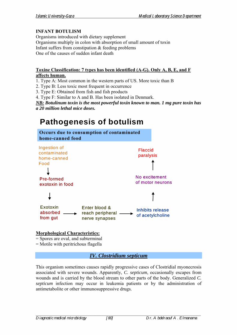
Islamic University-Gaza Medical Laboratory Science Department
_____________________________________________________________________ Diagnostic medical microbiology [80] Dr. Abdelraouf A. Elmanama
INFANT BOTULISM Organisms introduced with dietary supplement Organisms multiply in colon with absorption of small amount of toxin Infant suffers from constipation & feeding problems One of the causes of sudden infant death Toxine Classification: 7 types has been identified (A-G). Only A, B, E, and F affects human. 1. Type A: Most common in the western parts of US. More toxic than B 2. Type B: Less toxic most frequent in occurrence 3. Type E: Obtained from fish and fish products 4. Type F: Similar to A and B. Has been isolated in Denmark. NB: Botulinum toxin is the most powerful toxin known to man. 1 mg pure toxin has a 20 million lethal mice doses.
Ingestion of contaminated home-cannedFood
Pathogenesis of botulismOccurs due to consumption of contaminated home-canned food
Pre-formed exotoxin in foodPre-formed exotoxin in food
Exotoxin absorbed from gut
Exotoxin absorbed from gut
Inhibits release of acetylcholineInhibits release of acetylcholine
No excitement of motor neuronsNo excitement of motor neurons
Flaccid paralysisFlaccid paralysis
Enter blood & reach peripheral nerve synapses
Enter blood & reach peripheral nerve synapses
Morphological Characteristics: = Spores are oval, and subterminal = Motile with peritrichous flagella
IV. Clostridium septicum This organism sometimes causes rapidly progressive cases of Clostridial myonecrosis associated with severe wounds. Apparently, C. septicum, occasionally escapes from wounds and is carried by the blood stream to other parts of the body. Generalized C. septicum infection may occur in leukemia patients or by the administration of antimetabolite or other immunosuppressive drugs.
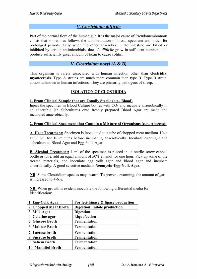
Islamic University-Gaza Medical Laboratory Science Department
_____________________________________________________________________ Diagnostic medical microbiology [81] Dr. Abdelraouf A. Elmanama
V. Clostridium difficile Part of the normal flora of the human gut. It is the major cause of Pseudomembranous colitis that sometimes follows the administration of broad spectrum antibiotics for prolonged periods. Only when the other anaerobes in the intestine are killed or inhibited by certain antimicrobials, does C. difficile grow in sufficient numbers, and produce sufficiently great amount of toxin to cause colitis.
V. Clostridium novyi (A & B) This organism is rarely associated with human infection other than clostridial myonecrosis. Type A strains are much more common than type B. Type B strain, almost unknown in human infections. They are primarily pathogens of sheep.
ISOLATION OF CLOSTRIDIA
1. From Clinical Sample that are Usually Sterile (e.g., Blood) Inject the specimen in Blood Culture bottles with CO2 and incubate anaerobically in an anaerobic jar. Subcultures onto freshly prepared Blood Agar are made and incubated anaerobically. 2. From Clinical Specimens that Contain a Mixture of Organisms (e.g., Abscess): A. Heat Treatment: Specimen is inoculated to a tube of chopped-meat medium. Heat at 80 oC for 10 minutes before incubating anaerobically. Incubate overnight and subculture to Blood Agar and Egg-Yolk Agar. B. Alcohol Treatment: 1 ml of the specimen is placed in a sterile screw-capped bottle or tube, add an equal amount of 50% ethanol for one hour. Pick up some of the treated materials, and inoculate egg yolk agar and blood agar and incubate anaerobically. A good selective media is Neomycin-Egg-Yolk Agar. NB: Some Clostridium species may swarm. To prevent swarming, the amount of gar is increased to 4-6%. NB: When growth is evident inoculate the following differential media for identification:
1. Egg-Yolk Agar For lecithinase & lipase production 2. Chopped Meat Broth Digestion; indole production 3. Milk Agar Digestion 4. Gelatine agar Liquefaction 5. Glucose Broth Fermentation 6. Maltose Broth Fermentation 7. Lactose broth Fermentation 8. Sucrose broth Fermentation 9. Salicin Broth Fermentation 10. Mannitol Broth Fermentation
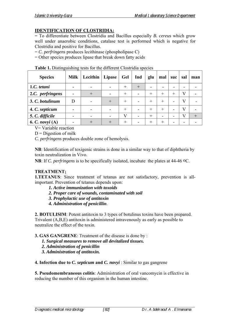
Islamic University-Gaza Medical Laboratory Science Department
_____________________________________________________________________ Diagnostic medical microbiology [82] Dr. Abdelraouf A. Elmanama
IDENTIFICATION OF CLOSTRIDIA: = To differentiate between Clostridia and Bacillus especially B. cereus which grow well under anaerobic conditions, catalase test is performed which is negative for Clostridia and positive for Bacillus. = C. perfringens produces lecithinase (phospholipase C) = Other species produces lipase that break down fatty acids Table 1. Distinguishing tests for the different Clostridia species
Species Milk Lecithin Lipase Gel Ind glu mal suc sal man
1.C. tetani - - - + + - - - - - 2.C. perfringens - + - + - + + + V - 3. C. botulinum D - + + - + + - V -
4. C. septicum - - - + - + + - V - 5. C. difficile - - - V - + - - V + 6. C. novyi (A) - + + + - + + - - -
V= Variable reaction D = Digestion of milk C. perfringens produces double zone of hemolysis. NB: Identification of toxigenic strains is done in a similar way to that of diphtheria by toxin neutralization in Vivo. NB: If C. perfringens is to be specifically isolated, incubate the plates at 44-46 oC. TREATMENT; 1.TETANUS: Since treatment of tetanus are not satisfactory, prevention is all-important. Prevention of tetanus depends upon:
1. Active immunization with toxoids 2. Proper care of wounds, contaminated with soil 3. Prophylactic use of antitoxin 4. Administration of penicillin.
2. BOTULISIM: Potent antitoxin to 3 types of botulinus toxins have been prepared. Trivalent (A,B,E) antitoxin is administered intravenously as early as possible to neutralize the effect of the toxin. 3. GAS GANGRENE: Treatment of the disease is done by :
1. Surgical measures to remove all devitalized tissues. 2. Administration of penicillin 3. Administration of antitoxin.
4. Infection due to C. septicum and C. novyi : Similar to gas gangrene 5. Pseudomembraneous colitis: Administration of oral vancomycin is effective in reducing the number of this organism in the human intestine.

Islamic University-Gaza Medical Laboratory Science Department
_____________________________________________________________________ Diagnostic medical microbiology [83] Dr. Abdelraouf A. Elmanama
Nagler’s Reaction for C. perfringensNagler’s Reaction for C. perfringens
Half with antitoxin
Half without antitoxin
LEYMA PLATE
OpacityOpacity
•Divide LEYMA plate into two halves.•With a sterile swab cover one half with C. perfringens antitoxin & dry it•Inoculate the test organism at right angles to center line. •Similarly inoculate a negative control•Incubate anaerobically. •Opacity in around inoculum of test organism in half without antitoxin and •No opacity in half with antitoxin
Test organismTest organismTest organism
Negative controlNegative controlNegative control
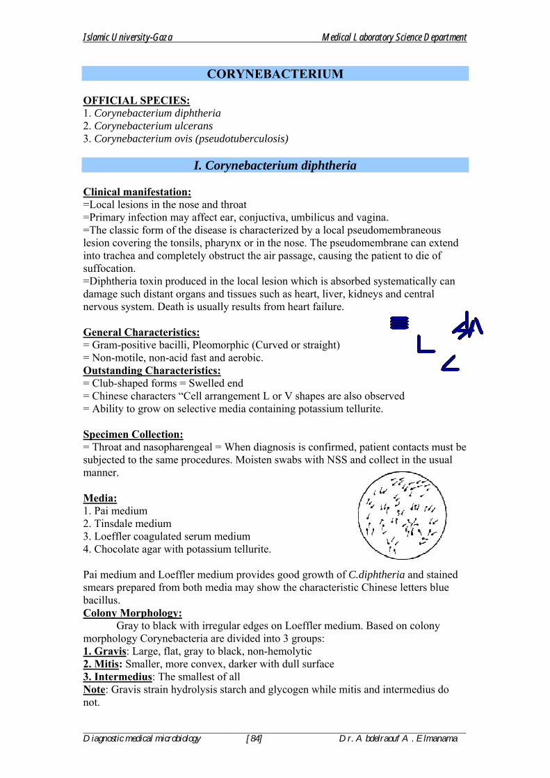
Islamic University-Gaza Medical Laboratory Science Department
_____________________________________________________________________ Diagnostic medical microbiology [84] Dr. Abdelraouf A. Elmanama
CORYNEBACTERIUM OFFICIAL SPECIES: 1. Corynebacterium diphtheria 2. Corynebacterium ulcerans 3. Corynebacterium ovis (pseudotuberculosis)
I. Corynebacterium diphtheria Clinical manifestation: =Local lesions in the nose and throat =Primary infection may affect ear, conjuctiva, umbilicus and vagina. =The classic form of the disease is characterized by a local pseudomembraneous lesion covering the tonsils, pharynx or in the nose. The pseudomembrane can extend into trachea and completely obstruct the air passage, causing the patient to die of suffocation. =Diphtheria toxin produced in the local lesion which is absorbed systematically can damage such distant organs and tissues such as heart, liver, kidneys and central nervous system. Death is usually results from heart failure. General Characteristics: = Gram-positive bacilli, Pleomorphic (Curved or straight) = Non-motile, non-acid fast and aerobic. Outstanding Characteristics: = Club-shaped forms = Swelled end = Chinese characters “Cell arrangement L or V shapes are also observed = Ability to grow on selective media containing potassium tellurite. Specimen Collection: = Throat and nasopharengeal = When diagnosis is confirmed, patient contacts must be subjected to the same procedures. Moisten swabs with NSS and collect in the usual manner. Media: 1. Pai medium 2. Tinsdale medium 3. Loeffler coagulated serum medium 4. Chocolate agar with potassium tellurite. Pai medium and Loeffler medium provides good growth of C.diphtheria and stained smears prepared from both media may show the characteristic Chinese letters blue bacillus. Colony Morphology: Gray to black with irregular edges on Loeffler medium. Based on colony morphology Corynebacteria are divided into 3 groups: 1. Gravis: Large, flat, gray to black, non-hemolytic 2. Mitis: Smaller, more convex, darker with dull surface 3. Intermedius: The smallest of all Note: Gravis strain hydrolysis starch and glycogen while mitis and intermedius do not.

Islamic University-Gaza Medical Laboratory Science Department
_____________________________________________________________________ Diagnostic medical microbiology [85] Dr. Abdelraouf A. Elmanama
Isolation and Identification:
24 hours 2-8 hours
18 hours Biochemical tests: The colonial morphology of C. diphtheria on Tinsdale medium is the most important characteristics in differentiating it from other species of corynebacteria. =Urease Negative =Nitrate Positive =Gelatin liquefaction: Negative Fermentation reactions of various carbohydrates. For accurate differential characteristics, refer to the following table.
Swab
Tellurite Agar CA or BA Loeffler or Pai slant
Subculture to Tellurite agar
Examine for Beta-hemolytic Streptococci
Stain with Methylene blue
Loeffler or Pai slant Agar medium for In-Vitro toxigenicity test
Broth medium for In-Vivo toxigenicity test
Biochemical tests Smear stained with Methylene blue
Pick colonies resembling C. diphtheria
Inoculate suspected colonies onto
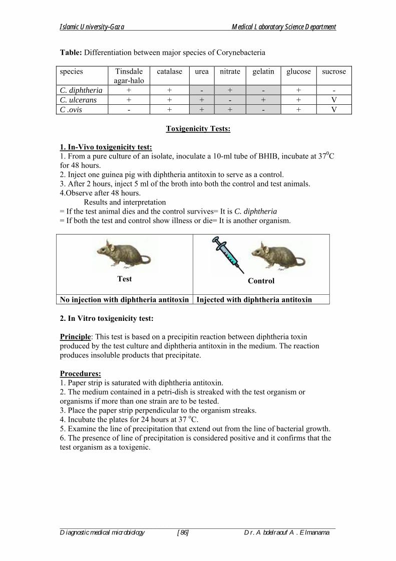
Islamic University-Gaza Medical Laboratory Science Department
_____________________________________________________________________ Diagnostic medical microbiology [86] Dr. Abdelraouf A. Elmanama
Table: Differentiation between major species of Corynebacteria species Tinsdale
agar-halo catalase urea nitrate gelatin glucose sucrose
C. diphtheria + + - + - + - C. ulcerans + + + - + + V C .ovis - + + + - + V
Toxigenicity Tests: 1. In-Vivo toxigenicity test: 1. From a pure culture of an isolate, inoculate a 10-ml tube of BHIB, incubate at 370C for 48 hours. 2. Inject one guinea pig with diphtheria antitoxin to serve as a control. 3. After 2 hours, inject 5 ml of the broth into both the control and test animals. 4.Observe after 48 hours. Results and interpretation = If the test animal dies and the control survives= It is C. diphtheria = If both the test and control show illness or die= It is another organism.
Test
Control
No injection with diphtheria antitoxin Injected with diphtheria antitoxin 2. In Vitro toxigenicity test: Principle: This test is based on a precipitin reaction between diphtheria toxin produced by the test culture and diphtheria antitoxin in the medium. The reaction produces insoluble products that precipitate. Procedures: 1. Paper strip is saturated with diphtheria antitoxin. 2. The medium contained in a petri-dish is streaked with the test organism or organisms if more than one strain are to be tested. 3. Place the paper strip perpendicular to the organism streaks. 4. Incubate the plates for 24 hours at 37 oC. 5. Examine the line of precipitation that extend out from the line of bacterial growth. 6. The presence of line of precipitation is considered positive and it confirms that the test organism as a toxigenic.
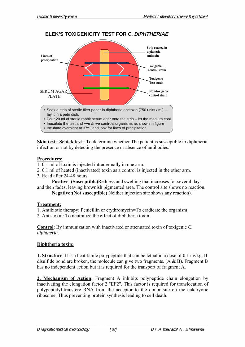
Islamic University-Gaza Medical Laboratory Science Department
_____________________________________________________________________ Diagnostic medical microbiology [87] Dr. Abdelraouf A. Elmanama
ELEK’S TOXIGENICITY TEST FOR C. DIPHTHERIAE
Strip soaked in diphtheria antitoxin
Strip soaked in diphtheria antitoxin
Toxigeniccontrol strainToxigeniccontrol strain
SERUM AGAR PLATE
ToxigenicTest strainToxigenicTest strain
Non-toxigenic control strainNon-toxigenic control strain
Lines ofprecipitationLines ofprecipitation
• Soak a strip of sterile filter paper in diphtheria antitoxin (750 units / ml) –lay it in a petri dish.
• Pour 20 ml of sterile rabbit serum agar onto the strip – let the medium cool• Inoculate the test and +ve & -ve controls organisms as shown in figure• Incubate overnight at 37oC and look for lines of precipitation
Skin test= Schick test= To determine whether The patient is susceptible to diphtheria infection or not by detecting the presence or absence of antibodies. Procedures: 1. 0.1 ml of toxin is injected intradermally in one arm. 2. 0.1 ml of heated (inactivated) toxin as a control is injected in the other arm. 3. Read after 24-48 hours. Positive: (Susceptible)Redness and swelling that increases for several days and then fades, leaving brownish pigmented area. The control site shows no reaction. Negative:(Not susceptible) Neither injection site shows any reaction). Treatment: 1. Antibiotic therapy: Penicillin or erythromycin=To eradicate the organism 2. Anti-toxin: To neutralize the effect of diphtheria toxin. Control: By immunization with inactivated or attenuated toxin of toxigenic C. diphtheria. Diphtheria toxin: 1. Structure: It is a heat-labile polypeptide that can be lethal in a dose of 0.1 ug/kg. If disulfide bond are broken, the molecule can give two fragments. (A & B). Fragment B has no independent action but it is required for the transport of fragment A. 2. Mechanism of Action: Fragment A inhibits polypeptide chain elongation by inactivating the elongation factor 2 "EF2". This factor is required for translocation of polypeptidyl-transfere RNA from the acceptor to the donor site on the eukaryotic ribosome. Thus preventing protein synthesis leading to cell death.
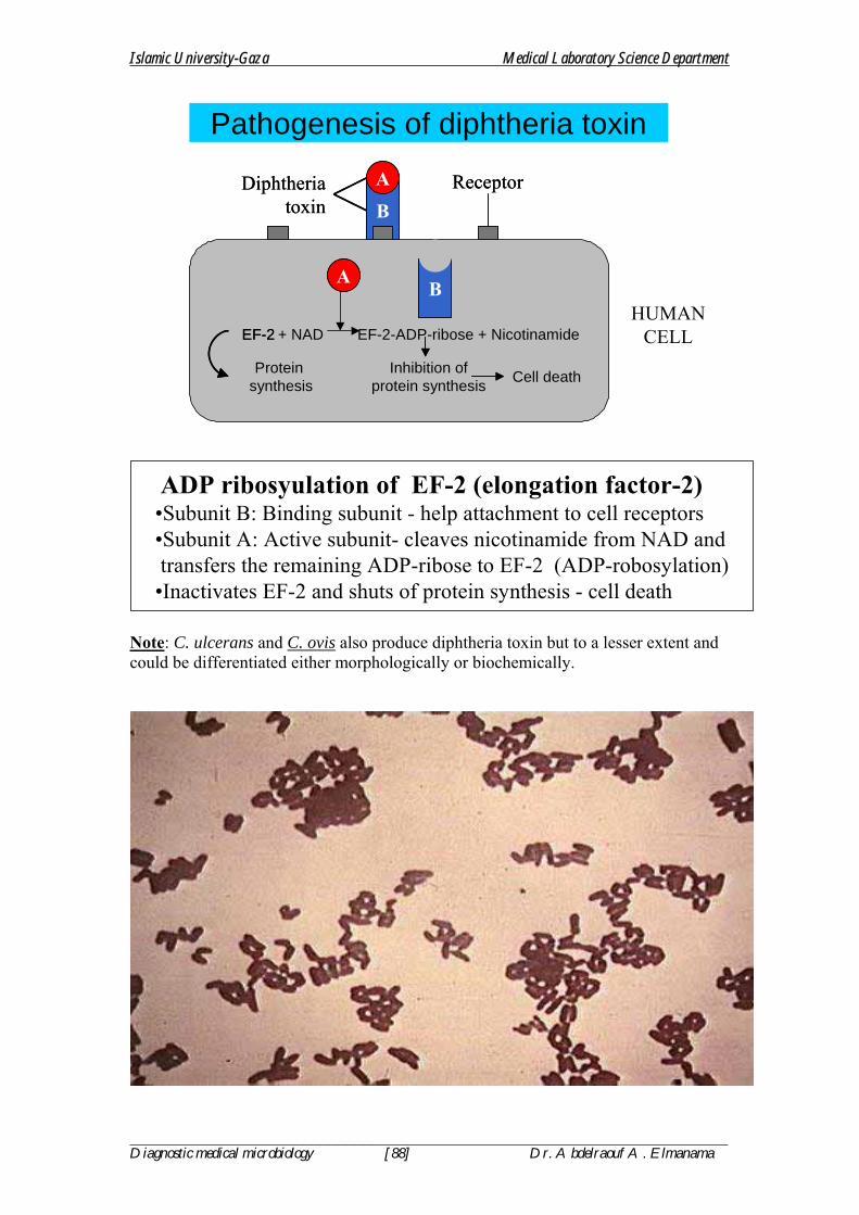
Islamic University-Gaza Medical Laboratory Science Department
_____________________________________________________________________ Diagnostic medical microbiology [88] Dr. Abdelraouf A. Elmanama
BA
BA
+ NAD EF-2-ADP-ribose + Nicotinamide+ NAD EF-2-ADP-ribose + Nicotinamide
Inhibition ofprotein synthesis
Inhibition ofprotein synthesis Cell deathCell death
Pathogenesis of diphtheria toxin
HUMANCELL
B
ADiphtheriatoxin B
ADiphtheriatoxin
ReceptorReceptor
Protein synthesis
EF-2
Protein synthesis
EF-2
ADP ribosyulation of EF-2 (elongation factor-2)•Subunit B: Binding subunit - help attachment to cell receptors•Subunit A: Active subunit- cleaves nicotinamide from NAD and transfers the remaining ADP-ribose to EF-2 (ADP-robosylation)
•Inactivates EF-2 and shuts of protein synthesis - cell death
Note: C. ulcerans and C. ovis also produce diphtheria toxin but to a lesser extent and could be differentiated either morphologically or biochemically.
Islamic University-Gaza Medical Laboratory Science Department
_____________________________________________________________________ Diagnostic medical microbiology [89] Dr. Abdelraouf A. Elmanama
Erysipelothrix insidiosa E. rhusiopathiae
Disease: Erysipleoid (red skin) = it has three distinct clinical enteities: 1. Erysipeloid : Localized cutaneous infection usually on the finger and hands. 2. Generalized cutaneous infection. 3. Septicemia Organism: - Gram-positive rods - Non-motile - Non-spore forming - Facultative anaerobe Specimen and media: - Usually skin biopsy from the advancing borders of the lesion. - Blood specimen if septicemia is suspected. - Culture media: 1% glucose broth and blood agar NB: Make gram staining for the organism from the clinical specimen and the growth on blood agar. Biochemical tests used in the identification: - Test tube brush appearance in gelatine stabs - H2S production - Acidification of slant and butt in TSIA with no gas production. - To differentiate this organism from Listeria monocytogenes, perform the catalase test. Listeria monocytogenes is catalase Positive. Treatment: - Penicillin is the drug of choice
Listeria monocytogenes
Disease: Listerosis = it has four forms 1. Non-specific flu-like illness during pregnancy or puerperal sepsis 2. Neonatal sepsis and later meningitis 3. Sepsis or meningitis in immunocompromised patients. 4. Food poisoning. Organism: - Gram-positive rods (Shorter & Thinner). - Non-spore forming - Facultative anaerobe - Beta hemolytic on Blood agar.

Islamic University-Gaza Medical Laboratory Science Department
_____________________________________________________________________ Diagnostic medical microbiology [90] Dr. Abdelraouf A. Elmanama
Biochemical Characteristics: - Motile (Umbrella-type growth in semi-solid media) - Motile (Tumbling motility as seen by the hanging drop technique) - Catalase ( + ) - V.P ( + ) Specimen and Culture media: - Blood, cerebrospinal fluid (CSF) and genital tract secretion. - Media : BHIA + 5% sheep blood and BHIB. - Selective Listeria Agar SLA = Brownish to black discoloration is usually seen around the colonies of Listeria monocytogenes. Identification: 1. The demonstration of Gram-positive bacilli from stained smears (CSF and other fluids or food samples). 2. Culture and Isolation: On Blood Agar or Selective Listeria Agar 3. Biochemical tests: Catalase test, V.P, and motility test 4. Serological test: Agglutination with specific sera. Treatment: Combined therapy of penicillin plus aminoglycosides. Tetracycline also may be used as a second line if the patient is sensitive to penicilllin or aminoglycosides.

Islamic University-Gaza Medical Laboratory Science Department
_____________________________________________________________________ Diagnostic medical microbiology [91] Dr. Abdelraouf A. Elmanama
MYCOBACTERIA
SPECIES OF MEDICAL IMPORTANCE: 1. Mycobacterium tuberculosis complex
• M. tuberculosis • M. bovis • M. africanum
2. Non-tuberculosis mycobacteria (NTM) a) Atypical mycobacteria
• M. avium-intracellular • M. marinum • M. scrofulaceum
b) Non-cultivable • M. leprae
General Characteristics of Mycobacteria: 1. Acid fast straight or slightly curved rods. 2. Non- motile 3. Non-spore forming 4. Aerobic (growth is enhanced by the presence of 5-10% CO2 5. Slow growing bacteria (Average doubling time is 12 hours), fastidious in nutrition. 6. Resist staining with basic dyes as a result of their high-cell wall lipid contents.
I. Tuberculosis Mycobacterium tuberculosis
Clinical Manifestation: Organisms is droplets of 1-5 um are inhaled and reaches alveoli. The disease results from establishment and proliferation of virulent organisms and interaction with the host. The production and development of lesions and their healing or progression are determined chiefly by the number of mycobacteria and their multiplication and the resistance and hypersensitivity of the host. Two types of lesion are produced as a result of M. tuberculosis infection:
LUNGSsubpleural area
of mid zone
MTB inhaled
LUNGSsubpleural area
of mid zone
MTB inhaledSurvive & multiply
in macrophagesSurvive & multiply
in macrophagesPresented to
T- lymphocytesPresented to
T- lymphocytes
Produce lymphokinesattract monocytes to
site of infection
Produce lymphokinesattract monocytes to
site of infection
Multinucleated giant cells
Multinucleated giant cells
Limit bacterial multiplication
Limit bacterial multiplication
PATHOGENESIS OF PRI. TB
GRANULOMA
Lymphnode
Lymphnode
BloodBloodBlood
Differentorgans
Differentorgans
Monocytes transform toactivated macrophages
(Epitheloid cells)
Monocytes transform toactivated macrophages
(Epitheloid cells)
Monocytes transform toactivated macrophages
(Epitheloid cells)
Ingested bymacrophages
MTB
Ingested bymacrophagesIngested by
macrophages
MTB

Islamic University-Gaza Medical Laboratory Science Department
_____________________________________________________________________ Diagnostic medical microbiology [92] Dr. Abdelraouf A. Elmanama
1. Exudative type: This consist of an acute inflammatory reaction, with edema fluid, polymorphonuclear cells, and later, monocytes around the tubercle bacilli. This type is particularly seen in lung tissues, where it resembles bacterial pneumonia. 2. Productive type: When fully developed, this lesion, a chronic granuloma, consist of three zones: A. A central area of large multinucleated giant cells containing M. tuberculosis. B. A mid-zone of pale epithelial cells C. A peripheral zone of fibroblast, lymphocytes and monocytes. This lesion is called a tubercle. Septicemia usually occur early in the course of infection and meningitis is not uncommon. Specimen: Fresh sputum, gastric aspirate or washing, urine, pleural fluid, cerebrospinal fluid, joint fluid, biopsy materials. Stained smears: Sputum or sediments from other body fluids is stained for acid fast bacilli by Ziel-Neelsen technique. Culture: Sputum must undergo concentration procedures prior culturing: Procedures: 1. Mix an equal volume of sputum with 4% sodium hydroxide containing 0.004% phenol red in 100 ml tube containing glass beads. Homogenize by shaking for few minutes. 2. Centrifuge at 3000 rpm for 20-30 minutes. 3. Decant the supernatant fluid into a disinfectant, leaving little fluid to resuspend the sediments. Add one drop of phenol red indicator. 4. Using sterile pipette, add 2N HCl drop by drop to a definite yellow end point. 5. Inoculate the sediments to egg media. Two Lowenstein-Jensen slopes. 6. Prepare a smear from sedimented materials and stain with Acid-Fast staining procedures. NB: Incubation of the un inoculated media is continued for up to 8 weeks. Increased CO2 tension enhances growth NB: Colony morphology: Bread-crumbs appearance. Lowenstein Jensen Medium – growth in 4-6 weeks Middlebrook Agar (7H11) – growth 2-3 weeks Biochemical Identification: 1. Niacin test: Only M. tuberculosis and M. simiae are positive for this test PROCEDURES: 1. Add one ml of sterile distilled water to a 3-4 weeks old culture on L.J medium and hold the tube so that the water covers the medium around the colonies. 2. After 15 minutes, remove 0.5 ml of the liquid containing extracted niacin, and transfer it to a clean, screw-capped test tube. 3. Add 0.5 ml of 4% aniline solution and 0.5 ml of 10% cyanogen bromide. 4. If yellow color appears immediately, niacin is present.
Islamic University-Gaza Medical Laboratory Science Department
_____________________________________________________________________ Diagnostic medical microbiology [93] Dr. Abdelraouf A. Elmanama
NB: To differentiate M. tuberculosis from M. simiae. M.simiae produces pigment while M. tuberculosis does not. Other Biochemical Tests: 1. Nitrate reduction test 2. Urease test 3. Tellurite reduction test 4. Sodium chloride tolerance test 5. Pigment production test 6. Catalase test 7. Arylsulfate test 8. Growth on MacConkey agar SEROLOGICAL TEST: 1. Skin test: Tuberculin test: An intradermal injection composed of one of these extracts: 1. Old tuberculin: Crude extract from the filtrate of M. tuberculosis broth culture. Also known as Tuberculin-Koch. 2. Purified Protein Derivatives: (PPD): Purified extract. TUBERCULIN (MONTOUX) TEST Purified Protein Derivative (PPD) from tubercle bacilli (standardized) in Tuberculin Units - TU) is injected intradermaly DOSE 5 TU is usual dose – if test negative; increase dose to 250 TU POSITIVE TEST The test is read after 48 – 72 hours Induration of ≥ 10 mm with erythema Induration of 5-9 mm – low level sensitization with tubercle bacilli or cross reacting mycobacteria. In AIDS patients: 5 mm induration – positive test. INTERPRETATION OF TUBERCULIN TEST Positive Test
• Active disease • Person infected by M. tuberculosis at sometime in life • Person infected with strongly cross reacting other mycobacteria • Previous vaccination with BCG • Child < 5 years if not vaccinated: active disease.
Negative Test
• No induration or < 5 mm • In healthy individual – not infected with MTB • Pre-hypersensitivity stage of primary infection
False negative test • Early TB (test becomes positive after 4-6 weeks of infection) • Miliary TB • Immunosuppression (AIDS) • Steroid therapy
Islamic University-Gaza Medical Laboratory Science Department
_____________________________________________________________________ Diagnostic medical microbiology [94] Dr. Abdelraouf A. Elmanama
Treatment: Because M. tuberculosis infections are hard to treat and only few antibiotics has activity against it, and because of the fear of emerging of resistant strains, tuberculosis is treated with 2 or 3 antibiotics given as combined therapy. Active antibiotics against M. tuberculosis include: 1. Isonizide (INH) 2. Rifamycin 3. Streptomycin 4. Ethambutol 5. Para-aminosalicylic acid PAS 6. Cycloserine 7. Pyrazinamide 8. Viomycin Treatment for complete recovery may take as long as one year. The shortest term may be of 6 months with the administration of combined drugs (Isonizid, rifamycin, streptomycin).
ATYPICAL MYCOBACTERIA M. avium – interacellulare complex
Worldwide distribution Source – natural water Diseases TB in birds Cervical lymphadenitis in children Disseminated TB in AIDS patients DIFFERENCES OF ATYPICAL FROM TYPICAL MYCOBACTERIA Colonial morphology Niacin test – negative Relatively more resistant to anti-TB drugs Less acid fastness Diseases less invasive
II. Mycobacterium leprae Leprosy
General Characteristics: = Typical acid fast bacilli, singly or in parallel bundles. = Regularly found in smears or scrapings from skin or mucus membrane in lepromatous leprosy. = The organism have not been grown on artificial media. Clinical manifestation: The lesions involve the cooler tissue of the body, skin, superficial nerves, nose, pharynx. The skin lesions may occur as pale, anesthetic 1-10 cm in diameter. Neurologic disturbances are manifested by nerve infiltration and thickening.

Islamic University-Gaza Medical Laboratory Science Department
_____________________________________________________________________ Diagnostic medical microbiology [95] Dr. Abdelraouf A. Elmanama
The disease is divided into two distinct types: 1. Lepromatous leprosy: The course is progressive, with nodular skin lesions; slow, symmetric nerve involvement, abundant acid-fast bacilli in the skin lesion; continuous bacteremia and negative skin test (lepromin test) 2. Tuberculoid leprosy: The course is non-progressive with macular skin lesions, severe asymmetric nerve involvement of sudden onset with few bacilli present in the lesion. Diagnosis: Scrapping with a scalpel blade from skin, nasal mucosa, or from a biopsy of ear lobe skin are smeared on a slide and stained by Zeil-Neelsen technique, and the demonstration of typical acid fast bacilli. Treatment: Combined therapy of dapsone, rifamycin and clofazimine
Islamic University-Gaza Medical Laboratory Science Department
_____________________________________________________________________ Diagnostic medical microbiology [96] Dr. Abdelraouf A. Elmanama
Francisella tularensis Tularemia “Rabbit fever”
Characteristics of the Organism: 1. Gram-negative rods “Shows variable shapes ranging from coccobacilli, to cocci” 2. Strict aerobe 3. Requires enriched medium for growth “Cystine-Glucose-Blood Agar” CGBA 4. Non-motile 5. Remain viable for long periods in water, soil......etc. 6. Highly infectious. Specimen and cultures must be handled with great care. Clinical Manifestation: Tularemia in human vary according to: 1. Route of infection 2. The infecting dose 3. The type of organism (A or B strain) The disease may be transmitted through the skin and mucus membrane including the conjuctiva through the bite of infective ticks. Transmission may also occur through the respiratory route or gastrointestinal tract through ingesting contaminated meat of infected animals. THE ULCEROGLANDULAR: The most common, with slowly healing ulcer at the site of entry and regional lymph nodes involvement is observed. This infection results from injury while handling infected animal carcasses or from the bite of a blood-sucking arthropod such as ticks and flies. OCULOGLANDULAR: Can result from rubbing contaminated materials into the eye. Conjuctivitis occur and regional lymph node may be enlarged and suppurate. THE PULMONARY OR PNEUMONIC: Can result from inhaling infectious aerosols or dust. Pneumonia and pleuritis are associated with severe tularemia. TYPHOIDAL, GIT & OROPHARYNGEAL INFECTION: Results from the ingestion of contaminated food. Symptoms begin as flu-like associated with vomiting, headache, fever and prostration. Diarrhea usually accompany these symptoms. Subspecies of F. tularnsis 1. F. tularensis A : More virulent than B. It is also referred to as tick-deerfly-sheep type and occurs naturally in North America. Fatality rate among untreated patient is 5-7%. 2. F. tularensis B: It is referred to as the Beaver-Muskrat water type. It produces mild (Subclinical) infection in human. It have frequently been isolated from natural running water. NB: To differentiate between the two, 1% glycerol is added to Cystin-glucose-heart agar without blood and enough bromothymol blue indicator. TYPE A: Turns the medium yellow (acid) TYPE B: Turns the medium dark blue (alkaline)

Islamic University-Gaza Medical Laboratory Science Department
_____________________________________________________________________ Diagnostic medical microbiology [97] Dr. Abdelraouf A. Elmanama
Collection of specimen: 1. Exudates from cutaneous or mucosal ulcers or lymph node aspirate. 2. Blood or sputum for pulmonary infection 3. Blood specimen for serologic for measuring antibody titer 4. Specimens (biopsy) from enlarged liver or spleen., Diagnostic procedures: A. Microscopic appearance: - Short gram-negative non-motile bacilli - Morphologically may be similar to Yersinia species and shows bipolar reaction. B. Cultural Characteristics - On CGBA: colonies resemble a medium sized mercury droplets surrounded by a small zone of partial hemolysis. Colonies often requires 2-4 days at 37 oC to reach a diameter of 1 mm. C. Inoculation of the specimen: - Primary isolation from specimen may be difficult even though it may contain large numbers of F. tularensis. - Contaminating organisms can easily overgrow cultures or produce large amounts of acids to prevent the growth of F. tularensis on the enriched media needed for isolation. - Cystine glucose blood agar is the medium of choice. It is commercially available or can be prepared in the laboratory. D. Biochemical Identification: - Biochemical tests are of a little value in identifying the organism. 1. Tube agglutination test: 2. The slide agglutination method: 3. The precipitin test: Can be used to measure antibody titer and demonstrate the presence of antigen. This test is useful for grossly contaminated, even decayed specimen such as liver and spleen of naturally infected, field-collected animal specimen, 4. Hemagglutination and complement fixation tests are of limited use. 5. Skin test have been used for prevalence of past infection in large population. Treatment and antibiotic sensitivity: Streptomycin, Chloramphenicol and tetracycline are used to eradicate this organism.

Islamic University-Gaza Medical Laboratory Science Department
_____________________________________________________________________ Diagnostic medical microbiology [98] Dr. Abdelraouf A. Elmanama
GENUS: HAEMOPHILUS SPECIES: 1. H. influenza (Biotype B is the most important) 2. H. parainfluenza 3. H. haemplyticus 4. H. ducreyi 5. H. aphrophilus 6. H. paraphrophilus 7. H. segnis General Characteristics: 1. Gram-negative small rods (pleomorphic and may show variable gram reaction and has poor absorbance of stain) 2. Facultative anaerobe 3. Require CO2 for growth 4. Require growth factors such as factor X (hemin) and factor V (NAD) 5. May be capsulated or non-capsulated. 6. Most strains are indole positive.
H. influenza Pathogenicity: In human, H. influenza is the most common etiologic agent of acute bacterial meningitis in young children. H. influenza is the most important pathogen among the other species of the genus Haemophilus. Among the most common infections caused by this organism are: 1. It is the most common cause of meningitis in children 2. Cause epiglotitis 3. Septic arthritis 4. Bronchitis and pneumonia 5. Acute and chronic otitis media 6. Endocarditis and pericarditis. 7. May enter the blood stream of women during birth causing puerperal sepsis, the complication of which may threaten the mother life. 8. Eye infection (Conjuctivitis) Normal Habitat: It forms part of the normal flora of the upper respiratory tract of man. Speciemn: C.S.F, nasopharengeal specimen, blood, pus and sputum for culture. As a general precaustion, specimen suspected of containing H.influenza must not be refrigerated. For H.ducreyi, a genital specimen is required. Microscopy: Small, non-motile, gram-negative (may show gram variable reaction). Bacilli with long thread-like forms of H. influenza may be seen in CSF or older culture.
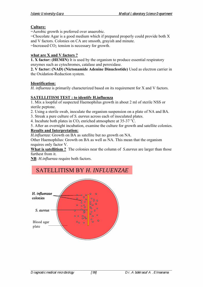
Islamic University-Gaza Medical Laboratory Science Department
_____________________________________________________________________ Diagnostic medical microbiology [99] Dr. Abdelraouf A. Elmanama
Culture: =Aerobic growth is preferred over anaerobic. =Chocolate Agar is a good medium which if prepared properly could provide both X and V factors. Colonies on CA are smooth, grayish and minute. =Increased CO2 tension is necessary for growth. what are X and V factors ? 1. X factor: (HEMIN) It is used by the organism to produce essential respiratory enzymes such as cytochromes, catalase and peroxidase. 2. V factor: (NAD) (Nictonamide Adenine Dinucleotide) Used as electron carrier in the Oxidation-Reduction system. Identification: H. influenza is primarily characterized based on its requirement for X and V factors. SATELLITISM TEST : to identify H.influenza 1. Mix a loopful of suspected Haemophilus growth in about 2 ml of sterile NSS or sterile peptone. 2. Using a sterile swab, inoculate the organism suspension on a plate of NA and BA. 3. Streak a pure culture of S. aureus across each of inoculated plates. 4. Incubate both plates in CO2 enriched atmosphere at 35-37 oC. 5. After an overnight incubation, examine the culture for growth and satellite colonies. Results and Interpretation: H.influenza: Growth on BA as satellite but no growth on NA. Other Haemophilus: Growth on BA as well as NA. This mean that the organism requires only factor V. What is satellitism ? The colonies near the column of S.aureus are larger than those furthest from it. NB: H.influenza require both factors.
S. aureusS. aureus
H. influenzaecoloniesH. influenzaecoloniesH. influenzaecolonies
SATELLITISM BY H. INFLUENZAE
Naeem
Blood agar plate

Islamic University-Gaza Medical Laboratory Science Department
_____________________________________________________________________ Diagnostic medical microbiology [100] Dr. Abdelraouf A. Elmanama
Biochemical Identification: (seldom used) Biochemical tests are used to differentiate species in the genus Haemophilus. The following tests are commonly employed: 1. Hemolysis on Blood agar 2. Indole test 3. Growth factors requirements: (factor X and V) 4. Urease test. Table Growth requirements and hemolytic activity of Haemophilus species.
Haemophilus Growth factors Hemolysis on BA X V 1.H. influenza + + - 2. H. parainfluenza - + - 3. H. haemolyticus + + + 4. H. aphrophilus + - - 5. H. ducreyi + - slight 6. H. segnis - + - 7. H. paraphrophilus - + - Treatment: Ampicillin if sensitive is the drug of choice. = Chloramphenicol = Tetracycline
HAEMOPHILUS DUCREYI Gram-negative coccobacilli Cultured on chocolate agar Require only X factor Causes Chancroid A sexually transmitted disease with soft ulcers on external genitalia with local lymphadenitis (bubo) Treatment: Ceftriaxone or ciprofloxacin
GARDENERELLA VAGINALIS Gram-negative coccobacilli Formerly called Haemophilus vaginalis Found in small numbers in vaginal secretions of 50% of normal women Disease: Bacterial Vaginosis Malodourous vaginal discharge Lab Diagnosis Gram-smear of Vaginal discharge Clue cells (epithelial cells covered with bacteria)

Islamic University-Gaza Medical Laboratory Science Department
_____________________________________________________________________ Diagnostic medical microbiology [101] Dr. Abdelraouf A. Elmanama
Bordetella pertussis
Disease: Pertussis = Transmitted by the respiratory tract rout from early case and possibly via carriers. The organism adheres to and multiply rapidly on the surface of the epithelium in the trachea and bronchi and interfere with ciliary action . Disintegrating organisms liberate a toxin that irritate surface cells, giving rise to catarrhal symptoms. Most B. pertussis strains contains peptide that promotes marked lymophocytosis in the host. The toxin is heat stable.
Activation of adenyl cyclaseActivation of adenyl cyclase
Pathogenesis of Pertussis Toxin
RES
P IR
ATO
RY
EPIT
HEL
IAL
CEL
L
ReceptorReceptor
+ NAD G-2-ADP-ribose + NicotinamideProtein G + NAD G-2-ADP-ribose + Nicotinamide+ NAD G-2-ADP-ribose + NicotinamideProtein G
ADP-ribosyulation of Protein G• Subunits B facilitate entry of subunit A (Active subunit) into cell• Subunit A cleaves nicotinamide from NAD and transfers the
remaining ADP-ribose to protein G• Activates adenyl cyclase to convert ATP to cAMP - secretions
AA
AA
ATP cAMP secretions ATP cAMP secretions
AB
BB
BB B
Cholera toxin AB
BB
BB B
Cholera toxin
Organism: - Short, ovoid, gram-negative bacilli - Aerobic - Complex, enriched media is required for isolation (Bordet-Gengou) Laboratory Diagnosis: - Nasopharyngeal swabs or cough droplets, expelled onto "Cough plates" held in front of the mouth of the patient during a paroxysms. - Incubate (Bordet-gengou`s plates) at 35 oC for 2-5 days. - Typical colonies resemble a 1mm droplets of mercury surrounded by a zone of hemolysis. - Colonies are confirmed with specific antiserum - Direct immunofluorescent staining of smears made from nasopharyngeal swabs may give a rapid positive test.
Islamic University-Gaza Medical Laboratory Science Department
_____________________________________________________________________ Diagnostic medical microbiology [102] Dr. Abdelraouf A. Elmanama
Other Species Differentiation
FEATURE B. pertussis B. parapertussis B. bronchiseptica
Growth on blood free media - + + Motility - - + Urease - + + Citrate - + + Nitrate reduction - - +
Treatment: Erythromycin is the drug of choice
Legionella pneumophila Introduction: Legionellosis was first recognized in association with the epidemic of legionnaires disease which occurred in Philadelphia in July 1976. Shortly following the epidemic, the causative agent was isolated from lung tissues of patient who died of this disease. The organism was named Legionella pneumophila. ORGANISM:
• Gram negative rods • Slow grower (2-5 days in enriched media containing L-cystein and
supplemented source of ferric iron) • Old culture may exhibit filamentous forms, swollen rods, and bizarre forms. • Stain poorly with gram stain unless safranin is applied for long periods. • Good growth obtained on Charcoal-Yeast Extract (CYE). • Good growth occur in an atmosphere containing 2.5% CO2 • Soluble pigment that results in brown coloration on Feely-Gorman agar. • Most strains are flagellated.
Biochemical Characteristics: - Weak oxidase positive - Strong catalase positive - Liquefy gelatine - No carbohydrate fermentation - No nitrate reduction - No urea hydrolysis - Hydrolysis of hippurate Positive (This differentiate it from other legionella species) Virulence factors: 1. Proteolytic enzymes 2. Exotoxin with lysed laboratory animal RBCs 3. Cytotoxin which caused inhibition of cell growth in laboratory animals.
Islamic University-Gaza Medical Laboratory Science Department
_____________________________________________________________________ Diagnostic medical microbiology [103] Dr. Abdelraouf A. Elmanama
Clinical Manifestation: Acute pneumonia begins 2-10 days after exposure with a brief prodorm of malaise, myalgia, and headache, followed rapidly by prostration, high fever, an rigors. Cough, dyspnea, pleuritic and abdominal pain, vomiting, diarrhea, and unexplained encephalopathy are often seen. Gram stain of sputum is not diagnostic. Leukocytosis, elevated ESR, proteinurea, hematuria and abnormal serum enzyme determination are common. The common causes of death due to respiratory failure and shock. Transmission: Via respiratory tract. Collection & Processing of Speciemn: Caution: Specimen must be handled in a biological safety cabinet. 1. Lung tissue obtained at autopsy or biopsy is optimally selected from areas of necrosis. A representative portion of specimen should be placed in 10% neutral formalin for Direct Fluorescent Antibody and histopathological examination. 2. Pleural fluid, transtracheal aspirate. Diagnosis: 1. Isolation and identification of Legionella from clinical specimens either on artificial media or guinea pig inoculation followed by embryonated hen`s egg. 2. Demonstration of a four-fold or greater rise in antibody titer from acute phase to convalescent phase by the IFA test. 3. Demonstration of the organism in clinical specimen by DFA. Treatment: Erythromycin is the drug of choice.
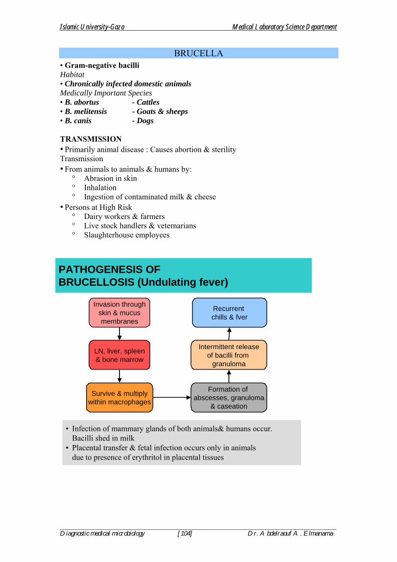
Islamic University-Gaza Medical Laboratory Science Department
_____________________________________________________________________ Diagnostic medical microbiology [104] Dr. Abdelraouf A. Elmanama
BRUCELLA • Gram-negative bacilli Habitat • Chronically infected domestic animals Medically Important Species • B. abortus - Cattles • B. melitensis - Goats & sheeps • B. canis - Dogs TRANSMISSION • Primarily animal disease : Causes abortion & sterility Transmission • From animals to animals & humans by:
° Abrasion in skin ° Inhalation ° Ingestion of contaminated milk & cheese
• Persons at High Risk ° Dairy workers & farmers ° Live stock handlers & veternarians ° Slaughterhouse employees
PATHOGENESIS OF BRUCELLOSIS (Undulating fever)
Invasion throughskin & mucusmembranes
LN, liver, spleen& bone marrowLN, liver, spleen& bone marrow
Survive & multiplywithin macrophagesSurvive & multiply
within macrophages
Formation of abscesses, granuloma
& caseation
Formation of abscesses, granuloma
& caseation
Intermittent releaseof bacilli from
granuloma
Intermittent releaseof bacilli from
granuloma
Recurrent chills & fverRecurrent chills & fver
• Infection of mammary glands of both animals& humans occur. Bacilli shed in milk
• Placental transfer & fetal infection occurs only in animals due to presence of erythritol in placental tissues
Islamic University-Gaza Medical Laboratory Science Department
_____________________________________________________________________ Diagnostic medical microbiology [105] Dr. Abdelraouf A. Elmanama
CLINICAL FEATURES OF BRUCELLOSIS (Undulating fever) • Incubation period: 1-3 weeks • Initially influenza-like • Undulating (rising & falling) fever for weeks & months • A chronic illness • Splenomegaly • Hepatomegaly • Enlarged lymph nodes
LAB DIAGNOSIS • Specimens Blood, LN & BM biopsy • Culture Require 5-10% CO2 • Serology
° Antibody titre of 1:160 or more (significant) ° Titre returns to normal within a year of successful treatment
TREATMENT • Ciprofloxacin May be up to 6 weeks to prevent relapses PREVENTION • Pasteurization of milk • Immunization of animals • Eradication of infected livestock • Minimize occupational exposure

Islamic University-Gaza Medical Laboratory Science Department
_____________________________________________________________________ Diagnostic medical microbiology [106] Dr. Abdelraouf A. Elmanama
Syphilis Treponema pallidum
General Characteristics of the Organism: 1. A thin, motile spiral organism 2. Can not be cultivated on artificial media. 3. Can not be visualized by light microscope 4. Rabbits could be infected with this organism.
•Are spiral flexible organisms that move without flagella•Multiply by transverse binary fission
Treponema : Many narrow regular coilsTreponema : Many narrow regular coils
Borrelia : Few wide, irregular coilsBorrelia : Few wide, irregular coils
Leptospira : Many very narrow coils with hooked endsLeptospira : Many very narrow coils with hooked ends
Clinical description of the disease: Syphilis is a chronic granulomatous disease that characteristically progress by stages of clinically apparent infections. Each stage has different manifestation and lesion morphology, and stages are separated by periods of latency. The only evidence of infection is a reactive serologic test during the latency periods. Except for congenital syphilis which is transmitted from infected mothers to the fetus across the placenta, syphilis is almost always transmitted by sexual exposure. PRIMARY STAGE: The primary syphilitic lesion, the chancre, appears. The chancre is most frequently a painless, ulcer-like lesion with raised borders and a necrotic base. The serum (exudate) from the chancre contains T. pallidum and is highly infectious. The chancre heals spontaneously without scaring over a period of 2-3 weeks. SECONDARY STAGE: It occur 6 weeks to 6 months after the chancre heals. Symptoms of this stage includes, headache and malaise, general lymphoadenopathy and a variety of skin rashes, hepatitis, meningitis, glmerulnephritis and nephritis. Nephrosis is a result of Antigen-antibody complexes deposited in the kidney. Secondary lesions heal spontenaneously over a period of several weeks and then enters in a latent stage number 2. TERTIARY STAGE: The destructive stage of the disease. It may begin 5-20 years after the initial infection. Only one third (1/3) of untreated patient develop tertiary which are of three types: 1. 80% are aneurysms (تمدد في األوعية الدموية) : Leading to dilatation of aortic valve 2. 15% are CNS disease and meningitis 3. 5% gummas (األورام الصمغية): Lesions which histologically resemble a tubercle. At this stage, bone and teeth deformation may occur.

Islamic University-Gaza Medical Laboratory Science Department
_____________________________________________________________________ Diagnostic medical microbiology [107] Dr. Abdelraouf A. Elmanama
CONGINITAL SYPHILIS: The fetus may be aborted, stillborn or liveborn or without clinical evidence of syphilis. The lesions of early congenital syphilis are of the secondary type and may be present at birth. Blindness secondary to optic atrophy. Deformed bones may also occur.
Entry of organisms at site of contact
Chancre & LN Contain a large no. of organisms
Go to regional LN(inflammed LN)
Go to regional LN(inflammed LN)
BacteremiaBacteremia
Chancre Formationat site of contact
After 3 weeks(A flat dull ulcer)
Chancre Formationat site of contact
After 3 weeks(A flat dull ulcer)
Chancre is painless &Heals spontaneouslyChancre is painless &Heals spontaneously
LABORATORY IDENTIFICATION: T. pallidum can not be cultivated in the laboratory In-Vitro. Only two methods are available for the diagnosis of T. pallidum. 1. DARK-FIELD MICROSCOPY: Most specific for infectious syphilis. • Clean the chancre with gauze moistened with sterile sodium chloride solution. Dry
it. • Scrap the edges of the chancre several times with the flat side of a sterile lancet.
Do not draw blood. • Press with dry sterile gauze. • Remove the swab and wait for a few minutes until a pinkish serous fluid appear.
Draw off the fluid with a Pasteur pipette. • Place a drop of the fluid on a thin glass slide. • Examine under the microscope using dark field condenser. Search for the
characteristic morphology of T. pallidum. (Motile thin spirochetes, rotating around their long axes.
2. SEROLOGICAL TESTING: Two types of antigens are used. A. Non-Treponemal Antigens Test: It utilizes "cardiolipin" as its antigen, which reacts with IgM or IgG immunoglobulin known as "reagin" antibody 1. Flocculation test (VDRL): (Venerial Disease Research Laboratories) This test is based on the fact that particles of the lipid antigen ( cardiolipin) remains dispersed in normal sera, but combines with reagin to form visible aggregate within a few minutes. Rapid Plasma Reagin test (RPR) is a good modification for rapid surveys.

Islamic University-Gaza Medical Laboratory Science Department
_____________________________________________________________________ Diagnostic medical microbiology [108] Dr. Abdelraouf A. Elmanama
2. Complement fixation test "Wasserman, Kolmer" This test is based on the fact that, reagin-containing sera fix complement in the presence of cardiolipin. NB: Biologic false results may occur with many patients having: 1. Malaria 2. Leprosy 3. Measles 4. Infectious mononucleosis 5. SLE B. Treponemal Antibody Test: 1. Fluorescent Treponema Antibody Absorption Test (FTA-ABS) This test employs indirect immunofluorescence (Killed T. pallidum + patient`s serum + labeled anti-human gammaglobulins). The test serum is added to the antigen (killed T. pallidum) which is fixed on a glass slide, incubated and then washed to remove excess serum. Conjugate (Labeled antihuman gammaglobulin) is added , incubated and then washed. This test is read microscopically with an ultraviolet light source. 2. T. pallidum immobilization test (TPI): Specific antibodies in the patient serum after the second week of infection could be demonstrated by their ability to immobilize actively motile T. pallidum, extracted from the chancre of infected rabbits. The test is read microscopically with an dark field microscope. 3. T. pallidum-Hemagglutination test (TP-HA): Red cells are treated to adsorb treponemal antigens on their surface. When mixed with serum containing antitreponemal antibodies, the cells become clumped. Clumping is considered as positive results. NB: VDRL and FTA-ABS tests can also be performed on spinal fluids. Antibodies do not reach the CSF from the blood stream but are probably formed in CNS in response to syphylitic infection. TREATMENT: Penicillin is the treatment of choice of syphilis Diseases related to Treponema: 1. Yaws: (Frambesia): is a tropical infection of the skin, bones and joints caused by T. pertenue 2. Pinta is a human skin disease endemic to Mexico, Central America, and South America. It is caused by infection with a spirochete, Treponema carateum, which is morphologically and serologically indistinguishable from the organism that causes syphilis. Pinta initially presents as a raised papule, followed by a generalized eruption of flat, reddened areas, and is followed by the development of bluish coloration and a subsequent loss of pigmentation. Unlike syphilis, it is transmitted by nonsexual skin contact, often between children living in conditions of poor hygiene. The disease can be treated with penicillin, tetracycline, or chloramphenicol, and can be prevented through contact tracing by public health officials.

Islamic University-Gaza Medical Laboratory Science Department
_____________________________________________________________________ Diagnostic medical microbiology [109] Dr. Abdelraouf A. Elmanama
LEPTOSPIRA INTERROGANS Causative agent of Leptospirosis : A zoonotic disease Habitat : Infected domestic live-stock & pets The organism settles in the kidney and excreted in urine Mode of Infection Direct Contact with: Urine of infected animal Water & soil recently contaminated with infected urine The organism enters body through: Skin lesions Conjunctival mucus membrane Ingestion High-Risk Groups Farmers Sewer workers LEPTOSPIROSIS Pathogenesis Incubation period : 1-2 weeks Bacteremia : organisms multiply in liver, spleen, kidney, meninges, conjunctiva Clinical Features Influenza-like followed by hepatitis & meningitis Weil’s Disease (severe leptospirosis) Jaundice, hemorrhage, renal failure Lab Diagnosis Dark field Microscopy of blood & CSF Sero-diagnosis Treatment & Prevention Penicillin Animal immunization Proper treatment and disposal of contaminated water

Islamic University-Gaza Medical Laboratory Science Department
_____________________________________________________________________ Diagnostic medical microbiology [110] Dr. Abdelraouf A. Elmanama
BORRELIA RECURRENTIS
Can be stained with Giemsa stain in blood film Can grow in media containing serum & tissue extracts High frequency of antigenic variation in major surface protein and is responsible for : Organism can escape immune system Relapses of disease Disease : Relapsing Fever Transmission : Tick-borne Relapsing Fever From infected rodents to human Louse-borne Relapsing Fever From infected human to human Clinical Features Bacteremia : Infect various organs A non-pruritic rash at bite site Fever, rigors and headache for weeks to months Weeks to months later: Cardiac and neurological symptoms Predominant arthritis One of the causes of PUO Lab Diagnosis Giemsa staining of blood smear Detection of IgM OR rising titer of IgG PCR Treatment & Prevention Amoxycillin Louse, tick & rodent control Hygienic measures
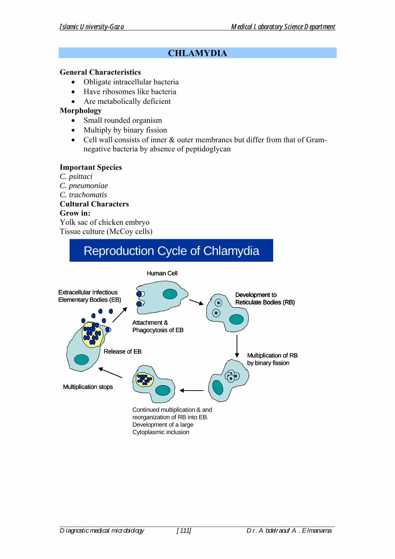
Islamic University-Gaza Medical Laboratory Science Department
_____________________________________________________________________ Diagnostic medical microbiology [111] Dr. Abdelraouf A. Elmanama
CHLAMYDIA General Characteristics
• Obligate intracellular bacteria • Have ribosomes like bacteria • Are metabolically deficient
Morphology • Small rounded organism • Multiply by binary fission • Cell wall consists of inner & outer membranes but differ from that of Gram-
negative bacteria by absence of peptidoglycan Important Species C. psittaci C. pneumoniae C. trachomatis Cultural Characters Grow in: Yolk sac of chicken embryo Tissue culture (McCoy cells)
Reproduction Cycle of Chlamydia
Attachment & Phagocytosis of EBAttachment & Phagocytosis of EB
Continued multiplication & and reorganization of RB into EB.Development of a large Cytoplasmic inclusion
Release of EBRelease of EB
Extracellular InfectiousElementary Bodies (EB)Extracellular InfectiousElementary Bodies (EB)
Development toReticulate Bodies (RB)Development toReticulate Bodies (RB)Development toReticulate Bodies (RB)
Multiplication of RB by binary fissionMultiplication of RB by binary fission
Multiplication stopsMultiplication stops
Human CellHuman Cell

Islamic University-Gaza Medical Laboratory Science Department
_____________________________________________________________________ Diagnostic medical microbiology [112] Dr. Abdelraouf A. Elmanama
PATHOGENESIS • Infect epithelial cells of mucous membranes & lungs • Virulence is due to: • Resistance to phagocytic killing by lysosomal enzymes • Heat-labile toxin • Competition with host cell for nutrients • Host’s immune response may account for inflammation & tissue destruction
CHLAMYDIA PSITTACI A zoonotic respiratory disease Natural habitat : birds Transmitted through inhalation of : Respiratory secretions & dust from faeces of infected birds Common in poultry workers Disease : Pneumonia (Psittacosis) Diagnosis Isolation of organism from sputum by tissue culture Complement fixation test to detect specific Abs Treatment Tetracycline in adults Erythromycin in babies
CHLAMYDIA PNEUMONIAE Also known as TWAR (TW – Taiwan & AR – acute respiratory) Cause atypical pneumonia like Mycoplasma pneumoniae Treatment Tetracycline in adults Erythromycin in babies
CHLAMYDIA TRACHOAMATIS 15 serotypes (A-L) Transmission: Through close personal contact like:
• Sexual • Passage through birth canal • Finger to eye or fomite to eye (Trachoma)
DISEASES 1. Trachoma
• Caused by serotypes A, B, Ba & C • One of the leading causes of blindness in developing countries with dry & hot
weather • Chronic conjunctivitis : leads to scarring of eye lids and cornea
2. Genital Tract Infections (Serotypes D-K)
Islamic University-Gaza Medical Laboratory Science Department
_____________________________________________________________________ Diagnostic medical microbiology [113] Dr. Abdelraouf A. Elmanama
• Non-gonococcal urethritis in men • A common cause of non-gonococcal urethritis • Mucopurrulent urethral discharge • May progress to epidydmitis & orchitis • Cervicitis & Vaginitis • Mucopurrulent vaginal discharge • Pelvic Inflammatory Disease (PID) • May lead to secondary infertility
3. Neonatal Infections (Caused by serotypes D-K) • Acquired from mother’s birth canal • Inclusion Conjunctivitis • Profuse mucopurrulent discharge 7-12 days after birth • Pneumonia
4. Lymphogranuloma Venereum (LGV)
• Caused by serotypes L1, L2 & L3 • A STD with lesions on genitalia & LNs (buboes)
5. Reiter’s Disease An autoimmune disease caused by Abs formed against C. trachomatis which cross react with antigens on cells of urethra & joints. LAB DIAGNOSIS
• Specimens from urethra, conjunctiva, sputum & cervix • Microscopy • Chlamydial “cytoplasmic inclusions” are detected by: • Giemsa staining • Fluorescent Ab staining • PCR • Cell Culture • Sero-diagnosis
TREATMENT Tetracycline in adults Erythromycin or Azithromycin in babies
Calymmatobacterium granulomatous GRANULOMA INGUINALE
General characteristics Capsulated short Gram-negative rod A STD with higher incidence in homosexuals Clinical Features Initially papules appear on external genitalia which ulcerate and extend widely – ulcer formation Base of ulcer is “BEEFY”; spreads by contact so is known as “KISSING ulcers” Lymph Nodes may enlarge Treatment : Tetracycline

Islamic University-Gaza Medical Laboratory Science Department
_____________________________________________________________________ Diagnostic medical microbiology [114] Dr. Abdelraouf A. Elmanama
MYCOPLASMA GENERAL CHARACTERISTICS The smallest free-living organism (0.3 µ diameter) Have no cell wall Insensitive to penicillins & cephalosporins Poorly stained by Gram-staining Cytoplasmic membrane contains cholesterol Slow growth on specialized artificial culture media (a week) Typical “fried-egg” appearance of colonies by microscope
MYCOPLASMA PNEUMONIAE MAIN DISEASE
• Primary atypical pneumonia • Common in late summer and early autumn
PATHOGENESIS & EPIDEMIOLOGY
• Droplet infection • Organism adhere to respiratory epithelium • Inhibit ciliary motion • Damage epithelium • ~10% of infected individuals develop pneumonia • 5-10% of community acquired pneumonia • Common in chidren & young adults • Increased incidence in winter
PRIMARY ATYPICAL PNEUMONIA CLINICAL FEATURES Sore throat, fever & headache Cough with small amount of whitish non-purulent sputum Some extrapulmonary symptoms Opacities on chest X-Rays IMMUNITY Incomplete: second episode can occur Auto IgM Abs are produced against type O RBCs Agglutinate RBCs at 4 oC but not at 37 oC : are called “cold agglutinins”
UREAPLASMA URELYTICUM Differentiated from mycoplasma due to urease enzyme production Like mycoplasma produce “fried egg” colonies on specialized medium Diseases
• Non-gonococcal, non-chlamydial urethritis in men • Post-partum fever in women • Transmitted by sexual contact

Islamic University-Gaza Medical Laboratory Science Department
_____________________________________________________________________ Diagnostic medical microbiology [115] Dr. Abdelraouf A. Elmanama
MYCOPLASMA & UREPLASMA LAB DIAGNOSIS Culture : “Fried egg” colonies on specialized medium Cold Agglutinin detection A titer of 1:128 or higher – indicates recent infection TREATMENT Tetracycline OR Spectinomycin
RICKETTSIA & COXIELLA
• Are obligate intracellular parasites • Have animal reservoirs (zoonotic diseases) • Humans are accidental host in most cases (except R. prowazeki) • Are transmitted through vectors (except C. burnetti)
Morphology
• Coccobacilli • Stained poorly with Gram-staining • Can be stained within infected cells by Giemsa stain • Multiply by binary fission
Growth characteristics
• Grow only in eukaryotic cell like • Tissue cultures OR embryonated eggs • Transmission • Maintained in arthropods like ticks, lice, fleas & mite • Transmitted to human by bite of arthropod vector (except C. burnetti)
Disease Organism Vector
Bacteria infect vascular
endothelium
Enter cells by endocytosis
Enter cells by endocytosis
Multiply within cellMultiply
within cell
Host cell lysisHost cell lysisRelease of
rickettsiaRelease of
rickettsia
Infection of other cells
Infection of other cells
Vasculitis &ThrombosisVasculitis &Thrombosis
Blockade of small blood vessels & skin rashes
Blockade of small blood vessels & skin rashes
PATHOGENICITY OF RICKETTSIA

Islamic University-Gaza Medical Laboratory Science Department
_____________________________________________________________________ Diagnostic medical microbiology [116] Dr. Abdelraouf A. Elmanama
1. Rocky mountain Spotted fever R. rickttsii Ticks 2. Epidemic typhus R. prowazeki Lice 3. Q fever C. burnetti None
RICKETTSIA & COXIELLA : LAB DIAGNOSIS
• Serology • PCR • Tissue culture • Weil-Felix Reaction • Antigens of several species of Rickettsiae cross-react with cell wall O antigen
of Proteus OX-2, OX-19, OX-K • These Proteus antigens can be used in lab to detect presence of specific
antibodies against certain Rickettsia in patients serum. • Reaction negative in Q fever
TREATMENT
• Tetracycline • Chloramphenicol
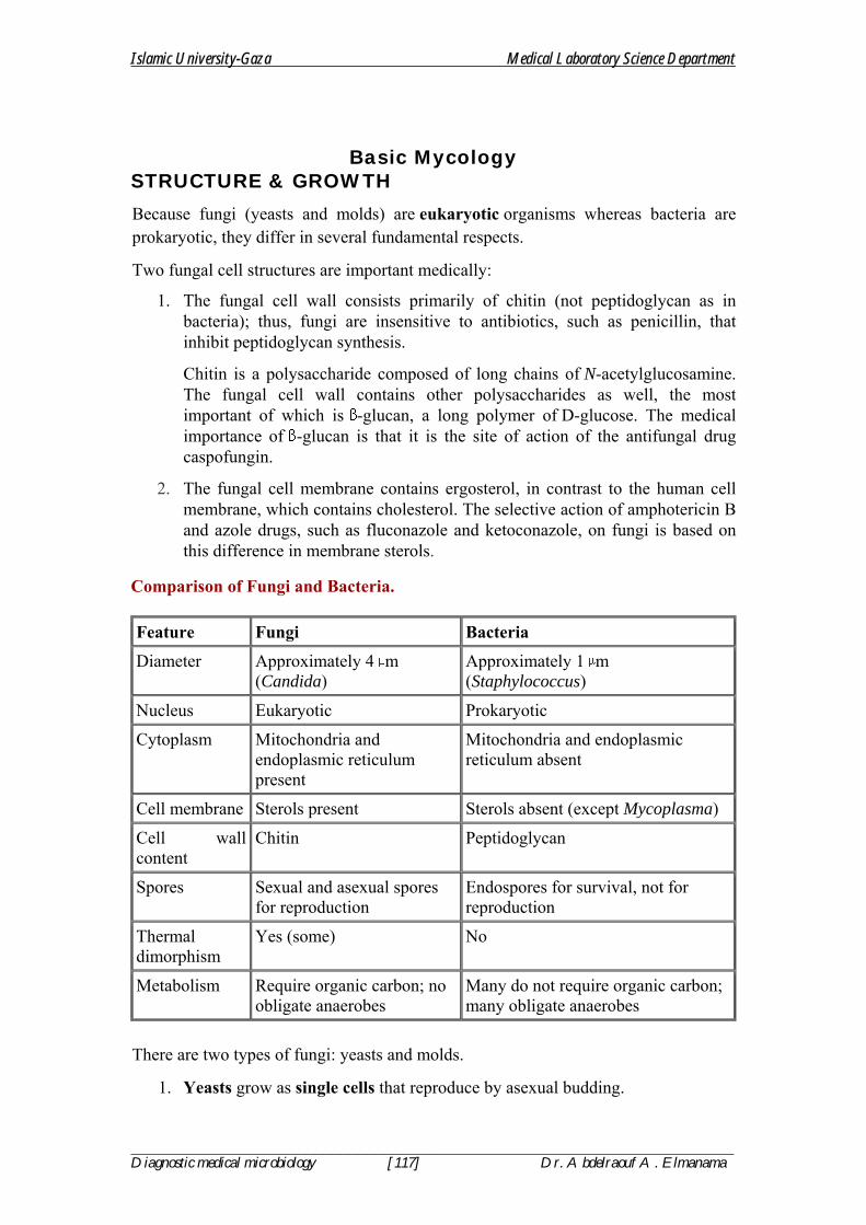
Islamic University-Gaza Medical Laboratory Science Department
_____________________________________________________________________ Diagnostic medical microbiology [117] Dr. Abdelraouf A. Elmanama
Basic Mycology STRUCTURE & GROWTH
Because fungi (yeasts and molds) are eukaryotic organisms whereas bacteria are prokaryotic, they differ in several fundamental respects.
Two fungal cell structures are important medically:
1. The fungal cell wall consists primarily of chitin (not peptidoglycan as in bacteria); thus, fungi are insensitive to antibiotics, such as penicillin, that inhibit peptidoglycan synthesis.
Chitin is a polysaccharide composed of long chains of N-acetylglucosamine. The fungal cell wall contains other polysaccharides as well, the most important of which is -glucan, a long polymer of D-glucose. The medical importance of -glucan is that it is the site of action of the antifungal drug caspofungin.
2. The fungal cell membrane contains ergosterol, in contrast to the human cell membrane, which contains cholesterol. The selective action of amphotericin B and azole drugs, such as fluconazole and ketoconazole, on fungi is based on this difference in membrane sterols.
Comparison of Fungi and Bacteria. Feature Fungi Bacteria
Diameter Approximately 4 m (Candida)
Approximately 1 m (Staphylococcus)
Nucleus Eukaryotic Prokaryotic
Cytoplasm Mitochondria and endoplasmic reticulum present
Mitochondria and endoplasmic reticulum absent
Cell membrane Sterols present Sterols absent (except Mycoplasma)
Cell wall content
Chitin Peptidoglycan
Spores Sexual and asexual spores for reproduction
Endospores for survival, not for reproduction
Thermal dimorphism
Yes (some) No
Metabolism Require organic carbon; no obligate anaerobes
Many do not require organic carbon; many obligate anaerobes
There are two types of fungi: yeasts and molds.
1. Yeasts grow as single cells that reproduce by asexual budding.

Islamic University-Gaza Medical Laboratory Science Department
_____________________________________________________________________ Diagnostic medical microbiology [118] Dr. Abdelraouf A. Elmanama
2. Molds grow as long filaments (hyphae) and form a mat (mycelium).
• Some hyphae form transverse walls (septate hyphae), whereas others do not (nonseptate hyphae). Nonseptate hyphae are multinucleated (coencytic).
• Several medically important fungi are thermally dimorphic; i.e., they form different structures at different temperatures.
• They exist as molds in the environment at ambient temperature and as yeasts (or other structures) in human tissues at body temperature.
• Most fungi are obligate aerobes; some are facultative anaerobes; but none are obligate anaerobes.
• All fungi require a preformed organic source of carbon—hence their frequent association with decaying matter.
• The natural habitat of most fungi is, therefore, the environment.
• An important exception is Candida albicans, which is part of the normal human flora.
• Some fungi reproduce sexually by mating and forming sexual spores, e.g., zygospores, ascospores, and basidiospores.
• Zygospores are single large spores with thick walls; ascospores are formed in a sac called ascus; and basidiospores are formed externally on the tip of a pedestal called a basidium.
• The classification of these fungi is based on their sexual spores. Fungi that do not form sexual spores are termed "imperfect" and are classified as fungi imperfecti.
• Most fungi of medical interest propagate asexually by forming conidia (asexual spores) from the sides or ends of specialized structures.
• The shape, color, and arrangement of conidia aid in the identification of fungi. Some important conidia are
(1) arthrospores, which arise by fragmentation of the ends of hyphae and are the mode of transmission of Coccidioides immitis;
(2) chlamydospores, which are rounded, thick-walled, and quite resistant (the terminal chlamydospores of C. albicans aid in its identification);
(3) blastospores, which are formed by the budding process by which yeasts reproduce asexually (some yeasts, e.g., C. albicans, can form multiple buds that do not detach, thus producing sausage like chains called pseudohyphae, which can be used for identification); and
(4) sporangiospores, which are formed within a sac (sporangium) on a stalk by molds such asRhizopus and Mucor.

Islamic University-Gaza Medical Laboratory Science Department
_____________________________________________________________________ Diagnostic medical microbiology [119] Dr. Abdelraouf A. Elmanama
Asexual spores. A: Blastoconidia and pseudohyphae (Candida). B: Chlamydospores (Candida). C: Arthrospores (Coccidioides). D: Sporangia and sporangiospores (Mucor). E: Microconidia (Aspergillus). F: Microconidia and macroconidia (Microsporum). (Modified and reproduced, with permission, from Conant NF et al: Manual of Clinical Mycology, 3rd ed. Saunders, 1971.)
• Although this book focuses on the fungi that are human pathogens, it should
be remembered that fungi are used in the production of important foods, e.g., bread, cheese etc...
• Fungi are also responsible for the spoilage of certain foods.
• Because molds can grow in a drier, more acidic, and higher-osmotic-pressure environment than bacteria, they tend to be involved in the spoilage of fruits, grains, vegetables, and jams.
PATHOGENESIS
• The response to infection with many fungi is the formation of granulomas.
• Granulomas are produced in the major systemic fungal diseases, e.g., coccidioidomycosis, histoplasmosis, and blastomycosis, as well as several others.
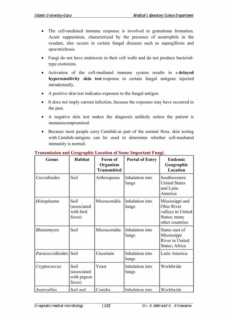
Islamic University-Gaza Medical Laboratory Science Department
_____________________________________________________________________ Diagnostic medical microbiology [120] Dr. Abdelraouf A. Elmanama
• The cell-mediated immune response is involved in granuloma formation. Acute suppuration, characterized by the presence of neutrophils in the exudate, also occurs in certain fungal diseases such as aspergillosis and sporotrichosis.
• Fungi do not have endotoxin in their cell walls and do not produce bacterial- type exotoxins.
• Activation of the cell-mediated immune system results in a delayed hypersensitivity skin test response to certain fungal antigens injected intradermally.
• A positive skin test indicates exposure to the fungal antigen.
• It does not imply current infection, because the exposure may have occurred in the past.
• A negative skin test makes the diagnosis unlikely unless the patient is immunocompromised.
• Because most people carry Candida as part of the normal flora, skin testing with Candida antigens can be used to determine whether cell-mediated immunity is normal.
Transmission and Geographic Location of Some Important Fungi. Genus Habitat Form of
Organism Transmitted
Portal of Entry Endemic Geographic
Location
Coccidioides Soil Arthrospores Inhalation into lungs
Southwestern United States and Latin America
Histoplasma Soil (associated with bird feces)
Microconidia Inhalation into lungs
Mississippi and Ohio River valleys in United States; many other countries
Blastomyces Soil Microconidia Inhalation into lungs
States east of Mississippi River in United States; Africa
Paracoccidioides Soil Uncertain Inhalation into lungs
Latin America
Cryptococcus Soil (associated with pigeon feces)
Yeast Inhalation into lungs
Worldwide
Aspergillus Soil and Conidia Inhalation into Worldwide

Islamic University-Gaza Medical Laboratory Science Department
_____________________________________________________________________ Diagnostic medical microbiology [121] Dr. Abdelraouf A. Elmanama
vegetation lungs
Candida Human body
Yeast Normal flora of skin, mouth, gastrointestinal tract, and vagina
Worldwide
• Intact skin is an effective host defense against certain fungi (e.g., Candida, dermatophytes), but if the skin is damaged, organisms can become established.
• Fatty acids in the skin inhibit dermatophyte growth, and hormone-associated skin changes at puberty limit ringworm of the scalp caused by Trichophyton.
• The normal flora of the skin and mucous membranes suppress fungi.
• When the normal flora is inhibited, e.g., by antibiotics, overgrowth of fungi such as C. albicans can occur.
• In the respiratory tract, the important host defenses are the mucous membranes of the nasopharynx, which trap inhaled fungal spores, and alveolar macrophages.
• Circulating IgG and IgM are produced in response to fungal infection, but their role in protection from disease is uncertain.
• The cell-mediated immune response is protective; its suppression can lead to reactivation and dissemination of asymptomatic fungal infections and to disease caused by opportunistic fungi.
FUNGAL TOXINS & ALLERGIES
In addition to mycotic infections, there are two other kinds of fungal disease: (1) mycotoxicoses, caused by ingested toxins and
(2) allergies to fungal spores. The best-known mycotoxicosis occurs after eating Amanita mushrooms.
• These fungi produce five toxins, two of which—amanitin and phalloidin—are among the most potent hepatotoxins.
• The toxicity of amanitin is based on its ability to inhibit cellular RNA polymerase, which prevents mRNA synthesis.
• Another mycotoxicosis, ergotism, is caused by the mold Claviceps purpura, which infects grains and produces alkaloids (e.g., ergotamine and lysergic acid diethylamide [LSD]) that cause pronounced vascular and neurologic effects.

Islamic University-Gaza Medical Laboratory Science Department
_____________________________________________________________________ Diagnostic medical microbiology [122] Dr. Abdelraouf A. Elmanama
• Other ingested toxins, aflatoxins, are coumarin derivatives produced by Aspergillus flavus that cause liver damage and tumors in animals and are suspected of causing hepatic carcinoma in humans.
• Aflatoxins are ingested with spoiled grains and peanuts and are metabolized by the liver to the epoxide, a potent carcinogen.
• Aflatoxin B1 induces a mutation in the p53 tumor suppressor gene, leading to a loss of p53 protein and a consequent loss of growth control in the hepatocyte.
• Allergies to fungal spores, particularly those of Aspergillus, are manifested primarily by an asthmatic reaction (rapid bronchoconstriction mediated by IgE), eosinophilia, and a "wheal and flare" skin test reaction.
• These clinical findings are caused by an immediate hypersensitivity response to the fungal spores.
LABORATORY DIAGNOSIS
There are four approaches to the laboratory diagnosis of fungal diseases:
(1) Direct microscopic examination,
(2) Culture of the organism,
(3) DNA probe tests, and
(4) Serologic tests.
• Direct microscopic examination of clinical specimens such as sputum, lung biopsy material, and skin scrapings depends on finding characteristic asexual spores, hyphae, or yeasts in the light microscope.
• The specimen is either treated with 10% KOH to dissolve tissue material, leaving the alkali-resistant fungi intact, or stained with special fungal stains.
Some examples of diagnostically important findings made by direct examination are
(1) The spherules of C. immitis and
(2) The wide capsule of Cryptococcus neoformans seen in India ink preparations of spinal fluid.
• Calcofluor white is a fluorescent dye that binds to fungal cell walls and is useful in the identification of fungi in tissue specimens. Methenamine-silver stain is also useful in the microscopic diagnosis of fungi in tissue.
• Fungi are frequently cultured on Sabouraud's agar, which facilitates the appearance of the slow-growing fungi by inhibiting the growth of bacteria in the specimen.
• Inhibition of bacterial growth is due to the low pH of the medium and to the chloramphenicol and cycloheximide that are frequently added.

Islamic University-Gaza Medical Laboratory Science Department
_____________________________________________________________________ Diagnostic medical microbiology [123] Dr. Abdelraouf A. Elmanama
• The appearance of the mycelium and the nature of the asexual spores are frequently sufficient to identify the organism.
• Tests involving DNA probes can identify colonies growing in culture at an earlier stage of growth than can tests based on visual detection of the colonies.
• As a result, the diagnosis can be made more rapidly. At present, DNA probe tests are available for Coccidioides, Histoplasma, Blastomyces, andCryptococcus.
• Tests for the presence of antibodies in the patient's serum or spinal fluid are useful in diagnosing systemic mycoses but less so in diagnosing other fungal infections.
• As is the case for bacterial and viral serologic testing, a significant rise in the antibody titer must be observed to confirm a diagnosis.
• The complement fixation test is most frequently used in suspected cases of coccidioidomycosis, histoplasmosis, and blastomycosis.
• In cryptococcal meningitis, the presence of the polysaccharide capsular antigens of C. neoformans in the spinal fluid can be detected by the latex agglutination test.
ANTIFUNGAL THERAPY
• The drugs used to treat bacterial diseases have no effect on fungal diseases.
• For example, penicillins and aminoglycosides inhibit the growth of many bacteria but do not affect the growth of fungi.
• This difference is explained by the presence of certain structures in bacteria, e.g., peptidoglycan and 70S ribosomes, that are absent in fungi.
• The most effective antifungal drugs, amphotericin B and the various azoles, exploit the presence of ergosterol in fungal cell membranes that is not found in bacterial or human cell membranes.
• Amphotericin B disrupts fungal cell membranes at the site of ergosterol and azole drugs inhibit the synthesis of ergosterol, which is an essential component of fungal membranes.
• Another antifungal drug, caspofungin (Cancidas), inhibits the synthesis of -glucan, which is found in fungal cell walls but not in bacterial cell walls.
• Human cells do not have a cell wall.
• There is no clinically significant resistance to antifungal drugs.

Islamic University-Gaza Medical Laboratory Science Department
_____________________________________________________________________ Diagnostic medical microbiology [124] Dr. Abdelraouf A. Elmanama
Mechanism of Action and Adverse Effects of Antifungal Drugs.
Usage Name of Drug Mechanism of Action
Important Adverse Reactions
Systemic use (intravenous, oral)
Amphotericin B Binds to ergosterol and disrupts fungal cell membranes
Renal toxicity, fever, and chills; monitor kidney function; use test dose; liposomal preparation reduces toxicity
Azoles such as fluconazole, ketoconazole, itraconazole, voriconazole, posaconazole
Inhibits ergosterol synthesis
Ketoconazole inhibits human cytochrome P450; this decreases synthesis of gonadal steroids resulting in gynecomastia
Echinocandins such as caspofungin, mycafungin
Inhibits synthesis of D-glucan, a component of fungal cell wall
Well tolerated
Flucytosine (FC) Inhibits DNA synthesis; FC converted to fluorouracil, which inhibits thymidine synthetase
Bone marrow toxicity
Griseofulvin Disrupts mitotic spindle by binding to tubulin
Liver toxicity
Topical use (skin only); too toxic for systemic use
Azoles such as cotrimazole, miconazole
Inhibits ergosterol synthesis
Well tolerated on skin
Terbinafine Inhibits ergosterol synthesis
Well tolerated on skin
Tolnoftate Inhibits ergosterol synthesis
Well tolerated on skin
Nystatin Binds to ergosterol and disrupts fungal cell membranes
Well tolerated on skin
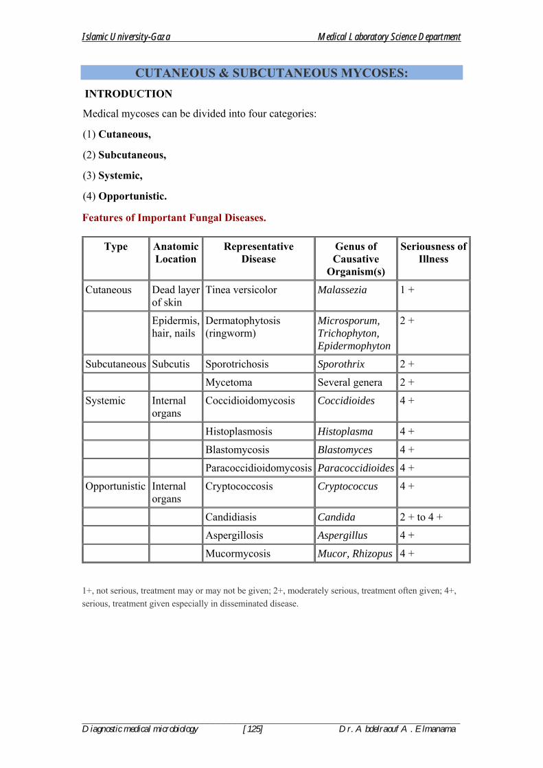
Islamic University-Gaza Medical Laboratory Science Department
_____________________________________________________________________ Diagnostic medical microbiology [125] Dr. Abdelraouf A. Elmanama
CUTANEOUS & SUBCUTANEOUS MYCOSES: INTRODUCTION
Medical mycoses can be divided into four categories:
(1) Cutaneous,
(2) Subcutaneous,
(3) Systemic,
(4) Opportunistic.
Features of Important Fungal Diseases.
Type Anatomic Location
Representative Disease
Genus of Causative
Organism(s)
Seriousness of Illness
Cutaneous Dead layer of skin
Tinea versicolor Malassezia 1 +
Epidermis, hair, nails
Dermatophytosis (ringworm)
Microsporum, Trichophyton, Epidermophyton
2 +
Subcutaneous Subcutis Sporotrichosis Sporothrix 2 +
Mycetoma Several genera 2 +
Systemic Internal organs
Coccidioidomycosis Coccidioides 4 +
Histoplasmosis Histoplasma 4 +
Blastomycosis Blastomyces 4 +
Paracoccidioidomycosis Paracoccidioides 4 +
Opportunistic Internal organs
Cryptococcosis Cryptococcus 4 +
Candidiasis Candida 2 + to 4 +
Aspergillosis Aspergillus 4 +
Mucormycosis Mucor, Rhizopus 4 +
1+, not serious, treatment may or may not be given; 2+, moderately serious, treatment often given; 4+, serious, treatment given especially in disseminated disease.

Islamic University-Gaza Medical Laboratory Science Department
_____________________________________________________________________ Diagnostic medical microbiology [126] Dr. Abdelraouf A. Elmanama
CUTANEOUS MYCOSES
Dermatophytoses
• Dermatophytoses are caused by fungi (dermatophytes) that infect only superficial keratinized structures (skin, hair, and nails), not deeper tissues.
• The most important dermatophytes are classified in three genera: Epidermophyton, Trichophyton, and Microsporum.
• They are spread from infected persons by direct contact. Microsporum is also spread from animals such as dogs and cats.
• This indicates that to prevent reinfection, the animal must be treated also.
• Dermatophytoses (tinea, ringworm) are chronic infections often located in the warm, humid areas of the body, e.g., athlete's foot.
• Typical ringworm lesions have an inflamed circular border containing papules and vesicles surrounding a clear area of relatively normal skin.
• Broken hairs and thickened broken nails are often seen.
• The disease is typically named for the affected body part, i.e., tinea capitis (head), tinea corporis (body), tinea cruris (groin), and tinea pedis (foot).
• Trichophyton tonsurans is the most common cause of outbreaks of tinea capitis in children and is the main cause of endothrix (inside the hair) infections.
• Trichophyton rubrum is also a very common cause of tinea capitis.
• Trichophyton schoenleinii is the cause of favus, a form of tinea capitis in which crusts are seen on the scalp.
• In some infected persons, hypersensitivity causes dermatophytid ("id") reactions, e.g., vesicles on the fingers. Id lesions are a response to circulating fungal antigens; the lesions do not contain hyphae.
• Patients with tinea infections show positive skin tests with fungal extracts, e.g., trichophytin.
• Scrapings of skin or nail placed in 10% KOH on a glass slide show septate hyphae under microscopy.
• Cultures on Sabouraud's agar at room temperature develop typical hyphae and conidia.
• Tinea capitis lesions caused by Microsporum species can be detected by seeing fluorescence when the lesions are exposed to ultraviolet light from a Wood's lamp.
Islamic University-Gaza Medical Laboratory Science Department
_____________________________________________________________________ Diagnostic medical microbiology [127] Dr. Abdelraouf A. Elmanama
• Treatment involves local antifungal creams (undecylenic acid, miconazole, tolnaftate, etc) or oral griseofulvin. Prevention centers on keeping skin dry and cool.
Tinea Versicolor
• Tinea versicolor (pityriasis versicolor), a superficial skin infection of cosmetic importance only, is caused by Malassezia furfur.
• The lesions are usually noticed as hypopigmented areas, especially on tanned skin in the summer.
• There may be slight scaling or itching, but usually the infection is asymptomatic. It occurs more frequently in hot, humid weather.
• The lesions contain both budding yeast cells and hyphae.
• Diagnosis is usually made by observing this mixture in KOH preparations of skin scrapings.
• Culture is not usually done.
• The treatment of choice is topical miconazole, but the lesions have a tendency to recur and a permanent cure is difficult to achieve.
Tinea Nigra
• Tinea nigra is an infection of the keratinized layers of the skin.
• It appears as a brownish spot caused by the melanin-like pigment in the hyphae.
• The causative organism, Cladosporium werneckii, is found in the soil and transmitted during injury.
• In the United States, the disease is seen in the southern states.
• Diagnosis is made by microscopic examination and culture of skin scrapings.
• The infection is treated with a topical keratolytic agent, e.g., salicylic acid.
SUBCUTANEOUS MYCOSES
These are caused by fungi that grow in soil and on vegetation and are introduced into subcutaneous tissue through trauma.
Sporotrichosis
• Sporothrix schenckii is a dimorphic fungus that lives on vegetation.

Islamic University-Gaza Medical Laboratory Science Department
_____________________________________________________________________ Diagnostic medical microbiology [128] Dr. Abdelraouf A. Elmanama
• When introduced into the skin, typically by a thorn, it causes a local pustule or ulcer with nodules along the draining lymphatics.
• There is little systemic illness. Lesions may be chronic. Sporotrichosis occurs most often in gardeners, especially those who prune roses, because they may be stuck by a rose thorn.
• In the clinical laboratory, round or cigar-shaped budding yeasts are seen in tissue specimens.
• In culture, hyphae occur bearing oval conidia in clusters at the tip of slender conidiophores (resembling a daisy).
• The drug of choice for skin lesions is itraconazole.
• It can be prevented by protecting skin when touching plants, moss, and wood.
Chromomycosis
• This is a slowly progressive granulomatous infection that is caused by several soil fungi (Fonsecaea, Phialophora, Cladosporium, etc) when introduced into the skin through trauma.
• These fungi are collectively called dematiaceous fungi, so named because their conidia or hyphae are dark-colored, either gray or black.
• Wartlike lesions with crusting abscesses extend along the lymphatics.
• The disease occurs mainly in the tropics and is found on bare feet and legs.
• In the clinical laboratory, dark brown, round fungal cells are seen in leukocytes or giant cells.
• The disease is treated with oral flucytosine or thiabendazole, plus local surgery.
Mycetoma
• Soil organisms (Petriellidium, Madurella) enter through wounds on the feet, hands, or back and cause abscesses, with pus discharged through sinuses.
• The pus contains compact colored granules.
• Actinomycetes such as Nocardia can cause similar lesions (actinomycotic mycetoma). Sulfonamides may help the actinomycotic form.
• There is no effective drug against the fungal form; surgical excision is recommended.
SYSTEMIC MYCOSES

Islamic University-Gaza Medical Laboratory Science Department
_____________________________________________________________________ Diagnostic medical microbiology [129] Dr. Abdelraouf A. Elmanama
INTRODUCTION
• These infections result from inhalation of the spores of dimorphic fungi that have their mold forms in the soil. Within the lungs, the spores differentiate into yeasts or other specialized forms.
• Most lung infections are asymptomatic and self-limited.
• However, in some persons, disseminated disease develops in which the organisms grow in other organs, cause destructive lesions, and may result in death.
• Infected persons do not communicate these diseases to others.
COCCIDIOIDES
Disease
• Coccidioides immitis causes coccidioidomycosis.
Properties
• C. immitis is a dimorphic fungus that exists as a mold in soil and as a spherule in tissue.
Stages of Coccidioides immitis.A: Arthrospores form at the ends of hyphae in the soil. They germinate in the soil to form new hyphae. If inhaled, the arthrospores differentiate into spherules. B: Endospores form within spherules in tissue. When spherules rupture, endospores disseminate and form new spherules. (Modified and reproduced, with permission, from Brooks GF et al: Medical Microbiology, 20th ed. Originally published by Appleton & Lange. Copyright © 1995 by The McGraw-Hill Companies, Inc.)
Transmission & Epidemiology

Islamic University-Gaza Medical Laboratory Science Department
_____________________________________________________________________ Diagnostic medical microbiology [130] Dr. Abdelraouf A. Elmanama
• The fungus is endemic in arid regions of the southwestern United States and Latin America.
• People who live in Central and Southern California, Arizona, New Mexico, Western Texas, and Northern Mexico, a geographic region called the Lower Sonoran Life Zone, are often infected.
• In soil, it forms hyphae with alternating arthrospores and empty cells.
• Arthrospores are very light and are carried by the wind.
They can be inhaled and infect the lungs.
Pathogenesis
• In the lungs, arthrospores form spherules that are large (30 m in diameter), have a thick, doubly refractive wall, and are filled with endospores.
• Upon rupture of the wall, endospores are released and differentiate to form new spherules.
• The organism can spread within a person by direct extension or via the bloodstream.
• Granulomatous lesions can occur in virtually any organ but are found primarily in bones and the central nervous system (meningitis).
Coccidioides immitis—Spherule. Long arrow points to a spherule in lung tissue. Spherules are large thick-walled structures containing many endospores. Short arrow points to an endospore. Provider: CDC/Dr. L. Georg.

Islamic University-Gaza Medical Laboratory Science Department
_____________________________________________________________________ Diagnostic medical microbiology [131] Dr. Abdelraouf A. Elmanama
• Dissemination from the lungs to other organs occurs in people who have a defect in cell-mediated immunity.
• Most people who are infected by C. immitisdevelop a cell-mediated (delayed hypersensitivity) immune response that restricts the growth of the organism.
• One way to determine whether a person has produced adequate cell-mediated immunity to the organism is to do a skin test.
• In general, a person who has a positive skin test reaction has developed sufficient immunity to prevent disseminated disease from occurring.
• If, at a later time, a person's cellular immunity is suppressed by drugs or disease, disseminated disease can occur.
Clinical Findings
• Infection of the lungs is often asymptomatic and is evident only by a positive skin test and the presence of antibodies.
• Some infected persons have an influenzalike illness with fever and cough. About 50% have changes in the lungs (infiltrates, adenopathy, or effusions) as seen on chest x-ray, and 10% develop erythema nodosum (see below) or arthralgias.
• This syndrome is called "valley fever" (in the San Joaquin Valley of California) or "desert rheumatism" (in Arizona); it tends to subside spontaneously.
• Disseminated disease can occur in almost any organ; the meninges, bone, and skin are important sites.
• The overall incidence of dissemination in persons infected with C. immitis is 1%, although the incidence in Filipinos and African Americans is 10 times higher.
• Women in the third trimester of pregnancy also have a markedly increased incidence of dissemination.
• Erythema nodosum (EN) manifests as red, tender nodules ("desert bumps") on extensor surfaces such as the shins.
• It is a delayed (cell-mediated) hypersensitivity response to fungal antigens and thus is an indicator of a good prognosis.
• There are no organisms in these lesions; they are not a sign of disseminated disease.
• EN is not specific for coccidioidomycosis; it occurs in other granulomatous diseases, e.g., histoplasmosis, tuberculosis, and leprosy.
Islamic University-Gaza Medical Laboratory Science Department
_____________________________________________________________________ Diagnostic medical microbiology [132] Dr. Abdelraouf A. Elmanama
• In infected persons, skin tests with fungal extracts (coccidioidin or spherulin) cause at least a 5-mm induration 48 hours after injection (delayed hypersensitivity reaction).
• Skin tests become positive within 2–4 weeks of infection and remain so for years but are often negative (anergy) in patients with disseminated disease.
Laboratory Diagnosis
• In tissue specimens, spherules are seen microscopically.
• Cultures on Sabouraud's agar incubated at 25°C show hyphae with arthrospores. (Caution:Cultures are highly infectious; precautions against inhaling arthrospores must be taken.)
• In serologic tests, IgM and IgG precipitins appear within 2–4 weeks of infection and then decline in subsequent months.
• Complement-fixing antibodies occur at low titer initially, but the titer rises greatly if dissemination occurs.
Treatment & Prevention
• No treatment is needed in asymptomatic or mild primary infection.
• Amphotericin B (Fungizone) or itraconazole is used for persisting lung lesions or disseminated disease. Ketoconazole is also effective in lung disease.
• If meningitis occurs, fluconazole is the drug of choice. Intrathecal amphotericin B may be required and may induce remission, but long-term results are often poor.
• There are no means of prevention except avoiding travel to endemic areas.
HISTOPLASMA
Disease
• Histoplasma capsulatum causes histoplasmosis.
Properties
• H. capsulatum is a dimorphic fungus that exists as a mold in soil and as a yeast in tissue. It forms two types of asexual spores:
(1)tuberculate macroconidia, with typical thick walls and fingerlike projections that are important in laboratory identification, and
(2) microconidia, which are smaller, thin, smooth-walled spores that, if inhaled, transmit the infection.

Islamic University-Gaza Medical Laboratory Science Department
_____________________________________________________________________ Diagnostic medical microbiology [133] Dr. Abdelraouf A. Elmanama
Asexual spores of Histoplasma capsulatum.A: Tuberculate macroconidia. B: Microconidia. (Reproduced, with permission, from Brooks GF et al: Medical Microbiology,19th ed. Originally published by Appleton & Lange. Copyright © 1991 by The McGraw-Hill Companies, Inc.)
Transmission & Epidemiology
• This fungus occurs in many parts of the world. In the United States it is endemic in central and eastern states, especially in the Ohio and Mississippi River valleys.
• It grows in soil, particularly if the soil is heavily contaminated with bird droppings, especially from starlings.
• Although the birds are not infected, bats can be infected and can excrete the organism in their guano.
• In areas of endemic infection, excavation of the soil during construction or exploration of bat-infested caves has resulted in a significant number of infected individuals.
• In several tropical African countries, histoplasmosis is caused by Histoplasma duboisii.
• The clinical picture is different from that caused by H. capsulatum.
Pathogenesis & Clinical Findings
• Inhaled spores are engulfed by macrophages and develop into yeast forms.
• In tissues, H. capsulatum occurs as an oval budding yeast inside macrophages.
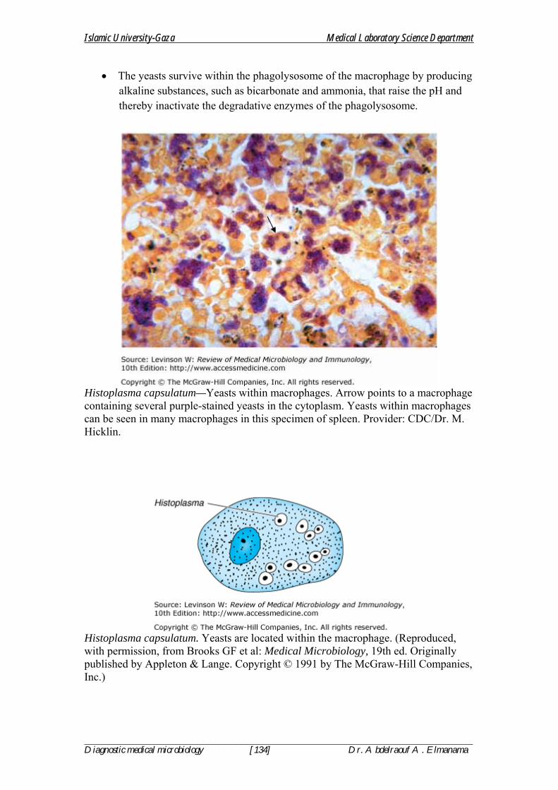
Islamic University-Gaza Medical Laboratory Science Department
_____________________________________________________________________ Diagnostic medical microbiology [134] Dr. Abdelraouf A. Elmanama
• The yeasts survive within the phagolysosome of the macrophage by producing alkaline substances, such as bicarbonate and ammonia, that raise the pH and thereby inactivate the degradative enzymes of the phagolysosome.
Histoplasma capsulatum—Yeasts within macrophages. Arrow points to a macrophage containing several purple-stained yeasts in the cytoplasm. Yeasts within macrophages can be seen in many macrophages in this specimen of spleen. Provider: CDC/Dr. M. Hicklin.
Histoplasma capsulatum. Yeasts are located within the macrophage. (Reproduced, with permission, from Brooks GF et al: Medical Microbiology, 19th ed. Originally published by Appleton & Lange. Copyright © 1991 by The McGraw-Hill Companies, Inc.)

Islamic University-Gaza Medical Laboratory Science Department
_____________________________________________________________________ Diagnostic medical microbiology [135] Dr. Abdelraouf A. Elmanama
• The organisms spread widely throughout the body, especially to the liver and spleen, but most infections remain asymptomatic, and the small granulomatous foci heal by calcification.
• With intense exposure (e.g., in a chicken house or bat-infested cave), pneumonia may become clinically manifest.
• Severe disseminated histoplasmosis develops in a small minority of infected persons, especially infants and individuals with reduced cell-mediated immunity, such as AIDS patients.
• In AIDS patients, ulcerated lesions on the tongue are typical of disseminated histoplasmosis.
• In immunocompetent people, EN can occur (see description of EN in Coccidioides above).
• EN is a sign that cell-mediated immunity is active and the organism will probably be contained.
• A skin test using histoplasmin (a mycelial extract) becomes positive, i.e., shows at least 5 mm of induration, within 2–3 weeks after infection and remains positive for many years.
• However, because there are many false-positive reactions (due to cross-reactivity) and many false-negative reactions (in disseminated disease), the skin test is not useful for diagnosis.
• Furthermore, the skin test can stimulate an antibody response and confuse the serologic tests.
• The skin test is useful for epidemiologic studies, and up to 90% of individuals have positive results in areas of endemic infection.
Laboratory Diagnosis
• In tissue biopsy specimens or bone marrow aspirates, oval yeast cells within macrophages are seen microscopically.
• Cultures on Sabouraud's agar show hyphae with tuberculate macroconidia when grown at low temperature, e.g., 25°C and yeasts when grown at 37°C.
• Tests that detect Histoplasmaantigens by radioimmunoassay and Histoplasma RNA with DNA probes are also useful.
• In immunocompromised patients with disseminated disease, tests for antigens in the urine are especially useful because antibody tests may be negative.
• Two serologic tests are useful for diagnosis: complement fixation (CF) and immunodiffusion (ID).

Islamic University-Gaza Medical Laboratory Science Department
_____________________________________________________________________ Diagnostic medical microbiology [136] Dr. Abdelraouf A. Elmanama
• An antibody titer of 1:32 in the CF test with yeast phase antigens is considered to be diagnostic.
• However, cross-reactions with other fungi, especially Blastomyces, occur.
Treatment & Prevention
• No therapy is needed in asymptomatic or mild primary infections.
• With progressive lung lesions, oral itraconazole is beneficial.
• In disseminated disease, amphotericin B is the treatment of choice.
• In meningitis, fluconazole is often used because it penetrates the spinal fluid well.
• Oral itraconazole is used to treat pulmonary or disseminated disease, as well as for chronic suppression in patients with AIDS.
• There are no means of prevention except avoiding exposure in areas of endemic infection.
BLASTOMYCES
Disease
• Blastomyces dermatitidis causes blastomycosis, also known as North American blastomycosis.
Properties
• B. dermatitidis is a dimorphic fungus that exists as a mold in soil and as a yeast in tissue.
• The yeast is round with a doubly refractive wall and a singlebroad-based bud.
• Note that this organism forms a broad-based bud, whereas Cryptococcus neoformans is a yeast that forms a narrow-based bud.

Islamic University-Gaza Medical Laboratory Science Department
_____________________________________________________________________ Diagnostic medical microbiology [137] Dr. Abdelraouf A. Elmanama
Blastomyces dermatitidis—Broad-based budding yeast. Arrow points to the broad base of the budding yeast. Provider: CDC/Dr. L. Ajello.
Blastomyces dermatitidis.A: Yeast with a broad-based bud at 37°C. B: Mold with microconidia at 20°C. (Reproduced, with permission, from Brooks GF et al: Medical Microbiology, 19th ed. Originally published by Appleton & Lange. Copyright © 1991 by The McGraw-Hill Companies, Inc.)

Islamic University-Gaza Medical Laboratory Science Department
_____________________________________________________________________ Diagnostic medical microbiology [138] Dr. Abdelraouf A. Elmanama
Transmission & Epidemiology
• This fungus is endemic primarily in eastern North America, especially in the region bordering the Ohio, Mississippi, and St. Lawrence rivers, and the Great Lakes region.
• Less commonly, blastomycosis has also occurred in Central and South America, Africa, and the Middle East.
• It grows in moist soil rich in organic material, forming hyphae with small pear-shaped conidia.
• Inhalation of the conidia causes human infection.
Pathogenesis & Clinical Findings
• Infection occurs mainly via the respiratory tract.
• Asymptomatic or mild cases are rarely recognized.
• Dissemination may result in ulcerated granulomas of skin, bone, or other sites.
Laboratory Diagnosis
• In tissue biopsy specimens, thick-walled yeast cells with single broad-based buds are seen microscopically.
• Hyphae with small pear-shaped conidia are visible on culture.
• The skin test lacks specificity and has little value.
• Serologic tests have little value.
Treatment & Prevention
• Itraconazole is the drug of choice for most patients, but amphotericin B should be used to treat severe disease.
• Surgical excision may be helpful.
• There are no means of prevention.
PARACOCCIDIOIDES
Disease
• Paracoccidioides brasiliensis causes paracoccidioidomycosis, also known as South American blastomycosis.
Properties
• P. brasiliensis is a dimorphic fungus that exists as a mold in soil and as a yeast in tissue.
• The yeast is thick-walled with multiple buds, in contrast to B. dermatitidis, which has a single bud.
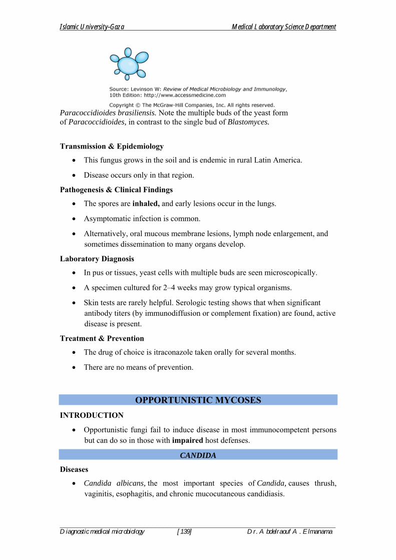
Islamic University-Gaza Medical Laboratory Science Department
_____________________________________________________________________ Diagnostic medical microbiology [139] Dr. Abdelraouf A. Elmanama
Paracoccidioides brasiliensis. Note the multiple buds of the yeast form of Paracoccidioides, in contrast to the single bud of Blastomyces.
Transmission & Epidemiology
• This fungus grows in the soil and is endemic in rural Latin America.
• Disease occurs only in that region.
Pathogenesis & Clinical Findings
• The spores are inhaled, and early lesions occur in the lungs.
• Asymptomatic infection is common.
• Alternatively, oral mucous membrane lesions, lymph node enlargement, and sometimes dissemination to many organs develop.
Laboratory Diagnosis
• In pus or tissues, yeast cells with multiple buds are seen microscopically.
• A specimen cultured for 2–4 weeks may grow typical organisms.
• Skin tests are rarely helpful. Serologic testing shows that when significant antibody titers (by immunodiffusion or complement fixation) are found, active disease is present.
Treatment & Prevention
• The drug of choice is itraconazole taken orally for several months.
• There are no means of prevention.
OPPORTUNISTIC MYCOSES INTRODUCTION
• Opportunistic fungi fail to induce disease in most immunocompetent persons but can do so in those with impaired host defenses.
CANDIDA
Diseases
• Candida albicans, the most important species of Candida, causes thrush, vaginitis, esophagitis, and chronic mucocutaneous candidiasis.

Islamic University-Gaza Medical Laboratory Science Department
_____________________________________________________________________ Diagnostic medical microbiology [140] Dr. Abdelraouf A. Elmanama
• It also causes disseminated infections such as right-sided endocarditis (especially in intravenous drug users) and blood stream infections (candidemia).
• Infections related to indwelling intravenous and urinary catheters are also important.
Properties
• C. albicans is an oval yeast with a single bud.
• It is part of the normal flora of mucous membranes of the upper respiratory, gastrointestinal, and female genital tracts.
• In tissues it may appear as yeasts or as pseudohyphae
• Pseudohyphae are elongated yeasts that visually resemble hyphae but are not true hyphae.
• Carbohydrate fermentation reactions differentiate it from other species, e.g., Candida tropicalis, Candida parapsilosis, Candida krusei, and Candida glabrata.
Candida albicans—Yeast. Long arrow points to a budding yeast. Short arrow points to the outer membrane of a vaginal epithelial cell. In this gram-stained specimen, various bacteria that are part of the normal flora of the vagina can be seen. Provider: CDC/Dr. S. Brown.

Islamic University-Gaza Medical Laboratory Science Department
_____________________________________________________________________ Diagnostic medical microbiology [141] Dr. Abdelraouf A. Elmanama
Candida albicans—Pseudohyphae. Two arrows point to pseudohyphae of Candida albicans. Provider: CDC/Dr. S. Brown.
Candida albicans.A: Budding yeasts and pseudohyphae in tissues or exudate. B: Pseudohyphae and chlamydospores in culture at 20°C. C: Germ tubes at 37°C. (Reproduced, with permission, from Brooks GF et al: Medical Microbiology, 20th ed. Originally published by Appleton & Lange. Copyright © 1995 by The McGraw-Hill Companies, Inc.)
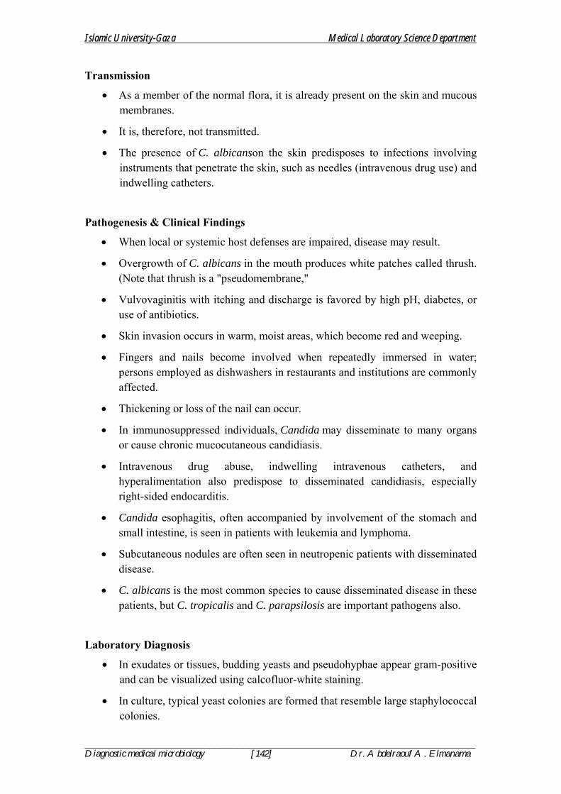
Islamic University-Gaza Medical Laboratory Science Department
_____________________________________________________________________ Diagnostic medical microbiology [142] Dr. Abdelraouf A. Elmanama
Transmission
• As a member of the normal flora, it is already present on the skin and mucous membranes.
• It is, therefore, not transmitted.
• The presence of C. albicanson the skin predisposes to infections involving instruments that penetrate the skin, such as needles (intravenous drug use) and indwelling catheters.
Pathogenesis & Clinical Findings
• When local or systemic host defenses are impaired, disease may result.
• Overgrowth of C. albicans in the mouth produces white patches called thrush. (Note that thrush is a "pseudomembrane,"
• Vulvovaginitis with itching and discharge is favored by high pH, diabetes, or use of antibiotics.
• Skin invasion occurs in warm, moist areas, which become red and weeping.
• Fingers and nails become involved when repeatedly immersed in water; persons employed as dishwashers in restaurants and institutions are commonly affected.
• Thickening or loss of the nail can occur.
• In immunosuppressed individuals, Candida may disseminate to many organs or cause chronic mucocutaneous candidiasis.
• Intravenous drug abuse, indwelling intravenous catheters, and hyperalimentation also predispose to disseminated candidiasis, especially right-sided endocarditis.
• Candida esophagitis, often accompanied by involvement of the stomach and small intestine, is seen in patients with leukemia and lymphoma.
• Subcutaneous nodules are often seen in neutropenic patients with disseminated disease.
• C. albicans is the most common species to cause disseminated disease in these patients, but C. tropicalis and C. parapsilosis are important pathogens also.
Laboratory Diagnosis
• In exudates or tissues, budding yeasts and pseudohyphae appear gram-positive and can be visualized using calcofluor-white staining.
• In culture, typical yeast colonies are formed that resemble large staphylococcal colonies.

Islamic University-Gaza Medical Laboratory Science Department
_____________________________________________________________________ Diagnostic medical microbiology [143] Dr. Abdelraouf A. Elmanama
• Germ tubes form in serum at 37°C, which serves to distinguish C. albicans from most other Candida species.
• Chlamydospores are typically formed by C. albicans but not by other species of Candida. Serologic testing is rarely helpful.
• Skin tests with Candida antigens are uniformly positive in immunocompetent adults and are used as an indicator that the person can mount a cellular immune response.
• A person who does not respond to Candida antigens in the skin test is presumed to have deficient cell-mediated immunity.
• Such a person is anergic, and other skin tests cannot be interpreted.
• Thus, if a person has a negative Candida skin test, a negative PPD skin test for tuberculosis could be a false-negative result.
Treatment & Prevention
• The drug of choice for oropharyngeal or esophageal thrush is fluconazole.
• Caspofungin or micafungin can also be used for esophageal candidiasis.
• Treatment of skin infections consists of topical antifungal drugs, e.g., clotrimazole or nystatin. Mucocutaneous candidiasis can be controlled by ketoconazole.
• Treatment of disseminated candidiasis consists of either amphotericin B or fluconazole.
• These two drugs can be used with or without flucytosine.
• Treatment of candidal infections with antifungal drugs should be supplemented by reduction of predisposing factors.
• Certain candidal infections, e.g., thrush, can be prevented by oral clotrimazole troches or nystatin "swish and swallow."
• Fluconazole is useful in preventing candidal infections in high-risk patients, such as those undergoing bone marrow transplantation and premature infants. Micafungin can also be used.
• There is no vaccine.
CRYPTOCOCCUS
Disease
• Cryptococcus neoformans causes cryptococcosis, especially cryptococcal meningitis.

Islamic University-Gaza Medical Laboratory Science Department
_____________________________________________________________________ Diagnostic medical microbiology [144] Dr. Abdelraouf A. Elmanama
• Cryptococcosis is the most common life-threatening fungal disease in AIDS patients.
Properties
• C. neoformans is an oval, budding yeast surrounded by a wide polysaccharide capsule.
• It is not dimorphic. Note that this organism forms a narrow-based bud, whereas the yeast form of Blastomyces dermatitidis forms a broad-based bud.
Cryptococcus neoformans—India ink preparation. Arrow points to a budding yeast of Cryptococcus neoformans. Note the thick, translucent polysaccharide capsule outlined by the dark India ink particles. Provider: CDC/Dr. L. Haley.
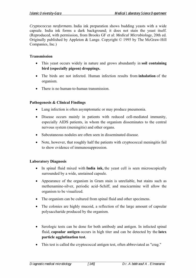
Islamic University-Gaza Medical Laboratory Science Department
_____________________________________________________________________ Diagnostic medical microbiology [145] Dr. Abdelraouf A. Elmanama
Cryptococcus neoformans. India ink preparation shows budding yeasts with a wide capsule. India ink forms a dark background; it does not stain the yeast itself. (Reproduced, with permission, from Brooks GF et al: Medical Microbiology, 20th ed. Originally published by Appleton & Lange. Copyright © 1995 by The McGraw-Hill Companies, Inc.)
Transmission
• This yeast occurs widely in nature and grows abundantly in soil containing bird (especially pigeon) droppings.
• The birds are not infected. Human infection results from inhalation of the organism.
• There is no human-to-human transmission.
Pathogenesis & Clinical Findings
• Lung infection is often asymptomatic or may produce pneumonia.
• Disease occurs mainly in patients with reduced cell-mediated immunity, especially AIDS patients, in whom the organism disseminates to the central nervous system (meningitis) and other organs.
• Subcutaneous nodules are often seen in disseminated disease.
• Note, however, that roughly half the patients with cryptococcal meningitis fail to show evidence of immunosuppression.
Laboratory Diagnosis
• In spinal fluid mixed with India ink, the yeast cell is seen microscopically surrounded by a wide, unstained capsule.
• Appearance of the organism in Gram stain is unreliable, but stains such as methenamine-silver, periodic acid–Schiff, and mucicarmine will allow the organism to be visualized.
• The organism can be cultured from spinal fluid and other specimens.
• The colonies are highly mucoid, a reflection of the large amount of capsular polysaccharide produced by the organism.
• Serologic tests can be done for both antibody and antigen. In infected spinal fluid, capsular antigen occurs in high titer and can be detected by the latex particle agglutination test.
• This test is called the cryptococcal antigen test, often abbreviated as "crag."

Islamic University-Gaza Medical Laboratory Science Department
_____________________________________________________________________ Diagnostic medical microbiology [146] Dr. Abdelraouf A. Elmanama
Treatment & Prevention
• Combined treatment with amphotericin B and flucytosine is used in meningitis and other disseminated disease.
• There are no specific means of prevention.
• Fluconazole is used in AIDS patients for long-term suppression of cryptococcal meningitis.
ASPERGILLUS
Disease
• Aspergillus species, especially Aspergillus fumigatus, cause infections of the skin, eyes, ears, and other organs; "fungus ball" in the lungs; and allergic bronchopulmonary aspergillosis.
Properties
• Aspergillus species exist only as molds; they are not dimorphic.
• They have septate hyphae that form V-shaped (dichotomous) branches.
• The walls are more or less parallel, in contrast to Mucor and Rhizopus walls, which are irregular.
• The conidia of Aspergillus form radiating chains, in contrast to those of Mucor and Rhizopus, which are enclosed within a sporangium.

Islamic University-Gaza Medical Laboratory Science Department
_____________________________________________________________________ Diagnostic medical microbiology [147] Dr. Abdelraouf A. Elmanama
Aspergillusfumigatus—Septate hyphae. Long arrow points to the septate hyphae of Aspergillus. Note the straight parallel cell walls of this mold. Short arrow points to the typical low-angle, Y-shaped branching. Provider: Professor Henry Sanchez, University of California, San Francisco School of Medicine. With permission.
Aspergillus and Mucor in tissue. A:Aspergillus has septate hyphae with V-shaped branching. B:Mucor has nonseptate hyphae with right-angle branching.
Aspergillus and Mucor in culture. A: Aspergillus spores form in radiating columns. B:Mucor spores are contained within a sporangium.
Transmission
• These molds are widely distributed in nature.
• They grow on decaying vegetation, producing chains of conidia.
• Transmission is by airborne conidia.
Pathogenesis & Clinical Findings
• A.fumigatus can colonize and later invade abraded skin, wounds, burns, the cornea, the external ear, or paranasal sinuses.

Islamic University-Gaza Medical Laboratory Science Department
_____________________________________________________________________ Diagnostic medical microbiology [148] Dr. Abdelraouf A. Elmanama
• It is the most common cause of fungal sinusitis. In immunocompromised persons, especially those with neutropenia, it can invade the lungs and other organs, producing hemoptysis and granulomas.
• Aspergilli are well known for their ability to grow in cavities within the lungs, especially cavities caused by tuberculosis. Within the cavities, they produce an aspergilloma (fungus ball), which can be seen on chest x-ray as a radi-opaque structure that changes its position when the patient is moved from an erect to a supine position.
• Allergic bronchopulmonary aspergillosis (ABPA) is an infection of the bronchi by Aspergillus species.
• Patients with ABPA have asthmatic symptoms and a high IgE titer against Aspergillus antigens, and they expectorate brownish bronchial plugs containing hyphae.
• Asthma caused by the inhalation of airborne conidia, especially in certain occupational settings, also occurs.
• Aspergillus flavus growing on cereals or nuts produces aflatoxins that may be carcinogenic or acutely toxic.
Laboratory Diagnosis
• Biopsy specimens show septate, branching hyphae invading tissue.
• Cultures show colonies with characteristic radiating chains of conidia.
• However, positive cultures do not prove disease because colonization is common.
• In persons with invasive aspergillosis, there may be high titers of galactomannan antigen in serum.
• Patients with ABPA have high levels of IgE specific for Aspergillus antigens and prominent eosinophilia.
• IgG precipitins are also present.
Treatment & Prevention
• Invasive aspergillosis is treated with amphotericin B, but results may be poor.
• Caspofungin may be effective in cases of invasive aspergillosis that do not respond to amphotericin B.
• A fungus ball growing in a sinus or in a pulmonary cavity can be surgically removed.
• Patients with ABPA can be treated with steroids and antifungal agents. There are no specific means of prevention.

Islamic University-Gaza Medical Laboratory Science Department
_____________________________________________________________________ Diagnostic medical microbiology [149] Dr. Abdelraouf A. Elmanama
MUCOR & RHIZOPUS
• Mucormycosis (zygomycosis, phycomycosis) is a disease caused by saprophytic molds (e.g., Mucor, Rhizopus, and Absidia) found widely in the environment.
• They are not dimorphic. These organisms are transmitted by airborne asexual spores and invade tissues of patients with reduced host defenses.
• They proliferate in the walls of blood vessels, particularly of the paranasal sinuses, lungs, or gut, and cause infarction and necrosis of tissue distal to the blocked vessel.
• Patients with diabetic ketoacidosis, burns, or leukemia are particularly susceptible.
• One species, Rhizopus oryzae, causes about 60% of cases of mucormycosis.
• In biopsy specimens, organisms are seen microscopically as nonseptate hyphae with broad, irregular walls and branches that form more or less at right angles.
• Cultures show colonies with spores contained within a sporangium.
• These organisms are difficult to culture because they are a single, very long cell and damage to any part of the cell can limit its ability to grow.
• If diagnosis is made early, treatment of the underlying disorder, plus administration of amphotericin B and surgical removal of necrotic infected tissue, has resulted in some remissions and cures.

Islamic University-Gaza Medical Laboratory Science Department
_____________________________________________________________________ Diagnostic medical microbiology [150] Dr. Abdelraouf A. Elmanama
Mucor species—Non-septate hyphae. Arrow points to irregular-shaped, non-septate hyphae of Mucor. Provider: CDC/Dr. L. Ajello.
PNEUMOCYSTIS
• Pneumocystis carinii is classified as a yeast on the basis of molecular analysis, but medically many still think of it as a protozoan or as an "unclassified" organism.
• It is therefore discussed in Chapter 52 with the blood and tissue protozoa.
• In 2002, taxonomists renamed the human species of Pneumocystis as P. jiroveci and recommended that P. carinii be used only to describe the rat species of Pneumocystis.
• There is some controversy surrounding this change of names.
Penicillium marneffei
• P. marneffei is a dimorphic fungus that causes tuberculosis-like disease in AIDS patients, particularly in southeast Asian countries such as Thailand.
• It grows as a mold that produces a rose-colored pigment at 25°C but at 37°C grows as a small yeast that resembles Histoplasma capsulatum.
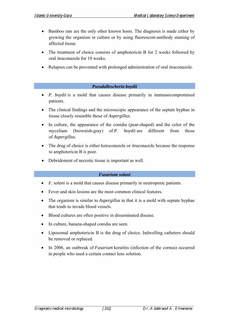
Islamic University-Gaza Medical Laboratory Science Department
_____________________________________________________________________ Diagnostic medical microbiology [151] Dr. Abdelraouf A. Elmanama
• Bamboo rats are the only other known hosts. The diagnosis is made either by growing the organism in culture or by using fluorescent-antibody staining of affected tissue.
• The treatment of choice consists of amphotericin B for 2 weeks followed by oral itraconazole for 10 weeks.
• Relapses can be prevented with prolonged administration of oral itraconazole.
Pseudallescheria boydii
• P. boydii is a mold that causes disease primarily in immunocompromised patients.
• The clinical findings and the microscopic appearance of the septate hyphae in tissue closely resemble those of Aspergillus.
• In culture, the appearance of the conidia (pear-shaped) and the color of the mycelium (brownish-gray) of P. boydii are different from those of Aspergillus.
• The drug of choice is either ketoconazole or itraconazole because the response to amphotericin B is poor.
• Debridement of necrotic tissue is important as well.
Fusarium solani
• F. solani is a mold that causes disease primarily in neutropenic patients.
• Fever and skin lesions are the most common clinical features.
• The organism is similar to Aspergillus in that it is a mold with septate hyphae that tends to invade blood vessels.
• Blood cultures are often positive in disseminated disease.
• In culture, banana-shaped conidia are seen.
• Liposomal amphotericin B is the drug of choice. Indwelling catheters should be removed or replaced.
• In 2006, an outbreak of Fusarium keratitis (infection of the cornea) occurred in people who used a certain contact lens solution.