Master Microbiology Checklist - College of American ...
102
Master Every patient deserves the GOLD STANDARD ... Microbiology Checklist CAP Accreditation Program College of American Pathologists 325 Waukegan Road Northfield, IL 60093-2750 www.cap.org 04.21.2014
-
Upload
khangminh22 -
Category
Documents
-
view
2 -
download
0
Transcript of Master Microbiology Checklist - College of American ...

Master
Every patientdeserves theGOLD STANDARD ...
Microbiology Checklist
CAP Accreditation Program
College of American Pathologists325 Waukegan RoadNorthfield, IL 60093-2750www.cap.org 04.21.2014
Disclaimer and Copyright NoticeOn-site inspections are performed with the edition of the Checklists mailed to a facility at the completionof the application or reapplication process, not necessarily those currently posted on the Web site. Thechecklists undergo regular revision and a new edition may be published after the inspection materials aresent.
For questions about the use of the Checklists or Checklist interpretation, email [email protected] or call800-323-4040 or 847-832-7000 (international customers, use country code 001).
The Checklists used for inspection by the College of American Pathologists' Accreditation Programs havebeen created by the CAP and are copyrighted works of the CAP. The CAP has authorized copying anduse of the checklists by CAP inspectors in conducting laboratory inspections for the Commission onLaboratory Accreditation and by laboratories that are preparing for such inspections. Except as permittedby section 107 of the Copyright Act, 17 U.S.C. sec. 107, any other use of the Checklists constitutesinfringement of the CAP's copyrights in the Checklists.The CAP will take appropriate legal action to protectthese copyrights.
All Checklists are ©2014. College of American Pathologists. All rights reserved.
2 of 102
04.21.2014Microbiology Checklist
Microbiology Checklist
TABLE OF CONTENTS
SUMMARY OF CHANGES.....................................................................................................................5INTRODUCTION....................................................................................................................................8GENERAL MICROBIOLOGY.................................................................................................................8
PROFICIENCY TESTING.....................................................................................................................................................8QUALITY MANAGEMENT AND QUALITY CONTROL........................................................................................................9
WAIVED TESTS.............................................................................................................................................................9GENERAL ISSUES......................................................................................................................................................10SPECIMEN COLLECTION AND HANDLING..............................................................................................................12REAGENTS - GENERAL.............................................................................................................................................14REPORTING OF RESULTS.........................................................................................................................................15INSTRUMENTS AND EQUIPMENT............................................................................................................................16
MATRIX-ASSISTED LASER DESORPTION IONIZATION TIME-OF-FLIGHT (MALDI-TOF) MASS SPECTROMETRY....17PERSONNEL.....................................................................................................................................................................18BIOSAFETY.......................................................................................................................................................................19
BACTERIOLOGY.................................................................................................................................21MEDIA................................................................................................................................................................................21STAINS...............................................................................................................................................................................23REAGENTS........................................................................................................................................................................24ANTIMICROBIAL SUSCEPTIBILITY TESTING, QC REQUIREMENTS, AND RESULTS REPORTING...........................26PROCEDURES AND TESTS.............................................................................................................................................30
RESPIRATORY SPECIMENS......................................................................................................................................31URINE SPECIMENS....................................................................................................................................................32GENITAL SPECIMENS................................................................................................................................................32STOOL SPECIMENS...................................................................................................................................................33CEREBROSPINAL & OTHER BODY FLUID SPECIMENS.........................................................................................34BLOOD CULTURES.....................................................................................................................................................35WOUND SPECIMENS.................................................................................................................................................37GAS CHROMATOGRAPHY (GC) FOR MICROBIAL IDENTIFICATION......................................................................37
LABORATORY SAFETY.....................................................................................................................................................40
MYCOBACTERIOLOGY......................................................................................................................40QUALITY CONTROL..........................................................................................................................................................40
SPECIMEN HANDLING...............................................................................................................................................41REPORTING OF RESULTS.........................................................................................................................................41MEDIA..........................................................................................................................................................................42CONTROLS AND STANDARDS..................................................................................................................................43
PROCEDURES AND TESTS.............................................................................................................................................44RAPID METHODS.......................................................................................................................................................44CONCENTRATION, INOCULATION, INCUBATION....................................................................................................45CULTURES..................................................................................................................................................................46DIFFERENTIAL BIOCHEMICAL PROCEDURES........................................................................................................46
High Performance Liquid Chromatography (HPLC) for Microbial Identification.....................................................47LABORATORY SAFETY.....................................................................................................................................................50
MYCOLOGY.........................................................................................................................................52QUALITY CONTROL..........................................................................................................................................................52
3 of 102
04.21.2014Microbiology Checklist
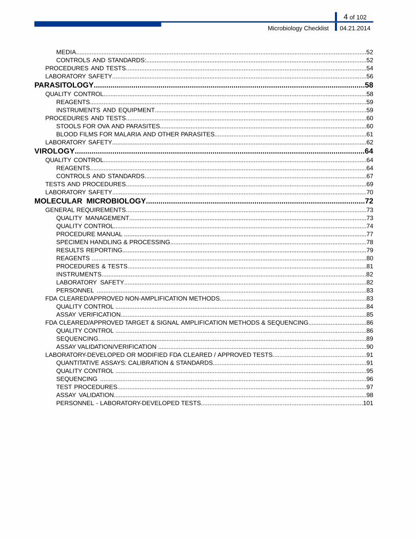
MEDIA..........................................................................................................................................................................52CONTROLS AND STANDARDS:.................................................................................................................................52
PROCEDURES AND TESTS.............................................................................................................................................54LABORATORY SAFETY.....................................................................................................................................................56
PARASITOLOGY..................................................................................................................................58QUALITY CONTROL..........................................................................................................................................................58
REAGENTS..................................................................................................................................................................59INSTRUMENTS AND EQUIPMENT............................................................................................................................59
PROCEDURES AND TESTS.............................................................................................................................................60STOOLS FOR OVA AND PARASITES.........................................................................................................................60BLOOD FILMS FOR MALARIA AND OTHER PARASITES.........................................................................................61
LABORATORY SAFETY.....................................................................................................................................................62
VIROLOGY...........................................................................................................................................64QUALITY CONTROL..........................................................................................................................................................64
REAGENTS..................................................................................................................................................................64CONTROLS AND STANDARDS..................................................................................................................................67
TESTS AND PROCEDURES.............................................................................................................................................69LABORATORY SAFETY.....................................................................................................................................................70
MOLECULAR MICROBIOLOGY.........................................................................................................72GENERAL REQUIREMENTS.............................................................................................................................................73
QUALITY MANAGEMENT...........................................................................................................................................73QUALITY CONTROL....................................................................................................................................................74PROCEDURE MANUAL ..............................................................................................................................................77SPECIMEN HANDLING & PROCESSING...................................................................................................................78RESULTS REPORTING...............................................................................................................................................79REAGENTS .................................................................................................................................................................80PROCEDURES & TESTS............................................................................................................................................81INSTRUMENTS...........................................................................................................................................................82LABORATORY SAFETY..............................................................................................................................................82PERSONNEL ..............................................................................................................................................................83
FDA CLEARED/APPROVED NON-AMPLIFICATION METHODS......................................................................................83QUALITY CONTROL ...................................................................................................................................................84ASSAY VERIFICATION................................................................................................................................................85
FDA CLEARED/APPROVED TARGET & SIGNAL AMPLIFICATION METHODS & SEQUENCING..................................86QUALITY CONTROL ...................................................................................................................................................86SEQUENCING.............................................................................................................................................................89ASSAY VALIDATION/VERIFICATION ..........................................................................................................................90
LABORATORY-DEVELOPED OR MODIFIED FDA CLEARED / APPROVED TESTS.......................................................91QUANTITATIVE ASSAYS: CALIBRATION & STANDARDS..........................................................................................91QUALITY CONTROL ...................................................................................................................................................95SEQUENCING ............................................................................................................................................................96TEST PROCEDURES..................................................................................................................................................97ASSAY VALIDATION....................................................................................................................................................98PERSONNEL - LABORATORY-DEVELOPED TESTS...............................................................................................101
4 of 102
04.21.2014Microbiology Checklist

ON-LINE CHECKLIST AVAILABILITY
Participants of the CAP accreditation programs may download the checklists from the CAP Web site (www.cap.org)by logging into e-LAB Solutions. They are available in different checklist types and formatting options, including:
● Master — contains ALL of the requirements and instructions available in PDF, Word/XML or Excel formats● Custom — customized based on the laboratory's activity (test) menu; available in PDF, Word/XML or Excel
formats● Changes Only — contains only those requirements with significant changes since the previous checklist
edition in a track changes format to show the differences; in PDF version only. Requirements that havebeen moved or merged appear in a table at the end of the file.
SUMMARY OF CHECKLIST EDITION CHANGESMicrobiology Checklist
04/21/2014 Edition
The information below includes a listing of checklist requirements with significant changes in the current editionand previous edition of this checklist. The list is separated into three categories:
1. New2. Revised:
● Modifications that may require a change in policy, procedure, or process for continued compliance;or
● A change to the Phase3. Deleted/Moved/Merged:
● Deleted● Moved — Relocation of a requirement into a different checklist (requirements that have been
resequenced within the same checklist are not listed)● Merged — The combining of similar requirements
NOTE: The listing of requirements below is from the Master version of the checklist. The customized checklistversion created for on-site inspections and self-evaluations may not list all of these requirements.
NEW Checklist Requirements
Effective DateRequirement04/21/2014MIC.1137507/29/2013MIC.1657507/29/2013MIC.1659507/29/2013MIC.1660507/29/2013MIC.1661507/29/2013MIC.1662507/29/2013MIC.4202504/21/2014MIC.6325907/29/2013MIC.64832
REVISED Checklist Requirements
Effective DateRequirement04/21/2014MIC.1006004/21/2014MIC.1101604/21/2014MIC.1102007/29/2013MIC.1458307/29/2013MIC.14616
5 of 102
04.21.2014Microbiology Checklist
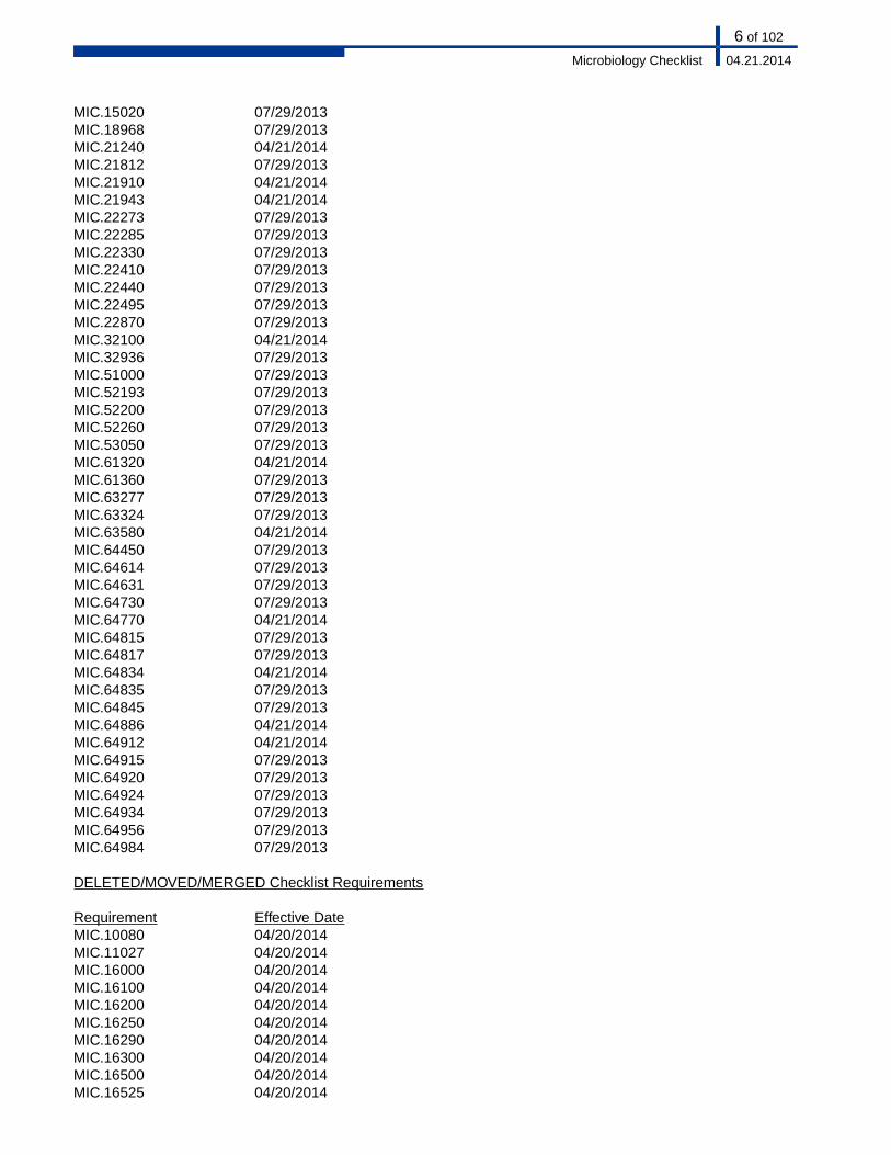
07/29/2013MIC.1502007/29/2013MIC.1896804/21/2014MIC.2124007/29/2013MIC.2181204/21/2014MIC.2191004/21/2014MIC.2194307/29/2013MIC.2227307/29/2013MIC.2228507/29/2013MIC.2233007/29/2013MIC.2241007/29/2013MIC.2244007/29/2013MIC.2249507/29/2013MIC.2287004/21/2014MIC.3210007/29/2013MIC.3293607/29/2013MIC.5100007/29/2013MIC.5219307/29/2013MIC.5220007/29/2013MIC.5226007/29/2013MIC.5305004/21/2014MIC.6132007/29/2013MIC.6136007/29/2013MIC.6327707/29/2013MIC.6332404/21/2014MIC.6358007/29/2013MIC.6445007/29/2013MIC.6461407/29/2013MIC.6463107/29/2013MIC.6473004/21/2014MIC.6477007/29/2013MIC.6481507/29/2013MIC.6481704/21/2014MIC.6483407/29/2013MIC.6483507/29/2013MIC.6484504/21/2014MIC.6488604/21/2014MIC.6491207/29/2013MIC.6491507/29/2013MIC.6492007/29/2013MIC.6492407/29/2013MIC.6493407/29/2013MIC.6495607/29/2013MIC.64984
DELETED/MOVED/MERGED Checklist Requirements
Effective DateRequirement04/20/2014MIC.1008004/20/2014MIC.1102704/20/2014MIC.1600004/20/2014MIC.1610004/20/2014MIC.1620004/20/2014MIC.1625004/20/2014MIC.1629004/20/2014MIC.1630004/20/2014MIC.1650004/20/2014MIC.16525
6 of 102
04.21.2014Microbiology Checklist
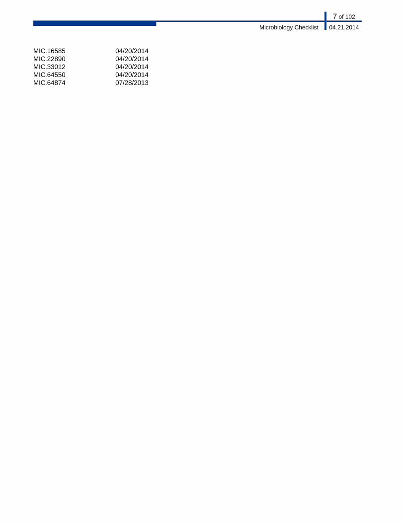
04/20/2014MIC.1658504/20/2014MIC.2289004/20/2014MIC.3301204/20/2014MIC.6455007/28/2013MIC.64874
7 of 102
04.21.2014Microbiology Checklist
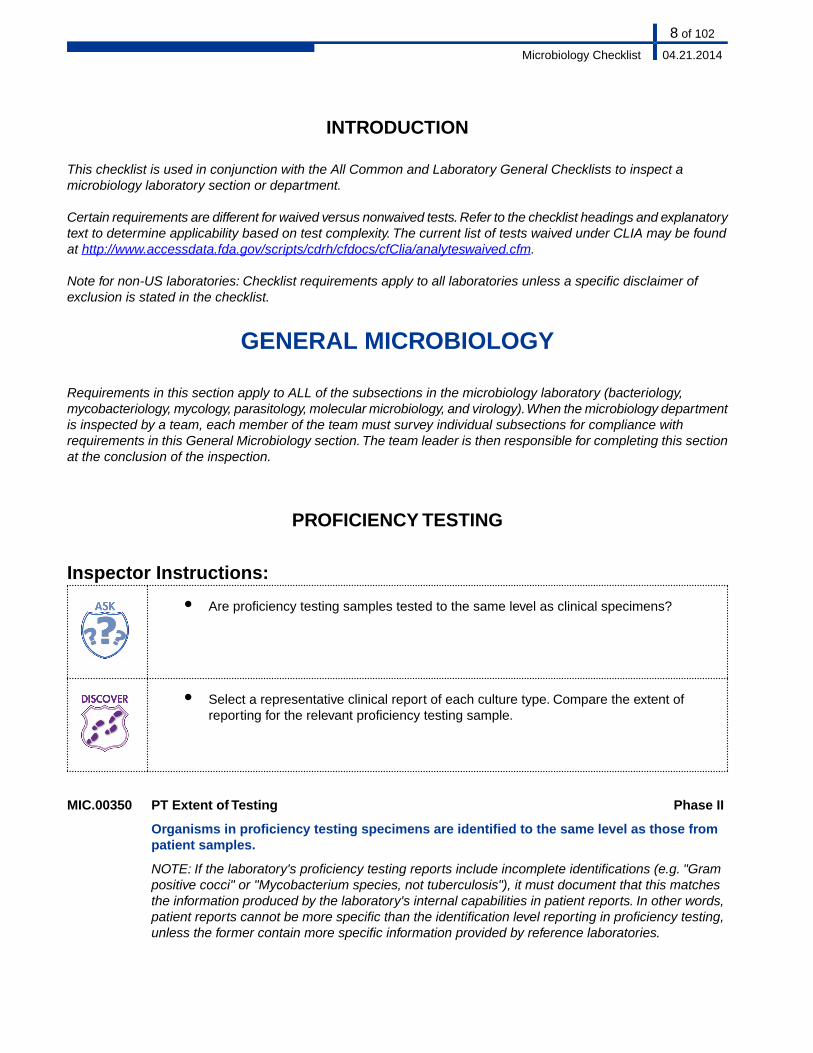
INTRODUCTION
This checklist is used in conjunction with the All Common and Laboratory General Checklists to inspect amicrobiology laboratory section or department.
Certain requirements are different for waived versus nonwaived tests. Refer to the checklist headings and explanatorytext to determine applicability based on test complexity. The current list of tests waived under CLIA may be foundat http://www.accessdata.fda.gov/scripts/cdrh/cfdocs/cfClia/analyteswaived.cfm.
Note for non-US laboratories: Checklist requirements apply to all laboratories unless a specific disclaimer ofexclusion is stated in the checklist.
GENERAL MICROBIOLOGY
Requirements in this section apply to ALL of the subsections in the microbiology laboratory (bacteriology,mycobacteriology, mycology, parasitology, molecular microbiology, and virology).When the microbiology departmentis inspected by a team, each member of the team must survey individual subsections for compliance withrequirements in this General Microbiology section. The team leader is then responsible for completing this sectionat the conclusion of the inspection.
PROFICIENCY TESTING
Inspector Instructions:
● Are proficiency testing samples tested to the same level as clinical specimens?
● Select a representative clinical report of each culture type. Compare the extent ofreporting for the relevant proficiency testing sample.
Phase IIPT Extent of TestingMIC.00350
Organisms in proficiency testing specimens are identified to the same level as those frompatient samples.
NOTE: If the laboratory's proficiency testing reports include incomplete identifications (e.g. "Grampositive cocci" or "Mycobacterium species, not tuberculosis"), it must document that this matchesthe information produced by the laboratory's internal capabilities in patient reports. In other words,patient reports cannot be more specific than the identification level reporting in proficiency testing,unless the former contain more specific information provided by reference laboratories.
8 of 102
04.21.2014Microbiology Checklist
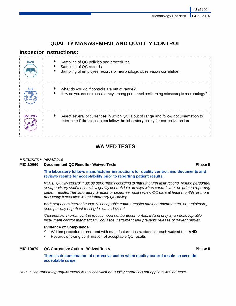
QUALITY MANAGEMENT AND QUALITY CONTROL
Inspector Instructions:
● Sampling of QC policies and procedures● Sampling of QC records● Sampling of employee records of morphologic observation correlation
● What do you do if controls are out of range?● How do you ensure consistency among personnel performing microscopic morphology?
● Select several occurrences in which QC is out of range and follow documentation todetermine if the steps taken follow the laboratory policy for corrective action
WAIVED TESTS
**REVISED** 04/21/2014Phase IIDocumented QC Results - Waived TestsMIC.10060
The laboratory follows manufacturer instructions for quality control, and documents andreviews results for acceptability prior to reporting patient results.
NOTE: Quality control must be performed according to manufacturer instructions.Testing personnelor supervisory staff must review quality control data on days when controls are run prior to reportingpatient results. The laboratory director or designee must review QC data at least monthly or morefrequently if specified in the laboratory QC policy.
With respect to internal controls, acceptable control results must be documented, at a minimum,once per day of patient testing for each device.*
*Acceptable internal control results need not be documented, if (and only if) an unacceptableinstrument control automatically locks the instrument and prevents release of patient results.
Evidence of Compliance:✓ Written procedure consistent with manufacturer instructions for each waived test AND✓ Records showing confirmation of acceptable QC results
Phase IIQC Corrective Action - Waived TestsMIC.10070
There is documentation of corrective action when quality control results exceed theacceptable range.
NOTE: The remaining requirements in this checklist on quality control do not apply to waived tests.
9 of 102
04.21.2014Microbiology Checklist
GENERAL ISSUES
Phase IIQC HandlingMIC.11015
Control specimens are tested in the same manner and by the same personnel as patientsamples.
NOTE: QC specimens must be analyzed by personnel who routinely perform patient testing. Thisdoes not imply that each operator must perform QC daily, so long as each instrument and/or testsystem has QC performed at required frequencies, and all analysts participate in QC on a regularbasis. To the extent possible, all steps of the testing process must be controlled, recognizing thatpreanalytic and postanalytic variables may differ from those encountered with patients.
Evidence of Compliance:✓ Records reflecting that QC is run by the same personnel performing patient testing
REFERENCES1) Department of Health and Human Services, Centers for Medicare and Medicaid Services. Clinical laboratory improvement amendments
of 1988; final rule. Fed Register. 2003(Jan 24):7166 [42CFR493.1256(d)(8)].1256(f)]
**REVISED** 04/21/2014Phase IICommercial Product - QCMIC.11016
When using a commercial product, QC is performed according to the manufacturer'sinstructions or CAP Checklist requirements, whichever is more stringent.
NOTE: This includes, but is not limited to, antimicrobial susceptibility testing/identification (AST/ID)systems.
Phase IIQC Confirmation of AcceptabilityMIC.11017
Control results are reviewed for acceptability before reporting patient results.
Evidence of Compliance:✓ Written policy/procedure stating that controls are reviewed and acceptable prior to reporting
patient results AND✓ Evidence of corrective action taken when QC results are not acceptable
REFERENCES1) Department of Health and Human Services, Centers for Medicare and Medicaid Services. Clinical laboratory improvement amendments
of 1988; final rule. Fed Register. 2003(Jan 24)1992(Feb 28):7166 [42CFR493.1256(f)]
Phase IIQC Corrective ActionMIC.11018
There is documentation of corrective action when control results exceed defined acceptabilitylimits.
NOTE: Patient/client test results obtained in an analytically unacceptable test run or since the lastacceptable test run must be re-evaluated to determine if there is a significant clinical difference inpatient/client results. Re-evaluation may or may not include re-testing patient samples, dependingon the circumstances.
Even if patient samples are no longer available, test results can be re-evaluated to search forevidence of an out-of-control condition that might have affected patient results.
REFERENCES1) Department of Health and Human Services, Centers for Medicare and Medicaid Services. Clinical laboratory improvement amendments
of 1988; final rule. Fed Register. 2003(Oct 1):1046[42CFR493.1282(b)(2)]
10 of 102
04.21.2014Microbiology Checklist
**REVISED** 04/21/2014Phase IIMonthly QC ReviewMIC.11020
Quality control data are reviewed and assessed at least monthly by the laboratory directoror designee.
NOTE: The review of quality control data must be documented and include follow-up for outliers,trends, or omissions that were not previously addressed.
The QC data for tests performed less frequently than once per month should be reviewed whenthe tests are performed.
Evidence of Compliance:✓ Records of QC review with documented follow-up for outliers, trends or omissions
Phase IIValidation of AccuracyMIC.11025
If the laboratory performs test procedures for which calibration and control materials arenot available, procedures have been established to validate the accuracy of patient testresults.
REFERENCES1) Elder BL, et al. Verification and Validation of Procedures in the Clinical Microbiology Laboratory. Cumitech 31, February 1997. ASM
Press; Washington DC2) Sharp S and Clark R. Verification and Validation of Procedures in the Clinical Microbiology Laboratory. Cumitech 31a. 2009. ASM Pres:
Washington, DC
Phase IIMorphologic Observation AssessmentMIC.11350
The microbiology laboratory at least annually assesses morphologic observations amongpersonnel performing Gram, trichrome and other organism stains, to ensure consistency.
NOTE: Suggested methods to accomplish this include:
1. Circulation of organisms with defined staining characteristics, and/or2. Multi-headed microscopy, and/or3. Use of photomicrographs with referee and participant identifications (e.g. former CAP
microbiology Surveys or other photomicrographs from teaching collections)4. Use of digital images
Evidence of Compliance:✓ Written procedure defining the method and criteria used for evaluation of consistency AND✓ Employee records documenting morphology assessment
REFERENCES1) Flournoy DJ. Interpreting the sputum gram stain report. Lab Med. 1998;29:763-768
**NEW** 04/21/2014Phase ITaxonomy ChangesMIC.11375
The laboratory incorporates taxonomic changes that potentially affect the choice ofappropriate antimicrobials to report and/or the interpretative breakpoints to use.
NOTE: The genus and/or species names of microorganisms may change as new methods areapplied to their taxonomy. This can impact the antimicrobials that should be reported for thatorganism. It may also change the breakpoints that should be used for interpreting susceptibility testresults. For example, Actinobacillus actinomycetemcomitans was moved to the genus Haemophilusin 1985 and then to the new genus Aggregatibacter in 2006.The antimicrobials differ for Haemophilusspecies (CLSI M100, Table 2E) versus Aggregatibacter species (CLSI M45, Table 7).The laboratoryshould have a procedure ensuring that clinically relevant taxonomic changes are incorporated intoreporting patient and proficiency testing results even when commercial identification systems havenot been updated.
11 of 102
04.21.2014Microbiology Checklist

REFERENCES1) Clinical and Laboratory Standards Institute (CLSI). Methods for Antimicrobial Dilution and Disk Susceptibility Testing of Infrequently
Isolated or Fastidious Bacteria; Approved Guideline—Second Edition. CLSI Document M45-A2 (ISBN 1-56238-732-4).Clinical andLaboratory Standards Institute, 940 West Valley Road, Suite 1400, Wayne, PA 19087-1898 USA, 2010.
2) Clinical and Laboratory Standards Institute (CLSI). Performance Standards for Antimicrobial Susceptibility Testing; Twenty-FourthInformational Supplement. CLSI Document M100-S24 (ISBN 1-56238-898-3).Clinical and Laboratory Standards Institute, 940 WestValley Road, Suite 2500, Wayne, PA 19087-1898 USA, 2014.
SPECIMEN COLLECTION AND HANDLING
Culture specimens are often collected by nurses or others outside the laboratory. An important aspect of qualitycontrol is the provision of adequate instructions to ensure proper collection and handling of specimens before theyare received by the laboratory.
Inspector Instructions:
● Sampling of specimen collection and handling policies and procedures● Sampling of requisitions for completeness● Sampling of specimen rejection records/log
● Sampling of microbiology specimens (transport media, timely delivery, labeling)
● What is your course of action when you receive unacceptable microbiology specimens?
Phase IISpecimen Acceptability CriteriaMIC.13100
There are criteria for establishing specimen acceptability.
NOTE: This could include important issues such as absence of gross external contamination,adequate specimen type/quantity, suitable preservation, prevention of dried swabs, and correct useof transport media when required.
Evidence of Compliance:✓ Records of rejected specimens
Phase IViral Culture SpecimensMIC.13175
Specimens for viral culture are collected appropriately and transported to the laboratorywithout delay.
NOTE: The laboratory should provide procedures for the appropriate collection, transport andstorage of all specimen types tested in the laboratory. Specimens should be delivered to thelaboratory promptly, ideally within 2-4 hrs of sample collection and preferably within 1 day ofcollection.This may not be possible for laboratories that refer samples to offsite reference laboratoriesfor viral testing. In these instances samples should be stored and shipped under conditions thatwould preserve the integrity of the sample. Unless otherwise indicated, specimens should berefrigerated or frozen depending on the duration of storage prior to testing.
12 of 102
04.21.2014Microbiology Checklist
REFERENCES1) Clinical and Laboratory Standards Institute (CLSI). Viral Culture; Approved Guideline. CLSI document M41-A (ISBN 1-56238-623-9).
Clinical and Laboratory Standards Institute, 940 West Valley Road, Suite 1400, Wayne, Pennsylvania 19087-1898 USA, 20062) Ginocchio, CC. Quality Assurance in Clinical Virology. In: Spector S, Hodinka RL, Young SA, editors. Clinical Virology Manual. Fourth
Edition. Washington: ASM Press; 2009.p. 3-17
Phase IRequisitionsMIC.13200
Requests for analysis include source of specimen, test or tests requested and, whenappropriate, type of infection and/or organism expected.
REFERENCES1) Department of Health and Human Services, Centers for Medicare and Medicaid Services. Clinical laboratory improvement amendments
of 1988; final rule. Fed Register. 2003(Jan 24):7162 [42CFR493.1241(c)]
Phase IISpecimen Collection/HandlingMIC.13250
There are documented instructions for microbiology specimen collection and handling thatinclude all of the following.
1. Method for proper collection of culture specimens from different sources2. Proper labeling of culture specimens3. Use of transport media when necessary4. Procedures for safe handling of specimens (tightly sealed containers, no external
spillage)5. Need for prompt delivery of specimens to ensure minimum delay and processing
(e.g. CSF, wound cultures, anaerobes)6. Method for preservation of specimens if processing is delayed (e.g. refrigeration
of urines)
NOTE: Manufacturer's recommendations must be followed when there is a delay in delivery orprocessing of specimens for automated instruments (e.g. blood culture instruments).
REFERENCES1) Miller JM, et al. Specimen collection, transport, and storage. In: Murray PR, et al, ed. Manual of Clinical Microbiology, 8th ed.Washington,
DC: ASM Press; 2003:55-66
Phase IISpecimens for Molecular AmplificationMIC.13275
The laboratory has procedures for the handling of specimens that will also be tested usingmolecular amplification methods.
NOTE: Special precautions must be taken to avoid sample cross-contamination that may not affectculture-based methods but may lead to false positive results when tested using molecularamplification methods. For example, proper procedures to prevent cross-contamination must beused when samples are processed in the same biohazard hood in which virus cultures aremanipulated post-inoculation. Please refer to the Molecular Microbiology section of this checklist.
REFERENCES1) Clinical and Laboratory Standards Institute (CLSI). Collection, Transport, Preparations, and Storage of Specimens for Molecular Methods;
Approved Guideline. CLSI document MM13-A (ISBN 1-56238-591-7). Clinical and Laboratory Standards Institute, 940 West ValleyRoad, Suite 1400, Wayne, Pennsylvania 19087-1898 USA, 2005.
13 of 102
04.21.2014Microbiology Checklist

REAGENTS - GENERAL
Inspector Instructions:
● Sampling of test procedures for QC● Sampling of reagent QC records
Additional requirements are in the REAGENTS section of the All Common Checklist.
The following generic requirements apply to all subsections of the Microbiology Laboratory for nonwaived testingonly.
**REVISED** 07/29/2013Phase IIDirect Antigen Test QCMIC.14583
For nonwaived direct antigen tests on patient specimens that DO include internal controls,a positive and negative external control are tested and documented with each new kit lotnumber or shipment, and as frequently as recommended by the manufacturer, or every 30days (whichever is more frequent).
NOTE: Internal controls may be used for daily quality control, providing that the following requirementsare met:
1. Prior to initiating patient testing, the internal controls are checked for acceptabilityagainst external controls. Acceptability studies must include daily comparison of externalcontrols to built-in controls for at least 20 consecutive days when patient samples aretested. For panels or batteries, controls must be employed for each antigen sought.The requirement for 20 consecutive daily comparisons is effective for studies performedafter 1/31/2012. Acceptable results are required before daily quality control can belimited to built-in controls.The laboratory director is responsible for determining criteriafor acceptability. These records must be retained while an instrument/method is inservice, and for two years afterwards.
2. A positive and negative external control (organism or antigen extract) are tested anddocumented with each new kit lot number or separate shipments of a given lot number.
3. Manufacturers’ recommendations are followed. “Flow” or “procedural” controls qualifyas internal controls.
4. For tests classified as "high complexity" under CLIA, the system must be checked eachday of use with a positive external control (organism or antigen).
5. External surrogate sample controls are run as frequently as recommended by the testmanufacturer, or every 30 days, whichever is more frequent.
6. This requirement pertains to nonwaived tests with a protein, enzyme, or toxin whichacts as an antigen and the assay contains an internal control. Examples include, butare not limited to: Group A Streptococcus antigen, C. difficile toxin, fecal lactoferrin andimmunochemical occult blood tests.
For those Direct Antigen Tests that are done seasonally or intermittently, please review the AllCommon Checklist requirement COM.40100 for applicability and additional information.
14 of 102
04.21.2014Microbiology Checklist

Evidence of Compliance:✓ Written QC procedures for each test consistent with the manufacturer's instructions AND/OR
records documenting in-house acceptability studies of internal control systems
REFERENCES1) Department of Health and Human Services, Centers for Medicare and Medicaid Services. Clinical laboratory improvement amendments
of 1988, final rule. Fed Register. 2003(Jan 24): [42CFR493.1261(a)]
**REVISED** 07/29/2013Phase IIDirect Antigen Test QCMIC.14616
For nonwaived direct antigen tests on patient specimens that do NOT include internalcontrols, a positive and negative control are tested and documented each day of patienttesting.
NOTE: For panels or batteries, controls must be employed for each antigen sought in patientspecimens. For each test system that requires an antigen extraction phase, the system must bechecked with an appropriate positive control that will detect problems in the extraction process.
REFERENCES1) Department of Health and Human Services, Centers for Medicare and Medicaid Services. Fed Register. 2003(Jan 24):3708
[42CFR493.1256(d)] and [42CFR493.1261(a)]
REPORTING OF RESULTS
Inspector Instructions:
● Sampling of patient preliminary reports
Phase IPreliminary ReportsMIC.15000
When indicated, preliminary reports are promptly generated.
Evidence of Compliance:✓ Written procedure(s) defining when preliminary results are issued
**REVISED** 07/29/2013Phase IAzoospermic Specimen Result ReportingMIC.15020
For azoospermic and post-vasectomy seminal fluid specimens, the laboratory clearlycommunicates the findings of the assay and either employs a concentrating technique onseminal fluid or includes a comment in the patient report indicating that a concentratingtechnique was not performed.
NOTE: Without a concentration technique, the presence of both motile and non-motile sperm maynot be detected.The method for detection of motile and non-motile sperm and the laboratory findingsmust be clearly communicated on the patient report so that the clinician can interpret the results incontext to the method performed. The decision on the method used and extent of testing to beperformed should be made in consultation with the medical staff served.
The American Urological Association (AUA) Vasectomy Guideline recommends a careful evaluationof an uncentrifuged specimen, and does not recommend centrifugation of the specimen for furtherassessment. The AUA Guideline also recommends reporting both the presence and absence ofsperm and presence or absence of sperm motility on the patient report. If no sperm are seen in the
15 of 102
04.21.2014Microbiology Checklist

uncentrifuged specimen, the guideline recommends reporting that the presence of sperm is belowthe limit of detection.
Evidence of Compliance:✓ Patient report with concentration findings or appropriate comment indicating that concentration
was not performed
REFERENCES1) Evaluation of the Azoospermic Male. Fertil Steril. 2008; 90 (S74-7)
2) Diagnostic Evaluation of the Infertile Male: A Committee Opinion. Fertil Steril. 2012; 98:294-301
3) American Urological Association (AUA) Guideline. American Urological Association Education and Research, Inc. 2012http://www.auanet.org/common/pdf/education/clinical-guidance/Vasectomy.pdf
4) Vasectomy Update 2010. Can Urol Assoc J. 2010 October; 4(5):306-309
INSTRUMENTS AND EQUIPMENT
The checklist requirements in this section should be used in conjunction with the requirements in the All CommonChecklist relating to instruments and equipment.
Inspector Instructions:
● Sampling of pipette/diluter checks
● Microscope filters - used as indicated by manufacturer● Incubators (adequate space, maintained)
Phase IIPipettors and DilutersMIC.16150
Pipettes, microtiter diluters or automatic dispensers that are used for quantitative dispensingof material are checked for accuracy and reproducibility at specified intervals, with resultsdocumented.
NOTE: This requirement is not applicable for precalibrated inoculation loops that are used in thedirect plating of clinical specimens such as urine cultures.
Evidence of Compliance:✓ Written procedure detailing method for checking the accuracy and reproducibility of pipettes
REFERENCES1) Curtis RH. Performance verification of manual action pipets. Part I. Am Clin Lab. 1994;12(7):8-9
2) Curtis RH. Performance verification of manual action pipets. Part II. Am Clin Lab. 1994;12(9):16-17
3) Perrier S, et al. Micro-pipette calibration using a ratiometric photometer-reagent system as compared to the gravimetric method. ClinChem. 1995;41:S183
4) Clinical and Laboratory Standards Institute (CLSI). Laboratory Instrument Implementation, Verification, and Maintenance; ApprovedGuideline. CLSI Document GP31-A. (ISBN 1-56238-697-2). Clinical and Laboratory Standards Institute, 940 West Valley Road, Suite1400, Wayne, PA 19087-1898, USA, 2009.
5) Johnson B. Calibration to dye for: Artel's new pipette calibration system. Scientist. 1999;13(12):14
6) Connors M, Curtis R. Pipetting error: a real problem with a simple solution. Parts I and II. Am Lab News. 1999;31(13):20-22
7) Skeen GA, Ashwood ER. Using spectrophotometry to evaluate volumetric devices. Lab Med. 2000;31:478-479
Phase IMicroscopesMIC.16275
16 of 102
04.21.2014Microbiology Checklist

Microscopes used for immunofluorescent testing contain the appropriate filter(s)recommended by the manufacturer.
NOTE: The use of filters not recommended by the manufacturer can lead to erroneous results.
REFERENCES1) Schutzbank TE, McGuire R. Immunofluorescence. In: Specter S, Hodinka RL, Young SA, editors. Clinical Virology Manual. Third Edition
ed. Washington: ASM Press; 2000. p. 69-78
Phase IAdequate IncubatorsMIC.16550
There are sufficient, clean, and well-maintained incubators available at specified temperatureranges.
MATRIX-ASSISTED LASER DESORPTION IONIZATION TIME-OF-FLIGHT(MALDI-TOF) MASS SPECTROMETRY
This section applies to laboratories using MALDI-TOF systems to perform organism identification. Refer to theTest Method Validation section in the All Common Checklist for validation requirements pertinent tolaboratory-developed tests.
**NEW** 07/29/2013Phase IIInstrument OperationMIC.16575
Procedures are documented for operation and calibration of the mass spectrometer.
**NEW** 07/29/2013Phase IIMass Spectrometer CalibrationMIC.16595
A calibration control is run each day of patient/client testing, with each change in targetplate, or according to manufacturer's recommendations and these records are maintained.
NOTE: Acceptable tolerance limits for calibration parameters must be defined, and recordsmaintained.
Evidence of Compliance:✓ Records of calibration
**NEW** 07/29/2013Phase IIMass Spectrometer ControlsMIC.16605
Appropriate control organisms are tested on a daily basis.
NOTE: Appropriate controls would include at least one bacterium, with a representative yeast andmycobacterium also being run if these organisms are being tested for that day/routinely. ForFDA-approved platforms, the organisms required by the manufacturer must be used. For laboratorydeveloped tests, choice of control organisms is at the Laboratory Director's discretion. Controlorganisms should be subjected to the same testing conditions throughout the testing procedure aspatient specimens. An extraction control should be included if any of the organisms being testedare run with extraction. A blank control needs to be run with each new target being used to assessthe cleanliness of the target (demonstrating a lack of peaks prior to testing).
Evidence of Compliance:✓ Written procedure defining QC requirements AND✓ QC records documented at defined frequency
17 of 102
04.21.2014Microbiology Checklist
**NEW** 07/29/2013Phase IIMass Spectrometer Reagent GradeMIC.16615
Reagents and solvents are of appropriate grade.
NOTE: Only the manufacturer's specified grade of solvents are used for this procedure. This maybe HPLC-grade or other reagent grades as indicated.
Evidence of Compliance:✓ Reagent logs
**NEW** 07/29/2013Phase IIMass Spectrometer ConsumablesMIC.16625
Consumables are of appropriate manufacturing type to function as required.
NOTE: For FDA-approved platforms, consumables utilized may be specified by the manufacturer.Deviation from the manufacturer's recommendation must be documented by an appropriate validationof the non-recommended consumables, if appropriate.
Evidence of Compliance:✓ Consumable logs AND✓ Validation of alternative consumables not specified by the manufacturer
PERSONNEL
Inspector Instructions:
● Documentation of education and experience
Phase IIBench Testing SupervisionMIC.17000
The person(s) in charge of bench testing/section supervisor in microbiology has educationin microbiology equivalent to an associate's degree (or beyond) in a chemical, physical orbiological science or medical technology and at least 4 years experience (one of which isin microbiology) under a qualified section director.
Evidence of Compliance:✓ Records of qualifications including degree or transcript, certification/registration, current license
(if required) and work history in related field
REFERENCES1) Church DL, et al. Effects of restructuring on the performance of microbiology laboratories in Alberta. Arch Pathol Lab Med.
2000;124:357-361
Phase IVisual Color DiscriminationMIC.17050
Personnel working in microbiology are checked for visual color discrimination.
NOTE: Testing is not required for personnel who do not perform laboratory tests requiring colordiscrimination. This does not mean that visually color-impaired technical personnel cannot beemployed, only that they be tested, with job assignments and responsibilities evaluated accordingly.
Evidence of Compliance:✔ Record of color discrimination testing OR functional assessment, if indicated
18 of 102
04.21.2014Microbiology Checklist
BIOSAFETY
Items in this section apply to ALL areas of the microbiology laboratory. Additional items for specific subsections(bacteriology, mycobacteriology, mycology, parasitology, and virology) are found under the Laboratory Safetysubsections for each of those areas.
Inspector Instructions:
● Sampling of biosafety policies and procedures● Sampling of bench top decontamination logs● Records of biological safety cabinet certification
● How would you recognize a potential agent of bioterrorism? What action would you takeif you encountered a suspect organism?
**REVISED** 07/29/2013Phase IIAgents of BioterrorismMIC.18968
The microbiology laboratory has policies and procedures for the recognition and safehandling of isolates that may be used as agents of bioterrorism.
NOTE: Microorganisms likely to be utilized as biological weapons include Bacillus anthracis (anthrax),Brucella species (brucellosis), Clostridium botulinum (botulism), Francisella tularensis (tularemia),Yersinia pestis (plague) and variola major (smallpox).
As part of an institution-wide plan to prepare and respond to a bioterrorism event, the microbiologylaboratory should have policies and procedures for the recognition of isolates that may be used asagents of bioterrorism.
Safe handling includes such activities as handling organisms under a certified biological safetycabinet, and not subjecting the isolates to identification utilizing automated instruments.
REFERENCES1) Snyder JW. Role of the hospital-based microbiology laboratory in preparation and response to a bioterrorism event. J Clin Microbiol.
January, 20032) Gilchrist MJR. Laboratory Safety, Management, and Diagnosis of Biological Agents Associated with Bioterrorism
3) Robinson-Dunn B. The microbiology laboratory's role in response to bioterrorism. Arch PatholLab Med. March 2002; 126
4) Morse SA. Bioterrorism: Laboratory Security. Lab Med. June 2001
5) Sewell, DL. Laboratory safety practices associated with potential agents of biocrime or bioterrorism. J. Clin. Microbiology. July2003;41(7):2801-2809
6) http://www.asm.org/index.php/issues/sentinel-laboratory-guidelines
Phase IBioterrorism Response PlanMIC.18976
The laboratory participates in the institution's bioterrorism response plan.
Evidence of Compliance:✓ Organizational bioterrorism plan describing the role of the laboratory
REFERENCES1) Snyder JW. Role of the hospital-based microbiology laboratory in preparation and response to a bioterrorism event. J Clin Microbiol.
January, 20032) Gilchrist MJR. Laboratory Safety, Management, and Diagram of Biological Agents Associated with Bioterrorism
3) Robinson-Dunn B. The microbiology laboratory's role in response to bioterrorism. Arch PatholLab Med. March 2002; 126
4) Morse SA. Bioterrorism: Laboratory Security. Lab Med. June 2001
19 of 102
04.21.2014Microbiology Checklist
Phase IISpill HandlingMIC.18985
There are documented policies for handling spills of contaminated materials.
REFERENCES1) Department of Health and Human Services. Biosafety in Microbiological and Biomedical Laboratories, 5th ed. Washington, DC: HHS
Publishing No. (CDC) 21-1112, December 2009
Phase IIBench Top DecontaminationMIC.19010
There is documentation of daily decontamination of bench tops.
Phase IISafe Specimen ProcessingMIC.19035
There are documented policies and procedures for the safe handling and processing ofspecimens.
NOTE: Suggested topics to be considered in the policies and procedures for the safe handling andprocessing of specimens include the need for tight sealing of containers, avoiding spills of hazardousmaterials, requirements for wearing gloves, the need for respirator protection, availability and useof vaccinations, and the potential hazards of sniffing plates.
REFERENCES1) Jamison R, et al. Laboratory Safety in Clinical Microbiology, Cumitech 29, July 1996, ASM Press; Washington DC
2) Fleming DO, Hunt DL. Biological Safety, Principles and Practices, 3rd ed. ASM Press; Washington DC
Phase IIBiosafety LevelsMIC.19060
Policies and procedures have been developed to minimize the occupational risk of exposureto infectious agents handled in the microbiology laboratory, in accordance with currentrecommendations regarding the biosafety levels for working with different organisms.
NOTE: The laboratory director is responsible for the maintenance of precautions in the laboratoryto minimize the risk of personnel infection. Precautions must be appropriate for the types of organismstested and the nature of the studies performed.
Each level consists of combinations of equipment, procedures and techniques, and laboratorydesign that are appropriate for the type of laboratory and infectious agent handled.
REFERENCES1) Department of Health and Human Services. Biosafety in Microbiological and Biomedical Laboratories, 5th ed. Washington, DC: HHS
Publishing No. (CDC) 21-1112, December 20092) Richmond J. "Arthropod Borne Diseases", Anthology of Biosafety VI: American Biological Safety Association, Mundelein, IL April 2003
Phase IIBiosafety LevelsMIC.19160
Engineering and work practice controls appropriate to the Biosafety level of the laboratoryare defined and implemented.
NOTE: Each increasing BSL number (1 to 4) implies increased occupational risk from exposure toan agent or performance of a procedure, and therefore is associated with more stringent controland containment practices.
REFERENCES1) Department of Health and Human Services. Biosafety in Microbiological and Biomedical Laboratories, 5th ed. Washington, DC: HHS
Publishing No. (CDC) 21-1112, December 20092) Richmond J. BSL-4 Laboratories. Anthology of Biosafety V: American Biological Safety Association, Mundelein, IL January 2002
3) Richmond J. Biosafety Level 3. Anthology of Biosafety VII: American Biological Safety Association, Mundelein, IL December 2003
Phase IIBiological Safety CabinetMIC.19840
A biological safety cabinet (BSC) or hood is available for handling specimens or organismsconsidered highly contagious by airborne routes.
20 of 102
04.21.2014Microbiology Checklist

Evidence of Compliance:✓ Maintenance schedule of BSC function checks AND✓ Records of testing and certification
REFERENCES1) Department of Health and Human Services. Biosafety in Microbiological and Biomedical Laboratories, 5th ed. Washington, DC: HHS
Publishing No. (CDC) 21-1112, December 2009
Phase IIBiological Safety CabinetMIC.20520
The biological safety cabinet (BSC) is certified at least annually to ensure that filters arefunctioning properly and that airflow rates meet specifications.
Evidence of Compliance:✓ Maintenance schedule of BSC function checks AND✓ Records of testing and certification
BACTERIOLOGY
MEDIA
Inspector Instructions:
● Sampling of media QC policies and procedures● Sampling of media supplier records of QC● Sampling of QC records for media prepared in-house or not exempt from M22-A3
● Sampling of media (expiration date, condition, contamination)
● What is your QC process when receiving a new lot of media?
● Follow a shipment of new media from receipt, examination and QC (if applicable).Determine if practice follows laboratory policy.
Phase IIMedia SupplierMIC.21200
The laboratory has documentation that its media supplier carries out the quality assuranceguidelines enumerated in CLSI/NCCLS Document M22-A3.
NOTE: The laboratory has the responsibility for ensuring that all media used, whether purchasedor prepared by the laboratory, are sterile, able to support growth appropriately and are appropriately
21 of 102
04.21.2014Microbiology Checklist
reactive biochemically. This will ordinarily require that the laboratory maintain a stock of referenceorganisms and test the media before or concurrent with use. Explicit documentation of such testingis essential.
For prepared, purchased media, the laboratory must have explicit documentation that each lot ofpurchased medium has been tested for sterility, ability to support growth of appropriate organismsand biochemical reactivity at the time of preparation or concurrent with use in the laboratory. Therecipient laboratory must have a copy of the CLSI/NCCLS document number M22-A3 (Qualityassurance for commercially prepared microbiological culture media) as a reference source. Themanufacturer or preparer must document to the user that their quality control activities meet theCLSI/NCCLS guidelines, or are otherwise equivalent. The laboratory director may wish to have asigned contractual arrangement with his/her selected manufacturer to cover all expected qualitycontrol and documentation thereof. For each lot, the preparer will certify that quality controlperformance was acceptable, and maintain a record of test results and the lot numbers for all mediafor at least 2 years. The user laboratory may record that fact in place of the more detaileddocumentation of media performance.The user must visually examine each shipment for breakage,contamination, appearance, or evidence of freezing or overheating.Transportation of media/reagentsunder unfavorable environmental conditions may adversely affect product performance.
The user laboratory must continue to test each lot of media except those listed as being exemptfrom such testing in the tables in M22-A3, using quality control methods that are used for mediamanufactured in-house. In addition, each shipment or lot, if more than one lot number is receivedper shipment of a commercial identification system must be tested for appropriate performance.
REFERENCES1) NCCLS. Quality Control for Commercially Prepared Microbiological Culture Media; Approved Standard - Third Edition. NCCLS document
M22-A3. 940 West Valley Road, Suite 1400, Wayne, PA 19087-1898 USA, 2004.2) Department of Health and Human Services, Centers for Medicare and Medicaid Services. Clinical laboratory improvement amendments
of 1988, final rule. Fed Register. 2003(Jan 24): [42CFR493.1256(e)]
Phase IMedia Visual InspectionMIC.21220
The laboratory has documentation that each shipment of purchased media is examined forbreakage, contamination, appearance, and evidence of freezing or overheating.
REFERENCES1) NCCLS. Quality Control for Commercially Prepared Microbiological Culture Media; Approved Standard - Third Edition. NCCLS document
M22-A3. 940 West Valley Road, Suite 1400, Wayne, PA 19087-1898 USA, 2004.
**REVISED** 04/21/2014Phase IIMedia QC - PurchasedMIC.21240
The laboratory has documentation that an appropriate sample from each lot and shipmentof each purchased medium that is not listed in M22-A3 as exempt from testing is checkedfor each of the following:
1. Ability to support growth (where applicable) by means of stock cultures or byparallel testing with previous batches
2. Biochemical reactivity, where appropriate3. End user quality control must be performed on the following, regardless of exempt
status:● Campylobacter agar;● Chocolate agar;● Media for the selective isolation of pathogenic Neisseria;● Other media not listed on Table 2 of M22-A3 (e.g. dermatophyte test medium);● Media used for the isolation of parasites, viruses, Mycoplasmas, Chlamydia;● Mueller-Hinton media used for antimicrobial susceptibility tests; or● Media commercially prepared and packaged as a unit or system consisting
of two or more different substrates, primarily used for microbial identification.
REFERENCES
22 of 102
04.21.2014Microbiology Checklist

1) NCCLS. Quality Control for Commercially Prepared Microbiological Culture Media; Approved Standard - Third Edition. NCCLS documentM22-A3. 940 West Valley Road, Suite 1400, Wayne, PA 19087-1898 USA, 2004.
Phase IIMedia QC In-House PreparedMIC.21300
For microbiology media prepared in-house, there is documentation that an appropriatesample of each medium prepared by the laboratory is checked for each of the following:
1. Sterility (following introduction of additives after sterilization)2. Ability to support growth (where applicable) by means of stock cultures or by
parallel testing with previous batches3. Biochemical reactivity (where appropriate)
Evidence of Compliance:✓ Written procedure for testing media prepared in-house
Phase IIMedia Visual ExaminationMIC.21420
All media are in visibly satisfactory condition (with expiration date, plates smooth, adequatelyhydrated, uncontaminated, appropriate color and thickness, tubed media not dried or loosefrom sides).
Phase IIQuality Control OrganismsMIC.21460
Quality control organisms are used to check stains, reagents and susceptibility test methods.
NOTE:1. Quality control organisms may be ATCC strains or well characterized laboratory strains
unless specified by the manufacturer2. Quality control organisms are maintained in a manner to preserve their bioreactivity,
phenotypic characteristics and integrity
REFERENCES1) Jones RN, et al. Method preferences and test accuracy of antimicrobial susceptibility testing. Updates from the College of American
Pathologists microbiology surveys program (2000). Arch Pathol Lab Med. 2001;125:1285-1289
STAINS
Inspector Instructions:
● Sampling of staining policies and procedures● Sampling of stain QC records/logs
Phase IDirect Gram Stain ProceduresMIC.21530
The laboratory has protocols in place to use Gram stain results to provide a preliminaryidentification of organisms, evaluate specimen quality when appropriate, and to guidework-up of cultures.
NOTE: The laboratory should have guidelines for the interpretation of the Gram stain reaction ofthe organism, morphology of the organism, and the quantification of organisms and cells. Theprotocol should address correlation of direct Gram stain results with final culture results.
23 of 102
04.21.2014Microbiology Checklist
This does not mean that interpretation of the Gram stain morphology suggesting a specific organismidentification (e.g. gram positive diplococcic morphologically suggestive of pneumococcus) isrequired.
Evidence of Compliance:✓ Written procedure for Gram stain (laboratories may use the correlation of Gram stain results
with the final culture results as a component of the QC program)
Phase IIGram Stain QCMIC.21540
Quality control of Gram stain reagents is performed for intended reactivity and recorded foreach new batch of stains and at least weekly against known gram-positive and gram-negativequality control organisms.
NOTE: Personnel who perform Gram stains less frequently must run a gram-positive andgram-negative control each day of testing.
Evidence of Compliance:✓ Written procedure for Gram stain QC
REFERENCES1) August, Hindler, Huber, Sewell. Quality control and quality assurance practices. In: Clinical microbiology, Cumitech 3A. Washington,
DC: American Society for Microbiology, 19902) Department of Health and Human Services, Centers for Medicare and Medicaid Services. Clinical laboratory improvement amendments
of 1988, final rule. Fed Register. 2003(Jan 24):3708 [42CFR493.1261(a)(2)]
Phase IINon-Immunofluorescent Stain QCMIC.21560
Quality control of all non-immunofluorescent, non-immunologic-based stains (other thanGram stains) is performed and recorded with a positive and negative quality control organismfor intended reactivity each day of use, and for each new batch, lot number and shipment.
NOTE: Refer to MIC.51160 for requirement pertaining to parasitology permanent stains.
Evidence of Compliance:✓ Written procedure for QC of non-immunofluorescent stains
Phase IIFluorescent Stain QCMIC.21570
Quality control of fluorescent stains is performed for positive and negative reactivity eachtime of use.
Evidence of Compliance:✓ Written procedure QC of fluorescent stain
REFERENCES1) Department of Health and Human Services, Centers for Medicare and Medicaid Services. Clinical laboratory improvement amendments
of 1988; final rule. Fed Register. 2003(Jan 24):7146 [42CFR493.1256(e)(3); 493.1273(a)]
REAGENTS
Inspector Instructions:
● Sampling of reagent QC policies and procedures● Sampling of reagent QC records● CO2 monitoring procedure and CO2 recording log● Anaerobic incubation condition monitoring records● Campylobacter incubation condition records
24 of 102
04.21.2014Microbiology Checklist
● What is your QC process when receiving a new lot of identification system materials?
Phase IIReagent QCMIC.21624
Positive and negative controls are tested and results recorded for each new batch, lot number,and shipment of reagents, disks/strips and stains.
NOTE: Reagents subject to this requirement include (but are not limited to) catalase, coagulase(including latex methods), oxidase and indole reagents; bacitracin, optochin, streptococcal groupingreagents, ONPG, X, V, and XV disks/strips.This does not include tests for antimicrobial susceptibility.
REFERENCES1) Department of Health and Human Services, Centers for Medicare and Medicaid Services. Clinical laboratory improvement amendments
of 1988; final rule. Fed Register. 2003(Jan 24): 3708 [42CFR493.1256 (e) (1) and (2)]
Phase IIIdentification System QCMIC.21626
Appropriate positive and negative control organisms are tested and results recorded foreach new lot and shipment of reagents used in bacterial identification systems.
NOTE: Streamlined QC may be performed, as specified by the manufacturer, for commercialmicrobial identifications systems (MIS) if the systems and streamlined QC protocols are usedaccording to the manufacturer's instructions without modification.The laboratory may use additionalQC organisms in addition to those required for the streamlined QC. In order to qualify for streamlinedQC, the user must fulfill initial and ongoing requirements as defined by the manufacturer and CLSIdocument M50-A.
For user-developed identification systems, commercial systems for which a streamlined QC processhas not been developed, or any commercial system whose use is altered in any way from themanufacturer's instructions, all biochemical tests in each new lot number and shipment must beevaluated with known positive and negative control organisms, to assure appropriate reactivity.
If streamlined QC is used, it is critical for laboratories to keep documentation of the test systemverification and historical QC review as long as the streamlined QC is used, but in no case for lessthan two years.
Any test (e.g. oxidase test) required for interpretation of MIS results which is not part of the MIScannot be included in MIS streamlined QC protocols. QC requirements for such tests, including theuse of positive and negative controls for each new batch, lot number and shipment are given inMIC.21624.
Evidence of Compliance:✓ Written procedure for QC on new lot numbers or shipments of reagents for each MIS using the
conventional QC method (a positive and negative control for each substrate) OR a writtenprocedure for streamlined QC AND
✓ Records of test system verification and historical QC review used to qualify for streamlined QC,if applicable
REFERENCES1) Department of Health and Human Services, Centers for Medicare and Medicaid Services. Clinical laboratory improvement amendments
of 1988; final rule. Fed Register. 2003(Jan 24):3708 [42CFR493.1256(e)(1)]2) Clinical and Laboratory Standards Institute (CLSI). Quality Control for Commercial Microbial Identification Systems. CLSI document
M50-A (ISBN 1-56238-675-1). Clinical and Laboratory Standards Institute, 940 West Valley Road, Suite 1400, Wayne, Pennsylvania19087-1898 USA, 2008.
Phase IAntisera QCMIC.21628
25 of 102
04.21.2014Microbiology Checklist

Positive and negative controls are tested and results recorded for each new batch, lot numberand shipment of antisera when prepared or opened and once every 6 months thereafter (e.g.Salmonella/Shigella antisera).
REFERENCES1) Department of Health and Human Services, Centers for Medicare and Medicaid Services. Clinical laboratory improvement amendments
of 1988; final rule. Fed Register. 2003 (January 24):3708 [42CFR493.1261(a)(3)]
Phase IIBeta-Lactamase QCMIC.21632
Positive and negative controls are tested and results recorded for beta-lactamase (otherthan Cefinase ®) on each day of use.
NOTE: Beta lactamase tests using Cefinase ® need be checked only with each batch, lot numberand shipment.
REFERENCES1) Department of Health and Human Services, Centers for Medicare and Medicaid Services. Clinical laboratory improvement amendments
of 1988; final rule. Fed Register. 2003 (January 24):3708 [42CFR493.1261 (a) (1)]
**REVISED** 07/29/2013Phase IAnaerobic Incubation Conditions QCMIC.21812
There is documentation that anaerobic incubation systems (e.g. jars, chambers, bags) arechecked for adequate anaerobic conditions with methylene blue strips, fastidious anaerobicorganisms or other appropriate procedures.
Phase ICO2 Incubator LevelsMIC.21813
CO2 incubators are checked daily for adequate CO2 levels, with recording of results.
NOTE: Some organisms require CO2 to grow sufficiently to form visible colonies. CO2 monitoringis required in all CO2 incubators, including those that adjust gas flow to maintain a set CO2 level,to ensure that the environment is within an acceptable range for CO2 content. It is acceptable tomonitor and record CO2 levels from digital readouts; however, the laboratory must verify that thereadout is accurate (by initial calibration or Fyrite).The frequency of verification of the digital readoutmust be defined in the laboratory's equipment quality control procedure and should be performed,at minimum, at the frequency recommended by the manufacturer.
Phase IQC Campylobacter Incubation ConditionsMIC.21815
Campylobacter incubation conditions are checked using QC organisms or other appropriatemethods to ensure adequate environmental conditions to support growth of Campylobacterjejuni.
ANTIMICROBIAL SUSCEPTIBILITY TESTING, QC REQUIREMENTS, ANDRESULTS REPORTING
Inspector Instructions:
● Sampling of susceptibility test, QC and reporting policies and procedures● Sampling of susceptibility QC records
26 of 102
04.21.2014Microbiology Checklist
● Susceptibility test set-up (standardized inoculum, pure culture)
● How does your laboratory work with the pharmacy and medical staff to determineguidelines for reporting of antimicrobial agents?
Phase IISusceptibility Testing - Pure CulturesMIC.21820
Only pure cultures are used for performance of antimicrobial susceptibility testing (i.e.susceptibility testing is not performed on mixed cultures).
Evidence of Compliance:✓ Written procedure describing the use of pure cultures for susceptibility testing, including the
use of purity plates
Phase IISusceptibility Test QCMIC.21840
Quality control is performed on each new lot of disks and media and each new lot of MICpanels before or concurrent with initial use with appropriate QC organisms.
Evidence of Compliance:✓ Records of new lot susceptibility disk QC
REFERENCES1) Department of Health and Human Services, Centers for Medicare and Medicaid Services. Clinical laboratory improvement amendments
of 1988; final rule. Fed Register. 2003(Jan 24):7167 [42CFR493.1261(b)(1)2) Clinical and Laboratory Standards Institute (CLSI). Performance Standards for Antimicrobial Disk Susceptibility Tests; Approved
Standard-Eleventh Edition. CLSI Document M02-A11 (ISBN 1-56238-688-3). Clinical and Laboratory Standards Institute, 940 WestValley Road, Suite 1400, Wayne, PA 19087-1898 USA, 2011.
3) Clinical and Laboratory Standards Institute (CLSI). Methods for Dilution Antimicrobial Susceptibility Tests for Bacteria That GrowAerobically; Approved Standard-Ninth Edition. CLSI Document M07-A9 (ISBN 1-56238-689-1). Clinical and Laboratory StandardsInstitute, 940 West Valley Road, Suite 1400, Wayne, PA 19087-1898 USA, 2011.
4) Clinical and Laboratory Standards Institute (CLSI). Performance Standards for Antimicrobial Susceptibility Testing; Twenty-FourthInformational Supplement. CLSI Document M100-S24 (ISBN 1-56238-898-3).Clinical and Laboratory Standards Institute, 940 WestValley Road, Suite 2500, Wayne, PA 19087-1898 USA, 2014.
**REVISED** 04/21/2014Phase IISusceptibility Test QC FrequencyMIC.21910
For antimicrobial susceptibility testing by either disk or dilution (MIC) methods, qualitycontrol organisms are tested with each new lot number or shipment of antimicrobials ormedia, and each day the test is performed thereafter.
NOTE: For antimicrobial susceptibility testing, quality control (QC) organisms must be tested witheach new lot number or shipment of antimicrobials or media, and daily thereafter. However, thefrequency of QC testing may be reduced to weekly (including the testing of new lots or batches ofantimicrobials or media) if the laboratory can document satisfactory performance with daily QCtests as suggested by CLSI guidelines. For this purpose, satisfactory performance is defined asfollows:
1. There is documentation that all QC organisms were tested for 20 or 30 consecutivetest days, and
2. For each drug/microorganism combination, no more than 1 of 20 or 3 of the 30 values(zone diameter or MICs) may be outside the accepted QC ranges.These accepted QC
27 of 102
04.21.2014Microbiology Checklist
ranges may be those defined in the current CLSI guidelines or commercial deviceinstructions or may be established by the laboratory
Or
1. There is documentation that all QC organisms were tested in triplicate (using separateinoculum suspensions) for 5 consecutive test days
2. For each drug/microorganism combination, no more than 1 of the 15 values (zonediameter or MICs) may be outside the accepted QC range
3. If 2 or 3 values are outside the accepted QC range during testing of 15 replicates, dailyQC testing should be continued and performed in triplicate (using separate inoculumsuspensions) for another 5 consecutive test days
4. For each drug/microorganism combination, no more than 4 of the 30 values (zonediameter or MICs) may be outside the accepted QC range
When a result is outside the accepted QC range during weekly QC testing, refer to the most recentCLSI guidelines for the required corrective action.
For frequency of QC for screening tests, refer to the most recent CLSI guidelines.
Evidence of Compliance:✓ Records of susceptibility QC results documented at defined frequency and meeting defined
acceptability criteria
REFERENCES1) Department of Health and Human Services, Centers for Medicare and Medicaid Services. Clinical laboratory improvement amendments
of 1988; final rule. Fed Register. 2003(Jan 24):7167 [42CFR493.1261(b)(1)]2) Clinical and Laboratory Standards Institute (CLSI). Methods for Antimicrobial Susceptibility Testing of Anaerobic Bacteria; Approved
Standard—Eighth Edition. CLSI document M11-A8 (ISBN 1-56238-626-3). Clinical and Laboratory Standards Institute, 940 West ValleyRoad, Suite 1400, Wayne, Pennsylvania 19087-1898 USA, 2012.
3) Clinical and Laboratory Standards Institute (CLSI). Performance Standards for Antimicrobial Disk Susceptibility Tests; ApprovedStandard-Eleventh Edition. CLSI Document M02-A11 (ISBN 1-56238-688-3). Clinical and Laboratory Standards Institute, 940 WestValley Road, Suite 1400, Wayne, PA 19087-1898 USA, 2011.
4) Clinical and Laboratory Standards Institute (CLSI). Methods for Dilution Antimicrobial Susceptibility Tests for Bacteria That GrowAerobically; Approved Standard-Ninth Edition. CLSI Document M07-A9 (ISBN 1-56238-689-1). Clinical and Laboratory StandardsInstitute, 940 West Valley Road, Suite 1400, Wayne, PA 19087-1898 USA, 2011.
5) Clinical and Laboratory Standards Institute (CLSI). Performance Standards for Antimicrobial Susceptibility Testing; Twenty-FourthInformational Supplement. CLSI Document M100-S24 (ISBN 1-56238-898-3).Clinical and Laboratory Standards Institute, 940 WestValley Road, Suite 2500, Wayne, PA 19087-1898 USA, 2014.
Phase IISusceptibility Test Endpoint DeterminationMIC.21930
For antimicrobial susceptibility testing systems, there are documented criteria for measuringand determining the MIC endpoint or zone size.
NOTE: There must be stated criteria to determine the presence of an endpoint or zone size in theantimicrobial susceptibility testing system. The laboratory may use CLSI (NCCLS) criteria, but theuse of other validated criteria is acceptable.
REFERENCES1) Clinical and Laboratory Standards Institute (CLSI). Performance Standards for Antimicrobial Disk Susceptibility Tests; Approved
Standard-Eleventh Edition. CLSI Document M02-A11 (ISBN 1-56238-688-3). Clinical and Laboratory Standards Institute, 940 WestValley Road, Suite 1400, Wayne, PA 19087-1898 USA, 2011.
2) Clinical and Laboratory Standards Institute (CLSI). Performance Standards for Antimicrobial Susceptibility Testing; Twenty-FourthInformational Supplement. CLSI Document M100-S24 (ISBN 1-56238-898-3).Clinical and Laboratory Standards Institute, 940 WestValley Road, Suite 2500, Wayne, PA 19087-1898 USA, 2014.
3) Clinical and Laboratory Standards Institute (CLSI). Methods for Dilution Antimicrobial Susceptibility Tests for Bacteria That GrowAerobically; Approved Standard-Ninth Edition. CLSI Document M07-A9 (ISBN 1-56238-689-1). Clinical and Laboratory StandardsInstitute, 940 West Valley Road, Suite 1400, Wayne, PA 19087-1898 USA, 2011.
4) Clinical and Laboratory Standards Institute (CLSI). Methods for Antimicrobial Susceptibility Testing of Anaerobic Bacteria; ApprovedStandard—Eighth Edition. CLSI document M11-A8 (ISBN 1-56238-626-3). Clinical and Laboratory Standards Institute, 940 West ValleyRoad, Suite 1400, Wayne, Pennsylvania 19087-1898 USA, 2012.
Phase IIStandardized InoculumMIC.21940
The inoculum used for antimicrobial susceptibility testing (i.e. inoculum size) is controlledusing a turbidity standard or other acceptable method.
28 of 102
04.21.2014Microbiology Checklist

NOTE: Antibiotic susceptibility may be substantially affected by inoculum size.
Evidence of Compliance:✓ Written procedure for standardizing susceptibility inoculum
REFERENCES1) Clinical and Laboratory Standards Institute (CLSI). Performance Standards for Antimicrobial Disk Susceptibility Tests; Approved
Standard-Eleventh Edition. CLSI Document M02-A11 (ISBN 1-56238-688-3). Clinical and Laboratory Standards Institute, 940 WestValley Road, Suite 1400, Wayne, PA 19087-1898 USA, 2011.
2) Clinical and Laboratory Standards Institute (CLSI). Methods for Dilution Antimicrobial Susceptibility Tests for Bacteria That GrowAerobically; Approved Standard-Ninth Edition. CLSI Document M07-A9 (ISBN 1-56238-689-1). Clinical and Laboratory StandardsInstitute, 940 West Valley Road, Suite 1400, Wayne, PA 19087-1898 USA, 2011.
3) Clinical and Laboratory Standards Institute (CLSI). Performance Standards for Antimicrobial Susceptibility Testing; Twenty-FourthInformational Supplement. CLSI Document M100-S24 (ISBN 1-56238-898-3).Clinical and Laboratory Standards Institute, 940 WestValley Road, Suite 2500, Wayne, PA 19087-1898 USA, 2014.
4) Clinical and Laboratory Standards Institute (CLSI). Methods for Antimicrobial Susceptibility Testing of Anaerobic Bacteria; ApprovedStandard—Eighth Edition. CLSI document M11-A8 (ISBN 1-56238-626-3). Clinical and Laboratory Standards Institute, 940 West ValleyRoad, Suite 1400, Wayne, Pennsylvania 19087-1898 USA, 2012.
**REVISED** 04/21/2014Phase IISelection of Antimicrobial Agents to ReportMIC.21943
Guidelines are established to ensure that only antimicrobial agents appropriate for theorganism and body site are routinely reported.
NOTES:The microbiology department should consult with the medical staff and pharmacy to developa list of antimicrobial agents to be reported for specific organisms isolated from various body sites.These lists may be based on the CLSI recommendations provided in the M100 Table 1, whichsuggests those agents that might be reported routinely (Group A) and that might be reportedselectively (Group B). Selective reporting should help improve the clinical relevance of antimicrobialreporting and help minimize overuse of broad-spectrum agents that might result in selection ofmulti-resistant organisms.
Laboratories should also only report those antimicrobial agents that are effective at the site fromwhich the organism was isolated. Documentation of agreed upon protocols should be available inthe laboratory, and these protocols should be reviewed with the stakeholders annually. The sameprotocols should be used in reporting proficiency testing susceptibility results, particularly for isolatesfrom cerebrospinal fluid and urine.
Evidence of Compliance:✓ Documentation of reporting of antimicrobial agents for different body sites AND✓ Documentation that the antimicrobial reporting protocols have been reviewed on an annual
basis AND✓ Documentation that proficiency testing susceptibility reporting follows these protocols
REFERENCES1) Poulter MD, Hindler JF. Challenges in Antimicrobial Susceptibility Testing and Reporting. Lab Med, November, 2002:11:33
2) Clinical and Laboratory Standards Institute (CLSI). Performance Standards for Antimicrobial Susceptibility Testing; Twenty-FourthInformational Supplement. CLSI Document M100-S24 (ISBN 1-56238-898-3).Clinical and Laboratory Standards Institute, 940 WestValley Road, Suite 2500, Wayne, PA 19087-1898 USA, 2014.
Phase ITesting and Reporting Supplemental Antimicrobial AgentsMIC.21944
There are protocols for testing supplemental agents when needed on isolates resistant toroutinely tested antimicrobial agents.
NOTE:The protocol may include submission of isolates to an outside reference laboratory if testingis not performed onsite.
Evidence of Compliance:✓ Patient testing reports demonstrating additional testing or referral
Phase ICumulative Susceptibility DataMIC.21946
29 of 102
04.21.2014Microbiology Checklist
For hospital based microbiology laboratories, cumulative antimicrobial susceptibility testdata are maintained and reported to the medical staff at least yearly.
REFERENCES1) Clinical and Laboratory Standards Institute (CLSI). Analysis and Presentation of Cumulative Antimicrobial Susceptibility Test Data;
Approved Guideline—Third Edition. CLSI document M39-A3 (ISBN 1-56238-692-1. Clinical and Laboratory Standards Institute, 940West Valley Road, Suite 1400, Wayne, Pennsylvania 19087-1898 USA, 2009
Phase IInconsistent Antimicrobial ResultsMIC.21950
The procedure manual addresses unusual or inconsistent antimicrobial testing results.
NOTE: Acceptable results derived from testing QC strains does not guarantee accurate results withall patient isolates. Results from testing patient isolates must be reviewed and unusual or inconsistentresults should be investigated to ensure accuracy. Expert software can identify unusual orinconsistent results that might be due to technical errors and to identify emerging resistance. Eachlaboratory should have a protocol such as that provided by CLSI for confirming unusual orinconsistent results. In some cases, it may be necessary to repeat susceptibility testing and/oridentification procedures to confirm initial results.This may involve using alternative testing methodsor sending the isolate to a reference lab. Some examples include:
1. Escherichia coli resistant to carbapenems2. Klebsiella spp. susceptible to ampicillin3. Staphylococcus aureus resistant to vancomycin
Evidence of Compliance:✓ Records of investigation for unusual/inconsistent results
REFERENCES1) Clinical and Laboratory Standards Institute (CLSI). Performance Standards for Antimicrobial Susceptibility Testing; Twenty-Fourth
Informational Supplement. CLSI Document M100-S24 (ISBN 1-56238-898-3).Clinical and Laboratory Standards Institute, 940 WestValley Road, Suite 2500, Wayne, PA 19087-1898 USA, 2014.
PROCEDURES AND TESTS
Inspector Instructions:
● Sampling of bacteriology test procedures of different source types● Sampling of patient worksheets/records● Sampling of patient reports
● How does your laboratory evaluate culture findings?
● Follow a specimen from evaluation of culture findings, recording of results and reporting.Determine if practice follows test procedures.
ROUTINE PROCEDURES:The following requirements define minimum standards for evaluation of routine bacterialcultures. The outlined procedure (media used and incubation conditions) permits recovery of bacteria expected in
30 of 102
04.21.2014Microbiology Checklist
the type of specimen used. This does not preclude the use of screening cultures (limited studies) and should notbe construed to mean that all routine cultures require special media. Special media should be available if needed.
RESPIRATORY SPECIMENS
Routine procedures from acceptable respiratory cultures should allow the isolation of Streptococcus pneumoniae,and Hemophilus species.
Phase ISputum Gram StainMIC.22100
A gram-stained smear is performed routinely on expectorated sputa to determine acceptabilityof a specimen for bacterial culture and as a guide for culture workup.
Evidence of Compliance:✓ Policy defining acceptable specimens
REFERENCES1) Valenstein PN. Semiquantitation of bacteria in sputum Gram stains. J Clin Microbiol. 1988;26:1791-1794
2) Wilson ML. Clinically relevant, cost-effective clinical microbiology. Strategies to decrease unnecessary testing. Am J Clin Pathol.1997;107:154-165
3) Church DL. Are there published standards for Gram stain results for sputa to establish specimen quality for culture? College of AmericanPathologists CAP Today. 2000;14(4):97
4) Murray, Patrick, et al. Manual of Clinical Microbiology-9th ed. ASM Press, March 2011
5) Garcia, Lynne S, et al. Manual of Clinical Microbiology Procedures Handbook 3rd ed. ASM Press, May 2010
Phase IUnacceptable Sputum SpecimensMIC.22110
Specimens deemed unacceptable by Gram stain review are not cultured for routine bacteria(or cultured only by special request) and the health care provider or submitting laboratoryis notified so another specimen can be collected without delay, if clinically indicated.
NOTE: It is suggested that the laboratory notify an appropriate caregiver about an inadequatespecimen even when specimens are submitted from an outpatient setting, or submitted to a referencelaboratory. Notification can be by phone or computer report. The laboratory may implement writtenagreements with particular providers or submitting laboratories defining protocols for handlingsputum samples.
Evidence of Compliance:✓ Records of specimen rejection such as rejection log or patient report
REFERENCES1) Bartlett RC. Medical microbiology: quality cost and clinical relevance. New York, NY: Wiley, 1974:24-31
2) Carroll KC. Laboratory Diagnosis of Lower Respiratory Tract Infection: Controversy and Conundrums. J Clin Microbiol. 2002;3115-3120
Phase IGroup A Streptococcus Direct Antigen DetectionMIC.22140
If group A Streptococcus direct antigen testing is performed, confirmatory testing isperformed as appropriate on negative samples.
NOTE 1: Guidelines should be established for the use of cultures or other additional tests onspecimens that test negative, as appropriate.These guidelines should take into account the sensitivityof the assay in use, the age and clinical presentation of the patient, and other factors.
NOTE 2: Direct antigen tests should be performed and reported in a timely fashion, since theirprincipal advantage (compared to more sensitive methods such as culture) is rapid turn-around-time.
REFERENCES1) www.cdc.gov/getsmart/campaign-materials/info-sheets/adult-acute-pharyngitis.html
2) www.cdc.gov/getsmart/campaign-materials/info-sheets/child-pharyngitis.html
31 of 102
04.21.2014Microbiology Checklist
URINE SPECIMENS
Phase IIUrine Colony CountMIC.22200
Quantitative cultures (colony counts) are performed.
NOTE: Urine cultures should include an estimate of CFU/volume.
Evidence of Compliance:✓ Written procedure for colony counts
REFERENCES1) Susan E. Sharp, et al, Cumitech 2C: Laboratory Diagnosis of Urinary Tract Infections, 2009. ASM Press, Washington, DC
Phase IIUrine Culture ProcedureMIC.22210
The media and procedures used permit the isolation and identification of both gram-positiveand gram-negative bacteria.
NOTE: This does not require the use of gram-positive selective media.
REFERENCES1) Susan E. Sharp, et al, Cumitech 2C: Laboratory Diagnosis of Urinary Tract Infections, 2009. ASM Press, Washington, DC
GENITAL SPECIMENS
**REVISED** 07/29/2013Phase IIGroup B Streptococcus ScreenMIC.22273
Group B streptococcus screens from pregnant women are collected and cultured inaccordance with the current guidelines.
NOTE: Universal prenatal screening for vaginal and rectal Group B streptococcal (GBS) colonizationof all pregnant women at 35-37 weeks gestation is recommended. The optimum specimen for thistest is a vaginal/rectal swab and results may be compromised if only a vaginal swab is submitted.Detection of GBS in urine cultures in this population should also be addressed. Procedures forcollecting and processing clinical specimens for GBS culture and performing susceptibility testingto clindamycin and erythromycin for highly penicillin allergic women are also included in theguidelines.
REFERENCES1) Center for Disease Control and Prevention, 2010. Prevention of perinatal Group B streptococcal disease MMWR 59(RR-10);1-32
Phase IBacterial VaginosisMIC.22280
When Gram stains are performed to make the laboratory diagnosis of bacterial vaginosis,the smear is scored and interpreted according to published criteria.
NOTE: Culture should not be used for the diagnosis of bacterial vaginosis. Bacterial vaginosis (BV)is a syndrome involving a shift in the concentrations of aerobic and anaerobic flora of the genitourinarytract flora from a predominant presence of Lactobacillus sp. to that of a mixture of anaerobes,Gardnerella vaginalis and other gram-negative bacteria. Culturing for a particular organism, suchas Gardnerella vaginalis, or any single organism or combination of organisms is not specific for thediagnosis of BV. Use of a scored Gram stain that demonstrates whether there has been a shift inthe vaginal flora from predominantly gram-positive Lactobacillus to a gram-negative flora has been
32 of 102
04.21.2014Microbiology Checklist
shown to correlate well with the Amsel criteria for the diagnosis of BV. The primary reason forperforming a Gram stain on vaginal secretions is to diagnose bacterial vaginosis.
REFERENCES1) Nugent RP, Krohn MA, Hillier SL. 1991. JCM 29;297-301
2) Forsum U, Hallen A, Larsson PG. Bacterial Vaginosis - a laboratory and clinical diagnostic enigma. Review article III. APMIS 113:153-61. 2005
**REVISED** 07/29/2013Phase IIGenital PathogensMIC.22285
Appropriate protocols are established to ensure the recovery of genital pathogens such asNeisseria gonorrhoeae.
STOOL SPECIMENS
**REVISED** 07/29/2013Phase IIClostridium difficileMIC.22330
The laboratory has protocols for the timely detection and reporting of C. difficile or its toxins.
REFERENCES1) Novak-Weekley SM, et al. Clostridium difficile testing in the Clinical Laboratory by Use of Multiple Testing Algorithms. Journal of Clinical
Microbiology 2010; 48:889-8932) Eastwood K, et al. Comparison of Nine Commercially Available Colostridium difficile Toxin Detection Assays, a Real-Time PCR Assay
for C. difficile tcdB and a Glutamate Dehydrogenase Detection Assay to Cytotoxin Testing and Cytotoxigenic Culture Methods. Journalof Clinical Microbiology 2009; 47:3211-3217
3) Peterson LR and Robicsek A. Does my Patient have Clostridium difficile Infection? Annals of Internal Medicine 2009; 151:176-178
Phase IStool Specimen ReportingMIC.22336
The final report for stool cultures submitted for routine bacterial pathogen examination liststhe organisms for which the specimen was cultured (e.g. Salmonella, Shigella, Vibrio, etc.).
NOTE:1. It is inappropriate to report “No enteric pathogens isolated.” The report should list the
organisms whose presence was specifically sought (e.g. No Salmonella, Shigella, orCampylobacter, etc. isolated).
2. When indicated, tests to detect shiga toxin producing E. coli (STEC) should be availableat a referral laboratory if not performed onsite.
REFERENCES1) Gilligan PH, et al. Laboratory diagnosis of bacterial diarrhea. Cumitech 12A, 1992. ASM Press; Washington DC
**REVISED** 07/29/2013Phase IStool Culture Enrichment/Selective MediaMIC.22410
Appropriate methods are used routinely to recover enteric pathogens.
NOTE: Enrichment media may be used in addition to selective plating media to enhance recoveryof pathogens, which may be present at low numbers.
**REVISED** 07/29/2013Phase IStool Specimen Number/TimingMIC.22440
The laboratory has guidelines for the number and/or timing of collection of stool specimenssubmitted for routine bacterial testing.
33 of 102
04.21.2014Microbiology Checklist
NOTE: The laboratory should consider developing guidelines with its clinicians for the numberand/or timing of collection of stool specimens submitted for routine bacterial testing. Suggestionsmade by the authors of a 1996 CAP Q-Probes study (Valenstein et al) include:
1. Accept no more than 2 specimens/patient without prior consultation with an individualwho can explain the limited yield provided by additional specimens
2. Do not accept specimens from inpatients after the third hospital day, without priorconsultation
3. Test stool for Clostridium difficile toxin for all patients with clinically significant diarrheaand a history of antibiotic exposure. Consider C. difficile testing as an alternative toroutine microbiologic studies for inpatients who have test requests for routine entericpathogens
4. Positive test results for Clostridium difficile do not correlate well with disease in youngchildren. Follow manufacturer guidelines for guidance on the testing of pediatric patients.
These recommendations are for diagnostic testing. Different guidelines may apply to tests orderedfor follow-up.
REFERENCES1) Yannelli B, et al. Yield of stool cultures, ova and parasite tests, and Clostridium difficile determinations in nosocomial diarrhea. Am J
Infect Control. 1988;16:246-2492) Siegel DL, et al. Inappropriate testing for diarrheal diseases in the hospital. JAMA. 1990;263:979-982
3) Asnis DS, et al. Cost-effective approach to evaluation of diarrheal illness in hospitals. J Clin Microbiol. 1993;31:1675
4) Fan K, et al. Application of rejection criteria for stool cultures for bacterial enteric pathogens. J Clin Microbiol. 1993;31:2233-2235
5) Valenstein P, et al. The use and abuse of routine stool microbiology. A College of American Pathologists Q-probes study of 601institutions. Arch Pathol Lab Med. 1996;120:206-211
6) Wilson ML. Clinically relevant, cost-effective clinical microbiology. Strategies to decrease unnecessary testing. Am J Clin Pathol.1997;107:154-165
7) Blackman E, et al. Cryptosporidiosis in HIV-infected patients: diagnostic sensitivity of stool examination, based on number of specimenssubmitted. Am J Gastroenterol. 1997;92:451-453
8) Wood M. When stool cultures from adult inpatients are not appropriate. Lancet. 2001;357:901-902
9) Bauer, TM, et al. Derivation and validation of guidelines for stool cultures for enteropathogenic bacteria other than Clostridium difficilein hospitalized patients. JAMA. 2001;285:313-319
CEREBROSPINAL & OTHER BODY FLUID SPECIMENS
**REVISED** 07/29/2013Phase ICentrifugation of Body FluidsMIC.22495
If only plated media are used for sterile body fluids, fluid is centrifuged and the sedimentused to inoculate media unless the entire specimen is plated.
NOTE: When inadequate volume is received, the report should note that the culture results maybe compromised by the limited volume of specimen received. Equivalent methods are acceptable,if validated by the laboratory.
REFERENCES1) Versalovic, James. Manual of Clinical Microbiology - 10th Edition. Washington, DC: ASM Press. 2011
Phase IICSF ProcessingMIC.22500
CSF samples for culture are processed immediately on receipt.
NOTE: Bacterial meningitis is a critical condition that requires immediate attention. Samples mustbe processed upon receipt when meningitis is suspected. The laboratory may choose to handlesurveillance cultures, e.g. involving neurosurgical implants, differently.
Evidence of Compliance:✓ Policy and procedure for CSF processing AND✓ Culture log or patient records
34 of 102
04.21.2014Microbiology Checklist
Phase IICSF Media/IncubationMIC.22520
The procedure (media and incubation conditions) permits recovery of fastidious bacteriaexpected in this type of specimen (N. meningitidis, S. pneumonia, H. influenzae).
Phase IICSF Back-Up CulturesMIC.22550
If bacterial antigen-detection methods are used, back-up cultures are performed on bothpositive and negative CSF specimens.
NOTE: Total dependence on a bacterial antigen test for the diagnosis of bacterial meningitis doesNOT meet accreditation requirements. Meningitis may be caused by bacteria not detected by theantigen tests. Thus, culture is essential for proper evaluation of bacterial meningitis, and must beperformed on the patient specimen - if not performed onsite by the laboratory, the inspector mustseek evidence that a culture has been performed in a referral laboratory.
Evidence of Compliance:✓ Written procedure stating that CSF cultures are performed in conjunction with bacterial antigen
tests OR procedure describing testing at another location AND✓ Records of back-up CSF cultures performed on-site OR records indicating that cultures are
performed at another location OR documentation that order for CSF bacterial antigen wasblocked by the computer due to no order for a culture
REFERENCES1) Forward KR. Prospective evaluation of bacterial antigen detection in cerebral spinal fluid in the diagnosis of bacterial meningitis in a
predominantly adult hospital. Diagn Micro Infect Dis. 1988;11:61-632) Maxson S, et al. Clinical usefulness of cerebrospinal fluid bacterial antigen studies. J Pediat. 1994; 125:235-238
3) Finlay FO, et al. Latex agglutination testing in bacterial meningitis. Arch Dis Child. 1995;73:160-161
4) Rathore MH, et al. Latex particle agglutination tests on the cerebrospinal fluid. A reappraisal. J Florida Med Assoc. 1995;82:21-23
5) Kiska DL, et al. Quality assurance study of bacterial antigen testing of cerebrospinal fluid. J Clin Micro. 1995;33:1141-1144
6) Perkins MD, et al. Rapid bacterial antigen detection is not clinically useful. J Clin Micro. 1995;33:1486-1491
BLOOD CULTURES
Phase IIBlood Culture SystemMIC.22600
The blood culture system in use is designed to recover both aerobic and, when indicatedor if intended to be part of the routine procedure, anaerobic organisms.
NOTE:This criterion is not intended to imply that anaerobic cultures must be performed on all bloodcultures if circumstances where anaerobic cultures are not indicated are specifically delineated(e.g. on neonates where volume is of concern).
REFERENCES1) Clinical and Laboratory Standards Institute (CLSI). Principles and Procedures for Blood Cultures; Approved Guideline. CLSI Document
M47-A (ISBN 1-56238-641-7). Clinical and Laboratory Standards Institute, 940 West Valley Road, Suite 1400, Wayne, PA 19087-1898USA, 2007.
2) Baron EJ, et al. Blood Cultures IV, Cumitech 1C, 2005, ASM Press; Washington, DC
3) Murray, Patrick, et al. Manual of Clinical Microbiology-9th ed. ASM Press, March 2011
4) Garcia, Lynne S, et al. Clinical Microbiology Procedures Handbook-3rd edition. ASM Press, May 2010
Phase IIManual Blood Culture SystemsMIC.22610
For non-automated systems, macroscopically negative aerobic blood cultures are stainedand/or subcultured within 12-48 hours of incubation.
NOTE: Subcultures and/or stains need not be done on blood cultures performed by automatedmethods if bottles are monitored for at least 5 days.
Evidence of Compliance:✓ Records of staining and/or subculture of macroscopically negative cultures
35 of 102
04.21.2014Microbiology Checklist
REFERENCES1) Clinical and Laboratory Standards Institute (CLSI). Principles and Procedures for Blood Cultures; Approved Guideline. CLSI Document
M47-A (ISBN 1-56238-641-7). Clinical and Laboratory Standards Institute, 940 West Valley Road, Suite 1400, Wayne, PA 19087-1898USA, 2007.
2) Baron EJ, et al. Blood Cultures IV, Cumitech 1C, 2005, ASM Press; Washington, DC
3) Murray, Patrick, et al. Manual of Clinical Microbiology-9th ed. ASM Press, March 2011
4) Garcia, Lynne S, et al. Clinical Microbiology Procedures Handbook-3rd edition. ASM Press, May 2010
Phase IIBlood Culture ExaminationMIC.22620
Blood cultures are examined (macroscopically if manual method) for evidence of growth atleast twice daily for the first two days of incubation, then at least daily for the remainder ofthe incubation period.
NOTE:The time to detection of positive blood cultures, whether processed by manual or automatedmethods, depends on the schedule of inspection for evidence of growth.The means of the inspectionmay include visual examination, gram staining, subculturing, or electronic analysis by continuousmonitoring instruments. Because most significant positive blood cultures may be detected within48 hours of incubation, it is recommended that blood cultures be examined for evidence of growthat least two times on the first two days of incubation, then at least once daily through the remainderof the laboratory's routine incubation period.
Evidence of Compliance:✓ Patient records/worksheet with result of examination for manual methods documented at defined
frequency
REFERENCES1) Clinical and Laboratory Standards Institute (CLSI). Principles and Procedures for Blood Cultures; Approved Guideline. CLSI Document
M47-A (ISBN 1-56238-641-7). Clinical and Laboratory Standards Institute, 940 West Valley Road, Suite 1400, Wayne, PA 19087-1898USA, 2007.
2) Baron EJ, et al. Blood Cultures IV, Cumitech 1C, 2005, ASM Press; Washington, DC
Phase IIBlood Culture CollectionMIC.22630
Sterile techniques for drawing and handling of blood cultures are defined, made availableto individuals responsible for specimen collection and practiced.
NOTE: It is recommended that blood culture statistics, including number of contaminated cultures,be maintained and reviewed regularly by the laboratory director. The laboratory should establish athreshold for an acceptable rate of contamination. Tracking the contamination rate and providingfeedback to phlebotomists or other persons drawing cultures has been shown to reducecontamination rates. Other measures to monitor include types of skin disinfection, volume of blooddrawn, number of culture sets drawn, number of single cultures and line draws.
REFERENCES1) Gibb AP, et al. Reduction in blood culture contamination rate by feedback to phlebotomists. Arch Pathol Lab Med. 1997;121:50-507
2) Schifman RB, et al. Blood culture contamination. A College of American Pathologists Q-probes study involving 640 institutions and 497134 specimens from adult patients. Arch Pathol Lab Med. 1998;122:216-221
3) DesJardin JA, et al. Clinical utility of blood cultures drawn from indwelling central venous catheters in hospitalized patients with cancer.Ann Int Med. 1999;131:641-647
4) Ernst DJ. Controlling blood culture contamination rates. Med Lab Observ. 2000;32(5):36-47
5) Ruge DG, et al. Reduction of blood culture contamination rates by establishment of policy for central intravenous catheters. Lab Med.2002;33:797-800
6) Clinical and Laboratory Standards Institute (CLSI). Principles and Procedures for Blood Cultures; Approved Guideline. CLSI DocumentM47-A (ISBN 1-56238-641-7). Clinical and Laboratory Standards Institute, 940 West Valley Road, Suite 1400, Wayne, PA 19087-1898USA, 2007.
7) Baron EJ, et al. Blood Cultures IV, Cumitech 1C, 2005, ASM Press; Washington, DC
8) Murray, Patrick, et al. Manual of Clinical Microbiology-9th ed. ASM Press, March 2011
9) Garcia, Lynne S, et al. Clinical Microbiology Procedures Handbook-3rd edition. ASM Press, May 2010
Phase IBlood Culture VolumeMIC.22640
The laboratory has a system for monitoring blood cultures for adequate volume and feedingback the results to blood collectors.
36 of 102
04.21.2014Microbiology Checklist

NOTE: Larger volumes of blood increase the yield of true positive cultures. In adults, optimally 20mL of blood per culture set (2 bottles) should be collected for culture. The laboratory shouldperiodically monitor collected blood volumes and provide feedback to clinical staff. Automated bloodculture systems approved or cleared by the FDA may use smaller volumes per culture set and areacceptable.
Evidence of Compliance:✓ Documentation of monitoring of volume at a defined frequency AND✓ Documentation of feedback to the clinical staff
REFERENCES1) Kellogg JA, et al. Justification and implementation of a policy requiring two blood cultures when one is ordered. Lab Med. 1994;25:323-330
2) Li J, et al. Effects of volume and periodicity on blood cultures. J Clin Microbiol. 1994;32:2829-2831
3) Schiffman RB, et al. Blood culture quality improvement. A College of American Pathologists Q-probes study involving 909 institutionsand 289 572 blood culture sets. Arch Pathol Lab Med. 1996;120:999-1002
4) Arendup M, et al. Diagnosing bacteremia at a Danish hospital using one early large blood volume for culture. Scand J Infect Dis.1996;28:609-614
5) Wilson ML. Clinically relevant, cost-effective clinical microbiology. Strategies to decrease unnecessary testing. Am J Clin Pathol.1997;107:154-165
6) Novis DA, et al. Solitary blood cultures. A College of American Pathologists Q-Probes study of 132778 blood culture sets in 333 smallhospitals. Arch Pathol Lab Med. 2001;125:1290-1294
7) Clinical and Laboratory Standards Institute (CLSI). Principles and Procedures for Blood Cultures; Approved Guideline. CLSI DocumentM47-A (ISBN 1-56238-641-7). Clinical and Laboratory Standards Institute, 940 West Valley Road, Suite 1400, Wayne, PA 19087-1898USA, 2007.
8) Baron EJ, et al. Blood Cultures IV, Cumitech 1C, 2005, ASM Press; Washington, DC
WOUND SPECIMENS
Phase IIWound/Anaerobic CulturesMIC.22700
Special procedures are defined to culture anaerobic organisms when indicated.
NOTE: The minimum standards for the evaluation of deep wound cultures require adequateprocedures for the collection, recovery and identification of clinically relevant pathogens. Thisincludes aerobic, facultatively anaerobic, and strictly anaerobic organisms, when indicated andwhen the submitted material is of sufficient quality to provide an interpretable result. Suggestedmedia for anaerobes include an anaerobic blood agar plate, a medium that inhibits gram-positiveand facultative gram-negative bacilli such as KV blood agar, a differential or selective medium suchas BBE (Bacteroides bile-esculin), and a gram-positive selective medium (colistin-nalidixic acidblood agar or phenylethyl alcohol blood agar). Provisions for adequate anaerobic incubation, withmonitoring of the anaerobic environment, must be available. If the laboratory is not equipped tohandle anaerobic incubation, there must be a procedure to refer the specimen to a referencelaboratory in an expeditious fashion using a satisfactory transport system.
Phase IDirect Smear Gram StainMIC.22710
Gram stains of direct smears are examined and results reported, when indicated.
NOTE: Gram stains are recommended to evaluate specimen quality and guide the work-up of thespecimen. Examination of the smear may reveal quantity and morphotypes of the organisms present,acute inflammatory cells and squamous epithelial cells.
GAS CHROMATOGRAPHY (GC) FOR MICROBIAL IDENTIFICATION
37 of 102
04.21.2014Microbiology Checklist
Inspector Instructions:
● Sampling of gas chromatography policies and procedures● Sampling of QC records
● Sampling of reagents (grade)● Chromatograph pattern controls
Phase IIGC Calibrators/StandardsMIC.22790
Appropriate calibrators or standards are run with each analytic batch.
NOTE: For GC, a calibrator mixture must be run approximately every tenth analysis. The calibratormixture must contain acids that cover the entire analysis spectrum, that is, C-10 through C-20, aswell as any labile acids such as hydroxyl fatty acids.
Evidence of Compliance:✓ Written procedure defining frequency and content of calibrator mixtures AND✓ Records of calibration/calibration verification with each batch
Phase IIGC ControlsMIC.22810
Appropriate controls are extracted and run through the entire procedure.
NOTE: For any GC system, positive controls must be run daily and include two organisms containingrepresentative cellular fatty acids of all classes, i.e. saturated, unsaturated, iso, cyclopropane andhydroxyl acids. If one is using the MIDI (or related system), the similarity index must be >0.6 foraerobic bacteria and >0.3 for anaerobes. This represents a procedure control, as opposed to acalibrator mixture, which is an instrument control. For all GC assays that are used for identificationof microbes, a reagent blank must be run daily to evaluate reagent contamination and carry-over.
Evidence of Compliance:✓ Written procedure defining QC requirements AND✓ QC records documenting controls documented at defined frequency
REFERENCES1) Department of Health and Human Services, Centers for Medicare and Medicaid Services. Medicare, Medicaid and CLIA programs;
CLIA fee collection; correction and final rule. Fed Register. 2003(Jan 24):5232 [42CFR493.1256]2) Winn W et al. The anaerobic bacteria in Koneman's color atlas and textbook of diagnostic microbiology. 6th ed. Philadelphia, PA:
Lippincott Williams & Wilkins, 2006:906-912
Phase IChromatogram ControlsMIC.22820
External chromatogram pattern controls are available.
NOTE: Patterns for known strains should be established. In addition, laboratories should haveaccess to the standard method manuals containing comparable chromatographic patterns forcomparison.
Phase IIGC Growth ConditionsMIC.22830
The laboratory procedures define which growth media and conditions may be used fororganisms in order to be analyzed by GC.
38 of 102
04.21.2014Microbiology Checklist

NOTE: Final results can be influenced by conditions of growth. For reliable results, standardconditions of analysis must be met, including growth media.
REFERENCES1) Winn W et al. The anaerobic bacteria in Koneman's color atlas and textbook of diagnostic microbiology. 6th ed. Philadelphia, PA:
Lippincott Williams & Wilkins, 2006:906-912
Phase IIChromatographic Method ValidationMIC.22840
The chromatographic method has been validated using known strains of bacteria, includingstrains expected to be encountered in routine clinical use.
Evidence of Compliance:✓ Record of method validation with appropriate strains
REFERENCES1) Department of Health and Human Services, Centers for Medicare and Medicaid Services. Medicare, Medicaid and CLIA programs;
CLIA fee collection; correction and final rule. Fed Register. 2003(Jan 24):5232 [42CFR493.1256]
Phase IIChromatographic Result ReviewMIC.22850
There is a procedure for review of chromatographic results in conjunction with otherlaboratory data, prior to reporting results.
NOTE: Chromatography is only one tool for microbial identification.When results of analysis conflictwith growth characteristics, pigmentation, or the results of biochemical or molecular testing,identification must be based on all the information available.
REFERENCES1) Winn W et al. The anaerobic bacteria in Koneman's color atlas and textbook of diagnostic microbiology. 6th ed. Philadelphia, PA:
Lippincott Williams & Wilkins, 2006:906-9122) Welch DF. Application of cellular fatty acid analysis. Clin Micro Rev. 1991;4:422-38
Phase IIChromatographic Analysis - Pure IsolatesMIC.22860
There are procedures to check the purity of cultures used as a source for chromatographicanalysis.
NOTE: Results of chromatographic analysis may be unreliable if mixed cultures are tested. Ifchromatography is performed on an isolate from liquid culture and an interpretable chromatogramis obtained, it is not necessary to await the results of the purity check before reporting results, buta purity check must still be performed.
**REVISED** 07/29/2013Phase IGC Reagent GradeMIC.22870
Reagents are of appropriate grade.
Evidence of Compliance:✓ Reagent logs
Phase IIInstrument OperationMIC.22880
Procedures are documented for operation and calibration.
NOTE: Basic principles of chromatographic analysis require continual monitoring of analysisconditions, including system calibration. System problems and corrective actions must beappropriately documented.
39 of 102
04.21.2014Microbiology Checklist
LABORATORY SAFETY
Inspector Instructions:
● Hazardous waste disposal policy
● How does your laboratory dispose of specimens and contaminated media?
NOTE TO THE INSPECTOR: The inspector should review relevant requirements from the Safety section of theLaboratory General checklist, to assure that the bacteriology laboratory is in compliance. Please elaborate uponthe location and the details of each deficiency in the Inspector's Summation Report.
The following requirement pertains specifically to the bacteriology laboratory.
Phase IIHazardous Waste DisposalMIC.23200
Microbiology specimen residuals and contaminated media are disposed of in a manner tominimize hazards to all personnel handling the material.
NOTE: Sterilization or decontamination within the microbiology section before disposal is preferred.If such material is transported before treatment, it must be placed into a leak-resistant rigid container,and appropriately labeled.
Evidence of Compliance:✓ Written procedure for the handling and disposal of microbiology waste
REFERENCES1) Clinical and Laboratory Standards Institute (CLSI). Clinical Laboratory Waste Management; Approved Guideline—Third Edition. CLSI
document GP05-A3 (ISBN 1-56238-744-8). Clinical and Laboratory Standards Institute, 940 West Valley Road, Suite 1400, Wayne,Pennsylvania 19087-1898, USA 2011.
MYCOBACTERIOLOGY
QUALITY CONTROL
Inspector Instructions:
● Mycobacteriology specimen collection, transport and handling procedure● Mycobacteriology reporting policy● Sampling of patient test reports● Sampling of mycobacteriology media/stain/reagent QC policies and procedures● Sampling of mycobacteriology QC records
40 of 102
04.21.2014Microbiology Checklist

● Sampling of media (expiration date, condition, contamination)
● How do you determine when QC is unacceptable and when corrective actions areneeded?
● What is your policy for performing AFB stains on week-ends and holidays?
● Select several occurrences in which QC is out of range and follow documentation todetermine if the steps taken follow the laboratory policy for corrective action
SPECIMEN HANDLING
Phase ISpecimen Collection/TransportMIC.31100
Specimens for mycobacterial culture are collected appropriately and transported to thelaboratory without delay.
NOTE: The laboratory should recommend collecting 3 sputum specimens for acid-fast smears andculture in patients with clinical and chest x-ray findings compatible with tuberculosis. These threesamples should be collected at 8-24 hour intervals (24 hours when possible) and should include atleast one first morning specimen. Specimens must be delivered to the laboratory promptly; specimensthat cannot be processed within one hour of the time of collection should be refrigerated duringtransport to and storage in the laboratory prior to processing. This will decrease overgrowth withcontaminating organisms likely to be present.
Laboratories are encouraged to process acid-fast specimens in their laboratory or obtain resultsfrom referral laboratories as soon as possible so that smear results can be available within 24 hoursof collection (see MIC.31200 below).
Evidence of Compliance:✓ Written procedure describing specimen collection and handling requirements
REFERENCES1) Toman K. How many bacilli are present in a sputum specimen found positive by smear microscopy [Chapter 4]. In: Frieden T, ed.
Toman's tuberculosis case detection, treatment, and monitoring: questions and answers. 2nd ed. Geneva, Switzerland: World HealthOrganization; 2004:11-3
2) Siddiqui AH, Perl TM, Conlon M, Donegan N, Roghmann MC. Preventing nosocomial transmission of pulmonary tuberculosis: whenmay isolation be discontinued for patients with suspected tuberculosis? Infect Control Hosp Epidemiol 2002;23:141-4
3) CDC. "Treatment of Tuberculosis: American Thoracic Society, CDC, and Infectious Diseases Society of America" MMWR 2003:52(No.RR-11)
4) Hopewell PC, Pai M, Maher D, et al. International Standards for Tuberculosis Care. Lancet 2006;6:710-25
REPORTING OF RESULTS
Phase IAcid Fast Stain ResultsMIC.31200
When clinically indicated, results of acid-fast stains are reported within 24 hours of specimenreceipt by the testing laboratory.
41 of 102
04.21.2014Microbiology Checklist
Evidence of Compliance:✓ Written procedure defining turnaround time for reporting acid-fast stain results
REFERENCES1) Huebner RE, et al. Current practices in mycobacteriology: results of a survey of state public health laboratories. J Clin Microbiol.
1993;31:771-7752) Tenover FC, et al. The resurgence of tuberculosis: is your laboratory ready? J Clin Microbiol. 1993;31:767-770
3) Woods GL, et al. Mycobacterial testing in clinical laboratories that participate in the College of American Pathologists mycobacteriologysurveys. Changes in practices based on responses to 1992, 1993, and 1995 questionnaires. Arch Pathol Lab Med. 1996;120:429-435
Phase ISusceptibility Test ResultsMIC.31220
Susceptibility test results for M. tuberculosis are available in a timely manner.
NOTE: The rapid recognition of drug-resistant organisms is essential to the control ofmultidrug-resistant tuberculosis. For isolates of M. tuberculosis complex, the CDC and PreventionLaboratory work group recommends that laboratories use methods that may allow susceptibilitytest results to be available within 28 days of specimen receipt. From a CAP accreditation perspective,28 days is a goal, not a requirement.
REFERENCES1) Tenover FC, et al. The resurgence of tuberculosis: is your laboratory ready? J Clin Microbiol. 1993;31:767-770
2) College of American Pathologists position statement regarding rapid detection of Mycobacterium tuberculosis. Arch Pathol Lab Med.1993;117:873
3) Woods GL, Witebsky FG. Current status of mycobacterial testing in clinical laboratories. Arch Pathol Lab Med. 1993;117:876-884
4) Huebner RE, et al. Current practices in mycobacteriology: results of a survey of state public health laboratories. J Clin Microbiol.1993;31:771-775
5) Clinical and Laboratory Standards Institute (CLSI). Susceptibility Testing for Mycobacteria, Nocardia, and Other Aerobic Actinomycetes;Approved Standard - Second Edition. CLSI document M24-A2. Clinical and Laboratory Standards Institute, 940 West Valley Road,Suite 1400 Wayne, PA 19087-1898, USA, 2011.
6) Woods GL, et al. Mycobacterial testing in clinical laboratories that participate in the College of American Pathologists mycobacteriologysurveys. Changes in practices based on responses to 1992, 1993, and 1995 questionnaires. Arch Pathol Lab Med. 1996;120:429-435
7) Woods GL, Witebsky FG. Susceptibility testing of Mycobacterium avium complex in clinical laboratories. Results of a questionnaire andproficiency test performance by participants in the College of American Pathologists mycobacteriology E survey. Arch Pathol Lab Med.1996;120:436-439
8) Howanitz JH, Howanitz PJ. Timeliness as a quality attribute and strategy. Am J Clin Pathol. 2001;116:311-315
MEDIA
Phase IIMedia QCMIC.31400
An appropriate sample of each medium and additive prepared by the laboratory is checkedfor all of the following elements.
1. Sterility (if additives are introduced after initial sterilization)2. Ability to support growth (when applicable) by means of stock cultures or by
parallel testing with previous batches3. Biochemical reactivity (where appropriate)
NOTE: This checklist requirement does not apply to commercially prepared additives that arereconstituted when added to mycobacterial media.
Evidence of Compliance:✓ Records of media QC for laboratory-prepared media and additives
REFERENCES1) Sharp SE, et al. Lowenstein-Jensen media. No longer necessary for mycobacterial isolation. Am J Clin Pathol. 2000:113:770-773
Phase IIMedia Visual ExaminationMIC.31460
All media are in satisfactory condition (adequately hydrated, tubed media not dried or loosefrom sides).
42 of 102
04.21.2014Microbiology Checklist

CONTROLS AND STANDARDS
Phase IIQC Confirmation of AcceptabilityMIC.31630
The results of controls are reviewed for acceptability before reporting patient results.
Evidence of Compliance:✓ Written policy/procedure stating that controls are reviewed and acceptable prior to reporting
patient results AND✓ Evidence of corrective action taken when QC results are not acceptable
REFERENCES1) Department of Health and Human Services, Centers for Medicare and Medicaid Services. Clinical laboratory improvement amendments
of 1988; final rule. Fed Register. 2003(Jan 24):7166 [42CFR493.1256(f)]
Phase IIQC Corrective ActionMIC.31635
There is documentation of corrective action when control results exceed the acceptabilitylimits.
NOTE: Patient test results obtained in an analytically unacceptable test run or since the lastacceptable test run must be re-evaluated to determine if there is a significant clinical difference inpatient/client results. Re-evaluation may or may not include retesting samples, depending on thecircumstances.
REFERENCES1) Department of Health and Human Services, Centers for Medicare and Medicaid Services. Clinical laboratory improvement amendments
of 1988; final rule. Fed Register. 2003(Oct 1):1046[42CFR493.1282(b)(2)]
Phase IIAFB Stain QCMIC.31640
AFB stains are checked each day of use with appropriate positive and negative controls,and results documented.
REFERENCES1) Department of Health and Human Services, Centers for Medicare and Medicaid Services. Clinical laboratory improvement amendments
of 1988; final rule. Fed Register. 2003(Jan 24):3708 [42CFR493.1256(e)(2)]
Phase IIFluorescent Stain QCMIC.31650
Fluorescent stains are checked with positive and negative controls each time of use andresults documented.
REFERENCES1) Department of Health and Human Services, Centers for Medicare and Medicaid Services. Clinical laboratory improvement amendments
of 1988; final rule. Fed Register. 2003(Jan 24):3708 [42CFR493.1256(e)(3)]
Phase INAP Test QCMIC.31660
A known strain of M. tuberculosis is tested whenever the NAP(p-nitro-alpha-acetylamino-beta-hydroxypropiophenone) test is performed.
Phase IINucleic Acid Probe QCMIC.31670
If nucleic acid probes are used for identification of mycobacteria grown in culture, appropriatepositive and negative controls are tested on each day of use.
Evidence of Compliance:✓ Records of nucleic acid probe QC documented at defined frequency
43 of 102
04.21.2014Microbiology Checklist
REFERENCES1) Woods GL. Molecular methods in the detection and identification of mycobacterial infections. Arch Pathol Lab Med. 1999;123:1002-1006
Phase IISusceptibility QCMIC.31680
If the laboratory performs susceptibility testing of M. tuberculosis, a control strain sensitiveto all antimycobacterial agents is run each week of patient testing, and with each new batch/lotnumber of media and antimicrobial agents.
Evidence of Compliance:✓ Records of routine and new lot QC results documented at defined frequency
REFERENCES1) Department of Health and Human Services, Centers for Medicare and Medicaid Services. Clinical laboratory improvement amendments
of 1988; final rule. Fed Register. 2003(Jan 24):3708 [42CFR493.1262(b)]
PROCEDURES AND TESTS
Inspector Instructions:
● Sampling of mycobacteriology test procedures/identification schemes● Sampling of mycobacteriology QC policies and procedures● Sampling of patient worksheets/records● Sampling of mycobacteriology QC records● Sampling of HPLC policies and procedures● Sampling of HPLC QC records● New HPLC column verification
● Chromatograph pattern controls● Sampling of reagents (storage, grade, solvent purity)
● How do you determine when QC is unacceptable and when corrective actions areneeded?
● What specimens are concentrated before AFB examination and culture?
● Follow a positive patient worksheet from receipt and processing to identification andreporting
RAPID METHODS
The College of American Pathologists encourages laboratories in areas of the country where the incidence oftuberculosis has increased over the past several years and laboratories in other parts of the country that haveexperienced an increased rate of recovery of mycobacteria to utilize the most rapid and reliable methods availablefor detection and identification of mycobacteria, especially M. tuberculosis, and the most rapid and reliable methodsavailable for susceptibility testing of isolates of M. tuberculosis.
44 of 102
04.21.2014Microbiology Checklist

**REVISED** 04/21/2014Phase IIFluorochrome StainMIC.32100
Fluorochrome staining is performed on mycobacterial smears prepared from primaryspecimens, either in the laboratory or by the reference laboratory.
NOTE: Such smears are easier to read than those stained with a conventional carbol-fuchsin basedstain. Fluorescing organisms stand out prominently against the background of the smear, and thesmears can be examined at a lower power than conventionally-stained smears, so that a largeramount of material can be examined in a given period of time. As with the interpretation ofZiehl-Neelsen- and Kinyoun-stained smears, expertise is needed for interpretation of smears stainedwith a fluorescent stain; not everything that fluoresces in such a stain is necessarily a mycobacterium.Particularly when only a few organism-like structures are seen, it is important to pay careful attentionto their morphology before interpreting them as mycobacteria.
This requirement does not apply to laboratories outside of the United States where local regulationsprevent fluorochrome staining.
Evidence of Compliance:✓ Written procedure for including fluorochrome staining on primary specimens for mycobacterial
culture OR written policy for referral of specimens to a reference laboratory for fluorochromestaining AND
✓ Patient reports/worksheets with fluorochrome stain results OR reference laboratory reports withresults
REFERENCES1) Narain R, et al. Microscopy positive and microscopy negative cases of pulmonary tuberculosis. Am Rev Resp Dis. 1971;103:761-773
2) Lipsky BA, et al. Factors affecting the clinical value of microscopy for acid-fast bacilli. Rev Infect Dis. 1984;6:214-222
3) Witebsky FG. Q&A. College of American Pathologists CAP TODAY, 1999;13(1):72
4) Somoskovi A, et al. Lessons from a proficiency testing event for acid-fast microscopy. Chest. 2001;120:250-257
Phase IRapid MethodMIC.32140
Nucleic acid probes, chromatography, the NAP test, or other rapid method (e.g. nucleic acidamplification or sequencing) is employed for identification of mycobacterial isolates.
Evidence of Compliance:✓ Written procedure defining method(s) in use for identification of mycobacterial isolates
REFERENCES1) Smith MB, et al. Evaluation of the enhanced amplified Mycobacterium tuberculosis direct test for direct detection of Mycobacterium
tuberculosis complex in respiratory specimens. Arch Pathol Lab Med. 1999;123:1101-11032) Driscoll JR, et al. How and why we fingerprint tuberculosis. RT J Resp Care Pract. 2001:Feb/Mar
CONCENTRATION, INOCULATION, INCUBATION
Phase IIAFB ConcentrationMIC.32200
Certain specimens (e.g. sputum) are concentrated before AFB smear examination and culture.
Evidence of Compliance:✓ Documentation of specimens requiring concentration
Phase ISpecimen InoculationMIC.32250
Specimens (other than blood) are routinely inoculated on media that support optimal growthof the majority of clinically relevant mycobacterial species.
45 of 102
04.21.2014Microbiology Checklist
NOTE:The use of two types of media (for specimens other than blood), including one liquid medium(when possible) or a comparable culture method, is recommended for optimal isolation ofmycobacteria.
REFERENCES1) Clinical and Laboratory Standards Institute (CLSI). Laboratory Detection and Identification of Mycobacteria; Approved Guideline. CLSI
document M48-A (ISBN 1-56238-659-7). Clinical and Laboratory Standards Institute, 940 West Valley Road, Suite 1400, Wayne,Pennsylvania 19087-1898 USA, 2008.
CULTURES
Laboratories providing complete identification must provide a sufficient variety of differential tests to accuratelyidentify and differentiate the different types of mycobacteria, including temperature growth requirements andphotoreactivity studies. Laboratories not providing complete identification are encouraged to at least providephotoreactivity studies.
Phase IIIncubation TemperatureMIC.32320
Mycobacterial cultures are maintained at 35-37°C.
NOTE: The optimal incubation temperature for most mycobacterial specimens is 35 to 37° C.Exceptions to this include specimens obtained from skin or soft tissue suspected to contain M.marinum (incubate at 30-32° C) or M. xenopi (incubate at 42° C). These specimens should be heldat 35 – 37° C in addition to the lower or higher temperature.
Evidence of Compliance:✓ Temperature records
DIFFERENTIAL BIOCHEMICAL PROCEDURES
Phase IIDifferential Biochemical TestMIC.32420
Differential biochemical tests are appropriate for the extent and manner of mycobacterialidentification.
NOTE: The number and types of biochemical tests needed depend upon (a) the extent to whichmycobacteria are identified (e.g. "Mycobacterium kansasii" or "photochromogen"), (b) the particularspecies which a laboratory attempts to identify (e.g. does it attempt to identify Mycobacterium terraecomplex, or the species and subspecies of the Mycobacterium chelonae-Mycobacterium fortuitumcomplex), and (c) the degree to which biochemical testing is ancillary to other methods such asnucleic acid probes and HPLC. Useful biochemical tests include, but are not limited to, arylsulfatase,68° C catalase, semiquantitative catalase, iron uptake, MacConkey agar, 5% NaCl, niacinaccumulation, nitrate reductase, Tween 80 hydrolysis, and urease. These tests are particularlyuseful for the following identifications and discriminations:
46 of 102
04.21.2014Microbiology Checklist
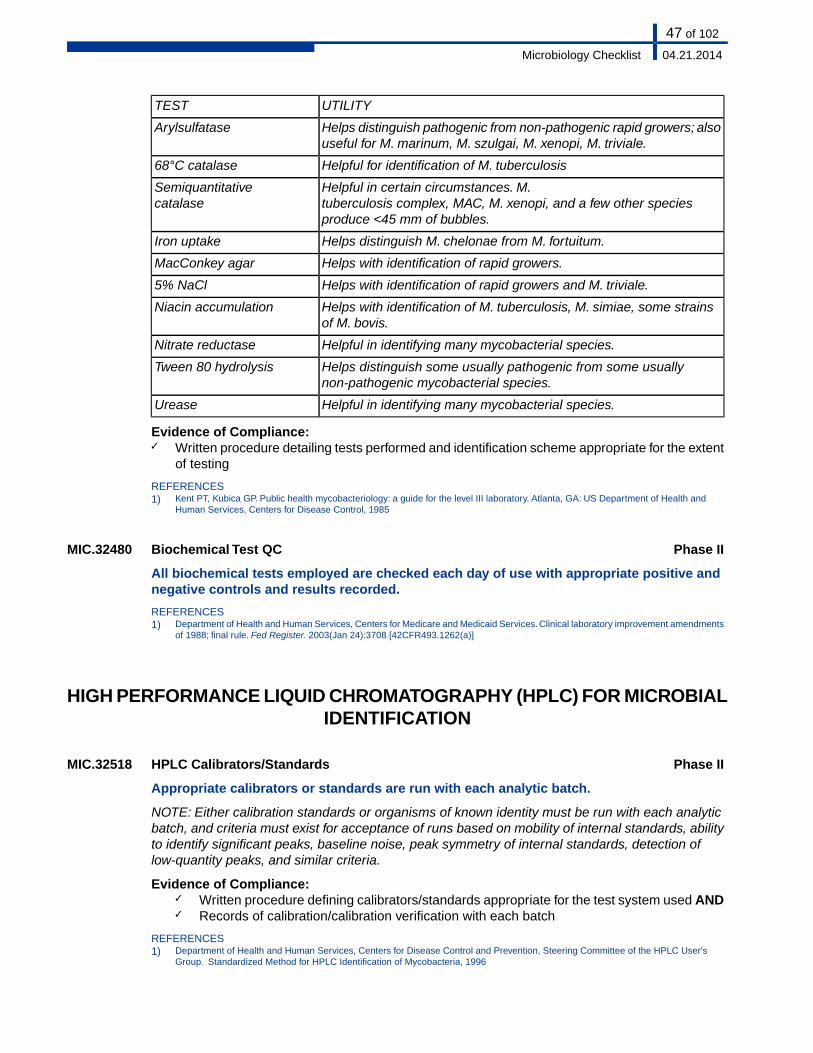
UTILITYTEST
Helps distinguish pathogenic from non-pathogenic rapid growers; alsouseful for M. marinum, M. szulgai, M. xenopi, M. triviale.
Arylsulfatase
Helpful for identification of M. tuberculosis68°C catalase
Helpful in certain circumstances. M.tuberculosis complex, MAC, M. xenopi, and a few other speciesproduce <45 mm of bubbles.
Semiquantitativecatalase
Helps distinguish M. chelonae from M. fortuitum.Iron uptake
Helps with identification of rapid growers.MacConkey agar
Helps with identification of rapid growers and M. triviale.5% NaCl
Helps with identification of M. tuberculosis, M. simiae, some strainsof M. bovis.
Niacin accumulation
Helpful in identifying many mycobacterial species.Nitrate reductase
Helps distinguish some usually pathogenic from some usuallynon-pathogenic mycobacterial species.
Tween 80 hydrolysis
Helpful in identifying many mycobacterial species.Urease
Evidence of Compliance:✓ Written procedure detailing tests performed and identification scheme appropriate for the extent
of testing
REFERENCES1) Kent PT, Kubica GP. Public health mycobacteriology: a guide for the level III laboratory. Atlanta, GA: US Department of Health and
Human Services, Centers for Disease Control, 1985
Phase IIBiochemical Test QCMIC.32480
All biochemical tests employed are checked each day of use with appropriate positive andnegative controls and results recorded.
REFERENCES1) Department of Health and Human Services, Centers for Medicare and Medicaid Services. Clinical laboratory improvement amendments
of 1988; final rule. Fed Register. 2003(Jan 24):3708 [42CFR493.1262(a)]
HIGH PERFORMANCE LIQUID CHROMATOGRAPHY (HPLC) FOR MICROBIALIDENTIFICATION
Phase IIHPLC Calibrators/StandardsMIC.32518
Appropriate calibrators or standards are run with each analytic batch.
NOTE: Either calibration standards or organisms of known identity must be run with each analyticbatch, and criteria must exist for acceptance of runs based on mobility of internal standards, abilityto identify significant peaks, baseline noise, peak symmetry of internal standards, detection oflow-quantity peaks, and similar criteria.
Evidence of Compliance:✓ Written procedure defining calibrators/standards appropriate for the test system used AND✓ Records of calibration/calibration verification with each batch
REFERENCES1) Department of Health and Human Services, Centers for Disease Control and Prevention, Steering Committee of the HPLC User's
Group. Standardized Method for HPLC Identification of Mycobacteria, 1996
47 of 102
04.21.2014Microbiology Checklist
Phase IIHPLC ControlsMIC.32556
Appropriate controls are extracted and run through the entire procedure.
NOTE: Control organisms must be extracted and carried through the entire procedure with eachrun or batch. Appropriate positive (e.g. mycobacterial) and negative controls (organisms such asCandida from which no mycolic acids are expected) must be included with each run.
Evidence of Compliance:✓ Written procedure defining QC requirements AND✓ QC records documented at defined frequency
REFERENCES1) Department of Health and Human Services, Centers for Medicare and Medicaid Services. Medicare, Medicaid and CLIA programs;
CLIA fee collection; correction and final rule. Fed Register. 2003(Jan 24):5232 [42CFR493.1256]2) Department of Health and Human Services, Centers for Disease Control and Prevention, Steering Committee of the HPLC User's
Group. Standardized Method for HPLC Identification of Mycobacteria, 1996
Phase IChromatogram ControlsMIC.32594
External chromatogram pattern controls are available.
NOTE: Patterns for known strains should be established in those laboratories using HPLC. Inaddition laboratories should have access to the standard method manuals containing comparablechromatographic patterns for comparison.
REFERENCES1) Department of Health and Human Services, Centers for Disease Control and Prevention, Steering Committee of the HPLC User's
Group. Standardized Method for HPLC Identification of Mycobacteria, 1996
Phase IIColumn VerificationMIC.32632
New columns are verified for performance before use.
NOTE: Column verification must include assessment of flow, consistency, and carryover. If theHPLC-method interpretive software uses a peak-naming table, it must be calibrated with eachchange of column. Generally the basic performance of new columns is certified by the manufacturer.HPLC analysis requires columns be equilibrated with about 10 column volumes of solvent followedwith a blank run to test pressure and solvent flow.
Evidence of Compliance:✓ Written procedure for column verification AND✓ Records of column verification
REFERENCES1) Department of Health and Human Services, Centers for Disease Control and Prevention, Steering Committee of the HPLC User's
Group. Standardized Method for HPLC Identification of Mycobacteria, 1996
Phase IIColumn/Detector MonitoringMIC.32670
The performance of the column and detector are monitored on each day of use.
NOTE: Unextracted standard organisms and extracted calibrators or controls, typically containinga range of mycolic acids (or other appropriate targets) of known relative retention times, may beanalyzed to monitor critical aspects of HPLC performance. Appropriate criteria for evaluating suchparameters as retention time of specific standards, relative retention compounds time, separationof closely eluting peaks of interest, detection of known low-quantity peaks, column pressure,chromatography quality and detector response should be established and monitored. Columntemperatures and pump pressures are monitored with each run to ensure they met specified criteriafor analysis. The column and detector operations are monitored with a blank run prior to use andduring batch runs. Positive and negative control samples supplement the blank run when samplesare analyzed.
Evidence of Compliance:✓ Records for column and detector monitoring documented at defined frequency
48 of 102
04.21.2014Microbiology Checklist

REFERENCES1) Department of Health and Human Services, Centers for Disease Control and Prevention, Steering Committee of the HPLC User's
Group. Standardized Method for HPLC Identification of Mycobacteria, 1996
Phase IICarryover DetectionMIC.32708
There is a procedure for the detection and evaluation of potential carryover.
NOTE: No matter what type of injection is used, the procedure must address criteria for the evaluationof potential carryover from a preceding elevated (high concentration) sample to the following sample,either periodically, or in each analytical batch analysis.
Evidence of Compliance:✓ Records of reassessment of samples with potential carryover
REFERENCES1) Clinical and Laboratory Standards Institute (CLSI). Preliminary Evaluation of Quantitative Clinical Laboratory Methods – Approved
Guideline- Third Edition. CLSI Document EP10-A3 (ISBN 1-56238-622-0). Clinical and Laboratory Standards Institute, 940 West ValleyRoad, Wayne, PA, 19087-1898, USA, 2006.
2) Society of Forensic Toxicologists/American Academy of Forensic Sciences. Forensic Toxicology Laboratory Guidelines. 2002; 8.2.8:13
3) Department of Health and Human Services, Centers for Disease Control and Prevention, Steering Committee of the HPLC User'sGroup. Standardized Method for HPLC Identification of Mycobacteria, 1996
Phase IIHPLC Growth MediaMIC.32746
The laboratory procedures define which growth media may be used for organisms to beanalyzed by HPLC.
NOTE: Final results can be influenced by conditions of growth. For reliable results, standardconditions of analysis must be met, including growth media.
REFERENCES1) Department of Health and Human Services, Centers for Disease Control and Prevention, Steering Committee of the HPLC User's
Group. Standardized Method for HPLC Identification of Mycobacteria, 1996
Phase IIPeak VerificationMIC.32784
There is a procedure for verifying calibration of the peak-naming table, if used.
NOTE: In order to insure that peaks are correctly identified by interpretive software, the table mustbe verified at least annually with standard materials or organisms with known characteristics.
REFERENCES1) Department of Health and Human Services, Centers for Disease Control and Prevention, Steering Committee of the HPLC User's
Group. Standardized Method for HPLC Identification of Mycobacteria, 1996
Phase IIHPLC Method ValidationMIC.32822
The HPLC method has been validated using known strains of bacteria, including strainsexpected to be encountered in routine clinical use.
Evidence of Compliance:✓ Record of method validation with appropriate strains
REFERENCES1) Department of Health and Human Services, Centers for Disease Control and Prevention, Steering Committee of the HPLC User's
Group. Standardized Method for HPLC Identification of Mycobacteria, 1996
Phase IIHPLC Result ReviewMIC.32860
There is a procedure for review of HPLC results in conjunction with other laboratory dataprior to reporting results.
NOTE: HPLC is only one tool for microbial identification. When results of HPLC analysis conflictwith growth characteristics, pigmentation, or the results of biochemical or molecular testing,identification decisions must be based on all the information available.
49 of 102
04.21.2014Microbiology Checklist
REFERENCES1) Department of Health and Human Services, Centers for Disease Control and Prevention, Steering Committee of the HPLC User's
Group. Standardized Method for HPLC Identification of Mycobacteria, 1996
Phase IIHPLC Analysis - Pure IsolatesMIC.32898
There are procedures to check the purity of cultures used as a source for HPLC analysis.
NOTE: Results of HPLC analysis may be unreliable if mixed cultures are tested. If HPLC is performedon an isolate from liquid culture and an interpretable chromatogram is obtained, it is not necessaryto await the results of the purity check before reporting results, but a purity check must still beperformed.
REFERENCES1) Department of Health and Human Services, Centers for Disease Control and Prevention, Steering Committee of the HPLC User's
Group. Standardized Method for HPLC Identification of Mycobacteria, 1996
**REVISED** 07/29/2013Phase IHPLC Reagent GradeMIC.32936
Reagents and solvents are of appropriate grade, and solvent purity is assessed when needed.
NOTE: Only HPLC grade solvents are recommended for this procedure. Degradation begins onceultra-pure solvents are opened. Degradation can be slowed by storing solvents in tightly capped,amber bottles in the dark. Solvent purity verification is suggested when a degradation-relatedproblem is suspected.
Evidence of Compliance:✓ Reagent logs
REFERENCES1) Department of Health and Human Services, Centers for Disease Control and Prevention, Steering Committee of the HPLC User's
Group. Standardized Method for HPLC Identification of Mycobacteria, 1996
Phase IIInstrument OperationMIC.32974
Procedures are documented for operation and calibration.
NOTE: Basic principles of HPLC analysis require continual monitoring of analysis conditions,including standard operating procedures, and system calibration. System problems and correctiveactions must be appropriately documented.
REFERENCES1) Department of Health and Human Services, Centers for Disease Control and Prevention, Steering Committee of the HPLC User's
Group. Standardized Method for HPLC Identification of Mycobacteria, 1996
LABORATORY SAFETY
NOTE TO THE INSPECTOR: The inspector should review relevant requirements from the Safety section of theLaboratory General checklist, to assure that the mycobacteriology laboratory is in compliance. Please elaborateupon the location and the details of each deficiency in the Inspector's Summation Report.
The following requirements pertain specifically to the mycobacteriology laboratory.
50 of 102
04.21.2014Microbiology Checklist
Inspector Instructions:
● Mycobacteriology specimen collection, transport and handling procedure● Records of biological safety cabinet certification
● Patient mycobacteriology specimens (sealed, leak proof containers)● Sealed, safety centrifuge carriers
Phase IISpecimen CollectionMIC.33050
All specimens for mycobacterial culture are collected and/or received in sealed leak-proofcontainers.
Phase IICentrifuge SafetyMIC.33100
In centrifuging specimens, sealed screw-capped tubes are enclosed in sealed safety centrifugecarriers (i.e. a double closure system) used to minimize aerosol hazards.
REFERENCES1) Clinical and Laboratory Standards Institute (CLSI). Clinical Laboratory Safety; Approved Guideline Third Edition. CLSI document
GP17-A3 [ISBN 1-56238-797-9 (Print); ISBN 1-56238-798-7 (Electronic)]. Clinical and Laboratory Standards Institute, 940 West ValleyRoad, Suite 1400, Wayne, Pennsylvania 19087-1898 USA, 2012.
Phase IIBiological Safety CabinetMIC.33300
The biological safety cabinet meets minimum requirements for mycobacterial work.
NOTE: Exhaust air from a class I or class II biological safety cabinet must be filtered through highefficiency particulate air (HEPA) filters. Air from Class I and IIB cabinets is hard-ducted to the outside.Air from Class IIA cabinets may be recirculated within the laboratory if the cabinet is tested andcertified at least every 12 months. It may be exhausted through a dedicated stack that protectsagainst backflow of air from adverse weather conditions or through the building exhaust air systemin a manner (e.g. thimble connection) that avoids any interference with the air balance of thebiological safety cabinet or building exhaust system.
Evidence of Compliance:✓ Written procedure defining the types of safety cabinets, filtration systems and exhaust systems
used AND✓ Maintenance schedule of BSC function checks AND✓ Records of testing and certification AND✓ Records of HEPA filters used for filtration of all BSC classes AND✓ Records of exhaust mechanism OR recirculation, if appropriate
REFERENCES1) Department of Health and Human Services. Biosafety in Microbiological and Biomedical Laboratories, 5th ed. Washington, DC: HHS
Publishing No. (CDC) 21-1112, December 20092) Clinical and Laboratory Standards Institute (CLSI). Protection of Laboratory Workers From Occupationally Acquired Infections; Approved
Guideline—Third Edition. CLSI document M29-A3 (ISBN 1-56238-567-4). Clinical and Laboratory Standards Institute, 940 West ValleyRoad, Suite 1400, Wayne, Pennsylvania 19087-1898 USA, 2005.
51 of 102
04.21.2014Microbiology Checklist

MYCOLOGY
QUALITY CONTROL
Inspector Instructions:
● Sampling of mycology media/stain/reagent QC policies and procedures● Sampling of mycology QC records
● How do you determine when QC is unacceptable and when corrective actions areneeded?
● Select several occurrences in which QC is out of range and follow documentation todetermine if the steps taken follow the laboratory policy for corrective action
MEDIA
Phase IIMedia QCMIC.41200
An appropriate sample of each medium prepared by the laboratory or purchased but notexcluded from testing in NCCLS M22-A3 is checked for each of the following.
1. Sterility (following introduction of additives after sterilization)2. Ability to support growth and biochemical reactivity (where applicable) by means
of stock cultures or by parallel testing with previous batches
Evidence of Compliance:✓ Records of media QC for laboratory-prepared or non-exempt purchased media
REFERENCES1) NCCLS. Quality Control for Commercially Prepared Microbiological Culture Media; Approved Standard - Third Edition. NCCLS document
M22-A3. 940 West Valley Road, Suite 1400, Wayne, PA 19087-1898 USA, 2004.2) Clinical and Laboratory Standards Institute (CLSI). Principles and Procedures for Detection of Fungi in Clinical Specimens-Direct
Examination and Culture; Approved Guideline. CLSI document M54-A (ISBN 1-56238-857-6 [Print] ISBN 1-56238-858-4 [Electronic]).Clinical and Laboratory Standards Institute, 940 West Valley Road, Suite 2500, Wayne, Pennsylvania 19087-1898 USA, 2012.
CONTROLS AND STANDARDS
Good laboratory practice includes checking all media either at the time of receipt or concurrently with use. Thisapplies to purchased media as well as media prepared by the laboratory.
52 of 102
04.21.2014Microbiology Checklist
Phase IIReference OrganismsMIC.41250
Reference cultures are used to check stains and reagents at appropriate intervals.
REFERENCES1) Department of Health and Human Services, Centers for Medicare and Medicaid Services. Clinical laboratory improvement amendments
of 1988; final rule. Fed Register. 2003(Jan 24):7167 [42CFR493.1261(b)]
Phase IINucleic Acid Probe/Exo-antigen QCMIC.41270
If nucleic acid probes or exo-antigen tests are used for identification of fungi isolated fromculture, appropriate positive and negative controls are tested on each day of use.
Evidence of Compliance:✓ Written procedure defining QC for nucleic acid probe or exo-antigen tests AND✓ Records of nucleic acid probe or exo-antigen QC documented at defined frequency
Phase IIQC Confirmation of AcceptabilityMIC.41330
The results of controls are reviewed for acceptability before reporting patient results.
Evidence of Compliance:✓ Written policy/procedure stating that controls are reviewed and acceptable prior to reporting
patient results AND✓ Evidence of corrective action taken when QC results are not acceptable
REFERENCES1) Department of Health and Human Services, Centers for Medicare and Medicaid Services. Clinical laboratory improvement amendments
of 1988; final rule. Fed Register. 2003(Jan 24):7166 [42CFR493.1256(f)]
Phase IIQC Corrective ActionMIC.41345
There is documentation of corrective action when quality control results exceed theacceptability limits.
NOTE: Patient test results obtained in an analytically unacceptable test run or since the lastacceptable test run must be re-evaluated to determine if there is a significant clinical difference inpatient results. Re-evaluation may or may not include retesting samples, depending on thecircumstances.
REFERENCES1) Department of Health and Human Services, Centers for Medicare and Medicaid Services. Clinical laboratory improvement amendments
of 1988; final rule. Fed Register. 2003(Oct 1):1046[42CFR493.1282(b)(2)]
Phase IIDirect Smear Stain QCMIC.41370
Direct patient specimen stains (e.g. acid fast, PAS, Giemsa, Gomori's methenamine silver,India ink) are checked with positive and negative controls on each day of patient sampletesting.
NOTE: For certain stains such as GMS and Giemsa, the slide itself serves as the negative control.Controls for KOH preparations are not required.
Evidence of Compliance:✓ Records of stain QC documented at defined frequency
REFERENCES1) August, Hindler, Huber, Sewell. Quality control and quality assurance practices. In: Clinical Microbiology, Cumitech 3A. Washington,
DC: American Society for Microbiology, 19902) Department of Health and Human Services, Centers for Medicare and Medicaid Services. Clinical laboratory improvement amendments
of 1988; final rule. Fed Register. 2003(Jan 24):7166 [42CFR493. 1256(e)(2); 493.1256(e)(3); 493.1273(a)]3) Department of Health and Human Services, Centers for Medicare and Medicaid Services. Clinical laboratory improvement amendments
of 1988; final rule. Fed Register. 2003(Jan 24):3709 [42CFR493.1263(a)]
Phase IIFluorescent Stain QCMIC.41390
53 of 102
04.21.2014Microbiology Checklist

Fluorescent stains (such as calcofluor white) are checked with positive and negative controlseach time of use and results documented.
REFERENCES1) Department of Health and Human Services, Centers for Medicare and Medicaid Services. Clinical laboratory improvement amendments
of 1988; final rule. Fed Register. 2003(Jan 24):3708 [42CFR493.1256(e)(3)]
PROCEDURES AND TESTS
The intent of this series of requirements is to ensure the use of an appropriate variety of media and growth conditionsto isolate the significant pathogens with minimal interference from contaminants.
Inspector Instructions:
● Sampling of mycology test procedures/identification schemes● Sampling of patient worksheets/records
● What tests or procedures are used to identify dimorphic fungi? Filamentous fungi?
● Follow a mycology patient worksheet from receipt and processing to culture evaluation,identification and reporting
**NEW** 07/29/2013Phase IICryptococcal AntigenMIC.42025
If cryptococcal antigen-detection methods are used on CSF, back-up cultures are performedon positive CSF specimens submitted for diagnosis.
NOTE: It is important to recover the causative organism for precise identification (C. neoformansvs. C. gattii) and potentially susceptibility testing. Back-up cultures of follow-up specimens used fortrending the antigen titer are not required. If culture is not performed onsite by the laboratory, thelaboratory must show evidence that it has been performed in a referral laboratory.
Evidence of Compliance:✓ Written procedure stating that CSF cultures are performed in conjunction with initial cryptococcal
antigen tests OR procedure describing testing at another location AND✓ Records of back-up CSF cultures performed on-site OR records indicating that cultures are
performed at another location
Phase IISelective MediaMIC.42050
Suitable selective media are used for the growth and isolation of dermatophytes and/orsystemic fungi.
Evidence of Compliance:✓ Written procedure for mycology culture defining the media used for growth and isolation
54 of 102
04.21.2014Microbiology Checklist
Phase IISelective MediaMIC.42100
Media with antimicrobial agents are used to suppress the growth of contaminants.
NOTE: Antimicrobial agents may inhibit some yeasts and the yeast phase of dimorphic organisms.Both types of media (with and without antimicrobials) should be available and used when indicated.
Evidence of Compliance:✓ Written procedure for mycology culture defining the use of media to suppress contaminants
REFERENCES1) Clinical and Laboratory Standards Institute (CLSI). Principles and Procedures for Detection of Fungi in Clinical Specimens-Direct
Examination and Culture; Approved Guideline. CLSI document M54-A (ISBN 1-56238-857-6 [Print] ISBN 1-56238-858-4 [Electronic]).Clinical and Laboratory Standards Institute, 940 West Valley Road, Suite 2500, Wayne, Pennsylvania 19087-1898 USA, 2012.
Phase IIIncubation TemperatureMIC.42150
Incubation temperatures for the growth and isolation of dermatophytes and systemic fungiare defined and followed under culture conditions.
Evidence of Compliance:✓ Temperature records
Phase IIIncubation TemperatureMIC.42200
If cultures are incubated at room temperatures, actual ambient temperature (22-26° C) ischecked daily to determine if proper growth conditions are being maintained.
Phase IIDifferential TestsMIC.42250
Procedures for the differentiation and identification of fungi (differential tests) are adequatefor the needs of the laboratory.
NOTE: Laboratories offering full identification must have sufficient procedures to do so. Smallerlaboratories with limited services should have an arrangement with an approved reference laboratoryfor back-up and complete identification of mycology specimens.
Evidence of Compliance:✓ Written procedure detailing tests performed and identification scheme appropriate for the extent
of testing
REFERENCES1) Riddle DL, et al. Clinical comparison of the Baxter Microscan yeast identification panel and the Vitek yeast biochemical card. Am J Clin
Pathol. 1994;101:438-442
Phase IIDifferential TestsMIC.42350
Differential tests include biochemical tests (e.g. urease, carbohydrate assimilation and/orfermentation).
Phase IDifferential TestsMIC.42400
Differential tests include slide cultures (when appropriate).
Phase IDifferential TestsMIC.42450
Differential tests include nutritional studies for dermatophytes when identification is carriedto the species level.
55 of 102
04.21.2014Microbiology Checklist

Phase IDimorphic FungiMIC.42550
The identification of dimorphic fungal isolates is confirmed by exo-antigen, molecular,yeast-mold conversion or tissue phase detection tests.
NOTE: Exo-antigen tests, DNA probes, or DNA sequencing are recommended.
LABORATORY SAFETY
NOTE TO THE INSPECTOR: The inspector should review relevant requirements from the Safety section of theLaboratory General checklist, to assure that the mycology laboratory is in compliance. Please elaborate upon thelocation and the details of each deficiency in the Inspector's Summation Report.
The following requirements pertain specifically to the mycology laboratory.
Inspector Instructions:
● Sampling of mycology safety policies and procedures● Records of biological safety cabinet certification
● Safe work practices (taping of culture plates, procedures performed under BSC)
Phase IISafety PrecautionsMIC.43050
If plate culture media is used in mycology, appropriate safety precautions are taken (suchas taping lid to plate on both sides when not in use or other appropriate measures) to preventthe accidental opening of a plate.
NOTE: Some authorities recommend the transfer of growing colonies from plate to tubed media, ifthe former is routinely used for initial inoculation.
Evidence of Compliance:✓ Written procedure defining safety precautions for handling mycology culture plates
REFERENCES1) Clinical and Laboratory Standards Institute (CLSI). Principles and Procedures for Detection of Fungi in Clinical Specimens-Direct
Examination and Culture; Approved Guideline. CLSI document M54-A (ISBN 1-56238-857-6 [Print] ISBN 1-56238-858-4 [Electronic]).Clinical and Laboratory Standards Institute, 940 West Valley Road, Suite 2500, Wayne, Pennsylvania 19087-1898 USA, 2012.
Phase IISafety PrecautionsMIC.43100
When working with a colony exhibiting mycelial growth, transfers are performed within abiological safety cabinet.
REFERENCES1) Clinical and Laboratory Standards Institute (CLSI). Principles and Procedures for Detection of Fungi in Clinical Specimens-Direct
Examination and Culture; Approved Guideline. CLSI document M54-A (ISBN 1-56238-857-6 [Print] ISBN 1-56238-858-4 [Electronic]).Clinical and Laboratory Standards Institute, 940 West Valley Road, Suite 2500, Wayne, Pennsylvania 19087-1898 USA, 2012.
Phase IISafety PrecautionsMIC.43150
56 of 102
04.21.2014Microbiology Checklist

The use of slide culture techniques is limited, where possible, to work with low virulenceorganisms; or if used for dimorphic fungi, special safety precautions are defined and rigidlyadhered to.
Phase IISafety PrecautionsMIC.43200
When preparing teased preparations or "scotch" tape preps, mycelia are always submergedin some liquid medium (such as lactophenol cotton blue).
Phase IIBiological Safety CabinetMIC.43250
A biological safety cabinet (BSC) or hood is available for handling specimens or organismsconsidered to be highly contagious by airborne routes.
Evidence of Compliance:✓ Maintenance schedule of BSC function checks AND✓ Records of testing and certification
REFERENCES1) Department of Health and Human Services. Biosafety in Microbiological and Biomedical Laboratories, 5th ed. Washington, DC: HHS
Publishing No. (CDC) 21-1112, December 2009
Phase IIBiological Safety CabinetMIC.43300
The biological safety cabinet (BSC) is certified annually to ensure that filters are functioningproperly and that airflow rates meet specifications.
Evidence of Compliance:✓ Maintenance schedule of BSC function checks AND✓ Records of testing and certification
Phase IIBiological Safety CabinetMIC.43350
The BSC meets minimum requirements for mycologic work.
NOTE: Exhaust air from a class I or class II BSC must be filtered through HEPA filters. Air fromClass I and IIB is hard ducted to the outside. Air from Class IIA cabinets may be recirculated withinthe laboratory if the cabinet is tested and certified at least every 12 months. It may be exhaustedthrough a dedicated stack that protects against backflow of air from adverse weather conditions orthrough the building exhaust air system in a manner (e.g. thimble connection) that avoids anyinterference with the air balance of the BSC or building exhaust system.
Evidence of Compliance:✓ Written procedure defining the types of safety cabinets, filtration systems and exhaust systems
used AND✓ Maintenance schedule of BSC function checks AND✓ Records of testing and certification AND✓ Records of HEPA filters used for filtration of all BSC classes AND✓ Records of exhaust mechanism OR recirculation, if appropriate
REFERENCES1) Department of Health and Human Services. Biosafety in Microbiological and Biomedical Laboratories, 5th ed. Washington, DC: HHS
Publishing No. (CDC) 21-1112, December 2009
57 of 102
04.21.2014Microbiology Checklist
PARASITOLOGY
QUALITY CONTROL
Inspector Instructions:
● Sampling of parasitology stain/reagent QC policies and procedures● Sampling of parasitology QC records● Ocular micrometer calibration procedure and records
● Reference materials (permanent mounts, photomicrographs or atlas available)● Zinc sulfate solution (tightly-stoppered)● Ocular micrometer
● How do you determine when QC is unacceptable and when corrective actions areneeded?
● What resources do you have available to assist in identification of parasites?
● Select several occurrences in which QC is out of range and follow documentation todetermine if the steps taken follow the laboratory policy for corrective action
Phase IIQC Confirmation of AcceptabilityMIC.45900
The results of controls are reviewed for acceptability before reporting patient results.
Evidence of Compliance:✓ Written policy/procedure stating that controls are reviewed and acceptable prior to reporting
patient results AND✓ Evidence of corrective action taken when QC results are not acceptable
REFERENCES1) Department of Health and Human Services, Centers for Medicare and Medicaid Services. Clinical laboratory improvement amendments
of 1988; final rule. Fed Register. 2003(Jan 24)1992(Feb 28):7166 [42CFR493.1256(f)]
Phase IIQC Corrective ActionMIC.48450
There is documentation of corrective action when control results exceed the acceptabilitylimits.
NOTE: Patient test results obtained in an analytically unacceptable test run or since the lastacceptable test run must be re-evaluated to determine if there is a significant clinical difference inpatient/client results. Re-evaluation may or may not include retesting samples, depending on thecircumstances.
REFERENCES1) Department of Health and Human Services, Centers for Medicare and Medicaid Services. Clinical laboratory improvement amendments
of 1988; final rule. Fed Register. 2003(Oct 1):1046[42CFR493.1282(b)(2)]
58 of 102
04.21.2014Microbiology Checklist
**REVISED** 07/29/2013Phase IReference MaterialsMIC.51000
Reference materials, such as permanent mounts, photomicrographs, NCCLS documentsM15-A and M28-A2, or printed atlases are available at the work bench to assist withidentifications.
REFERENCES1) NCCLS. Laboratory Diagnosis of Blood-Borne Parasitic Diseases; Approved Guideline. NCCLS Document M15-A. (ISBN 1-56238-401-5).
NCCLS, 940 West Valley Road, Suite 1400, Wayne, PA 19087-1898 USA, 2000.2) Garcia LS, Shimizu R. Diagnostic parasitology: parasitic infections and the compromised host. Lab Med. 1993;24:205-215
3) Ash R, Orihel TC. Atlas of human parasitology, 4th ed. Chicago, IL: American Society of Clinical Pathology, 1996
4) Clinical and Laboratory Standards Institute (CLSI). Procedures for the Recovery and Identification of Parasites From the Intestinal Tract;Approved Guideline—Second Edition. CLSI document M28-A2 (ISBN 1-56238-572-0). Clinical and Laboratory Standards Institute, 940West Valley Road, Suite 1400, Wayne, Pennsylvania 19087-1898 USA, 2005
5) Garcia LS,. Diagnostic medical parasitology. 4th ed. Washington, DC: ASM Press, 2001
6) Colmer-Hamood JA, Fecal microscopy. Artifacts mimicking ova and parasites. Lab Med. 2001;32:80-84
7) Department of Health and Human Services, Centers for Medicare and Medicaid Services. Clinical laboratory improvement amendmentsof 1988, final rule. Fed Register. 2003(Jan 24): [42CFR493.1264(a)]
REAGENTS
Phase IIReagentsMIC.51120
If zinc sulfate is used, the solution is stored in a tightly-stoppered bottle and checked forspecific gravity (1.18 for fresh specimens and 1.20 for formalin-fixed specimens) with ahydrometer whose scale is large enough to differentiate the two values.
Evidence of Compliance:✓ Records for specific gravity checks on the zinc sulfate solution
Phase IIPermanent Stain QCMIC.51160
All permanent parasitology stains are checked for intended reactivity with controls orreference materials at least monthly (or with each test if performed less frequently than everymonth).
NOTE: PVA fixative solutions thoroughly mixed with fresh fecal material that has been seeded withbuffy coat leukocytes usually provides reliable controls for permanent stains.
Evidence of Compliance:✓ Records of permanent stain QC documented at defined frequency
REFERENCES1) Department of Health and Human Services, Centers for Medicare and Medicaid Services. Clinical laboratory improvement amendments
of 1988; final rule. Fed Register. 2003(Jan 24):7167 [42CFR493.1264(c)]2) Garcia LS, Diagnostic medical parasitology, 4th ed. Washington, DC: ASM Press, 2001
Phase IISpecial Stain QCMIC.51170
Stains that are used to detect specific parasites (e.g. acid fast, fluorescent) are checked withappropriate control organisms each time that stain is used.
Evidence of Compliance:✓ Records of special stain QC each time of use
INSTRUMENTS AND EQUIPMENT
Phase IIOcular MicrometerMIC.51210
59 of 102
04.21.2014Microbiology Checklist
An ocular micrometer is available for determining the size of eggs, larvae, cysts, trophozoites,and microfilaria or other bloodborne parasites.
REFERENCES1) Department of Health and Human Services, Centers for Medicare and Medicaid Services. Clinical laboratory improvement amendments
of 1988; final rule. Fed Register. 2003(Jan 24):7167 [42CFR493.1264(b)]
Phase IICalibration/Recalibration - Ocular MicrometerMIC.51220
The ocular micrometer has been calibrated for the microscope(s) in which it is used and itis recalibrated each time the eyepieces or objectives are changed.
NOTE: Calibrations can be checked against a micrometer or other objects of known dimensions.If there are no changes to a particular microscope's optical components, there is no need to recheckcalibration.
Evidence of Compliance:✓ Records of initial calibration and recalibration if applicable
REFERENCES1) Department of Health and Human Services, Centers for Medicare and Medicaid Services. Clinical laboratory improvement amendments
of 1988; final rule. Fed Register. 2003(Jan 24):7167 [42CFR493.1264(b)]
PROCEDURES AND TESTS
Inspector Instructions:
● Sampling of parasitology test procedures● Sampling of patient worksheets/records
● Follow a parasitology patient worksheet from receipt and processing to identificationand reporting
STOOLS FOR OVA AND PARASITES
Phase IIOva/Parasite ExamMIC.52100
The microscopic examination of all stools submitted for an ova and parasite (O&P)examination includes a concentration procedure and a permanent stain.
NOTE: When a stool specimen is submitted fresh, the usual approach would be to perform a directwet preparation (looking for motility), a concentration (helminth eggs/larvae/protozoan cysts), andthe permanent stained smear (identification of protozoa missed by concentration and confirmationof suspect organisms). As a minimum (and certainly if the stool is submitted in preservatives), thestandard O&P examination would include the concentration procedure and a permanent stainedsmear. The main point is to ensure that the permanent stained smear is performed on all stoolspecimens, regardless of what was or was not seen in the concentration wet preparation. Often,intestinal protozoa will be seen in the permanent stained smear, but may be missed in theconcentration examination.
Evidence of Compliance:✓ Written procedures for stool for O&P AND
60 of 102
04.21.2014Microbiology Checklist

✓ Patient reports/worksheets with concentration and permanent stain results
REFERENCES1) Garcia LS, Diagnostic medical parasitology, 4th edition. Washington, DC: ASM Press, 2001
2) Clinical and Laboratory Standards Institute (CLSI). Procedures for the Recovery and Identification of Parasites From the Intestinal Tract;Approved Guideline—Second Edition. CLSI document M28-A2 (ISBN 1-56238-572-0). Clinical and Laboratory Standards Institute, 940West Valley Road, Suite 1400, Wayne, Pennsylvania 19087-1898 USA, 2005
Phase IStool Number/TimingMIC.52190
The laboratory has guidelines (developed with clinicians) for the number and/or timing ofcollection of stool specimens submitted for routine parasitology testing.
NOTE: Suggestions made by the authors of a 1996 CAP Q-Probes study (Valenstein et al) include:
1. Accept no more than 2 or 3 specimens/patients without prior consultation with anindividual who can explain the limited yield provided by additional specimens
2. Do not accept specimens from inpatients after the fourth hospital day, without priorconsultation
These recommendations are for diagnostic testing. Different guidelines may apply to tests orderedfor follow-up.
REFERENCES1) Yannelli B, et al. Yield of stool cultures, ova and parasite tests, and Clostridium difficile determinations in nosocomial diarrhea. Am J
Infect Control. 1988;16:246-2492) Morris AJ, et al. Application of rejection criteria for stool ovum and parasite examinations. J Clin Microbiol. 1992;30:3213-3216
3) Valenstein P, et al. The use and abuse of routine stool microbiology. A College of American Pathologists Q-probes study of 601institutions. Arch Pathol Lab Med. 1996;120:206-211
4) Cartwright CP. Utility of multiple-stool-specimen ova and parasite examinations in high-prevalence setting. J Clin Microbiol.1999;37:2408-2411
BLOOD FILMS FOR MALARIA AND OTHER PARASITES
**REVISED** 07/29/2013Phase IIBlood Parasite DetectionMIC.52193
The microscopic examination of blood films submitted for detection of blood parasites allowsfor detection of parasites responsible for malaria, babesiosis, trypanosomiasis and filariasis.
REFERENCES1) NCCLS. Laboratory Diagnosis of Blood-Borne Parasitic Diseases; Approved Guideline. NCCLS Document M15-A. (ISBN 1-56238-401-5).
NCCLS, 940 West Valley Road, Suite 1400, Wayne, PA 19087-1898 USA, 2000.
Phase IPercentage Parasitemia ReportingMIC.52195
When blood films are positive for malaria parasites (Plasmodium spp.), the percentageparasitemia is reported along with the organism identification.
NOTE: It is important to report the percentage of parasitemia when blood films are reviewed andfound to be positive for malaria parasites. Because of the potential for drug resistance in some ofthe Plasmodium species, particularly P. falciparum, it is important that every positive smear beassessed and the parasitemia reported exactly the same way on follow-up specimens as on theinitial specimen. This allows the parasitemia to be followed after therapy has been initiated. Theparasitemia will usually drop very quickly within the first 24 hours; however, in cases of drugresistance, the level may not decrease, but actually increase over time.
Although there are currently no requirements for reporting percent parasitemia when blood filmsare positive for Babesia species, physicians may ask for these data to monitor the response totherapy.
61 of 102
04.21.2014Microbiology Checklist

Evidence of Compliance:✓ Written procedure for performing and reporting parasitemia percentage with identification
REFERENCES1) CLSI. Laboratory Diagnosis of Blood-Borne Parasitic Diseases; Approved Guideline. CLSI Document M15-A. (ISBN 1-56238-401-5).
CLSI, 940 West Valley Road, Suite 1400, Wayne, PA 19087-1898 USA, 20002) Garcia LS, Diagnostic Medical Parasitology. Washington, DC, ASM Press, 2001
3) http://www.dpd.cdc.gov/DPDx/HTML/DiagnosticProcedures.htm
**REVISED** 07/29/2013Phase IIThick and Thin FilmsMIC.52200
Both thick and thin films (routine blood films and/or buffy coat films), or methods of equivalentsensitivity, are made to provide thorough examination for blood parasites.
REFERENCES1) NCCLS. Laboratory Diagnosis of Blood-Borne Parasitic Diseases; Approved Guideline. NCCLS Document M15-A. (ISBN 1-56238-401-5).
NCCLS, 940 West Valley Road, Suite 1400, Wayne, PA 19087-1898 USA, 2000.2) Thomson S, et al. External quality assessment in the examination of blood films for malarial parasites within Ontario, Canada. Arch
Pathol Lab Med. 2000;124:57-60
Phase IMalaria Stain ProcedureMIC.52220
There is documentation that malaria stains are washed with a buffer of a pH appropriate forthe stain used (e.g. pH 6.8-7.2 for Giemsa), or the range specified by the manufacturer.
REFERENCES1) NCCLS. Laboratory Diagnosis of Blood-Borne Parasitic Diseases; Approved Guideline. NCCLS Document M15-A. (ISBN 1-56238-401-5).
NCCLS, 940 West Valley Road, Suite 1400, Wayne, PA 19087-1898 USA, 2000.2) Garcia LS, Diagnostic medical parasitology. 4th ed. Washington, DC: ASM Press, 2001
**REVISED** 07/29/2013Phase IISlide Review ProcedureMIC.52260
An adequate number of fields are examined under oil immersion using the 100X oil immersionobjective (e.g. 300 fields).
Evidence of Compliance:✓ Written procedure defining criteria for assessment of malaria slides including objective and
number of fields examined
REFERENCES1) NCCLS. Laboratory Diagnosis of Blood-Borne Parasitic Diseases; Approved Guideline. NCCLS Document M15-A. (ISBN 1-56238-401-5).
NCCLS, 940 West Valley Road, Suite 1400, Wayne, PA 19087-1898 USA, 2000.
LABORATORY SAFETY
NOTE TO THE INSPECTOR: The inspector should review relevant requirements from the Safety section of theLaboratory General checklist, to assure that the parasitology laboratory is in compliance. Please elaborate uponthe location and the details of each deficiency in the Inspector's Summation Report.
The following requirements pertain specifically to the parasitology laboratory.
Inspector Instructions:
● Formalin monitoring procedure and records of monitoring
62 of 102
04.21.2014Microbiology Checklist
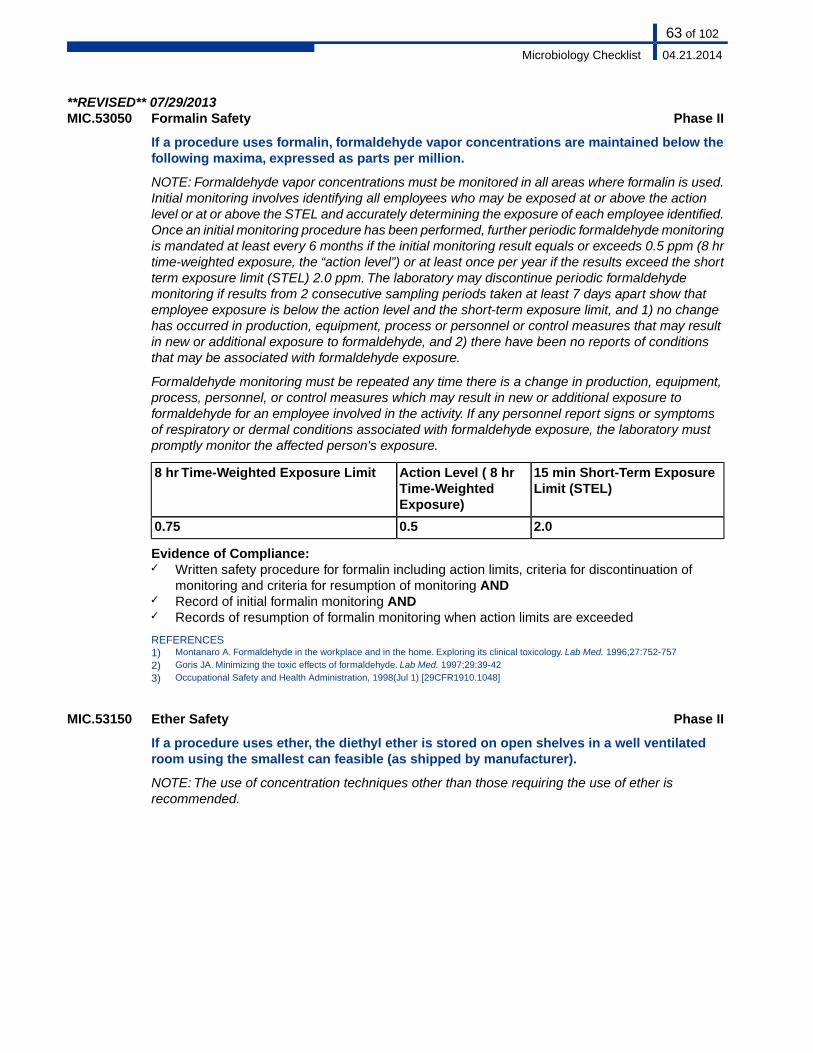
**REVISED** 07/29/2013Phase IIFormalin SafetyMIC.53050
If a procedure uses formalin, formaldehyde vapor concentrations are maintained below thefollowing maxima, expressed as parts per million.
NOTE: Formaldehyde vapor concentrations must be monitored in all areas where formalin is used.Initial monitoring involves identifying all employees who may be exposed at or above the actionlevel or at or above the STEL and accurately determining the exposure of each employee identified.Once an initial monitoring procedure has been performed, further periodic formaldehyde monitoringis mandated at least every 6 months if the initial monitoring result equals or exceeds 0.5 ppm (8 hrtime-weighted exposure, the “action level”) or at least once per year if the results exceed the shortterm exposure limit (STEL) 2.0 ppm. The laboratory may discontinue periodic formaldehydemonitoring if results from 2 consecutive sampling periods taken at least 7 days apart show thatemployee exposure is below the action level and the short-term exposure limit, and 1) no changehas occurred in production, equipment, process or personnel or control measures that may resultin new or additional exposure to formaldehyde, and 2) there have been no reports of conditionsthat may be associated with formaldehyde exposure.
Formaldehyde monitoring must be repeated any time there is a change in production, equipment,process, personnel, or control measures which may result in new or additional exposure toformaldehyde for an employee involved in the activity. If any personnel report signs or symptomsof respiratory or dermal conditions associated with formaldehyde exposure, the laboratory mustpromptly monitor the affected person's exposure.
15 min Short-Term ExposureLimit (STEL)
Action Level ( 8 hrTime-WeightedExposure)
8 hr Time-Weighted Exposure Limit
2.00.50.75
Evidence of Compliance:✓ Written safety procedure for formalin including action limits, criteria for discontinuation of
monitoring and criteria for resumption of monitoring AND✓ Record of initial formalin monitoring AND✓ Records of resumption of formalin monitoring when action limits are exceeded
REFERENCES1) Montanaro A. Formaldehyde in the workplace and in the home. Exploring its clinical toxicology. Lab Med. 1996;27:752-757
2) Goris JA. Minimizing the toxic effects of formaldehyde. Lab Med. 1997;29:39-42
3) Occupational Safety and Health Administration, 1998(Jul 1) [29CFR1910.1048]
Phase IIEther SafetyMIC.53150
If a procedure uses ether, the diethyl ether is stored on open shelves in a well ventilatedroom using the smallest can feasible (as shipped by manufacturer).
NOTE: The use of concentration techniques other than those requiring the use of ether isrecommended.
63 of 102
04.21.2014Microbiology Checklist

VIROLOGY
QUALITY CONTROL
Inspector Instructions:
● Sampling of virology test procedures● Sampling of virology media/reagent/cell line QC policies and procedures● Sampling of virology QC records and cell line checks
● How are cell lines checked for contamination?● How do you determine when QC is unacceptable and when corrective actions are
needed?
● Select several occurrences in which QC is unacceptable and follow documentation todetermine if the steps taken follow the laboratory policy for corrective action
● Follow a virology patient worksheet from receipt and processing to identification andreporting
REAGENTS
Phase IBreakageMIC.61000
The laboratory has documentation that each shipment of commercial cell culture tubes,flasks, shell vials or cluster trays is examined for breakage.
REFERENCES1) Clinical and Laboratory Standards Institute (CLSI). Viral Culture; Approved Guideline. CLSI document M41-A (ISBN 1-56238-623-9).
Clinical and Laboratory Standards Institute, 940 West Valley Road, Suite 1400, Wayne, Pennsylvania 19087-1898 USA, 20062) Ginocchio, CC. Quality Assurance in Clinical Virology. In: Spector S, Hodinka RL, Young SA, editors. Clinical Virology Manual. Fourth
Edition. Washington: ASM Press; 2009.p. 3-17
Phase IIAcceptance of MaterialsMIC.61050
The laboratory has procedures for the acceptance and rejection of cell culture tubes, flasks,shell vials or cluster trays used for virus isolation.
NOTE: Cell cultures should be observed microscopically to confirm that the cells are attached tothe substratum, the confluency of the monolayer is appropriate for the method and cell line(75%-90%) and cell appearance is typical. Confluent or overgrown monolayers may obscure viralcytopathic effect (CPE) in tube or flask cell cultures and can adversely affect the recovery of somemore fastidious viruses, such as RSV.The cell culture media should be free of contamination (clear)and should be near a neutral ph (salmon pink in color). Cell culture tubes, flasks, vials or plates notmeeting the expected criteria should be observed or rejected, depending on the observation andlaboratory policies.
REFERENCES1) Clinical and Laboratory Standards Institute (CLSI). Viral Culture; Approved Guideline. CLSI document M41-A (ISBN 1-56238-623-9).
Clinical and Laboratory Standards Institute, 940 West Valley Road, Suite 1400, Wayne, Pennsylvania 19087-1898 USA, 20062) Ginocchio, CC. Quality Assurance in Clinical Virology. In: Spector S, Hodinka RL, Young SA, editors. Clinical Virology Manual. Fourth
Edition. Washington: ASM Press; 2009.p. 3-17
64 of 102
04.21.2014Microbiology Checklist

3) Department of Health and Human Services, Centers for Medicare and Medicaid Services. Clinical laboratory improvement amendmentsof 1988, final rule. Fed Register. 2003(Jan 24): [42CFR493.1283(a)(3)]
Phase IICulture Media QCMIC.61140
Culture media are tested for sterility (if additives are introduced after initial sterilization).
NOTE: Entering the media to remove aliquots for refeeding, etc. does not generate the need forrepeat sterility testing.
Evidence of Compliance:✓ Written procedure for culture media QC AND✓ Records of media sterility QC
Phase IICell Line QCMIC.61150
Continuous cell lines are checked for mycoplasma contamination.
NOTE: An alternative method to culturing for mycoplasma is the monitoring of a negative,uninoculated control. If cell lines are provided by a commercial vendor, documentation of mycoplasmascreening by the vendor is acceptable. Vendor documentation must be retained by the laboratory.
REFERENCES1) Clinical and Laboratory Standards Institute (CLSI). Viral Culture; Approved Guideline. CLSI document M41-A (ISBN 1-56238-623-9).
Clinical and Laboratory Standards Institute, 940 West Valley Road, Suite 1400, Wayne, Pennsylvania 19087-1898 USA, 20062) Lennette DA. General Principles for Laboratory Diagnosis for Viral, Rickettsial, and Chlamydial Infections. In: Lennette EH, Lennette,
DA, Lennette ET, editors. Diagnostic Procedures for Viral, Rickettsial, and Chlamydial Infections. Seventh Edition ed. Washington:American Public Health Association; 1995 p. 3-25
Phase IICell Line QCMIC.61155
Continuous cell lines are checked for endogenous viral contamination.
NOTE: Upon receipt in the laboratory and during the period of use, cell lines must be monitored forthe presence of endogenous contamination due to viruses such as foamy virus and monkey virus.
Endogenous viral contamination must be documented. Individual laboratories may decide to rejectall cell lines with contamination. Alternatively, cell lines may be monitored to determine if thecontamination will prohibit the isolation and identification of patient viral isolates. Endogenouslycontaminated cell cultures can be evaluated by considering conditions such as the degree (percent)of contamination of the monolayer, the specificity of the contamination CPE, the effect of thecontamination of the quality of the culture media, and the condition of the culture cell to supportvirus growth. If such contamination will affect the ability to recover patient isolates, cell lines mustbe rejected.
REFERENCES1) Clinical and Laboratory Standards Institute (CLSI). Viral Culture; Approved Guideline. CLSI document M41-A (ISBN 1-56238-623-9).
Clinical and Laboratory Standards Institute, 940 West Valley Road, Suite 1400, Wayne, Pennsylvania 19087-1898 USA, 20062) Lennette DA. General Principles for Laboratory Diagnosis for Viral, Rickettsial, and Chlamydial Infections. In: Lennette EH, Lennette,
DA, Lennette ET, editors. Diagnostic Procedures for Viral, Rickettsial, and Chlamydial Infections. Seventh Edition ed. Washington:American Public Health Association; 1995 p. 3-25
Phase IIMedia QCMIC.61160
Animal sera used for cell growth media are checked for absence of toxicity to cells.
Evidence of Compliance:✓ Written procedure for checks of animal sera for toxicity AND✓ Records of animal sera checks
Phase IICell LinesMIC.61180
The laboratory has the appropriate minimal cell line(s) available for all types of specimenstested and for all viruses reported by the laboratory.
65 of 102
04.21.2014Microbiology Checklist
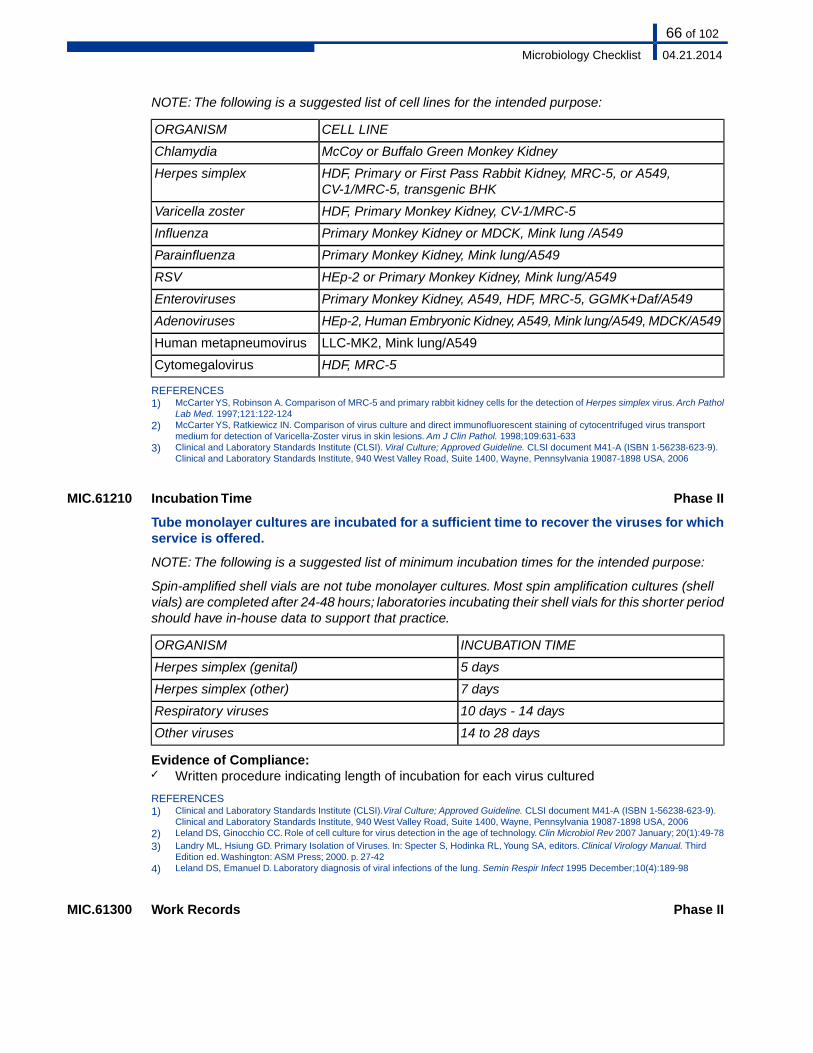
NOTE: The following is a suggested list of cell lines for the intended purpose:
CELL LINEORGANISM
McCoy or Buffalo Green Monkey KidneyChlamydia
HDF, Primary or First Pass Rabbit Kidney, MRC-5, or A549,CV-1/MRC-5, transgenic BHK
Herpes simplex
HDF, Primary Monkey Kidney, CV-1/MRC-5Varicella zoster
Primary Monkey Kidney or MDCK, Mink lung /A549Influenza
Primary Monkey Kidney, Mink lung/A549Parainfluenza
HEp-2 or Primary Monkey Kidney, Mink lung/A549RSV
Primary Monkey Kidney, A549, HDF, MRC-5, GGMK+Daf/A549Enteroviruses
HEp-2, Human Embryonic Kidney, A549, Mink lung/A549, MDCK/A549Adenoviruses
LLC-MK2, Mink lung/A549Human metapneumovirus
HDF, MRC-5Cytomegalovirus
REFERENCES1) McCarter YS, Robinson A. Comparison of MRC-5 and primary rabbit kidney cells for the detection of Herpes simplex virus. Arch Pathol
Lab Med. 1997;121:122-1242) McCarter YS, Ratkiewicz IN. Comparison of virus culture and direct immunofluorescent staining of cytocentrifuged virus transport
medium for detection of Varicella-Zoster virus in skin lesions. Am J Clin Pathol. 1998;109:631-6333) Clinical and Laboratory Standards Institute (CLSI). Viral Culture; Approved Guideline. CLSI document M41-A (ISBN 1-56238-623-9).
Clinical and Laboratory Standards Institute, 940 West Valley Road, Suite 1400, Wayne, Pennsylvania 19087-1898 USA, 2006
Phase IIIncubation TimeMIC.61210
Tube monolayer cultures are incubated for a sufficient time to recover the viruses for whichservice is offered.
NOTE: The following is a suggested list of minimum incubation times for the intended purpose:
Spin-amplified shell vials are not tube monolayer cultures. Most spin amplification cultures (shellvials) are completed after 24-48 hours; laboratories incubating their shell vials for this shorter periodshould have in-house data to support that practice.
INCUBATION TIMEORGANISM
5 daysHerpes simplex (genital)
7 daysHerpes simplex (other)
10 days - 14 daysRespiratory viruses
14 to 28 daysOther viruses
Evidence of Compliance:✓ Written procedure indicating length of incubation for each virus cultured
REFERENCES1) Clinical and Laboratory Standards Institute (CLSI).Viral Culture; Approved Guideline. CLSI document M41-A (ISBN 1-56238-623-9).
Clinical and Laboratory Standards Institute, 940 West Valley Road, Suite 1400, Wayne, Pennsylvania 19087-1898 USA, 20062) Leland DS, Ginocchio CC. Role of cell culture for virus detection in the age of technology. Clin Microbiol Rev 2007 January; 20(1):49-78
3) Landry ML, Hsiung GD. Primary Isolation of Viruses. In: Specter S, Hodinka RL, Young SA, editors. Clinical Virology Manual. ThirdEdition ed. Washington: ASM Press; 2000. p. 27-42
4) Leland DS, Emanuel D. Laboratory diagnosis of viral infections of the lung. Semin Respir Infect 1995 December;10(4):189-98
Phase IIWork RecordsMIC.61300
66 of 102
04.21.2014Microbiology Checklist
Records are kept of cell types, passage number, source, and media that are used for specimentesting, virus culture or propagation and maintenance of control organisms.
CONTROLS AND STANDARDS
Phase IICytopathic EffectMIC.61310
Inoculated cultures are checked for cytopathic effect in a fashion that optimizes the time todetection of viral pathogens.
NOTE: If tube cultures are the only means for detection of virus, primary cultures must be checkedat least every other working day for cytopathic effect during the first 2 weeks of incubation. Ifadditional diagnostic methods are used (e.g. shell vials, antisera), the observation schedule maybe modified as appropriate.
Evidence of Compliance:✓ Written procedure defining the frequency of CPE checks AND✓ Work records reflecting CPE examination documented at defined frequency
**REVISED** 04/21/2014Phase IIMedia QCMIC.61320
Uninoculated cell monolayers or monolayers that have been inoculated with sterile materialare available for comparison with cultures of clinical material.
NOTE: Uninoculated cell culture controls must be included on each inoculation day of cell culturetubes in order to detect non-specific degeneration; or to detect extraneous infection of the cellculture with endogenous viral agents capable of producing cytopathic effects.
Evidence of Compliance:✓ Records of uninnoculated/sterile cell monolayer checks documented at defined frequency
REFERENCES1) Ginocchio, CC. Quality Assurance in Clinical Virology. In: Spector S, Hodinka RL, Young SA, editors. Clinical Virology Manual. Fourth
Edition. Washington: ASM Press; 2009.p. 3-172) Lennette DA. General Principles for Laboratory Diagnosis for Viral, Rickettsial, and Chlamydial Infections. In: Lennette EH, Lennette,
DA, Lennette ET, editors. Diagnostic Procedures for Viral, Rickettsial, and Chlamydial Infections. Seventh Edition ed. Washington:American Public Health Association; 1995 p. 3-25
3) Department of Health and Human Services, Centers for Medicare and Medicaid Services. Clinical laboratory improvement amendmentsof 1988, final rule. Fed Register. 2003(Jan 24): [42CFR493.1265(a)]
Phase IUnusual Cytopathic EffectMIC.61325
There are procedures for the handling of cell cultures with unusual cytopathic effect.
NOTE: Unusual CPE can be detected in cell culture from samples positive for viruses not commonlydetected by the laboratory (i.e. monkey pox, vaccina, variola, etc.) Procedures should includeguidelines as to the extent of further manipulation of the cell culture and notification of the appropriateregulatory agency (ex. Department of Health, CDC).
Phase IIMedia/Diluent QCMIC.61330
Media and diluents are checked for sterility and pH.
NOTE: It is satisfactory to test either the individual components or the final product.
Evidence of Compliance:✓ Records of sterility and pH QC results
67 of 102
04.21.2014Microbiology Checklist
Phase IStandardized Red Cell SuspensionsMIC.61340
Red cell suspensions that are used for quantitative serologic procedures are standardized(photometrically or with some other equivalent procedure).
Evidence of Compliance:✓ Written procedure for standardization of red cell suspensions
Phase IAgglutination/Lysis CriteriaMIC.61350
Criteria for degrees of agglutination and lysis are defined for quantitative serologicprocedures.
**REVISED** 07/29/2013Phase IWork RecordsMIC.61360
Worksheets and/or records indicate actual titers, when known, of reagents and control sera.
Phase IISerologic QCMIC.61370
Reactive and nonreactive controls are processed in serologic reactions for detection ofantibodies or antigens.
NOTE: Positive and negative controls must be run daily for immunofluorescent and immunochromatictesting when using pool reagents and for virus specific reagents, if performed. For testing usingshell vials and/or co-cultivated cell lines that detect multiple viruses, laboratories can control boththe ability of the cell lines to support the growth of the virus(es) and the reactivity of the detectionreagents by rotating growth controls that include the viruses tested for. By using this method overthe course of a week, the cell culture lot and detection reagents will be checked for all viruses.Whilerotation of viral culture controls for all viral targets is desirable, certain viruses may be difficult tomaintain by serial propagation (for example varicella zoster virus). In these cases, integrity of thecell line may be demonstrated through the use of other, more easily propagated viruses.
Evidence of Compliance:✓ Written procedure for serological QC AND✓ Records of serological QC results
Phase IIReagent VerificationMIC.61380
Each new lot and shipment of reagents that detect multiple viruses are verified for eachindividual virus component prior to patient testing
NOTE: A pool reagent cannot be verified using only a pool control, as the reactivity of each virusspecific component cannot be individually assessed. After initial verification, pool controls can beused for daily quality control of the pool reagent.
Evidence of Compliance:✓ Records of IF reagent verification, as applicable
REFERENCES1) Ginocchio, CC. Quality Assurance in Clinical Virology. In: Spector S, Hodinka RL, Young SA, editors. Clinical Virology Manual. Fourth
Edition. Washington: ASM Press; 2009.p. 3-17
Phase IIQC Confirmation of AcceptabilityMIC.61550
The results of controls are reviewed for acceptability before reporting patient results.
68 of 102
04.21.2014Microbiology Checklist

Evidence of Compliance:✓ Written policy/procedure stating that controls are reviewed and acceptable prior to reporting
patient results AND✓ Evidence of corrective action taken when QC results are not acceptable
REFERENCES1) Department of Health and Human Services, Centers for Medicare and Medicaid Services. Clinical laboratory improvement amendments
of 1988; final rule. Fed Register. 2003(Jan 24):7166 [42CFR493.1256(f)]
Phase IIQC Corrective ActionMIC.62300
There is documentation of corrective action when quality control results exceed theacceptability limits.
NOTE: Patient test results obtained in an analytically unacceptable test run or since the lastacceptable test run must be re-evaluated to determine if there is a significant clinical difference inpatient results. Re-evaluation may or may not include retesting samples, depending on thecircumstances.
REFERENCES1) Department of Health and Human Services, Centers for Medicare and Medicaid Services. Clinical laboratory improvement amendments
of 1988; final rule. Fed Register. 2003(Oct 1):1046[42CFR493.1282(b)(2)]
TESTS AND PROCEDURES
Inspector Instructions:
● Sampling of virology test procedures● Sampling of virology patient reports
Phase IOrder InformationMIC.62400
For viral screening tests by direct antigen detection (direct immunofluorescence or EIA),rapid cell culture or molecular methods, reports and test order information indicates thespecific viruses sought/detected by the assay.
NOTE: For example, if the rapid cell culture method is used to detect 7 different respiratory viruses,then the report must specifically indicate which viruses are included in the screening. While the celllines in use may permit the growth of other viruses, such as enterovirus, these need not be specificallyenumerated in the report, unless detected in a given sample.
Phase IIsolation ProceduresMIC.62500
The laboratory has procedures for the isolation of viruses based upon such criteria asspecimen source, diagnosis, suspected virus(es) and season.
NOTE: Testing algorithms can vary depending on specimen type, virus(es) suspected, immunestatus of the patient, and season. For example, routine rapid EIA testing for influenza is notrecommended outside of the respiratory virus season due to low specificity.
REFERENCES1) Leland DS, Ginocchio CC. Role of cell culture for virus detection in the age of technology. Clin Microbiol Rev 2007 January; 20(1):49-78
2) WHO recommendations on the use of rapid testing for influenza diagnosis, World Health Organization, Geneva, Switzerland, 2005.http://www.who.int/csr/disease/avian_influenza/guidelines/RapidTestInfluenza_web.pdf
Phase ICMV AntigenemiaMIC.62550
69 of 102
04.21.2014Microbiology Checklist
The laboratory has policies for the acceptance and rejection of samples for CMV antigenemiatesting.
NOTE: Policies and procedures must be in place to deal with suboptimal specimens, such as thosewhose receipt in the laboratory exceeds the time frame for optimal test sensitivity, or those sampleswith low cellularity. If it is not possible to recollect a sample, and such specimens are tested, resultsmust be accompanied by a comment noting the potential reduction in test reliability due toinappropriate sample storage and/or delay in processing.
REFERENCES1) Boeckh M, Boivin G. Quantitation of cytomegalovirus: methodologic aspects and clinical applications. Clin Microbiol Rev 1998
July;11(3):533-54
Phase ITzanck TestMIC.62560
Slides for the Tzanck test are of sufficient quality for diagnosis.
NOTE: Slides must be of adequate technical quality to be diagnostically useful. The laboratoryshould have access to a photographic atlas appropriate to the diagnostic purpose and method (e.g.Papanicolaou or Giemsa) in use.
LABORATORY SAFETY
NOTE TO THE INSPECTOR: The inspector should review relevant requirements from the Safety section of theLaboratory General checklist, to assure that the virology laboratory is in compliance. Please elaborate upon thelocation and the details of each deficiency in the Inspector's Summation Report.
The following requirements pertain specifically to the virology laboratory.
Inspector Instructions:
● Sampling of virology specimen handling and processing procedures● Records of biological safety cabinet certification
Phase IIBiological Safety CabinetMIC.63050
A biological safety cabinet (BSC) or hood is available for handling specimens or organismsconsidered to be highly contagious by airborne routes.
Evidence of Compliance:✓ Maintenance schedule of BSC function checks AND✓ Records of testing and certification
REFERENCES1) Department of Health and Human Services. Biosafety in Microbiological and Biomedical Laboratories, 5th ed. Washington, DC: HHS
Publishing No. (CDC) 21-1112, December 20092) Biological Safety, Principles and Practices, 4th ed. Fleming DO, Hunt DL, Ed. (ISBN 978-1-55581-339-0) ASM Press; Washington DC.,
20063) Clinical and Laboratory Standards Institute (CLSI). Protection of Laboratory Workers From Occupationally Acquired Infections; Approved
Guideline—Third Edition. CLSI document M29-A3 (ISBN 1-56238-567-4). Clinical and Laboratory Standards Institute, 940 West ValleyRoad, Suite 1400, Wayne, Pennsylvania 19087-1898 USA, 2005.
Phase IIBiological Safety CabinetMIC.63100
The BSC is certified annually to ensure that filters are functioning properly and that airflowrates meet specifications.
70 of 102
04.21.2014Microbiology Checklist

Evidence of Compliance:✓ Maintenance schedule of BSC function checks AND✓ Records of testing and certification
Phase IIBiological Safety CabinetMIC.63150
The BSC meets minimum requirements for virology work.
NOTE: Exhaust air from a class I or class II BSC must be filtered through HEPA filters. Air fromClass I and IIB is hard ducted to the outside. Air from Class IIA cabinets may be recirculated withinthe laboratory if the cabinet is tested and certified at least every 12 months. It may be exhaustedthrough a dedicated stack that protects against backflow of air from adverse weather conditions orthrough the building exhaust air system in a manner (e.g. thimble connection) that avoids anyinterference with the air balance of the BSC or building exhaust system.
Evidence of Compliance:✓ Written procedure defining the types of safety cabinets, filtration systems and exhaust systems
used AND✓ Maintenance schedule of BSC function checks AND✓ Records of testing and certification AND✓ Records of HEPA filters used for filtration of all BSC classes AND✓ Records of exhaust mechanism OR recirculation, if appropriate
REFERENCES1) Department of Health and Human Services. Biosafety in Microbiological and Biomedical Laboratories, 5th ed. Washington, DC: HHS
Publishing No. (CDC) 21-1112, December 20092) Biological Safety, Principles and Practices, 4th ed. Fleming DO, Hunt DL, Ed. (ISBN 978-1-55581-339-0) ASM Press; Washington DC.,
2006
Phase IISpecimen Handling/ProcessingMIC.63200
There are written procedures for the safe handling and processing of virology specimens.
Evidence of Compliance:✓ Written policies for safe handling/processing of specimens
REFERENCES1) Department of Health and Human Services. Biosafety in Microbiological and Biomedical Laboratories, 5th ed. Washington, DC: HHS
Publishing No. (CDC) 21-1112, December 20092) Clinical and Laboratory Standards Institute (CLSI). Protection of Laboratory Workers From Occupationally Acquired Infections; Approved
Guideline—Third Edition. CLSI document M29-A3 (ISBN 1-56238-567-4). Clinical and Laboratory Standards Institute, 940 West ValleyRoad, Suite 1400, Wayne, Pennsylvania 19087-1898 USA, 2005.
Phase IISpecimen Handling/ProcessingMIC.63220
There are written procedures for the safe handling and processing of samples that aresuspected to contain avian influenza virus, SARS coronavirus, or other similar emergingpathogens.
NOTE: Laboratories must follow State and Federal guidelines for the handling of samples frompatients suspected to have high risk pathogens such as avian influenza or SARS.
REFERENCES1) Department of Health and Human Services. Biosafety in Microbiological and Biomedical Laboratories, 5th ed. Washington, DC: HHS
Publishing No. (CDC) 21-1112, December 2009
Phase IIHazardous Waste DisposalMIC.63250
Specimens and used media are disinfected, sterilized or contained in a manner to minimizethe hazard of an accident during transportation to a remote autoclave or incinerator.
Evidence of Compliance:✓ Written procedure for the handling and disposal of microbiology waste
71 of 102
04.21.2014Microbiology Checklist
REFERENCES1) Clinical and Laboratory Standards Institute (CLSI). Clinical Laboratory Waste Management; Approved Guideline—Third Edition. CLSI
document GP05-A3 (ISBN 1-56238-744-8). Clinical and Laboratory Standards Institute, 940 West Valley Road, Suite 1400, Wayne,Pennsylvania 19087-1898, USA 2011.
MOLECULAR MICROBIOLOGY
For guidance on which subsection of the Molecular part of the Microbiology checklist applies to specific types oftests, see table below. Note that the subsection on General Requirements applies to all of these tests.
Required Checklist SectionExamplesTest Description
FDA-cleared/approvednon-amplification methods
PACE2 for detection of CT/GC, PNAFISH, AFFIRM
FDA-cleared/approvednon-amplification methods
FDA-cleared/approved target & signalamplification methods & sequencing
HIV-1 viral load, amplified CT/GC,HPV, HIV-1 genotyping
FDA-cleared/approved amplificationmethods
FDA-cleared/approved target & signalamplification methods & sequencing**
Use of CSF specimens in HIV-1 viralload testing, use of rectal swab forCT testing
FDA-cleared/approved amplificationmethods, using a different specimentype other than cleared/approved
FDA-cleared/approved target & signalamplification methods & sequencing**
Changing the collection device orswab for CT/GC testing, alternativeswab or transport media forrespiratory virus detection
FDA-cleared/approved amplificationmethods, using a collection deviceother than cleared/approved
Laboratory-developed or modifiedFDA-cleared/approved tests
Changing the extraction method forHIV-viral load testing, modificationof interpretative criteria
FDA-cleared/approved amplificationmethods with modification of a testcomponent
Laboratory-developed or modifiedFDA-cleared/approved tests
CMV viral load testing using an ASR,laboratory developed EBV viral loadwith custom synthesized primers andprobes
Laboratory-developed tests
**The requirements for validating these modifications are found in the "FDA-cleared/approved Target & SignalAmplification Methods & Sequencing" section. The validation data must demonstrate that the test performs betterthan or equal to the FDA-cleared/approved protocol. While these tests do not require the "Laboratory-developedor Modified FDA-cleared/approved Tests" section of the Microbiology Checklist to be used, the CAP recognizesthat any modification to the manufacturer instructions no longer allows the test method to be considered FDA-clearedor approved. Laboratories are also subject to the test method validation requirements in the All Common Checklist.
This checklist section does not apply to tests using direct non-amplified nucleic acid probes to identify organismsfrom a positive culture. Such tests may be inspected with the Mycobacteriology, Mycology or other appropriatesection of this checklist.
This checklist section applies to all other molecular microbiology tests, including FDA-cleared/approved tests, aswell as tests not cleared/approved by the FDA (including FDA-cleared/approved tests modified by the laboratory,and laboratory-developed tests). A database of FDA-cleared/approved tests can be found athttp://www.accessdata.fda.gov/scripts/cdrh/cfdocs/cfCLIA/Search.cfm
When specimens are referred to outside reference laboratories for sequence analysis or other testing, suchlaboratories must meet the requirements in GEN.41350 and other applicable requirements in the "Reporting ofResults" section of the Laboratory General checklist.
Laboratories that use this section of the checklist must also comply with all applicable requirements included inthe General section of the Microbiology checklist.
72 of 102
04.21.2014Microbiology Checklist

GENERAL REQUIREMENTS
QUALITY MANAGEMENT
Inspector Instructions:
● Sampling of QM statistics/turnaround time data
● What is your course of action when monitored statistics increase above the expectedpositive rate?
Phase IStatisticsMIC.63252
When appropriate, appropriate statistics (e.g. percentage of results that are positive forChlamydia trachomatis and/or Neisseria gonorrhoeae) are maintained and monitored.
NOTE: An increase above the expected positive rate within a run or over multiple runs shouldprompt investigation for potential false positive results.
Evidence of Compliance:✓ Written procedure for calculating statistics including thresholds AND✓ Records of statistical data, evaluation and corrective action if indicated
Phase ITurnaround TimesMIC.63256
There is evidence that the laboratory monitors sample turnaround times and that they areappropriate for the intended purpose of the test.
NOTE: There are certain clinical situation in which rapid completion is essential. An example isdetection of HSV in CSF.
Evidence of Compliance:✓ Written policy defining turnaround time and mechanism for monitoring AND✓ Records showing that times defined in the policy are routinely met
**NEW** 04/21/2014Phase IIValidation of Specimen PoolingMIC.63259
If the laboratory chooses to pool specimens for tests performed using test systems thathave not been FDA-cleared/approved for that purpose (e.g. Chlamydia trachomatis/Neisseriagonorrhoeae NAAT on pooled urine specimens), the testing procedure for pooled specimensmust be validated, including limit of detection (sensitivity), reproducibility, and accuracy(method comparison).
NOTE: As part of the method comparison, the protocol for pooled specimens must be comparedto the single (non-pooled) testing protocol using an adequate number of clinical specimens covering
73 of 102
04.21.2014Microbiology Checklist
the entire range of organism concentration seen in clinical specimens (i.e. low and high positivespecimens).
The sensitivity of the pooling method compared to the FDA-cleared/approved unpooled methodmust be established. The validation requirements in the "Laboratory-developed or ModifiedFDA-cleared/approved Test" section of the checklist must be used for inspection of this testing.Any clinical claim regarding the efficacy of pooling must be validated (see COM.40640).
REFERENCES1) Centers for Disease Control and Prevention. Screening Tests to Detect Chlamydia trachomatis and Neisseria gonorrhoeae
Infections—2002. MMWR 2002;51(No. RR-15).
QUALITY CONTROL
Controls are samples that act as surrogates for patient/client specimens. They are processed like a patient/clientsample to monitor the ongoing performance of the entire analytic process in every run.
Qualitative molecular tests typically include positive and negative controls and, in some instances, a sensitivitycontrol to show that low level target is detectable. Quantitative tests typically include a negative control and at leasttwo (2) levels of control at relevant decision points to verify that calibration status is maintained within acceptablelimits.
Inspector Instructions:
● Sampling of molecular microbiology QC policies and procedures● Sampling of molecular microbiology QC records
● How do you determine when QC is unacceptable and when corrective actions areneeded?
● How would you investigate results of negative controls that test as positive or equivocal?● What is your course of action when monthly precision data changes significantly from
the previous month's data?
● Select several occurrences in which QC is out of range and follow documentation todetermine if the steps taken follow the laboratory policy for corrective action
Phase IIDaily QCMIC.63262
Controls are run daily for quantitative and qualitative tests.
NOTE 1: Controls should check assay performance at relevant clinical decision points.The selectionof these points may be based on clinical or analytical criteria.
NOTE 2: Except for tests meeting the criteria in Note 3, below, external surrogate sample* controlsmust be run as follows:
■ For quantitative molecular tests, 3 controls must be run daily - a negative control, alow-positive control and a high-positive control, except where a specific exception is givenin this checklist
■ For qualitative tests, a positive and negative control must be run daily
Control testing is not necessary on days when patient testing is not performed.
74 of 102
04.21.2014Microbiology Checklist
NOTE 3: Daily controls may be limited to electronic/procedural/built -in (e.g. internal, includingbuilt-in liquid) controls for tests meeting the following criteria:
1. For quantitative tests, the test system includes 2 levels of electronic/procedural/built-ininternal controls that are run daily
2. For qualitative tests, the test system includes an electronic/procedural /built-in internalcontrol run daily
3. For laboratories subject to US regulations, the system is FDA-cleared or approved,and not modified by the laboratory
4. The laboratory has performed studies to check the adequacy of limiting daily QC to theelectronic/procedural/built-in controls. Studies must include daily comparison of externalcontrols to built-in controls for at least 20 consecutive days when patient samples aretested. For checking of multiple identical devices, the minimum of 20 consecutive dailycomparisons applies to the initial device; the laboratory director is responsible fordetermining the extent of the comparison studies for the other devices. Acceptableresults are required before daily quality control can be limited to built-in controls. Thelaboratory director is responsible for determining criteria for acceptability. Records mustbe retained while an instrument/method is in service, and for two years afterwards.Therequirement for 20 consecutive daily comparisons is effective for verification studiesperformed after 1/31/2012. Corrective action must be taken if either the internal orexternal control is out of acceptable range during or after the evaluation process.Repeating controls or re-evaluation of the internal control system may be necessaryto achieve acceptable results.
5. External surrogate sample controls are run for each new lot number or shipment of testmaterials; after major system maintenance; and after software upgrades.** Regardingthe positive external control for qualitative tests, best practice is to run a weak positivecontrol, and in the case of drug testing, also a high negative control (e.g. 25% belowcutoff) to maximize detection of problems with the test system..
6. External surrogate sample controls are run as frequently as recommended by the testmanufacturer, or every 30 days, whichever is more frequent.
NOTE 4: Controls must assess adequacy of extraction and amplification, e.g. positive and negativecontrols that go through the entire testing process. NOTE 4 does not apply to single use cartridgetests.
1. If the internal control does not go through the extraction method, a separate extractioncontrol is needed for each run (positive controls fulfill this requirement).
2. If the samples from an extraction batch are tested over multiple amplification runs, eachamplification run (as defined by the laboratory) must have its own amplification control.A single extraction control need only be tested in one of the amplification runs.
3. If samples from multiple extraction batches are tested in a single amplification run, eachextraction batch needs an extraction control. All extraction controls must be tested ina single amplification run. A single amplification control is sufficient.
*A “surrogate sample” is a specimen designed to simulate a patient sample for quality controlpurposes. Traditional external liquid control materials are considered surrogate external surrogatesample controls. Some surrogate sample controls may not be external, but may be contained withinan instrument (e.g. in a cartridge); systems using these built-in controls must meet the requirementsin Note 2, above.
**Repetition of the initial comparison study is not required when running external surrogate samplecontrols with new lots/shipments of test materials, after system maintenance or software upgrades,or in accordance with paragraph 6 in the Note.
75 of 102
04.21.2014Microbiology Checklist
Evidence of Compliance:✓ Records of QC results including external and electronic/procedural/built-in control systems
AND✓ Records documenting in-house checking of electronic/procedural/built-in control systems,
if used
REFERENCES1) Clinical and Laboratory Standards Institute (CLSI). Statistical Quality Control for Quantitative Measurement Procedures: Principles and
Definitions; Approved Guideline—Third Edition. CLSI document C24-A3 (ISBN 1-56238-613-1). Clinical and Laboratory StandardsInstitute, 940 West Valley Road, Suite 1400, Wayne, Pennsylvania 19087-1898 USA, 2006
2) Ye JJ, et al. Performance evaluation and planning for patient/client-based quality control procedures. Am J Clin Pathol. 2000;113:240-248
Phase IIMultiplex QCMIC.63264
For multiplex tests, controls for each analyte are either included in each run or rotated sothat all analytes are tested periodically.
Evidence of Compliance:✓ Written procedure defining multiplex test QC AND✓ Records of multiplex test QC
Phase IIQC Confirmation of AccuracyMIC.63274
Results of controls are reviewed for acceptability prior to reporting patient results.
NOTE: Conditions causing unacceptable control results must be investigated and corrective actionmust be documented.
Evidence of Compliance:✓ Written policy/procedure stating that controls are reviewed and acceptable prior to reporting
patient results AND✓ Evidence of corrective action taken when QC results are not acceptable
Phase IIQC Acceptability LimitsMIC.63275
Acceptability limits are defined for all control procedures, control materials, and standards.
NOTE: Acceptability limits must be defined for all control procedures, control materials, andstandards. These controls must be appropriate for the range of sensitivities tested and should,ideally, focus on result ranges that are near clinical decision points.
Evidence of Compliance:✓ Written QC procedure(s) defining acceptability limits
Phase IIQC Corrective ActionMIC.63276
There is documentation of corrective action when control results exceed defined acceptabilitylimits.
NOTE: Even if patient samples are no longer available, test results can be re-evaluated to searchfor evidence of an out-of-control condition that might have affected patient results. For example,evaluation could include comparison of patient means for the run in question to historical patientmeans, and/or review of selected patient results against previous results to see if there are consistentbiases (all results higher or lower currently than previously for the test(s) in question). Re-evaluationmay or may not include retesting samples, depending on the circumstances.
REFERENCES1) Department of Health and Human Services, Centers for Medicare and Medicaid Services. Clinical laboratory improvement amendments
of 1988; final rule. Fed Register. 2003(Oct 1):1046[42CFR493.1282(b)(2)].
**REVISED** 07/29/2013
76 of 102
04.21.2014Microbiology Checklist
Phase IQC StatisticsMIC.63277
For quantitative assays, quality control statistics are performed monthly to define analyticimprecision and to monitor trends over time.
NOTE: The laboratory must use statistical methods such as calculating SD and CV monthly toevaluate variance in numeric QC data.
Evidence of Compliance:✓ Written procedure for monitoring of analytic imprecision including statistical analysis of data
REFERENCES1) Mukherjee KL. Introductory mathematics for the clinical laboratory. Chicago, IL: American Society of Clinical Pathology, 1979:81-94
2) Barnett RN. Clinical laboratory statistics, 2nd ed. Boston, M; Little, Brown, 1979
3) Weisbrodt IM. Statistics for the clinical laboratory. Philadelphia, PA: JB Lippincott, 1985
4) Matthews DF, Farewell VT. Understanding and using medical statistics. NY, NY: Karger, 1988
5) Department of Health and Human Services, CMS. Clinical laboratory improvement amendments of 1988; final rule. Fed Register.2003(Jan 24):7146 [42CFR493.1256(d)(10)(i)
6) Ross JW, Lawson NS. Analytic goals, concentrations relationships, and the state of the art of clinical laboratory precision. Arch PatholLab Med. 1995;119:495-513
7) Clinical and Laboratory Standards Institute (CLSI). Statistical Quality Control for Quantitative Measurement Procedures: Principles andDefinitions; Approved Guideline—Third Edition. CLSI document C24-A3 (ISBN 1-56238-613-1). Clinical and Laboratory StandardsInstitute, 940 West Valley Road, Suite 1400, Wayne, Pennsylvania 19087-1898 USA, 2006
8) Brooks ZC, et al. Critical systematic error used of varied QC rules in routine chemistry. Clin Chem. 2000;46:A70
Phase IIInhibition AssessmentMIC.63278
For assays without an internal control, the laboratory has a procedure to assess inhibitionfor each specimen type.
NOTE: Documentation of an acceptable inhibition rate may be provided by the manufacturer. If not,laboratories may test for inhibition by spiking an aliquot of the clinical specimen with target nucleicacid. This practice can be discontinued once the laboratory accumulates sufficient data showingthat the inhibition rate falls within acceptable limits.
This requirement does not apply to probe-based solution hybridization methods (e.g. Gen-ProbeAccuProbe) performed without nucleic acid amplification.
REFERENCES1) Ballagi-Pordany A, Belek S. Mol Cell Probes 1996 Jun 10(3):159-64
Phase IIEquivocal QCMIC.63282
If results of negative controls are positive or equivocal, the laboratory has a written procedurein place to investigate and resolve the problem.
NOTE: This checklist requirement does not apply to probe-based solution hybridization methods(e.g. Gen-Probe AccuProbe) performed without nucleic acid amplification.
PROCEDURE MANUAL
NOTE: The requirements in the Procedure Manual subsection of the All Common Checklist are applicable to theMolecular Microbiology section.
Inspector Instructions:
● Sampling of molecular microbiology procedures for analytic interpretation of results andcalculation of quantitative values
77 of 102
04.21.2014Microbiology Checklist

Phase IIAnalytic InterpretationMIC.63297
There are written guidelines for analytic interpretation of results, as applicable.
Phase IICalculating Quantitative ValuesMIC.63298
For quantitative molecular tests, methods for calculating quantitative values are adequatelydescribed and units clearly documented.
SPECIMEN HANDLING & PROCESSING
Inspector Instructions:
● Sampling of molecular microbiology specimen handling and processing policies andprocedures
● Patient specimens/aliquots (storage, labeling)
● What is your process for identification of patient specimens and aliquots?
Phase IISpecimen Collection ManualMIC.63318
Procedures are in place to prevent specimen loss, alteration, or contamination duringcollection, transport, processing and storage.
NOTE: Specimen collection, processing and storage must follow manufacturer instructions. Forexample, for amplified molecular testing using liquid based cervical cytology (LBC) specimens,there must be a procedure in place to ensure absence of cross contamination of samples duringthe LBC processing steps; alternatively, an aliquot can be removed for amplified molecular testingprior to LBC processing.
Phase IISpecimen AliquotsMIC.63322
If aliquoting of specimens is performed, there is a written procedure to prevent any possiblecross-contamination of the aliquot containers.
NOTE: Although in some cases it may be appropriate to aliquot a specimen, the laboratory musthave a policy that no aliquot is ever returned to the original container.
**REVISED** 07/29/2013Phase IIResidual SamplesMIC.63324
78 of 102
04.21.2014Microbiology Checklist

If residual samples are used for amplification-based testing, policies and procedures ensureabsence of cross-contamination of samples.
NOTE: An example of a residual sample is a liquid based cytology sample that is tested post-cytologicprocessing using amplified C. trachomatis or N. gonorrhoeae tests.
Phase IISpecimen IDMIC.63327
There is a system to positively identify all patient specimens, specimen types and aliquotsthrough all phases of the analysis, including specimen receipt, nucleic acid extraction,nucleic acid quantification, hybridization, detection, documentation, and storage.
NOTE: Each specimen container must identify the patient uniquely. Identification may be text-based,numeric, bar-coded, etc. The form of this system is entirely at the discretion of each laboratory, solong as all primary collection containers and their aliquots have a unique label which one can traceback to full particulars of patient identification, collection date, specimen type, etc.
Phase IISpecimen Processing/StorageMIC.63328
Patient samples are processed promptly or stored appropriately to minimize degradation ofnucleic acids.
NOTE: Frost-free freezers may not be used to store patient samples unless freezer temperature ismonitored by a continuous monitoring system, or a maximum/minimum thermometer.
Evidence of Compliance:✓ Written procedure for processing and storage of specimens
REFERENCES1) Farkas DH, Kaul KL, Wiedbrauk DL, et al. Specimen Collection and Storage for Diagnostic Molecular Pathology Investigation. Arch
Pathol Lab Med. 1996;120:591-5962) Kiechle FL, Kaul KL, Farkas DH. Mitochondrial Disorders: Methods and Specimen Selection for Diagnostic Molecular Pathology. Arch
Pathol Lab Med. 1996;120:597-6033) Farkas DH, Drevon AM, Kiechle FL, et al. Specimen Stability for DNA-based Diagnostic Testing. Diag Molec Pathol. 1996;5(4):227-235
RESULTS REPORTING
Inspector Instructions:
● Sampling of test reports (test methodology, clinical interpretation)
Phase IFinal ReportMIC.63330
The final report includes a summary of the test method and information regarding clinicalinterpretation if appropriate.
NOTE: For example, HIV-1 viral load results may vary significantly depending upon the test methodused; including the test method in the report is important information for interpreting the results.
79 of 102
04.21.2014Microbiology Checklist

REAGENTS
Inspector Instructions:
● Sampling of reagents/controls (storage, designated pre- and post-amplification )
● How do you verify new multiplex lots/shipments?
Phase IIReagent StorageMIC.63350
All test reagents and controls are stored properly and in a manner which minimizes targetDNA/RNA contamination and degradation.
NOTE: Pre- and post-amplification reagents and controls should be stored under appropriatetemperature and other conditions in designated pre- and post-amplification areas.Temperature-sensitive reagents and/or controls may not be stored in frost-free freezers, unlesseither of the following conditions are met: 1) Reagent/control materials are kept in thermal containersand the laboratory can demonstrate that the function of these materials is not compromised; or 2)Freezer temperature is monitored by a continuous monitoring system, or a maximum/minimumthermometer.
Patient samples may be stored in a frost free freezer only if the temperature is monitored inaccordance with (2), above.
Evidence of Compliance:✓ Written procedure defining storage requirements for reagents and controls
REFERENCES1) Borst A, Box AT, Fluit AC. False-positive results and contamination in nucleic acid amplification assays: suggestions for a prevent and
destroy strategy. Eur J Clin Microbiol Infect Dis. 2004 Apr;23(4):289-99. Epub 2004 Mar 10. Review. PMID: 15015033 [PubMed -indexed for MEDLINE]
**REVISED** 04/21/2014Phase IINew Reagent Lot - Multiplex TestsMIC.63580
For multiplex tests, all analytes detected by the assay are individually verified for each newshipment and/or lot.
NOTE: Verification of new shipments and/or lots may be difficult for rare organisms or subtypes. Inthese situations, verification may be performed annually.
When a new lot is received, it is recommended that rotation of individual multiplex analytes be testedto challenge the limit of detection and ensure there has not been a loss in sensitivity of the testmethod. This may also be ensured if the manufacturer publishes documentation of the sensitivityor LOD for each lot.
Evidence of Compliance:✓ Written procedure for new lot/shipment verification of all analytes detected by each multiplex
assay AND✓ Records of new lot/shipment verification
80 of 102
04.21.2014Microbiology Checklist
PROCEDURES & TESTS
Inspector Instructions:
● Sampling of test procedures● Sampling of temperature incubation logs
● Physical containment practices (frequent glove change, separate manipulation of pre-and post-specimens, dedicated pipettes)
● What is your course of action when the incubation temperature is out of range?
Phase IICarryoverMIC.63800
Nucleic acid amplification procedures (e.g. PCR) are designed to minimize carryover (falsepositive results) using appropriate physical containment and procedural controls.
NOTE: This item is primarily directed at ensuring adequate physical separation of pre- andpost-amplification samples to avoid amplicon contamination.The extreme sensitivity of amplificationsystems requires that the laboratory take special precautions. For example, pre- andpost-amplification samples should be manipulated in physically separate areas; gloves must beworn and frequently changed during processing; dedicated pipettes (positive displacement type orwith aerosol barrier tips) must be used; and manipulations must minimize aerosolization. Enzymaticdestruction of amplification products is often helpful, as is real-time measurement of products toavoid manual manipulation of amplification products.
Evidence of Compliance:✓ Written procedure that defines the use of physical containment and procedural controls as
applicable to minimizing carryover
REFERENCES1) Kwok S, Higuchi R. Avoiding false positives with PCR. Nature 1989;339:237-238
Phase IIIsolation/PreparationMIC.64025
The adequacy of nucleic acid isolation/preparation procedures are evaluated.
NOTE: Adequacy of nucleic acid isolation/preparation procedures (manual or automated) must beevaluated with each assay by the use of positive and negative controls run in parallel with patientsamples.To the extent possible, controls must be processed through all steps of the assay, includingthe extraction phase.
Evidence of Compliance:✓ Written procedure for evaluating adequacy of nucleic acid AND✓ Records of controls used to assess adequacy
81 of 102
04.21.2014Microbiology Checklist
Phase IITemperature Range DefinedMIC.64350
For each step of the procedure all incubation temperatures are defined and documented.
NOTE: For some instruments this function is performed automatically by software provided by themanufacturer.
**REVISED** 07/29/2013Phase IIIncubations - Manufacturer SpecificationsMIC.64450
Incubations (reactions) performed using baths/blocks/instruments meet manufacturerspecifications.
NOTE: Bath/blocks/instruments must be able to maintain the appropriate temperature throughoutthe incubation (reaction) within the range specified by the manufacturer of the assay.
Evidence of Compliance:✓ Written procedure for incubation performance consistent with manufacturer specifications
INSTRUMENTS
Inspector Instructions:
● Sampling of thermocycler well accuracy records
**REVISED** 07/29/2013Phase IIThermocycler Temperature ChecksMIC.64614
Individual wells (or a representative sample thereof) of thermocyclers are checked fortemperature accuracy before being placed in service and at least annually thereafter.
NOTE: A downstream measure of well-temperature accuracy (such as productivity of amplification)may be substituted to functionally meet this requirement. For closed systems this function shouldbe performed as a component of the manufacturer-provided preventative maintenance.
Evidence of Compliance:✓ Written procedure for verification of thermocycler accuracy AND✓ Records of thermocycler verification
REFERENCES1) Saunders GC, et al. Interlaboratory study on thermal cycler performance in controlled PCR and random amplified polymorphic DNA
analyses. Clin Chem. 2001;47:47-55
LABORATORY SAFETY
The inspector should review relevant requirements from the Safety section of the Laboratory General checklist, toassure that the molecular testing section is in compliance. Please elaborate upon the location and the details ofeach deficiency in the Inspector's Summation Report.
The following requirement pertains specifically to the molecular microbiology section.
82 of 102
04.21.2014Microbiology Checklist
Inspector Instructions:
● Sampling of molecular microbiology specimen handling and processing procedures
Phase IISpecimen Handling/ProcessingMIC.64620
There are documented policies for the safe handling and processing of samples from patientswith suspected infections due to avian influenza, SARS, or similar emerging pathogens.
PERSONNEL
Inspector Instructions:
● Sampling of personnel training records
**REVISED** 07/29/2013Phase IIPersonnel TrainingMIC.64631
There is an adequate training program for supervisory personnel and technologists.
Evidence of Compliance:✓ Documented training program AND✓ Records of training by the institution or appropriate outside organization
REFERENCES1) Department of Health and Human Services, Centers for Medicare and Medicaid Services. Clinical laboratory improvement amendments
of 1988; final rule. Fed Register. 1992(Feb 28):7173 [42CFR493.1407(e)(10-11)], 7176 [42CFR493.1445(e)(12)], [42CFR493.1451(b)(7)],[42CFR493.1413(b)(7]
2) Von Needa P. Keep everyone keen on continuing education. Med Lab Observ. 1979(May):117-126
3) Berry CD. The JCAH approach to continuing education in the clinical laboratory. Lab Med. 1981;12:10-11
4) Umiker W. The role of the pathologist in continuing education programs for laboratory personnel. Lab Med. 1981;12:18-21
5) Yapit MK. Resources and strategies for a successful CE program. Med Lab Observ. 1989(Apr):47-566
6) Matta N. Training new technologists in the basics of molecular pathology. Lab Med. 1993;24:636-641
FDA CLEARED/APPROVED NON-AMPLIFICATION METHODS
Examples of this type of testing include C. trachomatis/N. gonorrhoeae DNA probes, Group A Streptococcus DNAprobes, PNA FISH testing, and Affirm T. vaginalis, C. albicans, and G. vaginalis testing.
This section is used for the direct detection of pathogens from clinical samples and not for culture confirmation.
83 of 102
04.21.2014Microbiology Checklist

QUALITY CONTROL
Inspector Instructions:
● Sampling of molecular microbiology QC policies and procedures● Sampling of molecular microbiology QC records
● What is your course of action when ISH results do not correlate with culture findings?● What follow-up action is taken when a negative result is obtained for Group B strep tests
using direct DNA probes?
Phase IIISH QCMIC.64710
Appropriate positive and negative controls are run in parallel and results documented foreach microbial in situ hybridization (ISH) analysis.
NOTE: Laboratories should refer to the manufacturer’s guidelines for the selection of appropriatecontrols. Quality control must be performed with every run, independent of the number of samplestested (i.e. 1 sample or batch of several samples)
Evidence of Compliance:✓ Written procedure for ISH QC consistent with manufacturer's guidelines
REFERENCES1) Stefano, K., and J. J. Hyldig-Nielsen. 1997. Diagnostic applications of PNA oligomers. In S. A. Minden and L. M. Savage (ed.), Diagnostic
gene detection & quantification technologies. IBC Library Series, Southborough, Mass2) Perry-O'Keefe, H., S. Rigby, K. Oliveira, D. Sorensen, H. Stender, J. Coull, and J. J. Hyldig-Nielsen. 2001. Identification of indicator
microorganisms using a standardized PNA FISH method. J. Microbiol. Methods 47:281-292.[CrossRef][Medline]3) Oliveira, K., S. M. Brecher, A. Durbin, D. S. Shapiro, D. R. Schwartz, P. C. De Girolami, J. Dakos, G. W. Procop, D. Wilson, C. S. Hanna,
G. Haase, H. Peltroche-Llacsahuanga, K. C. Chapin, M. C. Musgnug, M. H. Levi, C. Shoemaker, and H. Stender. 2003. Direct identificationof Staphylococcus aureus from positive blood culture bottles. J. Clin. Microbiol. 41:889-891.[Abstract/Free Full Text]
Phase IIQC Corrective ActionMIC.64720
Corrective action is documented when microbial ISH (in situ hybridization) results do notcorrelate with culture findings.
NOTE: Discordant findings should be promptly investigated for potential false positive or falsenegative results from reagent failure, technical error, interpretive error or cross-reactivity of probes.
REFERENCES1) Stefano, K., and J. J. Hyldig-Nielsen. 1997. Diagnostic applications of PNA oligomers. In S. A. Minden and L. M.
2) Perry-O'Keefe, H., S. Rigby, K. Oliveira, D. Sorensen, H. Stender, J. Coull, and J. J. Hyldig-Nielsen. 2001. Identification of indicatormicroorganisms using a standardized PNA FISH method. J. Microbiol. Methods 47:281-292.[CrossRef][Medline]
3) Oliveira, K., S. M. Brecher, A. Durbin, D. S. Shapiro, D. R. Schwartz, P. C. De Girolami, J. Dakos, G. W. Procop, D. Wilson, C. S. Hanna,G. Haase, H. Peltroche-Llacsahuanga, K. C. Chapin, M. C. Musgnug, M. H. Levi, C. Shoemaker, and H. Stender. 2003. Direct identificationof Staphylococcus aureus from positive blood culture bottles. J. Clin. Microbiol. 41:889-891.[Abstract/Free Full Text]
**REVISED** 07/29/2013Phase IISlide Usage - Manufacturer RecommendationsMIC.64730
For microbial fluorescence in situ hybridization (FISH) testing, the laboratory uses only themicroscope slides and filters recommended by the manufacturer.
84 of 102
04.21.2014Microbiology Checklist

NOTE: Use of other microscope slides can result in inaccurate or inconclusive results fromnon-specific or interfering background fluorescence.
Phase IIGroup B ScreeningMIC.64750
Negative results obtained for Group B streptococcus intrapartum screening by direct DNAprobe are followed up with a selective broth culture method.
NOTE: This test is insufficiently sensitive to detect light colonization and is therefore not adequateto replace culture based prenatal screening or to use in place of risk based approaches when cultureresults are unknown at the time of labor. An adequate rapid intrapartum test must be as sensitiveas culture of vaginal and rectal swabs inoculated into selective broth media.
Evidence of Compliance:✓ Written procedure requiring follow-up testing for negative Group B performed by direct DNA
probe
REFERENCES1) Prevention of Perinatal Group B Streptococcal Disease: Revised Guidelines form the CDC, 2010. Morbidity and Mortality Weekly Report
http://www.cdc.gov/mmwr/preview/mmwrhtml/rr5910a1.htm2) Prevention of Early-Onset Group B Streptococcal Disease in Newborns. 2011.The American College of Obstetricians and Gynecologists
Committee on Obstetric Practice Opinion 485.http://www.acog.org/~/media/Committee%20Opinions/Committee%20on%20Obstetric%20Practice/co485.pdf?
ASSAY VERIFICATION
Inspector Instructions:
● Sampling of assay verification studies
● How does your laboratory verify assay performance prior to test implementation?
Phase IIVerification StudyMIC.64760
There is documentation that the laboratory has performed a verification study prior toreporting patient results.
NOTE: Laboratories must verify manufacturer data on analytic accuracy, precision and reportablerange. Verification studies must include an adequate number of positive and negative samplesrepresenting the specimen types used in the assay (e.g. cervical swabs, urethral swabs and urine).Samples may include spiked specimens (suspensions of target added to appropriate matrix), ifpatient samples are not available or inadequate in number across the dynamic range of a quantitativeassay.
For analytic sensitivity, interferences and reference ranges, laboratories may use data from themanufacturer or the literature. Refer to the section “Test Method Verification” in the All CommonChecklist for additional details.
**REVISED** 04/21/2014Phase IIValidation Studies - Sample Type/CollectionMIC.64770
85 of 102
04.21.2014Microbiology Checklist
If the laboratory tests sample types or uses collection devices other than those listed in thepackage insert, the laboratory performs validation studies to document adequate performanceof the test.
NOTE: If the use of an alternative specimen type or collection device requires any part of the testprocedure to be modified (including, for example, a preprocessing step), the test is subject to therequirements of the section "Laboratory-developed or Modified FDA-cleared/approved Tests."
If no additional modifications to the procedure are necessary, the use of the separate section is notrequired for specimen types or collection devices not listed in the package insert. While these testsdo not require the "Laboratory-developed or Modified FDA-cleared/approved Tests" section of theMicrobiology Checklist to be used, the CAP recognizes that any modification to the manufacturerinstructions no longer allows the test method to be considered FDA-cleared or approved. Laboratoriesare also subject to the test method validation requirements in the All Common Checklist.
Results from tests performed on sample types not listed in the package insert may be reportedwithout complete validation only if the sample type is encountered rarely, precluding an adequatenumber for validation studies. Under these circumstances, the test report must include a disclaimerstating that the sample type has not been validated
FDA CLEARED/APPROVED TARGET & SIGNAL AMPLIFICATION METHODS& SEQUENCING
Examples of this type of testing include both target and signal nucleic acid amplification tests for the detection ofC. trachomatis, N. gonorrhoeae , methicillin resistant Staphylococcus aureus, human papillomavirus, Mycobacteriumtuberculosis, HIV-1 and Hepatitis C viral load testing, and HIV-1 genotypic testing.
QUALITY CONTROL
Inspector Instructions:
● Sampling of molecular microbiology test procedures● Validation studies if different sample types or collection devices are utilized● Validation studies if different cut-off values are utilized● Sampling of calibration records
● What collection device is used for this assay (confirm as stated in package insert)?
Phase IITest Performance - Manufacturer InstructionsMIC.64810
Tests are performed and results reported as specified in package inserts without substitutionof reagents or modification of testing protocol.
NOTE: If use of an alternative specimen type or collection device requires any part of the testprocedure to be modified (e.g. a preprocessing step), the test is subject to the requirements of thechecklist section "Laboratory-developed or Modified FDA-cleared/approved Tests."
86 of 102
04.21.2014Microbiology Checklist
If no additional modifications to the procedure are necessary the use of the separate section is notrequired for specimen types or collection devices not listed in the package insert; however, themodification is subject to the validation requirements defined in MIC.64815.
**REVISED** 07/29/2013Phase IIValidation Studies - Sample Type/CollectionMIC.64815
If the laboratory tests sample types or uses collection devices other than those listed in thepackage insert, the laboratory performs validation studies to document adequate performanceof the test.
NOTE: If the use of an alternative specimen type or collection device requires any part of the testprocedure to be modified (including, for example, a preprocessing step), the test is subject to therequirements of the section "Laboratory-developed or Modified FDA-cleared/approved Tests."
If no additional modifications to the procedure are necessary, the use of the separate section is notrequired for specimen types or collection devices not listed in the package insert. While these testsdo not require the "Laboratory-developed or Modified FDA-cleared/approved Tests" section of theMicrobiology Checklist to be used, the CAP recognizes that any modification to the manufacturerinstructions no longer allows the test method to be considered FDA-cleared or approved. Laboratoriesare also subject to the test method validation requirements in the All Common Checklist.
Results from tests performed on sample types not listed in the package insert may be reportedwithout complete validation only if the sample type is encountered rarely, precluding an adequatenumber for validation studies. In these circumstances, the test report must include a disclaimerstating that the sample type has not been validated.
**REVISED** 07/29/2013Phase IIPre-enrichment Step - GBSMIC.64817
A pre-enrichment step using a selective broth enrichment culture is performed for antepartum(35-37 weeks gestation) vaginal/rectal swab screening for Group B streptococci (GBS)colonization by nucleic acid amplification testing (NAAT).
NOTE: If direct sample testing is performed, without the broth enrichment step, all antepartumsamples testing negative for GBS must be followed up with a selective enrichment broth step inconjunction with culture or NAAT testing.
The utility of NAAT assays for intrapartum testing (i.e. during active labor) remains unsettled. Ifused, it is recommended that testing only be considered for women not appropriately screened at35-37 weeks and for whom no other clinical risk factors related to neonatal GBS infection are presentduring labor.
REFERENCES1) Prevention of Perinatal Group B Streptococcal Disease: Revised Guidelines form the CDC, 2010. Morbidity and Mortality Weekly Report
http://www.cdc.gov/mmwr/preview/mmwrhtml/rr5910a1.htm.2) Prevention of Early-Onset Group B Streptococcal Disease in Newborns. 2011.The American College of Obstetricians and Gynecologists
Committee on Obstetric Practice Opinion 485.http://www.acog.org/~/media/Committee%20Opinions/Committee%20on%20Obstetric%20Practice/co485.pdf?
Phase IIM.tb Molecular TestingMIC.64820
When performing molecular testing for the detection of M. tuberculosis directly from clinicalspecimens, culture is performed on all samples regardless of the molecular test result.
Evidence of Compliance:✓ Patient reports or worksheets
Phase IIModified Cut-OffMIC.64825
87 of 102
04.21.2014Microbiology Checklist

If the laboratory has modified the manufacturer’s cut off-value for a positive result, the newcut-off value has been validated.
Evidence of Compliance:✓ Records of cut-off validation when different cut-off values are utilized
Phase IITest CalibrationMIC.64830
For quantitative tests, test calibration is performed according to the manufacturer’sspecifications.
NOTE: Calibrators should be run following the manufacturer’s recommendations. Some systemsmay use electronic calibration data.
Evidence of Compliance:✓ Records of calibration
**NEW** 07/29/2013Phase IIAMR VerificationMIC.64832
Verification of the analytical measurement range (AMR) is performed with matrix-appropriatematerials, which at a minimum, include the low, mid and high range of the AMR, and theprocess is documented.
NOTE: If the materials used for calibration or for calibration verification include low, midpoint, andhigh values that are near the stated AMR, and if calibration verification data are within the laboratory'sacceptance criteria, the AMR has been verified; no additional procedures are required. If thecalibration and/or calibration verification materials do not span the full AMR, or the laboratory extendsthe AMR beyond the manufacturer's stated range, the AMR must be verified by assaying materialsreasonably near the lowest and highest values of the AMR.
Calibration, calibration verification, and validation of the analytical measurement range (AMR) arerequired to substantiate the continued accuracy of a test method. The CLIA regulations use theterm "calibration verification" to refer to both verification of correct method calibration and verificationof the analytical measurement range. This Checklist uses separate terms to identify two distinctprocesses that are both required for good laboratory practice.
Evidence of Compliance:✓ Written procedure for AMR verification defining the types of materials used and acceptability
criteria consistent with manufacturer's instructions
REFERENCES1) Department of Health and Human Services, Centers for Medicare and Medicaid Services. Clinical laboratory improvement amendments
of 1988; final rule. Fed Register. 2003(Jan 24):3707 [42CFR493.1255]
**NEW/REVISED** 04/21/2014Phase IIAMR Verification CriteriaMIC.64834
Criteria are established for verifying the analytical measurement range and compliance isdocumented.
NOTE: The AMR must be verified at least every six months after a method is initially placed inservice and if any of the following occur:
1. A change of reagent lots for chemically or physically active or critical components,unless the laboratory can demonstrate that the use of different lots does not affect theaccuracy of patient/client test results, and the range used to report patient/client testdata
2. If QC materials reflect an unusual trend or shift or are outside of the laboratory'sacceptable limits and other means of assessing and correcting unacceptable controlvalues fail to identify and correct the problem
3. After major preventive maintenance or change of a critical instrument component
88 of 102
04.21.2014Microbiology Checklist
4. When recommended by the manufacturer
Evidence of Compliance:✓ Written procedure defining the method, frequency and acceptability criteria for AMR verification
SEQUENCING
Inspector Instructions:
● Sampling of molecular microbiology test procedures
● Manufacturer's interpretive software (most current version)
● How does your laboratory detect cross-contamination of samples/amplicons? Whataction would you take if cross contamination is suspected?
**REVISED** 07/29/2013Phase IISequencing Data CriteriaMIC.64835
Criteria are established for the acceptability and interpretation of primary sequencing data.
NOTE: The laboratory should follow manufacturer guidelines for rejection and acceptance criteriafor assessing acceptability of sequencing results.
Phase ISequence Data InterpretationMIC.64840
The laboratory has a process in place to assure that interpretation of sequence data is basedon the latest version of the manufacturer’s interpretive software.
**REVISED** 07/29/2013Phase IIAlternative Sequencing Interpretive DBMIC.64845
If the laboratory uses alternative sequence interpretive databases, either alone or inconjunction with manufacturer’s software, the alternative databases have been validatedfor the interpretation of the sequence data.
NOTE:This validation can be completed using published literature that documents the interpretationof the sequence data (for example the ISA-USA resistance interpretation guidelines). If the use ofalternative data bases is done by the clinician after laboratory reporting of sequence interpretation,this validation is not necessary.
Evidence of Compliance:✓ Records of validation study if alternative interpretive databases are utilized, if applicable
89 of 102
04.21.2014Microbiology Checklist

Phase IISample/Amplicon ContaminationMIC.64850
There is a procedure to prevent or detect potential cross-contamination of samples and/oramplicons.
NOTE: Examples of procedures are the use of negative controls in each batch, the manufacturer’suse of Uracil N-glycosylase (UNG), or the fingerprinting program provided by the manufacturer.
Phase IISample/Amplicon ContaminationMIC.64855
If results of fingerprint analysis or negative control indicate a potential for sample and/oramplicon contamination, the laboratory has a written procedure in place to investigate andresolve the problem.
ASSAY VALIDATION/VERIFICATION
Inspector Instructions:
● Sampling of assay validation/verification studies
● How does your laboratory validate/verify assay performance prior to test implementation?
Phase IIValidation/Verification StudyMIC.64860
There is documentation that the laboratory has performed a validation/verification studyprior to reporting patient results.
NOTE:The laboratory is required to verify certain performance characteristics of the test as outlinedin the package insert for all testable specimen types. For qualitative tests this includes comparisonof positive and negative test results to a comparable test method. Specimens for thevalidation/verification can include external control material, cultured organisms and proficiencytesting material, and must include positive and negative patient samples. For quantitative tests, themanufacturer’s limit of detection, linearity, reportable range, and precision should be validated/verifiedby the laboratory, as well as a comparison of patient test results across the reportable range of thetest. Specimens for the validation/verification can include quantitative external control material,cultured organisms (quantified) and proficiency testing material, and must include patient samples.
Refer to the section “Test Method Validation/Verification” in the All Common Checklist for additionaldetails.
90 of 102
04.21.2014Microbiology Checklist
LABORATORY-DEVELOPED OR MODIFIED FDA CLEARED / APPROVEDTESTS
This section must be used for all laboratory-developed tests* (qualitative, quantitative, or sequencing) as well asFDA cleared/approved tests in which the test methodology has been altered (for example, use of an alternativeextraction method).
A laboratory-developed test (LDT) is defined as follows: A test used in patient management that has all of thefollowing characteristics:
● The test is performed by the clinical laboratory in which the test was developed● The test is neither FDA-cleared nor FDA-approved, or the laboratory has made a modification
to manufacturer's instructions for an FDA-cleared/approved test (changes in sample types orcollection devices are examples of common modifications)
● The test was first used for clinical testing after April 23, 2003
A laboratory is considered to have developed a test if the test procedure was created by the laboratory performingthe testing, irrespective of whether fundamental research underlying the test was developed elsewhere or reagents(including ASRs), equipment, or technology integral to the test were purchased, adopted, or licensed from anotherentity.
*i.e. laboratory-developed tests using ASRs or reagents developed solely by the laboratory.
QUANTITATIVE ASSAYS: CALIBRATION & STANDARDS
This section of the checklist only applies to quantitative tests for which appropriate external materials exist.
This introduction discusses the processes of calibration, calibration verification, and analytic measurement range(AMR) verification.
DEFINITIONS:CALIBRATION is the set of operations that establish, under specified conditions, the relationship between reagentsystem/instrument response and the corresponding concentration/activity values of an analyte. Calibration proceduresare typically specified in the manufacturer's instructions, but may also be established by the laboratory.
CALIBRATION VERIFICATION denotes the process of confirming that the current calibration settings for eachanalyte remain valid for a test system. If calibration verification confirms that the current calibration settings foreach analyte are valid, it is not necessary to perform a complete calibration or recalibration of the test system.Each laboratory must define limits for accepting or rejecting tests of calibration verification. Calibration verificationcan be accomplished in several ways. If the manufacturer provides a calibration validation or verification process,it should be followed. Other techniques include: 1) assay of the current method calibration materials as unknownspecimens, and determination that the correct target values are recovered, and 2) assay of matrix-appropriatematerials with target values that are specific for the test system.
MATERIALS SUITABLE FOR CALIBRATION VERIFICATIONMaterials for calibration verification must have a matrix appropriate for the clinical specimens assayed by thatmethod, and target values appropriate for the measurement system. Suitable materials may include, but are notlimited to:
1. Calibrators used to calibrate the analytical measurement system
91 of 102
04.21.2014Microbiology Checklist
2. Materials provided by the analytical measurement system vendor for the purpose of calibrationverification
3. Previously tested unaltered patient/client specimens4. Primary or secondary standards or reference materials with matrix characteristics and target values
appropriate for the method5. Proficiency testing material or proficiency testing validated material with matrix characteristics and
target values appropriate for the method
In general, routine control materials are not suitable for calibration verification, except in situations where thematerial is specifically designated by the method manufacturer as suitable for verification of the method's calibrationprocess.
ANALYTICAL MEASUREMENT RANGE (AMR) is the range of analyte values that a method can directly measureon the specimen without any dilution, concentration, or other pretreatment not part of the usual assay process.
AMR VERIFICATION is the process of confirming that the assay system will correctly recover the concentrationor activity of the analyte over the AMR. The materials used for verification must be known to have matrixcharacteristics appropriate for the method. The matrix of the sample (i.e. the environment in which the analyte issuspended or dissolved) may influence the measurement of the analyte.The method manufacturer may recommendsuitable materials. The test specimens must have analyte values that, at a minimum, are near the low, midpoint,and high values of the AMR. Specimen target values can be established by comparison with peer group valuesfor reference materials, by assignment of reference or comparative method values, and by dilution or admixtureratios of one or more specimens with known values. Each laboratory must define limits for accepting or rejectingverification tests of the AMR.
MATERIALS SUITABLE FOR AMR VERIFICATIONMaterials for AMR verification should have a matrix appropriate for the clinical specimens assayed by that method,and target values appropriate for the measurement system. Materials may include, but are not limited to:
1. Linearity material of appropriate matrix, e.g. CAP Survey-based or other suitable linearity verificationmaterial
2. Proficiency testing survey material or proficiency testing survey-validated material3. Previously tested patient/client specimens, unaltered4. Previously tested patient/client specimens, altered by admixture with other specimens, dilution, spiking
in known amounts of an analyte, or other technique5. Primary or secondary standards or reference materials with matrix characteristics and target values
appropriate for the method6. Calibrators used to calibrate the analytic measurement system7. Control materials, if they adequately span the AMR.
RECALIBRATION/CALIBRATION VERIFICATION and AMR VERIFICATION INTERVAL: Recalibration or calibrationverification, and AMR verification must be performed at least once every 6 months. Successful calibration verificationcertifies that the calibration is still valid; unsuccessful calibration verification requires remedial action, which usuallyincludes recalibration. The performance of recalibration or a calibration verification procedure resets the calendarto a new maximum 6-month interval before the next required reassessment. Test systems that are recalibratedmore frequently than every 6 months do not require a separate calibration verification procedure.
In addition to the every 6 month requirement, laboratories must perform recalibration or calibration verification andAMR verification at changes in major system components, and at changes of lots of chemically or physically activereagents unless the laboratory can demonstrate that changing reagent lot numbers does not affect the range usedto report patient/client test results. The director should determine what constitutes a verification of the AMR.Manufacturers’ instructions should be followed.
The laboratory should establish other criteria, as appropriate, for recalibration/calibration verification.These includebut are not limited to failure of quality control to meet established criteria, and major maintenance or service to theinstrument.
92 of 102
04.21.2014Microbiology Checklist

Inspector Instructions:
● Sampling of calibration and AMR policies and procedures● Sampling of calibration records● Sampling of AMR verification records
● Sampling of calibration materials (labeling, storage, quality)
● What is your course of action if calibration is unacceptable?● When was the last time you performed a calibration procedure and how did you verify
the calibration?● What is your course of action when results fall outside the AMR?
● Further evaluate the responses, corrective actions, and resolutions for unacceptablecalibration, unacceptable calibration verification, and results outside the AMR
Phase IICalibration ProceduresMIC.64868
Calibration procedures for each test system are adequate, and the calibration records arereviewed for acceptability.
REFERENCES1) Department of Health and Human Services, Centers for Medicare & Medicaid Services. Clinical laboratory improvement amendments
of 1988; final rule. Fed Register. 1992(Feb 28):7165 [42CFR493.1217]2) Department of Health and Human Services, Centers for Medicare & Medicaid Services. Medicare, Medicaid and CLIA Programs;
Laboratory Requirements Relating to Quality Systems and Certain Personnel Qualifications; final rule. Fed Register. 2003(Jan 24):3707[42CFR493.1255]
3) Kroll MH, Emancipator K. A theoretical evaluation of linearity. Clin Chem. 1993;39:405-413
4) Clinical and Laboratory Standards Institute. Evaluation of Matrix Effects; Approved Guideline Second Edition. Clinical and LaboratoryStandards Institute document EP14-A2 (ISBN 1-56238-561-5). Clinical and Laboratory Standards Institute, 940 West Valley Road,Suite 1400, Wayne, Pennsylvania 19087-1898 USA, 2005
5) Miller WG. “Quality control.” Professional Practice in Clinical Chemistry: A Companion Text, ed. DR Dufour. Washington, DC: AACCPress, 1999:12-1 to 12-22
6) Kroll MH, et al. Evaluation of the extent of non linearity in reportable range studies. Arch Pathol Lab Med. 2000;124:1331-1338
7) Miller WG. Quality Control In: Henry's Clinical Diagnosis and Management by Laboratory Methods, 21st Edition, ed. by McPherson RAand Pincus MR, Saunders Elsevier 2007, p99-111
Phase IICalibration MaterialsMIC.64870
High quality materials with test system and matrix-appropriate target values are used forcalibration and calibration verification whenever possible.
NOTE: For example, if multiple specimen types are tested in a quantitative test, the test calibrationmust encompass the range for all expected values for each specimen type. If one calibration rangeis not sufficient, then more than one calibration range may be required.
Evidence of Compliance:✓ Written procedure defining the use of appropriate calibration/calibration verification materials
REFERENCES1) Clinical and Laboratory Standards Institute. Evaluation of Matrix Effects; Approved Guideline Second Edition. Clinical and Laboratory
Standards Institute document EP14-A2 (ISBN 1-56238-561-5). Clinical and Laboratory Standards Institute, 940 West Valley Road,Suite 1400, Wayne, Pennsylvania 19087-1898 USA, 2005
93 of 102
04.21.2014Microbiology Checklist
2) Department of Health and Human Services, Centers for Medicare and Medicaid Services. Clinical laboratory improvement amendmentsof 1988, final rule. Fed Register. 2003(Jan 24): [42CFR493.1255]
Phase IICalibration MaterialsMIC.64872
All calibration materials used for non-FDA cleared/approved tests are documented as toquality.
NOTE: Commercial standards used to prepare calibrators require certificates of analysis. Thelaboratory should document the accuracy of a new lot of calibrators by checking the new lot againstthe current lot.
REFERENCES1) Department of Health and Human Services, Centers for Medicare and Medicaid Services. Clinical laboratory improvement amendments
of 1988, final rule. Fed Register. 2003(Jan 24): [42CFR493.1255]
Phase IICalibration Verification CriteriaMIC.64880
Criteria are established for frequency of calibration or calibration verification, and theacceptability of results.
NOTE: Criteria may include:
1. At changes of reagent lots, unless the laboratory can demonstrate that the use ofdifferent lots does not affect the accuracy of patient/client test results and the rangeused to report patient/client test data
2. If QC materials reflect an unusual trend or shift or are outside of the laboratory'sacceptable limits, and other means of assessing and correcting unacceptable controlvalues fail to identify and correct the problem
3. After major maintenance or service4. When recommended by the manufacturer5. At least every 6 months
Evidence of Compliance:✓ Written procedure defining the method, frequency and limits of acceptability of calibration
verification for each instrument/test system AND✓ Records of calibration verification documented at defined frequency
REFERENCES1) Department of Health and Human Services, Centers for Medicare and Medicaid Services. Clinical laboratory improvement amendments
of 1988; final rule. Fed Register. 2003(Jan 24):3707[42CFR493.1255(b)(3)]2) Miller WG. “Quality control.” Professional Practice in Clinical Chemistry: A Companion Text, ed. DR Dufour. Washington, DC: AACC
Press, 1999:12-1 to 12-22
Phase IIRecalibrationMIC.64882
The system is recalibrated when calibration verification fails to meet the established criteriaof the laboratory.
NOTE: An indication of a potential calibration failure would be external or kit controls with valuesthat repeatedly fall outside of the established control range.
Evidence of Compliance:✓ Written procedure defining criteria for recalibration AND✓ Records of recalibration, if calibration or calibration verification has failed
Phase IIAMR VerificationMIC.64884
Verification of the analytical measurement range (AMR) is performed with matrix-appropriatematerials, which at a minimum, include the low, mid and high range of the AMR, and theprocess is documented.
94 of 102
04.21.2014Microbiology Checklist
NOTE: If the materials used for calibration or for calibration verification include low, midpoint, andhigh values that are near the stated AMR, and if calibration verification data are within the laboratory'sacceptance criteria, the AMR has been verified; no additional procedures are required. If thecalibration and/or calibration verification materials do not span the full AMR, or the laboratory extendsthe AMR beyond the manufacturer's stated range, the AMR must be verified by assaying materialsreasonably near the lowest and highest values of the AMR.
Calibration, calibration verification, and verification of the analytical measurement range (AMR) arerequired to substantiate the continued accuracy of a test method. The CLIA regulations use theterm "calibration verification" to refer to both verification of correct method calibration and verificationof the analytical measurement range. This Checklist uses separate terms to identify two distinctprocesses that are both required for good laboratory practice.
Evidence of Compliance:✓ Written procedure for AMR verification defining the types of materials used and acceptability
criteria consistent with manufacturer's instructions
REFERENCES1) Department of Health and Human Services, Centers for Medicare and Medicaid Services. Clinical laboratory improvement amendments
of 1988; final rule. Fed Register. 2003(Jan 24):3707 [42CFR493.1255]
**REVISED** 04/21/2014Phase IIAMR Verification CriteriaMIC.64886
Criteria are established for verifying the analytical measurement range and compliance isdocumented.
NOTE: The AMR must be verified at least every six months after a method is initially placed inservice and if any of the following occur:
1. A change of reagent lots for chemically or physically active or critical components,unless the laboratory can demonstrate that the use of different lots does not affect theaccuracy of patient/client test results, and the range used to report patient/client testdata
2. If QC materials reflect an unusual trend or shift or are outside of the laboratory'sacceptable limits and other means of assessing and correcting unacceptable controlvalues fail to identify and correct the problem
3. After major preventive maintenance or change of a critical instrument component4. When recommended by the manufacturer
Evidence of Compliance:✓ Written procedure defining the method, frequency and acceptability criteria for AMR verification
REFERENCES1) Department of Health and Human Services, Centers for Medicare and Medicaid Services. Clinical laboratory improvement amendments
of 1988, final rule. Fed Register. 2003(Jan 24): [42CFR493.1255(b)(3)]
QUALITY CONTROL
Inspector Instructions:
● Sampling of detailed information regarding probes/primers● Sampling of cut-off value verification studies
Phase IIProbe CharacteristicsMIC.64910
95 of 102
04.21.2014Microbiology Checklist
Sufficient information is documented regarding the nature of any probe or primer used inan assay to permit interpretation and troubleshooting of test results.
Evidence of Compliance:✓ Records of probe details including oligonucleotide sequence, target, concentration, or purity,
as applicable
**REVISED** 04/21/2014Phase IICurrent Primers/ProbesMIC.64912
The laboratory has a process in place to evaluate nucleic acid tests for compatibility withcurrently circulating microbial strains.
NOTE: This can include, but is not limited to in silico analysis of compatibility of primers and probeswith their intended targets.The performance of the assays in use should be assessed against newlydescribed variants (e.g. Influenza H1N1) if they occur in the patient population served by thelaboratory.
**REVISED** 07/29/2013Phase IIQualitative Cut-OffMIC.64915
For qualitative tests that use a cut-off value to distinguish positive from negative, the cut-offvalue is established initially, and verified with every change in lot or at least every 6 months.
NOTE:The limit of detection that distinguishes a positive from a negative result should be establishedor verified when the test is initially placed in service, and verified with every change in lot (e.g. newmaster mix), instrument maintenance, or at least every six months thereafter. Note that a low-positivecontrol that is close to the limit of detection can satisfy this checklist requirement, but must beexternal to the kit (e.g. weak-positive patient sample or reference material prepared in appropriatematrix).
Evidence of Compliance:✓ Written procedure for initial establishment and verification of the cut-off value AND✓ Records of initial establishment and verification documented at defined frequency
SEQUENCING
Inspector Instructions:
● Sampling of molecular microbiology sequencing test procedures
● How does your laboratory detect cross-contamination of samples/amplicons? Whataction would you take if cross contamination is suspected?
**REVISED** 07/29/2013Phase IISequencing Data CriteriaMIC.64920
Criteria are established for the acceptability and interpretation of primary sequencing data.
96 of 102
04.21.2014Microbiology Checklist

NOTE: Test procedures must assure that each target is visualized adequately to produce anunequivocal sequence readout, whether by manual or automated methods. Point mutations inparticular may be overlooked if the signals are low or unequal. One approach to preventing thisproblem is to perform sequencing in both directions (opposite strands).
Phase IISequencing Data InterpretationMIC.64922
The laboratory has a process in place to assure that appropriate databases are used for theinterpretation of sequencing data.
NOTE: Data bases should be comprehensive and current.
**REVISED** 07/29/2013Phase IISequence Data CorrelationMIC.64924
The sequence data are correlated with available phenotypic data.
Evidence of Compliance:✓ Records of result review including correlation with phenotypic data
Phase IISample/Amplicon ContaminationMIC.64926
Procedures are in place to prevent or detect potential cross-contamination of samples and/oramplicons and to resolve problems from contamination of sequencing reactions.
NOTE: Examples of procedures are the use of negative controls in each batch, the manufacturer’suse of Uracil N-glycosylase (UNG), or the fingerprinting program provided by the manufacturer.
TEST PROCEDURES
Inspector Instructions:
● Sampling of molecular microbiology test procedures● Sampling of melting temperature record monitoring
● Autoradiographs/gel photographs (low background, clear signal absence of bubbles)
● How is the endpoint of gel electrophoresis determined?● What criteria are used to interpret autoradiographs or electrophoretic gels?
Phase IINucleic Acid Extraction/PurificationMIC.64930
Nucleic acids are extracted and purified by validated methods.
NOTE:These can include methods reported in the literature, an established commercially availablekit or instrument, or a laboratory-developed method.
97 of 102
04.21.2014Microbiology Checklist

Evidence of Compliance:✓ Records to support nucleic acid extraction/purification is performed by a validated method
**REVISED** 07/29/2013Phase IIMelting TemperatureMIC.64934
For tests that generate a result based on a melting temperature (Tm), appropriately narrowtemperature ranges ( +/- 2.5º C) are defined and monitored.
Phase IIAutoradiograph Resolution/Gel CriteriaMIC.64938
The autoradiographs and electrophoretic gel photographs are of sufficient resolution andquality (low background, clear signal, absence of bubbles, etc.) to permit the reportedinterpretation using objective criteria.
Evidence of Compliance:✓ Written procedure that includes criteria for the resolution of bands
REFERENCES1) American Association of Blood Banks. Standards for parentage testing laboratories. Bethesda, MD: AABB, 1998:7.100, 7.500
2) Cossman J, et al. Gene rearrangements in the diagnosis of lymphoma/leukemia. Guideline for use based on a multiinstitutional study.Am J Clin Pathol. 1991;95:347-354
Phase IIMolecular Weight MarkersMIC.64940
Known molecular weight markers that span the range of expected bands are used for eachelectrophoretic run.
Evidence of Compliance:✓ Records of appropriate markers documented with each run
Phase IIVisual/Fluorescent MarkersMIC.64944
Visual or fluorescent markers are used to determine the endpoint of gel electrophoresis.
ASSAY VALIDATION
It is important to confirm the analytical performance characteristics of the assay and to confirm the clinical validityof the assay. Performance characteristics that should be determined prior to reporting patient test results includeanalytical and diagnostic sensitivity and analytical and diagnostic specificity, precision, linearity (for quantitativetests), the reportable range of patient test results; the reference range (normal values); performance with clinicalspecimens; and any other applicable performance characteristic. Refer to the section “Test Method Validation” inthe Laboratory General checklist for additional details.
Analytic sensitivity refers to the ability of a test to detect a given analyte (i.e. the lower limit of detection). Analyticspecificity refers to the degree to which related organisms are not detected by a test.
Precision refers to the reproducibility of a test result (e.g. within-technologist, between-technologist, within-run,and between-run).
Clinical sensitivity refers to the ability of a test to detect a disease or clinical condition, while clinical specificityrefers to the degree to which a test is negative when disease is absent.
Diagnostic sensitivity and specificity must be determined relative to some "gold standard" (e.g. biopsy findings,clinical findings, etc.).The sensitivity of an assay equals [TP/(TP+FN)] X 100 and the specificity of an assay equals[TN/(TN+FP)] X 100. (TP=true positive, TN=true negative, FN=false negative, FP=false positive.) Determinations
98 of 102
04.21.2014Microbiology Checklist
of sensitivity and specificity should be done in a "blinded" fashion (i.e. without prior knowledge of the patient'sdisease status). For some infections, it may not be possible to identify large numbers of positives (i.e. patients withthe infection) to establish the diagnostic sensitivity of the assay. In such instances, the laboratory should procureas many positive cases as is reasonably possible for method validation and in addition cite any publications thathave investigated the diagnostic sensitivity of the assay.
Inspector Instructions:
● Sampling of assay validation studies● Sampling of laboratory-developed patient test reports including methodology, statement
and performance characteristics● Sampling of patient test reports performed with Class I ASRs including appropriate
disclaimer
● How does your laboratory validate assay performance prior to test implementation?● How do you ensure that the modified FDA-cleared/approved test exhibits equal or
superior performance?
Phase IIValidation StudyMIC.64952
There is documentation that the laboratory has performed a validation study prior to reportingpatient results.
REFERENCES1) Lawrence Jennings, Vivianna M. Van Deerlin, Margaret L. Gulley (2009) Recommended Principles and Practices for Validating Clinical
Molecular Pathology Tests. Archives of Pathology & Laboratory Medicine: Vol. 133, No. 5, pp. 743-755
**REVISED** 07/29/2013Phase IIModified FDA-Cleared/Approved AssayMIC.64956
If the laboratory modifies an FDA-cleared/approved assay, the modified procedure has beenvalidated to yield equivalent or superior performance.
Evidence of Compliance:✓ Records of validation studies for modified FDA-cleared/approved assays
REFERENCES1) American College of Medical Genetics Laboratory. Standards and Guidelines for Clinical Genetics Laboratories, 3rd ed. Bethesda, MD:
ACMG, 2003. Available at: http://www.acmg.net Accessed 20062) Clinical and Laboratory Standards Institute (CLSI). Fluorescence In Situ Hybridization Methods for Clinical Laboratories; Approved
Guideline—Second Edition. CLSI document MM07-A2 (ISBN 1-56238-885-1] Clinical and Laboratory Standards Institute, 940 WestValley Road, Suite 2500, Wayne, Pennsylvania 19087-1898 USA, 2013.
3) Lawrence Jennings, Vivianna M. Van Deerlin, Margaret L. Gulley (2009) Recommended Principles and Practices for Validating ClinicalMolecular Pathology Tests. Archives of Pathology & Laboratory Medicine: Vol. 133, No. 5, pp. 743-755
4) Department of Health and Human Services, Centers for Medicare and Medicaid Services. Clinical laboratory improvement amendmentsof 1988, final rule. Fed Register. 2003(Jan 24): [42CFR493.1253](b)(2)]
Phase IIValidation Studies - Specimen SelectionMIC.64960
Validation studies were performed with an adequate number and representative (reasonable)distribution of samples for each type of specimen (e.g. blood, fresh/frozen tissue,paraffin-embedded tissue).
NOTE: Validation studies must include an adequate number of positive and negative samplesrepresenting the specimen types used in the assay (e.g. plasma, blood, CSF). Samples may includespiked specimens (suspensions of target added to appropriate matrix), if patient samples are notavailable or inadequate in number across the dynamic range of a quantitative assay.
Evidence of Compliance:✓ Records of validation studies
99 of 102
04.21.2014Microbiology Checklist

Phase IIValidation Studies - Specimen SelectionMIC.64964
Validation studies include specimens representing each strain or genotype, when appropriate.
Evidence of Compliance:✓ Records of validation studies
Phase IIValidation Study ComparisonMIC.64968
The results of each validation study were compared to another valid test, such as acomparison to another test method or specimen exchange with a laboratory performing thesame type of test in a similar fashion.
NOTE: There may not be a closely related test to be used for comparison. In such cases, the testperformance (sensitivity and specificity) should be assessed in relation to the patient's clinicaldiagnosis and in addition assessed by exchanges of specimens with a laboratory that performs thetest in a similar fashion.
Evidence of Compliance:✓ Records of comparison and evaluation of each validation study to another test method OR
records of comparison using specimen exchange with another laboratory
Phase IIReference/Reportable Range QualitativeMIC.64972
For qualitative assays, the reference value and reportable range are defined.
Evidence of Compliance:✓ Written procedure defining reference and reportable range for each test
REFERENCES1) American College of Medical Genetics Laboratory. Standards and Guidelines for Clinical Genetics Laboratories, 3rd ed. Bethesda, MD:
ACMG, 2003. Available at: http://www.acmg.net Accessed 20062) Clinical and Laboratory Standards Institute (CLSI). Fluorescence In Situ Hybridization Methods for Clinical Laboratories; Approved
Guideline—Second Edition. CLSI document MM07-A2 (ISBN 1-56238-885-1] Clinical and Laboratory Standards Institute, 940 WestValley Road, Suite 2500, Wayne, Pennsylvania 19087-1898 USA, 2013.
Phase IIReference/Reportable Range - QuantitativeMIC.64976
For quantitative assays, the reference and reportable ranges are defined.
NOTE: Reference and reportable ranges are pertinent to quantitative assays (e.g. viral load). The“reference range” is the range of results expected in the “normal” population, while the “reportablerange” encompasses the full range of reported values. The laboratory must define the analyticmeasurement range (AMR) as described in the “Quantitative Assays; Calibration and Standards”section of the checklist. The laboratory must also determine how to handle positive patient resultsbelow or above the AMR, since numerical values outside the AMR may be inaccurate. For example,these may be reported as <x or >y, or they may be reported as “low positive” or “ high positive”along with an explanation that values outside the linear range cannot be quantitated, or the samplemay be concentrated or diluted and rerun to calculate an accurate value within the reportable range.
Evidence of Compliance:✓ Written procedure defining reference and reportable range for each test
REFERENCES1) Department of Health and Human Services, Centers for Medicare and Medicaid Services. Clinical laboratory improvement amendments
of 1988, final rule. Fed Register. 2003(Jan 24): [42CFR493.1253](b)(2)]
Phase IIValidation StudyMIC.64980
Validation studies document test accuracy, analytical sensitivity, analytical specificity,precision, and linear range (quantitative tests only).
100 of 102
04.21.2014Microbiology Checklist

NOTE: For analytes without an international quantitative standard, it may not be possible to definenumerical accuracy.
REFERENCES1) Lawrence Jennings, Vivianna M. Van Deerlin, Margaret L. Gulley (2009) Recommended Principles and Practices for Validating Clinical
Molecular Pathology Tests. Archives of Pathology & Laboratory Medicine: Vol. 133, No. 5, pp. 743-7552) Department of Health and Human Services, Centers for Medicare and Medicaid Services. Clinical laboratory improvement amendments
of 1988, final rule. Fed Register. 2003(Jan 24): [42CFR493.1253](b)(2)]
**REVISED** 07/29/2013Phase ILDT ReportMIC.64984
Reports for laboratory-developed assays contain a description of the method, a statementthat the assay was developed by the laboratory, and appropriate performance characteristics.
Phase IIASR ReportMIC.64988
If patient testing is performed using analyte-specific reagents (ASRs) obtained or purchasedfrom an outside vendor, the patient report includes the disclaimer required by federalregulations.
NOTE: ASRs are antibodies, both polyclonal and monoclonal, specific receptor proteins, ligands,nucleic acid sequences, and similar reagents which, through specific binding or chemical reactionwith substances in a specimen, are intended for use in a diagnostic application for identificationand quantification of an individual chemical substance or ligand in biological specimens.
An ASR is the active ingredient of a laboratory-developed test system.
This checklist requirement concerns Class I ASRs. Class I ASRs are not subject to preclearanceby the Us Food and Drug Administration (FDA) or to special controls by FDA. Most ASRs are ClassI. Exceptions include those used by blood banks to screen for infectious diseases (Class II or III),or used to diagnose certain contagious diseases (e.g. HIV infection and tuberculosis) (class III).
If the laboratory performs patient testing using Class I ASRs, federal regulations require that thefollowing disclaimer accompany the test result on the patient report: "This test was developed andits performance characteristics determined by (laboratory name). It has not been cleared or approvedby the US Food and Drug Administration."
The CAP recommends additional language, such as "FDA does not require this test to go throughpremarket FDA review. This test is used for clinical purposes. It should not be regarded asinvestigational or for research.This laboratory is certified under the Clinical Laboratory ImprovementAmendments (CLIA) as qualified to perform high complexity clinical laboratory testing."
The disclaimer is not required for tests using reagents that are sold in kit form with other materialsor an instrument, nor reagents sold with instructions for use, nor reagents labeled “for in vitrodiagnostic use” (IVD) by the manufacturer.
The laboratory must establish the performance characteristics of tests using Class I ASRs inaccordance with the Assay Validation section of this checklist.
REFERENCES1) Department of Health and Human Services, Food and Drug Administration. Medical devices; classification/reclassification; restricted
devices; analyte specific reagents. Final rule. Fed Register. 1997(Nov 21);62243-45 [21CFR809, 21CFR864]2) Caldwell CW. Analyte-specific reagents in the flow cytometry laboratory. Arch Pathol Lab Med. 1998;122:861-864
3) Graziano. Disclaimer now needed for analyte-specific reagents. Northfield, IL: College of American Pathologists CAP Today.1998;12(11):5-11
4) Shapiro JD and Prebula RJ. FDA’s Regulation of Analyte-Specific Reagents. Medical Devicelink, February 2003.http://www.devicelink.com/mddi/archive/03/02/018.html.
PERSONNEL - LABORATORY-DEVELOPED TESTS
101 of 102
04.21.2014Microbiology Checklist
Inspector Instructions:
● Documentation of education and experience
Phase IIBench Testing SupervisionMIC.65000
If the laboratory performs laboratory-developed or modified FDA-cleared/approved moleculartesting, the person in charge of bench testing/section supervisor of molecular microbiologyhas education equivalent to an associate's degree (or beyond) in a chemical, physical,biological science or medical technology and at least 4 years experience (one of which isin molecular diagnostic testing) under a qualified section director.
Evidence of Compliance:✓ Records of qualifications including degree or transcript, certification/registration, current license
(if required) and work history in related field
102 of 102
04.21.2014Microbiology Checklist