Future Microbiology Review Drug resistance in human African trypanosomiasis
Upload
khangminh22Category
view
1download
0
August 2021ISSN 1996-0808 DOI: 10.5897/AJMRwww.academicjournals.org
O P EN A C C ESS
African Journal of
Microbiology Research

About AJMR
The African Journal of Microbiology Research (AJMR) is a peer reviewed open access journal. The journal commenced publication in May 2007. The journal covers all areas of microbiology such as environmental microbiology, clinical microbiology, immunology, virology, bacteriology, phycology, molecular and cellular biology, molecular microbiology, food microbiology, mycology and parasitology, microbial ecology, probiotics and prebiotics and industrial microbiology.
Indexing
CAB Abstracts, CABI’s Global Health Database, Chemical Abstracts (CAS Source Index) Dimensions Database, Google Scholar, Matrix of Information for The Analysis of Journals (MIAR), Microsoft Academic, Research Gate
Open Access Policy Open Access is a publication model that enables the dissemination of research articles to the global community without restriction through the internet. All articles published under open access can be accessed by anyone with internet connection. The African Journal of Microbiology Research is an Open Access journal. Abstracts and full texts of all articles published in this journal are freely accessible to everyone immediately after publication without any form of restriction. Article License All articles published by African Journal of Microbiology Research are licensed under the Creative Commons Attribution 4.0 International License. This permits anyone to copy, redistribute, remix, transmit and adapt the work provided the original work and source is appropriately cited. Citation should include the article DOI. The article license is displayed on the abstract page the following statement: This article is published under the terms of the Creative Commons Attribution License 4.0 Please refer to https://creativecommons.org/licenses/by/4.0/legalcode for details about Creative Commons Attribution License 4.0
Article Copyright When an article is published by in the African Journal of Microbiology Research, the author(s) of the article retain the copyright of article. Author(s) may republish the article as part of a book or other materials. When reusing a published article, author(s) should; Cite the original source of the publication when reusing the article. i.e. cite that the article was originally published in the African Journal of Microbiology Research. Include the article DOI, Accept that the article remains published by the African Journal of Microbiology Research (except in occasion of a retraction of the article). The article is licensed under the Creative Commons Attribution 4.0 International License.

A copyright statement is stated in the abstract page of each article. The following statement is an example of a copyright statement on an abstract page. Copyright ©2016 Author(s) retains the copyright of this article.
Self-Archiving Policy
The African Journal of Microbiology Research is a RoMEO green journal. This permits authors to archive any version of their article they find most suitable, including the published version on their institutional repository and any other suitable website.
Digital Archiving Policy The African Journal of Microbiology Research is committed to the long-term preservation of its content. All articles published by the journal are preserved by Portico. In addition, the journal encourages authors to archive the published version of their articles on their institutional repositories and as well as other appropriate websites. https://www.portico.org/publishers/ajournals/
Metadata Harvesting The African Journal of Microbiology Research encourages metadata harvesting of all its content. The journal fully supports and implement the OAI version 2.0, which comes in a standard XML format. See Harvesting Parameter

Memberships and Standards
Academic Journals strongly supports the Open Access initiative. Abstracts and full texts of all articles
published by Academic Journals are freely accessible to everyone immediately after publication.
All articles published by Academic Journals are licensed under the Creative Commons Attribution 4.0
International License (CC BY 4.0). This permits anyone to copy, redistribute, remix, transmit and
adapt the work provided the original work and source is appropriately cited.
Crossref is an association of scholarly publishers that developed Digital Object Identification (DOI)
system for the unique identification published materials. Academic Journals is a member of Crossref
and uses the DOI system. All articles published by Academic Journals are issued DOI.
Similarity Check powered by iThenticate is an initiative started by CrossRef to help its members
actively engage in efforts to prevent scholarly and professional plagiarism. Academic Journals is a
member of Similarity Check.
CrossRef Cited-by Linking (formerly Forward Linking) is a service that allows you to discover how
your publications are being cited and to incorporate that information into your online publication
platform. Academic Journals is a member of CrossRef Cited-by.
Academic Journals is a member of the International Digital Publishing Forum (IDPF). The
IDPF is the global trade and standards organization dedicated to the development and
promotion of electronic publishing and content consumption.

Contact
Editorial Office: [email protected]
Help Desk: [email protected]
Website: http://www.academicjournals.org/journal/AJMR
Submit manuscript online http://ms.academicjournals.org
Academic Journals 73023 Victoria Island, Lagos, Nigeria
ICEA Building, 17th Floor, Kenyatta Avenue, Nairobi, Kenya.

Editors
Prof. Adriano Gomes da Cruz University of Campinas (UNICAMP), Brazil. Prof. Ashok Kumar School of Biotechnology Banaras Hindu UniversityUttar Pradesh, India. Dr. Mohd Fuat Abd Razak Infectious Disease Research Centre, Institute for Medical Research, Jalan Pahang, Malaysia. Dr. Adibe Maxwell Ogochukwu Department of Clinical Pharmacy and Pharmacy Management, University of Nigeria Nsukka, Nigeria. Dr. Nadezhda Fursova Molecular Microbiology, State Research Center for Applied Microbiology and Biotechnology, Russia. Dr. Mehdi Azami Parasitology & Mycology Department Baghaeei Lab. Isfahan, Iran. Dr. Franco Mutinelli Istituto Zooprofilattico Sperimentale delle Venezie Italy. Prof. Ebiamadon Andi Brisibe University of Calabar, Calabar, Nigeria.
Prof. Nazime Mercan Dogan Department of Biology Faculty of Science and Arts University Denizli Turkey. Prof. Long-Liu Lin Department of Applied Chemistry National Chiayi University Chiayi County Taiwan. Prof. Natasha Potgieter University of Venda South Africa. Dr. Tamer Edirne Department of Family Medicine University of Pamukkale Turkey. Dr. Kwabena Ofori-Kwakye Department of Pharmaceutics Kwame Nkrumah University of Science & Technology Kumasi, Ghana. Dr. Tülin Askun Department of Biology Faculty of Sciences & Arts Balikesir University Turkey. Dr. James Stefan Rokem Department of Microbiology & Molecular Genetics Institute of Medical Research Israel – Canada The Hebrew University – Hadassah Medical School Jerusalem, Israel.

Dr. Afework Kassu University of Gondar Ethiopia. Dr. Wael Elnaggar Faculty of Pharmacy Northern Border University Rafha Saudi Arabia. Dr. Maulin Shah Industrial Waste Water Research Laboratory Division of Applied & Environmental Microbiology, Enviro Technology Limited Gujarat, India. Dr. Ahmed Mohammed Pathological Analysis Department Thi-Qar University College of Science Iraq. Prof. Naziha Hassanein Department of Microbiology Faculty of Science Ain Shams University Egypt. Dr. Shikha Thakur Department of Microbiology Sai Institute of Paramedical and Allied Sciences India. Prof. Pongsak Rattanachaikunsopon Department of Biological Science, Ubon Ratchathani University, Thailand. Dr. Rafael Lopes e Oliveira Chemical Engineering, Amazon State University - Uea, Brazil. Dr. Annalisa Serio Faculty of Bioscience and Technology for Food, Agriculture and Environment, University of Teramo. Italy .
Dr. Samuel K Ameyaw Civista Medical Center USA. Dr. Mahmoud A. M. Mohammed Department of Food Hygiene and Control Faculty of Veterinary Medicine Mansoura University Egypt. Dr. Anubrata Ghosal Department of Biology MIT - Massachusetts Institute of Technology USA. Dr. Bellamkonda Ramesh Department of Food Technology Vikrama Simhapuri University India. Dr. Sabiha Yusuf Essack Department of Pharmaceutical Sciences University of KwaZulu-Natal South Africa. Dr. Navneet Rai Genome Center University of California Davis USA. Dr. Iheanyi Omezuruike Okonko Department of Virology Faculty of Basic Medical Sciences University of Ibadan Ibadan, Nigeria. Dr. Mike Agenbag Municipal Health Services, Joe Gqabi, South Africa. Dr. Abdel-Hady El-Gilany Department of Public Health & Community Medicine, Faculty of Medicine Mansoura University Egypt. Dr. Bachir Raho Ghalem Biology Department, Faculty of natural sciences and life, Mascara university, Algeria.

Table of Content
Biopreservative application of bacteriocins obtained from samples I ctalurus punctatus and fermented Zea mays 408 Oyinlade C. Ogundare, Simeon K. Odetunde, Mutiat A. Omotayo, Oluremilekun O. Sokefun, Rasheed O. Akindiya and Adetayo Akinboro
Aflatoxigenic potential of Aspergillus section Flavi isolated from maize seeds, in Burkina Faso 420 Hamidou COMPAORE, Serge SAMANDOULOUGOU, Fidèle W. TAPSOBA, Alima BAMBARA, Hissein RATONGUE, Ignace SAWADOGO, Donatien KABORE, Pane B. OUATTARA-SOURABIE and Hagrétou SAWADOGO-LINGANI
Detection of Legionella pneumophila as the cause of atypical pneumonia in the water sources of the holy places of Makkah 429 Sami S. Ashgar and Hamdi M. Al-Said
71
Phenotypic and genotypic characterization of antibiotic resistant gram negative bacteria isolated in Tabuk City, Saudi Arabia 433 Tarig M. S. Alnour, Elmutuz H. Elssaig, Faisel M. Abuduhier, Khalid A. S. Alfifi, Mohammad S. Abusuliman, Tawfiq Albalawi and Eltayib H. Ahmed-Abakur
Effect of bacteriocins from lactic acid bacteria obtained from Zea mays-based “Ogi” on foodborne bacteria from contaminated cabbage 440 Orji J. O., Ayogu T. E., Amaobi C. B., Moses I. B., Elom E. E., Uzoh C. V., Otu J. O., Chukwunwejim C. R., Okeh C. O., Ikegbune C., Peter I. U. and Igwe C. P. Prevalence and characterization of extended-spectrum β-lactamase-producing Escherichia coli and Klebsiella pneumoniae isolated from poultry in Ouagadougou, Burkina Faso 447 Souleymane Soré, Fatimata B. Josiane Diarra, Juste Isidore Bonkoungou, Charles Sawadogo, Boubié G. Bationo, Hervé Ky, Patrick Djim-Madjim Madingar, Abdoul Salam Ouédraogo and Idrissa Sanou
Heavy metals and microbial contamination of palm oil produced and sold at some markets in Kogi East Area, Kogi State, Nigeria 454 Enemuor S. C., Adige A. A. and Okechukwu V. C.

Vol. 15(8), pp. 408-419, August, 2021
DOI: 10.5897/AJMR2017.8443
Article Number: E1A09F967481
ISSN: 1996-0808
Copyright ©2021
Author(s) retain the copyright of this article
http://www.academicjournals.org/AJMR
African Journal of Microbiology Research
Full Length Research Paper
Biopreservative application of bacteriocins obtained from samples Ictalurus punctatus and fermented
Zea mays
Oyinlade C. Ogundare1*, Simeon K. Odetunde1, Mutiat A. Omotayo1, Oluremilekun O. Sokefun1, Rasheed O. Akindiya1 and Adetayo Akinboro2
1Department of Chemical Science, School of Pure and Applied Science, Lagos State Polytechnic, Ikorodu, PMB 21606,
Ikeja, Lagos State, Nigeria. 2Department of Biochemistry, Faculty of Basic Medical Sciences, Ladoke Akintola University of Technology, Ogbomoso,
Oyo State, Nigeria.
Received 12 January, 2017; Accepted 7 April, 2021
This study evaluated the preservative ability of protein-like cell free supernatants produced by lactic acid bacteria (LAB) isolates from samples of Ictalurus punctatus (Cat fish) and slurry of fermented Zea mays (Ogi). The LAB strains were separately isolated from understudied samples using De Man, Rogosa and Sharpe (MRS) media at 37°C for 48 h. The isolated strains were characterized with Gram staining, oxidase and catalase tests, microscopy study, carbohydrate fermentation, acid production and NaCl tolerance. Thereafter, the protein concentrations of crude bacteriocin supernatants from the Gram positive, rod shaped, oxidase and catalase negative strains were studied. Also, the growth inhibition of Bacillus subtilis, Staphyloccocus aureus and Escherichia coli, heat stability, pH tolerance, effect of proteolytic enzyme and biopreservation efficiency of protein-like cell free supernatants (crude bacteriocins) were determined. Biopreservative efficiency of the crude bacteriocin samples was also determined in orange (Citrus sinenses) and Titus fish (Scomber scombrus). The isolates from intestine of I. punctatus and fermented Z. mays fermented carbohydrate, and grew optimally at 3% NaCl, and 10 and 37°C, respectively. They inhibited the multiplication of E. coli at various extents, but more effective on different strains. The bacteriocins from slurry of fermented Z. mays on the other hand, were more potent in E. coli (22.7 ± 0.8 mm) than S. aureus (7.9 ± 0.1 mm). The biopreservative efficiency of crude bacteriocin from I. punctatus was greater than that of Z. mays. The LAB obtained from the selected samples produced protein-like substances in form of bacteriocins with potent antibacterial and biopreservative proficiencies through the growth inhibition of tested pathogens and low colony counts on tested food samples, respectively. Bacterial isolates obtained from samples of I. punctatus and Z. mays can be successfully used in the preservation of food and vegetables. Key words: Ictalurus punctatus, Zea mays, bacteriocin, protein-like substances, biopreservative ability.
INTRODUCTION Ictalurus punctatus and fermented Zea mays are parts of the many functional foods that are consumed in West African countries, and are produced through the use of lactic acid bacteria (LAB) during metabolism or production
processes. For instance, several LAB strains have been isolated and established from grain products, dairy products, meat and fish products, beer and wine, fruit and its fruit juices, pickled vegetables and mash foods, as

well as during fermentation of plant materials (Liu et al., 2014)
I. punctatus (Channel Catfish) is a fresh water fish and commonly used as one of the protein sources in African diets. It is widely known as ‘Eja aro’ in western part of Nigeria. The demand for I. punctatus has grown significantly in the recent years (Eun et al., 1994). Like any other aquatic animals, the GIT or gut of I. punctatus contains series of bacteria which include LAB or compounds obtained from LAB (bacteriocins, organic acid and many more), and these candidates are known for probiotic activity against both Gram-positive or -negative pathogens (Ringø and Gatesoupe, 1998). Moreover, the presence of probiotic LAB or their products in aquatic animals converses immunity to the animals (Behnsenet al., 2013; Shahid et al., 2017).
Fermented Z. mays is commonly called Ogi, Pap, Koko and Akamu in different parts of Nigeria. It is taken by both children and adults, and can be processed to give different products. Fermented Z. mays is obtained by fermentation of maize in the presence of LAB leading to improvement of nutritional and sensory properties, and shelf life of the fermented Z. mays (Adesokan et al., 2010; Ejigui et al., 2005; Ijarotimi and Keshinro, 2011).
In the fermentation of Z. mays, two fermentation procedures are applied; natural fermentation in which raw clean Z. mays are allowed to ferment naturally by steeping in water at room temperature for a period of 12 to 72 h, and artificial fermentation in which Z. mays are exposed to LAB and anti-fungi agents in the presence of water for a period of 12 to 48 h (Alka et al., 2012; Ogodo et al., 2017).
LAB, which are naturally part of the microbial flora that are present in foods such as Z. mays or during steeping in the present inoculum during artificial fermentation encourages fermentation via rapid acidification of the food matrix and enhances food safety or production of antimicrobial metabolites, which create a physicochemical environment that prevents the growth of potential spoilage and pathogenic organism, improves food texture, nutritional value, and aroma (Smid and Kleerebezem, 2014).
The benefits of LAB cannot be overemphasized, LAB being part of the component of daily food materials such as poultry, fish, dairy and meat products, may enhance appropriate equilibrium in the intestinal flora, improved digestion of lactose, control serum cholesterol and certain types of cancer (Ali, 2010; Udhayashree et al., 2012). The LAB strains are used as starter culture for important biological processes including fermentation, aroma production, as well as microbiological stability (De Vuyst and Leroy, 2007; du Toit et al., 2011; Smid and Kleerebezem, 2014; Trząskowska et al., 2014).
Ogundare et al. 409 Microbiological stability of food samples in the presence of LAB is achieved by liberation of antimicrobial substances (organic acids, diacetyl, hydrogen peroxide and bacteriocins), and has been reportedly responsible for food preservation (Vignolo et al., 2012; Yang et al., 2014). Reports showed that the addition of antimicrobial substances (bacteriocins) to foods may not pose risks to the consumer's health or affect the nutritional and sensory quality of the food (Vignolo et al., 2012; Woraprayote et al., 2016).
Perez et al. (2014) described bacteriocins as heat stable antimicrobial peptides or proteinaceous compounds that are synthesized in the ribosomes by LAB strains which are naturally found in foods, and are effective in inhibiting the growth of similar or closely related bacterial strains from fermented foods without affecting the producing strain (Ramu et al., 2015). A recent report showed that the peptide compounds are effective on Gram-positive bacteria, and numerous food-borne and pathogenic microorganisms (Barbosa et al., 2017). Although bacteriocins may be sensitive to certain proteolytic enzymes, temperature and pH, their application in food preservation is generally regarded as safe and known to enhance the sensory qualities of the food samples and extend their shelf life (Chang and Chang, 2010; Reis et al., 2012). Therefore, bacteriocins are exploited in food preservation (Del Nobile et al., 2012; Silva et al., 2018). The LAB bacteriocins function by different mechanisms in order to exercise their antimicrobial activity (Deegan et al., 2006). It involves the leakage of proteins, alteration of cell membrane integrity, DNA and RNA (Gould, 2012; Lee and Kim, 2011). Recently, as a result of the safe potency of origins of bacteriocins and extensive scope of efficacy of the peptide substance on pathogenic organisms, attention of researchers has been placed on the use bacteriocins in inhibition of pathogenic organisms in foods, and then application in industrial food preservation (Ghanbari et al., 2013).
The use of preservatives in food safety has been one of the major ways by which foods are made available at all seasons, as their shelf lives are extended via protection of foods from chemical, physical and microbiological alterations that cause food spoilage (Yousef and Balasubramaniam, 2013). Methods involving physical and chemical processes using natural (preservatives obtained from plants, animals or microorganisms) or artificial (synthetic compounds) preservatives are employed in preservation to destroy, remove or inhibit the growth of unwanted microorganisms (Farkas, 2007; Gould, 2012; Lück, 1985). Natural process like drying or roasting is used to kill or reduce the levels of food poisoning causing microorganisms in food products.
*Corresponding author. E-mail: [email protected].
Author(s) agree that this article remain permanently open access under the terms of the Creative Commons Attribution
License 4.0 International License
410 Afr. J. Microbiol. Res. These methods alter the colour of foods, while many of the chemical preservation methods are limited due their side effects. Nitrates, benzoic acid or its salts, formaldehyde, sorbates, parabens, butylated hydroxyl toluene (BHA), and butylated hydroxy anisole (BHA) are responsible for serious health perils such as hypersensitivity, asthma, allergy, cancer, hyperactivity and neurological damage of consumers (Shahidi, 2015; Sharma, 2015). Of all these preservatives, the most commonly used artificial preservative is benzoic acid. Aside from drying and roasting, antioxidant and antimicrobial agents are exploited in the prevention of food spoilage, and increase shelf life of foods and vegetables. These compounds include antioxidant such as vitamins C and E, and antimicrobial: bacteriocin (Davidson et al., 2012). The antioxidant forms of preservative are known to generate free radicals especially when used at a relatively high dosage (Piper et al., 2001).
Summarily, the currently applied methods of food preservative (physical and chemical methods) are limited as a result of the consumer needs for safe and minimally processed foods. The associated limitations have led to recent researches in the production bio-preservatives such bacteriocins. Although, the use of bacteriocins from LAB strains have been previously reported by scientists, but to the best of our knowledge there has been paucity of data as regard the production of proteinaceous bacteriocin produced by LAB isolates obtained from samples of I. punctatus and the slurry of fermented Z. mays. Therefore, this study attempts to produce and characterize and investigate antibacterial potential of proteinaceous substances from the understudied food sample. Moreover, the bio-preservative activity of the suspected bacteriocins was established against pathogens associated with samples of Titus fish and orange juice.
MATERIALS AND METHODS Collection of samples for analysis A total of six (6) samples of life I. punctatus were randomly collected from nearby Fish-farm in Ikorodu, Lagos State and taken to the laboratory in a cellophane bag containing small quantity of clean water. In the laboratory, the samples of fish were sacrificed, and the obtained intestine was stored at 4°C for about 2 h in readiness for analysis. The slurries of fermented Z. mays samples were also collected from nearby local producers, stored in ice bath and taken to the laboratory for instant use. Isolation and identification of bacteriocin producing organisms The collected samples of I. punctatus were cut and their intestines rinsed in normal saline. The intestines (1.0 g) were taken from each I. punctatus and pulverized to paste in normal saline (10 mL) by use of mortar and pestle to give stock solution of 0.1 g sample/mL. Similarly, slurry sample (1.0 g) of fermented Z. mays was also taken into clean mortal and pulverized to paste in normal saline (10 mL)
by use of mortar and pestle to give stock solution of 0.1 g sample/mL. Homogenate of the intestine of I. punctatus or fermented Z. mays was centrifuged at 5000 rpm for 10 min to obtain the supernatant. The supernatants obtained from samples of intestine of I. punctatus or fermented Z. mays were combined to give homogenates of intestine of I. punctatus or fermented Z. mays, respectively. A measure (10 mL) of each supernatant was taken into a conical flask and carefully inoculated into freshly prepared de Man, Rogosa (MRS) broth (40 mL) in order to isolate the possible LAB isolates. The culture was in turn distributed into 10 mL sterilized test tubes and incubated at 37°C for 2 days with persistent shaking on a shaker under anaerobic situations. Every tube exhibiting turbidity was chosen, and further inoculated onto MRS agar plates and incubated for 2 days at 37°C under anaerobic conditions.
Possible LAB plates (plates showing creamy or white colonies) were selected, and further purified for two successful times by aseptically streaking the organisms on MRS agar plates so as to increase the number of pure bacteria. The resulting creamy or white cultures that were established by Gram staining using crystal violet dye, oxidase test trips, cell morphology by examination on microscope and catalase test were branded as LAB. The plates containing pure LAB colonies were stored in the refrigerator for further studies. Additionally, the LAB isolates were further identified by the following assays. Fermentation of carbohydrates by LAB isolates Ability to ferments carbohydrate by use of protocol of Tserovska et al. (2002) was adopted with slight modifications. MRS broth (medium containing 1 g beef extract, 10 g protease peptone No. 3,
5 g yeast extract, 2 g K2HPO4, 5 g CH3COONa3H2O, 5 g sodium chloride, 0.2 g MgSO4, 0.05 g MnSO4, 0.17 g phenol red and 1 mL of tween 80) was prepared in distilled water. The aforementioned solution was filtered and used as solvent for preparation of 1% sugar solution (carbon source), this is an orange coloured carbohydrate broth, pH 7.4. The carbohydrate broth (5 mL) was poured into 10 mL test tube and Durham tube was inserted into it so as to detect gas production. The tube was then autoclaved at 121°C for 15 min for glucose, and 121°C for 3 min for lactose, maltose or sucrose. The LAB isolates were aseptically inoculated by use of inoculating loop into different test tubes, and incubated for 37°C. A pronounced air bubble in the Durham tube after 48 h indicates fermentation of sugar with gas production, and lack of gas bubble indicates that fermentation did not occur. Acid production by LAB isolates The reaction tubes that have been subjected to fermentation were further studied for acid production. Acid production by the isolates was characterized by the change in the orange colour of the solution in the test tube to yellow colouration as a result of production of acid by the lactic acid bacteria. Heat tolerance test The ability of the isolates to grow at various temperatures was investigated by use of Kozaki et al. (1992) method. Pure colonies of LAB isolates were aseptically obtained from MRS agar plates, and inoculated into tubes containing MRS broth. Tubes were incubated in anaerobic jars at temperatures of 10, 27, 37 and 50°C for 48 h. Positive results were determined as formation of turbid or cloud solution. Heat tolerance was monitored following the streaking of 1 mL of broth on sterile MRS agar plates. This was incubated at 37°C for a period of 48 h.

NaCl tolerance MRS broth (10 mL) containing 3, 5, 7 and 9% (w/w) NaCl was prepared into different test tubes and sterilized (Zou et al., 2013). LAB isolates were inoculated into the MRS broth and incubated at 37°C for 48 h. Test tubes were visualized in order to monitor the growth based on turbidity of the resulting broth. NaCl tolerance was evaluated following the streaking of 1 mL of broth on sterile MRS agar plates. This was incubated at 37°C for a period of 48 h. Tubes containing LAB cultures without NaCl served as positive control. Production of crude bacteriocins Gram positive, cocci-shaped organisms, which are found to be oxidase and catalase negative isolates (purified LAB), were inoculated into MRS broth at 37°C for 2 days to obtain bacteriocin as more LAB isolates are produced. At the expiration of fermentation, cells were harvested by centrifugation at 30000 rpm for 15 min. Denaturation was prevented by maintaining temperature range of 2 to 4°C in an ice bath. The resulting cell free supernatant were tested for protein which was quantified by use of Lowry’s method (Lowry, 1951). These were reserved as the crude bacteriocin samples. Determination of protein concentration The concentrations of protein in crude bacteriocins obtained from LAB isolates from intestines of I. punctatus and slurries of fermented Z. mays were determined according to Lowry’s method (Lowry, 1951). Briefly, a set of nine test tubes containing 0.5 mL of standard bovine serum albumin (BSA) solutions of concentration ranging from 0 to 2 mg/mL were prepared as standard from a stock BSA (4 mg/mL) solution, and used to prepare a standard curve. The bacteriocins were also dispensed into different test tubes. The standard or sample of bacteriocins (0.1 mL) was separately mixed with 0.1 mL of 2 N NaOH. These were hydrolyzed at 100°C for 10 min in a boiling water bath and cooled to room temperature. Additionally, 1 mL of freshly prepared complex-forming reagent prepared from a mixture of solutions of 2% (w/v) Na2C03, 1% (w/v) CuS04.5H20 and 2% (w/v) sodium potassium tartrate in ratio 100:1:1 was added. The reaction mixtures were incubated at room temperature for 40 min and their absorbance values were read at 550 nm. The analysis was done in triplicates and the protein concentration of the bacteriocins obtained from standard curve. Antimicrobial activity of crude bacteriocins Antimicrobial activities of bacteriocins against three common pathogenic microorganisms (Escherichia coli, Staphylococcus aureus and Bacillus subtilis) were determined by well diffusion method under anaerobic condition. The activity was considered according to the extent of growth of the test organisms as bactericidal (where there is no growth of the organism in the presence of the bacteriocin) or bacteriostatic (inhibitory activity). Summarily, inoculum of test organisms (1 × 10⁵ CFU/mL) was introduced into freshly prepared nutrient agar plates. This was spread over the plates using swab sticks and four wells (8 mm) were bored into each plate before 20 μl of crude bacteriocin (cell free supernatant) was introduced into each well. The plates were incubated at the 37°C [optimum temperature for indicator microorganisms as documented in previous reports (Noor et al., 3013; Stewart, 2003; Hanim, 2017)] for 24 h. The antimicrobial activity of crude bacteriocins was determined by measuring diameter of clear zone around each well. Values were expressed as mean of triplicate readings.
Ogundare et al. 411 Heat stability of crude bacteriocins
Protocol of Udhayashree et al. (2012) was adopted with slight modification. A measure of 5 mL of crude bacteriocins in different test tubes was heated at 10, 37, 50, 80 and 90°C for a period of 2 h under pressure. The heat treated bacteriocin samples were then studied for antimicrobial activity on the indicator organisms for which the bacteriocin was bactericidal by use of well diffusion method.
Effect of pH on crude bacteriocins
Aliquot of crude bacteriocins (5 mL) was taken in test tubes and the pH of the contents was separately regulated at pH 2, 4, 6, 7 and 9, using either 1 M solution of HCl or NaOH. The tubes and their contents were left at room temperature for 2 h and assessed for antimicrobial activity by use of well diffusion method (Udhayashree et al., 2012).
Effect of trypsin on crude bacteriocins
Indicator organism that was selected here was E. coli. Aliquot of crude bacteriocins (5 mL) was taken into test tubes and treated with trypsin (1 mg/mL) at optimum pH for the bacteriocin substance (pH 7). The control contained no enzyme, but 5 mL of phosphate buffer and bacteriocin. Test tubes and their contents were incubated at 37°C for 2 h and heated at 100°C for 3 min to denature the enzyme. Both the control and samples were studied for antimicrobial activity using well diffusion method according to protocol of Udhayashree et al. (2012).
Biopreservative efficiency of bacteriocins
Healthy ripe oranges (Citrus sinenses) obtained from a nearby market were washed, peeled, cut into pieces and pressed on juice extractor. The extract obtained was filtered using filter paper to separate the juice from the orange insoluble fiber. The orange juice was stored in a clean sample bottles at 4°C for further use.
Fresh Titus fish (Scomber scombrus) were obtained from nearby market, the flesh was removed and ground in mortal in a measure of 100 g Titus fish to 1 L of 3% NaCl solution so as to obtain a 10% fish homogenate. The homogenate was then stored at 4°C in the refrigerator until analysis. The selected sample solutions were sterilized in an autoclave at 72°C for 2 min. In other to compare the biopreservative ability of the bacteriocins with a chemical preservative, benzoic acid was used as a standard. The assessment was done according to the protocol of Pratush et al. (2012). Briefly, inoculum of E. coli (8.5 × 10⁵ CFU/ mL) was introduced to three sets of sterilized glass bottles labelled as Control, Standard and Sample that contain 100 mL of either orange juice or fish homogenate. This was followed by addition of sodium benzoate at a concentration of 600 mg/mL to the Standard, while the Sample was treated with only crude bacteriocin at 600 mg/mL. The test samples were incubated at 37°C for seven days and their microbial counts were monitored daily. The experiment was done in triplicates.
Statistical analysis
Statistical analysis of bacterial growth was achieved by use of comparison at P<0.05 value through Turkey test with the aid of GraphPad Prism (version 5.01). Standard deviations for all the analyzed data are indicated by error bars.

412 Afr. J. Microbiol. Res.
Table 1. Characteristics of isolates from intestine of I. punctatus and slurry of fermented Z. mays.
Test Intestine of I. punctatus Fermented Z. mays
Growth in MRS broth Consistent turbidity Consistent turbidity
Number of colonies on MRS agar 8 smooth round colonies 17 smooth round colonies
Colony morphology Cream or white coloured rod organisms Bright white coloured rod organisms
Gram staining Gram positive non-spore forming Gram positive non-spore forming
Catalase test Negative Negative
Oxidase test Negative Negative
Acid production during glucose fermentation Yes Yes
Glucose fermentation Gas production Gas production
Fructose fermentation Gas production Gas production
Maltose fermentation Gas production Gas production
Lactose fermentation Gas production Gas production
Heat tolerance
Growth at 10 ºC Yes Yes
Growth at 27 ºC Yes Yes
Growth at 37 ºC Yes Yes
Growth at 50 ºC No No
NaCl tolerance
Growth in 3% NaCl Yes Yes
Growth in 5% NaCl Yes No
Growth in 7% NaCl No No
Growth in 9% NaCl No No
Table 2. Protein concentrations of bacteriocin like substance from intestine of I. punctatus and slurry of fermented Z. mays.
Test Intestine of I. punctatus Fermented Z. mays
Protein concentrations 108.4 ± 3.9 mg/mL 102.7 ± 3.0 mg/mL
RESULTS Selection of potential probiotic requires proper identification of the selected organism through morphological, biochemical and most times genotypic characterization (Pham et al., 2014). In the present study, morphological and biochemical properties of LAB isolates from intestine of I. punctatus and slurry of fermented Z. mays (Table 1) revealed the presence of eight (8) white colour rod shaped micro-organisms in intestine of I. punctatus compared to the seventeen (17) that were found in slurry of fermented Z. mays. These organisms appeared white in colour. Furthermore, biochemical characterization of the isolated microorganisms showed that there was no liberation of O2 in the presence of H2O2, neither was there a change in the colour of the strip of paper (purple) during oxidase test by use of Kovács oxidase reagent. The isolated organisms liberated acid and gas from glucose during fermentation, and produced gas in the fermentation of other carbohydrates (fructose,
maltose and lactose). Table 1 also illustrates the heat and salt (sodium chloride) tolerance capacity of the isolates. The strains were able to grow between 10 and 37°C and tolerated at least 3% NaCl concentration.
Table 2 reveals that the cell free supernatant obtained from cultures of LAB isolates from intestine of I. punctatus and slurry of fermented Z. mays contained 108.4±3.91 and 102.7 ± 3.0 mg/mL crude protein, respectively. The proteinaceous supernatants inhibited growth of E. coli, S. aureus and B. subtilis at varied capacity (Table 3) as shown by the diameter of the circle that is formed around the diameter of the cork borer (was used for the well) as a result of the inhibitory activity of proteinaceous supernatants (crude bacteriocins) against indicator organisms. The crude bacteriocin from the isolates from intestine of I. punctatus was more potent on B. subtilis (26.0 ± 0.9 mm) than E. coli (8.1±0.31 mm), but did not inhibit the growth of S. aureus at all. The bacteriocin from slurry of fermented Z. mays on the other hand, was more potent on E. coli (22.7 ± 0.8 mm) unlike

Ogundare et al. 413
Table 3. Antimicrobial activity of crude bacteriocins from intestine of I. punctatus and slurry of fermented Z. mays.
Indicator organism Zones of inhibition of bacteriocin (mm)
Intestine of I. punctatus Fermented Z. mays
E. coli 8.1 ± 0.3 22.7 ± 0.8
S. aureus No inhibition 7.9 ± 0.1
B. subtilis 26.0 ± 0.9 No inhibition
Table 4. Effect of temperature on the inhibitory activities of crude bacteriocins from intestine of I. punctatus and slurry of fermented Z. mays.
Indicator organisms Temperature
(ºC)
Zones of inhibition of bacteriocin (mm)
Intestine of I. punctatus Fermented Z. mays
E. coli
10 7.60 ± 0.3 17.10 ± 0.2
37 7.50 ± 1.0 22.00 ± 0.2
50 10.10 ± 0.2 19.50 ± 1.8
80 2.30 ± 0.1 5.30 ± 0.2
90 No inhibition No inhibition
S. aureus
10 No inhibition 2.40 ± 0.01
37 No inhibition 7.10 ± 0.21
50 No inhibition 6.00 ± 0.05
80 No inhibition No inhibition
90 No inhibition No inhibition
B. subtilis
10 16.0 ± 0.3 No inhibition
37 26.1 ± 0.1 No inhibition
50 No inhibition No inhibition
80 No inhibition No inhibition
90 No inhibition No inhibition
the inhibition of S. aureus (7.9 ± 0.1 mm). Effects of temperature (Table 4) and pH (Table 5)
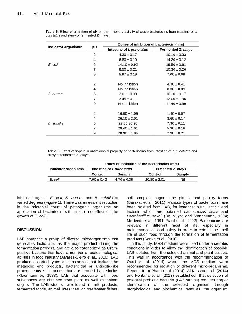
revealed that the crude isolated bacteriocins were optimally stable at 37 and 50°C for bacteriocins from fermented Z. mays and intestine of I. punctatus, respectively, and pH 6 to 7, respectively against selected indicator organisms. The inhibition of growth of E. coli by the trypsin treated bacteriocin that was obtained from LAB isolates was investigated by agar well diffusion method (Table 6). The zone of inhibition (mm) in the presence of the trypsin treated bacteriocin from LAB isolates from intestine of I. punctatus was reduced, while the one from slurry of fermented Z. mays was totally eliminated.
Table 7 describes the biopreservative potential of crude bacteriocins from intestine of I. punctatus (BI) and slurry of fermented Z. mays (BZ) on juice of ripe orange and Titus fish. The tested samples were initially sterilized in order to eliminate possible contamination before the assessment. There was a reduction in growth of inoculated organism (E. coli) in the orange juice, Titus juice and standard (benzoic acid) compared to control
group as the treatment progressed. This was revealed by the reduced values of colony forming units of the indicator (Log CFU/mL) pathogen (Table 7) in all the treatment groups in relation to the control group. The growth inhibition of the pathogen by BZ (9.96 ± 0.09 Log CFU/mL) during the six day of the preservation of orange juice was significantly (p<0.05) lower than that of BI (10.96±0.09 Log CFU/mL) or standard preservative (11.70±0.10 Log CFU/mL) in the treated orange juice (Figure 1a). The preservation of Titus fish was a reversal as there was a significant (p<0.05) decrease in inhibition of the indicator organism as a result of application of BI (10.00±0.10 Log CFU/mL) to sample of Titus fish (Figure 1b) as at the last (6th) day of treatment in relation to other groups.
Using agar well diffusion method to access the production of antimicrobial agents by the selected bacterial isolates from the I. punctatus intestine and fermented Z. mays against three pathogens, the susceptibility of various Gram positive (S. aureus and B. subtilis) and Gram negative (E. coli) bacteria to grow in presence of crude extract of bacteriocin revealed
414 Afr. J. Microbiol. Res.
Table 5. Effect of alteration of pH on the inhibitory activity of crude bacteriocins from intestine of I. punctatus and slurry of fermented Z. mays.
Indicator organisms pH Zones of inhibition of bacteriocin (mm)
Intestine of I. punctatus Fermented Z. mays
E. coli
2 4.30 ± 0.17 10.10 ± 0.33
4 6.80 ± 0.19 14.20 ± 0.12
6 14.10 ± 0.92 19.50 ± 0.61
7 8.50 ± 0.21 10.30 ± 0.26
9 5.97 ± 0.19 7.00 ± 0.09
S. aureus
2 No inhibition 4.30 ± 0.41
4 No inhibition 8.30 ± 0.39
6 2.01 ± 0.08 10.10 ± 0.17
7 3.45 ± 0.11 12.00 ± 1.96
9 No inhibition 11.40 ± 0.99
B. subtilis
2 16.00 ± 1.05 1.40 ± 0.07
4 26.10 ± 2.01 3.60 ± 0.17
6 29.60 ±0.98 7.30 ± 0.11
7 29.40 ± 1.01 5.30 ± 0.18
9 20.90 ± 1.06 2.90 ± 0.21
Table 6. Effect of trypsin in antimicrobial property of bacteriocins from intestine of I. punctatus and slurry of fermented Z. mays.
Indicator organisms
Zones of inhibition of the bacteriocins (mm)
Intestine of I. punctatus Fermented Z. mays
Control Sample Control Sample
E. coli 7.90 ± 0.43 4.70 ± 0.05 20.80 ± 2.01 Nil
inhibition against E. coli, S. aureus and B. subtilis at varied degrees (Figure 1). There was an evident reduction in the microbial count of pathogenic organisms on application of bacteriocin with little or no effect on the growth of E. coli. DISCUSSION LAB comprise a group of diverse microorganisms that generates lactic acid as the major product during the fermentation process, and are also categorized as Gram-positive bacteria that have a number of biotechnological abilities in food industry (Alvarez-Sieiro et al., 2016). LAB produce assorted types of substances that include the metabolic end products, bactericidal or antibiotic-like proteneceous substances that are termed bacteriocins (Klaenhammer, 1988). LAB that associate with food substances are obtained from plant as well as animal origins. The LAB strains are found in milk products, fermented foods, animal intestines or freshwater fishes,
soil samples, sugar cane plants, and poultry farms (Barakat et al., 2011). Various types of bacteriocin have been isolated from LAB, for instance: nisin, lacticin and lactosin which are obtained Lactococcus lactis and Lactobacillus sakei (De Vuyst and Vandamme, 1994; Mørtvedt et al., 1991; Piard et al., 1992). Bacteriocins are relevant in different facet of life, especially in maintenance of food safety in order to extend the shelf life of such food through the formation of fermentation products (Sarika et al., 2010).
In this study, MRS medium were used under anaerobic conditions in order to allow the identification of possible LAB isolates from the selected animal and plant tissues. This was in accordance with the recommendation of Ouali et al. (2014) where the MRS medium were recommended for isolation of different micro-organisms. Reports from Pham et al. (2014), Al Kassaa et al. (2014) and Fontana et al. (2013) established that selection of potential probiotic bacteria (LAB strains) requires proper identification of the selected organism through morphological and biochemical tests as the organism

Ogundare et al. 415
Table 7. Biopreservative potential of bacteriocins from intestine of I. punctatus and slurry of fermented Z. mays.
Test food samples Microbial counts (Log CFU/mL)
Control Sample (BI) Sample (BZ) Standard
Ripe oranges
Day 0 5.93 ± 0.01 5.93 ± 0.07 5.93 ± 0.01 5.93 ± 0.03
Day 1 5.99 ± 0.04 5.98 ± 0.03 6.13 ± 0.02 5.98 ± 0.03
Day 2 6.13 ± 0.11 6.10 ± 0.04 6.16 ± 0.02 6.17 ± 0.05
Day 3 7.54 ± 0.06 7.11 ± 0.04 7.22 ± 0.04 7.48 ± 0.08
Day 4 9.85 ± 0.09 7.90 ± 0.08 8.03 ± 0.06 8.20 ± 0.07
Day 5 10.17 ± 0.08 8.93 ± 0.06 9.95 ± 0.08 9.78 ± 0.09
Day 6 14.99 ± 0.11 9.86 ± 0.10 10.96 ± 0.09 11.70 ± 0.10
Titus fish
Day 0 5.93±0.03 5.93 ± 0.02 5.93 ± 0.01 5.93 ± 0.03
Day 1 6.29±0.03 5.98 ± 0.03 6.02 ± 0.01 6.19 ± 0.03
Day 2 7.49±0.01 6.10 ± 0.07 6.39 ± 0.03 7.22 ± 0.03
Day 3 8.00±0.10 7.65 ± 0.03 7.65 ± 0.06 7.55 ± 0.04
Day 4 10.30±0.10 7.94 ± 0.07 8.03 ± 0.07 8.11 ± 0.09
Day 5 12.70±0.11 9.99 ± 0.10 10.03 ± 0.09 10.04 ± 0.09
Day 6 15.04±0.13 10.00 ± 0.10 11.01 ± 0.09 11.01 ± 0.08
0
5
10
15
20Control
BI
BZ
Standard
Biopreservation activity of bacteriocins
Lo
g C
FU
/mL
0
5
10
15
20Control
BI
BZ
Standard
Biopreservation activity of bacteriocins
Lo
g C
FU
/mL
a b
Figure 1. Biopreservative activities of bacteriocins from intestine of I. punctatus (BI) and slurry of fermented Z. mays (BZ) on orange juice (a) and Titus fish (b) as at day 7. All values are presented as Mean ± Standard Error of Mean of triplicate readings. Comparisons were made between the treatment groups. (*) p<0.05 versus Standard (benzoic acid); (◦) p<0.05 versus Control.
shows a bacilli shape, and without catalase activity. A total of the 8 isolates obtained from I. punctatus intestine and 7 isolates from slurry of fermented Z. mays were confirmed to be Gram positive, catalase negative, oxidase negative, non-spore, and white or cream coloured rod micro-organisms. Previous reports also described LAB as genetically and physiologically distinct set of rod-shaped, Gram-positive and catalase negative bacteria (Ashmaig et al., 2009; Dallal et al., 2017; Guetouache and Guessas, 2015).
Furthermore, the isolates which were able to grow in anaerobic condition displayed an ability to ferment carbohydrates such as glucose, fructose, maltose and
lactose as they liberate gas in the culture media. Fermentation of carbohydrates by LAB strains has been reported by Rattanachaikunsopon and Phumkhachorn (2010), Zou et al. (2013) and Jose et al. (2015). Also, there was a production of acid from glucose by these isolates, thereby, suggesting the properties of Lactobacillus species as described by Wang et al. (2010) and Ni et al. (2015). Previously, LAB have been isolated from both animal and plant sources in a bid to determine their probiotic ability or tendency to liberate antimicrobial substances that can be used in food preservation (Barakat et al., 2011; Tufail et al., 2011). Some of these sources include intestine or gut of fish (Balcázar et al.,

416 Afr. J. Microbiol. Res.
2008; Rao et al., 2015; Ringø et al., 2018; Sica et al., 2012). Fermented food samples including fermented Z. mays have also been reported to possess probiotic LAB strains (Onwuakor et al., 2014; Oyedeji et al., 2013; Rao et al., 2015; Zou et al., 2013). The isolated LAB strains produced acid in fermentation broth as an attribute of heterofermenter. Homofermenters are known for production of lactic acid from glucose. Two classes of fermentation strains of LAB (homofermentative and heterofermentative) were previously mentioned by researchers (Akalu et al., 2017; Nigatu et al., 2015). Some of the considerations made in the selection of potential probiotic LAB include optimum growth temperature and effect of salt concentration on their fermentation activities. Table 1 shows that the LAB isolates were stable at relatively high temperature range (10 to 37°C), and can be said to be heat tolerant, therefore the basis for the production of acid in the fermentation broth by the LAB isolates from the increased glycolytic activity. This is an added advantage over thermolabile pathogenic organisms, as the liberated acid reduces the contamination by other microorganisms. The report of this study is in agreement with Qiuju et al. (2013) and Zorriehzahra et al. (2016). The LAB isolates from the two tested samples were osmotolerance at 3% NaCl, while only the LAB isolates from intestine of I. punctatus could grow in 5% NaCl (Table 1). This indicates that the LAB strains from intestine of I. punctatus may be more tolerance to osmotic concentrations of NaCl than the strains from slurry of fermented Z. mays. Van Sinderen and Crowley (2013) and Adnan and Tan (2007) described tolerance of LAB strains to osmotic concentrations of salt like NaCl as an added advantage to commercial applications. Other scientists have previously reported the ability of LAB strains to withstand osmotic concentration resulting from addition of salts (Subramanyam, 2020; Van Sinderen and Crowley, 2013).
Despite the abundant information on production of bacteriocins from terrestrial origins or LAB that are capable of producing bacteriocins, there have been paucity of information on application of LAB especially in bacteriocins production in I. punctatus. Production of bacteriocin by LAB strains is essential factor in the choice of probiotic bacterial strains (Dobson et al., 2012). The bacteriocins which are proteinaceous substances are used to inhibit the growth of related microorganisms, and are recently applied in food preservation. Table 2 shows that the cell free supernatants obtained from culture of LAB strains from intestine of I. punctatus and fermented Z. mays samples contained proteinaceous substance (suspected to be bacteriocins) with protein concentrations 108.4 ± 3.9 and 102.7 ± 3.0 mg/mL, respectively. This is known as bacteriocins. This is similar to the reports of Udhayashree et al. (2012) and Abbasiliasi et al. (2012).
In a bid to characterize the proteinaceous substance, the antimicrobial activity of the substance was investigated in cultures of E. coli, S. aureus and B. subtilis
(Table 3). The indicator organisms were vulnerable to the activity of the crude bacteriocins at varied degrees. Gram-positive bacteria (S. aureus and B. subtilis) responded positively to inhibition of growth by the crude bacteriocins obtained from intestine of I. punctatus and Z. mays. This is an indication of antibacterial activity of bacteriocins produced by the isolated LAB against the selected pathogens. In the company of these are Gram-negative bacteria (E. coli) whose cell membrane is surrounded by lipid rich cell wall as in the case of any Gram-negative bacteria, but still proved sensitive to antibacterial actions of the extracted bacteriocins. Reports from Tufail et al. (2011) and Sankar et al. (2012) revealed the antibacterial activity of bacteriocin against some pathogenic organisms like E. coli and S. aureus. Yang et al. (2012), Djadouni and Kihal (2012) and Gaamouche et al. (2014) reported the antimicrobial activity of LAB bacteriocins in some Gram-positive bacteria. For instance, Afolayan et al. (2017) and Rather et al. (2017) recounted the antimicrobial activity of substance obtained from LAB isolates from fermented Z. mays and gut of fishes, respectively. This work supported the tendency of bacteriocins to affect the growth of both Gram-positive and Gram-negative organisms (Abriouel et al., 2011).
The effects of alteration of temperature and pH on activity of crude bacteriocins from the LAB isolates were determined using E. coli, S. aureus and B. subtilis as indicator organism. The crude bacteriocins were found to be heat stable especially at 37 and 50°C for bacteriocins from fermented Z. mays and intestine of I. punctatus, respectively (Table 4). These results indicate that bacteriocin produced by LAB from intestine of I. punctatus is more heat stable than the fermented Z. mays, as its activity was sustained after the heat treatment at the aforementioned temperature. Bacteriocins that are used as food preservative are usually heat stable since preparation of many food requires heat in one way or the other (Ogunbanwo et al., 2003). Previous reports have also corroborated the present finding that the bacteriocins from the LAB isolates are heat stable (Gómez-Sala et al., 2015; Udhayashree et al., 2012).
Effect of pH on activity of crude bacteriocins from fermented Z. mays and intestine of I. punctatus, respectively (Table 5) was carried out. It was observed that bacteriocin produced by LAB in intestine of I. punctatus and fermented Z. mays were optimally stable at pH 6. This further confirmed the tolerance of bacteriocins from the LAB to acidic rather than the alkaline pH values and that they can be applied in acidic foods (Adesina et al., 2016; Ayed et al., 2015; Li et al., 2015).
Exposure of E. coli to the trypsin treated bacteriocin that were obtained from LAB isolates showed that zone of inhibition (mm) in the presence of the trypsin treated bacteriocin from LAB isolates from intestine of I. punctatus was reduced, while the one from slurry of

fermented Z. mays was totally eliminated (Table 6). This indicates that crude bacteriocins were inactivated by treatment with trypsin as a result of reduction or elimination of antimicrobial activity when it relates to controls, and further established the antimicrobial substances obtained from the isolated LAB cultures to be bacteriocin; a proteinaceous substance (Sankar et al., 2012).
Biopreservation is a potent natural method of extension of shelf life and safety of foods by using naturally occurring microorganisms, their innate antibacterial agents of specified quality and quantity (Ghanbari et al., 2013). Biopreservative activity of bacteriocin from LAB has been of utmost interest in the recent time. The reduction of microbial population in Titus fish and orange juice after addition of the crude protein-like substances produced from intestine of I. punctatus and Z. mays (Table 7) shows that the bacteriocins can be applied in preservation of food from plant and animal origins. The result also revealed that bacteriocin obtained from LAB in intestine of I. punctatus is more efficient in Titus fish than bacteriocin from Z. mays. Reduction of bacterial counts in food samples after treatment with crude bacteriocins as a measure of preservation has been documented. Gómez-Sala et al. (2016), Ghanbari et al. (2013) and Sarika et al. (2019) observed the extension of shelf life of fish after treatment with bacteriocins. Similarly, Udhayashree et al. (2012) and Ageni et al. (2017) reported a decrease in microbial loads in edible milk and button mushrooms, and in fermented maize (Ogi) and cassava (Fufu), respectively, In addition, bacteriocins from LAB obtained from these food items are efficient in the preservation of the selected test food samples, the crude bacteriocin from fish intestine (BI) was more efficient in Titus fish than in orange juice than the chemical preservative. Conclusion The present study revealed that the protein-like antibacterial substances from LAB isolates obtained in the samples of I. punctatus (Cat fish) and slurry fermented Z. mays (Ogi) possess an extensive spectrum of inhibitory activity against S. aureus and B. subtilis. The reduction in the microbial load in Titus fish and Orange juice exhibited by these proteinaceous substances (crude bacteriocins) also justify their tendency to preserve sea foods and fruits. CONFLICT OF INTERESTS The authors have not declared any conflict of interests.
ACKNOWLEDGEMENTS
The authors thank the management of Lagos State
Ogundare et al. 417 Polytechnic and the entire members of staff of Chemical Science and Biological Science Departments for their supports throughout the study. REFERENCES Abbasiliasi S, Tan JS, Ibrahim TAT, Ramanan RN, Vakhshiteh F,
Mustafa S, Ariff AB (2012). Isolation of Pediococcus acidilactici Kp10 with ability to secrete bacteriocin-like inhibitory substance from milk products for applications in food industry. BMC Microbiology 12(1):260.
Abriouel H, Franz C, Omar NB, Gálvez A (2011). Diversity and applications of Bacillus bacteriocins. FEMS Microbiology Reviews 35(1):201-232.
Adesina I, Ojokoh A, Arotupin D (2016). Effect of bacteriocinogenic Pediococcus pentosaceus IO1 strain and its bacteriocin on growth performance and intestinal microbiota of albino rat. Microbiology Research Journal International. https://www.researchgate.net/publication/297663001_Effect_of_Bacteriocinogenic_Pediococcus_pentosaceus_IO1_Strain_and_Its_Bacteriocin_on_Growth_Performance_and_Intestinal_Microbiota_of_Albino_Rat
Adesokan I, Abiola O, Ogundiya M (2010). Influence of ginger on sensory properties and shelf-life of Ogi, a Nigerian traditional fermented food. African Journal of Biotechnology 9(12).
Adnan AFM, Tan IK (2007). Isolation of lactic acid bacteria from Malaysian foods and assessment of the isolates for industrial potential. Bioresource Technology 98(7):1380-1385.
Afolayan AO, Ayeni FA, Ruppitsch W (2017). Antagonistic and quantitative assessment of indigenous lactic acid bacteria in different varieties of ogi against gastrointestinal pathogens. The Pan African Medical Journal 27.
Ageni L, Ajibade G, Yerima B, Appah J (2017). Shelf life extension study of ogi and fufu using bacteriocin isolated from Lactobacillus acidophilus of fermented dairy products. African Journal of Microbiology Research 11(32):1286-1293.
Akalu N, Assefa F, Dessalegn A (2017). In vitro evaluation of lactic acid bacteria isolated from traditional fermented Shamita and Kocho for their desirable characteristics as probiotics. African Journal of Biotechnology 16(12):594-606.
Al Kassaa I, Hamze M, Hober D, Chihib NE, Drider D (2014). Identification of vaginal lactobacilli with potential probiotic properties isolated from women in North Lebanon. Microbial Ecology 67(3):722-734.
Ali AA (2010). Beneficial role of lactic acid bacteria in food preservation and human health: a review. Research Journal of Microbiology 5(12):1213-1221.
Alka S, Neelam Y, Shruti S (2012). Effect of fermentation on physicochemical properties and in vitro starch and protein digestibility of selected cereals. International Journal of Agricultural and Food Science 2(3):66-70.
Alvarez-Sieiro P, Montalbán-López M, Mu D, Kuipers OP (2016). Bacteriocins of lactic acid bacteria: extending the family. Applied Microbiology and Biotechnology 100(7):2939-2951.
Ashmaig A, Hasan A, El Gaali E (2009). Identification of lactic acid bacteria isolated from traditional Sudanese fermented camel’s milk (Gariss). African Journal of Microbiology Research 3(8):451-457.
Ayed HB, Maalej H, Hmidet N, Nasri M (2015). Isolation and biochemical characterisation of a bacteriocin-like substance produced by Bacillus amyloliquefaciens An6. Journal of Global Antimicrobial Resistance 3(4):255-261.
Balcázar JL, Vendrell D, de Blas I, Ruiz-Zarzuela I, Muzquiz JL, Girones O (2008). Characterization of probiotic properties of lactic acid bacteria isolated from intestinal microbiota of fish. Aquaculture 278(1-4):188-191.
Barakat OS, Ibrahim G, Tawfik N, El-Kholy W, El-Rab GD (2011). Identification and probiotic characteristics of Lactobacillus strains isolated from traditional Domiati cheese. International Journal of Microbiology Research 3(1):59.
Barbosa AAT, Mantovani HC, Jain S (2017). Bacteriocins from lactic

418 Afr. J. Microbiol. Res.
acid bacteria and their potential in the preservation of fruit products. Critical Reviews in Biotechnology 37(7):852-864.
Behnsen J, Deriu E, Sassone-Corsi M, Raffatellu M (2013). Probiotics: properties, examples, and specific applications. Cold Spring Harbor Perspectives in Medicine 3(3):a010074.
Chang JY, Chang HC (2010). Improvements in the quality and shelf life of kimchi by fermentation with the induced bacteriocin‐producing strain, Leuconostoc citreum GJ7 as a starter. Journal of Food Science 75(2):M103-M110.
Dallal MS, Zamaniahari S, Davoodabadi A, Hosseini M, Rajabi Z (2017). Identification and characterization of probiotic lactic acid bacteria isolated from traditional persian pickled vegetables. GMS Hygiene and Infection Control 12.
Davidson PM, Taylor TM, Schmidt SE (2012). Chemical preservatives and natural antimicrobial compounds. Food Microbiology: Fundamentals and Frontiers pp. 765-801.
De Vuyst L, Leroy F (2007). Bacteriocins from lactic acid bacteria: production, purification, and food applications. Journal of Molecular Microbiology and Biotechnology 13(4):194-199.
De Vuyst L, Vandamme EJ (1994). Nisin, a lantibiotic produced by Lactococcus lactis subsp. lactis: properties, biosynthesis, fermentation and applications. Bacteriocins of Lactic Acid Bacteria. pp. 151-221.
Deegan LH, Cotter PD, Hill C, Ross P (2006). Bacteriocins: biological tools for bio-preservation and shelf-life extension. International Dairy Journal 16(9):1058-1071.
Del Nobile MA, Lucera A, Costa C, Conte A (2012). Food applications of natural antimicrobial compounds. Frontiers in Microbiology 3:287.
Djadouni F, Kihal M (2012). Antimicrobial activity of lactic acid bacteria and the spectrum of their biopeptides against spoiling germs in foods. Brazilian Archives of Biology and Technology 55(3):435-444.
Du Toit M, Engelbrecht L, Lerm E, Krieger-Weber S (2011). Lactobacillus: the next generation of malolactic fermentation starter cultures—an overview. Food and Bioprocess Technology 4(6):876-906.
Ejigui J, Savoie L, Marin J, Desrosiers T (2005). Beneficial changes and drawbacks of a traditional fermentation process on chemical composition and antinutritional factors of yellow maize (Z. mays). Journal of Biological Sciences 5(5):590-596.
Eun JB, Chung HJ, Hearnsberger J (1994). Chemical composition and microflora of channel catfish (Ictalurus punctatus) roe and swim bladder. Journal of Agricultural and Food Chemistry 42(3):714-717.
Farkas J (2007). Physical methods of food preservation Food Microbiology: Fundamentals and Frontiers. Third Edition American Society of Microbiology pp. 685-712.
Fontana L, Bermudez-Brito M, Plaza-Diaz J, Munoz-Quezada S, Gil A (2013). Sources, isolation, characterisation and evaluation of probiotics British. Journal of Nutrition 109(S2):S35-S50.
Gaamouche S, Arakrak A, Bakkal M, LaglaouiA (2014). Antimicrobial activity of lactic acid bacteria and bacteriocins isolated from a traditional brine table olives against pathogenic bacteria. International Journal of Current Microbiology and Applied Science 3(11):657-666.
Ghanbari M, Jami M, Domig KJ, Kneifel W (2013). Seafood biopreservation by lactic acid bacteria–a review. LWT-Food Science and Technology 54(2):315-324.
Gómez-Sala B, Herranz C, Díaz-Freitas B, Hernánde PE, Sal A, Cintas LM (2016). Strategies to increase the hygienic and economic value of fresh fish: Biopreservation using lactic acid bacteria of marine origin. International Journal of Food Microbiology 223:41-49.
Gómez-Sala B, Muñoz-Atienza E, Sánchez J, Basanta A, Herranz C, Hernández PE, Cintas LM (2015). Bacteriocin production by lactic acid bacteria isolated from fish, seafood and fish products. European Food Research and Technology 241(3):341-356.
Gould GW (2012). New methods of food preservation: Springer Science and Business Media.
Guetouache M, Guessas B (2015). Characterization and identification of lactic acid bacteria isolated from traditional cheese (Klila) prepared from cow's milk. African Journal of Microbiology Research 9(2):71-77.
Hanim C (2017). Effect of pH and Temperature on Bacillus subtilis FNCC 0059 oxalate decarboxylase activity. Pakistan Journal of Biological Sciences 20(9):436-441.
Ijarotimi SO, Keshinro OO (2011). Determination of amino acid, fatty
acid, mineral, functional and choking properties of germinated and fermented popcorn (Z. mays everta) flour. European Journal of Nutrition and Food Safety pp. 102-122.
Jose NM, Bunt CR, Hussain MA (2015). Comparison of microbiological and probiotic characteristics of lactobacilli isolates from dairy food products and animal rumen contents. Microorganisms 3(2):198-212.
Klaenhammer TR (1988). Bacteriocins of lactic acid bacteria. Biochimie 70(3):337-349.
Kozaki M, Uchimura T, Okada S (1992). Experimental manual of lactic acid bacteria. Tokyo, Japan: Asakurasyoten pp. 34-37.
Lee HJ, Kim HJ (2011). Lantibiotics, class I bacteriocins from the genus Bacillus. Journal of Microbiology and Biotechnology 21(3):229-235.
Li D, Ni K, Pang H, Wang Y, Cai Y, Jin Q (2015). Identification and antimicrobial activity detection of lactic acid bacteria isolated from corn stover silage. Asian-Australasian Journal of Animal Sciences 28(5):620.
Liu W, Pang H, Zhang H, Cai Y (2014). Biodiversity of lactic acid bacteria. Springer pp. 103-203.
Lück E (1985). Chemical preservation of food. Zentralblatt fur Bakteriologie, Mikrobiologie und Hygiene. 1. Abt. Originale B, Hygiene 180(2-3): 311-318.
Mørtvedt C, Nissen-Meyer J, Sletten K, Nes I (1991). Purification and amino acid sequence of lactocin S, a bacteriocin produced by Lactobacillus sake L45. Applied and Environmental Microbiology 57(6):1829-1834.
Ni K, Wang Y, Li D, Cai Y, Pang H (2015). Characterization, identification and application of lactic acid bacteria isolated from forage paddy rice silage PloS one 10(3):e0121967.
Nigatu JM, Tuji FA, Tefera AT (2015). Evaluation of the antagonistic effect of six mixed cultures of lactic acid bacteria, isolated from the Ethiopian fermented milk ergo, against some foodborne pathogens inoculated into the Ethiopian cottage cheese ayib. African Journal of Microbiology Research 9(29):1789-1797.
Noor R, Islam Z, Munshi SK, Rahman F (2013). Influence of temperature on Escherichia coli growth in different culture media. Journal of Pure and Applied Microbiology 7(2): 899-904.
Ogodo A, Ugbogu O, Onyeagba R, Orji F (2017). Dynamics of functional properties of sorghum flours fermented with lactic acid bacteria (LAB)-consortium isolated from cereals. Food Research Journal 24(6):2666-2671.
Ogunbanwo S, Sanni A, Onilude A (2003). Characterization of bacteriocin produced by Lactobacillus plantarum F1 and Lactobacillus brevis OG1. African Journal of Biotechnology 2(8):219-227.
Onwuakor C, Nwaugo V, Nnadi , Emetole J (2014). Effect of varied culture conditions on crude supernatant (bacteriocin) production from four Lactobacillus species isolated from locally fermented maize (ogi) . American Journal of Microbiological Research 2(5):125-130.
Ouali FA, Al Kassaa I, Cudennec B, Abdallah M, Bendali F, Sadoun D, Chihib NE, Drider D (2014). Identification of lactobacilli with inhibitory effect on biofilm formation by pathogenic bacteria on stainless steel surfaces. International Journal of Food Microbiology 191:116-124.
Oyedeji O, Ogunbanwo ST, Onilude AA (2013). Predominant lactic acid bacteria involved in the traditional fermentation of fufu and ogi, two Nigerian fermented food products. Food and Nutrition Sciences 4(11):40.
Perez RH, Zendo T, Sonomoto K (2014). Novel bacteriocins from lactic acid bacteria (LAB): various structures and applications. Microbial Cell Factories 13(1):S3. doi:10.1186/1475-2859-13-S1-S3
Pham D, Ansquer D, Chevalier A, Dauga C, Peyramale A, Wabete N, Labreuche Y (2014). Selection and characterization of potential probiotic bacteria for Litopenaeus stylirostris shrimp hatcheries in New Caledonia. Aquaculture 432:475-482.
Piard JC, Muriana P, Desmazeaud M, Klaenhammer T (1992). Purification and partial characterization of lacticin 481, a lanthionine-containing bacteriocin produced by Lactococcus lactis subsp. lactis CNRZ 481. Applied and Environmental Microbiology 58(1):279-284.
Piper P, Calderon CO, Hatzixanthis K, Mollapour M (2001). Weak acid adaptation: the stress response that confers yeasts with resistance to organic acid food preservatives. Microbiology 147(10):2635-42.
Pratush A, Gupta A, Kumar A, Vyas G (2012). Application of purified bacteriocin produced by Lactococcus lactis AP2 as food biopreservative in acidic foods. Annals, Food Science Technology

13:82-87. Qiuju W, Yizhe C, Shengjun L, Ruijin Z (2013). Isolation and
identification of lactic acid bacteria in duodenum of laying hens fed in cage. Journal of Heilongjiang Bayi Agricultural University 3:8.
Ramu R, Shirahatti PS, Devi AT, Prasad A (2015). Bacteriocins and their applications in food preservation. Critical Reviews in Food Science and Nutrition 00-00.
Rao KP, Chennappa G, Suraj U, Nagaraja H, Raj AC, Sreenivasa M (2015). Probiotic potential of Lactobacillus strains isolated from sorghum-based traditional fermented food. Probiotics and Antimicrobial Proteins 7(2):146-156.
Rather IA, Galope R, Bajpai VK, Lim J, Paek WK, Park YH (2017). Diversity of marine bacteria and their bacteriocins: applications in aquaculture. Reviews in Fisheries Science and Aquaculture 25(4):257-269.
Rattanachaikunsopon P, Phumkhachorn P (2010). Lactic acid bacteria: their antimicrobial compounds and their uses in food production Annals of Biological Research 1(4):218-228.
Lowry OH, Rosebrough NJ, Farr AL, Randall RJ (1951). Protein measurement with the Folin phenol reagent. Journal of Biological Chemistry 193:265-275.
Reis JA, Paula AT, Casarotti SN, Penna ALB (2012). Lactic acid bacteria antimicrobial compounds: characteristics and applications. Food Engineering Reviews 4(2):124-140. doi:10.1007/s12393-012-9051-2
Ringø E , Gatesoupe FJ (1998). Lactic acid bacteria in fish: a review. Aquaculture 160(3-4):177-203.
Ringø E, Hoseinifar SH, Ghosh K, Doan HV, Beck BR, Song SK (2018). Lactic acid bacteria in finfish-An update. Frontiers in Microbiology 9:1818.
Sankar NR, Priyanka VD, Reddy PS, Rajanikanth P, Kumar VK, Indira M (2012). Purification and characterization of bacteriocin produced by Lactobacillus plantarum isolated from cow milk. International Journal of Microbiology Research 3(2):133-137.
Sarika A, Lipton A, Aishwarya M (2010). Bacteriocin production by a new isolate of Lactobacillus rhamnosus GP1 under different culture conditions. Advance Journal of Food Science and Technololgy 2(5):291-297.
Sarika A, Lipton AP, Aishwarya M (2019). Biopreservative efficacy of bacteriocin GP1 of Lactobacillus rhamnosus GP1 on stored fish filets. Frontiers in Nutrition 6:29.
Shahid M, Hussain B, Riaz D, Khurshid M, Ismail M, Tariq M (2017). Identification and partial characterization of potential probiotic lactic acid bacteria in freshwater Labeo rohita and Cirrhinus mrigala. Aquaculture Research 48(4):1688-1698.
Shahidi F (2015). Handbook of antioxidants for food preservation: Woodhead Publishing.
Sharma S (2015). Food preservatives and their harmful effects. International Journal of Scientific and Research Publications 5(4):1-2.
Sica MG, Brugnoni LI, Marucci PL, Cubitto MA (2012). Characterization of probiotic properties of lactic acid bacteria isolated from an estuarine environment for application in rainbow trout (Oncorhynchus mykiss, Walbaum) farming. Antonie van Leeuwenhoek 101(4):869-879.
Silva CC, Silva SP, Ribeiro SC (2018). Application of bacteriocins and protective cultures in dairy food preservation. Frontiers in Microbiology 9:594.
Smid E, Kleerebezem M (2014). Production of aroma compounds in lactic fermentations. Annual Review of Food Science and Technology 5:313-326.
Stewart CM (2003). Staphylococcus aureus and staphylococcal enterotoxins. Ch 12 In: Hocking AD (ed) Foodborne microorganisms of public health significance. 6th ed, Australian Institute of Food Science and Technology (NSW Branch) Sydney pp. 359-380.
Subramanyam MN (2020). Molecular characterisation of probiotic Lactobacillus fermentum isolated from home made curd Journal of Microbiology, Biotechnology and Food Sciences 9(4):848-855.
Ogundare et al. 419 Trząskowska M, Kołożyn-Krajewska D, WójciakK, Dolatowski Z (2014).
Microbiological quality of raw-fermented sausages with Lactobacillus casei LOCK 0900 probiotic strain. Food Control 35(1):184-191.
Tserovska L, Stefanova S, Yordanova T (2002). Identification of lactic acid bacteria isolated from Katyk, goat’s milk and Cheese. Journal of Culture Collections 3:48-52.
Tufail M, Hussain S, Malik F, Mirza T, Parveen G, Shafaat S, Sadiq A (2011). Isolation and evaluation of antibacterial activity of bacteriocin produced by Lactobacillus bulgaricus from yogurt. African Journal of Microbiology Research 5(22):3842-3847.
Udhayashree N, Senbagam D, Senthilkumar B, Nithya K, Gurusamy R (2012). Production of bacteriocin and their application in food products Asian Pacific Journal of Tropical Biomedicine 2(1):S406-S410.
Van Sinderen D, Crowley S (2013). Lactobacillus plantarum species possessing broad spectrum anti-fungal activity and exhibiting high heat tolerance and osmotolerance: Google Patents.
Vignolo G, Saavedra L, Sesma F, Raya R (2012). 22 Food bioprotection: lactic acid bacteria as natural preservatives. Progress in Food Preservation P 453.
Wang CY, Lin PR, Ng CC, Shyu YT (2010). Probiotic properties of Lactobacillus strains isolated from the feces of breast-fed infants and Taiwanese pickled cabbage. Anaerobe 16(6):578-585.
Woraprayote W, Malila Y, Sorapukdee S, Swetwiwathana A, Benjakul S, Visessanguan W (2016). Bacteriocins from lactic acid bacteria and their applications in meat and meat products. Meat Science 120:118-132.
Yang E, Fan L, Jiang Y, Doucette C, Fillmore S (2012). Antimicrobial activity of bacteriocin-producing lactic acid bacteria isolated from cheeses and yogurts. AMB Express 2(1):48.
Yang SC, Lin CH, Sung CT, Fang JY (2014). Antibacterial activities of bacteriocins: application in foods and pharmaceuticals. Frontiers in Microbiology 5:241.
Yousef AE, Balasubramaniam V (2013). Physical methods of food preservation. Food Microbiology American Society of Microbiology pp. 737-763.
Zorriehzahra MJ, Delshad ST, Adel M, Tiwari R, Karthik K, Dhama K, Lazado CC (2016). Probiotics as beneficial microbes in aquaculture: an update on their multiple modes of action: a review. Veterinary Quarterly 36(4):228-241.
Zou Y, Liu F, Fang C, Wan D, Yang R, Su Q, Zhao J (2013). Lactobacillus shenzhenensis sp. nov., isolated from a fermented dairy beverage. International Journal of Systematic and Evolutionary Microbiology 63(5):1817-1823.

Vol. 15(8), pp. 420-428, August, 2021
DOI: 10.5897/AJMR2021.9553
Article Number: 3D00C4C67530
ISSN: 1996-0808
Copyright ©2021
Author(s) retain the copyright of this article
http://www.academicjournals.org/AJMR
African Journal of Microbiology Research
Full Length Research Paper
Aflatoxigenic potential of Aspergillus section Flavi isolated from maize seeds, in Burkina Faso
Hamidou COMPAORE1*, Serge SAMANDOULOUGOU1, Fidèle W. TAPSOBA1, Alima BAMBARA2, Hissein RATONGUE3, Ignace SAWADOGO1, Donatien KABORE1,
Pane B. OUATTARA-SOURABIE4 and Hagrétou SAWADOGO-LINGANI1
1DTA/IRSAT/CNRST, 03 BP 7047 Ouagadougou 03, Burkina Faso.
2Département de Biochimie-Microbiologie, Laboratoire de Biochimie et d’Immunologie Appliquée (LaBIA),
Université Joseph KI-ZERBO, 03 BP 7021 Ouagadougou 03, Burkina Faso. 3Université Catholique de l’Afrique de l’Ouest, Unité Universitaire à Bobo-Dioulasso (UCAO/UUB),
1052 Bobo-Dioulasso, Burkina Faso. 4Laboratoire National de Santé Publique (LNSP), 09 BP 24 Ouagadougou 09, Burkina Faso (Kadiogo / Centre).
Received 14 June, 2021; Accepted 27 July, 2021
The frequency of occurrence and four principal kinds of aflatoxin concentration in maize seeds grown in Burkina Faso was investigated. Ten (10) samples collected, were analyzed by high performance liquid chromatography (HPLC) with post-column derivatisation after immunoaffinity column cleanup. Eight strains of Aspergillus section Flavi were previously isolated from these samples and cultivated on “Aspergillus flavus and parasiticus agar (AFPA)” to ascertain if they belong to A. flavus or A. parasiticus species. The qualitative ability of aflatoxin production was also previously performed by fluorescence emission under ultra violet light at 365 nm after four (4) days of incubation at 30 °C on Coconut Agar Medium (CAM). Results showed that 70% of samples were contaminated by aflatoxins. The levels ranged from 0.93 to 58.94 µg/kg. Samples M1 and M10 had high concentrations, 58.94 µg/kg and 70.73 µg/kg; whereas M4 and M5 had low concentrations from 1.68 to 0.93 µg/kg, respectively. In these samples, four were contaminated with aflatoxin B1 (AFB1) and aflatoxin G1 (AFG1), two with AFB1 and aflatoxin B2 (AFB2) and one (01) with AFB1 only. We notice that AFB1 was the most prevalent member of aflatoxins, and AFG2 was absent in all samples. Key words: Maize, Aspergillus, aflatoxins, HPLC, Burkina Faso.
INTRODUCTION Cereals are a staple food for humans and animals. In Burkina Faso, their annual consumption is estimated at 62% of the food consumed by households (Waongo et
al., 2013). Among food crops, maize is the most used product by over 98% of rural households (Bambara, 2021). In Burkina Faso, maize ranks second among
*Corresponding author. E-mail: [email protected]. Tel: +226 25 37 31 31.
Author(s) agree that this article remain permanently open access under the terms of the Creative Commons Attribution
License 4.0 International License

cultivated cereals, both in area, production and consumption (Somda, 2016). It also makes a significant contribution to the country’s economies. The area devoted to maize cultivation in the country increased from 790,321 ha in 2010 to 1,019,181 ha in 2018 (GDSSS/MAHAD, 2020). National maize production reached 1,700,127 T in 2018 against 1,133,480 T in 2010 (GDSSS/MAHAD, 2020). Maize is grown widely around the tropical world owing to its good adaptation to climate and its popularity. Besides being distributed widely, maize can be used for many purposes, such as animal feed, industrial uses, and is even the staple food in many developing countries. It also makes a large contribution to the economies of developed and developing countries. Nevertheless, the aflatoxin phenomenon is undermining the sector in Africa (Chauhan et al., 2016). Thus, maize is food crop that is easily contaminated with mycotoxins such as aflatoxins that are cancer-causing, immuno-suppressive mycotoxins (Makhlouf, 2019). According to Bambara (2021), 40% of maize production is affected by aflatoxins in developing countries. Contamination of crops with aflatoxin is a global food safety issue (Compaoré et al., 2021). The most important members of AFs are AFB1, AFB2, AFG1 and AFG2. The International Agency for Research on Cancer (IARC) has classified AFB1, AFB2, AFG1 and AFG2 as Group I human carcinogens (IARC, 2002). Among the many known toxins in the world, aflatoxins are highly toxic and carcinogenic compounds that can cause diseases in livestock and humans (Ouattara-Sourabié, 2018). AFs are a group of mycotoxins produced as secondary metabolites by species in Aspergillus section Flavi. The species most notorious for aflatoxin production are Aspergillus flavus (produces only aflatoxins B) and Aspergillus parasiticus (produces both B and G aflatoxins) (Kachapulula et al., 2017).
Previous studies in Africa have found that the occurrence of aflatoxins in food products is mainly influenced by favorable conditions such as high moisture content and temperature (Waré et al., 2017). In fact, in the Sahelian zone dry, post-harvest conservation is the only means of ensuring the link between the harvest occurring once in the year and consumption that is permanent and obligatory. The harvests, kept in general under inappropriate conditions, are attacked by insects, rodents and molds (Waongo et al., 2013). Also, in Burkina Faso the improper storage or preservation methods used such as maize bad drying would also lead to attacks by micro-organisms including molds of the genus Aspergillus, Penicillium, Fusarium, and Alternaria (Sanou, 2000). Aflatoxins contamination of maize has always remained a topic of debate in terms of international market as well as economic development of country which are part of trade market (Chauhan et al., 2016). In view of the huge economic losses and health problems caused by mycotoxins; a great deal of interest
Compaore et al. 421 is currently accorded to them throughout the world. Thus to guarantee the health of consumers, each country is obliged to adopt specific legislation for the main mycotoxins in foods liable to harbor toxigenic molds (Waré et al., 2017). Several countries in the world have established or proposed regulatory limits for mycotoxins in foods. European Union countries edited regulations that have been revised periodically to limit their presence in the foods in Europe (EU Regulation 1881, 2006). In Africa certain countries also have regulations on mycotoxins and produce significant research, especially on aflatoxins and fumonisins that affect health (Ezekiel et al., 2014). Unfortunately, Burkina Faso, a country of West Africa has not set a mycotoxin regulation and uses those of Codex alimentarius which fixes the maximum levels of AFB1 and total aflatoxins respectively at 2 µg/Kg and 5 µg/Kg (EU Regulation 1881, 2006). Indeed, there are few studies and surveys about the contamination of mycotoxins in maize in Burkina Faso and this does not really reflect the problem in our country (Sanou, 2000). Our farmers therefore often find it difficult to export their products to countries whose mycotoxin regulations are in force. Indeed, because crops in tropical and subtropical regions are more susceptible to contamination due to favorable climatic and their inadequate storage conditions which facilitate the proliferation of molds and their secondary metabolites (Waré et al., 2017). Therefore, the aim of the proposed work is to determine the fungal load of maize samples from Ouagadougou City in Burkina Faso and to quantify the concentration of all kinds of aflatoxins using high performance liquid chromatography. This will confirm the results of the qualitative demonstration of aflatoxins production capacity of molds previously isolated from these maize samples, such as fluorescence emission under ultra violet light at 365 nm and those in “A. flavus and parasiticus agar (AFPA)”. MATERIALS AND METHODS
Physicochemical analyses
The maize samples were first subjected to physicochemical analyses such as humidity and pH using standards methods. The tests were repeated three times.
Sampling and fungi isolation from maize seeds
A total of ten (10) maize samples in commercially available were collected during the period from December to February 2021 at Ouagadougou markets. Fungi were isolated and purified on Potato Dextrose Agar (PDA) and subculture in “Aspergillus flavus and parasiticus agar (AFPA)” to identify A. flavus and A. parasiticus according to Pitt et al. (1983) and Cotty (1994) protocols. Systematic determination and the strains identification were made on Potato Dextrose Agar (PDA) at 25 and 37 °C depending on the methods used by Christensen (1981), Hocking (1982) and Cooney and Emerson (1964). Inoculation was done in three points equidistant.

422 Afr. J. Microbiol. Res. Reference strains For comparison of cultural and microscopic characters between strains isolated from maize seeds and those of reference strains, three references belonging to Aspergillus section Flavi were used. They were UBOCC-A-106031 (A. flavus aflatoxinogenic) of French origin, UBOCC-A-111042 (A. parasiticus var. globosus aflatoxinogenic) of Japanese origin and S2 (A. flavus aflatoxinogenic) previously isolated from groundnuts and identified in Burkina Faso using molecular method by two (2) PCR based on 28S ribosomal sub unit (D1-D2 region) and the hyper variable ITS1-5.8S-ITS2 region, (Compaoré et al., 2016). This comparison was performed on Potato Dextrose Agar (PDA) medium at 30 °C for seven days. Aflatoxins quantification Sample extraction (AOAC, 2005) Aflatoxins production is confirmed by HPLC using aflatoxins B and G Standard, Romer Biopure, and a blank consisting of the extraction solution. HPLC analysis was performed at the Toxicological Department of the National Public Health Laboratory (LNSP) in Ouagadougou. Ten (10) maize samples were subjected to this analysis.
The principle was used to extract aflatoxin from the samples using suitable organic solvents, to purify this aflatoxin on an immunoaffinity column and then to identify and quantify it. To do this, 25 ± 0.2 g of mix ground maize was weighed and added approximately 3 g of sodium chloride and placed in blender cup. 125 ml of extraction solution methanol-distilled water (70:30; v/v) was added to the sample and the whole was stirred for 20 min. The solution was then filtered through Whatman No.4 filter paper and 15 ml of the filtrate was transferred into a beaker and diluted with 30 ml of distilled water. The diluted homogenized sample solution was filtered through glass microfiber membrane.
Sample cleanup The immunoaffinity columns were previously conditioned by passing 10 ml phosphate buffered saline (PBS) through the column by gravity and placed on the variant cuvette; the silica gel was allowed to flow. 15 ml of the diluted filtrate (1g sample equivalent) was taken and poured into the immunoaffinity column where it retained the desired molecules. The molecules were then washed with 10 ml of distilled water which was poured twice into the immunoaffinity column. Under gravity, the bound aflatoxins were eluted with 1 ml of pure methanol and air was pushed through the column to collect the last drops of eluate. 1 ml of distilled water was added to the eluate. The eluate was filtered through 0.45 µm Methanol-compatible membrane filter (PTFE or Nylon) and collected into a micro sample vial. Then this solution containing the aflatoxin molecules was sent to the HPLC for the detection and quantification of these aflatoxin molecules. Each experiment was conducted in triplicate and aflatoxins contents were determined according to their corresponding standard curves.
HPLC analyses
Spiked samples were prepared on relevant matrix for each batch of sample to obtain the % recovery. 0.5 ml of the working solution was added (100 µg/kg B1, G1 and 25 µg/kg B2, G2) per 25 g of matrix, mixed well and proceeded to sample extraction. The
chromatographic system consisted of an automatic Agilent 1200, with Immunoaffinity column cleanup and post-column derivatisation manufactured by Shim-pack VP-ODS, 4.6 mm (ID) x 150 mm (L). The post-column derivatisation was achieved using a Kobra Cell to obtain electrochemically generated bromine (ISO 16050 CEN/TC-34, 2006); Romer Biopure). It is equipped with an auto-sampler (10 μl, injector vol), a Shim-pack VP-ODS column with a Reverse-Phase C18 (4.6 (ID) x 150 mm (L)) and a fluorescence detector. The detector was set at EX= 350 nm, EM = 450 nm. The mobile phase was isocratic and composed of methanol: water (45:55)-KBr-HNO3 mixture with 450 ml of methanol, 119 mg of potassium bromide and 87.5 μl of 16 M nitric acid per liter of mobile phase. The flow rate was set at 1 ml/min.
The quality control standard (40 µg/kg calibration solution) was injected 3 times. The % CV of the peak area corresponding to the consecutive injections shall be within ± 10%. 10 μl of each of the Calibration Standards Solutions (2.5 µg/kg, 5 µg/kg, 10 µg/kg, 20 µg/kg, 40 µg/kg and 80 µg/kg) was injected into the HPLC system, followed by sample.
The concentration of Aflatoxins in the sample is calculated using the following formula:
Where
Statistical analysis (XLSTAT. 2016) The differences in aflatoxins concentration in maize samples between the Ouagadougou zones Burkina Faso and those of physicochemical analyzes were compared by Analysis of variance (ANOVA) using XLSTAT-Pro 7.5.2 software. Interpretation of values was performed using Newman-Keuls test at probability level p = 5%. The results were expressed as mean ± SD and the measures were repeated three times (n=3).
RESULTS AND DISCUSSION
Physicochemical analyses of maize sample
Humidity rate analysis carried out on the ten (10) samples of maize seeds revealed that our samples are not very humid with an average value between 3.91 and 4.66% (Table 1).
These values are lower than the average moisture content of mold growth. In fact, the minimum humidity for certain molds to start growing is 10% (Compaoré, 2017). Our data could be explained by contamination of samples in the field or during storage. Mold strains are able to survive unfavorable conditions for their growth by
producing large numbers of spores (Tabuc, 2007). Thanks
to its great adaptability to environmental conditions,
F:Total sample extract ml × Vol of final sample extract (µl)
Volume for column ml × Vol for injection into HPLC µl × Weight of sample(g)

Compaore et al. 423
Table 1. Humidity rate of maize samples
Sample Humidity (%)
M1 4.10 ± 0.07cd
M2 4.45 ± 0.07ab
M3 4.07 ± 0.07cd
M4 4.71 ± 0.07a
M5 3.99 ± 0.07cd
M6 4.22 ± 0.07bc
M7 4.48 ± 0.07ab
M8 3.91 ± 0.07d
M9 4.66 ± 0.07a
M10 4.26 ± 0.07bc
Significance level < 0.0001
For each column and for each maize sample, the means which are in common the same letter are not significantly different according to the Newman-keuls test at the 5% probability level.
Table 2. pH values of maize samples.
Sample pH
M1 5.93 ± 0.04a
M2 5.83 ± 0.04a
M3 5.89 ± 0.04a
M4 5.95 ± 0.04a
M5 5.98 ± 0.04a
M6 5.94 ± 0.04a
M7 5.96 ± 0.04a
M8 5.99 ± 0.04a
M9 5.93 ± 0.04a
M10 5.96 ± 0.04a
Signifiance level < 0.0001
For each column and for each maize sample, the means which are in common the same letter are not significantly different according to the Newman-keuls test at the 5% probability level.
A. flavus can grow both on crops in the field, during harvest as well as later, during storage (Makhlouf, 2019). For the different samples, statistical analyses showed significant differences between the different humidity values. From this analysis it appears that the humidity is very variable from one sample to another. The pH results of the various maize samples analyzed indicate that all of the samples are slightly acidic, with pH values ranging from 5.83 to 5.99 (Table 2).
The pH values are very favorable to the growth of molds because according to Gauthier (2016), molds can grow in a pH range from 3 to 8, with optimal growth being rather between 5 and 6. The production of mycotoxins takes place for pH close to optimal growth pH (Makhlouf, 2019). Due to their acidity, many foods are much more prone to fungal than bacterial spoilage (Tabuc, 2007).
Statistical analyses showed no significant difference between the different pH values. Fungi isolation from maize seeds In our study, ten (10) maize samples were used as a matrix for the collection of strains of Aspergillus section Flavi. From the consortium of fungi grown on maize, twenty-three (23) isolates were collected. Macroscopic observation of the Petri dishes made it possible to retain only the isolates forming colonies of yellowish and greenish color and have powder aspect. In optic microscopy, we were interested in those with non-septate and hyaline conidiophores. Eight (08) local Aspergillus section Flavi strains were therefore isolated from the

424 Afr. J. Microbiol. Res.
Table 3. Number of Aspergillus section Flavi isolates from collected maize samples.
Sample Number of Aspergillus section Flavi isolates Isolates code
M1 1 A2
M2 1 A5
M3 0 -
M4 0 -
M5 3 A4, A6 et A8
M6 0 -
M7 0 -
M8 0 -
M9 1 A3
M10 2 A1 and A7
Total 8 -
consortium of fungi grown on maize seeds (Table 3). Aflatoxins level in maize samples Results of the quantitative aflatoxin analysis are reported in Table 4. 70% of the samples were found to be contaminated with total aflatoxins. These were M1, M2, M4, M5, M6, M9 and M10. The total aflatoxin contents in the maize samples ranged from 0.93 to 70.78 µg/kg. M1 and M10 had high concentrations (58.86 and 70.78 µg/kg respectively) and M4 and M5 had low concentrations with respective values of 1.67 and 0.93 µg/kg. In all the contaminated samples, aflatoxin B1 was present and the most concentrated with however the absence of aflatoxin G2 as it can be seen from the chromatograms in Figure 1. Out of seven (7) samples, four (04) were contaminated with aflatoxin B1 (AFB1) and aflatoxin G1 (AFG1), two (02) with AFB1 and aflatoxin B2 (AFB2) and one (01) with AFB1 only.
The contamination of our samples with aflatoxins could be explained by the presence of toxigenic strains Aspergillus section Flavi. Indeed, the latter are the fungal contaminants most frequently encountered in cereals and which secrete aflatoxins there when environmental conditions (temperature, humidity, pH, etc.) are met, as is the case with the pH of our maize samples which are slightly acidic with a pH around 5.83 to 5.99 (Table 2). Maize is considered among the fragile crops at high risk of contamination by toxigenic molds unlike other cereals, particularly barley and wheat (Brahmi and Zahi, 2016).
According to the Codex alimentarius, the maximum levels of AFB1 and total aflatoxins (B1, B2, G1 and G2) are respectively 2 and 5 µg/Kg (EU Regulation 1881, 2006). Our results revealed that the contamination rate of the different samples varied from sample to sample. Four (04) samples had total aflatoxin levels between 13 and 71 µg/Kg and therefore well above the Codex alimentarius standard. Only the M2, M3, M4, M5, M7 and M8 samples
complied with the standard. The high concentration of aflatoxins in our samples could be explained by climatic conditions in Africa, poor agricultural practices and poor crop storage conditions. According to Zinedine (2004), crops grown in hot, humid climates and exposed to toxigenic molds provide optimal conditions for mold growth and can be contaminated with mycotoxins. According to Makhlouf (2019), the infestation of maize, by Aspergillus flavus (producer of AFB) before harvest, is often linked to the aggression of the plant by insects and rodents in the field. Cereals can be contaminated either by the spores that are initially found in the cereals or later during storage mainly if it is bad (Waré et al., 2017). Indeed, overripe crops and grain damaged during threshing are postharvest conditions that promote fungal growth in crops (Bhat et al., 2015). In addition, crops, usually stored in inadequate conditions, are attacked by insects, rodents and molds (Waongo et al., 2013). In Burkina Faso, the ears of maize are kept either in bundles or sheaves and hung from branches or above the hearths, or in straw or mud granaries. These storage systems do not protect the food from direct attack by insects and many other pests such as birds and rodents which carry microorganisms (Somda, 2016).
Aflatoxin B1 (AFB1) was predominantly present in all seven (7) contaminated samples with concentrations ranging from 0.54 ± 0.08 to 63.24 ± 0.07 µg/Kg. The maximum values of AFB1 that we obtained are much lower than the results obtained by Vargas et al. (2001) in Brazil, where the level of contamination of maize by aflatoxins reached 129 µg/kg. Nevertheless, our results are superior to those obtained in Ethiopia by Chauhan et al. (2016) which was 53 µg/Kg.
Aflatoxins are produced mainly by Aspergillus section Flavi species, mainly A. flavus and A. parasiticus. These two species are both producers of type B aflatoxins, but A. parasiticus also produces type G aflatoxins (Vargas et al., 2001). Out of seven (07) samples, three (03) were only contaminated with AFB. Aspergillus spp. isolated from these

Compaore et al. 425
Table 4. Aflatoxin level in maize samples.
Aflatoxin (µg/kg) M1 M2 M3 M4 M5 M6 M7 M8 M9 M10 Codex limit
B1 55.70± 0.21b 1.81± 0.04
e nd 1.67± 0.12
e 0.54± 0.08
f 9.58± 0.05
c nd nd 6.73± 0.06
d 63.24± 0.07
a 2
B2 3.16± 0.07b nd nd nd nd nd nd nd nd 7.54± 0.11
a -
G1 nd 0.62± 0.04c nd nd 0.39± 0.01
d 5.73± 0.06
b nd nd 6.39± 0.03
a nd -
G2 nd nd nd nd nd nd nd nd nd nd -
Significance level < 0.0001
AFs 58.86 2.43 nd 1.67 0.93 15.31 nd nd 13.12 70.78 5
nd= not detected. For each line and for each maize sample, the means which are in common the same letter are not significantly different according to the Newman-keuls test at the 5% probability level.
maize samples would therefore belong either to A. flavus or to A. parasiticus. Four (04) on the other hand were contaminated with AFB and AFG, indicating the presence in these samples of strains of mold belonging to the species A. parasiticus. Samples M1, M4 and M10 would therefore be contaminated by Aspergillus flavus strains while M2, M5, M6 and M9 would be contaminated by A. parasiticus.
The presence of the different types of aflatoxins in our maize samples would confirm the morphological characterization of our different isolates carried out previously. Thus, the A1, A2
and A7 isolates from sample M10 would be strains of A. flavus. A5 isolated from M2; A4, A6, A8 isolated from M5 and A3 isolated from M9, would all be strains of A. parasiticus. Indeed, in a previous study by Compaoré et al. (2021), we determined by qualitative methods the ability of A2
and A3 isolates to produce aflatoxins through blue fluorescence emission when cultured on Coconut Agar Medium (CAM). Both isolates were also subcultured on “Aspergillus flavus and parasiticus Agar” (AFPA) medium a four days incubation at
30 °C to study their ability to produce aspergillic acid. The results showed that both isolates are capable of producing aflatoxin. Reference strains UBOCC-A-111042 and S2 which are aflatoxinogenic were also tested in order to compare the obtained results with those of our two maize isolates. The results showed that both isolates are capable of producing aflatoxin. In the present study, the quantitative analysis of the different types of aflatoxins came to confirm the morphological identification carried out previously.
Aflatoxins production and their determination by fluorescence HPLC
Results of the qualitative aflatoxin analysis are reported in Figure 2. Aflatoxin production abilities tested previously by fluorescence under UV light of strains by cultivating them on Coconut Agar Medium (CAM) and AFPA medium were in concordance with those obtained by HPLC determination (Table 5). We have found that both A2 and A3 isolates showing fluorescence under UV light produced aflatoxins in CAM.
Conclusion The present study included ten (10) samples of maize seeds grown in Burkina Faso. The results of the aflatoxin analysis showed that the majority of these are of unsatisfactory sanitary quality and have a fairly high average mold load predominantly dominated by genus Aspergillus. A total of eight (8) fungal strains belonging to Aspergillus section Flavi were isolated and characterized. The quantitative aflatoxin analysis method such as HPLC performed in the present study attests to the results of aflatoxin-producing ability previously performed and confirms that isolates A2 and A3 belong respectively to A. flavus and A. parasiticus. Nevertheless, this identification should be confirmed by Biology molecular methods. In view of the high levels of aflatoxins in cereals in Burkina Faso and the danger represented by the ingestion of contaminated food. The players in the maize sector must observe the Good Practice pre and post-harvest as well as decontamination methods such as biopreservation by lactic acid bacteria and Bacillus in order to preserve the health of consumers.

426 Afr. J. Microbiol. Res.
Figure 1. HPLC chromatograms of aflatoxins standards (AFB1, AFB2, AFG1 and AFG2); AFB1 and AFG1 in sample M2, lake of Aflatoxin in sample M3, AFB1 and AFB2 in sample M10.

Compaore et al. 427
Figure 2. Showed the detection of aflatoxigenic strains by Coconut Agar Medium under UV light at 365 nm on the fourth (4th) day of incubation at 30 °C. (1) control of aflatoxigenic strain UBOCC-A-111042, (2) S2 aflatoxigenic, (3) A2 isolate and (4) A3 isolate showing a blue-green fluorescent ring around the colony.
Table 5. Comparison of aflatoxigenic strains responses for three methods of identification (response on AFPA, fluorescence under UV light on CAM and HPLC response of maize samples source of A2 and A3 Aspergillus spp. isolates).
Isolate Response on AFPA
Fluorescence under UV light on CAM
Aflatoxins production in maize sample
A. flavus UBOCC-A-106031 France + + na
A. parasiticus var. globosus UBOCC-A-111042 Japan + + na
A2 + + +
A3 + + +
A5 + + +
A4, A6 and A8 + + +
A1 and A7 + + +
(-) negative; (+) positive; (na) not analyzed
CONFLICT OF INTERESTS The authors have not declared any conflict of interests. ACKNOWLEDGEMENTS The authors thank the ministry in charge of trade for having funded this work through the project “Reduction of Contamination of Maize and Maize by-products by Aflatoxins in Burkina-Faso, West Africa (ReCMA-BF)” REFERENCES Association of Official Agricultural Chemists (AOAC) (2005). AOAC
International; Aflatoxins in corn, raw peanuts and peanut butter: immunoaffinity column (aflatest) method. http://www.eoma.aoac.org/methods/info.asp?ID=27960
Bambara A (2021). Characterization of Bacillus sp. strains with antifungal activities against Aspergillus section Flavi contaminating maize. Master memory, University of Ouagadougou, Ouagadougou 96 p.
Bhat RV, Shetty HPK, Vasanthi S (2015). Human and animal health significance of mycotoxins in sorghum with special reference to fumonisins natural occurrence of fumonisins in sorghum 11 p.
Brahmi N, Zahi M (2016). Search for aflatoxinogenic fungi of the genus Aspergillus in poultry feed and detection of aflatoxins. Master II graduation document, Saad Dahlab-Blida University, Algeria 97 p.
Chauhan NM, Washe AP, Minota T (2016). Fungal infection and aflatoxin contamination in maize collected from Gedeo zone, Ethiopia. SpringerPlus 5(1):1-8.
Christensen M (1981). A synoptic Key and evaluation of species in the Aspergillus flavus group. Mycologia 73:1056-1084.
Compaoré H (2017). Search for substances with antibacterial activities produced by strains of mold isolated from certain foods in Burkina Faso. Doctorate thesis, University of Ouagadougou, Ouagadougou, 185 p.
Compaoré H, Sawadogo/Lingani H, Waré LY, Guira F, Samandoulougou S, Savadogo A, Traore AS (2016). Morphological and molecular characterization of three fungi strains isolated from locals foods in Burkina Faso. International Journal of Biosciences, 9(6):374-382.
Compaoré H, Samandoulougou S, Waré LY, Bambara A, Ratongue H, Sawadogo I, Sawadogo-Lingani H (2021). Identification of Aspergillus section Flavi and Fumigati in maize grown in Burkina Faso. International Journal of Biosciences 18(6):25-36.
Cooney DG, Emerson R (1964). Thermophilic fungi: An account of their Biology, Activities and classification. ed. W.H. Freeman and Company. San Francisco, Calif. 188 p.
Cotty PJ (1994). Comparison of four media for the isolation of Aspergillus flavus group fungi. Mycopathologia 125:157-162.
EU Regulation 1881 (2006). EU Regulation 1881/2006; setting maximum levels for certain contaminants in foodstuffs. Official Journal of the European Communities pp. 20-25.
Ezekiel CN, Udom IE, Frisvad JC, Adetunji MC, Houbraken J, Fapohunda SO (2014). Assessment of aflatoxigenic Aspergillus and other fungi in millet and sesame from Plateau State, Nigeria. Mycology 5:16-22.
Gauthier A (2016). Mycotoxins in food and their impact on health. Doctorate thesis, University of Bordeaux, Bordeaux, 132 p.
1 2 3 4

428 Afr. J. Microbiol. Res. GDSSS/MAHAD (2020). Yearbook of agricultural statistics 2018,
General Directorate of Studies and Sectoral Statistics, Ministry of Agriculture and Hydro-Agricultural Development, Burkina Faso 290 p.
Hocking AD (1982). Aflatoxigenic fungi and their detection. Food Technology in Australia 34:1-3.
IARC (2002). Monograph on the Evaluation of Carcinogenic Risk to Humans. In: Some Traditional Herbal Medicines, Some Mycotoxins, Naphtalene and Styrene, volume. 82. Lyon: IARC 171-300.
Kachapulula PW, Akello J, Bandyopadhyay R, Cotty PJ (2017). Aspergillus section Flavi community structure in Zambia influences aflatoxin contamination of maize and groundnut. International Journal of Food Microbiology 261:49-56.
Makhlouf J (2019). Characterization of Aspergillus section Flavi biodiversity isolated from foods marketed in Lebanon: molecular, metabolic and morphological approach. Doctorate thesis, University of Toulouse, Toulouse 142 p.
Ouattara-Sourabié PB (2018). Inhibition de la croissance et de la production d'aflatoxines chez Aspergillus spp. par des extraits de plantes (Cymbopogon citratus, Cymbopogon giganteus et Hyptis spicigera). Thèse unique de Doctorat, Université Ouaga I Pr Joseph Ki-Zerbo, Burkina Faso 154 p.
Pitt JI, Hocking AD, Glenn DR (1983). An improved medium for detection of Aspergillus flavus and Aspergillus. parasiticus. Journal of Applied Bacteriology 54:109-114.
Sanou D (2000). Study of the prevalence of mycotoxins in agricultural products from Burkina Faso: Case of contamination of maize (zea mays L.) by aflatoxins and fumonisins in western Burkina Faso. Advanced Studies Diploma Thesis, University of Ouagadougou 59 p.
Somda KBM (2016). Problems of post-harvest conservation of maize in the upper basins. Engineer thesis, Polytechnic University of Bobo-Dioulasso, Burkina Faso 77 p.
Tabuc C (2007). Fungal flora of different substrates and optimal conditions for the mycotoxins production. Doctorate thesis, University of Toulouse, Toulouse 190 p.
Vargas EA, Preis RA, Castro L, Silva CMG (2001). Co-occurrence of
aflatoxins B1, B2, G1, G2, zearalenone and fumonisin B1 in Brazilian corn. Food Additives and Contaminants 18(11):981-986.
Waongo A, Yamkoulga M, Dabiré-Binso LC, Ba NM, Sanon A (2013). Post-harvest conservation of cereals in the South Sudanese zone of Burkina Faso: Farmer perception and stock assessment. International Journal of Biological and Chemical Science 7(3):1157-1167.
Waré LY, Durand N, Nikiema PA, Alter P, Fontana A, Montet D, Barro N (2017). Occurrence of mycotoxins in commercial infant formulas locally produced in Ouagadougou (Burkina Faso). Food Control 73:518-523.
Zinedine A (2004). Determination of mycotoxins in food and study of the reduction of aflatoxins by lactic acid bacteria isolated from traditional bread ferments. Doctorate thesis, Sidi Mohammed Ben Abdellah University, Morocco 162 p.

Vol. 15(8), pp. 429-432, August, 2021
DOI: 10.5897/AJMR2021.9515
Article Number: 0F1F35867559
ISSN: 1996-0808
Copyright ©2021
Author(s) retain the copyright of this article
http://www.academicjournals.org/AJMR
African Journal of Microbiology Research
Full Length Research Paper
Detection of Legionella pneumophila as the cause of atypical pneumonia in the water sources of the holy
places of Makkah
Sami S. Ashgar and Hamdi M. Al-Said*
Department of Microbiology, College of Medicine, Umm Al-Qura University, Saudi Arabia.
Received 27 February, 2021; Accepted 16 August, 2021
Legionella pneumophila is an important pathogen and is involved in more than 95% of cases of severe atypical pneumonia. The current study focused on the ablution water in the grand mosque in Makkah as well as water tanks in hotels surrounding the holy mosque. A total of 100 water samples were collected from ablution water in the Haram and the hotels in the central area around the grand mosque and sent to the research laboratory of microbiology, Faculty of Medicine, University of Umm Al Qura. Samples were filtrated and inoculated onto buffer charcoal-yeast extract agar base and incubated at 37°C in a CO2 incubator. The plates were examined after seven days of incubation. Isolated organisms were confirmed by using the “Microgen Legionella” (latex agglutination test). Out of the 100 water samples tested, 11 samples were positive for L. pneumophila. All positive water samples were from hotels water tanks. This indicates that this type of bacteria existing in the water sources. So requires further research to cover all sources of water to avoid an outbreak of this infection among the pilgrims. To avoid the possibility of this, constant maintenance of hotel water tanks regularly and the use of chlorine in specific proportions according to recommended specifications can help reduce the spread of these microbial infections. Key words: Holy Mosque, Legionella pneumophila, water tanks, environmental.
INTRODUCTION Legionnaires disease is considered a major form of travel-associated pneumonia. Legionella pneumophila is the main causal agent of this disease (Heuner and Steinert, 2003; Miyashita et al., 2020). This infection is transmitted between people through the air polluted with water droplets loaded with this microbe which may be present in water tanks and warm water systems. Transition occurs during the shower through inhalation of this water, which may be contaminated with this type of
pathogenic bacteria. This disease, often in the summer season where the temperature rises which is considered an important environmental factor for the growth and multiplication of this type of bacteria. Symptoms start as high temperature, headache, muscle pain, and coughing. The people with major risk factors for community-acquired Legionnaires are immunocompromised people, chronic diseases, cigarette smokers, and the elderly; they are the most vulnerable to this bacterial infection (Smith
*Corresponding author. E-mail: [email protected] or [email protected]. Tel: 966551941735.
Author(s) agree that this article remain permanently open access under the terms of the Creative Commons Attribution
License 4.0 International License

430 Afr. J. Microbiol. Res. et al., 2019). Therefore, Legionella is responsible for an uncommon type of human respiratory disease called Legionnaires’ disease that could be fatal if not treated (Leoni et al., 2005). This disease was first identified in the seventies of the last century in America and was initially believed to be a flu-like disease when a group of people who attended a conference in America died in 1976 (Terranova et al., 1978; Han et al., 2019). According to some studies issued by the center for disease control and prevention (CDC), more than 20,000 patients every year, in addition to nearly four thousand deaths (Chamberlain et al., 2017; Borges et al., 2016). Interestingly, a published study of formerly collected data, investigated both water samples from different sources include water coolers, sprinkles, in addition to clinical samples collected from patients who were admitted to five specialized hospitals in Makkah during Haj season (Cristovam et al., 2017; Pierre et al., 2017; Ohno et al., 2003; Khodr et al., 2016). Another study revealed that most of the causes of acute pneumonia during the Hajj season are Haemophilus influenzae and Streptococcus pneumoniae. However, Legionella was not mentioned, and this may be due to the lack of tools available for testing for Legionella. Another study to identify the etiological agents of severe community-acquired pneumonia during Hajj declared that most of the bacterial infected samples were due to H. influenzae and S. pneumonia, but no Legionella test was performed (Azhar et al., 2010; Correia et al., 2016; van Heijnsbergen et al., 2015). For a clear understanding of the factors causing the colonization of water pipes and domestic water systems by L. pneomophilia in Saudi Arabia, this study was designed to detect the presence of L. pneumophila in the water sources in the hotels of the accommodation of pilgrims surrounding the Grand Mosque in Makkah city and to obtain data on L. pneumophila to help in controlling this type of infection.
MATERIALS AND METHODS Collection of samples The number of samples collected in this study was 100 water samples, 90 of them from the hotel tanks surrounding the campus and 10 samples from the ablution water in the Haram during three months in the middle of the year 2014. The water samples were collected in 1 L sterile bottles containing 1 ml of 0.1 N sodium thiosulfate to neutralize the chlorine in the water samples so that the bacteria to be isolated will not be affected. The source of water for the ablution water in the Grand Mosque in Makkah city is chlorinated municipal water, while the water of hotel tanks is a mixture of chlorinated municipal water and non-chlorinated well water in the city of Makkah. Water is transferred from wells to hotel tanks by vehicles equipped for this. The sample size in the current study was designed according to the international organization for standardization (ISO) 11731. This study was conducted during the year 2014.
Transportation of the specimens After collecting the samples, they were sent to the microbiology
research center in Medicine College, Umm Al-Qura University. Water sample preparation and analysis The samples were filtered by using a filtration vacuum pump device. The polycarbonate membrane filter 0.2 um was used for filtering the water samples. The filter was removed and placed in 10 ml of sterile water inside a sterile tube with a capacity of 50 ml. Then the tube was placed in a centrifuge at 3000 rpm for 1 min. The supernatant was removed and the sedimentation was mixed using the vortex to obtain homogenous suspension. Then 0.1 ml of the final concentration was taken and inoculated on a selective medium Buffered Charcoal Yeast Extract (BCYE). The agar plates were incubated under CO2 conditions at 37°C. Identification and confirmation After seven days incubation, the plates were examined by traditional methods, then the suspected bacterial colonies expected to be Legionella were re-cultured on blood agar plates and BCYE agar plates media and were re-incubated for three days. Blood agar plates that had no growth indicating that this is Legionella spp. Confirmation tests were carried out using, latex agglutination technique “Microgen Legionella” (latex agglutination test).
To differentiate between Legionella spp., a serotyping test was performed using the Legionella Latex test (Cat. No. DR0800M oxoid, UK). This test was done by the direct method. All reagents were placed at room temperature before the assay started. The tube was numbered according to the number of samples and then 0.4 mL of 0.85% saline was added to each test tube. A number of growing colonies of each sample were transferred to 0.4 mL of 0.85% saline in the test tube. This was mixed to obtain a homogeneous mixture. One drop of the three reagents and control reagent was distributed in four circles. One drop of the bacterial suspension was added to each of the four circles, then mixed for exactly 1 min. The agglutination in any circle showed L. pnemophilia within 1 min. Whereas, other circles that did not show agglutination within 1 min confirmed that it was negative for L. pnemophilia.
RESULTS AND DISCUSSION The time period to collect water samples for the current study was three months, according to the action plan that was designed to conduct this study. Hundreds of water samples were collected, 10 of them taken from the ablution water in Grand Mosque and 90 water samples were collected from water tanks in hotels surrounding the Holy Mosque in Makkah city for detection of L. pneumophila. 11 (11%) water samples out of 100 water samples showed positive results of Legionella spp. All positive samples were from water samples collected from hotels, while water samples collected from ablution water in Grand Mosque showed negative results. The permits granted to approve the collection of water samples in this study were one-time, so we were unable to repeat the water samples.
These isolates were subjected to confirmatory tests to detection the pathogenic strains that cause atypical pneumonia, and this was done by test, “Microgen Legionella” (latex agglutination test) that confirmed that

Ashgar and Al-Said 431 Table 1. Distribution of water samples and prevalence of Legionella pneumophila according to specimen collection locations.
Specimen collection locations No. of
specimens
No. of
positive
specimens
Positive
Specimens (%)
No. of
negative
specimens
Negative
specimens (%)
No. of Legionella
pneumophila
Ablution water in Grand Mosque in Makkah 10 0 0 10 100 0
Water tanks in hotels 90 11 12.2 79 87.8 11
Total 100 11 11 89 89 11
11 isolates were related to L. pneumophila shown in Table 1. Positive and negative controls were also used during the cultivation of these samples on BCYE and blood agar media to compare them with the water samples results, using a sterile water sample containing a reference strain, the American Type Culture Collection (ATCC) of Legionella pneumophila and another sample for sterile water only as a negative control.
Published studies regarding pilgrim’s health particularly respiratory tract infections during Hajj showed that pulmonary infections during Hajj are of great burden in Saudi health authorities. The study also showed that 160 admitted patients in Arafat and Mona hospitals during Hajj were diagnosed with respiratory tract infections and were the highest health attributed problem in hospitalized pilgrims during mild weather (Memish et al., 2014; Al-Ghamdi et al., 2003; Madani et al., 2006). Another cross-sectional study in the same year has confirmed that pneumonia was recorded as the highest cause of hospitalization in 808 patients admitted to seven hospitals in Arafat and Mona during the Hajj festival (Khan, 2006). Indeed, these findings were also confirmed by a review article published which studied 689 hospitalized cases belonging to 49 countries in a tertiary hospital in Makah for 5 weeks during Hajj and the findings showed that pneumonia was identified as a major cause of illness and the leading cause of death in 28 patients diagnosed with pneumonia (Alzeer, 2009). Another published review article of respiratory tract infection during Hajj identified these infections as the most common cause of hospital admissions (Mandourah et al., 2012). Interestingly, a published study of formerly collected data, showed both water samples from different sources include water coolers, sprinkles, and storage tanks in Makah areas such as Arafat, Mona, Muzdaliphah, and nearby the Holy Mosque "Haram", and clinical specimens from pneumonia diagnosed patients admitted to five tertiary hospitals in Makkah and the findings showed that pneumonia was the major cause of sickness in pilgrims and recommended with more studies on water sources (Sreenath et al., 2020; Al-Tawfiq et al., 2013, 2014; Spiegelman et al., 2020; Zahran et al., 2018). Absence of documented data on the isolation rates of this type of bacteria cause this disease in Saudi Arabia and in view of the results of previous studies during the Hajj season; the reason for the emergence of this type of bacteria may be due to the fact that these hotels use water from wells which can be
the main source of these pathogenic bacterial strains. This requires more research to cover all water sources. As well as the work for the creation of a database on the prevalence rates of this type of bacteria that causes Legionnaires disease and finding mechanisms to resist and control them to avoid the events of outbreaks of this type of disease during the Hajj and Umrah seasons. Conclusion Based on the results of the current study, the water samples that showed positive results for L. pneumophila were collected from hotel water tanks, while no positive samples were recorded for L. pneumophila from ablution water sources in the Holy Haram. It is likely that the reason for the presence of these bacterial strains in the water tanks of hotels is due to the use of well water that is not treated with chlorine often during the congestion in the Hajj and Umrah seasons. To avoid the possibility of this, constant maintenance of hotel water tanks regularly and the use of chlorine in specific proportions according to recommended specifications can help reduce the spread of these microbial infections. CONFLICT OF INTERESTS The authors have not declared any conflict of interests. ACKNOWLEDGEMENTS The authors would like to thank everyone who helped complete this study and the custodian of the two Holy Mosques institutes of the Hajj Research (CTHMIHR) at Umm Al-Qura University Makkah, for funding this study. They thank their colleagues who helped prepare the basic materials, Mr. Abdullah Abyad and specialist Asim Abdul Shakur and students of the College of Medical Sciences for collecting the samples obtained. REFERENCES Al-Ghamdi SM, Akbar HO, Qari YA, Fathaldin OA, Al-Rashed RS
(2003). Pattern of admission to hospitals during muslim pilgrimage (Hajj). Saudi Medical Journal 24(10):1073-1076.

432 Afr. J. Microbiol. Res. Al-Tawfiq JA, Zumla A, Memish ZA (2013). Respiratory tract infections
during the annual Hajj: potential risks and mitigation strategies. Current Opinion in Pulmonary Medicine 19(3):192-197.
Al-Tawfiq JA, Memish ZA (2014). Mass gathering medicine: 2014 Hajj and Umra preparation as a leading example. International Journal of Infectious Diseases 27:26-31.
Alzeer AH (2009). Respiratory tract infection during Hajj. Annals of Thoracic Medicine 4(2):50-53.
Azhar EI AA, Asghar AH, Bukhari SZ, Zafar TA, Jukdar HA (2010). an Environmental and Clinical study during Hajj season. Professional Medical Journal 17(3):479-482.
Borges V, Nunes A, Sampaio DA, Vieira L, Machado J, Simões MJ, Gonçalves P, Gomes JP (2016). Legionella pneumophila strain associated with the first evidence of person-to-person transmission of Legionnaires’ disease: a unique mosaic genetic backbone. Scientific Reports 6:26261.
Chamberlain AT, Lehnert JD, Berkelman RL (2017). The 2015 New York City Legionnaires’ Disease Outbreak: A Case Study on a History-Making Outbreak. Journal of Public Health Management and Practice 23(4):410-416.
Correia AM, Ferreira JS, Borges V, Nunes A, Gomes B, Capucho R, Gonçalves J, Antunes DM, Almeida S, Mendes A, Guerreiro M, Sampaio DA, Vieira L, Machado J, Simões MJ, Gonçalves P, Gomes JP (2016). Probable Person-to-Person Transmission of Legionnaires' Disease. New England Journal of Medicine 374(5):497-498.
Cristovam E, Almeida D, Caldeira D, Ferreira JJ, Marques T (2017). Accuracy of diagnostic tests for Legionnaires' disease: a systematic review. Journal of Medical Microbiology 66(4):485-489.
Han XY (2019). Solar and Climate Effects Explain the Wide Variation in Legionellosis Incidence Rates in the United States. Applied and Environmental Microbiology 85(22):e01776-19.
Heuner K, Steinert M (2003). The flagellum of Legionella pneumophila and its link to the expression of the virulent phenotype. International Journal of Medical Microbiology 293:133-43.
Khan NA (2006). Pattern of medical diseases and determinants of prognosis of hospitalization during 2005 Muslim pilgrimage Hajj in a tertiary care hospital. A prospective cohort study. Saudi Medical Journal 27(9):1373-1380.
Khodr A, Kay E, Gomez-Valero L, Ginevra C, Doublet P, Buchrieser C, Jarraud S (2016). Molecular epidemiology, phylogeny and evolution of Legionella. Infection Genetics and Evolution Journal 43:108-22
Leoni E, De Luca G, Legnani PP, Sacchetti R, Stampi S, Zanetti F (2005). Legionella waterline colonization: detection of Legionella species in domestic, hotel and hospital hot water systems. Journal of Applied Microbiology 98(2):373-379.
Madani TA, Ghabrah TM, Al-Hedaithy MA, Alhazmi MA, Alazraqi TA, Albarrak AM, Ishaq AH (2006). Causes of hospitalization of pilgrims in the Hajj season of the Islamic year 1423 (2003). Annals of Saudi Medicine 26(5):346-351.
Mandourah Y, Al-Radi A, Ocheltree AH, Ocheltree SR, Fowler RA (2012). Clinical and temporal patterns of severe pneumonia causing critical illness during Hajj. BMC Infectious Diseases 12(1):1-8.
Memish ZA, Zumla A, Alhakeem RF, Assiri A, Turkestani A, Al Harby KD, Alyemni M, Dhafar K, Gautret P, Barbeschi M, McCloskey B, Heymann D, Al Rabeeah AA, Al-Tawfiq JA (2014). Hajj: infectious disease surveillance and control. Lancet 383(9934):2073-2082.
Miyashita N, Higa F, Aoki Y, Kikuchi T, Seki M, Tateda K, Maki N, Uchino K, Ogasawara K, Kiyota H, Watanabe A (2020). Distribution of Legionella species and serogroups in patients with culture-confirmed Legionella pneumonia. Journal of Infection and Chemotherapy 26(5):411-417.
Ohno A, Kato N, Yamad K, Yamaguchi K (2003). Factors influencing
survival of Legionella pneumophila serotype 1 in hot spring water and tap water. Applied and Environmental Microbiology 69(5):2540-2547.
Pierre DM, Baron J, Yu VL, Stout JE (2017). Diagnostic testing for Legionnaires’ disease. Annals of Clinical Microbiology and Antimicrobials 16:59.
Smith AF, Huss A, Dorevitch S, Heijnen L, Arntzen VH, Davies M, Robert-Du Ry, van Beest, Holle M, Fujita Y, Verschoor AM, Raterman B, Oesterholt F, Heederik D, Medema G (2019). Multiple Sources of the Outbreak of Legionnaires' Disease in Genesee County, Michigan, in 2014 and 2015. Environmental Health Perspectives 127(12):127001.
Spiegelman J, Pedutem T, Francisco MJ (2020). Legionnaires' Disease Cases at a Large Community Hospital-Common and Underdiagnosed. International Journal of Environmental Research and Public Health 17(1):332.
Sreenath K, Dey AB, Kabra SK, Thakur B, Guleria R, Chaudhry R (2020). Legionella pneumophila in Patients with Pneumonia at a Referral Hospital, New Delhi, India, 2015-2020. The American Journal of Tropical Medicine and Hygiene tpmd200653 DOI:10.4269/ajtmh.20-0653.
Terranova W, Cohen ML, Fraser DW (1978). 1974 outbreak of Legionnaires' Disease diagnosed in 1977. Clinical and epidemiological features. Lancet 2(8081):122-4.
Van Heijnsbergen E, Schalk JA, Euser SM, Brandsema PS, den Boer JW, de Roda Husman AM (2015). Confirmed and Potential Sources of Legionella Reviewed. Environmental Science and Technology 49(8):4797-815.
Zahran S, McElmurry SP, Kilgore PE, Mushinski D, Press J, Love NG, Sadler RC, Swanson MS (2018). Assessment of the Legionnaires' disease outbreak in Flint, Michigan. Proceedings of the National Academy of Sciences of the United States of America 115(8):E1730-E1739.

Vol. 15(8), pp. 433-439, August, 2021
DOI: 10.5897/AJMR2021.9556
Article Number: C8C892A67569
ISSN: 1996-0808
Copyright ©2021
Author(s) retain the copyright of this article
http://www.academicjournals.org/AJMR
African Journal of Microbiology Research
Full Length Research Paper
Phenotypic and genotypic characterization of antibiotic resistant gram negative bacteria isolated in Tabuk City,
Saudi Arabia
Tarig M. S. Alnour1,2,3, Elmutuz H. Elssaig1,3, Faisel M. Abuduhier1,2, Khalid A. S. Alfifi4, Mohammad S. Abusuliman5, Tawfiq Albalawi6 and Eltayib H. Ahmed-Abakur1,2,3*
1Department of Medical Laboratory Technologym, Faculty of Applied Medical Science, University of Tabuk, P. O. Box
741, Tabuk -Postal code 71411, Saudi Arabia. 2Prince Fahad Research Chair, Department of Medical Laboratory Technology (FAMS), University of Tabuk, P. O. Box
741, Tabuk -Postal code 71411, Saudi Arabia. 3Department of Microbiology and Immunology, Faculty of Medical Laboratory Science, Alzaiem Alazhari University,
Khartoum north, Sudan, Postal code 11111, Sudan. 4Medical laboratory specialist, King Fahad Specialist Hospital, Tabuk, Saudi Arabia.
5Laboratory Medicine (Microbiology), King Fahad Specialist Hospital, Tabuk, Saudi Arabia.
6Department of laboratory and Blood Bank, King Fahad Specialist Hospital, Tabuk, Saudi Arabia.
Received 19 June, 2021; Accepted 29 July, 2021
Antimicrobial surveillance and identifying the genetic basis of antimicrobial resistance provide important information to optimize patient care. The present study is an analytical cross sectional study aimed to determine the prevalence of multidrug resistant (MDR), extensively drug resistant (XDR), pan
drug resistant (PDR) and extended-spectrum -lactamases genes among Gram-negative bacteria isolated in Saudi Arabia. A total number of 386 non-duplicate Gram-negative isolates were collected. Identification and susceptibility testing were done using automation system (BD Phoenix™). The extracted DNAs were subjected to multiplex polymerase chain reaction (PCR). The results showed that only 15 (3.9%) of isolates were fully susceptible, the overall prevalence of XDR, MDR, PDR was 129 (33.4%), 113 (29.3%) and 48(12.4%) respectively. High resistant rate was observed against the antibiotic agents of cephalosporins class 79.3% followed by the agents of penicillins class 69.4%. The most dominant resistant gene was bla SHV which was detected in 106/386 (27.5%) isolates, followed by bla CTX-M 90/386 (23.3%). Bla CTX-M showed significant relation with all used antibiotic except ampicillin/clavulanic acid, aztreonam, cefoxtin, and meropene. The isolates which showed frequent resistant genes were: Klebsiella pneumoniae 90/124 (72.6%), A. baumanni 37/67 (55.2%), and P. mirabilis 24/44 (54.5%). These findings underscore the need for optimization of current therapies and prevention of the spread of these organisms. Key word: Multidrug resistant (MDR), extensively drug resistant (XDR), pan drug resistant (PDR), extended spectrum beta lactamase (ESBL), bla SHV, bla TEM, bla CTX-M.
INTRODUCTION Antimicrobial resistance is described as a condition in which the pathogens escape from the stress of the
antibiotic exposure (Alam et al., 2017). The increasing incidence of antimicrobial resistance is a key concern

434 Afr. J. Microbiol. Res. globally and considered main obstacle in the treatment of patients suffering from bacterial infections (Patil et al., 2019). It has been estimated that about 1.3 to 2 fold rise in mortality is caused by antimicrobial resistant bacteria compared to susceptible infections (Alam et al., 2017). A dramatic evolution has occurred in the significance of infections caused by Gram-negative bacteria (GNB) and associated with considerable mortality (Alam et al., 2017; Patil et al., 2019; Paterson, 2008). The efficiency of the current prophylactic and empiric antibiotic treatment is compromised by the emergence of pan drug resistant (PDR), extensively drug resistant (XDR) and multidrug resistant (MDR) Gram-negative bacteria (GNB) (Patil et al., 2019; Paterson, 2008). The ability to escape from the antimicrobial effects may have contributed to the nature of these organisms, which are heterogeneous, complex group of plasmids-borne and rapidly evolving enzymes which are capable of hydrolyzing cephalosporins, penicillins, aztreonam and monobactams (Fernando et al., 2017; Provenzani et al., 2020).
The American society of infectious diseases identified six top priority dangerous pathogens producing extended spectrum β-lactamases (ESBLs). Three of these six pathogens are antibiotic resistant Gram-negative bacteria: Pseudomonas aeruginosa, Acinetobacter baumannii and Enterobacteriaceae (Lee et al., 2007). ESBLs have been classified into three major groups: bla SHV, bla CTX-M and bla TEM (Patil et al., 2019). Ting et al. (2013) stated that bla TEM, bla SHV and bla CTX-M
genes are super-resistant extended spectrum -lactamases. In 2017, the WHO published list of global priority pathogens, a catalog of twelve species of bacteria grouped according to their antibiotic resistance under three priority tiers: critical, high, and medium. The critical group involved three pathogens that were Gram-negative bacilli: Acinetobacter baumannii, Pseudomonas aeruginosa, and Enterobacteriaceae (WHO, 2017). The WHO also notified that the level of resistance to antimicrobial drugs used to treat common infections is reaching a crisis point.
If world administrations do not control infections in order to slow down the growth of drug resistance, entire populations could be wiped out by superbugs (Perez et al., 2014; Ibrahim et al., 2010). There are few novel antibiotic classes targeting gram-negative bacterial infections in the pipeline (Provenzani et al., 2020). However the availability of regional information on the resistance rate is fundamental to implementing efficient treatment protocols against infectious pathogens and may help to prevent infections with multidrug resistance pathogens at the local level (Moolchandani et al., 2017; Ibrahim, 2018). Therefore, the present study is aimed to determine pattern of antimicrobial resistance and to
detect ESBL genes among Gram-negative bacteria isolated in Tabuk city, Saudi Arabia. MATERIALS AND METHODS
The present study is an analytical cross sectional study, conducted in King Fahad Specialist Hospital and prince Fahad Bin Sultan Research Chair (University of Tabuk), Saudi Arabia. A total number of 386 non-duplicate Gram-negative isolates were collected in order to determine the prevalence of MDR, XDR, and PDR and to detect
extended-spectrum -lactamases genes bla SHV, bla CTX-M and bla TEM.
Identification and susceptibility test
Depending on the origin of the samples, each sample was cultured on suitable medium/ media from: MacConkey agar, CLED agar blood agar, and Chocolate agar or Brain Heart infusion broth. Then they were incubated aerobically at 37°C for 24 to 48 h except for the blood culture, which was incubated for 5 to 7 days in broth medium. Growths of corresponding organisms were further sub-cultured for purification purpose. The significant growth was identified to the species level. Identification and susceptibility testing were done using automation system (BD Phoenix™). Identified strains were tested in vitro against several antimicrobial classes including carboxypenicillin (Ticarcillin/Clavulanic acid), Penicillinase resistant penicillin (Ampicillin/Sulbactam and Piperacillin/Tazobactam), Cephalosporins (Ceftazidime and Cefepime), Aztreonam, Carbapenems (Ertapenem, Imipenem and Meropenem), Aminoglycoside (Amikacin, Gentamicin and Tobramycin), Fluoroquinolones (Ciprofloxacin and levofloxacin), Minocycline (Tetracyclin), Glycylcycline (Tigecycline), polymyxin E (Colistin), and sulpha drugs (Trimethoprim/Sulfamethoxazole). Antimicrobial selection for testing depends on types of isolates and site of samples which were done automatically by the program. Based on susceptibility test of the above mentioned antibiotic classes, the isolates were characterized as MDR, XDR and PDR.
Moreover thirteen agents of five antimicrobial classes which represented the most commonly used antibiotics, carboxypenicillin, penicillinase resistant penicillin, cephalosporins, aztreonam and carbapenems, were subjected for further study in order to determine the relation between these antibiotics and ESBL genes. These agents include: ampicillin, ampicillin/clavulanic acid, ticarcillin/clavulanic acid, aztreonam, piperacillin/tazobactam, cefalotin, cefoxitin, ceftazidime, ceftrixone, cefepime, imipenem, meropenem and ertapenem. Quality control and maintenance were achieved according to the manufacturer’s guidelines.
The BD Phoenix™ automated identification and susceptibility testing system empowers workflow efficiency, using automated nephelometry, which results in a standardized isolate inoculum and a reduction in potential technologist error along with accurate, reliable and rapid detection of known and emerging antimicrobial resistance (Carroll et al., 2006).
Detection of antimicrobial resistance genes
DNA was extracted from whole 386 GNB isolates, using boiling technique. Few colonies from each isolate were mixed with
*Corresponding author E-mail: [email protected]. Tel: +966534609127.
Author(s) agree that this article remain permanently open access under the terms of the Creative Commons Attribution
License 4.0 International License

Alnour et al. 435
Table 1. The primers sequences of ESBLs genes and corresponding size.
Gene Primer sequences Size
blaTEM F-5 -TCGTGTCGCCCTTATTCCCTTTTT-3 R-5-GCGGTTAGCTCCTCC GGTCCTC-3 426
blaSHV F-5-GTGGATGC CGGTGACGAACAGC-3 R-5 -TGGCGCAAAAA GGCAGTCAATCCT-3 212
blaCTX-M F-5’TTTGCGATGTGCAGTACCAGTAA3’ R-5-CGATATCGTTGGTGGTGCCATA3’ 619
molecular biology-grade water (Eppendorf, Hamburg, Germany), the mixture were centrifuged at 15,000 × g for 5 min. The supernatant was discharged and the pellet was re-suspended in molecular biology grade water (Eppendorf, Hamburg, Germany) and subjected to boiling at 100°C in a water bath for 20 min, then cooled and centrifuged at 15,000 × g for 60s before it was stored at −20°C.
Multiplex polymerase chain reaction (PCR) was done to determine the presence of three ESBLs genes encoding bla TEM, bla SHV, bla CTX-M. Each extracted DNA was tested against the three sets of specific primers in a single test (Table 1). Amplification
was performed in a final volume of 25µL containing 5L of template
DNA, 0.5 L Taq polymerase, 1.0 L of each primers and 0.2 L dNTP mixture (10 mM), and finally the volume was completed to 25
µL by molecular biology-grade water to reach volume of 25 L. The PCR was run according to the following protocol: essential denaturation at 95◦C for 5 min followed by 40 cycle of denaturation at 95°C for 30s, annealing at 60°C for 30 sec and extension at 72°C for 1 min, the final step was extension at 72°C for 5 min. PCR product was run on 2% ethidium bromide agarose gel electrophoresis, and examined with gel imaging system, bands pattern was observed and interpreted according to their size (Table 1). Analysis The proportion of resistant for each antibiotic was calculated as the sum of resistant antibiotic relative to the sum of susceptible and resistant. The proportion of resistant class of antimicrobial represent the mean of resistance of all antimicrobial agents belong to that class. Chi-square tests were performed to determine the relation between ESBLs genes and antibiotic resistant using SPSS version 22. P value <0.05 was considered significant. The isolates which showed susceptibility to all groups of antimicrobial agents were classified as susceptible, while those that showed resistant to one group were classified as mono-resistant. The isolates resistant to one drug in two groups were classified as resistant to two antimicrobial groups. Multidrug resistant (MDR) denote when the isolates shown resistance to 3 or more antimicrobial group but susceptible to 2 or more. The US Centers for Disease Control and Prevention (CDC) and European Centre for Disease Prevention and Control (ECDC) have defined bacteria as pan drug resistant (PDR) when they are non-susceptible to all agents in all antimicrobial categories and as extensively drug-resistant (XDR) when they are non-susceptible to at least one agent in all, but two or fewer antimicrobial categories (Mohapatra et al. 2018). The ethical clearance for this study (UT-86-10-2019) was obtained from the research ethics committee, University of Tabuk (Saudi Arabia).
RESULTS Table 2 shows the frequency of isolates along with patterns of occurrence of resistant ESBLs genes. The
results revealed that the most common isolates were Klebsiella pneumoniae 124(32.1%), followed by A. baumanni 67(17.4%), E. coli 51(13.2%), P. aeruginosa 50(13.0%) and P. mirabilis 44(11.2%). The isolates showed high resistant rate against the antimicrobial agents of cephalosporins group, 79.3%, followed by the agents of penicillins, 69.4%; while against the agents of carbapenems they exhibited 32.5% resistance rate. In term of individual antimicrobial agent, the isolated Gram-negative bacteria revealed a high resistance rate against ampicillin 262(93.2%), followed by aztreonam 87(90.6%) and cefalotin 157(90.2%). Only 15(3.9%) of isolates were fully susceptible to all used antimicrobials. The overall prevalence of MDR, XDR, PDR was 113 (29.3%), 129 (33.4%) and 48(12.4%) respectively (Table 3).
Screening for resistance genes showed that most of Gram-negative isolates harbored with resistance genes 198/386(51.3%), while isolates free from resistant genes were 188 (48.7%) (Table 2). Bla SHV was the most dominant gene, which was detected in 106/386 (27.5%) isolates; followed by bla CTX-M and bla TEM, which was detected in 90/386 (23.3%) and 78/386 (20.2%) isolates, respectively (Figure 1). Single resistant gene was detected in 137/386 (35.5%) isolates, coexistence of two genes were detected in 41/386 (10.6%) isolates, while triple genes were present in 20 (5.2%) isolates. Bla CTX-M showed significant relation with all used antibiotic except ampicillin/clavulanic acid, aztreonam and cefoxtin, while bla SHV exhibited significant statistic relationship with Piperacillin/ Tazobactam, Cefalotin, ceftazidime, cefepime, imipenem and meropenem. Bla TEM displayed significant relation only to ampicillin/clavulanic acid, ceftazidime, cefepime and imipenem. Ceftazidime and cefepime agents of cephalosporins class were least effective agents as they showed significant relation to the three resistant genes (Table 4). The isolates which showed the most frequent resistant genes were K. pneumonia 90/124 (72.6%), A. baumanni 37/67 (55.2%), E. coli 22/51 (43.1%), P. aeruginosa 8/42(19.0%) and P. mirabilis 24/44 (54.5%) (Table 2).
DISCUSSION
Bacterial resistant to different classes of antimicrobial agents are major threat to humanity and are of high risk, which may return the world to pre-antimicrobial era (Mohapatra et al., 2018; Koulenti et al., 2019). While

436 Afr. J. Microbiol. Res.
Table 2. Frequency of isolates and patterns of occurrence of resistant ESBLs genes.
Isolate Frequency of isolate
(%)
Occurrence of resistant encoding gene
None (%) Patten of occurrence
Single gene (%) Two genes (%) Three genes (%) Total (%)
A. baumannii 67/386 (17.4) 30 (44.8) 31(46.3) 4 (6.0) 2(3.0) 37/67(55.2)
C. freundii 3/386 (0.78) 2 (66.7) 1 (33.3) 0 (0.0) 0 (0.0) 1/3 (33.3)
C. youngae 1/386 (0.26) 1 (100) 0 (0.0) 0 (0.0) 0 (0.0) 0 (0.0)
Comamonas testosteroni 1/386 (0.26) 0 (0.0) 1 (100) 0 (0.0) 0 (0.0) 0 (0.0)
E. cloacae 7/386 (1.8) 5 (71.4) 2 (28.6) 0 (0.0) 0 (0.0) 2/7 (28.6)
E. coli 51/386 (13.2) 29 (56.9) 15 (29.4) 6(11.8) 1 (1.2) 22/51(43.1)
K. pneumoniae 124/386(32.1) 34 (27.4) 45 (36.3) 29 (23.4) 16 (12.9) 90/124(72.6)
M. morgani 12/386 (3.1) 9 (75.0) 3 (25.0) 0 (0.0) 0 (0.0) 3/12 (25.2)
P. aeruginosa 50/386 (13.0) 42 (84.0) 7(14.0) 1 (2.0) 0 (0.0) 8/50 (16.0)
P. hauseri 2/386 (0.52) 2 (100) 0 (0.0) 0 (0.0) 0 (0.0) 0 (0.0)
P. mirabilis 44v (11.2) 20 (45.5) 22 (50.0) 1 (2.3) 1 (2.3) 24/44 (54.5)
P. stuartii 16/386 (4.1) 9 (56.3) 7 (43.8) 0 (0.0) 0 (0.0) 7/16(43.8)
S. marcescens 8/386 (2.1) 5 (62.5) 3 (37.5) 0 (0.0) 0 (0.0) 3/8(37.5)
Total 386 188 137 41 20 386 (100)
Table 3. Phenotypic characterization of antibiotic resistance among Gram-negative bacteria.
Phenotypic characterization of isolate Frequency Percentage
Mono-resistant 28 7.2
Resist to 2 antimicrobial group 53 13.7
MDR 113 29.3
XDR 129 33.4
PDR 48 12.4
Fully Sensitive 15 3.9
Total 386 100%
Figure 1. Multiplex polymerase chain reaction (PCR) result of ESBLs genes encoding bla TEM, bla SHV, bla CTX-M.

Alnour et al. 437 Table 4. Phenotypic resistance along with frequency of ESBLs genes.
Beta lactam groups Antimicrobials Resistance Nr
(%)
Genes
blaSHV Nr (%) blaTEM Nr (%) blaCTX-M Nr (%)
Penicillinase resistant penicillins and carboxypenicillin
Ampicillin 262/282 (93.2) 92/262 (35.1) 60/262 (22.9) 78/262 (29.8) P = 0.005
Ampicillin/ clavulanic acid 78/141 (55.3) 46/78 (58.9) 21/78 (26.9) P = 0.022 32/78 (41.0)
Ticarcillin/Clavulanic acid 96/161 (59.6) 24/96 (25) 18/96 (18.8) 21/96 (21.9) P = 0.003
Azetreonam Aztreonam 87/96 (90.6) 24/87 (27.6) 23/87 (26.4) 11/87 (12.6)
Monobactam / sulbactam Piperacillin/ Tazobactam 168/249 (67.5) 59/168 (35.1) P = 0.006 41/168 (24.4) 58/168 (34.5) P = 0.000
Cephalosporins
Cefalotin 157/174 (90.2) 61/157 (38.9) P = 0.043 37/157 (23.6) 53/157 (33.8) P = 0.017
Cefoxitin 88/174 (50.6) 42/88 (47.7) 22/88 (25) 29/88 (33)
Ceftazidime 325/386 (84.2) 98/325 (30.2) P = 0.016 75/325 (23.1) P = 0.002 87/325 (26.8) P = 0.002
Ceftriaxone 185/210 (88.1) 64/185 (34.6) 46/185 (24.9) 64/185 (34.6) P = 0.021
Cefepime 322/386 (83.4) 99/322 (30.7) P = 0.014 75/322 (23.3) P = 0.001 88/322 (27.3) P = 0.001
Carbapenems
Imipenem 201/375 (53.6) 56/201 (27.9) P = 0.003 52/201 (25.9) P = 0.016 51/201 (25.4) P = 0.020
Meropenem 158/381 (41.5) 55/158 (34.8) P = 0.042 33/158 (20.9) 47/158 (29.7) P = 0.032
Ertapenem 2/80 (2.5) 0/2 (0) 1/2 (50) 0/2 (0)
active surveillance systems are set up in many countries in Europe, USA and Asia, little is reported on antimicrobial resistance status among Gram-negative bacteria in the Middle East, Africa and Saudi Arabia (Zowawi, 2016). The present study determined the prevalence of MDR,
XDR, PDR and extended-spectrum- lactamases genes (TEM, SHV, CTX-M) among Gram-negative bacteria in Saudi Arabia.
The third generation cephalosporin such as ceftazidime and cefoperazone marked by stability to the common beta-lactamases of Gram-negative bacilli and these compounds are highly active against Enterobacteriaceae (Maina et al., 2012; Arumugham and Cascella, 2021). The isolated Gram-negative bacilli in this study showed high resistant rate to the antibiotic agents of cephalosporins class (79.3%), followed by the agents of penicillinase resistant penicillins which showed 69.4% resistant; while the agents of carbapenems had least resistant rate. The highest resistance rate was reported against ampicillin (93.2%), followed by aztreonam (90.6%) and cefalotin 157(90.2%). Similar trend of resistance was observed by Ruppé et al. (2015) who own the dramatic increase in the rates of resistance to third-generation cephalosporins to spread of plasmid-borne extended spectrum beta-lactamases (ESBLs) in Enterobacteriaceae and to occurrence of sequential chromosomal mutations, which may lead to the overproduction of intrinsic beta-lactamases, hyper-expression of efflux pumps, target modifications and permeability alterations in non-fermenting Gram-negative bacteria. The serious finding in our study was emerging of carbapenems resistant, Carbapenems agents considered the most active and potent agents against multidrug resistant (MDR) (Sheu et al., 2019). This finding is totally contradictory to that reported by Zaman et al. (2015) who determined the susceptibility pattern of
Gram-negative bacilli isolated from a teaching hospital in Jeddah, Saudi Arabia and reported 100% sensitivity of enterobacteriaceae to carbapenems. Koulenti et al. (2019) concluded that carbapenem-resistant Enterobacteriaceae are particularly challenging to treat. However, WHO have listed carbapenem-resistant Enterobacteriaceae among the top tier of the antibiotic resistant that pose the greatest threat to human health (WHO, 2017).
The overall prevalence of MDR, XDR and PDR in this study was 29.3, 33.4, and 12.4% respectively. Several studies were conducted in Saudi Arabia and showed high resistance rate among Gram-negative bacteria, most of these studies focused on susceptibility per individual pathogen (Alam et al., 2017; Ibrahim et al., 2010; Ibrahim, 2018; Zowawi, 2016; Al Yousef, 2016; El-Saed et al., 2020). However, Ibrahim (2018), reported higher rate of MDR in Southwest Saudi Arabia (67.9%). The present study showed high PDR and less XDR compared to that reported by Mohapatra et al. (2018). Increasing antimicrobial resistant in Saudi Arabia may also be due to increased cross geographic transmission of drug resistant strains. Saudi Arabia is the capital of the Islamic world and has great number of expatriates, which makes it a potential center for the import and export of multi drug
resistant strains (Ahmed‑Abakur and Alnour, 2019), a
recent study by Leangapichart et al. (2016) showed that returned travelers from Hajj had acquired MDR A. baumannii and NDM producing E. coli during the Hajj event.
The findings from this study showed that more than half (51.3%) of isolated Gram-negative harbored with resistant genes, while isolates free from resistant genes were 48.7%. Only 3.9% of isolates were fully susceptible to the used antimicrobials. This finding in alignment with Munita and Arias (2016) report and with Patil et al. (2019)

438 Afr. J. Microbiol. Res. results, who showed increase in the resistant rate among gram-negative pathogen and stated that Gram-negative bacteria are continuously evolving mechanisms to deactivate clinically important antimicrobial drugs by acquisition of resistance elements such as bla SHV, bla TEM and bla CTX-M. However antimicrobial resistant is an outcome of multifaceted microbial interactions such as microbial characteristics to gain resistance genes, selective pressure owing to inappropriate use and widespread of antibiotics, resistance may arise by the acquisition of de-novo mutation during treatment or by acquisition of integrative or replicative mobile genetic elements that have evolved over time in microbes in the natural ecosystem (Munita and Arias, 2016).
The results from this study indicate that bla SHV was the most prevalent resistant gene which was detected in (27.5%) isolates, followed by bla CTX-M (23.3%). Similar results indicated bla CTX-M and bla-SHV as the most prevalent genotypes of ESBLs producing gram-negative pathogens were reported in several countries (Paterson, 2008; Maina et al., 2012; Tian et al., 2010; Bajpai et al., 2017). Abrar et al. (2019) conducted three years study to determine the distribution of bla SHV, bla TEM, bla OXA and bla CTX-M genes in clinical isolates, they reported bla CTX-M as most dominant gene followed by bla OXA, bla TEM and bla SHV. Asokan et al. (2019) stated that the bla CTX-M gene indicates bacterial evolution due to cover prescription or weak enforcement of existing antibiotics policies. Moreover Provenzani et al. (2020) specified that the resistance rate grows rapidly when antibiotics are used inappropriately.
The present study showed that Klebsiella pneumoniae (72.6%), A.baumanni (55.2%), P.mirabilis (54.5%) E.coli (43.1%), and P.aeruginosa (19.0%) were the highest isolates harbored with of ESBLs genes. These findings are in agreement with Maina et al. (2012), Ibrahim (2018) and Asokan et al. (2019). Enterobacteriaceae such as K. pneumoniae, E. coli, Proteus spp, P. aeruginosa are naturally competent and can uptake naked DNA from the environment in suitable conditions (Patil et al., 2019). In our study bla CTX-M showed significant association to all used antibiotic agents of cephalosporin. This finding is in alignment with Maina et al. (2012), who stated that bla CTX-M type extended-spectrum β-lactamases (ESBLs), showing resistance to third and fourth-generation cephalosporins and to aztreonam. Similar result was reported by Nachimuthu et al. (2020).
Conclusion
Infections caused by multi-resistant Gram-negative pathogens negatively influence patient outcomes and costs. This study showed that only 3.9% of isolates had susceptibility to all used antibiotics, high resistant rate was observed against the antimicrobial agents of cephalosporins class and penicillinase resistant penicillin. The most dominant gene was bla SHV.
CONFLICT OF INTERESTS
The authors have not declared any conflict of interest. ACKNOWLEDGMENTS
The authors would like to thank the Deanship of Scientific Research (DSR) University of Tabuk, Saudi Arabia, for sponsoring the current research (project number S-1440-0339). They are very grateful for the valuable efforts put in during the sample collection by the Microbiology staff of King Fahad Specialist Hospital. They also appreciate Prince Fahad Bin Sultan Research Chair and Medical laboratory Technology staff, faculty of applied medical sciences, Tabuk University for their valuable support. REFERENCES Abrar S, Ain NU, Liaqat H, Hussain S, Rasheed F, Riaz S (2019).
Distribution of blaCTX − M, blaTEM, blaSHV and blaOXA genes in Extended-spectrum-β-lactamase-producing Clinical isolates: A three-year multi-center study from Lahore, Pakistan. Antimicrobial Resistance and Infection Control 8:80.
Ahmed-Abakur EH, Alnour TM (2019). Detection of multidrug resistant Mycobacterium tuberculosis in Tabuk, Saudi Arabia, using genotype MTBDRplus. International Journal of Mycobacteriology 8:25-28.
Alam MZ, Alam Q, Jiman-Fatani AA, shukri HA, Haque A (2017). A surveillance study on the prevalence and antimicrobial resistance pattern among different groups of bacteria isolated from Western province of Saudi Arabia. Biomedical Research India 28(2):898-906.
Al Yousef SA (2016). Surveillance of Antibiotic-Resistant Bacteria in King Khalid Hospital, Hafr Al-Batin, Saudi Arabia, During 2013. Jundishapur Journal of Microbiology 9(9):e19552.
Arumugham VB, Cascella M (2021). Third Generation Cephalosporins. https://www.ncbi.nlm.nih.gov/books/NBK549881/ Accessed on 17.07. 2020.
Asokan GV, Ramadhan T, Ahmed E, Sanad H (2019). ITS Global Priority Pathogens List: A Bibliometric Analysis of MedlinePubMed for Knowledge Mobilization to Infection Prevention and Control Practices in Bahrain. Oman Medical Journal 34(3):184-193.
Bajpai T, Pandey M, Varma M, Bhatambare GS (2017). Prevalence of TEM, SHV, and CTX-M Beta-Lactamase genes in the urinary isolates of a tertiary care hospital. Avicenna Journal of Medicine 7(1):12-16.
Carroll KC, Glanz BD, Borek AP, Burger C, Bhally HS, Henciak S, Flayhart D (2006). Evaluation of the BD Phoenix automated microbiology system for identification and antimicrobial susceptibility testing of Enterobacteriaceae. Journal of Clinical Microbiology 44(10):3506-3509.
El-Saed A, Balkhy HH, Alshamrani MM, Aljohani S, Alsaedi A, Al Nasser W, Gammal AE, Almohrij SA, Alyousef Z, Almunif S, Alzahrani M (2020). High contribution and impact of resistant gram negative pathogens causing surgical site infections at a multi-hospital healthcare system in Saudi Arabia. BMC Infectious Diseases 20:275.
Fernando MMPSC, Luke WANV, Miththinda JKND, Wickramasinghe RDSS, Sebastiampillai BS, Gunathilake MPML, Silva FHDS, Premaratna R (2017). Extended spectrum beta lactamase producing organisms causing urinary tract infections in Sri Lanka and their antibiotic susceptibility pattern –A hospital based cross sectional study. BMC Infectious Diseases 17:138.
Ibrahim ME (2018). High antimicrobial resistant rates among Gram-negative pathogens in intensive care units. A retrospective study at a tertiary care hospital in Southwest Saudi Arabia. Saudi Medical Journal 39(10):1035-1043.
Ibrahim MK, Galal AM, Al-Turk IM, Al-Zhrany KD (2010) .Antibiotic resistance in Gram-negative pathogenic bacteria in hospitals' drain in

Al-Madina Al-Munnawara. Journal of Taibah University for Science 3(1):14-22.
Leangapichart T, Gautret P, Griffiths K, Belhouchat K, Memish Z, Raoult D, Rolain J (2016). Acquisition of a high diversity of bacteria during Hajj pilgrimage, including Acinetobacter baumannii with blaOXA-72, and Escherichia coli with blaNDM-5 carbapenemases. Antimicrobial Agents and Chemotherapy 60(10):5942-5948.
Koulenti D, Song A, Ellingboe A, Abdul-Aziz MH, Harris P, Gavey E, Lipman J (2019). Infections by multidrug-resistant Gram-negative Bacteria: What's new in our arsenal and what's in the pipeline?. International Journal of Antimicrobial Agents 53(3):211-224.
Lee JH, Jong SH, Lee SC (2007). A lack of drugs for antibioticresistant Gram-negative bacteria. Nature Reviews Drug Discovery 6:938.
Maina D, Revathi G, Kariuki S, Ozwara H (2012). Genotypes and cephalosporin susceptibility in extended-spectrum beta-lactamase producing enterobacteriaceae in the community. Journal of Infection in Developing Countries 6(6):470-477.
Mohapatra DP, Debata NK, Singh SK (2018). Extensively drug-resistant and pan drug-resistant Gram-negative bacteria in a tertiary-care hospital in Eastern India: A 4-year retrospective study. Journal of Global Antimicrobial Resistance 15:246-249.
Moolchandani K, Sastry AS, Deepashree R, Sistla S, Harish BN, Mandal J (2017). Antimicrobial Resistance Surveillance among Intensive Care Units of a Tertiary Care Hospital in Southern India. Journal of Clinical and Diagnostic Research 11(2):DC01-DC07.
Munita JM, Arias CA (2016). Mechanisms of antibiotic resistance. Microbiology Spectrum 4(2):1-37.
Nachimuthu R, Kannan VR, Bozdogan B, Krishnakumar V, S KP, Manohar P (2020). CTX-M-type ESBL-mediated resistance to third-generation cephalosporins and conjugative transfer of resistance in Gram-negative bacteria isolated from hospitals in Tamil Nadu, India. Access Microbiology 3(3):000142.
Paterson DL (2008). Impact of Antibiotic Resistance in Gram-Negative Bacilli on Empirical and Definitive Antibiotic Therapy. Clinical Infectious Diseases 47(1):S14-S20.
Patil S, Chen H, Zhang X, Lian M, Ren P, Wen F (2019). Antimicrobial Resistance and Resistance Determinant Insights into Multi-Drug Resistant Gram-Negative Bacteria Isolates from Paediatric Patients in China. Infection and Drug Resistance 22(12):3625-3634.
Alnour et al. 439 Perez F, Adachi J, Bonomo RA (2014). Antibiotic-resistant gram-
negative bacterial infections in patients with cancer. Clinical Infectious Diseases 15:59(Suppl 5):S335-S339.
Provenzani A, Hospodar AR, Meyer AL, Leonardi Vinci D, Hwang EY, Butrus CM, Polidori P (2020). Multidrug-resistant gram-negative organisms: a review of recently approved antibiotics and novel pipeline agents. International Journal of Clinical Pharmacy 42:1016-1025.
Ruppé E, Woerther PL, Barbier F (2015). Mechanisms of antimicrobial resistance in Gram-negative bacilli. Annals of Intensive Care 5:21.
Sheu C, Chang
Y, Lin
S, Chen
Y, Hsueh
P (2019). Infections Caused by
Carbapenem-Resistant Enterobacteriaceae: An Update on Therapeutic Options. Frontiers in Microbiology 10:80.
Tian G, Garcia J, Adams-Haduch JM, Evangelista JP, Destura RV, Wang H, Doi Y (2010). CTX-M as the predominant extended-spectrum beta-lactamases among Enterobacteriaceae in Manila, Philippines. Journal of Antimicrobial Chemotherapy 65:584-586.
Ting C, Jun A, Shun Z (2013). Detection of the common resistance genes in Gram-negative bacteria using gene chip technology. Indian Journal of Medical Microbiology 31(2):142-147.
World Health Organization (WHO) (2017). Media Centre. News Release. WHO publishes list of bacteria for which new antibiotics are urgently needed. (2017). https://www.who.int/news/item/27-02-2017-who-publishes-list-of-bacteria-for-which-new-antibiotics-are-urgently-needed (accessed on 8.12.2020)
Zaman R, Aly MM, Helmi NR (2015). Antimicrobial susceptibility pattern of Gram-negative bacilli isolated from a Teaching Hospital in Jeddah, Saudi Arabia. African Journal of Microbiology Research 9(41):2145-2158.
Zowawi HM (2016). Antimicrobial resistance in Saudi Arabia An urgent call for an immediate action. Saudi Medical Journal 37(9):935-940.

Vol. 15(8), pp. 440-446, August, 2021
DOI: 10.5897/AJMR2021.9560
Article Number: 3C02FA767571
ISSN: 1996-0808
Copyright ©2021
Author(s) retain the copyright of this article
http://www.academicjournals.org/AJMR
African Journal of Microbiology Research
Full Length Research Paper
Effect of bacteriocins from lactic acid bacteria obtained from Zea mays-based “Ogi” on foodborne bacteria
from contaminated cabbage
Orji J. O.1, Ayogu T. E.1, Amaobi C. B.1, Moses I. B.1*, Elom E. E.1, Uzoh C. V.2, Otu J. O.1, Chukwunwejim C. R.3, Okeh C. O.4, Ikegbune C.5, Peter I. U.6 and Igwe C. P1.
1Department of Applied Microbiology, Faculty of Sciences, Ebonyi State University, Abakaliki, Nigeria.
2Department of Biology/Microbiology/Biotechnology, Alex Ekwueme Federal University, Ndufu-Alike, Ikwo, Ebonyi State,
Nigeria. 3Department of Pharmaceutical Microbiology and Biotechnology, Faculty of Pharmaceutical Sciences, Nnamdi Azikiwe
University, Awka, Nigeria. 4Department of Microbiology, Alex Ekwueme Federal University, Ndufu-Alike, Ikwo, Ebonyi State, Nigeria.
5Department of Pharmaceutical Microbiology and Biotechnology, Faculty of Pharmaceutical Science, Chukwuemeka
Odumegwu Ojukwu University, Igbariam, Anambra State, Nigeria. 6Department of Public Health, Faculty of Health Technology and Engineering Federal College of Dental technology and
Therapy, Trans-Ekulu, Enugu, Nigeria.
Received 26 June, 2021; Accepted 17 August, 2021
The objective of this work was to evaluate the effect of bacteriocins from lactic acid bacteria (LAB) in Zea mays-based Ogi on some foodborne bacteria contaminating cabbage in Abakaliki, Nigeria. Ten (10) samples (5 samples of Z. mays-based Ogi and 5 samples of suspected contaminated cabbage heads) were aseptically collected and analyzed using standard microbiological methods. Five different Lactobacillus isolates (A, B, C, D, and E) were isolated from the Z. mays-based Ogi while 5 different species of bacterial pathogens; Staphylococcus aureus, Escherichia coli, Pseudomonas aeruginosa, Salmonella spp., and Shigella spp were isolated from cabbage heads. Results showed that Lactobacillus isolates exhibited high inhibitory effect against foodborne bacteria (S. aureus, E. coli, and Shigella spp) isolated from cabbage with inhibition zone diameter (IZD) ranging from 14 to 20 mm. A very high antimicrobial activity against foodborne bacteria isolated from cabbage was also observed for the crude bacteriocin at pH of 2. The stability of the antimicrobial affinity of the bacteriocin decreased as pH rises from 6 to 7. This study has shown that bacteriocin has antimicrobial activity against foodborne bacteria contaminating cabbage and could be used as bio-preservatives instead of hazardous chemical preservatives with adverse effects on the human body. Key words: Lactic acid bacteria, bacteriocin, Zea mays, cabbage, fermented food, Ogi.
INTRODUCTION Traditional fermented foods prepared from cereals such as maize (Zea mays) are consumed especially by the middle and low income earners in many West African countries. “Ogi”, an acid fermented cereal gruel, made
from maize serves as weaning foods for infants, food for the convalescent and the elderly, as well as breakfast meal by different age groups in Nigeria (Chilaka et al., 2016). Ogi has been known to exhibit health promoting

properties such as in the control of gastroenteritis in animals and man. In vitro and in vivo data have been obtained on the probiotic effects (hypolipidemic, hepatoprotective, and antibacterial) of some lactic acid bacteria (LAB) isolated from Ogi (Oyetayo and Osho, 2004; Aderiye et al., 2007). LAB are known for their potential of producing antimicrobial compound and other value-added products. They have the ability to inhibit growth of pathogenic microorganisms, degrade mycotoxins, and with probiotic capabilities (Mokoena, 2017). Bacteriocins are small ribosomally synthesized antimicrobial peptides against which the producer species is immune and act against other bacteria in a bactericidal or bacteriostatic manner (Hill et al., 2017). Most of the bacteriocins are bactericidal with some exceptions that are bacteriostatic (Jeeravatnam et al., 2005). Bacteriocin-producing LAB strains protect themselves from their own toxins by the expression of a specific immunity protein, encoded in the bacteriocin operon (Mokoena, 2017). The application of bacteriocin in food is effective against food spoilage/pathogenic bacteria, and it is assumed to be degraded by protease in the gastrointestinal tract which makes it safe for human consumption (Cleveland et al., 2001; Pal et al., 2015). The increasing interest in bacteriocins as alternatives to antibiotics and chemical food preservatives guided this study interest to evaluate the effect of bacteriocins from LAB in Z. mays-based Ogi on some bacterial pathogens contaminating cabbage in Abakaliki, Nigeria with a view to understanding its application to avoid food spoilage. MATERIALS AND METHODS Collection of Zea mays-based Ogi samples Five (5) samples of Z. mays-based Ogi were randomly purchased at Abakpa Market, Abakaliki metropolis, Ebonyi State between June and October, 2019. Samples were aseptically collected and transported within two hours to the Department of Applied Microbiology Laboratory, Ebonyi State University, for bacteriological analysis.
Isolation and identification of LAB from Zea mays-based Ogi samples Pour plate technique was used for the isolation of LAB. One gram of each „Ogi‟ samples were dissolved in 10 ml of water and swirled to mix properly. A tenfold serial dilution was performed and an aliquot for the 10-7 and 10-8 dilution factors were inoculated on Mann Rogosa and Sharp (MRS) agar with 50 µg/ml of nystatin to suppress the growth of fungi. The inoculated plates were incubated at 37°C for 72 h in anaerobic jar, and suspected LAB colonies were then sub-cultured on MRS agar to obtain pure culture (Harley and Prescott, 2002; Sharma, 2009). All isolates were identified by standard techniques such as Gram staining, and biochemical tests
Orji et al. 441 such as citrate utilization, oxidase, indole, methyl red, Voges Proskauer (VP), and sugar (lactose, glucose, sucrose, fructose, maltose and mannitol) fermentation (Holt et al., 1994; Harley and Prescott, 2002; Sharma, 2009). Isolation of foodborne bacteria from contaminated cabbage samples Cabbage samples collected from Abakpa Main Market, Abakaliki, Ebonyi State were processed by removing their outer leaves and 20 g of each of the samples were weighed, washed with distilled water and blended in an electric blender. The samples were put in a clean beaker containing 20 ml of sterile water and sieved. The resulting filtrate was serially diluted, and 1 ml of 10-7 and 10-8 dilutions were plated on Cysteine Lactose Electrolyte Deficient (CLED) agar, Mannitol salt agar (MSA), Salmonella-Shigella (SS) agar. The plates were incubated at 37°C for 24 h. The discrete colonies of each of the bacterial isolates were identified by standard biochemical tests (Holt et al., 1994; Harley and Prescott, 2002; Sharma, 2009) and sub-cultured on nutrient agar to obtain pure cultures. Determination of antagonistic activity of LAB The antagonistic activity of LAB isolates was tested by the agar well diffusion technique with the cell-free supernatant of each isolate. A standardized suspension of each isolated bacterium (Escherichia coli, Staphylococcus aureus, Pseudomonas aeruginosa, Shigella spp., Salmonella sp.) from the cabbage samples was prepared and inoculated on the surfaces of prepared MHA plates using sterile cotton swab. The plates were dried and a sterile cork borer with a diameter of 4 mm was used to cut uniform wells in the agar plates. Each of the wells was filled with 0.1 ml aliquot of the test LAB. The plates were incubated at 37°C for 72 h. Isolates exhibiting highest zone of growth inhibition were selected and screened for bacteriocin production. Screening and selection of LAB with antagonistic activity The isolates were screened and selected using agar well diffusion technique using the cell free supernatant. A sterilized molten Mueller-Hinton (MHA) was prepared and dispensed into the petri dishes and allowed to solidify. A lawn of indicator organisms such as E. coli, S. aureus, P. aeruginosa, Shigella spp. and Salmonella sp. were made by spreading their suspension over the surfaces of prepared MHA plates with aid of sterile cotton swab. The plates were allowed to dry and a sterile cork borer with a diameter of 4 mm was used to cut uniform well in the agar plates. Each of the wells in the MHA plate was filled with 0.1 ml aliquot of the test LAB isolates. The plates were then incubated at 37°C for 72 h. Isolates exhibiting maximum zone of growth inhibition were selected and used for bacteriocin production.
Purification of crude bacteriocin production
Bacteriocin was extracted from LAB that had highest growth inhibition by growth in 1 L of MRS broth for 72 h at 30°C under anaerobic conditions (Ogunbanwo et al., 2003). Extract was
*Corresponding author. E-mail: [email protected]. Tel: +2348134136233.
Author(s) agree that this article remain permanently open access under the terms of the Creative Commons Attribution
License 4.0 International License

442 Afr. J. Microbiol. Res. obtained by centrifuging the culture at 12,000 rpm for 15 min to separate the cells. The pH of the cell-free culture supernatant was adjusted to 6.5 with 1 M NaOH. Catalase (1 mg/ml) was added to remove hydrogen peroxide from the supernatant (Daba et al., 1991). The supernatant was filtered through a 0.45-µm pore size membrane and the protein was precipitated using 80% (w/v) saturated ammonium sulphate. The mixture was stirred for 1 h and stored at 4°C. After precipitation, the mixture was centrifuged at 16,000 rpm at 4°C for 30 min, and pellet was stored at 42°C. The pellet was separated from impurities by dissolving 1 ml in distilled water in an Eppendorf tube and centrifuging at 10,000 rpm for 10 min at 4°C. The supernatant was discarded and the pellet containing bacteriocin was washed with deionized water. The mixture was dispensed into another Eppendorf tube and centrifuged at 10,000 rpm for 15 min at 4°C to spin down the proteins, and the supernatant discarded. The pellet was dissolved in 500 µl of 0.1 M sodium phosphate buffer (pH 7.0) and the total volume was made up to 2 ml. The sample was loaded into a pre-treated dialysis tubing cellulose membrane (18-20 mm length) and dialyzed in a 3-L 0.1 M sodium phosphate buffer (pH 7.0) for 2 h; the buffer was changed and the sample further dialyzed overnight at 4°C. After 24 h dialysis, the sample was added to an Eppendorf tube, centrifuged at 10,000 rpm at 4°C for 30 min and stored at -20°C.
Purification of proteins from crude bacteriocin samples
The crude bacteriocin samples were dispensed into Eppendorf tubes and centrifuged at 12,000 rpm for 15 min at 4°C to pellet down the cells. The supernatant was poured into a 50-ml capacity beaker in an ice pack at a temperature of 4°C and 15 g of ammonium sulphate (NH4)2SO4 in dry form was measured and dissolved in the supernatant. The mixture was stirred for 1 h and stored at 4°C for 24 h. The stirring was carefully done to prevent foaming that may lead to protein denaturation (Hensyl et al., 1994).
Determination of bacteriocin activity
Agar well diffusion assay was used to determine the bacteriocin activity of the LAB isolates (Khalid, 2011). Ten ml of partially purified bacteriocin was serially diluted up to 10-2 using saline diluent. An overnight culture of bacteria isolated from cabbage grown in tryptic soy broth (TSB) at 37°C was diluted in saline to a 0.5 McFarland standards. The suspension was inoculated on the surface of MHA plates using a sterile cotton swab. The plates were dried and wells were done as described above. Each of the wells was filled with 0.1 ml aliquot. The plates were kept at 4°C for 2 h to ensure diffusion of the supernatant fluid into the agar, and then incubated at 37°C for 24 h. The antimicrobial activity was determined by measuring the diameter of zones of inhibition around the wells.
Determination of effect of pH on antibacterial activity of crude bacteriocin
Sodium hydroxide (NaOH) and hydrogen chloride (HCl) was added to separate tubes to obtain pH values of 2, 3, 4, 5, 6, 7, and 8 confirmed with Jenway pH meter. The solutions were kept at room temperature for 2 h. Aliquots of 50 µl from each test tube were placed in wells of MHA plates inoculated with overnight broth cultures of the isolated bacteria (S. aureus, E. coli, P. aeruginosa, Salmonella sp., and Shigella spp.) from cabbage. The plates were incubated at 30°C for 24 h and the zones of inhibition around the wells were measured.
RESULTS Isolation and identification of LAB from Zea mays-based Ogi samples and foodborne bacteria from cabbage samples Results showed that five different lactic acid bacteria species; Lactobacillus isolates A, Lactobacillus isolates B, Lactobacillus isolates C, Lactobacillus isolates D, and Lactobacillus isolates E were obtained from Z. mays-based Ogi samples collected from Abakpa Main Market, Abakaliki, Ebonyi State (Table 1). Five micororganisms (S. aureus, E. coli, P. aeruginosa, Salmonella sp., and Shigella spp.) were isolated from cabbage samples randomly collected from Abakpa Main Market, Abakaliki (Table 2).
Antagonistic activity of lactic acid bacteria isolated from Zea mays-based Ogi against food-borne bacterial isolates from cabbage
The inhibition zone diameters (IZDs) of Lactobacillus isolates (A, B, C, D, and E) against the foodborne bacteria (S. aureus, E. coli, P. aeruginosa, Salmonella sp., and Shigella spp.) are shown in Figure 1. Lactobacillus isolates A, B, C, D, and E had inhibition zone diameters (IZDs) which ranged from 10 - 15 mm, 10 - 16 mm, 0 - 12 mm, 0 - 10 mm and 10 - 20 mm, respectively against the isolated foodborne bacteria (S. aureus, E. coli, P. aeruginosa, Salmonella sp., and Shigella spp.) (Figure 1).
Determination of the antimicrobial activity of the diluted crude bacteriocin from Lactobacillus isolates A, B, C, D, and E obtained from Zea mays-based Ogi against foodborne bacteria from cabbage
Results showed that 0.1 aliquot of the crude bacteriocin from Lactobacillus isolate A exhibited IZDs of 12, 10, 14, 10, and 12 mm against S. aureus, E. coli, P. aeruginosa, Shigella spp., and Salmonella sp. respectively while at 0.01 aliquot, IZDs of 7, 4, 9, 6, and 7 mm were recorded against S. aureus, E. coli, P. aeruginosa, Shigella spp., and Salmonella sp. respectively (Figure 2).
At 0.1 aliquot of the crude bacteriocin from Lactobacillus isolate B, inhibition zone diameters (IZDs) of 11, 9, 12, 14, and 15 mm were recorded against S. aureus, E. coli, P. aeruginosa, Shigella spp., and Salmonella sp. respectively, while at 0.01 aliquot, an inhibition zone diameter of 8, 5, 4, 8, and 6 mm were recorded against S. aureus, E. coli, P. aeruginosa, Shigella spp, and Salmonella sp. respectively (Figure 2).
At 0.1 aliquot of the crude bacteriocin from Lactobacillus isolate C, IZDs of 10, 12, 10, 10, and 9 mm were recorded against S. aureus, E. coli, P. aeruginosa, Shigella spp.,

Orji et al. 443 Table 1. Morphological, physiological, and biochemical characteristics of bacteria isolated from Zea mays-based Ogi samples.
Morphological characteristics Physiological and biochemical characteristics
Suspected Organisms Gram
reaction Motility
test Catalase
test Mannose Arabinose
Sugar fermentation test Fructose Maltose
Shape Colour Lactose Ribose Glucose Mannitol Sucrose
Rod White + - - + - + + - + + - + Lactobacillus isolate A
Rod Cream with a secreted edge
+ - - - - + + + - + + + Lactobacillus isolate B
Rod Dirty white + - - - + + + + + + - - Lactobacillus isolate C
Cocci bacilli Light Yellow + - - + - + + + - + + + Lactobacillus isolate D
Rod Dirty white on medium
+ - - + - - + + - + + + Lactobacillus isolate E
+ = Positive, - = Negative.
Table 2. Morphological, physiological, and biochemical characteristics of bacteria isolated from cabbage sample.
Morphological characteristics Gram reaction
Motility test
Physiological and Biochemical characteristics Suspected Organism Citrate
Test Oxidase
Test Coagulase
Test Indole Test
Sugar Fermentation Test Catalase Test
Urease Test
Voges Proskauer
Methyl Red Shape Colour Lactose Glucose Sucrose
Cocci Yellow on mannitol salt agar
+ - - - + - + + - + + + + Staphylococcus aureus
Rods Opaque yellow on CLED
- + - - - + + + + + - - + E. coli
Rods Greenish on CLED - + - + - - + + + + - - + P. aeruginosa
Rod Pink on SS agar - - - - - - - + - + - - + Shigella spp.
Rods Black on SS agar - + - - + - - + - + - - + Salmonella sp.
+ = Positive, - = Negative.
and Salmonella sp. respectively, while at 0.01 aliquot, an IZD of 5, 8, 7, 5, and 10 mm were recorded against S. aureus, E. coli, P. aeruginosa, Shigella spp., and Salmonella sp. respectively (Figure 2).
Bacteriocin crude extract were not recovered from Lactobacillus isolate D and E.
Effect of pH on stability of bacteriocin The results of the e f f e c t s of pH on the stability of bacteriocin produced by LAB isolated from Z. mays-based Ogi against foodborne bacteria isolated from cabbage samples revealed that the inhibition zone for all bacterial isolates
decreased as the pH increased, ranging from IZD of 21 to 0 mm (Table 3). DISCUSSION Lactic acid bacteria (LAB) are important organisms

444 Afr. J. Microbiol. Res.
Figure 1. Antagonistic activity of lactic acid bacteria isolated from Zea mays-based Ogi against foodborne bacteria from cabbage.
recognized for their fermentative ability as well as their health and nutritional benefits. They produce various compounds such as bacteriocins bacteriostatic or bacteriocidal proteins during lactic acid fermentation. A large number of bacteriocins from lactic acid bacteria have been characterized until today, and many different studies have indicated the potential usefulness of bacteriocin as food preservative.
This present study was designed to evaluate the effect of bacteriocins from lactic acid bacteria (LAB) in Z. mays-based Ogi on some bacterial pathogens contaminating cabbage in Abakaliki, Nigeria. This study has shown that bacteriocin has antimicrobial activity against Gram-positive (S. aureus) and Gram-negative (E. coli, P. aeruginosa, Salmonella sp., and Shigella spp.) foodborne bacteria contaminating cabbage.
In this study, a total of five Lactobacillus species were isolated from Z. mays-based Ogi-based. These results are similar to the work of Ohenhen et al. (2015) who isolated five different species of Lactobacillus isolates from fermented Ogi samples. Gram-positive (S. aureus) and Gram-negative (E. coli, P. aeruginosa, Salmonella sp., and Shigella spp.) bacterial pathogens were also isolated from the contaminated cabbage collected from
Abakpa Main Market. This is also in agreement with the report of Sujeet and Vipin (2017), who observed the presence of the same foodborne bacteria in cabbage and other salad vegetables. The bacteria found are mostly enteric, which suggest a possible feacal-oral transmission. They have also been implicated as common foodborne pathogens causing gastrointestinal illnesses such as diarrhoea (Clevland et al., 2001). It is a well-known fact that food is a valuable source of nutrients for microbes to grow; and as these organisms grow on the food, they may cause spoilage such as bad taste, unpleasant smell, and poor appearance (Pal, 2013). The isolated LAB from Z. mays-based Ogi showed antimicrobial activity against foodborne bacteria isolated from cabbage. The antagonistic activity of LAB isolated from Z. mays based Ogi against foodborne bacteria revealed that even though Lactobacillus isolates A, B, and E had antimicrobial effect on all the indicator microorganisms, the effect on S. aureus was best; evident in the larger zones of inhibition. This could be a result of bacteriocin exhibiting bactericidal activity against closely related producers‟ strains. This is in agreement with the work of Tannock (2004) who stated that the inhibitory action of LAB is mainly due to the accumulation
0
5
10
15
20
25
S. aureus E. coli P. aeruginosa Salmonella spp. Shigella spp.
Inhi
btio
n Zo
ne (m
m)
Isolates
Lactobacillus A
Lactobacillus B
Lactobacillus C
Lactobacillus D
Lactobacillus E
S. aureus E. coli P. aeruginosa Salmonella sp. Shigella spp.
Isolates

Orji et al. 445
Figure 2. Antimicrobial activity of the diluted crude bacteriocin from Lactobacillus (LAB) isolates A, B, C, D, & E from Zea mays-based Ogi against foodborne bacteria from cabbage samples.
Table 3. Effect of pH on the stability of the antimicrobial activity of bacteriocin against tested foodborne bacterial isolates.
pH Isolate
S. aureus E. coli P. aeruginosa Salmonella sp. Shigella spp.
2 21 17 14 19 18
3 14 13 11 12 11
4 13.5 10 9 10 11
5 11 11 10 9 10
6 9 7 4 5 4
7 5 3 5 2 3
8 3 1 4 NI 1
NI - No Inhibition.
of main primary metabolites. He also stated that LAB are capable of producing antimicrobial compounds such as formic acid, benzoic acid, hydrogen peroxide, and bacteriocins. The reason for the high inhibition zone diameter in S. aureus could be as a result of the cell wall structure and the physiological characteristics of the organisms (Tannock, 2004). It was reported that Gram-negative bacteria tend to resist antimicrobial compounds as a result of their complex cell wall composition (Pal et al., 2015). This is in contrast with the work of Ohenhen et al. (2015) who observed highest and lowest zone of inhibitions for E. coli and S. aureus respectively by Lactobacillus plantarum. Other Lactobacillus isolates; C, and D were less effective against the foodborne bacteria isolated from cabbage. The antimicrobial activity of the diluted bacteriocin from Lactobacillus isolate A, Lactobacillus isolate B, and Lactobacillus isolate C obtained
from Z. mays showed that all the isolates have antimicrobial effect on the indicator microorganisms at 0.1 ml dilution while at 0.01 ml dilution, it recorded lower inhibition zone. This implies that optimal inhibition zone occurs at 0.1 ml. This result is in agreement with the findings of Kwon et al. (2002) who reported that bacteriocins had broad spectrum of activities against fish pathogens. It has also been reported that certain LAB bacteriocins, especially the class 2 bacteriocin, pediocin, can inhibit Gram-negative bacteria such as Shigella spp., Salmonella sp., and Pseudomonas species.
Hwanhlen et al. (2011) also reported that lactic acid was able to cause sub-lethal injury to E. coli. Similar properties have also been assigned to acetic acid (Hwanhlen et al., 2011). Because bacteriocin does not act equally against target species, many researchers have examined the activity of bacteriocin on specific
0
2
4
6
8
10
12
14
16
Inh
ibit
ion
Zo
nes
(m
m)
Dilution Series 0.1 ml 0.01 ml

446 Afr. J. Microbiol. Res. bacteria species and strains (Castro et al., 2011). Indirect evidence suggests that such injury involve disruption of the lipopolysaccharide (LPSP) layer (Castro et al., 2011). Our study also showed that the crude bacteriocin extract partially purified from LAB displayed a very high antimicrobial activity at the pH 2, compared to the antimicrobial activity displayed at pH 6 and 7. No antimicrobial activity was observed in the diluted crude bacteriocin extract at pH 8. Lactobacillus isolates have tolerance to low pH. This observation is similar to that reported by Ogunbanwo et al. (2003) who indicated that purified bacteriocin extract recovered from L. plantarum was more active at pH 2 and 6, than at pH 10 and 12. The increased sensitivity of S. aureus to the pH amended crude bacteriocin extract could be as a result of the cell wall and physiology of the bacterium. However, Gram-positive bacteria only have a thick-like cell wall, made of peptidoglycan which constructs about 90% of the cell wall (Omar et al., 2006). This observation is in agreement with a report by Rammelsberg and Radler (1990) who observed that antimicrobial activity of purified bacteriocin extracted from L. parecasei subsp. tolerans was more active against S. aureus and Listeria monocytogenes than E. coli. The diluted crude bacteriocin extract at pH 8 exhibited less antimicrobial activity against the indicator organisms. It is possible that a high pH value had a negative effect on the antibacterial activity of the extract. Hence, further studies are needed in the extraction, characterization, and purification of bacteriocin from LAB with a view to utilizing them as alternatives to harmful chemical preservatives and as potential sources of probiotics and antimicrobial agents.
Conclusion This study has shown that bacteriocin from lactic acid bacteria (LAB) has antimicrobial activities against one Gram-positive (S. aureus) and four Gram-negative (E. coli, P. aeruginosa, Salmonella sp., and Shigella spp.) foodborne bacteria isolated from cabbage. The bacteriocins produced by the LAB isolates in this study were noted to have maximum activity at pH 2. This study show the possibility that bacteriocins could be used as bio-preservatives in acidic food products like fruits juices instead of chemical preservatives which may have adverse effects on human body after consumption. CONFLICT OF INTERESTS The authors have not declared any conflict of interests. REFERENCES Aderiye BI, Laleye SA, Odeyemi A (2007). Thypolipidemic Effect of
Lactobacillus and Streptococcus Species from some Nigerian
Fermented Foods. Research Journal of Microbiology 2(6):538-544. Castro MP, Palarecino NZ, Herman C, Garro AO, Campos CA (2011).
Lactic Acid Bacteria Isolated from Artisanal Dry Sausage: Characterization of Antimicrobial Compounds and Study of the Factor Affecting Bacteriocin Production. Meat Sciences 87:321-329.
Chilaka CA, Boevre MD, Atanda OO, Saeger SD (2016). Occurrence of Fusarium Mycotoxins in Cereal Crops and Processed Products (Ogi) from Nigeria. Toxins 47:23-26.
Cleveland J, Montville TJ, Nes IF, Chikindas ML (2001). Bacteriocins: Safe, Natural Antimicrobials for Food Preservation. International Journal of Food Microbiology 71:1-20.
Daba H, Pandean S, Gosselin JF, Simard RE, Huang J, Lacroix C (1991). Detection and Activity of Bacteriocin Produced by Leuconostoc mesenteriodes. Journal of Applied Environmental Microbiology 57:3450-3455.
Harley JP, Prescott LM (2002). Laboratory Exercises in Microbiology. 5
th Edition Mc Graw Hill, New York, pp. 65-64.
Hensyl, W. R., Williams and Wilkins (1994). Bergey‟s Manual of Systematic Bacteriology, 9
th Edition, pp. 45-47.
Hill D, Sugrue I, Arendt E, Hill C, Stanton C, Ross PR (2017). Recent Advances in Microbial Fermentation for Dairy and Health. F1000Research 6.
Holt JG, Krig NR, Statey JT, Williams ST (1994). Bergey‟s Manual of Determinative Bacteriology 9
th Edition, Preston Street, Baltmore,
Maryland 21202 USA, pp. 528-540. Hwanhlen N, Buradaleng S, Waltanachant S, Benjakal, Tani A,
Maneerat SS (2011). Isolation and Screening of Lactic Acid Bacteria from Thai Traditional Fermented Fish (plasom) and Production of Plasmon from Selected Strain. Food Control 22:401-407.
Jeeravatnam k, Jamuna M, Bawa SA (2005). Biological Preservation of Food-Bacterocins of Lactic Acid Bacteria. Indian Journal of Biotechnology 4:446-454.
Khalid K (2011). An Overview of Lactic Acid Bacteria. International Journal of Biosciences 3:1-13.
Kwon DY, Koo MS, Ryoo CR, Kang CH, Min KH, Kim WJ (2002). Bacteriocin produced by pediococcus sp in kimchi and its characteristics. Journal of Microbiology and Biotechnology 12:96-105.
Mokoena MP (2017). Lactic Acid Bacteria and Their Bacteriocins: Classification, Biosynthesis and Applications against Uropathogens. Molecules 2:45-47.
Ogunbanwo ST, Sanni AI, Onilude AA (2003). Characterization of Bacteriocin Produced by Lactobacillus plantarum F1 and Lactobacillus brevis OG1. African Journal of resistant Staphylococcus aureus: An Emerging Threat. Lancet Infectious Diseases 5:275-286.
Ohenhen RE, Isibor JO, Emonfonmwan G, Enabulele SA (2015). Effect of pH and Storage Temperature on Antimicrobial Activity of Bacteriocin Produced by Lactic Acid Bacteria Isolated from Ogi. British Microbiology Research Journal 9(3):1-9.
Omar NB, Abriouel H, Lucas R, Martinez-Canamero M, Guyot J, Galvez A (2006). Isolation of bacteriocinogenic Lactobacillus plantarum Strains from Ben Salga, a Traditional Fermented Gruel from Burkina Faso. International Journal of Food Microbiology 112:44-50.
Oyetayo VO, Osho B (2004). Assessment of Probiotic Properties of a Strain of Lactobacillus plantarum Isolated from Fermenting Corn Slurry. Journal of Food Agriculture Environment 2(1):132-134.
Pal M (2013). Food spoilage. Ph.D., Lecture Notes. Addis Abba University College of veterinary Medicine, Debre Zeit Ethiopia, pp 1-9.
Pal M, Gebretensay A, Shiberu T, Mukarim MA, Karanfil O (2015). The Role of Bacteriocin as Food Preservative. College of Veterinary Medicine, Addis Ababa University.
Rammelsberg M, Radler F (1990). Antibacterial Polypeptides of Lactobacillus species. Journal of Applied Bacteriology 69:177-184.
Sharma K (2009). Manual of Microbiology. Ane Books. Pvt. Ltd. Sujeet KM, Vipin K (2017). A Study on Prevalence of Microbial
Contamination on the Surface of Raw Salad Vegetable. International Journal of Food Microbiology 6:411-418.
Tannock GW (2004). A Special Fondness for Lactobacilli. Journal of Applied Environmental Microbiology 70: 3189-3194.

Vol. 15(8), pp. 447-453, August, 2021
DOI: 10.5897/AJMR2021.9489
Article Number: 463DF7E67656
ISSN: 1996-0808
Copyright ©2021
Author(s) retain the copyright of this article
http://www.academicjournals.org/AJMR
African Journal of Microbiology Research
Full Length Research Paper
Prevalence and characterization of extended-spectrum β-lactamase-producing Escherichia coli and Klebsiella
pneumoniae isolated from poultry in Ouagadougou, Burkina Faso
Souleymane Soré1,2*, Fatimata B. Josiane Diarra3, Juste Isidore Bonkoungou3, Charles Sawadogo1, Boubié G. Bationo1, Hervé Ky1, Patrick Djim-Madjim Madingar1,
Abdoul Salam Ouédraogo5 and Idrissa Sanou2,4
1Medical Biology Laboratories, Health Ministry, P. O. Box 7022, Ouagadougou 03, Burkina Faso.
²School of Sciences, Health and Technologies, Saint Thomas d´Aquin University, P. O. Box 10212, Ouagadougou 06, Burkina Faso.
3Laboratory of Molecular Biology, Epidemiology and Surveillance of Bacteria and Viruses Transmitted by Food
(LaBESTA), Center for Research in Biological, Food and Nutritional Sciences (CRSBAN), Graduate School of Science and Technology (EDST), University of Ouaga I Professor Joseph KI-ZERBO, Ouagadougou 03 BP 7021, Burkina Faso.
4Tengandogo Teaching Hospital, P. O. Box 104, Ouagadougou CMS 11, Burkina Faso.
5Souro Sanou University Hospital, Bobo-Dioulasso, Burkina Faso.
Received 20 January, 2021; Accepted 12 March, 2021
This study aimed to determine the prevalence of extended spectrum beta-lactamase producing (ESBL) and multidrug resistant Escherichia coli and Klebsiella pneumoniae. A cross-sectional study was conducted in Ouagadougou in two poultry farms and two slaughterhouses. 375 cloacal swabs and 46 environment samples were collected and members of Enterobacteriaceae were isolated on EMB agar containing 4 µg/L of cefotaxime. E. coli and K. pneumoniae were identified using biochemical tests and ESBL production was confirmed by the double-disc synergy test. Antibiotic susceptibility was determined by the disc diffusion method. Prevalence of faecal ESBL producing E. coli and K. pneumoniae was 12.11% (95% CI = 9.3-15.6). In sampling sites, the prevalence were 5.15% in Farm A, 2.22% in Farm B, 17.50% in slaughterhouse C, 20.59% in slaughterhouse D and 19.57% in environment. E. coli (n = 43) and K. pneumoniae (n = 13) were frequently identified. ESBL-producing E. coli and K. pneumoniae MDR was 89.29% (95% CI = 78.5–95.0). Resistance to aminoglycosides was 6.25% in poultry and 10.00% in slaughterhouse, fluoroquinolones 32.5% in slaughterhouse, sulfonamides 100% in poultry and 82.50% slaughterhouse, tetracycline 100% in poultry and 95.0% in slaughterhouse. This study showed that antimicrobial resistance in poultry in Ouagadougou portends a serious problem. Key words: Escherichia coli, Klebsiella pneumoniae, extended-spectrum β-lactamase (ESBL), poultry, farms, slaughterhouses, Ouagadougou.
INTRODUCTION The discovery of antibiotics has been a major breakthrough in human history. However, the emergence
of multidrug resistance (MDR) among pathogenic bacteria such as Escherichia coli and Klebsiella

448 Afr. J. Microbiol. Res. pneumoniae is an important public health problem in human medicine, animal husbandry, veterinary medicine, and livestock management (Montso et al., 2019). These two species are associated with a wide range of infections such as pneumonia, urinary tract infections, septicemia, and soft tissue infections in humans (Ghosh et al., 2019). They also cause infections in cats, dogs, birds, horses, monkeys, pigs, rats, elephants, and poultry (Mobasseri et al., 2019; Russo et al., 2021). Specifically E. coli infections in chicken viz airsacculitis, cellulitis, pericarditis, perihepatitis, and respiratory distress, are critical production-limiting disease for the poultry industry (Ghosh et al., 2019).
By 2050, predictions estimate that over 10 million of deaths and ≈ nearly 100 trillion USD economic loss would result from antibiotic resistance worldwide (Maestre-Carballa et al., 2019). Although this multi-resistance to bacteria is due to the use of antimicrobials on a given chicken farm varies, used by farmers who may have less knowledge of antimicrobials, poorer bio-security practices (including housing other species in close proximity to chickens), and, consequently, a higher burden of disease is even less predictable. Globally, 63,000 tons of antibiotics are being used in livestock, which will further increase to 10,5000 tons in 2030 (Boeckel et al., 2015). These practices contribute to the widespread increase of antimicrobial resistant pathogens in human, livestock, and the environment, which consequently leads to the prolonged hospital stay for patients, financial burden to the society, and even fatal consequences (Klein et al., 2018).
Infections caused by multidrug resistant bacteria are associated with higher mortality, morbidity and healthcare costs (Ndir et al., 2016). Enterobacteriacae producing extended-spectrum-β-lactamases (ESBLs) represent main challenges to antibiotic therapy, with increasing prevalence rates throughout the world (Doi et al., 2017). In Burkina Faso, the number of clinical infections with ESBL-producing organisms is increasingly high (Ouedraogo et al., 2016). Precise information about the spreading of ESBL-organisms influenced by poultry and foodborne is poorly known; however, the proportions of ESBLs carriage in animal vary between countries and farms, but they generally range from 10 to 50% for the faecal carriage in healthy animals, and up to 95% in chicken raw meat (Saidani et al., 2019). Current knowledge concerning the presence of ESBL-producing E. coli and K. pneumoniae on poultry in Burkina Faso is limited because few studies have been carried out in this aspect. The aim of this study was to determine prevalence and characterize extended-spectrum β- lactamase-producing E. coli and K. pneumoniae isolated from poultry in Ouagadougou.
MATERIALS AND METHODS Type, period and sampling area of study
This was a cross-sectional study conducted during
the period August 1 to November 30, 2020. Two poultry farms located in rural area of Ouagadougou designated A and B, their proximity environment (chickens drinking water, food and farms space) and two poultry slaughterhouses designated C and D were used in this study. It was:
- Farm A, located in Pabré GPS (Longitude: -1.592197, Latitude: 12.536435) which grows chickens like Isa-brown, Holland blue and Harco. - Farm B, located in Pabré GPS (Longitude: -1.557643, latitude: 12.518307) which grows local chickens, guinea fowl, coquelet, turkey, Isa-brown. - Slaughterhouse C, located in Kamboinsin GPS (Longitude: -1.548692, Latitude: 12.439923), which sells chickens like Isa-brown and Holland blue. - Slaughterhouse D , located in Tanghin GPS (Longitude: -1.5158699, Latitude: 12.3946333) which sells local chickens and guinea fowl. - Farms environment which group poultry drinking water, food, and their caecal.
Samples and data collection
Written informed consent was obtained from the poultry farms managers before enrollment. Each manager was interviewed using a questionnaire to obtain information on age, sex, poultry breed and use of antibiotics in treatment or supplementation of their poultry. Poultry were selected at random from each farm; sampled poultry were separated from others; and in slaughterhouses, 10 chickens were selected each collection day for 19 days. Cloacal samples were taken by swabbing the cloacae of each poultry, using sterile swab soaked in sterile physiological saline. Environmental samples were taken from chicken’s drinking water, food, farms space. All swabs and environment samples were transported in cooler boxes and stored at 4°C.
Isolation and identification of enterobacteriaceae
On August to November 2020, a total of 375 cloacal swabs and 46 environment samples were screened. Swabs were seeded broth-heart-brain (HiMedia, india) and incubated overnight at 37˚C to improve the bacteriological yield. After this enrichment, 10 µl of the broth was transferred to eosine methylen blue (EMB) agar (HiMedia, india) supplemented with 4 µg/L of cefotaxime and incubated at 37°C for 24 h to screen for ESBL/AmpC-producing Enterobacteriaceae. Predominant colonies of different morphotypes were identified at species level using phenotypic characteristics, Gram staining, oxidase, and fermentation tests. Bacteria Gram-negative, oxidase negative and fermentation-positive isolates were biochemically identified using in-house biochemical tests (Triple sugar iron agar, Sulfur-indole-motility test, Simmons´s citrate agar, and urease test). Finally, bacterial isolates were stored at -80°C in brain heart infusion broth supplemented with 20% glycerol
*Corresponding author. Email: [email protected]. Tel: (00 226) 72 04 29 15.
Author(s) agree that this article remain permanently open access under the terms of the Creative Commons Attribution
License 4.0 International License

Soré et al. 449
Table 1. Distribution of poultry according to age group and gender.
Characteristics Farms Slaughterhouse
Total A B C D
Sex Cocks (n) 35 16 100 12 163
Hens (n) 62 74 20 56 212
Age group (Month)
1-2 0 0 0 0 0
2-3 19 61 20 12 112
3-4 31 10 100 50 191
4-5 0 13 0 6 19
5-6 6 6 0 0 12
≥6 41 0 0 0 41
n: number.
Phenotypic ESBL/AmpC testing All E. coli and K. pneumoniae isolates were screened for ESBL production by the double-disk synergy test (DDST) using cefotaxime 30 μg, ceftazidime 30 μg , ceftriaxone 30 μg, cefepime 30 μg and amoxicillin/clavulanic 20/10 μg (HiMedia Laboratories Pvt. Ltd). Phenotypic detection of AmpC production was carried out for E. coli and K. pneumoniae strains that were either resistant to cefoxitin and/or resistant to ≥3 β-lactam antibiotics (CA-SFM, 2013). Antimicrobial susceptibility testing ESBL-producing E. coli and K. pneumoniae isolates were analysed by Kirby-Bauer disk diffusion technique to determine the resistance patterns of the isolates (CA-SFM, 2013). The strains were tested for antimicrobial susceptibility using the cefotaxime (CTX 30 μg), ceftazidime (CAZ 30 μg), ceftriaxone (CRO 30 μg), Cefepime (CPM 30 μg), cefoxitine (CX 30 μg), amoxicillin/clavulanic (AMC (20/10 μg), imipinem (IPM 10 μg), gentamicin (CN 10 μg), ciprofloxacin (CIP 5 μg), trimethoprim/sulfamethoxazole (SXT,1.25/23.75 μg), and Tetracycline (TE 30 µg) (HiMedia Laboratories Pvt. Ltd). Zones of inhibition were measured with a precision caliper and isolates exhibiting resistance to at least three antimicrobial agents tested were considered as multidrug-resistant strains (CA-SFM, 2013). Zone diameters were compared with the EUCAST criteria to determine if isolates were resistant, intermediate, or susceptible. Quality control Standard protocoles have been strictly followed during laboratory analyzes. The expiration dates of culture media, reagents and other consumables have been checked and documented. A verification of the contaminants of the media already prepared was carried out by sterility test. A quality control was carried out in order to test fertility of media. Each new batch was checked before use by testing E. coli ATCC 25922 which is one of the standard control strains. During the detection of ESBL E. coli and K. pneumoniae, K. pneumoniae ATCC 700603 (ESBL positive) and E. coli ATCC 25922 were used as controls for this study. Statistical analysis Data entry was performed and statistical analysis of the results was
done using XLSTAT 2017 version 19.5. The distributions of the variables were compared by the χ2 independence test. The
significance level was set at 5%.
RESULTS Characteristics of poultry Distribution of poultry according to age group and gender The majority of the birds included were hens with a frequency of 212/375 (56.53% CI= 51.5-61.5). The overall sex ratio was 0.76. The minimum age was 2 months with a maximum of 20 months (Table 1). Distribution of poultry according to breed and notion of antibiotics supplementation According to the breed of poultry, the Isa-brown breed was the majority (107/187) (57.21% CI=50.1 -64.1) followed by Holland blue (66/187) (35.29% CI=28.8-42.4). Oxytetracycline and polymixin were used in all breeds on both intensive livestock farms (Table 2). Prevalence of Escherichia coli and Klebsiella pneumoniae The 421 samples analysed originated mostly from poultry farms (n=187), slaughterhouse (n=188) and poultry environment (n=46). E. coli and K. pneumoniae isolates were detected in 51 out of 421 samples analyzed (12.11%) (95% CI = 9.3-15.6). E. coli and K. pneumoniae were isolated from all four sites (farms and slaughterhouse) but with varying proportions: 5/97 (5.15%) in Farm A, 2/90 (2.22%) in Farm B, 21/120 (17.50%) in Slaughterhouse C, 14/68 (20.59%) in

450 Afr. J. Microbiol. Res.
Table 2. Distribution of poultry according to breed and antibiotics used.
Characteristics Poultry breed
Total (n) Isa-brown (n) Harco (n) Holland blue (n) Turkey (n)
Farms A 47 8 42 0 97
B 60 0 24 6 90
Antibiotics
Oxytetracycline 107 8 66 6 187
Colistine 107 8 66 6 187
Quinolones 47 8 42 0 97
Phenicol 60 0 24 6 90
Amoxicilline 47 8 42 0 97
n: number.
Table 3. Frequency of ESBL producing E. coli and K. pneumoniae isolated from farms and slaughterhouse according to sampling area.
Code No. Positive Prevalence (%) 95% CI
Farms A 97 5 5.15 2.2 - 11.5
B 90 2 2.22 0.6 - 7.8
Slaughterhouse C 120 21 17.50 11.7 - 25.3
D 68 14 20.59 12.7 - 31.7
Environment E 46 9 19.57 10.6 - 33.2
Total 421 51 12.11 9.3 - 15.6
No.: Number, CI: Confidence internal.
Slaughterhouse D, and 9/46 (19.57%) in Environment farm (Table 3). E. coli (n = 43) and K. pneumoniae (n = 13) were frequently identified bacterial species. Prevalence of conjugative ESBL-AMPc-producing E. coli and K. pneumoniae A total of 421 from poultry and slaughterhouse were further analysed. Combined ESBL- and AmpC-producing phenotypes were observed in 4 of 421 (0.95%) of the isolates. Distribution of multi-drugs resistance E. coli and K. pneumoniae among study area ESBL-producing E. coli and K. pneumoniae resistant to more than three antimicrobial classes, was 50/56 (89.29%) (95% CI = 78.5–95.0). Resistance to the aminoglycoside, fluroquinolonones, sulfonamide, and tetracycline classes were dominant, observed in 8.93, 23.21, 87.5 and 96.43% of the isolates, respectively. Among site of sampling, frequency of MDR were high to
Slaughterhouse C (42.00% CI=29.4-55.8), Slaughterhouse D (26.00% CI= 15.9-39.6 and Farms environment (18.00%, 95% CI = 9.8-30.8) (Figure 1). Antimicrobial resistance of all Escherichia coli and Klebsiella pneumoniae strains All isolates were sensitive to carbapenems. Prevalence was observed in ciprofoxacin (32.5% in slaughterhouse), gentamicin (6.25% in poultry and 10.00% in slaughter-house), sulfamethoxazole – trimethoprime (100% in poultry and 82.50% slaughterhouse), tetracycline (100% in poultry and 95.0% in slaughterhouse), cefoxitin (2.5% in slaughterhouse), and amoxicillin/clavulanic acid (100% in poultry and 95.0% in slaughterhouse). Absolute resistance was observed in cefotaxime (100%), ceftriaxone (100%), cefepime (100%) and ceftazidime (100%) (Figure 2). DISCUSSION During the study, prevalence (12.11%, 95% CI = 9.3-

Soré et al. 451
Figure 1. Repartition of MDR isolates from poultry farms, slaughterhouses and farms environment in Ouagadougou.
Figure 2. Percentage of antimicrobial resistance of all Escherichia coli and Klebsiella pneumoniae strains in this study. AMC: Amoxiccilline/clavulanic acid, SXT: sulfamethoxazole – trimethoprime.
15.6) of ESBL producers was seen in isolates from poultry farms, slaugterhouse and farms environment. This frequency could be explained by the fact that antibiotics had been widely used in healthy animals for growth promotion, a practice now banned in West Africa and in other countries, but still active in others (Maamar et al., 2016). In Burkina Faso, the use of antibiotics is excessive and not controlled in animal health
(Samandoulougou et al., 2015). This high prevalence of ESBL producing E. coli and K. pneumoniae detected in poultry feces could increase human fecal carriage if the microbial flora of the chicken meat is not well inactivated. This frequency of multiresistant enterobacteria isolated in poultry could be concealed in humans and environment by the consumption of contaminated meat. The lack of bio-security in some farms could explain the possible
10.0
4.0
42.0
26.0
18.0
0.0
5.0
10.0
15.0
20.0
25.0
30.0
35.0
40.0
45.0
A B C D E
Poultry Slaughterhouse Environment
Fre
qu
en
cy
Sampling area
Rate of MDR
100 100 100 100 100
6
100 100 95 100 100 100 100
3
33
10
83
95
0
20
40
60
80
100
120
Fre
qu
en
cy
Antibiotics
Farms
Slaugterhouses

452 Afr. J. Microbiol. Res. dissemination of these clonal isolates. ESBL/AmpC producing strains might have been transmitted vertically from breeding flocks to chicks and established in the poultry environment (Vounba et al., 2019). Under the pressure of antibiotic selectivity, drug-resistant bacteria emerged, disseminated in healthy poultry and can spread to humans through consumption of contaminated food, from direct contact with poultry, or by environmental spread (Montso et al., 2019). In the present study, in all two farms, two poultry slaughterhouses and environment poultry were colonized by ESBL producers E. coli isolates. The rate of fecal carriage of these multidrug resistant bacteria in healthy chickens varied from one site to another (2.22 to 20.59%). These results confirm that chicken farms constitute a reservoir of ESBL producing E. coli and K. pneumoniae isolates, which might reflect a high antibiotic pressure for selection of resistant bacteria in this ecosystem (Umair et al., 2019; Saidani et al., 2019; Ghosh et al., 2019; Mobasseri et al., 2019). This prevalence is higher than those observed in Bobo Dioulasso in 2019 where the poultry faecal carriage of ESBL producing E .coli and K. pneumoniae was 0.8% (Sanou et al., 2019). However, our results are similar to those found in Pakistan where prevalence rate was 13.7% (Umair et al., 2019) and Tunisie where prevalence was between 4 and 67.3% (Saidani et al., 2019). In our study, like in other several countries, cephalosporins are not used for poultry, but high prevalence of ESBL-producing bacteria remains (Sanou et al., 2019). This suggests that there are additional sources for the contamination with ESBL-producing bacteria in livestock. Our situation may be explained by an environmental contamination. Indeed, ESBL-producing bacteria may spread to environment by waste products from human activities and animal production (Soré et al., 2020).
The ESBL E. coli and K. pneumoniae showed any resistance to imipenem. These two species also showed resistance to ciprofoxacin (33.0% in slaugtherhouse and 0% in farms), gentamicin (10.0% in slaugtherhouse and 6.0% in farms) and sulfamethoxazole – trimethoprime (83.0% in slaugtherhouse and 100% in farms), tetracycline (95.0% in slaugtherhouse and 100% in farms). This resistance to fluoroquinolones, aminoglycosids, sulfamethoxazole – trimethoprime and tetracyclines might have resulted from overuse due to easy access, and lack of control of these antibiotics in the market. In addition to that, most isolates presented multiple-associated resistances, highlighting that ESBL producing E. coli can be selected using other veterinary-licensed antibiotics besides broad-spectrum cephalosporins (Diab et al., 2017). Plasmids carrying genes encoding ESBLs is known to also carry other genes conferring resistance to fluoroquinolones, aminoglycosides, tetracycline, and cotrimoxazole (Ouédraogo et al., 2017). The high resistance of cephalosporins explained by Ceftiofur was still systematically used at the hatchery to prevent omphalitis
in broiler chicken farms during the period of our sample collection, which could therefore explain the high prevalence of cephalosporin resistance, as demonstrated by a previous study from France (Vounba et al., 2019). However, gentamicin resistance was not highly prevalent although it is not used as aminoglycosides in poultry in the country (Samandoulougou et al., 2015).
Of the 56 ESBL E. coli and K. pneumoniae, 50 isolates (89.29%) showed multidrug resistance that is resistance to at least three antimicrobials (Figure 1). This prevalence of MDR Enterobacteriacae, was 14.0% in farms, 68.0% slaughterhouses and 18.0% in farms environment. All the strains from farms and farm environment showed resistance to tetracycline, which is used widely in feed supplements (Mobasseri et al., 2019). In general, E. coli and K. pneumoniae MDR are becoming a serious issue in humans and animals, with an increasing resistance to most available antibiotics (Mobasseri et al., 2019; Stuart, 2002). The presence of these various plasmids, often mediating resistance to several antimicrobials, could also explain the high prevalence of MDR mentioned previously (Vounba et al., 2019). Conclusion The results shown in the present investigation emphasize the role of poultry in the spread of ESBL-producing E coli and K. pneumoniae, and the risk that these microorganisms can reach humans through the food chain. Therefore, close surveillance of antimicrobial resistant bacteria from poultry and food production livestocks should be established as a priority. Almost all of the isolates were MDR, with resistance to major antibiotics used in human medicine such as broad spectrum beta-lactams, fluoroquinolones, aminoglycosides, trimethoprim– sulfamethoxazole and tetracycline. However, in order to avoid the selection of resistant mutants, farmers should refrain from supplementing antibiotics in poultry feed. Also, in order to reduce the dissemination of these multiresistant bacteria in the environment and in humans, a strengthening of hygiene at the level of farms and slaughterhouses is recommended. CONFLICT OF INTERESTS The authors have not declared any conflict of interests. ACKNOWLEDGEMENTS The authors thank the Director of Access to Health Products, Director of Medical Biology Laboratories and all poultry and slaughterhouse managers for their assistance and contribution to this study.
REFERENCES Boeckel TPV, Brower C, Gilbert M, Grenfell BT, Levin SA, Robinsoni
TP, Teillant A, Laxminarayan R (2015). Global trends in antimicrobial use in food animals. Agricultural Sciences 112 (18):5649-5654.
CA-SFM (2013). Antibiogramm Committee of French Society for Microbiology. International Journal of Antimicrobial Agents 21(4):364-391.
Diab M, Hamze M, Madec J, Haenni M, Asia A (2017). High prevalence of non-ST131 CTX-M-15-producing Escherichia coli in healthy cattle in Lebanon. Microbial Drug Resistance 23(2):261-266.
Doi Y, Iovleva A, Bonomo RA (2017). The ecology of extended-spectrum β-Lactamases ( ESBLs ) in the developed world. Journal of Travel Medecine 24(1):44-51.
Ghosh KK, Lebert LA, Mcewen SA, Reid-smith RJ, Deckert AE, Agunos A, Reid MA, Rubin JE (2019). Extended-spectrum β-lactamase and AmpC β-lactamase-producing Escherichia coli isolates from chickens raised in small flocks in Ontario, Canada. Microbial Drug Resistance 25(8):1250-1256.
Klein EY, Boeckel TPV, Martinez EM, Pant S, Gandra S, Levin SA (2018). Global increase and geographic convergence in antibiotic consumption between 2000 and 2015. Proceedings of the National Academy of Sciences 115(15):3463-3470.
Maamar E, Hammami S, Andrea C, Dakhli N, Salah M, Ferjani S, Hamzaoui Z, Saidani M, Torres C, Boubaker IB (2016). High prevalence of extended-spectrum and plasmidic AmpC beta- lactamase-producing Escherichia coli from poultry in Tunisia. International Journal of Food Microbiology 231:69-75.
Maestre-carballa L, Gomez ML, Navarro AA, Garcia-heredia I, Martinez-hernandez F, Martinez-garcia M (2019). Insights into the antibiotic resistance dissemination in a wastewater effluent microbiome : bacteria, viruses and vesicles matter. Environmental Microbiology 21(12):4582-4596.
Mobasseri G, Shuan C, Teh J, Ooi PT, Tan SC, Thong KL (2019). Molecular characterization of multidrug-resistant Klebsiella pneumoniae isolated from swine farms in Malaysia. Microbial Drug Resistance 25(7):1087-1098.
Montso KP, Dlamini SB, Kumar A, Ateba CN (2019). Antimicrobial resistance factors of extended-spectrum beta-lactamases producing Escherichia coli and Klebsiella pneumoniae isolated from cattle farms and raw beef in north-west province , South Africa. Hindawi BioMed Research International 1:13.
Ndir A, Diop A, Ka R, Faye PM, Dia-badiane NM, Ndoye B, Astagneau P (2016). Infections caused by extended-spectrum beta-lactamases producing Enterobacteriaceae: clinical and economic impact in patients hospitalized in 2 teaching hospitals in Dakar , Senegal. Antimicrobial Resistance and Infection Control 5(13):1-8.
Ouédraogo A-S, Jean Pierre H, Banul AL, Ouédraogo R, Godreuil S (2017). Emergence and spread of antibiotic resistance in west Africa : contributing factors and threat assessment. Médecine et Santé Tropicales 27:147-54.
Ouedraogo A, Sanou M, Kissou A, Sanou S, Solaré H, Kaboré F, Poda A, Aberkane S, Bouzinbi N, Sano I, Nacro B, Sangaré L, Carrière C, Decré D, Ouégraogo R, Jean-pierre H (2016). High prevalence of extended-spectrum ß-lactamase producing Enterobacteriaceae among clinical isolates in Burkina Faso. BMC Infectious Diseases 16(326):1-9.
Soré et al. 453 Russo N, Stamilla A, Cascone G, Randazzo CL, Messina A, Lanza M,
Pino A, Caggia C, Antoci F (2021). The wide range of antibiotic resistance and variability of genotypic profiles in Escherichia coli from domestic animals in eastern Sicily. Antibiotics 10(28):1-13.
Saidani M, Messadi L, Chaouechi A, Tabib I, Saras E, Soudani A, Daaloul-jedidi M, Mamlouk A, Chehida F Ben, Chakroun C, Madec J, Haenni M (2019). High genetic diversity of Enterobacteriaceae clones and plasmids disseminating resistance. Microbial Drug Resistance 25(10):1507-1513.
Samandoulougou S, Ilboudo AJ, Bagre TS, Tapsoba FW, Savadogo A, Scippo M, Traore AS (2015). Screening of antibiotics residues in beef consumed in Ouagadougou , Burkina Faso. African Journal of Food Science 9(6):367-371.
Sanou S, Ouedraogo AS, Lounnas M, Zougmore A, Poda A, Zoungrana J, Ouedraogo GA, Traore/Ouedraogo R, Ouchar O, Carriere C, Pierre HJ, Godreuil S (2019). Epidemiology and molecular characterization of Enterobacteriaceae producing extended spectrum β-lactamase in intensive and extensive breeding animals in Burkina Faso. Research Square 4(1):1-16.
Soré S, Sawadogo Y, Bonkoungou JI, Kaboré SP, Béogo S, Sawadogo C, Bationo BG, Ky H, Madingar PD, Ouédraogo AS, Sanou I (2020). Detection , identification and characterization of extended-spectrum beta- lactamases producing Enterobacteriaceae in wastewater and salads marketed in Ouagadougou , Burkina Faso. International Journal of Biological and Chemical Sciences 14(8):2746-2757.
Stuart BL (2002). Factors impacting on the problem of antibiotic resistance. Journal of Antimicrobial Chemotherapy 49:25-30.
Umair M, Mohsin M, Ali Q, Qamar MU, Raza S (2019). Prevalence and genetic relatedness of extended spectrum-β-Lactamase-producing Escherichia coli among humans, cattle, and poultry in Pakistan. Microbial Drug Resistance 25(9):1374-1381.
Vounba P, Arsenault J, Bada-alambe R, Fairbrother JM (2019). Antimicrobial resistance and potential pathogenicity of Escherichia coli isolates from healthy broilers in Quebec, Canada. Microbial Drug Resistance 25(7):1111-1121.

Vol. 15(8), pp. 454-460, August, 2021
DOI: 10.5897/AJMR2021.9564
Article Number: C621C8067661
ISSN: 1996-0808
Copyright ©2021
Author(s) retain the copyright of this article
http://www.academicjournals.org/AJMR
African Journal of Microbiology Research
Full Length Research Paper
Heavy metals and microbial contamination of palm oil produced and sold at some markets in Kogi East Area,
Kogi State, Nigeria
Enemuor S. C.*, Adige A. A. and Okechukwu V. C.
Biocatalysis and Environmental Health Research Group, Department of Microbiology, Faculty of Biological Sciences, University of Nigeria, Nsukka, 410001, Enugu State, Nigeria.
Received 9 July, 2021; Accepted 24 August, 2021
The physicochemical, microbial and heavy metal contamination of palm oil samples randomly selected from three different markets (Ankpa, Anyigba and Idah Markets) in Kogi State, Nigeria, was investigated. The results revealed the presence of moisture (0.5, 0.4, 0.4%); impurity (0.4, 0.4, 0.3%) and free fatty acid (13, 12.8, 12.3%) for Ankpa, Anyigba and Idah Markets respectively. The microbial contaminants isolated were Enterobacter sp, Bacillus sp, Proteus sp, Micrococcus sp, Staphylococcus sp, Pseudomonas sp, Aspergillus sp, Candida sp, Mucor sp, Rhizopus sp and Penicillium sp. The presence (ppm) of heavy metals in the samples was analyzed using atomic absorption spectroscopy for chromium, cadmium, lead, arsenic, copper and iron. All the tested metals except chromium and lead were detected in samples from Anyigba Market with the mean concentration of arsenic (0.29), iron (4.66), cadmium (0.001) and copper (0.006). The metals detected in samples from Idah Market were cadmium (0.46), arsenic (0.19), iron (7.34), chromium (0.04) and copper (0.03). Only three metals, arsenic (0.04) cadmium (0.01) and iron (1.88) were detected in samples from Ankpa Market. From the findings, the microbial load of the samples fell within the acceptable limits stipulated by World Health Organization limits. However, most of the heavy metals assayed for were above acceptable limits. The present findings indicate the need for refining of locally produced palm oil to eliminate metal contaminants because of the health implication of their accumulation in the body. Key words: Heavy metals, microbial contamination, palm oil, Kogi East, Nigeria.
INTRODUCTION
Palm oil is an edible vegetable oil obtained from the fruit of the oil palm tree (Eleais guineensis). Palm oil is regarded as the most widely produced and consumed vegetable oil in the world (Shahbandeh, 2021) and ranks among the most important oil producing crops in Sub-Saharan Africa (Tagoe et al., 2012). Elaeis guineensis Jacqu original habitat is in West Africa’s tropical rain
forests (www.fao.org). Oil palm tree is an economic plant that grows abundantly in equatorial West Africa, both in the wild and in plantations. It is cultivated extensively in Southeast Asia where Malaysia and Indonesia are ranked as the largest producers and exporters of oil palm products (Uning et al., 2020). The major countries where oil palm thrives in West and Central Africa include
*Corresponding author. E-mail: [email protected]. Tel: +234 803 7789 633.
Author(s) agree that this article remain permanently open access under the terms of the Creative Commons Attribution
License 4.0 International License

Nigeria, Benin, Togo, Ghana, Cote de’Ivoire, Liberia, Sierra Leone, Cameroun, Angola, Zaire, Congo and Angola. In Nigeria the main oil palm growing area is the South, especially the South-Eastern and Mid-Western regions (Raw Materials Research and Development Council, Nigeria, 2004).
Palm oil itself is reddish because it contains high amount of beta-carotene (Akinola et al., 2010). Crude (unrefined) palm oil is an important ingredient in the diet of many people in West Africa (Uning et al., 2020). Chemically, palm oil, like other seeds is a fatty acid ester of glycerol commonly called triglycerides (Akpanabiatu et al., 2001). Palm oil and its products are useful for cooking/frying, in local dishes, soap manufacture, as source of vitamin A, E and K (Oguntibeju et al., 2009). The value of palm oil depends largely on its quality. Okogbenin et al. (2014) isolated a number of food pathogens from freshly prepared palm oil and some of them have potential to secrete toxins in the palm oil. Palm oil becomes prone to contamination by microorganisms found in the production environment, raw materials and equipment used for the processing, as well as those used for storage and distribution. Contamination of palm oil with heavy metals could pose potential risk to humans because of their bioaccumulation in the body (Engwa et al., 2018; Tchounwou et al., 2012).
There are reports of heavy metals contamination of palm oil sold in markets from other areas (Olafisoye et al., 2020; Aigbemu et al., 2017; Nnorom et al., 2014; Asemave et al., 2012). Eastern part of Kogi State, Nigeria, is known for production and distribution of palm oil to other states in Nigeria, especially the Federal Capital Territory, Abuja and northern part of the country. However, earlier report in this area evaluated only the fungal contaminants in ready to use palm oil sold at Anyigba Market (Enemuor et al., 2012). There is no report on evaluation of heavy metals contamination of palm oil produced from this area. There is, therefore, the need to investigate the levels of microbial and heavy metals contamination in palm oil produced from this area
Enemuor and Adige 455 and sold at the three major markets located in the area. MATERIALS AND METHODS Study area The study was conducted in the Kogi East area of Kogi State, Nigeria, from three different main (local) markets located at Anyigba (located on 7.4934º N and longitude: 7.1736º E), Ankpa (located on 7.4053º N and longitude: 7.6223º E) and Idah (located on latitude 7.1138º N and longitude 6.7440º E). Kogi East area was chosen for the study because of abundance of oil palm plantations in the area and the processing of palm oil is mostly by crude traditional methods. These markets were selected because crude palm oil is distributed in commercial quantity from there to other states in Nigeria especially to the northern part of the country including Abuja, the Federal Capital Territory.
Collection of samples From each local market ten palm oil samples were randomly selected and bought from ten different sellers (local producers) who employed traditional methods of processing making a total of thirty samples in all. The samples were collected in sterile universal bottles and care was taken not to contaminate the samples during and after collection. The samples from each market were packed in a cooler and transported to the laboratory for microbiological and heavy metals analyses. Determination of physicochemical properties Determination of free fatty acids The free fatty acid content of palm oil samples was determined using the British Standard Method (1976). Five grammes of palm oil sample were measured into Erlenmeyer flask and 50 ml diethyl ether diluted with absolute ethanol in the ratio of 1:1 (v/v) was added. The mixture was heated and swirled at intervals until the oil was completely dissolved. Six drops of phenolphthalein indicator were added and the mixture was titrated against 0.1 M KOH until the end point (orange, red or pink colour which persisted for about 15 s). The titre values were recorded and the free fatty acids (FFA) content was calculated in percentage from the relation:
)(goilpalmofWeight
KOHofmassmolarrelativevaluetitreKOHofionconcentratMolarFAA
Determination of moisture content
The moisture content of the palm oil samples was determined using the British Standard Method (1976). Erlenmeyer flask was weighed (W1) and 5 g of palm oil sample was added and reweighed (W2). The sample was dried in the oven for 2 h at 105 ºC. It was allowed to cool in a desiccator and its final weight determined (W3).
This process was carried out in duplicates for all the samples. The moisture content was expressed in percentage from the formula:
12
32 100
WW
WWcontentmoisturePercentage
Determination of percentage impurity
The degree of impurity in the oil samples was determined by the method described by Tagoe et al. (2012). A glass funnel was lined with filter paper, washed with hexane and dried at 105 ºC for 30 min. The funnel was allowed to cool and weighed as W1. Erlenmeyer flasks (100 ml) were weighed (W2) and 2 g of oil added and reweighed (W3). Hexane (20 ml) was added to the oil and the flask swirled and heated on a heating mantle to homogenize the mixtures. The mixture was then poured through the funnel and allowed to drain. The flask was rinsed with hexane to recover any remaining particles and poured through the funnel and allowed to drain completely. The funnel thereafter was dried in the oven at 105

456 Afr. J. Microbiol. Res. Table 1. Some physicochemical properties of palm oil samples from the three areas.
Sample
Ankpa Market Anyigba Market Idah Market
FFA content (%)
Moisture content (%)
Impurity content (%)
FFA content (%)
Moisture content (%)
Impurity content (%)
FFA content (%)
Moisture content (%)
Impurity content (%)
1 10.2 ±0.1 0.4 ± 0.1 0.0 ±0.0 10.2 ±0.2 0.4 ±0.0 0.0 ±0.0 14.2 ±0.1 0.2 ±0.0 1.0 ±0.0
2 13.0 ±0.0 0.2 ±0.0 0.5 ±0.0 13.0 ±0.1 0.2 ±0.0 0.5 ±0.0 12.5 ±0.0 0.4 ± 0.0 0.0 ±0.0
3 11.3 ±0.1 0.4 ±0.0 0.5 ±0.0 11.3 ±0.1 0.4 ±0.0 0.5 ±0.0 13.3 ±0.0 0.2 ±0.0 0.5 ±0.0
4 12.0 ±0.1 0.2 ±0.0 0.0 ±0.0 12.0 ±0.1 0.2 ±0.0 0.0 ±0.0 11.8 ±0.0 0.6 ±0.0 0.0 ±0.0
5 14.1 ± 0.0 0.6 ±0.0 0.5 ±0.0 14.1 ±0.0 0.6 ±0.0 0.5 ±0.0 10.2 ±0.0 0.4 ±0.0 0.5 ±0.0
6 12.3 ±0.1 0.6 ±0.0 0.0 ±0.0 12.3 ±0.2 0.6 ±0.0 0.0±0.0 12.5 ±0.0 0.2 ±0;0 0.0 ±0.0
7 12.8 ±0.0 0.2 ±0.1 1.0 ±0.0 12.8 ±0.0 0.2 ±0.0 1.0 ±0.0 12.3 ±0.0 0.4 ±0.1 0.0 ±0.0
8 14.4 ±0.5 0.4 ±0.0 1.0 ±0.0 14.4 ±0.1 0.4 ±0.0 1.0 ±0.0 09.8 ±0.0 0.8 ±0.0 0.0 ±0.0
9 13.6 ±0.2 0.4 ±0.1 0.0 ±0.0 13.6 ±0.0 0.4 ±0.0 0.0 ±0.0 11.5 ±0.1 0.6 ±0.0 0.5 ±0.0
10 14.1 ±0.1 0.6 ±0.0 0.0 ±0.0 14.1 ±0.0 0.6 ±0.0 0.0 ±0.0 15.5 ±0.0 0.2 ±0.0 0.0 ±0.0
Mean 12.8 0.4 0.4 12.8 0.4 0.4 12.4 0.4 0.3
SON permissible limit 3-5 0.29 0.2
SON = Standard Organization of Nigeria.
for 30 min, cooled and reweighed (W4). This process was carried out in duplicates for all the samples. Impurity in the oil was expressed as a percentage from the formula below:
23
14 100
WW
WWimpurityPercentage
Determination of microbial contaminations A stock solution of each of the samples was made by dissolving 1ml of each palm oil sample in 8 ml of prepared peptone water and thereafter 1 ml of sterile Tween 80 was added using sterile 2 ml syringe. Each solution was prepared in a Bijou bottle, capped and shaken vigorously to obtain a homogenized solution. Three fold serial dilutions were made from each stock solution by measuring 1ml of the stock solution into 9 ml of prepared peptone water in the test tube labelled as 10-1 and down to 10-3). Aliquots of each dilution (10-1-10-3) were inoculated on three Nutrient Agar (NA) and Sabouraud Dextrose Agar
(SDA) plates using the pour plate method. The NA plates were subsequently incubated at in an incubator at 37ºC while the SDA plates were incubated at room temperature (28 ºC) for 72 to 120 h. All bacterial and fungal isolates were sub-cultured unto freshly prepared NA and SDA plates for proper identification. Standard bacteriological and mycological identification process was strictly followed.
Determination of heavy metals
The concentrations of lead (Pb), cadmium (Cd), iron (Fe), arsenic (Ar) and copper (Cu) in the palm oil samples were determined by Flame Atomic Absorption Spectrometry (AAS). Each sample (5 g) was digested with 20 ml of acid mixture (650 ml Conc. HNO3; 80 ml perchloric acid; 20 ml
Conc. H2SO4). RESULTS AND DISCUSSION The result of some physical characteristics of the
palm oil samples from the different markets are shown in Table 1. The mean moisture contents of samples from the three Markets in this study were slightly above the permissible levels for palm oil by Standard Organization of Nigeria (SON) (SON, 2000). This slight increase could be attributable to the fact that traditional methods employed in processing palm oil in these localities do not subject it to boiling at elevated temperature which could have reduced the moisture content. Orji and Mbata (2008) reported that the traditional methods employed in palm oil processing in Nigeria produced low quality product in terms of free fatty acids, moisture and impurity contents. Other workers reported on the need for improved processing technique to ensure better quality of palm oil sold in the Nigerian markets (Adebayo-Oyetero et al., 2019). However, Japir et al. (2017) reported that the physicochemical properties of high free fatty acid of crude palm oil and low

Enemuor and Adige 457
Table 2. Aerobic viable plate count of palm oil samples from the three areas.
Sample Ankpa Anyigba Idah
Bacterial count (cfu/ml) Fungal count (cfu/ml) Bacterial count (cfu/ml) Fungal count (cfu/ml) Bacterial count (cfu/ml) Fungal count (cfu/ml)
1 1.05×10² ± 1.70 1.85×10² ± 1.50 1.00×10² ± 0.80 1.92×10² ± 1.50 1.20×101 ± 0.00 2.00×10² ± 1.00
2 1.31×10² ± 1.40 1.60×10² ± 1.20 2.20×10² ± 1.10 1.68×10² ± 1.00 1.70×101 ± 0.50 1.40×10² ± 0.90
3 1.80×101 ± 1.50 1.33×10² ± 2.20 2.80×101 ± 0.70 1.05×10² ± 0.70 1.50×101 ± 0.60 5.00×101 ± 0.30
4 2.30×101 ± 0.00 1.54×10² ± 0.80 1.20×10² ± 1.80 1.92×10² ± 1.60 2.00×10² ± 1.10 1.30×101 ± 1.50
5 1.73×10² ± 1.10 2.70×101 ± 0.40 2.00×10² ± 0.10 1.38×10² ± 0.50 1.30×101 ± 1.00 1.80×101 ± 1.20
6 1.54×10² ± 1.00 3.00×101 ± 2.80 1.30×10² ± 1.20 1.06×10² ± 1.60 1.50×101 ± 2.20 1.90×10² ± 1.00
7 1.30×10² ± 1.00 2.60×101 ± 2.40 3.00×101 ± 1.50 2.00×10² ± 1.00 1.90×101 ± 0.20 1.20×101 ± 0.80
8 2.07×10² ± 1.30 1.54×10² ± 1.70 1.89×101 ± 2.00 3.00×10² ± 1.00 2.80×101 ± 1.90 1.37×10² ± 2.40
9 2.00×10² ± 1.20 1.60×10² ± 0.00 1.20×10² ± 1.40 2.80×101 ± 00.0 2.00×101 ± 0.70 2.70×101 ± 1.00
10 1.20×10² ± 2.10 1.38×10² ± 0.00 1.00×101 ± 1.70 1.05×10² ± 2.50 1.06×10² ± 1.60 1.30×101 ± 0.20
Mean 1.14×10² 1.17×10² 1.15×10² 1.26×10² 4.45×101 8.00×101
SON Permissible limit
2 x 10⁴
Legend: SON = Standard Organization of Nigeria.
free fatty acids of crude palm oil were consistent with the Malaysian standard for crude palm oil with the exception of free fatty acid percentage, hydroxyl value and moisture content.
Although the mean bacterial and fungal counts (Table 2) in the oil samples were within acceptable limits the high moisture content could lend to their multiplication especially as the storage time prolongs. This could lead to ease of spoilage and rancidity and shorten the shelf-life. Other workers have isolated several microbes in ready-to-use palm oil sold in different markets in Nigeria (Okechalu et al., 2011, Enemuor et al., 2012, Okogbenin et al., 2014, Seiyaboh et al., 2018, Odoh et al., 2016). Bacillus species which was isolated can cause food poisoning, bacteremia and endocarditis. It has been reported that Bacillus spores are dormant and highly resistant to lethal effects of heat and ultraviolet radiation. Aspergillus and Mucor species also
produce heat resistant spores and have been noted for their ability to survive in oil by producing lipase enzyme which can lead to rancidity of the oil. The frequency of the bacterial and fungal isolates is shown in Figures 1 and 2 respectively. Figure 1, shows the frequency of isolation of bacterial species from palm oil samples from the three markets located at Ankpa, Anyigba and Idah. The bacterial isolates included: Bacillus, Staphylococcus, Enterobacter, Proteus, Micrococcus and Pseudomonas spp. The frequency of isolation of Bacillus sp. is highest in samples from Ankpa Market while Entrobacter sp. was highest in samples from Anyigba Market. Although Staphylococcus sp. had the highest frequency in samples from Idah Market, it should be noted that samples from Idah Market had the lowest bacterial load in comparison with Ankpa and Anyigba Markets. Bacillus, Enterobacter and Proteus spp. were all isolated from samples from
the three markets. Figure 2, shows the frequency of isolation of
fungal species in the three markets located at Ankpa, Anyigba and Idah. The fungal isolates included: Candida, Aspergillus, Rhizopus, Mucor and Penicillum spp. All the fungal species were isolated from samples from the three markets. Candida sp. had the highest frequency from the three markets followed by Aspergillus sp. Samples from Anyigba Market had the highest fungal load than that from Ankpa and Idah Markets followed by samples from Ankpa Market. Although the frequency of Candida sp. in samples from Idah Market was high however, they (samples) had the least fungal load comparatively. All the microorganisms isolated in this study have been implicated in similar studies by other workers (Okechalu et al., 2011; Enemuor et al., 2012; Okogbenin et al., 2014; Ohimain and Izah, 2015; Odoh et al., 2016; Seiyaboh et al., 2018;

458 Afr. J. Microbiol. Res.
Figure 1. Percentage frequency of occurrence of bacterial isolates in palm oil samples.
Figure 2. Percentage frequency of occurrence fungal isolates in palm oil samples.
Ngangjoh et al., 2020). It was observed that palm oil samples from Idah area had lowest microbial load and frequency of the isolated microorganisms (Table 2, Figures 1 and 2). The fact that the mean concentrations
of Cd and Fe in palm oil samples from Idah (Table 3) were higher than in samples from Ankpa and Anyigba (and above WHO acceptable limit) could offer some explanations for the low microbial load. Several reports
0
10
20
30
40
50
60
70
80
Bacillus Staphylococcus Enterobacter Proteus Micrococcus Pseudomonas
Ankpa
Anyigba
Idah
% O
ccu
rren
ce
Bacterial isolates
Figure 1: Percentage frequency of occurrence of bacterial isolates in palm oil samples
0
20
40
60
80
100
120
Candida Aspergillus Rhizopus Mucor Penicillium
Ankpa
Anyigba
Idah
% O
ccu
rre
nce
Fugal isolates
Figure 2: Percentage frequency of occurrence of fungal isolates in palm oil samples

Enemuor and Adige 459
Table 3. Comparison of detectable metals in palm oil samples from Ankpa, Anyigba and Idah areas.
Metal Mean concentration (ppm)
Ankpa Market Anyigba Market Idah Market WHO limits
Cadmium (Cd) 0.010 0.001 0.460 0.050
Chromium (Cr) ˂0.001 ˂0.001 0.040 0.100
Lead (Pb) ˂0.001 ˂0.001 ˂0.001 0.010
Arsenic (As) 0.040 0.290 0.190 0.010
Copper (Cu) ˂0.001 0.006 0.030 1.300
Iron (Fe) 1.880 4.660 7.340 1.000
WHO: World Health Organization; ppm: parts per million. have elucidated on the toxicity of heavy metals on microbial growth by interfering with the biochemical and physiological properties of microorganisms (Igiri et al., 2018; Oijagbe et al., 2018; Xie et al., 2016; Chen et al., 2014; Das et al., 2012). Table 3 shows the mean concentrations of some heavy metals in the palm oil sample from the three local markets.
The mean concentrations of As and Fe were above acceptable limits in all the samples from the three area markets.
Arsenic can cause a number of human health effects and diet has been reported as the largest source of exposure to arsenic (Tchounwou et al., 2012). Therefore, consumption of the palm oil sold in these markets exposes the consumers to these health risks. The mean concentration of Cd in the samples from Idah Market was also above WHO permissible limits. It has also been reported that human exposure to Cd is possible through eating contaminated food (Tchounwou et al., 2012). Blood vessels are considered to be the organs of Cd toxicity and Cadmium compounds are classified as human carcinogens by several regulatory agencies (Tchounwou et al., 2012).
Several studies conducted in different parts of Nigeria reported detectable amounts of heavy metals including Cd, Cr, Ar and Pb in the ready-to-consume red palm oil samples sold at the local markets (Adepoju-Bello et al., 2012; Aigbemu et al., 2017; Asemave et al., 2012; Tor et al., 2017; Nnorom et al., 2014; Ogabiela et al., 2010). Some of the detected metal concentrations in these studies were reported to fall within permissible limits. They also speculated that source of metals in edible oils could be from soil, environmental or implements during processing.
Obviously, palm oil sold in markets in urban areas and cities in Nigeria could be supplied from different geographical locations within the country that have oil palm plantations. Soils from different locations differ in metallic content. Idah area has oil palm plantations and is located at the bank of River Niger and upstream are located Ajaokuta Steel Company and iron ore mining activities at Itakpe within the same region. These industrial activities tend to confirm the possible metallic
content of soil in this area. Again, when the river overflows its banks during the long raining seasons the soil could be contaminated the more with metals coming down from the wash offs from the industries located upstream. The bioavailability of metals in soils on oil palm plantations in Nigeria has been assessed (Olafisoye et al., 2016). They concluded that the concentrations of Pb and Cu were highest in the organic fraction and that metals in the organic phase are more easily released into soil solution comparatively. In Malaysia, other workers determined selected heavy metal concentrations in an oil palm plantation soil and concluded that the soil was uncontaminated but noted that the detectable amounts of metals were from application of chemical fertilizers (Ab-Manan et al., 2018). In another study on the accumulation and risk assessment of metals in palm oil from contaminated oil palm plantation soils, Olafisoye et al. (2020) reported that there is a correlation between the accumulations of metals from soil to palm oil. Apart from oil palm trees assimilating these metals from the soil more contamination of the palm oil by these metals could come from the contaminated water used by locals in the processing because they do have access to treated water supply.
The source of Fe contamination could as well be from wears of metallic implements and equipment used in local processing of palm oil in these three localities as its average concentration in the samples from these zones is above permissible limits. Iron poisoning has always been of interest mainly to pediatricians because children are highly susceptible to iron toxicity (Erber, 2012; Gupta, 2016; Kleiner, 2018). Conclusion From the findings, the microbial load of the samples fell within the acceptable limits stipulated by regulatory agencies. However, most of the heavy metals assayed for were above acceptable limits. The present findings indicate the need for refining of our locally produced palm oil to eliminate metal contaminants because of the health implication of their accumulation in the body.

460 Afr. J. Microbiol. Res. CONFLICT OF INTERESTS
The authors have not declared any conflicts of interests. REFERNCES Adebayo-Oyetero AO, Ojaomo TM, Akinwande FF, Adeyeye SAO
(2019). Assessment of palm oil sold in Mainland Markets in Lagos State, Nigeria. EC Nutrition 14(3):298-214.
Adepoju-Bello AA, Osagiede SA, Oguntibeju OO (2012). Evaluation of the concentration of some toxic metals in dietary red palm oil. Journal of Bioanalysis and Biomedicine 4(5):092-093.
Aigbemu AO, Ovuru KF, Izah SC (2017). Evaluation of selected heavy metals in palm oil sold in some markets in Yenagoa metropolis, Bayelsa State, Nigeria. EC Nutrition 11(6):244-252.
Akinola F, Oguntibeju OO, Adisa AW, Owojuyigbe OS (2010). Physico-chemical properties of palm oil from different palm oil local factories in Nigeria. Journal of Food Agriculture and Environment 8:264-269.
Akpanabiatu M, Ekpa D, Maduro A, Rizzo R (2001). Nutrient composition of Nigerian palm oil. Food Chemistry 72:173-177.
Ab-Manan WNA, Sulaiman FR, Alias R, Laiman R (2018). Determination of selected heavy metal concentrations in an oil palm plantation soil. Journal of Physical Science 29(3):63-70.
Asemave K, Ubwa ST, Anhwange B, Gbaamende AG (2012). Comparative evaluation of some metals in palm oil, groundnut oil and soybean oil from Nigeria. International Journal of Modern Chemistry 1(1):28-35.
British Standard Method (1976). British Standard 1976. BS 684: Section 2.17:1976. (https://www.fao.org)
Chen YP, Lui Q, Liu YJ, Jia FA, He XH (2014). Responses of soil microbial activity to cadmium pollution and elevated CO2. Scientific Reports 4:4287
Das S, Jean S-J, Kar S, Chakraborty S (2012). Effect of arsenic contamination on bacterial and fungal biomassand enzyme activities in tropical arsenic-contaminated soils. Biology and Fertility of Soils 49(6):757-765.
Enemuor SC, Adegoke SA, Haruna AO, Oguntibeju OO (2012). Environmental and fungal contamination of palm oil sold in Anyigba Market, Nigeria. African Journal of Microbiology Research 6(11):2744-2747.
Engwa GA, Ferdinand PU, Nwalo FN, Unachukwu MN (2018). Mechanism and health effects of heavy metal toxicity in humans. DOI: 10.5772/intechopen.82511.
Erber WN (2012). Red blood cell disorders, pp. 496-508. In Bennett PN, Brown MJ, Sharma P (eds) Clinical Pharmacology, 11
th Ed. Churchill
Livingstone, London. Gupta PK (2016). Toxic effects of metals, pp. 203-213. In Fundamentals
of Toxicology, Academic Press, Elsevier, Cambridge. Igiri BE, Okodua SIR, Idoko GO, Akabuogu EP, Adeyi AO, Ejiogu IK
(2018).Toxicity and heavy metals contaminated ecosystem from tannery wastewater: A review. Journal of Toxicology https://doi.org/10.1155/2018/2568038.
Japir AAW, Salimon J, Derawi D, Bahadi M, Al-Shujaa S, Yusop MR (2017). Physicochemical characteristics of high free fatty acid crude palm oil OCL – Oilseeds and Fats, Crops and Lipids 24(5):1-9 published by EDP Sciences.
Kleiner DE (2018). Drugs and toxins, pp. 673-779. In Burt AD, Ferrel LD, Hubsher SG (eds), MacSween’s Pathology of Liver, 7
th Ed.
Elsevier. Ngangjoh AS, Tatanja NA, Ejoh AB (2020). Spoilage and microbial
quality of crude palm oil from the North-West Region of Cameroon. African Journal of Food Science 14(9):304-312.
Nnorom IC, Alagbaoso JE, Amaechi UH, Kanu C, Ewuzie U (2014). Determination of Beneficial and Toxic Metals in Fresh Palm Oil (Elaeis guineensis Jacq.) from South-Eastern Nigeria: Estimation of Dietary Intake Benefits and Risks. Journal of Scientific Research and Reports 3(16):2216-2226.
Ohimain EI, Izah SC (2015).Physicochemical characteristics and microbial population of palm oil sold in major markets in Yenagoa metropolis, Bayelsa State, Nigeria. Journal of Environmental Treatment techniques 3(3):143-147.
Odoh CK, Amapu TY, Orjiakor IP, Martins PE, Seibai BT, Akpi UK,
Ugwu CS, Lerum NI, Nwankwoegwu AS (2016). Assessment of mold contamination and physicochemical properties of crude palm oil sold in Jos, Nigeria. Food Science and Nutrition 5(2):310-316.
Ogabiela EE, Oklo AD, Yebpella GG, Ade-Ajayi A, Mmereole UJ, Ezeayanaso CS, Okonkwo EM, Udiba UU, Mahmood A, Gandu I (2010). Determination of the level of some elements in edible oils sold in Zaria, Northern Nigeria. Global Journal of Pure and Applied Sciences 16(3):325-331.
Oguntibeju OO, Esterhuyse AJ, Truter EJ (2009). The nutritional, physiological and therapeutic roles of red palm oil in improving human wellbeing and quality of life. British Journal of Biomedical Sciences 66(4):216-222.
Oijagbe IJ, Abubakar BY, Edogbanya PRO (2018). Impact of cadmium and lead on soil microbial biomass nitrogen of the botanical gaeden of Ahmadu Bello University, Nigeria. MOJ Biology and Medicine 3(3):94-97.
Okechalu JN, Dashen MM, Lar PM, Okechalu B, Gushop T (2011). Microbiological quality and chemical characteristics of palm oil sold within Jos Metropolis, Plateau State, Nigeria. Journal of Microbiology and Biotechnology Research 1(2):107-112.
Okogbenin OB, Okogbenin EA, Okunwaye T, Odigie EE, Ojieabu A (2014). Isolation of food pathogens from freshly milled palm oil and the effect of sterilization on oil quality parameters. Journal of Food Security 2(2):65-71.
Olafisoye BO, Fatoki OS, Oguntibeju OO, Osibote OA (2020). Accumulation and risk assessment of metals in palm oil cultivated on contaminated oil palm plantation soils. Toxicology Reports 7:324–334.
Olafisoye BO, Oguntibeju OO, Osibote OA (2016). An assessment of the bioavailability of metals in soils on oil palm plantations in Nigeria. Polish Journal of Environmental Studies 25(3):1125-1140.
Orji MA, Mbata TI (2008). Effects of extraction methods on the quality and spoilage of Nigerian palm oil. African Journal of Biochemistry Research 2(9):192-196.
Raw Materials Research and Development Council, Nigeria (2004). Report on survey of selected agricultural raw materials in Nigeria, on oil palm pp 1-150. (https://www.agris.fao.org).
Seiyaboh EI, Kigigha LT, Alagoa CT, Izah SC (2018). Microbial quality of palm oil sold in Amassoma, Bayelsa State, Nigeria. International Journal of Public Health and Safety 3(2):153.
Shahbandeh M (2021). Vegetable oils: production worldwide 2012/13 to 2020/21, by type. Statista. (https://www.statista.com).
Standard organization of Nigeria. (2000). Standards for edible refined palm oil and its processed form. 2-5. (https://www.fao.org)
Tagoe SMA, Dickinson MJ, Apetorgbor MM (2012). Factors influencing quality of palm oil produced at the cottage industry level in Ghana. Int. Food Research Journal 19(1):271-278.
Tchounwou PB, Yedjou CG, Patiolla AK, Sutton DJ (2012). Heavy metals toxicity and environment. Experimentia Supplementum 101:133-164.
Tor PN, Vesuwe RN, Oloruntoba SO, Surma N (2017). Chemical analysis of palm oil used in some parts of Benue State, Nigeria. International Journal of Advanced Research in Chemical Science 4(2):12-17.
Uning R, Latif MT, Othman M, Juneng L, Hanif NM, Nadzir MSM, Maulud KNA, Jaafar WSM, Said NFS, Ahamad F, Takri MS (2020). A review of Southeast Asian oil palm and its CO2 fluxes. Sustainability 12:5077 https:// doi.org/10.3390/su12125077. www.fao.org (Accessed on February 17, 2021).
Xie Y, Fan J, Zhu W, Amombo E, Lou Y, Chen L, Fu J (2016). Effect of heavy metals pollution on soil microbial diversity and Bermuda grass genetic variation. Frontiers in Plant Science and Physiology 7:755.

Journal of
Microbiology and Antimicrobials
Journal of Microbiology and Antimicrobials
International Journal of
Biotechnology and Molecular
Biology Research
African Journal of
Microbiology Research
African Journal of
Microbiology Research
www.academicjournals.org
African Journal of
Biochemistry Research
African Journal of
Biochemistry Research
Journal of
Bioinformatics and Sequence Analysis
Journal of
Biophysics and Structural Biology
Copyright © 2022 FDOKUMEN