Journal of General Microbiology 1963 Volume.30 No.1
189
THE JOURNAL OF GENERAL MICROBIOLOGY EDITED FOR THE SOCIETY FOR GENERAL MICROBIOLOGY Editors B. C. J. G. KNIGHT A. F. B. STANDFAST Associate Editors P. W. BRIAN E. W. BUXTON R. C. CLOWES R. DAVIES T. H. FLEWE1T B. A. FRY T. GIBSON N. G. HEATLEY D. S. HOARE C. KAPLAN J. W. LIGHTBOWN K. McQUILLEN G. G. MEYNELL P. MITCHELL R. H. NIMMO-SMIT,H J. R. POSTGATE P. M. F. SHATTOCK B. A. D. STOCKER L. J. ZATMAN K. ZINNEMANN VOLUME 30, 1963 CAMBRIDGE AT THE UNIVERSITY PRESS 1963
-
Upload
khangminh22 -
Category
Documents
-
view
2 -
download
0
Transcript of Journal of General Microbiology 1963 Volume.30 No.1

THE JOURNAL OFGENERAL MICROBIOLOGY
EDITED FORTHE SOCIETY FOR GENERAL MICROBIOLOGY
EditorsB. C. J. G. KNIGHT A. F. B. STANDFAST
Associate EditorsP. W. BRIAN E. W. BUXTON R. C. CLOWES R. DAVIES T. H. FLEWE1TB. A. FRY T. GIBSONN. G. HEATLEY D. S. HOAREC. KAPLAN
J. W. LIGHTBOWNK. McQUILLEN G. G. MEYNELL P. MITCHELLR. H. NIMMO-SMIT,HJ. R. POSTGATEP. M. F. SHATTOCK B. A. D. STOCKERL. J. ZATMANK. ZINNEM ANN
VOLUME 30, 1963
CAMBRIDGEAT THE UNIVERSITY PRESS
1963

P U B L I S H E D B YT H E C A M B R I D G E U N I V E R S I T Y P R E S SB en tle y H ouse, 200 E u sto n R o a d , Lon don, N .W .l
A m erican B ra n c h : 32 E a s t 57th Street, N ew Y o rk 22 , X .Y .
P rin ted , in G rea t B r i t a in at the U n iv e r s ity P r e s s , C am bridge (B ro o k e C ru tch ley , U n iv e r s ity P r in te r )

Contents
S tu d ie s o n M a rin e F la v o b a c te r ia by P . R . H a y e s ........................................... 1C e llu la r M o rp h o lo g y o f F o rm 2 M y c o b a c te r ia in S lid e C u ltu re by A . Csillag 21M u ta n ts w i th Im p a ir e d R e s p i r a t io n in S ta p h y lo c o c c u s a fe r m e n ta n s b y G . F .
G a u s e , G . V . K o c h e t k o v a a n d G . B . V l a d im ir o v a . . . . 29T ra n s a m in a t io n R e a c t io n s in U re d o s p o re s o f P u c c in ia h e lia n th i b y J . E .
S m i t h ...................................................................................................................................................... 35P u lm o n a ry In fe c t io n o f A d u l t W h ite M ice w ith th e T E 55 S t r a in o f T ra c h o m a
V iru s by J . F . W a tk in s a n d A . M . R . Ma c k e n z ie . . . . 43F a c to r s w h ic h A ffec t t h e S ize o f th e O rg a n ism s a n d th e O p tic a l D e n s i ty o f
S u sp e n s io n s o f P s e u d o m o n a s a e r u g in o s a a n d E sc h e r ic h ia c o li b y F . B e r n h e i m ...........................................................................................................................................53
S t r u c tu r e a n d R e p l ic a t io n o f th e T ra c h o m a A g e n t in Cell C u ltu re s , as S h o w n b y E le c tr o n M ic ro sco p y b y J . A . A rm strong , R . C. V a l e n t in e a n d C. F i l d e s ............................................................................................................................... 59
M y co lo g ica l E x a m in a t io n o f D u s t f ro m M o u ld y H a y A sso c ia te d w ithF a r m e r ’s L u n g D ise a se by P . H . Gr eg o ry a n d M . E . L acey . . 75
R e c o v e ry o f D e o x y rib o n u c le ic A c id f ro m th e E ffe c ts o f A lk y la t io n b y B . S.St r a u s s ...........................................................................................................................................89
A z u r in : A C o p p e r P r o te in F o u n d in B o rd e te l la b y I . W . Su t h e r l a n d a n dJ . F . W i l k i n s o n .............................................................................................................................105
C h an g es in G ro ss C h em ica l C o m p o n e n ts o f T r ic h o p h y to n m e n ta g r o p h y te s d u r in g In c u b a t io n in In c re a s e d C a rb o n D io x id e T e n s io n s b y B . Ch in a n d S. G . K n i g h t .............................................................................................................................113
S t im u la t io n o f G lu co se M e ta b o lism in T r ic h o p h y to n m e n ta g r o p h y te s d u r in g in c u b a t io n in In c re a s e d C a rb o n D io x id e T e n s io n by B . Ch in a n d S. C. K n i g h t ...............................................................................................................................121
A u to ly t ic E n z y m e s a s a S o u rc e o f E r r o r in th e P r e p a r a t io n a n d S tu d y o fG ra m -n e g a tiv e C ell W a lls b y W. W e id e l , H . F rank a n d W . L e u t g e b 127
I n t r a c e l lu la r S ite s o f S y n th e s is o f E n c e p h a lo m y o c a rd it is V iru s C o m p o n e n ts in K re b s -2 A sc ite s T u m o u r Cells b y A . J . D . B e l l e t t a n d A . T . H . B u r n e s s ........................................................................................................................................ 131
N u tr i t io n a l R e q u ir e m e n ts a n d M e ta b o lism o f M y c o p la s m a la id la w i i byS. R azin a n d A . Co h e n ......................................................................................................141
L y s is o f M y c o p la sm a , B a c te r ia l P r o to p la s ts , S p h e ro p la s ts a n d L -fo rm s b yV a rio u s A g e n ts b y S. R azin a n d M . A r g a m a n ...................................................... 155
(No. 1 issued 22 January 1963)PAGE

IV Contents
On the Nature of the ‘Lethal Zygote’ produced by Crossing Non-Colicino-genic with Colicinogenic Bacteria by R. Ben-Gurion . . . . 1 7 3
The Conditions which Govern the Adsorption of a Tryptophan-DependentBacteriophage to Kaolin and Bacteria by P. Fildes and D. K ay . 183
Spontaneous Mutation in Spheroplasts of E s c h e r ic h ia c o li by F. J. Ryan.T. O kada an d T . N a g a t a .......................................................................................193
High Infectivity of S a lm o n e l la t y p h im u r iu m newly infected by the c o l l factorby B. A. D. Stocker, S. M. Smith and H. Ozeki . . . . 201
The Relationship Between the Nature of the Cell Wall and the Gram Stainby M. R. J. Sa l t o n ....................................................................................223
The Adansonian Taxonomy of some Yellow Pigmented Marine Bacteria byG. D. Floodgate and P. R. Ha y e s ........................................................237
Vitamin Requirements of Root Nodule Bacteria by P. H. G raham . . 245Heterozygous clones in S tr e p to m y c e s co e lico lo r by D. A. Hopwood, G.
Sermonti and I. Spada-Se r m o n t i ........................................................249On the Mode of Action of 11-deoxycorticosterone on the Metabolism of
T r ic h o p h y to n r u b r u m by F. W. Chattaway, J. D. Townsley and A. J.E. Ba r lo w ............................................................................................................ 261
Immunological Assay of some Immobilizing Antigens of P a r a m e c iu m a u r e l ia ,Variety 1 by J. O. B i s h o p .................................................................................271
Dose-Mutation Relationships in Ultraviolet-Induced Reversion from Auxo-trophy in E s c h e r ic h ia c o li by R. F. Hi l l .....................................................281
The Stability of Spontaneous and Ultraviolet-Induced Reversions fromAuxotrophy in E s c h e r ic h ia c o li by R. F. Hi l l ............................................. 289
Penicillinase and Ampicillin Resistance in a Strain of E s c h e r ic h ia c o li byJ. T. Smith . .............................................................. 299
Anaerobic Growth as a Factor Influencing Radiosensitivity by B. Hodgkinsand T. Al p e r ................................................................................................... 307
Induction of Forward Mutants in th p y r - 3 Region of Neurospora by J. L.Re i s s i g ............................................................................................................ 317
Spectrum of Forward Mutants in the p y r - 3 Region of Neurospora by J. L.Re i s s i g ............................................................................................................ 327
Ampicillin Inactivation and Sensitivity of Coliform Bacilli by G. A. J.Ay l i f f e ............................................................................................................ 339
(No. 2 issued 25 February 1963)PAGE

Contents V
Ploidal Inheritance in the Slime Mould D ic ty o s te l iu m d is c o id e u m Haploidiza- tion and Genetic Segregation of Diploid Strains by R. R. Sussman and M. Sussman......................................................................................................349
Control of Internal Induction of Galactose Pathway Enzymes in an E s c h e r ic h ia c o li Mutant by E. Jordan and M. B. Yarmclinsky . . . 357
The Effect of Lipids on Citric Acid Production by an A s p e r g i l lu s n ig e r Mutantby N. F. Millis, B . H. Trumpy and B . M. Palmer . . . . 365
Nutritional Requirements of an A s p e r g i l lu s n ig e r Mutant for Citric AcidProduction by B .H . Trumpy and N .F . Mi l l i s ............................................381
Damage and Survival of Bacteria during Freeze-Drying and during Storageover a 10-Year Period by A. P. Harrison, Jun. and M. J. Pelczar, Jun. 395
Fine Structure of Vegetative Hyphae of Rhizopus by L. E. Hawker andP. McV. Ab b o t t ................................................................................................... 401
A Classification of Micrococci and Staphylococci Based on Physiological andBiochemical Tests by A. C. Baird-Parker..................................................... 409
Morphogenesis and Nutrition in the Memnionella-Stachybotrys Group ofFungi by A. B. McQu a d e .................................................................................429
The Examination of Brucella Cultures for Lysis by Phage by W. J. B.Mo r g a n .............................................................................................................437
The Swelling of Bacterial Spores during Germination and Outgrowth b y
A. D. Hitchins, G. W. Gould and A. Hu r s t ..................................... 445Isolation and Composition of Staphylococcal Alpha Toxin by A. W. Bern-
heimer and L. L. Sc h w a r t z ........................................................................455Cell-wall Constituents of Rickettsiae and Psittacosis-Lymphogranuloma
Organisms by H. R. Perkins and A. C. Allison . . . . 469The Examination of Sulphur Auxotrophs: A Warning by J. R. Postgate . 481Effects of Glucose on the Production by E s c h e r ic h ia c o li of Hydrogen Sul
phide from Cysteine by D. C. Anderson and K. R. Johansson . . 485The Decomposition of Dichloropropionate by Soil Micro-organisms by A. N.
Macgregor.............................................................................................................497Autoradiographic Studies of the Differential Incorporation of Glycine, and
Purine and Pyrimidine Ribosides by P a r a m e c iu m a m e l i a by H. M. Butzel, Jun. and W. J. van Wa g t e n d o n k ............................................503
(No. 3 issued 20 March 1963)PAGE
THEJOURNAL OF GENERAL MICROBIOLOGY
The Journal will publish accounts of original research in general microbiology, i.e. the study of bacteria, microfungi, microscopic algae, protozoa, and viruses in their biological activities and, more particularly, the fundamental aspects of the study of these forms, including structure, development, physiology, genetics, cytology, systematics and ecology. Writers of papers on a specialized aspect of their subject should describe their work so that its relevance to their own science and to microbiology ir. general will be apparent to readers who may be unfamiliar with the particular aspect.
THE PREPARATION OF PAPERS‘Easy writing’s curst hard reading.’—Richard Brinsley Sheridan.‘Easy reading’s curst hard writing.’— The Editors, J . gen. Microbiol.
The Editors wish to emphasize ways in which contributors can help to avoid delays in publication.(1) Papers should be written with the utmost conciseness consistent with clarity. The best English for the
purpose of the Journal is that which gives the sense in the fewest short words.(2) A paper should be written only when a piece of work is rounded-off. Authors should not be seduced into
writing a series of papers on the same subject seriatim, as results come to hand. I t is better, for many reasons, to wait until a concise and comprehensive paper can be written.
(3) Authors should state the objects they had in view when the work was undertaken, the means by which they carried it out and the conclusions they draw. A section labelled ‘Discussion’ should be strictly limited to discussing, if this be necessary, and not to recapitulating. Many papers when first sent to the Journal are too long for the crucial information they contain. I t is unnecessary to describe preliminary or abortive experiments.
(4) Figures and tables should be selected to illustrate the points made, to summarize, or to record important quantitative results. Well-designed tables or graphs should need little explanatory letterpress. Photographs or drawings should not be submitted unless they illustrate facts that cannot be conveniently described in the text.
(5) Authors should remember that in preparing their typescript they are giving instructions to the printer (about layout, etc.), as well as attempting to convey their meaning to their readers. The latter object will be the better attained the more carefully authors consider how their typescripts will be converted to the printed page. Ink corrections on a typescript greatly prolong the type-setter’s work; the final version of a paper must if necessary be retyped to provide a clean copy for the printer. Typescripts which do not conform to the conventions of the Journal will be returned to authors for revision.
(6) Special attention should be given to the details below in ‘Directions to Contributors’. Strict observance of these requirements will help to shorten the interval between the receipt of a paper and its publication. Where relevant the ‘ Suggestions to Authors, Symbols and Abbreviations and Notes on Usage and Conventions ’ published in the Eiochemical Journal (1957), 66, 1-16 should be followed. The pamphlet, General Notes on the Preparation of Scientific Papers, published by the Royal Society, Burlington House, Piccadilly, London, W. 1 (2s. 6d.; post free, 2s. 9d.) will be found useful.
Editors do not alter authors’ typescripts except to increase clarity and conciseness, or to bring them into line with the Journal’s conventions. I f an editorial alteration changes an author’s meaning one implication is that it was expressed ambiguously. When an editor can grasp the meaning of a sentence unequivocally it may be assumed that anyone can.
DIRECTIONS TO CONTRIBUTORSC om m u n icatio n s. Papers submitted for publication should be sent to A. F. B. Standfast (The Journal of General Microbiology), Lister Institute of Preventive Medicine, Elstree, Hertfordshire, England. Communications about offprints should be addressed to The University Press, Cambridge.G e n e ra l. Submission of a paper to the Editors will be held to imply that it reports unpublished work, that it is not under consideration for publication elsewhere, and that if accented for the Journal it will not be published again in the same form, either in English or in any other language, without the consent of the Editors.
F o rm of P a p e rs Su b m itted fo r P u b licatio n . Theonus of preparing a paper in a form suitable for sending to press lies in the first place with the author. Authors should consult a current issue in order to make themselves familiar with the Journal’s typographical and other conventions, use of cross-headings, layout of tables, etc.
Papers should be headed with the title of the paper, the names of the authors (male authors use initials, female authors use one given name in full) and the name and address of the laboratory where the work was performed.
A paper should be submitted in double-spaced typing (top copy) with a 1J in. left-hand margin, and on paper suitable for ink corrections. The paper should be written

in English and should in general be divided in to the following p arts in the order indicated: (a) Sum m ary: brief and self-contained; (6) In troduction ; (c) Methods;(d ) R esults (illustrative protocols only should be included);(e) Discussion (if any), and general conclusions: (/) acknowledgem ents; (g ) References.
The position of Tables and Figures should be indicated in the typescript.
Typescripts should carry th e name and address of the person to whom proofs are to be sent, and a shortened version of the paper’s title , n o t ex seeding forty-five letters and spaces in length, suitable for a running title in the published pages of the work.R eferen ces. References in th e te x t are cited thu s: Brewer & Stewer (1942), (Brewer & Stewer, 1942). W here a paper to be cited has more th an two authors, the names of all th e au thors should be given when reference is first m ade in th e tex t, e.g. (Brewer, Stewer & Gurney, 1944), and subsequently as (Brewer et a l. 1944); b u t papers with more th an four authors m ay be cited, e.g. (Cobley el al. 1940) in th e first instance. W here more th an one paper by the same author(s) has appeared in one year th e references should be distinguished in th e te x t and th e bibliography by th e le tters a, b, etc. following th e citation of th e year (e.g. 1914a, 19146, or 1914a, 6).
References a t th e end of the paper should be given in alphabetical order according to th e nam e of the first au thor of each publication, and should include the title o f the paper. T itles of journals should be abbreviated in accordance w ith the W orld L i s t o f S c ie n tific P er io d ica ls , 3rd edn. (1952). References to books and monographs should include year of publication, title , edition, to wn of publication and publisher, in th a t order. I t is the d u ty o f the a u th o r to check h is references and see th a t th e correct abbreviations are used.I llu stra tio n s . Ilustra tions and diagram s should be approxim ately tw ice the s ize o f the fin ish ed block, each on a separate sheet, bearing th e au th o r’s names, short title o f the paper and P la te or Figure num bers on th e back. Diagrams should be draw n in indian ink on plain w hite paper, Bristol board, fa in tly 61?te-lined paper, or tracing linen (bu t not plastic tracing linen) w ith letters, num bers, etc. w ritten lightly in pencil. L ettering should be clear of th e diagram and indicate by blue pencilled lines th e desired position. Caption and legend should be typed on a sheet separate from th e illustration and num bered to correspond. D raw ings and photographs should include a sta tem en t of magnification. Photographs should be well-contrasted p rin ts on glossy paper, and should be chosen for size and num ber, bearing in m ind layout on the finished P la te ; layout should be indicated. Coloured plates m ust be paid for by the author.T a b le s . Tables should carry headings describing their conten t and be comprehensible w ithout reference to the tex t. E ach table should be typed on a separate sheet and its approxim ate position in the te x t indicated on the typescript.S y m b o ls and A b b re v ia tio n s . A uthors should refer to current issues of T h e J o u r n a l o f G eneral M icro b io lo g y for inform ation in th is connection. A tten tion is particularly draw n to the following points: c .= circa or approxim ately; degrees Centigrade are w ritten, e.g. 100°, no t 100° C.; hr., m in., sec. (singular and plural); M = m olar; m (mi li-)= lO“3 and j i (micro-) = 10“8; ml. (millilitre) should be used instead
of c.c., and f ig . (microgram) instead of y ; N = norm al (of solutions); No. or no. = num ber. Ratios should be w ritten 1:10; dilutions, 1/10.C h em ical F o rm u la e . These should be w ritten as far as possible on one line. The chemical nom enclature adopted is th a t followed by the Chemical Society ( J . chem . Soc . 1936, p. 1067). W ith a few- exceptions the symbols and abbreviations are those adopted by a com m ittee of th e Chemical, F araday , and Physical Societies in 1937 (see J . chem . Soc . 1944, p. 717). Care should be taken to specify exactly w hether anhydrous or h ydra ted compounds were used, i.e. the correct molecular form ula should be used, e.g. C uS04, C uS04.H 20 or C uS04.5H 20 .D escrip tio n of So lu tio n s. The concentrations o f solutions are preferably defined in term s of norm ality (n) or m olarity (m). The term ‘ % ’ m ust be used in correct sense, i.e. g./100 g. of solution. For ‘per cent of vo lum e’, i.e. ml./100 ml., th e term *% (v/v)’ should be used, and for weight of a substance in 100 ml. of solution, th e te rm ‘ °o (w/v)’.P ro p rie ta ry S u b stan ces and M a te ria ls . A t first m ention, the correct designation of a m anufacturer should be given in th e tex t, w ith address to enable o ther workers to obtain th e product mentioned.N om en clature of A m in o A c id s and V ita m in s. Therules published in the B io ch em ica l J o u r n a l (1952), 52, 1-2, should be followed.N om en clature and D escrip tio n s of M ic ro o rg a n ism s . Binomial L atin names o f micro-organisms, th e generic nam e only w ith a capital, m ust be used in accordance with In ternational Rules o f N om enclature; in full a t th e first m ention in each paragraph and in the Sum m ary b u t in subsequent m ention w ith the generic nam e abbreviated. Single in itial le tte r abbreviations are used where they are n o t ambiguous. Binomials should be underlined in the typescript. Scientific epithets or triv ial nam es are no t underlined and should be w ithout capitals.
Descriptions of new' species of cultivable microbes should n o t be subm itted unless an au then tic specimen of a living culture has been deposited in a recognized culture collection.
The word ‘ generation ’ should not be used synonym ously w ith ‘sub cu ltu re’. F o r an agreed use o f term s like stra in , type, v arian t, phase, etc., see the In ternational B acteriological Code of Nomenclature, Section 1, Rules 7 and 8.
E xcept for good reasons, micro-organisms should be designated by th e nam es used in the works listed below. W hen o ther authorities are followed, they should be cited whenever obscurity m ight result from th e ir use.M j c r o i c M il. A in s w o r th & B is b y ’s D ic tio n a ry o f the F u n g i,
1961, 5 th ed. (Kew: Commonwealth MycologicalIn stitu te .)Plant pathogenic fungi and plant diseases. L i s t o f C o m m o n B r itish P la n t D isea ses, 1944. (Cambridge U niversity Press.)
Plant viruses and virus diseases (1957). R e v . a p p l. M y co l. 35, Suppl, 1-78.Bacteria. A uthor’s references in nam ing are a t present accepted provided th a t the designation is unambiguous and conforms with th e In ternational Bacteriological Code of Nom enclature (1949; J . gen . M icro b io l. 3, 444) and the Opinions issued by th e In te rn ation a l Committee on Bacteriological Nom enclature. I f desired, a synonym m ay be added in brackets when a nam e is first m entioned.

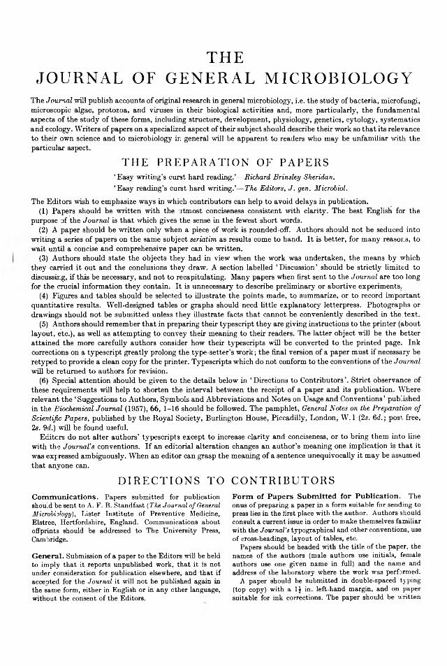
C O R R I G E N D A
In Bojalil. L . F . . Cekbon, J . & Trujillo, A . (1962). J. Gai. Microbiol. 28. 3 3 3 -8 4 6 .
0 ------- 10------ 20 ------- 30— -
Inscrt on page 335.Adansonian classification
-40-------5 0 -------60 ------- 70------- 80- -90-
Branch
Mycobacterium
Branch II
Branch
•— 100 % S
— MI. peregrinimi— M. smegmatis— M. phlei— Irregular branch
- M. piscium -M. marinum -M. fortuitum- M. flavescens- M. acapulcensis- M. thamnopheos -M. runyonii
- M. gordonae -M. marianum- M. avium
■ M. kansasiiFig. 2. Taxonomic tree of the mycobacteria. Branch I, high metabolic capacity; Branch II, intermediate metabolic activity; Branch III, low metabolic activity.
On page 342, fo r Fig. 2 read Fig. 3.

J . gen. Microbiol. (1963), 30, 1-19 Printed in Great Britain
1
Studies on Marine FlavobacteriaB y P . R . H A Y E S *
Torry Research Station, Aberdeen(Received 14 December 1961)
SUMMARYS ix ty - tw o p ig m e n te d s tr a in s , 61 o f w h ic h h a d b e e n c la ss ified a s Flavobacterium sp ec ie s b y th e w o rk e rs w h o iso la te d th e m , w e re s tu d ie d .M o rp h o lo g ica l, c u l tu ra l , e n v ir o n m e n ta l , b io c h e m ic a l a n d n u t r i t io n a l
s tu d ie s c o n firm e d t h a t 32 n o n -m o tile s t r a in s w e re Flavobacterium sp ec ie s ;21 s t r a in s w e re re c la ss if ie d a s p re s u m p t iv e Cytophaga sp ec ie s a n d th e r e m a in in g 8 s t r a in s w e re a s c r ib e d to th e g e n e ra Pseudomonas, Vibrio o r Corynebacterium, o r w e re u n c la ss if ia b le . A n e w g e n u s is su g g e s te d fo r t h e p e r i tr ic h o u s f la g e lla te d o rg a n ism s , p re v io u s ly in c lu d e d in th e g e n u s Flavobacterium. B o th th e f la v o b a c te r ia a n d th e c y to p h a g a s w e re fo u n d to h a v e m a n y p ro p e r t ie s in c o m m o n . S a ti s f a c to ry m e th o d s o f d if fe re n t ia t in g b e tw e e n r e p r e s e n ta t iv e s o f th e s e g e n e ra w e re l im i te d to th e s w a rm in g a b i l i ty a n d g r e a te r h e a t re s is ta n c e o f t h e l a t t e r . O f th e 32 n o n -m o tile Flavobacterium s t r a in s , 18 w e re d iv is ib le in to tw o w ell d e fin e d g ro u p s (? sp ec ies) a n d t h e r e m a in d e r w e re a h e te ro g e n e o u s c o lle c tio n . T h e22 Cytophaga s t r a in s w e re a lso d iv is ib le in to tw o g ro u p s (? sp ec ies). T h e p o s s ib il i ty o f a r e la t io n s h ip e x is t in g b e tw e e n f la v o b a c te r ia a n d c y to p h a g a s is d is c u s s e d b rie fly .
INTRODUCTIONT h e g e n u s Flavobacterium is o n e o f t h e m o re c o m m o n ly re p re s e n te d g e n e ra in
t h e m a r in e e n v ir o n m e n t ; f la v o b a c te r ia a r e n o rm a l ly fo u n d in th e s lim e a n d in te s t in e s o f f re s h a n d sp o ilin g fish . T h e y h a v e a lso b e e n i s o la te d f ro m s e a w a te r , m a r in e m u d s a n d se a w e e d (fo r re v ie w s se e : Z o B e ll, 1 9 46 ; S h e w a n , 1 9 4 9 ; T a r r ,1954). T h e d e f in it io n o f t h e g e n u s h a s u n d e rg o n e c o n s id e ra b le m o d if ic a t io n s in c e i t w as e re c te d in t h e f i r s t e d i t io n o f Bergey’s Manual (1923), w h e re t h e g e n u s in c lu d e d m o ti le p e r i t r ic h o u s - o r p o la r - f la g e lla te , a n d n o n -m o ti le G ra m -p o s i t iv e a n d G ra m - n e g a t iv e ro d s . T h e p o la r - f la g e lla te fo rm s w e re re m o v e d in th e f i f th e d i t io n o f Bergey’s Manual (19 39). T h e e x c lu s io n o f t h e G ra m -p o s i t iv e sp ec ie s w as s u g g e s te d b y G a ry(1950) a n d W e e k s (1955). T h e g e n u s , a s n o w d e f in e d b y W e e k s (Bergey’s Manual,1957), c o n s is ts o f G ra m -n e g a tiv e ro d s , m o ti le b y m e a n s o f p e r i t r ic h o u s f la g e lla o r n o n -m o tile , a n d p ro d u c in g y e llo w to b ro w n p ig m e n ts . H o w e v e r , th is d e f in it io n is n o t a c c e p te d b y a ll w o rk e rs . F o r e x a m p le , B r is o u , T y s s e t & V a c h e r (1959) g a v e d e ta i ls o f tw o f la v o b a c te r ia w h ic h w e re m o ti le b y m e a n s o f p o la r f la g e lla a n d F e r r a r i & Z a n n in i (1958) c o n c lu d e d t h a t t h e ex c lu s io n o f t h e G ra m -p o s i t iv e s t r a in s f r c m th e g e n u s w as n o t ju s t if ie d .
I t is a lso re c o g n iz e d t h a t c o n fu s io n m ig h t a r is e b e tw e e n t h e F la v o b a c te r iu m sp ec ies a n d c e r ta in y e l lo w -p ig m e n te d C y to p h a g e sp ec ies . T h u s S ta n ie r (1947), w o rk in g w ith c h itin -d e c o m p o s in g c y to p h a g a s , sh o w e d t h a t t h e y m a y s im u la te
* Present address: Birds Eye Foods Ltd., South Denes, Great Yarmouth, Norfolk.Vol. 29, No. 4 was issued 11 January 1963
i G. Microb. xxx
2 P. K. H ayese u b a c te r ia u n d e r c e r ta in c i rc u m s ta n c e s . H e c o n s id e re d t h a t m a n y ill-d e fin e d e u b a e te r ia l g e n e ra c o n ta in in g G ra m -n e g a tiv e ro d s w h ic h d o n o t fo rm sp o re s a n d a re n o t m o tile , m a y c o n ta in c y to p h a g a s . G ib so n (1955) iso la te d o rg a n is m s f ro m so il id e n t ic a l w i th ty p ic a l c y to p h a g a s e x c e p t in th e ir fa i lu re to sh o w g lid in g m o t i l i ty , a n d c o n firm e d t h a t t h e s e p a ra t io n o f c y to p h a g a s f ro m e u b a c te r ia w a s u n d e r su sp ic io n . I t is k n o w n t h a t C y to p h a g a sp ec ies a r e w id e ly d is t r ib u te d in m a r in e e n v iro n m e n ts (H u m m , 1 9 46 ; S t a r r & O rd a l, 1953 ; B a c h m a n n , 1 9 55 ; K a d o ta , 1956 ; V e la n k a r , 1957), th o u g h iso la t io n s f ro m fish h a v e b e e n v e r y r a r e (S h e w a n & B a in , p r iv a te c o m m u n ic a t io n ; G e o rg a la , 1957). I t seem s p o ss ib le t h a t t h e p a u c i ty o f re fe re n c e s to C y to p h a g a sp ec ies b e in g fo u n d o n fish m a y w ell b e d u e to th e u n fa v o u r a b le c o n d it io n s u n d e r w h ic h th e y h a v e b e e n s tu d ie d a n d t h a t m a n y o rg a n is m s a s c r ib e d to th e g e n u s Flavobacterium sh o u ld b e a s c r ib e d to th e g e n u s Cytophaga. T h e p u rp o s e o f t h e p re s e n t in v e s t ig a t io n w as to s tu d y m a r in e b a c te r ia w h ic h h a d b e e n id e n tif ie d a s f la v o b a c te r ia a n d d e te rm in e w h e th e r a n y h a d b e e n w ro n g ly n a m e d .
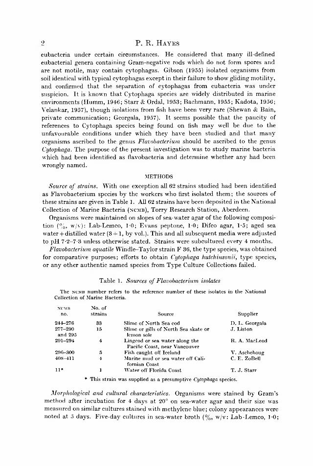
METHODSSource of strains. W i th o n e e x c e p tio n a ll 62 s t r a in s s tu d ie d h a d b e e n id e n tif ie d
a s F la v o b a c te r iu m sp ec ies b y th e w o rk e rs w h o f ir s t iso la te d th e m ; th e so u rc e s o f th e s e s t r a in s a re g iv e n in T a b le 1. A ll 62 s t r a in s h a v e b e e n d e p o s i te d in t h e N a t io n a l C o llec tio n o f M a rin e B a c te r ia (ncmb), T o r r y R e s e a rc h S ta t io n , A b e rd e e n .
O rg a n ism s w e re m a in ta in e d o n s lo p es o f s e a -w a te r a g a r o f t h e fo llo w in g c o m p o s it io n (% , w /v ) : L a b -L e m c o , 1 -0 ; E v a n s p e p to n e , 1 -0 ; D ifco a g a r , 1 -5 ; a g e d sea w a te r + d is t i l le d w a te r (3 + 1, b y v o l.) . T h is a n d a ll s u b s e q u e n t m e d ia w e re a d ju s te d to p H 7 -2 -7 -3 u n le ss o th e rw is e s ta te d . S t r a in s w e re s u b c u l tu re d e v e ry 4 m o n th s .
Flavobacterium aquatile W in d le -T a y lo r s t r a in F 36, t h e t y p e sp ec ies , w as o b ta in e d fo r c o m p a r a t iv e p u rp o s e s ; e ffo rts to o b ta in Cytophaga hutchinsonii, ty p e sp ec ies , o r a n y o th e r a u th e n t ic n a m e d sp ec ies f ro m T y p e C u ltu re C o llec tio n s fa iled .
T a b le 1. Sources of Flavobacterium isolatesThe ncmb number refers to the reference number of these isolates in the National
Collection of Marine Bacteria.ncmb No. of
no. strains Source Supplier244-276 33 Slime of North Sea cod D. L. Georgala277-290 and 295 15 Slime or gills of North Sea skate or
lemon soleJ. Liston
291-294 4 Lingcod or sea water along the Pacific Coast, near Vancouver
R. A. MacLeod296-300 5 Fish caught off Iceland V. Aschehoug408-411 4 Marine mud or sea water off Cali
fornian CoastC. E. ZoBell
11* 1 Water off Florida Coast T. J. Starr* This strain was supplied as a presumptive Cytophaga species.
Morphological and cultural characteristics. O rg a n ism s w e re s ta in e d b y G r a m ’s m e th o d a f te r in c u b a t io n fo r 4 d a y s a t 20° o n s e a -w a te r a g a r a n d t h e i r s iz e w as m e a s u re d o n s im ila r c u l tu re s s ta in e d w ith m e th y le n e b lu e ; c o lo n y a p p e a ra n c e s w ere n o te d a t 5 d a y s . F iv e -d a y c u l tu re s in s e a -w a te r b r o th (% , w /v : L a b -L e m c o , 1 -0 ;

E v a n s p e p to n e , 1 -0 ; a g e d se a w a te r + d is t i l le d w a te r , 3 + 1, b y v o l.) in c u b a te d a t 20° w e re s tu d ie d u n d e r p h a s e c o n t r a s t a n d th e a p p e a ra n c e o f 7 -d a y c u l tu re s r e c o rd e d . I s o la te s w e re e x a m in e d fo r m o t i l i ty in s e a -w a te r t r y p to n e w a te r (D ifco t r y p to n e , 1-5 % , w /v ; a g e d se a w a te r + d is t i l le d w a te r , 3 + 1, b y v o l.) . O n ly m o ti le s t r a in s w e re s ta in e d fo r f la g e lla ; t h e te c h n iq u e w as b a s e d o n a m e th o d e m p lo y in g K i r k p a t r i c k ’s f ix a t iv e a n d C a s a re s -G il’s m o d if ic a tio n o f P l im m e r & P a in e ’s f lag e lla s ta in (Staining Procedures, 1947). M o tile o rg a n is m s w e re a lso e x a m in e d w ith t h e e le c tro n m ic ro sc o p e .
Effect of environmental conditions on growthA s ta n d a r d in o c u lu m fo r a ll t e s t s w a s o n e lo o p fu l o f a 5 -d a y s e a -w a te r b r o th
c u l tu re .Temperature range of growth. T h e a b i l i ty o f t h e s t r a in s to g ro w o n s e a -w a te r a g a r
a t 37°, 30°, 25°, 20°, 10°, 5° a n d 0° w as n o te d , t h e in c u b a t io n p e r io d s e x te n d in g fro m 1 to 6 w eek s , d e p e n d in g u p o n th e t e m p e r a tu r e o f in c u b a t io n .
Heat resistance. T h e v ia b i l i ty o f c u l tu re s g ro w n in 3 m l. o f s e a -w a te r b r o th fo r5 d a y s w as in v e s t ig a te d b y h e a t in g fo r v a r io u s t im e s (15, 30 , 45, 60 m in .) a t 45° a n d 55° b e fo re p la t in g t h e h e a te d c u l tu re s o n s e a -w a te r a g a r a n d in c u b a t in g a t 20° fo r 10 d a y s . T h o s e iso la te s w h ic h fa i le d to g ro w a t 37° w e re in v e s t ig a te d fo r a b i l i ty to w i th s ta n d th is t e m p e r a tu r e b y in o c u la t in g th e m o n tw o se rie s o f s e a -w a te r a g a r p la te s . O n e s e t w a s in c u b a te d a t 37° fo r 24 h r . a n d th e o th e r fo r 48 h r . ; b o th w e re th e n in c u b a te d fo r a f u r th e r 7 d a y s a t 20°.
Anaerobic growth. O rg a n ism s w e re in c u b a te d a n a e ro b ic a l ly o n s e a -w a te r a g a r in M c In to s h & F ild e s j a r s fo r 14 d a y s a t 20°.
Salinity range of growth. T h e s t r a in s w e re p la te d o n O x o id b lo o d a g a r b a se , w i th a d d i t io n a l N a C l (A n a la r) w h e re r e q u ire d , to g iv e f in a lN a C l c o n c e n tr a t io n s (% , w /v ) o f 0-5, 2-0, 4-0, 6-0 a n d 10-0 ; t h e y w e re th e n in c u b a te d fo r 14 d a y s a t 20°.
Sensitivity to antibiotics and a ‘ vibrio static’’ compound. T h e iso la te s w e re te s te d fo r s e n s i t iv i ty to p e n ic il l in (2-5 i .u . p e r t a b le t ) , s t r e p to m y c in (80 fig.), c h lo r a m p h e n ico l (100 fig.), te r r a m y c in (10 fig.) a n d a v ib r io s ta t i c c o m p o u n d (2 ,4 -d ia m in o -6 ,7 -d i- f s o p ro p y lp te r id in e ) a s d e s c r ib e d b y S h e w a n , H o d g k is s L is to n (1954). S t a n d a r d a g a r p la te s , p r e p a re d as fo r s e a -w a te r a g a r b u t w i th d is t i l le d w a te r + 0-5 % (w /v ) N aC l w e re u se d , s in c e s e n s i t iv i ty to a n t ib io t ic s is m a r k e d ly d if fe re n t o n s e a -w a te r a g a r , t h e h ig h e r s a l t c o n c e n t r a t io n e i th e r m a s k in g th e in h ib i to r y e ffec ts o f t h e a n t ib io t ic s o r s t im u la t in g b a c te r ia l g ro w th .
Physiological testsT h e s a m e s ta n d a r d in o c u lu m w as u s e d a s p re v io u s ly . A ll t h e m e d ia w e re in
c u b a te d a t 20°.Gelatin liquefaction. N u t r ie n t g e la t in m e d iu m (% , w /v : L a b -L e m c o , 1 -0 ; E v a n s
p e p to n e , 1 -0 ; N aC l, 2 -0 : O x o id g e la t in , 12-0) w a s in o c u la te d a n d in c u b a te d fo r6 w eek s , t h e a p p e a ra n c e a n d e x te n t o f l iq u e fa c t io n b e in g n o te d a t r e g u la r in te rv a ls .
Action on litmus milk. T h e c u l tu re s w e re in c u b a te d in l i tm u s m ilk fo r 6 w eek s ,c h a n g e s in a p p e a ra n c e b e in g n o te d a t r e g u la r in te rv a ls .
Reduction of nitrate to nitrite. N i t r i t e w as t e s te d fo r b y t h e G r ie s s - I lo s v a y re a g e n ts a f te r in c u b a t io n fo r 10 d a y s in n i t r a t e b r o th (% , w /v : L a b -L e m c o , 1-0;
Marine flavobacteria 3

E v a n s p e p to n e , 1 0 ; N aC l, 2 -0 ; K N 0 3, 0-1). P o w d e re d z in c w as u s e d to t e s t fo r fa lse n e g a tiv e s .
Ammonia production w as t e s te d fo r b y N e s s le r ’s r e a g e n t a f te r in c u b a t io n fo r 10 d a y s in s e a -w a te r p e p tc n e w a te r (E v a n s p e p to n e , 1 -0 % , w /v ; a g e d s e a w a te r + d is t i l le d w a te r , 3 + 1, b y v o l.) .
Hydrogen sulphide formation w as d e te c te d w ith s tr ip s o f f i lte r p a p e r im p re g n a te d w i th le a d a c e ta te o n c u l tu re s in c u b a te d fo r 14 d a y s in s e a -w a te r b r o th c o n ta in in g0 -0 1 % (w /v ) c y s te in e h y d ro c h lo r id e (fin a l p H 7-6).
Indole formation w as d e te rm in e d b y E h r l i c h ’s r e a g e n t a f te r x y le n e e x t r a c t io n o f c u l tu re s in c u b a te d fo r 10 d a y s in s e a -w a te r t r v p to n e w a te r .
Hydrolysis of urea. C u ltu re s w e re in c u b a te d fo r 14 d a y s o n s lo p es o f C h r is te n se n a g a r (1946), m o d ifie d b y t h e in c lu s io n o f (% , w /v ) : K H 2P 0 4, 0 -0 5 ; K 2H P 0 4, 0 -05 ; O x o id y e a s t e x t r a c t , 0 -01 ; b r o m th y m o l b lu e , 0 -003; a g e d se a w a te r + d is t i l le d w a te r (3 + 1, b y v o l.) . T o e n s u re t h a t t h e a lk a l in i ty p ro d u c e d w a s d u e t o u re a h y d ro ly s is a c o n tr o l m e d iu m w i th o u t u re a w as in o c u la te d w i th t h e u re a s e -p o s it iv e s t r a in s . O n ly th o s e s t r a in s w h ic h g a v e a m a rk e d ly m o re a lk a l in e r e a c t io n in th e u r e a m e d iu m w e re c o n s id e re d to b e t r u e p o s it iv e s .
Trimethylamine oxide reduction. C u ltu re s w e re in c u b a te d fo r 14 d a y s in W o o d & B a i r d ’s m e d iu m (1943) m o d ifie d b y th e u se o f a g e d se a w a te r + d is t i l le d w a te r (3 + 1, b y v o l.) . T h e p re se n c e o f t r im e th y la m in e w a s th e n te s te d fo r a c c o rd in g to t h e m e th o d g iv e n in Topley and Wilson’s Principles (1955).
Production of acid from carbohydrate. C u ltu re s w e re in c u b a te d fo r 28 d a y s in a m e d iu m c o m p o se d o f : E v a n s p e p to n e , 0 -5 % (w /v ) ; N aC l, 2 -0 % (w /v ) ; A n d r a d e ’s in d ic a to r , 1 -0 % (v /v ) ; t e s t c a rb o h y d ra te , 1 -0 % (w /v ). E a c h c u l tu re w a s e x a m in e d re g u la r ly fo r a c id a n d g a s p ro d u c tio n . T h e fo llo w in g c a rb o h y d ra te s w e re u s e d : a ra b in o se , x y lo se , rh a m n o s e , g lu co se , f ru c to s e , m a n n o s e , g a la c to s e , su c ro se , m a lto s e , la c to se , t r e h a lo s e , ra ff in o se , s ta r c h , in u lin , m a n n i to l , d u lc ito l, s o rb ito l , in o s ito l , sa lic in .
Glucose utilization. T h e m e th o d o f H u g h & L e ifso n (1953) w as u s e d to d e te rm in e w h e th e r g lu co se w as u s e d o x id a t iv e ly o r f e rm e n ta t iv e ly .
Production of 2-ketogluconic acid. C u ltu re s w e re in c u b a te d fo r 7 d a y s in P a t o n ’s m e d iu m (1959). A ll t h e s t r a in s w h ic h g rew in th is m e d iu m w e re e x a m in e d fo r2 -k e to g lu c o n ic a c id p r o d u c t io n b y u s in g th e r e a g e n t o f H o u g h , J o n e s & W a d m a n(1950).
Starch hydrolysis. A f te r in c u b a t io n o f t h e s t r a in s in t h e s ta r c h m e d iu m fo r 28 d a y s (see Production of acid from carbohydrates) th o s e c u l tu re s w h ic h sh o w e d n o a c id p r o d u c t io n w e re t e s te d w k h a d ro p o f io d in e s o lu t io n fo r t h e p re s e n c e o f s ta r c h b re a k d o w n p ro d u c ts .
Tributyrin hydrolysis. C u ltu re s w e re in c u b a te d a n d e x a m in e d o n O x o id t r ib u t y r in a g a r fo r 14 d a y s .
Growth in Koser’s citrate medium w a s r e a d a f te r in c u b a t io n fo r 7 d a y s ; s t r a in s w h ic h sh o w e d g ro w th a f t e r tw o s e r ia l s u b c u l tu re s w e re re c o rd e d a s p o s i t iv e .
Voges-Proskauer and methyl red tests. C u ltu re s w e re t e s te d a f te r in c u b a t io n fo r 10 d a y s in g lu co se p h o s p h a te b r o th b y B a r r i t t ’s m o d if ic a t io n fo r a c e ty lm e th y l- c a rb in o l {Topley and Wilson’s Principles, 1955).
Digestion of chitin. C h it in , p r e p a re d f ro m c ra b sh e lls b y th e m e th o d o f B e n to n (1935), w as in c o r p o ra te d in t h e fo llo w in g m e d iu m (% , w /v ) : c h i t in , 3 -5 ; g lu co se ,
4 P. R. H ayes
0 -1 ; E v a n s p e p to n e , 0 -1 ; K 2H P 0 4, 0 -005 ; D ifc o a g a r , 1-5; a g e d se a w a te r + d is t i l le d w a te r (3 + 1 , b y v o l.) . C u ltu re s w e re in c u b a te d fo r 6 w eek s a n d e x a m in e d a t r e g u la r in te rv a ls .
Decomposition of cellulose. C u ltu re s w e re in c u b a te d fo r 6 w eek s o n a m e d iu m c o n s is t in g o f (% , w /v ) : W h a tm a n p o w d e re d ce llu lo se , 5 -0 ; O x o id y e a s t e x t r a c t ,0 -02 ; E v a n s p e p to n e , 0 -2 ; K H „ P 0 4, 0 -1 ; F e P 0 4.2 H 20 , t r a c e ; D ifc o a g a r , 1-5; a g e d se a w a te r + d is t i l le d w a te r (3 + 1 , b y v o l.) . T h e p la te s w e re e x a m in e d a t r e g u la r in te rv a ls .
Blood agar. T h e c u l tu re s w e re in c u b a te d o n h o rse -b lo o d a g a r fo r 14 d a y s a n d e x a m in e d r e g u la r ly fo r g ro w th a n d h a e m o ly s is .
Actomyosin agar. A c to m y o s in , p re p a re d f ro m fish m u sc le b y th e m e th o d o f C o n n e ll (1958), w as in c o r p o ra te d in n o rm a l s e a -w a te r a g a r a t 1-0 % (w /v ), g iv in g a n o p a q u e m e d iu m . T h e c u l tu re s w e re in c u b a te d fo r 6 w eek s a n d e x a m in e d r e g u la r ly fo r a c to m y o s in b re a k d o w n as in d ic a te d b y c le a r zo n es .
Catalase and oxidase formation. C a ta la s e w as t e s te d fo r b y a d d in g h y d ro g e n p e ro x id e (20 v o l.) to 3 -d a y c u l tu re s o n s e a -w a te r a g a r ; fo r o x id a se fo rm a tio n th e m e th o d o f Iv o v acs (1956) w as u s e d o n s im ila r c u l tu re s .
NutritionPreparation and cultivation of inoculum. T h e s ta n d a r d in o c u lu m fo r a ll t e s t s w as
o n e lo o p fu l o f a 5 -d a y s e a -w a te r b r o th c u l tu re . M e d ia w e re in c u b a te d fo r 7 d a y s a t 2 0 c a n d o n ly th o s e s t r a in s w h ic h sh o w e d v is ib le g ro w th a f te r tw o se ria l s u b c u l tu re s w e re re c o rd e d a s p o s i t iv e .
Media preparation. T h e c o m p o s itio n s o f t h e m e d ia u s e d a re g iv e n in T a b le 2. A ll m e d ia w e re a d ju s te d to p H 7-2- 7-3 a n d s te r i l iz e d b y i n t e r m i t t e n t s te a m in g .
SwarmingA p re l im in a ry s tu d y w ith d if fe re n t c o n c e n t r a t io n s o f a g a r a n d p e p to n e in d ic a te d
t h a t s w a rm in g w as m o s t m a r k e d ly d e p e n d e n t o n t h e p e p to n e c o n c e n t r a t io n w ith 2 % (w /v ) a g a r . T h e re fo re th e b a s a l m e d iu m c o n s is te d o f (% , w /v ) : L a b -L e m c o ,0 -1 ; D ifc o a g a r , 2 -0 ; a g e d se a w a te r + d is t i l le d w a te r (3 + 1, b y v o l.) . T o th is w e re a d d e d d if fe re n t a m o u n ts o f E v a n s p e p to n e a s fo llo w s (% , w /v ) : 8-0, 4-0, 1-0, 0-25,0-05 a n d 0-01. T h e L a b -L e m c o w as in c lu d e d t o s t im u la te g ro w th ; w i th o u t i t co lo n ies w e re so m in u te o n t h e lo w e r p e p to n e c o n c e n t r a t io n s a s to b e im p o ss ib le to b e e x a m in e d v is u a l ly fo r s w a rm in g . T h e L a b -L e m c o a p p e a re d to e x e r t n o in h ib i t in g e ffe c t o n sw a rm in g . A f te r t h e p la te s h a d b e e n p o u r e d w i th th e s e m e d ia th e y w e re d r ie d fo r 30 m in . a t 55° a n d th e n s to re d o v e rn ig h t a t 20°, b e fo re in o c u la t io n w ith t h e s ta n d a r d in o c u lu m . T h e p la te s w e re in c u b a te d a t 20° fo r 10 d a y s w h e n c o lo n y a p p e a ra n c e a n d a n y in d ic a t io n o f s w a rm in g w as n o te d . Flavobacterium aquatile F 36 w as p la te d o n d is t i l le d w a te r m e d ia c o n ta in in g 0-5 % (w /v ) N aC l a n d th e lo w e s t th r e e p e p to n e c o n c e n tra t io n s .
Marine flavobacteria 5

6 P. R. H ayes
Cell-wall studiesStaining of cell walls. C ell w a lls w e re s ta in e d b y u s in g th e p h o s p h o m o ly b d ic
a c id m o r d a n t te c h n iq u e re c o m m e n d e d b y H a le (1953).Cell-wall analysis. S t r a in s n c m b 244, 249 , 251 a n d 253 w e re t a k e n as r e p r e s e n ta
t iv e s o f G ro u p s 1 -4 (see R e s u l ts ) . Flavobacterium aquatile F 36 a n d s t r a in n c m b 11 w ere u s e d as c o n tro ls fo r t h e g e n e ra Flavobacterium a n d Cytophaga, re s p e c tiv e ly .
T a b le 2. Composition of media No. medium
1 2 3 4 5 6Amino Growth Growth Purine+
Basal acid factor factor pyrimidine Peptonemedium medium medium medium medium medium*
Basal constituents (g./l.) Glucose 5 + + + + + +NaCl 25 + + + + + +k 2h p o 4 1 + + + + + +MgS04.7H20 1 + + + + + +(NH4)2S04 2 + + + + + +CaCl2 (anliyd.) trace + + + + + +FeCl3.6HaO trace
Amino acids, each at+ + + + + +
100 mg./l. : glycine, dl- alanine, DL-serine, L-cysteine, DL-threonine, DL-methionine, L-valine, Di,-leucine, DL-phenyl- alanine, DL-tyrosine, dl- tryptophan, L-histidine HC1, m.-ornithine HBr, L-arginine, DL-glutamic acid, DL-aspartic acid,
' - + + + + +
DL-prolineGrowth factors (/¿g./l.) p-aminobenzoic acid (100), nicotinic acid (500), pantothenic acid (500), pyri- doxine HC1 (200), thiamine HC1 (500), riboflavin (100)
_ - + + + +
Folic acid (1), biotin (2), /?-alanine (500), vitamin B,2 (0-5) - - + + +
Purines + pyrimidines,each at 200 mg./l. uracil, adenine, guanine, xanthine - " + +
Evans peptone (1 g./l.) - - - - - +* Filtered sea water was added to the peptone medium (6), in the ratio medium (6): sea
water (3:1), giving an additional medium (7).+ , Constituent(s) included in medium. —, Constituent(s) absent.
Culture media. T h e s a m e m e th o d w as u se d fo r a l l iso la te s e x c e p t t h a t F. aquatile w a s g ro w n in s ta n d a r d n u t r i e n t b r o th w h e re a s th e m a r in e b a c te r ia w e re g ro w n in s e a -w a te r b ro th . T h re e h u n d re d m l. a m o u n ts o f m e d ia w ere u s e d in 1 1. c o n ic a l f la sk s. A f te r in o c u la t io n , t h e flask s w e re s h a k e n fo r 3 d a y s o n a T o w n s o n a n d M e rc e r

‘l a t e r a l ’ s h a k e r a t a sp e e d o f 8 0 -9 0 th r o w s /m in ., t h e in c u b a t io n t e m p e r a tu r e b e in g20 ° .
Preparation and hydrolysis of cell-wall suspensions. T h e te c h n iq u e u s e d w as t h a t o f C u m m in s & H a r r i s (1956).
Chromatography. F o r a m in o a c id s a n d h e x o sa m in e s , tw o -d im e n s io n a l a s c e n d in g c h ro m a to g ra m s o n W h a tm a n N o . 1 p a p e r w e re u se d . L ess ce ll-w a ll m a te r ia l w as a v a i la b le w i th s t r a in s n c m b 244 a n d 249 b e c a u s e o f t h e c o m p a r a t iv e d if f ic u lty in c e n tr ifu g in g th e m ; th is e ffe c t w as f r e q u e n t ly n o tic e d w h e n c e n tr i fu g in g m a r in e o rg a n is m s g ro w n in s e a -w a te r b a s e d m e d ia . P h e n o l + w a te r (80 + 20, b y v o l.) in a n a m m o n ia c a l a tm o s p h e re w as u s e d a s th e f i r s t s o lv e n t a n d lu t id in e + c o llid in e + w a te r (150 + 100 + 120, b y v o l.) a s t h e s e co n d . T h e s p o ts w e re lo c a te d b y s p ra y in g w ith n in h y d r in s o lu t io n .
S u g a rs w e re d e te c te d b y d e s c e n d in g c h ro m a to g ra p h y o n W h a tm a n N o . 1 p a p e r w i th e th y l a c e t a t e + p y r id in e + w a te r (120 + 5 0 + 4 0 , b y v o l.) a s s o lv e n t a n d a n ilin e h y d r o g e n p h th a la t e (P a r t r id g e , 1949) a s lo c a t in g a g e n t ; b e c a u s e o f s im ila r RF v a lu e s a r a b in o s e a n d m a n n o s e w e re d is t in g u is h e d b y p la c in g t h e c h ro m a to g ra m u n d e r th e u l t r a v io le t l ig h t , w h e n th e a ra b in o s e flu o re sc e d a n d L ie m a n n o s e a p p e a re d a s a b ro w n s p o t .
Marine flavobacteria 7
RESULTSS ix ty o f t h e s t r a in s s tu d ie d w e re G ra m -n e g a tiv e , o f w h ic h 54 w e re n o n -m o tile
a n d 6 m o ti le ; t h e re m a in in g 2 s t r a in s w e re G ra m -p o s i t iv e a n d n o n -m o tile . A n a n a ly s is o f t h e re s u l ts o b ta in e d w i th t h e 54 n o n - m o t lc G ra m -n e g a tiv e b a c te r ia c le a r ly s u g g e s ts t h a t m a n y o f th e s e o rg a n is m s c o u ld b e p la c e d in o n e o f t h e fo llo w in g 4 m a in g ro u p s .
Group 1. U s u a lly s m a ll s le n d e r ro d s p ro d u c in g p a le y e llo w o r y e llo w -g re e n p ig m e n te d co lo n ies o n a g a r . G ro w th ra n g e 0 ° -3 0 ° . R e s i s t a n t to 45° fo r 1 h r . a n d to 37° fo r 48 h r . M a rk e d ly s e n s it iv e to p e n ic il l in a n d to t l ie ‘ v ib r io s ta t ic ’ c o m p o u n d . G e la t in l iq u e f ie d s lo w ly , i f a t a l l ; l i tm u s m ilk u n c h a n g e d . A m m o n ia n o t fo rm e d f ro m p e p to n e . C a rb o h y d ra te s a t t a c k e d w ith p r o d u c t io n o f a c id ; la c to se a n d s ta r c h n o t a t t a c k e d . G e n e ra l ly c o m p le x n u t r i t io n a l r e q u ire m e n ts , fa il in g to g ro w o n d e fin e d m e d ia . T h is g ro u p is c o m p o se d o f s t r a in s n c m b 2 4 4 -6 , 248 , 252, 255, 2 5 8 -9 , 261 , 263 , 273, 281, 295 a n d 298.
Group 2. U s u a lly s h o r t , o v a l ro d s b u t so m e p le o m o rp h ic . D e e p y e llo w o r o ra n g e - y e llo w p ig m e n te d co lo n ies . N o g ro w th a t 30°. N o t r e s i s ta n t to 45° fo r 1 h r . o r to 37° fo r 24 h r . G e la t in l iq u e fie d . L i tm u s m ilk r e d u c e d , w i th p e p to n iz a t io n (n c m b 267, 2 7 0 -1 ) o r w i th a n a lk a l in e r e a c t io n (n c m b 260, 252, 265 , 269 , 284). C a rb o h y d r a te s n o t a t t a c k e d a n d s ta r c h n o t h y d ro ly s e d . C o m p lex n u t r i t io n a l r e q u ir e m e n ts bu 3 g ro w o n d e f in e d m e d ia . N o g ro w th o n b lo o d a g a r . T h is g ro u p is c o m p o se d o f s t r a in s n c m b 2 5 0 -1 , 260, 262, 2 6 4 -5 , 2 6 7 -7 2 , 276, 284 a n d 289.
Group 3. S h o r t o v a l o r p le o m o rp h ic ro d s . D e e p y e llo w o r o ra n g e -y e llo w p ig m e n te d co lo n ies . N o g ro w th a t 30°. N o t r e s i s ta n t to 45° fo r 1 h r . b u t v ia b le a f te r 48 h r . a t 37°. G e la t in n o t l iq u e fie d . L i tm u s m ilk re d u c e d . M o st c a rb o h y d ra te s n o t a t t a c k e d , b u t s ta r c h h y d ro ly s e d . N u tr i t io n a l r e q u ir e m e n ts n o t c o m p le x a n d g ro w th fa c to rs n o t r e q u ire d . T h is g ro u p is c o m p o se d o f s t r a in s n c m b 249, 254 a n d 287 .
Group 4. L o n g s le n d e r ro d s , p le o m o rp h ic . P a le y e llo w o r y e llo w -g re e n p ig m e n te d co lon ies . N o t r e s i s ta n t to 45° fo r 1 h r . b u t v ia b le a f te r 48 h r . a t 37°. L itm u s m ilk

8 P. R. H a y e sreduced. Carbohydrates attacked, including lactose and starch, with production of acid. Complex nutritional requirements but will grow on defined media. This group is composed of strains ncmb 247, 253 , 2 5 6 -7 , 275 and 285.
T h ir ty - e ig h t iso la te s a r e in c lu d e d in th e 4 g ro u p s a b o v e . O f th e re m a in d e r , s t r a in ncmb 11 is c o n firm e d a s a C y to p h a g a sp ec ies a n d s t r a in ncmb 292 is a lso c o n s id e re d to b e a ty p ic a l c y to p h a g a . B o th th e s e o rg a n is m s sw a rm e d r e a d i ly o n n o rm a l m e d ia a n d , in a d d it io n , h a d th e fo llo w in g p ro p e r t ie s in c o m m o n w i th th e g e n u s C y to p h a g a (B e r g e y ’s M a n u a l , 1 957): (1) m o rp h o lo g y , b e in g lo n g , s le n d e r ro d s w i th a x e s s t r a ig h t , b e n t , U- o r S -sh a p e d ; (2) a b se n c e o f m ic ro c y s ts ; (3) y e llo w p ig m e n te d s p re a d in g co lo n ies ty p ic a l o f c y to p h a g a s . O th e r p ro p e r t ie s th e s e o rg a n ism s h a d in c o m m o n w e re : (4) o x id a s e -p o s it iv e ; (5) l iq u e fa c tio n o f g e la t in ; (6) re s is ta n c e to 45° fo r 1 h r . ; (7) v ia b i l i ty a f te r 48 h r . a t 37°. N e i th e r is d e s c r ib e d in th e g e n u s C y to p h a g a in B e r g e y ’s M a n u a l (1957).
The remaining 14 Gram-negative non-motile strains cannot be justifiably placed in any of the above groups. An analysis of their properties suggests that further small groups may be formed but these groups would be less rigid in their definition than those already described. Three strains, ncmb 282, 291 and 408, appear to merit individual species rank, each being markedly different from any other organisms.
The 6 Gram-negative motile bacteria formed a heterogeneous collection as follows.
(1) S t r a in ncmb 283 is c o n s id e re d to b e a V ib r io sp ec ies b e c a u s e i t : (a ) is m o t i le w i th a s in g le p o la r f la g e llu m ; (b) p ro d u c e s a c id in g lu c o se ; (c) is f e r m e n ta t iv e in H u g h a n d L e ifs o n ’s m e d iu m ; (d ) is s e n s it iv e to th e v ib r io s ta t ic c o m p o u n d .
(2) S t r a in ncmb 286 is c o n s id e re d to b e a p ig m e n te d P s e u d o m o n a s spec ies , fa i l in g to p ro d u c e a c id f ro m c a rb o h y d ra te s , b e c a u s e i t : (a ) is m o ti le w i th p o la r f la g e lla ; (b ) re d u c e s n i t r a t e to a m m o n ia o r n i t ro g e n ; (c) is o x id a s e -p o s it iv e ; (d ) r e d u c e s t r im e th y la m in e o x id e . H o w e v e r , i t is p e n ic il l in -s e n s i tiv e , a r a r e p r o p e r ty in t h e g e n u s .
(3) S t r a in ncmb 294 is c o n s id e re d l ik e ly to b e a p ig m e n te d P s e u d o m o n a s sp ec ies , fa i l in g to p ro d u c e a c id f ro m c a rb o h y d ra te s , b e c a u s e i t is : (a) m o ti le w i th p o la r f la g e lla ; (b) o x id a s e -p o s it iv e ; (c) in s e n s i t iv e to p e n ic illin .
(4) S t r a in ncmb 297 is a p in k -p ig m e n te d p e r i tr ic h o u s ly f la g e lla te o rg a n is m p ro d u c in g a c id f ro m s e v e ra l c a rb o h y d ra te s in c lu d in g g lu co se a n d la c to s e ; i t c a n n o t b e s a t is f a c to r i ly a s c r ib e d to a n y g en u s .
(5) S tr a in s ncmb 296 a n d 300 a re g ro u p e d to g e th e r b e c a u s e th e y h a v e m a n y p ro p e r t ie s in c o m m o n in c lu d in g : (a ) y e llo w -g re e n p ig m e n te d co lo n ie s ; (b) p e r i t r i - c h o u s f la g e lla ; (c) re s is ta n c e to 45° fo r 1 h r . ; (d) s tr o n g h y d ro g e n su lp h id e p r o d u c t io n ; (e) s t r o n g ly u re a s e -p o s i t iv e ; ( / ) r e d u c t io n o f n i t r a t e to n i t r i t e ; (g ) n o l iq u e fa c t io n o f g e la t in ; (h ) g ro w th in b a s a l m e d iu m ; ( i) p ro d u c tio n o f 2 -k e to g lu c o n ic a c id ; ( j ) h y d ro ly s is o f t r ib u ty r in . A c c o rd in g to B e r g e y ’s M a n u a l (1957) th e y w o u ld b e id e n tif ie d a s F la v o b a c te r iu m sp ec ie s b u t th e y a re n o t s im ila r to a n y sp ec ie s l is te d .
The Gram-positive strains, ncmb 280 and 299, are from their morphology, colony appearance and biochemical reactions, typical Corynebacterium species.
The characters distinguishing many of the strains have been mentioned already; detailed properties of individual strains are omitted for brevity but the principal results are summarized below.

Marine flavobacteria 9M o r p h o lo g ic a l a n d c u l tu r a l c h a ra c te r is t ic s
G r a m r e a c tio n . W ith t h e e x c e p t io n o f th e tw o c o ry n e b a c te r ia , a ll o rg a n is m s w e re G ra m -n e g a tiv e ro d s v a r y in g f ro m lo n g s le n d e r fo rm s ( le n g th , 5 -0y; w id th , 0 -4 /i) to s h o r t s to u t ‘ co cco b ac illi ’ ( le n g th , 1-0/*; w id th , 0 -6 /i).
M o r p h o lo g y u n d e r p h a s e c o n tra s t. T h e G ra m -n e g a tiv e b a c te r ia a g a in v a r ie d f ro m lo n g s le n d e r ro d s to s h o r t s to u t ro d s , th o u g h th e r e w as a m a rk e d te n d e n c y fo r lo n g e r a n d m o re s le n d e r ro d s to d e v e lo p in l iq u id m e d ia t h a n o n so lid m e d ia .
M o t i l i t y a n d f la g e l la s ta in in g . O f th e 6 m o ti le b a c te r ia , p e r i t r ic h o u s f la g e lla w e re d e m o n s t r a te d fo r 3 s t r a in s a n d p o la r f la g e lla (1 -2 /ro d ) fo r 3 (see ab o v e ).
C o lo n y c h a ra c te r is t ic s . C o lo n y p ig m e n ta t io n o f t h e G ra m -n e g a tiv e n o n -m o tile b a c te r ia r a n g e d f ro m d e e p y e llo w o r o ra n g e -y e llo w to p a le y e llo w o r y e llo w -g re e n . T h e b a c te r ia c o m p r is in g e a c h o f t h e 4 m a in g ro u p s a b o v e h a d s im ila r ly p ig m e n te d co lon ies . T h e ty p ic a l a p p e a ra n c e o f co lo n ies a t 5 d a y s w a s : y e llo w p ig m e n ta t io n ,1 -5 -0 -5 m m . in d ia m e te r , c irc u la r , c o n v e x , su r fa c e s m o o th a n d s h in y , e n t i r e ed g e , t r a n s lu c e n t b y t r a n s m i t t e d l ig h t . T h e c y to p h a g a , ncmb 292, w as a n a g a r l iq u e fie r .
A p p e a r a n c e in s e a -w a te r b ro th . A lm o s t a ll iso la te s g re w s lo w ly in s e a -w a te r b r o th , v is ib le t u r b i d i ty a p p e a r in g in 2 o r 3 d a y s . N e a r ly a ll iso la te s g a v e a m o d e ra te d e g re e o f t u r b i d i ty th r o u g h o u t th e m e d iu m ; th e p re s e n c e o r a b se n c e o f s u rfa c e g ro w th a n d d e p o s i ts w e re v a r ia b le c h a ra c te r s .
E ffe c t o f e n v ir o n m e n ta l c o n d i t io n s o n g ro w thT e m p e r a tu r e r a n g e o f g r o w th . F o r ty - th r e e iso la te s g rew in e i th e r t h e 0° to 30°,
o r 0° to 25° t e m p e r a tu r e ra n g e s ; o n ly 9 d id n o t g ro w b e lo w 10°. O n ly s t r a in s ncmb 300 a n d 411, t h e c y to p h a g a ncmb 11, a n d th e c o ry n e b a c te r iu m ncmb 280, g ro w a t 37°.
H e a t r e s is ta n c e . T h ir ty - n in e iso la te s , in c lu d in g a ll m e m b e rs o f g ro u p s 2, 3 a n d 4, w e re n o t r e s i s ta n t to 45° fo r 1 h r . , a n d o f th e se , 28 w e re u n a b le to w i th s ta n d 45° fo r 30 m in . I n f a c t 13 iso la te s , m a in ly m e m b e rs o f g ro u p 2, w e re n o t r e s i s ta n t to 45° fo r 15 m in . O f th e re m a in in g 23 o rg a n is m s o n ly tw o (ncmb 4 1 1 ; th e c o ry n e b a c te r iu m ncmb 280) w e re r e s i s ta n t to 55° fo r 30 m in . O f th e 58 o rg a n is m s e x a m in e d , 36 g re w n o rm a l ly a f te r 24 h r . a t 37°, th is n u m b e r d e c re a s in g to 27 a f te r 48 h r . N o t u n e x p e c te d ly th o s e s tr a in s w h ic h sh o w e d lo ss o f v ia b i l i ty a f te r 24 h r . a t 37° w e re l e a s t r e s i s ta n t to h ig h e r t e m p e r a tu r e s a n d th o s e w h ic h s u rv iv e d a f te r 48 h r . a t 37 ° w e re a l l a b le to w i th s ta n d 45° fo r 1 h r .
A n a e r o b ic g r o w th . O n ly fo u r o rg a n ism s , ncmb 263, 268 , 279 a n d th e v ib r io ncmb 283, g re w a n a e ro b ic a l ly , p ro d u c in g o n ly s l ig h t g ro w th .
S a l i n i t y r a n g e o f g r o w th . F if ty -o n e s t r a in s g re w in 6 % (w /v ) N aC l a n d 6 o f th e s e (ncmb 289, 296, 299, 409, t h e c o ry n e b a c te r iu m ncmb 299, a n d th e u n c la ss if ie d s t r a in ncmb 297) g re w in 1 0 % (w /v ) N a C l; w i th th e m a jo r i ty g ro w th w as b e t te r o n th e lo w e r c o n c e n t r a t io n s b u t a t 6 % N a C l th e g ro w th w as u s u a lly m o re t h a n s c a n ty . N in e s t r a in s d id n o t g ro w a t a c o n c e n t r a t io n g r e a te r t h a n 4 % (w /v ) N a C l a n d tw o (ncmb 291, 408) o n ly to le r a te d 2 % (w /v ) N aC l. A n u m b e r o f iso la te s ncmb 266, 279, 285 , 289 , 290, 2 9 3 ; t h e c y to p h a g a s ncmb 11, 292) sh o w e d a m a rk e d d e c re a se o f g ro w th a t t h e lo w e s t c o n c e n t r a t io n (0 -5 % , w /v , N aC l).
S e n s i t i v i t y to a n t ib io t ic s a n d a ‘ v ib r io s ta t ic ’ c o m p o u n d . F if ty - s e v e n o rg a n is m s

10were markedly sensitive to chloramphenicol (radius of inhibited growth > 10 mm.), the exceptions (ncmb 262, 273 , 411, the vibrio ncmb 283 and the unclassified strain ncmb 297) being sensitive to a lesser degree. Fifty-four isolates were sensitive to streptomycin and 36 to penicillin; the organisms comprising groups 1 and 3 were almost all markedly sensitive to penicillin. Twenty-one isolates were sensitive to the vibriostatic compound, 14 being group 1 strains, but these isolates were non-motile and would not be considered as Vibrio species (B e r g e y ’s M a n u a l ,1957).
P h y s io lo g ic a l f e a tu r e sG e la tin liq u e fa c tio n . Forty-two (6 7 % ) isolates were slow gelatin liquefiers, the
reactions ranging from complete liquefaction in 14 days to slight liquefaction in 6 weeks. With one exception the liquefaction was saccate or infundibuliform; the exception, strain ncmb 292, was stratiform. Seventeen strains did not liquefy gelatin and three did not grow under the test conditions (see Table 3). These results confirm those of Gary (1950 ; who, in a study of 30 non-marine flavobacteria, found that 83 % liquefied gelatin. An analysis of the properties of those Flavobacterium species listed in B e r g e y ’s M a n u a l (1957), which had been isolated from marine sources (‘marine flavobacteria’) showed 88% to be gelatin liquefiers. Ferrari & Zannini (1958), with 46 strains considered by them to be flavobacteria, found that 17 % liquefied gelatin. Several representative cultures received from these authors had a morphology and colony appearance typical of Corynebacterium species (e.g. ncmb 644, 667). It seems almost certain therefore that they were studying a heterogeneous collection of bacteria and for this reason their results will not be discussed further.
A c t io n o n l i tm u s m i lk . F o r ty - tw o (67 % ) s t r a in s p ro d u c e d a v a r i e ty o f c h a n g e s in l i tm u s m ilk ; in n o c a se w e re th e s e r a p id . T h e m o s t c o m m o n a c t io n w a s th e r e d u c t io n o f l i tm u s f r e q u e n t ly a s s o c ia te d w i th e i th e r p e p to n iz a t io n o r a n a c id o r a lk a l in e r e a c t io n ; 8 s t r a in s p ro d u c e d a n a lk a l in e r e a c t io n w i th n o o th e r c h a n g e . W ith th e e x c e p tio n o f g ro u p 1 s t r a in s , w h ic h fa i le d to c h a n g e th e m e d iu m , g ro u p s 2, 3 a n d 4 p ro d u c e d n o c h a ra c te r is t ic r e a c t io n s (see T a b le 3 ). O f th e b a c te r ia e x a m in e d b y G a ry (1950) 4 9 % p ro d u c e d c h a n g e s in l i tm u s m ilk a n d 5 6 % o f th e m a r in e f la v o b a c te r ia in B e r g e y ’s M a n u a l (1957) a re l is te d a s p ro d u c in g ch a n g e s .
R e d u c t io n o f n i tr a te to n i t r i te . Only 5 isolates (ncmb 280, 283, 286, 2 96 , 300) reduced nitrate to nitrite, strain ncmb 286 continuing the reduction to ammonia or nitrogen gas. It should be noted that only two of these isolates are flavobacteria and these are motile (see Discussion). Gary (1950) found only 1 3 % of his fiavo- bacteria to be nitrate reducers, whereas 47 % of the marine flavobacteria in B e r g e y 's M a n u a l (1957) are listed as nitrate-reducing species.
A m m o n ia p r o d u c t io n . Forty-two (6 7 % ) organisms gave a positive ammonia reaction but only 6, including the group 3 isolates, were strongly positive (see Table 3). Of the marine flavobacteria in B e r g e y ’s M a n u a l (1957), 75 % are given as positive.
H y d r o g e n s u lp h id e f o r m a t io n . F i f ty - tw o (84 % ) o f t h e iso la te s p ro d u c e d h y d ro g e n s u lp h id e (see T a b le 3). T h e s e re s u l ts c o n tr a d ic t th o s e o f G a ry (1950), w h o fo u n d t h a t n o n e o f h is f la v o b a c te r ia p ro d u c e d h y d ro g e n s u lp h id e , a n d th o s e o f B e r g e y ’s M a n u a l(1957) w h ic h l is ts 3 3 % o f th e m a r in e f la v o b a c te r ia a s p o s it iv e .
I n d o le f o r m a t io n . None of the organisms gave an indole-positive reaction. All
P . R . H a y e s

Table
3.
Bioc
hemi
cal r
eacti
ons o
f the
Gra
m-ne
gativ
e non
-moti
le iso
lates
I +u p .2 S, Ad U « Y , . V+? I I + +
! | S184 33
23323.
.as+ + 1 +s
.a0 s
•Si i i +
1 I i i i +
§ ' 1 »
I 8 ■ a 32 + 1 I +
2 8 i o ! I
+ + +I I I !+ - + + +- + ft + I + ft ft ft ft I ft ft ft
+ + +| + + I I + + + + + - I I 1 +
W| + + + + + + + I + + I + + + - I + + +
& I «n ft g<73 332 I 2
, M "3 'tí! I I 1 2 2 2¡ w — , Ü5 &+ + I + + + + + + + + +¡z¡̂ 5 + £ i + I +
I I I I I I I i I 1 1 1 +
I l I + I + I I I I I 1 1 1 +
+ + I + I + t I I I I I !
+ I l I + I I I I I I + I
| | I I + I I I I I I I + + I
+ 1 I I + + I I I I I I I
| | + I I + I + I I I I I + + +
+ 1 I I + + I + I + + + +!
+ | I + + +I I I I I + + +
+ I + + + +I I I I I + + +
| + I I + 1+ + + + + + + I I I I I + + +3
12 2 + i i I I I I I I I I + I I I I I + I I
l ! I I l + l I l + l l + l
I I I I + + I I I + + + +I
$ $ $ Seg c3 W Wf q 1 1 I 3 1 11 I £ a o 'S'-15 a $ g.ff &#? & I
O O V'"/Tj< 6 j??0 Gil O GC
3 u8 Sa 1
H & H 2* ÍH Í+EH Y f tH N ■© Oca^lNOOOSMOOOrH CO 00 O C r2 ©" 8 2 CO 2 -S I a
2 ¿ IO IOíz¡ ¡Z5 i> i>-
h a. CS qE2 £S » &t * 2 10 .« ** jg i § § -d .
* * § Jg-0.0.0 £ c a -o « & 055 a> ® 3 nJ J? » 3 «I'S 'SY s6 ^ § 2^20?. 9 h y ® i «8 ^ c3 <N &O .S o ^ “
g &
. 00 § © 33 0 ” 125 ̂ Q .2 ̂ >-./g' ® -P Q O<M d U d O tí Í3 03 « o l o 9 m
a s n o s 8 3 3 ̂ s g 5 8 ftg33 « _ C —5SR'S ¡5 $ S B| s io 9 *0 N-/ o Y O P £ 0 . 0 W5 d d Si H^ 3 Ì 1 115 1 S Í -S'
a ®3 IQ NÜ <m <©o o
CO £ ' C— o ’ <M rj53
, O (M“¿la a1 9 £ |S
5 33 433 ^ T33 © a§ M s !
3« §ad «5 a31 « 3 J o3 C N P / - s l ++ „>-1 -_s ÍS »0 «o ce sS 6P . 'S'<̂M cq <U COft a P 015 •*« § » 1 CO
•g Ì25t ! g ij s ¿ f i l l io . f t oo ® £ s ^ O. S n N _ o§ sgg-g£•g § § °l o
. r&o « ̂ 3K 3 OH „ ,5b o^ 2 oí o g ^ a33 -S ' + « O 2s -g g.■ 2 r § &“ § ° hgtoSO !5-P ro Tjl1o c S I „ .« "tì Ü sD g Tt< ^a à * . * « ̂C6 CO g gi-iissS?»5 "6 o ¡, g f l-ü0 I & £ § 2 8a 'g -d 'S o ’o o ?? P o1 1 s s c S. &, ' g S3 £ . 2 2 2+ 2i ft-g ja o-S-a 2 ̂ ̂•S 3 /o Y e 'Cl IM N

12 P . R . H a y e sm a r in e f la v o b a c te r ia in Bergey’s Manual (1957) a re s a id to b e n e g a tiv e , w h e re a s G a ry (1950) fo u n d 1 7 % o f h is f la v o b a c te r ia to b e p o s i t iv e .
Hydrolysis of urea. O n ly 6 o rg a n is m s ( n c m b 283, 286, 2 9 5 -7 , 300) h y d r o ly s e d u r e a ; o f th e s e o n ly n c m b 295 is a m e m b e r o f o n e o f th e m a in g ro u p s .
Trimethylamine oxide reduction. O n ly n c m b 286, P s e u d o m o n a s sp ec ies , w a s fo u n d to re d u c e t r im e th y la m in e o x id e .
Production of acid from carbohydrates. Thirty-six (5 8 % ) isolates produced acid only from one or more carbohydrates. Acid production was not generally noted until after incubation for 1 week and only slight to moderate amounts were then present (see Table 3 ); of the carbohydrates not listed, inulin was attacked with acid formation by 4 strains ( n c m b 247, 2 66 , 297 , 410), sorbitol by one strain ( n c m b 244), whilst dulcitol and inositol were not attacked by any of the isolates. Glucose was the most commonly attacked carbohydrate though three isolates which dissimilated galactose with acid production failed to attack glucose. Gary (1 9 5 0 ) found the formation of acid from glucose, sucrose, maltose, lactose and mannitol varied between 10 and 2 3 % ; a few flavobacteria formed acid and gas from glucose and sucrose but Gary considered that the ‘ fermentative ’ strains should be excluded from the genus. Little information on carbohydrate dissimilation by the marine flavobacteria is given in Bergey’s Manual (1957) except that 7 0 % formed acid from glucose.
Glucose utilization. O n ly 2 s t r a in s ( n c m b 297, 409) w e re o x id a t iv e a n d 3 ( n c m b 278, 2 8 2 -3 ) f e rm e n ta t iv e . S e v e n te e n iso la te s , in c lu d in g 13 f ro m g ro u p 2 , p ro d u c e d a n a lk a l in e r e a c t io n in th e a e ro b ic tu b e o n ly , a n d 31 o rg a n ism s d id n o t c h a n g e th e m e d iu m ; th e r e m a in in g 9 iso la te s d id n o t g ro w . T h i r ty - th r e e o rg a n is m s fo rm e d a c id f ro m g lu c o se a n d y e t o n ly 5 g a v e a p o s i t iv e r e s u l t in H u g h & L e ifs o n ’s m e d iu m ; th e re m a in in g 28 o rg a n is m s e i th e r d id n o t c h a n g e th e m e d iu m o r d id n o t g ro w . W ith th e s t r a in s w h ic h fa i le d to p ro d u c e a n y c h a n g e th e g ro w th w as f r e q u e n t ly v e r y s l ig h t; th is m a y a c c o u n t in p a r t fo r th e s e u n e x p e c te d r e s u l ts . A lso th e s t r a in s w e re r e la t iv e ly l a te a n d w e a k a c id p ro d u c e rs a n d th e in d ic a to r m a y n o t h a v e b e e n su ff ic ie n tly s e n s it iv e . A c id p ro d u c tio n w as m o re m a rk e d f ro m f ru c to s e t h a n fro m g lu co se , a n d f ru c to s e w a s th e re fo re s u b s t i tu t e d fo r g lu c o se in th e m e d iu m . A c id - p ro d u c in g b a c te r ia w e re e x a m in e d b u t g ro w th w as a g a in s lig h t a n d n o p H c h a n g e w as n o te d . T h u s i t a p p e a re d t h a t th e s e a n o m a lo u s r e s u l ts w e re n o t d u e to th e in s e n s i t iv i ty o f t h e in d ic a to r b u t w e re p o ss ib ly d u e to th e in d ic a to r e x e r t in g a n in h ib i to r y e ffe c t. T o c o n firm th is A n d r a d e ’s in d ic a to r w as u s e d as a n a l te r n a t iv e . T h o u g h le ss s e n s it iv e t h a n b ro m th y m o l b lu e a d e f in ite a c id r e a c t io n w as o b ta in e d w i th a n u m b e r o f o rg a n is m s w h ic h p re v io u s ly h a d s h o w n n o c h a n g e . H o w e v e r a few s t r a in s d id n o t g ro w a n d o th e r s sh o w e d o n ly s lig h t g ro w th w ith n o p H c h a n g e a n d a n o m a lo u s re s u l ts re m a in e d .
Production of 2-ketogluconic acid. O n ly 3 iso la te s g re w in t h e g lu c o n a te m e d iu m a n d o f th e se , N C M B 296 a n d 300 ( b o th m o ti le f la v o b a c te r ia ; see D isc u ss io n ) g a v e a p o s i t iv e re a c t io n . N o f la v o b a c te r ia h a v e b e e n r e p o r te d t o p ro d u c e 2 -k e to g lu c o n ic a c id .
Starch hydrolysis. T h ir te e n (21 % ) iso la te s h y d ro ly s e d s ta r c h a n d o f th e se , 9 c o n t in u e d t h e b re a k d o w n to a c id ; a lso , 11 o th e r s t r a in s p a r t ia l ly h y d r o ly s e d s ta r c h (see T a b le 3). G a ry (1950) fo u n d t h a t 4 0 % o f h is f la v o b a c te r ia h y d ro ly s e d s ta r c h .
Tributyrin hydrolysis. Only 18 isolates grew on the test medium and 15 of these

hydrolysed tributyrin, including 6 isolates from group 1, one from group 3, the cytophaga n c m b 11, and n c m b 408, 409 and 4 1 1 ; the remainder consisted of the motile strains, n c m b 296 and 300 and the corynebacteria.
G ro w th in K o s e r ' s c i tr a te m e d iu m . O n ly th e C o ry n e b a te r iu m sp ec ies n c m b 299, t h e u n id e n t if ie d sp ec ie s n c m b 297, a n d t h e m o ti le f la v o b a c te r iu m n c m b 296, u ti l iz e d c i t r a te ; G a ry (1950) fo u n d t h a t 4 3 % o f h is f la v o b a c te r ia g re w in c i t r a te .
V o g e s -P r o s k a u e r a n d m e th y l r e d te s ts . None of the strains was Voges-Proskauer- positive and only 2 (the vibrio and the unidentified species) were methyl redpositive; 12 isolates did not grow.
D ig e s t io n o f c h i t in . O f th e 29 iso la te s w h ic h g re w in t h e m e d iu m c o n ta in in g c h i t in n o n e d ig e s te d th is s u b s t r a te .
D e c o m p o s i t io n o f c e llu lo se . All isolates grew on the test medium but none appeared to decompose the cellulose.
B lo o d a g a r . O n ly 32 iso la te s g re w o n b lo o d a g a r ; n o n e h a e m o ly s e d th e b lo o d .A c to m y o s in a g a r . All the isolates grew on this medium but none attacked the
actomyosin.C a ta la s e a n d o x id a s e f o r m a t io n . A ll iso la te s w e re c a ta la s e -p o s i t iv e a n d 21 w e re
o x id a s e -p o s it iv e (see T a b le 3).
Table 4. N u tr i t i o n a l r e q u ir e m e n ts
M arine flavobacteria 13
No.strains
firstshowing
Medium growth ncmb num berBasal medium 1 2 296, 300Amino acid medium 2 10 249, 254, 269, 280,
286, 287, 294, 11, 409, 411
Growth factor m edium 3 6 273, 275, 281, 284, 292, 297
Growth factor medium 4 18 247, 250, 251, 253, 256, 257, 262, 264, 265, 267, 270, 271, 272, 276, 278, 282, 299, 408
Purine + pyrimidine medium 5 3 260, 268, 274Peptone medium 6* 20 244, 245, 246, 248,
252, 255, 258, 259, 261, 263, 266, 279, 288, 289, 290, 291, 293, 295, 298, 410
* S train ncmb 285 only grew on the addition of sea w ater to this medium. Strains ncmb 277 and 283 failed to grow in any of the test media.
N u tr i t i o nT h e n u t r i t io n a l r e q u ir e m e n ts o f t h e iso la te s a r e g iv e n in T a b le 4, w h ic h sh o w s th e
g e n e ra l ly c o m p le x r e q u ir e m e n ts o f t h e m a jo r i t y o f iso la te s te s te d . O n ly th e m o ti le f la v o b a c te r ia ( n c m b 296, 300) g re w in th e b a s a l m e d iu m . T h e 10 iso la te s w h ic h g rew in t h e a m in o a c id m e d iu m in c lu d e d a ll g ro u p 3 s tr a in s , t h e tw o P s e u d o m o n a s

sp ec ies , o n e e a c h f ro m g ro u p s 1 a n d 2, a n d th e c y to p h a g a n c m b 1 1 ; th o s e w h ic h g re w in th e g ro w th f a c to r m e d iu m no . 3 in c lu d e d th e c y to p h a g a n c m b 292, tw o g ro u p 1 o rg a n ism s , o n e e a c h f ro m g ro u p s 2 a n d 4 a n d th e u n id e n tif ie d sp ec ies n c m b 297. O f t h e 18 iso la te s w h ic h g rew in th e g ro w th f a c to r m e d iu m n o . 4, 10 w e re g ro u p 2 s t r a in s a n d 4 g ro u p 4 s t r a in s . O f t h e 20 iso la te s w h ic h r e q u i r e d p e p to n e , 12 w e re g ro u p 1 o rg a n ism s . T h e is o la te n c m b 285 w h ic h o n ly g re w o n t h e a d d it io n o f se a w a te r a lso fa i le d to g ro w in d is t i l le d w a te r n u t r i e n t b r o th a n d i t c a n o n ly b e a s s u m e d t h a t i t h a d c o m p le x io n ic re q u ire m e n ts . T h e tw o o rg a n is m s w h ic h d id n o t g ro w in a n y t e s t m e d ia g rew in s e a -w a te r p e p to n e w a te r ; th is a n o m a lo u s r e s u l t m a y h a v e b e e n d u e t o a n ta g o n is m b e tw e e n n u t r ie n t s a t t h e c o n c e n t r a t io n s su p p lie d .
The results are generally in agreement with the few publications on this topic. No previous paper has dealt specifically with the nutrition of marine flavobacteria. It is apparent from two contributions on non-marine flavobacteria that they have complex nutritional requirements (Prince, Beck, Cleverdon & Kulp, 1 9 5 4 ; Prince & Cleverdon, 1955). In a study of the nutrition and metabolism of marine bacteria, MacLeod, Onofrey & Norris (1954) found the flavobacteria to have diverse requirements: one strain required unknown growth factors; three required amino acid mixtures; one strain had simple requirements. Weeks & Beck (1960) found Flavobacterium aquatile to require only thiamine as a growth factor, though an enzymic hydrolysate of casein, glucose and inorganic salts were also supplied.
SwarmingT h e c y to p h a g a s n c m b 11 a n d 292 , a ll 6 g ro u p 4 iso la te s a n d 12 o f t h e 14 g ro u p 1
iso la te s a ll p ro d u c e d s p re a d in g co lo n ies in o n e o r m o re o f t h e m e d ia u se d . A c le a r g r a d a t io n w as se e n in t h e co lo n ies d e v e lo p e d b y t h e s t r a in s w h ic h sh o w e d sw a rm in g te n d e n c ie s . W h e n g ro w n o n h ig h p e p to n e c o n c e n t r a t io n s th e co lo n ies , e v e n o f th e C y to p h a g a sp ec ies , w e re d is c re te a n d c o r re s p o n d e d to th o s e fo rm e d b y ty p ic a l F la v o b a c te r iu m sp ec ies . A te n d e n c y fo r co lo n ies to s p re a d b e c a m e m o re e v id e n t w i th d e c re a s in g p e p to n e c o n c e n tr a t io n s ; s t r a in s f r e q u e n t ly p ro d u c e d ‘ in te r m e d ia te ' co lo n ies w h ic h h a d ra is e d c e n tre s a n d f l a t te n e d p e r ip h e r ie s b u t d id n o t e x h ib i t t r u e s w a rm in g . I n th e lo w e s t p e p to n e c o n c e n tr a t io n s w a rm in g w as less in e v id e n c e , t h e co lon ies b e in g m in u te b e c a u s e o f t h e e x t r e m e la c k o f a v a i la b le n u t r ie n t s .
N o rm a l s e a -w a te r a g a r c o n ta in e d 1 -0 % (w /v ) p e p to n e w i th 1 -5 % (w /v ) a g a r a n d o n th is m e d iu m 4 iso la te s d e v e lo p e d s p re a d in g co lo n ies a f te r in c u b a t io n fo r 10 d a y s ; w i th 2 -0 % (w /v ) a g a r th is n u m b e r w as in c re a s e d to 6 iso la te s . W h e n t h e p e p to n e c o n c e n t r a t io n w as lo w e re d to 0-25 % (w /v ) sw a rm in g w as o b se rv e d w i th 15 iso la te s a n d a f u r th e r 3 iso la te s g a v e r ise to ‘i n t e r m e d ia te ’ co lo n ies . T h e s e 18 iso la te s s w a rm e d r e a d i ly a t t h e lo w e r p e p to n e c o n c e n tra t io n s . T w o iso la te s o n ly d e v e lo p e d s p re a d in g co lo n ies o n 0-01 % (w /v ) p e p to n e . B y in c re a s in g th e p e p to n e c o n c e n t r a t io n to 4 % (w /v ) o n ly th e tw o c y to p h a g a s n c m b 11 a n d 292 s w a rm e d ; a t 8 % (w /v ) p e p to n e , s w a rm in g w as c o m p le te ly in h ib i te d . I s o la te s w h ic h fa ile d to fo rm s p re a d in g co lo n ies o n a n y o f t h e m e d ia sh o w e d o n ly a p ro g re s s iv e d im in u t io n in c o lo n y size w i th lo w e r p e p to n e c o n c e n tr a t io n s . Flavobacterium aquatile d id n o t s w a rm o n a n y o f t h e t e s t m e d ia .
W h e n th e s w a rm in g e d g es o f co lo n ies o f t h e c y to p h a g a s n c m b 11 a n d 292 w e re o b se rv e d m ic ro sc o p ic a lly , g lid in g m o t i l i ty w as seen d is t in c t ly o n ly o n 1 a n d 0-25 % (w /v ) p e p to n e a g a r ; g lid in g m o t i l i ty w as n o t seen w ith co lo n ies o f th e s e o rg a n is m s
14 P. R. Hayes

Marine flavóbacteria 15
ViS ,M i 3m
O
0
I I
I I
+ I I
+ + + | + ^ + '
1 1 : + : +
++
g*tS>«
§«oio
©&
0)3C3H
<s>Ó ajg jH ”
¡3•c
.Sw► >1-1
.3CJ33Oa;.3_>%o
■w .a -dÌ £ 'S C 5 «
+ + + + + + + +
I I + + + I
I + + + + + + + + +
+ + I +
+ + +
è » 33fi (D -H*3 'S' oa l «3 A
+
+ +
+ +
+ +
I +
+ +
+ + + ++ + + +
-O$
oPh
3
o&
+ + + + .3+ + + + *
+ + + + + + + +
+ + + + + + + +
+ + + + + + + +
++
+
I + + I I
+ + + + + + + +
ffl Sa - iO 5^ 3 ■<? ® ri^ ^ «5 N (M N
&e&; s
y ̂ •u iu rn ia«fN « tmnritnfnniw
fm m 'N B flit ™ ™ ™

on 4 and 8 % (w/v) peptone agar. A gradation in degree of motility corresponding to the colony form was thus suggested, confirming that gliding motility was characterized macroscopically by spreading growth of the colony. Distinct gliding motility was not observed microscopically with any other isolates examined but this may be explained by the relatively restricted degree of spread produced, which was roughly equal to that of the cytophagas n c m b 11 and 292 on 4% (w/v) peptone agar.
E x a m in a t io n o f ce ll w a l lsS ta in in g o f ce ll w a l ls . Though B a c i l lu s ce re u s when used as a readily stainable
control gave excellent results, all the test isolates were less satisfactory. This was not unexpected since Hale & Bisset (1956) found that Gram-positive bacteria were far more readily stainable than Gram-negative ones. An additional disadvantage was that, in the majority of cases, the cells were so small or slender that even with a good staining technique the walls would be difficult to discern. However a few definite results were obtained which could be summarized as follows: (1) cell walls were not seen in the case of the cytophagas ( n c m b 292, 11); (2) distinct cell walls were observed in groups 2 and 3 isolates and also in some of the 14 Gram-negative non-motile isolates which were not grouped; (3) the remaining isolates included those in which cell walls could probably be discerned (e.g. other isolates in groups 2 and 3) and those where the demonstration of walls was virtually impossible because of the small size of the cells (e.g. group 1 isolates).
C e ll-w a ll a n a ly s i s . The results (see Table 5) show that the 6 isolates examined formed a homogeneous group with regard to the amino acids identified. Those amino acids which gave the stronger spots with strains n c m b 251 and 253, the cytophaga n c m b 11 and F la v o b a c te r iu m a q u a ti le were the only ones detected in strains n c m b 244 and 249, where smaller amounts of material were available (see Methods). The isolates studied had a fairly wide range of amino acids in their cell walls; this is true of most Gram-negative bacteria (Salton, 1956). The results for the cytophaga confirm those of Mason & Powelson (1958) who, by hydrolysis of the cell wall of M y x o c o c c u s x a n th u s , identified 17 amino acids including the 12 detected in the present work; these authors concluded that the cell walls were similar to those of other Gram-negative bacteria. A quantitative difference was observed with the sugars, the spots being substantially stronger with strain n c m b 251 and F . a q u a ti le than with strain n c m b 253 and the cytophaga n c m b 11. The absence of sugar spots with strains n c m b 244 and 249 may probably be explained by the smaller amounts of material chromatographed. The detection of sugars in the cell wall of the cytophaga confirms the findings of Mason & Powelson (1958) who identified galactose, glucose and rhamnose from M . x a n th u s . The failure to detect glucosamine and muramic acid was probably due to the low concentrations of hexosamines normally found in Gram-negative bacteria (Salton, 1956), though the isolation of an unidentified hexosamine from M . x a n th u s (Mason & Powelson, 1958) suggests the concentrations may differ.
16 P. R. H ayes
DISCUSSIONIt is probable that each of the groups 1-4 merits species rank and the members
of the groups are strains. Groups 2 and 3 are sufficiently close to the description of the genus F la v o b a c te r iu m in B e r g e y ’s M a n u a l (1957) to justify their inclusion

in the genus, although a comparison between these groups and the species listed in B e r g e y ’s M a n u a l shows that neither of the groups is sufficiently like any named species as to be identical to it; in addition to groups 2 and 3, the 14 Gram-negative non-motile isolates are also regarded as Flavobacterium species. Groups 1 and 4 would also be included as Flavobacterium species except that spreading colonies were formed. The inclusion of the motile bacteria ( n c m b 296, 300) in the genus is questionable since their properties are so very different from the non-motile flavobacteria examined. Since polarly flagellate yellow-pigmented organisms have been excluded from the genus the retention of peritrichous forms could be further criticized; a new genus could therefore be suggested for the peritrichous forms.
The absence of a demonstrable cell wall is given as a characteristic of myxobacteria in B e r g e y ’s M a n u a l (1957) but the chemical composition of the cell walls of the cytophaga examined clearly has certain affinities with the cell walls of the flavobacteria. An additional property of myxobacteria is renuted to be that of flexion or vigorous bending of individual organisms; this has been observed by many workers (e.g. Bachmann, 1955). However no flexion was noticed in the present work and it may be a feature of doubtful significance as suggested by Anderson & Ordal (1961).
The only property which distinguishes flavobacteria from cytophaga experimentally appears to be that of spreading growth. Ary non-flagellate bacterium which shows spreading growth should exhibit some form of gliding motility. Gliding motility is accepted as one of the principal distinguishing properties of the genus C y to p h a g a (B e r g e y ’’s M a n u a l , 1957); thus all non-flagellate bacteria which have this property may be considered as myxobacteria. The majority of group 1 and all group 4 isolates swarmed readily under certain conditions; they are therefore identified as presumptive Cytophaga species. Those isolates from group 1 which did not swarm are retained in the group because of their otherwise similar properties. Therefore, besides the cytophagas n c m b 11 and 292, a. further 20 isolates are likely to be cytophagas.
There is a great similarity between the properties of the swarming and nonswarming isolates in this work; the swarming isolates have a greater heat resistance than the typical flavobacteria and this is the only other characteristic which differentiates these two groups. The similarity between the swarming and nonswarming isolates examined suggests that the affinities between flavobacteria and cytophagas may be stronger than appears. Further work is necessary to establish whether or not the properties of swarming, flexion and non-stainable cell walls are specific to myxobacteria; the study of this problem must be extended to include both non-marine and marine named species of the two genera.
I should like to thank Drs J. M. Shewan and G. D. Floodgate for advice in the preparation of this paper. This work was financed by the Development Commission and carried out by a Research Fellow of the Department of Bacteriology, University of Aberdeen, as part of the research programme of the Department of Scientific and Industrial Research. It formed part of an M.Sc. thesis for Birmingham University.
Marine flavobacteria 17
G. Microb. xxx

18 P . R . H a y e s
R EFER EN C ESA n d e r so n , R. L. & O r d a l , E. J. (1961). C ytoph aga su ccin ica n s sp.n., a facultatively
anaerobic, aquatic myxobacterium. J . B a c t. 8 1 , 130.B a c i i m a n n , B . J . (1955). Studies on C ytoph aga ferm en to n s , n.sp., a facultatively anaerobic
lower myxobacterium. ./. gen . M icro b io l. 1 3 , 541.B e n t o n , A. G. (1935). Chitinivorous bacteria—a preliminary survey. J . B a c t. 29, 449. B c rg ey ’s M a n u a l o f D ete rm in a tive B ac terio logy (1923). 1st ed. Ed. by D. H. Bergev,
F. C. Harrison, R. S. Breed, B. W. Hammer & F. M. Huntoon. Baltimore : Williams and Wilkins.
B e rg e ifs M a n u a l o f D ete rm in a tive B ac terio logy (1939). 5th ed. Ed. by D. H. Bergey, R. S.Breed, E. G. D. Murray & A. P. Hitchens. London: Baillière, Tindall and Cox.
B erg ey’s M a n u a l o f D ete rm in a tive B acterio logy (1957). 7th ed. Ed. by R. S. Breed, E. G. D.Murray & N. R. Smith. London: Baillière, Tindall and Cox.
B r i s o u , J., T y s s e t , C. & V a c h e r , B. (1959). Recherches sur les Pseudomonadaceae. Étude de deux souches de Flavobacterium isolées des poissons d’eau douce. A n n . In s t. P a steu r , 96, 633.
Ch r is t e n s e n . W. B. (1946). Urea decomposition as a means of differentiating Proteus and Paracolon cultures from each other and from Salmonella and Shigella t ypes. J . B act. 52, 461.
C o n n e l l , J . J . (1958). Studies in the proteins of fish skeletal muscle. 5. Molecular weight and shape of cod fibrillar proteins. B iochem . J . 70, 81.
Cu m m in s , C. S. & H a r r is , H . (1956). The chemical composition of the cell wall in some Gram-positive bacteria and its possible value as a taxonomic character. J . gen . M icro b io l. 14 , 583.
F e r r a r i , A. & Z a n n i n i , E. (1958). Ricerche sulle specie der genere F la v o b a c te r iu m . A n n . M icro b io l. E n z im . 8, 138.
G a r y , N. D. (1950). A taxon om ic s tu d y o f the germ s Flavobacterium B erg ey et al. M.A. thesis, Indiana.
G e o r G A l a , D. L. (1957). Q u a n tita tive a n d q u a lita tive aspects o f the sk in f lo ra o f N orth S ea cod a n d the effect thereon o f h an d lin g on sh ip a n d on shore. Ph.D. thesis, Aberdeen.
G i b s o n , T. (1955). General morphology. J . gen . M icro b io l. 12 , 324.H a l e , C. M. F. (1953). The use of phosphomolybdic acid in the mordanting of bacterial
cell walls. L a b . P ra c t. 2, 115.H a l e , C. M. F. & B i s s e t , K . A. (1956). A comparison of the staining reactions of the cell
walls of A zotobacter cliroococcum and those of Gram-positive and Gram-negative bacteria. J . gen . M icro b io l. 1 5 , 423.
H o u g h , L ., J o n e s , J . K . N . & W a d m a n , W . H . (1950). Quantitative analysis of mixtures of sugars by the method of partition chromatography. 5 . Improved methods for the separation and detection of the sugars and their methylated derivatives on the paper chromatogram. J . chem . Soc. p. 1702.
H u g h , R. & L e i f s o n , E. (1953). The taxonomic significance of fermentative versus oxidative metabolism of carbohydrates by various Gram negative bacteria. J . B a c t. 66, 24.
H u m m , H. J. (1946). Marine agar-digesting bacteria of the South Atlantic coast. B u ll. D u k e U n iv . M a r . S ta . 3, 43.
Ivadota , H. (1956). A study on the marine aerobic cellulose-decomposing bacteria. M e m .C oll. A g ric . K y o to , No. 74 (Fisheries Series No. 6), 1.
K ovacs, N . (1956). Identification of P seu d om on a s p yo c ya n e a b y the oxidase reaction. N a tu re , B on d. 178, 703.
M a c l e o d , R. A., O n o f r e y , E. & N o r r i s , M. E. (1954). Nutrition and metabolism of marine bacteria. 1 . Survey of nutritional requirements. J . B act. 68, 680.
M a s o n , D. J. & P o w e l s o n , D. (1 9 58 ). The cell wall of M yxococcu s x a n th u s . B ioch irn . b io p h ys. A c ta , 29, 1 .
P a r t r id g e , S. M. (1949). Aniline hydrogen phthalate as a spraying reagent for chromatography of sugars. N a tu re , B on d. 164, 443.

P a to n , A. M. (1959). Enhancem ent of pigm ent production by Pseudomonas. Nature, Land,. 184, 1254.
P r in c e , H. N., B e c k , E. S., Cl e v e r d o n , R . C. & K u l p , W. L. (1954). The flavobacteria. 1. N utritional requirements. J . Bact. 68, 326.
P r in c e , H . N. & Cl e v e r d o n , R. C. (1955). The flavobacteria. 2. Utilization of nitrogen compounds. J . Bact. 69, 307.
Sa lto n , M. R . J . (1956). Bacterial cell walls. In Bacterial Anatomy. Sym p. Soc. gen. Microbiol. 6, 81.
S iie w a n , J . M. (1949). Some bacteriological aspects of handling, processing and distribution of fish. J . R . sanit. Inst. 69, 394.
S iie w a n , J . M., H o d g k iss , W. & L is t o n , J . (1954). A m ethod for the rapid differentiation of certain non-pathogenic, asporogenous bacilli. Nature, Bond. 173, 208.
Staining Procedures, Biological Stain Commission (1947). 2nd edn. Ed. by H. J . Conn & M. A. Darrow. Geneva, New York: Biotech. Publications.
St a n ie r , R. Y. (1947). Studies on non-fruiting m yxobacteria. 1. Cytophaga johnsonae, n.sp., a chitin-decomposing myxobacterium. J . Bact. 53, 297.
St a r r , T. J . & O r d a l , E. J . (1953). A study of marine myxobacteria. Tech. Rep. No. 14, Office of Naval Research, U.S.A.
T a r r , H. L. A. (1954). Microbiological deterioration of fish | ost mortem , its detection and control. Bact. Rev. 18, 1.
Topley and Wilson’s Principles of Bacteriology and Im m unity (1955), 4 th edn. Ed. by G. S. Wilson & A. A. Miles. London: Edw ard Arnold.
Ve l a n k a r , N. K. (1957). Bacteria isolated from sea-water and marine m ud off M andapam (Gulf of M annar and Palk Bay). Indian J . Fish. 4, 208.
W e e k s , O. B. (1955). Flavobacterium aquatile (Frankland and Frankland) Bergey et al., type species of the genus Flavobacterium. J . Bact. 69, 649.
W e e k s , O. B. & B e c k , S. M. (1960). N utrition of Flavobacterium aquatile strain Taylor and a microbiological assay for thiam ine. J . gen. Microbtol. 23, 217.
W o o d , A. J . & B a ir d , E. A. (1943). Reduction of trim ethylam ine oxide by bacteria, 1. The Enterobacteriaceae. J . Fish. Res. B d Can. 6, 194.
ZoB e l l , C. E . (1946). M arine Microbiology. W altham , Mass.: Chronica Botanica.
Marine flavobacteria 19
2-2

J. gen. Microbiol. (1963), 30, 21-27With 1 platePrinted in Great Britain
21
Cellular Morphology of Form 2 Mycobacteria in Slide CultureB y ANNA CSILLAG
T h e M e d ic a l R e se a rc h C o u n c il 's U n i t f o r R e s e a r c h o n D r u g S e n s i t i v i t y in T u b e r c u lo s is , P o s tg r a d u a te M e d ic a l S c h o o l o f L o n d o n , D u c a n e R o a d , L o n d o n , W . 12
(Received 4 April 1962)SUMMARY
Form 2 of a strain of M y c o b a c te r iu m tu b e rc u lo s is var. h o m in is was isolated. The cellular morphology of this organism when growing in glycerol agar slide culture is described. The form 2 strain grew by the initial production of septate filaments which soon ramified as a result of pseudobranching. The filaments fragmented early into bacillary elements and much later into coccoid elements. Endospores were formed within some of the bacillary elements. The young cells were Gram negative and the older cells Gram positive. The cells were never acid-fast. Growth occurred in aerobic and anaerobic culture, but the morphological changes progressed more rapidly under anaerobic conditions. The strain has many characteristics also found in some members of the Actinomycetaceae; however there are also differences, the most important of which is endo- spore formation. Thus the strain cannot yet be classified.
INTRODUCTIONIn a previous publication (Csillag, 1961), it was reported that rapidly growing
organisms, which were not acid-fast, were obtained from cultures of mycobacteria (the majority of the strains were M y c o b a c te r iu m tu b e r c u lo s is) grown under special conditions, when these cultures were inoculated on plates of nutrient agar. The rapidly growing organisms, at the time of their isolation, were described as Gramnegative spore-bearing rods or as Gram-negative cocci often arranged in chains. It was therefore suggested that mycobacteria might be dimorphic in the same sense that some of the human pathogenic fungi are known to be dimorphic. The acid-fast mycobacteria, as usually described, were termed ‘form 1 ’ and the organisms obtained on nutrient agar were termed ‘form 2’. Subsequently, form 2 organisms have been isolated from all of the 123 strains of M . tu b e rc u lo s is examined. The form 2 organisms were similar in their main characteristics, though variations between strains occurred. The life cycle of one of the strains, selected at random, has now been studied in detail. Reported here is a description of the cell morphology of this strain when grown under undisturbed conditions in slide culture. This particular aspect of a complex life cycle was chosen, in part, because it may throw light on the taxonomic position of the strain, and in part because slide culture is an easy means of identifying form 2 organisms and of distinguishing them from contaminants.

22 A n n a Csill a g
METHODSStrain. The strain (11413) of Mycobacterium tuberculosis used was isolated from
the sputum of a British patient with pulmonary tuberculosis. It was virulent in the guinea-pig, sensitive to isoniazid, streptomycin and ^-aminosalicylic acid and produced nicotinic acid (Runyon, Selin & Harris, 1959). The strain was maintained on Lowenstein-Jensen medium at 37° for 5 months before the isolation of form 2 organisms was begun. (The strain is deposited in the National Collection of Type Cultures, Colindale Avenue, London, N.W. 9 as nctc 10.280.)
Media. (1) The Lowenstein Jensen medium used was without potato starch (Jensen, 1955); (2) nutrient broth was prepared from meat extract + peptone (Oxoid, No. 2, Oxo Ltd., London); (3) nutrient agar was made by adding 1-4 % (w/v) agar to the nutrient broth; (4) glycerol agar was prepared by adding 7 % analytical grade glycerol to nutrient agar; (5) 7H-10 oleic acid-albumin agar (Cohn, Middle- brook & Russell, 1959). All media were incubated at 37° for 2-3 days before use, as a test of their sterility.
Isolation and. maintenance of form 2 organisms. After preliminary purification of the form 1 strain on 7H-10 medium plates, form 2 organisms were obtained, as previously described (Csillag, 1961), by subcultivation from growth on aerated Lowenstein- Jensen medium slopes to nutrient agar plates. The form 2 strain was purified twice by single colony selection from nutrient agar plates which had been incubated for 3 days at 37°. Nutrient agar slopes in screw-capped bottles were inoculated from colonies on the second plate. The slopes were incubated with loose caps at 37° for 3 days until sporulation had occurred; the caps were then closed and the cultures maintained at room temperature. The strain was re-purified and sporu- lation allowed to occur every two months. At the start of the experiments to be described, the strain had been maintained for 12 months and the last subculture was one week old (the initial culture).
Preparation of slide cultures. A 1-2 x 4 in., flat-bottomed specimen bottle, plugged with cotton-wool and containing a 3 x 1 in. microscopic slide and 10 ml. glycerol agar, was autoclaved. The bottle was then allowed to cool in a sloped position, so that the slide was at right angles to the surface of the medium. All the slide cultures were prepared at the same time and from the same batch of medium. A standard inoculum, containing vegetative forms only was prepared for the slide cultures in the following manner. The initial culture was plated out on nutrient agar. After incubation at 37° for 24 hr., a suspension was prepared by adding a single colony to a joz. screw-capped bottle containing 0-4 ml. sterile distilled water and glass beads; the bottle was then shaken by hand for a few seconds. The suspension consisted of Gram-negative rods, some containing endospores (PL 1, fig. 1). A loopful of the suspension was inoculated into a \ oz. screw-capped bottle containing 8 ml. nutrient broth which was shaken mechanically at 37° for 6 hr. The culture was composed of Gram-variable, vegetative rods; neither endospores nor free spores were present (PI. 1, fig. 2). A loopful of this culture was then inoculated on to the slide cultures along the line where the slide entered the medium, thus allowing the organism to grow first on to a thin layer of medium on the slide and then on to the glass surface of the slide. The plugs of the slide cultures incubated under aerobic conditions were covered with aluminium foil. The purity of the inoculum was

checked by plating on nutrient agar, and uninoculated slide cultures were incubated as medium controls.
Investigation of slide cultures. Several slide cultures were incubated at 37° either aerobically or anaerobically in a McIntosh & Fildes jar, controlled with a Lucas semi-solid indicator. At intervals one of each of the two types of culture was examined and then discarded. The slide was removed with forceps, allowed to dry under an ultraviolet lamp and fixed with methanol for 5 min. The growth on the slide close to the bottom of the specimen tubes, where condensation water was usually present during incubation, was examined. Preparations were stained by the Gram and Zichl -Neelscn methods as described by Csillag (1961). Staining with malachite green (1 % malachite green in 1 % aqueous phenol, filtered before use) was done for 5 min. Cell walls were stained by the tannic acid-crystal violet method (Bisset, 1955).
RESULTSAerobic incubation
Morphology under low-power magnification. In slide cultures incubated under aerobic conditions, the following changes occurred in the cell morphology of the form 2 organisms, as observed under a magnification of x 120. At one day, long, narrow filaments were seen growing away from the junction of the slide and the medium. The filaments had short side branches (PI. 1, Lg. 3). At about 7 days, the primary filaments were considerably thicker, the side branches had elongated and further ramification had occurred (PI. 1, fig. 4). For at least the next 5 weeks, the filaments continued to ramify over further areas of the slide. The younger filaments were narrow but increased in thickness with age. The filamentous arrangement was maintained until the completion of the experiment at 6 weeks.
Morphology under high-power magnification. Examination of the slide cultures under high-power magnification ( x 960) yielded further information on the detail of cell morphology and on the mode of branching. The growing ends of the filaments were composed of multicellular strands, about 0-8 p diameter, in which the crosswalls were only visible when stained with tannic acid-crystal violet (PI. 1, fig. 5) or when the preparation was examined unstained by phase contrast. In Gram- stained preparations Gram-negative segments alternated with unstained zones, so that the continuity of the filaments was not evident (PL 1, fig. 7, left-hand portion). Older portions of the filaments were composed of separate rods lying end to end but without continuity from cell to cell when observed in preparations stained by all four methods. This arrangement suggested that the cells originated by fragmentation of the younger multicellular filaments. The youngest branches were composed of a single strand of cells, but gradually bundles containing parallel strands appeared (PI. 1, figs. 6, 7). Some of the strands lay so close together that it was difficult to distinguish individual cells (PI. 1, fig. 6). The majority of the cells in younger branches were still Gram negative but occasionally contained Gram-positive granules (PI. 1, fig. 6). In older strands an increasing proportion of the cells were uniformly Gram positive (PI. 1, fig. 7). Gram-positive material could not be distinguished by Ziehl-Neelsen or malachite green staining, the cells stained evenly with malachite green and none of them was acid-fast. At from 7 to 17 days endospores appeared in some cells and free-lying spores were also seen (PI. 1,
Morphology of form 2 mycobacteria 23

fig. 8). In Gram stains the wall of the endospores was Gram-labile and the interior unstained; in Ziehl—Neelsen stains the wall was blue and the interior unstained, and the spores stained uniformly with malachite green.
The branching observed under low-power magnification proved to be pseudobranching when examined under the higher power. A cell lying in the outer strand of a bundle changed its direction of growth and produced a filament at an angle to the main bundle (PI. 1, fig. 7). Further strands then grew, probably from the main bundle, to lie parallel to this new filament.
After about 6 weeks some of the cells lying in strands were seen to fragment into coccoid elements. This process occurred in only a few cells, particularly those in the lower part of the slide culture near the water of condensation. At first the cells containing the coccoid elements were faintly stained, but later they disappeared, leaving the coccoid bodies lying in chains, in pairs or singly (PI. 1, fig. 9). While intracellular, the coccoid bodies varied in shape (spherical or oval), but they were always spherical when extracellular. In both situations they ranged from 0-5-1-0 /i in diameter. Both the coccoid bodies and the cells were Gram positive, not acid-fast and stained by malachite green, but the coccoid bodies were stained more intensely.
S m e a r p r e p a r a t io n s . Smears taken from the junction of the slide and the agar surface invariably failed to show any filamentous arrangement, thus suggesting that the filaments were easily disintegrated. Predominantly Gram-negative rods were found in smears from young cultures and, with increasing age, there were also Gram-variable and Gram-positive rods present. Smears from the oldest cultures were composed of Gram-positive rods (some containing endospores), free lying spores and cocci, giving the impression of a mixture of different bacteria.
A n a e r o b ic in c u b a tio nThe sequence of changes described in slide cultures grown aerobically also occurred
in slide cultures incubated anaerobically, though they took place more rapidly. In anaerobic culture the changes may be summarized as follows: (1) ramification occurred at about 1 day instead of at about 7 days (PI. 1, fig. 10). (2) The change from Gram-negative to Gram-positive cells appeared earlier. (3) Fragmentation of the filaments into individual cells occurred earlier in younger cells. Furthermore, the majority of the cells then lost their filamentous arrangement and were arranged randomly on the slide. (4) Fragmentation into coccoid elements was seen within 14 days (PL 1, fig. 11); although occurring infrequently, it was found in a higher proportion of the cells than in aerobic culture. (5) The free coccoid elements continued to divide by binary fission to form diplococci, tetrads and sarcina-like configurations, in which the individual cocci varied considerably in size (PI. 1, fig.12). This division of the liberated coccoid elements was never seen in aerobic cultures.(6) In making smears it was apparent that the majority of the growth occurred within and not on the surface of the glycerol-agar medium of the slide-cultures.
24 An n a Csillag

Morphology o f form 2 mycobacteria 25DISCUSSION
The main characteristics of the organism described are the formation of a pseudobranching filament which rapidly fragments into bacillary elements, followed much later by fragmentation into coccoid elements. Similar characteristics are known to be the main features of some organisms classified in the Order Actinomyce- tales. Waksman & Henrici (1943) included in this order three families: (1) Myco- bacteriaceae ; (2) Actinomycetaceae ; (3) Streptomycetaceae. The family Antinomy cet- aceae is subdivided into two genera: (1) A c t in o m y c e s (anaerobic, microaerophilic);(2) N o c a r d ia (aerobic). Members of the family Actinomycetaceae (Buchanan) are defined as organisms forming a vegetative branching mycelium which fragments into bacillary and coccoid elements ( H e n r ic i ’s M o ld s , 1947; B e r g e y ’s M a n u a l , 1948; Waksman, 1959). The fragments have been termed oidiospores, arthrospores, segmentation spores or fragmentation spores by different authors; their shape and size is similar to rod-shaped and spherical bacteria (Waksman, 1959). These fragments continue to divide by transverse fission and, when smears are made, the arrangement of the mycelial fragments is disturbed and the smears appear to consist of ordinary, mixed bacteria ( H e n r ic i ’s M o ld s , 1947). When subcultured into a suitable fresh medium, the fragments germinate to form a new mycelium (Waksman, 1959). In some strains parallel bundles of filaments have been found (Baldacci, Gilardi & Amici, 1956); Gram-positive granules have been seen in the Gram-negative cytoplasm of the cells of some species ( H e n r ic i ’s M o ld s , 1947).
Although there are many similarities between the characteristics of the strain described in the present paper and the characteristics of the Actinomycetaceae, certain differences are apparent. However, when account is taken of the more recent publications on the morphology of the Actinomycetaceae the differences are less substantial and can be summarized as follows. (1) True branching was not observed in the filaments of the strain. Bisset & Moore (1949) showed that true branching, which occurs in the Streptomycetaceae, is not seen in the Actinomycetaceae and Bisset (1955) suggested that members of the Actinomycetaceae should not be regarded as forming true mycelia, but as being composed of a mass of separate, filamentous bacteria. (2) The present strain had cross-walls even in the youngest filaments, whereas the young substrate mycelium of the Actinomycetaceae is generally considered to be non-septate initially (0rskov, 1923; H e n r ic i ’s M o ld s , 1947). However, Bisset & Moore (1949) showed, by using the tannic acid-crystal violet stain, that the filaments were really composed of numerous short cells.(3) The youngest cells of the present strain were Gram negative. Although the Actinomycetaceae are usually considered to be Gram positive, occasional Gramnegative strains have been reported (Waksman, 1959). (4) The method of formation of endospores by the present strain (PI. 1, fig. 8) appears to differ from the description of spore formation in the Actinomycetaceae. (5) Fragmentation into coccoid elements occurred at about 42 days in aerobic culture and at about 14 days in anaerobic culture. The formation of coccoid fragmentation-spores in the Actinomycetaceae is usually complete in 1-3 days (Krassilnikov, 1959; Erikson, 1949). On the basis of the majority of the characteristics the present strain should be classified in the family Actinomycetaceae. However, the production of structures which morphologically resemble endospores appears to exclude this possibility. Endo-

spore production suggests classification among the Bacillaceae, but the continued multiplication of the coccoid elements by binary fission renders this classification untenable. It will be shown in a later publication that all form 2 mycobacteria, as well as the present strain, produce coccal bodies under microaerophilic conditions and that these continue to multiply regularly. Only certain aspects of the morphology of a typical form 2 strain are described in the present publication. The final classification of the strain must depend on a fuller account of the life cycle.
There have been several claims that rapidly growing, not acid-fast, saphrophytic and extremely pleomorphic organisms, resembling species of the Aetinomycetaceae, have been obtained from various strains of mycobacteria (Coppen-Jones, 1895; Dostal, 1910; Karwacki, 1980; Mollgaard, 1931; Vaudremer, 1931). Weissfeiler (1933), from his own experience and by reviewing the literature, supported the theory of earlier workers that the genus Mycobacterium. is a true member of the Actinomyce- taceae; it is not acid-fast in its saprophytic form and appears only in the form of an acid-fast rod when adapted to parasitic life. Weissfeiler claimed that the parasitic form reverted to the saprophytic form under the influence of factors such as the old age of the cultures, frequent subcultivation, media poor in nutriments and the presence of toxic agents. According to these views, the ac-id-fast form of Mycobacterium tuberculosis might be only a single stage in a more complex life cycle.
26 A nna Csillag
I wish to express my sincere thanks to Dr D. A. Mitchison for advice and help in carrying out this work. I am grateful to Mr B. Uncles for his careful technical assistance.
REFERENCESB a lda cci, E., G il a r d i, E. & Am ic i, A. M. (1956). II ciclo di vite degli Attinomieeti
osservato ai microscopio elettronico. Giorn. Microbiol. 6, 512.Bcrgey's M anual of Determinative Bacteriology (1948), 6th ed. p. 892. Ed. R. S. Breed,
E. G. C. M urray & A. P. Hitchens. London: Ballière, Tindall & Cox.B is s e t , K. A. & Mo o r e , F. W. (1949). The relationship of certain branched Bacterial
Genera. J . gen. Microbiol. 3, 387.B is s e t , K. A. (1955). The Cytology and Life-history of Bacteria, 2nd ed., pp. 12, 95, 96.
Edinburgh and London: E. & S. Livingstone L td.Co h n , M. L., M id d l e b r o o k , G. & R u ss e l l , W. F. (1959). Combined drug trea tm en t of
tuberculosis. J . d in . Invest. 38, 1349.Co p p e n - J o n e s , A. (1895). Über die Morphologie und system atische Stellung des Tuber
kelpilzes und über die Kolbenbildung bei Actinomycose und Tuberkulose. Zbl. Bakt. (1. Orig.) 17, 70.
Csilla g , A. (1961). Spore form ation and ‘D im orphism ’ in the Mycobacteria. J . gen. Microbiol. 26, 97.
D ostal , H. (1910). Zur Stellung des Tuberkelbacillus im System der Micro-organismen. Wien. med. Wschr. 60, 2098.
E r ik s o n , D. (1949). Differentiation of the vegetative and sporogenous Phases of the Actinomycètes. 4. Partially acid-fast Proactinomycetes. J . gen. Microbiol. 3, 361.
HenricVs Molds, Yeasts and Actinomycètes (1947), 2nd ed. pp. 350, 355, 356, 364. Ed. by C. E. Skinner, C. W. Emmons & H. M. Tsuchiya. London: Chapman & Hall.
J e n s e n , K. A. (1955). Towards a standardisation of laboratory methods. 2nd report of the Subcommittee of laboratory m ethods of the International Union against Tuberculosis. Bull. int. Un. Tuberc. 25, 89.
K a r w a c k i, L. (1930). Bacilles tuberculeux comme forme évolutive d ’un S treptothrix . Zbl. Bakt. (1. Orig.) 119, 369.

Journal of General Microbiology, Yol. 30, So. 1 Plate 1
A . C S I I .L A G (Facing p. 27)

K r a s s i l n i k o v , N. A. (1959). Diagnostik der Bakterien und Actinomyceten, pp. 119, 130, 138. Jena: Gustav Fischer Verlag.
M o l l g a a r d , H. (1931). Über den fraglichen Entwicklungseyclus des Tuberkelbacillus. Beitr. K lin . Tuberk. 77, 83.
O r s k o v , J . (1923). Investigations into the morphology of the ray fungi. K obenhavn: Levin & Munksgaard.
R u n y o n , E. H ., S e l i n , M. J . & H a r r i s , H. W. M. (1959). Distinguishing M ycobacteria by the niacin test. Amer. Rev. Tubcrc. 79, 663.
V a u d s e m e r , A. (1931). É tudes biologique du Bacille Tuberculeux. Les formes filtrantes. Beitr. K lin . Tuberk. 77, 16.
W a k s m a n , S. A. & H e n r i c i , A. T. (1943). The nom enclature and classification of the actinomycètes. J . Bact. 46, 337.
W a k s m a n , S. A. (1959). The Actinomycètes, pp. 53, 65, 72, 79, 85, 89. London: Baillière, Tindall & Cox.
W e i s s f e i l e r , J . (1933). Die Frage der nichtsäurefesten Form en des Tuberkulose Erregers. Zbl. Tbk. Forsch. 38, 449.
Morphology o f form 2 mycobacteria 27
EXPLANATION OF PLATE 1Figs. 1-12. Various stages in the development of a strain of Mycobacterium tuberculosis, form 2.Fig. 1. Nutrient agar plate, 1 day, Gram stain, x 800. Gram-negative rods, with and without endospores.F ig. 2. Shaken nutrient broth culture 6 hr., Gram stain, x 800. Gram-labile vegetative rods. Figs. 3-9. Glycerol agar slide cultures, grown under aerobic conditions.F’ig. 3. 1 day, unstained, x 100. Long filaments with short side branches.Fig. 4. 7 days, Gram stain, x 100. Elongated side branches with further ramilication; filaments thicker with increasing age.F’ig. 5. 4 days, cell-wall stain, x 800. Growing ends composed of a single multicellular filament.Fig. 6. 4 days, Gram stain, x 800. Very tight parallel bundles of strands; many of the Gramnegative cells contain Gram-positive granules; fragmentation into bacillary elements.Fig. 7. 4 days, Gram stain, x 800. Increasing proportion of the cells stain uniformly Gram positive: development of pseudobranching.F'ig. 8. 17 days, Gram stain, x 800. Intracellular and free endospores.Fig. 9. 42 days, Gram stain, x 800. Fragmentation into coccoid elements, lying in chains, in pairs or singly.Figs. 10-12. Glycerol agar slide cultures, grown under anaerobic conditions.Fig. 10. 1 day, Gram stain, x 100. Extensive ramification.Fig. 11. 14 days, Gram stain, x 800. Fragmentation into coccoid elements.Fig. 12. 20 days, Gram stain, x 800. Free coccoid elements dividing by binary fission to form diplococci, tetrads and sarcina-like configurations; sizes of coccoid elements very variable.
J. gen. Microbiol. (1963), 30, 29-33 Printed in Great Britain
29
Mutants with Impaired Respiration in S ta p h y lo c o c c u sa fe r m e n ta n s
By G. F . GAUSE, G. V. KOCHETKOVA a n d G. B. VLADIMIROVAI n s t i tu te o f A n t ib io t ic s , A c a d e m y o f M e d ic a l S c ie n c e s , M o s c o w , U .S .S .R .
(R e c e iv e d 4 A p r i l 1962)SUMMARY
Mutants with small colonies and impaired respiration were induced by 5-fluorouracil in cultures of S ta p h y lo c o c c u s a fe r m e n ta n s , n c t c 7503. The consumption of oxygen in colourless mutants attained 66-77 %, and in mutants with orange colonies 68-77% of normal values. Inhibitors of protein synthesis (chloramphenicol, tetracycline, chlortetracycline), inhibitors of cell-wall synthesis (penicillin G, phenoxymethylpenieillin), as well as an inhibitor of RNA formation (actinomycin C) affected the growth of the parent and mutant staphylococci to the same extent. On the other hand, inhibitors which affect DNA—namely, degranol, myleran-mannitol and mitomycin C—selectively inhibited the growth of mutant staphylococci with impaired respiration. Mitomycin C induced irreversible depolymerization of DNA in bacterial cells; colourless mutants were 2-7-3-5 times, and mutants with orange colonies 33-45 times more vulnerable to its action than the parent culture. The extraction of DNA from bacterial cells showed that the capacity of DNA for extraction from a complex with proteins was decreased in colourless mutants by 2'8-2-9 times, and in mutants with orange colonies by 5-0-7-3 times.INTRODUCTION
Mutants with impaired respiration in S ta p h y lo c o c c u s a u r e u s , induced by ultraviolet radiation (Gause, Kochetkova & Vladimirova, 1957), have been studied in some detail (Gause, Kochetkova & Vladimirova, 1961). These mutants were selectively inhibited by substances which affected the nucleic acids (Gause & Kochetkova, 1962). In this connexion the induction of mutants with ‘small colonies’ and impaired respiration in some other species of micrococci, and a comparative investigation of their characters, are highly desirable. Some new results along these lines obtained in the work with S ta p h y lo c o c c u s a fe r m e n ta n s are described here.
METHODSO r g a n is m s . The original culture of S ta p h y lo c o c c u s a fe r m e n ta n s was obtained from
the National Collection of Type Cultures—nctc 7503. It was isolated during the course of a routine examination of an eye swab from an 11-year-old boy in the Public Health Laboratory at Wakefield, Yorkshire, in 1948.
M e a s u r e m e n t o f b a c te r io s ta tic a c tio n o f c h e m ic a l a g e n ts . The methods used, as well as composition of the nutrient media, were described earlier in this Journal (Gause & Kochetkova, 1962).
M a n o m e tr y . Manometric experiments were carried out in a conventional Warburg apparatus under aerobic conditions at 37°. The organisms were grown on
nutrient agar in Petri dishes for 24 hr. at 37°, washed twice on a centrifuge with 0-8% NaCl, and afterwards suspended in 0-8% NaCl in the concentration of about 5% w/v (wet weight). The dry weight of bacterial suspensions was also estimated, and used in the calculation of oxygen consumption. Suspensions of bacteria immediately before experiments were diluted ten times with ni/15 phosphate buffer (Na2H P04.2H20 + KH2P 0 4.2H20, pH 7-4) containing 0-5% of glucose.
Estimation of nucleic acids. Bacteria contained in 100 ml. of medium were washed by centrifugation, and ribonucleic acid (RNA) was extracted from sedimented cells by 1-5 ml. 10% perchloric acid in the cold ( — 4°) for 17 hr. The cells were again washed on the centrifuge with 0-5 ml. fresh cold 10 % perchloric acid. The extracts were combined and diluted by an equal volume of 10 % perchloric acid. These extracts did not contain deoxyribonucleic acid (DNA). RNA contents of extracts were estimated by the orcinol test as follows: to 1 ml. of the diluted extract were added2-5 ml. 0-02% FeClg (in concentrated HC1) and 0T5 ml. 10% orcinol (in 95% EtOH). After the tube contents were mixed, the tube was heated for 15 min. in a boiling water bath, cooled, and read photometrically at 660 m/i.
After removal of RNA the bacterial cells were extracted with a fresh 1-5 ml. portion of 10% perchloric acid at 90° for 15 min., and the contents of DNA in the extract were assayed by the diphenylamine method as follows: to 1 ml. of extract in a small tube were added 2-5 ml. of the diphenylamine reagent (1-0 g. diphenylamine dissolved in 2-0 ml. cone. H2S04 and 98 ml. glacial acetic acid), and the tube was placed in a boiling water bath for 10 min. It was cooled and read photometrically at 540 m/t.
Extraction of DNA by Schmidt & Thannhauser procedure. The method used was that described by Spirin, Belozersky, Shugaeva & Vanushin (1957) as a modification of the original procedure (Schmidt & Thannhauser, 1945). In our experiments 500 mg. of dry bacteria (previously washed by alcohol and ether until washings were colourless) were suspended in 4 ml. O-Sx-NaOH, and hydrolysed at 37° for 18 hr. The suspension was then acidified in the cold by 70 % HC104 to make the total concentration of perchloric acid in the solution equal to 1- 2%, and then centrifuged in the cold. The solution containing RNA was discarded, and the sediment containing DNA was washed twice with cold 0-2n-HC1O4, once with alcohol and once with alcohol and ether (1+1) in the cold, and finally again with the cold 0-2n-HC1O4 to remove alcohol and ether. A volume of 10 ml. was used for washing at each stage. The wet sediment was neutralized with a saturated solution of NaHC03, and then extracted by alkaline (pH 8) 10 % NaCl on the boiling water bath for 1 hr. The concentration of DNA in the extract was estimated spectrophotometrically by the difference of optical densities at 595 and 650 m/i. Spectrophotometer SF-4 (U.S.S.R.) was used in our work.
RESULTSInduction of mutants with impaired respiration. Various mutagenic factors were
tried, but positive results were observed only in experiments with 5-fluorouracil. The parent culture of Staphylococcus afermentans 7503 was grown at 37° in test tubes in nutrient broth containing 60—70/ig. 5-fluorouracil/'ml., and samples from the tubes were daily streaked in various dilutions on plates of nutrient agar. After4-5 days of incubation of the culture in broth, the subcultures on plates showed
3 0 G. F . G a u s e , G. Y . K o c h e t k o v a a n d G. B . V l a d i m i r o v a

31ths appearance of small colonies of mutants, dispersed among the colonies of normal bacteria. Some of the mutants were growing in the form of sectors in the colonies of normal organisms. Numerous mutants with small colonies were isolated in the course of this work, and classified into two groups : colourless colonies and colonies of orange colour in various shades. Of the former group, mutants 19 and 41 and of the second group mutants 42 and 22 were used in this study.
Table 1 shows that the consumption of oxygen in colourless mutants attained 66- 77 %, and in mutants with orange colonies 68-77 % of normal values, i.e. respiration was impaired to the same degree in these mutants.
Table 1. O x y g e n c o n s u m p tio n b y w a s h e d ce ll s u s p e n s io n s o f p a r e n t a n d m u ta n t c u ltu re s o f Staphylococcus afermentans in the p r e s e n c e o f g lu c o se
Mutants in Staphylococcus afermentans
No. ofexperiments Qo2*
Parent culture 12 82-5Mutant 42 (orange colonies) 7 63-3Mutant 22 (orange colonies) 5 56-2Mutant 19 (colourless colonies) 5 54-5Mutant 44 (colourless colonies) 5 63-9* The consumption of oxygen in mm3/hr. calculated for 1 mg. dry weight of bacteria.
E ffe c ts o f in h ib i to r s . Measurement of minimum inhibitory concentrations of various compounds in nutrient broth showed that inhibitors of protein synthesis in the bacterial cell affected the growth of the parent and mutant organisms to the same degree (0-4-0-5fig. chloramphenicol/ml.; 0-2-0-3/ig. tetracycline/ml.;0-4 0-5//g. chlortetracycline/ml.). Mutant and parent cultures were affected also to the same degree by inhibitors of cell-wall synthesis (0-03-0-05 unit penicillin G/ml.;0-02-0-03 unit phenoxymethylpenicillin/ml.), as well as by 5-fluorouracil. Addition of thymine (lOO/rg./ml.) or uracil (lOO/ig./ml.) produced nc effect upon the inhibitory action of fluorouracil.
We were particularly interested in the analysis of the inhibitory action of actino- mycin C and mitomycin C, as far as these compounds selectively affect the formation oi nucleic acids in the, bacterial cell. Experiments showed that mitomycin C selectively inhibited formation of DNA, and actinomycin C selectively affected synthesis of RNA in the logarithmic phase of growth of S ta p h y lo c o c c u s a fe r m e n ta n s (Table 2).
It is of interest that actinomycin C inhibited the growth of parent and mutant cultures of S ta p h y lo c o c c u s a fe r m e n ta n s to the same degree; the minimal inhibitory concentration in broth attained 0 •003- 0 ■004//, g./ml. The estimations were made by the tube dilution method, as described by Gause & Kochetkova (1962). In distinction from this, the substances selectively affecting DNA formation—namely, mitomycin C, degranol (l,6-fus-(/?-chloroethylamino)-l,6-deoxy-d-mannitol), andmyleran mannitol (1,6-dimethanesulphonyl-d-mannitol) also selectively inhibited the growth of mutants with the impaired respiration (Table 3).
The action of mitomycin C is of particular interest, since this compound selectively and irreversibly depolymerizes DNA in bacterial cells (Reich, Shatkin & Tatum, 1961). It is therefore of significance that colourless mutants were 2-7-3-5 times, and mutants with orange colonies 33-45 times more vulnerable to the action

of this compound than the parent culture. It may be suggested that some alteration in the DNA of mutants with impaired respiration made the cells more vulnerable to the action of mitomycin C, and that these alterations are more fundamental in mutants with orange colonies as compared to colourless mutants.
32 G. F. Ga u se , G. V. K ochetkova amj G. B. V ladimirova
Table 2. E ffe c t o f M i to m y c in C a n d A c t in o m y c in C o n th e g r o w th a n d n u c le ic a c id s y n th e s is o f Staphylococcus afermentans*
Incubation Turbidity,RNA
(orcinol reaction)DNA
(diphenylaminereaction)
Compound, /jg./ml. time, min. ^660 -̂ 66( Af,4()Control 0 0-190 0-411 0 091
75 0-265 0-481 0 113150 0-337 0-578 0 132
Mitomycin C, 0-1 0 0-190 0-411 0 09175 0-270 0-487 0 086
150 0-342 0-577 0 088Control 0 0-171 0-399 0 085
90 0-261 0-477 0 109180 0-369 0-581 0 136
Actinomycin C, 0-1 0 0-171 0-399 0 08590 0-209 0-359 0 092
180 0-259 0-372 0 109* The content of RNA and DNA in the microbial cells are given per 100 ml. of the culture.
Turbidity E660 is proportional to the dry weight of bacteria. The figures give the average data of 3 series of experiments. Growth at 37° in Erlenmeyer’s flasks. To 180 ml. of nutrient broth were added 20 ml. of 18 hr. culture of bacteria, and kept at 37° for 90 min. At this time compounds were added and the first sample taken, marked as ‘0 time’. For each analysis two samples of 50 ml. each were taken from two duplicate flasks, and mixed to make 100 ml.
Table 3. M i n i m a l g ro w th in h ib i to r y c o n c e n tr a tio n s (fig.¡m l.) o f v a r io u s c o m p o u n d s in n u tr ie n t b ro th f o r Staphylococcus afermentans a n d i t s m u ta n ts *
Compound ParentMutant 42
(orange)Mutant 22
(orange)Mutant 19 (colourless)
Mutant 44 (colourless)
Mitomycin C 0 0500Degranol 210Myleran-mannitol 4000
0001522
62000011
28520
00140 0018025 29
620 410* The conditions of assays were described by Gause & Kochetkova (1962).
D iffe r e n c e s in the D N A - p r o te in r e la t io n s h ip o f n o r m a l a n d m u ta n t o r g a n is m s . The extraction of DNA from bacterial cells by Schmidt and Thannhauser procedure (in the modification described by Spirin et a l. 1957; see Methods) showed that the average activity of extracts (D595-D650) in the parent culture attained 0-262, in mutant 19 (colourless) 0-094, in mutant 44 (colourless) 0-090. in mutant 42 (orange) 0-036, and in mutant 22 (orange) 0-052. In other words the capacity of DNA for extraction from a complex with proteins was decreased in colourless mutants by 2-8-2-9 times, and in mutants with orange colonies by 5-0-7-3 times.
Our observations refer to mild separation of DNA from proteins by extraction at pH 8 by 10% NaCl, as prescribed by Schmidt & Thannhauser (1945). Indistinction from this, an acid hydrolysis by 0-5N-HC1O at 70° for 20 min. (Ogur & llosen, 1950)

completely separates DNA from proteins in normal and mutant cells of Staphylococcus afermentans, and differences between these cultures are no longer observed. It is difficult at present to interpret these observations, but it is interesting that Kirby(1961) found recently some differences in the DNA-protein complexes of normal and cancer cells.
We are indebted to Dr S. T. Cowan for a culture of Staphylococcus afermentans and to Dr J . W. Lightbown for helpful discussions.
M utants in Staphylococcus afermentans 33
REFERENCESGa u s e , G. F ., K o ch etk ov a , G. V. & V la d im ir o v a , G. B. (1957). Biochemical m utan ts of
staphylococci w ith im paired respiration. D o k l. A c a d . S e t. U .S .S .R ., 117, 720.Ga u s e , G. F ., K o ch etk ov a , G. V. & V ladlmirova , G. B. (1961). Biochemical changes
accom panying im paired respiration in staphylococci. N a tu re , L a n d . 190, 978.Ga u s e , G. F. & K o ch etk ov a , G. V. (1962). Vulnerability of nucleic acids in m u tan t
staphylococci. J . gen . M ic ro b io l. 29, 317.K ir b y , K . S. (1961). Nucleic acids and cancer. P ro g r. ex p . T u m o r R es. 2 , 291.O g u r , M. & R o se n , G. (1950). The nucleic acids of p lan t tissues. I . The extraction and
estim ation of deoxypentose nucleic acid and pentose nucleic acid. A rch . B ioch em . 25, 262.
R e ic h , E., Sh a t k in , A. J . & T a tum , E. L. (1961). Bactericidal action of m itomycin C. B io c h im . b io p h y s . A c ta , 53, 132.
S c h m id t , G. & T h a n n h a u se r , S. J . (1945). A m ethod for the determ ination of deoxyribonucleic acid, and phosphoproteins in animal tissues. J . biol. C hem . 161, 83.
Sp ir in , A. S., B e l o z e r sk y , A. N., S h u g aev a , N. V. & Va n u s h in , B . F . (1957). A s tu d y o f species specific ity o f nucleic ac ids in bac te ria . B io k h im ia (M oscow ), 22, 744.
G. Microb. x x x
J. gen. Microbiol. (1963), 30, 35-41 Printed in Great Britain
35
Transamination Reactions in Uredospores of P u ccin ia h e lia n th i
B y J . E . SMITHD e p a r tm e n t o f A p p l i e d M ic r o b io lo g y a n d B io lo g y , R o y a l C o llege o f S c ie n c e a n d
T e c h n o lo g y , G la sg o w
(R e c e iv e d 17 A p r i l 1962)SUMMARY
Various transamination reactions were observed in germinating uredospores of P u c c in ia h e lia n th i Schw. The localization and kinetic properties of glutamate-aspartate transaminase were examined. The presence of these transaminase enzymes in the uredospores suggests that the uredo- spore could synthesize a large number of amino acids.
INTRODUCTIONSince the discovery of enzymic transamination in pigeon breast muscle by Braun
stein & Kritzmann (1937) many transamination reactions have been studied in various organisms including fungi. In N e u r o s p o r a c r a s s a there are at least four transaminases which show specificity for a-ketoglutarate (Fincham & Boulter, 1956) and a-keto-/?-methylvalerate and a-keto-iso valerate have been shown to transaminate readily with leucine and phenylalanine (Wagner & Ifland, 1956; Wagner, Berquist & Karp, 1958). Sanwal (1958) showed that in cell-free extracts of F u s a r iu m ly c o p e r s ic i some 10 or 11 amino acids underwent transamination with a-ketoglutarate. In B la s to c la d ie l la e m e r s o n i i an alanine glyoxylate transaminase has been demonstrated and purified (McCurdy & Cantino, 1960). S a c c h a r o m y c e s c s r e v is ia e can transaminate several amino acids (Bigger-Gehring, 1955), and it seems likely that there is more than one transaminase responsible for these reactions (Sentheshanmuganathan,1960). The present paper gives the results of an examination of the scope of transamination reactions in germinating uredospores of the obligate rust parasite P u c c in ia h e lia n th i Schw.
METHODSOrganism and enzyme preparations
Uredospores were collected from plants of Helianthus annuus var. Foundation (seeds of which were kindly given by Co-operative Vegetable Oils Ltd., Manitoba, Canada) inoculated with Puccinia helianthi. The spores were placed in small dry vials (about 300 rag.¡5 ml. vial) which were tightly corked and stored at —10°. Under these conditions uredospores can be stored for several months with little loss in viability (Sackston, 1960).
Uredospores were brushed evenly over the surface of water-filled Visking tubes, which had previously been soaked in warm water for 6 hr., and the spores allowed to germinate in a covered glass tank for 20 hr. To aid the germination a phenol
3-2

36solution (5 x 10~4 m ) was introduced into the moisture-saturated atmosphere on strips of filter paper. Phenol stimulates germination and in common with coumarin (Farkas & Ledingham, 1959) is sufficiently volatile to be active across an air gap. The resulting mat of mycelium was carefully removed, rinsed in tapwater, and blotted dry.
The particulate fraction was isolated by grinding the germinated spores and an equal weight of aeid-washed powdered glass for 1 min. in a chilled mortar with a solution containing 0-6M-sucrose + 0-05M-potassium phosphate+0-005 M-ethylene- diaminetetra-acetic acid (EDTA), previously adjusted to pH 7-8 with N-NaOH. The homogenate was passed through several layers of moist butter muslin and clarified by centrifugation at 1000 g for 10 min. in an M.S.E. ‘ High-Speed 17 ’ refrigerated centrifuge to remove coarse cell debris and nuclei. The particulate fraction was then sedimented from the supernatant fluid by centrifugation at 20,000 g for 30 min. The deposit was suspended in the extraction medium, resedimented and then suspended in a small volume of 0-6 n-sucrose in 0-05 M-phosphate buffer (pH 7-9).Assay of enzyme activities
Transaminase activity was assayed by measuring quantitatively the appearance of the new amino acid on paper chromatograms. The reaction system contained0-4 ml. enzyme preparation, 0-1 ml. (30 //mole) a-keto acid, 0-05 ml. (10 /¿mole) amino acid, 0 05 ml. (1 /¿mole) pyridoxal phosphate and potassium phosphate buffer (pH 8-0) to a final volume of 1-0 ml. Where DL-isomers were used the substrate concentration was doubled. A reference control was run with the same mixture but lacking the amino acid donor. Incubations were carried out at 34c and the reaction stopped by immersing the tubes in boiling water for 3 min. Denatured proteins were removed by centrifugation at 3000 g for 10 min. The amino acid components were separated by one-dimensional ascending paper chromatography on Whatman No. 1 filter paper by using either 80 % phenol or ?i-butanol + pyridine + water (3+ 2+ 1-5 by vol.) as solvents. The amino acids were located by spraying with 0-25 % ninhydrin in 95 % ethanol in water. The individual coloured spots were identified by their known Rf values carefully cut out and eluted overnight at 2° in 50 % ethanol in 0-025 M-phosphate buffer (pH 6-5). The absorption of the eluted colours at 570 m/¿ was determined. Background corrections were made by cutting a comparable section of paper from the reference control, eluting it, measuring the absorption of the solution at 570 m¡i and subtracting this value from the absorption of the amino acid complex. Amino acid solutions of known concentration were subjected to this procedure and standard curves obtained for each amino acid.
A unit of enzyme activity is defined here as the amount of enzyme required to produce by transamination 0-1 /¿mole acid,hr./mg. protein.
The amount of protein in solution was calculated from the optical density of the enzyme preparations at 260 m/¿ and 280 m/¿ (Adams).
RESULTSRange of transaminationTwenty-two amino acids were tested qualitatively and quantitatively for ability
to transfer their amino group to a-ketoglutarate in the presence of a washed particulate fraction from germinated uredospores. It was not found necessary to dialyse
J. E. S m it h

37the extracts since preliminary experiments showed that there were no free amino acids present which would interfere with the chromatographic analysis. Positive transamination occurred with phenylalanine, y-aminobutyric acid, arginine, asparagine, valine, tyrosine, aspartic acid, lysine, alanine, isoleucine, leucine, tryptophan and methionine (Table 1). With the exception of aspartic acid the transamination activities were low. In the case of methionine and tryptophan where extremely low transamination rates were obtained the results were always consistently higher (2-3 times) than the reference control. When the extract was dialysed for 18 hr. against de-ionized water at 1° the ability to transaminate with tryptophan and methionine was lost and with asparagine and leucine was decreased to trace values.
Table 1. Transaminase activity of a particulate fraction from uredospores ofPuccinia helianthi
The reaction system contained: 10/¿mole amino acid; 30 /»mole potassium a-keto- glutarate ; 1 /¿mole pyridoxal phosphate ; 0-4 ml. enzyme suspension and 40 /tmole potassium phosphate buffer containing 01 % (v/v) Nonidet P 40, pH 7-9. Total volume 1 ml., temp. 32°.
Transamination in uredospores
Amino group donorsUnits of activity Amino group donors
Units of activity
L-Lysine 2-50 L-Serine —
l-Alanine 0-50 L-Histidine —
L-Isoleucine 0-61 l-Aspartic acid 12-50L-Leucine 0-57 L-Ornithine —
Glycine — L-Valine 0-45L-Citrulline — L-Methionine 0-20L-Proline — i,-Tyrosine 0-80L-Phenylalanine 0-65 L-Cysteine —y-Amino-butyric acid 0-70 L-Cystine —
l-Arginine 0-46 1,-Tryptophan 010l- Asparagine 0-70 L-Threonine —
Localization of transaminase activitySince the particulate fractions used in these experiments were thoroughly washed
in an isotonic medium to remove any cytoplasmic enzymes which may have adhered to tne surface of particles during the extraction procedure, it is considered that the transaminase activity associated with the particles is most probably derived from particulate-bound enzymes. Furthermore, the glutamate-aspartate transaminase could be quantitatively released and solubilized from the particles by treatment with Nonidet P40 (Shell Chemical Co., Ltd., Glasgow) a non-ionic detergent. The ability of this detergent to free particulate-bound enzymes has previously been demonstrated for plant tissue (Smith, 1962). The soluble extracts were obtained by treating the particulate fraction with a 0-1% (v/v) solution of Nonidet in 0-6 m- sucrose + phosphate buffer and removing the particulate fragments by centrifugation for 30 min. at 20,000 g. A comparable fraction was similarly treated except the buffer did not contain Nonidet. The supernatant solutions were retained and the particulate deposits in each case taken up in a similar volume of buffer which contained 0-1 % Nonidet to ensure complete activation of the bound enzymes When the Nonidet was added prior to centrifugation the glutamate-aspartate transaminase was quantitatively released or solubilized from the mitochondria whereas in the control lacking Nonidet the enzyme remained firmly attached to

the mitochondria. Control experiments showed that the detergent did not inhibit or stimulate the enzyme.Coenzyme requirements
The effect of pyridoxal phosphate (10 /tg. free acid/ml. reaction mixture) on glutamate-aspartate transaminase was examined. The addition of coenzyme had little effect on the undialysed enzyme but it did increase activity by 20 % when the enzyme extract had been dialysed for several hours. Partial resolution of the transaminase into apoenzyme and coenzyme as a result of dialysis would account for this increase in activity.
38 J. E. Smith
Fig. 1. The effect of pH on glutamate-aspartate transaminase. Reaction system as in Table 1. except for variation in pH value as indicated. Incubated a t 32° for 30 min. Total volume 1 ml.
pH activity curveThe activity of glutamate-aspartate transaminase was examined as a function
of pH value. Activities were estimated from the rate of glutamic acid formation under the conditions described for the enzyme assay. In these determinations the final buffer concentration was 0-05 m and the pH value of the reaction mixture was measured immediately after the assay. Phosphate buffer was used for pH 6-0 to 8 0 and borax + boric acid for pH 8-0 to 9-0. Initial reaction velocities are expressed as percentages of maximum activity. The activity curve is illustrated in Fig. 1 and shows that the enzyme has an optimum at pH 7-S-8-0.Reversibility
The reversibility of the glutamate-aspartate transaminase reaction was demonstrated by the formation of aspartate from glutamate and oxaloacetic acid (forward reaction) and by the formation of glutamate from aspartate and a-ketoglutarate (reverse reaction). Progress curves of the forward and reverse reactions are shown in Fig. 2. The points on the curve were obtained by estimating glutamic acid and aspartic acid at each specified time. Equilibrium was reached in 3 hr. and this balanced system remained unchanged for a further 1 hr. The equilibrium was 60 % in favour of the formation of glutamic acid (Aspartate + a-ketoglutarate reaction).

39Transamination in uredospores
K = ( ^ t e ) ( « - f e t o eluteate)_(glutamate) (oxaloacetate) r
Calculation of the equilibrium constant for the reactions gives a value of
Fig. 2. Reversibility of glutamate-aspartate transaminase. Ordinate numbers on the right show percentage transamination measured by glutamic acid formation (reverse reaction (O)). Ordinate numbers on the left show percentage transamination measured by aspartic acid formation (forward reaction ( • ) ) . Reaction system contained: 10 /tmole L-amino acid; 30 /¿mole keto acid; 1 /¿mole pyridoxal phosphate; 0-4 ml. enzyme extract and potassium phosphate buffer containing 0-1 % (v/v) Nonidet P40, pH 7-9. Total volume 1 ml. ; temp. 32°.
DISCUSSIONThe results of these experiments demonstrate unequivocally that a wide variety
of transaminase reactions occur in germinating uredospores of Puccinia helianthi and that in common with plant and animal tissue (Smith, 1962; Hird & Rowsell,1950) enzyme activity can be localized on a particulate fraction.
The reversibility of the glutamate-aspartate enzyme was demonstrated and the equilibrium constant at pH 7-9 and 32° was 2-3. This is in close agreement with the2-2 obtained by Bigger-Gehring (1955) for the same enzyme from Saccharomyces cerevisiae. The uredospore enzyme showed optimum activity between pH 7-8 and 8-0. The effect of pyridoxal phosphate on the enzyme was small, though it seems that this compound is the coenzyme for all transaminase enzymes (Meister, 1957). The effect of pyridoxal phosphate on the dialysed enzyme suggests that some resolution into apoenzyme and coenzyme had occurred during the period of dialysis.
The presence of these transaminase enzymes in the uredospores would suggest that the uredospore could synthesize a wide variety of amino acids provided the corresponding keto acids and glutamic acid were available.
Several rust fungi can synthesize amino acids from extraneous carbohydrates (Kasting, McGinnis & Broadfoot, 1959; Staples, Burchfield & Baker, 1961; Reisener, McConnell & Ledingham, 1961). Reisener et al. (1961) germinated uredospores of Puccinia graminis tritici on a solution containing valerate-1-14C and demonstrated the incorporation of the labelled carbon into several amino acids, in particular glutamic acid, y-aminobutyric acid, glutamine and alanine. The high activity of

the Cj in glutamate suggests that the valerate had undergone extensive p oxidation to acetyl CoA which was subsequently metabolized via the tricarboxylic acid (TCA) cycle. Reisener et al. (1961) have suggested that the y-aminobutyric acid may result from the enzymic decarboxylation of glutamic acid. However, this may not be so, since there is apparently no glutamic decarboxylase present in P. helianthi (Smith,1959) and furthermore, there is a decrease in glutamic decarboxylase activity in several different rust-infected host-parasite complexes, including susceptible wheat infected with P. graminis tritici (Smith, 1959).
In a comparative study of the metabolic capabilities of several obligately parasitic and saprophytic fungi (Staples et al. 1961), there is strong indication that the assimilate, in this case acetate-2-14C, is also metabolized by way of the TCA cycle since the first free and protein amino acids to be labelled are all closely associated with the cycle. Although labelled leucine and methionine, which are not directly associated with the TCA cycle, did eventually accumulate in the free amino acid pool of the obligate parasite Uromyces phaseoli, they were never incorporated into protein. Conversely, short-term incubation with L-leucine-U-14C and L-methionine- 35S did lead to the rapid incorporation of the labelled substance into the protein fraction. Further studies showed that U. phaseoli suffered a rapid loss in the mechanism for protein synthesis after wetting and this coupled with slow synthesis of leucine and methionine would account for the failure to detect labelling in the leucine and methionine in the protein fraction. Thus it would seem that the uredo- spores do have the mechanism for the synthesis of proteins but are restricted by their slowness or inability to synthesize the precursors of proteins.
The rapid synthesis of glutamic acid shown by uredospores of several rust fungi (Staples et al. 1961; Reisener et al. 1961) may be due to the activity of glutamic dehydrogenase since germinating uredospores of Puccina helianthi (Smith, 1959) contain a highly active glutamic dehydrogenase which catalyses the formation of glutamate from a-ketoglutarate and ammonia. This enzyme plays a very significant part in metabolism as it serves not only as a port of entry for ammonia but also as a link between carbohydrate and nitrogen metabolism. Further metabolism of the glutamic acid by transamination could involve extensive interconversion of the amino group giving rise to a wide variety of amino acids. The limiting factor in this hypothesis would undoubtedly be the availability of keto acids, in particular the keto acids remote from the TCA cycle.
The failure to detect labelled carbon in certain amino acids and slow uptake of labelled carbon in others (Staples et al. 1961; Reisener et al. 1961) does not necessarily imply that these amino acids were not formed. On the assumption that glutamic acid is synthesized by reductive amination of a-ketoglutaric acid and subsequently involved in a variety of transamination reactions, then the labelled carbon would not appear immediately in the new amino acid but in a-ketoglutaric acid. Hence a labelled amino acid would be formed only when the labelled carbon had been previously incorporated into the corresponding keto acid.
Since uredospores can incorporate labelled carbon into 10 or 12 amino acids from extraneous carbohydrate after prolonged incubation (Kasting et al. 1959; Staples et al. 1961; Reisener et al. 1961) and assuming that transamination plays a major role in their synthesis, it seems safe to deduce that the uredospores are able to synthesize the corresponding keto acids. The apparent inability to synthesize
40 J. E. Smith
certain amino acids and the relative slow synthesis of others may indicate that the parasite is dependent on the host for the supply of these amino acids or conversely on a supply of the corresponding keto acids. It has already been suggested that races of rust may differ in their specific amino acid requirements, in particular those amino acids which occur in very low concentrations (Samborski, Forsyth & Person, 1958; Samborski & Forsyth, 1960).
REFERENCESA dam s, E. Nomograph distributed by the California Corporation for Biochemical Research,
Los Angeles 63, California, U.S.A.B ig g e r -Ge h r in g , L. (1955). Transam ination reactions in S acch arom yces f r a g i l is . J . gen .
M ic ro b io l. 13, 45.B r a u n s t e in , A. E. & K r itzm a n n , M. G. (1937). Ü ber den Ab- und Aufbau von Amino
säuren durch Umaminierung. E n zym o lo g ia , 2, 129.F a rk a s , G. L. & L ed in g h a m , G. A. (1959). Studies on the polyphenol-polyphenoloxidase
system of w heat stem ru st uredospores. C a n a d. J . M icro b io l. 5, 141.F in c h a m , J . R . S. & B o u l t e r , A. B. (1956). Effects of amino acids on transam inase
production in N eu ro sp o ra cra ssa: evidence for four different enzymes. B ioch em . J . 62, 72.
H ir d , F. J . R. & R o w sell , E. V. (1950). Additional transam ination by insoluble particle preparations of ra t liver. N a tu re , B on d. 166, 517.
K a stin g , R ., M cG in n is , A. J . & B roa d fo o t , W. C. (1959). Biosynthesis of some amino acids from sucrose by germ inating uredospores of w heat stem rust, race 15 B. N a tu re , B o n d ., 184, 1943.
M cCu r d y , H. D. & Ca n t in o , E. C. (1960). Isocitritase, glycine-alanine transam inase, and developm ent in B la stoc la d ie lla em erson ii. P la n t P h y s io l. 35, 463.
Me is t e r , A. (1957). B io c h e m is try o f the a m in o a cid s. New Y ork: Academic Press Inc. R e is e n e r , H ., McCo n n e l l , W. B. & L ed in g h a m , G. A. (1961). Studies on the metabolism
of valerate-l-C 14 by uredospores of w heat stem rust. C an ad. J . B iochem . P h y s io l. 39, 1559.
Sa ck sto n , W. E . (1960). Studies on sunflower rust. 11. Longevity of uredospores of P u c c in ia h e lian th i. C a n a d . J . B o t. 38, 883.
Sa m b o r sk i, D. J . & F o r sy th , F. R . (1960). Inhibition of ru st developm ent on detached w heat leaves by metabolites, antim etabolites, and enzyme poisons. C a n a d. J . B ot. 38, 467.
Sa m b o r sk i, D. J ., F o r sy th , F. R. & P e r so n , C. (1958). Metabolic changes in detached w heat leaves floated on benzimidazole and the effect of these changes on ru st reaction. C an ad. J . B o t. 36, 591.
Sa n w a l , B. D. (1958). A study of transam ination system in Fusarium . E x p e r ie n tia , 14, 246.
Sen th esh a n m u g a n a t h a n , S. (1960). The purification and properties of the tyrosine-2- oxoglutarate transam inase of Sacch arom yces cerev isiae. B iochem . J . 77, 619.
Sm ith , J . E . (1959). A lte ra tio n s in the a c tiv ity o f certa in en zym es in vo lved in the n itrogen m eta bo lism o f re s is ta n t a n d su scep tib le su n flow er va r ie tie s a sso cia ted w ith in fec tio n by Puccinia helianthi S ch w . Ph.D . Thesis, U niversity of M anitoba, Canada.
Sm it h , J . E . (1962). Mitochondrial transam ination in sunflower hypocotyls. B iochem . B io p h y s . A c ta , 57, 183.
St a p l e s , R . C., B u r c h f ie l d , H. P. & B a k e r , J . G. (1961). Comparative biochem istry of obligately parasitic and saprophytic fungi. 1. Assimilation of C14-labeled substrates by nongerm inating spores. C on tr. B oyce T h o m p so n I n s t. 21, 97.
W a g n e r , R. P ., B e r g q u ist , A. & K a r p , G. W. (1958). Some factors influencing valine and isoleucine transam inase activ ity in N eu ro sp o ra crassa . A rch . B ioch em . B io p h y s . 74, 182.
W a g n e r , R. P. & I f l a n d , P. W. (1956). Biochemical and physiological studies on a strain of N eu ro sp o ra crassa inhibited by threonine. C .R . B ab . C arlsberg (S é r . p h y s io l.) , 26, 381.
Transamination in uredospores 41

J. gen. Microbiol. (1963), 30, 43-52With 2 platesPrinted in Great Britain
43
Pulmonary Infection of Adult White Mice with the TE 55 Strain of Trachoma Virus
By J. F. WATKINS and A. M. R. MACKENZIES i r W i l l ia m D u n n S c h o o l o f P a th o lo g y , O x fo rd U n iv e r s i t y
( R e c e iv e d 14 A p r i l 1962)SUMMARY
The effects of intranasal inoculation of mice more than 6 weeks old with the TE 55 strain of trachoma virus were studied. The histology and course of the pulmonary lesions produced are described. Infected mice showed a poor response of complement-fixing antibodies after infection. Precipitating antigens were detected in infected mouse lungs.INTRODUCTION
Thygeson & Nataf (1958) stated, ‘I t is now generally agreed that only primates are susceptible to experimental infection with trachoma virus’. Since then Giroud, Renoux & Nataf (1958) have reported pulmonary hepatization in mice after intranasal inoculation of material from trachomatous conjunctivae; Hurst & Reeve(1960) reported passage of the TE55 strain in mouse brain; and Bernkopf (1959) reported the susceptibility of young mice to infection after intranasal inoculation with the TE55 and Dari strains of trachoma virus. Bell, Snyder & Murray (1959) showed a rapid toxic effect of certain strains after intravenous injection into adult mice, but this phenomenon did not depend upon the growth of virus. These workers were chiefly interested in the mouse either as a means of growing the virus or as an experimental system for neutralization tests. For the second purpose a lethal effect of intranasal inoculation was required. Bernkopf (1959) found that 9-day-old mice were highly susceptible, three-week old mice much less susceptible, and six- week old mice insusceptible to intranasal inoculation with the TE55 strain. Insusceptibility in this sense means that the mice did not die.
The experiments reported here were made to examine the effects of intranasal inoculation of older, and therefore ‘insusceptible’ mice with the TE55 strain of trachoma virus. It was considered that these mice would either be totally insusceptible, in which case the system could be studied as an example of host resistance developing with age, or partly susceptible, in which case lesions would develop which would not lead to death, and would presumably resolve. Preliminary observations showed that older mice were, in fact, partly susceptible, and that they would provide a system in which the pathogenesis and immunology of trachomatous infection could be studied in a non-primate host.
METHODSV ir u s . The TE55 strain of trachoma virus (T’ang e l a l. 1957), propagated in
chick embryo yolk sac, was provided by Dr L. H. Collier (The Lister Institute, London). At the beginning of these experiments the virus was in its 37th passage,

at the end in its 48th. The methods of propagation and storage were described previously (Watkins, 1961). Infected yolk sac homogenates were inoculated on to blood agar and into thioglycollate medium (Brewer, 1940) to examine for bacterial contamination.
E s t im a t io n o f i n je c t i v i t y . The infectivity of virus suspensions is expressed as the Mean Survival Time (MST), in days, of four to six chick embryos after injection of 0-2 ml. of suspension into each yolk sac at 7 or 8 days of incubation. The MST is approximately inversely proportional to the infectivity of the suspension inoculated (Watkins, 1961). The most infective suspensions gave an MST of 4-5 days, the least an MST of 13-14 days, by this technique.
M ic e . An inbred strain of Swiss albino mice was used. All mice weighed 20 to 30 g., and were over 6 weeks old. Two or three drops of infected yolk sac homogenate (0-05-0-075 ml.) were inoculated intranasally under ether anaesthesia. Mice were killed by dislocation of the cervical vertebrae.
M o u s e lu n g h o m o g en a tes were prepared by grinding the lungs in 2 ml. of sucrose+ potassium glutamate (SPG) (Bovarnick, Miller & Snyder, 1950) containing 50,000 f ig . streptomycin/ml. in a Griffiths tube. The homogenate was centrifuged at 400 g for 10 min. and the supernatant fluid used in experiments. This supernatant fluid will be referred to as MLH (mouse lung homogenate). 0-2 ml. of MLH was injected into four to six 7 day yolk sacs for infectivity studies. Eggs were candled daily for 14 days.
H is to lo g y . Tissues for histological examination were fixed in Zenker-formol solution or 5 % (v/v) formol-saline and stained with Giemsa’s stain or haematoxylin and eosin. Homogenates examined for elementary bodies were stained with Giemsa stain (G. T. Gurr, London).
A n t is e r a . The antiserum designated R l/3 was prepared by five weekly intramuscular injections of 1 ml. of stock infected yolk sac homogenate (MST 4-5 days) into a rabbit. A booster dose was given 5 months later and blood was taken 1 week after the booster dose. The serum was stored at —20°.
Mouse sera were obtained by bleeding from the tail or the jugular vein under light ether anaesthesia. After separation the pooled sera were preserved by the addition of thiomersalate to 1/10,000 (w/v), or sodium azide to 0-1 % (w/v) or by freezing at —20°. All sera were inactivated for 30 min. at 56°.
P r e p a r a t io n o f a n tig e n s . Antigens were prepared in one of two ways.(1) Yolk sacs rich in elementary bodies were homogenized in 10 ml. phosphate-
buffered saline (PBS, Dulbecco & Vogt, 1954) per yolk-sac. The homogenate was shaken with an equal volume of ether and the mixture placed at 4° overnight. The lower red layer was centrifuged at 400 g for 10 min., the deposit washed three times in 2 ml. of PBS and the washings added to the supernatant fluid, which was centrifuged at 10,000 g for 30 min. This deposit was resuspended to the original volume in PBS and centrifuged at 400 g for 10 min. and the supernatant from this at 10,000 g for 30 min. After a third cycle of centrifugation at 400 g for 10 min. and 10,000 g for 30 min. the final deposit was resuspended in CFT diluent (referred to below) at 3 ml./origina. yolk sac. Smears of this antigen preparation showed more than 100 elementary bodies/microscope field at a magnification of xl700. Electron microscopy of a suspension prepared in this way was kindly undertaken by Dr D. Kay (Sir William Dunn School of Pathology). A drop of virus suspension
44 J . F. W a t k in s a n d A. M. R. M a c k e n z ie

45was placed on a formvar coated grid, fixed in formaldehyde vapour, washed in water and examined after shadowing with palladium. Plate 1, fig. 1, shows intact virus particles.
(2) Some antigens were prepared by the method of Nigg, Hilleman & Bowser(1946) which involves phenolizing an infected yolk sac suspension with 0-5 % phenol for four weeks at 37°. In our experiments the antigen was shaken with phenol at 37° for 2 weeks.
Control antigens for each type of virus antigen were prepared from normal yolk sacs of the same age. It is known that sera from cases of lymphogranuloma venereum will fix complement with phenolized trachoma antigen (Woolridge, Jackson & Grayston,1960). Two LGV sera (kindly provided by Professor C. F. Barwell, The London Hospital) gave titres of 1/256 and 1/512 with phenolized antigen in the CF test described below. They did not fix complement with controls made from normal yolk sacs.
Complement-fixation tests. These were performed in cups in Perspex trays (Salk pattern haemagglutination trays). The unit volume was 0-02 ml. delivered from a calibrated dropping pipette. The minimal haemolytic dose (MHD) of complement was determined for 100% haemolysis in the presence af antigen. Twofold serial dilutions of antiserum were mixed with undiluted antigen and 2 MHD of complement. Fixation took place at 4° for 18 hr. followed by 20 min. at room temperature. After addition of the haemolytic system (2-5 % (v/v) sheep red cells and 3-4 MHD of haemolytic serum) incubation was continued at 37° until haemolysis was complete which took l |-2 hr. Controls of i MHD, 1 MHD and 2 MHD of complement and serum and antigen controls were included in each batch of tests. The guinea-pig complement had been preserved by Richardson’s method (1941) (Burroughs Wellcome & Co., London). Horse anti-sheep cell serum was preserved with glycerol (Burroughs Wellcome & Co., London). All dilutions were made in barbitone CFT diluent made up from commercially obtained tablets (Oxoid, England).
Agar gel precipitation (Ouchterlony, 1948). Perspex moulds were made consisting of six pegs arranged around a central peg. Holes were bored through the centres of the pegs. Each peg was 1-0 cm. in diameter, and the distance between any two adjacent pegs was 0-5 cm. The mould was placed in a flat-bottomed Petri dish 10 cm. in diameter and 10 ml. of 2 % (w/v) agar in 0-9 % (w/v) NaCl with 1/10,000 thiomersalate were poured in. The mould was removed when the agar had set,0-1 ml. of antiserum or antigen solution was placed in the cups and the plate incubated at 37° in a humid atmosphere. Lines of precipitation developed in the agar within 2 days; further incubation produced no change in the number of lines.
Trachoma virus in mouse lung
RESULTSAbsence of injectivity and antigens in lungs of control mice
It is well known that latent infection with several viruses may occur in laboratory mice. To test for the presence of such viruses normal yolk sac homogenate was instilled intranasally into normal mice; the lung homogenate from these mice was then instilled into fresh mice and so on for five passages. The homogenates from control mice were examined for infectivity by inoculation into yolk sacs and for the presence of antigen precipitating with serum Rl/3 ir_ agar. Neither infectivity

46nor precipitating antigen was detected in these homogenates after various intervals between intranasal inoculation and removal of the lungs (Table 1). Homogenates of normal mouse lungs, normal mouse lungs after intranasal inoculation of normal yolk sac homogenate, and normal mouse lung after intranasal inoculation of normal mouse lung homogenate were examined on several other occasions for yolk sac infectivity and the presence of precipitating antigen. The results were uniformly negative. It was concluded that any ‘elementary body virus’ and precipitating antigen recovered from lungs after intranasal inoculation of yolk sac homogenate containing the TE 55 strain of trachoma virus must have been derived from inoculated virus and not from an awakened latent infection.
J. F . W a t k in s a n d A. M . R. M a c k e n z ie
Table 1. Presence or absence of lines in agar produced by mouse lung homogenate and rabbit antiserum R l/3 after various inocula intranasally
Time between inoculation
and removalInoculum
of lungs (days) No. of mice
No. of lungs positive
No. of lungs negative
Normal yolk sac homogenate 7 6 0 611 2 0 214 6 0 6
Virus TE55 infected yolk sac 1 7 3 4homogenate (MST* between 2 1 1 04 and 5 days) 3 1 1 04 1 1 07 4 1 311 2 1 114 2 2 0
28 1 1 0Normal mouse lung homogenate 7 5 0 5
(passage) 14 6 0 620 6 0 652 4 0 4Passage infected (virus TE55) 4 3 1 2mouse lung homogenate 10 4 4 014 4 4 018 1 1 022 1 1 0No inoculum 0 10 0 10
* Mean survival time.
Table 2. Relation between lethality of virus TE55 for chick embryos after yolk sac inoculation, and mice after intranasal inoculation
MST ofinoculum in No. of mice
yolk sac (days) inoculatedNo. and percentage
of mice dying of pneumonitis
4-4 274-6 1404-7 60
>5 > 400*
14 (51-8%) 3(2-14%) 0 0
* At various times throughout these experiments.

Trachoma virus in mouse lung 47Lethality of virus for mice after intranasal inoculation
Table 2 shows the relation between the infectivity of TE 55 virus for the yolk sac, expressed as the mean survival time (MST) of chick embryos, and the percentage of mice dying of pneumonitis after intranasal inoculation. It is apparent that deaths occurred only after the administration of inocula with high infectivity for chick embryos. All the fatally infected mice died between 2 and 4 days after infection. Post-mortem examination showed massive consolidation of both lungs, and virus was recovered from several lungs by inoculation into the yolk sac. The mice which survived inoculation showed for the most part no signs of illness; some of those which received higher doses had staring fur for a few days, and sometimes rales could be heard by holding the mouse close to the ear.
Histology of lesionsThe massively consolidated lungs in fatally infected mice showed acute inflam
mation (PI. 1, figs. 2 & 3). Typical inclusion bodies were seen occasionally. It was impossible to discern which cells contained these bodies, although in some cases they were situated in the alveolar wall (PI. 2, fig. 4a, b). The inclusion shown was photographed at two levels to show the elementary bodies which it contained. At the periphery of this inclusion larger bodies can be seen, conforming to the description of trachomatous initial bodies first given by Lindner (1910). The inclusion bodies did not differ morphologically or in staining properties from trachoma inclusions in yolk sac entodermal cells and HeLa cells.
In mice which received a sublethal dose the size of the lesion varied with the infectivity of the inoculum. Inocula with a chick embryo MST of 5-6 days produced local consolidations sometimes a few millimetres in diameter, sometimes affecting a single lobe in its entirety. The numbers and sizes of the lesions were too variable to be used for virus titration in the way psittacosis virus has been titrated in mouse lung (Rudd & Burnet, 1941). After inocula having chick embryo MST values greater than 7 or 8 days, lesions visible to the naked eye were generally not seen; when they were seen they were small. The earliest histological lesions were seen 2 days after infection in an experiment with an inoculum whose MST value for chick embryos was 5 days; they consisted of areas about 70 y in diameter of infiltration with polymorphonuclear leucocytes. There was also some generalized capillary dilation. By the 3rd day the areas had become confluent, and gave the low-power appearance shown in PI. 2, fig. 5. This photograph shows peribronchial infiltration, and is taken from a set of serial sections which demonstrated that the bronchioles in the centre of the inflammatory area opened into normal alveoli. In this case, therefore, the picture is that of a bronchiolitis; in other mice the lesions affected only the alveoli. The difference is presumably due to variation in the distance to which the inoculum penetrated as a result of inspiration. On the 4th day the vessels in the areas of inflammation were surrounded by zones of mononuclear cells (PI. 2, fig. 6). Resolution of the lesions was completed between the seventh and ninth days, but some perivascular cuffing with mononuclear cells persisted until at least the 28th day. After resolution of the acute lesions there was no evidence of tissue destruction or fibrosis or lymphocytic infiltration characteristic
of ophthalmic trachoma. Apart from enlargement of hilar lymph nodes, which persisted for 2—3 weeks after infection no other organs showed any abnormalities.
Recovery of virus from mouse lung after intranasal inoculation of infectedyolk sac homogenate
Infective virus, indistinguishable morphologically from trachoma virus, was recovered from lungs by yolk sac inoculation several times at intervals from 1 hr. to 7 days after intranasal inoculation of yolk sac homogenates of infectivities ranging from MST 4-4 days to MST 8-0 days. The infectivities of MLH ranged from MST7-5 days to MST 12-5 days in all these experiments. The infectivities of lungs from mice killed at intervals up to 7 days after receiving the same inoculum showed no significant increase, which suggested that virus was either remaining static in the lung, or was being removed at a rate which kept pace with its replication. No infectivity was detected 14 days or more after inoculation and recovery of virus was not uniformly successful in the period up to 7 days after inoculation.
Presence of viral antigen in lungsSoluble antigens were detected in infected lungs by diffusion in an Ouchterlony
plate against a rabbit antiserum (Rl/3) prepared by repeated intramuscular injections of infected yolk sac homogenate. Homogenates prepared from normal mouse lungs after instillation of normal yolk sac or normal lung homogenates never gave precipitation lines with this or any other anti-trachoma virus serum. Lines were not seen after inoculation of virus suspensions whose MST was greater than 6 or 7 days, and even after the inoculation of suspensions of higher infectivity their appearance was not constant (as Table 1). They were most frequent when homogenates were made from lungs showing massive consolidation; lines were given by a homogenate prepared 14 days after infection in an experiment in which half the mice died of pneumonitis. The antigen persisted for several weeks; lines were produced with a homogenate prepared 4 weeks after inoculation of virus with an MST of 4 days. The pattern of lines was constantly a single line near the antigen cup with a less well-defined complex of one to three lines near the antiserum cup. The lines were also produced with a second rabbit antiserum prepared by injection of infected yolk sac homogenate.
Serial passage of virus in mouse lungTwo passage series were attempted. The first was started by intranasal inoculation
of six mice with virus of MST 5 days. Passage of infected lung homogenate was undertaken at intervals of 7-14 days except for an interval of 22 days between the eighth and ninth passages. Six mice were inoculated at each passage, and homogenates for further passage were made from two to six mice in each passage. The mice of all passages remained clinically well, except for one mouse left for observation out of the fifth passage, which died 18 days after inoculation with patchy pneumonitis of both lungs. Apart from the homogenates of the first two passages virus was not recovered from the lungs by a single passage of the homogenate in yolk sacs, which meant that there was no direct evidence that virus was being passed. There was, however, some indirect evidence. First, antigens were detected
48 J. F. W atkins and A. M. R. Mackenzie
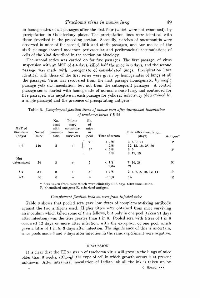
49in homogenates of all passages after the first four (which were not examined), by precipitation in Ouchterlony plates. The precipitation lines were identical with those described in the preceding section. Secondly, patches of pneumonitis were observed in mice of the second, fifth and ninth passages, and one mouse of the sixth passage showed moderate perivascular and peribronchial accumulations of cells of the kind described in the section on histology.
The second series was carried on for five passages. The first passage, of virus suspension with an MST of 4-4 days, killed half the mice in 3 days, and the second passage was made with homogenate of consolidated lungs. Precipitation lines identical with those of the first series were given by homogenates of lungs of all the passages. Virus was recovered from the first passage homogenate, by single passage yolk sac inoculation, but not from the subsequent passages. A control passage series started with homogenate of normal mouse lung, and continued for five passages, was negative in each passage for yolk sac infectivity (determined by a single passage) and the presence of precipitating antigens.
Table 3. Complement-fixation titres of mouse sera after intranasal inoculation
Trachoma virus in mouse lung
of trachoma virus TE55No. Pulmo No.dead nary of
MST of with consolida miceinoculum No. of pneumo tion in in Time after inoculation
(days) mice nitis survivors pool Titre of serum (days) Antigen*Ir 7 < 1/8 3, 6, 9, 21 P4-6 140 3 + 1/8 12, 15, 18, 28, 30I 3* < 1/8 6, 9 P[ 1/8 3, 12, 15
Notdetermined 24 3 + 5 < 1/8 7, 14, 28 E1/64 21
5-2 54 0 ± 3 < 1/8 2, 4, 6, 8, 10, 12, 14 P4-7 60 0 + 4 < 1/8 14 E
* Sera taken from mice which were clinically ill 3 days after inoculation. P, phenolized antigen; E, etherized antigen.
Complement-fixation tests on sera from infected mice Table 3 shows that pooled sera gave low titres of complement-fixing antibody
against the two antigens used. Higher titres were obtained from mice surviving an inoculum which killed some of their fellows, but only in one pool (taken 21 days after infection) was the titre greater than 1 in 8. Pooled sera with titres of 1 in 8 occurred 12 days or more after infection, with the exception of one pool which gave a titre of 1 in 8, 3 days after infection. The significance of this is uncertain, since pools made 6 and 9 days after infection in the same experiment were negative.
DISCUSSIONIt is clear that the TE 55 strain of trachoma virus will grow in the lungs of mice
older than 6 weeks, although the type of cell in which growth occurs is at present unknown. After intranasal inoculation of Indian ink all the ink is taken up by
4 G. Microb. xxx

macrophages within 24 hr. (Watkins, unpublished). If trachoma virus is taken up in the same way it is possible that growth occurs within these macrophages. The inclusion bodies seen in mouse lung resembled trachoma inclusion bodies in conjunctival epithelium, so that the method of virus replication in a host far removed taxonomically from the natural host is, morphologically at least, of the same kind. It was surprising that inocula containing about 109 elementary bodies produced so few inclusion bodies, even in massively consolidated lungs. This may have been due to absence of infectivity of most of the particles, since the LD50 of this strain for chick embryos by the yolk sac route was of the order of 103 to 104 elementary bodies. An alternative explanation, that the particles successful in producing inclusion bodies were genetically different from the unsuccessful ones, is made unlikely by the failure of repeated passage to produce an increase in virulence. Material which killed chick embryos in more than 6 days rarely produced naked-eye or histological lesions. This suggests that the production of lesions depends on a balance between destruction of virus and its successful establishment within a cell.
The pathogenesis of the acute inflammation, which resembles that occurring in the early stages of human conjunctival infection, is still obscure. The extent of the reaction seemed out of proportion to the small numbers of inclusion bodies present. A simple reaction to inert foreign material can be excluded, since nothing similar is seen after heavy inocula of Indian ink. Three explanations are possible. The reaction may be provoked by the release of toxic elementary bodies from cells in which the virus is growing, Growth of virus may be accompanied by the production of a soluble toxin, as suggested by Mitsui (1954) and others. The detection of diffusible antigens by gel precipitation gives some support to this idea, since it shows that some kind of soluble material is associated with the growth of virus. The third explanation is based on the finding by Bell et al. (1959) that some strains of trachoma virus produce a rapid toxic death in mice after intravenous injection of washed elementary bodies. It is possible that intranasal inoculation of a large number of elementary bodies may itself produce an inflammatory reaction. On this view the growth of virus alone would be responsible at the most for only a small proportion of the reaction. At present it is impossible to say which of these three explanations is correct, or whether more than one mechanism may be involved.
The low titres of complement-fixing antibody against particulate (etherized) and phenolized antigen, together with the late appearance of detectable antibody, suggest that this type of antibody may have little to do with the process of resolution of the inflammation. Mice are capable of producing high titres of complement-fixing antibody (up to 1/5120) after intranasal infection with organisms of the pleuropneumonia group (Lemcke, 1961), so the low titres obtained after trachomatous infection may mean that trachoma virus is a relatively poor antigen for mice. Low titres of complement-fixing antibody are usual after infection of human beings with members of the psittacosis-lymphogranuloma group of viruses; it is difficult to ascertain whether this is due to a poor host response, or to deficiencies in the antigens used in complement-fixation tests.
The failure to recover infective virus from the lungs of passage mice when there was circumstantial evidence that their lungs were infected may perhaps be explained by the fact that two completely different criteria of infectivity were being compared. On the one hand, in trying to detect virus by yolk sac inoculation in these experi
50 J. F. W a t k in s a n d A. M. K . M a c k e n z ie

ments enough virus had to be present to kill the embryo within 14 days; on the other hand, to infect mouse lungs with mouse-lung passaged material enough virus had to be present to give rise to detectable diffusible antigen and to occasional small patches of pneumonitis. If the second method was more sensitive than the first the failure to recover virus from passage lungs can be explained. However, the possibility cannot be excluded that continued passage in mouse lung produced a strain of virus with altered properties of growth in yolk sac entoderm.
I am grateful to Dr L. H. Collier for the strain of virus; to Professor C. F. Barwell for the lymphogranuloma sera; to Dr D. Kay for the electron photomigrograph; to Mr H. Axtell for histology and to Mr S. Buckingham for the photographs. Part of this material was incorporated in a thesis for the degree of B.Sc. of Oxford University. A. M. R. M. was in receipt of a Research Training Scholarship from the Medical Research Council.
R E F E R E N C E SB e l l , S . D . J r .. S n y d e r , J . C. & M u r r a y , E . S . (1 9 5 9 ). Im m u n isa tio n o f m ice a g a in st
to x ic doses o f hom ologous e le m e n ta ry b o d ies o f tra c h o m a . Scien ce , 130 , 626 . B e r n k o p f , H . (1 9 5 9 ). T h e s u s c e p tib ility o f w h ite m ice to a s tra in o f tra ch o m a v iru s an d
th e ir use in n e u tra lisa tio n te s ts . B u ll. R es. C ou n cil I s ra e l, 8 E , N o . 1 -2 , p . 2 5 . B o v a r n i c k , M . R ., M i l l e r , J . C. & S n y d e r , J . C. (1 9 5 0 ). T h e in flu e n ce o f c e rta in s a lts ,
a m in o -a c id s , su g a rs an d p ro te in s on th e s ta b ilit y o f R ic k e tts ia e . J . B a d . 5 9 , 50 9 . B r e w e r , J . H. (1 9 4 0 ). C le a r liq u id m ed iu m fo r th e ‘ a e ro b ic ’ c u lt iv a t io n o f an ae ro b es.
J . A m e r. m ed . A s s . 115 , 59 8 .D u l b e c c o , R . & V o g t , M . (1 9 5 4 ). P la q u e fo rm a tio n a n d iso la tio n o f p u re lin e s w ith
p o lio m y e litis v iru s e s . J . ex p . M e d . 9 9 , 16 7 .G i r o u d , P ., R e n o u x , G . & N a t a f , R . (1 9 5 8 ). C u ltu re de p ro d u its tra c h o m a te u x su r so u ris ,
m e rio n s , g e rb ille s , e t o eu fs e m b ryo n n es. A rc h . I n s ts P a s te u r A fr . N . 35, 3 2 7 .H u r s t , E . W . & R e e v e , P . (1 9 6 0 ). T ra n sm is s io n o f th e v iru se s o f tra c h o m a , in c lu s io n
b le n n o rrh o e a , an d lym p h o g ra n u lo m a to m ice . N a tu re , B on d . 186, 33 6 .L e m c k e , R . (1 9 6 1 ). A sso c ia tio n o f P P L O in fe c tio n an d a n tib o d y resp o nse in ra ts and
m ice . J . H y g . C am b. 5 9 , 4 0 1 .L i n d n e r , K. (1 9 1 0 ). D ie fre ie In it ia lfo rm d e r P ro w a ze k sch e n E in s c h lü s s e , v. G raefs.
A rc h . O phth al. 7 6 , 559 .M i t s u i , Y . , T a n a k a , C., Y a m a s h i t a , K. & H a n a b u s a , J . (1 9 5 4 ). T o x in - lik e su b stan ce o f
tra ch o m a v ir u s . A rch . O ph th a l., N . Y . , 5 2 , 7 2 .N i g g , C., H i l l e m a n n , M . R . & B o w s e r , B . M . (1 9 4 6 ). S tu d ie s on lym p h o g ra n u lo m a
co m p le m e n t-fix in g a n tig e n s . I . E n h a n ce m e n t b y p h en o l o r b o ilin g . J . Im m u n o l. 5 3 , 2 5 9 . O u c h t e r l o n y , O . (1 9 4 8 ). In v it ro m eth o d fo r te s tin g th e to x in -p ro d u c in g c a p a c ity o f
d ip h th e ria b a c te ria . A c ta p a th , m icrob io l. scan d . 2 5 , 18 6 .R i c h a r d s o n , G . M . (1 9 4 1 ). P re s e rv a tio n o f liq u id co m p lem en t se ra . L an ce t, i i , 69 6 . R u d d , G . V . & B u r n e t , F . M . (1 9 4 1 ). In t ra n a s a l in fe c tio n o f m ice w ith v iru s o f p s itta c o s is .
A u s t. J . ex p . B io l . m ed . S e i. 19 , 33 .T ’a n g , F . F ., Ch a n g , H . L ., H u a n g , Y . T . & W a n g , K . C. (1957). S tu d ie s on th e a e tio lo g y
o f tra ch o m a w ith sp e c ia l re fe ren ce to iso la tio n o f th e v iru s in c h ic k e m b ryo . C h in . m ed . J . 75, 429.
T h y g e s o n , P. & N a t a f , R . (1 9 5 8 ). E tio lo g ic p ro b lem s in tra c h o m a . R ev. I n t. trachom e, 2 , 17 7 .
W a t k i n s , J . F . (1 9 6 1 ). T it ra t io n o f tra ch o m a v iru s w ith o b se rva tio n s on y o lk -sa c in fe c tio n an d s e n s it iv ity to O x y te tra c y c lin e , u s in g th e s in g le -d ilu tio n m eth o d . J. gen . M icro b io l. 2 6 , 4 2 7 .
W o o l r i d g e , R . L . , J a c k s o n , E . B . & G r a y s t o n , J . T . (1 9 6 0 ). S e ro lo g ica l re la tio n sh ip o f tra c h o m a , p s itta c o s is an d lym p h o g ran u lo m a ven ere u m v iru s e s . P ro c. Soc . ex p . B io l .,N .Y . 10 4 , 2 9 8 .
Trachoma virus in mouse lung 51
4-2

52 J. F. W a t k in s a n d A. M. R. M a c k e n z ie
EXPLANATION OF PLATES 1 & 2Fig. 1. Electron micrograph of etherized suspension of trachoma virus TE 55. Palladium-shadowed ( x 50,000).Fig. 2. Area of inflammation in mouse lung 3 days after intranasal inoculation with trachoma virus TE55 ( x450).Fig. 3. Area of inflammation in mouse of fifth passage series ( x 450).Fig. 4. Trachoma inclusion body in same lung as that shown in Fig. 2, photographed at two different levels ( x 1500).Fig. 5. Mouse lung showing foci of inflammation 3 days after intranasal inoculation of trachoma virus TE55 ( x 40).Fig. 6. Perivascular infiltration of mononuclear cells into pneumonitic area in mouse lung 4 days after intranasal inoculation of trachoma virus TE55 ( x200).

Journal of General Microbioloyy, Vol. 30, Xo. 1 Plate 1
•J. F. WATKINS a n d A. M. H. MACKENZIE ( F a c in g p . 52)

.Journal of General Microbiology, Vol. cO, No. 1 Plate 2
.1. 1<\ WATKINS A M I A. M. II. MACKENZIE
i.'-l

J . gen . M icro b io l. (1963), 30, 53-58W ith 1 p la teP r in te d in G reat B r i ta in
53
Factors which Affect the Size of the Organisms and the Optical Density of Suspensions of P se u d o m o n a s
a eru g in o sa and E sc h e r ich ia co liB y F. BERNHEIM
T h e D e p a r tm e n t o f P h y s io lo g y a n d P h a r m a c o lo g y , D u k e U n iv e r s i t y M e d ic a l C en ter , D u r h a m , N o r th C a r o lin a , U .S .A .
(R e c e iv e d 24 A p r i l 1962)SUMMARY
When water-washed organisms of a strain of P s e u d o m o n a s a e ru g in o s a were exposed to solutions of NaCl or sucrose, they shrank in size and became more dense. The optical density of suspensions increased rapidly. Pre-treatment of the organisms with various cations, surface-active agents and heat and cold, affected the reaction, and the organisms then behaved differently toward NaCl and sucrose. Some but not all of the effects observed with P . a e r u g in o s a were also observed with a strain of E s c h e r ic h ia coli.
INTRODUCTIONMager, Kuczinski, Schatzberg & Avi-Dor (1956) observed that the turbidity of
water suspensions of many Gram-negative bacteria increased when the osmotic pressure was increased by the addition of either electrolytes or non-electrolytes. The magnitude of the effect varied with the age, the degree of viability and the pH. Gram-positive bacteria did not show the turbidity change. Avi-Dor, Kuczinski, Schatzberg & Mager (1956) studied the kinetics of the turbidity changes in suspensions of P a s te u r e l la tu la r e n s is . The initial increase was rapid, caused by the loss of water from the organisms, which decreased in size. This was followed by a decrease in turbidity and the rate of this decrease was affected by compounds which altered the metabolism of the organisms. In the present work, turbidity changes were studied in suspensions of a strain of P s e u d o m o n a s a e ru g in o s a , an organism very sensitive to osmotic effects, and in a strain of E s c h e r ic h ia co li.
METHODSA strain of P s e u d o m o n a s a e r u g in o s a which has been maintained in this laboratory
for 15 years, was grown at 34° for 24 hr. in Difco nutrient broth. The organisms were centrifuged down and washed twice with distilled water. In this process they lost potassium, an average of 5-7 /tmole/40 ml. of wash water, but were subsequently able to take up potassium when incubated with potassium salts. The determinations of potassium were made with a flame photometer. One ml. of final suspension in water was placed in each of a series of test tubes. Various compounds were added dissolved in OT or 0-2 ml. and allowed to react with the organisms for 15 min. at 23°. Water was then added to a final volume of 10 ml. and the initial optical density (o.d .) read at 490 m/t in a photocolorimeter. Solid NaCl, sucrose and other substances

5 4were then added so that their final concentrations were 01-0-2M and the o . d . read as soon as solution was complete, usually within 40 sec. During this time the maximal increase in o . d . had already occurred, and in fact the increase could be detected by the naked eye within a few seconds after addition was made. The o . d . was then measured at intervals. In other experiments various compounds were added after the o . d . had been increased by NaCl, sucrose, etc., and the rate of change as compared with the control was determined. The values are expressed as follows: a standard curve was obtained with different volumes of a suspension of untreated organisms. When the initial o . d . of an experimental series gave a reading corresponding to 1-0 ml. organisms in the standard curve it was given the value of1 - 0 , when the o . d . corresponded to 2 - 0 ml. organisms in the standard curve it was given the value of 2 - 0 .
Avi-Dor et al. (1956) showed that the increase in optical density is the result of a decrease in organism size caused by water loss. Electron-microscope pictures taken of control organisms and others which had been exposed to 0T5 M-NaCl confirmed the decrease in size and showed an increase in density consistent with water loss (PL 1).
The effect of pre-incubation of Pseudomonas aeruginosa with potassium salts on the change in o . d . caused by the addition of 0-1 M-NaCl or 0-2 M-sucrose
Table 1 shows the effects of allowing the organisms to react with 0-05 m-KCI for 15 min. before dilution and addition of either NaCl or sucrose. Two effects can be seen. (1) The initial o . d . , i . e . , before addition of salt or sucrose, was decreased by
F . B e r n h e im
Table 1. The effect of pre-incubation of Pseudomonas aeruginosa suspensions with 0-05 m-KCI on the increase in optical density (o.d.) values produced by 0-1 M-NaCl or 0-2 M-sucrose
Pre-treatmento.n. values O.D. values
Agent Initial Zero time Agent Initial Zero timeNoneKC1 NaClNaCl 0-99 1-72 0-95 1-32 SucroseSucrose 0-990-95
1-751-34pre-incubation with KC1. This decrease was small but consistent and indicates an increase in size of organism as the result of the uptake of K+ accompanied by water.(2) The increase in o .d . following the addition of salt or sucrose (zero time o .d .) was diminished by the pre-incubation with KC1. Apparently an increase in intracellular potassium partially protected the organisms against the subsequent osmotic effect. That the pre-incubation was necessary is shown by the fact that KC1 added to the organisms immediately before dilution and addition of sucrose had little effect on the zero time o .d .
Since potassium was taken up by the organisms when they were suspended in water it was of interest to determine whether it would be taken up by organisms suspended in sucrose. Sucrose was added to the diluted suspension of organisms, the o.d . read, 0-2 ml. of 0-5 m-KCI added, and the o .d . read again after 2 min. There was a marked decrease in the o .d . reading, indicating an increase in organism size. Direct measurement of potassium uptake was not possible because of the sucrose, but the only reasonable mechanism to explain the effect is that K+ ions

5 5accompanied by water entered the cell. Table 2 compares the effect of potassium with other monovalent cations which also increased the organism size, though at a slower rate.
The entrance of cations into the organisms is probably the result of an active process. Table 3 shows the evidence for this in the case of potassium. Potassium phosphate solution (pH 7-4), was pre-incubated with the organisms for 15 min. in the presence and absence of 40 y g . KCN; the suspensions were then diluted and NaCl added. Cyanide prevented the effect of the potassium phosphate, presumably by inhibiting some metabolic activity necessary for the transport.
Table 2. T h e ch a n g e i n o p t ic a l d e n s i ty {o .d .) v a lu e s 2 m in . a f te r th e a d d i t io n o f0 -2 m l . o f 0 -5 M -so lu tio n s o f v a r io u s m o n o v a le n t c a tio n s to Pseudomonas aeruginosa o r g a n is m s s u s p e n d e d in 1 0 m l. o f 0 -2 u - s u c r o se
Factors affecting size and density
Salt added Change in o .d . value
None 0LiCl -0-26NaCl -0-30KC1 -0-34RbCl - 0-20CsCl - 0-20
Table 3. T h e e ffec t o f p r e - in c u b a t io n o f Pseudomonas aeruginosa s u s p e n s io n w i th 0 -0 5 M -p o ta s s iu m p h o s p h a te s o lu tio n { p H 7 -4 ) w i th a n d w ith o u t 40 f ig . K C N o n th e in c r e a s e i n o p t ic a l d e n s i ty {o.d .) v a lu e p r o d u c e d b y 0-1 n - N a C l
o.d . values
Pre-treatment Initial Zero time 60 min.None 100 1-70 1'49K phosphate 0-81 109 0-86KCN 0-87 1-49 1-33KCN + phosphate 0-87 1-43 1-30
When the organisms were placed in boiling water for 5 min. the initial o.d . value was increased (Table 4). Addition of NaCl caused an immediate decrease followed by a slower decrease to that of the control (unboiled) o .d . value. Despite the denaturation of the proteins, changes in organism size are possible when sodium ions and water enter it. Lysis appears to play little part in this effect. It could not explain the rapid and slow components of the curve nor the fact that the change stopped when the control o .d . value was reached. When sucrose was used instead of NaCl, a smaller decrease in o.d . occurred, indicating that the damaged membrane still presented a partial barrier to sucrose. The effect of heating the organisms to 50° for 15 min. shows the difference between the effects of NaCl and sucrose more clearly (Table 4). The initial o .d . value was again increased. The effect of NaCl was minima], but that of sucrose was only slightly less than it was on normal organisms.
The effect of freezing and thawing was somewhat different from that of heat (Table 4). The initial o .d . was not affected, but NaCl was unable to increase theo .d ., which decreased progressively with time to 60-70% of the initial value;

i.e. the organism had become much larger or some had been lysed. Sucrose, however, was still able to increase the o .d . and this value did not decline below that of the initial.Table 4. The effect of heat and of freezing and thawing suspensions of Pseudomonas aeruginosa on the change in optical density (o.d.) value produced by 0-1 M-NaCl or 0-2 M-sucrose Time of observation
56 F . B e r n h e im
Pre-treatmentNone
AgentNaCl
Initial Zero time 60 min. o.d. value
120 min. 180 min.
1-10 1-82 1-71 1-53 1-43Boiled 5 min. NaCl 1-65 1-26 116 110 100None Sucrose 1-08 1-71 1-43 1-43 1-43Boiled 5 min. Sucrose 1-65 1-55 1-48 1-43 1-43None NaCl 1-18 1-96 1-72 1-55 1-43At 50° for 15 min. NaCl 1-44 1-60 0-90 0-83 0-82None Sucrose 118 1-93 1-55 1-43 1-40At 50° for 15 min. Sucrose 1-44 1-82 1-60 1-55 1-43Freezing and thawing NaCl 109 0-86 0-63 0-58 —
Sucrose 1-09 1-45 1-09 109 —
T h e e ffec t o f o r g a n ic a n io n sSodium succinate was compared to sodium chloride as the osmotic agent. The
increase of o .d . value with NaCl at zero time was 0-86 and at 60 min. it was 0-73 units; with sodium succinate the respective figures were 0-73 and 0-27 units. Similar results were obtained with the sodium salts, sodium pyruvate, formate, acetate, benzoate and salicylate. The organic anions, whether possible sources of energy or not, caused a rapid decrease in o.d . to the initial value.
T h e effect o f E D T A , b e n z a lk o n iu m c h lo r id e a n d s o d iu m a lk y lb e n z e n e s u lp h o n a teThese compounds were added to the organism suspension 15 min. before dilution
and addition of NaCl or sucrose. EDTA (ethylenediaminetetra-acetic acid) and benzalkonium chloride increased the initial o.d .; Na alkylbenzene sulphonate had no effect on it. EDTA partially inhibited the rise in o.d . with NaCl, and the decrease in o.d . with time was more rapid. EDTA had no effect on the action of sucrose. After benzalkonium chloride, NaCl caused only a decrease in o .d . as if the barrier to its entry were eliminated, but the response to sucrose was little affected. After alkylbenzene sulphonate, NaCl produced a normal increase in o.d . but the subsequent decrease in o.d . was more rapid than usual. At the concentration used this anionic detergent did not prevent the initial effect of NaCl but did allow the subsequent more rapid entry of the salt into the organisms. Again the action of sucrose was not affected. These results are shown in Table 5.
All the effects of sucrose listed above were observed with glucose. The effects of KC1 were the same as those of NaCl except that the decrease in o .d . with time was more rapid.
Experiments were done with a strain of E s c h e r ic h ia c o li obtained from the clinical laboratory and the differences between it and the P s e u d o m o n a s a e ru g in o s a can be

summarized as follows. Sodium chloride increased the o .d . but the percentage increase was 15 % smaller than that produced in P . a e ru g in o s a . Osmolar sucrose had only a slight transient effect. Apparently E . c o li is readily permeable to sucrose. Glucose increased the o .d . with E . c o li to the same extent as did NaCl. Sodium succinate and pyruvate were as effective as NaCl but sodium formate, acetate and benzoate were inactive. Heating to 50° and freezing and thawing were without effect, but after boiling, E . c o li behaved like P . a e ru g in o s a . EDTA and alkyl- benzene sulphonate in the concentrations used, had no effect but benzalkonium chloride was active.
Factors affecting size and density 57
Table 5. T h e effec t o f p r e - in c u b a t io n o f s u s p e n s io n s o f Pseudomonas aeruginosa w ith E D T A , b e n z a lk o n iu m c h lo r id e ( B A ) a n d s o d iu m a lk y lb e n z e n e s u lp h o n a te ( A S ) o n the ch a n g e i n o p t ic a l d e n s i t y ( o . d . ) v a lu e s p r o d u c e d b y 0 -1 u - N a C l o r su c ro se
In these experiments the osmolarity of sucrose was half that of NaCl.Time of observation
Initial Zero time 60 min. 120 min.O.D. valuePre-treatment Agent ------------- -A-------------------------------- ---------------------- N
None NaCl 1-08 2-12 2-01 1-89EDTA 50 fig . NaCl 1-26 1-72 1-36 1-26None Sucrose 108 1-87 1-65 1-54EDTA 50 fig . Sucrose 1-26 1-89 1-65 1-62None NaCl 0-73 1-35 1-18 1-17BA 50 fig . NaCl 0-95 0-68 0-53 0-53None Sucrose 0-73 1-25 1-09 1-00BA 50 fig . Sucrose 0-99 1-34 0-95 0-93None NaCl 1-26 1-95 1-72 1-64AS 120 fig . NaCl 1-26 1-95 1-35 1-18None Sucrose 1-26 2-01 1-82 1-72AS 120 fig . Sucrose 1-26 2-01 1-78 1-72
DISCUSSIONThe osmotic effect produced by the various compounds on these bacteria depends
on the characteristics of the cell membrane, i.e. its ability to impose a barrier to free diffusion. Urea, for instance, diffuses through most membranes and causes no change in o .d . when added to suspensions of these organisms. The results with this simple method show some interesting differences in membrane characteristics. For instance, P s e u d o m o n a s a e r u g in o s a has a barrier to sucrose, E s c h e r ic h ia c o li has not. Heat, and freezing and thawing destroy the barrier to cations but not to sucrose in P . a e r u g in o s a . Similar treatment had no effect on the ion barrier in E . co li. P . a e r u g in o s a is more sensitive to the action of surface-active agents than E . co li, and in the former organism these agents affect the barrier to cations at concentrations which do not affect the barrier to sucrose. When thr organisms P . a e r u g in o s a and E . c o li were allowed to take up cations before being subjected to the osmotic shock the change in o .d . was smaller. Another factor which would impose a limit to change in size of organism is the elasticity of the cell wall; this might explain why an increase in osmolarity above the equivalent of 0-1 M-NaCl had little or no effect. The rate of decrease in o .d . following the initial increase seems to depend on

5 8several factors. First, the rate of cation uptake. When NaCl was used as the osmotic agent, the mean decrease in o . d . in 60 min. was 0-20; when KC1 was used it was 0-38 units. Secondly, the nature of the anion. The rates of decrease ofo . d . with NaCl, NaBr and NaN03 were the same. But when the anion was organic the rate of decrease was consistently more rapid. There is no obvious explanation for this. Finally, pre-treatment of P. aeruginosa with EDTA or benzalkonium chloride increased the initial o . d . This implies that these agents allowed an outward diffusion of ions accompanied by water, with the consequent shrinking of the organisms.
REFERENCESA v i - D o r , Y., K u c z i n s k i , M., S c h a t z b e r g , G. & M a g e r , J. (1956). Turbidity changes in bacterial suspensions: kinetics and relation to metabolic state. J . gen . M icro b io l. 14, 76.M a g e r , J., K u c z i n s k i , M., S c h a t z b e r g , G. & A v i - D o r , Y. (1956). Turbidity changes in bacterial suspensions in relation to osmotic pressure. J. gen . M icro b io l. 14, 69.
F . B e r n h e im
EXPLANATION OF PLATE 1Fig. 1. Electron-microscope picture of washed P seu d o m o n a s aeru g in osa organisms after suspension in water x 16,800.Fig. 2. After suspension in 0T5 M-NaCl x 16,800.The pictures were taken by Dr J. R. Overman of the Department of Microbiology on an RCA- EML electron microscope.

Journal of General Microbiology, Vol. 30, No. 1 Plato 1
Fig. ]
F. BERXHKIM (Facing p. 58)

./. gen . M icro b io l. (1963), 30, 59-73W ith 8 p la te sP r in te d in G reat B r i ta in
5 9
Structure and Replication of the Trachoma Agent in Cell Cultures, as shown by Electron Microscopy
By J. A. ARMSTRONG, R. C. VALENTINE a n d CELIA FILDESN a t io n a l I n s t i tu te fo r M e d ic a l R e se a rc h , L o n d o n , iV.IF. 7
(R e c e iv e d 25 A p r i l 1962)SUMMARY
Propagation of the trachoma agent in human cell cultures has made possible the observation by electron microscopy of sequential stages in the replication of the micro-organism. Scattered reticular foci with incomplete limiting membranes were present in the cytoplasm of HeLa cells 12 hr. after exposure to the infectious agent in high multiplicity. After 24 hr. the foci had coalesced to give a single reticular mass. Within it there appeared large and discrete developmental forms from which, by progressive subdivision and diminution in size, typical elementary bodies were formed. The elementary bodies had a prominent limiting membrane, and a dense core within which a fibrillar nucleoid could be recognized. The controversial taxonomic status of the trachoma agent is considered briefly in the light of ultrastructural evidence.
INTRODUCTIONMembers of the psittacosis group, with the related causal agents of trachoma and
inclusion blennorrhoea, present an interesting and controversial problem in taxonomy (Andrewes, 1952; Bedson, 1959). Although acknowledged as obligate intracellular parasites and virus-like in size, the micro-organisms have properties that prompt misgivings about their viral nature, for they appear to have much in common with the bacteria and rickettsias. There is increasing support for the view expressed by Weiss (1955) that they are, in fact, typical of neither rickettsias nor viruses but lie somewhere between the two. The features usually cited as incompatible with a viral classification are: (i) microscopical studies have consistently upheld the view that multiplication is by binary fission of intact particles; (ii) chemical analyses indicate that the infective particles have a chemical complexity akin to that of bacteria; (iii) the occurrence of an eclipse phase is unproven; (iv) the organisms are susceptible to a variety of antibiotics and other compounds. The issue is by no means settled, however, and some recent investigations draw attention to the virus- like aspects of the psittacosis group. McCloskey & Morgan (1961) observed that the latent phase following inoculation of L cell cultures with psittacosis virus could be prolonged for 24 hr. by withholding certain vitamins and amino acids: during this period infectivity was lost, and no viral structures or inclusion bodies were detected by light or electron microscopy. Infectious virus reappeared in the system within 24 hr. of rectifying the nutritional deficiency. Tanami, Pollard & Starr (1961) studied the effects of pyrimidine analogues and p-fluorophenylalanine on the growth curve of psittacosis virus, also in tissue cultures. It was inferred that a lag of 10 hr.

occurred between viral DNA synthesis and i~s incorporation into infectious particles, and it was suggested that replication of ‘ naked ’ DNA indicates an essentially viral mode of multiplication. In similar vein Becker, Mashiah & Bernkopf (1962), describing a growth cycle of some 72 hr. fcr the trachoma agent i n v i tr o , reported that it passed initially through a non-infecrious phase lasting 24 hr.
Following isolation of the trachoma agent in the yolk sac of chick embryos (T’ang, Chang, Huang & Wang, 1957) it was propagated successfully in epithelial cell cultures, producing a widespread cytopathic effect typical of the psittacosis group (Furness, Graham, Reeve & Collier, 1960; Bernkopf, Mashiah & Maythar,1960). Electron microscopical study of such a system seemed an opportune means of gaining more precise information, in morphological terms, about multiplication of the trachoma agent, and so perhaps of the psittacosis group as a whole.
METHODSC e ll c u ltu re s . Stock cultures of HeLa cells in 250 ml. bottles were grown in a
medium consisting of Gey’s balanced salt solution, 10 % (v/v) human serum inactivated at 56° for 30 min. and 0-5 % (w/v) lactalbumin hydrolysate, with streptomycin 200 /¿g./ml. Cells were subcultured into half test tubes containing 7 mm x 22 mm. coverslips, using the same medium but without antibiotics.
I n o c u lu m . The T ’ang TE55 strain of the trachoma agent (T’ang e t a l. 1957), originally supplied to us by Dr L . H. Collier (The Lister Institute) was maintained in this laboratory by serial yolk-sac propagation in chick embryos (Collier & Sowa,1958). Infected yolk-sac tissue was ground in a tissue grinder with 0-85 % saline containing 1 % (v/v) horse serum and 10 % (v/v) nutrient broth, centrifuged at 2500 rev./min. for 10 min., and the supernatant fluid stored in sealed glass capillaries at -70°.
A s s a y a n d in o c u la t io n . The infectious material was titrated in HeLa cells by a method essentially similar to that of Furness, Graham & Reeve (1960). An inoculum capable of infecting 90-100% of the cells was prepared by 1/100 dilution of the yolk-sac material in Gey’s solution; 0-5 ml. of inoculum in each tube was left to adsorb for 1 hr. at 37°. It was then removed, fresh antibiotic-free medium added, and incubation continued at 37°.
E le c tr o n m ic r o s c o p y . Infected cultures were processed for electron microscopy, together with uninoculated controls, at intervals of 6, 12, 24, 48, and 72 hr. after inoculation. The samples for each time consisted of pooled cells from batches of six tubes. The culture medium was first removed, and replaced with 3 ml. of the 1 % (w/v) osmium tetroxide fixative of Palade (1952) at pH 7-4; after a few minutes the cells were pushed gently from the tube walls into the fixative by using a rubber tipped glass rod. The contents of the six tubes were mixed and fixation continued for 1 hr. at room temperature. In loose pellet form the cells were then washed, dehydrated, and infiltrated with methacrylate monomer in the usual way. Final embedding was in a prepolymerized 1 + 4 mixture of methyl and w-butyl methacrylates, catalysed with 1 % (w/v) benzoyl peroxide. Blocks of suitable hardness resulted from standing at 60° for 24 hr.
Sections were cut with a Porter-Blum microtome and collected on copper grids bearing carbon-stabilized nitrocellulose films. Before examination most of the
60 J . A. A r m s t r o n g , R. C. V a l e n t i n e a n d C. F i l d e s

grids were floated face downwards for 3 hr. on a solution of 5 % (w/v) uranyl acetate in 1 % (v/v) acetic acid (Valentine, 1962), washed vigorously for about 30 sec. in distilled water and allowed to dry. The lead hydroxide method of Dalton & Zeigel(1960) was also used. The sections were viewed with a Siemens UM 100 electron microscope operating at 60 kV; pictures were taken at x 1250 or x 7000 on ultrafine grain Ilford N.60 (photomechanical) plates, and subsequently enlarged.
L ig h t m ic r o s c o p y . The progress of cytopathic change in the cultures was assessed, before electron microscopy, by inspection of the coverslips placed for this purpose into the culture tubes. Coverslips from each of the inoculated batches, and from uninfected control tubes, were fixed for about 20 min. in Palade’s buffered osmium tetroxide solution; after rinsing in Gey’s solution they were transferred to 70 % (v/v) ethanol in water for about 24 hr. They were then mounted in water, wax-sealed and examined by phase-contrast microscopy as described elsewhere (Armstrong & Pereira, 1960). Similar monolayers were stained by the periodic acid-Schiff (PAS) method for demonstration of polysaccharides, and counterstained with haematoxylin. Identification of glycogen was confirmed by treatment of some cultures with saliva for 45 min. at 37°, before PAS staining. A few coverslip cultures were ethanol-fixed for Giemsa staining.
Electron microscopy of the trachoma agent 61
RESULTSP h a s e -c o n tr a s t m ic r o s c o p y
Inspection of the osmium-fixed cell monolayers revealed a progression of cellular changes after inoculation. Those examined after 6 hr. were not visibly different from the control HeLa cells. After 12 hr. a small optically dense nodular mass was detectable to one side of the nucleus in many cells, and by 24 hr. a well-defined juxtanuclear inclusion was present in almost every cell. It had a vesicular mottled appearance and continued to enlarge until 72 hr. after inoculation, when the experiment was terminated. The cells were then grossly distended, in each the nucleus was displaced to the periphery by a very large vesicular inclusion containing aggregates of dense particulate material (PI. 1, fig. 1). These findings are in line with recent descriptions of trachoma-infected tissue cultures (Gordon, Quan & Trimmer, 1960; Furness, Graham, Reeve & Collier, 1960; Bernkopf e t a l. 1960), and comparable with the earlier accounts of changes that accompany multiplication of other members of the psittacosis group (Bedson & Bland, 1932). The cytopathic changes developed synchronously throughout the cell sheets; counts indicated that more than 90 % of the cells were infected at the outset. At 72 hr. the presence of a few cells with ‘early-type’ inclusions suggested the onset of a second cycle of growth.
E le c tr o n m ic r o s c o p yT h e e le m e n ta r y b o d y . Observations were directed first towards the contents of
the fully developed inclusion or vacuole, in cells fixed 72 hr. after inoculation. The object was to determine the structural features of the infectious trachoma elementary bodies, believed to occur in the inclusion at this late stage, before attempting to interpret changes leading to their formation. There was no difficulty in finding inclusions in the sectioned cells. They contained a large number of small, rounded or oval particles of high electron density dispersed within a matrix of very much

62lower density. Measurements gave an average particle size of 375 x265 m/i, mean diameter about 320 m/i. The long axes of those presenting an oval profile were invariably orientated in one direction, suggesting that truly spherical structures had undergone the usual compression on sectioning. When allowance is made for differences due to the plane of section, the regularity in size and form of the dense particles was such as would be expected of viral elementary bodies.
Part of a typical 72 hr. inclusion is illustrated at high magnification in PI. 1, fig. 2; the area shown contains four of the objects which we regard as elementary bodies. Each had a prominent and dense inner core (mean diameter 220 m//) separated by a clear zone from a characteristically folded limiting membrane. The thickness of the membrane was about 10 m/i, but often looked more in the plane of a section because of its folded nature. This interpretation of cross-sections is entirely consistent with the ‘wrinkled pea’ description of intact elementary bodies, as revealed by electron microscopy of air-dried and metal-shadowed material from trachoma-infected chick embryos (Collier, 1959). The inner cores seemed on section to be of two kinds. One was a composite mass of fibrillar and granular elements, forming a compact structure with a regular outline. The other kind was less regular in outline and had the fibrillar component segregated from the granular part by a distinct but usually incomplete membrane. We shall refer to this saccular structure, which measured some 130 m/t in diameter and was of great electron density, as a nucleoid (PI. 1, figs. 2, 3). Inspection of random thin sections cannot establish whether an organized nucleoid is, in fact, entirely lacking in cores of the first kind; but it is conceivable that the variations in core structure signify different levels of particle maturity.
A p p e a r a n c e 6 h r . a f te r in o c u la tio n . Many of the cells examined at this stage were not obviously different from those of the uninoculated control cultures. However, two abnormal features appeared quite often in the sections and were undoubtedly a consequence of the preceding inoculation. Pale mottled areas occurred in the cytoplasm ranging in size from small patches as seen in PI. 2, fig. 4, to confluent masses larger than the cell nucleus. At high magnification they appeared to be largely amorphous but with a variable content of moderately dense 15-30 m/i particles, closely comparable with the large type of glycogen deposits observed in liver cells after osmium fixation (Revel, Napolitano & Fawcett, 1960). As a test of this interpretation, sections were floated on a solution of potassium permanganate as advocated by Drochmans (1960); the previously pale areas developed great electron density and became the most conspicuous objects in the cell cytoplasm. Further confirmation came from examination with the light microscope of PAS- stained monolayer cultures; almost all cells were seen to contain one or more large masses of PAS-positive material which was absent after saliva digestion. Negligible amounts of glycogen were demonstrable in the control cultures.
The other abnormality was a heterogeneous collection of small dense bodies in a juxtanuclear position, usually intermingled with the Golgi complex. Occasional structures of the same kind occurred in cells of the uninoculated cultures. A typical cluster within a cell inoculated 6 hr. previously is shown in PI. 2, fig. 4. The profiles average some 350 m/i in size, variation being attributable at least in part to plane of section. Until quite recently structures of this kind have been referred to in electron microscopical investigations in general terms, e.g. ‘dense body’, ‘micro
J. A . A r m s t r o n g , R. C. V a l e n t i n e a n d C. T i l d e s

body’, ‘lamellar body’ and so on. Studies on mammalian liver cell structure (Essner & Novikoff, 1960; Daems & van Rijssel, 1961) have established the identity of peribiliarv dense bodies as lysosomes, i.e. membrane-limited organelles containing acid phosphatase and other enzymes, which are directly concerned in phagocytic and lytic activities of the cell (De Duve et a l. 1955). The morphological heterogeneity of lysosomes has been emphasized, but typically they contain ferritin- like granules and have a single limiting membrane. Many of the dense bodies with which we are presently concerned conformed to the descriptions of liver cell lysosomes and their presence would be consistent with recent phagocytic activity. Amongst them, however, was an occasional example having a dense fibrillo-granular central area surrounded by a markedly folded membrane. There was a superficial similarity between such forms and trachoma elementary bodies. Nevertheless, close examination of many cells fixed 6 hr. after inoculation did not reveal structures, in the cytoplasm or at the cell surfaces, that could be identified unequivocally as elementary bodies or their derivatives.
A p p e a r a n c e 1 2 h r . a f te r in o c u la tio n . Some degree of alteration was now detectable in almost every cell examined. Lysosome aggregates were less obvious than at 6 hr., but large glycogen deposits were much in evidence. Light microscopy of PAS and haematoxylin-stained monolayers at this stage revealed, in addition to cytoplasmic glycogen, groups of basophilic particles in the general vicinity of the Golgi complex. They had the typical appearance of early developmental forms of the psittacosis group (Bedson & Bland, 1932).
The first indication of structural elements presaging specific cytoplasmic inclusions was given in micrographs at this stage. At lower magnifications it was possible to detect in the cytoplasm more or less rounded formations, distinct from the glycogen and unlike any normal component of the HeLa cell. Ranging in size from under 1 ¡i to more than 2 /t in diameter, they had on section the consistency of a fine reticulum of low to moderate electron density. Several such reticular foci are depicted within a limited area of the cell cytoplasm in PI. 3, fig. 5. Although sometimes grouped in this way, individual foci were also more widely scattered in the cells. At higher magnification each reticular focus appeared to consist essentially of a meshwork of delicate and wavy fibrils, usually bounded by a well-defined bilaminar membrane. In some, however, notably those over 1 -5fi in diameter, the enclosing membrane was fragmentary or incomplete, the fibrillar contents being then in direct continuity with surrounding cytoplasm. Similar fibrillar material was sometimes observed without a vestige of a limiting membrane. No structures resembling elementary bodies were found.
A p p e a r a n c e 2 4 h r . a f te r in o c u la tio n . There was no difficulty after 24 hr. in locating with the electron microscope characteristic juxtanuclear inclusions, corresponding to those observed by phase-contrast in the osmium-fixed monolayers. Indeed, they were of such size that at least part of one was encountered in almost every cell sectioned. The inclusion appeared as a localized area of altered cytoplasm several microns in diameter, with no equivalent in the uninoculated control cells. The presence of the abnormal structure caused slight indentation of the otherwise normal cell nucleus, and a corresponding bulge was often visible in the cell wall overlying the inclusion. Mitochondria, ribosomes and endoplasmic reticulum were absent from the area occupied by the inclusion, and abnormal glycogen deposits
Electron microscopy of the trachoma agent 63

were distinctly smaller and less numerous than in the specimens fixed at 6 hr. and 12 hr. after inoculation.
The fine structure of the inclusion was remarkably constant from cell to cell. It consisted for the most part of a fibrillar matrix or reticulum of moderate overall density, incorporating somewhat denser patches each partially enclosed by an incomplete and fragmentary bilaminar membrane. The character of the inclusions suggested that the separate reticular foci seen at 12 hr. had become more numerous and were forming a confluent mass (PI. 4, fig. 6). I t was a regular finding that no membrane or other definable barrier separated the interior of the inclusion from the surrounding cell cytoplasm. Detail from a typical inclusion is shown at higher magnification in PI. 5, fig. 7: curved profiles of several incomplete bilaminar membranes are present, and the components of the reticulum are seen as wavy fibrils, of high density and about 4 m/i in thickness. Their length could not be determined from the study of sections. No elementary bodies were seen in the cells at this time.
A p p e a r a n c e 4 8 a n d 7 2 h r . a f te r in o c u la tio n . A remarkable change in the character of the intracellular inclusions took place in the interval between 24 and 48 hr. after inoculation, accompanied by a considerable distortion of host-cell morphology. As anticipated from the preliminary phase-contrast observations there was pronounced enlargement of the inclusion, which now appeared as a distinctly vesicular formation -with a diameter of 20//, or more. The cell nucleus was displaced to the side, markedly compressed by the inclusion but otherwise structurally unaltered. Recognizable cytoplasmic elements were restricted to a perinuclear area and a tenuous peripheral rim around the massive inclusion. Degenerative changes in the mitochondria were conspicuous in some cells. Little remained of the glycogen deposits seen at early stages.
As in the earlier stages, examination at high magnification revealed no continuous morphological barrier between the interior of the inclusion and the surrounding cytoplasm. The boundary is therefore probably a phase interface rather than a morphological entity. Each inclusion contained a number of discrete, more or less round bodies suspended within a matrix of comparatively low density (PI. 5, fig. 8). The bodies were remarkably variable in size and structural detail, but all were alike in possessing a complete and well-defined limiting membrane; they will be referred to as developmental forms of the trachoma agent. The largest measured 3 or 4/t in diameter and the smallest between 30 D and 400 m//; different sizes occurred together in the majority of inclusions, but occasional cells appeared to contain only the larger ones. The larger developmental forms contained a reticulum of fibrillar nature similar to that observed, in an unenclosed form, in cultures examined at the 24 hr. stage. In addition, many of the larger forms also contained a number of prominent condensations of fibrillar material, round or oval in shape and about 300 m// in diameter (PL 5, fig. 8). Complication of the cross-sectional morphology of the large forms frequently arose from the occurrence of deep finger-like invaginations of the limiting membrane, producing profiles of a bizarre nature. Inspection of developmental forms of decreasing size revealed a progression towards profiles of a more regular character, with increasing density and more even distribution of contents. Within forms typical of the intermediate size range (say, 0-5-1 //. in diameter) one or two centrally placed fibrillar condensations were separated from
6 4 J . A. A r m s t r o n g , R . C. V a l e n t i n e a n d C. F i l d e s

the limiting membrane by a wide zone occupied by material of a different nature, predominantly granular and of comparatively low electron density (PI. 6, fig. 9). Developmental forms of smaller size than this became virtually indistinguishable from elementary bodies.
The cultures fixed 72 hr. after inoculation contained many cells similar to those seen at 48 hr.; but in general the inclusions were even larger and host-cell components correspondingly more displaced. Inside the inclusions elementary bodies predominated, with a reduced number of developmental forms of the large and intermediate sizes (PI. 7, fig. 12; PI. 8, fig. 13). In addition, a few disrupted large forms and some others that appeared to be intact but devoid of contents were often visible amongst the elementary bodies.
A considerable part of each inclusion was accounted for by the matrix in which developmental forms and elementary bodies were suspended. Owing to low overall density there was little to indicate its composition, except for scattered particles of moderate density, 20-50 m/f in size, sometimes aggregated to form larger stellate clusters (PI. 8, fig. 14). This is reminiscent of descriptions of particulate glycogen (Revel e t at. 1960). This identification was borne out by the PAS-stained monolayer cultures; the contents of inclusion bodies were strongly PAS-positive from the 48 hr. stage.
E v id e n c e o f f i s s io n . Arbitrary classification of the developmental forms on a basis of size or structural differences cannot be justified; a whole range of transitional forms was recognizable in the electron micrographs, from the largest down to the elementary bodies. From comparison of the contents of cytoplasmic inclusions in 48 and 72 hr. specimens, it could be inferred that the larger developmental forms were somehow being replaced by those of smaller size. A search was therefore made for profiles that might be a key to inter-relationship of the various forms, bearing in mind the many previous reports of binary fission in the psittacosis group. In the 48 hr. inclusions there were always a few developmental forms so shaped, or disposed in relation to each other, as to suggest they had indeed been dividing at the time of fixation. Furthermore, it seemed that at least two modes of division could be recognized, namely into unequal or equal parts. Unequal division was shown mainly by the largest forms, and could be described as budding. Examples of it are illustrated in PI. 6, fig. 10: here, two forms just a little larger than elementary bodies are each situated in close apposition to a comparatively large form, from which it could be supposed they were separating when fixed. The frequency of such associations made it unlikely they were entirely fortuitous; especially as large forms were also to be found showing localized protrusions which, though still continuous with the membrane of the parent body, exhibited varying degrees of intervening constriction. On the other hand, many intimately paired profiles of smaller developmental forms seemed to suggest division into equal halves (PI. 6, fig. 11; PI. 8, fig. 13). In general, the smaller the forms concerned, the more closely did their division approximate to binary fission as the term is generally understood. There were frequent signs of such divisions in the inclusions at 72 hr., when the larger developmental forms had mostly disappeared; and the possibility of elementary bodies undergoing repeated binary fission cannot be ruled out.
Electron microscopy of the trachoma agent 65
5 G. Microb. x x x

66 J. A. A r m s t b o n g , R. C. V a l e n t i n e a n d C. F i l d e s
d i s c u s s i o n
The first systematic observations on the growth of psittacosis virus in infected tissues and cell cultures were based on conventional staining techniques for light microscopy (Bedson & Bland, 1932, 1934; Bland & Canti, 1935). Similar work was undertaken on the closely related causal agents of lymphogranuloma, venereum and meningopneumonitis (Findlay, Mackenzie &, MacCallum, 1938; Rake & Jones, 1942; Higashi, Notake & Fukada, 1959). These researches have led to a wide measure of agreement about the cytopathic changes that accompany multiplication of the psittacosis group of organisms, the process culminating in a mass of newly formed elementary bodies less than 0-5// in size. It has been generally considered that invading particles retain their integrity in the host cell cytoplasm and become transformed, after a latent period of 5 to 20 hr. into a visible cluster of comparatively large structures known as initial bodies, between 1 and 2 /i in size. These seem to undergo repeated binary fission which, w:th a decrease in size, gives origin to the new infectious particles. The term ‘plaque’ was applied to large cytoplasmic structures of homogeneous appearance. Although plaques were first interpreted as plasmodia of viral nature (Bedson & Bland, 1932) this view was later rejected in favour of regarding them as just closely packed colonies of initial bodies (Bedson & Bland, 1934; Bedson, 1959). Light microscopy of cell cultures inoculated with the trachoma agent revealed an almost identical sequence of cytopathic changes (Gordon e t a l. 1960; Furness e t a l. 1960; Bernkopf e t d l . 1960). In this connexion it is interesting to recall that a similar developmental cycle was postulated many years ago after examination of cell inclusions in tissue from trachoma cases (Halberstaedter & von Prowazek, 1907; Lindner, 1910).
Although light microscopy clearly demonstrates affinities within the psittacosis group, limited resolving power has impeded the understanding of early stages in the growth cycle. The uncertain nature of markedly pleomorphic developmental forms, and doubts concerning the degree of association between the infectious agent and the host cell, have been obstacles in the way of comparing satisfactorily the reproductive mechanisms of the psittacosis group and other micro-organisms such as the bacteria (Weiss, 1955; Träger, 1960). Already in several laboratories workers have turned to electron microscopy in the hope that greater resolving power would be decisive in the vexed question of taxonomic relationships. Thin- section studies have been made of cells i ifected with the agents of meningopneumonitis (Gaylord, 1954; Tajima, Nomura & Kubota, 1957; Higashi, 1959), feline pneumonitis (Litwin, 1959), psittacosis (Litwin, Officer, Brown & Moulder, 1961) and trachoma (Mitsui e t a l. 1958). Mater als examined included tissues of infected animals and embryonated eggs, as well as inoculated cell cultures; but in terms of advancing previous knowledge the outcome has been inconclusive. Selective viewing of fragments from an organized tissue system is not an ideal basis for the analysis of a complex developmental cycle; and most of the published accounts deal chiefly with the structure of well-developed cytoplasmic inclusions. As pointed out by Higashi (1959) morphological changes giving origin to the large developmental forms have proved elusive; also, the internal structure of the elementary bodies has not so far been described in detail.
The findings of Tajima e t a l . (1957) in a study of chick chorioallantoic membranes,

fixed at intervals from 2 hr. to 4 days after inoculation with the virus of meningo- pneumonitis, are of exceptional interest. For this group of workers reported a localized ‘viral matrix’ in the cytoplasm of infected cells, before the appearance of discrete developmental forms with limiting membranes. The matrix was described as granular, not sharply demarcated from the surrounding cell cytoplasm, and similar to the diffuse viroplasm characterizing the initial stages of intracellular multiplication of pox viruses. Nevertheless, Litwin et al. (1961) were unable to
Electron microscopy of the trachoma agent 67
Fig. 1. R epresentation of stages in the developmental cycle of the trachom a agent in HeLa cells.
identify a similar matrix in psittacosis-infected chorioallantoic membranes; they concluded that a mechanism involving such elements ‘cannot be an important means of multiplication in the psittacosis group’.
In the present investigation a regular sequence of changes was observed in HeLa cells inoculated with a high multiplicity of the trachoma agent. It has been possible from this to reconstruct some important aspects of the complex cycle of intra-
5 -2

cellular growth. Four significant stages are showji schematically in Fig. 1. The first was seen 12 hr. alter inoculation, and is the s ta g e o f s c a tte re d r e t ic u la r f o c i , with incomplete limiting membranes. Coalescence of these between 12 and 2-1 hr. produces the single inclusion of the s ta g e o f c o n flu e n t r e t ic u lu m . There can be little doubt that the formation which characterizes this stage corresponds to the disputed ‘viral matrix’ described by Tajima c t a l. (1957). In the period from 24 to 48 hr. the formation of new membranes within the inclusion gives rise to a series of spherical closed compartments, with a maximum diameter of about 3//. This is the s ta g e o f d is c r e te d e v e lo p m e n ta l f o r m s . Focal condensations occur within the larger forms, and between 48 and 72 hr. after the onset of infection, subdivision by means of budding and fission, with progressive diminution in size, gives rise to the final s ta g e o f n e w ly f o r m e d e le m e n ta r y b o d ie s . The mechanism by which newly formed elementary bodies escape from the inclusion remains obscure; transfer of individual elementary bodies through the cell wall to the exterior did not seem to be occurring in the material examined.
How infecting particles normally gain access to the host-cell cytoplasm, and their immediate fate thereafter, have not been elucidated. It is now certain that information on this could be expected only from close study of infected cells at a stage earlier than any in the present series. The demonstration recently of the phagocytosis and subsequent breakdown of vaccinia virus particles (Dales & Siminovitch, 1961) encourages the speculation that infectious particles of trachoma, and others in the psittacosis group, may be taken into the cell in this way. Certainly, the finding of conspicuous lvsosome aggregates 6 hr. after inoculation in the present series suggests that a high level of phagocytic activity had been taking place. We do not know whether each of the reticular foci seen in 12 hr. specimens had developed from a single infecting particle, or had been formed d e n o v o after complete disintegration of the infecting particles.
In spite of these uncertainties, points of special interest revealed by the electron microscope are the incomplete separation of the contents of early reticular foci from surrounding cytoplasm, and their subsequent fusion into a confluent mass which is the definitive inclusion of the 24 hr. stage. The possibility has to be considered that the incomplete nature of the bilaminar membranes observed in early stages of the cycle was a technical artefact, due to disruptive polymerization of the methacrylate monomer or to defective fixation. Polymerization damage has characteristic effects on cellular fine structure (Morgan, Rose & Moore, 1957) but evidence of this was encountered only in occasional blocks in this series. If polymerization damage is involved it would therefore be necessary to postulate unusual susceptibility of the early developmental forms of the trachoma agent. Fixation artefact can take various forms and is still little understood; it is therefore arguable that complete but delicate membranes may enclose the reticular foci, and be imperfectly preserved by the buffered osmium tetroxide method currently favoured by electron microscopists. In fact, it is difficult to envisage the observed fine structural details arising in this way, but we bring the view forward as one which cannot be entirely ruled out.
Correlation of electron microscopical details with the classical cytopathic changes associated with organisms of the psittacosis group seems to be largely a matter of terminology, presenting no great difficulty. Thus, the structures formerly termed
08 J . A . A r m s t r o n g , R. C. V a l e n t i n e a n d C. F i l d e s

1 plaques ’ can reasonably be equated with the confluent reticulum seen at 24 hr. in the present study. Their plasmodial nature, hitherto conjectural, is borne out by electron microscopy. The precise equivalent of ‘initial bodies’ is less obvious; but it is likely that both the reticular foci and the later discrete developmental lorms of the present account have been included together under this single heading in earlier descriptions. With both forms of microscopy it is possible to observe the innumerable elementary bodies within mature inclusions, t ut their internal structure is not of course represented in the images obtained with light microscopes.
It is of special interest to consider the fine structure of trachoma-infected cells in relation to recent cytoehemical studies on the growth in cell cultures of several members of the psittacosis group, including the trachoma agent itself. The acridine orange fluorescence technique, together with nuclease digestion tests, has been used to demonstrate a series of regular changes in the distribution of nucleic acids in the cytoplasmic inclusions; the results were correlated with infectivity studies (Starr, Pollard, Tanami & Moore, I960; Pollard, Starr, Tanami & Moore, 1960; Becker e t a l. 1962). During the first 24 hr., corresponding to the stages referred to here as scattered reticular foci and confluent reticulum, one or more small bodies became visible in the cytoplasm and then enlarged into a single juxtanuclear mass; this gave a strong red fluorescence signifying the accumulation of newly synthesized ribonucleic acid (RNA). As the mass enlarged further its red fluorescence tended to diminish and increasingly numerous points of intense yellow-green fluorescence emerged, indicating the development of particles containing deoxyribonucleic acid (DNA) within the preformed pool of RNA-containing material. This coincided with a rise of demonstrable infectivity. It was at about this stage in the present experiments, that discrete developmental forms were first seer, in the micrographs. The presence of DNA may well be related to the more or less organized fibrillar component revealed in the elementary bodies by electron microscopy . Indeed, the ultra- structural complexity of the elementary bodies is consistent with the findings of chemical analysis which suggest that micro-organisms of the psittacosis group contain not only DNA, but also RNA and muramic acid (Zahler & Moulder, 1953; Ross & Gogolak, 1957; Allison & Burke, 1962). A glycogen-containing matrix inside the fully developed cytoplasmic inclusions of trachoma-infected cells has been known for some years (Rice, 1936; Thygeson, 1938) and is merely confirmed by the present observations. However, the formation of widespread glycogen deposits in the host cell cytoplasm, as early as 6 hr. after inoculation, does not seem to have been noted previously; the possibility of this being a response to renewal of the culture medium, rather than to infection p e r se . is suggested by recent observations on fine structural variations in normal HeLa cells (Bruni, Gey & Svotelis, 1961).
It is widely believed that the mode of intracellular replication permits a real distinction to be drawn between viral infection and parasitism by larger microorganisms. With viruses, breakdown of invading particles results in an intimate association between the infectious agent and the host cell, initiating the synthesis of viral components from which new infectious particles are assembled; the latter do not divide. It is generally accepted on the other hand that intracellular bacteria and rickettsias remain intact, multiplying by binary fission. Difficulty in the past over classification of the psittacosis group has been attributable partly to uncertainty
Electron microscopy o f the trachoma agent 69
concerning the degree of association between visible developmental forms and the cell, owing to limitations of the light microscope. However, complexity of the replicating mechanism for the trachoma agent is emphasized rather than diminished by electron microscopy of infected HeLa cells. The first part of the cycle is somewhat suggestive of a viral mechanism, in which derivatives of the infecting particles coalesce into a single mass of reticulum not obviously separated from the cell cytoplasm; but bacterial affinities are shown by the developmental forms which then emerge and give rise to elementary bodies through a series of divisions. In this developmental cycle the taxonomic dilemma is epitomized and it is hard to see how current concepts of viruses and bacteria allow the trachoma agent to be placed in either category. If an evolutionary process has yielded a succession of organisms decreasing in complexity from bacteria to the simplest viruses then border-line cases are only to be expected (Bedson, 1959), and their classification becomes an arbitrary question of definition. However, a theory now gaining support is that viruses and bacteria have entirely separate origins (e.g. see Allison & Burke, 1962), in which case one may reasonably expect some absolute distinction between them. Which of the several properties generally held to distinguish viruses from bacteria should be regarded as absolute, is something which can emerge only from detailed study of both groups. Only when the true criteria are recognized can the position of organisms such as the trachoma agent be settled.
Our particular thanks are due to Dr Janet S. F. Niven, who suggested this study, for her continuing interest and advice; and to Dr L. H. Collier of the Lister Institute who kindly supplied us with infectious material. We also acknowledge gratefully the assistance of Miss Barbara Mantle in the tissue culture preparations, and of Mr M. R. Young, Mr R. D. Wood and Mr D. Kedgely in the preparation of illustrations.
70 J. A. A r m s t r o n g , R. C. V a l e n t i n e a n d C . F i l d e s
REFEREN CESA ll iso n , A. C. & B u rk f ., D. C. (1962). The nucleic acid content of viruses. J .g e n . M icro b io l. 27, 181.A n d e e w e s , C. H. (1952). Classification and nomenclature of viruses. A n n a . R ev. M icro b io l. 6, 119.A rm stron g , J. A. & P e r e ir a , II. G. (1960). Significance of cytopatliie effects observed during the growth of adenovirus. E x p . C ell R es. 21, 144.B e c k e r , Y., Ma sh ia h , P. & B e r n k o p f , II. (1962). Biochemical changes in FL cell cultures infected with a trachoma agent. N a tu re , L on d. 193, 271.B e d s o n , S. P. (1959). The psittacosis-lymphogranuloma group of infective agents. J . R o y .
In s t. p u b . H lth , 22, 67, 99, 131.B e d s o n , S. P. & B la n d , J. O. W. (1932). A morphological study of psittacosis virus with the description of a developmental cycle. B rit . J . exp . P a th . 13, 461.B ed so n , S. P. & B la n d , J. O. W. (1934). The developmental form s o f psittaco sis v irus.
B r it . J . ex p . P a th . 15, 248.B e r n k o p f , H., Ma sh ia h , P. & Ma y th a r , B. (1960). Cultivation of a strain of trachoma virus in the FL line of human amnion cells. B u ll. R es. C oim c. Isra e l, 8E, 121.B la n d , J. O. W. & Ca n t i, R. G. (1935). The growth and development of psittacosis virus in tissue cultures. J . P a th . B a d . 40, 231.B r u n i, C., Ge y , M. K. & Sv o te lis , M. (1961). Changes in the fine structure of HeLa cells in relation to growth. J o h n s H opk . IIo sp . B u ll. 109, 160.Co l l ie r , L. H. (1959). Recent advances in the virology of trachoma, inclusion conjunctivitis and allied diseases. B rit . m ed. B u ll. 15, 231.

Co l l ie r , L. H. & Sow a , J. (1958). Isolation of trachoma virus in embryonate eggs. L ancet, i, 993.
D a em s , W. T. & Va n R i js s e l , T. G. (1961). The fine structure of the peribiliary dense bodies in mouse liver tissue. J . U ltra str . R es. 5, 263.D a les , S. & S im in o v itc h , L . (1961). The development of vaccinia virus in Earle’s L strain cells as examined by electron microscopy. J . b io p h ys. biochem . C yto l. 10 , 475.D a lto n , A. J. & Ze ig e l , R. F. (1960). A simplified method of staining thin sections of biological material with lead hydroxide for electron microscopy. J . b io p h ys. b iochem .
C yto l. 7, 409.D e D u v e , C., P r essm a n , B . C., Gia n e t t o , R . J . , W a ttic a u x , R . J . & Appe l m a n s , F .(1955). Tissue fractionation studies. 6. Intracellular distribution patterns of enzymes in rat liver tissue. B ioch em . J . 6 0 , 604.D rochm ans, P. (1960). Mise en évidence du glycogène dans la cellule hépatique par microscopie électronique. J . b io p h ys. biochem . C ytol. 8, 553.E s sn e r , E. & N o v ik o f f , A. B. (1960). Human hepatocellular pigments and lysosomes.
J . U ltra str. R es. 3, 374.F in d l a y , G. M., M a c k e n z ie , R . D. & MacCa llu m , F. O. (1938). A morphological study of the virus of lymphogranuloma inguinale (climatic bubo). T ra n s. R . S o c . trop . M e d .
H y g . 32, 183.F u r n e ss , G., Graham , D. M. & R e e v e , P. (1960). The titration of trachoma and inclusion blennorrhoea viruses in cell cultures. J . gen . M ic ro b io l. 23, 613.F u r n e ss , G., G raham , D., R e e v e , P. & Co l l ie r , L. H. (1960). The growth of trachoma and inclusion blennorrhoea viruses in cell culture. R ev . in t. T rach om e, 4 , 574.Ga y lo r d , W. H. (1954). Intracellular forms of meningopneumonitis virus. J . ex p . M ed .
100 , 575.Go rd o n , F. B., Qu a n , A. L. & T r im m er , R. W. (1960). Morphologic observations on trachoma virus in cell cultures. Scien ce , 131 , 733.H a l b e r s t a e d t e r , L. & V on P r o w a zek , S. (1907). Über Zelleinschlüsse parasitärer Natur beim Trachom. A rb . G esu n d h A m t., B erl. 26, 44.H ig a s h i, N. (1959). Electron microscopy of viruses in thin sections of cells grown in culture. P ro g r. m ed . V iro l. 2, 43.H ig a s h i, N., N o t a k e , K. & F u k a d a , T. (1959). Growth characteristics of the meningopneumonitis virus in strain L cells. A n n . R e p . I n s t. V ir u s R e s ., K y o to U n iv ., B, 2, 23.L in d n e r , K. (1910). Die freie Initialform der Prowazekschen Einschlüsse, v . G raefes
A rch . O phth al. 76, 559.L it w in , J. (1959). The growth cycle of the psittacosis group of micro-organisms. J . in fect.
D is . 105 , 129.L it w in , J., Of f ic e r , J. E., B r o w n , A. & M o u l d e r , J. W. (1961). A comparative study of the growth cycles of different members of the psittacosis group in different host cells.
J . in fect. D is . 109 , 251.McCl o sk e y , R. V. & M org an , H. R. (1961). Latent viral infection of cells in tissue culture. VIII. Morphological observations of psittacosis virus in L cells. P ro c. Soc.
ex p . B io l ., N . Y . 106 , 85.M it s u i, Y., S u z u k i, A., H a n a bu sa , J., M in o d a , R., O gata , S., F u k u sh im a , S. & Miu r a , M.(1958). Structures of the initial bodies of trachoma inclusion as revealed in section by electron microscopy. V iro logy , 6, 137.Morg an , C., R o se , H. M. & Mo o r e , D. H. (1957). An evaluation of host cell changes accompanying viral multiplication as observed in the electron microscope. A n n . N .Y .
A c a d . S e i. 68, 302.P a l a d e , G. E. (1952). A study of fixation for electron microscopy. J . ex p . M ed . 9 5 , 285.P o lla rd , M., St a r r , T. J., T a n a m i, Y . & M o o r e , R . W . (1960). Propagation of trachoma virus in cultures of human tissues. P ro c . Soc . ex p . B io l., N . Y . 104 , 223.R a k e , G. & J o n e s , H. P. (1942). Studies on lymphogranuloma venereum. I. Development of the agent in the yolk sac of the chicken embryo. J . ex p . M ed . 75, 323.R e v e l , J. P., N a po lita n o , L. & F a w c ett , D. W. (1960). Identification of glycogen in electron micrographs of thin tissue sections. J . b io p h ys. biochem . C yto l. 8, 575.
Electron microscopy o f the trachoma agent 71

72R ic e , C. E . (1936). T he c a rb o h y d ra te m a trix o f th e ep ithelial-cell inclusion in trach o m a .
A m e r. J . O phth al., N . Y . 19, 1.R oss, M. R. & G o g o l a k , F. M. (1957). The antigenic structure of psittacosis and feline
pneumonitis viruses. II. Chemical nature of the alkali-soluble antigens. V iro log y , 3, 365.
S t a r r , T. J., P o l l a r d , M., T a n a m i , Y. & M o o r e , R. W. (1960). Cytochemical studies with psittacosis virus by fluorescence microscopy. T exas R e p . B io l. M ed . 18, 501.
T a j i m a , M., N o m u r a , Y. & K u b o t a , Y. (1957). Structure and development of viruses of the psittacosis-lymphogranuloma group observed in the electron microscope. J . B a d . 74, 605.
T a n a m i , Y., P o l l a r d , M. & S t a r r , T. J. (1961). Replication pattern of psittacosis virus in a tissue culture system. V iro logy , 15, 22.
T ’a n g , F. F., C h a n g , H. L., H u a n g , Y . T. & W a n g , K. C. (1957). Studies on the etiology of trachoma with special reference to isolation of the virus in chick embryo. C h in . m ed . J . 75, 429.
T h y g e s o n , P. (1938). The matrix of the epithelial cell inclusion body of trachoma. A m e r . J . P a th . 14, 455.
T r ä g e r , W. (1960). In T he Cell-. B io ch em istry , P h y s io lo g y , M o rp h o lo g y , vol. 4, p. 151.Ed. J. Brachet and A. E. Mirsky. New York and London: Academic Press.
V a l e n t i n e , R. C. (1962). Contrast enhancement in the electron microscopy of viruses. A d v a n c . V ir u s R es. 8 , 287.
W e i s s , E. (1955). The nature of the psittacosis-lymphogranuloma group of micro-organisms. A n n u . R ev . M icro b io l. 9, 227.
Z a h l e r , S. A. & M o u l d e r , J. W. (1953). The incorporation of radioactive phosphate into feline pneumonitis virus in the chick embryo yolk sac. J . In fec t. D is . 93, 159.
EXPLANATION OF PLATESAll figures, with the exception of PI. 1, fig. 1, are electron micrographs of thin sections of HeLa cells from cultures inoculated with the trachoma agent.
P late 1Fig. 1. HeLa cell coverslip culture 72 hr. after inoculation with the trachoma agent. In each cell there is a large vesicular inclusion and the nucleus has been pushed aside. Phase-contrast, after osmium tetroxide fixation ( x 1350).Fig. 2. High magnification of part of an inclusion in a 72 hr.-infected cell. Four sectioned elementary bodies are shown, in two of which the core is a compact fibrillo-granular structure. The other two have cores in which the fibrillar element is partially segregated within a saccular nucleoid. Note the folded limiting membranes ( x 100,000).Fig. 3. An elementary body with a particularly well differentiated oval nucleoid ( x 100,000).
P late 2Fig. 4. Features of cytoplasmic fine structure 6 hr. after inoculation. A collection of pleomorphic lysosomes occupies a juxtanuclear position in the lower central part of the field. Low density glycogen-containing areas (Gl) were numerous and often larger than that illustrated. Normal elements include part of a cell nucleus (N), the Golgi complex (Gc) and mitochondria (M) ( x 25,000).
P late 3Fig. 5. Cytoplasmic reticular foci (RF), approximately \ y in diameter, in a cell 12 hr. after inoculation. Note the incomplete limiting membranes around some of the foci. Unidentified smaller bodies (B) are possibly precursors of reticular foci ( x 23,000).
P late 4Fig. 6. A localized inclusion mass between the nucleus and cell membrane of a cell from a culture inoculated 24 hr. previously. The inclusion appears to consist of coalescent reticular foci ( x 20,000).
J. A. A r m s t r o n g , R. C. V a l e n t in e a n d C. F il d e s

Journal of General Microbiology, Vol. 30, No. 1 Plate 1
J. A. ARMSTRONG, R. C. VALENTINE a n d TILDES (Facing p. 72)
V*.
..

Journal of General Microbiology, Vol. 30, No. 1 Plate 2
•J. A. ARMSTRONG, R. C. VALENTINE a n d C. FILDES

Journal of General Microbiology, Vol. 30, No. 1 Plate 3
J. A. ARMSTRONG, R. C. VALENTINE a n d C. EILDES
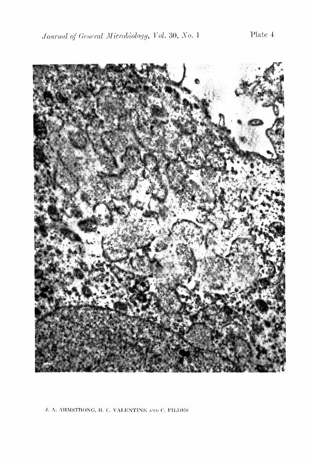
Journal of General Microbiology, Vol. 30, No. 1 Plate 4
J. A. ARMSTRONG, R. C. VALENTINE a n » C. FILERS

Journal of General Microbiology, Vol. 30, No. 1 Plate 5
J. A. ARMSTRONG, R. C. VALENTINE a n d C. EILDES

Journal of General Microbiology, Vol. 30, No. 1 Plate 6
J. A. ARMSTRONG, R. C. VALENTINE a n d C. TILDES

Journal of General Microbiology, Vol. 30, No. 1 Plate 7
,T. A. ARMSTRONG, R. C. VALENTINE a n d C. FILDES
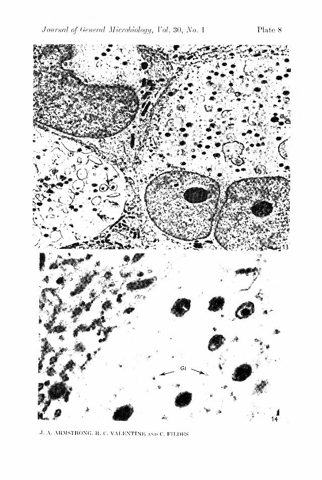
Journal of General Microbiology, Vol. 30, No. 1 Plate 8
J. A. ARMSTRONG, R. C. VALENTINE a n d C. FILDKS

Electron microscopy of the trachoma agent 73P late 5
Fig. 7. A small part of a 24 hr. inclusion at higher magnification: the main visible component is a network of line, electron dense fibrils. Remnants of the bilaminar membranes previously enclosing individual reticular foci are also present ( x 70,000).Fig. 8. Large developmental forms with complete limiting membranes, contained in a typical inclusion 48 hr. after inoculation. Note the presence of several fibrillar condensations (arrows) inside the developmental forms; in between the latter are a few apparently complete elementary bodies (EB) ( x 20,000).
P late 6Fig. 9. The structure of a discrete developmental form approximately 1 fi in diameter, at high magnification. Note the central fibrillar condensation, separated from the limiting membrane by a wide zone containing scattered granular elements ( x 70,000).Fig. 10. Profiles indicating the occurrence of unequal division, or budding, in a 48 hr. inclusion ( x 35,000).Fig. 11. Three paired profiles of relatively small developmental forms found in a section through a 72 hr. inclusion. They seem to indicate stages of division into equal halves ( x 50,000).
P late 7Fig. 12. Section through a cell containing an inclusion, 72 hr. after inoculation. The relationship of the inclusion (Inc) to the host cell is clearly displayed. Its contents include numerous elementary bodies; it is embedded in cytoplasm containing elements such as mitochondria and endoplasmic reticulum (ER). The cell nucleus (N) has been displaced but is structurally unaltered ( x 5500).
P late 8Fig. 13. Typical cells in a 72 hr. infected culture. In each, the cytoplasmic area is largely taken up by an enormous inclusion. The inclusion to the right in the micrograph has many mature elementary bodies; whereas that at the lower left still contains developmental forms of various sizes some of which show signs of division (arrow) ( x 6000).Fig. 14. Section showing the edge of a fully developed inclusion. No distinctive membrane is visible between the inclusion and the surrounding cytoplasm. Note that between elementary bodies the matrix contains moderately dense granular material (Gl), probably particulate glycogen ( x 35,000).

gen. Microbiol. (1963), 30, 75-88 With 1 platePrinted in Great Britain
75
Mycological Examination of Dust from Mouldy Hay Associated with Farmer’s Lung DiseaseB y P. H. GREGORY a n d MAUREEN E . LACEY
llothamsted Experimental Station, Harpenden, Hertfordshire(Received 25 April 1962)
SUMMARYSmall samples of hay were shaken in a perforated drum in a wind of4-2 m./sec.; the liberated dust cloud was sampled with the cascade impactor for microscopical examination, and with the Andersen sampler for identification of organisms in culture. The results of testing batches of hay showed large differences in microbial content. Twenty-eight batches classed as ‘good hay’ gave up to 3 million spores/g. dry wt. hay, mainly Aspergillus glaucus with Cladosporium spp. and Hemispora stellata. Dust blown from seventeen batches of ‘ mouldy hay ’ differed greatly in composition and had from 5 to 250 million spores/g.: abundant forms includedA. glaucus, A. fumigatus, A. nidulans, Penicillium spp., Absidia spp., Mucor pusillus, bacteria, many actinomycetes. Fourteen batches of mouldy hay associated with cases of ‘farmer’s lung’ disease were generally similar in mould content to the batches of mouldy hay, but had many more spores of Humicola (Monotospora) lanuginosa and A. fumigatus, and were specially characterized by very many actinomycetes. Farmer’s lung hays tended to neutrality (averaging pH 7-0, as compared with pH 5 to 6 of other hays), and were rich in thermophilic organisms, commonly with hundreds of millions of actinomycete spores/g., a large proportion of which grew readily at 60°. Hays associated with farmer’s lung appear to have heated spontaneously to a higher temperature during maturation than the other hays. Dust from all hays contained particles of higher plants, but there were not obviously more in farmer’s lung than in other hays.
All types of spores were shown to be potentially able to penetrate to the deeper parts of the lung (especially the spores of actinomycetes, A. fumigatus and H. stellata), but whether these organisms play any part in the aetiology of the disease is unknown.
INTRODUCTIONFarmer’s lung is a disease attributed to inhalation of dust from mouldy hay. This
disease (reviewed by Frank, 1958) has been known since 1932 (Fuller, 1958), but interest in it has increased recently, perhaps because of changes in haymaking practice caused by the pick-up baler. The aetiology of farmer’s lung is still obscure and the purpose of our work was to assess the possible role of the microbial components of the inhaled dust from mouldy hay. Previous research on spoilage of stored hay has been concerned mainly with self-heating (Browne, 1929) and spontaneous combustion (Miehe, 1930). The development of the microbial flora on grass composts has been studied by Webley (1947), Forsyth & Webley (1948) and by Eastwood (1948). Our work involved developing and testing of methods for sampling dust from hay, and examining farm hays (including both good hays and

hays associated with human and animal disease). The microfloral succession in experimental batches of hay baled at different moisture contents will be described later.
76 P. H. Gregory and M. E. Lacey
METHODSFarmer’s lung disease is attributed to the inhalation of dust from hay by farm
workers when feeding mouldy hay to cattle in cowsheds. Hays were therefore tested by shaking in air and taking samples of the air for examination. The standard method of shaking in water and plating out dilutions was considered inappropriate because it would remove many kinds of particles which remain firmly attached to dry hay and which do not normally enter the air-borne dust fraction. (The washing method was, however, used for comparison with the blowing method in early tests.)
The method adopted after preliminary trials (Gregory & Lacey, 1963) uses reproducible conditions to give a standard test by which various batches of hay can be compared. Because the primary pathological changes occur in the deeper parts of the lung, it was considered useful to classify the dust into two fractions: (1) ‘ nonpenetrating ’ larger particles which would be deposited mainly in the upper respiratory tract, nose, throat, bronchi and larger bronchioles; (2) ‘penetrating’ smaller particles which would be expected to reach the alveoli. Particles aerodynamically similar to unit-density spheres 5 /( diam. are considered to have a 50 % chance of penetrating to the alveoli, and this particle diameter was accordingly chosen to separate the two fractions, although the limiting size depends to some extent on the activity of respiration (Davies, 1952).
H a y s a m p l in g . Hay for test was normally received from the farm as whole or part bales and stored under cover at outdoor air temperatures (slices from bales were stored in large unsealed polythene bags). Samples for test were removed from the stored hay by pulling a handful from near the centre of the bale and carried to the laboratory in a small polythene bag with as little shaking as possible. (A 5-g. subsample was cut into 3 cm. lengths and occasionally stirred while in 250 ml. water for 15 min. before the pH value was measured electrometrically.)
S h a k in g in a ir . A sample of about 20-30 g. hay was tipped from the polythene bag into a cylinder of perforated zinc (2 mm. diam. perforations) with Perspex ends, which was mounted horizontally to rotate on bearings across a wind-tunnel of square cross-section with 29 cm. sides (Fig. 1). The cylinder was rotated during test at about 60 rev./min. by an electric motor to give a gentle ‘tedding’ action on the hay sample. Air was drawn down the wind-tunnel by a fan, a speed of about 4-2 m./sec. being adopted in routine tests. The dust cloud blown out of the cylinder travelled with the wind and reached the sampling position after a diffusion path of1-2 m.
A ir - s a m p l in g m e th o d s . A general study of hay dust must be based primarily on microscopic examination to reveal all its components, and must be supplemented by cultural examination for more precise identification of any organisms that can be grown in culture. For microscopic examination, air was sampled with a C'asella model of the cascade impactor (May, 1945), connected to a vacuum pump drawing 20 l./min. through the orifice. Sampling at this rate was approximately isokinetic at the wind speed used. The cascade impactor accelerates the air-stream through four successively narrowing jets, impacting the entrained dust particles, graded into
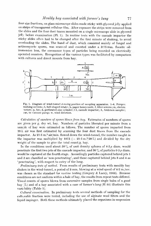
Mouldy hay associated with fanner's lung 77four size fractions, on glass microscope slides made sticky with glycerol jelly applied on strips of transparent cellulose film. After exposure the strips were removed from the slides and the four dust traces mounted on a single microscope slide in glycerol jelly before examination (PL 1). In routine tests with the cascade impactor the sticky slides often had to be changed after the first minute of shaking, to avoid overloading the slides. The band of dust, which consisted mainly of fungal and actinomycete spores, was scanned and counted under a 3-75 mm. fluorite oil- immersion lens, the commoner types of particles being recorded on electrically operated counters. Recognition of the various types was facilitated by comparison with cultures and direct mounts from hay.
Fig. 1. Diagram of wind-tunnel showing position of sampling apparatus. 1-4, Perspex working sections; b, bell-shaped intake; h, paper honeycomb; f, filter screens; m, electric motors; p, fan; d, perforated zinc cylinder; Cl, cascade impactor; A, Andersen sampler; vac, to vacuum pump; w, wind direction.
C a lc u la tio n o f n u m b e r s o f s p o r e s b lo w n f r o m h a y . Estimates of numbers of spores are given per g. dry wt. hay. Numbers of particles liberated per minute from a sample of hay were estimated as follows. The number of spores impacted from 20 1. air was first estimated by scanning the four dust traces from the cascade impactor. As 21-5 m.3 air/min. flowed down the wind-tunnel, the number caught in the impactor was multiplied by 1075 (= 21-5 m.3/20 1.) and divided by the dry weight of the sample to give the total count/g. hay.
In the conditions used about 50 % of unit density spheres of 4-5 //, diam. would penetrate the first two jets of the cascade impactor, and 30 % of particles 0-5 //, diam. would be captured at the fourth stage. Accordingly particles captured behind jets 1 and 2 are classified as ‘non-penetrating’, and those captured behind jets 3 and 4 as ‘penetrating’, with respect to entry of the lung.
P r e l im in a r y te s ts o f m e th o d . From results of preliminary tests with mouldy hay shaken in the wind-tunnel, a period of 3 min. blowing at a wind speed of 4-2 m./sec. was chosen as the standard for routine testing (Gregory & Lacey, 1963). Because conditions are not uniform within a bale of hay, the results from repeat tests differed. Visual counts of spores blown from successive samples from single bales of a good hay (L) and of a hay associated with a case of farmer’s lung (H 44) illustrate this variability (Table 1).
C u ltu r a l e x a m in a t io n . In preliminary tests several methods of sampling for the culturable fraction were tested, including the use of alginate wool filters and the liquid impinger. Both these methods ultimately placed the organisms in suspension
in water from which serial dilutions can be plated. However, with the mixture of organisms present in hay, the dilution stages proved completely unreliable; in one test a tenfold dilution series decreased numbers by only one fourth at each stage (compare Brierley, Jewson & Brierley, 1927). Dilution methods were therefore given up in favour of the Andersen sampler (Andersen, 1958). In this instrument the air enters a circular orifice and is drawn in succession through a series of six circular plates, each perforated with 400 holes through which spores are impacted directly on the surface of sterile medium in Petri dishes. Succeeding stages in the series have
Table 1. N u m b e r s o f s p o r e s b lo w n f r o m su c c e ss iv e s a m p le s ta k e n f r o m s in g le b a le s o f h a y
Good hay (L) Farmer’s lung hay (H 44)
78 P. H. Gregory and M. E. Lacey
ActinomycètesMoulds (and bacteria)
SporesDate
(millions/g. dry wt. hay)'
17. vi. 59 0-23 1-6919. vi. 59 007 0-5830. vi. 59 0-27 0-077. vii. 59 019 0-246. x. 59 003 0-24
ActinomycètesMoulds (and bacteria)
SporesDate
(millions/g. dry wt. hay)
8. i. 60 67-9 40011. i. 60 51-7 68620. i. 60 34-1 20530. iii. 60 1530 96930. vi. 60 69-2 276
progressively smaller holes; the largest particles are deposited in the first dish and the smallest in the last. The Andersen sampler was placed horizontally on the axis of the wind-tunnel with the orifice facing the wind (Fig. 1), and was operated at 25 1. air/min. According ~o Andersen, the first two stages with larger holes retain the ‘non-penetrating’ fraction, while stages 3 to 6 retain the ‘penetrating’ fraction, as defined above. Flow into the sampler is non-isokinetic and, at the wind speed of 4-2 m./sec., the speed of air-flow through the orifice is only 16% of the ambient tunnel wind speed; the largest particles in the non-penetrating range therefore are over-collected. On the other hand some of the large particles are lost by impaction on the front of the first perforated plate.
In an actual test of a sample of hay the cascade impactor was used during the first 3 min. of shaking, and then several sets of Petri dishes were exposed in the Andersen sampler during the fourth minute. After exposure, the sets of malt extract agar (2-0 % malt extract, 2-0 % agar, about pH 5-7), with 20 units penicillin/ ml. and 40 units streptomycin/ml. to suppress bacteria, were incubated at 25° and 40°. Plates of half-strength Oxoid nutrient agar (pH 7-4) with 0-5 mg. actidione/ml. to suppress moulds were incubated at 25°, 40° and 60°. All plates were poured the day previously to allow the surface to dry. Each set of six plates was exposed in the Andersen sampler for 15 sec., giving 5 1. of air through the apparatus (occasionally to avoid overcrowding only 5 sec. were given). Although exposures were made during the fourth minute of shaking after the cascade impactor runs, when most of the spores were blown off, the plates were frequently overloaded. This was shown by the presence of the maximum possible number of 400 colonies/plate, many of them multiple infections for which no allowance has been made in Tables 3 and 5 to 7; counts from the Andersen sampler are therefore underestimates.

Mouldy hay associated with farmer's lung 79RESULTS
E x a m in a t io n o f f a r m h a y sResults of examining different batches of hay are presented in four groups:
a good hays from a hay-drying experiment; b other good hays; c mouldy hays and hays associated with animal diseases; d farmer’s lung hays. Classification into groups b and c was made on the results of the examination, but classification into group d was based on the diagnosis of farmer’s lung in the patient by the physician. Table 2 shows the presence and abundance (over 2 million/g. hay) of spores in the different groups of hay as seen on the cascade impacto::1 slides. Table 3 shows the presence and abundance (over twenty-five colonies isolated) of organisms isolated from the four groups of hay with the Andersen sampler.
Table 2. P r e s e n c e o f s p o r e s c o u n te d o n th e c a sc a d e im p a c to r s l id e s(Brackets indicate number of samples containing over 2 million spores/g. dry wt. hay.)
Experimental Good Mouldy Farmer’shays, hays, hays, lung hays,
group a group o group c group dNc. batches tested
10 18 17 14Actinomycètes 10(3) 18 (5) 17 (15) 14 (14)A c rem o n ie lla a tra 0 0 1 2A lte r n a r ia spp. 1 1 1 0A s p e r g illu s and P é n ic i ll iu m spp. 10 (7) 16(5) 17 (7) 14 (7)C h a eto m iu m spp. 0 0 0 5C la d o sp o r iu m spp. 5 2 10 7E p ico ccu m spp. 1 0 -1 1H u m ic o la lan u g in o sa 0 0 9 13 (3)H . ste lla ta 0 0 2 3Mucoraceae 2 7(1) 13 (3) 11 (3)T rich o th ec iu m roseu m 0 2 3 5Number of categories 6(2) 6(3) 10 (3) 10 (4)
G r o u p a: g o o d h a y s f r o m a h a y -d r y in g e x p e r im e n t ( T a b le 4). In July 1958 batches of hay were dried experimentally at the National Institute of Agricultural Engineering, Silsoe, Bedfordshire. Samples were obtained and tested in February and March 1959. Spores of A s p e r g i l lu s g la u c u s and Pénicillium type were the ones present in large quantities, and occurred in the swath dried hays (H7, 10, 11). A few spores of Cladosporium, Alternaria, Epicoccum and of Mucoraceae were also present. These hays never had more than 3 million aitinomycete spores/g. hay. These batches of good hay were studied with alginate wocl filters before the Andersen sampler was used. The fungi isolated were mainly mesophilic, the most numerous were A . g la u c u s (especially in H 10) and P é n ic i l l iu m spp. A b s i d i a sp. was isolated from H 6 and 7, A . f u m ig a tu s from only H 8 and 13, and H e m is p o r a s te l la ta from H 5, 10, 11, and 13. Neither M u c o r p u s i l lu s nor H u m ic o la la n u g in o s a were isolated.
G r o u p b: o th er g o o d h a y s ( T a b le 5). Samples classed as ‘good hay’ had very different microbial contents, ranging from H 33, of which the farmer who sent it was justly proud, to others yielding several million spores/g. hay. The pH values varied from 4-5 to 6-5 (av. of 13 hays, pH 5-9). The range of microbial content was

80 P. H. Gregory and M. E. Laceygenerally similar to the experimental hays of Table 4. Some samples, however, contained more actinomycète spores than moulds. A s p e r g i l lu s g la u c u s was the most plentiful mould spore, especially in H 56, 58, 68 and 69. H e m is p o r a s te lla te1 was
Table 3. I s o la t io n o f o r g a n is m s in c u ltu re (Brackets indicate number of samples with over 25 isolations.)
Experimental Good Mouldy Farmer’shays, hays, hays, lung hays,
group a group b group c No. batches tested
group d
( 10 18 17 N14fungi
A b s id ia (mainly ram osa ) 2 15 13 (4) 13 (2)A . corym bifera 0 2 0 0A crem o n ie lla a tra 1 0 0 0A ite rn a r ia spp. 1 0 3 0A sp e rg illu s spp. (total) 8(1) 17 (6) 17 (10) 14 (10)
A . fu m ig a tu s 2 11 15 (5) 13 (5)A . g lau cu s 7(1) 15 (6) 12 (7) 12 (7)A . n id u la n s 1 13 11 (4) 8 (1)A . n iger 0 0 5 0A . ochraceus 0 0 1 0A . terreu s 0 2 6 4(1)A . versico lor 3 1 7(2) 7(1)
B o tr y tis spp. 5 1 3 3C a n d id a spp. 0 8 3 3C ep h a lo sp o riu m sp. 1 0 1 0C h aetom iu m sp. 1 3 5 4C la d o sp o riu m sp. 2 15(2) 13(4) 13(1)E p ico ccu m sp. 2 1 1 0F u sa r iu m sp. 0 0 1 0Geo-myces type 3 2 0 1H em isp o ra ste lla ta 4 3(3) 3(1) 0H u m ic o la lan u g in osa 0 6 4 9(3)I I . s te lla ta 0 0 0 1M u c o r p u s i l lu s 0 9 12 (3) 10(1)M . s p in u s 0 0 0 1P a ec ilo m yces sp. 0 6 3 (2) 9P a p u la r ia sp. 0 1 1 0P é n ic ill iu m spp. 8 10 13 (4) 10(1)P u llu la n o sp. 0 1 1 1R h izo p u s sp. 0 1 1 1S c o p u la r io p s is b rev ica u lis 3 1 3 4 (2)S p o ren d o n em a type 0 0 0 1S p o ro tr ich u m sp. 1 0 0 3S ta ch yb o try s sp. 0 0 0 1S ty s a n u s spp. 1 0 1 0T rich oderm a v ir id e 0 0 0 2T r ic h o th ed u m roseu m 0 1 3 (1) 0Yeasts 1 7 5 3 (1)Sterile mycelium or unidentified — 10 7 5
Number of categories 19 (1) 25 (3) 29 (11) 26 (12)Bacteria
24° orange — 3 6(4) 10 (3)others 1 16 (5) 16(3) 13 (4)40° B a c illu s cereu s-m ycoides — 11 16 (1) 13 (3)others — 13 17 (3) 11 (3)Number of categories 1 4 (1) 4 (4) 4 (4)

81Mouldy hay associated with farmer's lungTable 3 (c o n t.)
Experimental Good Mouldy Farm ershays, hays, hays, lung hays,
Actinomycetes group a group b group c group d24° 2 15 17(8) 13 (8)40° whitish — 15 (12) 17 (10) 15 (9)blue — 2 8 (2) 11 (4)
grey — 10(1) 12 12 (3)thin — 3 3 (1) 0small — 4(2) 11 (6) 13 (12)00° whits — 11 (3) 13 (7) 13 (11)small — 0 4 (3) 9 (9)
Number of categories 1 7(4) 8 (7) 7(7)Total number of categories 21 (1) 36 (8) 41 (22) 37 (23)
Table 4. M ic r o b ia l co n te n t o f g o o d h a y (g r o u p a) a r t i f ic ia l ly d r ie d a t N . I . A . E . , S i ls o e , B e d fo r d s h ire , in J u l y 1958
Visual examinationActino-
Moulds mycetesSpores
(millions/g. dry wt. hay)Hay ref. no. Treatment ---------- ----- A-----
H 5 (D. 1) Tunnel dried 4-5 11H 6 (D. 2) H ut rack dried 2-9 0-8H 7 (D. 3) Swath dried 32-3 2-4H 8 (C. 1) Tunnel dried 01 0-6H 9 (C. 2) H ut rack dried 21 0-9H 10 (C. 3) Swath dried 9-2 1-3H 11 (C. 4) Swath dried 10-3 2-5H 12 (E. 1) Barn dried 5-6 1-6H 13 (E. 2) H ut rack dried 2-8 0-5H 14 (E. 3) Swath dried 6-9 30
seen in H 37 and 69, and in H 56 0-68 million spores/g. hay were counted. Mucoraceae spores were seen in only seven of these hays (Table 2) and only in H 17 reached 2 million/g. The hays from groups a and b contain fungi, such as Clado- sporium, Epicoccum, Helminthosporium and Alternaria, which are the normal constituents of fine weather air-spora; but hays of poorer quality had moulded with A . g la u c u s , H . s te l la ta and P e n ic i l l iu m spp. In culture, the only abundant moulds were A . g la u c u s (H 37, 56, 58, 68, G, Y), C la d o s p o r iu m sp. (H 67, 68), and H . s te l la ta (H 37, 56 and 69 with 14,000 viable spores/g.). A few A . f u m ig a tu s were isolated from all except H 33, 37, B, G, and N, of A b s id ia , spp. from all except H 19, 33, and 71, and of M u c o r p u s i l lu s from half. Very few H u m ic o la la n u g in o s a were isolated from H 69, 71, B, G, L and Y. Bacteria and actinomycetes were isolated from most hays but even when shown ‘abundant’ (Table 6) were many fewer than in hays of groups c and d .
G r o u p c : m o u ld y h a y s ( T a b le 6). These included batches associated with diseases of farm animals and various other samples sent to us as mouldy, but excluding any
A G. Microb. x xx

samples sent as implicated in producing farmer’s lung disease; the latter are dealt with in group d . The pH value ranged from 4-0 to 8-3 ; the average of 13 hays (pH 6-5) was very similar to that of the good hays of group b. The hays of group c had very different microbial contents, and spores of moulds or actinomycètes (or both) usually numbered between 10 and 100 million/g. Aspergillus and Pénicillium type spores were numerous in H 27, 49, 59, 63, 66, 74, and 79, Mucoraceae were abundant in only H 27 and 76. H u m ic o la la n u g in o s a was present in H 1, 27, 40, 41, 49, 59, 61, 63 and 66, H . s te l la ta in H 27 and 41.
Table 5. M ic r o b ia l c o n te n t o f g o o d h a y s a m p le s , g r o u p bVisual examination
,--------- *--------- , From culture
82 P. H. Gregory and M. E. Lacey
Actino- ,--------------- *--------------- .Moulds mycet.es Actino-
Spores Moulds mycetes BacteriaHay ( millions/g. Sporesref. PH dry wt. hay) (thousands/g. dry wt. hay)no. Origin value ' '
H 17 Weybridge, Surrey, as fed to cows at — 2-75 1-34 — — —
Central Veterinary LaboratoryH 19 Plumpton, Sussex (normal hay fed to cows — 0-77 1-68 — ------ —
on a farm where mycotic abortion had occurred)
H 33 Leckhampstead, Newbury, Berks. — 017 0-22 — — —H 37 Dulverton, Som. (1959 hay ‘inactive’ ; 6-4 2-60 2-90 26-6 4-30 0-41
cf. H 36 (Table 7))H 38 Weymouth. Dorset (associated with case 6-5 3-2 2-4 — — —
of sarcoid)H 56 Lympsham, nr. Weston-super-Mare, 6-2 3-5 10 17-6 5-86 308
Somerset (associated with non-seasonal allergic hay fever and asthma)
H 58 Banwell, Somerset 6-3 5-44 8-17 7-39 7-87 0-76H 67 Beacon, Honiton, Devon, old ley, 1961 6-3 0-21 0-67 1-73 0-19 1-12H 68 Luppitt, Honiton, Devon, 2nd year ley, 1961 6-2 6-89 0-25 17-9 0-30 0-28H 69 Luppitt, Honiton, Devon, 3rd year ley, 1961 5-4 6-23 019 9-36 0-75 0-66H 71 Knighton, Radnorshire, 2nd year ley, 1961 6-2 0-23 1-89 1-18 0-72 0-85H 72 Knighton, Radnorshire, meadow hay, 1961 6-2 0-81 0-72 1-99 106 2-84H 73 Knighton, Radnorshire, 1st year ley, 1961 4-5 O il 0-41 0-97 0-36 1-25B Rothamsted, Harpenden, Herts., 1961 6 0 0 008 0-30 0-37 1-58G Rothamsted, Harpenden, Herts., 1961 6 3 0-76 007 13-9 0-94 0-82L Rothamsted, Harpenden, Herts., 1959 — 0-41 0-54 — — --N Rothamsted, Harpenden, Herts., 1959 — 2-21 5'6 — — —
Y Rothamsted, Harpenden, Herts., 1960 5 2 1-43 2-16 4-40 4-78 0-73In culture, many thermophilic moulds were abundant: A b s id ia spp. (H 61, 75,
76, 79), A s p e r g i l lu s f m n ig a tu s (H 1 , 63, 66, 76, 79), A . n id u la n s (H 1 , 66, 76, 79), M u c o r p u s i l lu s (H 59, 66, 79) and P a e c i lo m y c e s sp. (H 59, 75). Abundant meso- philic moulds were: A . g la u c u s (H 1, 46, 49, 53, 61, 66, 74), A . v e r s ic o lo r (H 59, 74), C la d o s p o r iu m sp. (H 49, 53, 59, 74), H e m is p o r a s te l la ta (H 74), P é n ic i l l iu m spp. (H 1, 74, 76, 79) and T r ic h o th e c iu m ro s e u m (H 79). H u m ic o la la n u g in o s a was isolated from H 1, 40, 53 and 63. Bacteria and actinomycètes were isolated from all samples. Bacteria isolated at 24° were abundant in H 1, 16, 59, 61, 66 and 74, and at 40° in H 1 , 16, 59 and 66. Actinomycètes isolated at 24° were abundant in H 1 , 16, 27, 59, 61, 63, 76 and 79; at 40° in H 1, 16, 27, 41, 59, 61, 63, 75, 76 and 79; and at 60° in H 1, 16, 59, 61, 63, 75 and 79.
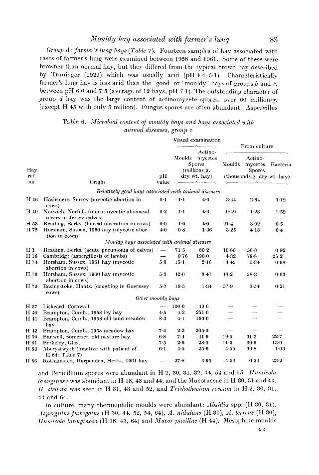
K K
WWW
G r o u p d: f a r m e r ' s lu n g h a y s (T a b le 7). Fourteen samples of hay associated with cases of farmer’s lung were examined between 1958 and 1961. Some of these were browner than normal hay, but they differed from the typical brown hay described by Truninger (1929) which was usually acid (pH 4-4-5-1). Characteristically farmer’s lung hay is less acid than the ‘good’ or ‘mouldy’ hays of groups b and c, between pH 6-0 and 7-5 (average of 12 hays, pH 7-1). The outstanding character of group d hay wras the large content of aetinomycete spores, over 60 million/g. (except H 45 with only 5 million). Fungus spores are often abundant. Aspergillus
Table 6. M ic r o b ia l c o n te n t o f m o u ld y h a y s a n d h a y s a s s o c ia te d w ith a n im a l d is e a s e s , g r o u p c
Visual examination,---------- *---------- , From culture
Actino- ,-----------------*-----------------,Moulds mycetes Aetino-
Spores Moulds mycetes Bacteria(millions/g. Spores
pH dry wt. hay) (thousands/g. dry wt. hay) value ---------- *---------- v ,---------------- f---------------- ,
Mouldy hay associated with farmer's lung 83
Relatively good hays associated with animal diseasesH 46 Haslemere, Surrey (mycotic abortion in
cows)61 11 4-0 3-44 2-64 1-12
H 49 Norwich, Norfolk (mucormycotic abomasal ulcers in Jersey calves)
6-2 11 4-6 9-49 1-23 1-52H 53 Reading, 3erks. (buccal ulceration in cows) 6 0 1 0 4 0 21-4 3-92 0-5H 75 Horsham, Sussex, 1960 hay (mycotic abor- 4 0 0-8 1-36
tion in cows)Mouldy hays associated with animal diseases
3-25 4-18 0-4
H 1 Reading, Berks, (acute pneumonia of calves) — 71-5 80-2 10-83 56-3 0-92H 16 Cambridge (aspergillosis of lambs) — 0-76 1900 4-32 79-8 25-2H 74 Horsham, Sussex, 1961 hay (mycotic
abortion in cows)5*8 151 2-16 4-45 0-34 0-98
H 76 Horsham, Sussex, 1960 hay (mycotic abortion in cows)
5-3 420 8-47 48-2 58-3 0-63H 79 Basingstoke, H ants, (coughing in Guernsey 5-7 19-5
cows)Other mouldy hays
1-34 57-9 0-54 0-21
H 27 Liskeard, Cornwall — 100-0 49-6 — — —
H 40 Bram pton, Cumb., 1958 ley hay 4-5 4-2 251-0 — — —H 41 Bram pton, Cumb., 1958 old land meadow
hay8-3 41 193-0 — —
H 42 Bram pton, Cumb., 1958 meadow hay 7-4 2-2 205-0 — — —H 59 Banwell, Somerset, old pasture hay 6-8 7-4 41-9 19-5 31-3 22-7H 61 Berkeley, Glos. 7-5 2-6 28-0 11-2 60-9 13-0H 63 Aberystwyth (inactive with patient of
H 64; Table 7)61 4-5 25-6 4-55 39-8 1-03
H 66 Rothams~ed, Harpenden, H erts., 1961 hay — 27-8 3-95 4-56 0-24 23-2and Penicillium spores were abundant in H 2, 30, 31, 32, 44, 54 and 55. H u m ic o la la n u g in o s i was abundant in H 18, 43 and 44, and the Mucoraceae in H 30, 31 and 44. H . s te l la ta was seen in H 31, 43 and 52, and T r ic h o th e c iu m r o s e u m in H 2, 30, 31, 44 and 64..
In culture, many thermophilic moulds were abundant: A b s id ia spp. (H 30, 31), A s p e r g i l lu s f u m ig a tu s (H 30, 44, 52, 54, 64), A . n id u la n s (H 30), A . te r re u s (H 30), H u m ic o la la n u g in o s a (H 18, 43, 64) and M u c o r j m s i l lu s (H 44). Mesophilic moulds
6 -2
H ayref.no. Origin

8 4that were abundant were: A . g la u c u s (II 2, 31, 32, 36, 44, 45, 65), A . v e r s ic o lo r (H 2), C la d o s p o r iu m sp. (H 30), P e n ic i l l iu m spp. (H 30), S c a p u la r io p s is b r e v ic a u l is (H 2, 31) and a yeast (H 32). H u m ic o la s te l la te was isolated from H 54 and a thermophilic S p o r o tr ic h u m sp. from H 18, 44 and 52. Abundant bacteria were isolated at both 24° and 40° from H 30, 31, 32, 44 and 64. Actinomycetes isolated at 24° were abundant in H 2, 30, 31, 36, 44, 52, 54 and 64; at 40° and 60° in all hays except II 45 (and II 52 which was not tested at 60°). Although H 45 seems out of place in this group, the diagnosis of farmer’s lung was well established and the discrepancy may
Table 7. M ic r o b ia l co n te n t o f f a r m e r s lu n g h a y s , g r o u p d
P. H. Gregory and M. E. Lacey
Visual exam ination From culture______________A_______________ _______________ A----------------
Actino- I I . la n u - Actino-Moulds mveetes g in o s a Moulds mveetes Bacteria
H ay Spores Sporesref. pH (millions/g. dry w t. hay) (thousands,'g. dry wt. hay)no. Origin value r r
H 2 W itney, Oxfordshire (acute 7-5 110 1340 0-08 11-3 70-3 1-01thresher’s lung)
II 18 Near Aberystwyth 7-0 3-6 1680 1-10 4-84 16-2 4-72H 30 Whitchurch, Shropshire 6-0 86-2 1320 13-8 143-2 103-3 53-85H 31 Broadwoodkelly, nr. Winkleigh, 7-6 120 3630 0-64 6-24 60-7 12-52
Devon (1958 hay)II 32 Broadwoodkelly, nr. Winkleigh, 7-3 7-8 4800 0-97 18-7 85-7 9-45
Devon 0959 hay)H 36 Dulverton, Som. (1958 hay 6-6 91 91-3 0 13-4 216-6 5-78
‘active’) cf. H 37, Table 5H 43 Awliseombe, nr. Exeter, Devon 6-6 7-4 1232 2-4 14-4 152-1 1-48H 44 Inwardleigh, nr. Okehampton, 7-4 1020 827-0 11-2 101-8 208-3 21-01
DevonH 45 Lampeter, mid-Cardiganshire 6-7 1 0 5 0 0-1 3-23 1-57 0-6S
(farmer's lung of young lad)II 52 Mid-Wales — 1-5 6330 0-24 10-5 28-0 43-67H 54 Longnor. Staffordshire 7-4 16-6 78-3 2-87 4-07 24-7 2-28H 55 Cardiff — 12-4 123-0 0 3-75 23-2 0-57II 64 Talybont. nr. Aberystwyth, 7-5 2-3 175-0 0-83 9-14 81-5 4-10
1960 ‘active’ hay (cf. H 63, Table 6 1 (farm er’s lung for 20 years)
H 65 Matlock, nr. Chesterfield, 7-2 1-4 040 0-12 8-04 37-2 2-20Derbyshire
mean either that the sample sent from the farm was taken accidentally from the wrong batch of hay, or the sample sent may have been unrepresentative of the batch because of the variability within the batch from uneven drying.
P r o p o r t io n o f th e r m o p h il ic o r g a n is m sIn comparison with good hay, the mouldy hay group c and the farmer’s lung
group d were characterized by many more thermophilic moulds and actinomycetes, as shown by incubation of Andersen sampler plates at different temperatures. These average values (Fig. 2), however, were calculated from individual hays which differed among themselves over a wide range of values, were uncorrected for multiple infection, and included some organisms which grow at more than one temperature. The mucoraeeous moulds were more numerous at 40° than at 24°, and in mouldy

ha> s (group c) than in farmer’s lung hays (group d ) . A s p e r g i l lu s f u m i g a tu s was isolated in larger numbers at 40° than at 24°, and H u m ic o la la n u g in o s a only at 40°. The relatively few bacteria were isolated mainly at the lower temperatures. The most striking effect was the very large increase in thermophilic actinomycètes in farmer’s lung hays (group d ).
Mouldy hay associated with farmer's lung 85
Total
Absidi a spp. Mucor pusillus Aspergillus Aspergillus fumigatus glaucus HumicolalanuginosaFig. 2. Numbers of organisms isolated a t 24° (solid), 40° (dotted) and 60° (open), averages
for: (b ) good hays; (c) mouldy bays; and (d ) farm er’s lung hays.
O th er c o m p o n e n ts o f d u s tsThe visual inspection of the first slide from the cascade impactor showed a good
deal of coarse material other than the spores of micro-organisms. This material included fragments of plants, plant hairs, cell walls, faecal pellets of mites (packed with mould spores), cast skins of mites, as well as whole conidial heads of Aspergillus and sporangia of Mucoraceae (the last help to substantiate identification of the Mucoraceae spore type among the finer fractions). Few of these particles penetrated beyond the first stage of the impactor and material deposited in the later stages was predominantly microbial spores. The better hays seemed to have more non-spore dust than the mould)' hays, possibly because the process of becoming mouldy tended to bind the loose particles in a weft of mycelium. Plate 1 shows typical fields of cascade impactor slides of a good hay (G) and a farmer’s lung hay (H 44).
P o s s ib i l i t y o f p e n e tr a t io n o f p a r t i c le s in to lu n g s The visual and cultural methods of assessing potential alveolar penetration in
general agreed. For the two methods, Fig. 3 shows graphically the % of particles retained by the four cascade impactor slides and the six Andersen sampler plates. The values obtained are combined averages of results from mouldy and farmer’s lung hays (groups c, d ) . According to visual estimates, only 8 % of spores of H u m i-

cola lanuginosa and the Mucoraceae penetrated beyond stage 2 of the cascade impactor, but 43 % of Aspergillus and 55 % of actinomycete spores were ‘ penetrating’, by the criteria mentioned under Methods. According to cultural tests, however, a larger proportion of spores were ‘penetrating’, namely, 60% of the Mucoraceae and over 80 % of the other groups. This discrepancy is partly attributed to the fact that the first two plates received clumps of spores (or even whole sporangia as in stage 1 of Mucoraceae) which, although they contained many spores, produced only one colony. Further, discrimination between single spores of Mucoraceae and members of the Aspergillus-Penicillium type was sometimes difficult; although spores of Absidia spp. are easily recognized, some of Mucor pusillus may have been misclassified, suggesting that the Mucoraceae types (Fig. 3) appeared to be less
Stages Actinomycetes A spergillus spp. Mucoraceae H um ico la lanuginosa
86 P. H. Gregory and M. E. Lacey
i i i i i i i i i i i i i i i i i i i i i i I I I l i l— i— i— 1— 1— 1— 1 _ L - i —i40 20 0 20 40 20 0 20 40 40 20 0 20 40 40 20 0 20 40 % spores reaching each stage
Fig. 3. Percentage penetration of spores to the four stages of the cascade im pactor (C l; dotted), and to the six stages of the Andersen sampler (A ; solid), average of groups c and d .
penetrating than they actually were. Spores of Humicola lanuginosa are about 9 p diam. and the maximum proportion of 42 % were found on stage 3; those of Aspergillus are about 4 p and the maximum of 38 % was found on stage 4, but 30 % penetrated as far as stage 5. Hemispora stellata has smaller spores, most were found on stage 5 and hardly any on stage 6. Spores of the actinomycetes penetrated deeply, 27 % reaching stage 5, and 23 % stage 6. Evidently many single spores of fungi and the majority of actinomycete spores could penetrate to the alveoli.
DISCUSSIONOur standard method of examining hays gives results that are reproducible
enough to characterize batches of hay. Standardizing the humidity of the air in the wind tunnel would be a desirable improvement, as Zoberi (1961) found that humidity greatly affected the numbers of dry spores liberated into wind; but since a bale of hay is a concretion of micro-environments, errors from differences between bales and between parts of a single bale (Table 1) are likely to outweigh the humidity factor. Because the dust load is so easily shaken out, it was not considered feasible to work with a subsample taken after thoroughly mixing several separate samples.

8 7Incidental to the main object of the work, the Andersen sampler proved an
excellent tool for isolating thermophilic actinomycetes from natural substrata. Dilution in air, instead of in water by the traditional manner, and impacting the spores from suspension in air directly on to the previously dried surface of halfstrength nutrient agar containing actidione, prevented contamination by bacteria and moulds, and on incubation overnight at 60° gave numerous colonies of thermophilic actinomycetes (Gregory & Lacey, 1962).
The proportion of spores seen on cascade impactor slides which was actually obtained in culture with the Andersen sampler was always small, and averaged about one in a thousand, even after allowing for the fact that two-thirds of the spore load had teen lost from the sample during the 3 min. blowing period for the cascade impactor run before the 15 sec. run with the Andersen sampler started (Table 8). Reasons for this small % recovery in culture include the following: (1) no correction was applied for multiple infection, so that the counts tended to be gross underestimates, especially when nearly the maximum number of 400 colonies are recorded on one plate; (2) the media, aeration conditions and isolation temperatures may have been unsuitable for some organisms; (3) slow-growing organisms may have been overgrown or antagonized on the crowded plates; (4) many spores may have been dead. Table 8 shows that the percentage obtained in culture was larger with the better-quality hays. From these tests it is clear that both the mouldy and farmer’s lung hays (groups c and d ) are important natural reservoirs of several moulds reported to be pathogenic to man or animals, e.g. A b s i d i a r a m o s a and A . c o ry m b ife r a , A s p e r g i l lu s f u m ig a tu s , H e m is p o r a s te l la ta , M u c o r p u s iU u s , S c o p u la r io p s i s b re v i- c a u l is and S ta c h y b o tr y s sp.
Table 8. N u m b e r s o b ta in e d i n c u ltu re o n A n d e r s e n s a m p le r p la te s a s p e r c e n ta g e (%) o f s p o r e s re c o rd e d v i s u a l l y o n c a sc a d e im p a c to r s l id e s
Mouldy hay associated with farmer's lung
ActinomycetesMoulds and bact<
Hays AA)------------\
Group b 0-34 0-22Group c 010 O ilGroup d 014 003
The tests show it possible to characterize farmer’s lung hays (group d ) . They had clearly heated spontaneously and become mouldy during maturation, and tended to be neutral in contrast to the low pH values of good hays or other mouldy hays examined. They had many more thermophilic organisms, including A s p e r g i l lu s
f u m ig a tu s , and H u m ic o la la n u g in o s a , than other mouldy hays, and were rich in thermophilic actinomycetes (especially at 60°), which were often seen microscopically to be growing vigorously on the hay and on mould hyphae and spores. Several hays of the mouldy group c might well have been classified as farmer’s lung hays had they come into contact with a susceptible patient. In spite of their abundance, and evidence of their potential ability to penetrate to the alveoli, it is uncertain whether any of these organisms play any direct part in the aetiology of farmer’s lung disease, but the possibility that the actinomycetes are active merits serious attention in view of the occurrence of the disease after inhalation of white dust from mouldy hay reported by Campbell (1932) and Frank (1958, p. 202).
88We thank the Agricultural Research Council, at whose request this study was
undertaken, for a grant to start the wore; also Mr P. K. C. Austwick (Central Veterinary Laboratory, Weybridge) for help throughout the work. For batches of hay to test we are indebted to the following: Mr D. E. Amesbury (Newbury), Dr T. A. Blyton (Chesterfield), Dr D. 0. Clarke (Weston-Super-Mare), Dr A. W. Frankland (St Mary’s Hospital, Paddington), Dr C. J. Fuller (Exeter), Mr M. Jenkins (Brampton), Dr S. T. H. Jenkins (Aberystwyth), Messrs F. S. Mitchell and G. Shepperson (N.I.A.E., Silsoe), Mr L. Ögilvie (N.A.A.S., Bristol), Mr J. Sage (Honiton), Dr R. M. E. Seal (Penarth), Mr L. Thomas (Knighton), Mr J. W. Thwaite (Liskeard), and Dr J. McA. Williams (Hartington). We also thank Messrs V. Stans- field and F. Cowland for photography, and Miss M. Goodson for technical assistance.
REFERENCESA n d e h s e n , A . A. (1958). N ew sam pler fo r th e collection , sizing, a n d en u m era tio n of
v iab le a irb o rn e p a rtic les . J . B a d . 7 6 , 471.B e ie e l e y , W. B., J ew so n , S. T. & B r ie r l e v , M. (1927). The q u a n tita tiv e s tu d y o f soil
fungi. P ro c. I n t. C ongr. S o i l S e i., W a sh in g to n , 3, 48.B r o w n e , C. A. (1929). The spontaneous com bustion of hay. U .S .D .A . T ech n ica l B u ll.
no. 141, 38 pp.Ca m pb e ll , J . M. (1932). Acute symptom s following work w ith hay. B r it . m ed. J . 2 , 1143. D a v ie s , C. N. (1952). D ust sampling and lung disease. B r it . J . In d u s tr . M ed . 9 , 120. E a stw o od , D. J . (1948). The fungus flora of composts. T ra n s. B r it , m yco l. Soc . 3 5 , 215. F o r sy th , W. G. C. & W e b l e y , D. M. (1948). T ie microbiology of composting. II . A study
of the aerobic therm ophilic bacterial flora developing in grass composts. P ro c . Soc . a p p l . B a d . 11,34.
F r a n k , R. C. (1958). Farm er’s lung—a form of pneumoconiosis due to organic dusts. A m e r. J . R oentgenol. 79 , 189.
F u l l e r , C. J . (1958). Farm er’s lung; in F u n g o u s D isea se s a n d T h e ir T rea tm en t, p. 138.Ed. R. W. Ridell & G. T. Stewart, L ondon: B utterw orth.
Gr eg o r y , P. H . & L a c ey , M. E. (1962). The isolation of thermophilic actinomycetes. N a tu re , B on d. 195, 95.
Gr e g o r y , P. H . & L a c e y , M. E . (1963). L iberation of spores from mouldy hay. T ra n s. B r it , m yco l. Soc . (in the Press).
Ma y , K. R . (1945). The cascade im pactor: a r instrum ent for sampling coarse aerosols. J . S e i. In s tru m . 22, 187.
Mie h e , H . (1930). Ü ber die Selbsterhitzung des Heues. (2. Aufl.) A rb . dtsch. L an d ioG . 1 9 6 ,1 , T r u n in g e r , E. (1929). Allgemeine Beobachtungen und Untersuchungen über Wesen,
Ursache und Verlauf der Selbsterhitzung und Selbstentzündung von D ürrfutter. L a n d w . J b . S ch w eiz, 43 , 278.
W e b l e y , D. M. (1947). The microbiology of composting. I. The behaviour of the aerobic mesophilic bacterial flora of composts and its relation to other changes taking place during composting. P ro c . Soc . a p p l. B a d . 10 , 83.
Z o b e r i, M. H. (1961). Take-off of mould spores in relation to wind speed and hum idity . A n n . B o t., L o n d . 25 , 53.
P. H. Gregory and M. E. Lacey
EXPLANATION OF PLATE 1Photomicrographs ( x 600) from cascade impactor slides 1, 2, 3 and 4 to compare a good hay with a farmer’s lung hay.
a Farmer’s lung hay from Devon (H 44). b Good hay from Rothamsted Experimental Farm (G, 1961).

J o u r n a l o f G enera l M ic ro b io lo g y , Vol. 30, .Vo. 1 Plate 1
1>. II. GREGORY A M ) M. K. LACEY (Facing p. 88)

J . Gen. Mi-robiol. (1963), 30, 89-103 Printed in Great Britain
8 9
Recovery of Deoxyribonucleic Acid from the Effects of Alkylation
By B. S. STRAUSSDepartment of Microbiology, The University of Chicago, Chicago 37,
I l l in o i s , U .S .A .
( R e c e iv e d 30 A p r i l 1902)SUMMARY
The transforming activity for an indole locus of DNA extracted from a methionine-requiring strain of Bacillus subtilis was decreased about tenfold per unit of DNA by treatment of organisms with 0-025 M-methylmethane- sulphonate for 30 min. at 37°. The treatment decreased the viable count to one tenth of the original and induced a reversion frequency of 10 A Transforming activity of DNA prepared from alkylated organisms was sensitive, to heating at 50° as compared to DNA extracted from non-alkvlated organisms. Methylation and heating of DNA in vitro did not inhibit its uptake by competent organisms. Incubation of treated and non-treated organisms in amino acid supplemented medium led to an increase in extractable transforming activity. A period of incubation which led to a 34 % increase in DNA content more than doubled the transforming activity obtainable from methyl methanesulphonate treated organisms. The heat sensitivity of transforming DNA from alkylated organisms decreased after incubation of the organisms. Synthesis of DNA in the absence of a net DNA increase was demonstrated in methyl methanesulphonate treated organisms since thyrr_idine-2-14C was incorporated without lag from the start of incubation into an alkali-stable hot acid-soluble form. The experiments are interpreted to mean that alkylated DNA can serve as a template for the replication of DNA of normal activity.
INTRODUCTIONOne of the questions to be answered about any mutagenic agent is whether its
reaction with the genetic material (deoxyribonucleic acid, DNA) of an organism is direct or : ndirect. If the mutagen methylmethanesulphonate reacted directly with DNA for example, it would be possible to show that immediately after treatment of organisms with alkylating agent, the DNA of the organism became heat sensitive (Strauss, 1962a). Since such a determination would require a biological assay for DNA activity, the experiments would have to be done with an organism capable of undergoing transformation. It seemed likely that the system described by Spizizen (1958) and Anagnostopoulos & Spizizen (1961) with B a c i l lu s s u b t i l i s would be particalarly satisfactory because of the possibility of obtaining auxotrophic mutants of this organism similar to those of E s c h e r ic h ia c o li. The experiments to be described here show that treatment of B . s u b t i l i s with an alkylating agent resulted in heat sensitization of the DNA extractable from the treated organism. In addition, the DNA of methylated organisms gradually lost its abnormal heat sensitivity as these organisms were incubated in growth medium. Alkylated DNA appears to serve as a template for the production of normal DNA.

90 B. S. Strauss
METHODSSeveral strains of Bacillus subtilis were used. The first, a methionine-requiring
mutant, was obtained by replica plating from a culture of the prototrophic B. subtilis which had been treated with ethylmethanesulphonate. This strain was used as a DNA donor for the transformations described in this paper with the exception of transformation with 32P labelled DNA when a wild type donor was used. The methionine-requiring strain was selected because it could be induced to revert by treatment with methylmethanesulphonate (Fig. 1) as well as with the ethyl derivative, and because it would serve as a recipient strain for transformation as well as being a satisfactory donor. The strain was heat sensitive after methylation
Table 1. Loss of the differential heat sensitivity due to alkylation upon incubation of a methionine-requiring mutant of Bacillus subtilis
A washed suspension of cells from an overnight culture was treated for 30 min. at 37° with 0 025 M-methylmethanesulphonate in salts solution (0 043 ml. methylmethane- sulphonate/20 ml. of salts solution). After treatment the organisms were washed and suspended in 10 ml. of salts solution. Two ml. were then added to flasks containing 38 ml. of minimal medium (Spizizen, 1958) supplemented with 0-02 % casein hydrolysate, and 50 /(g./ml. each of L-methionine and l-tryptophan. The flasks were incubated for the indicated time at 37° with shaking and then the contents were collected by centrifugation and washed with salts solution. The organisms were suspended in 5 ml. of salts solution and plated for viability before and after a heat shock of 50 for 6 min. The organisms w ere cooled in an ice bath after heating and were kept cold until plated. Viability was determined on minimal medium supplemented with 50 mg. of methionine and 20 mg. of yeast extract /1. and hardened by the addition of 1-5 % agar.
Viable organisms (no. x 10~8)Incubation After washing
time in in saltssupplemented After wasliing solution
minimal in salts and heatingmedium solution 0 min. at 50° RatioPre-treatment of organisms (min.) («) (6) a,'b:
None 0 4-8 0-32 1510 71 0-5 1430 3-9 0-44 8-9
Methylmethane 0 0-81 000053 1530sulphonate (0025 m) 10 11 0-009 12230 1-2 012 10
as indicated by the inactivating effect of a 6 min. incubation in buffer at 50° and the differential heat sensitivity disappeared after the start of incubation in minimal medium (Spizizen, 1958) supplemented with 0-02% acid casein hydrolysate and 50 fig./ml. each of L-tryptophan and L-methionine (Table 1). As the recipient strain for transformation we used the histidine + tyrosine + indole requiring triple mutant described by Nester & Lederberg (1961). This strain, supplied by Dr E. Nester, was used to permit 7he simultaneous determination of the effects of alkylation on linked markers. In one series of experiments utilizing DNA labelled with 32P, wild type Bacillus subtilis was used as the DNA donor and the indole requiring strain (168) was used as recipient.
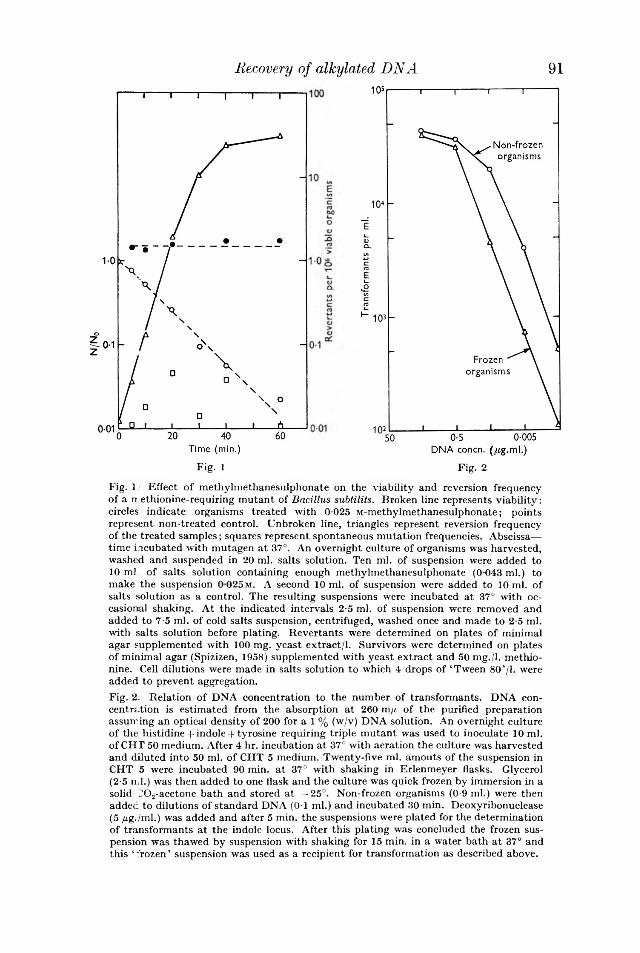
N/N
0
Recovery of alkylated D N A 91
Fig. 1 Fig. 2Fig. 1 Effect of methylmethanesulphonate on the viability and reversion frequency of a n ethionine-requiring mutant of Bacillus submits. Broken line represents viability; circles indicate organisms treated with 0-025 M-methylmethanesulphonate; points represent non-treated control. Unbroken line, triangles represent reversion frequency of the treated samples; squares represent spontaneous mutation frequencies. Abscissa— time incubated with mutagen at 37°. An overnight culture of organisms was harvested, washed and suspended in 20 ml. salts solution. Ten ml. of suspension were added to 10 ml of salts solution containing enough methylmethanesulphonate (0 043 ml.) to make the suspension 0 025 m. A second 10 ml. of suspension were added to 10 ml. of salts solution as a control. The resulting suspensions were incubated at 37° with occasional shaking. At the indicated intervals 2-5 ml. of suspension were removed and added to 7-5 ml. of cold salts suspension, centrifuged, washed once and made to 2-5 ml. with salts solution before plating. Revertants were determined on plates of minimal agar supplemented with 100 mg. yeast extract/1. Survivors were determined on plates of minimal agar (Spizizen, 1958) supplemented with yeast extract and 50 mg./l. methionine. Cell dilutions were made in salts solution to which 4 drops of ‘Tween 80’/I. were added to prevent aggregation.Fig. 2. Relation of DNA concentration to the number of transformants. DNA concentration is estimated from the absorption at 260 m/« of the purified preparation assuming an optical density of 200 for a 1 % (w/v) DNA solution. An overnight culture of the histidine + indole + tyrosine requiring triple mutant was used to inoculate 10 ml. of CHT 50 medium. After 4 hr. incubation at 37° with aeration the culture was harvested and diluted into 50 ml. of CHT 5 medium. Twenty-five ml. amouts of the suspension in CHT 5 were incubated 90 min. at 37° with shaking in Erlenmeyer flasks. Glycerol (2-5 nil.) was then added to one flask and the culture was quick frozen by immersion in a solid l'02-acetone bath and stored a t —25°. Non-frozen organisms (0-9 ml.) were then added to dilutions of standard DNA (0-1 ml.) and incubated 30 min. Deoxyribonuclease (5 /¿g./ml.) was added and after 5 min. the suspensions were plated for the determination of transformants a t the indole locus. After this plating was concluded the frozen suspension was thawed by suspension with shaking for 15 min. in a water bath at 37° and this ‘ frozen ’ suspension was used as a recipient for transformation as described above.

B. S. StraussTransformation was by slight modification of the method of Anagnostopoulos &
Spizizen (1961). An overnight culture was inoculated into liquid minimal medium (Spizizen, 1958) containing 0-5% glucose, 0 02% acid hydrolysed casein and 50 fig. L-tryptophan/ml. (CHT 50 medium). After 4 hr. incubation with aeration at 37° the culture was harvested by centrifugation and diluted ten-fold in fresh minimal medium containing 0-5 % glucose, 0-01 % casein hydrolysate, 5 //g./ml. L-tryptophan and an additional 5 /m/ml. of MgS04 (CHT 5 medium). The organisms were incubated for 90 min. at 37° with shaking in 25 ml. of CHT 5 medium in a 250 ml. Erlenmeyer flask. Glycerol (2-5 ml.) was then added and the organisms were frozen by immersion of the flask in a solid C02-acetone mixture. Frozen organisms were stored at —25° for up to 10 days before use. During this storage period the competence of the frozen suspension gradually decreased to about one-tenth the original value for the frozen organisms. Frozen organisms were used for transformation as follows: the suspension was thawed by shaking for 15 min. in a water bath at 37° and then 0-9 ml. of suspension was added to a series of 150 x 16 mm. test tubes containing 0-1 ml. of the DNA solution to be assayed. The mixture of DNA plus organisms was shaken for 30 min. at 37° before addition of deoxyribonuclease (Worthington Biochemical Co.) to 5 /¿g./ml. The mixture was incubated with deoxyribonuclease for 5 min. at 37° before being plated for transformants. Transformation was scored on minimal medium (Spizizen, 1958) hardened with1-5% agar and containing 20 mg. of Difco yeast extract/1. When only indole transformants were scored this medium was supplemented with 150 mg. casein hydrolv- sate/1. When histidine and tyrosine transformants were scored the minimal medium plus 20 mg. yeast extract/1. was supplemented with the proper combination of L-tryptophan (20 /¿g./ml.), L-tyrosine (20 /¿g./ml.) or m.-histidine (40 /¿g./ml.). Viable cell count was determined on minimal medium supplemented with 0 02% casein hydrolysate, 0-02% yeast extract and 20 fig. L-tryptophan/ml. Freezing decreased the amount of transformation obtainable from a given DNA preparation (Fig. 2) and the total percentage transformation was low (0-01-0-03) in our procedure using the triply auxotrophic mutant. However, the response to DNA was linear and reproducible.
Purified DNA for transformation experiments was prepared by the method of Marmur (1961) and was treated with alkylating agent in vitro by diluting the DNA in 0-1 m phosphate buffer (pH 7-0) containing either methyl- or ethvl-methane- sulphonate to give a final concentration of DNA of 60 /¿g./ml. and allowing the reaction to proceed at 37°. Two volumes of 95 % ethanol in water were then added and the suspension centrifuged after standing in the cold for 15 min. The precipitate was taken up in 0-015 n-sodium chloride + 0-0015 M-trisodium citrate (dilute saline + citrate; Marmur, 1961), the solution made 0-15 n-NaCl-l-0-015 M-citrate (saline-f citrate) and the DNA reprecipitated with ethanol and taken up in saline + citrate once again. This procedure was considered necessary to remove unreacted alkylating agent. DNA labelled with 32P was prepared by growing wild type Bacillus subtilis in low phosphorus medium supplemented with 32P 0 4 as described by Young & Spizizen (1961 ). The DNA was extracted from these organisms by the method of Marmur (1961).
In some of these experiments, organisms were treated with the mutagen and the DNA extracted from treated cells by a modification of the method of Stuv (1960)
9 2

9 3who extracted DNA from an Haemophilus after X-ray irradiation. Bacillus subtilis was incubated overnight on slopes of a minimal salts medium (Spizizen, 1958) supplemented with yeast extract (0-15%), acid hydrolysed casein (0-1%), Difco nutrient broth (0-8%) and DL-tryptophan (40 mg./l.). This minimal salts medium had the following composition per litre of medium: (NH4)2S04, 2 g.; MgS04.7H20, 0-2 g.; K2H P04, 14 g.; KH2P 0 4, 6 g.; Na3C9H50 7.2H20, 1 g.; glucose (sterilized separately), 5 g. This solution without glucose will be called ‘salts solution’ throughout this paper. Organisms were harvested by suspension in salts solution which was passed through a gauze filter to remove agar fragments. The organisms were washed and resuspended in salts solution and portions of the suspension were then added to salts solution in which sufficient methylmethanesul- phonate was dissolved to make the final solution 0-025 m (0-085 ml./40 ml.). After reaction for 30 min. at 37°, the suspensions were harvested, suspended in 0-15 M-NaCl + 0-1 u-ethylene diaminetetra-acetate (pH 8; saline EDTA), centrifuged and suspended in a small volume of saline EDTA. Crystalline lysozyme was added to 1 mg./ml. and the suspension incubated for 30 min. at 37°. Sodium laurvl sulphate (0-5 ml. of a 25 %, w/v, solution) was added, followed in 5 min. by sodium perchlorate to make the solution m . After deproteinization by shaking for 30 min. at room temperature with chloroform and isoamyl alcohol (24 + 1) the suspension was centrifuged for 10 min. at 5000 g and the upper layer transferred to a centrifuge tube. The tube was cooled and two volumes of cold 95% (v/v) ethanol added; after mixing and standing for 15 min. in the cold the suspension was centrifuged for 10 min. at 25,000 g, the supernatant poured off and the residue taken up in dilute saline citrate. The solution was made 0-15 M-NaCl + 0-015 M-citrate and clarified by centrifugation. The clarified supernatant was used as crude DNA extract.
DNA was determined by the diphenvlamine reaction (Burton, 1956) with purified salmon sperm DNA as a standard. When small amounts of DNA were to be determined, 2 mg. bovine serum albumin were added as a carrier for the acid precipitate. Thymidine-2-14C incorporation into organisms was determined on Millipore membrane filters as described by Kahan (1960). DNA was identified as material which could still be precipitated by cold perchloric acid after incubation for 1-5 hr. at 37° in x-NaOH but which was solubilized by treatment at 70° for 30 min. in 0-5 x'- perchloric acid.
Recovery of alkylated D N A
RESULTSPurified DNA preparations were inactivated exponentially by heating at 50°
after alkylation in vitro with either ethyl- or methyl-methanesulphonate (Fig. 3). Alkylation decreased the initial transforming activity and methylmethanesulpho- nate caused a greater inactivation on a molar basis than did the ethyl derivative. Inactivation of the linked indole and histidine markers occurred at the same rate on heating alkylated DNA. Labilization of DNA to heat after preliminary alkylation was first demonstrated by Zamenhof, Leidy, Hahn & Alexander (1956) who showed a complete inactivation of alkylated Haemophilus DNA after heating for 1 hr. at 76c. The decrease in activity of alkylated DNA and of DNA alkylated and heated for 5 hr. at 50° was not due to the failure of competent cells to take up this treated DNA (Table 2). DNA labelled with 32P was fixed in a deoxyribonuclease-resistant form almost as efficiently after alkylation, and alkylation and heating as was control

labelled DNA in spite of the great difference in transforming efficiency of the treated and untreated preparations. In this experiment competent organisms of the indole- requiring strain (168) were prepared by the method of Young & Spizizen (1961) and used without preliminary freezing. Similar results showing that Pneumococcus transforming DNA inactivated by nitrogen mustard is taken up by competent cells have been obtained by Lerman & Tolmach (1959).
To determine whether alkylation and heating led to drastic degradation, the sedimentation coefficient of Bacillus subtilis DNA was determined in the Spinco
94 B. S. Strauss
Fig. 3. Effect of heating a t 50° on transform ing activity. A , control, non-treatcd D N A :B , DNA which has been treated w ith 0 025 M-methvlmethanesulphonate and freed from excess alkylating agent by precipitation twice with ethanol as described in the te x t ;C, DNA treated with 01 M-ethylmethanesulphonate and treated as in B ; D , DNA treated with 0-2 M-ethylmethanesulphonate and treated as in B . Four ml. of DNA a t a concentration of 60 /(g./ml. were reacted w ith alkylating agents for 1 hr. a t 37’ in 0 1 M-phosphate buffer (pH 7 0). After precipitation w ith ethanol and redissolving the precipitate in dilute saline + citrate the samples in 0 15 M-NaCl-f- 0 015 M-citrate were diluted 1:10 in salts solution before heating. Similar results were obtained when the heating a t 50° was carried out in 0-1 M-phosphate buffer (pH 7-0).
Transform ation a t the indole locus was determ ined as described in the tex t using several dilutions of the treated DNA. The values for transform ants were determ ined at DNA dilutions giving about 100 transform ants per plate and were then corrected to the same dilution of DNA.

9 5ultracentrifuge at a speed of 42,040 rev./min. with ultraviolet optics. The DNA was centrifuged in a solution made 0T5 M-NaCl + 0-015 M-citrate and the rotor temperature was kept below 8° to retard any further decomposition of alkylated DNA. The S20w value of non-alkylated DNA was 26; DNA treated with 0-025 M-methyl methanesulphonate for 1 hr. at 37° gave an S2nw value of 25. After 6 hr. a t 48° (a treatment which inactivated the DNA) the methylmethanesulphonate treated material had a S ww value of 28.
When organisms were treated with 0-025 M-methyl methanesulphonate for 30 min.
Recovery of alkylated D N A
Table 2. E ffe c t o f a lk y la t io n a n d h e a tin g o n th e u p ta k e o f 32P la b e lled Bacillus subtilis DNA
B a c illu s s u b til is labelled with 32P was prepared by growing wild type organisms on the medium described by Young & Spizizen (1901) and containing 5 me. of 32PO., in 400 ml. of medium. DNA was prepared from these labelled cells by the method of M armur (1961). The DNA was stored in saline citrate and after standing one day 76% of the counts were precipitable with cold trichloroacetic acid plus carrier. The labelled DNA was alkylated as follows: 1 ml. of 32P labelled DNA (optical density a t 260 mft, = 2T4) was incubated for 90 min. w ith 1 ml. of 0-05 M-methylmethanesuIphonate in 0-1 m phosphate buffer (pH 7-0). Two volumes of cold 95 % ethanol in w ater were added, the precipitate was collected by centrifugation and dissolved by adding 0-9 ml. of dilute saline citrate followed by 0-1 ml. of l-5M-sodium chloride + 0-15 M-sodium citrate. The resulting solution was diluted 1 to 6-25 and divided into two parts. One portion was heated for 5 hr. a t 50° while the second was stored in the cold. Control DNA was taken through the same procedure w ith only the m ethylm ethanesulphonate elim inated from the initial treatm ent.
Competent indole B a c illu s s u b til is organisms were prepared following the procedure of Anagncstopoulos & Spizizen (1961). After 90 min. incubation in CHT 5 medium (see text) this suspension was incubated w ith 32P labelled DNA, 0-9 ml. of suspension was incubated w ith 0-1 ml. of the DNA solutions described above as well as w ith 1/10 and 1/100 DNA dilutions. A t the conclusion of a 30 min. incubation period, 0-1 ml. of a 500 /¿g./ml. solution of crystalline deoxyribonuclease (W orthington Biochemical Co.) was added to the m ixture of organisms and DNA. As a control separate portions of the DNA solutions were pretreated for 30 min. a t 37° w ith deoxyribonuclease (plus 0-1 ml. of 0-1 M-MgS04) and then 0-9 ml. of organisms in CHT 5 medium (which was supplemented with ex tra Mg ion as described by Anagnostopoulos & Spizizen (1961)) was added. The difference between the counts fixed when deoxyribonuclease trea tm en t of the DNA followed or preceded incubation of the organisms with DNA was taken as representing the DNA taken up. After incubation the organisms were either filtered through Millipore m embrane filters, washed w ith salts solution and counted or used for the determ ination of transform ants a t the indole locus. The concentration of DNA was calculated from the optical density of its solutions a t 260 mf i assuming th a t a 1 % DNA solution has an optical density of 200. The da ta are given for incubation of 1-16 x lO 8 viable organisms with 0-16 fig . of DNA labelled w ith 32P (6-06 x 104 c.p.m ./^g. DNA) in 1-0 ml. of medium.
Deoxyribonuclease added fig . DNATransform ed ,-------------------- *-------------------- , fig . DNA taken upcells nil.* a t A fter Before taken up/ viable
a DNA incubation incubation Difference transform ed cellconcentration (c.p.m. (c.p.m. as jug. cell (no.
DNA sample of 0-16 /¿g./ml. fixed) fixed) DNA (no. x 106) x 1010)Control 6-97 x 105 1518 382 0-019 0-027 1-6Control-heated 5-51 X 105 1057 198 0-014 0-026 1-2M ethylated 1-80 XlO6 1626 310 0-022 0-121 1-9M ethylated-heated 0-0537 x 105 1056 189 0-014 2-67 1-2
* Calculated from the num bers obtained a t a DNA concentration of 0-0016 /¿g./ml. assuming linearity of response. The high values for transform ants in this experiment are due to the use of fresh (unfrozen) cells of a highly com petent indole strain.

9 6at 37° (a treatment which decreased the fraction of viable organisms to 0-1, Fig. 1) the amount of transforming activity extractable was decreased as compared with the amount extracted from untreated organisms. In one experiment (Table 4) the transforming activity obtained from methylated organisms was 530 units (transformed colonies//<g. DNA) as compared to 6150 units for material from control organisms. The transforming DNA obtained from methylated organisms was inactivated by heating at 50° (Fig. 4).
Further purification of the extracts from treated and untreated organisms by continued chloroform + isoamyl alcohol deproteinization followed by a second ethanol precipitation yielded preparations with a ratio in specific activity, untreated: treated of 2-5 (data in Table 3). The more purified preparation of DNA from methyl- methanesulphonate-treated organisms retained its heat sensitivity as compared to DNA from control organisms. The DNA from the treated organisms used in the experiment shown in Table 3 had a transforming activity 0-22 the original after 3 hr. heating at 50° whereas the DNA from the control culture was completely unaffected by the heating. Admixture of partially purified DNA prepared from methylated organisms did not sensitize untreated DNA to heat. There was a similar lack of sensitization when crude DNA preparations from treated and untreated organisms were mixed. These mixing experiments showed that extracts from alkylated organisms did not contain a substance which could produce a change in DNA resulting in heat sensitization as a secondary reaction. It should be recalled that even the crude preparations have been treated so as to inactivate any protein (enzymes) by denaturation. There was some evidence (Table 3) of competition between treated and untreated DNA at higher DNA concentrations since mixtures of DNA prepared from methylated and control organisms did not give the number of transformants expected on the basis of additivity at the higher DNA concentrations.
Crude preparations of DNA from methylmethanesulphonate-treated organisms did not wind around a glass rod when ethanol was added, although they were precipitated and collected by the centrifugation procedure. There was no significant difference in the amount of acid-precipitable diphenylamine-reacting material present in the crude DNA preparations from control and methylmethanesulphonate treated organisms, indicating that the DNA from alkylated organisms did not undergo differential breakdown to an acid soluble stage during isolation.
Effects of incubation in growth mediumOrganisms were treated with methylmethanesulphonate and after washing out
the mutagen the organisms were incubated in CHT 50 medium supplemented withi.-methionine (50 //g./ml.). The organisms were incubated with rapid shaking at 37° in 250 ml. Erlenmeyer flasks containing 40 ml. medium. At intervals the organisms were harvested and washed with saline EDTA (pH 8). Crude DNA extracts were prepared and tested for transforming activity. As incubation proceeded the total amount of extractable transforming activity increased and the heat sensitivity of the extracted DNA diminished (Fig. 4).
The DNA content of the extracts and of the organisms from which the extracts were made did not show a comparable increase. For example, the DNA content of the organisms from which the extracts of Fig. 4 were made did not increase during the 150 min. incubation of this rather concentrated suspension. There was
B . S. Strauss

97Recovery o f alkylated D N Aan increase in the transforming units (trans from alkylated organisms as compared with th In the experiment shown in Table 4 the DISL sulphonate-treated organisms increased in act
rmed colonies//ig. DNA) in extract ¡ontrols while incubation proceeded, ixtracted from the methylmethane- ity from 53 to 131 units, an increase
Table 3. Competition between DNA extracted from methylated and control (untreated) organisms
DNA was extracted from Bacillus subtilis treated for 30 min. a t 37° with 0-025 m- methylmethanesulphonate in salts solution or incubated in salts solution as a control. The cells were washed, lysed with lysozyme as described in the text and deproteinized by treatment with sodium perchlorate and chloroform+ isoamyl alcohol. After the initial precipitation with ethanol as described in the text the DNA preparation in saline- citrate was deproteinized by an additional three treatments with chloroform + isoamyl alcohol, reprecipitated with ethanol and dissolved in dilute saline citrate. The solution was made to 015 M-sodium chloride+0 015 M-sodium citrate and stored in the cold. The transforming activity of the DNA from methylated organisms was decreased to 0-22 the original by heating for 3 hr. at 50°. The transforming activity of the DNA from control organisms retained all of its original activity after 3 hr. heating at 50°.
The predicted values are calculated from the number of transformants given by the extracts when tested separately. Transformation was scored at the indole locus. Each transformation tube had 0T ml. of the indicated DNA dilution added per ml.
Transformants/ml.
Mixture
Dilution of r mixture added
to transformation tubes Found
--- *-----------------—>Predicted on basis of
additivity10-5 ug. of DNA from treated 1 3600* —
organisms per ml. 1/10 610 —
1/100 110 —91 fig . of DNA from control 1 7650* —
organisms per ml. 1/10 1850 —1/100 240 —
10-5 fig . DNA from treated 1 7050* 11,250organisms+ 9-1 fig . DNA 1/10 2280 2460from control organisms per ml. 1/100 345 350
31-5 fig . DNA from treated 1 7300* 18,450organisms+ 9-1 fig . DNA 1/10 2800 3680from control organisms per ml. 1/100 565 570
* These represent DNA concentrations in the non-linear transformation zone.
of about 150%, whereas the extracts obtained from control cultures altered by only 35 %. The alkylated DNA lost its heat sensitivity as the incubation proceeded and approached the heat sensitivity of non-alkylated control DNA preparations.
During the period when the transforming units extracted from alkylated organisms increased from 530 to 1310 transformed colonies//ig. DNA in extract, the total cellular DNA in the treated culture increased by only 34 % after an initial lag or slight decrease (Fig. 5). More of the total cellular DNA appeared to be extracted from the control non-alkylated organisms by the lysozyme procedure (Table 4). The percentage of DNA extracted was determined by comparison of the DNA values obtained by the analysis of whole organisms with values for DNA in the extracts. Analytical values for DNA in whole organisms are given in Fig. 5 while the concentration of DNA in the extracts is shown in Table 4. Although there was
G. Microb. xxx7

no measurable increase in the total DNA in the cells during the first 80 min. incubation of methylmethanesulphonate-treated organisms (or even a slight decrease), new DNA was synthesized. Thymidine-2-14C was linearly incorporated into alkylated organisms incubated in growth medium containing this substance (Fig. 5)
98 B. S. Strauss
Fig. 4. Effect of heat on the transforming activity of crude extracts of DNA prepared from alkylated organisms. The methionine-requiring organisms used were harvested after overnight culture at 37° from 16 slopes of the complete medium described in the text contained in 200 x 25 mm. screw cap tubes. After washing in salts solution the organisms were treated for 30 min. a t 37° with 0-025 M-methylmethanesulphonate (0 085 ml./40 ml. of suspension in salts solution) and then washed with salts solution. A suspension of organisms in 12 ml. of salts solution was made and 2 ml. of this suspension were then added to 30 ml. of minimal medium supplemented with 0 02 % casein hydrolysate and 50 /ig./ml. of i.-methionine. The organisms were incubated with shaking at 37°. The times of incubation in min. are indicated in parenthesis on the figure. After incubation for the indicated time the organisms were harvested by centrifugation and washed with saline EDTA. A crude DNA preparation was then made from these cells as described in the text. The figures represent the number of transformants of the indole locus as determined by the standard assay procedure.Fig. 5. DNA synthesis and thymidine-2-14C uptake in alkylated organisms. Circles, thymidine uptake; triangles, DNA by diphenylamine reaction. Solid line, alkylated cells; broken line, controls. The thin line represents the amount of DNA present a t the start of incubation. Two /¿c. thymidine (4 x 10“ c.p.m.) of specific activity 10 /tc./mg. were added to 40 ml. medium. After washing the organisms were taken up in 8 ml. saline EDTA for analysis. C.p.m. are recorded for material precipitated by perchloric acid after a suspension of organisms was incubated 90 min. a t 37° in N-NaOH. The DNA determinations represent diphenylamine-reacting material extracted from whole cells by hot 0-5 M-perchloric acid as described in the text. Additional details of this experiment are given in the legend to Table 4.
//g. D
NA/
ml.
orga
nism
s
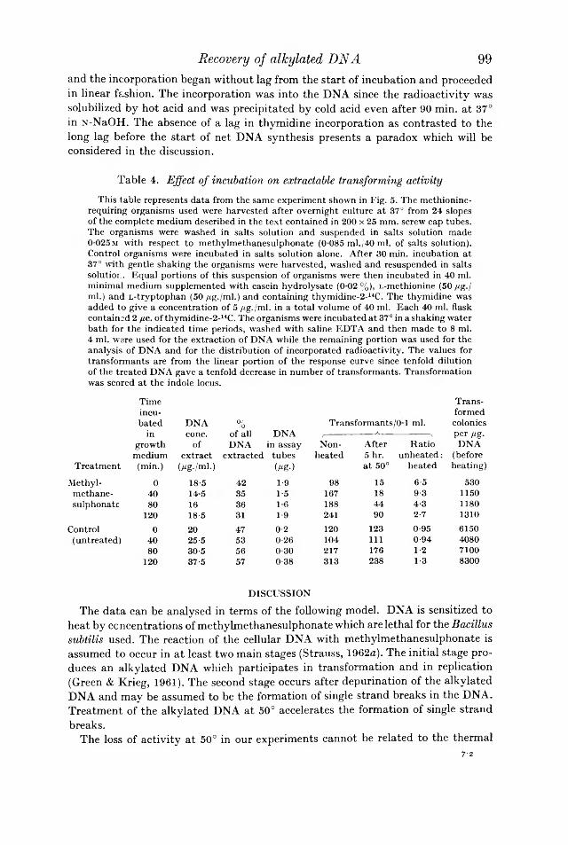
and the incorporation began without lag from the start of incubation and proceeded in linear fashion. The incorporation was into the DNA since the radioactivity was solubilized by hot acid and was precipitated by cold acid even after 90 min. at 37° in N-NaOH. The absence of a lag in thymidine incorporation as contrasted to the long lag before the start of net DNA synthesis presents a paradox which will be considered in the discussion.
Table 4. Effect of incubation on extractable transforming activityThis table represents data from the same experiment shown in Fig. 5. The methionine-
requiring organisms used were harvested after overnight culture at 37° from 24 slopes of the complete medium described in the text contained in 200 x 25 mm. screw cap tubes.The organisms were washed in salts solution and suspended in salts solution made 0 025m with respect to methylmethanesulphonate (0 085 ml./40 ml. of salts solution). Control organisms were incubated in salts solution alone. After 30 min. incubation at 37° with gentle shaking the organisms were harvested, washed and resuspended in salts solution. Equal portions of this suspension of organisms were then incubated in 40 ml. minimal medium supplemented with casein hydrolysate (0 02 %), L-methionine (50 /tg./ ml.) and L-tryptophan (50 /¿g./ml.) and containing thymidine-2-14C. The thymidine was added to give a concentration of 5 /¿g./ml. in a total volume of 40 ml. Each 40 ml. flask contained 2 /¿c. of thymidine-2-14C. The organisms were incubated a t 37° in a shaking water bath for the indicated time periods, washed with saline EDTA and then made to 8 ml.4 ml. were used for the extraction of DNA while the remaining portion was used for the analysis of DNA and for the distribution of incorporated radioactivity. The values for transformants are from the linear portion of the response curve since tenfold dilution of the treated DNA gave a tenfold decrease in number of transformants. Transformation was scored at the indole locus.
Time Trans-incu- formed
Recovery of allcylated D N A 99
bated DNA % Transformants/01 ml. coloniesin cone. of all DNA (-------- ____ A__ -----------\ per fig.
growth of DNA in assay Non- After Ratio DNAmedium extract extracted tubes heated 5 hr. unheated : (before
Treatment (min.) (/¿g./ml.) (/'«•) a t 50° heated heating)Methyl- 0 18-5 42 1-9 98 15 6-5 530methane- 40 145 35 1-5 167 18 9-3 1150sulphonate 80 16 36 16 188 44 4-3 1180
120 18-5 31 1-9 241 90 2-7 1310Control 0 20 47 0-2 120 123 0-95 6150
(untreated) 40 25-5 53 0-26 104 111 0-94 408080 30-5 56 0-30 217 176 1-2 7100
120 37-5 57 0-38 313 238 1-3 8300
DISCUSSIONThe data can be analysed in terms of the following model. DNA is sensitized to
heat by concentrations of methylmethanesulphonate which are lethal for the Bacillus subtilis used. The reaction of the cellular DNA with methylmethanesulphonate is assumed to occur in at least two main stages (Strauss, 1962a). The initial stage produces an alkylated DNA which participates in transformation and in replication (Green & Krieg, 1961). The second stage occurs after depurination of the alkylated DNA and may be assumed to be the formation of single strand breaks in the DNA. Treatment of the alkylated DNA at 50° accelerates the formation of single strand breaks.
The loss of activity at 50° in our experiments cannot be related to the thermal7-2

dénaturation of B a c i l lu s s u b t i l i s DNA since heating the alkylated DNA for 5 hr. at 50° does not cause an increase in its absorption at 259 m/r (Strauss, 1962 b) and since the T m of B a c i l lu s s u b t i l i s DNA in saline + citrate is 87-4° (Marmur, 1961). Alkylation of DNA does lead to a slight decrease in its thermal dénaturation temperature (Strauss, 19626) but the value (67-4° in 0-005m buffer compared to 70-5° for control DNA) is still well above 50°. Furthermore, the lack of any major
1 0 0 B . S . S t r a u s s
Before treatment After treatment
-M
+ MMS — »-0 025 M f
+10% 4
Heat sensitive -Mactive in
Mtransformation+
90%Inactive for
transformation
After treatment and incubation
Heat sensitive active
M +M-
No replication
Not heat sensitive
Fig. 6. Interpretation of the loss of heat sensitivity. The shaded areas represent DNA with single + double strand breaks which is supposed to be inactive for transformation of the indole locus and unable to replicate. Unshaded areas represent both alkylated and non-alkylated DNA able to replicate and of normal transforming activity. MMS = methylmethanesulphonate.
change in the sedimentation constant of alkylated or alkylated and heated B. s u b t i l i s DNA compared to non-treated material shows that the heating reaction does not cause any major degradation of the treated DNA into small molecules. Lerman & Tolmach (1957, 1959) showed that DNA with single strand breaks produced by deoxyribonuclease was inactive for transformation but was taken up by competent cells. Methylated, and methylated and heated B a c i l lu s s u b t i l i s DNA is also taken up by competent cells almost as efficiently as untreated DNA (Table 2). The DNA extracted from alkylated organisms must consist of a mixture of alkylated DNA and of DNA with single-strand breaks, but there can be very little normal DNA in the extract. This follows from the heat sensitivity of extracts from alkylated organisms (Fig. 4); less than 3% of the original transforming activity remained after 5 hr. at 50°.
If this hypothesis be correct, then the heat sensitivity of DNA formed on an alkylated template will be determined by the average number of alkylated strands in the parental DNA (Fig. 6). If on the average only half of the strands of the marked (indole) region are alkylated, then replication will give a population in

which half the markers are heat sensitive and half are resistant. If both strands are alkylated, then replication will lead to no decrease in heat sensitivity, since it is supposed that a single-strand break can inactivate a marker and each marker will still contain one alkylated heat-labile strand. Further replication will of course produce heat-resistant markers (Fig. 6). This hypothesis may be used for a rough calculation of the degree of resistance to heat and the increase in transforming activity obtained on incubation of alkylated organisms in growth medium. The calculations are presented more to show that the hypothesis does not lead to absurd conclusions than for their quantitative validity. In the experiment shown in Table 4 and Fig. 5, the number of transformed colonies//rg. DNA obtained from the treated organisms increased from 530 to 1310 after incubation for 120 min. with a net increase of only 34 % in the DNA. Heating for 5 hr. at 50° decreased the activity of the DNA from non-incubated organisms to 8-2 transformed colonies/ f ig. DNA. Consider an extent of methylmethanesulphonate reaction which leaves half of the progeny markers (marked regions in molecules) alkylated and heat sensitive (Fig. 6). The hypothesis requires that only the molecule active for transformation can participate in replication, and since the activity more than doubles I assume that all the active molecules participate in replication. For every 131 DNA molecules after replication there will then be 131/2 = 66 heat sensitive and 66 non-sensitive molecules. If the ratio of activity before and after heating is the same as before incubation (i.e. 6-5) there will be 66/6-5-1-66 = 76 active molecules after heating, giving an unheated:heated ratio of 131:76 = 1-7. If some of the progeny DNA molecules were produced on a template of doubly alkylated DNA (Fig. 6) more of the progeny molecules would be heat sensitive. If only 1/4 of 131 molecules were resistant to heat after replication whereas the others still contained an alkyl group, as would occur if the templates for replication had been an equal mixture of doubly and singly alkylated molecules, there would be 33 + 98/6-5 = 48 biologically active molecules present after heating for 5 hr. at 50°, which would give a ratio of unheated : heated of 131:48 = 2-7, which was the ratio obtained (Table 4, line 4). (The situation is complicated by the fact that the 2-5-fold increase in transforming activity requires more than one round of replication for certain of the DNA molecules.) It is also clear that there can be an initial increase in transforming activity obtained from alkylated organisms upon incubation in growth medium without decreased heat sensitivity, if doubly alkylated molecules are used as templates.
The ratio of the transforming activity from treated and non-treated organisms was 530/6150 transformed colonies//ig. DNA = 0-086 (Table 4). It may be assumed that this is the fraction of the total DNA in treated organisms which is capable of serving as a template for replication, that is, that the DNA from treated organisms consists of 8-6 % of alkylated but biologically active DNA and of 91-4 % biologically inactive DNA with single + double strand breaks (Fig. 6). The active alkylated DNA would have the normal transforming activity which in this experiment was 615 transformed colonies/̂ g. DNA. Any new DNA synthesized would also have normal transforming activity. After incubation for 120 min. there was a 34% increase in the DNA of the treated material (Fig. 5). In every 1-34 fig. DNA extracted after incubation there would then be 0-34 + 0-086 = 0-42 fig. active DNA and 0-82 fig. inactive DNA which had not participated in replication. The active DNA would produce 6150 transformed colonies//ig. DNA so that after incubation
Recovery of alkylated D N A 101

B . S. S traussone would expect 0-42x615 = 258 transformed colonies/1-34 / ¿ g . or 193 transformed colonies//ig. DNA as compared with an actual figure of 131 transformed colonies/,ttg. after incubation and 53 transformed colonies///». DNA before incubation. If the newly synthesized DNA still containing an alkyl group bad a lower transforming efficiency than normal DNA the calculated value would approach that found more closely.
The physical identification of the state produced by heating alkylated DNA at 50° with single strand breaks cannot be made with assurance in the absence of more physical data. It is still possible that this inactivation might represent the formation of apurinic sites, as suggested by the experiments of Brookes & Lawley(1961) and of Bautz & Freese (1960). It is also likely that the state of organisms after alkylation is more complex than we have indicated. Although the incorporation of thymidine-14C by treated organisms was linear from the start of incubation, there was no net increase in the DNA of treated organisms until 120 min. of incubation. Actually a slight decrease in DNA content after the start of incubation was a regular feature of our experiments. It seems reasonable to suppose that some process occurs soon after the start of incubation which serves to degrade DNA that has been alkylated. A degradative process affecting alkylated DNA has been described (Strauss, 19626). Breakdown of Haemophilus DNA was reported by Stuy (1960) after X-ray treatment. Degradation of DNA would of course introduce another element of uncertainty into the calculations above. Finally, there is no evidence that the ‘recovery’ from heat sensitivity of the extracted DNA (which actually represents synthesis of new DNA) is related to the rapid and dramatic recovery of viability of organisms from the effects of alkylation after very short periods of incubation in growth medium.
102
The author would like to thank Dr E. Nester for providing the recipient Bacillus subtilis stock used in these experiments and Dr E. P. Geiduschek for his aid in the determination of sedimentation constants. The expert technical assistance of Miss Joan Kerst is gratefully acknowledged. This work was supported by grants from the National Institutes of Health (RG 7816) and from the National Science Foundation.
R EFER EN C ESA n a g n o s t o p o u l o s , C. & S p i z i z e n , J . ( 19 6 1) . R equ irem ents for tran sform ation in B a c i l l u s
s u b t i l i s . J . B a d . 8 1 , 7 4 1.B a u t z , E . & F r e e s e , E . (1960). On the m utagen ic effect o f a lk y la tin g agents. P r o c .
n a t . A c a d . S c i . , W a s h . 46. 15 8 5 .B r o o k e s , P . & L a w l e y , P . ( 19 6 1) . The reaction o f mono- and di-functional a lk y la tin g
agents w ith nucleic acids. B i o c h e m . J . 80, 496.B u r t o n , K . (1956). T h e conditions and m echanism o f the d iphenvlam ine reaction fo r the
colorim etric estim ation o f deoxyribonucleic acid. B i o c h e m . J . 62, 3 15 .G r e e n , D . & Iv r i e g , D . ( 19 6 1) . The delayed origin o f m utan ts induced b y exposure
o f extracellu lar phage T 4 to eth yl m ethanesulfonate. P r o c . n a t . A c a d . S c i . , W a s h . 47, 64.
K a h a n , F . (1960). Pu rification and m easurem ent o f m icrogram am ounts o f rad io active nucleic acids and proteins from anim al cells in tissue culture. A n a l . B i o c h e m . 2. 10 7 .
L e h m a n , L . & T o l m a c h , L . (19 57). G enetic transform ation . I . Cellular incorporation o f D N A accom pan yin g tran sform ation in P n e u m o c o c c u s . B i o c h i m . b i o p l i y s . A c t a , 26, 68.

L er m a n , L. & T olm ach , L. (1959). Genetic transform ation. II . The significance of damage to the DNA molecule. Biochim. biophys. Acta, 33, 371.
Marmtjr, J . (1961). A procedure for the isolation of deoxyribonucleic acid from m icroorganisms. J . mol. Biol. 3, 208.
N e s t e r , E. & L e d e r b e r g , J . (1961). Linkage of genetic units of Bacillus subtilis in DNA transform ation. Proc. nat. Acad. Sci., Wash. 47, 52.
Sp iz iz e n , J . (1958). Transform ation of biochemically deficient strains of Bacillus subtilis by deoxyribonucleate. Proc. nat. Acad. Sci., Wash. 44, 1072.
Str a u ss , B. (1962a). Response of Escherichia coli auxotrophs to heat after trea tm en t w ith mutagenic alkyl m ethanesulfonates. J . B ad. 83, 241.
Str a u ss , B. (19626). Differential destruction of the transform ing activ ity of damaged deoxyribonucleic acid by a bacterial enzyme. Proc. nat. Acad. Sci., Wash. 48, 1670.
St u y , J . (1960). Studies on the radiation inactivation of micro-organisms. VI. X -ray- induced breakdown of deoxyribonucleic acid in Haemophilus influenzae and in other bacteria. J . B ad. 79, 707.
Y o un g , F. & Sp iz iz e n , J . (1961). Physiological and genetic factors affecting transform ation of Bacillus subtilis. J . B ad . 81, 823.
Z a m e n h o f , S., L e id y , G., H ahn , E . & A l e x a n d e r , H . (1956). Inactivation and unstabilization of the transform ing principle by m utagenic agents. J . B ad . 72, 1.
Recovery of alkylated D N A 103

J. gen. Microbiol. (1963), 30, 105-112 Printed in Great Britain
105
Azurin: A Copper Protein Found in BordetellaB y I. W. SUTHERLAND and J. F. WILKINSOND e p a r tm e n t o f B a c te r io lo g y , U n iv e r s i t y o f E d in b u r g h
(R e c e iv e d 2 M a y 1962)SUMMARY
AL strains of B o r d e te l la p e r tu s s i s , B . b r o n c h ise p tic a and B . p a r a p e r tu s s i s tested, were found to contain a blue protein; this has been named ‘Azurin’.It has been extracted from the cells of all three species and it has also been isolated from culture supernatant fluids of B . p e r tu s s i s . The azurin was purified by chromatography on ion-exchange cellulose derivatives and obtained in physically pure form; the materials from each species were identical. In one strain of B . b r o n c h is e p tic a azurin comprised 0-1 % of the bacterial dry weight. Azurin is a water-soluble autoxidizable protein of high oxidation-reduction potential (+395 mV.). It has an absorption maximum at 625 r a p in the oxidized form, with a millimolar extinction coefficient of 3-5 x 10³. It contains 0-45 % copper which can be completely removed by dialysis against cyanide. The molecular weight is 14,600 (¿>20, = 1-58 x 10~13, D i0 w = 10-6 x 10~7). Azurin can be reduced bycysteine, glutathione and other reducing agents; the blue colour disappears on reduction. Azurin undergoes reduction in the presence of concentrated cell-free extracts of B . b r o n c h is e p tic a and succinate, and is reoxidized on aeration.INTRODUCTION
Blue and green metallo-protein complexes have been isolated from a number of widely differing sources in nature. Several of the blue copper proteins isolated have been shown to be phenol oxidases, but other compounds without apparent enzymic activities have also been discovered. A green pigment, termed desulphoviridin, was obtained from the obligate anaerobic bacteria D e s u lp h o v ib r io d e s u lp h u r ic a n s by Postgate (1956), while a copper protein was found in the chloroplasts of C h lo re lla e lU p s o id e a and in higher plants and named plastocyanin (Katoh, 1960; Katoh & Takamiya, 1961). A blue protein was first discovered in P s e u d o m o n a s a e ru g in o s a some years ago (Verhoeven & Takeda, 1956); it was since isolated as a blue water- soluble, copper-protein complex by Horio (1958 a) and its physical and chemical properties investigated (Horio, 19585; Coval, Horio & Kamen, 1961). The presence of a blue protein in fractions obtained from B o r d e te l la p e r tu s s i s was first reported by Hammarsten, Palmstierna & Meyer (1959). The present paper describes the preparation, purification and some properties of a blue protein present in the three species of the genus B o r d e te l la . A preliminary account of part of this work has appeared (Sutherland & Wilkinson, 1962).
METHODSO r g a m s m s a n d m e th o d s o f c u ltu re . The bacteria used were obtained from the
National Collection of Type Cultures, Colindale, unless otherwise indicated. The strains used were: B o r d e te l la p e r tu s s i s , nctc 8474, 4412 (obtained from Dr H.

Billaudelle, Karolinska Institute, Stockholm), 4507 (isolated at the City Hospital, Edinburgh), and 364 (the original Phase IV strain of Leslie & Gardner kindly provided by Dr Jean Dolby, Lister Institute, Elstree). Strain 364 was avirulent for mice and a further avirulent strain, 4507 a v , was obtained by repeated subculture of strain 4507. Two B o r d e te l la p a r a p e r tu s s i s strains were n c t c 7385 and n c t c 8250. The B . b r o n c h is e p tic a strains were n c t c 454, n c t c 8344, n c t c 8761 and n c t c 8762, strain BR/1 (isolated in the Bacteriology Department, Edinburgh University) and strain 8759 (obtained from Dr A. P. Maclennan, Microbiological Research Establishment, Porton).
M e d ia . Virulent B o r d e te l la p e r tu s s is strains were grown in the tris + resin liquid medium described in an earlier paper (Sutherland & Wilkinson, 1961). Other species were grown in a medium which contained (quantities/1. medium): Casamino acids (Difco technical) 4-0 g.; sodium glutamate, 4-0 g.; KH2P04, 0-1 g.; MgCl2,6H20, 0-08 g.; FeS04,7H20, 0-002 g.; CuS04,5H20, 0-004 g.; CaCl2, 0-01 g.; yeast extract 200 ml. The yeast extract was prepared by the method of Cohen & Wheeler (1946). The medium was adjusted to pH 7-0 with N-NaOH, distributed in 250 ml. quantities into 500 ml. Ehrlenmeyer flasks and sterilized by autoclaving at 120° for 15 min. After inoculation the flasks were incubated at 37° and shaken at approximately 100 oscillations/min. on a reciprocating shaker (Distillers Company Ltd, Epsom). The cultures of B . b r o n c h ise p tic a , B . p a r a p e r tu s s i s and B . p e r tu s s i s were incubated for 24, 48 and 72 hr., respectively. The bacteria were harvested by centrifugation at about 5000g for 30 min. in a MSE refrigerated centrifuge, and stored unwashed at — 40° until required.
A b s o r p t io n s p e c tr a . A Unicam SP 500 spectrophotometer with 1 cm. light path was used.
T o ta l n itr o g e n . Nitrogen was determined by the micro-Kjeldahl method, the ammonia being trapped in boric acid and estimated colorimetrically with Nessler reagent.
P a p e r c h r o m a to g r a p h y . Two-dimensional paper chromatography was used to identify the amino acids in protein hydrolysates. Whatman No. 1 paper sheets were run for 18 hr. with 80% (w/v) phenol containing 0-3% (v/v) ammonia. They were then dried, rotated through 90° and run for 24 hr. with butanol + acetic acid + water (4 + 1 + 5 by vol.). All chromatograms were run at 25°. Amino acid spots were detected with ninhydrin.
B u ffe r s o lu tio n s . All salts used were analytical grade reagents. The pH values were read on a Pye * Dynacap ’ pH meter.
K r e b s - R in g e r s o lu tio n . This had the following composition (g./lOO ml. solution): NaCl, 0-9; KC1, 0-04; MgS04,7H20, 0-04; NaH2P 04,2H20, 0-39; 0-4 ml. n-HCI.
M e a s u r e m e n t o f o x id a tio n -r e d u c tio n p o te n t ia ls . These were determined potentio- metrically by the method of Gibson (1961) in an atmosphere of oxygen-free nitrogen.
C o n c e n tra tio n o f s o lu tio n s o f h e a t- la b ile s u b s ta n c e s . Bacterial extracts and other protein-containing solutions were concentrated by the method of Palmstierna (1960), by using a form of carboxymethyl cellulose known as ‘ courulose ’ (British Celanese Ltd, Coventry), Alternatively, polyethylene glycol of molecular weight about 6000 was used according to the method of Kohn (1959). Both methods were satisfactory, the first being preferable when it was necessary to concentrate large volumes.
106 I. W. Sutherland and J. F. W ilkinson

A copper protein in Bordetella 107RESULTS
E x tr a c t io n o f b lu e p r o te inWashings of the frozen B o r d e te l la p e r tu s s i s organisms in distilled water, or in
Krebs-Ringer solution were blue in colour, but no coloured material was observed in washings from organisms of the other two species. This was probably because they were less easily lysed by the freezing and subsequent thawing. Several extraction procedures were investigated ; the following routine was found to be applicable to all the species which were examined. The frozen bacteria (equivalent to about 50 g. dry weight, obtained from 30 1. of medium), were suspended in one 1. Krebs- Ringer solution to which 0T M-urea had been added. The suspension was stirred at 0° for 30 min. by a high-speed stirrer, followed by centrifugation at c . 25,000g for 60 min. in the model L preparative Spinco ultracentrifuge. The supernatant fluid was stored in the cold and the bacterial deposit was extracted with a further 500 ml. of Krebs-Ringer solution containing urea. After centrifugation, as before, the supernatant fluid was again retained while the bacteria were suspended in 250 ml. distilled water, frozen and thawed. The viscous suspension was stirred as above, then centrifuged. The organisms were extracted with a further sample of distilled water, removed by centrifugation and discarded. The four extracts were pooled to give a viscous, opalescent solution; it was dark, greenish blue in colour. This solution was dialysed against cold running tap-water for 8 hr., then concentrated until the volume was about 100 ml. The extract was centrifuged at 100,000g for 15 min. in the ultracentrifuge. The deposit was washed with a small volume of distilled water, recentrifuged and discarded, the supernatant fluid being added to the initial solution. The coloured solution was dialysed against phosphate buffer ( 0 - 0 2 m - KH2P04 + Na2HP04) for 24 hr. at 4° with constant agitation. The non-diffusible material was applied to a column of DEAE cellulose equilibrated against the same buffer, and pumped through the column at a rate of 10 ml./hr. The column was then washed through with a further 20 ml. buffer, the washings being added to the original eluate. This was bright blue in colour and appeared to be considerably less viscous than the original solution. The blue eluate was concentrated and dialysed for 48 hr. against acetate buffer (0 -02 m , pH 4-6). A variable amount of precipitate appeared at this stage and was removed by centrifugation at 100,000g for 15 min. The supernatant fluid was applied to a CM cellulose column in the sodium form, which had been thoroughly washed with the same acetate buffer. The coloured material was all adsorbed at the top of the column. The pH value was gradually increased, using a gradient of acetate buffer (0 -0 2 m , pH 516) and 200 ml. mixer volume, and fractions were collected in an automatic fraction collector. The flow rate of the buffer was adjusted to 10 ml. /hr. The blue material started to elute from the column at about pH 5-12. After the complete elution of the blue material, the top of the column was reddish brown in colour, probably due to the retention of cytochromes or other haemoproteins. The fractions containing blue material were concentrated, dialysed against acetate buffer (pH 4-6) and rechromatographed on CM Sephadex (Pharmacia, Uppsala) using the same conditions. The final product was concentrated, dialysed against glass-distilled water and either stored at — 40° or lyophilized.
In typical experiments with B o r d e te lla b ro n c h ise p tica , strain 8344, equiv. 50 g.

108dry weight of bacteria yielded 50-55 mg. of purified blue material. Although strains 454 and 8761 yielded comparable amounts of the product, the other strains and species contained less blue pigment. The differences did not appear to be due to incomplete extraction of the pigment from the bacteria, as ultrasonic disintegration of the cells at the end of the extraction process, followed by the complete extraction cycle, yielded no further blue material. In B . p e r tu s s i s cultures, the longer incubation period resulted in some autolysis, with consequent liberation of blue pigment into the surrounding medium. When supernatants fluids from B . p e r tu s s i s cultures were dialysed, concentrated and treated in the same way as cell extracts, about 0-2 mg. blue material/1. supernatant fluid was recovered. However, no pigment was isolated from the supernatant fluids of cultures of the other two species, even after prolonged incubation. This was probably because of the different degrees of autolysis occurring in these B o r d e te l la species.
P r o p e r t i e s o f th e b lu e p r o te inT h e b lu e m a te r ia l w as p r e s e n t in a ll s t r a in s o f t h e th r e e B o rd e te l la sp ec ies te s te d .
I t c o u ld b e e x t r a c te d f ro m th e b a c te r ia w i th K r e b s - R in g e r s o lu t io n a lo n e b u t o m iss io n o f t h e u r e a m a d e i t n e c e s s a ry to in c re a s e t h e n u m b e r o f e x t r a c t io n s to fiv e o r s ix b e fo re a ll t h e p ig m e n t w as re m o v e d f ro m th e ce lls. T h e b lu e m a te r ia l c o u ld a lso b e o b ta in e d in th e s o lu b le f r a c t io n a f te r u l t r a s o n ic d is in te g r a t io n o f t h e b a c te r ia . A ll t h e p ig m e n t w as in th e s o lu t io n a f t e r u l t r a - c e n tr i f u g a t io n o f t h e so n ic e x t r a c t s a n d n o n e w as a s s o c ia te d w i th th e s e d im e n ta b le p o r t io n .
The final product from ion exchange chromatography was very soluble in distilled water and solutions up to 10 % could be prepared. I t was insoluble in chloroform, ether and ethanol.
A b s o r p t io n s p e c tr u m . The absorption spectrum of the purified pigment from a pertussis strain is shown in Fig. 1. In the oxidized form, maxima were observed at 279 r a p and 625 r a p , but the latter peak disappeared on reduction. The millimolar extinction coefficient at 625 r a p , calculated on the basis of the molecular weight given below, was 3-5 x 10³. Table 1 gives the ratio of E625 to E280 at various stages of purification. Attempts to raise this value by other methods of purification, such as electrophoresis or chromatography on other ion exchange materials, were unsuccessful.
I. W. Sutherland and J. F. W ilkinson
Fig. 1. Absorption spectrum of Bordetella blue protein

S ta b i l i t y . T h e b lu e m a te r ia l w as r e a d i ly o x id iz e d b y a i r o r o x y g e n . I n th e o x id iz e d fo rm , n o c o lo u r c h a n g e w as o b se rv e d o v e r th e ra n g e p H 2 -5 -1 0 -0 . T h e m a te r ia l w as ra p id ly d e s tr o y e d b y b o ilin g , t h e c o lo u r d is a p p e a r in g a n d a w h ite p re c ip i ta te o f d e n a tu r e d p ro te in b e in g d e p o s i te d . T h e p ig m e n t c o u ld b e ly o p h iliz e d to a b lu e p o w d e r w h ic h w as c o m p le te ly so lu b le in d is t i l le d w a te r .
O x id a tio n -r e d u c tio n p o te n t ia l . After purification, the pigment was in the fully oxidized blue form. It coidd be reduced by a variety of reducing agents such as ascorbic acid, cysteine, sodium thioglycollate or sodium dithionite. The oxidation- reduction potential of samples of the blue material (5 mg.) was titrated potentio- metrically and found to be +395 mV at 20°.
T a b .e 1. E x t in c t io n r a t io s o f b lu e p r o te in a t v a r io u s s ta g e s o f p u r i f ic a t io n^ 6 2 5 0 1 - /^ 2 8 0
A copper protein in Bordetella 109
(1) Crude, pooled extract after concentration and 0 004ultracentrifugation
(2) Eluate from DEAE cellulose column 0 049(3) As (2), after dialysis against acetate buffer (pH 0108
4-6) and removal of the precipitate(4) Eluate from CM cellulose column 0-343(5) Eluate from CM Sephadex column 0-465
N itr o g e n c o n te n t. Determination of the total nitrogen in samples of the pigment extracted from different strains revealed a content of 14-9-15-3 %. This was indicative of a protein and paper chromatography of acid hydrolysates showed the presence of the following amino acids : cystine, glycine, aspartic acid, serine, glutamic acid, lysine, arginine, threonine, alanine, tyrosine, phenylalanine, isoleucine, leucine. The possible presence of others, in amounts too small to be detected by the techniques used, was not excluded.
M o le c u la r w e ig h t. T h e s e d im e n ta t io n co e ffic ien t o f th e b lu e p ro te in f ro m B o r d e te l la b r o n c h is e v tic a w as d e te rm in e d in a se rie s o f ru n s in a S p in co m o d e l E a n a ly t ic a l u l t r a c e n tr i fu g e . T h e sa m p le s , a t th r e e c o n c e n tr a t io n s (0-15, 0-4 a n d 1-0%), w e re d is so lv e d in p h o s p h a te b u ffe r (0*2m, p H 7-0) a n d d ia ly s e d a g a in s t t h e s a m e b u ffe r . T h e v a lu e o b ta in e d fo r t h e s e d im e n ta t io n co e ffic ien t e x t r a p o la te d fo r z e ro p ro te in c o n c e n t r a t io n w a s S 20 w = 1-59 x 10~13.
T h e d if fu s io n co e ffic ien t w as o b ta in e d f ro m a s in g le r u n fo r 36 h r . in a m o d ifie d T ise liu s e le c tro p h o re s is a p p a r a tu s , a t 20°, f ro m w h ic h th e r e s u l ta n t D 20 w = 10-6 x 10⁻⁷. T h e p a r t ia l sp ec ific v o lu m e w as a s s u m e d to b e 0-75. T h is y ie ld s a m o le c u la r w e ig h t fo r t h e b lu e p ro te in o f 14,600 a n d a f r ic t io n a l r a t io o f 1-28. D u r in g s e d im e n ta t io n , a s in g le p e a k w as a p p a r e n t a n d th e b lu e p e a k m o v e d w i th t h e S c h lie re n p e a k th r o u g h o u t th e ru n s . S im ila r v a lu e s w e re o b ta in e d fo r a r u n w i th a s a m p le o f B o r d e te l la p e r tu s s i s p ig m e n t.
C o p p e r c o n te n t. Spectrophotometric analysis indicated the presence of copper and the absence of iron, manganese and magnesium. Analysis of samples for their copper content was performed by activation analysis at the Atomic Energy Establishment, Wantage. The copper content of blue protein from four different strains is shown in Table 2; the mean value obtained from these results was 0-447 %. This, allied to the molecular weight, indicates the presence of one copper atom per

molecule of protein. No increase in copper content was obtained by dialysis against a copper sulphate solution followed by dialysis against distilled water to remove free copper ions. Dialysis against the chelating agent ethylenediamine tetra-acetic acid decreased the copper content of a sample of blue protein to 0-210%, while dialysis against cyanide completely removed the copper.
Table 2. C o p p e r c o n te n t o f b lu e p r o te in f r o m d if fe r e n t B o r d e te l la s t r a in sCopper
content (% )
110 I. W. Sutherland and J. F. W ilkinson
B . p e r t u s s i s 4412 0-436B . p e r t u s s i s 364 0-452B . p a r a p e r t u s s i s 7S 85 0-448B . b r o n c h is e p t ic a 8344 0-451
M e ta b o lic a c t iv i t ie s . The blue protein was autoxidizable. A solution of the colourless reduced protein containing 100 /¿mole/ml. was completely oxidized in 20-25 min. at 37° when oxygen or air was passed through it. The rate of re-oxidation was very much greater, following the addition of small quantities of cell-free extracts of Bordetella species. The pigment was in the reduced form when the bacteria were harvested. This could be shown by visual examination of thick suspensions of B o r d e te l la b r o n c h ise p tic a cells immediately after harvesting; they were buff or pale pink in colour. However, when they were allowed to stand in air, or when air was passed through the suspensions, they rapidly turned greenish blue. This was seen most markedly in centrifuged bacterial deposits, the surface of which became blue while the underlying material remained pink. In the presence of succinate, the blue protein was rapidly reduced by cell-free extracts of Bordetella species.
P re s e n c e o f s im i la r p ig m e n ts i n o th er b a c te r ia . Samples of bacteria of several other species were extracted by the method applied to Bordetella cells. Blue proteins, with properties very similar to those described for Bordetella material, were found in A lc a l ig e n e s f a e c a l i s n c t c 655, A lc a l ig e n e s d e n i tr i f ic a n s n c t c 8582 and P s e u d o m o n a s f lu o r e s c e n s strain 2. No trace of this type of protein was found in E s c h e r ic h ia c o li B, S a lm o n e l la t y p h im u r iu m strain 5, B a c i l lu s m e g a te r iu m strain 19, C h ro m o - b a c te r iu m v io la c e u m n c t c 9757, A c h r o m o b a c te r a n i t r a tu s n c t c 7844 or M o r a x e l la b o v is n c t c 8561.
DISCUSSIONThe results described in this paper indicate that all three Bordetella species
examined possess blue copper-proteins which are identical with respect to the tests that were made. We suggest the name azurin for this protein, as being in agreement with the nomenclature used for similar pigments, e.g. desulphoviridin (Postgate, 1956). The presence of copper-protein complexes of different function is widespread in nature. However, the only one of these compounds with any resemblance to azurin is the ‘blue protein’ isolated from P s e u d o m o n a s a e r u g in o s a by Horio (1958 a, b). This is remarkably similar in its redox potential ( + 390 mV), the amended molecular weight of 14,600 (Coval, Horio & Kamen, 1961) and in the presence of one atom of copper per molecule. Further, the ‘blue protein’ of P . a e r u g in o s a can be purified by the method outlined for Bordetella azurin. A further similarity lies in the report that the Pseudomonas protein, like azurin, can be freed from copper by

I lldialysis against cyanide solution at neutral pH (Horio, Sekuzu, Higashi & Okunuki,1961). In both cases, the copper could be restored by dialysis against copper sulphate solution. This property is common to several copper-protein complexes (Westerfeld, 1961). There is one difference in the reported properties; Horio (1958a) stated that the ‘ blue protein ’ was not autoxidizable. However, when it is prepared from P . a e r u g in o s a by the method described above, the product is autoxidizable at a rate similar to that of azurin (Sutherland, unpublished results).
The continuous spectrum of azurin shows the absence of any Soret band similar to that found in desulphoviridin (Postgate, 1956). This indicates that the copper is not bound to the protein through a porphyrin molecule. The visible spectrum closely resembles that obtained for artificial copper-glycine complexes (Klotz, Faller & Urquart, 1950) and it seems possible that the chromophore is a copper- amino acid complex.
The function of azurin is uncertain. Its distribution appears to be somewhat limited in nature. The presence of azurin or similar pigments has only been found in the three genera B o r d e te lla , A lc a l ig e n e s and P s e u d o m o n a s . No trace of similar compounds could be found in the species of S a lm o n e l la , E sc h e r ic h ia , A c h r o m o b a c te r or B a c i l lu s tested. A relationship between the genus A lc a l ig e n e s and certain P s e u d o m o n a s species was indicated by Lysenko (1961) and it has been suggested that A .f a e c a l i s is related to B . b r o n c h is e p tic a (Szturm & Bourdon, 1948). However, it is difficult to see what, if any, is the common metabolic process in azurin-containing micro-organisms. One important difference between the metabolism of P s e u d o m o n a s species and that of the genus B o r d e te l la is the ability of the former to grow anaerobically. The conditions of growth used by Horio (1958a), 100 1. unaerated medium in deep vessels, would indicate that growth was anaerobic. It therefore appears that the ‘ blue protein ’ was produced under anaerobic conditions, although the actual yield was not reported. Nor is there any quantitative result for the production of the protein under different oxygen tensions. Azurin, however, is obtained from highly aerated cultures and it is remarkable that some strains of B . b ro n c h is e p t ic a contain as much as 0T % of their dry weight as this copper protein.
The ‘blue protein’ of P s e u d o m o n a s a e r u g in o s a is thought to act as an alternative electron-transport system between a cytochrome of the c type (Pseudomonas cytochrome 551) and cytochrome oxidase (Horio, 19586). Initial investigations with azurin, using B o r d e te l la b r o n c h ise p tic a cell-free extracts and purified cytochromes suggest a similar function (Sutherland, unpublished results).
The authors wish to thank Professor E. Hammarsten and Dr H. Palmstierna for a gift of pertussis material; Dr H. J. Cruft for ultracentrifuge and electrophoresis studies and calculations; and Miss S. C. McCabe, Miss J. Thompson and Mr J. D. Caird for technical assistance. One of us (I.W.S.) is indebted to the University of Edinburgh for a Graduate Research Fellowship during part of this work.
A copper protein in Bordetella

112 I. W. Sutherland and J. F. W ilkinson
REFERENCESCo h e n , S. M. & W h e e l e r , M. W . (1946). Pertussis vaccine prepared w ith phase-1
cultures grown in fluid medium. A m e r. J . p u b l. Hlth, 36, 371.Coval, M. L., H o r io , T. & K a m en , M. D. (1961). The amino acid composition of some
bacterial haem proteins. B ioch im . B io p h ys . A c ta , 51, 246.G i b s o n , J . (1961). Cytochrome pigments from the green photosynthetic bacterium ,
C hlorobium Ih iosu lph a toph ilu m . B iochem . J . 79, 151.H am m arsten , E., P a lm stier n a , H. & Me y e r , E. (1959). Separation of acid polyelectro
lytes and proteins in cellular extracts. J . biochem . m icrob io l. Tech. E n g . 1, 273.H o rio , T. (1958 a). Terminal oxidation systems in bacteria. I. Purification of cytochromes
from P seu d om on a s aeru ginosa . J . B ioch em ., T o k yo , 45, 195.H o rio , T. (19586). Term inal oxidation systems in bacteria. II . Some physical and
physiological properties of purified cytochromes of P seu dom on as aeru ginosa . J . B ioch em ., T o k yo , 45, 267.
H o rio , T., Se k u z u , I., H ig a s h i, T. & O k u n u k i, K. (1961). Cytochrome oxidases of P seu d om on a s aeru g in osa and ox-lieart muscle, and their related respirator}- components. In H a em a tin E n zym e s, p. 302. Ed. Falk, J . E ., Lemberg, R . & Morton, R. K. Oxford: Pergamon Press.
K a toh , S. (1960). A new copper protein from C hlorella e llip so id ea . N a tu re , B on d. 186, 533.
K atoh , S. & T a ka m iy a , A. (1961). A new leaf copper protein ‘p lastocyanin’, a natural Hill oxidant. N a tu re , B on d. 189, 665.
K lotz, I. M., F a l l e r , I. L. & U r q u a r t , J . E . (1950). Spectra of copper complexes with some proteins, amino acids and related substances. J . p h y s . coll. Chem . 54, 18.
K o h n , J . (1959). A simple m ethod for concentrating fluids containing protein. N a tu re , B on d. 183, 1055.
L y se n k o , O. (1961). P seu d om on a s—An attem p t a t a general classification. J . gen. M icro b io l. 25, 379.
P a lm stier n a , H. (1960). A m ethod for concentrating solutions of high molecular weight compounds. B ioch em . B io p h y s . res. C om m n . 2, 53.
P ostgate , J . R. (1956). Cytochrome c3 and desulphoviridin ; pigments of the anaerobe D esu lp h o vibr io d esu lph u rican s. J . gen . M icro b io l. 14, 545.
Su th e r l a n d , I. W. & W il k in s o n , J . F. (1961). A new growth medium for virulent B ordetella p e r tu ss is . J . P a th . B a d . 82, 431.
S u t h e r l a n d , I. W. & W il k in so n , J . F. (1962). Azurin, a blue bacterial protein. B ioch em . J . (in the press).
Szturm , S. & B o u r d o n , D. (1948). B a c teriu m a lca lig in es fe c a lis e t hem ophilus b ronch i- sep ticu s; ca rac tè res m orphologiques e t b iochim iques. A n n . I n s t. P a steu r , 75, 65.
Ve r h o e v e n , W. & T a k ed a , Y. (1956). The participation of cytochrome c in n itrate reduction. In In o rg a n ic N itro g en M etab o lism , p. 159. Ed. McElroy, W. D. & Glass, B. Baltimore : The Johns Hopkins Press.
W e s t e r f e l d , W. W. (1961). Effect of m etal-binding agents on metallo-proteins. F e d . P roc. 20, 158.

J. gen. Microbiol. (1963), 30, 113-120 Printed in Great Britain
113
Changes in Gross Chemical Components of T r ic h o p h y to n m e n ta g r o p h y te s During Incubation in Increased
Carbon Dioxide TensionsBy B. CHIN a n d S. G. KNIGHT
D e p a r tm e n t o f B a c te r io lo g y , U n iv e r s i t y o f W is c o n s in , M a d is o n , W is c o n s in , U .S .A
(R e c e iv e d 21 M a y 1962)SUMMARY
Macroconidia of T r ic h o p h y to n m e n ta g r o p h y te s harvested from a culture grown on a medium rich in glucose contained 33% acid-soluble carbohydrate. Paper chromatography of hydrolysates showed glucose as the main sugar in this fraction; there was also a trace of an unidentified sugar. Macroconidia from the same strain grown on the same medium but deficient in glucose contained 19% carbohydrate; hydrolysates of this fraction contained glucose, with traces of galactose and the unidentified sugar. Microconidia harvested from a second strain grown on a medium rich in glucose contained 11 % acid-soluble carbohydrate; hydrolysates of this fraction also contained glucose with traces of galactose and the unidentified sugar. These differences were reflected in whole cultures induced for macroconidial formation with carbon dioxide. Strains which responded well increased in acid-soluble carbohydrate; glucose was the predominating sugar in hydrolysates of these fractions. Strains which responded poorly did not increase in carbohydrate content; glucose, and traces of galactose and an unidentified sugar, were found in hydrolysates of these fractions.
INTRODUCTIONA p re v io u s r e p o r t f ro m th i s l a b o r a to r y (C h in & K n ig h t , 1957) d e s c r ib e d th e
s t im u la t io n o f m a c ro c o n id ia l fo rm a tio n in T r ic h o p h y to n m e n ta g r o p h y te s d u r in g i n c u b a t io n a t in c re a s e d c a rb o n d io x id e te n s io n s . A re q u i r e m e n t fo r g lu co se w a s d e m o n s t r a te d , s in ce m a c ro c o n id ia f o rm a tio n w as n o t s t im u la te d in in c re a s e d a tm o s p h e r ic te n s io n s o f C 0 2 w h e n g lu c o se w as o m i t te d f ro m th e m e d iu m . T h e e x p e r im e n ts d e s c r ib e d in th e p re s e n t p a p e r a t t e m p te d to a n a ly s e a n d c o m p a re th e g ro ss c h e m ic a l c o m p o n e n ts o f m a c ro c o n id ia , m ic ro c o n id ia a n d h y p h a e . I t w as o u r in te n t io n to f in d so m e m e a s u r a b le d if fe re n c e b e tw e e n m a c ro c o n id ia a n d th e o th e r m o rp h o lo g ic a l u n i t s o f T . m e n ta g r o p h y te s w ith th e h o p e t h a t th is d if fe ren ce m ig h t b e in d u c e d in t h e n o rm a l ly n o n - s p o ru la t in g s tr a in s w h e n m a c ro c o n id ia fo rm a tio n w a s s t im u la te d b y c a rb o n d io x id e . B e c a u s e o f d if f ic u ltie s in v o lv e d in s e p a r a t in g m a c ro c o n id ia f ro m m ic ro c o n id ia a n d h y p h a e , m u t a n t s t r a in s w h ic h d if fe re d in ty p e o f s p o r u la t io n w e re d e r iv e d f ro m a c o m m o n p a r e n t . M a c ro c o n id ia a re e a s ily h a rv e s te d f ro m a s p o r u la t in g m u t a n t w h ic h d o es n o t fo rm m ic ro c o n id ia , a n d v ice v e rs a .
8 G. Microb. xxx

114 B. Ch in a n d S. G. K nig h t
, METHODSMutantsAll mutants were derived from Trichophyton mentagrophytes strain A-280 (from
the Communicable Disease Center, Chamblee, Georgia, U.S.A.). Stock cultures were maintained by 3-monthly transfer on Sabouraud’s glucose agar (Difco) and incubation at 30° for 10-14 days followed by storage at 5°. Sabouraud's glucose agar (4% glucose, 1% Neopeptone, 1-5% agar) was used throughout. Liquid medium was prepared by omitting the agar, and Sabouraud’s conservation medium by the omission of glucose. Cultures were incubated for 5 days before harvest. The genetic purities of parent and mutant strains were assured by single spore isolations to avoid the selection of macroconidia-forming variants from mixed clones during incubation in C02 (Emmons & Hollaender, 1939). A microconidium was isolated from strain A-280 by the excision and transfer to fresh medium of a germinated microconidium from ruled agar blocks of Sabouraud’s glucose agar. The clone resulting from the growth of this spore was maintained as the parent culture, strain 5-7. Spores from this clone were seeded on Petri plates containing solid media and irradiated with ultraviolet radiation. Morphological mutants differing in type of sporulation were selected from the survivors and transferred to fresh medium. In turn, clones resulting from the growth of transferred survivors were purified by the isolation of microconidia. Since strain 1-2-1 does not form microconidia, a macroconidium was isolated. The growth characteristics of all mutants have remained stable through 3 years of serial transfer. Their sporulating patterns and responses to C02 are recorded in Table 1.
Table 1. Macroconidial formation by mutants of Trichophyton mentagrophytes and their response to increased carbon dioxide tensions in air
Mutant Normal air 16% (v/v)5-7 rare profuse1-1-2 none poor1-2-1 abundant profuse1-3-1 none poor
Inoculum and growthInoculum for solid media was prepared by dispersing the growth on a stock
culture slope with sterile water and pipette. Cultures for chemical analysis were grown as surface mats on liquid media. A floating inoculum was prepared by pipetting 1 ml. medium on to the surface of a stock culture slope. The culture tube was rolled gently to dislodge spores and hyphal fragments, the majority of which floated on the surface of the medium. In inoculation, this preparation was poured into a flask containing liquid medium. Flasks which were incubated with increased atmospheric tensions of C02 (0-16% v/v) were fitted with rubber stoppers and glass tubing outlets. The gassing procedure was as previously described (Chin & Knight, 1957). After incubation the floating cultures were washed by pouring off the media and refloating the culture mats with water 4 times. The mats were then removed from the flasks and suspended in cold acetone (Umbreit, Burris & Stauffer, 1957). The organism was separated from the acetone by centrifugation and the supernatant acetone drawn off through a capillary pipette connected to an aspirator pump. Samples of organism were dried in vacuo overnight.

CO2 and metabolism of T. mentagrophytes 115Spores
Spores were harvested from cultures grown on solid media in Roux bottles; macroconidia were harvested from strain 1-2-1, microeonidia from strain 5-7. After 5 days of incubation, 40 ml. water were added to each bottle, the contents vigorously shaken and strained through gauze. Although many spores were retained with mycelium in the filter, the effluent usually contained a high percentage of the spores in the original harvest. Spores were separated from the effluent by centrifugation. The spores were washed 4 times, after which they were suspended in cold acetone and dried as described above.Spore walls
Macroconidia were harvested and broken, prior to acetone treatment, in a Serval Omni-mix with water and glass microbeads for 30 min. The proportions of the mixture were spores + glass beads + water (1+2+3, by vol.). Microscopic examination of samples stained with lactophenol cotton blue revealed that 95 % of the macroconidia were broken in all compartments. The spore walls were washed with water in excess of twenty times, suspended in cold acetone, and dried.Extraction procedure
Weighed samples were transferred to test tubes and sequentially extracted in a boiling water bath with water for 15 min., with 5 % (w/v) trichloroacetic acid (TCA) for 30 min., with N-NaOH for 30 min. The tubes were capped with glass marbles to prevent evaporation. After each extraction, the contents of the tubes were separated by centrifugation. Samples of extracts were removed for analysis with a pipetting bulb; the rest of the extract was discarded through a capillary pipette connected to an aspirator pump. The cell residues remaining after extraction with hot NaOH were washed with water four times by repeated centrifugation and transferred to tared aluminum cups. The residues were dried overnight at 80° and the weights determined by difference.Chemical analyses
Carbohydrate was measured with the anthrone reagent, with glucose as the standard (Loewus, 1952). Samples from TCA extracts were hydrolysed with 0-6 n - HC1 in a boiling water bath before paper chromatography. The solvent systems used for paper chromatography were isopropanol + water (8 + 2, by vol.) and amyl acetate + pyridine+ water (3 + 3 +1, by vol.). Paper chromatograms were developed with AgN03 reagent (Smith, 1958).
Protein was measured with the Folin phenol reagent method based on the modification of Lowry, Rosenbrough, Farr & Randall (1951).
Total nucleic acids (NA) were estimated by the method of Logan, Mannell & Rossiter (1952) at 268-5 m¡i in a Beckman model DU spectrophotometer with3-0 ml. silica cuvettes after extraction by the method of Schneider (1945). Samples were read against a blank of 5 % TCA. Herring sperm deoxyribonucleic acid (DN A Nutritional Biochemical Co.), similarly hydrolysed, was used as the standard.
DNA content was determined by the method of Kech (1958). Ribonucleic acid (RNA) was estimated by the difference between total NA and DNA.
8 -2

% o
f dr
y w
eigh
t
116 B . Ch in a n d S. G. K nig h t
RESULTSThe gross chemical compositions of macroconidia, microconidia, and whole
culture of Trichophyton mentagrophytes strain 5-7, which produces microconidia but no macroconidia on Sabouraud’s glucose agar, are described in Table 2. The
Table 2. Gross chemical components of macroconidia, microconidia, and whole culture of strain 5-7 of Trichophyton mentagrophytes
Macro- Micro-co lidia conidia Strain 5-7
(% of dry wt.)Extract Component '-------------h 2o CHO* 11-8 13-2 7-6
PRO 115 7-3 12-3TCA CHO £3-3 111 11-9
PRO 5-5 5-4 5-8RNA 2-6 3-5 1-4DNA 0-2 0-3 01
NaOII PRO 15-6 160 13-4CR 10-6 120 301
* CHO: carbohydrate, PRO, protein; RNA, ribonucleic acid; DNA, deoxyribonucleic acid;CR, cell residue.
0 ------------ 1-------------1------------ 1________ I0 4 8 12 16
% ( v / v ) C 0 2 in a i r % ( v / v ) c o 2 in a i r
Fig. 1 Fig. 2Fig. 1. Changes in gross chemical composition, in percent of dry weight, of Trichophyton mentagrophytes strain 5-7 after incubation in increased carbon dioxide tensions. CHO- H 20 , hot-water-extractable carbohydrate; PRO-HzO, hot-water-extraetable protein; CHO-TCA, hot-TCA-extractable carbohydrati; PRO-TCA, hot-TCA-extractable protein; RNA, ribonucleic acid; DNA, deoxyribonucleic acid; PRO-NaOH, hot-NaOH-extractable protein; CR. cell residue.Fig. 2. Changes in acid-soluble carbohydrate, in percent of dry weight, in mutants which vary in type and degree of sporulati jn during incubation in increasing carbon dioxide tensions. All mutants were harvested as surface cultures on liquid Sabouraud's medium, strain 5-7 was grown on medium with and without glucose (S GLU).

macroconidia contained 33% hot-water-insoluble, hot-acid-soluble carbohydrate as compared with 11% in microconidia and 12% in strain 5-7. These differences became more striking when paper chromatograms of hydrolysates of these fractions were examined. Glucose and trace amounts of an, at present, unidentified sugar were found in the hydrolysate of the TCA extract from macroconidia; in microconidia and whole organisms of strain 5-7, glucose and trace amounts of galactose and the same unidentified sugar were found. The unidentified sugar is suspected to be fructose, rhamnose or sorbose. Macroconidia harvested from strain 1-2-1 grown on Sabouraud’s conservation agar (prepared by the omission of glucose) contained 19% acid-soluble carbohydrate. Again, paper chromatography showed that
C 02 and metabolism of T. mentagrophytes 117
Fig. 3 Fig. 4Fig. 3. Changes in protein content, in percent of dry weight, in mutants which vary in type and degree of sporulation during incubation in increasing carbon dioxide tensions.All mutants were harvested as surface cultures on liquid Sabouraud’s medium; strain 5-7 was grown on medium with and without glucose (S GLU).Fig. 4. Changes in cell residue, in percent of dry weight, in mutants which vary in type and degree of sporulation during incubation in increasing carbon dioxide tensions. All mutants were harvested as surface cultures on liquid Sabouraud’s medium; strain 5-7 was grown on medium with and without glucose (S GLU).
glucose was the predominant sugar in hydrolysates of this fraction; only traces of galactose and the unidentified sugar were found. Walls of macroconidia from strain1-2-1 grown on Sabouraud’s glucose agar contained 9% acid-soluble carbohydrate.
After pre-treatment with acetone, the cytoplasm of macroconidia, microconidia and hyphae stain red-brown with Lugol’s iodine solution. Hyphal and spore walls do not take the stain.
Strains 5-7 and 1-2-1 responded markedly to incubation in increased atmospheric tensions of C02 and macroconidia formation was stimulated greatly. Strains 1-1-2

and 1-3-1 did not respond well to C02 and macroconidia formation was poorly stimulated. The changes in gross chemical components of the parent strain, 5-7, grown in increasing amounts of C02 are shown in Fig. 1. The greatest fluctuations occur in three fractions: acid soluble carbohydrate (CHO-TCA), NaOH-extractable protein (PRO-NaOH), and cell residue (CR). The changes in these fractions, for all mutants, in response to C02 are described in Fig. 2 (CHO-TCA), Fig. 3 (PRO- NaOH), and Fig. 4 (CR). In strains 5-7 and 1-2-1, increases in carbohydrate and protein, and decreases in cell residue, accompanied increased macroconidia formation as it was induced by increasing C02 tensions. Strains 1-1-2 and 1-3-1 were poorly stimulated to form macroconidia and did not show increases in carbohydrate. Under increased atmospheric tensions of C02, strain 1-1-2 increased in protein and decreased in cell residue; strain 1-3-1 did not increase in protein or decrease in cell residue. Paper chromatograms of TCA extract hydrolysates from all cultures showed that glucose was the predominant sugar in these fractions; traces of galactose and the unidentified sugar were also present. When strain 5-7 was grown on Sabouraud’s conservation medium under increased tensions of C02, macroconidia formation was poorly stimulated. Analyses of gross chemical components during incubation with C02 revealed no increase in carbohydrate, an increase in protein, and no decrease in cell residue.
118 B . Ch in a n d S. G. K nig h t
DISCUSSIONThe formation of macroconidia and microconidia by Trichophyton mentegrophytes
implies that differences in structure and function, as well as size, exist between the two spore forms. Unfortunately, information about functional differences between macroconidia and microconidia is not available. One difference in function has been implied (Wilhelm, 1947). Since macroconidia are multinucleate and microconidia are uninucleate (Emmons, 1934), a heterokaryotic state may exist in the multinuclear compartment of a macroconidium and provide a survival advantage by allowing the macroconidium to carry a recessive gene.
The stimulation of macroconidia formation by incubation in increased C02 tensions is difficult to investigate, since very little is known about the physiology of the organism. If any existing differences in the gross chemical components of morphological structures were known, it should be possible to investigate the physiological changes in mycelia during the process of sporulation and the effect of C02 on these changes. The experiments described in this paper were designed to this end.
It was previously demonstrated that stimulation of macroconidia formation by incubation in increased C02 tensions did not occur when glucose was omitted from the medium. This requirement for glucose may be a direct reflexion of the higher concentration of carbohydrate in macroconidia than in microconidia or hyphae. Perhaps the formation of macroconidia requires higher concentrations of glucose in the medium for a structural precursor of the spore itself. The carbohydrate appears to reside in the cytoplasm.
It is our experience that macroconidia germinate more rapidly than microconidia on Sabouraud’s glucose agar. Wheeler, Cabaniss & Cawley (1958) showed that macroconidia of Microsporum fulvum, another dermatophyte, were capable of germinating in distilled water in 4-5—10 hr. (confirmed in our laboratory). Macroconidia of

119Trichophyton mentagrophytes germinate in distilled water in 8 hr. when they are aerated; microconidia will not germinate under the same conditions. We have postulated that the higher concentration of carbohydrate in macroconidia may be more available as an energy source for germination than the lower quantity in microconidia. This is not the only factor to be considered, however, since microconidia suspended in 4 % glucose solution will not germinate in 48 hr.
Other changes in gross chemical components of cultures also occur during incubation in increased C02 tensions, particularly, in protein and cell residue fractions. However, investigations into these two fractions have been deferred because the separation of strong macroconidia-forming mutants and weak macroconidia- forming mutants is not as clear as in the carbohydrate fraction. These fractions have not, by any means, been dismissed from further consideration since the process of macroconidia formation must be complex. Physiological and structural changes during sporulation will rely heavily upon the formation of new and more protein. Generalizations about the sporulating processes of fungi are difficult to make. The factors which influence spore formation are many and varied (Cochrane, 1958); an agent which stimulates sporulation in one organism may inhibit it in another. Comparisons of sporulating processes will have to await more studies similar to those of Wright & Anderson (1958) on Dictyostelium discoideum and Cantino (1956) on Blastocladiella emersonii. I t is of interest that D. discoideum accumulates cellulose during one phase of sporulation and that C02 induces the formation of resistant sporangia in B. emersonii. Cantino & Horenstein (1956) implicated a succinate- ketoglutarate-isocitrate cycle in the sporulation of Blastocladiella. Whether these aspects of carbohydrate metabolism are related to the accumulation of glucose in Trichophyton mentagrophytes remains to be seen.
This work was supported by Grant E-1201 from the National Institutes of Health, U.S.A.
REFERENCESC a n t i n o , E. C . (1956). The relation between cellular metabolism and morphogenesis in
Blastocladiella. M yc o lo g ia , 48, 225.C a n t i n o , E. C. & H o r e n s t e i n , E. A. (1956). The stim ulating effect of light upon growth
and COa fixation in Blastocladiella. I. The S.K .I. cycle. M yc o lo g ia , 48, 777.C h i n , B. & K n i g h t , S. G. (1957). Growth of T rich o p h yto n m en tagroph ytes and T rich o p h yto n
ru bru m in increased carhon dioxide tensions. J . gen . M icro b io l. 16, 642.C o c h r a n e , V. W. (1958). P h y s io lo g y o f F u n g i. New Y ork: John Wiley and Sons, Inc. E m m o n s , C . W. (1934). D erm atophytes. N atural groupings based on form of the spores
and accessory organs. A rch . D erm . S y p h . (C hicago), 30, 337.E m m o n s , C . W. & H o l l a n d e r , A. (1939). The influence of monochromatic ultraviolet
radiation on the ra te of varian t production in T rich o p h yto n m en tagroph ytes. G enetics, 24, 70.
K e c h , K . (1958). An ultram icro technique for the determ ination of desoxypentose nucleic acid. A rch . B io c h im . B io p h y s . 63, 446.
L o e w u s , F. A. (1952). Im provem ent in anthrone m ethod for colorimetric determ ination of carbohydrates. A n a l. C hem . 24, 219.
L o g a n , J . E ., M a n n e l l , W. A. & R o s s i t e r , R . J . (1952). A note on the determ ination of deoxypentosenucleic acid and pentosenucleic acid in tissue from the nervous system by ultraviolet absorption. B iochem . J . 51, 480.
L o w r y , O . H . , R o s e n b r o u g h , W . J ., F a r r , A . L . & R a n d a l l , R . J . (1951). Protein measurements w ith Folin phenol reagent. J . b io l. C hem . 193, 265.
C 02 and metabolism of T. mentagrophytes

120 B . Ch in a n d S. G. K nig h tS c h n e i d e r , W. C. (1945). Phosphorous compounds in animal tissues. I. E x traction and
estim ation of deoxypentosenucleic acid and pentosenucleic acid. J . M ol. C hem . 164, 293.
S m i t h , J . (1958). C h rom atographic T ech niqu es. C h em ica l a n d B io ch em ica l A p p lic a tio n s . New Y ork: Interscience Publishers, Inc.
U m b r e i t , W. W ., B u r r i s , R. H . & S t a u f f e r , J . F . (1957). M a n o m e tr ic T ech niqu es, 3rd ed. Minneapolis, Minn., U . S . A . : Burgess Publishing Co.
W h e e l e r , C. E ., C a b a n i s s , W. H . & C a w l e y , E . P . (1958). The germ ination of fuseaux of M ic ro sp o ru m fu lv u m . J . in vest. D erm . 30, 27.
W i l h e l m , S . (1947). The dual phenomenon in the derm atophytes. M yco lo g ia , 39, 716.W r i g h t , B a r b a r a E. & A n d e r s o n , M i n n i e L. (1958). Enzyme patte rns during dif
ferentiation in the slime mold. In T he C h em ica l B a s is o f D eve lopm en t, p. 296. Ed. by W. D. McElroy and B. Glass. Baltimore, M aryland: Johns Hopkins Press.

J. gen. Microbiol. (1963), 30, 121-126 Printed in Great Britain 121
Stim ulation of Glucose M etabolism in Trichophyton m entagrophytes during Incubation in Increased
Carbon Dioxide TensionBy B. CHIN a n d S. G. KNIGHT
Department of Bacteriology, University of Wisconsin, Madison, Wisconsin, U.S.A.(Received 21 May 1962)
SUMMARYAn inducible UDPG pyrophosphorylase (UTP:a-D-glucose-l-phosphate uridyl transferase, EC 2.7.7.9) has been demonstrated in cell-free extracts from Trichophyton mentagrophytes incubated in increasing atmospheric tensions of carbon dioxide. Assays of related enzymes have shown that UDPG pyrophosphorylase is not the only enzyme induced by C02. Glucose-6-phosphate dehydrogenase (EC 1.1.1.49) and glucose-phosphate isomerase (EC 5.3.1.7) were more strongly stimulated than UDPG pyrophosphorylase. Hexokinase (EC 2.7.1.1), phosphoglucomutase (EC 2.7.5.1) and 6-phosphogluconate dehydrogenase (EC 1.1.1.43) were more weakly stimulated than UDPG pyrophosphorylase. The stimulation of UDPG pyrophosphorylase does not occur as a result of sequential induction.
INTRODUCTIONTwo previous papers from this laboratory (Chin & Knight, 1957, 1963) described
the stimulation of macroconidial formation in Trichophyton mentagrophytes when the organism was grown on Sabouraud’s glucose medium and incubated under increasing atmospheric tensions of C02. When glucose was omitted from the medium, macroconidial formation was not stimulated. Macroconidia contain 33% acid- soluble carbohydrate; microconidia and the whole culture of a strain, which produces microconidia but no macroconidia, contain 11% and 12% carbohydrate, respectively. A correlation between increase of acid-soluble carbohydrate and macroconidial formation was demonstrated in whole cultures as they were incubated in increased C02 tensions. Two strains, 5-7 and 1-2-1, were strongly stimulated to form macroconidia and increase in carbohydrate content; two strains, 1-1-2 and1-3-1, were weakly stimulated to form macroconidia and did not increase in carbohydrate content.
A preliminary experiment, which forms the basis of this paper, demonstrated the stimulation of uridinediphosphoglucose (UDPG) pyrophosphorylase (UTP:a-D- glucose-1-phosphate uridyltransferase, EC 2.7.7.9) activity in cell-free extracts of cultures incubated under increased C02 tensions. The study of polysaccharide formation during induction of macroconidial formation by incubation of the organism in increased C02 tensions was divided into three parts: (1) the effect of C02 on other enzymes involved in polysaccharide formation; (2) the chemical nature of the polysaccharide ; (3) an attempt to demonstrate the transfer of glucose from UDPG to polysaccharide. The first part of these studies is described in this paper.

122 B. Ch in a n d S. G. K nig h t
METHODSMutants and, culture methods. The mutants and culture methods used were as pre
viously described (Chin & Knight, 1957, 1963). Trichophyton mentagrophytes strains5-7 and 12-1 which are strongly stimulated to form macroconidia and increase in carbohydrate content during incubation in increased C02 tensions, as well as strains1-1-2 and 1-3-1 which are weakly stimulated to form macroconidia and do not increase in carbohydrate content, were used in the present work. Cultures were grown as surface mats on Sabouraud’s liquid medium under increasing tensions of C02 (normal air to 16%, v/v). The inoculating and gassing procedures were as described previously.
Cell-free extracts. After 4 days of incubation at 30°, floating cultures were washed by pouring off the medium and refloating the culture mats with water 4 times. The mycelial mats were transferred to chilled mortars, and after cold 0-02M-sodium acetate buffer (pH 5-9) and acid-washed sand were added, the mats were crushed with a chilled pestle for 5 min. The crushed material and sand was transferred to test tubes and the supernatant solutions separated by centrifugation at 700g for 5 min. The supernatant solutions, which will be referred to as cell-free extracts, were withdrawn with a pipetting bulb.
Protein determination. The protein content of cell-free extracts was measured by the method of Lowry, Rosenbrough, Farr & Randall (1951) with bovine serum albumin as the standard.
Assays of enzymes. Enzymatic activities of cell-free extracts were measured in a Beckman Model DK-II Recording Spectrophotometer with a thermostatically controlled (30°) cuvette holder and 1 ml. silica cuvettes. All enzymes, with the exception of UDPG pvrophosphorylase, were assayed by taking advantage of an active glucose-6-phosphate dehydrogenase (EC 1.1.1.49) in cell-free extracts and measured by the reduction of nicotinamide-adenine dinueleotide phosphate (NADP) at 340 m¡i. Cell-free extract equivalent to 100/ig. protein was used in each assay. However, where activities were low, notably in the assay of hexokinase (EC 2.7.1.1), up to 500 /¿g. protein was used. Specific activities are expressed as the increase in optical density at 340 m/t/mg. protein/min.
Hexokinase {EC 2.7.1.1). The hexokinase assay was adapted from the method of Chattaway, Thompson & Barlow (1960), using a volume of 1 ml. The reaction mixture contained 30/¿moles 2-amino-2-hydroxymethylpropane-l,3-diol (tris) buffer (pH 7-4), 0-25/imole glucose, 006/imole MgS04, cell-free extract equivalent to 100/ig. protein, 0-50/¿mole NADP and 1-00/imole adenosine triphosphate (ATP). ATP was omitted from the reference cuvette and added to the sample cuvette to start the reaction.
Glucose-6-phosphate dehydrogenase {EC 1.1.1.49). Glucose-6-phosphate dehydrogenase activity was assayed by an adaptation of the phosphohexoseisomerase assay by Slein (1955). The reaction mixture contained 30/imoles tris buffer (pH 7-4), 0-25/¿mole glucose-6-phosphate, 1-00/imole MgCl2, 0-50/¿mole NADP and cell-free extract equivalent to 100/ig. protein in a total reaction volume of 1 ml. Glucose-6- phosphate was omitted from the reference cuvette and added to the sample cuvette to start the reaction.
Glucosephosphate isomerase {EC 5.3.1.9). Glucosephosphate isomerase was

assayed by the method of Slein (1955) in a total reaction volume of 1 ml. The reaction mixture contained 30/¿moles tris buffer (pH 7-4), 0-25/¿mole fructose-6-phos- phate, 1-00/¿mole MgCl2, O'50/¿mole NADP, and cell-free extract equivalent to 100/tg. protein. Fructose-6-phosphate was omitted from the reference cuvette and added to the sample cuvette to start the reaction.
6 -P h o s p h o g lu c o n a te d e h y d ro g e n a se (E C 1.1.1.43). 6-Phosphogluconate dehydrogenase was assayed after adapting the method of Chattaway e t a l. (1960) to a reaction volume of 1 ml. The reaction mixture contained 30/¿mole tris buffer (pH 7-4), 0'25/<mole 6-phosphogluconate, 0-60 «mole MnCl2, 0-50/tmole NADP, and cell-free extract equivalent to 100 /¿g. protein. 6-Phosphogluconate was omitted from the reference cuvette and added to the sample cuvette to start the reaction.
P h o s p h o g lu c o m u ta se ( E C 2.7.5.1). Phosphoglucomutase was assayed after adapting the method of Chattaway e t a l. (1960) to a reaction volume of 1 ml. The reaction mixture contained 30/¿moles tris buffer (pH 7-4), 0-25/¿mole glucose-1- phosphate, 0-60/¿mole MnCl2, 0-50/¿mole NADP, and cell-free extract equivalent to 100/¿g. protein. Glucose-l-phosphate was omitted from the reference cuvette and added to the sample cuvette to start the reaction.
U D P G p y r o p h o s p h o r y la s e ( E C 2.7.7.9). UDPG pyrophosphorylase was assayed by a modification of the method of Kalckar & Anderson (1957). The reaction mixture contained, in a volume of 1 ml., 100/¿moles glycine buffer (pH 8-7), 0-1 /¿mole uridine triphosphate (UTP), 0■ 6//mole glucose-l-phosphate (G-l-P), 1-0/¿mole MgCl2, 1-0/¿mole NAD, cell-free extract equivalent to 100/¿g. protein, and 5 units commercial UDPG dehydrogenase (Sigma Chemical Co.). Two reactions were run simultaneously: the formation of UDPG from UTP and G-l-P by UDPG pyrophosphorylase and the oxidation-reduction of UDPG to UDP-glucuronic acid and NAD to NADH2 by UDPG dehydrogenase (EC 1.1.1.22). Under the conditions described, UDPG pyrophosphorylase was maintained as the rate-limiting reaction. UTP was omitted from the reference cuvette and added to the sample cuvette to start the reaction. Progress of the reaction was measured by the reduction of NAD at 340 m^.
Glucose metabolism, and C 02 123
RESULTS AND DISCUSSIONThe demonstration of an inducible UDPG pyrophosphorylase in T r ic h o p h y to n
m e n ta g r o p h y te s during incubation in increasing carbon dioxide tensions is presented as presumptive evidence that the concomitant accumulation of high levels of polysaccharide proceeds via this pathway rather than directly through glucose-l- phosphate directly (Sols, 1961). The experiments in this paper were designed to determine whether UDPG pyrophosphorylase was the only enzyme, in the early steps of glucose metabolism, which was stimulated by C02, or, if other enzymes are stimulated, whether the induction of UDPG pyrophosphorylase is sequential (Stanier, 1950). Four strains of T . m e n ta g r o p h y te s , derived from the same parent, were used. Two strains, 5-7 and 1-2-1, responded strongly to incubation in increased C02 tensions ; macroconidial formation was profuse and high levels of polysaccharide accumulated. Two strains, 1-1-2 and 1-3-1, responded weakly to incubation in increased C02 tensions ; macroconidial formation was poor and high levels of polysaccharide were not accumulated. Cell-free extracts from cultures incubated in increasing C02 tensions were assayed for hexokinase, phosphoglucomutase, UDPG

124 B. Chin and S. G. K nightpyrophosphorylase, glucose-6-phosphate dehydrogenase, 6-phosphogluconate dehydrogenase, and glucosephosphate isomerase activities. The results of these assays, as they are depicted in Figs. 1, 2, 3 and 4, demonstrate that UDPG pyrophosphorylase was not the only enzyme which was induced by C02. Glucose-6-phosphate dehydrogenase and glucosephosphate isomerase were more strongly stimulated than UDPG pyrophosphorylase. Hexokinase, phosphoglucomutase and 6-phospho- gluconate dehydrogenase were more weakly stimulated, if at all, than UDPG pyrophosphorylase. Sequential induction of UDPG pyrophosphorylase, through stepwise induction of hexokinase and then phosphoglucomutase before induction of UDPG pyrophosphorylase, was not observed.
Fig. 1 Fig. 2Fig. 1. Changes in specific activities, as AOD/mg. protein/min., of enzymes in cell-free extracts from strain 1-2-1 grown in increased carbon dioxide tensions. The enzymes assayed were (1) hexokinase, (2) phosphoglucomutase, (3) UDPG pyrophosphorylase,(4) glucose-6-phosphate dehydrogenase, (5) 6-phosphogluconate dehydrogenase and (6) glucosephosphate isomerase.Fig. 2. Changes in specific activities, as AOD/mg. protein/min., of enzymes in cell-free extracts from strain 5-7 grown in increased carbon dioxide tensions. The enzymes assayed were (1) hexokinase, (2) phosphoglucomutase, (3) UDPG pyrophosphorylase, (4) glucose- 6-phosphate dehydrogenase, (5) 6-phosphogluconate dehydrogenase, and (6) glucosephosphate isomerase.
On the other hand, conditions which contribute to the accumulation of polysaccharide during incubation in increased C02 tensions were shown to exist. The greatest amount of enzyme stimulation resided in glucose-6-phosphate dehydrogenase, glucosephosphate isomerase and UDPG pyrophosphorylase. Figures 1 and 2 show that UDPG pyrophosphorylase activity in strains 5-7 and 1-2-1, which were

A OD/m
g. pro
tein/m
istrongly stimulated by C 02 to form macroconidia and accumulate polysaccharide, continued to increase after glucose-6-phosphate dehydrogenase, the first enzyme leading into the hexosemonophosphate shunt, and glucosephosphate isomerase, the first enzyme leading into glycolysis, had attained maximum stimulation. In strain1-3-1, which was weakly stimulated by C 02 to form macroconidia and accumulate polysaccharide, the stimulation of UDPG pyrophosphorylase activity followed a course similar to those of glucose-6-phosphate dehydrogenase and glucosephosphate isomerase activities. These three enzymes were stimulated (Fig. 3) to their maxima at 8 % (v/v ) C 02. Figure 4 shows that the stimulation of all enzymes in strain 1-1-2, which responded poorly to C02, was weaker than in the other strains tested.
Glucose metabolism and C 02 125
Fig. 3. Changes in specific activities, as AOD/mg. protein/min., of enzymes in cell-free extracts from strain 1-3-1 grown in increased carbon dioxide tensions. The enzymes assayed were (1) hexokinase, (2) phosphoglucomutase, (3) UDPG pyrophosphorylase,(4) glucose-6-phosphate dehydrogenase, (5) 6-phosphoglueonate dehydrogenase, and (6) glucosephosphate isomerase.Fig. 4. Changes in specific activities, as AOD/mg. protein/min., of enzymes in cell-free extracts from strain 1-2-1 grown in increased carbon dioxide tensions. The enzymes assayed were (1) hexokinase, (2) phosphoglucomutase, (3) UDPG pyrophosphorylase,(4) glucose-6-phosphate dehydrogenase, (5) 6-phosphogluconate dehydrogenase and (0) glucosephosphate isomerase.
These results pose two questions which must be answered as the physiological study of Trichophyton mentagrophytes is continued. What is the mechanism by which C 02 induces glucose-6-phosphate dehydrogenase, glucosephosphate isomerase and UDPG pyrophosphorylase? Does the stimulation of glucose-6-phosphate dehydrogenase and glucosephosphate isomerase imply that C 02 may bring about

macroconidia formation by stimulating the hexosemonophosphate shunt and/or glycolysis?
This work was supported by Grant E-1201 from the National Institutes of Health, U.S.A.
REFERENCESCh attaw ay , F. W ., T h o m pso n , C. C. & B a r i, >w , A. J . E . (1960). Carbohydrate m eta
bolism in Microsporum canis. J . gen. Microbiol. 22, 649.Ch in , B. & K n ig h t , S. G. (1957). Growth of Trichophyton mentagrophytes and Trichophyton
rubrum in increased carbon dioxide tensions. J . gen. Microbiol. 16, 642.Ch in , B. & K n ig h t , S. G. (1963). Trichophyton mentagrophytes: Changes in gross chemical
components during incubation in increased carbon dioxide tensions. J . gen. Microbiol. 30, 113.
K a lck ar , H. M. & A n d e r so n , E. P . (1957). Determ ination of UDPG and UTP by means of UDPG dehydrogenase. II . Enzym atic determ ination of UTP. In Methods of Enzynw- logy, 3, 976. Ed. by S. P. Colo wick & N. O. K aplan. New York: Academic Press Inc.
L o w r y , O. H ., R o sen b r o u g h , W. J ., F a r r , A. L. & R a n d a ll , R . J . (1951). Protein measurements w ith folin phenol reagent. J . biol. Chem. 193, 265.
Sl e in , M. W. (1955). Phosphohexoseisomerases in muscle. In Methods o f Enzymology, 1, 299. Ed. by S. P. Colowick & N. O. K aplan. New York: Academic Press Inc.
So ls , A. (1961). Carbohydrate metabolism. A:m u. Rev. Biochem. 30, 213.Stander, R . Y. (1950). Problems of bacterial oxidative metabolism. Ract. Rev. 14, 179.
126 B. Chin and S. G. K night

J. gen. Microbiol. (1963), 30, 127-130With 2 platesPrinted in Great Britain
127
Autolytic Enzymes as a Source of Error in the Preparation and Study of Gram-negative Cell WallsB y W. W EIDEL, H. FRANK a n d W. LEUTGEB
M a x - P la n c k - I n s t i tu t f ü r B io lo g ie , A b t . W e id e l , T ü b in g e n , G e r m a n y
{R ece ived , 22 M a y 1962)SUMMARY
The cell wall of S a lm o n e l la g a l l in a r u m , like that of other Gram-negative organisms, was found to contain a separate continuous rigid mucopolymer layer. This structure was rapidly broken down by the cell’s own enzymes during wall preparation when suitable precaution against this action was not taken. Possible errors in the interpretation of cell-wall structure, deriving from this and other causes, are discussed.
INTRODUCTIONRecent work on Gram-negative cell-wall structure has led several authors, working
independently, to differing views about the mucopolymer component of the wall (cf. Shafa & Salton, 1960; Work, 1961; Weidel, Frank & Martin, 1960). The opposing views are that the wall contains no continuous separate mucopolymer layer or that it does contain such a layer (/¿-layer). Its absence was concluded from two sets of observations. Shafa & Salton (1960) found complete disaggregation by anionic detergents of walls prepared according to their methods. Work (1961), on the other hand, found a complex spectrum of peptides to be released from walls of E s c h e r ic h ia c o li by enzymes of lysozyme-like character, and interpreted these peptides as containing typical mucopolymer components linked to sets of amino acids derived from protein. Her conclusion was that ‘each of the so-called “ layers” contained mucopeptide components’.
Since our own results seemed to demonstrate clearly the existence of a separate mucopolymer layer in the walls of E s c h e r ic h ia c o li (Weidel e t a l. 1960) and other Gram-negative micro-organisms (Martin & Frank, 1962 a; Schocher, Baylev & Watson, 1962), the simplest interpretation of the findings of Shafa & Salton (1960) is that their wall preparations no longer contained an intact mucopolymer layer. As found recently in this laboratory, the empty Gram-negative cell wall does not necessarily disintegrate spontaneously even upon thorough destruction of its /¿-layer with lysozyme (Martin & Frank, 19626). Hydrogen or other non-covalent bonding between components of the outer wall layers is sufficiently strong to keep them together, thereby preserving the shape of the cell wall fairly well; but by interfering with these weak bonds, mechanically or by anionic detergents, such a precarious structure disintegrates completely. It is easy to see how damage can be done inadvertently to the /¿-layer during wall preparation. For theoretical reasons, the cell must contain enzymes for the specific purpose of systematically hydrolysing certain covalent bonds which hold the R - layer together, otherwise the wall could not grow (Weidel e t a l. 1960). If not inactivated during wall preparation, these enzymes

can be expected to digest the E-layer, with the result that such cell walls can indeed be disintegrated by detergent alone. With Gram-negative organisms especially, the damaging effect of the enzymes is likely to be rapid and efficient, since the E-layer is extremely thin and closest to the cytoplasm, which is difficult to remove completely from the walls.
Two early observations were taken by us as directly indicating the presence and activity of such enzymes within E s c h e r ic h ia c o li. First, most of the cell walls of E . c o li in preparations made by our old method which involves a prolonged period of ‘autolysis’ under toluene (Weidel, 1951) are obtained as rounded-off rather than oblong forms. Secondly, cell walls prepared in this way yielded, on incubation of their phenol-insoluble fraction with lysozyme, proportions of the two main mueo- polymer split products C5 and C6 (Primosigh, Pelzer, Maass & Weidel, 1961) which varied from preparation to preparation. In the hope of suppressing both effects, the procedure for wall preparation was changed so as to use the enzyme-inhibiting powers of sodium dodecylsulphate (SDS) at the earliest possible stage. When this was done, only oblong cell walls were obtained, in which the presence of a separate E-layer was now easily demonstratable.
Thus one is confronted with the apparent paradox that the very agent which in the hands of Shafa & Salton led to complete disintegration of their cell-wall preparations, served to preserve our walls in good shape. We thought it advisable therefore to investigate this paradoxical phenomenon further with one of Dr Salton’s own cultures, and he kindly sent us his strain of S a lm o n e l la g a l l in a r u m for the purpose.
T h e H - la y e r o f Salmonella gallinarumThe organisms were grown at 37° on a shaker for 9 hr. in nutrient broth '500 ml.),
spun down and carefully resuspended in 200 ml. distilled water. To this suspension, 200 ml. of a 4% (w/v) SDS solution in water were added. The suspension became very viscous, and was allowed to stand overnight at about 20°, after which it was diluted to 1600 ml. with water and centrifuged at 66,000 g. The sediment was taken up in 200 ml. water, the suspension treated briefly with DNAase and RNAase (Mg++ added) and centrifuged at 66,000 g The sediment was then resuspended in 4% (w/v) SDS solution, shaken for 1 hr. with glass beads in a Mickle disintegrator, washed three times with water on the centrifuge at 23,500 g and then shaken for 1 hr. in 4% (w/v) SDS solution. This was followed by six more washings on the centrifuge. Electron microscopic control showed the sediment now to consist of cell walls unchanged in shape, more than two-thirds of them completelv emptv (PI. G fig. 1). Since we had the impression that most of the wall lipopolysaccharide had already been removed (contrary to the behaviour of E s c h e r ic h ia c o li strain b lipopolysaccharide), phenol dissociation was omitted and the final proteolytic digestion effected immediately with either trypsin or pepsin. Either enzyme released material which still covered the mucopolymer layer of these walls without being arranged in a pattern of tightly packed spheres, as with E . c o li . What was not solubilized of this material remained deposited on or beside the empty mucopolymer bags (PI. 1 , fig. 2), but could be removed now by washing the sedimented mixture three times at 23,500 g, then shaking the deposit in 4% (w/v) SDS solution for 15 min. and washing again ten times at 66,000 g. The final sediment consisted of pure mucopolymer layers of S a lm o n e lla g a l l in a r u m cell walls (PI. 1 , fig. 3).
128 W. W e id e l , H. F rank and W. Letttgeb

Cell wall structure 129As expected, this material dissolved completely in a buffered solution of lysozyme.
The chromatographic pattern of ninhydrin-positive split products yielded by the lysozyme treatment had a marked qualitative and quantitative resemblance to that obtained with a lysozyme digest of the E s c h e r ic h ia c o li mucopolymer laver (Primosigh e t a l . 1961; PI. 2, fig. 4). There was only one major spot (X) between C3 and C4 which was either absent from or much weaker on the E . c o li chromatogram. The similarity of the two patterns was confirmed by the determination of the molar ratios of muramic acid, glutamic acid, diaminopimelic acid and alanine in a hydrolysate of pure Salmonella mucopolymer (methods described by Primosigh e t a l.1961). In this sequence, they were found to be 1-02:1-01:0-99:1-00:1-84 (mean of four determinations). Comparable protein-free mucopolymer bags from E . c o li B gave ratios of l : l : l : l : l - 8 (Martin & Frank, 19626). Other amino acids were not detectable on the chromatograms run for quantitative analysis.
D is a p p e a r a n c e o f th e D - la y e r f r o m u n s ta b i l i z e d ce ll w a lls After establishing the presence in the S a lm o n e l la g a l l in a r u m cell wall of a con
tinuous separate mucopolymer layer, walls of this organism were prepared according to Shafa & Salton (1960), i.e. Mickle disintegrator treatment of organisms suspended in distilled water, thorough washing of the cell-wall fraction and treatment with trypsin. Plate 2, fig. 5, shows an electronmicrograph of this cell-wall preparation, which was not further washed in order not to lose anything. The walls are definitely deformed, as compared to walls shown in PL 1, fig. 1. The turbid suspension of these cell walls when mixed with one-tenth of its volume of 4 % (w/v) SDS solution cleared almost immediately, and electron microscopy of the mixture then revealed no structures resembling cell walls or mucopolymer bags among the debris (PL 2, fig. 6), thus confirming the findings of Shafa & Salton (1960) and, at the same time, our interpretation of this result.
DISCUSSIONThe lesson to be learnt from the foregoing is that ‘a cell wall is not a cell wall is
not a cell wall ’, i.e. is not nearly as static an object as might be thought. Just because in the complete Gram-negative cell wall (as opposed to the wall of Gram-positive organisms) the mucopolymer layer is only a minor (yet functionally highly important) component, it is an especially sensitive indicator of shifts in the interplay of the enzymes building and rebuilding it. This is illustrated by the fact that we found the structure of the E s c h e r ic h ia c o li mucopolymer to change even during the short time it takes to spin down the organisms from the liquid culture, resuspend them and treat them with SDS or heat for stabilization. Lysozyme digests of mucopolymer from such organisms contain the components C6 and C6 (fig. 4; see also Primosigh e t a l. 1961) in a molar ratio of about 1:2. Shortening the time between harvesting the organisms and blocking the degradative enzymes noticeably decreases the ratio. But in order really to ‘ freeze ’ the mucopolymer structure in the form in which it exists in the fully active cells, one has to squirt the culture into boiling water immediately after stopping aeration. Then the mucopolymer yields almost no C5, but correspondingly more C6 instead. This is explained by the finding of one of our colleagues that E . c o li cells contain an enzyme capable or removing C-terminal D-alanine from the peptide chain of the C6 subunit of the mucopolymer, thereby
Q G. Microb. xxx

changing it into a C6 subunit (Pelzer, 1962). For structural analysis of the Gram- negative cell wall it is essential to keep in mind, in addition to the necessity of working with stabilized wall material, that the whole structure is established through the intricate co-operation of covalent and non-covalent bonds. This means that upon destruction of the supporting mucopolymer layer, e.g. with lysozyme, a host of wall components will be freed, most of which are anything but lysozyme split products. These non-mucopolymer components may have contributed to the multitude of peptides encountered by Work (1961) in her lysozyme digests of E . c o li cell walls. Judging from the chemical nature of split products obtained, it seems possible that her cell-wall preparations contained a variety of active hydrolytic enzymes, including proteolytic ones. We feel that it is unsafe to draw conclusions about cell wall structure unless the various wall layers are separated as far as possible, before analysing their components (Weidel & Primosigh, 1957; Martin & Frank, 19625).
REFERENCESMa r t in , H . H . & F r a n k , H . (1962a). Die M ucopeptid-Grundstruktur in der Zellwand
Gram -negativer Bakterien. Zbl. Bakt. (1. Abt. Orig.). 184, 306.Ma r t in , H. H . & F r a n k , H. (19626). Q uantitative Bausteinanalyse der Stützm em bran
in der Zellwand von Escherichia coli B. Z. Naturf. 176, 190.P e l z e r , H. (1962). The chemical structure of two mucopeptides released from E. coli B cell
walls by lysozyme. Biochim. Biophys. Acta, 63, 229.P r im o sig h , J ., P e l z e r , H., Maass , D. & W e id e l , W . (1961). Chemical characterization
of mucopeptides released from the E. coli B cell wall by enzym atic action. Biochim. Biophys. Acta, 46, 68.
S c h o ch er , A. J ., B a y l e y , S. T. & W a tso n , R. W . (1962). Composition of purified muco- peptide from the cell wall of Aerobacter cloacae. Cañad. J . Microbiol. 8, 89.
Sh a fa , F . & Sa ltó n , M. R . J . (1960). Disaggregation of bacterial cell walls by anionic detergents. J . gen. Microbiol. 23, 137.
W e id e l , W. (1951). Ü ber die Zellmembran von Escherichia coli B. I. Mitteilung. Z. Naturf. 66, 251.
W e id e l , W. & P r im o sig h , J . (1957). Die gemeinsame Wurzel der Lyse von Escherichia coli durch Penicillin oder durch Phagen. Z. Naturf. 126, 421.
W e id e l , W., F r a n k , H. & Ma r t in , H . H . (1960). The rigid layer of the cell wall of Escherichia coli strain B. J . gen. Microbiol. 22, 158.
W o r k , E. (1961). The m ucopeptides of bacterial cell walls. J . gen. Microbiol. 25, 167.EXPLANATION OF PLATES
P late 1Fig. 1. Cell walls of Salmonella gallinarum after treatment in a Mickle disintegrator with sodium dodecylsulphate. x 32,000.Fig. 2. Same as fig. 1, after treatment with trypsin, x 32,000.Fig. 3. Pure preparation of Salmonella gallinarum mucopolymer bags (ñ-layer). x 32,000.
P late 2Fig. 4. Parallel chromatogram of lysozyme digests of Escherichia coli mucopolymer (C) and Salmonella gallinarum mucopolymer (S). Material in main spots of Escherichia coli chromatogram: -V-acetyl-glucosaminyl-lV-acetyl-muraminyl-L-alanyl-D-glutammyl-m-diaminopimelic acid (upper spot = C5) ; the lower spot ( = C6) contains the same mucopeptide with an additional D-alanine attached to DAP as C-terminal (cf. Primosigh et al. 1961; Pelzer, 1962).Fig. 5. Salmonella gallinarum cell walls after Mickle disintegrator treatment and trypsin digestion. Sodium dodecyl sulphate was not used up to this stage, x 32,000.Fig. 6. Same as fig. 5, after addition of sodium dodecyl sulphate, x 32,000.
130 W. W eid el , H. F rank and W. Leutgeb

Journal of General Microbiology, Vol. 30, No. 1 Plate 1
W. WEIDEL, II. FRANK a n d W. I/EIJTGER (Facing p. 130)

Journal of General Microbiology, Vol. 30, No. 1 Plate 2
W. YVEIDEL, II. FRANK a n d \ \ . LEUTGEB

J. gen. Microbiol. (1963), 30, 131-140 Printed in Great Britain
131
Intracellular Sites of Synthesis of Encephalomyocarditis Virus Components in Krebs-2 Ascites Tumour Cells
B y A. J . D. BELLETT an d A. T. H. BURNESSV ir u s R e s e a r c h U n i t , M e d ic a l R e s e a r c h C o u n c il L a b o r a to r ie s , C a r sh a lto n ,
S u r r e y , E n g la n d
( R e c e iv e d 23 M a y 1962)SUMMARY
Ascites tumour cells were homogenized and fractionated at various times after infection with encephalomyocarditis virus i n v i tr o . Infective ribonucleic acid (RNA) could be obtained from the nuclear fraction soon after infection and the amount of infective RNA recovered from this fraction increased until a maximum was reached at about 4 | hr. From 4 | hr. onwards the amount of infective RNA associated with the nuclear fraction fell while that from the mitochondrial fraction increased, reaching a maximum at about 5 | hr. The amount of haemagglutinin and plaque-forming virus began to increase at about 4J hr. and continued to rise until 8 hr.Most of this virus activity was found in the mitochondrial fraction, although from 6 hr. onwards an increasing amount was found in the microsomal fraction.INTRODUCTION
Murine encephalomyocarditis (EMC) virus is a small ribonucleic acid (RNA) virus which can be grown under one-step conditions in agitated suspensions of ascites carcinoma cells (Sanders, Huppert & Hoskins, 1958). After infection there is an eclipse phase lasting 4-4 J hr. followed by intracellular accumulation of virus until a maximum is reached about 8 hr. after infection. From 8 to 12 hr. virus is released into the medium; at the same time the cells die.
Huppert & Sanders (1958) obtained infective RNA preparations by cold (4°) phenol extraction of infected cells, but similar preparations from partially purified EMC virus were not infective. This led the authors to postulate that the source of infective RNA in infected cells was not the virus, but an RNA component which might be a virus precursor. Bellett e t a l . (1962) have shown that infective RNA can be obtained from virus by using the improved cold phenol extraction described by Sanders (1960), and that the result obtained by Huppert & Sanders (1958) may have been due to ribonuclease in their preparations. The synthesis and subsequent fate of virus precursor RNA have, therefore, been followed by studying changes in the amount of infective RNA which occur without corresponding changes in virus titre. Such studies of ascites cells infected by EMC virus confirm that the synthesis of infective RNA begins 1-2 hr. before virus formation begins, and is complete 3 hr. before virus reaches its maximum titre (Sanders, 1960). Martin & Work (1962) showed that 60 % of the RNA destined to be incorporated into virus particles has been synthesized before virus protein synthesis begins 4 hr. after infection. Cells inhibited by the addition of euflavine 4-5 hr. after infection produce the normal amount of infective RNA, but no haemagglutinin or infective virus (Sanders, 1960).
9-2
132As a working hypothesis, the multiplication of EMC virus in this system has
therefore been divided into the following stages given in chronological order:(1) synthesis of a precursor, consisting of or containing infective RNA,(2) synthesis of haemagglutinin,(3) assembly of RNA and protein into plaque-forming virus particles. Experiments reported here were designed to determine in which part of the
infected cell each stage occurs.
A. J. D. B ellett and A. T. H. B erness
METHODSC e ll c u ltu re s a n d v ir u s g r o w th . Krebs-2 ascites cells at a concentration of 108/ml. in
phosphate buffered saline (PBS) were infected with 3 plaque forming units (pfu) of EMC virus (K-2 strain)/cell and left at 4°for one or more hours. Storage at 4° for up to 16 hr. had no effect on subsequent virus yield. These infected cells were then diluted rapidly to 107/ml. in Earle’s saline at 37° and maintained in suspensions; one-step growth of virus with maximum possible synchrony of multiplication occurred under these conditions. Thus, times after infection referred to were times after transfer to 37°. After virus growth the cultures were chilled rapidly and kept at 0° during all subsequent procedures. Cells were harvested, washed once in PBS free of calcium and magnesium and resuspended to a concentration of 108/ml. A sample was removed as a control; the remainder was lysed and ssparated into sub-cellular fractions.
L y s i s a n d f r a c t io n a t io n o f ce lls . Cells were lysed by double osmotic shock followed by homogenization (Martin, Malec, Coote & Work, 1961). The lysate was centrifuged at 500g (max.) for 5 min. at 0°, the deposited nuclear fraction washed once in about 5 vol. of a solution containing 0-25 M-sucrose and 0-1m-KC1, and the washings added to the supernatant fluid. The crude nuclei were further purified by homogenization at c . 1000 rev./min. for 15 sec. in 10 vol. of 2M-sucrose, followed by centrifugation at 40,000g for 30 min. Nuclei were deposited while whole cells and nuclei with attached cytoplasm formed a band at the top of the tube (Busch, Starbuck & Davis, 1959). The purified nuclei were washed in the sucrose KC1 solution and resuspended in this solution to give about 5 x 10⁷ nuclei/ml.
The cell homogenate after removal of nuclei at 500g was centrifuged at 9,000g for 15 min. then at 144,000g' for 45 min. to deposit the mitochondrial and microsomal fractions, respectively, leaving the supernatant fluid as cell sap. The mitochondrial and microsomal fractions were resuspended in the same volume of 0-25 M-sucrose + 0-Im-KCI as that used for the nuclei. Samples were removed for deoxyribonucleic acid (DNA) and succinic dehydrogenase determinations, and the remainder was stored at —20° until used for RNA extraction and haemagglutinin and plaque titrations.
P u r i t y o f th e f r a c t io n s . We have followed the common practice of calling the 500g, 9000g' and 144,000g deposits the nuclear, mitochondrial and microsomal fractions respectively, to indicate the main constituents of the fractions. The material not sedimented at 144,000g we have called cell sap. Each of these fractions probably contains minor components, and the main constituent of a fraction is not necessarily involved in synthesis of virus material.
Since our results were affected by contamination of one fraction by another, it was necessary to estimate the extent of such contamination. We attempted to do this by cytological and biochemical investigations of she fractions. Haemocytometer counts

133of the crude lysates after staining for 5 min. in 10 vol. of 0-2% (w/v) nigrosin in 0-8M-KC1 showed that they usually contained more than 95 % nuclei and less than 5 % whole cells. Similar counts of the nuclear fraction after purification showed on average 86% nuclei without cytoplasmic attachments, 9% nuclei with cytoplasmic attachments, 5 % free cytoplasmic fragments and less than 1 % whole cells based on counts of about 100 nuclei. These results were confirmed by observing unstained material with the phase-contrast microscope. About 0-7 % of the nuclei in the crude lysate was deposited with the mitochondrial fraction as shown by staining with two volumes of 0-2% (w/v) nigrosin.
The contamination of other fractions by mitochondria can be estimated by their content of succinic dehydrogenase, a known mitochondrial enzyme, determined by the method of Aldridge & Johnson (1959). The nuclear fraction usually contained about 10% and microsomes and cell sap up to 2-0% of mitochondria or unbroken cells. Contamination in excess of these figures is mentioned in the text. Similarly, DNA was used as a measure of nuclear contamination of the cytoplasmic fractions. DNA was extracted by the Schneider technique as modified by Ogur & Rosen (1950) and determined by the method of Burton (1956). Over 96% of the DNA was found in the nuclear fraction, between 0-3-2-8% in the mitochondrial fraction, 0-4-1-4 % in the microsomal fraction, and 0-2-1-1 % in the cell sap. Nuclear contamination of cytoplasmic fractions was, therefore, not significant.
I n fe c t io u s R N A . This was prepared by phenol extraction at 4° (Gierer & Schramm, 1956; Sanders, 1960) with 0-25M-sucrose + 0-1 m-KCI as the suspending medium during extraction. The concentration of RNA was estimated by optical density measurements at 260 mp. All samples had spectra typical of RNA. Infectivity of RNA preparations was estimated by mixing dilutions in PBS with an equal volume of Krebs-2 ascites cells (108/ml.) in PBS. Five mice per dilution were inoculated intraperitoneally with 0-2 ml. of mixture after it had stood at room temperature for 30 min. (Huppert & Sanders, 1958). LD50 endpoints were calculated by the method of Reed & Muench (1938). Results were expressed in terms of infectivity (LD50/108 cell equivalents), and specific infectivity (LD50//<g. RNA) which allows for differences in recovery of RNA. A fraction in which infective RNA is concentrated can be identified since its specific infectivity will exceed that of whole cells. Specific infectivity may, however, obscure the importance of a fraction rich in non- infective RNA (e.g. microsomes) so that it is also necessary to consider results in terms of infectivity per 10s cell equivalents.
H a e m a g g lu t in in te s ts were performed in MRC pattern plastic trays with sheep red blood cells (0-1%, v/v). The diluent was one part glucose solution (4-5%, w/v) + 1 part PBS with 0-05% (w/v) gelatin added.
V ir u s t i t r a t io n s were by a plaque technique using Krebs-2 ascites cells in agar suspension (Sanders e t a l. 1958). Plates were incubated in a sealed vessel containing C02 buffer (Bellett, 1960).
Sites o f synthesis of EM C virus components
RESULTSA b s e n c e o f n o n -s p e c if ic a d s o r p t io n b y ce ll f r a c t io n s . One possible disadvantage of
cell fractionation in the study of the intracellular location of virus materials is nonspecific adsorption of these materials by cell particles after homogenization of the cells (Stickl, quoted by Breitenfeld & Schäfer, 1957). Two experimental facts suggest

that non-specific adsorption is not significant in the EMC-ascites cell system. First, the distribution of virus materials between cell fractions varies with time after infection. Secondly, virus added directly to a cell homogenate sedimented with the microsomes as expected and showed no evidence of non-specific adsorption (Table 1).
Infective RNA in the nuclear fraction immediately after infection. When cells were fractionated after 30 min. contact with virus at 4°, infective RNA was recovered from the nuclear fraction while RNA from other fractions was non-infective (Table 2). The specific infectivity (LD 50//ig.) of nuclear RNA was greater than that of whole cell RNA since the latter was diluted with non-infective RNA from other fractions. After a further hr. at 4°, the infectivity of RNA from the nuclear fraction did not increase, but a small amount of infectivity was detected in the mitochondrial fraction, not all of which could be accounted for by nuclear contamination. The specific infectivity of RNA from the mitochondrial fraction did not exceed that from whole cells.
Table 1. The distribution of virus added to an homogenate of uninfected cellsVirus
134 A . J. D. B e l l e t t a n d A . T. H. B u r n e s s
Cell fractionIlaem agglutinating
units totalPlaque forming units
total ( x 10—5)Nuclei 25 20.Mitochondria 200 200Microsomes 800 1950Sap < 175 250
Virus (2000 hacm agglutinating units, 3 x 10⁸ plaque forming units) was added to an hom ogenate o f Krebs-2 ascites cells which was then centrifuged :o give subcellular fractions.
Table 2. Infective RNA in the nuclei of cells infected at 4°30 min. 90 min.
Cell fraction Infectiv itySpecific
in fectiv ity Infectiv itySpecific
in fectiv ityW hole cells 32 0 0 2 6 6 8 0 0 0 7 2N uclei 69 0-220 24-6 0 1 6 0M itochondria 0 0 1 0 0-015Microsomes 0 0 0 0Cell sap 0 0 0 0
Cells were infected w ith 3 p.f.u. o f virus per cell and kept a t 4° for the stated tim es. The cells were then lysed and fractionated. R N A was extracted from the fractions, its concentration was estim ated by optical density m easurem ents a t 260 m « and its in fectiv ity for mice was determined. Infectiv ity , L D 50 /1 0 8 cell equivalents; specific infectiv ity , C D 50 /ig. R N A .
Synthesis of infective RNA in the nuclear fraction of infected cells during the eclipse phase. Synthesis of infective RNA apparently occurred in the nuclear fraction when infected cells were transferred from 4° to £7°. The infectivity of RNA from the nuclear fraction increased rapidly from aboat 3 hr. after transfer to 37° (Fig. 1), reaching a maximum at about 4j hr. and exceeded the specific infectivitv of whole cell RNA during this period (Fig. 2). The infectivity of RNA from the mitochondrial fraction increased slightly during the eclipse phase but did not exceed the specific infectivity of whole cell RNA. Microsomal and cell sap RNA showed little infectivity throughout the eclipse phase.

135Association of infective RNA with the mitochondrial fraction after the eclipse phase.
There was a loss of infective RNA from the nuclear fraction between 4 | and 8 hr. after infection (Figs. 1 and 2). This was accompanied by an increase in both the infectivity and specific infectivity of RNA extracted from the mitochondrial fraction from 4 | to 5\ hr. No comparable increase occurred in the infectivity of microsomal or cell sap RNA. The amount of infective RNA recovered from all fractions
Sites of synthesis o f E M C virus components
Fig. 1. In fectiv ity (L D 50/108 cell equivalents) o f R N A extracted from fractions o f infected cells during virus growth. H ours after infection refers to tim e of incubation a t 37°.O—O, cells; • —• , nuclei; x ------x , mitochondria; A — A, microsomes; A-------- A,sap.
oLO
Fig. 2. Specific in fectiv ity (L D 50//ig .) o f R N A extracted from infected cells during virusgrowth. O------O, cells; • —• , nuclei; x — x , mitochondria; A — A, microsomes;A.......... A, sap.
decreased from 5-| to 8 hr. This loss was also reported by Sanders (1960) who suggested it was due to incorporation of RNA into virus particles, whence it could not be extracted in an infective form with cold phenol. While we can now obtain infective RNA from virus by the technique used in this paper, it still seems that it may be more difficult to obtain it from virus within cells 5 | to 8 hr. after infection

than from precursor RNA or extracellular virus. From 10® pfu of extracellular virus we usually obtained about 10° LD50 of infective RNA. However, from 109 pfu of virus within 8 hr. infected cells we obtained only 3 x 10s LD50 of RNA. The reduced recovery of RNA from all fractions after 5 | hr. may, therefore, be due to assembly of the RNA component into virus particles which, within cells, are apparently more resistant to phenol extraction.
Association of haemagglutinin with mitochondria after the eclipse phase. Haemagglu- tinin increased rapidly from about 4§—8 hr.; most of the haemagglutinin was found in
136 A. J. D. B e l l e t t a n d A. T. H. B tjrness
Fig. 3. H aem agglutinin in fractions o f infected cells during virus growth. O—0> cells;• — • , nuclei; x — x , mitochondria; ▲ -------▲ , m icrosom es; A, sap.
the mitochondrial fraction, which showed the greatest increase in titre during this period (Fig. 3). The nuclear fraction also contained haemagglutinin which increased in titre during the same period but neither the amount of haemagglutinin nor its increase was as great as that found in the mitochondrial fraction. Further, some of this haemagglutinin may have been due to cytoplasmic contamination, since 19% of the succinic dehydrogenase activity was found in the nuclear fraction at 8 hr. The microsomal fraction showed an increase in haemagglutinin from 6 to 8 hr. after infection, although the total amount was st:ll low; this increase was probably not due to cytoplasmic contamination ( < 1 %). This suggests that some haemagglutinin

137had been incorporated into virus particles which were then liberated from the mitochondrial fraction and so were deposited with the microsomes which have similar sedimentation properties (Table 1).
Sites of synthesis of EM C virus components
Fig. 4. Plaque-forming virus in fractions of infected cells during virus growth. O—O, cells; • —• , nuclei; x------x, mitochondria; A—A, microsomes; A, sap.Association of virus progeny with the mitochondrial fraction. Infective (plaque-
forming) virus was detected in the mitochondrial fraction at 4 | hr. after infection (Fig. 4) and increased rapidly up to 8 hr. after infection, representing a substantial proportion of the virus found in the whole cell control. The nuclear fraction also contained virus ; this showed little increase in titre and, as suggested in the case of haemagglutinin, may have been due to cytoplasmic contamination at 8 hr. Little virus was detectable in the microsomal fraction before 6 hr. but, between 6 and 8 hr. after infection a marked increase in titre occurred, reaching 30 % of the total virus at 8 hr. This increase was greater than the increase in haemagglutinin of the microsomal fraction and provided further evidence that completed virus particles were released from the mitochondrial fraction and were then deposited with the microsomes on centrifugation (Table 1).
D ISC U SSIO NThe infective RNA obtained from the nuclear fraction after infection of cells at 4°
may be derived from complete virus particles or from some component (which may be RNA) released by them. However, the RNA synthesized during the first 4 | hr.

138 A. J. D. B e l l e t t a n d A. T. H. B e r n e s safter transfer to 37° appeared to be in the form of RNA component and not virus, since increase in the amount of infective RNA obtained was not accompanied by a corresponding increase in haemagglutinin or plaque-forming virus. Increase in the infectivity of nuclear RNA between 0 and if hr. and the subsequent decrease may then be interpreted as synthesis of the RNA component in the nuclei and the transfer of this component to the cytoplasm to initiate the production of haemagglutinin and infective virus. Another interpretation is that the RNA is synthesized elsewhere and concentrated in the nuclear fraction ; but this is unlikely since other fractions are virtually free of infective RNA until 4 hr. after infection. It has also been suggested (Franklin & Rosner, 1962) that our results might have been due to contamination of our nuclear fraction by viral RNA of cytoplasmic origin. We attempted to assess this type of contamination in the nuclear fraction by measuring the activity of succinic dehydrogenase, which is a mitochondrial enzyme. The amount of infective RNA recovered from the nuclear fraction could not be correlated with the amount of cytoplasmic contamination in a series of experiments at a given time after infection. Further, in one experiment, although there was no succinic dehydrogenase activity in the nuclear fraction, three times more infective RNA was recovered from this fraction than from all the cytoplasmic fractions put together. In another experiment infective RNA was recovered immediately after infection only from the nuclear fraction, whereas if this RNA resulted from contamination with cytoplasmic material, some infective RNA should also have been obtained from the cytoplasmic fractions.
Some of the infective RNA obtained from the mitochondrial fraction from 5 hr. onward may come from virus although the bn lk of the RNA must be from the RNA component up to 5 hr. since little virus is detec table during this time. It thus appears that the RNA component is transferred from the nuclear fraction to the mitochondrial fraction and only then does synthesis of haemagglutinin and production of complete virus begin.
The synthesis of haemagglutinin and assembly of virus components into plaqueforming particles took place in the cytoplasm and was associated with the mitochondrial fraction. This result was also obtained by Martin & Work (1961), and can be interpreted in at least four ways :
(1) Virus material free in the cell was non-specifically adsorbed by the mitochondria which are not involved in virus synthesis. The results of an experiment in which virus was added to a homogenate of uninfected cells (Table 1) suggest that this is untenable.
(2) Virus was produced in packets which sedimented with the mitochondria.(3) Some virus-synthesizing organelle containing virus material sedimented with
the mitochondria.(4) Mitochondria, themselves, were the sites of synthesis of haemagglutinin and
of assembly of RNA component and haemagglutinin into the mature virus particles.It will be impossible to distinguish between the last three interpretations until
techniques are available which yield guaranteed pure fractions and yet are sufficiently rapid to preserve virus activity. In the absence of such techniques, we must emphasize that the term mitochondrial fraction includes all cytoplasmic particles sedimented at 9000g. No evidence was obtained suggesting that the microsomal (144,000g) fraction was in anyway involved i:i the synthesis of EMC virus although

139virus was deposited with the microsomes from cells fractionated between 6 and 8 hr. after infection (Fig. 4). Microsomes and EMC virus have similar sedimentation properties; 70-80 % of the virus was recovered from the microsomal fraction when virus was added to an uninfected cell homogenate which was then fractionated (Table 1). Any virus free in the cell would be expected to deposit with the microsomes and therefore that found in the microsomal fraction obtained from 6 to 8 hr. infected cells was possibly mature virus recently liberated from the mitochondrial (9000g) particles.
Sanders (1960) suggested that haemagglutinin synthesis and virus assembly are separate processes in EMC virus multiplication. Additional evidence for this view is that the ratio of haemagglutinin to plaque-forming titre was much higher in our mitochondrial fraction than in the microsomal fraction 8 hr. after infection. This suggests that the mitochondrial fraction contained haemagglutinin not yet incorporated into plaque-forming virus particles, although no difference was detected between the times of appearance of haemagglutinin and virus.
Biochemical changes induced in Krebs ascites tumour cells on infecting with EMC virus have been studied using 14C-labelled orotic acid and valine (Martin & Work, 1961). Changes in the rate of incorporation of labels into cell materials after infection obscured incorporation into virus components and the movement of these components within the cell. However, the reported loss of phosphate soluble, nuclear RNA which was compensated by a similar rise in mitochondrial RNA possibly represented the transfer of infective RNA component from the nucleus to the mitochondrial fraction that we observed. Increased incorporation of orotic acid into cytoplasmic RNA during the appearance of virus was not due to viral RNA synthesis since this had already taken place (Martin & Work, 1962).
Our results using biological markers suggest a hypothesis for the multiplication of EMC virus which has as its main features the replication of the RNA component in the nucleus of the cell during the eclipse phase and the transfer of this component to the cytoplasm where it may initiate and act as a template for the synthesis of haemagglutinin. The RNA component and haemagglutinin are then incorporated into infective particles. Similar conclusions have been reached by Martin & Work (1961, 1962).
Franklin & Rosner (1962) have studied by autoradiography the incorporation of (3H)uridine into L cells infected by Mengo virus, which is closely related to EMC. They found decreased nuclear incorporation, followed by stimulated cytoplasmic incorporation during and after the appearance of virus, thus confirming the results obtained by Martin & Work (1961). Franklin & Rosner, unlike Martin & Work, concluded that synthesis of virus RNA was cytoplasmic.
It has been amply demonstrated that RNA synthesis precedes the appearance of EMC virus in infected ascites cells (Sanders, 1960; Martin & Work, 1962; this paper). Since in the L cells-mengo virus system virus was detected 2 hr. after infection, it may be presumed that only changes occurring in the first two hours have any relevance to viral RNA synthesis. During this time the stimulation of cytoplasmic incorporation as measured by autoradiographic techniques amounted to less than 2 photographic grains per cell (Franklin & Rosner, 1962), whereas after virus multiplication was complete a stimulation of 50—60 grains per cell was observed. It seems that the experiment of Franklin & Rosner has detected a gross disturbance of the
Sites of synthesis o f E M C virus components

nucleic acid metabolism of the host cells, but gives no indication of the site of viralRNA synthesis.
We are grateful to Dr F. K. Sanders, Dr E. M. Martin and Dr T. S. Work foradvice and discussion during the course of this work. We wish to acknowledge thetechnical assistance of Miss J. Moss and Mr R. G. Harris.
REFERENCESA l d r id g e , W. N. & J o h n so n , M. K. (1959). Cholinesterase, succinic dehydrogenase, nucleic
acids, esterase and glutathione reductase in sub-cellular fractions from rat brain. B ioch em . J . 73, 270.
B e l l e t t , A. J. D. (1960). Use of Pardee’s C 02 buffer in plaque titration of EMC virus. V iro lo g y , 10, 285.
B e l l e t t , A. S. D., B u r n e ss , A. T. 11. & Sa n d e r s , F. K. (1962). Infective ribo-nucleic acid component of cells infected by encephalomyocarditis virus. N a tu re , L o n d . 195, 874.
B r e it e n f e l d , P. M. & Sc h a fe r , W. (1957). The formation of fowl plague virus antigens in infected cells, as studied with fluorescent antibodies. V iro log y , 4, 328.
B u r t o n , K. (1956). A study of the conditions and mechanism of the diphenylamine reaction for the colorimetric estimation of deoxyribonucleic acid. B iochem . J . 62, 315.
B u sc h , H ., Sta rb u c k , W. C. & D a v is , J. R . (1959). A method for isolation of nuclei from cells of the Walker 256 carcinosarcoma. C an cer R es. 19, 684.
F r a n k l in , R . M. & R o sn e r , J . (1962). L oca liza tion o f ribonucleic ac id syn thesis in m engo- v iru s-in fec ted L-cells. Biochim .. b io p h ys. A c ta , 55, 240.
G ie r e r , A. & Schram m , G. (1956). Infectivity of ribonucleic acid from Tobacco Mosaic Virus. N a tu re , L o n d . 177, 702.
H u p p e r t , J. & Sa n d e r s , F. K. (1958). An infective ‘ribonucleic acid’ component from tumour cells infected with encephalomyocarditis virus. N a tu re , L on d . 182, 515.
Ma r t in , E. M., M a lec , J., Co o te , J. L. & W o r k , T. S. (1961). Studies on protein and nucleic acid metabolism in virus-infected mammalian cells. 3. Methods for the disruption of Krebs II mouse-ascites-tumour cells. B iochem . J . 80, 606.
Ma r t in , E. M. & W o r k , T. S. (1961). Studies on protein and nucleic acid metabolism in virus-infected mammalian cells. 4. The localization of metabolic changes within sub- cellular fractions of Krebs II mouse-ascites-tumour cells infected with encephalomyocarditis virus. B ioch em . J . 81, 514.
Ma r t in , E. M. & W o r k , T. S. (1962). Studies on protein and nucleic acid metabolism in virus infected mammalian cells. 5. The Kinetics of synthesis of virus protein and virus ribonucleic acid in Krebs II mouse ascites tumour cells infected with encephalomyocarditis virus. B ioch em . J . 83, 574.
O g u r , M. & R o se n , G. (1950). The nucleic acids of plant tissues. 1 . The extraction and estimation of desoxypentose nucleic acid and pentose nucleic acid. A rch . B iochem . 25, 262.
R e e d , L. J. & Mu e n c h , J. (1938). A simple method of estimating fifty per cent endpoints. A m e r. J . H y g . 27, 493.
Sa n d e r s , F. K. (1960). Role of infective nucleic acid in the production of encephalomyocarditis virus. N a tu re , L o n d . 185, 802.
Sa n d e r s , F. K., H u p p e r t , J. & H o sk in s , J. M. (1958). Replication of an animal virus. S y m p . Soc. exp . B io l. 12, 123.
140 A. J. D. B e l l e t t a n d A. T. H. B u r n e s s

J . g en . M icro b io l. (1963), 30, 141-154 P r in te d in G reat B r i ta in
141
Nutritional Requirements and M etabolism of M ycoplasm a laidlawii
B y S. RAZIN a n d ALICE COHENDepartment of Clinical Microbiology, Hebrew University—Hadassah Medical
School, Jerusalem, Israel(Received 31 May 1962)
SUMMARYA study of the nutritional requirements of Mycoplasma laidlawii has led to the development of a growth medium containing inorganic salts, amino acids, glucose, nucleosides, vitamins and 1 % (w/v) bovine serum albumin fraction V. No cholesterol was detected in this medium. Potassium, magnesium and phosphate ions were essential growth requirements. Sodium chloride provided the high tonicity required by the growth medium. The addition of an ammonium salt to the medium enabled the growth of the test organism in the presence of 13 amino acids. Resting organisms of M. laidlawii did not catabolize any of the 22 different amino acids tested under conditions which allowed amino acid degradation by Mycoplasma hominis. Glucose was essential for growth and could be replaced by maltose but not galactose, fructose, mannose, lactose or sucrose.The requirement for nucleic acid precursors was satisfied by adenosine, guanosine and cytidine, and that for vitamins by nicotinic acid, riboflavin, folinic acid, pyridoxine, pyridoxal and thiamine. Cholesterol was not necessary for growth of the test organism.
INTRODUCTIONThe nutritional requirements of the Mycoplasmataceae are of special interest
since they are the smallest autonomous self-replicating organisms known. Our knowledge concerning the nutritional requirements of this group is still fragmentary and based on experiments performed with partially defined media (Adler & Shifrine, 1960; Razin, 1962 a). An attempt to compound a chemically defined culture medium for Mycoplasma was made by Razin & Knight (1960 a) with the saprophytic Mycoplasma laidlawii as the main test organism. The medium obtained was only partially defined, but enabled the determination of nucleic acid precursor requirements of the saprophytic Mycoplasma (Razin & Knight, 19606; Razin, 19626). The aim of the present work was to render the partially defined medium of Razin & Knight (1960 a) as chemically defined as possible. Various modifications of this medium led to the development of a ‘ minimal ’ medium, which enabled the determination of inorganic ion, carbohydrate, amino acid, nucleic acid precursor and vitamin requirements of M. laidlawii. In order to relate the observed nutritional requirements to metabolic activity, the amino acid metabolism of M. laidlawii was also investigated.
The ‘ minimal ’ medium obtained proved to be cholesterol-free and was thus suitable for ascertaining the cholesterol requirement. The problem of cholesterol
142requirement by saprophytic Mycoplasma is of great theoretical interest. The parasitic Mycoplasma have been shown to require cholesterol or related sterols for growth (Edward & Fitzgerald, 1951; Smith & Lynn, 1958; Rodwell, 1956). As hitherto no ether group of bacteria is known to require cholesterol, it was suggested to use this requirement as a criterion for distinguishing Mycoplasma spp. from bacteria and L-forms (Edward & Freundt, 1956). The possibility that the saprophytic Mycoplasma do not require cholesterol was already indicated by several authors (Razin & Knight, 1960a; Rothblat & Smith, 1961). Growth of M. laidlawii in the ‘minimal’ medium has proved that this saprophytic strain does not require cholesterol for growth.
S. R a z in a n d A . Co h e n
METHODSOrganisms. Mycoplasma laidlawii strain A (PG 8} was kindly provided by Dr
D. G. ff. Edward (The Wellcome Research Laboratories, Beckenham, Kent); in the sequel ‘Mycoplasma laidlawii’ will refer to this strain. Mycoplasma hominis strain no. 23 was isolated in our laboratory from a case of vaginitis.
Media. A modified Edward medium (Razin & Oliver, 1961) was used for keeping stock cultures and growing the organism used in the nutrition and metabolic experiments. The same medium, solidified with 1-25% (w/v) Bacto-agar (Difco, certified) was used for viable counts (Butler & Knight, 1960). The partially defined medium described by Razin & Knight (1960 a) and Razin (19626) served as a basis for the nutritional studies. This medium will be referred to as the partially defined medium.
Pyrex glassware was used in all experiments. Before use it was cleaned in a chromic + sulphuric acid mixture and thoroughly rinsed in distilled water. The tested medium was dispensed in 10 ml. quantities into 6 x f in. sterile screw-capped test tubes. Sterilization of heat-labile substances was carried out by suction through sintered glass filters or by steaming for 20 min. (Merrifield & Dunn, 1950).
Conditions of growth. The organisms were grown in 5 ml. liquid Edward medium for 24 hr. at 37°, harvested and washed according to Butler & Knight (1960). Each tube of experimental medium received 0-1 ml. of a 1/10 dilution of the washed organisms. The initial inoculum contained about 105 viable particles/ml. medium. Inoculated test tubes were incubated statically in air at 37°. Growth was usually estimated after incubation for 96 hr.
Assessment of growth. Growth in experimental media was measured by titration of the acid formed by the organisms during growth (Razin & Knight, 1960 a) or by the viable count technique as described by Butler & Knight (1960).
Suspensions of organisms. The organisms were grown in 1-2 1. quantities of liquid Edward medium in gently rotated Erlenmeyer flasks. Mycoplasma laidlawii was harvested after 24 hr. and the slower growing M. hominis after 48 hr. of incubation at 37°. The organisms were sedimented by centrifugation at 9000 g for 10 min. at 6° and resuspended in 0-067 M-phosphate buffer (pH 7-8). Since washing with buffer decreased the metabolic activity of the organisms very markedly, they were used unwashed. Suspensions of the organism in buffer were made to contain 1 mg. N/ml. Cellular-N was determined by comparing turbidity at 420 m/t to a previously prepared standard curve which related turbidity and cellular-N as determined by the micro-Kjeldahl procedure (Ma & Zuazaga, 1942).
Cell-free extract. A very thick suspension of Mycoplasma laidlawii was crushed

1 4 3without abrasives in a Hughes press previously cooled to —35° (Hughes, 1951). The intact organisms and debris were sedimented by centrifugation at 10,000 g for 20 min. The resulting supernatant fluid was opalescent.
Chemicals. Most of the chemicals used were analytical reagents. Crystallized bovine plasma albumin was the product of Armour Pharmaceutical Co. (Kankakee, Illinois, U.S.A.). Albumin (bovine) powder fraction V was obtained from the Mann Research Laboratories Inc. (New York 6, N.Y., U.S.A.). Defatted albumin fraction V was prepared by extraction of the dry powder with methanol and ether (Gilby, Few & McQuillen, 1958). Dialysis of human serum and albumin fraction V was carried out as described previously (Razin & Knight, 1960a). Dialysis was improved by constant stirring of the dialysing medium. The dialysed serum and albumin solutions were sterilized by filtration through sintered glass filters. The dialysed serum was kept at —10° until used.
Manometric methods. Oxygen uptake was measured by the conventional Warburg manometric technique (Umbreit, Burris & Stauffer, 1957). Each Warburg vessel contained 10 /¿mole substrate, 1 ml. cell suspension (equiv. 1 mg. N), 1 ml. of 0-067 M-phosphate buffer (pH 7-8) and 0-1 ml. of 1 x 10⁻⁴ M-pyridoxal phosphate; the total volume was 3-2 ml.; 0-2 ml. of 15% (w/v) KOH was in the centre well. Incubation was carried out in air at 30° for 3 hr. All manometric data were corrected for the endogenous rates.
Transamination. This was tested in Thunberg tubes with hydrogen as the gas phase. Each tube contained 0-5 ml. of cell-free extract, 0-2 ml. of 0-067 M-phosphate buffer (pH 7-8), 10 /¿mole sodium pyruvate, 10 /¿mole glutamic acid or aspartic acid and 0-01 /¿mole pyridoxal phosphate; total volume 1-2 ml. Incubation was carried out at 37° for 3 hr.
Determination of amino acids. Paper chromatography was used for qualitative and quantitative determinations of amino acids and their degradation products (Giri, Radhakrishnan & Vaidyanathan, 1952). The standard solvent was butanol+ acetic acid + water (40 +10 + 50, by vol.). The papers were sprayed with 0-5 % (w/v) ninhydrin in acetone. Proline was identified by spraying the paper with 0-2 % (w/v) isatin in acetone (Block, Durrum & Zweig, 1958). For the i dentification of citrulline the paper was sprayed with 1 % (w/v) p-dimethylaminebenzaldehyde in n-HCI (Block et al. 1958).
Ammonia production was estimated by nesslerization (Umbreit, Burris & Stauffer, 1957).
Total cholesterol. Fifty ml. of the minimal medium (Table 2) were freeze-dried. The lipid of the resulting solid residue was extracted by methanol and ether (Gilby et al. 1958). The etheral extract was saponified to liberate esterified cholesterol and the total cholesterol determined by digitonin precipitation and the Liebermann- Burchard test, as outlined by Cook (1958).
Nutrition of Mycoplasma
RESULTSInorganic ion requirements
The phosphate buffer included in the partially defined medium of Razin & Knight (1960a, Table 1) has a low buffering capacity at pH values near 8-0, which is the optimal pH for growth of Mycoplasma laidlawii (Razin & Oliver, 1961) and becomes

growth-inhibitory at concentrations higher than 0-04m (Razin & Knight, 1960 a).2-Amino-2-hvdroxymethylpropane-l:3-diol (tris) buffer, with optimal buffering capacity at pH 8-0, was therefore tried as a substitute for phosphate buffer. Figure 1 shows that growth of M. laidlawii was markedly improved both in the rate and in the total growth when phosphate buffer was replaced by tris buffer. The optimal concentration of tris buffer for growth was found to be 0-05m. Tris buffer was used instead of phosphate buffer in all subsequent experiments.
144 S. R a z in a n d A. Co h e n
Fig. 1 Fig. 2Fig. 1. Growth of M y c o p la s m a la id la w i i in partia lly defined medium containing 10 % (v/v) hum an serum and different am ounts of phosphate buffer ( O ) or tris buffer ( ® ) a t pH 8-3.Fig. 2. Effects of: sodium chloride ( • ) ; sodium acetate (O ); sodium lactate (A ); sucrose ( A ); on the growth of M y c o p la s m a l a id la w i i in partially defined m edium from which sodium chloride was om itted. The medium contained 10 % (v/v) hum an serum and salt-free Bacto-vitam in free Casamino acids.
The role of sodium chloride included in the partially defined medium was tested by its omission from the growth medium. For this experiment the Bacto-vitamin free Casamino acids which contain about 38% (w/w) NaCl, was replaced by salt-free Bacto-vitamin free Casamino acids. As seen from Fig. 2, no growth of the test organism took place when NaCl was omitted. Optimal growth was obtained

Acid
pro
duct
ion
equi
vale
nt (
H)1
-NaO
H; m
l./10
ml.
cultu
re145
with 0-15 Ji-NaCI. Sodium acetate, and to a less extent sodium lactate and sucrose, may replace XaCl in the growth medium (Fig. 2). Spermine, which is known to protect fragile organisms and organelles from lysis in hypotonic media (Tabor, Tabor & Rosenthal, 1961) could not replace NaCl in the medium. Spermine became growth-inhibitory at concentrations of 10 /tg./ml. or higher.
No growth of the test organism occurred when KC1 was omitted from the growth medium (Fig. 3). Mycoplasma laidlawii did not grow when magnesium sulphate or
Nutrition of Mycoplasma
Fig. 3. The growtli of M y c o p la s m a la id la w i i in partially defined medium containing different am ounts of potassium chloride + 1 0 °0 (v/v) hum an serum.Fig. 4. Requirem ent of glucose for growth of M y c o p la s m a la id la w i i . The partially defined medium contained: 10% (v/v) whole hum an serum ( • ) ; equiv. 10% (v/v) hum an serum dialysed (O ); 1 % (w/v) bovine serum albumin fraction V (A ). — ; with 0 - 7 5 % (w/v) g lu c o se .------ , without glucose.
sodium phosphate were omitted from the minimal medium (Table 2). Addition to the minimal medium of several inorganic salts known to be required by microorganisms in trace amounts failed to improve growth. The salts tested were:
Fe(NH4)2(S04)2.6H20 ; MnS04.4H20 ; ZnS04.7H20;CuS04.5H20 ; (NH4)6Mo70 24.4H20 ; H3B03.
Growth of M. laidlawii was inhibited by Fe(NH4)2(S04)2.6H20 at concentrations of 10 f i g ./ml. or higher.
IO G. Microb. xxx

146 S. R a zin a n d A . Co h e n
Carbohydrate requirementGlucose was essential for the growth of Mycojilasma laidlawii in all the experi
mental media used in the present work (Fig. 4). The requirement for glucose, when tested in the albumin-containing medium, could be completely replaced by maltose, but not by galactose, fructose, mannose, lactose or sucrose.
Amino acid requirementsCasamino acids served as the main source of amino acids in the partially defined
medium of Razin & Knight (1960a). Small amounts of amino acids were also supplied by the serum component of the medium. To eliminate the latter source of free amino acids, the serum was thoroughly dialysed. Dialysis did not affect to any significant degree the growth-promoting activity of the serum. Replacement of Casamino acids by known amino acid mixtures was then tried with the partially defined medium, which contained 10% (v/v) dialysed human serum. Thirteen different amino acid mixtures were tested. Twelve mixtures were compounded according to various authors who had used them in defined media for tissue cultures or microorganisms. One amino acid mixture was compounded according to an analysis of Casamino acids kindly provided by Difco Laboratories. Several of the tested amino acid mixtures enabled good growth of the test organism, but none equalled Casamino acids.
The mixture which provided best growth was then chosen for the analysis of the amino acid requirements. By a single omission technique, cystine and isoleucine were found to be essential for the growth of Mycoplasma laidlawii. Amino acids were now added one by one to cystine and isoleucine, and the resulting mixtures tested for their effect on the growth of M. laidlawii. Good growth could be obtained only with mixtures containing at least 15 amino acids. As most of these amino acids were interchangeable, it was suggested that mixtures containing a small number of amino acids were unable to promote growth not only because of the lack of essential amino acids, but also because of an insufficient amount of nitrogen in the medium. The effect of the addition of an ammonium salt was therefore tested. Ammonium sulphate or ammonium chloride markedly improved growth when incorporated into media containing various amino acid mixtures (Fig. 5). Potassium nitrate had no such effect.
The addition to the dialysed serum medium of (NH4)2S04 together with a mixture of 11 amino acids (the first eleven mentioned in Table 2) enabled good growth of Mycoplasma laidlawii equivalent to that obtained with Casamino acids (Fig. 6). Of the eleven amino acids included in this medium only cystine, isoleucine, glutamine and asparagine were shown to be definite growth requirements. The omission of any one of these 4 amino acids abolished or markedly decreased growth, whereas the omission of any of the other 7 affected growth slightly or not at all. However, growth in the presence of all 11 amino acids was always better than in the presence of cystine + isoleucine + glutamine + asparagine only.
No growth of Mycoplasma laidlawii took place with the above mentioned amino acid mixture when the dialysed serum was replaced by crystalline bovine plasma albumin or bovine serum albumin fraction V (Fig. 6). Growth appeared when

methionine was added, and was further improved by the addition of threonine. As seen in Fig. 6, growth of M. laidlawii in the medium containing a mixture of thirteen amino acids and 1% (w/v) bovine serum albumin fraction V was equal to that obtained with Casamino acids + dialysed serum. Experiments were carried out with a view to replacing the methionine required for growth of M. laidlawii in the albumin medium. The addition to the medium of homocysteine together with ‘C, unit’ donors, such as glycine, serine, formate or choline, with folinic acid and vitamin B12 as co-factors, did not enable growth when methionine was omitted.
Nutrition of Mycoplasma 147
F ig . 5. The effect o f am m onium su lphate on the grow th o f M y c o p la s m a la id la w i i . The pa rt ia lly defined medium contained 10 % (v/v) d ia lysed hum an serum and 10 am ino acids (the first ten m entioned in Tab le 2; • ) ; 15 am ino acids (glycine, DL-leucine, L-tyrosine, L-h istid ine, L-arginine, DL-valine, DL-isoleucine, L-tryptophan, DL-phenyl- alanine, DL-aspartic acid, r.-lysine, L-g lu tam ic acid, DL-methionine, L-cystine, d l - threonine; O); 21 am ino acids (the previous m ix tu re+ L-hyd roxyp ro line , DL-serme, i.-cyst.eine, DL-alanine, L-glutam ine, L-asparagine; A).
Amino acid metabolismTo establish the most suitable conditions for testing amino acid metabolism of
non-multiplying suspensions of Mycoplasma laidlawii manometric experiments with glucose as substrate were made. Glucose is known to be oxidized by M. laidlawii (Tourtellotte & Jacobs, 1960; Neimark & Pickett, 1960). It was found that even one washing of the organisms with 0-067 M-phosphate buffer (pH 7-6) markedly decreased their oxidative activity, while a second washing abolished it completely. As the endogenous oxygen uptake by unwashed organisms was very low, unwashed organisms were used for all subsequent experiments. The pH value of the phosphate buffer used in the reaction vessels had a marked influence on glucose oxidation by the suspensions. No oxygen uptake was noted at pH 6-0, low values were obtained at pH 7-0 and the highest values at pH 7-8 to 8-4. Consequently 0-067 M-phosphate buffer (pH 7-8) was chosen for the following experiments.
10-2

Acid
pro
duct
ion
equi
vale
nt 0
-01
N-N
aOH
ml./
10 m
l. cu
lture
Twenty-two amino acids (glycine, d l - alanine, DL-leucine, m.-isoleueine, l- tyrosine, DL-phenylalanine, L-tryptophan, L-proline, L-hydroxyproline, L-histidine, i,-arginine, DL-serine, DL-valine, d i-aspartic acid, L-asparagine, L-glutamic acid, L-glutamine, L-lysine, DL-methionine, L-cystine, L-cysteine, DL-threonine) were tested for oxidation and degradation by suspensions of M. laidlawii. In repeated experiments no significant oxygen uptake, substrate disappearance, or ammonia production was observed with any of these amino acids. Experiments were also made with strict adherence to the procedure described by Smith (1 9 5 5 ) for demonstrating amino acid degradation by Mycoplasma hominis. The results of these experiments, carried out under aerobic and under anaerobic conditions and in the
148 S. R a z in a n d A. Co h e n
1 2 3 4 1 2 3 4 5 6 7Medium (no.) Vitamin mixture (no.)
F ig . 6 F ig . 7Fig. 6. Growth of M y c o p l a s m a l a i d l a w i i w ith Casamino acids or known amino acid m ixtures. The partially defined medium contained 0-7 % (w/v) (NH4)2SO4 + 10% (v /v ) dialysed serum or 1 % (w/v) bovine serum albumin fraction V + : (1) Casamino acids;(2) 11 am ino acids (the first acids m entioned in Tab le 2); (3) the same as (2) + 0-2 mg. DL-m eth ion ine/m l.; (4) the same as (3)+ 0-4 mg. DL-threonine/m l. □ , bovine serum a lbum in (1 % w/v); d ia lysed hum an serum (10% w/v).Fig. 7. Growth of M y c o p l a s m a l a i d l a w i i w ith various vitam in mixtures. The minimal medium (Table 2) contained: (1) riboflavin alone; (2) riboflavin + nicotinic acid; (3) as (2) -t-folinic acid; (4) as (3) + pyridoxine HC1; (5) as (4)+ pyridoxal HC1; (6) as (5) + thiam ine; (7) as (6) + nicotinamide, pantothenic acid (Ca salt), folic acid, choline-HCl, »i-inositoi. The final concentration of each vitam in was 2-5 /¿g./ml. medium, except folinic acid which was 1 /jg./ml. medium.
presence of 10~5 M-pyridoxal phosphate were likewise negative. The addition of 5 //mole glucose to each of the reaction vessels did not promote degradation of any of the amino acids tested. A single experiment to test transaminase activity in a cell-free extract of M. laidlawii also gave negative results.

149To make sure that our experimental conditions were suitable for amino acid
degradation by Mycoplasma organisms a simultaneous experiment was carried out with Mycoplasma laidlawii and M. hominis, the latter having been found by Smith ( 1 9 6 0 ) to degrade several amino acids. The following amino acids were tested: l - arginine, L-glutamine, L-glutamic acid, DL-aspartic acid, L-alanine. M. laidlawii did not attack any of these substrates, whereas M. hominis degraded arginine completely to citrulline and ornithine, and glutamine to glutamic acid (Table 1). This degradation was not accompanied by any significant oxygen uptake. The three other amino acids tested were not attacked by M. hominis.
Table 1. Degradation of arginine and glutamine by resting Mycoplasma hominisorganisms
Each Warburg vessel contained 10 /¿mole substrate, 1 ml. cell suspension (equiv.1 mg. N), 1 ml. 0-067 M-phosphate buffer (pH 7-8) 0-1 ml. 1 x 10⁻⁴ M-pyridoxal phosphate; total vol. 3-2 ml. In the centre well was 0-2 ml. of 15 % (w/v) KOH. Incubation was in air at 30° for 3 hr. The amount of substrates and degradation products was determined chromatographically at the end of the incubation period.Amount of substrate and degradation products at the end of Substrate the incubation period (/tmole)___________________________________________________________________ A________________________________________________________________________ N
GlutamicArginine Citrulline Ornithine Glutamine acidL-Arginine 0 0-75 9-0 — —L-Glutamine — — — 5-4 4-1
Vitamin requirementsThe requirements of Mycoplasma laidlawii for nicotinic acid, riboflavin, folinic
acid and pyridoxine or pyridoxal could be demonstrated in all the experimental media tested. In some experiments the omission of thiamine from the growth medium decreased growth to some extent. A mixture of the above mentioned six vitamins enabled growth of M. laidlawii comparable with that obtained with the full mixture of ten vitamins included in the partially defined medium (Fig. 7).
Nicotinamide, diphosphopyridine nucleotide or triphosphopyridine nucleotide were much less effective in growth-promotion than nicotinic acid, whereas flavin- adenine dinucleotide, pyridoxal phosphate and thiamine pyrophosphate were fully capable of replacing the entire requirement for the corresponding parent vitamins. The addition to the minimal medium (Table 2) of various vitamins and growth factors, such as vitamin B12, biotin, p-aminobenzoic acid, thioctic acid or putrescine, did not improve the growth of M. laidlawii.
The minimal mediumThe determination of inorganic ion, amino acid, carbohydrate, vitamin, nucleic
acid precursor (Razin, 19626) and albumin requirements, permitted the composition of a minimal medium, suitable for growth of Mycoplasma laidlawii. The ingredients of this medium are presented in Table 2. The extent of growth of the test organism in this minimal medium was comparable to that obtained in earlier serum-containing media (Fig. 8). The minimal medium was also found suitable for serial transfers of M. laidlawii. More than ten transfers were made in this medium without any reduction in growth. Sodium acetate, sodium lactate, glycerol and potassium
Nutrition of Mycoplasma

150 S. R a z in a n d A. Co h e ncitrate have been reported to promote growth of various Mycoplasma organisms (Smith & Lynn, 1958; Rodwell & Abbot, 1961; Dr M. Shifrine, personal communication). These substances did not improve growth of M. laidlawii when added in various concentrations and mixtures to the minimal medium; lactate and citrate inhibited growth at concentrations higher than 0-5 mg./ml. The development of the minimal medium enabled a more direct approach to the examination of the cholesterol requirement of saprophytic Mycoplasma; no cholesterol was detected in this medium. Lipid extraction or dialysis of the serum albumin included in the medium did not affect growth of M. laidlawii. Cholesterol, when added to the minimal medium, did not improve growth.
Table 2. The ingredients of the minimal medium for Mycoplasma laidlawiiInorganic salts (g./l.) Amino acids (mg./l.)NaCl 6-8 L-Cystine 30(NH4)2S04 7-0 d l- Isoleucine 600
2-Amino-2-hydroxymethylpro- L-Glutamine 100pane-l:3-dioI (‘tris’) 6-0 L-Asparagine 200KC1 0-4 Glycine 100MgS04.7H20 0-2 d l-Alanine 200Na2H P04 01 DL-Phenylalanine 400L-Tyrosine 50Vitamins (mg./l.) L-Histidine-HCl 150Nicotinic acid 2-5 DL-Leucine 800Riboflavin 2-5 L-Arginine-HCl 200Folinic acid 10 DL-Methionine 200Pyridoxine-HCl 2-5 DL-Threonine 400Pyridoxal-HCl 2-5Thiamine 2-5
Nucleosides (mg./I.) MiscellaneousAdenosine 100 Glucose: 7-5 g./l.Guanosine 100 Bovine serum albumin fraction V :: 10 g./l.Cytidine 100 Penicillin G: 200 u./ml.Medium finally adjusted to pH 8-3
Growth curves of Mycoplasma laidlawii in the experimental mediaIn most of the experiments described so far growth was measured indirectly
by the titration of acid formed by the organisms during growth. Experiments with the viable count technique were therefore made to study more directly the rate and extent of growth of Mycoplasma laidlawii in the experimental media used. The growth curves presented in Fig. 8 show that growth obtained with the partially defined medium, containing either whole or dialysed serum, was comparable to that obtained in the complex Edward medium. Replacement of the serum by serum albumin fraction V or by crystallized albumin decreased growth to some extent. Replacement of the serum component by activated charcoal did not promote growth, but the inoculum survived for about 48 hr. and then decreased. Bacto- PPLO serum fraction (Smith & Morton, 1951) was much inferior to serum when tested in the partially defined medium. No growth of M. laidlawii took place in this medium when it contained less than 3 % (v/v) of Bacto-PPLO serum fraction; growth with 10 % (v/v) Bacto-PPLO serum fraction was still less to that obtained with serum (Fig. 8).

Nutrition of Mycoplasma 1 5 1
Time (hr.)
Fig. 8. Growth of M y c o p la s m a la id la w ii in various experimental media. 1 = Edward medium; 2 = the partially defined medium + Casamino acids+ 10% (v/v) PPLO serum fraction; 3 = minimal medium + 1% (w/v) crystallized bovine plasma albumin;4 = minimal medium +1 % (w/v) bovine serum albumin fraction V ; 5 = minimal medium+ 10% (v/v) dialysed human serum; 6 = minimal medium+ 10% (v/v) whole human serum; 7 = minimal medium+ 1 % (w/v) charcoal.
DISCUSSIONThe minimal medium developed in the present work may not reflect the minimal
growth requirements of Mycoplasma laidlawii in the strict sense; nevertheless it enabled a better analysis of the nutritional requirements of this organism than heretofore. The rate and extent of growth of M. laidlawii in this minimal medium exceeded that obtained in the less well-defined medium of Razin & Knight (1960a). The demonstration of the requirements for phosphate, Mg++ and K+ was to be expected, as these are common growth requirements of micro-organisms (Snell,

1951). The growth response of M. laidlawii to the concentration of NaCl (Fig. 2) conforms with the finding that Mycoplasma organisms can multiply only within very restricted tonicity values of the growth medium and require an optimal osmotic pressure of about 10 atmospheres (Rodwell, 1956; Adler & Shifrine, 1960; Leach,1962). Sucrose only partially replaced the NaCl requirement; growth in the presence of sucrose was inferior to that with equimolar concentrations of NaCl. Similar results were obtained by Leach (1962, Table 4). Spermine, which enabled growth of the obligate halophile Achromobacter fischerii in a hypotonic NaCl-deficient medium (Mager, 1959) did not enable growth of M. laidlawii when NaCl was omitted from our medium. The failure of spermine to protect growth of Mycoplasma in the hypotonic medium is apparently due to growth inhibitory properties of this polyamine. The antibacterial effect of spermine was most pronounced in alkaline media (Rozansky, Bachrach & Grossowicz, 1954). This explains the marked growth inhibition of M. laidlawii by relatively low concentrations of spermine in the minimal medium, which has a pH value of 8-3. However, spermine did inhibit lysis of M. laidlawii by alternate freezing and thawing (Razin & Argaman, 1962). Thus spermine protects the fragile Mycoplasma cells from lysis, but at the same concentration inhibits their multiplication in the growth medium.
A metabolizable carbohydrate was found to be indispensable for the growth of Mycoplasma laidlawii in the minimal medium. This finding may be correlated with the inability of this Mycoplasma strain to catabolize amino acids. The carbo hydrate included in the medium therefore served both as a source of carbon and of energy. Similar findings were reported by Rodwell (1960) for Mycoplasma mycoides var. mycoides. This Mycoplasma organism also required a metabolizable carbohydrate in the medium, and its amino acid catabolism was very limited, serine and threonine being the only amino acids which were degraded. Although M. laidlawii was reported by Edward (1954) and Freundt (1958) to be able to ferment fructose, no significant growth was obtained when glucose in the minimal medium was replaced by fructose. However, the fermentation studies of Edward and Freundt were carried out in rich complex media.
The Casamino acids included in the partially defined medium of Razin & Knight (1960a) were replaced by a mixture of 13 amino acids without affecting growth. Apparently not all amino acids included in this mixture were essential growtli requirements; several amino acids could be omitted without decreasing the amount of growth. Methionine and threonine were required only when serum albumin was substituted for dialysed serum in the medium. This might have been due to the presence of sufficient amounts of these amino acids in the dialysed serum. Dialysed human serum, stored for long periods at 5°, was found to contain significant amounts of free amino acids liberated by proteolysis (Piez, Oyama, Levintow & Eagle, 1960). We therefore stored the dialysed serum at —10° to minimize this proteolytic effect. Our findings indicate the ability of Mycoplasma laidlawii to utilize ammonium ion as a nitrogen source. The inability of this Mycoplasma to utilize nitrate for this purpose is in accordance with the findings of Handler & Handler (1955) and Freundt(1958), wTho were unable to demonstrate nitrate reduction by M. laidlawii.
Rather unexpected was our complete failure to show degradation of any amino acid by suspensions of Mycoplasma laidlawii. This could hardly be attributed to inadequate experimental conditions, since arginine and glutamine were degraded
152 S. R a z in a n d A. Co h e n

by a culture of Mycoplasma hominis under identical conditions. It seems that amino acids are not good energy and carbon sources for any of the Mycoplasma strains tested so far. Even with Mycoplasma strains which are incapable of utilizing carbohydrates, amino acids cannot serve as the sole energy and carbon source, since amino acid metabolism of these strains is predominantly anabolic in nature (Smith, 1960).
Of the vitamins required by Mycoplasma laidlawii, nicotinic acid, riboflavin and thiamine were required also by M. mycoides var. mycoides (Rodwell, 1960). The requirement for folinic acid could be met by the addition of thymidine to the growth medium (Razin, 19626). The no more than partial replacement of the nicotinic acid requirement by nicotinamide is not yet explained. The function of the serum albumin included in the minimal medium has not yet been studied in detail. The question is whether the albumin acts as a nutrient or merely as an adsorbent of growth inhibitors, such as unsaturated fatty acids (Davis & Dubos, 1947). It seems that neutralization of toxic agents is not the sole function of the albumin since in the minimal medium it was not replaced by charcoal. Albumin might act as a carrier of essential nutrients (Eagle & Piez, 1960) or might provide an adequate physical environment for growth of the plastic Mycoplasma organisms (Salzman, 1961).
We thank Mrs E. Friedberg and Mr M. Wormser for their valuable technical assistance. This work was supported in part by a grant from the Joint Research Fund of the Hebrew University-Hadassah Medical School.
REFERENCESA d l e r , H. E. & S h i f r i n e , M. (1960). Nutrition, metabolism, and pathogenicity of
mycoplasmas. A n n u . R ev . M icro b io l. 14, 141.B l o c k , R. J., D u r r u m , E. L. & Z w e i g , G. (1958). A M a n u a l o f P a p e r C h rom atograph y
a n d P a p e r E lectroph oresis. 2nd ed. New York: Academic Press Inc.B u t l e r , M. & K n i g h t , B. C. J. G. (1960). The survival of washed suspensions of Myco
plasma. J . gen . M icro b io l. 22, 470.C o o k , R. P. (1958). C holesterol. New York: Academic Press Inc.D a v i s , B. D. & D u b o s , R. J. (1947). The binding of fatty acids by serum albumin, a pro
tective growth factor in bacteriological media. J . ex p . M ed . 86, 215.E a g l e , H. & P i e z , K. A. (1960). The utilization of proteins b y cultured human cells. J .
bio l. C hem . 235, 1095.E d w a r d , D. G. ef. (1954). The pleuropneumonia group of organisms: a review, together
with some new observations. J . gen . M icro b io l. 10, 27.E d w a r d , D. G. f f . & F i t z g e r a l d , W. A. (1951). Cholesterol in the growth of the pleuro
pneumonia group. J . gen . M icro b io l. 5, 576.E d w a r d , D. G. f f . & F r e u n d t , E. A. (1956). The classification and nomenclature of
organisms of the pleuropneumonia group. J . gen . M icro b io l. 14, 197.F r e u n d t , E. A. (1958). T he M yco p la sm a ta cea e (T h e p leu ro p n e u m o n ia g ro u p o f organ ism s)
M o rp h o lo g y , B io lo g y a n d T a x o n o m y. Copenhagen: Munksgaard.G i l b y , A. R., F e w , A. Y. & M c Q u i l l e n , K. (1958). The chemical composition of the
protoplast membrane of M icrococcus ly so de ik ticu s. B io ch im . b io p h ys. A c ta , 29, 21.G i r i , K. V., R a d h a k r i s h n a n , A. N. & V a i d y a n a t h a n , C. S. (1952). A simple paper
chromatographic method for the study of transamination reactions. N a tu re , B on d. 170, 1025.
H u g h e s , D. E. (1951). A press for disrupting bacteria and other micro-organisms. B r it . J . ex p . P a th . 32, 97.
Nutrition o f Mycoplasma 153

154 S. R a z in a n d A . Co h e nH a n d l e r , G. & H a n d l e r , O. (1955). Ernahrungs-und stoffwechselphysiologische Unter-
suchungen an pleuropneumonieahnlichen Organismen und der L-Phase der Bacterien. Z bl. B a k t. i Abt. II), 108, 383.
L ea c h , R. H. (1962). The osmotic requirements for growth of Mycoplasma. J . gen . M icro b io l. 27, 345.
M a , T. S. & Zuazaga , G. (1942). Microkjeldahl determination of nitrogen. In d u s tr . E n gn g C hem . (A n a l.) 14, 280.
M a g er , J. (1959). Spermine as a protective agent against osmotic lysis. N a tu re , L on d . 183, 1827.
Me r r if ie l d , R. B. & D u n n , M. S. (1950). The microbiological determination of pyrimidines with lactobacilli. J . b io l. Chem . 186, 331.
N eim a r k , H. C. & P ic k e t t , M. J. (1960). Products of glucose metabolism by pleuropneumonialike organisms. A n n . N . Y . A c a d . S c i. 79, 531.
P ie z , K. A., O yama , V. I., L e v in t o w , L. & E a g le , H. (1960). Proteolysis in stored serum and its possible significance in cell culture. N a tu re , L o n d ., 188, 59.
R a zin , S. (1962 a). Nutrition and metabolism of pleuropneumonia-like organisms. In P leu ro p n eu m o n ia -lik e O rg a n ism s (P P L O ) M yco p la sm a ta cea e . Ed. E. Klieneberger-Nobel. London: Academic Press Inc.
R a zin , S. (19626). Nucleic acid precursor requirements of M yc o p la sm a la id la w ii. J . gen . M icro b io l. 28, 243.
R a zin , S. & A rgam an , M. (1963). Lysis of Mycoplasma, bacterial protoplasts, spheroplasts and L-forms by various agents. J . gen . M icro b io l. 30, 155.
R a zin , S. & K n ig h t , B. C. J. G. (1960a). A partially defined medium for the growth of Mycoplasma. J . gen . M icro b io l. 22, 492.
R a zin , S. & K n ig h t , B. C. J. G. (19606). The effects of ribonucleic acid and deoxyribonucleic acid on the growth of Mycoplasma. J . gen . M icro b io l. 22, 504.
R a zin , S. & Ol iv e r , O. (1961). Morphogenesis of Mycoplasma and Bacterial L-form colonies. J. gen . M icro b io l. 24, 225.
R o d w ell , A. W. (1956). The role of serum in the nutrition of A sterococcus m yco ides. A u s t. J . bio l. S c i. 9, 105.
R o d w e l l , A. W. (1960). Nutrition and metabolism of M y c o p la sm a m yco ides var. m yco id es. A n n . N .Y . A c a d . S c i. 79, 499.
R o d w ell , A. W. & A b b o t , A. (1961). The function of glycerol, cholesterol and long-chain fatty acids in the nutrition of M y c o p la sm a m yco ides. J . gen . M icro b io l. 25, 201.
R o thbla t , G. H. & Sm it h , P. F. (1961). Nonsaponifiable lipids of representative pleuropneumonia-like organisms. J . B a d . 82, 479.
R ozansky , R., B a ciir a c h , U. & Grossow icz , N. (1954). Studies on the antibacterial action of spermine. J . gen . M icro b io l. 10, 11.
Salzm an , N. P. (1961). Animal cell cultures. Scien ce , 133, 1559.Sm it h , P. F. (1955). Amino acid metabolism by pleuropneumonia-like organisms.
I. General catabolism. J . B a d . 70, 552.Sm it h , P. F. (1960). Amino acid metabolism of PPLO. A n n . N . Y . A c a d . S c i. 79, 543. Sm it h , P. F. & L y n n , R. J. (1958). Lipid requirements for the growth of pleuropneumonia
like organisms. J . B a d . 76 , 264.Sm it h , P. F. & Mo rto n , H. E. (1951). The separation and characterization of the growth
factor in serum and ascitic fluid which is required by certain pleuropneumonia-like organisms. J . B a d . 61, 395.
Sn e l l , E. E. (1951). Bacterial nutrition-chemical factors in B a c ter ia l P h y sio lo g y , p. 214.Ed. C. H. Werkman and P. W. Wilson. New York: Academic Press Inc.
T a bo r , II., T a bo r , C. W. & R o sen th a l , S. M. (1961). The biochemistry of the polvamines, spermidine and spermine. A n n . R ev . B ioch em . 30, 579.
T o u r t e l l o t t e , M. E. & J a co bs , R. E. (1960). Physiological and serological comparisons of PPLO from various sources. A n n . N .Y . A c a d . S c i. 79, 521.
U m b r e it , W. W., B u r r is , R. H. & St a u f f e r , J. F. (1957). M a n o m e tr ic T ech n iqu es. Minneapolis: Burgess Publishing Co.

J . gen . M icro b io l. (1963), 30, 155-172 P r in te d in G rea t B r i ta in
155
Lysis of M ycoplasma, Bacterial Protoplasts, Spheroplasts and L-form s by Various Agents
By S. RAZIN and M. ARGAMANDepartment of Clinical Microbiology, Hebrew University-Hadassah
Medical School, Jerusalem, Israel{Received 31 May 1962)
SUMMARYThe susceptibility to lysis of several Mycoplasma organisms, bacterial protoplasts, spheroplasts, L-forms and intact bacteria was compared. The Mycoplasma and L-forms were much more resistant to lysis by osmotic shock and to alternate freezing and thawing than were the bacterial protoplasts and spheroplasts. Like bacterial protoplasts, the mycoplasmas were very sensitive to lysis by surface-active substances, primary alcohols and alkali; bacterial spheroplasts and L-forms were less sensitive to lysis by these agents. All the organisms tested, except intact bacteria, showed various degrees of sensitivity to lysis by pancreatic lipase. The mycoplasmas and the L-forms were completely resistant to the lytic action of lysozyme. The mycoplasmas differed from all other microorganisms tested in being sensitive to lysis by digitonin and saponin. This sensitivity to digitonin was considerably smaller when the organism was grown in a cholesterol-free defined medium. The structure and properties of the Mycoplasma cell envelope are discussed on the basis of the present findings.
INTRODUCTIONOne of the most peculiar features of the Mycoplasma organisms (pleuropneumonia-
like organisms, PPLO) is their thin and plastic envelope. Evidence for the absence of a rigid cell wall has been furnished by electron microscope studies of whole mycoplasma cells and of ultrathin sections (Van Iterson & Ruys, 1960; Ruys & Van Iterson, 1961; Edwards & Fogh, 1960; Sharp 1960; Klieneberger-Nobel, 1962). Ultrathin sections of mycoplasma organisms showed the envelope to consist of two electron-dense outer layers and a transparent inner zone (Van Iterson & Ruys, 1960; Ruys & Van Iterson, 1961) resembling the ‘unit membrane’ which is of universal distribution in nature (Robertson, 1959). The thickness of the mycoplasma cell envelope was found to be x 75 A by Van Iterson & Ruys (1960), similar to thaz of the bacterial plasma membrane (Thorsson & Weibull, 1958; van Iterson, 1961 ; Imaeda & Convit, 1962). Lack of the ‘ mucopeptide ’ polymer in Mycoplasma (Kandler & Zehender, 1957; Plackett, 1959; Razin & Argaman, 1961) further supported the assumption that Mycoplasma has no cell wall and is limited by a membrane which resembles the plasma membrane of bacterial protoplasts. Very little is known as yet about the chemical composition of the Mycoplasma envelope. The presence of a cardiolipin-like compound in the envelope of Mycoplasma mycoides var. mycoides was suggested by Rodwell & Abbot (1961). Complex phosphatidic

156acid, resembling cardiolipin, was found to be the major lipid component of the bacterial plasma membrane (Weibull & Bergstrom, 1958; Gilby, Few & McQuillen,1958). Carotenoid pigments were found in the ‘membrane fraction’ of Mycoplasma laidlawii (Rothblat & Smith, 1961), as in the plasma membrane of Micrococcus lysodeikticus (Gilby et al. 1958). Rothblat & Smith (1961) found cholesterol or derivatives of it in the ‘membrane fraction’ of several Mycoplasma strains. No cholesterol has so far been found in any of the bacterial plasma membranes studied (Weibull & Bergstrom, 1958; Gilby et al. 1958).
The apparent similarity between the Mycoplasma envelope and that of bacterial protoplasts prompted us to compare the susceptibility of these organisms to lysis by various physical and chemical agents. Bacterial spheroplasts and stable L-forms containing defective cell walls (Klieneberger-Nobel, 1960) were also examined.
METHODSOrganisms. Mycoplasma laidlawii strain A (PG 8) was obtained from Dr D. G. ff.
Edward (The Wellcome Research Laboratories, Beckenham, Kent). Mycoplasma mycoides var. capri and the stable L-form of Streptobacillus moniliformis were kindly provided by Dr E. Klieneberger-Nobel (The Lister Institute of Preventive Medicine, London). Mycoplasma hominis no. 23 was isolated in our laboratory from a case of vaginitis. Micrococcus lysodeikticus and Escherichia coli strain b were obtained from the collection of the Department.
Media and organism suspensions. Mycoplasma laidlawii and M. mycoides var. capri were grown in a modified liquid Edward medium (Razin & Oliver, 1961) containing 2% (v/v) Bacto PPLO serum fraction. Mycoplasma hominis and the L-form of Streptobacillus moniliformis were grown in the same medium, but with 15% (v/v) inactivated (pooled) human serum instead of PPLO serum fraction. Growth was carried out in rotated Erlenmeyer flasks for 24-48 hr. at 37°. The organisms were collected in a Sharpies supercentrifuge and resuspended in de-ionized water or in M-sucrose + 0-05M-NaCl solution, referred to below as the sucrose medium. Micrococcus lysodeikticus was grown on brain-heart infusion agar (Difco) for 18 hr. at 37°, collected, washed three times in de-ionized water and resuspended in de-ionized water or in the sucrose medium.
Preparation of protoplasts. Protoplasts of Micrococcus lysodeikticus were prepared by the action on the washed organisms (at equiv. 10 mg. dry wt./ml.) of crystalline lysozyme (100 /¿g./ml.) in the sucrose medium (Gilby & Few, 1960a). The quantitative conversion of the bacteria into protoplasts was usually accomplished after incubation at 37° for 30-45 min. The resulting protoplasts were centrifuged down and after the supernatant fluid was decanted, were gently resuspended in the sucrose medium.
Preparation of spheroplasts. The method described by Ledcrberg (1956) was used. Escherichia coli B was grown in 11. nutrient broth (Difco) at 37° for 18 hr. The broth culture was then transferred to 3 1. fresh nutrient broth containing 20 % (w/v) sucrose + 0-2% (w/v) MgSO4.7H2O + 1000 units crystalline penicillin G/ml. Incubation was continued at 37 °, an d the formation of spheroplasts followed by phase-contrast microscopy. The quantitative conversion of the bacilli into spheroplasts usually took place after 4-5 hr. of incubation. The spheroplasts were sedimented by
S . R a z in a n d M . A r c am a n

centrifugation at 7000 g for 15 min. and resuspended in 20% (w/v) sucrose + 0-2 % (w/v) MgS04.7H20 solution.
Chemicals. Spermine tetrahydrochloride was the product of Hoffmann La Roche and Co. Ltd. (Basle, Switzerland); protamine sulphate was obtained from L. Light & Co. Ltd. (Colnbrook, Bucks., England) poly-L-lysine (n-34) was a gift of ProfessorE. Katchalski (The Weizmann Institute of Science, Rehovoth, Israel). Sodium lauryl sulphate (U.S.P.) was the product of Amend Drug and Chemical Co. (New York 10, N.Y., U.S.A.). Cetyltrimethylammonium bromide (pure: CTAB) was obtained from Hopkin & Williams Ltd. (London, E.C. 1). Sodium deoxycholate and sodium taurocholate were the products of Difco Laboratories, Inc., Detroit 1, Michigan, U.S.A. ‘Tween’ 80, U.S.P. was bought from the City Chemical Co. (New York) and ‘Triton’ WR-1339 from Winthrop-Stearns Inc. (New York 18, N.Y., U.S.A.). Ethanol, «-propanol and «-butanol of A.R. quality were used without further purification. Digitonin was obtained from the Nutritional Biochemicals Corp. (Cleveland, Ohio, U.S.A.) and saponin from Hopkin & Williams Ltd. (London, E.C. 1). Lysozyme chloride (crystalline, egg white) was the product of L. Light & Co. Trypsin (2 x crystallized), lipase (bovine), bromelin and pepsin (1/20,000) were bought from Nutritional Biochemicals Corporation.
Lysis of Mycoplasma 157
Examination of lysisTonicity changes. Test tubes contained 4 ml. of serial twofold dilutions of sucrose
medium or of a 2 M-NaCl (in 0-01 M-phosphate buffer, pH 6-8) solution. Each test tube received 0T ml. suspension of test organism to an optical density (OD) of 0-3-0-4 at 500 m//. Changes in optical density were measured after 30 min. incubation at room temperature in a ‘Unicam SP 500’ spectrophotometer. The effect of the substitution of other solutes for the sucrose or NaCl in the suspending medium was tested by the method of Mitchell & Moyle (1956a). Samples (0-1 ml.) of suspensions of the organisms in the sucrose medium were added to 4 ml. of 1-5 m solution (in 0-OlM-phosphate buffer, pH 6-8) of the solute to be tested. The rate of lysis was measured by optical density changes at room temperature during short time intervals.
Alternate freezing and thawing. Suspensions of the organisms were made in dilutions of the sucrose medium or in de-ionized water. The suspensions were dispensed into test tubes in quantities of 4 ml. and frozen in an ethanol bath at — 24°. When frozen, the test tubes were transferred for thawing to a water bath at 40°. This procedure was repeated as many times as required and the optical density measured after each freezing and thawing.
Heating. Suspensions of organisms were made in sucrose medium, dispensed in 4 ml. quantities in test tubes and heated for 10 min. at 40°, 50°, 60°, 70° or 80°. The optical density of the suspensions was measured as described above.
Chemicals. Solutions of the chemicals to be tested were prepared in 4 ml. quantities of sucrose medium or in de-ionized water. Each test tube received 0Y ml. of suspension of organism in sucrose medium or de-ionized water. Changes in the optical densities of the suspensions were determined after 30 min. of incubation at room temperature. In some experiments where ionic detergents were tested, the organisms were pre-treated with uranyl nitrate according to Gilby & Few

158 S. R a z in a n d M. A r g a m a n(1960a). The organisms were suspended in the usual sucrose medium, containing different concentrations of uranyl nitrate. After 30 min. incubation at room temperature the organisms were centrifuged down, resuspended in sucrose medium and recentrifuged. Finally, they were resuspended in sucrose medium and lysis with the detergents was tested as above.
Enzymes. The sensitivity of the organisms to the lytic action of lysozyme was tested on washed organisms suspended in 0-067 M-phosphate buffer (pH 6-8; Smolelis & Hartsell, 1949) or on organisms pre-treated by heating and changing pH from 3-5 to 10-5 (Grula & Hartsell, 1957), or treatment by freezing and thawing (Kohn, 1960), trypsinization (Becker & Hartsell, 1954), treatment with ethvlene- diaminetetra-acetate (EDTA; Repaske, 1956).
The effect of proteolytic and lipolytic enzymes was tested on organisms suspended in 0-067 M-phosphate buffer (pH 7-0), or 0-01 M-phosphate buffer (pH 8-0) containing 0-002 M-Mg++, Ca++ and Mn++ as chloride. For comparative experiments with bacterial protoplasts and spheroplasts, sucrose was added to the buffer to final concentration of 1m. The test tubes thus contained 3-5 ml. enzyme solution in buffer and 0-5 ml. suspension of organisms. Parallel experiments were carried out with organisms pre-treated by heating at 70° for 15 min. The test tubes were incubated at 37° and optical density changes determined at various time intervals.
Agglutination by polyamines. Agglutination of the organisms by polyamines was tested in de-ionized water according to the method of Razin & Rozansky (1959).
RESULTSLysis by decreasing the tonicity of the medium
The effect on the test organisms of decreasing the tonicity of the medium was measured by observing the optical density changes of suspensions prepared in sucrose or NaCl solutions (Fig. 1). The protoplasts of Micrococcus lysodeikticus were very sensitive to decreases in tonicity and underwent almost complete lysis at 0-25 M-sucrose. The spheroplasts of Escherichia coli were less sensitive to lysis than the protoplasts; decreases in the optical density of their suspensions appeared only at concentrations of sucrose less than 0-12m. The Mycoplasma organisms observed and the L-form of Streptobacillus moniliformis were not lysed to any significant degree even by transfer from M-sucrose solution to de-ionized water. The increase in the optical density of the Mycoplasma suspensions at higher sucrose concentrations might have been due to the shrinkage of the plastic cells in the hypertonic medium, while the decrease in optical density at the highest sucrose concentrations was apparently caused by depression of the light scattering of the organisms because of the high refractive index of the concentrated sucrose solution (Gilby & Few, 1959). The decrease of the optical density of bacterial suspensions at increasing sucrose concentrations is clearly demonstrated in the case of intact Micrococcus lysodeikticus (Fig. 1). The results were similar to those described above with media which contained different amounts of sodium chloride. Attempts at assessment by phase contrast microscopy (maximal magnification x 1250) of changes in cell volume of Mycoplasma laidlawii suspended in media of various tonicity failed owing to the very small dimensions of the Mycoplasma organisms.
The nature of the solute used for obtaining the required tonicity of the suspending

medium was of great importance. Solutes capable of penetrating bacterial protoplasts (e.g. glycerol, D-ribose) were unsuitable for use as osmotic stabilizers (Mitchell & Moyle, 1956a). Replacement of the sucrose in our medium by other sugars or by glycerol was therefore tried. It may be seen from Fig. 2 that the protoplasts of Micrococcus lysodeikticus and the spheroplasts of Escherichia coli underwent complete and immediate lysis when suspended in 1-5 M-glycerol solution. Lysis in 1-5 m - D-ribose solution was slower. Mycoplasma laidlawii and the L-form of Streptobacillus
Lysis of Mycoplasma 159
Fig. 1. The effect of the sucrose concentration in the suspension medium on the optical density of suspensions of M y c o p la sm a la id la w ii ( A ); M y c o p la s m a m yco id es var. c a p r i ( A ) ; M y c o p la s m a h o m in is ( ■ ) ; L-form of S trep to b a c illu s m o n ilifo rm is ( x ) ; spheroplasts of E sch erich ia co li (□ ); M icro coccu s ly so d e ik ticu s (O) and protoplasts of M . ly so d e ik ticu s ( • ) . Optical density measured after 30 min. incubation at room temperature.
moniliformis were lysed to a certain degree in glycerol or D-ribose solution. No significant lysis of any of the organisms tested occurred when 1-5 M-glucose or fructose replaced sucrose in the suspending medium. Magnesium sulphate (0-2%, w/v) decreased the degree of lysis of the spheroplasts of E. coli in 1-5 M-glycerol or in de-ionized water by about one half. Spermine (10 /tg./ml.) completely protected the spheroplasts from lysis in these media.
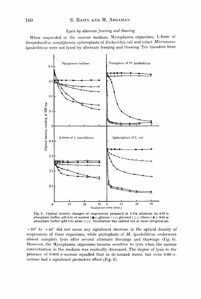
160 S. R a z in a n d M. A r g a m a n
Lysis by alternate freezing and thawingWhen suspended in the sucrose medium, Mycoplasma organisms, L-form of
Streptobacillns moniliformis, spheroplasts of Escherichia, coli and intact Micrococcus lysodeikticus were not lysed by alternate freezing and thawing. Ten transfers from
Fig. 2. Optical density changes of suspensions prepared in 1-5m solutions (in 0 01m- phosphate buffer, pH 6-8) of sucrose ( • ) ; glucose ( x ); glycerol ( A ); ribose ( ▲ ); 0 01 m - phosphate buffer (pH 6-8) alone ( O). Incubation was carried out at room temperature.— 2 4 ° to + 4 0 ° did not cause any significant decrease in the optical density of suspensions of these organisms, while protoplasts of M. lysodeikticus underwent almost complete lysis after several alternate freezings and thawings (Fig. 3). However, the Mycoplasma organisms became sensitive to lysis when the sucrose concentration in the medium was markedly decreased. The degree of lysis in the presence of 0-0 06 M-sucrose equalled that in de-ionized water, but even 0-06 m - sucrose had a significant protective effect (Fig. 3).

Spermine, known to protect some fragile organisms and organelles from lysis under unfavourable conditions (Tabor, Tabor & Rosenthal, 1961) was tested for such effect on Mycoplasma treated by freezing and thawing in de-ionized water. A concentration of 10 //g./ml. of this polyamine protected Mycoplasma organisms from lysis by ten alternate freezings and thawings, while 2 /¿g./ml. was insufficient. These results were obtained with both Mycoplasma mycoides var. capri and M. laidlawii. Spermine, protamine and poly-L-lysine agglutinated M. mycoides var.
Lysis of Mycoplasma 161
Fig. 3. Lysis of M y c o p la s m a laidlcctcii by alternate freezing and thawing in media containing different molar concentrations of sucrose (indicated in figure). The lysis curve of protoplasts of M icro coccu s ly so d e ik ticu s in M-sucrose (O — O) is included for comparison.
capri and M. laidlawii within a certain range of concentrations (spermine, 80— 1250/¿g./ml.; protamine, 80-5000 Mg./ml.; polylysine, 80—320 /¿g./ml.). Intact M . lysodeikticus organisms were agglutinated by the same polyamine concentrations. The aggregates were formed within 1-2 hr. of incubation at 37°; they sedimented overnight, leaving the supernatant fluid clear.
i i G. Microb. xxx

162 S. B a z in a n d M. An d a m a n
Lysis by heatingSpheroplasts of Aerobacter aerogenes were found to lyse at temperatures of about
56-65° (Gebicki & James, 1960). Similar experiments with our test organisms suspended in the sucrose medium showed that the optical density of the suspension of Micrococcus lysodeikticus protoplasts decreased by about 50 % after heating at 80° for 10 min. A decrease of about 30 % in optical density was found with the Mycoplasma suspensions heated to the same temperature; the optical density of these suspensions began to decrease at 50°.
Sodium taurocholate concentration (%, w/v)
Fig. 4. Lysis by sodium taurocholate. M y c o p la s m a la id la m ii ( A ); M y c o p la sm a m yco id es var. c a p r i ' ▲ ); L-form of S trep to b a c illu s m o n ilifo rm is ( x ); spheroplasts of E sch erich ia coli (□ ); intact cells of M icro coccu s ly so d e ik ticu s (O ); protoplasts of M . lyso d e ik ticu s ( • ) .The experiment was carried out in the sucrose medium and optical density was measured after 80 min. incubation at room temperature.
Lysis by treatment with surface active agentsThe Mycoplasma organisms tested were sensitive to lysis by sodium deoxycholate
and sodium taurocholate (Fig. 4). The Mycoplasma organisms were more sensitive to lysis by these agents than were the L-form of Streptobacillus moniliformis and spheroplasts of Escherichia coli. On the other hand the Mycoplasma organisms were more resistant to lysis than were protoplasts of Micrococcus lysodeikticus. Similar results were obtained with the anionic detergent sodium lauryl sulphate (Fig. 5 a). The cationic detergent cetyltrimethylammonium bromide (CTAB) caused lysis of the bacterial protoplasts and of Mycoplasma laidlawii only within a narrow range of concentrations; at higher concentrations agglutination and precipitation of the

163
Cetyltrimethylammonium bromide concentration (MX 10 5)Fig. 5. Lysis by ionic detergents, (a) Sodium lauryl sulphate; (6) cetyltrimethylammonium bromide. M y c o p la s m a la id la w ii (A ); M y c o p la s m a m yco id es var. c a p r i (A); M y c o p la s m a h o m in is (■ ); L-form of S trep to b a c illu s m o n ilifo rm is ( x ); spheroplasts of E sch erich ia co li (□ ); intact M icro coccu s ly so d e ik ticu s (O); protoplasts of M . lyso d e ik ticu s (#). The experiment was carried out in the sucrose medium and optical density was measured after incubation for 30 min. at room temperature.
11-2

164organisms occurred and the optical density of their suspensions increased (Fig. 5 b). The cationic detergent CTAB had no marked lytic effect on any of the other organisms tested. Pre-treatment of M. laidlawii organisms and M. lysodeikticus protoplasts with uranyl nitrate gave protection against lysis by anionic and by cationic detergents (Fig. 6).
S. R a z in a n d M. A r g a m a n
Uranyl nitrate concentration (M x1 0 5)
Fig. 6. Lysis by sodium lauryl sulphate (SLS) and cetyltrimethylammonium bromide (CTAB) of M y c o p la s m a la id la w ii (A ) and protoplasts of M icrococcu s lysod eik ticu s ( • ) pretreated with different concentrations of uranyl nitrate. The experiment was carried out in the sucrose medium. Optical density was measured after incubation for 30 min. at room temperature and plotted as % optical density of suspensions before the addition of detergents.The non-ionic detergents Tween 80 and Triton had a much lower lytic activity
than had the ionic detergents, and caused no lysis when the organisms were suspended in the sucrose medium. Some lysis occurred when Mycoplasma laidlawii was suspended in de-ionized water. None of the other Mycoplasma organisms nor the L-form of Streptobacillus miniliformis were lysed by non-ionic detergents, even when suspended in de-ionized water.

Lysis of Mycoplasma 165
n-butanol concentration (M)Fig. 7. Lysis by primary alcohols, (a ) Ethanol; (b ) n-propanol; (c) n-butanol. M y c o p la s m a la id la w ii (A); M y c o p la s m a m yc o id e s var. c a p r i (A ); L-form of S trep to b a c illu s m o n ilifo rm is ( x ); intact cells of M icro coccu s ly so d e ik ticu s (O); protoplasts of M . lyso - d e ik ticu s ( • ) . The experiment was carried out in the sucrose medium and optical density was measured after incubation for 30 min. at room temperature.

166 S. R a z in a n d M. A b g a m a n
L y s i s b y a lco h o lsThe Mycoplasma protoplasts, like bacterial protoplasts, were very sensitive to
lysis by primary alcohols (Fig. 7). On a molar basis the lytic effect of n-butanol was about four times greater than that of w-propanol and about ten times greater than that of ethanol. The increase in the optical density of the suspensions at high ethanol concentrations was apparently due to precipitation of the cellular components at these high alcohol concentrations. The L-form of S tr e p to b a c i l lu s m o n i l i f o r m i s was much more resistant to the lytic effects of alcohols than were the Mycoplasma organisms.
F ig . 8 . L y s is b y a lk a li. M ycoplasm a laidlawii ( A ) ; M ycoplasm a mycoides v a r. capri ( ▲ ); M ycoplasm a hom inis ( ■ ) ; L-fo rm o f Streptobacillus m oniliform is ( x ) ; spherop lasts o f Escherichia coli ( □ ) ; in ta c t Micrococcus lysodeikticus ( O ) ; p ro top lasts o f M . lysodeikticus ( • ) . Th e exp erim ent w as ca rried out in the sucrose m edium and the o p tica l d ensity m easured a fte r in cub atio n fo r 30 m in . a t room tem perature.
L y s i s b y a lk a l iAll the organisms tested, except intact M ic r o c o c c u s ly s o d e ik t ic u s , showed various
degrees of sensitivity to lysis by NaOH. Mycoplasma organisms and spheroplasts of E sc h e r ic h ias c o li showed the highest sensitivity; bacterial protoplasts and L- forms were less sensitive to lysis by alkali (Fig. 8). Hydrochloric acid did not cause lysis of any of the test organisms within the range of concentrations 0-03-5-0m.

Lysis o f Mycoplasma 167L y s i s b y d ig i to n in a n d s a p o n in
All the Mycoplasma strains tested showed a marked sensitivity to lysis by digitonin and saponin; all the other organisms tested were completely resistant. Human erythrocytes were more sensitive than Mycoplasma organisms to lysis by digitonin and saponin (Fig. 9). Washed digitonin-resistant organisms were not rendered sensitive to lysis by exposure to 0-01 mg. cholesterol/ml. in the sucrose medium for 3 hr. at 37°. M y c o p la s m a l a id la w i i organisms grown in a cholesterol- free defined medium (Razin & Cohen, 1962) were much more resistant to lysis by digitonin and saponin than were organisms grown in the cholesterol-containing Edward medium (Fig. 10).
F ig . 9Digitonin concentration (//g./ml.)
F ig . 10
F ig . 9 . L y s is b y d ig ito n in . M ycoplasm a la id law ii ( A ) ; M ycoplasm a m ycoides v a r. capri ( A ) ; L-fo rm o f Streptobacillus m oniliform is ( x ) ; spherop lasts o f Escherichia coli ( □ ) ; in ta c t M icrococcus lysodeikticus (O ) ; p ro top lasts o f M . lysodeikticus ( • ) ; hum an eryth ro cytes (■ ). Th e experim ent w as ca rried out in the sucrose m edium and th e o p tica l d en sity m easured a fte r incub atio n fo r 30 m in . a t room tem p erature .F ig . 10. L y s is b y d ig ito n in o f M ycoplasm a la id law ii grow n in a cho lestero l-free defined m edium (R a z in & Cohen, 1962 ; O ) o r in the cho lestero l-contain ing E d w a rd m edium ( • ) . Th e exp erim ent w as carried out in de-ionized w ater and o p tica l d ensity m easured a fte r in cu b atio n fo r 30 m in . a t room tem perature.
L y s i s b y e n z y m e sL y s o z y m e . This enzyme had no lytic effect on Mycoplasma organisms or on the
L-form of S tr e p to b a c i l lu s m o n i l i fo r m is by any of the methods tested.L ip a s e . Crude pancreatic lipase caused lysis of all the Mycoplasma strains ex
amined. The lytic effect was greater in 0-067 M-phosphate buffer (pH 7-0) than in 0-01 M-phosphate buffer (pH 8-0). Pancreatic lipase also caused lysis of bacterial protoplasts, spheroplasts and L-forms. Intact organisms of M ic r o c o c c u s ly s o d e ik t ic u s and E s c h e r ic h ia c o li were not lysed by this enzyme (Fig. 11).
P r o te o ly t ic e n z y m e s . Trypsin had very little lytic effect on any fresh organism tested. A marked lytic effect was seen with the Mycoplasma organisms and spheroplasts of E s c h e r ic h ia c o li when these were preheated at 70° for 15 min. The L-form

168 S. R aZIN AND M, A rGAMANof S tr e p to b a c i l lu s m o n i l i fo r m is was insensitive to lysis by trypsin even when preheated. Of the other proteolytic enzymes tested, bromelin, like trypsin, acted only on heated organisms; papain had no lydc activity.
Lipase concentration (¿¿g./ml.)
F ig . 11. L y s is b y p an crea tic lip ase . C e lls o f M ycoplasm a laidlawii (A ) , Mycoplasma mycoides v a r . capri ( A ) ; L-fo rm o f Streptobacillus m oniliform is ( x ) ; spherop lasts o f Escherichia coli ( □ ) ; p ro top lasts o f Micrococcus lysodeikticus ( • ) ; in ta c t ce lls o f M . lysodeikticus (O ) o r E . coli (■ ) w ere suspended in M -sucrose+ 0 0 6 7 M -phosphate bu ffer (p H 7-0) so lu tion conta in ing vario u s concentrations o f lip ase . O p tica l d e n sity w as m easured a t 37° a fte r in cub atio n fo r 90 m in .
D IS C U S S IO N
The relative resistance of Mycoplasma organisms to osmotic shock has been described by several authors (Smith & Sasaki, 1958; Plackett, 1959; Butler & Knight, I960). Several hypotheses might be provided to explain the resistance of Mycoplasma to lysis by osmotic shock: (a ) The very small dimensions and spherical shape of most Mycoplasma organisms decrease the adverse effects of osmotic pressure changes (Mitchell & Moyle, 19565). Thus, the small elements of the L-form of P r o te u s v u lg a r is were much more resistant to osmotic shock than were the larger elements of this L-form (Thorsson & Weibull, 1958). (b) The elasticity of the Mycoplasma envelope might permit considerable distension of the organism without its being ruptured, (c) The internal osmotic pressure of the Mycoplasma organisms might be low. There are marked differences between the internal osmotic pressures of different bacteria. Gram-positive cocci, such as M ic r o c o c c u s ly s o d e ik t ic u s , have osmotic pressures of 20-30 atmospheres (Mitchell & Moyle, 19565) while Gram-negative rods have internal osmotic pressure of 4-8 atmospheres only (Gebicki & James, 1960; Hugo & Russell, 1960). Protoplasts of M . ly s o d e ik t ic u s therefore underwent almost complete lysis in 0-25 M-sucrose, while spheroplasts of E s c h e r ic h ia c o li lysed to a similar degree only in 0-03 M-sucrose (Fig. 1). We have no direct way of

169measuring the internal osmotic pressure of Mycoplasma cells. However, indirect evidence for a low internal osmotic pressure was obtained while testing the sensitivity of Mycoplasma to lysis by alternate freezing and thawing. Mycoplasma organisms were lysed by this procedure when suspended in de-ionized water, but resisted lysis when sucrose or NaCl were added to the medium (Fig. 3). A concentration of 0T2-0-25 M-sucrose protected almost all Mycoplasma organisms against lysis by freezing and thawing. The protoplasts of M . ly s o d e ik t ic u s were lysed by alternate freezing and thawing, even when suspended in M-sucrose solution (Fig. 3). The protective effect of these solutes might be explained by the shrinkage of the plastic Mycoplasma cells by osmotic dehydration in hypertonic media (Postgate & Hunter, 1961). The cell envelope of the shrunken cell is capable of resisting more efficiently the shearing forces of the ice crystals. If this explanation be correct, then 0-25 M-sucrose solution has to be regarded as hypertonic, and 0T2 M-sucrose as about isotonic, to the Mycoplasma organisms. The osmotic pressure of a 0T2 m - sucrose solution is about 3 atmospheres (H a n d b o o k o f C h e m is tr y & P h y s ic s , 1954-1955). Hence, the internal osmotic pressure of Mycoplasma organisms seems to be remarkably low.
The marked resistance of the stable L-form of S tr e p to b a c i l lu s m o n i l i fo r m is to lysis in hypotonic media might be explained along the same lines as the resistance of Mycoplasma organisms to osmotic shock. Medill-Brown, Hutchinson & Cocklin (1960), on comparing the osmotic fragility of penicillin-induced spheroplasts and L-forms derived from P r o te u s m ir a b i l i s , found that the spheroplasts were much more sensitive to osmotic shock than were the L-forms. The agglutination of the Mycoplasma organisms by spermine and other polyamines indicates that the cell envelope of Mycoplasma has a net negative charge, like that of the bacterial protoplast membrane (Few, Gilby & Seaman, 1960) and of bacterial cell walls (Razin & Rozansky, 1959). The great sensitivity of Mycoplasma organisms to surface active substances is well known (Klieneberger-Nobel, 1962). Our studies have shown the close similarity between Mycoplasma organisms and bacterial protoplasts with respect to lysis by surface active agents. Mycoplasma organisms were lysed by about the same concentrations of ionic detergents as were bacterial protoplasts. Apparently the ionic detergents act on similar components present in the membranes of Mycoplasma organisms and of bacterial protoplasts, as indicated by the inhibition of lysis when the organisms were pre-treated with uranyl nitrate. Uranyl ions have a strong affinity for the phosphate groups of phospholipids present in the bacterial membrane (Gilby & Few, 1960 a). Since the cationic detergent cetyltrimethyl- ammonium bromide (CTAB) combines with these acidic groups, its lytic action is inhibited more efficiently by uranyl ions than by the anionic detergent sodium lauryl sulphate (SLS), which combines mainly with the protein component of the membrane (Gilby & Few, 1960a). The cationic detergent CTAB causes lysis of Mycoplasma organisms and bacterial protoplasts only within a narrow limit of concentrations. At concentrations higher than 5 x 1 0 _ 5 m this detergent caused protein denaturation and ‘ fixation’ of the organisms, as well as agglutination by neutralization of surface negative charge (Dawson, Lominski & Stern, 1953).
The similar sensitivity of Mycoplasma organisms and bacterial protoplasts to lysis by primary alcohols and pancreatic lipase suggests the presence of a lipid component in the Mycoplasma cell envelope. Gilby & Few (19606) claimed that
Lysis of Mycoplasma

170alcohols act on the lipid component of the protoplast membrane, causing lysis by disruption of membrane permeability. The marked lysis by alkali of all the organisms tested, except intact bacteria, is apparently due to the hydration and dispersion of cell proteins (Grula & Hartsell, 1957). The mucopeptide polymer of the bacterial cell wall is alkali resistant and protects the intact bacterial cell from lysis. Digestion of the mucopeptide by lysozyme renders the bacteria sensitive to lysis by alkali (Grula & Hartsell, 1957).
The most remarkable difference between Mycoplasma organisms and all the other organisms examined was the sensitivity of Mycoplasma to lysis by digitonin and saponin; lysis of Mycoplasma by digitonin was reported by Smith & Rothblat (1960). This might be explained by the presence of cholesterol in the Mycoplasma cell envelope (Rothblat & Smith, 1961; Razin & Argaman, unpublished data). The much lower sensitivity of Mycoplasma laidlawii to lysis by digitonin when grown in a cholesterol-free medium (Fig. 10) suggests that at least most of the cholesterol in this Mycoplasma species derives from the growth medium.
Our studies show that bacterial L-forms and spheroplasts are less sensitive to lysis than bacterial protoplasts and Mycoplasma organisms. The higher resistance of L-forms and spheroplasts to lysis might be attributed to the presence of a modified cell wall (Thorsson & Weibull, 1958; Klieneberger-Nobel, 1960). The present findings and the morphological studies of ultrathin sections of Mycoplasma organisms (van Iterson & Ruys, 1960) suggest that the Mycoplasma organisms are limited by a membrane of lipoprotein nature, very like that of the plasma membrane which encloses the bacterial protoplast. Chemical analysis of isolated Mycoplasma cell membranes also supports this assumption (Razin & Argaman, to be published). The only difference so far found between the cell membrane of Mycoplasma and the plasma membrane of bacteria is the presence of cholesterol in the former.
Our thanks are due to Mrs E. Friedberg and Mr M. Wormser for their valuable technical assistance. This work was supported in part by a grant from the Joint Research Fund of the Hebrew University-Hadassah Medical School.
S. R a z in a n d M, A n d a m a n
R E F E R E N C E S
Becker, M . E . & Hartsell, S . E . (1 9 5 4 ). F a c to rs a ffe c tin g b a c te rio ly s is u s in g ly so zy m e in d u a l en zym e sy s te m s. A rch . B iocliem . B io p h y s . 5 3 , 4 0 2 .
Butler, M . & Knight, B . C . J . G . (1 9 6 0 ). T h e s u rv iv a l of w ash ed su sp e n sio n s o f M yco p la sm a . J . gen. M icrobio l. 2 2 , 4 7 0 .Dawson, I . M ., Lominski, I . & Stern, H . (1 9 5 8 ). A n e lectro n -m icro sco p e s tu d y o f th e a c tio n o f ce ty l-trim e th y l-a m m o n iu m b ro m id e on Staphylococcus aureus. J . P a th . B a d . 6 6 , 51 3 .
Edwards, G . A . & Fogh, J . (1 9 6 0 ). F in e s tru c tu re o f p le u ro p n e u m o n ia -lik e o rg an ism s in p u re c u ltu re an d in in fe c te d tis su e c u ltu re c e lls . J . B a d . 79, 26 7 .
Few , A . V., Gilby, A . R . & Seaman, G. V. F. (1960). A n e le c tro p h o re tic s tu d y on s tru c tu ra l co m p o nents o f M icrococcus lysodeikticus. B io ch im . b iophys. A c ta , 3 8 , 130.Gebicki, J . M . & J ames, A . M . (1 9 6 0 ). T h e p re p a ra tio n an d p ro p e rtie s o f sp h e ro p la sts o f A erobacter aerogenes. J . gen. M icrobio l. 2 3 , 9 .
Gilby, A . R . & Few, A . V . (1 9 5 9 ). O sm o tic p ro p e rtie s o f p ro to p la sts o f M icrococcus lysodeik ticus. J . gen. M icrobio l. 2 0 , 321 .
Gilby, A . R . & Few, A . V . (1 9 6 0 a ). L y s is o f p ro to p la sts o f M icrococcus lysodeik ticus b y io n ic d e te rg e n ts . J . gen. M icrobio l. 2 3 , 19 .

G i l b y , A . R . & F e w , A . V . (1 96 0Ö ). L y s is o f p ro to p la sts o f M icrococcus ly so deik ticus b y a lc o h o ls . J . gen. M icrob io l. 2 3 , 2 7 .
Gilby, A . R ., Few, A . V . & McQuillen, K . (1 9 5 8 ). T h e c h e m ica l co m p o sitio n o f th e p ro to p la s t m em b ran e o f M icrococcus lysodeik ticus. B io ch im . b iophys. A c ta , 2 9 , 2 1 .
Gr u la , E . A . & H a r t se l l , S . E . (1 9 5 7 ). L y so z y m e in th e b a c te rio ly s is o f G ra m -n e g a tive b a c te ria . I I . F a c to rs in flu e n c in g c le a rin g d u rin g th e N a k a m u ra tre a tm e n t. C anad. J. M icrobio l. 3 , 2 3 .
H andbook o f Chew,is try a n d P h ys ic s ( 1954—1 9 5 5 ), 3 6 th ed . E d . C . D . H o d g m an . C le v e la n d , O h io , U . S . A . : C h e m ica l R u b b e r P u b lish in g C o .
Hugo, W . B . & Russell, A . D . (1 9 6 0 ). Q u a n tita t iv e a sp e cts o f p e n ic illin a c tio n on E scherich ia coli in h y p e rto n ic m e d iu m . J . B a d . 8 0 , 4 3 6 .Imaeda, T . & Convit, J . (1 9 6 2 ). E le c tro n m icro sco p e s tu d y o f M ycobacterium leprae an d it s e n v iro n m e n t in a v e s ic u la r le p ro u s le s io n . J . B a d . 83, 4 3 .
H a n d l e r , O . & Z e h e n d e r , C . (1 9 5 7 ). Ü b e r d as V e rko m m e n v o n a , e -D ia m in o p im e lin sä u re b e i ve rsch ie d e n e n L -P h a se n ty p e n vo n P ro teu s vu lgaris u n d b e i den p leu ro p n eu m o n ie ä h n lich e n O rg an ism e n . Z . N a tu r f . 12 b, 7 2 5 .
K l ie n e b e r g e r -N o b e l , E . (1 9 6 0 ). L - fo rm s o f b a c te r ia . In T h e B acteria , A T rea tise on S tru c tu re a n d F u n c tio n , v o l. 1 , p . 3 6 1 . E d . I . C . G u n sa lu s & R . Y . S ta n ie r . N ew Y o r k : A ca d e m ic P re s s In c .
K l ie n e b e r g e r -N o b e l , E . (1 9 6 2 ). P leu ro p n eu m o n ia -like organism s (P P L O ) M yco p la sm a - taceae. L o n d o n : A ca d e m ic P re s s .
Kohn, A . (1 9 6 0 ). L y s is o f fro ze n an d th a w e d c e lls o f E sch erich ia coli b y ly so zy m e , an d th e ir co n ve rsio n in to sp h e ro p la s ts . J . B a d . 7 9 , 69 7 .
Lederberg, J . (1956). B a c te r ia l p ro to p la sts in d u ce d by p e n ic illin . P roc. n a t. A ca d . S e i., W a sh . 42, 574.
Me d il l -B r o w n , M ., H u t c h in so n , W . G . & Co c k lin , E . (1 9 6 0 ). T h e L - fo rm s o f P ro teus m ira b ilis . A n n . N . Y . A ca d . S e i. 7 9 , 8 7 4 .
M it c h e l l , P . & Mo y l e , J . (1 95 6 a ) . L ib e ra t io n an d o sm o tic p ro p e rtie s o f th e p ro to p la sts o f M icrococcus lysodeik ticus an d S a rc in a lutea. J . gen. M icrobio l. 15, 51 2 .
M it c h e l l , P . & Mo y l e , J . (1 9 5 6 6 ). O sm o tic fu n c tio n an d s tru c tu re in b a c te ria , in B a cteria l A n a to m y . S y m p . Soc. gen. M icrobio l. 6 , 150 .Plackett, P . (1 9 5 9 ). O n th e p ro b a b le ab sence o f ‘ m u co co m p le x ’ fro m M y co p la sm a m ycoides. B io ch im . b iophys. A c ta , 3 5 , 26 0 .
P o stg a te , J . R . & H u n t e r , J . R . (1961). O n th e su rv iv a l o f frozen b a c te ria . J . gen. M icrobio l. 26, 367.
R a z in , S . & A rgam an , M . (1 9 6 1 ). P ro p e rtie s o f th e M yco p la sm a (P P L O ) c e ll en ve lo p e . B u ll. R es. C oun. Isra e l, 9 E , 121 .
R a zin , S . & Co h e n , A . (1 9 6 2 ). N u tr it io n a l re q u ire m e n ts an d m e tab o lism o f M y co p la sm a la id la w ii. J . gen. M icrob io l. 3 0 , 141 .
R a zin , S . & Ol iv e r , O. (1961). M orp hogenesis o f M yco p la sm a an d b a c te ria l L - fo rm co lo n ie s. J . gen. M icrob io l. 24, 225 .
R a zin , S . & R o zan sk y , R . (1 9 5 9 ). M ech an ism o f th e a n tib a c te ria l a c tio n o f sp e rm in e . A rch . B iochem . B io p h ys . 81, 36.
R e p a s k e , R . (1 9 5 6 ). L y s is o f G ra m -n e g a tive b a c te ria b y ly so zy m e . B iochem . b iophys. A c ta , 2 2 , 189 .
R o b er tso n , J . D . (1 9 5 9 ). T h e u ltra s tru c tu re o f c e ll m em b ran es an d th e ir d e riv a tiv e s . B iochem . Soc. S y m p . 16, 3.Rodwell, A . W . & Abbot, A . (1 9 6 1 ). T h e fu n c tio n o f g ly c e ro l, ch o le ste ro l an d lo n g -ch a in fa t t y a c id s in th e n u tr it io n o f M y co p la sm a m ycoides. J . gen. M icrobio l. 2 5 , 201 .
R o th b la t , G . H . & Sm it h , P . F . (1 9 6 1 ). N o n sa p o n ifla b le lip id s o f re p re se n ta tiv e p le u ro p n e u m o n ia -like o rg a n ism s. J . B a d . 8 2 , 4 7 9 .Ruys, A . C . & Van Iterson, W . (1 9 6 1 ). Som e c h a ra c te ris t ic s o f p a th o g en ic a v ia n P P L O . A n to n ie va n Leeuw enhoek J . M icrobio l. Serol. 2 7 , 129 .Sharp, J . T . (1 9 6 0 ). T h e c e ll w a ll o f b a c te r ia l L - fo rm s an d p le u ro p n e u m o n ia -lik e o rg an ism s. A n n . N . Y . A ca d . S e i. 7 9 , 34 4 .
Lysis of Mycoplasma 171

172 S. R a z in a n d M. A r g a m a nSmith, P . F . & Rothblat, G . H . (1 9 6 0 ). In c o rp o ra tio n o f ch o le ste ro l b y p le u ro p n e u m o n ia
lik e o rg an ism s. J . B act. 8 0 , 84 2 .Smith, P . F . & Sasaki, S . (1 9 5 8 ). S ta b ilit y o f p le u ro p n e u m o n ia -lik e o rg an ism s to som e p h y s ic a l fa c to rs . A p p l . M icrobio l. 6 , 18 4 .Smolelis, A . N . & Hartsell, S . E . (1 9 4 9 ). T h e d e te rm in a tio n o f ly so zy m e . J . B a d . 5 8 , 73 1 .Tabor, H., Tabor, C . W . & Rosenthal, S . M . (1 9 6 1 ). T h e b io c h e m is try o f th e p o ly a m in e s : sp e rm id in e an d sp e rm in e . A n n . R ev. B iochem . 3 0 , 579 .Thorsson, K . G . & Weibull, C . (1 9 5 8 ). S tu d ie s on th e s tru c tu re o f b a c te r ia l L - fo rm s p ro to p la sts an d p ro to p la s t- lik e b o d ie s. J . U ltrastr. R es. 1 , 4 1 2 .
van Iterson, W . (1 9 6 1 ). Som e fe a tu re s o f a re m a rk a b le o rg an e lle in B a c illu s su b tilis . J . b iophys. biochem. Cytol. 9 , 183 .
van Iterson, W . & Ruys, A . C . (1 9 6 0 ). T h e fin e s tru c tu re o f th e M yco p la sm atace a e (m icro -o rg an ism s o f th e p leu ro p n eu m o n ia g ro up = P P L O ) . 1 . M y co p la sm a h o m in is , M . fe rm e n ta n s a n d M . sa liv a r iu m . J . U ltrastr. R es. 3 , 282 .Weibull, C . & Bergstrom, L . (1 9 5 8 ). T h e c h e m ica l n a tu re o f th e c y to p la sm ic m em b rane an d c e ll w a ll o f B a c illu s m egaterium , s t ra in M . B io ch im . b iophys. A c ta , 3 0 , 34 0 .