Agricultural Microbiology
679
Agricultural Microbiology This eCourse Developed By Indian Council of Agriculture Research
-
Upload
khangminh22 -
Category
Documents
-
view
1 -
download
0
Transcript of Agricultural Microbiology

Agricultural Microbiology
This eCourse Developed By
Indian Council of Agriculture Research

www.AgriMoon.Com
2
AGRICULTURAL MICROBIOLOGY
This eCourse Developed By
TNAU (ICAR)

Agricultural Microbiology
3
Course Outline:
Lecture 01: History of Microbiology: Spontaneous Generation Theory Lecture 02: Germ Theory of Disease Lecture 03: Protection against Infections Lecture 04: Metabolism in Bacteria Lecture 05: ATP Generation Lecture 06: Microbial Metabolism - Autotrophs
Lecture 07: Bacteriophages: Structure and Properties of Bacterial Viruses Lecture 08: Lytic and Lysogenic Cycles – Phage Multiplication Cycle Lecture 09: Viroids, Prions
Lecture 10: Bacterial Genetics Lecture 11: Gene Expression Lecture 12: Recombination in Bacteria Lecture 13: Genetic Engineering - Plasmids, Episomes
Lecture 14: Genetically Modified Organism
Lecture 15: Soil Microbiology: Microbial Group in Soil Lecture 16: Microbial Transformations of Carbon
Lecture 17: Microbial Transformations of Nitrogen, Phosphorus and Sulphur Lecture 18: Biological Nitrogen Fixation
Lecture 19: Phyllosphere Bacteria Lecture 20: Composting
Lecture 21: Environmental Microbiology
Lecture 22: Microbiology of Food: Microbial Spoilage Lecture 23: Principles of Preservation
Lecture 24: Role of Bacteria in Fermentation Lecture 25: Beneficial Microorganisms in Agriculture Lecture 26: Microbial Agents for Control of Plant Diseases Lecture 27: Biogas Production
Lecture 28: Biodegradable Plastics
Lecture 29: Plant – Microbe Interactions
Lecture 30: Bioremediation
Lecture 31: Biosensor Lecture 32: Microbial Products

www.AgriMoon.Com
4
Lecture 01:
HISTORY OF MICROBIOLOGY: SPONTANEOUS GENERATION THEORY
Microbiology often has been defined as the study of organisms and agents too small to be seen clearly by the unaided eye—that is, the study of microorganisms. Because objects less than about one millimeter in diameter cannot be seen clearly and must be examined with a microscope, microbiology is concerned primarily with organisms and agents this small and smaller.
Microbial World Microorganisms are everywhere. Almost every natural surface is colonized by microbes (including our skin). Some microorganisms can live quite happily in boiling hot springs, whereas others form complex microbial communities in frozen sea ice. Most microorganisms are harmless to humans. You swallow millions of microbes every day with no ill effects. In fact, we are dependent on microbes to help us digest our food and to protect our bodies from pathogens. Microbes also keep the biosphere running by carrying out essential functions such as decomposition of dead animals and plants. Microbes are the dominant form of life on planet Earth. More than half the biomass on Earth consists of microorganisms, whereas animals constitute only 15% of the mass of living organisms on Earth. This Microbiology course deals with
How and where they live Their structure How they derive food and energy Functions of soil micro flora Role in nutrient transformation Relation with plant Importance in Industries
The microorganisms can be divided into two distinct groups based on the nucleus structure: Prokaryotes – The organism lacking true nucleus (membrane enclosed chromosome
and nucleolus) and other organelles like mitochondria, Golgi body, entoplasmic reticulum etc. are referred as Prokaryotes. (Ex: Bacteria, archaea)

Agricultural Microbiology
5
Eukaryotes - The organism possessing membrane enclosed nucleus and other cell
organelles are referred as Eukaryotes (Ex : algae, fungi, protozoa) The microorganisms were divided into 6 distinct groups based on the phylogenic, morphological and physiological characters. The major groups of microorganisms are
1. Bacteria are phylogenetically related group of unicellular prokaryotic organisms distinct from archeae
2. Archaea is phylogenetically related group of prokaryotes which are primitive and distinct from bacteria
3. Fungi are group of eukaryotic organisms lacking chlorophyll. They range in size and shape from single celled yeast to multicellular mushrooms.
4. Algae refer the group of eukaryotic organisms with chlorophyll. They range in size and shape from single celled algae (Ex: Chlorella) to complex cellular structured plant like algae (Ex. Kelp)
5. Protozoa are group of eukaryotic organism‘s lack of cell wall. The morphology, nutrition and physiology is different from other groups
6. Viruses are group of non-cellular organisms, parasite or pathogen to plant, animals and other microorganisms. They are too small and cab be visualized only under electron microscopes
History of Microbiology in brief:
Obviously human have had to deal with microbes even before the recorded history. The first record of human using comes from ancient tablets from mid east. Babylonians were using yeast to make beer over 8000 years ago and acetic acid bacteria to make vinegar over 6000 years ago.
About 5000 years ago, Persia (Now Iran) region recorded the wine making. The Romans had God for that was specific for microorganisms. The roman God of mold and mildew was ―Robigus‖ and ―Robigo” which means crop rust. (Rust is one of the plant disease caused by fungus). God Robigus was very much feared because of crop lost.
About 2000 years ago, Romans proposed that diseases were caused by tiny animals. But, fundamentalist religions had a strong hold over the progress. The real microbiology history starts from 1600s, when people began to make crude lenses and microscopes.

www.AgriMoon.Com
6
HIGHLIGHTS IN THE HISTORY OF MICROBIOLOGY
Effects of Disease on Civilization
Infectious diseases have played major roles in shaping human history Bubonic Plague epidemic of mid 1300's, the "Great Plague", reduced population
of Western Europe by 25%. Plague bacterium was carried by fleas, spread from China via trade routes and poor hygiene. As fleas became established in rat populations in Western Europe, disease became major crisis.
Smallpox and other infectious diseases introduced by European explorers to the Americas in 1500's were responsible for decimating Native American populations. Example: In the century after Hernan Cortez's arrival in Mexico, the Aztec population declined from about 20 million to about 1.6 million, mainly because of disease.
Infectious diseases have killed more soldiers than battles in all wars up to WW II. Example: in U. S. Civil war, 93,000 Union soldiers died in direct combat; 210,000 died as a result of infections.
Until late 1800's, no one had proved that infectious diseases were caused by specific microbes, so the possibility of prevention or treatment had no sound empirical base.
Brueghel: The Triumph of Death (1560)
Discovery of Microbes
To see microbes, you need a microscope. The first microscope was invented by Antony van Leeuwenhoek (1632-1723), a Dutch businessman.
Leeuwenhoek took up lens grinding to make magnifiying glasses so he could examine fine weave of fabrics. In testing his lenses, he discovered many small creatures he called "animalcules" in samples such as pond water. His best lenses could magnify 300-500X.

Agricultural Microbiology
7
Leeuwenhoek microscopes were crude, relied on a single lens held in a metal plate.
Leeuwenhoek described many previously unseen life forms, including different forms of bacteria, mold spores, etc. Leeuwenhoek reported discoveries to Royal Society from 1670's on, firmly established existence of microbes. Nevertheless, the significance of this discovery was not apparent for almost 200 years.
Antony van Leeuwenhoek.
Origin of Life Controversy
Where did microbes come from? Many believed they arose from simple materials by process of spontaneous generation. This notion had been posited by Aristotle (382-322 B.C.) and other Greek philosophers to explain decay and appearance of animals such as flies and frogs, and was widely held as common sense even in 1700's and 1800's.
Francisco Redi (1626-1697) demonstrated that flies did not arise spontaneously
from rotting meat by simple experiment. If jar of meat was covered by fine muslin, maggots did not arise. However, the simpler life forms discovered by Leeuwenhoek lacked visible complexity, and most people still believed these could arise spontaneously.

www.AgriMoon.Com
8
John Needham (1731-1781):
a Scottish clergyman and naturalist, showed that mirobes grew in soups exposed to air. Claimed existence of a "life force" present in inorganic matter that could cause spontaneous generation. One of his more convincing demonstrations was to boil some soup (briefly), pour into clean flaskswith cork lids, and show that microbes would soon arise.
Lazzaro Spallanzani (1729-1799) claimed Needham's organisms came from heat-
resistant microbes. If flasks were boiled long enough (1-2 h), nothing grew. But Needham countered that prolonged heating destroyed the "life force".

Agricultural Microbiology
9
Louis Pasteur (1822-1895) was passionate believer that life only originated from
previous life, developed several experiments that finally deflated claims for spontaneous generation. Pasteur filtered air through cotton to trap airborne materials, then dissolved the cotton and examined the particulate matter under a microscope; many bacteria and spores of other life forms such as molds were present. Since most skeptics kept arguing that overheating killed the life force present in air, Pasteur developed and ingenious experiment using a swan neck flask that allowed fresh air to remain in contact with boiled materials. The long passageway prevented airborne microbes from reaching the nutrient liquid,
without impeding access to air. One of Pasteur's flasks is still sterile after 100+ years of being exposed to the air (Pasteur Institute, Paris).
Spontaneous Generation theory From earliest times, people had believed in spontaneous generation—that living organisms could develop from nonliving matter. Even the great Aristotle (384–322 B.C.) thought some of the simpler invertebrates could arise by spontaneous generation. This view finally was challenged by the Italian physician Francesco Redi (1626–1697), who carried out a series of experiments on decaying meat and its ability to produce maggots spontaneously. Redi placed meat in three containers. One was uncovered, a second was covered with paper, and the third was covered with fine gauze that would exclude flies. Flies laid their eggs on the uncovered meat and maggots developed. The other two pieces of meat did not produce maggots spontaneously. However, flies were attracted to the gauze-covered container and laid their eggs on the gauze; these
eggs produced maggots. Thus the generation of maggots by decaying meat resulted from the presence of fly eggs, and meat did not spontaneously generate maggots as previously believed. Similar experiments by others helped discredit the theory for larger organisms.
Leeuwenhoek‘s discovery of microorganisms renewed the controversy. Some proposed that microorganisms arose by spontaneous generation even though larger organisms did not. They pointed out that boiled extracts of hay or meat would give rise to microorganisms after sitting for a while. In 1748 the English priest John Needham (1713–1781) reported the results of his experiments on spontaneous generation. Needham boiled mutton broth and then tightly stopper the flasks. Eventually many of the flasks became cloudy and contained microorganisms. He thought organic matter contained a vital force that could confer the properties of life on nonliving matter. A few years later the Italian priest and naturalist Lazzaro Spallanzani (1729–1799) improved on Needham‘s experimental design by first sealing glass flasks that contained water and seeds. If the sealed flasks were placed in boiling water for 3/4 of an hour, no

www.AgriMoon.Com
10
growth took place as long as the flasks remained sealed. He proposed that air carried germs to the culture medium, but also commented that the external air might be required for growth of animals already in the medium. The supporters of spontaneous generation maintained that heating the air in sealed flasks destroyed its ability to support life. Several investigators attempted to counter such arguments. Theodore Schwann (1810–1882) allowed air to enter a flask containing a sterile nutrient solution after the air had passed through a red-hot tube. The flask remained sterile. Subsequently Georg Friedrich Schroder and Theodor von Dusch allowed air to enter a flask of heat-sterilized medium after it had passed through sterile cotton wool. No growth occurred in the medium even though the air had not been heated. Despite these experiments the French naturalist Felix Pouchet claimed in 1859 to have carried out experiments conclusively proving that microbial growth could occur without air contamination.
This claim provoked Louis Pasteur (1822–1895) to settle the matter once and for all. Pasteur first filtered air through cotton and found that objects resembling plant spores had been trapped. If a piece of the cotton was placed in sterile medium after air had been filtered through it, microbial growth appeared. Next he placed nutrient solutions in flasks, heated their necks in a flame, and drew them out into a variety of curves, while keeping the ends of the necks open to the atmosphere .Pasteur then boiled the solutions for a few minutes and allowed them to cool. No growth took place even though the contents of the flasks were exposed to the air. Pasteur pointed out that no growth occurred because dust and germs had been trapped on the walls of the curved necks. If the necks were broken, growth commenced immediately. Pasteur had not only resolved the controversy by 1861 but also had shown how to keep solutions sterile. The English physicist John Tyndall (1820–1893) dealt a final blow to spontaneous generation in 1877 by demonstrating that dust did indeed carry germs and that if dust was absent, broth remained sterile even if directly exposed to air. During the course of his studies, Tyndall provided evidence for the existence of exceptionally heat-resistant forms of bacteria. Working independently, the German botanist Ferdinand Cohn (1828–1898) discovered the existence of heat-resistant bacterial endospores
1. The Spontaneous Generation Experiment.
Pasteur‘s swan neck flasks used in his experiments on the
spontaneous generation of microorganisms.

Agricultural Microbiology
11
2. Disprove of Spontaneous Generation theory
At that time, the age old idea of ―Spontaneous Generation theory‖ was the dominant one. The idea that organism originate directly from non-living matter. (Life from non-living) also called as abiogenesis (a – not; bio – life; genesis – origin). Ex: Maggots were developed spontaneously via recombination of matters in rotting materials. (ex meat) The microbiology starts when the disprove of SG theory.
Louis Pasteur (1822 – 1895) and disproval of Spontaneous generation theory
He performed ―gooseneck experiment‖. The nutrient of flask was heated and the untreated – unfiltered air could pass in or out, but the germs settled in the gooseneck and no microbes were observed in the nutrient solution. His concept of Germs theory of disease (means germs are responsible for the disease not the inert mater) ends the SG theory.
Contributions of Louis Pasteur (1822 – 1895)
Disproved the SG theory Discovered that fermenting fruit to alcohol by microbes – From now the
Fermentation started Sorted different microbes giving different taste of wine. He selected a particular strain (Yeast) for high quality wine. He developed a method to remove the undesired microbes from juice without
affecting its quality. Heating the juice at 62.8°C for half-an hour did the job. This technique is called as Pasteurization, which is commonly used in the field of milk industry.
He discovered that parasites (protozoa) causing pebrine disease of silk worm. He suggested that disease free caterpillars can eliminate the disease.
He isolated the anthrax causing bacilli from the bloods of cattle, sheep and human being.
He also demonstrated the virulence (ability of microbe to cause disease) of bacteria
He developed vaccine (a killed or attenuated microbe to induce the immunity) against rabbis from the brains and spinal cord of rabbit
John Tyndall (1820 -1893)
Proved that dust carries the germs and if no dust in the air, the sterile broth remained free of microbial growth for indefinite period.

www.AgriMoon.Com
12
He also developed a sterilization method ―Tyndallization‖, referred as intermittent or fractional sterilization. The subsequent cooling and heating by steam for 3 days will remove the germs and their spores.
Martinus Willium Beijerinck (1851 – 1931)
Developed the enrichment technique to isolate various group of bacteria.
Isolated sulphur reducing bacteria and sulphur oxidizing bacteria from soil
Isolated free-living nitrogen fixing bacterium, Azotobacter from soil,
Root nodulating bacterium, Rhizobium, Lactobacillus, green algae were identified by him
He confirmed the Tobacco mosaic virus causes disease and it incorporated in the host plant to reproduce.
Sergei Winogradsky (1856 – 1953)
The following are the contributions of Winogradsky to soil microbiology.
Microorganisms involved in N cycle, C cycle, S cycle Nitrification process in soil Autotrophic nutrition of bacteria Chemolithotrophic nutrition of soil bacteria Discovered anaerobic nitrogen fixing bacterium Clostridium pasteurianum
Walther Hesse & Fannie E. Hesse (1883) They used agar instead of gelatin for preparation of media. Agar goes to solution at 100°C and solidifies at 45°C. Till now this was not replaced by any other substance.
Joseph Lister (1878)
Developed Pure culture technique. Pure culture referred as the growth of mass of cells of same species in a vessel. He developed the pure cultures of bacteria using serial dilution technique. He also discovered that carbolic acid to disinfect the surgical equipments and dressings leads the reduction of post-operational deaths/infections.

Agricultural Microbiology
13
Alexander Fleming (1928) identified Penicillium notatum inhibiting
Staphylococcus aureus and identified the antibiotic Penicillin
o 1929-Discovered antibiotic penicillin –important milestone in medical microbiology
o Found that natural substances having antimicrobial activity- Saliva,Nasal mucous
o Worked on Staphylococcus aureus,-inhibition of growth-due to Penicillin
o Florey &Chain-isolated Penicillin in pure culture.
Selman A Waksman, 1945 identified Streptomycin antibiotic from soil bacterium. He
also coined the term antibiotics (referring a chemical substance of microbial origin which is in small quantity exert antimicrobial activity.
1927- Wrote the book on Principles of soil Microbiology
In 1939 Waksman and his colleagues undertook a systematic effort to identify soil organisms producing soluble substances that might be useful in the control of
infectious diseases, what are now known as antibiotics Within a decade ten antibiotics were isolated and characterized, three of them with important clinical applications actinomycin in 1940, streptomycin in 1944, and neomycin in 1949. Eighteen antibiotics were discovered under his general direction.
******☺******

www.AgriMoon.Com
14
Lecture 02: GERM THEORY OF DISEASE
Introduction
Bacteria are mostly unicellular organisms that lack chlorophyll and are among the smallest living things on earth—only viruses are smaller. Multiplying rapidly under favorable conditions, bacteria can aggregate into colonies of millions or even billions of organisms within a space as small as a drop of water. The Dutch merchant and amateur scientist Anton van Leeuwenhoek was the first to observe bacteria and other microorganisms. Using single-lens microscopes of his own design, he described bacteria and other microorganisms (calling them "animacules") in a series of letters to the Royal Society of London between 1674 and 1723.
Bacteria are classified as prokaryotes. Broadly, this taxonomic ranking reflects the fact that the genetic material of bacteria is contained in a single, circular chain of deoxyribonucleic acid (DNA) that is not enclosed within a nuclear membrane. The word prokaryote is derived from Greek meaning "prenucleus." Moreover, the DNA of prokaryotes is not associated with the special chromosome proteins called histones, which are found in higher organisms. In addition, prokaryotic cells lack other membrane-bounded organelles, such as mitochondria. Prokaryotes belong to the kingdom Monera. Some scientists have proposed splitting this designation into the kingdoms Eubacteria and Archaebacteria. Eubacteria, or true bacteria, consist of more common species, while Archaebacteria (with the prefix archae—meaning ancient) represent strange bacteria that inhabit very hostile environments. Scientists believe these bacteria are most closely related to the bacteria which lived when the earth was very young. Examples of archae bacteria are those bacteria which currently live in extremely salty environments or extremely hot environments, like geothermal vents of the ocean floor.
Microbes are organisms that we need a microscope to see. The lower limit of our eye's resolution is about 0.1 to 0.2 mm or 100 - 200 um. Most microbes range in size from about 0.2 um to the 200 um upper limits, although some fruiting bodies of fungi can become much larger. Microbes include the bacteria, algae, fungi, and protozoa. In this lecture we will discuss mostly the bacteria and the fungi.
Bacteria are found everywhere in water, soil, and even air. These small prokaryotic cells, typically from 0.2 to 1 um in length, are capable of living in boiling water, frozen ground, acid volcanoes, and at the bottom of the ocean. They can reproduce by
Agricultural Microbiology
15
doubling with a generation time of 20 minutes, or survive for centuries in a resting stage. In natural waters (lakes, streams, oceans) their generation time is around 1 day. In soils they live in a film of water around plant roots or other particles, and their activity is dependent on the temperature and the amount of available moisture. In general, bacteria are found in concentrations of 106 cells/mL of water in surface waters, and 109 cells/ml of soil in soils and sediments.
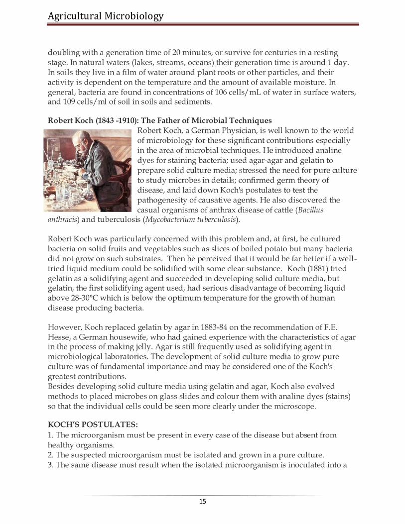
Robert Koch (1843 -1910): The Father of Microbial Techniques Robert Koch, a German Physician, is well known to the world of microbiology for these significant contributions especially in the area of microbial techniques. He introduced analine dyes for staining bacteria; used agar-agar and gelatin to prepare solid culture media; stressed the need for pure culture to study microbes in details; confirmed germ theory of disease, and laid down Koch's postulates to test the pathogenesity of causative agents. He also discovered the casual organisms of anthrax disease of cattle (Bacillus
anthracis) and tuberculosis (Mycobacterium tuberculosis).
Robert Koch was particularly concerned with this problem and, at first, he cultured bacteria on solid fruits and vegetables such as slices of boiled potato but many bacteria did not grow on such substrates. Then he perceived that it would be far better if a well-tried liquid medium could be solidified with some clear substance. Koch (1881) tried gelatin as a solidifying agent and succeeded in developing solid culture media, but gelatin, the first solidifying agent used, had serious disadvantage of becoming liquid above 28-30°C which is below the optimum temperature for the growth of human disease producing bacteria.
However, Koch replaced gelatin by agar in 1883-84 on the recommendation of F.E. Hesse, a German housewife, who had gained experience with the characteristics of agar in the process of making jelly. Agar is still frequently used as solidifying agent in microbiological laboratories. The development of solid culture media to grow pure culture was of fundamental importance and may be considered one of the Koch's greatest contributions. Besides developing solid culture media using gelatin and agar, Koch also evolved methods to placed microbes on glass slides and colour them with analine dyes (stains) so that the individual cells could be seen more clearly under the microscope.
KOCH’S POSTULATES:
1. The microorganism must be present in every case of the disease but absent from healthy organisms. 2. The suspected microorganism must be isolated and grown in a pure culture. 3. The same disease must result when the isolated microorganism is inoculated into a

www.AgriMoon.Com
16
healthy host. 4. The same microorganism must be isolated again from the diseased host.
"One microbe, one disease"
Robert Koch (1843-1910) was the first to rigorously demonstrate that a specific disease was caused by a specific microorganism.
Koch worked on anthrax, a disease mainly of animals. Koch noticed that cattle that died of anthrax all seemed to have a certain rod-shaped bacterium in blood, not found in healthy animals. Koch was able to isolate the bacterium in pure culture, put it back into healthy cows, and reproduce the disease.
Koch's Postulates: a logical way to identify the microbe causing a disease
1. A specific microbe must be present in all disease cases 2. Microbe must be cultivated outside host in a pure culture 3. When pure culture of microbe is inoculated into healthy hosts, disease symptoms
identical to those of initial host must be reproduced 4. Microbe can be isolated again in pure culture from this experimentally
inoculated host.
Initial attempts to isolate microbes used sliced potatoes or nutrient media containing gelatin -- not ideal media. Then Fannie Hesse (wife of lab worker) suggested agar, a gelling agent used in cooking. Agar rapidly became the standard gelling agent for microbial isolation because it is relatively inert (only some marine microbes have enzymes to digest agar). Agar only melts at high temperatures (100oC); once melted, it remains liquid until about 45oC, at which point it gels.
Koch's success at identifying anthrax with bacterium Bacillus anthracis led both Koch and Pasteur to identify the causes of many diseases -- cholera, tuberculosis, plague, etc. -- over the next few decades (late 1880's) -- the "Golden Age of Microbiology" (~ 1870-1920). Note that many microbiologists would regard the present as a new "Golden Age", since the development of molecular biological techniques, PCR, molecular phylogeny, and other developments have revealed many new insights and opened a world of new research directions and ways of understanding microbes.
******☺******

Agricultural Microbiology
17
Lecture 03:
PROTECTION AGAINST INFECTIONS
The control of microbial growth is necessary in many practical situations, and significant advances in agriculture, medicine, and food science have been made through study of this area of microbiology. The microorganisms are ubiquitous in nature. In order to study the nature and characteristics of a particular microbe, it is essential to isolate it from other contaminating microorganisms. This can be achieved by maintaining a completely sterile environment in which the microbe of interest is selectively grown. It is necessary that not only the place you are working with microorganisms should be free from contamination (other living organisms) but, the media and the materials you are using to handle and grow specific microorganisms should be free from other microbial contaminants. For this purpose ‗sterilization‘ of the place of work materials and media have to be done.
―Control of growth‖ as used here means to prevent the growth of microorganisms. This control is affected in two basic ways: (1) by killing microorganisms or (2) by inhibiting the growth of microorganisms. Control of growth usually involves the use of physical or chemical agents which either kill or prevent the growth of microorganisms. Agents which kill cells are called cidal agents; agents which inhibit the growth of cells (without killing them) are referred to as static agents. Thus the term bactericidal refers to killing bacteria and bacteriostatic refers to inhibiting the growth of bacterial cells.
A bactericide kills bacteria; a fungicide kills fungi, and so on. Sterilization is a process of complete removal or killing of all forms of microbial life
including spores from an object, surface, medium or environment without spoiling its nature.
Methods
There are various sterilization techniques available. However, several factorsinfluence the effectiveness of sterilization process like, the concentration of antimicrobial agents, time and temperature of exposure, size of population, type of contaminating microbes etc. Sterilization is brought about by a combination of physical and chemical agents that adversely affect the microorganisms either by causing damage to the cell wall or cell membrane or by inactivating the enzymes or by interfering with the synthesis of nucleic acids and protein.

www.AgriMoon.Com
18
I. PHYSICAL AGENTS
There are different types of physical agents.
(i) Heat: The heat employed for removal of micro-organisms varied with the nature of
object and also depend on the purpose. Based on these different processes are employed.
(a) Moist heat
It is the widely used effective means of sterilization process. In this, steam under high pressure is employed which imparts high penetration power resulting in the hydration of cells and coagulation of protein leading to the death of the microorganism. Autoclave is the apparatus used for sterilization by moist heat.
The autoclave is a double-jacketed steam chamber. The chamber is equipped with a device for generating saturated steam. It can be maintained at a particular temperature and pressure for any period of time. During operation of autoclave the air in the chamber is evacuated by steam since presence of air will reduce the temperature in the chamber. The time required for sterilization will depend upon the materials to be
sterilized. Solid materials must be heated for a longer time (1-2 hours) while liquid media can be sterilized within 15-30 minutes. Also acidic materials require shorter period than alkali materials. A temperature of 121°C for 15 min at a pressure of 15 lbs/ sq.inch is the sterilizing condition in the autoclave.
Advantages
Steam can penetrate through materials and sterilization is achieved by the coagulation or denaturation of proteins and other cell constituents. Liquid media, solid media, laboratory equipments (cloth, glasswares, etc.,) can be sterilized. The temperature and pressure is high enough to kill spores, vegetative cells and viruses.
Disadvantages
Temperature sensitive media, animal tissue culture media, antibiotics, amino acids, cannot be sterilized. Sometimes water may get inside incase of improper packing.
(b) Dry heat
This process is accomplished in a hot-air oven. Hot air or dry heat is employed for sterilization. The dry heat penetrates substances more slowly than the moist heat. Hence, the time required for effective sterilization is long (2 to 3 hours) and also the temperature required is too high (160°C -180°C). Microbial death results from the oxidation of cell constituents.

Agricultural Microbiology
19
Advantages Dry heat does not corrode glassware and metal instruments as moist heat does. All glassware‘s can be sterilized.
Disadvantages The sterilization process is slow. It is not suitable for heat sensitive materials like many plastic and rubber items.
(c) Boiling at 100°C for 30 minutes. Kills everything except some endospores (Actually,
for the purposes of purifying drinking water 100o for five minutes is probably adequate though there have been some reports that Giardia cysts can survive this process). To kill endospores, and therefore sterilize the solution, very long or intermittent boiling is required.
(d) Pasteurization is the use of mild heat to reduce the number of
microorganisms in a product or food. In the case of pasteurization of milk the time and temperature depend on killing potential pathogens that are transmitted in milk, i.e., staphylococci, streptococci, Brucella abortus and Mycobacterium tuberculosis. For pasteurization of milk:
batch method (Low temperature holding): 62.8oC for 30 minutes flash method (High
temperature short time): 71.7oC for 15 seconds
(e) Intermittent sterilization or Tyndallization is the process of boiling the materials at 100oC for 30 min. successively for three consecutive days. Destroys vegetative cells and spores; germinated spores.
(f) Incineration burns organisms and physically destroys them. Incineration is the
complete burning of the material in to ashes. Used for needles, inoculating wires, glassware, etc. and objects not destroyed in the incineration process. This is the direct and ultimate method of destroying cells. It is achieved by keeping the materials directly in contact with the flame of Bunsen burner as a result all the microorganisms in the surface are destroyed completely. Inoculating loops, needles and spreading rods are sterilized by this method.
Advantages: Immediate and quick.
Disadvantages: Cannot be used to sterilize heat labile material, material is lost by
incineration.

www.AgriMoon.Com
20
Recommended use of heat to control bacterial growth
Treatment Temperature Effectiveness
Incineration
5000 C
Vaporizes organic material on non flammable Surfaces but may destroy many substances in the process
Boiling
100 0C
30 minutes of boiling kills microbial pathogens and vegetative forms of bacteria but may not kill bacterial endospores
Intermittent boiling
1000C
Three 30-minute intervals of boiling, followed by Periods of cooling kills bacterial endospores.
Autoclave and pressure cooker (steam under pressure)
1210C for 15 min. at 15 lbs/ sq.inch pressure
Kills all forms of life including bacterial endospores. The substance being sterilized must be maintained at the effective T for the full time
Dry heat (hot air oven)
1600 C /2 hours
For materials that must remain dry and which are not destroyed at the between 121oC and 170oC Good for glassware, metal, not plastic or rubber items
Dry heat (hot air oven)
1800 C /1 hour
Same as above. Note increasing T by 10 degrees shortens the sterilizing time by 50 percent
Pasteurization (batch method)
62.8 0C /30 min.
kills most vegetative bacterial cells including pathogens such as streptococci, staphylococci and Mycobacterium tuberculosis
Pasteurization (flash method)
71.7 0C/15 seconds
Effect on bacterial cells similar to batch method; for milk, this method is more conducive to industry and has fewer undesirable effects on quality or taste
(ii) Radiation
Energy transmitted through space in a variety of forms is generally called radiation. It is also known as "cold sterilization" as only little heat is produced during the process. The most significant of this is electromagnetic radiation. The energy content and radiation wavelength are inversely proportional to each other. Radiation may be ionizing or non-ionizing.

Agricultural Microbiology
21
Ionizing radiation
High-energy electron beams (Gamma, X-rays, alpha and beta particles) have sufficient energy to cause ionization of molecules. They drive away electrons and split the molecules into ions. Water molecules are split into hydroxyl radicals (OH-), electrons and hydrogen ions (H+). OH- ions are highly reactive and destructive to normal cellular compounds such as DNA and proteins. Thus ionizing radiations are used in sterilization. e.g. 36Cs, 60Co
Advantages: X-rays and Gamma rays have high penetrating power. Packed food and
medical equipments are sterilized by using x-rays and gamma rays.
Disadvantage: Generating and controlling X-rays for sterilization is highly expensive.
Non-ionizing radiation
This includes ultraviolet (UV) rays. UV at a wavelength of 265 nm is most bactericidal. Absorption of UV radiation produces chemical modification of nucleoproteins i.e., thymine dimer formation that leads to misleading of genetic codes. This mutation impairs the total functions of the organism, consequently causing its death.
Advantages It is used to maintain aseptic conditions in laminar air flow chamber, lab, hospitals, pharmaceuticals, industries etc., and also in the sterilization of water and air.
Disadvantage: UV radiation has very little ability to penetrate matter and hence the micro organisms on the surface of an object are destroyed.
III) Filtration Filtration involves the passage of liquid or gas through a screen like material that has spores small enough to retain the micro organism of certain size. It is used to sterilize heat sensitive substance like enzyme solutions, bacterial toxins, certain biological media, cell extract and some sugars. Various types of filters are available in different grades of porosity. Vacuum or pressure is required to move the solutes through the filter.
Involves the physical removal of all cells in aliquid or gas, especially important to sterilize solutions which would be denatured by heat (eg: antibiotics, injectable drugs, amino acids, vitamins etc.)
Advantages: It si the best way to reduce microbial population in solutions of heat sensitive materials and it is sued to sterilize liquid media, vitamin solutions, hormones, growth factors, enzymes.

www.AgriMoon.Com
22
Disadvantages
Pleomorphic structures like mycoplasma cannot be effectively filtered by this technique. It is applicable to sterilize only small quantities.
Commonly used filters in micro biology The sintered glass fliter is made of fused Jen or pyrex glass, manufactured in such a way as to be porous, with apore size and adsorptive charge sufficient to retain bacteria. The seitz filters are compressed asbestos discs having porosity sufficiently small to retain bacteria. Tie chamber land filters are made of porcelain. The mandler/berkfield filters are made of diatomaceous earth. The membrane filter is a cellulose or nitrocellulose membrane with apore size sufficiently small (0.01mm to 10 mm) to trap and thereby remove bacterial from a liquid. The membrane filters are also used to concentrate and trap the micro organisms in water and other liquids. HEPA (High efficiency particulate air filters are of fiber glass filters for sterilization of air.
Low temperature Most organisms grow very little or not at all at 0O C. Store perishable foods at low temperatures to slow rate of growth and consequent spoilage (eg: milk). Low temperatures are not bactericidal. Psychrotrophs, rather tah true psychrophiles are the usual cause of food spoilage in refrigerated foods.
Dessication / Drying (removal of H2O) Most micro organisms cannot grow at reduced water activity (aw < 0.90). Often used to preserve foods (eg: fruits, grains etc). methods involve removal of water from product by heat, evaporation, freeze drying, addition of salt or sugar.
Surface tension is a property of the surface of a liquid that allows it to resist an external
force. It is revealed, for example, in floating of some objects on the surface of water, even though they are denser than water, and in the ability of some insects (e.g. water striders) and even reptiles (basilisk) to run on the water surface. This property is caused by cohesion of like molecules, and is responsible for many of the behaviors of liquids. Surface tension has the dimension of force per unit length, or of energy per unit area. The two are equivalent—but when referring to energy per unit of area, people use the term surface energy—which is a more general term in the sense that it applies also to solids and not just liquids. In materials science, surface tension is used for either surface stress or surface free energy.
Osmotic pressure – plasmolysis/ plasmotysis
Osmotic Pressure is the process in plant cells where the plasma membrane pulls away from the cell wall due to the loss of water through osmosis. The reverse process, cytolysis, can occur if the cell is in a hypotonic solution resulting in a higher

Agricultural Microbiology
23
external osmotic pressure and a net flow of water into the cell. Through observation of plasmolysis and deplasmolysis it is possible to determine the tonicity of the cell's environment as well as the rate solute molecules cross the cellular membrane.
Chemical agents
Chemical that is used to kill or inhibit the growth and development of micro organisms are called anti microbial agents. Disinfectants and antiseptics come under anti microbial agents and are usually used on inanimate materials. The mechanism of action is complex and non specific. It may act on lipid portion of cell membrane, oxidize or reduce an important functional group of an enzyme, prevent certain bio synthesis or cause extensive breakdown of DNA.
Types of microbial agents Chemical sterilants
Chemical sterilants are chemical anti microbial agents that are usd fro sterilization of heat sensitive substance/ materials. Normally plastic petriplates and medical supplies such as blood transfusion sets, plastic syringes, lenses etc. could be sterilized even in packets or bundles using ethylene oxide, formaldehyde or formalin is effectively used to sterilize enclosed areas/a septic chambers at 22 O C with a relative humidity of 60 – 80 %.
Antispetics Microbicidal agents harmless enough to be applied to the skin and mucous membrane, should not be taken internally. Eg: mercurails, silver nitrate, iodine solution, alcohols, detergents.
Disinfectants Agents that kill micro organisms, but not necessary their spores, not safe for application to living tissues, they are used on inanimate objects such as tables, floors, utensils etc. eg: Chlorine, hypochlorites, chlorine compounds, Lysol, copper sulfate, quaternary ammonium compounds.
Phenol
Derivative of phenol like benzyl resorcinol, o-cresol, m-cresol, etc., are used as effective disinfectants 5% aqueous solutions of phenols are used as disinfectant. It alters the protein structure and leads to denaturation of proteins and enzymes. Also affects permeability of cytoplasmic membrane. They readily kill vegetative cells of bacteria and fungi but for spores.
Alcohol
Alcohol at 70% concentration is more effective. It brings about denaturation and coagulation of protein. Ethanol is routinely used in laboratories to surface sterilize worktables and hands of the researcher/ experiment.

www.AgriMoon.Com
24
Halogens
Halogens such as hypocholrites, choramines and povidone- iodine are used to sanitize utensils, surface sterilize in animate objects, table surfaces and other instruments.
Heavy metals Heavy metals such as mercuric chloride are also used for surface sterilization purposes. Heavy metals acts as oxidizing agents and kill the micro organisms on the surface of the object. Usually 0.1 % mercuric chloride is used in the laboratories to sterilize the surface of worktable and explants.
Detergents Detergents are those compounds that make water repellant surfaces more wettable. There are two types of detergents viz., ionic and non ionic. Detergent soaps and other synthetic detergents are used for washing/cleaning glass wares, table tops etc.,
Common antiseptics and disinfectants
Chemical
Action Uses
Ethanol (50 -70 %) Denatures proteins and solubilizes lipids.
Anti septic used on skin
Isopropanol (50 – 70 %) Denatures proteins and solubilizes lipids.
Anti septic used on skin.
Formaldehyde (8%) Reacts with NH2, SH and COOH groups.
Disinfectant, kills endopsores.
Tincture of Iodine ( 2% in 70 % alcohol)
Inactivates proteins Antiseptic used on skin
Chlorine (Cl2) gas Forms hypochlorous acid (HClO), a strong oxidizing agent.
Dis infect drinking water, general disinfectant.
Silver Nitrate (Ag No3) Precipitates proteins. General antiseptic and used in the eyes of newborns.
Mercuric chloride Inactivates proteins by reacting with sulfide groups.
Disinfectant although occasionally used as an antiseptic on skin.
Detergents (eg: Quaternary ammonium compounds)
Disrupts cell membranes. Skin antiseptics and disinfectants.
Chemotherapeutic agents
Antimicrobial agents of synthetic origin useful in the treatment of microbial or viral disease. Examples: sulfonilamides, isoniazid, ethambutol, AZT, chloramphenicol.

Agricultural Microbiology
25
Antibiotics
Antimicrobial agents produced by micro organisms that kill or inhibit other micro organisms. This is the microbiologist‘s definition. A more broadened definition of an antibiotic includes many chemical of natural origin which has the effect to kill pr inhibit the growth of other types cells. Since most clinically useful anti biotics are produced by micro organisms and are used to kill or inhibit infectious bacteria we follow classic definition.
Antibiotics are low molecular weight (non- protein) molecules produced as secondary metabolites, mainly by micro organisms that live in the soil. Most of these micro organisms form some type of a spore or other dormant cell, and there is thought to be some relationship between anti biotic production and the process of sporulation. Among the molds, the notable antibiotic producers are penicillium and cephalosporium, which are the main source of the beta lactam antibiotics. In the bacteria, the actinomycetes, notable streptomyces species, produce a variety of types of anti biotics including the aminoglycosides (eg: streptomycin), macrolides (eg: erythromycin) and the tetracycline. Endospore forming bacillus species produce polypeptide anti biotics such as polymyxin and bactracin.
Chemical class examples Biological
source Spectrum
(effective against)
Mode of action
Beta – lactams (Penicillins and cephalosporins)
Penicillin G, Cephalothin
Penicillium notatum and cephalosporium sp.
Gram positive bacteria
Inhibits steps inc ell wall (peptidoglycan) synthesis and murein assembly.
Aminoglycosides
streptomycin Streptomyces griseus
Gram positive and gram negative bacteria
Inhibit translation (protein synthesis)
glycopeptides vancomycin Streptomyces orientales
Gram positive bacteria, esp. staphylococcus aurues
Inhibits steps inn murein (peptidoglycan) biosynthesis and assembly
macrolides erythromycin Streptomyces erythreus
Gram positive and gram negative bacteria not enteric,. Neisseria, legionella, mycoplasma
Inhibits translation(protein synthesis)
polypeptides polymyxin Bacillus Gram negative Damages cytoplasmic

www.AgriMoon.Com
26
polymyxa bacteria membranes
Polyenes amphotericin Streptomyces nodosus
Fungi Inactivate membranes containing sterols
tetracyclines tetracycline Streptomyces sp
Gram positive and gram negative bacteria, rickettsias
Inhibit translation (protein synthesis)
Chloramphenicol
Chloramphenicol Streptomyces venezuelae
Gram positive and gram negative bacteria
Inhibit translation (protein synthesis)
******☺******

Agricultural Microbiology
27
Lecture 04: METABOLISM IN BACTERIA
Microbial Metabolism
Metabolism refers the sum of biochemical reactions required for energy generation and the use of energy to synthesize cellular materials. The energy generation component is referred as catabolism and the build up of macromolecules and cell organelles are referred as anabolism.
During catabolism, the energy is changed from one compound to another and finally conserved as high energy bonds of ATP. ATP is the universal currency for energy. When energy is required for anabolism, it may be sent as high energy bonds of ATP which has the value of 8 kcal per mole.
Based on the source of carbon, the microbes can be divided into two groups namely, autotrophs and heterotrophs. Autotrophs utilize CO2 as sole carbon source and heterotrophs use organic carbon as sole carbon source.
I. Energy generation by heterotrophs
Heterotrophs use variety of carbon sources. Glucose is being the simple and wide variety of microbes prefers it. The glucose can be taken up by bacterium through diffusion and can be readily utilized. There are three possible pathways available in

www.AgriMoon.Com
28
bacteria to use glucose. All these path ways are fermentative type and substrate level phosphorylation occurs.
Embden-Meyerhof path way Phosphoketolase path way Entner – Doudoroff path way.
SUBSTRATE LEVEL PHOSPHORYLATION: (Fermentation) The EMP pathway, phosphoketolase pathway and ED pathway end with one or
two ATP synthesis by substrate level phosphorylation. There won‘t be any external source of electron acceptor will come in these reactions.
A. Embden-Meyerhof path way
This is the path way of glycolysis most familiar and common to most of the organisms. The path way is operated by yeast to produce alcohol and lactic acid bacteria to produce lactic acid and several organic acids, gases, fatty acids, and alcohols. The path way is as follows: Glucose à 2 pyruvate + 2 ATP + 2 NADH2 After pyruvate is formed, if the organism is a respirative type, the pyruvate will go to Krebs
cycle and if the organism is fermentative,
the reduction process ends with organic acids, alcohols etc.

Agricultural Microbiology
29
A model fermentation:After an intermediate product, a reduction takes place in
fermentation, whereas, if respiration, CO2 will be formed by complete oxidation through Krebs‘s cycle (Note: After pyruvate, the reduction process leads to fermentation and complete oxidation leads to respiration)
The Embden – Meyerhof path way can lead to a wide array of end products depending on the path ways taken in the reductive steps after the pyruvate formation. The following are some of the such fermentations:
Fermentation End products Model organism
Homolactic fermentation Lactic acid Lactobacillus
Mixed acid fermentation Lactate, acetate, formate, succinate
Enterobater
Butyric acid fermentation Butric acid, acetone Clostridium acetobutylicum
Propionic acid fermentation Propionic acid Propionibacterium
Alcohol fermentation Ethanol Saccharomyces
B. Phosphoketolase path way (Heterolactic path way)
The phosphoketolase path way is distinguished by the key cleavage enzyme phosphoketolase, which cleaves
pentose to glyceroldehyde 3 phosphate and acetyl phosphate. The path way ends with ethanol and lactic acid. Ex. Lactobacillus, Leuconostoc. The overall reaction is, Glucose à 1 lactate + 1 ethanol + 1 CO2 + 1 ATP This path way is useful in the dairy industry for preparation of kefir (fermented milk), yogurt, etc.

www.AgriMoon.Com
30
C. Entner – Doudoroff pathway
Only few bacteria like, Zymomonas mobilis employ the ED pathway. The path way is as follows: The overall reaction is Glucose à 2 ethanol + 2 CO2 + 1 ATP The alcohol productivity of Zymomonas is higher than yeast because of this fermentative pathway. ( Note : All the three pathways are end with 1 or 2 ATP by substrate level phosphorylation by means fermentation)
OXIDATIVE PHOSPHORYLATION (Respiration) If the organism is a respiratory type (that means complete oxidation of glucose), it needs four essential metabolic components for their respiration and oxidative phosphorylation.
a. Tricarboxylic acid cycle (also known as citric acid cycle or Kreb’s cycle) The
pyruvate formed during glycolysis will be completely oxidized to 3 CO2 by the use of this cycle. During oxidation of one pyruvate through TCA cycle, 4 NADH2, 1 FADH2 and 1 GTP are produced along with 3 CO2.
b. A membrane and associated Electron Transport System (ETC) The electron
transport chain is a sequence of electron carriers transport the electrons to a terminal electron acceptor. During this flow of electron in the membrane, a proton motive force across the membrane leads to formation ATP (is referred as electron transport phosphorylation).
c. An outside electron carrier: for aerobic respiration, O2 is the terminal electron
acceptor and reduced to H2O. This is normal for higher organisms. But in anaerobic bacteria, the terminal electron acceptor may be of nitrite, nitrate, sulphate or carbon dioxide.

Agricultural Microbiology
31
d. A membrane bound ATPase enzyme: The proton motive force developed during
ETC leads to formation of ATP by enzyme ATPase present in the membrane. (As in the diagram)
The table shows some aerobic and anaerobic respirations with specific examples:
Terminal electron acceptor
End product Process name Organism
O2 H2O Aerobic respiration Streptomyces
NO3 NO2, N2 Denitrification Pseudomonas denitrificans
SO4 S or H2S Sulphate reduction Desulfovibrio desulfuricans
Fumarate Succinate Anaerobic respiration
Escherichia
CO2 Methane (CH4) Methanogenesis Methanococcus

www.AgriMoon.Com
32
In aerobic organisms, the terminal electron acceptor will be of O2. In some anaerobic organisms, after the electron transport chain, instead of O2, some inorganic compounds like sulphate, nitrate or some organic compounds like fumarate act as terminal electron acceptor. Such type of respiration is referred as anaerobic respiration and the normal O2 mediated respiration is referred as aerobic respiration. The above table shows some anaerobic respiration with some terminal electron acceptors. The process is named based on the compounds as sulphur reduction, denitrification and methanogenesis. Energy generation by autotrophs.
Autotrophs use CO2 as their sole carbon source. There are two types such as photoautotrophs and chemoautotrophs. Photoautotrophs use light as energy source and CO2 as carbon source. Chemoautotrops use chemicals (especially inorganic) as energy source and CO2 as carbon source.
I. Energy and carbon assimilation by photoautotrphs: (Photoautotrophy) Phototrophs use sunlight to produce ATP through phosphorylation, referred as photophosphorylation. The phototrophs convert the light energy to chemical energy (ATP) through the process called photosynthesis.
Photosynthesis is a type of metabolism in which catabolism and anabolism occur as sequence. The catabolic reaction (energy generating process) of photosynthesis is light reaction in which the light energy is converted to chemical energy (ATP) and electrons or reducing powers (NADPH). The anabolic reaction (macromolecule synthesis) of photosynthesis is dark reaction in which CO2 is converted to organic molecules (carbohydrates), which is also called as CO2 fixation.
For conversion of light energy to ATP, the bacteria possess light harvesting pigments. They are chlorophyll a, carotenoids, phycobiliproteins (which are present in cyanobacteria) and bacteriochlorophyll (which are present in purple sulphur bacteria). In bacteria, there are two types of light reactions (conversion of light to ATP) and two types of CO2 fixation occur.
A. Light reaction (Photophosphorylation)
For photophos phorylation, light harvesting pigments, a membrane electron transport chain, source of electron (electron donor) and ATPase enzymes are required. Two types of photophosphorylations occur during photosynthesis. They are cyclic photophos phorylation and non-cyclic photophos phorylation.
In plant and cyanobacteria, both cyclic and non-cyclic photophosphorylation occurs whereas in purple bacteria, the cyclic photophosphorylation only occurs.
In plant and cyanobacteria, the electron source is water, by photolysis, H2O split into H+ and O2 and during the process, O2 is evolved and referred as oxygenic photosynthesis
Agricultural Microbiology
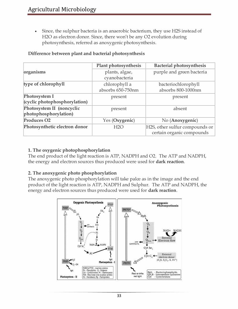
33
Since, the sulphur bacteria is an anaerobic bacterium, they use H2S instead of H2O as electron donor. Since, there won‘t be any O2 evolution during photosynthesis, referred as anoxygenic photosynthesis.
Difference between plant and bacterial photosynthesis
organisms
Plant photosynthesis Bacterial photosynthesis
plants, algae, cyanobacteria
purple and green bacteria
type of chlorophyll chlorophyll a absorbs 650-750nm
bacteriochlorophyll absorbs 800-1000nm
Photosystem I (cyclic photophosphorylation)
present present
Photosystem II (noncyclic photophosphorylation)
present absent
Produces O2 Yes (Oxygenic) No (Anoxygenic)
Photosynthetic electron donor H2O H2S, other sulfur compounds or certain organic compounds
1. The oxygenic photophosphorylation The end product of the light reaction is ATP, NADPH and O2. The ATP and NADPH, the energy and electron sources thus produced were used for dark reaction.
2. The anoxygenic photo phosphorylation The anoxygenic photo phosphorylation will take palce as in the image and the end product of the light reaction is ATP, NADPH and Sulphur. The ATP and NADPH, the energy and electron sources thus produced were used for dark reaction.

www.AgriMoon.Com
34
B. Dark reaction (CO2 fixation)
The dark reaction in which the ATP and NADPH were used as energy and electron sources to fix the CO2 as carbohydrates. The pathway involved in the dark reaction is Calvin cycle, by which the CO2 is fixed as phosphoglyceic acid and lead to formation of
many sugars. The enzyme RuBiSCO is the key enzyme for this process. The following pathway shows the Calvin cycle and the formation of key monomers for anabolic reactions such as hexose phosphate – polysaccharides; pyruvic acid – amino
acid and fatty acid; pentose phosphate – DNA and RNA.
A complete model of light and dark reaction of photosynthesis

Agricultural Microbiology
35
Another way of CO2 fixation by phototrophs
In phototrophs, the electron and energy were derived form sunlight and carbon from CO2 fixation through Calvin cycle. But some bacteria may derive electron and energy from sunlight and fixes CO2 by some other path way, not the Calvin cycle. The example is Photosynthetic green bacteria (Chlorobium). They derive NADPH and ATP through cyclic phosphorylation, but CO2 fixation is by reverse TCA cycle. Since TCA cycle is amphibolic pathway (referring the cycle can operate in both the directions), it can
also be used to fix the carbon-di-oxide if operated reversely. The pathway is as follows:
Another way of CO2 fixation is by methanogens: They use CO2 as terminal electron
acceptor and forms CH4 (methane). They also fix by acetyl CoA pathway for fixing CO2.
Synopsis:
Organism
Light reaction/ATP generation Dark reaction/CO2
fixation
Cyanobacteria (Nostoc), plant and alga
Cyclic and non-cyclic photophosphorylation Oxygenic photosynthesis
Calvin cycle
Purple bacteria (Chromatium)
Cyclic and non-cyclic photophosphorylation
Calvin cycle
www.AgriMoon.Com
36
Oxygenic photosynthesis
Green bacteria (Chlorobium)
Cyclic and non-cyclic photophosphorylation Oxygenic photosynthesis
Reverse TCA cycle
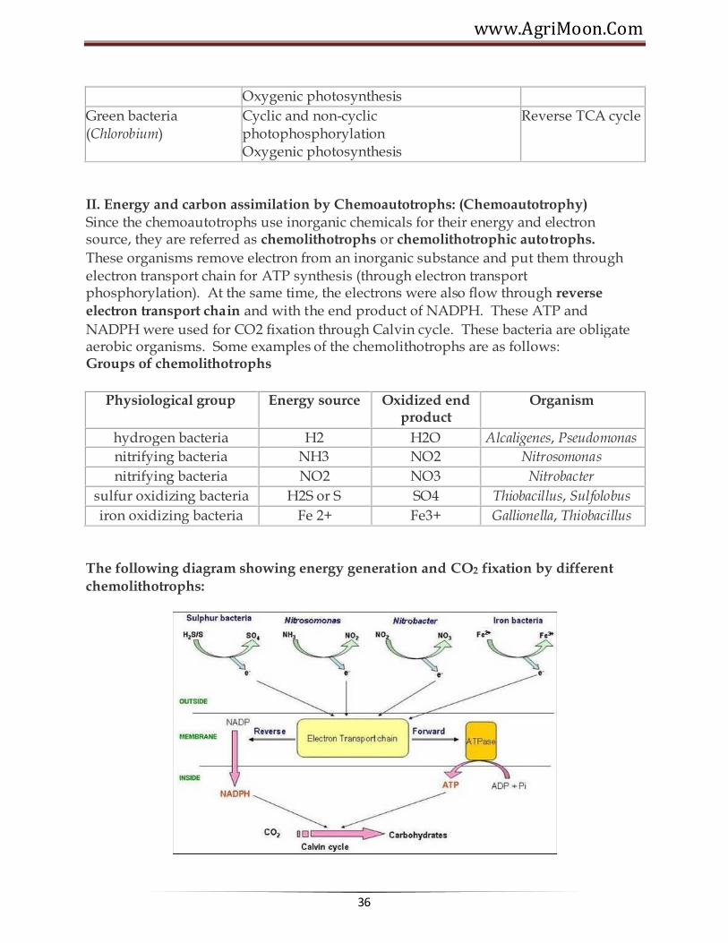
II. Energy and carbon assimilation by Chemoautotrophs: (Chemoautotrophy) Since the chemoautotrophs use inorganic chemicals for their energy and electron source, they are referred as chemolithotrophs or chemolithotrophic autotrophs.
These organisms remove electron from an inorganic substance and put them through electron transport chain for ATP synthesis (through electron transport phosphorylation). At the same time, the electrons were also flow through reverse
electron transport chain and with the end product of NADPH. These ATP and
NADPH were used for CO2 fixation through Calvin cycle. These bacteria are obligate aerobic organisms. Some examples of the chemolithotrophs are as follows: Groups of chemolithotrophs
Physiological group Energy source Oxidized end product
Organism
hydrogen bacteria H2 H2O Alcaligenes, Pseudomonas
nitrifying bacteria NH3 NO2 Nitrosomonas
nitrifying bacteria NO2 NO3 Nitrobacter
sulfur oxidizing bacteria H2S or S SO4 Thiobacillus, Sulfolobus
iron oxidizing bacteria Fe 2+ Fe3+ Gallionella, Thiobacillus
The following diagram showing energy generation and CO2 fixation by different
chemolithotrophs:

Agricultural Microbiology
37
Lecture 05: ATP GENERATION
The energy captured within ATP can then be harnessed to create order in the form of biosynthetic reactions. In a hypothetical enzyme reaction that converts substrates A−H and B−OH to A−B and H2 O, the energy from ATP hydrolysis is first used to convert B−OH to a higher-energy intermediate, B−O−PO4. This compound is only transiently formed, with the energy released during its decay used by the enzyme to form A−B. Thus, the energy released from the ATP hydrolysis reaction (large −_G) is coupled to the synthesis reaction (large +_G). In this way, the cell can progressively create order.
Electron transport system (ETS)
Although cells could transfer electrons directly from NADH to oxygen, this would liberate all energy in NADH directly as heat.
NADH possesses lots of energy. If electrons are transferred directly to oxygen: NADH + O2 NAD + H2O, delta Go' = - 218 kilojoules/mole If NADH has ~218 kilojoules of energy, and it only takes 30.5 kilojoules to make
one ATP, could conceivably make 218/30.5 = ~ 7 ATP per NADH if energy conversion were 100% efficient.
In practice, cells have evolved ways to get up to 40% efficiency (~ 3 ATP/NADH) under optimal circumstances.
Electron transport system (ETS) = membrane-bound pathway transferring electrons from organic molecules to oxygen.
ETS moves both electrons and protons: electrons are passed from carrier to carrier in the membrane, while protons are moved from inside to outside of membrane
Net result: electrons enter ETS from carriers like NADH or FADH, wind up at terminal oxidize, and get attached to oxygen.
ETS consists of 4 complexes, connected by mobile carriers (Coenzyme Q, cytochrome c) that shuttle between complexes in membrane

www.AgriMoon.Com
38
Specific carriers of ETS:
mitochondria (in eukaryotes): NADH ---> (Flavoprotein Iron sulfur proteins Quinone cytochrome b cytochrome c cytochrome a cytochrome a3 oxygen
bacteria (prokaryotes) have different ETS carriers, shorter chains. In E. coli, can have two different terminal oxidases, one functions at high oxygen levels, one at lower oxygen levels. Cytochromes involved include: b558, b595, b562, d, and o
proton gradient and oxidative phosphorylation (oxphos)
Chemiosmotic hypothesis (Peter Mitchell, 1961)
As electrons flow through ETS, at certain steps protons (H+) are moved from inside to outside of the membrane.
This builds up proton gradient; since + charges are removed from inside of cell, - charge remains inside, mainly as OH- ions.
pH just outside membrane can reach 5.5, pH just inside membrane can reach 8.5 ---> difference of 3 pH units, or 1000x concentration differential of H+ across membrane. This represents potential energy stored up in proton gradient = proton motive force.
Membrane is basically impermeable to protons, so gradient doesn't get squandered away by leaky reentry.
ATP synthase protein complex contains only channels for proton entry. As protons push in through channel, the base rotates. Specific binding sites allow ADP + Pi ATP. This can be called chemiosmotic phosphorylation (assuming chemiosmotic hypothesis is correct), or oxidative phosphorylation (makes no assumption about mechanism.

Agricultural Microbiology
39
Oxidative phosphorylation Differences between respiration in mitochondria (eukaryotes) and bacteria (procaryotes)
In Eukaryotes:
ETS located in inner mitochondrial membrane. Proton gradient develops across inner mitochondrial membrane.
Mitochondria are very efficient at generating proton gradient. Can measure how many ~P bonds (in ATP) are made for each O2 consumed = P/O ratio.
With NADH as electron donor, P/O ratio can be 3 (means 3 ATP made per NADH).
But with FADH as electron donor, P/O ration only 2 (fewer protons are transported, less proton gradient).
Overall efficiency of respiration in mitochondria: ~ 40% (means that about 40% of energy in glucose actually gets converted to ATP).
In Prokaryotes:
ETS located in cytoplasmic membrane. Proton gradient develops across this membrane.
Bacteria are not as efficient. ETS chains are shorter, P/O ratios are lower. As a ballpark estimate, P/O ratios for NADH are only ~2. Overall efficiency of
glucose oxidation is closer to 28%, not 40%.
Inhibitors of Oxidative Phosphorylation
Several chemicals can block electron transfer in ETS, or transfer of electrons to oxygen. All are strong poisons. Some examples:
Carbon monoxide -- combines directly with terminal cytochrome oxidase, blocks oxygen attachment
Cyanide (CN-) and Azide (N3-) bind to cytochrome iron atoms, prevent electron transfer.
Antimycin A (an antibiotic) inhibits electron transfer between cyt b and c.
Anaerobic respiration
Use of acceptors other than oxygen. Most common in bacteria. Most alternative electron acceptors are inorganic
molecules, but some organic molecules can serve. As with aerobic respiration, anaerobic respiration uses ETS, membrane
localization, proton gradient, and ATP synthase. Processes are of great importance both ecologically and industrially.

www.AgriMoon.Com
40
Anaerobic respiration Nitrate (NO3-)
Process called denitrification. Also called dissimilative nitrate reduction. Reduced waste products are excreted in significant amounts.
Redox potential is + 0.42 v (compared to + 0.82 v for oxygen). So organisms respiring anaerobically gain less energy than with oxygen.
Requires new terminal oxidase called nitrate reductase. Enzyme is repressed by oxygen, synthesis turned on in absence of oxygen.
Process can have several steps, proceed in two different directions:
(A) nitrate (NO3-) nitrite (NO2-) ammonia (NH3) (B) nitrate (NO3-) nitrite (NO2-) nitrous oxide (N2O) dinitrogen gas (N2)
Second process is major pathway for loss of nitrogen compounds from soil, return of nitrogen to atmosphere.
Pseudomonas species are common denitrifiers, widespread in soils. When fertilized soils become flooded, oxygen is rapidly depleted, pseudomonads switch to anaerobic respiration and can use up soil nitrate, leaving field in unfertile state.
Note: Studied this in lab. Media must contain nitrate in addition to nutrients, otherwise won't work. Also, in scavenger hunt at end of course, one target microbe will be Pseudomonas, enrichment culture depends on its ability to grown anaerobically using nitrate reduction.
Sulfate (SO42-)
1. Process called sulfate reduction. 2. Sulfate (SO42-) Hydrogen Sulfide (H2S) 3. Small group of bacteria carry out this reaction; all obligate anaerobes. 4. Have unique cytochrome c3. 5. Sulfate is common in sea water. Often, H2S combines with iron, forms
insoluble FeS black sediments. Common in estuaries.
Carbon dioxide (CO2)
1. One of most common inorganic ions. 2. Methanogens: most important group of CO2 reducers. Obligate
anaerobes, archaebacteria. Produce methane as waste product. 3. Reaction: CO2 + H2 + H+ CH4 + H2O 4. Note: reaction also requires Hydrogen gas. Methanogens typically live
alongside bacteria that produce hydrogen by fermentation, remove hydrogen as it is made.

Agricultural Microbiology
41
TCA cycle: further catabolism of pyruvate Formation of acetyl-CoA
1. Oxidation of pyruvate (3-C) + NAD+ Acetyl-CoA (2-C) + CO2 + NADH 2. Carried out by pyruvate dehydrogenase (multi-enzyme system) 3. Note: Acetyl-CoA can also be produced by breakdown of lipids or certain amino
acids -- important focal point of central metabolism
Net effects of TCA cycle:
1. To start cycle:
2. Acetyl-CoA (2-C) + oxaloacetate (4-C) citric acid (6-C) 3. Subsequent steps:
1. Convert citrate to isocitrate (still 6-C) 2. Oxidize alpha-ketoglutarate (5-C) + CO2 + NADH 3. Oxidize succinyl-CoA (4-C) + CO2 + NADH 4. SLP reaction: succinyl-CoA (4-C) + GDP succinate (4-C) + GTP (Note:
GTP can be interconverted with ADP to form ATP) 5. Oxidize fumarate (4-C) + FADH2 -- convert fumarate to malate 6. (6)oxidize again oxaloacetate (4-C) + NADH
4. Net yield: Acetyl-CoA (2-C) + 3 NAD+ + FAD 2 CO2 + 3 NADH + FADH2 + ATP
5. TCA cycle completes the oxidation of carbons in pyruvate to most oxidized form (CO2); removes electrons originally in C-H bonds to electron carriers NADH and FADH for use in respiration machinery.
Catabolism of substances other than glucose: Many other possible C-sources for
catabolism beside glucose. In general, must convert these into molecules that can enter into central metabolism, either in glycolysis or TCA cycle.
1. carbohydrates 1. Most abundant C-sources in most environments, most in various
polysaccharides (cellulose, starch, lignin, etc.) 2. To gain access to sugars, must first secrete hydrolytic enzymes that
break down glycosidic bonds in polysaccharides, produce mono- and disaccharides that can be transported into cells.
3. Starch, glycogen -- easily hydrolyzed by amylases 4. Cellulose -- difficult to digest, very insoluble, tightly folded. Many
fungi, some bacteria produce cellulases. 5. Agar -- some marine bacteria produce agars 6. Once mono- or disaccharides are available, they are transported
into cell, converted into some typical glycolytic intermediate such as glucose-6-phosphate, catabolized by glycolytic enzymes.

www.AgriMoon.Com
42
2. lipids
1. Biological lipids common as triglycerides, diglycerides. 2. To catabolize, bacteria secrete lipases, hydrolyze glycerides to free
fatty acids and glycerol. 3. Fatty acids attacked by Beta-oxidation pathway. 4. Using FAD and NAD+ to remove electrons, 2-C units are removed
as Acetyl-CoA, feed directly into central metabolism at TCA cycle entry. Glycolysis pathway not involved (except for use in synthesizing sugars needed for cell wall, running sections of pathway in reverse).
3. proteins 1. Proteins must first be hydrolyzed by protease enzymes, to get
individual amino acids which can be transported into cells. 2. Amino acids all have common structure: NH2 - RCH - COOH. 3. 1st step in catabolism is to remove amino group (deamination),
often by swapping it with another substrate (transamination). 4. Typical example: glutamic acid (an AA) + pyruvate alpha-
ketoglutarate + alanine (= pyruvate + amino group). Now alpha-KG can be oxidized in TCA cycle, since it is a TCA cycle compound.
5. As excess amino groups accumulate, must be secreted as waste products, possibly as ammonium ion (leads to alkaline pH).
******☺******

Agricultural Microbiology
43
Lecture 06:
MICROBIAL METABOLISM – AUTOTROPHS
Overview of Autotrophy
• Imagine being hungry, walking outside, taking off your shirt, lying in the sun for a few hours, becoming totally full (fat even!), and being done eating. No stores, no lines, no choices, just sunlight --- and the machinery of an autotroph --- and some CO2 and a couple of other requirements (water --- and H2S or Hydrogen gas, if you happen to be an anerobe) • Autotroph = gets all carbon from CO2, organic C not required (for C-source). Use special metabolic cycle: Calvin-Benson cycle • Refers to C-source only; some organisms still require organic C as energy source
Calvin-Benson cycle
• Each CO2 is added to a 5-C acceptor molecule (ribulose 1,5 bis-phosphate) • Immediately split into two 3-C molecules (3-phosphoglyceric acid) • Must add phosphate group (from ATP) and hydrogen (from NADPH) to get reduced product, 3 - phospho-glyceraldehyde (PGA) • Cannot take all (PGA) as product --- must regenerate some more acceptor to keep cycle going. How? • Take 5 PGA molecules (5 x 3C = 15 C atoms). Rearrange through series of reactions to make 3 5 - C molecules (still 15 C atoms). Add ATP to each, make 3 acceptor molecules (ribulose 1,5 bis-phosphate) • Net result: To get 1 PGA (3-C) as reduced product, need 3 CO2 molecules, added to 3 acceptor molecules ----> six 3 - C molecules, use 6 ATP and 6 NADPH ----> 6 PGA molecules; five of these are used to regenerate acceptor molecules (+ 3ATP), one PGA can leave cycle and be used by cell.
Summary
• Actual cyce exports 3-C reduced molecules: look at balanced equation: 3 CO2 + 9 ATP + 6 NADPH -----> 3-phospho-glyceraldehyde (PGA) + 9 ADP + 9 NADP+ • Often want to look at balanced equation relative to 6C synthesis. Must multiply all terms in balanced equation above by two (since 2 PGA ~ 1 glucose) 6 CO2 + 18 ATP + 12 NADPH -----> glucose + 18 ADP + 12 NADP+ • Note for reaction: glucose + O2 ----> 6CO2 + 6 H2O; delta Go'= - 688kcal/mole • If each ATP contains ~7.3 kcal/mole (from delta Go' for hydrolysis) and each NADPH

www.AgriMoon.Com
44
contains ~54 kcal/mole (from delta Go' for oxidation), then to make glucose costs 780 kcal/mole, more than the energy available by oxidizing glucose. • Conclusion: making sugar is expensive! Cell needs to supply large quantities of ATP and NADPH.
Chemolithotrophs
Hydrogen Bacteria
• Gain energy by oxidizing hydrogen gas: • H2 + NAD+ -------(hydrogenase enzyme)------> NADH + H+ • alternative: electrons can be donated directly to ETS chain, bypassing NAD • Note: only need one special enzyme to carry this step out: hydrogenase. • Many different genera of bacteria include members that can induce hydrogenase. When hydrogen disappears, back to heterotrophic life. Hydrogen bacteria are usually facultative chemolithotrophs.
Sulfur Bacteria
• Called "colorless" in contrast to chlorophyll-containing sulfur bacteria, usually green or purple
• Oxidize sulfur compounds: Example: Thiobacillus thiooxidansthiosulfate: S2O3= -----> SO4=free sulfur: 2 So + 2 H2O + 3 O2 -----> 2 H2SO4 • Note product: sulfuric acid!! Cells can grow even in pH 0 (1M sulfuric acid). But cell internal pH is ~7, so difference across membrane can be 6 or 7 pH units. • Acid mine drainage: common in Western Penn., E. Ohio, W. Virginia. Rivers can run rust red. Mines have been major sources of pollution. Water seeps in, sulfur deposits exposed during coal mining allow microbial growth ------> megatons of H2SO4 • Sulfuric acid leaches out, dissolves iron, precipitates in river with bicarbonate to form rusty deposits. • Quantities involved: Ohio River carries 100 million tons of 98% conc. H2SO4 per year. • To cure problem, must seal up old mines, prevent oxygen access. Also strip mines must be promptly covered up once mining is done to block access of microbes and oxygen to sulfur. • Value of this reaction: (a) farmers or gardeners can dump free S on alkaline soil, bacteria will produce acid (b) miners can use process to recover Cu from low grade ores, where smelting is not economical. Pile up mine "tailings" with copper ore; scrap shallow hole and fill with water. If tailings contain S, microbes will produce H2SO4. Now pump the acid over the tailings, Cu will be leached out and accumulate as soluble ions in acid pool. Eventually process the acid, recover Cu.

Agricultural Microbiology
45
Nitrifying Bacteria
• Very important soil organisms -- process all ammonia, nitrite in soils, break down amino acids, nitrogen bases ---> ammonia (NH3) • Two different groups: one oxidizes ammonia, one oxidizes nitrite • Ex. 1: Nitrosomonas: 2 NH3 (ammonia) + 3 O2 -----> 2 HNO2 (nitrite) + 2 H2O • Ex. 2: Nitrobacter: 2 HNO2 (nitrite) + 2 O2 -----> 2 HNO3 (nitrate) • Note potential problem: redox potential for nitrite as electron donor is + 0.42 v., so can easily pass electrons down to oxygen at + 0.82 v., reaction will be spontaneous. Electrons can be passed through an electron transport system, make ATP by chemiosmotic phosphorylation. • BUT --- how to make NADPH? (Remember, this an autotroph, needs both ATP and NADPH to grow). How to get NADPH? The redox potential is much higher than nitrite. • Solution: Reverse electron transport. Accumulate enough proton gradient by oxidation of nitrite to force electrons back to carriers with higher redox potentials, all the way back to NADH ---> NADPH. This works as long as concentrations of reduced forms are kept very low, and NADPH is used up immediately to make glyceraldehyde-3-phosphate. See handout • This is very inefficient process. Nitrobacter can have 18 hour generation time. But it has no competition, so what's a little extra time?
Iron Bacteria
• Curious discovery: Ferrobacillus ferrooxidans. Carries out oxidation of iron: Fe++ (ferrous) ----> Fe+++ (ferric) + e- • Originally thought bacteria get energy from oxidation, make ATP. But redox potential of Fe oxidation is + 0.78 v., and redox potential for oxygen is + 0.86 v., so delta Eo' for aerobic respiration is only -0.08 v., calculated delta Go' is much less than the 7.3 kcal/mole needed to make ATP. How does this organism grow? • It only grows in very acidic habitats, pH less than 3. Found with Thiobacillus thiooxidans, bacterium that produces sulfuric acid. Ferrobacillus lives off the pH gradient created by acidic pH. This maintains very high proton gradient. As H+ flows in, ATP gets made. But need to get rid of H+ inside, keep internal pH at 7. Use Fe++ as electron donor to oxygen, combine with H+ to form water, get rid of outside cell. Iron functions as electron supplier to get rid of protons. • Cells process an enormous amount of iron for very small yields of energy. Fe+++ reacts with OH- ions to form insoluble precipitate, Fe(OH)3, reddish yellow color.
Phototrophs
• Use energy from sunlight to get high energy electrons (attached to carriers high on redox tower). Use CO2 and Calvin-Benson cycle to make all organic molecules.

www.AgriMoon.Com
46
• Critical molecules: photon absorbers = bacteriochlorophylls. Several different varieties. Light is trapped by a patch of pigments = "antenna field", gets passed around to a "reaction center" where an electron is released from Mg++ ion with high energy, passed to electron transport system -- from this point, can use electron transport systems to generate proton gradients, make ATP. • Problem: need to make not only ATP (available from proton gradient), but also NADPH. How to obtain? • Two solutions: use a reduced molecule with high redox potential like hydrogen gas (H2) or hydrogen sulfide (H2S) to pass electrons to NADP+. Light not needed for this. use a reduced molecule with low redox potential like water to release electrons and H+ ions. Need lots of energy to drive this reaction, so need an extra step. Light is needed for this.
Anaerobic photosynthetic bacteria
• Three common groups:
Purple bacteria Exs: Chromatium vinosum, Thiospirillum jenense Purple nonsulfur bacteria. Exs: Rhodospirillum rubrum, Rhodobacter sphaeroides vannielii Green sulfur bacteria (many are actually brown) Exs: Chlorobium limicola, Prosthecochloris aestuarii, • Notes: in both groups, electrons released by light travel through electron transport systems back to the original photosystem = cyclic electron flow. Proton gradient is produced, ATP is made as protons flow back through ATP synthase molecules. Specific carriers are different. • To make NADPH, need reduced electron donor.(1) in purple bacteria, can use organic molecules (e.g. fumarate), or H2 for non-sulfur bacteria; or can use H2S or H2 for purple sulfur bacteria. Sulfur accumulates inside cells when H2S is used, hence the
name.(2) in green sulfur bacteria, can use H2S, or H2. Sulfur accumulates outside cells.
Aerobic photosynthetic bacteria = cyanobacteria
• includes both prokaryotes (cyanobacteria, formerly called blue-green bacteria) and eukaryotes (algae, green plants) • Two photosystems are needed, not one as in anoxygenic photosynthesis. Why? • Source of NADPH = electrons removed from photosystem I ---> excited by light to high redox potential, passed to ferredoxin, then directly to NADP+ -----> NADPH • Now photosystem I has + charge, can't supply any more electrons. Can't have this, so replace electrons from another photosystem II (see handout diagram), also energized by light. During this process, electrons flow through ETS system and make a proton gradient ( ------> ATP by chemiosmotic phosphorylation). But electrons aren't flowing

Agricultural Microbiology
47
back to same place they started from --- this is non-cyclic electron flow. Path resembles a letter "Z", so often called "Z-scheme" photosynthesis.
FERMENTATION
• Fermentation -- oxidation of an organic compound in the absence of external electron acceptor (no oxygen required). Uses SLP (substrate-level phosphorylation). • Respiration -- oxidation of an organic compound where oxygen is the final electron acceptor. Uses ETS (electron transport system) as well as SLP. • Anaerobic respiration (unique to bacteria) -- oxidation of organic compounds where an external substrate other than oxygen serves as final electron acceptor. Exs: nitrate, sulfate, carbon dioxide.
Lactic acid fermentation
• pyruvate + NADH lactic acid + NAD+ • found in many bacteria: lactic acid bacteria, Bacillus, also in some protozoa, water molds, even human skeletal muscle • Responsible for souring of milk products yogurt, cheese, buttermilk, sour cream, etc. Excellent keeping properties. • Some bacteria produce only lactic acid = Homolactic fermenters • Other bacteria produce other products as well; ethanol, CO2, lactate, etc. = Heterolactic fermenters
Alcoholic fermentation
• pyruvate acetaldehyde + CO2 • acetaldehyde + NADH ethanol + NAD+ • Found in many fungi, yeasts, some bacteria. • Very important in human applications. Bread, alcoholic spirits
Formic acid and mixed acid fermentations
• pyruvate (3-C) + CoA Acetyl-CoA (2-C) + formic acid (1-C) • HCOOH CO2 + H2 • found in many bacteria, very common in enterics (Gram-negative facultative anaerobic rods, include E. coli and other common intestinal tract denizens)
Useful in identification: 2 common variants
Mixed acid fermentation: Some bacteria use several pathways, produce ethanol, formic
acid, acetic acid, lactic acid, succinic acid, CO2, and H2. Note lots of acid, lower pH than
many other fermentations. Note: ATP yield via mixed acid is ~2.5 ATP/glucose, a bit higher than straight lactic acid fermentation

www.AgriMoon.Com
48
Butanediol fermentation: Butanediol produced, also much more CO2, and H2
Roles of fermentation in nature
• Fermentations play major role. • large part of cellulose ingested by herbivores is excreted in undigested form. • Wherever organic matter accumulates, bacteria can grow and remove oxygen (by respiration), leading to anaerobic conditions that favor fermentation. • Even in lab cultures (test tubes of media), bacteria eat up all available oxygen, rely largely on fermentation unless vigorous aeration is maintained! Bacteria are pigs, gorge themselves at every opportunity! • Beside bacteria, fermentations also carried out be protozoa, fungi , even animal muscle tissues (only works as temporary energy supplement).
What substances can be fermented?
• must have intermediate oxidation state (o.s.) • if totally oxidized (-CO)n cannot be fermented • if totally reduced (-CH2)n, cannot be fermented • must be convertible to a substrate for substrate level phosphorylation (usually into some glycolytic step) • Many sugars can be fermented. Also amino acids (e.g. by Clostridia, oxidizing one amino acid and using a different amino acid as electron acceptor.)
Respiration
• Use an external electron acceptor. Oxygen as prototype. • The "problem" with fermentation is that, by using an organic molecule as a terminal electron acceptor to be discarded as waste, cell is losing out on potential to further oxidize organic molecule, get more energy. • Alternative solution is to use some non-organic molecule that has a low redox potential, can accept electrons and become some reduced molecule. Oxygen is perfect for this, has extremely low redox potential, and becomes reduced to water, the "perfect" waste product for an aqueous environment. • To transfer electrons (and protons, H+) to oxygen, need special oxidase enzyme. In mitochondria, this is a cytochrome, cyt a. In bacteria, different cytochromes; in E. coli, cyt o or d.
******☺******

Agricultural Microbiology
49
Lecture 07:
BACTERIOPHAGES: STRUCTURE AND PROPERTIES OF BACTERIAL VIRUSES
Bacteriophage (phage) is obligate intracellular parasites that multiply inside bacteria by making use of some or all of the host biosynthetic machinery. The term is commonly
used in its shortened form, phage. Interestingly, Bacteriophages are much smaller than the bacteria they destroy. Phages are estimated to be the most widely distributed and diverse entities in the biosphere. Phages are ubiquitous and can be found in all reservoirs populated by bacterial hosts, such as soil or the intestines of animals. One of the densest natural sources for phages and other viruses is sea water. They have been used for over 60 years as an alternative to antibiotics, however, this much controversial area of research.
Typical phages have hollow heads (where the phage DNA or RNA is stored) and tunnel tails, the tips of which have the ability to bind to specific molecules on the surface of their target bacteria. The viral DNA is then injected through the tail into the host cell, where it directs the production of progeny phages often over a hundred in half an hour. These "young" phages burst from the host cell (killing it) and infect more bacteria.
Composition of bacteriophages Although different bacteriophages may contain different materials they all contain nucleic acid and protein. Depending upon the phage, the nucleic acid can be either DNA or RNA but not both and it can exist in various forms. Bacteriophages have been classified as:

www.AgriMoon.Com
50
The nucleic acids of phages often contain unusual or modified bases. These modified bases protect phage nucleic acid from nucleases that break down host nucleic acids during phage infection. The size of the nucleic acid varies depending upon the phage. The simplest phages only have enough nucleic acid to code for 3-5 average size gene products while the more complex phages may code for over 100 gene products. The number of different kinds of protein and the amount of each kind of protein in the phage particle will vary depending upon the phage. The simplest phages have many copies of only one or two different proteins while more complex phages may have many different kinds. The proteins function in infection and to protect the nucleic acid from nucleases in the environment. Phages are also commonly employed in gene cloning, especially those exhibiting lytic and lysogenic cycles .
Structure of bacteriophages Bacteriophage comes in many different sizes and shapes. The basic structural features of bacteriophages are (which depicts the phage called T4)
1. Size - T4 is among the largest phages; it is approximately 200 nm long and 80-100 nm
wide. Other phages are smaller. Most phages range in size from 24-200 nm in length.
2. Head or Capsid - All phages contain a head structure which can vary in size and
shape. Some are icosahedral (20 sides) others are filamentous. The head or capsid is composed of many copies of one or more different proteins. Inside the head is found the nucleic acid. The head acts as the protective covering for the nucleic acid.

Agricultural Microbiology
51
3. Tail - Many but not all phages have tails attached to the phage head. The tail is a
hollow tube through which the nucleic acid passes during infection. The size of the tail can vary and some phages do not even have a tail structure. In the more complex phages like T4 the tail is surrounded by a contractile sheath which contracts during infection of the bacterium. At the end of the tail the more complex phages like T4 have a base plate and one or more tail fibers attached to it. The base plate and tail fibers are involved in the binding of the phage to the bacterial cell. Not all phages have base plates and tail fibers. In these instances other structures are involved in binding of the phage particle to the bacterium.
Infection of Host Cells

www.AgriMoon.Com
52
A. Adsorption The first step in the infection process is the adsorption of the phage to the bacterial cell. This step is mediated by the tail fibers or by some analogous structure on those phages that lack tail fibers and it is reversible. The tail fibers attach to specific receptors on the bacterial cell and the host specificity of the phage (i.e. the bacteria that it is able to infect) is usually determined by the type of tail fibers that a phage has. The nature of the bacterial receptor varies for different bacteria. Examples include proteins on the outer surface of the bacterium, LPS, pili, and lipoprotein. These receptors are on the bacteria for other purposes and phages have evolved to use these receptors for infection.
B. Irreversible attachment
The attachment of the phage to the bacterium via the tail fibers is a weak one and is reversible. Irreversible binding of phage to a bacterium is mediated by one or more of the components of the base plate. Phages lacking base plates have other ways of becoming tightly bound to the bacterial cell.VIE
C. Sheath Contraction
The irreversible binding of the phage to the bacterium results in the contraction of the sheath (for those phages which have a sheath) and the hollow tail fiber is pushed through the bacterial envelope. Phages that don't have contractile sheaths use other mechanisms to get the phage particle through the bacterial envelope. Some phages have enzymes that digest various components of the bacterial envelope.
D. Nucleic Acid Injection When the phage has gotten through the bacterial envelope the nucleic acid from
the head passes through the hollow tail and enters the bacterial cell. Usually, the only phage component that actually enters the cell is the nucleic acid. The remainder of the phage remains on the outside of the bacterium. There are some exceptions to this rule. This is different from animal cell viruses in which most of the virus particle usually gets into the cell. This difference is probably due to the inability of bacteria to engulf materials.

Agricultural Microbiology
53
LYTIC AND LYSOGENIC CYCLES - PHAGE MULTIPLICATION CYCLE
A. Definition - Lytic or virulent phages are phages which can only multiply on bacteria
and kill the cell by lysis at the end of the life cycle.

www.AgriMoon.Com
54
Lytic or Virulent Phages a. Eclipse period - During the eclipse phase, no infectious phage particles can be found either inside or outside the bacterial cell. The phage nucleic acid takes over the host biosynthetic machinery and phage specified m-RNA's and proteins are made. There is an orderly expression of phage directed macromolecular synthesis, just as one sees in animal virus infections. Early m-RNA's code for early proteins which are needed for phage DNA synthesis and for shutting off host DNA, RNA and protein biosynthesis. In some cases the early proteins actually degrade the host chromosome. After phage DNA is made late m-RNA's and late proteins are made. The late proteins are the structural proteins that comprise the phage as well as the proteins needed for lysis of the bacterial cell.
b. Intracellular Accumulation Phase - In this phase the nucleic acid and structural
proteins that have been made are assembled and infectious phage particles accumulate within the cell.
c. Lysis and Release Phase - After a while the bacteria begin to lyse due to the
accumulation of the phage lysis protein and intracellular phage are released into the medium. The number of particles released per infected bacteria may be as high as 1000.
Assay for Lytic Phage a. Plaque assay - Lytic phage are enumerated by a plaque assay. A plaque is a clear area
which results from the lysis of bacteria. Each plaque arises from a single infectious phage. The infectious particle that gives rise to a plaque is called a pfu (plaque forming unit).

Agricultural Microbiology
55
B. Lysogenic or Temperate Phage 1. Definition - Lysogenic or temperate phages are those that can either multiply via the lytic cycle or enter a quiescent state in the cell. In this quiescent state most of the phage genes are not transcribed; the phage genome exists in a repressed state. The phage DNA in this repressed state is called a prophage because it is not a phage but it has the
potential to produce phage. In most cases the phage DNA actually integrates into the host chromosome and is replicated along with the host chromosome and passed on to the daughter cells. The cell harboring a prophage is not adversely affected by the presence of the prophage and the lysogenic state may persist indefinitely. The cell harboring a prophage is termed a lysogen.
2. Events Leading to Lysogeny - The Prototype Phage: Lambda a. Circularization of the phage chromosome - Lambda DNA is a double stranded
linear molecule with small single stranded regions at the 5' ends. These single stranded ends are complementary (cohesive ends) so that they can base pair and produce a
circular molecule. In the cell the free ends of the circle can be ligated to form a covalently closed circle as illustrated in Figure 5.
b. Site-specific recombination - A recombination event, catalyzed by a phage coded
enzyme, occurs between a particular site on the circularized phage DNA and a particular site on the host chromosome. The result is the integration of the phage DNA into the host chromosome as illustrated in Figure 6.
c. Repression of the phage genome - A phage coded protein, called a repressor, is made which binds to a particular site on the phage DNA, called the operator, and shuts
off transcription of most phage genes EXCEPT the repressor gene. The result is a stable repressed phage genome which is integrated into the host chromosome. Each temperate phage will only repress its own DNA and not that from other phage, so that repression is very specific (immunity to superinfection with the same phage).
3. Events Leading to Termination of Lysogeny Anytime a lysogenic bacterium is exposed to adverse conditions, the lysogenic state can be terminated. This process is called induction. Conditions which favor the termination
of the lysogenic state include: desiccation, exposure to UV or ionizing radiation, exposure to mutagenic chemicals, etc. Adverse conditions lead to the production of proteases (rec A protein) which destroy the repressor protein. This in turn leads to the expression of the phage genes, reversal of the integration process and lytic multiplication.

www.AgriMoon.Com
56
4. Lytic vs Lysogenic Cycle The decision for lambda to enter the lytic or lysogenic cycle when it first enters a cell is determined by the concentration of the repressor and another phage protein called cro in the cell. The cro protein turns off the synthesis of the repressor and thus
prevents the establishment of lysogeny. Environmental conditions that favor the production of cro will lead to the lytic cycle while those that favor the production of the repressor will favor lysogeny.

Agricultural Microbiology
57
5. Significance of Lysogeny a. Model for animal virus transformation - Lysogeny is a model system for virus
transformation of animal cells
b. Lysogenic conversion - When a cell becomes lysogenized, occasionally extra genes
carried by the phage get expressed in the cell. These genes can change the properties of the bacterial cell. This process is called lysogenic or phage conversion. This can be of significance clinically. e.g. Lysogenic phages have been shown to carry genes that can modify the Salmonella O antigen, which is one of the major antigens to which the immune response is directed. Toxin production by Corynebacterium diphtheriae is mediated by a gene carried by a phage. Only those strain that have been converted by lysogeny are pathogenic.
******☺******

www.AgriMoon.Com
58
Lecture 08: LYTIC AND LYSOGENIC CYCLES - PHAGE MULTIPLICATION CYCLE
A. Definition - Lytic or virulent phages are phages which can only multiply on bacteria
and kill the cell by lysis at the end of the life cycle. Lytic or Virulent Phages
a. Eclipse period - During the eclipse phase, no infectious phage particles can be found
either inside or outside the bacterial cell. The phage nucleic acid takes over the host biosynthetic machinery and phage specified m-RNA's and proteins are made. There is an orderly expression of phage directed macromolecular synthesis, just as one sees in animal virus infections. Early m-RNA's code for early proteins which are needed for phage DNA synthesis and for shutting off host DNA, RNA and protein biosynthesis. In some cases the early proteins actually degrade the host chromosome. After phage DNA is made late m-RNA's and late proteins are made. The late proteins are the structural proteins that comprise the phage as well as the proteins needed for lysis of the bacterial cell.
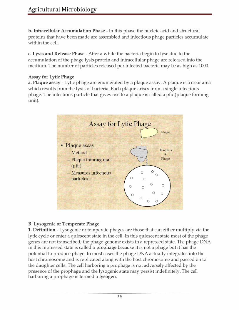
Agricultural Microbiology
59
b. Intracellular Accumulation Phase - In this phase the nucleic acid and structural
proteins that have been made are assembled and infectious phage particles accumulate within the cell.
c. Lysis and Release Phase - After a while the bacteria begin to lyse due to the
accumulation of the phage lysis protein and intracellular phage are released into the medium. The number of particles released per infected bacteria may be as high as 1000.
Assay for Lytic Phage a. Plaque assay - Lytic phage are enumerated by a plaque assay. A plaque is a clear area
which results from the lysis of bacteria. Each plaque arises from a single infectious phage. The infectious particle that gives rise to a plaque is called a pfu (plaque forming unit).
B. Lysogenic or Temperate Phage 1. Definition - Lysogenic or temperate phages are those that can either multiply via the
lytic cycle or enter a quiescent state in the cell. In this quiescent state most of the phage genes are not transcribed; the phage genome exists in a repressed state. The phage DNA in this repressed state is called a prophage because it is not a phage but it has the
potential to produce phage. In most cases the phage DNA actually integrates into the host chromosome and is replicated along with the host chromosome and passed on to the daughter cells. The cell harboring a prophage is not adversely affected by the presence of the prophage and the lysogenic state may persist indefinitely. The cell harboring a prophage is termed a lysogen.

www.AgriMoon.Com
60
2. Events Leading to Lysogeny - The Prototype Phage: Lambda a. Circularization of the phage chromosome - Lambda DNA is a double stranded linear molecule with small single stranded regions at the 5' ends. These single stranded ends are complementary (cohesive ends) so that they can base pair and produce a
circular molecule. In the cell the free ends of the circle can be ligated to form a covalently closed circle as illustrated in Figure 5. b. Site-specific recombination - A recombination event, catalyzed by a phage coded
enzyme, occurs between a particular site on the circularized phage DNA and a particular site on the host chromosome. The result is the integration of the phage DNA into the host chromosome as illustrated in Figure 6. c. Repression of the phage genome - A phage coded protein, called a repressor, is made which binds to a particular site on the phage DNA, called the operator, and shuts
off transcription of most phage genes EXCEPT the repressor gene. The result is a stable repressed phage genome which is integrated into the host chromosome. Each temperate phage will only repress its own DNA and not that from other phage, so that repression is very specific (immunity to superinfection with the same phage).
3. Events Leading to Termination of Lysogeny
Anytime a lysogenic bacterium is exposed to adverse conditions, the lysogenic state can be terminated. This process is called induction. Conditions which favor the termination
of the lysogenic state include: desiccation, exposure to UV or ionizing radiation, exposure to mutagenic chemicals, etc. Adverse conditions lead to the production of proteases (rec A protein) which destroy the repressor protein. This in turn leads to the expression of the phage genes, reversal of the integration process and lytic multiplication.
4. Lytic vs Lysogenic Cycle The decision for lambda to enter the lytic or lysogenic cycle when it first enters a cell is determined by the concentration of the repressor and another phage protein called cro in the cell. The cro protein turns off the synthesis of the repressor and thus
prevents the establishment of lysogeny. Environmental conditions that favor the production of cro will lead to the lytic cycle while those that favor the production of the repressor will favor lysogeny.

Agricultural Microbiology
61
5. Significance of Lysogeny a. Model for animal virus transformation - Lysogeny is a model system for virus
transformation of animal cells b. Lysogenic conversion - When a cell becomes lysogenized, occasionally extra genes
carried by the phage get expressed in the cell. These genes can change the properties of the bacterial cell. This process is called lysogenic or phage conversion. This can be of significance clinically. e.g. Lysogenic phages have been shown to carry genes that can modify the Salmonella O antigen, which is one of the major antigens to which the immune response is directed. Toxin production by Corynebacterium diphtheriae is mediated by a gene carried by a phage. Only those strain that have been converted by lysogeny are pathogenic.
******☺******

www.AgriMoon.Com
62
Lecture 09: VIROIDS, PRIONS
A virus is a small infectious agent that can replicate only inside the living cells of
organisms. Most viruses are too small to be seen directly with a light microscope. Viruses infect all types of organisms, from animals and plants to bacteria and archaea. Since the initial discovery of tobacco mosaic virus by Martinus Beijerinck in 1898, about 5,000 viruses have been described in detail though there are millions of different types. Viruses are found in almost every ecosystem on Earth and are the most abundant type of biological entity. The study of viruses is known as virology, a sub-speciality of microbiology.
Virus particles (known as virions) consist of two or three parts: the genetic material made from either DNA or RNA, long molecules that carry genetic information; aprotein coat that protects these genes; and in some cases an envelope of lipids that surrounds the protein coat when they are outside a cell. The shapes of viruses range from simple helical and icosahedral forms to more complex structures. The average virus is about one one-hundredth the size of the average bacterium.
Every virus has 2 stages o dormant, particulate, transmissible stage called the virion stage o an active, intracellular stage called the infectious stage
Virion Stage
Virions are the transmissible state of a virus. Metabolically inert. Virion = "a piece of nucleic acid wrapped up in a protein coat" (and/or a
membrane) The nucleic acid can be either DNA (double-stranded (ds) or single-stranded (ss))
or RNA (ds or ss); never both. The coat (also called viral shell or capsid) can be icosahedron (20-sided regular
geometric shape common in many bacterial, animal, and plant viruses), sphere, cylinder, bullet-shaped, or amorphous shaped particle.
Virions must be able to adhere and allow entry into some host cell(s). Also to survive outside of host cell environment.
Some virions more hardy than others (e.g., hepatitis virus can withstand short periods of boiling; most virions are destroyed by this).
Agricultural Microbiology
63
Infectious Stage
When virus infects a cell, nucleic acid must be uncoated and gain access to metabolic machinery of cell.
Virus life cycle is characterized by: o attachment o penetration, with entry of nucleic acid into cell o early expression of virus genes (either directly by translation, if virus
contains "+" RNA, or indirectly after transription and then translation) o replication of virus nucleic acid o synthesis of new virion components o packaging and assembly of new virions o exit from cell
Measurement of viral growth
Must grow virus on host cells to see anything. Can't grow virus without cells. To quantify viruses, need some way to get flat surface of growing cells, allow
virus-infected cells to spread radially where present = plaque. In bacterial cells this is easy. Spread "lawn" of bacteria on plate, add diluted
phage suspension or culture infected with phages. After 6-8 hours can see plaques in E. coli.
In plant cells, can be easy. Example: Tobacco Mosaic Virus (TMV), make virus dilution, rub over surface of tobacco leaf. After leaf growth, can observe plaque areas.
In animal cells, not so easy. In 1960's, standard assay was to inoculate chicken egg membranes of developing chick embryos, incubate for a week, cut open shell and count plaques on membrane in the air sac. Lots of work to get statistically reliable data!
In 1970's tissue culture became a viable alternative. Animal cells are cultured as microbes in glass or plastic, use special medium that contains most of nutrients present in blood. Cells will spread as monolayer on surface, can count plaques after staining.
Taxonomy of viruses
Based mainly on Virion and Kingdom of host Use Host cell type (Animal viruses, plant viruses, etc.) Use Nucleic Acid type (ds DNA, ss DNA, ds RNA, ss RNA) Use + or - polarity of RNA. "+" is able to serve as mRNA. "-" is the complement of
+, must function as template to make a complementary strand of + RNA before any translation can occur.
Use virus coat morphology. Enveloped vs. non-enveloped viruses.

www.AgriMoon.Com
64
Virion Structure "Naked" viruses
o Helical viruses Tobacco mosaic virus (TMV) is an example of a virus with helical
symmetry. A helical array of identical protein subunits surrounds an RNA
molecule.
o Icosahedral viruses
built from icosahedral (20-sided) assemblies of protein subunits.
Icosahedral shape is the minimum free energy structure for producing a shell of equivalently bonded identical structures.
The simplest icosahedral capsids are built up by using 3 identical subunits to form each triangular face, thereby requiring 60 identical subunits to form a complete capsid. A few simple virus particles are constructed in this way, e.g. bacteriophage ØX174.
Most icosahedral viruses have more than 60 subunits, usually some multiple N times 60. N (called the triangulation number) can have values of 1, 3, 4, 7, 9, 12, and more.

Agricultural Microbiology
65
"Enveloped" viruses
o "Naked" viruses require host death so viruses can be released. This may be wasteful, and may cause premature death of host cell.
o Alternative strategy: shed virus particles by budding out, continued release from cell membrane. Cell does not die (immediately), continues to serve as factory for virus assembly and release. Virus typically acquires a coating of host cell membrane, modified to include virus-specific proteins. This is the "envelope". Virus may have additional protein coats (often icosahedral) inside the envelope.
o Eventually host cell is too depleted to survive. Can see evidence of this as "cytopathic effect" (CPE). Cell then dies.
o Examples of enveloped viruses include: Retrovirus, including HIV Paramyxovirus, including influenza Rhabdovirus, including rabies Filovirus. Although very "hot" in the news, these viruses are very
poorly characterized because of their extreme pathogenicity. They are class IV pathogens, meaning they can only be cultured in total containment facilities, of which there are only two in the U. S. They are thought to be enveloped viruses with - RNA genomes.

www.AgriMoon.Com
66
Virus Genomes
Rule of Thumb: to estimate # of virus proteins, look at size of viral DNA or RNA. For each 1000 base pairs, can guess the existence of 1 protein
o "typical" gene has 300-400 amino acids = ~ 1000 base pairs = 1 kbp (= 1 protein)
o small virus: SV40 => 5000 base pairs = 5 kbp ~ 5 proteins o large virus: T4 => 200 kbp ~ 100-200 proteins o by comparison, E. coli: 4000 kbp
Bacterial Viruses = Phages Bacterial defenses against infection Cell surfaces: possibilities of mutation
Virus must attach to some specific cell surface protein or polysaccharide. But these are specified by genes, and genes can mutate. In population, will always find some variant strains with slightly different cell surfaces, may not bind virus well.
When phage first discovered, thought this could be effective weapon against bacterial disease. But frequency of resistant bacterial strains was too high, any given strain of virus quickly became useless as resistant survivors propagated.
Nucleases: endo- and exo-DNases and RNases
All bacteria seem to have nucleases that can attack DNA (called DNases) and RNA (called RNases).
Exoenzymes attack free 5' or 3' ends of DNA, RNA molecules. Bacteria are protected since DNA (and plasmids) are always circular. RNases are present, and in fact destroy mRNA eventually (bacteria are always making new RNAs, very responsive to enviroment changes).
Endonucleases are potentially lethal weapons. Called restriction enzymes. Attack at specific sequence: e.g., in E. coli, enzyme called EcoRI will attack any sequence with 5' G-A-A-T-T-C 3' (cuts DNA between G and A).
Why doesn't this kill cell? Because cell also has a second enzyme, called modification enzyme, that protects all host DNA sequences of this type. Typically adds a methyl (-CH3) group to one base at the cutting site. The methylated base is modified, and protected from the restriction enzyme. When foreign DNA comes into cell (e.g. virus DNA), if restriction site if present it will be cut and ----- requiem for the virus.
The importance of Restriction Enzymes
Restriction enzymes are responsible for the genetic revolution. They make reproducible, specific cuts with surgical precision. Major industry has emerged in biochemical supply

Agricultural Microbiology
67
companies to harvest bacteria, purify restriction enzymes, and sell these to research and applied industries.
Animal Viruses
Animal viruses are different in many respects from bacterial viruses. The host cells are more complex, with multiple compartments and more complex regulation of replication, transcription, and translation. Animal cells are not bounded by cell walls.
Not surprisingly, animal viruses have evolved to overcome these problems. They attach and enter by different mechanisms than phages, and their intracellular activities include the ability to move between different compartments as needed.
Viral entry and exit from cells is very different from bacteriophages. Animal viruses must enter through cell membrane, either by triggering endocytosis pathway or by fusing viral envelope with the cell envelope.
Modifications are needed in both cellular and viral mRNA to allow recognition and movement from nucleus to cytoplasm. For example:
o 3' tail of poly A o 5' cap of methyl Guanosine triphosphate
Types of infection
Viruses in animal cells show a variety of infection patterns:
lytic infection: destroys host cells. persistent infection: host cell continues to shed virus over long time. Cell
gradually becomes recognizably poorer (recognized as cytopathic effect, or CPE), eventually "crumps out".

www.AgriMoon.Com
68
transformation: infection by certain viruses causes cells to change, become
cancerous. Responsible genes are called oncogenes (tumor-producing genes). Viral oncogenes have also been found in uninfected cells. These are genes involved in regulation of cell cycle; when defective, normal regulatory control is lost and cell can become cancerous.
latent infection: virus genes may not be expressed for long time (ex. many
Herpes infections). Not the same as lysogeny -- genes are not integrated into host chromosome.
Human Immunodefiency Virus (HIV) and AIDS
AIDS first recognized in 1981. Over 300,000 cases reported in U.S., over 8 million in Africa, over 12 million infected world-wide.
View AIDS in perspective: a PBS website with current data on the AIDS epidemic.
Transmission: sexual activity, especially with multiple sex partners. Also contaminated blood, needles, hospitals. Not just a disease of homosexuals! In Africa (most # cases) about equal # of male and female victims.
AIDS lowers immune system's ability to respond to other infections, allows opportunistic pathogens to invade body. Most common infection is pneumonia (lung infection) caused by Pneumocystis (2/3 of all AIDS patients get this at some point).
Host cell for the virus is CD4 (T-helper) cell, needed to activate antibody production. In normal human, CD4 cells account for 70% of total T cells -- in AIDS, number decreases, may reach 0% of T cell pool.
Progress of HIV infection only recently understood. Formerly thought that virus became latent. Now discover that virus is anythig but latent: during infected period (which can last 10 years), body is destroying ~ billion virions/day, and virus is killing about 100 million CD4 T cells a day. HIV virus continues replicating, and body rapidly replenishes lost T cells. Only when lymph nodes wear out does virus gain the upper hand. See handout in class titled "Huge HIV turnover helps explain drug resistance, pathogenicity".
Prognosis: with carefully selected treatments, better than before. Virtually every infected person dies sooner of later, usually within 10 years of infection. No cure known, no vaccine yet available. Virus mutates rapidly, many strain variations. Vaccines being tried, results mixed but preliminary.
Drugs: some types of drugs offer limited success.

Agricultural Microbiology
69
o AZT (azidothymidine) is analog of thymidine, but is blocked at 3'-position, so no further chain growth possible. These target viral reverse transcriptase enzyme. Should reduce DNA synthesis in treated cells. But eventually, viral mutants resistant to drug arise. Also, long term use of drug can cause toxic side effects.
o protease inhibitors. Like many viruses, HIV needs to cleave large protein product into smaller products, using viral protease protein. By inhibiting this enzyme, should block necessary stage in viral replication cycle. Still under development, but resistant viral mutants to these type of drugs have already been found. Still, drug offers promise. See handout article for more details. Safe sex! Caution with sharps. Extra caution in clinical settings.
HIV Life Cycle
(budding through plasma membrane)

www.AgriMoon.Com
70
Viroids and Prions
Viroids very small ss RNA genomes (~300 nucleotides). No coat, and RNA does
not encode protein. Known viroids cause diseases in plants because host cells replicate the RNA.
Prions (protein infectious agent) do not have a nucleic acid genome. Prion
diseases are often called spongiform encephalopathies because of the post mortem appearance of the brain with large vacuoles in the cortex and cerebellum.
Pathology of brains infected with prion diseases: o Scrapie (sheep) o bovine spongiform encephalopathy (cows) = "mad cow disease" o Creutzfeldt-Jakob Disease (humans)
Prion diseases in humans are probably primarily a genetic neurotoxic disorder. Transmission of the disease to humans via infectious prions is likely to be rare.
The prion is a modified form of a normal cellular protein known as PrPc (for cellular), found predominantly on the surface of neurons and thought to be involved in synaptic function.
The modified form of PrPc (= prion) is known as PrPsc (for scrapie) which is relatively resistant to proteases and accumulates in cytoplasmic vesicles of diseased individuals. Prion protein may cause normal protein to fold abnormally.
******☺******

Agricultural Microbiology
71
Lecture 10: BACTERIAL GENETICS
Bacterial genetics is the study of gene structure and function in bacteria. Genetics itself is concerned with determining the number, location, and character of the genes of an organism. The process of replication is essential to understand in or manipulate the genome and to understand the functioning of these organisms.
DNA REPLICATION In general, DNA is replicated by uncoiling of the helix, strand separation by breaking of the hydrogen bonds between the complementary strands, and synthesis of two new strands by complementary base pairing. Replication begins at a specific site in the DNA called the origin of replication.
How does the DNA in the bacterial cell replicate ……

www.AgriMoon.Com
72
Transcription in bacteria… how does it happen……
Transcription is the process by which genetic information from DNA is transferred into RNA. DNA sequence is enzymatically copied by RNA polymerase to produce a complementary nucleotide RNA strand. One significant difference between RNA and DNA sequence is the presence of U, or uracil in RNA instead of the T, or thymine of DNA. In the case of protein-encoding DNA, transcription is the first step that ultimately leads to the translation of the genetic code, via the mRNA intermediate, into a functional peptide or protein. The stretch of DNA that is transcribed into an RNA molecule is called a transcription unit. A transcription unit that is translated into protein contains sequence that directs and regulates protein synthesis in addition to coding sequence that is translated into protein. Regulatory sequence that is before, or 5', of the coding sequence is called 5' untranslated (5'UTR) sequence, and sequence found following, or 3', of the coding sequence is called 3' untranslated (3'UTR) sequence. As in DNA replication, transcription proceeds in the 5' → 3' direction. The DNA template strand is read 3' → 5' by RNA polymerase and the new RNA strand is synthesized in the 5'→ 3' direction. RNA polymerase binds to the 3' end of a gene (promoter) on the DNA template strand and travels toward the 5' end. Except for the fact that thymines in DNA are converted to uracils in RNA, the newly synthesized RNA strand will have the same sequence as the coding (non-template) strand of the DNA.
Prokaryote Eukaryote
How to undertake experiments for understanding genetics of bacteria ? The classical way to investigate genes is to mate two organisms with different genotypes and compare the observable properties (phenotypes) of the parents with those of the progeny. Bacteria do not mate (in the usual way), so there is no way of getting all the chromosomes of two different bacteria into the same cell. However, there are a number of ways in which a part of the chromosome or genome from one bacterium can be inserted into another bacterium so that the outcome can be

Agricultural Microbiology
73
studied. The first step in performing genetic research on bacteria is to select mutants that differ from wild-type cells in one or more genes. Then crosses are made between mutants and wild types, or between two different mutants, to determine dominance-recessive relationships, chromosomal location, and other properties. Various genetic methods are used to select bacterial mutants, antibiotic-resistant cells, cells with specific growth requirements, and so on.
1. Mutants in bacteria are mostly biochemical in nature, because we can‘t generally see the cells.
2. The most important mutants are auxotrophs. An auxotroph needs some nutrient that the wild type strain (prototroph) can make for itself. For example, a trp- auxotroph can‘t make its own tryptophan (an amino acid). To grow trp- bacteria, you need to add tryptophan to the growth medium. Prototrophs are trp+; they don‘t need any tryptophan supplied since they make their own.
3. Chemoauxotrophs are mutants that can‘t use some nutrient (usually a sugar) that prototrophs can use as food. For example, lac- mutants can‘t grow on lactose (milk sugar), but lac+ prototrophs can grow on lactose.
4. Resistance mutants confer resistance to some environmental toxin: drugs, heavy metals, bacteriophages, etc. For instance, AmpR causes bacteria to be resistant to ampicillin, a common antibiotic related to penicillin.
5. Auxotrophs and chemoauxotrophs are usually recessive; drug resistance mutants are usually dominant. A common way to find bacterial mutants is replica plating, which means making two identical copies of the colonies on a petri plate under different conditions.
6. For instance, if you were looking for trp- auxotrophs, one plate would contain added tryptophan and the other plate would not have any tryptophan in it.
7. Bacteria are first spread on the permissive plate, the plate that allows both mutants and wild type to grow, the plate containing tryptophan in this case. They are allowed to grow fro a while, then a copy of the plate is made by pressing a piece of velvet onto the surface of the plate, then moving it to a fresh plate with the restrictive condition (no tryptophan). The velvet transfers some cells from each colony to an identical position on the restrictive plate.
8. Colonies that grow on the permissive plate but not the restrictive plate are (probably) trp- auxotrophs, because they can only grow if tryptophan is supplied.

www.AgriMoon.Com
74
Replica plating
The Ames test uses bacteria to test chemicalsfor capacity to cause mutations, as well as carcinogens (cancer-causing chemicals). The procedure is as fiollows:

Agricultural Microbiology
75
Lecture 11:
GENE EXPRESSION
Is the process by which information from a gene is used in the synthesis of a functional gene product. These products are often proteins, but in non-protein coding genes such as rRNA genes or tRNA genes, the product is a functional RNA. The process of gene expression is used by all known life - eukaryotes (includingmulticellular organisms), prokaryotes (bacteria and archaea) and viruses - to generate the macromolecular machinery for life. Several steps in the gene expression process may be modulated, including the transcription, RNA splicing, translation, and post-translational modification of a protein. Gene regulation gives the cell control over structure and function, and is the basis for cellular differentiation, morphogenesis and the versatility and adaptability of any organism. Gene regulation may also serve as a substrate for evolutionary change, since control of the timing, location, and amount of gene expression can have a profound effect on the functions (actions) of the gene in a cell or in a multicellular organism. In genetics, gene expression is the most fundamental level at whichgenotype gives rise to the phenotype. The genetic code is "interpreted" by gene expression, and the properties of the expression products give rise to the organism's phenotype.
Transcription The gene itself is typically a long stretch of DNA. It is a blueprint for the production of RNA. The production of RNA copies of the DNA is calledtranscription, and is performed by RNA polymerase, which adds one RNA nucleotide at a time to a growing RNA strand. This RNA iscomplementary to the template 3' → 5' DNA strand,[1] which is itself complementary to the coding 5' → 3' DNA strand. Therefore, the resulting 5' → 3' RNA strand is identical to the
coding DNA strand with the exception that thymines are replaced with uracils in the RNA. A coding DNA strand reading "ATG" is transcribed as "UAC" in RNA.

www.AgriMoon.Com
76
Translation
1. Initiation
2. 30S initiates binding to mRNA. 3. locates Shine-Dalgarno sequence (3-9 bases near 5' end of mRNA). 4. ribosome finds first AUG codon. 5. 50S ribosome binds. 6. tRNA carries N-formylmethione to first position 7. Elongation
8. 2 adjacent sites on ribosome: P and A site.A site accepts a new tRNA-AA.Psite holds existing chain peptide transferred from P site tRNA to A-site AA
9. enzyme activity is in ribosomal RNA, not protein 10. also required: Energy (GTP) and elongation factors 11. Termination
12. reach a "stop codon" UAG, UAA, or UGA 13. no t-RNAs for release, but release factors required 14. Net cost: 4 phosphate bonds/amino acid added! 15. B. Genetic Code
1. AUG = universal "start" codon 2. UAG, UAA, UGA = "stop" codons 3. A few messages in bacteria use GUG as start, but still need Shine-
Dalgarno sequence, stil code for N-formylmethionine.
Classic example: The lac operon

Agricultural Microbiology
77
1. gene is regulated by negative control; in absence of specific repressor, gene is transcribed just like constitutive gene. In order to regulate, must add specific block. Must say "no"; otherwise gene is not down-regulated.
2. Lactose = milk sugar, disaccharide made of galactose + glucose. In order to
metabolize lactose, cells must produce enzyme ß-galactosidase, split lactose into galactose + glucose
3. Observation: add lactose to cells: within minutes, ß-galactosidase enzyme
appears, also lac permease in membrane, and a third protein, transacetylase. Level of ß-galactosidase enzyme can accumulate to level of 10% of cytoplasmic protein
4. Explanation: in absence of lactose (= inducer), lac repressor blocked operator
site. 5. Lac repressor is allosteric protein. Coded for by another region of DNA
(constitutive gene, weak promoter, low level of expression) 6. Effector molecule = Inducer is lactose (actually allolactose, or analog such as
IPTG) binds to repressor protein, repressor released, RNA is made, all genes turned on as unit
Some genes are regulated by activator proteins
1. genes with weak promoters are rarely transcribed.some such genes can be activated by activator protein, causes RNA polymerase to bind more tightly.often there are two components to such regulation: a sensor protein and an activator protein. The activator protein is inactive until phosphorylated by the sensor, then it activates transcription, gene product is made.
Lac operon – in presence of inducer ( lactose)

www.AgriMoon.Com
78
1.
Lac operon – in absence of lactose
Role of mRNA Carries coding information for amino acids = codons, 3 adajacent nucleotide bases Example: AAA, AGU, etc.leader sequence on mRNA (called Shine-Dalgarno sequence) binds to complementar sequence on small ribosome subunit.

Agricultural Microbiology
79
Role of ribosome
acts as a "decoding box" or "tape player" for the information in mRNA30S & 50S subunits (= 70S)30S has 16S RNA + 21 proteins.50S has 23S & 5S RNA + 34 proteins. Role of tRNA
structure: 4 loops, anticodon, AA binding site ~ 60 types in bacteria (>100 in mammals) only 73-93 nucleotides long some modified bases: pseudouridine, inosine, others modified after transcription extensive hairpin loops anticodon site: recognizes codon on mRNA AA added by enzyme: AA-tRNA activating enzymes ATP required, forms AA-AMP + PP, then AA-tRNA + AMP
Visit for Other Agriculture books, News, Recruitment, Information, and Events at
www.agrimoon.com

www.AgriMoon.Com
80
Lecture 12: RECOMBINATION IN BACTERIA
Transfer of Genetic Material in Bacteria The process of transfer of genetic material and recombination is very interesting bascterial recombination is given (PPT. an overview of bacterial recombination).The
three main mechanisms by which bacteria acquire new DNA are transformation, conjugation, and transduction. Transformation involves acquisition of DNA from the environment, conjugation involves acquisition of DNA directly from another bacterium, and transduction involves acquisition of bacterial DNA via a bacteriophage intermediate.
Transformation
Transformation is the process by which bacteria pick up DNA from their environment. The DNA may come from a variety of sources, but most likely it is the remnants of DNA from dead bacterial cells.

Agricultural Microbiology
81
In order to become successfully transformed, bacteria must be competent. This means
that the bacteria are expressing the appropriate enzymes (the 'transformation machinery') required to transport the exogenous DNA into the cell. Therefore, the correct genes must be expressed in order to carry out transformation. Expression of these genes depends on the growth conditions: bacteria most likely to be competent are dividing rapidly, but nutrients in the environment are becoming limited. (For more on the control of gene expression, see the module on bacterial gene regulation. In transformation, a cell surface receptor binds to DNA in the environment. After binding, the DNA is transported across the membrane by the transformation machinery. As this occurs, one strand of the DNA is digested away by an exonuclease, so that the DNA that enters the cell is single stranded. This promotes recombination, as long as the DNA taken up is sufficiently homologous to the host DNA to allow recombination to occur. The recombination that occurs is one-way (non-reciprocal); unlike the exchange of strands diagrammed in the module on recombination, in this case the new DNA will simply replace a strand of the host DNA. The replaced segment of host DNA will be degraded. If the new DNA is of a different allelic nature than the host DNA, a gene conversion event can occur. This is what happened in the example mentioned above: the avirulent strain of S. pneumoniae had a mutation in a gene required for production of the bacterial capsule. Heat killing the virulent cells (which contained the wild-type capsule gene) caused the release of fragments of the dead cells' genomes. Some of the avirulent cells picked up a piece of DNA containing the wild-type capsule gene, and underwent gene conversion so that they were wild type for that gene, causing them to become virulent.
Conjugation
Conjugation is a mating process involving bacteria. It involves transfer of genetic information from one bacterial cell to another, and requires physical contact between the two bacteria involved. The contact between the cells is via a protein tube called an F or sex pilus, which is also the conduit for the transfer of the genetic material. Basic conjugation involves two strains of bacteria: F+ and F-. The difference between these two strains is the presence of a Fertility factor (or F factor) in the F+ cells. The F
factor is an episome that contains 19 genes and confers the ability to conjugate upon its host cell. Genetic transfer in conjugation is from an F+ cell to an F- cell, and the genetic material transferred is the F factor itself. Here is an overview of the process:
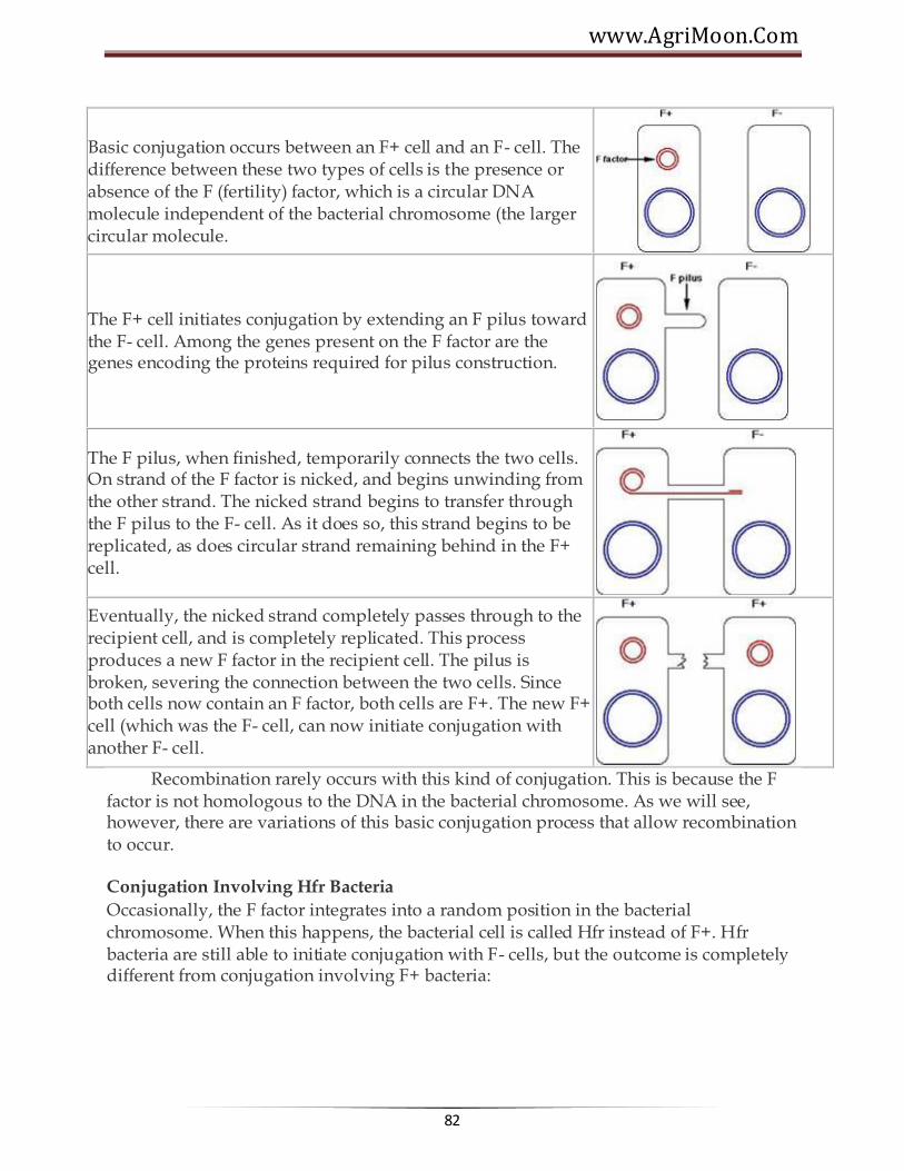
www.AgriMoon.Com
82
Basic conjugation occurs between an F+ cell and an F- cell. The difference between these two types of cells is the presence or absence of the F (fertility) factor, which is a circular DNA molecule independent of the bacterial chromosome (the larger circular molecule.
The F+ cell initiates conjugation by extending an F pilus toward the F- cell. Among the genes present on the F factor are the genes encoding the proteins required for pilus construction.
The F pilus, when finished, temporarily connects the two cells. On strand of the F factor is nicked, and begins unwinding from the other strand. The nicked strand begins to transfer through the F pilus to the F- cell. As it does so, this strand begins to be replicated, as does circular strand remaining behind in the F+ cell.
Eventually, the nicked strand completely passes through to the recipient cell, and is completely replicated. This process produces a new F factor in the recipient cell. The pilus is broken, severing the connection between the two cells. Since both cells now contain an F factor, both cells are F+. The new F+ cell (which was the F- cell, can now initiate conjugation with another F- cell.
Recombination rarely occurs with this kind of conjugation. This is because the F factor is not homologous to the DNA in the bacterial chromosome. As we will see, however, there are variations of this basic conjugation process that allow recombination to occur.
Conjugation Involving Hfr Bacteria
Occasionally, the F factor integrates into a random position in the bacterial chromosome. When this happens, the bacterial cell is called Hfr instead of F+. Hfr bacteria are still able to initiate conjugation with F- cells, but the outcome is completely different from conjugation involving F+ bacteria:

Agricultural Microbiology
83
As mentioned above, Hfr cells are formed when the F factor integrates into the bacterial chromosome. This integration occurs at a random location.
The Hfr cell is still able to initiate conjugation with an F- cell.
When DNA transfer begins, the Hfr cell tries to transfer the entire bacterial chromosome to the F- cell. The first DNA to be transferred is chromosomal DNA, and thelast DNA to be transferred will be the F factor DNA.
Transfer of the bacterial chromosome is almost never complete. Pili are fairly fragile
structures, and shear forces tend to break the pilus, disrupting DNA transfer before the entire chromosome can be transferred. As a result, the F factor itself is almost never transferred to the recipient cell. This cell will remain F-. This cell will receive new DNA from the Hfr cell however, and this new DNA can undergo recombination at a high frequency with the host chromosome, because the DNA sequences will be homologous. In fact, Hfr is short for 'high frequency recombination'. This recombination can result in gene conversion events, if the transferred DNA and the corresponding region of host DNA contain different alleles of the same gene.
Mapping Genes on Bacterial Chromosomes
Bacteria, since they are usually haploid, cannot have their chromosomes mapped by the same techniques as eukaryotes (For a reminder of how this works, see the module on linkage and mapping). They can, however, be mapped by using Hfr bacterial conjugation. For example, imagine that an F- cell has mutant alleles of two genes, a and b (the F- would therefore be a-, b-). If this cell undergoes conjugation with an Hfr cell that is a+, b+ (in other words, wild type), the F- cell should undergo gene conversion to a+, b+ when both of those genes have been transferred by conjugation. By determining how long it takes the b gene to transfer after the a gene has transferred, it is possible to get a relative idea of how far apart the two genes are on a chromosome. The experiment would be done this way: a+, b+ Hfr cells would be mixed with a-,b- F- cells. The time of mixing would be designated 'time zero'. At regular intervals, a small amount of the mixture would be removed and conjugation would be disrupted using a blender (the shear force of the blender would cause any pili to break). These bacteria would then be tested for gene conversion (for example, if the mutations rendered the F- bacteria auxotrophic, the bacteria could be tested by growing them on minimal medium, or minimal medium supplemented with the necessary nutrient required because of one or the other mutation). If the a gene was converted to wild type at 8 minutes after time zero, and the b gene was converted to wild type at 19 minutes after time zero, then the distance between the two genes would be '11 minutes' (because that was the difference in time required to transfer the b gene compared to the a gene). Bacterial map distances are always expressed in minutes, because of this technique.

www.AgriMoon.Com
84
F' Conjugation
Just as F factors can occasionally integrate into the bacterial chromosome (producing an Hfr cell from an F+ cell), integrated F factors can occasionally excise themselves from the bacterial chromosome. If this excision occurs properly, the Hfr cell becomes an F+ again. The excision is sometimes sloppy, however, and the F factor takes a small segment of the bacterial chromosome with it. Some of the chromosomal DNA has therefore become associated with the episome. When this happens, the cell is called an F'. Conjugation involving F' cells allows for the possibility of recombination, as shown below:
The F' cell has a full complement of chromosomal genes; however, some of those genes are now on the episome. F' cells are able to initiate conjugation with F- cells because of the presence of the F factor.
When the F factor begins to transfer its DNA to the recipient cell, it will transfer the small segment of chromosomal DNA as well.
Just as in the F+/F- mating, both cells wind up with a copy of the episome. The cell that was F- now has the F factor (along with the piece of chromosomal DNA) and is therefore now F'. This cell, however, also has a complete chromosome, so it will be diploid for the segment of chromosomal DNA on the episome. Such a partially diploid bacterial cell is called a merozygote. The chromosomal DNA on the episome can undergo recombination at high frequency with its homologous sequence on the chromosome.
Transduction
Transduction involves the exchange of DNA between bacteria using bacterial viruses (bacteriophage) as an intermediate. There are two types of transduction, generalized transduction and specialized transduction, which differ in their mechanism and in the DNA that gets transferred. Before we can address these processes, however, we need to understand the life cycle of a bacteriophage. When a phage infects a bacterial cell, it injects its DNA into the cell. The viral DNA is replicated numerous times, and viral genes are expressed, producing the proteins that make up the viral capsid (or protein coat) and nucleases that digest the host genome into fragments. The newly replicated viral DNA molecules are packaged into viral capsids, and the bacterial cell is lysed (burst, and therefore killed), releasing hundreds of viral progeny, which then go on to infect other cells.
Generalized Transduction
Sometimes, during bacteriophage replication, a mistake is made, and a fragment of the host DNA gets packaged into a viral capsid. The resulting phage would be able to infect another cell, but it would not have any viral genes, so it would not be able to replicate.

Agricultural Microbiology
85
The cell infected by this phage would survive, and would have an extra piece of bacterial DNA present, which could undergo recombination with the host chromosome, and perhaps cause a gene conversion event. Because it is a random fragment that gets packaged into the viral capsid, any segment of the bacterial DNA can be transferred this way (hence the name 'generalized').
Specialized Transduction
Specialized transduction occurs only with certain types of bacteriophage, such as phage lambda. Lambda has the ability to establish what is called a lysogenic
infection in a bacterial cell. In a lysogenic infection, the viral DNA becomes
incorporated into the host chromosome, much as the F factor did in Hfr cells. In a lysogenic infection by lambda, the DNA integrates into a very specific spot in the host chromosome. The integrated viral DNA can remain integrated for long periods of time, without disturbing the cell. Under the appropriate conditions (the regulation of this is very complex, so don't worry about it), the viral DNA will excise itself from the chromosome, and enter the lytic phase, in which the virus replicates just as described
above. The cell gets lysed, and new bacteriophage particles are released to infect other cells. As with excision of the F factor (when Hfr cells become F'), sometimes the excision of lambda is sloppy, and some bacteria DNA is excised along with it. When the resulting virus infects another cell, it will pass that bacterial DNA into the cell, along with its own DNA. If the infected cell survives (it can happen; there are bacterial defenses against viral infection), it will contain a new piece of bacterial DNA, which can undergo recombination and possibly cause gene conversion. Because the viral DNA integrates into a specific location, when it excises, the bacterial DNA removed with it will be the same in all cases. Therefore, the DNA transferred to the second cell will be the same segment of the bacterial chromosome. This is why this process is called 'specialized' transduction.
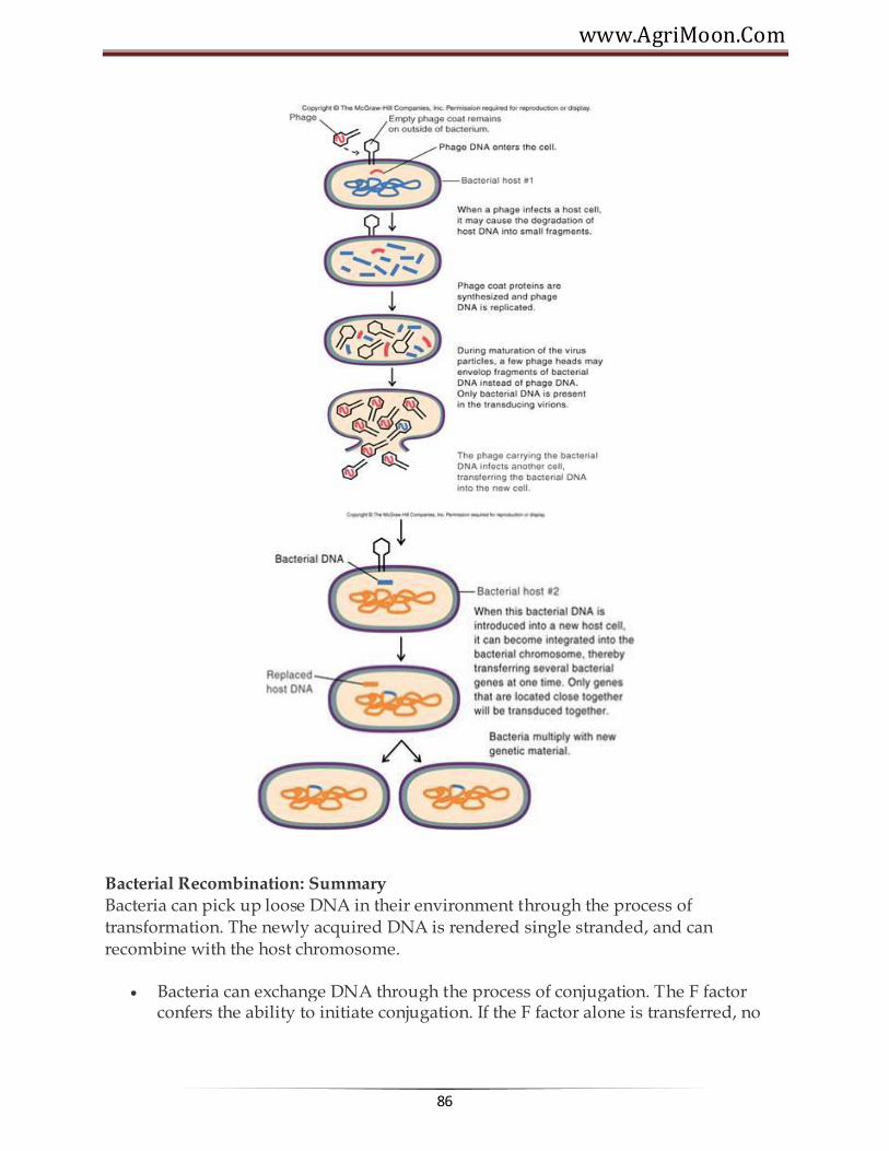
www.AgriMoon.Com
86
Bacterial Recombination: Summary Bacteria can pick up loose DNA in their environment through the process of transformation. The newly acquired DNA is rendered single stranded, and can recombine with the host chromosome.
Bacteria can exchange DNA through the process of conjugation. The F factor confers the ability to initiate conjugation. If the F factor alone is transferred, no

Agricultural Microbiology
87
recombination will occur. Under certain circumstances, chromosomal DNA can be transferred to the recipient cell. In these cases, recombination will occur.
Bacteria can receive bacterial DNA from viruses through the process of transduction. Bacterial viruses can accidentally pick up pieces of bacterial DNA. When they subsequently infect a cell, they transfer the pice of bacterial DNA, which can undergo recombination with the host bacterial chromosome.
The result of recombination in the above cases may be gene conversion, in which a mutant allele becomes wild-type or vice versa.
Conjugation involving Hfr bacteria can be used to map genes along the bacterial chromosome. This done by determining in what order genes are transferred during conjugation, waht the time difference is between the transfer of genes.
Bacteria do not reproduce sexually but can acquire new DNA through transformation, transduction or conjugation.These natural processes have been modified so that DNA can be deliberately incorporated into host microbes- even genes that would normally never be transferred this way.
******☺******

www.AgriMoon.Com
88
Lecture 13:
GENETIC ENGINEERING - PLASMIDS, EPISOMES Genetic Engineering: Genetic engineering is a laboratory technique used by scientists
to change the DNA of living organisms. DNA is the blueprint for the individuality of an organism. The organism relies upon the information stored in its DNA for the management of every biochemical process. The life, growth and unique features of the organism depend on its DNA. The segments of DNA which have been associated with specific features or functions of an organism are called genes.
Molecular biologists have discovered many enzymes which change the structure of DNA in living organisms. Some of these enzymes can cut and join strands of DNA. Using such enzymes, scientists learned to cut specific genes from DNA and to build customized DNA using these genes. They also learned about vectors, strands of DNA
such as viruses, which can infect a cell and insert themselves into its DNA. With this knowledge, scientists started to build vectors which incorporated genes of their choosing and used the new vectors to insert these genes into the DNA of living organisms. Genetic engineers believe they can improve the foods we eat by doing this. For example, tomatoes are sensitive to frost. This shortens their growing season. Fish, on the other hand, survive in very cold water. Scientists identified a particular gene which enables a flounder to resist cold and used the technology of genetic engineering to insert this 'anti-freeze' gene into a tomato. This makes it possible to extend the growing season of the tomato. Plasmids: A plasmid is an extra chromosomal DNA molecule separate from the chromosomal DNA which is capable of replicating independently from the chromosomal DNA. In many cases, it is circular and double-stranded. Plasmids usually occur naturally in bacteria, but are sometimes found in eukaryotic organisms (e.g., the 2-micrometre-ring in Saccharomyces cerevisiae). Plasmid size varies from 1 to over 1,000 kilobase pairs (kbp). The number of identical plasmids within a single cell can range anywhere from one to even thousands under some circumstances. The number of plasmids in a cell generally remains constant from generation to generation.
Properties of Plasmids
• Circular DNA elements, always double-stranded DNA, Supercoiled • Can occur in as few as 1 copy per cell (single copy plasmids) to as many as several

Agricultural Microbiology
89
dozen (multicopy plasmids). • Variable sizes; small plasmids about 0.1% size of host chromosome, large plasmids can be as much as 10% the size of host chromosome. Smaller plasmids have few genes (30 or less). Size ranges from 1000 bp (1 kbp) to 1000 kbp. • Ubiquitous; almost all cells isolated in nature carry plasmids, often more than one kind. (In E. coli alone, more than 300 different plasmids isolated.) • Have a replicon (origin for DNA replication), number of copies per cell regulated. Large plasmids typically only 1-5 copies/cell (stringent control); small plasmids ~10-50 copies/cell (relaxed control) • Many plasmids are incompatible; if one is present, cell cannot support another plasmid of same compatibility group. • Not essential to cell under all circumstances; can be "cured" by agents that impair DNA replication ----> cured cell lacking plasmid. Can be spontaneously lost over time unless some selection makes plasmid valuable to cell. • Extend range of environments in which a cell can live (e.g., by degrading antibiotics, or providing enzymes for digestion of novel catabolites).
Examples of Plasmid genes
1. Antibiotic resistance genes (enzymes that modify or degrade antibiotics) -- plasmids with these genes are called R factors
2. Heavy metal resistance (enzymes that detoxify metals by redox reactions) 3. Growth on unusual substrates (enzymes for hydrocarbon degradation, etc.) 4. Restriction/modification enzymes (protect DNA, degrade unprotected DNA) 5. Bacteriocins (proteins toxic to other bacteria lacking the same plasmid) 6. Toxins (proteins toxic to other organisms; e.g. humans) -- called virulence
plasmids.
Some Examples:
1. Staph aureus virulence factors: coagulase, hemolysin, enterotoxin, others 2. pathogenic E. coli strains: hemolysin, enterotoxin
Proteins that mediate plasmid transfer to uninfected strains There are two categories of plasmids. Stringent plasmids replicate only when the
chromosome replicates. This is good if you are working with a protein that is lethal to the cell. Relaxed plasmids replicate on their own. This gives you a higher ratio of
plasmids to chromosome. Some of the traits coded by plasmids include:

www.AgriMoon.Com
90
Classification of Plasmids
1. Transfer properties -
a. Conjugative plasmids - Conjugative plasmids are those that mediated conjugation.
These plasmids are usually large and have all the genes necessary for autonomous replication and for transfer of DNA to a recipient (e.g. genes for sex pilus). b. Nonconjugative plasmids - Nonconjugative plasmids are those that cannot mediate conjugation. They are usually smaller than conjugative plasmids and they lack one or more of the genes needed for transfer of DNA. A nonconjugative plasmid can be transferred by conjugation if the cell also harbors a conjugative plasmid.
2. Phenotypic effects -
a. Fertility plasmid (F factor) b. Bacteriocinogenic plasmids - These plasmids have genes which code for substances that kill other bacteria. These substances are calledbacteriocins or colicins. c. Resistance plasmids 7 factors) - These plasmids carry antibiotic resistance genes.
i) Origin - The origin of the R factors is not known. It is likely that they evolved for other purposes and the advent of the antibiotic age provided a selective advantage for their wide-spread dissemination. ii) Structure - R plasmids are conjugative plasmids in which the genes for replication and transfer are located on one part of the R factor and the resistance genes are located

Agricultural Microbiology
91
on another part as illustrated in Figure. RTF (Resistance Transfer Factor) - carries the transfer genes. R determinant - carries the resistance genes. The resistance genes are often parts of transposons.
Mode of action of resistance genes -
a) Modification (detoxification) of antibiotic - e.g. β-lactamase b) Alteration of target site - e.g. Streptomycin resistance c) Alteration of uptake - Tetracycline resistance d) Replacement of sensitive pathway - e.g. new folic acid pathway for resistance to sulfa drugs. Plasmids are easy to manipulate and isolate using bacteria. They can be integrated into mammalian genomes, thereby conferring to mammalian cells whatever genetic functionality they carry. Thus, this gives you the ability to introduce genes into a given organism by using bacteria to amplify the hybrid genes that are created in vitro. This tiny but mighty plasmid molecule is the basis of recombinant DNA technology.
Episome: Episome is a unit of genetic material composed of a series of genes that
sometimes has an independent existence in a host cell and at other times is integrated into a chromosome of the cell, replicating itself along with the chromosome. Episomes have been studied in bacteria. One group of episomes are actually viruses that infect bacteria. As autonomous units they destroy host cells, and as segments integrated into a chromosome they multiply in cell division and are transferred to daughter cells. Episomes called sex factors determine whether chromosome material will be transferred from one bacterium to another. Other episomes carry genes that make bacteria resistant to the inhibitory action of antibiotics.
Transposons : Are sequences of DNA that can move or transpose themselves to new positions within the genome of a single cell. The mechanism of transposition can be either "copy and paste" or "cut and paste". Transposition can create phenotypically significant mutations and alter the cell's genome size. Barbara McClintock's discovery of these jumping genes early in her career earned her a Nobel prize in 1983. Transposons make up a large fraction of the C-value of eukaryotic cells. Transposons are often considered "junk DNA". In Oxytricha, which has a unique genetic system, they play a critical role its development. Transposons are very useful to researchers as a means to alter DNA inside a living organism.
******☺******

www.AgriMoon.Com
92
Lecture 14:
GENETICALLY MODIFIED ORGANISM
Genetic engineering - was made possible through a series of scientific advances including the discovery of DNA and the creation of the first recombinant bacteria in 1973, i.e., E .coli expressing a salmonella gene. This led to concerns in the scientific community about potential risks from genetic engineering. A genetically modified organism (GMO) or genetically engineered organism (GEO) is an organism (plant or animal) whose genetic material has been altered using genetic engineering techniques. These modifications are generally used to benefit medicine or food production. There is controversy about these techniques, as there are fears of tampering with an organism's evolution, and the long-term irreversible effects that could come from that tampering. The general principle of producing a GMO is to add a lot of genetic material into an organism's genome to generate new traits. These techniques, generally known as recombinant DNA technology, use DNA molecules from different sources, which are combined into one molecule to create a new set of genes. This DNA is then transferred into an organism, giving it modified or novel genes. Transgenic organisms, a subset of GMOs, are organisms which have inserted DNA that originated in a different species.
Genetically modified bacteria were the first organisms to be modified in the laboratory, due to their simple genetics. These organisms are now used for several purposes, and are particularly important in producing large amounts of pure human proteins for use in medicine.The first example of this occurred in 1978 whenHerbert Boyer working at a University of California laboratory took a version of the human insulin gene and inserted into the bacterium Escherichia coli to produce synthetic "human" insulin.The drug industry has made good use of this discovery in i ts quest to cure diabetes. Similar bacteria have been used to produce clotting factors to treat haemophilia, and human growth hormone to treat various forms of dwarfism. These recombinant proteins are safer than the products they replaced, since the older products were purified from cadavers and could transmit diseases. Indeed the human-derived proteins caused many cases of AIDS and hepatitis C in haemophilliacs and Creutzfeldt-Jakob disease from human growth hormone.
For instance, the bacteria which cause tooth decay are called Streptococcus mutans. These bacteria consume leftover sugars in the mouth, producing lactic acid that corrodes tooth enamel and ultimately causes cavities. Scientists have recently modified Streptococcus

Agricultural Microbiology
93
mutans to produce no lactic acid. These transgenic bacteria, if properly colonized in a person's mouth, could reduce the formation of cavities. Transgenic microbes have also been used in recent research to kill or hinder tumors, and to fight Crohn's disease. Genetically modified bacteria are also used in some soils to facilitate crop growth, and can also produce chemicals which are toxic to crop pests.
Moving Genes between Species—how to do……
The process by which scientists introduce new genetic material into a microorganism is called molecular or gene cloning or genetic engineering.
It involves the isolation of DNA from a source other than the microorganism itself. Source organisms span the world of living things, from microbes to plants to animals, including humans. Scientists obtain source DNA in several different ways: by disrupting cells of the target microbe (or plant or animal) andfragmenting it into small pieces, by synthesizing it from an RNA template
using an enzyme called reverse transcriptase, or by knowing the specific gene sequence and synthesizing it directly in the laboratory.
Once obtained, the pieces of DNA are inserted into a small genetic component that has the ability to make copies of itself (replicate) independently from the microbial genome. This self-replicating unit is called a cloning vector. Although these genetic elements exist naturally in the form of plasmids and bacterial
viruses, many of the ones used today have been altered to improve their properties for transferring genes. Restriction enzymes, which nick the donor
DNA and the cloning vector at specific sites, and DNA ligase, which attaches the donor DNA to the cloning vector, allow the source genes of interest to be inserted into the cloning vector without disrupting its ability to replicate.
The next step in the process is the introduction of the cloning vector with its segment of new DNA into a living cell. Bacteria have the ability to transport DNA into their cells in a process called transformation, and this ability is commonly exploited to achieve this goal. Getting the DNA into the cell, however, is only the beginning. No transformation is 100 percent efficient, and so the bacteria that receive the gene(s) of interest must be separated from those that did not. One of the best studied and most commonly used cloning vectors, pBR322, is especially useful for this purpose, as it contains several genes for antibiotic
www.AgriMoon.Com
94
resistance. Hence, any cell transformed with DNA containing pBR322 will be antibiotic resistant, and thus can be isolated from similar cells that have not be so transformed by merely growing them in the presence of the appropriate drugs. All that remains is to identify bacteria that are producing the product of the desired gene(s), and cloning is a success.
The introduction of human genes into bacteria has several complicating wrinkles that make cloning them even more challenging. For example, a bacterial gene codes for a protein from start to finish in one long string of nucleotides, whereas
human cells have stretches of noncoding nucleotides called introns within their genes. Bacteria do not have the same ability as human cells to remove these introns when producing proteins from the gene, and if the introns are not removed, the intended protein cannot be produced. This, along with other complications, has been overcome using many of the tools of genetic engineering.
Uses of GMOs: Examples of GMOs are highly diverse, and include transgenic
(genetically modified by recombinant DNA methods) animals such as mice, fish, transgenic plants, or various microbes, such as fungi and bacteria. The generation and use of GMOs has many reasons, chief among them are their use in research that addresses fundamental or applied questions in biology or medicine, for the production of pharmaceuticals and industrial enzymes, and for direct, and often controversial, applications aimed at improving human health (e.g., gene therapy) or agriculture (e.g., golden rice). The term "genetically modified organism" does not always imply, but can include, targeted insertions of genes from one into another species. For example, a gene from a jellyfish, encoding a fluorescent protein called GFP, can be physically linked and thus co-expressed with mammalian genes to identify the location of the protein encoded by the GFP-tagged gene in the mammalian cell. These and other methods are useful and indispensable tools for biologists in many areas of research, including those that study the mechanisms of human and other diseases or fundamental biological processes in eukaryotic or prokaryotic cells.
Transgenic microbes:
Bacteria were the first organisms to be modified in the laboratory, due to their simple genetics. These organisms are now used in a variety of tasks, and are particularly important in producing large amounts of pure human proteins for use in medicine.

Agricultural Microbiology
95
Bacteria-synthesized transgenic products
Insulin Interferon Hepatitis B vaccine Tissue plasminogen activator Human growth hormone Ice-minus bacteria
Did you know that this is a nickname given to a variant of the common bacterium Pseudomonas syringae (P. syringae). This strain of P. syringae lacks the ability to produce a certain surface protein, usually found on wild-type P. syringae. The "ice-plus" protein (In a protein, "Ice nucleation-active" protein) found on the outer bacterial cell wall acts as the nucleating centers for ice crystals. This facilitates ice formation, hence the designation "ice-plus." The ice-minus variant of P. syringae is amutant, lacking the gene responsible for ice-nucleating surface protein production. This lack of surface protein provides a less favorable environment for ice formation. Both strains of P. syringae occur naturally, but recombinant DNA technology has allowed for the synthetic removal or alteration of specific genes, enabling the creation of the ice-minus strain. Modifying P. syringae may have unexpected consequences for climate. A study has shown that its ice nucleating proteins may play an important part in causing ice crystals to form in clouds. If humans increase the frequency of bacteria lacking these proteins then it may affect rainfall
Commercial Applications
Transgenic microbes have many commercial and practical applications, including the production of mammalian products. A company called Genentech was among the earliest and most successful commercial enterprises to use genetically engineered bacteria to produce human proteins. Their first product was human insulin produced by genetically engineered Escherichia coli. A variety of other human hormones, blood proteins, and immune modulators are now
produced in a similar fashion, in addition to vaccines for such infectious agents as hepatitis B virus and measles.
Another promising application of genetically engineered microbes is in environmental cleanup, or biomediation. Scientists have discovered many naturally occurring genes that code for enzymes that degrade toxic wastes and wastewater pollutants in bacteria. Examples include genes for degrading chlorinated pesticides, chlorobenzenes, naphthalene, toluene, anilines, and various hydrocarbons. Researchers are using molecular cloning to introduce these genes from several different microbes into a single microbe, creating "super microbes" with the ability to degrade multiple contaminants.
www.AgriMoon.Com
96
Ananda Chakrabarty created one of the first microbes of this nature in the early 1970s. He introduced genes from several different bacteria into a strain ofBurkholderia cepacia, giving it the ability to degrade toxic compounds found in petroleum. This microbe offered a potential alternative to skimming and absorbing spilled oil. Chakrabarty's genetically modified bacterium has never been used, however, due to public concerns about the release of genetically engineered microbes into the environment. The microbe did, on the other hand, play an important role in establishing the biotechnology industry. The U.S. Patent Office granted Chakrabarty the first patent ever for the construction and use of a genetically engineered bacterium. This established a precedent allowing biotechnology companies to protect their "inventions" in the same way chemical and pharmaceutical companies have done in the past.
In addition to bacteria being used for producing proteins, genetically modified viruses allow gene therapy. Gene therapy is a relatively new idea in medicine. A virus reproduces by injecting its own genetic material into an existing cell. That cell then follows the instructions in this genetic material and produces more viruses. In medicine this process is adapted to deliver a gene that could cure disease into human cells. Although gene therapy is still relatively new, it has had some successes. It has been used to treat genetic disorders such as severe combined immunodeficiency, and treatments are being developed for a range of other incurable diseases, such as cystic fibrosis, sickle cell anemia, and muscular dystrophy. Genetically modified bacteria are also used in agriculture to facilitate crop growth, and can also produce chemicals which are toxic to crop pests.
Transgenic animals
Transgenic animals are used as experimental models to perform phenotypic tests with genes whose function is unknown or to generate animals that are susceptible to certain compounds or stresses for testing in biomedical research. Other applications include the production of human hormones, such as insulin. Frequently used in genetic research are transgenic fruit flies (Drosophila melanogaster) as genetic models to study the effects of genetic changes on development. Transgenic mice are often used to study cellular and tissue-specific responses to disease.
Transgenic plants
Transgenic plants have been developed for various purposes. Most of transgenic plants were created for research purposes and were not intended for eventual commercialization. From these few which have reached the market the most common transgenic traits include 1) resistance to pests or herbicides, 2) improved product shelflife. In the near future crops with improved nutritional value and with resitance to harsh environmental conditions might reach the marketplace. Since the first commercial cultivation of GM plants in 1996, GM plants tolerant to the herbicides glufosinate or glyphosate, and producing the Bt toxin, an insecticide, have dominated the agriculutral

Agricultural Microbiology
97
seed market for corn and other crops (soybean, cotton). Recently, a new generation of GM plants promising benefits for consumers and industry purposes is entering the market. Whenever GM plants are grown on open fields without containment there are risks that the modification will escape into the general environment. Most countries require biosafety studies prior to the approval of a new GM plant release, usually followed by a monitoring programme to detect environmental impacts. Especially in Europe, the coexistence of GM plants with conventional and organic crops has raised many concerns. Since there is separate legislation for GM crops and a high demand from consumers for the freedom of choice between GM and non-GM foods, measures are required to separate foods and feed produced from GMO plants from conventional and organic foods. European research programmes such as Co-Extra, Transcontainer and SIGMEA are investigating appropriate tools and rules.
Controversy over GMOs
The use of GMOs has sparked significant controversy in many areas. Some groups or individuals see the generation and use of GMO as intolerable meddling with biological states or processes that have naturally evolved over long periods of time (although many crops and animals have been modified by humans via unnatural selection over the last several thousand years), while others are concerned about the limitations of modern science to fully comprehend all of the potential negative ramifications of genetic manipulation. While some groups advocate the complete prohibition of GMOs, others call for mandatory labeling of genetically modified food or other products. Other controversies include the definition of patent and property pertaining to products of genetic engineering and the possibility of unforeseen local and global effects as a result of transgenic organisms proliferating. The basic ethical issues involved in genetic research are discussed in the article on genetic engineering.
******☺******
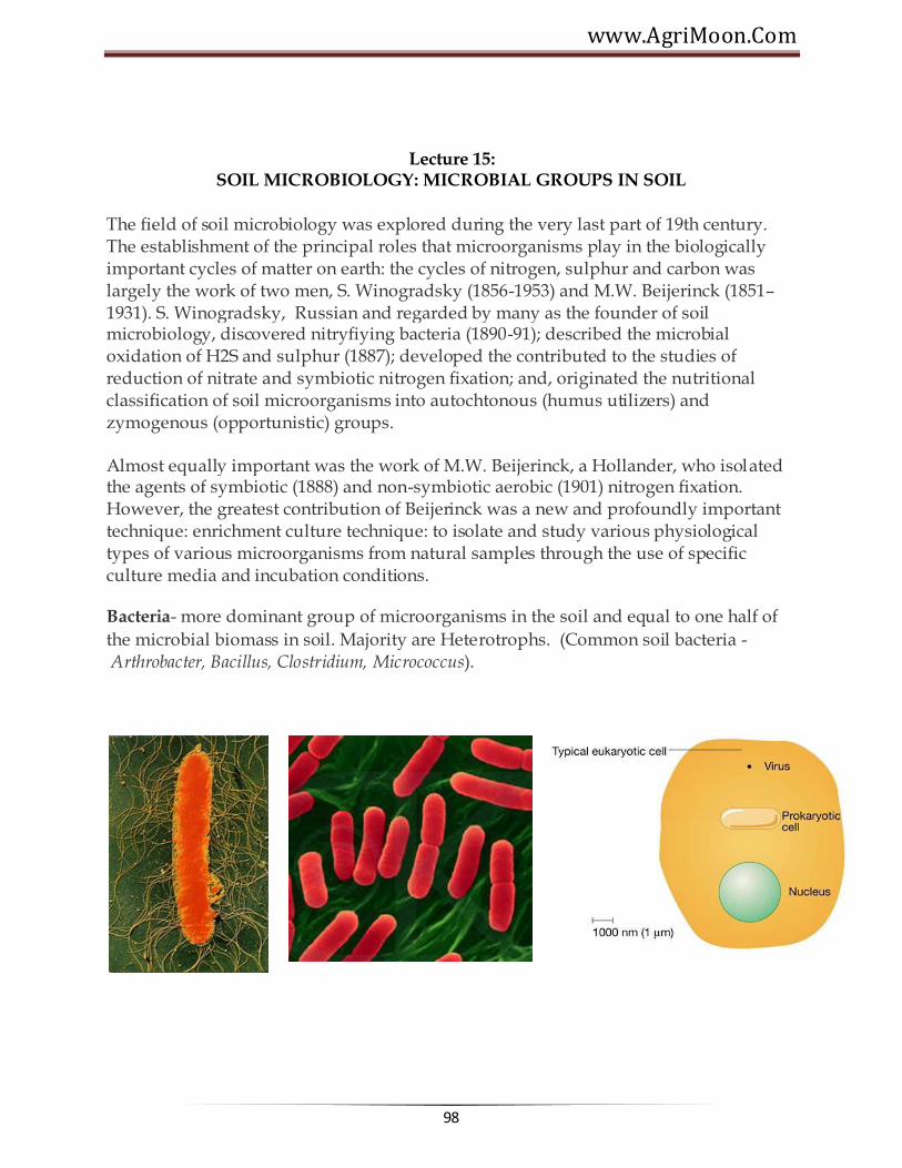
www.AgriMoon.Com
98
Lecture 15: SOIL MICROBIOLOGY: MICROBIAL GROUPS IN SOIL
The field of soil microbiology was explored during the very last part of 19th century. The establishment of the principal roles that microorganisms play in the biologically important cycles of matter on earth: the cycles of nitrogen, sulphur and carbon was largely the work of two men, S. Winogradsky (1856-1953) and M.W. Beijerinck (1851–1931). S. Winogradsky, Russian and regarded by many as the founder of soil microbiology, discovered nitryfiying bacteria (1890-91); described the microbial oxidation of H2S and sulphur (1887); developed the contributed to the studies of reduction of nitrate and symbiotic nitrogen fixation; and, originated the nutritional classification of soil microorganisms into autochtonous (humus utilizers) and zymogenous (opportunistic) groups.
Almost equally important was the work of M.W. Beijerinck, a Hollander, who isolated the agents of symbiotic (1888) and non-symbiotic aerobic (1901) nitrogen fixation. However, the greatest contribution of Beijerinck was a new and profoundly important technique: enrichment culture technique: to isolate and study various physiological types of various microorganisms from natural samples through the use of specific culture media and incubation conditions.
Bacteria- more dominant group of microorganisms in the soil and equal to one half of
the microbial biomass in soil. Majority are Heterotrophs. (Common soil bacteria - Arthrobacter, Bacillus, Clostridium, Micrococcus).

Agricultural Microbiology
99
ROD SHAPED BACTERIA
SPHERICAL BACTERIA
Actinomycetes - intermediate group between bacteria and fungi. Numerous and widely
distributed in soil. Abundance is next to bacteria. 104 - 108/g soil. 70% of soil actinomycetes are Streptomyces. Many of them are known to produce antibiotics. Population increases with depth of soil.
Actinomycetes - intermediate group between bacteria and fungi. Numerous and widely
distributed in soil. Abundance is next to bacteria. 104 - 108/g soil. 70% of soil actinomycetes are Streptomyces. Many of them are known to produce antibiotics.

www.AgriMoon.Com
100
Population increases with depth of soil. Fungi: More numerous in surface layers of well-aerated and cultivated soils-dominant
in acid soils. Common genera in soil are Aspergillus, Mucor, Penicillium Trichoderma, Alternaria, Rhizopus.
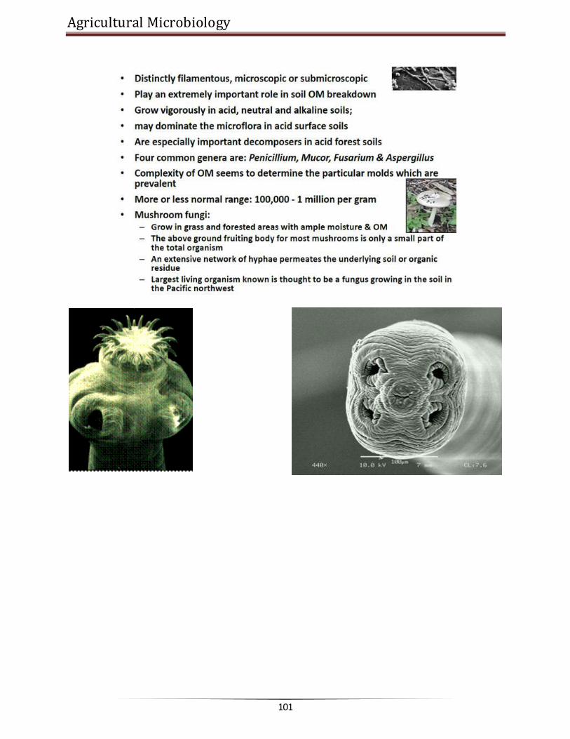
Agricultural Microbiology
101

www.AgriMoon.Com
102
Algae – found in most of the soils in number ranges from 100 to 10,000 per g.
Protozoa: Unicellular – population ranges from 10,000 to
100,000 per g of soil. Most of the soil forms are flagellates, amoebae or ciliates. Derive their nutrition by devouring soil bacteria. Abundant in upper larger of the soil. They are regulating the biological equilibrium in soil.

Agricultural Microbiology
103
Importance
Involved in nutrient transformation process Decomposition of resistant components of plant and animal tissue Role in microbial antagonism Participate in humus formation Predator of nematodes Surface blooming reduces erosion losses Improve soil structure Involved soil structure Maintenance of biological equilibrium
Actors influencing activities of soil microorganisms: Soil microorganisms are
influenced by various factors. Chief factors are fertility level Soil moisture Soil air soil temperature Organic matter H ion concentration Cultural factors.

www.AgriMoon.Com
104
Lecture 16: MICROBIAL TRANSFORMATIONS OF CARBON
The term soil generally refers to the loose material of the earth surface and is the region that supports the plant life. It consists of five major components such as mineral matter, water, air, organic matter and living organisms. The proportion of these components varies with soil type and other soil conditions. To maintain the level of these components it is essential that they undergo a regular process of recycling. This process of recycling through various transformations is brought about by different microorganism.
Carbon cycle (Video)
A- PS B- Respiration / plant C- Respiration / Animals D- Autotrophs E- Respiration / Microbial mineralization
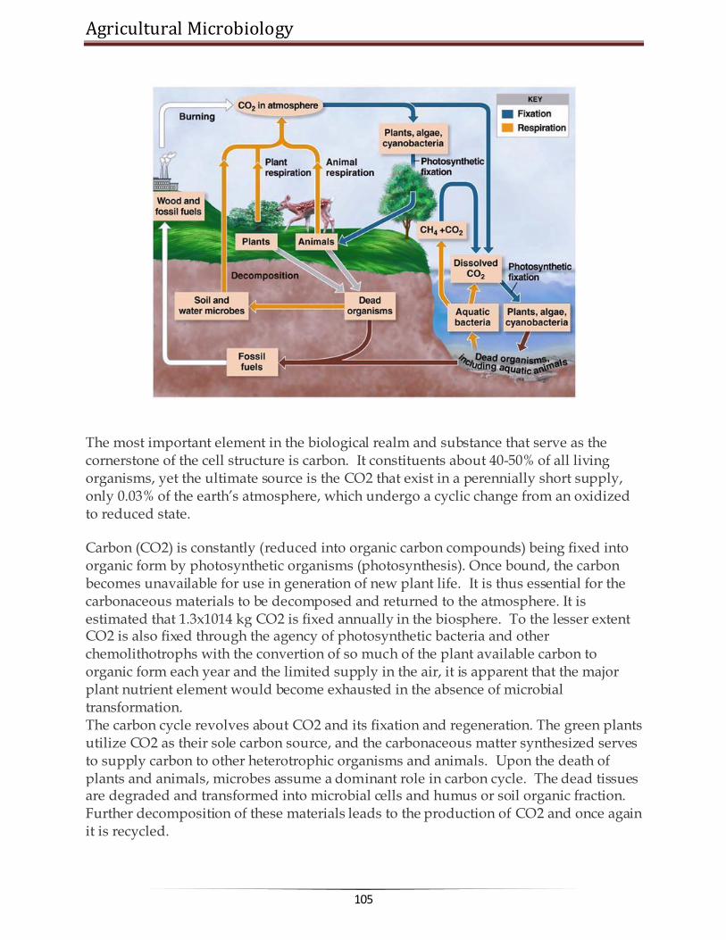
Agricultural Microbiology
105
The most important element in the biological realm and substance that serve as the cornerstone of the cell structure is carbon. It constituents about 40-50% of all living organisms, yet the ultimate source is the CO2 that exist in a perennially short supply, only 0.03% of the earth‘s atmosphere, which undergo a cyclic change from an oxidized to reduced state.
Carbon (CO2) is constantly (reduced into organic carbon compounds) being fixed into organic form by photosynthetic organisms (photosynthesis). Once bound, the carbon becomes unavailable for use in generation of new plant life. It is thus essential for the carbonaceous materials to be decomposed and returned to the atmosphere. It is estimated that 1.3x10¹⁴ kg CO2 is fixed annually in the biosphere. To the lesser extent CO2 is also fixed through the agency of photosynthetic bacteria and other chemolithotrophs with the convertion of so much of the plant available carbon to organic form each year and the limited supply in the air, it is apparent that the major plant nutrient element would become exhausted in the absence of microbial transformation. The carbon cycle revolves about CO2 and its fixation and regeneration. The green plants utilize CO2 as their sole carbon source, and the carbonaceous matter synthesized serves to supply carbon to other heterotrophic organisms and animals. Upon the death of plants and animals, microbes assume a dominant role in carbon cycle. The dead tissues are degraded and transformed into microbial cells and humus or soil organic fraction. Further decomposition of these materials leads to the production of CO2 and once again it is recycled.

www.AgriMoon.Com
106
Organic matter decomposition (Aerobic decay) Soil organic matter The organic matter subjected to microbial decay in soil comes from several sources like plant remains, forest litter, incorporation of plant and animal tissues and excretory products. The chemistry of organic matter is clearly very complex, and investigations of the transformations and the responsible organisms have therefore been extremely interesting. The organic constituents of the plants are commonly divided into six categories.
a) Cellulose - Most abundant 15-60% of the dry weight b) Hemicellulose - 10-30% of the plant dry weight c) Lignin - 5 – 30 % of the plant dry weight d) Water soluble fraction - 5-30%, included simple sugar, a. acids, e) Ether and alcohol soluble constituents, a fraction containing fat, oils, waxes, resins and a number of pigments f) Proteins.
As the plant ages, the content of water soluble constituents, proteins and minerals decreases and the % of abundance of cellulose, hemicellulose and lignin rises. Soil organic matter comprises residues of plant and animals and these compounds occur in soil in close combination with inorganic substances. Animals and plant residues are made up of complex carbohydrates, simple sugars, starch, cellulose, hemicellulose, pectins, gums, mucilage, proteins fats, oils, waxes, resins, alcohols, aldehydes , ketones, organic acids, lignin, phenols, tannins, hydrocarbons, alkaloids, pigments etc.
The soil microorganism play important role in the decomposition of soil organic matter.
Bacteria are the dominant group – mostly heterotrophic organisms (use energy from organic sources such as sugars, starch, cellulose and protein) – are involved. Autotrophic organism which occupy a small portion of the biomass in soil (and use inorganic sources such as Fe and S) are not directly involved in organic matter decomposition.
Actinomycetes grow on complex substances such as keratin, chitin and other complex polysaccharides and play active role in humus formation.
Soil fungi are mostly heterotrophos and use organic residues easily Soil algae contribute a small amount of organic matter through their biomass, but
they do not have any active role in organic matter decomposition.
Organic matter decomposition serves two important functions
a) Provide energy for growth b) Supply carbon for the formation of new cell materials
Agricultural Microbiology
107
Hence only heterotrophs are actively involved in the process of decomposition. The relationship between organic matter and plant growth may be direct or indirect.
Organic matter is a natural substrate for saprophytic micro organism and provides nutrition to plants indirectly through the activity of soil microorganisms
It is essential for the formation of soil aggregates and hence soil structure which ultimately determines the soil aeration and rooting habit of plants
Organic matter helps in the conservation of soil nutrients by preventing erosion and surface run off of nutrients.
Carbon assimilation
The process of converting substrate to protoplasmic carbon is known as assimilation. Under aerobic conditions 20-40% of the substrate carbon is assimilated, the remainder is released as CO2. Fungi are more efficient, in their metabolism, since they convert carbon into cell carbon as filaments and release less of CO2. 30-40% of which is used to form new mycelium during the decomposition. Compared to fungi, bacteira are less efficient. Aerobic bacteria are less efficient than anerobic bacteria.
C. Mineralization
Conversion of organic Carbon substance to inorganic form of carbon.
Immobilization Assimilation of nutrients and is the mechanism by which micro organism reduce the quantity of plant available nutrient in soil. Mineralization is considered well than immobilization. During the decomposition of organic matter three separate simultaneous processes can be distinguished. The important changes during decomposition are:
1. Plant and animal tissues constituents disappear under the influence of enzymes 2. Synthesis of new microbial cells so that proteins, polysaccharides and nucleic
acids typical of bacteria and fungi appear. 3. Third, certain end products of the breakdown are excreted into surroundings
there to accumulate or to be further metabolized.
Importance of organic matter decomposition
1. Important function is the breakdown of organic matter by which CO2 available for photosynthesis is replenished
2. Any compound that is synthesized biologically is subject to destruction by soil inhabitants, otherwise the compounds would have accumulated in vast amounts on the earth‘s surface

www.AgriMoon.Com
108
3. Since, organic matter degradation is a property of all heterotrophs, it is commonly used to indicate the level of microbial activity.
Methods to evaluate the decomposition rate
Measurement of CO2 evolution or O2 uptake Determination of decrease in organic matter either chemically or by weight loss Observations on disappearance of specific constituents such as cellulose,
hemicellulose or lignin.
Changes during organic matter decomposition As a result of development of mixed flora on chemically complex natural products, some components quickly disappear while others are less susceptible to microbial enzymes and persist. The water soluble fraction contains the least resistant plant components and is thus the first to be metabolized. Cellulose and hemicellulose on the other hand disappear not as quickly as water soluble substances, but their persistence usually is not too great. The lignins are highly resistant and consequently become relatively more abundant in the residual, decaying organic matter.
At aerobic conditions when carbonaceous substrates are incorporated into soil, immediate drop in O2 and an increase in CO2 content of soil air occurs.
Change in (O) (H) oxidation reduction potential (En) – it is shifted to a more reduced condition (fall in oxidation reduction potential).
The end products of decomposition are - CO2, H2O, NO3, SO4, CH4, NH4 and H2S
depending on the availability of air. Factors influencing the organic matter decomposition
Organic matter level of the soil Cultivation Temperature Moisture pH Depth Aeration Nature and abundance of micro organic involved. The extent of availability of C, N, P and K presence of inhibitory substance.
C: N ratio
Nitrogen is a key nutrient substance for microbial growth If N content of the substrate is high it is readily utilized and decomposition is
faster If N is poor decomposition is slower, needs additional N
Agricultural Microbiology
109
Protein rich substrates are readily decomposed Low N or wide C:N ratio results slow decay Optimum level of C: ratio for maximum decomposition is 20-25(1.4-1.7% N) Less than this range, more microbial cells, faster mineralization and it likely
exceeds immobilization Wider the ratio, lesser microbial cells, slower the immobilization and
mineralization increases gradually, resulting in accumulation of Ammonia and Nitrates
Microbes scavenge the soil solution to obtain enough N At optimum level, there must be an equilibrium between Mineralization and
Immobilization Soil N level constrains the maintenance of C:N (organic carbon /soil o.m) To make sound soil management Arable surface -10:1
o Sub soil -lower
Anaerobic decay / decomposition
The main products of aerobic carbon mineralization are CO2, water, cells and humus components. In the absence of O2 organic carbon is incompletely metabolized, intermediary substances accumulate, and abundant quantities of CH4 and smaller amounts of H2 are evolved. Energy yield during anaerobic fermentation is low, resulting in fewer microbial cells per unit of organic carbon degraded. Consequently, organic matter breakdown is consistently slower under total anaerobiosis than in environments containing adequate O2. The rate in water logged soils is intermediate between the two extremes. When a soil is water logged or flooded there is a shift from aerobic to anaerobic transformation. Formation and accumulation of organic acids viz., acetic, formic, butyric, lactic and succinic acids appear too, these are frequently detrimental to root development. Organic acids accumulate because of the fermentative character of the microflora of wet soils. The an aerobic carbon transformation are thus characterized by the formation of organic acids, alcohols, CH4 and CO2 as major end products. Under anaerobic conds, decomposition of organic residues takes place by the activity of both mesophilic, thermophilic microorganism resulting in the production of CO2, H, ethyl alcohol, and organic acids. Among mesoophilis, bacteria are more active than fungi or actinomycetes in cellulolytic activity. They belong to genusClostridium which are numerous in manure pits but rarely encountered in cultivated arable soils. In compost pits both meso and thermopholic (bacterial and actinomycetes) are important in the breakdown of cellulose substances. The primary microbial colonisers initially break down the complex CH2O and proteins into organic acids and alcohols. At a later stage, the methane bacteria which are strict anaerobes begin to act upon the secondary substrate chiefly lactic and butyric acids and ferment them into CH4 and CO2.

www.AgriMoon.Com
110
Humus
A dark coloured and fairly stable soil organic matter with known and unknown physical and chemical properties
It is an integral part of the organic matter complex in soil Humus can be defined as lingo protein complex containing approximately
45 % - lignin compounds 35% - amino acids 11% - carbohydrates 4% - cellulose 7% - Hemicellulose 3% - fats, wax, resins 6% - other miscellaneous substances, including plant growth substances and inhibitors.
Age and composition of the humus are dependent on its origin and environment. Bacterial and algal protoplasm contribute a good deal to the nutritive value of
humus Soil micro organism take part in humus formation. Some fungi such
as Penicillium, Aspergillus and actinomycetes produce dark humus like substances which serve as structural units for the synthesis of humic substances.
Benefits of humic substances
Improved seed germination, root growth, uptake of minerals by plants and other physiological effects on plant growth
Increases the enzyme activities involved in plant metabolism. Since humic acid serves as hydrogen acceptors.
Increases the cytochrome oxidase activity in root systems results in growth stimulatory effect (on roots)
Chelating effect – on trace elements Fe uptake by roots Vigour and yield of plants enhanced Humic acid known to influence the grown and proliferation of micro organism Aspergillus niger, Peni, Bacillus sp., Azotobacter are enhanced
The organic fraction of soil, often termed humus. It is a product of synthetic and decomposing activities of the microflora. Since it contains the organic C and N needed for microbial development, it is the dominant food reservoir. Because humus is both a product of microbial metabolism and an important food source, the organic fraction is of special interest.

Agricultural Microbiology
111
Humus formation
Once the plant or animal remains fall on or are incorporated into the soil, they are subjected to decomposition
From the original residues, a variety of products are formed As the original materials and the initial products undergo further decomposition,
they are converted to brown or black organic complexes At this stage any trace of the original remains no longer remains The native organic fraction originates from two sources: the original plant debris
entering the soil and the micro organism with in the soil body. The micro organism in soil body, work upon the former and synthesize microbial protoplasm and new compounds that become part of the organic fraction.
Humus exist in a dynamic state Chemistry of humus is complex It has been pointed out that the organic fraction is derived from
o Plant constituents that are modified by the microflora. o Constituents of microbial cells and products of microbial metabolism are
relatively resistant to decay and therefore persist for sometime after death of organism.
Interms of specific elements The organic fraction contains compounds of C, H, O, N, P, S and small amounts of other elements. Only a small portion of the total is soluble in water, but much can be brought into solution by alkali. Interms of type of compounds Humus contains a number of polymerized substances, aromatic, molecules, polysaccharides, ascorbic acids, polymers of uronic acids and P containing compounds. No definite composition can be assigned. It should be considered as a portion of the soil that is composed of a heterogenous group of substances, most having an unknown parentage and an unknown chemical structure. Lignin and lignin derived molecules have long been considered to be of significance in the formation of humus. It is possible either that simple aromatics released in the microbial attack on lignin polymerize to yield constituents of the soil organic fraction or those partially altered lignins itself give rise to humus constituents. The monomeric portions of humus are similar to the constituents of lignin.
Degradation processes (1) Cellulose is a CH2O composed of glucose units bound by β-linkage at carbon 1 and
4 of the sugar molecule. The cellulose concentration of higher plants is never fixed and the concentration. It is a polymer of glucose and is might abundant organic material in nature changes with age and type of plants. Woody materials have more cellulose and

www.AgriMoon.Com
112
succulent tissues had poor, but the concentration increases as the plant matures. Cellulose breakdown in soil is influenced by several environmental factors. Aerobic organism converts cellulose to 2 major products: CO2 + cell substance, but certain group releases small amount of organic acids. It is however resistant to microbial decomposition. When cellulose is associated with pentosans (xylan, mannans) it undergoes rapid decomposition. When associated with lignin, the decomposition rate is very low. Degradation is by the enzymes that converts cellulose into glucose. (Exoenzymes) Exoglucanase Endoglucanase β - glucosidase (cellulse complex) Exo glucanase
Native cellulose Amorphos cellulose + cellobiose
Endoglucanase Endogluconase β-glucosidase
D- Glucose Cellobiose (cellobiase) Most cellulolytic bacteria do not excrete significant amounts of cellulase but fungi are found to excrete these enzymes. The soluble sugars released by enzymatic hydrolysis are later utilized by the same or other micro organism for biosynthetic purpose. (2) Hemicellulose
It is a polymer of simple sugars such as pentose, hexose and uronic acids. They may be either homo or hetero polymers. When they are added to soil, degradation takes place at faster rate in initial stages. The hemicelluloses such as mannans are decomposed rapidly while galactons (polymer of galactose) are decomposed slowly. Many soil micro organisms utilize hemicellulose in both aerobic as well as anaerobic conditions. The microbial degradation occurs through the agency of extracellular enzymes called hemicellulases. (3) Lignin
Lignin is the third most abundant costituent of plants. It consists of heterocyclic aromatic organic molecules containing C, H and O. The degradation is very slow and rate of decomposition depends on the presence of other compounds such as cellulose and hemicellulose acid. Lignin is highly resistant to microbial degradation. Degradation is a complex process.
Lignin --à coniferyl ether --à coniferyl alcohol --à coniferyl aldehyde --à vanillin --à vannillic acid --à protocatechuic acid --à ring cleavage

Agricultural Microbiology
113
Genera of microorganisms capable of utilizing different components of organic
mater as reported by several workers: F-Fungi; B-Bacteria; A-Actinomycetes
Nature of substrate in organic matter
Genera of microorganisms
Cellulose F Alternaria, Aspergillus, Chaetomium, Coprinus, Fomes, Fusarium, Myrothecium, Penicillum, Polyporus, Rhizoctonia, Rhizopus, Trametes, Trichoderma, Trichothecium, Verticillium, Zygorynchus
B Achrombacter, Angiococcus, Bacillus, Cellfalcicula, Cellulomonas, Cellvibrio, Clostrium, Cytophaga, Polyangium, Pseudomonas, Sorangium, Sporocytophaga, Vibrio
A Micromonospora, Nocardia, Streptomyces, Streptosporangium
Hemicellulose F Alternaria, Fusarium, Trichothecium, Aspergillus, Rhizopous, Zygorynchus, Chateomium, Helminthosporium, Penicillium, Coriolus, Fomes, Polyporus
B Bacillus, Achromobacter, Pseudomonas, Cytophaga, Sporocytophaga, Lactobacillus, Vibrio
A Streptomyces
Lignin F Clavaria, Clitocybe, Collybia, Flammula Hypholoma, Lepiota, Mycena, Pholiota, Arthrobotrys, Cephalosporium, Humicola
B Pseudomonas, Flavobacterium
******☺******

www.AgriMoon.Com
114
Lecture 17:
MICROBIAL TRANSFORMATIONS OF NITROGEN, PHOSPHORUS AND SULPHUR
MICROBIAL TRANSFORMATIONS OF NITROGEN Biological availability of N, P and K is of considerable economic importance, since they are the major plant nutrients derived from the soil. Of the three, N stands out as the most susceptible one to microbial transformations. This element is the key building block of the protein molecule upon which all life is based on, it is an indispensable component of the protoplasm of plants, animals and micro organism. Molecular N2 constitutes about 78% of the earth‘s atmosphere but it is chemically inert and cannot be utilized by more living organism, plant animals and micro organism therefore depend on a source of combined N such as ammonia, nitrate or organic N compounds for their growth. Nitrogen undergoes a number of transformations that involve organic, inorganic and volatile forms of nitrogen. A small part of the large reservoir of N2 in the atmosphere is converted to organic compounds by certain free living micro organism or by plant microbe association that makes the element available to plant growth. The nitrogen present in the proteins or nucleic acids of plant tissue is used by animals. In the animal body, the N is converted to other simple and complex compounds. Upon the death, plants and animals undergo microbial decay and organic N is released as ammonium, which is then utilized by vegetation or is oxidized to nitrate by microorganisms. The nitrate from of N is mostly used by the plants or may be lost by bacteria reduced to gaseous N2, which escapes to atmosphere, there by completing the cycle. The Nitrogen cycle mainly includes transformations such as
1. Nitrogen mineralization : In which N containing organic complexes are decomposed and converted into inorganic compounds for use by plants
2. N immobilization : In which N containing inorganic compounds are assimilated
N2 is acted on by certain micro organism sometimes in symbiosis with a higher plant, which can use it is as a N source for growth. The process of nitrogen fixation, results in the accumulation of new organic compounds in the cells of responsible micro organisms. The N2 thus fixed reenters general circulation when the newly formed cells are inturn mineralized. By means of these reactions the subterranean microflora regulates the supply and governs the availability and chemical nature of N in soil.

Agricultural Microbiology
115
NitrogenCycle
A - Ammonium E - Immobilization
B - Mineralization F - Denitrification
C - Nitrification G - N2 Fixation (Non-symbiotic)
D - Nitrate reduction H - N2 fixation (Symbiotic )
I.
Nitrogen
mineralizatio
n The conver
sion of organic N to the more mobile, inorganic state is known as nitrogen mineralization. As a consequence of mineralization, ammonium and nitrate are generated and organic N disappears. This takes place in two distinct microbiological steps.
www.AgriMoon.Com
116
1. Ammonification
It is the process of mineralization in which proteins, nucleic acids and other organic components are degraded by micro organism with the eventual liberation of ammonia. This is called ammonification. A part of the liberated ammonia is assimilated by the micro organism themselves. The first step in ammonication process is the hydrolysis of proteins, nucleic acids and other organic nitrogenous compounds into amino acids (proteolysis). The amino compounds are then deaminated to yield ammonia. Ammonification usually occurs under aerobic conditions while under anerobic conditions protein decomposition leads to conversion of ammonia into amines and related compounds (eg) clostridium. The anaerobic decomposition of protein called as putrefaction. These amines are subsequently oxidized in the presence of O2 to release ammonia. Break down of nitrogenous substance is brought about by the activity of a multitude of microbial species. Almost all bacteria, actinomycetes and fungi can bring about proteolysis and the amino acids produced are utilized for the growth of these organisms.
(2) Nitrification
The biological oxidation of ammonium salts (in soil) to nitrites and the subsequent oxidation of nitrites to nitrates is called as nitrification. i.e. the biological convention of N in soil from a reduced to a more oxidized state, called nitrification. Nitrification occurs in two steps; First ammonia is oxidized to nitrite. 2 NH3 + 1½ H2O2 → NO2- + 2H+H2O-Nitrosofication This change is brought about by chemoautotrophic bacteria of the genera Nitrosomonas, Nitrosolobus, Nitrosococus, Nitrosospira. These bacteria obtain their energy requirement by the oxidation of NH4+ to NO-2. Among the nitrifiers Nitrosomonas are most important in soils. Some heteotrophs involved Streptomyces, Nocardia Second step Nitrite is further oxidized to nitrate HNO2 + ½ O2 → HNO3. Organisms: Nitrobacter, Aspergillus, Penicillium, Cephalosporium. Factors influencing the growth of nitrifying bacteria in soil
Levels of ammonia and nitrite, aeration, moisture, temperature, pH and organic matter. In acid soils – nitrification is poor. Waterlogged soils – deficient in O2 – not congenial for nitrification.
3. Denitrification The convention of nitrate and nitrite into molecular N2 or nitrous oxide through microbial processes is known as denitrification. Certain bacteria are capable of using nitrate as the terminal electron acceptor under anaerobic conditions. This is

Agricultural Microbiology
117
called nitrate respiration. As a consequence of nitrate respiration, NO3 is reduced to N2
gas or nitrous oxide. Denitirifcation leads to the loss of N from the soil. It depletes N, and therefore it is not a desirable reaction. The escape of molecular N into the atmosphere is also known as volatalization.
Denitirfication occur mostly in waterlogged anaerobic soils with a high organic matter contents. Denitrification of bound nitrogen to gaseous N is mediated by numerous species of bacteria, which normally use O2 as hydrogen acceptor (aerobically) and, also use nitrates and nitrites (anerobically). Anaerbic convertion of nitrate into molecular nitrogen is known as nitrate respiration. Bacterial genera which bring about denitirfication Pseudomonas, Achromobacter, Bacillus, Micrococcus 2NO-3 +10 H → N2 + 4H2O+ 2OH- (or) 2NO-2 +6 H → N2 +2H2O +2OH- (or) N2O + 2H → N2 + H2O Since nitrates are used as a source of electron acceptor, there is a net loss of N from soil. This process is termed also as dissimilatory nitrate reduction. Many soil bacteria like. Thiobacillus denitrificans
Oxidize S (chemoautotrophically) and also reduce nitrate to nitrogen 5S + 6 KNO3 + 2 H2O → 3N2 + K2SO4 + 4KHSO4 (or) 5 K2S2O3 + 8 KNO3 + H2O → 4N2 + 9 K2SO4 + H2SO4 General pathway of denitrification
Nitrate is first reduced to nitrite, which is then transformed to nitrous oxide (NO). The nitrous oxide is converted to N2 with N2O as an intermediate. 1 2 3 4 2 HNO3 → 2HNO2 → 2 NO → N2O → N2
The enzymes involved 1. Nitrate reductase 3. Nitric oxide reductase 2. Nitrite reductase 4. Nitrous oxide reductase
Fallow soils flooded with water are more congenial for denitrification than well drained and continuously cropped soils.
Though it is a undesirable reaction in point of view of plant nutrition, but have ecological importance. Because with out denitrification the supply of N on the earth world have got depleted and NO3 would have accumulated.
High concentration of NO3 are toxic, denitrification is a mechanism by which some of the N is released back to the atmosphere.
5. Nitrate reduction The reverse of nitrification process. That is the reduction of nitrate to nitrite and then ammonia. Since organisms are able to obtain cellular Nth ammonia assimilation, the process is called as assimilatory nitrate reduction. HNO3 + 4H2 → NH3 + 3H2O

www.AgriMoon.Com
118
II. Nitrogen immobilization The process of microbial assimilation of inorganic nitrogen is referred as immobilization. In contrast to mineralization, microbial immobilization leads to the biosynthesis of the complex molecules of microbial protoplasm from ammonium and nitrate. Immobilization results in a marked depression of nitrogen uptake by the plant. The mineralization of organic N and the microbial assimilation of inorganic ions proceeds simultaneously.Both mineralization and immobilization take place regardless of the % of N in the organic N in organic matter. On the death of micro organism, the immobilized N is however released through mineralization. It is also a loss of nitrogen. NO3 when accumulated in microbial protoplasm it is referred as assimilatory NO3 reduction. MICROBIAL TRANSFORMATION OF PHOSPHORUS AND SULPHUR I. Phosphorus cycle (Video) Phosphorus is only second to N2 as an inorganic nutrient required by both plants and micro organisms. Phosphate constitutes nearly 0.1% of the earth‘s crust. They occur in soil in inorganic and organic forms. The inorganic forms are derived from parent rocks or through fertilizers application and manuring with bone meal. They are soluble in water when present as phosphates of Na, K, Ca, Mg etc. The organic phosphorus containing compounds are derived from plants and micro organisms and are composed of nucleic acids, phospholipids, lecittin, phytin and related compounds.
Phosphorus in phytin, phospholipids and nucleic acids is found as phosphates Phytin is the calcium – magnesium salt of phytic acid Phospholipids are compounds in which phosphate is combined with a lipid,
contained 10% of cell phosphorus. Inorganic polyphosphates are quite abundant in certain fungi In soil, from15-85% of the total P is organic. Soils rich in organic matter contain
abundant organic P. Ratios of organic C to P of 100 to 300:1 N: organic P = 5 to 20: 1
In cultivated soil P present in abundant about 1100 kg/ha but most of them as not available to plants; only about 1% of the total P is in available form. PO43- in rocks and in cells
Acid from Thiobacillus
Microorganisms bring about a number of transformations of the element.

Agricultural Microbiology
119
1. Altering the solubility of inorganic compounds of P 2. Mineralization of organic compounds with the release of inorganic phosphate 3. Converting the inorganic, available anion into cell components, an
immobilization process (analogous to that occurring with N) 4. Bringing about an oxidation or reduction of inorganic P compounds
Particularly, important to P cycle are the microbial mineralization and immobilization reactions. (1) Solubilization of inorganic phosphorus Insoluble inorganic compounds of P are largely unavailable to plants, but many micro organisms can bring the PO4 into solution. P solubilizing are 105 to 107 / g soil. Eg: Pseudomonas striata, Microoccus Bacillus sp., Fusarium, pergillus sp, Solubilises calcium salts, iron, aluminum, magnesium manganese phosphate.
P is solubilized by the production of organic acids. The acids convert Ca3 (PO4)2 to di and monobasic phosphates and releases P to plants.
Solubilization of phosphates by plant roots & micro organism is dependent on soil pH. In neutrals and alkaline soils having a content of calcium, precipitation of CaPO4 takes place. Micro organism and plant root readily dissolve such PO4 and make them available to plants.
On contrary, acid soils are generally poor in Ca ions and phosphates and precipitated in the form of ferric or aluminum compounds which are not soluble. There, it is solubilized by the addition of PO4 solublizing micro organism.
Phosphorus exists mainly as apatides, with the basic formula M10 (PO4)6 X2. Commonly the mineral (M) is Ca, less often Al or Fe. The anion (X) is either F- or Cl- or OH- or CO2-3. Diverse combinations of M and X results in 200 forms of P.
(2) Mineralization of organic phosphorus Organic form of P is the larger reservoir of P in soil. By the action of bacteria, fungi and actinomycetes, bound element in remains of the vegetation and in soil organic matter is made available to succeeding generations of plants. Among the organic phosphours compounds, lecithin, nucleic acids and phytin occupy a prominent place. Lecithin contains 9.39 % P2O5, 1.6% N and 65.36% C. It is a process of convention of organic forms of phosphorus into inorganic available forms of P a highly significant correlation is observed between the rates of N and P convention to inorganic forms.
Mineralization is favoured by warm temperature, with the thermophilic range being more favourable than mesophilic range.
Neutral pH increases PO4 release, which favours microbial metabolism Quantity of substrate ie presence of organic P. If more P, more of mineralization

www.AgriMoon.Com
120
Mineralization is mediated by the enzymes called phosphatases. These enzymes cleave phosphorus from more frequently encountered organic substrates.
Phytases liberates PO4 from phytic acid or its Ca-Mg, Salt, Phytin. They remove PO4-s, one at a time, yield penta – tetra, di- and mono PO4 and then finally free inositol.
Bacillus, Pseudomonas, Aspergillus, Penicillium, Rhizopus can synthesize this enzyme. Mycorrhizal (fungi) are also able to mineralize the organic forms of P and increases P uptake by the plants.
(3) Immobilization Process of assimilation of P into microbial nucleic acids, phospholipids or other protoplasmic substances is called immobilization. It leads to the accumulation of non utilizable forms of the element. P accounts for 0.5-1.0% of fungus mycelium and 1.0 to 3.0% of the dry weight of the bacteria and actinomycetes.
(4) Oxidation reduction reactions
Biological oxidation of reduced phosphorus compounds into oxidized state. Phosphite (HPO3=) is oxidized to phosphate. A number of hetertrophic (bacteria), (fungi) & (actinomycetes) utilize phosphite as sole P source. Hypophosphites (HPO2=) can also be oxidized to phosphate by heterotrophs. HPO3= → HPO4= HPO2= → HPO4=
Reductive process, reductive pathway has also been functioned. PO4 is reduced to
phosphite and hypophosphite. H3PO4 → H3PO3 → H3PO2 Clostridium butyricum, E. coli form phosphite and hypophosphite from orthophosphate. It is biochemically analogue to the process of denitirification. Only little information is available about this process. P exist in an organic form in the protoplasm on the death of living organism, this (P) is changed to inorganic phosphoric acid. This is soon converted into insoluble salts of Ca, Fe, Mg and Al. Phosphorus thus alternates between organic and inorganic, and soluble and insoluble forms. In soluble P is solubilized by various acids produced by micro organism. Microbial activities involved in the cycling of C, N and P are absolutely essential for maintenance of soil fertility. II. Sulphur cycle
Sulphur like N, is an essential element for all living systems because of it‘s inert nature, is not utilized by plants. To be used first S has to to be oxidized or reduced. In soil, it occurs both organic (S containing amino acids, vitamins ) as well as inorganic forms (Sulphur, sulphates etc., ) . Four distinct transformations are recognized

Agricultural Microbiology
121
1. Decomposition/Mineralization of larger organic S compounds to smaller units
and their conversion into inorganic compounds 2. Microbial associated immobilization 3. Oxidation of inorganic ions and compounds such as sulphides,thiosulphates,
Sulphu 4. Reduction of Sulphates abd other sulphides
Mineralization Conversion of organic bound S into inorganic state, mediated through M.O. The released S in either absorbed by plants or escaped into atmosphere in the form of oxides Oxidation
Occurs both in aerobic and anaerobic condition Bacteria Nonfilamentous forms- Thiobacillus Filamentous forms – Beggiatoa,Thiothrix and Thioloca Fungi and actinomycets Aspergillus, Penecillium and Microsporium

www.AgriMoon.Com
122
Importance of Thiobacillus
Produces Sulphuric acid ,lower down the soil pH – Hence used in controlling plant disease
Apple and Potato scab –Streptomyces scabis ,Sweet potato rot – S. ipomea S+ Thiobacillus application is used for the control Remediation of alkali soil Increases the solubilization of other nutrients (P,K,Ca,Mn,Al and Mg ) Preparation of biosuper- Rock phosphate + T.thiooxidans and S--- Australia Lipman‘s process- Compost preparation Soil + manure + elemental S + rock phosphate
Sulphate reduction Reduces inorganic sulphate into Hydrogen sulphide –reduces the availability of S
for plant nutrition
Desulphovibrio desulphricans -anaerobe
******☺******

Agricultural Microbiology
123
Lecture 18: BIOLOGICAL NITROGEN FIXATION
Fixation of elemental nitrogen in the atmosphere by the micro organism through a reductive process into ammonia is called as BNF. A variety of prokaryotic organism have the ability to reduce the atmosphere N2 BNF accounts for about 70% of the total N fixed in the biosphere. The ability to reduce atmosphere N is restricted only to bacteria, which are belonging to the diverse groups. The root nodule associations were the first to be recognized for their ability to fix atmosphere N2. Rhizobia are the first group of organism realized for its potential of nitrogen fixation.
Nitrogen fixing bacteria Nitrogen fixing bacteria are classified according to their mode of fixation.
1. Free living N fixers – capable of fixing mol. N2 to cellular N independently of other living organism.
2. Associative N fixers 3. Endophytic N fixers 4. Symbiotic N fixers
Rhizobium is predominant symbiotic N2 fixing bacterium. Boussingault showed that leguminous plant can fix atmosphere N2. Then hellriegel and wilfarth – proved that N2 is fixed by certain bacteria living in root nodules of leguminous plants. Latter isolated in pure culture by Beijerinck. Winogradsky isolated clostridium pasteurianum. Which is an anaerobic N2 fixer. Beijerinck isolated Azotobacter as free living aerobic N2 fixing organisms.
Cross inoculation groups of rhizobium (CIG) It (CIG) refers the groups of leguminous plants that will develop effective nodules
when inoculated with the rhizobia obtained from the nodules from any member of that legume group.

www.AgriMoon.Com
124
I. Rhizobium
1.
Rhizobium leguminosarum
CIG Host it can nodulate
bv. viceae Pea Peas,lenfils, vicia
bv. phaseoli Bean Phaseolus spp
bv. trifoli Clover Trifolium spp
2. R. meliloti Alfalfa Alfalfa, clover, fenugreek
3. R. loti Lotus Trifoli, lupine,
4. R. fredii Soybean Soybean
5. R. spp Cowpea group Vigna, Arachis, Cajanus, Dolichus, Sesbania, Acacia, Prosopis, green gram and blackgram
6. R. sp Chickpea group Chickpea
II. Bradyrhizobium
B. japonicum Soybean B. spp Cowpea group
III. Azorhizobium - Stem nodulating – one.
Nodulates Sesbania rostrata. IV. Photorhizobia - Nodulants aeschynomene sp.
V. Sinorhizobium - fast growing soybean nodulator I. Biological nitrogen fixation Free living nitrogen fixers
Azotobacter - Aerobic Beijerinckia Clostridium – Anaerobic Cyanobacteria (Blue green algae) etc.,
II. Associative symbiotic nitrogen fixer
Azospirillum Herbaspirillum III. Endophytic nitrogen fixer
Gluconacetobacter diazotrophicus IV. Symbiotic nitrogen fixers
Rhizobium (Rhizobium – legume association) Bradyrhizobium (Bradyrhizobium – soybean association)

Agricultural Microbiology
125
Azorhizobium (Azorhizobium- Sesbania rostrata association) Anabaena azollae (Azolla – Anabaena association) Frankia (Frankia – Casuarina association)
Species of Azospirillum
lipoferum brasilense amazonense halopareferans irkense A.largomobilis
Species of Azotobacter
chroococcum vinelandii beijerinkii paspali agilis insignis macrocytogens
Important genera of blue green algae
Anabaena, Nostoc, Cylindrospermum, Rivularia, Oscillatoria, Plectonema, Aphanothece, Lyngbya, Scytonema, Calotrhix etc., Species of Azolla
pinnata filiculoides microphylla caroliniana mexicana nilotica
Nitrogen fixation Process of N2 fixation
The process of N2 fixation is mediated by the enzyme, called nitrogenase (which mediates the reduction of N2 to ammonia) first, this enzyme was extracted from the anaerobic di nitrogen fixer Clostridum pasteurianum. Latter, this has been isolated form most other N2 fixing bacteria. The mechanism of N2 fixation appears to be quite similar in most N2 fixing prokaryotes. The enzyme has been fairly well characterized and the enzymes from

www.AgriMoon.Com
126
these different systems share common properties allowing a unified single doscription of nitrogenase.
Nitrogenase
Nitrogeanse is a functional enzyme which reduces N2 to ammonia and depends on energy source from ATP. The nitrogenase has two components one containing Mo-Fe, designated as Mo – Fe protein and the other Fe protein . Two components are necessary for the nitrogenase activity.
Mo-Fe protein
Consists of 4 subunits and having the molecular not of 22,0000 or 270,000 daltons and it is the big component.
Fe-protein
Smaller component, contains 2 subunits, molecular weight 60,000 daltons. Ammonia is the end product of N2 fixation. The over all reaction is as follows. ATP
N2 + 3H2 2 NH3
General pathway of N2 fixation This process requires a source of ATP and reductants, which are provided by photosynthesis. 16 molecules of ATP are required to fix a molecule of N2.
ATP generation ATP
ADP
N=N
Energy source Electron
carriers
Nitrogenase complex
NH3
Nitrogenase can also reduce C2 H2 → C2H4 Hydrolysis of ATP into ADP with electron transfer from a reduced electron donor (Ferridoxin, Flavodoxin) is coupled to reduce N2 to 2NH3. The ammonia is the first stable product of fixation and it is assimilated by GS-GOGAT pathway.

Agricultural Microbiology
127
NADH
NADPH e- e- e- e-
Ferridoxin
Flarodoxin
Ubiquinone
APP
EMP e- Carrier proteins NII NI
EDP
Glycolysis
Nitrogenase is O2 labile various protection mechanism are operating in different N2 fixing systems.
Mechanism Reduction takes place on the surface of the enzyme
Six electrons are required to reduce one mole of N to two moles of ammonia.
N2 + 8H+ +8e- +16 ATP -------2 NH3+16 ADP+ 2H+ +16pi

www.AgriMoon.Com
128
It is postulated that, atoms of N2 are separated thr‘h charge in the valency of metal ion (mo) bound to the enzyme involved in reduction of N2. For every electron transfer, 4 ATP mole are required.
Hydrogenase -Uptake hydrogenase (HUP+) converts the release d hydrogen during
N2 fixation, and cycled back the Hydrogen for energy generation.by this they

Agricultural Microbiology
129
contribute 9-10 %ATP requirement for N2 fixation process. Formation of a Root Nodule
Factors affecting N2 fixation
1. Presence of nitrate or ammonium : More N2, No, N2 fixation 2. Presence of certain inorganic substances
Ca, Co, Mo – influence N2 fixation along with P
3. Availability of energy source – addn. of C source increase N2 fixation 4. pH : Neutral – favours Azotobacter – Acidic-Beijerinkia 5. Soil moisture : Adequate is good for fixation 6. Temperature: Mesophilic – 30°C.
The energy requirement for BNF is very high and it is a major factor determines the amount of N2 fixed. In, Azotobacter the rate depends on amount of available carbon. In symbiotic N2 fixers since photosynthesis is the ultimate source of energy the rate of N2 fixation is influenced by the factors that effect photosynthesis and rate of translocating photosynthates to the N2 fixing system. Nitrogenase protection mechansims
1. Leghaemoglobin scavenges O2 to protect nitrogenase in legume rhizobium symbiosis
2. Confirmatory protection in Azotobacter as well as the higher respiratory rate. 3. Thick walls of Heterocyst protect O2 in BGA, since Nitrogenase are present in the
heterocyst. 4. Microaerophilic nature in Azospirillum
Losses of N by non biological ways
Leaching
20 to 50%of fertilizer N. The most striking loss of N in rice soils where more than half of the fertilizer N applied get lost through leaching.
Volatalization
Another factor is the volatalizaiotn of ammonia in soil 5-20%. Fixation of ammonium in soils is the minor contributory factor to overall loss of N2 available for plant growth. Such losses of N by physical causes and by nitrification and denitirfication process can be controlled by the application of certain chemicals. Some chemicals have been designed to control the rate of release of nutrient from nitrogenous fertilizers, while others retard nitrification in soil by controlling the activity of nitrifying bacteria.

www.AgriMoon.Com
130
a. Controlled release fertilizers
Urea from... isobutyeldene diurea Crotonilidene diurea S coated urea
Fertilizers, sparingly soluble in water can regulate the release of N from fertilizers
b. Nitrification inhibitors These are substituted with pyridines, pyrimidines, anilines and isothiocyanates, Examples
1. 2 chloro 6 (tricholormethyl) – pyridine – (N serve ) 2. 2 amino 4 chloro 6 methyl pyridine –( AM.) N serve inhibits the growth of Nitrosomonas europea and N. agilis. The seeds of neem conain lipid associates act as nitrification inhibitors and there by increases the efficiency of urea fertilizers.
Ammonia assimilation N2 fixation results in NH4 formation which reacts with organic acids and form amino acids which is mediated by ammonia assimilating enzyme. GS – Glutamine synthetase GOGAT – Glutamate synthese GDH – Glutamate dehydrogenase
Genetics
Nif genes are responsible for N2 fixation. Nif genes are 22, which are located in 7 or 8 clusters.
GENETICS OF NODULATION AND N FIXATION Root nodule bacteria and symbiosis with legumes One of the most interesting and important plant bacterial interactions is that between leguminous plants and certain gram negative nitrogen fixing bacteria. Rhizobium, Bradyrhizobium, Sinorhizobium, Mesorhizobium and Azorhizobium are gram negative motile rods. Infection of the roots of a leguminous plant with the appropriate species of one of these genera leads to the formation of root nodules that are able to convert gaseous nitrogen to combined nitrogen, a process called nitrogen fixation. Nitrogen fixation by legume Rhizobium, symbiosis is of considerable agricultural importance, as it leads to very significant increases in combined nitrogen in the soil. Because nitrogen deficiencies often occur in unfertilized bare soil, modulated legumes are at a selective advantage under such conditions and can grow well in areas where other plants cannot.

Agricultural Microbiology
131
CIG refers, the groups of leguminous plants that will develop effective nodules when inoculated with the rhizobia obtained from the nodules from any member of that legume group.
Stages in root nodule formation
The stages in the infection and development of root nodules are not fairly well understood. They include
1. Recognition of the correct parameter on the part of both plant and bacterium and attachment of the bacterium to the root hairs.
2. Excretion of nod factors by the bacterium. 3. Invasion of the root hairs by the bacterial formation of an infection thread.
4. Travel to main root via the infection thread. 5. Formation of deformed bacterial cells, bacteroids, within the plant cells and
development of the nitrogen fixing state. 6. Continued plant and bacterial division and formation of the mature root nodule.
Nodulation events
1. Normal root hair 2. Exudation of organic substances 3. Accumulation of rhizobia in the rhizosphere 4. Orientation and binding of rhizobia 5. IAA production 6. Root hair curling and deformation 7. Formation of infection thread by rhizobia 8. Formation of sheperd‘s crook cells and entry of infection thread

www.AgriMoon.Com
132
9. Thread containing bacteroids extending into root hair cells 10. Entry of infection thread into cortex and branching 11. Nodule development
Attachment and infection
The roots of leguminous plants secrets a variety of organic compounds that stimulate the growth of a rhizosphere micro flora. This stimulation is not restricted to the rhizobia but occurs with a variety of rhizosphere bacteria. If there are rhizobia in the soil, they grow in the rhizosphere and build up to high population densities. Attachment of bacterium to plant in the legume Rhizobium symbiosis is the first step in the formation of nodules. A specific adhesion protein called rhicadhesin is present on the surface of
all species of Rhzobium and Bradyrhizobium. Rhicadhesin is a calcium-binding protein and may function by binding calcium complexes on the root hair surface. Other substances, such as carbohydrate-containing protein called lectins, also play in plant
bacterium attachment. Initial penetration of Rhizobium cells into the root hair is via the root hair tip. following binding, the root hair curls as a result of the action of substances excreted by the bacterium called nod factor and the bacteria enter the root hair and induce formation by the plant of a cellulosic tube, called infection thread, which spreads done the root hair. Root cells adjacent to the root hairs subsequently become infected by rhizobia and nod factors stimulate plant cell division, eventually leading to formation of the nodule.
Bacterioids Bacteriods are specifically referred to a swallon deformed Rhizobium cellfound in the root nodule,capable of nitrogen fixation The Rhizobium bacteria multiply rapidly with in the plant cells and are
transformed into swollen, misshapen and branched forms called bacteroids. When the plant dies, the nodules can be deteriorates, releasing bacteria into the soil. The bacterioid forms are incapable of division, but there are always a small number of dormant rod shaped cells present in the nodule. These now proliferate; using some of the products of the deteriorating nodule as nutrients, and the bacteria can initiate the infection in other roots or maintain a free living existence in the soil.
Lectins Plant proteins which specifically bind to carbohydrate receptors (polysaccharides) in the rhizobial cell
Genetics of nodule formation Genes directing specific steps in nodulation of a legume by a strain
of Rhizobium are called nod genes. Many nod genes from different Rhizobium species are highly conserved and are borne on large plasmids called sym plasmids. In addition to nod genes which direct specific nodulation events, sym plasmids contain specificity gene, which restrict a strain Rhizobium to a particular host plant. Indeed cross

Agricultural Microbiology
133
inoculation group specificity can be transferred across species of rhizobia by simply transferring the respective sym plasmid.
In the sym plasmid of Rhizobium leguminosarum bio var viciae, nod genes are located between two clusters of genes for nitrogen fixation the nif genes. Ten nodgenes have been identified in this species. The nod ABC genes are involved in the production of oligosaccharides called nod factors, which induce root hair curling and trigger plant cell division, eventually leading to formation of the nodule. In Rhizobium leguminosarum bio var viciae, the gene nodD encodes a regulatory protein; this controls transcription of other nod genes.
Nod D genes
1. Genes directing specific steps in nodulation of a legume by a strain of rhizobium are called Nod genes
2. Nod genes are born on large plasmids ,called sym plasmids 3. Nod genes are located between two clusters of genes for N2 fixation called Nif
genes 4. Nod gene consists of 8 genes 5. nod A,B,C,D,E,F,L,M 6. nod D controls the function of all nod genes
Nif Genes
1. Genes responsible for N fixation are called Nif genes 2. 22 genes are involved, arranged in 7 /8 clusters 3. Nif Q,B,A,L,F,M,Z,W,V,S,U,X,N,E,Y,T,K,D,H,J 4. KDH – control Nitrogenase enzyme complex
Factors affecting nodulation
Temperature and light Combined Nitrogen Hydrogen iron concentration Mineral nutrition-Co,Mo,P,Ca Genetic factors Ecological factors Salinity and alkalinity
******☺******
www.AgriMoon.Com
134
Lecture 19: PHYLLOSPHERE BACTERIA
Aerial plant surfaces represent the largest biological interface on Earth and provide essential services as sites of carbon dioxide fixation, molecular oxygen release, and primary biomass production. Rather than existing as axenic organisms, plants are colonized by microorganisms that affect both their health and growth.
For terrestrial plants, the phyllosphere represents the interface between the above-ground parts of plants and the air. Conservative estimates indicate that the roughly 1 billion square kilometers of worldwide leaf surfaces host more than 1026 bacteria, which are the most abundant colonizers of this habitat . The overall microbiota in this ecosystem is thus sufficiently large to have an impact on the global carbon and nitrogen cycles. Additionally, the phyllosphere inhabitants influence their hosts at the level of the individual plants. To a large extent, interest in phyllosphere microbiology has been driven by investigations on plant pathogens. Their spread, colonization, survival, and pathogenicity mechanisms have been the subject of numerous studies. Much less understood are nonpathogenic microorganisms that inhabit the phyllosphere. The composition of the phyllosphere microbiota has been analyzed in only a few studies by cultivation-independent methods; however, such methods are essential in light of the yet uncultivated majority of bacteria existing in nature, or more specifically on plant leaves. Not only their identity, but in particular the physiological properties of phyllosphere bacteria, their adaptations to the habitat, and their potential role (e.g., with respect to modulating population sizes of pathogens) remain largely unknown. Current knowledge on the traits important in the phyllosphere is derived from relatively few studies on gene expression and stems mostly from model bacteria cultivated on host plants under controlled conditions. However, under natural conditions, plants and their residing microorganisms are exposed to a host of diverse, highly variable environmental factors, including UV light, temperature, and water availability; moreover, individual microbes are subjected to competition with other microorganisms over resources, such as nutrients and space.
Toward a deeper understanding of phyllosphere microbiology, and in particular to learn more about the commensal majority of plant leaf colonizing bacteria, which may be of relevance for plant health and development, integrated approaches are needed. Here,

Agricultural Microbiology
135
Bacterial communities in the phyllosphere are thought to be l imited by carbon availability, and it may be expected that access to carbon compounds on leaves is a major determinant of epiphytic colonization. There is evidence that small amounts of nutrients, such as simple sugars including glucose, fructose, and sucrose, leach from the interior of the plant.
The above-ground parts of plants are normally colonized by a variety of bacteria, yeasts, and fungi. While a few microbial species can be isolated from within plant tissues, many more are recovered from the surfaces of healthy plants. The aerial habitat colonized by these microbes is termed the phyllosphere, and the inhabitants are called epiphytes. While there has been some investigation of the colonists of buds and flowers, most work on phyllosphere microbiology has focused on leaves, a more dominant aerial plant structure. Bacteria are by far the most numerous colonists of leaves, often being found in numbers averaging 106 to 107 cells/cm2 (up to 108 cells/g) of leaf. Because of their numerical dominance on leaves, and because more information is available on the process of bacterial colonization of leaves, we focus on this group of microbes in this review.
Compared to most other bacterial habitats, there has been relatively little examination of phyllosphere microbiology. This is somewhat surprising given the abundance of plants in the world and the roles of various phyllosphere bacteria in the important processes discussed below. Leaves constitute a very large microbial habitat. It is estimated that the terrestrial leaf surface area that might be colonized by microbes is about 6.4 x 10⁸ km2. Given the large number of bacteria on leaves in temperate regions of the world and that populations in tropical regions are probably even larger, the planetary phyllosphere bacterial population may be as large as 1026 cells. Clearly, in aggregate, these bacteria are sufficiently numerous to contribute in many processes of importance to global processes, as well as to the behavior of the individual plants on which they live.
The microbial communities of leaves are diverse and include many different genera of bacteria, filamentous fungi, yeasts, algae, and, less frequently, protozoa and nematodes. Filamentous fungi are considered transient inhabitants of leaf surfaces, being present predominantly as spores, whereas rapidly sporulating species and yeasts colonize this habitat more actively. Bacteria are by far the most abundant inhabitants of the phyllosphere. Epiphytic bacterial populations differ sharply in size among and within plants of the same species, as well as in close proximity, and over short time scales as well as over the growing season. These considerable variations in population sizes are caused in great part by the large fluctuations in the physical and nutritional conditions characteristic of the phyllosphere. Additionally, plant species appear to influence the microbial carrying capacity of the leaf, since the total number of culturable bacteria recovered from broad-leaf plants such as cucumber and beans was significantly greater than that recovered from grasses or waxy broad-leaf plants.

www.AgriMoon.Com
136
Reflective of marked differences in the physicochemical environments of above-ground versus subterranean plant surfaces, the leaf bacterial flora differs substantially from that of roots. For example, pigmented bacteria, which are rarely found in the rhizosphere, dominate leaf surfaces, presumably because solar radiation influences the ecology of the phyllosphere. The differential composition of leaf and root bacterial communities is further evidenced by the failure of common root colonizers such as Rhizobium and Azospirillum to become established on leaves.
Studies of the composition of bacterial communities on leaves have been numerous but rather limited in scope. It is generally believed that populations of culturable aerobic bacteria on leaves are dominated by a few genera. A few exhaustive studies of the variations in the microbial community of leaves over multiple time and space scales have provided important detailed knowledge about the identity and the ecology of bacterial leaf inhabitants. Ercolani made an extensive inventory of culturable aerobic bacteria isolated from the surface of olive leaves over six growing seasons and reported distinct bacterial community structures on leaves of the same age at a given time of the growing season. Thompson et al. analyzed 1,236 bacterial strains from immature, mature, and senescent leaves of field-grown sugar beets over a complete growing season. They identified 78 species and 37 named and 12 unnamed genera of bacteria. Most importantly, like Ercolani, they found distinct patterns of microbial colonization at different times of the year, with bacterial community diversity being lowest during the warmest and driest months of the season and highest during the cooler and rainy months. Coincidentally, in both of the above-described studies, communities on young leaves were composed of a greater number of taxa than those of old leaves. Thus, specific natural environments of the phyllosphere apparently select for the presence of specific genotypes within the leaf bacterial community. This is further supported by the finding that the acquisition by Pseudomonas fluorescens of plasmids that are indigenous to the leaf microflora coincided with a specific maturation stage of the plant over two consecutive years. This indicated that traits carried on these plasmids conferred variable selective fitness to specific plasmid-bacterial host combinations during the growing season, possibly in response to changing conditions in the phyllosphere habitat.
The study of bacterial colonizers of leaves has been restricted mostly to aerobic culturable bacteria and also driven by the importance of investigating the ecology of plant-pathogenic bacteria because of their deleterious effect on plant productivity. Thus, the microbial ecology of the phyllosphere has been viewed mainly through the biology of gram-negative bacteria such as Pseudomonas syringae and Erwinia (Pantoea) spp., two of the most ubiquitous bacterial participants of phyllosphere communities. There is reason to believe, however, that the extreme fluctuations in the physicochemical environment of the phyllosphere over short time scales may select for bacterial species that have unusual and versatile traits that make them fit to colonize plant surfaces but have remained unculturable. The leaf surface has long been considered a hostile

Agricultural Microbiology
137
environment for bacterial colonists. The leaf surface is exposed to rapidly fluctuating temperature and relative humidity, as well as repeated alternation between presence and absence of free moisture due to rain and dew. The leaf also provides limited nutrient resources to bacterial colonists. While other habitats probably offer more extreme conditions of desiccation or temperature, etc., they may not be subject to such rapid and extreme fluctuations in these several physical conditions. Several factors may influence the microhabitat experienced by bacteria on leaves. First, the leaf itself is surrounded by a very thin laminar layer in which moisture emitted through stomata may be sequestered, thereby alleviating the water stress to which epiphytes are exposed. Second, some cells in a leaf bacterial population, particularly in plant-pathogenic populations, may not reside in exposed sites on the leaf surface but instead may at least locally invade the interior of the leaf, avoiding the stresses on the exterior of the leaf by residing in substomatal chambers or other interior locations. Thus, while some phytopathogens may have the option of avoiding stresses, most other epiphytes apparently must tolerate them in some way.
The phyllosphere has many features that make it an excellent habitat in which to study microbial ecology. Leaves are clean, and microbes can be observed directly on leaves, enabling the use of powerful new microscopic techniques to measure microbial identity, activity, and gene expression. Plants can be readily grown without epiphytic microbial communities, allowing us to readily manipulate their inhabitants, while communities can be made as simple or complex as needed by simple inoculation. In addition, important microbial processes, such as immigration, and ecological models, such as island biogeography, can be readily explored in epiphytic bacterial systems. Thus, phyllosphere microbiology has much to offer to the field of microbial ecology and promises to contribute to more effective and less environmentally damaging means of plant protection.

www.AgriMoon.Com
138
******☺******

Agricultural Microbiology
139
Lecture 20: COMPOSTING
Composting is the active process of converting organic material to more stabilized forms of C through the action of microorganisms. Specifically, composting is the biological decomposition of wastes consisting of organic substances of plant or animal origin under controlled conditions to a state sufficiently stable for storage and utilization (Diaz et al., 1993). Compost as a product can be used in gardens, in nurseries, and on agricultural land. With respect to management of organisms, composting is perhaps the prime example since we manage the microbial process and the microbial product and manage the use of compost in microbially based systems (Cooperband, 2002). As the compost definition implies, practically any plant or animal material can be composted. Compost plays a major role in the agriculture of developing countries using organic agriculture and biodynamic farming, being relied upon to provide organic matter and nutrients and increase soil tilth. It also plays a role in processing the human waste stream. The United States alone produces nearly 10 MMT of sewage sludge and 185 MMT of garbage annually, on a dry weight basis. Less than 15% of municipal solid waste is recycled; however, more than 30% of the sewage sludge is beneficially used as composted products (Rynk, 1992; http://compost.css.cornell.edu/OnFarmHandbook).
Traditionally yard waste is thought of as ―the‖ compost material; however, manure, meat and dairy waste, wood, sawdust, and crop residue can be composted. In addition, animal carcass composting is receiving significant attention due to the environmental benefits versus burial, which can contribute to groundwater contamination.
One important aspect of the material that affects the compost process and product is the C:N ratio of the starting material, ideally it should be 25 to 30:1. Typical C:N ratios of different materials are shown in Table below:
C:N Ratios of Various Compost Materials
Materials C:N
Activated sludge 6
Grass clippings 12–15
Manure 20–50
Poultry manure 15
Soil humus 10
Sawdust 200–500

www.AgriMoon.Com
140
Vegetable waste 12
Wheat straw 80
Wood 400
The organisms and processes occurring during composting of straw. The length of time varies with outside temperature and extent of mixing but usually involves 200 days (from W. R. Horwath, personal communication).
Materials can be mixed to adjust the C:N ratio for a consistent product. There are numerous methods used for preparing materials and the environment for the composting process, including using waste materials alone, mixing organic materials of different quality, adding external nutrients and/or inocula, and controlling the physical environment to promote aerobic or anaerobic decomposition. In compost terminology, process strategy refers to the management of the biological and chemical activity of the composting process. The biological processing uses terminology referring to the stage of the composting, such as active stage (mesophilic), high-rate stage (thermophilic), controlled (cooling), and curing stage (maturing) (Fig. 17.2). Configuration refers to the physical management of the process such as using piles, stacks, or windrows.
Composting configurations range from windrow or open systems to enclosed systems, with windrow further classified as either static or turned. An example of a static system would be a stationary undisturbed mound of organic material with air being forced up through the mound or pulled down through the mound. In contrast, a turned system uses mixing as the aeration method, which also enhances the uniformity of

Agricultural Microbiology
141
decomposition and reduction in material particle size. The turned system is considered the traditional composting method for organic material (see Diaz et al., 1993, for more detail). With any composting system managing the composting process will consistently produce compost with the desired characteristics.
The most prevalent composting technique is aerobic decomposition, carried out by a diverse microbial population that changes composition as conditions change. This method is preferred since it proceeds more rapidly and provides a greater reduction in pathogens and weed seeds because higher temperatures are achieved. Physiochemical factors affecting aerobic composting are temperature, moisture, aeration, pH, additives, particle size, and the C:N ratio of the composted substrate. Mostly indigenous organism populations are used for the composting process; microbial inoculants are utilized only under certain conditions. Figure 17.2 depicts the process of composting straw for 200 days under optimum conditions of temperature and moisture. In the mesophilic stage metabolism of the labile-C-rich substrates increases rapidly, generating heat. At this point there is a mixture of bacteria, actinomycetes, and fungi contributing to the decomposition process. In the early and transition stage to thermophilic conditions the windrow is turned, causing a decline in temperature and oxygenation of the inner material, resulting in rapid decomposition and temperature increase. As the temperature reaches 40°C the system turns from a mesophilic to a thermophilic stage, favoring mainly thermophilic bacteria and actinomycetes, with Bacillus being the dominant genus. Common Bacillus species found at this stage, and accounting for 10% of the decomposition, arebrevis, circulans, coagulans, licheniformis, and subtilis.
Decomposition will continue in the thermophilic zone until substrates begin to decline, then a gradual decrease in temperature will occur.
As the temperature declines the mesophilic organisms reappear, especially fungi that have preference for the remaining lignin and cellulose substrates. Fungi, responsible for 30 to 40% of the decomposition the compost material, include Absidia, Mucor, and Allescheria spp., haetomium, thermophilum, dactylomyces. The actinomycetes, such as the Norcardia spp., Streptomyces thermofuscus, and S. thermoviolaceus are important in this phase when humic materials are formed from decomposition and condensation reactions. The actinomycetes are estimated to account for 15 to 30% of the decomposition of composted material.
The compost produced from this process is lower in C than the initial material, has a lower C:N and a higher pH, and can contain considerable NO3. The end product of composting depends on the original substrate, any added nutrients, degree of maturity, and composting method; typical properties of composted plant material are listed in Table 17.4. Adding compost to soil increases the SOM, which increases soil structure and water-holding capacity and infiltration. In addition, compost contains significant amounts of plant nutrients such as N, P, K, and S and micronutrients, which are slowly

www.AgriMoon.Com
142
released into the soil. As an ancillary benefit compost contains fairly resistant C compounds and may be dominated by fungi. Using compost on a garden or agricultural soil would favor an increase in the population of fungi and thus an increase in the fungi:bacteria ratio. Fungi are very abundant in soils and can constitute as much biomass as roots and as a group they are also the major organic matter decomposers in soil. Increasing the soil fungal population can increase C compounds that are agents in binding soil particles into aggregates, which increase soil tilth. Recent studies (Bailey et al., 2002) haveshown there is increased soil C storage in soils with greater fungal:bacterial ratios. Thus, as a consequence of using compost on our soil we have managed the soil microorganism population to our benefit.
General Compost Properties
% N >2
C : N <20
% Ash 10–20
% Moisture 10–20
% P20 15–1.5
Colour Brown black
Odour Earthy
% Water-holding capacity 150–200
CEC (meq 100 g–1) 75–100
% Reducing sugars <35
CROP ROTATIONS AND GREEN MANURES
Crop rotations have been practiced over the long history of agriculture. Studies dating from the 1840s on have shown that N supplied to grain crops was the major reason for using crop rotations containing legumes (Triplett and Mannering, 1978). With the advent of inexpensive nitrogen fertilizers, crop rotations containing legumes declined. Only recently has the value of crop rotations specifically including legumes been recognized as critical in maintaining SOM and soil productivity. Researchers in Canada studied the nutrient dynamics in a Canadian Luvisol after 50 years of cropping to a 2-year rotation (wheat–fallow) or a 5-year rotation (wheat–oats–barley–forage–forage) (McGill et al., 1986). Their results showed that the soil cropped to the 5-year rotation contained greater amounts of organic C and N. In addition they found that microbial turnover (i.e., carbon mineralization) was twice as fast in the 2-year rotation. The 5-year rotation doubled the input of carbon into the soil over the 2-year system and had a greater percentage of organic C and N in biological form. These results suggest that longer cropping system rotations that include forage or legumes will conserve SOM,

Agricultural Microbiology
143
maintain a greater biological nutrient pool, and put more nutrients into the soil than intensive rotations.
In a 10-year study, a low-input diverse crop system with manure and a low-input cash grain system with legumes showed significant increases in SOM compared to a conventional corn/soybean rotation (Wander et al., 1994). In addition, in both low-input (multiple crop rotations) systems the microbial biomass was greater and its activity higher than the conventional rotation of corn/soybeans with chemical inputs. The low-input systems also mineralized significantly more N and the microbial biomass contained 33 kg N ha_1 more N than the conventional system.
n agricultural systems, plant pathogens are an important part of the soil microbial community. As growers reduce tillage and incorporate a greater variety of crops in rotation they face an increasing number of plant diseases that can cause significant stand and yield reductions. These potential losses, however, may be offset in systems incorporating green manures by promoting disease-suppressing properties that reduce plant pathogens, either (1) by increasing the levels of SOM that create conditions supporting a greater microbial biomass, competition for resources, antibiosis, or antagonism or (2) through direct inhibition by production of antibacterial/fungal compounds as in the case of Brassica cover crops that produce isothiocyanates. Cover crops are known to control disease-causing organisms through competition for resources and space, control of soil micronutrient status, and alteration of root growth.
Visit for Other Agriculture books, News, Recruitment, Information, and Events at
www.agrimoon.com

www.AgriMoon.Com
144
Lecture 21: ENVIRONMENTAL MICROBIOLOGY
1. Microbes are nature's decomposers. The variety of metabolic abilities in microbes is enormous, and includes microbes that can degrade or mitigate all sorts of human products and activities, from oil spills to pesticide runoff to toxic waste.
2. Environmental Microbiology seeks to find ways to maximize the efficiency of microbes in helping to remove various kinds of wastes (e.g. sewage treatment), or to minimize the opportunities of microbes to produce problems (e.g. water treatment).
3. Environmental Microbiology is a growing field, often brings together issues of concern to engineers, geologists and hydrologists, microbiologists, and public health officials.
Sewage Treatment
1. Until 1900's, human wastes were simply dumped as raw sewage into the nearest outhouse, stream, or river. As connection of sewage to diseases such as cholera became clear, public policy changed to required water treatment. This had major impact on reduction of many diseases.
2. Sewage = mix of domestic + industrial waste plus drainage water from rainfall. Contains many microbes, mostly harmless but some pathogens from humans or animals. Can include bacteria such as Vibrio cholera, Shigella dysenteriae, enteropathogenic strains of E. coli and B. cereus, viruses such as Hepatitis A, many more.
3. Sewage treatment: goal is to get rid of pathogens, also reduce organic content of effluent to a low level.
4. Primary wastewater treatment: use screens to remove large objects (plastic bags, wads of paper, etc.), then move water to large tank to allow settling of heavier particulate matter as sludge.
5. Secondary wastewater treatment: modern facilities use "activated sludge process". After moving water from primary settling tank, bubble air through a secondary tank. Aerobic microbes grow and break down organic matter in the tank. Then move water to another tank called the secondary clarifier, where solids settle and are added to sludge. Clarified liquid is treated with chlorine to kill remaining microbes, then discharged as clear liquid into nearest river. Anaerobic bacteria break down organic matter, produce lots of fermentation products. Methanogens grow on these wastes and produce methane gas as waste. This can be trapped and used as fuel (useful in developing countries).

Agricultural Microbiology
145
6. Tertiary wastewater treatment: Secondary wastewater treatment does not remove inorganic ions, such as NH3, PO4-3, SO4-2. Wastewater can enrich local waters to create eutrophic conditions, including algal blooms and sufficient loss of oxygen that fish die. To prevent this, a few water treatment systems use additional steps to remove ammonia and phosphate, using additional processing tanks in which specific bacteria are used to remove ammonia and phosphate.
Bioremediation
1. Expanded use of chemicals in industry has produced major new problems of environmental pollution. U.S. alone has over 50,000 hazardous waste sites. Entire communities have been evacuated because of accumulated toxic wastes. Groundwaters are often polluted as well, including toxic chemicals such as commercial solvents used to degrease machinery or in "dry cleaning". Fertilizers and pesticides are often found in water downstream from agriculture.
2. Bioremediation = use of living organisms to promote destruction of environmental pollutants. See example of anaerobic toluene degrader .
3. Typically, native microbes are used (rather than introduced or genetically mofified organisms). Rather than waiting for "nature to take its course", try to speed up the process. How?
4. "Pump and treat". One way to speed bioremediation. Pump groundwater to surface, add nutrients (e.g., O2, methane), reinject into contaminated zone. In some cases, can inject
5. Bioreactor. Another technique. Put contaminated soil or groundwater into an industrial-sized fermentor, add appropriate microbes to degrade materials, keep adding more substrate over time. This works well for very toxic chemicals such as chlorinated compounds (PCBs).
6. Microbiology of water
Drinking water is obtained either from surface sources such as rivers, lakes or from underground water. Such natural waters are likely to be polluted with domestic and industrial wastes. Although water purification systems envisage protection from pollution, sometimes, the water supply can become a potential carrier of pathogenic organisms and endanger public health. A number of diseases such as cholera, typhoid, viral hepatitis etc., are known to be water borne. These pathogens are commonly transmitted through drinking water and cause infection of the intestinal tract. It is therefore, necessary to employ treatment facilities to purify water and to provide safe drinking water (Potable water). Water that is free from diseases producing organism and chemical substances deleterious to health is called potable water The main operations employed in water purification to produce potable water are: (i) sedimentation, (ii) filtration, and (iii) chlorination (fig 1). Sedimentation removes large particulate matter which settles at the bottomMost microorganisms are removed during

www.AgriMoon.Com
146
coagulation with aluminium sulphate and sand filtration and subsequent treatment of water with chlorine (0.2 – 2 mg free chlorine per liter) will ensure its potability.
Fig. 1 Main Operation in drinking water purification

Agricultural Microbiology
147
`
Microbiological Quality: Water can be perfectly clear in appearance and free from odour and taste and yet, be contaminated by microorganisms. Pathogenic organisms enter into water through sewage contamination or discharges from animals or humans into the reservoirs. The coliforms (E.coli and related organisms),Streptococcus faecalis and Clostridium perfringens which are normal inhabitants of the large intestine of
animals and humans enter water supplies through faecal contamination. The presence of any of these bacterial species in water is evidence of sewage or faecal pollution. Techniques are available by which the presence of these specific groups can be easily identified. The routine bacteriological examination contains consists of (i) Plate count to determine the number of bacteria present, and (ii) biochemical test to reveal the presence of coliform bacteria since these are indicator organisms for fecal contamination. Figure 2 shows a general laboratory testing scheme for detection of cliform group of bacteria in water. A variety of other bacteria and organisms which may not be serious pathogens including faecal streptococci, slime forming bacteria, Sulphur bacteria, algae etc. may also cause problems of odour, color and taste and it is essential that be eliminated from the drinking water.

www.AgriMoon.Com
148
Fig. 2 Laboratory tests for detecting contamination by coliforms
Presumptive test

Agricultural Microbiology
149
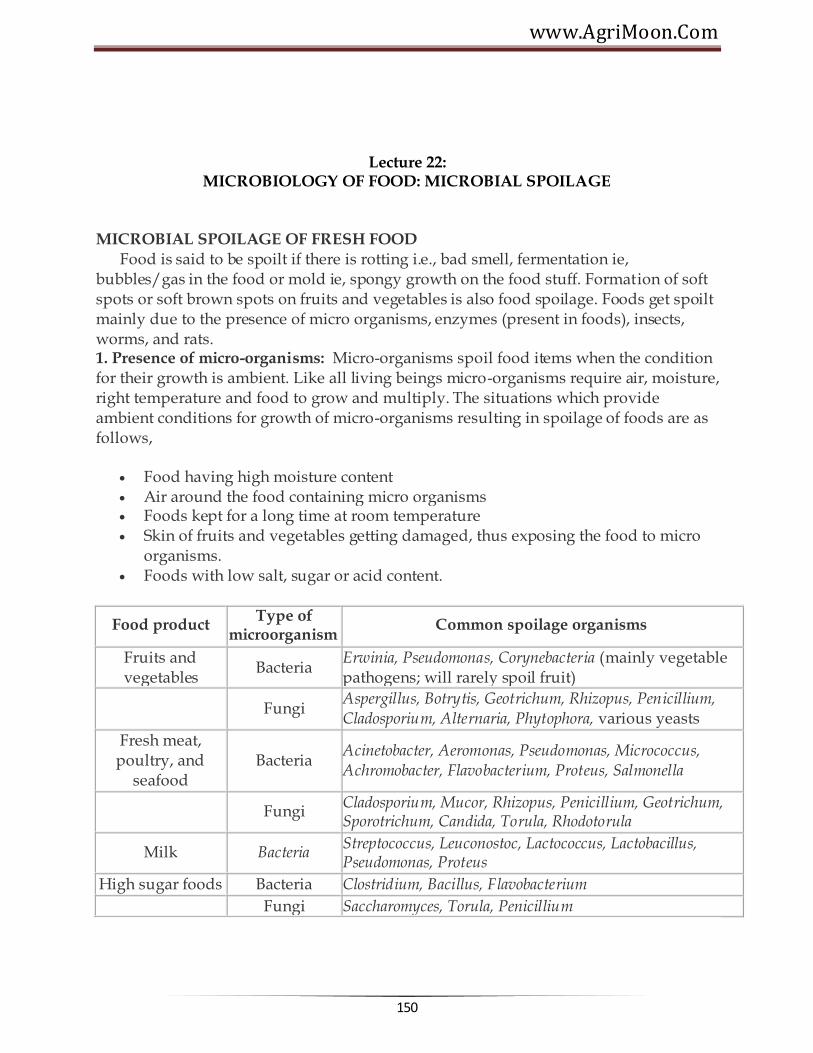
www.AgriMoon.Com
150
Lecture 22:
MICROBIOLOGY OF FOOD: MICROBIAL SPOILAGE
MICROBIAL SPOILAGE OF FRESH FOOD Food is said to be spoilt if there is rotting i.e., bad smell, fermentation ie, bubbles/gas in the food or mold ie, spongy growth on the food stuff. Formation of soft spots or soft brown spots on fruits and vegetables is also food spoilage. Foods get spoilt mainly due to the presence of micro organisms, enzymes (present in foods), insects, worms, and rats. 1. Presence of micro-organisms: Micro-organisms spoil food items when the condition for their growth is ambient. Like all living beings micro-organisms require air, moisture, right temperature and food to grow and multiply. The situations which provide ambient conditions for growth of micro-organisms resulting in spoilage of foods are as follows,
Food having high moisture content Air around the food containing micro organisms Foods kept for a long time at room temperature Skin of fruits and vegetables getting damaged, thus exposing the food to micro
organisms. Foods with low salt, sugar or acid content.
Food product Type of
microorganism Common spoilage organisms
Fruits and vegetables
Bacteria Erwinia, Pseudomonas, Corynebacteria (mainly vegetable pathogens; will rarely spoil fruit)
Fungi
Aspergillus, Botrytis, Geotrichum, Rhizopus, Penicillium, Cladosporium, Alternaria, Phytophora, various yeasts
Fresh meat, poultry, and
seafood Bacteria
Acinetobacter, Aeromonas, Pseudomonas, Micrococcus, Achromobacter, Flavobacterium, Proteus, Salmonella
Fungi
Cladosporium, Mucor, Rhizopus, Penicillium, Geotrichum, Sporotrichum, Candida, Torula, Rhodotorula
Milk Bacteria Streptococcus, Leuconostoc, Lactococcus, Lactobacillus, Pseudomonas, Proteus
High sugar foods Bacteria Clostridium, Bacillus, Flavobacterium
Fungi Saccharomyces, Torula, Penicillium

Agricultural Microbiology
151
2. Presence of enzymes: Enzymes are organic catalysts found in all plants and animals.
Enzymes help in ripening of fruits and vegetables. If a ripe fruit is kept for few days, it will become soft, develop black spots and will start smelling bad. This is due to continued action of enzymes.
3. Insects, worms and rats: Small insects and worms eat the food grains. They make
small holes in the grain and at times convert the grain to a fine powder. The food grain thus become unfit for human consumption.

www.AgriMoon.Com
152
Lecture 23: PRINCIPLES OF PRESERVATION
Principles of Food Preservation A good method of food preservation is one that slows down or prevents altogether the action of the agents of spoilage. Also, during the process of food preservation, the food should not be damaged. In order to achieve this, certain basic methods were applied on different types of foods. For example in earlier days, in very cold weather condition, ice was used to preserve foods. Thus, very low temperature became an efficient method for preventing food spoilage. Let us now list the principles of food preservation.
1. Removal of micro-organisms or inactivating them: This is done by removing air,
water (moisture), lowering or increasing temperature, increasing the concentration of salt or sugar or acid in foods. If you want to preserve green leafy vegetables, you have to remove the water from the leaves so that micro organisms cannot survive. You do this by drying the green leaves till all the moisture evaporates.
2. Inactivating enzymes: Enzymes found in foods can be inactivated by changing their
conditions such as temperature and moisture, when you preserve peas, one of the methods of preservations is to put them for a few minutes in boiling water. This method also known as blanching inactivates enzymes and thus, helps in preserving the food.
3. Removal of insects, worms and rats: By storing foods in dry, air tight containers the
insects, worms or rats are prevented from destroying it. Control
Control of microorganisms
Heat Cold Drying Acids Sugar and salt Oxygen concentration Smoke Radiation Chemicals (preservatives)

Agricultural Microbiology
153
Control of enzymes
Heat Oxygen removal Acids Chemicals (antioxidants)
Control of Other factors
Protective packaging Sanitation
Preservation methods: 1. Thermal processing
Application of heat
Inactivate enzymes Kill microorganisms. Most bacteria are killed in the range 82-93°c. Spores are not
destroyed even by boiling water at 100°c for 30 min.
To ensure sterility (total microbial destruction, including spores), a temperature of 121°c must be maintained for 15 min or longer.
Various methods are -
o Blanching o Pasteurization o Sterilization o Boiling o Steam under pressure
2. Removal of heat (cold processing)
Lowering temperature of food Decreases the rate of enzymatic, chemical and microbial reactions in food Storage life is extended
Various methods are -
a. Refrigeration b. Freezing

www.AgriMoon.Com
154
3. Control of water content (drying)
Microorganisms require free water Free water is removed from the food and therefore, is unavailable to microbial
cells Multiplication will stop Water unavailable for chemical/biochemical reactions Storage life extended
Various methods are -
Freezing Physical removal of water from food (dehydration) Removal of some of the water from food (concentration) Addition of substances that bind water in food, making it unavailable (sugar,
salts)
4. Radiation
Ionizing radiation Inactivate microorganisms in food Destroy storage pests Inactivate enzymes
Various methods are -
Infrared radiation Ultraviolet radiation
5. Atmosphere composition
Removal of oxygen Inhibits o2-dependant enzymatic and chemical reactions Inhibits growth of aerobic microorganisms
Various methods are -
Paraffin wax Nitrogen backflushed bags (potato chips) Controlled atmosphere storage Vacuum packaging of fresh food (cured meats)

Agricultural Microbiology
155
6. Fermentation
Specific microorganisms are used (starter cultures) Facilitate desirable chemical changes Longer storage life Produce acids, alcohol that will prevent growth of undesirable microorganisms Produce antimicrobial substances Addition of chemicals
Various chemicals used are -
Acids (inhibit microbial growth and enzymatic reactions) Organic acids (acetic, citric, tartaric acids) Inorganic acids (hydrochloric, phosphoric acids) Food grade, comply w/regulations Antioxidants (to delay oxidative rancidity) Antimicrobial agents:
o sodium propionate (mould inhibitor) o sodium benzoate (antibacterial) o sugar and salt (high concentrations)
8. Smoke
Contains preservative chemicals (eg. formaldehyde) from the burning wood Heat also helps destroy microorganisms Heat dries the food
9. Curing (Salt and Sugar)
Salt binds with water molecules and thus acts as a dehydrating agent in foods. Impair the conditions under which pathogens cannot survive. Curing is used with certain fruits and vegetables. ( sauerkraut, pickles),
Meats can be submerged in a salt solution known as brine
PRESERVATION BY USING CHEMICALS A preservative is defined as only substance which is capable of inhibiting, retarding or arresting the growth of microorganisms. Microbial spoilage of food products is also controlled by using chemical preservatives. The inhibitory action of preservatives is due to their interfering with the mechanism of cell division, permeability of cell membrane and activity of enzymes. Pasteurized squashes, cordials and crushes have a cooked flavour. After the container is

www.AgriMoon.Com
156
opened, they ferment and spoil within a short period, particularly in a tropical climate. To avoid this, it is necessary to use chemical preservatives. Chemically preserved squashes and crushes can be kept for a fairly long time even after opening the seal of the bottle. It is however, essential that the use of chemicals is properly controlled, as their indiscriminate use is likely to be harmful. The preservative used should not be injurious to health and should be non-irritant. It should be easy to detect and estimate. Two important chemical preservatives are permitted to beverages according to the FPO (1955). 1. Sulphur dioxide and 2. Benzoic acid
SULPHUR DIOXIDE
It is widely used throughout the world in the preservation of juice, pulp, nectar, squash, crush, cordial and other products. It has good preserving action against bacteria and moulds and inhibits enzymes, etc. In addition, it acts as an antioxidant and bleaching agent. These properties help in the retention of ascorbic acid, carotene and other oxidizable compounds. It also retards the development of nonenzymatic browning or discolouration of the product. It is generally used in the form of its salts such as sulphite, bisulphate and metabisulphite.
Potassium metabisulphite (K2O 2So2 (or) K2S2O5) is commonly used as a stable source of So2. Being a solid, it is easier to use than liquid (or) gaseous So2.It is fairly stable in neutral (or) alkaline media but decomposed by weak acids like carbonic, citric, tartaric acid and malic acids. When added to fruit juice (or) squash it reacts with the acid in the juice forming the potassium salt and So2, which is liberated and forms sulphurous acid with the water of the juice. The reactions involved are as follows
Potassium Potassium Sulphur Meta bisulphate + Citric acid ® Citrate + dioxide + H2O
SO2 + H2O ® H2SO3 (Sulphurous acid)
SO2 has a better preservative action than sodium benzoate against bacteria and moulds. It also retards the development of yeasts in juice, but cannot arrest their multiplication, once their number has reached a high value. It is well known that fruit juices with high acidity do not undergo fermentation readily. The preservative action of the fruit acid its due to is hydrogen ion concentration. The pH for the growth of moulds ranges from 1.5 to 8.5, that of yeasts from 2.5-8.0, and of bacteria from 4.0 to 7.5.As fruit beverage like citrus squashes and cordials have generally a pH of 2.5 to 3.5, the growth of moulds and yeasts in them cannot be prevented by acidity alone. Bacteria, however, cannot grow. The pH is therefore, of great importance in the preservation of food product and by regulating it, one or more kinds of microorganisms in the beverage can be eliminated.

Agricultural Microbiology
157
The concentration of So2 required preventing the growth of mirgroorganism at different pH levels are as under.
pH S.ellipsoideus (yeasts)
Mucor (mold)
Penicillium (mold)
Mixed
bacteria
2.5 200 200 300 100 3.5 800 600 600 300
7.0 Above 5000 Above 5000
Above 5000 Above 1000
The toxicity of So2 increases at high temperature. Hence its effectiveness depends on the acidity, pH, temperature and substances present in fruit juice. According to FPO, the maximum amount of So2 allowed in fruit juice is 700 ppm, in squash, crush and cordial 350 ppm and in RTS and nectar 100 ppm. The advantages of using So2 are a) It has a better preserving action than sodium benzoate against bacterial fermentation b) it helps to retain the colour of the beverage for a longer time than sodium benzoate ( c) being a gas, it helps in preserving the surface layer of juices also (d) being highly soluble in juices and squashes, it ensures better mixing and hence their preservation and (e) any excess of So2 present can be removed either by heating the juice to about 71oC or by passing air through it or by subjecting the juice to vacuum. This causes some loss of the flavouring materials due to volatilization, which can be compensated by adding flavours.
Disadvantages (or) limitations
It cannot be used in the case of some naturally coloured juices like those of jamun, pomegranate, strawberry, coloured grapes, plum etc. on account of its bleaching action.
It cannot also be used for juices which are to be packed in tin containers because it not only corrodes the tin causing pinholes, but also forms H2S which has a disagreeable smell and reacts with the iron of the tin container to form a black compound, both of which are highly undesirable and
So2 gives a slight taste and colour to freshly prepared beverages but these are not serious defects if the beverage is diluted before drinking.
II. Benzoic acid
It is only partially soluble in H2O hence its salt, sodium benzoate is used. One part of sodium benzoate is soluble in 1.8 parts of water at ordinary temperature, whereas only 0.34 parts of benzoic acid is soluble in 100 parts of water. Sodium benzoate is thus nearly 170 times as soluble as benzoic acid, pure sodium benzoate is tasteless and odourless. The antibacterial action of benzoic acid is increased in the presence of CO2 and acid

www.AgriMoon.Com
158
e.g. Bacillus subtilis cannot survive in benzoic acid solution in the presence of CO2. Benzoic acid is more effective against yeasts than against moulds. It does not stop lactic acid and acetic acid fermentation. The quantity of benzoic acid required depends on the nature of the product to be preserved, particularly its acidity. In case of juices having a pH of 3.5-4.0, which is the range of a majority of fruit juices, addition of 0.06 to 0.10% of sodium benzoate has been found to be sufficient. In case of less acid juices such as grape juice atleast 0.3% is necessary. The action of benzoic acid is reduced considerably at pH 5.0. Sodium benzoate is excess of 0.1% may produce a disagreeable burning taste. According to FPO its permitted level in RTS and nectar is 100 ppm and in squash, crush and cordial 600 ppm. In the long run benzoic acid may darken the product. It is, therefore, mostly used in coloured products of tomato, jamun, pomegranate, plum, watermelon, strawberry, coloured grapes etc.
Preservation by Using Radiation
Radiation may be defined as the emission and propagation of energy through space or through a material medium. The type of radiation of primary interest in food preservation is electromagnetic. Initially, the destruction of microorganisms in foods by ionizing radiation was referred to by terminology brought over from heat and chemical destruction of microorganisms. Although microorganisms can indeed be destroyed by chemicals, heat, and radiation, there is, nevertheless, a lack of precision in the use of this terminology for radiation-treated foods. Consequently, in 1964 an international group of microbiologists suggested the following terminology for radiation treatment of foods.24
Radappertization
Is equivalent to radiation sterilization or "commercial sterility," as it is understood in the canning industry. Typical levels of irradiation are 3(MK) kGy.
Radicidation
Is equivalent to pasteurization— of milk, for example. Specifically, it refers to the reduction of the number of viable specific nonspore- forming pathogens, other than viruses, so that none is detectable by any standard method. Typical levels to achieve this process are 2.5-10 kGy.
Radurization May be considered equivalent to pasteurization. It refers to the enhancement of the keeping quality of a food by causing substantial reduction in the numbers of viable specific spoilage microbes by radiation. Common dose levels are 0.75-2.5 kGy for fresh meats, poultry, seafood, fruits, vegetables, and cereal grains.

Agricultural Microbiology
159
Radappertization
Radappertization of any foods may be achieved by application of the proper dose of radiation under the proper conditions.
Preservation by Using High temperature The use of high temperatures to preserve food is based on their destructive effects on microorganisms. By high temperatures are meant any and all temperatures above ambient. With respect to food preservation, there are two temperature categories in common use: pasteurization and sterilization.
Pasteurization: by use of heat implies either the destruction of all disease-producing organisms (for example, pasteurization of milk) or the destruction or reduction in the number of spoilage organisms in certain foods, as in the pasteurization of vinegar. The pasteurization of milk is achieved by heating as follows: 145°F (63°C) for 30 minutes (low temperature, long time [LTLT]) 1610F (72°C) for 15 seconds (primary high temperature, short time [HTST] method) 191°F(89°C) for 1.0 second, 194°F (900C) for 0.5 second, 201°F(94°C)for0.1 second, 212°F (1000C) for 0.01 second. These treatments are equivalent and are sufficient to destroy the most heat resistant of the nonspore- forming pathogenic organisms—Mycobacterium tuberculosis and Coxiella burnetii. When six different strains of M. paratuberculosis were added to milk at levels from 40 to 100,000 colony-forming units (cfu)/mL followed by pasteurization by LTLT or HTST, no survivors survivors were detected on suitable culture media incubated for 4 months. Milk pasteurization temperatures are sufficient to destroy, in addition, all yeasts, molds, gram negative bacteria, and many gram positives. The two groups of organisms that survive milk pasteurization are placed into one of two groups: thermodurics and thermophiles. Thermoduric organisms are those that can survive exposure to relatively high temperatures but do not necessarily grow at these temperatures. The nonsporeforming organisms that survive milk pasteurization generally belong to the genera Streptococcus and Lactobacillus, and sometimes to other genera. Thermophilic organisms are those that not only survive relatively high temperatures but require high temperatures for their growth and metabolic activities. The genera Bacillus and Clostridiumcontain the thermophiles of greatest importance in foods. Pasteurization (to destroy spoilage biota) of beers in the brewing industry is carried out usually for 8-15 minutes at 600C.
Sterilization: means the destruction of all viable organisms as may be measured by an
appropriate plating or enumerating technique. Canned foods are sometimes called "commercially sterile" to indicate that no viable organisms can be detected by the usual cultural methods employed or that the number of survivors is so low as to be of no significance under the conditions of canning and storage. Also, microorganisms may be

www.AgriMoon.Com
160
present in canned foods that cannot grow in the product by reason of undesirable pH, oxidation-reduction potential (Eh), or temperature of storage.
Preservation by Using Low temperature
The use of low temperatures to preserve foods is based on the fact that the activities of food borne microorganisms can be slowed at temperatures above freezing and generally stopped at subfreezing temperatures. The reason is that all metabolic reactions of microorganisms are enzyme catalyzed and that the rate of enzyme catalyzed reactions is dependent on temperature. With a rise in temperature, there is an increase in reaction rate. The temperature coefficient (Q10) may be generally defined as follows:
The Qi0 for most biological systems is 1.5-2.5, so that for each 100C rise in temperature within the suitable range, there is a twofold increase in the rate of reaction. For every 100C decrease in temperature, the reverse is true. The term psychrophile was coined by Schmidt- Nielsen in 1902 for microorganisms that grow at O0C.30 This term is now applied to organisms that grow over the range of subzero to 200C, with an optimum range of 10-150C.44 Around 1960, the term psychrotroph (psychros, cold, and trephein, to nourish or to develop) was suggested for organisms able to grow at 5°C or below.1147 It is now widely accepted among food microbiologists that a psychrotroph is an organism that can grow at temperatures between 00C and 7°C and produce visible colonies (or turbidity) within 7-10 days. Because some psychrotrophs can grow at temperatures at least as high as 430C, they are, in fact, mesophiles. By these definitions, psychrophiles would be expected to occur only on products from oceanic waters or from extremely cold climes. The organisms that cause the spoilage of meats, poultry, and vegetables in the 0-50C range would be expected to be psychrotrophs.
Methods of freezing There are various methods of freezing 1. Sharp Freezing (Slow freezing) This technique, first used in 1861, involves freezing by circulation of air, either naturally or with the aid of fans. The temperature may vary from –15 to –29oC and freezing may take from 3 to 72 hours. The ice crystals formed one large and rupture the cells. The thawed tissue cannot regain its original water content. The first products to be sharp frozen were meat and butter. Now-a-days freezer rooms are maintained at –23 to –29oC or even lower, in contrast to the earlier temperature of –18oC.

Agricultural Microbiology
161
2. Quick freezing
In this process the food attains the temperature of maximum ice crystal formation (0 to – 4oC) in 30 min or less. Such a speed results in formation of very small ice crystals and hence minimum disturbance of cell structure. Most foods are quick frozen by one of the following three methods:
a) By direct immersion Since liquids are good heat conductors food can be frozen rapidly by direct immersion in a liquid such as brine or sugar solution at low temperature. Berries in sugar solution packed fruit juices and concentrates are frozen in this manner. The refrigeration medium must be edible and capable of remaining unfrozen at –18oC and slightly below. Direct immersion equipments such as ottenson Brine freezer, Zarotschenzeff ‗Fog‘ freezer, T.V.A. freezer, Bartlett freezer etc. of commercial importance earlier are not used today.
Advantages
There is perfect contact between the refrigerating medium and the product, hence the rate of heat transfer is very high.
Fruits are frozen with a coating of syrup which preserves the colour and flavour during storage.
The frozen product is not a solid block because each piece is separate.
Disadvantages
Brine is a good refrigerating medium but it cannot be used for fruits. It is difficult to make a syrup that will not become viscous at low temperature. The refrigeration temperature must be carefully controlled, as at high
temperature the medium will enter the product by osmosis and at low temperature the medium may freeze solid.
It is very difficult to maintain the medium at a definite concentration and also to keep it free from dirt and contamination.
b) By indirect contact with refrigerant
Indirect freeing may be defined as freezing by contact of the product with a metal surface which is itself cooled by freezing brine or other refrigerating media. This is an old method of freezing in which the food or package is kept in contact with the passage through the refrigerant at –18 to -46oC flows. Knowles Automatic Package feezer, Patterson continuous plate freezer, FMC continuous can freezer and Birds eye freezers are based on this principle.
c) By air blast In this method, refrigerated air at –18 to –34oC is blown across the material to be frozen.

www.AgriMoon.Com
162
The advantages claimed for quick freezing over slow freezing (sharp freezing) are (1) smaller (size) ice crystals are formed, hence there is less mechanical destruction of intact cells of the food (2) period for ice formation is shorter, therefore, there is less time for diffusion of soluble material and for separation of ice (3) more rapid preservation of microbial growth and (4) more rapid slowing down of enzyme action.
3) Cryogenic freezing Although most foods retain their quality when quick frozen by the above methods, a few require ultrafast freezing. Such materials are subjected to cryogenic freezing which is defined as freezing at very low temperature (below –60oC). The refrigerant used at present in cryogenic freeing are liquid nitrogen and liquid CO2. In the former case, freezing may be achieved by immersion in the liquid, spraying of liquid or circulation of its vapour over the product to be frozen.
4. Dehydro-freezing This is a process where freezing is proceded by partial dehydration. In case of
some fruits and vegetables about 50% of the moisture is removed by dehydration prior to freezing. This has been found to improve the quality of the food. Dehydration does not cause deterioration and dehydro frozen foods are relatively more stable.
5. Freeze drying
In this process food is first frozen at –18oC on trays in the lower chamber of a freeze drier and the frozen material dried (initially at 30oC for 24 hrs and then at 20oC). Under high vacuum (0.1 mm Hg) in the upper chamber. Direct sublimation of the ice takes place without passing through the intermediate liquid stage. The product is highly hygroscopic, excellent in taste and flavour and can be reconstituted readily. Mango pulp, orange juice concentrate, passion fruit juice and guava pulp are dehyderated by this method.
******☺******

Agricultural Microbiology
163
Lecture 24:
ROLE OF BACTERIA IN FERMENTATION
What is Fermentation?
Fermentation is the chemical transformation of organic substances into simpler compounds by the action of enzymes, complex organic catalysts, which are produced by microorganisms such as molds, yeasts, or bacteria. Enzymes act by hydrolysis, a process of breaking down or predigesting complex organic molecules to form smaller (and in the case of foods, more easily digestible) compounds and nutrients. For example, the enzyme protease breaks down huge protein molecules first into polypeptides and peptides, then into numerous amino acids, which are readily assimilated by the body. The enzyme amylase works on carbohydrates, reducing starches and complex sugars to simple sugars. And the enzyme lipase hydrolyzes complex fat molecules into simpler free fatty acids. These are but three of the more important enzymes. There are thousands more, both inside and outside of our bodies. In some fermentation, important by-products such as alcohol or various gases are also produced. The word "fermentation" is derived from the Latin meaning "to boil," since the bubbling and foaming of early fermenting beverages seemed closely akin to boiling.
Fermented foods often have numerous advantages over the raw materials from which they are made. As applied to soyfoods, fermentation not only makes the end product more digestible, it can also create improved (in many cases meatlike) flavor and texture, appearance and aroma, synthesize vitamins, destroy or mask undesirable or beany flavors, reduce or eliminate carbohydrates believed to cause flatulence, decrease the required cooking time, and increase storage life. Most fermentations are activated by either molds, yeasts, or bacteria, working singularly or together. The great majority of these microorganisms come from a relatively small number of genera; roughly eight genera of molds, five of yeasts, and six of bacteria. An even smaller number are used to make fermented soyfoods: the molds areAspergillus , Rhizopus , Mucor , Actinomucor , and Neurospora species; the yeasts are Saccharomyces species; and the bacteria are Bacillus and Pediococcusspecies plus any or all of the species used to make fermented milk products. Molds and yeasts belong to the fungus kingdom, the study of which is called mycology. Fungi are as distinct from true plants as they are from animals. The study of all microorganisms is called microbiology. While microorganisms are the most intimate friends of the food industry, they are also its ceaseless adversaries. They have long been used to make foods and beverages, yet they can also cause them to spoil. When used wisely and creatively, however, microorganisms are an unexploitable working class, whose very nature is to labor tirelessly day and night, never striking or complaining, ceaselessly providing human beings with new foods. Like human beings,

www.AgriMoon.Com
164
but unlike plants, microorganisms cannot make carbohydrates from carbon dioxide, water, and sunlight. They need a substrate to feed and grow on. The fermented foods they make are created incidentally as they live and grow.
Human beings are known to have made fermented foods since Neolithic times. The earliest types were beer, wine, and leavened bread (made primarily by yeasts) and cheeses (made by bacteria and molds). These were soon followed by East Asian fermented foods, yogurt and other fermented milk products, pickles, sauerkraut, vinegar (soured wine), butter, and a host of traditional alcoholic beverages. More recently molds have been used in industrial fermentation to make vitamins B-2 (riboflavin) and B-12, textured protein products (from Fusarium and Rhizopus in Europe) antibiotics (such as penicillin), citric acid, and gluconic acid. Bacteria are now used to make the amino acids lysine and glutamic acid. Single-celled protein foods such as nutritional yeast and microalgae (spirulina, chlorella) are also made in modern industrial fermentations.
For early societies, the transformation of basic food materials into fermented foods was a mystery and a miracle, for they had no idea what caused the usually sudden, dramatic, and welcomed transformation. Some societies attributed this to divine intervention; the Egyptians praised Osiris for the brewing of beer and the Greeks established Bacchus as the god of wine. Likewise, at many early Japanese miso and shoyu breweries, a small shrine occupied a central place and was bowed to daily. In ancient times fermentation joined smoking, drying, and freezing as basic and widely practiced food preservation techniques. Wang and Hesseltine (1979) note that "Probably the first fermentation were discovered accidentally when salt was incorporated with the food material, and the salt selected certain harmless microorganisms that fermented the product to give a nutritious and acceptable food." The process was taken a step further by the early Chinese who first inoculated with the basic foods with molds, which created enzymes; in salt-fermented soyfoods such as miso, soy sauce, soy nuggets, and fermented tofu, these aided salt-tolerant yeasts and bacteria??.
A Brief History of Fermentation in the West:
The origins of microbiology (other than the general knowledge of fermented foods which existed worldwide since ancient times) can be traced back to the invention of the compound microscope in the late 1500s. This relatively simple tool soon revolutionized man's knowledge of the heretofore invisible microbial world. In 1675 the Dutch merchant Anton van Leeuwenhoek, the greatest of the early microscopists, saw and reported one-celled organisms, which he called "animacules." (Today they are called "protozoa.") The discovery electrified the scientific world of the time. Then in 1680, using a microscope that magnified the diameter of each object 300-fold, he looked at yeast and found them to consist of tiny spheroids. While the protozoa were clearly alive, the yeast did not appear to be. No connection was drawn between the existence of

Agricultural Microbiology
165
these tiny organisms and the well known phenomenon of fermentation. So for 150 years after van Leeuwenhoek's pioneering observations, it was hardly thought that these minute organisms could be important enough to deserve serious study.
The early 1800s saw a great increase of interest in microbiology in Europe. The scientific period began with great advances in botany, increased interest in microscopy, and willingness to investigate individual organisms. The two major problems that would challenge the greatest researchers in the new field of microbiology concerned the basic nature of the fermentation process and the basic nature of enzymes. The scientific breakthroughs that would lead to the unraveling of the mysteries of fermentation starting in the 1830s were made primarily by French and German chemists. In the late 1700s Lavoisier showed that in the process of transforming sugar to alcohol and carbon dioxide (as in wine), the weight of the former that was consumed in the process equaled the weight of the latter produced. The first solid evidence of the living nature of yeast appeared between 1837 and 1838 when three publications appeared by C. Cagniard de la Tour, T. Swann, and F. Kuetzing, each of whom independently concluded as a result of microscopic investigations that yeast was a living organism that reproduced by budding. The word "yeast," it should be noted, traces its origins back to the Sanskrit word meaning "boiling." It was perhaps because wine, beer, and bread were each basic foods in Europe, that most of the early studies on fermentation were done on yeasts, with which they were made.
The view that fermentation was a process initiated by living organisms soon aroused fierce criticism from the finest chemists of the day, especially Justus von Liebig, J.J. Berzelius, and Friedrich Woehler. This view seemed to give new life to the waning mystical philosophy of vitalism, which they had worked so hard to defeat. Proponents of vitalism held that the functions of living organisms were due to a vital principal distinct from physico-chemical forces, that the processes of life were not explicable by the laws of physics and chemistry alone, and that life was in some part self determining. As we shall soon see, the vitalists played a key role in debate on the nature of fermentation. A long battle ensued, and while it was gradually recognized that yeast was a living organism, its exact function in fermentations remained a matter of controversy. The chemists still maintained that fermentation was due to catalytic action or molecular vibrations.
The debate was finally brought to an end by the great French chemist Louis Pasteur (1822-1895) who, during the 1850s and 1860s, in a series of classic investigations, proved conclusively that fermentation was initiated by living organisms. In 1857 Pasteur showed that lactic acid fermentation is caused by living organisms. In 1860 he demonstrated that bacteria cause souring in milk, a process formerly thought to be merely a chemical change, and his work in identifying the role of microorganisms in food spoilage led to the process of pasteurization. In 1877, working to improve the French brewing industry, Pasteur published his famous paper on fermentation, Etudes

www.AgriMoon.Com
166
sur la Biere , which was translated into English in 1879 as Studies on Fermentation . He defined fermentation (incorrectly) as "Life without air," but correctly showed specific types of microorganisms cause specific types of fermentations and specific end products. In 1877 the era of modern medical bacteriology began when Koch (a German physician; 1843-1910) and Pasteur showed that the anthrax bacillus caused the infectious disease anthrax. This epic discovery led in 1880 to Pasteur's general germ theory of infectious disease, which postulated for the first time that each such disease was caused by a specific microorganism. Koch also made the very significant discovery of a method for isolating microorganisms in pure culture.
Interestingly, until his death in 1873, the eminent German chemist J. von Liebig continued to attack Pasteur's work on fermentation, putrefaction, and infectious diseases. He recognized the similarity of these phenomena but refused to believe that living organisms were the main causative agents. Fermentation, he felt, was primarily a chemical rather than a biological process. History has shown, with the discovery of enzymes, that Pasteur was not entirely right, nor Liebig entirely wrong. The work of Pasteur and his many colleagues and predecessors opened up vast new vistas in the fields of biochemistry, microbiology, and fermentation. The term "biochemistry" was first used in English in 1869, but this new science of the application of chemistry to biology was generally called "physiological chemistry" until the early 1900s. The two outstanding pioneers were Liebig and Pasteur. The term "microbiology" was first used in English in 1885, long after Pasteur's major discoveries. But basic knowledge of this new science of the study of minute living organisms closely related to human activity or welfare did not begin to enter the popular consciousness until the early 1900s. At about this time the scientific breakthroughs of the 1870s and 1880s had begun to produce a change in people's conception of the world around them so sweeping and profound as to be termed revolutionary. Food microbiology was finally set on a scientific foundation, based on the action of specific microorganisms. A rational theory of infectious diseases (which formerly were not differentiated from one another) set people's minds free from the age-old fear of vengeance from an unknowable and invisible disease-causing entity. And the ancient theory of spontaneous generation of lower life forms, which said they could arise de novo and fully formed from decomposing matter, was replaced by the verifiable theory of biogenesis. For the first time people began to accept the fact that they shared their environment with multitudes of minute organisms that exerted an ongoing powerful influence on human life. This new world view, among other things, provided a tremendous stimulus for new research on fermented foods.
Although showing that fermentation was generally the result of the action of living microorganisms was an epic breakthrough, it did not explain the basic nature of the fermentation process, or prove that it was caused by the microorganisms that were apparently always present. As early as the late 1700s it had been recognized that there was another type of chemical change that resembled the yeast fermentation in some

Agricultural Microbiology
167
respects. This was the sort of changes that occur, for example, in the digestion of food. In 1752 Reamur, in studying the digestive processes of a falcon, showed that its digestive juices were able to dissolve meat. In 1785 William Irvine discovered that aqueous extracts of sprouted barley caused liquefaction of starch. The first clear recognition of what were later called "enzymes" came in 1833 when two French chemists, A. Payen and J.F. Persoz, made a more detailed investigation of the process of solubilizing starch with a malt extract to form a sugar that they called "maltose." They called the agent responsible for this transformation "diastase" and they showed that it was destroyed or inactivated by boiling, that without undergoing permanent change itself, a small amount of diastase could convert a large amount of starch to sugar, and that it could be concentrated and purified by precipitation with alcohol. In 1835 the German naturalist Swann, mentioned above for his early work with fermentation, isolated a substance from gastric juice which could bring about the dissolution of meat but which was not an acid. He called it "pepsin" from a Greek word meaning "digestion." It soon became fashionable to call organic catalysts such as diastase and pepsin "ferments," because digestion and fermentation, both allied with life, seemed to be somewhat similar processes. Under the influence of the vitalists, ferments were grouped into two types: those involved with life process were called "organized ferments" and those which were not (like pepsin) were merely "unorganized ferments." A relation between the two types of ferments was suspected by many, and in 1858 M. Traube put forward the theory that all fermentations were due to ferments, definite chemical substances he regarded as related to the proteins and produced in the cells by the organism. In 1876, to reduce confusion that existed concerning the two types of ferments, the German physiologist Wilhelm Kuehne suggested that an unorganized ferment, acting in the absence of life, be called an "enzym," after the Greek words meaning "in yeast;' in 1881 this term was anglicized to "enzyme" by William Roberts, and it had begun to catch on by the 1890s.
Many scientists, including Pasteur, had attempted unsuccessfully to extract the fermentation enzyme from yeast. Success came finally in 1897 when the German chemist Eduard Buechner ground up yeast, extracted a juice from them, then found to his amazement that this "dead" liquid would ferment a sugar solution, forming carbon dioxide and alcohol . . . just like living yeasts. Clearly the so-called "unorganized ferments" behaved just the organized ones. From that time on the term "enzyme" came to be applied to all ferments. The term "ferment" dropped out of the scientific vocabulary altogether and the vitalist position collapsed, never to recover. Thereafter it was agreed that only one set of laws applied to all things, both animate and inanimate, and that there was no special vital force which characterized living things and acted under different laws. And it was finally understood that fermentation is caused by enzymes which are produced by microorganisms. In 1907 Buechner won the Nobel Prize in chemistry for his work, which opened a new era in enzyme and fermentation studies. The sciences of microbiology, biochemistry, fermentation technology, mycology, and

www.AgriMoon.Com
168
bacteriology all shared a deep interest in the nature and working of enzymes. Yet still by the early 1900s no one knew exactly what enzymes were or how they acted. As the agricultural microbiologist Conn asked in 1901, "How can they produce chemical actions without being acted upon or entering into the reactions? Are enzymes fully lifeless or semi-living? We still do not know the fundamental mystery of fermentation." Gradually an understanding of enzymes and catalysts developed. In 1905 Harden and Young discovered coenzymes, agents necessary for the action of enzymes. In 1926 the American biochemist J.B. Sumner first purified and crystallized an enzyme (urease) and showed that it was a protein, more precisely a protein catalyst. Eventually enzymes came to be seen as the key catalysts in all the life processes, each highly specialized in its catalytic action and generally responsible for only one small step in complex, multi -step biochemical reactions. Enzymes are still produced only by living organisms, both animals and plants; they have never? been synthesized.
Advances in microbiology and fermentation technology have continued steadily up until the present. For example, in the late 1930s it was discovered that microorganisms could be mutated with physical and chemical treatments to be higher yielding, faster growing, tolerant of less oxygen, and able to use a more concentrated medium. Strain selection and hybridization developed as well, affecting most modern food fermentations (Hesseltine and Wang 1977).
A Brief History of Fermentation in East Asia . Traditional fermented foods play an
unusually extensive role in East Asia food systems. These fermented foods have a number of important distinguishing characteristics: a number of the most important fermentations use molds; dairy products and other animal proteins (excepting fish) are not widely used, as they are in the West; and modern fermentation processes and technology are based largely on traditional processes, yet are extremely advanced and sophisticated.
The main use of molds has been in the process of making koji (mold-fermented grains and/or soybeans), which serves as a source of more than 50 enzymes in a subsequent fermentation in much the same way that, in the West, the enzymes of malt (steeped and sprouted barley or other cereal grains) are used to make alcoholic beverages.
Since ancient times the koji making process has been unique to East Asia, where it has been used in the preparation of fermented foods such as miso, soy sauce, soy nuggets, sake, shochu (spirits), and rice vinegar ( yonezu ). The only traditional East Asian fermented soyfood not prepared with molds is Japan's natto, and its relatives thua-nao in Thailand and kinema in Nepal; these are bacterial fermentations. Some have suggested that molds are widely used since they grow well in areas having a humid climate and long rainy season during the warm months. In the West mold fermented foods are limited primarily to a number of cheeses characterized by their strong flavors and aromas: Camembert, Blue, Brie, and related types. Because of the widespread use of

Agricultural Microbiology
169
mold-fermented foods in East Asia, the word "mold" there has a rather positive connotation, something like "yeast" in the West. Most Westerners still have a deep-seated prejudice against moldy products, and they generally associate the word "mold" with food spoilage, as in "moldy bread."
Surprisingly little has been published in English about the history of fermentation and knowledge of the fermentation process in East Asia, especially the history prior to the 1870s and 1880s, when the new science of microbiology was introduced from the West. The few works that do exist will be cited later.
The earliest records of the koji-making process can be traced back to at least 300 BC in China and to the third century AD in Japan. Molds differ in one important respect from yeasts and bacteria in that they can be easily observed with the naked eye (without a microscope) and their growth, form, and color noted. In East Asia it was probably understood that fermentation was a life process long before it was in the West. By the sixth century AD, as recorded in the Ch'i-min yao-shu (the earliest encyclopedia of agriculture), the Chinese had distinct names for two types of molds used in fermented soyfoods; what we now call Aspergillus was then called "yellow robe" and Rhizopus was called "white robe." These cultures were carefully distinguished and propagated from year to year. By the 10th century a koji starter or inoculum was deliberately being used in the preparation of koji for fermented foods (Tamiya 1958; Sakaguchi 1972; 1979).
From these early times until the 1870s the traditional fermented foods industries in East Asia apparently advanced largely by an empirical, trial-and-error process without the benefit of general scientific research into the nature of microorganisms and of the fermentation process, and without any general theories in these areas. Prior to 1870, makers of East Asian fermented foods were unaware of the basic nature of the fermentation process of microorganisms, enzymes, and their respective interactions. Makers of koji had no idea what caused the grains and/or soybeans to become covered with a fragrant white mycelium after several days of incubation in a warm koji room, or what later transformed the koji almost magically into delicious, savory seasonings such as miso, shoyu, or soy nuggets, or into heady beverages such as sake.
The advances in food fermentations resulting from the exchange of people and ideas was most pronounced in Japan. The first generation of European scientists there plunged in to their investigations of the many fermented foods with great curiosity and enthusiasm. One of their first subjects of research was the koji mold, now known as Aspergillus oryzae, and the various foods in which it was used, especially sake and shoyu, which were major sources of tax revenue for the Meiji government. Tradition ascribed the introduction of sake brewing in Japan to some emigrants from Korea at about the end of the third century AD; they doubtless learned the process from China, where it had long been practiced. One of the earliest accounts of sake production by a

www.AgriMoon.Com
170
Westerner appeared in 1874 when Dr. J.J. Hoffmann, a German professor in the medical school of today's Tokyo University, published a translation of an article on sake from a Japanese encyclopedia of 1714. In 1884 Ferdinand J. Cohn, a Polish botanist and microbiologist, first gave the koji mold its present name, Aspergillus oryzae . After 1884 the koji mold was referred to as Aspergillus oryzae(Ahlburg) Cohn, in recognition of Ahlburg's earliest accurate description.
Another pioneer in the field of koji research was Atkinson, who had a BS degree from London and was a professor of analytical and applied chemistry at Tokyo University. In 1878, after visiting sake factories, he wrote "On Sake Brewing," which contained a preliminary description of the koji-making process and mentioned the word "koji." In 1881, after extensive research with his assistant Mr. Nakazawa at the koji plant of Mr. J. Kameyama in Yushima near Tokyo, he published two major articles. In his 73-page "On the Chemistry of Sake Brewing," he gave a detailed account of koji making in underground caves in Tokyo and an analysis of its composition.
Another early leader in the fields of microbiology and fermented soyfoods was K. Saito. He did excellent early investigations on the shoyu fermentation, named the primary tempeh mold ( Rhizopus oligosporus ) in 1905, and was an authority on yeasts and molds. Likewise K.N. Yabe did important early work in bacteriology and in natto fermentation.
Two other early pioneers in the introduction of microbiology and fermentation science to Japan were Dr. Teizo Takahashi (1875-1952) and his student Dr. Kinichiro Sakaguchi (1897-), both of whom were professors in the Department of Agricultural Chemistry of Tokyo University. Both men did numerous important studies relating to miso, shoyu, and the koji mold, Aspergillus.
During the 20th century, Japanese microbiologists have made many important contributions to the development of applied and industrial microbiology, including the manufacture of fermented soyfoods, as well summarized by Tamiya (1958) and Sakaguchi (1972). Until quite recently, their strength was more in the area of application of scientific knowledge than in pioneering basic scientific and microbiological breakthroughs. From the early 1900s, important studies on the koji mold and its enzymes were done by Japanese scientists. Important advances in enzymology, with much of the work done on koji molds, began in the 1920s. In 1928 Miyazaki developed the combined Amylo-Koji process. By the 1950s Japanese scientists had isolated various protease and amylase enzymes, induced mutations, and used them commercially. They also developed the technology for the microbial production of L-glutamic acid and monosodium glutamate ( MSG), lysine and other amino acids, flavor enhancing nucleotides such as inosinic acid, and organic acids. They used the koji mold Aspergillus oryzae in the commercial production of enzymes including proteases, amylases, amyloglucosidase, and lipase. They made microbial rennet and numerous other products. Indeed in the period following World War II, Japan became the world leader

Agricultural Microbiology
171
in the field of industrial fermentations. Wang and Hesseltine (1979) have suggested that this may have been "in large part due to the food fermentation base from which it launched its industrialization of micoorganisms."

www.AgriMoon.Com
172
Lecture 25:
BENEFICIAL MICROORGANISMS IN AGRICULTURE
Microbes are an integral part of soil and contribute to soil and plant health. Microorganisms have the ability to fix atmospheric nitrogen, solubilize and mobilize phosphorus, produce antibiotics and disease suppressing molecules. Owing to these properties, they are used in agriculture as biofertilizers and biopesticides. They are also important in the treatment of solid waste and sewage. They clean up the environment by degradation of several pollutants like pesticides, hydrocarbons, dyes and paints. They also help in the enhanced recovery of oil and metals from low grade ores or aqueous streams.
Man is a host to variety of pathogenic bacteria, protozoa and viruses. They can cause various infectious and non-infectious diseases. In order to control the disease and its transmission, it is essential to isolate and identify the causal agent from blood, sputum, urine, stool or pus. Various cultural and molecular methods can be employed for identification of pathogen. Sterilization techniques, use of disinfectants and vaccination can help control transmission of disease.
Biofertilizers
Biofetilizers are the products containing living cells of different types of microorganisms that enrich the nutrient quality of soil. The main sources of biofertilizers are bacteria, fungi and cyanobacteria (blue green algae). Most biofertilizers belong to one of the following categories: nitrogen fixing, phosphate solubilizing and mobilizing, and plant growth promoting rhizobacteria. Some of the major biofertilizers and target crops are given in table 8.1. Nitrogen fixing biofertilizers fix atmospheric nitrogen into forms which are readily useable by plants. These include Rhizobium, Azospirillum, Azotobacter, blue green algae and Azolla. WhileRhizobium requires symbiotic association with the root nodules of legumes to fix nitrogen, others can fix nitrogen independently. Phosphate solubilizing microorganisms secrete organic acids that enhance the uptake of phosphorus by plants by dissolving rock phosphate and tricalcium phosphate. Arbuscular mycorrhizal fungi are the most common phosphorus mobilising types that are omnipresent. A group of bacteria that enhance the growth of plant through nitrogen fixation, phosphorus solubilization or production of plant growth promoting metabolites are known as Plant Growth Promoting Rhizobacteria (PGPR). Many PGPR strains have a potential to be used as microbial inoculants to enhance crop productivity.

Agricultural Microbiology
173
Major biofertilisers and target crops
Biofertiliser Target crop
Rhizobium Leguminous crops
Azotobacter Wheat, maize, cotton, mustard and vegetables (Potato, onion, tomato, brinjal and others)
Azospirillum Cereal crops like wheat, maize, millets, sorghum, barley; and sugarcane.
Blue green algae (BGA) Rice
Azolla Rice
Phosphate solubilizing microorganisms
All
Arbuscular mycorrhiza Nursery raised crops and orchard trees
Plant growth promoting rhizobacteria
All
The growth in agricultural production in the last three decades has been accompanied by a sharp increase in the use of chemical fertilizers, causing serious concern. Foremost among these concerns is the effect of excessive fertilizers on the quality of soil and ground water. The use of environmental friendly biofertilizers can cut down the use of chemical fertilizers. Biofertilizers have definite advantage over chemical fertilizers. It is economical to use biofertilizers as they are a cheap source of nutrients when compared to chemical fertilizers. Biofertilisers in addition to nitrogen and phosphorus, also provides certain growth promoting substances like hormones, vitamins, and amino acids that improves the plant health and vigour. Continuous use of chemical fertilisers adversely affects the soil structure whereas biofertilizers when applied to soil improve the soil structure. The chemical fertilizers are toxic at higher doses where as biofertilizers have no toxic effects.
Nitrogen fixing bacteria
An atmosphere around us contains nearly 78% nitrogen that is in free form and is not utilized by the plants. Plants take up nitrogen in the form of ammonia or nitrate. Relatively small amount of ammonia is produced by lightning. Some ammonia also is produced industrially by the Haber-Bosch process, using an iron-based catalyst, very high pressures and fairly high temperature. But the major conversion of N2 into ammonia by the action of enzyme nitrogenase, and thence into proteins, is achieved by microorganisms in the process called nitrogen fixation (or dinitrogen fixation). All the nitrogen-fixing organisms are prokaryotes. There are different groups of nitrogen fixing microorganisms (diazotrophs) present in the nature. These are broadly divided into three categories, viz.,

www.AgriMoon.Com
174
1. Symbiotic microorganism 2. Asymbiotic or free living 3. Associative Symbiosis
Examples of nitrogen fixing microorganisms for each category are given in table Some examples of nitrogen fixing bacteria belonging to different categories.
Category
Examples
Symbiotic Rhizobium- legume symbiosis Rhizobium-Parasponia (non-legume) symbiosis Frankia- Trees (e.g Alder, Casuarina) Azolla- Anabaena Azotobacter paspali - Paspalum notatum
Free living
1. Aerobic Azotobacter Beijerinckia Cyanobacteria (e.g Nostoc, Anabaena, Tolypothrix, Aulosira)
2. Facultative Klebsiella pneumoniae Bacillus polymyxa
3. Anaerobic Clostridium Desulfovibrio Rhodospirillum Rhodopseudomonas Desulfotomaculum Desulfovibrio Chromatium Chlorobium
Associative Azospirillum Herbaspirillum Acetobacter diazotrophicus Azoarcus
The list of nitogen fixing bacteria is long but here we will discuss some of the important types of biofertilisers that can be considered for agrobased industries.
RHIZOBIUM INOCULANT
The bacteria of the genera Rhizobium, Bradyrhizobium, Mesorhizobium, Sinorhizobium and Azorhizobium collectively known as rhizobia, in symbiotic association with leguminous plants reduce atmospheric nitrogen. The rhizobial colonies appear raised, wet, shining, translucent or opaque with smooth margin on yeast extract mannitol agar (YEMA) medium. The

Agricultural Microbiology
175
legume-rhizobia symbiosis culminates in the formation nitrogen fixing root or stem nodules. These unique structures are agronomically significant as they provide alternative to the use of energy-expensive ammonium fertilizer. Not all legumes fix nitrogen. Of the three segregate families of legumes, the capacity to form nodules appear to be absent from the majority of species of Caesalpiniaceae. All members of family Mimosaceae and Fabaceae show formation of nodules with rhizobia. It is believed that legume-Rhizobium symbiosis contributes atleast 70 million metric tons N per year. The amount of nitrogen fixed varies with the strain of Rhizobium, the plant species and environmental conditions.
Fig Typical growth of Rhizobium on yeast mannitol agar medium with congo red
Fig Root nodules formed by extract rhizobia on mungbean plant
The taxonomy of root and stem nodulating bacteria is in a state of transition. The initial classification of these organisms based on plant infection into 7 cross inoculation groups has been abandoned after extensive criticism. A new system of classification was proposed by Jordan (1984) in Bergey's Manual of Systematic Bacteriology (Table ). He separated the root nodule bacteria into two genera, Rhizobium and Bradyrhizobium, based on data on numerical taxonomy, molecular characteristics and rate of growth on laboratory media. Fast growing strains were placed under genus Rhizobium whereas slow growing strains were placed inBradyrhizobium. Since 1984, the classification has undergone lot of changes. Three additional genera, Mesorhizobium, Sinorhizobium and Azorhizobium have been recognised and many new species have been reported so far.
Rhizobium inoculation
Legume inoculation is a significant strategy for the manipulation of rhizobial microflora and improving crop productivity and soil fertility. However, in tropical soils where there is presence of adequate native rhizobia and high levels of mineral N, legume

www.AgriMoon.Com
176
inoculation often fails. Thus there is an urgency to identify conditions where inoculation is needed. Different diagnostic measures to decide about inoculation have been suggested by various workers. Inoculation should be carried out if;
1. population density of species-specific rhizobia is low, 2. the same or symbiotically related legume is not grown in the area in the
immediate past history 3. waste-lands have to be reclaimed 4. legume follows a non leguminous crop in a rotation 5. soil is poor in mineral N (nitrate) 6. soils are acidic, alkaline and saline.
Selection of rhizobial strains for inoculant production
A large-scale screening should be carried out to identify ideal inoculant strain for different legume crops. The criterion for selection may vary for particular soil types like acidic, sodic, saline, nitrate-rich or heavy metal contaminated. Following are some of the desirable characters for a strain to be fit for use in commercial inoculants: 1. Ability to form nodules and fix N on the target legume 2. Ability to compete in nodule formation with populations of native rhizobia present in the soil. 3. Ability to fix N across a range of environmental conditions; 4. Ability to grow well in artificial media, in inoculant carrier and in the soil 5. Ability to persist in soil, particularly for annually regenerating legumes 6. Ability to migrate from the initial site of inoculation 7. Ability to colonize the soil in the absence of a legume host 8. Ability to tolerate environmental stresses; 9. Ability to fix N with a wide range of host genotypes; 10. Genetic stability 11. Compatibility with agrochemicals. Inoculant production
1. Propagation
Rhizobia are not very particular in their nutritional requirements. Yeast-extract mannitol (YEM) medium is commonly employed for culturing of rhizobia. For commercial production of cultures, cheaper sources like sucrose, molasses and corn steep liquor can be used. Mass scale propagation of rhizobia can be carried out using system of rotary shaker or fermentor. In shake flask culture, broth is raised in flasks with agitation by circular motion of rotary shaker. Fermentors are used for industrial scale production of bio-fertilizers. Culture vessels ranging from 5 to 1000 L can be used depending upon the requirement. The amount of inoculum culture to be added into the fermenter vessel depends on the size of the fermentors, but the ratio between the inoculum and the

Agricultural Microbiology
177
medium in the vessel should be maintained at 1:20 (5% inoculum rate). The broth is continuously aerated by forcing sterile air through porous stainless steel sparger. Various fermentation requirements like aeration, agitation and fermentation time vary from strain to strain. Table gives the optimum fermentation conditions for mass multiplication of rhizobial strains.
When the number of rhizobia in the broth has attained the required standard (108-109 cells ml-1) the broth should be added to the carrier for preparation of carrier-based inoculant. Optimum fermentation conditions for mass multiplication of Rhizobium strains
1. Type of reactor Stirred tank 2. Type of operation Batch
3. Carbon source Sucrose or malasses (3-5 g L-1)
4. Nitrogen source Corn steep liquor or yeast extract
5. pH 7.8 (controlled)
6. Temperature 280C
7. Inoculum rate 10% (V/V) 8. Inoculum count 109 cells mL-1
9. Antifoam PPG
Carriers for rhizobial inoculants
The medium in which rhizobia are allowed to multiply is an important factor in rhizobial culture preparation. The term 'carrier' is generally used for a medium that carries the live microorganisms. As per BIS specification, the carrier should be in powder form and capable of passing through 150-212 micron (72-100 mesh) IS sieve. A good carrier material should
have high water holding capacity be non-toxic to rhizobia be easy to sterilize by autoclaving or gamma irradiation be readily and inexpensively available provide good adhesion to seed have pH buffering capacity have cation and/or anion exchange capacity.
In India, different carrier materials like peat, liginite, charcoal, rice husk, pressmud, vermiculite, soil and coir dust has been employed. Although peat is the favoured base for inoculants world over, in India high quality peat is not available. A mixture of
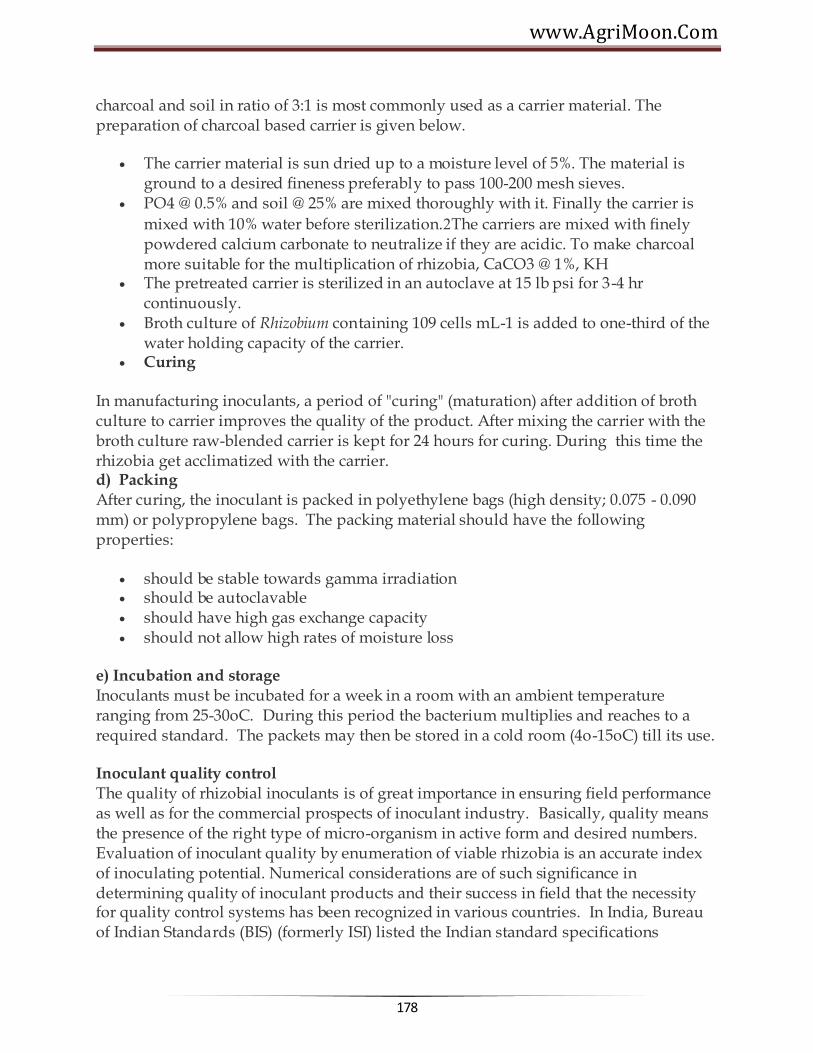
www.AgriMoon.Com
178
charcoal and soil in ratio of 3:1 is most commonly used as a carrier material. The preparation of charcoal based carrier is given below.
The carrier material is sun dried up to a moisture level of 5%. The material is ground to a desired fineness preferably to pass 100-200 mesh sieves.
PO4 @ 0.5% and soil @ 25% are mixed thoroughly with it. Finally the carrier is
mixed with 10% water before sterilization.The carriers are mixed with finely powdered calcium carbonate to neutralize if they are acidic. To make charcoal more suitable for the multiplication of rhizobia, CaCO3 @ 1%, KH
The pretreated carrier is sterilized in an autoclave at 15 lb psi for 3-4 hr continuously.
Broth culture of Rhizobium containing 109 cells mL-1 is added to one-third of the water holding capacity of the carrier.
Curing
In manufacturing inoculants, a period of "curing" (maturation) after addition of broth culture to carrier improves the quality of the product. After mixing the carrier with the broth culture raw-blended carrier is kept for 24 hours for curing. During this time the rhizobia get acclimatized with the carrier. d) Packing After curing, the inoculant is packed in polyethylene bags (high density; 0.075 - 0.090 mm) or polypropylene bags. The packing material should have the following properties:
should be stable towards gamma irradiation should be autoclavable should have high gas exchange capacity should not allow high rates of moisture loss
e) Incubation and storage Inoculants must be incubated for a week in a room with an ambient temperature ranging from 25-30oC. During this period the bacterium multiplies and reaches to a required standard. The packets may then be stored in a cold room (4o-15oC) till its use.
Inoculant quality control The quality of rhizobial inoculants is of great importance in ensuring field performance as well as for the commercial prospects of inoculant industry. Basically, quality means the presence of the right type of micro-organism in active form and desired numbers. Evaluation of inoculant quality by enumeration of viable rhizobia is an accurate index of inoculating potential. Numerical considerations are of such significance in determining quality of inoculant products and their success in field that the necessity for quality control systems has been recognized in various countries. In India, Bureau of Indian Standards (BIS) (formerly ISI) listed the Indian standard specifications

Agricultural Microbiology
179
for Rhizobium inoculants in 1977 (IS: 8268-1976). This was revised in 1986 (ISI 1986). These specifications are given in Table.
Indian Standard specifications for Rhizobium
Parameters Specifications
1. Base Carrier based
2. Cell number at the time of manufacture
108 g-1 carrier
3. Cell number at the time of expiry
107 g-1 carrier within 15 days before expiry date
4. Expiry period 6 months from the date of manufacture 5. Permissible contamination
No contamination at 108 dilution
6. pH 6.0- 7.0
7. Strain Should be checked serologically
8. Carrier Should pass through 150-212 micron, IS (72-100 mesh).
9. Others Nodulation test positive, results in 50% or more dry matter yield than control
Application of inoculants The major goal of legume inoculation is to introduce efficient and competitive strains in large numbers that can survive and establish in the legume rhizosphere and colonize the roots promptly. Application of inoculant to the seed surface prior to sowing is the traditional, most commonly used and most user-friendly means of inoculation. There are numerous adhesives like gur, sugar, gum arabic and methyl cellulose suitable for attaching inoculant to the seeds.
The method of seed inoculation includes preparation of 10% sugar or pharmaceutical grade gum arabic or 1% methyl cellulose solution. This solution is sprinkled on the seeds and the seeds are thoroughly mixed so as to have a uniform coating. A count of 1000 viable cells per seed is to be attained at the time of treating the seed and quantity of culture used is accordingly adjusted. The seeds are spread uniformly for drying on a gunny bag or cement floor in shade avoiding direct sunlight.
Response of legumes to Rhizobium inoculation
Rhizobium inoculation improves the productivity of leguminous crop plants. The efficacy of Rhizobium inoculation has been established in our country beyond any doubt by the results of coordinated trials conducted by the Indian Council of Agricultural Research. The yield response varies with the inoculant strain, location and crop variety.

www.AgriMoon.Com
180
Average increase in yield of some of the pulse crops due to Rhizobium inoculation is presented in table 8.5.
Percent increase in yield of some leguminous crops due to Rhizobium inoculation
Crop % increase
Arhar 32
Mungbean 33
Chickpea 41
Groundnut 49
Lentil 50
Soybean 61
Azotobacter: Azotobacter is a free living, heterotrophic nitrogen fixing bacteria that
occurs in the rhizosphere of variety of plants. The genus Azotobacter has six species viz., A. chroococcum, A. vinelandii, A. beijerinckii, A. nigricans, A. armeniacus and A. paspali. Except the last species, which is a rhizoplane bacterium, the other members are largely soil borne and rhizospheric. The potential of A. chroococcum and A. paspali as a biofertilizer for various non-legume crops is well documented.
Azotobacter is an aerobic, Gram negative, rod shaped bacteria occurs singly, in chains, or in clumps. It does not form endospores but do form thick-walled cysts. These cysts are resistant to desiccation and to some deleterious chemical and physic agents. They, however, cannot withstand extreme temperatures. While in the cyst stage of their life cycle, they do not fix nitrogen and are optically refractile. It may be motile by peritrichous flagella or non-mobile. It can produce a water soluble pigment, either yellow-green, fluorescent or red-violet/ brownish-black. It grows well at an optimum
Agricultural Microbiology
181
temperature range between 20 and 30°C and at pH 7.0 - 7.5. They are able to grow on various carbohydrates, alcohols, and organic acids.
Azotobacter was first discovered using a culture that was devoid of a combined nitrogen source. Azotobacter is found on neutral to alkaline soils, in aquatic environments, in the plant rhizosphere and phyllosphere. A.chroococcum is the most common species of Azotobacter present in the soil.
Azotobacter sp. are known to influence plant growth through their ability to fix molecular nitrogen; production of growth promoting substances like IAA, gibberellin or gibberellin-like compounds and vitamins, excretion of ammonia in the rhizosphere in the presence of root exudates; production of anti-fungal metabolites and phosphate solubilization.
The procedure followed for mass multiplication of Azotobacter, preparation of carrier based inoculant and seed inoculation with carrier based Azotobacter is similar to that of rhizobial inoculation. Jensen's N-free medium is routinely used for the mass multiplication of Azotobacter. Seed inoculation of A. chroococcum increases the yield of field crops by about 10 % and of cereals by about 15-20%. The response to inoculation was increased by manuring or by fertilizer application. Coinoculation ofAzotobacter with other bioinoculants like Rhizobium; Azospirillum, P-solubilizers; vesicular-arbuscular mycorrhiza have been reported to enhance the growth and yield of legumes, cereals and vegetable crops.
Beneficial effects of Azotobacter chroococcum inoculation has been reported by various workers on various cereal, vegetables, oil seed, legume and cash crops (Table ). Inoculation experiments with Azotobacter gave better yield performance only at lower levels of nitrogen (0 to 30 kg N ha-1). These diazotrophic bacteria require large amounts of available carbon for their survival in soil. Addition of farmyard manure (FYM), compost and other organic amendments to agricultural soils improves the efficiency of Azotobacter and hence the plant grown and yield.
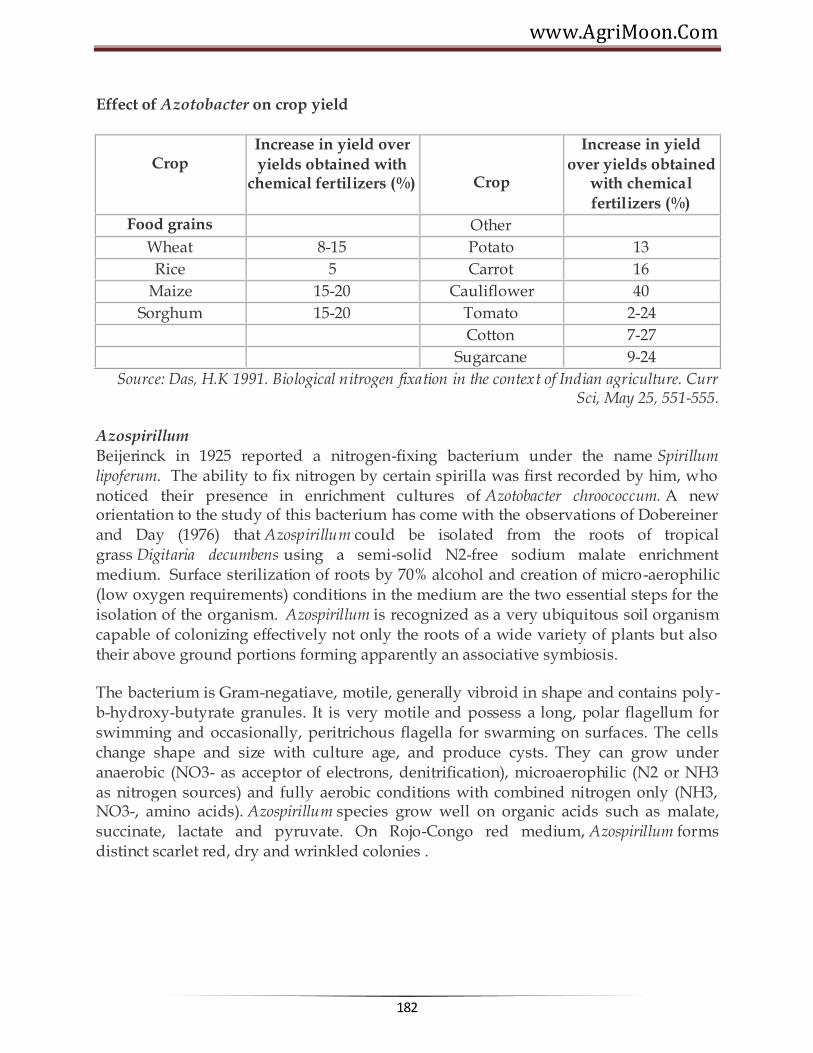
www.AgriMoon.Com
182
Effect of Azotobacter on crop yield
Crop
Increase in yield over
yields obtained with chemical fertilizers (%)
Crop
Increase in yield
over yields obtained with chemical
fertilizers (%)
Food grains Other
Wheat 8-15 Potato 13
Rice 5 Carrot 16
Maize 15-20 Cauliflower 40
Sorghum 15-20 Tomato 2-24
Cotton 7-27
Sugarcane 9-24
Source: Das, H.K 1991. Biological nitrogen fixation in the context of Indian agriculture. Curr Sci, May 25, 551-555.
Azospirillum Beijerinck in 1925 reported a nitrogen-fixing bacterium under the name Spirillum lipoferum. The ability to fix nitrogen by certain spirilla was first recorded by him, who noticed their presence in enrichment cultures of Azotobacter chroococcum. A new orientation to the study of this bacterium has come with the observations of Dobereiner and Day (1976) that Azospirillum could be isolated from the roots of tropical grass Digitaria decumbens using a semi-solid N2-free sodium malate enrichment medium. Surface sterilization of roots by 70% alcohol and creation of micro-aerophilic (low oxygen requirements) conditions in the medium are the two essential steps for the isolation of the organism. Azospirillum is recognized as a very ubiquitous soil organism capable of colonizing effectively not only the roots of a wide variety of plants but also their above ground portions forming apparently an associative symbiosis.
The bacterium is Gram-negatiave, motile, generally vibroid in shape and contains poly-b-hydroxy-butyrate granules. It is very motile and possess a long, polar flagellum for swimming and occasionally, peritrichous flagella for swarming on surfaces. The cells change shape and size with culture age, and produce cysts. They can grow under anaerobic (NO3- as acceptor of electrons, denitrification), microaerophilic (N2 or NH3 as nitrogen sources) and fully aerobic conditions with combined nitrogen only (NH3, NO3-, amino acids). Azospirillum species grow well on organic acids such as malate, succinate, lactate and pyruvate. On Rojo-Congo red medium, Azospirillum forms distinct scarlet red, dry and wrinkled colonies .

Agricultural Microbiology
183
Taxonomy
Azospirillum belongs to group 1 of the alpha subclass of the Proteobacteria . At present there are five known species of Azospirillum- A. brasilense, A. lipoferum,A. amazonense, A. halopraeferens and A. irakense. The distinguishing morphological and biochemical characteristics of the five species is given in table . Different morphological and biochemical characteristics of five known species
of Azospirillum ________________________________________________________________________ Characteristics A.lipoferumA.brasilense A.amazonenseA.irakense A.halopraeferens ________________________________________________________________________ Colony type On CR medium Scarlet Scarlet Pink Scarlet Pink On PDA medium Pink Pink White White No
growth Raised Raised Raised Flat Raised Biotin requirement. + - - - + Utilization of C Malate + + + + + D-Glucose + - - - + Glycerol + + - - + Sucrose - - + + -
________________________________________________________________________ Inoculant production
For mass multiplication of Azospirillum, the organism is allowed to grow in flasks containing NH4Cl and malate medium and incubated at 35o - 37o C for 3 days. When there is good growth, the broth culture is mixed with the carrier, and the carrier-based

www.AgriMoon.Com
184
culture is packed in polyethylene pouches. The technique used for preparation of carrier based inoculant and for inoculating the seed or seedlings with Azospirlllum culture is same as that described earlier in case of Rhizobium.
Crop response to Azospirillum inoculation
Azospirillum is extensively used as an inoculant for crop plants belonging to the family gramineae like wheat, sorghum, pearlmillet, fingermillet, barley and maize. Of all the crops tested, sorghum (Sorghum bicolor), pearlmillet (Pennisetum americanum) and fingermillet (Eleusine coracana) appeared to be consistently responsive to Azosprillum at more than one location in India. Azospirillum species promote the yield of agriculturally important crops in many different soils and climatic regions. By the use of this organism as a seed inoculant, savings of 20-30 kg N/ha equivalents can be achieved in these crops. However, the principal effects of azospirilla go far beyond furnishing nitrogen to host plants.
Once inoculated onto plant roots, Azospirillum cells induce remarkable changes in the morphology and behaviour of the entire root system. For instance, hairs close to the root tip take on a more distinctive appearance, and the overall density and the length of the root system increases (Fig ). Root hairs consist of expanded root epidermal cells, which play a role in water and nutrient exchanges and also help to anchor root to its surroundings. Inoculating azospirilla onto plant roots also increases the diameter and length of both lateral and adventitious roots and typically leads to additional branching of the lateral roots. These developments in the root system are important because they increase absorptive area and volume of the soil substrate available to the plant.
Strains of Azospirillum are known to produce siderophores. They are low molecular weight iron binding compounds synthesized in large amounts and excreted into culture medium by microorganisms under iron-deficient conditions. Siderophores form complexes with the metal ions in the culture medium followed by translocation of the complex through bacterial envelope. The ability of Azospirillum to synthesize

Agricultural Microbiology
185
siderophores may contribute to improve the iron nutrition of plants and offer protection from minor pathogens.
Biosynthesis of growth promoting substances like phytohormones, vitamins, antibacterial and anti fungal substances by Azospirillum is well documented. The most extensively reported growth promoters are IAA, gibberellins, cytokinin like-substances and vitamins.
The ability of azospirilla to form antibiotic substances varies from strain to strain. Fungistatic activity of azospirilla against a wide range of phyto-pathogenic fungi has been reported e.g. certain azospirilla offer protection to cotton plants against Thielaviopsis basicola and Fusarium oxysporum.
These enhancing features of Azospirillum inoculation are also evident in field experiments, with the bacteria not only increasing root numbers but also improving yields of crops such as wheat, sorghum, pearlmillet and maize. In field experiments in Israel, Azospirillum inoculated sorghum plants made better use of moisture stored in soils from winter precipitation than did uninoculated plants. In both green house and field experiments, inoculated plants are efficient at absorbing nitrogen, phosphorus, potassium and other microelements from soil than uninoculated plants.
In recent years, interest has shifted from plant-microbe interaction to plant-microbe-microbe interactions. Several reports have brought to light instances where beneficial effects of Azospirillum on plants are enhanced when coinoculated with other microorganisms like Rhizobium and Azotobacter. Synergistic effects of Azospirillum with Rhizobium on various legumes have been reported. Stimulation of nodulation may be due to an increase in production of lateral roots and in root hair branching. This, in turn, has been thought to be due to production of phytohormones by Azospirillum. The positive effect of inoculating non-legumes withAzospirillum brasilense and Azotobacter chroococcum, at low application rates of mineral N, on associative N2 fixation and on crop yield has been reported.
Acetobacter diazotrophicus
Acetobacter diazotrophicus, is a gram-negative, microaerobic, nitrogen fixing microorganism and was isolated from washed roots and stems of sugarcane, using semi-solid N-free sugar medium acidified with acetic acid to pH 4.5. Cells of Acetobacter
diazotrophicus C. Although sucrose is the best C-source forare straight rods with rounded ends, about 0.7 to 0.9 by 2 um, motile by lateral or peritrichous flagella. Optimum growth temperature is around 30 Acetobacter diazotrophicus but sugars like glucose, fructose, galactose, mannitol are also utilized. It grows well in the pH range of 3.8 to 5.8 with good nitrogenase activity. Growth and nitrogen fixation occur at sugar concentration ranging from 10 to 30 %.

www.AgriMoon.Com
186
Acetobacter diazotrophicus, an endophytic diazotroph, has been found mainly associated with sugar-rich plants such as sugarcane, sweet potato, Cameroon grass, sweet sorghum and coffee. It colonizes roots, stems and leaves of host plants. Reports from Brazil indicates that A. diazotrophicus contributes >50% of biologically fixed nitrogen in sugarcane. Nitrogen fixed by A. diazotrophicus is excreted as ammonia into the medium. Strains of Acetobacter have been shown to produce considerable amount of IAA. Synergistic effects on plant growth and yield following inoculation with Acetobacter diazotrophicus and AM fungi have been reported for sugarcane, sweet potato and sweet sorghum.
Blue green algae and Azolla Blue-green algae (cyanobacteria) are ubiquitous in distribution. They are either single celled or consist of branched or unbranched filaments (Fig. a,b,c). It is a group of free living organisms that has been demonstrated to be an ideal candidate as the biological nitrogen source in rice ecosystems. Some of them possess a peculiar structure known as 'heterocyst' and all heterocystous forms can fix nitrogen from air. Recently, some blue-green algae without heterocysts have also been found to fix nitrogen under special conditions like low oxygen tension. The algae that are generally used for field application are species of Aulosira, Tolypothrix, Scytonema,Nostoc, Anabaena and Plectonema as a mixture.
Cyanobacteria have ability to carry out both photosynthesis and nitrogen fixation. Besides contributing to the nitrogen economy of the soils these algae have other beneficial effects. Their exceptionally good water holding capacity, their ability to concentrate nutrients such as nitrogen, phosphorus, fixed carbon and trace elements, their soil binding capacity and their ability to scavenge sodium from salt affected soils are additional ecological advantages. The presence of BGA in the immediate vicinity of rice seeds can decrease sulphide and iron injury to the plants. Cyanobacteria also produce number of plant growth substances like amino acids, small proteins and peptides, sugars, complex polysaccharides, vitamins and growth hormones. Standing crops of nitrogen fixing BGA range from 5-20 tons per hectare fresh weight and

Agricultural Microbiology
187
contribute approximately 30kg nitrogen per season per hectare of rice field. A bulk of the organic matter produced by algal growth remains in the soil and becomes available to the next crop as organic enrichment.
Production of algae for field application
Based on the natural ecology of these algae, a simple rural-oriented open-air method of producing them in bulk has been developed. The basic principle is to grow them in natural sunlight under conditions stimulating these in the rice field. You can use a starter culture, consisting of soil-based mixture of efficient strains of BGA, supplied by various agricultural universities for mass multiplication. Shallow stays (15cm x 7.5 cm x 22.5 cm) of galvanized iron sheet, or brick and mortar, or pits lined with polythene sheets are prepared. The size can be increased if more material is to be produced. About 10kg soil is placed and mixed with 200 g super phosphate. The trays are then filled with water (5-15 cm) depending upon the local conditions and rate of evaporation; the pH of the soil should be around neutral. After soil settles down, saw-dust and the starter culture are sprinkled on the surface of the standing water. The whole assembly is exposed to sunlight. In hot summer months, the growth of the algae will be rapid and in about a week a thick algal mat will be formed on the surface of the soil and sometimes even floats up. If the daily rate of evaporation is high, more water is added intermittently. When the algal growth becomes sufficiently thick, addition of water should be discontinued and the water is allowed to dry up in the sun. The dried algal flakes are collected from the surface and stored in bags for future use in the fields. The trays are again filled with water and a small amount of the dry algal flakes is added, as further inoculum. The process is continued as above. Once the soil in the tray is exhausted (usually 3-4 harvests), fresh soil is put and mixed with super phosphate and the process is repeated as before. To prevent the breeding of insects, application of Malathion (0.00075 ppm) or Carbofuran (3% granules) is recommended. Algae are applied at the rate of 10kg/ha over the standing water in the field one week after transplantation. The field is kept waterlogged at least for a couple of days immediately after algal application.
Azolla-Anabaena symbiosis
Azolla is a small aquatic fern and is omnipresent in nature. Each leaf consists of two lobes, a thick aerial dorsal lobe and a thin ventral lobe occasionally of a slightly larger size. The dorsal lobe is green and has a blue green algal symbiont (Anabaena azollae) within a central cavity. The heterocyst of the symbiontAnabaena is the site of nitrogen fixation. Azolla provides nutrients and a protective leaf cavity for Anabaena, which in turn provided nitrogen for the fern. Azolla is found on still water in ponds, lakes, swamps, ditches and paddy fields of temperate and tropical regions. Because of its rapid growth, high N content and ability to grow in still water, it has been exploited as a fertilizer for rice. This is used in Vietnam and China for centuries, however, its use as a biofertilizer in India is relatively

www.AgriMoon.Com
188
a recent development. There are 7 living species of Azolla - A. pinnata, A. caroliniana, A. rubra, A filiculoides, A. nilotica, A. mexicana and A. microphylla. A. pinnata is native to India but now many of these species have been introduced.
The high N2 fixing ability, rapid growth, high biomass accumulation and N-content determines the potential of Azolla as a biofertilizer for rice. Biological nitrogen fixation through Azolla - Anabaena complex is considered a potential biological system for increasing rice yield at comparatively low cost. The ability of Azolla to fix N2 is about 1.1 kg N/ha/day. The doubling time varies between 2 and 10 days for most species and maximum biomass ranged between 0.8 to 5.2 t dry matter/ha with an average of 2.1 t/ha.
Large scale production of Azolla
The potential Azolla species are maintained in concrete tanks keeping soil under flooded conditions. Partial shade helps during summer months. From theseAzolla is harvested and used as inoculum in bigger size plots or in small ponds generally found in villages of rice growing areas. Its large-scale production is carried out in a nicely prepared field divided into small sub-plots with good irrigation facility (4-50 sq.m. plot with 5-10 cm water depth). Azolla is inoculated at the rate of 0.5 to 1.0 t/ha. Inoculation with higher doses ensures rapid multiplication. Super phosphate at the rate of 4-8 kg/ha stimulates fern growth. Insecticide like Furadan is also applied (2.5-3.0 kg/ha). Under optimum conditions, Azolla forms a thick mat on water surface in 15-20 days. About two-third of it is harvested and the remaining is left for further multiplication. It again multiplies and forms a thick mat in 2-3 weeks. About 100kg fresh Azollainoculum can be obtained every week from a nursery of 100m2. Super phosphate at the rate of 60 kg/ha can be split into 2-3 doses or added at week interval to have better results.
If Azolla multiplication is good even without addition of P, then there is no need to add it.
Phosphate solubilizing and mobilizing microbes
Phosphorus is a major nutrient required for the growth of plant. There are large reserves of phosphorus in soils but very little amount is available to the plant. There are microorganisms in soil that can solubilize the unavailable phosphorus and make it available to plant. They are called Phosphate solubilizing microorgamisms (PSM). A group of fungi associates with the roots of higher plants and mobilize the phosphorus from soil to the plant system.
Phosphate solubilizing microorganisms The majority of agricultural soils contain large reserves of phosphorus of which a considerable part has accumulated as consequence of regular applications of P-fertilizer. The phenomenon of fixation and precipitation of P in soil, which is highly dependent

Agricultural Microbiology
189
on pH, causes a low efficiency of soluble P fertilizers. In acidic soils P is precipitated as Al and Fe phosphates whereas in calcareous soils high concentration of Ca results in P precipitation. The soil is a habitat for diverse group of organisms that employ variety of solubilization reactions to release soluble phosphorus from insoluble phosphates. The potential of these phosphate solubilizing microorganisms has been realised and are utilised as bioinoculants for crop grown in soils poor in available P and amended with rock phosphate or tricalcium phosphate. Phosphorus solubilizing microorganisms include various bacterial, fungal and actinomycetes forms which help to convert insoluble inorganic phosphate into simple and soluble forms. Members of Pseudomonas, Micrococcus, Bacillus, Flavobacterium, Penicillum, Fusarium, Sclerotium and Aspergillus are some of the phosphate-solubilizing micro-organisms. They normally grow in a medium containing insoluble tri-calcium phosphate [Ca3(PO4)2], apatite, rock phosphate, FePO4 and AIPO4 as sole source of phosphate. The initial isolation of phosphate solubilizers is made by using Pikovaskaya medium suspended with insoluble-phosphates such as tri-calcium phosphate. The production of clearing zones around the colonies of the organism is an indication of the presence of phosphate-solubilizing organisms (Fig. ). Such cultures are isolated, identified and the extent of solubilization determined quantitatively. Several rock phosphate dissolving bacteria, fungi, yeast and actinomycetes were isolated from soil samples collected from rock phosphate deposits and rhizosphere soils of different leguminous crops. The most efficient bacterial isolates were identified as Pseudomonas striata, Pseudomonas rathonis and Bacillus polymyxa and fungal isolates as Aspergillus awamori, Penicillium digitatum, Aspergillus niger and a yeast-Schwanniomyces occidentalis. These efficient micro-organisms have shown consistently their capability to solubilize chemically-fixed soil phosphorus and rock phosphate from different sources - Mussorie, Udaipur, Matoon, Singhbhum, Morocco, Gafsa and Jordan. In addition, these micro-organisms were found to mineralize organic phosphorus to soluble form due to enzymatic activity.
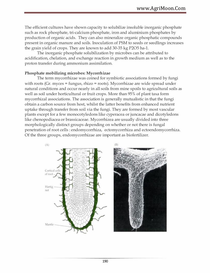
www.AgriMoon.Com
190
The efficient cultures have shown capacity to solubilize insoluble inorganic phosphate such as rock phosphate, tri-calcium phosphate, iron and aluminium phosphates by production of organic acids. They can also mineralize organic phosphatic compounds present in organic manure and soils. Inoculation of PSM to seeds or seedlings increases the grain yield of crops. They are known to add 30-35 kg P2O5 ha-1. The inorganic phosphate solubilization by microbes can be attributed to acidification, chelation, and exchange reaction in growth medium as well as to the proton transfer during ammonium assimilation.
Phosphate mobilizing microbes: Mycorrhizae The term mycorrhizae was coined for symbiotic associations formed by fungi
with roots (Gr. myces = fungus, rhizo = roots). Mycorrhizae are wide spread under natural conditions and occur nearly in all soils from mine spoils to agricultural soils as well as soil under horticultural or fruit crops. More than 95% of plant taxa form mycorrhizal associations. The association is generally mutualistic in that the fungi obtain a carbon source from host, whilst the latter benefits from enhanced nutrient uptake through transfer from soil via the fungi. They are formed by most vascular plants except for a few monocotyledons like cyperacea or juncacae and dicotyledons like chenopodiacea or brassicaceae. Mycorrhizea are usually divided into three morphologically distinct groups depending on whether or not there is fungal penetration of root cells : endomycorrhiza, ectomycorrhiza and ectoendomycorrhiza. Of the three groups, endomycorrhizae are important as biofertilizer.
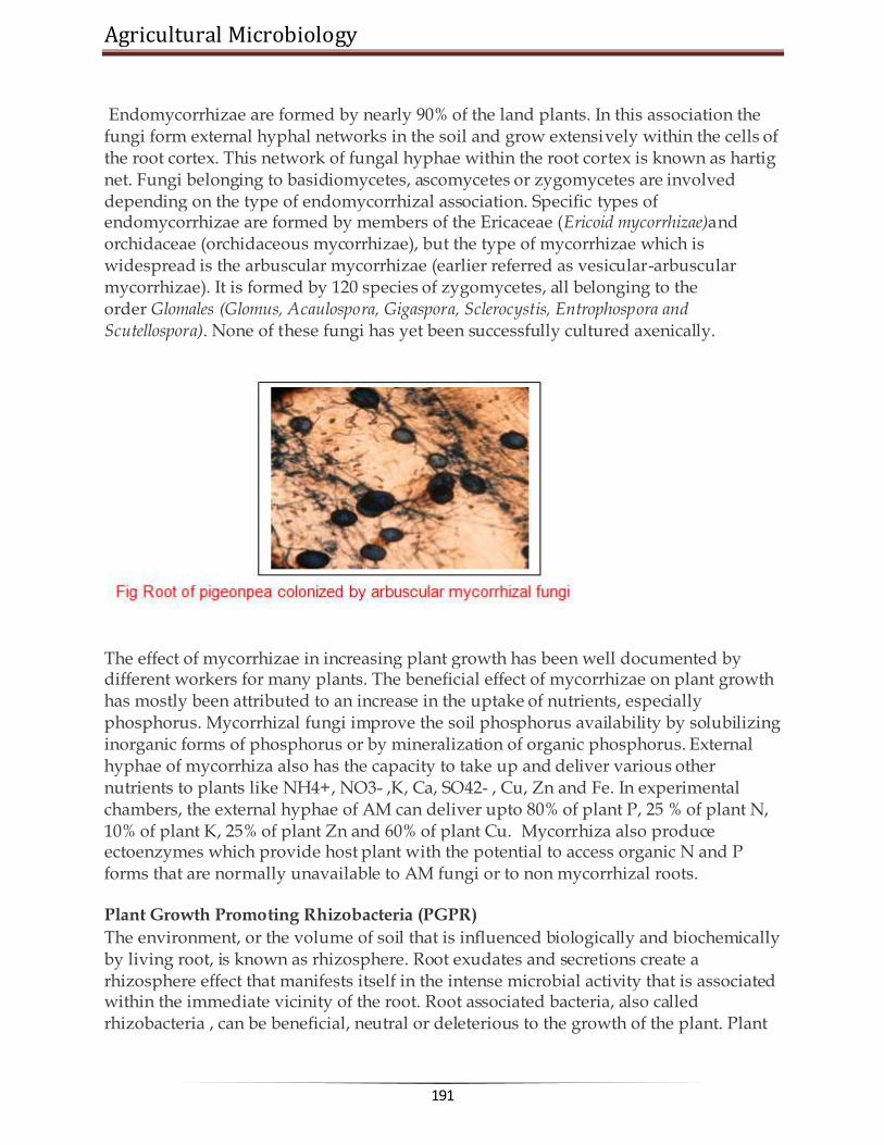
Agricultural Microbiology
191
Endomycorrhizae are formed by nearly 90% of the land plants. In this association the fungi form external hyphal networks in the soil and grow extensively within the cells of the root cortex. This network of fungal hyphae within the root cortex is known as hartig net. Fungi belonging to basidiomycetes, ascomycetes or zygomycetes are involved depending on the type of endomycorrhizal association. Specific types of endomycorrhizae are formed by members of the Ericaceae (Ericoid mycorrhizae)and orchidaceae (orchidaceous mycorrhizae), but the type of mycorrhizae which is widespread is the arbuscular mycorrhizae (earlier referred as vesicular-arbuscular mycorrhizae). It is formed by 120 species of zygomycetes, all belonging to the order Glomales (Glomus, Acaulospora, Gigaspora, Sclerocystis, Entrophospora and Scutellospora). None of these fungi has yet been successfully cultured axenically.
The effect of mycorrhizae in increasing plant growth has been well documented by different workers for many plants. The beneficial effect of mycorrhizae on plant growth has mostly been attributed to an increase in the uptake of nutrients, especially phosphorus. Mycorrhizal fungi improve the soil phosphorus availability by solubilizing inorganic forms of phosphorus or by mineralization of organic phosphorus. External hyphae of mycorrhiza also has the capacity to take up and deliver various other nutrients to plants like NH4+, NO3- ,K, Ca, SO42- , Cu, Zn and Fe. In experimental chambers, the external hyphae of AM can deliver upto 80% of plant P, 25 % of plant N, 10% of plant K, 25% of plant Zn and 60% of plant Cu. Mycorrhiza also produce ectoenzymes which provide host plant with the potential to access organic N and P forms that are normally unavailable to AM fungi or to non mycorrhizal roots.
Plant Growth Promoting Rhizobacteria (PGPR)
The environment, or the volume of soil that is influenced biologically and biochemically by living root, is known as rhizosphere. Root exudates and secretions create a rhizosphere effect that manifests itself in the intense microbial activity that is associated within the immediate vicinity of the root. Root associated bacteria, also called rhizobacteria , can be beneficial, neutral or deleterious to the growth of the plant. Plant

www.AgriMoon.Com
192
growth promoting rhizobacteria (PGPR) are one class of beneficial bacteria inhabiting the soil ecosystem (Kloepper et al 1989). The effects of PGPR on plant growth can be mediated by direct or indirect mechanisms (Glick 1995). The direct effects have been most commonly attributed to the production of plant hormones such as auxins, gibberellins and cytokinins, or by supplying biologically fixed nitrogen or solubilizing insoluble P. These PGPR also affect growth by indirect mechanisms such as supperssion of bacterial, fungal and nematode pathogens by the production of siderophores, HCN, ammonia, antibiotics, volatile metabolites etc., by induced systemic resistance and/or by competing with the pathogen for nutrients or for colonization space. The nitrogen fixing and phosphate solubilising bacteria have been discussed separately. Other PGPR include bacteria belonging to the genera Arthrobacter, Bacillus, Burkholderia, Enterobacter Klebsiella, Pseudomonas, Xanthomonas, Serratia and many more yet to be identified. Effects of PGPR on plant growth have been evaluated by many workers on different crops. Increase in plant height and root and shoot biomass of wheat was reported following inoculation with 12 different isolates of PGPR belonging to Pseudomonas aeruginosa, P. cepacia, P. fluorescens and P. putida. Similarly treatment of wheat seeds with fluorescent pseudomonads (antagonistic to Gaeumannomyces graminis) resulted in yield increases of 27% in field trials. PGPR are potent inoculants but are not commercialised due to lack of consistency under field conditions. Recent work suggests that combination of PGPR strains (two or more) which have diverge mode of plant growth promotion or antagonism against soil-borne pathogens are more effective than single strain inoculum. IAA producing Bacillus isolates promoted root growth and (or) nodulation when coinoculated with Rhizobium etli on Phaseolus vulgaris. Similarly coinoculation of soybean with B. japonicum and Serratia liquefaciens 2-68 or S. proteamaculans 1-102 increased soybean grain and protein yield compared to the non treated controls. Better biocontrol of take all disease of wheat was observed when fluorescent Pseudomonas was applied in combination with Trichoderma koningii. Seed treatment containing combinations of Escherichia coli S17R1 andBurkholderia cepacia BC-B provided significantly greater suppression of cucumber seedling pathogenesis in a field soil naturally infested with Pythium and Fusariumspp. than seeds treated with strain BC-B, S17R1 or Enterobacter cloacae 501 R3. Experiments performed at chernobyl showed that coinoculation of ‗duet‘ of nitrogen fixing Klebsiella oxytoca VN13 and Xanthomonas maltophila VN12 could protect maize from radionuclides penetration; as well improve the yield and percentage of protein in seed.

Agricultural Microbiology
193
Many bacterial genera have shown their potential for biocontrol both under in vivo and in vitro conditions. Agrobacterium, Arthrobacter, Alcaligenes, Bacillus,Escherichia coli, Enterobacter, Pseudomonas, Burkholderia, Rhizobium and Serratia were found to be potent for suppression of soil-borne fungal pathogens. Many of these biocontrol agents exhibited their effectiveness under field conditions also.
******☺******

www.AgriMoon.Com
194
Lecture 26: MICROBIAL AGENTS FOR CONTROL OF PLANT DISEASES
The intensive use of pesticides in agriculture is a cause of serious concern. The problem is especially serious because of the development of resistance to pesticides in important pests and the presence of pesticide residue in agricultural and dairy products. In India, the most serious problem of resistance is witnessed in cotton, for which American bollworm is a serious pest. The bollworm has developed resistance to almost all pesticides in a number of regions and is serious problem in many states. Other important pests of cotton, white fly and jassid, have also developed pesticide resistance in some places. Growing pesticide resistance has meant that a large proportion of agricultural production is lost to pests. Pesticide resistance has mainly been caused by excessive and indiscriminate use of pesticides. Pesticides of spurious quality, which are commonly sold in small towns and villages, have also contributed to resistance in many areas. Excessive use of chemical pesticides in agriculture is a serious cause of concern. It is, therefore, important that alternative, environmental friendly methods of plant protection are adopted such as integrated pest management (IPM) techniques, including the use of biopesticides. Biopesticides are an important group of pesticides that can reduce pesticide risks. They are derived from animals, plants and microorganisms such as bacteria and viruses. The advantages of biopesticides are:
They are inherently less harmful than chemical pesticides. They, in general, have a narrow target range and a very specific mode of action. They are often effective in small quantities. Also, they decompose quickly and do
not leave problematic residues. They are safer to humans and the environment than conventional pesticides.
Biopesticides is a broad term and includes bioinsecticides, biofungicides, bioherbicides and bionematicides. Microorganisms belonging to different groups like bacteria, fungi ans viruses are used as biopesticides. You will learn about these three groups of organisms in following text.
Bacterial Bacteria belonging to genus Bacillus are potent against many insect pests. They suppress pests by producing a toxin specific to the pest; causing a disease; preventing establishment of other microorganisms through competition; or other modes of action.

Agricultural Microbiology
195
An example of a bacterial pesticide is Bacillus thuringiensis, or "Bt." Bacillus thuringiensis is a naturally occurring soil bacteria that is toxic to the larvae of several species of insects but not toxic to non-target organisms. It is primarily a pathogen of lepidopterpous pests that are some of the most damaging. These include american bollworm in cotton and stem borers in rice. Bacillus thuringiensis can be applied to plant foliage or incorporated into the genetic material of crops. Bacillus thuringiensis, as discovered, is toxic to the caterpillars (larvae) of moths and butterflies. Several strains of Bt have been developed and now strains are available that control fly larvae. These can be used in controlling mosquitoes and blackflies.
Bacillus thuringiensis (Bt) is a ubiquitous gram-positive, spore forming bacterium which produces parasporal crystals during sporulation (stationary phase of its growth cycle). These crystals are predominantly comprised of d-endotoxins or insecticidal crystal proteins (ICPs), known to possess insecticidal activity when ingested by certain insects. The mode of action of Bt involves the following stages:
ingestion of sporulated Bt and ICP by an insect larva. solubilization of the crystalline ICP in the midgut: When Bt crystals are
ingested by insects, the crystal proteins are dissolved from the crystals. The pH in the gut of lepidopteran larvae varies between 9 and 12 and the lepidopteran-specific crystal bodies can only be solubilized above pH9.5. On getting solubilized in the midgut, the crystalline bodies release the protein called d-endotoxins.
activation of the ICP by midgut proteases: The crystalline protoxins are inactive, until they are hydrolysed by the gut proteases. The proteases cleave amino acids from both C-terminus and N-terminus of the protoxin and thus forms the active toxin.
binding of the activated ICP to specific receptors in the midgut cell
membrane: Brush border membrane vesicles (BBMVs) is the primary binding site for several insect species. The active toxins initially bind reversibly to the specific receptors located on the apical brush border membrane of the columnar cells.
insertion of the toxin in the cell membrane and formation of pores and
channels in the gut cell membrane, followed by destruction of the epithelial cells:After binding to the receptor, the toxin inserts irreversibly into the plasma
membrane of the cell. The formation of toxin induced pores in the columnar cell of apical membranes allows rapid fluxes of ions. The disruption of the gut integerity leads to the death of the insect through starvation or septicemia.
subsequent Bt spore germination and septicemia may enhance mortality.
For biopesticide applications, the Bt protein is usually used in a formulation containing the spores and crystalline inclusions that are released upon lysis of Bt cells during growth. The molecular potency of the toxin is 300 times greater than synthetic
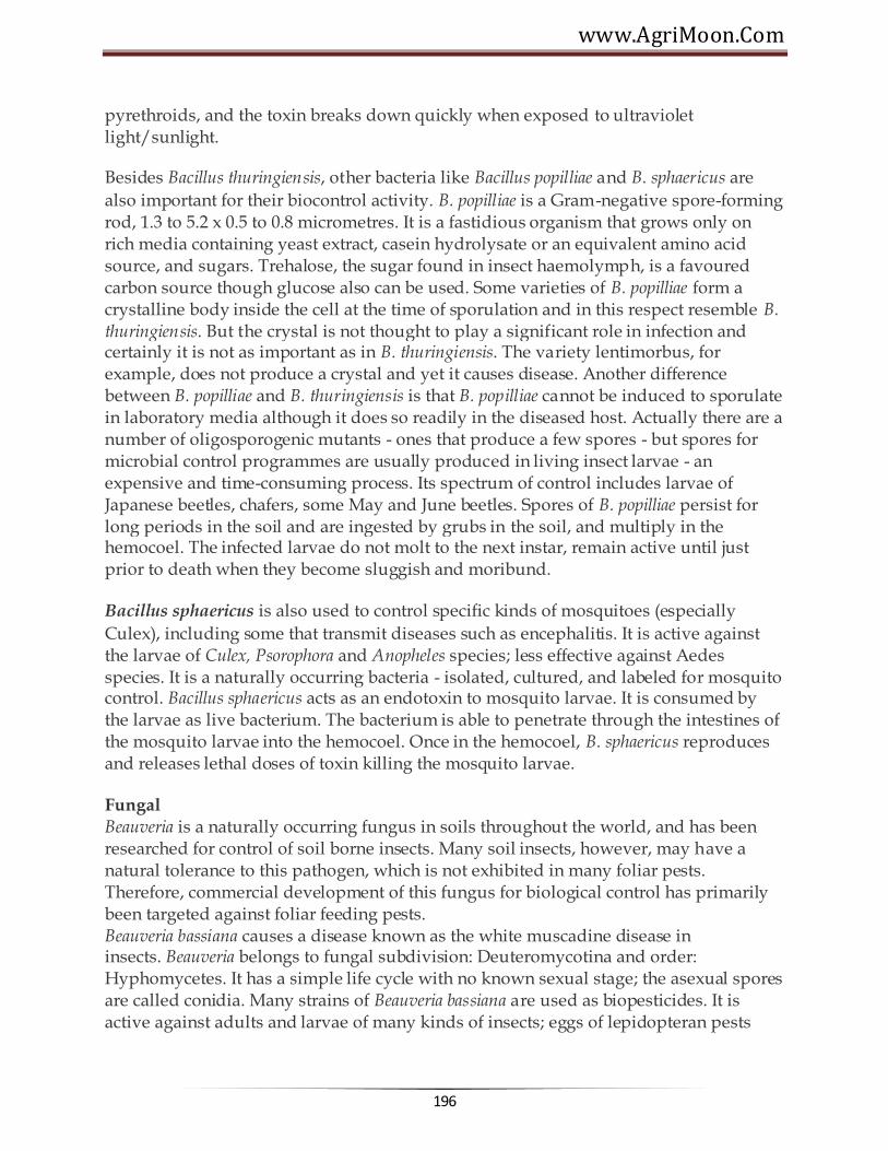
www.AgriMoon.Com
196
pyrethroids, and the toxin breaks down quickly when exposed to ultraviolet light/sunlight.
Besides Bacillus thuringiensis, other bacteria like Bacillus popilliae and B. sphaericus are
also important for their biocontrol activity. B. popilliae is a Gram-negative spore-forming rod, 1.3 to 5.2 x 0.5 to 0.8 micrometres. It is a fastidious organism that grows only on rich media containing yeast extract, casein hydrolysate or an equivalent amino acid source, and sugars. Trehalose, the sugar found in insect haemolymph, is a favoured carbon source though glucose also can be used. Some varieties of B. popilliae form a crystalline body inside the cell at the time of sporulation and in this respect resemble B. thuringiensis. But the crystal is not thought to play a significant role in infection and certainly it is not as important as in B. thuringiensis. The variety lentimorbus, for example, does not produce a crystal and yet it causes disease. Another difference between B. popilliae and B. thuringiensis is that B. popilliae cannot be induced to sporulate in laboratory media although it does so readily in the diseased host. Actually there are a number of oligosporogenic mutants - ones that produce a few spores - but spores for microbial control programmes are usually produced in living insect larvae - an expensive and time-consuming process. Its spectrum of control includes larvae of Japanese beetles, chafers, some May and June beetles. Spores of B. popilliae persist for long periods in the soil and are ingested by grubs in the soil, and multiply in the hemocoel. The infected larvae do not molt to the next instar, remain active until just prior to death when they become sluggish and moribund.
Bacillus sphaericus is also used to control specific kinds of mosquitoes (especially
Culex), including some that transmit diseases such as encephalitis. It is active against the larvae of Culex, Psorophora and Anopheles species; less effective against Aedes species. It is a naturally occurring bacteria - isolated, cultured, and labeled for mosquito control. Bacillus sphaericus acts as an endotoxin to mosquito larvae. It is consumed by the larvae as live bacterium. The bacterium is able to penetrate through the intestines of the mosquito larvae into the hemocoel. Once in the hemocoel, B. sphaericus reproduces and releases lethal doses of toxin killing the mosquito larvae.
Fungal Beauveria is a naturally occurring fungus in soils throughout the world, and has been researched for control of soil borne insects. Many soil insects, however, may have a natural tolerance to this pathogen, which is not exhibited in many foliar pests. Therefore, commercial development of this fungus for biological control has primarily been targeted against foliar feeding pests. Beauveria bassiana causes a disease known as the white muscadine disease in insects. Beauveria belongs to fungal subdivision: Deuteromycotina and order: Hyphomycetes. It has a simple life cycle with no known sexual stage; the asexual spores are called conidia. Many strains of Beauveria bassiana are used as biopesticides. It is active against adults and larvae of many kinds of insects; eggs of lepidopteran pests

Agricultural Microbiology
197
such as moths. The spectrum also includes mole cricket, chiggers, white grubs, fire ants, ants, flea beetle, boll weevil, whiteflies, plant bug, grasshoppers, thrips, aphids, mites and many others.
When spores of this fungus come in contact with the cuticle (skin) of susceptible insects, they germinate and grow directly through the cuticle to the inner body of their host. The fungus proliferates throughout the insect's body, producing toxins and draining the insect of nutrients, eventually killing it. Therefore, unlike bacterial and viral pathogens of insects, Beauveria and other fungal pathogens infect the insect with contact and do not need to be consumed by their host to cause infection. Once the fungus has killed it's host, it grows back out through the softer portions of the cuticle, covering the insect with a layer of white mold (hence the name white muscadine disease). This downy mold produces millions of new infective spores that are released to the environment.
Viral (Insect Viruses) Baculoviruses are pathogens that attack insects and other arthropods. Like some human viruses, they are usually extremely small (less than a thousandth of a millimeter across), and are composed primarily of double-stranded DNA that codes for genes needed for virus establishment and reproduction. Because this genetic material is easily destroyed by exposure to sunlight or by conditions in the host's gut, an infective baculovirus particle (virion) is protected by protein coat called a polyhedron (plural polyhedra). Most insect baculoviruses must be eaten by the host to produce an infection, that is, typically fatal to the insect.
The majority of baculoviruses used as biological control agents are in the genus Nucleopolyhedrovirus. These viruses are excellent candidates for species-specific, narrow spectrum insecticidal applications (Table 8.8). They have been shown to have no negative impacts on plants, mammals, birds, fish, or even on non-target insects. On the other hand, the high specificity of baculoviruses is also cited as a weakness for agricultural uses, since growers may want one product to use against a variety of pests. Currently, researchers are attempting to use genetic engineering techniques to expand virus host ranges to the desired pest species. Releases of such genetically-engineered baculoviruses have been made by researchers in the U.K. and the United States and show promise, although the cost of commercial production of these agents must be reduced if they are to be competitive.
Viruses are unable to reproduce without a host - they are obligate parasites. Baculoviruses are no exception. The cells of the host's body are taken over by the genetic message carried within each virion, and forced to produce more virus particles until the cell, and ultimately the insect, dies. Most baculoviruses cause the host insect to die in a way that will maximize the chance that other insects will come in contact with the virus and become infected in turn. Infection by baculovirus begins when an insect eats virus particles on a plant - perhaps from a sprayed treatment. The infected insect

www.AgriMoon.Com
198
dies and "melts" or falls apart on foliage, releasing more virus. This additional infective material can infect more insects, continuing the cycle.
It is widely acknowledged that baculoviruses can be as effective as chemical pesticides in controlling specific insect pests. However, the expense of treating a hectare of land with a baculovirus product invariably costs more than an equally efficacious chemical treament. This difference in price is due primarily to the labor intensive nature of baculovirus production. Some viruses can be produced in vitro (within cell cultures in the laboratory, not requiring whole, living insects). These are less expensive than those that can only be produced in vivo, that is, inside of living insects. The cost of rearing live hosts adds greatly to the final cost of the product. It is to be hoped that insect cell culture systems currently being developed for other uses may ultimately make viral pesticides more cost-effective.
Insects killed by baculoviruses have a characteristic shiny-oily appearance, and are often seen hanging limply from vegetation. They are extremely fragile to the touch, rupturing to release fluid filled with infective virus particles. This tendency to remain attached to foliage and then rupture is an important aspect of the virus life-cycle. As discussed above, infection of other insects will only occur if they eat foliage that has been contaminated by virus-killed larvae.

Agricultural Microbiology
199
Viruses used against different insect-pests of plants
Biocontrol
Biocontrol or Biological control can be defined as the use of natural enemies to control pests. Natural enemies of pests are categorized as parasites, predators and pathogens. It is a broad term that also includes use of biopesticides. About 30% of the yield in agriculture is lost because of the combined effects of biotic and abiotic stresses, with pathogenic fungi alone responsible for a reduction of about 12%. Control of fungal pathogens is based on the use of agronomic practices and

www.AgriMoon.Com
200
pesticides, but widespread application of chemicals inundates the agroecosystems with toxic compounds that affect the balance of the natural food chain. In addition, resistant and more virulent pathogen populations are selected causing escalation in the amount of pesticides used. A variety of new technologies are being developed to integrate or substitute the application of chemicals in an attempt to reduce both the ecological and financial cost of disease control. Antagonistic microorganisms are being studied in depth and considered as an attractive option for the development of microbial-based biofungicides. Successful and consistent results have been achieved with some biocontrol agents such asAgrobacterium and Bacillus, whose mechanisms of biocontrol are largely understood. However, limitations in the practical use of bacterial agents often arose from the production of toxic substances and formulations with a short shelf-life. The application of fungal biocontrol agents has also been delayed because of difficulties in obtaining consistent results in biocontrol and the relatively poor understanding of the plant-microbe and microbe-microbe interactions involved in the antagonistic processes.
While diverse microbes may contribute to the biological control of plant pathogens, most research and development efforts have focused on isolates of three genera,Bacillus, Trichoderma, and Pseudomonas.
The most studied fungal biocontrol agents are Trichoderma spp. and some isolates, effective as biofungicides in certain culture conditions, have been recently introduced in commercial agriculture. Concurrently, fundamental discoveries concerning the mechanism of action of these fungi have been made. Studies on the mechanism of biocontrol had indicated that Trichoderma and other mycoparasites have developed a vast array of molecular tools to support their parasitic behaviour. It is believed that Trichoderma produces different types of lytic enzymes that act on the cell wall of fungi and kill them. Genes encoding for cell wall degrading enzymes (CWDES) such as chitinolytic, glucanolytic and proteolytic enzymes have been isolated and used to improve biocontrol capabilities of Trichoderma strains.
Two species of Trichoderma, T. harzianum and T. viride are commonly used as biocontrol agent. Their spectrum of control includes fungal pathogens like Armillaria, Pythium, Rhizoctonia, Verticillium, Sclerotium and Botrytis.
******☺******

Agricultural Microbiology
201
Lecture 27:
BIOGAS PRODUCTION
BIOGAS (Methane) Biogas is mixture of methane (50-60 per cent), CO2 (30 – 40 per cent), hydrogen
(5-10 per cent), H2S and nitrogen (traces), produced from anaerobic digestion of animal, plant wastes or any cellulose containing waste material. The digester used for biogas production is called a Biogas plant. A typical biogas plant using cowdung as a raw material consists of: (a) digester and (b) gas holder. The digesters are either of (a) batch type which are filled once, sealed and emptied when the raw materials stop producing gas or (b) continuous type which are fed with a definite quantity of waste at regular intervals so that gas production is continuous and regular. The nature of fermentation in the digester is anaerobic
Methane production
Involves three process viz., hydrolysis, acidification and methonization
Four different groups of organisms are involved Hydrolytic bacteria -catabolises carbohydrates,proteins, lipids and other
components of biomass into fatty acids,H2 and CO2 H2 producing acetogenic bacxteria :Catabolises fatty acids, and the neutral ebd
products of I group into acetate,CO2 and H2 Homoacetogenic bacteria : Synthesize acetate using H2,CO2 and formate Methanogenic bacteria : Utilizes acetate,CO2,H2 to produce methane
The first group of bacteria includes facultative as well as strict anaerobes like Cellulomonas, Bacillus, Eubacterium etc.

www.AgriMoon.Com
202
Methanogenic phase
It is a strict anaerobic phase and during this phase organic Carbon is converted to microbial mass, CO2 and methane.
These bacteria are sensitive to pH and optimum is 6.8- 7.2. Drop in pH leads to inhibition of methanogenesis
Methanobacterium, Methanomicrobium, Methanococcus, Methanosarcina

Agricultural Microbiology
203
Alcohol production
Production of ethyl alcohol from sugary materials is one of the oldest known microbiological processes. Alcohol is an important solvent and raw material used in a variety of chemical industries. Although today industrial alcohol is also produced synthetically from ethylene, production of alcohol by fermentation of cheap sugary materials such as molasses by yeast is still an important industry.
For ethyl alcohol production, selected strains of Saccharomyces cerevisae are employed since all the strains are not equally efficient. The alcohol tolerance and sugar tolerance are important criteria used in the selection of yeast strains. Strains tolerant to high sugar and alcohol concentration are desired. The raw material generally used is either crude cane molasses or best molasses which contain about 50 per cent fermentable sugars. The
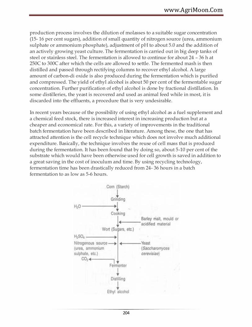
www.AgriMoon.Com
204
production process involves the dilution of molasses to a suitable sugar concentration (15- 16 per cent sugars), addition of small quantity of nitrogen source (urea, ammonium sulphate or ammonium phosphate), adjustment of pH to about 5.0 and the addition of an actively growing yeast culture. The fermentation is carried out in big deep tanks of steel or stainless steel. The fermentation is allowed to continue for about 24 – 36 h at 250C to 300C after which the cells are allowed to settle. The fermented mash is then distilled and passed through rectifying columns to recover ethyl alcohol. A large amount of carbon-di oxide is also produced during the fermentation which is purified and compressed. The yield of ethyl alcohol is about 50 per cent of the fermentable sugar concentration. Further purification of ethyl alcohol is done by fractional distillation. In some distilleries, the yeast is recovered and used as animal feed while in most, it is discarded into the effluents, a procedure that is very undesirable.
In recent years because of the possibility of using ethyl alcohol as a fuel supplement and a chemical feed stock, there is increased interest in increasing production but at a cheaper and economical rate. For this, a variety of improvements in the traditional batch fermentation have been described in literature. Among these, the one that has attracted attention is the cell recycle technique which does not involve much additional expenditure. Basically, the technique involves the reuse of cell mass that is produced during the fermentation. It has been found that by doing so, about 5-10 per cent of the substrate which would have been otherwise used for cell growth is saved in addition to a great saving in the cost of inoculum and time. By using recycling technology, fermentation time has been drastically reduced from 24- 36 hours in a batch fermentation to as low as 5-6 hours.

Agricultural Microbiology
205
Distilled Alcoholic Beverages
Whisky, rum, gin, brandy and vodka, are all products of alcoholic fermentation of different raw materials such as grains, molasses, potatoes and fruits are distilled products containing higher content of alcohol than either wine or beer. In principle, production of these alcoholic beverages is similar to the production of industrial alcohol. The quality of the alcohol produced and used for the production of these beverages depends on the type of secondary fractional alcohol distillation. It is essential to derive a pure spirit for the production of these beverages.
Whisky is traditionally produced from the fermentation of grain mash, rum from molasses, brandy from grape wines and vodka from potatoes. The flavour and aroma are introduced into the purified alcohol during the blending process. Sometimes, these alcoholic beverages are allowed to age for long periods in wooden casks before they are consumed.
******☺******

www.AgriMoon.Com
206
Lecture 28:
BIODEGRADABLE PLASTICS
Introduction
Although plastics as we know them today are a relatively recent invention, they have become an important part of modern life.
The age of plastics
Today, 200 billion pounds (100 million tons) of plastics are produced worldwide every year. Plastics are used for packaging, building materials, and virtually every type of consumer product. Past ages of human society have been called the Stone, Bronze, Copper, Iron, and Steel Ages, based on the material that was relied upon the most during that time. Today, the total volume of plastics produced worldwide has surpassed that of steel and continues to increase. Without a doubt, we have entered the Age of Plastics. Some common plastic items include: sunglasses, tooth brushes, super glue, paint brushes, tennis shoes, Frisbees, 2-liter bottles, Honda CRX's, Astroturf, photographs, street signs, pens, automobile paint, video tapes, rubber bands, balloons, bicycle tires, umbrellas, guitar strings, carpeting, shower doors, hearing aids, Scotch Tape, fishing lines, trash bags, and toilet seats. Plastic can be found in everything from clothing to machinary. It is important to understand the nature of plastics, and the consequences of their production and use. Virtually all plastics are made from nonrenewable resources, such as oil, coal or natural gas, which will eventually become exhausted. Plastics waste is increasing, adding to the already burdensome problems of waste management. And the use of plastics continues to grow, raising the important question, how can we balance convenient living with concern for ecology? To understand this concern, it is helpful to understand what plastics are.
Why green plastics? Green plastics are the focus of an emerging industry focused on making convenient living consistent with environmental stability. One reason to make a shift toward the use of green plastics is the availability of raw materials. Green plastics can be made using polymers that come from agricultural and marine feedstocks. These are abundant natural resources that are constantly being replenished. This, in turn could revitalize rural economy, both agricultural and marine, by providing additional demand for currently underutilized land or low-valued biomass commodities. Another favorable property of green plastics is their biodegradability, making them a natural material for use in such applications as compostable collection bags, such as for food or yard waste.

Agricultural Microbiology
207
But bioplastics have to possess adequate physical properties. Their properties have to be managed and controlled with technological means through the development of adequate formulations and plastics processing. The commercial ventures already under way in the United States, Canada, Europe, and Japan indicate that there is confidence technological advances are possible. The key to solving technical problems often simply knows what the problems are. Bioplastics also have to be cost-competitive. Commercially available biopolymers are typically more expensive than synthetic polymers, often significantly so. Currently only starch competes with synthetic polymers in terms of cost.
Interest in the development of bioplastics will grow largely to the extent that there is real interest in and concern over the environment. Societal concern over the environment is already being reflected in governmental restrictive legislation on the use of plastics, particularly aimed at plastic packaging. Legislation has begun at the local, state, federal, and international levels, and legislation will undoubtedly increase in the future. New legislation will likely contain restrictions aimed at materials that are neither recyclable nor biodegradable. Labeling legislation may lead to an "ecolabel," based on a product's raw material usage, energy consumption, emissions from manufacture and use, and waste disposal impact. Most of all, what is needed is a paradigm shift.
Making it a reality Ignoring nature's way of building strong materials, we have, for many applications, over-engineered our plastics for stability, with little consideration of their recyclability or ultimate fate, and ended up transforming irreplaceable resources into mountains of waste. There is another way. We can take nature's building materials and use them for our purposes, without taking them out of nature's cycles. We can be borrowers, not consumers, so that the process can continue indefinitely. If society is indeed, becoming more and more committed to resource conservation, environmental preservation and sustainable technologies, bioplastics will find their place in this Age of Plastics. The widespread use of these new plastics will depend on developing technologies that can be successful in the marketplace. That in turn will partly depend on how strongly society is committed to the concepts of resource conservation, environmental preservation, and sustainable technologies. There are growing signs that people indeed want to live in greater harmony with nature and leave future generations a healthy planet. If so, bioplastics will find a place in the current Age of Plastics.
Plastics Plastics are a class of material that has one or more polymers as its primary ingredient, that is shaped by flow when it is processed (usually using heat), and that is solid in its final form. Plastics can be made up of many different kinds of polymer, and can be processed in many different ways, but as long as they satisfy these three conditions, they are bona fide plastics.

www.AgriMoon.Com
208
The general "recipe" for any kind of plastic is a combination of three ingredients: a polymer, one or more plasticizers, and one or more additives. These ingredients can then be processed into different shapes, resulting in a wide variety of different materials with different properties.
Polymers Polymers are long molecules. They are one of the basic components of all
plastics.
Synthetic polymers Synthetic polymers are polymers that are man-made. Most synthetic polymers
are manufactured from petroleum. Some examples of synthetic polymers include:
Polystyrene is the polymer found in styrofoam, used for everything from
packing materials and insulation to drinking cups. Polyvinyl chloride, widely known by its abbreviation PVC, is used in a lot of
building material (and is well-known as being ubiquitous in piping).
These materials are generally not biodegradable, and because they are made from petroleum, once the basic materials for creating them are used up, we cannot make any more.
Biopolymers Biopolymers are polymers that occur in nature. Carbohydrates and proteins, for
example, are biopolymers. Many biopolymers are already being produced commercially on large scales, although they usually are not used for the production of plastics. Even if only a small percentage of the biopolymers already being produced were used in the production of plastics, it would significantly decrease our dependence on manufactured, non-renewable resources. Some examples of biopolymers include:
Cellulose is the most plentiful carbohydrate in the world; 40 percent of all
organic matter is cellulose! Starch is found in corn (maize), potatoes, wheat, tapioca (cassava), and some
other plants. Annual world production of starch is well over 70 billion pounds, with much of it being used for non-food purposes, like making paper, cardboard, textile sizing, and adhesives.
Collagen is the most abundant protein found in mammals. Gelatin is denatured
collagen, and is used in sausage casings, capsules for drugs and vitamin preparations, and other miscellaneous industrial applications including photography.

Agricultural Microbiology
209
Casein, commercially produced mainly from cow's skimmed milk, is used in
adhesives, binders, protective coatings, and other products. Soy protein and zein (from corn) are abundant plant proteins. They are used for
making adhesives and coatings for paper and cardboard. Polyesters are produced by bacteria, and can be made commercially on large
scales through fermentation processes. They are now being used in biomedical applications.
A number of other natural materials can be made into polymers that are biodegradable. For example:
Lactic acid is now commercially produced on large scales through the
fermentation of sugar feedstocks obtained from sugar beets or sugar cane, or from the conversion of starch from corn, potato peels, or other starch source. It can be polymerized to produce polylactic acid.
Triglycerides can also be polymerized. Triglycerides make up a large part of the
storage lipids in animal and plant cells. Over sixteen billion pounds of vegetable oils are produced in the United States each year, mainly from soybean, flax, and rapeseed. Triglycerides are another promising raw material for producing plastics.
These natural raw materials are abundant, renewable, and biodegradable, making them attractive feedstocks for bioplastics.
Plasticizers A plasticizer is a substance that can be added to material to increase its workability, flexibility, or pliability. Plasticizers are one of basic ingredients of all plastics.
Additives An additive is a substance that can be added to material to change its properties, usually to make the end product more desirable in some way. Additives are one of basic ingredients of all plastics. The pure polymer resin by itself may not always have the physical properties needed in the final product; it may be strong but too brittle, flexible but too elastic, or flexible and elastic but just plain ugly. Just like the polymer material itself, additives come in different varieties: some can be found in the environment, while others are manufactured. The amounts and types of additives used in manufacturing plastics are another factor that influence how environmentally-friendly they are.
Green Plastics Green Plastics, sometimes also called Bioplastics, are plastics that are
biodegradable and are usually made mostly or entirely from renewable resources.

www.AgriMoon.Com
210
Frequently there is also a focus on environmentally friendly processing. Green plastics are the focus of an emerging industry focused on making convenient living consistent with environmental stability. Like all plastics, bioplastics are composed of a polymer, combined with plasticizers and additives, and processed using extrusion or thermosetting. What makes green plastics "green" is one or more of the following properties:
1. they are biodegradable 2. they are made from renewable ingredients 3. they have environmentally friendly processing
Because different compounds can satisfy some or all of these criteria to different degrees, there are different "degrees of green" in green plastics. To evaluate how "green" a plastic material is, you need to ask three questions:
5. How quickly can the plastic be re-integrated into the environment after it is no longer being used?
6. How quickly are the ingredients that go into making the plastic created in the environment?
7. How much pollution or waste is created during the process of actually making the plastic?
Traditional plastics fail on all three of these points.
1. Biodegradability Biodegradation is a process where something breaks down into simple
compounds as a result of the action of microorganisms (like bacteria, fungi, or algae). The term biodegradation is actually a contraction, short for "biotic degradation." Something is biodegradable if it can be broken down by this kind of process. In order to say that something "biodegrades", it therefore has to meet the following requirements:
1. it has to break down (this is simply "degradation") 2. it's molecules have to break down from complex molecules into simpler ones
(this is "chemical degradation") 3. The breaking down of its molecules has to be accomplished by microorganisms.
4. In order to prevent misinformation in advertising, standards organizations have made even more strict requirements for something to be labelled as biodegradable. In addition to the above list, something can only be labelled as "biodegradable" if:

Agricultural Microbiology
211
5. The biodegradation of the material has to be scientifically measurable. Since most biodegradation produces CO2 as a by-product, usually this is measured by the amount of CO2 produced.
6. The biodegradation of the material has to be fast enough to have a significant effect in a reasonable amount of time. For example, the ISO standard requires 60% biodegradation within 180 days for a material to be called biodegradable; the Europenan Norm EN13432 is stricter, requiring 90% biodegradation within 90 days.
Types of Biodegradation Because biodegradation requires microorganism to do something to a material, usually the material has to be broken up into smaller pieces first. As a result, most biodegradable materials become biodegradable after the action of another kind of degradation.
Hydro-biodegradable
Hydro-biodegradable materials are first broken down by interaction with water
(a process called hydrolysis), and then are further broken down by microorganisms.
Photo-biodegradable
Photo-biodegradable materials are first broken down by interaction with sunlight (a process called photolysis), and then are further broken down by microorganisms.
Oxo-degradable
Some companies have been claiming that they have created an additive that can be added to traditional plastics to make them biodegradable. These products become what is called oxo-degradable, and sometimes is incorrectly identified as oxo-
biodegradable.
Although this allows the plastic to return to the environment, these products are not biodegradable. Instead, the additive allows the plastic material to break down physically when exposed to water, into pieces small enough to be accidentally ingested by microbes. However, the microbes are not able to actually break this material down further. The end result is therefore a material that combines biomass with polymer residue. The plastic never decomposes as a result of interaction with the organisms. This process is therefore more accurately called "disintergration" rather than "biodegradation".
For bioplastics to become practical, they must have properties that allow them to compete with the current plastics on the market: bioplastics must be able to be strong, resiliant, flexible, elastic, and above all, durable. It is the very durability of traditional plastics that has helped them in the marketplace, and has been a major goal of plastics research throughout the years. However, it is exactly this durability that now has

www.AgriMoon.Com
212
people increasingly worried. Now that we wrap our sandwiches in bags that will still be around when the sandwich, and even the person who ate it, are long gone, many people are wondering: have we gone too far?
There is a lot of current research going on concerning methods of decomposition. There is also research on controlling the time-line of biodegradation. One goal of this research is to make a product that is programmed-degradable: in other words, a product that allows you to control when and how it degrades, while insuring that the product remains strong while it is still in use.
2. Renewability A renewable resource is a natural resource that is created in the environment faster than it is used up by people. Many people think of "renewability" as a fixed trait: some things (like trees, grass, and wind) are renewable, while others (like oil and coal) are not. In fact, whether a resource is renewable depends on both how fast it is replenished and how fast people use it. As a result, some resources are more renewable than others, and some resources may or may not be renewable depending on how they are used.
Rate of Renewal The rate of rewal (sometimes also called the "sustainable yield") of a resource tells you how quickly it can be replenished by the environment. Solar energy, tides, rainfall, and winds are considered perpetual resources for energy because they renew much faster than they could ever be used. (Can you imagine us "using up" the wind, so that we would have to wait until the earth made more?) Living organisms provide the majority of resources that are generally considered "renewable", because they generally renew themelves within a reasonable amount of time relative to how quickly they are use. Agricultural feedstocks and marine feedstocks are two major categories of living organism feedstocks. Within this category, some organisms renew faster than others: for example, it takes much longer to grow a new tree than it does to grow grass.
Most of the resources that are considered "non-renewable" are based on coal, oil, natural gas, and other substances that take so long for the environment to create that almost any use of these resources at all will cause them to be used up before any more is created. Petro-chemical feedstocks are feedstocks derived from petroleum principally for the manufacture of chemicals, synthetic rubber, and a variety of plastics.
Rate of Use Imagine you live in a small village by a river. A turbine on the river spins, and it can generate enough electricity for the entire village every day. Clearly, their hydroelectric power is a completely renewable resource. However, as the size of the village grows, their energy use grows. If eventually the needs of the village far outstrip

Agricultural Microbiology
213
the energy that can be provided by the turbine, then the hydroelectric energy from the river is no longer a renewable resource for the village: the rate of use has exceeded the rate of replenishment.
The same issue exists for the use of plants. As long as our use of (for example) corn remains moderate compared to the amount of corn produced, corn is a renewable resource. However, if our use of corn increases dramatically without a corresponding increase in corn crop production, then corn will cease to be a renewable resource: we will use it all up, and we will either have to cease production until the corn renews itself or (worse) it will become extinct, so it will not replenish at all.
3. Processing
When making plastics, the initial mass of polymer, called resin, is processed into different shapes using a variety of methods, including: extrusion, injection molding, compression molding, transfer molding, and casting. Different processing techniques result in the wide variety of forms that plastic can take: ranging from thin films and elastic sheets, to resiliant panels and hard, solid three-dimensional shapes.
HISTORY OF BIOPLASTICS The use of natural polymers is not entirely a new idea. In one form or another, green plastics have been around for a long time.
Early History
Natural resins-like amber, shellac, and gutta percha-have been mentioned throughout history, including during Roman times and the Middle Ages. Native Americans were developing and refining techniques for making ladles and spoons from animal horns long before there was any European contact. In Europe, molded horn jewelry and snuff boxes were popular in the eighteenth century.
The 1800's
Significant commercialization of bioplastics only began in the middle of the nineteenth century... The American inventor, John Wesley Hyatt, Jr., was looking for a substitute for ivory in the manufacture of billiard balls, and in 1869 patented a cellulose derivative for coating non-ivory billiard balls. That attempt, however, was affected by the coating's flammability; balls were occasionally ignited when lit cigars accidentally came into contact with them. Hyatt continued working on the project and soon developed celluloid, the first widely used plastic, now most widely known for its use in photographic and movie film.
The 1900's
The history of plastics changed dramatically in the early 1900s, as petroleum emerged as a source of fuel and of chemicals. The early bioplastics were simply

www.AgriMoon.Com
214
displaced by plastics made from synthetic polymers. World War II brought on a large increase in plastics production, a growth which continues to this day.
The 1920's
In the 1920s Henry Ford experimented with using soybeans in the manufacture of automobiles. Ford was partly motivated by a desire to find non-food applications for agricultural surpluses, which existed then as they do now. Soy plastics were used for an increasing number of automobile parts, like steering wheels, interior trim, and dashboard panels. Finally Ford gave the go-ahead to produce a complete prototype "plastic car." Ford, a master at generating publicity, exhibited the prototype with great fanfare in 1941, but by the end of the year was no longer publicizing the "plastic car," probably for a variety of reasons. World War II played a role: armament work took precedent over almost everything else, and steel shortages limited all non-defense production. Today plastic automobile parts are common, but the use of plastics made from renewable raw materials got side-tracked.
The 1960's
One well established bioplastic that has survived the growth of the synthetic plastics industry is cellophane, a sheet material derived from cellulose. Although production peaked in the 1960s it is still used in packaging for candy, cigarettes, and other articles.
The 2000's and Beyond
Demand for materials like plastics is continually growing and will not be abated. Today, the plastics industry is an important component of our economy: The U.S. plastics industry includes over 20,000 facilities that produce or distribute materials or products, employ over 1.5 million workers, and ship over $300 billion in products each year. The magnitude of the plastics industry, however, is itself a cause for concern. The pressures of increasing waste and diminishing resources have lead many to try to re-discover natural polymers and put them to use as materials for manufactor and industry. As a result, there is increasing interest in the promise of a new generation of green plastics.
******☺******

Agricultural Microbiology
215
Lecture 29: PLANT – MICROBE INTERACTIONS
Plant - Microbe symbioses 1. Many microbes (bacteria, fungi) have important symbioses with plants 2. Rhizosphere = thin layer of soil immediately attached to root hairs of plants. Typically contains 109 microbes/g of soil. 3. Many rhizosphere organisms are ectosymbionts, living outside the roots. Others are endosymbionts, living inside or penetrating into plant roots. 4. Many of these bacteria contribute Nitrogen fixation, obtain plant nutrients in return (see below for Rhizobium symbiosis). Rhizobium-Legume symbioses 5. Plants of the legume family (soybeans, clover, alfalfa, beans, peas) can grow in soils lacking nitrogen compounds required by other plants. How? 6. These plants contain endosymbiotic Rhizobium bacteria that grow in root nodules.
Rhizobia can fix atmospheric Nitrogen gas (N2)N2 + 6[H] 2 NH3 7. The reaction requires total lack of oxygen and lots of energy as ATP. To bind oxygen and get rid of it, bacteria use protein called leghemoglobin, somewhat similar to animal hemoglobin. Globin part is encoded in plant genome, heme group is encoded in bacterial genome. Neither partner can fix nitrogen alone, only in symbiosis.
Hydrothermal vent Communities
1. Occurs only near thermal springs on ocean floor, 2 miles or more below surface. Totally black, no sunlight penetrates below 600 feet.

www.AgriMoon.Com
216
2. Associated with spreading centers of tectonic plates where hot magma close to surface causes area of floor to slowly drift apart.
3. Seawater seeps down, mixes w/ minerals at high temperature comes back to ocean water in plumes at 270-380 deg. C. These are sometimes called black smokers since minerals precipitate as black cloud when in contact with cold sea water .
4. Contains high levels of inorganics: Mn2+, H2, usually H2S; very low in organic matter
5. Astonishing discovery: such regions are densely populated by a community of unusual animals: 2 m long tube worms, giant clams, mussels, white shrimp.
6. What do they eat? Unlike earth's surface, there is no source of light to stimulate phototrophs, they "eat" chemolithotrophic bacteria!
7. Example: Inside the tube worms live huge colonies of bacterial endosymbionts. These are autotrophic chemolithotrophs, oxidizing sulfide to sulfate as their energy source. As bacteria grow, they provide carbon and nitrogen compounds for worms to feed on. Have not been cultivated outside of host, so little is known about details of the bacterium.
Ruminant Symbiosis
1. Ruminants are the herbivorous mammals whose digestive tract contains four chambers. First chamber (largest) is the rumen, provides a place for bacteria to break down the fiber in the plants so the cow can use it for energy.Ý
2. Includes cows, sheep, giraffes, buffalo, and elk. 3. Ruminants eat grasses and other plant materials, but do not produce
enzymes to digest cellulose, the primary plant metabolite. 4. Instead, ruminants rely on huge microbial community in rumen to digest
plant materials. Microbial densities can reach as high as 1012 microorganisms/ml, the highest density found anywhere in nature.
5. Ruminants feed off fermentation waste products of microorganisms; mainly acetic acid, propionic acid, and butyric acid.
Gnotobiotic Animals
1. Gnotobiotic = "known microbiota"; animal host is either entirely free of microbes (aka "germfree", "axenic") or has a microbiota whose identity is completely known.
2. Animals in utero are germfree, but acquire resident bacteria within hours of birth.

Agricultural Microbiology
217
3. Relatively easy to produce germfree animals for birds. Sterilize shell, use sterile incubator, keep animals in an environment where all air, food, water is sterilized before entry.
4. More difficult to establish germfree animals other than birds. Need cesarean section of pgrenant females, germfree isolation chambers where all air, food, water is sterilized before entry.
5. Germfree animals generally are less healthy than animals with normal microbiota. Defects include:
1. Greater vitamin requirements for K and B complex 2. lower cardiac output 3. much more susceptible to pathogens -- normal microbiota colonize access
sites, often compete successfully to prevent pathogens from binding to host tissues.much smaller infectious dose required to initiate an infection
Interrelationship between microorganisms: Beneficial and harmful relationships Interrelationships in soils are of 3 types
1. Plant microbe interaction 2. Microbe – microbe interaction and 3. Plant microbe – microbe interaction
a) Plant microbe interaction
It mainly constitutes the association of microorganism with plants little in a positive way or in a negative way. The positive approach is mainly the symbiotic relationships and the negative approach constituents mainly pathogen plant interactions.
b) Plant microbe – microbe interaction Also called tripartite symbiosis Eg: Alnus – Frankia –Mycorrhiza and Casbarina – Frankia – Mycorrhiza Ceanothus roots, with Frankia vesicles

www.AgriMoon.Com
218
c) Microbe – microbe interaction
Microbial interaction in soil
Interrelationship between microorganisms: Beneficial and harmful relationship
Microorganisms live in the soil, not in the form of pure culture, but as complex populations. Each particle of soil contains more than one type of organisms. So, microbial ecosystem of soil is the sum of the biotic and the abiotic components of soil. Many of these organisms depend upon one another for direct and indirect nutrients. Some complete with one another for energy sources and for the elements and components used as nutrients. This results in the formation of numerous associations among the soil micro organic. The composition of the microflora of any habitat is governed by the biological equilibrium created by the associations and interactions of all individual found in the community.
The micro organic that inhabits the soil exhibited many different types of associations or interactions. Some of the associations are indifferent or neutral, some are beneficial type of interactions and others are detrimental or negative.
Beneficial / positive interactions a. neutralism b. Symbiosis / mutualism c. Protoco-operation d. Communalism
a) Neutralism
It is a type of neutral association, in two microorganisms behaves entirely independently or eg: Each could utilize different nutrients with out producing metabolic end products that are inhibitory. This might be transitory as the condition change in the environment, parituclary the availability of nutrients, the relationship might change.
Symbiosis / Mutualism Mutualism is a form of symbiosis in which both organisms benefit. An example of mutualism is a clownfish and sea anemones. The clownfish gets protection, while the sea anemones become clean. This is mutualism, because both water animals benefit from having each other around
Agricultural Microbiology
219
b) Proto co-operation One type of mutualistic association is that involving the exchange of nutrients between two species, a phenomenon called syntrophism. Many micro organic synthesize the vitamins and anaerobic acids in excess of their nutritional requirements. Others have a requirement of one or more of these nutrients. Hence certain combinations of species will grow together but not apart when nutrient levels are very low. Nutritional proto co-operation has been demonstrated in cultures. Eg: In a medium deficient in nicotinic acid and biotin, neither Proteus vulgaris nor Bacillus polymyxa will multiply as the former (B) requires nicotinic acid and the latter biotin. In mixed culture, in the same medium however both grown since the partner bacterium synthesizes the missing vitamins.
c) Symbiosis The living together of two or more organisms; microbial association Symbiotic association is evident in soil among several groups of organisms algae and fungi in lichens, bacteria residing with in protozoa cells, bacteria and roots in the legume symbiosis, fungi and roots in mycoorhizae. In lichens, the algae and fungi are in such an intimate physical and physiological relationship that the lichens they make are classified as distinct organism. The alga benefits in part are se of the protection afforded to it by the hyphae that envelop and protect it from environmental stresses. While, the fungi gains by making use of the CO2 fixed by its photosynthetic partner. Where BGA participants, the heterotraph benefits from the fixed N2. Symbiotic relationship exists between micro and macro organisms. R-L associate N2 fixed is transferred to legume and organic which is transferred to the ® by CO2 metabolizing legume host.
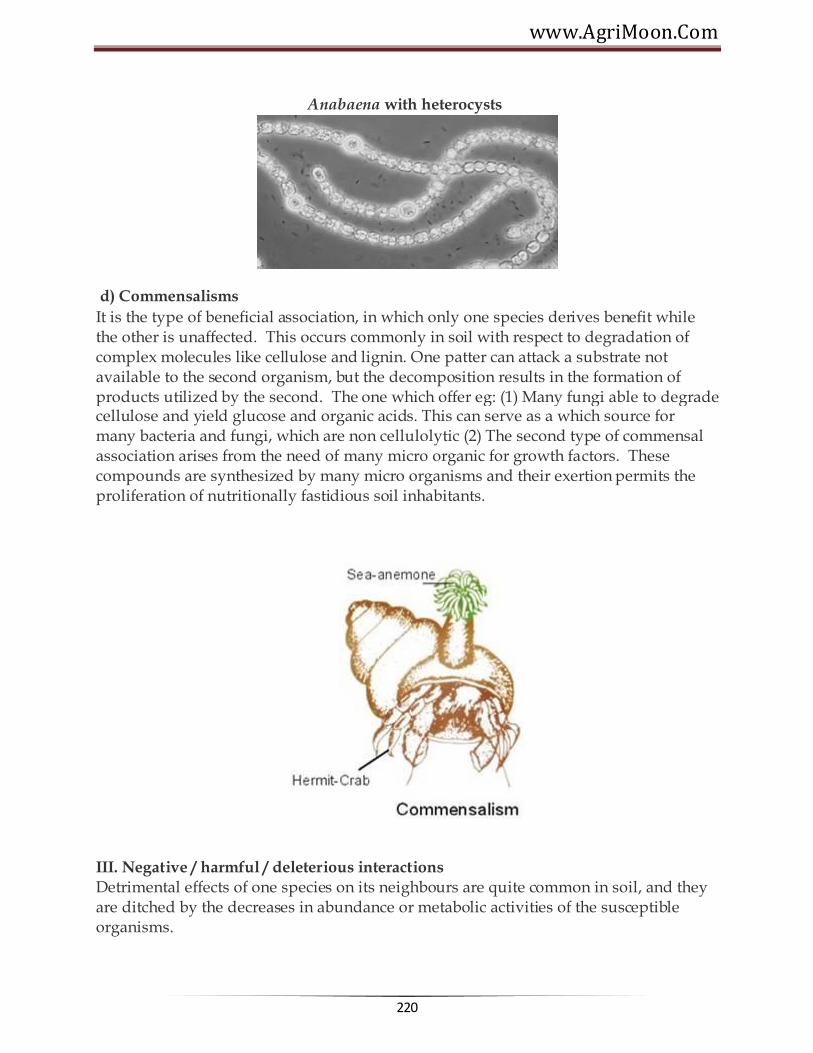
www.AgriMoon.Com
220
Anabaena with heterocysts
d) Commensalisms
It is the type of beneficial association, in which only one species derives benefit while the other is unaffected. This occurs commonly in soil with respect to degradation of complex molecules like cellulose and lignin. One patter can attack a substrate not available to the second organism, but the decomposition results in the formation of products utilized by the second. The one which offer eg: (1) Many fungi able to degrade cellulose and yield glucose and organic acids. This can serve as a which source for many bacteria and fungi, which are non cellulolytic (2) The second type of commensal association arises from the need of many micro organic for growth factors. These compounds are synthesized by many micro organisms and their exertion permits the proliferation of nutritionally fastidious soil inhabitants.
III. Negative / harmful / deleterious interactions Detrimental effects of one species on its neighbours are quite common in soil, and they are ditched by the decreases in abundance or metabolic activities of the susceptible organisms.

Agricultural Microbiology
221
This include a) Competition b) Amensalism c) Parasitism and predation
a. Competition
It is the rivalry for limiting nutrients or other common needs. In such situations the best adapted microbial species will predominate or infact, eliminate other species which are dependent upon the same limited nutrient substances. Eg: Competition between strains derived from soil and those applied with legume seeds at the time of sowing. The better competitor involves the root hairs more frequently and it is responsible for a high % of nodules.
Sea Anemones compete for the territory in tide pools
b. Amensalism It is a negative interaction, in which the release of products by one species is toxic to its neighbours. Antagonism is a type of ammensalism.
. Antagonism
The killing, injury or inhibition of growth of one species of micro organisms by another or when one organism adversely affects the environmental of the other is refered as antagonism. The toxic compounds are antibiotic. An antibiotic is a substance formed by one organic that in low concentrations inhibits the growth of another
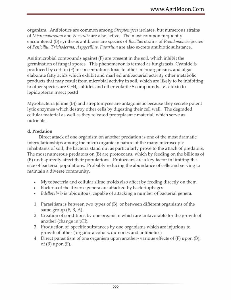
www.AgriMoon.Com
222
organism. Antibiotics are common among Streptomyces isolates, but numerous strains of Micromonespora and Nocardia are also active. The most common frequently encountered (B) synthesis antibiosis are species of Bacillus strains of Pseudomonasspecies of Peniciliu, Trichoderma, Aspgerillus, Fusarium are also excrete antibiotic substance. Anitimicrobial compounds against (F) are present in the soil, which inhibit the germination of fungal spores. This phenomenon is termed as fungistasis. Cyanide is produced by certain (F) in concentrations toxic to other microorganisms, and algae elaborate fatty acids which exhibit and marked antibacterial activity other metabolic products that may result from microbial activity in soil, which are likely to be inhibiting to other species are CH4, sulfides and other volatile S compounds. B. t toxin to lepidopteran insect pestd Myxobacteria (slime (B)) and streptomyces are antagonistic because they secrete potent lytic enzymes which destroy other cells by digesting their cell wall. The degraded cellular material as well as they released protoplasmic material, which serve as nutrients.
d. Predation Direct attack of one organism on another predation is one of the most dramatic interrelationships among the micro organic in nature of the many microscopic inhabitants of soil, the bacteria stand out as particularly prove to the attach of predators. The most numerous predators on (B) are protozoans, which by feeding on the billions of (B) undisputedly affect their populations. Protozoans are a key factor in limiting the size of bacterial populations. Probably reducing the abundance of cells and serving to maintain a diverse community.
Myxobacteria and cellular slime molds also affect by feeding directly on them Bacteria of the diverse genera are attacked by bacteriophages Bdellovibrio is ubiquitous, capable of attacking a number of bacterial genera.
1. Parasitism is between two types of (B), or between different organisms of the same group (F, B, A).
2. Creation of conditions by one organism which are unfavorable for the growth of another (change in pH).
3. Production of specific substances by one organisms which are injurious to growth of other ( organic alcohols, quinones and antibiotics)
4. Direct parasitism of one organism upon another- various effects of (F) upon (B), of (B) upon (F).
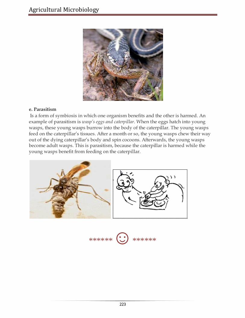
Agricultural Microbiology
223
e. Parasitism
Is a form of symbiosis in which one organism benefits and the other is harmed. An example of parasitism is wasp’s eggs and caterpillar. When the eggs hatch into young wasps, these young wasps burrow into the body of the caterpillar. The young wasps feed on the caterpillar‘s tissues. After a month or so, the young wasps chew their way out of the dying caterpillar‘s body and spin cocoons. Afterwards, the young wasps become adult wasps. This is parasitism, because the caterpillar is harmed while the young wasps benefit from feeding on the caterpillar.
******☺******
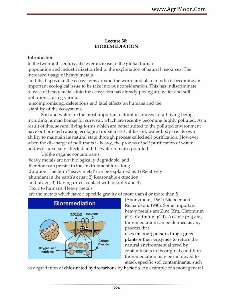
www.AgriMoon.Com
224
Lecture 30: BIOREMEDIATION
Introduction
In the twentieth century, the ever increase in the global human population and industrialization led to the exploitation of natural resources. The increased usage of heavy metals and its disposal in the ecosystems around the world and also in India is becoming an important ecological issue to be take into our consideration. This has indiscriminate release of heavy metals into the ecosystem has already posing air, water and soil pollution causing various uncompromising, deleterious and fatal effects on humans and the stability of the ecosystems. Soil and water are the most important natural resources for all living beings including human beings for survival, which are recently becoming highly polluted. As a result of this, several living forms which are better suited to the polluted environment have out bursted causing ecological imbalance. Unlike soil, water body has its own ability to maintain its natural state through process called self purification. However when the discharge of pollutants is heavy, the process of self purification of water bodies is adversely affected and the water remains polluted. Unlike organic contaminants, heavy metals are not biologically degradable, and therefore can persist in the environment for a long duration. The term ‗heavy metal‘ can be explained as 1) Relatively abundant in the earth‘s crust; 2) Reasonable extraction and usage; 3) Having direct contact with people; and 4) Toxic to humans. Heavy metals are the metals which have a specific gravity of more than 4 or more than 5
(Anonymous, 1964; Nieboer and Richardson, 1980). Some important heavy metals are Zinc (Zn), Chromium (Cr), Cadmium (Cd), Arsenic (As) etc., Bioremediation can be defined as any process that uses microorganisms, fungi, green plantsor their enzymes to return the natural environment altered by contaminants to its original condition. Bioremediation may be employed to attack specific soil contaminants, such
as degradation of chlorinated hydrocarbons by bacteria. An example of a more general

Agricultural Microbiology
225
approach is the cleanup of oil spills by the addition of nitrate and/or sulfate fertilisers to facilitate the decomposition of crude oil by indigenous or exogenous bacteria.
Mycoremediation is a form of bioremediation that uses fungi to reduce the level of contamination in a given environment. Fungi can release specific enzymes and acids that break down the major components of plant fiber. The accumulation of waste is directly proportional to population and as our population grows scientists will be under more pressure to find ways to eliminate contaminants from our environment. Bioremediation microbes will continue to be used in an attempt to return our polluted environments to their original state. Corning, Barnstead, and BD supply products used by those researching bioremediation.
Bioremediation technologies can be generally classified as in situ or ex situ. In situ bioremediation involves treating the contaminated material at the site while ex situinvolves the removal of the contaminated material to be treated elsewhere. Some examples of bioremediation technologies are bioventing, landfarming, bioreactor, Composting, bioaugumentation, rhizofiltration, and biostimulation.

www.AgriMoon.Com
226
BIOREMEDIATION STRATEGIES Different techniques are employed depending on the degree of saturation and aeration of an area. In situ techniques are defined as those that are applied to soil and groundwater at the site with minimal disturbance. Ex situ techniques are those that are applied to soil and groundwater at the site which has been removed from the site via excavation (soil) or pumping (water). Bioaugmentation techniques involve the addition of microorganisms with the ability to degrade pollutants.

Agricultural Microbiology
227
In situ bioremediation
These techniques are generally the most desirable options due to lower cost and less disturbance since they provide the treatment in place avoiding excavation and transport of contaminants. In situtreatment is limited by the depth of the soil that can be effectively treated. In many soils effective oxygen diffusion for desirable rates of bioremediation extend to a range of only a few centimeters to about 30 cm into the soil, although depths of 60 cm and greater have been effectively treated in some cases. The most important land treatments are:
Incineration Excavation
Bioventing
Is the most common in situ treatment and involves supplying air and nutrients through wells to contaminated soil to stimulate the indigenous bacteria. Bioventing employs low airflow rates and provides only the amount of oxygen necessary for the biodegradation while minimizing volatilization and release of contaminants to the atmosphere. It works for simple hydrocarbons and can be used where the contamination is deep under the surface.

www.AgriMoon.Com
228
In situ biodegradation Involves supplying oxygen and nutrients by circulating aqueous solutions through contaminated soils to stimulate naturally occurring bacteria to degrade organic contaminants. It can be used for soil and groundwater. Generally, this technique includes conditions such as the infiltration of water-containing nutrients and oxygen or other electron acceptors for groundwater treatment.
Biosparging:
Biosparging involves the injection of air under pressure below the water table to increase groundwater oxygen concentrations and enhance the rate of biological degradation of contaminants by naturally occurring bacteria. Biosparging increases the mixing in the saturated zone and there- by increases the contact between soil and groundwater. The ease and low cost of installing small-diam- eter air injection points allows considerable flexibility in the design and construction of the system.
Bioaugmentation:
Bioremediation frequently involves the addition of microorganisms indigenous or exogenous to the contaminated sites. Two factors limit the use of added microbial cultures in a land treatment unit: 1) nonindigenous cultures rarely compete well enough with an indigenous population to develop and sustain useful population levels and 2) most soils with long-term exposure to biodegradable waste have indigenous microorganisms that are effective degrades if the land treatment unit is well managed.
Ex situ bioremediation
These techniques involve the excavation or removal of contaminated soil from ground.
Landfarming
Is a simple technique in which contaminated soil is excavated and spread over a prepared bed and periodically tilled until pollutants are degraded. The goal is to stimulate indigenous biodegradative microorganisms and facilitate their aerobic degradation of contaminants. In general, the practice

Agricultural Microbiology
229
is limited to the treatment of superficial 10–35 cm of soil. Since landfarming has the potential to reduce monitoring and maintenance costs, as well as clean-up liabilities, it has received much attention as a disposal alternative.
Composting
Is a technique that involves combining contaminated soil with nonhazardous organic amendants such as manure or agricultural wastes. The presence of these organic materials supports the development of a rich microbial population and elevated temperature characteristic of composting.
Biopile
Are a hybrid of landfarming and composting. Essentially, engineered cells are constructed as aerated composted piles. Typically used for treatment of surface contamination with petro- leum hydrocarbons they are a refined version of landfarming that tend to control physical losses of the contaminants by leaching and volatilization. Biopiles provide a favorable environment for indigenous aerobic and anaerobic microorganisms.
Bioreactors
Slurry reactors or aqueous reactors are used for ex situ treatment of contaminated soil and water pumped up from a contaminated plume. Bioremediation in reactors involves the processing of contaminated solid material (soil, sediment, sludge) or water through an engineered containment system. A slurry bioreactor may be defined as a containment vessel and apparatus used to create a three-phase (solid, liquid, and gas) mixing condition to increase the bioremediation rate of soil- bound and water-soluble pollutants as a water slurry of the contaminated soil and biomass (usually indigenous microorganisms) capable of degrading target contaminants. In general, the rate and extent of biodegradation are greater in a bioreactor system than in situ or in solid-phase systems because the contained environment is more manageable and hence more controllable and predictable. Despite the advantages of reactor systems, there are some

www.AgriMoon.Com
230
disadvantages. The contaminated soil requires pre treatment (e.g., excavation) or alternatively the contaminant can be stripped from the soil via soil washing or physical extraction (e.g., vacuum extraction) before being placed in a bioreactor.
Advantages of bioremediation
• Bioremediation is a natural process and is therefore perceived by the public as an acceptable waste treatment process for contaminated material such as soil. Microbes able to degrade the contaminant increase in numbers when the contaminant is present; when the contaminant is degraded, the biodegradative population declines. The residues for the treatment are usually harmless products and include carbon dioxide, water, and cell biomass. • Theoretically, bioremediation is useful for the complete destruction of a wide variety of contaminants. Many compounds that are legally considered to be hazardous can be transformed to harmless products. This eliminates the chance of future liability associated with treatment and disposal of contaminated material. • Instead of transferring contaminants from one environmental medium to another, for example, from land to water or air, the complete destruction of target pollutants is possible. • Bioremediation can often be carried out on site, often without causing a major disruption of nor- mal activities. This also eliminates the need to transport quantities of waste off site and the poten- tial threats to human health and the environment that can arise during transportation. • Bioremediation can prove less expensive than other technologies that are used for clean-up of hazardous waste. Disadvantages of bioremediation
• Bioremediation is limited to those compounds that are biodegradable. Not all

Agricultural Microbiology
231
compounds are susceptible to rapid and complete degradation. • There are some concerns that the products of biodegradation may be more persistent or toxic than the parent compound. • Biological processes are often highly specific. Important site factors required for success include the presence of metabolically capable microbial populations, suitable environmental growth conditions, and appropriate levels of nutrients and contaminants. • It is difficult to extrapolate from bench and pilot-scale studies to full-scale field operations. • Research is needed to develop and engineer bioremediation technologies that are appropriate for sites with complex mixtures of contaminants that are not evenly dispersed in the environment. Contaminants may be present as solids, liquids, and gases. • Bioremediation often takes longer than other treatment options, such as excavation and removal of soil or incineration. • Regulatory uncertainty remains regarding acceptable performance criteria for bioremediation. There is no accepted definition of ―clean‖, evaluating performance of bioremediation is difficult, and there are no acceptable endpoints for bioremediation treatments.
******☺******

www.AgriMoon.Com
232
Lecture 31:
BIOSENSOR
Biosensor is an analytical device for the detection of an analyte that combines a
biological component with a physicochemical detector component.
It consists of 3 parts:
The sensitive biological element (biological material (eg. tissue, microorganisms, organelles, cell receptors, enzymes, antibodies, nucleic acids, etc), a biologically derived material or biomimic) the sensitive elements can be created by biological engineering.
The transducer or the detector element (works in a physicochemical way; optical, piezoelectric, electrochemical, etc.) that transforms the signal resulting from the interaction of the analyte with the biological element into another signal (i.e., transducers) that can be more easily measured and quantified;
Associated electronics or signal processors that are primarily responsible for the display of the results in a user-friendly way. This sometimes accounts for the most expensive part of the sensor device, however it is possible to generate a user friendly display that includes transducer and sensitive element (seeHolographic Sensor).
A common example of a commercial biosensor is the blood glucose biosensor, which uses the enzyme glucose oxidase to break blood glucose down. In doing so it first oxidizes glucose and uses two electrons to reduce the FAD (a component of the enzyme) to FADH2. This in turn is oxidized by the electrode (accepting two electrons from the electrode) in a number of steps. The resulting current is a measure of the concentration of glucose. In this case, the electrode is the transducer and the enzyme is the biologically active component.
Recently, arrays of many different detector molecules have been applied in so called electronic nose devices; where the pattern of response from the detectors is used to fingerprint substance. Current commercial electronic noses, however, do not use biological elements.
Principles of Detection
Analytical chemistry plays an important role in food quality parameters because almost every sector of industry and public service relies on quality control. A food quality biosensor is a device, which can respond to some property or properties of food and transform the response(s) into a detectable signal, often an electric signal. This signal

Agricultural Microbiology
233
may provide direct information about the quality factor(s) to be measured or may have a known relation to the quality factor. There are various kinds of biosensors most of which work on the principle of one of the following:
Electrochemical Biosensors
Electrochemical biosensors are based on monitoring electroactive species that are either produced or consumed by the action of the biological components (e.g., enzymes and cells). Transduction of the produced signal can be performed using one of several methods under two broad headings:
Potentiometric Biosensors Amperometric Biosensors
Potentiometric Biosensors These are based on monitoring the potential of a system at a working electrode, with respect to an accurate reference electrode, under conditions of essentially zero current flow. In process, potentiometric measurements are related to the analyte activity (of a target species) in the test sample. Potentiometric biosensors can operate over a wide range (usually several orders of magnitude) of concentrations. The use of potentiometric biosensors for food quality analysis has not been as widely reported as for amperometric sensors. However, some of the examples where this approach has been used for food quality analysis include estimating monophenolase activity in apple juice, determining the concentration of sucrose in soft drinks, measuring isocitrate concentrations in fruit juices, and determining urea levels in milk.
Amperometric Biosensors The use of amperometric biosensors in signal transduction has proved to be the most widely reported using an electrochemical approach. Both ―one-shot‖ (disposable) sensors and on-line (multi measurement) devices are commercially available, monitoring a wide range of target analytes. In contrast to potentiometric devices, the principle operation of amperometric biosensors is defined by a constant potential applied between a working and a reference electrode. The applied potential results in redox reactions, causing a net current to flow. The magnitude of this current is proportional to the concentration of electro active species present in test solution and both cathodic (reducing) and anodic (oxidizing) reactions can be monitored amperometrically. Most of the amperometric biosensors described use enzymes as the biorecognition element. Typically, oxidase and dehydrogenase enzymes have been the most frequently exploited catalysts used for these biosensor formats.
Calorimetric Biosensors Most of the biochemical reactions are accompanied by either heat absorption or production. Sensors based on calorimetric transduction are designed to detect heat generated or consumed during a biological reaction; by using sensitive heat detection

www.AgriMoon.Com
234
devices. Various biosensors for specific target analytes have been constructed. In the field of food quality analysis, uses of such biosensors to detect metabolites have been described.
Optical Biosensors
These sensors are based on measuring responses to illumination or to light emission. Optical biosensors can employ a number of techniques to detect the presence of a target analyte and are based on well-founded methods including chemiluminescence, fluorescence, light absorbance, phosphoresence, photothermal techniques, surface plasmon resonance (SPR), light polarization and rotation, and total internal reflectance. For example the use of this technique has been demonstrated to detect the presence of allergens, in particular peanuts, during food production.
Acoustic Biosensors Piezoelectric quartz crystals can be affected by a change of mass at the crystal surface; this phenomenon has been successfully exploited and used to develop acoustic biosensors. For practical applications, the surface of the crystal can be modified with recognition elements (e.g., antibodies) that can bind specifically to a target analyte.
Immunosensors
Immunosensors are based on exploiting the specific interaction of antibodies with antigens. Typically, immunoassays (such as the enzyme-linked immunosorbent assay technique) employ a label (e.g., enzyme, antibody, fluorescent marker) to detect the immunological reaction. The use of biosensor platforms, linked to an immunoassay format, offers a route to rapid and accurate quantitative measurements of target analytes.
Applications of Biosensors There are many potential applications of biosensors of various types. The main requirements for a biosensor approach to be valuable in terms of research and commercial applications are the identification of a target molecule, availability of a suitable biological recognition element, and the potential for disposable portable detection systems to be preferred to sensitive laboratory-based techniques in some situations. Some examples are given below:
Glucose monitoring in diabetes patients ←historical market driver
Other medical health related targets Environmental applications e.g. the detection of pesticides and river water
contaminants Remote sensing of airborne bacteria e.g. in counter-bioterrorist activities Detection of pathogens Determining levels of toxic substances before and after bioremediation Detection and determining of organophosphate

Agricultural Microbiology
235
Routine analytical measurement of folic acid, biotin, vitamin B12 and pantothenic acid as an alternative to microbiological assay
Determination of drug residues in food, such as antibiotics and growth promoters, particularly meat and honey.
Drug discovery and evaluation of biological activity of new compounds. Protein engineering in biosensors Detection of toxic metabolites such as mycotoxins
Utility Biosensors for applications in Agriculture in Food/ Fruit Quality Control Quality control is the essential part of a food industry and efficient quality assurance is becoming increasingly important. Consumers expect good quality and healthy food at a given price; with good shelf life and high safety while food inspections require good manufacturing practices, safety, labelling and compliance with the regulations. Further, food producers are increasingly asking for efficient control methods, in particular through on-line or at-line quality sensors. Their main aim is to satisfy the consumer and regulatory requirements and to improve the production feasibility, quality sorting, automation and reduction of production cost and production time subsequently.
Biochemical Composition of Fruits
The quality of soft fruit, in terms of taste, nutrition and consumers acceptance, is fundamentally based on the biochemical composition of the fruit. In soft fruits (viz. blackcurrant and strawberry) sugar: acid ratios can be used as an important index of fruit maturity and act as a determinant of overall fruit. However, sugar: acid ratios are infrequently used due to a requirement for specific instrumentation and semi-skilled analytical scientists. Today we need a simple and low-cost alternative, which would significantly enhance both the number and extent of tests carried out.
Fruit Maturity, Ripening and Quality Relationships
Fruit maturity at harvest is the most important factor that determines shelf life and final fruit quality. If harvested immature then fruits are more subject to shriveling and mechanical damage, and are of inferior quality when ripe, whereas overripe fruits are liable to become soft and mealy with bland flavour soon after harvest. Therefore, fruits harvested either too early or too late in their season are more susceptible to post harvest physiological disorders than fruits harvested at proper maturity.
Fruits can be divided into two groups:
Fruit that are incapable of enduring their ripening process once picked from the plant like berries, cheery, citrus fruits, grapes, lychee, pineapple, pomegranate, and tamarillo.
Fruits that can be harvested mature and ripped off the plant like apple, apricot, avocado, banana, cherimoya, guava, kiwifruit, mango, nectarine, papaya, passion fruit, pear, peach, persimmon, plum, quince, sapodilla, sapota.

www.AgriMoon.Com
236
Volatile compounds are responsible for the characteristic aroma of fruits and are present in extremely small quantities (<100< g/g fresh wt.). The major volatile formed is ethylene. Scientists are trying to develop portable instruments with sensors that detect volatile production by fruits and hence detecting maturity and quality. Other strategies include the removal of a very small amount of fruit tissue and measurement of total sugar or organic acid content.
Major organic acids in fruits Organic acids function in growth, maturation, senescence, color, and antimicrobial activity of fruits. The low pH of fruits is due to the three most common organic acids present in fruits citric acid, malic acid, and tartaric acid. The total amount of acid in fruits varies widely, from about 0.2% in pear juice to 0.8% in limejuice. The amount and type of acid present in fruits determine the fresh taste of fruits and also affects the shelf life.
Organic Acid as an Indicator of Fruit Maturity
Organic acids directly play an important role in the growth, maturation and acidity of the fruit, and also affect the shelf life of the fruit by influencing the growth of microorganisms. The citric, malic, oxalic, and tartaric acids ranging from 0.1 to 30 g/L were found in orange, grape, and apple juices. There is a considerable difference in the organic acid content found in various types and brands of fruit juice. For example, Minute Maid contains higher levels of oxalic and citric acids when compared to all other orange juices tested. Grape concentrate was found to have lower amount of malic acid than other grape juice, while freshly squeezed grape juice contains higher amount of tartaric acid. Brae burn apples contained the highest amount of citric acid in apples; however Granny Smith apples were the overall most acidic apples tested.
Successful Examples of Organic Acid Biosensors Developed Pyruvic Acid
Onion flavour is principally directed by the perception of pungency. A disposable prototype electrochemical screen-printed (carbon-based) biosensor (C2030519D5, GEM Ltd., Gwent, UK) was constructed using pyruvate dehydrogenase immobilized on mediated Meldolas Blue electrodes and a combined Ag/AgCl reference/counter electrode, both screen-printed onto a PVC substrate to determine pungency in onions (Allium cepa L.). Electrochemical measurements were carried out using a Palm Sense potentiostat (Palm Instruments BV, The Netherlands). The biosensor developed was able to differentiate between mild and pungent bulbs with pyruvate concentrations ranging between 4 and 8 mM in freshly extracted juices. Electrochemical measurements were carried out at +50 mV at 21°C.

Agricultural Microbiology
237
Glucose Biosensors
Most of the glucose biosensors developed are based on immobilized glucose oxidase. In many cases, glucose oxidase has been associated with mediators so as to bring down the high working potential required for hydrogen peroxide breakdown. The α-D glucose sensor developed was also based on glucose oxidase, at the working potential of -350 mV vs. Ag/AgCl, hydrogen peroxide was catalytically oxidized at a rhodinised carbon electrode (White et al, 1994). A novel and simple method which do not involve enzyme or monomer modifications, for the coimmobilization of ferrocene and GOx in a poly(pyrrole) matrix for use as glucose biosensor was developed (Foulds and Lowe, 1988). In spite of the low conductivity of the polypyrrole film formed, the biosensor‘s performance was better than that of other devices reported due to redox mediation of ferrocene that lowers the working potential to 0.4 V. The characterization of the polymer prepared from an ethanolic suspension demonstrated the presence of alcohol interferes in the polymerization kinetics (Pablo et al., 2001). However, this played a beneficial role in efficient immobilization of both, the enzyme and the ferrocene, in a very thin electroactive film. This fact improved the biosensor‘s time response, avoiding mass transport effects. A new type of disposable amperometric biosensor was devised by screen-printing thick-film electrodes directly on a porous nitrocellulose (NC) strip. A glucose biosensor based on hydrogen peroxide detection was constructed by immobilizing glucose oxidase (GOx) on the NC electrode strip and by formulating a

www.AgriMoon.Com
238
strong oxidation layer (i.e., PbO2) at the sample loading area, placed below the GOx reaction band. The screen-printed PbO2 paste serves as a sample pretreatment layer that removes interference by its strong oxidizingability. Samples applied were carried chromatographically, via the PbO2 paste, to the GOx layer, and glucose was catalyzed to liberate hydrogen peroxide, which was then detected at the electrode surface. The proposed NC/ PbO2 strip sensor is shown to be virtually insusceptible to interfering species such as acetaminophen and ascorbic and uric acids and to exhibit good performance, in terms of the sensor to sensor reproducibility. The characterization of metal-decorated CNTs was done using X-ray diffraction analysis, transmission electron microscopy (TEM), high-resolution TEM, scanning electron microscopy, and energy-dispersive X-ray analysis. Amperometric biosensor fabricated by depositing GOD over Nafion-solubilized Au-MWNT electrode retained its biocatalytic activity and obtained fast and sensitive glucose quantification. The fabricated GOD/Au- MWNT/Nafion electrode has a good glucose biosensing potential, and it displayed a linear response up to 22 mM glucose and a detection limit of 20 ìM method.
Sucrose Biosensor Sucrose is an essential part of any fruit, so estimating the concentration of sucrose at different maturity levels could help in identifying the ripening parameters of fruits. Therefore, with regard to sucrose detection, electrodes made up of invertase, mutarotase and glucose oxidase and mediated tri-enzyme electrode based on sucrose phosphorylase and electrocatalytic oxidation of NADH, have been used. Because real samples contain both glucose and sucrose, sucrose sensors have been operated in tandem with glucose oxidase sensors. The sucrose sensor developed was based on the invertase, mutarotase and glucose oxidase reaction scheme and the sucrose level was calculated with respect to the net glucose sensors.
Ascorbic Acid Sensor Ascorbic acid has been measured both by direct electrochemical oxidation and by enzymatic methods using ascorbate oxidase. In the first case, an ascorbate oxidase electrode was used to measure the signal generated by other electroactive interferents in

Agricultural Microbiology
239
the analyte. The second method was based on the measurement of oxygen consumed during the enzyme-catalysed oxidation of ascorbic acid using Clark Electrode.
Lactic Acid Biosensor
The level of lactic acid in blood is used in clinical diagnostics of hypoxia, lactic acidosis, some acute heart diseases and drug toxicity tests. Reliable blood lactate measurements would also be of interest in sports medicine. Lactate can be measured based on the reaction using NAD+ dependent lactate dehydrogenase and ferricyanide . The concentration of dissolved L-lactate was determined in tomato paste and baby food samples using a SIRE-based (sensors based on injection of the recognition element) biosensor. The evaluation principle was based on the injection of small amount of enzyme into an internal delivery flow system and held in direct spatial contact with the amperometric transducer by the use of a semipermeable membrane. All the measurements were based upon the reversible enzymatic conversion of L lactate to pyruvate and hydrogen peroxide by lactate oxidase. The L-lactate concentrations of the tomato paste and baby food were calculated to be 1.02 (0.02 mM) and 2.51 (0.10 mM), respectively, using the standard addition method.
Phenolic Compounds
Phenolic compounds are widespread in nature, and they play a significant role in living organisms. They are used in medicine and industries, including wood processing and pesticide production. Most of the phenolic derivative compounds are highly toxic, and their determination in low concentrations is the significant problem. Scientists are developing various procedures for determining phenols with biosensors. A biosensor based on crude seed hull enzyme extracts has been prepared for monitoring phenol and hydrogen peroxide. The biosensor has confirmed very promising results as a successful instrument to monitor both hydrogen peroxide and phenol. It is an inexpensive biosensor that could be operated for up to 3 weeks with rapid response and stability parameters. In conditions of response to phenol detection, the developed SBP biosensor was found less sensitive than other previously reported biosensors based on purified SBP or HRP or on crude extracts of sweet potato, which have detection limits in the micromolar range for phenols. The foremost reason for this was the low activity of the enzyme extracts. Further work on the improvement of biosensor sensitivity and applications for the detection of chlorophenols and other substituted phenols are in progress.
The amperometric biosensor described glucose oxidase and polyphenol oxidase carbon paste electrodes prepared via a new strategy of carbon paste modification based on the in situ electropolymerizaton of pyrrole monomer previously mixed within the paste. Such alteration induced a better electrical percolation of the carbon structure and enhanced the enzyme entrapment within the electrode material. Therefore, attractive potentialities offered by a biocomposite electrode based on PPO for the detection of flavonols have been demonstrated to control the phenolic levels in beer samples.

www.AgriMoon.Com
240
Benzoic Acid
An amperometric benzoic acid-sensing inhibitor biosensor was prepared by immobilizing mushroom (Agaricus bisporus) tissue homogenate on a Clarktype oxygen electrode. The effects of the quantity of mushroom tissue homogenate, the quantity of gelatin and the effect of the cross-linking agent glutaraldehyde percent on the biosensor were deliberated. The most favourable concentration of phenol used as substrate was 200 mM. The biosensor responded linearly to benzoic acid in a concentration range of 25–100 mM and Standard deviation (s.d.) was found to be ±0.49 μM for 7 successive determinations at a concentration of 75 μM. The inhibitor biosensor based on mushroom tissue homogenate was applied for the determination of benzoic acid in fizzy lemonade, some fruits and groundwater samples. A good concord was shown when the results were compared to those obtained using AOAC method.
Fructose A superior amperometric biosensor based on a solid binding matrix (SBM) composite transducer has been used for the determination of d-fructose in various food samples. The enzyme, d-fructose dehydrogenase (EC 1.1.99.11), was incorporated directly into a solid composite transducer containing both 2-hexadecanone as SBM and chemically modified graphite. The current variation caused by the presence of d-fructose was calculated amperometrically using Hexacyanoferrate (iii) as a redox mediator. The amperometric signals generated were fast, reproducible and linearly proportional to d-fructose concentrations in the range 50×10-6 – 10×10-3mol l-1, with a correlation coefficient of 0.999. A set of measurements at +0.20 V versus SCE for 2×10⁻³ mol l-1 D-fructose yielded a relative standard deviation for the steady-state current of 2.11%. The biosensor selectivity against anionic interferents such as Lascorbate was enhanced by the use of chemically modified graphite by a mild oxidation step. The biosensor was found stable for 6 months and the assay of D-fructose by this electrode was not affected by the presence of sugars or other interferents commonly found in food samples.
ENVIRONMENTAL APPLICATIONS Toxicity In environmental pollution monitoring, it is becoming a general opinion that chemical analysis by itself does not provide sufficient information to assess the ecological risk of polluted waters and wastewaters. In the European Union, along with more stringent demands for water treatment (Council Directive 91/271/EEC), industrial and urban wastewater effluents shall reach certain limits of nontoxicity before the effluent can be discharged into the environment. Thus, much effort has been made during the last years to develop and use different bioassays and biosensors for toxicity evaluation of water samples. Whole organisms are used to measure the potential biological impact (toxicity) of a water or soil sample. That is the case of the toxicity assays Microtox® (Azure, Bucks, UK), or ToxAlert® (Merck, Darmstadt, Germany). These systems are based on the use of luminescent bacteria, Vibrio fischeri, to measure toxicity from

Agricultural Microbiology
241
environmental samples. Bacterial bioluminescence has proved to be a convenient measure of cellular metabolism and, consequently, a reliable sensor for measuring the presence of toxic chemicals in aquatic samples. Some bioassay methods are integrated now in biosensors such as the Cellsense®, which is an amperometric sensor that incorporates Escherichia coli bacterial cells for rapid ecotoxicity analysis. It uses ferricyanine, a soluble electron mediator, to divert electrons from the respiratory system of the immobilized bacteria of a suitable carbon electrode. The resulting current is, thus, a measure of bacterial respiratory activity, and the perturbation by pollutants can be detected as a change in the magnitude of the current. Cellsense has been applied to investigate the toxicity of 3,5-dichlorophenol and other phenols in wastewater, for the determination of nonionic surfactants and benzene sulfonate compounds, for the analysis of wastewater treatment works (WWTW) influent and effluent, and for the toxicity testing of wastewaters and sewage sludge. Moreover, Cellsense has been proposed as one of the newer rapid toxicity assessment methods within the direct toxicity assessment (DTA) demonstration program of the UK Environmental Agency. Most environmental biosensors have focused on bacterial systems while eukariotic biosensors are rare; even more rare is the use of mammalian cells. The mammalian cell, which is more complex than bacteria, can give a more sensitive response when compared to bacteria while also responding to the estrogenic effects of chemicals. A recombinant fluorescent Chinese Hamster Ovary cell line, utilizing a fluorescent reporter system, was used to monitor various toxicants, especially endocrine-disrupting compounds (EDCs), in diverse aqueous environments. EDCs have been also analyzed with a multichannel two-stage mini-bioreactor system using genetically engineered bioluminescent bacteria. The toxicity of various samples spiked with known endocrine-disrupting chemicals, and phenol was investigated.
CONCLUSIONS Despite the huge potential of biosensors, and the ever-increasing number of biosensors developed, commercially available biosensors are being applied to a restricted area of the potential market. In general, biosensors for environmental analysis have several limitations: sensitivity, response time, and lifetime, which should be improved for them to become a competitive analytical tool. The areas of development that are expected to have an impact in biosensor technology are: immobilization techniques, nanotechnology, miniaturization, and multisensor array determinations. However, a crucial aspect may be the production of new sensing elements easy to synthesize and with the capability to broaden the spectra of selectivities that can be reached by a biosensor. At present, the preparation and production in large scales of biomolecules such as enzymes or antibodies need an investment of time and knowledge. Synthetic peptides and MIPs are contemplated as promising alternatives overcoming the above-mentioned limitations. Unfortunately, the affinity accomplished by these synthetic receptors is still several orders of magnitude below that of the antibodies. Improvement in the affinity, specificity, and mass production of the molecular recognition components may ultimately dictate the success or failure of detection technologies. The

www.AgriMoon.Com
242
possibility of tailor binding molecules with predefined properties, such as selectivity, affinity, and stability, is one of the major aims for biotechnology. The development of advanced receptors will allow the analysis of complex real samples and in situ measurements resolving the responses from the analyte and from nonspecific background effects. Since scientific attention is currently being given to biotechnology, as this review has pointed out, the development of improved molecular recognition elements will be followed by a corresponding enhancement of the biosensor features. From the above viewpoint, it is clear that the future of biosensors will rely on the success of emerging sophisticated micro and nanotechnologies, biochemistry, chemistry, thin-film physics, and electronics. To reach this goal, an important investment in research, expertise, and the necessary facilities is needed. However, as the world becomes more concerned about the impact that environmental contamination may cause on public health and the ecosystem, the demand for rapid detecting biosensors will only increase. Biosensors still need to achieve the confidence of potential users, having in mind that the commercialization of new devices will always be the best indicator of the success of a biosensor technology. The analysis of complex matrices and of analytes difficult to determine by the actual analytical procedures (i.e., highly polar compounds), are progressively being approached by biosensors. However, there is still a lack of alternative biosensing systems for an important bunch of emerging contaminants such as bisphenol A, phtalates, and polybrominated compounds (used as flame retardants), veterinary and human medicines and personal care products (nutraceuticals, synthetic fragances, sun screen agents, etc.)

Agricultural Microbiology
243
Lecture 32: MICROBIAL PRODUCTS
The term Industrial Microbiology refers to the use of microorganisms for industrial
purposes. Such things as anticoagulants, antidepressants, vasodilators, herbicides, insecticides, plant hormones, enzymes, and vitamins have been isolated from microorganisms or produced in large quantities by genetically engineering the organisms with foreign genes. In commercial industrial plants, microorganisms are widely used to produce numerous organic materials that have far-reaching value and application.
Antibiotic production
These are defined as substances produced by some micro-organisms which are in some way lethal to other micro-organisms. It is thought that these substances give the organisms that produce them (usually moulds or actinomycetes - which grow slowly) some sort of advantage in competition with other micro-organisms (usually bacteria - which grow fast) in the same habitat. However, their great medical advantage in healing infections is that the purified forms of antibiotics are more or less harmless to most humans. This means that they must act on some aspect of of the growth of micro-organisms which differs from ordinary mammalian cells. There are in fact several versions of Penicillin, variations on a common formula, produced by different strains of Penicillium, or using different culture media and methods. The Production of Antibiotics has been widespread since the pioneering efforts
of Florey and Chain in 1938. The importance of antibiotics to medicine has led to much research into their discovery and production.
Identifying Useful Antibiotics

www.AgriMoon.Com
244
An agar plate streaked with microorganisms. Despite the wide variety of known antibiotics, less than 1% of antimicrobial agents have medical or commercial value. For example, whereas penicillin has a high therapeutic index as it does not generally affect human cells, this is not so for many antibiotics. Other antibiotics simply lack advantage over that already in use, or have no other practical applications.
Useful antibiotics are often discovered using a screening process. To conduct such a screen, isolates of many different microorganisms are cultured and then tested for production of diffusible products that inhibit the growth of test organisms. Most antibiotics identified in such a screen are already known and must therefore be disregarded. The remainder must be tested for their selective toxicities and therapeutic activities, and the best candidates can be examined and possibly modified. A more modern version of this approach is a rational design program. This involves screening directed towards finding new natural products that inhibit a specific target, such as an enzyme only found in the target pathogen, rather than tests to show general inhibition of a culture.
Industrial Production Techniques Antibiotics are produced industrially by a process of fermentation, where the source microorganism is grown in large containers (100,000–150,000 liters or more) containing a liquid growth medium. Oxygen concentration, temperature, pH and nutrient levels must be optimal, and are closely monitored and adjusted if necessary. As antibiotics are secondary metabolites, the population size must be controlled very carefully to ensure that maximum yield is obtained before the cells die. Once the process is complete, the antibiotic must be extracted and purified to a crystalline product. This is simpler to achieve if the antibiotic is soluble in organic solvent. Otherwise it must first be removed by ion exchange, adsorption or chemical precipitation.
Strains Used For Production Microorganisms used in fermentation are rarely identical to the wild type. This is because species are often genetically modified to yield the maximum amounts of antibiotics. Mutation is often used, and is encouraged by introducing mutagens such as ultraviolet radiation, x-rays or certain chemicals. Selection and furtherreproduction of the higher yielding strains over many generations can raise yields by 20-fold or more. Another technique used to increase yields is gene amplification, where copies of genes coding for enzymes involved in the antibiotic production can be inserted back into a cell, via vectors such as plasmids. This process must be closely linked with retesting of antibiotic production and effectiveness.
WINE PRODUCTION Winemaking, or Vinification, is the production of wine, starting with selection of
the grapes or other produce and ending with bottling the finished wine. Although most

Agricultural Microbiology
245
wine is made from grapes, it may also be made from other fruit or non-toxic plant material. Mead is a wine that is made with honey being the primary ingredient after water.
Winemaking can be divided into two general categories: still wine production (without carbonation) and sparkling wine production (with carbonation). The science of wine and winemaking is known as oenology (in American English, enology).
PROCESS After the harvest, the grapes are taken into a winery and prepared for primary ferment, at this stage red wine making diverges from white wine making. Red wine is made from the must (pulp) of red or black grapes that undergo fermentation together with the grape skins. White wine is made by fermenting juice which is made by pressing crushed grapes to extract a juice; the skins are removed and play no further role. Occasionally white wine is made from red grapes, this is done by extracting their juice with minimal contact with the grapes' skins. Rosé wines are made from red grapes where the juice is allowed to stay in contact with the dark skins long enough to pick up a pinkish color, but little of the tannins contained in the skins.
To start primary fermentation yeast is added to the must for red wine or juice for white wine. During this fermentation, which often takes between one and two weeks, the yeast converts most of the sugars in the grape juice into ethanol (alcohol) and carbon dioxide. The carbon dioxide is lost to the atmosphere. After the primary fermentation of red grapes the free run wine is pumped off into tanks and the skins are pressed to extract the remaining juice and wine, the press wine blended with the free run wine at the wine makers discretion. The wine is kept warm and the remaining sugars are converted into alcohol and carbon dioxide. The next process in the

www.AgriMoon.Com
246
making of red wine is secondary fermentation. This is a bacterial fermentation which converts malic acid to lactic acid. This process decreases the acid in the wine and softens the taste of the wine. Red wine is sometimes transferred to oak barrels to mature for a period of weeks or months; this practice imparts oak aromas to the wine. The wine must be settled or clarified and adjustments made prior to filtration and bottling. The time from harvest to drinking can vary from a few months for Beaujolais nouveau wines to over twenty years for top wines. However, only about 10% of all red and 5% of white wine will taste better after five years than it will after just one year. Depending on the quality of grape and the target wine style, some of these steps may be combined or omitted to achieve the particular goals of the winemaker. Many wines of comparable quality are produced using similar but distinctly different approaches to their production; quality is dictated by the attributes of the starting material and not necessarily the steps taken during vinification.
Variations on the above procedure exist. With sparkling wines such as Champagne, an additional fermentation takes place inside the bottle, trapping carbon dioxide and creating the characteristic bubbles. Sweet wines are made by ensuring that some residual sugar remains after fermentation is completed. This can be done by harvesting late (late harvest wine), freezing the grapes to concentrate the sugar (ice wine), or adding a substance to kill the remaining yeast before fermentation is completed; for example, high proof brandy is added when making port wine. In other cases the winemaker may choose to hold back some of the sweet grape juice and add it to the wine after the fermentation is done, a technique known as süssreserve. The process produces wastewater, pomace, and lees that require collection, treatment, and disposal or beneficial use.

Agricultural Microbiology
247
SINGLE CELL PROTEIN Single cell protein (SCP) typically refers to sources of mixed protein extracted from pure or mixed cultures of algae, yeasts, fungi or bacteria (grown on agricultural wastes) used as a substitute for protein-rich foods, in human and animal feeds.
History Early history Since 2500 BC yeasts have been used in bread and beverage production. In 1781 processes for preparing highly concentrated forms of yeast were established. In 1919 Endomyces vernalis yielded fats from sulphite liquor (from paper manufacture), and similarly in 1941 employing Geotrichum. "Food from oil" In the 1960s, researchers at British Petroleum developed what they called "proteins-from-oil process": a technology for producing single cell protein by yeast fed by waxy n-paraffins, a product produced by oil refineries. Initial research work was done by Alfred Champagnat at BP's Lavera Oil Refinery in France; a small pilot plant there started operations in March in 1963, and the same construction of the second pilot plant, at Grangemouth Oil Refinery in Britain, was authorized. The term SCP was coined in 1966 by Carol L. Wilson at MIT.
The "food from oil" idea became quite popular by the 1970s, with Champagnat being awarded the UNESCO Science Prize in 1976, and paraffin-fed yeast facilities being built in a number of countries. The primary use of the product was as poultry and cattle feed.
The Soviets were particularly enthusiastic, opening large "BVK" (belkovo-vitaminny kontsentrat, i.e., "protein-vitamin concentrate") plants next to their oil refineries inKstovo (1973) and Kirishi (1974). The Soviet Ministry of Microbiological Industry had eight plants of this kind by 1989, when, pressured by the environmentalist movements, the government decided to close them down, or convert to some other microbiological processes.

www.AgriMoon.Com
248
SCP Production Process Single cell proteins develop when microbes ferment waste materials (including wood, straw, cannery and food processing wastes, residues from alcohol production, hydrocarbons, or human and animal excreta). The problem with extracting single cell proteins from the wastes is the dilution and cost. They are found in very low concentrations, usually less than 5%. Engineers have developed ways to increase the concentrations including centrifugation, flotation, precipitation, coagulation and filtration, or the use of semi-permeable membranes.
The single cell protein needs to be dehydrated to approximately 10% moisture content and/or acidified to aid in storage and prevent spoilage. The methods to increase the concentrations to adequate levels, and de-watering process require equipment that is expensive and not always suitable for small-scale operations. It is economically prudent to feed the product locally and shortly after it is produced.
Examples:
Microbes employed include yeasts (Saccharomyces cerevisiae, Candida utilis=Torulopsis and Geotrichum candidum(=Oidium lactis)), other fungi (Aspergillus oryzae,Sclerotium rolfsii, Polyporus and Trichoderma), bacteria (Rhodopseudomonas capsulata). and algae (Chlorella and Spirulina). Typical yields of 43 to 56%, with protein contents of 44% to 60%. The fungus Scytalidium acidophilum grows at below pH 1, offering advantages of:
1. low-cost aseptic conditions, 2. avoiding over 100-fold dilution of the acidic hydrolysates to pH values needed
for other microbes

Agricultural Microbiology
249
3. After the biomass is harvested, the acids can be reused.
Commercial production of SCP (Spirulina) includes Cyanotech in Hawaii and Earthrise in California. TOPRINA- scp made from condidor lipolytica in uk PRUTEEN-SCP made from methanol. TORUTEIN- SCP made from ethanol using torula yeast.
Advantages of Production of SCP Large-scale production of microbial biomass has many advantages over the traditional methods for producing proteins for food or feed. 1. Microorganisms have a high rate of multiplication to hence rapid succession of generation (algae: 2-6 hours, yeast: 1-3 hours, bacteria: 0.5-2 hours) 2. They can be easily genetically modified for varying the amino acid composition. 3. A very high protein content 43-85 % in the dry mass. 4. They can utilize a broad spectrum of raw materials as carbon sources, which include even waste products. Thus they help in the removal of pollutants also. 5. Strains with high yield and good composition can be selected or produce relatively easily. 6. Microbial biomass production occurs in continuous cultures and the quality is consistent since the growth is independent of seasonal and climatic variations. 7. Land requirements is low and is ecologically beneficial. 8. A high solar energy conversion efficiency per unit area. 9. Solar energy conversion efficiency can be maximized and yield can be enhanced by easy regulation of physical and nutritional factors. 10. Algal culture can be done in space which is normally unused and so there is no need to compete for land.
Microbes in Cheese and Yoghurt Milk Protein Consists of proteins, lipids, lactose, minerals, vitamins and Enzymes such
as oxidases, phosphatases, peroxidases, catalases, amylases and lipases.Casein makes up 80% of the milk proteincasein is precipitates along with other components when acidified. Milk clotting is done with rennet (chymosin) Rennet hydrolyses the bond between phenylalanine and methionine.
CHEESE TYPE EXAMPLE
Soft cheese
Semi soft cheese
Semi Hard
Hard
Very hard
Cambridge, Bondon
Limburger, Brie
Edam, Gouda
Cheddar, Cheshire
Parmesan, Romano

www.AgriMoon.Com
250
NORMAL FLORA OF CHEESE MILK
1. Corynebacteria 2. Micrococci 3. Enterococci 4. Spores of Bacillus and Clostridium 5. Staphylococci 6. Coliforms 7. Lactic acid bacteria
o Lactobacilli o Pediococci o Leuconostocs
L. casei L.acidophilus
Streptococcus salivaricus L.delbrueckii

Agricultural Microbiology
251
PROCESS OF CHEESEMAKING: -
Yoghurt or yogurt is a dairy product produced by bacterial fermentation of milk. Fermentation of lactose produces lactic acid, which acts on milk protein to give yoghurt its texture and its characteristic tang. Dairy yoghurt is produced using a culture of Lactobacillus delbrueckii subsp. bulgaricus and Streptococcus salivarius subsp. thermophilus bacteria. The milk is heated to about 80 °C to kill any undesirable bacteria and to change the milk proteins so that they set together rather than form curds. It is then cooled to about 45 °C. The bacteria culture is added, and this temperature is maintained for 4 to 7 hours for fermentation. Soy yoghurt, a non-dairy yoghurt alternative, is made from soy milk.
People have been making and eating yogurt for at least 5,400 years. Today, it is a common food item throughout the world. A nutritious food with unique health benefits, it is rich in protein, calcium, riboflavin, vitamin B6 and vitamin B12.
History There is evidence of precultured milk products being produced as food for at least 4,500 years. The earliest yoghurts were probably spontaneously fermented by wild bacteria. The oldest writings mentioning yogurt are attributed to Pliny the Elder, who remarked that certain nomadic tribes knew how "to thicken the milk into a substance with an agreeable acidity". The use of yoghurt by medieval Turks is recorded in the books Diwan Lughat al-Turk by Mahmud Kashgari and Kutadgu Bilig by Yusuf Has

www.AgriMoon.Com
252
Hajibwritten in the 11th century. Both texts mention the word "yoghurt" in different sections and describe its use by nomadic Turks. An early account of a European encounter with yoghurt occurs in French clinical history: Francis I suffered from a severe diarrhoea which no French doctor could cure. His ally Suleiman the Magnificent sent a doctor, who allegedly cured the patient with yoghurt. Being grateful, the French king spread around the information about the food which had cured him.
Raita is a condiment made with yoghurt and popular in India and Pakistan. Until the 1900s, yoghurt was a staple in diets of people in the Russian Empire (and especially Central Asia and the Caucasus), Western Asia, South Eastern Europe/Balkans, Central Europe, and India. Stamen Grigorov (1878–1945), a Bulgarian student of medicine in Geneva, first examined the microflora of the Bulgarian yoghurt. In 1905 he described it as consisting of a spherical and a rod-like lactic acid bacteria. In 1907 the rod-like bacteria was called Lactobacillus bulgaricus (now Lactobacillus delbrueckii subsp. bulgaricus). The Russian Nobel laureate biologist Ilya Ilyich Mechnikov, from the Institut Pasteur in Paris, was influenced by Grigorov's work and hypothesised that regular consumption of yoghurt was responsible for the unusually long lifespans of Bulgarian peasants. Believing Lactobacillus to be essential for good health, Mechnikov worked to popularise yoghurt as a foodstuff throughout Europe. Isaac Carasso industrialized the production of yoghurt. In 1919, Carasso, who was from Ottoman Salonika, started a small yoghurt business in Barcelona and named the business Danone ("little Daniel") after his son. The brand later expanded to the United States under an Americanised version of the name: Dannon.

Agricultural Microbiology
253
Tarator is a cold, refreshing soup made of yoghurt and cucumber (dill, garlic, walnuts and sunflower oil are sometimes added), popular in Bulgaria. Yoghurt with added fruit jam was patented in 1933 by the Radlická Mlékárna dairy in Prague. It was introduced to the United States in 1947, by Dannon.
Yoghurt was first introduced to the United States by Armenian immigrants Sarkis and Rose Colombosian, who started "Colombo and Sons Creamery" in Andover, Massachusetts in 1929. Colombo Yogurt was originally delivered around New England in a horse-drawn wagon inscribed with the Armenian word "mad zoon" which was later changed to "yogurt", the Turkish name of the product, as Turkish was the lingua franca between immigrants of the various Near Eastern ethnicities [citation needed] who were the main consumers at that time. Yoghurt's popularity in the United States was enhanced in the 1950s and 1960s, when it was presented as a health food. By the late 20th century yoghurt had become a common American food item and Colombo Yogurt was sold in 1993 to General Mills, which discontinued the brand in 2010.
Nutritional value and health benefits
Tzatziki is an appetiser made with yoghurt, popular in Greece and Bulgaria, where it is called Dry Tarator. Yoghurt is nutritionally rich in protein, calcium, riboflavin,vitamin B6 and vitamin B12. It has nutritional benefits beyond those of milk. People who are moderately lactose-intolerant can consume yoghurt without ill effects, because much of the lactose in the milk precursor is converted to lactic acid by the bacterial culture. Yoghurt may also be used in preventing antibiotic-associateddiarrhea. Yoghurt is believed to promote good gum health, possibly because of the effect of lactic acid present in yoghurt. A study published in the International Journal of Obesity (11 January 2005) also found that the consumption of low-fat yoghurt can promote weight loss, especially due to the calcium in the yoghurt.

www.AgriMoon.Com
254
Varieties and presentation
Dadiah sold in Bukittinggi Market Dadiah, or Dadih, is a traditional West Sumatran yoghurt made from water buffalo milk. It is fermented in bamboo tubes. Yoghurt is popular in Nepal, where it is served both as an appetizer and dessert. Locally called dahi, it is a part of the Nepali culture, used in local festivals, marriage ceremonies, parties, religious occasions, family gatherings, and so on. The most famous type of Nepalese yoghurt is called juju dhau, originating from the city of Bhaktapur. Tarator and Cacık are popular cold soups made from yoghurt, popular during summertime in Albania, Bulgaria, Republic of Macedonia, and Turkey. They are made with ayran, cucumbers, dill, salt, olive oil, and optionally garlic and ground walnuts. Tzatziki, a thick yoghurt-based sauce similar in concoction to tarator, is popular in Greece. Bulgaria typically calls tzatziki "dry tarator".
Khyar w Laban (cucumber and yogurt salad) is a popular dish in Lebanon. Also, a wide variety of local Lebanese dishes are cooked with yogurt like "Kibbi bi Laban" etc.. Rahmjoghurt, a creamy yoghurt with much higher fat content (10%) than most yoghurts offered in English-speaking countries (Rahm is German for "cream"), is available in Germany and other countries.
Cream-top yoghurt is yoghurt made with unhomogenized milk. A layer of cream rises to the top, forming a rich yoghurt cream. Cream-top yoghurt was first made commercially popular in the United States by Brown Cow of Newfield, New York, bucking the trend toward low- and non-fat yoghurts. Jameed is yoghurt which is salted and dried to preserve it. It is popular in Jordan. Zabadi is the type of yoghurt made in Egypt, usually from the milk of the Egyptian water buffalo. It is particularly associated with Ramadan fasting, as it is thought to prevent thirst during all-day fasting.
Raita is a yoghurt-based South Asian/Indian condiment, used as a side dish. The yoghurt is seasoned with cilantro (coriander), cumin, mint, cayenne pepper, and other herbs and spices. Vegetables such as cucumber and onions are mixed in, and the mixture is served chilled. Raita has a cooling effect on the palate which makes it a good foil for spicy Indian dishes. Dudh is a Sindhi-curd, popular in India. People drink dudh along with food at intervals, to help digestion and make food more delicious. In some

Agricultural Microbiology
255
places dudh is also served with plain rice. Dahi is a yoghurt of the Indian subcontinent, known for its characteristic taste and consistency. The worddahi seems to be derived from the Sanskrit word dadhi, one of the five elixirs, or panchamrita, often used in Hindu ritual. Dahi also holds cultural symbolism in many homes in the Mithilanchal region of Bihar. It is found in different flavours, two of which are famous: sour yoghurt (tauk doi) and sweet yoghurt (meesti or podi doi). In India, it is often used in cosmetics mixed with turmeric and honey. Sour yoghurt is also used as a hair conditioner by women in many parts of India.
Srikhand, a popular dessert in India, is made from drained yoghurt, saffron, cardamom, nutmeg and sugar and sometimes fruits such as mango or pineapple.
Sweetened and flavored yoghurt To offset its natural sourness, yoghurt can be sold sweetened, flavored or in containers with fruit or fruit jam on the bottom. If the fruit has been stirred into the yoghurt before purchase, it is commonly referred to as Swiss-style. Most yoghurts in North America] have added pectin, found naturally in fruit, and/or gelatin to artificially create thickness and creaminess at lower cost. This type of adulterated product is also marketed under the name Swiss-style, although it is unrelated to the way yoghurt is eaten in Switzerland. Some yoghurts, often called "cream line," are made with whole milk which has not been homogenized so the cream rises to the top. Fruit jam is used instead of raw fruit pieces in fruit yoghurts to allow storage for weeks. Sweeteners such as cane sugar or sucralose – for low-calorie yogurts – are often present in large amounts in commercial yoghurt. In the USA, sweetened, flavored yoghurt is the most popular type, typically sold in single-serving plastic cups. Typical flavors are vanilla, honey, or fruit such as strawberry, blueberry, blackberry, raspberry, or peach.
Strained yoghurts Strained yoghurts are types of yoghurt which are strained through a paper or cloth filter, traditionally made of muslin, to remove the whey, giving a much thicker consistency and a distinctive, slightly tangy taste.
Labneh is a strained yoghurt used for sandwiches popular in Arab countries. Olive oil, cucumber slices, olives, and various green herbs may be added. It can be thickened further and rolled into balls, preserved in olive oil, and fermented for a few more weeks. It is sometimes used with onions, meat, and nuts as a stuffing for a variety of pies or kebbeh balls. Some types of strained yoghurts are boiled in open vats first, so that the liquid content is reduced. The popular East Indian dessert, a variation of traditional dahi called mishti dahi, offers a thicker, more custard-like consistency, and is usually sweeter than western yoghurts. Strained yoghurt is also enjoyed in Greece and is the main component of tzadziki, a well-known accompaniment to gyros and souvlaki pita sandwiches.

www.AgriMoon.Com
256
YOGURT PRODUCTS:
Fruit-on-the-bottom style Soft-serve and Hard Pack frozen yogurt
******☺******
This Book Download From e-course of ICAR
Visit for Other Agriculture books, News,
Recruitment, Information, and Events at
www.agrimoon.com Give FeedBack & Suggestion at [email protected]
Disclaimer:
The information on this website does not warrant or assume any legal
liability or responsibility for the accuracy, completeness or usefulness of the
courseware contents.
The contents are provided free for noncommercial purpose such as teaching,
training, research, extension and self learning.
******☺******

Diseases of Field Crops and Their Management
Author
TNAU,
Tamil Nadu

Index
LN Lecture Name Page No
1 Diseases of Rice 4 - 27
2 Diseases of Sorghum 28 - 39
3 Diseases of Wheat 40 - 52
4 Diseases of Pearlmillet 53 - 60
5 Diseases of Maize 61 - 69
6 Diseases of Sugarcane 70 - 84
7 Diseases of Turmeric 85 - 88
8 Diseases of Tobacco 89 - 102
9 Diseases of Groundnut 103 - 115
10 Diseases of Castor 116 - 123
11 Diseases of Sunflower 124 - 133
12 Diseases of Sesamum 134 - 143
13 Disease of Cotton 144 - 160
14 Diseases of Red Gram 161 - 168
15 Diseases of Black gram 169 - 178
16 Diseases of Green gram 179 - 186
17 Diseases of Bengal gram 187 - 192
18 Diseases of Soybean 193 - 197

1. Diseases of Rice
Fungal Diseases
Blast - Pyricularia oryzae (Syn: P. grisea) (Sexual stage: Magnaporthe grisea)
Symptoms
The fungus attacks the crop at all stages of crop growth. Symptoms appear on leaves,
nodes, rachis, and glumes. On the leaves, the lesions appear as small bluish green flecks, which
enlarge under moist weather to form the characteristic spindle shaped spots with grey centre and
dark brown margin (Leaf blast).
The spots coalesce as the disease progresses and large areas of the leaves dry up and
wither. Spots also appear on sheath. Severely infected nursery and field appear as burnt. Black
lesions appear on nodes girdling them. The affected nodes may break up and all the plant parts
above the infected nodes may die (nodal blast).
During flower emergence, the fungus attacks the peduncle and the lesion turns to
brownish-black which is referred to as rotten neck / neck rot / panicle blast (neck blast).
In early neck infection, grain filling does not occur while in late infection, partial grain
filling occurs. Small brown to black spots may also be observed on glumes of the heavily
infected panicles. The pathogen causes yield losses ranging from 30-61 per cent depending upon
the stages of infection.
Pathogen
The mycelium is hyaline to olivaceous and septate. Conidia are produced in clusters on
long septate, olivaceous conidiophores. Conidia are pyriform to ellipsoid, attached at the broader
base by a hilum. Conidia are hyaline to pale olive green, usually 3 celled. The perfect state of the
Leaf blast
Diseases of Field Crops and Their Management
www.AgriMoon.Com4

fungus is M. grisea producing perithecia. The ascospores are hyaline, fusiform, 4 celled and
slightly curved.
Favourable Conditions
• Intermittent drizzles, cloudy weather, more of rainy days, longer duration of dew
high relative humidity (93-99 per cent).
• Low night temperature (between 15-20˚C or less than 26˚C).
• Aavailability of collateral hosts and excess dose of nitrogen.
Forecast for rice blast can be made on the basis of minimum night temperature
range of 20-26˚C in association with a high relative humidity of 90 per cent and above lasting for
a period of a week or more during any of the three susceptible phases of crop growth, viz.,
seedling stage, post transplanting tillering stage and neck emergence stage. In Japan, the first leaf
blast forecasting model was developed named as BLAST. Later several other models have also
been developed namely, PYRICULARIA, PYRIVIEW, BLASTAM, EPIBLA and PBLAST.
Disease Cycle
The disease spreads primarily through airborne conidia since spores of the fungus present
throughout the year. Mycelium and conidia in the infected straw and seeds are major sources of
inoculum. Irrigation water may carry the conidia to different fields.The fungus also survives on
collateral hosts viz., Panicum repens, Digitaria marginata, Brachiaria mutica, Leersia hexandra
and Echinochloa crusgalli.
Conidia and Conidiophore
of P. grisea
Diseases of Field Crops and Their Management
www.AgriMoon.Com5

Spores land on leaves, germinate, penetrate the leaf, and cause a lesion 4 days later; more
spores are produced in as little as 6 days. Infections from spores arriving from a distance are
termed primary infections.
Primary infections generally result in a few widely scattered spots on leaves. Spores
arising from the primary infections are capable of causing many more infections. This cycling is
called secondary spread. Secondary spread is responsible for the severe epidemics of blast in
fields and localized areas.
Management
• Grow resistant to moderately resistant varieties CO47, IR 20, ADT36, ADT39, ASD 18
and IR64. Avoid cultivation of highly susceptible varieties viz., IR50 and TKM6 in
disease favourable season.
• Remove and destory the weed hosts in the field bunds and channels.
• Treat the seeds with Captan or Thiram or Carbendazim or Tricyclazole at 2 g/kg. or
Pseudomonas fluorescens @ 10g/kg of seed. Spray the nursery with carbendazim
500mg/L or tricyclazole 300mg/L.
Diseases of Field Crops and Their Management
www.AgriMoon.Com6

• Spray the main field with Edifenphos 500 ml or Carbendazim 500 g or Tricyclazole 500
g or Iprobenphos (IBP) 500 ml /ha.
Brown Spot - Helminthosporium oryzae (Syn: Drechslera oryzae; Bipolaris oryzae)
(Sexual stage: Cochliobolus miyabeanus)
Symptoms
The fungus attacks the crop from seedling to milky stage in main field. Symptoms appear
as minute spots on the coleoptile, leaf blade, leaf sheath, and glume, being most prominent on the
leaf blade and glumes.
The spots become cylindrical or oval, dark brown with yellow halo later becoming
circular. Several spots coalesce and the leaf dries up. The seedlings die and affected nurseries
can be often recognised from a distance by scorched appearance. Dark brown or black spots also
appear on glumes leading to grain discoloration. It causes failure of seed germination, seedling
mortality and reduces the grain quality and weight.
Pathogen
Leaf symptoms Glume infection
Diseases of Field Crops and Their Management
www.AgriMoon.Com7

Bipolaris oryzae produces brown septate mycelium. Conidiophores arise singly or in
small groups. They are geniculate, brown in colour. Conidia are usually curved with a bulged
center and tapered ends. They are pale to golden brown in colour and are 6-14 septate. The
perfect stage of the fungus is C. miyabeanus.
It produces perithecia with asci containing 6-15 septate, filamentous or long cylinderical,
hyaline to pale olive green ascospores. The fungus produces terpenoid phytotoxins called
ophiobolin A (or Cochliobolin A), ophiobolin B (or cochliobolin B) and ophiobolin I.
Ophiobolin A is most toxic. These breakdown the protein fragment of cell wall resulting in
partial disruption of integrity of cell.
Favourable Conditions
• Temperature of 25-30˚C with relative humidity above 80 per cent are highly favourable.
• Excess of nitrogen aggravates the disease severity.
Disease Cycle
Infected seeds and stubbles are the most common source of primary infection.
Coidia and Conidiophore
Diseases of Field Crops and Their Management
www.AgriMoon.Com8

The conidia present on infected grain and mycelium in the infected tissue are viable for 2 to 3
years. Airborne conidia infect the plants both in nursery and in main field.
The fungus also survives on collateral hosts like Leersia hexandra and Echinochloa
colonum. The brown spot fungus is normally present in areas with a long history of rice culture.
Airborne spores that are capable of causing infection are produced in infested debris and
older lesions.
Management
• Field sanitation-removal of collateral hosts and infected debris from the field.
• Use of slow release nitrogenous fertilizers is advisable.
• Grow tolerant varieties viz., Co44 and Bhavani.
• Use disease free seeds.
• Treat the seeds with Thiram or Captan at 4 g/kg. Spray the nursery with Edifenphos 40
ml or Mancozeb 80 g for 20 cent nursery.
• Spray the crop in the main field with Edifenphos 500 ml or Mancozeb 2 kg/ha when
grade reaches 3. If needed repeat after 15 days.
Diseases of Field Crops and Their Management
www.AgriMoon.Com9

Narrow brown leaf spot - Cercospora janseana (Sexual stage: Sphaerulina oryzina)
Symptoms
The fungus produces short, linear brown spots mostly on leaves and also on sheaths,
pedicels and glumes. The spots appear in large numbers during later stages of crop growth.
Pathogen
Conidiophores are produced in groups and brown in colour. Conidia are hyaline or sub
hyaline, cylindrical and 3-5 septate.
Management
Spray Carbendazim 500 g or Mancozeb 2 kg/ha.
Sheath rot - Sarocladium oryzae (Syn: Acrocylindrium oryzae)
Symptoms
Initial symptoms are noticed only on the upper most leaf sheath enclosing young
panicles. The flag leaf sheath show oblong or irregular greyish brown spots. They enlarge and
develop grey centre and brown margins covering major portions of the leaf sheath.
The young panicles remain within the sheath or emerge partially. The panicles rot and
abundant whitish powdery fungal growth is seen inside the leaf sheath.
Symptoms
Symptoms
Diseases of Field Crops and Their Management
www.AgriMoon.Com10

Pathogen
The fungus produces whitish, sparsely branched, septate mycelium. Conidia are hyaline,
smooth, single celled and cylindrical in shape.
Favourable Conditions
• Closer planting
• High doses of nitrogen
• High humidity and temperature around 25-30˚C
• Injuries made by leaf folder, brown plant hopper and mites increase infection
Disease Cycle
The disease spreads mainly through air-borne conidia and also seed-borne. Primary
source of inoculum is by means of infected plant debris. Secondary spread is by means of air
borne conidia produced on the leaf sheath.
Management
• Spray Carbendazim 500g or Edifenphos 1L or Mancozeb 2 kg/ha at boot leaf stage and
15 days later.
• Soil application of gypsum (500 kg/ha) in two splits.
• Application of Neem Seed Kernal Extract (NSKE) 5% or neem oil 3 % or Ipomoea or
Prosopis leaf powder extract 25 Kg/ha. First spray at boot leaf stage and second 15 days
later.
Sheath blight - Rhizoctonia solani (Sexual stage: Thanetophorus cucumeris)
Symptoms
The fungus affects the crop from tillering to heading stage. Initial symptoms are noticed
on leaf sheaths near water level. On the leaf sheath oval or elliptical or irregular greenish grey
spots are formed. As the spots enlarge, the centre becomes greyish white with an irregular
blackish brown or purple brown border.
Lesions on the upper parts of plants extend rapidly coalesing with each other to cover
entire tillers from the water line to the flag leaf. The presence of several large lesions on a leaf
sheath usually causes death of the whole leaf, and in severe cases all the leaves of a plant may be
blighted
Diseases of Field Crops and Their Management
www.AgriMoon.Com11

The infection extends to the inner sheaths resulting in death of the entire plant. Older
plants are highly susceptible. Plants heavily infected in the early heading and grain filling growth
stages produce poorly filled grain, especially in the lower part of the panicle.
Pathogen
The fungus produces septate mycelium which are hyaline when young, yellowish brown
when old. It produces large number of spherical brown sclerotia.
Favourable Conditions
• High relative humidity (96-97 per cent), high temperature (30-32˚C).
• Closer planting.
• Heavy doses of nitrogenous fertilizers.
Disease cycle
Symptoms
Diseases of Field Crops and Their Management
www.AgriMoon.Com12

The pathogen can survive as sclerotia or mycelium in dry soil for about 20 months but for
5-8 months in moist soil. Sclerotia spread through irrigation water. The fungus has a wide host
range.
Management
• Grow resistant varieties like Mansarovar, Swarau Dhan, Pankaj etc.
• Apply organic amendments viz., neem cake @ 150Kg/ha or FYM 12.5 tons/ha. Avoid
flow of irrigation water from infected fields to healthy fields.
• Deep ploughing in summer and burning of stubbles.
• Spray Carbendazim 500 g/ha
• Soil application of P.fluorescens @ of 2.5 kg/ha after 30 days of transplanting (product
should be mixed with 50 kg of FYM/Sand and applied).
• Foliar spray P.fluorescens at 0.2% at boot leaf stage and 10 days later
False smut - Ustilaginoidea virens (Syn: Claviceps oryzae - sativa)
Symptoms
Diseases of Field Crops and Their Management
www.AgriMoon.Com13

The fungus transforms individual ovaries / grains into greenish spore balls of velvetty
appearance. Only a few spikelets in a panicle are affected.
Pathogen
Chlamydospores are formed as spore balls which are spherical to elliptical, warty and
olivaceous.
Disease Cycle
Grasses and wild rice species are alternate hosts. The main source of inoculum is air-
borne spores. Ascospores produced from sclerotia act as primary source of infection while
chalmydospores are secondary source of infection. Chlamydospores are air - borne, abundant at
heading stage.
Favorable conditions
• Rainfall and cloudy weather during flowering and maturity
Symptoms
Spore balls
Diseases of Field Crops and Their Management
www.AgriMoon.Com14

Udbatta disease - Ephelis oryzae (Sexual stage: Balansia oryzae-sativa)
Symptoms
Symptoms appear at the time of panicle emergence. The entire ear head is converted into
a straight compact cylindrical black spike like structure since the infected panicle is matted
together by the fungal mycelium. The spikelets are cemented to the central rachis and the size is
remarkably reduced. The entire spike is covered by greyish stroma with convex pycnidia
immersed inside.
Pathogen
Pycnidiospores are hyaline, needle shaped and 4-5 celled.
Management
• The pathogen is internally seed borne.
• Hot water seed treatment at 45˚C for 10 min. effectively controls the disease.
• Removal of collateral hosts Isachne elegans, Eragrostis tenuifolia and Cynadon dactylon.
Stackburn disease - Trichoconis padwickii (Syn: Alternaria padwickii)
Symptoms
Leaves and ripening grains are affected. On leaves circular to oval spots with dark brown
margins are formed. The center of the spot turns light brown or white with numerous minute
dots. On the glumes reddish brown spots appear. The kernels may shrivel and become brittle.
Symptoms
Diseases of Field Crops and Their Management
www.AgriMoon.Com15

Pathogen
Conidia are elongated with a long beak at the tip, 3 to 5 septate, thick walled and
constricted at the septa.
Management
• Treat the seeds with Thiram or Captan or Mancozeb at 2g/kg.
• Hot water treatment at 54˚ C for 15 minutes is also effective.
• Burn the stubbles and straw in the field.
Bunt or Kernel Smut or black smut - Tilletia barclayana
Minute black pustules or streaks are formed on the grains which burst open at the time of
ripening. The grains may be partially or entirely replaced by the fungal spores. The sorus pushes
the glumes apart exposing the black mass of spores. Only a few flowers are infected in an
inflorescence. The fungus survives as chlamydospores for one or more years under normal
condition and 3 years in stored grains.
Symptoms
Symptoms
Diseases of Field Crops and Their Management
www.AgriMoon.Com16

Stem rot – Sclerotium oryzae (Sexual stage: Magnaporthe salvinii)
Symptoms
Small black lesions are formed on the outer leaf sheath and they enlarge and reach the
inner leaf sheath also. The affected tissues rot and abundant small black sclerotia are seen in the
rotting tissues. The culm collapses and plants lodge. The sclerotia are carried in stubbles after
harvest.
Pathogen
White to greyish hyphae, spherical black and shiny sclerotia, visible to naked eyes as
black masses.
Favourable Conditions
• Infestation of leaf hoppers and stem borer.
• High doses of nitrogenous fertilizers.
Disease Cycle
The sclerotia survive in stubbles and straw those are carried through irrigation water. The
fungus over winters and survives for long periods as sclerotia in the upper layers (2-3 inches) of
the soil profile. The half-life of sclerotia in the field is about 2 years. Viable sclerotia have been
found in fields for up to 6 years after a rice crop. The sclerotia are buoyant and float to the
surface of floodwater where they contact, germinate, and infect rice tillers near the water line.
Management
• Deep ploughing in summer and burning stubbles to eliminate sclerotia.
Symptoms
Diseases of Field Crops and Their Management
www.AgriMoon.Com17

• Use of balanced application of fertilizer.
• Avoid flow of irrigation water from infected to healthy fields.
• Draining irrigation water and letting soil to dry.
Foot rot or Bakanae disease - Fusarium moniliforme (Sexual stage: Gibberella fujikuroi)
Symptoms
Infected seedlings in nursery are lean and lanky, much taller and die after some time. In
the main field, the affected plants have tall lanky tillers with longer internodes and aerial
adventitious roots from the nodes above ground level. The root system is fibrous and bushy. The
plants are killed before earhead formation or they produce only sterile spikelets. When the culm
is split open white mycelial growth can be seen.
Pathogen
Fungus produces both macroconidia and microconidia. Microconidia are hyaline, single
celled and oval. Macroconidia are slightly sickle shaped, and two to five celled.The fungus
produces the phytotoxin , fusaric acid, which is non-host specific.
Symptoms
Diseases of Field Crops and Their Management
www.AgriMoon.Com18

Management
• The fungus is externally seed-borne.
• Treat the seeds with Thiram or Captan or Carbendazim at 2 g/kg.
Grain discolouration - Drechslera oryzae, D. rostratum, D.tetramera, Curvularia lunata,
Trichoconis padwickii, Sarocladium oryzae, Alternaria tenuis, Fusarium moniliforme,
Cladosporium herbarum, Epicoccum purpurascens, Cephalosporium sp., Phoma sp.,
Nigrospora sp.
Symptoms
The grains may be infected by various organisms before or after harvesting causing
discoloration, the extent of which varies according to season and locality. The infection may be
external or internal causing discoloration of the glumes or kernels or both. Dark brown or black
spots appear on the grains.
The discoloration may be red, yellow, orange, pink or black, depending upon the
organism involved and the degree of infection. This disease is responsible for quantitative and
qualitative losses of grains.
Micro and macro conidia
Diseases of Field Crops and Their Management
www.AgriMoon.Com19

Favourable Conditions
• High humidity and cloudy weather during heading stage
Disease cycle
The disease spreads mainly through air-borne conidia and the fungus survives as parasite
and saprophyte in the infected grains, plant debris and also on other crop debris.
Management
• Pre and post-harvest measures should be taken into account for prevention of grain
discolouration.
• Spray the crop at boot leaf stage and at 50% flowering with Carbendazim + Mancozeb
(1:1) @ 0.2%.
• Store the grains with 13.5-14% moisture content.
Bacterial Disesases
Bacterial leaf blight - Xanthomonas oryzae pv. oryzae
Symptoms
The disease is usually noticed at the time of heading but it can occur earlier also.
Seedlings in the nursery show circular, yellow spots in the margin, that enlarge, coalesce leading
to drying of foliage. “Kresek” symptom is seen in seedlings, 1-2 weeks after transplanting. The
bacteria enter through the cut wounds in the leaf tips, become systemic and cause death of entire
seedling.
Symptoms
Diseases of Field Crops and Their Management
www.AgriMoon.Com20

In grown up plants water soaked, translucent lesions appear near the leaf margin. The
lesions enlarge both in length and width with a wavy margin and turn straw yellow within a few
days, covering the entire leaf. As the disease advances, the lesions cover the entire lamina which
turns white or straw coloured. Milky or opaque dew drops containing bacterial masses are
formed on young lesions in the early morning. They dry up on the surface leaving a white
encrustation. The affected grains have discoloured spots. If the cut end of leaf is dipped in water,
it becomes turbid because of bacterial ooze.
Pathogen
The bacterium is aerobic, gram negative, non spore forming, rod with size ranging from
1-2 x 0.8-1.0 m with monotrichous polar flagellum. Bacterial colonies are circular, convex with
entire margins, whitish yellow to straw yellow colored and opaque.
Favorable Conditions
• Clipping of tip of the seedling at the time of transplanting
Kresek symptom Leaf blight symptom
Bacterium
Diseases of Field Crops and Their Management
www.AgriMoon.Com21

• Heavy rain, heavy dew, flooding, deep irrigation water
• Severe wind and temperature of 25-30 C
• Application of excessive nitrogen, especially late top dressing
Disease Cycle
The infected seeds as a source of inoculum may not be important since the bacteria
decrease rapidly and die in the course of seed soaking. The pathogen survives in soil and in the
infected stubbles and on collateral hosts Leersia spp., Plantago najor, Paspalum dictum, and
Cyanodon dactylon. The pathogen spreads through irrigation water and also through rain storms.
Diseases of Field Crops and Their Management
www.AgriMoon.Com22

Management
• Burn the stubbles.
• Use optimum dose of fertilizers.
• Avoid clipping of tip of seedling at the time of transplanting.
• Avoid flooded conditions. Remove weed hosts.
• Grow resistant cultivars IR 20 and TKM 6.
• Spray Streptomycin sulphate and tetracycline combination 300g + Copper oxychloride
1.25 Kg/ha.
Bacterial leaf streak - Xanthomonas oryzae pv. oryzicola
Symptoms
Fine translucent streaks are formed on the veins and the lesions enlarge lengthwise and
infect larger veins and turn brown. On the surface of the lesions, bacterial ooze out and form
small yellow band-like exudates under humid conditions. In severe cases the leaves dry up.
Management
• Burn the stubbles.
• Use optimum dose of fertilizers.
• Avoid clipping of tip of seedling at the time of transplanting.
• Avoid flooded conditions.
• Remove weed hosts. Grow resistant cultivars IR 20 and TKM 6.
• Spray Streptomycin sulphate and tetracycline combination 300g + Copper oxychloride
1.25 Kg/ha.
Viral Diseases
Rice Tungro Disease (RTD) - Rice tungro bacilliform virus (RTBV) and Rice tungro
spherical virus (RTSV)
Symptoms
Infection occurs both in the nursery and main field. Plants are markedly stunted. Leaves
show yellow to orange discoloration and interveinal chlorosis. Young leaves are sometimes
mottled while rusty spots appear on older leaves. Tillering is reduced with poor root system.
Diseases of Field Crops and Their Management
www.AgriMoon.Com23

Panicles not formed in very early infection, if formed, remain small with few, deformed and
chaffy grains.
Pathogen
Two morphologically unrelated viruses present in phloem cells. Rice tungro bacilliform
virus (RTBV) bacilliform capsid, circular ds DNA genome and Rice tungro spherical virus
(RTSV) isometric capsid ss RNA genome.
Disease Cycle
Transmission mainly by the leaf hopper vector Nephotettix virescens Males, females and
nymphs of the insect can transmit the disease. Both the particles are transmitted semi-
persistently, in the vector the particles are noncirculative and nonpropagative. Plants infected
with RTSV alone may be symptomless or exhibit only mild stunting. RTBV enhances the
symptoms caused by RTSV. RTSV can be acquired from the infected plant independently of
RTBV, but acquisition of RTBV is dependent on RTSV which acts as a helper virus. Both the
viruses thrive in rice and several weed hosts which serve as source of inoculum for the next.
Ratoon from infected rice stubble serve as reservoirs of the virus. Disease incidence depends on
rice cultivars, time of planting, time of infection and presence of vectors and favorable weather
conditions
Symptoms
Diseases of Field Crops and Their Management
www.AgriMoon.Com24

Management
• Field sanitation, removal of weed hosts of the virus and vectors.
• Grow disease tolerant cultivars like Pankhari203, BM66, BM68, Latisail,
Ambemohar102, Kamod253, IR50 and Co45.
• Control the vectors in the nursery by application of Carbofuran 170 g/cent 10 days
after sowing to control hoppers.
• Spray Phosphomidan 500 ml or Monocrotophos 1lit/ha (2 ml/litre) or Neem oil 3% or
NSKE 5% to control the vector in the main field 15 and 30 days after transplanting.
• Set up light traps to monitor the vector population.
Rice Grassy stunt disease - Rice grassy stunt tenuivirus
Symptoms
Plants are markedly stunted with excessive tillering and an erect growth habit. Leaves
become narrow, pale green with small rusty spots. May produce a few small panicles which bear
dark brown unfilled grains.
Pathogen
Rice grassy stunt tenuivirus, flexuous, filamentous 950-1350nm long x 6nm wide, ssRNA
genome
Disease Cycle
Disease spreads by the brown plant hopper, Nilaparvata lugens, in a persistent manner
having a latent period of 5 to 28 days in the vector. Ratoon crop and presence of vector
perpetuate the disease from one crop to other.
Symptoms
Diseases of Field Crops and Their Management
www.AgriMoon.Com25

Rice dwarf – Rice dwarf virus
Symptoms
Infected plants show stunted growth, reduced tillering and root system. Leaves show
chlorotic specks turning to streaks along the veins. In early stage of infection no ear heads
formed.
Pathogen
• The virus is spherical, 70nm diameter with an envelope, dsRNA genome.
Disease Cycle
Spreads by leafhopper feeding by Nephotettix cincticeps, Recllia dorsalis and
N. nigropictus in a persistent manner. The transmission is transovarial through eggs. Gramineous
weeds Echinochloa crusgalli and Panicum miliaceaum serve as source of inoculum.
Management
• Destory weed host that serve as source of inoculum
• Spray Phosphamidon or Fenthinon 500 ml or Monocrotophos 1 lit/ha.
Rice ragged stunt disease – Rice ragged stunt virus
Symptoms
• Formation of ragged leaves with irregular margins, vein swelling, enations on leaf veins
may be formed
• Stunting of plants, delayed flowering, production of nodal branches and incomplete
emergence of panicles.
Symptoms
Diseases of Field Crops and Their Management
www.AgriMoon.Com26

Pathogen
• Spherical virus (Figivirus), 65 nm diameter, dsRNA genome
Disease Cycle
Spreads through brown planthopper, Nilaparvata lugens transmitted in a persistent
manner. Multiplies in the vector, latent period of 3 to 35 days, but not transmitted congenitally
Rice yellow dwarf disease – Rice yellow dwarf virus
Symptoms
Prominent stunting of plants and excessive tillering are the characteristic symptoms of the
disease. Leaves yellowish green to whitish green, become soft and droop. Plants usually remain
sterile but sometimes may produce small panicles with unfilled grains.
Pathogen
• Caused by a phytoplasma (rice yellow dwarf phytoplasma designated as a novel taxon,
‘Candidatus Phytoplasma oryzae’)
Disease Cycle
The disease is transmitted by leafhopper vectors Nephotettix sp. Nephotettix with a latent
period of 25-30 days in the vector. The pathogen survives on several grass weeds.
Management
• Deep ploughing during summer months and burning of stubbles.
• Rice varieties IR62 and IR64 are moderately resistant to the disease.
• The management practices followed for Rice Tungro disease holds good for this disease
also.
Symptoms
Diseases of Field Crops and Their Management
www.AgriMoon.Com27

2. Diseases of Sorghum
Downy Mildew - Peronosclerospora sorghi
Symptoms
The fungus causes systemic downy mildew of sorghum. It invades the growing points of
young plants, either through oospore or conidial infection. As the leaves unfold they exhibit
green or yellow colouration. Abundant downy white growth is produced on the lower surface of
the leaves, which consists of sporangiophores and sporangia.
Normally three or four leaves develop the chlorotic downy growth. Subsequent leaves
show progressively more of a complete bleaching of the leaf tissue in streaks or stripes. As the
infected bleached leaves mature they become necrotic and the interveinal tissues disintegrate,
releasing the resting spores (oospores) and leaving the vascular bundles loosely connected to
give the typical shredded leaf symptom.
Pathogen
P. sorghi is an obligate parasite systemic in young plant. The mycelium is intercellular,
non-septate. Sporangiophores emerge through the stomata in single or in clusters which are stout
and dichotomously branched. Spores are single celled, hyaline, globose and thin walled.
Oospores are spherical, thick walled and deep brown in colour.
Symptoms
Diseases of Field Crops and Their Management
www.AgriMoon.Com28

Favourable Conditions
• Maximum sporulation takes place at 100 per cent relative humidity.
• Optimum temperature for sporulation is 21-23˚C during night.
• Light drizzling accompanied by cool weather is highly favourable.
Disease Cycle
The primary infection is by means of oospores present in the soil which germinate and
initiate the systemic infection. Oospores persist in the soil for several years. Secondary spread is
by air-borne sporangia. Presence of mycelium of the fungus in the seeds of systemically infected
plants is also a source of infection.The disease has been known to occur through a collateral host,
Heteropogen centortus on which the fungus perpetuates of the host. The breakdown of tissue
causes shredding. The oospores either fall to the soil or are wind blown, often within host tissue.
They can remain viable in the soil for 5-10 years. Conidia are formed at night in large numbers.
The optimum temperature for production is 20-230C.
Sporangia and sporangiophores
Diseases of Field Crops and Their Management
www.AgriMoon.Com29

Management
• Crop rotation with other crops viz., pulses and oilseeds.
• Avoid the secondary spread of the disease by roguing out the infected plants since the
wind plays a major role in the secondary spread of the disease.
• Grow moderately resistant varieties like Co25 and Co26.
• Seed treatment with Metalaxyl at 6 g/kg of seed.
• Spray Metalaxyl 500 g or Mancozeb 2 kg or Ziram 1 kg or Zineb 1kg/ha.
Leaf blight - Exerohilum turcicum (Syn: Helminthosporium turcicum)
Symptoms
The pathogen also causes seed rot and seedling blight of sorghum. The disease appears as
small narrow elongated spots in the initial stage and in due course they extend along the length
of the leaf. On older plants, the typical symptoms are long elliptical necrotic lesions, straw
coloured in the centre with dark margins.
Diseases of Field Crops and Their Management
www.AgriMoon.Com30

The straw coloured centre becomes darker during sporulation. The lesions can be several
centimeters long and wide. Many lesions may develop and coalesce on the leaves, destroying
large areas of leaf tissue, giving the crop a burnt appearance.
Pathogen
The mycelium is localised in the infected lesion. Conidiophores emerge through stomata
and are simple, olivaceous, septate and geniculate. Conidia are olivaceous brown, 3-8 septate and
thick walled.
Favourable Conditions
• Cool moist weather.
• High humidity (90 per cent)
• High rainfall.
Disease cycle
The pathogen is found to persist in the infected plant debris. Seed borne conidia are
responsible for seedling infection. Secondary spread is through wind-borne conidia.
Management
• Use disease free seeds.
• Treat the seeds with Captan or Thiram at 4 g/kg.
• Spray Mancozeb 1.25 kg or Captafol 1 kg/ha.
Rectangular Leaf spot - Cercospora sorghi
Symptoms
The symptoms appear as small leaf spots which enlarge to become rectangular lesions
(which can be 5-15 mm long by 2 to 5 mm wide) on the leaf and leaf sheath. Usually the lower
Symptoms
Diseases of Field Crops and Their Management
www.AgriMoon.Com31

leaves are first attacked. The lesions are typical dark red to purplish with lighter centers. The
lesions are mostly isolated and limited by veins. The colour of the spots varies from red, purple,
brown or dark depending upon the variety.
Pathogen
Mycelium of the fungus is hyaline and septate. Conidiophores emerge in clusters through
stomata, which are brown and simple, rarely branched. Conidia are hyaline, thin walled, 2-13
celled and long obclavate.
Favourable Conditions
• Cool moist weather.
• High humidity (90 per cent)
• High rainfall.
Disease cycle
The conidia survive up to 5 months. The disease spreads through air-borne and seed-
borne conidia.
Management
• Use disease free seeds.
• Treat the seed with Captan or Thiram at 4 g/kg.
• Spray Mancozeb 2 kg /ha.
Anthracnose and red rot - Colletotrichum graminicolum
Symptoms
The fungus causes both leaf spot (anthracnose) and stalk rot (red rot). The disease
appears as small red coloured spots on both surfaces of the leaf. The centre of the spot is white in
colour encircled by red, purple or brown margin.
Diseases of Field Crops and Their Management
www.AgriMoon.Com32
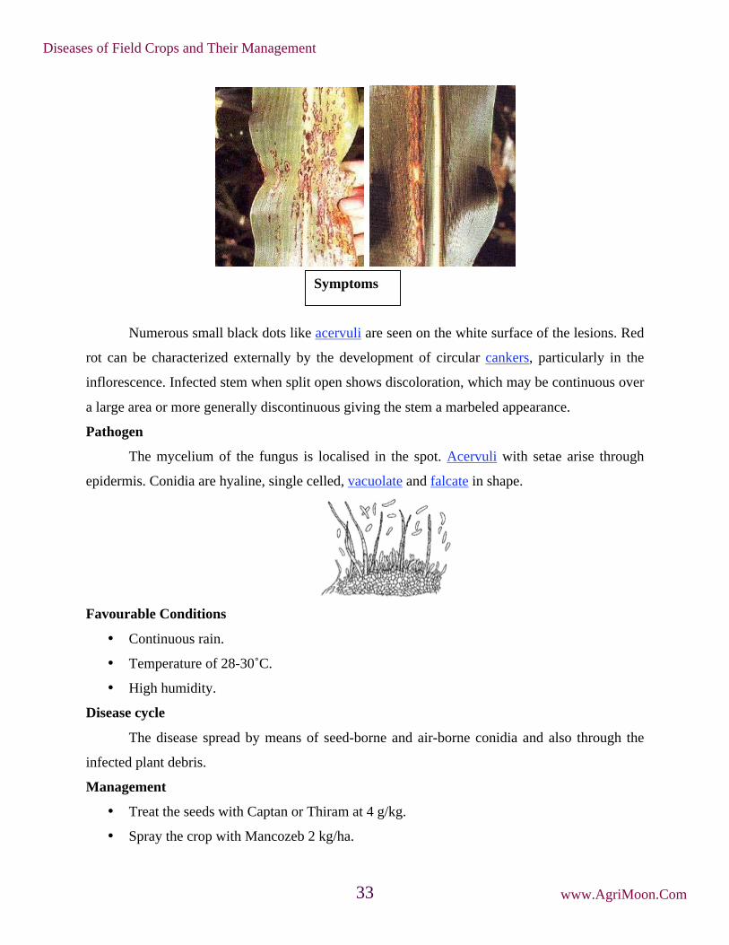
Numerous small black dots like acervuli are seen on the white surface of the lesions. Red
rot can be characterized externally by the development of circular cankers, particularly in the
inflorescence. Infected stem when split open shows discoloration, which may be continuous over
a large area or more generally discontinuous giving the stem a marbeled appearance.
Pathogen
The mycelium of the fungus is localised in the spot. Acervuli with setae arise through
epidermis. Conidia are hyaline, single celled, vacuolate and falcate in shape.
Favourable Conditions
• Continuous rain.
• Temperature of 28-30˚C.
• High humidity.
Disease cycle
The disease spread by means of seed-borne and air-borne conidia and also through the
infected plant debris.
Management
• Treat the seeds with Captan or Thiram at 4 g/kg.
• Spray the crop with Mancozeb 2 kg/ha.
Symptoms
Diseases of Field Crops and Their Management
www.AgriMoon.Com33

Rust - Puccinia purpurea
Symptoms
The fungus affects the crop at all stages of growth. The first symptoms are small flecks
on the lower leaves (purple, tan or red depending upon the cultivar). Pustules (uredosori) appear
on both surfaces of leaf as purplish spots which rupture to release
reddish powdery masses of uredospores. Teliopores develop later sometimes in the old uredosori
or in telisori, which are darker and longer than the uredosori. The pustules may also occur on the
leaf sheaths and on the stalks of inflorescence.
Pathogen
The uredospores are pedicellate, elliptical or oval, thin walled, echinulated and
darkbrown in colour. The teliospores are reddish or brown in colour and two celled, rounded at
the apex with one germ pore in each cell. The teliospores germinate and produce promycelium
and basidiospores. Basidiospores infect Oxalis corniculata (alternate host) where pycnial and
aecial stages arise.
Favourable Conditions
• Low temperature of 10 to 12˚C favours teliospore germination.
• A spell of rainy weather favours the onset of the disease.
Disease cycle
The uredospores survive for a short time in soil and infected debris. Presence of alternate
host helps in perpetuation of the fungus.
Symptoms on leaves and stalk
Diseases of Field Crops and Their Management
www.AgriMoon.Com34

Management
• Remove the alternate host Oxalis comiculata.
• Spray the crop with Mancozeb at 2 kg/ha.
Grain smut/Kernel smut / Covered smut / Short smut - Sphacelotheca sorghi
Symptoms
The individual grains are replaced by smut sori. The sori are oval or cyclindrical and are
covered with a tough creamy skin (peridium) which often persists unbroken up to thrashing.
Ratoon crops exhibit higher incidence of disease.
Loose smut/ kernel smut - Sphacelotheca cruenta
Symptoms
The affected plants can be detected before the ears come out. They are shorter than the
healthy plants with thinner stalks and marked tillering. The ears come out much earlier than the
healthy. The glumes are hypertrophied and the earhead gives a loose appearance than healthy.
Symptoms
Diseases of Field Crops and Their Management
www.AgriMoon.Com35

The sorus is covered by a thin membrane which ruptures very early, exposing the spores even as
the head emerges from the sheath.
Long smut - Tolyposporium ehrenbergii
Symptoms
This disease is normally restricted to a relatively a small proportion of the florets which
are scattered on a head. The sori are long, more or less cylindrical, elongated, slightly curved
with a relatively thick creamy-brown covering membrane (peridium). The peridium splits at the
apex to release black mass of spores (spore in groups of balls) among which are found several
dark brown filaments which represent the vascular bundles of the infected ovary.
Symptom
Symptoms
Diseases of Field Crops and Their Management
www.AgriMoon.Com36
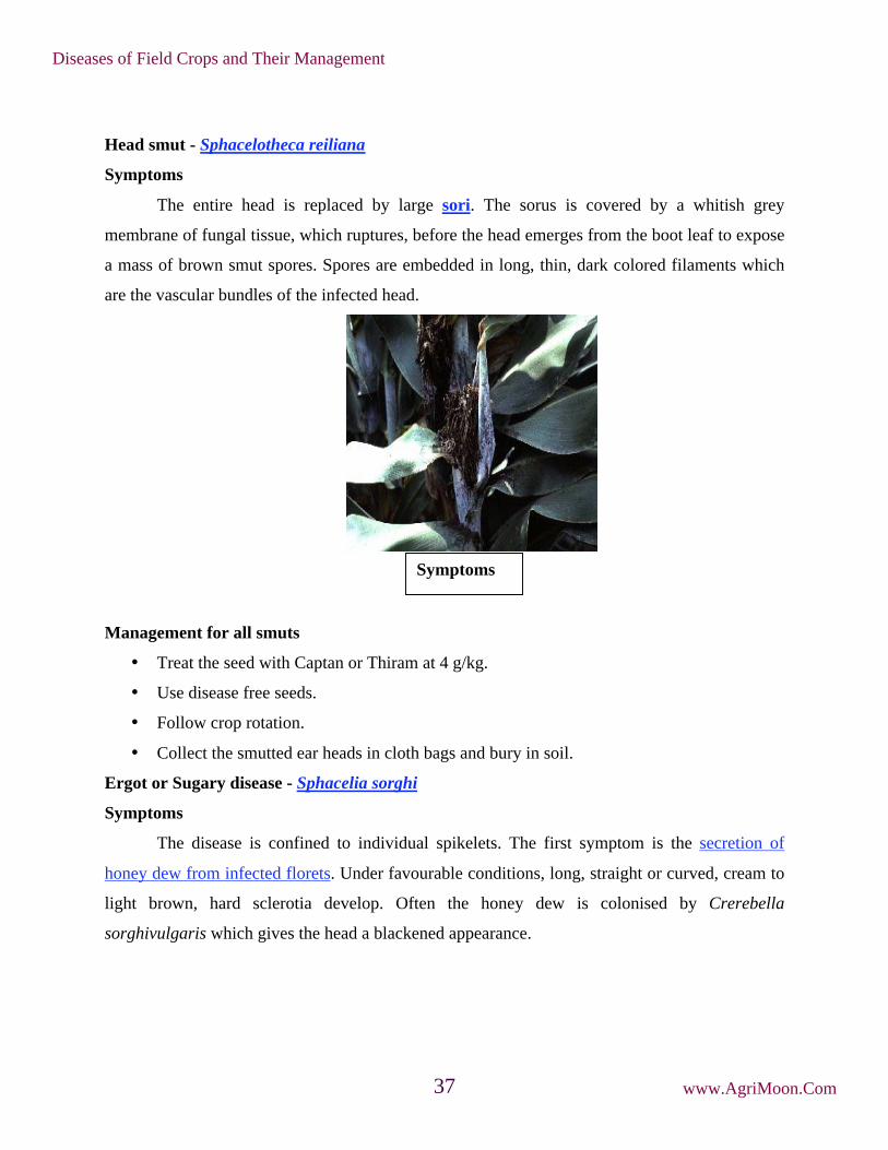
Head smut - Sphacelotheca reiliana
Symptoms
The entire head is replaced by large sori. The sorus is covered by a whitish grey
membrane of fungal tissue, which ruptures, before the head emerges from the boot leaf to expose
a mass of brown smut spores. Spores are embedded in long, thin, dark colored filaments which
are the vascular bundles of the infected head.
Management for all smuts
• Treat the seed with Captan or Thiram at 4 g/kg.
• Use disease free seeds.
• Follow crop rotation.
• Collect the smutted ear heads in cloth bags and bury in soil.
Ergot or Sugary disease - Sphacelia sorghi
Symptoms
The disease is confined to individual spikelets. The first symptom is the secretion of
honey dew from infected florets. Under favourable conditions, long, straight or curved, cream to
light brown, hard sclerotia develop. Often the honey dew is colonised by Crerebella
sorghivulgaris which gives the head a blackened appearance.
Symptoms
Diseases of Field Crops and Their Management
www.AgriMoon.Com37

Pathogen
The fungus produces septate mycelium. The honey dew is a concentrated suspension of
conidia, which are single celled, hyaline, elliptic or oblong.
Favourable Conditions
• A period of high rainfall and high humidity during flowering season.
• Cool night temperature and cloudy weather aggravate the disease.
Disease Cycle
The primary source of infection is through the germination of sclerotia which release
ascospores that infect the ovary. The secondary spread takes place through air and insect-borne
conidia. Rain splashes also help in spreading the disease.
Management
• Adjust the date of sowing so that the crop does not flower during September- October
when high rainfall and high humidity favor the disease.
• Spray any one of the following fungicides viz., Mancozeb 2 kg/ha (or) Carbendazim at
500 g/ha at emergence of ear head (5-10 per cent flowering stage) followed by a spray at
50 per cent flowering and repeat the spray after a week, if necessary.
Head mould/Grain mould/Head blight
Symptom
Diseases of Field Crops and Their Management
www.AgriMoon.Com38

More than thirty two genera of fungi were found to occur on the grains of sorghum.
Symptoms
If rains occur during the flowering and grain filling stages, severe grain moulding occusr.
The most frequently occurring genera are Fusarium, Curvularia, Alternaria, Aspergillus and
Phoma. Fusarium semitectum and F.moniliforme develop a fluffy white or pinkish coloration. C.
lunata colours the grain black. Symptom varies depending upon the organism involved and the
degree of infection.
Favourable Conditions
• Wet weather following the flowering favors grain mould development.
• The longer the wet period the greater the mould development.
• Compact ear heads are highly susceptible.
Disease cycle
The fungi mainly spread through air-borne conidia. The fungi survive as parasites as well
as saprophytes in the infected plant debris.
Management
• Adjust the sowing time.
• Spray any one of the following fungicides in case of intermittent rainfall during earhead
emergence, a week later and during milky stage.
• Mancozeb 1 kg/ha or Captan 1 kg + Aureofungin-sol 100 g/ha.
Phanerogamic parasite - Striga asiatica and Striga densiflora
Symptoms
Diseases of Field Crops and Their Management
www.AgriMoon.Com39

It is a partial root parasite and occurs mainly in the rainfed sorghum. It is a small plant
with bright green leaves, grows up to a height of 15-30 cm. The plants occur in clusters of 10-
20/host plant. S. asiatica produces red to pink flowers while. S. densiflora produces white
flowers. Each fruit contains minute seeds in abundance which survives in the soil for several
years.
The root exudates of sorghum stimulate the seeds of the parasite to germinate. The
parasite then slowly attaches to the root of the host by haustoria and grows below the soil surface
producing underground stems and roots for about 1-2 months. The parasite grows faster and
appears at the base of the plant. Severe infestation causes yellowing and wilting of the host
leaves. The infected plants are stunted in growth and may die prior to seed setting.
Management
• Regular weeding and intercultural operation during early stages of parasite growth.
• Spray Fernoxone (sodium salt of 2, 4-D) at 450g /500 litre of water.
Symptoms
Diseases of Field Crops and Their Management
www.AgriMoon.Com40

3. Diseases of Wheat
Black or stem rust - Puccinia graminis tritici
Symptoms
Symptoms are produced on almost all aerial parts of the wheat plant but are most
common on stem, leaf sheaths and upper and lower leaf surfaces. Uredial pustules (or sori) are
oval to spindle shaped and dark reddish brown (rust) in color. They erupt through the epidermis
of the host and are surrounded by tattered host tissue. The pustules are dusty in appearance due
to the vast number of spores produced. Spores are readily released when touched.
As the infection advances teliospores are produced in the same pustule. The color of the
pustule changes from rust color to black as teliospore production progresses. If a large number of
pustules are produced, stems become weakened and lodge. The pathogen attacks other host
(barberry) to complete its life cycle. Symptoms are very different on this woody host. Other
spores are Pycnia (spermagonia) produced on the upper leaf surface of barberry which appears as
raised orange spots. Small amounts of honeydew that attracts insects are produced in this
structure. Aecia, produced on the lower leaf surface, are yellow. They are bell-shaped and extend
as far as 5 mm from the leaf surface.
Brown or leaf rust - Puccinia triticina (P. recondita)
Symptom
Symptoms
Diseases of Field Crops and Their Management
www.AgriMoon.Com41

The most common site for symptoms is on leaf blades, however, sheaths, glumes and
awns may occasionally become infected and exhibit symptoms. Uredia are seen as small, circular
orange blisters or pustules on the upper surface of leaves.
Orange spores are easily dislodged and may cover clothing, hands or implements. When
the infection is severe leaves dry out and die. Since inoculum is blown into a given area,
symptoms are often seen on upper leaves first. As plants mature, the orange urediospores are
replaced by black teliospores. Pustules containing these spores are black and shiny since the
epidermis does not rupture. Yield loss often occurs as a result of infection by Puccinia recondita
f. sp. tritici. Heavy infection which extends to the flag leaf results in a shorter period of grain fill
and small kernels.
Yellow or stripe rust - Puccinia striiformis
Symptom
Mainly occur on leaves than the leaf sheaths and stem. Bright yellow pustules (Uredia)
appear on leaves at early stage of crop and pustules are arranged in linear rows as stripes. The
stripes are yellow to orange yellow. The teliospores are also arranged in long stripes and are dull
black in colour.
Symptoms
Diseases of Field Crops and Their Management
www.AgriMoon.Com42

Pathogen
The uredospores of rust pathogen are almost round or oval in shape and bright orange in
colour. The teliospores are bright organge to dark brown, two celled and flattened at the top.
Sterile paraphyses are also present at the end of sorus.
Disease Cycle
In India, all these rusts appear in wheat growing belt during Rabi crop season. Uredosori
turn into teliosori as summer approaches. The inoculum survives in the form of uredospores /
teliospores in the hills during off season on self sown crop or volunteer hosts, which provide an
excellent source of inoculum. In India, role of alternate host (Barberis) is not there in completing
the life cycle.
The fungus is inhibited by temperatures over 20˚C although strains tolerant of high
temperatures do exist. The complete cycle from infection to the production of new spores can
take as little as 7 days during ideal conditions. The disease cycle may therefore be repeated many
times in one season. During late summer, the dark teliospores may be produced. These can
germinate to produce yet another spore type, the basidiospore, but no alternate host has been
found. Although the teliospores seem to have no function in the disease cycle they may
contribute to the development of new races through sexual recombination.
Symptoms
Diseases of Field Crops and Their Management
www.AgriMoon.Com43
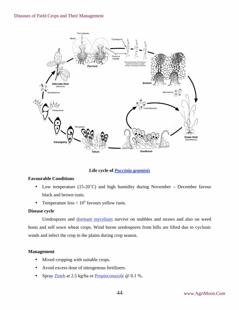
Life cycle of Puccinia graminis
Favourable Conditions
• Low temperature (15-20˚C) and high humidity during November – December favour
black and brown rusts.
• Temperature less < 10o favours yellow rusts.
Disease cycle
Uredospores and dormant mycelium survive on stubbles and straws and also on weed
hosts and self sown wheat crops. Wind borne uredospores from hills are lifted due to cyclonic
winds and infect the crop in the plains during crop season.
Management
• Mixed cropping with suitable crops.
• Avoid excess dose of nitrogenous fertilizers.
• Spray Zineb at 2.5 kg/ha or Propioconazole @ 0.1 %.
Diseases of Field Crops and Their Management
www.AgriMoon.Com44

• Grow resistant varieties like PBW 343, PBW 550, PBW 17
Loose smut - Ustilago nuda tritici (Ustilago tritici)
Symptoms
It is very difficult to detect infected plants in the field until heading. At this time, infected
heads emerge earlier than normal heads. The entire inflorescence is commonly affected and
appears as a mass of olive-black spores, initially covered by a thin gray membrane. Once the
membrane ruptures, the head appears powdery.
Spores are dislodged, leaving only the rachis intact. In some cases remnants of glumes
and awns may be present on the exposed rachis. Smutted heads are shorter than healthy heads
due to a reduction in the length of the rachis and peduncle. All or a portion of the heads on an
infected plant may exhibit these symptoms. While infected heads are shorter, the rest of the plant
is slightly taller than healthy plants. Prior to heading affected plants have dark green erect leaves.
Chlorotic streaks may also be visible on the leaves.
Disease Cycle
Ears of infected plants emerge early. The spores released from the infected heads land on
the later emerging florets and infect the developing seed. Infection during flowering is favored
Symptoms
Diseases of Field Crops and Their Management
www.AgriMoon.Com45

by frequent rain showers, high humidity and temperature. The disease is internally seed borne,
where pathogen infects the embryo in the seed.
Management
Treat the seed with Vitavax @ 2g/kg seed before sowing. Burry the infected ear heads in
the soil, so that secondary spread is avoided.
Flag smut - Urocystis tritici
Symptoms
The symptoms can be seen on stem, clum and leaves from late seedling stage to maturity.
The seedling infection leads to twisting and drooping of leaves followed by withering. Grey to
grayish black sori occurs on leaf blade and sheath. The sorus contains black powdery mass of
spores.
Diseases of Field Crops and Their Management
www.AgriMoon.Com46

Pathogen
Aggregated spore balls, consisting 1-6 bright globose, brown smoth walled spores
surrounded by a layer of flat sterile cells.
s
Favourable Conditions
• Temperature of 18-24˚C.
• Relative humidity 65% and above.
Disease cycle
Seed and soil borne. Smut spores are viable for more than 10 years.
Management
• Treat the seeds with carboxin at 2g /kg.
• Grow resistant varieties like Pusa 44 and WG 377.
Hill bunt or Stinking smut - Tilletia caries / T.foetida
Symptoms
Spore balls
Diseases of Field Crops and Their Management
www.AgriMoon.Com47

Symptoms
The fungus attacks seedling of 8-10 days old and become systemic and grows along the
tip of shoot. At the time of flowering hyphae concentrate in the inflorescence and spikelets and
transforming the ovary into smut sorus of dark green color with masses of chlamydospores. The
diseased plants mature earlier and all the spikelets are affected.
Pathogen
Reticulate, globose and rough walled. No resting period. Germinate to produce primary
sporidia which unite to form ‘H’ shaped structure.
Life cycle
The spores on the seed surface germinate along with the seed. Each produces a short
fungal thread terminating in a cluster of elongated cells. These then produce secondary spores
which infect the coleoptiles of the young seedlings before the emergence of the first true leaves.
The mycelium grows internally within the shoot infecting the developing ear. Affected plants
develop apparently normally until the ear emerges when it can be seen that grain sites have been
replaced by bunt balls. In India disease occurs only in Northern hills, where wheat is grown.
Symptoms on earhead and grains
Spores and its germination
Diseases of Field Crops and Their Management
www.AgriMoon.Com48

Favourable Conditions
• Temperature of 18-20˚C.
• High soil moisture.
Disease cycle
Externally seed borne
Management
• Treat the seeds with carboxin or carbendazim at 2g/kg.
• Grow the crop during high temperature period.
• Adopt shallow sowing.
• Grow resistant varieties like Kalyan sona, S227, PV18, HD2021, HD4513 and HD4519.
Karnal bunt - Neovassia indica
Symptoms
Symptoms of Karnal bunt are often difficult to distinguish in the field due to the fact that
incidence of infected kernels on a given head is low. There may be some spreading of the glumes
Diseases of Field Crops and Their Management
www.AgriMoon.Com49

due to sorus production but it is not as extensive as that observed with common bunt. Symptoms
are most readily detected on seed after harvest.
The black sorus, containing dusty spores is evident on part of the seed, commonly
occurring along the groove. Heavily infected seed is fragile and the pericarp ruptures easily. The
foul, fishy odor associated with common bunt is also found with karnal bunt. The odor is caused
by the production of trimethylamine by the fungus. Seed that is not extensively infected may
germinate and produce healthy plants.
Foot rot - Pythium graminicolum and P. arrhenomanes
Symptoms
Symptoms
Diseases of Field Crops and Their Management
www.AgriMoon.Com50

The disease mainly occurs in seedlings and roots and rootlets become brown in colour.
Seedlings become pale green and have stunted growth. Fungus produces sporangia and
zoospores and oospores.
Favourable Conditions
Wet weather and high rainfall.
Disease cycle
Through soil and irrigation water.
Management
• Follow crop rotation.
• Treat the seeds with Carboxin or Carbendazim at 2g/kg.
Powdery mildew - Erysiphe graminis var. tritici
Symptoms
Greyish white powdery growth appears on the leaf, sheath, stem and floral parts.
Powdery growth later become black lesion and cause drying of leaves and other parts.
Pathogen
Fungus produces septate, superficial, hyaline mycelium on leaf surface with short
conidiophores. The conidia are elliptical, hyaline, single celled, thin walled and produced in
Symptoms
Diseases of Field Crops and Their Management
www.AgriMoon.Com51

chains. Dark globose cleistothecia containing 9-30 asci develop with oblong, hyaline and
thinwalled ascospores.
Disease cycle
Fungus remains in infected plant debris as dormant mycelium and asci. Primary spread is
by the ascospores and secondary spread through airborne conidia.
Favourable Conditions
• Temperature of 20-21˚C.
Management
• Spray Wettable Sulphur 0.2% or Carbendazim @ 500 g/ha
Leaf blight - Alternaria triticina / Bipolaris sorokiniana
Symptoms
Reddish brown oval spots appear on young seedlings with bright yellow margin. In
severe cases, several spots coalesce to cause drying of leaves. It is a complex disease, having
association of A.triticina, B.sorokiniana and A. alternate.
Conidia and conidiophores
Diseases of Field Crops and Their Management
www.AgriMoon.Com52

Disease cycle
Primary spread is by externally seed-borne and soil borne conidia. Secondary spread by
air-borne conidia.
Favourable Conditions
• Temperature of 25˚C and high relative humidity.
Management
• Spray the crop with Mancozeb or Zineb at 2 kg/ha.
Other minor diseases
Helminthosporium leaf spot: Helminthosporium spp.
Tundu or yellow ear rot: Corynebacterium tritici + Anguina tritici
Seedling blight: Rhizoctonia solani and Fusarium sp
Sclerotinia rot: Sclerotinia sclerotiorum
Molya disease: Heterodera avenae (Nematode)
Symptoms
Diseases of Field Crops and Their Management
www.AgriMoon.Com53

4. Diseases of Pearlmillet
Downy mildew - Sclerospora graminicola
Symptoms
Infection is mainly systemic and symptoms appear on leaves and inflorescence. The
initial symptoms appear in seedlings at three to four leaf stages. The affected leaves show
patches of light green to light yellow colour on the upper surface and the corresponding lower
surface bears white downy growth of the fungus consisting of sporangiophores and sporangia.
The yellow discolouration often turns to streaks along veins. As a result of infection young plants
dry and die ultimately. Symptoms may appear first on the upper leaves of the main shoot or the
main shoot may be symptom free and symptoms appear on tillers or on the lateral shoots.
The inflorescence of infected plants gets completely or partially malformed with florets
converted into leafy structures, giving the typical symptom of green ear.
Infected leaves and inflorescences produce sporangia over a considerable period of time
under humid conditions and necrosis begins. The dry necrotic tissues contain masses of
oospores.
Symptoms
Diseases of Field Crops and Their Management
www.AgriMoon.Com54

Pathogen
The mycelium is systemic, non septae and intercellular. Short, stout, hyaline
sporangiophores arise through stomata and branch irregularly, with stalks bearing sporangia.
Sporangia are hyaline, thin walled, elliptical and bear prominent papilla. Oospores are round in
shape, surrounded by a smooth, thick and yellowish brown wall.
Favourable Conditions
• Very high humidity (90%).
• Presence of water on the leaves
• Low temperature of 15-25˚C favor the formation of sporangiophore and sporangia.
Disease cycle
Green ear symptoms
Oospores
Diseases of Field Crops and Their Management
www.AgriMoon.Com55

The oospores remain viable in soil for 5 years or longer giving rise to the primary
infection on seedlings. Secondary spread is through sporangia produced during rainy season. The
dormant mycelium of the fungus is present in embryo of infected seeds.
Management
• Deep ploughing to bury the oospores.
• Roguing out infected plants.
• Adopt crop rotation.
• Grow resistant varieties WCC-75, Co7 and Co (Cu)9.
• Treat the seeds with Metalaxyl at 6g/kg.
• Spray Mancozeb 2 kg or Metalaxyl + Mancozeb at 1 kg/ha on 20th day after sowing in
the field.
Smut - Tolyposporium penicillariae
Symptoms
The pathogen infects few florets and transforms them into plump sori containing smut
spores. The sori are larger than normal healthy grains and when the sori mature they become
dark brown releasing millions of black smut spore balls.
Pathogen
The fungus is mostly confined to the sorus. The sori contain spores in groups and are not
easy to separate. Each spore is angular or round and light brown.
Symptoms
Diseases of Field Crops and Their Management
www.AgriMoon.Com56

Favourable Conditions
• High relative humidity.
• Successive cropping with pearlmillet.
Disease cycle
• The pathogen survives as spore balls in the soil and serves as primary source of
inoculum. Secondary spread is by air-borne conidia.
Management
• The damage caused by the fungus is negligible.
• Removal and destruction of affected ear head will help in controlling the disease.
Rust - Puccinia pennisetti
Symptoms
Symptoms first appear mostly on the distal half of the lamina. The leaf soon becomes
covered by uredosori which appear more on the upper surface. The pustules may be formed on
leaf sheath, stem and on peduncles. Later, telial formation takes place on leaf blade, leaf sheath
and stem. While brownish uredia are exposed at maturity, the black telia remain covered by the
epidermis for a longer duration.
Pathogen
Uredospores are oval, elliptic, sparsely echinulated and pedicellate. Teliospores are dark
brown in colour, two celled, cylindrical to club shaped, apex flattened, broad at top and tapering
towards base. The fungus is macrocyclic producing uredial and telial stages on pearlmillet and
aecial and pycnial stages on brinjal.
Symptoms
Diseases of Field Crops and Their Management
www.AgriMoon.Com57

Favourable Conditions
• Closer spacing.
• Presence of abundant brinjal plants and other species of Solanum viz., S.torvum, S.
xanthocarpum and S. pubescens.
Disease cycle
Air-borne uredospores are the primary sources. The uredial stages also occur on several
species of Pennisetum, which helps in secondary spread of the pathogen.
Management
Spray with Wettable Sulphur 3 kg or Mancozeb 2 kg/ha.
Ergot or Sugary disease - Claviceps fusiformis
Symptoms
The symptom is seen by exudation of small droplets of light pinkish or brownish honey
dew from the infected spikelets. Under severe infection many such spikelets exude plenty of
honey dew which trickles along the earhead. This attracts several insects. In the later stages, the
infected ovary turns into small dark brown sclerotium which projects out of the spikelet.
Diseases of Field Crops and Their Management
www.AgriMoon.Com58

Pathogen
The pathogen produces septate mycelium which produces conidiophores and is closely
arranged. Conidia are hyaline and one celled. The sclerotia are small (3-8mm x 0.3-15mm) and
dark grey but white inside.
Disease cycle
Sclerotia are viable in soil for 6-8 months. The primary infection takes place by
germinating sclerotia present in the soil. Secondary spread is by insects or airborne conidia. The
role of collateral hosts like Cenchrus ciliaris and C. setigerus in perpetuation of fungus is
significant. The fungus also infects other species of Pennisetum.
Management
• Adjust the sowing date so that the crop does not flower during September when high
rainfall and high relative humidity favour the disease spread.
• Immerse the seeds in 10 per cent common salt solution and remove the floating sclerotia.
• Remove collateral hosts.
Symptoms
Diseases of Field Crops and Their Management
www.AgriMoon.Com59

• Spray with Carbendazim 500g or Mancozeb 2 kg or Ziram 1kg/ha when 5-10 per cent
flowers have opened and again at 50 per cent flowering stage.
Minor diseases
Grain mould - Fungal complex
Grains covered with white, pink or black moulds.
Blast - Pyricularia setariae
Diamond shaped to circular lesions with dark brown margins and chlorotic haloes.
Zonate leaf spot - Gloeocercospora sp.
Rough circular lesions with alternating concentric bands of straw and brown colour, often
coalescing over the leaf surface.
Banded leaf spot - Rhizoctonia spp.
Patch of light and dark, discoloured areas and often bearing fluffy to light brown fungal
mats.
Diseases of Field Crops and Their Management
www.AgriMoon.Com60
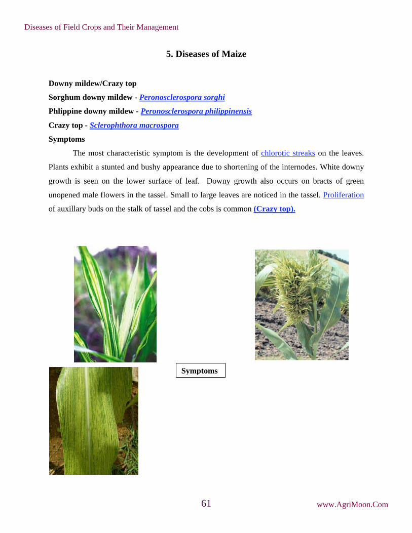
5. Diseases of Maize
Downy mildew/Crazy top
Sorghum downy mildew - Peronosclerospora sorghi
Phlippine downy mildew - Peronosclerospora philippinensis
Crazy top - Sclerophthora macrospora
Symptoms
The most characteristic symptom is the development of chlorotic streaks on the leaves.
Plants exhibit a stunted and bushy appearance due to shortening of the internodes. White downy
growth is seen on the lower surface of leaf. Downy growth also occurs on bracts of green
unopened male flowers in the tassel. Small to large leaves are noticed in the tassel. Proliferation
of auxillary buds on the stalk of tassel and the cobs is common (Crazy top).
Symptoms
Diseases of Field Crops and Their Management
www.AgriMoon.Com61

Pathogen
The fungus grows as white downy growth on both surface of the leaves, consisting of
sporangiophores and sporangia. Sporangiophores are quite short and stout, branch profusely into
series of pointed sterigmata which bear hyaline, oblong or ovoid sporangia (conidia). Sporangia
germinate directly and infect the plants. In advanced stages, oospores are formed which are
spherical, thick walled and deep brown.
Favourable Conditions
• Low temperature (21-33˚C)
• High relative humidity (90 per cent) and drizzling.
• Young plants are highly susceptible.
Disease cycle
The primary source of infection is through oospores in soil and also dormant mycelium
present in the infected maize seeds. Secondary spread is through airborne conidia. Depending on
the pathogen species, the initial source of disease inoculum can be oospores that over winter in
the soil or conidia produced in infected, over wintering crop debris and infected neighboring
plants. Some species that cause downy mildew can also be seed borne, although this is largely
restricted to seed that is fresh and has high moisture content.
At the onset of the growing season, at soil temperatures above 20°C, oospores in the soil
germinate in response to root exudates from susceptible maize seedlings. The germ tube infects
the underground sections of maize plants leading to characteristic symptoms of systemic
infection including extensive chlorosis and stunted growth. If the pathogen is seed borne, whole
plants show symptoms. Oospores are reported to survive in nature for up to 10 years.
Once the fungus has colonised host tissue, sporangiophores (conidiophores) emerge from
stomata and produce sporangia (conidia) which are wind and rain splash disseminated and
initiate secondary infections. Sporangia are always produced in the night. They are fragile and
can not be disseminated more than a few hundred meters and do not remain viable for more than
a few hours.
Germination of sporangia is dependent on the availability of free water on the leaf
surface. Initial symptoms of disease (chlorotic specks and streaks that elongate parallel to veins)
Diseases of Field Crops and Their Management
www.AgriMoon.Com62

occur in 3 days. Conidia are produced profusely during the growing season. As the crop
approaches senescence, oospores are produced in large numbers.
Management
• Deep ploughing.
• Crop rotation with pulses.
• Rogue out infected plants.
• Treat the seeds with metalaxyl at 6g/kg.
• Spray the crop with Metalaxyl + Mancozeb @ 1kg on 20th day after sowing.
• Grow resistant varieties and hybrids viz. CO1, COH1and COH2.
Leaf blight - Helminthosporium maydis (Syn: H. turcicum)
Symptoms
The fungus affects the crop at young stage. Small yellowish round to oval spots are seen
on the leaves. The spots gradually increase in area into bigger elliptical spots and are straw to
grayish brown in the centre with dark brown margins. The spots coalesce giving blighted
appearance. The surface is covered with olive green velvetty masses of conidia and
conidiophores.
Pathogen
Conidiophores are in group, geniculate, mid dark brown, pale near the apex and smooth.
Conidia are distinctly curved, fusiform, pale to mid dark golden brown with 5-11 septa.
Symptoms
Diseases of Field Crops and Their Management
www.AgriMoon.Com63

Favourable Conditions
• Optimum temperature for the germination of conidia is 8 to 27˚C provided with freewater
on the leaf.
• Infection takes place early in the wet season.
Disease cycle
It is a seed-borne fungus. It also infects sorghum, wheat, barely, oats, sugarcane and
spores of the fungus are also found to associate with seeds of green gram, black gram, cowpea,
varagu, Sudan grass, Johnson grass and Teosinte.
Management
• Treat the seeds with Captan or Thiram at 4 g/kg.
• Spray Mancozeb 2 kg or captan 1 kg/ha.
Rust - Puccinia sorghi
Symptoms
Circular to oval, elongated cinnamon-brown powdery pustules are scattered over both
surface of the leaves. As the plant matures, the pustules become brown to black owing to the
replacement of red uredospores by black teliospores.
Conidia
Diseases of Field Crops and Their Management
www.AgriMoon.Com64

Pathogen
Uredospores are globose or elliptical finely echinulate, yellowish brown with 4
germpores. Teliospores are brownish black, or dark brown, oblong to ellipsoidal, rounded to
flattened at the apex. They are two celled and slightly constricted at the septum and the spore
wall is thickened at the apex.
Favourable Conditions
• Cool temperature and high relative humidity.
Disease cycle
Primary source of inoculums is uredospores surviving on alternate hosts viz., Oxalis
corniculata and Euchlaena mexicana.
Management
• Remove the alternate hosts.
• Spray Mancozeb at 2 kg/ha.
Head smut - Sphacelotheca reiliana
Symptoms
Symptoms
Uredospores and teliospoes
Diseases of Field Crops and Their Management
www.AgriMoon.Com65

Symptoms are usually noticed on the cob and tassel. Large smut sori replace the tassel
and the ear. Sometimes the tassel is partially or wholly converted into smut sorus. The smutted
plants are stunted produce little yield and remain greener than that of the rest of the plants.
Pathogen
Smut spores are produced in large numbers which are reddish brown to black, thick
walled, finely spined, spherical.
Favourable Conditions
• Low temperature favours more infection and this fungus also infects the sorghum
Disease cycle
The smut spores retain its viability for two years. The fungus is externally seedborne and
soil-borne. The major source of infection is through soil-borne chlamydospores.
Management
• Field sanitation.
• Crop rotation with pulses.
• Treat the seeds with Captan or Thiram at 4 g/kg.
Charcoal rot - Macrophomina phaseolina (Rhizoctonia bataticola)
Symptoms
The affected plants exhibit wilting symptoms. The stalk of the infected plants can be
recognized by grayish streak. The pith becomes shredded and grayish black minute sclerotia
develop on the vascular bundles. Shredding of the interior of the stalk often causes stalks to
Symptoms
Diseases of Field Crops and Their Management
www.AgriMoon.Com66

break in the region of the crown. The crown region of the infected plant becomes dark in colour.
Shredding of root bark and disintegration of root system are the common features.
Pathogen
The fungus produces large number of sclerotia which are round and black in colour.
Sometimes, it produces pycnidia on the stems or stalks.
Favourable Conditions
• High temperature and low soil moisture (drought)
Disease cycle
The fungus has a wide host range, attacking sorghum, pearlmillet, fingermillet and
pulses. It survives for more than 16 years in the infected plant debris. The primary source of
infection is through soil-borne sclerotia. The pathogen also attacks many other hosts, which
helps in its perpetuation. Since the fungus is a facultative parasite it is capable of living
saprophytically on dead organic tissues, particularly many of its natural hosts producing
sclerotial bodies. The fungus over winters as a sclerotia in the soil and infects the host at
susceptible crop stage through roots and proceeds towards stem.
Management
• Long crop rotation with crops that are not natural host of the fungus.
• Irrigate the crops at the time of earhead emergence to maturity.
• Treat the seeds with Carbendazim or Captan at 2 g/kg.
• Grow disease tolerant varieties viz., SN-65, SWS-8029, Diva and Zenit.
Symptoms
Diseases of Field Crops and Their Management
www.AgriMoon.Com67

Minor diseases
Bacterial Stalk rot - Erwinia dissolvens
Symptoms
The basal internodes develop soft rot and give a water soaked appearance. A mild sweet
fermenting odour accompanies such rotting. Leaves some time show signs of wilting and
affected plants topple down in few days. Ears and shank may also show rot. They fail to develop
further and the ears hang down simply from the plant
Disease cycle
Borer insects play a significant role in initiation of the disease. The organism is soil borne
and makes its entry through wounds and injuries on the host surface. The organism survives
saprophytically on debris of infected materials and serves primary inoculum in the next
season.
Mosaic - Maize mosaic potyvirus
Symptoms
Symptoms appear as chlorotic spots, which gradually turn into stripes covering entire leaf
blade. Chlorotic stripes and spots can also develop on leaf sheaths, stalks and husks.
Moderate to severe rosetting of new growth is observed. Size of stalk, leaf blades and tassel
tend to be normal in late infection.
Symptoms
Diseases of Field Crops and Their Management
www.AgriMoon.Com68

Pathogen
It is caused by Maize mosaic potyvirus. Virions are flexuous, 750-900nm long, ssRNA
genome.
Disease cycle
It is transmitted in nature by leaf hopper vector, Perigrimus maidis.
Brown spot - Physoderma maydis
Water soaked lesions, which are oval, later turn into light green and finally brown.
Symptoms
Diseases of Field Crops and Their Management
www.AgriMoon.Com69

6. Diseases of Sugarcane
Red rot - Colletotrichum falcatum (Perfect stage: Physalospora tucumanensis)
Symptoms
The first external symptom appears mostly on third or fourth leaf which withers away at
the tips along the margins. Typical symptoms of red rot are observed in the internodes of a stalk
by splitting it longitudinally. These include the reddening of the internal tissues which are
usually elongated at right angles to the long axis of the stalk. The presence of cross-wise white
patches are the important diagnostic character of the disease. The diseased cane also emits
acidic-sour smell. As the disease advances, the stalk becomes hollow and covered with white
mycelial growth.
Later the rind shrinks longitudinally with minute black, velvetty fruiting bodies
protruding out of it. The pathogen also produces tiny reddish lesions on the upper surface of
leaves with dark dots in the centre. The lesions are initially blood red with dark margins and later
on with straw coloured centres. Often the infected leaves may break at the lesions and hang
down, with large number of minute black dots.
Pathogen
The fungus produces thin, hyaline, septate, profusely branched hyphae containing oil
droplets. The fungus produces black, minute velvetty acervuli with long, rigid bristle-like,
septate setae. Conidiophores are closely packed inside the acervulus, which are short, hyaline
and single celled. The conidia are single celled, hyaline, falcate, granular and guttulate. Fungus
Symptoms
Diseases of Field Crops and Their Management
www.AgriMoon.Com70

also produces large number of globose and dark brown to black perithecia with a papillate
ostiole.
Asci are clavate, unitunicate and eight-spored. Large number of hyaline, septate, filiform
paraphyses is also present among asci. Ascospores are ellipsoid or fusoid, hyaline, straight or
slightly curved and unicellular which measure 18-22 m x 7-8 m.
Favourable Conditions
• Monoculturing of sugarcane.
• Successive ratoon cropping.
• Water logged conditions and injuries caused by insects.
Disease cycle
The fungus is sett-borne and also persists in the soil on the diseased clumps and stubbles
as chlamydospores and dormant mycelium. The primary infection is mainly from infected setts.
Secondary spread in the field is through irrigation water and cultivation tools. The rain splash, air
currents and dew drops also help in the spread of conidia from the diseased to healthy plants in
the field. The fungus also survives on collateral hosts Sorghum vulgare, S. halepense and
Saccharum spontaneum.If the conidia settle on the leaves they may germinate and invade the
leaves through various types of wounds. Stem infection may take place through insect bores and
root primordia. The soil-borne fungus may also enter the healthy setts through cut-ends, and
Diseases of Field Crops and Their Management
www.AgriMoon.Com71

cause early infection of the shoots. Though the perfect stage of the fungus has been observed in
nature, the role of ascospores in the disease cycle is not understood.
Management
• Adopt crop rotation by including rice and green manure crops.
• Select the setts from the disease free fields or disease free areas.
• Aviod ratooning of the diseased crop.
• Soak the setts in 0.1% Carbendazim or Triademefon 0.05% solution for 15 minutes
before planting.
• Grow resistant varieties CO 62198, CO 7704 and moderately resistant varieties CO 8001,
CO8201.
• Setts can be treated with aerated steam at 52 ˚C for 4 to 5 hours and by moist hot air at
54˚C for 2 hours.
Smut - Ustilago scitaminea
Symptoms
It is a culmiculous smut. The affected plants are stunted and the central shoot is converted
into a long whip-like, dusty black structure. The length of the whip varies from few inches to
several feet. In early stages, this structure is covered by a thin, white papery membrane. The
whip may be straight or slightly curved.
On maturity it ruptures and millions of tiny black smut spores (teliospores) are liberated
and disseminated by the wind. Affected plants are usually thin, stiff and remain at acute angle.
The whip like structure, representing the central shoot with its various leaves, may be produced
by each one of the shoots/tillers arising from the clump.
Diseases of Field Crops and Their Management
www.AgriMoon.Com72

The smutted clumps also produce mummified arrows in which lower portion consisted of
a normal inflorescence with typical flowers and the upper portion of the rachis is converted into a
typical smut whip. Occasionally smut sori may develop on the leaves and stem.
Pathogen
The fungal hyphae are primarily intercellular and collect as a dense mass between the
vascular bundles of host cell and produce tiny black spores. The thin membrane which covers the
smut whip represents the host epidermis. The smut spores are light brown in colour, spherical,
echinulated and measuring 6.5- 8.5 m in diameter. Smut spores germinate to produce 3-4 celled,
hyaline promycelium and produce 3-4 sporidia which are hyaline and oval shaped with pointed
ends.
Favourable Conditions
• Monoculturing of sugarcane.
• Continuous ratooning and dry weather during tillering stage.
Disease cycle
Teliospores may survive in the soil for long periods, upto 10 years. The spores and
sporidia are also present in the infected plant materials in the soil. The smut spores and dormant
mycelium also present in or on the infected setts. The primary spread of the disease is through
diseased seed-pieces (setts). In addition, sporidia and spores present in the soil also spread
through rain and irrigation water and cause soil-borne infection. The secondary spread in the
Symptoms
Diseases of Field Crops and Their Management
www.AgriMoon.Com73

field is mainly through the smut spores developed in the whips, aided by air currents. The fungus
also survives on collateral hosts like Saccharum spontaneum, S. robustum, Sorghum vulgare,
Imperata arundinacea and Cyperus dilatatus.
Management
• Plant healthy setts taken from disease free area.
• Remove and destory the smutted clump (collect the whips in a thick cloth bag/polythene
bag and immerse in boiling water for 1 hr to kill the spores).
• Discourage ratooning of the diseased crops having more than 10 per cent infection.
• Follow crop rotation with green manure crops or dry fallowing.
• Grow redgram as a companion crop between 2 rows of sugarcane.
• Grow resistant varieties like Co 7704 and moderately resistant varieties COC 85061 and
COC 8201.
Sett rot or Pineapple disease - Ceratocystis paradoxa
Symptoms
The disease primarily affects the setts usually two to three weeks after planning. The
fungus is soil-borne and enters through cut ends and proliferates rapidly in the parenchymatous
tissues. The affected tissues first develop a reddish colour which turns to brownish black in the
later stages. The severely affected setts show internodal cavities covered with the mycelium and
abundant spores. A characteristic pineapple smell is associated with the rotting tissues. The setts
may decay before the buds germinate or the shoots may die after reaching a height of about 6-12
inches. Infected shoots are stunted.
Symptoms
Diseases of Field Crops and Their Management
www.AgriMoon.Com74

Pathogen
The fungus produces both macroconidia and microconidia. Conidiophores are linear, thin
walled with short cells at the base and a long terminal cell. The microconidia are hyaline when
young but become almost black at maturity. They are thinwalled, cylindrical and produced
endogenously in chains in the long cells of conidiophores and pushed out in succession.
Macroconidia are produced singly or in chains on a short, lateral conidiophores. Macroconidia
are spherical or elliptical or truncate or pyriform and are hyaline to olive green or black
measuring 16-19x10-12 um.
The fungus also produces chlamydospores on short lateral hyphae in chains, which are
oval, thick walled and brown in colour. The perithecia are flask shaped with a very long neck.
The bulbous base of the perithecium is hyaline or pale yellow, 200-300 m in diameter and
ornamented with irregularly shaped, knobbed appendages. The ostiole is covered by numerous
pale-brown, erect tapering hyphae. Asci are clavate and measures 25x10 m and ascospores are
single celled, hyaline, ellipsoid, more convex on one side, measures 7-10 x 2.5-4 m.
Favourable Conditions
• Poorly drained fields.
• Heavy clay soils
• Temperature of 25-30o C
• Prolonged rainfall after planting.
Disease Cycle
The fungus survives as conidia and chlamydospores in the soil and in the infected,
burried cane tissues. The inoculum moves from field to field through wind-borne conidia or
irrigation or rain water. Inside the sett it spreads rapidly through the parenchymatous tissues and
causes sett rot.
The insects like cane borer (Diatraea dyari) also helps in the spread of the disease. The
pathogen also survives on coconut, cocoa, mango, papaya, coffee, maize and arecanut. Insects
also play a part in the dissemination of the pathogen.
Management
• Soak the setts in 0.05% Carbendazim 15 minutes.
• Use long setts having 3 or 4 buds.
• Provide adequate drainage during rainy seasons.
Diseases of Field Crops and Their Management
www.AgriMoon.Com75

Wilt - Cephalosporium sacchari
Symptoms
The first symptom of the disease is visible in the canes of 4-5 month age. The canes may
wither in groups. The affected plants are stunted with yellowing and withering of crown leaves.
The midribs of all leaves in a crown generally turn yellow, while the leaf lamina may remain
green. The leaves dry up and stem develop hollowness in the core. The core shows the reddish
discolouration with longitudinal red streaks passing from one internode to another. In severe
cases, spindle shaped cavities tapering towards the nodes develop in each internode. The canes
emit a disagreeable odour, with lot of mycelial threads of the fungus cover the cavity.
Pathogen
The fungal mycelium is hyaline, septate and thin walled. The conidiophores are simple or
branched and produce single celled, hyaline, oval to elliptical microconidia.
Favourable Conditions
• High day temperature (30-35˚C).
• Low humidity (50-60%).
• Low soil moisture and alkaline soils.
• Excess doses of nitrogenous fertilizers.
Disease Cycle
The fungus is soil-borne and remains in the soil as saprophyte for 2-3 years. The disease
is primarily transmitted through infected seed pieces. The secondary spread is aided by wind,
rain and irrigation water.
Management
• Select the seed material from the disease-free plots.
• Avoid the practice of ratooning in diseased fields.
• Burn the trashes and stubbles in the field.
• Grow coriander or mustard as a companion crop in the early stages of crop.
• Dip the setts in 40ppm Boran or Manganese for 10 minutes or in 0.25% Emisan or 0.05%
Carbendazim for 15 minutes.
Diseases of Field Crops and Their Management
www.AgriMoon.Com76

Rust - Puccinia erianthi (Syn: P. melanocephala and
P. kuehnii)
Symptoms
Minute, elongated, yellow spots (uredia), usually
2-10 x 1-3 mm appear on both the surfaces of young leaves. The
pustules turn to brown on maturity. Late in the season, dark brown
to black telia appear on the lower surface of leaves. In severse cases,
the uredia also appear on the leaf sheath and the entire foliage looks
brownish from a distance.
Pathogen
The mycelium is hyaline, branched and septate. P.kuehnii produces ovoid or pear shaped,
single celled uredospores measuring 29-57 x 8-37 m with apical thickening and golden yellow
in colour. Teliospores are produced in scanty which are yellow in colour, club shaped, two
celled, smooth walled and measuring 24- 34 X 18-25 m single celled, dark yellow coloured with
4 equatorial pores.
Teliospores are produced in
abundance, which are pale to brick colour, two celled,
smooth walled and slightly constricted at septum. Occurrence of pycnial and aecial stages and
the role of alternate host are unknown.
Favourable Conditions
• Temperature of 30˚C.
• Rumidity between 70 and 90 per cent.
• High wind velocity and continuous cloudiness.
Disease Cycle
The fungus survives on collateral hosts like Erianthus fulvus and Saccharum spontaneum.
The uredospores also survive in the infected stubbles in the soil. The disease is mainly spread
through air-borne uredospores.
Uredospores
Diseases of Field Crops and Their Management
www.AgriMoon.Com77

Management
• Remove the collateral hosts.
• Spray Tridemorph 1 kg or Mancozeb 2 kg/ha.
Gummosis - Xanthomonas axonopodis pv. vasculorum
Symptoms
The bacterium produces two distinct types of symptoms. On the mature leaves,
longitudinal stripes or streaks, 3-7mm in width and several cm in length, appear around the
affected veins, near the tip. Initially these stripes are pale yellow in colour, later turn to brown.
The affected tissues slowly dry up.
The infected canes are stunted with short internodes, giving a bushy appearance. When
such canes are cut transversely or split open longitudinally, a dull yellow bacterial ooze comes
out from the cut ends and bacterial pockets are seen inside the slitted cane. The fibro vasuclar
bundles are deep red and internodal cavities formed in the severe cases are filled with yellow
coloured bacterial gums.
Pathogen
The bacterium is a short rod, Gram negative, non spore forming measuring 1.0 to 1.5 m
X 0.4 to 0.5 m, with a single polar flagellum. It is facultative anaerobe and it produces yellow
slimy growth.
Disease Cycle
The bacterium remains viable in the soil as well as in infected canes. The primary
transmission is through naturally affected diseased setts or through soil-borne contamination.
The secondary spread may be through wind splashed rain, harvesting implements, animals and
insects. The bacterium can survive in the insect's body for a long time and in this way may be
transmitted long distances. On entry into the host the bacterium reaches the vascular tissues and
becomes systemic. The bacterium also perpetuates on maize, sorghum, pearlmillet and other
weed hosts, which also serve as sources of inoculum.
Management
• Remove and burn the affected clumps and the stubbles in the field. Select setts from
disease free areas.
Diseases of Field Crops and Their Management
www.AgriMoon.Com78

• Avoid growing collateral hosts like maize, sorghum and pearlmillet near the sugarcane
fields.
Red stripe - Pseudomonas rubrilineans
Symptoms
The disease first makes it appearance on the basal part of the young leaves. The stripes
appear as water soaked, long, narrow chlorotic streaks and become reddish brown in few days.
These stripes are 0.5 to 1 mm in width and 5-100 mm in length, run parallel to the midrib. The
stripes remain confined to lower half of the leaf lamina and whitish flakes spreads to growing
points of the shoot and yellowish stripes develop, which later turn reddish brown. The rotting
may commence from the tip of the shoot and spreads downwards. The core is discoloured to
reddish brown and shrivelled and form cavity in the centre. In badly affected fields, a foul and
nauseating smell appears.
Pathogen
The bacterium is a short rod (0.7 X 1.67 m), gram negative, non capsulate with a polar
flagellum.
Favourable Conditions
• Continuous ratooning and prolonged rainy weather with low temperature (250 C)
Disease cycle
Symptoms
Diseases of Field Crops and Their Management
www.AgriMoon.Com79

The pathogen remains viable in the soil and infected plant residues. The bacterium also
survives on sorghum, pearlmillet, maize, fingermillet and other species of Saccharum. The
bacterium primarily spreads through infected canes. The secondary spread is mainly through
rainsplash, irrigation water and insects. Infected parenchymatous cells may collapse and normal
functioning of the plant parts may fail. Several grasses, including ragi and bajra, have been
reported to be infected by the bacteria and these hosts may also play a role in the perpetuation
and spread of the pathogen.
Management
• Whenever the disease is noticed; the affected plants should be removed and burnt.
• Growing resistant varieties Select setts from the healthy fields.
• Avoid growing collateral hosts near the sugarcane fields.
Sugarcane Mosaic - Sugarcane mosaic potyvirus
Symptoms
The disease appears more prominently on the basal portion of the younger foliage as
chlorotic or yellowish stripes alternate with normal green portion of the leaf. As infection
becomes severe, yellow stripes appear on the leaf sheath and stalks. Elongated necrotic lesions
are produced on the stalks and stem splitting occurs. The necrotic lesions also develop on the
internodes and the entire plant becomes stunted and chlorotic.
Pathogen
Sugarcane mosaic potyvirus is a flexous rod, 650-770nm long X 12-15nm with
ss RNA genome.
Disease cycle
Symptoms
Diseases of Field Crops and Their Management
www.AgriMoon.Com80

The virus is mainly transmitted through infected canes used as seed. The virus also
infects Zea mays and a number of other cereals (Sorghum vulgare, Pennisetum americanum,
Eleusine indica, Setaria lutescens, Echinochloa crusgalli, Stenotaphrum secondatum, Digitaria
didactyla) which serve as potential sources of virus inoculum. The virus also spreads through
viruliferous aphids viz., Melanaphis sacchari, Rhopalosiphum maidis in a non-persistant manner.
The virus is also sap-transmissible. The incubation period varies from 7 to 20 days, depending
upon the host variety and virus strain. The symptoms may be prominent or masked depending on
the environmental conditions and variety.
Management
• Roguing of infected plants and use of disease free planting material.
• Chemical sprays to manage the insect vector population in early crop stage.
• G row mosaic-resistant or, at least, tolerant varieties.
• Breeding mosaic-resistant varieties is needed.
• Saccharum spontaneum L. and S. barberi (Jesweit) carry resistance to mosaic and so
varieties with this background must be preferred.
• Rogue out the diseased clumps periodically. Select setts from the healthy fields as the
virus is sett-borne Aerated Steam Therapy (AST) at 56˚C for 3 hrs, for setts before
planting is advised.
Grassy shoot - Phytoplasma
Symptoms
The disease appears nearly two months after planting. The disease is characterised by the
production of numerous lanky tillers from the base of the affected shoots. Leaves become pale
yellow to completely chlorotic, thin and narrow. The plants appear bushy and ‘grass-like’ due to
reduction in the length of internodes premature and continuous tillering. The affected clumps are
stunted with premature proliferation of auxillary buds. Cane formation rarely occurs in the
affected clumps, if formed, thin with shorter internodes having aerial roots at the lower nodes.
The buds on such canes usually papery and abnormally elongated.
Pathogen
Diseases of Field Crops and Their Management
www.AgriMoon.Com81

The disease is caused by a phytoplasma. Two types of bodies are seen in ultrathin
sections of phloem cells of infected plants. The spherical bodies of 300-400 nm diameter and
filamentous bodies of 30-53 mm diameter in size.
Disease cycle
The primary spread of the phytoplasma is through diseased setts and cutting knifes. The
pathogen is transmitted secondarily by aphids viz., Rhopalosiphum maydis, Melanaphis sacchari
and M. idiosacchari. Sorghum and maize serves as natural collateral hosts.
Management
• Eradication of diseased parts as soon as symptoms are seen.
• Avoid selection of setts from diseased area.
• Pre-treating the healthy setts with hot water at 52°C for 1 hour before planting
• Treating them with hot air at 54°C for 8 hours.
• Spraying the crop twice a month with insecticides.
Ratoon stunting - Clavibacter xyli sub sp. xyli (Rickettsia Like Organism - RLO)
Symptoms
Diseased clumps usually display stunted growth, reduced tillering, thin stalks with
shortened internodes and yellowish foliage. Orange-red vascular bundles in shades of yellow at
the nodes are seen in the infected canes.
Pathogen
The pathogen (Clavibacter xyli sub sp. xyli) is a RLO known to be present in the xylem
cells of infected plants. They are small, thin, rod shaped or coryneform (0.15 to 0.32 m wide and
1.0-2.7 m long) and Gram positive.
Symptoms
Diseases of Field Crops and Their Management
www.AgriMoon.Com82

Disease cycle
The primary spread is through the use of diseased setts. The disease also spreads through
harvesting implements contaminated with the juice of the diseased canes. Maize, sorghum,
Sudan grass and Cynodon serves as collateral hosts for the pathogen.
Management
• Select the setts from disease free fields or from disease free commercial nursery.
• Remove and burn the clumps showing the disease incidence.
• Treat the setts before planting, as specified for grassy shoot disease.
Minor diseases
Damping-off - Pythium aphanidermatum, P. debaryanum, P. graminicola, P.ultimum
Germinating seeds and young seedlings are attacked and killed in pre-emergence phase
and seedlings show water soaked lesions at collar region, leading to withering and drying in post
emergence stage.
Downy mildew - Peronosclerospora sacchari
Downy fungal growth with yellow stripes on upper surface, shredding of older leaves,
rapid elongation of internodes of affected canes.
Eye spot - Helminthosporium sacchari
The water soaked spot develops on leaves, later elongated and turns to form “eye” shaped
spot with reddish brown centre surrounded by straw yellow tissues.
Ring spot –Leptosphaeria sacchari
The water soaked spots appear on leaves and turns to straw colour later surrounded by a
thin reddish brown band and a diffused discolouration zone.
Leaf scald - Xanthomonas albilineans
Diseases of Field Crops and Their Management
www.AgriMoon.Com83

Whitish lines appear on the leaves, run to the full length of leaves and sheaths. Later
leaves wither and dry from tip down-wards, gives a scald appearance to the clump. Sprouting of
lateral buds of the matured canes occurs in acropetal fashion.
White leaf - Phytoplasma
Sugarcane white leaf is of minor importance and is caused by phytoplasma. The plants
exhibit pure white leaves, stripped leaves and mottled leaves. Its vector is Matsumuratettix
hiroglyphicus.
Diseases of Field Crops and Their Management
www.AgriMoon.Com84
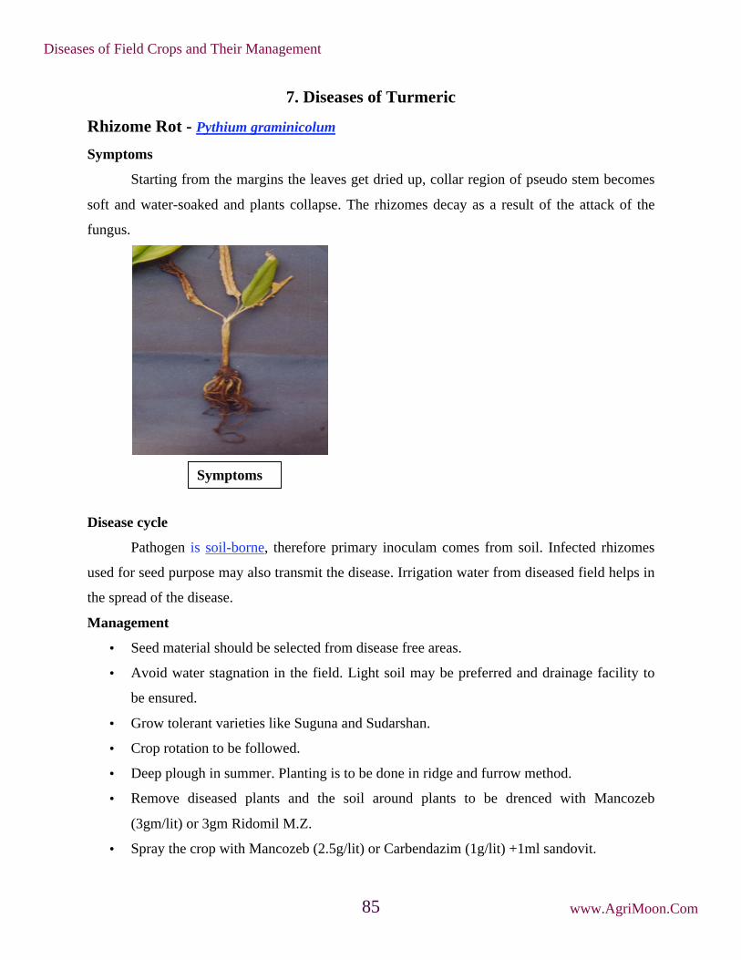
7. Diseases of Turmeric
Rhizome Rot - Pythium graminicolum
Symptoms
Starting from the margins the leaves get dried up, collar region of pseudo stem becomes
soft and water-soaked and plants collapse. The rhizomes decay as a result of the attack of the
fungus.
Disease cycle
Pathogen is soil-borne, therefore primary inoculam comes from soil. Infected rhizomes
used for seed purpose may also transmit the disease. Irrigation water from diseased field helps in
the spread of the disease.
Management
• Seed material should be selected from disease free areas.
• Avoid water stagnation in the field. Light soil may be preferred and drainage facility to
be ensured.
• Grow tolerant varieties like Suguna and Sudarshan.
• Crop rotation to be followed.
• Deep plough in summer. Planting is to be done in ridge and furrow method.
• Remove diseased plants and the soil around plants to be drenced with Mancozeb
(3gm/lit) or 3gm Ridomil M.Z.
• Spray the crop with Mancozeb (2.5g/lit) or Carbendazim (1g/lit) +1ml sandovit.
Symptoms
Diseases of Field Crops and Their Management
www.AgriMoon.Com85

• Keep rhizomes in 3g Metalaxyl or 3g Mancozeb mixed in one litre of water for one hour
and shade dry before planting.
Leaf Spot - Colletotrichum capsici
Symptoms
Oblong brown spots with grey centres are found on leaves. The spots are about 4-5 cm in
length and 2-3 cm in width. In advanced stages of disease black dots representing fungal acervuli
occur in concentric rings on spot. The grey centers become thin and gets teared. Severely
effected leaves dry and wilt. They are surrounded by yellow halos. Indefinite number of spots
may be found on a single leaf and as the disease advances; spots enlarge and cover a major
portion of leaf blade.
Favorable condition
• The disease is usually appears in October and November
• Relative humidity of 80% and temperatures of 21 – 230C favours the primary infection
Disease cycle
The fungus is carried on the scales of rhizomes which are the source of primary infection
during sowing. The secondary spread is by wind, water and other physical and biological agents.
The same pathogen is also reported to cause leaf-spot and fruit rot of chilli where it is transmitted
through seed borne infections. If chilli is grown in nearby fields or used in crop rotation with
turmeric, the pathogen perpetuates easily, building up inoculum potential for epiphytotic
outbreaks.
Management
Symptoms
Diseases of Field Crops and Their Management
www.AgriMoon.Com86

• Select seed material from disease free areas.
• Treat seed material with mancozeb @ 3g/litre of water or carbendazim @ 1 g/litre of
water, for 30 minutes and shade dry before sowing.
• Spray mancozeb @ 2.5 g/litre of water or carbendazim @ 1g/litre; 2-3 sprays at
fortnightly intervals.
• The infected and dried leaves should be collected and burnt in order to reduce the
inoculum source in the field.
• Spraying Blitox or Blue copper at 3 g/l of water was found effective against leaf spot.
• Crop rotations should be followed whenever possible.
• Cultivate tolerant varieties like Suguna and Sudarshan.
Leaf Blotch -Taphrina maculans
Symptoms
This disease usually appears on lower leaves in October and November. The individual
spots are small 1-2 mm in width and are mostly rectangular in shape. The disease is
characterized by the appearance of several spots on both the surfaces of leaves, being generally
numerous on the upper surface. They are arranged in rows along the veins. The spots coalesce
freely and form irregular lesions. They first appear as pale yellow discolorations and then
become dirty yellow in colour. The infected leaves disort and have reddish brown appearance.
Disease cycle
The fungus is mainly air borne and primary infection occurs on lower leaves with the
inoculum surviving in dried leaves of host, left over in the field. The ascospores discharged from
Symptoms
Diseases of Field Crops and Their Management
www.AgriMoon.Com87

successively maturing asci infect fresh leaves without dormancy, thus causing secondary
infection. Secondary infection is most dangerous than primary one causing profuse sprouting all
over the leaves. The pathogen persists in summer by means of ascogenous cells on leaf debris,
and dessicated ascospores and blastospores in soil and among fallen leaves.
Management
• Select seed material from disease free areas.
• Treat the seed material with Mancozeb @ 3g/litre of water or Carbendazim @ 1 g/litre of
water for 30 minutes and shade dry before sowing.
• Spray mancozeb @ 2.5 g/litre of water or Carbendazim @ 1g/litre; 2-3 sprays at
fortnightly intervals.
• The infected and dried leaves should be collected and burnt in order to reduce the
inoculum source in the field.
• Spraying Cpper oxy chloride at 3 g/l of water was found effective against leaf blotch.
• Crop rotations should be followed whenever possible.
Minor diseases
a. Dry rot - Rhizoctonia bataticola
b. Leaf spot - Cercospora curcuma
c. Leaf Blight - Rhizoctonia solani
d. Brown rot - It is a complex disease caused by the nematode
Pratylenchus sp. associated with Fusarium sp.
Diseases of Field Crops and Their Management
www.AgriMoon.Com88

8. Diseases of Tobacco
Damping off - Pythium aphanidermatum
Symptoms
The pathogen attacks the seedlings at any stage in the nursery. Sprouting seedlings are
infected and wither before emergence from the soil (Pre emergence damping off). Water soaked
minute lesions appear on the stems near the soil surface, soon girdling the stem, spreading up and
down in the stems and with in one or two days stem may rot leading to toppling over of the
seedlings (Post-emergence damping off).
The young seedlings in the nursery are killed in patches and infection spreads quickly.
Under the favorable conditions, the entire seedlings in the nursery are killed within 3 to 4 days.
A thick weft of mycelium may be seen on the surface of the soil.
Pathogen
The fungus produces thick, hyaline, thin walled, non-septate mycelium. It produces
irregularly lobed sporangia which germinate to produce vesicle containing zoopores. The
zoospores are kidney shaped and biflagellate. Oospores spherical, light to deep yellow or
yellowish brown coloured, measuring 17-19 m in diameter.
Favourable Conditions
• Over crowding of seedling.
• Ill drained nursery beds
• Heavy shade in nursery
• High atmospheric humidity (90-100 per cent)
Pre and post emergence
Diseases of Field Crops and Their Management
www.AgriMoon.Com89

• High soil moisture
• Low temperature (below 24 C) and low soil temperature of about 20˚C.
Disease cycle
The pathogen survives in the soil as oospores and chlamydospores. The primary infection
is from the soil-borne fungal spores and secondary spread through sporangia and zoospores
transmitted by wind and irrigation water.
Management
• Prepare raised seed beds with adequate drainage facility.
• Burn the seed beds with paddy husk before sowing.
• Drench the seed bed with 1 per cent Bordeaux mixture or 0.2 per cent Copper
oxychloride, two days before sowing.
Diseases of Field Crops and Their Management
www.AgriMoon.Com90

• Avoid over crowding of seedlings by using recommended seed rate (1 to 1.5g/2.5m2).
• Avoid excess watering of the seedlings.
• Spray the nursery beds two weeks after sowing with 1 per cent Bordeaux mixture or 0.2
per cent Copper oxychloride or 0.2 per cent Mancozeb and repeat subsequently at 4 days
interval under dry weather and at 2 days interval under wet cloudy weather or spray 0.2
per cent Metalaxyl at 10 days interval commencing from 20 days after germination.
Black shank - Phytophthora parasitica var. nicotianae
Symptoms
The pathogen may affect the crop at any stage of its growth. Even though all parts are
affected, the disease infects chiefly the roots and base of the stem. Seedlings in the nursery show
black discolor of the stem near the soil level and blackening of roots, leading the wet rot in
humid condition and seedling blight in dry weather with withering and drying of tips. The
pathogen also spreads to the leaves and causes blighting and drying of the bottom leaves. In the
transplanted crop, the disease appears as minute black spot on the stem, spreads along the stem to
produce irregular black patches and often girdling occurs.
The upward movement leads to development of necrotic patches on the stems.
The infected tissues shrink, leaving a depression and in advanced condition the stem shrivels and
plant wilts. When the affected stem is split open, the pith region is found to be dried up in disc-
like plates showing black discolouration. On the leaves large brown concentrically zonate
patches appear during humid weather, leading to blackening and rotting of the leaves.
Pathogen
Symptoms
Diseases of Field Crops and Their Management
www.AgriMoon.Com91

The fungus produces hyaline and non-septate mycelium. The sporangia, which are
hyaline, thin walled, ovate or pyriform with papillae, develop on the sporangiophores in a
sympodial fashion. Sporangia germinate to release zoospores which are usually kidney shaped,
biciliate and measure 11-13 x 8-9 m. The fungus also produces globoose and thick walled
chlamydospores, measuring 27-42 m in diameter. Oospores are thick walled, globose, smooth
and light yellow coloured, measuring 15-20 m in diameter.
Favourable Conditions
• Frequent rainfall and high soil moisture.
• High population of rootknot nematodes Meloidogyne incognita var. acrita.
Disease cycle
The fungus lives as a saprophyte on organic wastes and infected crop residues in soil. The
fungus is also present in the soil as dormant mycelium, oospores and chlamydospores for more
than 2 years. The primary infection is by means of oospores and chlamydospores in the soil.
Secondary spread is by wind-borne sporangia. The pathogen in the soil spreads through irrigation
water, transport of soil, farm implements and animals.
Management
• Cover the seed beds with paddy husk or groundnut shell at 15-20 cm thick layer and
burn.
• Provide adequate drainage in the nursery. Drench the nursery beds with 1 per cent
Bordeaux mixture or 0.2 per cent Copper oxychloride, two days before sowing.
• Spray the beds two weeks after sowing with 0.2 per cent Metalaxyl or 0.2 per cent
Captafol or 0.2 per cent Copper oxychloride or 1 per cent Boreaux mixture and repeat
after 10 days.
Sporangia
Diseases of Field Crops and Their Management
www.AgriMoon.Com92
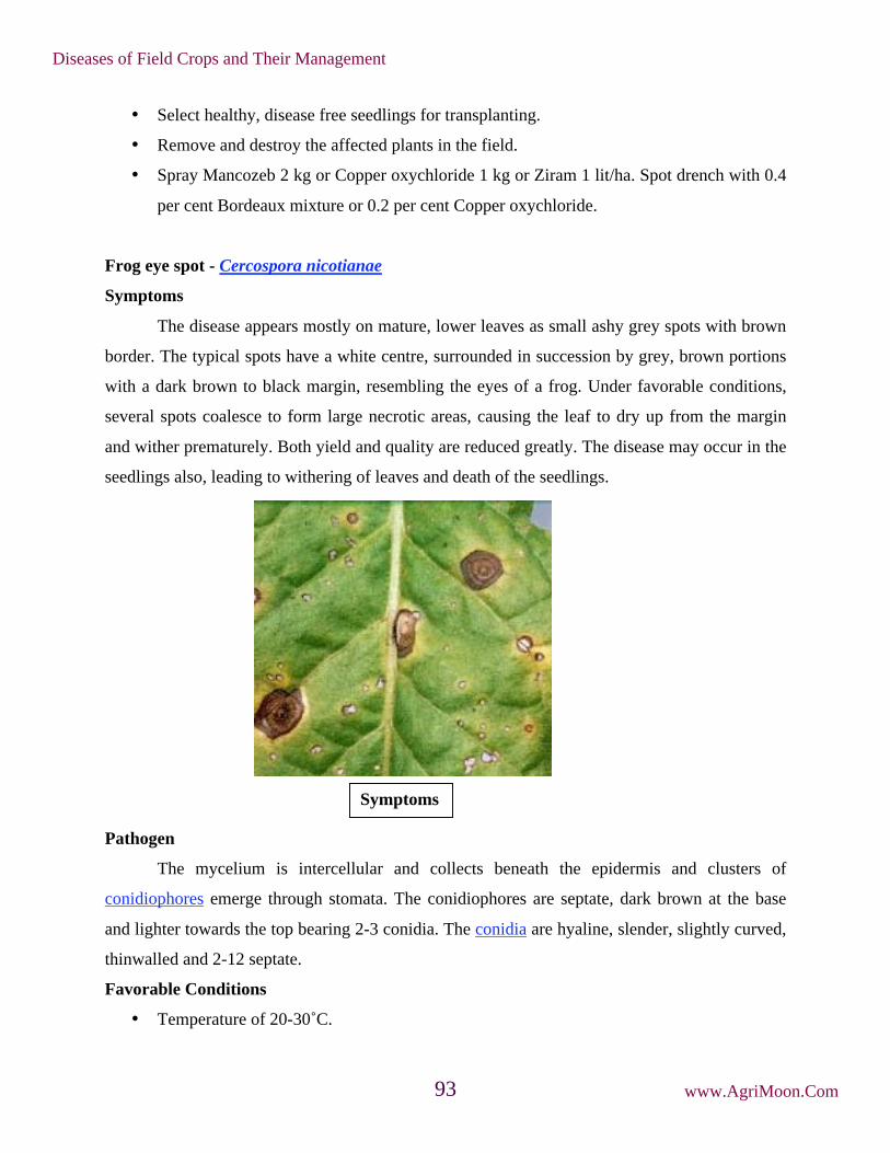
• Select healthy, disease free seedlings for transplanting.
• Remove and destroy the affected plants in the field.
• Spray Mancozeb 2 kg or Copper oxychloride 1 kg or Ziram 1 lit/ha. Spot drench with 0.4
per cent Bordeaux mixture or 0.2 per cent Copper oxychloride.
Frog eye spot - Cercospora nicotianae
Symptoms
The disease appears mostly on mature, lower leaves as small ashy grey spots with brown
border. The typical spots have a white centre, surrounded in succession by grey, brown portions
with a dark brown to black margin, resembling the eyes of a frog. Under favorable conditions,
several spots coalesce to form large necrotic areas, causing the leaf to dry up from the margin
and wither prematurely. Both yield and quality are reduced greatly. The disease may occur in the
seedlings also, leading to withering of leaves and death of the seedlings.
Pathogen
The mycelium is intercellular and collects beneath the epidermis and clusters of
conidiophores emerge through stomata. The conidiophores are septate, dark brown at the base
and lighter towards the top bearing 2-3 conidia. The conidia are hyaline, slender, slightly curved,
thinwalled and 2-12 septate.
Favorable Conditions
• Temperature of 20-30˚C.
Symptoms
Diseases of Field Crops and Their Management
www.AgriMoon.Com93

• High humidity (80-90 per cent).
• Close spacing, frequent irrigation and excess application of nitrogenous fertilizers.
Disease cycle
The pathogen is seed-borne and also persists on crop residues in the soil. The primary
infection is from the seed and soil-borne inoculum. The secondary spread is through wind-borne
conidia.
Management
• Remove and burn plant debris in the soil.
• Avoid excess nitrogenous fertilization.
• Adopt optimum spacing.
• Regulate irrigation frequency.
• Spray the crop with 0.4 per cent Bordeaux mixture or Thiophanate Methyl 750g/ha or
Carbendazim 750 g/ha and repeat after 15 days.
Powdery mildew - Erysiphe cichoracearum var. nicotianae
Symptoms
Initially the disease appears as small, white isolated patches on the upper surface of the
leaves. Later, it spreads fast and covers the entire lamina. The disease initially appears on the
lower leaves and as disease advances, the rest of the leaves are also infected and sometimes
powdery growth can be seen on the stem also. The affected leaves turn to brown and wither and
show scorched appearance. The severe infection leads to defoliation and reduction in quantity
and quality of the curable leaves.
Pathogen
The fungus is ecotophytic and produces hyaline, septate and highly branched mycelium.
Short, stout and hyaline conidiophores arise from the mycelium and bear conidia in chains. The
conidia are barrel shaped or cylindrical, hyaline and thin walled. Cleistothecia are black,
spherical with no ostiole, with numerous densely-woven septate, brown-coloured appendages.
They contain 10-15 asci which are ovate with a short stalk. Each ascus contains two ascospores
which are oval to elliptical, thinwalled, hyaline and single celled.
Diseases of Field Crops and Their Management
www.AgriMoon.Com94

Favourable Conditions
• Humid cloudy weather.
• Low temperature (16-23˚C.
• Close planting and excess doses of nitrogenous fertilizers.
Disease cycle
The fungus remains dormant as mycelium and cleistothecia in the infected plant debris in
soil. The primary infection is mainly from soil-borne inoculum. The secondary spread is aided by
wind blown conidia.
Management
• Apply balanced ferilizers.
• Avoid overcrowding of plants.
• Remove and destroy the affected leaves.
• Plant early in the season so that crop escapes the cool temperature at maturity phase.
• Spray dinocap at 375 ml or Carbendazim at 500g/ha.
Brown spot - Alternaria longipes
Symptoms
Brown spot in contrast to frog-eye spot is not
normally observed in the nursery but is very much
prevalent in the field. Initially it appears on lower and
older leaves as small brown, circular lesions, which
Conidia and conidiophores
Diseases of Field Crops and Their Management
www.AgriMoon.Com95

spread, to upper leaves, petioles, stalks and capsules even. In warm weather (30˚ C) under high
humidity, the leaf spots enlarge, 1-3 cm in diameter, centres are necroses and turn brown with
characteristic marking giving target board appearance with a definite outline. In severe infection
spots enlarge, coalesce and damage large areas making leaf dark-brown, ragged and worthless.
On leaves nearing maturity, leaf spots are surrounded by bright yellow halo, due to production of
toxin 'alternin' by the fungus.
Disease cycle
The fungus over summers in the soil as mycelium in the diseased plant debris such as
stems of tobacco, weeds and other hosts. Under favourable weather in the next season conidial
production starts which infect the lowermost leaves. As the season progresses, repeated infection
cycles of the fungus attack healthy tissues of all aerial parts of tobacco of any age under high
humidity. There is enormous spore density in the air near the end of the harvesting. Fungus
persists as a mycelium in dead tissue for several months.
Management
• Removal and destruction of diseased plant debris can check the primary infection
promptly.
• Continuous growing of tobacco after tobacco must be avoided in the heavily infected
fields.
• Weekly, spraying of fungicides such as Maneb or Zineb @ 2g/ha or Benomyl or
Thiophanate methyl at 1kg/ha.
Symptoms
Diseases of Field Crops and Their Management
www.AgriMoon.Com96

Anthracnose -Colletotrichum tabacum
Symptoms
Initially, infection starts on lower leaves as pale-brown circular spots of 0.5 mm diameter
with papery depressed centres outlined by slightly raised brown margin. The leaf-spots may
remain small with white areas in the centre or coalesce to form large necrotic lesions. Under
continuous humid weather, dark brown or black, elongated, sunken necrotic lesions appear on
midrib, petiole and stem resulting in petiole and stem rot. Such seedlings do not establish in the
field if planted. Primary infection starts from affected bits of aerial parts left in the soil in the
previous season. The pathogen is not seed-borne but persists in the soil on dried plant debris.
Management
• Raised seed beds and rabbing with farm wastes help in reducing the initial infection
• Removal and destruction of all diseased debris minimises the pathogen in the soil.
• Rogueing diseased seedlings especially with necrotic lesions on stem
• Protective spraying with Bordeaux mixture at 1.0% (2-2-500) or Zineb @ 2 kg/ha
Wild fire - Pseudomonas tabaci
Symptoms
The leaf spots may occur at any stage of plant growth including the nursery seedlings.
Dark brown to black spots with a yellow halo spreads quickly causing withering and drying of
leaves. In advanced cases, lesions develop on the young stem tissues leading to withering and
drying of the seedlings. In the fields, initially numerous water soaked black spots appear and
latter become angular when restricted by the veins and veinlets.
Diseases of Field Crops and Their Management
www.AgriMoon.Com97

Several spots may coalesce to cause necrotic patches on the leaves. In advanced
conditions, the entire leaf is fully covered with enlarged spots with yellow haloes. The leaves
slowly wither and dry. Under humid weather condition, the disease spreads very fast and covers
all the leaves and the entire plant gives a blighted appearance.
Pathogen
The bacterium is a rod, motile with a single polar flagellum, non-capsulated, non spore
forming and Gram negative.
Favourable Conditions
• Close planting.
• Humid wet weather.
• Strong winds.
Disaease cycle
The bacterium survives in the infected crop residues in the soil, which is the primary
source of infection. The secondary spread of the pathogen in the field is through wind splashed
rain water and implements.
Management
• Remove and burn the infected crop residues in the soil.
• Avoid very close planting.
Tobacco mosaic - Tobacco mosaic virus (TMV)
Symptoms
Symptoms
Diseases of Field Crops and Their Management
www.AgriMoon.Com98

The disease begins as light discoloration along the veins of the youngest leaves. Soon the
leaves develop a characteristic light and dark green pattern, the dark green areas associated more
with the veins, turning into irregular blisters.
The early infected plants in the season are usually stunted with small, chlorotic, mottled
and curled leaves. In severe infections, the leaves are narrowed, puckered, thin and malformed
beyond recognition, Later, dark brown necrotic spots develop under hot weather and this
symptom is called “Mosaic burn” or “Mosaic scorching”.
Pathogen
The disease is caused by Tobacco mosaic tobamovirus. It is a rigid rod measuring 300 X
150-180 nm with a central hollow tube of about 4nm diameter with ssRNA as its genome.
Disease cycle
The virus spreads most rapidly by contact
wounds, sap and farm implements and operators. The
virus remains viable in the plant debris in the soil as
the source of inoculum as the longevity of the virus is very high. It is capable of remaining
infective when stored dry for over 50 years. The virus has a wide host range, affecting nearly 50
plant species belonging to nine different families. The virus is not seed-transmitted in tobacco
but tomato seeds transmit the virus. No insect vector known to transmit the virus.
Management
• Remove and destroy infected plants.
• Keep the field free of weeds which harbour the virus.
Diseases of Field Crops and Their Management
www.AgriMoon.Com99

• Wash hands with soap and running water before or after handling the plants or after
weeding.
• Prohibit smoking, chewing and snuffing during field operations.
• Spray the nursery and main field with botanical leaf extracts of Bougainvillea or Basella
alba at 1 litre of extract in 150 litres of water, two to three times at weekly intervals.
• Adopt crop rotation by growing non-host plants for two seasons.
• Grow resistant varieties like TMV RR2, TMV RR 2a and TMV RR3.
Leaf curl - Tobacco leaf curl virus (TLCV)
Symptoms
The infections may occur at any stage, when young plants are infected the entire plant
remains very much dwarfed. Curling of leaves with clearing and thickening of veins; twisting of
petioles; puckering of leaves; rugose and brittle and development of enations are the important
symptoms of tobacco leaf curl disease.Three forms of leaf curl expression are observed. First the
leaf margins curl downward towards the dorsal side and show thickening of veins with enation
on the lower surface. Second crinkle form shows curling of whole leaf edge towards dorsal side
with enation on the veins and the lamina arching towards the ventral side between the veinlets.
Third the transparent symptom shows the curling of leaves towards the ventral side with clearing
of the veins and enations are absent.
Symptoms
Diseases of Field Crops and Their Management
www.AgriMoon.Com100

Pathogen
It is caused by Tobacco leaf curl geminivirus. Virions are geminate, non- enveloped, 18
nm diameter circular ssDNA genome.The virus is a white fly transmitted Geminivirus with
ssDNA as genome.
Disease cycle
The virus has a narrow host range in eight plant families. The virus is not transmissible
through sap or seed. The whitefly, Bemisia tabaci is the vector. Due to wide host range of the
virus many other plants are acting as source of inoculums.
Management
• Remove and destroy the infected plants.
• Rogue out the reservoir weed hosts which harbour the virus and whiteflies. Planting
tobacco crop during the crop periods when the vector population is low.
• Spray Methyldemeton at 0.1 to 0.2 per cent to control the vectors.
Phanerogamic parasite
Broom rape - Orobanche cernua var. desertorum
Symptoms
The affected tobacco plants are stunted and show withering and drooping of leaves to
wilting. These indicate underground parasitism of the tobacco roots by the parasite. The young
shoot of the parasite emerges from the soil at the base of the plants 5-6 weeks after transplanting.
Normally, it appears on clusters of 50-100 shoots around the base of a single tobacco plant. The
plants which are attacked very late exhibit no external symptoms but the quality and yield of
leaves are reduced.
Diseases of Field Crops and Their Management
www.AgriMoon.Com101

Parasite
It is a total root parasite. It is an annual, fleshy flowering plant with a short, stout stem,
10-15 inches long. The stem is pale yellow or brownish red in colour and covered by small, thin,
brown scaly leaves and the base of the stem is thickened. White-coloured flowers appear in the
leaf axils. The floral parts are well developed with a lobed calyx, tubular corolla, superior ovary,
numerous ovules and a large four-lobed stigma. The fruits are capsules containing small, black,
reticulate and ovoid seeds.
Disease cycle
The seeds of the parasite remain dormant in the soil for several years. Primary infection
occurs from the seeds in the soil. The seeds spread from field to field by irrigation water,
animals, human beings and implements. The dormant seeds are stimulated to germinate by the
root exudates of tobacco and attach itself, to the roots by forming haustoria. Later, it grows
rapidly to produce shoot and flowers. Orobanche also attacks other crops like brinjal, tomato,
cauliflower, turnip and other cruciferous crops.
Management
• Rogue out the tender shoots of the parasite before flowering and seed set.
• Spray the soil with 25 per cent copper sulphate.
• Spray 0.1 per cent Allyl alcohol.
• Apply few drops of kerosene directly on the shoot.
• Grow decoy or trap crops like chilli, moth bean, sorghum or cowpea to stimulate seed
germination and kill the parasite.
Diseases of Field Crops and Their Management
www.AgriMoon.Com102

9. Diseases of Groundnut
Tikka leaf spots
Early leaf spot: Cercopora arachidicola (Sexual Stage: Mycosphaerella arachidis)
Late leaf spot: Phaeoisariopsis personata (Syn : Cercospora personata)
(Sexual stage : Mycosphaerella berkeleyii)
Symptoms
The disease occurs on all above ground parts of the plant, more severely on the leaves.
The leaf symptoms produced by the two pathogens can be easily distinguished by appearance,
spot colour and shapes. Both the fungi produce lesions also on petiole, stem and pegs. The
lesions caused by both species coalesce as infection develops and severely spotted leaves shed
prematurely. The quality and yield of nuts are drastically reduced in severe infections.
Pathogen C. arachidicola (Sexual stage: M. arachidis) Symptoms
Diseases of Field Crops and Their Management
www.AgriMoon.Com103

The pathogen is intercellular and do not produce haustoria and become intracellular when
host cells die. The fungus produces abundant sporulation on the upper surface of the leaves.
Conidiophores are olivaceous brown or yellowish brown in colour, short, 1 or 2 septate,
unbranched and geniculate and arise in clusters.
Conidia are sub hyaline or pale yellow, obclavate, often curved 3-12 septate, 35- 110 x
2.5 - 5.4 m in size with rounded to distinctly truncate base and sub-acute tip. The perfect stage
of the fungus produces perithecia as ascostromata. They are globose with papillate ostiole. Asci
are cylindrical to clavate and contain 8 ascospores. Ascospores are hyaline, slightly curved and
two celled, apical cell larger than the lower cell.
P. personata (C. personata) (Sexual stage: M. berkeleyii)
The fungus produces internal and intercellular mycelium with the production of
haustoria. The conidiphores are long, continuous, 1-2 septate, geniculate, arise inclusters and
olive brown in colour. The conidia are cylindrical or obclavate, short, measure
18-60 x 6-10 m, hyaline to olive brown, usually straight or curved slightly with 1-9 septa, not
constricted but mostly 3-4 septate. The fungus in its perfect stage produces perithecia as
ascostromata which are globose or broadly ovate with papillate ostiole. Asci are cylindrical to
ovate, contain 8 ascospores. Ascospores are 2 celled and constricted at septum and hyaline.
Favourable Conditions
• Prolonged high relative humidity for 3 days.
• Low temperature (20 C) with dew on leaf surface.
• Heavy doses of nitrogen and phosporus fertilizers
• Deficiency of magesium in soil.
Disease cycle
The pathogen survives for a long period in the infected plant debris through conidia,
dormant mycelium and perithecia in soil. The volunteer groundnut plants also harbour the
pathogen. The primary infection is by ascospores or conidia from infected plant debris or infectd
seeds. The secondary spread is by wind blown conidia. Rain splash also helps in the spread of
conidia.
Management
• Remove and destory the infected plant debris.
Diseases of Field Crops and Their Management
www.AgriMoon.Com104

• Eradicate the volunteer groundnut plants.
• Keep weeds under control.
• Treat the seeds with Carbendazim or Thiram at 2g/kg.
• Spray Carbendazim 500g or mancozeb 2 kg or Chlorothalonil 2 kg/ha and if necessary,
repeat after 15 days.
• Grow moderately resistant varieties like ALR 1.
Rust - Puccinia arachidis
Symptoms
The disease attacks all aerial parts of the plant. The disease is usually found when the
plants are about 6 weeks old. Small brown to chestnut dusty pustules (uredosori) appear on the
lower surface of leaves. The epidermis ruptures and exposes a powdery mass of uredospores.
Corresponding to the sori, small, necrotic, brown spots appear on the upper surface of leaves.
The rust pustules may be seen on petioles and stem. Late in the season, brown teliosori, as dark
pustules, appear among the necrotic patches. In severe infection lower leaves dry and drop
prematurely. The severe infection leads to production of small and shriveled seeds.
Pathogen
The pathogen produces both uredial and telial stages. Uredial stages are produced
abundant in groundnut and production of telia is limited. Uredospores are pedicellate,
Symptoms
Diseases of Field Crops and Their Management
www.AgriMoon.Com105

unicellular, yellow, oval or round and echinulated with 2 or 3 germpores. Teliospores are dark
brown with two cells. Pycnial and aecial stages have not been recorded and there is no
information available about the role of alternate host.
Teliospores
Uredospores
Favourable Conditions
• High relative humidity (above 85 per cent).
• Heavy rainfall.
• Low temperature (20-25˚C).
Disease cycle
The pathogen survives as uredospores on volunter groundnut plants. The fungus also
survives in infected plant debris in soil. The spread is mainly through wind borne inoculum of
uredospores. The uredospores also spread as contamination of seeds and pods. Rainsplash and
implements also help in dissemination. The fungus also survives on the collateral hosts like
Arachis marginata, A. nambyquarae and A. prostrate.
Management
• Avoid monoculturing of groundnut.
• Remove volunteer groundnut plants and reservoir hosts.
• Spray mancozeb 2 kg or Wettable Sulphur 3 kg or Tridemorph 500ml or Chlorothalonil 2
kg/ha.
• Grow moderatelyresistant varieties like ALR 1.
Collar rot or seedling blight or crown rot - Aspergillus niger and A. pulverulentum
Symptoms
Diseases of Field Crops and Their Management
www.AgriMoon.Com106

The disease usually appears in three phases.
i. Pre-emergence rot
Seeds are attacked by soil-borne conidia and caused rotting of seeds. The seeds are
covered with black masses of spores and internal tissues of seed become soft and watery.
ii. Post-emergence rot
The pathogen attacks the emerging young seedling and cause circular brown spots on the
cotyledons. The symptom spreads later to the hypocotyl and stem. Brown discolored spots
appear on collar region. The affected portion become soft and rotten, resulting in the collapse of
the seedling. The collar region is covered by profuse growth of fungus and conidia and affected
stem also show shredding symptom.
iii. Crown rot
The infection when occurs in adult plants show crown rot symptoms. Large lesions
develop on the stem below the soil and spread upwards along the branches causing drooping of
leaves and wilting of plant.
Pathogen
The mycelium of the fungus is hyaline to sub-hyaline. Conidiophores arise directly from
the substrate and are septate, thick walled, hyaline or olive brown in colour. The vesicles are
mostly globose and have two rows of hyaline phialides viz., primary and secondary phialides.
Symptoms
Diseases of Field Crops and Their Management
www.AgriMoon.Com107
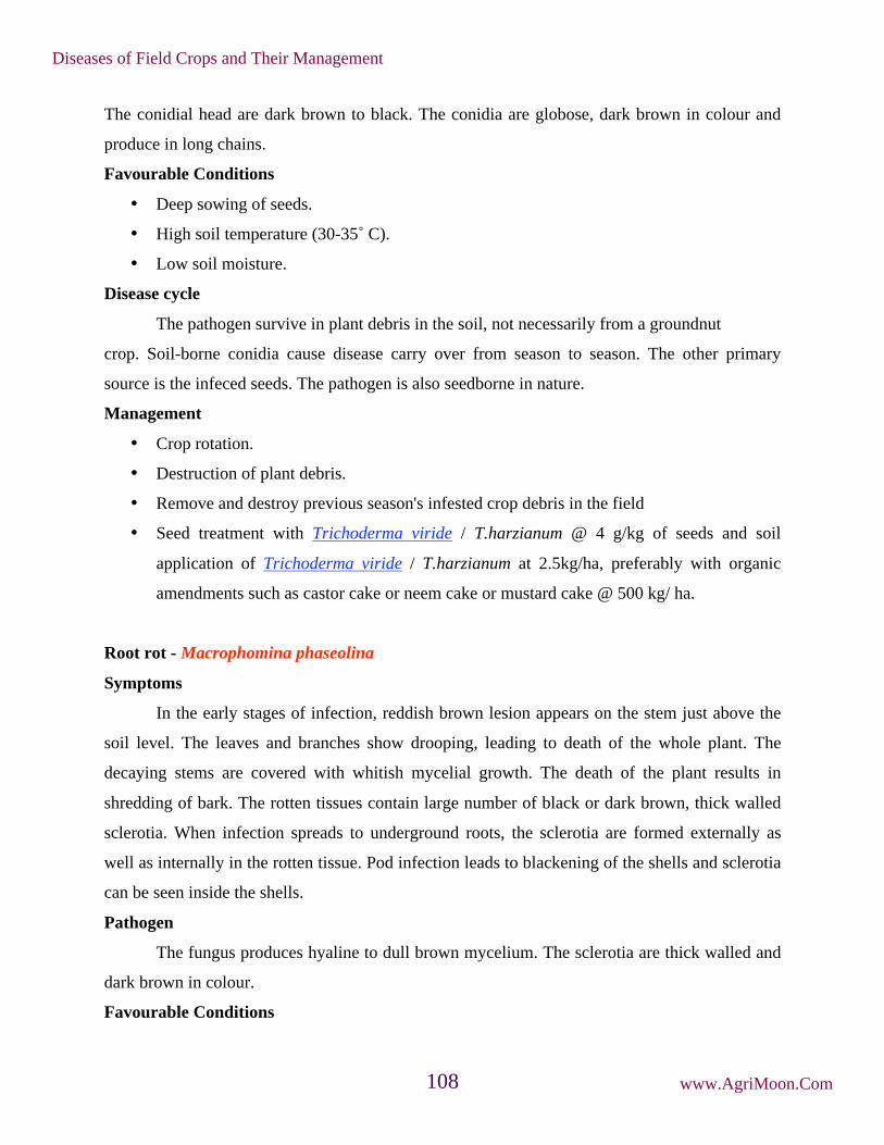
The conidial head are dark brown to black. The conidia are globose, dark brown in colour and
produce in long chains.
Favourable Conditions
• Deep sowing of seeds.
• High soil temperature (30-35˚ C).
• Low soil moisture.
Disease cycle
The pathogen survive in plant debris in the soil, not necessarily from a groundnut
crop. Soil-borne conidia cause disease carry over from season to season. The other primary
source is the infeced seeds. The pathogen is also seedborne in nature.
Management
• Crop rotation.
• Destruction of plant debris.
• Remove and destroy previous season's infested crop debris in the field
• Seed treatment with Trichoderma viride / T.harzianum @ 4 g/kg of seeds and soil
application of Trichoderma viride / T.harzianum at 2.5kg/ha, preferably with organic
amendments such as castor cake or neem cake or mustard cake @ 500 kg/ ha.
Root rot - Macrophomina phaseolina
Symptoms
In the early stages of infection, reddish brown lesion appears on the stem just above the
soil level. The leaves and branches show drooping, leading to death of the whole plant. The
decaying stems are covered with whitish mycelial growth. The death of the plant results in
shredding of bark. The rotten tissues contain large number of black or dark brown, thick walled
sclerotia. When infection spreads to underground roots, the sclerotia are formed externally as
well as internally in the rotten tissue. Pod infection leads to blackening of the shells and sclerotia
can be seen inside the shells.
Pathogen
The fungus produces hyaline to dull brown mycelium. The sclerotia are thick walled and
dark brown in colour.
Favourable Conditions
Diseases of Field Crops and Their Management
www.AgriMoon.Com108

• Prolonged rainy season at seedling stage and low lying areas.
Disease cycle
The fungus remains dormant as sclerotia for a long period in the soil and in infected plant
debris. The primary infection is through soil-borne and seed-borne sclerotia. The secondary
spread of sclerotia is aided by irrigation water, human agency, implements and cattle etc.
Management
• Treat the seeds with thiram or carbendazim 2g/kg or Trichoderma viride at 4g/kg.
• Spot drench with Carbendazim at 0.5 g/lit.
Rossette - Groundnut rosette assistor virus (GRAV), Groundnut rosette virus and Groundnut
rosette satellites
Symptoms
The affected plants are characterized by the appearance of dense clump or dwarf shoots
with tuft of small leaves forming in a rosette fashion. The plant exhibits chlorosis and mosaic
mottling. The infected plants remain stunted and produce flowers, but only a few of the pegs may
develop further to nuts but no seed formation.
Pathogen
The disease is caused by a complex mixture of viruses viz.,Groundnut rosette assistor
virus (GRAV), Ground nut rosette virus and Groundnut rosette satellites is an isometric, not
enveloped and 28nm diameter (reported from India) and it gives no overt symptom in groundnut.
Groundnut rosette virus is with ssRNA genome, which becomes packaged in GRAV virious and
thus depends on it for aphid transmission, but produces no overt symptoms in groundnut. The
Symptoms
Diseases of Field Crops and Their Management
www.AgriMoon.Com109

groundnut rosette satellites are satellite RNAs that control the symptoms and cause the different
types of rosette (chlorotic, green and mosaic).
Disease Cycle
The primary source of spread by aphid vector, Aphis craccivora and A. gossipii in a
persistent manner, retained by vector but not transmitted congenitally. The virus is not
transmitted by any other means like mechanical or seed or pollen. The virus can survive on the
volunteer plants of groundnut and other weed hosts.
Management
• Practice clean cultivation.
• Use heavy seed rate and rogue out the infected plants periodically.
• Spray Monocrotophos or Methyl demeton at 500 ml/ha.
Groundnut bud necrosis disease - Groundnut bud necrosis virus (GBNV- Tospo virus)
Symptoms
First symptoms are visible 2-6 weeks after infection as ring spots on leaves. The newly
emerging leaves are small, rounded or pinched inwards and rugose with varying patterns of
mottling and minute ring spots. Necrotic spots and irregularly shaped lesions develop on leaves
and petioles. Stem also exhibits necrotic streaks.
Symptoms
Diseases of Field Crops and Their Management
www.AgriMoon.Com110

Plant becomes stunted with short internodes and short auxillary shoots. Leaflets show
reduction in size, distortion of the lamina, mosaic mottling and general chlorosis. In advanced
conditions, the necrosis of buds occurs. Top bud is killed and necrosis spreads downwards.
Drastic reduction in flowering and seeds produced are abnormally small and wrinkled with the
dark black lesions on the testa.
Pathogen
It is caused by Groundnut bud necrosis virus (GBNV). The virus particles are spherical,
30 nm in diameter, enveloped, ssRNA with multipartite genome.
Disease cycle
The virus perpetuates in the weed hosts viz., Bidens pilosa, Erigon bonariensis,
Tagetes minuta and Trifolium subterraneum. The virus is transmitted by thrips viz., Thrips palmi,
T. tabaci and Frankliniella sp.
Management
• Adopt plant spacing of 15x15 cm.
• Remove and destory infected plants up to 6 weeks after sowing.
• Application of Monocrotophos 500 ml/ha, 30 days after sowing either alone or in
combination with AVP (Anti Viral Principle) extracted from sorghum or coconut leaves.
Spray the crop with 10 per cent AVP at 500 lit/ha, ten and twenty days after sowing.
Minor diseases
Stem rot - Sclerotium rolfsii
Symptoms
The first symptom is the sudden drying of a branch which is completely or partially in
contact with the soil. The leaves turn brown and dry but remain attached to the plant. Near soil
on stems white growth of fungus mycelium is appeared. As the disease advances white
mycelium web spreads over the soil and the basal canopy of the plant. The sclerotia, the size and
colour of mustard seeds, appear on the infected areas as the disease develops and spreads. The
entire plant may be killed or only two or three branches may be affected. Lesions on the
developing pegs can retard pod development. Infected pods are usually rotted.
Diseases of Field Crops and Their Management
www.AgriMoon.Com111

Management
• Cultural practices such as deep' covering or burial of organic matter before planting, non-
dirting cultivation by avoiding movement of soil up around the base of plants and
preventing accumulation of organic debris are extremely useful in reducing the disease.
• Crop rotation with wheat, corn and soyabean may minimize the incidence of stem rot.
• Seed treatment with Carbendazim / Thiram / Captan @ 2-3 g/kg seed.
• Seed treatment with Trichoderma viride formulation (4g/kg) followed by application of
2.5kg Trichoderma viride formulation mixed with 50kg farm yard manure before sowing.
Wilt - Fusarium oxysporum and F. solani
Symptoms
Germinating seeds are attacked by the pathogens shortly before emergence. There is
general tissue disintegration and the surface of the seedling is covered with sporulating
mycelium. Damping off symptoms characterized by brown to dark brown Water soaked sunken
lesions on the hypocotyl which later encircle the stem and extend above the soil level. Roots are
also attacked, especially the apical portions. The affected seedlings become yellow and wilted.
The leaves turn greyish green and the plants dry up and die. The roots and stems show internal
vascular browning and discolouration. These fungi are also commonly associated with pod rot.
Management
• Seed treatment with systemic fungicides like Carbendazim at 2g/kg seed.
Anthracnose - Colletotrichum dematium and C. capsici
Symptoms
Symptoms
Diseases of Field Crops and Their Management
www.AgriMoon.Com112
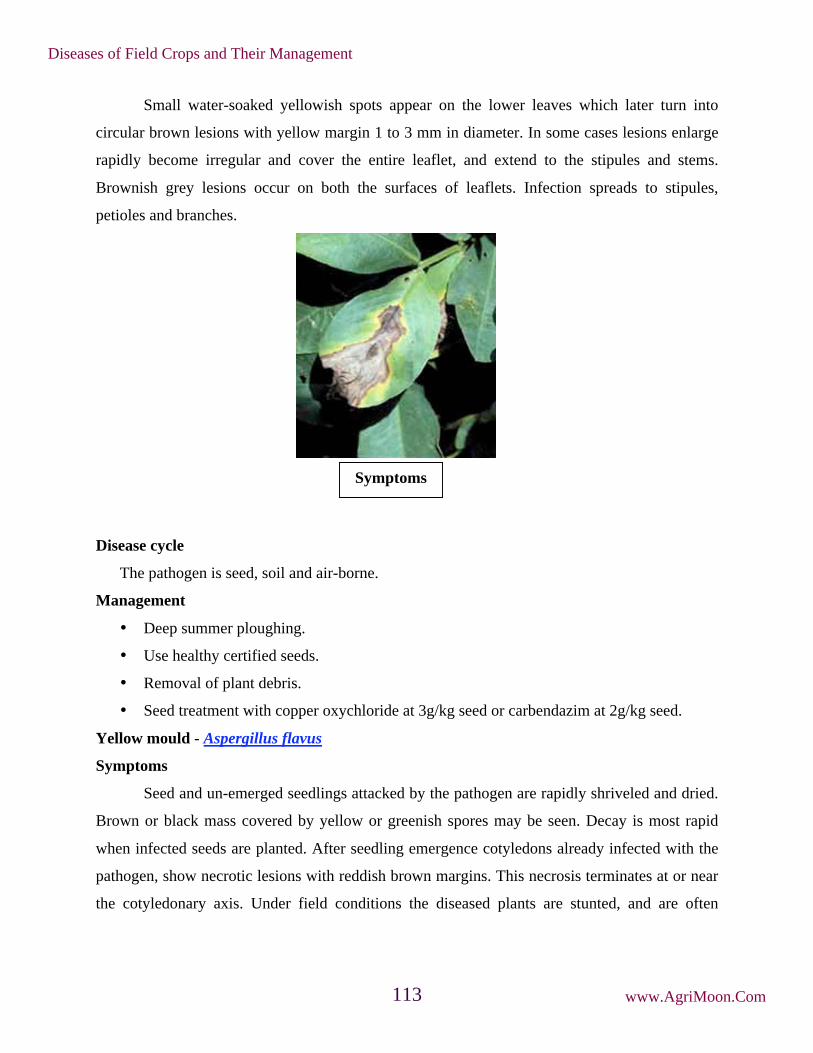
Small water-soaked yellowish spots appear on the lower leaves which later turn into
circular brown lesions with yellow margin 1 to 3 mm in diameter. In some cases lesions enlarge
rapidly become irregular and cover the entire leaflet, and extend to the stipules and stems.
Brownish grey lesions occur on both the surfaces of leaflets. Infection spreads to stipules,
petioles and branches.
Disease cycle
The pathogen is seed, soil and air-borne.
Management
• Deep summer ploughing.
• Use healthy certified seeds.
• Removal of plant debris.
• Seed treatment with copper oxychloride at 3g/kg seed or carbendazim at 2g/kg seed.
Yellow mould - Aspergillus flavus
Symptoms
Seed and un-emerged seedlings attacked by the pathogen are rapidly shriveled and dried.
Brown or black mass covered by yellow or greenish spores may be seen. Decay is most rapid
when infected seeds are planted. After seedling emergence cotyledons already infected with the
pathogen, show necrotic lesions with reddish brown margins. This necrosis terminates at or near
the cotyledonary axis. Under field conditions the diseased plants are stunted, and are often
Symptoms
Diseases of Field Crops and Their Management
www.AgriMoon.Com113

chlorotic. The leaflets are reduced in size with pointed tips, widely varied in shape and
sometimes with veinal clearing.
Management
• Since the fungus is a weak parasite, agronomic practices which favour rapid germination
and vigorous growth of seedling will reduce the chance of A. flavus infection.
• Seed treatment with carbendazim or captan or thiram at 2g/kg seed.
Grey mould - Botrytis cinerea
Infection is seen on leaves, stem and underground parts of the groundnut. Initially
infection occurs at ground level by a light grey fungal rot which causes death of the plants.
Bacterial wilt - Pseudomonas solanacearum
Infected plants appear unhealthy, chlorotic and wilt under water stress. Dark brown
discolouration of xylem is seen. Grey slimy liquid ooze out of the vascular bundles.
Leaf spot - Alternaria arachidis and A. tenuissima
Symptoms
Lesions produced by A. arachidis are brown in colour and irregular in shape surrounded
by yellowish halos. Symptoms produced by A. tenuissima are characterized by blighting of
apical portions of leaflets which turn light to dark brown colour. Lesions produced by A.
alternata are small, chlorotic, water soaked, that spread over the surface of the leaf. The lesions
become necrotic and brown and are round to irregular in shape. Veins and veinlets adjacent to
the lesions become necrotic. Lesions increase in area and their central portions become pale,
rapidly dry out, and disintegrate. Affected leaves show chlorosis and in severe attacks become
prematurely senescent. Lesions can coalesce, give the leaf a ragged and blighted appearance.
Symptoms
Diseases of Field Crops and Their Management
www.AgriMoon.Com114

Management
• Foliar application of Mancozeb (2kg/ha) or Copper oxychloride (2kg/ha) or Carbendazim
(500g/ha).
Indian Peanut Clump Disease - Peanut Clump virus
Earlier this disease was confused with groundnut rosstte. Now it is recognized as a
distinct virus causing clump disease. The leaves turn very dark and plants become severely
stunted. The disease is soil borne and transmitted by a fungus, Polymyxa graminis. The pH of the
soil affects transmission. It is also transmitted by seed. The virus is rod shaped, 190-245nm long
x 21nm wide, not enveloped, ssRNA genome.
Other virus diseases of minor importance occurring on groundnut are:
Peanut chlorotic streak (caused by Caulimovirus, occurs only in India), Peanut green
mosaic and mottle (caused by a Potyvirus), peanut stunt (caused by Cucumovirus), groundnut
chlorotic spot (caused by a Potexvirus), groundnut eye spot (caused by Potyvirus) and groundnut
ringspot.
Diseases of Field Crops and Their Management
www.AgriMoon.Com115

10. Diseases of Castor
Seedling blight - Phytophthora parasitica
Symptoms
The disease appears circular, dull green patch on both the surface of the cotyledon leaves.
It later spreads and causes rotting. The infection moves to stem and causes withering and death
of seedling. In mature plants, the infection initially appears on the young leaves and spreads to
petiole and stem causing black discoloration and severe defoliation.
Dead seedling
Spot on older leaf Leaf blight symptom
Pathogen
The pathogen produces non-septate and hyaline mycelium. Sporangiophores emerge
through the stomata on the lower surface singly or in groups. They are unbranched and bear
single celled, hyaline, round or oval sporangia at the tip singly. The sporangia germinate to
produce abundant zoospores. The fungus also produces oospores and chlamydospores in adverse
seasons.
Favourable Conditions
Diseases of Field Crops and Their Management
www.AgriMoon.Com116

• Continuous rainy weather.
• Low temperature (20-25˚C).
• Low lying and ill drained soils.
Disease cycle
The pathogen remains in the soil as chlamydospores and oospores which act as primary
source of infection. The fungus also survives on other hosts like potato, tomato, brinjal, sesamum
etc. The secondary spread takes place through wind borne sporangia.
Management
• Remove and destroy infected plant residues.
• Avoid low-lying and ill drained fields for sowing.
• Treat the seeds with thiram or captan at 4g/kg.
Rust – Melampsora ricini
Symptoms
Minute, orange-yellow coloured, raised pustules appear with powdery masses on the
lower surface of the leaves and the corresponding areas on the upper surface of the leaves are
yellow. Often the pustules are grouped in concentric rings and coalesce together to for drying of
leaves.
Powdery mass covering entire leaf
Pathogen
The pathogen produces only uredosori in castor plants and other stages of the life cycle
are unknown. Uredospores are two kinds, one is thick walled and other is thin walled. They are
elliptical to round, orange-yellow coloured and finely warty.
Disease cycle
The fungus survives in the self sown castor crops in the off season. It can also survive on
other species of Ricinus. The fungus also attacks Euphorbia obtusifolia, E.geniculata and
E.marginata. The infection spreads through airborne uredospores.
Diseases of Field Crops and Their Management
www.AgriMoon.Com117

Management
• Rogue out the self-sown castor crops and other weed hosts.
• Spray Mancozeb at 2kg/ha or Propioconazole 1l/ha.
Leaf blight- Alternaria ricini
Symptoms
All the aerial parts of plants viz., leaves, stem, inflorescences and capsules are liable to be
attacked by the pathogen. Irregular brown spots with concentric rings form initially on the leaves
and covered with fungal growth. When the spots coaleasce to form big patches, premature
defoliation occurs. The stems, inflorescences and capsules are also show dark brown lesions with
concentric rings. On the capsules, initially brown sunken spots appear, enlarge rapidly and cover
the whole pod. The capsules crack and seeds are also get infected.
Alterneria leaf spot with concentric rings
Pathogen
The pathogen produces erect or slightly curved, light grey to brown conidiophores, which
are occasionally in groups. Conidia are produced in long chains. Conidia are obclavate, light
olive in colour with 5-16 cells having transverse and longitudinal septa with a beak at the tip.
Favourable Conditions
• High atmospheric humidity (85-90 %).
• Low temperature (16-20˚C)
Disease cycle
The pathogen survives on hosts like Jatropha pandurifolia and Bridelia hamiltoniana.
The pathogen is externally and internally seed-borne and causes primary infection. The
secondary infection is through air-borne conidia.
Diseases of Field Crops and Their Management
www.AgriMoon.Com118
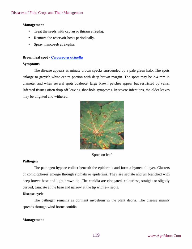
Management
• Treat the seeds with captan or thiram at 2g/kg.
• Remove the reservoir hosts periodically.
• Spray mancozeb at 2kg/ha.
Brown leaf spot - Cercospora ricinella
Symptoms
The disease appears as minute brown specks surrounded by a pale green halo. The spots
enlarge to greyish white centre portion with deep brown margin. The spots may be 2-4 mm in
diameter and when several spots coalesce, large brown patches appear but restricted by veins.
Infected tissues often drop off leaving shot-hole symptoms. In severe infections, the older leaves
may be blighted and withered.
Spots on leaf
Pathogen
The pathogen hyphae collect beneath the epidermis and form a hymenial layer. Clusters
of conidiophores emerge through stomata or epidermis. They are septate and un branched with
deep brown base and light brown tip. The conidia are elongated, colourless, straight or slightly
curved, truncate at the base and narrow at the tip with 2-7 septa.
Disease cycle
The pathogen remains as dormant mycelium in the plant debris. The disease mainly
spreads through wind borne conidia.
Management
Diseases of Field Crops and Their Management
www.AgriMoon.Com119

• Spraying with 1% Bordeaux mixture or Copper oxy chloride @ 0.2% may help to bring
the disease under check; but where the cultures of Eri-silk worm are maintained on castor
plants, spraying would not be desirable.
• Use of resistant varieties would be the most effective method for combating the disease.
• Spraying twice with Mancozeb 2g/lit or Carbendazim 500g/ha at 10-15 day interval
reduces the disease incidence.
• Treat the seed with thiram or Captan 2gm/kg seed.
Powdery mildew - Leveillula taurica
Symptoms
It is characterized by typical mildew growth which is generally confined to the under-
surface of the leaf. When the infection is severe the upper-surface is also covered by the whitish
growth of the fungus. Light green patches, corresponding to the diseased areas on the under
surface, are visible on the upper side especially when the leaves are held against light.
Powdery mass covering entire leaf
Management
• When weather is comparatively dry spray twice with wettable Sulphur 2g/lit at 15 days
interval, starting from 3 months after sowing.
• Spray 1ml hexaconazole or 2ml dinocap / litre of water at fortnight intervals. The variety
Jwala is resistant to this disease.
Stem rot - Macrophomina phaseolina
Symptoms
Small brown depressed lesions on and around nodes. Increase in size on both directions
causing 2 to 20 cm necrotic area. Lesions often coalesce and girdle the stem causing leaf drop.
Diseases of Field Crops and Their Management
www.AgriMoon.Com120

Drying and death starts from apex and progress. Infected capsules discoloured and drop easily.
Sudden wilting of plants in patches under high moisture stress coupled with high soil
temperature. Plant exhibit symptoms of drought and drooping of leaves. At ground level black
lesions are formed on the stem. Young leaves curl inwards with black margins and drop off later,
such branches Die-back. Entire branch and top of the plant withers.
Affected plant showing drooping of leaves
Management
• Grow tolerant and resistant varieties like Jyothi, Jwala, GCH-4, DCH-30 and SHB-145.
• Avoid water logging.
• Destruction of crop debris.
• Selection of healthy seed.
• Providing irrigation at critical stages of the crop.
• Treat the seed with thiram @ 2g/kg or carbendazim at 2g/ kg seed.
• Seed treatment with Trichoderma viride formulation at 4g/kg of seed.
• Soil drenching with Carbendazim (1g/1 litre of water) 2-3 times at 15 days interval.
Bacterial leaf spot - Xanthomonas campestris pv. ricinicola
Symptoms
The pathogen attacks cotyledons, leaves and veins and produces few to numerous small
round, water-soaked spots which later become angular and dark brown to jet black in color. The
spots are generally aggregated towards the tip. At a later stage the spots become irregular in
shape particularly when they coalesce and areas around such spots turn pale-brown and brittle.
Bacterial ooze is observed on both the sides of the leaf which is in the form of small shining
beads or fine scales.
Diseases of Field Crops and Their Management
www.AgriMoon.Com121

Pustules on lower leaf surface
Management
• Field sanitation help in minimizing the yield loss as pathogen survives on seed and plant
debris.
• Hot water treatment of seed at 58˚C to 60˚C for ten minutes.
• Grow tolerant varieties.
• Spray Copper oxychloride 2kg/ha or Streptocycline 100g/ha or Paushamycin 250g/ha.
Wilt - Fusarium oxysporum
Symptoms
When seedlings are attacked cotyledonary leaves turn to dull green colour, wither and die
subsequently. Leaves are droop and drop off leaving behind only top leaves. Diseased plants are
sickly in appearance. Wilting of plants, root degeneration, collar rot, drooping of leaves and
necrosis of affected tissue and finally leading to death of plants. Necrosis of leaves starts from
margins spreading to interveinal areas and finally to the whole leaf. Spilt open stem shows
brownish discolouration and white cottony growth of mycelia much prominently in the pith of
the stem.
Diseases of Field Crops and Their Management
www.AgriMoon.Com122

Management
• Selection of disease free seeds.
• Grow tolerant and resistant varieties like Jyothi, Jwala, GCH-4 DCH-30 and SHB 145.
• Avoid water logging
• Burning of crop debris
• Green manuring and intercropping with red gram
• Treat the seeds with thiram @ 2g/ kg or carbendiazim @ 2g/ kg seed.
• Seed treatment with 4g of Trichoderma viride talc formulation.
• Multiplication of 2kg of T.viride formulation by mixing in 50kg farm yard manure
• Sprinkling water and covering with polythene sheet for 15days and then applying
between rows of the crops is helpful in reducing the incidence.
Symptoms
Diseases of Field Crops and Their Management
www.AgriMoon.Com123

11. Diseases of Sunflower
Root rot or charcoal rot - Rhizoctonia bataticola (Pycnidial stage: Macrophomina phaseolina)
Symptoms
The pathogen is seed-borne and primarily causes seedling blight and collar rot in the
initial stages. The grown up plants also show symptoms after flowering stage. The infected
plants show drooping of leaves and death occurs in patches. The bark of the lower stem and roots
shreds and are associated with a large number of sclerotia. Dark coloured, minute pycnidia also
develop on the lower portion of the stem.
Pathogen
The fungus produces a large number of black, round to irregular shaped sclerotia. The
pycnidia are dark brown to black with an ostiole and contain numerous single celled, thin walled,
hyaline and elliptical pycnidiospores.
Symptoms
Diseases of Field Crops and Their Management
www.AgriMoon.Com124

Favourable Conditions
• Moisture stress and higher temperature favour development of the disease.
Disease cycle
The pathogen survives in soil and in infected crop residues through sclerotia and
pycnidia. The pathogen is seed-borne and it serves as primary source of infection. Wind-borne
conidia cause secondary spread. The soil borne sclerotia also spreads through rain splash,
irrigation water and implements.
Management
• Closer planting of the seedling should be avoided.
• Optimum nutrition should be provided to maintain the plant vigour.
• Whenever the soil becomes dry and the soil temperature rises then irrigation should be
provided.
• Seed treatment with Trichoderma viride formulation at 4 g/kg seed.
• In endemic areas long crop rotation should be followed.
• Treat the seeds with Carbendazim or Thiram at 2/kg
• Spot drench with Carbendazim at 500 mg/litre.
Leaf blight - Alternaria helianthi
Symptoms
The pathogen produces brown spots on the leaves, but the spots can also be seen on the
stem, sepals and petals. The lesions on the leaves are dark brown with pale margin surrounded by
a yellow halo. The spots later enlarge in size with concentric rings and become irregular in
shape. Several spots coalesce to show bigger irregular lesions leading to drying and defoliation.
Symptoms
Diseases of Field Crops and Their Management
www.AgriMoon.Com125

Pathogen
The pathogen produces cylindrical conidiophores, which are pale grey-yellow coloured,
straight or curved, geniculate, simple or branched, septate and bear single conidium. Conidia are
cylindrical to long ellipsoid, straight or slightly curved, pale grey-yellow to pale brown, 1 to 2
septate with longitudinal septa.
Favourable Conditions
• Rainy weather.
• Cool winter climate.
• Late sown crops are highly susceptible.
Disease cycle
The fungus survives in the infected host tissues and weed hosts. The fungus is also seed-
borne. The secondary spread is mainly through wind blown conidia.
Management
• Deep summer ploughing.
• Proper spacing
• Clean cultivation and field sanitation.
• Use of resistant or tolerant variety like B.S.H.1 .
• Application of well rotten manures.
• Practicing crop rotation.
• Planting in mid-September.
• Remove and destroy the diseased plants
• Treat the seeds with Thiram or Carbendazim at 2 g/kg. Spray Mancozeb at
2 kg/ha.
Diseases of Field Crops and Their Management
www.AgriMoon.Com126

Rust - Puccinia helianthi
Symptoms
Small, reddish brown pustules (uredia) covered with rusty dust appear on the lower
surface of bottom leaves. Infection later spreads to other leaves and even to the green parts of the
head. In severe infection, when numerous pustules appear on leaves, they become yellow and
dry. The black coloured telia are also seen among uredia on the lower surface. The disease is
autoecious rust. The pycnial and aecial stages occur on volunteer crops grown during off-season.
Pathogen
The uredospores are round or elliptical, dark cinnamon-brown in colour and minutely
echinulated with 2 equatorial germpores. Teliospores are elliptical or oblong, two celled, smooth
walled and cheshnut brown in colour with a long, colourless pedicel.
Favorable Conditions
• Day temperature of 25.5° to 30.5°C with relative humidity of 86 to 92 per cent enhances
intensity of rust attack.
Symptoms
Uredospores and Teliospores
Diseases of Field Crops and Their Management
www.AgriMoon.Com127

Disease cycle
The pathogen survives in the volunteer sunflower plants and in infected plant debris in
the soil as teliospores. The disease spreads by wind-borne uredospores from infected crop.
Management
• Use of tolerant and resistant varieties
• Crop rotation should be followed.
• Previous crop remains should be destroyed.
• Removal of crop residues
• Spray Mancozeb at 2kg/ha.
Head rot - Rhizopus sp.
Symptoms
The affected heads show water soaked lesions on the lower surface, which later turn
brown. The discoloration may extend to stalk from head. The affected portions of the head
become soft and pulpy and insects are also seen associated with the putrified tissues. The larvae
and insects which attack the head pave way for the entry of the fungus which attacks the inner
part of the head and the developing seeds. The seeds are converted into a black powdery mass.
The head finally withers and droops down with heavy fungal mycelial nets.
Pathogen
Pathogen produces dark brown or black coloured, non-septate hyphae. It produces many
aerial stolens and rhizoids. Sproangia are globose and black in colour with a central columella.
The sporangiospores are aplanate, dark coloured and ovoid.
Symptoms
Diseases of Field Crops and Their Management
www.AgriMoon.Com128

Favourable Conditions
• Prolonged rainy weather at flowering.
• Ddamages caused by insects and caterpillars.
Disease Cycle
The fungus survives as a saprophyte in host debris and other crop residues. The disease is
spread by wind blown spores.
Management
• Treat the seeds with thiram or carbendazim at 2g/kg.
• Control the caterpillars feeding on the heads.
• Spray the head with Mancozeb at 2kg/ha during intermittent rainy season and repeat after
10 days, if the humid weather persists.
Powdery mildew - Erysiphe cichoracearum
Symptoms
The disease produces white powdery growth on the leavesWhite to grey mildew on the
upper surface of older leaves. As plant matures black pin head sized are visible in white mildew
areas. The affected leaves more luster, curl, become chlorotic and die.
Structure of Rhizopus
Diseases of Field Crops and Their Management
www.AgriMoon.Com129

Favorable Conditions
• The disease is more under dry condition to the end of the winter months.
Management
• Complete field and crop sanitation.
• Early varieties should be preferred.
• Removal of infected plant debris.
• Application of karathane or calixin 1L/ha or wettable sulphur 2 kg/ha is found effective
in reducing the disease incidences.
Basal rot - Sclerotium rolfsii
Symptoms
Initial symptoms of the disease appear 40 days sowing. The infected plants can be
identified by their sickly appearance. Plants dry up due to the disease infestation. The lower
portion of stem is covered with white or brownish white fungal colonies. In extreme cases the
plants wilts and dies. Dark brown lesions appear on the base of the stem near ground level,
leading to withering. Large numbers of sclerotia are seen.
Symptoms
Diseases of Field Crops and Their Management
www.AgriMoon.Com130

Favourable Conditions
• Infection occurs in the crop in the month of July and August.
• The fungus survives through sclerotina in soil and plant debris.
Management
• Deep summer ploughing.
• Complete field and crop sanitation.
• Use of resistant or tolerant varieties.
• Collect and destroy plant debris.
• Apply Trichoderma on seed and soil to reduce wilt.
• Apply and incorporate fungus Coniothyrium minitans before sowing as it invades and
destroy the pathogen in the soil.
• Seed treatment with Pseudomonas fluorescens or P.putida strains protect sunflower from
Sclerotinia infection during seedling stage.
• Seed treatment with captan or thiram at the rate of 3 g/kg of seed.
• Drenching the base of the plant with chestnut compound 3 g per litre of water.
• Seed treatment with carbendazim at 0.2% followed by the addition of Trichoderma
harzianum 10 g/kg soil and spraying Carbendazim at 0.2 % to 15 days old seedling.
Necrosis -Tobacco streak virus (TSV)
Symptoms
Symptoms
Diseases of Field Crops and Their Management
www.AgriMoon.Com131

Characterised by the sudden necrosis of part of lamina followed by twisting of leaves and
systemic mosaic. Necrosis of lamina of the lamina, petiole, stem floral calyx and corolla.
Black streak on stem Necrosis of stems and petioles, terminal growth
curls down and plants often lodge
Advanced symptoms lead to plant death.
Pathogen
Caused by Tobacco streak virus an Ilarvirus 25-28 nm, tripartite genome encapzidated
separately
Disease cycle
Virus spreads through transmission by thrips Frankliniella schultzii. Weed hosts serve as
natural virus reservoirs. Long and continuous dry spell increases the disease incidence.
Diseases of Field Crops and Their Management
www.AgriMoon.Com132

Management
• Removal of weed hosts
• Management of veetor population`
• Changing planting dates
Diseases of Field Crops and Their Management
www.AgriMoon.Com133

12. Diseases of Sesamum
Root rot or stem rot or charcoal rot - Macrophomina phaseolina (Sclerotial stage:
Rhizoctonia bataticola)
Symptoms
The disease symptom starts as yellowing of lower leaves, followed by drooping and
defoliation. The stem portion near the ground level shows dark brown lesions and bark at the
collar region shows shredding. The sudden death of plants is seen in patches. In the grown-up
plants, the stem portion near the soil level shows large number of black pycnidia.
The stem portion can be easily pulled out leaving the rotten root portion in the soil. The
infection when spreads to pods, they open prematurely and immature seeds shriveled and
become black in colour. Minute pycnidia are also seen on the infected capsules and seeds. The
rotten root as well as stem tissues contains a large number of minute black sclerotia. The
sclerotia may also be present on the infected pods and seeds.
Pathogen
The pathogen produces dark brown, septate mycelium showing constrictions at the
hyphal junctions. The sclerotia are minute, dark black and 110-130 m in diameter. The pycnidia
are dark brown with a prominent ostiole. The conidia are hyaline, elliptical and single celled.
Favourable Conditions
• Day temperature of 30˚C and above
• Prolonged drought followed by copious irrigation.
Disease cycle
The fungus remains dormant as sclerotia in soil as well as in infected plant debris
Symptoms
Diseases of Field Crops and Their Management
www.AgriMoon.Com134

in soil. The infected plant debris also carries pycnidia. The fungus primarily spreads through
infected seeds which carry sclerotia and pycnidia. The fungus also spreads through soil-borne
sclerotia. The secondary spread is through the conidia transmitted by wind and rain water.
Management
• Seed treatment with carbendazim + thiram (1:1) at 2g/kg seed.
• Treat the seeds with Trichoderma viride at 4g/kg.
• Apply farm yard manure or green leaf manure at 10t/ha or neem cake 150 kg/ha. Spot
drench with Carbendazim at 1.0 g/litre.
Leaf blight - Alternaria sesami
Symptoms
Initially small, circular, reddish brown spots (1-8mm) appear on leaves which enlarge
later and cover large area with concentric rings. The lower surface of the spots are greyish brown
in colour. In severe blighting defoliation occurs. Dark brown lesions can also be seen on petioles,
stem and capsules. Infection of capsules results in premature splitting with shriveled seeds.
Pathogen
The mycelium of the fungus is dull brown and septate and produce large number
of pale grey-yellow conidiophores which are straight or curved. The conidia are light olive
coloured with transverse and longitudinal septa. These are around 3-5 septate and conidia are
borne in chain over short conidiophore.
Favourable Conditions
• Low temperature (20-25˚C),
• High relative humidity
• Cloudy weather.
Disease Cycle
The fungus is seed-borne and also soil-borne as it remains dormant in the infected plant
debris.
Management
• Treat the seeds with thiram or Carbendazim at 2g/kg.
• Spray Mancozeb at 2kg/ha or Iprodion 1L/ha.
Diseases of Field Crops and Their Management
www.AgriMoon.Com135

Leaf spot - Cercospora sesami
Symptoms
The disease first appears on the leaves as minute water-soaked lesions, which enlarge to
form round to irregular spots of 5-15 mm diameter on both the leaf surface. The spots coalesce to
form irregular patches of varying size leading to premature defoliation. The infection is also seen
on stem and petiole forming spots of varying lengths. Dark linear spots also occur on pods
causing drying shedding.
Pathogen
The hypha of the fungus is irregularly septate, light brown and thick walled.
Conidiophores are produced in cluster and are 1-3 septate, hyaline at the tip and light brown
coloured at base. Conidia are elongated, 7-10 septate, hyaline to light yellow, broad at the base
and tapering towards the apex.
Disease Cycle
The fungus is externally and internally seed-borne. The fungus also survives in plant
debris. Primary infection may be from the seeds and infected debris. The secondary spread is
through wind-borne conidia.
Management
• Treat the seeds with Carbendazin or Thiram at 2g/kg.
• Spray with Mancozeb at 2kg/ha.
Wilt - Fusarium oxysporum f.sp. sesami
Symptoms
Diseases of Field Crops and Their Management
www.AgriMoon.Com136

Symptoms
The disease appears as yellowing, drooping and withering of leaves. The plants gradually
wither, show wilting symptoms leading to drying. The infected portions of root and stem show
long, dark black streaks of vascular necrosis.
Pathogen
The fungus produces macroconidia, microconidia and chlamydospores. Macroconidia are
falcate shape, hyaline and 5-9 celled. Microconidia are hyaline, thin walled, unicellular and
ovoid. The dark walled chlamydospores are also produced.
Disease Cycle
The fungus survives in the soil in the infected plant debris. It is also seed-borne and
primary infection occurs through infected seeds or through chlamydospores in soil. The
secondary infection may be caused by conidia disseminated by rain splash and irrigation water.
Management
• Treat the seeds with Thiram or Carbendazim at 2g/kg
• Seed treatment with Trichoderma viride at 4g/kg.
• Apply heavy doses of green leaf manure or farm yard manure.
Stem blight - Phytophthora parasitica var. sesami
Symptoms
Diseases of Field Crops and Their Management
www.AgriMoon.Com137

Symptoms
Black coloured lesions appear on the stem near the soil level. The disease spreads further
and affects branches and may girdle the stem, resulting in the death of the plant. Leaves may also
show water-soaked patches and spread till the leaves wither. Infection may be seen on flowers
and capsules. Infected capsules are poorly developed with shriveled seeds.
Pathogen
The fungus produces non-septate, hyaline mycelium. The sporangiophores are hyaline
and branched sympodially and bear sporangia. The sporangia are hyaline and spherical with a
prominent apical papilla. The oospores are smooth, spherical and thick walled.
Favourable Conditions
• Prolonged rainfall,
• Low temperature (25˚C)
• High relative humidity (above 90 per cent)
Disease Cycle
The fungus can survive in the soil through dormant mycelium and oospores. The seeds
also carry the fungus as dormant mycelium, which causes the primary infection. Secondary
spread of the disease is through wind-borne sporangia.
Management
• Treat the seeds with captan or thiram at 2g/kg or metalaxyl @ 4g/kg.
• Avoid continuous cropping of sesamum in the same field.
• Remove and destrosy infected plant debris.
Symptoms
Diseases of Field Crops and Their Management
www.AgriMoon.Com138

• Spray metalaxyl 1kg/ha.
Powdery mildew - Erysiphe cichoracearum (Syn: Oidium acanthospermi)
Symptoms
Initially greyish-white powdery growth appears on the upper surface of leaves. When
several spots coalesce, the entire leaf surface may be covered with powdery coating. In severe
cases, the infection may be seen on the flowers and young capsules, leading to premature
shedding. The severally affected leaves may be twisted and malformed. In the advanced stages of
infection, the mycelial growth changes to dark or black because of development of cleistothecia.
Pathogen
The Pathogen produces hyaline, septate mycelium which is extophytic and sends
haustoria into the host epidermis. Conidiophores arise from the primary mycelium and are short
and non septate bearing conidia in long chains. The conidia are ellipsoid or barrel-shaped, single
celled and hyaline. The cleistothecia are dark, globose with the hyaline or pale brown myceloid
appendages. The asci are ovate and each ascus produces 2-3 ascospores, which are thin walled,
elliptical and pale brown in colour.
Favourable Conditions
• Dry humid weather.
• Low relative humidity.
Disease Cycle
Symptoms
Diseases of Field Crops and Their Management
www.AgriMoon.Com139

The Pathogen is an obligate parasite and disease perennates through cleistothecia in the
infected plant debris in soil. The ascospores from the cleistothecia cause primary infection. The
secondary spread is through wind-borne conidia.
Management
• Remove the infected plant debris and destroy.
• Spray wettable sulphur at 2.5 kg/ha or karathane 1L/ha repeat after 15 days.
Bacterial leaf spot - Xanthomonas campestris pv. sesami
Symptoms
Initially water-soaked spots appear on the undersurface of the leaf and then on the upper
surface. They increase in size, become angular and restricted by veins and dark brown in color.
Several spots coalesce together forming irregular brown patches and cause drying of leaves. The
reddish brown lesions may also occur on petioles and stem.
Pathogen
The bacterium is a Gram negative rod with a monotrichous flagellum.
Disease cycle
The bacterium survives in the infected plant debris and in seeds. The secondary spread is
by rain water.
Management
• Remove and burn infected plant debris.
Symptoms
Diseases of Field Crops and Their Management
www.AgriMoon.Com140

• Spray Streptomycin sulphate or oxytetracycline hydrochloride or strephocyclin at
100g/ha.
Bacterial leaf spot - Pseudomonas sesami
Symptoms
The disease appears as water-soaked yellow specks on the upper surface of the leaves.
They enlarge and become angular as resticted by veins and veinlets. The colour of spot may be
dark brown with shiny oozes of bacterial masses.
Pathogen
The bacterium is gram negative aerobic rod with one or more polar flagella.
Disease cycle
The bacterium remains viable in the infected plant tissues. It is internally seedborne and
secondary spread through rain splash and storms.
Management
• Keep the field free of infected plant debris.
• Spray with Streptomycin sulphate or oxytetracycline hydrochloride or streptocyclin at
100g/ha.
Phyllody - Phytoplasma
Symptoms
The symptoms starts with vein clearing of leaves .The disease manifests itself mostly
during flowering stage, when the floral parts are transformed into green leafy structures, which
Symptoms
Diseases of Field Crops and Their Management
www.AgriMoon.Com141

grow profusely. The flower is rendered sterile. The veins of phylloid structure are thick and
prominent. The plant is stunted with reduced internodes and abnormal branching.
Pathogen
It is caused by pleomorphic mycoplasma like bodies present in sieve tube of affected
plants, now designated as a phytoplasmal disease.
Disease cycle
The pathogen has a wide host range and survives on alternate hosts like Brassica
campestris var. toria, B. rapa, Cicer arietinum, Crotalaria sp., Trifolium sp., Arachis hypogaea
which serve as source of inoculum. The disease is transmitted by jassid, Orosius albicinctus.
Optimum acquisition period of vector is 3-4 days and inoculation feeding period is 30 minutes.
The incubation period of the pathogen in leaf hoppers may be 15-63 days and 13-61 days in
sesame. Nymphs are incapable of transmitting the phytoplasma. Vector population is more
during summer and less during winter months.
Management
• Remove all the reservoir and weed hosts.
• Avoid growing sesamum near cotton, groundnut and grain legumes.
• Rogue out the infected plants periodically.
• Spray Monocrotophos or Dimethoate at 500ml/ha to control the jassids
Symptoms
Diseases of Field Crops and Their Management
www.AgriMoon.Com142

• Soil treatment with Thirnet 10G @ 10 kg/ha or Phorate 10 G @ 11 kg/ha at the time of
sowing.
Minor disease
Anthracnose - Colletotrichum sp.
Dark brown lesions on leaf stem and capsules with black acervuli in the central portion.
Diseases of Field Crops and Their Management
www.AgriMoon.Com143

Wilt - F
Sympto
in the c
followe
is yellow
and spr
finally d
the top,
complet
only on
with les
Fusarium o
oms
The disease
cotyledons
ed by wilting
wing of edg
eads toward
drop off.
Sympto
, finally inv
te leaving th
ne portion o
ss abundant
oxysporum
e affects the
which turn
g and dryin
ges of leave
ds the midri
oms start fro
volving the
he stem alo
of the plant
t laterals.
13f.sp. vasinf
e crop at al
n yellow an
ng of the see
es and area a
ib. The leav
om the olde
branches a
one standing
is affected,
3. Diseasefectum
l stages. Th
nd then brow
edlings. In y
around the v
ves loose the
er leaves at
and the who
g in the field
the other r
Sympto
es of Cotto
he earliest s
wn. The ba
young and g
veins i.e. di
eir turgidity
the base, fo
ole plant. T
d. Sometim
emaining fr
oms
on
symptoms a
ase of petio
grown up pl
iscoloration
y, gradually
followed by
The defoliat
es partial w
ree. The tap
appear on th
ole shows b
ants, the fir
n starts from
turn brown
younger on
tion or wilt
wilting occur
proot is usu
he seedlings
brown ring
rst symptom
m the margin
n, droop and
nes towards
ting may be
rs; where in
ally stunted
s
,
m
n
d
s
e
n
d
Diseases of Field Crops and Their Management
www.AgriMoon.Com144

or strip
severe
stem, le
of stem
and ope
Pathog
microco
Chlamy
Fusaric
Favour
Disease
resting
is main
conidia
Browning o
pes may be
cases, disco
eaves and e
m. The plants
en before th
gen
Macroconid
onidia are
ydospores a
acid which
rable Cond
Soil temper
Hot and dry
Heavy blac
Increased d
Wounds c
(Myllocerus
e cycle
The fungus
spores. The
ly from dor
and chlamy
or blackenin
seen exten
olouration m
even bolls. I
s affected la
hey mature.
dia are 1 to
hyaline,
are dark co
h is partially
ditions
rature of 20
y periods fo
ck soils with
doses of nitr
aused by
s pustulatus
s can surviv
e pathogen
rmant hypha
ydospores w
ng of vascu
nding upwar
may extend
In transvers
ater in the s
o 5 septate,
thin walle
oloured and
y responsibl
0-30˚C
ollowed by r
h an alkaline
rogen and p
nematode
s).
ve in soil as
is both exte
ae and chlam
which are di
Macro
lar tissues i
rds to the b
d throughou
se section, d
season are s
, hyaline, th
ed, spheric
d thick wall
le for wiltin
rains
e reaction
hosphatic fe
(Meloidogy
s saprophyt
ernally and
mydospores
isseminated
o and micro
is the other
branches an
ut the plant
discoloured
stunted with
hin walled,
cal or ell
led. The fu
g of the pla
fertilizers
yne incogn
e for many
internally s
s in the soil
d by wind an
oconidia
important s
nd downwa
t starting fr
ring is see
h fewer boll
falcate wit
liptical, sin
ungus also
ants.
nita) and g
years and
seed-borne.
l. The secon
nd irrigation
symptom, b
ards to later
om roots e
n in the wo
ls which are
th tappering
ngle or tw
produces a
grubs of A
chlamydosp
The primar
ndary spread
n water.
lack streaks
ral roots. In
xtending to
oody tissues
e very small
g ends. The
wo celled
a vivotoxin
Ash weevil
pores act as
ry infection
d is through
s
n
o
s
l
e
.
,
l
s
n
h
Diseases of Field Crops and Their Management
www.AgriMoon.Com145
Management
Treat the acid delinted seeds with Carboxin or Carbendazim at 2 g/kg.
Remove and burn the infected plant debris in the soil after deep summer ploughing
during June-July.
Apply increased doses of potash with a balanced dose of nitrogenous and phosphatic
fertilizers.
Apply heavy doses of farm yard manure or other organic manures. Follow mixed
cropping with non-host plants.
Grow disease resistant varieties of G. hirsutum and G. barbadense like Varalakshmi,
Vijay Pratap, Jayadhar and Verum.
Spot drench with Carbendazim 1g/litre.
Verticillium wilt - Verticillium dahliae
Symptoms
The symptoms are seen when the crop is in squares and bolls. Plants infected at early
stages are severely stunted. The first symptoms can be seen as bronzing of veins. It is followed
by interveinal chlorosis and yellowing of leaves. Finally the leaves begin to dry, giving a
scorched appearence. At this stage, the characteristic diagnostic feature is the drying of the leaf
margins and areas between veins, which gives a “Tiger stripe” or “Tiger claw” appearance.
The affected leaves fall off leaving the branches barren. Infected stem and roots, when
split open, show a pinkish discolouration of the woody tissue which may taper off into
longitudinal streaks in the upper parts and branches. The infected leaf also shows brown spots at
the end of the petioles. The affected plants may bear a few smaller bolls with immature lint.
Diseases of Field Crops and Their Management
www.AgriMoon.Com146

Pathog
single c
scleroti
Favour
Disease
fungus
seeds a
gen
The fungus
celled, hyal
a are globos
rable Cond
Low tempe
Low lying a
Heavy soils
Heavy dose
e Cycle
The fungus
can survive
lso carry th
s produces h
line, spheri
se to oblong
ditions
erature of 15
and ill-drain
s with alkal
es of nitroge
s also infect
e in the infe
he micro sc
hyaline, sep
cal to oval
g, measurin
5-20˚C,
ned soils,
ine reaction
enous fertili
s the other h
ected plant d
lerotia and
ptate myceli
, borne sin
ng 48-120 X
n
izers.
hosts like b
debris and i
conidia in
Symptom
ium and two
gly on vert
X 26-45um.
rinjal, chilli
in soils as m
the fuzz. T
ms
o types of s
ticillate con
i, tobacco a
micro sclero
The primary
spores. The
ndiophores.
and bhendi.
otia upto 14
y spread is
conidia are
The micro
The
4 years. The
through the
e
o
e
e
Diseases of Field Crops and Their Management
www.AgriMoon.Com147

micro s
roots to
and oth
Manag
Root ro
Sympto
rot. Ger
the hyp
gaps in
formed
region l
sclerotia or
o healthy on
her impleme
gement
Treat the de
Remove an
(June-July)
Apply heav
Follow crop
Spot drench
Grow disea
MCU 5 WT
ot - Rhizoct
oms
The pathog
rminating s
pocotyl and
n the field. I
on the stem
leading to d
conidia in
nes and thro
ents.
elinted seed
nd destroy t
).
vy doses of
p rotation b
h with 0.05g
ase resistant
T.
tonia solani
gen causes t
eedlings an
cause black
In sore-shin
ms near the
drying of the
the soil. Th
ough dissem
ds with Carb
the infected
farmy and m
by growing p
g/l benomyl
t varieties li
i
three types
nd seedlings
k lesions, g
n stage (4 t
soil surfac
e leaves and
he seconda
mination of
boxin or Ca
d plant deb
manure or c
paddy or lu
l or carbend
ike Sujatha,
of symptom
s of one to
girdling of s
to 6 weeks
e, later turn
d subsequen
ary spread i
f infected pl
arbendazim
bris after de
compost at 1
cerne or chr
dazim 500m
, Suvin and
ms viz., see
two weeks
stem and de
old plants)
ning dark bl
ntly the entir
s through t
lant parts th
at 2 g/kg.
eep ploughi
100t/ha.
rysanthemu
mg/l.
CBS 156 a
edling disea
old are atta
eath of the s
), dark redd
lack and pla
re plant.
the contact
hrough irrig
ing in summ
um for 2-3 y
and tolerant
ase, sore-sh
acked by th
seedling, ca
dish-brown
ant breaks a
of diseased
gation water
mer months
years.
variety like
hin and root
he fungus at
ausing large
cankers are
at the collar
d
r
s
e
t
t
e
e
r
Diseases of Field Crops and Their Management
www.AgriMoon.Com148

most pr
leaves d
rotting
shreds a
become
shredde
Pathog
which m
Favour
Disease
several
implem
Manag
Typical roo
rominent sy
droop sudde
of entire ro
and even ex
e black and
ed bark.
gen
The fungal
measure 100
rable condi
Dry weathe
High soil te
Cultivation
Oil seeds an
Wounds ca
e cycle
The disease
years. The
ments, and ot
gement
Treat the se
Spot drench
ot rot symp
ymptom is s
enly and di
oot system
xtends abov
brittle. A la
l hyphae ar
0 m in diam
itions
er following
emperature
n of favourab
nd legumes
aused by ash
e is mainly
e spread is
ther cultura
eeds with Tr
h with 0.1%
ptom appea
sudden and
e with in a
except tap
ve ground l
arge numbe
re septate a
meter.
g heavy rain
(35-39˚C),
ble hosts lik
s preceding
h weevil gru
soil-borne
s through s
al operations
Trichoderma
% Carbendaz
ars normally
d complete w
day or two
p root and f
level. In ba
r of dark br
and fairly t
ns,
ke vegetable
cotton
ubs and nem
and the pat
sclerotia w
s.
a viride @ 4
zim.
Symptoms
y at the tim
wilting of p
o. The affec
few laterals
adly affecte
rown sclero
thick and p
es,
matodes.
thogen can
which are d
4g/kg of see
s
me of matur
plants in pa
cted plants w
s. The bark
d plants the
tia are seen
produce bla
survive in t
disseminated
ed.
rity of the
atches. Initi
when pulled
k of the aff
e woody po
n on the woo
ack, irregul
the soil as s
d by irriga
plants. The
ally, all the
d reveal the
fected plant
ortions may
od or on the
lar sclerotia
sclerotia for
ation water
e
e
e
t
y
e
a
r
,
Diseases of Field Crops and Their Management
www.AgriMoon.Com149

Anthra
Sympto
cotyled
causing
splitting
soaked,
or brow
dry up p
Pathog
slightly
conidio
Favour
Apply farm
Adjust the
June) so tha
Adopt inter
soil temper
acnose - Co
oms
The pathog
ons and pri
g seedling to
g and shred
, circular, re
wn, become
prematurely
gen
The pathog
y curved, sh
ophores. Num
rable Cond
Prolonged r
Close plant
m yard manu
sowing tim
at crop esca
rcropping w
rature.
lletotrichum
gen infects
imary leave
o wilt and d
dding of b
eddish brow
s a solid br
y.
gen forms la
hort, and clu
merous blac
ditions
rainfall at th
ting.
ure at 10t/ha
me, early sow
apes the high
with sorghu
m capsici
the seedlin
es. The lesi
die. In matu
bark. The m
wn depresse
rittle mass o
arge number
ub shaped. T
ck coloured
he time of b
a or neem ca
wing (First
h soil tempe
um or moth
ngs and pr
ons develop
ure plants,
most comm
d spots app
of fibre. Th
r of acervul
The conidia
d and thick w
boll formatio
Sympto
ake at 150 K
Week of A
erature cond
bean (Pha
roduces sm
p on the co
the fungus
mon sympto
pear on the b
he infected b
i on the infe
a are hyalin
walled setae
on
oms
Kg/ha.
pril) or late
ditions.
aseolus acon
mall reddish
ollar region,
attacks the
om is boll
bolls. The l
bolls cease
ected parts.
ne and falca
e are also pr
e sowing (L
nitifolius) to
circular sp
, stem may
e stem, lead
spotting. S
int is staine
to grow an
The conidi
ate, borne si
roduced in a
Last week of
o lower the
pots on the
be girdled
ding to stem
Small water
ed to yellow
nd burst and
iophores are
ingle on the
acervulus.
f
e
e
,
m
r
w
d
e
e
Diseases of Field Crops and Their Management
www.AgriMoon.Com150

Disease
seeds fo
the soil
hosts vi
Manag
Grey or
Sympto
nearing
3-4 mm
lesions
appears
white to
also. Th
dry up f
e Cycle
The pathog
or about a y
l. The secon
iz., Aristola
gement
Treat the de
Remove an
Rogue out t
Spray the c
or or Carbe
r Areolate
oms
The disease
g maturity. I
m) develop
appear as li
A frosty o
s on the low
o grey pow
he infection
from margin
gen survives
year. The pa
ndary sprea
achia bractia
elinted seed
nd burn the i
the weed ho
crop at boll
endazim 500
mildew - R
e usually ap
Irregular to
on the low
ight green o
or whitish g
wer surface.
wdery growt
n spreads to
n, cup inwa
s as dorman
athogen also
ad is by air-
ata and Hib
ds with Carb
infected pla
osts.
formation s
0g/ha.
Ramularia a
ppears on th
angular pa
wer surface,
or yellow gr
grey powde
When seve
th. White or
upper leav
ard; turn yel
nt mycelium
o perpetuate
-borne coni
biscus diver
bendazim or
ant debris an
stage with M
areola (Sexu
he under su
ale transluce
usually bo
reen specks.
ery growth,
eral spots c
r grey powd
ves and entir
llowish brow
Symptom
m in the see
es on the rot
dia. The pa
rsifolius.
r Carboxin
nd bolls in t
Mancozeb 2
ual stage: M
urface of th
ent lesions w
ound by vei
.
, consisting
oalesce, the
dery growth
re plant ma
wn and fall
ms
ed or as con
tten bolls an
athogen also
or Thiram o
the soil.
2kg or Copp
Mycosphaer
he bottom le
which meas
in lets. On
g of conidio
e entire leaf
h may occu
y be affecte
of prematur
nidia on the
nd other pla
o survives i
or Captan at
per oxychlo
rella areola)
eaves when
sure 1-10 m
the upper s
ophores of
f surface is
ur on the up
ed. The affe
rely.
e Surface of
ant debris in
in the weed
t 2g/kg.
oride 2.5 kg
a)
n the crop is
mm (usually
surface, the
the fungus
covered by
pper surface
ected leaves
f
n
d
g
s
y
e
,
y
e
s
Diseases of Field Crops and Their Management
www.AgriMoon.Com151

Pathogen
The pathogen produces endophytic, septate mycelium. Conidiophores are short,
hyalineand branched at the base. Conidia are borne singly or in chains at the tips of
conidiophores. The conidia are hyaline, irregularly oblong with pointed ends, sometimes rounded
to flattend ends, unicellular or 1-3 septate. The perfect stage of the fungus produces perithecia
containing many asci. The ascospores are hyaline and usually two celled.
Favourable Conditions
Wet humid conditions during winter cotton season,
Intermittent rains during North-East monsoon season,
Low temperature (20-30˚C) during October-January,
Close planting, excessive application of nitrogenous fertilizers,
Very early sowing or very late sowing of cotton
Disease cycle
The pathogen survives during the summer in the infected crop residues. The perennial
cotton plants and self-sown cotton plants also harbour the pathogen during summer months. The
primary infection is through conidia from infected plant debris and secondary spread is through
wind, rain splash, irrigation water and implements.
Management
Remove and burn the infected crop residues.
Rogue out the self-sown cotton plants during summer months.
Avoid excessive application of nitrogenous fertilizers/manures.
Adopt the correct spacing based on soil conditions and varieties.
Spray the crop with Carbendazim at 500g/ha, repeat after a week.
Grow the resistant varieties like Sujatha and Varalakshmi.
Boll rot - Fungal complex
It is a complex disease caused by several fungal pathogens viz., Fusarium moniliforme,
Colletotrichum capsici, Aspergillus flavus, A. niger, Rhizopus nigricans, Nematospora nagpuri
and Botryodiplodia sp.
Symptoms
Diseases of Field Crops and Their Management
www.AgriMoon.Com152
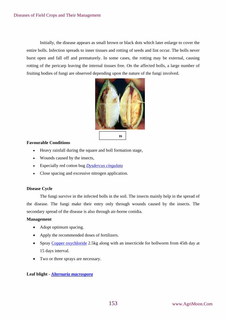
entire b
burst op
rotting
fruiting
Favour
Disease
the dis
seconda
Manag
Leaf bl
Initially, th
bolls. Infecti
pen and fal
of the peric
g bodies of f
rable Cond
Heavy rain
Wounds ca
Especially r
Close spaci
e Cycle
The fungi s
ease. The
ary spread o
gement
Adopt optim
Apply the r
Spray Copp
15 days inte
Two or thre
light - Alter
he disease ap
ion spreads
ll off and p
carp leaving
fungi are ob
ditions
fall during t
aused by the
red cotton b
ing and exc
survive in th
fungi make
of the diseas
mum spacin
recommend
per oxychlo
erval.
ee sprays ar
rnaria macr
ppears as sm
to inner tis
prematurely
g the intern
bserved depe
the square a
e insects,
bug Dysderc
essive nitro
he infected b
e their ent
se is also th
ng.
ded doses of
oride 2.5kg
re necessary
rospora
mall brown
ssues and ro
y. In some
nal tissues fr
ending upon
and boll form
cus cingula
ogen applica
bolls in the
try only th
hrough air-b
f fertilizers.
along with
y.
Symptom
or black do
otting of see
cases, the
ree. On the
n the nature
mation stag
ata
ation.
soil. The in
hrough wou
orne conidi
an insectici
ms
ots which lat
eds and lint
rotting may
affected bo
e of the fung
ge,
nsects mainl
unds caused
ia.
ide for bollw
ter enlarge t
occur. The
y be extern
olls, a large
gi involved.
ly help in th
d by the in
worm from
to cover the
bolls never
nal, causing
e number of
.
he spread of
nsects. The
45th day at
e
r
g
f
f
e
t
Diseases of Field Crops and Their Management
www.AgriMoon.Com153

Sympto
Small, p
the leav
together
lesions
Pathog
a single
transver
Favour
oms
The diseas
plate to bro
ves. Each sp
r to form bl
are also see
gen
The fungus
e conidium
rse septa an
rable Cond
High humid
e may occu
own, irregul
pot has a ce
lighted area
en. In severe
s produces d
at the apex
nd four long
ditions
dity.
ur in all sta
lar or round
entral lesion
as. The affe
e cases, the
dark brown
x. The conid
gitudinal sep
Sym
ages but m
d spots, mea
n surrounde
ected leaves
spots may
n, short, 1-8
dia are obcl
pta, with a p
mptoms
Conidia
ore severe
asuring 0.5
d by concen
s become br
appear on b
septate, irr
avate, light
prominent b
a
when plant
to 6mm dia
ntric rings.
rittle and fa
bracts and b
regularly be
to dark bro
beak.
ts are 45-6
ameter, may
Several spo
ll off. Some
olls.
end conidiop
own in colo
0 days old
y appear on
ots coalesce
etimes stem
phores with
our with 3-9
.
n
e
m
h
9
Diseases of Field Crops and Their Management
www.AgriMoon.Com154

Intermittent rains.
Moderate temperature of 25-28˚ C.
Disease cycle
The pathogen survives in the dead leaves as dormant mycelium. The pathogen primarily
spreads through irrigation water. The secondary spread is mainly by airborne conidia.
Management
Remove and destroy the infected plant residues.
Spray Mancozeb 2 kg or Copper oxychloride at 2kg/ha at the intimation of the disease.
Four to five sprays may be given at 15 days interval.
Bacterial blight - Xanthomonas axonopodis pv. malvacearum
Symptoms
The bacterium attacks all stages from seed to harvest. Usually five common phases of
symptoms are noticed.
i) Seedling blight:
Small, water-soaked, circular or irregular lesions develop on the cotyledons, later, the
infection spreads to stem through petiole and cause withering and death of seedlings.
ii) Angular leaf spot:
Small, dark green, water soaked areas develop on lower surface of leaves, enlarge
gradually and become angular when restricted by veins and veinlets and spots are visible on both
the surface of leaves. As the lesions become older, they turn to reddish brown colour and
infection spreads to veins and veinlets.
iii) Vein blight or vein necrosis or black vein:
The infection of veins cause blackening of the veins and veinlets, gives a typical
‘blighting’ appearance. On the lower surface of the leaf, bacterial oozes are formed as crusts or
scales. The affected leaves become crinkled and twisted inward and show withering. The
infection also spreads from veins to petiole and cause blighting leading to defoliation.
iv) Black arm:
On the stem and fruiting branches, dark brown to black lesions are formed, which may
girdle the stem and branches to cause premature drooping off of the leaves, cracking of stem and
Diseases of Field Crops and Their Management
www.AgriMoon.Com155

gummo
characte
v) Squa
spots. T
bolls le
yellow
infects t
Bac
osis, resultin
eristic “blac
are rot / Bo
On the bol
The infectio
ead to prem
because of
the seed and
cterial bligh
ng in break
ck arm” sym
oll rot:
lls, water so
on slowly sp
mature burst
bacterial oo
d causes red
ht lesions o
king of the
mptom.
oaked lesion
preads to en
ting. The b
oze and loo
duction in s
Angul
on leaf and
stem and
ns appear a
ntire boll an
bacterium s
oses its appe
ize and viab
ar leaf spot
the blackle
hang typic
and turn into
nd shedding
spreads insi
earance and
bility of the
t
eg symptom
cally as dry
o dark blac
g occurs. Th
ide the boll
d market va
e seeds.
m on the lea
y black twig
k and sunk
he infection
l and lint g
lue. The pa
af petiole
g to give a
en irregular
n on mature
gets stained
athogen also
a
r
e
d
o
Diseases of Field Crops and Their Management
www.AgriMoon.Com156

Pathog
forming
Favora
Disease
bacteriu
bacteriu
and Jat
seconda
insects
Manag
gen
The bacteri
g and measu
able Condit
Optimum s
High atmos
Relative hu
Delayed thi
Poor tillage
Potassium d
Rain follow
favorable.
e Cycle
The bacter
um is also s
um also att
tropha curc
ary spread o
and other im
gement
Delint the
delinted se
Streptomyc
Remove an
weed hosts
ium is a sho
ures 1.0-1.2
tions
oil tempera
spheric temp
umidity of 8
inning,
e, late irriga
deficiency i
wed by brigh
rium surviv
eed-borne a
tacks other
cus. The pri
of the bacte
mplements.
cotton seed
eds with ca
cin sulphate
nd destory th
.
ort rod with
2 X 0.7-0.9 µ
ature of 28˚C
perature of
85 per cent,
ation and
in soil.
ht sunshine
ves on infec
and remains
hosts like
imary infec
eria may be
ds with con
arboxin or
e overnight.
he infected
Boll rot
a single po
µm.
C,
30-40˚C,
early sowin
during the
cted, dried
s in the form
Thumbergi
ction starts
through wi
centrated su
oxycarboxi
plant debri
t
olar flagellu
ng,
months of O
plant debr
m of slimy m
ia thespesio
mainly from
ind, wind b
ulphuric ac
in at 2 g/kg
is. Rogue o
um. It is Gra
October and
ris in soil f
mass on the
oides, Eriod
m the seed
lown rain s
id at 100m
g or soak th
ut the volun
am negative
d Novembe
for several
e fuzz of see
dendron an
-borne bact
plash, irriga
l/kg of seed
he seeds in
nteer cotton
e, non-spore
er are highly
years. The
ed coat. The
nfructuosum
terium. The
ation water
d. Treat the
n 1000 ppm
n plants and
e
y
e
e
m
e
,
e
m
d
Diseases of Field Crops and Their Management
www.AgriMoon.Com157
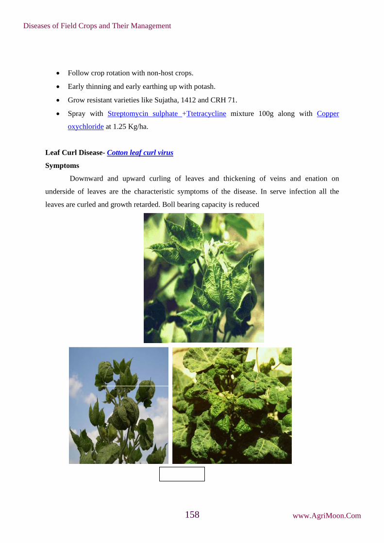
Leaf Cu
Sympto
undersi
leaves a
Follow crop
Early thinn
Grow resist
Spray with
oxychloride
url Disease
oms
Downward
de of leave
are curled an
p rotation w
ning and ear
tant varietie
h Streptom
e at 1.25 Kg
e- Cotton le
d and upw
es are the c
nd growth r
with non-hos
rly earthing
es like Sujat
mycin sulph
g/ha.
eaf curl viru
ward curling
characterist
retarded. Bo
Sy
st crops.
up with pot
tha, 1412 an
hate +Ttetra
us
g of leaves
tic symptom
oll bearing c
ymptoms
tash.
nd CRH 71.
acycline m
s and thick
ms of the d
capacity is r
.
mixture 100g
kening of v
disease. In
reduced
g along w
veins and
serve infec
with Copper
enation on
ction all the
r
n
e
Diseases of Field Crops and Their Management
www.AgriMoon.Com158

Pathogen
It is caused by Cotton leaf curl virus - a begomovirus of family geminiviridae. The
virions are typical geminate particles, ss circular DNA, bipartite genome with DNA-A and DNA-
B components.
Disease Cycle
The primary source is the viruliferous whitefly vector Bemisia tabaci. The alternate
hosts and cultivated hosts serve as virus reservoirs throughout the year. Not transmitted by seed
or contact.
Management
Management of planting date to avoid peak vector population.
Elimination of volunteer perennial cotton and alternate hosts including malvaceous hosts
like wild okra
Use of fungus Paecilomyces farinosus which parasitizes B.tabaci. It brings down vector
population.
Foliar application of neem leaf extract and 1% neem oil resulted in 80% reduction of
virus transmission.
Vector management by application of granular systemic insecticides.
Stenosis or Small leaf - Phytoplsama
Symptoms
The disease appears when the plants are two to three months old and affected plants are
stunted. They put forth numerous extremely small leaves in cluster and the dormant buds are
stimulated resulting in profuse vegetative growth. The leaves are disfigured and variously lobed.
Flowers remain small with abortive ovary.
Large number of flower buds and young seeds. Root system is poorly developed and can
be easily pulled out. Sometimes, the disease affects only the base of the plant, resulting in the
formation of clump of short branches which bear small and deformed leaves. The mode of
transmission of disease and the role of vector are unknown.
Diseases of Field Crops and Their Management
www.AgriMoon.Com159

Management
Rogue out the infected plants periodically.
Cotton varieties developed from Gossypium hirsutum and G. barbadense are found to be
resistant to the disease.
Minor diseases
Leaf spot - Cercospora gossypina
Round or irregular grayish spots with dark brown or blackish borders appear on
older leaves.
Myrothecium leaf spot - Myrothecium roridum
Reddish spots of 0.5 mm- 1 cm diameter may appear near the margins of the leaves. The
affected portions fall off leaving irregular shot holes in the leaves.
Rust - Phakopsora desmium
Yellowish brown raised pustules appear on the lower surface of leaves with rusty
spores. Several pustules join to give rusty appearence to entire leaf. The sori may also develop on
bolls.
Sooty mould - Capnodium sp.
Dark specks appear on the leaves and bolls, slowly spread and black powdery growth
covers the entire leaf area and bolls.
Diseases of Field Crops and Their Management
www.AgriMoon.Com160

14. Diseases of Red Gram
Wilt - Fusarium udum
Symptoms
The disease may appear from early stages of plant growth (4-6 week old plant) up to
flowering and podding. The disease appears as gradual withering and drying of plants.
Yellowing of leaves and blackening of stem starting from collar to branches which gradually
result in drooping and premature drying of leaves, stems, branches and finally death of plant.
Vascular tissues exhibit brown discoloration. Often only one side of the stem and root system is
affected resulting in partial wilting.
Pathogen
The fungus produces hyaline, septate mycelium. Microconidia are hyaline, small,
elliptical or curved, single celled or two celled. Macroconidia are also hyaline, thin walled,
linear, curved or fusoid, pointed at both ends with 3-4 septa. The fungus also poduce thick
walled, spherical or oval, terminal or intercalary chlamydospores singly or in chains of 2 to 3.
Favourable conditions
• Soil temperature of 17-25˚C.
• Continuous cultivation of redgram in the same field.
Disease cycle
The fungus survives in the infected stubbles in the field. The primary spread is by soil-
borne chlamydospores and also by infected seed. Chlamydospores remain viable in soil for 8-20
years. The secondary spread in the field is through irrigation water and implements.
Management
• Treat the seeds with Trichoderma viride at 4 g/kg (106cfu/g).
• Avoid successive cultivation of red gram in the same field.
• Crop rotation with tobacco.
• Mixed cropping with sorghum in the field.
• Grow resistant cultivars like Sharad, Jawahar, Maruthi, Malviya Arhar-2, C-11, Pusa-9,
Narendra Arhar-1 and Birsa Arhar-1
Dry root rot - Macrophomina phaseolina (Sclerotial stage: Rhizoctonia bataticola)
Symptoms
Diseases of Field Crops and Their Management
www.AgriMoon.Com161

The disease occurs both in young seedlings and grown up plants. Infected seedlings can
show reddish brown discoloration at collar region. The lower leaves show yellowing, drooping
and premature defoliation. The discolored area later turns to black and sudden death of the plants
occurs in patches.
The bark near the collar region shows shredding. The plant can be easily pulled off
leaving dark rotten root in the ground. Minute dark sclerotia are seen in the shredded bark and
root tissues. Large number of brown dots seen on the stem portion represents the pycnidial stage
of the fungus.
Pathogen
The fungus produces dark, brown, filamentous hyphae and constrictions are seen in
hyphal branches at the junction with main hyphae. Sclerotia are jet black, smooth, hard, minute,
globose and 110-130 m in diameter. The pycnidia are dark brown and ostiolated. Conidiophores
(phialides) are hyaline, short, obpyriform to cylindrical, develop from the inner walls of the
pycnidium. The conidia (Pycnidiospores) are hyaline, single celled and ellipsoid to ovoid.
Favourable Conditions
• Prolonged drought followed by irrigation.
• High temperature of 28-35˚C.
Disease cycle
The primary spread of the disease is by seed and soil. Secondary spread is by air-borne
conidia. The pathogen survives as sclerotia in the soil as facultative parasite and in dead host
debris.
Management
Symptoms
Diseases of Field Crops and Their Management
www.AgriMoon.Com162
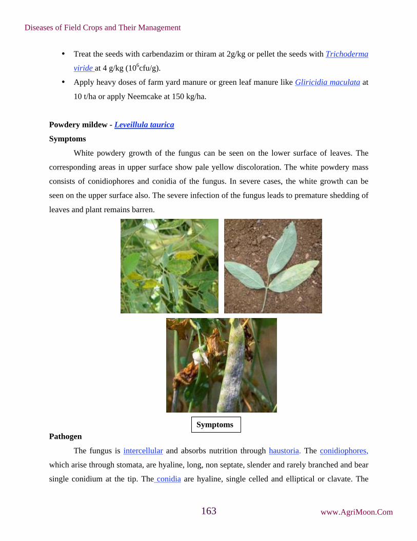
• Treat the seeds with carbendazim or thiram at 2g/kg or pellet the seeds with Trichoderma
viride at 4 g/kg (106cfu/g).
• Apply heavy doses of farm yard manure or green leaf manure like Gliricidia maculata at
10 t/ha or apply Neemcake at 150 kg/ha.
Powdery mildew - Leveillula taurica
Symptoms
White powdery growth of the fungus can be seen on the lower surface of leaves. The
corresponding areas in upper surface show pale yellow discoloration. The white powdery mass
consists of conidiophores and conidia of the fungus. In severe cases, the white growth can be
seen on the upper surface also. The severe infection of the fungus leads to premature shedding of
leaves and plant remains barren.
Pathogen
The fungus is intercellular and absorbs nutrition through haustoria. The conidiophores,
which arise through stomata, are hyaline, long, non septate, slender and rarely branched and bear
single conidium at the tip. The conidia are hyaline, single celled and elliptical or clavate. The
Symptoms
Diseases of Field Crops and Their Management
www.AgriMoon.Com163

fungus also produces black, globose cleistothecia with simple myceloid appendages. They
contain 9-20 cylindrical asci. Each ascus contains 3-5 ascospores which are also hyaline and
unicellular.
Favourable Conditions
• Dry humid weather following rainfall.
Disease Cycle
The fungus survives in the soil through cleistothecia and ascospores from asci infect the
first lower most leaves near the soil level. Secondary spread is by air-borne conidia.
Management
Spray Carbendazim 500g/ha or Wettable sulphur 2 kg/ha at the initiation of the disease
and repeat after 15 days.
Stem blight - Phytophthora drechsleri fsp. cajani
Symptoms
Initially purple to dark brown necrotic lesions girdle the basal portion of the stem and
later may occur an aerial parts. Initially lesions are small and smooth, later enlarging and slightly
depressed. Infected tissues become soft and whole plant dies. In grown up plants, infection is
mostly confined to basal portions of the stem. The infected bark becomes brown and the tissue
softens causing the plant to collapse. In leaf, localized yellowing starts from the tip and margin
and gradually extends towards the mid-rib. The centre of the spots later turn brown and hard. The
spots increase in size and cover a major portion of the lamina, leading to drying.
Cleistothecium Conidia and conidiophores
Diseases of Field Crops and Their Management
www.AgriMoon.Com164

Pathogen
Fungus produces hyaline, coenocytic mycelium. The sporangiophores are hyaline bearing
ovate or pyriform, non-papillate sporangia. Each sporangium produces 8-20 zoospores. Oospores
are globose, light brown, smooth and thick walled.
Favourable Conditions
• Soils with poor drainage,
• Low lying areas,
• Heavy rain during the months of July- September
• High temperature (28-30˚C).
Disease Cycle
The fungus survives in the soil and plant debris in the form of oospores. Primary
infection is from oospores and secondary spread of the disease by zoospores from sporangia.
Rain splash and irrigation water help for the movement of zoospores.
Management
• Treat the seeds with Metalaxyl at 6 g/kg.
• Spray Metalaxyl at 500 g/ha.
• Adjust the sowing time so that crop growth should not coincide with heavy rainfall.
Leaf spot - Cercospora indica
Symptoms
Small, light brown coloured spots appear on leaves. The spots later become dark brown
and the infected portions drop off leaving shot hole symptoms. When several spots join together,
irregular necrotic blotches develop and premature defoliation occurs. In severe cases, black
lesions develop on petioles and stem.
Symptoms
Diseases of Field Crops and Their Management
www.AgriMoon.Com165
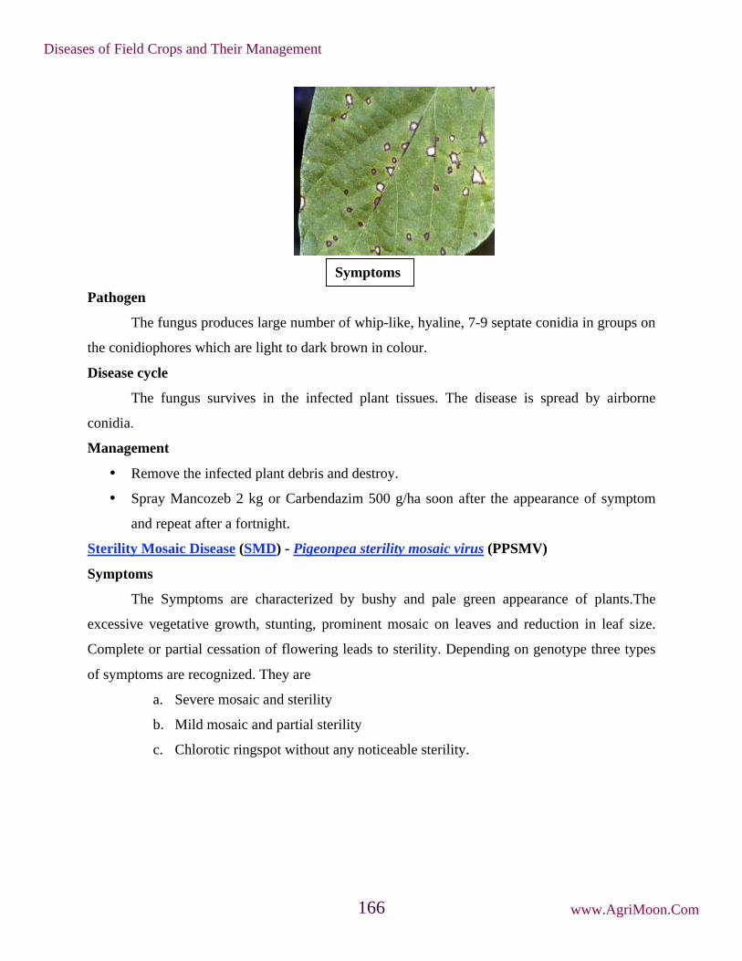
Pathogen
The fungus produces large number of whip-like, hyaline, 7-9 septate conidia in groups on
the conidiophores which are light to dark brown in colour.
Disease cycle
The fungus survives in the infected plant tissues. The disease is spread by airborne
conidia.
Management
• Remove the infected plant debris and destroy.
• Spray Mancozeb 2 kg or Carbendazim 500 g/ha soon after the appearance of symptom
and repeat after a fortnight.
Sterility Mosaic Disease (SMD) - Pigeonpea sterility mosaic virus (PPSMV)
Symptoms
The Symptoms are characterized by bushy and pale green appearance of plants.The
excessive vegetative growth, stunting, prominent mosaic on leaves and reduction in leaf size.
Complete or partial cessation of flowering leads to sterility. Depending on genotype three types
of symptoms are recognized. They are
a. Severe mosaic and sterility
b. Mild mosaic and partial sterility
c. Chlorotic ringspot without any noticeable sterility.
Symptoms
Diseases of Field Crops and Their Management
www.AgriMoon.Com166
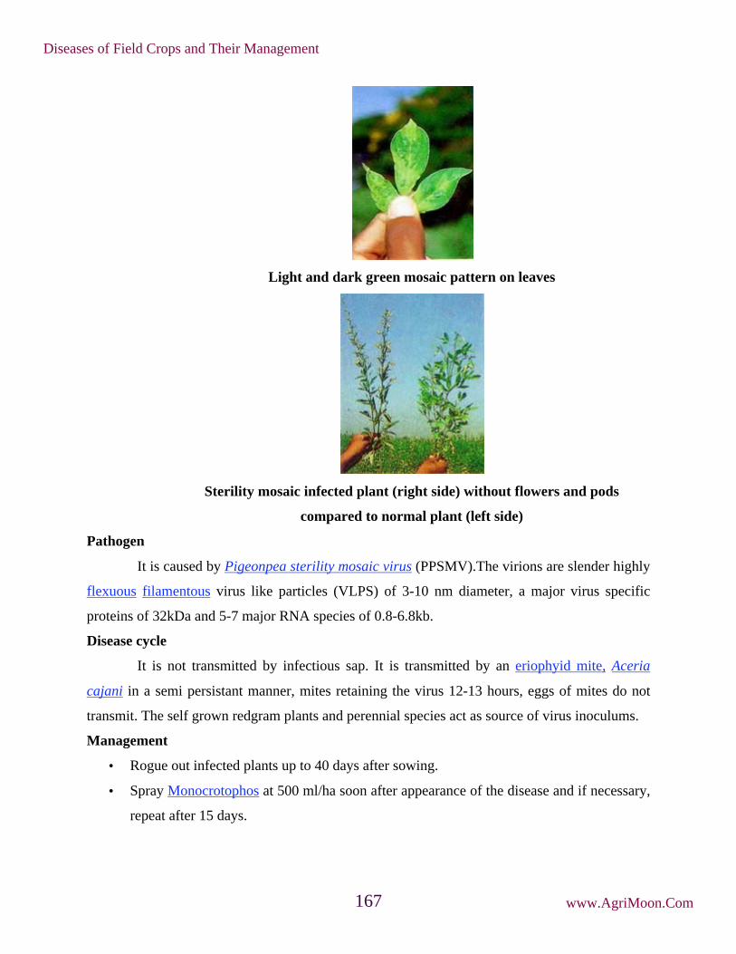
Light and dark green mosaic pattern on leaves
Sterility mosaic infected plant (right side) without flowers and pods
compared to normal plant (left side)
Pathogen
It is caused by Pigeonpea sterility mosaic virus (PPSMV).The virions are slender highly
flexuous filamentous virus like particles (VLPS) of 3-10 nm diameter, a major virus specific
proteins of 32kDa and 5-7 major RNA species of 0.8-6.8kb.
Disease cycle
It is not transmitted by infectious sap. It is transmitted by an eriophyid mite, Aceria
cajani in a semi persistant manner, mites retaining the virus 12-13 hours, eggs of mites do not
transmit. The self grown redgram plants and perennial species act as source of virus inoculums.
Management
• Rogue out infected plants up to 40 days after sowing.
• Spray Monocrotophos at 500 ml/ha soon after appearance of the disease and if necessary,
repeat after 15 days.
Diseases of Field Crops and Their Management
www.AgriMoon.Com167

• Grow resistant genotypes/cultivars like ICP 7035, VR3, Purple 1, DA11, DA32, ICP
6997, Bahar, BSMR 235, ICP 7198, PR 5149, ICP 8861 and Bhavanisagar 1.
Minor diseases
Seedling blight - Sclerotium rolfsii
Small brown water soaked dots appear near collar region, expands to irregular necrotic
spots leading to girdling of stem and death of seedling.
Brown blotch - Colletrtrichum capsici
Purple brown discolouration occurs mainly on pods but also on petioles, leaf veins, stems
and peduncles. Pods become distorted and have black fruiting bodies.
Anthracnose - Colletotrichum lindemuthianum (Glomerella cingulata)
Black lesions develop on stem which spreads to leaf petiole and leaves. Black sunken
lesions also develop on pod.
Stem rot - Pythium aphanidermatum
Seedlings of 2-3 weeks old are severely attacked at collar region and death occurs
immediately. Greyish green water soaked lesions develop on adult plants, leading to girdling of
stem.
Leaf spot - Alternaria alternata
Water soaked, circular to irregular spots occur. The centre of the spot is straw coloured
with raised reddish brown margins.
Halo blight - Pseudomonas phaseolicola
Small brown spots appearon leaves and develop a chlorotic halo. The spots extend and
form dried brown zone. Brown elongated streaks appear on petioles, stem and pods.
There are two other virus diseases reported on pigeonpea, mosaic and yellow mosaic
transmitted by aphids and whiteflies which are of sporadic occurrence only.
Diseases of Field Crops and Their Management
www.AgriMoon.Com168

15. Diseases of Black gram
Powdery mildew - Erysiphe polygoni
Symptoms
Small, irregular powdery spots appear on the upper surface of the leaves, sometimes on
both the surfaces. The disease becomes severe during flowering and pod development stage. The
white powdery spots completely cover the leaves, petioles, stem and even the pods. The plant
assumes greyish white appearance; leaves turn yellow and finally shed. Often pods are
malformed and small with few ill-filled seeds.
Pathogen
The fungus is ectophytic, spreading on the surface of the leaf, sending haustoria into the
epidermal cells. Conidiophores arise vertically from the leaf surface, bearing conidia in short
chains. Conidia are hyaline, thin walled, elliptical or barrel shaped or cylindrical and single
celled. Later in the season, cleistothecia appear as minute, black, globose structures with
myceloid appendages. Each cleistothecium contains 4-8 asci and each ascus contains 3-8
ascospores which are elliptical, hyaline and single celled.
Symptoms
Diseases of Field Crops and Their Management
www.AgriMoon.Com169

Favourable Conditions
• Warm humid weather.
• The disease is severe generally during late kharif and rabi seasons.
Disease cycle
The Pathogen is an obligate parasite and survives as cleistothecia in the infected plant
debris. Primary infection is usually from ascospores from perennating cleistothecia. The
secondary spread is carried out by the air-borne conidia. Rain splash also helps in the spread of
the disease.
Management
• Remove and destroy infected plant debris.
• Spray Carbendazim 500g or Wettable sulphur 2kg or Tridemorph 500 ml/ha at the
initiation of disease and repeat 15 days later.
Anthracnose - Colletotrichum lindemuthianum (Sexual stage: Glomerella lindemuthianum)
Symptoms
The symptom can be observed in all aerial parts of the plants and at any stage of crop
growth. The fungus produces dark brown to black sunken lesions on the hypocotyl area and
cause death of the seedlings. Small angular brown lesions appear on leaves, mostly adjacent to
veins, which later become greyish white centre with dark brown or reddish margin.
The lesions may be seen on the petioles and stem. The prominent symptom is seen on the
pods. Minute water soaked lesion appears on the pods initially and becomes brown and enlarges
to form circular, depressed spot with dark centre with bright red or yellow margin. Several spots
join to cause necrotic areas with acervuli. The infected pods have discolored seeds.
Cleistothecium Conidia and conidiophores
Diseases of Field Crops and Their Management
www.AgriMoon.Com170

Pathogen
The fungus mycelium is septate, hyaline and branched. Conidia are produced in acervuli,
arise from the stroma beneath the epidermis and later rupture to become erumpent. A few dark
coloured, septate setae are seen in the acervulus. The conidiophores are hyaline and short and
bear oblong or cylindrical, hyaline, thinwalled, single celled conidia with oil globules. The
perfect stage of the fungus produces perithecia with limited number of asci, which contain
typically 8 ascospores which are one or two celled with a central oil globule.
Favourable Conditions
• High relative humidity (Above 90 per cent),
• Low temperature (15-20˚ C)
• Cool rainy days.
Disease cycle
The fungus is seed-borne and cause primary infection. It also lives in the infected plant
tissues in soil. The secondary spread by air borne conidia produced on infected plant parts. Rain
splash also helps in dissemination.
Management
• Remove and destroy infected plant debris in soil.
• Treat the seeds with Carbendazim at 2 g/kg.
Symptoms
Diseases of Field Crops and Their Management
www.AgriMoon.Com171

• Spray Carbendazim 500g or Mancozeb 2kg/ha soon after the appearance of disease and
repeat after 15 days.
Leaf spot - Cercospora canescens
Symptoms
Small, circular spots develop on the leaves with grey centre and brown margin. Several
spots coalesce to form brown irregular lesions. In severe cases defoliation occurs. The brown
lesions may be seen on petioles and stem in severe cases. Powdery growth of the fungus may be
seen on the centre of the spots.
Pathogen
The fungus produces clusters of dark brown septate conidiophores. The conidia are
linear, hyaline, thin walled and 5-6 septate.
Favourable Conditions
• Humid weather and dense plant population.
Disease cycle
The fungus survives on diseased plant debris and on seeds. The secondary spread is by
air-borne conidia.
Management
• Remove and burn infected plant debris.
• Spray Mancozeb at 2 kg/ha or Carbendazim at 500 g/ha.
Symptoms
Diseases of Field Crops and Their Management
www.AgriMoon.Com172

Rust - Uromyces phaseoli typica (Syn: U. appendiculatus)
Symptoms
The disease is mostly seen on leaves, rarely on petioles, stem and pods. The fungus
produces small, round, reddish brown uredosori mostly on lower surface. They may appear in
groups and several sori coalesce to cover a large area of the lamina. In the late season, teliosori
appear on the leaves which are linear and dark brown in colour. Intense pustule formation causes
drying and shedding of leaves.
Pathogen
It is autoecious, long cycle rust and all the spore stages occur on the same host. The
uredospores are unicellular, globose or ellipsoid, yellowish brown with echinulations. The
teliospores are globose or elliptical, unicellular, pedicellate, chestnut brown in colour with warty
papillae at the top. Yellow coloured pycnia appear on the upper surface of leaves. Orange
coloured cupulate aecia develop later on the lower surface of leaves. The aeciospores are
unicellular and elliptical.
Favourable Conditions
• Cloudy humid weather, temperature of 21-26˚ C
• Nights with heavy dews
Mode of Spread and Survival
The pathogen survives in the soil through teliospores and as uredospores in crop debris.
Primary infection is by the sporidia developed from teliospores. Secondary spread is by wind-
borne uredospores. The fungus also survives on other legume hosts.
Symptoms
Diseases of Field Crops and Their Management
www.AgriMoon.Com173

Management
• Remove the infected plant debris and destroy.
• Spray Mancozeb 2 kg or Carbendazim 500 g or Propiconazole 1L/ha, immediately on the
set of disease and repeat after 15 days.
Dry root rot- Rhizoctonia bataticola (Pycnidial stage: Macrophomina phaseolina)
Symptoms
The disease symptom starts initially with yellowing and drooping of the leaves. The
leaves later fall off and the plant dies with in week. Dark brown lesions are seen on the stem at
ground level and bark shows shredding symptom. The affected plants can be easily pulled out
leaving dried, rotten root portions in the ground. The rotten tissues of stem and root contain a
large number of black minute sclerotia.
Pathogen
The fungus produces dark brown, septate mycelium with constrictions at hyphal
branches. Minute, dark, round sclerotia in abundance. The fungus also produces dark brown,
globose ostiolated pycnidia on the host tissues. The pycnidiospores are thin walled, hyaline,
single celled and elliptical.
Favourable conditions
• Day temperature of 30˚C.
• Prolonged dry season followed by irrigation.
Symptoms
Diseases of Field Crops and Their Management
www.AgriMoon.Com174

Disease cycle
The fungus survives in the infected debris and also as facultative parasite in soil. The
primary spread is through seed-borne and soil-borne sclerotia. The secondary spreads is through
pycnidiospores which are air-borne.
Management
• Treat the seeds with carbendazim + thiram at 2 g/kg (1:1 ratio) or pellet the seeds with
Trichoderma viride at 4 g/kg (106cfu/g) or Pseudonomas fluorescens @ (10
6cfu/g) of
seed.
• Apply farm yard manure or green leaf manure (Gliricidia maculata) at 10 t/ha or
neemcake at 150 kg/ha.
Mungbean Yellow mosaic disease - Mungbean yellow mosaic virus (MYMV)
Symptoms
Initially small yellow patches or spots appear on green lamina of young leaves. Soon it
develops into a characteristics bright yellow mosaic or golden yellow mosaic symptom. Yellow
discoloration slowly increases and leaves turn completely yellow. Infected plants mature later
and bear few flowers and pods. The pods are small and distorted. Early infection causes death of
the plant before seed set.
Pathogen
It is caused by Mungbean yellow mosaic India virus (MYMIV) in Northen and Central
region and Mungbean yellow mosaic virus (MYMV) in western and southern regions. It is a
Symptoms
Diseases of Field Crops and Their Management
www.AgriMoon.Com175

Begomovirus belonging to the family geminiviridae. Geminate virus particles, ssDNA, bipartite
genome with two gemonic components DNA-A and DNA-B.
Disease cycle
Transmitted by whitefly, Bemisia tabaci under favourable conditions. Disease spreads by
feeding of plants by viruliferous whiteflies. Summer sown crops are highly susceptible. Weed
hosts viz., Croton sparsiflorus, Acalypha indica, Eclipta alba and other legume hosts serve as
reservoir for inoculum.
Management
• Rogue out the diseased plants up to 40 days after sowing.
• Remove the weed hosts periodically.
• Increase the seed rate (25 kg/ha).
• Grow resistant black gram variety like VBN-1, PDU 10, IC12/2 and PLU 322. Cultivate
the crop during rabi season.
• Follow mixed cropping by growing two rows of maize (60 x 30 cm) or sorghum (45 x 15
cm) or cumbu (45 x 15 cm) for every 15 rows of black gram or green gram.
• Treat the seeds with Thiomethoxam-70WS or Imidacloprid-70WS @4g/kg
• Spray Thiamethoxam-25WG @ 100g or Imidacloprid 17.8% SL @ 100 ml in 500 lit of
water.
Leaf crinkle disease - Urdbean leaf crinkle virus (ULCV)
Symptoms
Crinkling and curling of the tips of leaflets and increase in leaf area. Crinkling and
rugosity in older leaves becomes severe and leaves thickened. Petioles as well as internodes are
shortened. Infected plant gives a stunted and bushy appearance. Flowering is delayed, if
inflorescence is formed, is malformed with small size flower buds and fails to open.
Pathogen
Casual organism of the disease is not yet ascertained.
Disease cycle
Presence of weed hosts like Aristolochia bracteata and Digera arvensis. Kharif season
crop and continuous cropping of other legumes serve as source of inoculum. The virus is seed-
Diseases of Field Crops and Their Management
www.AgriMoon.Com176

borne and primary infection occurs through infected seeds. Perhaps white fly, Bemisia tabaci
helps in the secondary spread. The virus is also sap transmissible.
Management
• Use increased seed rate (25 kg/ha).
• Rogue out the diseased plants at weekly interval up to 45 days after sowing. Cultivate
seed crop during rabi season.
• Remove weed hosts periodically.
• Spray methyl demeton on 30 and 40 days after sowing at 500 ml/ha.
Leaf curl / Necrosis - Groundnut bud necrosis virus (GBNV)
Symptoms
Upward cupping and curling of leaves with vein clearing. Infected leaves turn brittle and
sometimes show vein necrosis on the under surface of the leaves, extending to the petiole. Plants
affected in the early stages of growth develop top necrosis and die. Plant may produce a few
small and malformed pods.
Pathogen
It is caused by Groundnut bud necrosis virus
Disease cycle
The virus is transmitted by thrips viz., Frankliniella schultzii, Thrips tabaci and
Scirtothrips dorsalis. The virus survives in weed hosts, tomato, petunia and Chilli.
Management
• Rogue out infected plants up to 30 days after sowing.
• Remove the weed hosts which harbour virus and thrips.
• Spray imidachlor at 500 ml/ha on 30 and 45 days after sowing.
Minor diseases
Ascochyta leaf spot - Ascochyta phaseolorum
Small irregular spot with grey to brown centre and yellow border. They rapidly enlarge to
produce very large brown lesions with concentric markings.
Bacterial blight - Xanthomonas phaseoli
Diseases of Field Crops and Their Management
www.AgriMoon.Com177

Circular, reddish brown spots appear on leaves, enlarge to form irregular brown lesions.
Water soaked, sunken spots with red border occur on pods.
Diseases of Field Crops and Their Management
www.AgriMoon.Com178

16. Diseases of Green gram
Powdery mildew - Erysiphe polygoni
Symptoms
Powdery mildew is one of the widespread diseases of several legumes in green gram.
White powdery patches appear on leaves and other green parts which later become dull colored.
These patches gradually increase in size and become circular covering the lower surface also.
When the infection is severe, both the surfaces of the leaves are completely covered by whitish
powdery growth. Severely affected parts get shriveled and distorted. In severe infections,
foliage becomes yellow causing premature defoliation. The disease also creates forced maturity
of the infected plants which results in heavy yield losses.
Pathogen
The fungus is ectophytic, spreading on the surface of the leaf, sending haustoria into the
epidermal cells. Conidiophores arise vertically from the leaf surface, bearing conidia in short
chains. Conidia are hyaline, thinwalled, elliptical or barrel shaped or cylindrical and single
celled. Later in the season, cleistothecia appear as minute, black, globose structures with
myceloid appendages. Each cleistothecium contains 4-8 asci and each ascus contains 3-8
ascospores which are elliptical, hyaline and single celled.
Favourable Conditions
• The pathogen has a wide host range and survives in oidial form on various hosts in off-
season.
• Secondary spread is through air-borne oidia produced in the season
Disease Cycle
The fungus is an obligate parasite and survives as cleistothecia in the infected plant
debris. Primary infection is usually from ascospores from perennating cleistothecia. The
secondary spread is carried out by the air-borne conidia. Rain splash also helps in the spread of
the disease.
Management
• Use resistant varieties
Diseases of Field Crops and Their Management
www.AgriMoon.Com179

• The seeds must be sown early in the month of June to avoid early incidence of the disease
on the crop.
• . Spray Carbendazim 500g or Wettable sulphur 1.5 kg or Tridemorph 500 ml/ha at the
initiation of disease and repeat 15 days later.
Anthracnose - Colletotrichum lindemuthianum - (Sexual stage: Glomerella lindemuthianum)
Symptoms
The disease appears on all aerial part parts and at any stage of plant growth. Circular,
black, sunken spots with dark center and bright red orange margins on leaves and pods. In severe
infections, the affected parts wither off. Seedlings get blighted due to infection soon after seed
germination.
Pathogen
The Disease appears on fungus mycelium is septate, hyaline and branched. Conidia are
produced in acervuli, arise from the stroma beneath the epidermis and later rupture to become
erumpent. A few dark coloured, septate setae are seen in the acervulus. The conidiophores are
hyaline and short and bear oblong or cylindrical, hyaline, thinwalled, single celled conidia with
oil globules. The perfect stage of the fungus produces perithecia with limited number of asci,
which contain typically 8 ascospores which are one or two celled with a central oil globule.
Favourable Conditions
• The disease is more sever in cool and wet seasons.
Disease cycle
The fungus is seed-borne and cause primary infection. It also lives in the infected plant
tissues in soil. The secondary spread by air borne conidia produced on infected plant parts. Rain
splash also helps in dissemination.
Management
• Hot water treatment at 54º for 10 min.
• Use disease free seed.
• Follow crop rotation
• Remove and destroy infected plant debris in soil.
• Treat the seeds with Carbendazim at 2 g/kg.
Diseases of Field Crops and Their Management
www.AgriMoon.Com180
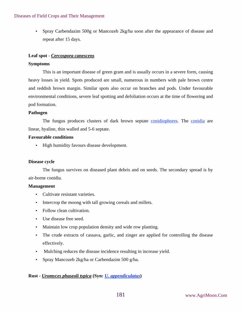
• Spray Carbendazim 500g or Mancozeb 2kg/ha soon after the appearance of disease and
repeat after 15 days.
Leaf spot - Cercospora canescens
Symptoms
This is an important disease of green gram and is usually occurs in a severe form, causing
heavy losses in yield. Spots produced are small, numerous in numbers with pale brown centre
and reddish brown margin. Similar spots also occur on branches and pods. Under favourable
environmental conditions, severe leaf spotting and defoliation occurs at the time of flowering and
pod formation.
Pathogen
The fungus produces clusters of dark brown septate conidiophores. The conidia are
linear, hyaline, thin walled and 5-6 septate.
Favourable conditions
• High humidity favours disease development.
Disease cycle
The fungus survives on diseased plant debris and on seeds. The secondary spread is by
air-borne conidia.
Management
• Cultivate resistant varieties.
• Intercrop the moong with tall growing cereals and millets.
• Follow clean cultivation.
• Use disease free seed.
• Maintain low crop population density and wide row planting.
• The crude extracts of cassava, garlic, and zinger are applied for controlling the disease
effectively.
• Mulching reduces the disease incidence resulting in increase yield.
• Spray Mancozeb 2kg/ha or Carbendazim 500 g/ha.
Rust - Uromyces phaseoli typica (Syn: U. appendiculatus)
Diseases of Field Crops and Their Management
www.AgriMoon.Com181

Symptoms
The disease appears as circular reddish brown pustules which appear more commonly on
the underside of the leaves, less abundant on pods and sparingly on stems. When leaves are
severely infected, both the surfaces are fully covered by rust pustules. Shriveling followed by
defoliation resulting in yield losses.
Pathogen
It is autoecious, long cycle rust and all the spore stages occur on the same host. The
uredospores are unicellular, globose or ellipsoid, yellowish brown with echinulations. The
teliospores are globose or elliptical, unicellular, pedicellate, chestnut brown in colour with warty
papillae at the top. Yellow coloured pycnia appear on the upper surface of leaves. Orange
coloured cupulate aecia develop later on the lower surface of leaves. The aeciospores are
unicellular and elliptical.
Favourable Conditions
• Cloudy humid weather,
• Temperature of 21-26˚C
• Nights with heavy dews.
Disease Cycle
The pathogen survives in the soil as teliospores and as uredospores in crop debris.
Primary infection is by the sporidia developed from teliospores. Secondary spread is by wind-
borne uredospores. The fungus also survives on other legume hosts.
Management
• Remove the infected plant debris and destroy.
• Spray Mancozeb 1 2 kg or Carbendazim 500 g or Propiconazole 1L/ha kg/ha,
immediately on the set of disease and repeat after 15 days.
• Use tolerant varieties.
Dry root rot - Rhizoctonia bataticola (Pycnidial stage: Macrophomina phaseolina)
Symptoms
The disease symptom starts initially with yellowing and drooping of the leaves. The
leaves later fall off and the plant dies with in week. Dark brown lesions are seen on the stem at
ground level and bark shows shredding symptom. The affected plants can be easily pulled out
Diseases of Field Crops and Their Management
www.AgriMoon.Com182

leaving dried, rotten root portions in the ground. The rotten tissues of stem and root contain a
large number of black minute sclerotia.
Pathogen
The fungus produces dark brown, septate mycelium with constrictions at hyphal
branches. Minute, dark, round sclerotia in abundance. The fungus also produces dark brown,
globose ostiolated pycnidia on the host tissues. The pycnidiospores are thin walled, hyaline,
single celled and elliptical.
Favourable conditions
• Day temperature of 30˚C.
• Prolonged dry season followed by irrigation.
Disease cycle
The fungus survives in the infected debris and also as facultative parasite in soil. The
primary spread is through seed-borne and soil-borne sclerotia. The secondary spread is through
air-borne pycnidiospores.
Management
• Treat the seeds with Carbendazim + Thiram at 2 g/kg or pellet the seeds with
Trichoderma viride at 4 g/kg or Pseudonomas fluorescens @ 10g/kg of seed.
• Apply farm yard manure or green leaf manure (Gliricidia maculate) at 10 t/ha or neem
cake at 150 kg/ha.
Yellow mosaic disease - Mungbean yellow mosaic virus (MYMV)
Symptoms
Initially small yellow patches or spots appear on green lamina of young leaves. Soon it
develops into a characteristics bright yellow mosaic or golden yellow mosaic symptom. Yellow
discoloration slowly increases and leaves turn completely yellow. Infected plants mature later
and bear few flowers and pods. The pods are small and distorted. Early infection causes death of
the plant before seed set.
Pathogen
It is caused by Mungbean yellow mosaic India virus (MYMIV) in Northen and Central
region and Mungbean yellow mosaic virus (MYMV) in western and southern regions. It is a
Begomovirus belonging to the family geminiviridae. Germinate virus particles, ssDNA, bipartite
genome with two gemonic components DNA-A and DNA-B.
Diseases of Field Crops and Their Management
www.AgriMoon.Com183

Disease cycle
Transmitted by whitefly, Bemisia tabaci under favourable conditions. Disease spreads by
feeding of plants by viruliferous whiteflies. Summer sown crops are highly susceptible. Weed
hosts viz., Croton sparsiflorus, Acalypha indica, Eclipta alba and other legume hosts serve as
reservoir for inoculum.
Management
• Rogue out the diseased plants up to 40 days after sowing.
• Remove the weed hosts periodically.
• Increase the seed rate (25 kg/ha).
• Grow resistant green gram variety like Pant Moong-3, Pusa Vishal, Basanti, ML-5, ML-
337, PDM-54 and Samrat.
• Cultivate the crop during rabi season.
• Follow mixed cropping by growing two rows of maize (60 x 30 cm) or sorghum (45 x 15
cm) or cumbu (45 x 15 cm) for every 15 rows of black gram or green gram.
• Treat the seeds with Thiomethoxam-70WS or Imidacloprid-70WS @4g/kg
• Spray Thiamethoxam-25WG @ 100g or Imidacloprid 17.8% SL @ 100 ml in 500 lit of
water.
Leaf crinkle disease - Urdbean leaf crinkle virus (ULCV)
Symptoms
Crinkling and rugosity in older leaves becomes severe and leaves thickened. Crinkling
and curling of the tips of leaflets are seen. Petioles as well as internodes are shortened. Infected
plant gives a stunted and bushy appearance. Flowering is delayed, inflorescence, if formed, are
malformed with small size flower buds and fail to open.
Pathogen
Casual organism of the disease is not yet ascertained work is in progress in different
laboratories.
Disease Cycle
Presence of weed hosts like Aristolochia bracteata and Digera arvensis. Kharif season
crop and continuous cropping of other legumes serve as source of inoculum. The virus is seed-
Diseases of Field Crops and Their Management
www.AgriMoon.Com184

borne and primary infection occurs through infected seeds. Perhaps white fly, Bemisia tabaci
helps in the secondary spread. The virus is also sap transmissible.
Management
• Use increased seed rate (25 kg/ha).
• Rogue out the diseased plants at weekly interval up to 45 days after sowing. Cultivate
seed crop during rabi season.
• Remove weed hosts periodically.
• Spray Methyl demeton on 30 and 40 days after sowing at 500 ml/ha.
Leaf curl / Necrosis - Groundnut bud necrosis virus
Symptoms
Upward cupping and curling of leaves with vein clearing. Infected leaves are brittle and
sometimes show vein necrosis on the under surface of the leaves, extends to the petiole. Plants
affected in the early stages of growth develop top necrosis and die. Plant may produce a few
small and malformed pods.
Pathogen
Caused by groundnut bud necrosis virus
Disease Cycle
The virus is transmitted by thrips viz., Frankliniella schultzii, Thrips tabaci and
Scirtothrips dorsalis. The virus survives in weed hosts, tomato, petunia and Chilli.
Management
• Rogue out infected plants up to 30 days after sowing.
• Remove the weed hosts which harbour virus and thrips.
• Spray Imidachlor at 500 ml/ha on 30 and 45 days after sowing.
Minor diseases
Ascochyta leaf spot - Ascochyta phaseolorum
Small irregular spot with grey to brown centre and yellow border. They rapidly enlarge to
produce very large brown lesions with concentric markings.
Bacterial blight - Xanthomonas phaseoli
Diseases of Field Crops and Their Management
www.AgriMoon.Com185

Circular, reddish brown spots appear on leaves, enlarge to form irregular brown lesions.
Water soaked, sunken spots with red border occur on pods.
Diseases of Field Crops and Their Management
www.AgriMoon.Com186

17. Diseases of Bengal gram
Ascochyta blight - Ascochyta rabiei
Symptoms
All above ground parts of the plant are infected. On leaf, the lesions are round or
elongated, bearing irregularly depressed brown spot and surrounded by a brownish red margin.
Similar spots may appear on the stem and pods. The spots on the stem and pods have pycnidia
arranged in concentric circles as minute block dots. When the lesions girdle the stem, the portion
above the point of attack rapidly dies. If the main stem is girdles at the collar region, the whole
plant dies.
Symptoms
Diseases of Field Crops and Their Management
www.AgriMoon.Com187

Pathogen
The fungus produces hyaline to brown and septate mycelium. Pycnidia are spherical to
sub-globose with a prominent ostiole. Pycnidiospores are hyaline, oval to oblong, straight or
slightly curved and single celled, occasionally bicelled.
Favourable conditions
• High rainfall during flowering.
• Temperature of 20-25˚C.
• Relative humidity of 60%.
Disease cycle
The fungus survives in the infected plant debris as pycnidia. The pathogen is also
externally and internally seed-borne. The primary spread is from seed-borne pycnidia and plant
debris in the soil. The secondary spreads is mainly through air-borne pycnidiopores (conidia).
Rain splash also helps in the spread of the disease.
Management
• Remove and destroy the infected plant debris in the field.
• Treat the seeds with Thiram 2g or Carbendazim 2 g or Thiram + Carbendazim (1:1 ratio)
at 2 g/kg.
• Exposure of seed at 40-50˚C reduced the survival of A. rabiei by about 40-70 per cent.
• Spray with Carbendazim at 500 g/ha or Chlorothalonil 1kg/ha.
• Follow crop rotation with cereals.
Rust - Uromyces ciceris-arietini
Symptoms
The infection appears as small oval, brown, powdery lesions on both the surface,
especially more on lower surface or leaf. The lesions, which are uredosori, cover the entire leaf
surface. Late in the season dark teliosori appear on the leaves. The rust pustules may appear on
petioles, stems and pods. The pycnial and aecial stages are unknown.
Pathogen
The uredospores are spherical, brownish yellow in colour, loosey echinulated with 4-8
germ pores. Teliospores are round to oval, brown, single celled with unthickened apex and the
walls are rough, brown and warty.
Diseases of Field Crops and Their Management
www.AgriMoon.Com188

Mode of Spread and Survival
The fungus survives as uredospores in the legume weed Trigonella polycerata during
summer months and serve as primary source of infection. The spread is through wind-borne
uredospores.
Management
• Destory weed host.
• Spray Carbendazim 500 g/ha or Propiconazole 1L/ha.
Wilt - Fusarium oxysporum f.sp. ciceris
Symptoms
The disease occurs at two stages of crop growth, seedling stage and flowering stage stage.
The main symptoms on seedlings are yellowing and drying of leaves, drooping of petioles and
rachis, withering of plants. In the case of adult plants drooping of leaves is observed initially in
upper part of plant, and soon observed in entire plant. Vascular browning is conspicuously seen
on the stem and root portion
Pathogen
The fungus produces hyaline to light brown, septate and profusely branched hyphae.
Microconidia are oval to cylindrical, hyaline, single celled, normally arise on short
conidiophores. Macroconidia which borne on branched conidiophores, are thin walled, 3 to
5septate, fusoid and pointed at both ends. Chlamydospores are roughwalled or smooth, terminal
or intercalary, may be formed singly or in chains.
Symptoms
Diseases of Field Crops and Their Management
www.AgriMoon.Com189

Favourable conditions
• High soil temperature (above 25˚C).
• High soil moisture.
Disease cycle
The disease is seed and soil borne. The primary infection is through chlamydospores in
soil, which remain viable upto next crop season. The secondary spread is through irrigation
water, cultural operations and implements.
Management
• Treat the seeds with Carbendazim or Thiram at 2 g/kg or Carbendazim 1 g+Thiram 1g/kg
or treat the seeds with Trichoderma viride at 4 g/kg (106cfu/g) Pseudonomas fluorescens
@ 10g/kg (106cfu/g) of seed.
• Apply heavy doses of organic manure or green manure.
• Grow resistant cultures like ICCC 42, H82-2, Avrodhi, Alok Samrat, Pusa-212, JG- 322,
GPF-2, Haryanachana-1 and Kabuli chickpea like Pusa-1073 and Pusa-2024.
Stunt disease - Virus
Symptoms
Affected plants are stunted and bushy with short internodes. The leaflets are smaller with
yellow, orange or brown discoloration. Stem also shows brown discoloration. The plants dry
prematurely. If survive, a very few small pods are formed. Phloem browning in the collar region
is the most characteristic symptom of the stunt, leaving xylem normal.
Disease cycle
The virus is transmitted by Aphis craccivora.
Symptoms
Diseases of Field Crops and Their Management
www.AgriMoon.Com190

Management
• Rogue out the infected plants.
• Spray Monocrotophos at 500 ml/ha.
Collar rot - Sclerotium rolfsii
Symptoms
It comes in the early stages i.e up to six weeks from sowing. Drying plants whose foliage
turns slightly yellow before death, scattered in the field is an indication of the disease. Seedlings
become chlorotic. The joint of stem and root turns soft slightly contracts and begins to decay.
Infected parts turn brown white. Black dots, like mustard in shape known as sclerotia are seen
appearing on the white infected plant parts.
Favorable conditions
• High soil moisture, low soil pH and high temperature.
• The presence of undecomposed organic matter on the soil surface and high moisture at
the time of sowing and at the seedling stage
• Disease incidence is higher when sown after rice or early sown crop.
Management
• Deep pluoghing in summer.
• Avoid high moisture at the sowing time.
• Seedlings should be protected from excessive moisture.
• Destroy the crop residues of last crop and weeds before sowing and after harvest.
Symptoms
Diseases of Field Crops and Their Management
www.AgriMoon.Com191
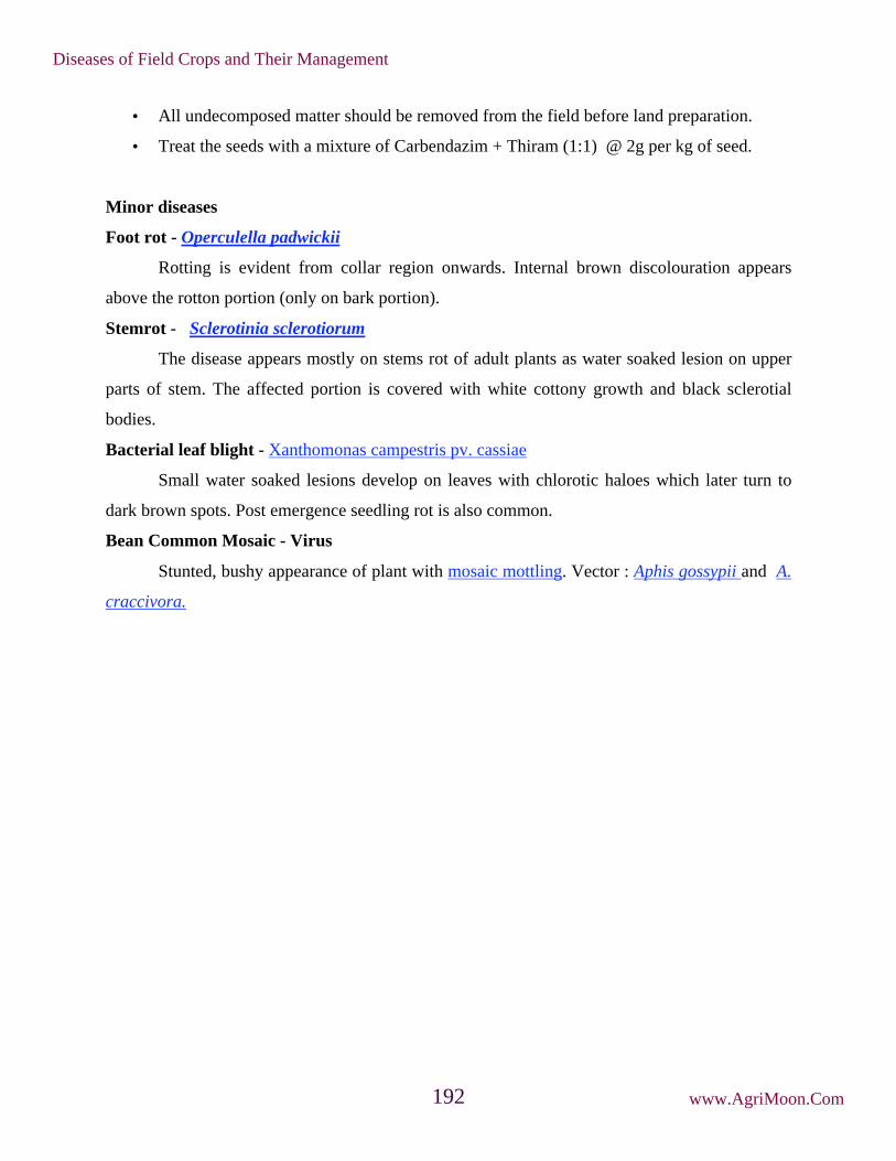
• All undecomposed matter should be removed from the field before land preparation.
• Treat the seeds with a mixture of Carbendazim + Thiram (1:1) @ 2g per kg of seed.
Minor diseases
Foot rot - Operculella padwickii
Rotting is evident from collar region onwards. Internal brown discolouration appears
above the rotton portion (only on bark portion).
Stemrot - Sclerotinia sclerotiorum
The disease appears mostly on stems rot of adult plants as water soaked lesion on upper
parts of stem. The affected portion is covered with white cottony growth and black sclerotial
bodies.
Bacterial leaf blight - Xanthomonas campestris pv. cassiae
Small water soaked lesions develop on leaves with chlorotic haloes which later turn to
dark brown spots. Post emergence seedling rot is also common.
Bean Common Mosaic - Virus
Stunted, bushy appearance of plant with mosaic mottling. Vector : Aphis gossypii and A.
craccivora.
Diseases of Field Crops and Their Management
www.AgriMoon.Com192

18. Diseases of Soybean
Dry root rot - Macrophomina phaseolina
Symptoms
The disease symptom starts initially with yellowing and drooping of the leaves.
The leaves later fall off and the plant dies with in week. Dark brown lesions are seen on
the stem at ground level and bark shows shredding symptom. The affected plants can be
easily pulled out leaving dried, rotten root portions in the ground. The rotten tissues of
stem and root contain a large number of black minute sclerotia.
Pathogen
The fungus produces dark brown, septate mycelium with constrictions at hyphal
branches. Minute, dark, round sclerotia in abundance. The fungus also produces dark
brown, globose ostiolated pycnidia on the host tissues. The pycnidiospores are thin
walled, hyaline, single celled and elliptical
Favourable conditions
• Day temperature of 30˚C
• Prolonged dry season followed by irrigation.
Disease cycle
The fungus survives in the infected debris and also as facultative parasite in soil.
The primary spread is through seed-borne and soil-borne sclerotia. The secondary spread
Symptoms
Diseases of Field Crops and Their Management
www.AgriMoon.Com193

is through seed-borne and soil-borne sclerotia. The secondary spreads is through
pycnidiospores which are air-borne.
Management
• Treat the seeds with Carbendazim or Thiram at 2 g/kg or pellet the seeds with
Trichoderma viride at 4 g/kg or Pseudonomas fluorescens @ 10g/kg of seed.
• Apply farm yard manure or green leaf manure (Gliricidia maculata) at 10 t/ha or
neem cake at 150 kg/ha.
Wilt - Fusarium oxysporum f. sp. tracheiphilum
Symptoms
Symptoms do not appear until the plants are about six weeks old. Initially a few
plants are noticed with pale green flaccid leaves which soon turn yellow. Growth is
stunted, chlorosis, drooping, premature shedding or withering of leaves with veinal
necrosis often occurs and finally plant dies within 5 days. Brownish, purple discoloration
of the cortical area is seen, often extends throughout the plant.
Symptoms
Diseases of Field Crops and Their Management
www.AgriMoon.Com194

Pathogen
The fungus produces falcate shaped macroconidia which are 4-5 septate, thin
walled and hyaline. The microconidia are single celled hyaline and oblong or oval. The
chlamydospores are also produced in abundance.
Favourable conditions
Temperature of 20-25˚C and moist humid weather.
Disease cycle
The fungus survives in the infected stubbles in the field. The primary spread is
through soilborne chlamydospores and infected seeds. The secondary spread is through
conidia by irrigation water.
Management
• Treat the seeds with Carbendazim or Thiram at 2 g/kg or treat the seeds with
Trichoderma viride at 4 g/kg.
• Spot drenching with Carbendazim at 0.5 g/litre.
Leaf spot - Cercospora sojana
Symptoms
Light to dark gray or brown areas varying from specks to large blotches appear on
seeds. The disease primarily affects foliage, but, stems, pods and seeds may also be
infected. Leaf lesions are circular or angular, at first brown then light brown to ash grey
with dark margins. The leaf spot may coalesce to form larger spots. When lesions are
numerous the leaves wither and drop prematurely. Lesions on pods are circular to
elongate, light sunken and reddish brown.
Symptoms
Diseases of Field Crops and Their Management
www.AgriMoon.Com195

Favourable conditions
• Fungus survives in infected seeds and in debris.
• Warm, humid weather favor disease incidence
Management
• Use resistant varieties.
• Use healthy or certified seeds.
• Rotate soybean with cereals.
• Completely remove plant residue by clean ploughing the field soon after harvest.
• Destroy last years infected stubble.
• Seed treatment with Thiram + Carbendazium (1:1) @ 2g/kg seed.
• Spray Mancozeb @ 2g/L or Carbenzadium (500 mg/L).
Mosai - Soybean mosaic virus (SMV)
Symptoms
Diseased plants are usually stunted with distorted (puckered, crinkled, ruffled,
narrow) leaves. Pods become fewer and smaller seeds. Infected seeds get mottled and
deformed. Infected seeds fail to germinate or they produce diseased seedlings.
Pathogen
It is caused by Soybean mosaic virus - a potyvirus. Flexuous particles 750 -
900nm long, ss RNA genome
.
Symptoms
Diseases of Field Crops and Their Management
www.AgriMoon.Com196

Disease cycle
Soybean mosaic virus is seed borne. The SMV can be transmitted through sap, 32
aphid species are involved in transmission.
Favorable conditions
• Temperature around 18o C
• Humid weather.
Management
• Deep summer ploughing.
• Use resistant or tolerant varieties.
• Use healthy/certified seeds.
• Keep the field free from weeds.
• Rogue out infected plants and burn them
• Pre-sowing soil application of Phorate @ 10 kg/ha.
• Two foliar sprays of Thiamethoxam 25 WG @ 100 g/ha or Methyl demeton 800
ml/ha at 30 and 45 days after sowing.
Diseases of Field Crops and Their Management
www.AgriMoon.Com197

******☺******
This Book Download From e-course of ICAR Visit for Other Agriculture books, News,
Recruitment, Information, and Events at
www.agrimoon.com
Give FeedBack & Suggestion at [email protected]
Send a Massage for daily Update of Agriculture on WhatsApp
+91-8148663744
Disclaimer:
The information on this website does not warrant or assume any legal
liability or responsibility for the accuracy, completeness or usefulness of the
courseware contents.
The contents are provided free for noncommercial purpose such as teaching,
training, research, extension and self learning.
******☺******
Connect With Us:
Diseases of Field Crops and Their Management
www.AgriMoon.Com198

K. K. WAGH COLLEGE OF AGRICULTURE,
NASHIK
DEPARTMENT OF PLANT PATHOLOGY
THEORY NOTES Course No.: - PATH -121
Course Title: - Fundamentals of Plant Pathology
Credits: - 3 (2+1)
Compiled By
Prof. Patil K.P.
Assistant Professor
Department of Plant Pathology

Teaching Schedule
a) Theory
Lecture Topic Weightage
(%)
1 Importance of plant diseases, scope and objectives of Plant
Pathology.....
3
2 History of Plant Pathology with special reference to Indian work 3
3,4 Terms and concepts in Plant Pathology, Pathogenesis 6
5 classification of plant diseases 5
6,7, 8 Causes of Plant Disease Biotic (fungi, bacteria, fastidious
vesicular bacteria, Phytoplasmas, spiroplasmas, viruses,
viroids, algae, protozoa, and nematodes ) and abiotic causes
with examples of diseases caused by them
10
9 Study of phanerogamic plant parasites. 3
10, 11 Symptoms of plant diseases 6
12,13,
14
Fungi: general characters, definition of fungus, somatic structures,
types of fungal thalli, fungal tissues, modifications of thallus,
7
15 Reproduction in fungi (asexual and sexual). 4
16, 17 Nomenclature, Binomial system of nomenclature, rules of
nomenclature,
6
18, 19 Classification of fungi. Key to divisions, sub-divisions, orders and
classes.
6
20, 21,
22
Bacteria and mollicutes: general morphological characters. Basic
methods of classification and reproduction in bacteria
8
23,24,
25
Viruses: nature, architecture, multiplication and transmission 7
26, 27 Nematodes: General morphology and reproduction, classification
of nematode Symptoms and nature of damage caused by plant
nematodes (Heterodera, Meloidogyne, Anguina etc.)
6
28, 29,
30
Principles and methods of plant disease management. 6
31, 32,
33
Nature, chemical combination, classification of fungicides and
antibiotics.
7
34, 35,
36
Mode of action and formulations of fungicides and antibiotics. 7
Total 100
Suggested Readings
1) Pathak, V. N. Essentials of Plant Pathology. Prakash Pub., Jaipur
2) Agrios, GN. 2010. Plant Pathology. Acad. Press.
3) Kamat, M. N. Introductory Plant Pathology. Prakash Pub, Jaipur
4) Singh RS. 2008. Plant Diseases. 8th Ed. Oxford & IBH. Pub. Co.
5) Singh RS. 2013. Introduction to Principles of Plant Pathology. Oxford and IBH Pub. Co.
6) Alexopoulos, Mims and Blackwel. Introductory Mycology
7) Mehrotra RS & Aggarwal A. 2007. Plant Pathology. 7th Ed. Tata Mc Graw Hill Publ. Co. Ltd.
8) Gibbs A & Harrison B. 1976. Plant Virology - The Principles. Edward Arnold, London.

9) Hull R. 2002. Mathew.s Plant Virology. 4th Ed. Academic Press, New York.
10) Verma JP. 1998. The Bacteria. Malhotra Publ. House, New Delhi.
11) Goto M. 1990. Fundamentals of Plant Bacteriology. Academic Press, New York.
12) Dhingra OD & Sinclair JB. 1986. Basic Plant Pathology Methods. CRC Press, London, Tokyo.
13) Nene YL & Thapliyal PN. 1993. Fungicides in Plant Disease Control. 3rd Ed. Oxford & IBH,
New Delhi.
14) Vyas SC. 1993. Handbook of Systemic Fungicides. Vols. I-III. Tata McGraw Hill, New Delhi.
15) Rajeev K & Mukherjee RC. 1996. Role of Plant Quarantine in IPM. Aditya Books.
16) Rhower GG. 1991. Regulatory Plant Pest Management. In: Handbook of Pest Management in
Agriculture. 2nd Ed. Vol. II. (Ed. David Pimental). CRC Press.

4
Importance of plant diseases, scope and objectives of Plant Pathology
Importance of the Plant Diseases
Globally, enormous losses of the crops are caused by the plant diseases. The loss can occur from
the time of seed sowing in the field to harvesting and storage. Important historical evidences of
plant disease epidemics are Irish Famine due to late blight of potato (Ireland, 1845), Bengal
famine due to brown spot of rice (India, 1942) and Coffee rust (Sri Lanka, 1967). Such
epidemics had left their effect on the economy of the affected countries.
Objectives of Plant Pathology
Plant Pathology (Phytopathology) deals with the cause, etiology, resulting losses and control or
management of the plant diseases. The objectives of the Plant Pathology are the study on:
i. the living entities that cause diseases in plants;
ii. the non-living entities and the environmental conditions that cause disorders in plants;
iii. the mechanisms by which the disease causing agents produce diseases;
iv. the interactions between the disease causing agents and host plant in relation to
overall environment; and
v. the method of preventing or management the diseases and reducing the
losses/damages caused by diseases.
Scope of Plant Pathology
Plant pathology comprises with the basic knowledge and technologies of Botany, Plant
Anatomy, Plant Physiology, Mycology, Bacteriology, Virology, Nematology, Genetics,
Molecular Biology, Genetic Engineering, Biochemistry, Horticulture, Tissue Culture, Soil
Science, Forestry, Physics, Chemistry, Meteorology, Statistics and many other branches of
applied science.

5
History of Plant Pathology with special reference to Indian work
Historical perspectives show that the attention of man to plant diseases and the science of plant
pathology were drawn first only in the European countries. Greek philosopher Theophrastus
(about 286 BC) recorded some plant diseases about 2400 years ago. This branch of science could
maintain a proper record on the plant disease and their causal organisms only after development
of compound microscope by the Dutch worker Antony von Leeuwenhoek in 1675. He first
visualized bacteria in 1683 under his microscope. Robert Hook (1635-1703) also developed
simple microscope which was used to study of minute structure of fungi. The Italian botanist
Pier’ Antonio Micheli (1679-1737) first made detail study of fungi in 1729. With the
contribution of many other scientists’ viz., Mathieu Tillet (1755), Christian Hendrik Persoon
(1801) and Elias Magnus Fries (1821), the foundation of modern plant pathology was built and
was further strengthened by Anton de Bary (1831-1888), who is regarded as the Father of Plant
Pathology.
Historically, plant pathology of India is quite ancient as the Indian agriculture, which is nearly
4000 years old. This confirms that mention about plant diseases was made much before the time
of Theophrastus. The events of the development of plant pathology in India are chronologically
recorded as follows:
(i) Plant diseases, other enemies of plants and methods of their control had been recorded in the
ancient books viz., Rigveda, Atharva Veda (1500-500 BC), Artha Shastra of Kautilya (321-186
BC), Sushruta Samhita (200-500AD), Vishnu Purana (500 AD), Agnipurana (500-700 AD),
Vishnudharmottara (500-700 AD) etc.
(ii) During 11th century, Surapal wrote Vraksha Ayurveda, which is the first book in
India where he gave detail account on plant diseases and their control. Plant diseases
were grouped into two-internal and external. Tree surgery, hygiene protective
covering with paste, use of honey, plant extracts, oil cakes of mustard, castor,
sesamum etc. are some of the disease management practices recorded in the book.

6
(iii) Symptoms of plant diseases are cited in other ancient Indian literatures viz. Jataka of
Buddhism, Raghuvamsha of Kalidas etc.
(iv) The Europeans started systemic study of fungi in India during 19th century. They
collected the fungi and sent to the laboratory in Europe for identification. D.D.
Cunningham and A. Barclay, during 1850-1875, started identification of fungi in
India itself. Cunningham specially studied on rusts and smuts. K.R. Kirtikar was
credited as the first Indian scientist for collection and identification of fungi in India.
(v) Edwin John Butler started the systemic study on Indian fungi and the diseases caused
by them. This Imperial Mycologist came to India in 1901 and initiated the works on
fungi at Imperial Agricultural Research Institute established by the British Government of Pusa
(Bihar). The first and most classic book in the field of plant pathology of India i.e. Fungi and
Diseases in Plants was written by him based on the exhaustive study on Indian fungi. He left
India in 1920 and joined as the first Director of Imperial Mycological Institute in England. He is
regarded as the Father of Indian PlantPathology.
(vi) Jahangir Ferdunji Dastur (1886-1971), a colleague of Butler, was the first Indian plant
pathologists to made detail study of fungi and plant diseases. He specially studied the
diseases of potato and castor caused by genus Phytophthora and established the
species P. parasitica from castor in 1913. In recognition of his command in Plant
Pathology, he was promoted to the Imperial Agricultural Science in 1919.
(vii) G.S. Kulkarni, a student of Butler, generated detail information on downy mildew and
smut of jowar and bajra. Another student S.L. Ajrekar studied wilt disease of cotton,
sugarcane smut and ergot of jowar.
(viii) Karam Chand Mehta (1894-1950) of Agra had contributed a lot to Plant Pathology of
India. He first joined Agricultural College as a demonstrator at Kanpur. His

7
outstanding contribution in the discovery of the life cycle of stem rust of wheat in
India and reported that barberry, an alternate host, does not play any role in
perpetuation of the rust fungus in India. He published two monographs entitled
“Further Studies on Cereal Rust in India” Part I (1940) and Part II (1952) and also
established three laboratories for rust works at Agra, Almora and Shimla.
(ix) Raghubir Prasad (1907-1992) trained under K.C. Mehta, contributed to the
identification of physiological races of cereal rusts and life cycle of linseed rust.
Subsequently, L.M. Joshi at IARI conclusively studied various aspects of wheat rusts
viz., chief foci of infection of rusts, dissemination of rust pathogens in India. Later on
S. Nagarajan and L.M. Joshi developed most useful mathematical models in 1978 to
predict appearance of stem and leaf rust of wheat.
(x) Manoranjan Mitra was considered as one of the most critical plant pathologist worked
on Helminthosporium. He first reported Karnal bunt of wheat in 1931 from Karnal in
Haryana.
(xi) B.B. Mundkur was the second mycologist trained under Butler and worked with
Mehta and Mitra. He worked on control of cotton wilt by using resistant varieties and
became successful in reducing yield loss in Maharashtra. His significant contribution
is the establishment of Indian Phytopathological Society (IPS) in 1948 with its journal
Indian Phytopathology. In the same year, he published a text book Fungi and Plant
Diseases which was the second book of Plant Pathology after the classic book of
Butler.
(xii) S.R. Bose was taxonomist, mainly worked on the classification of Polyporaceae and
isolated “polyporin” from Polyporus.
(xiii) Notable contribution in the field of Mycology was made by M.J. Thirumalachar
(1914-1999). He created 20 new genera and 300 new species of fungi, monographed

8
genera of Uredinales of the world and Ustilaginales of India. Similarly many
Hyphomycetes particularly Fusarium were elaborated by C.V. Subramanian in 1971.
(xiv) Works on fundamental plant pathology, especially the biochemistry of host-parasite
relationship were started at Lucknow and Madras (Chennai) lead by Sachindra Nath
Dasgupta (1904-1990) and T.S. Sadasivan (1913-2001), respectively. Dr. Dasgupta
initiated the works on leather mycology, paper pulp mycology and predacious fungi.
Dr. Sadasivan’s school developed the concept of vivotoxin and reported the
production of fusaric acid by Fusarium vasinfectum that causes wilt diseases in
cotton.
(xv) T.S. Ramakrishnan, a mycologist to Madras Government cultivated ergot diseased
rye for toxin production. He published two books entitled Diseases of Millets (1963)
and Diseases of Rice (1971). Renowned plant pathologists viz., G Rangaswami and
R. Ramakrishnan were his students.
(xvi) Plant Bacteriology in India got a shape with the effort of Makanj Kalyanji Patel
(1899-1967). He established a school of Plant Bacteriology at College of Agriculture,
Pune and first described a new species Xanthomonas campestris pv. uppali in 1948
from the host Ipomea muricota. He described more than 30 bacterial diseases from
India. Other scientists viz., V.P. Bhide and G. Rangaswami also contributed their
pioneering works to the phytobacteriology of India. D.N. Srivastava (1925-2000) is
mostly remembered for his tremendous contribution on bacterial blight of rice. M.K.
Hingorani reported about the complex nature of tundu disease of wheat caused by a
bacterium and a nematode in 1952 and also he confirmed the causal agent of ring
disease of potato as Pseudomonas (=Ralstonia) solanacerarum. J.P. Verma (1939-
2005) contributed many valuable findings on bacterial blight disease of cotton.
(xvii) Plant virus research in India was started particularly at IARI, New Delhi under the
leadership of R.S. Vasudeva (1905-1987), S.P. Raychaudhury (1916-2005) and
Anupam Varma. Considering the importance of plant viral diseases, IARI established

9
some Regional Research Stations at Shimla for temperate fruits (1952), at Pune for
fruits and vegetables (1952) and at Kalimpong (West Bengal) for large cadamom,
citrus and other crops in north-eastern sub-Himalayan mountain (1956). Y.L. Nene’s
contributions have been well remembered particularly the viral diseases of pulses and
the ‘Khaira’ disease of rice caused by Zinc deficiency. He wrote the book
“Fungicides in Plant Disease Control”.
(xviii) Teaching of plant pathology as a course was started at University of Calcutta,
Bombay and Madras in 1857 where only fungal taxonomy was emphasized. But plant
pathology as a university science was started in 1930 at University of Allahabad,
Lucknow and Madras. Of which, perhaps University of Madras was first to introduce
plant pathology as a course. Agra University had introduced one post-graduate
programme in plant pathology in Govt. Agricultural College, Kanpur in 1945. The
organized teaching in Mycology and Plant Pathology was began as a part of
agricultural science under the banner of Indian Agricultural Research Institute. The
subject received due importance and teaching of its supporting courses viz.
mycology, bacteriology, virology and nematology in both under- and post-graduate
programmes of Agriculture was taken up regularly after the establishment of
Agricultural Universities in different states of India in 1960. At present, most of the
courses related to plant pathology have been revised and added molecular plant
pathology by keeping pace with the advancement in the science.

10
Terms and concepts in Plant Pathology, Pathogenesis
Definition and terms
1. Parasite: An organism living upon or in another living organism (the host) and obtaining the
food from the invading host.
2. Pathogen: An entity, usually a micro-organism that can cause the disease.
3. Biotroph: A plant pathogenic fungus that requires living host cells i.e. an obligate parasite.
4. Hemibiotroph: A plant pathogenic fungus that initially requires living host cells but after
killing the host cell grows on the dead and dying cells.
5. Necrotroph: A pathogenic fungus that kills the host and survives on the dying and dead cells.
6. Pathogenicity: The relative capability of a pathogen to cause disease.
7. Pathogenesis: It is a process caused by an infectious agent (pathogen) when it comes in
contact with a susceptible host.
8. Virulence: The degree of infectivity of a given pathogen.
9. Infection: The initiation and establishment of a parasite within a host plant.
10. Primary infection: The first infection of a plant by the over wintering or over summering of
the pathogen.
11. Inoculum: That portion of pathogen which is transferred to plant and cause disease.
12. Invasion: The penetration and spread of a pathogen in the host.
13. Colonization: The growth of a pathogen, particularly a fungus, in the host after infection is
called colonization.
14. Inoculum potential: The growth or threshold of fungus available for colonization at
substratum (host).
15. Symptoms: The external and internal reaction or alterations of a plant as a result of disease.
16. Incubation period: The period of time between penetration of a pathogen to the host and the
first appearance of symptoms on the plant.
17. Disease cycle: The chain of events involved in disease development.
18. Disease syndrome: The set of varying symptoms characterizing a disease are collectively
called a syndrome.
19. Single cycle disease (Monocyclic): This type of disease is referred to those caused by the
pathogen (fungi) that can complete only one life cycle in one crop season of the host plant. e.g.
11
downy mildew of rapeseed, club root of crucifers, sclerotinia blight of brinjal etc.
20. Multiple cycle disease (Polycyclic): Some pathogens specially a fungus, can complete a
number of life cycles within one crop season of the host plant and the disease caused by such
pathogens is called multiple cycle disease e.g. wheat rust, rice blast, late blight of potato etc.
21. Alternate host: Plants not related to the main host of parasitic fungus, where it produces its
different stages to complete one cycle (heteroecious).
22. Collateral host: The wild host of same families of a pathogen is called as collateral host.
3
23. Predisposition: The effect of one or more environmental factors which makes a plant
vulnerable to attack by a pathogen.
24. Physiologic race: One or a group of microorganisms similar in morphology but dissimilar in
certain cultural, physiological or pathological characters.
25. Biotype: The smallest morphological unit within a species, the members of which are usually
genetically identical.
26. Symbiosis: A mutually beneficial association of two or more different kinds of organisms.
27. Mutualism: Symbiosis of two organisms that are mutually helpful or that mutually support
one another.
28. Antagonism: The counteraction between organisms or groups of organisms.
29. Mutation: An abrupt appearance of a new characteristic in an individual as a result of an
accidental change in genes present in chromosomes.
30. Disease: Any deviation in the general health, or physiology or function of plant or plant
parts, is recognized as a disease.
31. Cop Damage: It is defined as any reduction in the quality or quantity of yield or loss of
revenue resulting from crop injury.
32. Deficiency: Abnormality or disease caused by the lack or subnormal level of availability of
one or more essential nutrient elements.
12
Concept of Plant Disease
The normal physiological functions of plants are disturbed when they are affected by pathogenic
living organisms or by some environmental factors. Initially plants react to the disease causing
agents, particularly in the site of infection. Later, the reaction becomes more widespread and
histological changes take place. Such changes are expressed as different types of symptoms of
the disease which can be visualized macroscopically. As a result of the disease, plant growth in
reduced, deformed or even the plant dies.
When a plant is suffering, we call it diseased, i.e. it is at ‘dis-ease’. Disease is a condition that
occurs in consequence of abnormal changes in the form, physiology, integrity or behaviour of the
plant.
According to American Phytopathological Society (Phytopathology 30:361-368, 1940),
disease is a deviation from normal functioning of physiological processes of sufficient duration
or intensity to cause disturbance or cessation of vital activities. The British Mycological Society
(Trans. Brit. Mycol. Soc. 33:154-160, 1950) defined the disease as a harmful deviation from the
normal functioning of process. Recently, Encyclopedia Britannica (2002) forwarded a simplified
definition of plant disease. A plant is diseased when it is continuously disturbed by some causal
agent that results in abnormal physiological process that disrupts the plants normal structure,
growth, function or other activities. This interference with one or more plant’s essential
physiological or biochemical systems elicites characteristic pathological conditions or symptoms.

13
Parasitism and Pathogenesis
An organism which lives in or on other living organisms and derives its nutrients from the latter
is called parasite. The relationship between a parasite and its hosts is known as parasitism. Many
fungi and most bacteria grow on a non-living substrate within a living plant. The organism of
this type of mode of nutrition is called saprophyte. Based on the different types of modes or
nature of nutrition, the relationship between the host and parasite or saprophyte is termed in
many ways viz., obligate parasite (biotroph), obligate saprophyte, facultative parasite, facultative
saprophyte, hemibiotroph and necrotroph (perthotrophs or perthophyte).
Parasitism in cultivated crops is common phenomenon. Any agent that can cause suffering or
damage or disease is called a pathogen. In plant pathology, the term ‘pathogen’ is usually used to
the living or infectious organisms. The ability of a pathogen or parasite to cause disease is known
as pathogenicity.
It is obvious that a plant becomes diseased when it is attacked by a pathogen or parasite. The
ultimate condition i.e. disease occurs by passing through some distinct events. Thus, the genesis
or chain of events or stages of disease development are called pathogenesis. This is also called as
disease cycle. The events that occur in specific order are namely inoculation, penetration,
establishment of infection, invasion or colonization, growth and reproduction, dissemination and
survival of the pathogen (over wintering or over summering in absence of the host). The events
will continue to repeat in the same order in presence of both the host and pathogen/parasite that
may lead to severe disease condition.

14
Classification of plant diseases
To facilitate the study of plant diseases they are needed to be grouped in some orderly fashion.
Plant diseases can be grouped in various ways based on the symptoms or signs (rust, smut, blight
etc.), nature of infection (systemic or localized), habitat of the pathogens, mode of perpetuation
and spread (soil-, seed- and air-borne etc.), affected parts of the host (aerial, root disease etc.),
types of the plants (cereals, pulses, oilseed, ornamental, vegetable, forest diseases etc.). But the
most useful classification has been made based on the type of pathogens that cause plant
diseases. Since this type of classification indicates not only the cause of the disease, but also the
knowledge and information that suggest the probable development and spread of disease
alongwith their possible control measures. The classification is as follows:
1. Infectious plant diseases:
a. Disease caused by parasitic organisms: The organisms included in animate or biotic
causes can incite diseases in plants.
b. Diseases caused by viruses and viroids.
2. Non-infectious or non-parasitic or physiological diseases: The factors included in
inanimate or abiotic causes can incite such diseases in plants under a set of suitable
environmental conditions.
Plant diseases are caused by pathogens. Hence a pathogen is always associated with a disease. In
other way, disease is a symptom caused by the invasion of a pathogen that is able to survive,
perpetuate and spread. Further, the word “pathogen” can be broadly defined as any agent or
factor that incites 'pathos or disease in an organism or host. In strict sense, the causes of plant
diseases are grouped under following categories:
1. Animate or biotic causes: Pathogens of living nature are categorized into the following
groups.
(i) Fungi (v) Algae
(ii) Bacteria (vi) Phanerogams
(iii) Phytoplasma (vii) Protozoa
(iv) Rickettsia-like organisms (viii) Nematodes
2. Mesobiotic causes : These disease incitants are neither living or non-living, e.g.
(i) Viruses
(ii) Viroides

15
3. Inanimate or abiotic causes: In true sense these factors cause damages (any reduction in the
quality or quantity of yield or loss of revenue) to the plants rather than causing disease. The
causes are:
(i) Deficiencies or excess of nutrients (e.g. ‘Khaira’ disease of rice due to Zn deficiency)
(ii) Light
(iii) Moisture
(iv) Temperature
(v) Air pollutants (e.g. black tip of mango)
(vi) Lack of oxygen (e.g. hollow and black heart of potato)
(vii) Toxicity of pesticides
(viii) Improper cultural practices
(ix) Abnormality in soil conditions (acidity, alkalinity)

16
Flowering Plant Parasites (Phanerogams)
Most of the diseases are caused by fungi bacteria and viruses. There are few seeds plants called
flowering parasites (Phanerogams) which are parasitic on living plants. Some of these attack
roots of the host, while some parasites on stem. Some are devoid of chlorophyll and entirely
dependent on their host for food supply, while other have chlorophyll and obtain only mineral
constituents of food from host by drawing nutrition and water they are called as Holoparasites or
complete or total parasite. They have haustoria as absorbing organs, which are sent deep into the
vascular bundle of the host to draw nutrients, water and minerals.
Flowering Plant Parasites: There are two types of parasites.
1) Root Parasites:
i) Striga (Partial root parasite)
ii) Orobanche (Complete root parasite)
2) Stem Parasites:
i) Dodder (Cuscuta) (Complete stem parasite)
ii) Loranthus (Partial Stem parasite)
A. Root Parasites:
1. Total or Complete or Holoparasite:
Orobanche (Broom rape or Tokra)
It is annual flashy flowering plant growing to height of about 15-50 cm long, yellow or brownish
colour and covered by small thin scaly leaves. Flowers appears in the axil of leaves are white or
tubular. Fruits appears in the axil of leaves are white or tubular. Fruits are capsule containing and
seeds are very small, black in colour remain viable for several years. The hausteria of parasite
penetrates into the roots of hosts and draw its nourishment. The growth of the plant is retarded,
may die some times. It attacks tobacco, tomato, brinjal, cabbage, cauliflower.

17
2. Hemi Partial or Semi Root Parasite:
Striga (Witch Weed or Turfula or Talop)
Family Scrophulariaceae
It is a small plant with bright green leaves grows upto height 20-60 cm leaves beers chlorophylls
and developed in clusters of 10-20 % host plant. They are obligate parasites therefore, do not
obtain all their nutrient from their host root. Flowers are pink in colour, seed are very minute and
produce in grate number 5000 to 100000 seeds plant per years. One flower contain 1200-1500
seeds and remains viable upto 12-40 years. Dissemination takes place with rain water, flood,
wind and irrigation water. It cause yellowing and wilting of host leaves. It attacks sugarcane
jowar, Maize, cereals and millets.
B. Stem Parasites:
1. Total or Complete or Holoparasite:
Cuscuta or dodder (Amarvel, Lovevine) Family cuscutaceae.
Genus – Cuscuta
It is non chlorophyllous, leaf less parasitic seed plant.
It is yellow pink or orange in colour and attached to the host. They do not bear leaves but bear
minute function less scale leaves is produces flower and fruits. Flower are white, pink or
yellowish in colour and found in clusters. Seed are form in capsules. A single plant may be
produce 3000 seeds.
The first appearances of parasites is noticed as thread like leaf less stem which devoid of green
pigment and twine around the stem or leaves of the host. When stem of parasitic plant comes in
contact with host, the minute root like organs. i.e. hausteria penetrates into the host and absorbs.
When the relation ship of the host is firmly established, the dodder plant looses the contact from
soil.
These affect plant get weakened and yield poorly the seeds spread by animals, water and
implements and remain viable when condition are unfavorable.

18
It attacks berseem alfalfa, clover, flax, onion, potato, ornamental and hedge plants.
2. Partial, Semi or Hemi Stem Parasites:
Loranthus
Family- Loranthaceae.
It is a partial parasite of tree trunks and branches with brown stem, dark green leaves but no
roots.
1. Stem is thick and flattened of the node, appear in clusters at the point of attack which can be
easily spotted on the trees.
2. At the point of attachment with the tree, it shows swellings or tumourous growth where the
haustoria are produced. It produces flowers which are long, tabular, greenish, white or red colour
and found in clusters. It produces fleshy berries with single seed.
3. The affected host plant becomes stunted in growth and dispersal of seed is mostly through the
birds and animals. It attacks mango, citrus, apple, guava.
Phanerogamic Plant Parasites
Some flower and seed bearing higher plants (phanerogams) live parasitically on other living
plants and can cause important diseases on agricultural crops and also in forest trees. They are
classified in the following botanical families and genera,
1. Cuscutaceae (stem parasite)
Genus: Cuscuta, the dodders
2. Viscaceae (stem parasites)
Genus: Arceuthobium, the dwarf mistletoes of conifers
Phoradendron, the American true mistletoes of broad leaved trees
Viscum, the European tree mistletoes
Dendrophthoe, the giant mistletoes
3. Orobanchaceae (root parasite)
Genus: Orobanche, the broomrapes

19
4. Scrophulariaceae (root parasite)
Genus: Striga, the witchweeds
Identifying characters, reproduction and life cycle
1. Dodder (Love-vine, Amarbel): Dodder is slender, twining plant. The stem is tough,
succulent, threadlike, curling, leafless and bearing minute scales in place of leaves. Stem
is usually yellowish or orange in colour. They grow and climb in abundance on the wild
and cultivated plants. Their haustoria penetrate the stem or leave of the host plant and
absorb foodstuffs and water. Growth of the infected plants is suppressed and finally dies.
Tiny whitish flowers arise in clusters from the stem and they produce numerous grey to
reddish-brown seeds within few weeks of bloom. The seeds fall on the ground where they
either germinate immediately or remain dormant until next season. The seeds may be
spread to new areas by animals, water, equipments and by mixing with crop seeds.
2. Giant mistletoe (Loranthus) : Mistletoes are semi-parasites of thee-trunks and branches.
They have green leaves and many branches and hence grown like a small bush on the
host. They do not have true roots and hence develop haustoria to draw nutrients from the
vascular system of the host plant. Flowers are long, tubular, greenish white to red in
colour and borne in clusters. Seed is fleshy, contains one seed, sweet in taste and usually
eaten by birds and animals. The parasite is spread mostly through birds. Droppings of
bird containing seeds get deposited on the other trees, mainly at the junction of a branch
and main trunk. These seeds germinate, develop haustoria and established on the host
plant.
3. Broomrape: Broomrape is a complete root parasite. It affects tobacco mostly and many
other Solanaceous and Cruciferous plants. The parasite has a stout, fleshy stem of 10-15
cm long. The stem is pale yellow or brownish red in colour and is covered by small, thin,
browny, scaly leaves. Flowers are white and tubular and appear in the axil of leaves.
Seeds are very small, black in colour and can remain viable in soil for several years.
Roots are haustoria-like, can penetrate the roots of the host for nutrition. The affected
plants become stunted and may die.
4. Witchweed: It is a semi root parasite but obligate in nature. Commonly affected plants
by this parasitic plant are sugarcane, cereals, maize and millets. Striga is a small plant

20
with bright green, slightly hairy stem and leaves. The weed grows 15-60 cm high in
clusters. Leaves are long and narrow in opposite pairs. Flowers are small, brick red or
yellowish or almost white with yellow centers, appear throughout the season. Tiny brown
seeds are produced in each capsule and thus a single plant can produce 50,000-500,000
seeds. The seeds can remain viable for 12-40 years. Roots are watery white in colour
without root hairs and hence obtain nutrients by haustoria from the host plant. The life
cycle from seed germination to first seed setting on the developed plant needs 90-120
days.
21
Symptoms of Plant Diseases
A visible or detectable abnormality expressed on the plant as a result of disease or disorder is
called symptom. The totality of symptoms is collectively called as syndrome while the pathogen
or its parts or products seen on the affected parts of a host plant is called sign. Different types of
diseasesymptoms are cited below:
Necrosis: It indicates the death of cells, tissues and organs resulting from infection by
pathogen. Necrotic symptoms include spots, blights, burn, canker, streaks, stripes,
damping-off, rot etc.
Wilt: Withering and drooping of a plant starting from some leaves to growing tip
occurs suddenly or gradually. Wilting takes place due to blockage in the translocation
system caused by the pathogen.
Die-back: Drying of plant organs such as stem or branches which starts from the tip
and progresses gradually towards the main stem or trunk is called die-back or wither
tip.
Mildew: White, grey or brown coloured superficial growth of the pathogen on the
host surface is called mildew.
Rusts: Numerous small pustules growing out through host epidermis which gives
rusty (rust formation on iron) appearance of the affected parts.
Smuts: Charcoal-like and black or purplish-black dust like masses developed on the
affected plant parts, mostly on floral organs and inflorescens are called smut.
Blotch: A large area of discolouration of a leaf, fruit etc. giving a blotchy appearance.
White blisters: Numerous white coloured blister-like ruptures are surfaced on the

22
host epidermis that forms powdery masses of spores of fungi. They are called white
blisters or white rust.
Colour change: It denotes conversion of green pigment of leaves into other colours
mostly to yellow colour, in patches or covering the entire leaves. (i) Etioliation:
Yellowing due to lack of light, (ii) Chlorosis: Yellowing due to infection viruses,
bacteria, fungi, low temperature lack of iron etc. (iii) Albino: Lack of any pigment
and turned into white or bleached (iv) Chromosis: Red, purple or orange pigmentation
due to physiological orders etc.
Exudation: Such symptom is commonly found in bacterial diseases when masses of
bacterial cells ooze out to the surface of affected plant parts and form some drops or
smear, it is called exudation. This exudation forms a crust on the host surface after
drying.
Overgrowth: Excessive growth of the plant parts due to infection by pathogens.
Overgrowth takes place by two processes (i) Hyperplasia: abnormal increase in size
due to excessively more cell division (ii) Hypertrophy: abnormal increase in size or
shape due to excessive enlargement of the size of cell of a particular tissue.
Atrophy: It is known as hypoplasia or dwarfing which is resulted from the inhibition
of growth due to reduction in cell division or cell size.
Sclerotia: These are dark and hard structures of various shaped composed of dormant
mycelia of some fungi. Sometimes, sclerotia are developed on the affected parts of
the plant. Presence of sclerotia on the host surface is specifically called a sign of
disease rather than symptom.

23
General characteristics of fungi
FUNGI: Fungi are eukaryotic, spore bearing, achlorophyllous, heterotrophic organisms
that generally reproduce sexually and asexually and whose filamentous, branched
somatic structures are typically surrounded by cell walls containing chitin or cellulose or
both with many organic molecules and exhibiting absorptive nutrition.
Somatic structures:
Thallus/ Soma Commonly called as vegetative body or fungal body. A thallus( pl.
thalli) is a simple, entire body of the fungus devoid of chlorophyll with no differentiation
into stem, roots and leaves lacking vascular system.
Hypha (hypha=web) ( pl. hyphae) : Hypha is a thin, transparent, tubular filament filled
with protoplasm.It is the unit of a filamentous thallus and grows by apical elongation.
Mycelium( pl. mycelia): A net work of hyphae ( aggregation of hyphae) constituting the
filamentous thallus of a fungus.It may be colourless i.e., hyaline or coloured due to
presence of pigments in cell wall.The mycelium may be ectophytic or endophytic.
image of fungal mycelia

24
Types of fungal thalli:
1.Plasmodium (plasma = moulded body): It is a naked,multinucleate mass of
protoplasm moving and feeding in amoeboid fashion . Eg. Plasmodiophora brassicae
Plasmodiophora brassicae in host cell
2.Unicellular thallus: consisting of a single cell. Eg.Chytrids, Synchytrium
3.Multi cellular or filamentous thallus: Majority of fungi i.e., a true fungi are
filamentous, consisting of a number of branched, thread like filaments called
hyphae.Eg.Many fungi,Alternaria.
Fungi based on reproductive structures:
Holocarpic (holos=whole+karpos=fruit): If the thallus is entirely converted into
one or more reproductive structures, such thallus is called holocarpic thallus.
Eg.Synchytrium

25
Eucarpic(Eu=good+karpos=fruit):If the thallus is differentiated into a vegetative
part which absorbs nutrients and a reproductive part which forms reproductive
structures, such thallus is called eucarpic thallus. Eg.Pythium
Ectophytic fungus: If the fungal thallus is present on the surface of the host plant, it
is called ectophytic.Eg. Oidium .
Endophytic fungus: If the fungus penetrates into the host cell / present inside the
host, it is called endophytic.Eg. Puccinia. Endophytic fungus may be intercellular
(hypha grows in between the cells), or intra cellular ( hypha penetrates into host
cell).Eg.Ustilago, or vascular (xylem vessels) Eg. Fusarium oxysporum
Inter cellular hyphae produce special organs called haustoria which penetrate the host
cell and absorb food. These are absent in intracellular hyphae. Endophytic intra
cellular mycelium absorb food directly from protoplasm with out any specialized
structures.
In ectophytic mycelium, haustoria are produced in epidermal cells.
Septation in Fungi :(septum=hedge/partition) ( pl.septa)
Some fungal hyphae are provided with partitions or cross walls which divide the
fungus into a number of compartments /cells. These cross walls are called septa.
Aseptate hypha/coenocytic hypha: ( Koinos=common,kytos=hollow vessel) A
hypha with out septa is called aseptate /non-septate/ coenocytic hypha wherein the
nuclei are embedded in cytoplasm.Eg. lower fungi like Oomycetes and Zygomycetes.
Eg.common in higher fungi like Ascomycotina, Basidiomycotina
and Deuteromycotina

26
General types of septa:
1.Based on formation:
a)Primary septa : These are formed in direct association with nuclear division
(mitotic or meiotic) and are laid down between daughter nuclei separating the nuclei
/cells. Eg. Higher fungi like Ascomycotina and Basidiomycotina. b)Adventitious
septa: These are formed independent of nuclear division and these are produced to
delimit the reproductive structures. Eg. lower fungi like Oomycetes and Zygomycetes
in which septa are produced below gametangia (sex organs) which separate them
from rest of the cells.
2. Based on construction:
a)Simple septa: It is most common which is a plate like, with or without
perforation.
b)Complex septa: A septum with a central pore surrounded by a barrel shaped
swelling of the septal wall and covered on both sides by a perforated membrane
termed the septal pore cap or parenthosome. Eg. Dolipore septum in
Basidiomycotina.
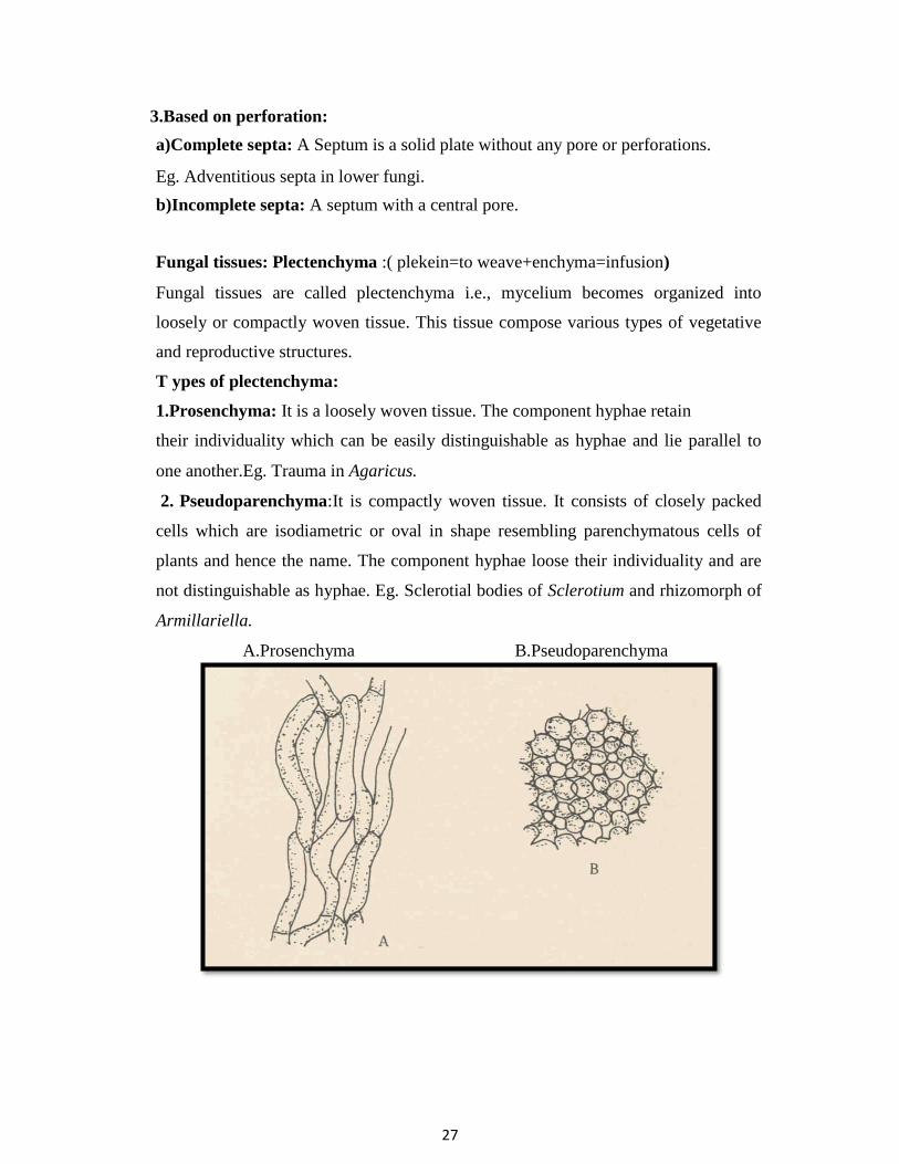
27
3.Based on perforation:
a)Complete septa: A Septum is a solid plate without any pore or perforations.
Eg. Adventitious septa in lower fungi.
b)Incomplete septa: A septum with a central pore.
Fungal tissues: Plectenchyma :( plekein=to weave+enchyma=infusion)
Fungal tissues are called plectenchyma i.e., mycelium becomes organized into
loosely or compactly woven tissue. This tissue compose various types of vegetative
and reproductive structures.
T ypes of plectenchyma:
1.Prosenchyma: It is a loosely woven tissue. The component hyphae retain
their individuality which can be easily distinguishable as hyphae and lie parallel to
one another.Eg. Trauma in Agaricus.
2. Pseudoparenchyma:It is compactly woven tissue. It consists of closely packed
cells which are isodiametric or oval in shape resembling parenchymatous cells of
plants and hence the name. The component hyphae loose their individuality and are
not distinguishable as hyphae. Eg. Sclerotial bodies of Sclerotium and rhizomorph of
Armillariella.
A.Prosenchyma B.Pseudoparenchyma

28
MODIFICATION OF MYCELIUM/ SPECIALISED SOMATIC
STRUCTURES
Purpose :
1. to obtain nourishment i. e., for nutrition .
2. to resist or tolerate unfavourable conditions for their survival i.e., over wintering,
over summering.
3. for reproduction.
1.Rhizomorphs: ( rhiza=root, morph=shape) Thick strands of somatic hyphae in
which the hyphae loose their individuality and form complex tissues that are resistant
to adverse conditions and remain dormant until favourable conditions return. The
structure of growing tip of rhizomorphs res emble that of a root tip, hence the name
rhizomorph. Eg. Armillariella mellea
2.Sclerotium: (skleron=hard) pl.sclerotia: It is a hard, round ( looks like mustard
seed)/ cylindrical or elongated (Claviceps ) dark coloured ( black or brown )resting
body formed due to aggregation of mycelium, the component hyphae loose their
individuality , resistant to unfavourable conditions and remain dormant for a longer
period of time and germinate on the return of favourable conditions.
Eg. Sclerotium, Rhizoctonia . 3.Stroma : (stroma=mattress) pl.stromata. It is a compact somatic structure looks
like a mattress or a cushion on which or in which fructifications ( spores or fruiting
bodies ) are usually formed.
a. Sub stomatal stroma: cushion like structure formed below epidermis in sub
stomatal region from which sporophores are produced. Eg. Cercospora
personata.
b. Perithecial stroma: When reproductive bodies like perithecia of some fungi are
embedded characteristically throughout periphery of stroma, such stroma are called
perithecial stroma. Eg.Claviceps, Xylaria.
4. Haustorium: ( hauster=drinker) pl.haustoria.It is a outgrowth of somatic hyphae
regarded as special absorbing organ produced on certain hyphae by parasitic

29
fungi for obtaining nourishment by piercing into living cells of host. They may be
knob like( Albugo), elongated ( Erysiphe, Uncinula), finger like ( Peronospora ).
5.Rhizoids: (rhiza=root, oeides=like) These are slender root like branched structures
found in the substratum produced by some fungi which are useful for anchoring the
thallus to substratum and for obtaining nourishment from the substrate.Eg. Rhizopus
stolonifer.
6. Appresorium: (apprimere=to press against) pl.appressoria A flattened tip of
hyphae or germ tube acting as pressing organ by attaching to the host surface
and gives rise to a minute infection peg which usually grows and penetrates the
epidermal cells of the host. Eg. Puccinia, Colletotrichum, Erysiphe.
Fungal cell :
Fungal cells are typically eukaryotic and lack chloroplasts.
Cell is bounded by cell wall, which provides rigidity and shape to the cell is the
outermost membrane of cell consisting of more than one layer with fibrous structure
and made up of chitin or cellulose or both.

30
The layer surrounding the cytoplasm is called cytoplasmic membrane or
plasmalemma. Protoplasm contains a true nucleus surrounded by two layered
membrane with nucleolus, cytoplasm and other inclusions.
Endoplasmic reticulum is not well developed, and it may be rough atudded with
ribosomes or smooth with out ribosomes.
Vacuoles in which metabolic products are accumulated are bounded by a
membrane called tonoplast .
Ribosomes are protenaceous bodies scattered all over cytoplasm, play a role in
protein synthesis.
Mitochondria are the sites of respiratory activities.
Lomasomes are the swollen membranous structures of plasmalemma.
Cytoplasm also contains fat particles, calcium oxalate crystals, resins, glycogen.
Fungal nutrition:
Fungi are heterotrophic with holophytic nutrition( absorptive type). The essential
elements for fungi are, C, H, O, N, P, K, S, Zn, Fe, Mg, Mn, Mo, Cu and Ca. Reserve
food material in the c ell may be either fat or carbohydrates. Fats may be present in
the form of oil drops and carbohydrates in the form of glycogen or sugars. Starch is
never present in the fungal cell.
Groups of fungi based on mode of nutrition:
1. Saprophytes: ( sapros=rotten, phytos=plant ) Organisms which obtain nutrition on
from dead organic matter either completely or for a part of their life. A large number
of fungi fall under this category.
Eg. Saprolegnia, Rhizopus, Mucor, Alternaria.
a. Obligate saprophytes: (obligare =to bind it self) Organisms which can
never grow on living organisms or can never obtain their food from living
source. They get their food only from dead organic matter.
Eg. Mucor, Agaricus .
. b. Facultative parasite: (facultas=ability) Organisms which are usually
saprophytic but have ability to become as parasites.

31
Eg. Pythium aphanidermatum, Fusarium solani, Rhizoctonia solani.
2.Parasites: Organisms which live within or out side another organisms for their
nutrition either completely or for a part of their life .
Pathogen : If a parasite damages the host then they are called as
pathogens..
All pathogens are not parasites and all parasites need not be pathogens .
a. Obligate parasites: (Organisms which obtain food only from living organisms (
living protoplasm) and can never derive their food from dead organic matter or
artificial medium. Eg. Puccinia graminis , Plasmopara viticola .
b. Facultative saprophytes: Organisms which are usually parasites but have ability
to become saprophytes .Eg. Ustilago maydis
Reproduction in fungi :
Reproduction is the formation of new individuals having all the characteristics of the
species.
T ypes of reproduction: 1. Asexual /non-sexual / vegetative / somatic
reproduction 2. Sexual reproduction.
1.Asexual reproduction :
Asexual reproduction stage is also known as imperfect stage and technically called as
anamorphic stage. There is no union of nuclei /sex cells/ sex organs. It is repeated
several times during the life span of a fungus producing numerous asexual spores.
Hence, it is more important for fungi than sexual reproduction.
Asexual spores are formed after mitosis, hence also called mito spores.

32
Methods of asexual reproduction :
1. Fragmentation 2. Fission. 3. Budding 4. Sporulation ( production of spores)
1. Fragmentation : It is the most common method. Hypha of fungus breaks into
small pieces, each broken piece is called a fragment, which function as a
propagating unit and grows into a new mycelium.The spores produced by
fragmentation are called arthrospores (arthron=joint) (spora=seed) or oidia..
Eg. Oidium, Geotrichum. Sometimes, the contents of intercalary cells or
terminal cells of hypha rounded off and surrounded by thick wall and
formed as chlamydospores which are thick walled resistant spores produced
either singly or in chains. Eg. Fusarium oxysporum, Ustilago tritici.
2. Fission / Transverse fission: The parent cell elongates,nucleus under go mitotic
division and forms two nuclei, then the contents divide into equal halves by the
formation of a transverse septum and separates into two daughter
cells.Eg.Saccharomyces cerevisiae.

33
3.Budding: The spores formed through budding are called blastospores. The parent
cell puts out initially a small out growth called bud / blastos ie.,sprout or out growth
which increases in size and nucleus divides, one daughter nucleus accompanied by a
portion of cytoplasm migrates into bud and the other nucleus remains in the parent
cell. Later, the bud increases in size and a constriction is formed at the base of bud,
cutting off completely from parent cell . Bud, when separated from parent cell, can
function as an independent propagating unit.Sometimes multiple buds are also seen
i.e., bud over bud and looks like pseudomycelium. Eg. Saccharomyces cerevisiae.
4. Sporulation ( spores): The process of production of spores is called
sporulation.
Spore: It is a minute, simple propagating unit of the fungi, functioning as
a seed but differs from it in lacking a preformed embryo that serves in the
reproduction of same species.
Spores vary in colour,size,number of cells and the way in which they are
borne.

34
There are 2 main types of spores.
1. Sporangiospores 2. Conidia
1.Sporangiospores:When the asexual spores are produced internally, within
the sporangia,such spores are called sporangiospores.The sac like structure
which produces sporangiospores is called sporangium. The special hypha
bearing sporangium is called sporangiophore which may or may not be
distinguishable from hypha. A small sporangium with or without columella
containing a few or single spore is called as sporangiolum.
Eg. Choanephora trispora .
Sporangium which is cylindrical in shape is called as merosporangium.
Eg. Syncephalastrum racemosum.
Sporangium with columella is called as columellate sporangium.
Eg.Rhizopus stolonifer
Sporangiospores are of 2 types.
a. Zoospores /planospores b.Aplanospores
a.Zoospores / planospores: sporangiospores which are motile by flagella are called
zoospores.Also known as planospores.Eg. Pythium, Phytophthora. b.Aplanospores:
Flagellation in fungi:
Flagella : (sing.flagellum)Flagella are thin, hair like delicate structures attached to a
basal granule called blepharoplast in cytoplasm and these are the organs of motility in
lower fungi and aquatic fungi.
T ypes of flagella :Flagella of zoospores are of 2 types.
a.Whiplash b.Tinsel
a)Whiplash : A flagellum with long, thick, rigid basal portion and with a short,
narrow, flexible, upper portion .It gives a whip like appearance to flagellum.

35
b)Tinsel : It is a feathery structure consisting of a long rachis with lateral hair like
projections called mastigonemes or flimmers on all sides along its entire length.
The number, position and nature of flagella play an important role in the
classification of lower fungi.
Uniflagellate zoospore : A zoospore with a single flagellum, may be placed at
anterior or posterior end of spore.
Biflagellate zoospore:A zoospore with two flagella, situated laterally or anteriorly
on zoospore.
One whiplash,one tinsel type flagella and equal in size.Eg. Pythium
aphanidermatum, Phytophthora infestans
Both whiplash flagella,unequal in size (heterokont).Eg .Plasmodiophora brassicae.
2. Conidia / Conidiospores: ( konis=dust; oides=like ) Conidia are non- motile
asexual spores which may arise directly from somatic hyphae or from specialized
conidiogenous cells ( a cell from which conidia are produced) or on conidiophore (
hypha which bear conidia).Conidia are produced freely on conidiophore ie.,at the tips
or sides of conidiophore or may be produced in specialized asexual fruiting bodies
viz., pycnidium, acervulus, sporodochium and synnemata.

36
Asexual fruiting bodies :
(a) Pycnidium: ( pl.pycnidia ) It is a globose or flask shaped fruiting body lined
inside with conidiophores which produce conidia. It may be completely closed or
may have an opening called ostiole.
Pycnidium may be provided with small papillum or long neck. Eg.Phomopsis.
(b) Acervulus: ( pl.acervuli ) A flat or saucer shaped fruiting body with a stromatic
mat of hyphae producing conidia on short conidiophores.
An acervulus lacks a definite wall structure and not having an ostiole or definite line
of dehiscence. Eg.Colletotrichum,Pestalotiopsis.
(c) Sporodochium : (pl.sporodochia) A cushion shaped asexual fruiting
body. Conidiophores arise from a central stroma and they are woven together on a
mass of hyphae and produce conidia.Eg.Fusarium.
(d) Synnemata: ( pl.synnema ) A group of conidiophores often united at the base
and free at the top. Conidia may be formed at its tip or along the length of synnema,
resembling a long handled feather duster.Eg.Graphium.
Pycnidium Acervulus Sporodochium Synnemata

37
SEXUAL REPRODUCTION
Sexual reproduction involves union of two compatible nuclei or sex cells or sex
organs or somatic cells or somatic hyphae for the formation of new individuls. Sexual
stage is perfect stage and technically called as teleomorphic stage. Sexual cycle
normally occurs once in the life span of the fungus. Sexual spores or sexual structures
which contain sexual spores are thick walled, resistant to unfavourable conditions and
are viable for longer period and thus these spores help the fungus to perpetuate from
one season to another , hence these are called as resting spores.Sexual spores are
definite in number.
Sex organs of fungi :
Gametangia: Sex organs of fungi are called gametangia containing gametes
or gamete nuclei.
Gametes: Sex cells are called as gametes .
Antheridium: ( pl.antheridia ) Male gametangium is called as antheridium.
Male gametangium is small and club shaped.
Oogonium / Ascogonium:(pl.oogonia/ascogonia ):The female gametangium is
called Oogonium (oomycetes) or ascogonium (ascomycotina).Female gametangium
is large and globose shaped.
Male gametes are called antherozoids or sperm or spermatozoids .
Female gametes are called egg or oosphere.
Planogametes: If gametes are motile, they are called planogametes.
Isogametangia: If gametangia are morphologically similar or identical
i.e.,indistinguishable as male and female , they are called as isogametangia.

38
Isogametes: If gametes are similar morphologically, they are called as isogametes.
Heterogametangia: If gametangia differ morphologically in size and structure, they
are called as heterogametangia.
Heterogametes: If gametes differ morphologically, they are called heterogametes.
+ or – signs: In some sexually undifferentiated fungi, male and female are
symbolically designated as ‘ + ’ ( male) and ‘ – ’ ( female ).
Classification of fungi based on sex :
1. Monoecious fungi / hermaphroditic fungi: ( mono=single,oikos=home)
The fungi which produce distinguishable male and female sex organs on the
same thallus, which may or may not be compatible are called monoecious/
hermaphroditic fungi. Eg. Pythium aphanidermatum.
2. Dioecious fungi: (di=two, oikos=home) The fungi which produce
distinguishable male and female sex organs on two different thalli ie., there
will be separate male and female thalli . Eg. Phytophthora infestans.
Classification of fungi based on compatibility ;
Homothallic fungi: Fungi in which both sexes occur on same thallus, which
can reproduce sexually by it self with out the aid of another thallus ie., self
compatible / self fertile are called homothallic fungi. Eg. Pythium
aphanidermatum.
Heterothallic fungi: A fungal species consisting of self sterile
(self incompatible) thallus requiring the union of two compatible thalli for
sexual reproduction, regardless of the possible presence of both male and
female organs on the same thallus. Heterothallic fungi are dioecious Eg.
Phytophthora infestans..
Phases in Sexual reproduction:.There are 3 phases in sexual reproduction.
1. Plasmogamy: union of two protoplasts takes place.As a result of it the
two nuclei come together within the same cell.
2. Karyogamy: union of 2 sexually compatible nuclei brought together by
plasmogamy to form a diploid nucleus (2n) i.e., zygote.

39
3. Meiosis: This is reduction division . The number of chromosomes is reduced to
haploid ( n ) i.e., diploid nucleus results into haploid nucleus..
In lower fungi (Phycomycetes -Mastigomycotina and Zygomycotina)
plasmogamy, karyogamy and meiosis occurs at regular intervals / sequence
i.e.,karyogamy follows immediately after plasmogamy. In higher fungi
(Ascomycotina, Basidiomycotina), karyogamy is delayed, as a result the nuclei
remain in pairs ( dikaryotic phase- n+ n condition), which may be brief or
prolonged.
Dikaryon : A pair of genetically different nuclei, lying side by side with out fusion
for a considerable period of time is called dikaryon.A cell containing dikaryon is
called dikaryotic cell. And the process is known as dikaryogamy.
Methods of sexual reproduction : 5 methods.
1. Planogametic copulation. 2. Gametangial contact 3. Gametangial copulation
4. Spermatisation 5. Somatogamy .
1.Planogametic copulation ( gametogamy ): This involves the union of 2 naked
gametes one or both of which are motile.
a.Isogamy (Isogamous planogametic copulation) : If both gametes are
motile and similar.Eg. Plasmodiophora brassicae.
b.Anisogamy (Anisogamous planogametic copula tion) : If both gametes
are motile but dissimilar.E g. Allomyces macrogynus
c.Heterogamy: If gametes are dissimilar, one motile, another is non
motile. Eg. Monoblepharis polymorpha.
2.Gametangial contact ( gametangy / oogamy ): Male and female gametangia
come in contact . At the place of contact, dissolution of wall occurs and a fertilization
tube is formed. The conte nts of male gametangium migrate into female
gametangium through a pore or fertilization tube developed at the point of contact.
The gametangia do not loose their identity. E g. Pythium aphanidermatum.

40
3.Gametangial copulation ( gametangiogamy ): The isogametangia come ni
contact, their intervening wall dissolves leading to fusion of entire contents of two
contacting gametangia to form a single unit.Gametangia loose their identity.The
protoplasts fuse and the unit increases in size. Eg. Rhizopus stolonifer.
4.Spermatiz ation: Minute, uninucleate male cells called as spermatia which are
produced on spermatiophores in a fruiting body ( pycnium) are carried to female
reproductive structures called receptive hyphae. Spermatia and receptive hyphae
come in contact and contents of male spermatium migrate into female receptive
hypha, thus making the cell binucleate. This process is called dikaryotization. Eg.
Puccinia graminis tritici.

41
5.Somatogamy: Many higher fungi do not produce sex organs.In such cases
somatogamy takes place.It is the union of 2 somatic hyphae or somatic cells
representing opposite sexes to form sexual spores .Eg. Agaricus campestris
Parasexual cycle / Parasexuality :Parasexual cycle is a process in which
plasmogamy, karyogamy and haploidisation (non meiotic process) takes place in a
sequence but not at specified points in the life cycle of a fungus. It was first
discovered in 1952 by Pontecorvo and Roper in Aspergillus nidulans, the imperfect
stage of Emericella nidulans.It is of importance in heterokaryotic fungi( a fungi in
which genetically different nuclei are associated in the same protoplast or
mycelium) .This is one of the methods of producing variability of fungal pathogens.
In majority of Deuteromycotina,true sexual cycle is absent but derive many of the
benefits of sexuality through this cycle.
Different types of sexual spores :Sexual spores are formed after
meiosis,hence also called meiospores.
1.Oospores 2.Zygospores 3.Ascospores 4.Basidiospores
1.Oospore: A thick walled sexual resting spore produced by the union of two
morphologically different gametangia.
Eg.Pythium, Phytophthora , members of class Oomycetes.
2. Zygospore: A thick walled sexual resting spore produced by the fusion of two
morphologically similar gametangia.
Eg.Rhizopus , members of sub-division Zygomycotina
3. Ascospore:Sexual spore produced in a specialized sac like like structure known
as ascus. Generally 8 ascospores are formed.
Eg.Erysiphe,members of sub-division Ascomycotina.
4.Basidiospore:Sexual spore produced on a club shaped structure known as
basidium.Generally 4 basidiospores are formed.
Eg.Puccinia, members of sub-division Basidiomycotina.
Various Life cycle patterns displayed by fungi:
1. Haplobiontic life cycle 2. Diplobiontic life cycle
1. Haplobiontic life cycle: If there is only one free living thallus, which is haploid
or diploid in life cycle of a fungus, it is called as haplobiontic life cycle.

42
( long haploid somatic phase and short diploid phase confined to zygote cell, which
undergoes meiosis immediately after karyogamy and develop ascospores ) Eg.
Schizosaccharomyces octosporus.
2. Diplobiontic life cycle: If haploid thallus alternates with a diploid thallus, the life
cycle is called diplobiontic life cycle, which has a long diploid somatic phase and a
very short haplo Id phase. Eg. Saccharomyces ludwigii.

43
Nomenclature, Binomial system of nomenclature, rules of
nomenclature
Taxonomy : The science of classification. It is concerned with pricnciples of
classification.
Classification: Grouping of organisms into classes,orders,families,genera,
species etc.
Nomenclature : Art of naming living organisms.
Importance of taxonomy and nomenclature;
1. for study of fungi
2. for scientific communication between mycologists and plant pathologists
throughout the world.
Binomial system of nomenclature was originally introduced by Carl Linnaeus for
higher plants. Later, this classification was adopted to fungi by his students
C.H.Persoon amd E.M. Fries.
Some important rules of nomenclature :
1.According to International code of Botanical Nomenclature, the names of
organisms should be binomial i.e., 2 parts.The first part is noun designating genus
and the first letter of the genus name should be in capital. The second name is
often an adjective, describing the noun which denotes the species, and the first
letter should be in small letter. Eg. Puccinia graminis.
2.Binomials are usually derived from Greek or Latin.
3.Binomials when hand written should be underlined and when printed italicised.
Eg.. Puccinia graminis ( hand written)
Puccinia graminis ( printed)
4. Citation of authors name: The full name or abbrevation name of scientist who
described fungus first, follows the species name.Eg. Puccinia graminis Persoon or
Pers.
5. Citation of two authors names: If name of species is transferred to another
genus from original ( Botrytis infestans), the name of first author who first
described species must be kept in parenthesis followed by name of second author

44
who gave present status of species. i.e., Phytophthora infestans. Eg.
Phytophthora infestans ( Mont.) de Bary.
6. The taxa ( groups) used in classification are Kingdom, Division, Class, Order,
Family, Genus and Species.Each category may be sub divided into sub groups
like Sub- Division,Sub- Class, Sub- Order.
7. Species is the unit of classification or basic taxonomic category ( taxon).
8. Species some times broken into variety / formae speciales ( f.sp.) and varieties
into races and races into biotypes.
Standard endings of TAXA:
Division ends with mycota
Sub- Division ends with mycotina
Class with mycetes
Sub- class with mycetidae
Order with ales
Family with aceae
No special ending for genus and species.
TAXA:
Kingdom
Division
Sub-division
Class
Sub-class
Order
Sub-order
Family
Genera
Species
Eg. Puccinia graminis triticirace 1
Kingdom :Fungi
Division : Eumycota
Sub-division : Basidiomycotina
Class : Teliomycetes
Order : Uredinales
Family : Pucciniaceae
Genus : Puccinia
Species : graminis
Variety : tritici
Race : 1

45
Classification of fungi. Key to divisions, sub-divisions, orders and
classes.
CLASSIFICATION OF FUNGI
(CLASSIFICATION BY AINSWORTH, 1973)
KINGDOM: MYCOTA
DIVISIONS
MYXOMYCOTA
EUMYCOT
A
CLASS
PLASMODIOPHOROMYCETES
ORDER : PLASMODIOPHORALES
FAMILY:PLASMODIOPHORACEAE
Eg.Plasmodiophora
SUB DIVISIONS
1. MASTIGOMYCOTINA
2. ZYGOMYCOTINA
3. ASCOMYCOTINA
4. BASIDIOMYCOTINA
5.DEUTEROMYCOTINA

46
S.D : 1. MASTIGOMYCOTINA
CLASSES
1.CHYTRIDIOMYCETES
ORDER: CHYTRIDIALES
FAMILY: SYNCHYTRIACEAE
Eg. Synchytrium
2.OOMYCETES
ORDER: PERONOSPORALES
FAMILY: PYTHIACEAE
Eg: Pythium, P hytophthora
FAMILY: ALBUGINACEAE
Eg. Albugo
FAMILY: PERONOSPORACEAE
Eg : Sclerospora . Peronospora,Peronosclerospora
Plasmopara , Pseudoperonospora, Bremia
S.D :2. ZYGOMYCOTINA
CLASS :ZYGOMYCETES
ORDER: MUCORALES
FAMILY: MUCORACEAE
Eg. Rhizopus, Mucor
S.D 3. ASCOMYCOTINA
CLASSES
1 .HEMIASCOMYCETES
ORDER:
PROTOMYCETALES
FAMILY: PROTOMYCETACEAE
Eg. Eurotium, Talaromyces
2 .PLECTOMYCETES
ORDER:
EUROTIALES
FAMILY: EUROTIACEAE
47
ORDER : ERYSIPHALES
FAMILY:ERYSIPHACEAE
Eg. Erysiphe, Leveillula, Phyllactinia, Uncinula,
Sphaerotheca,Podosphaera, Microsphaera
3.PYRENOMYCETES
ORDER: HYPOCREALES
FAMILY: CLAVICIPITACEAE
Eg. Claviceps
4.DISCOMYCETES
ORDER: TUBERALES
FAMILY: TUBERACEAE
Eg. Tuber
ORDER: PEZIZALES
FAMILY: MORCHELLACEAE
Eg. Morchella
5.LOCULOASCOMYCETES
ORDER: PLEOSPORALES
FAMILY: VENTURIACEAE
Eg. Venturia
FAMILY: PLEOSPORACEAE
Eg. Cochliobolus
ORDER: MYRIANGIALES
FAMILY: MYRIANGIACEAE
Eg. Elsinoe
ORDER: DOTHIDIALES

48
FAMILY: DOTHIDIACEAE
Eg. Mycosphaerella
S.D 4. BASIDIOMYCOTINA
CLASSES
1.TELIOMYCETES
ORDER: UREDINALES
FAMILY: PUCCINIACEAE
Eg.Puccinia, Uromyces, Hemileia
FAMILY: MELAMPSORACEAE
Eg. Melampsora
ORDER: USTILAGINALES
FAMILY: USTILAGINACEAE
Eg. Ustilago, Sphaecelotheca,Tolyposporium
FAMILY: TILLETIACEAE
Eg. Tilletia, Neovossia, Urocystis
FAMILY: GRAPHIOLACEAE
Eg. Graphiola
2.HYMENOMYCETES
SUB CLASS: HOLOBASIDIOMYCETIDAE
ORDER : AGARICALES
FAMILY: AGARICACEAE
Eg. Agaricus, Volvariella, Pleurotus
ORDER: APHYLLOPHORALES
FAMILY: POLYPORACEAE
Eg. Polyporus, Fomes, Peria
FAMILY: GANODERMATACEAE
49
Eg. Ganoderma
S.D 5 DEUTEROMYCOTINA
CLASSES
1.COELOMYCETES
ORDER: SPHAEROPSIDALES
FAMILY: SPHAEROPSIDACEAE
Eg. Phoma, Phomopsis, M acrophomina,
Phyllosticta, Diplodia,Botryodiplodia
FAMILY:EXCIPULACEAE
Eg. Ephelis
FAMILY: NECTRIOIDACEAE
Eg. Zythia
FAMILY: LEPTOSTROMATACEAE
Eg. Leptostroma
ORDER: MELANCONIALES
FAMILY: MELANCONIACEAE
Eg. Colletotrichum, Gloeosporium,
Pestalotiopsis, Pestalotia
2.HYPHOMYCETES
ORDER: HYPHOMYCETALES / MONILIALES
FAMILY: MONILIACEAE
Eg. Pyricularia, BotrytIs, Verticillium,
FAMILY: DEMATIACEAE
Eg. Alternaria ,Bipolaris, Cercospora, Phaeosariopsis
ORDER: STILBELLALES

50
FAMILY: STILBELLACEAE
Eg. Graphium
ORDER : TUBERCULARIALES
FAMILY: TUBERCULARIACEAE
Eg. Fusarium, Myrothecium
ORDER : AGONOMYCETALES
FAMILY: AGONOMYCETACEAE
Eg. Sclerotium,Rhizoctoni
51
IMPORTANT CHARACTERISTICS OF DIVISIONS AND SUB-DIVISIONS
DIVISIONS :
1. MYXOMYCOTA: Plasmodial forms with out cell wall. Plasmodium is a naked
multinucleate mass of protoplasm which moves and feeds in an amoeboid
direction.Also calledcas slime molds.
EUMYCOTA: True fungi. Thallus is typically filamentous with cell wall. Plasmodium absent
SUB DIVISIONS OF EUMYCOTA :
1. MASTIGOMYCOTINA :
Thallus is unicellular or aseptate mycelium.Asexual spores are zoospores (motile
spores).Sexual spores are oospores. Sexual reproduction by gametangial contact.
2. ZYGOMYCOTINA :
Thallus is aseptate mycelium. Motile spores are absent. Asexual spores are
sporangiospores (aplanospores).Sexual spores are zygospores.Sexual reproduction
through gametangial copulation.
3. ASCOMYCOTINA :
Thallus is septate mycelium. Rarely unicellular. Motile spores are absent. Asexual
spores are conidia.Sexual spores are ascospores produced endogenously in an
ascus.Sexual reproduction mainly by gametangial contact .
4. BASIDIOMYCOTINA:
Thallus is septate mycelium.Motile spores are absent.Clamp connections and
dolipore septum are present.Sexual spores are basidiospores produced exogenously
on basidium.Sexual reproduction is by spermatization and somatogamy.
5. DEUTEROMYCOTINA:
Thallus: septate mycelium . Motile spores are absent. Sexu al spores are absent. Asexual
spores called conidia are present.

52
DIVISION - MYXOMYCOTA
CLASS :PLASMODIOPHOROMYCETES,ORDER:PLASMODIOPHORALES
FAMILY:PLASMODIOPHORACEAE
1. These are obligate endoparasites.Commonly called as endoparasitic slime molds .
Thallus is a plasmodium,
2. There are 2 types of plasmodia.
a. Sporangiogenous plasmodium - formed asexually containing many thin
walled zoosporangia and each zoosporangium produce a single or many secondary
zoospores or sporangial zoospores.
b. Cystogenous plasmodium - formed sexually consisting of thick walled cysts
and each cyst gives rise to a single primary zoospore / cyst zoospore. Cysts may be
free or united.Cysts act as resting spores.
Zoospores are anteriorly biflagellte, whiplash type,unequal in size which are called
as Heterokont zoospores. After swimming for some time, the zoospore encysts on
the root hair of the host. A cylindrical sharp pointed body, called satchel, is formed
in a specialized pouch or sheath called Rohr.
4.Nuclear division is by cruciform division.
5. Sexual reproduction is by isogamous planogametic copulation.
6. Members cause abnormal enlargement and multiplication of host cells i.e.,
hypertrophy and hyperplasia. Eg: Plasmodiophora ,Spongospora
Differences between Plasmodiophora and Spongospora :
Plasmodiophora : Resting spores / cysts lie freely with in the host cell, but not in cystosorus.
Spongospora : Resting spores form balls and appear like sponge.
Diseases :Plasmodiophora brassicae : club root / finger & toe disease of
cabbage / crucifers.
Spongospora subterranea : powdery scab of potato

53
DIVISION :EUMYCOTA
SUB - DIVISION: MASTIGOMYCOTINA
IMPORTANT CHARACTERISTICS OF CLASS CHYTRIDIOMYCETES:
1. Thallus (a) primitive members - unicellular, advanced members with coenocytic
mycelium.
(b) endobiotic (fungus which lives with in the cells of host) or epibiotic
(reproductive organs of the fungus on surface of the host, part or entire thallus with in
the host cell).
(c) holocarpic or eucarpic.
2 .Zoospores are posteriorly uniflagellate whiplash type. Inside the zoospore,
around the nucleus cell ribosomes cluster together to form a nuclear cap.
3. Asexual reproduction is by zoospores produced in zoosporangia.
4.Sexual reproduction is by (a) planogametic copulation ( isogamy, anisogamy,
heterogamy) . (b)gametangial copulation.
5. Zygote is converted into resting sporangium / resting spore. Zoospores produced
from this resting spore infect host cell and produces prosorus
which is thick with golden-brown chitinous wall. Prosorus eventually gives rise to
sorus. Eg.Synchytrium.
Important characteristics of Order: Chytridiales 1.Thallus is epibiotic or endobiotic, monocentric or polycentric, vegetative parts are
rhizoidal .
2.Zoosporangium is operculate or inoperculate (operculum present or absent).
3.Zoospore germination is unipolar. 4.Resting spore on germination functions as a sporangium or prosporangium
Important characteristics of Family: Synchytriaceae : Includes only single genus - Synchytrium, species - endobioticum.
Thallus is unicellular, endobiotic and holocarpic . Warts contain resting sporangia . Thallus behaves as a prosorus.
Disease: Causes black wart of potato. It is prominent in hilly regions like
Darjeeling. Epidermal cells of tubers are infected by the fungus. Hypertrophy and hyperplasia takes place, as a result, out growths appear on tubers.
Diseases transmitted : Synchytrium endobioticum transmits potato virus - x .

54
IMPORTANT CHARACTERISTICS OF CLASS OOMYCETES
1. Members may be aquatic or terrestrial ,saprophytes or obligate parasites.
2. Thallus - mostly eucarpic ,coenocytic
3. Cell wall consists of cellulose. Chitin is absent.
4. Asexual reproduction is by zoospores produced in zoosporangia.
Zoospores are biflagelte (whiplash and tinsel), anteriorly or laterally
positioned, equal in size.
5. Sexual reproduction is oogamous type ie.,gametangial contact/ gametangy..
Heterogametangia come in contact, contents of antheridium (male
gametangium) passes into oogonium( female gametangium ) containing
oosphere (egg) through fertilization tube.
6. Zygote resulting from sexual reproduction is called oospore.
7. Oospore is the sexual resting spore which is the characteristic of
oomycetes.
8. This zygote/ oospore is diploid.Oospore which gives rise to mycelium/
gametangia is also diploid.
9. Meiosis o ccurs in gametangia instead of zygote.
IMPORTANT CHARACTERISTICS ORDER PERONOSPORALES:
1.Many species are destructive pathogens causing very serious diseases in some
important crop plants.The diseases caused by these members include white
rusts, downy mildews, damping off, leaf blights and seedling blights.
2.Members are mostly terrestrial.
3.Mycelium is coenocytic, produce inter and intra cellular hyphae.If inter –
cellular produce haustoria.
4.Sporangia are produced on well developed, distinct sporangiophores and
sporangia are deciduous ( fall off at maturity).

55
5.Sporangiophores may be indeterminate / indefinite type (sporangiophores
continue to grow indefinitely producing sporangia at the tip as they
grow.ie.,sporangia of different ages are seen on sporangiophores) or
determinate/definite type (sporangia are not produced until sporangiophores
complete their development and maturity and all the sporangia are produced at
one time.ie.,single crop of sporangia are produced.)
6.Zoospores are monomorphic (producing morphologically one type of
zoospores i.e., reniform zoospores) and monoplanetic ( only one swarming
period.).
7.Zoospores are reniform ie., kidney shaped and biflagellate.
Some species exhibit highly specialized parasitism i.e., obligate parasites.
8.Oogonium produces a single oosphere / egg surrounded by conspicuous
periplasm except in Family: Pythiaceae in which periplasm is
inconspicuous. Periplasm serves as a source of nutrients to oosphere.
ORDER PERONOSPORALES :
Families: 1.Pythiaceae 2.Albuginaceae 3.Peronosporaceae
These three families are distinguished based on characteristics of sporangiophores
and sporangia.
IMPORTANT CHARACTERISTICS OF FAMILY
1. PYTHIACEAE :
1.Species may be saprophytes or facultative parasites.Commonly called
water molds.
2.May be inter or intra cellular mycelium.If inter cellular,produce haustoria,
if intra cellular, no haustoria are produced.
3.Sporangiophores generally not distinguished from somatic hyphae unless
sporangia are present.
4.Sporangiophores are indefinite or indeterminate type .
5.In oogonium ,Periplasm is inconspicuous (not visible).
Eg. Pythium and Phytophthora

56
Genus PYTHIUM :Fungus is facultative parasite and lives in soil on dead organic matter
or parasitically on young seedlings of crop plants .Mycelium well
developed,branched,coenocytic,hyaline,intracellular mycelium without haustoria. Thallus
is homothallic.Asexual reproduction is through zoospores produced in zoosporangia .
Sporangia are large globose( P. debaryanum ) or irregularly lobed (P.aphanidermatum)
produced terminally or intercalary on somatic hyphae.Zoospores are produced in a
vesicle which emerge out of sporangium.ie.,zoospore differentiation takes place in
vesicle.Sexual reproduction is by gametangial contact.Paragynous
antheridium(Antheridium is by the side of oogonium).Oospores are smooth,thick
walled,round,light brown and aplerotic (oospore wall do not fuse with oogonial wall) or
plerotic (oospore wall fuses with oogonial wall). Diseases:Damping off of vegetable seedlings of solanaceous crops caused by
Pythium debaryanum or P.aphaniderma

57
Genus PHYTOPHTHORA : (Phyto=plant,Phthora=destructor)
Fungus lives in soil on dead organic matter or parasitically on potato tubers.
Mycelium is well developed,branched,coenocytic,hyaline,intercellular mycelium
with haustoria.Thallus is heterothallic.
Asexual reproduction is by zoospores produced in sporangia.Sporangia are lemon or
pear shaped,thin walled,papillate,formed terminally on sympodially branched
sporangiophore (sympodium is with a more or less zig zag growth and characteristic
swellings at nodes).Sporangiophores are distinct ie.,easily distinguished from somatic
hyphae.Vesicle is not formed, zoospores are differentiated in zoosporangium it self.
Sexual reproduction is by gametangial contact.Antheridium is amphigynous
(Oogonium penetrates the antheridium) or paragynous.Oospores are smooth,thick
walled,round ,dark brown and plerotic.
Also produces resting spores called chlamydospores.
Diseases: Phytophthora infestans - Late blight of Potato
P. parasitica var. nicotianae - black shank and leaf blight of tobacco

58
Differences between Pythium and Phytophthora
Pythium Phytophthora
1.Myceliumisbothinterand Only intercellular. Haustoria are
intracellular.Whenintracellular, no produced.
haustoria are produced.
2. Production of sporangia on somatic Sporangiophores can be distinguished
hyphae. Sporangiophores are by sympodial branching and nodal
indistinct from hyphae. swellings.
3.Sporangia are globose or elongated Sporangia are lemon or pear or oval
or lobed. shaped.
They are produced intercalary or Produced terminally.
Terminally.
No papillum. Papillum is present.
4.Sporangia germinate by forming No vesicle is seen.
vesicle.
Differentiation of zoospores takes Zoospores differentiate in the
Place in the vesicle. sporangium .
5. Antheridium is of paragynous type. Amphigynous type.
6. Homothallic Heterothallic.
7.Asexual reproduction is by Zoospores in sporangia and
Zoospores in sporangia. chlamydospores.
8.Oospores are plerotic / aplerotic. Oospores are aplerotic
9.Appresorium not formed Formed
10.Oospore hyaline,smooth Brown,warty

59
FAMILY ALBUGINACEAE - IMPORTANT CHARACTERISTICS
1.Members are obligate parasites.
2.Mycelium is intercellular producing knob shaped haustoria. 3.Sporangiophores are specialized which are short, unbranched and club
shaped.They are of indeterminate type.Sporangiophores are borne in close proximity to one another in compact layers or beds under the epidermis of the host.
4.Each sporangiophore gives rise to several aporangia which are produced in succession,one below the other,so that a chain of sporangia is formed with the oldest at the tip and youngest at the base ( basipetal manner ).
5.Sporangia are globose.Successive sporangia are connected by isthmus or
disjunctor cell or separation disc. 6.Periplasm is conspicuous. 7.Single genus under the family i.e., Albugo. The diseases caused by this genus are called white rusts. The term rust is restricted normally for the fungi belonging to the order Uredinales of class Teliomycetes of sub division Basidiomycotina and the diseases they cause. Since these white pustules resemble the pustules caused by true rusts in order Uredinales, the term white rust was coined to the group of diseases caused by Albugo sp. The white rusts also cause floral malformation and tumors on stems, leaves, petioles etc. due to hypertrophy of infected tissue.
Genus ALBUGO:
Fungus is an obligate parasite on crucifers and lives in soil in the form of oospores or
parasitically on plants.
Mycelium iscoenocytic,hyaline,endophytic,intercellular with knob shaped
haustotia.
Asexual reproduction is by zoospores produced in sporangia.Sporangiophores are
unbranched,hyaline clavate,bears sporangia in chains in basipetal
succession.Sporangia are spherical,thin walled,sessile, hyaline and germinate by
zoospores.
Sexual reproduction is by gametangial contact.Oospores are round ,thick
walled,dark brown and outer wall warty.
Diseases:White rust on mustard caused by Albugo candida

60
IMPORTANT CHARACTERISTICS OF FAMILY:3.PERONOSPORACEAE
1. All the members are obligate parasites of plants causing diseases called
downy mildews.
2. Mycelium is coenocytic and intercellular with haustoria
3.Sporangiophores are well developed, specialized , characteristically branched and
determinate type.Sporangiophores attain maturity and later produce sporangia at one
time. ie.,single crop of sporangia are produced.
4.Sporangia deciduous, may be papillate ( also called operculum) or may not be
papillate. In most genera, sporangia germinate by zoospore. However, in some
species they germinate by germ tube and function as conidia depending on
environmental conditions.
5.Oospores may be plerotic or aplerotic.
6.Periplasm conspicuous.
The name downy mildews( downy= feathery or soft + mildew= superficial growth) is
given because of soft feathery growth observed on the lower side of affected foliage
consisting of sporangiophores of these fungi.
The members are further divided into different genera and distinguished based
on two characteristics viz., 1. morphology of sporangiophore
( branching pattern) 2. method of germination of sporangia.
Eg. Peronospora, Pseudoperonospora, Peronosclerospora, Sclerospora,
Plasmopara, Bremia, Sclerophthora.

61
DISTINGUISHING CHARACTERISTICS OF DOWNY MILDEW GENERA:
1.SCLESPORA:Sporangiophores are stout ,having upright branches,
bearing sporangia on sterigmata.Sporangia are h yaline ,ovoid,smooth
walled,papillate and germinate by zoospores.Oospore is plerotic.
Eg.Sclerospora graminicola –downy mildew of bajra.
2.PERONOSPORA:Sporangiophores are dichotomously branched 2-7 times at
acute angles and tips of branches are curved and pointed bearing sporangia on
sterigmata.Sporangia are hyaline ,ovoid ,non-papillate and always germinate by
germ tube. i.e.,sporangia behave like conidia. Eg;Peronospora destructor-downy
mildew of onion.
3.PERONOSCLEROSPORA :Fungus possess characteristics of both
Peronospora and Sclerospora.Sporangiophores are erect,short,stout,widening
towards upper portion,dichotomously branched 2 -5 times at apex bearing
sporangia on sterigmata.Sporangia are hyaline,elliptical or ovoid,thin walled, non-
papillate and germinate by germ tube like Peronospora. Oospore is plerotic type
like Sclerospora. Eg.Peronosclerospora sorghi-downy mildew of jowar
P.philippinensis-downy mildew of maize
62
PSEUDOPERONOSPORA:Sporangiophores are branched at acute angles with
curved,blunt tips,bearing sporangia on sterigmata.Sterigmata are unequal ( 1 big and
1 small ).Sporangia are greyish,ovoid,papillate and germinate by zoospores. Eg.Pseudoperonospora cubensis -downy mildew of cucurbits.
5.PLASMOPARA;Sporangiophores are branched at right angles to the main axis at
regular intervals.Monopodial branching is observed.Subsequent branches are 3-6
which end in blunt sterigmata of 3 in number.Sporangia are ovoid and germinate by
zoospores. Eg.Plasmopara viticola-downy mildew of grapes.
6.BREMIA :Sporangiophores are dichotomously branched ,tips of branches are expanded to cup shaped apophysis with four sterigmata bearing sporangia.Sporangia are ovoid,papillate and germinate by zoospores. Eg:Bremia lactucae-downy mildew of lettuce.
SUB-DIVISION: ZYGOMYCOTINA
CLASS: ZYGOMYCETES
IMPORTANT CHARACTERISTICS OF CLASS: ZYGOMYCETES, ORDER:MUCORALES:
1.Absence of motile zoospores (planospores) and production of non- motile
sporangiospores (aplanospores).
2.Production of thick walled resting spore-zygospore
3.Well developed , coenocytic mycelium and cell wall with chitin
4.Asexual reproduction is by sporangiospores though some species produce
Chlamydospores.
5.Sexual reproduction is by gametangial copulation of isogametangia or
heterogametangia.
FAMILY:MUCORACEAE:
Eg.Rhizopus stolonifer
Genus:Rhizopus:
Rhizopus stolonifer, commonly called as bread mold is a general contaminant of
several food materials. The fungus is mostly saprophytic but is a weak parasite.

63
1.Well developed coenocytic mycelium.Mycelium is differentiated into rhizoids,
stolons and sporangiophores
2.Rhizoids / holdfast are a cluster of brown, slender, branched root like structures
which arise from the lower side of stolons and penetrate into the substrarum. These
are useful for anchoring the thallus into the substratum and for absorption of
nutrients.
3.Stolons or runners are aerial hyphae which grow on the surface of substratum
horizontally and connect the two nodal points ( the junction of stolon and rhizoid or
the point from which rhizoids are produced is called node).
4.Sporangiophores are erect, unbranched hyphae usually produced in fascicles
(groups) only from the nodes during asexual reproduction. Each sporangiophore
bears a single sporangium at its tip. Sporangia are large, globose , many spored with
a sterile structure called columella.
5.Asexual reproduction is by non-motile sporangiospores which are
uninucleate,globose,brown,smooth walled and are produced inside columellate
sporangium.The sporangiospores are liberated by rupture of sporangial wall.The
spores germinate under favourable conditions and gives rise to mycelium,
6.Fungus is heterothallic.Phenomenon of Heterothallism was discovered by in 1904
by A.F.Blakeslee.Heterothallism is favoured by sexual harmones called gammones or
trisporic acid.
7.Sexual reproduction is by isogametangial copulation.Zygospores are thick
walled,dark,warty sexual resting spores which develops in a zygosporangium.
Diseases: Soft rot of sweet potato, fruits and vegetables.

64

65
SUB DIVISION: ASCOMYCOTINA
The members of sub division Ascomycotina are commonly called as sac fungi,
because of production of sexual spores, ascospores in a sac like structure called ascus.
The members of sub divisions Ascomycotina, Basidiomycotina and Deuteromycotina
are also considered as higher fungi. The members are found in a variety of habitats .
Some are parasitic on plants, some saprophytes living in soil or on decaying
vegetable matter or grow on dung.
IMPORTANT CHARACTERISTICS OF SUB -DIVISION ASCOMYCOTINA :
1. Produce definite number ( usually eight) of sexual spores (ascospores) in a sac
like structure called ascus.
2. Mycelium is septate,branched and organized into tissues known as plectenchyma.
3. Production of sexual fruiting body called ascocarp in which asci are produced.
4. Absence of motile spores and presence of asexual spores called conidia.
5. Presence of a short dikaryotic phase in ascogenous hypha or
asc ogenous cell.
1. Somatic characteristics: Mycelium is septate and organized into fungal tissues.
Some consists of septate hyphae except in a few cases like yeasts which are single
celled. Mycelium is profusely branched. (except in yeasts) and th e hyphal walls
containing chitin.In few species cellulose is also reported. The septum is simple,
incomplete, perforated and have a central pore/ septal pore through which cytoplasm
and nuclei move from one cell to another cell thus, cytoplasmic continuity is
maintained.

66
2. Fungal tissues: Mycelium is mostly organized into fungal tissues known as
plectenchyma. These tissues are chiefly associated with fruiting bodies called
ascocarps viz.,cleistothecium,perithecium,apothecium and ascostroma.
3. Asexual reproduction:Asexual stage is also called as anamorph or imperfect
stage. Asexual reproduction occurs by fission (Yeasts), budding (blastospores)
(Yeasts),fragmentation( majority of fungi) chlamydospore and conidia. Conidia are
short lived, indefinite(enormous) in number,may be produced directly from somatic
hyphae, or from conidiogenous cells or from specialized hyphae called
conidiophores. Conidiophores may be short or long,branched or unbranched or may
form complex asexual fruiting bodies viz., pycnidium, acervulus, sporodochium and
synnemata and the method vary with species and environmental conditions.
4. Sexual reproduction: Sexual stage is also called as teleomorph or perfect stage
or ascigerous stage / state. Sexual spores are called ascospores which are produced in
a sac like structure called ascus. The methods of sexual reproduction are gametangial
copulation, gemetangial contact, spermatiz ation and somatogamy. The gametangia
are antheridium ( male sex organ) and ascogonium( female sex organ). Ascogonium
is provided with a hair like structure called trichogyne (receptive neck of
ascogonium), which is often long and functions as a fertilization tube.
5. The asci may be formed by any of the following methods.
A. Direct development of zygote into ascus - eg. In yeasts, the compatible nuclei brought
together during plasmogamy, fuse ( karyogamy) and the cells containing single diploid
nucleus ( zygote) directly develops into ascus.
B .Indirect Development of asci from ascogenous hyphae- eg. sexual reproduction and
ascus development as exemplified by general life cycle pattern in Pyronema omphalodes.
6.Short dikaryotic phase is seen.Plasmogamy and karyogamy are separated
both by space and time.

67
DEVELOPMENT OF ASCUS INDIRECTLY FROM ASCOGENOUS
HYPHAE OR LIFE CYCLE OF PYRONEMA OMPHALODES :
1.The male sex organ is known as antheridium and the female sex organ is known as
ascogonium.
2.During sexual reproduction,the male nuclei from the antheridium pass through the
trichogyne into the ascogonium and pairs up with female nuclei to form dikaryon. (
they do not fuse thus delaying karyogamy) .
3. The sexual act stimulate the ascogonium to produce a number of swellings/
papillae just opposite to groups of nuclei located in the periphery of the ascogonium.
4. The dikaryons in ascogonium multiply by conjugate division and as these
swellings enlarge, the daughter nuclear pair from ascogonium begin to pass into
swellings one by one.
5. Eventually, the swellings elongate into ascogenous hyphae. The nuclear pair or
dikaryon in ascogenous hyphae undergoes conjugate divisions.Later, septa are
formed in ascogenous hyphae in such a way that each cell of ascogenous hyphae is
dikaryotic,except the terminal cell which is uninucleate.Thus,the dikaryotic phase in
Ascomycotina is represented by ascogenous hyphae where in one nucleus is
ascogonial origin and the other antheridial.
6. The penultimate binucleate cell of ascogenous hypha elongate s and bends
over to form a hook like cell called as ‘ hook cell ‘ or ‘crozier ‘. The two nuclei in
crozier cell undergoes conjugate division to form 4 nuclei.
7. Now septa are formed in hook cell in such a way that basal and apical cells consist
of single nucleus each,and the middle cell consists of two nuclei.This binucleate
middle cell is known as ‘Crook cell ’.
8.Crook cell enlarges and converted into ‘Ascus mother cell ’. Karyogamy takes
place in ascus mother cell fusing two nuclei and forms diploid nucleus ( 2 n ).
Thus, plasmogamy ( in ascogonium) and karyogamy ( in ascus mother cell) occur at
different places.Meanwhile ascus mother cell elongates and develops into ascus.
9.The diplod nucleus undergoes meiosis resulting in the formation of 4 haploid
nuclei.
10.These 4 haploid nuclei further undergo mitosis forming 8 haploid nuclei.

68
11.The nuclei develop in to ascospores by reef cell formation.The process of
formation of ascospores is called ‘Free cell formation’ or ‘Ascosporogenesis’. The
ascospores are formed by aggregation of cytoplasm of the ascus around the nucleus
forming definite walls. Epiplasm is the portion of cytoplasm left over, out side the
ascospore walls , which supplies nutrients to the developing ascospores.
12. The number of ascospores may be 8, 16, 32, 64, or even more depending on the
number of mitotic divisions following meiosis. The ascospores vary in shape, size
and some times in color also.
13. Based on compatibility, the members may be homothallic(eg. Aspergillus,
powdery mildew fungi) or heterothallic(E g.Saccharomyces cerevisiae).
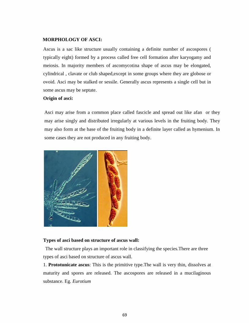
69
MORPHOLOGY OF ASCI:
Ascus is a sac like structure usually containing a definite number of ascospores (
typically eight) formed by a process called free cell formation after karyogamy and
meiosis. In majority members of ascomycotina shape of ascus may be elongated,
cylindrical , clavate or club shaped,except in some groups where they are globose or
ovoid. Asci may be stalked or sessile. Generally ascus represents a single cell but in
some ascus may be septate.
Origin of asci:
Asci may arise from a common place called fascicle and spread out like afan or they
may arise singly and distributed irregularly at various levels in the fruiting body. They
may also form at the base of the fruiting body in a definite layer called as hymenium. In
some cases they are not produced in any fruiting body.
Types of asci based on structure of ascus wall:
The wall structure plays an important role in classifying the species.There are three
types of asci based on structure of ascus wall.
1. Prototunicate ascus: This is the primitive type.The wall is very thin, dissolves at
maturity and spores are released. The ascospores are released in a mucilaginous
substance. Eg. Eurotium

70
2. Unitunicate ascus: The ascus wall consists of 2 layers which are rigid and unite
together through out length and existence of the ascus. . The outer wall is called
exotunica or exoascus and the inner wall is called endotunica or endoascus and not
separated during spore release. The spores are released through a terminal pore, slit or
operculum.E g. Claviceps.
ASCOCARPS:
Ascocarps are the fruiting bodies of members of Sub-division Ascomycotina which
produce the asci containing the ascospores. In some members such as yeasts,
Taphrina fruiting bodies are not produced and the asci are naked.
Types of ascocarps : 4 types.
1. Cleistothecium: It is a completely closed ball like( globose) ascocarp and it is
made up of a wall with pseudoparenchymatous tissue called as peridium. In some
species these are provided with outer appendages. Asci are scattered or
distributed at different levelsin ascocarp.When the asci are
matured, ascospores are released by disintegration of peridium. Eg. Eurotium, Erysiphe.
2. Perithecium; It is a flask shaped more or less closed ascocarp but provided with a
pore or opening at the tIp called true ostiole through which ascospores are released at
maturity.Ostiole is lined inside with sterile structures called as periphysis. The wall is
called peridium . The asci are arranged in definite layer called hymenium. In between
the asci,there are sterile thread like structures called paraphyses which help in
liberation of ascospores.Eg. Claviceps, Xylaria.
3. Apothecium.: It is an open cup shaped ascocarp with a wall peridium. The asci
are arranged in a layer called hymenium , either exposed from the beginning or later
exposed. The sterile structures called paraphyses ( tips free / not fused ) are also
present intermingled with asci which help in liberation and dispersal of
ascospores.Epithecium is a layer on the surface of hymenium of an apothecium
formed by fusion of tips of paraphyses over the asci.E g. Peziza, Tuber.
4. Ascostromata: The asci are formed directly in cavities called locules with in
stroma. The stroma itself serves as wall of ascostroma. Sterile structures called
pseudoparaphyses are present in ascostromata.Eg.Elsinoe.
If the ascostromata is with a single locule ie., An unilocular ascostroma which
resembles perithecium with pseudoparaphyses is called as
pseudothecium.Eg.Venturia.

71
STERILE THREAD LIKE STRUCTURES IN ASCOCARP:
Ascocarps contain sterile thread like structures of various types.
1.Paraphyses: These are elongated, cylindrical , club shaped or sometimes branched
threads arising from bottom of ascocarp. They may be septate or aseptate. They grow
among asci in hymenium and remain free at their tips. However, in Discomycetes, the
tips fuse together forming a layer known as epithecium. Paraphyses help in liberation
and dispersal of ascospores. Eg. Perithecium (Claviceps) , Apothecium( Peziza).
2.Paraphysoids:These are inter ascal tissue that stretch and resemble
pseudoparaphyses,but remotely septate,very narrow,anastomose and tips remain free.
3.Periphyses: These are short, hair like threads lining in side of an ostiole of
perithecium or pseudothecium. Their function is to direct the asci towards ostiole at
the time of ascospore release.
Eg. Perithecium( Claviceps) , Pseudothecium( Venturia).
4.Periphysoids: These are the lateral periphyses which are short and originate above
the level of developing asci but do not reach base of cavity and curve upwards
towards apex.
5.Pseudoparaphyses:These are distinct,vertical, paraphyses -like hyphae,that
originate above the level of asci and grow downwards between the developing
asci,finally becoming attached to the base of the cavity,thus forming curtains
between asci.These are often broader,regularly septate,branched and
anastomosing.Eg. Elsinoe.

72
Ascomycotina is sub divided into six classes based on presence or absence of
ascocarp and shape of the ascocarp.
Class 1. Hemiascomycetes 2. Plectomycetes 3. Pyrenomycetes 4. Discomycetes 5.
Loculoascomycetes. 6. Laboulbeniomycetes.
Important plant pathogens are in the classes :
Hemiascomycetes,Plectomycetes,Pyrenomycetes and Loculoascomycetes
IMPORT ANT CHARACTERISTICS OF CLASS HEMIASCOMYCETES
1.Mycelium is pseudomycelium or dikaryotic mycelium.
2.Ascocarp is absent i.e.,asci are naked.
3.Asci are not formed from ascogenous hyphae but formed directly from zygote or
ascogenous cells.
4.Asci release ascospores by bursting or deliquescing of ascus.
5.Orders:
a.Protomycetales, Family:Protomycetaceae.Eg.Protomyces,Protomycopsis
b.Taphrinales, Family:Taphrinaceae Eg.Taphrina
IMPORTANT CHARACTERISTICS OF ORDER
TAPHRINALES, FAMILY TAPHRINACEAE :
1. The order Taphrinales includes a single family Taphrinaceae and a single genus
Taphrina.
2. Mycelium is septate containing typical thick walled binucleate cells called
ascogenous cells.Hyphae may be intercellular, sub cuticular, or may grow with in
walls of epidermis.
3. Asexual reproduction is through small oval or spherical uninucleate haploid
blastospores that bud from ascospores either with in the ascus or after their release.
4. Ascocarps are not produced. Asci are naked. Sex organs are not formed. Asci are
formed from special binucleate ascogenous cells. Asci
are unitunicate and tip of the ascus bursts at the time of libetration of ascospores.
Eg. Taphrina deformans - peach leaf curl
T. maculans – leaf blotch of turmeric.

73
IMPORTANT CHARACTERISTICS OF CLASS PLECTOMYCETES
1. Ascocarp is a non-ostiolate cleistothecium.
2. Asci are thin walled, globose to pyriform,unitunicate.
3. Asci are produced from ascogenous hyphae,evanescent, scattered at various
levels in the cleistothecium and not forming a definite hymenium.
4. Ascospores are unicellular, released by disintegration of ascus wall.
IMPORTANT CHARACTERISTICS OF ORDER : ERYSIPHALES,
FAMILY : ERYSIPHACEAE :
1. Erysiphales is the exceptional order as it produces cleistothecium instead of
perithecium. The reason is that the asci are grouped in fascicles or form a basal
layer ( hymenium) at maturity and ascospores are released violently with
force.Cleistothecia are formed on superficial mycelium with out formation of
stroma.
2. Members cause a disease called powdery mildew because they produce
enormous number of conidia on the surface of infected host plants which appear to
the naked eye as a white powdery coating.
3. Mycelium is hyaline and mostly ectophytic
4. Members are obligate parasites of plants and nourishment through haustoria.
5. Asci are persistant, globose to pyriform and explodes at the time of release of
ascospores.
6. Important plant pathogenic genera are 1. Erysiphe 2. Leveillula 3. Phyllactinia
4. Uncinula 5. Sphaerotheca 6. Podosphaera 7. Microsphaera

74
Somatic characteristics:
Mycelium is well developed, septate, uninucleate, profusely branched entirely
superficial (ectophytic) except Leveillula (endophytic) and Phyllactinia (semi-
endophytic),produce haustoria into epidermal cells to absorb nourishment.
Asexual reproduction :
Asexual reproduction is through conidia produced on conidiophores.
Conidiophores are long, erect and hyaline.
Three types of conidial stages are recognised in powdery mildews.
1. Oidium 2. Oidiopsis 3. Ovulariopsis
1. Oidium (Acrosporium) : Mycelium is ectophytic, hyaline. Conidia are developed
from a flask shaped mother cell ( spore mother cell) formed on a short conidiophore .
Conidia are barrel shaped with flat ends and are produced in chains..The conidia are
also referred to as meris tem arthrospores as these are formed by fragmentation of
hyphae. Eg. The perfect stages viz.,
Erysiphe,Podosphaera, Uncinula, Sphaerotheca and Microsphaera produce Oidium
as conidial stage.
2. Oidiopsis: Mycelium is endophytic.Conidiophores may be branched or
unbranched, erect, septate, hyaline and emerge through stomata. Conidia are
produced singly and cylindrical in shape. Conidia are of two types. a. blunt tip b. pointed
tip. Eg. Leveillula sp. produce Oidiopsis as conidial stage.
3. Ovulariopsis: Mycelium is partly ectophytic and partly endophytic.The
conidiophores are hyaline, septate, unbranched, and bear a single conidium. Conidia
are rhomboid in shape.In some species,the conidiiphores are spiral in shape. Eg.
Phyllactinia subspiralis. Phyllactinia sp .produce ovulariopsis as conidial stage.
Oidium Oidiopsis Ovulariopsis

75
Powdery mildew conidia do not require free water for germination and are able to
germinate at very low humidity levels.
Sexual reproduction:
Some species are homothallic and some are heterothallic. Antheridia and
ascogonia are sex organs.Both gametangia are uninucleate .Fruiting body is
cleistothecium which is produced on superficial mycelium as a result of
gametangial contact. The cleistothecia are first white and finally black in color
when mature. The wall is made up of pseudoparenchymatous tissue of several layers
called peridium. Over wintering of powdery mildews takes place in clestothecial
stage which are resistant to winter conditions. In perennials , the mycelium may over
winter in the dormant buds of host. In warm weather , many species never form
cleistothecia and perpetuate by means of conidia. The cleistothecia are provided with
characteristic appendages which vary considerably in length and character.
T ypes of cleistothecial appendages:
1. Mycelioid appendage s: These are flexible, flaccid and res emble somatic hyphae.
Eg. Erysiphe, Sphaerotheca, Leveillula.
2. Circinoid / hooked / coiled appendages : These are rigid with curled or coiled
tips. Eg. Uncinula .
3. Dichotomously branched tips: These are rigid,flattened with dichotomously
branched tips. Eg. Podosphaera, Microsphaera.
4. Bulbous base with pointed tip: These are rigid, spear like with bulbous base and
pointed tip. Eg. Phyllactinia.

76
KEY FOR THE IDENTIFICATION OF POWDERY MILDEW GENERA:
1. Type of cleistothecial appendage
a. mycelioid
b. dichotomously branched c
. circinoid
d. bulbous base with pointed tip
2. Number of asci in cleistothecium
a.one
b.many
3. Type of conidial stage
a. Oidium
b. Oidiopsis
c. Ovulariopsis
4. Nature ofmycelium
a. ectophytic
b. endophytic
c . semi- endophytic
MYCELIUM
1. Ectophytic
a. One ascus per cleistothecium
Oidium type conidial stage
Mycelioid appendages- Eg. Sphaerotheca
Dichotomously branched appendages - Eg. Podosphaera
b. Several asci per cleistothecium
Oidium type conidial stage
Mycelioid appendages -Eg. Erysiphe
Circinoid appendages -Eg. Uncinula
Dichotomously branched appendages -Eg. Microsphaera

77
2. Endophytic
Many asci per cleistothecium
Oidiopsis type conidial stage
Mycelioid appendages-Eg. Leveillula
3. Semi- endophytic
Many asci per cleistothecium
Ovulariopsis type conidial stage
Bulbous base with pointed tip -Eg. Phyllactinia
Important powdery mildew diseases:
CROP PATHOGEN
1. Pea Erysiphe polygoni
2. Cucurbits E. cichoracearum
3. Grasses E. graminis
4. Mulberry Phyllactinia corylea
5. Chillies Leveillula taurica
6. Apples Podosphaera leucotricha
7. Roses Sphaerotheca pannosa
8. Lilac Microsphaera alni
9. Grapes Uncinula necator

78
IMPORTANT CHARACTERISTICS OF CLASS: PYRENOMYCETES
1. Ascocarp is mostly a true perithecium in which asci are arranged in a definite
layer called hymenium. Perithecium may be globose or flask shaped. Some members
produce cleistothecium.
2. Asci are unitunicate, persistant, club shaped or cylindrical.
This class includes 2 important Orders viz., Hypocreales and Sphaeriales.
IMPORTANT CHARACTERISTICS OF ORDER HYPOCREALES :
1.In the centrum ,apical paraphyses called peri-physoids arise from perithecial apex
below the periphyses and finally disintegrate as the asci grow among
them.
2.Ascocarps are usually bright coloured , fleshy, rarely non-ostiolate.
3.Asci are clavate to cylindrical.
4.Ascospores are colourless, non-septate or multiseptate .
IMPORTANT CHARACTERISTICS OF FAMILY CLAVICIPITACEAE ;
1.Members produce perithecia within a well developed stroma composed of
entirely fungal tissue.
2.Asci are long, narrow and cylindrical with a thick cap perforated by a long
cylindrical pore through which ascospores escape.
3. Lateral walls of ascocarps are lined with periphysoids (lateral paraphyses that
originate all along lateral walls of perithecium but do not occur among asci at the
base of perithecium.).
4.The ascospores are thread like and break into fragments after they are
released and each fragment function as individual spore capable of giving
rise to mycelium.
5.Many members are parasitic on grasses infecting gynoecium which later
converts it into sclerotial bodies (ergots) and thus causing a group of
diseases known as ergots.
6.Asexual stage:The fungus parasitises the ovaries of plants and form sporodochia
(asexual stage) bearing short conidiophores with minute, oval conidia at their tips.
These conidia are mixed with a sticky sweet nectar like
secretion.This sugary slime is called honey dew and hence the asexual

79
stage is commonly called as honey dew stage/sphacelia stage.
7.Sclerotial stage:L ater mycelium hardens converts into purple black hard
sclerotia.The sclerotium of Claviceps is known as ergot commercially. During
the harvesting operation, many sclerotia are knocked off the spikelets and fall to
the ground where they pass the winter.The ergots are
highly poisonous as they contain powerful alkaloids such as ergonovin ,
ergometrine and ergotamine.When the animals or human beings consume ergot
contaminated grains or flour, a serious disease termed ergotism occurs.
Another important disease in humans due to consumption of ergot
contaminated grain flour of rye is St. Anthony’s fire. Alkaloids have also got
medicinal values. They are used to prevent haemorrhage (bleeding) during
child birth and as artificial abortifacient. The drug prepared from ergot bodies is
called ergotin.
Diseases:
Ergot of rye- Claviceps purpurea ( I.S- Sphacelia segetum )
Ergot of bajra- C. microcephala,C.fujiformis
Sugary disease of sorghum- Claviceps sp. ( I.S: Sphacelia sorghi)
Parasitic on insects - Cordyceps sp.
IMPORTANT CHARACTERISTICS OF CLASS LOCULOASCOMYCETES
1. Ascocarp is ascostromata or pseudothecium .
2. Presence or absence of sterile structures pseudoparaphyses in ascocarp.
3. Asci are bitunicate and are borne in locules in stromatic tissue.
Order: 1. Pleosporales
Family: Venturiaceae
Eg. Venturia inaequalis(I.S: Spilocaea pomi)
Family: Pleosporaceae
Eg. Cochliobolus miyabeanus(I.S:Bipolaris oryzae)
Order 2. Myriangiales
Family: Myriangiaceae

80
Eg. Elsinoe ampelina (I.S:Sphaceloma ampelinum)
Order 3. Dothidiales
Family: Dothidiaceae
Eg.Mycosphaerella arachidis
(I.S:Cercospora arachidicola)
Mycosphaerella berkeleyi
(I.S:Cercosporidium personatum)
IMPORTANT CHARACTERISTICS OF ORDER: 1. PLEOSPORALES,
FAMILY: VENTURIACEAE
1. Fruiting body is called pseudothecium produced sub epidermally
or sub cuticularly.
2. Presence of pseudoparaphyses in fruiting body.
3. Conidiophores are short producing flame shaped conidium.
4. Conidiophore and conidia resemble a short burning candle.
5. Ascospores are 2 celled, ellipsoid, unequal in size, hence the name of the
species “inequalis”.
Eg. Apple scab- Venturia inaequalis (I.S:Spilocaea pomi)
Pear scab- V. pyrina
Flame shaped conidia
Conidiophore

81
IMPORTANT CHARACTERISTICS OF FAMILY : PLEOSPORACEAE
1.Ascocarp is pseudothecium.
2.Ascospores a re filiform and many celled.
3.Conidia are dark, cylindrical with many transeverse septa (pseudosepta)
Eg. Brown spot of rice- Cochliobolus miyabeanus ( I.S: Bipolaris oryzae)
Leaf spot of maize- C. heterostrophus
IMPORTANT CHARACTERISTICS OF ORDER:2. MYRIANGIALES
FAMILY: MYRIANGIACEAE
1. Ascocarp is ascostromata with uniascal locule.
2.Locules are distributed at various levels of ascostromata.
3. Asci are globose, thick walled with 8 ascospores.
4. Ascospores are 4 celled.
Eg. Citrus scab- Elsinoe fawcetti, Grape anthracnose- E. ampelina
Mango scab- E. mangiferae
IMPORTANT CHARACTERISTICS OF ORDER :3, DOTHIDIALES,
FAMILY : DOTHIDIACEAE
1. Ascocarp is pseudothecium, spherical in shape, immersed in host tissue,
ostiolate with periphyses, polyascal locules, ps eudoparaphyses absent.
2. Asci are clavate with 8 ascospores.
3. Ascospores are 2 celled, hyaline.
4. Sexual reproduction is by spermatization in some of the species producing
spermatia in spermagonium.
Eg. Tikka disease
a)Early leaf spot of groundnut- Mycosphaerella arachidis
(syn: M. arachidicola) ( I.S: Cercospora arachidicola)
b)Late leaf spot of groundnut- Mycosphaerella berkeleyi
(I.S:Phaeoisariopsis personata) (syn: Cercospora personata)
Sigatoka leaf spot of Banana:Mycospherella musicola (I.S:cercospora musicola)

82
IMPERFECT STAGES FOR THE GENERA OF CLASS LOCULOASCOMYCETES:
PERFECT STAGE IMPERFECT STAGE
1. Venturia inaequalis Spilocaea pomi
2.Cochliobolus miyabeanus Bipolaris oryzae
3.Elsinoe ampelina Sphaceloma ampelinum
4.Mycosphaerella arachidicola Cercospora arachidicola
5.Mycosphaerella berkeleyi Phaeoisariopsis personatum
6.Mycospherella musicola Cercospora musicola
somatogamy between compatible cells of monokaryotic mycelium or fusion of 2
basidiospores or spermatization.It exists during major part of the life cycle.Thus, this
stage is an independent and extensive phase unlike the short dikaryotic phase of
Ascomycotina.This is associated with special structures called clamp connections
through which dikaryotization takes place.(dikaryotiz ation is a process by which
monokaryotic primary mycelium is converted to dikaryotic secondary mycelium).
3.Tertiary mycelium: This is the binucleate mycelium which is organized into
specialised tissues which form into fruiting bodies called sporophores (basidiocarps)
in the members of higher Basidiomycotina .
Clamp connections: It is a hook like structure formed laterally in between the
dividing nuclei in a dikaryotic hypha. It acts as a by-pass for the nuclei , as they can
not pass through septal pore ie., dolipore septum. It is meant for multiplication of
dika ryotic cells.

83
Mode of development of clamp connection: When a binucleate cell is ready to
divide, a small lateral branch called clamp connection arises from the cell between
the 2 nuclei (a and b) and begins to form a curved hook.Then the 2 nuclei divide
simultaneously. One division orients obliquely so that one daughter nucleus “b”
forms in the clamp connection and the other daughter nucleus “b1 ” forms in the
dividing cell. The second division orients itself along the length of the dividing cell
so that one daughter nucleus “a” forms near one end of the cell and the other “ a1
”approaches the nucleus “ b1 “of the first division near the other end of the cell. In the
mean time, the clamp bents over and its free end fuses with the cell so that clamp
forms a bridge through which one of the daughter nucleus “ b” passes to the other end
of the cell and approaches daughter nucleus “ a “. A septum is formed to close the
clamp at the point of origin and another septum vertically under the bridge to divide
the parent cell into two daughter cells with “ a” and “ b ” in one
1 1 ”
in the other cell.The clamp remains daughter cell and nuclei “ a ” and “ b
permanently attached to hyphae. Its presence indicate s that the hypha is
dikaryotic.

84
SUB- DIVISION: BASIDIOMYCOTINA
1.Members of this sub-division are highly advanced fungi.
2.The name Basidiomycotina is given because the fungi produce sexual
spores on a special club shaped fruiting body called basidium.
3.A definite number of sexual spores called basidiospores (usually four in
number) are produced on each basidium .
4.Fungi belonging to this sub division are referred as club fungi.
5.The group includes mushrooms,toadstools,shelf fungi,jelly fungi, puff balls,
coral fungi,bracket fungi, birds nest fungi,stink horns, rusts and smuts.
General characteristics:
1. Produce sexual spores (basidiospores ) on the out side of a specializ ed spore
producing structure called basidium.
2. A typical basidium is a club shaped structure ,bearing specially 4 basidiospores
on pointed projections called sterigmata.
3. Basidiospores are haploid, uninucleate and are the result of plasmogamy,
karyogamy and meiosis.
4. Dikaryotic phase dominates the life cycle.
5. Presence of clamp connections on the mycelium .
6. Presence of dolipore septum, except in rusts and smuts.
7. Absence of motile spores .
Somatic structures:
The mycelium consists of well developed septate mycelium.The mycelium passes
through three distinct stages before the completion of life cycle. They are primary,
secondary and tertiary mycelium.
1.Primary mycelium: (homokaryon or monokaryotic mycelium) .It consists of
hyphae with uninucleate cells. It usually develops from the germination of a
basidiospore.It may be multinucleate at first when the nucleus of basidiospore divides
many times as the germ tube emerges and grow. This multinucleate stage is short
lived because septa are formed dividing the mycelium into uninucleate cells.

85
2.Secondary mycelium: (dikaryon or dikaryotic mycelium).This originates from
primary mycelium and its cells are dikaryotic (binucleate, n+ n nucleus) formed
Dolipore septum: Both primary and secondary mycelium consists of dolipore
septum. The septum around the central pore swells at the center forming a barrel
shaped structure with open ends,thus forming a septal pore.The septal pore is
surrounded by a cup like or dome shaped membrane called parenthosome or septal
pore cap or nuclear pore cap. It is made up of a double membrane and its function is
to shut the pore. The dolipore septum will not allow the movement of nuclei in
hyphae but maintai ns continuity of cytoplasm.
Asexual reproduction: Asexual reproduction takes place by means of budding
(conidia), fragmentation of hyphae ( arthrospores) , uredospores. Conidial production
is common in smuts while rusts produce uredospores (summer spores) that are
conidial in origin and function.
Sexual reproduction: Sexual reproduction results in the production of basidium
bearing haploid basidiospores. Basidiospores are formed as a result of karyogamy
and meiosis taking place in basidium. In most of the members, sex organs
(gametangia) are not produced and the somatic hyphae or detached somatic cells
(arthrospores) undergo sexual process by somatogamy.In Puccinia sexual process is
accomplished by spermatization through specialized organs called spermatia acting
as male gametes and receptive hyphae as female organs.Thus,sexual cycle involves
in typical cases, a monokaryotic phase and establishment of dikaryotic phase by

86
somatogamy or spermatization of primary mycelium and then karyogamy and
meiosis in the basidium and return to monokaryotic phase by means of
basidiospores.Thus,in the life cycle there is an alternation of monokaryotic and
dikaryotic phases.
Basidium: Basidium is a club shaped, sexual, fruiting body bearing on its surface a
definite number of (usually 4) basidiospores which are formed as a result of
karyogamy and meiosis.
Development of basidium: A simple, club shaped basidium originates as a terminal
cell of a binucleate hyphae and is separated from the rest of the hyphae by a septum
over which a clamp connection is generally seen. At first, basidium is narrow and
elongated and later it enlarges and becomes broader.Mean while, the
2 nuclei with in the young basidium, fuse (karyogamy) and the zygote nucleus soon
undergoes meiosis giving rise to 4 haploid nuclei. In the meantime, four small
outgrowths termed as sterigmata push out at the top of the basidium and their tips
enlarge eventually forming the basidiospore initials. During this time, a vacuole
forms at the base of the basidium and as it increases in size, it pushes the
contents of basidium out into basidiospore initials which finally become
basidiospores.

87
Parts of basidium: Basidium is divided into 3 patrs. Probasidium (portion where
nuclear fusion takes place),metabasidium / promycelium (portion where meiosis
occurs) and sterigmata ( any portion between metabasidium and basidiospore).
In smuts and rusts, fusion of 2 nuclei takes place in a specially formed thick walled
spores called chlamydospores and teleospores respectively. During the germination
of chlamydospore / teleospore, fusion of 2 nuclei takes place in the spore, followed
by meiosis. A germ tube called promycelium is formed which becomes transversely
septate into 4 cells, each cell containing a haploid nucleus. The basidiospores are
formed on the sterigmata on promycelium.
Basidiopspores: A basidiospore is typically a unicellular, uninucleate (exceptional 2
nuclei) haploid structure .The basidiospores are formed exogenously on the basidium
in contrary to the endogenous formation of ascospores. The basidiospores may be
globose, oval, elongate or sausage shaped and may be hyaline or coloured.
Dispersal of basidiospores: In majority cases, the spores are released violently and
such spores are called ballistospores.Many possible mechanisms of spore discharge
have been suggested. Buller was one of the first to examine critically the spore
discharge.According to him, Basidiospores rest on the tip of sterigmata in an oblique
fashion and a bubble or drop ( called Bullers drop consist of liquid which forms at the
hilar appendix ie.,a minute projection of the spore near the point of attachment to the
sterigmata ) is responsible for basidiospore discharge . This drop keeps on increasing
in size and its expansion results in explosive discharge of spore to a distance of about
0.1mm. The spores are discharged in succession at intervals of several seconds to
minutes.

88
Basidiocarp (Fungus flowers): Basidiocarp is a fruiting body that bears basidia
which may be crust like, gelatinous, papery, spongy, corky, woody in texture. They
vary in size from microscopic to a meter or more in diameter. Most Basidiomycotina
bear their basidia in basidiocarps except in rusts and smuts. Basodiocarp producing
fungi are mushrooms, shelf fungi/ bracket fungi, coral fungi, puff balls,bracket
fungi,birds nest fungi,earth stars etc. Basidia are formed typically in definite layers
called hymenium. The hymenium is a layer composed of basidia and any other sterile
structures like cystidium (larger and protrude beyond the other structures and of
taxonomic importance) and basidiole (resemble basidium but with out basidiospores
and provide support to fertile basidium).
Basidiole Cystidium
Basidiospore
Basidium
Hymenium
Compatibility: The members are either homo or heterothallic (majority).

89
IMPORTANT CHARACTERISTICS OF CLASS TELIOMYCETES:
1. Include rusts and smuts.
2. Basidiocarp is lacking and replaced by thick walled teleospores or
chlamydospores in sori with in the host tissue.
3. Basidia arise from thick walled resting spores i.e., teleospores or chlamydospores.
4. Members are obligate parasites or facultative saprophytes.
In class Teliomycetes there are two orders.1.Uredinales 2.Ustilaginales
IMPORTANT CHARACTERISTICS OF ORDER UREDINALES:
1. The popular name for the Uredinales is the rust fungi, which relates to the
reddish brown colour of some of the spores. All are obligate parasites of crop plants.
2. The mycelium is primary in the early stage and in the later stages secondary. The
mycelium is inter cellular and produce haustoria that penetrate the host cells and
obtain nourishment. There is no tertiary mycelium and hence there is no basidiocarp.
3. Clamp connections are rare or absent. Dikaryotisation takes place either through
somatogamy or spermatisation.
4. Teleospores originate from the apical cells of dikaryotic hyphae. They may be uni
or multi cellular. The structure of teleospores forms the basis for identification of the
rust genera. The teleospore acts as an encysted basidium in which karyogamy occurs.
It germinates by producing a promycelium ( metabasidium) in which meiosis takes
place.
5. The rusts have polymorphic life cycle. Production of many spore forms in the
life cycle is called polymorphism. Generally 5 types of spores are seen during the life
cycle viz., spermatia (uninucleate) in spermagonium, aeciospores(binucleate)
in aecium,uredospores( binucleate) in uredium , teleospores(binucleate)in telium and
basidiospores (uninucleate) on promycelium or metabasidium.The spermagonium
represents gametic stage (male gamete- spermatium, female sex organ- receptive
hypha), aecia represent the stage in which dikaryotisation occurs,uredia represent
conidial or repeating asexual stage, telia represent sexual stage and act as encysted

90
basidium in which karyogamy occurs and subsequently giving rise to basidiospores
from promycelium or metabasidium.
6. Autoecious rust: If all the spore stages are produced on the same host then the
fungus is called autoecious and the phenomenon is called
autoecism.Eg.Melampsora lini- linseed rust, Uromyces appendiculatus - bean rust. 7.
Heteroecious rust: If spore stages are formed on two unrelated hosts ie., pycnia and
aecia on one host and the uredia and telia on the other host, such rusts are called
heteroecious rusts and phenomenon is called heteroecism. Eg. Puccinia graminis f.
sp. tritici- black stem rust of wheat. Primary host: The host in which heterocious
rust produce the telial stage is called primary host (Eg. wheat). Secondary or
alternate host The host in which telial stage is not produced is called alternate or
secondary host ( Eg. barberry) .
8. Based on life cycle pattern, rusts are divided into macrocyclic, demicyclic and
microcyclic rusts.
Macrocyclic rust: ( long cycled rust): Rusts in which all 5 spore forms are
produced or produce at least one type of binucleate spore in addition to
teleospores are called macrocyclic rusts. It may be autoecious macrocyclic rust
(Eg.Puccinia helianthi- sun flower rust) or heteroecious , macrocyclic rust
(Eg.Puccinia graminis f.sp. tritici - black stem rust of wheat).
Demicyclic rust: The rust in which uredial stage is absent. eg. Gymnosporangium
juniperi – virginianae- cedar apple rust.
Microcyclic rust (short cycled rust):Rusts which produce no binucleate spore other
than teleosporei.e., teleospore is the binucleate spore produced and both aecia and
uredia are lacking. E g.Puccinia malvacearum- hollyhock rust.
IMPORTANT CHARACTERISTICS OF FAMILY PUCCINIACEAE:
1.Teleutospores are free or variously united, but never in the form of layers or crusts.
2.Teleutospores are stalked. E g. Puccinia, Uromyces, Hemileia

91
Genus Puccinia:
Obligate parasites.Teleutospores are two celled and stalked.They lie free in the sorus.
Disease: Puccinia graminis f.sp. tritici- black stem rust of wheat
Genus Uromyces:
Teleutospores are single celled with a thick apex ( papillum) and stalked. The stalks
are fragile and short.
Eg. U. appendiculatus - bean rust
Genus Hemileia:
Uredospores are reniform, bifacially ovate resembling orange segments, concave side
smooth, convex side echinulate.
Teleutospores are turnip shaped. 1 celled, stalked, smooth walled and produced on
erumpent,club shaped stalks which arise through stomata.
Eg. H. vastatrix - coffee rust
Puccinia Uromyces Hemileia
LIFE CYCLE OF PUCCINIA GRAMINIS F. SP. TRITICI
The pathogen is an obligate parasite and causes black stem rust of wheat. It is a
heterocious rust that requires 2 hosts for completion of its life cycle. The primary
host (wheat) and the secondary host (barberry). .On barberry it produces pycnia and
aecia while uredial telial stages are produced on wheat.
It is a macrocyclic rust producing all the five types of spores. The different kind of
spores and their spore stages are designated as follows.
Stage Spore Nucleus status
O spermagonia with spermatia uninucleate
(pycnia with pycniospores)

92
I aecia with aeciospores binucleate
II uredia with uredospores binucleate
III telia with teleospores binucleate
IV basidia with basidiospores uninucleate
It produces the first 2 stages in barbery and other 3 stages on wheat or other
graminaceous hosts.
Significance of each stage :
Stage “ O “: The spermatia produced in spermagonia were till recently thought to be
functionless asexual spores. Mycologists have not found them to germinate to
produce mycelium as in case of otHer asexual spores. Hence, they thought that they
are vestigial bodies. At the time when mycologists do not know the function of pycnia
and pycniospores the stage was designated as “ O “ stage. But in 1927, Cragie found
that spermatia are male gametes and are essential for spermatisation of receptive
hyphae (female organ) and consequent formation of aeciospores. This stage “ O”
represents the sexual stage of rust fungi but the nomenclature stage “ O “ retained
even today to to avoid confusion.
Stage I: Aeciospores are the first binucleate spores formed in the life cycle.
Stage II: Uredospores are also called as repeating asexual spores as they functionas
conidia for the propagation of the rust fungus.
Stage III: Teleutospores represents the perfect stage because karyogamy and
meiosis ocuur in them.
Stage IV: Basidiospores represent the sexual spores.
Stages “ O” and “ I” occur on barbery while stages “ II” and “ III “ occur on
wheat. Basidiospores can infect only barbery plant where as aeciospores can infect
only wheat plant.
Stage “ O”: Spermagonia with spermatia:

93
The spermagonia or pycnia are the structures which bear the sex organs of the
pathogen. It contains spermatia which are the male sex organs and the receptive
hyphae which are the female sex organs. These spermagonia are formed near the
upper epidermis in about 4 days after infection of the host by a basidiospore. In
nature it generally happens that several basidiospores at random will reach and infect
the same barbery leaf so that both + and – mycelium develop side by side and
intermingle in the barbery tissue. Each spermagonium contains numerous spermatia.
These are exuded (ooze out) in small droplets of nectar present in the spermagonium.
Each spermatium carries + or – nucleus depending on the strain of mycelium which
produced the spermagonium. All spermatia from a single spermagonium carry the
same factor or genetic make up as that of receptive hyphae. These arise from upper
part of spermagonia and protrude through the ostioles. Spermatisation i.e., fusion
between receptive hyphae and spermatia of opposite sex takesplace through agency
of insects which are attracted by the honey fluid. The spermatial contents pass into
the receptive hyphae . Meanwhile the mycelium penetrates the entire leaf and the
hyphae near the lower epidermis develop number of aecial primordial. It is presumed
that spermatial nuclei which pass from the spermatia into the receptive hyphae reach
the cells of the aecial promordia rendering them binucleate. It has been demonstrated
that aecial primordial fail to develop into aecia until and unless spermatisation takes
place.

94
Life cycle of Puccinia graminis f. sp. tritici

95
Stage“I” : Aecia and aeciospores are formed in the lower epidermis soon after
dikaryotisation. These are the first binucleate spores produced in the life history of
the fungus. An aecium is a group of binucleate hyphal cells ( aeciospore mother cell )
which give rise to aeciospores in chains. The aeciospores finally disseminated by the
wind and under favourable conditions germinate on graminaceous host . These can
not infect barbery.
Stage “ II”: Soon after infection by aeciospores on graminaceous host , binucleate
mycelium begins to form masses of cells. These are called uredia from which
binucleate uredospores are borne on long stalks. The uredospores are one celled, oval,
yellowish and spiny. They germinate readily in water and produce one or more germ
tubes. The uredospores are those spores which perpetuate the fungus throughout the
growing season and they are capable of reinfecting the graminaceous host on which
they produce. Hence, they are also known as repeating asexual spores. They spread
from plant to plant and from field to field and the disease soon becomes an
epiphytotic. The uredospores upon germination produce binucleate mycelium which
grows between the cells of the host and in a few days produce new uredia and
uredospores.
Aecium Uredium Teliospore
Stage “ III” : Late in summer, at the time of ripening of grain another kind of spores
known as teleospores or teleutospores are developed in the same mycelium when the
uredia begin to cease. The pustules which produce teleospore s are known as telia
and constitute the black stage of rust. The teleospores are ellipsoidal, oblong or
obclavate, typically two celled, and thick walled with slight constriction at the
septum. The young teleospore is binucleate . Karyogamy eventually take s place and

96
render the teleosspores diploid and uninucleate . The teleospores are not capable of
germinating immediately and should have resting period of several months and thus
remain dormant until the following spring
Stage “ IV” : Early in the spring each cell of teleospore germinate and produce a
basidium ( promycelium). The diploid nucleus in teleospore migrates in to the
promycelium and undergoes meiosis and four haploid nuclei are formed. Then septa
are formed, separating the nuclei from one another into four cells. Each cell of
promycelium produces a sterigmata on which basidiospore is formed. The nuclei now
migrate into the basidispore. Two of the basidiospores are of one strain ( + ) and two
are of other strain ( -- ) . Soon after their formation ,the basidiospores are ejected and
are carried away by wind. They can not infect graminaceius host, but can infect
barbery and produce a well developed monokaryotic mycelium. Thus, the life cycle
gets repeated on these two hosts viz., wheat and barbery.
DISTINGUISHING CHARACTERISTICS OF GENERA (OR) KEY FOR
IDENTIFICATION OF GENERA:
Ustilago: 1.Teleospores singles 2.Sori dusty at maturity
3.Sori covered peridium ( membrane) of host origin
Spacelotheca: 1.Teleospores singles 2.Sori dusty a t maturity
3.Sori covered by membrane ( peridium) made up of fungal cells
4.Central columella present
Tolyposporium:1.Spores in balls
2.Spore balls permanent, spores adhering by thickenings of
exospore .
DISEASES CAUSED BY THE GENERA:
U. nuda tritici- loose smut of wheat

97
Sphacelotheca sorghi- short or grain or kernal smut of jowar S.
cruenta- loose smut of jowar
S. reliana- head smut of jowar
Tolyposporium ehrenbergii- long smut of jowar
T. penicillariae- smut of bajra
DIFFERENCE BETWEEN RUSTS AND SMUTS
Character Rusts smuts
1. Systematic position Order: Uredinales Ustilaginales
2. Plant parts affected
foliar parts (leaves, stem, petiole
) floral parts( flowers)
3. Symptoms reddish brown coloured pustules
ovaries turn into
black
4. Parasitism obligate parasites
facultative
saprophytes
5. Polymorphism polymorphic not polymorphic
Germinating teliospores of bunt fungi

98
DISTINGUISHED CHARACTERISTICS (OR)KEY FOR IDENTIFICATION
OF GENERA:
T illetia:
1.Teleospores singles
2.Spores dusty and escaping at maturity
3.Sporidia are fused to form H shaped structures
Neovossia:
1.Teleospores singles
2.Spores dusty and escaping at maturity
3.Sporidia do not fuse, no H shaped structures
Urocystis :
1.Teleospores in balls
2.Sori dusty, spore balls surrounded by an adhering layer of hyaline sterile cells.
3. spore balls escape from sorus
DISEASES CAUSED BY THE GENERA:
Tilletia caries and T. foetida- bunt of wheat
Neovossia horrida- bunt of paddy
N. indica- Karnal bunt of wheat
Urocystis cepulae- onion smut
U. tritici- flag smut of wheat

99
DIFFERENCES BETWEEN SMUTS AND BUNTS :
S.No.
Smuts
Bunts
1 Belongs to family Ustilaginaceae
Belongs to family
Tilliteaceae
2 Promycelium is Septate Promycelium is non-Septate and
hollow tube like
3 Basidiospores are formed laterally from Basidiospores are formed at the tip of
each cell of the
promycelium the promycelium
4 Basidiospores are usually four Basidiospores are more than four
usually eight
5 H shaped structures are not formed H shaped structures are present in
which plasmogamy occurs.
6 Meiosis occurs in promycelium or Meiosis always occurs in teliospores
teliospores before germination
7 No fishy odour is observed Characteristic stinking fishy odour is
observed
8 Genera included are Ustilago, Genera included are Tilletia,
Sphaecelotheca, Tolyposporium Neovossia, Urocystis
100
IMPORTANT CHARACTERISTICS OF HYMENOMYCETES:
1.These fungi are popularly called as mushrooms.
2.It includes bracket or pore fungi, toadstools, jelly fungi, honey mushrooms, etc.
3.Basidia are formed on a hymenium of a well developed fruiting body,
basidiocarp.
4.Basidiocarps are gymnocarpous or hemiangiocarpous.
5. Basidia are not formed from teleospores.
6.The hymenium is exposed in the fruiting body from the beginning and thus
basidiospores are exposed before they mature.
7.Basidiospores are called ballistospores (the spores which are perched
obliquely and
discharge forcibly and violently are called ballistospores).
8.Members are saprophytes or facultative parasites.
IMPORTANT CHARACTERISTICS OF ORDER APHYLLOPHORALES:
1. All the members produc e single celled , club shaped basidia in well defined
hymenium.Basidiocarp is tough and non fleshy, may be cottony, leathery, corky or
woody in texture.
2. The development of basidiocarp is gymnocapous i.,e., the hymenium is exposed
while the spores are still mature.Thus, hymenium is exposed through out
development.
3. Hymenophore( the layer that supports hymenium) may be smooth, flatte ned or
resupinate, teeth like, with pores etc.
4. This order consists of both terrestrial and wood inhabiting forms. Some are
serious pathogens of forest trees causing root rot and heart rot. Dead trees and
lumber are commonly attacked by certain members.
IMPORTANT CHARACTERISTICS OF FAMILY GANODERMATACEAE;
1.Members are commonly called as bracket fungi or shelf fungi and members are
lignicolous forms.
2.The fruiting body of the fungus is called bracket which is formed laterally at the
base of affected plant as a leathery stalked fan shaped or bracket shaped or with out

101
stalk,made up of trimitic hyphal system,hymenophore poroid. The bracket is
tough,leathery or woody in texture and size vary from 1-20 inches in diameter.The
stalk is cylindrical and brown to black in colo ur.
3.The upper surface of bracket is reddish brown in colour and coated with a hard shiny
substance resembling sealing wax, while the lower side is white or
yellowish in colo ur. When examined with a lens, minute holes or pits are seen all over
the under surface. These are the openings of numerous hymenial tubes or pores which are
vertically oriented ins ide the fruiting body.Each basidium gives rise to 4 sterigmata, each
of which bears a basidispore at tis tip.
4.Basidiospores are coloured,two layered and cystidia are absent in
hymenium.Bracket shaped basidiocarp ,broadly and horizontally attached to the tree
trunks by means of a short stalk or stipe.Ganoderma differs from other bracket fungi
in having much longer span of spore release,extending upto 5 months. Diseases
caused by Ganoderma :
Ganoderma lucidum- root rot and wilt of coconut & other palm trees and citrus.
Ganoderma
102
22.SUB- DIVISION: DEUTEROMYCOTINA
These are a group of fungi which reproduce only by means of asexual spores or
fragmentation of hyphae or modified mycelium. The asexually produced spores are
generally called as conidia. A conidium is a non-motile asexual spore formed at the
tip or side of sporogenous cell. For several genera of this group sexual reproduction/
sexual stages/ perfect stages/ teleomorphic stages are not known or have not been
discovered or not found or rarely formed or have been dropped from the life cycles in
the evolution of these organisms.
Thes e fungi are commanly called as imperfect fungi and technically called as fungi
imperfecti as they have only imperfect stages or conidial stages.Whenever the
perfect stage of an imperfect fungus is detected in nature or laboratory cultures, it is
shifted to proper place on the basis of fruiting body.In most cases the perfect stages
have been found to belong to sub-division Basidiomycotina.
Mycelium is well developed, septate with branched hyphae and multinucleate cells.
Since , present classification is based on characters of sexual stage, these fungi are
not fit for natural classification.
For most of these genera perfect states are not known or rare in nature, they are
temporarily grouped as members of form class, form order, form family , form genus
and form species.
CLASSIFICATION OF DEUTEROMYCOTINA :
The classification is completely artificial. It is based on their conidial peculiarities
and neither bears connection to their sexual stage nor to their origin or evolution. It
can not therefore be called as a natural classification.
The main characteristics (criteria ) on which classification of fungi imperfect fungi is
based are
1. presence or absence of asexual spores( conidia )
2. type of asexual fruiting body
3. manner of production of asexual spores
4. morphology ( shape, size, color and septation) of asexual spores.

103
SACCARDOAN ( 1906 ) SPORE GROUP SYSTEM :
Traditionally, the form sub classes, Coelomycetidae and Hyphomycetidae have been
divided in to sections. The section is not an official category in the classification
system. The various families under each section were divided further by Saccardo
into seven sections based on conidial characters viz., shape, color, septation.
Saccardo ( 1906 ) later, modified the section names with the prefixes hyalo or phaeo
depending upon whether the conidia were hyaline or pigmented. This approach is
referred to as the Saccardoan Spore Group System since it was Saccardo ( 1899 ) who
initially proposed the system.
Saccardo described the spores in Deuteromycotina based on shape, septation and
colour.
I. Amerosporae: conidia non septate ( single celled) , spherical, ovoid to
elongated, or short cylidric.
a)Hyalosporae(Hyalo =colourless): conidia hyaline Eg: Phoma
b)Phaeosporae(phaeo=coloured): conidia coloured Eg: Sphaeropsis
II. Didymosporae : conidia ovoid to oblong, one septate ( two celled )
a)Hyalodidymae: conidia hyaline Eg: Fusarium micro conidia
b)Phaeodidymae: conidia coloured Eg: Botryodiplodia
III. Phragmosporae : conidia oblong, two to many septate ( 3 or more celled ) ,
only transverse septa present. a)Hyalophragmae:
conidia hyaline Eg: Pyricularia b)Phaeophragmae:
conidia coloured Eg: Drechslera
IV. Dictyosporae : conidia ovoid to oblong, both longitudinal and transverse septa
present ( muriform ) .
a) Hyalodictyae: conidia hyaline Eg: Epicoccum
b)Phaeodictyae: conidia coloured Eg: Alternaria
V. Scolecosporae: conidia thread like to worm like, filiform, septate or aseptate (
one to several celled ) Eg: Cercospora
VI. Helicosporae( Allantosporae) : conidia spirally cylindrical, curved ( allantoid ),
septate or aseptate. Eg:Helicomyces

104
VII. Staurosporae : conidia stellate ( star shaped) , radially lobed, septate or
aseptate (one to several celled ). Eg: Actinospora
Amerosporae Didymosporae Phragmosporae Dictyosporae
Scolecosporae Helicosporae Staurosporae
Color of conidia
1. Hyalosporae : cell wall of conidia hyaline
2. Phaeosporae : cell wall of conidia coloured/ pigmented.
AINSWORTH ( 1973 ) CLASSIFICATION:
According to Ainsworth ( 1973 ), 2 form classes are there in Deuteromycotina.
1. Coelomycetes
2. Hyphomycetes
IMPORTANT CHARACTERISTICS OF CLASS COELOMYCETES:
The conidia are borne on conidiogenous cells with or with out distinct conidiophores,
enclosed in fungal fructifications ( asexual fruiting bodies ) such as pycnidium or
acervulus.
Coelo mycetes is divided into 2 form- orders 1.Sphaeropsidales 2. Melanc oniales
IMPORTANT CHARACTERISTICS OF ORDER SPHAEROPSIDALES:
The fruiting bodies are called pycnidia. A pycnidium is a globose or flask shaped
asexual fruiting body that is lined inside with conidiophores. It may be completely
closed or may have an opening called ostiole. It may be papillate or beaked or long
necked at apex , leading to an opening. They vary greatly in their shape, size, color
and consistency of pseudoparenchymatous wall.

105
Sphaeropsidales are further divided in to 4 form families 1 Sphaeropsidaceae 2.
Excipulaceae 3. Nectrioidaceae (Zythiaceae ) 4. Leptostromataceae
Family : 1. Sphaeropsidaceae
Pycnidia are flask shaped or globose, thin or thick walled, dark coloured,
ostiolate, hard texture. Eg. Phoma, Phomopsis, Macrophomina, Phyllosticta,
Septoria, Diplodia , Botryodiplodia.
DISTINGUISHED CHARACTERS OF THE GENERA:
Phoma:
Pycnidia - small, dark coloured, immersed or semi immersed in the host tissue.
Globose or flask shaped, thin walled, and ostiolate. Wall consisting of dark
pseudoparenchymatous cells. Conidiophores / conidiogenous cells are short, hyaline
lining the inner pycnidial wall producing conidia in succession.
Conidia -hyaline, aseptate, guttulate( oil globules ) , pyriform to globose and ooze out
in long thread like cirrhus through the ostiole.
Eg. Phoma lingam – black leg of crucifers
P. vexans – blight and fruit rot of brinjal
Phomopsis:
Pycnidia- brown to black, globose, papillate ostiole, with one or more locules.
Conidiophores / conidiogenous cells- simple or branched.
Conidia of 2 types. (a)alpha conidia- hyaline , ovoid, 1 celled, (b) beta conidia –
hyaline, filiform, straight or curved, , 1 celled.

106
Eg. Phomopsis vexans- fruit rot and blight of brinjal
Phyllosticta :
Pycnidia dark, ostiolate , globose, immersed in host tissue, erumpent or with a short
beak. Conidiophores are short and obsolete.Conidia are small, one celled,
hyaline,ovoid to elongate.
Phyllosticta and Phoma are differentiated on the basis of plant organs attacked.
Phyllosticta principally occurs on leaves causing leaf spots and shot holes While,
Phoma occurs mainly on stem, twigs and fleshy roots. Eg: P. gingeberis – leaf spot
of ginger
Phoma Phomopsis

107
Conidia – hyaline , aseptate, cylindrical to fusiform
Sclerotia- more common in cultures, black, smooth, hard.
Eg. Macrophomina phaseolina – charcoal rot, canker, damping off, of jowar, maize,
ground nut.
Septoria:
Pycnidia – immersed in host tissue, globose, brown, thick walled, papillate and
ostiolate. Wall consists of pale brown cells.
Conidiophore / conidiogenous cells – hyaline, broad and round at base and narrow
above or barrel shaped.
Conidia – hyaline, many septate, filiform.
Eg. Septoria nodorum- glume blotch of wheat
S. lycopersici- leaf spot of tomato
Diplodia :
Pycnidium - black, globose, papillate, ostiolate,
Conidiophore/ conidiogenous cells - slender, hyaline
Conidia – brown, 2 celled, ovoid , apex obtuse ( round ) and base truncate ( shorten )
.
Eg. Diplodia natalensis - Diplodia gummosis of citrus
Botryodiplodia:
Pycnidium – carbonaceous, dark brown or black, no ostiole,
Conidiophores- simple and short.
Conidia – dark brown, 2 celled, ovoid.
Eg. Botryodiplodia theobromae – Flat limb of sapota
Septoria Diplodia Botryodiplodia

108
23.IMPORTANT CHARACTERISTICS OF FAMILY EXCIPULACEAE
Pycnidia are cup shaped.
Ephelis:
Pycnidium – cup shaped,
Conidia – hyaline, 1 celled, acicular ( needle shaped ).
Eg. Ephelis oryzae- udbatta disease of rice .
Ephelis
(A= Stroma and pycnidia, B= Pycnidia (cup shaped), C= Conidia)
FAMILY 3. NECTRIOIDACEAE ( ZYTHIACEAE ):
Pycnidia resemble perithecia of Nectria and hence the Family name Nectrioidaceae .
Zythia:
Pycnidia - flask shaped, coloured, soft textured ( fleshy ) , ostiolate.
Conidia – hyaline, aseptate, oblong, rounded at each end.
Eg. Zythia fragariae- leaf blotch and stem end rot of straw berry
Family 4. Leptostromataceae :

109
Pycnidia are shield shaped or elongated or flattened.
Leptostroma
Shield shaped pycnidia Conidiophores and conidia
Leptothyrium:
Pycnidium – shield shaped, dark, dimidiate ( one half smaller than other ).
Conidiophores- simple.
Conidia – hyaline, 1 celled, falcate ( curved like sickle ) .
Eg. Leptothyrium pomi- fly speck of apple

110
IMPORTANT CHARACTERISTIS OF ORDER
MELANCONIALES, FAMILY MELANCONIACEAE:
All the members of this order are grouped into a single family Melanconiaceae. The
fungi producing asexual fruiting bodies are called Acevulus. Acervulus is a mycelial
mat not having wall of its own and produces a cavity with in which closely packed
short conidiophores forming a bed like mass are produced.
DISTINGUISHED CHARACTERISTICS OF THE GENERA:
Colletotrichum:
Cushion shaped acervulus is seen below epidermis or cuticle with dark setae.
Macrophomina:
Pycnidia- globose, dark brown, papillate ostiole.
Conidiogenous cells- barrel shaped, hyaline.
Setae – septate, stout at base and pointed at tip, dark brown, long, present in the
periphery or in between the conidiophores.
Conidiophores – simple, elongate, septate, hyaline to brown,
Conidia- sickle shaped, , guttulate ( oil globule ), hyaline, single celled.
Eg. Colletotrichum capsici- fruit rot and die back of chillies
C. lindemuthianum- anthracnose of bean
C .falcatum- red rot of sugarcane
Gloeosporium:
This genus is differentiated from Colletotrichum based on absence of setae in
acervulus.
Eg. Gloeosporium ampelophagum - anthracnose or bird’s eye disease of grapes. G.
musarum – anthracnose of banana
Pestalotiopsis:
Acervuli are formed below the epidermis.
Conidiophores- hyaline, branched, septate, cylindrical.

111
Conidia - fusiform , 5 celled , basal cell hyaline with a single appendage, apical cell
hyaline with 2 or more apical , simple or branched appendages , middle cells dark
brown and thick walled.
Eg. Pestalotiopsis palmarum – grey blight of coconut and palmyra
P. mangiferae- leaf spot of mango
Pestalotia:
Acervulus- sub epidermal.
Conidiophores- short, simple or branched,
Conidia- similar to conidia of Petalotiopsis except 6 celled conidia.
Eg. Pestalotia psidi- grey blight, scab and fruit spot of guava.
Colletotrichum Gloeosporium Pestalotia

112
24.IMPORTANT CHARACTERISTICS OF CLASS HYPHOMYCETES
Conid ia and conidiophores are borne directly on hyphae. Conidiophores bearing
conidia may be separate or in aggregates arising from the mycelium. There are
certain fungi which lack conidial formation and forming mycelial structures such as
scretotial bodies. The members are identified based on morphology of conidia.
Hyphomycetes is divided into 4 form orders.
1. Hyphomycetales/ Moniliales/ Hyphales
2. Tuberculariales
3. Stilbellales
4. Agonomycetales.
IMPORTANT CHARACTERISTICS OF ORDER MONILIALES:
Conidia are produced on unorganized, .hyaline conidiophores or directly from
hyaline hyphae. This order is divided into 2 families 1. Moniliaceae 2. Dematiaceae.
FAMILY MONILIACEAE ;
Produce free conidiophores or conidiogenous cells from somatic hyphae. Mycelium,
conidiophores and conidia are hyaline or light coloured but not brown or black.
DISTINGUISHING CHARACTERISTICS OF THE GENERA:
Aspergillus :
Well known saprophyte, grown on all types of substrate and also a weak
parasite. commonly called as “ weed of the laboratory” Mycelium- septate,
branched, with multinucleate cells .
Conidiophore-The hyphal cell that gives rise to conidiophore is called foot cell.
Conidiophores arise singly on somatic hyphae, long, erect, non septate and bears at
its tip a spherical structure called vesicle, which bears two layers of bottle shaped
structures called sterigmata or phialides on which conidia are produced in chains.The
sterigmata of first layer ( lower most ) are called primary sterigmata and the second
layer ( upper most) are called secondary sterigmata.

113
Conidia: globose, one celled, multinucleate, thick , rough walled and black.
Eg. Aspergillus niger- collar rot of groundnut
Aspergillus
Penicillium :
The conidial apparatus technically is called “ penicillus” because it resembles a small
brush or broom, hence the name penicillium Mycelium: highly branched, septate.
Conidiophore: arise from any cell of hyphae (not from foot cell), branch once or
twice about 2/3 of the way to the tip in a characteristic symmetric or asymmetric
broom like fashion. The first generation branches are called primary branches or
rammi, on which whorls of second generation branches called metulae are produced.
Each metula ultimately bears bottled shaped phialides which bears conidia in chains
in basipetal succession. Conidia: globose, hyaline.
Eg. Penicillium notattum- citrus blue mold.
Conidia- hyaline, pyriform, broader at base and tapering towards apex , usually 3
celled.
Eg. Pyricularia oryzae- paddy blast.
Trichoderma:
The members are saprophytes, found in soil and several species are found to be
antagonistic by producing non-volatile antibiotics against a range of plant pathogens.
These are easily recognized by rapidly growing white, yellow or green colonies.

114
Conidiophores: hyaline, erect,solitary or aggregated into tufts, much branched with
phialides in singles or in groups(non-verticillate).
Conidia- hyaline,grey, one celled,ovoid borne in small terminal clusters as balls on
phialides. Eg: Trichoderma viridi, T.harzianum – biocontrol fungi.
Botrytis:
Conidiophores- branched, septate, long, slender, hyaline . Apical cell of conidiophore
with swollen tips bearing clusters of conidia on short sterigmata. Conidia – hyaline, 1
celled, ovoid.
Entire structure resemble like grape bunch.
Eg. Botrytis cinerea- grey mold of gram, bean, apple and grape.
Verticillium :
Conidiophores- slender, septate ,branched, some of the branches bearing verticillate (
in whorls ) phialides that give rise to conidia.
Conidia- ovoid to ellipsoid, 1 celled, borne singly or in moist clusters( mucus )
apically.
Eg. Verticillium albo- atrum - wilt of cotton and tomato.
V. dahliae- wilt of tobacco and brinjal.
Pyricularia Botrytis Verticillium

115
IMPORTANT CHARACTERISTICS OF FAMILY DEMATIACEAE
The hyphae, conidiophores and usually the conidia are brown or black, but some
times the hyphae alone or the conidia only are dark.
DISTINGUISHING CHARACTERISTICS OF THE GENERA:
Alternaria :
Mycelium – branched, septate, dark brown.
Conidiophores – simple, straight or curved , 1-3 septate , dark coloured.
Conidia – dictyospore , brown, obclavate with a beak, 3-8 tranversely septate and 1-2
longitudinally or obliquely septate, conidia are produced acropetally in chains
(catenulate ) through the pores formed at the apex of the beak of conidia.
Eg. Alternaria solani- early blight on tomato and potato
A.brassicae- leaf spot of crucifers
Drechslera:
Mycelium- branched, septate, brown.
Conidiophores- emerge through stomata, erect, septate, simple or branched, dark
brown, geniculate (knee joints), indefinite in growth ( continue growth sympodially
even after production of conidia )
Conidia- dark brown, cylindrical, straight , several celled, many pseudoseptate,
germinate from any or all cells .
Eg. D. turcica- leaf blight of sorghum
D. nodulosum- seedling blight and foot rot of ragi
Helminthosporium :
Mycelium – dark.
Conidiophore- single or clustered, tall, brown, simple.
Conidia- develop laterally through pores beneath septa, often appear in whorls,
obclavate, brown, many pseudoseptate with prominent basal scar.
Eg. Helminthosporium maydis- southern corn leaf blight
H. victoriae- victoria blight of oat.
116
Bipolaris :
Differentiated from Drechslera based on method of germination of conidia and shape
Conidia- germinate characteristically from two polar ( end ) cells only, fusoid and
slightly curved.
Eg. Bipolaris oryzae – brown spot of rice ;P.S:Cochliobolus miyabeanus (
old: Drechslera oryzae)
Cercospora ;
Mycelium : immersed in host tissue, branched, septate , pale brown
Conidiophore: emerge in clusters through stomata, brown, septate, simple or rarely
branched with knee joints ( sympodially extending ) marking the scars of fallen
spores
Conidia: terminal, arise singly from conidiophore, hyaline, filiform, severa celled ( 4
-12 septate ), a scar at the base.
Eg. . Cercospora arachidicola- tikka disease (early leaf spot)on groundnut
P.S:Mycosphaerella arachidicola
Phaeosariopsis ( Cercosporidium ) :
Mycelium : septate, intercellular with branched haustoria, pale brown, immersed
entirely in leaf tissue.
Conidiophores : emerging through ruptured epidermis in clusters, pale to olivaceous
brown, smooth, geniculate, septate , simple with prominenet conidial scars.
Conidia: light coloured, cylindrical, usually straight or slightly curved, rounded at
ends, base shortly tapered with a conspicuous hilum, mostly 3-4 septate.
Eg. Phaeoisariopsis personata ( Cercosporidium personatum ) tikka disease
(late leaf spot) on groundnut
P.S:Mycosphaerella berkeleyii

117
Helminthosporium Alternaria Cercospora Drechslera
IMPORTANT CHARACTERISTICS OF ORDER TUBERCULARIALES
FAMILY TUBERCULARIACEAE:
Include the fungi which produce sporodochium. Sporodochium is a cushion shaped
structure consisting of cluster of conidiophores with conidia woven together on a
mass of hyphae .
DISTINGUISHING CHARACTERISTICS OF THE GENERA:
Fusarium:
Mycelium -superficial, cottony in culture, septate, hyaline, grouped into sporodochia
Conidiophore- slender,short,hyaline,simple, stout or branched irregularly bearing a
whorl of spore producing structures called phialides bearing conidia.
Two types of conidia - macroconidia ( several celled, slightly curved or bent, pointed
at the both the ends, sickle shaped with a foot cell, hyaline), microconidia( 1 or 2
celled, ovoid, single or in chains, hyaline) and also chlamydospores.
Chlamydospores : hyaline, thick walled, terminal or inter calary, produced singly or
in chains by the mycelial hyphae or macroconidia. formed by modification of
previous cell.
Eg. Fusarium oxysporum f. sp. ciceri- wilt of gram
Fusarium oxysporum f.sp. vasinfectum – wilt of cotton
Fusarium oxysporum . sp . udum- wilt on redgram

118
Myrothecium:
Sporodochia - cusion like, marginal hyaline setae.
Conidiophores- sub hyaline to coloured, repeatedly branched, bearing conidia on
phialides
Conidia- 1 celled, sub hyaline to dark, ovoid, gathering in slimy mass.
Eg. Myrothecium roridum- shot hole on leaves of tomato, bhendi
IMPORTANT CHARACTERISTICS ORDER
STILBELLALES, FAMILY STILBELLACEAE:
Include fungi which produce synnemata. Synnemata ( sing. synnama ) is a structure
in which conidiophores are united together through out their length and free at their
tip producing slimy head of conidia at their tip or all around the aggregated
conidiophores. The whole structure resemble a long feather duster or brush.
Graphium:
Synnemata- tall, dark, bearing a rounded , terminal mass of conidia embedded in
mucus
Conidiophores- simple, hyaline , produced in abundance , bearing oblong conidia
Eg. Graphium ulmi- dutch elm disease
IMPORTANT CHARACTERISTICS ORDER
AGONOMYCETALES, FAMILY AGONOMYCETACEAE
Includes the fungi which do not produce conidia, form sclerotial bodies i.e.,
modification of mycelium , reproduction is by random fragmentation of hyphae.
DISTINGUISHING CHARACTERISTICS OF THE GENERA:
Sclerotium
Spores lackin
Mycelium-white or light coloured, Sclerotia hard, dark brown, globose , compact, bigger
than sclerotial bodies of Rhizoctonia, ( more than 1 mm diameter in size), consisting of
colourless to light coloured , thin walled rectangular cells inside and brown to black ,
thick walled cells at the periphery.
Eg. Sclerotium rolfsii- root rot of groundnut
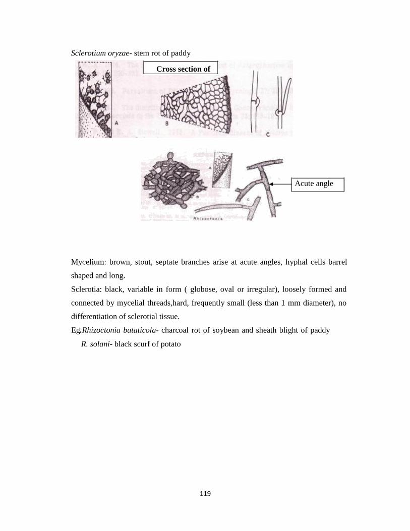
119
Sclerotium oryzae- stem rot of paddy
Cross section of
Acute angle
Mycelium: brown, stout, septate branches arise at acute angles, hyphal cells barrel
shaped and long.
Sclerotia: black, variable in form ( globose, oval or irregular), loosely formed and
connected by mycelial threads,hard, frequently small (less than 1 mm diameter), no
differentiation of sclerotial tissue.
Eg.Rhizoctonia bataticola- charcoal rot of soybean and sheath blight of paddy
R. solani- black scurf of potato

120
Bacteria and mollicutes: general morphological characters. Basic
methods of classification and reproduction in bacteria
Bacteria
Bacteria (sing. bacterium) are simplest prokaryotic unicellular microorganisms having the
common chemical composition of DNA, RNA and protein. They are highly adaptable and can
survive extremes of temperatures, pH, oxygen tension, osmotic and atmospheric pressures, and
hence found in almost all natural conditions.
Morphological characters: Being unicellular organism, bacteria may form groups of cells as
filaments. They are either motile or non-motile and lack the definitely organized nucleus.
Bacterial cell possess five shapes- (i) spherical (Micrococcus), (ii) rod-like /bacilliform (E. coli)
and (iii) spiral-shaped (Spirillum) and (iv) curved-rod (Vibrio) and (v) club-shaped (Clavibacter).
Motile cells are having long, whip-like flagella which may arise from one or both ends of the cell
(polar) or from all over the cell (peritrichous). Based on flagellar arrangement bacteria is
classified into 6 groups:
(i) Monotrichous - single polar flagellum at one end (Xanthomonas)
(ii) Amphitrichous - single polar flagellum at both ends (Pseudomonas)
(iii) Cephalotrichous - several flagella at one end (P. fluorescens)
(iv) Lophotrichous - several polar flagella at both ends (Spirillum)
(v) Sub-polar - single sub-polar flagellum (Agrobacterium)
(vi) Peritrichous - all over the cells (Erwinia)
Many bacteria have filamentous appendages called fimbriae or pilli. The size of bacteria ranges
from 1-5 µm and normal range of volume of a structural unit lies within 5-50 µm.
Structurally, a bacterial cell can be divided into following 5 regions as follows:
i.Surface appendages: Flagella and pilli.
ii. Surface adherents: Capsules and slime layers. The capsule in the outer most layer and
composed of polysaccharide or disaccharide and in some cases polypeptides. When
polysaccharide is more fluid in consistency, it forms a gelatinous slime layer around the cell
wall.

121
iii. Cell wall: It provides shape to the cell and protects underlying protoplasm having cytoplasm,
chromatin, vacuoles, globules etc. The bacterial cell wall is made up of mucopeptide (murein).
On the basis of two types of chemical composition of cell wall, bacteria are grouped into two as
Gram +ve (85% or more mucopeptide and rest is simple polysaccharide) and Gram -ve (only 3-
12% mucopeptide and rest are lipo-protein and lipo-polysaccharides)
iv. Cytoplasm and organelles: They contain soluble cytoplasmic constituents, nucleoid,
mesosomes, ribosomes, lamellae (thylakoid) or vesicles (=chromatophores, found in
photosynthetic bacteria) and some reserve materials like granules. Gas vacuoles or gas vesicles,
chlorosomes, carboxysomes and magnetosomes are also special type of organelles found in some
bacteria.
v. Special structures: Some bacteria form sporulation structures. Most characteristic spore
structures are endospores, exospores, conidia, spores(akinetes), myxospores, cysts, bdellocyst are
also formed by some genera of bacteria.
Morphological properties:
Genera Shape Size (µm) Motility
Xanthomonas Rod 0.4-1.0 x 1.2-3 Single polar flagellum
Pseudomonas Rod 0.5-1.0 x 1.5-4 One or many polar flagella
Erwinia Rod 0.5-1.0 x 1.0-3 Peritrichous
Agrobacterium Rod 0.8 x 1.5-3 Sub-polar or peritrichous(1-
4)
Clavibacter Club-
shaped/Rod 0.5-0.9 x 1.5-4
Non-motile/ motile with 1-2
polar
flagella
Ralstonia Rod Single polar flagella
Streptomyces Filamentous 0.5-2 (dia) Non-motile
Xylella Rod 0.3 x 1-4 Non-motile

122
Cultural characteristics:
Genera Characters
Agrobacteriu
m
Colonies are non-pigmented, smooth, gram -ve, oxidative
metabolism
Clavibacter Usually non pigmented, gram +ve, oxidative metabolism
Erwinia Usually non-pigmented, gram -ve, fermentative metabolism
Pseudomonas Green diffusible fluorescent, brown diffusible pigment or no pigment, gram -
ve, oxidative metabolism.
Xanthomonas Yellow, non-diffusible, gram -ve, oxidative metabolism
Ralstonia Non-pigmented, creamy white colonies, oxidative metabolism, gram-ve
Streptomyces Colonies are at first white coloured, small (1-10 mm dia), smooth, later
become powdery velvetty due to weft of aerial mycelium, gram -ve, produce
variety of pigments depending on the substrates, oxidative metabolism
Xyllela Produce long filamentous strand when cultured, gram-ve, colonies are small,
smooth or undulated margins, non-pigmented, arerobic/oxidative metabolism
Taxonomy, classification and nomenclature of bacteria: Taxonomy is the art of biological
classification which includes identification as well as description of the basic taxonomic units
(species) as completely as possible; it also determines the correct way of arrangement
(cataloguing) of these units.
Major divisions of bacteria on the basis of cell wall structure
Kingdom : Prokaryotae
Division I : Gracilicutes (thin skin/cell wall, gram negative bacteria)
Class : Scotobacteria
Anoxyphotobacteria
Oxyphotobacteria
Division II : Firmicutes (strong/durable cell wall, gram positive bacteria)
Class : Firmibacteria
Thallobacteria
Division III : Tenericutes( soft/tender cell wall, mycoplasma)

123
Class : Mollicutes
Division IV : Mendosicutes (faulty cell wall)
Class : Archaebacteria
Division and groups based on Bergey’s Manual of Systematical Bacteriology (1984)
Kingdom : Prokaryotae
Division I : Cyanobacteria(blue green algae , myxophyceae)
Division II: Bacteria
Part 1 : Phototrophic bacteria : 1Order, 3 Family, 18 Genera
Part 2 : Gliding bacteria : 2 Orders, 8 Family,21 Genera
Part 3 : Sheathed bacteria : 17 Genera
Part 4 : Budding and Appendaged bacteria : 17 Genera
Part 5 : Spirochetes : 1 Order, 1 Family, 5 Genera
Part 6 : Spiral and curved bacteria : 1 Family, 2 Genera
Part 7 : Gram-negative Aerobic rods and cocci : 5 Family, 14 Genera
Part 8 : Gram-negative Facultative Anaerobic rods : 2 Family, 17 Genera
Part 9 : Gram-negative Anaerobic bacteria : 1 Family, 3 Genera
Part 10 : Gram-negative Cocci and Coccobacilli : 1 Family, 2 Genera
Part 11 : Gram-negative Anaerobic Cocci : 1 Family, 3 Genera
Part 12 : Gram-negative Chemolithotrophic bacteria : 2 Family, 17 Genera
Part 13 : Methane producing bacteria : 1 Family, 3 Genera
Part 14 : Gram-positive Cocci : 3 Family, 12 Genera
Part 15 : Endospore forming Rods and Cocci : 1 Family, 5 Genera
Part 16 : Gram- positive Asporogenous rod-shaped bacteria : 1 Family, 1 Genus
Part 17 : Actinomycetes and related organisms : 4 Genera not assigned to any family;
1 Family with 2 Genera; 1 Order with 8 Family and 31 Genera
Part 18 : Rickettsias : 2 Order, 4 Family, 18 Genera
Part 19 : Mycoplasmas : 1 Class, 1 Order, 2 Family, 2 Genera
Nutrition and effect of physiochemical factors on growth:
A. Nutrition: Many organic substrates are the sources, two nutrients viz. carbon (C) and energy
which are important for bacterial growth. Certain bacteria e.g. Pseudomonas can use more than

124
90% organic compounds as a sole source of C and energy. Some bacteria can use two substrates
(methane and methanol by methane producing bacteria) or only one substrate (cellulose
decomposing bacteria). Bacteria need CO2 (5-10%) for satisfactory growth on organic media.
Thiamin (Vitamin B1) is also required for the growth of bacteria. However, the bacterial species
which can synthesise thaiamin, do not require any special compound.
While bacteria are grown on/in artificial medium, the medium should have balanced mixture of
necessary nutrients. Synthetic (ingredients are chemically known) and complex (ingredients are
chemically unknown) media are used for artifical culture of bacteria. Commonly used elements
in synthetic medium are K, Mg, Fe, Ca, Mn, Mo, Co, Zn, NH4 and glucose (for C). For nitrogen
fixing bacteria, N is not needed in media. Complex medium viz. nutrient medium contains
peptone and beef extract and is used to grow wide range of micro-organisms including those
microbes whose precise requirements (growth factors) are not known. Based on nutritional
requirement also bacterial classification is made using some specific terms like autotrophic,
heterotrophic, phototrophic, chemotrophic, lithotrophic and organotrophic.
B. Growth and Reproduction: In all cellular organisms, growth is achieved through cell
multiplication. Hence, multiplication of a multicellular organisms result in an increase in size,
while the multiplication of unicellular organisms results in increase in number. Growth in
bacteria takes place through multiplication where one bacterium doubles at regular intervals
(doubling time or generation time is 20-30 minutes) by binary fission (asexual reproduction).
Thus number of bacterial cells increases exponentially. Formation of endospores, cysts,
fragmentation, sporangiospores and conidia are some other means of asexual reproduction in
bacteria. The sexual reproduction in bacteria is represented by transformation, conjugation,
transduction and lysogenic conversions. The growth curve of bacteria can be plotted with four
phases viz. lag phase (slow growth), log phase (exponential growth), stationary phase (no
growth) and death phase (decline of living cells).
C. Effect of physical factors / forces on growth and reproduction:
(i) Temperature: Bacteria can survive temperatures of 0° to 85°C or even more
depending upon the species. On the basis of temperature requirement, bacteria are
divided into 3 catagories viz. psychrophilic (0-30°C, optimum 15°C), mesophilic
(min. 5-25°C , opt. 18-45°C, max.30-50°C) and thermophilic (min. 25-45°C, opt.
55°C,max.60-93°C).

125
(ii) Moisture: Bacteria are more aquatic than terrestrial and can survive in presence of
high percentage of water.
(iii) Light: Ordinary visible light does not affect bacterial activity. But different spectrum
of light viz UV light, infra-red light have different effect on the activity of bacterial
species.
(iv) Pressure: Ordianary mechanical pressure can not affect bacterial cells. Principle of
osmosis is the best used pressure for destruction of bacteria.
(v) Hidrogen-ion concentration: Suitable pH range for bacterial growth and
reproduction is 5.0 to 9.0
Bacterial Genetics and Variability: Knowledge on the genetic system of bacteria was dull till
1940. Prior to this period, no definite nucleus had been demonstrated in bacteria although
variability in bacterial cells was recognized well before. Only the development of science in
Molecular Biology helped to recognize transfer of genetic material i.e. DNA to the daughter cells
at the time of binary fission.
Variability among bacteria is resulted from the following processes:
(i) Conjugation: Two compatible bacterial cells come into contact. Then the recipient
female cell (F-) receives the DNA from the donor male cell (Hfr). Thus genetic make
up of both the cells is changed.
(ii) Transformation: The bacterial cell absorbs DNA exuded by compatible cells or freed
by dissolution of the cell-wall into the external medium.
(iii) Transduction: This process is a “phage-mediated genetic transfer”. The bacterial
viruses (bacteriophages or phage) can acquire DNA from one cell and transfer it to the
other cells attacked by them. If attacked cell is not destroyed due to infection by the
phase, it reproduces to form new races with different genetic character.

126
(iv) Lysogeny: It involves association of genetic material of a virus with that of bacterium.
Although it is different from above three processes, it also provides a permanent
genetic modification of the bacterial genome.
Mollicutes
Mollicutes is a class of bacteria distinguished by the absence of a cell wall. The word
"Mollicutes" is derived from the Latin mollis (meaning "soft" or "pliable"), and cutis (meaning
"skin"). Individuals are very small, typically only 0.2–0.3 μm (200-300 nm) in size and have a
very small genome size. They vary in form, although most have sterols that make the cell
membrane somewhat more rigid. Many are able to move about through gliding, but members of
the genus Spiroplasma are helical and move by twisting. The best-known genus in the Mollicutes
is Mycoplasma.
Mollicutes are parasites of various animals and plants, living on or in the host's cells. Many
cause diseases in humans, attaching to cells in the respiratory or urogenital tracts, particularly
species of Mycoplasma and Ureaplasma. Phytoplasma and Spiroplasma are plant pathogens
associated with insect vectors.
Whereas formerly the trivial name "mycoplasma" has commonly denoted any member of the
class Mollicutes, it now refers exclusively to a member of the genus Mycoplasma.
History of the classification
The classification of the Mollicutes has always been difficult. The individuals are tiny, and being
parasites, they have to be cultivated on special media. Until now, many species could not be
isolated at all. In the beginning, whether they were fungi, viruses, or bacteria was not clear. Also,
the resemblance to L-forms was confusing. At first, all members of the class Mollicutes were
generally named "mycoplasma" or pleuropneumonia-like organism (PPLO). Mollicutes other
than some members of genus Mycoplasma were still unidentified. The first species of
Mycoplasma/Mollicutes, that could be isolated was Mycoplasma mycoides. This bacterium was
cultivated by Nocard and Roux in 1898.

127
In 1956, D.G. Edward and E.A. Freundt made a first proposal for classifying and naming PPLOs.
They left undecided, however, whether they belong to the bacteria (prokaryotes, in 1956 called
"Schizomycetes") or to the eukaryotes. As type species (name-giving species) of the
PPLOs/mycoplasmas, Edward and Freundt proposed Mycoplasma mycoides, being the causative
organism of bovine pleuropneumonia and referring to the pleuropneumonia-like organisms. Until
then, Mycoplasma mycoides was known as Asterococcus mycoides, but later that name was not
recognized as valid. In their publication of 1956, they described 15 species of Mycoplasma.[9] In
1967 the class Mollicutes, containing the order Mycoplasmatales, was proposed by the
Subcommittee on Taxonomy of the Mycoplasmata.[5] Now, the name Mycoplasma should
exclusively be used for members of the genus Mycoplasma, rather than the use as a trivial name
for any mollicute. As the trivial name has been used in literature for a long time, this is yet not
always the case.

128
Viruses: nature, architecture, multiplication and transmission
Viruses
Matthew (1981) defined a virus as “a set of one or more nucleic acid template molecules,
normally encased in a protective coat, or coats of protein or lipoprotein, which is able to organize
its own replication within suitable host cells. Within such cells, virus production is (a) dependent
on the host’s protein synthesizing machinery, (b) organized from pools of the required materials
rather than by binary fission, and (c) located at sites which are non separated from the host cell
contents by a lipoprotein, bilayer membrane”.
Many plant diseases which are now known to be caused by viruses had been encountered long
ago. The causes of those diseases were not known. The first breakthrough was made by Adolf
Mayer in 1886, in the Netherlands, while studying the highly contagious, mysterious disease of
tobacco which he called “Mosaikkrankheit” i.e. mosaic like disease. He found that healthy plants
could be infected by injecting the sap of diseased plants. He also observed that the unknown
agent could be inactivated by boiling the sap. He concluded that the disease was the
manifestation of a bacterium. In 1892, Ivanovsky confirmed Meyer’s report and further showed
the sap to remain infections even after passage through bacteria-proof filter. However, he
claimed the incitant to be a microbe. But Martinus Beijerinck realized the causal agent to be
something novel. His results further confirmed the findings of Meyer and Ivanovsky and also
showed that the incitant could diffuse into an agar gel. Based on all these findings, Beijerinck, in
1898, concluded that the mysterious pathogen was not a bacterium, but a contagium vivum.
fluidum i.e. contagious infective material or infectious living fluid. He thought the contagium to
be able to reproduce itself in living plants and referred it as a virus.
Architecture of viruses and viriods
Morphologically, virus particles are (i) isometric (spherical, polyhedral) and (ii) anisometric
(rigid or flexuous rods, bacilliform or bullet-shaped). Many isometric viruses have symmetric
polyhedra which are either of three cubic symmetry i.e. tetrahedral, octahedral or icosahedral.
Isometric particles measure the diameter 17nm (satellite virus of tobacco necrosis virus) to 70nm
(reoviruses). The bacilliform viruses measure up to 300 nm length x 95 nm width (rhabdovirus
group). The rod-shaped viruses having short rigid rod measure 114-215 nm length x 23 nm width

129
(the tobraviruses) and those with long flexuous particles measure up to 2,000 nm length x 10 nm
in width (the closteroviruses). The rod shaped particles of tobacco mosaic virus (TMV) consist of
protein sub-units (capsomeres) built up in a regular, helical array, with the RNA chain compactly
coiled in a corresponding helix on the inside of the protein sub-units. The protein coat (capsid)
and RNA genome surround an axial hole or canal. In membrane-bound viruses, the inner
nucleoprotein core is called as nucleocapsid.
Viroids are smallest (1.1-1.3 x 10⁵ mol. wt.), simplest and non-encapsidated RNA. They consist
of a single molecular species of circular or linear form.
Chemical composition: Plant virus particles consist of infectious nucleic acid (the genome),
which is encapsidated within a protective protein coat or shell. The genome, essential for virus
replication, is composed of ribonucleic acid (RNA in most groups of viruses) and
deoxyribonucleic acid (DNA in the caulimovirus and geminivirus groups). The RNA and DNA
may be single stranded (ss) or double stranded (ds) Besides these two basic components, an
envelop of lipid or lipoprotein membrane is present in some plant viruses. Other components are
metallic ions and polyamines present in varying amounts. Some enzymes are found in reoviruses
and rhabdoviruses. Water constitutes 10-50 percent of the mass of virus particle.
The nucleic acid may be present as a single continuous strand (single molecular species) in a
particle. It is called mono-partite genome. Some nucleic acid genomes have two or more pieces
(molecular species) in different particles; usually they are not always encapsidated within
separate protein shells. Such genomes are termed as bi-, tri-, or multi-partite or the viruses with
divided genome.
In some RNA viruses, the genetic information is divided into two or more parts. They are called
multi-component viruses and the individual components are not infectious alone. Hence two or
more genomic elements are needed to cause infection and replication.
The genomic organization of viruses depicts structure and function of genes or cistrons. Some
triplet bases called codons are responsible for expression of genes. There are two types of
codons, (i) initiation codons (AUG, GUG) and (ii) termination codons (UAG, UAA, UGA) and
they control functions of genes and translation products.
Nomenclature of viruses and classification: A number of addition and deletion was made in
naming viral pathogens. Linnaean style of binomial nomenclature is not followed. Instead, plant

130
pathogenic viruses are named based on the common or vernacular name of the affected host
plant such as tobacco mosaic virus, rice dwarf virus, caulimoviruses, potato virus X and Y etc.
Later, these vernacular names were further simplified by abbreviating them such as TMV for
tobacco mosaic virus, CMV for cucumber mosaic virus, potex virus for potato virus X, CaMV
for cauliflower mosaic virus etc. In addition to such names, a system of cryptogams was
introduced to give concise information on the properties (immediate summary) of one virus for
example:
TMV = R/1 : 2/5 : E/E : S/O
1st term : Type of nucleic acid (RNA, DNA)/number of strands of nuleic acid (1=ss, 2=ds)
2nd term : Molecular weight of nuleic acid in millions/ % of nuleic acid in infective
particle
3rd term : Outline of particle shape (E=elongate, S=spherical, B=bacilliform)/outline of
nuclear capsid (E, S, B)
4th term : Type of host infected (B=bacteria, F=fungus, I=invertebrate, S=seed plant)/ type
of vector (Ap=aphid, Au=leafhopper, Cl=beetle, Fu=fungus, Ne=nematode,
Th=thrips, W=whitefly, O=spread without vector, Se=seed transmitted)
A system of plant virus classification was introduced by International Committee on Taxonomy
of Viruses (ICTV) based on the characteristics such as morphology of virus particle, type and
quantity of nucleic acid, genomic structure and type of vector. For example
Classification based on particle morphology and type of nucleic acid:
I. Elongated, Helical, ss RNA
A. Rigid
(a) Monopartite
(b) Multipartite
B. Flexuous, all monopartite
II. Isometric
A. Single stranded RNA
(a) Monopartite
(b) Multipartite
(b1) With envelope
B. Double stranded RNA

131
C. Single stranded DNA
D. Double stranded DNA
III Bacilliform
A. Without envelop
B. With envelop
Grouping of plant viruses
Like families and genera cited in the classification of fungi and bacteria, plant virologists have
been using “groups” for viruses. A total of 26 groups have been accepted by ICTV and some of
those are,
1. Alfalfa mosaic virus group
2. Bromoviruses
3. Carlaviruses
4. Caulimoviruses
5. Comoviruses
6. Dianthoviruses
7. Geminiviruses etc.
There are about 11 unclassified virus groups are known, e.g.
1. Barley yellow mosaic virus group
2. Carnation mottle virus group
3. Rice stripe virus group
4. Satellite virus group etc.
Criteria for identification of viruses
Viruses causing plant diseases can be identified correctly by number of ways:
1. Behaviour in host: Host range, symptoms and their types, tissue restriction, type and
location of intracellular inclusion bodies, seed transmissibility.
2. Vector relation: Taxa, acquisition and inoculation thresholds, persistence in vector,
multiplication in vector, modes of transmission (e.g. transovarial transmission).
3. Particle properties: Shape and symmetry, size, presence or absence of envelope,
capsomeric structure, sedimentation properties (number of components and

132
sedimentation co-efficient); coat protein properties (no. of polypeptides and their
molecular weight); properties of nucleic acid (RNA, DNA, strandedness, no. molecules
and molecular weight, presence or absence of 5’-terminal M7 Gppp, 5’-terminal VPg, 3’-
terminal Poly (A)); electrophoretic mobility; isoelectric point, serological relationship.
4. In vitro properties in crude sap: This is determined by the tests viz. thermal inactivation
point (TIP), dilution end point (DEP), longevity of virus in vitro (LIV).
5. Cross-Protection tests: Virus identity and strain relationship can be done. However, this
sort of test is less applied now-a-days.
Important techniques for virus detection and identification:
1. Electron microscopy: used to know shape, symmetry, size, presence or absence of envelope
and capsomeric structure.
2. Immunosorbent electron microscopy (ISEM).
3. Serology: The technique includes
(i) Precipitin tests (precipitin-tube test, precipitin-ring test, microprecipitin test ).
(ii) Immunodiffusion tests (single, radial diffusion test, gel double-diffusion or
Ouchterlony test)
(iii) Agglutination test (slide-agglutination or chloroplast-agglutination test).
(iv) Enzyme-linked immunosorbent assay (ELISA): It includes indirect ELISA and
direct double antibody sandwich (DAS) ELISA.
(v) Dot immunobinding assay (DIBA)
(vi) Molecular hybridization analysis (spot hybridization or dot-blot technique)
(vii) Monoclonal antibodies (MAb)
Multiplication and infection nature of plant viruses: The events in virus infection involve
three steps: adsorption, penetration or entry and uncoating or disassembly. The initial contact
between virus particle and host cell is referred to as adsorption or entry. The process during
which the virion or its nucleic acid passes into the cytoplasm of the cell is known as penetration
or entry. Uncoating is the removal of various components of the mature virion and subsequent
release of viral genome and other constituents that plays a major role in establishing infection.

133
(i)Multiplication of virus in plants: The replication of RNA and DNA plant viruses differ from
diffent groups of viruses or for individial viruses. The mechanism known for different groups of
plant viruses are – ssRNA virus- monopartite genomes, ssRNA virus-bipartite genome, ssRNA
virus- tripartite genome, dsRNA virus- monopartite genome, satellite viruses, helper viruses and
satellite RNAs, dsDNA viruses. The progeny RNA moves out to cytoplasm where the assembly
sythensized proteins and encapsidation of virion take place. The process continues till the host is
alive and a large number of new virus particles are formed.
(ii)Accumulation and movement of viruses in plant: The nascent virus appears in the cell
about 10 hours after infection. The concentration of virus varies based on the type of virus,
temperature, nutrition and duration of light. The virions are found aggregated in amorphous or
crystalline forms or they are dispersed in cytoplasm and nucleus.
In plant system, two stages of virus movement have been recorded. These are:
a. Cell to cell or short distance movement: This type of virus movement takes place through
protoplasmic bridges, the plasmodesmata. The plasmodesmata selectively allows passage
to the macromolecules and thus virus can move through it with the help of virus-coded
‘movement protein’ mechanism.
b. Movement from one part to another part of plant: This is a long distance movement of the
viruses taking place through vascular system. The movement is faster in elongated young
cells than in round and older cells. Moreover, virus moves fast at high temperature
because the protoplasmic streaming and cellular activities are higher at high temperature.
The nature of cells (parenchyma, xylem, phloem, sieve tubes) also determined rate
movement of viruses in plant.
As a result of the multiplication and distribution of the viruses in the plant system, the host
plant(s) are infected and exhibited varying degrees of disease symptoms.
Transmission of plant viruses: Viruses are distributed or transmitted from the infected plants to
the healthy ones in various ways in nature. As the plant viruses can not penetrate cuticle of their
hosts and hence they can enter into the host tissue through wounds only. The means of
transmission are:
1. Mechanical transmission: The sap of the infected plant is manually transferred to the
healthy plants. It is the easiest method of experimental inoculation.
2. Graft transmission: In this practice, if either the scion (shoot portion) or stock (root stock)

134
is infected, the virus usually moves to the healthy partner which may later express visible
symptoms of disease.
3.Transmission through vectors
(i) Insects: Some insect species are the vector of plant viruses which can carry/ transmit
viruses from infected plants to the healthy plants e.g. aphid (potato virus Y, PLVR),
white flies (tobacco leaf curl), beetles (cowpea mosaic virus), mealy bugs (cacao
mottle leaf), thrips (tomato spotted wilt), lace bugs (sugar beet viruses), mites
(sterility mosaic of arhar), leaf hoppers (beet curly top, rice tungro etc.), planthoppers (maize
mosaic, maize rough dwarf), tree hopper (tomato pseudo curly top).
(ii) Nematodes: Five genera of nematodues viz., Xiphinema, Longidorus,
Paralongidorus, Trichodorus and Paratrichodorus can transmit plant viruses.
(iii) Fungi: Some species of fungi can also transmit viruses e.g. Olpiduim brassicae
(tobacco necrosis), O. cucurbitacearum (cucumber necrosis), Polymyxa graminis (oat
mosaic, wheat mosaic), P. betae (beet necrotic yellow vein) and Spongospora
subterranea (potato mop top) etc.
4. Dodder transmission: Many viruses can be transmitted through dodder (Cuscuta spp.).
Dodder transmission is used in the laboratory to transfer viruses from the hosts.
5. Transmission through seeds and pollens: Seed coat (testa), embryo, and also pollens of
some plants can transmit viruses. e.g. alfalfa mosaic, barley stripe mosaic, bean common
mosaic, lettuce mosaic are transmitted by both seeds and pollens of Medicago sativa,
Hordeum vulgare, Phaseolus vulgaris and Lactuca sativa, respectively.
Basic characteristics of insect transmission: Virus transmission, specially in case of the aphid
transmission, takes place by three ways viz. non-persistent (stylet borne), semi-persistant and
persistent (circulative). These modes of transmission have been distinguished on the basis of
acquisition feeding time, inoculation feeding period, latent period and multiplication or
nomultiplication of the viruses within their vector etc. Apart from aphids, semi-persistent mode
is recorded in mealy bug (Planococcoides njalensis - cacao swollen shoot virus) and leaf hopper
(Graminella nigrifrons - maize chlorotic dwarf virus; Nephotettix impicticeps - rice tungro virus)
and persistent mode is recorded in leaf hopper (N. cincticeps - rice dwarf virus; Agallia
constricta - wound tumer virus), plant hopper (Peregrinus maidis - maize mosaic virus), tree
hopper (Micrutalis malleifera - tomato pseudo curly top virus), beetles (Ceratoma trifurcata –

135
cowpea mosaic virus; Phyllotreta sp. – turnip yellow mosaic virus) and thrips (Frankliniella
fusca – tomato spotted wilt virus).
Some viruses pathogenic to plant can be transmitted only in presence of a second virus (helper
virus) in the host and this type is called dependent transmission. For example, aphid (Myzus
persicae) transmits potato aucuba mosaic virus only.

136
Nematodes: General morphology and reproduction, classification of
nematode Symptoms and nature of damage caused by plant nematodes
(Heterodera, Meloidogyne, Anguina etc.)
Introduction
Nematology is an important branch of biological science, which deals with a complex,
diverse group of round worms known as Nematodes that occur worldwide in essentially all
environments. Nematodes are also known as eelworms in Europe, nemas in the United States
and round worms by zoologists. Many species are important parasites of plants and animals,
whereas others are beneficial to agriculture and the environment. Nematodes that are parasites
of man and animals are called helm inthes and the study is known as Helminthology. The plant
parasitic forms are called nematodes and the study is known as Plant Nematology. The name
nematode was derived from Greek words nema (thread) and oides (resembling). Annual crop losses due
to these obligate parasites have been estimated to be about $ 78 billion wordwide and $ 8 billion for U.S.
growers. The estimated annual crop loss in Tamil Nadu is around Rs. 200 crores.
The soils in a hectare of all agro ecosystem typically contain billions of plant parasitic as well
as beneficial nematodes. The damage to plants caused by nematodes is often overlooked because the
associated symptoms, including slow growth, stunting and yellowing, can also be
attributed to nutritional and water related disorders.
Even though nematodes occupy nearly every habitat on earth, they are remarkably
similar in morphology and life stages. Despite their structural complexity, certain basic
principles are common to all nematodes. Nematodes are triploblastic, bilaterally symmetrical,
unsegmented, Pseudocoelomate, vermiform and colourless animals. The plant parasitic nematodes are
slender elongate, spindle shaped or fusiform, tapering towards both ends and circular in cross section.
The length of the nematode may vary from 0.2 mm (Paratylenchus) to about 11.0mm (Paralongidorus
maximus). Their body width vary from 0.01 to 0.05 mm. In few genera, the females on maturity assume
pear shape (Meloidogyne), globular shape (Globodera), reniform (Rotylenchulus reniformis) or saccate
(Tylenchulus semipenetrans). The swelling increases the reproductive potential of the organism. Radially
symmetric traits (triradiate, tetraradiate and hexaradiate) exist in the anterior region. The regions of
intestine, excretory and reproductive systems show tendencies towards asymmetry.
The nematodes have one or two tubular gonads which open separately in the female and into the rectum
in the male which also have the copulatory spicules.
The free living saprophytic nematodes are generally larger in size.
The animal and human parasitic helminthes may have length of few centimeters to even a meteer or
more. The helminth parasitising whale fish is about 27 feet long. The study on these animal and human
parasites are known as Helminthology.

137
Nematode exhibits considerable variation in their external and internal structure. Despite this structural
complexity, certain basic principle is common to all nematodes
General shape & size
Nematodes : Triploblastic, bilaterally symmetrical, unsegmented, colourless, pseudo coelomate,
vermiform and circular in cross section animals.
Fig: General Morphology of Nematode
Shape: Nematode show great variation in their morphological characters. The plant parasitic nematodes
are slender, spindle shaped or fusiform organisms. The nematodes when relaxed with gental heat either
lie straight (Pratilenchus) or slightly curved (Hoplolaimus) or curved in ‘C’ shaped (Tylenchorynchus) or
form a spiral (Helicotylenchus). Sexual dimorphism in few species. Lemon, pear, kidney, saccate shape.
Size: Their size may vary from 0.2 mm to about 12 mm length with an average of 0.01 mm and 0.5 mm
in breadth (1 to 15 % to length). Males are smaller than females.
Body regions: The nematode body is not divisible into definite regions as that of insects, however there
are certain subdivisions like anterior part of the body having the mouth, lips and stoma is called head and
it is continuous with main body. The portion between the head and the base of oesophagus is the neck.
The part of the body beginning from the anus or cloca and extending up to posterior extremity is the
‘tail’. Longitudinally, the body can divided into four zones: the ventral which bears the natural openings
like excretory pour, anus or cloaca and vulva; the side apposite to the ventral is dorasal. The other two
sides are right and left laterals.
Lip region: The lip region is also called as head exhibits great variation which may be used taxonomic
purpose.
Tail region: It is the post-anal elongation of the body present in all stages of nematodes.

138
General structure of nematode:
The body of nematode is tubular which may be divided into three regions I) Outer body tube or
body wall
Inner body tube –Digestive system
body cavity– Reproductive system, Nervous system, Excretory
system
Outer body tube
Exoskeleton or cuticle,
Hypodermis and
Muscle layer.
Exoskeleton or cuticle:
It is outermost covering of body wall which is non-cellular, semipermeable
and tough layer secreted by the epidermal cells. It invades all natural opening of body including the
mouth, rectum, cloaca, vagina, excretory pore, amhids and phasmids.
The cuticle of many nematode species has markings on the surface. They are varied and complex and
often used by taxonomist in identification of nematode species. The cuticular lining/markings are
categorized in different types are as follows.
Cuticular lining or markings:
Punctations – They are commonly appearing as minute or round areas which are arranged in pattern. It
acts as a structure for strengthening cuticle and transport of proteins.
Transverse markings or Annules or Striations – There are several transverse lines present on the
surface of cuticle. These markings are exhibit on most of the plant parasitic nematodes and often used for
identification. Annulations give segmented appearance e.g. scales in Criconemoides & perineal pattern of
root-knot nematodes. Necessary for dorsoventral undulatory movement.
Longitudinal markings – These markings are the lines on the cuticle, which runs longitudinally
throughout the nematode body.
i) Ridges – These are raised areas, which run length of the body and occur on sub-median as well as
lateral surface.
ii) Alae – These are thickening or projections occur in lateral or sub-lateral region. They assist in
locomotion. There are three types of alae
Caudal alae – These are found in the posterior region and restricted to males as copulatory bursa.
Cervical alae – These are confined to anterior part of the nematode body. Cervical alae are found in
some species of marine nematodes.
Longitudinal alae – These are limits to the lateral fields. They are transverse by striations or furrows
varying in number from one to twelve which provide locomotion and may permit slight change in the
width of nematode.
Cuticular layering or covering:

139
The nematode cuticle is basically three layer structure and composed of (a) Cortical layer, (b) Median
layer and (c) Basal layer.
Cortical layer – It is often divided into external cortical layer and internal cortical layer. The surface of
external cortical layer is exposed to the environment. This layer is very thin measuring about 25 to 40
mµ. The external layer has been considered to be kertatine (protein) chemically. In cyst nematode the
cuticle of the female on maturity becomes tough and leathery to form cyst which protect eggs under dry
conditions.
Median layer – The average thickness of the median layer is 0.1 µ in the larva of Meloidogyne and
Heterodera. Chemically the median layer consist of protein, which resembles collagen (Non osmophilic
collagen protein).
Basal layer – It consist of regularly arranged vertical rods or striations. It is composed of protein with
very close linkage between the molecules, resulting in resistant layer which protect the nematode from
outer environment. The thickness of basal layer varies from 125 to 500 mµ (Osmophilic protein close to
keratine)
Functions of cuticle:
Protects the nematode from harsh environment.
Serves as exoskeleton
Provide mechanism of movement of the nematode through the soil and plant tissue.
Hypodermis –
The hypodermis is cellular or partially cellular layer. It secretes the cuticle. It
lies between cuticle and somatic muscle layer. It is important metabolic active part of the nematode.
Forms 4 cords (dorsal, ventral and two lateral). Contains hypodermal glands
Muscle layer -
It is arranged in a single layer. The muscle cells are spindal shaped and attached to the hypodermis
throughout their length. It is well connected to the nervous system. The stimulation of the muscles by
dorsal and ventral nerves cause contractions in the dorso-ventral plane and result in the characteristic
scinusodial movement of nematode.
On the basis of arrangement of the basic cells, following three types are identified:
a. Holomyarian: Having two muscle cells in each zone.
b. Meromyarian: Two or five muscle cells in each interchordal zone.
c. Polymyarian: More than five muscle cells in each zone
1. Reproductive system of nematode:-
- The males are generally slightly smaller than females.
- The nematodes are dioecious or amphigonus having a separate male
and female within a species.
- Generally the males are lesser in number than females or even be
completely absent. This indicates a tendency towards hermaphroditism
and parthenogenesis.
A. Female Reproductive system:-

140
• Monodelphic- The nematodes may have a single ovary the female is called as
monodelphic.
• Didelphic- The nematodes may have two ovaries then the female is called as
didelphic.
• Prodelphic- When a single gonad is present, it may be either directed
towards anterior to vulva then female is called as prodelphic.
• Opisthodelphic- The gonad either directed towards posterior to vulva then
female is opisthodelphic.
• Amphidelphic- The two ovaries are opposite to one another, such as one is
anteriorly directed and other posteriorly directed.
Fig. Prodelphic, Opisthodelphic and Amphidelphic

141
The female reproductive system typically consists of ovary, oviduct, uterus, vagina and vulva.
Vagina
Ovary
Fig. Female Reproductive System
(i) The ovary-
It is hollow elongate tube. The apical end of ovary has a cap cell at the tip
which is called as germinal or zone of multiplication in which rapid cell division takes
place to give rise germinal cells. This region is followed by growth zone which
constitutes the greater part of ovary. The oocytes or germ cells in this zone become
big and ripe which are generally arranged in single rows. After maturity they are
called oogonia.
(ii) Oviduct-
Next to growth zone of ovary the gonad has oviduct. The oocytes when ripe
they pass in to oviduct. Oviduct may serve as spermathica in some nematode.
However in others, the spermathica is in the proximal part of uterus or in the post-
vulvar sac at the distal end of gonad.
(iii) The uterus-
It is the largest and most complex part of the gonad, serves and function of
fertilization, egg shell formation and laying of eggs. As started above, the upper part
of uterus serves as spermathica in some nematodes.
(iv) Vagina-
The uterus entered in common vagina, which is a short, narrow and flattened
tube lined with cuticle and provided with muscles.
(v) Vulva-
The vagina opens through female gonopore, the vulva. The eggs are expelled
through a vulva which is normally situated middle of the body.
B. Male Reproductive system:-
• Monarchic- The nematode may have one testis are called monarchic.
• Diarchic- The nematode may have two testis are called diarchic.
Vulva
Uterus

142
The male reproductive system generally consists of three primordial parts: the
testis, seminal vesicle, and vas deference.
(i) The Testis-
In the testis the germinal and growth zone can be easily distinguished. In
germinal zone Spermatogonial division takes place, while in growth zone,
spermatocytes increases in size. The spermatocytes are arranged in single or double
rows.
(ii) Vas deference-
It consist of an anterior glandular region and posterior muscular region and
containing the ejaculatory duct at the posterior end.
(iii) Ejaculatory duct-
The ejaculatory duct helps in the ejection of sperms during fertilization. It
tapers gradually and opens ventrally into the cloca. The cloca is provided male
copulatory structures such as spicules, gubernaculums etc.

143
Classification of Nematodes
Class-
Adenophorea
Class- Secernentea
Sub-class: Enoplia Sub-class: Chromatorea
Super
Order: Marenoplica Super Order:
Terrenoplica
Order: Enoplida Order: Dorylaimida Order: Araeolaimida
Oncholaimida Isolaimida Chromadorida
Tripylida Mononchida Desmoscolecida
Stichosomida Desmodorida
Monhysterida
Class- Secernentea
Sub-class: Rhabditia Sub-class: Spiruria Sub-class: Diplogasteria
Order:
Rhabditida Order:
Ascaridida
Order:
Spirurida
Family Family Family
Heterorhabditidae Ascarididae Drocunculidae
Steinernrmatidae Dictophymatidae Filariidae
Order: Tylenchida Order: Diplogasterida
Family
Diplogasteridae
Sub-order:
Tylenchina Sub-order:
Aphelenchina
Sub-order:
Sphaerulariina
Family- Aphelenchina
Family- Fergusobiidae
Super family: Criconematoidae Super family: Tylenchoidae
Family: Anguinidae, Belonolaimidae Family: Criconematidae
Dolichodoridae, Heteroderidae Tylenchulidae
Tylenchidae, Hoplolaimidae

144
Ecological classification of nematodes
There are two major classes
I. Above ground feeder
II. Below ground feeder
I. Above ground feeder
a. Feeding on flower buds, leaves and bulbs
i) Seed gall nematode: Anguina tritici
ii) Leaf and bud nematode: Aphelenchoides
iii) Stem and bulb nematode: Dictylenchus
b. Feeding on tree trunk
i) Red ring nematode: Rhadinaphelenchus cocophilus
ii) Pine wilt nematode: Bursaphelenchus xylophilus
II. Bellow ground feeder
It is again classified in to three classes
I) Endoparasitic nematodes
II) Semiendoparasitic nematodes
III) Ectoparasitic nematodes
a) Endoparasitic nematodes
The entire nematode is found inside the root and the major portion of
nematode body found inside the plant tissues. They are two types
1) Migratory endoparasite:- These nematodes move in cortical parenchyma of
host root. While migrating they feed on cells, multiply and cause necrotic
lesion. Example, Pratylenchus spp., Radopholus spp. and Hirschmanniella spp.
2) Sedentory endoparasite: - the second stage larvae penetrate the root lets and
become sedentary throughout the life cycle, inside the root cortex. Examples,
Heterodera spp. and Meloidogyne spp.
b) Semiendoparasitic nematodes
The anterior part of nematode, head and neck being permanently fixed in the
cortex and the posterior part extends free into the soil. Examples, Rotylenchulus
reniformis and Tylenchulus semipenetrans.
c) Ectoparasitic nematodes
These nematodes live freely in the soil and move closely or on the root surface,
feed intermittently on the epidermis and root hair near the root tip. They are two types,
1) Migratory ectoparasites:- These nematodes spend their entire life cycle free in
the soil, feeding externally on the host plants, deposit eggs in soil. When the

145
roots are disturbed they detach themselves. Examples, Criconemoides spp.,
Paratylenchus spp. and s spp., etc.
2) Sedentory ectoparasites:- In this type of parasitism the attachment of
nematode to the root system is permanent but for this, it is similar to the
previous one. Examples, Hemicycliophora arenaria and Trichodorus spp., etc.
1) Root-knot Nematode, Meloidogyne spp.
Systematic Position:-
Order - Tylenchida
Sub order - Tylenchina
Super family - Tylenchoidea
Family - Heteroderidae
Sub family - Meloidogyninae
Genus - Meloidogyne
Species - i) incognita
ii) javanica
iii) arenaria
iv) hapla
Parasitism & Habitat:-
i) Females and III & IV stage of larvae are Sedentory endoparasites.
ii) Males and II stage larvae are migratory.
Morphological characters:-
i) Body - Elongate larvae and male Typically saccate, spheroid with a distinct neck
in females.
ii) Stylet - In males, Strong with rounded knob & In females, more slender than
males.
iii) Oesophagous – With
large median bulb
followed by short
isthumus.
iv) Excretory pore - Often
seen with part of
excretory tube in the
area between posterior
part of stylet knobs and
opposite to median bulb.
v) Vulvas & anus -
Females typically
opposite to neck and
surrounded by a pattern
of fine lines like human
fingerprint.(Perennial
pattern)
vi) Spicule - Very near the
terminus of males Bursa is absent.
- Yellowing of leaves
- Stunted growth
- Reduced vigor

146
- Reduced size & number of fruits
- Gall formation
- Multinucleate cell – Giant cell (Nurse cell)
- Hypertrophy – Enlargement of cell
- Hyperplasia – Multiplication of cell
Control:-
- Two to three deep Ploughing
- Rotation with cereal crops
- Apply carbofuron (Furdan 3G) @ 7 g/m2
- Resistant varieties of Tomato eg. Hisar Lalit, PNR 7
2) Reniform Nematode, Rotylenchulus reniformis
Systematic Position:-
Order - Tylenchida
Sub order - Tylenchina
Super family - Hoplolaimoidea
Family - Hoplolaimidae
Sub family - Hoplolaiminae Genus - Rotylenchulus Species - reniformis
Parasitism & Habitat:- Females are Semiendoparasitic on many plants.
Morphological characters:-
i) Body - Males and immature
females are slender and small,
adult females are kidney
shaped
ii) Oesophagous - Dorsal
oesophageal glands opens
about one stylet length
posterior to stylet knobs.
Symptoms:-
Yellowing of leaves,
delayed germination, reduced
plant growth and vigor, stunted
growth, browning of roots due to
penetration of nematode are the
general symptoms of this
nematode. Young and tender
plants are more vulunerable to
nematode attack.
3) Root-lesion Nematode, Pratylenchus spp.
Systematic Position:-
Order - Tylenchida
147
Sub order - Tylenchina
Super family - Tylenchoidea
Parasitism & Habitat:-
- Migratory endoparasites
- Feeding on root cortex of many crop/plant
- All stages found in root or soil.
Morphological characters:-
i) Body length - 0.4-0.8 mm.
ii) Lip region - Slightly set-off from body.
iii) Stylet - Typically short, strong with massive knob.
iv) Ovary - Monodelphic
v) Vulva - Posterior fourth of the body (75-80%).
vi) Tail - Nearly round to pointed and in males, the tail has bursa.
vii)
Symptoms:-
Late emergence of seedlings, less germination and stunted growth with
necrotic lesions on the root surface which are initially small coalesce at the later stage
and cause death of the rootlets. Root system is reduced.
Control:-
- Raise nursery in nematode free soil
- Pull and burn infected plants
-
4) Spiral Nematode, Helicotylenchus spp.
Systematic Position:-
Order - Tylenchida
Sub order - Tylenchina
Super family - Tylenchoidea
Family - Hoplolaimidae
Sub family - Rotylenchoidinae
Genus - Helicotylenchus
Parasitism & Habitat:- Endoparasitic and ectoparasitic on many plants
Family - Pratylenchidae
Sub family - Pratylenchinae
Genus - Pratylenchus
Species - i) coffeae - Citrus, Banana & coffee
ii) zeae - Maize
iii) thornei - Pulses
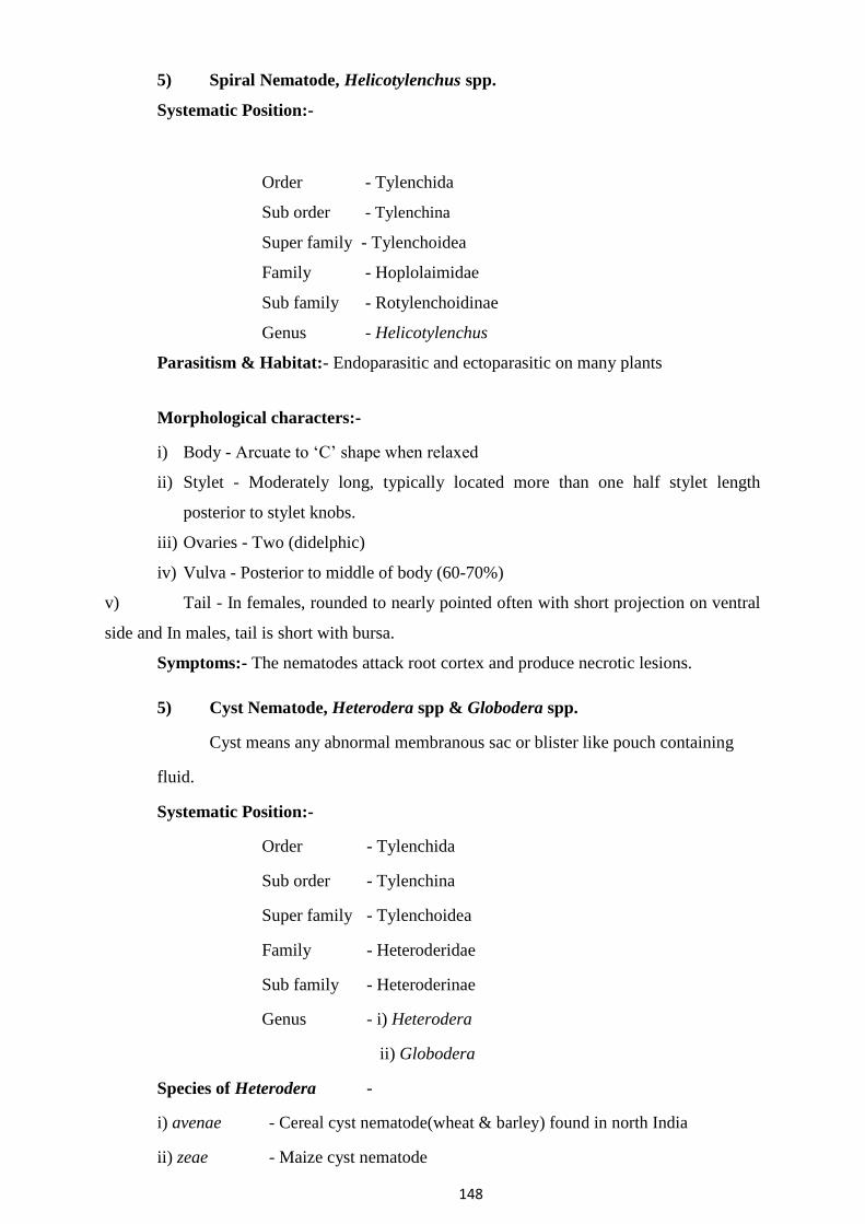
148
5) Spiral Nematode, Helicotylenchus spp.
Systematic Position:-
Order - Tylenchida
Sub order - Tylenchina
Super family - Tylenchoidea
Family - Hoplolaimidae
Sub family - Rotylenchoidinae
Genus - Helicotylenchus
Parasitism & Habitat:- Endoparasitic and ectoparasitic on many plants
Morphological characters:-
i) Body - Arcuate to ‘C’ shape when relaxed
ii) Stylet - Moderately long, typically located more than one half stylet length
posterior to stylet knobs.
iii) Ovaries - Two (didelphic)
iv) Vulva - Posterior to middle of body (60-70%)
v) Tail - In females, rounded to nearly pointed often with short projection on ventral
side and In males, tail is short with bursa.
Symptoms:- The nematodes attack root cortex and produce necrotic lesions.
5) Cyst Nematode, Heterodera spp & Globodera spp.
Cyst means any abnormal membranous sac or blister like pouch containing
fluid.
Systematic Position:-
Order - Tylenchida
Sub order - Tylenchina
Super family - Tylenchoidea
Family - Heteroderidae
Sub family - Heteroderinae
Genus - i) Heterodera
Species of Heterodera
ii) Globodera
-
i) avenae - Cereal cyst nematode(wheat & barley) found in north India
ii) zeae - Maize cyst nematode

149
iii) cajani- Pigeon pea cyst nematode (tur, mung, Udid & cowpea)
iv) oryzicola - Rice cyst nematode (rice & banana) found in Kerala, M.P., Orissa &
West Bengal.
Species of Globodera -
i) rostochinensis- Potato cyst nematode or Golden nematode
ii) pallida
Host plants - Potato, Tomato & Brinjal
Parasitism & Habitat:-
Parasitic on many plants mostly in temperate zone (Notably potatoes, sugar
beets, oats & other grains, clover, soybean & various cruciferous)
Morphological characters:-
i) Body - Slender in males (1.0-2.0 mm) and larvae (0.3-0.6 mm) In females,
typically swollen, lemon shaped (0.5-0.8 mm)

150
ii) Colour - White or yellow, cyst dark brown, lemon shaped (0.8 mm long & 0.5
mm wide) or nearly same shape as that Meloidogyne female.
iii) Stylet - Short in males with rounded basal knobs & in larvae, more than 0.02 mm
long.
iv) Oesophagous - With well-developed median bulb & lobe extending back &
overlapping the intestine.
v) Spicule - Near the posterior end of males
Globodera - Similar to Heterodera spp. slight difference in adult females are globular
(rounded) in shape and hence the genus is named as Globodera.
Symptoms:-
Heterodera - The diseased plants show yellowing of leaves, stunted growth, reduced
tillering. Earheads if formed are very small known as ‘Molya’ disease’ Globodera -
Typical symptoms of heavy infestation are stunted plants with unhealthy foliage,
premature yellowing, poor development of root system, reduction in size and number
of tubers. Such plants exhibit temporary wilting during hotter part of the day.
Control:-
Heterodera - Two- three summer ploughing at 10-15 days interval.
- Rotation with Mustard, chick pea
- Apply Carbofuron @ 1-2 kg a.i./ha. Globodera
- Rotation with pea, cabbage, carrot, cauliflower during autumn season.
- Grow resistant varieties of potatoes - Kufri Swarna, Kufri Thenmalai
6) Daggar Nematode, Xiphinema spp. Systematic Position:-
Order - Dorylaimida
Sub order - Dorylaimina
Super family - Dorylaimoidea
Family - Longidoridae
Sub family - Xiphineminae
Genus - Xiphinema
Parasitism & Habitat: - Migratory ectoparasites
Morphological characters:-
i) Body - Females elongate, cylindrical, forming open spiral with a greater curvature
in posterior half.
ii) Stylet - Typically long..
iii) Ovaries - Monodelphic or didelphic.
iv) Vulva - Situated at middle of body.
v) Tail - Bluntly rounded or with projections on ventral side in both males and
females.
vi) Males extremely rear, not essential for reproduction.

151
vii) Males similar to females but more slender caudal alae subterminal.
Disease Caused:- Alfa disease of Rice.
Symptoms:-
At vegetative phase, yellowing or white splash pattern of leaf sheath where
margins become concorted. Later splash patterns develop brownish stains and
internodes and stems turn black.
At the reproductive phase, the nematode collects around the floral primordia and
feed upon the developing earheads. Earheads emerges as crinkled or twisted with
empty spiklets (ripe ufra) or does not emerge at all (swollen ufra).
8) Citrus Nematode, Tylenchulus
semipenetrans Systematic Position:- Order - Tylenchida
Sub order - Tylenchina
Super family - Criconematoidea
Family - Tylenchulidae
Sub family - Tylenchulinae Genus - Tylenchulus Species - semipenetrans
Parasitism :- Endoparasitic on roots of citrus and other plants. Mature females are
semiendoparasitic.
Morphological characters:-
i) Body - Small all stages. Mature females swollen.
ii) Stylet - Small in larvaes and males, well developed in mature females.
iii) Oesophagous - With distinct posterior bulb in larvae young males and immature
females.
iv) Vulva - Prominent in posterior end of young and adult females.
v) Excretory pore - Typically situated posteriorly in protuberance just anterior to
vulva.
vi) Anus - Absent or difficult to see in immature stages.
vii) Bursa - Absent.
Symptoms:-
The diseased trees show reduction in growth and vigor with yellowing of
leaves. Such trees show gradual dieback symptoms starting from the uppermost
portion.
Roots of infected trees appear larger in diameter and darker than the healthy
trees mainly due to adherence of soil particles to the gelationous matrix excreted by
the adult females. Cortex of highy infested feeder roots decays and gets sloughed off
easily.
9) Burrowing Nematode, Radopholus similis
Order - Tylenchida
Sub order - Tylenchina
Super family - Tylenchoidea Family - Pratylenchidae Sub family - Pratylenchinae
Genus - Radopholus Species - similus

152
Parasitism: - Endoparasitic on roots of Banana and citrus.
Morphological characters:-
i) Body - 0.4-0.9 mm in length.
ii) Lip - Rounded in females, set off and knob like in males.
iii) Stylet - Short and stout in females, slender and rudimentary in males.
iv) Oesophagous - Forming a lobe, dorsally overlaps to intestine.
v) Vulva - Located at middle of the body.
vi) Ovaries - Didelphic
vii) Tail - Blunt end in females and male long tail with bursa.
Symptoms:-
In banana, bearing plants show poor growth and small fruit size,
prone to toppling over under high wind pressure. The nematode causes
wounding of roots resulting in reddish brown cortical lesions which are
clearly visible by splitting the affected roots longitudinally. Purplish
streaks on the young roots. The lesions lead to the formation of tunnels and
cavities in the roots. The infection spreads to young suckers also in which
necrotic tissues develop.

153
Symptoms Caused by Nematodes Most of the plant parasitic nematodes affect the root portion of plants
except Anguina spp, Aphelenchus spp, Aphelenchoides spp, Ditylenchus
spp, Rhadinaphelenchus cocophilus and Bursaphelenchus xylophilus.
Nematode suck the sap of the plants with the help of stylet and causes leaf
discolouration, stunted growth, reduced leaf size and fruits and lesions on
roots, galls, reduced root system and finally wilting.
Symptoms of nematode disease can be classified as
A) Symptoms produced by above ground feeder nematodes
B) Symptoms produced by below ground feeder nematodes
A) Symptoms produced by above ground feeder nematodes
i) Dead or devitalized buds – Nematode infection kills growing buds
e.g. Aphelenchoides fragariae on strawberry.
ii) Crinkled stems and foliage - e.g. Wheat gall nematode, Anguina
tritici Ulfa disease of rice, Ditylenchus angustus.
iii) Seed galls – e.g. Wheat gall nematode, Anguina tritici larva enter into
the flower primordium and develops in to a gall.
iv) Necrosis & discolouration – e.g. Red ring disease of coconut,
Rhadinaphelenchus cocophilus. Due to the infestation, red coloured
circular area appear in the trunk of infested palm.
v) Leaf lesions - Symptom on broad-leafed foliage plants. e.g.
Chrysanthemum foliar nematode, Aphelenchoides ritzemabosi
vi) Twisting of leaves and stem: e.g. In onion basal leaves become
twisted when infested with Ditylenchus dipsaci.
vii) Leaf discolouration: The leaf tip become white in rice due to rice
white tip nematode Aphelenchoides besseyi.
B) Symptoms produced by below ground feeder nematodes
The nematode infest and feed on root portion and exhibit symptoms
on below ground plant part as well as on the above ground plants parts and
they are classified as
I) Above ground symptoms
II) Below ground symptoms
I) Above ground symptoms:-
i. Stunting: Reduced plant growth and the plant cannot able to withstand
in adverse conditions. Patches of stunted plants appears in the field. e.g.
Heterodera avenae – Molya disease in wheat & barley. Globodera
rostochiensis – Golden nematode in potato

154
ii. Discolouration of foliage: Also due to nutritional
deficiency e.g. Root lesion nematode,
Pratylenchus coffeae White tip
nematode, Aphelenchoids besseyi
Citrus nematode, Tylenchulus
semipenetrans
iii. Wilting: e.g. Root-knot nematodes, Meloidogyne spp
iv. Decline and die back: eg. In banana decline and die back are caused
by Radopholus similis.
II) Below ground symptoms:-
i. Root galling: e.g. Meloidogyne spp. – Characteristic galls
on host roots Nacobbus spp – Larger
galls on beet & tomato Ditylenchus
radicicola – Small galls on cereals.
Hemicycliophora arenaria – Galling on
lemon roots Xiphinema diversicaudatum
- Galling on roses
ii) Reduced root system: Due to nematode feeding the root tip growth is
arrested and the root produced branches. This may be of various kinds such
as coarse root, stubby root and curly root.
a) Stubby roots – Stubby branches or rootlets arranged in cluster eg.
Trichodorus christiei on corn
b) Coarse root – Lateral roots stopped growth with no branches e.g.
Belonolaimus longicaudatus on corn.
c) Curly root – The nematode retard the elongation of roots and cause
curling of roots known as ‘Fish hook’ symptom. Eg. Injury caused by Xiphinema
spp.
iii) Root lesions – Necrotic lesions e.g. Pratylenchus spp (soybean),
Radopholus similis (citrus & banana), Helicotylenchus multicinctus (banana)
iv) Rotting – Nematode + Micro-organisms. e.g. Ditylenchus destructor – potato rot.
v) Excessive root branching – e.g. Meloidogyne hapla in tomato
Principles and metods of plant disease management

155
Plant Disease Management
The word ‘control’ is a complete term where permanent ‘control’ of a disease is rarely
achieved
whereas, ‘management’ of a disease is a continuous process and is more practical in
influencing
adverse affect caused by a disease. Disease management requires a detail understanding of all
aspects of crop production, economics, environmental, cultural, genetics and epidemiological
information upon which the management decisions are made.
A. Principles of plant disease management: There is six basic concept or principles or
objectives lying under plant disease management.
1. Avoidance of the pathogen: Occurrence of a disease can be avoided by planting/sowing
a crop at times when, or in areas where, inoculum remain ineffective/inactive due to
environmental conditions, or is rare or absent.
2. Exclusion of the pathogen: This can be achieved by preventing the inoculum from
entering or establishing in a field or area when it does not exist. Legislative measures like
quarantine regulations are needed to be strictly applied to prevent spread of a disease.
3. Eradication of the pathogen: It includes reducing, inactivating, eliminating or
destroying inoculum at the source, either form a region or from an individual plant
(rouging) in which it is already established.
4. Protection of the host: Host plants can be protected by creating a toxin barrier on the
host surface by the application of chemicals.
5. Disease resistance: Preventing infection or reducing the effect of infection of the
pathogen through the use of resistance host which is developed by genetic manipulation
or by chemotherapy.
6. Therapy: Reducing severity of a disease in an infected individual.
The first five principles are prophylactic (preventive) procedure and the last one is curative.
B. Methods of plant disease management
1. Avoidance of the pathogen:
i. Choice of geographical area
ii. Selection of a field
iii. Adjustment of time of sowing.
iv. Use of disease escaping varieties
v. Use of pathogen-free seed and planting material
vi. Modification of cultural practices

156
2. Exclusion of inoculum of the pathogen
i. Treatment of seed and plating materials
ii. Inspection and certification
iii. Quarantine regulations
iv. Eradication of insect vector
3. Eradication of the pathogen
i. Biological control of plant pathogens
ii. Eradication of alternate and collateral hosts
iii. Cultural methods:
a. Crop rotation
b. Sanitation of field by destroying/burning crop debris
c. Removal and destruction of diseased plants or plant parts
d. Rouging
iv. Heat and chemical treatment of diseased plants
v. Soil treatment: by use of chemicals, heat energy, flooding and fallowing
4. Protection of the host
i. Chemical control: application of chemicals (fungicides, antibiotics) by seed
treatment, dusting and spraying
ii. Chemical control of insect vectors
iii. Modifications of environment
iv. Modification of host nutrition
5. Disease resistance
Use of resistant varieties: Development of resistance in host is done by
i. Selection and hybridization for disease resistance
ii. Chemotherapy
iii. Host nutrition
iv. Genetic engineering, tissue culture
6. Therapy
Therapy of diseased plants can be done by
i. Chemotherapy
ii. Heat therapy
iii. Tree-surgery
157
Nature, chemical combination, classification of fungicides and
antibiotics.
Fungicides – definition
The word „fungicide‟ originated from two latin words, viz., „fungus‟ and „caedo‟. The
word „caedo‟ means „to kill.‟ Thus the fungicide is any agency/chemical which has the ability to
kill the fungus. According to this meaning, physical agents like ultra violet light and heat should
also be considered as fungicides. However, in common usage, the meaning is restricted to
chemicals only. Hence, fungicide is a chemical which is capable of killing fungi. Fungistat
Some chemicals do not kill the fungal pathogens. But they simply arrest the growth of the
fungus temporarily. These chemicals are called fungistat and the phenomenon of temporarily
inhibiting the fungal growth is termed as fungistatis. Antisporulant
Some other chemicals may inhibit the spore production without affecting the growth of
vegetative hyphas and are called as „Antisporulant‟. Eventhough, the antisporulant and
fungistatic compounds do not kill the fungi, they are included under the broad term fungicide
because by common usgage, the fungicide has been defined as a chemical agent which has the
ability to reduce or prevent the damage caused to plants and their products. So, some of the plant
pathologists prefer the term „Fungitoxicant‟ instead of fungicide. Characters of an ideal fungicide 1. It should have low phytotoxicity 2. It should have lonf shelf life 3. Stability during dilution 4. It should be less toxic to human being, cattle, earth worms , microorganisms etc. 5. It should be a broad spectrum in its action 6. Fungicide preparation should be ready for use 7. It should have compatibility with other agrochemicals 8. It must be cheaper one 9. It should be available in different formulations 10. It should be easily transportable

158
Classification of Fungicides
Fungicides can be broadly grouped based on their (i) mode of action (ii) general use and
(iii) chemical composition. I. Based on mode of action
Protectant
As the name suggests, protectant fungicides are prophylactic in their behaviour.Fungicide
which is effective only if applied prior to fungal infection is called a protectant, eg., Zineb,
Sulphur. Therapeutant
Fungicide which is capable of eradicating a fungus after it has caused infection and there
by curing the plant is called chemotherapeutant. eg. Carboxin, Oxycarboxin antibiotics like
Aureofungin. Usually chemo therapeutant are systemic in their action and affect the deep-seated
infection. Eradicant
Eradicant are those which remove pathogenic fungi from an infection court (area of the
host around a propagating unit of a fungus in which infection could possibly occur). eg. Organic
mercurials, lime sulphur, dodine etc. These chemicals eradicate the dormant or active pathogen
from the host. They can remain effective on or in the host for some time. II. Based on general uses
The fungicides can also be classified based on the nature of their use in managing the
diseases. 1. Seed protectants : Eg. Captan, thiram, organomercuries carbendazim, carboxin etc. 2. Soil fungicides (preplant) : Eg. Bordeaux mixture, copper oxy chloride, Chloropicrin,
Formaldehyde Vapam, etc., 3. Soil fungicides : Eg. Bordeaux mixture, copper oxy (for growing plants) chloride, Capton,
PCNB, thiram etc. 4. Foliage and blossom : Eg. Capton, ferbam, zineb, protectants mancozeb, chlorothalonil etc. 5. Fruit protectants : Eg. Captan, maneb, carbendazim, mancozeb etc. 6. Eradicants : Eg. Organomercurials, lime sulphur, etc. 7. Tree wound dressers : Eg. Boreaux paste, chaubattia paste, etc.

159
8. Antibiotics : Eg. Actidione, Griseofulvin, Streptomycin, Streptocycline, etc., 9. General purpose spray and dust formulations.
III. Based on Chemical Composition
The chemical available for plant disease control runs into hundreds, however, all are not
equally safe, effective and popular.Major group of fungicides used include salts of toxic metals
and organic acids, organic compounds of sulphur and mercury, quinines and heterocyclic
nitrogenous compounds. Copper, mercury, zinc, tin and nickel are some of the metals used as
base for inorganic and organic fungicides. The non metal substances include, sulphur, chlorine,
phosphorous etc. The fungicides can be broudly grouped as follows and discussed in detail. Groups of Fungicides – Copper Fungicides, Sulphur Fungicides and Mercury
Fungicides Copper Fungicides
The fungicidal action of copper was mentioned as early as 1807 by Prevost against wheat
bunt disease (Tilletia caries), but its large scale use as a fungicide started in 1885 after the
discovery of Bordeaux mixture by Millardet in France. The mixture of copper sulphate and lime
was effective in controlling downy mildew of grapevine caused by Plasmopara viticola and
later, late blight of potato (Phytophthora infestans).
Some other copper sulphate preparations later developed were Borduaux paste, Burgandy
mixture and Cheshnut compound which are all very effectively used in the control of several
plant diseases. In addition some preparations of copper oxy chloride preparations arev also
mused. These are all insoluble copper compounds very successfully used in managing several
leaf diseases and seeding diseases in nursery. Some of the important diseases controlled by
copper fungicides are listed below. I. Copper sulphate preparations
Boreaux Mixture
In 1882, Millardet in France (Bordeaux University) accidently observed the efficacy of
the copper sulphate against the downy mildew of grapes caused by Plasmopara viticola. When
copper sulphate was mixed with lime suspension, it effectively checked the disease incidence.
The mixture of copper sulphate and lime was named as “Bouillie Bordelaise” (Bordeaux
Mixture). The original formula developed by Millardet contains 5 lbs of CuSO4 + 5lbs of lime +
50 gallons of water. The chemistry of Bordeaux mixture is complex and the suggested reaction
is:

160
CuSO4 + Ca (OH)2 Cu(OH)2 + CaSO4
The ultimate mixture contains a gelatinous precipitate of copper hydroxide and calcium
sulphate, which is usually sky blue in colour. Cupric hrdroxide is the active principle and is toxic
to fungal spores. In metric system, to prepare one percent Bordeaux mixture the following
procedure is adopted:
One kg of copper sulphate is powdered and dissolved in 50 litres of water. Similarly, 1 kg
of lime is powdered and dissolved in another 50 litres of water. Then copper sulphate solution is
slowly added to lime solution with constant stirring or alternatively, both the solutions may be
poured simultaneously to a third contained and mixed well.
The ratio of copper sulphate to lime solution determines the pH of the mixture. The
mixture prepared in the above said ratio gives neutral or alkaline mixture. If the quality of the
used is inferior, the mixture may become acidic. If the mixture is acidic, it contains free copper
which is highly phytotoxic resulting in scorching of the plants. Therefore, it is highly essential to
test the presence of free copper in the mixture before applied. There are several methods to test
the neutrality of the mixture, which are indicated below: (i) Field Test: Dip a well polished knife or a sickle in the mixture for few minutes. If reddish
deposit appears on the knife/sickle, it indicates the acidic nature of the mixture. (ii) Litmus paper test: The colour of blue litmus paper must not change when dipped in the
mixture. (iii) pH paper test : If the paper is dipped in the mixture, it should show neutral pH. (iv) Chemical test: Acid a few drops of the mixture into a test tube containing 5 ml of 10%
potassium ferrocyanide. If red precipitate appears, it indicates the acidic nature of the mixture.
If the prepared mixture is in the acidic range, it can be brought to neutral or near alkaline
condition by adding some more lime solution into the mixture. Bordeaux mixture preparation is
cumbersome and the following precautions are needed during preparation and application. (i) The solution should be prepared in earthen or wooden or plastic vessels. Avoid using metal
containers for the preparation, as it is corrosive to metallic vessels. (ii) Always copper sulphate solution should be added to the lime solution, reverse the addition
leads to precipitation of copper and resulted suspension is least toxic.

161
(iii) Bordeaux mixture should be prepared fresh every time before spraying. In case, the mixture
has to be stored for a short time or a day, jaggery can be added at the rate of 100kg/100 litres of
the mixture. (iv) Bordeaux mixture is sometimes phytotoxic to apples, peaches, rice varieties like IR8 and
maize varieties like Ganga Hybrid 3. Bordeaux paste
Bordeaux Paste consists of same constituents as that of Bordeaux mixture, but it is in the
form of a paste as the quantity of water used is too little. It is nothing but 10 percent Bordeaux
mixture and is prepared by mixing 1 kg of copper sulphate and 1 kg of lime in 10 litres of water.
The method of mixing solution is similar to that of Bordeaux mixture. It is a wound dresser and
used to protect the wounded portions, cut ends of trees etc., against the infection by fungal
pathogens. Burgundy mixture
It is prepared in the same way as Bordeaux mixture, except the lime is substituted by
sodium carbonate. So it is called as „Soda Bordeaux‟. It was developed Burgundy (France) in 1887 by Mason. The usual formula contains 1 kg of copper sulphate and 1 kg of
sodium carbonate in 100 litres of water. It is a good substitute for Bordeaux mixture and used in
copper-sensitive crops. Cheshunt compound
It is compound usually prepared by mixing 2 parts of copper sulphate and 11 parts of
ammonium carbonate. This formula was suggested by Bewley in the year 1921. The two salts are
well powdered, mixed thoroughly and stored in a air tight container for 24 hours before being
used. The ripened mixture is used by dissolving it in water at the rate of 3 g/litre. The mixture is
dissolved initially in a little hot water and volume is made up with cold water and used for
spraying. II. Copper carbonate
preparation Chaubattia Paste
Chaubattia paste is another wound dressing fungicide developed by Singh in 1942 at
Government Fruit Research Station, Chaubattia in the Almora district of Uttar Pradesh. It is
usually prepared in glass containers or chinaware pot, by mixing 800g of copper carbonate and
800g of red lead in litre of raw linseed oil or lanolin. This paste is usually applied to pruned parts

162
of apple, pear and peaches to control several diseases. The paste has the added advantage that it
is not easily washed away by rain water. III. Copper carbonate preparation
III. Cuprous oxide Fungimar, Perenox, Cuprous oxide is a
Preparation Copper Sandoz, Copper protective fungicide, used
4% dust, Perecot, mainly for seed treatment
Cuproxd, Kirt i copper. and for foilage application
against blight, downy
mildew and rusts.
IV. Copper oxychloride Blitox, Cupramar 50% It is a protective
Preparation. WP, Fytolan, Bilmix 4%, fungicide, controls
Micop D-06, Micop w-50, Phytophthora infestans on
Blue copper 50, Cupravit, potatoes and several leaf
Cobox, Cuprax, Mycop. spot and leaf blight
pathogens in field.
Sulphur fungicides
Use of sulphur in plant disease control is probably the oldest one and can be classified as
inorganic sulphur and organic sulphur. Inorganic sulphur is used in the form of elemental sulphur
or as lime sulphur. Elemental sulphur can be either used as dust or wettable sulphur, later being
more widely used in plant disease control. Sulphur is best known for its effectiveness against
powdery mildew of many plants, but also effective against certain rusts, leaf blights and fruit
diseases.
Sulphur fungicides emit sufficient vapour to prevent the growth of the fungal spores at a
distance from the area of deposition. This is an added advantage in sulphur fungicides as
compared to other fungitoxicants.

163
Organic compounds of sulphur are now widely used in these days. All these compounds, called
as „carbamate fungicides‟, are derivatives of Dithiocarbamic acid, Dithiocarbamates are broadly
grouped into two, based on the mechanism of action. Dithiocarbamates
Monoalkyl Dithiocarbamates Dialkyl Dithiocarbamates
Eg. Zineb, Maneb, Eg. Thiram, Ziram,
Mancozeb, Nabam, Vapam Ferbam
List of sulphur fungicides and the important diseases controlled by them are tabulated below:
Trade Name Diseases Controlled
Inorganic Sulphur Sulphur dust Sulphur is a contact and
1. Elemental Sulphur Cosan, Wetsulf, Microsul protective fingicide,
(i) Sulphur dust normally applied as
sprays or as dust. It is
generally used to control
powdery mildews of
fruits, vegetables,
flowers and tobacco.
This is also effective
against apple scab
(Venturia inaequallis)
and rusts of field crops.
2. Lime Sulphur (Calcium It can be prepared by boiling Lime Sulphur is
poly sulphide) 9 Kg or rock lime and 6.75Kg effective against
of sulphur in 225 litres of powdery mildews as a
water. protective fungicide.
Organic Sulphur Hexathane 75% WP, It is used to protect
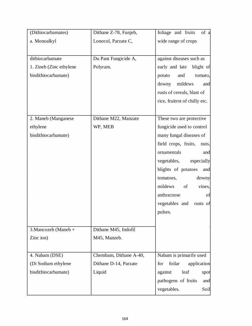
164
(Dithiocarbamates) Dithane Z-78, Funjeb, foliage and fruits of a
a. Monoalkyl Lonocol, Parzate C, wide range of crops
dithiocarbamate Du Pant Fungicide A, against diseases such as
1. Zineb (Zinc ethylene Polyram. early and late blight of
bisdithiocarbamate) potato and tomato,
downy mildews and
rusts of cereals, blast of
rice, fruitrot of chilly etc.
2. Maneb (Manganese Dithane M22, Manzate These two are protective
ethylene WP, MEB fungicide used to control
bisdithiocarbamate) many fungal diseases of
field crops, fruits, nuts,
ornamentals and
vegetables, especially
blights of potatoes and
tomatoes, downy
mildews of vines,
anthracnose of
vegetables and rusts of
pulses.
3.Mancozeb (Maneb + Dithane M45, Indofil
Zinc ion) M45, Manzeb.
4. Nabam (DSE) Chembam, Dithane A-40, Nabam is primarily used
(Di Sodium ethylene Dithane D-14, Parzate for foilar application
bisdithiocarbamate) Liquid against leaf spot
pathogens of fruits and
vegetables. Soil
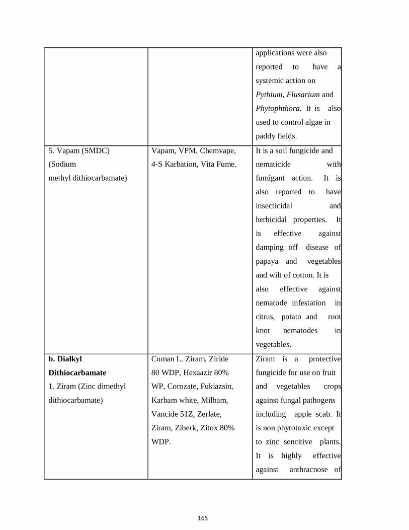
165
applications were also
reported to have a
systemic action on
Pythium, Flusarium and
Phytophthora. It is also
used to control algae in
paddy fields.
5. Vapam (SMDC) Vapam, VPM, Chemvape, It is a soil fungicide and
(Sodium 4-S Karbation, Vita Fume. nematicide with
methyl dithiocarbamate) fumigant action. It is
also reported to have
insecticidal and
herbicidal properties. It
is effective against
damping off disease of
papaya and vegetables
and wilt of cotton. It is
also effective against
nematode infestation in
citrus, potato and root
knot nematodes in
vegetables.
b. Dialkyl Cuman L. Ziram, Ziride Ziram is a protective
Dithiocarbamate 80 WDP, Hexaazir 80% fungicide for use on fruit
1. Ziram (Zinc dimethyl WP, Corozate, Fukiazsin, and vegetables crops
dithiocarbamate) Karbam white, Milbam, against fungal pathogens
Vancide 51Z, Zerlate, including apple scab. It
Ziram, Ziberk, Zitox 80% is non phytotoxic except
WDP. to zinc sencitive plants.
It is highly effective
against anthracnose of

166
beans, pulses, tobacco &
tomato, and also against
rusts of beans etc.
2. Ferbam (Ferric Coromat, Febam, Ferberk, Ferbam is mainly used
dimethyl Femate, Fermate D, for the protection of
dithiocarbamate) Fermicide, Hexaferb 75% foliage against fungal
WP, Karbam Black, pathogens of fruits and
Ferradow. vegetables including
Taphrina deformans of
peaches, anthracnose of
citrus, downy mildew of
tobacco and apple scab.
3. Thiram (Tetra methyl Thiride 75 WDP, Thiride It is used for seed
thiram disulphide) 750, Thiram 75% WDP, treatment both as dry
Hexathir, Normerson, powder or as a slurry. It
Panoram 75, Thiram, is a protective fungicide
TMTD, Arasan, Tersan also suitable for
75, Thylate, Pomarsol, application to foilage to
Thiuram. control Botrytis spp. on
lettuces, ornamental, soft
fruits and vegetables,
rust on ornamentals and
Venturia pirina on pears.
It is also effective
against soilborne
pathogens like Pythium,
Rhizoctonia and
Fusarium.

167
Mercury Fungicides
Mercury fungicides can be grouped as inorganic and organic mercury compounds. Both
the groups are highly fungitoxic and were extensively used as seed treatment chemicals against
seed borne diseases. Ignorance compounds show bactericidal property also. However, due to
their residual toxicity in soil and plants and their extreme toxicity nature to animal and human
beings, the use of mercury fungicides is beings discouraged. In most of the countries, the use of
mercury fungicides is banned and in countries like India, the use of mercury fungicides is
restricted only in seed treatment for certain crops. The list of diseases against which mercury
fungicides used are listed below
Common Name Trade Name Diseases Controlled
I. Inorganic Mercury It is used for treating potato
1. Mercuric chloride Merfusan, Mersil tubers and propagative materials
of other root crops
2. Mercurous chloride Cyclosan, M-C Turf Mercurous chloride is
fungicide. limited to soil application in crop
protection use because
of its phytotoxicity.
These are used mainly for
treatment of seeds and planting
II. Organomercurials Agallol, Aretan, Emisan, materials. These fungicides are
Methoxy ethyl mercury Ceresan wet (India) used for seed treatment by dry,
Chloride wet or slurry method. For seed
Ceresan Dry (India), treatment 1% metallic
Phenyl mercury chloride Ceresol, mercury is applied at 0.25%
Leytosan. concentration

168
Ceresan (USA)
Ethyl Mercury Chloride
Agrosan GN.
Tolyl mercury acetate
Heterocyclic Nitrogen Compounds, Quinones and Miscellaneous Fungicides
Heterocyclic Nitrogen Compounds
Heterocyclic nitrogen compounds are mostly used as foliage and fruits protectants. Some
compounds are very effectively used as seed dressers. Some of the commonly used fungicides
are listed below.
Common Name Trade Name Diseases Controlled
1.Captan (Kittleson‟s Captan 50W, Captan 75 It is a seed dressing fungicide used
Killer) (N-trichloromethyl W, Esso Fungicide 406, to control
thio-4- cyclohexence-1,2- Orthocide 406, Vancide diseases of many fruits,
dicarboximide) 89, Deltan, Merpan, ornamental and vegetable
Hexacap. crops against rots and damping
off.
2. Captafol (Cis-N- Foltaf, Difolaton, Difosan, It is a protective
1,1,2,2-tetra chloro hexane Captaspor, Foleid, fungicide, widly used to
1,2- dicarboximide) Sanspor. control foliage and fruit
diseases of tomatoes,
coffee potato.
3. Glyodin Glyoxaliadine, Glyoxide, It has a narrow specrum of

169
Glyodin, Glyoxide Dry, activity. As a spray, it controls
Glyodex 30% liquid and apple scab and cherry leaf spot.
70% WP.
4.Folpet (Folpet) [N- Phartan, Acryptan, It is also a protective
(trichloromethyl-thi)] Phaltan, Folpan, fungicide used mainly for
phthalimide Orthophaltan. foliage application against
leaf spots, downy and powdery
mildews of many crops.
Benzene compounds
Many aromatic compounds have important anti-microbial properties and have been
developed as fungicides. Some important benzene compounds commonly used in plant disease
control are listed below.
Common Name Trade Name Diseases Controlled
1. Quintozene (PCNB) Brassicol, Terraclor, It is used for seed and soil
Tritisan 10%, 20%, 40% D treatment. It is effective
and 75% WP, PCNB 75% against Botrytis, Sclerotium,
WP. Rhizoctonia and Sclerotinia
spp.
2. Dichloran Botran 50% WP and 75% It is a protective fungicide
WP, Allisan. and very effective against
Botrytis, Rhizopus and
Sclerotinia spp.
3. Fenaminsosuplh Dexon 5% G and 70% WP It is very specific in
(Sodiumpdimethylamino protecting germinating
benzenediazosulphonate seeds and growing plants
from seeds as well as soil-
borne infection of

170
Phythium, Aphanomyces
and Phytophthora spp.
4.Dinocap (2,4-dinitro-6- Karathane, Arathane, Itisanon-systemic
octyl phenylcrotonate) DNOPC, Mildex, acaricide and control
Crotothane, Crotothane fungicide recommended to
25% WP, control powdery mildews
Crotothane 48% Liq. on various fruits and
ornamentals. It is also used
for seed treatment.
Quinone Fungicides
Quinone areresent naturally in plants and animals and they exhibit anti-microbial activity
and some compounds are successfully developed and used in the plant disease control. Quinones
are very effectively used for seed treatment and two commonly used fungicides are listed below:
Common Name Trade Name Diseases Controlled
1. Chloranil (2,3,5,6- Spergon Chloronil is mainly used as
tetrachlora- a seed protectant against
1,4-benzoquinone) smuts of barely and
sorghum and bunt of wheat.
Dichlone has been used
2. Dichlone (2,3-dichloro- Phygon, Phygon XL WP. widely as seed protectant.
1,4- napthoquinone) This is also used as a
foliage fungicide,
particularly against apple
scab and peach leaf curl.
Organo – Phosphorous It has a specific action

171
fungicide against Pyricularia oryzae
Ediphenphos (Edifenphos) Hinosan 50% EC and 2% (Rice blast). It is also
(O-ethyl-SS- D. effective against Corticium
diphenyldithiophosphate) sesakii and Cochliobolus
miyabeanus in rice.
Organo Tin compounds
Several other organic compounds containing tin, lead, etc. have been developed and
successfully used in plant disease control. Among them, organo tin compounds are more popular
and effective against many fungal diseases. These compounds also show anti bactericidal
properties. Some of the organo tin compounds commonly used are listed below.
Common Name Trade Name Diseases Controlled
1. Fentin hydroxide Du-Ter WP 20% or 50% It is a non-systemic fungicide
(TPTHTiphenyl WP. Du-Ter Extra-WP, recommended for the control
tin hydroxide) Farmatin 50 WP, Du- of early and late blight of
Terforte WP, Tubotin. potato, leaf spot of sugar beet,
blast of rice and tikka leaf spot
of ground nut.
It is a non systemic fungicide
recommended
2. Fentin acetate Brestan WP 40% and to control Ramularia spp.on
(TPTATriphenyl tin 60% WP. celeryand sugar beet
acetate) anthracnose and downy
mildew
It is effective against
Cercospora leaf spot of

172
3. Fentin Chloride Brestanol 45% WP, sugarbeet and paddy blast
(TPTC- Triphenyl tin Tinmate.
chloride)
Systemic Fungicides and Antibiotics
Systemic Fungicides
Since the late 1960s there has been substantial development in systemic fungicides. Any
compound capable of being freely translocated after penetrating the plant is called systemic. A
systemic fungicide is defined as fungitoxic compound that controls a fungal pathogen remote
from the point of application, and that can be detected and identified. Thus, a systemic fungicide
could eradicate established infection and protect the new parts of the plant.
Several systemic fungicides have been used as seed dressing to eliminate seed infection.
These chemicals, however, have not been very successful in the cases of trees and shrubs. On the
basis of chemical structure, systemic fungicides can be classified as Benzimidazoles,
Thiophanates, Oxathilins and related compounds, pyrimidines, morpholines, organo-phosphorus
compounds and miscellaneous group. I. Oxathilin and related compounds
Oxathalins were the earliest developed compounds. This group of systemic fungicide is
also called as carboxamides, carboxyluc acid anillides, carboxaanillides or simply as anillides
which are effective only against the fungi belong to Basidiomycotina and Rhizoctonia solani.
Some of the chemicals developed are (i) Carboxin (DMOC: 5,6 - dithydra-2-methyl-1, 4-
oxathin-3-carboxanillide) and (ii) Oxycarboxin (DCMOD- 2,3-dihydro-5-carboxanillido-6-
methyl-1, 4 oxathilin-4, 4, dioxide). The diseases controlled by these chemicals are listed below.
Common Name Trade Name Diseases Controlled
1. Carboxin (5,6-dinydro- 2- Vitavax 10% D, Vitavax It is systemic fungicide
methyl-1-4-oxanthin-3- 75% WP, used for seed treatment of
carboxanlido) Vitavax 34% liq. cereals against bunts and
Vitaflow. smuts, especially loose smut
of wheat

173
2. Oxycarboxin (5,6- Plantvax 5G, Plantvax It is a systemic fungicide
dihydro-2-methyl- 1,4- 5% liq. Plantvax 1.5 EC, used for the treatment of
oxathin-3-carboxianilid- 10% dust, 75 WP. rust diseases of cereals,
4,4- dioxide) pulses, ornamentals,
vegetables and coffee
Sicarol. It controls rusts, smuts of
3.Pyracarbolid (2-methyl- many crops and
5,6-dihydro- 4H-Pyran-3- Rhizoctonia solani, but is
carboxylic acid anilide). slightly more effective than
carboxin
II. Benzimidazoles
The chemicals of this group show a very broad spectrum activity against a variety of
fungi. However, they are not effective against bacteria as well as fungi belongs to Mastigomycotina. Two types of fungicidal derivates of benzimidazoles are known. The first type
of derivates includes fungicides such as thiabendazole and fuberidazole. The fungicidal moiety
of the second type is methyl-2-benzimidazole carbamate (MBC). The fungicides of this group
may be simple MBC such as carbendazim or a complex from such as benomyl, which transforms
into MBC in plant system. Some of the important diseases controlled by these compounds are
shown below:
Common Name Trade Name Diseases Controlled
1.Benomyl (Methyl - 10 Benlate 50 WP, Benomyl. It is a protective and
(butly carbomyl)-2 Bavistin 50 WP, MBC, eradicative fungicide with
benzimidazole carbamate) Dersol, B.Sten 50, Zoom, systemic activity, effective
Tagstin, Agrozim, against a wide range of fungi

174
2. Carbendazim (MBC) Jkenstin. affecting field crops, fruits and
(Methyl -2- ornamentals.It is very effective
benzimidazole against rice blast, apple scab,
carbamate) powdery mildew of cereals,
rose, curcurbits and apple and
Diseases caused by
Verticillium and Rhizoctonia.
It is also used as pre-and
postharvest sprays of dips for
the control of storage rots of
fruits and vegetables.
Carbendazim is a systemic
fungicide controlling a wide
range of fungal pathogens of
field crops, fruits, ornamentals
and vegetables. It is used as
spray, seedling dip, seed
treatment, soil drench and as
post harvest treatment of
fruits. It is very effective
against wilt diseases
especially, banana wilt. It
controls effectively the
sigatoka leaf spot of banana,
turmeric leaf spot and rust
diseases in many
crops.
3. Thiabendazole (TBZ) Thiabendazole, Mertect, It is a broad spectrum systemic
(2,4-thiazoyl Tecto, Storite. fungicide effectivel against
benzimidazole) many major fungal diseases.
Pathogenic fungal control

175
includes species of
Botrytis, Ceratocystis,
Cercospora, Colletotrichum,
Fusarium, Rhizoctonia,
Sclerotinia, Septoria and
Verticillium. It is also
effective for the post
harvest treatment of fruits
and vegetables to control
storage diseases.
Voronit. It is used for the treatment
of seeds against diseases
4.Fuberidazole (2, (2- caused by Fusarium,
buryl) - benzimidazole). Particularly F.nivale on rye
and F.culmorum of peas
III. Thiophanates
These compounds represent a new group of systemic fungicides based on thiourea. They
are the derivatives of thioallophanic acid. These fungicides contain aromatic nucleus which is
converted into benzimidazole ring for their activity. Hence, thiophanates are often classified
under benzimidazole group and the biological activity of thiophanates resembles of benomyl.
Two compounds are developed under this group are discussed.
Common Name Trade Name Diseases Controlled
1. Thiophanate(1,2 - bis Topsin 50 WP, Cercobin It is a systemic fungicide
(ethyl carbonyl-2- 50 WP, Enovit. with a broad range of
thioureido) benzene) action, effective against
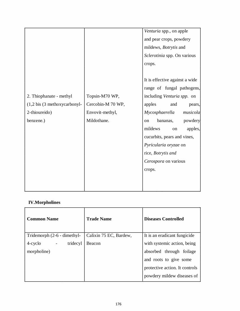
176
Venturia spp., on apple
and pear crops, powdery
mildews, Botrytis and
Sclerotinia spp. On various
crops.
It is effective against a wide
range of fungal pathogens,
2. Thiophanate - methyl Topsin-M70 WP, including Venturia spp. on
(1,2 bis (3 methoxycarbonyl- Cercobin-M 70 WP, apples and pears,
2-thioureido) Envovit-methyl, Mycosphaerella musicola
benzene.) Mildothane. on bananas, powdery
mildews on apples,
cucurbits, pears and vines,
Pyricularia oryzae on
rice, Botrytis and
Cerospora on various
crops.
IV.Morpholines
Common Name Trade Name Diseases Controlled
Tridemorph (2-6 - dimethyl- Calixin 75 EC, Bardew, It is an eradicant fungicide
4-cyclo - tridecyl Beacon with systemic action, being
morpholine) absorbed through foilage
and roots to give some
protective action. It controls
powdery mildew diseases of

177
cereals, vegetables and
ornamentals. It is highly
effective against
Mycosphaerella,
Exobasidium
V. Pyrimidines, Pyridines, Piperidines and Imidazole
Common Name Trade Name Diseases Controlled
1. Triadimefon Bayleton, Amiral It is very effective against
(1-(4-chlorophenoxy)-3, powdery mildews and rusts
3-dimethyl-1-(1-2-triazol- of several crops.
1yl) butan-2-one)
It is also very
2. Triadimenol Baytan effective against
(1-(4-Chlorophenoxyl-3, powdery mildews
3-dimethyl-1(1,2,4-triazol-1- and rusts of several
yl) butan-2-ol) crops.
It is highly effective against
3. Bitertanal Baycor rusts and powdery mildew
(B-(1-1-biphenyl-4-yloxy-a- of a variety of crops. It is
(1-1-dimethyl-ethyl-1-H-1,2- also used against Venturia
and Monilinia on fruits and
Cereospora leafspots of
groundnut and banana.
4- triazole-1-ethanol)
Terrazole 30% WP,
Terrazole 95% WP,

178
Terrazole 25% EC, Koban,
Pansol EG, Pansol 4% DP, It is very effective
Turban WP, Terracoat against
4. Etridiazole Aaterra. Phytophthora and
(5-ethaoxy-3- Pythium spp. and
trichloromethyl, 1,2- seeding diseases of
4-thiadiazole) cotton, groundnut,
vegetables, fruits
and ornamentals
VI. Hydroxy Pyrinidines
Common Name Trade Name Diseases Controlled
1. Ethirimol (5-butyl 2- Milliatem 80 WDP, It is effective against
ethyl amino-4-hydrop-6- Milcurb Super, powdery mildew of cereals
methyl pyrimidine) Milgo and other field crops. It is
also effective against
powdery mildews of
cucumber and ornamentals.
2. Dimethirimol (5-butyl It is very effective against
2-dimethylamino-4- Milcurb powdery mildews of
hydroxy-6-methy chrysanthemum and
pyrimidine) cucurbits.
VII. Furan derivatives It is used as seed or soil
1. Furcarbanil application, It systemically
(2-5-dimethyl-3- controlled bean rust and is
furanilide) being used as a seed

179
dressing fungicide against
loose smut of wheat and
barley.
It is effective against
bunts, smuts and rusts of
2. Cyclafuramid cereals, coffee rust, blister
(N-cyclohexyl-2,5- blight of tea, smut and red
dimethyl firamide) rot of sugarcane, Fusarium
wilt of tomato, Rhizoctonia
on tomato, potato,
groundnut, rice as well as
Armillaria sp. On rubber.
It is effective against
yellow rust on wheat and
barley (P. striiformis) and
brown rust on barley (P.
VIII.Benzanilide hordei). It is also having
derivative direct fungitoxic activity
1. Mebenil against Sclerotium rolfsli
(2-methyl benzanilide) and Rhizoctonia.
IX. Organo phosphorous compounds
Common Name Trade Name Diseases Controlled
1. Pyrazophos (2-0-0- Afugan, Curamil, WP, It is used to control

180
Diethylthionophosphoryl) Missile EC.
-5- methyl-6-carbethoxy
pyrazolo-(1- 5a)pyrimidine)
2. Iprobenphos (IBP) Kitazin 48% EC, Kitazin
(S-benyzl-0-0- 17G, Kitazin 2% D.
bisisopropylphosphorothiate)
X. Piperazine Saprol-EG, Fungitex.
1.Triforine(N,N-bis-(1-
foramido-2,2,2-
trichloroethyl- piperazine)
XI. Phenol derivative Demonsan 65 WP, Tersan
1. Choloroneb (1-4-dichloro- SP, Turf
2,5-dimethoxy fungicide
benzene)
powdery mildews of
cereals, vegetables, fruits
and ornamentals.
It is used to control
Pyricularia oryzae and
sheath blight of rice.
It is effective against
powdery mildew, scab and
other diseases of fruits and
rust on ornamentals and
cereals.
It is also active against
storage diseases of fruits.
It is highly fungistatic to
Rhizoctonia spp.,
moderately so to Pythium
spp. and poorly to
Fusarium spp. It is used
as a supplemental seed
treatment for beans and
soyabeans to control
seedling disease
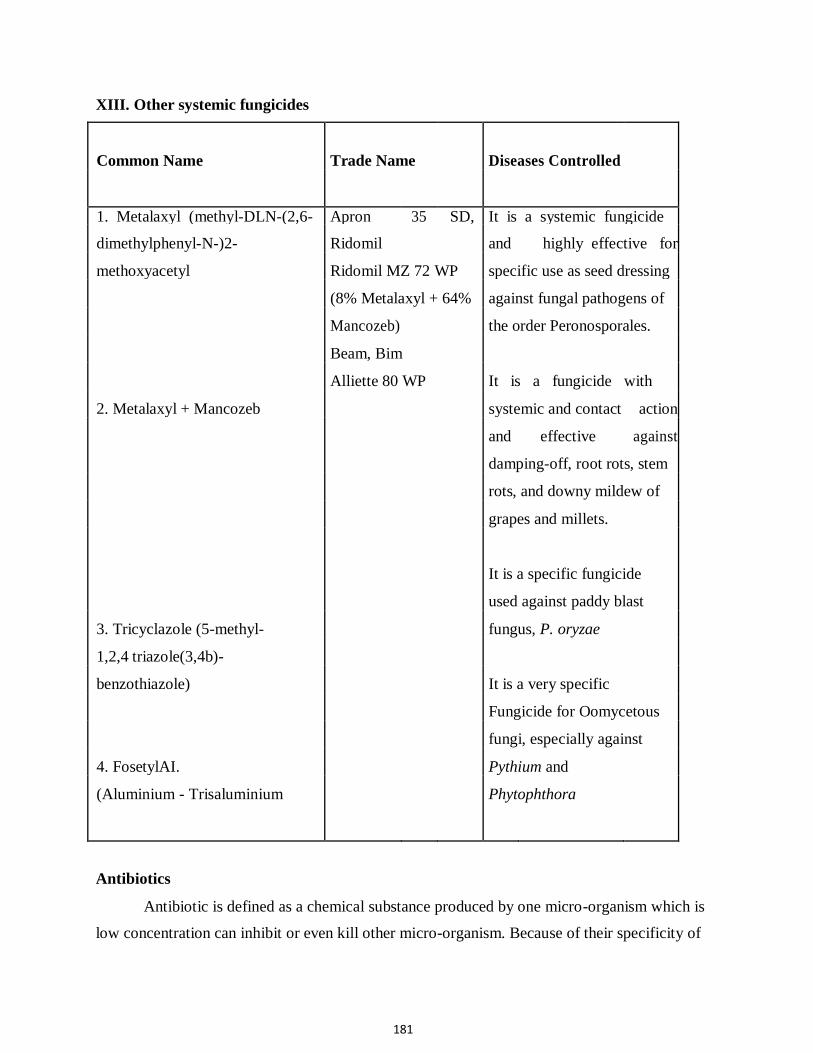
181
XIII. Other systemic fungicides
Common Name Trade Name Diseases Controlled
1. Metalaxyl (methyl-DLN-(2,6- Apron 35 SD, It is a systemic fungicide
dimethylphenyl-N-)2- Ridomil and highly effective for
methoxyacetyl Ridomil MZ 72 WP specific use as seed dressing
(8% Metalaxyl + 64% against fungal pathogens of
Mancozeb) the order Peronosporales.
Beam, Bim
Alliette 80 WP It is a fungicide with
2. Metalaxyl + Mancozeb systemic and contact action
and effective against
damping-off, root rots, stem
rots, and downy mildew of
grapes and millets.
It is a specific fungicide
used against paddy blast
3. Tricyclazole (5-methyl- fungus, P. oryzae
1,2,4 triazole(3,4b)-
benzothiazole) It is a very specific
Fungicide for Oomycetous
fungi, especially against
4. FosetylAI. Pythium and
(Aluminium - Trisaluminium Phytophthora
Antibiotics
Antibiotic is defined as a chemical substance produced by one micro-organism which is
low concentration can inhibit or even kill other micro-organism. Because of their specificity of

182
action against plant pathogens, relatively low phytotoxicity, absorption through foliage and
systemic translocation and activity in low concentration, the use of antibiotic is becoming very
popular and very effectively used in managing several plant diseases. They can be grouped as
antibacterial antibiotics and antifungal antibiotics. Most antibiotics are products of several
actinomycetes and a few are from fungi and bacteria. I. Antibacterial antibiotics
1. Streptomycin sulphate
Streptomycin is an antibacterial, antibiotic produced by streptomyces griseus.
Streptomycin are streptomycin sulphate is sold as Agrimycin,-100, Streptomycin sulphate,
Plantomycin, Streptocycline, Paushamycin, Phytostrip, Agristrep and Embamycin, Agrimycin -
100 contains 15 per cent streptomycin sulphate + 1.5 percent terramycin (Oxy tetracycline). Agristerp contains 37 percent streptomycin sulphate. Phytomycin contains 20 percent
streptomycin. Streptocycline and paushamycin contains 9 parts f streptomycin and 1 part of
tetracycline hydrochloride.
This group of antibiotics act against a broad range of bacterial pathogens causing blights,
wilt, rots etc. This antibiotic is used at concentrations of 100-500 ppm. Some important diseases
controlled are blight of apple and pear (Erwinia amylovora), Citrus canker (Xanthomonas
campestris p.v. citri), Cotton black arm (X.c. p.v. malvacearum), bacterial leaf spot of tomato
(Pseudomonas solanacearum), wild fire of tobacco (Pseudomonas tabaci) and soft rot of
vegetables (Erwinia carotovora).
In addition, it is used as a dip for potato seed pieces against various bacterial rots and as
an disinfectant in bacterial pathogens of beans, cotton, crucifers, cereals and vegetables.
Although it is an antibacterial antibiotic, it is also effective against some diseases caused by
Oomycetous fungi, especially foot-rot and leaf rot of betelvine caused by Phytophthora
parasitica var. piperina. 2. Tetracyclines
Antibiotics belonging to this group are produced by many species of Streptomyces. This
group includes Terramycin or Oxymicin (Oxytetracycline). All these antibiotics are
bacteriostatic, bactericidal and mycoplasmastatic. These are very effective against seed-borne
bacteria. This group of antibiotic is very effective in managing MLO diseases of a wide range of
crops. These are mostly used as combination products with Streptomycin sulphate in controlling

183
a wide range of bacterial diseases. Oxytetracyclines are effectively used as soil drench or as root
dip controlling crown gall diseases in rosaceous plants caused by Agrobacterium tumefaciens. II Antifungal antibiotics 1. Aureofungin
It is a hepataene antibiotic produced in sub-merged culture of Streptoverticillium
cinnamomeum var. terricola. It is absorbed and translocated to other parts of the plants when
applied as spray or given to roots as drench. It is sold as Aurefungin-Sol. Containing 33.3%
Aureofungin and normally sprays at 50-100 ppm. The diseases controlled are citrus gummosis
caused by several species of Phytophthora, powdery mildew of apple caused by Podosphaera
leucotricha and apple scab (Venturia inaequalis), groundnut tikka leaf spot, downy mildew,
powdery mildew and anthracnose of grapes, potato early and late blight. As seed treatment it
effectively checked are Diplodia rot of mango, Alternaria rot of tomato, Pythium rot of cucurbits
and Penicillium rot of apples and citrus. As a truck application/root feeding, 2 g of aureofungin-
sol+1g of copper sulphate in 100 ml of water effectively reduce. Thanjavur wilt of coconut. 2. Griseofulvin
This antifungal antibiotic was first discovered to be produced by Penicillium
griseofulvum and now by several species of Penicillium, viz., P.patulum, P.nigricans, P.urticae,
and P.raciborskii. It is commercially available as Griseofulvin, Fulvicin and Grisovin. It is
highly toxic to powdery mildew of beans and roses, downy mildew of cucumber. It is also used
to control Alternaria solani in tomato Sclerotinia fructigena in apple and Botrytis cinerea in
lettuce. 3. Cycloheximide
It is obtained as a by-product in streptomycin manufacture. It is produced by different
species of Streptomyces, including S.griseus and S. nouresi. It is commercially available as
Actidione, Actidione PM, Actidione RZ and Actispray. It is active against a wide range of fungi
and yeast. Its use is limited because it is extremely phytotoxic. It is effective against powdery
mildew of beans (Erysiphe polygoni), Bunt of wheat (Tilletia spp.) brownnot of peach
(Sclerotinia fructicola) and post harvest rots of fruits caused by Rhizopus and Botrytis spp. 4. Blasticdin
It is a product of Streptomyces griseochromogenes and specifically used against blast
disease of rice caused by Pyricularia oryzae. It is commercially sold as Bla-s.

184
5. Antimycin
It is produced by several species of Streptomyces, especially S. griseus and S.
Kitasawensis. It is effectively used against early blight of tomato, rice blast and seedling blight
of oats. It is commercially sold as Antimycin. 6. Kasugamycin
It is obtained from Streptomyces kasugaensis. It is also very specific antibiotic against
rice blast disease. It is commercially available as Kasumin. 7. Thiolution
It is produced by Streptomyces albus and effectively used to control late blight of potato
and downy mildew of cruciferous vegetables. 8. Endomycin
It is a product of Streptomyces endus and effectively used against leaf rust of wheat and
fruit rot of strawberry (Botrytis cinerea). 9. Bulbiformin
It is produced by a bacterium, Bacillus subtills and is very effectively used against wilt
diseases, particularaly redgram wilt. 10. Nystatin
It is also produced by Streptomyces noursei. It is successfully used against anthracnose
disease of banana and beans. It also checks downy mildew of cucuribits. As a post harvest dip, it
effectively reduces brown rot of peach and anthracnose of banana in stroage rooms. It is
commercially marketed as Mycostain and Fungicidin. 11. Eurocidin
It is a pentaene antibiotic produced by Streptomyces anandii and called as pentaene G-8.
It is effectively used against diseases caused by several species of Colletotrichum and Helminthosporium.
Methods of allocation of fungicides – Precautions and safety measures while handling
fungicides
Proper selection of a fungicide and its application at the correct dose and the proper time
are highly essential for the management of plant diseases. The basic requirement of an
application method is that it delivers the fungicide to the site where the active compound will

185
prevent the fungus damaging the plant. This is mostly achieved by spray, fog, smoke, aerosol,
mist, dust, or granules applied to the growing plant or by seed or soil treatment.
In addition, some trees and shrubs can be protected by injection of fungicide liquid into
the trunk or by brushing wounds with fungicide paints or slurries. In the case of sprays, mists,
aerosols and fogs, the fungicide is in of droplets of water of another fluid. In the case of smokers,
the solid particles of the fungicide are carried by the air. In the case of dusts and granules, the
fungicide is straightly mixed with an inert carrier, impregnated into it coated on the particles,
which are applied mechanically.
The object of spraying or dusting is to cover the entire susceptible surface of hostwith a
thin covering of a suitable concentration of the fungicide before the pathogen has come into
contact with the host. However, these practices may not effectively eradicate the inoculum
present on the surface of the seeds or deep-seated in the seed. So, the application of chemicals as
seed dressing is highly essential.
In addition, soil harbours several pathogens which cause root diseases in several crop
plants. So treatment of soil with chemicals is also highly useful in reducing the inoculum load
present in the soil. The fungicidal application varies according to the nature of the host part
diseased and nature of survival and spread of the pathogen. The method which are commonly
adopted in the application of the fungicides are discussed. 1. Seed treatment
The seed treatment with fungicides is highly essential because a large number of fungal
pathogens are carried on or in the seed. In addition, when the seed is sown, it is also vulnerable
to attack by many common soil-borne pathogens, leading to either seed rot, seeding mortality or
produce diseases at a later stage. Seed treatment is probably the effective and economic method
of disease control and is being advocated as a regular practice in crop protection against soil and
seed-borne pathogens. Seed treatment is therapeutic when it kills pathogens that infect embroys,
cotyledons or endosperms under the seed coat, eradicative when it kills pathogens that
contaminate seed surfaces and protective when it prevents penetration of soilborne pathogens
into the seedling. There are various types of seed treatment and broadly they may be divided into
three categories (a) Mechanical, (b) Chemical and (c) Physical.

186
A. Mechanical method
Some pathogen when attack the seeds, there may be alteration in size, shape and weight
of seeds by which it is possible to detect the infected seeds and separate them from the healthy
ones. In the case of ergot diseases of cumbu, rye and sorghum, the fungal sclerotia are usually
larger in size and lighter than healthy grains. So by sieving or flotation, the infected grains may
be easily separated. Such mechanical separation eleminates the infected grains may be easily
separated. Such mechanical separation eliminates the infected materials to a larger extent. This
method is also highly useful to separate infected grains in the case of „tundu‟ disease of wheat.
Eg. Removal of ergot in cumbu seeds.
Dissolve 2kg of common salt in 10 litres of water (20% solution). Drop the seeds into the
salt solution and stir well. Remove the ergot affected seeds and sclerotia which float on the
surface. Wash the seeds in fresh water 2 or 3 times to remove the salts on the seeds. Dry the
seeds in shade and use for sowing. B. Chemical methods
Using fungicides on seed is one of the most efficient and economical methods of
chemical disease control. On the basis of their tenacity and action, the seed dressing chemicals
may be grouped as (i) Seed disinfectant, which disinfect the seed but may not remain active for a
long period after the seed has been sown and (ii) Seed protectants, which disinfect the seed
surface and stick to the seed surface for sometime after the seed has been sown, thus giving
temporary protection to the young seedlings against soil borne fungi. Now, the systemic
fungicides are impregnated into the seeds to eliminate the deep seated infection in the seeds. The
seed dressing chemicals may be applied by (i) Dry treatment (ii) Wet treatment and (iii) Slurry. (i) Dry Seed Treatment
In this method, the fungicide adheres in a fine from on the surface of the seeds. A
calculated quantity of fungicide is applied and mixed with seed using machinery specially
designed for the purpose. The fungicides may be treated with the seeds of small lots using simple
Rotary seed Dresser (Seed treating drum) or of large seed lots at seed processing plants using
Grain treating machines. Normally in field level, dry seed treatment is carried out in dry rotary
seed treating drums which ensure proper coating of the chemical on the surface of seeds. In
addition, the dry dressing method is also used in pulses, cotton and oil seeds with the
187
antagonistic fungus like Trichoderma vitide by mixing the formulation at the rate of 4g/kg of the
seed. Eg. Dry seed treatment in paddy.
Mix a required amount of fungicide with required quantity of seeds in a seed treating
drum or polythene lined gunny bags, so as to provide uniform coating of the fungicide over the
seeds. Treat the seeds atleast 24 hours prior to soaking for sprouting. Any one of the following
chemical may be used for treatment at the rate of 2g/kg : Thiram or Captan or Carboxin or
Tricyclazole. (ii) Wet seed treatment
This method involves preparing fungicide suspension in water, often at field rates and
then dipping the seeds or seedlings or propagative materials for a specified time. The seeds
cannot be stored and the treatment has to be done before sowing. This treatment is usually
applied for treating vegetatively propagative materials like cuttings, tubers, corms, setts
rhizomes, bulbs etc., which are not amenable to dry or slurry treatment. a. Seed dip / Seed soaking
For certain crops, seed soaking is essential. Seeds treated by these methods have to be
properly dried after treatment. The fungicide adheres as a thin film over the seed surface which
gives protection against invasion by soil-borne pathogens. Eg. Seed dip treatment in paddy.
Prepare the fungicidal solution by mixing any of the fungicides viz., carbendazim or
pyroquilon or tricyclazole at the rate of 2g/litre of water and soak the seeds in the solution for 2
hrs. Drain the solution and keep the seeds for sprouting. Eg. Seed dip treatment in Wheat.
Prepare 0.2% of carboxin (2g/litre of water) and soak the seeds for 6 hours. Drain the
solution and dry the seeds properly before sowing. This effectively eliminates the loose smut
pathogen, Ustilago nuda tritici. b. Seedling dip / root dip
The seedlings of vegetables and fruits are normally dipped in 0.25% copper oxychloride
or 0.1% carbendazin solution for 5 minutes to protect against seedling blight and rots.

188
c. Rhizome dip
The rhizomes of cardamom, ginger and turmeric are treated with 0.1% emisan solution
for 20 minutes to eliminate rot causing pathogen present in the soil. d. Sett dip / Sucker dip
The sets of sugarcane and tapioca are dipped in 0.1% emisan solution for 30 minutes. The
suckers of pine apple may also be treated by this method to protect from soilborne diseases. (iii) Slurry treatment (Seed pelleting)
In this method, chemical is applied in the form of a thin paste (active material is
dissolved in small quantity of water). The required quantity of the fungicide slurry is mixed with
the specified quantity of the seed so that during the process of treatment slurry gets deposited on
the surface of seeds in the form of a thin paste which later dries up.
Almost all the seed processing units have slurry treaters. In these, slurry treaters, the
requisite quantity of fungicides slurry is mixed with specified quantity of seed before the seed lot
is bagged. The slurry treatment is more efficient than the rotary seed dressers. Eg. Seed pelleting
in ragi.
Mix 2.5g of carbendazim in 40 ml of water and add 0.5g of gum to the fungicidal
solution. Add 2 kg of seeds to this solution and mix thoroughly to ensure a uniform coating of
the fungicide over the seed. Dry the seeds under the shade. Treat the seeds 24 hrs prior to
sowing. (iv) Special method of seed treatment
Eg. Acid - delinting in cotton
This is follows in cotton to kill the seed-borne fungi and bacteria. The seeds are treated
with concentrated sulphuric acid @ 100 ml/kg of seed for 2-3 minutes. The seeds are then
washed 2 or 3 times thoroughly with cold water and shade dried. After drying, they are again
treated with captan or thiram @ 4g/kg before sowing. II. Soil treatment
It is well known that soil harbours a large number of plant pathogens and the primary
sources of many plant pathogens happens to be in soil where dead organic matter supports active
or dormant stages of pathogens. In addition, seed treatment does not afford sufficient protection
against seedling diseases and a treatment of soil around the seed is necessary to protect them.

189
Soil treatment is largely curativ in nature as it mainly aims at killing the pathogens in soil and
making the soil „safe‟ for the growth of the plant.
Chemical treatments of the soil is comparatively simple, especially when the soil is
fallow as the chemical is volatile and disappears quickly either by volatilization or
decomposition. Soil treating chemicals should be non-injurious to the plants in the soil adjacent
to the area where treatment has been carried out because there may be standing crop in adjacent
fields. The soil treatment methods involving the use of chemicals are (i) Soil drenching, (ii) broadcasting, (iii) furrow application, (iv) fumigation and
(v) chemigation.
(i) Soil drenching
This method is followed for followed for controlling damping off and root rot infections
at the ground level. Requisite quantity of fungicide suspension is applied per unit area so that the
fungicide reaches to a depth of atleast 10-15 cm. Eg. Emisan, PCNB, Carbendazim, Copper fungicides, etc.
(ii) Broadcasting
It is followed in granular fungicides wherein the pellets are broadcasted near the plant.
(iii) Furrow application
It is done specifically in the control of some diseases where the direct application of the
fungicides on the plant surface results in phytotoxic. It is specifically practiced in the control of
powdery mildew of tobacco where the sulphur dust is applied in the furrows. (iv)Fumigation
Volatile toxicants (fumigants) such as methyl bromide, chloropicrin, formaldehyde and
vapam are the best chemical sterilants for soil to kill fungi and nematodes as they penetrate the
soil efficiently. Fumigations are normally done in nursery areas and in glass houses. The
fumigant is applied to the soil and covered by thin polythene sheets for 5-7 days and removed.
For example, Formaldehyde is applied at 400 ml/100 Sq.m. The treated soil was irrigated and
used 1 or 2 weeks later. Vapam is normally sprinkled on the soil surface and covered. Volatile
liquid fumigants are also injected to a depth of 15-20 cm, using sub-soil injectors. (v) Chemigation
In this method, the fungicides are directly mixed in the irrigation water. It is normally
adopted using sprinkler or drip irrigation system.

190
III. Foliar application
A. Spraying
This is the most commonly followed method. Spraying of fungicides is done on leaves,
stems and fruits. Wettable powders are most commonly used for preparing spray solutions. The
most common diluent or carrier is water. The dispersion of the spray is usually achieved by its
passage under pressure through nozzle of the sprayer.
The amount of spray solution required for a hectare will depend on the nature of crops to
be treated. For trees and shrubs more amount of spray solution is required than in the case of
ground crops. Depending on the volume of fluid used for coverage, the sprays are categorised
into high volume, medium volume, low volume, very high volume and ultra low volume.
The different equipments used for spray application are: Foot-operated sprayer, rocking
sprayer, knapsack sprayer, motorised knapsack sprayer (Power sprayer), tractor mounted sprayer,
mist blower and aircraft or helicopter (aerial spray). B. Dusting
Dusts are applied to all aerial parts of a plant as an alternative to spraying. Dry powders
are used for covering host surface. Generally, dusting is practicable in calm weather and a better
protective action is obtained if the dust is applied when the plant surface is wet with dew or rain
drops. The equipments employed for the dusting operation are: Bellow duster, rotary duster,
motorised knapsack duster and aircraft (aerial application). IV. Post – harvest application
Fruits and vegetables are largely damaged after harvest by fungi and bacteria. Many
chemicals have been used as spray or dip or fumigation. Post harvest fungicides are most
frequently applied as aqueous suspensions or solutions. Dip application has the advantage of
totally submerging the commodity so that maximum opportunity for penetration to the infection
sites.
Systemic fungicides, particularly thiabendazole, benomyl, carbendazim, metalaxyl,
fosety-AI have been found to be very effective against storage diseases. In addition,
dithiocarbamates and antibiotics are also applied to control the post-harvest diseases. Wrapping
the harvested products with fungicide impregnated wax paper is the latest method available.

191
VI. Special method of applications
1. Trunk Application / Trunk Injection
It is normally adopted in coconut gardens to control Thanjavur wilt caused by
Ganoderma lucidum. In the infected plant, a downward hole is made to a depth of 3-4” at an
angle of 450C at the height of 3‟ from the ground level with the help of an auger. The solution
containing 2g of Aureofungin soil and 1 g of copper sulphate in 100 ml of water is taken in a
saline bottle and the bottle is tied with the tree. The hose is inserted into the hole and the stopper
is adjusted to allow the solution in drops. After the treatment, the hole is covered with clay. 2. Root Feeding
Root feeding is also adopted for the control of Thanjavur wilt of coconut instead of trunk
application. The root region is exposed; actively growing young root is selected and given a
slanting cut at the tip. The root is inserted into a polythene bag containing 100 ml of the
fungicidal solution. The mouth of the bag is tied tightly with the root. 3. Pseudostem Injection
This method is very effective in controlling the aphid vector (Pentalonia nigronervosa)
of bunchy top of bannana. The banana injector is used for injecting the insecticide.Banana
injector is nothing but an Aspee baby sprayer of 500 ml capacity. In which, the nozzle is
replaced by leurlock system and aspirator needle No. 16. The tip of the needle is closed and two
small holes are made in opposite direction.
It is for free flow of fluid and the lock system prevents the needle from dropping from
the sprayer. One ml of monocrotophos mixed with water at 1:4 ratio is injected into the
pseudostem of 3 months old crop and repeated twice at monthly intervals. The same injector can
also be used to kill the bunchy top infected plants by injecting 2 ml of 2, 4-D (Femoxone) mixed
in water at 1:8 ratio. 4. Corn Injection
It is an effective method used to control Panama will of banana caused by Fusarium
oxysporum f. sp. cubense. Capsule applicator is used for this purpose. It is nothing but an iron
rod of 7 mm thickness to which a handle is attached at one end. The length of the rod is 45 cm
and an iron plate is fixed at a distance of 7 cm from the tip.

192
The corm is exposed by removing the soil and a hole is made at 45) angle to a depth of 5 cm.
One or two gelatin capsules containing 50-60 mg of carbendazim is pushed in slowly and
covered with soil. Instead of capsule, 3 ml of 2% carbendazim solution can also be injected into
the hole. 5. Paring and Pralinage
It is used to control Fusarium wilt and burrowing nematode (Radopholus similis) of
banana. The roots as well as a small portion of corm is removed or chopped off with a sharp
knife and the sucker is dipped in 0.1% carbendazim solution for 5 minutes. Then, the sucker is dipped in clay slurry and furadan granules are sprinkled over the corm @ 40
g/corm.

193
FUNGICIDES MODE OF ACTION TABLE
FRAC MODE OF ACTION CHEMICAL FAMILY ACTIVE
GROUP (GROUP) INGREDIENTS
1 Mitosis and cell division benzimidazoles thiabendazole
1 thiophanates thiophanate-methyl
2 Respiration iprodione vinclozolin
3 Sterol synethesis imidazoles Imazilil
3 piperazines Triforine
3 pyrimidines Fenarimol
3 triazoles bitertanol cyproconazole difenoconazole
fenbuconazole
flusilazole
ipconazole
metconazole
myclobutanil
propiconazole
prothioconazole
tebuconazole
tetraconazole
triadimefon
triadimenol
triticonazole
4 Nucleic acid synethesis acylalanines metalaxyl metalaxyl-M
(=mefenoxam)
7 Respiration boscalid carboxin flutolanil
9 Protein synthesis cyprodinil
11 Respiration methoxyacrylates azoxystrobin picoxystrobin
11 methoxy-carbamates pyraclostrobin
11 oximino acetates kresoxim-methyl trifloxystrobin
11 oxazolidine-diones famoxadone
11 dihydro-dioxazines fluoxastrobin
11 imidazolinones fenamidone
12 Signaling fludioxonil
13 Signaling quinoxyfen
14 Lipids and membranes chloroneb dicloran quintozene (PCNB)
14 1,2,4-thiadiazoles etridiazole
17 Sterol synthesis fenhexamid

194
FUNGICIDES MODE OF ACTION TABLE
FRAC MODE OF ACTION CHEMICAL FAMILY ACTIVE
GROUP (GROUP) INGREDIENTS
19 Cell wall synthesis peptidyl pyrimidine polyoxin nucleoside
21 Respiration cyanoimidazole cyazofamid
22 Cell division zoxamide
24 Protein synthesis kasugamycin
25 Protein synthesis streptomycin
27 Unkown cymoxanil
28 Cell membrane permeability propamocarb
29 Respiration 2,6-dinitro-anilines fluazinam
30 Respiration tri phenyl tin compounds fentin hydroxide
33 Unkown ethyl phosphonates fosetyl-Al
33 phophorous acid and salts
40 Cell wall synthesis cinnamic acid amides dimethomorph
40 mandelic acid amides mandipropamid
41 Protein synthesis oxytetracycline
P Host plant defense induction benzo-thiadiazole BTH acibenzolar-S-methyl
M Multi-site contact activity inorganic copper
inorganic sulphur
dithiocarbamates and ferbam
relatives mancozeb
maeb
metiram
thiram
ziram
phthalimides captan
chloronitriles chlorothalonil
(phthalonitriles)
guanidines dodine
NC Not classified diverse mineral oils, organic oils, potassium bicarbonate

195
Mode of action and formulations of fungicides
A fungicide’s formulation has a big impact on a fungicide’s activity. For example the more
finely ground the sulfur particles, the more effective as a powdery mildew fungicide but also the
more likely that phytotoxicity can occur! Unfortunately, pesticide formulations can be almost as
confusing as pesticide classes, to know what pesticide formulation will work best for your specific
purposes you should know the characteristics, advantages, and disadvantages of the different
formulations and adjuvants.
What is a formulation: The pesticide formulation is a mixture of the active and inert ingredients in
the pesticide. The active ingredients are the chemicals that affect the target pest. The inert
ingredients are all other ingredients in the pesticide, and are also called inactive ingredients. Inert
ingredients are used to dilute the active ingredient or make it safer, easier to handle, and more
effective. Some formulations are ready to use, others must be further diluted by air (air-blast
sprayer), water, or a petroleum-based solvent. A single active ingredient is often sold in multiple
formulations - you must choose the formulation that works best for you.
How to choose the formulation: There are several questions that you must answer while choosing
the formulation.
1. Do you have the equipment needed for this type of formulation?
2. Can the formulation be applied safely under the conditions of the application area?
3. Will the formulation reach the target and stay there long enough for control?
4. Is there a possibility the formulation will harm the surface on which it is applied? To answer
these questions, you must know the characteristics of the formulations and the advantages and
disadvantages of each type. The most common formulations found in grape disease control are:
Liquid Formulations
Emulsifiable Concentrates (EC or E) – contains a liquid active ingredient, one or more
petroleum-based solvents, and an agent that allows the product to be mixed with water to form an
emulsion. An emulsion is a mixture of two or more liquids that are not soluble in one another. Each
gallon of EC usually contains 25 to 75% (2 to 8-lbs) active ingredient.
These are among the most versatile formulations and are adaptable to many application equipment
types from small, portable sprayers to hydraulic sprayers, low- volume ground sprayers, and mist
blowers.

196
Advantages:
• Relatively easy to transport, handle, and store.
• Little agitation required (will not settle or separate when equipment is running).
• Non-abrasive.
• Does not plug nozzles or screens.
• Little visible residues on treated surfaces.
• Highly concentrated, making it easy to over- or under-dose by mixing and
calibration errors.
• May cause phytotoxicity.
• Easily absorbed through skin.
• Solvents may damage rubber or plastic hoses, gaskets, pump parts, and metal or painted
surfaces.
• May cause pitting or discoloration of painted surfaces.
• Flammable – must be stored away from open flame or heat.
• May be corrosive.
Solutions (S) – pesticide active ingredients that readily dissolve when mixed with a
solvent such as water or a petroleum-based solvent. These formulations form a solution
that will not settle out or separate once mixed. Solutions usually contain the active
ingredient, the solvent, and one or more inert ingredients. Solutions may be used in any
Type of sprayer.
Advantages:
• Relatively easy to transport, handle, and store.
• Little agitation required (will not settle or separate when equipment is running).
Disadvantages:
• Easily absorbed through skin.
Concentrate solutions (C or LC) – solutions sold as concentrates that must be
further diluted with a liquid solvent. The solvent may be water but more often is refined oil
or petroleum-based.
Advantages:
• No agitation
needed. Disadvantages:
• Less formulations of this type.
Other advantages and disadvantages vary depending on the solvent used, the concentration of the
active ingredient, and the type of application.
Flowables (F or L) – finely ground active ingredients (in this case, soluble solids)
are mixed with liquid along with inert ingredients to form a suspension. A suspension is a
substance that contains undissolved particles mixed throughout a liquid. Flowables are
mixed with water for application and are similar to EC or WP formulations for case of handling and
use.

197
Advantages:
• Seldom clogs nozzles.
• Easy to handle and apply.
Disadvantages:
• Requires moderate agitation to maintain solids in suspension.
• May leave a visible residue.
Dry Formulations
Dusts (D) – ready to use formulations containing a low percentage of active ingredient (0.5 to
10%), combined with a fine, dry inert carrier made from talc, chalk, clay, nut hulls, or volcanic ash.
The size of the dust particle is variable. A few dust formulations are available as concentrates,
containing a high percentage of active ingredient, which must be mixed with inert carriers before they
are applied. Dusts easily drift onto non-target areas.
Advantages:
• Usually ready to use with no mixing involved.
• Effective where moisture from a spray may be harmful.
• Requires simple equipment.
• Easily drifts off target during application.
• May irritate eyes, nose, throat, and skin.
• Does not stick to surfaces as well as liquid formulations do.
• Difficult to achieve even distribution of particles on surfaces.
Granules (G) – similar to dust formulations except granules have larger and heavier
particles. These coarse particles are composed of absorptive materials such as clay, corn
cobs, or walnut shells. The active ingredient either coats the outside of the granules or is
absorbed into them. The amount of active ingredient in this formulation is relatively low,
typically ranging from 1 to 15%. Granular pesticides are most often applied to control
weeds, nematodes, and soil insects.
Advantages:
• Ready to use with no mixing involved.
• Drift hazard is low because heavier particles quickly settle.
• Fewer hazards to the applicator (no spray, little dust).
• Requires simple application equipment such as seeders or fertilizer spreaders.
• May break down more slowly than WP’s or EC’s by slow release coating.
Disadvantages:

198
• Does not stick to foliage or other non-level surfaces.
• May need to incorporate into soil.
• May require moisture to start pesticide action.
• May be hazardous to nontarget species that mistake granule for grain or seed.
Pellets (P or PS) similar to granular formulations, the terms are often used interchangeably.
However, in a pellet formulation all the particles are the same weight and shape. This uniformity
allows pellets to be applied by precision applicators such as those used for precision planting of
pelleted seed.
Wettable Powders (WP or W) – dry, finely ground formulations that look like
dusts. Wettable powders are usually mixed with water for application as a spray. A few
products are available that may be applied as dusts or as a spray. Wettable powders
contain 5 to 95% active ingredient (usually 50% or more). The powder particles do not
dissolve in water, and settle out quickly unless constantly agitated. This is one of the most
widely used pesticide formulations, useable for most pest problems and with most types of
spray equipment if agitation is available.
Advantages:
• Easy to transport, store, and handle.
• Less likely to cause phytotoxicity than EC’s and other petroleum-based pesticides.
• Easily measured and mixed.
• Less skin and eye absorption than EC’s and other liquid formulations.
Disadvantages:
• Inhalation hazard when handling the concentrated powder.
• Requires good and constant agitation (usually mechanical agitation in the spray tank).
• Abrasive on many pumps and nozzles.
• Difficult to mix in hard or alkaline water.
• Often clogs nozzles and screens.
• Residues may be visible.
Soluble Powders (SP or WSP) – looks like wettable powders; however, when mixed with
water dissolves readily and forms a true solution. After soluble powders are mixed thoroughly no
additional agitation is necessary. The amount of active ingredient in soluble powders ranges from 15
to 95% (usually 50% or more). Soluble powders have all the advantages of wettable powders and
none of the disadvantages except an inhalation

199
A = Aerosol
AF = Aqueous Flowable
An adjuvant is a chemical AS =
Aqueous Solution of Aqueous
Suspension
added to the pesticide formulation or
B = Bait
C = Concentrate
tank mix to increase the safety or CM = Concentrate Mixture
efficacy of a pesticide. Most
CG = Concentrate Granules
D = Dust
pesticide formulations are composed DF = Dry Flowable
of a small percentage of adjuvants.
DS = Soluble Dust
E = Em ulsifiable Concentrate
Common adjuvants are: EC = Em ulsifiable Concentrate
• Surfactants or surface active
F = Flowable
G = Granules
ingredients – alter the H/A = Harvest Aid
dispersal, spreading, and
L = Flowable
LC = Liquid Concentrate or Low Concentrate
wetting properties of spray LV = Low Volatile
Adjuvants
hazard while mixing. Few pesticides are available in this formulation because few active
ingredients are water soluble.
Water-Dispersible Granules or Dry Flowables (WDG or DF) – like wettable powders except
the active ingredient is prepared as granule-sized particles. Water-dispersible granules must be mixed
with water to be applied. In the water, the granules break into fine particles. This formulation requires
constant agitation to keep the solids in suspension. Water-dispersible granules have the same advantages
and disadvantages of wettable powders except that WDGs are more easily mixed and measured and have
less
inhalation hazard to the
handler. Form ulation A bbreviations

200
droplets. M = Microencapsulated
• Wetting agents – allow
MTF = Multiple Tem perature Form ulation
P = Pellets
wettable powders to be mixed
PS = Pellets
with water.
RTU = Ready To Use
S = Solution
• Emulsifiers – allow
SD = Soluble Dust
SG = Soluble Granule
petroleum-based pesticides SP = Soluble Powder
(EC’s) to mix with water. ULV = Ultra Low Volum e
• Invert emulsifiers – allow
ULW =
Ultra Low W eight or Ultra Low W
ettable
W S = W ater Soluble
water-based pesticides to be
W SG = W ater-Soluble Granules
W SL = W ater-Soluble Liquid
mix with a petroleum carrier. W = W ettable Powder
• Spreaders – allow pesticides
W SP = Soluble Powder
to form a uniform layer on the treated surface.
• Penetrants – allow the pesticide to get through the outer surface to the interior of the treated
area (e.g. a leaf).
• Stickers – allow pesticides to stay on the treated surface.
• Foaming agents – reduce drift.
• Thickeners – reduce drift by increasing droplet size.
• Safeners – reduce toxicity of a pesticide formulation to the handler or treated surface.
• Compatibility agents – aid in combining pesticides.
• Buffers – allow pesticides to be mixed with diluents or pesticides of different acidity or
alkalinity.
• Anti-foaming agents – reduce foaming of spray mixtures that require vigorous
agitation.

Lec 01 - History of Microbiology True or False 1. Robert Koch is the Father of Microbiology. False. 2. The first recorded observation of microorganisms was in the nineteenth century.
False. 3. Oliver Wendell Holmes was one of the first scientists to imply that hand washing
might be important in preventing infection during childbirth. True. 4. Boiling surgical instruments was a common practice in the early 1800s for
reducing infection. False. 5. The scientist responsible for discovering the fermenting properties of
microorganisms was Louis Pasteur. True. Provide the terms or phrase that makes the statement correct. 1. The postulates that were proposed to determine the etiology of disease were
formulated by ____________________. (Robert Koch). 2. What mouthwash on the market today is a testament to one of the pioneers of
antiseptic technique ________________? (Listerine) 3. The last man to finally demonstrate the absurdity of spontaneous generation was
___________________. (Pasteur) 4. One of the first compounds used as an antiseptic for wound dressings was
______________________ (Carbolic acid) 5. The solid support used in microbiological media is called ______________.
(agar-agar). microscopy 1. The clarity of an image seen microscopically is determined by the ____________ of
the microscope. (resolving power) 2. More parallel light rays can enter the objective lens if __________ is placed between
the specimen and the objective. (immersion oil) 3. The organic molecule imparting color to a dye is called a(n) ____________.
(chromophore) 4. The staining of the background surrounding a microbial cell is called ___________
staining. (negative) 5. The charge on the bacterial surface is __________ because of the presence of many
__________ groups. (negative) (carboxy1) 6. The most important of the differential stains is the __________ stain. (Gram) 7. In ___________ microscopy the background appears dark while the microbial cell
appears bright and transparent. (darkfield) 8. At magnifications of 1000 X the ____________ microscope allows due to distinguish
objects in the microbial cell because of their density. (phase contrast)

9. The light source for the fluorescent microscope is a(n) ____________ lamp, whereas a(n) ______________ lamp is used for brightfield microscopy. (mercury vapor) (incandescent)
10. The electron microscope uses _____________ instead of the glass lens system used in brightfield microscopy. (electromagents)
Lec 02 - Germ Theory of Disease 1.Besides providing strong evidence toward the disproof of spontaneous generation, Louis Pasteur made many other contributions toward the advancement of microbiology. Which of the following is not one of Pasteur’s contributions? (a) Provided evidence for the germ theory with his association of specific microbes with certain diseases in silkworms (b) Developed the first rabies vaccine (c) Developed the technique of pasteurization to cure sour wine (d) Developed a cowpox vaccine for smallpox (e) Contributed to the emerging science of immunology with the study of chicken cholera in chickens 2.The germ theory of disease states that: (a) Microorganisms that invade other organisms can cause disease in those organisms (b) Microorganisms can spontaneously arise in debilitated hosts (c) Microorganisms do not cause infectious diseases (d) Not all microorganisms are harmful (e) Malaria is caused by bad air 3. Put Koch’s postulates in order. (a) The disease organism must be isolated in pure culture. (b) The disease organism must be recovered from the inoculated animal. (c) The specific causative agent must be found in every case of the disease. (d) Inoculation of a sample of the culture into a healthy, susceptible animal must produce the same disease. 4.Match the following scientists who emerged in specialized fields of microbiology to their famous contributions and specialized field: I. Metchinikoff II. Beijerinck III. McClintock IV. Ehrlich 1. Mobile (‘‘jumping’’) genes 2. Salvarsan against syphilis 3. Cellular immunity (phagocytes) 4. Infectious filtrates contain viruses. 5. Less than 1% of microorganisms are harmful and cause disease. True or false? 6. Life on earth would be much better if all microbes were eradicated. True or false?

Lec 03 - Protection against Infection 1.Match the following types of antimicrobials with their actions: ___ Bacteriostatic ___ Germicidal ___ Viricidal ___ Sporicidal ___ Fungicidal ___ Bacteriocidal
a) Kills microbes (b) Inactivates viruses (c) Kills bacteria (d) Stops bacterial growth (e) Kills bacterial endospores and fungal spores (f) Kills yeasts and molds
2. Which term is used to describe the reduction in numbers of pathogenic organisms on objects or in materials so that they do not pose a disease threat? (a) Sanitization (b) Sterilization (c) Disinfection (d) Decontamination (e) Lyophilization 3. Which of the following is true of the phenol coefficienttest? (a) Uses Salmonella typhi and Staphylococcus aureus (b) Uses phenol as the standard chemical against which other chemicals are compared (c) If a chemical has a phenol coefficient less than 1.0, it is less effective than phenol (d) It is particularly reliable for chemicals derived from phenol (e) All of these 4.The pasteurization process does which of the following in milk? (a) It kills all microbes. (b) It inactivates viruses. (c) It kills all bacterial spores. (d) It kills microbial pathogens that might be present in milk. (e) It sterilizes milk. 5. The advantage of UV-radiation disinfection is that it readily penetrates through most samples. True or false? 6. Which of the following are reasons why UV light might be expected to be less effective in killing bacteria? (a) UV light cannot penetrate glass, cloth, paper, or most materials under which microbes might be located. (b) UV light can penetrate air. (c) Small DNA-binding proteins in bacterial spores make the DNA resistant to UV light damage. (d) UV light sources gain intensity over time. (e) UV light kills fewer bacteria than expected because of their DNA repair mechanisms.

7. Quaternary ammonium compounds (quats) are a type of: (a) Soap (b) Alkylating agent (c) Detergent (d) Phenolic substance (e) Basic solution 8. The active antimicrobial ingredient in bleach is: (a) Phenol (b) Hydrochloride (c) Hypochlorite (d) Iodine (e) Bromide 9. Match the following chemical antimicrobial agents to their uses in combating microbes: ___ Phenol derivatives ___ Iodine ___ Alcohols ___ Acids ___ Chlorine ___ Oxidizing agents ___ Nitrates
(a) Food preservation (b) Puncture wound disinfection (c) Skin disinfection (d) Instrument disinfection (e) Water disinfection
10. Heat-sensitive materials (rubber and plastic) and bulky materials (mattresses) can be sterilized using: (a) Dry heat (b) Autoclaving (c) UV radiation (d) Gaseous ethylene oxide (e) None of these 11. In the process of autoclaving it is the increased temperature and not the increased pressure that kills all microbes, including spores and the nucleic acids of viruses. True or False? 12. The minimum time used for sterilization by autoclaving is: (a) 5 minutes (b) 15 minutes (c) 45 minutes (d) 1 hour (e) 2 hours Lec 04 - Metabolism in Bacteria Structure and function
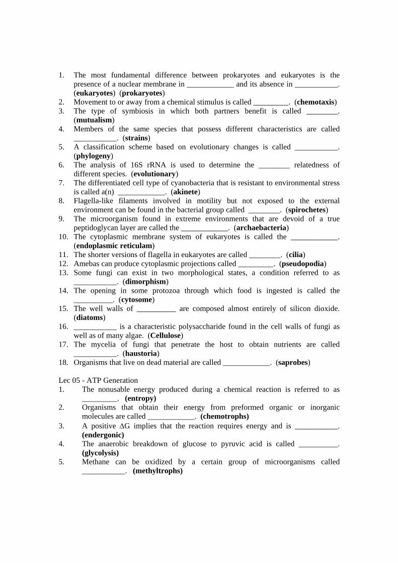
1. The most fundamental difference between prokaryotes and eukaryotes is the presence of a nuclear membrane in ____________ and its absence in ___________. (eukaryotes) (prokaryotes)
2. Movement to or away from a chemical stimulus is called _________. (chemotaxis) 3. The type of symbiosis in which both partners benefit is called ________.
(mutualism) 4. Members of the same species that possess different characteristics are called
___________. (strains) 5. A classification scheme based on evolutionary changes is called ___________.
(phylogeny) 6. The analysis of 16S rRNA is used to determine the ________ relatedness of
different species. (evolutionary) 7. The differentiated cell type of cyanobacteria that is resistant to environmental stress
is called a(n) ____________. (akinete) 8. Flagella-like filaments involved in motility but not exposed to the external
environment can be found in the bacterial group called ________. (spirochetes) 9. The microorganism found in extreme environments that are devoid of a true
peptidoglycan layer are called the ____________. (archaebacteria) 10. The cytoplasmic membrane system of eukaryotes is called the ____________.
(endoplasmic reticulam) 11. The shorter versions of flagella in eukaryotes are called ________. (cilia) 12. Amebas can produce cytoplasmic projections called _________. (pseudopodia) 13. Some fungi can exist in two morphological states, a condition referred to as
___________. (dimorphism) 14. The opening in some protozoa through which food is ingested is called the
__________. (cytosome) 15. The well walls of __________ are composed almost entirely of silicon dioxide.
(diatoms) 16. ___________ is a characteristic polysaccharide found in the cell walls of fungi as
well as of many algae. (Cellulose) 17. The mycelia of fungi that penetrate the host to obtain nutrients are called
___________. (haustoria) 18. Organisms that live on dead material are called ____________. (saprobes) Lec 05 - ATP Generation 1. The nonusable energy produced during a chemical reaction is referred to as
_________. (entropy) 2. Organisms that obtain their energy from preformed organic or inorganic
molecules are called ____________. (chemotrophs) 3. A positive G implies that the reaction requires energy and is ___________.
(endergonic) 4. The anaerobic breakdown of glucose to pyruvic acid is called __________.
(glycolysis) 5. Methane can be oxidized by a certain group of microorganisms called
___________. (methyltrophs)

6. The pathway that supplies reduced NADP for biosynthesis is called the _______________ or pentose phosphate pathway. (hexose monophosphate shunt)
7. The first electron carrier in the respiratory chain is usually a(n) ____________. (flavoprotein)
8. The formation of ATP during the transport of electrons and hydrogen to oxygen is called _______________. (oxidative phosphorylation)
9. Energy is released when electrons travel from a more ____________ source to a more ____________ substance. (negative) (positive)
10. Hydrocarbons such as alkanes are ultimately oxidized to __________ by microorganisms. (fatty acids)
Multiple choice Select the appropriate letter that correctly answers the question or completes the statement 1. Which of the following statements concerning photosystems is not true?
a. The photolysis of water results in the formation of carbon dioxide.
b. ATP formation may be cyclic or noncyclic. c. Both photosystems are not always present in some bacteria. d. Chlorophyll is one of the light-harvesting pigments e. NADP is one of the electron carriers.
1. Carbon dioxide is fixed to which of the following molecules during
photosynthesis to produce carbohydrate? a. ribulose 1,5-diphosphate d. ribose 5-phosphate b. glyceraldehyde 3-phosphate e. erythrose 4-phosphate c. glucose 6-phosphate 2. Certain molecules or structures in the cell are used to quench the overproduction
of oxygen during photosynthesis and are called a. phycobilisomes d. tetrapyrroles b. carboxysomes e. phycobiliproteins c. isoprenoids 3. Species of Thiobacillus are noted for their ability to oxidize a. inorganic nitrogen compounds d. hydrogen gas b. hydrocarbons e. sulfur compounds c. methane 4. One of the principal organic electron acceptors during anaerobic respiration is a. pyruvate d. formaldehyde

b. lactate e. fumarate c. acetate Lec 06 - Microbial Metabolism
1. What is a catalyst? a. a synthesis of cell molecules and structures b. a substance that speeds up a reaction c. where the substrate binds to on an enzyme d. organic compound that alters a substrate
2. An enzyme _______ the activation energy required for a chemical reaction.
a. increases b. converts c. lowers d. catalyzes
3. Which of these types of organisms gets its organic nutrients and energy from another
organism? a. chemoheterotroph b. chemoautotroph c. photoheterotroph d. photoautotroph
4. What is the chemical pathway that uses glucose and oxygen to produce carbon dioxide
and water? a. aerobic cellular respiration b. fermentation c. photosynthesis d. oxidative phosphorylation
5. Where does the electron transport system take place in bacteria?
a. cell membrane b. mitochondria c. ribosome d. cytoplasm
6. Where do the substrates bind on an enzyme?
a. allosteric site b. active site c. amino acid site d. enzymatic site
7. Match the following electron transport and oxidative phosphorylation terms to their description: ___ Oxidative phosphorylation ___ Chemiosmosis
(a) Transfer of electrons to final electron acceptor (oxygen)

___ Flavoproteins, cytochromes, and quinones ___ Electron transport
(b) Energy capture in the form of ATP harnessed from a series of redox reactions, with oxygen being the final electron acceptor (c) Electron carriers (d) ATP production from a proton gradient across the plasma membrane
8. The end products of photosynthesis in cyanobacteria and plant cells are: (a) Water and oxygen (b) Glucose and water (c) Glucose and oxygen (d) Water and carbon dioxide (e) Glucose and carbon dioxide 9. The energy source that drives the photosynthetic reactions in cyanobacteria is: (a) Heat (b) Light (c) Complex sugars (d) ATP (e) Oxygen 10. In the photosynthetic reactions, which of the following is NOT true? (a) Carbon dioxide is required in the dark reactions. (b) Energy is produced in the dark reactions. (c) Light reactions require light energy. (d) Occur in the thylakoids of the eukaryotic cells. (e) Generally result in the formation of glucose. 11.Match the following: ___ Chemiosmosis ___ Glycolysis ___ Electron transport chain ___ Fermentation ___ Photosynthesis ___ Krebs cycle
(a) Pathway that begins the breakdown of glucose (b) ATP production from a proton gradient across the plasma membrane (c) Anaerobic pathway that uses an organic final electron acceptor (d) Pathway that uses carbon dioxide, light, and chlorophyll to produce carbohydrates (e) Also is known as the tricarboxylic acid cycle (TCA) or as the citric acid cycle (f) Flavoproteins, cytochromes, and quinones
Lec 07 - Bacteriophages Lec 08 - Lytic and Lysogenic Cycles 1. The classification of viruses is based primarily on __________ and
____________ composition and structure. (morphology) (nucleic acid)
2. Bacterial viruses are called _______ or simply _________. (bacteriophage) (phage)
3. The complete and infective viral particle is called a(n) ________. (virion) 4. The protein cost surrounding the virus is called a(n) ___________, which is made
up of smaller protein units called ________. (capsid) (capsomeres) 5. The nucleic acid found in fungal viruses is only _____________. (RNA) 6. The maximum number of capsomeres that have been found in viruses is _______,
and the smallest number is __________. (252) (12) 7. The peplomers on the influenza virus envelope are ________ and __________.
(hemagglutinin) (neuraminidase) 8. The lesions produced by animal viruses on embryonic membranes are called
____________. (pocks) Lec 09 - Viroids, Prions 1. A compound frequently used to inactive viruses for use in vaccines is
a. ethylene glycol d. ether b. isopropy1 alcohol e. chloroform c. formaldehyde
1. Which of the following characteristics is not associated with viruses?
a. They can be cultivated on artificial media as long as ATP is provided.
b. Nucleic acid may be single-stranded or double-stranded DNA or RNA.
c. They can be inactivated only at temperatures above 100° C. d. They use the ribosomes of the cell to make viral protein. e. They show absolute specificity for one type of host.
1. Which of the following characteristics would not be appropriate for use in viral
classification? a. nucleic acid type b. capsid symmetry c. presence or absence of an envelope d. number of capsomeres e. all of the above
1. Which of the following best describes a viroid?
a. Nucleocapsid has icosahedral symmetry, is an RNA virus, and causes disease in plants.
b. It has no protein coat, is an RNA virus, and causes disease in plants.
c. It has no protein coat, is a DNA virus, and causes disease in plants. d. It has no protein coat, is a RNA virus, and causes disease in plants. e. None of the above is appropriate.
1. Which of the following agents would not inactivate most viruses?

a. 40% ethyl alcohol d. phenol b. 37° C for 15 minutes e. formaldehyde c. 2% glutaraldehyde
1. The viruses that exhibit complex capsid symmetry are
a. Adenovirus d. smallpox virus b. T4 bacteriophage e. none of the above c. Influenza virus
Lec 10 - Bacterial Genetics 1. The enzyme used by organisms for the synthesis of DNA is called DNA
_________ and the enzymes used to degrade DNA are called _________. (polymerase) (nucleases [exonucleases and endonucleases])
2. The DNA strand from which a complimentary molecule is synthesized is called the _____________ strand. (primer)
3. Bidirectional DNA synthesis occurs primarily in _________ whereas unidirectional DNA synthesis is more frequently observed in __________. (prokaryotes and eukaryotes) (viruses)
4. The group of enzymes that aids in the interconversion of relaxed and superhelical forms of DNA are called ____________. (topoisomerases)
5. The rolling circle mechanism of replication is an example of ___________ replication. (unidirectional)
6. The term used to describe multiple copies of DNA that are joined together is ______________. (concatameric)
7. Sex factor plasmids that can be integrated into the chromosome are called ____________. (episomes)
8. Proteins produced by the bacterial cell that are toxic to related species are called ____________. (bacteriocins)
9. Any chemical agent capable of altering the genotype and possibly the phenotype of the cell is called a(n) ____________. (mutagen)
1. Short sequences of DNA that can "jump" from one DNA site to another have been
called ___________ sequences. (insertion) Multiple choice Select the appropriate letter that correctly answers the question or completes the statement. 1. The test used to determine the carcinogenic potential of a chemical is called the a. reversion test d. replica plating test b. Ames test e. none of the above c. insertion frequency test

2. A mutant having a requirement for a certain growth factor is called a(n) a. protptroph d. chemotroph b. autotroph e. auxotroph c. heterotroph 3. The technique used to demonstrate that microbial mutants can arise spontaneously
in the absence of various chemical or physical agents is called the a. Ames test d. Kornberg assay b. transformation technique e. none of the above c. replica plating technique 4. DNA synthesized discontinuously produced short fragments called a. Ames fragments d. Okazaki fragments b. Kornberg sequences e. none of the above c. insertion sequences 5. The enzyme that catalyzes the union of the DNA fragments synthesized discontinuously is called a. DNA gyrase d. DNA polymerase III b. DNA helicase e. DNA ligase c. DNA polymerase I Lec 11 - Gene Expression Which of the following is not characteristic of the lactose operon?
a. The lactose repressor molecule does not require a corepressor. b. The operon is under positive as well as negative control
mechanisms. c. The repressor molecule genes are in the same operon. d. The lactose structural genes are in the same operon. e. The lactose genes code for enzymes involved in a catabolic
process. 2. The "wobble" hypothesis refers to
a. The inability of certain tRNAs to stabilize themselves on the mRNA.
b. The flexibility of the codon-anticodon interaction at the third codon position.
c. The loose binding of codons that do not specify an amino acid. d. None of the above.
3. The type of mutation that would cause a change in the reading frame from CAT CAT CAT CAT . . . to CAT ATC ATC ATC . . . is called a(n) a. insertion d. transversion

b. deletion e. none of the above c. transition 1. The catabolite activator protein (CAP) functions in the cell to a. repress the synthesis of biosynthetic enzymes
a. bind a catabolite and prevent repressor from attaching to the promoter region
b. bind a catabolite and prevent RNA polymerase from binding to the promoter region
c. bind cyclic AMP and attach to the promoter region d. none of the above
1. Pribnow sequences are believed to carry out which of the following functions in
the cell? a. site for binding of rho and termination of mRNA synthesis b. site for attachment of N-formylmethionine c. initiation site for DNA replication d. site on the DNA for binding the sigma subunit of RNA
polymerase e. site for attachment of repressor on the operator
1. Which of the following characteristic is associated with tRNA molecules?
a. They have a CCA end to which is attached an amino acid. b. They have abnormal bases such as pseudouracil and
dimethylguanine. c. They have a cloverleaf appearance. d. They have a site for binding aminoacy1 tRNA synthetases. e. All of the above are true.
1. The name given to mRNA to which several ribosomes are attached is a. monocistronic d. polygenic b. polycistronic e. none of the above c. polysome 2. Which of the following nucleic acids has the fewest nucleotides? a. tRNA d. mRNA b. rRNA e. DNA 1. The area on the DNA that codes for a polypeptide is called a(n) ____________.
(gene) 2. A genetic code in which more than one code word specifies a single amino acid is
called ____________. (degenerate) 3. The product of structural gene activity that facilitates the binding of an
aporepressor to the operator is called the ___________. (corepressor) 4. The enzymes that catalyze the attachment of amino acids to their specific tRNA
molecules are called __________. (aminoacy1 tRNA synthetases) 5. The triplet on the tRNA molecule that is complimentary to a triplet on the mRNA
is called a(n) ______________. (anticodon)

6. The component of the RNA polymerase enzyme that is specifically involved in initiating mRNA synthesis is called the ____________. (sigma factor)
7. Messenger RNAs that contain information for more than one polypeptide are called _____________. (polycistronic)
8. The energy for translocation in protein synthesis is derived from the hydrolysis of ____________. (GTP)
Lec 12 - Recombination in Bacteria 1. Virus that integrates into the host genome is called a(n) _________. (provirus) 2. _________ is the process in which there is movement of certain genes from one
DNA molecule to another. (Transposition) 3. The process in which cell-free DNA is taken up by a cell and engages in genetic
recombination is called _________. (transformation) 4. The bridge formed between conjugants during the conjugation progress is called
a(n) ___________. (pilus) 5. The gene transfer mechanism in which bacterial information is carried by a virus
is called ___________. (transduction) 6. The inverted repeat sequences found on the ends of certain DNA units are called
_______________. (palindromic) 7. Plasmids that are composed of DNA from more than one source are called
_________. (chimeric) 8. The amlification of a gene in a microbial cell in which the gene has been dervied
from another source is called _____________. (cloning) 9. The gene transfer mechanism in bacteria in which there is a cone-way transfer
between two mating types is called _______________. (conjugation) 10. The crossover event between lambda and the host chromosome during the
excision of lambda often results in the formation of a lambda particle carrying the bacterial genes _______ and _______. (galactose [Ga1]) (biotin [Bio])
Lec 13 - Genetic Engineering Lec 14 - Genetically Modified Organism Lec 15 - Soil Microbiology
1. The group of organisms most frequently associated with the production of antibiotics is
(a) Actinomyces (b) Klebsiella (c) Streptomyces (d) Pseudomonas
2. Which type of organism most frequently dominates the soil in terms of total biomass?
(a) Bacteria (b) Fungi (c) Algae (d) Protozoa
3. The soil element that is most responsible for limitations of plant growth and food production is
(a) oxygen (b) nitrogen (c) phosphorous (d) sulphur
4. The term ‘rhizosphere’ was coined by
(a) Winogradsky (b) Beijerinck (c) Hiltner (d) Waksman
5. The rate of organic matter decomposition is measured by,

(a) Dilution plate count method (b) Carbon‐di‐oxide evolution method
(c) Conn’s direct microscopic method (d) None of the above
6. One characteristic of the rhizosphere region of the soil ecosystem is its high
(a) oxygen content (b) microbial count (c) macropore count (d) humus content
7. Carbon cycles relatively rapidly except when it is
(a) dissolved in freshwater ecosystems (b) released by respiration
(c) stored in petroleum, coal or wood (d) part of bicarbonate reservoir in oceans
8. Contact slide or buried slide technique for qualitative determination of soil microflora was given by
(a) Winogradsky (b) Rossi and Cholodney (c) Beijerinck (d) Dobereiner
9. Fastest decomposition rate in soil is expected with residues having
(a) lowest N content (b) widest C:N ratio (c) lowest C:N ratio (d) highest C content
10. Which pool in the global carbon cycle uses biochemical energy from reduced carbon compounds?
(a) heterotrophs (b) autotrophs (c) carbon dioxide (d) soil organic matter
11. A free living non‐symbiotic Gram negative dinitrogen fixing bacteria
(a) Azospirillum (b) Anabaena (c) Azotobacter (d) Rhizobium
12. Conversion of organic complex of an element in to its inorganic state is called
(a) Mineralization(b) Immobilization (c) Nitrification (d) Oxidation
13. Adding nitrogen fertilizer to a compost pile will ____ the decomposition rate and ______ humus production.
(a) increase, increase (b) slow, increase (c) increase, decrease (d) slow, decrease
14. The element associated with dinitrogen reductase in diazotrophs is
(a) Oxygen (b) Magnesium (c) Molybdenum (d) Cobalt
15. The process that converts gaseous nitrogen to compounds that can be used by the biological community is called
(a) mineralization (b) nitrogen fixation (c) nitrogen mobilization (d) ammonification
16. Which pool in the nitrogen cycle can be both fixed and nitrified?
(a) humus (b) ammonium (c) Nitrogen (d) nitrate
17. Sulfur metabolism is an important part of energy metabolism in which of the following groups of microorganisms?
(a) phototrophic bacteria (b) Enterobacteriaceae (c) cyanobacteria (d) Pseudomonadaceae
18. An example of bacteria oxidizing ammonia to nitrites is

(a) Pseudomonas (b) Bacillus (c) Rhizobium (d) Nitrosomonas
19. The actinomycete that fixes atmospheric nitrogen in association with non‐leguminous plants is
(a) Actinomyces (b) Streptomyces (c) Nocardia (d) Frankia
20. The term mycorrhizae describes a symbiotic relationship between
(a) a heterotroph and an autotroph (b) an antibiotic and a pathogen
(c) nitrification and denitrification (d) a bacteria and a fungus
State whether the following statements are TRUE or FALSE
_______ 1. Microbial enzyme activities depends on soil pH.
_______ 2. Soil is unique with solid, liquid and gaseous phases interacting and thus supports more number and types of microflora.
_______ 3. Soil protozoa balances the soil ecosystem by feeding on soil fungi.
_______ 4. Bacteria which grows at higher soil temperatures are barophiles.
_______ 5. The classification of viruses is based primarily on morphology and nucleic acid composition and structure.
_______ 6. The mycelia of fungi that penetrate the host to obtain nutrients are called haustoria.
_______ 7. An example of an associatively symbiotic nitrogen fixing bacterium is Azotobacter.
_______ 8. Thiobacillus is involved in transformation of both nitrogen and iron.
_______ 9. The major group of organisms surrounding the root of plants are true bacteria.
_______ 10. Lignin is broken down in the soil primarily by fungi.
Fill in the blanks with appropriate words
1. The autotrophic mode of nutrition in bacteria was first established by __________.
2. An _________ pH favours growth of soil fungi.
3. Cellulose is polymer of _______________.
4. The rate of decomposition of organic matter is measured by ___________ method.
5. Soil microbial activity can be quantified by determining the __________ enzyme activity.
6. _______________ in root nodules regulates the supply of oxygen.
7. The site of nitrogen fixation in blue green algae is __________.
8. The primary wood degrading microorganisms are _________.
9. A non filamentous bacteria capable of sulphur oxidation is __________.
10. The region around leaf surface is called __________
Lec 16 - Microbial Transformations of Carbon Lec 17 - Microbial Transformations of Nitrogen, Phosphorus and Sulphur Lec 18 - Biological Nitrogen Fixation Lec 19 - Phyllosphere Bacteria

Lec 20 - Composting Lec 21 - Environmental Microbiology 1. Which of the following is not a major subdivision of the biosphere? a. hydrosphere c. stratosphere b. lithosphere d. atmosphere 2. A/an is defined as a collection of populations sharing a given habitat. a. biosphere c. biome b. community d. ecosystem 3. The quantity of available nutrients from the lower levels of the energy pyramid to the higher ones. a. increases c. remains stable b. decreases d. cycles 4. Photosynthetic organisms convert the energy of into chemical energy. a. electrons c. photons b. protons d. hydrogen atoms 5. Which of the following is considered a greenhouse gas? a. CO2 c. N2O b. CH4 d. all of these 6. The Calvin cycle operates during which part of photosynthesis? a. only in the light c. in both light and dark b. only in the dark d. only during photosystem I 7. Root nodules contain , which can . a. Azotobacter, fix N2 b. Nitrosomonas, nitrify NH3 c. rhizobia, fix N2 d. Bacillus, denitrify NO3 8. Which element(s) has/have an inorganic reservoir that exists primarily in sedimentary deposits? a. nitrogen c. sulfur b. phosphorus d. b and c 9. The floating assemblage of microbes, plants, and animals that drifts on or near the surface of large bodies of water is the community. a. abyssal c. littoral b. benthic d. plankton 10. An oligotrophic ecosystem would be most likely to exist in a/an

a. ocean c. tropical pond b. high mountain lake d. polluted river 11.Which of the following does not vary predictably with the depth of the aquatic environment? a. dissolved oxygen b. temperature c. penetration by sunlight d. salinity 12. Which of the following would be least accurate in detecting coliform bacteria in a water sample? a. the presumptive MPN test b. the standard plate count c. the membrane filter method d. the confirmatory MPN test Lec 22 - Microbiology of Food 1. The time required to kill a specified number of microorganisms at a particular
temperature is called the ____________. (thermal death time [TDT]) 2. Canned foods are thermally processed in steam-heated vats called
_____________. (retorts) 3. The species of Vibrio associated with shellfish poisoning is ___________.
(V. parahemolyticus) 4. The organisms responsible for the spoilage (called rope) of bread is
____________. (Bacillus subtilis) 5. Fatal forms of food poisoning are more frequently caused by the microorganism
____________. (Clostridium botulinum) 6. Food handlers are the most frequent source of food poisoning caused by
____________. (Staphylococcus aureus) 7. The principal viral agent associated with foodborne illness is the _____________.
(hepatitis A virus) 8. The animal parasite associated with foodborne illness and resulting from the
ingestion of undercooked bear or other wild meats is ______________. (Trichinella spiralis)
The recently discovered cause of bacterial foodborne illness that is associated with cereal dishes is ____________. (Bacillus cereus) 1. Which of the following species or genera is a frequent contaminant of water
supplies ? a. Lactobacillus d. Clostridium b. Streptococcus e. Bacillus c. Pseudomonas 2. Contemporary milk pasteurization times and temperatures have been selected
because they will destroy

a. Mycobacterium tuberculosis d. Bacillus anthracis b. Coxiella burnetii e. Legionella pneumophilia c. Streptococcus faecalis 3. The majority of "swollen" cans observed in the marketplace are the result of
a. CO2 production by clostridia b. H2 production by clostridia c. H2 production from the interaction of tin and acid d. O2 production by aerobic sporeformers e. None of the above
1. The microorganisms most frequently involved in the spoilage of canned foods are
a. Streptococci and lactobacilli b. Leuconostoc and Pseudomonas c. Clostridium and Bacillus d. Saccharomyces and Staphylococcus e. Lactobacillus and Streptococcus
1. If an organism has a water activity of 0.62, in which of the following foods (water
activity in parentheses) will it most likely grow? a. flour (0.61) d. honey (0.50) b. chocolate (0.54) e. dried beef (0.87) c. fresh meat (0.98) 2. The predominant group of microorganisms found in milk today are a. mycobacteria d. lactobacilli and streptococci b. Brucella e. Clostridium and Bacillus c. Gram-negative rods Lec 23 - Principles of Preservation Lec 24 - Role of Bacteria in Fermentation
1. During fermentation, pyruvic acid is converted into major organic products such as (A) glucose and maltose (B) starch and cellulose
(C) ethyl alcohol and lactic acid (D) citric acid and isocitric acid 2. The group of organisms most frequently associated with the production of antibiotics is (A) Actinomyces (B) Klebsiella (C) Streptomyces (D) Pseudomonas
3. Maximum production of antibiotics generally occurs
(A) during the log phase (B) after the log phase (B) before the log phase (D) only when the pH drops below 4.3
4. The Koji process is a technique used in the industrial production of (A) vinegar (B) microbial polysaccharides

(C) pectinase (D) single cell protein
5. The precursor added to a fermenting medium for penicillin G production is (A) malic acid (B) tryptophan
(C) yeast extract (D) lysine State true or False:
6. Baffles are provided in a fermentor for mixing and turbulence. 7. A type of bacterial growth where the cells never reach its stationary phase is batch
culture. 8. Mushroom production is an example of solid state fermentation. 9. Strict sterile conditions are not required in lactic acid production because the organism
can be cultivated at a high temperature. 10. A most common mutagen used for strain improvement of industrial microbes is IR rays.
Fill up the blanks:
11. The device used to maintain cells in logarithmic state in a fermentor is called as _______________.
12. Aspergillus niger is used for the industrial production of ____________. 13. The organism used in recombinant DNA technology for the commercial production of
interferon, insulin and growth hormones is ____________. 14. The classical approach to strain improvement of industrially important strain is
__________. 15. The method for screening antibiotic producing microorganisms is called ___________.

Match the following:
16. Saccharomyces cerevisiae : (a) Amylase 17. Abhya gossypii : (b) SCP 18. Scenedesmus : (c) Streptomycin 19. Bacillus amyloliquifaciens : (d) Ethanol 20. Streptomyces griseus : (e) Riboflavin
Lec 25 - Beneficial Microorganisms in Agriculture 1. The major morphological group of bacteria found in the soil is
a. long rods, non-spore-forming b. cocci c. rods, spore-forming d. coccoidal rods e. rods, gram-positive non-spore-forming
1. Which type of organisms most frequently dominates the soil in terms of total
biomass? a. Bacteria c. algae b. Fungi d. protozoa
1. The actinomycete that fixes nitrogen in association with nonleguminous plants is
a member of the genus a. Actinomyces d. Arachnia b. Streptomyces e. Nocardia c. Frankia 2. The bacterial inhabitant of the soil that can parasitize other bacteria belongs to the
genus a. Erwinia d. Bdellovibrio b. Rhizobium e. Bacillus c. Frankia 3. The rhizosphere and rhizoplane are surrounded by a microbial community whose
cell density is a. greater than 108/g c. approximately 107/g b. approximately 105/g d. less than 105/g 4. The major group of microorganisms found surrounding the roots of plants are a. algae d. fungi b. true bacteria e. protozoa c. actinomycetes 5. The primary purpose of the rhizosphere community is to

a. destroy potential invading plant pathogens b. supply amino acids to the plant c. reduce the level of toxic acids that surround the plant roots d. convert organic compounds containing nitrogen, phosphorous,
and sulfur to inorganic products suitable for assimilation by the plant e. convert inorganic compounds of nitrogen, phosphorous, and sulfur
to organic products suitable for growth of the plant Choose the best answer:
1. The rate of organic matter decomposition is measured by,
(a) Dilution plate count method (b) Carbon‐di‐oxide evolution method
(c) Conn’s direct microscopic method (d) None of the above
2. Which of the following substance is most resistant to microbial biodegradation?
(A) pectin (B) cellulose
(C) lignin (D) hemicellulose
3. Which pool in the global carbon cycle uses biochemical energy from reduced carbon
compounds?
(A) heterotrophs (B) autotrophs
(C) lithotrophs (D) organotrophs
16. Fastest decomposition rate in soil is expected with residues having
(A) lowest N content (B) widest C:N ratio
(C) lowest C:N ratio (D) highest C content
17. Adding nitrogen fertilizer to a compost pile will _________ the decomposition rate and
___________ humus production.
(A) increase, increase (B) slow, increase
(C) increase, decrease (D) slow, decrease
Define the following or answer in one sentence:
18. Humic acid
19. Mesophiles
20. Herbicide
21. Biogas
22. Vermiculture
Fill up the blanks:
23. Conversion of organic complex of an element in to its inorganic state is called
_______________.
24. Soil microbial activity can be quantified by determining the __________ enzyme activity.
25. The organism most frequently dominates the soil in terms of total numbers and types
are ____________.
26. The increase in concentration of a recalcitrant molecule as it passes through the trophic
levels is called ______________.
27. Those compounds, both biological and synthetic, that are resistant to microbial
digestion are called ____________ molecules.
Match the following:
16. Phanerochate chrysosporium : (f) Methane producer
17. Pseudomonas putida : (g) Cellulose degrader
18. Methanothrix : (h) Pesticide degrader
19. Streptomyces : (i) Wood degrading fungi
20. Trichoderma : (j) Compost pits
Lec 26 - Microbial Agents for Control of Plant Disease Lec 27 - Biogas Production Lec 28 - Biodegradable Plastics Lec 29 - Plant – Microbe Interactions Lec 30 - Bioremediation Lec 31 - Bio Sensor Lec 32 - Microbial Products Nitrite is converted into nitrate by the bacteria
173. Phycobiont is
a. The algal part in Lichens
b. The fungal part in Lichens
c. Laustoria formation
d. None of these
174. Parasitic form must contain
a. Capsules b. Cell-wall c. Endospores d. Flagella
175. The total no. of genes in the group of same individuals is
a. Nitrosomonas c. Nitrobacter
b. Nitrosocytes d. Azatobacter

165. Sulphur oxidizing bacteria is
a. Alcaligenes b. Pseudomonas c. Thiobacillus d. None of these
166. Bacillus Schlegelli is
a. Hydrogen – Oxydising bacteria
b. Sulphur – Oxydising bacteria
c. Iron-Oxidising bacteria
d. Nitrite oxidizing bacteria
167. The group of bacteria which deopends on organic sources in nature for their energy requirements. They are said to be �a. Chemotrophs b. Phototrophs c. Heterotrophes d. Organotrophs
168. Majority of bacteria are
a. Genome c. Gene pool
b. Gene map�d. None of these
a. Saprophytes c. Commensals
169. Symbionts are
b. Symbionts d. Parasites
176. Transformation was observed mainly in
a. Bacteriophages b. Temperate phages c. –phage d. All of these
177. Capsulated forms of bacteria are
a. Virulent b. A virulent c. Useful d. Symbiotic
178. The bacterial cells participating in conjugation are
a. Bacteria in symbiotic association
b. The group of fungi in symbiotic association
c. The groups participating in symbiotic association

d. All of these
170. The best example for symbiotic associa- tion is
a. E.coli in intestine of man
b. Lichens
c. Normal floraof skin
d. All of the above
171. The enzymes responsible for decomposi- tion is
a. Conjugants c. Exconjugants
179. Phagocytes are
a. Monocytes c. Basophils
b. Fertile cells�d. None of these
b. Macrophages d. All of these
a. Lipolytic c. Lysozyme
b. Proteolytic�d. Both a and b
180. The microorganism engulfed by phago- cyte resides in a vacuole is known as
a. Phagosome b. Lysosome�c. both a and b d. None of these
181. Toxic products in phagolysosome are
a. H2SO4
b. Singlet O2
c. Superoxide radicals
d. All of these
182. During destruction of antigen particle in phagolysosome the product formed in phagolysosome the product formed during formulation is
a. Acetic acid b. Lactic acid�c. Citric acid d. None of these
172. Urea is decomposed by the species
a. Micrococcus sps. b. Nitrosomonas sps. c. Proteus sps. d. Both a and c