antifungal effect of powdered spices and their extracts on ...
REVIEW ARTICLEpublished: 20 March 2014
doi: 10.3389/fmicb.2014.00097
Defensins: antifungal lessons from eukaryotesPatrícia M. Silva, Sónia Gonçalves and Nuno C. Santos*
Instituto de Medicina Molecular, Faculdade de Medicina, Universidade de Lisboa, Lisbon, Portugal
Edited by:
Octavio Luiz Franco, UniversidadeCatólica de Brasília, Brazil
Reviewed by:
Valdirene Moreira Gomes,Universidade Estadual do NorteFluminense, BrazilFrançoise Gosti, Centre National de laRecherche Scientifique, France
*Correspondence:
Nuno C. Santos, Instituto de MedicinaMolecular, Faculdade de Medicina,Universidade de Lisboa, AvenidaProfessor Egas Moniz, 1649-028Lisbon, Portugale-mail: [email protected]
Over the last years, antimicrobial peptides (AMPs) have been the focus of intense researchtoward the finding of a viable alternative to current antifungal drugs. Defensins are oneof the major families of AMPs and the most represented among all eukaryotic groups,providing an important first line of host defense against pathogenic microorganisms.Several of these cysteine-stabilized peptides present a relevant effect against fungi.Defensins are the AMPs with the broader distribution across all eukaryotic kingdoms,namely, Fungi, Plantae, and Animalia, and were recently shown to have an ancestorin a bacterial organism. As a part of the host defense, defensins act as an importantvehicle of information between innate and adaptive immune system and have a role inimmunomodulation. This multidimensionality represents a powerful host shield, hard formicroorganisms to overcome using single approach resistance strategies. Pathogenic fungiresistance to conventional antimycotic drugs is becoming a major problem. Defensins, asother AMPs, have shown to be an effective alternative to the current antimycotic therapies,demonstrating potential as novel therapeutic agents or drug leads. In this review, wesummarize the current knowledge on some eukaryotic defensins with antifungal action.An overview of the main targets in the fungal cell and the mechanism of action of theseAMPs (namely, the selectivity for some fungal membrane components) are presented.Additionally, recent works on antifungal defensins structure, activity, and cytotoxicity arealso reviewed.
Keywords: antimicrobial peptides, defensins, antifungal, resistance, host defense peptides
INTRODUCTIONNaturally occurring antimicrobial peptides (AMPs) probablyrepresent one of the first successful forms of chemical defenseof eukaryotic cells against bacteria, protozoa, fungi, and viruses(Ganz and Lehrer, 1998; Lehrer and Ganz, 1999; Zasloff, 2002;Mookherjee and Hancock, 2007; Lai and Gallo, 2009; Guo et al.,2012; Domingues et al., 2014), being also active against can-cer cells (Hoskin and Ramamoorthy, 2008; Gaspar et al., 2013).Currently commercialized antibiotics are mostly of microbialorigin or synthesized from those. These antibiotics are losingefficacy as a result of high selection pressure, leading to rapidemergence of resistance in many important human pathogens,thus threatening to put an end to the golden age of antibiotics(Clardy et al., 2009; Fisher et al., 2012). The use of antifun-gal treatments has increased as a consequence of the increaseof immunocompromised patients, mostly due to improvementsin oncology and transplant fields (Mehra et al., 2012), lead-ing to more frequent resistances to the drugs used. A strategyto overcome this problem can be found in AMPs, which arepart of the innate immune system of different living organ-isms (Hegedus and Marx, 2013), such as plants (Thommaet al., 2002; Lay and Anderson, 2005; Gonçalves et al., 2012b),fungi (Mygind et al., 2005; Schneider et al., 2010; Oeemig et al.,2012), bacteria (Zhu, 2007; Gao et al., 2009), invertebrates (Buletand Stocklin, 2005; Ayroza et al., 2012), and vertebrates (Ganz,2004; Sahl et al., 2005; van Dijk et al., 2008; Gonçalves et al.,2012a).
Although some AMPs have had their target unveiled, manyare still unclear. Some mechanisms of action of antifungal pep-tides have been reported, such as binding to the cell wall,membrane permeabilization, receptor-mediated internalizationinducing signaling cascades, and interaction with intracellular tar-gets, inducing the formation of reactive oxygen species (ROS),leading ultimately to apoptosis (Hancock and Rozek, 2002; Ober-parleiter et al., 2003; Thevissen et al., 2003a, 2004; de Conincket al., 2013; van der Weerden et al., 2013). Apoptosis is a type ofprogramed cell death, which is regulated by a complex network ofproteins and metabolic pathways. The central core of this processis regulated by a family of proteins named caspases. Yeasts have atleast one ortholog of mammalian caspases: the metacaspase YCA1(yeast caspase 1; Madeo et al., 2002). Routinely used assays aimingat the detection of these apoptotic features are used for the identi-fication of fungal cells undergoing apoptosis after treatment withantifungal agents.
Antimicrobial peptides have variable amino acid compositionand size (ranging from less than 10 to more than 100 amino acidresidues), commonly being cationic and amphipathic molecules(Brogden et al., 1996; Yang et al., 2003; Fontana et al., 2004;Glaser et al., 2005; Domingues et al., 2014). To date, more than2200 natural or synthetic AMPs have been identified, as listedby the Antimicrobial Peptide Database (APD1), of which over1900 have antibacterial activity and 800 have antifungal activity.
1http://aps.unmc.edu/AP/main.php
www.frontiersin.org March 2014 | Volume 5 | Article 97 | 1

Silva et al. Defensins: antifungal lessons from eukaryotes
This discrepancy, however, may be redundant as antibacterialAMPs may also have antifungal activity, but this property is notsystematically assessed.
Antimicrobial peptides may have linear structures, like indoli-cidin (Ladokhin et al., 1999), or they may have tertiary struc-tures stabilized by disulfide bonds, with β-sheet (e.g., protegrin;Aumelas et al., 1996; and the defensin human neutrophil pep-tide 1, HNP-1; Zhang et al., 2011), α-helix (e.g., dermaseptin;Mor et al., 1991) or αβ-motif secondary structure (e.g., dro-somycin; Landon et al., 1997; and Pisum sativum defensin 1, Psd1;Almeida et al., 2002).
The most studied families of AMPs are cathelicidins, der-maseptins, magainins, cecropins, and defensins. Cathelicidinsare found in the innate immune system of mammals, amphib-ians, and reptiles (Wang et al., 2008; Tsai et al., 2011; Hao et al.,2012); dermaseptins and magainins are found in amphibians(Morton et al., 2007); cecropins are found in insects (YiZenget al., 1989); and defensins, which are the largest family of AMPs,have also the broader distribution across the majority of eukary-otic organisms (Wang et al., 2013). Besides having antimicrobialactivity, defensins also have immunomodulatory functions in theorganisms that produce these peptides. Defensins with antifun-gal properties are present in all eukaryotic kingdoms, pointingout to a common ancestor. This review is focused on defensins(as well as some defensin-like peptides) with antifungal activity.However, it is impossible to describe here all the defensins withthis activity. Therefore, we highlight some of the most recentresearch made on this field. The chosen peptides are describedtaking into consideration their specific properties, evolution-ary background, organism of origin, and antifungal mode ofaction.
Some databases have been created in order to provide usefulinformation for the study of AMPs. Among the AMP databases,PhytAMP is a database dedicated to antimicrobial plant pep-tides2 (Hammami et al., 2009). This resource contains valuableinformation on these AMPs, including peptide sequences, tax-onomic, microbiological, and physicochemical data. Anotherdatabase, Collection of Anti-Microbial Peptides (CAMP3), holds
2http://phytamp.pfba-lab-tun.org/main.php3http://www.camp.bicnirrh.res.in/
experimentally validated and predicted AMP sequences andstructures of AMPs. These databases include several tools forAMPs analysis and prediction, helping in the design of newtherapeutic peptides based on specific structure and functionalfeatures.
DEFENSINSDefensins are the largest groups of AMPs. These peptides arecysteine-rich and have diverse sequences and structures, stabilizedinto compact shapes by three or four conserved cysteine disulfidebridges. They have at least two positive charges (lysine or argi-nine residues) and are small, ranging approximately from 12 to 50amino acid residues (2–6 kDa; Ganz, 2003; Ren et al., 2011; Gaoand Zhu, 2012).
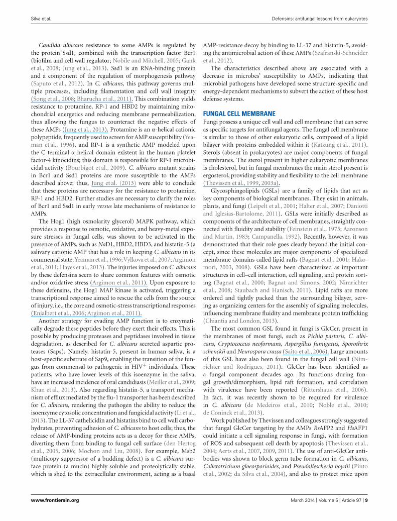
Vertebrates’ defensins are divided into three subfamilies: α-, β-,and θ-defensins. α-Defensins are present in mammals such ashumans, monkeys, and several rodent species, being particularlyabundant in neutrophils, certain macrophage subpopulations andPaneth cells of the small intestine (Ouellette and Selsted, 1996;Ganz and Lehrer, 1998; Lehrer and Ganz, 1999). β-defensinsare found in a wide range of vertebrates, presenting a cysteine-stabilized αβ-motif composed of an antiparallel β-sheet and anα-helix. As an example, on bovine neutrophils, as many as 13β-defensins have been identified (Yang et al., 2002a). However,in other species, β-defensins are mostly produced by epithelialcells lining different organs (e.g., epidermis, bronchial tree, andgenitourinary tract). θ-Defensins, present only in Old World mon-keys, are cyclic and derived from α-defensins (Lehrer, 2004; Lehrerand Lu, 2011; Semple and Dorin, 2012). In Figure 1, conservedcysteine residues among defensins from different kingdoms areshown.
Plants, fungi, and many invertebrates produce defensin-likepeptides structurally similar to the β-defensins from vertebrates(Thomma et al., 2002; Bulet and Stocklin, 2005; Mygind et al.,2005; Sahl et al., 2005; Yount and Yeaman, 2006; van Dijk et al.,2008; Ayroza et al., 2012; Oeemig et al., 2012). These observa-tions allowed to assume that defensins and defensin-like peptidesall evolved from a common precursor. The relatively recentidentification of three defensins in lower eukaryotes, plectasinfrom Pseudoplectania nigrella (Mygind et al., 2005), eurocin fromEurotium amstelodami (Oeemig et al., 2012), and bubble protein
FIGURE 1 | Comparison of amino acid residues sequence of
selected defensins with antifungal activity from animals, plants, and
fungi: human defensin 5 (HD5), Pisum sativum defensin 1 (Psd1),
and Penicillium brevicompactum Dierckx bubble protein (BP),
respectively. Conserved cysteine residues among defensins fromdifferent kingdoms are indicated. The colors used in the sequencesrepresent basic, non-polar hydrophobic, polar-uncharged and acidicresidues.
Frontiers in Microbiology | Antimicrobials, Resistance and Chemotherapy March 2014 | Volume 5 | Article 97 | 2

Silva et al. Defensins: antifungal lessons from eukaryotes
(BP) from Penicillium brevicompactum Dierckx (Seibold et al.,2011), is important to demonstrate the wide distribution ofthese peptides over diverse eukaryotic lineages, which suggeststhat ancestral defensin genes existed over 1500 million yearsago, before Fungi, Plantae, and Animalia kingdoms diverged(Wang et al., 1999). The wide distribution of these peptides inthe Eukarya domain could suggest their uniqueness to eukary-otic cells, but it was possible to determine that these peptidesmay have had their ancestor in a prokaryotic organism afterthe discovery of the first defensin-like peptide in the bacte-ria Anaeromyxobacter dehalogenans (AdDLP; Zhu, 2007; Gaoet al., 2009). This defensin-like peptide is proposed as an ances-tor of eukaryotic defensins and defensin-like peptides due tothe similarity of their structures, namely at the level of thecysteine-stabilized αβ-motif (Zhu, 2007). These findings furthersupport the concept that AMPs may have been fundamental tothe evolution of multicellular organisms within microbial-exposedenvironments.
Although defensins were initially identified only as AMPs,recent studies have demonstrated that they have a much broaderrange of action, including immunomodulatory function (issuefurther developed in the text, in Section “ImmunomodulatoryFunction”; Ulm et al., 2012).
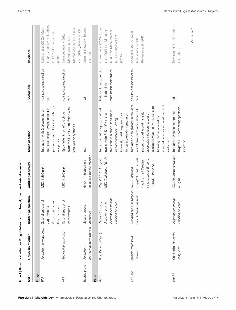
RECENTLY STUDIED ANTIFUNGAL DEFENSINSWhen a new AMP is described, the most usual properties toassess are structure and peptide sequence, antimicrobial activity,expressed mainly in terms of minimal inhibitory concentration(MIC) or half maximal inhibitory concentration (IC50), cytotox-icity and lytic activity against human cells (whenever the AMPorigin is not mammalian), target, and mode of action toward thepathogen tested. The following AMPs were classified as defensinsor defensin-like peptides due to their structural and sequencehomologies with other defensins. They are examples of some ofthe recently studied antifungal defensins from fungal, plant, andanimal origin that fulfill most of the properties expressed above.Further details and a list of some of these defensins can be foundin Table 1.
FUNGAL SOURCESDefensin-like antifungal peptides secreted by filamentous fungihave a low molecular mass (5.8–6.6 kDa), a basic char-acter, presence of 4–10 cysteine residues and several disul-fide bonds (providing resistance against temperature stress oradverse solvent conditions), and a β-barrel conformation (Hagenet al., 2007; Seibold et al., 2011). Proteins with such proper-ties with antifungal activity have been isolated and investi-gated from several Ascomycota fungal species, such as Peni-cillium chrysogenum, Penicillium nalgiovense, Penicillium bre-vicompactum Dierckx, Aspergillus giganteus, and Aspergillusniger (Galgóczy et al., 2010; Seibold et al., 2011). Among thesefungus-derived antifungal peptides, the most intensively stud-ied are Penicillium chrysogenum antifungal protein (PAF) andAspergillus giganteus antifungal protein (AFP), with six andeight cysteines, respectively. Penicillium brevicompactum Dier-ckx bubble protein structure is considerably similar to PAF andAFP.
Penicillium chrysogenum antifungal proteinPAF acts through a G protein-coupled signal transduction path-way, although this mechanism is not entirely understood (Marxet al., 2008). The G protein-coupled activity of PAF was confirmedby the study of the fadA mutant of Aspergillus nidulans whichproved to be less sensitive to PAF treatment compared to thewild-type. The fadA gene encodes the heterotrimeric G proteinα subunit, which dissociation from the Gβγ complex is inhibitedin the fadA mutant Aspergillus nidulans (Leiter et al., 2005). Theseresults indicate that PAF toxicity requires active heterotrimericG protein signaling (Leiter et al., 2005; Marx et al., 2008). Basedon these observations, Marx et al. (2008) hypothesized that PAFinteracts directly or indirectly with G protein signal transductionpathways. These authors also hypothesized that the cell wall couldbe a selective barrier for PAF, but in vivo chitin-binding activityof PAF has not been demonstrated yet. In the sensitive organ-isms, PAF exerts multiple detrimental effects: induction of plasmamembrane polarization, increased exposure of phosphatidylserine(PS) on the cell surface, DNA fragmentation, membrane blebbing,cell shrinking, intracellular ROS production, and apoptosis-likephenotype (Kaiserer et al., 2003; Leiter et al., 2005; Marx et al.,2008).
Aspergillus giganteus antifungal proteinIn susceptible organisms, AFP disturbs the polarized growth ofhyphae by interfering directly or indirectly with the cell wallbiosynthesis (Theis et al., 2003). Hagen et al. (2007) demonstratedthat AFP can bind to chitin in vitro, and inhibits the cell wallchitin biosynthesis by the specific inhibition of chitin synthase IIIand V. These enzymes are unique among fungi and essential for themaintenance of the polarized growth and virulence of pathogenicfungi. Presence of chitin synthase III and V is confirmed in theAFP-sensitive fungi, while the insensitive fungal species do nothave these enzyme classes. Sphingolipid membrane componentsin the sensitive fungi might serve as secondary receptors for AFP.This was further confirmed by the discovery that the depletionof cellular glucosylceramide (GlcCer) levels in AFP-sensitive fun-gal species (Aspergillus fumigatus and Aspergillus niger) resulted inreduced AFP susceptibility (Meyer, 2008). This, together with theobservation that the sphingolipids are necessary to maintain thepolarized hyphal growth, elucidates the mechanism of polarizedgrowth degeneration effect of AFP (Li et al., 2006; Meyer, 2008).The species specificity of AFP may be related with the sphingolipidprofile of the sensitive fungi (Meyer, 2008).
Penicillium brevicompactum Dierckx bubble proteinThis fungal defensin was first described in 2003 (Olsen et al., 2004).It is found in the bright yellow–green fluorescent exudate bubblesof the ascomycete fungus Penicillium brevicompactum Dierckx.Similarly to other ascomycetes, BP produces a small antimicro-bial molecule, mycophenolic acid, which gives the bubbles theiryellow–green fluorescence. This combined production suggests apossible synergistic action between defensins and other antibioticagents produced by this class of fungi (Seibold et al., 2011). BP has64 amino acid residues, with high content of basic amino acids,β-barrel conformation (Olsen et al., 2004), and a cage-like patternof four very stable disulfide bridges. In addition, it was discovered
www.frontiersin.org March 2014 | Volume 5 | Article 97 | 3
Silva et al. Defensins: antifungal lessons from eukaryotes
Ta
ble
1|
Re
ce
ntl
ystu
die
da
nti
fun
ga
ld
efe
nsin
sfr
om
fun
ga
l,p
lan
t,a
nd
an
ima
lso
urc
es.
AM
PO
rga
nis
mo
fo
rig
inA
nti
fun
ga
lsp
ectr
um
An
tifu
ng
al
acti
vit
yM
od
eo
fa
cti
on
Cy
toto
xic
ity
Re
fere
nce
Fu
ng
i
PAF
Peni
cilli
umch
ryso
genu
mS
ever
alsp
ecie
sof
Zygo
myc
etes
,
Asc
omyc
etes
,and
Bas
idio
myc
ota
MIC
:1–2
00μ
g/m
lIn
tera
ctio
nw
ithG
prot
ein
sign
al
tran
sduc
tion
path
way
s,le
adin
gto
prod
uctio
nof
RO
San
din
duct
ion
of
apop
tosi
s
Non
-tox
icto
mam
mal
ian
cells
Kai
sere
ret
al.(
2003
);M
arx
(200
4);G
algó
czy
etal
.(20
05,
2007
,200
8);B
arna
etal
.
(200
8)
AFP
Asp
ergi
llus
giga
nteu
sS
ever
alsp
ecie
sof
Asc
omyc
etes
MIC
:1–2
00μ
g/m
lS
peci
ficin
hibi
tion
ofth
ech
itin
synt
hase
IIIan
dV,
inte
rfer
ing
with
cell
wal
lbio
synt
hesi
s
Non
-tox
icto
mam
mal
ian
cells
Laca
dena
etal
.(19
95);
Mor
eno
etal
.(20
03);
Mor
eno
etal
.(20
06);
Thei
s
etal
.(20
03);
Mey
er(2
008)
Bub
ble
prot
ein
Peni
cilli
um
brev
icom
pact
umD
ierc
kx
Sac
char
omyc
es
cere
visi
ae
Gro
wth
inhi
bitio
nin
a
dose
-dep
ende
ntm
anne
r
n.d.
n.d.
Ols
enet
al.(
2004
);S
eibo
ld
etal
.(20
11)
Pla
nt
Psd
1Pe
a(P
isum
sativ
um)
Asp
ergi
llus
spp.
;
Fusa
rium
sola
ni;
Neu
rosp
ora
cras
sa;
Can
dida
albi
cans
IC50
:0.0
4–21
.7μ
g/m
l;
MIC
C.a
lbic
ans:
20μ
M
Impa
irmen
tof
prog
ress
ion
ofce
ll
cycl
e:cy
clin
F;S
toG
2ph
ase
tran
sitio
nis
bloc
ked,
resu
lting
in
endo
redu
plic
atio
n;st
rong
inte
ract
ion
with
ergo
ster
olan
d
fung
alst
erol
-ric
hm
embr
anes
Red
uced
inte
ract
ion
with
chol
este
rol-r
ich
mam
mal
ian
mem
bran
es
Alm
eida
etal
.(20
00);
Lobo
etal
.(20
07);
deM
edei
ros
(200
9);d
eM
edei
ros
etal
.
(201
0);G
onça
lves
etal
.
(201
2b)
RsA
FP2
Rad
ish
(Rap
hanu
s
sativ
us)
Can
dida
spp.
;Asp
ergi
llus
flavu
s;Fu
sariu
mso
lani
IC30
C.a
lbic
ans:
10μ
g/m
l.R
educ
edce
ll
viab
ility
inal
lCan
dida
spp.
test
edw
ithup
to
10μ
Mof
RsA
FP2
Inte
ract
ion
with
gluc
osyl
cera
mid
es;
mem
bran
epe
rmea
biliz
atio
n;R
OS
prod
uctio
n;ce
llgr
owth
arre
st;
apop
tosi
sin
duct
ion;
casp
ase
activ
atio
n;ye
ast-
to-h
ypha
tran
sitio
n
bloc
king
;sep
tinlo
caliz
atio
n;
cera
mid
eac
cum
ulat
ion;
alte
red
cell
wal
lsha
pe
Non
-tox
icto
mam
mal
ian
cells
Aer
tset
al.(
2007
,200
9);
Tava
res
etal
.(20
08);
Thev
isse
net
al.(
2012
)
HsA
FP1
Cor
albe
lls(H
euch
era
sang
uine
a)
Neu
rosp
ora
cras
sa;
Can
dida
albi
cans
IC50
Neu
rosp
ora
cras
sa:
4μ
g/m
l
Inte
ract
ion
with
cell
mem
bran
e
(hyp
ha);
RO
Sfo
rmat
ion;
apop
tosi
s
indu
ctio
n
n.d.
Thev
isse
net
al.(
1997
);A
erts
etal
.(20
11)
(Con
tinue
d)
Frontiers in Microbiology | Antimicrobials, Resistance and Chemotherapy March 2014 | Volume 5 | Article 97 | 4
Silva et al. Defensins: antifungal lessons from eukaryotes
Ta
ble
1|
Co
nti
nu
ed
AM
PO
rga
nis
mo
fo
rig
inA
nti
fun
ga
lsp
ectr
um
An
tifu
ng
al
acti
vit
yM
od
eo
fa
cti
on
Cy
toto
xic
ity
Re
fere
nce
An
ima
l
Art
hro
po
d
Cop
risin
Kore
andu
ngbe
etle
(Cop
ristr
ipar
titus
)
Asp
ergi
llus
spp.
;Can
dida
spp.
;Mal
asse
zia
furf
ur;
Tric
hosp
oron
beig
elii;
Tric
hoph
yton
rubr
um
MIC
:5–2
0μ
MA
popt
osis
indu
ctio
n;R
OS
form
atio
n;di
srup
tion
of
mito
chon
dria
lmem
bran
epo
tent
ial;
cyto
chro
me
cre
leas
e;in
trac
ellu
lar
met
acas
pase
activ
atio
n
No
hem
olyt
icac
tivity
on
hum
aner
ythr
ocyt
es
Lee
etal
.(20
12)
Juru
inA
maz
onia
npi
nkto
e
spid
er(A
vicu
laria
juru
ensi
s)
Can
dida
spp.
;Asp
ergi
llus
nige
r
MIC
:2.5
–10
μM
;
fung
icid
alac
tivity
,rat
her
than
fung
ista
tic
n.d.
No
hem
olyt
icac
tivity
on
hum
aner
ythr
ocyt
es
Ayr
oza
etal
.(20
12)
Re
pti
le
Cro
tam
ine
Sou
th-A
mer
ican
ratt
lesn
ake
(Cro
talu
s
duris
sus
terr
ificu
s)
Can
dida
spp.
;
Tric
hosp
oron
spp.
;
Cry
ptoc
occu
s
neof
orm
ans
MIC
:12.
5–50
μg/
ml;
fung
icid
alac
tivity
,rat
her
than
fung
ista
tic
Pron
ounc
edul
tras
truc
tura
l
alte
ratio
ns;m
embr
ane
colla
pse;
cyto
plas
mic
coag
ulat
ion
No
hem
olyt
icac
tivity
on
hum
aner
ythr
ocyt
es;
CC
50>
50μ
Mag
ains
t
non-
tum
oral
anim
alan
d
hum
ance
lls
Yam
ane
etal
.(20
13)
n.d.
,not
dete
rmin
ed.
www.frontiersin.org March 2014 | Volume 5 | Article 97 | 5

Silva et al. Defensins: antifungal lessons from eukaryotes
that the closely related fungus Penicillium chrysogenum encodes aBP homolog (in addition to PAF), indicating that fungi may havemore than one defensin (Seibold et al., 2011).
PLANT SOURCESMany fungi are phytopathogenic, with species such as Fusariumspp., Cladosporium spp., Pythium spp., Curvularia spp., Aspergillusflavus, and Puccinia pittieriana affecting potato, rice, corn, wheat,tobacco, and cotton crops by causing wilt, mold, crown rot,mildew, and rust, just to name a few plant diseases (The Ameri-can Phytopathological Society, APS4). These diseases can depleteentire crops, bearing enormous costs for agriculture due to thedifficulty in eliminating fungal infections from plants, once theyappear. Soils harbor plants for most of their life cycle, but alsoa considerable amount of bacteria, fungi, and parasites, manyof which can be phytopathogenic. For this reason, plants needto have good defenses against these microorganisms; thus, it iseasy conceivable that plants are major AMPs producers, oftenwith antifungal activity, but also antibacterial activity (Morenoet al., 1994; Segura et al., 1998; Thomma et al., 2002; Mayer et al.,2013).
In fact, a major research effort has been put forward on thescreening for these molecules in plants. Besides defensins, otherAMPs are also produced by plants, being exclusive to them. Exam-ples of these plant exclusive AMPs are thionins, lipid transferproteins and snakins, which were also demonstrated to have anti-fungal activity (Segura et al., 1999; Silverstein et al., 2007; Sunet al., 2008; Asano et al., 2013). Plant defensins with antifungalactivity have been purified from several plants, such as Pisumsativum (Almeida et al., 2000; Lobo et al., 2007; de Medeiros,2009; de Medeiros et al., 2010; Gonçalves et al., 2012b), Raphanussativus (Aerts et al., 2007, 2009; Tavares et al., 2008; Thevissenet al., 2012), and Heuchera sanguinea (Thevissen et al., 1997; Aertset al., 2011), which will be addressed below. Several other plantdefensins with antifungal activity have also been studied. Specificinformation about some of those defensins can be found on thefollowing references: Medicago sativa defensin 1 (MsDef1) andMedicago truncatula defensin 4 (MtDef4; Spelbrink et al., 2004;Ramamoorthy et al., 2007a,b; Sagaram et al., 2011); Dahlia mer-ckii AMP 1 (DmAMP1; Thevissen et al., 1999, 2000a,b, 2003b; Jhaet al., 2009; Salahinejad et al., 2013); Phaseolus vulgaris defensin 1(PvD1; Games et al., 2008; Mello et al., 2011; Wu et al., 2011; Chanet al., 2012; Wong et al., 2012; Chan and Ng, 2013); Nicotiana alatadefensin 1 (NaD1; Lay et al., 2003, 2012; van der Weerden et al.,2008, 2010; Hayes et al., 2013).
Psd1This garden pea (Pisum sativum) seed defensin, firstly character-ized in 2000 (Almeida et al., 2000), has 46 amino acid residues.Its secondary structure comprises a globular fold composed ofβ-sheets and an α-helix stabilized by four disulfide bridges, i.e.,a cysteine-stabilized αβ-motif (Almeida et al., 2002). As demon-strated by a yeast two-hybrid screening system, Psd1 has affinity toa Neurospora crassa protein related to the cell cycle control, cyclinF (Lobo et al., 2007). Using a developing retinal tissue of neonatal
4http://www.apsnet.org/
rats as a model to study this interaction, it was proven that Psd1impairs the correct cell cycle progression, by blocking cyclin F rolein the transition of S to G2 phases of the cell cycle, promotingendoreduplication and disturbing nuclear migration. Recently, ithas been demonstrated through partition studies that Psd1 has ahigh affinity with high specificity to model membranes enrichedwith ergosterol, the main sterol present in fungal membranes, andGlcCer (Gonçalves et al., 2012b). On the contrary, there is no inter-action between the defensin and model membranes enriched incholesterol (a characteristic of mammalian cells), reducing Psd1toxicity to human cells.
RsAFP2This defensin, isolated from radish (Raphanus sativus) seeds in1992 (Terras et al., 1992), has 51 amino acid residues and is highlycationic. It has eight cysteine residues, forming four disulfidebridges that stabilize its αβ-motif structure. RsAFP2 has fungalGlcCer as its target, as observed in experiments performed withwild-type Candida albicans and a mutant lacking GlcCer in themembrane (�gcs; Aerts et al., 2007). It does not need to be inter-nalized to have its antifungal effect. After the initial contact, asignaling cascade is activated inside the cell and ROS are formed,leading to membrane permeabilization and consequent cell death(Aerts et al., 2007). Other effects of RsAFP2 comprise the induc-tion of apoptosis in C. albicans by triggering caspases activation,but not of metacaspases, implying that different apoptotic path-ways can be induced in C. albicans (Aerts et al., 2009). RsAFP2also promotes an accumulation of ceramides in C. albicans, whichcan be lethal to the cell, and blocks the yeast-to-hypha transition(Thevissen et al., 2012). In vivo experiments were performed inmurine models, proving that RsAFP2 considerably reduces thefungal burden in kidneys of mice infected with C. albicans. Thisdefensin has low susceptibility to serum peptidases, meaning thatupon entering the bloodstream it will not be degraded, maintain-ing its antifungal activity. Lactate dehydrogenase (LDH) releaselevels are indicative of cell damage and tissue breakdown. Humanbrain endothelial cells incubated with RsAFP2 show no releaseof LDH, hence supporting the conclusion that this defensin haslimited toxicity to mammalian cells.
HsAFP1Firstly identified in 1995, this defensin found in the seeds of coralbells plant H. sanguinea (Osborn et al., 1995) was shown to havehigh affinity to specific sites in fungal membranes and to perme-abilize cells of susceptible fungi (Thevissen et al., 1997). UnlikeRsAFP2, which relies on an interaction with GlcCer to exert itsantifungal effect, HsAFP1 has antifungal activity against C. albi-cans �gcs and its wild-type counterpart (Aerts et al., 2011). It wasproposed that HsAFP1 may interact with essential components ofthe fungal membrane, resulting in a low occurrence of resistancein vitro, an advantage for the use of HsAFP1 as a novel antifun-gal agent (Aerts et al., 2011). Using sodium azide, a respiratoryinhibitor, mitochondrial function is impaired and HsAFP1 anti-fungal activity is affected, indicating that the defensin requires aproperly working respiratory chain. This defensin induces ROSformation and apoptosis in yeast. It was also proposed thatmitogen-activated protein kinase (MAPK) signaling pathways may
Frontiers in Microbiology | Antimicrobials, Resistance and Chemotherapy March 2014 | Volume 5 | Article 97 | 6
Silva et al. Defensins: antifungal lessons from eukaryotes
be a possible strategy for yeast tolerance to HsAFP1 (Aerts et al.,2011).
ANIMAL SOURCESMammal defensinsAntimicrobial peptides from animal sources have shown antifun-gal and immunomodulatory activities, being mammals majorproducers of defensins (Yang et al., 2002a,b; Sahl et al., 2005).θ-Defensins are the less studied defensin family, at least partiallydue to their source. To date, no antifungal activity was attributedto θ-defensins; as such, these defensins will not be further dis-cussed in the present review. Being vertebrates, mammals possessan adaptive immune system, hence having a more complex net-work of signaling pathways, diverse responses against pathogensinvading the organism and an array of AMPs produced in differentorgans and tissues, each with its particular function and mode ofaction (Ganz and Lehrer, 1998; Ganz, 2004; Lai and Gallo, 2009;Pasupuleti et al., 2012). Human β-defensins 1 and 2 are chemo-tactic for memory T cells and immature dendritic cells (Pazgieret al., 2006). Mammal defensins differ substantially in their antimi-crobial specificities. For example, HNP-1, HNP-5 and humanbeta-defensins 1 and 3 (HBD1 and HBD3, respectively) have broadantimicrobial activities against Gram-negative and Gram-positivebacteria and yeasts (Ganz et al., 1985; Bensch et al., 1995; Porteret al., 1997; Harder et al., 2001; Hoover et al., 2003; Joly et al., 2004).HBD1 and HBD3 have been shown to be effective against C. albi-cans (Krishnakumari et al., 2009), while HBD2 has been shownto possess significant microbicidal activity against Gram-negativebacteria and C. albicans (Schroder and Harder, 1999). Recombi-nant human intestinal defensin 5 (rHD-5) exhibits microbicidalactivity against Listeria monocytogenes, Escherichia coli, and C.albicans. Opposed to cryptdins, the mouse intestinal defensins,rHD-5 is active against both mouse-virulent wild-type Salmonellatyphimurium and its isogenic, mouse-avirulent phoP mutant(Porter et al., 1997).
Mouse β-defensin 3 (MBD3), a HBD2 homolog, is anAMP expressed in the mouse epithelial and mucosal tissues(Jiang et al., 2010). The fungicidal properties of recombinantMBD3 suggest that similar peptide formulations can be usedin the treatment of fungal and/or bacterial infections. MBD3 isexpressed in footpads, skin, and mucosal membranes (tongue)of normal mice. Potent antifungal activity was observed againstfilamentous fungi, such as Aspergillus fumaricus, Microspo-rum canis, Trichophyton rubrum, Trichophyton tonsurans, andTrichophyton violaceum (all these species are primary humanpathogens, meaning they cause infection whether or not theimmune defenses are weakened, as opposed to opportunisticfungal infections), as well as yeast strains like C. albicans andCryptococcus neoformans. This peptide also presents bacterici-dal activity against Gram-positive and Gram-negative bacteria,such as Staphylococcus aureus, E. coli, and Salmonella typhi(Jiang et al., 2010).
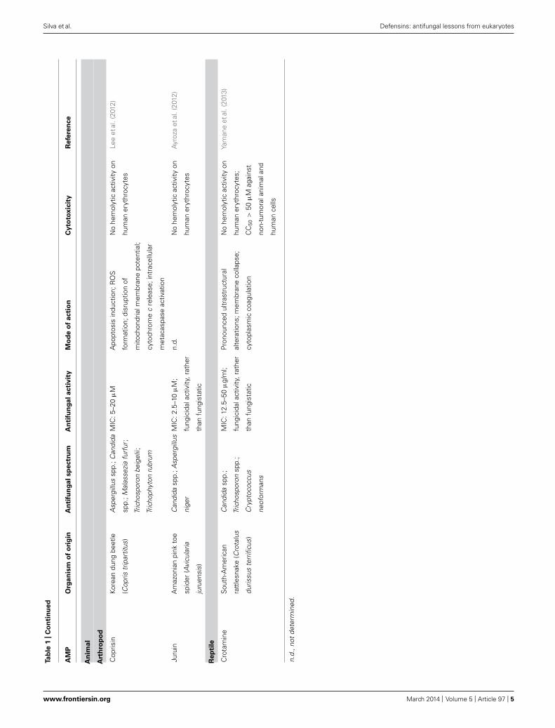
Arthropods defensinsCoprisin. This 43 amino acid residues beetle defensin-like pep-tide was described in 2009 as an antibacterial peptide (Hwanget al., 2009). Its structure comprises an αβ-motif, stabilized by
three cysteine disulfide bridges (Lee et al., 2013). In 2012, thesame authors investigated its antifungal activity against C. albi-cans, revealing that coprisin enters the fungal cell and localizesin the nucleus, which indicates that coprisin penetrated themembrane without disrupting the fungal plasma membrane, asconfirmed with 1,6-diphenyl-1,3,5-hexatriene (DPH) analysis,calcein-leakage, and giant unilamellar vesicle assays (Lee et al.,2012). Using H2O2 as a positive control for apoptotic induction,coprisin proved to have the same effects in inducing early andlate apoptosis, features shown by the annexin V conjugated withfluorescein and propidium iodide co-staining method. Apoptosisinduced by this AMP is metacaspase-dependent. Concomitantly,coprisin compromises mitochondrial membrane potential andROS production, in addition to the release of cytochrome cfrom the mitochondria to the cytosol. No hemolytic activity wasobserved for this peptide in human erythrocytes (Lee et al., 2012).
Juruin. This defensin-like peptide was discovered in 2012 byscreening the venom on the theraposid Amazonian pink toe spi-der Avicularia juruensis (Ayroza et al., 2012). It has 38 amino acidresidues, three disulfide bonds and, like neurotoxins reported tohave antimicrobial activity, it has a putative inhibitory cysteineknot (ICK) motif, i.e., a fold common to venom peptides fromspiders, scorpions, and aquatic cone snails (Smith et al., 2011).ICK-containing peptides of spider venom are likely to have evolvedfrom β-defensins (Fry et al., 2009). Based on amino acid sequenceand structure similarities with insecticidal peptides of other spi-ders, this peptide is likely to belong to a group of conserved toxinswith voltage-gated ion channels inhibitory action. Juruin showeda fungicidal rather than fungistatic effect against C. albicans andC. tropicalis, without hemolytic activity (Fry et al., 2009).
Reptile defensinsCrotamine. This highly basic peptide, isolated from the venom ofa South-American rattlesnake, was discovered in 1947 (Gonçalvesand Polson, 1947). It shares structural similarity to β-defensinsdue to an identical disulfide bridge pattern (Fadel et al., 2005;Yamane et al., 2013). Crotamine structure comprises an antipar-allel β-sheet and an α-helix stabilized by three disulfide bridges(Nicastro et al., 2003; Fadel et al., 2005). Recombinant crotaminedisplayed a more potent antimicrobial activity than native andsynthesized crotamine (Yamane et al., 2013). This peptide inducesextensive ultrastructural modifications in C. albicans. TEM stud-ies showed deformed cell shape, irregular layering structure of cellwall and cytoplasmic contents coagulation, but without detectablehemolytic effects and low toxicity to mammalian cells (Yamaneet al., 2013).
IMMUNOMODULATORY FUNCTIONDefensins may be produced constitutively or have their expres-sion triggered when there is an inflammatory process, by therecognition of microbial conserved structures, such as lipopolysac-charide (LPS) and lipoteichoic acid, or inflammatory effectors,like cytokines. These AMPs are expressed differentially depend-ing on the peptide itself and on the tissue or cell type (Ulmet al., 2012). Defensins, besides their antimicrobial action, canalso be immunomodulatory and inhibitors of virulence factors.This ability is not exclusive of defensins, as other AMPs also
www.frontiersin.org March 2014 | Volume 5 | Article 97 | 7

Silva et al. Defensins: antifungal lessons from eukaryotes
share this property. Thus, they can enhance the host’s immunesystem, with this multifunctional character rendering these pep-tides lower probability of becoming tolerated by microorganisms(Mehra et al., 2012; Jarczak et al., 2013).
Pro-inflammatory mediation has been recognized in someof these molecules, as they can bind to chemokine receptors,being able to recruit immune cells, thus enhancing the immuneresponse (Mookherjee and Hancock, 2007; Lai and Gallo, 2009;Alba et al., 2012; Semple and Dorin, 2012; Ulm et al., 2012;Zhu and Gao, 2013). β-Defensins were demonstrated to havethe capability to induce chemoattraction of CD4+ memory Tcells, macrophages, and immature dendritic cells, by binding toreceptors in the membrane (Yang et al., 1999; Wu et al., 2003;Taylor et al., 2008). This binding favors the attraction and migra-tion of inflammatory cells to the inflammation site, in orderto improve and speed up the inflammatory response. α and β-Defensins have also been shown to inhibit neutrophil apoptosis(Nagaoka et al., 2008). These authors showed that HBD3 bindsto CCR6 at the neutrophil cell surface, initiating an increasein the levels of the antiapoptotic protein Bcl-xL and inhibit-ing caspase activity. This increases neutrophils life span and isan inflammatory event that is beneficial to eradicate invadingmicroorganisms (Nagaoka et al., 2008), thus promoting the pro-duction of proinflammatory cytokines and chemokines, which inturn, amplifies the immune system response. Defensins have beenshown to have a proinflammatory effect on human keratinocytes(Niyonsaba et al., 2007). Treatment of these cells with HBD2HBD3 or HBD4 leads to the increase of the expression of pro-inflammatory mediators, like monocyte chemoattractant protein-1, macrophage inflammatory protein-3, and some interleukins(Niyonsaba et al., 2007).
Surprisingly, some defensins are also able to attenuate pro-inflammatory responses whenever these can be harmful to theorganism (Lande et al., 2007; Yamasaki et al., 2007). Theseantagonistic effects depend on the level of expression, dis-ease state, and pathogen exposure. It has been previouslydescribed for α-defensins that mice having a matrilysin defi-ciency (hence without mature α-defensins in the intestine) aremore susceptible to chemically induced colitis than wild-typecontrols. Interleukin-1β (IL-1β), a cytokine with an importantrole in mediation of inflammation, reaches level significantlyincreased in the deficient mice and it was ultimately shownthat α-defensins are able to inhibit the production of IL-1β
(Shi et al., 2007).It has been demonstrated that HBD3 (mainly expressed in
epithelial cells), when in basal concentration, has an immuno-suppressive effect in the presence of LPS, contributing to themaintenance of a non-inflammatory environment over continuallow-level exposure to microorganisms, commensal or pathogenic(Semple et al., 2010). Concentrations of HBD3 ranging from 0.5 to1 μM are able to suppress the induction of tumor necrosis factorα (TNFα), a proinflammatory effector of the immune system, andIL-6, an interleukin that acts both as pro and anti-inflammatory. Atthese concentrations, proinflammatory proteins are not inducedand there is no proinflammatory gene expression (Semple et al.,2010). The proinflammatory effects of β-defensins were observedat slightly higher concentrations of the defensin, in the 4–6 μM
range (Funderburg et al., 2007; Niyonsaba et al., 2010). This wasnot the first case observed of opposite effects in immunomodu-lating AMPs. Cathelicidin LL-37 has been shown to have also aduality in inflammatory effects, being proinflammatory at con-centrations above 20 μg/ml but anti-inflammatory at 1–5 μg/ml(Scott et al., 2002). Defensins were also shown to have a role inother biological processes, namely wound healing (Hirsch et al.,2009), dog coat color determination (Candille et al., 2007), fer-tility (Li et al., 2001), plant development (Stotz et al., 2009), andcarcinogenesis regulation (Donald et al., 2003; Gambichler et al.,2006; Joly et al., 2009).
It is clear that defensins have many functions that aredetermined by the level of expression. Whereas higher expres-sion of defensins takes place at the pathogen’s site of entry,with a proinflammatory response and the chemoattraction ofmacrophages and other immune cells, defensins expressed atlower levels may be involved in the resolution of the immuneresponse. When the danger is neutralized and defensins andother proinflammatory molecules decrease in the inflammationsite, defensins may then have a role in resolving inflammation(Semple and Dorin, 2012).
Due to this multifunctionality, AMPs have also been referredto as host defense peptides (HDPs; Steinstraesser et al., 2010; Ulmet al., 2012).
RESISTANCELike other antibiotics resistance, it is easily conceivable thatAMPs resistance is a key characteristic for increased virulenceof pathogenic strains. Despite this fact, and contrary to antibi-otics that act through a single approach (meaning that microbescan evade them through a single resistance system), AMPs fol-low a multidimensional strategy against microbial invasion (Laiand Gallo, 2009). Therefore, selective pressures on microbes areavoided, reducing the development of resistant strains (Zasloff,2002).
A synergistic effect between different host AMPs is also possible,as evidenced by the fact that the MIC of AMPs in vitro are usuallyhigher than the physiological concentrations of those AMPs in vivo(Lai and Gallo, 2009). Two distinct AMPs may have their combinedMIC much lower than when acting isolated, strongly suggestingheterologous HDP interactions (Westerhoff et al., 1995).
Microorganisms have evolved their own strategies for evadingthe antimicrobial action of the compounds used against them.AMPs frequently have the ability to disrupt microbial membranesand to inhibit the synthesis of some of their components; thus,strategies to escape the action of those AMPs follow the redesign ofcell membranes, as described for both Gram-negative and Gram-positive bacteria (Gunn et al., 2000; Li et al., 2007). Other evasionmechanisms include affecting the correct function of the AMPby turning off its expression, releasing plasmid DNA in epithe-lial cells, a strategy adopted by highly contagious bacteria fromthe Shigella genus that cause dysentery (Islam et al., 2001). AsAMPs frequently rely on transmembrane potential to interact withmicrobial pathogens and exert their mechanism of action againstthem, it is probable that another microbe strategy for evadingAMPs could be to change their transmembrane potential status(Yeaman and Yount, 2003).
Frontiers in Microbiology | Antimicrobials, Resistance and Chemotherapy March 2014 | Volume 5 | Article 97 | 8
Silva et al. Defensins: antifungal lessons from eukaryotes
Candida albicans resistance to some AMPs is regulated bythe protein Ssd1, combined with the transcription factor Bcr1(biofilm and cell wall regulator; Nobile and Mitchell, 2005; Ganket al., 2008; Jung et al., 2013). Ssd1 is an RNA-binding proteinand a component of the regulation of morphogenesis pathway(Saputo et al., 2012). In C. albicans, this pathway governs mul-tiple processes, including filamentation and cell wall integrity(Song et al., 2008; Bharucha et al., 2011). This combination yieldsresistance to protamine, RP-1 and HBD2 by maintaining mito-chondrial energetics and reducing membrane permeabilization,thus allowing the fungus to counteract the negative effects ofthese AMPs (Jung et al., 2013). Protamine is an α-helical cationicpolypeptide, frequently used to screen for AMP susceptibility (Yea-man et al., 1996), and RP-1 is a synthetic AMP modeled uponthe C-terminal α-helical domain existent in the human plateletfactor-4 kinocidins; this domain is responsible for RP-1 microbi-cidal activity (Bourbigot et al., 2009). C. albicans mutant strainsin Bcr1 and Ssd1 proteins are more susceptible to the AMPsdescribed above; thus, Jung et al. (2013) were able to concludethat these proteins are necessary for the resistance to protamine,RP-1 and HBD2. Further studies are necessary to clarify the rolesof Bcr1 and Ssd1 in early versus late mechanisms of resistance toAMPs.
The Hog1 (high osmolarity glycerol) MAPK pathway, whichprovides a response to osmotic, oxidative, and heavy-metal expo-sure stresses in fungal cells, was shown to be activated in thepresence of AMPs, such as NaD1, HBD2, HBD3, and histatin-5 (asalivary cationic AMP that has a role in keeping C. albicans in itscommensal state; Yeaman et al.,1996; Vylkova et al.,2007; Argimonet al., 2011; Hayes et al., 2013). The injuries imposed on C. albicansby these defensins seem to share common features with osmoticand/or oxidative stress (Argimon et al., 2011). Upon exposure tothese defensins, the Hog1 MAP kinase is activated, triggering atranscriptional response aimed to rescue the cells from the sourceof injury, i.e., the core and osmotic-stress transcriptional responses(Enjalbert et al., 2006; Argimon et al., 2011).
Another strategy for evading AMP function is to enzymati-cally degrade these peptides before they exert their effects. This ispossible by producing proteases and peptidases involved in tissuedegradation, as described for C. albicans secreted aspartic pro-teases (Saps). Namely, histatin-5, present in human saliva, is ahost-specific substrate of Sap9, enabling the transition of the fun-gus from commensal to pathogenic in HIV+ individuals. Thesepatients, who have lower levels of this isoenzyme in the saliva,have an increased incidence of oral candidiasis (Meiller et al., 2009;Khan et al., 2013). Also regarding histatin-5, a transport mecha-nism of efflux mediated by the flu-1 transporter has been describedfor C. albicans, rendering the pathogen the ability to reduce theisoenzyme cytosolic concentration and fungicidal activity (Li et al.,2013). The LL-37 cathelicidin and histatins bind to cell wall carbo-hydrates, preventing adhesion of C. albicans to host cells; thus, therelease of AMP-binding proteins acts as a decoy for these AMPs,diverting them from binding to fungal cell surface (den Hertoget al., 2005, 2006; Mochon and Liu, 2008). For example, Msb2(multicopy suppressor of a budding defect) is a C. albicans sur-face protein (a mucin) highly soluble and proteolytically stable,which is shed to the extracellular environment, acting as a basal
AMP-resistance decoy by binding to LL-37 and histatin-5, avoid-ing the antimicrobial action of these AMPs (Szafranski-Schneideret al., 2012).
The characteristics described above are associated with adecrease in microbes’ susceptibility to AMPs, indicating thatmicrobial pathogens have developed some structure-specific andenergy-dependent mechanisms to subvert the action of these hostdefense systems.
FUNGAL CELL MEMBRANEFungi possess a unique cell wall and cell membrane that can serveas specific targets for antifungal agents. The fungal cell membraneis similar to those of other eukaryotic cells, composed of a lipidbilayer with proteins embedded within it (Katzung et al., 2011).Sterols (absent in prokaryotes) are major components of fungalmembranes. The sterol present in higher eukaryotic membranesis cholesterol, but in fungal membranes the main sterol present isergosterol, providing stability and flexibility to the cell membrane(Thevissen et al., 1999, 2003a).
Glycosphingolipids (GSLs) are a family of lipids that act askey components of biological membranes. They exist in animals,plants, and fungi (Leipelt et al., 2001; Halter et al., 2007; Daniottiand Iglesias-Bartolome, 2011). GSLs were initially described ascomponents of the architecture of cell membranes, straightly con-nected with fluidity and stability (Feinstein et al., 1975; Aaronsonand Martin, 1983; Campanella, 1992). Recently, however, it wasdemonstrated that their role goes clearly beyond the initial con-cept, since these molecules are major components of specializedmembrane domains called lipid rafts (Bagnat et al., 2001; Hako-mori, 2003, 2008). GSLs have been characterized as importantstructures in cell–cell interaction, cell signaling, and protein sort-ing (Bagnat et al., 2000; Bagnat and Simons, 2002; Nimrichteret al., 2008; Staubach and Hanisch, 2011). Lipid rafts are moreordered and tightly packed than the surrounding bilayer, serv-ing as organizing centers for the assembly of signaling molecules,influencing membrane fluidity and membrane protein trafficking(Chiantia and London, 2013).
The most common GSL found in fungi is GlcCer, present inthe membranes of most fungi, such as Pichia pastoris, C. albi-cans, Cryptococcus neoformans, Aspergillus fumigatus, Sporothrixschenckii and Neurospora crassa (Saito et al., 2006). Large amountsof this GSL have also been found in the fungal cell wall (Nim-richter and Rodrigues, 2011). GlcCer has been identified asa fungal component decades ago. Its functions during fun-gal growth/dimorphism, lipid raft formation, and correlationwith virulence have been reported (Rittershaus et al., 2006).In fact, it was recently shown to be required for virulencein C. albicans (de Medeiros et al., 2010; Noble et al., 2010;de Coninck et al., 2013).
Work published by Thevissen and colleagues strongly suggestedthat fungal GlcCer targeting by the AMPs RsAFP2 and HsAFP1could initiate a cell signaling response in fungi, with formationof ROS and subsequent cell death by apoptosis (Thevissen et al.,2004; Aerts et al., 2007, 2009, 2011). The use of anti-GlcCer anti-bodies was shown to block germ tube formation in C. albicans,Colletotrichum gloeosporioides, and Pseudallescheria boydii (Pintoet al., 2002; da Silva et al., 2004), and also to protect mice upon
www.frontiersin.org March 2014 | Volume 5 | Article 97 | 9

Silva et al. Defensins: antifungal lessons from eukaryotes
the potentially lethal infection by C. neoformans (Rodrigues et al.,2007).
The crescent knowledge of GlcCer functions in eukaryotes(may these be related to virulence, growth or morphologicaltransitions), together with the findings described above, can beconnected to specific and essential structural features and partic-ular biosynthetic steps to validate this GSL, as well as other fungalspecific membrane lipids and sterols, as potential targets on thedevelopment and discovery of new antifungal drugs (Nimrichterand Rodrigues, 2011; Gonçalves et al., 2012b). Besides GlcCer,fungal membranes are also rich in phosphomannans and in thenegatively charged phospholipids PS, phosphatidylinositol (PI)and diphosphatidylglycerol (DPG), which confer a highly negativesurface charge to these membranes (Pasupuleti et al., 2012).
MODELS OF MEMBRANE ACTIVITY – MECHANISM OFACTIONThe biological activity of AMPs is strongly influenced by peptide–membrane interactions. To explain how some AMPs show differ-ential membrane affinity, their biological activities, and modesof action have been assessed on studies of defensins interac-tion with fungal membrane model systems, which showed astrong dependence on membrane lipid composition and onthe concentration of specific components (de Medeiros et al.,2010; Gonçalves et al., 2012a,b). As with other AMPs, themechanisms of action of some plant defensins with antifun-gal activity involve membrane binding, binding to the cell wall,interaction with intracellular targets leading to apoptosis, mem-brane permeabilization, and receptor-mediated internalization(van der Weerden et al., 2013).
The mechanisms of action of some defensins have been studiedby using synthetic lipid vesicles mimicking the lipid compositionof fungal, bacterial and mammal membranes (de Medeiros et al.,2010; Wimley and Hristova, 2011; Gonçalves et al., 2012a,b). Thepermeabilization models used to explain the mode of action ofdefensins could be classified into two main groups: transmem-brane pore formation, such as the barrel-stave and toroidal models,and non-pore formation, such as the carpet, aggregate channel,Shai–Matsuzaki–Huang, lipid clustering, and interfacial activitymodels (Alba et al., 2012). The carpet model can evolve to dis-rupt the membrane through pore formation models or through adetergent-like mechanism, with partial micellization of the mem-brane (Bechinger and Lohner, 2006; Chang et al., 2008; Hoskinand Ramamoorthy, 2008). There are currently at least three differ-ent commonly accepted models describing possible AMPs modeof action: the barrel-stave pore model, the toroidal pore model,and the carpet model (Shai, 2002; Chang et al., 2008; Hoskin andRamamoorthy, 2008; Alba et al., 2012).
Most defensins are amphipathic molecules with clusters of pos-itively charged amino acid residues side chains and hydrophobicamino acid side chains (Lehrer and Lu, 2011). This structuralbehavior allows them to interact with microbial membranes bothat the level of the negatively charged phospholipid head groupsand of the hydrophobic fatty acid chains. The orientation of thepeptide on the membrane surface depends on the specific peptide–lipid system, but it is common for the AMP to stay at the membraneinterface until a threshold peptide concentration is reached (Yang
et al., 2000; Yount and Yeaman, 2005; Pasupuleti et al., 2012). Inthe barrel-stave model (Figure 2), once the critical threshold con-centration of peptide is reached, peptides self-aggregate in themembrane resulting in the formation of a transmembrane porelined by peptide, which dissipates proton and ionic gradients(Ehrenstein and Lecar, 1977; Reddy et al., 2004), but the mem-brane thickness and homogeneity do not change (Chang et al.,2008). The toroidal pore model is a variant of the barrel-stavemodel, claiming that, at some critical peptide concentration, cur-vature strain induces membranes to curve inward, resulting inthe formation of a pore that is lined by both peptides and lipidheadgroups (Figure 2). Toroidal pores seem to have varying life-times and longer-lived pores may have a lethal effect similar tobarrel-stave pores, with dissipation of proton and ion gradients.This type of mechanism of AMPs action also causes a decrease inmembrane thickness and a slightly decreased surface homogene-ity (Chang et al., 2008). In the carpet model (Figure 2), peptidesbind to phospholipid head groups by electrostatic interactions andalign themselves parallel to the membrane surface in a carpet-likefashion until a critical threshold concentration is reached. Whena detergent-like membrane micellization takes place, a strongdecrease of membrane homogeneity occurs (Chang et al., 2008;Hoskin and Ramamoorthy, 2008; Epand et al., 2010; Hazlett andWu, 2011; Li et al., 2012; Pasupuleti et al., 2012).
Besides targeting fungal membranes’ specific components,defensins may also have other mechanisms of action, as previ-ously referred. These mechanisms comprise binding to the cellwall, membrane permeabilization, receptor-mediated internaliza-tion inducing signaling cascades and interaction with intracellulartargets, which would cause the formation of ROS, leading ulti-mately to programed cell death. To address these mechanisms thereader is directed to some relevant references on this topic (Han-cock and Rozek, 2002; Oberparleiter et al., 2003; Thevissen et al.,2003a, 2004; Schroeder et al., 2011; de Coninck et al., 2013; De
FIGURE 2 | Models of lipid membrane permeabilization by AMPs.
Initially, the peptide (magenta) is adsorbed at the membrane surface. Afteran initial recognition of the surface, a conformational change of the peptideoccurs. Once a threshold concentration of peptide on the membrane isreached, it is followed by membrane disruption by one of these threemechanisms.
Frontiers in Microbiology | Antimicrobials, Resistance and Chemotherapy March 2014 | Volume 5 | Article 97 | 10

Silva et al. Defensins: antifungal lessons from eukaryotes
Paula et al., 2013; Jaeger et al., 2013; van der Weerden et al., 2013;Zhang et al., 2013).
CONCLUSIONThe knowledge on AMPs has been increasing considerably duringthe last 20 years. This increased knowledge shows that AMPs havemuch more than only antimicrobial activity, presenting a broadspectrum of physiological functions. Defensins are the most rep-resented AMPs across the eukaryotic domain, and in all typesof eukaryotic organisms we can find defensins not only withantifungal activity but also with other potential applications.
Despite this relevance, defensins may have limitations in termsof new drug development, due to their cationic, amphiphilic, andprotease labile nature, leading to a low serum half-life that limitstheir systemic administration (Maisetta et al., 2008). This limi-tation can be overcome by the use of peptidomimetics, like thesubstitution of natural occurring L-amino acid residues by D-amino acid residues or unusual amino acids (Oren et al., 1997;McPhee et al., 2005). Defensins bare a favorable characteristicagainst this problem, as their disulfide-stabilized structure con-fers increased protease-resistance (Wu et al., 2003). Nonetheless,defensins combine targeted antimicrobial activity with the capac-ity to positively modulate the immune system, and have provento be effective across life evolution, making these peptides highlyappealing as an anti-infective strategy.
Defensins have evolved as successful barrier of defense notonly against bacteria, but also pathogenic fungi, present amongplants, animals, and fungi. This ability may serve as a “lesson”on how selective pressures that shape organisms and their com-ponents served and continue to serve as a lever for the evolutionof better defenses. Most antibiotics used nowadays are from bac-terial origin or synthetic (Clardy et al., 2009; Fisher et al., 2012).The molecular design and synthesis of new molecules inspiredon defensins or on other AMP structures and sequences seem tobe a promising approach to develop a new and extensive field ofapplications, ranging from antimicrobial therapy, to their possi-ble use as vaccine adjuvants. Therefore, a better understanding offunction and mechanism of action of HDPs, specially defensins,is highly relevant for the development of new anti-infective andimmunomodulatory therapeutics (Guaní-Guerra et al., 2013).
ACKNOWLEDGMENTSThis work was partially supported by Fundação para a Ciência eTecnologia – Ministério da Educação e Ciência (FCT-MEC,Portugal) and by the European Union FP7-IRSES project MEM-PEPACROSS.
REFERENCESAaronson, L. R., and Martin, C. E. (1983). Temperature-induced modifications of
glycosphingolipids in plasma membranes of Neurospora crassa. Biochim. Biophys.Acta 735, 252–258. doi: 10.1016/0005-2736(83)90300-0
Aerts, A. M., Bammens, L., Govaert, G., Carmona-Gutierrez, D., Madeo, F., Cam-mue, B. P., et al. (2011). The antifungal plant defensin HsAFP1 from Heucherasanguinea induces apoptosis in Candida albicans. Front. Microbiol. 2:47. doi:10.3389/fmicb.2011.00047
Aerts, A. M., Carmona-Gutierrez, D., Lefevre, S., Govaert, G., François, I. E., Madeo,F., et al. (2009). The antifungal plant defensin RsAFP2 from radish induces apop-tosis in a metacaspase independent way in Candida albicans. FEBS Lett. 583,2513–2516. doi: 10.1016/j.febslet.2009.07.004
Aerts, A. M., François, I. E., Meert, E. M., Li, Q. T., Cammue, B. P., and Thevissen,K. (2007). The antifungal activity of RsAFP2, a plant defensin from Raphanussativus, involves the induction of reactive oxygen species in Candida albicans. J.Mol. Microbiol. Biotechnol. 13, 243–247. doi: 10.1159/000104753
Alba, A., Lopez-Abarrategui, C., and Otero-Gonzalez, A. J. (2012). Host defensepeptides: an alternative as antiinfective and immunomodulatory therapeutics.Biopolymers 98, 251–267. doi: 10.1002/bip.22076
Almeida, M. S., Cabral, K. M., Kurtenbach, E., Almeida, F. C., and Valente, A. P.(2002). Solution structure of Pisum sativum defensin 1 by high resolution NMR:plant defensins, identical backbone with different mechanisms of action. J. Mol.Biol. 315, 749–757. doi: 10.1006/jmbi.2001.5252
Almeida, M. S., Cabral, K. M., Zingali, R. B., and Kurtenbach, E. (2000). Charac-terization of two novel defense peptides from pea (Pisum sativum) seeds. Arch.Biochem. Biophys. 378, 278–286. doi: 10.1006/abbi.2000.1824
Argimon, S., Fanning, S., Blankenship, J. R., and Mitchell, A. P. (2011). Interactionbetween the Candida albicans high-osmolarity glycerol (HOG) pathway and theresponse to human beta-defensins 2 and 3. Eukaryot. Cell 10, 272–275. doi:10.1128/Ec.00133-10
Asano, T., Miwa, A., Maeda, K., Kimura, M., and Nishiuchi, T. (2013). The secretedantifungal protein thionin 2.4 in Arabidopsis thaliana suppresses the toxicity of afungal fruit body lectin from Fusarium graminearum. PLoS Pathog. 9:e1003581.doi: 10.1371/journal.ppat.1003581
Aumelas, A., Mangoni, M., Roumestand, C., Chiche, L., Despaux, E., Grassy, G., et al.(1996). Synthesis and solution structure of the antimicrobial peptide protegrin-1.Eur. J. Biochem. 237, 575–583. doi: 10.1111/j.1432-1033.1996.0575p.x
Ayroza, G., Ferreira, I. L., Sayegh, R. S., Tashima, A. K., and da Silva Junior, P. I.(2012). Juruin: an antifungal peptide from the venom of the Amazonian PinkToe spider, Avicularia juruensis, which contains the inhibitory cystine knot motif.Front. Microbiol. 3:324. doi: 10.3389/fmicb.2012.00324
Bagnat, M., Chang, A., and Simons, K. (2001). Plasma membrane proton ATPasePma1p requires raft association for surface delivery in yeast. Mol. Biol. Cell 12,4129–4138. doi: 10.1091/mbc.12.12.4129
Bagnat, M., Keranen, S., Shevchenko, A., and Simons, K. (2000). Lipid rafts functionin biosynthetic delivery of proteins to the cell surface in yeast. Proc. Natl. Acad.Sci. U.S.A. 97, 3254–3259. doi: 10.1073/pnas.060034697
Bagnat, M., and Simons, K. (2002). Lipid rafts in protein sorting and cell polarityin budding yeast Saccharomyces cerevisiae. Biol. Chem. 383, 1475–1480. doi:10.1515/BC.2002.169
Barna, B., Leiter, E., Hegedus, N., Biro, T., and Pocsi, I. (2008). Effect of the Penicil-lium chrysogenum antifungal protein (PAF) on barley powdery mildew and wheatleaf rust pathogens. J. Basic Microbiol. 48, 516–520. doi: 10.1002/jobm.200800197
Bechinger, B., and Lohner, K. (2006). Detergent-like actions of linear amphipathiccationic antimicrobial peptides. Biochim. Biophys. Acta 1758, 1529–1539. doi:10.1016/j.bbamem.2006.07.001
Bensch, K. W., Raida, M., Magert, H. J., Schulz-Knappe, P., and Forssmann, W.G. (1995). hBD-1: a novel beta-defensin from human plasma. FEBS Lett. 368,331–335. doi: 10.1016/0014-5793(95)00687-5
Bharucha, N., Chabrier-Rosello, Y., Xu, T., Johnson, C., Sobczynski, S., Song, Q.,et al. (2011). A large-scale complex haploinsufficiency-based genetic interactionscreen in Candida albicans: analysis of the RAM network during morphogenesis.PLoS Genet. 7:e1002058. doi: 10.1371/journal.pgen.1002058
Bourbigot, S., Dodd, E., Horwood, C., Cumby, N., Fardy, L., Welch, W. H., et al.(2009). Antimicrobial peptide RP-1 structure and interactions with anionic versuszwitterionic micelles. Biopolymers 91, 1–13. doi: 10.1002/bip.21071
Brogden, K. A., De Lucca, A. J., Bland, J., and Elliott, S. (1996). Isolation of an ovinepulmonary surfactant-associated anionic peptide bactericidal for Pasteurellahaemolytica. Proc. Natl. Acad. Sci. U.S.A. 93, 412–416. doi: 10.1073/pnas.93.1.412
Bulet, P., and Stocklin, R. (2005). Insect antimicrobial peptides: struc-tures, properties and gene regulation. Protein Pept. Lett. 12, 3–11. doi:10.2174/0929866053406011
Campanella, R. (1992). Membrane lipids modifications in human gliomas ofdifferent degree of malignancy. J. Neurosurg. Sci. 36, 11–25.
Candille, S. I., Kaelin, C. B., Cattanach, B. M., Yu, B., Thompson, D. A., Nix, M.A., et al. (2007). A β-defensin mutation causes black coat color in domestic dogs.Science 318, 1418–1423. doi: 10.1126/science.1147880
Chan, Y. S., and Ng, T. B. (2013). Northeast red beans produce a thermostableand pH-stable defensin-like peptide with potent antifungal activity. Cell Biochem.Biophys. 66, 637–648. doi: 10.1007/s12013-012-9508-1
www.frontiersin.org March 2014 | Volume 5 | Article 97 | 11

Silva et al. Defensins: antifungal lessons from eukaryotes
Chan, Y. S., Wong, J. H., Fang, E. F., Pan, W. L., and Ng, T. B. (2012). An antifungalpeptide from Phaseolus vulgaris cv. brown kidney bean. Acta Biochim. Biophys.Sin. (Shanghai) 44, 307–315. doi: 10.1093/abbs/gms003
Chang, W. K., Wimley, W. C., Searson, P. C., Hristova, K., and Merzlyakov,M. (2008). Characterization of antimicrobial peptide activity by electrochem-ical impedance spectroscopy. Biochim. Biophys. Acta 1778, 2430–2436. doi:10.1016/j.bbamem.2008.06.016
Chiantia, S., and London, E. (2013). “Sphingolipids and membrane domains: recentadvances,” in Handbook of Experimental Pharmacology, eds. E. Gulbins and I.Petrache. (Stony Brook, NY: Springer), 33–55. doi: 10.1007/978-3-7091-1368-4_2
Clardy, J., Fischbach, M. A., and Currie, C. R. (2009). The natural history ofantibiotics. Curr. Biol. 19, R437–R441. doi: 10.1016/j.cub.2009.04.001
Daniotti, J. L., and Iglesias-Bartolome, R. (2011). Metabolic pathways and intracel-lular trafficking of gangliosides. IUBMB Life 63, 513–520. doi: 10.1002/iub.477
da Silva, A. F., Rodrigues, M. L., Farias, S. E., Almeida, I. C., Pinto, M. R., andBarreto-Bergter, E. (2004). Glucosylceramides in Colletotrichum gloeosporioidesare involved in the differentiation of conidia into mycelial cells. FEBS Lett. 561,137–143. doi: 10.1016/S0014-5793(04)00156-5
de Coninck, B., Cammue, B. P. A., and Thevissen, K. (2013). Modes of antifungalaction and in planta functions of plant defensins and defensin-like peptides.Fungal Biol. Rev. 26, 109–120. doi: 10.1016/j.fbr.2012.10.002
de Medeiros, L. N. (2009). Interação da defensina Psd1 com a monohexosilceramida (CMH) isolada do fungo Fusarium solani. Doutor em Química Biológica,Universidade Federal do Rio de Janeiro, Rio de Janeiro.
de Medeiros, L. N., Angeli, R., Sarzedas, C. G., Barreto-Bergter, E., Valente, A.P., Kurtenbach, E., et al. (2010). Backbone dynamics of the antifungal Psd1 peadefensin and its correlation with membrane interaction by NMR spectroscopy.Biochim. Biophys. Acta 1798, 105–113. doi: 10.1016/j.bbamem.2009.07.013
De Paula, V. S., Gomes, N. S., Lima, L. G., Miyamoto, C. A., Monteiro, R. Q., Almeida,F. C., et al. (2013). Structural basis for the interaction of human beta-defensin6 and its putative chemokine receptor CCR2 and breast cancer microvesicles. J.Mol. Biol. 425, 4479–4495. doi: 10.1016/j.jmb.2013.08.001
den Hertog, A. L., van Marle, J., van Veen, H. A., Van’t Hof, W., Bolscher, J. G.,Veerman, E. C., et al. (2005). Candidacidal effects of two antimicrobial peptides:histatin 5 causes small membrane defects, but LL-37 causes massive disruptionof the cell membrane. Biochem. J. 388, 689–695. doi: 10.1042/BJ20042099
den Hertog, A. L., van Marle, J., Veerman, E. C., Valentijn-Benz, M., Nazmi, K., Kalay,H., et al. (2006). The human cathelicidin peptide LL-37 and truncated variantsinduce segregation of lipids and proteins in the plasma membrane of Candidaalbicans. Biol. Chem. 387, 1495–1502. doi: 10.1515/BC.2006.187
Domingues, M. M., Silva, P. M., Franquelim, H. G., Carvalho, F. A., Castanho, M.A. R. B., and Santos, N. C. (2014). Antimicrobial protein rBPI-induced surfacechanges on Gram-negative and Gram-positive bacteria. Nanomedicine (NBM).doi: 10.1016/j.nano.2013.11.002 [Epub ahead of print].
Donald, C. D., Sun, C. Q., Lim, S. D., Macoska, J., Cohen, C., Amin, M. B., et al.(2003). Cancer-specific loss of beta-defensin 1 in renal and prostatic carcinomas.Lab. Invest. 83, 501–505. doi: 10.1097/01.LAB.0000063929.61760.F6
Ehrenstein, G., and Lecar, H. (1977). Electrically gated ionic channels in lipidbilayers. Q. Rev. Biophys. 10, 1–34. doi: 10.1017/S0033583500000123
Enjalbert, B., Smith, D. A., Cornell, M. J., Alam, I., Nicholls, S., Brown, A. J., et al.(2006). Role of the Hog1 stress-activated protein kinase in the global transcrip-tional response to stress in the fungal pathogen Candida albicans. Mol. Biol. Cell17, 1018–1032. doi: 10.1091/mbc.E05-06-0501
Epand, R. F., Maloy, L., Ramamoorthy, A., and Epand, R. M. (2010). Amphi-pathic helical cationic antimicrobial peptides promote rapid formation ofcrystalline states in the presence of phosphatidylglycerol: lipid clustering inanionic membranes. Biophys. J. 98, 2564–2573. doi: 10.1016/j.bpj.2010.03.002
Fadel, V., Bettendorff, P., Herrmann, T., de Azevedo, W. F. Jr., Oliveira, E. B.,Yamane, T., et al. (2005). Automated NMR structure determination and disulfidebond identification of the myotoxin crotamine from Crotalus durissus terrificus.Toxicon 46, 759–767. doi: 10.1016/j.toxicon.2005.07.018
Feinstein, M. B., Fernandez, S. M., and Sha’afi, R. I. (1975). Fluidity of naturalmembranes and phosphatidylserine and ganglioside dispersions. Effect of localanesthetics, cholesterol and protein. Biochim. Biophys. Acta 413, 354–370. doi:10.1016/0005-2736(75)90121-2
Fisher, M. C., Henk, D. A., Briggs, C. J., Brownstein, J. S., Madoff, L. C., McCraw, S.L., et al. (2012). Emerging fungal threats to animal, plant and ecosystem health.Nature 484, 186–194. doi: 10.1038/nature10947
Fontana, R., Mendes, M. A., de Souza, B. M., Konno, K., Cesar, L. M.,Malaspina, O., et al. (2004). Jelleines: a family of antimicrobial peptidesfrom the Royal Jelly of honeybees (Apis mellifera). Peptides 25, 919–928. doi:10.1016/j.peptides.2004.03.016
Fry, B. G., Roelants, K., Champagne, D. E., Scheib, H., Tyndall, J. D., King, G.F., et al. (2009). The toxicogenomic multiverse: convergent recruitment of pro-teins into animal venoms. Annu. Rev. Genomics Hum. Genet. 10, 483–511. doi:10.1146/annurev.genom.9.081307.164356
Funderburg, N., Lederman, M. M., Feng, Z., Drage, M. G., Jadlowsky, J., Harding,C. V., et al. (2007). Human-defensin-3 activates professional antigen-presentingcells via Toll-like receptors 1 and 2. Proc. Natl. Acad. Sci. U.S.A. 104, 18631–18635.doi: 10.1073/pnas.0702130104
Galgóczy, L., Kovács, L., and Vágvölgyi, C. (2010). “Defensin-like antifungal proteinssecreted by filamentous fungi,” in Current Research, Technology and EducationTopics in Applied Microbiology and Microbial Technology, ed. A. Méndez-Vilas(Valladolid: Formatex), 550–559.
Galgóczy, L., Papp, T., Leiter, E., Marx, F., Pocsi, I., and Vagvolgyi, C.(2005). Sensitivity of different zygomycetes to the Penicillium chrysogenumantifungal protein (PAF). J. Basic Microbiol. 45, 136–141. doi: 10.1002/jobm.200410512
Galgóczy, L., Papp, T., Lukacs, G., Leiter, E., Pocsi, I., and Vagvolgyi, C. (2007). Inter-actions between statins and Penicillium chrysogenum antifungal protein (PAF) toinhibit the germination of sporangiospores of different sensitive Zygomycetes.FEMS Microbiol. Lett. 270, 109–115. doi: 10.1111/j.1574-6968.2007.00661.x
Galgóczy, L., Papp, T., Pocsi, I., Hegedus, N., and Vagvolgyi, C. (2008). In vitroactivity of Penicillium chrysogenum antifungal protein (PAF) and its combinationwith fluconazole against different dermatophytes. Antonie van Leeuwenhoek 94,463–470. doi: 10.1007/s10482-008-9263-x
Gambichler, T., Skrygan, M., Huyn, J., Bechara, F. G., Sand, M., Altmeyer, P., et al.(2006). Pattern of mRNA expression of beta-defensins in basal cell carcinoma.BMC Cancer 6:163. doi: 10.1186/1471-2407-6-163
Games, P. D., Dos Santos, I. S., Mello, E. O., Diz, M. S., Carvalho, A. O., de Souza-Filho, G. A., et al. (2008). Isolation, characterization and cloning of a cDNAencoding a new antifungal defensin from Phaseolus vulgaris L. seeds. Peptides 29,2090–2100. doi: 10.1016/j.peptides.2008.08.008
Gank, K. D., Yeaman, M. R., Kojima, S., Yount, N. Y., Park, H., Edwards, J. E., et al.(2008). SSD1 is integral to host defense peptide resistance in Candida albicans.Eukaryot. Cell 7, 1318–1327. doi: 10.1128/Ec.00402-07
Ganz, T. (2003). The role of antimicrobial peptides in innate immunity. Integr.Comp. Biol. 43, 300–304. doi: 10.1093/Icb/43.2.300
Ganz, T. (2004). Defensins: antimicrobial peptides of vertebrates. C. R. Biol. 327,539–549. doi: 10.1016/j.crvi.2003.12.007
Ganz, T., and Lehrer, R. I. (1998). Antimicrobial peptides of vertebrates. Curr. Opin.Immunol. 10, 41–44. doi: 10.1016/S0952-7915(98)80029-0
Ganz, T., Selsted, M. E., Szklarek, D., Harwig, S. S. L., Daher, K., Bainton, D. F., et al.(1985). Defensins – natural peptide antibiotics of human-neutrophils. J. Clin.Invest. 76, 1427–1435. doi: 10.1172/Jci112120
Gao, B., Rodriguez Mdel, C., Lanz-Mendoza, H., and Zhu, S. (2009). AdDLP, a bac-terial defensin-like peptide, exhibits anti-Plasmodium activity. Biochem. Biophys.Res. Commun. 387, 393–398. doi: 10.1016/j.bbrc.2009.07.043
Gao, B., and Zhu, S. (2012). Alteration of the mode of antibacterial action ofa defensin by the amino-terminal loop substitution. Biochem. Biophys. Res.Commun. 426, 630–635. doi: 10.1016/j.bbrc.2012.08.143
Gaspar, D., Veiga, A. S., and Castanho, M. A. R. B. (2013). From antimicrobial toanticancer peptides. A review. Front. Microbiol. 4:294. doi: 10.3389/fmicb.2013.00294
Glaser, R., Harder, J., Lange, H., Bartels, J., Christophers, E., and Schroder, J. M.(2005). Antimicrobial psoriasin (S100A7) protects human skin from Escherichiacoli infection. Nat. Immunol. 6, 57–64. doi: 10.1038/ni1142
Gonçalves, J. M., and Polson, A. (1947). The electrophoretic analysis of snakevenoms. Arch. of Biochem. 13, 253–259.
Gonçalves, S., Abade, J., Teixeira, A., and Santos, N. C. (2012a). Lipid compositionis a determinant for human defensin HNP1 selectivity. Biopolymers 98, 313–321.doi: 10.1002/Bip.22088
Gonçalves, S., Teixeira, A., Abade, J., de Medeiros, L. N., Kurtenbach, E.,and Santos, N. C. (2012b). Evaluation of the membrane lipid selectivityof the pea defensin Psd1. Biochim. Biophys. Acta 1818, 1420–1426. doi:10.1016/j.bbamem.2012.02.012
Frontiers in Microbiology | Antimicrobials, Resistance and Chemotherapy March 2014 | Volume 5 | Article 97 | 12
Silva et al. Defensins: antifungal lessons from eukaryotes
Guaní-Guerra, E., Santos-Mendoza, T., Lugo-Reyes, S. O., and Terán, L. M.(2013). Antimicrobial peptides: general overview and clinical implications inhuman health and disease. Clin. Immunol. 135, 1–11. doi: 10.1016/j.clim.2009.12.004
Gunn, J. S., Ryan, S. S., Van Velkinburgh, J. C., Ernst, R. K., and Miller, S. I. (2000).Genetic and functional analysis of a PmrA–PmrB-regulated locus necessary forlipopolysaccharide modification, antimicrobial peptide resistance, and oral viru-lence of Salmonella enterica serovar Typhimurium. Infect. Immun. 68, 6139–6146.doi: 10.1128/IAI.68.11.6139-6146.2000
Guo, M. L., Wei, J. G., Huang, X. H., Huang, Y. H., and Qin, Q. W. (2012). Antivi-ral effects of beta-defensin derived from orange-spotted grouper (Epinepheluscoioides). Fish Shellfish Immunol. 32, 828–838. doi: 10.1016/j.fsi.2012.02.005
Hagen, S., Marx, F., Ram, A. F., and Meyer, V. (2007). The antifungal proteinAFP from Aspergillus giganteus inhibits chitin synthesis in sensitive fungi. Appl.Environ. Microbiol. 73, 2128–2134. doi: 10.1128/AEM.02497-06
Hakomori, S. (2003). Structure, organization, and function of glycosphingolipids inmembrane. Curr. Opin. Hematol. 10, 16–24. doi: 10.1097/00062752-200301000-00004
Hakomori, S. I. (2008). Structure and function of glycosphingolipids and sphin-golipids: recollections and future trends. Biochim. Biophys. Acta 1780, 325–346.doi: 10.1016/j.bbagen.2007.08.015
Halter, D., Neumann, S., van Dijk, S. M., Wolthoorn, J., de Maziere, A.M., Vieira, O. V., et al. (2007). Pre- and post-Golgi translocation of gluco-sylceramide in glycosphingolipid synthesis. J. Cell Biol. 179, 101–115. doi:10.1083/jcb.200704091
Hammami, R., Ben Hamida, J., Vergoten, G., and Fliss, I. (2009). PhytAMP: adatabase dedicated to antimicrobial plant peptides. Nucleic Acids Res. 37, D963–D968. doi 10.1093/Nar/Gkn655
Hancock, R. E., and Rozek, A. (2002). Role of membranes in the activitiesof antimicrobial cationic peptides. FEMS Microbiol. Lett. 206, 143–149. doi:10.1016/S0378-1097(01)00480-3
Hao, X., Yang, H., Wei, L., Yang, S., Zhu, W., Ma, D., et al. (2012). Amphibiancathelicidin fills the evolutionary gap of cathelicidin in vertebrate. Amino Acids43, 677–685. doi: 10.1007/s00726-011-1116-7
Harder, J., Bartels, J., Christophers, E., and Schroder, J. M. (2001). Isola-tion and characterization of human beta-defensin-3, a novel human induciblepeptide antibiotic. J. Biol. Chem. 276, 5707–5713. doi: 10.1074/jbc.M008557200
Hayes, B. M., Bleackley, M. R., Wiltshire, J. L., Anderson, M. A., Traven, A., and vander Weerden, N. L. (2013). Identification and mechanism of action of the plantdefensin NaD1 as a new member of the antifungal drug arsenal against Can-dida albicans. Antimicrob. Agents Chemother. 57, 3667–3675. doi: 10.1128/AAC.00365-13
Hazlett, L., and Wu, M. (2011). Defensins in innate immunity. Cell Tissue Res. 343,175–188. doi: 10.1007/s00441-010-1022-4
Hegedus, N., and Marx, F. (2013). Antifungal proteins: more than antimicrobials?Fungal Biol. Rev. 26, 132–145. doi: 10.1016/j.fbr.2012.07.002
Hirsch, T., Spielmann, M., Zuhaili, B., Fossum, M., Metzig, M., Koehler, T., et al.(2009). Human beta-defensin-3 promotes wound healing in infected diabeticwounds. J. Gene Med. 11, 220–228. doi: 10.1002/jgm.1287
Hoover, D. M., Wu, Z., Tucker, K., Lu, W., and Lubkowski, J. (2003). Antimicro-bial characterization of human beta-defensin 3 derivatives. Antimicrob. AgentsChemother. 47, 2804–2809. doi: 10.1128/AAC.47.9.2804-2809.2003
Hoskin, D. W., and Ramamoorthy, A. (2008). Studies on anticancer activi-ties of antimicrobial peptides. Biochim. Biophys. Acta 1778, 357–375. doi:10.1016/j.bbamem.2007.11.008
Hwang, J. S., Lee, J., Kim,Y. J., Bang, H. S.,Yun, E.Y., Kim, S. R., et al. (2009). Isolationand characterization of a defensin-like peptide (coprisin) from the dung beetle,Copris tripartitus. Int. J. Pept. 2009, 5. doi: 10.1155/2009/136284
Islam, D., Bandholtz, L., Nilsson, J., Wigzell, H., Christensson, B., Agerberth, B.,et al. (2001). Downregulation of bactericidal peptides in enteric infections: anovel immune escape mechanism with bacterial DNA as a potential regulator.Nat. Med. 7, 180–185. doi: 10.1038/84627
Jaeger, S. U., Schroeder, B. O., Meyer-Hoffert, U., Courth, L., Fehr, S. N., Gerse-mann, M., et al. (2013). Cell-mediated reduction of human beta-defensin 1:a major role for mucosal thioredoxin. Mucosal Immunol. 6, 1179–1190. doi:10.1038/mi.2013.17
Jarczak, J., Kosciuczuk, E. M., Lisowski, P., Strzalkowska, N., Jozwik, A., Horbanczuk,J., et al. (2013). Defensins: natural component of human innate immunity. Hum.Immunol. 74, 1069–1079. doi: 10.1016/j.humimm.2013.05.008
Jha, S., Tank, H. G., Prasad, B. D., and Chattoo, B. B. (2009). Expression of Dm-AMP1 in rice confers resistance to Magnaporthe oryzae and Rhizoctonia solani.Transgenic Res. 18, 59–69. doi: 10.1007/s11248-008-9196-1
Jiang, Y., Wang, Y., Wang, B., Yang, D., Yu, K., Yang, X., et al. (2010). Antifungalactivity of recombinant mouse beta-defensin 3. Lett. Appl. Microbiol. 50, 468–473.doi: 10.1111/j.1472-765X.2010.02824.x
Joly, S., Compton, L. M., Pujol, C., Kurago, Z. B., and Guthmiller, J. M. (2009). Lossof human beta-defensin 1, 2, and 3 expression in oral squamous cell carcinoma.Oral Microbiol. Immunol. 24, 353–360. doi: 10.1111/j.1399-302X.2009.00512.x
Joly, S., Maze, C., McCray, P. B. Jr., and Guthmiller, J. M. (2004). Human beta-defensins 2 and 3 demonstrate strain-selective activity against oral microorgan-isms. J. Clin. Microbiol. 42, 1024–1029. doi: 10.1128/JCM.42.3.1024-1029.2004
Jung, S. I., Finkel, J. S., Solis, N. V., Chaili, S., Mitchell, A. P., Yeaman, M. R.,et al. (2013). Bcr1 functions downstream of Ssd1 to mediate antimicrobial pep-tide resistance in Candida albicans. Eukaryot. Cell 12, 411–419. doi: 10.1128/Ec.00285-12
Kaiserer, L., Oberparleiter, C., Weiler-Gorz, R., Burgstaller, W., Leiter, E., and Marx,F. (2003). Characterization of the Penicillium chrysogenum antifungal proteinPAF. Arch. Microbiol. 180, 204–210. doi: 10.1007/s00203-003-0578-8
Katzung, B. G., Masters, S. B., and Trevor, A. J. (2011). Basic and ClinicalPharmacology. New York: Lange.
Khan, S. A., Fidel, P. L., Jr., Thunayyan, A. A., Varlotta, S., Meiller, T. F., andJabra-Rizk, M. A. (2013). Impaired Histatin-5 levels and salivary antimicrobialactivity against in HIV infected individuals. J. AIDS Clin. Res. 4, 193–198. doi:10.4172/2155-6113.1000193
Krishnakumari, V., Rangaraj, N., and Nagaraj, R. (2009). Antifungal activi-ties of human beta-defensins HBD-1 to HBD-3 and their C-terminal analogsPhd1 to Phd3. Antimicrob. Agents Chemother. 53, 256–260. doi: 10.1128/Aac.00470-08
Lacadena, J., Martinez del Pozo, A., Gasset, M., Patino, B., Campos-Olivas, R.,Vazquez, C., et al. (1995). Characterization of the antifungal protein secretedby the mould Aspergillus giganteus. Arch. Biochem. Biophys. 324, 273–281. doi:10.1006/abbi.1995.0040
Ladokhin, A. S., Selsted, M. E., and White, S. H. (1999). CD spectra of indoli-cidin antimicrobial peptides suggest turns, not polyproline helix. Biochemistry38, 12313–12319. doi: 10.1021/Bi9907936
Lai, Y. P., and Gallo, R. L. (2009). AMPed up immunity: how antimicrobial pep-tides have multiple roles in immune defense. Trends Immunol. 30, 131–141. doi:10.1016/J.It.2008.12.003
Lande, R., Gregorio, J., Facchinetti, V., Chatterjee, B., Wang, Y. H., Homey, B., et al.(2007). Plasmacytoid dendritic cells sense self-DNA coupled with antimicrobialpeptide. Nature 449, 564–569. doi: 10.1038/nature06116
Landon, C., Sodano, P., Hetru, C., Hoffmann, J., and Ptak, M. (1997). Solution struc-ture of drosomycin, the first inducible antifungal protein from insects. ProteinSci. 6, 1878–1884. doi: 10.1002/pro.5560060908
Lay, F. T., and Anderson, M. A. (2005). Defensins – components of theinnate immune system in plants. Curr. Protein Pept. Sci. 6, 85–101. doi:10.2174/1389203053027575
Lay, F. T., Mills, G. D., Poon, I. K., Cowieson, N. P., Kirby, N., Baxter, A. A., et al.(2012). Dimerization of plant defensin NaD1 enhances its antifungal activity. J.Biol. Chem. 287, 19961–19972. doi: 10.1074/jbc.M111.331009
Lay, F. T., Schirra, H. J., Scanlon, M. J., Anderson, M. A., and Craik, D. J. (2003).The three-dimensional solution structure of NaD1, a new floral defensin fromNicotiana alata and its application to a homology model of the crop defenseprotein alfAFP. J. Mol. Biol. 325, 175–188. doi: 10.1016/S0022-2836(02)01103-8
Lee, E., Kim, J. K., Shin, S., Jeong, K. W., Shin, A., Lee, J., et al. (2013). Insightinto the antimicrobial activities of coprisin isolated from the dung beetle, Copristripartitus, revealed by structure–activity relationships. Biochim. Biophys. Acta1828, 271–283. doi: 10.1016/j.bbamem.2012.10.028
Lee, J., Hwang, J. S., Hwang, I. S., Cho, J., Lee, E., Kim, Y., et al.(2012). Coprisin-induced antifungal effects in Candida albicans correlatewith apoptotic mechanisms. Free Radic. Biol. Med. 52, 2302–2311. doi:10.1016/j.freeradbiomed.2012.03.012
Lehrer, R. I. (2004). Primate defensins. Nat. Rev. Microbiol. 2, 727–738. doi:10.1038/nrmicro976
www.frontiersin.org March 2014 | Volume 5 | Article 97 | 13

Silva et al. Defensins: antifungal lessons from eukaryotes
Lehrer, R. I., and Ganz, T. (1999). Antimicrobial peptides in mammalian andinsect host defence. Curr. Opin. Immunol. 11, 23–27. doi: 10.1016/S0952-7915(99)80005-3
Lehrer, R. I., and Lu, W. (2011). a-Defensins in human innate immunity. Immunol.Rev. 245, 84–112. doi: 10.1111/j.1600-065X.2011.01082.x
Leipelt, M., Warnecke, D., Zahringer, U., Ott, C., Muller, F., Hube, B., et al. (2001).Glucosylceramide synthases, a gene family responsible for the biosynthesis ofglucosphingolipids in animals, plants, and fungi. J. Biol. Chem. 276, 33621–33629.doi: 10.1074/jbc.M104952200
Leiter, E., Szappanos, H., Oberparleiter, C., Kaiserer, L., Csernoch, L., Pusztahelyi,T., et al. (2005). Antifungal protein PAF severely affects the integrity of the plasmamembrane of Aspergillus nidulans and induces an apoptosis-like phenotype.Antimicrob. Agents Chemother. 49, 2445–2453. doi: 10.1128/AAC.49.6.2445-2453.2005
Li, M., Lai, Y. P., Villaruz, A. E., Cha, D. J., Sturdevant, D. E., and Otto, M.(2007). Gram-positive three-component antimicrobial peptide-sensing system.Proc. Natl. Acad. Sci. U.S.A. 104, 9469–9474. doi: 10.1073/pnas.0702159104
Li, P., Chan, H. C., He, B., So, S. C., Chung, Y. W., Shang, Q., et al. (2001). Anantimicrobial peptide gene found in the male reproductive system of rats. Science291, 1783–1785. doi: 10.1126/science.1056545
Li, R., Kumar, R., Tati, S., Puri, S., and Edgerton, M. (2013). Candida albicansFlu1-mediated efflux of salivary histatin 5 reduces its cytosolic concentrationand fungicidal activity. Antimicrob. Agents Chemother. 57, 1832–1839. doi:10.1128/Aac.02295-12
Li, S., Du, L., Yuen, G., and Harris, S. D. (2006). Distinct ceramide synthases regulatepolarized growth in the filamentous fungus Aspergillus nidulans. Mol. Biol. Cell17, 1218–1227. doi: 10.1091/mbc.E05-06-0533
Li, Y., Xiang, Q., Zhang, Q., Huang, Y., and Su, Z. (2012). Overview on the recentstudy of antimicrobial peptides: origins, functions, relative mechanisms andapplication. Peptides 37, 207–215. doi: 10.1016/j.peptides.2012.07.001
Lobo, D. S., Pereira, I. B., Fragel-Madeira, L., Medeiros, L. N., Cabral, L. M., Faria, J.,et al. (2007). Antifungal Pisum sativum defensin 1 interacts with Neurospora crassacyclin F related to the cell cycle. Biochemistry 46, 987–996. doi: 10.1021/Bi061441j
Madeo, F., Herker, E., Maldener, C., Wissing, S., Lachelt, S., Herlan, M., et al. (2002).A caspase-related protease regulates apoptosis in yeast. Mol. Cell 9, 911–917. doi:10.1016/S1097-2765(02)00501-4
Maisetta, G., Di Luca, M., Esin, S., Florio, W., Brancatisano, F. L., Bottai, D.,et al. (2008). Evaluation of the inhibitory effects of human serum compo-nents on bactericidal activity of human beta defensin 3. Peptides 29, 1–6. doi:10.1016/j.peptides.2007.10.013
Marx, F. (2004). Small, basic antifungal proteins secreted from filamentousascomycetes: a comparative study regarding expression, structure, functionand potential application. Appl. Microbiol. Biotechnol. 65, 133–142. doi:10.1007/s00253-004-1600-z
Marx, F., Binder, U., Leiter, E., and Pocsi, I. (2008). The Penicillium chrysogenumantifungal protein PAF, a promising tool for the development of new antifungaltherapies and fungal cell biology studies. Cell. Mol. Life Sci. 65, 445–454. doi:10.1007/s00018-007-7364-8
Mayer, F. L., Wilson, D., and Hube, B. (2013). Candida albicans pathogenicitymechanisms. Virulence 4, 119–128. doi: 10.4161/viru.22913
McPhee, J. B., Scott, M. G., and Hancock, R. E. (2005). Design of host defencepeptides for antimicrobial and immunity enhancing activities. Comb. Chem. HighThroughput Screen. 8, 257–272. doi: 10.2174/1386207053764558
Mehra, T., Koberle, M., Braunsdorf, C., Mailander-Sanchez, D., Borelli, C.,and Schaller, M. (2012). Alternative approaches to antifungal therapies. Exp.Dermatol. 21, 778–782. doi: 10.1111/exd.12004
Meiller, T. F., Hube, B., Schild, L., Shirtliff, M. E., Scheper, M. A., Winkler,R., et al. (2009). A novel immune evasion strategy of Candida albicans: pro-teolytic cleavage of a salivary antimicrobial peptide. PLoS ONE 4:e5039. doi:10.1371/journal.pone.0005039
Mello, E. O., Ribeiro, S. F., Carvalho, A. O., Santos, I. S., Da Cunha, M., Santa-Catarina, C., et al. (2011). Antifungal activity of PvD1 defensin involves plasmamembrane permeabilization, inhibition of medium acidification, and inductionof ROS in fungi cells. Curr. Microbiol. 62, 1209–1217. doi: 10.1007/s00284-010-9847-3
Meyer, V. (2008). A small protein that fights fungi: AFP as a new promising antifun-gal agent of biotechnological value. Appl. Microbiol. Biotechnol. 78, 17–28. doi:10.1007/s00253-007-1291-3
Mochon, A. B., and Liu, H. (2008). The antimicrobial peptide histatin-5 causesa spatially restricted disruption on the Candida albicans surface, allowingrapid entry of the peptide into the cytoplasm. PLoS Pathog. 4:e1000190. doi:10.1371/journal.ppat.1000190
Mookherjee, N., and Hancock, R. E. W. (2007). Cationic host defence peptides:innate immune regulatory peptides as a novel approach for treating infections.Cell. Mol. Life Sci. 64, 922–933. doi: 10.1007/s00018-007-6475-6
Mor, A., Nguyen, V. H., Delfour, A., Miglioresamour, D., and Nicolas, P.(1991). Isolation, amino-acid-sequence, and synthesis of dermaseptin, a novelantimicrobial peptide of amphibian skin. Biochemistry 30, 8824–8830. doi:10.1021/Bi00100a014
Moreno, A. B., Del Pozo, A. M., Borja, M., and Segundo, B. S. (2003). Activ-ity of the antifungal protein from Aspergillus giganteus against Botrytis cinerea.Phytopathology 93, 1344–1353. doi: 10.1094/PHYTO.2003.93.11.1344
Moreno, A. B., Martinez Del Pozo, A., and San Segundo, B. (2006). Biotechnolog-ically relevant enzymes and proteins. Antifungal mechanism of the Aspergillusgiganteus AFP against the rice blast fungus Magnaporthe grisea. Appl. Microbiol.Biotechnol. 72, 883–895. doi: 10.1007/s00253-006-0362-1
Moreno, M., Segura, A., and Garcia-Olmedo, F. (1994). Pseudothionin-St1, a potatopeptide active against potato pathogens. Eur. J. Biochem. 223, 135–139. doi:10.1111/j.1432-1033.1994.tb18974.x
Morton, C. O., Hayes, A., Wilson, M., Rash, B. M., Oliver, S. G., and Coote, P.(2007). Global phenotype screening and transcript analysis outlines the inhibitorymode(s) of action of two amphibian-derived, alpha-helical, cationic peptideson Saccharomyces cerevisiae. Antimicrob. Agents Chemother. 51, 3948–3959. doi:10.1128/AAC.01007-07
Mygind, P. H., Fischer, R. L., Schnorr, K. M., Hansen, M. T., Sonksen, C. P., Lud-vigsen, S., et al. (2005). Plectasin is a peptide antibiotic with therapeutic potentialfrom a saprophytic fungus. Nature 437, 975–980. doi: 10.1038/nature04051
Nagaoka, I., Niyonsaba, F., Tsutsumi-Ishii, Y., Tamura, H., and Hirata, M. (2008).Evaluation of the effect of human beta-defensins on neutrophil apoptosis. Int.Immunol. 20, 543–553. doi: 10.1093/intimm/dxn012
Nicastro, G., Franzoni, L., de Chiara, C., Mancin, A. C., Giglio, J. R., andSpisni, A. (2003). Solution structure of crotamine, a Na+ channel affecting toxinfrom Crotalus durissus terrificus venom. Eur. J. Biochem. 270, 1969–1979. doi:10.1046/j.1432-1033.2003.03563.x
Nimrichter, L., Burdick, M. M., Aoki, K., Laroy, W., Fierro, M. A., Hudson, S. A.,et al. (2008). E-selectin receptors on human leukocytes. Blood 112, 3744–3752.doi: 10.1182/blood-2008-04-149641
Nimrichter, L., and Rodrigues, M. L. (2011). Fungal glucosylceramides: from struc-tural components to biologically active targets of new antimicrobials. Front.Microbiol. 2:212. doi: 10.3389/fmicb.2011.00212
Niyonsaba, F., Ushio, H., Hara, M., Yokoi, H., Tominaga, M., Takamori,K., et al. (2010). Antimicrobial peptides human beta-defensins and cathe-licidin LL-37 induce the secretion of a pruritogenic cytokine IL-31 byhuman mast cells. J. Immunol. 184, 3526–3534. doi: 10.4049/jimmunol.0900712
Niyonsaba, F., Ushio, H., Nakano, N., Ng, W., Sayama, K., Hashimoto, K., et al.(2007). Antimicrobial peptides human beta-defensins stimulate epidermal ker-atinocyte migration, proliferation and production of proinflammatory cytokinesand chemokines. J. Invest. Dermatol. 127, 594–604. doi: 10.1038/sj.jid.5700599
Nobile, C. J., and Mitchell, A. P. (2005). Regulation of cell-surface genes and biofilmformation by the C. albicans transcription factor Bcr1p. Curr. Biol. 15, 1150–1155.doi: 10.1016/j.cub.2005.05.047
Noble, S. M., French, S., Kohn, L. A., Chen, V., and Johnson, A. D. (2010).Systematic screens of a Candida albicans homozygous deletion library decou-ple morphogenetic switching and pathogenicity. Nat. Genet. 42, 590–598. doi:10.1038/ng.605
Oberparleiter, C., Kaiserer, L., Haas, H., Ladurner, P., Andratsch, M., and Marx, F.(2003). Active internalization of the Penicillium chrysogenum antifungal proteinPAF in sensitive aspergilli. Antimicrob. Agents Chemother. 47, 3598–3601. doi:10.1128/AAC.47.11.3598-3601.2003
Oeemig, J. S., Lynggaard, C., Knudsen, D. H., Hansen, F. T., Norgaard, K.D., Schneider, T., et al. (2012). Eurocin, a new fungal defensin structure,lipid binding, and its mode of action. J. Biol. Chem. 287, 42361–42372. doi:10.1074/jbc.M112.382028
Olsen, J. G., Flensburg, C., Olsen, O., Bricogne, G., and Henriksen, A. (2004).Solving the structure of the bubble protein using the anomalous sulfur signal
Frontiers in Microbiology | Antimicrobials, Resistance and Chemotherapy March 2014 | Volume 5 | Article 97 | 14
Silva et al. Defensins: antifungal lessons from eukaryotes
from single-crystal in-house CuK alpha diffraction data only. Acta Crystallogr. DBiol. Crystallogr. 60, 250–255. doi: 10.1107/S0907444903025927
Oren, Z., Hong, J., and Shai, Y. (1997). A repertoire of novel antibacterial diastere-omeric peptides with selective cytolytic activity. J. Biol. Chem. 272, 14643–14649.doi: 10.1074/jbc.272.23.14643
Osborn, R. W., De Samblanx, G. W., Thevissen, K., Goderis, I., Torrekens, S., VanLeuven, F., et al. (1995). Isolation and characterisation of plant defensins fromseeds of Asteraceae, Fabaceae, Hippocastanaceae and Saxifragaceae. FEBS Lett.368, 257–262. doi: 10.1016/0014-5793(95)00666-W
Ouellette, A. J., and Selsted, M. E. (1996). Paneth cell defensins: endogenous peptidecomponents of intestinal host defense. FASEB J. 10, 1280–1289.
Pasupuleti, M., Schmidtchen, A., and Malmsten, M. (2012). Antimicrobial peptides:key components of the innate immune system. Crit. Rev. Biotechnol. 32, 143–171.doi: 10.3109/07388551.2011.594423
Pazgier, M., Hoover, D. M., Yang, D., Lu, W., and Lubkowski, J. (2006). Human beta-defensins. Cell. Mol. Life Sci. 63, 1294–1313. doi: 10.1007/s00018-005-5540-2
Pinto, M. R., Rodrigues, M. L., Travassos, L. R., Haido, R. M., Wait, R., and Barreto-Bergter, E. (2002). Characterization of glucosylceramides in Pseudallescheriaboydii and their involvement in fungal differentiation. Glycobiology 12, 251–260.doi: 10.1093/glycob/12.4.251
Porter, E. M., van Dam, E., Valore, E. V., and Ganz, T. (1997). Broad-spectrumantimicrobial activity of human intestinal defensin 5. Infect. Immun. 65, 2396–2401.
Ramamoorthy, V., Cahoon, E. B., Li, J., Thokala, M., Minto, R. E., andShah, D. M. (2007a). Glucosylceramide synthase is essential for alfalfadefensin-mediated growth inhibition but not for pathogenicity of Fusariumgraminearum. Mol. Microbiol. 66, 771–786. doi: 10.1111/j.1365-2958.2007.05955.x
Ramamoorthy, V., Zhao, X., Snyder, A. K., Xu, J. R., and Shah, D. M. (2007b). Twomitogen-activated protein kinase signalling cascades mediate basal resistance toantifungal plant defensins in Fusarium graminearum. Cell. Microbiol. 9, 1491–1506. doi: 10.1111/j.1462-5822.2006.00887.x
Reddy, K. V., Yedery, R. D., and Aranha, C. (2004). Antimicrobial pep-tides: premises and promises. Int. J. Antimicrob. Agents 24, 536–547. doi:10.1016/j.ijantimicag.2004.09.005
Ren, Q., Li, M., Zhang, C. Y., and Chen, K. P. (2011). Six defensins from the triangle-shell pearl mussel Hyriopsis cumingii. Fish Shellfish Immunol. 31, 1232–1238. doi:10.1016/j.fsi.2011.07.020
Rittershaus, P. C., Kechichian, T. B., Allegood, J. C., Merrill, A. H. Jr., Hennig, M.,and Luberto, C., et al. (2006). Glucosylceramide synthase is an essential regulatorof pathogenicity of Cryptococcus neoformans. J. Clin. Invest. 116, 1651–1659. doi:10.1172/JCI27890
Rodrigues, M. L., Shi, L., Barreto-Bergter, E., Nimrichter, L., Farias, S. E., Rodrigues,E. G., et al. (2007). Monoclonal antibody to fungal glucosylceramide protectsmice against lethal Cryptococcus neoformans infection. Clin. Vaccine Immunol. 14,1372–1376. doi: 10.1128/CVI.00202-07
Sagaram, U. S., Pandurangi, R., Kaur, J., Smith, T. J., and Shah, D. M.(2011). Structure–activity determinants in antifungal plant defensins MsDef1and MtDef4 with different modes of action against Fusarium graminearum. PLoSONE 6:e18550. doi: 10.1371/journal.pone.0018550
Sahl, H. G., Pag, U., Bonness, S., Wagner, S., Antcheva, N., and Tossi, A. (2005).Mammalian defensins: structures and mechanism of antibiotic activity. J. Leukoc.Biol. 77, 466–475. doi: 10.1189/Jlb.0804452
Saito, K., Takakuwa, N., Ohnishi, M., and Oda, Y. (2006). Presence of glucosyl-ceramide in yeast and its relation to alkali tolerance of yeast. Appl. Microbiol.Biotechnol. 71, 515–521. doi: 10.1007/s00253-005-0187-3
Salahinejad, E., Hadianfard, M. J., Macdonald, D. D., Sharifi Asl, S., Mozafari,M., Walker, K. J., et al. (2013). Surface modification of stainless steel orthopedicimplants by sol-gel ZrTiO4 and ZrTiO4-PMMA coatings. J. Biomed. Nanotechnol.9, 1327–1335. doi: 10.1166/jbn.2013.1619
Saputo, S., Chabrier-Rosello, Y., Luca, F. C., Kumar, A., and Krysan, D. J.(2012). The RAM network in pathogenic fungi. Eukaryot. Cell 11, 708–717.doi: 10.1128/EC.00044-12
Schneider, T., Kruse, T., Wimmer, R., Wiedemann, I., Sass, V., Pag, U., et al. (2010).Plectasin, a fungal defensin, targets the bacterial cell wall precursor lipid II. Science328, 1168–1172. doi: 10.1126/science.1185723
Schroder, J. M., and Harder, J. (1999). Human beta-defensin-2. Int. J. Biochem. CellBiol. 31, 645–651. doi: 10.1016/S1357-2725(99)00013-8
Schroeder, B. O., Wu, Z., Nuding, S., Groscurth, S., Marcinowski, M., Beisner, J., et al.(2011). Reduction of disulphide bonds unmasks potent antimicrobial activity ofhuman beta-defensin 1. Nature 469, 419–423. doi: 10.1038/nature09674
Scott, M. G., Davidson, D. J., Gold, M. R., Bowdish, D., and Hancock, R. E. (2002).The human antimicrobial peptide LL-37 is a multifunctional modulator of innateimmune responses. J. Immunol. 169, 3883–3891.
Segura, A., Moreno, M., Madueno, F., Molina, A., and Garcia-Olmedo, F. (1999).Snakin-1, a peptide from potato that is active against plant pathogens. Mol. Plant.Microbe Interact. 12, 16–23. doi: 10.1094/MPMI.1999.12.1.16
Segura, A., Moreno, M., Molina, A., and Garcia-Olmedo, F. (1998). Novel defensinsubfamily from spinach (Spinacia oleracea). FEBS Lett. 435, 159–162. doi:10.1016/S0014-5793(98)01060-6
Seibold, M., Wolschann, P., Bodevin, S., and Olsen, O. (2011). Properties of thebubble protein, a defensin and an abundant component of a fungal exudate.Peptides 32, 1989–1995. doi: 10.1016/j.peptides.2011.08.022
Semple, F., and Dorin, J. R. (2012). β-Defensins: multifunctional modulatorsof infection, inflammation and more? J. Innate Immun. 4, 337–348. doi:10.1159/000336619
Semple, F., Webb, S., Li, H. N., Patel, H. B., Perretti, M., Jackson, I. J., et al. (2010).Human beta-defensin 3 has immunosuppressive activity in vitro and in vivo. Eur.J. Immunol. 40, 1073–1078. doi: 10.1002/eji.200940041
Shai, Y. (2002). Mode of action of membrane active antimicrobial peptides.Biopolymers 66, 236–248. doi: 10.1002/bip.10260
Shi, J., Aono, S., Lu, W., Ouellette, A. J., Hu, X., Ji, Y., et al. (2007). A novel role fordefensins in intestinal homeostasis: regulation of IL-1beta secretion. J. Immunol.179, 1245–1253.
Silverstein, K. A., Moskal, W. A. Jr., Wu, H. C., Underwood, B. A., Graham, M.A., Town, C. D., et al. (2007). Small cysteine-rich peptides resembling antimi-crobial peptides have been under-predicted in plants. Plant J. 51, 262–280. doi:10.1111/j.1365-313X.2007.03136.x
Smith, J. J., Hill, J. M., Little, M. J., Nicholson, G. M., King, G. F.,and Alewood, P. F. (2011). Unique scorpion toxin with a putative ances-tral fold provides insight into evolution of the inhibitor cystine knotmotif. Proc. Natl. Acad. Sci. U.S.A. 108, 10478–10483. doi: 10.1073/pnas.1103501108
Song, Y., Cheon, S. A., Lee, K. E., Lee, S. Y., Lee, B. K., Oh, D. B., et al. (2008). Role ofthe RAM network in cell polarity and hyphal morphogenesis in Candida albicans.Mol. Biol. Cell 19, 5456–5477. doi: 10.1091/mbc.E08-03-0272
Spelbrink, R. G., Dilmac, N., Allen, A., Smith, T. J., Shah, D. M., and Hocker-man, G. H. (2004). Differential antifungal and calcium channel-blocking activityamong structurally related plant defensins. Plant Physiol. 135, 2055–2067. doi:10.1104/pp.104.040873
Staubach, S., and Hanisch, F. G. (2011). Lipid rafts: signaling and sorting plat-forms of cells and their roles in cancer. Expert Rev. Proteomics 8, 263–277. doi:10.1586/epr.11.2
Steinstraesser, L., Kraneburg, U., Jacobsen, F., and Al-Benna, S. (2010). Host defensepeptides and their antimicrobial-immunomodulatory duality. Immunobiology216, 322–333. doi: 10.1016/j.imbio.2010.07.003
Stotz, H. U., Spence, B., and Wang, Y. (2009). A defensin from tomato withdual function in defense and development. Plant Mol. Biol. 71, 131–143. doi:10.1007/s11103-009-9512-z
Sun, J. Y., Gaudet, D. A., Lu, Z. X., Frick, M., Puchalski, B., and Laroche, A. (2008).Characterization and antifungal properties of wheat nonspecific lipid transferproteins. Mol. Plant. Microbe Interact. 21, 346–360. doi: 10.1094/MPMI-21-3-0346
Szafranski-Schneider, E., Swidergall, M., Cottier, F., Tielker, D., Roman,E., Pla, J., et al. (2012). Msb2 shedding protects Candida albicans againstantimicrobial peptides. PLoS Pathog. 8:e1002501. doi: 10.1371/journal.ppat.1002501
Tavares, P. M., Thevissen, K., Cammue, B. P. A., Francois, I. E. J. A., Barreto-Bergter, E., Taborda, C. P., et al. (2008). In vitro activity of the antifungal plantdefensin RsAFP2 against Candida isolates and its in vivo efficacy in prophylacticmurine models of candidiasis. Antimicrob. Agents Chemother. 52, 4522–4525. doi:10.1128/Aac.00448-08
Taylor, K., Clarke, D. J., McCullough, B., Chin, W., Seo, E., Yang, D., et al.(2008). Analysis and separation of residues important for the chemoattractantand antimicrobial activities of beta-defensin 3. J. Biol. Chem. 283, 6631–6639.doi: 10.1074/jbc.M709238200
www.frontiersin.org March 2014 | Volume 5 | Article 97 | 15

Silva et al. Defensins: antifungal lessons from eukaryotes
Terras, F. R., Schoofs, H. M., De Bolle, M. F.,Van Leuven, F., Rees, S. B.,Vanderleyden,J., et al. (1992). Analysis of two novel classes of plant antifungal proteins fromradish (Raphanus sativus L.) seeds. J. Biol. Chem. 267, 15301–15309.
Theis, T., Wedde, M., Meyer, V., and Stahl, U. (2003). The antifungal proteinfrom Aspergillus giganteus causes membrane permeabilization. Antimicrob. AgentsChemother. 47, 588–593. doi: 10.1128/AAC.47.2.588-593.2003
Thevissen, K., Cammue, B. P., Lemaire, K., Winderickx, J., Dickson, R. C., Lester,R. L., et al. (2000a). A gene encoding a sphingolipid biosynthesis enzyme deter-mines the sensitivity of Saccharomyces cerevisiae to an antifungal plant defensinfrom dahlia (Dahlia merckii). Proc. Natl. Acad. Sci. U.S.A. 97, 9531–9536. doi:10.1073/pnas.160077797
Thevissen, K., Osborn, R. W., Acland, D. P., and Broekaert, W. F. (2000b). Specificbinding sites for an antifungal plant defensin from dahlia (Dahlia merckii) onfungal cells are required for antifungal activity. Mol. Plant Microbe Interact. 13,54–61. doi: 10.1094/MPMI.2000.13.1.54
Thevissen, K., Ferket, K. K., Francois, I. E., and Cammue, B. P. (2003a). Interactionsof antifungal plant defensins with fungal membrane components. Peptides 24,1705–1712. doi: 10.1016/j.peptides.2003.09.014
Thevissen, K., Francois, I. E., Takemoto, J. Y., Ferket, K. K., Meert, E. M., andCammue, B. P. (2003b). DmAMP1, an antifungal plant defensin from dahlia(Dahlia merckii), interacts with sphingolipids from Saccharomyces cerevisiae.FEMS Microbiol. Lett. 226, 169–173. doi: 10.1016/S0378-1097(03)00590-1
Thevissen, K., Osborn, R. W., Acland, D. P., and Broekaert, W. F. (1997). Spe-cific, high affinity binding sites for an antifungal plant defensin on Neurosporacrassa hyphae and microsomal membranes. J. Biol. Chem. 272, 32176–32181. doi:10.1074/jbc.272.51.32176
Thevissen, K., Tavares, P. D., Xu, D. M., Blankenship, J., Vandenbosch, D., Idkowiak-Baldys, J., et al. (2012). The plant defensin RsAFP2 induces cell wall stress,septin mislocalization and accumulation of ceramides in Candida albicans. Mol.Microbiol. 84, 166–180. doi: 10.1111/j.1365-2958.2012.08017.x
Thevissen, K., Terras, F. R., and Broekaert, W. F. (1999). Permeabilization of fungalmembranes by plant defensins inhibits fungal growth. Appl. Environ. Microbiol.65, 5451–5458.
Thevissen, K., Warnecke, D. C., Francois, E. J. A., Leipelt, M., Heinz, E., Ott, C., et al.(2004). Defensins from insects and plants interact with fungal glucosylceramides.J. Biol. Chem. 279, 3900–3905. doi: 10.1074/jbc.M311165200
Thomma, B. P. H. J., Cammue, B. P. A., and Thevissen, K. (2002). Plant defensins.Planta 216, 193–202. doi: 10.1007/s00425-002-0902-6
Tsai, P. W., Yang, C. Y., Chang, H. T., and Lan, C. Y. (2011). Human antimicrobialpeptide LL-37 inhibits adhesion of Candida albicans by interacting with yeastcell-wall carbohydrates. PLoS ONE 6:e17755. doi: 10.1371/journal.pone.0017755
Ulm, H., Wilmes, M., Shai, Y., and Sahl, H. G. (2012). Antimicrobial host defensins-specific antibiotic activities and innate defense modulation. Front. Immunol.3:249. doi: 10.3389/fimmu.2012.00249
van der Weerden, N. L., Bleackley, M. R., and Anderson, M. A. (2013). Propertiesand mechanisms of action of naturally occurring antifungal peptides. Cell. Mol.Life Sci. 70, 3545–3570. doi: 10.1007/s00018-013-1260-1
van der Weerden, N. L., Hancock, R. E., and Anderson, M. A. (2010). Per-meabilization of fungal hyphae by the plant defensin NaD1 occurs througha cell wall-dependent process. J. Biol. Chem. 285, 37513–37520. doi:10.1074/jbc.M110.134882
van der Weerden, N. L., Lay, F. T., and Anderson, M. A. (2008). The plant defensin,NaD1, enters the cytoplasm of Fusarium oxysporum hyphae. J. Biol. Chem. 283,14445–14452. doi: 10.1074/jbc.M709867200
van Dijk, A., Veldhuizen, E. J., and Haagsman, H. P. (2008). Avian defensins. Vet.Immunol. Immunopathol. 124, 1–18. doi: 10.1016/j.vetimm.2007.12.006
Vylkova, S., Jang, W. S., Li, W., Nayyar, N., and Edgerton, M. (2007). Histatin 5 ini-tiates osmotic stress response in Candida albicans via activation of the Hog1mitogen-activated protein kinase pathway. Eukaryot. Cell 6, 1876–1888. doi:10.1128/EC.00039-07
Wang, D. Y. C., Kumar, S., and Hedges, S. B. (1999). Divergence time estimates forthe early history of animal phyla and the origin of plants, animals and fungi. Proc.Biol. Sci. 266, 163–171. doi: 10.1098/rspb.1999.0617
Wang, Y., Hong, J., Liu, X., Yang, H., Liu, R., Wu, J., et al. (2008). Snake cathelicidinfrom Bungarus fasciatus is a potent peptide antibiotics. PLoS ONE 3:e3217. doi:10.1371/journal.pone.0003217
Wang, Z. Z., Shi, M., Ye, X. Q., Chen, M. Y., and Chen, X. X. (2013). Identification,characterization and expression of a defensin-like antifungal peptide from the
whitefly Bemisia tabaci (Gennadius) (Hemiptera: Aleyrodidae). Insect Mol. Biol.22, 297–305. doi: 10.1111/imb.12021
Westerhoff, H. V., Zasloff, M., Rosner, J. L., Hendler, R. W., De Waal, A., Vaz Gomes,A., et al. (1995). Functional synergism of the magainins PGLa and magainin-2 inEscherichia coli, tumor cells and liposomes. Eur. J. Biochem. 228, 257–264. doi:10.1111/j.1432-1033.1995.tb20258.x
Wimley, W. C., and Hristova, K. (2011). Antimicrobial peptides: successes,challenges and unanswered questions. J. Membr. Biol. 239, 27–34. doi:10.1007/s00232-011-9343-0
Wong, J. H., Ip, D. C., Ng, T. B., Chan, Y. S., Fang, F., and Pan, W. L. (2012). Adefensin-like peptide from Phaseolus vulgaris cv. ‘King Pole Bean’. Food Chem.135, 408–414. doi: 10.1016/j.foodchem.2012.04.119
Wu, X., Sun, J., Zhang, G., Wang, H., and Ng, T. B. (2011). An antifungaldefensin from Phaseolus vulgaris cv. ‘Cloud Bean’. Phytomedicine 18, 104–109.doi: 10.1016/j.phymed.2010.06.010
Wu, Z. B., Hoover, D. M., Yang, D., Boulegue, C., Santamaria, F., Oppenheim, J. J.,et al. (2003). Engineering disulfide bridges to dissect antimicrobial and chemo-tactic activities of human beta-defensin 3. Proc. Natl. Acad. Sci. U.S.A. 100,8880–8885. doi: 10.1073/pnas.1533186100
Yamane, E. S., Bizerra, F. C., Oliveira, E. B., Moreira, J. T., Rajabi, M., Nunes,G. L. C., et al. (2013). Unraveling the antifungal activity of a South Ameri-can rattlesnake toxin crotamine. Biochimie 95, 231–240. doi: 10.1016/j.biochi.2012.09.019
Yamasaki, K., Di Nardo, A., Bardan, A., Murakami, M., Ohtake, T., Coda, A.,et al. (2007). Increased serine protease activity and cathelicidin promotes skininflammation in rosacea. Nat. Med. 13, 975–980. doi: 10.1038/nm1616
Yang, D., Biragyn, A., Kwak, L. W., and Oppenheim, J. J. (2002a). Mammaliandefensins in immunity: more than just microbicidal. Trends Immunol. 23, 291–296. doi: 10.1016/S1471-4906(02)02246-9
Yang, S. H., Hong, C. Y., and Yu, C. L. (2002b). The stimulatory effects of nasaldischarge from patients with perennial allergic rhinitis on normal human neu-trophils are normalized after treatment with a new mixed formula of Chineseherbs. Int. Immunopharmacol. 2, 1627–1639. doi: 10.1016/S1567-5769(02)00133-9
Yang, D., Chen, Q., Hoover, D. M., Staley, P., Tucker, K. D., Lubkowski, J., et al.(2003). Many chemokines including CCL20/MIP-3alpha display antimicrobialactivity. J. Leukoc. Biol. 74, 448–455. doi: 10.1189/jlb.0103024
Yang, D., Chertov, O., Bykovskaia, S. N., Chen, Q., Buffo, M. J., Shogan, J.,et al. (1999). Beta-defensins: linking innate and adaptive immunity throughdendritic and T cell CCR6. Science 286, 525–528. doi: 10.1126/science.286.5439.525
Yang, L., Weiss, T. M., Lehrer, R. I., and Huang, H. W. (2000). Crystallizationof antimicrobial pores in membranes: magainin and protegrin. Biophys. J. 79,2002–2009. doi: 10.1016/S0006-3495(00)76448-4
Yeaman, M. R., Soldan, S. S., Ghannoum, M. A., Edwards, J. E., Filler, S. G.,and Bayer, A. S. (1996). Resistance to platelet microbicidal protein results inincreased severity of experimental Candida albicans endocarditis. Infect. Immun.64, 1379–1384.
Yeaman, M. R., and Yount, N. Y. (2003). Mechanisms of antimicrobial peptide actionand resistance. Pharmacol. Rev. 55, 27–55. doi: 10.1124/pr.55.1.2
YiZeng, T., Shuangquan, Z., and Xianming, Q. (1989). Separation, purification ofantibacterial CM4 and the research of the structure and character. Sci. China B32, 473–480.
Yount, N. Y., and Yeaman, M. R. (2005). Immunocontinuum: perspectives in antimi-crobial peptide mechanisms of action and resistance. Protein Pept. Lett. 12, 49–67.doi: 10.2174/0929866053405959
Yount, N. Y., and Yeaman, M. R. (2006). Structural congruenceamong membrane-active host defense polypeptides of diverse phylogeny.Biochim. Biophys. Acta 1758, 1373–1386. doi: 10.1016/j.bbamem.2006.03.027
Zasloff, M. (2002). Antimicrobial peptides of multicellular organisms. Nature 415,389–395. doi: 10.1038/415389a
Zhang, Y., Cougnon, F. B., Wanniarachchi, Y. A., Hayden, J. A., and Nolan, E. M.(2013). Reduction of human defensin 5 affords a high-affinity zinc-chelatingpeptide. ACS Chem. Biol. 8, 1907–1911. doi: 10.1021/cb400340k
Zhang, Y., Lu, W., and Hong, M. (2011). The membrane-bound structure and topol-ogy of a human alpha-defensin indicate a dimer pore mechanism for membranedisruption. Biochemistry 49, 9770–9782. doi: 10.1021/bi101512j
Frontiers in Microbiology | Antimicrobials, Resistance and Chemotherapy March 2014 | Volume 5 | Article 97 | 16

Silva et al. Defensins: antifungal lessons from eukaryotes
Zhu, S. (2007). Evidence for myxobacterial origin of eukaryotic defensins.Immunogenetics 59, 949–954. doi: 10.1007/s00251-007-0259-x
Zhu, S. Y., and Gao, B. (2013). Evolutionary origin of beta-defensins. Dev. Comp.Immunol. 39, 79–84. doi: 10.1016/j.dci.2012.02.011
Conflict of Interest Statement: The authors declare that the research was conductedin the absence of any commercial or financial relationships that could be construedas a potential conflict of interest.
Received: 14 August 2013; accepted: 21 February 2014; published online: 20 March2014.
Citation: Silva PM, Gonçalves S and Santos NC (2014) Defensins: antifungal lessonsfrom eukaryotes. Front. Microbiol. 5:97. doi: 10.3389/fmicb.2014.00097This article was submitted to Antimicrobials, Resistance and Chemotherapy, a sectionof the journal Frontiers in Microbiology.Copyright © 2014 Silva, Gonçalves and Santos. This is an open-access arti-cle distributed under the terms of the Creative Commons Attribution License(CC BY). The use, distribution or reproduction in other forums is permitted,provided the original author(s) or licensor are credited and that the original pub-lication in this journal is cited, in accordance with accepted academic practice. Nouse, distribution or reproduction is permitted which does not comply with theseterms.
www.frontiersin.org March 2014 | Volume 5 | Article 97 | 17
Copyright © 2022 FDOKUMEN