Distribution of the antifungal agents sordarins across filamentous fungi
17
Distribution of the antifungal agents sordarins across filamentous fungi Francisca VICENTE a , Angela BASILIO a , Gonzalo PLATAS a , Javier COLLADO a , Gerald F. BILLS a , Antonio GONZA ´ LEZ DEL VAL a , Jesu ´ s MARTI ´ N a , Jose ´ R. TORMO a , Guy H. HARRIS b , Deborah L. ZINK b , Michael JUSTICE c , Jennifer NIELSEN KAHN c , Fernando PELA ´ EZ a, * a Centro de Investigacio ´n Ba ´sica, Merck Sharp and Dohme de Espan ˜a, Josefa Valcarcel, 38, 28027 Madrid, Spain b Natural Products Chemistry, Merck Research Laboratories, P.O. Box 2000, Rahway, NJ 07065, USA c Infectious Diseases, Merck Research Laboratories, P.O. Box 2000, Rahway, NJ 07065, USA article info Article history: Received 30 October 2008 Received in revised form 10 January 2009 Accepted 18 February 2009 Published online 26 February 2009 Corresponding Editor: Mark Ramsdale Keywords: Antifungal agents Natural products Sordarin Xylarin Xylariales Zofimarin abstract Sordarins are a class of natural antifungal agents which act by specifically inhibiting fungal protein synthesis through their interaction with the elongation factor 2, EF2. A number of natural sordarins produced by diverse fungi of different classes have been reported in the literature. We have run an exhaustive search of sordarin-producing fungi using two differ- ent approaches consecutively, the first one being a differential sensitivity screen using a sordarin-resistant mutant yeast strain run in parallel with a wild type strain, and the sec- ond one an empiric screen against Candida albicans followed by early detection of sordarins by LC–MS analysis. Using these two strategies we have detected as many as 22 new strains producing a number of different sordarin analogues, either known (sordarin, xylarin, zofi- marin) or novel (isozofimarin and 4 0 -O-demethyl sordarin). Sordarin and xylarin were the most frequently found compounds in the class. The producing strains were subjected to sequencing of the ITS region to determine their phylogenetic affinities. All the strains were shown to belong to the Xylariales, being distributed across three families in this order, the Xylariaceae, the Amphisphaeriaceae, and the Diatrypaceae. Despite being screened in large numbers, we did not find sordarin production in any other fungal group, including those orders where sordarin producing fungi are known to exist (i.e., Sordariales, Eurotiales, and Microascales), suggesting that the production of sordarin is a trait more frequently associ- ated to members of the Xylariales than to any other fungal order. ª 2009 The British Mycological Society. Published by Elsevier Ltd. All rights reserved. Introduction Protein synthesis has been considered as one of the most at- tractive targets for the development of antibacterial agents (Hall et al. 1992). Although the application of this idea to the field of antifungal therapy is not trivial because of the high similarity between the fungal and the mammalian protein synthesis machineries, fungal translation has evolved as a de- sirable target. The most important family of antifungal agents acting at the protein synthesis level are the sordarins. * Corresponding author. Present address: Biotechnology Programme, Centro Nacional de Investigaciones Oncolo ´ gicas (CNIO), Spanish National Cancer Research Center, Melchor Ferna ´ ndez Almagro, 3, 28029 Madrid, Spain. Tel.: þ34 629 052 737; fax: þ34 91 2246972. E-mail address: [email protected] journal homepage: www.elsevier.com/locate/mycres mycological research 113 (2009) 754–770 0953-7562/$ – see front matter ª 2009 The British Mycological Society. Published by Elsevier Ltd. All rights reserved. doi:10.1016/j.mycres.2009.02.011
Transcript of Distribution of the antifungal agents sordarins across filamentous fungi

m y c o l o g i c a l r e s e a r c h 1 1 3 ( 2 0 0 9 ) 7 5 4 – 7 7 0
j ourna l homepage : www.e lsev ier . com/ loca te /mycres
Distribution of the antifungal agents sordarins acrossfilamentous fungi
Francisca VICENTEa, Angela BASILIOa, Gonzalo PLATASa, Javier COLLADOa,Gerald F. BILLSa, Antonio GONZALEZ DEL VALa, Jesus MARTINa, Jose R. TORMOa,Guy H. HARRISb, Deborah L. ZINKb, Michael JUSTICEc, Jennifer NIELSEN KAHNc,Fernando PELAEZa,*aCentro de Investigacion Basica, Merck Sharp and Dohme de Espana, Josefa Valcarcel, 38, 28027 Madrid, SpainbNatural Products Chemistry, Merck Research Laboratories, P.O. Box 2000, Rahway, NJ 07065, USAcInfectious Diseases, Merck Research Laboratories, P.O. Box 2000, Rahway, NJ 07065, USA
a r t i c l e i n f o
Article history:
Received 30 October 2008
Received in revised form
10 January 2009
Accepted 18 February 2009
Published online 26 February 2009
Corresponding Editor: Mark Ramsdale
Keywords:
Antifungal agents
Natural products
Sordarin
Xylarin
Xylariales
Zofimarin
* Corresponding author. Present address: BNational Cancer Research Center, Melchor F
E-mail address: [email protected]/$ – see front matter ª 2009 The Bdoi:10.1016/j.mycres.2009.02.011
a b s t r a c t
Sordarins are a class of natural antifungal agents which act by specifically inhibiting fungal
protein synthesis through their interaction with the elongation factor 2, EF2. A number of
natural sordarins produced by diverse fungi of different classes have been reported in the
literature. We have run an exhaustive search of sordarin-producing fungi using two differ-
ent approaches consecutively, the first one being a differential sensitivity screen using
a sordarin-resistant mutant yeast strain run in parallel with a wild type strain, and the sec-
ond one an empiric screen against Candida albicans followed by early detection of sordarins
by LC–MS analysis. Using these two strategies we have detected as many as 22 new strains
producing a number of different sordarin analogues, either known (sordarin, xylarin, zofi-
marin) or novel (isozofimarin and 40-O-demethyl sordarin). Sordarin and xylarin were the
most frequently found compounds in the class. The producing strains were subjected to
sequencing of the ITS region to determine their phylogenetic affinities. All the strains
were shown to belong to the Xylariales, being distributed across three families in this order,
the Xylariaceae, the Amphisphaeriaceae, and the Diatrypaceae. Despite being screened in large
numbers, we did not find sordarin production in any other fungal group, including those
orders where sordarin producing fungi are known to exist (i.e., Sordariales, Eurotiales, and
Microascales), suggesting that the production of sordarin is a trait more frequently associ-
ated to members of the Xylariales than to any other fungal order.
ª 2009 The British Mycological Society. Published by Elsevier Ltd. All rights reserved.
Introduction field of antifungal therapy is not trivial because of the high
Protein synthesis has been considered as one of the most at-
tractive targets for the development of antibacterial agents
(Hall et al. 1992). Although the application of this idea to the
iotechnology Programmeernandez Almagro, 3, 280
ritish Mycological Society
similarity between the fungal and the mammalian protein
synthesis machineries, fungal translation has evolved as a de-
sirable target. The most important family of antifungal agents
acting at the protein synthesis level are the sordarins.
, Centro Nacional de Investigaciones Oncologicas (CNIO), Spanish29 Madrid, Spain. Tel.: þ34 629 052 737; fax: þ34 91 2246972.
. Published by Elsevier Ltd. All rights reserved.

Sordarins across filamentous fungi 755
Publications from Merck and GSK groups demonstrated that
the sordarins are potent and selective inhibitors of translation
in fungi, which act via a specific interaction with the fungal
elongation factor 2 (EF2), stabilizing the EF2-ribosome com-
plex. The specificity of the sordarins for the fungal EF2 makes
this an attractive antifungal target, despite the high homology
in amino acid sequence exhibited by the EF2 from various eu-
karyotes (Justice et al. 1998; Dominguez and Martin 1998). All
compounds in this class inhibit in vitro translation in Candida
albicans, C. tropicalis, C. kefyr and Cryptococcus neoformans, but
to varying degrees. The lack of activity of the sordarins against
Candida krusei, C. glabrata and C. parapsilosis, in comparison
with their high potency against C. albicans, suggests that these
compounds have a highly specific binding site, which may
also be the basis for the greater selectivity of these compounds
against fungal vs. mammalian protein synthesis.
After the discovery of sordarin (Hauser & Sigg 1971), several
compounds structurally related in sharing the common agly-
cone of sordarin, sordaricin, were isolated from diverse fungal
species (Fig 1, Table 1): zofimarin (Ogita et al. 1987; Sato et al.
1995); BE31405 (Okada et al. 1998); SCH57404 (Coval et al.
1995), also known as xylarin (Schneider et al. 1995); hypoxysor-
darin (Daferner et al. 1999); hydroxysordarin and neosordarin
(Davoli et al. 2002); and compound GR135402 and derivatives
(Kennedy et al. 1998).
During recent years, multiple patents for sordarin deriva-
tives have been filed by diverse companies, including GSK (Mar-
tin et al. 2000), Merck (Balkovec and Tse, 2000), Banyu (Hirano
et al. 2000), Sankyo (Kaneko et al. 2003) and BMS (Serrano-Wu
et al. 2002), evidencing the interest that these molecules have
raised across drug discovery groups in industry. Efforts directed
toward the development of new sordarin antifungal agents with
improved activity against pathogenic fungi and better pharma-
cological properties have resulted in the discovery of new series
of derivatives where the sugar moiety of sordarin has been
replaced by other substitutents, such as a morpholinyl ring,
in the azasordarins (Herreros et al. 2001), an oxazepane
ring (Kamai et al. 2005) or a sulphur-containing side chain
(Serrano-Wu et al. 2002). The potent broad-spectrum in vitro ac-
tivity and their oral efficacy in animal models are significant ad-
vantages of these compounds, justifying the ongoing efforts
into developing the full clinical potential of the sordarin class.
Besides the therapeutic potential of these molecules, sor-
darins are likely to play an ecological role in nature. Studies
have shown that sordarins are produced by Podospora pleio-
spora in its natural substrate (dung) at sufficiently high doses
to produce antibiosis against yeasts (Weber et al. 2005).
In our own laboratory we have performed across the years
exhaustive screening of antifungal agents, including strate-
gies specifically designed to search for compounds with the
same mode of action as sordarins. Our search for other mem-
bers of this class resulted in the discovery of the novel sor-
darin analog moriniafungin (Basilio et al. 2006). In addition,
this continued effort, using different screening approaches,
allowed us to detect sordarin and several known derivatives
in diverse fungal strains. These fungi have been subjected to
rDNA sequencing to assess their phylogenetic affinities. The
results obtained have provided interesting information about
the taxonomic distribution of sordarin and related com-
pounds across filamentous fungi.
Material and methods
Fungal isolation
Fungal strains were isolated from environmental sources fol-
lowing methods described in the literature (Bills & Polishook
1993, 1994; Collado et al. 1996, 2007). The majority were prefer-
entially isolated from living or decaying plant material and mi-
nor proportions from other sources, such as soil, dung,
freshwater, or marine samples, or directly isolated from fungal
fruitbodies. Substrates for microbial isolations were collected
from both tropical and temperate regions, including all conti-
nents. After isolation, dereplication of redundant strains was
carried out using macro- and micro-morphological criteria
(Pelaez & Genilloud 2003). The in-house isolated strains were
complemented with strains purchased to culture collections
or received from external collaborators for the screening.
Fungal fermentations
Fungal fermentations for the screening process using the dif-
ferential sensitivity test (see below) were carried out as fol-
lows: Seed flasks were prepared from fresh slants as
described (Bills et al. 1992). Two-ml portions of the resulting
cultures were used to inoculate 250 ml unbaffled Erlenmeyer
flasks containing 50 ml of diverse production media, as indi-
cated in Table 4. Production flasks were incubated at
220 rpm, at 22 �C and 50 % relative humidity for different
time periods (14, 21 and 28 d) before harvesting.
For the screening process using a Candida albicans liquid
growth assay, liquid inocula for fungal fermentations were
prepared in tubes containing 8 ml of seed medium from fro-
zen agar plugs of the selected strains. The tubes were incu-
bated in a rotary shaker for 4–6 d at 22 �C. Microscope cover
glasses were placed at the bottom of the tubes to shear myce-
lium and obtain a homogeneous suspension of hyphae in
grown cultures. Inoculum suspensions were arrayed in a mas-
ter plate by transferring 1 ml aliquots from the tubes into the
wells of a 96-square deep-well plate. A Duetz cryoreplicator
pin tool (Duetz et al. 2000) was used to transfer minute inocu-
lum aliquots from the master plate to 8–12 production plates.
Production plates consisted of 96-deep well plates each filled
with 1 ml of a production medium, as indicated in Table 6. Pro-
duction plates were incubated statically for 3 weeks at 22 �C.
The preparation of inoculum and production cultures is
described in detail in Bills et al. (2008).
Extraction of fungal fermentations for screening
Secondary metabolites produced by the cultures grown in
flasks were extracted with methyl ethyl ketone (MEK) or meth-
anol (MeOH), as described (Cabello et al. 2001). In short, for the
semisolid media, extracts were prepared by adding 50 ml of
MEK or MeOH to the flasks, disrupting the mycelium with
a spoon and shaking for 1 h. Aliquots of the organic phase
(0.8 ml) were taken, dried out completely in a Savant Speed-
Vac (GMI Inc., Ramsey, Minnesota) or under N2 flow, and the
solid residue reconstituted in 0.5 ml of 25 % DMSO. For the liq-
uid media, 2 ml aliquots were taken from the whole broth and

CHO
R
COOH
OHRSordaricin
Hydroxysordarin
O O
HO O
OH
OH
R
RO O
OHO
O
O
Xylarin (SCH5740)
RO O
OHO
O
O
OBE-31405
B
4’-O-demethyl-sordarin
O O
HO O
OH
R
€H
CHO
O O
RHO O
COOH
OH
RSordarin
O
O
R
Zofimarin
O
O
R
GR135402-7
O
O
RGR135402-6
O
O
RGR135402-3
Sordarin B R
OH
O
HO
OO
R
Neosordarin
ROO
OHO2C
MoriniafunginO
H
OH
OH
HO
R
Hypoxysordarin
A
RO
OIsozofimarin
Fig 1 – Structure of the naturally occurring sordarin derivatives. A. Analogues sharing the sugar moiety of sordarin, differing
in the radical in position 30. B. Analogues with other sugar moieties.
756 F. Vicente et al.
subjected to extraction with an equivalent amount of either
MeOH or with 2.8 ml of MEK.
For the cultures grown in 96-well plates we used acetone
(1 ml per well) to extract the metabolites produced. The
culture-solvent mixture was agitated for 1 h on a shaker board
and centrifuged for 5 min. A total 900 ml of the supernatant
were taken from each well and transferred to a recipient 96-
well plate with 2-ml wells. 100 ml per well of DMSO were added

Table 1 – Natural sordarins reported in the literature
Fungal species(strain code)
Family, Order Strain origin Compound reported Reference
Sordaria araneosa Cain
(ATCC36386)
Sordariaceae, Sordarialesa Unknown Sordarin Hauser & Sigg 1971
Sordaria araneosa Cain
(ATCC36386)
Sordariaceae, Sordariales Unknown Neosordarin, hydroxysordarin Davoli et al. 2002
Hypoxylon croceum (M97-25) Xylariaceae, Xylariales Driftwood, mangrove estuary,
Everglades (Florida, US)
Hypoxysordarin, sordarin Daferner et al. 1999
Graphium putredinis
(F13302/F13310)
Microascaceae, Microascales Soil, excavated site, Leeds
(UK)
GR135402 (and steroisomers),
zofimarin, acetyl-sordarin,
6-hydroxy-GR135402
Kinsman et al. 1998;
Kennedy et al. 1998
Penicillium minioluteum
(F31405)
Trichocomaceae, Eurotiales Soil, Saitama prefecture
(Japan)
BE-31405 Okada et al. 1998
Podospora pleiospora (D01035) Lasiosphaeriaceae,
Sordariales
Rabbit dung, Braunton
Burrows, Devon (UK)
Sordarin, sordarin B,
hydroxysordarin, sordaricin
Weber et al. 2005
Xylaria sp. (PSU-D14) Xylariaceae, Xylariales Garcinia dulcis leaves,
Songkhla Prov. (Thailand)
Sordaricin Pongcharoen et al.
2008
Unidentified fungus
(SCF1082A)
Unknown Unknown SCH57404 Coval et al. 1995
Xylaria sp. (A19-91) Xylariaceae, Xylariales Wood, Lescun (France) Xylarin (¼SCH57404) Schneider et al. 1995
Zopfiella marina
(SANK 21274, CBS 155.77)
Lasiosphaeriaceae,
Sordariales
Marine mud, Chinese Sea
(Taiwan)
Zofimarin Ogita et al. 1987
Morinia pestalozzioides
(MF6856)
Amphisphaeriaceae,
Xylariales
Sedum sediforme, Sierra
Alhamilla, Almerıa (Spain)
Moriniafungin Basilio et al. 2006;
Collado et al. 2007
a This strain is shown to belong to the Lasiosphaeriaceae in this work.
Sordarins across filamentous fungi 757
to minimize precipitation. The volume of the mixture was re-
duced by 50 % under a N2 stream to get a final 500 ml aqueous
extract with 20 % DMSO that was used for antifungal tests
(Bills et al. 2008).
Differential susceptibility test against sordarin resistantand sensitive Saccharomyces cerevisiae strains
A screening assay was designed to identify antifungal com-
pounds targeting the sordarin-sensitive step of fungal protein
synthesis (EF2). The screen consisted of a two-plate differen-
tial zone size determination comparing the sensitivity of an
erg6 S. cerevisiae mutant (WT EFT2) to that of an erg6 sor-
darin-resistant mutant (SordR). In this strain, the mutation
A562 / P is located in the genomic copy of EFT2 (MAT alpha
ade2 lys2 ura3 erg6::LEU2 eft1::HIS3 eft2-sordarin resistant),
conferring resistance to sordarin at levels >100 mg ml�1, com-
pared to a sensitivity of 1 mg ml�1 for WT EFT2 (MAT alpha
ade2 lys2 ura3 erg6::LEU2 eft1::HIS3 EFT2) when assayed in
YPAD medium (Justice et al. 1998). In both strains the ERG6
gene was inactivated, a trait that confers increased sensitivity
to a variety of agents (Gaber et al. 1989). The yeast gene ERG6 is
required for normal membrane function but is not essential
for biosynthesis of the cell-cycle-sparking sterol (Gaber et al.
1989). Both strains are deposited in the Merck Culture Collec-
tion (strain codes MY2302 and MY2303 for the wild type and
resistant strains, respectively).
A frozen stock of each strain was thawed in a flask contain-
ing YPAD medium and incubated overnight at 28 �C with
shaking at 220 rpm (to mid or late log phase) to obtain the in-
oculum for the assay plates. The cultures were diluted 1:100
with water, and the cells counted in a Neubauer camera.
YPAD medium containing 1.5 % agar, equilibrated at 45 �C in
a water bath, were seeded with each culture at
3� 105 CFU ml�1. Aliquots of 100 ml of the inoculated agar
media were poured into Nunc plates (24� 24 cm).
Twenty-five ml aliquots of the MEK or MeOH extracts were
applied to the surface of the seeded assay plates. Sordarin
(2 mg) was used as positive control, amphotericin B (62.5 mg)
and nystatin (25 mg) were used as negative controls in each as-
say plate. The plates were incubated at 28 �C and inhibition
zones were scored 24 h later. Diameters of the zones were
read to the outermost edge and recorded in mm. The haziness
of the zone was reported as clear, hazy, or very hazy. Those
extracts producing a difference of more than 5 mm between
in the size of the inhibition zone in the sensitive vs. the resis-
tant strain, or a clear zone of inhibition vs. a hazy/very hazy
zone, were selected for follow up as they could contain poten-
tial inhibitors of fungal protein synthesis.
Candida albicans liquid growth screening assay
For the empiric screening of antifungal activity in natural
products extracts, a strain of C. albicans MY1055 (Merck Cul-
ture Collection) was used. Frozen stocks of this strain were
used to inoculate Sabouraud Dextrose Agar (SDA) plates for
confluent growth. Plates were incubated for 24 h, at 35 �C.
The grown colonies were harvested from the SDA plates
and suspended in RPMI-1640 modified medium, which was
prepared as follows: 20.8 g of RPMI powder (Sigma) were
poured into a 2 l flask, together with 13.4 g of YNB, 1.8 l of
milliQ water, 80 ml of Hepes 1 M, and 72 ml of glucose 50 %.
The volume was adjusted at 2 l and filtered. The OD660 was
adjusted to 0.25 (w1.0� 108 CFU ml�1) using RPMI-1640 mod-
ified as diluent and blank. This inoculum was diluted 1:10
and kept on ice until used to inoculate 96-well microtiter

758 F. Vicente et al.
plates. For the screening assay, 90 ml of the 1:10 diluted inoc-
ulum were mixed with 10 ml of the samples to screen.
Amphotericin B and Penicillin G were used as positive and
negative controls at each plate, respectively. After dispensing
the inoculum, the assay plates were read in a Tecan Ultraevo-
lution spectrophotometer (Mannedorf, Switzerland) at
612 nm for T0 (zero time). Then, the plates were statically in-
cubated at 37 �C for 18–24 h. After incubation, the plates were
shaken in a DPC Micromix-5 (Flanders, New Jersey) and read
again for Tf (final time). Percentage of growth inhibition was
calculated using the following normalization: % Inhibition¼[1� [(TfSample� T0Sample)� (TfBlank�T0Blank)/(TfGrowth�T0Growth)� (TfBlank�T0Blank)]� 100. An extract was
considered to have activity when its percentage of inhibition
was superior to 50 %.
Determination of sordarin titers
Whole broth samples from the strains selected as active in the
differential sensitivity screen were extracted with either MEK
or MeOH as described above. MEK extracts were concentrated
to dryness in vacuo and dissolved in MeOH:H2O (50:50 v/v) con-
taining 0.1 N NaOH and shaken overnight at room tempera-
ture. MeOH extracts were adjusted to 0.5 N NaOH by the
addition of concentrated (5 N) NaOH and shaken overnight.
The base-treated extracts were neutralized by the addition
of concentrated H2SO4 and clarified using high speed
Fig 2 – Phylogenetic tree of selected species of the Amphisphaeria
analysis based on ITS sequences. Tree length: 294 steps. Clade
producing strains are marked in bold in the tree, with informat
microcentrifugation. Sordarin titers in the samples were de-
termined using analytical RP-HPLC on Phenomenex Prime-
sphere (Torrance, California) C8, with a mobile phase of
acetonitrile:H2O (45:55 v/v) containing 0.1 % H3PO4 at
1.0 ml min�1, 40 �C and detection at 210 nm.
Liquid chromatography–mass spectrometry (LC–MS) analysisand database matching of known microbial metabolites
Aliquots of the extracts (2 ml) selected from the empiric screen
against Candida albicans were analyzed by LC–MS. Analysis
was performed on an Agilent (Santa Clara, California) 1100
single Quadrupole LC–MS, using an Agilent Zorbax SB-C8 col-
umn (2.1� 30 mm), maintained at 40 �C and with a flow rate of
300 ml min�1. Solvent A consisted of 10 % acetonitrile and 90 %
water with 1.3 mM trifluoroacetic acid and ammonium for-
mate, while solvent B was 90 % acetonitrile and 10 % water
with 1.3 mM trifluoroacetic acid and ammonium formate.
The gradient started at 10 % B and went to 100 % B in 6 min,
kept at 100 % B for 2 min and returned to 10 % B for 2 min to
initialize the system. Full diode array (DAD) UV scans from
100 to 900 nm were collected in 4 nm steps at 0.25 s scan�1.
Ionization of the eluting solvent was obtained using the
standard Agilent 1100 ESI source adjusted to a drying gas
flow of 11 l min�1 at 325 �C and a nebulizer pressure of
40 psig. The capillary voltage was set to 3500 V. Mass spectra
ceae including sordarin producers, generated from Bayesian
credibility values are indicated at the branches. Sordarin-
ion on the sordarin analogues detected.

Fig 3 – Phylogenetic tree of selected species of the Diatrypaceae including sordarin producers, generated from Bayesian
analysis based on ITS sequences. Tree length: 429 steps. Clade credibility values are indicated at the branches. Sordarin-
producing strains are marked in bold in the tree, with information on the sordarin analogues detected. Strain CBS 211.87,
labeled in the tree as Diatrype stigma, is found in CBS catalog as Selenosporella falcate (see text).
Sordarins across filamentous fungi 759
were collected as full scans from 150 m/z to 1500 m/z, with one
scan every 0.77 s, in both positive and negative modes.
Database matching was performed using an in-house
developed application against a Merck proprietary reference
library (Zink et al. 2002, 2005) where the DAD, retention time,
POS and NEG mass spectra of the active samples were com-
pared to the UV–LC–MS data of known metabolites stored in
a proprietary database. This contained metabolite standard
data obtained under identical conditions as for the samples
under analysis.
rDNA sequencing and phylogenetic analysis
DNA isolation, PCR amplification of the ITS1-5.8S-ITS2 region
and sequencing were performed as previously described (Bills
et al. 1999). Sequence alignment was made using the multiple
alignment program CLUSTALW (Thompson et al. 1997) (Intelli-
Genetics, Inc., Mountain View, California). Due to the high var-
iability found in the ITS1 region of Xylariales in previous
studies (Platas et al. 2001), the section of the ITS1 subunit
containing tandem repeats was removed from the analysis.
All the sequences obtained in this work have been depos-
ited in GenBank (Table 3, Figs 2–5). In addition to the sor-
darin-producing strains, some additional sequences were
obtained from strains belonging to species related to some
of the sordarin producers. These are indicated in the legends
of the phylogenetic trees.
The phylogenetic analysis of the aligned sequences was per-
formed using a Bayesian analysis based on Markov chain Monte
Carlo (MCMC) approach run as implemented in the computer
program MrBayes 3.01 (Ronquist & Huelsenbeck 2003). To im-
prove mixing of the chain, four incrementally heated simulta-
neous Monte Carlo Markov chains were run over 2 000 000
generations. MrModeltest 2.2 (Nylander 2004) was used to per-
form hierarchical likelihood ratio tests to calculate the Akaike
information criterion (AIC) values of the nucleotide substitution
models. The model selected by AIC for the alignment of the
ITS1-5.8S-ITS2 gene fragment was the HKYþG model for
Amphisphaeriaceae, Xylariaceae and Diatrypaceae, and the
GTRþ IþG for Lasiosphaeriaceae. In all cases six classes of substi-
tution types were allowed, a portion of invariant alignment po-
sitions, and mean substitution rates varying across the
remaining positions according to a gamma distribution. Priors
used for the MCMC process were a Dirichlet distribution for sub-
stitution rates and nucleotide frequencies, and a uniform prior
for the rate parameter of the gamma distribution. The MCMC
analysis for both trees were carried out using the following pa-
rameters: sampling frequency¼ 100, the number of trees dis-
carded for the consensus tree was 1000; and the resulting
consensus tree was a majority rule consensus tree including

Fig 4 – Phylogenetic tree of selected species of the Xylariaceae including sordarin producers, generated from Bayesian anal-
ysis based on ITS sequences. Tree length: 928 steps. Clade credibility values are indicated at the branches. All the ITS
sequences obtained in this work are marked in bold, including the sordarin-producing strains, with information on the
sordarin analogues detected. The ITS sequences of the Rosellinia species obtained from CBS have been deposited in GenBank
with the accession numbers FJ175180 (R. abscondita CBS 447.89), FJ175182 (R. britannica CBS 446.89) and FJ175183
(R. mammiformis CBS 445.89), as well as the sequence of Xylaria mellissii F-048,697 (FJ175173).
760 F. Vicente et al.
compatible groups of lower frequencies. The alignments have
been submitted to Treebase with the accession number
SN4079-19725, SN4079-19726, SN4079-19727 and SN4079-19728.
Results and discussion
Detection of sordarin-like compounds from the screeningof fungal natural products
Two different strategies were used in our laboratory across
the years that allowed us to detect the production of sordarins
and sordarin derivatives. In our first approach we run an ex-
haustive screening for microbial secondary metabolites spe-
cifically searching for inhibitors of fungal protein synthesis,
using a differential susceptibility test based on a mutant resis-
tant to sordarin as described in Methods. The number of mi-
crobial strains tested as well as those found active in the
differential susceptibility assay, distributed by fungal orders,
are shown in Table 2.
A total of 45 987 fungal isolates representing 45 fungal or-
ders from both Ascomycota and Basidiomycota, were screened.
The best represented in number of strains tested were the
Xylariales, followed by Pleosporales, and Hypocreales, but at least
ten further orders were represented by large numbers of iso-
lates (several hundred), guaranteeing that the chances of find-
ing sordarin production in members of those groups, if they
exist, would be reasonably high. Overall, all the groups with
previous records of sordarin production (i.e., Xylariales, Sordar-
iales, Microascales and Eurotiales) were well represented.
Only 86 of those isolates tested produced activity in the
primary screening. Eleven of these 86 active isolates con-
firmed the activity in the differential susceptibility agar as-
say and were worth of further study. The remaining
activities were discarded due to lack of sufficient potency,
small differential between the two strains, lack of reproduc-
ibility or other reasons. Extracts from these 11 cultures
were fractionated by HPLC and found to contain different
members of the sordarin class, including sordarin, xylarin,
zofimarin and the new derivatives isozofimarin and 40-O-
demethyl sordarin. (Table 3, Fig 1). Isozofimarin is a new
geometric isomer of zofimarin around the sugar ester side
chain double bond (200–300 E/400–500 Z in isozofimarin vs. 200–300
Z/400–500 E in zofimarin).
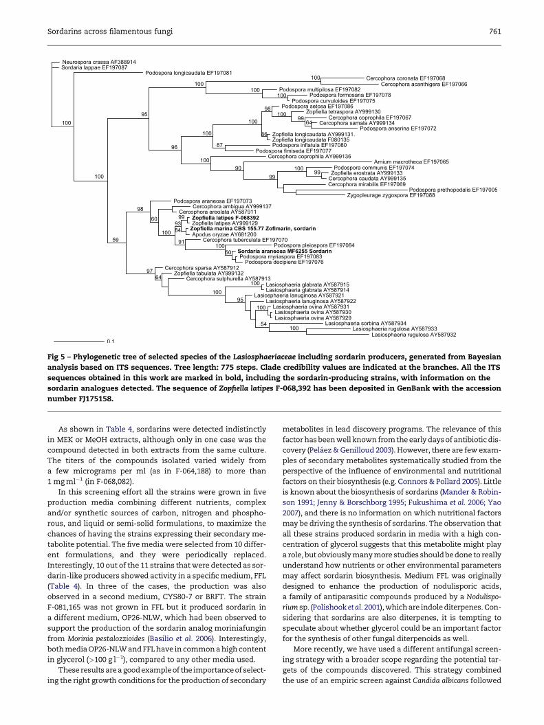
Fig 5 – Phylogenetic tree of selected species of the Lasiosphaeriaceae including sordarin producers, generated from Bayesian
analysis based on ITS sequences. Tree length: 775 steps. Clade credibility values are indicated at the branches. All the ITS
sequences obtained in this work are marked in bold, including the sordarin-producing strains, with information on the
sordarin analogues detected. The sequence of Zopfiella latipes F-068,392 has been deposited in GenBank with the accession
number FJ175158.
Sordarins across filamentous fungi 761
As shown in Table 4, sordarins were detected indistinctly
in MEK or MeOH extracts, although only in one case was the
compound detected in both extracts from the same culture.
The titers of the compounds isolated varied widely from
a few micrograms per ml (as in F-064,188) to more than
1 mg ml�1 (in F-068,082).
In this screening effort all the strains were grown in five
production media combining different nutrients, complex
and/or synthetic sources of carbon, nitrogen and phospho-
rous, and liquid or semi-solid formulations, to maximize the
chances of having the strains expressing their secondary me-
tabolite potential. The five media were selected from 10 differ-
ent formulations, and they were periodically replaced.
Interestingly, 10 out of the 11 strains that were detected as sor-
darin-like producers showed activity in a specific medium, FFL
(Table 4). In three of the cases, the production was also
observed in a second medium, CYS80-7 or BRFT. The strain
F-081,165 was not grown in FFL but it produced sordarin in
a different medium, OP26-NLW, which had been observed to
support the production of the sordarin analog moriniafungin
from Morinia pestalozzioides (Basilio et al. 2006). Interestingly,
both media OP26-NLW and FFL have in common a high content
in glycerol (>100 g l�1), compared to any other media used.
These results are a good example of the importance of select-
ing the right growth conditions for the production of secondary
metabolites in lead discovery programs. The relevance of this
factor has been well known from the early days of antibiotic dis-
covery (Pelaez & Genilloud 2003). However, there are few exam-
ples of secondary metabolites systematically studied from the
perspective of the influence of environmental and nutritional
factors on their biosynthesis (e.g. Connors & Pollard 2005). Little
is known about the biosynthesis of sordarins (Mander & Robin-
son 1991; Jenny & Borschborg 1995; Fukushima et al. 2006; Yao
2007), and there is no information on which nutritional factors
may be driving the synthesis of sordarins. The observation that
all these strains produced sordarin in media with a high con-
centration of glycerol suggests that this metabolite might play
a role, but obviously many more studies should be done to really
understand how nutrients or other environmental parameters
may affect sordarin biosynthesis. Medium FFL was originally
designed to enhance the production of nodulisporic acids,
a family of antiparasitic compounds produced by a Nodulispo-
rium sp. (Polishook et al. 2001), which are indole diterpenes. Con-
sidering that sordarins are also diterpenes, it is tempting to
speculate about whether glycerol could be an important factor
for the synthesis of other fungal diterpenoids as well.
More recently, we have used a different antifungal screen-
ing strategy with a broader scope regarding the potential tar-
gets of the compounds discovered. This strategy combined
the use of an empiric screen against Candida albicans followed

Table 2 – Strains tested in a differential sensitivity screenlooking for antifungal agents with the same mode ofaction as sordarins, sorted according to their adscriptionto fungal orders, following Index Fungorumcomplemented as per Hibbett et al. (2007)
Class Order Isolates tested
Ascomycetes Xylariales 8053
Pleosporales 7780
Hypocreales 5052
Dothideales 2074
Helotiales 2011
Diaporthales 1953
Eurotiales 1620
Sordariales 886
Phyllachorales 761
Pezizales 637
Mycosphaerellales 592
Microascales 472
Trichosphaeriales 350
Chaetothyriales 144
Onygenales 111
Ophiostomatales 84
Calosphaeriales 28
Others/Undetermined 2533
Anamorphic fungi
(incertae sedis)
5192
Basidiomycetes Agaricales 1624
Polyporales 832
Russulales 155
Hymenochaetales 128
Boletales 96
Thelephorales 54
Dacrymycetales 48
Others/Undetermined 1009
Anamorphic fungi
(incertae sedis)
72
Others 87
Sterile isolates and
unidentified fungi
1621
Total 45 987
762 F. Vicente et al.
by early dereplication of known compounds by LC–MS and
subsequent use of a genomic-wide platform using C. albicans
heterozygous strains to prioritize antifungal extracts with in-
teresting modes of action (Jiang et al. 2008; Parish et al. 2008).
This new paradigm was coupled with the use of a system of
mini-fermentations in nutritional arrays using microtiter
plates that allowed generating microbial extracts in high
throughput mode, facilitating the cultivation of each strain
in multiple conditions with minimum additional effort. This
system has been shown to increase the probabilities of detect-
ing biological activities from any defined fungal set (Bills et al.
2008).
Across 11 569 fungal strains screened under this new para-
digm, nearly 25 % provided a positive signal in the empiric an-
tifungal screen against C. albicans, but only 11 strains from this
pool were detected as producing sordarin or sordarin ana-
logues by LC–MS analysis (Table 3). Dereplication of natural
products using different analytical methods (HPLC, UV spec-
troscopy, MS or even NMR) and reference databases has
become one of the most useful and widely used approaches
to minimize the redundant isolation of known metabolites
(Bitzer et al. 2007).
About half of the strains tested were subjected to some
level of taxonomic identification, usually by morphological
analysis, and a number of isolates were also subjected to
rDNA sequencing. These strains were distributed across
more than 60 fungal orders, mostly from the Ascomycota. Pleo-
sporales, Helotiales, and Hypocreales were the groups repre-
sented with the largest number of isolates (Table 5). Again,
the screening included all the groups known to contain sor-
darin-producing fungi (Xylariales, Sordariales, Eurotiales, and
Microascales).
In this case, and unlike our previous experience with the
differential sensitivity screen, the production of sordarins
was not associated with any particular growth condition,
and as many as 9 different production media supported the
synthesis of sordarins (Table 6). Consistently with previous
observations though, production of sordarins was observed
in very few conditions for each strain (only in 1–3 out of the
8 media used for each strain). It is important to note that dur-
ing this time we did not use the media FFL and OP26-NLW, due
to the technical inconveniences associated with their prepara-
tion, which requires the sterilization of some components
separately. The control strain Zopfiella marina produced
a strong antifungal activity in three out of the four media
tested, and LC–MS analysis detected the presence of both zofi-
marin and sordarin in the extracts.
The striking association of sordarin biosynthesis with me-
dium FFL in the first screening period sharply contrasts with
the lack of correlation with any particular medium in the sec-
ond screening effort. It is worth noting that none of the media
used during the latter period contained high concentrations of
glycerol, what could explain this lack of preference for any
particular medium. The screening would detect sordarins
only in those strains able to produce sufficiently high titers
in any other culture media. However, other differences be-
tween the two screening strategies may also contribute to
the dissimilar results. First, the different fermentation for-
mats (flasks vs. microtiter plates), secondly the different ex-
traction process; and probably even more important, the
sensitivity of the screening process itself. It is questionable
whether the antifungal extracts in which sordarins were
detected by LC–MS, would have given a positive result if tested
in the differential sensitivity test using the Saccharomyces
cerevisiae mutants.
It is interesting to note that the two screening strategies
identified the same number of sordarin producing strains,
but starting from a very different number of tested strains
(45 000 vs. 11 000). Thus, the empiric screening against C. albi-
cans followed by LC–MS analysis was more efficient in the de-
tection of sordarin-producing strains. However, it seems
likely that this second screening strategy, not specifically
designed for the detection of antifungal agents with the
same mode of action as sordarins but with a much broader
scope, may have actually underestimated the potential of
the strains tested to produce sordarins. Probably the fre-
quency of detection of sordarins would have been even
higher if a glycerol-rich medium had been included among
the media selected.

Table 3 – Fungal strains found to produce sordarin and sordarin analogues
Strain code Taxonomy(morphology)
Taxonomy(rDNA
sequencing)
Collection place Substrate Compoundsdetected
GenBankaccession
Control strains described in previous literature
F-116,360a
(MF-6239)
Rosellinia subiculata Xylariaceae,
Rosellinia subiculata
(?)
Navesink River,
New Jersey (USA)
Decayed wood Sordarin FJ175163
F-116,361
(MF-6255)
Sordaria araneosa Lasiosphaeriaceae,
related to several
Podospora spp.
Original strain from
Sandoz, no data on
origin
Unknown Sordarin FJ175160
F-262,723
(CBS 155.77)
Zopfiella marina Lasiosphaeriaceae,
closely related to
Apodus oryzae
Chinese Sea
(Taiwan)
Marine mud Zofimarin,
sordarin
FJ175159
Strains discovered through a differential sensitivity screening using a sordarin-resistant mutant vs. wild type yeast
F-068,082 Seimatosporium sp. Amphisphaeriaceae,
consistent with
Seimatosporium
grevillae
Sierra Villuercas,
Caceres (Spain)
Twigs and leaves of
Quercus faginea
40-O-dimethyl
sordarin
FJ175156
F-064,147 Sterile isolate Xylariaceae, likely
Xylaria sp.
Curepipe
(Mauricio Is.)
Leaf litter Xylarin FJ175166
F-064,695 Sterile isolate Xylariaceae, likely
Xylaria sp.
Le Morne
(Mauricio Is.)
Twigs from conifer Xylarin FJ175165
F-064,186 Sterile isolate Xylariaceae, likely
Xylaria sp.
Grandes Gorges
(Mauricio Is.)
Twigs from
unidentified bush
Xylarin FJ175167
F-068,980 Sterile isolate Xylariaceae, likely
Xylaria sp.
Acan-Bot Esaveg
(Bata, Eq. Guinea)
Bark of Plagiostylis
Africana
Xylarin FJ175169
F-067,683 Sterile isolate Xylariaceae, likely
Xylaria mellissii
Three Okumes, Road
Bata-Mbini Km. 27
(Eq. Guinea)
Mixed leaf litter Xylarin FJ175171
F-069,049 Sterile isolate Xylariaceae, likely
Xylaria mellissii
Three Okumes, Road
Bata-Mbini Km. 27
(Eq. Guinea)
Leaves of
Monopetalanthus
microphyllus
Xylarin FJ175172
F-065,308 Sterile isolate Xylariaceae, likely
Xylaria mellissii
Tepui Ruraima
(Venezuela)
Leaves from
unidentified bush
Xylarin FJ175174
F-065,977 Sterile isolate Xylariaceae, likely
Xylaria mellissii
Punta Cana (Rep.
Dominicana)
Undetermined plant Xylarin FJ175175
F-064,188 Sterile isolate Xylariaceae Grandes Gorges
(Mauricio Is.)
Twigs from
unidentified bush
Sordarin,
zofimarin,
isozofimarin
FJ175177
F-081,165 Sterile isolate Diatrypaceae, likely
Eutypa tetragona
Santa Elena, Jaen
(Spain)
Phellodon melaleucus
fruitbody
Zofimarin FJ175178
Strains discovered through empiric screening against C. albicans followed by LC/MS analysis
F-190,561 Truncatella sp. Amphisphaeriaceae,
closely related to
Truncatella angustata
Guatemala Mixed leaf litter Sordarin FJ175157
F-230,275 Sterile isolate Xylariaceae, likely
Xylaria sp.
El Calafate, Santa
Cruz (Argentina)
Undetermined plant Xylarin FJ175168
F-130,895 Sterile isolate Xylariaceae Santa Marina,
Taramundi, Asturias
(Spain)
Taxus baccata Sordarin FJ175176
F-235,338 Sterile isolate Xylariaceae, likely
Rosellinia sp.
Alcantud, Cuenca
(Spain)
Leaf litter from
Pinus sp.
Xylarin FJ175170
F-246,940 Sterile isolate Amphisphaeriacae,
likely
Amphisphaeria sp.
Salazie (Reunion Is.) Leaf litter Sordarin FJ175155
F-260,230 Sterile isolate Amphisphaeriacae,
likely
Amphisphaeria sp.
Turku (Finland) Lichen Sordarin FJ175154
F-254,988 Sterile isolate Xylariaceae Huelva (Spain) Scirpus holoschoenus Sordarin FJ175164
F-247,493
(CBS 284.87)
Eutypa tetragona Diatrypaceae,
consistent with
Eutypa tetragona
Tourrettes/Loup,
Alpes Maritimes
(France)
Sarothamnus
scoparius
Zofimarin FJ175179
F-249,532
(IZ-1295)
Sterile isolate Xylariaceae, likely
Rosellinia corticium
La Coruna (Spain) Thinopyrum
junceiforme
Xylarin FJ175162
(continued on next page)
Sordarins across filamentous fungi 763

Table 3 – (continued)
Strain code Taxonomy(morphology)
Taxonomy(rDNA
sequencing)
Collection place Substrate Compoundsdetected
GenBankaccession
F-249,628
(CBS 449.89)
Rosellinia nectrioides Xylariaceae, closely
related to Rosellinia
abscondita
Jerusalem, Dalby Par.
Uppland (Sweden)
Trifolium medium Sordarin FJ175181
F-266,831
(CBS 211.87)
Diatrype stigma Diatrypaceae,
consistent with
Diatrype stigma
Loch Dan, Wicklow
Co. (Ireland)
Quercus cf. robur Sordarin AJ302438b
a Strains with codes other than F numbers in parenthesis were either obtained from culture collections or received from external collaborators.
MF: Merck Culture Collection (Rahway, NJ, USA); CBS: Centraalbureau voor Schimmelcultures (Utrecht, The Netherlands); IZ: Inigo Zabalgogeaz-
coa (Instituto de Recursos Naturales y Agrobiologıa, Salamanca, Spain).
b This strain had been already sequenced by our group years ago (Acero et al. 2004). We sequenced it again for confirmation, but the original
GenBank access code is indicated.
764 F. Vicente et al.
Phylogenetic analysis of the sordarin-producing organisms
All the fungal strains detected to produce sordarin and
sordarin-like compounds were subjected to a preliminary
taxonomic analysis based on the morphology of the sporula-
tion structures. Unfortunately, most of the strains remained
sterile even after prolonged incubation, as frequently ob-
served when working with fungi isolated from plant mate-
rials using indirect methods (e.g., Guo et al. 2000; Collado
et al. 2001). With the aim of getting more meaningful infor-
mation on their taxonomy, all of them were subjected to
Table 4 – Production media supporting the production of sordaboxes indicate combinations of strain/medium that resulted inthat were inactive. All the strains were tested in five different msordarin analog produced (in mg mlL1). Those strains where lecontaminations or lack of growth of the production culture. Stproduce sordarin (see Table 3). Media formulations are describ(2006), dPolishook et al. (2001), or taken from the Merck proprie
Productio
Strain codes CYS80a
FFLd
TG106a
MV8b
BRFTb
AD
F-068,082 1200
F-064,147 ~590
F-064,695 156
F-064,186 ~590
F-068,980 34
F-067,683 110
F-069,049 27
F-065,308 13
F-065,977 590
F-064,188 74** ND
F-081,165
MF 6239
MF 6255
*ND¼Not determined. ** This strain produced 74 mg ml�1 of sordarin and
sequencing of the ITS region. The sequences obtained were
compared with sequences in GenBank using BLAST analysis,
and the closest matches found were retrieved, comple-
mented with other related species and used to build the
phylogenetic trees shown in Figs 2–5. This analysis included
several strains previously known to produce sordarins, in-
cluded in our screening systems for quality control
purposes.
As shown in Table 3, the strains detected as sordarin-
producing fungi were all of them from the Xylariales, being
distributed across three different families: Xylariaceae,
rins detected using a differential sensitivity screen. Blackthe production of sordarins, grey boxes are combinationsedia. The figures in the boxes indicate the concentration of
ss than five media were tested correspond to cases ofrains MF 2639 and MF6255 were control strains known toed in aPelaez et al. (2001), bSingh et al. (2003), cBasilio et al.tary database (e)
n media
2M2BMb
CYS80-7a
OATe
STPa
OP26NLWc
Solvent
MeOH
MeOH
MeOH
MeOH
MeOH
ND* MeOH/MEK
MeOH
ND MEK
MEK
MEK
ND MEK
ND MeOH
ND MeOH
minor amounts (w10 mg ml�1) of zofimarin and isozofimarin.

Table 5 – Strains tested in an empiric screen looking forantifungal agents with activity against Candida albicans,sorted according to their adscription to fungal orders,following Index Fungorum complemented as per Hibbettet al. (2007)
Class Order Isolates tested
Ascomycetes Pleosporales 1361
Hypocreales 964
Helotiales 687
Diaporthales 343
Capnodiales 361
Xylariales 309
Sordariales 216
Chaetothyriales 133
Eurotiales 83
Ophiostomatales 77
Botryosphaeriales 73
Dothideales 56
Chaetosphaeriales 50
Coniochaetales 35
Hysteriales 32
Microascales 32
Onygenales 28
Others/undetermined 372
Basidiomycetes Agaricales 42
Polyporales 35
Hymenochaetales 20
Others/undetermined 72
Sterile isolates and
unidentified fungi
6188
Total 11 569
Sordarins across filamentous fungi 765
Diatrypaceae and Amphisphaeriaceae. Although sordarins have
been reported previously from members of the Sordariales,
Microascales and Eurotiales (Table 1), we have not found any
new sordarin producing strain from these three orders.
AmphisphaeriaceaeWithin this family we observed production of sordarins in
four strains. Fig 2 shows the phylogenetic relationships of
these four strains with a number of species of the Amphi-
sphaeriaceae, based on ITS sequences. The sequence alignment
used to build this tree contained 471 characters, 309 of which
were constant.
The strain F-068,082 could be identified morphologically as
a Seimatosporium sp., and the ITS sequence was identical to
a sequence deposited in GenBank as S. grevilleae, strongly sug-
gesting these two strains to be conspecific. Accordingly, the
phylogenetic tree showed these two strains grouped in
a branch, as a sister group of two sequences of Sarcostroma res-
tionis. Interestingly, this strain produced a new natural analog
of sordarin (40-O-demethyl sordarin), never reported before.
The remaining strains of the family produced sordarin.
Another strain (F-190,561) was identified as a Truncatella sp.
by morphology, and the ITS sequence analysis showed it to be
closely related to a group of T. angustata sequences, placed as
a sister group rooted at the base of the branch harboring these
sequences. This topology would be consistent with this strain
being either T. angustataorat leastan intimately relatedspecies.
The remaining two strains belonging to the Amphisphaeria-
ceae (F-260,940 and F-260,230) showed almost identical ITS
sequences (99 % homology) and were grouped together in
a branch that contained sequences of Discosia sp. and Amphi-
sphaeria sp., being also highly related to those ones (nucleotide
homology>97 %). Despite their high similarity, which suggest
their conspecificity, those two strains came from very distant
geographic origins (Finland and Reunion Island), suggesting
they would belong to a species with a cosmopolitan
distribution.
We had described previously the production of another sor-
darin analog, moriniafungin, from Morinia pestalozzioides, an-
other member of the Amphisphaeriaceae (Basilio et al. 2006;
Collado et al. 2006). We have not found this particular sordarin de-
rivative in any other member of this or any other fungal family.
Putting all these findings together we conclude that the produc-
tion of sordarin-like compounds is a trait relatively widespread
across different lineages within the Amphisphaeriaceae.
The topology of the phylogenetic tree was consistent with
previously reported phylogenies of the Amphisphaeriaceae
(Jeewon et al. 2002; Lee et al. 2006), showing well supported
monophyletic clades correlating with conidial morphology.
DiatrypaceaeWe observed production of sordarins in three strains ascribed
to species of this family. The phylogenetic tree originated from
the alignment of ITS sequences of these isolates and a number
of species from the family is shown in Fig 3. The dataset con-
sisted of 551 characters, 349 of which were constant.
Two of the strains were shown to produce zofimarin. One of
these strains (F-247,493) was obtained from CBS (Eutypa
tetragona CBS 284.87), whereas the other one (F-081,165) was
isolated from a fruitbody of Phellodon melaleucus collected in
Spain. These two strains were grouped together in the phylo-
genetic tree in a branch with very high statistical support,
the closest neighbor being another sequence labeled as E. tet-
ragona. The nucleotide homology among these three se-
quences was very high (>93 %). Although the statistical
support for the whole group was weak (58 %), it seems reason-
able to conclude that the two zofimarin producing strains be-
long to the species E. tetragona.
The third strain (F-266,831; CBS 211.87) produced sordarin,
not zofimarin, and it appears in CBS catalog under the name
Selenosporella falcata. This strain fell within a monophyletic
group with two strains of Diatrype stigma. Actually, this strain
was originally described by Rappaz (1987) as D. stigma, and
neither this author nor others who have studied D. stigma in
culture (Glawe & Rogers 1982) assigned the name S. falcata to
its anamorph. Likewise, the original description of S. falcata
did not mention this apparent connection with the teleo-
morph (Sutton 1973).
To our knowledge, this is the first report of the production
of sordarins in members of the Diatrypaceae, a fungal group
poorly known with respect to their potential to produce bio-
logically active metabolites, compared to other related groups
such as the Xylariaceae (Stadler & Hellwig 2005).
XylariaceaeBy far the group where the production of sordarins seems to
be the most common is the Xylariaceae. As many as 15 strains
were detected producing sordarin or related compounds. The
ITS derived phylogenetic affinities of 13 of these strains can be

Table 6 – Production media supporting the production of sordarins detected from C. albicans empiric screen followed by LC–MS analysis. Black boxes indicate combinations of strain/medium that resulted in the production of sordarins, grey boxesare combinations that were inactive. All the strains were grown in eight different media. Those strains where less thaneight media were tested correspond to cases of contaminations or lack of growth of the production culture. The strain ofZopfiella marina CBS 155.77 (last raw in the table) was used as control in this experiment. Media formulations described inaPelaez et al. (2001), bSingh et al. (2003), cScott et al. (1970), or taken from the Merck proprietary database (d)
Production media
Strain
codes BRFTb
CMKd
CYS80a
MED1d
MMKd
MMK2d
MSCMd
MV8b
STPa
SCASd
SUPMd
TG106a
WHEAT-1d
WS80d
YESc
F-190,561
F-230,275
F-130,895
F-235,338
F-246,940
F-260,230
F-254,988
F-247,493
F-249,532
F-249,628
F-266,831
CBS 155.77
766 F. Vicente et al.
tracked in the tree shown in Fig 4, which contains exclusively
sequences from the Xylarioideae, essentially Xylaria and Roselli-
nia species. The sequence dataset used to build the tree con-
tained 482 characters, 261 of which were constant.
The two remaining strains (F-064,188 and F-130,895) had
their closest matches in GenBank with a sequence of Anthosto-
mella eucalyptorum and other sequences ascribed to several
xylariaceous fungi, but the homology and the quality of the
alignments was too low to allow for any reliable conclusion
about the phylogenetic relationships of these strains, and
they were excluded from this analysis.
Xylarin was the compound the most frequently found in
this group, being produced by 11 of the xylariaceous strains.
Xylarin has been previously reported from a Xylaria sp. and
from one unidentified fungus (Schneider et al. 1995; Coval
et al. 1995). The remaining strains produced only sordarin,
except for the strain F-064,188, which produced sordarin, zofi-
marin and the new isomer isozofimarin.
The strain CBS 449.89 (Rosellinia nectrioides) was found to
produce sordarin. This strain showed an ITS sequence identi-
cal to another CBS strain identified as R. abscondita, and they
appeared together in the tree in a branch with 100 % clade cred-
ibility. This strongly suggests that these two isolates would be
conspecific. Actually, both species are morphologically ex-
tremely similar or even indistinguishable (Petrini & Petrini
2005; Bahl et al. 2005), but their synonymy has not been formally
proposed. However, we did not detect any antifungal activity in
the extracts derived from the strain of R. abscondita CBS 447.89,
and therefore these extracts were not subjected to LC–MS anal-
ysis. This negative result might be due to a real lack of sordarin
production, or to the titers of the compound being too low to
produce an antifungal signal.
We isolated another phylogenetically related strain
(F-235,338) from pine leaf litter, but producing xylarin instead
of sordarin. This strain fell in a clade with high statistical
support together with R. nectrioides, R. abscondita, and two
other Rosellinia species; R. britannica and R. mammiformis.
These four species are morphologically closely related, shar-
ing the presence of a Geniculosporium anamorph, ellipsoidal as-
cospores, germ slit straight or sigmoid covering most of the
length of the ascospore, and rim of ascus apical structure
rounded. Rosellinia britannica and R. mammiformis differ from
the other two species in the morphology of the slimy sheath
covering the ascopore. They all belong to subgenus Calomastia,
and fell together in a clade in a UPGMA cluster analysis based
on morphological characters (Petrini & Petrini 2005). Again, we
did not detect any antifungal activity in the strains of R. bri-
tannica and R. mammiformis, but it would be interesting to con-
firm with additional studies whether these related species are
really unable to produce sordarins, since this character could
represent an additional feature to discriminate species across
this group. In any case, the position of the strain F-235,338 in
the tree strongly suggests that it would belong to a closely
related Rosellinia species.
Another xylarin-producing strain (F-249,532) was isolated
as an endophyte of Thinopyrum junceiforme and shown to
have a sequence 99 % identical to a sequence of R. corticium,
suggesting these two strains to be conspecific. However, we
did not find any sordarin analog production in the strain of
R. corticium F-160,845 (corresponding to the sequence
AY908999) when this was subsequently tested. These strains
were relatively close to strains of R. subiculata, one of which
(MF6239, ATCC 74386) at least has been reported to produce
sordarin in the patent literature (Balkovec & Tse 2000).
Three xylarin producing strains (F-064,186; F-064,695 and
F-64,147) were closely related (nucleotide homology> 95%)
and grouped together in the tree. All of them were isolated
from plant material collected in Mauritius Island, and they
could well be conspecific. The closest match (99 % homology)
in GenBank was a sequence (AF153731) labeled as Xylaria sp.,

Sordarins across filamentous fungi 767
isolated as an endophyte of Livistonia chinensis in Hong Kong
(Guo et al. 2000). These four strains were grouped in a clade
with total statistical support. Another xylarin-producing strain
(F-230,275) isolated from plant material in Argentina appeared
rooted to the base of this branch, and all these five sequences
were grouped in a clade with very high clade credibility (98 %).
Other four xylarin-producing strains were also grouped in
a monophyletic clade, together with a sequence from a strain
identified as X. mellissii. Those strains came from diverse
tropical areas, including Equatorial Guinea (F-067,683 and F-
069,049), Venezuela (F-065,308), and Republica Dominicana
(F-065,977). The two strains from Equatorial Guinea were al-
most identical in sequence (99 % homology), strongly suggest-
ing they would be conspecific, and all the sequences in the
clade shared >93 % homology, what could be interpreted as
all of them being also conspecific with X. mellissii. Unfortu-
nately, we have not been able to check whether the strain
identified as X. mellissii produces xylarin as well, due to the
lack of viability upon preservation at �70 �C.
The xylarin-producing strain F-068,980, isolated from bark
collected in Equatorial Guinea, was relatively close to a branch
containing two Xylaria species, X. cornudamae and X. fioriana, al-
though the support for the clade containing these three se-
quences was rather weak (67 %). This small clade fell within
a branch containing a number of other Xylaria species (X. allan-
toidea, X. enteroleuca, X. acuta, X. longipes) and Stilbohypoxylon quis-
quiliarum with very good statistical support (97 %). This suggests
this strain would most likely belong to a related Xylaria species.
The sordarin-producing strain F-254,988, an endophyte of
Scirpus holoschoenus from Spain, was clustered together with
two Xylaria species (X. globosa and X. schweinitzii) in a branch
with high clade credibility, although the nucleotide homology
with those sequences was low (77 %).
It is interesting to remark that all these strains showed af-
finities with the Xylarioideae. Actually, we did not find any
strains clearly related to the Hypoxyloideae, although Hypoxylon
croceum has been reported as producing sordarin and hypoxy-
sordarin (Daferner et al. 1999).
LasiosphaeriaceaeAlthough we did not find any new isolate from this family pro-
ducing sordarin, we subjected to ITS sequencing some strains
from culture collections that have been reported as sordarin
producers, which were used as controls for the screening pro-
cess. These strains were shown to belong to the Lasiosphaeria-
ceae. The phylogenetic tree in Fig 5 shows the relationships of
those strains with other members of the family. The size of
the alignment was 456 characters, 243 of which were constant.
The fungal strain MF6255 is a duplicate of the strain depos-
ited by Sandoz as the original producer of sordarin, obtained
from ATCC (ATCC36386/NRRL3196). The strain was originally
identified as Sordaria araneosa, and its taxonomic adscription
gave name to this family of antifungal compounds. Interest-
ingly, our ITS sequence analysis showed that this strain is not
a member of genus Sordaria, bearing very little homology to
any sequence of other Sordaria species in GenBank. However,
the strain was found to be phylogenetically closely related to
a number of Podospora species (P. myriaspora, P. decipiens and P.
pleiospora), as shown in Fig 5. Nucleotide homology with P. myr-
iaspora and P. decipiens in particular was very high (97 %).
Sordaria araneosa had been synonymized with Podospora ara-
neosa (Cain 1962), therefore our molecular data would appear
consistent with this synonymy. However, the sordarin-produc-
ing strain is not conspecific with the only sequence of P. araneosa
in GenBank (EF197073). This may be either because the latter se-
quence is incorrectly labeled or because the sordarin-producing
strain is not really S. araneosa/P. araneosa. In the absence of other
sequences of this species to provide further verification, our
data just suggest that the original sordarin producing strain is
akin to several Podospora species. Interestingly, a strain of P. plei-
ospora, a species that appears to be close to the original sordarin
producer, has been also reported to produce sordarin and
a number of analogues (Table 1; Weber et al. 2005).
An obvious implication of these findings is that the name
of sordarins applied to this class of compounds, is somewhat
misleading. The production of sordarins is not apparently
a feature associated to genus Sordaria or to the Sordariaceae
but to the Lasiosphaeriaceae, and even more frequent in other
fungal families, as shown above.
Within the Lasiosphaeriaceae, we also confirmed the produc-
tion of sordarins from the strain of Zopfiella marina CBS 155.77
(¼SANK 21274, reported to produce zofimarin (Ogita et al.
1987, Sato et al. 1995);¼NHL 2731, type of the species). Our re-
sults showed that this strain produced both zofimarin and sor-
darin. In the phylogenetic tree, the type strain of Z. marina
appeared very closely related (98 % nucleotide homology)
with the type strain of Apodus oryzae (CBS 376.74), and as a sister
group of two strains of Z. latipes, within a clade with high cred-
ibility support (93 %). Cai et al. (2006a) reported that genus Apo-
dus fits better in the Lasiosphaeriaceae than in the Sordariaceae,
where it was originally ascribed; and later on they reported
a close relationship between A. oryzae and Z. latipes based on
multi-gene molecular phylogenies (Cai et al. 2006b), but this
finding was not subjected to any interpretation. Our results
confirm this relationship, and include an additional dimension
by placing Z. marina as another tightly related species. The high
nucleotide homology with A. oryzae even suggests the potential
synonymy between these two species. Actually, their original
descriptions (Furuya & Udagawa 1975; Arx 1975) reported re-
markably similar characters, and the minor differences could
probably be attributed to strain to strain (and lab to lab) varia-
tions. However, we have not compared morphologically these
two strains vis-a-vis, neither tested any strain of A. oryzae to
check for the production of sordarin analogues.
It is worth noting that all the strains producing sordarins,
although belonging to different genera, were grouped together
in a clade with total statistical support.
The phylogenetic tree in Fig 5, on the other hand, is fully
consistent with previous molecular analysis on the Lasios-
phaeriaceae. Except for the species of Lasiosphaeria, which
appeared clustered together in a branch with total statistical
support as in Miller & Huhndorf (2004), the rest of genera in
the tree (Podospora, Cercophora and Zopfiella) appeared to be
polyphyletic, in agreement with previous reports (Cai et al.
2005, 2006b).
Sordarins as chemotaxonomic markers
The data reported here can be used to explore potential corre-
lations between the production of specific sordarin derivatives

768 F. Vicente et al.
and the taxonomic placement and phylogenetic affinities of
the producing organisms. In this regard, a few points are note-
worthy. Thus, sordarin and xylarin were the derivatives more
frequently found in our screening (8 and 11 strains respec-
tively), but while the production of sordarin appeared to be
spread across all the fungal families reported here, xylarin
was confined to the Xylariaceae, where it is most frequent in
Xylaria spp. Zofimarin was far less frequent, with only three
strains, two of them identified as Eutypa tetragona, within the
Diatrypaceae. The new compounds 40-O-demethyl sordarin
and isozofimarin were found only in one strain. Last, there
seem to be some rather infrequent sordarin derivatives (e.g.,
moriniafungin, BE-31405, GR135402), for which our extensive
screening program did not yield any further producing strains
beyond those reported in the literature.
One of the main conclusions from this study is a confirma-
tion and extension of scattered historical data of the distribu-
tion of sordarins across the Xylariales. Sordarins are
particularly frequent in the Xylariaceae, but extend also to
the Amphisphaeriaceae and the Diatrypaceae, the latter being
implicated for the first time in the present report.
Despite including in our screen significant numbers of iso-
lates from the Sordariales, Eurotiales and Microascales, orders
where the production of sordarins has been reported, we did
not find any producing strain, suggesting that sordarin pro-
duction is a rare feature in these groups. Although it is possi-
ble that our screening systems (including cultivation
techniques) could favor sordarin production in Xylariales, we
did detect sordarins also in control strains from the Lasios-
phaeriaceae (S. araneosa and Z. marina). Finally, other fungal
groups that have been massively subjected to screen, such
as the Pleosporales, Hypocreales or Helotiales, seem to lack the
ability to synthesize this class of compounds.
Acknowledgements
The authors are particularly indebted to the CIBE technical staff
assisting in the screening process. This work is dedicated to all
the people from CIBE and MRL who dedicated the best years of
their lives to the exciting endeavour of discovering new natural
molecules of therapeutic utility from microbial sources during
the last 50þ years. Their good work and dedication will stand
foreverasa hallmark inthe historyof naturalproducts research.
r e f e r e n c e s
Acero FJ, Gonzalez V, Sanchez-Ballesteros J, Rubio V, Checa J,Bills GF, Salazar O, Platas G, Pelaez F, 2004. Molecular phylo-genetic studies on the Diatrypaceae based on rDNA–ITS se-quences. Mycologia 96: 249–259.
Arx JA von, 1975. On Thielavia and some similar genera of asco-mycetes. Studies in Mycology 8: 1–29.
Bahl J, Jeewon R, Hyde KD, 2005. Phylogeny of Rosellinia cape-tribulensis sp. nov. and its allies (Xylariaceae). Mycologia 97:1102–1110.
Balkovec JM, Tse B, 2000. Sordarin derivatives. US patent No6,040,463.
Basilio A, Justice MC, Harris GH, Bills GF, Collado J, de la Cruz M,Dıez MT, Hernandez P, Liberator PA, Kahn JN, Pelaez F,
Platas G, Schmatz DM, Shastry M, Tormo JR, Vicente MF, 2006.The discovery of moriniafungin, a novel sordarin derivativeproduced by Morinia pestalozzioides. Bioorganic Medicinal Chem-istry 14: 560–566.
Bills GF, Polishook JD, 1993. Selective isolation of fungi from dungof Odocoileus hemionus (mule deer). Nova Hedwigia 57: 195–206.
Bills GF, Polishook JD, 1994. Abundance and diversity of micro-fungi in leaf litter of a lowland rain forest in Costa Rica.Mycologia 86: 187–198.
Bills GF, Giacobbe RA, Lee SH, Pelaez F, Tkacz JS, 1992. Tremor-genic mycotoxins paspalitrem A and C, from a tropical Pho-mopsis. Mycological Research 96: 977–983.
Bills GF, Platas G, Pelaez F, Masurekar P, 1999. Reclassification ofa pneumocandin-producing anamorph, Glarea lozoyensis gen.et sp. nov., previously identified as Zalerion arboricola. Myco-logical Research 103: 179–192.
Bills GF, Platas G, Fillola A, Jimenez R, Collado J, Vicente MF,Martın J, Gonzalez del Val A, Bur-Zimmerman J, Tormo JR,Pelaez F, 2008. Enhancement of antibiotic and secondarymetabolite detection from filamentous fungi by growth onnutritional arrays. Journal of Applied Microbiology 104: 1644–1658.
Bitzer J, Kopcke B, Stadler M, Hellwig V, Ju Y-M, Seip S, Henkel T,2007. Accelerated dereplication of natural products, supportedby reference libraries. Chimia International Journal for Chemistry61: 332–338.
Cabello MA, Platas G, Collado J, Dıez MT, Martın I, Vicente F,Meinz M, Onishi JC, Douglas C, Thompson J, Kurtz MB,Schwartz RE, Bills GF, Giacobbe RA, Abruzzo GK, Pelaez F,2001. The discovery of arundifungin, a novel antifungalcompound produced by fungi. Biological activity and taxon-omy of the producing organisms. International Microbiology 4:93–102.
Cai L, Jeewon RA, Hyde KD, 2005. Phylogenetic evaluation andtaxonomic revision of Schizothecium based on ribosomal DNAand protein coding genes. Fungal Diversity 19: 1–21.
Cai L, Jeewon RA, Hyde KD, 2006a. Phylogenetic investigations ofSordariaceae based on multiple gene sequences and mor-phology. Mycological Research 110: 137–150.
Cai L, Jeewon RA, Hyde KD, 2006b. Molecular systematics of Zop-fiella and allied genera: evidence from multi-gene sequenceanalysis. Mycological Research 110: 359–368.
Cain RF, 1962. Studies on coprophilous ascomycetes VIII. Newspecies of Podospora. Canadian Journal of Botany 40: 447–490.
Collado J, Platas G, Pelaez F, 1996. Fungal endophytes in leavestwigs and bark of Quercus ilex from central Spain. Nova Hed-wigia 63: 347–360.
Collado J, Platas G, Pelaez F, 2001. Identification of an endophyticNodulisporium sp. from Quercus ilex in Central Spain as theanamorph of Biscogniauxia mediterranea by rDNA sequencinganalysis and effect of different ecological factors on distribu-tion of the fungus. Mycologia 93: 875–886.
Collado J, Platas G, Bills GF, Basilio A, Vicente MF, Tormo JR,Hernandez P, Dıez MT, Pelaez F, 2006. Studies on Morinia:recognition of Morinia longiappendiculata sp. nov. as a new en-dophytic fungus, and a new circumscription of Morinia pesta-lozzioides. Mycologia 98: 616–627.
Collado J, Platas G, Paulus B, Bills GF, 2007. High-throughput cul-turing of fungi from plant litter by a dilution-to-extinctiontechnique. FEMS Microbiology Ecology 60: 521–533.
Connors N, Pollard D, 2005. Pneumocandin B0 production by fer-mentation of the fungus Glarea lozoyensis: physiological andengineering factors affecting titer and structural analogueformation. In: An Z (ed), Mycology Series. Handbook of IndustrialMycology, vol. 22. Marcel Dekker Inc., New York, pp. 515–538.
Coval SJ, Puar MS, Phife DW, Terracciano JS, Patel M, 1995. SCH 57404an antifungal agent possessing the rare sordaricin skeleton anda tricyclic sugar moiety. Journal of Antibiotics 48: 1171–1172.

Sordarins across filamentous fungi 769
Daferner M, Mensch S, Anke T, Sterner O, 1999. Hypoxysordarin,a new sordarin derivative from Hypoxylon croceum. Zeitschriftfur Naturforschung 54c: 474–480.
Davoli P, Engel G, Werle A, Sterner O, Anke T, 2002. Neosordarinand hydroxysordarin, two new antifungal agents from Sorda-ria araneosa. Journal of Antibiotics 55: 377–382.
Dominguez JM, Martin JJ, 1998. Identification of elongationFactor 2 as the essential protein targeted by sordarins inCandida albicans. Antimicrobial Agents and Chemotherapy 42:2279–2283.
Duetz WA, Ruedi L, Hermann R, O’Connor K, Buchs J, Witholt B,2000. Methods for intense aeration, growth, storage, and rep-lication of bacterial strains in microtiter plates. Applied andEnvironmental Microbiology 66: 2641–2646.
Fukushima M, Watanabe E, Eguchi T, Kakinuma K, 2006. Biosyn-thesis of zofimarin diterpene glycoside antifungal compound.Nippon Kagakkai Koen Yokoshu 86: 1314.
Furuya K, Udagawa S, 1975. Two new species of cleistothecialAscomycetes. Journal of Japanese Botany 50: 249–254.
Gaber RF, Copple DM, Kennedy BK, Vidal M, Bard M, 1989. Theyeast gene ERG6 is required for normal membrane functionbut is not essential for biosynthesis of the cell-cycle-sparkingsterol. Molecular and Cellular Biology 9: 3447–3456.
Glawe DA, Rogers JD, 1982. Observations on the anamorphs of sixspecies of Diatrype and Diatrypella. Canadian Journal of Botany60: 245–251.
Guo LD, Hyde KD, Liew ECY, 2000. Identification of endophyticfungi from Livistona chinensis based on morphology andrDNA sequences. New Phytologist 147: 617–630.
Hall CC, Bertasso AB, Watkins JD, Georgopapadakou NH, 1992.Screening assays for protein synthesis inhibitors. Journal ofAntibiotics 45: 1697–1699.
Hauser D, Sigg HP, 1971. Isolierung und abbau von sordarin.Helvetica Chimica Acta 54: 1178–1190.
Herreros E, Almela MJ, Lozano S, Gomez de las Heras F, Gargallo-Viola D, 2001. Antifungal activities and cytotoxicity studies ofsix new azasordarins. Antimicrobial Agents and Chemotherapy45: 3132–3139.
Hibbett DS, Binder M, Bischoff JF, Blackwell M, Cannon PF,Eriksson OE, Huhndorf S, James T, Kirk PM, Lucking R,Lumbsch HT, Lutzoni F, Matheny PB, McLaughlin DJ,Powell MJ, Redhead S, Schoch CL, Spatafora JW, Stalpers JA,Vilgalys R, Aime MC, Aptroot A, Bauer R, Begerow D, Benny GL,Castlebury LA, Crous PW, Dai Y-C, Gams W, Geiser DM,Griffith GW, Gueidan C, Hawksworth DL, Hestmark G,Hosaka K, Humber RA, Hyde KD, Ironside JE, Koljalg U,Kurtzman CP, Larsson K-H, Lichtwardt R, Longcore J,Miadlikowska J, Miller A, Moncalvo J-M, Mozley-Standridge S,Oberwinkler F, Parmasto E, Reeb V, Rogers JD, Roux C,Ryvarden L, Sampaio JP, Schußler A, Sugiyama J, Thorn RG,Tibell L, Untereiner WA, Walker C, Wang Z, Weir A, Weiss M,White MM, Winka K, Yao Y-J, Zhang N, 2007. A higher-levelphylogenetic classification of the. Fungi. Mycological Research111: 509–547.
Hirano A, Torigoe K, Ogawa H, Kamiya S, Okada H, Nagashima M,Kojiri K, Suda H, 2000. Derivatives of antifungal substanceBE-31405 and process for producing the same. US Patent No6087390.
Jenny L, Borschborg H-J, 1995. Synthesis of the dolabellane di-terpene hydrocarbon (�)-d- araneosene. Helvetica Chimica Acta78: 715–731.
Jeewon R, Liew ECY, Hyde KD, 2002. Phylogenetic relationships ofPestalotiopsis and allied genera inferred from ribosomal DNAsequences and morphological characters. Molecular Phyloge-netics and Evolution 25: 378–392.
Jiang B, Xu D, Allocco J, Parish C, Davison J, Veillette K,Sillaots S, Hu W, Rodrıguez-Suarez R, Trosok S, Zhang L,Li Y, Rahkoodaee F, Ransom T, Martel N, Wang H,
Gauvin D, Wiltsie J, Wisniewski D, Salowe SP, Kahn JN,Hsu M-J, Giacobbe R, Abruzzo G, Flattery A, Youngman P,Wilson K, Bills G, Platas G, Pelaez F, Dıez MT, Kauffman S,Becker J, Harris G, Liberator P, Roemer T, 2008. PAP inhib-itor with in vivo efficacy identified by Candida albicans ge-netic profiling of natural products. Chemistry and Biology 15:363–374.
Justice M, Hsu MJ, Tse B, Ku T, Balkovec J, Schmatz D, Nielsen J,1998. Elongation factor 2 as a novel target for selective inhi-bition of fungal protein synthesis. Journal of Biological Chemistry273: 3148–3151.
Kamai Y, Kakuta M, Shibayama T, Fukuoka T, Kuwahara S, 2005.Antifungal activities of R-135853, a sordarin derivative, inexperimental candidiasis in mice. Antimicrobial Agents andChemotherapy 49: 52–56.
Kaneko S, Arai M, Uchida T, Konosu T, 2003. Antimycotic agentcontaining zofimarin derivative having oxazepane ring. JapanPatent No JP2003261450.
Kennedy TC, Webb G, Cannell RJP, Kinsman OS, Middleton RF,Sidebottom PJ, Taylor NL, Dawson MJ, Buss AD, 1998. Novelinhibitors of fungal protein synthesis produced by a strain ofGraphium putredinis. Isolation, characterization and biologicalproperties. Journal of Antibiotics 51: 1012–1018.
Kinsman OS, Chalk PA, Jackson HC, Middleton RF, ShuttleworthA, Rudd BAM, Jones CA, Noble HM, Wildman HG, Dawson MJ,Stylli C, Sidebottom PJ, Lamont B, Lynn S, Hayes MV, 1998.Isolation and characterisation of an antifungal antibiotic(GR135402) with protein synthesis inhibition. Journal of Antibi-otics 51: 41–49.
Lee S, Crous PW, Wingfield MJ, 2006. Pestalotioid fungi fromRestionaceae in the Cape Floral Kingdom. Studies in Mycology55: 175–187.
Mander LN, Robinson RP, 1991. Studies on the synthesis of thediterpenoid mold metabolite sordaricin. Exploration of a pro-spective biogenetic intramolecular [4þ 2] cycloaddition. Jour-nal of Organic Chemistry 56: 3595–3601.
Martin JJ, Chicharro-Gonzalo J, Gomez JRR, Garcia-Ochoa S,Gomez de las Heras F, Hayes MV, Dawson MJ, Wildman HG,Hall RM, 2000. Antifungal sordaridin derivatives. US patent No6054478.
Miller AN, Huhndorf SM, 2004. Using phylogenetic species rec-ognition to delimit species boundaries within Lasiosphaeria.Mycologia 96: 1106–1127.
Nylander JA, 2004. MrModeltest v2. Program distributed by theauthor. Evolutionary Biology Centre, Uppsala University.
Ogita T, Hayashi A, Sato S, Furaya K, 1987. Antibiotic zofimarin.Japan Kokai Tokkyo Koho JP 62-40292.
Okada H, Kamiya S, Shina Y, Suwa H, Nagashima M, Nakajima S,Shimokawa H, Sugiyama E, Kondo H, Kojiri K, Suda H, 1998.BE-31405, a new antifungal antibiotic produced by Penicilliumminioluteum. I. Description of producing organism, fermenta-tion isolation, physico-chemical and biological properties.Journal of Antibiotics 51: 1081–1086.
Parish CA, Smith SK, Calati KB, Zink DL, Wilson KE, Roemer T,Jiang B, Xu D, Bills GF, Platas G, Pelaez F, Dıez MT, Tsou NN,McKeown AE, Ball RG, Powles MA, Yeung L, Liberator PA,Harris GH, 2008. Isolation and structure elucidation ofparnafungins, antifungal natural products that inhibit mRNApoly-adenylation. Journal of the American Chemical Society 130:7060–7066.
Pelaez F, Collado J, Platas G, Dıez MT, Basilio A, Gonzalez A,Vicente F, Dai P, Harris G, Rosenbach M, Dreikorn S,Thompson J, 2001. Studies on Tricellulortus pepoformis, a tropi-cal mitosporic fungus. Nova Hedwigia 72: 217–230.
Pelaez F, Genilloud O, 2003. Discovering new drugs from microbialnatural products. In: Barredo JL (ed), Microorganisms for HealthCare, Foods and Enzyme Production. Research Signpost,Trivandrum, India, pp. 1–22.

770 F. Vicente et al.
Petrini LE, Petrini O, 2005. Morphological studies in Rosellinia(Xylariaceae): the first step towards a polyphasic taxonomy.Mycological Research 109: 569–580.
Platas G, Salazar O, Acero J, Sanchez-Ballesteros J, Rubio V,Gonzalez V, Portal MA, Borkowski J, Pelaez F, 2001. Presence ofa simple tandem repeat DNA sequence in the ITS1 region ofthe Xylariales. Current Microbiology 43: 43–50.
Polishook JD, Pelaez F, Platas G, Ondeyka JG, Dombrowskii A,Teran AM, 2001. Biogeography and relatedness of Nodulispo-rium strains producing nodulisporic acid. Mycologia 93:1125–1137.
Pongcharoen W, Rukachaisirikul V, Phongpaichit S, Kuhn T,Pelzing M, Sakayarog J, Taylor WC, 2008. Metabolites from theendophytic fungus Xylaria sp. PSU-D14. Phytochemistry 69:1900–1902.
Rappaz F, 1987. Taxonomie et nomenclature de Diatrype stigma, D.decorticata et D. undulata (Diatrypaceae, Ascomycetes). Myco-taxon 30: 209–219.
Ronquist F, Huelsenbeck JP, 2003. MRBAYES 3: bayesian phylogeneticinference under mixed models. Bioinformatics 19: 1572–1574.
Sato A, Takahashi S, Ogita T, Sugano M, Kodama K, 1995. Marinenatural products. Annual Reports of Sankyo Research Laboratories47: 1–58.
Schneider G, Anke H, Sterner O, 1995. Xylarin, an antifungal Xy-laria metabolite with an unusual tricyclic uronic acid moiety.Natural Products Letters 7: 309–316.
Scott PM, Lawrence JW, van Walbeek V, 1970. Detection of my-cotoxins by thin-layer chromatography: applications toscreening of fungal extracts. Applied Microbiology 20: 839–842.
Singh SB, Jayasuriya H, Dewey R, Polishook JD, Dombrowski AW,Zink DL, Guan Z, Collado J, Platas G, Pelaez F, Felock PJ,
Hazuda DJ, 2003. Isolation, structure and HIV-1-integrase in-hibitory activity of structurally diverse fungal metabolites.Journal of Industrial Microbiology and Biotechnology 30: 721–731.
Stadler M, Hellwig V, 2005. Chemotaxonomy of the Xylariaceaeand remarkable bioactive compounds from Xylariales andtheir associated asexual stages. Recent Research Developments inPhytochemistry 9: 41–93.
Serrano-Wu M, Du X, Balasubramanian N, St Laurent DR, 2002.Thio derivatives of sordarin as antifungal agents. US Patent No6436930.
Sutton BC, 1973. Hyphomycetes from Manitoba and Saskatche-wan, Canada. Mycological Papers 132: 1–143.
Thompson JD, Gibson TJ, Plewniak F, Jeanmougin F, Higgins DG,1997. The Clustal_X windows interface: flexible strategies formultiple sequence alignment aided by quality analysis tools.Nucleic Acids Research 25: 4876–4882.
Weber RWS, Meffert A, Anke H, Sterner O, 2005. Production ofsordarin and related metabolites by the coprophilous fungusPodospora pleiospora in submerged culture and in its naturalsubstrate. Mycological Research 109: 619–626.
Yao Q, 2007. Biosynthetic studies on fungal diterpene antibiotics.MSc. thesis, Oregon State University.
Zink D, Dufresne C, Liesch J, Martin J, 2002. Automated LC–MSanalysis of natural products: extraction of UV, MS and reten-tion time data for component identification and character-ization. Poster presented at the American Society for MassSpectrometry (ASMS) 50th Annual Meeting.
Zink D, Dufresne C, Liesch J, Martin J, 2005. Identification/dere-plication of natural products by LC–UV–MS Spectral searchparameters. Poster presented at the Small Molecule ScienceConference (COSMOS).