The antifungal Dm-AMP1 protein from Dahlia merckii expressed in Solanum melongena is released in...
11
© New Phytologist (2004) 163: 393–403 www.newphytologist.org 393 Research Blackwell Publishing, Ltd. The antifungal Dm-AMP1 protein from Dahlia merckii expressed in Solanum melongena is released in root exudates and differentially affects pathogenic fungi and mycorrhizal symbiosis A. Turrini 1 , C. Sbrana 2 , L. Pitto 3 , M. Ruffini Castiglione 2 , L. Giorgetti 2 , R. Briganti 2 , T. Bracci 2 , M. Evangelista 3 , M. P. Nuti 1,2 and M. Giovannetti 1,2 1 Dipartimento di Chimica e Biotecnologie Agrarie, Università di Pisa, via del Borghetto 80, I−56124 Pisa, Italy; 2 Istituto di Biologia e Biotecnologia Agraria, CNR, Sezione di Pisa, via del Borghetto 80, I−56124 Pisa, Italy; 3 Istituto di Fisiologia Clinica, CNR, Area della Ricerca, via Moruzzi 1, I−56124 Pisa, Italy Summary • Transformed aubergine plants constitutively expressing the Dm-AMP1 antimicrobial defensin (from Dahlia merckii) were generated and characterized. • Transgenic plants were selected on kanamycin and screened by polymerase chain reaction analysis. The expression of Dm-AMP1 in plant tissues and its release in root exudates were detected by Western blot analyses. Dm-AMP1 localization was performed by immunohistochemical experiments. • Dm-AMP1 expression ranged from 0.2% to 0.48% of total soluble proteins in primary transformants and from 0.16% to 0.66% in F 2 plants. Transformed clones showed resistance to the pathogenic fungus Botrytis cinerea, whose development on leaves was reduced by 36–100%, with respect to controls. The protein was released in root exudates of the transformed plants and was active in reducing the growth of the co-cultured pathogenic fungus Verticillium albo-atrum , whereas it did not interfere with recognition responses and symbiosis establishment by the arbuscular mycorrhizal fungus Glomus mosseae . • Dm-AMP1 transformants may represent a useful model to study the interactions between genetically modified plants and pathogenic fungi or beneficial nontarget microorganisms. Key words: arbuscular mycorrhizal fungi, Solanum melongena , Botrytis cinerea , defensin, transformed plants, Verticillium albo-atrum . © New Phytologist (2004) 163 : 393–403 Author for correspondence: M. Giovannetti Tel: +39 050 571561 Fax: +39 050 571562 Email: [email protected] Received: 8 January 2004 Accepted: 20 March 2004 doi: 10.1111/j.1469-8137.2004.01107.x Introduction Plants protect themselves from pathogenic organisms by activ- ating different defense strategies such as cell wall thickening, localized cell necrosis, production of phytoalexins. More- over, plants can synthesize a wide variety of antimicrobial proteins, such as zeamatins, osmotins, ribosomal inhibitor proteins (Broekaert et al., 1997; Shewry & Lucas, 1997) and enzymes able to hydrolyse fungal hyphae (Mauch et al., 1988; Melchers et al., 1994). Another group of plant antimicrobial compounds is represented by cysteine-rich peptides (Broekaert et al., 1995; Rao, 1995) that includes thionins (Bohlmann & Apel, 1991), lipid-transfer proteins (LTPs) (Garcìa-Olmedo et al., 1995) and defensins (Broekaert et al., 1995). Plant defensins are small peptides (45–54 amino acids) with a characteristic three- dimensional folding pattern stabilized by disulfide-linked cysteines. They share common characters with those described in insects and mammals, suggesting that these defense molecules predate the evolutionary divergence of plants and animals (Broekaert et al., 1995). So far, over 80 defensin sequences from different plant species have been identified (Thomma et al., 2002). Plant defensins were first isolated from barley and wheat seeds (Mendez et al., 1990) and successively from seeds and vegetative tissues of different dicotyledon and monocotyledon
Transcript of The antifungal Dm-AMP1 protein from Dahlia merckii expressed in Solanum melongena is released in...
©
New Phytologist
(2004)
163
: 393–403
www.newphytologist.org
393
Research
Blackwell Publishing, Ltd.
The antifungal Dm-AMP1 protein from
Dahlia merckii
expressed in
Solanum melongena
is released in root exudates and differentially affects pathogenic fungi
and mycorrhizal symbiosis
A. Turrini
1
, C. Sbrana
2
, L. Pitto
3
, M. Ruffini Castiglione
2
, L. Giorgetti
2
, R. Briganti
2
, T. Bracci
2
, M. Evangelista
3
, M. P. Nuti
1,2
and M. Giovannetti
1,2
1
Dipartimento di Chimica e Biotecnologie Agrarie, Università di Pisa, via del Borghetto 80, I
−
56124 Pisa, Italy;
2
Istituto di Biologia e Biotecnologia Agraria,
CNR, Sezione di Pisa, via del Borghetto 80, I
−
56124 Pisa, Italy;
3
Istituto di Fisiologia Clinica, CNR, Area della Ricerca, via Moruzzi 1, I
−
56124 Pisa, Italy
Summary
• Transformed aubergine plants constitutively expressing the Dm-AMP1 antimicrobialdefensin (from
Dahlia merckii
) were generated and characterized.• Transgenic plants were selected on kanamycin and screened by polymerasechain reaction analysis. The expression of Dm-AMP1 in plant tissues and its releasein root exudates were detected by Western blot analyses. Dm-AMP1 localizationwas performed by immunohistochemical experiments.• Dm-AMP1 expression ranged from 0.2% to 0.48% of total soluble proteins inprimary transformants and from 0.16% to 0.66% in F
2
plants. Transformed clonesshowed resistance to the pathogenic fungus
Botrytis cinerea
, whose developmenton leaves was reduced by 36–100%, with respect to controls. The protein wasreleased in root exudates of the transformed plants and was active in reducing thegrowth of the co-cultured pathogenic fungus
Verticillium albo-atrum
, whereas itdid not interfere with recognition responses and symbiosis establishment by thearbuscular mycorrhizal fungus
Glomus mosseae
.• Dm-AMP1 transformants may represent a useful model to study the interactionsbetween genetically modified plants and pathogenic fungi or beneficial nontargetmicroorganisms.
Key words:
arbuscular mycorrhizal fungi,
Solanum melongena
,
Botrytis cinerea
,defensin, transformed plants,
Verticillium albo-atrum
.
©
New Phytologist
(2004)
163
: 393–403
Author for correspondence:
M. GiovannettiTel: +39 050 571561Fax: +39 050 571562Email: [email protected]
Received:
8 January 2004
Accepted:
20 March 2004
doi: 10.1111/j.1469-8137.2004.01107.x
Introduction
Plants protect themselves from pathogenic organisms by activ-ating different defense strategies such as cell wall thickening,localized cell necrosis, production of phytoalexins. More-over, plants can synthesize a wide variety of antimicrobial proteins,such as zeamatins, osmotins, ribosomal inhibitor proteins(Broekaert
et al
., 1997; Shewry & Lucas, 1997) and enzymesable to hydrolyse fungal hyphae (Mauch
et al
., 1988; Melchers
et al
., 1994). Another group of plant antimicrobial compoundsis represented by cysteine-rich peptides (Broekaert
et al
., 1995;Rao, 1995) that includes thionins (Bohlmann & Apel, 1991),
lipid-transfer proteins (LTPs) (Garcìa-Olmedo
et al
., 1995) anddefensins (Broekaert
et al
., 1995). Plant defensins are smallpeptides (45–54 amino acids) with a characteristic three-dimensional folding pattern stabilized by disulfide-linkedcysteines. They share common characters with those describedin insects and mammals, suggesting that these defense moleculespredate the evolutionary divergence of plants and animals(Broekaert
et al
., 1995). So far, over 80 defensin sequences fromdifferent plant species have been identified (Thomma
et al
.,2002). Plant defensins were first isolated from barley and wheatseeds (Mendez
et al
., 1990) and successively from seeds andvegetative tissues of different dicotyledon and monocotyledon

www.newphytologist.org
©
New Phytologist
(2004)
163
: 393–403
Research394
plants (Terras
et al
., 1992, 1993; Moreno
et al
., 1994; Osborn
et al
., 1995; Gao
et al
., 2000).Brassicaceae and Saxifragaceae produce ‘morphogenic’
defensins, which inhibit pathogenic fungal growth by reducinghyphal elongation and increasing hyphal branching. Themode of action of plant defensins from Asteraceae, includingDm-AMP1, is typical of ‘nonmorphogenic’ defensins andconsists of slowing down hyphal extension and not inducingmarked morphological changes. Molecular mechanisms ofplant defensins action is not completely well-understood.
Neurospora crassa
hyphae treated with either Rs-AFP2 orDm-AMP1 defensins showed a rapid potassium (K
+
) efflux,calcium (Ca
2+
) uptake and alkalinization of the incubationmedium. In addition, membrane potential changes, but notthe formation of membrane pores, were observed (Thevissen
et al
., 1996). Defensins are able to bind to hyphal membranesat specific sites and indirect evidence indicates that this bind-ing is required for antifungal activity (Thevissen
et al
., 1997,2000a). Recently, it was shown that different plant defensinsare able to interact with different sphingolipids. For example,Dm-AMP1 interacts with mannosylated sphingolipids occur-ring in the outer plasma membrane (Thevissen
et al
., 2000b,2003a), whereas the radish plant defensin Rs-AFP2 inter-acts with fungal glucosylceramides (Thevissen
et al
., 2004).Some plant defensins are constitutively expressed in seeds
and are released during germination. It has been suggested thatthe defensin Rs-AFP2 from radish (
Raphanus sativus
) couldhave a role in protecting plantlet tissues from pathogen attack,thus enhancing the chances of seedling survival and plantreproduction (Terras
et al
., 1995). Other plant defensins areexpressed at a low level in the leaves and other vegetativeplant tissues, such as roots, stems, flowers and fruits, but theirconcentration greatly increases after pathogen attack (Terras
et al
., 1995).Antimicrobial compounds synthesized by seeds or plant
tissues represent a powerful tool to enhance plant resistanceagainst fungal pathogens, as shown in tobacco, scentedgeranium, canola and potato plants constitutively expressingantifungal proteins cloned from
R. sativus
,
Mirabilis jalapa
,
Amaranthus caudatus
,
Allium cepa
,
Pisum sativum
and
Medicago sativa
, respectively (Terras
et al
., 1995; de Bolle
et al
.,1996; Bi
et al
., 1999; Wang
et al
., 1999; Gao
et al
., 2000).Transformed plants expressing antimicrobial compounds
could represent a potential benefit for human health andenvironmental safety because their production could decrease theneed for chemical pesticides. Moreover they might representa useful tool for studying potential risks related to the release oftransformed plants and their impact on nontarget organisms.
In this work, we produced transformed aubergine (
Solanummelongena
) plants expressing the gene for Dm-AMP1 protein,cloned from
Dahlia merckii
and analysed: (1) Dm-AMP1expression and localization in different plant tissues; (2) foliarresistance to the phytopathogenic fungus
Botrytis cinerea
;(3) the release of Dm-AMP1 in root exudates; (
iv
) the impact of
released Dm-AMP1 on the phytopathogenic fungus
Verticilliumalbo-atrum
; (4) the interaction of Dm-AMP1 transformedplants with the symbiotic fungus
Glomus mosseae
.
Materials and Methods
Plant material used for genetic transformation experiments
Seeds of aubergine (
S. melongena
L.) cv. Violetta of NewYork were surface sterilized in 1.5% sodium hypochloritefor 20 min and then rinsed several times with sterile distilledwater. Seeds were germinated in sterile Magenta culture vessels(GA7; Sigma-Aldrich, Milan, Italy) in half-strength B5 medium(Gamborg
et al
., 1968) supplemented with 0.7% bacto-agaradjusted to pH 5.8. Seedling growth and all subsequent
in vitro
culture steps were carried out in a temperature-controlled25
°
C growth chamber under cool 6–12 µmol m
−
2
s
−
1
whitefluorescent light with a dark-light cycle of 16h : 8 h. Four- tofive-week-old plants were used as explant source in genetictransformation experiments.
Bacterial strains and vectors
Agrobacterium
strain EHA105 carrying the plasmid pDm-AMPLCC (kindly provided by Dr A. Greenland, ZenecaAgrochemical, Calgary, Alberta, Canada) was used as vectorsystem for transformation. Plasmid pDmAMPLCC containsthe neomycin phosphotransferase II (
nptII
) gene under thenopaline synthase (
nos
) promoter and the Dm-AMP1 geneunder the cauliflower mosaic virus (
CaMV35S
) promoter,with duplicated enhancer region. The Dm-AMP1 defensingene and protein sequences (Osborn
et al
., 1995) haveGenBank accession numbers AAB34972 and A26963. Thebacterial strain pDmAMPLCC/EHA105 was grown onLuria Bertani broth (LB) medium with appropriate anti-biotics (50 mg l
−
1
kanamycin and 20 mg l
−
1
rifampicin). For co-cultivation, isolated colonies of bacteria were picked up fromselection plates and grown overnight in 10 ml of LB liquidmedium at 28
°
C to the late logarithmic stage (OD600 0.8–1).The bacteria were centrifuged and the pellet was re-suspended1 : 10 in MS medium (Murashige & Skoog, 1962) and usedfor the infection and co-cultivation procedure in the genetictransformation experiments.
Genetic transformation experiments
The transformation system described by Billings
et al
. (1997)was used with some modifications. Leaf disks 1.5 cm indiameter were excised and placed on shoot regeneration (SR)medium (MS basal salt and vitamins, 10 µ
N6-[isopentyl]adenine (2iP), 0.1 m
thidiazuron (TDZ), 2% sucrose, 0.6%agar, pH 5.8), covered with a
Nicotiana glauca
feeder layer, for48 h of preculture before co-cultivation. The
N. glauca
cell
©
New Phytologist
(2004)
163
: 393–403
www.newphytologist.org
Research 395
suspensions were subcultured every 14 d. Feeder plateswere prepared by plating 2 ml of exponentially growing cellsuspensions on 25 ml of solidified SR medium in Petri dishes.After 1 d of incubation in the growth chamber in the dark,discs of dried sterile filter paper were placed on top ofthe
N. glauca
cells and the leaf explants were then depositedon the filter paper after infection (1 min) with
Agrobacteriumtumefaciens
cell suspension. After 48 h of co-cultivation, explantswere transferred on SR medium containing 300 mg l
−
1
aug-mentin and 50 mg l
−
1
kanamycin. Explants were subculturedevery 10 d on fresh selection medium until shoots started toregenerate. Regenerated shoots were transferred on half-strengthMS medium with 50 mg l
−
1
kanamycin.
Polymerase chain reaction (PCR) analysis
For PCR analysis, DNA was isolated from kanamycin-resistantleaves and wild-type control leaves, using a cetyltrimethy-lammonium bromide (CTAB) procedure (Doyle & Doyle,1990). The PCR was performed in a Hybaid Omnigene(Hybaid, Teddington, UK) thermocycler. The primers used foramplification of the 344 bp fragment of the
nptII
gene were5
′
-TTCTTTTTGTCAAGACCGACCT-3
′ (upstream) and5′-TTCGTCCAGATCATCCT-3′ (downstream). A sampleof 100 ng of each DNA were amplified with the followingprotocol: 1 cycle at 94°C for 2 min, 30 cycles at 94°C for1 min, 50°C for 1 min and 72°C for 1 min, 1 cycle at 72°Cfor 5 min.
Western blot analysis
Total soluble proteins were extracted using liquid nitrogenand resuspended in extraction buffer (15 m Na2HPO4, 10 mNaH2PO4, 100 m KCl, 2 m ethylenediaminetetraaceticacid (EDTA), pH 7). Samples were heated for 10 min at 85°Cand heat-labile denatured proteins were removed by centrifu-gation (12 000 g ). Soluble proteins were quantified using Bradfordreagent (Bio-Rad, Milan, Italy) and recording solution absorb-ance at λ = 595 nm, with different concentrations of bovineserum albumin (BSA) as standards. Different quantities (20,40 µg) of total soluble proteins for each sample were separatedby sodium dodecyl sulfate (SDS) gel electrophoresis (acry-lamide : bisacrylamide 30 : 0.8) through a 5% stacking geloverlaid on a 15% separating gel at constant 200 V. Thepurified protein DmAMP1 (25, 50 and 100 ng) was used asstandard. Proteins were blotted at constant 250 mA for 1 h at4°C onto a nitrocellulose membrane (Amersham Bioscience,Milan, Italy) using a Mini Trans-blot Transfer Cell (Bio-Rad).After blotting the membrane was blocked in Tris-bufferedsaline (TBS), containing 0.05% Tween and 5% skimmed milk,for 1 h at room temperature, then incubated for 1 h withDm-AMP1 antibody (1 : 1000), kindly provided by ZenecaAgrochemicals, washed three times with TBS and incubatedfor 1 h with 1 : 2000 peroxidase-labeled goat antirabbit
antibody (Santa Cruz Biotechnology). The membrane wasthen washed three times before detection carried out using theenhanced chemiluminescence (ECL) method (Amersham).Images acquired with ImageMaster VDS system (Amersham)were analysed with ImageJ software for signals quantification.
Immunohistochemical procedure
Isolated young leaves and roots from transformed (line 12)and control plants micropropagated in vitro in MS mediawere fixed with 4% paraformaldehyde + 1% glutaraldehydemixture in 0.15 phosphate buffer, pH 7.4, for 18 h andembedded in paraffin. Sections, 8 µm thick, were prepared byusing a manual microtome and processed for immunologicaldetection of Dm-AMP1 protein. The slides were rehydratedand treated with H2O2 (3%) to inhibit endogenous peroxidaseactivities. After extensive washing the slides were buffered inSolution A (0.1 phosphate-buffered saline (PBS) pH 7.4,1% BSA, 0.1% Tween 20). All the steps described belowwere performed at room temperature in a moist chamber. Theslides were covered for 15 min with 1.5% normal goat serumin solution A to block nonspecific sites. Incubation with thepolyclonal antibodies against Dm-AMP1 was carried out for2 h using 1 : 100 diluted serum in solution A. After threewashes in solution A, the slides were exposed to the secondaryantibody (1 : 50 peroxidase conjugate goat antirabbit; SantaCruz) for 1 h. Three washes in solution A and three in PBSfacilitated the removal of unbound secondary antibodiesand the progression to the detection step. The slides wereincubated with 0.06% 3, 3′-diaminobenzidine (DAB) solutionin PBS for 10 min. After the addition of H2O2 to a finalconcentration of 0.06%, the incubation time was extendedfor further 10 min. The color development was stopped bywashing three times in PBS and three times in distilled water.The slides were then air-dried and mounted for microscopicanalysis. Images were captured under a Axioskop microscope(Zeiss, Milan, Italy) equipped with a video-camera (DC100;Leica, Milan, Italy).
Conventional controls were performed in each experimentand the specificity of the immunolabeling was tested by meansof: (1) treatment with a nonimmune rabbit serum instead ofthe primary antibodies; (2) further dilution and omission ofthe primary antibody; (3) omission of the secondary per-oxidase conjugated antibody; (4) inhibition of the immuno-response by preincubating the antibodies with an excess ofhomologous antigens before use.
Hydroponic cultures
In vitro-transformed aubergine plants (lines 5 and 12) wereplaced in sterile 100 ml flasks with roots plunged into 30 mlof sterile half-strengh MS basal medium with no sugar. Sterileair was continuously blown into the liquid by placing a0.22 µm filter upstream a pipette linked to a pump. Liquid

www.newphytologist.org © New Phytologist (2004) 163: 393–403
Research396
samples (10 ml) were collected after 4 d from the beginningof the hydroponic culture, dialysed against double-distilledwater using dialysis tubing (molecular weight cut off (MWCO)5 kDa) and concentrated by lyophilization. The concentratedproteins were resuspended in 40 µl double-distilled water andused for Western blot analysis.
Bioassay on Verticillium albo-atrum
In vitro-micropropagated transformed aubergine plants oflines 1, 5, 8 and 12 were placed with roots lying on the agarsurface in the center of a 160 mm Petri dish, half-filled withhalf-strength MS agar medium. The fungus V. albo-atrum wasgrown on potato dextrose agar (PDA) at 24°C for 2–3 wk.Mycelial plugs were removed from a colony with a 5 mmdiameter sterile cork borer, and placed, mycelium-side down,near the plant roots. Six transformed and six control plantswere used in this bioassay, with two mycelial plugs per plant.Plants were placed on the agar simultaneously with the mycelialplugs. Petri dishes were incubated for 18 d, and at the end ofthis period mycelial growth was estimated by measuringfungal colony diameters and calculating corresponding areas.Results obtained were analysed by one way .
Resistance to Botrytis cinerea
Plant resistance to B. cinerea, a phytopathogenic fungus infect-ing leaves, was tested as follows: the third to fifth leaf from theapical meristem of transformed aubergine plants (lines 1, 5, 8and 12, three leaves and at least three plants for each line) andnontransformed controls were harvested. Leaves were placedin sterile Petri dishes (15 cm diameter) containing sterilemoistened filter paper, and 30-µl drops of sterile water wereplaced on the adaxial surface of each leaf. Mycelial plugs(5 mm diameter) from PDA plates containing 1-wk-old grow-ing mycelium of B. cinerea were placed on each droplet andplates were sealed with both Parafilm and polyethylene film tomaintain moisture. Plates were incubated for 72 h at 21°C toallow the development of mycelium before evaluating necroticlesions sizes. Results obtained were analysed by one-way .
Bioassay with symbiotic fungi
The experiments were carried out on the arbuscular mycorrhizal(AM) fungus G. mosseae (Nicol. and Gerd.) Gerdemann &Trappe (IMA 1) obtained from pot cultures maintained in thecollection of the Department of Chemistry and AgriculturalBiotechnology, University of Pisa, Italy. Sporocarps were extractedfrom pot-culture soil by wet-sieving and decanting, downto a mesh size of 100 µm (Gerdemann & Nicolson, 1963).Fungal material retained on sieves was flushed into Petri dishesand sporocarps were manually collected with forceps under adissecting microscope (Wild; Leica). Sporocarps were placedon cellulose ester Millipore (Milano, Italy) membranes (0.45 µm
diameter pore size) and used to assess the development ofhyphal differential morphogenesis and of mycorrhizal infectionin roots of micropropagated transformed aubergine plants (lines1, 5, 8 and 12) as previously described (Giovannetti et al.,1993, 1994). Briefly, plant root system was placed betweentwo Millipore membranes, one containing 15 sporocarpsof G. mosseae. Another membrane containing 15 sporocarpswas placed over the membranes containing the roots. Fivereplicates were set up for both transformed and control plants.The plants were transplanted into pots containing sterilequartz grit and maintained under controlled conditions (18–24°C, 16–8 h photoperiod). Plants were watered daily andwere not fertilized during the growing period (1 month).
One month after inoculation plants were removed from pots,root sandwiches were opened and membranes were stained with0.05% Trypan blue in lactic acid to assess hyphal growth anddifferentiation. Plant roots were cleared and stained followingPhillips & Hayman (1970), using lactic acid instead of lacto-phenol. Infected root length was calculated by using the grid-line intersect method (Giovannetti & Mosse, 1980). Colonizedroots were mounted on microscope slides and observed undera Polyvar light microscope (Reichert-Jung, Vienna, Austria) toassess the number of entry points per cm of root.
Results
Characterization of Dm-AMP1 transformed plants
Agrobacterium tumefaciens-mediated genetic transformationexperiments were performed on aubergine leaf explants toobtain the constitutive expression of the protein Dm-AMP1.The antibiotic resistance gene nptII was used as a selectablemarker. We selected 12 putative regenerated plants which wereresistant to kanamycin. They were analysed by PCR experimentsusing the primers corresponding to the nptII coding sequence.As illustrated in Fig. 1, in which six putative transformed
Fig. 1 Polymerase chain reaction analysis showing the presence of nptII gene in six Dm-AMP1 transformed aubergine (Solanum melongena) plants. The expected nptII band was 344 bp. Lanes: 1, molecular weight marker (MW); 3, DNA from nontransformed control plants (C); 4–9, DNA from six transformed plants (T1–T12); 11, positive control from pDmAMPLCC plasmid (pLCC).

© New Phytologist (2004) 163: 393–403 www.newphytologist.org
Research 397
plants are shown, all the DNA extracted from the leaves ofthe putative transformed plants and from the positive control(pDmAMPLCC plasmid) showed the expected 344 bp band,which was absent in DNA from nontransformed plants.
To verify the expression of the protein Dm-AMP1 in thetissues of aubergine-transformed plants, Western blot experi-ments were performed. In Fig. 2a representative Western blotresults, carried out on total soluble proteins obtained from leaves,are shown. Nonspecific bands, resulting from the use of a poly-clonal antibody, were detected on Western blots of bothtransformed and nontransformed plants, but their molecularweights did not correspond to that of Dm-AMP1 protein.
Transformed plants produced the Dm-AMP1 protein, whichwas not detectable in control plants. The densitometric meas-ures of the bands corresponding to the Dm-AMP1 protein inthe different primary transformants analysed, revealed expres-sion levels ranging from 0.20% to 0.48% of the total solubleproteins of leaf extracts. Comparable expression levels of theDm-AMP1 protein were present in all vegetative tissues of plants(Fig. 2b) and in fruits (Fig. 2c). Transformed lines, showingbands of expected size upon PCR and immunoblot analyses,were self pollinated to obtain F1 plants. Western-blot analyseswere carried out on F1 plants showing a 3 : 1 segregationof kanamycin resistance in segregation experiments. The
Fig. 2 Western-blot analysis showing the expression of Dm-AMP1 peptide in aubergine (Solanum melongena) transformed plants. (a) Analysis of Dm-AMP1 expression in primary transformants leaves. Lanes: 1–3, purified Dm-AMP1 peptide; 4, total soluble proteins of nontransformed control plants; 5–6, total soluble proteins of two different transformed aubergine plants. (b) Analysis of different plant tissues of transformed aubergine (line 12) detecting the expression of Dm-AMP1 protein. Lanes: 1–2, purified Dm-AMP1 peptide; 3–4, total soluble proteins from leaves; 5–6, total soluble proteins from stems; 7–8, total soluble proteins from roots. (c) Analysis of Dm-AMP1 expression in fruit tissues of different transformed aubergine lines. Lanes: 1, purified Dm-AMP1 peptide; 2–3, total soluble proteins of transformant 5, 40 µg; 4–5, total soluble proteins of transformant 8, 40 µg; 6–7, total soluble proteins of transformant 1, 40 µg. F, fruit flesh; S, fruit skin.

www.newphytologist.org © New Phytologist (2004) 163: 393–403
Research398
F1 kanamycin-resistant plants were further self-pollinated toobtain homozygous F2 plants. Sample leaves of F2 seedlingswere analysed by Western blot to reveal Dm-AMP1 signals, andplants showing Dm-AMP1 expression level ranges of 0.16–0.25% (transformed line 12), 0.38–0.66% (transformedline 5), 0.24–0.62% (transformed line 1) and 0.34–0.47%(transformed line 8) were selected for studying their interac-tions with pathogenic and symbiotic fungi.
Localization of the Dm-AMP1 protein in aubergine tissues
With the aim of localizing the Dm-AMP1 protein, immuno-histochemical experiments were carried out using anti-Dm-AMP1 antibodies. Analysis was performed on clones of theprimary transformant 12 and on control plants. This studyrevealed that the Dm-AMP1 defensin was detectable inthe cytoplasm of leaves (Fig. 3b) and roots (Fig. 3d) cells oftransformed aubergine. It can be observed in Fig. 3d thatDm-AMP1 protein was also localized in cytoplasmic globularstructures in root cells. No signals or background were observed
in any of the conventional controls performed to test thespecificity of the immunolabeling (nonimmune rabbit serum,omission of primary antibody, omission of secondary antibody,antibodies preincubated with homologous antigens).
Occurrence of the Dm-AMP1 protein in root exudates of transformed aubergine plants
To verify whether Dm-AMP1 was released by transformedaubergine roots, transformed plants (lines 5 and 12) weregrown in hydroponic culture and root exudates, collected after4 d, were analysed by Western blotting. A band correspondingto the Dm-AMP1 protein was present in exudates releasedfrom both transformed clones (Fig. 4). The Dm-AMP1 bandwas never detected in the exudates released from roots ofcontrol plants.
Verticillium albo-atrum bioassay
To study the effect of exudates released from Dm-AMP1transformed plants on the pathogenic fungus V. albo-atrum,
Fig. 3 Immunolocalization of transformed Dm-AMP1 in leaves and roots of transformed aubergines (Solanum melongena) assayed using an anti-Dm-AMP1 antibody. (a,b) Longitudinal sections of leaves of a nontransformed control (a) and transformed (b) aubergine plants. (c,d) Longitudinal sections of roots of the same nontransformed control (c) and transformed (d) aubergine plants. Arrow in (d) indicates vesicles stained after the immuno-reaction with the anti-Dm-AMP1 antibody. Bar, 100 µm (a–d).

© New Phytologist (2004) 163: 393–403 www.newphytologist.org
Research 399
we developed a bioassay in which transformed plants (lines 1,5, 8 and 12) or controls were co-cultured together with thepathogen. Fungal development was evaluated by measuring thearea of fungal colonies growing near plant roots. The resultsof the assay showed a significant difference (P < 0.03) betweenthe areas of the colonies grown in presence of all transformedplant lines compared with control plants (Table 1). Since therewas no contact between V. albo-atrum mycelium and plantroots, this bioassay confirmed the release of active antifungalprotein from the roots of transformed aubergine plants.
Resistance to Botrytis cinerea
In order to test plant resistance to phytopathogenic fungiinfecting leaf tissues we used Botrytis cinerea and monitoredthe development of necrotic areas on leaves of control andtransformed aubergines (lines 1, 5, 8 and 12). Results indicatedthat transformed lines 1, 5 and 8 were highly resistant to B.cinerea, showing, respectively, 100%, 85% and 94% reductionof necrotic areas, compared with controls. By contrast, trans-formant 12 revealed a lower ability to react against this phyto-pathogenic fungus, showing 36% reduction of necrotic areaswith respect to controls (Fig. 5).
Bioassay with the AM symbiont G. mosseae
With the aim of assessing whether Dm-AMP1 protein affec-ted the nontarget AM fungus G. mosseae, two steps of its lifecycle were analysed (i.e. host recognition responses and the
Fig. 4 Western blot analysis of aubergine (Solanum melongena) root exudates released after 4 d of hydroponic culture. Lanes: 1, purified Dm-AMP1 peptide; 2, exudates released from transformed aubergine line 12; 3, exudates released from transformed aubergine line 5; 4, exudates released from nontransformed aubergine.
Fig. 5 Differential development of necrotic areas on aubergine (Solanum melongena) leaves inoculated with the phytopathogenic fungus Botrytis cinerea after 72 h of incubation. (a) Transformed aubergine line 1; (b) nontransformed aubergine; (c) transformed aubergine line 12; (d) nontransformed aubergine. Bar, 1 cm in (a,b); and 1.43 cm in (c,d).
Table 1 Mycelial growth of the phytopathogenic fungus Verticillium albo-atrum in the presence of Dm-AMP1 transformed Solanum melongena plants
Plant lineMycelial colonyarea (mm2)
Mycelial growth reduction P
Transformant 1 37.58 ± 3.2 64 0.003Control 103.10 ± 8.2Transformant 5 29.92 ± 2.1 71 0.003Control 103.10 ± 8.2Transformant 8 39.80 ± 2.01 55 0.00001Control 89.20 ± 5.32Transformant 12 68.30 ± 13.2 49 0.029Control 133.01 ± 20.4
Means (± SE) obtained in the presence of transformed lines are significantly different from their controls for P reported in the table.
www.newphytologist.org © New Phytologist (2004) 163: 393–403
Research400
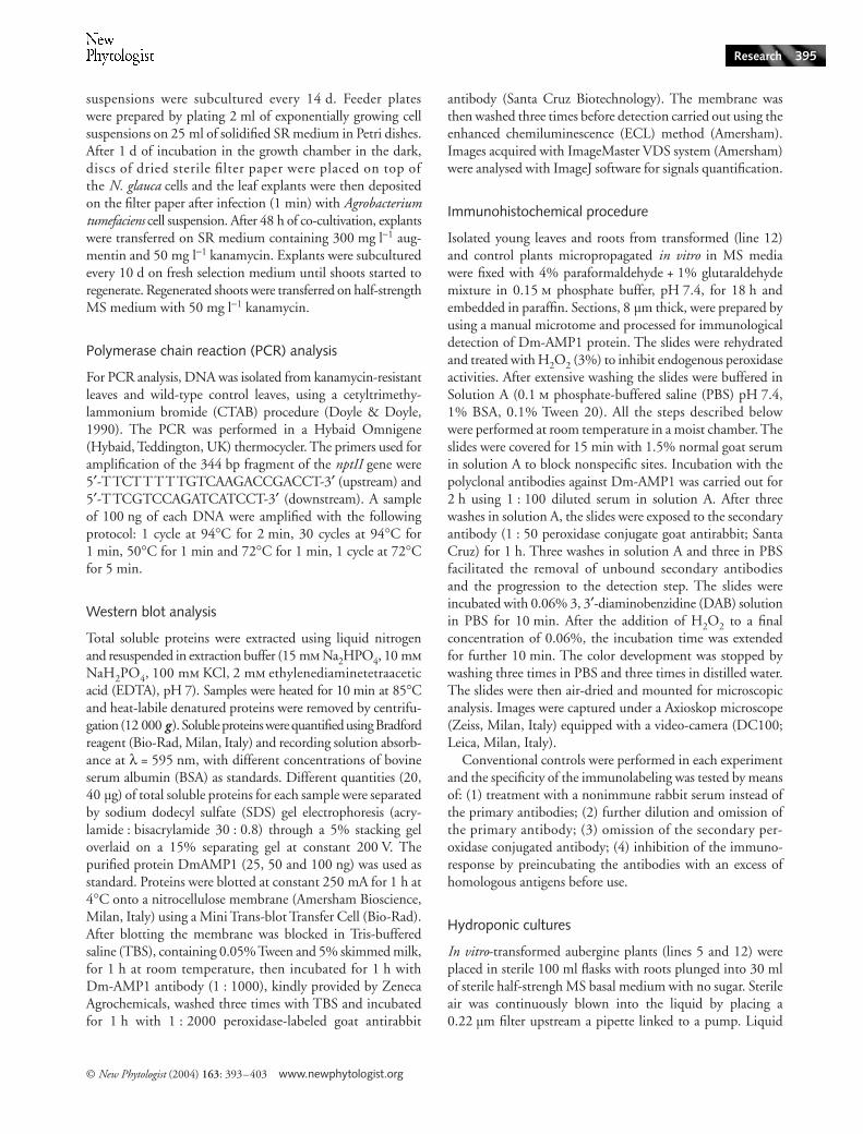
establishment of symbiosis). Glomus mosseae hyphal branchingenhancement elicited by the presence of host-derived signalswere not affected by released defensins. In fact, both controland transformed aubergine (lines 1, 5, 8 and 12) roots,growing underneath the membranes, elicited differential hyphalmorphogenesis in G. mosseae mycelium (Fig. 6a,b), showingthat the antifungal protein Dm-AMP1 released in rootexudates did not interfere with the host recognition system ofAM fungi. The establishment of mycorrhizal symbiosis intransformed plants did not differ from controls. Neither theinfected root length percentages, ranging between 30% and60% in different plants and lines, nor the number of entrypoints, between 2.5 and 3.5 per root cm, showed statisticallysignificant differences. Characteristic structures of arbuscularmycorrhizas, namely appressoria, arbuscules and vesicles, weredetected in root systems of both transformed and controlplants (Fig. 6c,d). Transformed plants inoculated with G. mosseaeshowed larger growth than uninoculated controls, a generalsign of mycorrhizal functioning (Fig. 7).
Discussion
In this work, we obtained transformed aubergines expressingDm-AMP1 defensin from D. merckii in all plant tissues
and showing resistance against the phytopathogenic fungusB. cinerea. Transformants were also able to release the antimicro-bial Dm-AMP1 protein in root exudates, which reduced thegrowth of the phytopathogenic fungus V. albo-atrum, butdid not interfere with recognition events and symbiosisestablishment by the AM fungus G. mosseae, considered as anontarget organism.
The important role played by antimicrobial compoundsin plant defense strategies has been shown by many authors(Broekaert et al., 1995; Terras et al., 1995; de Bolle et al., 1996).The use of genes expressing antimicrobial compounds toobtain transformed phenotypes more resistant to pathogenswas successful in the case of tobacco, geranium and potatoplants (Terras et al., 1995; de Bolle et al., 1996; Bi et al., 1999;Gao et al., 2000). The expression level of Dm-AMP1 inaubergine lines ranged between 0.16% and 0.66% of totalsoluble protein content and it was higher than those reportedfor tobacco plants transformed with different antimicrobialprotein gene constructs cloned from M. jalapa and A. caudatus(de Bolle et al., 1996), which showed expression levels of0.1% of total soluble protein content. The difference betweenthe expression values of Dm-AMP1 in aubergine comparedwith those reported in literature might be due to the transla-tional enhancer Ω derived from tobacco mosaic virus (Gallie
Fig. 6 Light photomicrographs showing hyphal morphogenesis and root infection structures of the arbuscular mycorrhizal fungus Glomus mosseae in the presence of aubergine (Solanum melongena) plants. (a,b) Differential morphogenesis developed on membranes overlying nontransformed (a) and transformed (b) roots. Bar, 50 µm in (a) and 90 µm in (b). (c,d) Development of appressoria (c) and symbiotic structures (vesicles, arbuscules) (d) in transformed roots. Bar, 25 µm in (c) and 20 µm in (d).

© New Phytologist (2004) 163: 393–403 www.newphytologist.org
Research 401
& Walbot, 1992) used in our construct. The expression levelof genes for antimicrobial proteins is generally correlated withplant resistance in transformed plants, although resistancealso depends on the kind of antimicrobial activity and on thesubcellular localization of peptides. Defensins expressed inseeds, such as Rs-AFPs from R. sativus, were shown to occurin the outer cell wall layers of the seeds and to be released inthe growth medium (Terras et al., 1995). By means of immu-nolocalization, the authors showed that Rs-AFPs were expressedin the middle lamellae of cell walls throughout the differentseed tissues (endosperm, cotyledons and hypocotyl) where thefirst contacts with the invading fungi occurred. In this workwe observed that Dm-AMP1, whose amino acid sequenceis highly homologous to Rs-AFPs (Osborn et al., 1995), isdetectable in leaves and root cells of transformed aubergines.Although polyclonal antibodies show a reduced specificity,the immunolocalization experiment provides further supportto evidence of Dm-AMP1 expression in the different planttissues. It is interesting to note that in root cells the Dm-AMP1 protein is localized in cytoplasmic globular structuresresembling either protein bodies, which might have an accu-mulation function, or vesicles, which might be involved intransferring the antimicrobial protein outside root cells.
Our findings provide the first evidence of the release ofDm-AMP1 in transformed aubergine root exudates. Thisis an important trait of transformed plants to be taken intoaccount since exudates containing antimicrobial proteinsmight affect nontarget microbial soil communities (Siciliano& Germida, 1999; Griffiths et al., 2000). Recent researches,carried out on maize plants expressing the insecticidal toxinCry1Ab from Bacillus thuringiensis, showed that Bt plantsreleased the active toxin in root exudates (Saxena et al., 1999,2002; Saxena & Stotzky, 2000) and that these did not affectsaprophytic soil fungi (Saxena & Stotzky, 2001). However,the effect of root exudates from transgenic plants releasinginsecticidal toxins or antimicrobial compounds on the
complete range of soil microorganisms is still controversialand should be studied in detail.
Results obtained from V. albo-atrum bioassay showed thatthe presence of Dm-AMP1 transformed roots in the culturemedium reduced the growth of nearby colonies of the patho-genic fungus by 49–71% with respect to controls, suggestingthat released defensin, consistent with previous work (Osbornet al., 1995), was still active.
Plants expressing Dm-AMP1 protein showed reducednecrotic areas development, with respect to nontransformedplants, when leaves were inoculated with the phytopathogenicfungus B. cinerea. Infection assays performed with leaves or leafdiscs represent a useful tool to evaluate plant resistance againstfoliar pathogens through the assessment of necrotic lesionsdevelopment or localized sporulation density (Laemmlen &Sink, 1978; Bi et al., 1999).
In this work, transformed line 12, showing lower Dm-AMP1expression with respect to lines 1, 5 and 8, also displayed thelargest B. cinerea-induced lesions on leaf tissues whereas no,or highly reduced, necrotic lesions occurred on leaves of theother transformants. These results suggest a correlation betweenDm-AMP1 protein expression in leaves and plant resistance tofoliar pathogens.
From these results, which might be the effects of transformedplants and their exudates on beneficial symbiotic fungi?Although Dm-AMP1 was highly expressed in all transformedplant tissues, roots included, and was released in exudates,it did not interfere with the host recognition system of AMfungi and with the following steps leading to the establish-ment of mycorrhizal symbiosis by G. mosseae. Moreover, thetypical growth enhancement due to the presence of thesymbiosis was observed in all the transformed lines tested,suggesting that the established symbiosis was functional.
Previous studies showed that plants of Nicotiana sylvestrisand Nicotiana tabacum constitutively expressing tobaccochitinases and different kinds of pathogenesis-related proteins
Fig. 7 Transformed aubergine (Solanum melongena) plants (line 12) inoculated with the arbuscular mycorrhizal fungus Glomus mosseae (left), showing growth enhancements compared with controls (right).

www.newphytologist.org © New Phytologist (2004) 163: 393–403
Research402
(PRs), respectively, were colonized by G. mosseae to the sameextent as control plants (Vierheilig et al., 1993, 1995). Thereasons why mycorrhizal fungi were not affected by plantdefense compounds remain to be investigated. Some studiesreported that AM fungal colonization induced transientenhancement or accumulation of antimicrobial compoundssuch as chitinases, glucanases, phytoalexins and phenolics(Volpin et al., 1994; Dumas-Gaudot et al., 1996; Gianinazzi-Pearson et al., 1996; Morandi, 1996). Accordingly, the hypothesisthat AM fungi have developed a general tolerance to plantdefense compounds is also supported by our results on plantdefensins. Since recent works reported that Dm-AMP1 inducedmembrane destabilization after preferential binding to membranepatches containing sphingolipids (Thevissen et al., 2000b,2003a), we could hypothesize that the AM fungus G. mosseaedoes not have suitable binding sites for Dm-AMP1.
The occurrence of Dm-AMP1 defensin in root exudates oftransformed aubergines allowed us to show that the mycor-rhizal symbiont G. mosseae was still able to recognize host-derivedsignals inducing differential morphogenesis and infectionstructures formation (Giovannetti et al., 1993, 1994, 1996;Giovannetti, 2000). The experimental model system used inthis work may represent a reliable biotest for assessing theimpact of transformed plants and their root exudates onimportant nontarget soil microorganisms such as AM fungi,which are fundamental to soil fertility and plant nutrition(Smith & Read, 1997).
Acknowledgements
The authors thank Dr Chiara Geri for stimulating discus-sion. They also thank Dr Andy Greenland and Dr SheilaAttenborough for kindly providing biological material usedthroughout this study. This work was partly supported byISPESL, Italy (Istituto Superiore per la Prevenzione E laSicurezza del Lavoro, contract PF/DIPIA/UO4/99) and ECO-SAFE (Ecological and Environmental Biosafety Assessment ofNovel Plant and Microbial Biotechnology Products).
References
Bi YM, Cammue BPA, Goodwin PH, KrishnaRaj S, Saxena PK. 1999. Resistance to Botrytis cinerea in scented geranium transformed with a gene encoding the antimicrobial protein Ace-AMP1. Plant Cell Reports 18: 835–840.
Billings S, Jelenkovic G, Chin C, Eberhardt J. 1997. The effect of growth regulators and antibiotics on eggplant transformation. Journal of the American Society for Horticultural Science 122: 158–162.
Bohlmann H, Apel K. 1991. Thionins. Annual Review of Plant Physiology and Plant Molecular Biology 42: 227–240.
Broekaert W, Terras FRG, Cammue BPA, Osborn RW. 1995. Plant defensins: novel antimicrobial peptides as components of the host defence system. Plant Physiology 108: 1353–1358.
Broekaert W, Cammue BPA, de Bolle MFC, Thevissen K, de Samblanx GW, Osborn RW. 1997. Antimicrobial peptides from plants. Critical Reviews in Plant Science 16: 297–323.
de Bolle MFC, Osborn RW, Goderi IJ, Noe L, Acland D, Hart CA, Torrekens S, van Leuven F, Broekaert W. 1996. Antimicrobial peptides from Mirabilis jalapa and Amaranthus caudatus: expression, processing, localization and biological activity in transgenic tobacco. Plant Molecular Biology 31: 993–1008.
Doyle J, Doyle J. 1990. Isolation of plant DNA from fresh tissue. Focus 12: 13–15.
Dumas-Gaudot E, Slezack S, Dessi B, Pozo MJ, Gianinazzi-Pearson V, Gianinazzi S. 1996. Plant hydrolytic enzymes (chitinases and beta 1,3 glucanases) in root reactions to pathogenic and symbiotic microorganisms. Plant and Soil 185: 211–221.
Gallie DR, Walbot V. 1992. Identification of the motifs within the tobacco mosaic virus 5′-leader responsible for enhancing translation. Nucleic Acids Research 20: 4631–4638.
Gamborg OL, Miller LA, Ojima K. 1968. Nutritional requirement suspension cultures of soybean root cells. Experimental Cell Research 50: 151–158.
Gao AG, Haikimi SM, Mittanck CA, Wu Y, Woerner BM, Stark DM, Shah DM, Liang J, Rommens CMT. 2000. Fungal pathogen protection in potato by expression of a plant defensin peptide. Nature Biotechnology 18: 1307–1310.
Garcìa-Olmedo F, Molina A, Segura A, Moreno M. 1995. The defensive role of non-specific lipid-transfer proteins in plants. Trends in Microbiology 3: 72–74.
Gerdemann JW, Nicolson TH. 1963. Spores of mycorrhizal Endogone species extracted from soil by wet sieving and decanting. Transactions of the British Mycological Society 46: 235–246.
Gianinazzi-Pearson V, Dumas-Gaudot E, Gallotte A, Tahiri-Alaoui A, Gianinazzi S. 1996. Cellular and molecular defence-related root responses to invasion by arbuscular mycorrhizal fungi. New Phytologist 133: 45–57.
Giovannetti M. 2000. Spore germination and pre-symbiotic mycelial growth. In: Kapulnik Y, Douds Jr DD, eds. Arbuscular mycorrhizas: physiology and function. Dordrecht, The Netherlands: Kluwer Academic Publishers, 47–68.
Giovannetti M, Mosse B. 1980. An evaluation of techniques for measuring vesicular–arbuscular mycorrhizal infection in roots. New Phytologist 84: 489–500.
Giovannetti M, Sbrana C, Avio L, Citernesi AS, Logi C. 1993. Differential hyphal morphogenesis in arbuscular mycorrhizal fungi during pre-infection stages. New Phytologist 125: 587–594.
Giovannetti M, Sbrana C, Logi C. 1994. Early processes involved in host recognition by arbuscular mycorrhizal fungi. New Phytologist 127: 703–709.
Giovannetti M, Sbrana C, Citernesi AS, Avio L. 1996. Analysis of factors involved in fungal recognition responses to host-derived signals by arbuscular mycorrhizal fungi. New Phytologist 133: 65–71.
Griffiths BS, Geoghegan IE, Robertson WM. 2000. Testing genetically engineered potato, producing the lectins GNA and Con A, on non-target soil organisms and processes. Journal of Applied Ecology 37: 159–170.
Laemmlen FF, Sink KC. 1978. Evaluation of petunia cultivars for Botrytis resistance. Plant Disease Report 62: 361–365.
Mauch F, Mauch-Mani B, Boller T. 1988. Antifungal hydrolases in pea tissue. II. Inhibition of fungal growth by combinations of chitinase and beta-1,3-glucanase. Plant Physiology 88: 936–942.
Melchers LS, Apotheker-de Groot M, van der Knaap JA, Ponstein AS, Sela-Buurlage MB, Bol JF, Cornelissen BJC, Linthorst HJM. 1994. A new class of tobacco chitinases homologous to bacterial exo-chitinases displays antifungal activity. Plant Journal 5: 469–480.
Mendez E, Moreno A, Collila F, Pelaez F, Limas GG, Mendez R, Soriano F, Salinas M, de Haro C. 1990. Primary structure and inhibition of protein synthesis in eukaryotic cell-free system of a novel thionin, gamma-hortothionin, from barley endosperm. European Journal of Biochemistry 194: 533–539.
Morandi D. 1996. Occurence of phytoalexins and phenolic compounds in endomycorrhizal interactions, and their potential role in biological control. Plant and Soil 185: 241–251.

© New Phytologist (2004) 163: 393–403 www.newphytologist.org
Research 403
Moreno M, Segura A, Garcìa-Olmedo F. 1994. Pseudothionin, a potato peptide active against potato pathogens. European Journal of Biochemistry 223: 135–139.
Murashige T, Skoog F. 1962. A revised medium for rapid growth and bioassays with tobacco tissue culture. Physiologia Plantarum 15: 473–497.
Osborn RW, de Samblanx GW, Thevissen K, Goderis I, Torrekens S, van Leuven F, Attenborough S, Rees S, Broekaert WF. 1995. Isolation and characterisation of plant defensins from seeds of Asteraceae, Fabaceae, Hippocastanaceae and Saxifragaceae. FEBS Letters 368: 257–262.
Phillips JM, Hayman DJ. 1970. Improved procedures for clearing roots and staining parasitic and vesicular–arbuscular mycorrhizal fungi for rapid assessment of infection. Transactions of the British Mycological Society 55: 158–161.
Rao AG. 1995. Antimicrobial peptides. Molecular Plant–Microbe Interactions 8: 6–13.
Saxena D, Stotzky G. 2000. Insecticidal toxin from Bacillus thuringiensis is released from roots of transgenic Bt corn in vitro and in situ. FEMS Microbiology Ecology 33: 35–39.
Saxena D, Stotzky G. 2001. Bacillus thuringiensis (Bt) toxin released from root exudates and biomass of Bt corn has no apparent effect on earthworms, nematodes, protozoa, bacteria, and fungi in soil. Soil Biology and Biochemistry 33: 1225–1230.
Saxena D, Flores S, Stotzky G. 1999. Insecticidal toxin in root exudates from Bt corn. Nature 402: 480.
Saxena D, Flores S, Stotzky G. 2002. Bt toxin is released in root exudates from 12 transgenic corn hybrids representing three transformation events. Soil Biology and Biochemistry 34: 133–137.
Shewry PA, Lucas JA. 1997. Plant proteins that confer resistance to pests and pathogens. Advances in Botanical Research 26: 135–192.
Siciliano SD, Germida JJ. 1999. Taxonomic diversity of bacteria associated with the roots of field-grown transgenic Brassica napus cv. Excel and B. rapa cv. Parkland. FEMS Microbiology Ecology 29: 263–272.
Smith SE, Read DJ. 1997. Mycorrhizal symbiosis. London, UK: Academic Press.
Terras FRG, Schoofs HME, de Bolle MFC, van Leuven F, Rees SB, Vanderleyden J, Cammue BPA, Broekaert WF. 1992. Analysis of two novel classes of antifungal proteins from radish (Raphanus sativus L.) seeds. Journal of Biological Chemistry 267: 15301–15309.
Terras FRG, Torrekens S, van Leuven F, Osborn RW, Vanderleyden J, Cammue BPA, Broekaert WF. 1993. A new family of basic cysteine-rich plant antifungal proteins from Brassicaceae species. FEBS Letters 316: 233–240.
Terras FRG, Eggermont K, Kovaleva V, Raikhel NV, Osborn RW, Kester A, Rees SB, Torrekens S, van Leuven FV, Vanderleyden J,
Cammue BPA, Broekaert WF. 1995. Small cysteine-rich antifungal proteins from radish: their role in host defence. Plant Cell 7: 573–588.
Thevissen K, Ghazi A, De Samblanx GWB, Rowlee C, Osborn RW, Broekaert WF. 1996. Fungal membrane responses induced by plant defensins and thionins. Journal of Biological Chemistry 271: 15018–15025.
Thevissen K, Osborn RW, Acland D, Broekaert WF. 1997. Specific high-affinity binding sites for an antifungal plant defensin on Neurospora crassa iphae and microsomal membranes. Journal of Biological Chemistry 272: 32176–32187.
Thevissen K, Osborn R, Acland D, Broekaert WF. 2000a. Specific binding site for an antifungal plant defensin from Dahlia (Dahlia merckii ) are required for antifungal activity. Molecular Plant–Microbe Interactions 13: 54–61.
Thevissen K, Cammue BP, Lemaire K, Winderickx J, Dickson RC, Lester RL, Ferket KK, van Even F, Parret AH, Broekaert WF. 2000b. A gene encoding a sphingolipid biosynthesis enzyme determines the sensitivity of Saccharomyces cerevisiae to an antifungal plant defensin from dahlia (Dahlia merkii ). Proceedings of the National Academy of Sciences, USA 97: 9531–9536.
Thevissen K, Francois IEJA, Takemoto JY, Ferket KKA, Meert EMK, Cammue BPA. 2003a. DmAMP1, an antifungal plant defensin from dahlia (Dahlia merckii ), interacts with sphingolipids from Saccharomyces cerevisiae. FEMS Microbiology Letters 226: 169–173.
Thevissen K, Warnecke DC, Francois IEJA, Leipelt M, Heinz E, Ott C, Zahringer U, Thomma BPHJ, Ferket KKA, Cammue BPA. 2004. Defensins from insect and plants interact with fungal glucosylceramides. Journal of Biological Chemistry 279: 3900–3905.
Thomma BPHJ, Cammue BPA, Thevissen K. 2002. Plant defensins. Planta 216: 193–202.
Vierheilig H, Alt M, Neuhaus JM, Boller T, Wiemken A. 1993. Colonization of transgenic Nicotiana silvestris plants, expressing different forms of Nicotiana tabacum chitinase, by the root pathogen Rhizoctonia solani and by the mycorrhizal symbiont Glomus mosseae. Molecular Plant–Microbe Interactions 6: 261–264.
Vierheilig H, Alt M, Lange J, Gut-Rella M, Wiemken A, Boller T. 1995. Colonization of transgenic tobacco constitutively expressing pathogenesis-related proteins by the vesicular–arbuscular mycorrhizal fungus Glomus mosseae. Applied Environmental Microbiology 61: 3031–3034.
Volpin H, Elkind Y, Okon Y, Kapulnik Y. 1994. A vesicular arbuscular mycorrhizal fungus (Glomus intraradix) induces a defence response in alfalfa roots. Plant Physiology 104: 683–689.
Wang YP, Nowak G, Culley D, Hadwiger LA, Fristensky B. 1999. Constitutive expression of pea defense gene DRR206 confers resistance to blackleg (Leptosphaeria maculans) disease in transgenic canola (Brassica napus). Molecular Plant–Microbe Interactions 12: 410–418.