a SPECT computer-aided analysis - BMC Neuroscience
9
Liu et al. BMC Neurosci (2016) 17:59 DOI 10.1186/s12868-016-0294-3 RESEARCH ARTICLE Cerebral blood perfusion changes in amputees with myoelectric hands after rehabilitation: a SPECT computer-aided analysis Qiufang Liu 1 , Xiujuan Zheng 2 , Panli Li 1 , Lian Xu 1 , Longwen He 3 , Zhao Mei 3 , Yinyan Zhu 1 , Gang Huang 1 , Chunlong Zhong 4* and Shaoli Song 1* Abstract Background: Rehabilitation, which is essential for amputees with myoelectric hands, can improve the quality of daily life by remodeling the neuron network. In our study, we aim to develop a cerebral blood perfusion (CBF) single- photon emission computed tomography computer-aided (SPECT-CA) detection scheme to automatically locate the brain’s activated regions after rehabilitation. Results: Five participants without forearms (three male, two female, mean age 51 ± 12.89 years, two missing the right side, and three missing the left side) were included in our study. In the clinical assessment, all of the partici- pants received higher scores after training. The results of the SPM analysis indicated that CBF in the precentral gyrus, postcentral gyrus, frontal lobe, temporal lobe and cerebellum was significantly different among the five participants (P < 0.05). Moreover, SPECT-CA showed that the activated brain areas mainly included the precentral gyrus, postcen- tral gyrus, cerebellum and extensive cerebral cortex. Conclusion: Our study demonstrated that the CBF SPECT-CA method can detect the brain blood perfusion changes induced by rehabilitation with high sensitivity and accuracy. This method has great potential for locating the remod- eled neuron regions of amputees with myoelectric hands after rehabilitation. Keywords: Cerebral blood flow, SPECT/CT, Computed-aided method, Myoelectric hand, Rehabilitation © 2016 The Author(s). This article is distributed under the terms of the Creative Commons Attribution 4.0 International License (http://creativecommons.org/licenses/by/4.0/), which permits unrestricted use, distribution, and reproduction in any medium, provided you give appropriate credit to the original author(s) and the source, provide a link to the Creative Commons license, and indicate if changes were made. The Creative Commons Public Domain Dedication waiver (http://creativecommons.org/ publicdomain/zero/1.0/) applies to the data made available in this article, unless otherwise stated. Background It is projected that the number of people living with the loss of a limb in the United States will increase to 3.6 million by the year 2050 [1]. As a result, amputees face a number of problems in daily life. Myoelectric hands can partially compensate for functions of the hand according to the amputee’s will. Amputees can control myoelectric hands by voluntarily contracting their residual muscles, which can generate myoelectric signals and are received by two cutaneous electrodes attached to the residual muscles [2]. Due to the changes in the electrical signal of the muscles, the amputees can drive a set of motors in the myoelectric hand within the prosthetic device, allowing them to open and close or grasp and pinch. In addition, the enhanced utility the myoelectric hand of amputees was found to have a beneficial effect on both phantom limb pain and cortical reorganization [3, 4]. erefore, this device is widely applied in amputees. Rehabilitation is required before amputees can fully use their device. During rehabilitation, amputees can learn how to control their residual muscles to reach thresh- old levels of myoelectric signal strength that can induce the motion of the myoelectric hand. At the same time, rehabilitation may induce neuron network remodeling. Open Access BMC Neuroscience *Correspondence: [email protected]; [email protected] 1 Department of Nuclear Medicine, Ren Ji Hospital, School of Medicine, Shanghai Jiao Tong University, 160 Pujian Road, Shanghai 200127, China 4 Department of Neurosurgery, Ren Ji Hospital, School of Medicine, Shanghai Jiao Tong University, 160 Pujian Road, Shanghai 200127, China Full list of author information is available at the end of the article
-
Upload
khangminh22 -
Category
Documents
-
view
5 -
download
0
Transcript of a SPECT computer-aided analysis - BMC Neuroscience
Liu et al. BMC Neurosci (2016) 17:59 DOI 10.1186/s12868-016-0294-3
RESEARCH ARTICLE
Cerebral blood perfusion changes in amputees with myoelectric hands after rehabilitation: a SPECT computer-aided analysisQiufang Liu1, Xiujuan Zheng2, Panli Li1, Lian Xu1, Longwen He3, Zhao Mei3, Yinyan Zhu1, Gang Huang1, Chunlong Zhong4* and Shaoli Song1*
Abstract
Background: Rehabilitation, which is essential for amputees with myoelectric hands, can improve the quality of daily life by remodeling the neuron network. In our study, we aim to develop a cerebral blood perfusion (CBF) single-photon emission computed tomography computer-aided (SPECT-CA) detection scheme to automatically locate the brain’s activated regions after rehabilitation.
Results: Five participants without forearms (three male, two female, mean age 51 ± 12.89 years, two missing the right side, and three missing the left side) were included in our study. In the clinical assessment, all of the partici-pants received higher scores after training. The results of the SPM analysis indicated that CBF in the precentral gyrus, postcentral gyrus, frontal lobe, temporal lobe and cerebellum was significantly different among the five participants (P < 0.05). Moreover, SPECT-CA showed that the activated brain areas mainly included the precentral gyrus, postcen-tral gyrus, cerebellum and extensive cerebral cortex.
Conclusion: Our study demonstrated that the CBF SPECT-CA method can detect the brain blood perfusion changes induced by rehabilitation with high sensitivity and accuracy. This method has great potential for locating the remod-eled neuron regions of amputees with myoelectric hands after rehabilitation.
Keywords: Cerebral blood flow, SPECT/CT, Computed-aided method, Myoelectric hand, Rehabilitation
© 2016 The Author(s). This article is distributed under the terms of the Creative Commons Attribution 4.0 International License (http://creativecommons.org/licenses/by/4.0/), which permits unrestricted use, distribution, and reproduction in any medium, provided you give appropriate credit to the original author(s) and the source, provide a link to the Creative Commons license, and indicate if changes were made. The Creative Commons Public Domain Dedication waiver (http://creativecommons.org/publicdomain/zero/1.0/) applies to the data made available in this article, unless otherwise stated.
BackgroundIt is projected that the number of people living with the loss of a limb in the United States will increase to 3.6 million by the year 2050 [1]. As a result, amputees face a number of problems in daily life. Myoelectric hands can partially compensate for functions of the hand according to the amputee’s will. Amputees can control myoelectric hands by voluntarily contracting their residual muscles, which can generate myoelectric signals and are received
by two cutaneous electrodes attached to the residual muscles [2]. Due to the changes in the electrical signal of the muscles, the amputees can drive a set of motors in the myoelectric hand within the prosthetic device, allowing them to open and close or grasp and pinch. In addition, the enhanced utility the myoelectric hand of amputees was found to have a beneficial effect on both phantom limb pain and cortical reorganization [3, 4]. Therefore, this device is widely applied in amputees.
Rehabilitation is required before amputees can fully use their device. During rehabilitation, amputees can learn how to control their residual muscles to reach thresh-old levels of myoelectric signal strength that can induce the motion of the myoelectric hand. At the same time, rehabilitation may induce neuron network remodeling.
Open Access
BMC Neuroscience
*Correspondence: [email protected]; [email protected] 1 Department of Nuclear Medicine, Ren Ji Hospital, School of Medicine, Shanghai Jiao Tong University, 160 Pujian Road, Shanghai 200127, China 4 Department of Neurosurgery, Ren Ji Hospital, School of Medicine, Shanghai Jiao Tong University, 160 Pujian Road, Shanghai 200127, ChinaFull list of author information is available at the end of the article
Page 2 of 9Liu et al. BMC Neurosci (2016) 17:59
Previously, it has been confirmed that the microstruc-ture of the brain is not fixed in adulthood but rather is susceptible to experience-dependent structural plasticity throughout life [5]. Furthermore, studies have indicated that training can induced gray matter changes [6, 7]. The association between rehabilitation, brain activation and performance in controlling of the myoelectric hand is recognized important but has seldom been explored.
Brain imaging, such as functional magnetic resonance (fMRI) and CBF SPECT/CT, can provide useful brain functional information. Additionally, the computer-aided method is used for the quantitative analysis and detect-ing target regions with higher sensitivity and accuracy [8, 9]. Several studies have focused on the changes dur-ing manipulating the prosthesis and described activa-tion regions in fMRI examinations [10, 11]. CBF SPECT/CT imaging is used to measure regional cerebral blood flow changes elicited by stimulation or activation of behavioral functions, which may reflect changes in the underlying neuronal network. However, there has been little research using the SPECT-CA method on the brain perfusion changes of amputees after rehabilitation with myoelectric hands.
In this study, we conducted CBF SPECT/CT imaging before and after rehabilitation. Subtracted images from the two time points for each participant were analyzed by our computer program to automatically detect and quantify the activated regions. The purpose of this study was to develop a CBF SPECT-CA detection scheme to automatically locate the activated brain regions that were associated with rehabilitation and to study the associa-tion between rehabilitation, brain activation and the abil-ity to control the myoelectric hand.
MethodsParticipantsFive participants without forearms (three male, two female, mean age 51 ± 12.89 years, two missing the right side and three missing the left side) were studied. Subjects were eligible for inclusion if they were without motor problems or neurologic or psychiatric history, had normal or corrected-to-normal sight and had no earlier experience with a prosthetic or simulator. The study was approved by the Ethics Committee of Ren Ji Hospital, and informed consent was provided by all subjects before participation.
RehabilitationRehabilitation was provided for 8 weeks and included basic myoelectric signal training, feedback training and activities of daily living (ADL) training [12, 13].
Basic myoelectric signal training was used to teach the participants how to activate and isolate their individual
signals. It consisted of three parts and lasted for 2 weeks: imaginary training, muscle-strength exercise and electro-myography (EMG) strength training. In part 1, partici-pants were asked to complete a variety of actions, such as opening and closing the hand with an imaginary hand. Each exercise was performed in three groups, 80 times per group, and limited to 3 min. In part 2, participants used the self-made up-limber muscle training device, which weighed 4 kilograms and was hung onto the resid-ual stump, to perform elbow flexion and extension as well as shoulder adduction and abduction. The training was given in three groups, 30 times per group at 2-min intervals. In part 3, the EMG signals were measured by two EMG electrodes that were placed separately on the extensor carpi ulnaris and flexor carpi ulnaris and linked to an MT-2 EMG trainer (Kesheng prostheses corpora-tion, Shanghai, China). The participants attempted to enhance the EMG signal by controlling muscle strength, reach a signal strength reach 40 μV or above and main-tain this signal for 2 s. In this part, we adjusted the posi-tion of the electrodes and gradually found the spot where the signal was the maximum. This process was completed in six groups total, with each EMG electrode correspond-ing to three groups, 30 times per group.
In the process of feedback training, we used computer software to provide visual feedback to the participants where the movement of a three-dimensional (3D) virtual prosthetic hand was controlled by the participants’ mus-cles. EMG electrodes were placed on the best spot, and the signal was generated by the participants controlling their muscles and was passed to the software matched to the EMG trainer in the computer. Participants were instructed to open and close the hand fully and continu-ously. The training was completed in four groups per day for 1 h per group. After opening and closing the hand 20 times, participants would take a short break to avoid muscles fatigue. The feedback training lasted for 2 weeks.
Daily life training helped the participants learn how to perform activities of daily living with actual myoelectric prostheses. ADL training mainly included (1) grasping and moving a water glass, (2) taking a weight, (3) opening and closing a door and (4) carrying books and newspa-pers. The training was performed in three groups per day, 20 times per group. The ADL training lasted for 4 weeks.
Image acquisitionCBF SPECT/CT imaging was performed before and after 8-weeks of rehabilitation using 99mTc-ECD (Shanghai GMS Pharmaceutical Corporation, Shanghai, China). The radiopharmaceutical was injected intravenously at a dose of 1110 MBq in a quiet environment. Patients were in a supine position and were instructed to remain calm with their eyes closed. SPECT acquisition was performed

Page 3 of 9Liu et al. BMC Neurosci (2016) 17:59
for 20–30 min after injection, using a rotating, double-head, large-field-of-view gamma camera (PHILIPS Prec-edence, Philip Medical Systems, Netherlands), equipped with low-energy, high-resolution collimators. The data were collected in a 128 × 128 matrix through a 360° rota-tion at 6° intervals. The CT scan was performed using a multi-slice CT Scanner (PHILIPS Precedence, Philip Medical Systems, Netherlands, 140 kv, 35 mAs, 3 mm per scan). Reconstruction of transaxial slices was performed by filtered back projection (Metz filter power, 3.00; full width at half maximum, 10 mm) with subsequent attenu-ation correction using the Chang method (attenuation coefficient of 0.12).
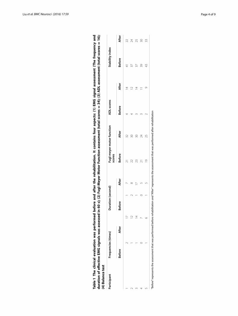
EvaluationEvaluation was performed before and after the 8 weeks of rehabilitation. In the evaluation process, participants with myoelectric hands underwent the myoelectric sig-nal assessment, brief Fugl-Meyer Motor Function assess-ment, ADL assessment and balance test. Brief Fugl-Meyer Motor Function and ADL assessments were performed according to the supplemental materials (Additional file 1). In the myoelectric signal assessment, we recorded the threshold signal intensity of each participant, which is the minimum signal that can induce the movement of myoelectric prosthetic hands. We defined the EMG signal that exceeded the threshold signal as an effective EMG signal. The frequencies and duration of effective EMG sig-nals were used as the index when assessing the myoelec-tric signal in those participants. The stability index (ST) was assessed in the Tetrax balance test system (Tetrax, Sunlight, Israel) to evaluate the balance function. The participants stood on the center of the sensor with their hands naturally hanging down or flat, with their eyes open or closed and while standing on one foot or two feet. By measuring and analyzing the mechanical signal changes of vertical pressure on the feet, the Tetrax balance test system yielded the ST, which can reflect the overall stabil-ity of the participant. With the ST, a higher value repre-sents a worse balance function.
SPECT‑CA methodTo detect the changes in all five participants after train-ing, the SPM toolbox (SPM8, Wellcome Trust Centre for Neuroimaging, University College London, London, UK) and PickAtlas software (WFU Pickatlas, version 3.0) were used for analyzing the SPECT images [14, 15]. For each subject, the pre- and post-SPECT images were spatially normalized into the standard stereotactic Montreal Neu-rological Institute (MNI) space using a 99mTc-ECD tem-plate and smoothed using a 12-mm isotropic Gaussian filter. Moreover, a general whole-brain mask was created based on an anatomical labeling atlas and adopted to all
images for further statistical comparisons. Paired t-tests were performed to compare the pre-training and post-training group. A p value of less than 0.05 was considered statistically significant.
To validate the results of SPM analysis further, we also explored the changes in each participant using the SPECT-CA program. Pre- and post-CBF SPECT/CT imaging were performed to obtain baseline and follow-up SPECT images before and after rehabilitation for each participant. In our SPECT-CA program, the follow-up image was first aligned with the baseline image using a rigid registration algorithm for further comparisons. Then, the values were normalized for these two aligned images using the Z-map approach introduced by Boussio [16]. The change rate of the CBF was calculated voxel-by-voxel to generate a parametric image, noted as a change-rate map. Based on the change-rate map, the changed regions were automatically obtained using the K-means clustering method. Considering the SPECT image resolu-tion and partial volume effects, the regions with higher average change rates (>25 %) and larger volumes (>200 voxels) were selected as activated regions according to our patent submitted to the state intellectual prop-erty office of the P.R.C [17]. According to the activated regions, the change rates of the regions are listed and reported with respect to the absolute pixel number, mean change rate and location for each participant.
ResultsClinical assessmentsFrom the EMG signal assessment results, we found that after rehabilitation all of the participants could con-duct more effective EMG signals for a longer duration (Table 1). Moreover, all five participants received higher scores in the Fugl-Meyer Motor Function and ADL assessments after rehabilitation, consistent with the EMG signal assessment results (Table 1). The stability index declined, indicating that the participants’ balance function improved after rehabilitation. In addition, three of the participants (participants 1, 2 and 3) received a higher score on the assessment, which indicates that they could manipulate the myoelectric hand more accurately and consistently compared with the others.
SPECT‑CA analysisThe results of SPM analysis and paired t-tests indicated that it existed activated regions of all the 5 participants after training. And they were 5 regions had signifi-cant difference, which were precentral gyrus, postcen-tral gyrus, frontal lobe, temporal lobe and cerebellum (P < 0.05).
In addition, using our SPECT-CA program, we found that each participant displayed brain activation after
Page 4 of 9Liu et al. BMC Neurosci (2016) 17:59
Tabl
e 1
The
clin
ical
eva
luat
ion
was
per
form
ed b
efor
e an
d af
ter
the
reha
bilit
atio
n. I
t co
ntai
ns f
our
aspe
cts:
(1)
EM
G s
igna
l as
sess
men
t (T
he f
requ
ency
and
du
rati
on o
f eff
ecti
ve E
MG
sig
nals
was
ass
esse
d in
60
s); (
2) F
ugl-M
eyer
Mot
or F
unct
ion
asse
ssm
ent
(tot
al s
core
s =
34)
; (3)
AD
L as
sess
men
t (t
otal
sco
res =
16)
; (4
) Bal
ance
test
“Bef
ore”
repr
esen
ts th
e as
sess
men
t tha
t was
per
form
ed b
efor
e re
habi
litat
ion
and
“Aft
er” r
epre
sent
s th
e as
sess
men
t tha
t was
per
form
ed a
fter
reha
bilit
atio
n
Part
icip
ant
Freq
uenc
ies
(tim
es)
Dur
atio
n (s
econ
d)Fu
gl‑m
eyer
mot
or fu
nctio
n sc
ores
AD
L sc
ores
Stab
ility
inde
x
Befo
reA
fter
Befo
reA
fter
Befo
reA
fter
Befo
reA
fter
Befo
reA
fter
12
171
721
324
1441
22
21
122
822
304
1237
24
31
141
1723
303
1437
25
40
70
521
243
1139
30
51
61
519
252
943
33
Page 5 of 9Liu et al. BMC Neurosci (2016) 17:59
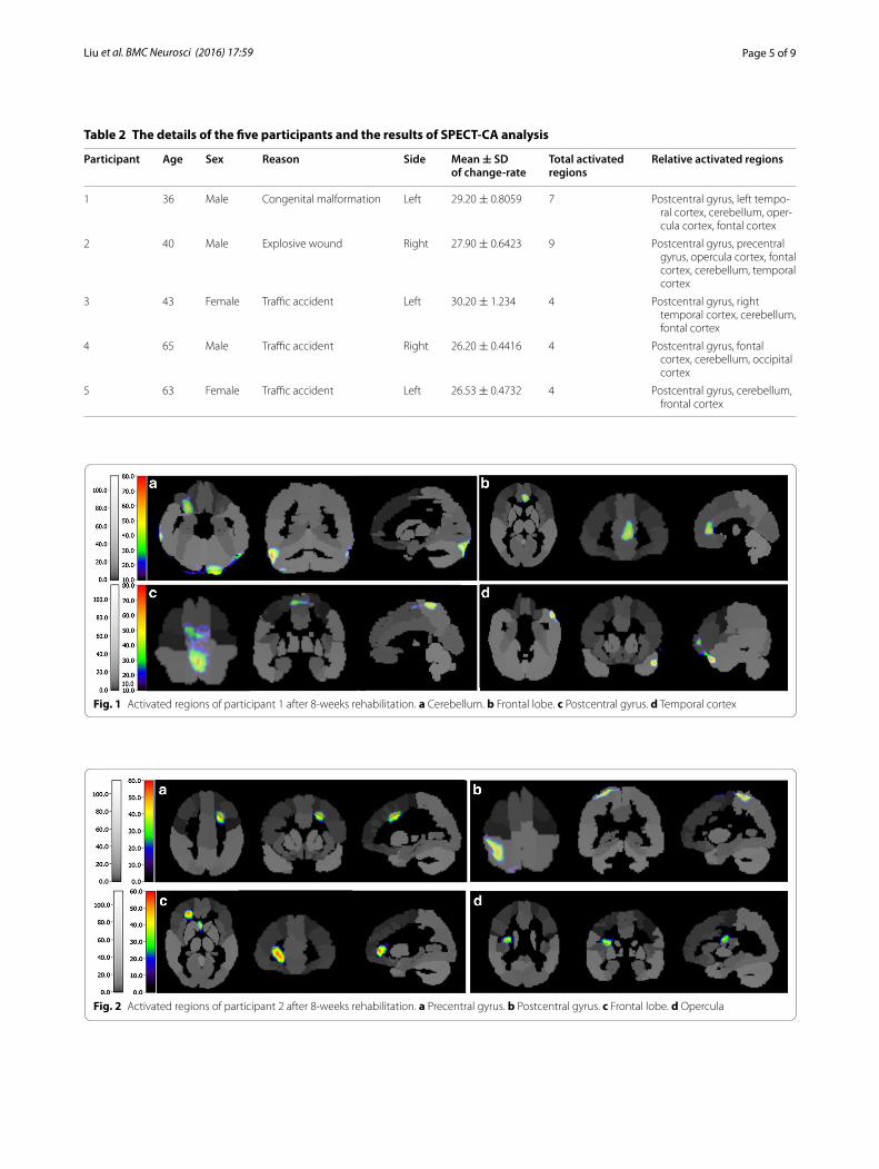
Table 2 The details of the five participants and the results of SPECT-CA analysis
Participant Age Sex Reason Side Mean ± SD of change‑rate
Total activated regions
Relative activated regions
1 36 Male Congenital malformation Left 29.20 ± 0.8059 7 Postcentral gyrus, left tempo-ral cortex, cerebellum, oper-cula cortex, fontal cortex
2 40 Male Explosive wound Right 27.90 ± 0.6423 9 Postcentral gyrus, precentral gyrus, opercula cortex, fontal cortex, cerebellum, temporal cortex
3 43 Female Traffic accident Left 30.20 ± 1.234 4 Postcentral gyrus, right temporal cortex, cerebellum, fontal cortex
4 65 Male Traffic accident Right 26.20 ± 0.4416 4 Postcentral gyrus, fontal cortex, cerebellum, occipital cortex
5 63 Female Traffic accident Left 26.53 ± 0.4732 4 Postcentral gyrus, cerebellum, frontal cortex
Fig. 1 Activated regions of participant 1 after 8-weeks rehabilitation. a Cerebellum. b Frontal lobe. c Postcentral gyrus. d Temporal cortex
Fig. 2 Activated regions of participant 2 after 8-weeks rehabilitation. a Precentral gyrus. b Postcentral gyrus. c Frontal lobe. d Opercula
Page 6 of 9Liu et al. BMC Neurosci (2016) 17:59
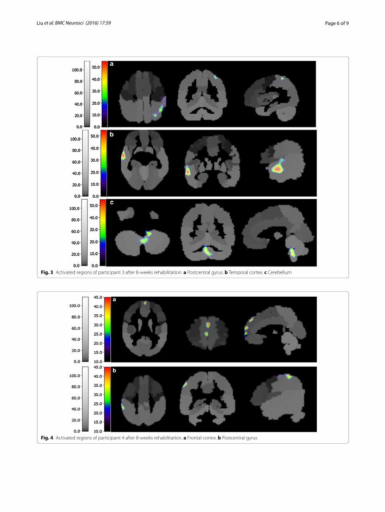
Fig. 3 Activated regions of participant 3 after 8-weeks rehabilitation. a Postcentral gyrus. b Temporal cortex. c Cerebellum
Fig. 4 Activated regions of participant 4 after 8-weeks rehabilitation. a Frontal cortex. b Postcentral gyrus
Page 7 of 9Liu et al. BMC Neurosci (2016) 17:59
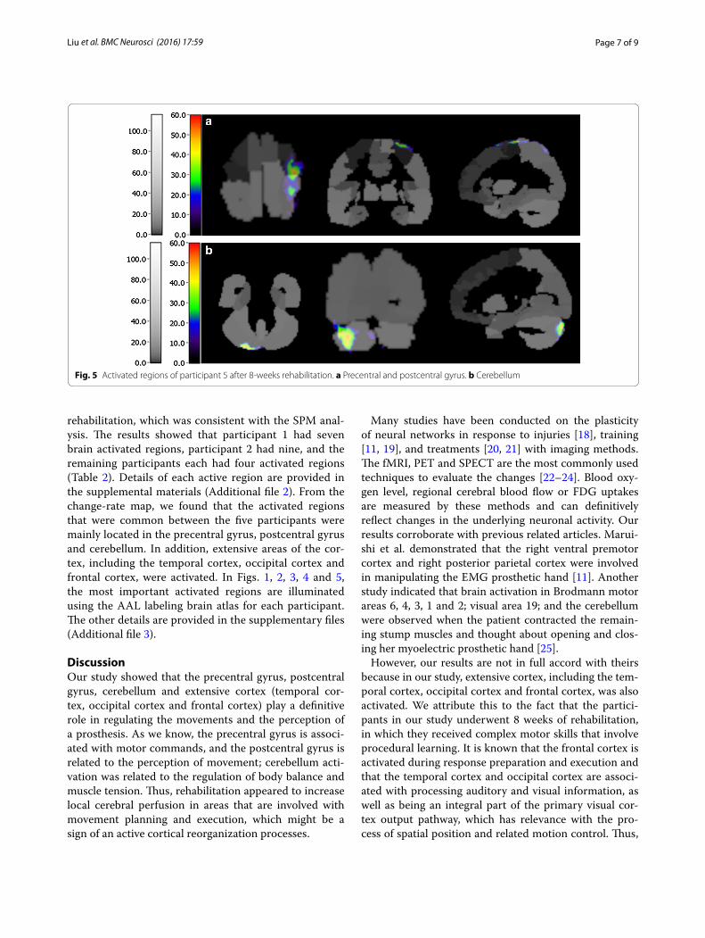
rehabilitation, which was consistent with the SPM anal-ysis. The results showed that participant 1 had seven brain activated regions, participant 2 had nine, and the remaining participants each had four activated regions (Table 2). Details of each active region are provided in the supplemental materials (Additional file 2). From the change-rate map, we found that the activated regions that were common between the five participants were mainly located in the precentral gyrus, postcentral gyrus and cerebellum. In addition, extensive areas of the cor-tex, including the temporal cortex, occipital cortex and frontal cortex, were activated. In Figs. 1, 2, 3, 4 and 5, the most important activated regions are illuminated using the AAL labeling brain atlas for each participant. The other details are provided in the supplementary files (Additional file 3).
DiscussionOur study showed that the precentral gyrus, postcentral gyrus, cerebellum and extensive cortex (temporal cor-tex, occipital cortex and frontal cortex) play a definitive role in regulating the movements and the perception of a prosthesis. As we know, the precentral gyrus is associ-ated with motor commands, and the postcentral gyrus is related to the perception of movement; cerebellum acti-vation was related to the regulation of body balance and muscle tension. Thus, rehabilitation appeared to increase local cerebral perfusion in areas that are involved with movement planning and execution, which might be a sign of an active cortical reorganization processes.
Many studies have been conducted on the plasticity of neural networks in response to injuries [18], training [11, 19], and treatments [20, 21] with imaging methods. The fMRI, PET and SPECT are the most commonly used techniques to evaluate the changes [22–24]. Blood oxy-gen level, regional cerebral blood flow or FDG uptakes are measured by these methods and can definitively reflect changes in the underlying neuronal activity. Our results corroborate with previous related articles. Marui-shi et al. demonstrated that the right ventral premotor cortex and right posterior parietal cortex were involved in manipulating the EMG prosthetic hand [11]. Another study indicated that brain activation in Brodmann motor areas 6, 4, 3, 1 and 2; visual area 19; and the cerebellum were observed when the patient contracted the remain-ing stump muscles and thought about opening and clos-ing her myoelectric prosthetic hand [25].
However, our results are not in full accord with theirs because in our study, extensive cortex, including the tem-poral cortex, occipital cortex and frontal cortex, was also activated. We attribute this to the fact that the partici-pants in our study underwent 8 weeks of rehabilitation, in which they received complex motor skills that involve procedural learning. It is known that the frontal cortex is activated during response preparation and execution and that the temporal cortex and occipital cortex are associ-ated with processing auditory and visual information, as well as being an integral part of the primary visual cor-tex output pathway, which has relevance with the pro-cess of spatial position and related motion control. Thus,
Fig. 5 Activated regions of participant 5 after 8-weeks rehabilitation. a Precentral and postcentral gyrus. b Cerebellum
Page 8 of 9Liu et al. BMC Neurosci (2016) 17:59
the extensive cortex activated in our study is part of the visual-to-motor networks, which was conducted in our rehabilitation. Similar studies have also demonstrated increased gray matter density in the visual motion area, frontal and parietal cortex after as little as 7 days of short-term training [26, 27].
From the results, we found that participants 1, 2 and 3, who had more activated regions or higher change rates, had a better performance in the clinical assess-ments. This is because the more activated regions or higher change-rates indicate more neurogenesis, synaptogenesis, vascularization and myelination and axonal sprouting in the white matter [28, 29]. These are the basic components of the neural pathways to control the muscles and complete some movements. Furthermore, the amount and activity of the neurons is also responsible for the specification of movement parameters such as amplitude, continuity and speed of movement [30]. That is, the control of the speed, force and modulation of a myoelectric prosthesis depends on the activity of specific areas of the brain. Therefore, participants with more activated regions or higher change rates may have better control of the myoelec-tric prosthesis.
The CBF SPECT-CA program we used is automatic and can give fast, objective assessments of the whole brain. By analyzing the subtracted images of the baseline and follow-up SPECT imaging with this method, we could not only erase the effect of unequal injected activities of the images but also quantify the activated regions. More importantly, our results verify that it can detect the acti-vated regions of amputees after rehabilitation with high sensitivity and accuracy. It has great potential for predict-ing the performance in controlling prostheses and locat-ing the reconstruction of the neuron network induced by rehabilitation.
Furthermore, we found that there were differences among the five participants after rehabilitation, which may be due to different ages, residual muscles or ampu-tation-specific reasons. Consequently, further research should recruit larger populations to validate the results and analyze the factors that may affect the results of reha-bilitation and brain activation.
ConclusionIn summary, our study demonstrated that the CBF SPECT-CA method can detect the brain changes induced by rehabilitation with high sensitivity and accuracy. This method is promising for locating the remodeling neuron regions of amputees with myoelectric hands after reha-bilitation. In addition, participants with more activated regions or higher change rates may have better control of the myoelectric prosthesis.
AbbreviationsCBF: cerebral blood flow; SPECT-CA: single-photon emission computed tomography computered aided; 99mTc-ECD: 99mTc-ethyl cysteinate dimmer; SPM: statistical parametric mapping; fMRI: functional magnetic resonance; ADL: activities of daily living; EMG: electromyography; 3D: three-dimensional; MNI: Montreal Neurological Institute.
Authors’ contributionsQL, PL, LX and YZ carried out all the experiments; QL and XZ conducted the statistical analysis and wrote the paper. LH and ZM developed the device and revised the manuscript; GH revised the manuscript; CZ and SS designed and directed the study. All authors read and approved the final manuscript.
Author details1 Department of Nuclear Medicine, Ren Ji Hospital, School of Medicine, Shang-hai Jiao Tong University, 160 Pujian Road, Shanghai 200127, China. 2 Depart-ment of Automation, School of Electrical Engineering Information, Sichuan University, Sichuan 610065, China. 3 Shanghai Rehabilitation and Vocational Training Center for the Disabled, Shanghai 200127, China. 4 Department of Neurosurgery, Ren Ji Hospital, School of Medicine, Shanghai Jiao Tong University, 160 Pujian Road, Shanghai 200127, China.
AcknowledgementsWe thank Philippe Briandet of Segami Corporation for the image processing.
Competing interestsThe authors declare that they have no competing interests.
Availability of data and materialAll data are included in the manuscript.
Consent to publishAll of the participants have provided written informed consent for the publica-tion of individual clinical details.
Ethics approval and consent to participateThe study was approved by the Ethics Committee of Ren Ji Hospital and informed consent was given to all subjects before participation.
FundingThis work was supported by the basic research program of project 973 by the Ministry of Science and Technology of China (2011CB013304) and National Natural Science Foundation of China (No. 81471708, to Shaoli Song and No. 81201146, to Xiujuan Zheng).
Received: 9 June 2016 Accepted: 29 July 2016
References 1. Ziegler-Graham K, MacKenzie EJ, Ephraim PL, Travison TG, Brookmeyer R.
Estimating the prevalence of limb loss in the United States: 2005 to 2050. Arch Phys Med Rehabil. 2008;89(3):422–9.
2. Flor H, Elbert T, Knecht S, Wienbruch C, Pantev C, Birbaumer N, Larbig W, Taub E. Phantom-limb pain as a perceptual correlate of cortical reorgani-zation following arm amputation. Nature. 1995;375(6531):482–4.
3. Lotze M, Grodd W, Birbaumer N, Erb M, Huse E, Flor H. Does use of a myoelectric prosthesis prevent cortical reorganization and phantom limb pain? Nat Neurosci. 1999;2(6):501–2.
Additional files
Additional file 1. The contents of Brief Fugl-Meyer Scale and ADL Assessment.
Additional file 2. The change-rate of the 5 participants.
Additional file 3. The activated regions of the five participants.

Page 9 of 9Liu et al. BMC Neurosci (2016) 17:59
• We accept pre-submission inquiries
• Our selector tool helps you to find the most relevant journal
• We provide round the clock customer support
• Convenient online submission
• Thorough peer review
• Inclusion in PubMed and all major indexing services
• Maximum visibility for your research
Submit your manuscript atwww.biomedcentral.com/submit
Submit your next manuscript to BioMed Central and we will help you at every step:
4. Ramachandran VS, Rogers-Ramachandran D, Cobb S. Touching the phan-tom limb. Nature. 1995;377(6549):489–90.
5. Taubert M, Villringer A, Ragert P. Learning-related gray and white matter changes in humans: an update. Neuroscientist. 2012;18(4):320–5.
6. Takeuchi H, Sekiguchi A, Taki Y, Yokoyama S, Yomogida Y, Komuro N, Yamanouchi T, Suzuki S, Kawashima R. Training of working memory impacts structural connectivity. J Neurosci. 2010;30(9):3297–303.
7. Filippi M, Ceccarelli A, Pagani E, Gatti R, Rossi A, Stefanelli L, Falini A, Comi G, Rocca MA. Motor learning in healthy humans is associated to gray matter changes: a tensor-based morphometry study. PLoS One. 2010;5(4):e10198.
8. Nocun A, Wilczynski M, Wronski J, Chrapko B. Usefulness of 99mTc-ECD brain SPECT with voxel-based analysis in evaluation of perfusion changes early after carotid endarterectomy. Med Sci Monit. 2011;17(5):CR297–303.
9. Takahashi N, Lee Y, Tsai DY, Kinoshita T, Ouchi N, Ishii K. Computer-aided detection scheme for identification of hypoattenuation of acute stroke in unenhanced CT. Radiol Phys Technol. 2012;5(1):98–104.
10. Lacourse MG, Orr EL, Cramer SC, Cohen MJ. Brain activation during execution and motor imagery of novel and skilled sequential hand movements. Neuroimage. 2005;27(3):505–19.
11. Maruishi M, Tanaka Y, Muranaka H, Tsuji T, Ozawa Y, Imaizumi S, Miyatani M, Kawahara J. Brain activation during manipulation of the myoelectric prosthetic hand: a functional magnetic resonance imaging study. Neuro-image. 2004;21(4):1604–11.
12. Bouwsema H, van der Sluis CK, Bongers RM. Effect of feedback during virtual training of grip force control with a myoelectric prosthesis. PLoS One. 2014;9(5):e98301.
13. Terlaak B, Bouwsema H, van der Sluis CK, Bongers RM. Virtual training of the myosignal. PLoS One. 2015;10(9):e0137161.
14. Acton PD, Friston KJ. Statistical parametric mapping in functional neuroimaging: beyond PET and fMRI activation studies. Eur J Nucl Med. 1998;25(7):663–7.
15. Maldjian JA, Laurienti PJ, Kraft RA, Burdette JH. An automated method for neuroanatomic and cytoarchitectonic atlas-based interrogation of fMRI data sets. Neuroimage. 2003;19(3):1233–9.
16. Boussion N, Houzard C, Ostrowsky K, Ryvlin P, Mauguiere F, Cinotti L. Automated detection of local normalization areas for ictal-interictal subtraction brain SPECT. J Nucl Med. 2002;43(11):1419–25.
17. A image-based treatment monitoring system for ischemic stroke. Chi-nese patent no. 201610006027.7.
18. Mikell CB, Banks GP, Frey HP, Youngerman BE, Nelp TB, Karas PJ, Chan AK, Voss HU, Connolly ES, Claassen J. Frontal networks associated with com-mand following after hemorrhagic stroke. Stroke. 2015;46(1):49–57.
19. Belleville S, Clement F, Mellah S, Gilbert B, Fontaine F, Gauthier S. Training-related brain plasticity in subjects at risk of developing Alzheimer’s disease. Brain. 2011;134(Pt 6):1623–34.
20. Kimura N, Kumamoto T, Masuda T, Nomura Y, Hanaoka T, Hazama Y, Okazaki T. Evaluation of the effects of thyrotropin releasing hormone (TRH) therapy on regional cerebral blood flow in the cerebellar variant of multiple system atrophy using 3DSRT. J Neuroimaging. 2011;21(2):132–7.
21. Abel S, Weiller C, Huber W, Willmes K, Specht K. Therapy-induced brain reorganization patterns in aphasia. Brain. 2015;138(Pt 4):1097–112.
22. Ding H, Qin W, Liang M, Ming D, Wan B, Li Q, Yu C. Cross-modal activation of auditory regions during visuo-spatial working memory in early deaf-ness. Brain. 2015;138(Pt 9):2750–65.
23. Mayer G, Bitterlich M, Kuwert T, Ritt P, Stefan H. Ictal SPECT in patients with rapid eye movement sleep behaviour disorder. Brain. 2015;138(Pt 5):1263–70.
24. Hamelin L, Lagarde J, Dorothee G, Leroy C, Labit M, Comley RA, de Souza LC, Corne H, Dauphinot L, Bertoux M, et al. Early and protective microglial activation in Alzheimer’s disease: a prospective study using 18F-DPA-714 PET imaging. Brain. 2016;139(Pt 4):1252–64.
25. da Paz AC, Braga LW Jr. Brain activation in a myoelectric prosthetic hand: the role of the brain in the rehabilitation of amputees. J Pediatr Orthop. 2007;27(8):947–51.
26. Driemeyer J, Boyke J, Gaser C, Buchel C, May A. Changes in gray matter induced by learning—revisited. PLoS One. 2008;3(7):e2669.
27. Taubert M, Draganski B, Anwander A, Muller K, Horstmann A, Villringer A, Ragert P. Dynamic properties of human brain structure: learning-related changes in cortical areas and associated fiber connections. J Neurosci. 2010;30(35):11670–7.
28. Zatorre RJ, Fields RD, Johansen-Berg H. Plasticity in gray and white: neuroimaging changes in brain structure during learning. Nat Neurosci. 2012;15(4):528–36.
29. Jiang G, Yin X, Li C, Li L, Zhao L, Evans AC, Jiang T, Wu J, Wang J. The plasticity of brain gray matter and white matter following lower limb amputation. Neural Plast. 2015;2015:823185.
30. Kantak SS, Stinear JW, Buch ER, Cohen LG. Rewiring the brain: potential role of the premotor cortex in motor control, learning, and recov-ery of function following brain injury. Neurorehabil Neural Repair. 2012;26(3):282–9.