BMC Molecular Biology
15
BioMed Central Page 1 of 15 (page number not for citation purposes) BMC Molecular Biology Open Access Research article Biased exonization of transposed elements in duplicated genes: A lesson from the TIF-IA gene Maayan Amit 1 , Noa Sela 1 , Hadas Keren 1 , Ze'ev Melamed 1 , Inna Muler 1,2,3 , Noam Shomron 4 , Shai Izraeli 1,2,3 and Gil Ast* 1 Address: 1 Department of Human Molecular Genetics and Biochemistry, Sackler Faculty of Medicine, Tel-Aviv University, Ramat Aviv 69978, Israel, 2 Chaim Sheba Cancer Research Center, Tel Hashomer, Israel, 3 Pediatric Hemato-Oncology, Sheba Medical Center, Tel Hashomer, Israel and 4 Department of Biology, Massachusetts Institute of Technology, Cambridge, Massachusetts 02139, USA Email: Maayan Amit - [email protected]; Noa Sela - [email protected]; Hadas Keren - [email protected]; Ze'ev Melamed - [email protected]; Inna Muler - [email protected]; Noam Shomron - [email protected]; Shai Izraeli - [email protected]; Gil Ast* - [email protected] * Corresponding author Abstract Background: Gene duplication and exonization of intronic transposed elements are two mechanisms that enhance genomic diversity. We examined whether there is less selection against exonization of transposed elements in duplicated genes than in single-copy genes. Results: Genome-wide analysis of exonization of transposed elements revealed a higher rate of exonization within duplicated genes relative to single-copy genes. The gene for TIF-IA, an RNA polymerase I transcription initiation factor, underwent a humanoid-specific triplication, all three copies of the gene are active transcriptionally, although only one copy retains the ability to generate the TIF-IA protein. Prior to TIF-IA triplication, an Alu element was inserted into the first intron. In one of the non-protein coding copies, this Alu is exonized. We identified a single point mutation leading to exonization in one of the gene duplicates. When this mutation was introduced into the TIF-IA coding copy, exonization was activated and the level of the protein-coding mRNA was reduced substantially. A very low level of exonization was detected in normal human cells. However, this exonization was abundant in most leukemia cell lines evaluated, although the genomic sequence is unchanged in these cancerous cells compared to normal cells. Conclusion: The definition of the Alu element within the TIF-IA gene as an exon is restricted to certain types of cancers; the element is not exonized in normal human cells. These results further our understanding of the delicate interplay between gene duplication and alternative splicing and of the molecular evolutionary mechanisms leading to genetic innovations. This implies the existence of purifying selection against exonization in single copy genes, with duplicate genes free from such constrains. Background The human and mouse genome sequencing projects revealed that 99% of human genes have a homolog or an ortholog in mouse, with an average of 88% conservation in the coding sequence [1]. This suggests that other factors must contribute to the phenotypic differences between Published: 29 November 2007 BMC Molecular Biology 2007, 8:109 doi:10.1186/1471-2199-8-109 Received: 7 June 2007 Accepted: 29 November 2007 This article is available from: http://www.biomedcentral.com/1471-2199/8/109 © 2007 Amit et al; licensee BioMed Central Ltd. This is an Open Access article distributed under the terms of the Creative Commons Attribution License (http://creativecommons.org/licenses/by/2.0 ), which permits unrestricted use, distribution, and reproduction in any medium, provided the original work is properly cited.
-
Upload
khangminh22 -
Category
Documents
-
view
0 -
download
0
Transcript of BMC Molecular Biology

BioMed CentralBMC Molecular Biology
ss
Open AcceResearch articleBiased exonization of transposed elements in duplicated genes: A lesson from the TIF-IA geneMaayan Amit1, Noa Sela1, Hadas Keren1, Ze'ev Melamed1, Inna Muler1,2,3, Noam Shomron4, Shai Izraeli1,2,3 and Gil Ast*1Address: 1Department of Human Molecular Genetics and Biochemistry, Sackler Faculty of Medicine, Tel-Aviv University, Ramat Aviv 69978, Israel, 2Chaim Sheba Cancer Research Center, Tel Hashomer, Israel, 3Pediatric Hemato-Oncology, Sheba Medical Center, Tel Hashomer, Israel and 4Department of Biology, Massachusetts Institute of Technology, Cambridge, Massachusetts 02139, USA
Email: Maayan Amit - [email protected]; Noa Sela - [email protected]; Hadas Keren - [email protected]; Ze'ev Melamed - [email protected]; Inna Muler - [email protected]; Noam Shomron - [email protected]; Shai Izraeli - [email protected]; Gil Ast* - [email protected]
* Corresponding author
AbstractBackground: Gene duplication and exonization of intronic transposed elements are twomechanisms that enhance genomic diversity. We examined whether there is less selection againstexonization of transposed elements in duplicated genes than in single-copy genes.
Results: Genome-wide analysis of exonization of transposed elements revealed a higher rate ofexonization within duplicated genes relative to single-copy genes. The gene for TIF-IA, an RNApolymerase I transcription initiation factor, underwent a humanoid-specific triplication, all threecopies of the gene are active transcriptionally, although only one copy retains the ability to generatethe TIF-IA protein. Prior to TIF-IA triplication, an Alu element was inserted into the first intron. Inone of the non-protein coding copies, this Alu is exonized. We identified a single point mutationleading to exonization in one of the gene duplicates. When this mutation was introduced into theTIF-IA coding copy, exonization was activated and the level of the protein-coding mRNA wasreduced substantially. A very low level of exonization was detected in normal human cells.However, this exonization was abundant in most leukemia cell lines evaluated, although thegenomic sequence is unchanged in these cancerous cells compared to normal cells.
Conclusion: The definition of the Alu element within the TIF-IA gene as an exon is restricted tocertain types of cancers; the element is not exonized in normal human cells. These results furtherour understanding of the delicate interplay between gene duplication and alternative splicing and ofthe molecular evolutionary mechanisms leading to genetic innovations. This implies the existenceof purifying selection against exonization in single copy genes, with duplicate genes free from suchconstrains.
BackgroundThe human and mouse genome sequencing projectsrevealed that 99% of human genes have a homolog or an
ortholog in mouse, with an average of 88% conservationin the coding sequence [1]. This suggests that other factorsmust contribute to the phenotypic differences between
Published: 29 November 2007
BMC Molecular Biology 2007, 8:109 doi:10.1186/1471-2199-8-109
Received: 7 June 2007Accepted: 29 November 2007
This article is available from: http://www.biomedcentral.com/1471-2199/8/109
© 2007 Amit et al; licensee BioMed Central Ltd. This is an Open Access article distributed under the terms of the Creative Commons Attribution License (http://creativecommons.org/licenses/by/2.0), which permits unrestricted use, distribution, and reproduction in any medium, provided the original work is properly cited.
Page 1 of 15(page number not for citation purposes)

BMC Molecular Biology 2007, 8:109 http://www.biomedcentral.com/1471-2199/8/109
human and mouse. Gene duplication and alternativesplicing are two fundamental mechanisms that shapegenome evolution. Alternative splicing acts at the level ofmRNA processing, whereas gene duplication affectsgenomic DNA. Gene duplication can also operate at thelevel of RNA via retroposition, which has been shown togenerate functional intronless duplicates of entire genes[2-5]. The contributions of these two processes to the pro-teome variability are substantially different [6,7].
Duplication of existing genes is an important mechanismfor generating new genes while maintaining the original[8]. Gene duplication gives rise to a state of genetic redun-dancy, in which one of the newly formed gene copiesenters a period of reduced evolutionary pressure, allowingentirely novel functional patterns to emerge. Selectiveconstraints ensure that one of the duplicates retains itsoriginal function but the second copy is free from theseconstraints and, thus, accumulates mutations. Thesemutations can lead to a non-functional pseudogene thatmay continue (during a transition period) to be expressedat the RNA level; eventually the pseudogene accumulatesfurther mutations that inactivate its transcription [9].Alternatively, the mutations may lead to a differentexpression pattern or to neo-functionalization thatadvances organism speciation [10]. Neo-functionaliza-tion of duplicated genes was previously attributed toamino acid sequence changes through sporadic mutationsor through changes in gene expression patterns [11-13].Indeed, in plants whole genome duplication is associatedwith speciation [12].
An average human gene is 28,000 nucleotides long andconsists of 8.8 exons of ~130 nucleotides each (excludingterminal exons) that are separated by 7.8 introns [14].RNA splicing is the process in which introns are removedand exons are joined together to form a mature mRNA.RNA splicing is carried out by the spliceosome, which iscomprised of more than 150 proteins and five snRNPs,called U1, U2, U4, U5, and U6 [15]. Alternative splicinggenerates multiple mRNA products from a single gene,contributing to transcriptome and proteome diversity.Alternative splicing is a possible mechanism for bridgingthe gap between the gene count in an organism's genomeand its level of phenotypic complexity [16-18]. Up-to-dateestimates suggest that more than 60% of human genesundergo alternative splicing [18]. About 80% of alterna-tive splicing events change the encoded protein and ~15%of genetic diseases are caused by mutations within splic-ing regulatory elements [19]. There are four types of alter-native splicing alternative 5' splice or 3' splice siteselection, exon skipping, and intron retention. Selectionof previously un-used splice sites can result in creation ofa new exon, which is alternatively spliced. Exonization is
essentially a birthing process of new exons from intronicsequences.
In human, most of newly generated exons originate fromthe primate-specific transposed element, Alu. RepetitiveDNA sequences are found in most organisms and, insome, constitute a substantial fraction of the entiregenome (~46% in human). Various types of repetitiveDNA sequences are found within mammalian genomesand have contributed to mammalian evolution [20]. TheAlu sequences are short interspersed elements (SINE) ofabout 300 nucleotides in length, which are unique to pri-mates [21-24]. Over the past 65 million years, the Alusequence has been amplified via an RNA-mediated trans-position process to a copy number of 1.1 million, com-prising an estimated 10% of the human genome [14,24-27].
Seven hundred thousand Alu elements exist withinintronic sequences; of these, 480,052 are found withinintrons of protein coding genes, in both sense and anti-sense orientations with respect to the mRNA precursor[28,29]. The Alu element in the antisense orientation con-tains most of the characteristics required for identificationas an exon by the splicing machinery. Alu exonization isan evolutionary pathway that generates primate-specifictranscriptomic diversity [30].
Recent studies that examined the evolutionary trend ofalternative splicing after gene duplication revealed thatduplicate genes have fewer alternatively spliced formsthan single-copy genes (singletons) and that an inversecorrelation exists between the mean number of alternativesplice forms and the gene family size [31]. These resultssuggest that there is a loss of alternative splicing in dupli-cated genes after the duplication and that there is an asym-metric evolution of alternative splicing after geneduplication [32-34]. It seems that the duplication eventcompensates for a reduced use of alternative splicing andthat gene duplication and alternative splicing do notevolve independently [31,34].
In this study, we compared the level of de novo exonizationof transposed elements in duplicated genes with the levelin singletons. We employed a whole-genome bioinfor-matic analysis, supported by in vivo analysis, to examinewhether duplicated genes exhibited a lower level of selec-tion against exonization of transposed elements (TE). Theresults suggest that alternatively spliced exons that origi-nate from exonization of transposed elements are foundsignificantly more frequently in duplicated genes than insingletons. In one of these genes, TIF-IA, the TE in one ofthe duplicates, but not in the original, is exonized. Weidentified the point mutation leading to the exonization.When this mutation was inserted into the original gene, it
Page 2 of 15(page number not for citation purposes)

BMC Molecular Biology 2007, 8:109 http://www.biomedcentral.com/1471-2199/8/109
caused exonization that substantially reduced productionof the protein-coding mRNA. This implies that there wasselection against exonization in the original gene, whereasthe duplicate was free from such constraints. The exoniza-tion in the duplicated gene occurs in some leukemia cells,but not in normal cells, implying that changes in activityor concentration of splicing factors [35] within leukemiacells changes exon/intron recognition.
ResultsTransposed elements more likely to be exonized in duplicated genesDuplicated genes undergo relaxation of selective pressurefollowing duplication. The increase in mutational rateleads to changes in the mRNA sequence in the duplicatedgene relative to the original [36,37]. In this study, weinvestigated whether the relaxation in selective pressurealso leads to an increase in exonization of TEs in duplicategenes compared with singletons. For this purpose, wecompiled a dataset of exons resulting from TE exoniza-tions within the human genome [28]. These exons weredivided into two groups: those that reside within dupli-cated genes and those that reside within singletons. Ourdataset of TE exonizations contains 1824 exons in 1388different genes. We found 57 Alu exons in 45 duplicatedgenes, 7 MIR (mammalian interspersed repeat) exons in 7duplicated genes, 15 L1 (LINE-1) exons in 15 duplicatedgenes, three L2 (LINE-2) exons in three duplicated genes,15 LTR (long terminal repeat) exons in 12 genes, and
three DNA exons in three genes. Overall, we identified100 TE exons in 77 duplicated genes, that is, an average of1.3 TE exons per gene (the genes with numerous exoniza-tions are listed in Table 1; also see Additional file 1). Allother TE exons (1724) reside in 1559 genes, that is, anaverage of 1.1 exons per gene. The exonization rate withinduplicated genes was significantly higher than that in sin-gletons (two-tailed Mann-Whitney p-value < 0.005).
Are these TE exonizations within duplicated genes part ofa neo-functionalization or a non-functionalization proc-ess? Apparently, both trajectories are present within ourdataset. An example for neo-functionalization is the pri-mate-specific duplication of the gene GON4L that gener-ated the duplicated gene YY1AP1 [38,39]. The geneYY1AP1 is functional and also has a new role as a co-acti-vator, YY1; it also has different levels of expression withinhuman tissue relative to its duplicate [38,40]. Our exoni-zation analysis revealed that YY1AP1 contains two differ-ent Alu exonizations that do not exist within GON4L. Oneof the exonizations results from the Alu that was insertedprior to the gene duplication. The other resulted from aninsertion of an Alu element after the duplication and sub-sequent exonization [41].
An example of non-functionalization and subsequentpseudogenesis is found in the duplication of the NCF1gene. NCF1 is one of the genes responsible for the chronicgranulomatous disease (CGD) and also contributes to
Table 1: Duplicated genes with multiple TE exonization
No. of exons
TE Gene Accession Description Dup. gene Dup. accession Dup. description
3 AluSx AluJo MER5A
SRPN NM_022807 small nuclear ribonucleo-protein polypeptide N
SNRPB BC003530 small nuclear ribonucleo-protein polypeptides B and B1
2 AluSx AluJb NCF1 pseudo-gene
BC002816 neutrophil cytosolic factor 1 pseudo-gene
NCF1 NM_000265 neutrophil cytosolic factor 1
2 AluSx AluJb YY1AP1 BC025272 YY1 associated protein GON4L BX648802 gon-4-like protein2 AluSp MIR NSUN5C NM_148936 NOL1/NOP2/Sun domain
family member 5CNSUN5 NM_018044 NOL1/NOP2/Sun domain
family, member 52 AluY AluJb RAP1A NM_001010935 RAP1A, member of RAS
oncogene familyRAP1B NM_015646 RAP1B, member of RAS
oncogene family2 AluSx MSTA POLR2J2 BC086857 RPB11b1beta protein
(DNA directed RNA polymerase II polypeptide J-related)
POLR2J2 NM_032958 DNA directed RNA polymerase II polypeptide
2 AluSg/x AluJb
ZNF611 NM_030972 zinc finger protein 611 ZNF701 NM_018260 zinc finger protein 701
2 AluSq AluSp ZNF702 NM_024924 zinc finger protein 702 ZNF137 NM_003438 zinc finger protein 1373 MSTA MSTA
MSTAPMS2L14 AB017005 PMS2L14 protein. PMS2 NM_000533 PMS2 postmeiotic
segregation increased 2 isoform
2 AluJb AluY ZNF283 AK098175 zinc finger protein 283 ZNF420 NM_144689 zinc finger protein 4202 AluJo L1MA9 ZNF678 NM_178549 zinc finger protein 678 ZNF479 AF277624 zinc finger protein 4792 AluSx LTR49 PILRB NM_013440 paired immunoglobulin-like
type 2 receptor betaPVRIG NM_024070 poliovirus receptor related
immunoglobulin
Page 3 of 15(page number not for citation purposes)

BMC Molecular Biology 2007, 8:109 http://www.biomedcentral.com/1471-2199/8/109
autoimmunity [42]. In human, the gene underwent tripli-cation, the two duplicates are well characterized [43]. Ouranalysis of TE exonization revealed that there was an Aluexonization within the second intron of each of the twopseudogenes. There is no evidence for this exonizationwithin the functional gene, even though the Alu elementis present within the intron of the functional gene as well.Our dataset contains both exonization of TEs that wereinserted prior to gene duplication and those that wereinserted after gene duplication. Two of the Alu exoniza-tions in duplicated genes in our dataset were shown to beassociated with cancerous tissues based on the tissue ori-gin of their ESTs [28,44]; the Alu exonization withinintron 2 of the gene YY1AP1, the aforementioned dupli-cate of the gene GON4L [38]; and Alu exonization withinintron 6 of the gene ACAD9, a paralog of ACADVL (p-value < 0.01).
TIF-IA gene underwent a hominidae-specific triplicationOne of the genes from our bioinformatic analysis is tran-scription initiation factor IA (TIF-IA). This transcriptioninitiation factor directs growth-dependent regulation ofRNA polymerase I. It is a 75-kDa protein and levels, oractivity, fluctuate in response to cell proliferation [45].Genetic inactivation of the transcription factor TIF-IAleads to nucleolar disruption, cell cycle arrest, and p53-mediated apoptosis [46]. Therefore, TIF-IA is a key protein
in adapting cellular biosynthetic activities to cell growth[47].
The gene that encodes TIF-IA is highly conserved fromyeast to human and is essential for cell survival. Align-ment of the human cDNA of TIF-IA against the humangenome using Blat (UCSC Genome Browser [48])revealed two imperfect copies of TIF-IA, which presuma-bly resulted from gene duplication. The original copy ofTIF-IA gene (termed locus 15), as well as the two dupli-cates (termed locus 28 and locus 21), are located on chro-mosome 16. In addition, a processed pseudo-gene of TIF-IA is located on chromosome 2, but is not transcribedaccording to EST data.
The two duplicates of the gene were probably generated ina sequential manner, as illustrated in Figure 1. Non-homologous recombination was probably the mecha-nism for the triplication, because all the copies are on thesame chromosome. In detail, the original gene from locus15 was duplicated almost completely, except for the lastexon (exon 18), to locus 28. Next, a major deletion of3,517 bp within the duplicate, which included exons 11and 12, occurred. Then, locus 28 was duplicated to locus21. This duplication was also incomplete, resulting loss ofthe last three exons (exons 16 to 18). The deletion wasconfirmed experimentally (data not shown).
The original TIF-IA gene and its two duplicates in the human genomeFigure 1The original TIF-IA gene and its two duplicates in the human genome. Exons and introns are marked with numbered boxes and horizontal lines, respectively. Splicing events between exon 1 and 2 are marked with dashed lines. Each of the TIF-IA genes is named according to its relative location on chromosome 16: that is, loci 15, 28, and 21. The relative levels of expression from each locus, as measured by DS Gene®, are given as percent of total TIF-IA mRNA in P69 cells and shown on the left. Locus 15 is the original full-length gene, containing 18 exons, coding for a 75 kDa protein (651 aa) (see Addi-tional file 3). The transcription start site is marked by an arrow above exon 1 and the stop codon by a stop sign above exon 18. The 3'UTR accounts for ~47% of the mRNA molecule. The Alu element is located in intron 1. Locus 28 is a duplication of the original TIF-IA gene containing the promoter region of the original gene (marked by black box). A deletion of 768 nucleotides in the promoter region reduced its transcription activity. Also, this locus contains two additional deletions with respect to the original gene: a deletion of 3517 nucleotides between intron 10 and intron 12 (between LTR16A1 and Alu-Sg transposed elements) and a deletion of the last exon (exon 18). The last two exons of locus 28 probably originated following a duplication event of exons 3 and 4 of BANP gene (96.6% sequence similarity). The complete BANP gene is also located on chromosome 16. The mRNA synthesized from that site has the potential to encode a protein of 514 aa according to a translation-prediction tool, using the same start codon as that of locus 15. A protein corre-sponding to the predicted molecular weight was not detected by western blotting analysis (see Additional file 3). Locus 21 is presumably a duplication of locus 28, containing the exact major deletion as that of locus 28 and, in addition, a deletion downstream of exon 15. The last two exons originated from exonization events of LINE-LTR and Alu-Sq transposed elements. Alternative poly-adenylation signal in the last two exons is shown. Locus 21 has the potential to encode a 106 aa protein from the third AUG downstream from the transcription start site. The first AUG potentially encodes a 39-aa polypeptide. Analysis of ESTs and RT-PCR revealed that two RNA molecules are generated from locus 21, as shown in the lower panel (data not shown).
Page 4 of 15(page number not for citation purposes)
BMC Molecular Biology 2007, 8:109 http://www.biomedcentral.com/1471-2199/8/109
Blat of human mRNA on the chimpanzee genome sug-gests that it also contains more than one copy of TIF-IAgene. The original gene is located on chromosome 16, asit is in the human genome. There are several regions ofhomology to TIF-IA on chromosome 16 (in addition tothe WT gene), as well as a processed pseudogene on chro-mosome 2b. It was difficult to determine the order andpattern of duplication events in the chimpanzee genomedue to incomplete sequencing and the low level ofgenome assembly; however, it is clear that the chimpgenome contains more than one copy of the TIF-IA geneon the same chromosome.
Alignment of TIF-IA cDNA with the rhesus genomerevealed the presence of the original copy of the gene onchromosome 20 and the processed pseudogene on chro-mosome 12, but no duplications. Similar analysis onother vertebrate genomes (C. familiaris, B. taurus, M. mus-culus, R. norvegicus, G. gallus, X. tropicalis, D. rerio) andnon-vertebrate organisms (D. melanogaster, S. cerevisiae, S.pombe, C. elegans) revealed that each had only a singlecopy of TIF-IA gene. Therefore, we concluded that theduplication of TIF-IA gene occurred after the human-chimp-rhesus split but before the human-chimp splitbetween 4 and 25 million years ago.
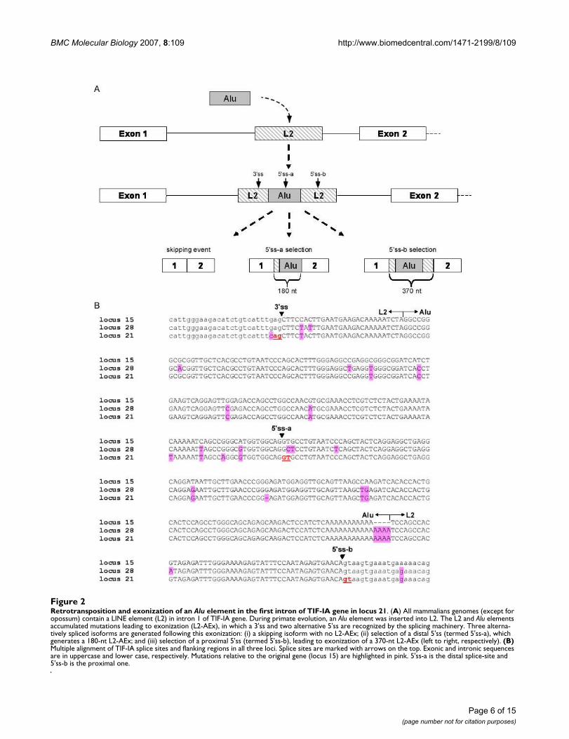
Alu insertion in the first intron of TIF-IAExamination of the TIF-IA genes revealed another intrigu-ing event: An Alu element was inserted into the first intronof the common ancestor of human, chimpanzee, and rhe-sus (approximately 25 million years ago). This Alu wasinserted into another transposed element called L2,located in intron 1 of the TIF-IA gene. We reconstructedthis scenario by examination of intron 1 of the TIF-IA genein other mammals. The L2 element was present in allsequenced mammals except opossum. The insertion ofthe Alu element into the L2 transposed element led to anexonization in human. Based on alignment of ESTs to thehuman genome (see Additional file 2), the 3' splice site(3'ss) is donated by the L2 transposed element and twoalternative 5' splice site (5'ss) are selected, one donated bythe Alu sequence and the other by L2. We designated thedistal 5'ss, located in the Alu sequence as 5'ss-a, and theproximal 5'ss, located in the L2 element, as 5'ss-b (Figure2A and 2B). The overall steps that lead to the exonizationof that exon were as follows: (1) During primate evolu-tion, an Alu-Sx element was inserted into an L2 retrotrans-poson of the LINE family. (2) The sequence accumulatedmutations that caused exonization, leading to selection ofthree different isoforms as demonstrated in Figure 2A.This exon is termed L2-AEx.
Transcription and translation of the wild-type and duplicate TIF-IA genesTo examine whether the duplicates in the human genomeare active transcriptionally, we examined the putative pro-moter regions of all three loci (Figure 1). The region of 1kb upstream of the translation start codon of locus 15 has96.5% identity to the corresponding upstream region oflocus 21. The 1-kb promoter region of locus 28 showssimilarity to that of locus 15; however, this putative pro-moter contains a deletion of 768 bp that ends at position-117 upstream of the potential translation start codon.
The many changes that occurred between the wild-typegene and the two duplicates suggest that the duplicates arenot under selective pressure to encode functional protein.Translation of the mRNA produced from the original TIF-IA gene results in a 651-aa protein with a molecularweight of 75 kDa. The duplicate at locus 28 potentiallyencodes a 514-aa polypeptide chain, whereas the dupli-cate at locus 21 potentially encodes a 39-aa polypeptideusing the genuine start codon or a 106-aa polypeptidechain using the third ATG downstream of the transcrip-tion start site (TSS). Polypeptides of sizes expected fromthe duplicate loci were not detected by western blottinganalysis with polyclonal antibodies to the wild-type TIF-IA protein (Additional file 3). Although proteins do notappear to be generated from the duplicate genes, theseduplicates are active transcriptionally as indicated by ESTsthat derive from these loci. In addition, we measured therelative mRNA levels based on nucleotide variation at cer-tain positions between the loci. In P69 cells, mRNA fromthe wild-type gene comprises about 80% of the mRNAtranscribed from the three loci and locus 28 and locus 21constitute 3% and 17% of the mRNA, respectively (Figure1).
Alu-exonization is exclusive to the TIF-IA duplicate in locus 21EST analysis revealed that a large fraction of the L2-AEx-containing ESTs originated from carcinogenic tissues,such as Burkitt's lymphomas (Additional file 2). To exam-ine the splicing patterns of exons 1 and 2 of the wild-typegene and the two duplicates, we designed a pair of primersto sequences in the flanking exons: 1 and 2 that are con-served among all three loci, meaning all three copies willbe amplified simultaneously. We then performed RT-PCRanalysis of human cDNA from normal tissues and fromtransformed cell lines. The major mRNA product from theTIF-IA genes in most of normal tissues skips the Alu exonand shows either no exonization or a negligible level ofexonization (Figure 3A). In two cell lines, 293T and BJ-1,there was a substantial level of exonization (Figure 3A,lanes 9 and lane 10, respectively). These results implythat, for most of the tested cells, the exonization is aminor or non-existent event. We did not detect any exoni-
Page 5 of 15(page number not for citation purposes)
BMC Molecular Biology 2007, 8:109 http://www.biomedcentral.com/1471-2199/8/109
Page 6 of 15(page number not for citation purposes)
Retrotransposition and exonization of an Alu element in the first intron of TIF-IA gene in locus 21Figure 2Retrotransposition and exonization of an Alu element in the first intron of TIF-IA gene in locus 21. (A) All mammalians genomes (except for opossum) contain a LINE element (L2) in intron 1 of TIF-IA gene. During primate evolution, an Alu element was inserted into L2. The L2 and Alu elements accumulated mutations leading to exonization (L2-AEx), in which a 3'ss and two alternative 5'ss are recognized by the splicing machinery. Three alterna-tively spliced isoforms are generated following this exonization: (i) a skipping isoform with no L2-AEx; (ii) selection of a distal 5'ss (termed 5'ss-a), which generates a 180-nt L2-AEx; and (iii) selection of a proximal 5'ss (termed 5'ss-b), leading to exonization of a 370-nt L2-AEx (left to right, respectively). (B) Multiple alignment of TIF-IA splice sites and flanking regions in all three loci. Splice sites are marked with arrows on the top. Exonic and intronic sequences are in uppercase and lower case, respectively. Mutations relative to the original gene (locus 15) are highlighted in pink. 5'ss-a is the distal splice-site and 5'ss-b is the proximal one.

BMC Molecular Biology 2007, 8:109 http://www.biomedcentral.com/1471-2199/8/109
zation of L2-AEx within chimpanzee blood cells, implyingthat this exonization may be human specific (Figure 3A,lane 17).
Next, we examined the exonization in 17 leukemia celllines by RT-PCR analysis and detected a high level ofexonization of both the short and long exonized form in13 out of the 17 cell lines (76%). To specifically identifythe locus from which this exonization occurred, wedesigned loci-specific primers and repeated the RT-PCRanalysis using the pair of primers that detects all three lociand the locus-specific pairs of primers (Figure 3B). Only
locus 21 showed exonization in which 5'ss-a or of 5'ss-bor both were selected and the ratio among the three iso-forms varied significantly among the cell lines. Thus, therewas a low level or an absence of exonization in normalhuman tissues and in epithelial cancer cell lines. In con-trast, most leukemia cell lines exhibited a high level ofexonization. The exonization was restricted to one of theduplicates (locus 21), but not to the original gene (locus15). The cells that show exonization do not exhibit aknown common characteristic, such as a specific hemat-opoietic lineage or mutations in a certain pathway or genethat can explain the exonization in these cells but not in
Scanning of diverse normal human tissues, various human cell lines, and chimpanzee blood for Alu-exonization in the TIF-IA genesFigure 3Scanning of diverse normal human tissues, various human cell lines, and chimpanzee blood for Alu-exonization in the TIF-IA genes. (A) Low level of L2-AEx inclusion in normal tissues. cDNA from normal human tissues and cell lines was amplified by PCR analysis with primers directed to exons 1 and 2 of all three loci of TIF-IA genes. No evidence for Alu exonization was observed in chimpanzee blood. Splicing products were separated on 1.5% agarose gel. The three mRNA isoforms are shown on the right; selected PCR products were eluted and sequenced. 293T are epithelial kidney cells. BJ-1 is normal human fibroblast cell line. HeLa are epithelial cervix cells with adenocarcinoma. HT 1080 originated from a fibrosarcoma. MCF-7 is a breast-cancer cell line. P69 was derived by immortalization of human primary prostate epithelial cells. SK MEL-28 is a melanoma cell line and U2OS is an osteosa-rcoma cell line. (B) Splicing patterns of L2-AEx in various human leukemia cell lines. Total cytoplasmic RNA was extracted from the indicated cell lines. Splicing products were separated on 1.5% agarose gel after RT-PCR analysis, using primers to exons 1 and 2 of all three loci of TIF-IA, to locus 15 alone, to locus 28 alone, and to locus 21 alone. The three mRNA isoforms are shown on the right. Selected splicing products were eluted from the gel and sequenced. Jurkat, Molt-3, and HSB are human T-cell leukemia cell lines. HL-60 is a myeloid cell line. Dami is a megakaryocytic AMKL (acute megakaryob-lastic leukemia) non-Down syndrome (DS) cell line. U937 is derived from a human histiocyic lymphoma. MT-4 is a human T-cell lymphoblast line. Dg-75, Raji, and Daudi are Burkitt's lymphoma cell lines. KM H2 is a human Hodgkin's lymphoma cell line. CMK is a megakaryocytic DS. K562 is a chronic myeloid leukemia (CMK) cell line. MEG-01 is a megakaryoblastic cell line. 697 is pre-B ALL cell line. Nalm-6 and REH are human precursor leukemia cell lines.
Page 7 of 15(page number not for citation purposes)
BMC Molecular Biology 2007, 8:109 http://www.biomedcentral.com/1471-2199/8/109
the others. These results imply that there is a certain levelof transcriptomic instability of the TIF-IA locus in certaincancer cells, in particular leukemic. The definition ofexon/intron is abnormal for the TIF-IA locus, and perhapsother loci as well, in these cancer cells; what is defined asan intron for normal tissues is selected as an exon inleukemic cells.
The wild-type TIF-IA gene is one step away from exonizationTo understand the molecular mechanisms leading toexonization in locus 21, but not in loci 15 and 28, wecompared the genomic sequence of the L2-Alu among theloci (Figure 2B). Many point mutations have accumulatedin the L2-Alu genomic sequence following the duplicationevent. One of these mutations was of particular interest,
because it changed a GAG found in locus 15 and 28 toCAG in locus 21. This CAG is used as the 3'ss of the L2-AEx. GAG is a week 3'ss, whereas CAG is a strong one [49].We, thus, hypothesized that this mutation leads to locus21-specific exonization.
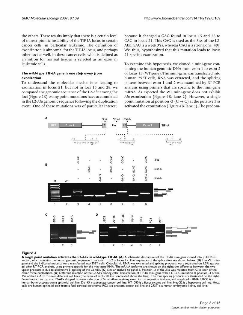
To examine this hypothesis, we cloned a mini-gene con-taining the human genomic DNA from exon 1 to exon 2of locus 15 (WT gene). The mini-gene was transfected intohuman 293T cells, RNA was extracted, and the splicingpattern between exon 1 and 2 was examined by RT-PCRanalysis using primers that are specific to the mini-genemRNA. As expected the WT mini-gene does not exhibitAlu-exonization (Figure 4B, lane 2). However, a singlepoint mutation at position -3 (G → C) at the putative 3'ssactivated the exonization (Figure 4B, lane 3). The predom-
A single point mutation activates the L2-AEx in wild-type TIF-IAFigure 4A single point mutation activates the L2-AEx in wild-type TIF-IA. (A) A schematic description of the TIF-IA mini-gene cloned into pEGFP-C3 vector, which contains the human genomic sequence from exon 1 to 2 of locus 15. The sequences of the splice sites are shown below. (B) The WT mini-gene and the indicated mutants were transfected into 293T cells. Cytoplasmic RNA was extracted and splicing products were separated on 1.5% agarose gel after RT-PCR analysis, using primers specific for the mini-gene RNA. The mRNA isoforms are shown on the right; the difference between the two upper products is due to alternative 5' splicing of the L2-AEx. (C) Similar analysis to panel B. Position -3 of the 3'ss was mutated from G to each of the other three nucleotides. (D) Different selection of the L2-AEx among cells. Transfection of TIF-IA mini-gene with a G → C mutation at position -3 of the 3'ss of the L2-AEx to seven different cell lines (the name of each cell line is indicated above the lane). The four splicing products are illustrated on the right. From bottom to top are: L2-AEx skipped isoform, selection of 5'ss-b Alu-containing exon, intron retention isoform, and unspliced mRNA. U2OS is a human-bone-osteosarcoma epithelial cell line. Du145 is a prostate-cancer cell line. HT1080 is a fibrosarcoma cell line. HepG2 is a hepatoma cell line. HeLa cells are human epithelial cells from a fatal cervical carcinoma. PC3 is a prostate cancer cell line and 293T is a human-embryonic-kidney cell line.
Page 8 of 15(page number not for citation purposes)

BMC Molecular Biology 2007, 8:109 http://www.biomedcentral.com/1471-2199/8/109
inant mRNA generated following this mutation is the onethat includes the L2-AExs (selection of 5'ss-b), leading toa low level of the normal mRNA. In addition, the 3'ssmutation also generated an intron retention isoform. Thisindicates that the L2-Alu in the wild-type gene (locus 15)is one mutation away from exonization. If such a muta-tion were to occur, it would terminate production of anormal TIF-IA protein in the cells almost completely,because the L2-AEx inserts a premature stop codon.
We next evaluated the effect of 5'ss strength on exoniza-tion. Strengthening of the either of the two sites did notactivate the exonization (Figure 4B, lanes 4 and 5) andneither did strengthening both (Figure 4B, lane 8). Whenthe 3'ss is functional, the selection of 5'ss-a or 5'ss-b isdetermined by their relative strength (Figure 4B, lanes 6,7, and 9). Overall, these results indicate that the L2-Aluelement in the WT TIF-IA gene is on the edge of exoniza-tion and that a single point mutation from G to C in posi-tion -3 of the putative 3'ss leads to exonization.
Next, we examined which nucleotides in position -3 of the3'ss would activate exonization. We mutated the 3'ss fromGAG to CAG, TAG, and AAG (Figure 4C, lanes 1, 2, and 3,respectively). Only the mutation from G to C at position -3 lead to exonization: The mutations to A and T did not.These results support our previous observation that CAGis the strongest 3'ss [49]. Also, the results show that alter-native splicing is delicately balanced and is partially con-trolled by the strength of the 3' and 5'ss signals.
Alternative splicing is often regulated in a tissue-specificor developmental-stage-specific manner. A commonexplanation for differential splicing patterns is that theconcentrations of splicing regulatory proteins varydepending on tissue type and developmental stage. There-fore, we examined the effect of different cellular environ-ments on the splicing of the TIF-IA mini-gene bytransfection into different cell lines; we used the mini-gene containing the G-to-C mutant at position -3 of the3'ss. The patterns of splicing observed depended on thecell line (Figure 4D). In two cell lines, Du145 and PC3,the introns were not always excised (Figure 4D, lanes 2and 6, respectively; see figure legend for the source of eachcell line) and in U2OS, HT1080, HepG2, HeLa, and 293Tcell lines, the alternatively spliced mRNA containing L2-AEx was the predominant isoform (lanes 1, 3, 4, 5, and 7,respectively). These findings show that, for this TIF-IAmini-gene, the cellular environment regulates the level ofexonization and intron/exon recognition.
Alu-exonization in YY1AP, the duplicate of GON4LbTo further support the bioinformatics analysis we demon-strated another case of a gene that underwent a duplica-tion event, and the duplicate gene exhibits a distinct
alternative splicing pattern originated from an intronicAlu element, which also exist in the ancestor gene, but isnot exonized. Figure 5 demonstrates the genomic struc-ture of the original GON4Lb gene compared to the dupli-cated gene, YY1AP1 [38]. An intronic Alu element isexonized between exons 13 and 14 (Fig. 5A). Aligning theAlu element in both original and duplicated genes uncov-ers many sequence changes between them (Fig. 5B). Incontrast to the TIF-IA gene, no mutation was detected inthe splice signals, thus we assume that the reason forexonization in the duplicated gene and not in the originalGON4Lb gene is due to mutations in regulatory sequencessuch as ESRs [50]. Figure 5C examines the splicing patternof the corresponding genomic region and revealed thatthe Alu element in GON4Lb gene is not exonized whilethe duplicated gene (YY1AP1) exhibits low levels ofexonization. When the Alu-exon enters into the matureRNA of YY1AP1, it inserts a pre-mature terminationcodon (PTC) [51]. This strengthened our bioinformaticsanalysis showing a second example of exonization that isfound in the duplicated gene and not in the original copy.
DiscussionGene duplication and alternative splicing are two mecha-nisms that enhance genome and transcriptome complex-ity. Gene duplication works at the level of DNA andalternative splicing at the level of mRNA. These two proc-esses are seemingly independent, but recent comprehen-sive bioinformatics analyses suggest that they arecorrelated inversely [7,31]. That is, certain genes prolifer-ate and acquire new functions by duplication, while othergenes gain new functional properties through alternativesplicing.
It was observed previously that duplicated genes, unlikesingletons, undergo relaxation in selective pressure. Theduplicate rapidly diverges from the original sequence dueto a higher rate of nucleotide substitutions within the cod-ing regions relative to orthologs [9,36]. Here we show thatthere is another contributor to the fast divergence ofduplicated genes: a higher level of exonization of trans-posed elements in duplicated genes relative to singletons.Previous work dealing with the correlation between alter-native splicing and gene duplications indicated that thereis less alternative splicing in duplicated genes and thatthere is alternative splicing loss after duplication [31,34].All together our observations imply that although in gen-eral there is a reduction in alternative splicing in gene fam-ilies, the level of TEs exonization is higher in duplicatedgenes.
In the analysis described here, we found that, followingduplication, genes can acquire new alternatively splicedisoforms through exonization of transposed elements andthat genes with more than one copy per genome (dupli-
Page 9 of 15(page number not for citation purposes)

BMC Molecular Biology 2007, 8:109 http://www.biomedcentral.com/1471-2199/8/109
cated genes) tend to undergo more exonization of TE ele-ments in one of the duplicates than do singletons. Thisindicates a high level of selection against exonization insingletons, with fewer restrictions against exonizations inmulti-copy genes. It also supports the observation thatone of the duplicates is under lower selective pressure,which allows the accumulation of mutations leading toexonization without deleterious effects on the organism.
Our results suggest that alternatively spliced exons thatoriginate from exonization of transposed elements are
found significantly more frequently in duplicated genesthan in singletons. We selected one of these duplicatedgenes, TIF-IA, for further examination. TIF-IA underwent ahumanoid-specific triplication, in which one of the dupli-cates retained the evolutionary conserved exon-intronstructure and maintained the reading frame. Because thetwo duplicates were under less evolutionary constraint,each underwent substantial changes with respect to theoriginal copy. These changes include large and small dele-tions and point mutations in the original codingsequence. In addition, in one of the duplicates, we identi-
TheGON4Lb gene and its duplicate YY1AP1Figure 5TheGON4Lb gene and its duplicate YY1AP1. (A) Schematic representation of the genomic organization of GON4Lb and YY1AP1. Start codons and stop codons are marked with black arrows and stop signs, respectively. Insertion of an Alu-exon to the transcript of YY1AP1 insert a premature termination codon (PTC) (B) Multiple alignment of the alternative Alu-exon of YY1AP1 and the relative intronic region of GON4L. Splice sites are marked with black arrows. Substitutions and deletions rel-ative to the original gene are marked in pink. (C) Exonization of an Alu exon in YY1AP1, and not in the original gene, as shown by RT-PCR analysis of U2OS cell line. Primers were designed to the flanking exons of each copy.
Page 10 of 15(page number not for citation purposes)

BMC Molecular Biology 2007, 8:109 http://www.biomedcentral.com/1471-2199/8/109
fied an exonization of an Alu element that was insertedinto the first intron prior to the duplication. All three cop-ies of TIF-IA gene are active transcriptionally, but the wild-type gene is the major contributor to TIF-IA mRNA levels.Only the wild-type mRNA is translated into protein. Wefound negligible levels of this exonization in diverse nor-mal tissues and diverse cell lines, with the exception of the293T and BJ-1 cell lines (Figure 3A). In contrast, when weexamined various leukemia cell lines, we discovered highlevels of L2-Alu-exonization in 13 of the 17 cell lines. Thisindicates that the definition of this Alu as an exon isrestricted to certain types of cancers. The genomicsequence of the Alu between the cells that show exoniza-tion and those that do not is unchanged (not shown).This implies that this exonization is due to aberrantexpression or activity of one or more splicing factors.
Next, we generated a mini-gene with the genomic regionof locus 15, the original copy of TIF-IA gene. The singleproduct of this wild-type mini-gene was the one thatjoined exon 1 with exon 2; the Alu element was intronic.Introducing a single point mutation at position -3 of the3'ss (from G to C) in the Alu element strengthened the 3'ss[49]. The main spliced isoform in this mutant was the onein which 5'ss-b was selected. Alternative splicing of Aluexons usually generates both the isoform containing theadditional exon and the original isoform that presumablyholds an important function in the cell. Thus, alternativeexonization with low inclusion level can advance tran-scriptomic diversification. We previously demonstratedthat alternatively spliced Alu exons have low inclusion lev-els (10–19%) [52]. This strongly suggests that a pointmutation that strengthened the Alu element 3'ss were tooccur in the original gene of TIF-IA (locus 15), therewould be a substantial decrease in the amount of the evo-lutionary conserved protein. Under these conditions, agenetic disease might develop [46].
TIF-IA (locus 15) is a vital gene [46], however its twoduplicates (loci 28 and 21) are probably non-functionalbased on their relative short open reading frames and verylow expression levels. This duplication probably repre-sents the non-functional stage described by Ohno in theperiod of time following the duplication event. Asdescribed by Ohno, this initial phase of non-functionali-zation will be followed by complete relaxation of selectiveconstraints and may be followed by a neo-functionaliza-tion process, wherein the duplicate acquires a new func-tion [8,53]. The NCF1 gene duplicates may also be in thenon-functional stage as its two well-described pseudo-genes are presumably non-functional [43].
Why might there be selective forces against acquisition ofthe exons in certain genes and others not? We have dem-onstrated recently that conserved human-mouse alterna-
tive exons that disrupt the reading frame (termed non-symmetrical) tend to undergo fixation in the beginning ofthe gene, whereas those in the middle and the 3' half ofthe genes were presumably "weeded out" during evolu-tion [51]. This suggests that acquisition of new exons closeto the beginning of the genes is tolerated. In contrast,those in other parts of the genes are deleterious becausepolypeptides translated from these mRNAs are less likelyto be targeted for the nonsense mediated decay thanshorter products and the exon-containing isoforms arelikely to be deleterious to the cells. If the initial exoniza-tion is natural or even present beneficial advantages to thecells, it will lead to an increase in the inclusion level ofthese alternative exons [54]. The mutation in the 3'ss ofthe L2-AEx in TIF-IA gene (locus 15) presumably occurredduring evolution in some individuals, but was selectedagainst since TIF-IA is a vital gene, this isoform must havebeen deleterious due to the insertion of a premature ter-mination codon.
ConclusionOur results add another layer to our understanding of thedelicate interplay between gene duplication and alterna-tive splicing. Both increase gene complexity, albeitthrough different mechanisms. We have demonstrated anindirect link between duplication of genes and exoniza-tion of transposed elements. Duplicated genes, especiallythe non-functional, tend to undergo exonization morethan singletons.
MethodsDataset of TE exons in human and mouse genomesWe compiled a dataset of TE exonizations within proteincoding genes [28]. The human NCBI 35 (hg17, May 2004)and the mouse NCBI33m (mm6, March 2005) assemblieswere downloaded, along with their annotations, from theUCSC genome browser database [55]. Coordinates of theEST and cDNA mapping were obtained fromchrN_intronEST and chrN_mrna tables, respectively. TheTE mapping was obtained from chrN_rmsk tables. A TEwas considered intragenic if there was no overlap withESTs or cDNA alignments; it was considered intronic if itwas found within an alignment of an EST or cDNA withinan intronic region. Finally, a TE was considered exonic ifit was found within an exonic part of the EST or cDNA, ifit possessed canonical splice sites, and if it was an internalexon of the EST/cDNA found within the CDS or a UTR.The insertions of TEs within EST/cDNA alignments wereseparated into two parts: those that entered within proteincoding genes relative to the list of the knownGene table inthe UCSC genome browser [55] (based on proteins fromSWISS-PROT, TrEMBL, TrEMBL-NEW and their corre-sponding mRNA from GenBank), and other insertionswithin cDNA/ESTs alignments that were not mapped tothe known genes list and, therefore, were considered as
Page 11 of 15(page number not for citation purposes)

BMC Molecular Biology 2007, 8:109 http://www.biomedcentral.com/1471-2199/8/109
non-protein-coding genes. Non-protein-coding geneswere determined as genomic regions covered by at leasttwo correctly spliced cDNA/ESTs (flanked by canonicalsplice sites) containing at least three exons that did notoverlap any annotated gene based on UCSC known geneslists, versions hg17 and mm6 for human and mouse,respectively. Unspliced genes were not included in ouranalysis; we only considered genes with at least twointrons. Internal UTR exons were considered to be inter-nal according to the annotations of knownGenes in UCSCand the fact that they were internal within the cDNA/EST.The TE position within the gene (UTR or CDS) and theexon phase were calculated based on knownGenes tableannotations of the gene start and end positions, as well asCDS start and end positions.
TE exonization within duplicated genesTo analyze whether the exonization occurred within aduplicated gene or a singletons, we performed a blastsearch of these genes against all mRNA sequences listed asknown genes (knownGene table) downloaded fromUCSC human genome build hg17 [55] and searched fortranscripts with at least 75% sequence similarity along 40percent of the gene, the dataset was filtered to delete tran-scripts of isoforms generated by alternative splicing thatbelonged to the same gene and nonduplicated genes. Thefiltration of similar mRNA that are isoforms of the samegene was done by comparing the locus of the mRNA asindicated in UCSC genome browser; only mRNAs thatmapped to different loci were considered.
Cell line maintenance293T, HeLa, HT1080, HepG2, Du145, and U2OS celllines were cultured in Dulbecco's Modification of EagleMedium (DMEM), supplemented with 4.5 g/mL glucose(Biological Industries, Inc., Israel), 10% fetal calf serum(FCS), 100 U/mL penicillin, 0.1 mg/mL streptomycin,and 1 U/mL nystatin (Biological Industries, Inc.). PC3cells were cultured in Ham's F12K medium with 2 mM L-glutamine adjusted to contain 1.5 g/L sodium bicarbonate(90%) and fetal bovine serum (10%). Cells were grown in6-well plates or 100-mm culture dishes under standardconditions at 37°C with 5% CO2. Cells were split at a 1:10ratio twice weekly.
TransfectionCells were grown to 50% confluence in 100-mm culturedishes or 6-well plates and maintained as describedabove. Twenty-four hours prior to transfection, cells weresplit, and transfection was performed using FuGENE6(Roche) with 0.5–1 μg of plasmid DNA. Cells were har-vested after 48 hr.
RNA isolation and splicing analysisTotal RNA was extracted using TRI Reagent (Sigma), fol-lowed by treatment with 2 U of RNase-free DNase(Ambion). Reverse transcription (RT) was performed on1–2 μg total RNA in a 12.5 μL final volume containing:100 mM DTT, 10 mM dNTPs, 100 mM oligo(dT) primer,2 U of reverse transcriptase avian myeloblastosis virus(RT-AMV, Roche), and RT buffer. The final mixture wasthen incubated for 1 hr at 42°C. The spliced cDNA prod-ucts derived from the expressed mini-genes were detectedby PCR, using Taq polymerase (BioTools), and pEGFP-C3specific reverse and forward primers. Primer sets for cell-line scanning purposes were designed to amplify all lociin a single PCR or to amplify every locus separately: 3 loci:5'-CGT TAG TTC GGC CCA ATG-3', 5'-CTT CAG CAAGAC TTC TGT CAC-3'; locus 15: 5'-CTT CGT CCT CTGCAG TTA AGA AG-3', 5'-CTT CAG CAA GAC TTC TGTCAC-3'; locus 28: 5'-CGC TTC GCC CTC TGC AGT C-3',5'-CTT CAG CAA GAC TTC TGT CAC-3' after reverse tran-scription (RT) with unique primer; locus 21: 5'-CTT CACACG TTG TTT GTC G-3', 5'-CTT CAG CAA GAC TTC TGTCAC-3'. Amplification of the chimpanzee cDNA (prolifer-ating blood cells of a female chimpanzee from the safariin Ramat-Gan) was performed with 5'-CGT TAG TTCGGC CCA ATG-3' and 5'-GCT GGT TCT TCA ACA ACTCAA A-3'. The primers flank the Alu element in intron 1 ofthe TIF-IA's genes. Human GAPDH: 5'-TCG TGG AGTCCA CTG GCG TCT T-3' and 5'-TGG CAG TGA TGG CATGGA CTG T-3'. Chimp GAPDH: 5'-TCG TGG AGT CCACTG GCG TCT T-3' and 5'-TGG CAG TGA TGG CAT GGACCG T-3'. GON4L: 5'-ATG AGC TGA TGG AAG AGC TG-3' and 5'-GAG GGG TGT TAA AGT TAG GAC GAG-3'.YY1AP1: 5'-CAA ATG AGC TGA TGG AAG AT-3' and 5'-GAG GGG TGT TAA AGT TAG CTT-3'. Amplification wasperformed for 30 cycles, consisting of denaturation for 30seconds at 94°C, annealing for 45 seconds at 58°C, andelongation for 1.5 minutes at 72°C. The products wereseparated in 1.5% agarose gel; selected bands were con-firmed by sequencing.
Plasmid constructA genomic DNA from 293T cell line (Gentra) correspond-ing to exon 1 through exon 2 of the TIF-IA gene (fromlocus 15) was PCR amplified and cloned into pEGFP-C3vector (Clontech) between XhoI and BamHI sites underthe control of the human cytomegalovirus (CMV) imme-diate early promoter, giving a ~1.9 kb insert. F: 5'-AAAAAA ACT CGA GGC TGA TTG GCT GAA GGT TG-3'; R: 5'-AAA AGG ATC CCA GCA ATA GTT GTA TTC TGA CCTAAC C-3'.
Site-directed mutagenesisSpecific overlapping oligonucleotide primers that con-tained the desired mutation were used to insert the muta-tion using PfuTurbo DNA polymerase (Stratagene). After
Page 12 of 15(page number not for citation purposes)

BMC Molecular Biology 2007, 8:109 http://www.biomedcentral.com/1471-2199/8/109
PCR amplification, the reaction was digested with DpnIrestriction enzyme (New England Biolabs) for 1 hr at37°C; 1–3 μL of the reaction were transformed into E. coliXL-1 strain and colonies were picked for mini-prep extrac-tion (Qiagen) and sequenced. L2-AEx-3'ss (-3)G->C: 5'-GAA GAC ATC TGT CAT TTC AGC TTC CAC TTG AAT G-3' and 5'-CAT TCA AGT GGA AGC TGA AAT GAC AGATGT CTT C-3'. L2-AEx-3'ss (-3)G->T: 5'-GAA GAC ATCTGT CAT TTT AGC TTC CAC TTG AAT G-3' and 5'-CATTCA AGT GGA AGC TAA AAT GAC AGA TGT CTT C-3'. L2-AEx-3'ss (-3)G->A: 5'-GAA GAC ATC TGT CAT TTA AGCTTC CAC TTG AAT G-3' and 5'-CAT TCA AGT GGA AGCTTA AAT GAC AGA TGT CTT C-3'. L2-AEx-5'ss-a (5)C->G:5'-CAT GGT GGC AGG TGC GTG TAA TCC CAG CTA C-3' and 5'-GTA GCT GGG ATT ACA CGC ACC TGC CACCAT G-3'. L2-AEx-5'ss-b (-1)A->G: 5'-GTA TTT CCA ATAGAG TGA ACG GTA AGT GAA ATG AAA AAC AGC-3' and5'-GCT GTT TTT CAT TTC ACT TAC CGT TCA CTC TATTGG AAA TAC-3'.
Western blottingLysis buffer (50 mM Tris, pH 7.5; 1% NP40; 150 mMNaCl; 0.1% SDS; 0.5% deoxycholic acid; protease inhibi-tor cocktail and phosphatase inhibitor cocktail I and II;Sigma) was used for protein extraction. Then lysates werecentrifuged for 30 minutes at 14,000 rpm at 4°C. Totalprotein concentrations were measured using BioRad Pro-tein Assay (BioRad). Proteins were separated in 10% SDS-polyacrylamide gel electrophoresis (SDS-PAGE) and thenelectroblotted onto a Protran membrane (Schleicher andSchuell). The membranes were probed with polyclonalanti-TIF-IA antibody (kindly provided by Ingrid Grummt)at 1/10000 dilution followed by rabbit secondary anti-body. Immunoblots were visualized by enhanced chemi-luminescence (Lumi-Light Western Blotting Substrate;Roche) and exposure to x-ray film.
Transposed elements analysisRepeatMasker© software version 3.1.0 [56] was used forthe detection of transposed elements.
Analysis of the relative mRNA levelsThe PCR product from RT-PCR of P69 cells was excisedand purified following electrophoresis on 1.5% agarosegel (Promega, Madison, WI, USA). Direct sequencing wasperformed using the ABI PRISM (Applied Biosystems, Fos-ter-City, CA, USA). The variation percentage from directsequencing was calculated for the reverse primer; the pre-sented percentages represent an average of three separatedpositions (31, 63 and 105) along exon 2 of the mRNAs.The nucleotides were quantified by the Discovery Studio(DS) Gene 1.5 program (Accelrys Inc., San Diego, CA,USA).
List of abbreviationsTE, transposed elements; 3'ss, 3' splice site; 5'ss, 5' splicesite; SINE, short interspersed elements
Authors' contributionsMA was responsible for analyzing different cell lines, gen-erating the constructs, transfection experiments and draft-ing the manuscript. NSE executed the bioinformatic dataanalysis and drafted the manuscript. HK carried out theidentification of TIF-IA protein. ZM performed theGON4L and YY1AP1 analysis. IM provided many of thecell culture samples. NSH participated in the cell linescharacterization. SI was involved in designing the studyand helped to draft the manuscript. GA conceived andsupervised the study design, and drafted the manuscript.All the authors read and approved the final manuscript.
Additional material
AcknowledgementsWe would like to thank Ingrid Grummt from the German Cancer Research Center (DKFZ) for providing us with the antibody against TIF-IA. We thank Eddo Kim and Oren Ram for helpful discussions and Nurit Paz for calculat-ing the relative mRNA levels with the DS gene program. This work was
Additional file 1Transposed elements exonizations within duplicated genes in the human genome. A list of transposed elements exonizations within dupli-cated genes and their genome coordinates.Click here for file[http://www.biomedcentral.com/content/supplementary/1471-2199-8-109-S1.xls]
Additional file 2Indications of exonization only from locus 21. Alignment of EST/cDNA with the human genome using UCSC Genome Browser reveals alternative L2-AExs between exon 1 and 2. The L2-AExs' are marked with red circles. Exons' numbers are indicated on the bottom. Seven out of the 19 ESTs that cover that locus contain an L2-AEx, of which 5 were originated from cancerous sources (3 from Burkitt's lymphoma, 1 from neuroblastoma cell line, and 1 from poorly differentiated adenocarcenoma). The rest of the Alu-containing spliced isoforms originated from reproductive organs (tes-tis and uterus). The alignment indicates that the L2-AEx is also alterna-tively 5' spliced.Click here for file[http://www.biomedcentral.com/content/supplementary/1471-2199-8-109-S2.tiff]
Additional file 3The WT TIF-IA protein is synthesized in cells with and without the exonization. Total protein was extracted from the indicated cells and ana-lyzed by Western blotting using polyclonal rabbit anti-TIF-IA serum (kindly gifted from the Grummt's lab) at a 1/10,000 dilution. 293T and U2OS are human cell lines, and 3T3 is a murine cell line.Click here for file[http://www.biomedcentral.com/content/supplementary/1471-2199-8-109-S3.tiff]
Page 13 of 15(page number not for citation purposes)
BMC Molecular Biology 2007, 8:109 http://www.biomedcentral.com/1471-2199/8/109
supported by ICA through the Ber-Lehmsdorf Memorial Fund, and TAU Cancer Center, by the Cooperation Program in Cancer Research of the Deutsches Krebsforschungszentrum (DKFZ) and Israeli's Ministry of Sci-ence and Technology (MOST), by a grant from the Israel Science Founda-tion (1449/04 and 40/05), MOP Germany-Israel, GIF, N.S. is funded in part by EURASNET.
References1. Waterston RH, Lindblad-Toh K, Birney E, Rogers J, Abril JF, Agarwal
P, Agarwala R, Ainscough R, Alexandersson M, An P, Antonarakis SE,Attwood J, Baertsch R, Bailey J, Barlow K, Beck S, Berry E, Birren B,Bloom T, Bork P, Botcherby M, Bray N, Brent MR, Brown DG, BrownSD, Bult C, Burton J, Butler J, Campbell RD, Carninci P, Cawley S,Chiaromonte F, Chinwalla AT, Church DM, Clamp M, Clee C, CollinsFS, Cook LL, Copley RR, Coulson A, Couronne O, Cuff J, Curwen V,Cutts T, Daly M, David R, Davies J, Delehaunty KD, Deri J, Dermitza-kis ET, Dewey C, Dickens NJ, Diekhans M, Dodge S, Dubchak I, DunnDM, Eddy SR, Elnitski L, Emes RD, Eswara P, Eyras E, Felsenfeld A,Fewell GA, Flicek P, Foley K, Frankel WN, Fulton LA, Fulton RS, FureyTS, Gage D, Gibbs RA, Glusman G, Gnerre S, Goldman N, GoodstadtL, Grafham D, Graves TA, Green ED, Gregory S, Guigo R, Guyer M,Hardison RC, Haussler D, Hayashizaki Y, Hillier LW, Hinrichs A,Hlavina W, Holzer T, Hsu F, Hua A, Hubbard T, Hunt A, Jackson I,Jaffe DB, Johnson LS, Jones M, Jones TA, Joy A, Kamal M, Karlsson EK,Karolchik D, Kasprzyk A, Kawai J, Keibler E, Kells C, Kent WJ, KirbyA, Kolbe DL, Korf I, Kucherlapati RS, Kulbokas EJ, Kulp D, Landers T,Leger JP, Leonard S, Letunic I, Levine R, Li J, Li M, Lloyd C, Lucas S,Ma B, Maglott DR, Mardis ER, Matthews L, Mauceli E, Mayer JH,McCarthy M, McCombie WR, McLaren S, McLay K, McPherson JD,Meldrim J, Meredith B, Mesirov JP, Miller W, Miner TL, Mongin E,Montgomery KT, Morgan M, Mott R, Mullikin JC, Muzny DM, NashWE, Nelson JO, Nhan MN, Nicol R, Ning Z, Nusbaum C, O'ConnorMJ, Okazaki Y, Oliver K, Overton-Larty E, Pachter L, Parra G, PepinKH, Peterson J, Pevzner P, Plumb R, Pohl CS, Poliakov A, Ponce TC,Ponting CP, Potter S, Quail M, Reymond A, Roe BA, Roskin KM,Rubin EM, Rust AG, Santos R, Sapojnikov V, Schultz B, Schultz J,Schwartz MS, Schwartz S, Scott C, Seaman S, Searle S, Sharpe T,Sheridan A, Shownkeen R, Sims S, Singer JB, Slater G, Smit A, SmithDR, Spencer B, Stabenau A, Stange-Thomann N, Sugnet C, Suyama M,Tesler G, Thompson J, Torrents D, Trevaskis E, Tromp J, Ucla C,Ureta-Vidal A, Vinson JP, Von Niederhausern AC, Wade CM, Wall M,Weber RJ, Weiss RB, Wendl MC, West AP, Wetterstrand K,Wheeler R, Whelan S, Wierzbowski J, Willey D, Williams S, WilsonRK, Winter E, Worley KC, Wyman D, Yang S, Yang SP, Zdobnov EM,Zody MC, Lander ES: Initial sequencing and comparative anal-ysis of the mouse genome. Nature 2002, 420(6915):520-562.
2. Black DL: Mechanisms of alternative pre-messenger RNAsplicing. Annu Rev Biochem 2003, 72:291-336.
3. Brosius J: Gene duplication and other evolutionary strategies:from the RNA world to the future. J Struct Funct Genomics 2003,3(1-4):1-17.
4. Brosius J, Gould SJ: On "genomenclature": a comprehensive(and respectful) taxonomy for pseudogenes and other "junkDNA". Proc Natl Acad Sci U S A 1992, 89(22):10706-10710.
5. Koch AL: Enzyme evolution. I. The importance of untranslat-able intermediates. Genetics 1972, 72(2):297-316.
6. Chothia C, Gough J, Vogel C, Teichmann SA: Evolution of the pro-tein repertoire. Science 2003, 300(5626):1701-1703.
7. Talavera D, Vogel C, Orozco M, Teichmann SA, de la Cruz X: The(In)dependence of Alternative Splicing and Gene Duplica-tion. PLoS Comput Biol 2007, 3(3):e33.
8. Ohno S: Evolution by Gene and Genome Duplication . Berlin, Springer-Verlag; 1970.
9. Lynch M, Katju V: The altered evolutionary trajectories of geneduplicates. Trends Genet 2004, 20(11):544-549.
10. Force A, Lynch M, Pickett FB, Amores A, Yan YL, Postlethwait J:Preservation of duplicate genes by complementary, degen-erative mutations. Genetics 1999, 151(4):1531-1545.
11. Li WH, Yang J, Gu X: Expression divergence between duplicategenes. Trends Genet 2005, 21(11):602-607.
12. Prince VE, Pickett FB: Splitting pairs: the diverging fates ofduplicated genes. Nat Rev Genet 2002, 3(11):827-837.
13. Shakhnovich BE, Koonin EV: Origins and impact of constraints inevolution of gene families. Genome Res 2006, 16(12):1529-1536.
14. Lander ES, Linton LM, Birren B, Nusbaum C, Zody MC, Baldwin J,Devon K, Dewar K, Doyle M, FitzHugh W, Funke R, Gage D, HarrisK, Heaford A, Howland J, Kann L, Lehoczky J, LeVine R, McEwan P,McKernan K, Meldrim J, Mesirov JP, Miranda C, Morris W, Naylor J,Raymond C, Rosetti M, Santos R, Sheridan A, Sougnez C, Stange-Thomann N, Stojanovic N, Subramanian A, Wyman D, Rogers J, Sul-ston J, Ainscough R, Beck S, Bentley D, Burton J, Clee C, Carter N,Coulson A, Deadman R, Deloukas P, Dunham A, Dunham I, DurbinR, French L, Grafham D, Gregory S, Hubbard T, Humphray S, HuntA, Jones M, Lloyd C, McMurray A, Matthews L, Mercer S, Milne S,Mullikin JC, Mungall A, Plumb R, Ross M, Shownkeen R, Sims S,Waterston RH, Wilson RK, Hillier LW, McPherson JD, Marra MA,Mardis ER, Fulton LA, Chinwalla AT, Pepin KH, Gish WR, Chissoe SL,Wendl MC, Delehaunty KD, Miner TL, Delehaunty A, Kramer JB,Cook LL, Fulton RS, Johnson DL, Minx PJ, Clifton SW, Hawkins T,Branscomb E, Predki P, Richardson P, Wenning S, Slezak T, DoggettN, Cheng JF, Olsen A, Lucas S, Elkin C, Uberbacher E, Frazier M,Gibbs RA, Muzny DM, Scherer SE, Bouck JB, Sodergren EJ, WorleyKC, Rives CM, Gorrell JH, Metzker ML, Naylor SL, Kucherlapati RS,Nelson DL, Weinstock GM, Sakaki Y, Fujiyama A, Hattori M, Yada T,Toyoda A, Itoh T, Kawagoe C, Watanabe H, Totoki Y, Taylor T,Weissenbach J, Heilig R, Saurin W, Artiguenave F, Brottier P, Bruls T,Pelletier E, Robert C, Wincker P, Smith DR, Doucette-Stamm L,Rubenfield M, Weinstock K, Lee HM, Dubois J, Rosenthal A, PlatzerM, Nyakatura G, Taudien S, Rump A, Yang H, Yu J, Wang J, Huang G,Gu J, Hood L, Rowen L, Madan A, Qin S, Davis RW, Federspiel NA,Abola AP, Proctor MJ, Myers RM, Schmutz J, Dickson M, GrimwoodJ, Cox DR, Olson MV, Kaul R, Shimizu N, Kawasaki K, Minoshima S,Evans GA, Athanasiou M, Schultz R, Roe BA, Chen F, Pan H, RamserJ, Lehrach H, Reinhardt R, McCombie WR, de la Bastide M, DedhiaN, Blocker H, Hornischer K, Nordsiek G, Agarwala R, Aravind L, Bai-ley JA, Bateman A, Batzoglou S, Birney E, Bork P, Brown DG, BurgeCB, Cerutti L, Chen HC, Church D, Clamp M, Copley RR, Doerks T,Eddy SR, Eichler EE, Furey TS, Galagan J, Gilbert JG, Harmon C, Hay-ashizaki Y, Haussler D, Hermjakob H, Hokamp K, Jang W, Johnson LS,Jones TA, Kasif S, Kaspryzk A, Kennedy S, Kent WJ, Kitts P, KooninEV, Korf I, Kulp D, Lancet D, Lowe TM, McLysaght A, Mikkelsen T,Moran JV, Mulder N, Pollara VJ, Ponting CP, Schuler G, Schultz J,Slater G, Smit AF, Stupka E, Szustakowski J, Thierry-Mieg D, Thierry-Mieg J, Wagner L, Wallis J, Wheeler R, Williams A, Wolf YI, WolfeKH, Yang SP, Yeh RF, Collins F, Guyer MS, Peterson J, Felsenfeld A,Wetterstrand KA, Patrinos A, Morgan MJ, Szustakowki J: Initialsequencing and analysis of the human genome. Nature 2001,409(6822):860-921.
15. Zhou Z, Licklider LJ, Gygi SP, Reed R: Comprehensive proteomicanalysis of the human spliceosome. Nature 2002,419(6903):182-185.
16. Modrek B, Lee C: A genomic view of alternative splicing. NatGenet 2002, 30(1):13-19.
17. Graveley BR: Alternative splicing: increasing diversity in theproteomic world. Trends Genet 2001, 17(2):100-107.
18. Kim E, Magen A, Ast G: Different levels of alternative splicingamong eukaryotes. Nucleic Acids Res 2007, 35(1):125-131.
19. Mironov AA, Fickett JW, Gelfand MS: Frequent alternative splic-ing of human genes. Genome Res 1999, 9(12):1288-1293.
20. Deininger PL, Moran JV, Batzer MA, Kazazian HH Jr.: Mobile ele-ments and mammalian genome evolution. Curr Opin Genet Dev2003, 13(6):651-658.
21. Hedges DJ, Batzer MA: From the margins of the genome:mobile elements shape primate evolution. Bioessays 2005,27(8):785-794.
22. Krull M, Brosius J, Schmitz J: Alu-SINE exonization: en route toprotein-coding function. Mol Biol Evol 2005, 22(8):1702-1711.
23. Kriegs JO, Churakov G, Jurka J, Brosius J, Schmitz J: Evolutionaryhistory of 7SL RNA-derived SINEs in Supraprimates. TrendsGenet 2007, 23(4):158-161.
24. Mighell AJ, Markham AF, Robinson PA: Alu sequences. FEBS Lett1997, 417(1):1-5.
25. Rowold DJ, Herrera RJ: Alu elements and the human genome.Genetica 2000, 108(1):57-72.
26. Schmid CW: Alu: structure, origin, evolution, significance andfunction of one- tenth of human DNA. Prog Nucleic Acid Res MolBiol 1996, 53:283-319.
27. Onafuwa-Nuga AA, Telesnitsky A, King SR: 7SL RNA, but not the54-kd signal recognition particle protein, is an abundant
Page 14 of 15(page number not for citation purposes)
http://www.ncbi.nlm.nih.gov/entrez/query.fcgi?cmd=Retrieve&db=PubMed&dopt=Abstract&list_uids=1279691
http://www.ncbi.nlm.nih.gov/entrez/query.fcgi?cmd=Retrieve&db=PubMed&dopt=Abstract&list_uids=1279691
http://www.ncbi.nlm.nih.gov/entrez/query.fcgi?cmd=Retrieve&db=PubMed&dopt=Abstract&list_uids=1279691
http://www.ncbi.nlm.nih.gov/entrez/query.fcgi?cmd=Retrieve&db=PubMed&dopt=Abstract&list_uids=4567287
http://www.ncbi.nlm.nih.gov/entrez/query.fcgi?cmd=Retrieve&db=PubMed&dopt=Abstract&list_uids=4567287
http://www.ncbi.nlm.nih.gov/entrez/query.fcgi?cmd=Retrieve&db=PubMed&dopt=Abstract&list_uids=9395063
http://www.ncbi.nlm.nih.gov/entrez/query.fcgi?cmd=Retrieve&db=PubMed&dopt=Abstract&list_uids=8650306

BMC Molecular Biology 2007, 8:109 http://www.biomedcentral.com/1471-2199/8/109
Publish with BioMed Central and every scientist can read your work free of charge
"BioMed Central will be the most significant development for disseminating the results of biomedical research in our lifetime."
Sir Paul Nurse, Cancer Research UK
Your research papers will be:
available free of charge to the entire biomedical community
peer reviewed and published immediately upon acceptance
cited in PubMed and archived on PubMed Central
yours — you keep the copyright
Submit your manuscript here:http://www.biomedcentral.com/info/publishing_adv.asp
BioMedcentral
component of both infectious HIV-1 and minimal virus-likeparticles. Rna 2006, 12(4):542-546.
28. Sela N, Mersch B, Gal-Mark N, Lev-Maor G, Hotz-Wagenblatt A, AstG: Comparative analysis of transposed element insertionwithin human and mouse genomes reveals Alu's unique rolein shaping the human transcriptome. Genome Biol 2007,8(6):R127.
29. Sorek R, Lev-Maor G, Reznik M, Dagan T, Belinky F, Graur D, Ast G:Minimal conditions for exonization of intronic sequences: 5'splice site formation in alu exons. Mol Cell 2004, 14(2):221-231.
30. Ast G: How did alternative splicing evolve? Nat Rev Genet 2004,5(10):773-782.
31. Kopelman NM, Lancet D, Yanai I: Alternative splicing and geneduplication are inversely correlated evolutionary mecha-nisms. Nat Genet 2005, 37(6):588-589.
32. Conant GC, Wagner A: Asymmetric sequence divergence ofduplicate genes. Genome Res 2003, 13(9):2052-2058.
33. Yu WP, Brenner S, Venkatesh B: Duplication, degeneration andsubfunctionalization of the nested synapsin-Timp genes inFugu. Trends Genet 2003, 19(4):180-183.
34. Su Z, Wang J, Yu J, Huang X, Gu X: Evolution of alternative splic-ing after gene duplication. Genome Res 2006, 16(2):182-189.
35. Karni R, de Stanchina E, Lowe SW, Sinha R, Mu D, Krainer AR: Thegene encoding the splicing factor SF2/ASF is a proto-onco-gene. Nat Struct Mol Biol 2007, 14(3):185-193.
36. Kondrashov FA, Rogozin IB, Wolf YI, Koonin EV: Selection in theevolution of gene duplications. Genome Biol 2002,3(2):RESEARCH0008.
37. Grover D, Mukerji M, Bhatnagar P, Kannan K, Brahmachari SK: Alurepeat analysis in the complete human genome: trends andvariations with respect to genomic composition. Bioinformatics2004, 20(6):813-817.
38. Kuryshev VY, Vorobyov E, Zink D, Schmitz J, Rozhdestvensky TS,Munstermann E, Ernst U, Wellenreuther R, Moosmayer P, Bechtel S,Schupp I, Horst J, Korn B, Poustka A, Wiemann S: An anthropoid-specific segmental duplication on human chromosome 1q22.Genomics 2006, 88(2):143-151.
39. Xing J, Wang H, Belancio VP, Cordaux R, Deininger PL, Batzer MA:Emergence of primate genes by retrotransposon-mediatedsequence transduction. Proc Natl Acad Sci U S A 2006,103(47):17608-17613.
40. Wang CY, Liang YJ, Lin YS, Shih HM, Jou YS, Yu WC: YY1AP, anovel co-activator of YY1. J Biol Chem 2004,279(17):17750-17755.
41. Grover D, Majumder PP, C BR, Brahmachari SK, Mukerji M: Nonran-dom distribution of alu elements in genes of various func-tional categories: insight from analysis of humanchromosomes 21 and 22. Mol Biol Evol 2003, 20(9):1420-1424.
42. Spurkland A, Sollid LM: Mapping genes and pathways in autoim-mune disease. Trends Immunol 2006, 27(7):336-342.
43. Vazquez N, Lehrnbecher T, Chen R, Christensen BL, Gallin JI, MalechH, Holland S, Zhu S, Chanock SJ: Mutational analysis of patientswith p47-phox-deficient chronic granulomatous disease: Thesignificance of recombination events between the p47-phoxgene (NCF1) and its highly homologous pseudogenes. ExpHematol 2001, 29(2):234-243.
44. Mersch B, Sela N, Ast G, Suhai S, Hotz-Wagenblatt A: AluScreen:Identifying tissue or tumor-specific Alu-containing isoformsin DNA. submitted 2007.
45. Schnapp A, Pfleiderer C, Rosenbauer H, Grummt I: A growth-dependent transcription initiation factor (TIF-IA) interact-ing with RNA polymerase I regulates mouse ribosomal RNAsynthesis. Embo J 1990, 9(9):2857-2863.
46. Yuan X, Zhou Y, Casanova E, Chai M, Kiss E, Grone HJ, Schutz G,Grummt I: Genetic inactivation of the transcription factorTIF-IA leads to nucleolar disruption, cell cycle arrest, andp53-mediated apoptosis. Mol Cell 2005, 19(1):77-87.
47. Buttgereit D, Pflugfelder G, Grummt I: Growth-dependent regu-lation of rRNA synthesis is mediated by a transcription initi-ation factor (TIF-IA). Nucleic Acids Res 1985, 13(22):8165-8180.
48. Kuhn RM, Karolchik D, Zweig AS, Trumbower H, Thomas DJ,Thakkapallayil A, Sugnet CW, Stanke M, Smith KE, Siepel A, Rosen-bloom KR, Rhead B, Raney BJ, Pohl A, Pedersen JS, Hsu F, HinrichsAS, Harte RA, Diekhans M, Clawson H, Bejerano G, Barber GP,Baertsch R, Haussler D, Kent WJ: The UCSC genome browser
database: update 2007. Nucleic Acids Res 2007, 35(Databaseissue):D668-73.
49. Lev-Maor G, Sorek R, Shomron N, Ast G: The birth of an alterna-tively spliced exon: 3' splice-site selection in Alu exons. Sci-ence 2003, 300(5623):1288-1291.
50. Goren A, Ram O, Amit M, Keren H, Lev-Maor G, Vig I, Pupko T, AstG: Comparative analysis identifies exonic splicing regulatorysequences--The complex definition of enhancers and silenc-ers. Mol Cell 2006, 22(6):769-781.
51. Magen A, Ast G: The importance of being divisible by three inalternative splicing. Nucleic Acids Res 2005, 33(17):5574-5582.
52. Sorek R, Ast G: Intronic sequences flanking alternativelyspliced exons are conserved between human and mouse.Genome Res 2003, 13(7):1631-1637.
53. Gould SJ, Vrba ES: Exaptation; a missing term in the science ofform. Paleobiology 1982, 8(1):4-15.
54. Zhang XH, Chasin LA: Comparison of multiple vertebrategenomes reveals the birth and evolution of human exons.Proc Natl Acad Sci U S A 2006, 103(36):13427-13432.
55. UCSC genome browser [http://genome.ucsc.edu]56. Jurka J, Kapitonov VV, Pavlicek A, Klonowski P, Kohany O, Walichie-
wicz J: Repbase Update, a database of eukaryotic repetitiveelements. Cytogenet Genome Res 2005, 110(1-4):462-467.
Page 15 of 15(page number not for citation purposes)
http://www.ncbi.nlm.nih.gov/entrez/query.fcgi?cmd=Retrieve&db=PubMed&dopt=Abstract&list_uids=2390974
http://www.ncbi.nlm.nih.gov/entrez/query.fcgi?cmd=Retrieve&db=PubMed&dopt=Abstract&list_uids=2390974
http://www.ncbi.nlm.nih.gov/entrez/query.fcgi?cmd=Retrieve&db=PubMed&dopt=Abstract&list_uids=2390974
http://www.ncbi.nlm.nih.gov/entrez/query.fcgi?cmd=Retrieve&db=PubMed&dopt=Abstract&list_uids=4070001
http://www.ncbi.nlm.nih.gov/entrez/query.fcgi?cmd=Retrieve&db=PubMed&dopt=Abstract&list_uids=4070001