Molecular Biology Of The Cell 5th.Ed - Alberts
37
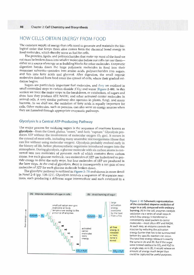
88 Chapter 2:Cell Chemistry and Biosynthesis HOW CELLS OBTAIN ENERGY FROM FOOD The constant supply of energy that cells need to generate and maintain the bio- logical order that keeps them alive comes from the chemical bond energy in food molecules, which thereby serve as fuel for cells. The proteins, lipids, and polysaccharidesthat make up most of the food we eat must be broken down into smaller molecules before our cells can use them- either as a source of energy or as building blocks for other molecules. EnzFnatic digestion breaks down the large polymeric molecules in food into their monomer subunits-proteins into amino acids, polysaccharides into sugars, and fats into fatty acids and glycerol. After d^igestion, the small orginic molecules derived from food enter the cltosol of cells, where their gradual oxi- dation begins. Sugars are particularly important fuel molecules, and they are oxidized in small controlled steps to carbon dioxide (coz) and water (Figure 2-69). In this section we trace the major steps in the breakdor.tm, or catabolism, of sugarsand show how they produce ATB NADH, and other activated carrier molecules in animal cells. A very similar pathway also operates in plants, fungi, and many bacteria. As we shall see, the oxidation of fatty acids is equally important for cells. other molecules, such as proteins, can also serve as energy sourceswhen they are funneled through appropriate enzymatic pathways. Glycolysis ls a Central ATP-Producing Pathway The major process for oxidizing sugars is the sequence of reactions known as verted into two molecules of pyruuate, each of which contains three carbon atoms. For each glucose molecule, two molecules of ATp are hydrolyzed to pro- vide energy to drive the early steps, but four molecules of Arp are produced in the later steps.At the end of glycolysis,there is consequently a nef gain of two molecules of AIP for each glucose molecule broken down. The glycolltic pathway is outlined in Figure 2-zo and shown in more detail in Panel 2-8 (pp. I?}-IZL). Glycolysis involves a sequence of l0 separate reac_ tions, each producing a different sugar intermediate and each caialvzed bv a (A) stepwise oxidation of sugar in cells (B) directburningof sugar small activation energies overcome at body temperature owing to the presence of enzymes SUGAR+ O, I a q o E activated ca rner molecules store energy all free energy rs reteaSed as heat; none rs stored Figure2-69 Schematic representation of the controlled stepwise oxidation of sugarin a cell, compared with ordinary burning, (A)In the cell, enzymes catalyze oxidation via a series of small steos in whichfreeenergy is transferred in conveniently sized packets to carrier molecules-most oftenATP and NADH. At each step, an enzyme controls the reaction by reducing the activation energy barrier that has to be surmounted before the specific reaction can occur. The total free energyreleased is exactly the same in (A) and (B). But if the sugar wereinstead oxidized to CO2 and H2O in a single step, as in (B), it would release an amount of energy much larger than could be captured for useful purposes. & q 1w CO,+ HrO CO,+ HrO
-
Upload
khangminh22 -
Category
Documents
-
view
0 -
download
0
Transcript of Molecular Biology Of The Cell 5th.Ed - Alberts
88 Chapter 2: Cell Chemistry and Biosynthesis
HOW CELLS OBTAIN ENERGY FROM FOODThe constant supply of energy that cells need to generate and maintain the bio-logical order that keeps them alive comes from the chemical bond energy infood molecules, which thereby serve as fuel for cells.
The proteins, lipids, and polysaccharides that make up most of the food weeat must be broken down into smaller molecules before our cells can use them-either as a source of energy or as building blocks for other molecules. EnzFnaticdigestion breaks down the large polymeric molecules in food into theirmonomer subunits-proteins into amino acids, polysaccharides into sugars,and fats into fatty acids and glycerol. After d^igestion, the small orginicmolecules derived from food enter the cltosol of cells, where their gradual oxi-dation begins.
Sugars are particularly important fuel molecules, and they are oxidized insmall controlled steps to carbon dioxide (coz) and water (Figure 2-69). In thissection we trace the major steps in the breakdor.tm, or catabolism, of sugars andshow how they produce ATB NADH, and other activated carrier molecules inanimal cells. A very similar pathway also operates in plants, fungi, and manybacteria. As we shall see, the oxidation of fatty acids is equally important forcells. other molecules, such as proteins, can also serve as energy sources whenthey are funneled through appropriate enzymatic pathways.
Glycolysis ls a Central ATP-Producing PathwayThe major process for oxidizing sugars is the sequence of reactions known as
verted into two molecules of pyruuate, each of which contains three carbonatoms. For each glucose molecule, two molecules of ATp are hydrolyzed to pro-vide energy to drive the early steps, but four molecules of Arp are produced inthe later steps. At the end of glycolysis, there is consequently a nef gain of twomolecules of AIP for each glucose molecule broken down.
The glycolltic pathway is outlined in Figure 2-zo and shown in more detailin Panel 2-8 (pp. I?}-IZL). Glycolysis involves a sequence of l0 separate reac_tions, each producing a different sugar intermediate and each caialvzed bv a
(A) stepwise oxidation of sugar in cells (B) d i rect burning of sugar
smal l act ivat ion energiesovercome at bodytemperature owing to thepresence of enzymes
SUGAR + O,IaqoE
activatedca rnermoleculesstoreenergy
al l f reeenergy rsreteaSedas heat;none rsstored
Figure 2-69 Schematic representationof the controlled stepwise oxidation ofsugar in a cel l , compared with ordinaryburning, (A) In the cel l , enzymes catalyzeoxidation via a series of small steos inwhich free energy is transferred inconveniently sized packets to carr iermolecules-most often ATP and NADH.At each step, an enzyme controls thereaction by reducing the activationenergy barrier that has to be surmountedbefore the specific reaction can occur.The total free energy released is exactlythe same in (A) and (B). But i f the sugarwere instead oxidized to CO2 and H2O ina single step, as in (B), i t would release anamount of energy much larger thancould be captured for useful purposes.
&
q
1w
CO, + HrO CO, + HrO
HOW CELLS OBTAIN ENERGY.FROM FOOD
one moleculeof g lucose
fructose 1,6-bisphosphate
energyinvestmentto berecoupeolater
energygeneration
cleavage ofs ix-ca rbonsugar to twoth ree-carbonsuga rs
89
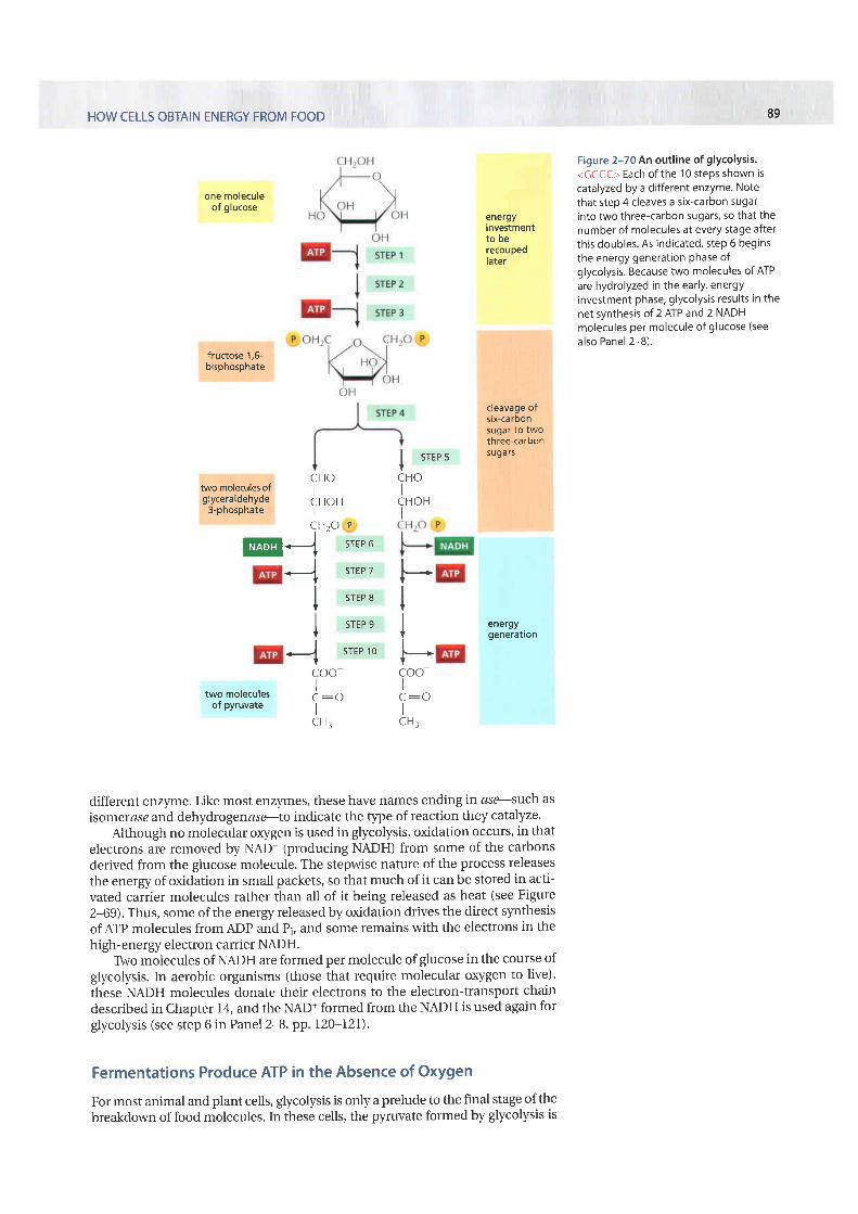
Figure 2-70 An outl ine of glycolysis'<GGGC> Each ofthe 10 steps shown iscatalyzed by a different enzyme. Notethat step 4 cleaves a six-carbon sugarinto two three-carbon sugars, so that thenumber of molecules at every stage afterthis doubles. As indicated, step 6 beginsthe energy generation phase ofglycolysis. Because two molecules of ATPare hydrolyzed in the early, energYinvestment phase, glycolysis results in thenet synthesis of 2 ATP and 2 NADHmolecules per molecule of glucose (seealso Panel 2-B).
two molecules ofglyceraldehyde
3-phosphate
C H OI
C H O Hi
cH2o PI
Ia srEPT
| , ' ,r ,I
I srEP eI
< sTEP1O
COO
C : oI
C H r
@
two moleculesof pyruvate
different enzyrne. Like most enzymes, these have names ending in ase--such asisomerase and dehydro genase-,lo indicate the type of reaction they catalyze.
Although no molecular oxygen is used in glycolysis, oxidation occurs, in thatelectrons are removed by NAD+ (producing NADH) from some of the carbonsderived from the glucose molecule. The stepwise nature of the process releasesthe energy of oxidation in small packets, so that much of it can be stored in acti-vated carrier molecules rather than all of it being released as heat (see Figure2-69). Thus, some of the energy released by oxidation drives the direct slmthesisof ATP molecules from ADP and Pi, and some remains with the electrons in thehigh-energy electron carrier NADH.
TWo molecules of NADH are formed per molecule of glucose in the course ofglycolysis. In aerobic organisms (those that require molecular oxygen to live),these NADH molecules donate their electrons to the electron-transport chaindescribed in Chapter 14, and the NAD+ formed from the NADH is used again forglycolysis (see step 6 in Panel 2-8, pp. 120-l2I).
Fermentations Produce ATP in the Absence of OxygenFor most animal and plant cells, glycolysis is only a prelude to the final stage of thebreakdo'orn of food molecules. In these cells, the p],'ruvate formed by glycolysis is
I srre sICHOI
CHOHI
t---*IIIItIl-coo-II
C H :
90 Chapter 2: Cell Chemistry and Biosynthesis
rapidly transported into the mitochondria, where it is converted into co2 plusacetyl CoA, which is then completely oxidized to CO2 and H2O.
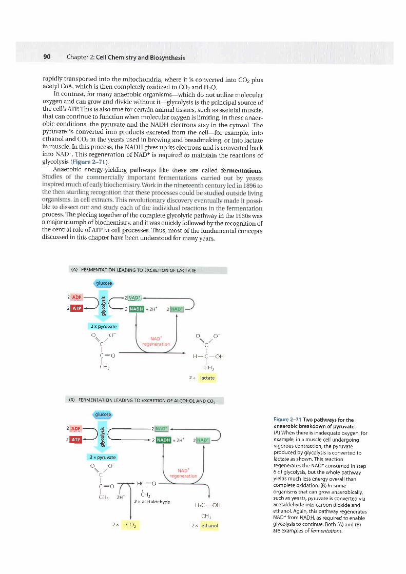
In contrast, for many anaerobic organisms-which do not utilize morecularoxygen and can grow and divide without it-glycolysis is the principal source ofthe cell's ArP This is also true for certain animal tissues, such as skeletal muscle,that can continue to function when molecular oxygen is limiting. In these anaer-obic conditions, the pyruvate and the NADH electrons stay in the cytosol. Thepyruvate is converted into products excreted from the cell-for example, intoethanol and co2 in the yeasts used in brewing and breadmaking, or into lactatein muscle. In this process, the NADH gives up its electrons and is converted backinto NAD+. This regeneration of NAD+ is required to maintain the reactions ofglycolysis (Figure 2--7 l).
Anaerobic energy-yielding pathways like these are called fermentations.
process. The piecing together of the complete glycolltic pathway in the 1930s wasa major triumph of biochemistry, and it was quickly followed by the recognition ofthe central role of ArP in cell processes. Thus, most of the fundamentalionceptsdiscussed in this chapter have been understood for manv vears.
(A) FERMENTATION LEADING TO EXCRETION OF LACTATE
grucose
- ^-' ........._.\ '6 /.-'t - > I=-
E D l r
2 x pyruvate
oo -\ , ,CIIcH:r
o. o-\ / /CII
CH:
Figure 2-71 Two pathways for theanaerobic breakdown of pyruvate.(A) When there is inadequate oxygen, forexample, in a muscle cel l undergoingvigorous contraction, the pyruvateproduced by glycolysis is converted tolactate as shown. This reacttonregenerates the NADt consumed in step6 of glycolysis, but the whole pathwayyields much less energy overal l thancomplete oxidation. (B) In someorganisms that can grow anaerobical ly,such as yeasts, pyruvate is converted viaacetaldehyde into carbon dioxide andethanol. Again, this pathway regeneratesNAD+ from NADH, as required to enableglycolysis to continue. Both (A) and (B)are exampf es of fermentations.
H,C -OH- lCH:
2 x ethanol
2 ADF - .r -2l|{4q:\ E /-E-; --E2 x pyruvate
(B) FERMENTATION LEADING TO EXCRETION OF ALCOHOL AND CO.
grucose
H C : OICH:
2 x acetaldehyde
o. o-\ . /CI
H - C - O HICH:
2 x lactate
2 x C O z
HOW CELLS OBTAIN ENERGY FROM FOOD
Glycolysis l l lustrates How Enzymes Couple Oxidation to EnergyStorageReturning to the paddle-wheel analogy that we used to introduce coupled reac-tions (see Figure 2-56), we can now equate enzymes with the paddle wheel.Enzymes act to harvest useful energy from the oxidation of organic moleculesby coupling an energetically unfavorable reaction with a favorable one. Todemonstrate this coupling, we examine a step in glycolysis to see exactly howsuch coupled reactions occur.
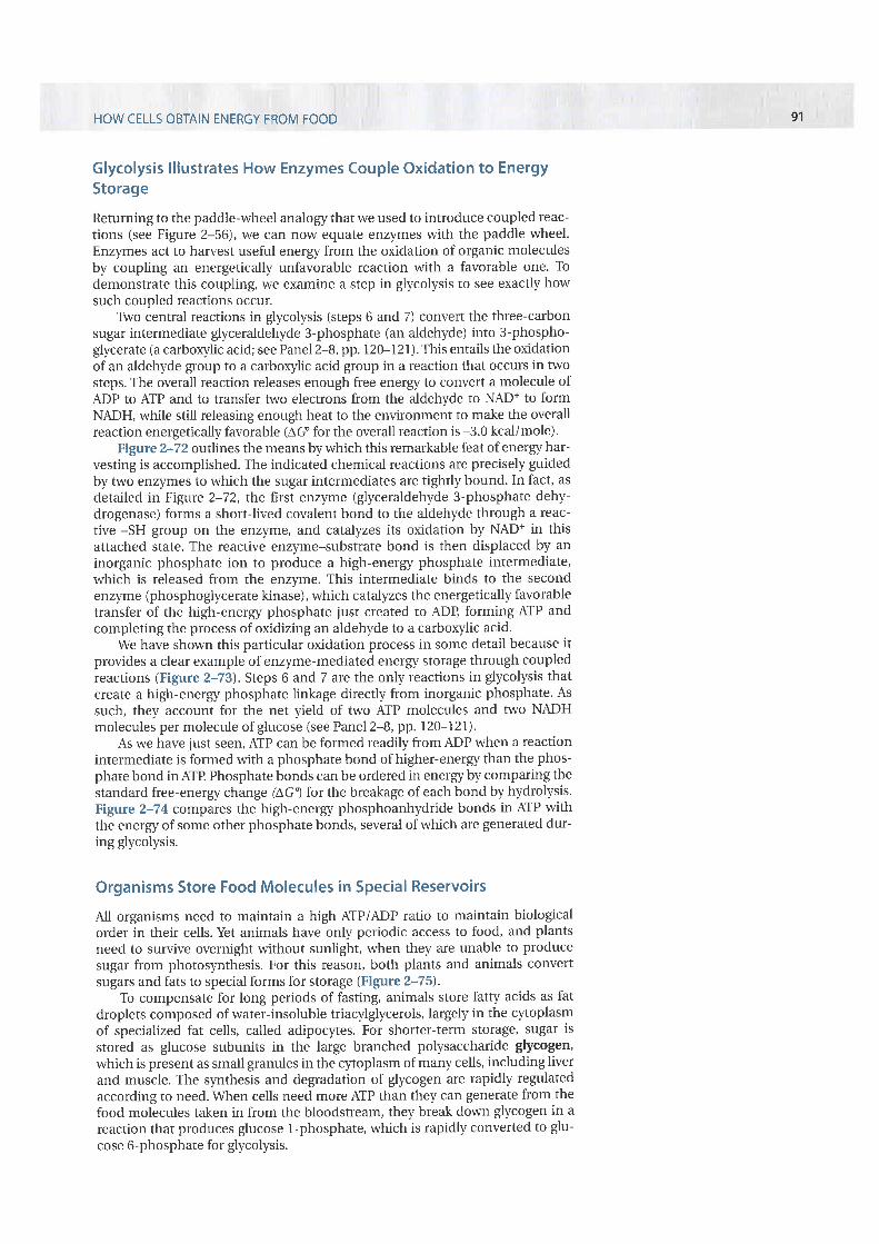
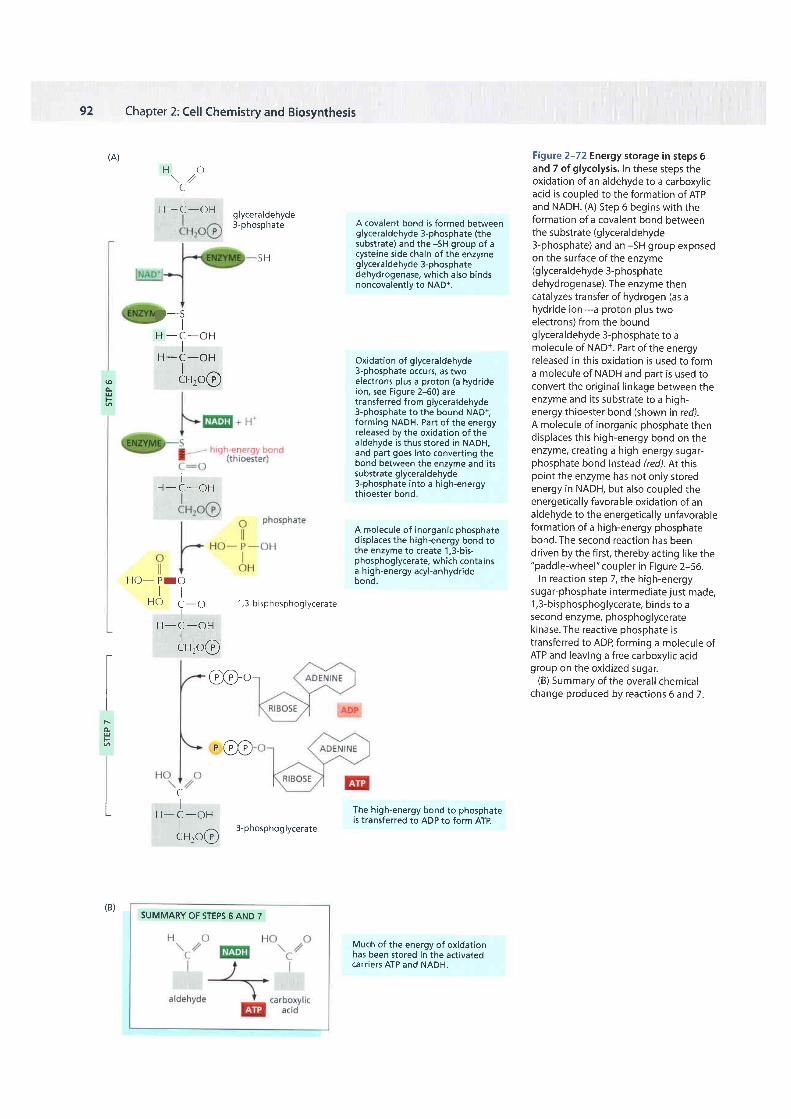
TWo central reactions in glycolysis (steps 6 and 7) convert the three-carbonsugar intermediate glyceraldehyde 3-phosphate (an aldehyde) into 3-phospho-glycerate (a carboxylic acid; see Panel 2-8, pp. 120-121). This entails the oxidationof an aldehyde group to a carboxylic acid group in a reaction that occurs in twosteps. The overall reaction releases enough free energy to convert a molecule ofADP to AIP and to transfer two electrons from the aldehyde to NAD* to formNADH, while still releasing enough heat to the environment to make the overallreaction energetically favorable (AG for the overall reaction is -3.0 kcal/mole).
Figure 2-72 otttlines the means by which this remarkable feat of energy har-vesting is accomplished. The indicated chemical reactions are precisely guidedby two enzymes to which the sugar intermediates are tightly bound. In fact, asdetailed in Figure 2-72, the first enzyme (glyceraldehyde 3-phosphate dehy-drogenase) forms a short-lived covalent bond to the aldehyde through a reac-tive -SH group on the enzyme, and catalyzes its oxidation by NAD+ in thisattached state. The reactive enzyme-substrate bond is then displaced by aninorganic phosphate ion to produce a high-energy phosphate intermediate,which is released from the enzyme. This intermediate binds to the secondenzyme (phosphoglycerate kinase), which catalyzes the energetically favorabletransfer of the high-energy phosphate just created to ADB forming AIP andcompleting the process of oxidizing an aldehyde to a carboxylic acid.
We have shown this particular oxidation process in some detail because itprovides a clear example of enzyme-mediated energy storage through coupledreactions (Figure 2-73). Steps 6 and 7 are the onlyreactions in glycolysis thatcreate a high-energy phosphate linkage directly from inorganic phosphate. Assuch, they account for the net yield of two AIP molecules and two NADHmolecules per molecule of glucose (see Panel 2-8, pp.l20-I2l).
As we have just seen, AIP can be formed readily from ADP when a reactionintermediate is formed with a phosphate bond of higher-energy than the phos-phate bond in AIP Phosphate bonds can be ordered in energy by comparing thestandard free-energy change (AGl for the breakage of each bond by hydrolysis.Figure 2-74 compares the high-energy phosphoanhydride bonds in ATP withthe energy of some other phosphate bonds, several of which are generated dur-ing glycolysis.
Organisms Store Food Molecules in Special ReservoirsAll organisms need to maintain a high ATP/ADP ratio to maintain biologicalorder in their cells. Yet animals have only periodic access to food, and plantsneed to survive overnight without sunlight, when they are unable to producesugar from photosynthesis. For this reason, both plants and animals convertsugars and fats to special forms for storage (Figure 2-75).
To compensate for long periods of fasting, animals store fatty acids as fatdroplets composed of water-insoluble triacylglycerols, largely in the cytoplasmof specialized fat cells, called adipocltes. For shorter-term storage, sugar isstored as glucose subunits in the large branched polysaccharide glycogen,which is present as small granules in the cltoplasm of many cells, including liverand muscle. The synthesis and degradation of glycogen are rapidly regulatedaccording to need. \.A/hen cells need more AIP than they can generate from thefood molecules taken in from the bloodstream, they break down glycogen in areaction that produces glucose 1-phosphate, which is rapidly converted to glu-cose 6-phosphate for glycolysis.
91
92 Chapter 2: Cell Chemistry and Biosynthesis
H O\ / /(_
IH - C - O H glyceraldehyde
3-phosphate
(A)
A covalent bond is formed betweenglyceraldehyde 3-phosphate (thesubstrate) and the -5H group of acysteine side chain of the enzymeglyceraldehyde 3-phosphatedehydrogenase, which also bindsnoncovalently to NAD+.
Oxidation of glyceraldehyde3-phosphate occurs, as twoelectrons plus a proton (a hydrideion, see Figure 2-60) aretransferred from glyceraldehyde3-phosphate to the bound NAD+,forming NADH. Part of the energyreleased by the oxidation of thealdehyde is thus stored in NADH,and part goes into converting thebond between the enzyme and itssubstrate glyceraldehyde3-phosphate into a high-energythioester bond
A molecule of inorganic phosphatedisplaces the high-energy bond tothe enzyme to create 1,3-bis-phosphoglycerate, which containsa high-energy acyl-anhydridebond.
The high-energy bond to phosphateis transferred to ADP to form ATP.
Much of the energy of oxidationhas been stored in the activateocarriers ATP and NADH.
Figure 2-72 Energy storage in steps 6and 7 of glycolysis. In these steps theoxidation of an aldehyde to a carboxyl icacid is coupled to the formation of ATPand NADH. (A) Step 6 begins with theformation of a covalent bond betweenthe substrate (glyceraldehyde3-phosphate) and an -5H group exposedon the surface of the enzyme(glyceraldehyde 3-phosphatedehydrogenase). The enzyme thencatalyzes transfer of hydrogen (as ahydride ion-a proton plus twoelectrons) from the boundglyceraldehyde 3-phosphate to amolecule of NAD+. Part of the energyreleased in this oxidation is used to forma molecule of NADH and part is used toconvert the original l inkage between theenzyme and i ts substrate to a high-energy thioester bond (shown in red.).A molecule of inorganic phosphate thendisplaces this high-energy bond on theenzyme, creating a high-energy sugar-phosphate bond instead (red). At thispoint the enzyme has not only storedenergy in NADH, but also coupled theenergetical ly favorable oxidation of analdehyde to the energetical ly unfavorableformation of a high-energy phosphatebond. The second reaction has beendriven by the f irst, thereby acting l ike the"paddle-wheel" coupler in Figure 2-56.
In reaction step 7, the high-energysugar-phosphate intermediate just made,1,3-bisphosphoglycerate, binds to asecond enzyme, phosphoglyceratekinase.The reactive phosphate istransferred to ADP, forming a molecule ofATP and leaving a free carboxyl ic acidgroup on the oxidized sugar.
(B) Summary of the overal l chemicalchange produced by reactions 6 and 7 .
S H
(ocrF
IH - C - O H
H O - P I Ot iU Ar- iLJ C:O 1 ,3-b isphosphog lycera te
IH - C - O H
l ^cH2o(L)
@@o
r fr_A-r,
F4
H
CI
H - C - O Hl ^cH2o(L)
3-phosphog lycerate
SUMMARY OF STEPS 6 AND 7
t-?I
H - C - O HI
H _ C _ O HIcH2oo
(B)

HOW CELLS OBTAIN ENERGY FROM FOOD
C-H bondoxidat ion
energy
STEP6 +
total energy change for step 6 fo l lowed by step
o
\ , ,
93
Figure 2-73 Schematic view of thecoupled reactions that form NADH andATP in steps 6 and 7 of glycolysis. TheC-H bond oxidation energy drives theformation of both NADH and a high-energy phosphate bond. The breakage ofthe high-energy bond then drives ATPformation.o
coao
ME
o\ , /
CI
CI
STEP 7
7 is a favorable -3 kcal /mole
enol phosphatebond
anhydr idebond to carbon
phosphatebond increaTtnephosphate
anhydr idebond tophosphate a._a)-(phospho-anhydr idebond)
phosphoesterbond
o -o\ / /C Or l l
H 'C : C -O- .P -O-' , / lHro / o-
oooi l i l t li-"-i-o7i-"-o - o -
/ o -Hzo
H Ol l l, -i-"vP-o-" , / o -Hzo
type of phosphate bond
phosphoenol pyruvateisee ianel Z-8,' pp. 120-121)
- 14'6(-61 e)
o ot l t lc-c-o y'P o-
H r o / o
creat ine ohosphate(act ivated carr ier that -10.3stores energy in muscle) (-43.0)
for example,1,3-bisphosphoglycerate(see Panel 2-8)
for example,ATP when hydrolyzedtO ADP
for example,glucose 6-phosphate(see Panel 2-8)
-7.3(-30 6)
specific examples showing thestandard free-energy change (AG')for hydrolysis of phosphate bond
Figwe 2-74 Phosphate bonds have different energies. Examples of different types of phosphate bonds with their sites of hydrolysis areshown in the molecules depicted on the left. Those itarting with a gray catbon atom show only part of a molecule. Examples of moleculescontaining such bonds are given on the r ight, with the free-energy change for hydrolysis in ki localories (ki lojoules in parentheses)'The transferof a phosfhate group from one molecule to another is energetically favorable if the standard free-energy change (AG') for hydrolysis of thephosphate bond of the f irst molecule is more negative than that for hydrolysis of the phosphate bond in the second. Thus, a phosphate groupis readily transferred from 1,3-bisphosphoglycerate to ADP to form ATP The hydrolysis reaction can be viewed as the transfer of the phosphategroup to water.
94 Chapter 2: Cell Chemisty and Biosynthesis
9rycogeng ranu les i nthe cytoplasmof a l iver cel l
branch point g lucose subuni ts
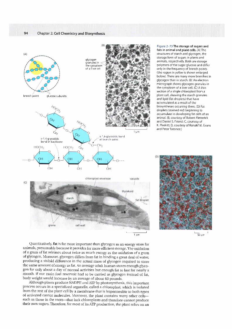
Figure 2-75 The storage of sugars andfats in animal and plant cel ls. (A) Thestructures of starch and glycogen, thestorage form of sugars in plants andanimals, respectively. Both are storagepolymers of the sugar glucose and dif feronly in the frequency of branch points(the region inyel lowis shown enlargedbelow). There are many more branches inglycogen than in starch. (B) An electronmicrograph shows glycogen granules inthe cytoplasm of a l iver cel l . (C) A thinsection of a single chloroplast from aplant cel l , showing the starch granulesand l ipid (fat droplets) that haveaccumulated as a result of thebiosyntheses occurring there. (D) Fatdroplets (stained red) beginning toaccumulate in developing fat cel ls of ananimal. (8, courtesy of Robert Fletterickand Daniel S. Friend; C, courtesy ofK. Plaskit t ; D, courtesy of Ronald M. Evansand PeterTotonoz.)
1 p m
o 1,6-9lycosid ic bond,,- at branch point
/o -cH2
l;------.,
Quantitatively, fat is far more important than glycogen as an energy store foranimals, presumably because it provides for more efficient storage. The oxidationof a gram of fat releases about twice as much energy as the oxidation of a gramof glycogen. Moreover, glycogen differs from fat in binding a great deal of water,producing a sixfold difference in the actual mass of glycogen required to storethe same amount of energy as fat. An average adult human stores enough glyco-gen for only about a day of normal activities but enough fat to last for nearly amonth. If our main fuel reservoir had to be carried as glycogen instead of fat,body weight would increase by an average of about 60 pounds.
Although plants produce NADPH and Arp by photosynthesis, this importantprocess occurs in a specialized organelle, called a chloroplast, which is isolatedfrom the rest of the plant cell by a membrane that is impermeable to both typesof activated carrier molecules. Moreover, the plant contains many other cells-such as those in the roots-that lack chloroplasts and therefore cannot producetheir or,rm sugars. Therefore, for most of its ATP production, the plant relies on an
O H
ql
a 1,4-glycosid icbond in backbone
50 u.
HOW CELLS OBTAIN ENERGY FROM FOOD
metabolites
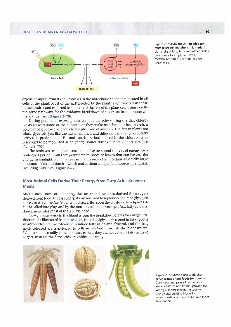
export of sugars from its chloroplasts to the mitochondria that are located in allcells of the plant. Most of the AIP needed by the plant is synthesized in thesemitochondria and exported from them to the rest of the plant cell, using exactlythe same pathways for the oxidative breakdor,rrn of sugars as in nonphotosyn-thetic organisms (Figure 2-76).
During periods of excess photosynthetic capacity during the day, chloro-plasts convert some of the sugars that they make into fats and into starch, apolgner of glucose analogous to the glycogen of animals. The fats in plants aretriacylglycerols, just like the fats in animals, and differ only in the types of fattyacids that predominate. Fat and starch are both stored in the chloroplast asreservoirs to be mobilized as an energy source during periods of darkness (seeFigure 2-75C).
The embryos inside plant seeds must live on stored sources of energy for aprolonged period, until they germinate to produce leaves that can harvest theenergy in sunlight. For this reason plant seeds often contain especially largeamounts of fats and starch-which makes them a malor food source for animals,including ourselves (Figare 2-7 7),
Most Animal Cells Derive Their Energy from Fatty Acids BetweenMealsAfter a meal, most of the energy that an animal needs is derived from sugarsderived from food. Excess sugars, if any, are used to replenish depleted glycogenstores, or to synthesize fats as a food store. But soon the fat stored in adipose tis-sue is called into play, and by the morning after an overnight fast, fatty acid oxi-dation generates most of the ATP we need.
Low glucose levels in the blood trigger the breakdown of fats for energy pro-duction. As illustrated in Figure 2-78, the triacylglycerols stored in fat dropletsin adipocl'tes are hydrolyzed to produce fatty acids and glycerol, and the fattyacids released are transferred to cells in the body through the bloodstream.\.\hile animals readily convert sugars to fats, they cannot convert fatty acids tosugars. Instead, the fatty acids are oxidized directly.
l i gh t
. 9 5
Figure 2-76 How the ATP needed formost plant cel l metabolism is made. Inplants, the chloroplasts and mitochondriacol laborate to supply cel ls withmetabolites and ATP. (For details, seeChapter 14.)
Figure 2-77 Some Plant seeds thatserve as important foods for humans.Corn, nuts, and Peas al l contain r ichstores of starch and fat that provide theyoung plant embryo in the seed withenergy and bui lding blocks forbiosynthesis. (Courtesy of the John InnesFoundation.)
ch loroplast
96 Chapter 2: Cel l Chemistry and Biosynthesis
glycerol
MUSCLE CELL
fatty acidsoxidat ion inmitochondr ia
Sugars and Fats Are Both Degraded to AcetylCoA inMitochondria
The fatty acids imported from the bloodstream are moved into mitochon-dria, where all of their oxidation takes place ). Each molecule offatty acid (as the activated molecule /a tty acyl coA) is broken down completelyby a cycle of reactions that trims two carbons at a time from its carboxyl end,generating one molecule of acetyl coA for each turn of the cycle. A molecule ofNADH and a molecule of FADH2 are also produced in this proces
Sugars and fats are the major energy sources for most non-organisms, including humans. However, most of the useful ene
stored fat
8 t r imers oflipoamide reductase-transacetylase
Figure 2-78 How stored fats aremobilized for energy production inanimals. Low glucose levels in the bloodtrigger the hydrolysis of thetriacylglycerol molecules in fat dropletsto free fatty acids and glycerol, asillustrated. These fatty acids enter thebloodstream, where they bind to theabundant blood protein, serum albumin.Special fatty acid transporters in theplasma membrane of cel ls that oxidizefatty acids, such as muscle cells, then passthese fatty acids into the cytosol, fromwhich they are moved into mitochondriafor energy production (see Figure 2-80).
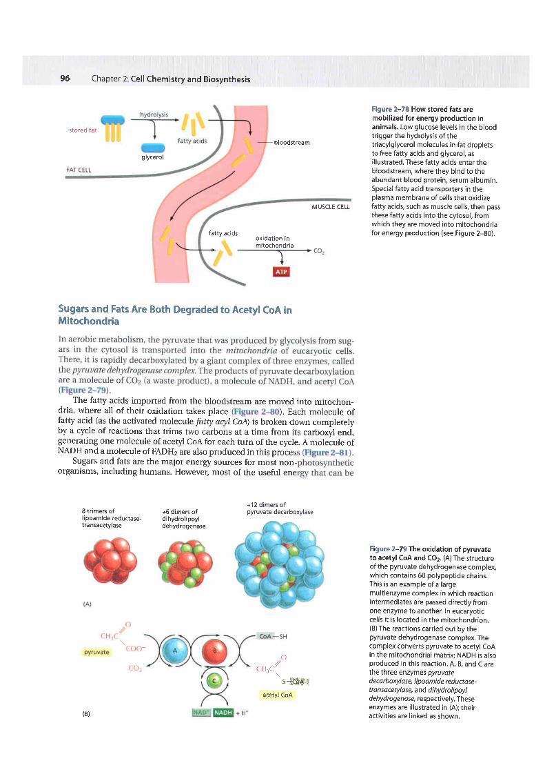
Figure 2-79 The oxidation of pyruvateto acetyl CoA and COz. (A) The structureof the pyruvate dehydrogenase complex,which contains 60 polypeptide chains.This is an example of a largemultienzyme complex in which reactionintermediates are passed directly fromone enzyme to another. In eucaryoticcel ls i t is located in the mitochondrion.(B) The reactions carried out by thepyruvate dehydrogenase complex. Thecomplex converts pyruvate to acetyl coAin the mitochondrial matr ix; NADH is alsoproduced in this reaction. A, B, and C arethe three enzymes pyruvated eca rboxylase, I i poa m i de red uctase-tro n sacetylo se, and di hyd rol ipoy Id ehyd rogen ase, respectively. Theseenzymes are i l lustrated in (A); theiract ivi t ies are l inked as shown.
bloodstream
+12 dimers ofpyruvate decarboxylase
)
+6 dimers ofdihydrol ipoyldehydrogenase
o,//cH.csi*ht{iiii
acetyl coA
(B)
HOW CELL5 OBTAIN ENERGY FROM FOOD
Sugars andpolysaccharides
Fats +fatty acids
extracted from the oxidation of both types of foodstuffs remains stored in theacetyl CoA molecules that are produced by the two t)?es of reactions justdescribed. The citric acid cycle of reactions, in which the acetyl group in acetylCoA is oxidized to CO2 and H2O, is therefore central to the energy metabolism ofaerobic organisms. In eucaryotes these reactions all take place in mitochondria.We should therefore not be surprised to discover that the mitochondrion is theplace where most of the ATP is produced in animal cells. In contrast, aerobicbacteria carry out all of their reactions in a single compartment, the cytosol, andit is here that the citric acid cycle takes place in these cells.
The Citric Acid Cycle Generates NADH by Oxidizing Acetyl Groupsto COzIn the nineteenth century, biologists noticed that in the absence of air (anaero-bic conditions) cells produce lactic acid (for example, in muscle) or ethanol (forexample, in yeast), while in its presence (aerobic conditions) they consume 02and produce CO2 and H2O. Efforts to define the pathways of aerobic metabolism
97
Figure 2-80 Pathways for the production of acetyl CoA from sugars and fats. The mitochondrion ineucaryotic cel ls is the place where acetyl CoA is produced from both types of major food molecules. l t istherefore the olace where most of the cel l 's oxidation reactions occur and where most of i ts ATP is made.The structure and function of mitochondria are discussed in detai l in Chapter 14.
(B)
fatty acyl CoAshortened by .:fl,- CH2two carDons
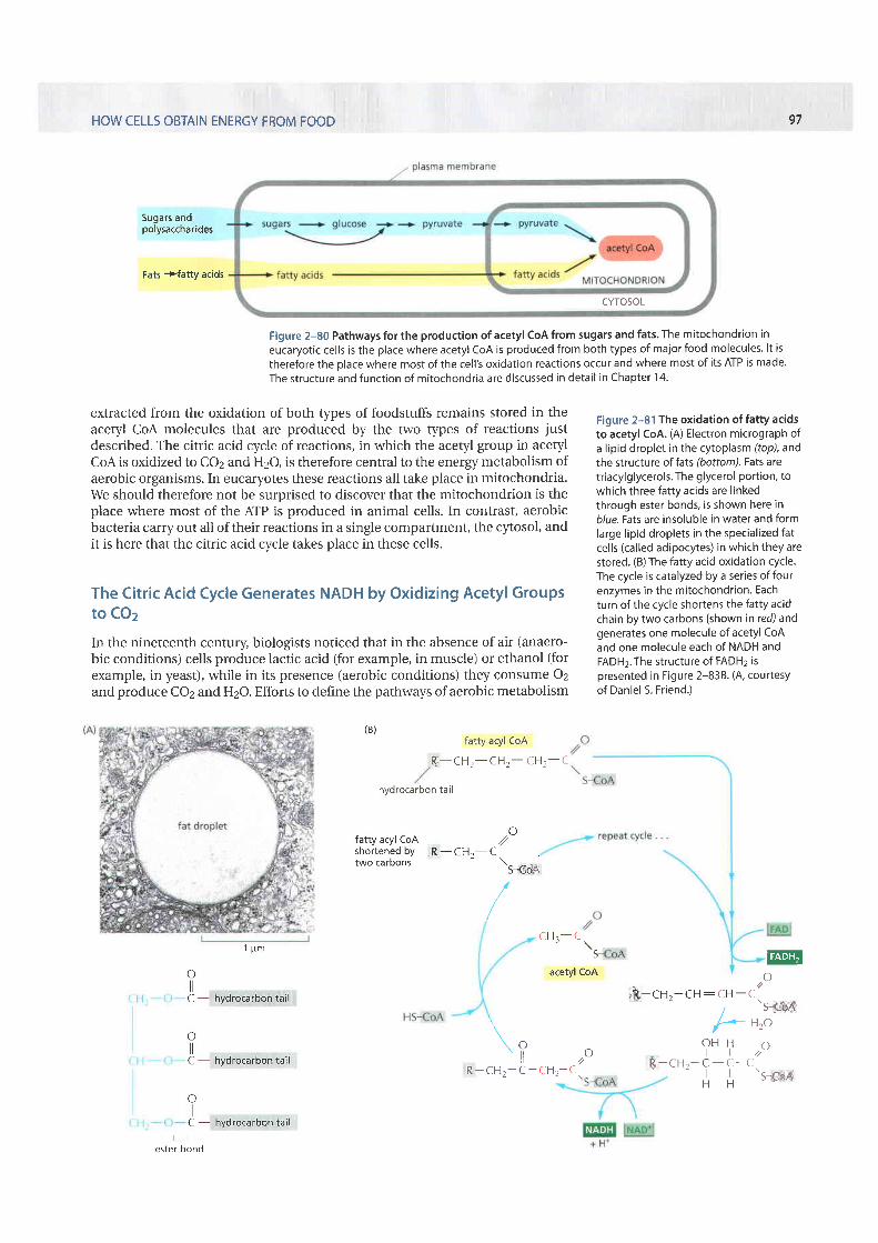
Figure 2-81 The oxidation of fatty acidsto acetyl CoA. (A) Electron micrograph ofa lipid droplet in the cytoplasm (top), andthe structure of fats (bottom). Fats aretriacylglycerols. The glycerol portion, towhich three fatty acids are l inkedthrough ester bonds, is shown here inblue.Fats are insoluble in water and formlarge l ipid droplets in the special ized fatcel ls (cal led adipocytes) in which they arestored. (B) The fatty acid oxidation cycle.The cycle is catalyzed by a series of fourenzymes in the mitochondrion. Eachturn of the cycle shortens the fatty acidchain by two carbons (shown in red) andgenerates one molecule of acetyl CoAand one molecule each of NADH andFADHz. The structure of FADHz ispresented in Figure 2-838. (4, courtesyof Daniel 5. Friend.)
@
i$tct- tr-cu: cH -co//
's 6itHzo
o2C .5{6A
fatty acyl CoA
Rr , -CH2-CH2- CH2-C
hydrocarbon tail
o//- C
\s-coA
/
1 p m
oC - hydrirclrrboh tail
oC - hydrocarbon ta i l
ot lC - hydrocarbon tail
ester bond
C H ? - C' \ S
acetyl CoA
\? ,,oR - C H 2 - C - C H 2 - C .
O H Ht l
C - C -
H H
CYTOSOL
r$,-CHr-
98 Chapter 2: Cell Chemistry and Biosynthesis
eventually focused on the oxidation ofpyruvate and led in 1937 to the discoveryof the citric acid cycle, also knoum as the tricarboxylic acid cycle or the Krebscycle.The citric acid cycle accounts for about two-thirds of the total oxidation ofcarbon compounds in most cells, and its major end products are CO2 and high-energy electrons in the form of NADH. The CO2 is released as a waste product,while the high-energy electrons from NADH are passed to a membrane-boundelectron-transport chain (discussed in Chapter 14), eventually combining with02 to produce H2O. Although the citric acid cycle itself does not use 02, itrequires 02 in order to proceed because there is no other efficient way for theNADH to get rid of its electrons and thus regenerate the NAD+ that is needed tokeep the cycle going.
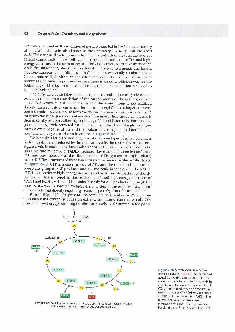
The citric acid cycle takes place inside mitochondria in eucaryotic cells. Itresults in the complete oxidation of the carbon atoms of the acetyl groups inacetyl CoA, converting them into CO2. But the acetyl group is not oxidizeddirectly. Instead, this group is transferred from acetyl CoA to a larger, four-car-bon molecule, oxaloacetate, to form the six-carbon tricarboxylic acid, citric acid,for which the subsequent cycle of reactions is named. The citric acid molecule isthen gradually oxidized, allowing the energy of this oxidation to be harnessed toproduce energy-rich activated carrier molecules. The chain of eight reactionsforms a cycle because at the end the oxaloacetate is regenerated and enters anew turn of the cycle, as shown in outline in Figure 2-82.
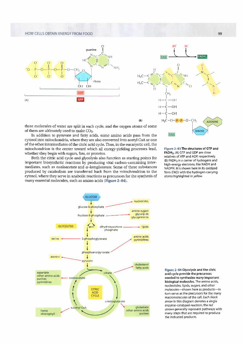
we have thus far discussed only one of the three types of activated carriermolecules that are produced by the citric acid cycle, the NAD+-NADH pair (seeFigure 2-60). In addition to three molecules of NADH, each turn of the cycle alsoproduces one molecule of FADH2 (reduced flavin adenine dinucleotide) fromFAD and one molecule of the ribonucleotide GTP (guanosine triphosphate)from GDP The structures of these two activated carrier molecules are illustratedin Figure 2-83. GTP is a close relative of ATB and the transfer of its terminalphosphate group to ADP produces one ATP molecule in each cycle. Like NADH,FADHz is a carrier of high-energy electrons and hydrogen. As we discuss shortly,the energy that is stored in the readily transferred high-energy electrons ofNADH and FADH2 will be utilized subsequently for Arp production through theprocess of oxidatiue phosphorylation, the only step in the oxidative catabolismof foodstuffs that directly requires gaseous oxygen (oz) from the atmosphere.
Panel 2-9 (pp. 122-123) presents the complete citric acid cycle. Water, ratherthan molecular oxygen, supplies the extra oxygen atoms required to make co2from the acetyl groups entering the citric acid cycle. As illustrated in the panel,
oHrc -t - s-coA
acetyl CoA2Ct
o x d t o a c e l a l e ./4C STEP 1
- ) lu4lllil-,,/+ H * V
/ srEP 8I
4C
* {srEP 2 5C
\ r*E!E--srEP 3
f.- co,I
5C
NET RESULT ONE TURN OF THE CYCLE PRODUCES THREE NADH, ONE GTB ANDONE FADH2, AND RELEASES TWO MOLECULES OF COI
Figure 2-82 Simple overview of thecitric acid cycle. <TAGT> The reaction ofacetyl coA with oxaloacetate starts thecycle by producing citrate (citr ic acid). Ineach turn of the cycle, two molecules ofCO2 are produced as waste products, plusthree molecules of NADH, one moleculeof GTP, and one molecule of FADH2. Thenumber of carbon atoms in eachintermediate is shown in a yellow box.For details, see Panel 2-9 (pp. 122-123).
???\'.,-io -
oguanine l lc
HrC-9H3C-C
OH OH
(B)
three molecules of water are split in each cycle, and the orygen atoms of someof them are ultimately used to make CO2.
In addition to pyruvate and fatty acids, some amino acids pass from thecytosol into mitochondria, where they are also converted into acetyl CoA or oneof the other intermediates of the citric acid cycle. Thus, in the eucaryotic cell, themitochondrion is the center toward which all energy-yielding processes lead,whether they begin with sugars, fats, or proteins.
Both the citric acid cycle and glycolysis also function as starting points forimportant biosynthetic reactions by producing vital carbon-containing inter-mediates, such as oxaloacetate and a-ketoglutarate. Some of these substancesproduced by catabolism are transferred back from the mitochondrion to thecytosol, where they serve in anabolic reactions as precursors for the synthesis ofmany essential molecules, such as amino acids (Figure 244).
2H- 2e
c - N--c - t - rn , rN ' ' c , .l l r t l lt - r - t - - N - C \ O , r C ' - N . '
O Hi l l
t lC H , nI '
H - C - O HI
H - C - O HI
H - C - O H
H2C-O- -O-
Figure 2-83 The structures of GTP andFADHz. (A) GTP and GDP are closerelatives of ATP and ADP, respectively.(B) FADH2 is a carrier of hydrogens andhigh-energy electrons, like NADH andNADPH. lt is shown here in its oxidizedform (FAD) with the hydrogen-canyingatoms h ighlighted in yellow.
Figure 2-84 Glycolysis and the citricacid cycle provide the precursorsneeded to synthesize many importantbiological molecules. The amino acids,nucleotides, lipids, sugars, and othermolecules-shown here as products-inturn serve as the precursors for the manymacromoleculesof the cell. Each b/ackarrowin this diagram denotes a singleenzyme-catalyzed reaction; the redarrows generally represent pathways withmany steps that are required to producethe indicated products.
glucose 6-phosp nur. / nucleotides
+ amrno sugarsfructose 6-phosphate ' glycol ipids
+ glycoproteins
/ \ dihydroxyacetone + l ip idsI PhosPhate+
serine 3-phosphoglycerate++phosphoenol pyruvate
alanine .*- .-
orrrlura"
amino acidspyr imidines
100 Chapter 2: Cell Chemistry and Biosynthesis
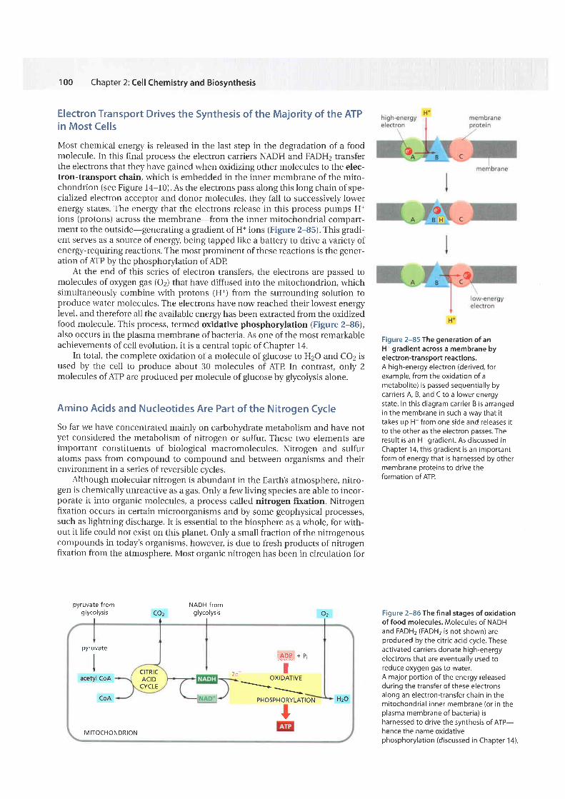
Electron Transport Drives the Synthesis of the Majority of the ATPin Most CellsMost chemical energy is released in the last step in the degradation of a foodmolecule. In this final process the electron carriers NADH and FADH2 transferthe electrons that they have gained when oxidizing other molecules to the elec-tron-transport chain, which is embedded in the inner membrane of the mito-chondrion (see Figure 14-10). As the electrons pass along this long chain of spe-cialized electron acceptor and donor molecules, they fall to successively lowerenergy states. The energy that the electrons release in this process pumps H+ions (protons) across the membrane-from the inner mitochondrial compart-ment to the outside-generating a gradient of H+ ions (Figure 2-85). This gradi-ent serves as a source of energy, being tapped like a battery to drive a variety ofenergy-requiring reactions. The most prominent of these reactions is the gener-ation of ATP by the phosphorylation of ADP
At the end of this series of electron transfers, the electrons are passed tomolecules of oxygen gas (Oz) that have diffused into the mitochondrion, whichsimultaneously combine with protons (H*) from the surrounding solution toproduce water molecules. The electrons have now reached their lowest energyIevel, and therefore all the available energy has been extracted from the oxidizedfood molecule. This process, termed oxidative phosphorylation (Figure 2-86),also occurs in the plasma membrane of bacteria. As one of the most remarkableachievements of cell evolution, it is a central topic of Chapter 14.
In total, the complete oxidation of a molecule of glucose to H2O and CO2 isused by the cell to produce about 30 molecules of ATP In contrast, only 2molecules of ATP are produced per molecule of glucose by glycolysis alone.
Amino Acids and Nucleotides Are Part of the Nitrogen CycleSo far we have concentrated mainly on carbohydrate metabolism and have notyet considered the metabolism of nitrogen or sulfur. These two elements areimportant constituents of biological macromolecules. Nitrogen and sulfuratoms pass from compound to compound and between organisms and theirenvironment in a series of reversible cycles.
Although molecular nitrogen is abundant in the Earth's atmosphere, nitro-gen is chemically unreactive as a gas. Only a few living species are able to incor-porate it into organic molecules, a process called nitrogen fixation. Nitrogenfixation occurs in certain microorganisms and by some geophysical processes,such as lightning discharge. It is essential to the biosphere as a whole, for with-out it life could not exist on this planet. Only a small fraction of the nitrogenouscompounds in today's organisms, however, is due to fresh products of nitrogenfixation from the atmosphere. Most organic nitrogen has been in circulation for
pyruvate fromg lycolysis
I
NADH fromglycolysis
IOzI
CozI
Figure 2-85 The generation of anH+ gradient across a membrane byelectron-transport reactions.A high-energy electron (derived, forexample, from the oxidation of ametaboli te) is passed sequential ly bycarriers A, B, and C to a lower energystate. In this diagram carrier B is arrangedin the membrane in such a way that i ttakes up H+ from one side and releases i tto the other as the electron passes. Theresult is an H+ gradient. As discussed inChapter 14, this gradient is an importantform of energy that is harnessed by othermembrane oroteins to drive theformation of ATP.
Figure 2-86 The f inal stages of oxidationof food molecules. Molecules of NADHand FADH2 (FADHz is not shown) areproduced by the citr ic acid cycle. Theseactivated carr iers donate high-energyelectrons that are eventual ly used toreduce oxygen gas to water.A major port ion of the energy releasedduring the transfer of these electronsalong an electron-transfer chain in themitochondrial inner membrane (or in theplasma membrane of bacteria) isharnessed to drive the synthesis of ATP-hence the name oxidativephosphorylat ion (discussed in Chapter 14).
pyruvate
IIt
acetyl CoA
CoA
ctTRtcACIDCYCLE
:*Hl * P
2e I u^rDAi lvE- -
PHOSPHORYLATIONtMITOCHONDRION
Hzo
HOW CELLS OBTAIN ENERGY FROM FOOD
some time, passing from one living organism to another. Thus present-daynitrogen-fixing reactions can be said to perform a "topping-up" function for thetotal nitrogen supply.
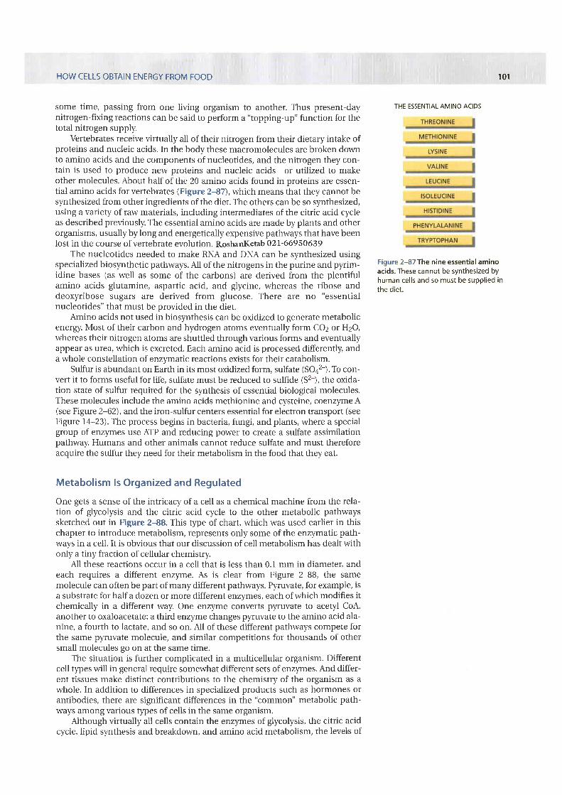
Vertebrates receive virtually all of their nitrogen from their dietary intake ofproteins and nucleic acids. In the body these macromolecules are broken downto amino acids and the components of nucleotides, and the nitrogen they con-tain is used to produce new proteins and nucleic acids-or utilized to makeother molecules. About half of the 20 amino acids found in proteins are essen-tial amino acids for vertebrates (Figure 2-87), which means that they cannot besynthesized from other ingredients of the diet. The others can be so synthesized,using a variety of raw materials, including intermediates of the citric acid cycleas described previously. The essential amino acids are made by plants and otherorganisms, usually by long and energetically expensive pathways that have beenlost in the course of vertebrate evolution. Roshanl(eab 02l-66950639
The nucleotides needed to make RNA and DNA can be synthesized usingspecialized biosynthetic pathways. All of the nitrogens in the purine and pyrim-idine bases (as well as some of the carbons) are derived from the plentifulamino acids glutamine, aspartic acid, and glycine, whereas the ribose anddeoxyribose sugars are derived from glucose. There are no "essentialnucleotides" that must be provided in the diet.
Amino acids not used in biosynthesis can be oxidized to generate metabolicenergy. Most of their carbon and hydrogen atoms eventually form COz or HzO,whereas their nitrogen atoms are shuttled through various forms and eventuallyappear as urea, which is excreted. Each amino acid is processed differently, anda whole constellation of enzymatic reactions exists for their catabolism.
Sulfur is abundant on Earth in its most oxidized form, sulfate (SOaz-). To con-vert it to forms useful for life, sulfate must be reduced to sulfide (S2-), the oxida-tion state of sulfur required for the synthesis of essential biological molecules.These molecules include the amino acids methionine and cysteine, coenzyme A(see Figure 2-62), and the iron-sulfur centers essential for electron transport (seeFigure 14-23). The process begins in bacteria, fungi, and plants, where a specialgroup of enzymes use ATP and reducing power to create a sulfate assimilationpathway. Humans and other animals cannot reduce sulfate and must thereforeacquire the sulfur they need for their metabolism in the food that they eat.
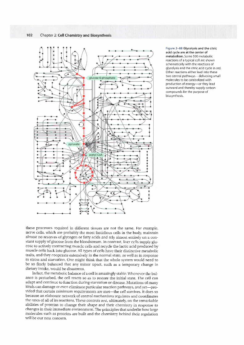
Metabolism ls Organized and RegulatedOne gets a sense of the intricacy of a cell as a chemical machine from the rela-tion of glycolysis and the citric acid cycle to the other metabolic pathwayssketched out in Figure 2-88. This type of chart, which was used earlier in thischapter to introduce metabolism, represents only some of the enzymatic path-ways in a cell. It is obvious that our discussion of cell metabolism has dealt withonly a tiny fraction of cellular chemistry.
All these reactions occur in a cell that is less than 0.1 mm in diameter, andeach requires a different enzyme. As is clear from Figure 2-88, the samemolecule can often be part of many different pathways. Pyruvate, for example, isa substrate for half a dozen or more different enzymes, each of which modifies itchemically in a different way. One enzyme converts pyruvate to acetyl CoA,another to oxaloacetate; a third enzyrne changes pyruvate to the amino acid ala-nine, a fourth to lactate, and so on. All of these different pathways compete forthe same pyruvate molecule, and similar competitions for thousands of othersmall molecules go on at the same time.
The situation is further complicated in a multicellular organism. Differentcell tlpes will in general require somewhat different sets of enzymes. And differ-ent tissues make distinct contributions to the chemistry of the organism as awhole. In addition to differences in specialized products such as hormones orantibodies, there are significant differences in the "common" metabolic path-ways among various types of cells in the same organism.
Although virtually all cells contain the enzymes of glycolysis, the citric acidcycle, lipid synthesis and breakdown, and amino acid metabolism, the levels of
1 0 1
THE ESSENTIAL AMINO ACIDS
Figure ?-87The nine essential aminoacids. These cannot be synthesized byhuman cel ls and so must be supplied inthe diet.
102 Chapter 2: Cell Chemistry and Biosynthesis
these processes required in different tissues are not the same. For example,nerve cells, which are probably the most fastidious cells in the body, maintainalmost no reserves of glycogen or fatty acids and rely almost entirely on a con-stant supply of glucose from the bloodstream. In contrast, liver cells supply glu-cose to actively contracting muscle cells and recycle the lactic acid produced bymuscle cells back into glucose. All types of cells have their distinctive metabolictraits, and they cooperate extensively in the normal state, as well as in responseto stress and starvation. One might think that the whole system would need tobe so finely balanced that any minor upset, such as a temporary change indietary intake, would be disastrous.
In fact, the metabolic balance of a cell is amazingly stable. \.A/henever the bal-ance is perturbed, the cell reacts so as to restore the initial state. The cell canadapt and continue to function during starvation or disease. Mutations of manykinds can damage or even eliminate particular reaction pathways, and yet-pro-vided that certain minimum requirements are met-the cell survives. It does sobecause an elaborate network of control mechanisms regulates and coordinatesthe rates of all of its reactions. These controls rest, ultimately, on the remarkableabilities of proteins to change their shape and their chemistry in response tochanges in their immediate environment. The principles that underlie how largemolecules such as proteins are built and the chemistry behind their regulationwill be our next concern.
Figure 2-88 Glycolysis and the citricacid cycle are at the center ofmetabolism. Some 500 metabolicreactions of a typical cel l are shownschematical ly with the reactions ofglycolysis and the citr ic acid cycle in red.Other reactions either lead into thesetwo central pathways-del ivering smallmolecules to be catabolized withproduction of energy-or they leadoutward and thereby supply carboncompounds for the purpose ofbiosynthesis.
END-OF-CHAPTER PROBLEMS
SummaryGlucose and other food molecules are broken down by controlled stepwise oxidation toprouide chemical energy in the form of ATP and NADH. There are three main sets ofreactions that act in series-the products of each being the starting material for thenext: glycolysis (which occurs in the cytosol), the citric acid cycle (in the mitochondrialmatrix), and oxidatiue phosphorylation (on the inner mitochondrial membrane). Theintermediate products of glycolysk and the citric acid cycle are used both as sources ofmetabolic energy and to produce many of the small molecules used as the raw materi-als for biosynthesis. Cells store sugar molecules as glycogen in animals and starch inplants; both plants and animals also use fats extensiuely as a food store. These storagematerials in turn serue as a major source of food for humans, along with the proteinsthat comprise the majority of the dry mass of most of the cells in the foods we eet.
103
PROBLEMSWhich statements are true? Explain why or why not.
2-1 Of the original radioactivity in a sample, only about1/ 1000 will remain after 10 half-lives.
2-2 A 10-B M solution of HCI has a pH of B.
2-3 Most of the interactions between macromoleculescould be mediated just as well by covalent bonds as by non-covalent bonds.
2-4 Animals and plants use oxidation to extract energyfrom food molecules.
2-5 If an oxidation occurs in a reaction, it must beaccompanied by a reduction.
2-6 Linking the energetically unfavorable reaction A -+ Bto a second, favorable reaction B -+ C will shift the equilib-rium constant for the first reaction.
2-7 The criterion for whether a reaction proceeds spon-taneously is AG not AGo, because AG takes into account theconcentrations of the substrates and products.
2-8 Because glycolysis is only a prelude to the oxidationof glucose in mitochondria, which yields l5-fold more AIBglycolysis is not really important for human cells.
2-9 The oxygen consumed during the oxidation of glu-cose in animal cells is returned as COz to the atmosphere.
Discuss the following problems.
2- 1 0 The organic chemistry of living cells is said to be spe-cial for two teasons: it occurs in an aqueous environmentand it accomplishes some very complex reactions. But doyou suppose it is really all that much different from theorganic chemistry carried out in the top laboratories in theworld? \A/try or why not?
2-11 The molecular weight of ethanol (CHgCHzOH) is 46and its density is 0.789 g/cm3.A. \A4rat is the molarity of ethanol in beer that is 5%ethanol by volume? [Alcohol content of beer varies fromabout 4Vo (lite beer) to B% (stout beer).1B. The legal limit for a driver's blood alcohol contentvaries, but 80 mg of ethanol per 100 mL of blood (usually
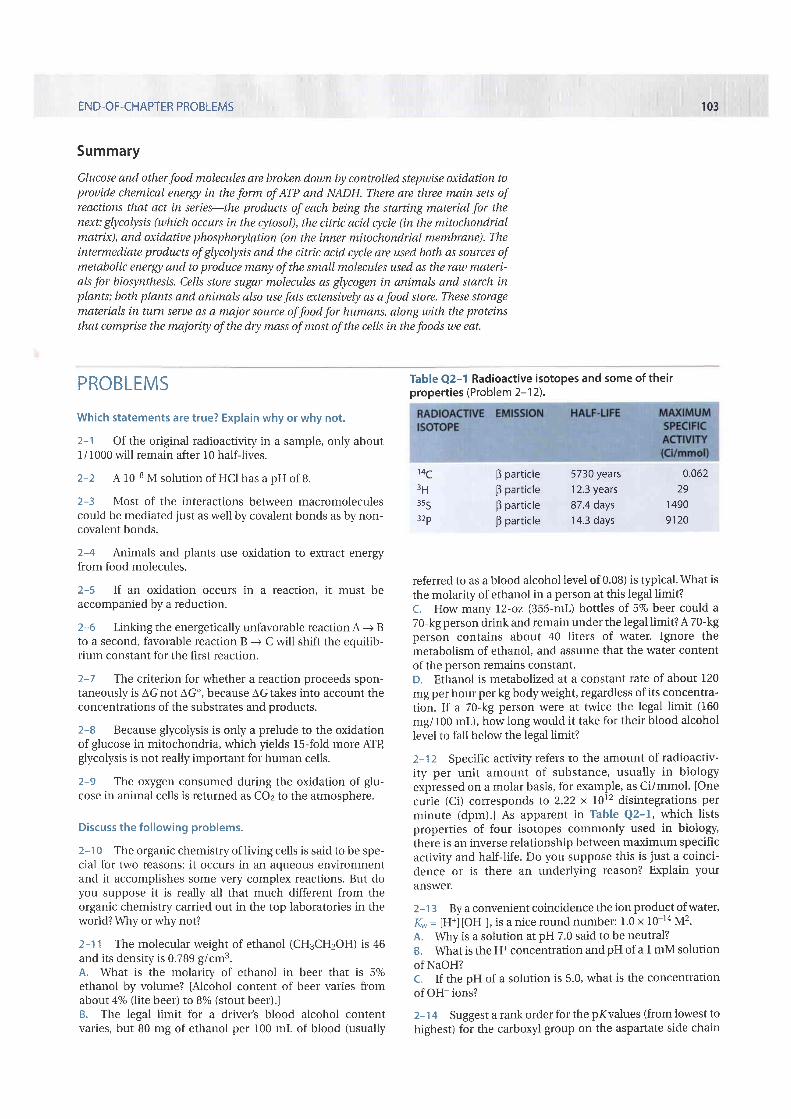
Table Q2-1 Radioactive isotopes and some of theirproperties (Problem 2-1 2).
14c3 635s32P
B particle 5730 yearsB particle 12.3 yearsB particle 87.4 daysB particle 14.3 days
0.06229
14909120
referred to as a blood alcohol level of 0.08) is t)?ical. \ /hat isthe molarity of ethanol in a person at this legal limit?t. How many l2-oz (355-mL) bottles of 5% beer could a70-kg person drink and remain under the legal limit? A 70-kgperson contains about 40 l iters of water. Ignore themetabolism of ethanol, and assume that the water contentof the person remains constant.D. Ethanol is metabolized at a constant rate of about 120mg per hour per kg body weight, regardless of its concentra-tion. If a 70-kg person were at twice the legal limit (160mg/f 00 mL), how long would it take for their blood alcohollevel to fall below the legal limit?
2-12 Specific activity refers to the amount of radioactiv-ity per unit amount of substance, usually in biologyexpressed on a molar basis, for example, as Ci/mmol. [Onecurie (Ci) corresponds to 2.22 x 10¹² disintegrations perminute (dpm;.1 As apparent in Table Q2-1, which listsproperties of four isotopes commonly used in biology,there is an inverse relationship between maximum specificactivity and half-life. Do you suppose this is just a coinci-dence or is there an underlying reason? Explain youranswer.
2-13 By a convenient coincidence the ion product ofwater,K- = lH+l [OH-], is a nice round number: 1.0 x 10⁻¹⁴ M2.A. \AIhy is a solution at pH 7.0 said to be neutral?B. \A/trat is the H+ concentration and pH of a I mM solutionof NaOH?C. If the pH of a solution is 5.0, what is the concentrationof OH- ions?
2-14 Suggest a rank order for the pKvalues (from lowest tohighest) for the carboxyl group on the aspartate side chain
cfEoE
o
104 Chapter 2: Cell Chemistry and Biosynthesis
in the following environments in a protein. Explain yourranking.1. An aspartate side chain on the surface of a protein withno other ionizable groups nearby.2. An aspartate side chain buried in a hydrophobic pocketon the surlace of a protein.3. An aspartate side chain in a hydrophobic pocket adja-cent to a glutamate side chain.4. An aspartate side chain in a hydrophobic pocket adja-cent to a lysine side chain.
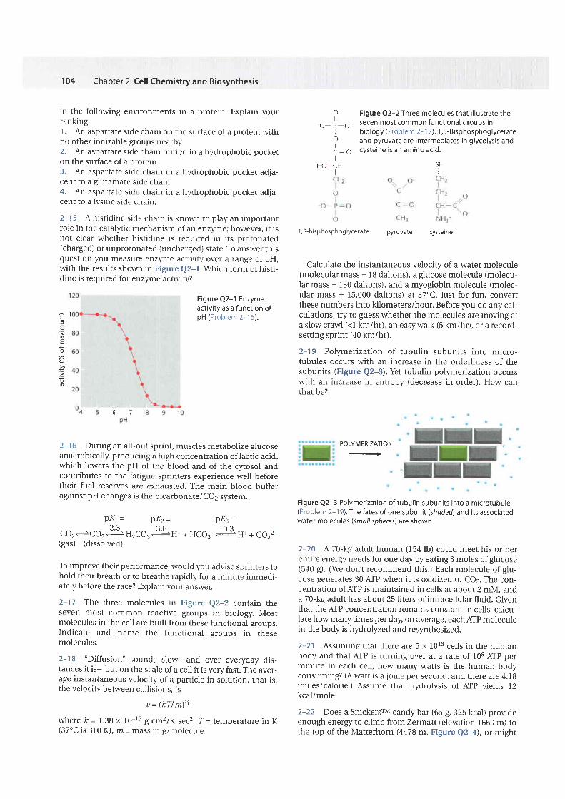
2-15 A histidine side chain is knol,rrn to play an importantrole in the cataly.tic mechanism of an enz).ryne; however, it isnot clear whether histidine is required in its protonated(charged) or unprotonated (uncharged) state. To answer thisquestion you measure enzyrne activity over a range of pH,with the results sho\^Tr in Figure Q2-1. \Ahich form of histi-dine is required for enz)ryne activity?
Figure Q2-1 Enzymeactivi ty as a function ofpH (Prob lem 2-15) .
2-16 During an all-out sprint, muscles metabolize glucoseanaerobically, producing a high concentration oflactic acid,which lowers the pH of the blood and of the cytosol andcontributes to the fatigue sprinters experience well beforetheir fuel reserves are exhausted. The main blood bufferagainst pH changes is the bicarbonate/CO2 system.
PKr = PK2= PtrG =2 . 3 3 8 l o "
CO2+CO2 +: H2CO3 +H* + HCO3- JsH* + CO32-(gas) (dissolved)
To improve their performance, would you advise sprinters tohold their breath or to breathe rapidly for a minute immedi-ately before the race? Explain your answer.
2-17 The three molecules in Figure Q2-2 contain theseven most common reactive groups in biology. Mostmolecules in the cell are built from these functional groups.Indicate and name the functional groups in thesemolecules.
2-18 "Diffusion" sounds slow-and over everyday dis-tances it is-but on the scale of a cell it is very fast. The aver-age instantaneous velocity of a particle in solution, that is,the velocity between collisions, rs
y = (kTlm)h
where k= 1.38 x 10⁻¹⁶ g cmzlKsecz, T = temperature in K(37'C is 310 K), m = mass in g/molecule.
o Figure Q2-2 Three molecules that i l lustrate the,r_ ,_a, seven most common functional groups in
, b io logy(Prob lem2-17) .1 ,3 -B isphosphog lycera te! and pyruvate are intermediates in glycolysis andC - O cysteine is an amino acid.
HO-CH SH
1,3-bisphosphoglycerate pyruvare cyslerne
Calculate the instantaneous velocity of a water molecule(molecular mass = 1B daltons), a glucose molecule (molecu-lar mass = lB0 daltons), and a myoglobin molecule (molec-ular mass = 15,000 daltons) at 37"C. Just for fun, convertthese numbers into kilometers/hour. Before you do any cal-culations, try to guess whether the molecules are moving ata slow crawl (<1 km/hr), an easywalk (5 km/hr), or a record-setting sprint (40 km/hr).
2-19 Polymerization of tubulin subunits into micro-tubules occurs with an increase in the orderliness of thesubunits (Figure Q2-3). Yet tubulin polymerization occurswith an increase in entropy (decrease in order). How canthat be?
Figure Q2-3 Polymerization of tubulin subunits into a microtubule(Problem 2-1 9).The fates of one subunit (shoded) and its associatedwater molecules (small spheres) are shown.
2-2O A 70-kg adult human (154 lb) could meet his or herentire energy needs for one day by eating 3 moles of glucose(5a0 g). (We don't recommend this.) Each molecule of glu-cose generates 30 AIP when it is oxidized to CO2. The con-centration of AIP is maintained in cells at about 2 mM, anda 70-kg adult has about 25 liters of intracellular fluid. Giventhat the ATP concentration remains constant in cells, calcu-late how many times per day, on average, each AIP moleculein the body is hydrolyzed and reslmthesized.
2-21 Assuming that there are 5 x 10¹³ cells in the humanbody and that AIP is turning over at a rate of 10e AIP perminute in each cell, how many watts is the human bodyconsuming? (A watt is a joule per second, and there are 4.18joules/calorie.) Assume that hydrolysis of AIP yields 12kcal/mole.
2-22 Does a SnickersrM candy bar (65 g, 325 kcal) provideenough energy to climb from Zermatt (elevation 1660 m) tothe top of the Matterhorn (4478 m, Figure Q2-4), or might
POLYMERIZATION.+
5 7

END-OF-CHAPTER PROBLEMS
Figure Q2-4 TheMatterhorn (Problem2-22). (Courtesy ofZermatt Tourism.)
you need to stop at Hdrnli Hut (3260 m) to eat another one?Imagine that you and your gear have a mass of 75 kg, andthat all of your work is done against gravity (that is, you arejust climbing straight up). Remember from your introduc-tory physics course that
work (D = mass (kg) x g (m/sec2; x height gained (m)
where gis acceleration due to gravity (9.8 m/sec2). One jouleis 1 kg m2lsec2 and there are 4.lB kf per kcal.
\Alhat assumptions made here will greatly underestimatehow much candy you need?
2-23 At first glance, fermentation of pyruvate to lactateappears to be an optional add-on reaction to glycolysis. Afterall, could cells growing in the absence of oxygen not simplydiscard pyruvate as a waste product? In the absence of fer-mentation, which products derived from glycolysis wouldaccumulate in cells under anaerobic conditions? Could themetabolism of glucose via the glycoll'tic pathway continue inthe absence of oxygen in cells that cannot carry out fermen-tation?\.A/hy or why not?
2-24 In the absence of oxygen, cells consume glucose at ahigh, steady rate. When oxygen is added, glucose consump-tion drops precipitously and is then maintained at the lowerrate. \.Vhy is glucose consumed at a high rate in the absenceof oxygen and at a low rate in its presence?
2-25 The liver provides glucose to the rest of the bodybetween meals. It does so by breaking down glycogen, form-ing glucose 6-phosphate in the penultimate step. Glucose 6-phosphate is converted to glucose by splitting offthe phos-phate (AG' = -3.3 kcal/mole). \Mhy do you suppose the liverremoves the phosphate by hydrolysis, rather than reversingthe reaction by which glucose 6-phosphate (G6P) is formedfrom glucose (glucose + AIP -+ G6P + ADB AG' = -4.0kcal/mole)? By reversing this reaction the liver could gener-ate both glucose and AIP
2-26 In 1904 Franz Knoop performed what was probablythe first successful labeling experiment to study metabolicpathways. He fed many different fatty acids labeled with aterminal benzene ring to dogs and analyzed their urine forexcreted benzene derivatives. \Alhenever the fatty acid hadan even number of carbon atoms, phenylacetate wasexcreted (Figure Q2-5A). lVhenever the fatty acid had anodd number of carbon atoms, benzoate was excreted (Fig-ure Q2-58).
From these experiments Knoop deduced that oxidation offatty acids to CO2 and H2O involved the removal of two-car-bon fragments from the carboxylic acid end of the chain.
105
cH2-C//o\o_
phenylacetate
ott '
*^^ n'\-cur-cH2-cH2-cH2-cH2-cH2-co-comoound l l - |
\at seven-carbon chain
oexcreted ri\.
comoound l l - l o' \4.
TrpE-
I A I - ^ Ofe r t a \ l -CHr -CH2-CH2-CH2-CH2-CH2-CH2-C
.o,,no'Jrna ll -l o-\t' eight-carbon chain
excretedcompound
benzoate
Figure Q2-5 The original labeling experiment to analyze fatty acidoxidation (Problem 2-26). ( ) Fed and excreted derivatives of aneven-number fatty acid chain. (B) Fed and excreted derivatives of anodd-number fatty acid chain.
Can you explain the reasoning that led him to conclude thattwo-carbon fragments, as opposed to any other number,were removed, and that degradation was from the carboxylicacid end, as opposed to the other end?
2-27 Pathways for synthesis of amino acids in microor-ganisms were worked out in part by cross-feeding experi-ments among mutant organisms that were defective forindividual steps in the pathway. Results of cross-feedingexperiments for three mutants defective in the trlptophanparhway-TrpF, TrpIl, and TrpE-are sholrryt in FigureQ2-6. The mutants were streaked on a Petri dish andallowed to grow briefly in the presence of a very smallamount of tryptophan, producing three pale streaks. Asshown, heavier growth was observed at points where somestreaks were close to other streaks. These spots of heaviergrowth indicate that one mutant can cross-feed (supply anintermediate) to the other one.
From the pattern of cross-feeding shor,trr in Figure Q2-6,deduce the order ofthe steps controlled by the products ofthe TrpB, TrpD, and TrpE genes. Explain your reasoning.
CROSS-FEEDING RESULT
TrpD-
TrDB'
Figure Q2-6 Defining the pathway for tryptophan synthesis usingcross-feeding experiments (Problem 2-27). Results of a cross-feeding experiment among mutants defective for steps in thetryptophan biosynthetic pathway. Darkoreas on the Petri dish showregions of cel l growth.
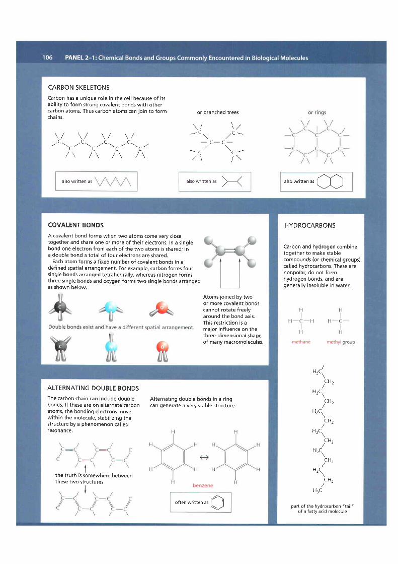
CARBON SKELETONSCarbon has a unique role in the cel l because of i tsabi l i ty to form strong covalent bonds with othercarbon atoms. Thus carbon atoms can ioin to formchai ns.
\ / \ / \ / \ /C C C C
,/ " \ _ r,, " \ ^,r- -\
^,.. "\ ^,,.L L L L
/ \ / \ / \ / \
or branched trees
\ / \ , ,- t \
. / c -- C - C -- c / \ a -, 2 1 / \
also written as alsowrit ten as X
arso written r, al-)
COVALENT BONDSA covalent bond forms when two atoms come very closetogether and share one or more of their electrons. In a singlebond one electron from each of the two atoms is shared; ina double bond a total of four electrons are shared.
Each atom forms a f ixed number of covalent bonds in adefined spatial arrangement. For example, carbon forms foursingle bonds arranged tetrahedral ly, whereas nitrogen formsthree single bonds and oxygen forms two single bonds arrangedas shown below.
Atoms joined by twoor more covalent bondscannot rotate freelyaround the bond axis.This restriction is amajor inf luence on thethree-dimensional shaoeof many macromolecules.
HYDROCARBONS
Carbon and hydrogen combinetogether to make stablecompounds (or chemical groups)cal led hydrocarbons. These arenonpolar, do not formhydrogen bonds, and aregeneral ly insoluble in water.
part of the hydrocarbon "tail"of a fatty acid molecule
HrC.
CH,
HrC.
CHt
HrC.
CH,
HrC.
CH,
H,C
CH,
Hzc.
9H,H:C
ALTERNATING DOUBLE BONDSThe carbon chain can include doublebonds. l f these are on alternate carbonatoms, the bonding electrons movewithin the molecule, stabi l izing thestructure by a phenomenon cal ledreS0nance.
the truth is sbmewhere berweenthese two structures
Alternating double bonds in a r ingcan generate a very stable structure.
often written., O
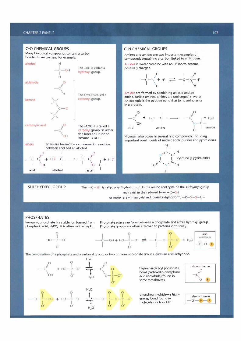
C_O CHEMICAL GROUPSMany biological compounds contain a carbonbonded to an oxygen. For example,
The -COOH is cal led acarboxyl group. In waterthis loses an H+ ion tobecome -COO-.
esters Esters are formed by a condensation reactionbetween acid and an alcohol.
' . O r , O_[_r /+Ho_l___Lr/
I \ | | \ l + H 2 0' o H o - c -acid alcohol ester I
C-N CHEMICAL GROUPSAmines and amides are two important examples ofcompounds containing a carbon l inked to a nitrogen.
Amines in water combine with an H+ ion to becomeposit ively charged.
l r H t , Ht / l /_ c_ ( +u * * _q_n_H*r \nr \H
Amides are formed by combining an acid and anamine. Unlike amines, amides are uncharged in water.An example is the peptide bond that joins amino acidsin a orotein.
o-c / + H"N-J - -
\o" Iac id amtne
Nitrogen also occurs in several r ing compounds, includingimportant consti tuents of nucleic acids: purines and pyrimidines.
)n '^4 r t nI l l cytosine (a pyrimidine)
o/"C tN-C-H
H
SULFHYDRYL GROUP is called a sulfhydryl group. In the amino acid cysteine the sulfhydryl groupmay exist in the reduced form, -t-Sn
l r lor more rarely in an oxidized, cross-bridging form, -C-S-S-C-
PHOSPHATESInorganic phosphate is a stable ion formed fromphosphoric acid, H3PO4. l t is often writ ten as Pi.
Phosphate esters can form between a phosphate and a free hydroxyl group.Phosphate groups are often attached to proteins in this way.
carboxylHzo
l*T
Hzo
Hzo
LT
Hzo
group, or two or more phosphate groups, gives an acid anhydride.
high-energy acyl phosphatebond (carboxylic-phosphoricacid anhydride) found insome metabolites
phosphoanhydride-a high-energy bond found inmolecules such as ATP
also written aso^,- t _
o - P
also written as-o
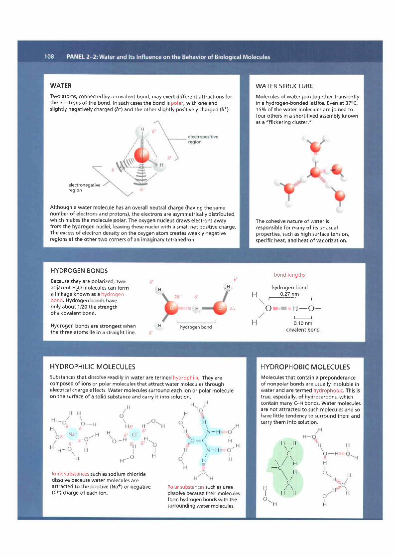
WATERTwo atoms, connected by a covalent bond, may exert different attractions forthe electrons of the bond. In such cases the bond is polar, with one endsl ightly negatively charged (6-) and the other sl ightly posit ively charged (6+).
electronegativeregron
Although a water molecule has an overal l neutral charge (having the samenumber of electrons and protons), the electrons are asymmetrical ly distr ibuteo,which makes the molecule polar. The oxygen nucleus draws electrons awayfrom the hydrogen nuclei, leaving these nuclei with a small net posit ive charge.The excess of electron density on the oxygen atom creates weakly negativeregions at the other two corners of an imaginary tetrahedron.
WATER STRUCTUREMolecules of water join together transientlyin a hydrogen-bonded latt ice. Even at 37oC,15o/o oI the water molecules are joined tofour others in a short- l ived assemblv knownas a "f l ickering cluster."
The cohesive nature of water isresponsible for many of i ts unusualpropert ies, such as high surface tension,specif ic heat, and heat of vaporization.
.:tt:'i l,::,:
bond lengths
hydrogen bond0.27 nm
Q rrrrrrrrrrrrrrrrrrr H -O-
o-.to r'ttcovalent bond
HYDROGEN BONDSBecause they are polarized, twoadjacent H2O molecules can forma l inkage known as a hydrogenbond. Hydrogen bonds haveonly about 1/20 the strengthof a covalent bond.
Hydrogen bonds are strongest whenthe three atoms l ie in a straight l ine.
6-
H ""!,,,
l ,uH hydrogen bond
HYDROPHILIC MOLECULESSubstances that dissolve readi ly in water are termed hydrophil ic. They arecomposed of ions or polar molecules that attract water molecules throughelectr ical charge effects. Water molecules surround each ion or polar moleculeon the surface of a sol id substance and carrv i t into solut ion.
H
lonic substances such as sodium chloridedissolve because water molecules areattracted to the positive (Na+) or negative(Cl ) charge of each ion.
Polar substances such as ureadissolve because their moleculesform hydrogen bonds with thesurrounding water molecules.
HYDROPHOBIC MOLECULESMolecules that contain a preponderanceof nonpolar bonds are usually insoluble inwater and are termed hydrophobic. This istrue, especial ly, of hydrocarbons, whichcontain many C-H bonds. Water moleculesare not attracted to such molecules and sohave l i t t le tendency to surround them andcarry them into solut ion.
H H\ /C
. \\ H- C
H/
CH , / \I H Ho\ . H
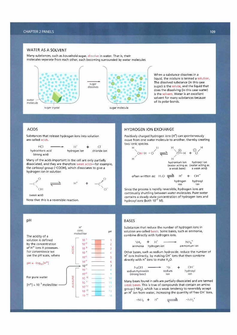
WATER AS A SOLVENTMany substances, such as household sugar, dissolve in water. That is, theirmolecules separate from each other, each becoming surrounded by water molecules.
When a substance dissolves in al iquid, the mixture is termed a solut ion.The dissolved substance ( in this casesugar) is the solute, and the l iquid thatdoes the dissolving ( in this case water)is the solvent. Water is an excel lentsolvent for many substances becauseof i ts polar bonds.
sugar crystal sugar molecule
ACIDSSubstances that release hydrogen ions into solut ionare ca l led ac ids .
H C I - + C fhydrochloric acid hydrogen ion chloride ion
(strong acid)
Many of the acids important in the cel l are only part ial lydissociated, and they are therefore weak acids-for example,the carboxyl group (-COOH), which dissociates to give ahydrogen ion in solut ion
(weak acid)
Note that th is is a revers ib le react ion.
H* + -c /\
HYDROGEN ION EXCHANGEPosit ively charged hydrogen ions (H+) can spontaneouslymove from one water molecule to another, thereby creatingtwo ionic species.
H H,/
o i l i l i l rH-o -H
hydronium ion hydroxyl ion(water acting as (water acting as
a weak base) a weak acid)
often written as: Hro i H* + oH-nrl:Xn"n hydroxyl
Since the process is rapidly reversible, hydrogen ions arecontinually shuttl ing between water molecules. Pure watercontains a steady-state concentration of hydrogen ions andhydroxyl ions (both 10-' M).
n..P- , +oo/
BASESSubstances that reduce the number of hydrogen ions insolut ion are cal led bases. Some bases, such as ammonia,combine direct ly with hydrogen ions.
N H : + H * - N H o *ammonia hydrogen ion ammonium ion
Other bases, such as sodium hydroxide, reduce the number ofH* ions indirect ly, by making OH- ions that then combinedirectly with H' ions to make H2O.
NaOH - Na-sodium hydroxide sodium
(strong base) ion
Many bases found in cells are partially dissociated and are termedweak bases. This is true of compounds that contain an aminogroup (-NH2), which has a weak tendency to reversibly acceptan H' ion from water, increasing the quanti ty of free OH- ions.
- N H z + H * - - N H : *
pH
The acidity of asolut ion is definedby the concentrat ionof H+ ions it possesses.For convenience weuse the pH scale, where
pH = log ,o [H+]
For pure water
[H+] = 10-7 moles/l i ter
1 0 11 0 2
1 0 3
1 o-41 o-s1 0-61o-71 0 8
1 0 e
1 o 1 0
1o -111 0 1 2
1 0 1 3
1 0 1 4
pH
125
A
56789
1 01 11 2I I
1 4
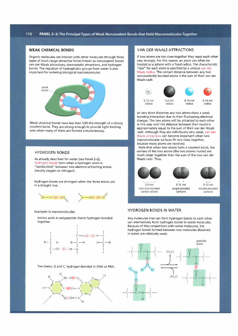
WEAK CHEMICAL BONDSOrganic molecules can interact with other molecules through threetypes of short-range attractive forces known as noncovalent bonds:van der Waals attractions, electrostatic attractions, and hydrogenbonds. The repulsion of hydrophobic groups from water is alsoimportant for ordering biological macromolecules.
Weak chemical bonds have less than 1/20 the strength of a strongcovalent bond. They are strong enough to provide t ight bindingonly when many of them are formed simultaneously.
VAN DER WAALS ATTRACTIONSlf two atoms are too close together they repel each othervery strongly. For this reason, an atom can often betreated as a sohere with a f ixed radius. The characterist ic"size" lor each atom is specif ied by a unique van derWaals radius. The contact distance between any twononcovalently bonded atoms is the sum of their van derWaals radi i .
t l0 . 1 5 n m
single-bondedcaroonS
&0 . 1 5 n m 0 . 1 4 n mradius radius
At very short distances any two atoms show a weakbonding interaction due to their f luctuating electr icalcharges. The two atoms wil l be attracted to each otherin this way unti l the distance between their nuclei isapproximately equal to the sum of their van der Waalsradi i . Although they are individual ly very weak, van derWaals attract ions can become imoortant when twomacromolecular surfaces fit very close together,because many atoms are involved.Note that when two atoms form a covalent bond, the
centers of the two atoms (the two atomic nuclei) aremuch closer together than the sum of the two van derWaals rad i i . Thus .
HYDROGEN BONDSAs already described for water (see Panel 2-2),hydrogen bonds form when a hydrogen atom is"sandwiched " between two electron-attract ing atoms(usually oxygen or nitrogen).
Hydrogen bonds are strongest when the three atoms arein a s t ra igh t l ine :
to-" nnnro\ N - H | | | | | /
Examples in macromolecules:
Amino acids in polypeptide chains hydrogen-bondedtogether.
IIl tc :o i l i l i l i l i l H-N
R - C - HI
F F C - RIc : o l l l l l l l l l t H - N I
l l l
Two bases, G and C, hydrogen-bonded in DNA or RNA.
F i gH. N -Hililill\ '
C - C// \\. . : / \H- L. .N ilililililtH-\ /N - C .,/ \\
, , / 'b l l l l l l l tn-N'tH
I
R - C - H
HYDROGEN BONDS IN WATER
Any molecules that can form hydrogen bonds to each othercan alternatively form hydrogen bonds to water molecules.Because of this competit ion with water molecules, thehydrogen bonds formed between two molecules dissolvedin water are relat ively weak.

HYDROPHOBIC FORCES Water forces hydrophobic groups together,because doing so minimizes their disruptiveeffects on the hydrogen-bonded waternetwork. Hydrophobic groups heldtogether in this way are sometimes saidto be held together by "hydrophobicbonds," even though the apparent attract ionis actual ly caused by a repulsion from thewater.
E LECTROSTATIC ATTRACTIONS
Attractive forces occur both between fully chargedgroups ( ionic bond) and between the part ial ly chargedgroups on polar molecules.
The force of attraction between the two charges, 6+and 5-, fal ls off rapidly as the distance between thecharges increases.
In the absence of water, electrostatic forces are very strong.They are responsible for the strength of such minerals asmarble and agate, and for crystal formation in commontable salt . NaCl.
ELECTROSTATIC ATTRACTIONS I NAQUEOUS sOLUTIONS
Charged groups are shielded by theirinteractions with water molecules.E lectrostatic attractions are thereforequite weak in water.
-S H '.,o.H . . H
Similarly, ions in solut ion can cluster aroundcharged groups and further weakenthese attractions.
Noo6+
CI
Cl , HI
H -.-N -r r . t I
CI \ H
CIa l \
Despite being weakened by water and salt ,electrostatic attractions are very important inbiological systems. For example, an enzyme thatbinds a posit ively charged substrate wil l oftenhave a negatively charged amino acid side chainat the appropriate place.
,.O ^Na// \],- C\
,rNa[/
@Na
cl-
Na+
a crystal ofsalt , NaCl
1 m m

MONOSACCHARIDESMonosaccharides usually have the general formula (CH2O)', where n can be 3, 4, 5, 6,7, or 8, and have two or more hydroxyl groups.They either contain an aldehyde group ( -c( l ) and are cal led aldoses or a ketone group ( ). :o ) and are cal led ketoses.
3-carbon (TRIOSES) S-carbon (PENTOSES) 6-carbon (HEXOSES)
U
o"t
/roc 'I
H - C - O HI
H - C - O HI
H
g lyce ra 1 dehyde
H , Ota/I
H - C - O HI
H - C - O HI
H - C - O HI
H - C - O HI
H
n bose
H , O\^//L
{H - C - O H
IH O - C - H
IH - C - O H
IH - C - O H
IH - C - O H
IH
g I ucose
U
uY
HI
H - C - O HIc*-oI
H - C - O HI
H
d i hyd roxyacetone
HI
H - C - O HII
H - C - O HI
H - C - O HI
H - C - O HIH
r ibu lose
HI
H - C - O HIc :oI
H O - C - HI
H - C - O HI
H - C - O HI
H - C - O HI
H
fructose
RING FORMATIONIn aqueous solut ion, the aldehyde or ketone group of a sugarmolecule tends to react with a hydroxyl group of the samemolecule, thereby closing the molecule into a r ing.
H'. zror c
H 2 C - O H
H O ; C - H
H - C - O Ha l
H -sC -OH
-CH?OH
H'. zo, fH - C
- O H- l
H - C - O H
H I C - O H
-CH,OH
I grucose
OH OHNote that each carbon atomhas a number.
cH20H
OH
ISOMERSMany monosaccharides dif fer only in the spatial arrangementof atoms-that is, they are isomers. For example, glucose,galactose, and mannose have the same formula (C6H,'2Oj butdif fer in the arrangement of groups around one or two carbonatoms.
mannoSe
These small dif ferences make only minor changes in thechemical propert ies of the sugars. But they are recognized byenzymes and other proteins and therefore can have importantbiological effects.
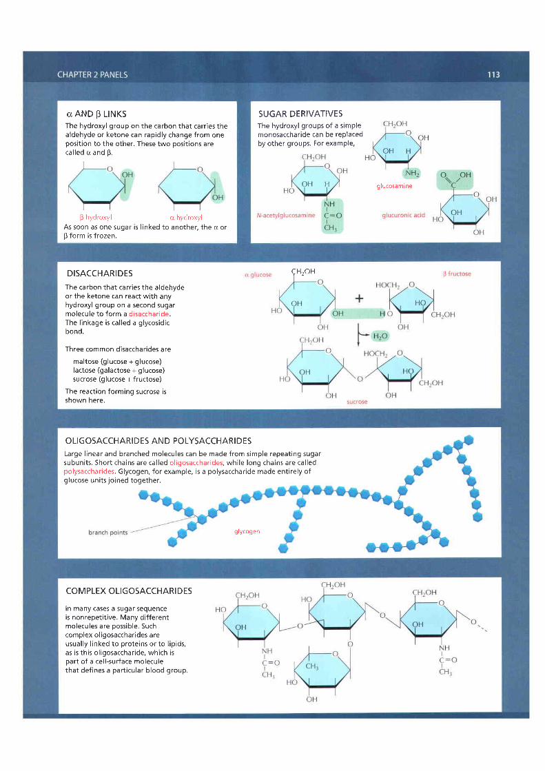
0 AND B LTNKSThe hydroxyl group on the carbon that carr ies thealdehyde or ketone can rapidly change from oneposit ion to the other. These two posit ions arecal led a and B.
B hydroxyl cr hydroxylAs soon as one sugar is l inked to another, the o orB form is frozen.
SUGAR DERIVATIVESThe hydroxyl groups of a simplemonosaccharide can be replacedby other groups. For example,
glucosamine
DISACCHARIDESThe carbon that carr ies the aldehydeor the ketone can react with anyhydroxyl group on a second sugarmolecu le to fo rm a d isacchar ide .The l inkage is cal led a glycosidicbond.
Three common disaccharides aremaltose (glucose + glucose)lactose (galactose + glucose)sucrose (glucose + fructose)
The reaction forming sucrose isshown here.
qH2oH
OLIGOSACCHARIDES AND POLYSACCHARIDESLarge l inear and branched molecules can be made from simple repeating sugarsubun i ts . Shor t cha ins are ca l led o l igosacchar ides , wh i le long cha ins are ca l ledpolysaccharides. Glycogen, for example, is a polysaccharide made entirely ofglucose units joined together.
glycogen
COM PLEX OLIGOSACCHARI DE5
In many cases a sugar sequenceis nonrepeti t ive. Many dif ferentmolecules are possible. Suchcomplex ol igosaccharides areusually l inked to proteins or to l ipids,as is this ol igosaccharide, which isoart of a cel l-surface moleculethat defines a part icular blood group.
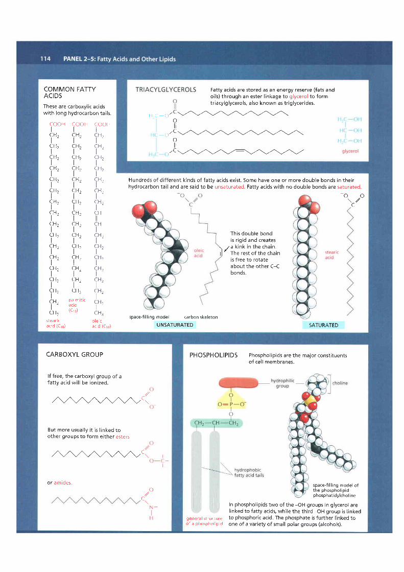
COMMON FATTYACIDSThese are carboxyl ic acidswith long hydrocarbon tai ls.
Hundreds of dif ferent kinds of fatty acids exist. Some have one or more double bonds in theirhydrocarbon tai l and are said to be unsaturated. Fatty acids with no double bonds are saturated.
Th is doub le bondis r igid and creates
. za k ink in the cha in .l l ' rh" rest of the chain
COOHI
CH,I
CH,ICHzI
CH,I
CH,I
C H ,I
CH,I
CH"I
CHrI
CH,I
C H tI
CHtI
C H ,I
CH,I9H,IcH .t "
CH:stea r| cacid (Cre)
COOHI
f",CH,I
C H ,l -
CH,I
CH,If",fn,f*'fn,fn,fn'CHtIi",f*,CH:
3i,'J''''(Crs)
COOHI
CHtI
CHtt -I
CH tI9H,ICHrI
CH,I
CH,I
C H
C HI
CHtI9H,I
CHtt -I
CH .I
C H ,I
CH,I
CH.t '
C H :o l e i cac id (Cre)
- oo\ /
L-
space-f i l l ingmodel carbonskeletonUNSATURATED SATURATED
is free to rotateabout the other C-Cbonds.
Fatty acids are stored as an energy reserve (fats andoils) through an ester l inkage to glycerol to formtriacylglycerols, also known as tr iglycerides.
l l ( , r ' Cot l
o 'Coil
),,c
CARBOXYL GROUP
lf free, the carboxyl group of afatty acid wil l be ionized.
,a/&c..
But more usua l ly i t i s l inked toother groups to form either esters
WC
or amideso,1/
C\
PHOSPHOLIPIDS Phosphol ip ids are the major const i tuentsof cell membranes.
In phospholipids two of the -OH groups in glycerol arel inked to fatty acids, while the third -OH group is l inkedto phosphoric acid. The phosphate is further l inked toone of a variety of small polar groups (alcohols).
general st ructureof a phosphol ip id
space-f i l l ing model ofthe phosphol ip idphosphat idylchol i ne
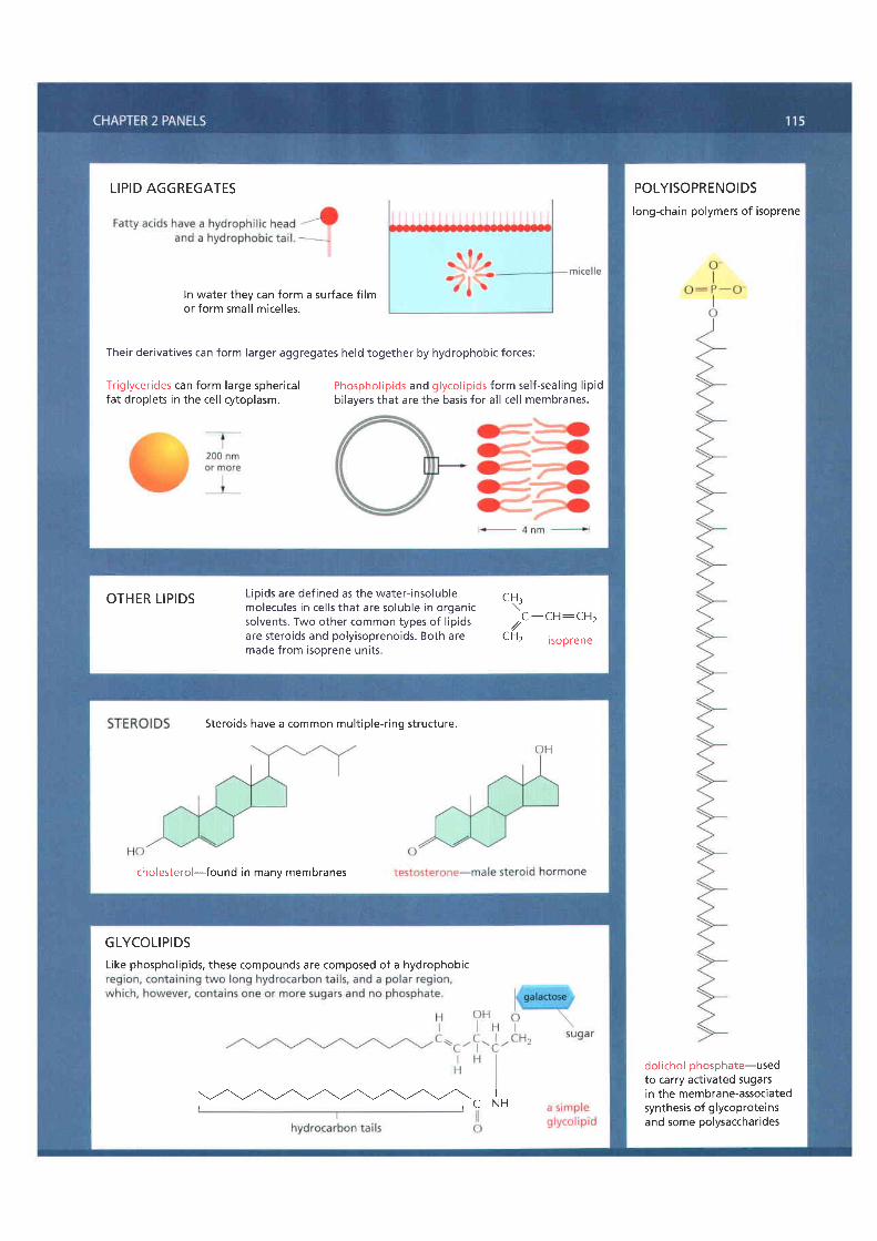
LIPID AGGREGATES
ln water they can form a surface f i lmor fo rm smal l m ice l les .
Their derivatives can form larger aggregates held together by hydrophobic forces:
Triglycerides can form large sphericalfat droplets in the cel l cytoplasm.
Phospho l ip ids and g lyco l ip ids fo rm se l f -sea l ing l ip idbi layers that are the basis for al l cel l membranes.
POLYISOPRENOIDSlong-chain polymers of isoprene
dolichol phosphate-usedto carry activated sugarsin the membrane-associatedsynthesis of glycoprotei nsand some polysaccharides
OTHER L IPIDS Lioids are def ined as the water- insolublemolecu les in ce l l s tha t a re so lub le in o rgan icsolvents. Two other common types of l ipidsare steroids and polyisoprenoids. Both aremade from isoprene units.
C H .\. C - C H : C H r
,//L r 12 i sop rene
Steroids have a common mult iple-r ing structure.
cholesterol-found in manv membranes
GLYCOLIPIDSLike phospholipids, these compounds are composed of a hydrophobic
I- C - N H

BASE
NUCLEOTIDESA nucleotide consists of a nitrogen-containingbase, a f ive-carbon sugar, and one or morephosphate groups.
PHOSPHATESThe phosphates are normally joined tothe C5 hydroxyl of the r ibose ordeoxyribose sugar (designated 5'). Mono-,di-, and tr iphosphates are common.
i l i l t l-o - P -o - P -o - P - . ) -cH^ as In- I
" I -
l - " l " 'o t to -o -oThe phosphate makes a nucleotidenegatively charged.
BASIC SUGARLINKAGE
The base is l inked tothe same carbon (C1)used in sugar-sugarbonds.
\ H u H c l C \ N H
I l l U lu rac i lHCl i \x " t - ' . , - t \
l lC l c v tos i ne H
n t - r - t \ o
" n,.- I' \ . / - \ * *
l l r Ithy l \ - t \rl
The bases are nitrogen-containing r ingcompounds, e i ther pyr im id ines or pur ines .
N Hl 2Ii
ol 1
3
PENTOSEa f ive-carbon sugar
Each numbered carbon on the sugar of a nucleotide isfol lowed by a prime mark; therefore, one speaks of the"5-pr ime carbon, " e tc .
B-o-2-deoxyri boseused in deoxvribonucleic acid
two kinds are used

NOMENCLATURE A nucleoside or nucleotide is named according to i ts nitrogenous base.Single letter abbreviat ions are used variously asshorthand for (1) the base alone, (2) thenucleoside, or (3) the whole nucleotide-the context wi l l usual ly make clear which ofthe three enti t ies is meant. When the contextis not suff icient, we wil l add the terms "base","nucleoside", "nucleotide", or-as in theexamoles below-use the ful l 3- letter nucleotidecooe.AMP = adenosine monophosphatedAMP = deoxyadenosine monophosphateUDP = ur id ine d iphosphateATP = adenosine tr iphosphate
BASE + SUGAR = NUCLEOSIDE
BASE + SUGAR + PHOSPHATE = NUCLEOTIDE
NUCLEOTIDES HAVE MANY OTHER FUNCTIONS
They carry chemical energy in their easi ly hydrolyzed phosphoanhydride bonds.
phosphoanhydr ide bonds
-l -loooi l i l t l-o- l -o-P-o-P-o
- fH,o- o- o-
example: ATP (or ) OH OH
They combine with other groups to form coenzymes.
H H O H H O H C H . H O Ot t i l r r l l l l ' l l l l l
H S _ C _ C _ N _ C _ C - C _ N _ C _ C _ C - C - O - P _ O - P - O -t t t t l l l l l l l
H H H H H H H O C H 3 H O - O -
CH2
example: coenzyme A (CoA) o o HI
O : P - O -
o-They are used as specif ic signal ing molecules in the cel l '
oIIIP _Io-
o o H
l)Hz
example: cycl ic AMP (cAMP)
NUCLEIC ACIDSNucleotides are joined together by aphosphodiester l inkage between 5' and3' carbon atoms to form nucleic acids.The l inear sequence of nucleotides in anucleic acid chain is commonlyabbreviated by a one-letter code,A-G-C-T-T-A-C-A, with the 5'end of the chain at the left .
t5 ' end o f cha in
5',
o

THE IMPORTANCE OF FREE ENERGY FOR CELLSLife is possible because of the complex network of interactingchemical reactions occurring in every cel l . In viewing themetabolic pathways that comprise this network, one mightsuspect that the cel l has had the abi l i ty to evolve an enzyme tocarry out any reaction that i t needs. But this is not so. Althoughenzymes are powerful catalysts, they can speed up only thosereactions that are thermodynamical ly possible; other reactionsproceed in cel ls only because they are coupled to very favorablereactions that drive them. The ouestion of whether a reaction
can occur spontaneously, or instead needs to be coupled toanother reaction, is central to cel l biology. The answer isobtained by reference to a quantity called the free energy: thetotal change in free energy during a set of reactions determineswhether or not the entire reaction seouence can occur. ln thispanel we shal l explain some of the fundamental ideas-derivedfrom a special branch of chemistry and physics cal led thermo-dynamics-that are required for understanding what freeenergy is and why i t is so important to cel ls.
ENERGY RELEASED BY CHANGES IN CHEMICAL BONDING IS CONVERTED INTO HEAT
5EA
UNIVERSE
An enclosed system is def ined as a col lect ion of molecules thatdoes not exchange matter with the rest of the universe (forexample, the "cel l in a box" shown above). Any such system wil lcontain molecules with a total energy E. This energy wil l bedistr ibuted in a variety of ways: some as the translat ional energyof the molecules, some as their vibrat ional and rotat ional energies,but most as the bonding energies between the individual atomsthat make up the molecules. Suppose that a reaction occurs inthe system. The f i rst law of thermodynamics places a constrainton what types of reactions are possible: i t states that " in anyprocess, the total energy of the universe remains constant."For example, suppose that reaction A - B occurs somewhere inthe box and releases a great deal of chemical bond energy. Thisenergy wil l ini t ial ly increase the intensity of molecular motions(translat ional, vibrat ional, and rotat ional) in the system, whichis equivalent to raising i ts temperature. However, these increasedmotions wil l soon be transferred out of the svstem bv a series
of molecular col l is ions that heat uo f irst the walls of the boxand then the outside world (represented by the sea inour example). In the end, the system returns to i ts ini t ialtemperature, by which t ime al l the chemical bond energyreleased in the box has been converted into heat energy andtransferred out of the box to the surroundings. According tothe f irst law, the change in the energy in the box (AE6o", whichwe shal l denote as AE) must be equal and opposite to theamount of heat energy transferred, which we shal l designateas h: that is, AE = -h. Thus, the energy in the box (E) decreaseswhen heat leaves the system.
Ealso can change during a reaction as a result of work beingdone on the outside world. For example, suppose that there isa small increase in the volume (Ay) of the box during a reaction.Since the walls of the box must push against the constantpressure (P) in the surroundings in order to expand, this doeswork on the outside world and requires energy. The energyused is P(AV), which according to the f irst law must decreasethe energy in the box (E) by the same amount. ln most reactionschemical bond energy is converted into both work and heat.Enthalpy (H) is a composite function that includes both of these(H = E + PV).To be r igorous, i t is the change in enthalpy(AH) in an enclosed system, and not the change in energy, thatis equal to the heat transferred to the outside world during areaction. Reactions in which H decreases release heat to thesurroundings and are said to be "exothermic," while reactionsin which H increases absorb heat from the surroundings andare said to be "endothermic." Thus, -h = AH. However, thevolume change is negl igible in most biological reactions, so toa good approximation
THE SECOND LAW OF THERMODYNAMICSConsider a container in which 1000 coins are al l ly ing heads up.l f the container is shaken vigorously, subjecting the coins tothe types of random motions that al l molecules experience dueto their frequent col l is ions with other molecules, one wil l endup with about half the coins oriented heads down. Thereason for this reorientat ion is that there is only a single way inwhich the original orderly state of the coins can be reinstated(every coin mu{l ie heads up), whereas there are many dif ferentways (about 102s8) to achieve a disorderly state in which there isan equal mixture of heads and tai ls; in fact, there are more wavs
to achieve a 50-50 state than to achieve any other state. Eachstate has a probabil i ty of occurrence that is proport ional to thenumber of ways i t can be real ized. The second law of thermo-dynamics states that "systems wil l change spontaneously fromstates of lower probabil i ty to states of higher probabil i ty."Since states of lower probabil i ty are more "ordered" thanstates of high probabil i ty, the second law can be restated:"the universe constantly changes so as to become moredisordered."
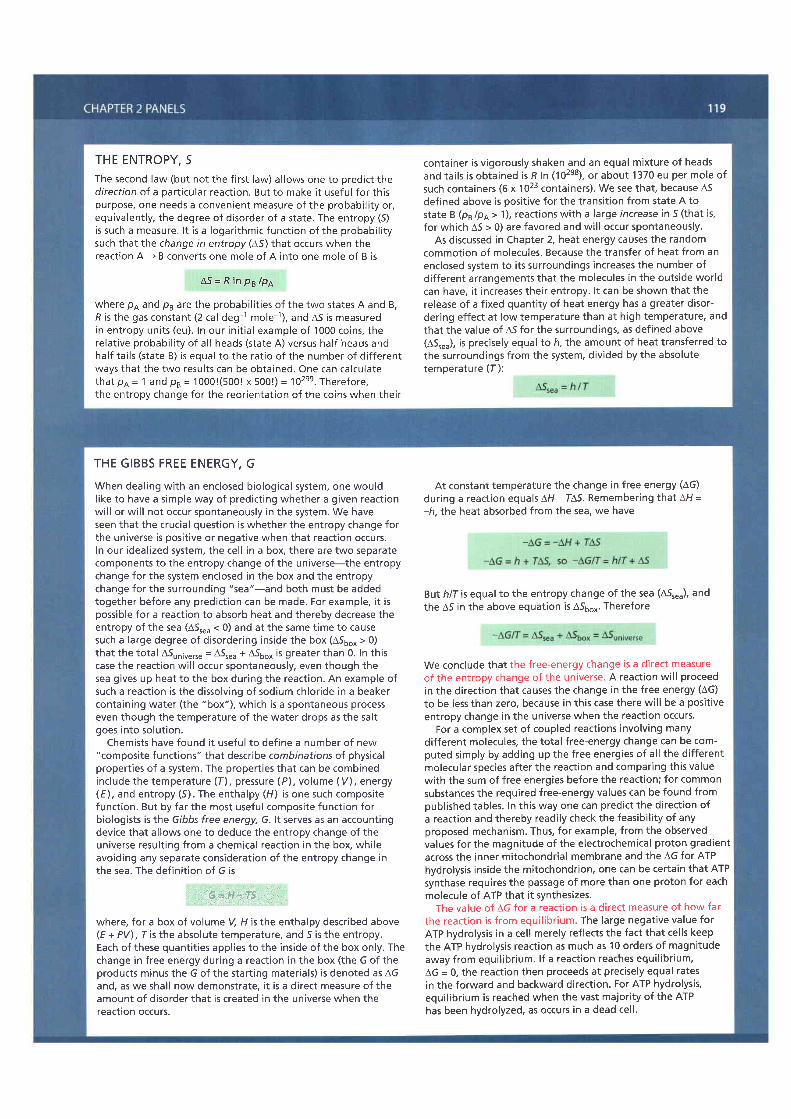
THE ENTROPY, 5The second law (but not the f irst law) al lows one to predict thedirect ion of a part icular reaction. But to make i t useful for thispurpose, one needs a convenient measure of the probabil i ty or,equivalently, the degree of disorder of a state. The entropy (5)is such a measure . l t i s a logar i thmic func t ion o f the probab i l i t ysuch that the change in entropy (45) that occurs when thereaction A - B converts one mole of A into one mole of B is
A 5 = R I n p B / p A
where p4 and p, are the probabil i t ies of the two states A and B,R is the gas constant (2 cal deg 1 mole 1), and A5 is measuredin en t ropy un i ts (eu) . In our in i t ia l example o f 1000 co ins , therelat ive probabil i ty of al l heads (state A) versus half heads andhalf tai ls (state B) is equal to the rat io of the number of dif ferentways that the two results can be obtained. One can calculatetha t pA = 1 and p , = 10001(5001 x 5001) - 102se. There fore ,the entropy change for the reorientat ion of the coins when their
container is vigorously shaken and an equal mixture of headsand tai ls is obtained is R In (102e8), or about 1370 eu per mole ofsuch containers (6 x 10²³ containers). We see that, because A5defined above is posit ive for the transit ion from state A tostate B (ps /p4 > 1), reactions with a large increase in 5 (that is,for which A5 > 0) are favored and wil l occur spontaneously.
As discussed in Chapter 2, heat energy causes the randomcommotion of molecules. Because the transfer of heat from anenclosed system to i ts surroundings increases the number ofdif ferent arrangements that the molecules in the outside worldcan have, i t increases their entropy. l t can be shown that therelease of a f ixed quanti ty of heat energy has a greater disor-dering effect at low temperature than at high temperature, andthat the value of A5 for the surroundings. as defined above(ASr"u), is precisely equalto h, the amount of heattransferred tothe surroundings from the system, divided by the absolutetemperature (f ) :
THE GIBBS FREE ENERGY, GWhen dealing with an enclosed biological system, one wouldl ike to have a simple way of predict ing whether a given reactionwil l or wi l l not occur spontaneously in the system. We haveseen that the crucial question is whether the entropy change forthe universe is posit ive or negative when that reaction occurs.In our ideal ized system, the cel l in a box, there are two separatecomponents to the entropy change of the universe-the entropychange for the system enclosed in the box and the entropychange for the surrounding "sea"-and both must be addedtogether before any predict ion can be made. For example, i t ispossible for a reaction to absorb heat and thereby decrease theentropy of the sea (A5r"" < 0) and at the same time to causesuch a large degree of disordering inside the box (A56o* > 0)that the total A5rn;u"rr" = A5r"" + A56o, is greater than 0. In thiscase the reaction wil l occur spontaneously, even though thesea gives up heat to the box during the reaction. An example ofsuch a reaction is the dissolving of sodium chloride in a beakercontaining water (the "box"), which is a spontaneous processeven though the temperature of the water drops as the saltgoes into solut ion.
Chemists have found i t useful to define a number of new"composite functions" that describe combinations of physicalpropert ies of a system. The propert ies that can be combinedinclude the temperature (f), pressure (P), volume (V), energy(E), and entropy (5). The enthalpy (H) is one such compositefunction. But by far the most useful composite function forbiologists is the Gibbs free energy, G. lt serves as an accountingdevice that al lows one to deduce the entropy change of theuniverse result ing from a chemical reaction in the box, whileavoiding any separate consideration of the entropy change inthe sea. The definit ion of G is
G = H - T S
where, for a box of volume V, H is the enthalpy described above(E + PV), r is the absolute temperature, and 5 is the entropy.Each of these quanti t ies appl ies to the inside of the box only. Thechange in free energy during a reaction in the box (the G of theproducts minus the G of the start ing materials) is denoted as AGand, as we shal l now demonstrate, i t is a direct measure of theamount of disorder that is created in the universe when thereaction occurs.
At constant temperature the change in free energy (AG)during a reaction equals AH - IA5. Remembering that AH =-h, the heat absorbed from the sea, we have
But h/f is equal to the entropy change of the sea (A5r""), andthe A5 in the above equation is A56o^. Therefore
We conclude that the free-energy change is a direct measureof the entropy change of the universe. A reaction wil l proceedin the direct ion that causes the change in the free energy (AG)to be less than zero, because in this case there wil l be a posit iveentropy change in the universe when the reaction occurs.
For a complex set of coupled reactions involving manydifferent molecules, the total free-energy change can be com-puted simply by adding up the free energies of al l the dif ferentmolecular species after the reaction and comparing this valuewith the sum of free energies before the reaction; for commonsubstances the required free-energy values can be found frompublished tables. In this way one can predict the direct ion ofa reaction and thereby readi ly check the feasibi l i ty of anyproposed mechanism. Thus, for example, from the observedvalues for the magnitude of the electrochemical proton gradientacross the inner mitochondrial membrane and the AG for ATPhydrolysis inside the mitochondrion, one can be certain that ATPsynthase requires the passage of more than one proton for eachmolecule of ATP that i t synthesizes.
The value of AG for a reaction is a direct measure of how farthe reaction is from equil ibr ium. The large negative value forATP hydrolysis in a cel l merely ref lects the fact that cel ls keepthe ATP hydrolysis reaction as much as 10 orders of magnitudeaway from equil ibr ium. l f a reaction reaches equil ibr ium,AG = 0, the reaction then proceeds at precisely equal ratesin the forward and backward direction. For ATP hydrolysis,equi l ibr ium is reached when the vast majori ty of the ATPhas been hydrolyzed, as occurs in a dead cel l '

For each step, the part of the molecule that undergoes a change is shadowed in b lue,and the name oJ the enzyme that catalyzes the react ion is in a yel low box.
STEP 1 Glucose isphosphorylated by ATP toform a sugar phosphate.The negat ive charge of thephosphate prevents passageof the sugarphosphate through theprasma memDrane,t rapping glucose insidethe cel l .
f rom carbon 1 to HOcaroon z, tormtng
o. .H\ , /! rI
H-C. -OHl '
H . ) - ( - -H* ' - i ?+ l -
H - O H
H - C - O Hl 5
.CH,O P(open chain form)
1CH?OH' t -
H - C - O H5 | (r ing form)- C H , O q P -
(open chain form)
a ketose from analdose sugar (SeePanel 2-4.)
STEP 3 The new hydroxylgroup on carbon 1 ' , . lphosphorylated by ATP, inpreparat ion for the format ionof two three-carbon sugarphosphates. The entry of sugarsinto g lycolysis is contro l led at th isstep, through regulat ion of theenzy me p hosp hof ru cto k i n a se -
P O H ) C o C H , O P- | , " - \ |( Hr)) + ADPrLJ/l
t t ' 'OH
f ructose 1,6-bisphosphate
STEP 4 Thesix-carbon sugar isc leaved to producetwo three-carbonmolecules Only theglyceraldehyde3-phosphate canproceed immediatelythrough glycolysis
C H , O PI
a - A
IH O - C - H
IH - C - O H
IH - C - O H
IcH2o P
H O\ / /+ C
IH - C - O H
IcH2o P
glyceraldehyde? - n h n c n h : t e
(open chain form)f ructose 1,5-bisphosphate
STEP 5 The otherproduct of step 4,d i hyd roxyacetonephosphate, isisomerized to formglyceraldehyde3-phosphate.
CH,OHII
cH2o P
" ,ro
CI
H - C - O HI
cH2o Pglyceraldehyde 3-phosphate
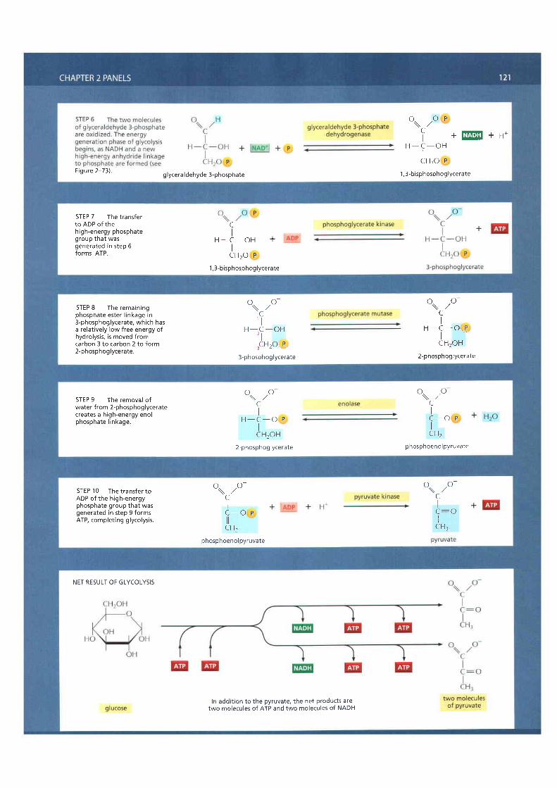
Figure 2-73) g lyceraldehyde 3-phosphate
ovo t
f +EEEE+u*H - C - O H
lcH2o P
1,3-bisphosphoglycerate
STEP 7 The t ransferto ADP of thehigh-energy phosphategroup that wasgenerated in step 6forms ATP.
CI
H - C - O H +IcH2o P
1,3-bisphosphoglycerate
STEP 8 The remainingphosphate ester l inkage in3-phosphoglycerate, which hasa relatively low free energy ofhydrolysis, is moved fromcarbon 3 to carbon 2 to form2-phosphog lycerate.
o. .o-\ / /C' l
H - C - O H, l-CHrO'. P.
3-phosphog lycerate
o. .o\ / /CI
H - C - O i * Pt -cH2oH
2- p hosp h og lycerate
STEP 9 The removal ofwater from 2-phosphoglyceratecreates a high-energy enolphosphate l inkage.
o. .o\ , /CI
H - C - O PIcH2oH
2-phosphog lycerate
o. .o\ . /CIC - O P
cHzphosp hoeno I pyruvate
STEP 1 0 The transfer toADP of the high-energyphosphate group that wasgenerated in step 9 formsATP, complet ing glycolysis.
o. .o-\ / /CIC - O P
cHzphosphoenol pyruvate
o. .o-\ . /CII
CH:
NET RESULT OF GLYCOLYSIS
In addition to the pyruvate, the net products aretwo molecules of ATP and two molecules of NADH
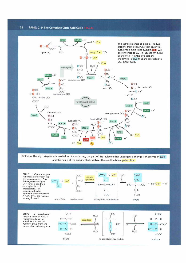
HS -CoAThe complete citr ic acid cycle. The twocarbons from acetyl CoA that enter thisturn of the cycle (shadowed in ) wi l lbe converted to CO, in subsequent turnsof the cycle: i t is the two carbonsshadowed in blue that are converted toCO, in this cycle.
pyruvate acetyl CoA (2c)
HS-CoA
eoo*",
t -H O - C - C O O - ( + a ^ )
in, \* ' -ioo- \ Po
citrate (6c) t fn,
isocitrate (6c)
HC - COOno- tH
Coo-
ffoo-fumarate (4C)
€oo-t
f ut ' ' succinate (4C)C H , r S t e p 6 G O O -
(x-ketoglutarate (5C)2 Coz
succinyl CoA (4C) CH.t '
Icoo-Ioo-Hzo
2CH,t -
C = OI
il*p_:,HS -CoA
S -CoAEE*t -
HS -CoA
Step 1
coz
Detai ls of the eight steps are shown below. For each step, the part of the molecule that undergoes a change is shadowed in hlueand the name of the enzyme that catalyzes the reaction is in a yel low box.
STEP 1 After the enzymeremoves a proton from theCH, group on acetyl CoA,the negat ively chargedCHr- forms a bond to acarbonyl carbon ofoxaloacetate. Thesubsequent loss byhydrolysis of the coenzymeA (CoA) drives the reactionstrongly forward.
O : C - S - C o AICH,t -
H O - C - C O OI9H,lcoo-
coo-ICHrt -
H O - C - C O OI
CH,t -coo-
citrate
+ HS-CoA + H*
STEP 2 An isomerizat ionreaction, in which water isf i rs t removed and thenadded back, moves thehydroxyl group from onecarbon atom to i ts neighbor
coo-Hzo coo-
IC - HIc-coo-tlC - HIcoo-
H -
coo-I
H - C - HI
H-C -COO-I
H O - C - HIcoo
isocitratecitrate cls-aconitate intermediate
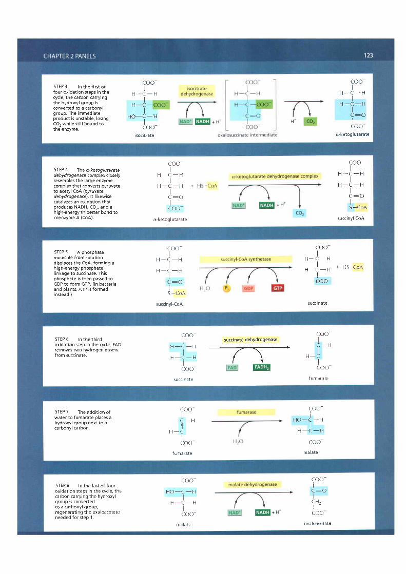
STEP 3 ln the first offour oxidat ion steps in thecycle, the carbon carryingthe hydroxyl group isconverted to a carbonylgroup. The immediateproduct is unstable, los ingCO, whi le st i l l bound tothe enzyme.
coo-I
H - C - HI
H _ C
H O - c - HIcoo-
isoci t rate
coo-I
H - C - HI
H - C - HI
a - i
Icoo(x-ketog I utarate
STEP 4 The o-ketog/utaratede hyd ro gen ase complex closelyresembles the large enzymecomplex that converts pyruvateto acetyl co{(pyruvatedehydrogenase). lt likewisecatalyzes an oxidation thatproduces NADH, CO2, and ahigh-energy th ioester bond tocoenzyme A (CoA).
coo-I
H - C - HI
H - C - HIc :oIcoo-
(x-ketogl utarate
coo-I
H - C - HI
H - C - HIIS -CoA
succinyl-CoA
STEP 5 A phosphatemolecule f rom solut iondisplaces the CoA, forming ahigh-energy phosphatel inkage to succinate. Thisphosphate is then passed toGDP to form GTP. ( ln bacter iaand plants, ATP is formedinstead.)
coo-H - C - H
IH - C - H
IIS -CoA
succinyl -CoA
cooI
H - C - HI
, , ) , , + H S - C o An - L - rIcoo-
succ l nare
STEP 6 ln the th i rdoxidation step in the cycle, FADremoves two hydrogen atomsfrom succinate.
cooI
H - C - H
IH - C - H
Icoosucci nate
succinate dehydrogenase
- / \r l
coo-I
C - H
H - CIcoo-
f umarate
STEP 7 The addi t ion ofwater to fumarate places ahydroxyl group next to acarbonyl carbon.
coo-I
C - H
H - CIcoo
f u ma rate
coo-I
H O - C - HI
H - C - HIcoo
mala te
STEP 8 ln the last of Jouroxidat ion steps in the cycle, thecarbon carry ing the hydroxylgroup is convertedto a carbonyl group,regenerating the oxaloacetateneeded for step 1.
coo-I
H O - C - HI
H - C - HIcoo-
mala te
coo-Ic : oI
C H ,Icoo
oxa loacetate

124 Chapter 2: Cell Chemistry and Biosynthesis
REFERENCESGeneralBerg, JM, Tymoczko, JL & Stryer L (2006) Biochemistry, 6th ed New
York: WH FreemanGarrett RH & Grisham CM (2005) Biochemistry, 3rd ed phi ladelphia:
Thomson Brooks/ColeHorton l-1R, Moran LA, Scrimgeour et a (2005) Princip es of
Bioch-.mistry 4th ed Upper Saddle River, NJ: prentice HallNelson DL & Cox MM (2004) Lehnlnger Principles of Biochemistry,
4th ed New York: WorthNichol ls DG & Ferguson S_l (2002) Bioenergerics,3rd ed New york:
Academic PressMathews CK, van Ho de KE & Ahern K G (2000) Biochemistry, 3rd ed
5aq l ra rc ,sco : Ben jar r CummingsMoore JA (1993) Sclence As a Way of Knowing Cambridge, MA:
Harvard University PressVoet D, Voet. lG & Pratt CIV (2004) Fundamentals of Biochemistry,
2nd ed New York: Wiley
The Chemical Components of a CellAbeles RH, Frey PA & Jencks WP (1992) Biochemistry Boston: Jones &
Ba rtlettAtkins PW (' l996) Mo ecu es New York: WH FreemanBranden C & Tooze J ( l 999) ntroduction to Protein Structure, 2nd ed
New York: Garland Sc enceBretscher MS (, l985) The molecules of the cel membrane 5clAm
253:1 0O I O9Bur ey 5K & Petsko GA (t 9BB) Weakly polar interactions in proteins ,4dy
Pratein Chem 39.125-1 89De Duve C (2005) Singulanties: Landmarks on the pathways of Lif- .
Cambridge: Cambridge University PressDowhan W (1997) Molecular basis for membrane phospholipid diversity:
Why are there so many ipids? Annu Rev Biochem 66:j99-232E senberg D & Kauzman W (l 969) The Structure and propert ies of
Water Oxford: Oxford Univers ty PressFersht AR (198/) Ihe hydrogen bond in molecular recognit ion Irendj
Biochem Sci 123A1-304Franks F ( ' l 993) Water Cambridge: Royal Society of ChemistryHenderson l l (1927) The Fitness of the Env ronment, 1 958 ed Boston:
BeaconNeidhardt FC, Ingraham _lL & Schaechter M (t 990) physioiogy of the
Bacterial Ce l : A Mo ecular Approach Sunderland, MA: SinauerPauling L (1 960) Ihe Nature of the Chemical Bond, 3rd ed thaca, Ny:
Cornel l University PressSaenger W (l 984) Princrples of Nucleic Acid Structure, New yorx:
Spr i ngerSharon N (1980) Carbohydrates 5ci,4,m 243.90 116Sti l l inger FH (1980) Warer revisited Science 2a9.45j-457Tanford C ( '1978) The hydrophobtc effect and the organization of l iv ing
mat te r Sc lence 200: , l012 l O l8Tanford C (1980) The lydrophobic Effect Formation of Micel es and
Bio ogical Membranes, 2nd ed New york. John Wi ey
Catalysis and the Use of Energy by CellsAtkins PW (1994) Ihe Second Law: Energy, Chaos and Form New york:
Scienti f c American BooksAtkins PW & De Paula iD (2006) Physical Chemistry for the Life
Sciences Oxford: Oxford University pressBaldwin JE & Krebs H (1981)The Evolurion of Metabolic Cycles Nciure
291:381-382Berg HC (1983) Random Walks in B ology Princeton, NJ: princeton
University PressDickerson RE (,1969) MolecularThermodynamics Menlo park, CA:
Ben jamin CummingsDil l KA & Bromberg S (2003) Molecular Driving Forces: Stat ist icalThermo-
dynamics in Chemistry and Bio ogy New york: Garland ScienceDressler D & Potter H (1991) Discovelng Enzymes New york: Sclenti f ic
American L brary
Einstein A (1956) lnvestigations on the Theory of Brownian MovementNew York: Dover
Fruton JS (1999) Proteins, Enzymes, Genes: The nterplay of Chemistryand Bio ogy New Haven:Yale University Press,
Goodse I DS (1991) nside a living cell Trends Biochem Sci 16:203-206Karplus M & McCammon JA (1986) The dynamics of prote ns SclAm
254:42-51Karp lus M & Petsko GA (1990) Mo lecu la r dynamics s imu la t ions in
biology Nature 347:631 639Kauzmann W (1967) Thermodynamics and Statist ics: with Applications to
Gases In Thermal Properties of Matter Vol 2 New York: WA Benjamin,Inc
Kornberg A (1989) For the Love of Enzymes Cambridge, MA: HarvardUniversity Press
Lavenda BH (,1985) Brownian Motion 5ci,4m 252:70 85Lawlor DW (2001) Photosynthesis, 3rd ed Oxford: BIOSLehn inger AL (1911) The Molecu la r Bas is o f B io log ica l Energy
Transformations, 2nd ed Menlo Park, CA: Benjamin CummingsLipmann F (1941) Metabolic generation and uti izat ion of phosphate
bond energy Adv Enzymol 1:99-162Lipmann F (1971) Wanderings of a Biochemist New York: WileyNisbet EE & Sleep NH (2001)The habitat and narure of early l i fe Nature
409:1083 1091Racker E ( l9BO) From Pasteur to Mitchel l : a hundred years of
n ln t rn t r r^o l r rc Lo /1 p t^ . t , ) :2 lO-2 I 5Schrodinger E (1944 & 1 958) What is Life?: The Physicai Aspect of the
L iv ing Ce I and Mind and Mat te r , 1992 combined ed Cambr idge:Cambridge University Press
van Holde KE, Johnson WC & Ho PS (2005) Principles of PhysicalBiochemistry, 2nd ed Upper Saddle River, NJ: Prentice Hal
Walsh C (2001) Enabling the chemistry of l i fe Nature 409.226-23iWestheimer FH (1982) Why nature chose phosphates Science
235.11 /3-1 t78Youvan DC & Marrs BL (1987) Molecular mechanisms of
photosynthesis Sci Am 256:42 49
How Cells Obtain Energy from FoodCramer WA & Knaff DB (1990) Energy Transduction in Bio ogical
Membranes, New York: Springer-Verlag,Dismukes GC, Kl imov W, Baranov SV et al (2001) The origin of
atmospheric oxygen on Earth: The innovation of oxygenicphotosyntheis Prac Nat Acad Sci USA 982170 2175
Fel D ( l 997) Understanding the Control of Metabolism London:Port and Press
F att JP (1 995) Use and storage of carbohydrate and fat Am J Clin Nutr61,9525 9595.
Friedmann HC (2004) From Butybacterium to E. coli: An essay on unityin biochemistry Perspect Biol lrled 47:47-66
Fothergi l l -Gilmore LA (,1986) The evolut ion of the glycolyt ic pathwayTrends Biochem Sci 11:47 5l
Heinrich R, Melendez-Hevia E, Montero F et al ( ,1999) The structuraldesign of glycolysis: An evo utionary approach Biochem SocTrans27:294-298
Huynen MA, Dandekar T & Bork P (1 999) Variarion and evolut ion of thecitric acid cycle: a genomic perspective, Trends Microbrol l:281-291
Kornberg HL (2000) Krebs and his tr ini ty of cycles Nature Rev Mol CellBiol 1.225-228
Krebs HA & Mart in A (1981) Reminiscences and Reflect ionsOxford/New York: Clarendon Press/Oxford University Press,
Krebs HA (l 970) The history of the tricarboxylic acid cycle perspect BiolMed 14.154-17a
Mart ln BR (1987) Metabolic Regulat ion: A Molecular Approach Oxford:Blackwell Scienti f ic
McGilvery RW (,1983) Biochemistry: A Functional Approach, 3rd edPh i lade lph ia : Saunders
Morowitz HJ (1993) Beginnings of Cellular Life: MetabolismRecapitulates Biogenesis New Haven: Yale University Press
Newsholme EA & Stark C (1973) Regulat ion of MetabolismNew York. Wiley,


![6.start.stop.07.v2.ppt [Read-Only] - Molecular and Cell Biology |](https://static.fdokumen.com/doc/165x107/631a9313c51d6b41aa04e24f/6startstop07v2ppt-read-only-molecular-and-cell-biology-.jpg)