Molecular Biology paper
10
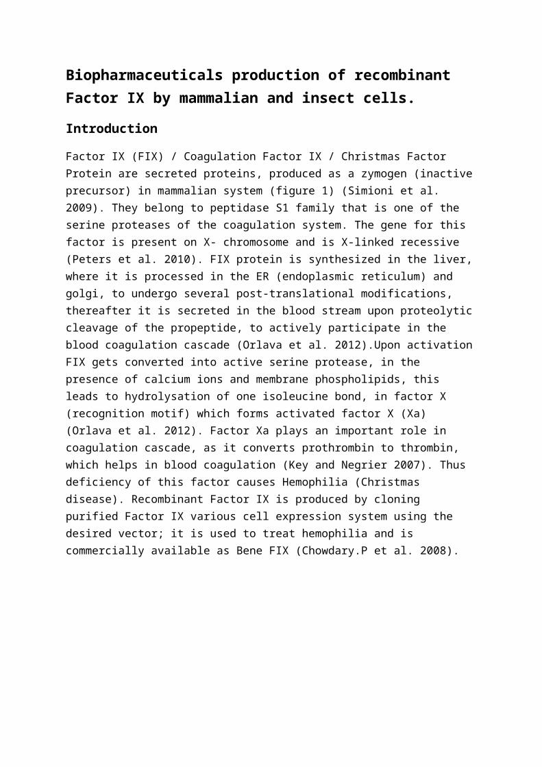
Biopharmaceuticals production of recombinant Factor IX by mammalian and insect cells. Introduction Factor IX (FIX) / Coagulation Factor IX / Christmas Factor Protein are secreted proteins, produced as a zymogen (inactive precursor) in mammalian system (figure 1) (Simioni et al. 2009). They belong to peptidase S1 family that is one of the serine proteases of the coagulation system. The gene for this factor is present on X- chromosome and is X-linked recessive (Peters et al. 2010). FIX protein is synthesized in the liver, where it is processed in the ER (endoplasmic reticulum) and golgi, to undergo several post-translational modifications, thereafter it is secreted in the blood stream upon proteolytic cleavage of the propeptide, to actively participate in the blood coagulation cascade (Orlava et al. 2012).Upon activation FIX gets converted into active serine protease, in the presence of calcium ions and membrane phospholipids, this leads to hydrolysation of one isoleucine bond, in factor X (recognition motif) which forms activated factor X (Xa) (Orlava et al. 2012). Factor Xa plays an important role in coagulation cascade, as it converts prothrombin to thrombin, which helps in blood coagulation (Key and Negrier 2007). Thus deficiency of this factor causes Hemophilia (Christmas disease). Recombinant Factor IX is produced by cloning purified Factor IX various cell expression system using the desired vector; it is used to treat hemophilia and is commercially available as Bene FIX (Chowdary.P et al. 2008).
Transcript of Molecular Biology paper
Biopharmaceuticals production of recombinant Factor IX by mammalian and insect cells.
Introduction
Factor IX (FIX) / Coagulation Factor IX / Christmas Factor Protein are secreted proteins, produced as a zymogen (inactiveprecursor) in mammalian system (figure 1) (Simioni et al. 2009). They belong to peptidase S1 family that is one of the serine proteases of the coagulation system. The gene for this factor is present on X- chromosome and is X-linked recessive (Peters et al. 2010). FIX protein is synthesized in the liver,where it is processed in the ER (endoplasmic reticulum) and golgi, to undergo several post-translational modifications, thereafter it is secreted in the blood stream upon proteolyticcleavage of the propeptide, to actively participate in the blood coagulation cascade (Orlava et al. 2012).Upon activationFIX gets converted into active serine protease, in the presence of calcium ions and membrane phospholipids, this leads to hydrolysation of one isoleucine bond, in factor X (recognition motif) which forms activated factor X (Xa) (Orlava et al. 2012). Factor Xa plays an important role in coagulation cascade, as it converts prothrombin to thrombin, which helps in blood coagulation (Key and Negrier 2007). Thus deficiency of this factor causes Hemophilia (Christmas disease). Recombinant Factor IX is produced by cloning purified Factor IX various cell expression system using the desired vector; it is used to treat hemophilia and is commercially available as Bene FIX (Chowdary.P et al. 2008).
Figure-1,Structure of FIX. S – signal peptide; P – propeptide, Gla – Gla domain, EGF1 and EGF2– epidermal growth factor, AP – activation peptide, Protease – serine protease domain. Posttranslational modifications sites: Gla – γ-carboxylation, Hya – β-hydroxylation; S – sulfation, P – phosphorylation, ♦ – N-linked and ● – O-linked glycosylation, cleavage points are indicated by triangles.(Taken from (Orlava et al. 2012)).
Recombinant Factor IX (rFIX) production by mammalian and insect cells
Selection of appropriate expression system and vector depends on the characteristics and purpose for which the production ofrecombinant protein is done (Brondyk 2009). Production of rFIXprotein is a multi-step process, which involves development and selection of a cell line; capable of expressing high levels of active rFIX protein (Matasci.M. et al. 2008) (figure2).
Development of cell line involves, firstly chromosomal integration which is done by delivering recombinant DNA (rDNA)into host cell nucleus leading to transgene expression. Several methods can be employed for this process like electroporation, lipofection, calcium phosphate precipitation and retrovioral transfection, depending upon the functionalityand requirement (Jayapal, Wlaschin and H. 2007). Secondly stably expressed cells are selected using low levels of MTX (methotrexate) and cultivated in the absence of thymidine, glycine and hypoxanthine as they can sometimes cause mutation in the cell line (Hackera, Jesusb and Wurm 2009). Then the surviving cells expressing protein of interest are recovered
and amplified, this process often leads to the production of cells with different integration sites, copy numbers and productivities (Baldi et al. 2007). Henceforth clones with highest productivity and copy numbers are separated by using serial dilutions in multi-well plates, to isolate single colonies having uniform cell population (Baldi et al. 2007). Then the chosen clones are expanded and evaluated via several rounds of passaging in lab-scale bioreactors. Lastly a cell line capable of expressing high levels of active rFIX protein is chosen and stored as frozen vial for future use (Jayapal, Wlaschin and H. 2007).
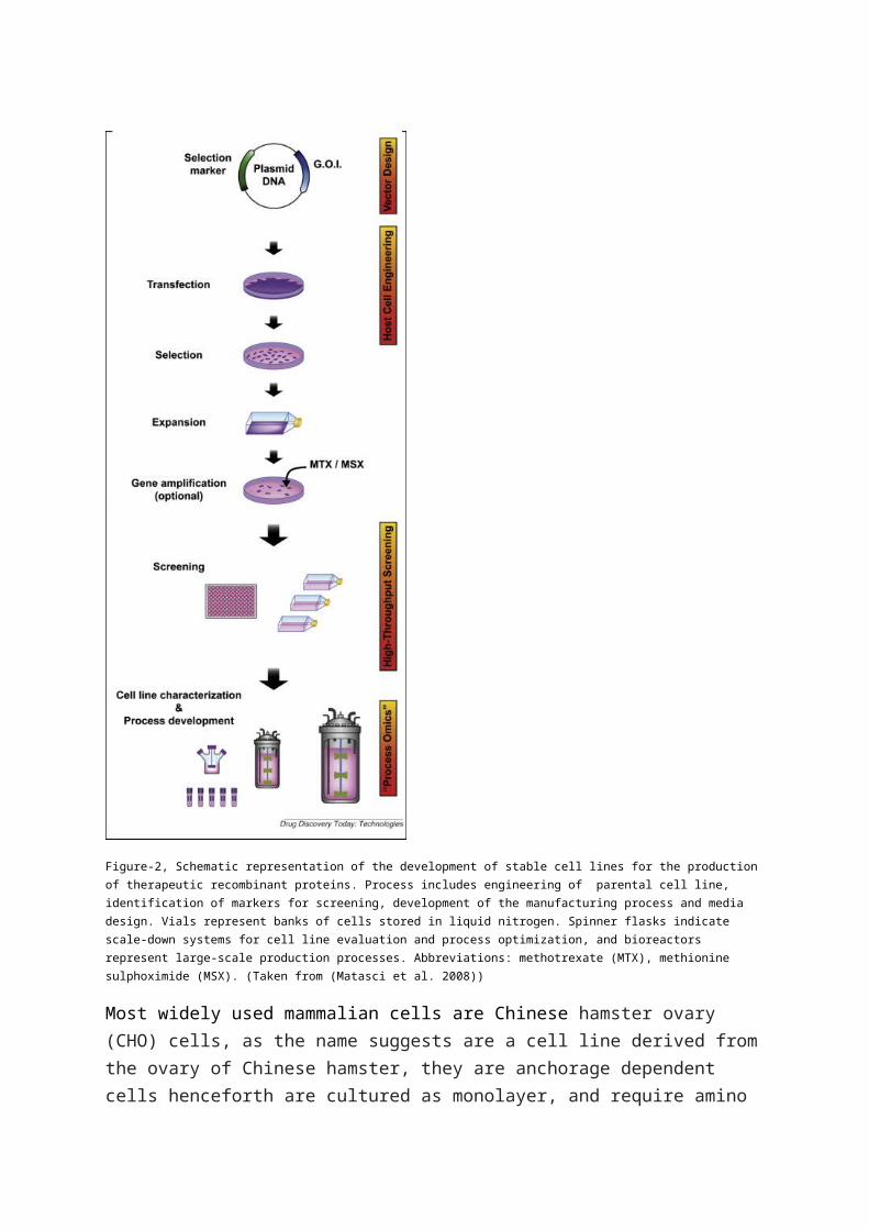
Figure-2, Schematic representation of the development of stable cell lines for the production of therapeutic recombinant proteins. Process includes engineering of parental cell line, identification of markers for screening, development of the manufacturing process and media design. Vials represent banks of cells stored in liquid nitrogen. Spinner flasks indicate scale-down systems for cell line evaluation and process optimization, and bioreactors represent large-scale production processes. Abbreviations: methotrexate (MTX), methionine sulphoximide (MSX). (Taken from (Matasci et al. 2008))
Most widely used mammalian cells are Chinese hamster ovary (CHO) cells, as the name suggests are a cell line derived fromthe ovary of Chinese hamster, they are anchorage dependent cells henceforth are cultured as monolayer, and require amino
acid proline in their culture medium (Jayapal, Wlaschin and H.2007). They can be easily manipulated genetically and are quite flexible and highly adaptable in nature. CHO cells (FreeStyle CHO-S) are used , as they can produce large amountsof functional recombinant protein, henceforth guarantees approximately 95% efficiency, they are very safe hosts and do not allow replication of human pathogenic viruses like HIV, herpes, etc thereby eliminating the risk of contamination up to pictogram levels of the product (Datta, Linhardt and Sharfstein 2013). They can transfect, select, amplify and stably express accurate post translationally modified proteinswith glycoforms which are compatible and active in humans (Nolan, Ryan and Kyongbum 2011). Furthermore they are highly robust and prove to be versatile protein production hosts.
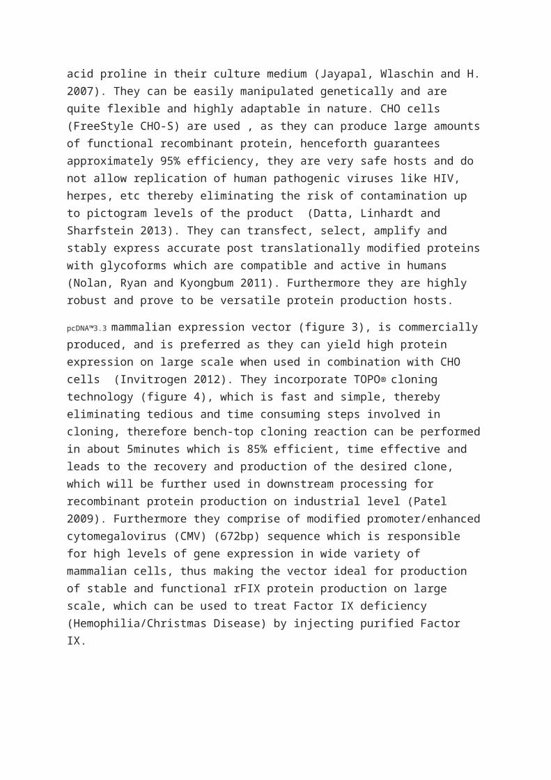
pcDNA™3.3 mammalian expression vector (figure 3), is commercially produced, and is preferred as they can yield high protein expression on large scale when used in combination with CHO cells (Invitrogen 2012). They incorporate TOPO® cloning technology (figure 4), which is fast and simple, thereby eliminating tedious and time consuming steps involved in cloning, therefore bench-top cloning reaction can be performedin about 5minutes which is 85% efficient, time effective and leads to the recovery and production of the desired clone, which will be further used in downstream processing for recombinant protein production on industrial level (Patel 2009). Furthermore they comprise of modified promoter/enhancedcytomegalovirus (CMV) (672bp) sequence which is responsible for high levels of gene expression in wide variety of mammalian cells, thus making the vector ideal for production of stable and functional rFIX protein production on large scale, which can be used to treat Factor IX deficiency (Hemophilia/Christmas Disease) by injecting purified Factor IX.
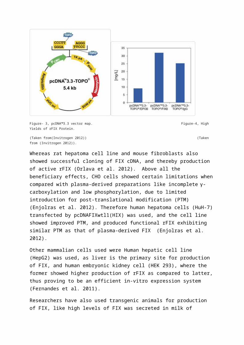
Figure- 3, pcDNA™3.3 vector map. Figure-4, High Yields of rFIX Protein.
(Taken from(Invitrogen 2012)) (Taken from (Invitrogen 2012)).
Whereas rat hepatoma cell line and mouse fibroblasts also showed successful cloning of FIX cDNA, and thereby production of active rFIX (Orlava et al. 2012). Above all the beneficiary effects, CHO cells showed certain limitations whencompared with plasma-derived preparations like incomplete γ-carboxylation and low phosphorylation, due to limited introduction for post-translational modification (PTM) (Enjolras et al. 2012). Therefore human hepatoma cells (HuH-7)transfected by pcDNAFIXwtl1(HIX) was used, and the cell line showed improved PTM, and produced functional rFIX exhibiting similar PTM as that of plasma-derived FIX (Enjolras et al. 2012).
Other mammalian cells used were Human hepatic cell line (HepG2) was used, as liver is the primary site for production of FIX, and human embryonic kidney cell (HEK 293), where the former showed higher production of rFIX as compared to latter,thus proving to be an efficient in-vitro expression system (Fernandes et al. 2011).
Researchers have also used transgenic animals for production of FIX, like high levels of FIX was secreted in milk of
transgenic sheep and transgenic cow by using nuclear transfer techniques (Orlava et al. 2012). Milk from transgenic pig, also produced FIX, but it had a rate limiting step owing to incomplete γ-carboxylation (Sun et al. 2012). Lately transgenic goat had been engineered for high secretion of rFIXin its milk, by integration of linearized recombinant vector (pBC1) encompassing FIX cDNA into goat fetal fibroblast cells through electroporation (Yekta et al. 2013).
Recombinant FIX can also be produced by insect cells like thatof Drosophila as its γ-glutamyl carboxylase (dyc) enzyme showssame substrate recognition properties as that of vertebrate γ-carboxylase (γC), experiments conducted on Drosophila Schnider(S2) cell line by transfecting it by metallothionein promoter-regulated rFIX-expressing plasmid, resulted in production of high levels of biologically active rFIX protein with improved γ-carboxylation (Vatandoost et al. 2013).
Baculovirus (insect derived vector), is the most commonly usedexpression vector for rFIX production in insect cells as its quite versatile and safe and have high expression efficiency (Airenne et al. 2013), as compared to Escherichia coli (E.colibacterium) which cannot express very large proteins (Demaina and Vaishnavb 2009). Other viral vectors used are retroviral (RV) or lentiviral (LV) or episomal adenovirus (AV) or adeno-associated viruses (AAV) (Wanisch and Rafael 2008).
Other sources which can be considered are seeds and tissue of transgenic plants which are known to be a good source of biopharamaceutical protein, however in case of rFIX; transgenic tomatoes showed expression of inactive FIX, although soybean seeds resulted delivering some promising levels of rFIX (Orlava et al. 2012). Still they have certain limitations of incomplete expression of γ-carboxylase and its co-enzymes.
Conclusion

Henceforth it can be concluded that biopharmaceutical production of rFIX protein, can be done by both mammalian and insect cells. Mammalian cells currently being, a more feasiblechoice owing to its similarities with the vertebrate system, though insect cell lines are also an upcoming field for production of rFIX. Thus it can be said, that presence of PTM is the most important factor, responsible for the determination of suitable host and vector, for the production of rFIX. Gene therapy by FIX cDNA in viral vectors is also quite promising. Hopefully haemophilia will be fully cured, with increase in the production of rFIX by more productive cell lines.
Bibliography
AIRENNE, K.J., et al. (2013). Baculovirus: an Insect-derived Vector for Diverse Gene Transfer Applications. Molecular Therapy,21 (10), 739-749.
BALDI, L., et al. (2007). Recombinant protein production by large-scale transient gene expression in mammalian cells: state of the art and future perspectives. Biotechnology Letters, 29(5), 677-684.
BRONDYK, William.H. (2009). Selecting an Appropriate Method for Expressing a Recombinant Protein. Methods in Enzymology, 463 (2), 131-147.
CHOWDARY.P, et al. (2008). Recombinant factor IX (BeneFix®) byadjusted continuous infusion: a study of stability, sterility and clinical experience. The Official Journal of the World Federation of Haemophilia, 7 (2), 140-145.
DATTA, Payel, LINHARDT, Robert J and SHARFSTEIN, Susan T (2013). An 'omics approach towards CHO cell engineering. Biotechnology and bioengineering, 110 (5), 1255-1271.
DEMAINA, A.L. and VAISHNAVB, P. (2009). Production of recombinant proteins by microbes and higher organisms. Biotechnology Advances, 27 (3), 297-306.
DYRING, Charlotte. (2011). Optimising the drosophila S2 expression system for production of therapeutic vaccines. Bioprocessing Journal 10, 10 (2), 28-35.
ENJOLRAS, N, et al. (2012). Human hepatoma cell line HuH-7 is an effective cellular system to produce recombinant factor IX with improved post-translational modifications. Thrombosis Research, 130 (5), 266-273.
FERNANDES, A., et al. (2011). Stable and high-level productionof recombinant Factor IX in human hepatic cell line. Biotechnology and Applied Biochemistry , 58 (4), 243-249.
HACKERA, D.L., JESUSB, M.D. and WURM, F.M. (2009). 25 years ofrecombinant proteins from reactor-grown cells — Where do we gofrom here? Biotechnology Advances, 27 (6), 1023-1027.
INVITROGEN (2012). It’s about time TOPO® PCR cloning technology. [online]. Last accessed 22 April 2013 at: HYPERLINK "http://www.invitrogen.com/etc/medialib/en/filelibrary/pdf/Brochures.Par.30893.File.dat/B-2D071435-TOPO-Bro-FLR.pdf" http://www.invitrogen.com/etc/medialib/en/filelibrary/pdf/Brochures.Par.30893.File.dat/B-2D071435-TOPO-Bro-FLR.pdf
JAYAPAL, K.P., WLASCHIN, K.F. and H., Wei-Shou. (2007). Recombinant Protein Therapeutics from CHO Cells — 20 Years andCounting. CHO Consortium, 16 (3), 41-47.
KEY, Nigel.S. and NEGRIER, Claude. (2007). Coagulation factor concentrates: past, present, and future. The lancet, 370 (9585), 439-448.
MATASCI.M., et al. (2008). Recombinant therapeutic protein production in cultivated mammalian cells: current status and future prospects. Drug Discovery Today: Technologies, 5 (3), 37-42.
MATASCI, M., et al. (2008). Recombinant therapeutic protein production in cultivated mammalian cells: current status and future prospects. Drug Discovery Today: Technologies, 5 (2), 37-42.
NOLAN, RYAN, P. and KYONGBUM, L. (2011). Dynamic model of CHO cell metabolism. Metabolic engineering, 13 (1), 108-124.
ORLAVA, N.A., et al. (2012). Coagulation Factor IX for Hemophilia B Therapy. Acta Naturae, 4 (2), 62-73.
PATEL, Balwant (2009). Simple, Fast, and Efficient Cloning of PCR Products with TOPO® Cloning Vectors. BioTechniques, 6 (476-476), 46.
PETERS, Robert.T., et al. (2010). Prolonged activity of factorIX as a monomeric Fc fusion protein. Blood, 115 (10), 2057-2064.
SIMIONI, P., et al. (2009). X-linked thrombophilia with a mutant factor IX. Journal of Medicine, 361 (17), 1671-1675.
SUN, Y.L., et al. (2012). Pilot production of recombinant human clotting factor IXfrom transgenic sow. Journal of Chromatography B, 898 (5), 78-89.
VATANDOOST, J., et al. (2013). Expression of biologically active human clotting factor IX in Drosophila S2 cells: γ-carboxylation of a human vitamin K-dependent protein by the insect enzyme. Biotechnology Progress, 28 (1), 45-51.
WANISCH, K. and RAFAEL, J. (2008). Integration-deficient Lentiviral Vectors: A Slow Coming of Age. Molecular Therapy, 17 (8), 1316-1332.
YEKTA, A.A., et al. (2013). Optimization of the electroporation conditions for transfection of human factor IXinto the goat fetal fibroblasts. Cell Journal, 14 (4), 270-275.