CSIR-Centre for Cellular and Molecular Biology Hyderabad
220
-
Upload
khangminh22 -
Category
Documents
-
view
3 -
download
0
Transcript of CSIR-Centre for Cellular and Molecular Biology Hyderabad


Annual Report2016 - 2017
CSIR-Centre for Cellular and Molecular BiologyHyderabad


K Guruprasad 48(Protein Sequence, Structure Analysis and Drug Design)
K Thangaraj 50(Evolutionary and Medical Genetics)
Lakshmi Rao Kandukuri 54(Chromosome Biology and Human Reproductive Genetics)
Arvind Kumar 57(Non-coding RNAs in diverse brain regions in stress responseand depression)
Lekha Dinesh Kumar 59(Role of wnt signalling in EMT and development of colon cancer)
Satish Kumar 62(Functional Genomics using Transgenic and Knockout Miceand Molecular Approaches in Animal Breeding)
Mukesh Lodha 65(Mechanism of Epigenetic Inheritance in Plants)
M M Idris 67(Bio-mechanisms of Regeneration and Degeneration)
M V Jagannadham 69(Studies on outer membrane vesicles of bacteria)
Rakesh K Mishra 72(Genome Organization and Epigenetic Regulation)
P Chandra Shekar 77(Early embryonic development in mouse)
Veena K Parnaik 79(Nuclear organization and lamin biology)
Anant B Patel 8113( C Nuclear Magnetic Resonance Investigations of
Neurotransmitter Energetics in Neurological Disorders)
R Nagaraj 85Host-defense Antimicrobial Peptides; Activity and Developing Future Therapeutic Agents
ii

iii
Palani Murugan Rangasamy 88(The Regulation of Polyamine Homeostasis and their relevancein Health and Diseases of Eukaryotes)
Ch Mohan Rao 90(Molecular chaperones in health and diseases & Molecular diagnostics, therapeutics and drug delivery)
Swasti Raychaudhuri 95(Proteotoxicity in age-related diseases)
Manjula Reddy 98(Bacterial cell wall synthesis and its regulation)
Kumaraswamy Regalla 100(Non-coding RNAs in patho-physiology of the heart)
Rajan Sankaranarayanan 102(Structural Biology)
Yogendra Sharma 106(Calcium signaling via calcium-binding proteins)
Imran Siddiqi 108(Plant Reproductive Biology)
Puran Singh Sijwali 110(Roles of the ubiquitin proteasome system and autophagy in malaria parasite biology and pathogenesis)
Shashi Singh 113(Adult Stem Cells from Various Sources and Tissue Engineering)
Sadanand D Sontakke 115Reproductive Biotechnologies for Wildlife Conservation)
Ramesh V Sonti 117(Plant-Pathogen Interactions)
Ghanshyam Swarup 120(Molecular mechanisms of neuro-degeneration caused by mutations in optineurin)
Raghunand R Tirumalai 123(Dissecting the Molecular Basis of Mycobacterium tuberculosis Pathogenesis)

Shrish Tiwari 126(Sequence analysis of biomolecules)
Tushar Vaidya 128(Molecular analysis of host-pathogen interactions)
Karthikeyan Vasudevan 132(Ecology and conservation of endangered species)
Vegesna Radha 136(Signaling to the cytoskeleton and regulation of cellular functions)
Sunil Kumar Verma 139(Signal Transduction Studies in Human health and Disease and Molecular Biology Applications in Wildlife Conservation)
III.1 List of Publications and Patents 141
III.2 PhD Program 153
III.3 Training Programs 154
III.4 Research Facilities 155
III.5 Research Resources 161
III.6 Services 164
III.7 CCMB Innovation Hub 166
IV. Administration and Management 167
V. General Information 171
VI. JONAKI-BRIT/DAE 32P LABELLED 199 BIOMOLECULES LABORATORY
iv


Foreword

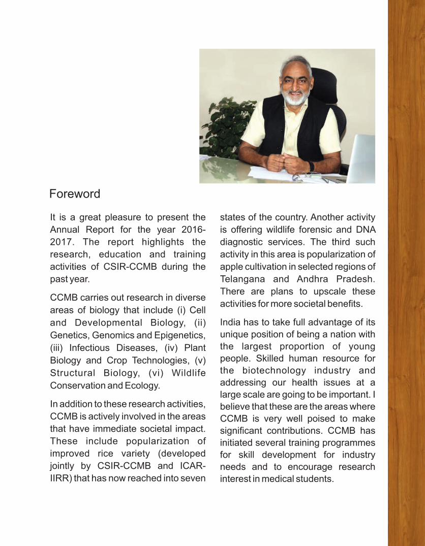
It is a great pleasure to present the
Annual Report for the year 2016-
2017. The report highlights the
research, education and training
activities of CSIR-CCMB during the
past year.
CCMB carries out research in diverse
areas of biology that include (i) Cell
and Developmental Biology, (ii)
Genetics, Genomics and Epigenetics,
(iii) Infectious Diseases, (iv) Plant
Biology and Crop Technologies, (v)
Structural Biology, (vi) Wildlife
Conservation and Ecology.
In addition to these research activities,
CCMB is actively involved in the areas
that have immediate societal impact.
These include popularization of
improved rice variety (developed
jointly by CSIR-CCMB and ICAR-
IIRR) that has now reached into seven
states of the country. Another activity
is offering wildlife forensic and DNA
diagnostic services. The third such
activity in this area is popularization of
apple cultivation in selected regions of
Telangana and Andhra Pradesh.
There are plans to upscale these
activities for more societal benefits.
India has to take full advantage of its
unique position of being a nation with
the largest proportion of young
people. Skilled human resource for
the biotechnology industry and
addressing our health issues at a
large scale are going to be important. I
believe that these are the areas where
CCMB is very well poised to make
significant contributions. CCMB has
initiated several training programmes
for skill development for industry
needs and to encourage research
interest in medical students.
Foreword

Today’s science will set the agenda for
tomorrow’s socio-economic activities.
It is, therefore, extremely important to
spread the message of science, its
unprecedented possibilities and
awareness on issues in society.
CCMB is committed to this cause: we
organize a number of activities under
which the CCMB community interacts
with young students and the public
from across the country every year
through its open day programme,
educational tours, visits of police,
forest, judiciary and industry officials.
A changing set of technologies, and
newly emerging possibilities &
challenges keep laboratories such as
CSIR-CCMB at the centre stage
where we are expected to deliver to
the needs and aspirations of the
nation. I have no doubt that CCMB is
ready to meet this challenge.
Rakesh Kumar MishraDirector

I. Charter


The objectives of the Centre are :
a) To conduct research in frontier and multi-
disciplinary areas of modern biology and to
seek potential applications of this work.
b) To carry out exploratory work in areas of
biology with a view to aid the development of
biochemical and biological technology in the
country on a sound basis.
c) To train people in the advanced areas of
biology to serve the needs of development in
these areas, with special provision for short-
term training of staff from other institutions in
techniques for which adequate facilities may
not exist elsewhere.
d) To provide centralized facilities in the
country for new and modern techniques
in the inter-disciplinary areas of biology,
and to ensure that these facilities are so
organized, maintained and administered
that they can be put to maximal use by
research workers from other laboratories
and institutions in the country.
e) To interact adequately with other institutions
doing basic or applied work in areas related to
the activities of the Centre.
f) To collect, collate and disseminate
information relevant to biological research.
The Centre for Cellular and Molecular Biology (CCMB) is one of the constituent national
laboratories of the Council of Scientific and Industrial Research (CSIR), New Delhi, the premier multi-
disciplinary research organization in the country funded by the Government of India. It was set up
formally in April, 1977
03


ResearchProgrammes


07
From Left to Right: Purushotham, Lomous Kumar, Harsh Joshi, Ramesh K. Aggarwal, Arcot K. Nagamani, Chhavi Dawar
Ramesh Kumar AggarwalMolecular Genetics and Developmental Biology
RESEARCH INTERESTS: • Epigenetics of temperature-dependent sex determination• DNA Markers: development and applications in crop improvement• Barcoding of Anurans of India• Evolution/phylogeography of endangered species• Decoding of Indian genomes
Selected recent publications
1. Jegath Janani S, Vasudevan Karthikeyan, Prendini Elizabeth, Dutta Sushil Kumar, and Aggarwal Ramesh K (2017). A new species of the genus Nasikabatrachus (Anura, Nasikabatrachi-dae) from the eastern slopes of the Western Ghats, India. 34(1-4), 1-19. (the paper is covered Alytes extensively by National/ international press/-online news groups, including ones like National Geography).
2. Aggarwal RK, Dawar C, Phanindranath R, Mutnuri L and Dayal AM. (2016). Draft genome sequence of a versatile hydrocarbon-degrading bacter ium, Rhodococcus pyridinivorans strain KG-16, collected from oil fields in India. Genome Announcement 4(1): e01704-15.
3. Chandramouli SR, Vasudevan K, Harikrishnan S, Dutta SK, Janani SJ, Sharma R, Das I and Aggarwal RK. (2016). A new genus and species of arboreal toad with phytotelmonous larvae, from the Andaman Islands, India (Lissamphibia, Anura, Bufonidae). 555: 57-90.ZooKeys
4. Prakash N S, Jeena Devasia, Ramesh K Aggarwal, and Raghuramulu Y (2017) COFFEE. In: Biotechnology of Plantation Crops, Eds: Chowdappa P, Anitha Karun, Rajesh M K, Ramesh S V, Chapter 17, pp 355 - 404, (ISBN: 978-93-5124-836-1) , New Delhi.Daya Publishing House
Our group has identified several candidate genes having putative role in TSD,
described new species of anurans, developed >2500 species specific genic-/genomic
microsatellite markers, constructed the first-generation framework molecular linkage maps
of coffee and mulberry, established that the Indian wolf populations represent new species,
i.e., Canis indica and Canis himalayensis, discovered many new frog species and
showed that the olive ridleys in Indian waters are the ancient source population for the olives
in other global basins.

Our main research interest is to understand the eco-
devo of temperature-dependent sex determination
(TSD) using Indian mugger as a model system. We
are equally interested in application oriented studies
involving development/utilization of genomic
resources useful for crop improvement, wildlife
conservation and disease diagnostics. Lately, we
have embarked on whole genome sequencing of
Indian mugger, apple, mulberry and a few interesting
microbial isolates of clinical/applied significance.
Temperature-dependent sex-determination: Sex-
determination is one of the most important
developmental decisions occurring early in
embryogenesis and is central to the existence/
survival of a species. Broadly there exist two
mechanisms of sex determination in vertebrates:
Genetic Sex Determination (GSD) that involves
specific gene(s) and/or highly specialized sex
chromosomes, and Environmental Sex Determination
(ESD), wherein simple environmental cues appear to
be the primary determinants of the sex. Temperature
dependent Sex Determination (TSD) is the most
common ESD mechanism seen in many reptilians.
Indian mugger (Crocodylus palustris) is one such
species and thus, provides an ideal system to study
the role of epigenetic factors such as environmental
temperature in vertebrate development, more
specifically, in sex-determination.
We have been using Indian mugger as an
experimental system to identify the putative genetic
mechanism(s) underlying TSD. We have isolated and
characterized the crocodile homologues of a few of
the evolutionarily conserved, sex-related genes, and
have also identified a number of novel candidate
genes that show differential expression at Male-
/Female-Promoting Temperature (MPT/FPT) in the
bipotential gonads. Our studies on cpSox9 and
cpDmrt1, the crocodile homologues of Sox9 (Sry-
related transcription factor Sox9) and Dmrt1
(doublesex-/mab-3 related transcription factor-1),
as well as a few other candidate genes demonstrate
that generation of sex-specific/unique mRNA
transcripts by 'extensive alternate splicing', is the
norm of the complex genetic interplay underlying
the molecular basis of TSD. More recently, we have
identified/isolated several isoforms of heat-shock
proteins (cp_HSPs) and heat-shock factors
(cp_HSF1 and related genes) that may be involved in
sensing the temperature stimuli to initiate the set of
genetic factors during TSD. Interestingly, some of
these isoforms also seem to have sex-specific
differential expression through Temperature-
Sensitive Window (TSW) in the GAM complex in our
preliminary studies.
To expedite our efforts to identify the putative
candidate gene(s) underlying the TSD, we initiated
whole/global transcriptome profiling of GAM and
brain tissues of male/female embryos using the
high-throughput NGS approaches. We have now
generated NGS data for 36 GAM and brain
transcriptomes (ca. 6.0 GB cleaned sequencing
data/transcriptome), representing biological
duplicates of developmental stages through TSW
(stage 21 to 25th) using Illumina HiSeq and Roche-
454 platforms. The clean sequence reads are
normalized to construct a reference data set that is
being used to assemble/annotate denovo sex-/
stage-specific GAM transcriptomes, followed by
comparative analysis to identify/characterize
differentially expressed genes. Primary denovo
reference assemblies have been completed with
Trinity, a de-bruin based assembler working on
fixed k-mer value of 25 giving an average N50
value of 2.6Kb. Presently we are optimizing the k-mer
value to be used for multi-kmer multi tool approach to
obtain more complete assemblies using various
assemblers like Oases, SOAP-denovoTrans and
TransAByss. Simultaneously, we have also initiated
Whole-Genome Bisulphite Sequencing (WGBS)
using Illumina platform to ascertain the methylation
patterns of the gene space (especially regulatory
regions/ promoter sequences of selected
candidate genes) in the genomic DNA of GAM
tissues; this is expected to help us understand the
epigenetic control (if any) of gene(s) expression
that may underlie the TSD.
Development and application of DNA markers:
DNA markers provide high genetic resolution and
have revolutionized genetic analysis for enumeration,
management, utilization of biodiversity resources,
understanding origin, evolution, phylogeography
of extant species, or genetic enhancement of
existing agriculturally important variety/cultivars.
Development and application of DNA marker tools
have been a major focus of our research, which is
carried out in collaboration with various national and
international institutes.
Germplasm characterization and linkage studies
in rice, coffee, mulberry and apple: Our earlier
studies on rice germplasm including the wild rice
species and their derivatives have helped us assign
two new genomes (GG, HHJJ) of Oryza, provided
empirical data to support Gondwanaland as the
centre of origin for Oryza, identified novel potential
rice sources for early nodulin gene homologues, and
resulted in a DNA polymorphism database useful for
the identification, protection and improvement of elite
Basmati and specialty rice varieties of India. | 01 |
Our other more recent major interest has been to
create genetic and genomic resources for orphan tree
crop species of socio-economic significance. During
the last few years, we have developed: a) large
repertoires of genomic and genic microsatellite
markers of coffee, mulberry and apple; b) molecular
linkage maps of robusta coffee and mulberry; c) leaf-
specific EST resources of coffee and mulberry.
Similarly, we have identified a number of Resistance
Gene Analogues in secondary genepool of coffee,
and potential germplasm sources of resistance to
powdery mildew in mulberry, which are expected to
be useful in breeding for disease-resistance
germplasm in these crops. We have also developed/
established a pseudo-testcross population of apple
from a cross of two elite varieties, i.e., 'Red Delicious'
and 'Maharaji', in collaboration with our partners from
Jammu University, Kashmir University, YSPUF&H,
and GBPIHED. This is the first such genetic resource
developed in the country, and will greatly facilitate
linkage mapping and QTL studies for this important
fruit crop. Our similar work on mulberry, being carried
out in collaboration with CSRTI, Berhampore (WB)
has resulted in identification of a number of putative
QTLs for agronomically important traits, as well as,
identification of a few High-yielding/resistant
progenies from the mapping population (which have
the potential to be developed directly in HYV suited to
West Bengal environment).
Wildlife conservation studies: We have been
interested in genetic characterization of extant
populations of endangered species, and to enable the
same, we have developed species-specific
microsatellite markers for many species, viz.,
Indian mugger, red panda, olive ridley turtles and a
number of frog species. Our work has given
new perspective to the evolution and phylogeography
of wolves, anurans, olive ridleys and crocodilians in
the Indian subcontinent, and highlights the need
for conservation of their fragile environment.
Significantly, we have shown that: a) the Indian wolf
populations represent two new species (Canis indica
and Canis himalayensis) basal to the grey wolf-dog
lineage, b) anuran endemism in the Western Ghats
and other Gondwana break-up landmasses is much
earlier than the CT boundary; c) the olive ridley in
Indian waters are the source ancestral population for
ridleys found across other global basins; and d) the
two gharial genera (Gavialis and Tomistoma) are
closer to each other, and together with crocodylids
represent a distinct genetic lineage that has diverged
from that of alligators and caimans during cretaceous
era about 80 - 100 MYA.
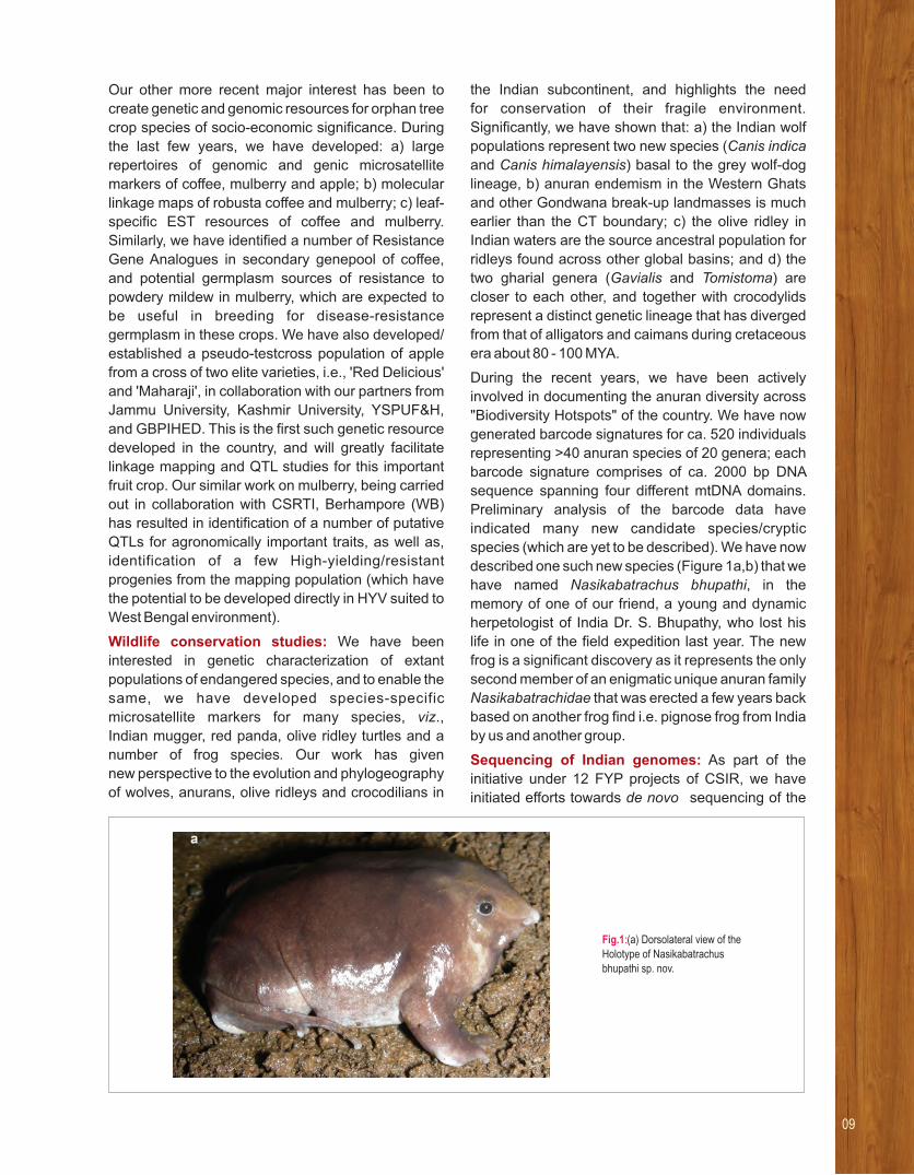
During the recent years, we have been actively
involved in documenting the anuran diversity across
"Biodiversity Hotspots" of the country. We have now
generated barcode signatures for ca. 520 individuals
representing >40 anuran species of 20 genera; each
barcode signature comprises of ca. 2000 bp DNA
sequence spanning four different mtDNA domains.
Preliminary analysis of the barcode data have
indicated many new candidate species/cryptic
species (which are yet to be described). We have now
described one such new species (Figure 1a,b) that we
have named Nasikabatrachus bhupathi, in the
memory of one of our friend, a young and dynamic
herpetologist of India Dr. S. Bhupathy, who lost his
life in one of the field expedition last year. The new
frog is a significant discovery as it represents the only
second member of an enigmatic unique anuran family
Nasikabatrachidae that was erected a few years back
based on another frog find i.e. pignose frog from India
by us and another group.
Sequencing of Indian genomes: As part of the
initiative under 12 FYP projects of CSIR, we have
initiated efforts towards de novo sequencing of the
Fig.1:(a) Dorsolateral view of the Holotype of Nasikabatrachus bhupathi sp. nov.
09

whole genome of Indian mugger. For the purpose,
we are sequencing an alpha male mugger
'Makara' that had been 'the prime sire in breeding
programs at the Crocodile Bank, Chennai
in 1970s/1980s. We are attempting 'Hybrid
sequencing approach' using three different
NGS platforms (Roche-454, PacBIO, and
Illumina HighSeq/MiSeq) that significantly differ
in the size and depth of the sequencing read
data. Todate, we have generated ca. 389 Gb raw
data using multiple libraries and HiSeq/MiSeq
chemistry on the Illumina sequencer. The data have
been quality trimmed and cleaned for noise, and is
being used to standardize the various parameters
(like k-mer, window-size, bubble size etc) and
different assemblers (like SOAP denovo, velvet,
Masurca, Platanus, Discovar) to generate an optimal
de novo genome assembly of Indian mugger.
Presently, we have initiated efforts to generate long-
read data providing ca. 50X genome coverage using
the PacBio chemistry; availability of which is expected
to help obtain an robust high-quality de novo draft
genome of Indian mugger. Simultaneously, we are
trying to sequence the genomes of apple and Indian
mulberry . We have now generated considerable raw
sequencing data (~150 -200 Gb for each plant
species). The plant WGS data will be used to extract,
assemble and annotate respective mitochondrial and
chloroplast genomes and also de novo whole
genome assemblies. These genomes will add to our
efforts to investigate complex important traits in these
plants and development of DNA based markers.
Fig.1: (b) a Bayesian phylogenetic tree inferred from concatenated two partial mitochondrial genes (12S rRNA and 16S rRNA) showing the new frog as the conger of the N. sahyadrensis (support values of the nodes are written above each node: Bayesian posterior probabilities, followed by ML bootstrap values).

11
From left to right (back row): Ira Bhatnagar, Shahila Parween (Front row): O Gopi Suresh, Amit Asthana, Saurabh K. Srivastava
Amit AsthanaApplications of Microfluidics, Micro and Nanotechnology in Life Sciences
RESEARCH INTERESTS:
• Non-conventional methods to fabricate microfluidic devices.• Microfluidic device for proteomic analysis to understand wound healing
mechanisms• Point-of-care diagnostic devices based on paper-microfluidics• Paper-based devices as Raman immune-sensor• Biopolymer microfluidic devices for tissue engineering and cell culture• 3D printing and 3D cell culture• Generation of “site targeted” drug delivery vectors for drug delivery and
diagnosis, using microfluidic device
Selected recent publications
1. Bhatnagar I, Mani Vasagan P and Bramhachari PV (2016). Opening Avenues in Marine Probiotics – Present and Future. In: Nollet LML (Ed.), Marine Microorganisms: Extraction and Analysis of Bioactive Compounds. CRC Press, Boca Raton.
2. Bramhachari PV, Mutyala S, Bhatnagar I, Pallela R (2016). Novel Insights on the Symbiotic Interactions of Marine Sponge-Associated Microorganisms: Marine Microbial Biotechnology Perspective. In: Pallela R, Ehrlich H (Eds.). Marine Sponges: Chemicobiological and Biomedical Applications, Springer-Verlag, Germany, pp. 69–95.
3. Bhatnagar I, Pallela R, Bramhachari PV and Ealla KKR. (2016). Chronicles of Sponge Biomaterials: The Saga in Biomedicine. In: Pallela R, Ehrlich H (Eds.). Marine Sponges: Chemicobiological and Biomedical Applications, Springer-Verlag, Germany, pp.315-327.
4. Pham UHT, Hanif M, Asthana A and Iqbal SM (2015) A microfluidic device approach to generate hollow alginate microfibers with controlled wall thickness and inner diameter, Journal of Applied Physics, 117: 214703.
5. Micheal I J, Aditya J, Vidyasagar, Kiran Kumar B, Naveen Kumar M, Amit Asthana and Ch Mohan Rao (2014) Foil assisted replica molding for fabrication of microfluidic devices and their application in vitro, 14 (19): 3695 Lab-on-a-chip –3699.
Our group uses micro and nanotechnology to address biological
problems. Using a non-conventional method developed in our group to fabricate microfluidic
devices, termed as “modified embedded templates”, we have recently demonstrated an
approach to generate hollow alginate microfibres with controlled wall thickness and
inner diameter using the above mentioned microdevices. These devices are also used to
generate PLGA and alginate microparticles in the group.
As a part of our attempts to fabricate affordable
diagnostic devices, we are currently exploring non-
conventional ways to fabricate polymer- as well as
paper-based microfluidic devices. Among all the
affordable devices mentioned in the literature, paper-
based microfluidics is considered a potential “game-
changer” in the field of diagnostics because of the
affordable prices of paper as a substrate. In our
group, we have developed a “truly single step”
method of fabricating paper devices using an ink
developed in CCMB and permanent ink pen.
Currently we are using 4 different ways of making
devices (1) wax printer (2) XY plotter with permanent
ink pen (3) XY plotter with ink developed in our lab
(4) laser cutter. Among the various applications of
paper based devices, a few are listed below:
Cost effective and efficient paper-based
viscometer (with Dr. Ch Mohan Rao)
The use of paper-based devices for affordable
diagnostics is gaining interest due to unique
advantages like affordability, portability, easy
disposability and inherent capillarity. As capillary
transportation is an integral component of paper-
based devices, a low sample volume with faster
measurement becomes an additional advantage. We
have exploited the aforementioned features of paper-
based devices to develop a simple microfluidic device
suitable for measuring viscosity of Newtonian fluids
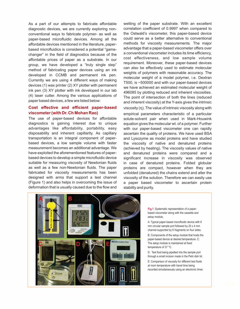
as well as a few non-Newtonian fluids. The paper
fabricated for viscosity measurements has been
designed with arms that support a test channel
(Figure 1) and also helps in overcoming the issue of
deformation that is usually caused due to the flow and
wetting of the paper substrate. With an excellent
correlation coefficient of 0.9997 when compared to
the Ostwald’s viscometer, this paper-based device
could serve as a better alternative to conventional
methods for viscosity measurements. The major
advantage that a paper-based viscometer offers over
a conventional viscometer includes its time efficiency,
cost effectiveness, and low sample volume
requirement. Moreover, these paper-based devices
can also be effectively used to estimate molecular
weights of polymers with reasonable accuracy. The
molecular weight of a model polymer, i.e. Dextran
T500, is ~500000 and with our paper-based devices
we have achieved an estimated molecular weight of
496085 by plotting reduced and inherent viscosities.
The point of intersection of both the lines (reduced
and inherent viscosity) at the Y-axis gives the intrinsic
viscosity (ɳ). The value of intrinsic viscosity along with
empirical parameters characteristic of a particular
solute-solvent pair when used in Mark-Houwink
equation gives the molecular wt. of a polymer. Further
with our paper-based viscometer one can rapidly
ascertain the quality of proteins. We have used BSA
and Lysozyme as model proteins and have studied
the viscosity of native and denatured proteins
(achieved by heating). The viscosity values of native
and denatured proteins were compared and a
significant increase in viscosity was observed
in case of denatured proteins. Folded globular
proteins are compact, however when they are
unfolded (denatured) the chains extend and alter the
viscosity of the solution. Therefore we can easily use
a paper based viscometer to ascertain protein
stability and purity.
| 01 |
Fig.1: Systematic representation of a paper-
based viscometer along with the cassette and
setup module,
A: Typical paper-based microfluidic device with 8
mm circular sample port followed by 20 x 4 mm
channel supported by 6 fragments on four sides.
B: Components of the setup module that hosts the
paper-based device at desired temperature. C:
The setup module is maintained at fixed
temperature of 37 °C.
D: Test fluid being pipetted into the sample port
through a small incision made in the Petri dish lid.
E: Comparison of viscosity for different test fluids
at room temperature with travel time being
recorded simultaneously using an electronic timer.

Paper-based Surface Enhanced Raman microscopy substrates for detection enhancement
Raman spectroscopy is a technique that measures
the shift in wavelengths of light arising due to
inelastic scattering. It is used to characterize the
vibrational and rotational state of a molecule and
is a signature of a particular molecule. However,
typical Raman signals are too weak to be
detected and require some sort of augmentation.
Surface-Enhanced Raman Spectroscopy (SERS)
is a modification of Raman spectroscopy where
the analyte to be investigated is layered on a
metallic surface to enhance the Raman signal. The
metallic surface has to be uneven for better results.
Hence, either a roughened surface or substances
with small radii of curvature are used. Metallic
nanoparticles made of silver or gold are an excellent
substratum for SERS. In our lab we have created four
different ways of fabricating rapid and affordable
paper-based SERS substrate for potential clinical
applications. Lysozyme and Rhodamine 6G were
used as model molecules to evaluate SERS
performance of the as-synthesized paper-based
SERS substrates. Figure 2 shows some of the SEM
pictures of paper devices and Raman spectra of
lysozyme showing enhancement in the Raman
singles.
Fig.2: Enchantment of Raman spectra of 5 mg/ml of Lysozyme with increase in density of gold nanoparticles on paper substrate. An enhancement of 108 was achieved using 10mM HAuCl4.
12000000
10000000
8000000
6000000
4000000
2000000
Inte
nsi
ty
0
500 700 900 1100 1300 1500 1700 1900 2100 2300-1Raman Shift/cm
13

From left to right: Siripini Satish, Pankaj Kumar, Hatim Kara, Vykuntham Naga Gowthami, Amere Subbarao Sreedhar, Khanderao Paithankar, Guntipally Mounika, A Vijaya Lakshmi; Inset-left: Kanugovi Abhijnya Vijayavittal, inset-right: Akhil Kotwal
A S SreedharStress Biology and Molecular Medicine
RESEARCH INTERESTS:
• Enforced senescence as a tumor suppressor mechanism
• Hsp90 in the epigenetic regulation of cancer
• Hsp90 in the cross-talk between acquired multidrug resistance and metastasis of cancer cells
• Evaluation of mitochondrial chaperone, Trap1 role in cancer cells
Selected recent publications
1. Kanugovi AV, Joseph C, Sreedhar AS (2016) The tumor INK4asuppressor p16 expression bypasses 17AAG mediated cellular
effects in human neuroblastoma, IMR-32. (6) 74.Transl Med.
2. Kanugovi AV, Sugunan S, Joseph C and Amere SS (2015). The prosurvival activity of HSF2 in response to autophagic stress is triggered by HSF2-dependent transactivation of BTG2. Mol Biol Cell. 26(25):4523.
3. Kanugovi AV and Amere SS (2014). Oncogene de-addiction from Hsp90 induces senescence-like phenotype in malignant and metastatic tumor cells. 25(25):3987.Mol. Biol. Cell.
4. Sarangi U, Singh MK, Abhijnya KVV, PrasannaAnjaneya Reddy L, Siva Prasad B, Pitke VV, Paithankar KR and Sreedhar AS(2013). Hsp60 chaperonin acts as barrier to pharmacologically induced oxidative stress mediated apoptosis in tumor cells with differential stress response. (7)35-5.Drug Target Insights
5. Sarangi U, Paithankar KR, Ujwal Kumar J, Subramaniam V and Sreedhar AS(2012). 17AAG treatment accelerates doxorubicin induced cellular senescence: Hsp90 interferes with enforced senescence of tumor cells. , 6: 19-35.Drug Target Insights
Our group
• identified tumor selective functions of Hsp90
• demonstrated that enforced senescence through Hsp90 inhibition acts as tumor
suppressor mechanism
• identified novel roles of Hsp90 in multidrug resistance
• identified potential role of Trap1in mitochondrial dynamics and metabolism
Heat shock proteins (Hsps) form the most ancient
defense system in all living forms. Hsps are highly
conserved and ubiquitously expressed proteins that
play major roles in the maintenance of cellular
homeostasis. Induced Hsps protect cells from various
harmful stimuli. Increased Hsp expression is also
found in several pathological conditions including
cancer. We are interested in studying the
unconventional roles of Hsps in tumor cells using
molecular and chemotherapeutic approaches.
Hsp90 in oncogene addiction
We earlier demonstrated that mutated oncogenic
Raf-1 (CAAX-Raf) gets addicted to Hsp90. As a
result, the conformation specific Hsp90 inhibitor
17AAG, destabilizes Raf-1 and promotes cellular
senescence. It is proposed that Hsp90-kinase
interaction occurs through a common surface on
kinases, the α5β4 loop. Through co-precipitation
experiments in vitro, we observed CAAX-Raf
interacting with Hsp90α more efficiently than Hsp90β.
The co-chaperone, cdc37 was also found to be pulled
down in the complex. Subsequently using Hsp90
deletion constructs and CAAX-Raf, we observed wild
type Raf interacting with Hsp90 without the hinge
region (a region between N-terminus and the middle
domain), but not CAAX-Raf, indicating that the hinge
region of Hsp90 is responsible for CAAX-Raf
interaction. These results indicated that oncogene
addiction to Hsp90 occurs through kinase interaction
with the hinge region, but not through the α5β4 loop.
Enforced senescence as a tumor suppressor
mechanism
Earlier we demonstrated that Raf or Ras addiction to
Hsp90 sensitizes cells to Hsp90 inhibition, however,
this induces senescence by activating the DNA
damage response (DDR) pathway mediated through
p53-p21WAF-1 axis. To demonstrate tumor
suppression in vivo, we used the mouse xenograft
model as a tool. Since oncogene de-addiction from
Hsp90 is resulting in the activation of senescence
program, we believe that activation of senescence-
like phenotype may have clinical advantage. To
further evaluate our findings in vivo, nude mice
were subcutaneously injected with wild type and
CAAX-Raf transfected cells, treated with 17AAG,
and solid tumor growth was monitored over a period
of time. Compared to wild type Raf cells, CAAX-Raf
cells showed increase in tumor growth and upon
17AAG treatment were unable to develop tumors.
The significant decrease in tumor volume and cells at
tumor site of injection indicates antitumor effects of
17AAG in CAAX-Raf cells. The increase in
senescence associated β-galactosidase activity in
tumor cells only in response to the combination
treatment suggests that the lack of tumorigenic
potential is due to activation of a senescence
program. Subsequently CAAX-Raf transfected cells
were injected intravenously and found that the tumor
is being metastasizing to lungs. However i.v injections
followed by 17AAG treatment resulted in decrease in
metastasizing effects of the tumor to lungs. Our
results demonstrate that oncogene de-addiction to
Hsp90 activates the senescence program thus acting
as a tumor suppressor mechanism.
Hsp90 in the epigenetic regulation of cancer
Cancer emerges from both genetic and epigenetic
mechanisms. Earlier we showed the involvement
of Hsp90 in the epigenetic regulation of Rb and
Raf-1. We hypothesized that Hsp90 regulates
Rb transcription at least by three mechanisms,
(1) through stabilizing CDK4/6, (2) by directly
binding to the Rb promoter (3) and through E2F1.
Although, the role of Hsp90 in the cytoplasm is well
known through its stabilization of protein kinase
clients, its nuclear functions are less studied. Our
study aims at understanding novel interactions and
functions of nuclear Hsp90. We found that E2F1 and
E2F2 are novel interacting partners of Hsp90 in the
nucleus. As of now, we could explore the role of
Hsp90 in the functional stabilization and
transcriptional activation of E2F1. Interestingly, we
also observed acetylated Hsp90 in the nuclear
localization and interaction with E2F1 and E2F2.
We are evaluating role of Hsp90 in cell cycle
regulation through different E2F transcription factors.
Cancer EMT
Earlier we reported that hypoxia induces EMT in
MCF7 and MDAMB-231 breast cancer cells.
Interestingly, challenging these cells with
chemotherapeutic agents including Hsp90 inhibitors
also showed enhanced EMT. While the mechanism
of Hsp90 inhibition mediated EMT is not known, the
data provided insights that Hsp90 interferes with (or
keeps under check) EMT. We asked for the possible
mechanism involved in this EMT. Though we did not
observe a prominent decrease in E-cadherin
expression at the transcription level, we observed a
decrease in surface E-cadherin correlating with EMT
progression. Since loss of E-cadherin facilitates the
translocation of β-catenin to the nucleus to induce the
transcription of genes responsible for EMT, we
examined its translocation and observed that
induced EMT is not dependent on β-catenin. We are
now examining the mechanism(s) of therapeutically
15

induced EMT and its link with Hsp90. Our in vivo data
indicated decreased metastasis with Hsp90 inhibition
in MCF7, and in contrast, we observed increased
metastasis in MDAMB-231 cells. We correlated these
findings with in vitro data and postulated the
hypothesis that the amount of stem cell pool in the
tumor population decides the fate of cells in response
to treatments. MCF-7 cells showed enhanced
pluripotency on the onset of EMT and showed
sensitivity to Hsp90 inhibition. In contrast, MDAMB-
231 cells showed decreased pluripotency, but
enhanced stemness leading to enhanced metastasis.
We are yet to re-confirm the data with our in vivo
findings to draw a conclusion.
Hsp90 inhibition and multidrug resistance
Earlier we reported a positive correlation between
Hsp90 expression with increased multidrug
resistance of cancer cells. Multidrug efflux pumps
are located at the plasma membrane and are
more specially enriched within the cholesterol
rich membrane micro-domains called lipid rafts.
We demonstrated co-localization of Hsp90 and
p-glycoprotein with the help of dual immuno-
fluorescence. Now we show that an increase in
Hsp90 increases the cholesterol accumulation
to the lipid rafts that incidentally correlated with
the accumulation of Hsp90 itself along with the
p-glycoprotein. Further, increased Hsp90 also
correlated with increased HMGCoA biosynthesis
indicating its possible regulatory role in cholesterol
biosynthesis. However, although the HMGCoA
inhibitor lovastatin decreased cholesterol
biosynthesis, its accumulation at the lipid rafts was
not affected, indicating that Hsp90 is majorly involved
in cholesterol transport to the lipid rafts. Depletion
of cholesterol using methyl β-cyclodextrin (MβCD)
decreased both Hsp90 and p-glycoprotein
accumulation at the lipid rafts. From these results we
demonstrate that Hsp90 potentiates drug afflux
activity through enhanced cholesterol transport to the
lipid rafts.
Earlier we reported that Hsp90 inhibition increases
MMP7 expression and decreases MMP11
expression. We also showed that increased drug
resistance may be a consequence of EMT induced by
17AAG. To understand MMP7 and MMP11 mediated
metastatic potential of multidrug resistant cells, we
examined the in vivo tumorigenic potential of MMP7
and MMP11 over expressing cells. From the
preliminary results, we found that both cell types show
tumor growth but differ in their tumor forming ability.
From these results, we learn that MMP7 and MMP11
cells have opposite functions in drug afflux activity as
well as in tumor forming ability. These results need to
be further examined.
Tumor selective functions of Trap1
Trap1 (TNF receptor associated protein 1) is a
nuclear encoded, mitochondrial chaperone belonging
to the HSP90 family. Unlike Hsp90, Trap1 functions
are not fully understood. Earlier we showed that Trap1
expression increases with the aggressiveness of
tumors and is associated with enhanced
mitochondrial fission, whereas its knockdown
promotes mitochondria fusion. Since mitochondria
are central to cellular energy metabolism, we
examined the role of Trap1 in regulating mitochondrial
metabolism. While measuring the oxygen
consumption rate (OCR), we discovered that Trap1
over expression decreases OCR, while its
knockdown increases the same. Since altered OCR
may alter ATP, we measured ATP and found out that
ATP levels showed opposite effect with OCR
indicating that ATP production in these cells may be
OXPHOS-independent. Further, we also observed
that decreased Trap1 itself elicits a hypoxic
response. Although, we did not find a direct
correlation of Trap1 with mitochondria metabolism,
we obtained a role for Trap1 in metabolic
reprogramming. We are currently examining
the role of Trap1 in mitochondrial dynamics and its
impact on metabolic reprogramming.

17
Ashutosh Shukla, Purnima Bhargava
Purnima BhargavaEpigenetic Mechanisms of Gene Regulation
RESEARCH INTERESTS:
• Transcription by yeast RNA polymerase III
• Epigenetic regulatory mechanisms
• Determinants of Nucleosome positioning
Selected recent publications
1. Belagal P, Normand C, Shukla A, Wang R, Dez C, Bhargava P and Gadal O (2016) Decoding the principles underlying the frequency of association with nucleoli for RNA polymerase III-transcribed genes in budding yeast. E16-03- Mol. Biol. Cell.0145.
2. Vernekar D V and Bhargava P (2015) Yeast Bud27 modulates the biogenesis of Rpc128 and Rpc160 subunits and the assembly of RNA polymerase III. 1849, 1340-1353.BBA-GRM
3. Albert B, Mathon J, Shukla A, Saad H, Normand C, Legere-Silvestre I, Villa D, Kamgoue A, Mozziconacci J, Wong H, Zimmer C, Bhargava P, Bancaud A and Gadal O (2013) Systematic characterization of the conformation and dynamics of budding yeast chromosome XII. 202: 201-210. J Cell Biol.
4. Kumar, Y. and Bhargava, P. (2013) A unique nucleosome arrangement, maintained actively by chromatin remodelers facilitates transcription of yeast tRNA genes. BMC Genomics14:402.
5. Mahapatra, S., Dewari, P.S., Bhardwaj, A. and Bhargava, P. (2011) Yeast H2A.Z, FACT complex and RSC regulate transcription of tRNA gene through differential dynamics of flanking nucleosomes. 39, 4023-4034.Nucl. Acids Res.
Our group showed that
Yeast prefoldin protein, Bud27 regulates the biogenesis of two of the
core subunits and hence the assembly of the17-subunit yeast RNA polymerase (pol) III.
Yeast Paf1 complex has a protective role on damage-prone pol III-transcribed gene which
show high transcription activity in vivo.Spt16 subunit of the yeast histone chaperone
FACT maintains the Swr1 and H2A.Z levels in the nucleosome downstream (DS) of the terminator
at the 3’ gene end.
FACT is specifically enriched at the 3’ end of all pol III-transcribed genes.
FACT and pol III co-occupy and traverse the gene body together. Pol III delivers FACT
to the DS nucleosome at the end of each transcription cycle.
Eukaryotic transcription by the RNA polymerases
takes place in a generally repressive chromatin
milieu. Organization of the genome into chromatin
restricts the access of transcription machinery to its
templates. We have been studying the relationship of
chromatin and transcription using yeast as model
eukaryotic system. In the budding yeast
Saccharomyces cerevisiae, ~300 genes found
scattered on different chromosomes are transcribed
by the enzyme RNA polymerase (pol) III. Pol III is
dedicated to synthesizing the short, stable, non-
coding RNAs, required for the vital cell processes like
translation, ribogenesis and mRNA processing. Pol III
is assisted by its two basal factors TFIIIC and TFIIIB,
which assemble the transcription complex utilizing
the intra-genic promoter elements. Generally no
upstream regulatory sequences are found and as
compared to the genes transcribed by pol II, only 3-5
classical, DNA-binding regulators are known for the
pol III-transcribed genes. Recent evidences for
regulation by chromatin-related mechanisms have
been unraveling novel mechanistic details
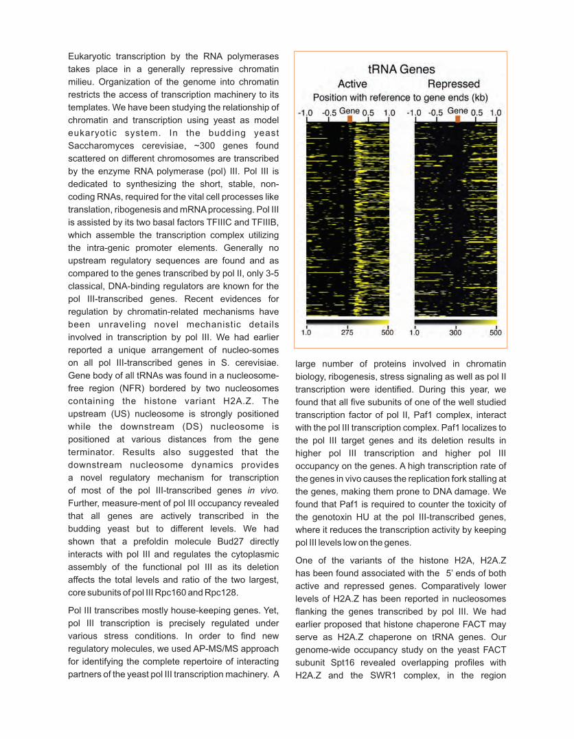
involved in transcription by pol III. We had earlier
reported a unique arrangement of nucleo-somes
on all pol III-transcribed genes in S. cerevisiae.
Gene body of all tRNAs was found in a nucleosome-
free region (NFR) bordered by two nucleosomes
containing the histone variant H2A.Z. The
upstream (US) nucleosome is strongly positioned
while the downstream (DS) nucleosome is
positioned at various distances from the gene
terminator. Results also suggested that the
downstream nucleosome dynamics provides
a novel regulatory mechanism for transcription
of most of the pol III-transcribed genes in vivo.
Further, measure-ment of pol III occupancy revealed
that all genes are actively transcribed in the
budding yeast but to different levels. We had
shown that a prefoldin molecule Bud27 directly
interacts with pol III and regulates the cytoplasmic
assembly of the functional pol III as its deletion
affects the total levels and ratio of the two largest,
core subunits of pol III Rpc160 and Rpc128.
Pol III transcribes mostly house-keeping genes. Yet,
pol III transcription is precisely regulated under
various stress conditions. In order to find new
regulatory molecules, we used AP-MS/MS approach
for identifying the complete repertoire of interacting
partners of the yeast pol III transcription machinery. A
large number of proteins involved in chromatin
biology, ribogenesis, stress signaling as well as pol II
transcription were identified. During this year, we
found that all five subunits of one of the well studied
transcription factor of pol II, Paf1 complex, interact
with the pol III transcription complex. Paf1 localizes to
the pol III target genes and its deletion results in
higher pol III transcription and higher pol III
occupancy on the genes. A high transcription rate of
the genes in vivo causes the replication fork stalling at
the genes, making them prone to DNA damage. We
found that Paf1 is required to counter the toxicity of
the genotoxin HU at the pol III-transcribed genes,
where it reduces the transcription activity by keeping
pol III levels low on the genes.
One of the variants of the histone H2A, H2A.Z
has been found associated with the 5’ ends of both
active and repressed genes. Comparatively lower
levels of H2A.Z has been reported in nucleosomes
flanking the genes transcribed by pol III. We had
earlier proposed that histone chaperone FACT may
serve as H2A.Z chaperone on tRNA genes. Our
genome-wide occupancy study on the yeast FACT
subunit Spt16 revealed overlapping profiles with
H2A.Z and the SWR1 complex, in the region

downstream (DS) of the 3’-end of the tRNA genes.
H2A.Z is deposited in the nucleosomes by the Swr1
complex while Spt16 evicts H2A.Z. Spt16 maintains
Swr1 and H2A.Z levels on these genes. Spt16 also
maintains the occupancy and positioning of the DS
nucleosome. Deposition of H2A.Z confers instability
to the tDNA flanking nucleosomes. Higher H2A.Z
levels in the DS than the US nucleosomes may make
them more movable and hence suitable for quick
response to regulatory cues.
H2A.Z has been proposed to mark the activity
status of the genes. We found both H2A.Z and
Spt16 are not required for tDNA transcription by pol III.
In comparison, on the longer, non-tRNA pol III-
transcribed genes, a disruption of gene body nucleo-
somes by Spt16 facilitates their transcription. Spt16
does not interact with TFIIIB but it interacts with TFIIIC
and pol III physically. Higher Spt16 levels in the DS
region are reduced to basal levels under nutrient
starvation when pol III is lost from the genes. Active
transcription and not merely pol III presence leads to
Spt16 enrichment in the DS region. This showed that
the DS enrichment of pol III is associated with
traversing of the genes by pol III. Spt16 travels with
pol III during transcription and pol III delivers Spt16 to
the DS nucleosome at the gene terminator with the
end of each transcription cycle. Thus, by associating
with pol III, Spt16 has a direct approach to sense the
transcription status at the pol III-transcribed genes.
This may serve to link H2A.Z levels with the DS
nucleosome dynamics, which may play a regulatory
role in differential transcription of isogenes.
19

Venkataditya R Chalamcharla
Venkataditya R ChalamcharlaRegulation of Gene Expression by Affecting the Production or Stability of RNA
RESEARCH INTERESTS:
Regulation of gene expression - by affecting the production or stability of RNA is fundamental to cellular growth, development, and adaptation to short- and long- term environmental stress in all organisms. A vast majority of messenger RNAs (mRNA) and long non-coding transcripts (lncRNA) that are linked to human development and disease are regulated during the elongation phase of RNA polymerase II (RNAPII) transcription. However, the precise molecular mechanisms dictating the production and quality-control of RNA during transcription elongation remain unclear.
Selected recent publications
1. Diego Folco H, Venkata R C, Tomoyasu S, Gobi T, Martin Z, Vanivilasini B, Jothy D, Takeshi M and Shiv I S G (2017) Untimely expression of gametogenic genes in vegetative cells causes uniparental disomy. 543 (7643): 126-130.Nature
2. Venkata R C, Diego Folco H, Jothy D and Shiv I S G (2015) A conserved factor Dhp1/Rat1/Xrn2 triggers premature transcription termination and nucleates heterochromatin to promote gene silencing. 112(51): 15548-55.PNAS
3. Nathan N L, Venkata R C, Francisca R, Sameet M, Martin Z, Vanivilasini B, Jothy D, Nitika T, Soichiro Y, Ming Z and Shiv I S G (2013) Mtr4-like protein coordinates nuclear RNA processingfor heterochromatin assembly and for telomere maintenance.
155(5) 1061-74.Cell
4. Venkata R C, Joan M C and Marlene Belfort (2010) Nuclear expression of a group II intron is consistent with spliceosomal intron ancestry. 24(8) 827-836.Genes Dev.
Using the fission yeast Schizosaccharomyces pombe as a model system, my laboratory is
committed: 1. to determine the key factors and mechanisms (genetic and epigenetic) that
control the movement (pausing and release) of RNAPII to affect RNA production. 2. to
elucidate how aberrant RNAs are recognized and degraded co-transcriptionally. We use
genetic and biochemical approaches involving “traditional” and high-throughput methods to
address these questions.

21
From left to right: Front row: Ashutosh Singh Tomar,S Meraj Ahmed, Seema Bhasker, N Suraj Singh, K Radha Mani, Radhika P Ramachandran, K Pujitha, Sumit PaliwalBack row: B Ramya, Dilip Kumar Yadav, Lovejeet Kaur, Sara Sajjadi, P Ashok, Divya Sri Priyanka T, Giriraj R Chandak, Akshay Dedaniya, Ajay Deepak Verma,
V Jyothi, Prachand Issarapu, Ch Mounika, D Vinay, Swati Bayyana, P Sangam
Giriraj Ratan ChandakComplex Human Genetic Disorders
RESEARCH INTERESTS:
• Gene-nutrient interaction and Developmental Origin of Health and Diseases (DOHaD)
• Genetic susceptibility of type 2 diabetes mellitus and related intermediate traits
• Genetic basis of chronic pancreatitis with special reference to tropical calcific pancreatitis
Selected recent publications
1. Prachand I, Paliwal S, Bhaskar S, Giriraj R Chandak (2017) Evolution of Phenotypic and Genetic Profile of Tropical Calcific Pancreatitis. In 'Special types of chronic pancreatitis'. © Springer Nature Singapore Pte Ltd. and Shanghai Scientific andTechnical Publishers. Z.-S. Li et al. (eds.), Chronic Pancreatitis,
10.1007/978-981-10-4515-8_11.DOI
2. Ahmad M, Sinha A, Ghosh S, Kumar V, Davila S, Yajnik CS and Chandak GR (2017). Inclusion of Population-specific Reference Panel from India to the 1000 Genomes Phase 3 Panel Improves Imputation Accuracy. . 7(1):6733. Sci Rep
3. Khot VV, Yadav DK, Shrestha S, Kaur L, Sundrani DP, Chavan-Gautam PM, Mehendale SS, Chandak GR, Joshi SR (2017). Hypermethylated CpG sites in the MTR gene promoter in preterm placenta. 9(7):985-996.Epigenomics
4. Murali Krishna, Mohan Kumar G, Veena SR, Krishnaveni G V, Kalyanaraman K, Samuel C K, Patsy C, Clive O, John R C, Chandak G R, Dattatray B, Mathew V, Martin P, Caroline F(2016) Birth size, risk factors across life and cognition in late life: protocol of prospective longitudinal follow-up of the MYNAH (MYsore studies of Natal effects on Ageing and Health) cohort. ; BMJ Open6: e012552. doi:10.1136/bmjopen-2016-012552.
5. Boulling A, Masson E, Zou W-B, et al. (2017) Identification of a functional enhancer variant within the chronic pancreatitis-associated SPINK1 c.101A>G (p.Asn34Ser)-containing haplotype. . 38(8):1014-1024.Hum Mutat
Our group has provided evidence that genetic basis of complex
diseases and related intermediate traits in Indians have unique features. Epigenetic
regulation of genes involved in key pathways implicated in type 2 diabetes and related
intermediate traits including obesity, in response to maternal (micro) nutrients may
explain this variability and provide intervention opportunities for alleviation of future risk of
these diseases.

Based on the established observation that the
phenotype and the clinical course of many complex
diseases in Indians is different as compared to
Europeans, my group has been investigating the role
of individual genes and dissect gene-gene and gene-
environment, especially gene-nutrient interaction in
the etiopathogenesis of common complex diseases
such as tropical calcific pancreatitis, type 2 diabetes
mellitus and metabolic syndrome. Over last several
years, we have provided evidence that genetic basis
of the abovementioned disorders has some
differences between Indians and Europeans or
Americans. Consolidating further on the earlier
results, we are attempting to understand the influence
of micronutrients in regulating the genetic risk for
chronic diseases through epigenetic mechanisms,
which also forms the basis of Developmental Origin of
Health and Diseases (DOHaD).
Molecular mechanism of B mediated 12
programming of diabesity
DOHaD proposes that susceptibility for complex and metabolic diseases including type 2 diabetes (T2D) originates in the intrauterine life by environmental fetal programming. Maternal nutrition and metabolism are major mechanisms by which intrauterine environment programs the health of the offspring. Micronutrient deficiencies during pregnancy and early development have been associated with adverse pregnancy and long-term health outcomes. Epidemiologic studies have established that low birth weight (LBW) or relative thinness at birth and during early childhood is associated with increased risk of cardio vascular diseases (CVD), stroke, adiposity T2D and metabolic syndrome in adult life. Furthermore, studies have shown that prenatal environment and intake of specific nutrient during pregnancy alters the
development and future risks of metabolic diseasesin offspring. In addition, several supplementation studies have described the effect of micronutrienton fetal growth. In Pune Maternal Nutrition Study (PMNS), we have earlier established that Indian mothers are small, thin with low body mass index (BMI) and are hyperhomocysteinemic due to low
plasma vitamin B levels. We also demonstrated that 12
low maternal B levels predict LBW and low B and 12 12
high erythrocyte folate predicts higher adiposity and insulin resistance in offspring. In the follow-up study of the children at 6 years of age showed that children born to mothers with lowest B and highest folate 12
levels were most insulin resistant compared to those born to mothers with higher B and low/normal12
folate. Although substantial amount of evidence is available which show that B and folate imbalance 12
during early development is central to fetal programming for disease in later life, the underlying molecular mechanism is still elusive.
To understand the molecular mechanism of B 12
mediated programming of T2D and obesity, we investigated the DNA methylation changes using Methylated DNA Immunoprecipitation sequencing (MeDIP-seq) approach in 6 years old children bornto mothers from the 1) lowest decile of B and12
highest decile folate and were most insulinresistant and 2) highest decile of B and lowest12
decile of folate and were least insulin resistant.We identified several differentially methylatedregions (DMRs) in several genes associated withtype 2 diabetes, related intermediate traits andtheir regulatory regions. Furthermore, using in vitro techniques, we functionally characterized aspecific DMR in the peroxisome proliferator-activated receptor delta (PPARD) locus as an insulator/enhancer blocker element (Figure 1). We observe that when only SV40E was present upstream
Fig.1: Schematic representation for cloning strategy of potential insulator element in pGL4.23 minimal promoter vector (A). When only enhancer (SV40E) is cloned upstream to minimal promoter (SpGL4.23) it enhances the minimal promoter activity (pGL4.23), while when only potential insulator in clone upstream to minimal promoter (PpGL4.23) it does not affect its activity but when potential insulator is cloned between SV40E and minimal promoter (SPpGL4.23) it abolishes the enhancing effect of SV40 enhancer on minimal promoter in HEK 293 (B) and HepG2 cell lines (C). When potential insulator was cloned upstream to SV40 enhancer the effect of SV40E on minimal promoter restored in both HEK 293 (D) and HepG2 (E) cell lines respectively. Super shift assay showing super shift using CTCF antibody confirming that CTCF binds to potential insulator element (F). *, P ≤ 0.05; **, P ≤ 0.01, *** P ≤ 0.001; ns, non-significant; all data presented as mean ±SEM.

to the minimal promoter (SpGL4.23), it significantly enhanced the promoter activity of the minimal promoter but no effect on minimal promoteractivity was noted when only INS was present upstream to the minimal promoter (PpGL4.23). Interestingly, the effect of enhancer on minimal promoter activity was abolished when INS was present between enhancer and the minimal promoter (SPpGL4.23). We have also characterized a repressor element downstream to the characterized insulator element and demonstrated that when present upstream to the minimal promoter, it suppresses the promoter activity (Figure 2). Next to investigate whether characterized insulator element has potential to inhibit the repressor activity, we have investigated the interaction between them using in vitro techniques. We observed that the silencing
activity of repressor/ silencer remained unaffected when insulator is present upstream to the repressor element, but when present between repressorand minimal promoter, it significantly reduces the effect of repressor/silencer element on the minimal promoter (Figure 3). These observations suggestthat abnormal methylation pattern of the insulator element may result in deregulation of PPARDgene which may results in higher adiposity andinsulin resistance in children born to motherswith an imbalance of B and folate levels in12
Indians. Since PPARD has an established rolein T2D, lipid and glucose metabolism and insulin sensitization, our results aid in understanding thelink between B mediated risk of T2D and its12
related intermediate traits such as insulin resistance and adiposity.
Fig.2: Schematic representation for cloning strategy of predicted enhancer element in pGL4.23 minimal promoter vector (A). When predicted enhancerwas cloned up stream to minimal promoter, allthe three fragments, that is complete predicted enhancer element of 2kb and two truncated fragments
1.2kb and 0.8kb exerted repressor activity on minimal promoter of pGL4.23 in both HEK 293 (B) and HepG2 (C) cell lines respectively. *, P ≤ 0.05; **, P ≤ 0.01, ns, non-significant; all data presented as mean ±SEM.
Fig.3: A. Representation of construct generation to investigate the PPARD insulator and repressor/ silencer interaction;
B. Effect of insulator on complete 2 kb repressor, C. on 1.2 kb
repressor and D. 0.8 kb repressor in HepG2 cell line; E. Effect
of insulator on complete 2 kb repressor, F. on 1.2 kb repressor
and G. 0.8 kb repressor in HEK 293 cell line; *, P ≤ 0.05; **, P ≤ 0.01, ns, non-significant; all data presented as mean ±SEM.
23

Inclusion of population-specific reference
panel from India to the 1000 Genomes Phase
3 panel improves imputation accuracy
Genome-Wide Association Studies (GWASs) have
identified many disease risk loci for various complex
diseases and traits. Several population specific risk
loci have also been reported, which brings GWASs to
a phase where population-specific loci expand the
understanding of disease mechanisms and
pathways. The content of the variants may not
overlap across different GWAS arrays which make it
difficult to compare or meta-analyze two or more
GWASs. Imputation is a cost-effective computational
strategy based on the pattern of LD structure and
sharing of haplotype stretches among individuals. It
allows analyzing a larger number of variants without
genotyping them directly by using the GWAS data
alongside a comprehensive dataset called as
reference panel and thus increases the power of
GWASs, meta-analyses and fine mapping studies.
Researchers have been using The 1000 Genomes
phase 1 reference panel for imputation with
reasonable accuracy. Recent independent studies
using Japanese population reference panel (1KJPN)
from 1070 Japanese individuals and Genome of
Netherlands (GoNL) panel from 769 Dutch individuals
have been shown to add to the imputation accuracy. It
is thus important to have appropriate reference panel
for accurate imputation since features such as
haplotype structure, presence of population-specific
variants and altered frequency of variants influence
the imputation quality and genomic coverage of the
imputed variants. Owing to second highest genetic
diversity in India after African populations, including
more Indian samples from different ethnic
backgrounds and sub-populations can enhance the
imputation performance.
We generated Western-Indian Reference Panel
(WIP), a population-specific panel by combining the
Affy6.0 data with Illumina HumanCoreExome data on
407 individuals from the PMNS cohort. The combined
dataset includes 931,371 high-quality autosomal
single nucleotide polymorphisms (SNPs). The SNPs
from Affy6.0 chip on another 1880 individuals on
chromosome 20 were imputed using IMPUTE2. The
r-square is the metric calculated as the squared
correlation between input and masked/imputed
genotypes at a SNP which is used to measure
imputation accuracy. It varies between 0-1, where
values near 1 indicate that a SNP has been imputed
with high certainty. Comparison of the r-square
values averaged for each minor allele frequency
(MAF) bin for SNPs common between the imputed
datasets (18266 SNPs) shows that the WIP confers
marginal enhancement in imputation performance
than the 1000 Genomes phase 3 panel (KGP3-ALL)
(Figure 4). The accuracy was further enhanced
significantly across the MAF spectrum when the
combined panel, WIP+1KGP3-ALL was used (p<0.05
for >93% MAF bins).
Imputation performance was assessed using GWAS
array SNPs but a comparison of the imputed SNPs in
certain genomic regions with much denser
experimental genotype data available from next
generation sequencing (NGS) is desirable. Hence,
we compared concordance between imputed
genotypes and the direct genotypes obtained through
targeted NGS for a 3.57 Mb region spanning
Fig. :4 Evaluation of population-
specific reference panel for
imputation accuracy. Affy6.0 SNPs from 1880 individuals from Western India were imputed at khap 3000 using 3 different reference panels: The 1000 Genomes Phase 3 (1KGP3-ALL), Western-Indian reference panel (WIP) and mergedWestern-Indian-1KGP3-ALL (WIP+1KGP3-ALL). Average r-square values were plotted against each minor allele frequency (MAF) bin. Two-tailed paired-end TTEST was performed for the mean r-square values at given MAF-bins between 1KGP3-ALL and WIP+1KGP3-ALL panel imputed SNPs. 'p' values of <0.001, <0.01 and <0.05 are indicated by ***, ** and * respectively. Results are restricted to SNPs on chromosome 20 only.

chromosomes 3, 5 and 10 on 823 subjects. For a
given missingness threshold, percentage
discordance was lesser for WIP+1KGP3-ALL as
compared to the 1KGP3-ALL panel indicating that
concordance between imputed and true genotypes is
higher with the WIP+1KGP3-ALL panel (Figure 5).
This study stresses on the existence of population
substructure among the Indian populations and on
the need for a more comprehensive reference panel
from Indian populations with denser genotype
information based on whole genome sequence data.
Such a panel could then be applied to populations of
South-Asian diaspora which can enhance the
imputation accuracy.
A functional enhancer variant within
SPINK1 c.101A>G (p.Asn34Ser)-containing
haplotype influences the risk of chronic
pancreatitis
Chronic pancreatitis (CP) is an inflammatory disease
of pancreas that results in irreversible structural and
functional damage to pancreatic parenchyma.
Mutations in several genes including those
predominantly expressed in pancreas - cationic
trypsinogen (PRSS1), anionic trypsinogen (PRSS2),
Serine protease inhibitor Kazal type I (SPINK1) and
Chymotrypsin C (CTRC) have been reported to be
associated with chronic pancreatitis worldwide.
We have earlier demonstrated absence of mutations
in cationic and anionic trypsinogen genes in Indian
CP patients using tropical calcific pancreatitis
(TCP) as model, and hence SPINK1 mutations,
especially the SPINK1 c.101A>G (rs17107315: A>G;
p.Asn34Ser) variant is likely to play a very important
role in the genetic susceptibility of TCP. Although, the
SPINK1 rs17107315 variant-associated haplotype
has emerged as the strongest risk predictor, despite
extensive studies, functional variant within this
haplotype has remained elusive. Therefore, we
explored the possibility that the causal variant could
be residing within an uncharacterized flanking region
of the SPINK1 gene and may have a regulatory
potential. On the basis of results obtained from 2HaploReg v4.1 with LD threshold of r ≥ 0.40 using
the 1000 Genomes Project Phase 1 data, and
bioinformatic prediction for transcription factor
binding sites as well as visual inspection, SPINK1
c.-4141C>A (rs142703147:C>A) was identified as
one of the potential candidate variant. We genotyped
the above-mentioned variant in 347 Indian CP
patients and 264 well-characterized controls. We
observed significantly increased risk (OR=14.82; -16P=2.04x10 ) of CP in carriers of ‘A’ allele at
rs142703147 variant. Further, visual inspection
of the local DNA sequence spanning the
rs142703147 variant showed disruption of putative
transcription factor binding site for the pancreatic-
specific trimeric complex, PTF1L thus suggesting
a likely functional role for this variant. However,
unlike in the Europeans where it is in perfect2linkage disequilibrium (LD; r =1) with rs17107315,
2the two variants are in moderate LD (r =0.59) in
Indians. Consequently, although the variant
rs142703147:C>A appears to be of functional
significance, observations from population genetic
studies clearly suggest that it is essentially
one component of the chronic pancreatitis-
predisposing functional elements contained within
the risk haplotype of interest. Thus, we are still far
from deciphering the pathogenic mechanisms
underlying SPINK1 gene, the strongest heritable risk
factor for CP.
Fig.5: Validation of imputation performance using genotypes from
targeted next-generation sequencing. The imputed genotypes in Affy6.0 data on 823 individuals generated using different panels were compared with the genotypes at 18979 common SNPs from targeted
NGS of 3.57 Mb region. The imputation performance is illustrated by the percentage discordance (X-axis) plotted against percentage missing genotypes (Y-axis) for the SNPs common to the imputed and NGS genotype datasets. The figure shows the (A) full range of results
corresponding to the probability thresholds ranging from 0.33 to 1.00 and (B) magnified results for probability thresholds near 0.90 and above
for better comparison. 1KGP3-ALL, The 1000 Genomes phase 3 reference panel; WIP, Western-Indian reference panel; WIP+1KGP3-ALL, merged panel of WIP and 1KGP3-ALL; NGS, next generation
sequencing; SNPs, single nucleotide polymorphisms.
25

From top (left to right): Md. Jafurulla, Amitabha Chattopadhyay, Sandeep Shrivastava, Suman Bandari, G. Aditya Kumar, Bhagyashree D. Rao,Sreetama Pal, Parijat Sarkar, Thirupathi Reddy, Shroddha Bose, M. Ranga Rao, Nikita P. Chutake, K. Venkatlaxshmi
Amitabha ChattopadhyayMembrane and Receptor Biology
RESEARCH INTERESTS:
• Interaction of membrane lipids and cytoskeletal proteins with G protein-coupled receptors
• Membrane cholesterol in membrane protein structure and function
• Role of cell membranes in the entry of pathogens
• Dynamics of solvent relaxation in membranes and proteins
• Novel applications of membrane dipole potential in membrane biology
Selected recent publications
1. Chaudhuri A., Prasanna X., Agiru P., Chakraborty H., Rydström A., Ho JCS., Svanborg C., Sengupta D., and Chattopadhyay A. (2016) “Protein-dependent Membrane Interaction of A Partially Disordered Protein Complex with Oleic Acid: Implications for Cancer Lipidomics” 6: 35015.Sci. Rep.
2. Kumar GA., Roy S., Jafurulla M., Mandal C., and Chattopadhyay A. (2016) “Statin-induced Chronic Cholesterol Depletion Inhibits Leishmaniadonovani Infection: Relevance of Optimum Host Membrane Cholesterol” (Biomembranes) Biochim. Biophys. Acta1858: 2088-2096.
3. Prasanna X., Sengupta D., and Chattopadhyay A. (2016) “Cholesterol-dependent Conformational Plasticity in GPCR Dimers” 6: 31858.Sci. Rep.
4. Pydi SP., Jafurulla M., Wai L., Bhullar RP., Chelikani P., and Chattopadhyay A. (2016) “Cholesterol Modulates Bitter Taste Receptor Function” 1858: 2081-2087.Biochim. Biophys. Acta
5. Viswanathan G., Jafurulla M., Kumar GA., Raghunand TR., and Chattopadhyay A. (2015) “Dissecting the Membrane Cholesterol Requirement for Mycobacterial Entry into Host Cells” Chem. Phys. Lipids 189: 19-27.
The overall research interest of our
group is centered on membrane and receptor biology using a variety of biophysical,
biochemical and cell biological approaches. Fluorescence-based spectroscopic and
microscopic approaches are extensively used for this purpose. A major area of research is
the interaction of G protein-coupled receptors (GPCRs) with membrane lipids and its
implications in health and disease. An interesting application of this work is in the role
of membrane lipids and cytoskeleton in the entry of intracellular pathogens.

Organization and Dynamics of Membranes
and Proteins utilizing the Wavelength-
Selective Fluorescence Approach
Our group pioneered the application of wavelength-
selective fluorescence as a novel approach to monitor
organization and dynamics of probes and proteins in
membranes and membrane-mimetics such as
micelles and reverse micelles. Wavelength-selective
fluorescence relies on the slow rates of solvent
relaxation around an excited state fluorophore, which
is a function of the motional restriction imposed on the
solvent molecules in the immediate vicinity of the
fluorophore. Utilizing this approach, it becomes
possible to probe the mobility parameters of the
environment itself (which is represented by the
relaxing solvent molecules) using the fluorophore
merely as the reporter group. Further, since the
ubiquitous solvent for biological systems is water, the
information obtained in such cases will come from the
otherwise 'optically silent' water molecules. This
makes the use of wavelength-selective fluorescence
approach significant in biology since hydration plays a
crucial modulatory role in a large number of vital
cellular events. Important applications of this
approach include monitoring the environment of the
functionally relevant tryptophans in the prototypical
ion channel gramicidin and in the lytic peptide melittin
from bee venom. Interesting applications include
monitoring defined depths in the membrane utilizing
depth-dependent solvent relaxation as a dipstick and
lipid-protein interactions in membranes.
In addition, our group applied the wavelength-
selective fluorescence approach to monitor
organization and dynamics of functionally important
tryptophan residues in tubulin, erythroidspectrin
and -lactalbumin. In a recent work, the wavelength-
selective fluorescence characteristics of the green
fluorescent protein (GFP) was monitored. Results
show that the slow dipolar relaxation of GFP is due to
the rigid protein matrix of GFP around its fluorophore,
independent of the viscosity of the surrounding
medium. In a recent work, the rotational dynamics of
Golgi membranes was measured using the principle
of wavelength-selective fluorescence.
Interaction of Membrane Cholesterol with the
Serotonin Receptor1A
Membrane proteins mediate a wide range of essential
cellular processes such as signaling across the
membrane, cell-cell recognition, and membrane
transport. About 30% of all open reading frames
(ORFs) are predicted to encode membrane
proteins and almost 50% of all proteins encoded by
eukaryotic genomes are membrane proteins.
Importantly, membrane proteins represent prime
candidates for the generation of novel drugs
in all clinical areas. Since a significant portion
of integral membrane proteins remains in
contact with the membrane, the structure and
function of membrane proteins depend on
their interactions with the surrounding lipids.
The sero ton in recep to r, an impor tan t 1 A
neurotransmitter receptor, is a member of a
superfamily of seven transmembrane domain
receptors that couple to GTP-binding regulatory
proteins (G-proteins). Although Gprotein-coupled
receptors (GPCRs) represent ~50% of current
drug targets, only a small fraction of all
GPCRs are presently targeted by drugs.
Serotonergic signaling plays a key role in the
generation and modulation of various cognitive,
behavioral and developmental funct ions.
Disruptions in serotonergic systems have been
implicated in the etiology of mental disorders
such as schizophrenia, migraine, infantile
aut ism, eat ing disorders, and obsessive
compulsive disorder. Although none of the
serotonin receptors have been purified to
homogeneity from native sources yet, we have
been able to partially purify and solubilize
functional serotonin receptors. 1A
Semina l work f rom our labora to ry has
comprehensively demonstrated the requirement
of membrane cholesterol in the function of the
serotonin receptor. In addition, a cellular1A
model for the Smith-Lemli-Opitz Syndrome
(SLOS, a disease associated with defective
cholesterol biosynthesis), was generated
to further address this issue. SLOS is an
autosomal recessive disorder characterized
cl inical ly by mental retardation, physical
deformities, failure to thrive and multiple
congenital anomalies. Ligand binding activity,
G-protein coupling and downstream signaling
of serotonin receptors are found to be impaired1A
in the cellular model of SLOS. These results
could be potentially useful in understanding
the molecular basis that underlie the pathophysiology
27

of SLOS and could provide novel insight in
formulating future treatment for the disease.
In addition, it was shown that 7-dehydrocholesterol
(7-DHC), the immediate biosynthetic precursor
of cholesterol, could not support the function
of the serotonin receptor. These results 1A
comprehensively demonstrate the specific
requirement of membrane cholesterol for the
function of serotonin receptor, although global 1A
membrane effects cannot be completely
ruled out. In an interesting and developing
aspect of this work, our group has addressed
the issue of stringency criterion of the molecular
structure of cholesterol in supporting the function of
the serotonin receptor. A prominent site among 1A
these is the cholesterol recognition/interaction
amino acid consensus (CRAC) motif, recently
identified by this group in GPCRs. Recent work
from our group has shown that cholesterol
binding sites in GPCRs are highly dynamic,
and have a microsecond time scale of exchange
with bulk membrane lipids. In addition, GPCR
dimers exhibit cholesterol-dependent conformational
p las t ic i ty. These resu l ts have potent ia l
implications for the design of therapeutic
strategies for tissue-specific and age-dependent
interventions.
Exploring the Effect of Local Anesthetics on
Serotonin Receptor Function 1A
Local anesthetics belong to a group of amphiphilic
compounds which supress the feeling of pain,
when applied in a particular part of the body by
preventing the transmission of nerve impulse,
thereby reducing the pain in that area. In spite of a
large amount of research, the molecular
mechan ism beh ind the ac t i on o f l oca l
anesthetics is not well understood. Phenylethanol
(PEtOH) is a constituent of essential oils
having a pleasant odor and can act as a local
anesthetic. In recent work, we have studied
the effect of PEtOH on the function of
hippocampal serotonin receptor, a representative 1A
neurotransmitter receptor belonging to the
G protein-coupled receptor (GPCR) family. The
results obtained show that PEtOH causes a
reduction in ligand binding to the serotonin1A
receptor due to lowering of agonist binding
affinity, and a decrease in the extent of G-protein
coupling. Environment-sensitive fluorescent
probe DPH showed a reduction in membrane
order with increasing PEtOH concentration, as
apparent from the decrease in rotational
correlation time of the probe. An overall
analysis of results obtained reveal that the
action of local anesthetics could be ascribed
to the combined effects of specific interaction
of the receptor with anesthetics and change
in membrane physical properties (such as
membrane order). These results are relevant
in the context of anesthetic action and could
be beneficial to accomplish a better understanding
of the possible role of anesthetics in the function of
neuronal receptors.
Role of Sphingomyelinase of Leishmania
donovani in its Entry into Host Cells
Leishmania donovani is an obligate intracellular
parasite that causes visceral leishmaniasis in
humans, a major cause of mortality and
morb id i ty wor ldwide, par t icu lar ly among
economically weaker sections in tropical and
subtropical regions. Visceral leishmaniasis is a
potent disease which is fatal if untreated.
The molecular mechanism involved in the
interaction of Leishmania with the plasma
membrane of host cells during its internalization
is poorly characterized. Previous work from
our group has demonstrated the role of
membrane cholesterol and actin cytoskeleton of
macrophages in the entry and survival of
the Leishmaniain host cells. In a recent work,
we (in collaboration with Dr. Chitra Mandal's
group at CSIR-IICB) identified, for the first time,
an active neutral sphingomyelinase (NSMase)
enzyme in Leishmania donovani. Since Leishmania
does not have sphingomyelin, the parasite
could utilize its NSMase to target macrophage
sphingomyelin for its entry into host cells. In line with
this, our results show that an active NSMase is
essential for the entry of L. donovani into host
macrophages. These results help understand
the role of NSMase enzyme of the parasite
in infecting host macrophages and assume
relevance in the context of developing future
therapeutic strategies to tackle leishmaniasis.

Differential Membrane Dipolar Orientation
Induced by Acute and Chronic Cholesterol
Depletion
Cholesterol plays a crucial role in cell membrane
organization, dynamics and function. Depletion of
cholesterol represents a popular approach to explore
cholesterol-sensitivity of membrane proteins. An
emerging body of literature shows that the
consequence of membrane cholesterol depletion
often depends on the actual process of depletion,
(acute or chronic), although the molecular
mechanism underlying the difference is not clear.
With the overall goal of addressing molecular
differences underlying these processes, recent work
from our group has demonstrated differential
membrane dipole potential under conditions
of acute and chronic cholesterol depletion
in CHO-K1 cells, using a voltage-sensitive
fluorescent dye in dual wavelength ratiometric
mode. These results show that the observed
membrane dipole potential exhibits difference
under acute and chronic cholesterol depletion
conditions, even when cholesterol content is
identical. These results provide molecular
insight highlighting differences between these
p rocesses , and he lp in comprehens ive
understanding of processes in which membrane
cholesterol gets modulated.
Fig.1: Molecular insights into acute & chronic cholesterol depletion using membrane dipole potential. The left panel represents confocal micrographs showing fluorescence intensity ratio (R) map (color coded and shown as a scale bar) of CHO-K1 cells labeled with a voltage-sensitive fluorescent dyedi-8-ANEPPS, under conditions of acute (MβCD) and chronic (lovastatin) cholesterol depletion. The figure shows that there is progressive reduction inR with increasing MβCD and lovastatin concentration, i.e., with decreasing membrane cholesterol. The right panel shows that the observed membrane dipole potential exhibits difference under acute and chronic cholesterol depletion conditions. The orthogonal projections on the axes show that membrane dipole potential could vary appreciably even when membrane cholesterol content is identical. From Sarkar et al. (2017) Sci. Rep.(in press).
29

From left to right: K. Joy, Akanksha Bhisht, Sneha Paturi, Mandar V Deshmukh, Ramdas Aute, Upasana Rai
Mandar V DeshmukhPost-transcriptional Gene Silencing Mediated by RNA-Binding Proteins
RESEARCH INTERESTS:
• Structural biology of RNA binding proteins
• NMR methods and applications
Selected recent publications
1. Sai Chaitanya C, Ramdas A, Upasana R and Mandar V Deshmukh (2017) DRB4 dsRBD1 drives dsRNA recognition in Arabidopsis thalianatasi/siRNA pathway ,45(14), 2017.Nucleic Acids Research
2. Sai Chaitanya C and Mandar V Deshmukh (2016) Recent Excitements in protein NMR: Large proteins and biologically relevant dynamics. 41(4); 787-803.Journal of Biosciences,
3. Sai Chaitanya C and Mandar V Deshmukh (2015) Chemicalshift assignments of DRB4 (1-153), a dsRNA binding proteinin A. thaliana RNAi pathway. 9(2), Biomolecular NMR Assignment253-6, 2015.
4. Rakhi Sharma, B Sahu, Malay K Ray and Mandar V Deshmukh 13(2015) Backbone and stereospecific C methyl Ile (1), Leu and Val
sidechain chemical shift assignments of Crc. Biomolecular NMR Assignments, 9(1), 75-9, 2015.
5. Sai Chaitanya C and Mandar V Deshmukh (2014) Structureof RDE-4 dsRBDs and mutational studies provide insightsin the dsRNA recognition in C. elegans RNAi Biochemical Journal458(1), 119-130.
(Highlighted under "Spotlight" section of BJ Biomolecules in the same issue)
How do RNA-binding proteins effect post-
transcriptional gene regulation? Our group is interested in understanding the role of
regulatory proteins, which bind to a variety of RNA molecules and effect post-transcriptional
gene regulation. We utilize solution NMR spectroscopy as a major tool together with
complementary techniques in molecular biology, biochemistry and biophysics.

Our lab explores the evolutionary divergence in the
process of RNAi initiation in various organisms. It is
known that Watson-Crick base pairing in dsRNA
predominantly assumes form-A of double helical
nucleic acid structure. This arrangement imposes
restrictions in the groove structures making minor
groove shallower and major groove deeper and
narrower, a feature that is recognized by a highly
conserved dsRNA binding domains (dsRBD). A
canonical dsRBD is comprised of abbba� structure
where two a helices organize to form an a1a2�interface that packs against antiparallel beta sheet
formed by b1b2b3.�dsRBDs recognize dsRNA through
the helical face with three steric contacts in which
residues Q/H and E in a1 contact minor groove,
KKxxK belonging to the N-terminus of a2bind to the
major groove, and the H of the b1b2 loop contacts
subsequent minor grove of dsRNA.
The ability to recognise exclusive structural features
on dsRNA in an RNA sequence independent manner
makes dsRBDs as the domain of choice for several
important regulatory enzymes, e.g., Dicers.
Interestingly, despite containing multiple dsRNA
binding domains at their C-terminal region, across all
organisms, Dicers heavily depend on an additional
class of auxillary proteins, dsRBPs (dsRNA Binding
Domain Containing Proteins),so heavily that the
activity of Dicer is driven by these dsRBPs. For
example, in Drosophila, Loquacious associates with
Dcr-1 for miRNA-mediated silencing, and R2D2
forms a stable complex with Dcr-2 to initiate siRNA-
mediated silencing. Four Dicer-like proteins of
Arabidopsis interact with five dsRBPs (HYL1, DRB2
to DRB5). Human Dicer requires dsRBPs, TRBP, and
PACT for miRNA mediated gene silencing. In C.
elegans, the dsRBP, RDE-4 promotes Dicer-1 during
the initiation of RNAi.
One of the key features of dsRBPs is the presence of
two or three dsRNA binding domains that bear
significant sequence homology across various
organisms. Despite similarities, dsRBPs are seen to
exhibit a wide spectrum of affinities with dsRNA. The
differential affinity allows some dsRBPs to selectively
bind to a longer dsRNA (initiation of RNAi through
Dicer, an upstream process) while others bind to a
smaller dsRNA (transportation of siRNA to Ago, a
downstream process). In a few cases, loss of dsRNA
affinity endows them to involve in other functions such
as protein:protein interaction. Several dsRBPs either
co-elute with the corresponding Dicer or constitute a
homodimer for efficient recognition of the substrate.
Our lab has been exploring the molecular basis of
dsRNA binding proteins in the RNAi pathway of
various organisms.
Previously, we have solved the solution structure of
RDE-4, which initiates RNAi in C. elegans, and
showed that both dsRBDs possess additional
structural elements that are responsible for the
dsRNA binding ability. The solution structure of
dsRBD2 suggests that the linker embraces the
dsRBD fold while allowing its access to Dcr-1. The
structural arrangement further confers accessibility to
the canonical dsRNA binding surface for the
simultaneous dsRNA interaction. Interestingly, part of
the linker and dsRBD2 alone are necessary and
sufficient for RDE-4's ability to initiate RNAi by the
association with Dcr-1 and dsRNA. Thus, our study
rationalized the higher affinity demonstrated by
dsRBD2 and sheds light on Dicer recognition process
in the RNAi initiation.
Plants depend on DRB4 in achieving efficient tasi/si
RNA dependent viral defense response by two
independent and spatially separated pathways (i.e.,
tasi/siRNA mediated gene silencing in the nucleus)
and 'R' mediated antiviral response (in the
cytoplasm). During the previous year, we have solved
the solution structure of tandem dsRBDs of DRB4
(DRB4D1D2), which consists of two dsRBDs
(DRB4D1 and DRB4D2) connected by a linker region
of nine amino acids. While the solution structure
reveals that both dsRBDs fold into a canonical abbba
structure, DRB4D1 exhibits higher affinity with two
binding modes (0.3 and 3 mM) for 20 bpdsRNA over
dsRBD2 (~ 1 mM). The differential affinity exhibited by
structurally similar DRB4 dsRBDs was quite
unexpected and hence we probed if DRB4D1
possesses any advantages in terms of slow time 15scale dynamics. The N CPMG data suggests that
despite having similar structural motifs, the
differential affinity exhibited by both dsRBDs stem
from subtle structural differences coupled with
favourable conformation sampling at the RNA binding
regions.The favorable structural arrangement and
presence of dynamics at ms-ms time scale makes
dsRBD1 as the domain of choice for the dsRNA
interaction in DRB4. The low amplitude slow motions
provide the necessary flexibility to the a1 helix in
dsRBD1 so that it can be accommodated onto the
minor groove of dsRNA. On the contrary, despite
possessing a canonical dsRBD fold, dsRBD2 lacks
the ideal sidechains orientation in the key RNA
binding residues as well as the domain exhibits no
significant motions.
Interestingly, the weaker affinity of DRB4D2 befalls
31

weaker in DRB4D1D2 and the electrophoretic
mobility shift assay indicated that about only 30% of
dsRBD2 is accessible to dsRNA due to the presence
of dsRBD1. Further validation of key RNA binding
residues by the gel shift assays using several
DRB4D1D2 single and double active site mutants
confirmed that dsRBD1 contributed to overall 70%
activity and dsRBD2 contributes to ~ 30% activity in
dsRNA:dsRBP complex. Interdomain PRE
emanating from multiple sites of both domains
suggested that the reduced flexibility of the linker
might induce a preferred interdomain orientation.
DRB4D1D2 structure suggests that conformations
adopted by the linker possibly hinder the key dsRNA
recognition amino acids in a1 of dsRBD2. In such
scenario, dsRBD2:dsRNA complex formation is
driven by contacts made only by the N-terminus of a2�and b1-b2 loop and would result in reduced binding.
Further, the steric hindrance induced by the linker
additionally reduces dsRNA recognition ability of
DRB4D2. Therefore, we postulate that dsRBD2 may
be recruited to recognize other RNA transcripts which
does not require tripartite contact. The studies on R
protein mediated viral defense show that the viral
RNA translational enhancer RNA adapt a tRNA-like
structure (TLS) and is exclusively recognized by
DRB4D2, but not DRB4D1, to repress viral RNA
translation. Therefore, we propose that limited access
of dsRBD2 to dsRNA may be an advantage for its
strong association to viral tRNA-like structures
containing stem loops and mismatches.
In case of DRB4, the native trigger dsRNA for
DRB4:DCL4 complex is always > 60 bp dsRNA with
perfect palindromic sequence. Our results suggest
that DRB4 dsRBDs use a complex mechanism to bind
to longer dsRNA than previously studied dsRBPs
such as TRBP and DRB1. While a single dsRNA can
be associated with four molecules of individual and
tandem dsRBDs, which is in agreement with TRBP,
the significant resonance broadening during dsRNA
binding observed for DRB4D1 and DRB4D1D2 is
puzzling. The resonance broadening coupled with
multiple binding events allow DRB4 dsRBD1 to slide
over long dsRNA. As stated earlier, dsRBD1 is the
principle dsRNA recognizer and the steric hindrance
induced by the linker does not allow dsRBD2
Fig.1: Functional model of DRB4. (a) DRB4D1 and DRB4D2 cannot simultaneously associate with a same molecule of dsRNA due to steric restriction imposed by short linker (left panel), however, DRB4D1's high affinity and accessibility allows it to be the domain of choice for interaction with dsRNA (middle panel). The presence of a linker reduces dsRNA binding affinity of DRB4D2
leading to its binding with other transcripts or tRNA like structures (TLS) (right panel). (b) Dicer adopts L-shaped architecture (hDicer model adopted from EM databank, accession no. 5605), where PAZ binds to the 5' end of dsRNA, and Helicase at the base cleaves the dsRNA. The second contact from DRB4Cc to PBD domain provides additional stability for the DCL4:DRB4 interaction. DRB4 dsRBD2 possessing higher affinity to TLS supresses viral protein translation process and PxxP motif rich
DRB4C mediates interaction with HRT and CP for effective 'R' mediated viral defence. The model describes utility of functionally independent domains of DRB4 in respective pathways.

to bind to the same molecule that is bound by
dsRBD1. In this scenario, the complex formed by
DRB4D1D2 with longer dsRNA will be highly
heterogeneous and dynamic. The resonance
broadening in NMR based titrations with 20 bp
dsRNA is a result of multiple simultaneous
events, i.e., binding of multiple dsRBD1 (up to four)
with a single molecule of dsRNA, the binding
kinetics, the sliding of dsRBD1 over the length
of dsRNA and transient interactions of free
dsRBD2 with another molecule of dsRNA. The
multiple binding events and the sliding are also
reflected in the broadening of imino protons of
dsRNA and� a-helical binding face in dsRBD1,
whereas remaining residues of dsRBD1, linker and
dsRBD2 experience broadening due to the formation
of large macromolecular assembly. Given the
complex nature of the binding, it is difficult to
draw more definitive conclusion on the binding
event of DRB4D1D2 with longer dsRNA.
Our study elucidates that the key structural
and dynamic features in dsRBD1 as well as the
unique nature of the linker are important for the
successful outcome of the tasi/siRNA pathway in
plants.
In another project, the lab is explor ing
structural details of DRB2 which synchronously
acts with DRB1 in Arabidopsis miRNA pathway.
So far, we have derived the solution structurest ndof the 1 dsRBD and 2 dsRBD appears as a
higher order oligomer in NMR as well as in gel
filtration studies. The project continues in
exploring functional significance of the abnormal
structural features in DRB2 and its role in miRNA
biogenesis in plants.
33
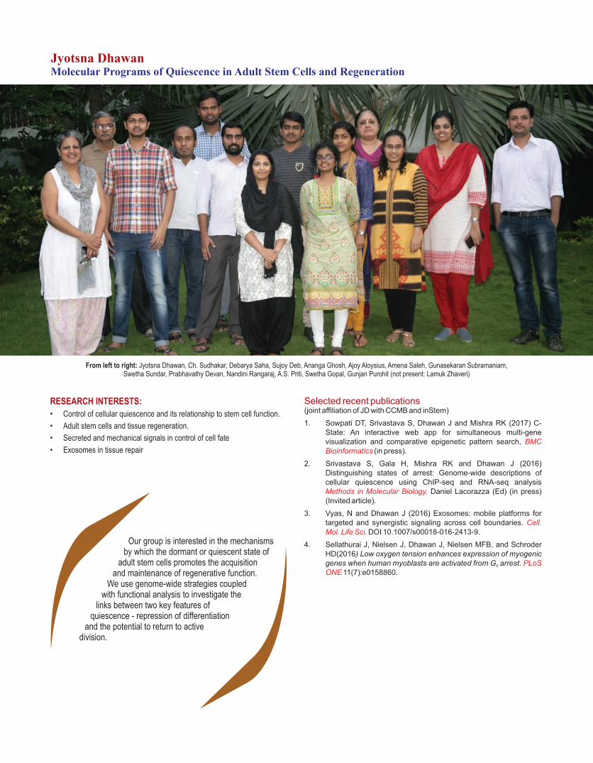
From left to right: Jyotsna Dhawan, Ch. Sudhakar, Debarya Saha, Sujoy Deb, Ananga Ghosh, Ajoy Aloysius, Amena Saleh, Gunasekaran Subramaniam, Swetha Sundar, Prabhavathy Devan, Nandini Rangaraj, A.S. Priti, Swetha Gopal, Gunjan Purohit (not present: Lamuk Zhaveri)
Jyotsna DhawanMolecular Programs of Quiescence in Adult Stem Cells and Regeneration
RESEARCH INTERESTS:
• Control of cellular quiescence and its relationship to stem cell function.
• Adult stem cells and tissue regeneration.
• Secreted and mechanical signals in control of cell fate
• Exosomes in tissue repair
Selected recent publications(joint affiliation of JD with CCMB and inStem)
1. Sowpati DT, Srivastava S, Dhawan J and Mishra RK (2017) C-State: An interactive web app for simultaneous multi-gene visualization and comparative epigenetic pattern search, BMC Bioinformatics (in press).
2. Srivastava S, Gala H, Mishra RK and Dhawan J (2016) Distinguishing states of arrest: Genome-wide descriptions of cellular quiescence using ChIP-seq and RNA-seq analysis Methods in Molecular Biology, Daniel Lacorazza (Ed) (in press) (Invited article).
3. Vyas, N and Dhawan J (2016) Exosomes: mobile platforms for targeted and synergistic signaling across cell boundaries. Cell. Mol. Life Sci. DOI 10.1007/s00018-016-2413-9.
4. Sellathurai J, Nielsen J, Dhawan J, Nielsen MFB, and Schroder HD(2016) Low oxygen tension enhances expression of myogenic genes when human myoblasts are activated from G arrest. 0 PLoS ONE 11(7):e0158860.
Our group is interested in the mechanisms by which the dormant or quiescent state of
adult stem cells promotes the acquisition and maintenance of regenerative function.
We use genome-wide strategies coupled with functional analysis to investigate the
links between two key features of quiescence - repression of differentiation
and the potential to return to active division.

Our group is interested in the mechanisms that
regulate cellular quiescence in adult mammalian
stem cells and impact their regenerative function.
Most cells in adult tissue have ceased cell
division, but can exist in distinct arrested states.
Differentiated cells permanently withdraw from the
cell cycle, but stem cells idle in a dormant state known
as quiescence or G . These temporarily arrested 0
progenitors maintain adult tissues undergoing normal
turnover, and also repair and regenerate tissue
following injury. De-regulation of quiescence
underlies pathologies at opposite ends of a spectrum-
cancer may represent a failure to enter quiescence,
while degenerative disease may represent a failure to
exit quiescence. Therefore, understanding the
acquisition and maintenance of quiescence has
broad implications for human disease.
We use genome-wide strategies coupled with
functional analysis to investigate the links between
two key features of quiescence -repression of
differentiation and the potential to return to active
division. Using myogenic cell lines, muscle stem cells
(Fig.1) and mesenchymal stem cells we have
described active controls at multiple levels of gene
regulation specific to quiescence. Our studies
indicate that quiescent cells preserve two
antagonistic programs (division vs differentiation) in
an inactive but poised state that is rapidly altered
by cell cycle re-entry.
Over the past year, we have continued our
investigations into the molecular control of
adult stem cell quiescence using cultured
cell lines that model quiescence, as well as using
primary mouse and human stem cells. As a means of
deconstructing the quiescent state we have
investigated the contribution of mechanisms at
different levels. Some highlights of these studies are
given below.
Fig.1: Quiescence markers identified
from cultured myoblasts are expressed
in quiescent muscle stem cells. The
Pax7ngfp transgenic mouse (kind gift of
Prof ShahragimTajbakhsh, Institute
Pasteur) has been instrumental for in
vivo analysis and in permitting isolation
of pure populations of muscle stem cells
(MuSC) for analysis ex vivo.
A. Cross section from adult tibialis
anterior muscle showing location of
quiescent Pax7ngfp+ MuSC (green);
DAPI stained nuclei in myofiber and
MuSC (blue). B. FACS-purification of
Pax7ngfp+ MuSC for culture.
C. Freshly isolated, activated (24 hr)
and differentiated MuSC (72 hr)-note
loss of Pax7ngfp as quiescent MuSC
(QSC) are activated (ASC) or
differentiated (DSC) in culture.
D. Expression analysis of quiescence
markers identified earlier by the lab:
Rgs2 (Subramaniam et al, 2013) and
MLL5 (Sebastian et al, 2009) were
isolated from expression screens in
quiescent C2C12 myoblasts, and are
specifically expressed in freshly isolated
quiescent MuSC, and down regulated
during proliferation/differentiation.
Expression of other markers Pax7 and
p27 (quiescence), Cyclin A2
(proliferation) and Myogenin
(differentiation) validates the different
cellular states.
Myoblasts Myotubes
DIP
//
mD
ialD
NA
mD
IP/
/C
ytC
DN
AD
IP/
/C
RT
DN
AD
IP/
/G
M1
30
DN
A
35

Chromatin and transcriptionalmechanisms in G0
Earlier we reported that an epigenetic regulator
PRDM2 controls the decision between two key
aspects of quiescence- repressed differentiation and
poising of the cell cycle, and defined a new
mechanism by which PRDM2 governs this balance,
by targeting the bivalent marking of a PRE-like
element in the Cyclin A regulatory region (Cheedipudi,
Puri et al, Nucleic Acids Res, 2015). Over the past
year, we have extended these studies in three ways (i)
immuno-precipitation of tagged PRDM2 isoforms to
identify interacting partners using mass spectrometric
analysis (ii) site-directed mutagenesis to define the
PRE-like regulatory element where PRDM2 is bound
(iii) defining the signaling pathways directly
downstream of PRDM2.
We are deciphering the rules that may govern the
distinct chromatin landscape in quiescent stem
cells using a population of cardiac MSC, in
collaboration with Richard Harvey at VCCRI,
Sydney and Rakesh Mishra at CCMB. The results
reinforce our view that global patterns of histone
modifications induce a poised state in G . Using this 0
data set, in collaboration with Rakesh Mishra's
group we have devised a means of visualizing
the distribution of multiple epigenetic marks in
multiple cellular states and thereby discerning
patterns that allow the generation and testing of
hypotheses about global regulation at the level of
chromatin (Sowpati et al, 2017).
We have found that promoter-proximal pausing
of RNA pol II is involved in the maintenance
and exit from the quiescent self-renewing state.
By combining RNAseq and ChIP-seq of RNA
polymerase occupancy in different cellular
states, we uncovered networks that point to
quiescence-specific priming of the transcriptional
program. Knockdown analysis of G -stalled genes 0
shows that stalling contributes to self-renewal
(Gala et al, in revision). Over the past year we
have extended our analysis to up-stream
transcriptional regulators and find that control of
polymerase stalling determines the timing of entry
into the cell cycle.
RNA biology in quiescence
Earlier, we reported cell state-specific dynamics of
self-assembling mRNP granules known as P-bodies.
We also discussed evidence that in quiescence
transcript stabilization in mRNPs dominates over
transcript turnover. In culture, knock-down of different
P-body components that are specifically enriched in
muscle stem cells in vivo differentially affects
proliferation and quiescence. Studies with knockout
mice indicated a role in muscle homeostasis. Over the
past year we have uncovered a cross-regulation
between different mRNP granule components that
implies a stage-specific mechanism to monitor their
expression, assembly and function.
Secreted and mechanical signals in control
of cell fate
Secreted ligands such as Wnt and bFGF are known to
be critical for muscle cell proliferation. Our earlier
studies established that threshold levels of Wnt-β-
catenin-TCF signaling are important for the
quiescence program and appear to be associated
with a very different spectrum of genes in quiescent
myoblasts when compared to either cycling or
differentiated muscle cells. Consistent with the
surprising finding that β-catenin is dispensable for this
pathway and instead cross talk with other signaling
systems is required in G (Aloysius et al, in revision). 0
Our studies over the past year have revealed
that this transcriptional cross-talk is likely to
occur in muscle stem cells during post-natal
development.
Mechanical signals from the substratum have a
profound impact on cytoskeletal and nuclear
architecture and consequently on cell fate. Earlier we
had reported the generation of culture systems that
permit human mesenchymal stem cells to be toggled
between quiescence and activation in vitro, by limiting
cell attachment or by culture on soft substrates (in
collaboration with Moustapha Kassem at Odense).
We have uncovered quiescence- induced
transcriptional programs associated with a clinically
relevant phenotype (Rumman et al, in preparation).
Over the past year, we have also found that inducing
quiescence in culture enhances the function of
transplanted hMSCs in vivo.
Cytoskeletal signaling is integrated with growth factor
signaling via the activity of the RhoA GTPase. Earlier
we had identified mDiaphanous (mDia) as the key
effector of RhoA in mediating signaling via regulation
of transcription factors that are responsive to the
polymerization state of the actin cytoskeleton
(Gopinath et al, J. Cell Sci, 2007). More recently,
using yeast 2-hybrid analysis we have isolated a
novel interacting protein of mDia, confirmed their
interaction by immuno-precipitation and mass

spectrometry, and mapped the interacting domains.
In particular, this novel protein appears to regulate a
stage-specific signaling complex to controls
differentiation. Fig. 2 shows the localization of the
mDia-interacting protein at different stages of
myogenesis in culture. Our current studies are aimed
at understanding how the interaction of mDia with
DIP regulates muscle differentiation.
Secreted exosomes and tissue regeneration
Earlier work initiated by N. Vyas while the lab was at in
Stem revealed that extracellular vesicles
(“exosomes”) are secreted in at least two distinct
forms with unique protein and miRNA cargoes and
distinct signaling capabilities (Vyas et al, Sci Rep,
2014). As part of an Indo-Danish collaborative grant
with 4 Danish and 5 Indian labs, we are now
extending our studies towards understanding
whether stem cells improve tissue repair by secreting
exosomes that stimulate more efficient regeneration.
Fig.3 shows preliminary characterization of
exosomes from cultured muscle cells. Our studies
over the next year will focus on the exosomal cargo of
proteins and miRNAs.
Fig.2: Localization of
Dia-interacting protein
(DIP) in muscle cells
during proliferation and
differentiation. Co-
immunostaining of DIP
was performed along
with GM130 (Golgi
marker), Calreticulin
(CRT-Endoplasmic
reticulum marker) and
Cytochrome C (CytC-
Mitochondrial marker) in
proliferating and
differentiating C2C12.
DIP localizes to the
mitochondria and
nucleus but not to the
Golgi apparatus and
endoplasmic reticulum.
Fig.3: Exosomes
secreted by muscle
cells in culture.
A. Dynamic light
scattering profile
shows nanoparticles
in the range of 20-
200 nm
B. Transmission
electron microscope
image (negative
staining) reveals
vesicles in a similar
size range
C. Atomic force
microscope
topographical
scan showing a
preparation of
secreted
vesiclesisolated from
proliferating
myoblasts enriched
for exosomes (30-
150 nm).
37

From left to right: Neema, Abirami, M.S Ram, Mahender Reddy, Mihir Trivedi, G. Umapathy, Vinod Kumar, Deepa Purohit, Pravalika, Kirti Prasad
G UmapathyStudies on Species Extinction and Conservation Physiology
RESEARCH INTERESTS:
• Understanding the extinction process of species.
• Impact of habitat fragmentation in Western Ghats.
• Conservation breeding.
• Conservation physiology.
• Understanding anthropogenic disturbance to wildlife health and reproduction.
Selected recent publications
1. Chakraborty D, Tiwari S, Reddy DM, Umapathy G (2016) Prevalence of gastrointestinal parasites in civets of fragmented rainforest patches in Anamalai hills, Western Ghats, India. Journal of Parasitology 102: 463-467.
2. Ram MS, Kittur SM, Biswas J, Nag S, Shil J, Umapathy G (2016). Genetic Diversity and Structure among Isolated Populations of the Endangered Gees Golden Langur in Assam, India. PLoS ONE 11(8): e0161866. doi:10.1371/journal.pone.0161866.
3. Budithia NMR, Kumar V, Yalla SKa, Rai U and Umapathy G (2016). Non-invasive monitoring of reproductive and stress hormones in the endangered red panda (Ailurus fulgens fulgens). Animal Reproduction Science, 172: 173–181.
4. Kumar, V, Reddy, V, KokkiligaddaA, Shivaji, S and Umapathy, G. (2014). Non-invasive assessment of reproductive status and stress in captive Asian elephants in three south Indian zoos. General and Comparative Endocrinology, 201: 37-44.
5. Laurance WF, Umapathy G, et al. (2012). Averting biodiversity collapse in tropical forest protected areas. 489: 290-294. Nature
Our group aims to understand factors that influence the extinction of species in
fragmented landscape, and to assess reproductive and stress status in wild
animals.

Genetic diversity and structure among
isolated populations of the endangered
Gee's golden langur in Assam, India
Gee's golden langur (Trachypithecus geei), a
colobine primate endemic to Indo-Bhutan border,
was discovered in the late 1950s based on
morphological differences with the capped langur
(T. pileatus). It has been listed as endangered
species in the IUCN Red List and Schedule-I species
in Indian Wildlife Protection Act (1972). Golden langur
is found in the sub-tropical, monsoon-fed, semi-
evergreen and mixed deciduous forests of western
Assam in India and south-central Bhutan. It is
considered to be one of the most restricted-range
primates of South Asia. Habitat fragmentation
consistently has large, negative effects on measures
of biodiversity such as population size, distribution
and genetic diversity, especially in habitat specialist
species. The present study aimed to estimate the
genetic diversity in the Indian populations of golden
langur using HVRI. It also aims to examine the
phylogenetic position of Indian golden langur with
respect to the Bhutanese population and closely
related Trachypithecus spp. using cytb (Fig 1). It is
hoped that this study would serve as a platform for a
more scientific approach towards managing the wild
population of this enigmatic species.
Out of 71 samples collected, 59 samples gave
successful amplification for HVRI and 518 bases
were sequenced and aligned. Nineteen haplotypes
were revealed based on 47 mutations (45 transitions
and two transversions) spread over 46 segregating
sites, out of which 43 were parsimony-informative.
The overall haplotype diversity was high (h = 0.938)
but the nucleotide diversity was low (π = 0.02443).
The haplotypes were assigned an alphabetical code
(from GL-A to GL-S) based on the chronology of
discovery. Haplotype GL-C was the most frequent,
represented by eight individuals from two fragments,
followed by GL-A represented by seven individuals.
More than half of the haplotypes represented fewer
than three individuals. Haplotype and nucleotide
diversities were calculated for each population. CS
and BH showed the highest number of haplotypes
(four each) and CR showed the second highest (three
haplotypes). Consequently, CS and BH also had the
highest haplotype diversities (0.76 and 0.8
respectively). Five fragments (KJ, NY, ND, MN and
BG) showed two haplotypes while UM showed no
variation and KJ, ND and BG showed the least
number of polymorphic sites among their haplotypes.
Surprisingly, NY, which had only two haplotypes,
showed the most number of polymorphic sites (17) for
any fragment, giving it the highest nucleotide
diversity. It was followed by CR (14), CS (7), MN (7)
Fig.1: Current and past distribution of golden langur in Assam, India based on previous reports and sampling locations in the present study.
39
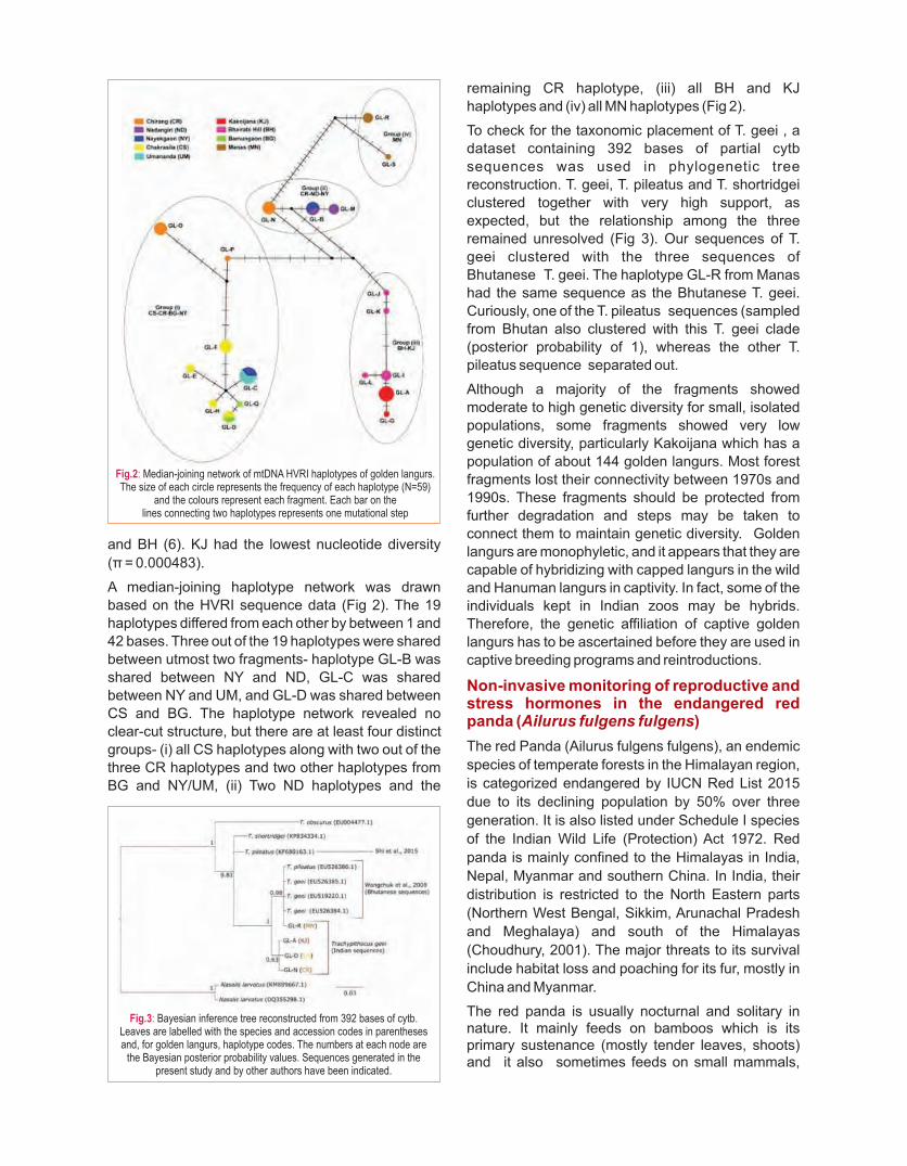
and BH (6). KJ had the lowest nucleotide diversity
(π = 0.000483).
A median-joining haplotype network was drawn
based on the HVRI sequence data (Fig 2). The 19
haplotypes differed from each other by between 1 and
42 bases. Three out of the 19 haplotypes were shared
between utmost two fragments- haplotype GL-B was
shared between NY and ND, GL-C was shared
between NY and UM, and GL-D was shared between
CS and BG. The haplotype network revealed no
clear-cut structure, but there are at least four distinct
groups- (i) all CS haplotypes along with two out of the
three CR haplotypes and two other haplotypes from
BG and NY/UM, (ii) Two ND haplotypes and the
remaining CR haplotype, (iii) all BH and KJ
haplotypes and (iv) all MN haplotypes (Fig 2).
To check for the taxonomic placement of T. geei , a
dataset containing 392 bases of partial cytb
sequences was used in phylogenetic tree
reconstruction. T. geei, T. pileatus and T. shortridgei
clustered together with very high support, as
expected, but the relationship among the three
remained unresolved (Fig 3). Our sequences of T.
geei clustered with the three sequences of
Bhutanese T. geei. The haplotype GL-R from Manas
had the same sequence as the Bhutanese T. geei.
Curiously, one of the T. pileatus sequences (sampled
from Bhutan also clustered with this T. geei clade
(posterior probability of 1), whereas the other T.
pileatus sequence separated out.
Although a majority of the fragments showed
moderate to high genetic diversity for small, isolated
populations, some fragments showed very low
genetic diversity, particularly Kakoijana which has a
population of about 144 golden langurs. Most forest
fragments lost their connectivity between 1970s and
1990s. These fragments should be protected from
further degradation and steps may be taken to
connect them to maintain genetic diversity. Golden
langurs are monophyletic, and it appears that they are
capable of hybridizing with capped langurs in the wild
and Hanuman langurs in captivity. In fact, some of the
individuals kept in Indian zoos may be hybrids.
Therefore, the genetic affiliation of captive golden
langurs has to be ascertained before they are used in
captive breeding programs and reintroductions.
Non-invasive monitoring of reproductive and stress hormones in the endangered red panda (Ailurus fulgens fulgens)
The red Panda (Ailurus fulgens fulgens), an endemic
species of temperate forests in the Himalayan region,
is categorized endangered by IUCN Red List 2015
due to its declining population by 50% over three
generation. It is also listed under Schedule I species
of the Indian Wild Life (Protection) Act 1972. Red
panda is mainly confined to the Himalayas in India,
Nepal, Myanmar and southern China. In India, their
distribution is restricted to the North Eastern parts
(Northern West Bengal, Sikkim, Arunachal Pradesh
and Meghalaya) and south of the Himalayas
(Choudhury, 2001). The major threats to its survival
include habitat loss and poaching for its fur, mostly in
China and Myanmar.
The red panda is usually nocturnal and solitary in nature. It mainly feeds on bamboos which is its primary sustenance (mostly tender leaves, shoots) and it also sometimes feeds on small mammals,
Fig.3: Bayesian inference tree reconstructed from 392 bases of cytb. Leaves are labelled with the species and accession codes in parentheses and, for golden langurs, haplotype codes. The numbers at each node are
the Bayesian posterior probability values. Sequences generated in the present study and by other authors have been indicated.
Fig.2: Median-joining network of mtDNA HVRI haplotypes of golden langurs. The size of each circle represents the frequency of each haplotype (N=59)
and the colours represent each fragment. Each bar on the lines connecting two haplotypes represents one mutational step

bird eggs, fruits and acorns. Understanding of reproductive physiology is very important for the successful captive breeding program. As a part of the conservation breeding program of the red panda, the present study aimed to validate and standardize enzyme immunoassays (EIAs) to assess the fertility status, pregnancy detection and stress level in captive red panda using faecal steroid hormones. Faecal samples of females (n=4) and male (n=1) were collected every day in the morning between 8 to 9 hours from January 2013 to April 2014 at Padmaja Naidu Himalayan Zoological Park, Darjeeling, West Bengal, India. Faecal progestagen, testosterone and cortisol were measured previously validated in big cats and ungulates.
A total of 1471 faecal samples were collected from four adult females and one adult male for 15 months. Of the four females, matings in three females were observed, and one female gave birth to a cub during the study period.Of the 4 females monitored, three were observed matings with males during January, of which one female gave birth to a cub in June(Fig.4A). On the basis of mating observation and parturition, the gestation period was estimated to be 150 days. Overall, faecal 5α-pregnan-3α-ol-20-one concentrations ranged widely from 370 ng/g (Sambridhi, a non pregnant animal) to 6000 ng/g (Sheetal, a pregnant animal). In non-pregnant animals, weekly mean basal faecal 5α-pregnan-3α-ol-20-one concentration ranged from 370 ng/g to 3886 ng/g but it did not significantly vary among individuals (Friedman test χ2 = 5.5; P = 0.067). In the pregnant animal, it ranged from 2143 ng/g to 6000 ng/g. Following matings (Sheetal), the faecal5α-pregnan-3α-ol-20-one level raised graduallyto 2 to 3 times higher than the basal values (mean = 1840; n = 1016) and the mean weekly concentrations increased significantly from 8 weeks until parturition (Friedman test χ2 = 4.5; P = 0.03) and decreasedto basal concentration a week after parturition.Faecal cortisol levels were measured for all animals. Monthly mean concentrations ranged from 4.4 ± 0.81 ng/g to 107.5 ± 24.9 ng/g (Fig. 1). Both the lowest and highest values were observed in females. Overall, monthly mean cortisol concentrations were increasing with decreasing ambient temperature in all animals (rs = -0.874; P = 0.001).
This study provides faecal 5α-pregnan-3α-ol-20-one, testosterone and cortisol profiles in the captive red panda. EIA developed against progesterone metabolite could be used for reproductive monitoring and detection of pregnancy in the red panda. Furthermore, the study would facilitate reproductive monitoring of the red panda in captive breeding programs in India and elsewhere.
An adult Red panda at Darjeeling Zoo
Fig.4: Faecal progestogen profiles of the four female red panda individuals (a-sheetal, b-Janaki, c-Sambridhi, d-Smile) -
arrows indicate parturition and mating.
41

From left to right: Vaishnavi Kunteepuram, Siuli Mitra, Ajay Gaur, B. Charan Kumar, A. Sreenivasu, Drishti R. Parmar
Ajay GaurConservation Genetics of Endangered Species of India
RESEARCH INTERESTS:
• Population Genetics
• Evolutionary Genetics
• Wildlife Forensics
• Conservation Breeding
• DNA Banking
Selected recent publications
1. Tabasum W, Sreenivas A, Kesav Kumar B, Rao TG and Gaur A (2016) Complete mitochondrial genome sequence of Indian clouded leopard (Neofelis nebulosa). doi: Mitochondrial DNA:10.1080/23802359.2016.1214543.
2. Tabasum W, Sreenivas A, Rai N, Thangaraj K and Gaur A (2016) Complete mitochondrial genome sequence of Asiatic lion (Panthera leo persica). doi: 10.1080/ Mitochondrial DNA:23802359.2016.1214541.
3. Modi N, Sreenivas A, Tabasum W and Gaur A (2016) DNA tools to establish species and individual identity of confiscated biological samples in wildlife related crime: A case report. Journal of Forensic, Legal and Investigative Sciences 2: 006.
4. Ram M S, Marne M, Gaur A, Kumara H N, Singh M, Kumar A and Umapathy G (2015) Pre-Historic and recent vicariance events shape genetic structure and diversity in endangered Lion-tailed macaque in the Western Ghats: Implications for conservation. PLoS ONE 10: e0142597.
5. Gupta S, Kumar A, Gaur A and Hussain S A (2015) Detection of 40 bp insertion-deletion (INDEL) in mitochondrial control region among sambar (Rusa unicolor) populations in India. BMC Res Notes 8:581.
Our group focuses on the development and application of molecular markers in
conservation genetics of Indian endangered species.
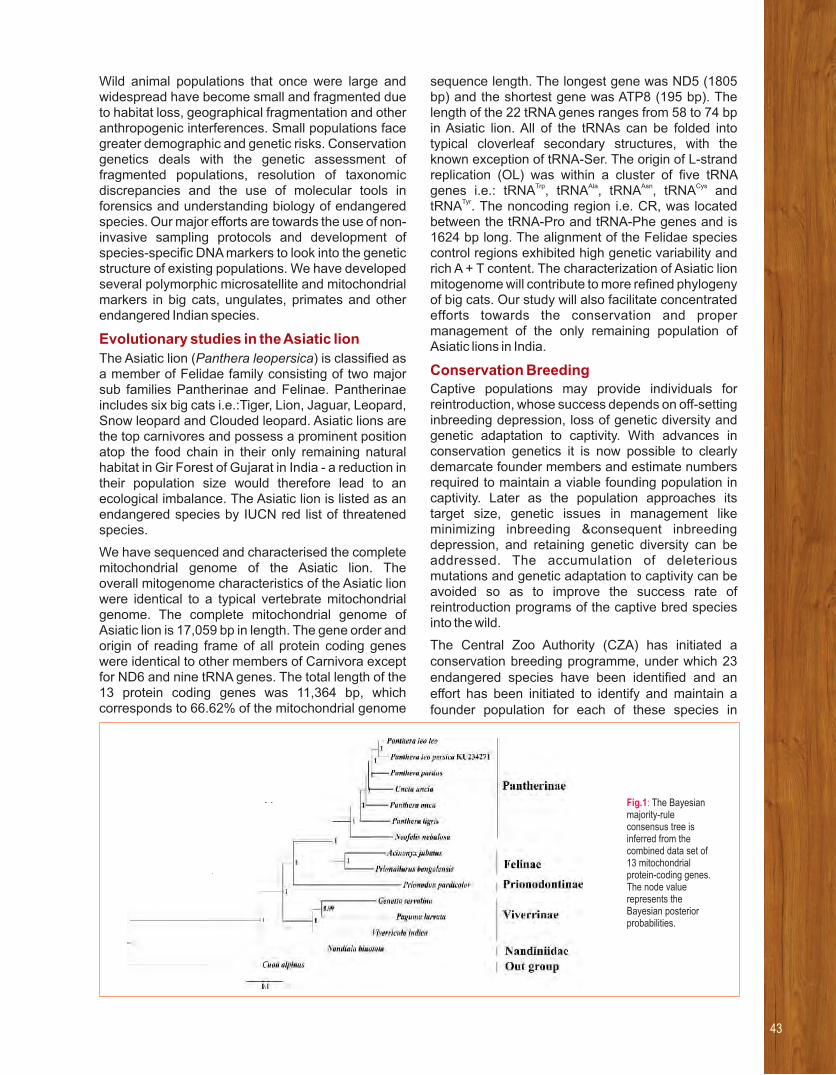
Fig.1: The Bayesian majority-rule consensus tree is inferred from the combined data set of 13 mitochondrial protein-coding genes. The node value represents the Bayesian posterior probabilities.
Wild animal populations that once were large and widespread have become small and fragmented due to habitat loss, geographical fragmentation and other anthropogenic interferences. Small populations face greater demographic and genetic risks. Conservation genetics deals with the genetic assessment of fragmented populations, resolution of taxonomic discrepancies and the use of molecular tools in forensics and understanding biology of endangered species. Our major efforts are towards the use of non-invasive sampling protocols and development of species-specific DNA markers to look into the genetic structure of existing populations. We have developed several polymorphic microsatellite and mitochondrial markers in big cats, ungulates, primates and other endangered Indian species.
Evolutionary studies in the Asiatic lion
The Asiatic lion (Panthera leopersica) is classified as a member of Felidae family consisting of two major sub families Pantherinae and Felinae. Pantherinae includes six big cats i.e.:Tiger, Lion, Jaguar, Leopard, Snow leopard and Clouded leopard. Asiatic lions are the top carnivores and possess a prominent position atop the food chain in their only remaining natural habitat in Gir Forest of Gujarat in India - a reduction in their population size would therefore lead to an ecological imbalance The Asiatic lion is listed as an .endangered species by IUCN red list of threatened species.
We have sequenced and characterised the complete mitochondrial genome of the Asiatic lion. Theoverall mitogenome characteristics of the Asiatic lion were identical to a typical vertebrate mitochondrial genome. The complete mitochondrial genome of Asiatic lion is 17,059 bp in length. The gene order and origin of reading frame of all protein coding genes were identical to other members of Carnivora except for ND6 and nine tRNA genes. The total length of the 13 protein coding genes was 11,364 bp, which corresponds to 66.62% of the mitochondrial genome
sequence length. The longest gene was ND5 (1805 bp) and the shortest gene was ATP8 (195 bp). The length of the 22 tRNA genes ranges from 58 to 74 bp in Asiatic lion. All of the tRNAs can be folded into typical cloverleaf secondary structures, with the known exception of tRNA-Ser. The origin of L-strand replication (OL) was within a cluster of five tRNA genes i.e.: Trp Ala Asn CystRNA , tRNA , tRNA , tRNA and
TyrtRNA . The noncoding region i.e. CR, was located between the tRNA-Pro and tRNA-Phe genes and is 1624 bp long. The alignment of the Felidae species control regions exhibited high genetic variability and rich A + T content. The characterization of Asiatic lion mitogenome will contribute to more refined phylogeny of big cats. Our study will also facilitate concentrated efforts towards the conservation and proper management of the only remaining population of Asiatic lions in India.
Conservation BreedingCaptive populations may provide individuals for reintroduction, whose success depends on off-setting inbreeding depression, loss of genetic diversity and genetic adaptation to captivity. With advances in conservation genetics it is now possible to clearly demarcate founder members and estimate numbers required to maintain a viable founding population in captivity. Later as the population approaches its target size, genetic issues in management like minimizing inbreeding &consequent inbreeding depression, and retaining genetic diversity can be addressed. The accumulation of deleterious mutations and genetic adaptation to captivity can be avoided so as to improve the success rate of reintroduction programs of the captive bred species into the wild.
The Central Zoo Authority (CZA) has initiated a conservation breeding programme, under which 23 endangered species have been identified and an effort has been initiated to identify and maintain a founder population for each of these species in
43

captivity. LaCONES is an important partner for this initiative and is conducting genetic analysis for the origin, purity and heterozygosity status. This work involves identification or development of nuclear microsatellite and mitochondrial markers. A large number of blood, tissue and feather samples of different species have been received under different species' conservation breeding programs for genetic health checkup. Six reports were submitted to respective Zoos and the CZA, for genetic health status, during last year (April 2016 – March 2017).
·�Genetic studies in Asiatic lions
Genomic DNA was extracted from the blood samples of Eighteen (18) Asiatic lion housed in Lion Safari of Nehru Zoological Park, Hyderabad. The genetic status of the individuals was assessed in terms of their being heterozygous/homozygous at each locus. Further, DNA was amplified to generate molecular signatures (nucleotide sequences) from the mitochondrial cytb gene. Genotypes (Allelic distribution) of 18 Asiatic Lion samples for eight microsatellite loci were included for further analysis. The mean observed heterozygosity (Ho) was 0.472 and the mean expected heterozygosity was 0.439. Further, the analysis of the molecular signature (mtDNA cytb) generated from the above samples revealed four different haplotypes. The animals namely Ajay, Akash, Rita, Atul, Anthony, Aruna, Soniya and Jyothi shared similar haplotypes (H1), Sindhu, Sahiti, Jeetu, Vishwas, and Shilpa shared similar haplotypes (H2) and Vicky, Kashyap, Shruthi, and Vasu shared similar haplotypes (H3). Crazy was found to be separate haplotype (H4) and genetically most divergent lion among all the samples. The
animals found to be most heterozygous and unrelated were recommended to be used for selective breeding purpose.
Wildlife ForensicsAt LaCONES, we provide DNA-based species, individual identification, sexing and rehabilitation services to the nation for the purpose of wildlife crime investigation. It is one of the major on-going activities in LaCONES. We receive biological specimens confiscated in wildlife related crimes forwarded by the state forest, judiciary, police and customs departments. During the period April 2016 - March 2017, we have registered a total of 172 wildlife crime cases, out of which, 148 cases have been successfully reported to the forwarding authorities. During this period, a revenue of more than 15 lakhs was generated from the fee charges for DNA analysis. The forwarded biological samples comprised of 446 varieties such as meat, cooked/raw meat, skin, venom, tusks, bones, claws, feces, blood stains, saliva and swabs; we also recently received a large number of Hatha Jodi samples (Fig. 2).
Monitor Lizard (Varanus bengalensis) hemi-penis or the male sexual organs of monitor lizards are being sold in markets as holy plant root named as 'Hatha Jodi'. These lizards are being illegally poached from the wild, caught in traps and snares. The dried hemi-penis of monitor lizards looks like a dried plant root. As per traditional belief it is primarily sought as a tantric (black magic) piece for influencing people, winning over enemies and also to bring wealth, bravery, happiness and good fortune. On the basis of the DNA analysis using Universal Primers, we could establish the correct identity of all the 28 Hatha Jodi samples.
Fig.2: Confiscated Hatha Jodi samples

45
From left to right: Mohan Singh Moodu, Manish Johri, Divya Gupta, Haripriya Parthasarathy, Hitha Nair, Dhiviya Vedagiri, Iqra Kuchay, Krishnan H Harshan
Krishnan H HarshanRegulation of Protein Translation in Viral Infections and Biological Systems
RESEARCH INTERESTS:
• Regulation of Protein Translation in Viral Infections
• Reprogramming of Protein Translation in Epithelial to Mesenchymal Transition
Selected recent publications
1. Krishnan HH, Naranatt PP, Smith MS, Zeng L, Bloomer C, and Chandran B (2004) Concurrent expression of latent and a limited number of lytic genes with immune modulation and anti-apoptotic functions by KSHV/HHV-8 early during infection of primary endothelial and fibroblast cells and subsequent decline of lytic gene expression. : 78:3601-20. J Virology
2. Krishnan HH, Walia N-S, Zeng L, Gao S J and Chandran B (2005) Envelope glycoprotein gB of Kaposi's sarcoma Associated Herpesvirus (KSHV/HHV-8) is essential for egress from infected cells 79: 10952-10967.J. Virology
3. Krishnan HH, Walia N-S, Streblow DN, Naranatt PP and Chandran B (2005) Focal Adhesion Kinase (FAK) is critical for Kaposi's Sarcoma Associated Herpesvirus (KSHV-HHV-8) entry into the target cells. 80:1167-1180. (Selected J. Virology.in the J.Virology spotlight in the same issue).
4. George A, Panda S, Kudmulwar D, Chhatbar S D, Nayak S C and Krishnan H H (2012) Hepatitis C virus NS5A binds to the mRNA Cap binding eIF4F complex and upregulates host translation initiation machinery through 4EBP1 inactivation. J. Biol. Chemistry. 10; 287(7): 5042-58.
5. Panda S, Vedagiri D, Viveka T S, Jonddula R C and Harshan K H (2014) Hepatitis C Virus Protein NS5A Coordinates a Unique Phosphorylation Dependent eIF4E Assembly on 40S Ribosomes that Regulates IRES Activity. Biochem. J.462(2):291-302).
Oncoviruses establish a long-term relationship with hosts during chronic
infections. They remain in a latent stage with minimal expression of viral proteins that helps
them evade the host immune system. Our goal is to understand the manipulation of host
translation by viral proteins and to understand its consequence on viral immune evasion and
tumorigenesis.

The role of deregulated host transcription in
pathogenesis associated with viral infection is well
acknowledged. However, the involvement of
translation as a major regulatory event in various
cellular activities including cell cycle regulation has
only been recognized in the recent decade and a half.
Of late, its involvement in tumor development and
progression has been demonstrated unambiguously,
suggesting the role of protein translation as major
regulatory step in tumorigenesis. Translation is also
exploited by many viruses for their preferential
survival over the host. The focus of our research is to
understand the involvement of translation in the
pathogenesis of oncoviral infections.
In eukaryotes, the majority of the mRNA transcripts
are translated by 5'Cap initiation, which starts with the
binding of eukaryotic translation initiation factor 4F
complex (4F) to the cap structure. The 4F complex is
composed of three different eukaryotic translation
initiation factors, eIF4E (4E), eIF4G (4G) and eIF4A
(4A). 4F complex formation is a major step in
translation initiation and is the dearest rate-limiting
step in the process. Under phosphorylated 4E binding
proteins 4EBP-1 and 4EBP-2 competitively bind to
4E, thus inhibiting its interactions with 4G and
subsequent 4F complex formation. This results in the
inhibition of cap mediated translation rate. This block
can be removed by the hyper phosphorylation of
4EBPs by mammalian target of rapamycin (mTOR).
mTOR is phosphorylated by AKT and is negatively
regulated by phosphatase and tensin homolog
(PTEN) and tuberous sclerosis proteins (TSC1 and
2). mTOR also phosphorylates p70 S6 Kinase (S6K),
which positively regulates translation initiation along
with 4EBP phosphorylation.
HCV and host translation
Microbiology 5, 453-463 (1 June 2007) | doi:10.1038/
nrmicro1645 Hepatitis C Virus (HCV) is a member of
flaviviridae family and has a single stranded RNA
genome of about 9.6 kb with positive polarity. It is a
major causative agent of liver cirrhosis (LC) and
hepatocellular carcinoma (HCC) in humans. About 1-
3% of the worldwide population is estimated to be
infected with HCV. Currently, there are no vaccines
against HCV infection. The combinational therapy of
interferon- α and antiviral drug ribavirin is used to treat
patients with varied success that depends on the HCV
genotype. The RNA genome of HCV undergoes
mutations at a very high frequency, making it very
difficult to target the virus with drugs. HCV has an
internal ribosome entry site (IRES) in its
5'untranslated region (5'UTR), which it uses for its
translation. Unlike 5'Cap mediated translation, IRES
mediated translation is significantly less dependent
on various host facotrs and can still operate in the
event of host translation shut off. Many viruses shut
off host translation in various ways to hijack the
machinery for viral translation, even as the degree of
host translation shut off could vary and hence is a
deciding factor in pathogenesis. While many viruses
still manage to keep the host cell alive for their own
survival, some virus infections lead to total host
translation shut off resulting in its death and
subsequent termination of viral cycle progression.
HCV allows host translation to go on as is evident
from the nature of infection. The virus establishes a
long term chronic infection in hepatocytes and
infected people develop symptoms only after 10-15
years post-infection.
Fig.1: Representation of HCV genome. Adopted from Nature Reviews Microbiology 5, 453–463 (1 June 2007)

47
Our laboratory seeks to understand various signaling
pathways/molecules regulated by HCV proteins in
human liver cells by using two different systems:
a. Infection of hepatocytes by cell culture prepared
infectious HCV clones (HCVcc). HCVcc is
prepared in a biosafety level -2 (BSL-2) and
human hepatoma and primary hepatocytes are
infected in the containment facility.
b. Over-expression of HCV proteins in hepatocytes.
Effects of both HCVcc infection and viral protein
expressions are being studied using various
biochemical, cellular and molecular biological
methods. Infected and HCV protein over-
expressing cells are used to prepare protein
lysates to study various signaling events
leading to regulation of translation initiation.
We employ microscopy, flow cytometry and
microarray based analyses to understand the
implication of translation in HCV associated
pathogenesis in detail.
Our earlier studies showed that HCV enhances cap-
dependent translation in Huh7.5 cells through
activation of mTORC1 and also by phosphorylating
eIF4E. Subsequent studies on regulation of
translation by HCV NS5A demonstrated a direct
association of NS5A with 40S ribosomes through
eIF4E leading to formation of a unique complex ENR
(eIF4E-NS5A-Ribosome). Further studies with
bicistronic luciferase vectors carrying HCV IRES
identified that these regions of domain 1 are also
critical in the upregulation of IRES by NS5A, directly
linking the ENR assembly to HCV pathogenesis. We
are currently trying to understand the contribution of
activated mTORC1 complex and eIF4E to the whole
process of viral life cycle.
Protein Translation in Epithelial
Mesenchymal Transition
Rapidly proliferating tumor cells demand larger
amounts of proteins. If protein translation rates are
not synchronized with that of transcription, this
demand cannot be met. Tumor cells meet this
demand by increasing ribosomal biogenesis and
upregulating protein translation machinery. Epithelial
mesenchymal transition (EMT) is a process by which
epithelial cells abandon many of their characteristic
features and acquire various mesenchymal ones.
During this process, epthelial cells lose their
adhesive nature, characteristic extracellular matrix
sheath and polarity while acquiring spindle
shape with tapering ends and migratory capabilities.
EMT is demonstrated to be behind the assimilation of
metastic capabilities by tumor cells. This transition
facilitates increased migratory and invasive
capabilities of tumor cells. We have characterized an
atypical EMT in human hepatoma cells and identified
substantial upregulation of protein translation
initiation during this process. We identified that GSK-
3β is critical in maintaining the mesenchymal
phenotype and that p38MAPK and ERK1/2, two
major MAPKs, regulate this activity through GSK-3β.
Our laboratory is actively researching the link
between protein translation regulation and EMT.
Fig.2: Assembly of ENR complex and its interaction with HCV genomic RNA
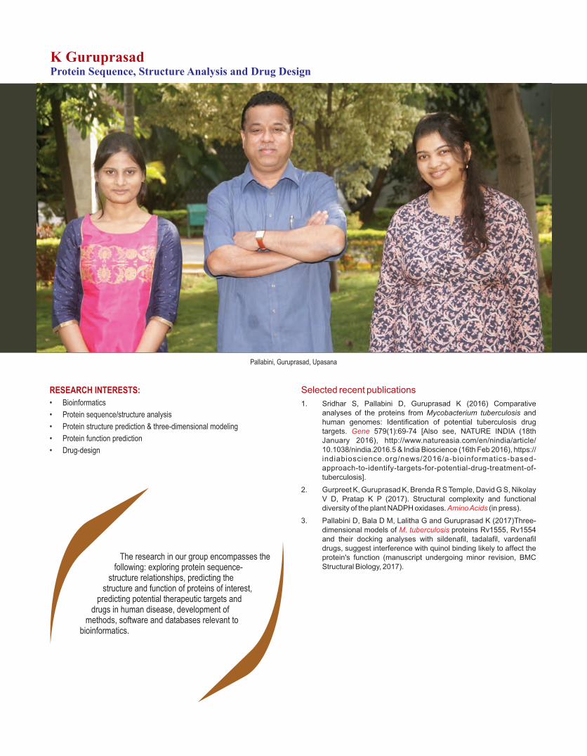
Pallabini, Guruprasad, Upasana
K GuruprasadProtein Sequence, Structure Analysis and Drug Design
RESEARCH INTERESTS:
• Bioinformatics
• Protein sequence/structure analysis
• Protein structure prediction & three-dimensional modeling
• Protein function prediction
• Drug-design
Selected recent publications
1. Sridhar S, Pallabini D, Guruprasad K (2016) Comparative analyses of the proteins from Mycobacterium tuberculosis and human genomes: Identification of potential tuberculosis drug targets. 579(1):69-74 [Also see, NATURE INDIA (18th GeneJanuary 2016), http://www.natureasia.com/en/nindia/article/ 10.1038/nindia.2016.5 & India Bioscience (16th Feb 2016), https:// indiabioscience.org/news/2016/a-bioinformatics-based-approach-to-identify-targets-for-potential-drug-treatment-of-tuberculosis].
2. Gurpreet K, Guruprasad K, Brenda R S Temple, David G S, Nikolay V D, Pratap K P (2017). Structural complexity and functional diversity of the plant NADPH oxidases. (in press).Amino Acids
3. Pallabini D, Bala D M, Lalitha G and Guruprasad K (2017)Three-dimensional models of proteins Rv1555, Rv1554 M. tuberculosisand their docking analyses with sildenafil, tadalafil, vardenafil drugs, suggest interference with quinol binding likely to affect the protein's function (manuscript undergoing minor revision, BMC Structural Biology, 2017).
The research in our group encompasses the following: exploring protein sequence-
structure relationships, predicting the structure and function of proteins of interest,
predicting potential therapeutic targets and drugs in human disease, development of
methods, software and databases relevant to bioinformatics.

Prediction of the inhibitory action of potential repurposed drugs against novel M.tb targets
We recently identified 94 new potential TB drug targets based on a bioinformatics approach developed in our laboratory. Ten of these targets were also predicted to be 'druggable' with certain known drugs in the market (GENE 2016). During the present year, we have analyzed one of the potential druggable targets, in order to understand the possible mechanism by which the protein's function is likely to be inhibited. Our analyses based on molecular modeling and docking suggests that the M. tuberculosis Rv1555 and Rv1554 proteins are likely to be inhibited by some of the known drugs; sildenafil (Viagra), tadalafil (Cialis) and vardenafil (Levitra) drugs, by interfering with the quinol binding sites essential for the protein's function as observed in the homologous E.coli quinol fumarate reductase respiratory protein complex. Further, via collaboration
with School of Chemistry, University of Hyderabad, we have observed the inhibition of in-vitro growth of E.coli bacteria containing the homologous M.tb proteins with sildenafil citrate and tadalafil drugs.Our analyses suggest dual binding sites, Q (distal) and D
Q (proximal) for the monomer and dimer M.tb P
proteins, respectively. The predicted binding sites corroborate with the experimentally observed binding sites in the E.coli fumarate reductase protein crystal structure complex. We therefore suggest that sildenafil citrate and related drugs currently used in the treatment of male erectile dysfunction targeting the human phosphodiesterase 5 enzyme may be evaluated for their plausible role as repurposed drugs in the treatment of tuberculosis.
We are currently analyzing the possible inhibitory action of the other drugs and their potential new targets predicted and reported by us (GENE 2016).
Fig.1: Structural overlay of
the M.tbmonomer (Rv1555)
and M.tbdimer (Rv1555,
Rv1554) proteins complexed
with sildenafil, tadalafil,
vardenafil drugs on to the
crystal structure of E.coli QFR
tetrameric dimer protein
complex (PDB code: 1L0V).
The drug binding sites
coincidewith the equivalent
menaquinol binding at the QD
site for monomer and at the
Q site for dimer M.tbproteins.P
Fig.2: The FAD, iron-sulfur
clusters, menaquinol
molecules bound near the
Q and Q sites inthe P D
crystal structure of E.coli
QFR tetrameric dimer
protein complex (PDB
code: 1L0V) (proteinis
excluded for the sake of
clarity). Sildenafil, tadalafil,
vardenafil drug binding
coinciding withthe Q site P
in the M.tb dimer protein.
49

From left to right & Top to bottom: Umesh Kumar, Rajan K. Jha, Vishwanath, Satya Ranjan Singh, Lomous Kumar, Niraj Rai, Jaydeep A. Badarukhiya,A. Paramasivam, Mohd. Haneef, Sheikh Nizamuddin, Jagamohan Chhatai, D V S Sudhakar, Sunil K. Tripathi, Venkatapathi Challa, Deepak K. Kashyap,
G. Mala, Sunitha Kundur, Usha Kabilan, Nipa Basak, Narmadha Ganapathy, S Deepa Selvi Rani, K. Thangaraj
K ThangarajEvolutionary and Medical Genetics
RESEARCH INTERESTS:
• Origin of modern human
• Genetic basis of - Male infertility - Sex determination and differentiation - Mitochondrial disorders - Cardiovascular diseases - Ayurveda prakritis
• Ancient DNA
• Forensic genetics
Selected recent publications
1. Mallick S, …… Thangaraj K, Pääbo S, Kelso J, Patterson N, Reich D (2016) The Simons Genome Diversity Project: 300 genomes from 142 diverse populations. 538: 201-206. Nature.
2. Mishra A, Nizammuddin S, Mallick CB, Singh S, Prakash S, Siddiqui NA, Rai N, Carlus SJ, Sudhakar DV, Tripathi VP, Möls M, Kim-Howard X, Dewangan H, Mishra A, Reddy AG, Roy B, Pandey K, Chaubey G, Das P, Nath SK, Singh L, Thangaraj K (2017) Genotype-Phenotype Study of the Middle Gangetic Plain in India Shows Association of rs2470102 with Skin Pigmentation. J Invest Dermatol. 137:670-677.
3. Chaubey G, Tamang R, Pennarun E, Dubey P, Rai N, Upadhyay RK, Meena RP, Patel JR, van Driem G, Thangaraj K, Metspalu M, Villems R (2017) Reconstructing the population history of the largest tribe of India: the Dravidian speaking Gond. Eur J Hum Genet. 25:493-498.
4. Chaubey G, Ayub Q, Rai N, Prakash S, Mushrif-Tripathy V, Mezzavilla M, Pathak AK, Tamang R, Firasat S, Reidla M, Karmin M, Rani DS, Reddy AG, Parik J, Metspalu E, Rootsi S, Dalal K, Khaliq S, Mehdi SQ, Singh L, Metspalu M, Kivisild T, Tyler-Smith C, Villems R, Thangaraj K (2017) "Like sugar in milk": reconstructing the genetic history of the Parsi population. Genome Biol. 18:110.
5. Nakatsuka N, Moorjani P, Rai N, Sarkar B, Tandon A, Patterson N, Bhavani GS, Girisha KM, Mustak MS, Srinivasan S, Kaushik A, Vahab SA, Jagadeesh SM, Satyamoorthy K, Singh L, Reich D, Thangaraj K (2017) The promise of discovering population-specific disease-associated genes in South Asia. 49: Nat Genet.1403-1407.
We have demonstrated that how the 1.5 billion people of South Asia are vulnerable to
rare and population-specific diseases, due to practice of endogamy for the last 2000 years.
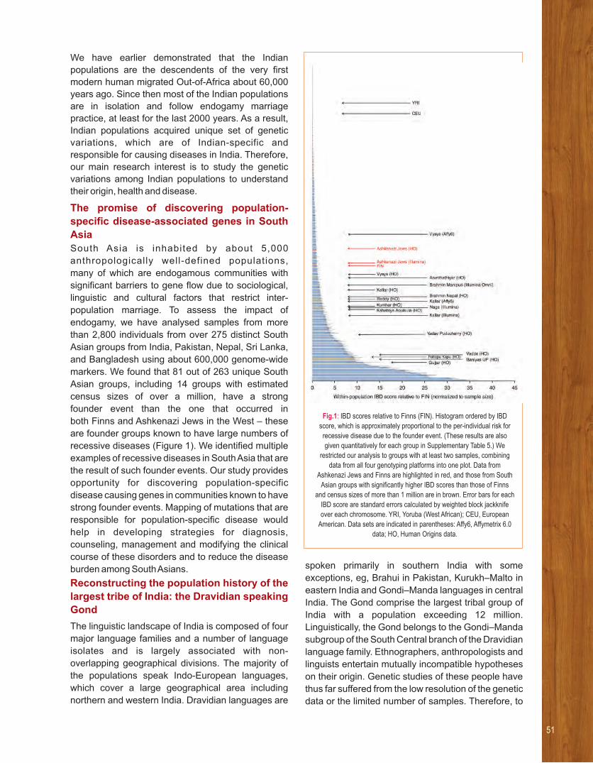
We have earlier demonstrated that the Indian
populations are the descendents of the very first
modern human migrated Out-of-Africa about 60,000
years ago. Since then most of the Indian populations
are in isolation and follow endogamy marriage
practice, at least for the last 2000 years. As a result,
Indian populations acquired unique set of genetic
variations, which are of Indian-specific and
responsible for causing diseases in India. Therefore,
our main research interest is to study the genetic
variations among Indian populations to understand
their origin, health and disease.
The promise of discovering population-
specific disease-associated genes in South
Asia
South Asia is inhabited by about 5,000
anthropologically well-defined populations,
many of which are endogamous communities with
significant barriers to gene flow due to sociological,
linguistic and cultural factors that restrict inter-
population marriage. To assess the impact of
endogamy, we have analysed samples from more
than 2,800 individuals from over 275 distinct South
Asian groups from India, Pakistan, Nepal, Sri Lanka,
and Bangladesh using about 600,000 genome-wide
markers. We found that 81 out of 263 unique South
Asian groups, including 14 groups with estimated
census sizes of over a million, have a strong
founder event than the one that occurred in
both Finns and Ashkenazi Jews in the West – these
are founder groups known to have large numbers of
recessive diseases (Figure 1). We identified multiple
examples of recessive diseases in South Asia that are
the result of such founder events. Our study provides
opportunity for discovering population-specific
disease causing genes in communities known to have
strong founder events. Mapping of mutations that are
responsible for population-specific disease would
help in developing strategies for diagnosis,
counseling, management and modifying the clinical
course of these disorders and to reduce the disease
burden among South Asians.
Reconstructing the population history of the
largest tribe of India: the Dravidian speaking
Gond
The linguistic landscape of India is composed of four
major language families and a number of language
isolates and is largely associated with non-
overlapping geographical divisions. The majority of
the populations speak Indo-European languages,
which cover a large geographical area including
northern and western India. Dravidian languages are
spoken primarily in southern India with some
exceptions, eg, Brahui in Pakistan, Kurukh–Malto in
eastern India and Gondi–Manda languages in central
India. The Gond comprise the largest tribal group of
India with a population exceeding 12 million.
Linguistically, the Gond belongs to the Gondi–Manda
subgroup of the South Central branch of the Dravidian
language family. Ethnographers, anthropologists and
linguists entertain mutually incompatible hypotheses
on their origin. Genetic studies of these people have
thus far suffered from the low resolution of the genetic
data or the limited number of samples. Therefore, to
Fig.1: IBD scores relative to Finns (FIN). Histogram ordered by IBD
score, which is approximately proportional to the per-individual risk for
recessive disease due to the founder event. (These results are also
given quantitatively for each group in Supplementary Table 5.) We
restricted our analysis to groups with at least two samples, combining
data from all four genotyping platforms into one plot. Data from
Ashkenazi Jews and Finns are highlighted in red, and those from South
Asian groups with significantly higher IBD scores than those of Finns
and census sizes of more than 1 million are in brown. Error bars for each
IBD score are standard errors calculated by weighted block jackknife
over each chromosome. YRI, Yoruba (West African); CEU, European
American. Data sets are indicated in parentheses: Affy6, Affymetrix 6.0
data; HO, Human Origins data.
51

gain a more comprehensive view on ancient ancestry
and genetic affinities of the Gond with the
neighbouring populations speaking Indo-European,
Dravidian and Austroasiatic languages, we have
studied four geographically distinct groups of Gond
using high-resolution data. All the Gond groups share
a common ancestry with a certain degree of isolation
and differentiation. Our allele frequency and
haplotype-based analyses reveal that the Gond share
substantial genetic ancestry with the Indian
Austroasiatic (ie, Munda) groups, rather than with the
other Dravidian groups to whom they are most closely
related linguistically (Figure 2).
The paternal ancestry of Uttarakhand does
not imitate the classical caste system of India
Although, there have been rigorous research on the
Indian caste system by several disciplines, it is still
one of the controversial socio-scientific topic.
Previous genetic studies on the subcontinent have
supported a classical hierarchal sharing of genetic
component by various castes of India. In the present
study, we have used high-resolution mtDNA and Y
chromosomal markers to characterize the genetic
structuring of the Uttarakhand populations in the
context of neighboring regions. Furthermore, we have
Fig.2: (a) Principal component analysis (PCA) of the combined autosomal SNP data of individuals from Eurasia. The inset picture showed the plot of mean eigen values of Gond and their genetic neighbours. (b) Plot of population-wise unsupervised ADMIXTURE analysis (K = 9) of world population with a
zoom-in of various Indian populations including Gonds. The colour codes of the Indian populations have been given according to their linguistic affiliation shown in Figure 1a. Bhil_GUJ, Bhils from Gujarat; Bhil_MP, Bhils from Madhya Pradesh; Munda_N, North Munda group; Munda_S, South Munda group.

tested whether the genetic structuring of caste
populations at different social levels of this region,
follow the classical chaturvarna system. Interestingly,
we found that this region showed a high level of
variation for East Eurasian ancestry in both maternal
and paternal lines of descent. Moreover, the intra-
population comparison showed a high level of
heterogeneity, likely because of different caste
hierarchy, interpolated on asymmetric admixture
of populations inhabiting on both sides of the
Himalayas.
Leber’s hereditary optic neuropathy–
specific mutation m.11778G>A exists on
diverse mitochondrial haplogroups in India
Leber’s hereditary optic neuropathy (LHON; OMIM
535000) is one of the most common maternally
inher i ted mi tochondr ia l d isorders. Three
mitochondrial DNA point mutations—m.3460G>A
(MT-ND1) , m.11778G>A (MT-ND4) , and
m.14484T>C (MT-ND6)—account for the majority of
reported LHON cases. Only approximately 50% of
males and approximately 10% of females carrying
these mutations develop optic neuropathy and
blindness. Additional factors, such as mtDNA/nuclear
genetic background and environmental modifiers, are
likely to contribute toward the observed incomplete
penetrance and gender bias. We aimed to investigate
whether mtDNA haplogroup influences LHON clinical
expression in Indian patients harboring the
m.11778G>A mutation. Detailed clinical assessment
and complete mitochondrial genome sequencing was
undertaken in 64 LHON families harboring the
m.11778G>A mutation. Mitochondrial haplogroup
was assigned based on evolutionarily conserved
mtDNA variations. A total of 543 individuals (295
male, 248 female) from 64 unrelated families
harboring the m.11778G>A mutation were recruited
to the study. The overall disease penetrance was
27.07% (146 of 543) and higher in males (37.9%; 112
of 295) than females (13.7%; 34 of 248). The mtDNA
haplogroup analysis revealed that all affected
probands belonged to different mtDNA haplogroups.
No association between the m.11778G>A mutation
and the background mtDNA haplogroup was
detected. The first detailed study of Indian LHON
patients confirm that the m.11778G>A related LHON
in India coexists with multiple different mtDNA
haplogroups, unlike the preferential association of
west Eurasian haplogroup J and the reported
increased clinical penetrance with the J2 sub-
haplogroup. However, we observed variable
penetrance of LHON in different Indian mtDNA
haplogroup backgrounds, indicating their possible
influence on clinical expression. These data suggest
that a similar heterogeneity, resulting from the mtDNA
haplogroup, might also exist in other mitochondrial
diseases among Indian populations.
Mutation in CETP is associated with coronary
artery disease in south Indians
Coronary artery disease (CAD) is one of the leading
causes of mortality worldwide. It is a multi-factorial
disease and several studies have demonstrated that
the genetic factors play a major role in CAD. Although
variations in cholesteryl ester transfer protein (CETP)
gene are reported to be associated with CAD, this
gene has not been studied in South Indian
populations. Hence, we evaluated the CETP gene
variations in CAD patients of South Indian origin. We
sequenced all the exons, exon-intron boundaries and
UTRs of CETP in 323 CAD patients along with 300
ethnically and age matched controls. Variations
observed in CETP were subjected to various
statistical analyses. Our analysis revealed a total of
13 variations. Of these, one 3'UTR variant rs1801706
(c. *84G>A) was significantly associated with CAD
(genotype association test: OR = 2.16, 95% CI:
1.50±3.10, p = 1.88x10⁻⁵ and allelic association test:
OR = 1.92, 95% CI: 1.40±2.63, p = 2.57x10⁻⁵). Mutant
allele ªAº was observed to influence the higher
concentration of mRNA (p = 7.09×10⁻³, R2 = 0.029
and β = 0.2163). Since expression of CETP has been
shown to be positively correlated with the risk of CAD,
higher frequency of ªAº allele (patients: 22.69% vs.
controls: 13%) reveals that c.*84G>A is a risk factor
for CAD in South Indians. This is the first report of the
CETP gene among South Indians CAD patients. Our
results suggest that rs1801706 (c.*84G>A) is a risk
factor for CAD in South Indian population.
53

From left to right: Praveen K, Naresh M, Padmalatha OV, Lakshmi Rao K, Kanakavalli MK, Amulya Madhuri K.
Lakshmi Rao KandukuriChromosome Biology and Human Reproductive Genetics
RESEARCH INTERESTS:
• Chromosomal and genetic evaluation of infertile men
• Genetics of reproductive dysfunction in women
• Understanding the genetic basis of recurrent early pregnancy loss
• Characterizing chromosome breakpoints associated with infertility
Selected recent publications
1. Aswini S, Kanakavalli, MK, Anuradha D, Chandra RS and Lakshmi Rao K (2016) Ring Chromosome 9 and Chromosome 9p Deletion Syndrome in a Patient Associated with Developmental Delay – A Case Report and Review of Literature, Cytogenetics and Genome Research 148(2-3):165-73.
2. Aswini S, Kanakavalli MK, VenkataPadmalatha O, Anuradha D, Chandra RS, Lakshmi Rao K (2017)De novo ̀ pure' partial trisomy (6)(p22.3→pter) - a case report and review of the literature, Clinical Dysmorphology 26(1):26-32.
Under the various human reprogenetics programmes, the group focuses on
chromosomal and molecular basis of spermatogenesis, ovarian development and
cycling disorders, polycystic ovarian syndrome and recurrent early pregnancy
loss.

In addition to identifying chromosome defects, our
group also undertakes research to understand
the genetic etiology of human reproductive
disorders. We also carry out chromosome
diagnosis for patients with short stature,
hypogonadism, undescended testis, microcephaly,
delayed developmental milestones, familial
and isolated mental retardation, and cerebral palsy.
Among the 77 postnatal referral cases, an `unusual
triple XXX syndrome with a marker chromosome
associated with primary amenorrhea’ is presented
wherein the proband, aged 13 years, was
referred for chromosome analysis with the
diagnosis of primary amenorrhea. She was first
seen as an outpatient in an endocrine clinic
because of her short stature and primary
amenorrhea. Pelvic ultrasonography showed small
hypoplastic uterus and ovaries like structures
in adnexae. The endocrinologic examination
revealed elevated levels of serum FSH (126.6
mIU/mL; normal 3–20 mIU/mL) and normal LH
levels. Skeletal maturity was abnormal for her age,
and corresponded to an age of 10 years. The product
of a first full-term pregnancy with normal delivery
of a non-consanguineous parents, she has two
healthy elder sisters. Her intellectual development
was normal and allowed to pursue her academic
activities.
Cytogenetically, this case with primary amenorrhea
was analyzed with an abnormal karyotype
[47,XX,der(X;X)(p22.32?;p22.32),+mar] analysed
in 100 metaphases. Normally females have
two X chromosomes with 46,XX karyotype. The
proband shows triple X syndrome presenting
three copies of X chromosome and one additional
marker chromosome in all the 45 analyzed
metaphases. Also observed the derivative
X chromosome formed by fusion of third
copy of the X chromosome giving rise to
Robertsonian translocation as indicated by red
arrows in the Fig 1A (GTG banding) and 1B
(FISH using whole chromosome paint for
chromosome X). The effects on the proband are
exactly the same as Triple X syndrome associated
having an extra entire X chromosome.
The proband’s parents and two sibling sisters
also were further investigated for chromosomes;
wherein mother and one sibling sister showed
one additional marker chromosome presented
47,XX,+mar karyotype in all the 20 analyzed
metaphases which confirms its stability. Her father
and other sister showed a normal karyotypes.
The unusual triple X karyotype as presented
by the proband was further characterized;
The presence of Centromeres, Androgen Receptor
locus (Xq11-12), XIST locus (Xq13.2) and
Fig.1: A: Proband with her parents and two siblings; GTG banding results showing Triple X Syndrome along with presence of marker chromosome;B: [47,XX,der(X;X)(p22.32?; p22.32),+mar].
C: FISH on metaphase spread with the Vysis WCP X DNA probe, which hybridizes the X chromosome. The arrow indicates fusion of the third copy to one of the X chromosome.
D: Probands mother and one sister sibling presented karyotypes with marker chromosome 47,XX,+mar.
55

subtelomere regions of X of the X chromosome was
confirmed by FISH using a locus-specific painting
probe. The Centromeres, androgen receptor, XIST
and subtelomere signals are seen in normal X and
fusion forms of the X chromosome as shown in Fig 2:
A, B, C and D. The derivative X chromosome
presented two signals of Centromere, Androgen
Receptor &Xist Locus, whereas in case subtelomere
signals of Xp regions are noted to be interstitially
localized.
This novel X chromosome abnormality presented
associated with ovarian defect and molecular
cytogenetic characterization of derivative X
chromosome would be helpful for genetic counseling
and besides, infertility clinics could use them to
decide suitable strategies to help such patients.
Fig.2: FISH on metaphase spread with the Vysis
A: CEP X DNA probe, which hybridizes the X centromere
B: Androgen Receptor DNA probe
C: XIST DNA probe, shows two signals on derivative chromosomes D: Xp (green) and Xq (red) subtelomere DNA probes showed Xp green signals as interstitially localized as indicated by arrow in each figure.

57
From left to right: V. Lenin, Sachin Singh, Arvind Kumar, Avijeet Kamle, Archana Golla, Unis Ahmad Bhat, Sandeep Kumar Dey, Shams-ul-Haq, Jitendra Kumar Sinha, Nitin Khandelwal
Arvind KumarNon-coding RNAs in Diverse Brain Regions in Stress Response and Depression
RESEARCH INTERESTS:
The focus of the lab is to unravel the epigenetic regulatory mechanisms in chronic neuropsychiatric diseases, in particular depression and related affective disorders.
Selected recent publications
1. Pathak SS et al. (2017) Histone lysine demethylases ofJMJD2 or KDM4 family are important epigenetic regulators in reward circuitry in the etiopathology of depression. Neuropsychopharm, 42, 854-863.
2. Chakravarty S et al. (2017) Insights into the epigenetic mechanisms involving histone lysine methylation and demethylation in ischemia induced damage and repair has therapeutic implication. BBA - Molecular Basis of Disease,1863:152-164.
3. Joshi PC et al (2017) A 2-oxa-spiro[5.4] decane scaffolddisplays neurotrophic, neurogenic and anti-neuroinflammatory activities with high potential for development as a versatile CNS therapeutic. 7, 1492.Sci. Rep,
4. Karisetty BC et al (2017) Sex difference in mouse hypothalamic transcriptome profile in stress-induced depression model. BiochemBiophys Res Comm 486, 1122-1128.
5. Jhelum P et al (2017) Implications of epigenetic mechanisms and their targets in cerebral ischemia models. Curr Neuropharmacol, 15, 815-830.
Currently, the study on 3 of the lincRNAs is in progress using ex
vivo and in vivo models.
Depression, anxiety and related disorders are
debilitating mental illness, but unfortunately
newer drugs with less side effects are still not in the
offing. The reason for this is our failure to have
better molecular insight into etiopathology of
these psychiatric disorders. Recent research
findings using diverse animal models have
elegantly shown that dysregulation in epigenetic
regulatory mechanisms in critical regions of
neural circuitries results in altered gene/protein
expression causing the development of depression
and related mood disorder phenotype. Epigenetic
mechanisms involving DNA and histones
have been extensively studied in stress-induced
animal models of mood disorders. However,
there is another under-investigated layer of gene
regulat ion mediated by non-coding RNA
(ncRNAs),which has also been implicated in
stress-induced neural and behavioural changes in
models of depression which sometimes acts
epigenetically. Our efforts in the last few years
in this direction has led us to uncover a number of
miRNAs, in addition to few piRNAS and lincRNAs,
deregulated in the mouse cognitive circuitry in
response to chronic stressors, which appear to be
involved in depression-associated neural and
neurogenic changes.
Using a chronic social defeat stress model of
depression, our lab has identified hundreds of
miRNAs deregulated in the neurogenic region
dentate gyrus (DG) of the mouse hippocampus.
The most striking finding was the deregulation
of most of the members of the mir-30 family, in
neuroglial and behavioural changes associated
with the affective disorder phenotype. Using high-
throughput approaches such as miRarray, gene
array, small RNA-seq and RNA-Seq, differentially
regulated miRNA-mRNA networks were identified
and a few of these were selected for further
investigation. As the DG is the highly neurogenic
region in the adult brain, adult mouse DG-derived
differentiating and proliferating NSCs/NPCs
were subjected to a miRNA array. To understand
their modes of action, gene arrays were performed
using samples from both the models/systems,
where several dysregulated genes were
identified. Using bioinformatics tools, predicted
gene targets of these miRNAs were listed and
compared with the dysregulated genes in our
model systems to search for overlapping genes.
To identify the direct targets of the miRNAs
among the selected genes, luciferase reporter
assays were performed. The studies have given us
insights into the crucial role that a few selected
miRNAs play through some of their gene targets in
mediating the effects of chronic stress on
neurogenesis in the social defeat mouse model of
depression, anxiety, and related mood disorders.
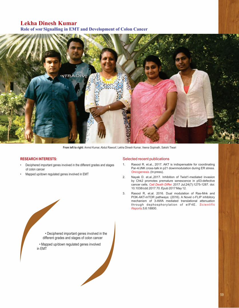
59
• Deciphered important genes involved in the different grades and stages of colon cancer
• Mapped up/down regulated genes involved in EMT
From left to right: Anmol Kumar, Abdul Rawoof, Lekha Dinesh Kumar, Veena Gopinath, Sakshi Tiwari
Lekha Dinesh KumarRole of wnt Signalling in EMT and Development of Colon Cancer
RESEARCH INTERESTS:
• Deciphered important genes involved in the different grades and stages of colon cancer
• Mapped up/down regulated genes involved in EMT
Selected recent publications
1. Rasool R. et.al., 2017. AKT is indispensable for coordinatingPar-4/JNK cross-talk in p21 downmodulation during ER stress. Oncogenesis. (In press).
2. Nayak D. et.al.,2017. Inhibition of Twist1-mediated invasionby Chk2 promotes premature senescence in p53-defectivecancer cells. 2017 Jul;24(7):1275-1287. doi: Cell Death Differ. 10.1038/cdd.2017.70. Epub 2017 May 12.
3. Rasool R, et.al. 2016. Dual modulation of Ras-Mnk andPI3K-AKT-mTOR pathways: (2016). A Novel c-FLIP inhibitory mechanism of 3-AWA mediated translational attenuation th rough dephosphory la t ion o f e IF4E. Scient i f i c Reports.5;6:18800.

The initiation and progression of malignancy involves a series of molecular changes including various signals transduced during this process. Wnt signal transduction pathway is one of the major pathways deregulated in majority of cancers. Most sporadic colorectal cancers arise as a result of a mutation in the
Wnt/b-catenin pathway which in turn is critical
towards colorectal cancer (CRC) progression. The key factor involved in this signaling pathway lies in the
stabilization of b-catenin and its interaction with TCF
transcription factors within the nucleus. In the
cytoplasm the b-catenin is tightly regulated and the
absence of Wnt factors results in its ubiquitinationand degradation by the proteasome. Colon cancers frequently harbor genetic defects that inhibit
the degradation of b-catenin which leads to nuclear
b-catenin translocation and activation of target genes
responsible for the Wnt activation. Constitutive
transcription by the b-catenin/Tcf complex has been
recognized as the key step for initiation of CRC.About 70% of all colorectal cancers show homozygous inactivation of the adenomatous polyposi coli (APC) tumor suppressor generesulting in the activation of the Wnt signaling
pathway and the constitutive transcription by the b-
catenin/Tcf complex. Loss of APC function is present throughout the sequence of intestinal carcinogenesis (i.e., from benign adenomas to fully malignant colorectal cancer and metastasis.
Epithelial Mesenchymal transition (EMT) is often associated with embryonic development whenmajor transitional events from blastula-morula-gastrula stages occur. This is a process with which embryonic cells loses its polarity and cell adhesion properties and acquire migratory and invasive
properties of mesenchymal cells. During this process, many important genes responsible for cell division/multiplication would be down regulated and another set of genes responsible for migration of the cells will be activated associated with EMT process. The initiation and progression of cancers also involves such a phenomenon where the epithelial cells acquire more of mesenchyal properties for progression and invasion which leads into metastasis.
Cancers are thought to result from the accumulation of multiple genetic aberrations that transform cells, which allows for their abnormal growth, proliferation, and metastasis. Discovery of these aberrationsand understanding how they contribute to the pathophysiology of cancer are necessary for improvements in diagnosis and therapy. More than any other model system, mice have revolutionized our ability to study gene function in vivo and understand the molecular mechanisms of cancer pathogenesis. As a model system, mice haveseveral important advantages over othermammalian models having a small in size, rapid reproduction having large litters and they can be genetically manipulated.
The development of transgenic and gene targeting technologies facilitated the generation of genetically engineered mouse (GEM) models to study tumor
Fig.1: Venn diagram represents the differentially expressed genes in well differentiated, moderately differentiated and poorly differentiated
human colon cancer samples which are > 1.5 fold up/down regulated compared to their respective adjacent normal sample
List contain 201 unique elements
Fig.2: Scatter plot representing intensity distribution of genes in different stages within moderately differentiated colorectal cancer samples
Fig.3: Four way venn diagram representing genes expressed different stages of moderately differentiated grade of Colon cancer.

biology through the manipulation of the mouse germ line. The most common ways to generate mouse models of cancers are to activate oncogenes or inactivate tumor-suppressor genes (or both) in vivo through the use of transgenic and gene-targeting approaches, such as knockouts and knockins.
With an aim to decipher the wnt deregulators involved in the initiation and progression of colorectal cancers (EMT), microarray analysis was carried out in human colon cancer patients uding nanostring technology. The human cancer genes were segregatedbased on their expression in well differentiated, moderately differentiated and poorly differentiated grades compared to their respective adjacent normals (Fig 1).
The genes expressed in the different stages ofeach grade which showed significant fold regulation (> 1.5 fold up/down regulated) compared to their adjacent normals were also classified. The scatter plot representing intensity distribution of these
genes are represented in Fig 2. Differentially expressed genes in different stages of moderately differentiated grade, stage I, II, III, IV has 144, 127, 130, 168 genes respectively compared to their respective adjacent normal samples. (Fig.3).These were then mapped into human chromosomes. These results were compared with the earlierresults obtained from microarray analysis of murine colon cancer model (Apc fl/fl).
Epithelial mesenchymal transition being an important paradigm of cancer, it can be considered as a pathological process which contributes to cancer progression. Hence it is important to classify the genes responsible for progression of cancer from stage I to stage II which is often associated with EMT. After finding out the differentially expressed genes from different grades of colorectal cancer, we classified them based on the various progressive stages within each grade.
The various deregulated genes were classified as up and down regulated ones based on >=1.5 fold and were placed on different signaling pathways associated with colon cancer. The status of these genes involved in various oncogenic pathways is shown below. The common genes of both mouse and human array experiments were selected forfurther studies. CITED-1 is a non-DNA binding transcription factor which has been reportedto be deregulated in many cancers. We found (Fig 4)this to be up regulated 25 times in mice colonmodels and deregulated in human colon cancer samples using different platforms of microarray technique. In case of moderately differentiatedcolon cancer samples, cited-1 expression continuously increased from stage I (>1.5 fold change) to stage IV (>5). This was confirmed by immuno histochemistry as well as q-PCR in various grades and stages of human cancer samples.
Table.1: Highly upregulated genes found in different grades and stages of human cancer in common with Apc fl/fl mouse models of colon cancer
Expression of CITED-1 gene in stage III of moderately differentiated colorectal cancer samples
Fig.4: Expression of CITED-1 gene in stage III of moderately differentiated colorectal cancer samples
Normal Tissue (40X) Cancer Tissue (40X)
61

From left to right (standing) : Ankita Chattopadhyay, Leena Kamalaskar, Manjusha Neerukonda, Sivapriya Pavuluri , Satish Kumar, Rajesh V Iyer, CB Tripura, Archana B Siva, Shalu Singh; (Sitting): Rohan Arora , M Manikandan, Arun Prakash Mishra, Alok Kumar, Rohit K Deshmukh
Satish KumarFunctional Genomics using Transgenic and Knockout Mice and Molecular Approaches in Animal Breeding
RESEARCH INTERESTS:
• Functional genomics using transgenic and gene knockout approaches in mice
• Understanding the genetics of livestock species
Selected recent publications
1. Singh VP, Katta S,Kumar S (2017). WD-repeat protein WDR13 is a
novel transcriptional regulator of c-Jun and modulates intestinal
homeostasis in mice. 17(1):148.BMC Cancer
2. Mitra S, Sameer Kumar GS, Tiwari V, Lakshmi BJ, Thakur SS,
Kumar S (2016). Implication of Genetic Deletion of Wdr13 in
Mice: Mild Anxiety, Better Performance in Spatial Memory Task,
with Upregulation of Multiple Synaptic Proteins. Front Mol
Neurosci. 9:73.
3. Whitelaw CB, Joshi A, Kumar S, Lillico SG, Proudfoot C (2016).
Genetically engineering milk. . 83(1):3-11.J Dairy Res
Molecular characterization of Wdr13 knockout female mice uteri: A model for
endometrial hyperplasia
Genotypic and phenotypic characterization of mutant mice exhibiting abnormalities in hair
growth.
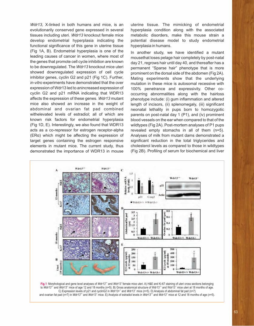
Wdr13, X-linked in both humans and mice, is an
evolutionarily conserved gene expressed in several
tissues including uteri. Wdr13 knockout female mice
develop endometrial hyperplasia indicating the
functional significance of this gene in uterine tissue
(Fig 1A, B). Endometrial hyperplasia is one of the
leading causes of cancer in women, where most of
the genes that promote cell cycle inhibition are known
to be downregulated. The Wdr13 knockout mice uteri
showed downregulated expression of cell cycle
inhibitor genes, cyclin G2 and p21 (Fig 1C). Further,
in-vitro experiments have demonstrated that the over
expression of Wdr13 led to anincreased expression of
cyclin G2 and p21 mRNA indicating that WDR13
affects the expression of these genes. Wdr13 mutant
mice also showed an increase in the weight of
abdominal and ovarian fat pad combined
withelevated levels of estradiol; all of which are
known risk factors for endometrial hyperplasia
(Fig 1D, E). Interestingly, we also found that WDR13
acts as a co-repressor for estrogen receptor-alpha
(ERα) which might be affecting the expression of
target genes containing the estrogen responsive
elements in mutant mice. The current study, thus
demonstrated the importance of WDR13 in mouse
uterine tissue. The mimicking of endometrial
hyperplasia condition along with the associated
metabolic disorders, make this mouse strain a
potential disease model to study endometrial
hyperplasia in humans.
In another study, we have identified a mutant
mousethat loses pelage hair completely by post-natal
day 21, regrows hair until day 40, and thereafter has a
permanent “Sparse hair” phenotype that is more
prominent on the dorsal side of the abdomen (Fig 2A).
Mating experiments show that the underlying
mutation in these mice is autosomal recessive with
100% penetrance and expressivity. Other co-
occurring abnormalities along with the hairloss
phenotype include: (i) gum inflammation and altered
length of incisors, (ii) splenomegaly, (iii) significant
neonatal lethality in pups born to homozygotic
parents on post-natal day 1 (P1), and (iv) prominent
blood vessels on the ear when compared to that of the
wildtypes (Fig 2A). Post-mortem analyses of P1 pups
revealed empty stomachs in all of them (n=5).
Analyses of milk from mutant dams demonstrated a
significant reduction in the total triglycerides and
cholesterol levels as compared to those in wildtypes
(Fig 2B). Profiling of serum for biochemical and liver
Fig.1: +/+ -/- Morphological and gene level analyses of Wdr13 and Wdr13 female mice uteri. A) H&E and Ki-67 staining of uteri cross sections belonging +/+ -/- +/+ -/-to Wdr13 and Wdr13 mice of age 12 and 18 months (n=5). B) Gross anatomical structure of Wdr13 and Wdr13 mice uteri at 18 months of age.
/+ -/-C) Expression levels of p21 and cyclinG2 in Wdr13+ and Wdr13 mice (n=5). D) Analysis of abdominal fat pad (n=7) +/+ -/- +/+ -/-and ovarian fat pad (n=7) in Wdr13 and Wdr13 mice. E) Analysis of estradiol levels in Wdr13 and Wdr13 mice at 12 and 18 months of age (n=5).
63

function parameters such as SGPT (Fig 2B) indicates
a metabolic syndrome in the mutant pups born to and
suckled by mutant females. Sequencing of Hr gene- a
commonly mutated gene in hair loss conditions,
revealed a novel heterozygous trinucleotide deletion
in Exon 6 (Fig 2C). This deletion is in-frame and
removes a Glutamic acid residue at position 534 of
the protein. Multiple sequence alignment of hr
orthologues from mouse, human, rhesus macaque,
rat, pig, and cow showed that the corresponding
Glutamic acid residue is conserved. Surprisingly, in a
larger panel of mutant animals, this deletion did not
segregate with the observed phenotype in our study
(Fig 2D).
In the area of livestock genetics, an ongoing
collaborative project (with Roslin Institute, UK and
BAIF Research Foundation, Pune), focuses on
understanding the genetic basis of resistance/
predisposition to tuberculosis and brucellosis in
Ind ian bu f fa lo . An ex tens ive mo lecu la r
epidemiological survey was carried out in the
breeding tracts of Jaffarabadi buffalo to identify
Tuberculosis and Brucellosis positive animals.
Genome of 36 animals (6 each of Murrah,
Jaffarabadi, Banni, Pandharpuri, Surti and Bhadawari
breeds) have been sequenced using NGS platform.
Efforts are ongoing to generate a reference genome
of Jaffarabadi buffalo by combining NGS data with
PacBio sequencing. A fine scale atlas of gene
expression in three breeds of domestic riverine water
buffalo (Mediterranean, Pandharpuri and Bhadawari)
has been generated through RNA-sequencing.
Fig.2: Phenotypic and genotypic characterization of mutant C57BL/6J mice. A) Morphological characteristics of mutant mice exhibiting sparse hair phenotype, inflammation of gums with altered length of incisors, splenomegaly, neonatal lethality in day1 pups and prominent blood vessels in the earas compared to those of wildtype. B) Analyses of levels of triglycerides and cholesterol in milk, and serum SGPT in wildtype and mutant mice.
C) Sanger sequencing of Hr exon-6with reverse primershowing the trinucleotide deletion-TTC. D) PCR-RFLP genotyping for Hr – exon6 trinucleotide deletion using Mbo-II restriction enzyme shows that a mutant animal does not carry this particular deletion.
65
We have a strong interest in elucidating mechanisms involved in the transmission of
epigenetic memory from one cell to another or one generation to the next. We use
Arabidopsis thaliana as a model plant and take cell biology, molecular biology, biochemical
and genetic approaches to address the question of mechanism of inheritance of
epigenetic cellular memory.
From left to right : Akanksha Garhewal, Supriya Sarma, Preethi Jampala, Asha Ganpatrao Mane, Mukesh Lodha, Sai Deep, Aditya Undru
Mukesh LodhaMechanism of Epigenetic Cellular Memory Inheritance in Plants
RESEARCH INTERESTS:
• Mechanism of inheritance of epigenetic cellular memory
• Chromatin mediated regulation of developmental genes
Selected recent publications
1. Lodha M, Marco CF, and Timmermans MCP (2013). Direct
Recruitment of Polycomb Repressive Complex2 by the
Arabidopsis ASYMMETRIC LEAVES Complex. Genes and
Development. 27:596 -601.
2. Husbands AY, Benkovics AH, Nogueira FT, Lodha M and
Timmermans MC (2015). The ASYMMETRIC LEAVES
Complex Employs Multiple Modes of Regulation to Affect
Adaxial-Abaxial Patterning and Leaf Complexity. Plant Cell.
27(12):3321-35.
3. Lodha M, Schulz-Raffelt M and Schroda M (2008). A new assay
for promoter analysis in Chlamydomonas reveals roles for heat
shock elements and the TATA box in HSP70A promoter-
mediated activation of transgene expression. . Eukaryotic Cell
7(1):172-6.
4. Schulz-Raffelt M, Lodha M and Schroda M. Heat shock factor 1
is a key regulator of thestress response in Chlamydomonas.
Plant J. 52(2):286-95 (2007).
5. Lodha M and Schroda M (2005). Analysis of chromatin structure
in the control regions of the chlamydomonas HSP70Aand
RBCS2 genes. 59(3):501-13.Plant Mol Biol.

Epigenetic information is heritable during mitotic
and/meiotic cell divisions but it is not encoded in the
genetic material. It is stable even in the absence of
initial trigger and is reversible to various extents.
Epigenetic regulation is important for hetero-
chromatin maintenance and euchromatic gene
regulation among many other cellular processes. We
know fair bit about acquisition of epigenetic memory
but an important question that remains underexplored
is the mechanism of inheritance of the epigenetic
state.
Histone post translational modifications, DNA
methylation and non-coding RNA can potentially
transmit memory during cell division. As a first step in
understanding the mechanism of epigenetic
inheritance, we are trying to identify histone tail
residues which are important in epigenetic
inheritance of memory of cold in Arabidopsis. We are
using transgenic plants expressing c-terminally
tagged and mutants in N-terminal tail amino acid
residues. These plants will be tested in a vernalization
(mitotically stable memory of cold) paradigm to test
their ability to gain and retain epigenetic memory. We
will test the proteins which are involved in turnover of
the known histone post translational modifications on
the identified histone tail residue for their roles in
inheritance of epigenetic memory. We are also testing
candidate protein complexes like trithorax and
polycomb for their role in inheritance of epigenetic
memory.
A large share of our understanding of epigenetics is
achieved through developmental genes. Our group is
using SHOOT MERISTEMLESS (STM), an important
shoot meristem stem cell regulator and a determining
factor in leaf complexity, as a tool to understand
epigenetic regulation in plants. In simple leaved
species such as Arabidopsis thaliana STM is
expressed in the shoot apical meristem and is down
regulated in leaf primordia. This downregulation is
maintained throughout the leaf development and is
required for proper simple leaf development. In
compound leaf species like Cardamine hirsuta,
tomato and pea, STM down regulation occurs in leaf
primordial but is not maintained. STM reactivation in
developing leaves is necessary and sufficient for
compound leaf formation. Remarkably, the STM
promoter from Arabidopsis thaliana (simple leaf
species) can recapitulate the simple leaf STM
expression pattern when introduced in Cardamine
hirsuta (compound leaf species). Conversely, the
STM promoter from Cardamine hirsuta can
recapitulate a compound leaf expression pattern
when introduced into Arabidopsis thaliana. These
experiments establish that cis regulatory sequences
of Arabidopsis thaliana and Cardamine hirsuta STM
promoters are important in determining their
expression patterns and thereby leaf complexity.
There are indications that STM is down regulated in
the leaf primordia in simple leaf species like
Arabidopsis thaliana by transcriptional repressors,
Polycomb complexes. It is tempting to hypothesize
that binding of PRC2/transcriptional repressors tocis
regulatory elements restricts STM expression only to
the shoot apical meristem in simple leaf species and
such cis regulatory sequences are absent in
compound leaf species. We are trying to identify and
characterize the cis regulatory elements in the simple
leaf species Arabidopsis thaliana and compound leaf
species Cardamine hirsuta which allows differential
expression of STM in these two species as a model of
leaf complexity in plants, and to establish role of
chromatin in differential regulation of STM.

67
Our group works on understanding the bio-mechanisms involved in the regeneration of organs
and the degeneration of brain structures in humans, zebrafish, ascidians and
echinoderms.Understanding the bio-mechanisms of regeneration and the association of various genes
(or proteins) in the regenerating environment is of high significance, as it might help us engineer non-
regenerating systems into regenerating systems for therapy and healing. Understanding the
regeneration mechanisms among different model animals is likely to lead to a better understanding of
the underlying mechanisms.
Mohammed Idris, Mir Quoseena, Naga Sowmya
M M IdrisBio-mechanisms of Regeneration and Degeneration
RESEARCH INTERESTS:
• Bio-mechanism of Regeneration
• Bio-mechanism of Degeneration
Selected recent publications
1. Babu NS, Murthy CL, Kakara S, Sharma R, Swamy CV, Idris
MM (2016) MPTP induced Parkinson's disease in zebrafish.
Proteomics 16(9): 1407 - 1420.
2. Babu NS, Krishnan S, VenakataSubbaiah GP, Gurava Reddy
VG, Swamy CV, Idris MM (2016) Quantitative proteomic
analysis of normal and degenerated human intervertebral disc.
Spine J. 16(8): 989- 1000.
3. Saxena S, Purushothaman S, Meghah V, Bhatti B, Poruri A,
Meena Lakshmi MG, Babu NS, Murthy CL, Mandal KK, Kumar
A, Idris MM (2016) Role of Annexin gene and its regulation
during zebrafish caudal fin regeneration. Wound, Repair and
Regen. 24(3): 551-559.
4. Purushothaman S, Saxena S, Meghah V, Swamy CVB, Ortega-
Martinez O, Dupont S, Idris MM (2015) Transcriptomic and
proteomic analyses of Amphiurafiliformis arm tissue-
undergoing regeneration. 112:113- 124. J Proteomics
5. Purushothaman S, Saxena S, Meghah V, Meena Lakshmi MG,
Singh SK, Swamy CVB, Idris MM (2015) Proteomic and gene
expression analysis of zebrafish brain undergoing continuous
light/dark stress. . 24(4): 458 - 465.J Sleep Res

We are focusing on the regeneration of appendages
in zebrafish, nervous tissues in ascidians and arms in
starfish using proteomics and transcriptomics
approaches. We have identified several proteins
including Annexin 1, 2, and 5 as differentially
regulated or posttranslationally modified during
regeneration of the zebrafish caudal fin. The proteins
were differentially expressed in the regenerating
tissues either immediately after amputation(1-hour
post amputation (hpa)) or later during regeneration
(12, 24, 48 and 72 hpa). Similarly, we have identified
several novel transcription factors and epigenetic
modifiers undergoing differential regulation during
regeneration. The roles of epigenetic regulatory
mechanisms, such as histone H3 and H4 lysine
acetylation and methylation during zebrafish caudal
fin regeneration were also studied using ChIP and RT-
PCR assays. More than 700 genes were identified as
differentially regulated in the echinoderm arm
regeneration based on high throughput transcriptome
mapping and nearly 200 proteins were identified as
differentially expressed in the nervous systemof
ascidians during regeneration.
We are a lso involved in understanding
the mechanism of degeneration in human
intervertebral disc (Degenerative disc disease)
and neurodegenerative disorders such as
Spinocerebellar ataxia(SCA) and Parkinson’s
disease (PD)in the zebrafish model. A total of 759
and 692 proteins respectively, were identified
as differentially regulated in the Annulus fibrosus
and Nucleus pulposus tissues of the human
intervertebral disc based on proteomics studies.
Neuro-degeneration, the chronic breakdown of
neuronal structures, is mostly associated with
expansion of CAG triplet repeats leading to
aggregation and polymerizationof proteins. Our
laboratory aims to understand the association of
expanded triplet repeats (CAG) in the ataxin gene
with neurodegeneration based on a transgene
approach in zebrafish. We have also identified the
role of chronic unpredictable stress and chemicals
such as MPTP involved in inducing degeneration of
the zebrafish nervous system.

69
Our group identified The N-terminal aminoacid residues for the tryptic peptides generated form
in gel digested proteins after acetylating the peptides and detecting b1 ions in the mass spectra.
The methods developed helps in improving thede novo sequencing efficiency of peptides using
mass spectrometry.
M V Jagannadham, P Gayathri, Deepika
M V JagannadhamStudies on Outer Membrane Vesicles of Bacteria
RESEARCH INTERESTS:
• Structural and functional studies of outer membrane vesicles of bacteria
• Chemical derivatization of proteins and peptides for facilitated analysis by mass spectrometry
• Detecting Post translational modifications of proteins using Mass spectrometry
Selected recent publications
1. Bhanuramanand K, Sankararao K, Jagannadham MV, Swamy
Ch. V B, Dholpe, V.M. and Rao, N M (2017) Combinatorial
labelling method for improving peptide fragmentation in Mass
spectrometry. 28(6): 1216-1226.J. Amer. Soc. Mass spectrum.
2. Chattopadhyay MK and Jagannadham MV (2015) Vesicles-
mediated resistance to antibiotics in bacteria. Front. Microbiol.
6:758. Doi: 10.3389/fmicb.2015.00758.
3. Kulkarni HM, Nagaraj R and Jagannadham MV (2015)
Protective role of E. coli outer membrane vesicles against
antibiotics. 181: 1-7. Microbiol. Res.
4. Fulsundar S, Kulkarni HM, Jagannadham MV, Nair R, Sravani
Keerthi, Sant P, Pardesi K, Bellare J and Chopade BA (2015)
Molecular characterization of outer membrane vesicles
released from Acinetobacter radioresistens and their potential
roles in pathogenesis. 83-84: 12-22.Microbial Pathogenesis
5. Kulkarni HM and Jaganadham M V (2014) Biogenesis and
multifaceted roles of outer membrane vesicles form Gram-
negative bacteria. 160: 2109-2121.Microbiology

Role of OMVs from Acenitobacter
baumannii in antibiotic resistance
Acenitobacter baumannii, an opportunistic human
pathogen, is responsible for several diseases like
pneumonia, meningitis and a wide range of other
infections. It is also typically involved in nosocomial
infections. Outer membrane vesicles (OMVs)
released from the bacteria play an important role in
the bacterial physiology including pathogenesis.
They also contribute to antibiotic-resistance of
bacteria in various ways. The present investigation
was undertaken to look into the molecular basis of
vesicle-mediated antibiotic-resistance in bacteria.
OMVs were obtained from a clinical, multidrug
resistant, nosocomial strain of A. baumannii, isolated
by Ms Bina Agarwal under the supervision of Prof. G.
Ahmed at the University of Guwahati. The strain was
originally obtained from a tertiary care hospital in
Guwahati, Assam. The OMVs were prepared form the
bacteria, the protein bands from the OMVs were cut
and subjected to in gel digestion using trypsin. The
peptides were extracted using standard protocols and
subjected to LC coupled ESI MS/MS studies. The
proteins were identified by SEQUEST. A total of 320
proteins were identified in OMVs produced by the
antibiotic-sensitive strain, and 560 proteins were
identified from the vesicles produced by the multi-
drug resistance (MDR) strain. An overlap of 15
proteins was observed, 545 proteins were exclusively
present in MDR OMVS and 305 proteins were specific
from the sensitive strain OMVs.A comparative
proteomics analysis of the OMVs from the A.
baumannii sensitive and MDR strain revealed the
increased content of carbapenemase and a host of
other related antibiotic-inactivating enzymes in the
OMVs of the A. baumannii MDR strain. Growth
studies on the A. baumannii and MDR strain in the
presence of polymixin B and OMVs prepared from the
respective strains revealed that the OMVs protected
the bacteria against the antibiotic. These studies
reveal that OMVs may play an important role in
antibiotic resistance/persistence.
Identification of proteins from species of
unknown genome sequences (collaboration
with Dr R Srinivas , CSIR- IICT, Hyderabad)
The proteins from the Acenitobacter radioresistance,
whose genome sequence is not known, were chosen
to analyse using MS/MS spectra. The proteins were
identified using a database prepared from the
genome sequences of the other species of
Acenitobacter. After extracting the total proteins of the
organism, equal amounts of the extract were loaded
into two different wells in 12% SDS-Polyacrylamide
gel. The protein bands obtained in both the lanes
were excised and subjected to ingel trypsin digestion.
The digested bands in one lane were identified by LC
coupled ESI-MS/MS analysis. The digested bands
from the other lane were acetylated and subsequently
identified by LC coupled ESI-MS/MS analysis. The
peptides containing N-terminal histidine were
analysed in detail to understand the mechanism
for the formation of b ion. N-terminal histidine 1
containing peptides were shown to from b ion, by 1
cyclization with the side chain imidazole (Fig 1A).
The detection of post translational modifications
(PTMs) of proteins is an important comprehensive
research subject. Among all possible downfalls that
may lead to misidentifications, the chemical stability
of modified peptides is scarcely questioned. Global
proteomic studies devoted to protein acetylation are
becoming attractive. Thus, we were concerned about
the acetylation of histidine and its intrinsic stability of
b -ion of the Oxazolone ring and/or with side chain 1
imidazole bicyclic product. Because of the formation
of an oxazolone structure may occur when an amino
moiety has gone through an acetylation reaction and
when it is present in the vicinity of the side chain
imidazole moiety. Here, we probed the possibility of a
ring formation between oxazolone and a bicyclic
compound with the side chain imidazole. Tryptic
Fig.1: Schematic representation for the formation of b -ion with side chain 1
imidazole ring and oxazolone ring for N-terminal acetylated peptides

peptides containing N-terminal histidine was explored
in a standard proteomic workflow. We have validated
the formation of the Oxazolone ring through a low
energy calculation of density functional theory (DFT)
studies. This computational method suggested that
the conformer (C) in which the protonation has
occurred at the imidazole ring is more stable than the
other two conformers namely A and B. Among all
these conformers, A is unstable than C by ~24
kcal/mole and the other conformer B, is intermediate
between both the A and C conformers. The results
obtained in the present study showed that the
histidine had undergone acetylation and that the N-
acetylated group also may participate in ring
formation. This was the case for the N-acetylated
peptide. In this case, the oxazolone ring with N-acetyl
group may also form rather only with the side chain
imidazole group, indicating that N-terminal N-
acetylated histidine forming oxazolone ring may be
stable and will be one of the products in the
proteomics workflow. The N-terminal N-acetyl
histidine oxazolone ring obtained, has low energy and
stable structure in proteomic samples. We also report
the shift of MS to differentiate between peptides
containing acetylated histidine from the unacetylated
moiety by showing a clear mass shift. Further, studies
are in progress to characterize the immonium ions
and b-ions after acetylation of tryptic peptides. These
studies will help in improving the de novo sequencing
efficiency of the peptides, in addition to validate the
sequence of peptides.
Detecting multiple phosphorylations in
peptides using MS
A wide variety of post-translational modifications such
as oxidation, phosphorylation, glycosylation,
methylation and acetylation play critical roles in
cellular functions. Detection of PTMs in proteins is
important to understand their crucial roles in cellular
functions. Identifying each modification requires
special attention in mass spectral acquisition and
analysis. Here, we report a mass spectral method for
the detection of multiple phosphorylations by
analyzing the fragments. Synthetic peptides were
used to identify these modifications by Matrix-
assisted laser desorption/Ionization (MALDI)
TOF/TOF. Peptides with serine, threonine and
tyrosine were used with mono- to tetra-
phosphorylation sites in different combinations to get
insights into their fragmentation and identify the
location of these sites. The y- ions series were
observed without the loss of phosphate groups and
were thus very useful in determining the localization
and sequence of the phosphate residues. Acetylation
of the peptides was found to be useful in detecting the
b1 ion and helped in identifying the N-terminus. When
a mixture of the phosphorylated peptides (from
mouse protein sequences) was analyzed by LC-
MS/MS on a VelosOrbitrap Mass Spectrometer and
the data were subjected to analysis by Sequest using
the mouse database, the peptides were identified
along with the parent proteins. A comparison of
MALDI TOF/TOF spectra with ESI MS/MS helped in
eliminating falsely discovered peptides using the
database search.
71

RESEARCH INTERESTS:
• Comparative and functional genomics of non-coding DNA.
• Organization and regulation of Hox complexes: evolutionary logic of body plan in animals.
• Epigenetic regulation and development.
Selected recent publications
1. Akshay Kumar A, Saketh S, DivyaTej S and Rakesh K. Mishra
(2017) MSDB: A Comprehensive Database of Simple Sequence
Repeats. 9(6):1797-1802.Genome Biol Evol
2. Krishnan J, Fathima A, Swaroopa Rani T and Rakesh K. Mishra
(2017) Simple sequence repeats showing 'length preference' have
regulatory functions in humans. 628:156-161.Gene
3. Gopal Kushawah and Rakesh K. Mishra (2017). Ultraconserved
sequences associated with HoxD cluster have strong repression
activity. 9 (8): 2049-2054.Genome Biol Evol
4. Debaditya De, Satish K, Jae-Min Lim, Rashmi U Pathak and
Rakesh K. Mishra (2017). O-GlcNAcylation of Boundary Element
Associated Factor (BEAF 32) in Drosophila melanogaster
correlates with active histone marks at the promoters of its target
genes. doi: 10.1080/19491034.2017.1367887.Nucleus
5. DivyaTej S, Surabhi S, Jyotsna D and Rakesh K. Mishra (2017). C-
State: An interactive web app for simultaneous multi-gene
visualization and comparative epigenetic pattern search. BMC
Bioinformatics : doi.org/10.1101/163634.
We developed a Comprehensive Database of Simple Sequence Repeats (MSDB) and
demonstrated that SSRs that are enriched in complex genomes have diverse functions
Earlier we identified conserved non-coding sequences associated with the Hox clusters that,
as we now show for the first time, have a repressive function
We demonstrate that post transcriptional modifications of BEAF, a boundary element
associated factor, determine its functional specificity
M Arif, F Athar, Akshay Kavvaru, Ashish Bihani, V Chakrapani, Nikhil Hajirnis, Runa Hamid, Alshymaa Hassan, Shagufta Khan, Gopal Kushwah, Shreyashi Mukherjee, M Nisha, Indira Paddibhatla, Rashmi U Pathak, K Phanindhar, Titus Ponaratnam, Parna Saha, Ravina Saini, Salam Herojeet Singh, Divya Tej Sowpati,
A Srinivasan, Avinash Srivastava, Ishani Srivastava, Surabhi Srivastava, Rahul Sureka, ShreekantVerma, Mukesh P Yadav, Rakesh K Mishra
Rakesh K MishraGenome Organization and Epigenetic Regulation
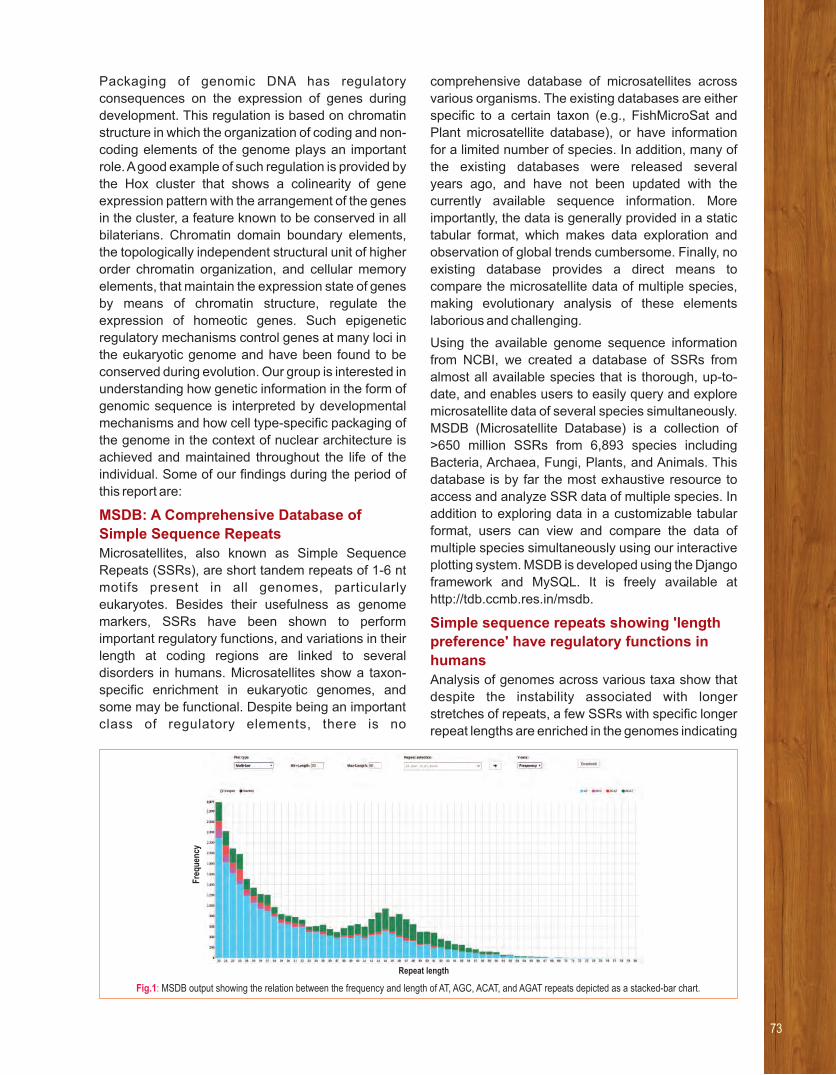
Packaging of genomic DNA has regulatory
consequences on the expression of genes during
development. This regulation is based on chromatin
structure in which the organization of coding and non-
coding elements of the genome plays an important
role. A good example of such regulation is provided by
the Hox cluster that shows a colinearity of gene
expression pattern with the arrangement of the genes
in the cluster, a feature known to be conserved in all
bilaterians. Chromatin domain boundary elements,
the topologically independent structural unit of higher
order chromatin organization, and cellular memory
elements, that maintain the expression state of genes
by means of chromatin structure, regulate the
expression of homeotic genes. Such epigenetic
regulatory mechanisms control genes at many loci in
the eukaryotic genome and have been found to be
conserved during evolution. Our group is interested in
understanding how genetic information in the form of
genomic sequence is interpreted by developmental
mechanisms and how cell type-specific packaging of
the genome in the context of nuclear architecture is
achieved and maintained throughout the life of the
individual. Some of our findings during the period of
this report are:
MSDB: A Comprehensive Database of
Simple Sequence Repeats
Microsatellites, also known as Simple Sequence
Repeats (SSRs), are short tandem repeats of 1-6 nt
motifs present in all genomes, particularly
eukaryotes. Besides their usefulness as genome
markers, SSRs have been shown to perform
important regulatory functions, and variations in their
length at coding regions are linked to several
disorders in humans. Microsatellites show a taxon-
specific enrichment in eukaryotic genomes, and
some may be functional. Despite being an important
class of regulatory elements, there is no
comprehensive database of microsatellites across
various organisms. The existing databases are either
specific to a certain taxon (e.g., FishMicroSat and
Plant microsatellite database), or have information
for a limited number of species. In addition, many of
the existing databases were released several
years ago, and have not been updated with the
currently available sequence information. More
importantly, the data is generally provided in a static
tabular format, which makes data exploration and
observation of global trends cumbersome. Finally, no
existing database provides a direct means to
compare the microsatellite data of multiple species,
making evolutionary analysis of these elements
laborious and challenging.
Using the available genome sequence information
from NCBI, we created a database of SSRs from
almost all available species that is thorough, up-to-
date, and enables users to easily query and explore
microsatellite data of several species simultaneously.
MSDB (Microsatellite Database) is a collection of
>650 million SSRs from 6,893 species including
Bacteria, Archaea, Fungi, Plants, and Animals. This
database is by far the most exhaustive resource to
access and analyze SSR data of multiple species. In
addition to exploring data in a customizable tabular
format, users can view and compare the data of
multiple species simultaneously using our interactive
plotting system. MSDB is developed using the Django
framework and MySQL. It is freely available at
http://tdb.ccmb.res.in/msdb.
Simple sequence repeats showing 'length
preference' have regulatory functions in
humans
Analysis of genomes across various taxa show that
despite the instability associated with longer
stretches of repeats, a few SSRs with specific longer
repeat lengths are enriched in the genomes indicating
Fig.1: MSDB output showing the relation between the frequency and length of AT, AGC, ACAT, and AGAT repeats depicted as a stacked-bar chart.
Fre
qu
ency
Repeat length
73

a positive selection. This conserved feature of length
dependent enrichment hints at not only sequence but
also length dependent functionality for SSRs. We
selected 23 SSRs of the human genome that show
specific repeat length dependent enrichment and
analysed their cis-regulatory potential using promoter
modulation, boundary and barrier assays. We find
that the 23 SSR sequences, which are mostly
intergenic and intronic, possess distinct cis-
regulatory potential. They modulate minimal
promoter activity in transient luciferase assays and
are capable of functioning as enhancer-blockers and
barrier elements. The results of our functional assays
propose cis-gene regulatory roles for these specific
length enriched SSRs and opens avenues for further
investigations.
Ultra-conserved sequences associated with
the HoxD cluster have strong repression
activity
An increase in the complexity of organisms during
evolution strongly correlates with the increase in the
non-coding DNA content of their genomes. Although a
gradual increase in the proportion of repetitive DNA
elements along with increasing complexity is known,
most of the non-coding components of the genome
remain uncharacterized. A non-repetitive but highly
conserved non-coding component of the genome in
vertebrates, called ultra-conserved DNA sequences,
constitutes up to 5% of the human genome. The
functions of most of the ultra-conserved DNA
elements are not well understood. One such ultra-
conserved stretch of DNA has been identified
upstream of the HoxD cluster in vertebrates. We
analyzed the function of these elements in different
cell lines and zebrafish. Our results suggest that
these ultra-conserved sequences work as repressor
elements. This is the first report which reveals the
repressor function of ultra-conserved sequences and
implicates their role in the regulation of
developmental genes.
Fig.2: Promoter modulation assay for the 23 human SSRs in human cells: Promoter modulation assay was carried out in K562 cell line. SSRs were
cloned upstream of an SV40 promoter which regulates the luciferase gene. Luciferase activity was determined, normalised to vector control.
Statistically significant results are represented. (p< 0.05 *, <0.01 **, <0.001 ***, <0.0001 ****).
Fig.3: Reporter assay in zebrafish: Individual zebrafish embryos injected with different mouse CRs reporter constructs in equimolar concentration and imaged on different days’ post fertilization (dpf). GFP expression in the same embryo is shown at 2, 4, 6 and 8dpf in the respective row. CR2 embryo shows high level of GFP expression during early embryogenesis and after 4 dpf GFP expression reduces drastically as compared to the control embryo.
CR1, CR3 and CR6 show comparable GFP expression as in the case of control even after 8dpf.

O-GlcNAcylation of Boundary Element
Associated Factor (BEAF 32) in Drosophila
melanogaster correlates with active histone
marks at the promoters of its target genes:
BEAF 32 is a sequence specific DNA binding
protein involved in functioning of chromatin
domain boundaries in Drosophila. Several studies
also show it to be involved in transcriptional
regulation of a large number of genes, many
of which are annotated to have cell cycle,
development and differentiation related function.
Since post-translational modifications (PTMs)
of proteins add to their functional capacity,
we investigated the PTMs on BEAF 32. The
protein is known to be phosphorylated and
O-GlcNAcylated. We mapped the O-GlcNAc
site at T91 of BEAF 32 and showed that it is
linked to the deposition of active histone
(H3K4me3) marks at the transcription start
site (TSS) of associated genes. Its role as a
boundary associated factor, however, does not
depend on this modification. Our study shows
that by virtue of O-GlcNAcylation, BEAF 32 is
linked to epigenetic mechanisms that activate a
subset of associated genes.
C-State: An interactive web app for
simultaneous multi-gene visualization and
comparative epigenetic pattern search
Comparative epigenomic analysis across multiple
genes presents a bottleneck for bench biologists
working with NGS data. Despite the development of
standardized peak analysis algorithms, the
identification of novel epigenetic patterns and
their visualization across gene subsets remains
a challenge. We developed a fast and interactive
web app, C-State (Chromatin-State), to query
and plot chromatin landscapes across multiple
loci and cell types. C-State has an interactive,
JavaScript- based graphical user interface
and runs locally in modern web browsers that
are pre-instal led on al l computers, thus
eliminating the need for cumbersome data
transfer, pre-processing and prior programming
knowledge. C-State is unique in its ability to
extract and analyze multi-gene epigenetic
information. It allows for powerful GUI-based
pat te rn search ing and v isua l i za t ion . In
conclusion, C-State is a standalone application
for epigenetic and gene expression analysis,
providing an easy solution for experimental
Fig.4: PTM variants of BEAF 32 and their functions: A. Dephosphorylated BEAF32 binds to DNA and localizes to NuMat. NE or NuMat proteins from S2 cells, was electro-focussed on pH 3-10 NL strip and then resolved using 12% SDS-PAGE. Western blots were probed with anti BEAF. For southwestern analysis, the same blot used for western was probed with 32P-labelled scs’ DNA to reveal the DNA binding variants i. PTM variants of BEAF 32 in S2 cell extract resolved as six distinct spots (1-6) in 2D PAGE. ii. SAP treatment of the S2 extract abolished the upper spots (4-6). iii. One of the dephosphorylated variant (spot 3) was present in NuMat. iv. Dephosphorylated variants of BEAF 32 (spots 2 & 3) bound to DNA. B. Summary of PTMs and related functions of BEAF 32.Top left cell represents the PTM variants of BEAF 32 graphically. ‘+’ indicates the presence and ‘-’ absence of the respective PTM.
A
B
75

biologists to investigate epigenetic patterns
without needing to know how to handle or
parse big data. In addition to running locally
on the user's system, it can also be hosted
centrally on an internal network to which multiple
users can connect for visualizing and sharing their
data. C-State's searchable filtering and display
modules are extensible and can handle data from any
organism for a large number of genes and a flexible
amount of intergenic regions. This is very useful for
researchers to analyze novel or publicly available
datasets in order to formulate new hypothesis for
experimental testing, without investing in complicated
programming or bioinformatics help. C-State also
allows the incorporation of user-generated data
with published datasets for rapid comparison
and analysis and facilitates easy documentation of
salient information via capture of high quality,
publication ready images. The tool is available at
http://www.ccmb.res.in/rakeshmishra/c-state/
Fig.5: Overview of C-State’s architecture

77
RESEARCH INTERESTS:
• Early embryonic development in mouse
• Understanding cell fate choice based on transcription factor modulation in pluripotent state
• Genetic disease modeling using pluripotent stem cells.
Selected recent publications
1. Miguel F, Chandra Shekar P, Yen-Sin A, Fujiwara Y, Stuart H.
Orkin, Jianlong W (2011) Zfp281 Functions as a
Transcriptional Repressor for Pluripotency of Mouse
Embryonic Stem Cells. 29:1705–1716. Stem Cells
2. Chandra Shekar P, Adnan Naim P, ParthaSarathi D and
Satish Kumar (2011) Argonaute-2-null embryonic stem cells
are retarded in self-renewal and differentiation. 36: J. Biosci.
649-657.
3. P. Chandra Shekar, Sandeep Goel, S. Deepa Selvi Rani, D.
Partha Sarathi, Jomini Liza Alex, Shashi Singh, and Satish
Kumar; (2006) κ-Casein-deficient mice fail to lactate. Proc.
Natl. Acad. Sci. USA 103:8000-8005.
4. Ravishankar K V, Chandra Shekar P, Sreedhara SA, Dinesh
LalithaAnand M R, Saiprasad GVS; (2004) Diverse genetic
bases of Indian polyembryonic and monoembryonic mango
(MangiferaindicaL) cultivars. 87:870-871. Current Science
We are trying to understand how cell fate decisions are made in the early stages of
embryogenesis and during stem cells differentiation based on the expression levels of
key transcription factors. We use functional analysis and biochemical approaches to perturb
transcription factor networks to understand underlying mechanisms.
From left to right : Vishnu Vijay, Usha Kabilan, Hanuman Kale, Debabrata Jana, P Chandra Shekar
P Chandra ShekarEarly Embryonic Development in Mouse
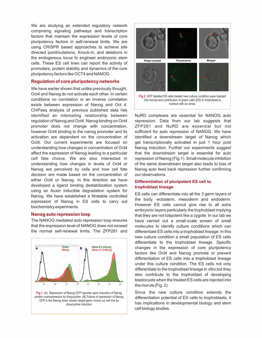
We are studying an extended regulatory network
comprising signaling pathways and transcription
factors that maintain the expression levels of core
pluripotency factors in self-renewal limits. We are
using CRISPR based approaches to achieve site
directed pointmutations, Knock-in, and deletions in
the endogenous locus to engineer embryonic stem
cells. These ES cell lines can report the activity of
promoters, protein stability and dynamics of the core
pluripotency factors like OCT4 and NANOG.
Regulation of core pluripotency networks
We have earlier shown that unlike previously thought,
Oct4 and Nanog do not activate each other. In certain
conditions no correlation or an inverse correlation
exists between expression of Nanog and Oct 4.
ChIPseq analysis of previous published data has
identified an interesting relationship between
regulation of Nanog and Oct4. Nanog binding on Oct4
promoter does not change with concentration,
however Oct4 binding to the nanog promoter and its
activation are dependent on the concentration of
Oct4. Our current experiments are focused on
understanding how changes in concentration of Oct4
affect the expression of Nanog leading to a particular
cell fate choice. We are also interested in
understanding how changes in levels of Oct4 or
Nanog are perceived by cells and how cell fate
decision are made based on the concentration of
either Oct4 or Nanog. In this direction we have
developed a ligand binding destabilization system
using an Auxin inducible degradation system for
Nanog. We have established a titratable controlled
expression of Nanog in ES cells to carry out
biochemistry experiments.
Nanog auto repression loop
The NANOG mediated auto repression loop ensures
that the expression level of NANOG does not exceed
the normal self-renewal limits. The ZFP281 and
NuRD complexes are essential for NANOG auto
repression. Data from our lab suggests that
ZFP281 and NuRD are essential but not
sufficient for auto repression of NANOG. We have
identified a downstream target of Nanog which
get transcriptionally activated in just 1 hour post
Nanog induction. Further our experiments suggest
that the downstream target is essential for auto
repression of Nanog (Fig.1). Small molecule inhibition
of the same downstream target also leads to loss of
Nanog auto feed back repression further confirming
our observations.
Differentiation of pluripotent ES cell to
trophoblast lineage
ES cells can differentiate into all the 3 germ layers of
the body -ectoderm, mesoderm and endoderm.
However ES cells cannot give rise to all extra
embryonic layers particularly the trophoblast implying
that they are not totipotent like a zygote. In our lab we
have carried out a small-scale screen of small
molecules to identify culture conditions which can
differentiate ES cells into a trophoblast lineage. In this
new culture condition a small population of ES cells
differentiate to the trophoblast lineage. Specific
changes in the expression of core pluripotency
factors like Oct4 and Nanog promote or prevent
differentiation of ES cells into a trophoblast lineage
under this culture condition. The ES cells not only
differentiate to the trophoblast lineage in vitro but they
also contribute to the trophoblast of developing
blastocysts when the treated ES cells are injected into
the morula (Fig. 2).
Since the new culture condition extends the
differentiation potential of ES cells to trophoblasts, it
has implications in developmental biology and stem
cell biology studies.
Fig.1: (A) Repression of Nanog GFP reporter upon induction of Nanog protein overexpression by doxycycline. (B) Failure of repression of Nanog
GFP in the Nanog down stream target gene- knock out cell line by doxycycline induction
Fig.2: GFP labelled ES cells treated new culture condition were injected into morula and contribution of green cells (ES) to trophoblast is
marked with an arrow.

79
RESEARCH INTERESTS:
• Effects of laminopathic mutants on differentiation and development
• Mechanism of protein degradation induced by laminopathic mutants
Selected recent publications
1. Verma, AD and Parnaik VK (2017) Heart-specific expression of
laminopathic mutations in transgenic zebrafish. Cell Biol Int
41:809-819.
2. Verma, A D and Parnaik VK (2015) Identification of tissue-
specific regulatory region in zebrafish lamin A. 567:73-80. Gene
3. Chaturvedi P, Khanna R and Parnaik VK (2012) Ubiquitin ligase
RNF123 mediates degradation of heterochromatin protein 1α
and β in lamin A/C knock-down cells 7: e47558.. PLoS ONE
4. Chaturvedi P and Parnaik VK (2010) Lamin A rod mutants target
heterochromatin protein 1 and for proteasomal degradation by
activation of F-box protein, FBXW10. 5: e10620.PLoS ONE
5. Parnaik VK (2008) Role of nuclear lamins in nuclear
organization, cellular signaling and inherited diseases. Int Rev
of Cell Mol Biol 266: 157-206.
We have characterised transgenic zebrafish models of heart-specific laminopathies
From left to right : Richa Khanna, Veena K Parnaik, Vidhya Krishnamoorthy, Ajay D Verma
Veena K ParnaikNuclear Organization and Lamin Biology

Fig.1: Quantitative RT-PCR analysis of cardiac developmental and regeneration genes in transgenic embryos at 3.5 days post fertilization(open bars) and 5.5 days post fertilization (solid grey bars); *p ≤ 0.05
The lamins are major components of a filamentous
network of proteins termed the nuclear lamina that
lies beneath the inner nuclear membrane in a
metazoan cell. Lamins are important for the
organization of nuclear functions as well as chromatin
organization, and have also been proposed to play
key roles in cell differentiation pathways. Two major
kinds of lamins are present in higher eukaryotes. The
B-type lamins are expressed in all somatic cell types
whereas the expression of A-type lamins is restricted
to differentiated cells of most lineages. Mutations in
the human laminA gene (LMNA) have been
associated with at least 15 debilitating inherited
diseases, collectively termed laminopathies, that
affect specific tissues such as skeletal muscle,
cardiac muscle, adipose tissue and bone, and also
cause premature ageing syndromes. A few
laminopathies have been studied using the mouse as
a model organism.
The current research in my group is directed towards
investigating the effects of laminopathic mutations on
cellular functions and embryonic development. The
zebrafish has emerged as a useful model system for
the study of vertebrate development, especially due
to the ease of visualization of GFP-tagged genes
during embryogenesis in transgenic fish. Zebrafish
has four genes that code for lamins A, B1, B2 and LIII.
Zebrafish laminA gene shows 63% identity and 76%
similarity with the human lamin A gene. Based on its
conserved gene structure and sequence similarity,
zebrafish lamin A can be considered to be an
orthologue of mammalian lamin A. As zebrafish is a
well-established model for the study of cardiac
development and disease, we have investigated the
effects of heart-specific laminA mutations in
transgenic zebrafish. We have developed transgenic
lines of zebrafish expressing conserved laminA
mutations that cause cardiac dysfunction in humans.
Expression of zlaminA mutations Q291P and M368K
in the heart was driven by the zebrafish cardiac
troponin T2 promoter. Homozygous embryos
displayed nuclear abnormalities in ~20% of
cardiomyocyte nuclei. Expression analysis showed
the upregulation of genes involved in heart
regeneration in transgenic mutant embryos and a cell
proliferation marker was increased in heart tissue. At
the physiological level, there was deviation of up to
20% from normal heart rate in transgenic embryos
expressing mutant lamins. Adult homozygous
zebrafish were fertile and did not show signs of early
mortality. Our results suggest that transgenic
zebrafish models of heart-specific laminopathies
show cardiac regeneration and moderate deviations
in heart rate during embryonic development.
An emerging concept in lamin pathogenesis is that
laminopathic mutations alter interactions of lamins
with key regulatory factors and chromatin bound
proteins. We have earlier shown that laminopathic
mutat ions can induce ubiqui t in-mediated
proteasomal degradation of specific chromatin-
binding proteins such as heterochromatin protein 1
(HP1) isoforms and the DNA damage sensor, ATR
(Ataxia-telangiectasia-mutated-and-Rad3-related)
kinase by activation of specific E3 ubiquitin ligase
components, namely, the monomeric RING ligase
RNF123, the HECT ligase HECW2 and the substrate
adaptor for a multimeric RING ligase FBXW10.
Our current studies on the identification of additional
substrates for these ligasesindicate that lamin-
binding proteins are important targets for RNF123
and HECW2. Our findings imply that ubiquitination
and degradation of lamin-binding proteins may
contribute to disease-causing mechanisms in
laminopathies by depletion of key nuclear proteins.
81
RESEARCH INTERESTS:
• Development of 13C NMR methods to study Brain Energy metabolism
• Excitatory and Inhibitory neurotransmission in neurodegenerative disorders
• Neuronal and astroglial metabolic activity in psychiatric disorders
• Development of MRI contrast materials
Selected recent publications
1. Patel AB, Tiwari V, Veeraiah P, Saba K (2017) Increased
astroglial activity and reduced neuronal function across brain in
AβPP-PS1 mouse model of Alzheimer's disease. J Cereb Blood
Flow Metab doi:10.1177/0271678X17709463.
2. Bagga P, Behar KL, Mason GF, De Feyter HM, Rothman DL,
Patel AB (2014) Characterization of Cerebral Glutamine Uptake
from Blood in the Mouse Brain: Implications for Metabolic 13Modeling of C NMR Data. 34:1666-J Cereb Blood Flow Metab
1672.
3. Veeraiah P, Noronha JM, Maitra S, Bagga P, Khandelwal N,
Chakravarty S, Kumar A, Patel AB (2014) Dysfunctional
Glutamatergic and GABAergic Activities in Prefrontal Cortex of
Mice in Social Defeat Model of Depression. Biol Psychiatry
76:231-238.
4. Patel AB, Lai JCK, Chowdhury GMI, Hyder F, Rothman DL,
Shulman RG, Behar KL (2014) Direct Evidence for Activity-
Dependent Glucose Phosphorylation in Neurons With
Implications for the Astrocyte-to-Neuron Lactate Shuttle. Proc
Natl AcadSci USA 111:5385–5390.
5. Bagga P, Chugani AN, Varadarajan KS, Patel AB (2013) In Vivo
NMR Studies of Regional Cerebral Energetics in MPTP Model of
Parkinson's Disease: Recovery of Cerebral Metabolism with
Acute Levodopa Treatment. 127:365-77.J Neurochem
We have shown that the energetics of Glutametergic and GABAergic
neuro-transmission is distinct across brain.
In a contradiction of Astrocyte-to-Neuron Lactate Shuttle hypothesis we have shown direct uptake
and phosphorylation of glucose in neurons.
Impairment of neurometabolism at preclinical stage provides a novel insight for development of
biomarkers in diagnosis of Alzheimer’s disease.
Reduction in neuronal activity in prefrontal cortex plays an essential role in the pathophysiology of
depression.
From left to right : Pooja, Kamal, Arun, Anant, Sampath, Narayan, Rajan, Dipak, Pravin, Naveleen, Madhuri
Anant B Patel13C Nuclear Magnetic Resonance Investigations of Neurotransmitter Energetics in Neurological Disorders

Although the human brain represents only ~2% of the
body weight, it accounts for ~20% of total oxygen
utilization of the whole body, indicating the
overwhelming energy demands of the brain.
Metabolic and neurophysiological research has
experimentally related brain energy consumption,
in the form of glucose oxidation, to the brain
work supporting neuronal firing. Glutamate and -
aminobutyric (GABA) acid are the major excitatory
and inhibitory neurotransmitters respectively, in the
matured mammalian central nervous system. In 13vivo C NMR spectroscopy studies have shown that
the rates of neurotransmitter cycle and neuronal
glucose oxidation are stoichiometrically coupled
indicating neuronal energetics is supported by
oxidative glucose metabolism. Our research interest 13lies in the development and application of C NMR
methods to investigate neuro transmitter energetics 13in different neurological conditions using C NMR
13spectroscopy together with an infusion of C labeled
substrates (glucose, acetate, β-hydroxybutyrate). 13Metabolism of [1,6- C ]glucose in the glutamatergic 2
and GABAergic neurons labels Glu and GABAC4 C2
during the first turn of the TCA cycle. The trafficking of
Glu and GABA into astrocytes in corporates label C4 C2
into Gln . Due to the exclusive presence of C413monocarboxylate transporter on astrocytes, [2- C]
acetate is selectively transported and metabolized
therein, and labels Gln . The labelling of the C4
neurotransmitters Glu and GABA occurred via C4 C2
neurotransmitter cycling between astroglia and 13neurons. Hence, the C turnover of amino acids
provides quantitative information about neuronal &
astroglial metabolic activity and neurotransmitter
cycling in the brain.
Anesthetics play significant role during surgical
interventions. Different anesthetics have been used
to investigate neurometabolism in rodents but their
impact on the homeostasis of d i f ferent
neurometabolites, and brain function is not well
understood. We have evaluated the impact of two
anesthetics, urethane (injectable) and isoflurane
(volatile), on the homeostasis of neurometabolites in
the brain. For this, male C57BL6/J mice were
anesthetized using isoflurane (1.2-1.5%) and
urethane (1.5 g/kg, i.p.) for 45 min, and euthanized
using focused beam microwave irradiation. The level
of neurometabolites were measured in brain tissue 1extract ex vivo in H NMR spectrum (Fig. 1A).
Exposure with isoflurane led to a significant reduction
in the levels of aspartate and succinate, and an
increase in alanine level in all brain regions studied.
The level of glutamate was decreased in the cerebral
cortex, striatum and prefrontal cortex in mice
administered with urethane, while it was lowered
in the striatum, thalamus-hypothalamus and
cerebellum under isoflurane. GABA level was
reduced in the cerebral cortex under isoflurane
but it was increased in the cerebellum and olfactory
bulb under urethane and isoflurane (Fig. 1B).
Additionally, taurine levels decreased in the cerebral
cortex and striatum in urethane treated mice, while
they were elevated in the cerebellum and olfactory
bulb in mice anesthetized with urethane as well as
isoflurane. These data suggest isoflurane and
urethane perturb neurometabolites homeostasis
differentially across the brain.
We are studying the pathophysiology of Alzheimer's
disease (AD) using a transgenic mouse model. Our
earlier studies have indicated that neuronal activity is
decreased while astroglial function is enhanced in
AD. Although different approaches have been used to
combat AD, there has been very limited success. We
have evaluated the efficacy of riluzole, an FDA
approved drug for Amyotrophic lateral sclerosis, for
improvement of neurometabolism in AD mice. 13Cerebral metabolism was followed using C NMR
spectroscopy in tissue extracts following an infusion 13 13of [1,6- C ]glucose. The concentrations of C labeled 2
Fig.1: A.1H NMR spectrum of cerebellar tissue extract of awake mouse depicting resonances of different metabolites. B. Relative change
in the concentrations of metabolites in cerebellum of urethane and isoflurane treated mice when compared with awake.

amino acids were found to be significantly lower in
AβPP-PS1 as compared with age matched controls
sugges t ing g lucose hypometabo l i sm by
glutamatergic neurons in the cerebral cortex. The
administration of riluzole in AβPP-PS1 mice 13increased C labeling of brain amino suggesting
improved cerebral metabolic rate of glucose
oxidation to the control level (Fig. 2). As rate of
neuronal glucose oxidation is stochiometrically
coupled with neurotransmitter cycling, our finding of
increased neuronal glucose oxidation with riluzole
suggest improved neurotransmission in AD mice.
These data suggest that riluzole has the potential to
manage cognitive function in Alzheimer's disease.
Amyotrophic lateral sclerosis (ALS) is a
neurodegenerative disease caused by selective
degeneration of upper and lower motor neurons
leading to progressive muscle weakness and
eventual death as a result of respiratory failure. We
are studying the pathophysiology of ALS using G37RSOD1 mice. These mice exhibit reduced paw
grip strength and rigid paralysis as compared to
wild type controls.
The analysis of neurometabolites indicated
decreased level of N-acetyl aspartate and increased G37Rmyo-inostol level in SOD1 , which suggest
neuronal death and neuroinflammation in the
spinal cord and brain stem of ALS mice (Fig. 3A). G37RSOD1 mice exhibit decreased labeling of
glutamate-C4, GABA-C2 and glutamine-C4 from 13[1,6- C ]glucose when compared with controls, 2
suggestingimpaired cerebral metabolic rates of
glucose oxidation by glutamatergic and GABAergic
neurons in the spinal cord under ALS condition (Fig
3B). In contrast, themetabolic activity of excitatory
(glutamatergic) and inhibitory (GABAergic) neurons
was enhanced in the cerebral cortex and striatum.
These data suggest distinct pathology in the spinal
cord and brain in ALS condition.
We are studying the pathophysiology of depression
using social defeat and (SD) and chronic
unpredictable mild stress (CUMS) models of
depression. Earlier, we have shown compromised
excitatory and inhibitory neuronal activity in the
prefrontal cortex under depression. We have
evaluated the flux through the pentose phosphate
pathway (PPP) in the social defeat model of 13depression. The C labeling of different isotopomers
13of amino acids labeled from [1,2- C ]glucose were 213 1measured using C-[ H]-NMR spectroscopy. The
13labelling of [4- C]glutamate was lower in SD mice
when compared with controls. The PPP flux was
significantly decreased in the cerebral cortex as well
as in sub-cortical region in SD mice. The reduction in
PPP flux was more in the sub-cortical region as
compared with that in the cerebral cortex. PPP
generates NADPH, which acts as a cofactor for
glutathione reductase, an enzyme involved to
maintain glutathione in the reduced form. The finding
of reduced PPP flux in cerebral cortex and subcortical
region suggests a compromised antioxidant defense
system of neural cells in SD mice that might be
responsible for reduced neuronal function in
depression.
Fig.2: Metabolic rates of glucose oxidation in neurons following riluzole treatment in AβPP-PS1 mice.
Fig.3: 1 3C A. H-[1 ]-NMR spectra of spinal cord extract showing total 13 G37R concentration and C labeled metabolites in control and SOD1 mice.
B. Rates of glucose oxidation in glutamatergic and GABAegic neurons in the spinal cord and cerebral cortex
83

We are using SD and CUMS models to evaluate
efficacy of different intervention in depression.
Recently, we have shown that intervention of NMDA
receptor antagonist in SD/CUMS mice restored the
behavioral phenotype and neuronal activity to the
control level. We have explored the impact of pre-
supplementation of Rasa Sindoor, a formulation used
in the Indian traditional medicine, on behavior and
neurometabolic activity in CUMS model of
depression. Prior supplementation of RS in CUMS
mice decreased the immobility time (Fig. 4A) and
increased sucrose preference (Fig. 4B) when
13compared to vehicle administered CUMS. The C
labeling of prefrontal cortex amino acids was
increased significantly in RS supplemented CUMS
mice when compared to vehicle treated, and was not
significantly different from vehicle treated controls.
These data suggest that prior supplementation
with RS maintained the neuronal glucose
oxidation by excitatory (glutamatergic) and
inhibitory (GABAergic) neurons (Fig. 4C), and
neurotransmission in CUMS mice. Therefore,
RS supplementation will be beneficial to combat
chronic mild stress in humans.
Fig.4: Effects of Rasa Sindoor supplementation on behavior phenotype: A. Forced Swim test and B. Sucrose preference, and C. Neuronal Metabolic Rates in PFC of CUMS mice.

85
From left to right : Ankeeta Guru, Basil Mathew, R Nagaraj
RESEARCH INTERESTS:
• How the host-defense antimicrobial peptides, defensins exert their activity and to explore routes of developing them as future therapeutic agents
Selected recent publications
1. Pachahara SK, Subbalakshmi C, Nagaraj R (2017). Formation
of Nanostructures by Peptides. ;Curr Protein Pept Sci.
18:920-938.
2. Mathew B, Nagaraj R (2017). Variations in the interaction
of human defensins with Escherichia coli: Possible
implications in bacterial killing. (In Press).PLoS One
Our study on the activity of defensin analogs on biofilms provides significant insights into the
structural requirements for disrupting pre-formed biofilms by human defensin analogs. Further,
D-enantiomers of defensins could become potential therapeutic candidates for treating
biofilm-associated infections.
R NagarajHost-defense Antimicrobial Peptides; Activity and Developing Future Therapeutic Agents

Interaction of defensin analogs with biofilms
Biofilms are complex sessile communities of microorganisms. More than 80% of the medically
relevant microbial infections are associated with
biofilms. Therefore, there is an urgent need to develop
new therapeutic agents to overcome the challenges
associated with biofilm-associated infections. Recent
studies indicate that cationic host-defense peptides
are promising agents against biofilms. Defensins are
one of the largest class of host-defense peptides
present in humans. It has been reported that human
β-defensin 3 (HBD3) exhibits potent anti-biofilm
activity, even against antibiotic resistant bacterial
biofilms. We have generated a large number of
human defensins HBD 1-4, HD5 and 6 analogs with
antibacterial activity against planktonic forms of
Escherichia coli and Staphylococcus aureus. This
library of analogs can be classified into four different
groups: 1) linear analogs; 2) linear fatty acylated
analogs; 3) disulfide containing analogs; 4) chimeric
analogs of HBD-1 and theta defensin. Their effect on
biofilms have not been explored. In this study, we
have examined the effect of these defensin analogs
on pre-established biofilms of E. coli and S. aureus.
Linear and linear fatty acylated defensin analogs did
not show any effect on biofilms. Chimeric analogs
hBTD-1 and its D-enantiomer [D]hBTD-1 eradicate S.
aureus biofilm efficiently with a minimum biofilm
eradication concentration (MBEC) of 150 µM and 200
µM, respectively. Although analogs HC-1(R) and
[D]HC-1(R) reduce the number of viable cells in S.
aureus biofilm, complete reduction is not observed.
Interestingly, considerable activity against E. coli
biofilm is observed only for [D]hBTD-1, an analog of
HBD-1 containing D-amino acids. The analog
[D]hBTD-1 shows comparable activity against E. coli
and S. aureus biofilms with a MBEC of 200 µM.
Clearly, not all analogs eradicate biofilms to the same
extent. Linear fatty acylated analogs, disulfide
constrained analogs and chimeric analogs show
comparable potencies against planktonic forms of
bacteria. It appears that conformational constraints
by a disulfide bond appears to play a critical role in
endowing antibiofilm activity to defensin analogs.
Interestingly, amino acid sequences of chimeric
defensins hBTD-1 and [D]hBTD-1 differ from HC-1(R)
and [D]HC-1(R) only by the C-terminal segment. This
segmen t o f ch imer i c de fens in ana logs
“KAKRIGRRIC” has more cationic residues than
the corresponding segment “RAKCR” of HC-1(R).
Further, it also contains hydrophobic residues.
Presumably, the combination of hydrophobicity and
cationicity in the “KAKRIGRRIC” sequence facilitates
stronger penetration of peptides into biofilm matrix
and improves the killing efficacy. Unlike the activity
against, S. aureus biofilms, peptide backbone
chirality appears to play critical role in the activity
against E. coli biofilm. The inability of [D]HC-1(R), a
peptide with several D-amino acids, to eradicate E.
coli biofilm suggests that mere proteolytic stability
cannot account for the efficacy of [D]hBTD-1. Our
study provides significant insights into the structural
requirements for disrupting pre-formed biofilms in
human defensin analogs. Further, D-enantiomers of
defensins could become potential candidates for
treating biofilm-associated infections.
Variations in the Interaction of Human
defensins with Escherichia coli: Possible
Implications in Bacterial Killing
Human α and β-defensins are cationic antimicrobial
peptides characterized by three disulfide bonds with a
triple stranded β-sheet motif. Despite having similar
structures, mammalian α- and β-defensins show
considerable variations in their antibacterial
potencies and spectrum of activity. The interactions
by α- and β-defensins with model membranes are
highly variable and their relevance to bacterial killing
is not yet established unequivocally. It is presumed
that interaction with the bacterial cell surface and
membrane permeabilization by defensins is an
important step in the killing process. The activities of
α- and β-defensins on model membranes or
membranes of bacteria have not been compared in
the same series of experiments. This would help
direct comparison of their activities and also get
better insights into the differences in their
interaction with membranes. We have compared
membrane activities of four human α-defensins and
four β-defensins against bacterial and model
membranes. We have also included the inactive
α-defensin HD6 in the study. Among the α-defensins,
HNP4, HD5 and HD6 were more effective in
increasing the zeta potential as compared to HNP3.

Among the β-defensins, HBD1 was the least effective
in increasing the zeta potential. The zeta potential
modulation data indicate variations in the surface
charge neutralizing ability of α- and β-defensins.
Comparison of E. coli inner membrane and model
membrane permeabilizing abilities indicated that
HD5, HD6 and HBD1 do not permeabilize
membranes. Although HBD4 does not permeabilize
model membranes, considerable damage to the inner
membrane of E. coli is observed. Thus, there are
considerable variations in their ability to interact with
the E. coli cell surface and model membranes,
suggesting differences in the mechanisms by which
human defensins exert their antibacterial activity.
Although our arguments are based on the
observations on E. coli, similar variations in the
mechanism of bacterial killing can be anticipated for
other gram-negative species as well. Defensins show
differences in membrane destabilization which could
result in variations in bacterial killing mechanisms
despite having very similar three dimensional
structures. It is evident that the topography of
positively selected amino acids during the evolution
play a critical role in rendering highly heterogeneous
mechanisms of bacterial killing without affecting their
overall three dimensional fold. In conclusion, our
study indicates that mammalian defensins do not kill
E. coli by a simple mechanism involving membrane
permeabilization though their antibacterial potencies
are very similar.
87
RESEARCH INTERESTS:
• “The research group is interested in understanding the regulation of polyamine homeostasis and their relevance in health and diseases of eukaryotes”
Selected recent publications
1. Beenukumar R, Gödderz D, Palanimurugan R, Dohmen RJ (2015)
Polyamines directly promote antizyme-mediated degradation
of ornithine decarboxylase by the proteasome. Microbial Cell
2(6); 197-205.
2. Kurian L, Palanimurugan R, Gödderz D, Dohmen RJ(2011).
Polyamine sensing by nascent ornithine decarboxylase antizyme
stimulates decoding of its mRNA. 477(7365): 490-94.Nature
3. Gödderz D, Schäfer E, Palanimurugan R, Dohmen RJ (2011) The
N-terminal unstructured domain of yeast ODC functions as a
transplantable and replaceable ubiquitin-independent degron. J
Mol Biol. 407(3):354-67.
4. Palanimurugan R, Scheel H, Hofmann K, Dohmen RJ(2004).
Polyamines regulate their synthesis by inducing expression and
blocking degradation of ODC antizyme. EMBO J. 23(24): 4857-67.
Highlighted in:Complex regulation by polyamines of their own
synthesis. Nature view. 2004 Nov 18.
5. Marques AJ, Palanimurugan R, Matias AC, Ramos PC, Dohmen
RJ.(2009) Catalytic mechanism and assembly of the proteasome.
Chem Rev. 109(4): 1509-36.
From left to right : Archana Gopalakrishna, Babu R, Ashis Kumar Pradhan, Palani Murugan Rangasamy, Upasana Chatterjee, Shravani Kandikanti, Alavelu
Palani Murugan RangasamyThe Regulation of Polyamine Homeostasis and their relevance in Health and Diseases of Eukaryotes
Our research team is understanding the basic biology of
polyamine homeostasis and their relevance in human health and disease.

Polyamines are aliphatic, multivalent polycations
having various cellular functions. Although many
derivatives are present putrescine, spermidine and
spermine are namely the most important polyamines.
Because of their diversity of functions, they are
essential for life and found in all life forms. Polyamines
are indispensable for normal cellular growth, they
prolong lifespan, act as odour, and donate modifying
group for hypusination. Due to their property in
stimulating growth, higher levels of polyamines are
shown to cause cancer and lower levels of
polyamines induce apoptosis or cell death. Further,
imbalances in polyamine levels are also associated
with aging and neuro-degeneration. Regulating
polyamine homeostasis therefore is critical for normal
cell and organismal physiology. In an evolutionarily
conserved mechanism from yeast to humans,
polyamine biosynthesis is regulated by a feedback
mechanism that controls the rate-limiting enzyme
Ornithine Decarboxylase (ODC) by ODC antizyme
(OAZ or AZ). Using unicellular as well as multi cellular
model systems our research work is aimed at
understanding this important process.
Research questions
1. How are polyamine levels sensed in the cell?
2. What are the molecular mechanisms of polyamine
homeostasis?
3. What is the effect of deregulated polyamine
homeostasis in human health and disease?
Regulation of polyamine biosynthesis
89

RESEARCH INTERESTS:
• We combine molecular and cell biological approaches as well as nanobiology and microfluidics to address problems of biomedical importance. Our main objectives include understanding the structural and functional aspects of heat shock proteins in health and disease and the development of affordable molecular diagnostics and point-of-care devices.
• Molecular chaperones and heat shock proteins
• Molecular diagnostics and point-of-care devices
• Nanobiology and microfluidics
• Stem cells and angiogenesis
Selected recent publications
1. Ahsan SM, and Rao CM (2017) Condition responsive
nanoparticles for managing infection and inflammation in keratitis.
Nanoscale, 9 (28); 9946-9959(Covered in “Chemistry World” a
monthly chemistry news magazine published by the Royal Society
of Chemistry, UK).
2. Ahsan SM and Rao CM (2017) Role of surface charge in the
desolvation process of gelatin: implications in nanoparticle
synthesis and modulation of drug release 12; Int. J. Nanomedicine,
795–808.
3. Ahsan SM and Rao CM (2017) Structural studies on aqueous
gelatin solutions: implications for designing a thermo-responsive
nanoparticulate formulation 95; Int. J. Biological Macromolecules,
1126-1134.
4. Suvarsha Rao M, Ramakrishna T and Rao CM (2017) Clusterin:
full-length protein and one of its chains show opposing effects on
cellular lipid accumulation 7: 41235.Scientific Reports,
5. Kiran Kumar B, Gopi Suresh O, SasiKumar S, Nirosha R, Rao CM,
Kranthi Kiran R(2017): Gene delivery approaches for
mesenchymal stem cell therapy: Strategies to increase efficiency
and specificity. doi Stem Cell Reviews and Reports:
10.1007/s12015-017-9760-2.
From left to right ( top): Ch. Mohan Rao, (Bottom) Kranti Kiran Akula, Prashanth Budnar, Kamakshi Dandu, Gopi Suresh Oggu
Ch Mohan RaoMolecular Chaperones in Health and Diseases, Molecular Diagnostics, Therapeutics and Drug Delivery
Our laboratory has been investigating, on one hand, molecular chaperones and small heat
shock proteins in the context of health and disease, and on the other hand developing
therapeutics for AMD (Age-related Macular Degeneration), diabetic retinopathy,
retinoblastoma, smart nano drug delivery systems.
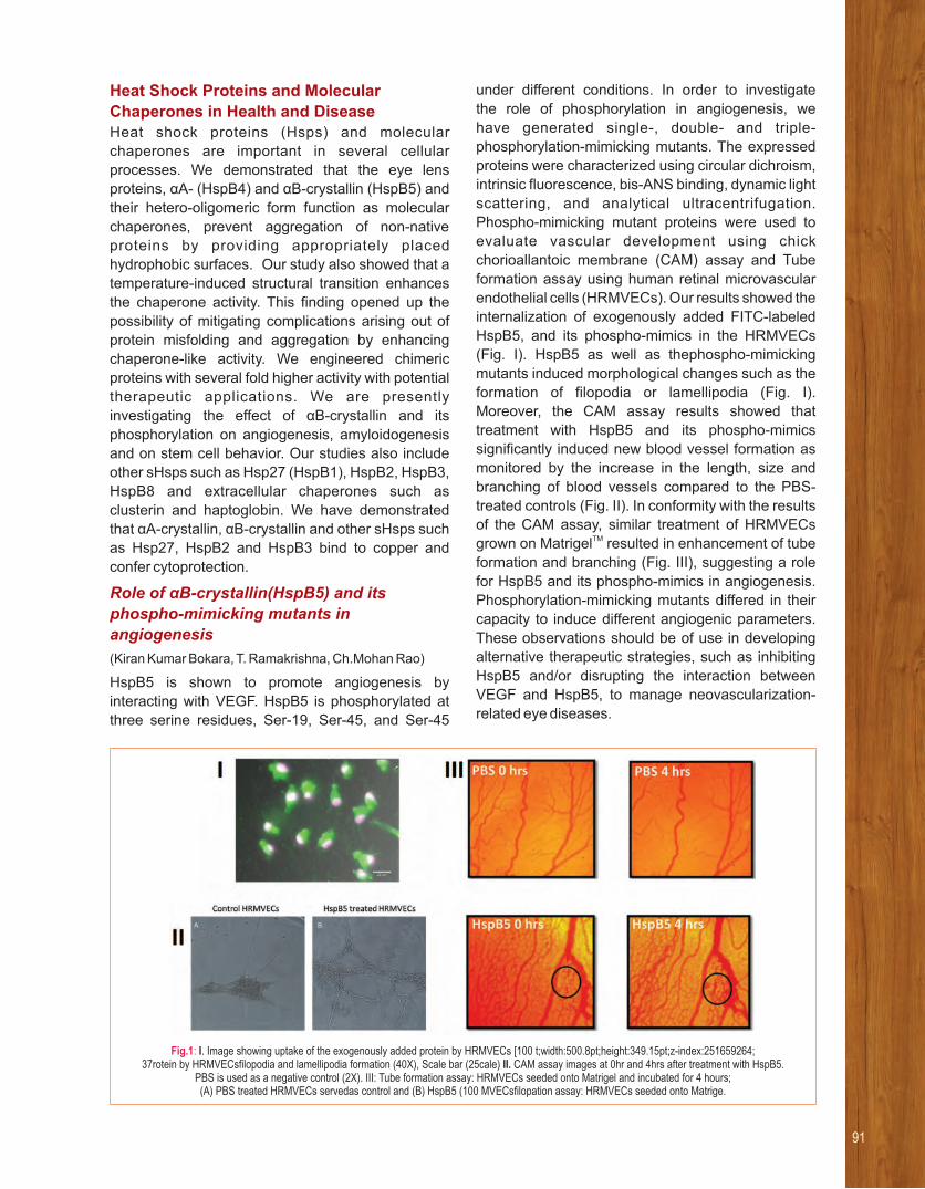
Heat Shock Proteins and Molecular
Chaperones in Health and DiseaseHeat shock proteins (Hsps) and molecular
chaperones are important in several cellular
processes. We demonstrated that the eye lens
proteins, αA- (HspB4) and αB-crystallin (HspB5) and
their hetero-oligomeric form function as molecular
chaperones, prevent aggregation of non-native
proteins by providing appropriately placed
hydrophobic surfaces. Our study also showed that a
temperature-induced structural transition enhances
the chaperone activity. This finding opened up the
possibility of mitigating complications arising out of
protein misfolding and aggregation by enhancing
chaperone-like activity. We engineered chimeric
proteins with several fold higher activity with potential
therapeutic applications. We are presently
investigating the effect of αB-crystallin and its
phosphorylation on angiogenesis, amyloidogenesis
and on stem cell behavior. Our studies also include
other sHsps such as Hsp27 (HspB1), HspB2, HspB3,
HspB8 and extracellular chaperones such as
clusterin and haptoglobin. We have demonstrated
that αA-crystallin, αB-crystallin and other sHsps such
as Hsp27, HspB2 and HspB3 bind to copper and
confer cytoprotection.
Role of αB-crystallin(HspB5) and its
phospho-mimicking mutants in
angiogenesis
(Kiran Kumar Bokara, T. Ramakrishna, Ch.Mohan Rao)
HspB5 is shown to promote angiogenesis by
interacting with VEGF. HspB5 is phosphorylated at
three serine residues, Ser-19, Ser-45, and Ser-45
under different conditions. In order to investigate
the role of phosphorylation in angiogenesis, we
have generated single-, double- and triple-
phosphorylation-mimicking mutants. The expressed
proteins were characterized using circular dichroism,
intrinsic fluorescence, bis-ANS binding, dynamic light
scattering, and analytical ultracentrifugation.
Phospho-mimicking mutant proteins were used to
evaluate vascular development using chick
chorioallantoic membrane (CAM) assay and Tube
formation assay using human retinal microvascular
endothelial cells (HRMVECs). Our results showed the
internalization of exogenously added FITC-labeled
HspB5, and its phospho-mimics in the HRMVECs
(Fig. I). HspB5 as well as thephospho-mimicking
mutants induced morphological changes such as the
formation of filopodia or lamellipodia (Fig. I).
Moreover, the CAM assay results showed that
treatment with HspB5 and its phospho-mimics
significantly induced new blood vessel formation as
monitored by the increase in the length, size and
branching of blood vessels compared to the PBS-
treated controls (Fig. II). In conformity with the results
of the CAM assay, similar treatment of HRMVECs TMgrown on Matrigel resulted in enhancement of tube
formation and branching (Fig. III), suggesting a role
for HspB5 and its phospho-mimics in angiogenesis.
Phosphorylation-mimicking mutants differed in their
capacity to induce different angiogenic parameters.
These observations should be of use in developing
alternative therapeutic strategies, such as inhibiting
HspB5 and/or disrupting the interaction between
VEGF and HspB5, to manage neovascularization-
related eye diseases.
Fig.1: I. Image showing uptake of the exogenously added protein by HRMVECs [100 t;width:500.8pt;height:349.15pt;z-index:251659264; 37rotein by HRMVECsfilopodia and lamellipodia formation (40X), Scale bar (25cale) II. CAM assay images at 0hr and 4hrs after treatment with HspB5.
PBS is used as a negative control (2X). III: Tube formation assay: HRMVECs seeded onto Matrigel and incubated for 4 hours; (A) PBS treated HRMVECs servedas control and (B) HspB5 (100 MVECsfilopation assay: HRMVECs seeded onto Matrige.
91
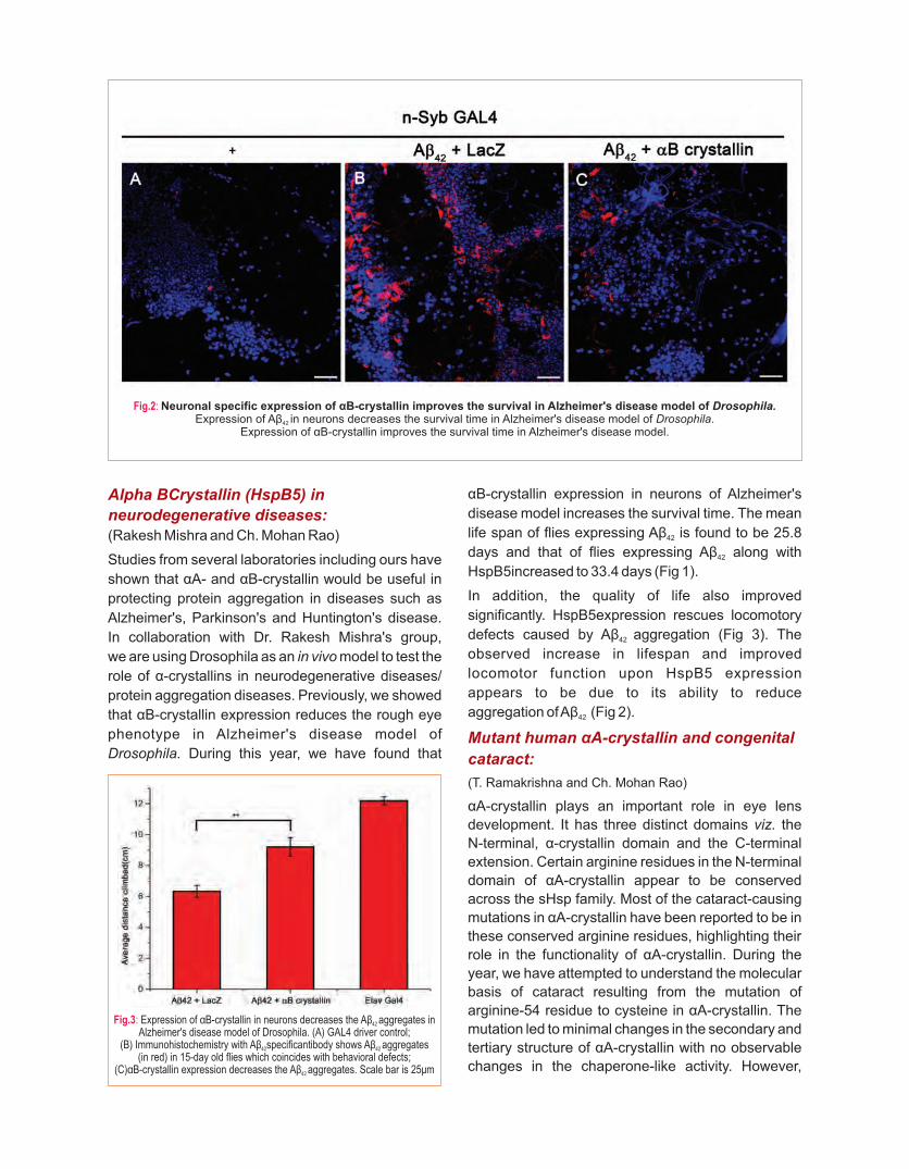
Alpha BCrystallin (HspB5) in
neurodegenerative diseases:(Rakesh Mishra and Ch. Mohan Rao)
Studies from several laboratories including ours have
shown that αA- and αB-crystallin would be useful in
protecting protein aggregation in diseases such as
Alzheimer's, Parkinson's and Huntington's disease.
In collaboration with Dr. Rakesh Mishra's group,
we are using Drosophila as an in vivo model to test the
role of α-crystallins in neurodegenerative diseases/
protein aggregation diseases. Previously, we showed
that αB-crystallin expression reduces the rough eye
phenotype in Alzheimer's disease model of
Drosophila. During this year, we have found that
αB-crystallin expression in neurons of Alzheimer's
disease model increases the survival time. The mean
life span of flies expressing Aβ is found to be 25.8 42
days and that of flies expressing Aβ along with 42
HspB5increased to 33.4 days (Fig 1).
In addition, the quality of life also improved
significantly. HspB5expression rescues locomotory
defects caused by Aβ aggregation (Fig 3). The 42
observed increase in lifespan and improved
locomotor function upon HspB5 expression
appears to be due to its ability to reduce
aggregation of Aβ (Fig 2).42
Mutant human αA-crystallin and congenital
cataract:
(T. Ramakrishna and Ch. Mohan Rao)
αA-crystallin plays an important role in eye lens
development. It has three distinct domains viz. the
N-terminal, α-crystallin domain and the C-terminal
extension. Certain arginine residues in the N-terminal
domain of αA-crystallin appear to be conserved
across the sHsp family. Most of the cataract-causing
mutations in αA-crystallin have been reported to be in
these conserved arginine residues, highlighting their
role in the functionality of αA-crystallin. During the
year, we have attempted to understand the molecular
basis of cataract resulting from the mutation of
arginine-54 residue to cysteine in αA-crystallin. The
mutation led to minimal changes in the secondary and
tertiary structure of αA-crystallin with no observable
changes in the chaperone-like activity. However,
Fig.2: Neuronal specific expression of αB-crystallin improves the survival in Alzheimer's disease model of Drosophila. Expression of Aβ in neurons decreases the survival time in Alzheimer's disease model of Drosophila. 42
Expression of αB-crystallin improves the survival time in Alzheimer's disease model.
Fig.3: Expression of αB-crystallin in neurons decreases the Aβ aggregates in 42
Alzheimer's disease model of Drosophila. (A) GAL4 driver control; (B) Immunohistochemistry with Aβ specificantibody shows Aβ aggregates42 42
(in red) in 15-day old flies which coincides with behavioral defects; (C)αB-crystallin expression decreases the Aβ aggregates. Scale bar is 25μm42

Fig.4: Expression of αB-crystallin in neurons decreases the Aβ aggregates in 42
Alzheimer's disease model of Drosophila. (A) GAL4 driver control; (B) Immunohistochemistry with Aβ specificantibody shows Aβ aggregates42 42
(in red) in 15-day old flies which coincides with behavioral defects; (C)αB-crystallin expression decreases the Aβ aggregates. Scale bar is 25μm42
when expressed in lens epithelial cells, R54C-αA-
crystallin aggregated and translocated to the nuclei of
the cells, causing a stress-like response, and
translocation of αB-crystallin to the nucleus. The
mutation also led to a decrease in its interaction with
HSPB5 and HSP27 and triggered cell death through
apoptosis, as evident by the activation of Caspase 3.
Our results shed light on the molecular basis of
cataract caused by the R54C mutation in αA-crystallin
and highlight the important role of altered inter-protein
interactions in the pathogenesis.
Retinoblastoma:(Suman Thakur and Ch. Mohan Rao)
Retinoblastoma, ocular cancer, is mostly associated
with early childhood. Many cases need enucleation of
the eye. If not treated in time, it is fatal due to
intracranial extension and metastasis. We observed
that a small molecule disrupts clusters of
retinoblastoma cell line Y79. These cells grow in
clusters and disruption of clusters leads to cell death.
By performing proteomics, we identified that a large
number of cell adhesive molecules are involved in the
process. Interestingly, we also observed that
oncogene products are down-regulated and several
anti-proliferative factors are up-regulated upon
treatment with this small molecule. The molecule was
codenamed E4 for its potential patentability.
Cell culture studies showed that E4 is effective in
killing Y79 cells. We extended our studies to in vivo
models. Xenografted nude mice of Y79 cells
developed tumors. Treatment of these tumors
with E4 resulted in dramatic regression of the
tumors. Histopathological evaluation showed that
treatment with E4 did not affect liver, spleen, lung,
kidney and heart.
Encouraged by the results of our xenograft nude
experiments, we planned to test the molecule in a
retinoblastoma genetic knockout mouse model
(Chx10-Cre; RbLox/−; p53Lox/−; p107−/−). Due to
the absence of Rb, p53 along with p107, these
knockout mice develop aggressive retinoblastoma.
During the year, we have obtained knockout animals
and are breeding them at our animal facility. The
increase in the colony size is very slow due to the high
mortality rate of the animals. We have initiated
studies with intra-tumoral injections. We believe
that this non-toxic molecule, with its low
molecular weight and good water solubility
would be a potential therapeutic for retinoblastoma.
Paper-based microfluidic devices:
Early diagnosis significantly reduces the health
care burden and also improves the treatment
outcome. Our country needs affordable, scalable
diagnostics that can be used by unskilled persons
in semi-urban and rural settings; preferably not
requiring the cold chain. Paper microfluidics-based,
point-of-care systems are eminently suitable
for our needs. We have built the capacity to
design and develop such paper microfluidic
systems. We are now developing paper-based
microfluidic devices for blood typing, determination
of Erythrocyte Sedimentation Rate, pregnancy
detection kit for livestock etc. We are also
work ing towards deve lop ing DNA-based
diagnostics for human diseases on a paper-based
system.
Paper-based microfluidic viscometer:(Amit Asthana and Ch. Mohan Rao)
During our experiments with a paper-based ESR
device, we noticed that capillary mobility of liquids in
paper-based device clearly depended on their
viscosity. Based on this observation, we set out to
develop a paper-based viscometer. We successfully
designed a device with required stability, measured
viscosities of a few test fluids and obtained an
excellent correlation coefficient of 0.9997 with
the results obtained using a conventional
Ostwald's viscometer. A paper-based viscometer
offers advantages such as time efficiency, cost-
effectiveness, and low sample volume requirement
over the conventional viscometer. These paper-
based devices can also be used to estimate
molecular weights of polymers with reasonable
accuracy.
93

Surface Enhanced Raman Spectroscopy (SERS) for enhancing the sensitivity of biomarker detection.(Amit Asthana and Ch. Mohan Rao)
Raman spectroscopy, despite the use of LASERS,
is not very sensitive to detect very low concentrations
of biomarkers. SERS is known to provide a
significant enhancement in the signal. Hence,
either a roughened surface or substances with
small radii of curvature are used. We are
investigating several different methods to deposit
metals onto the paper device to use them as
SERS substrates. During the year we successfully
tested our substrates with lysozyme and
Rhodamine 6G.
Development of paper-based pregnancy detection kit for cattle:
(Amit Asthana, G. Umapathy and Ch. Mohan Rao)
Early detection of pregnancy would help shorten the
calving interval and plan for re-breeding. This leads to
maximizing the mi lk product ion, thereby
enhancing the farmer's income (The value of
the dairy industry is Rs. 5 lakh crores in 2015 and
reported to be growing). Dr. Umapathy's group
identified a metabolite that correlates with the
pregnancy status in wild animals. They observed
that the metabolite could also be used for
determining the pregnancy status in cattle.
Based on these observations we set out to
develop a paper-based device for an early
pregnancy test in cattle. During the year, we have
troubleshot several issues with antibody,
stability, different methods of detection etc.
and made a few prototypes with encouraging
results. Presently, we are also developing
another ant ibody and different detect ion
strategies to improve stability and sensitivity.
The device would be usable by untrained
persons with the naked eye and would not
require a cold chain.
95
RESEARCH INTERESTS:
• How do misfolded proteins promote proteotoxicity?
• Why are protein-misfolding diseases prevalent during aging?
Selected recent publications
1. Raychaudhuri S, Banerjee R, Mukhopadhyay S, Bhattacharyya
NP (2014) Conserved C-terminal nascent peptide binding
domain of HYPK facilitates its chaperone like activity. J. Biosci
39:1-14.
2. Raychaudhuri S, Loew C, Koerner R, Pinkert S, Theis M,
Hayer-Hartl M, Buchholz F, Hartl FU (2014) Interplay of
Acetyltransferase EP300 and the Proteasome System
in Regulating Heat Shock Transcription Factor 1. Cell
156(5):975-85.
3. Gupta R, Kasturi P, Bracher A, Loew C, Zheng M, Villella A,
Garza D, Hartl FU, Raychaudhuri S (2011) Firefly luciferase
mutants as sensors of proteome stress. Nat Methods
8(10):879-84.
From left to right : Swathi, Shivali, Anusha, Swasti, Bodhisattwa, Debodyuti, Shemin
Swasti RaychaudhuriProteotoxicity in Age-related Diseases
• Generated multiple cell culture based protein aggregation
models - mild to aggressive
• Developed multiple quantitative mass spectrometry based assays to investigate protein
destabilization events in mass-scale
• Shown proteasome dysfunction directly impedes mitochondrial respiration by perturbing stability
and assembly of Respiratory Complex subunits, and provide an explanation for the association
between these two hallmarks of age-related degenerative diseases.

Inside a cell, numerous proteins interact with each-other to form a 'society'; the so-called 'cellular proteome'. Such 'protein-societies' are responsible for proper functioning of every cell. Any deviation from the functional conformation/concentration of a 'single member-protein' may negatively affect the 'society' and lead to gradual functional impairment. This is called 'proteotoxic-collapse' and is often true for many age-related diseases. We are a group of researchers interested in understanding the coordination of the proteins towards a functional proteome and the defence-mechanisms in the face of proteotoxic events.
Cancer, diabetes, cardio-vascular diseases and several neurodegenerative disorders represent the majority of the protein-misfolding related health-setbacks in the aging population. For example, in each of the neurodegenerative disorders known, a “symptomatic-protein” misfolds and damages the protein homeostasis in the nerve cells in various ways. Formation of toxic aggregates of the islet amyloid polypeptide (IAPP) contributes to β-cell dysfunction and diabetes. Soluble pre-amyloid oligomers are present in the cardiomyocytes of many human heart failure samples. Chronic, uncontrolled protein misfolding and resultant abnormal heat shock protein load is known to foster tumor development. Importantly, loss-of-function of the misfolded proteins is not likely to be the major reason behind proteotoxicity. Rather, several common protein-metabolic pathways (starting from protein synthesis,
folding, transport to degradation) are perturbed in all these diseases. This common pathogenic modality offers unique opportunity to discover novel drug-targets that could be useful in ameliorating multiple proteotoxic conditions; however, systemic investigations are emergent.
In our lab, we are interested in studying the impact of symptomatic misfolding-prone protein on the cellular proteome using different protein-misfolding disease models. The novelty of our approach relies on the identification of exclusive molecular players which could be targeted in multiple diseases. Currently, we are using mutant versions of α-synuclein (responsible for familial Parkinson's disease), Ubiquitin-2 (Amyotrophic Lateral Sclerosis), Huntingtin
(Huntington's disease) and FlucDM-EGFP [1] as
model misfolding-prone proteins. Several cell-lines conditionally expressing these misfolding-prone proteins have been generated. Multiple chemical biology tools and quantitative proteomics protocols to study the proteostasis changes in these cell-lines have been successfully standardized. Pilot experiments to study the impact of protein-misfolding on the soluble and functional proteome, activation of
stress response[2] and protein degradation etc. have
been initiated. These investigations will lead to: a) Understanding the mechanism of proteotoxicity in protein-misfolding diseases; b) The identification of novel drug targets; c) The development of multi-dimensional, high-throughput small-molecule screening platforms for protein-misfolding diseases.
Fig.1: Uptake of FITC-tagged Pre-formed Fibrils (PFF) by HEK293T cells expressing wild type mCherry-tagged α-synuclein& the double mutant (A30P+A53T) and formation of chimeric aggregates. Cells were incubated with FITC tagged PFF for seven days. Fluorescence microscopy images show presence of PFF
inside the cells (green) and aggregation of endogenous synuclein (red). Questions being addressed a) This model opens up the opportunity to study the alteration of “membrane proteome” and follow up cell biology investigations to provide knowledge on SNCA fibril-
entry mechanism into the cells.b) Differences in the amyloid-formation kinetics between the SNCA-variants indicate presence of diverse seeding-intermediates in the cells. During PFF incubation,
the folding-pattern of the overall-proteome is expected to change depending on the seeding-intermediates and with time. We are performing experiments to characterize the differences between the protein-folding patterns in these different stages. The improvised proteomics experiments will provide a mechanistic knowledge ofthe propagation of proteotoxicity during amyloidosis.
Stable cell-line over-expressingdifferent SNCA variants
( )mCherry Tagged
Seven days

Proteotoxicity by α-Synuclein
To understand the mechanism of proteotoxicity
caused by α-Synuclein, we are using α-Synuclein
(SNCA) expressing cell-culture models. Duplications
and triplications of this gene as well as mutations in
the N-terminal region (A53T, A30P, E46K etc.) cause
aggregation of this protein resulting in a range of
synucleinopathies. We have successfully generated
inducible stable-lines in HEK293T cells expressing
wild-type α-synuclein (SNCA-wt), mutants A53T,
A30P and the double mutant (DM; A53T+A30P).
We have also purified recombinant wild-type and
mutant α-synuclein proteins and compared
amyloidosis in vitro (preformed amyloid fibrils (PFF)).
Upon long-term (7 days) incubation with these PFF,
cells expressing EGFP tagged SNCA-wt and A30P
showed diffuse EGFP fluorescence whereas
SNCA-A53T and DM showed EGFP-positive hairy
aggregates. These results suggest that the in vitro
generated PFF probably crossed the plasma
membrane barrier; or caused substantial damages in
the cytoplasmic proteome via altering membrane-
trafficking that resulted in aggregation of EGFP-
tagged endogenous SNCA-A53T and SNCA-DM.
Re-adjustment of the functional proteome in
response to proteasome-block
Proper folding and solubility are two major
determinants of protein functions. A large number of
proteins deviate from their native functions due to
misfolding and insolubil i ty in age-related
neurodegenerative diseases. Simultaneous
proteasome-dysfunction often increases the load of
non-functional ubiquitinated proteins resulting in an
accumulative proteotoxicity. We blocked protein-
degradation in Neuro2A cells with MG132; a specific
cell-permeable proteasome inhibitor. Using a low
dose and early incubation time, we studied the
primary-reorganization events in the soluble
proteome. Proteasome inactivation resulted in loss-
of-solubility of several mitochondrial respirasome
components as measured by SILAC-based
quantitative mass spectrometry. These depleted
proteins were mostly nuclear encoded and rely on
chaperones to safeguard their loosely-folded
conformation during translocation. Functional
impairment of these proteins marks the onset of
mitochondrial dysfunction during proteasome-
inhibition. High chaperone-demand by these proteins
may be responsible for titrating out heat shock
proteins from the dormant chaperone-Hsf1 complex
to trans-activate heat shock response as apparent by
the elevated mRNA levels of hsp70 (hspa1b) and
ubiquitin (ubc). Moreover, we observed increased
level of histone H3 and H4 proteins and their modifiers
(Morf4l2) in the soluble proteome indicating fine-
tuning of chromatin-assembly. Histone-H3 tri-
methylation (H3K4me3) level was also increased
which is a known prerequisite for successful Hsf1-
mediated transcription. Thus, we report the
reorganization events in the soluble proteome of
Neuro2A cells at an early juncture that allows
predicting the long term toxic-consequences of
proteasome-inhibition.
Fig.2: SILAC based mass spectrometry analysis of the MG132 treated soluble proteome of Neuro2A cells - distribution of z-scores. The proteins with (1>z>-1) scores are grouped as Unchanged (inner box). The proteins with (1.96>z>1) or (-1.96<z<-1) are classified as Enriched and Depleted, respectively (outer box). Proteins having (-1.96>z >1.96) are termed as 'highly depleted' and 'highly enriched', respectively (beyond the boxes). Colour code: blue - depleted; red - enriched; white - unchanged proteins. Proteins significantly enriched (filled red circles) or depleted (filled blue circles) in a dose-independent manner are and named. Mitochondrial components are boxed (green). Orange box - respirasome components; brown box -respirasome associated mitoribosomal proteins.References: 1. Gupta, R., et al., Nat Methods, 2011. 8: p. 879-884. 2. Raychaudhuri, S., et al., Cell, 2014. 156(5): p. 975-85.
97

RESEARCH INTERESTS:
• Understanding cell wall biogenesis and its regulation in bacteria
Selected recent publications
1. Parveen S and Reddy M (2017) Identification of YfiH (PgeF) as a
factor contributing to the maintenance of bacterial peptidoglycan
composition. doi: 10.1111/mmi.13730.Molecular Microbiology
2. Singh SK, Parveen S, SaiSree L and Reddy M (2015) Regulated
proteolysis of a cross-linkspecific peptidoglycan hydrolase
contributes to bacterial morphogenesis. Proc. Natl. Acad. Sci.,
USA 112: 10956-10961.
3. Mahalakshmi S, Sunayana MR, SaiSree L and Reddy M
(2014) yciM is an essential gene required for regulation of
lipopolysaccharide synthesis in Escherichia coli. Molecular
Microbiology 91: 145-157.
4. Singh SK, SaiSree L, Amrutha RN and Reddy M. (2012) Three
redundant murein endopeptidases catalyze an essential
cleavage step in peptidoglycan synthesis of Escherichia coli K12.
Molecular Microbiology 86: 1036-1051
Our recent results show that editing or proofreading pathways exist in bacteria to
maintain the composition and integrity of bacterial peptidoglycan
From left to right (Front row): Aisha Hamid*, Shambhavi Garde, Sadiya Parveen, Anju Bala, Moneca Kaul, L Sai Sree(Back row) Raj Bahadur, Manjula Reddy, Pavan Kumar, AnubhavBharadwaj, V Balaji, NilanjanSom, Pratap Acharya* (Dissertation students*)
Manjula ReddyBacterial Cell Wall Synthesis and its Regulation

Fig.1: Schematic of peptidoglycan editing pathway by YfiH (PgeF). Peptidoglycan (PG) precursors are synthesized in the cytosol and are flipped
across the inner membrane (IM) to be incorporated into the existing PG sacculus.YfiH edits the incorrect muropeptides in the periplasmic space (OM-Outer membrane).
Research in my laboratory is focused towards understanding how bacteria elongate, divide and split their cell walls during cell cycle to successfully generate two equal daughter cells. We take a multi-discipl inary approach including genetics, biochemistry, cell biology, and genomics to address these questions using the Gram-negative rod-shaped bacterium Escherichia coli as a primary model system. We expect that our work facilitates a better understanding of fundamental aspects of bacterial cell biology and will also provide novel strategies for development of antimicrobial therapeutics.
Most bacteria are surrounded by a protective mesh-like exoskeleton called peptidoglycan (PG or murein) sacculus to protect them-selves from internal turgor pressure. PG is a single, large, covalently cross-linked macromolecule that forms a net-like structure completely encasing the bacterial cytoplasmic membrane. Hence, growth of a bacterial cell is intimately coupled to expansion of murein sacculus and requires concerted activity of hydrolases that cleave the cross-links for insertion of new material and synthases that catalyze the cross-link formation. Although, conceptualized nearly five decades ago, the mechanism of such essential murein cleavage activity was not known earlier. To examine the significance of cross-link cleavage in PG growth, we identified three previously unknown murein hydrolytic enzymes, two (Spr and YdhO) belonging to the NlpC/P60 peptidase superfamily and the third (YebA) to the lysostaphin family of proteins that cleave peptide cross-bridges between glycan chains. We showed that these endopeptidases are redundantly essential for bacterial growth and viability as a conditional mutant lacking all the three enzymes is unable to incorporate new murein and undergoes rapid lysis upon shift to restrictive conditions. In summary, we demonstrated that the step of cross-link cleavage is essential for bacterial PG growth and identify these endopeptidases to be the long-postulated space-maker hydrolases required for enlargement of the murein sacculus (Singh, SaiSree, Amrutha and Reddy, Molecular Microbiology, 2012). Accordingly, these endopeptidases were renamed MepS (Spr), MepM (YebA) and MepH (YdhO), respectively, in which mep stands for murein endopeptidase.
Although, the cross-link cleavage is fundamental for PG growth, such cleavage needs to be stringently regulated at the spatiotemporal level to avoidlethal breakage and rupture of PG. To understand how this potential lethal hydrolytic activity iscontrolled in the cell, we examined the regulation of the D, D-endopeptidase, MepS. We find that expression of MepS is dependent on bacterial growth cycle; it is highly abundant in exponential phase of growth with the levels falling sharply at the onset of
stationary phase. Using combined genetic, molecular and biochemical approaches, we showed that MepS is rapidly degraded with a half-life of ~2 min by a novel proteolytic system comprising of an outer membrane (OM) lipoprotein of unknown function, NlpI, and a periplasmic protease, Prc. In summary, we showed that NlpI-Prc system modulates PG synthesis by regulating the levels of MepS, a hydrolase that breaks the cross-links for insertion of new murein material during growth of PG sacculus. MepS is the only enzyme of PG metabolism to be regulated during growth cycle signifying the importance of cross-link cleavage suggesting that the step of cross-link cleavage may indeed be the rate-limiting step of PG biosynthesis (Singh, Sadiya, SaiSree and Reddy, Proc. Natl. Acad. Sci. 2015).
Recently, we showed that a conserved open reading frame of unknown function, YfiH (renamed PgeF for Peptidoglycan editing Factor) contributes to the maintenance of peptide composition in E. coli. Using genetic, biochemical and mass spectrometrical analyses, we demonstrated that absence of yfiH results in incorporation of non-canonical amino acids, L-serine or glycine in place of L-alanine in PG sacculi leading to β-lactam sensitivity, lethality in mutants defective in PG remodelling or recycling pathways, altered cell morphology and reduced PG synthesis (Fig. 1). yfiH orthologs from other Gram-positive genera were able to compensate the absence of yfiH in E. coli indicating a conserved pathway in bacterial kingdom. Overall, our results suggest editing/ quality control mechanisms exist to maintain composition and integrity of bacterial peptidoglycan(Parveen and Reddy, Molecular Microbiology, 2017).
99

RESEARCH INTERESTS:
• Non-coding RNAs
• Cardiac hypertrophy and failure
• Cardiac regeneration
Selected recent publications
1. Viereck J, Kumarswamy R..... and Thum T(2016) Long noncoding
RNA Chast promotes cardiac remodeling. Science Translational
Medicine 8(326):326ra22.
2. Kumarswamy R, Volkmann I et al and Thum T (2014) Vascular
importance of the miR-212/132 cluster. European Heart Journal
35(45):3224-31.
3. Kumarswamy R, Bauters C et al and Thum T (2014) Circulating
long non-coding RNA, LIPCAR, predicts survival in patients with
heart failure. 114(10):1569-75.Circulation Research
4. Kumarswamy R, Lyon AR et al and, Thum T (2012) SERCA2a
gene therapy restores microRNA-1 expression in heart failure via
an Akt/FoxO3A-dependent pathway. European Heart Journal.
33(9):1067-75.
5. Kumarswamy R, Volkmann I, Jazbutyte V, Dangwal S, Park DH,
Thum T (2012) Transforming growth factor-β-induced endothelial-
to-mesenchymal transition is partly mediated by microRNA-21.
Arteriosclerosis Thrombosis and Vascular Biology 32(2):361-9The main focus of our newly established lab is to
understand the role of non-coding RNAs in patho-physiology of the heart
From left to right: Abishek Bharadwaj, Manisha Sahu, Regalla Kumaraswamy
Regalla KumaraswamyNon-coding RNAs in Patho-physiology of the Heart

The human genome is tremendously larger than that
of more simple organisms such as worms. However,
genomes of humans and worms have a comparable
number of protein coding genes. For instance, the
genome of the 959cell bodied C. elegans and
100,000 billioncell bodied humans typically contain a
similar number of protein coding genes and yet the
human genome is 30 times bigger than the C. elegans
genome (3000 MB vs 100MB). All protein coding
genes in humans originate from about 2% of the
genome and a majority of the remaining portion of the
genome remains un-transcribed or transcribed as
noncoding RNAs. All the available therapies and
disease diagnostics today are based on this 2% of the
coding genome. Until recently, the noncoding portion
of the genome was believed to be ‘junk’ or dark matter
of the genome and transcribed noncoding RNAs were
described as ‘Transcriptional noise’. However, recent
studies have highlighted the importance of noncoding
RNAs in various physiological and pathological
settings and now they are referred as ‘precise
regulators of gene expression’. MicroRNAs (~21 nt
long) and long noncoding RNAs (≤ 200nt long) are the
two major classes of noncoding RNAs that regulate
gene expression. Because of their gene regulatory
function, noncoding RNAs have been proposed as
attractive targets for development of novel
therapeutic strategies in various disease conditions
including heart failure.
Heart diseases are the leading cause of death
worldwide. In India about 40% of all deaths in urban
areas and 30% in rural areas are attributed to
cardiovascular diseases. Incidence of heart diseases
in India has steadily increased from about 2% (1960)
to 10.5% (2000). Although available therapies
improve symptoms, they are not able to reverse
fibrosis or activate hibernating myocardium. The main
research focus of our newly established lab in CCMB
is to understand the role of non-coding RNAs in
patho-physiology of the heart. Research in my lab is
being funded by the Wellcome Trust/ DBT India
Alliance.
Fig.1: Left ventricular (LV) anterior (AW) and posterior (PW) wall thickness in control and obese mice were imaged by Vevo 2100 small animal ultrasound imaging system. Please note increased wall thickness in obese mice, which indicates left ventricular hypertrophy.
101

RESEARCH INTERESTS:
• Proofreading during translation of the genetic code
• Mycobacterium tuberculosis complex lipid synthesis
• Secretome action of a rice pathogenic bacteria
• Calcium-binding proteins
Selected recent publications
1. Srivastava SS, Jamkhindikar A A, Raman R, Jobby MK,
Chadalawada S, Sankaranarayanan, R, Sharma Y(2017) A
Transition Metal-Binding, Trimeric βγ-Crystallin from Methane-
Producing Thermophilic Archaea, Methano saeta thermophila.
Biochemistry 59:1299-1310.
2. Routh, S B, Pawar K I, Ahmad S, Singh S, Suma K, Kumar M,
Kuncha S K, Yadav K, Kruparani S P and Sankaranarayanan R
(2016) Elongation factor Tu prevents misediting of Gly-tRNA (Gly)
caused by the design behind the chiral proofreading site of
D-aminoacyl-tRNAdeacylase. 14(5) e1002465, 1-22.PLoS Biol.
3. Kinatukara P, Patel KD, Haque AS, Singh R, Gokhale RS and
Sankaranarayananan R (2016) Structural insights into the
regulation of NADPH binding to reductase domains of
nonribosomal peptide synthetases: A concerted loop movement
model. 194:368-74.J Struct Biol.
4. Ahmad, S., Muthukumar, S., Kuncha, S.K., Routh, S.B.,
Yerabham, A.S., Hussain, T., Kamarthapu, V., Kruparani, S.P.,
Sankaranarayanan, R. (2015) Specificity and catalysis
hardwired at the RNA–protein interface in a translational
proofreading enzyme. 6:7552.Nat Commun.
5. Ahmad, S., Routh, S.B., Kamarthapu, V., Chalissery, J.,
Muthukumar, S., Hussain, T., Kruparani, S.P., Deshmukh, M.V.,
Sankaranarayanan, R. (2013) Mechanism of chiral proofreading
during translation of the genetic code. 2:e01519.Elife
(highlighted by Science (343) Editor's Choice, “Chirality check”, pp
119, 10th Jan, 2014).
Our principal focus is to elucidate various proofreading/editing
mechanisms involved in the maintenance of quality control during translation of the genetic
code as well as to understand their physiological significance.
From left to right (1st row): Raghvendra, Aditi, Mazeed, Sankaranarayanan, Santosh, Murali, Rajkanwar, Ayshwarya, Shantiswaroop, Aditya, Jotin(2nd row): Biswajit, Gajanan, Bhavita, Satya Brata, Priyadarshan, Priyanka, Rukmini, Lalitha, Sudipta, Mallesh, Aravind
(3rd row) : Subhash, Herojeet, Mantu Kumar, Suma, Sambavi, Shobha
Rajan SankaranarayananStructural Biology

We mainly use X-ray crystallography along with
biochemical and biophysical approaches to obtain
atomic-level structural details of various proteins,
which in turn help in gaining mechanistic insights into
their functioning. We also employ various model
organisms to establish the physiological relevance of
some of these processes. A major focus of the lab is to
study various factors involved in proofreading during
translation of the genetic code. D-amino acids are
occasionally attached to tRNAs by mistake. An
enzyme named D-aminoacyl-tRNAdeacylase (DTD)
hydrolyzes the ester bond between a D-amino acid
and a tRNA, thereby helping to prevent infiltration of
D-amino acids into the translational machinery. We
have shown that DTD employs a Gly-cisPro dipeptide
motif in the active site to discriminate between L- and
D-amino acids through strict L-chiral rejection rather
than D-chiral selection. Consequently, DTD acts on
both D-aminoacyl-tRNAs and the achiral substrate Gly GlyGly-tRNA . This creates an unwanted Gly-tRNA
“misediting paradox” which must be overcome
because glycine is an integral component of proteins.
The translational elongation factor thermo
unstable(EF-Tu), which delivers aminoacyl-tRNAs to
ribosome during protein synthesis, resolves the Glyparadox by conferring protection on Gly-tRNA
against DTD. However, DTD levels in cell must be
kept low and tightly regulated because over
expression of DTD causes cellular toxicity in GlyEscherichia coli due to depletion of Gly-tRNA . We
are currently probing DTD's role in eliminating the Alanon-cognate (incorrect) Gly-tRNA species
generated by alanyl-tRNAsynthetase (AlaRS). We
are also generating DTD knockout in the fruit fly
(Drosophila melanogaster), zebrafish (Danio rerio)
and mouse (Mus musculus) to decipher the
physiological significance of DTD and its role in higher
systems. We are also characterizing an Animalia-
specific DTDhomolog (ATD) and studying various
aspects of other DTDs, namely DTD2 (present in
archaea and plants) and DTD3 (found in
cyanobacteria).
Another major project is on understanding the
structure–function relationship of enzymes involved
in the metabolism of complex lipids in Mycobacterium
tuberculosis(Mtb) in collaboration with the group of
Dr. Rajesh Gokhale, IGIB, New Delhi. Mtb lipids play a
major role in its pathogenesis and are synthesized by
polyketide synthases (PKS) and nonribosomal
peptide synthetases (NRPS). Previously, our
biochemical studies have revealed that Mtb uniquely
employ reductases domains (R-domains) capable of
Fig.1: The Gly-tRNAGly “misediting paradox” and its resolution by EF-Tu. Model depicting that DTD does not act on L-aminoacyl-tRNAs, but does so efficiently on D-aminoacyl-tRNAs, irrespective of the presenceor absence of EF-Tu. In the case of Gly-tRNAGly, DTD acts on the achiral substrate, thus generating Gly-tRNAGly “misediting paradox”. Cellular concentrations of EF-Tu are sufficient to confer protection on achiral substrate, thereby resolving the paradox. Nevertheless, the levels of DTD in the cell are tightly regulated as higher amounts of DTD relieve this protection, causing misediting of Gly-tRNAGly and cellular toxicity.
103

Fig.2: (a) Structural comparison between the NADPH-bound RMyx and the two crystal forms of RNRP (o-RNRP and t-RNRP) showing differences in the conformations of gating loop (GL) and catalytic loop (CL) with the bound NADPH shown as sticks. (b) The GL (orange) shown away from NADPH-
binding site (shown as surface in blue) in NADPH-bound form. (c) The GL (orange) shown to obstruct NADPH-binding site in NADPH-free form.
a reductive release of the biosynthetic products using
the cofactor NADPH. The first crystal structure of R-
domainsfrom our laboratory revealed an additional C-
terminal domain to the common Short-chain
dehydrogenase/reductase fold, responsible for
providing a hydrophobic platform for the final lipid
product in Mtb. Our earlier Small-angle X-ray
scattering (SAXS) studies revealed the process of
binding of NADPH reorientation at the unique C-
terminal domain. Structural comparison of two
different crystal forms of apo R-domains showed
significant differences in the C-terminal domain
orientation and two important loops of the N-terminal
domain called the gating loop (GL) and catalytic loop
(CL). The GL in the two apo-forms exclude NADPH
binding (Figure-2) through an intricate network of
interactions, which progressively perished as they
approached an NADPH-binding state. These
changes were also accompanied by a change in
orientation of linkers that connect the R-domains with
carrier domains that carry the final product to its active
site for product release. These observations allowed
the proposition of a “Concerted loop movement”
model in the present work. According to this model, R-
domains only allow a fully mature product to be
reductively released because conformations with no
interactions prevent NADPH binding (Figure-3).
Thus, the model provides yet another example of
coordinated cross-talk between domains and
modules of NRPS.
In collaboration with Dr. Ramesh Sonti's group we are
trying to understand the structure-function
relationship of virulence factors of Xantho-
monasoryzaepv.oryzae(Xoo), a plant pathogen that
causes bacterial blight of rice. The pathogen secretes
a battery of plant cell wall-degrading enzymes by
Type II secretion system. Our laboratory has solved
the structure of LipA and CbsA (N-terminal catalytic
domain). LipA protein belongs to a new class of cell
wall-degrading enzymes which has a unique mode of
substrate recognition.CbsA (catalytic domain),
belongs to the glycosyl hydrolase 6 (GH-6) family. stConversion of its aspartic acid at 131 position to
alanine confers endoglucanase activity to the mutant
protein. This altered enzymatic activity of D131A
CbsA protein affects the virulence promoting activity
of CbsA but does not affect the ability of the protein to
induce rice innate immune responses. The C-terminal
region of CbsA has a fibronectin type 3 (FN3) domain.
A deletion mutant of FN3 domain was found to be
deficient in virulence. Western blot analysis indicate
that this deficiency is due to less secretion of the
CbsA protein in ∆FN3 mutant when compared
to the wild type. Currently we are trying to understand
if FN3 domain has any role in the transport of CbsA
protein. Interestingly, in the presence of FN domain
the catalytic domain is biochemically inactive.
Using X-ray crystallography and SAXS, we are trying
to understand how the presence of FN3 domain
affects the biochemical activity of catalytic
domain of CbsA.

In collaboration with Dr.Yogendra Sharma's
group, we have established the double clamp2+motif (N/D-N/D-X -X -T/S-S) for binding Ca in the1 2
βγ-Crystallin superfamily of proteins. Recently
we reported a member of βγ-Crystallin superfamily,
methallin which binds to transition metal ions
with nano-molar affinity. Methallin is a protein
that belongs to Methanosaeta-thermophila, an
archea where members of βγ-Crysta l l in
superfamily are sparsely present with only three
reported in archeal whole genome sequences.
Methallin forms a trimer, with a single transition metal
ion at the trimeric axis which is chelated by two
histidine residues coming from each protomer
(Figure-4). We are further exploring βγ-Crystallins
that bind transition metals.
Fig.4: Crystal structure of βγ-crystallin “Methallin” from methanoarchaea M. thermophila. (A) Cartoon representation of crystal structure of Methallin shows transition metal bound at the trimeric axis chelated by three histidine residues from each protomer. (B) A protomer of Methallin is comprised of
two Greek key motifs (purple and red) with the β-strands forming a typical βγ-crystallin domain. Two histidine residues (His14 and His16),which coordinate the metal ion, are located on the second β-strand.
Fig.3: The concerted loop model depicted as a triangle with the base representing NADPH-free form with highest number of interactions between GL and CL depicted as cartoon in the section coloured light red. Sequential loss of some of these interactions to another NADPH-free form depicted as
cartoon in the section coloured orange while the interaction-free GL and CL are depicted as cartoon in the section coloured blue.
105

RESEARCH INTERESTS: 2+• βγ-Crystallin superfold and Ca -binding
2+• Bacterial immunoglobulin-like fold and Ca binding
• Neuronal calcium sensors and pathophysiology
Selected recent publications
1. Kiran U, Regur P, Kreutz M, Sharma Y and Chakraborty A (2017) 2+Intermotif communication induces hierarchical Ca filling of
Caldendrin. 56:2467-2476.Biochemistry
2. Khandelwal R, Sharma A K, Chadalawada S and Sharma Y (2017) 2+Secretagogin is a redox-responsive Ca sensor. Biochemistry
56:411-420.
3. Srivastava SS, Jamkhindikar A, Raman R, Jobby M K,
Chadalawada S, Sankaranarayanan R and Sharma Y (2017) A
transition metal–binding, trimeric βγ-crystallin from methane
producing thermophilic archaea Methanosaetathermophila.
Biochemistry 56:1299-1310 (featured on journal cover page).
4. Rajanikanth V, Sharma AK, Rajyalakshmi M, Chandra K, Chary KV
and Sharma Y (2015) Liaison between Myristoylation and Cryptic 2+EF-Hand Motif Confers Ca Sensitivity to Neuronal Calcium
Sensor-1. 54(4):1111-1122.Biochemistry2+5. Srivastav SS, Mishra A, Krishnan B and Sharma Y (2014) Ca -
binding motif of βγ-crystallins. 289(16):10958-J. Biol. Chem.
10966. (Minireview).
Our group 2+explores the properties of Ca -binding proteins
from all domains of life. In bacterial systems, we were instrumental in the discovery and
2+establishment of a novel Ca -binding protein superfamily, i.e., the βγ-crystallins. Currently, we
are deciphering the anti-diabetic efficacy of a2+β-cell enriched Ca sensor protein, Secretagogin.
From left to right (top): Yogendra Sharma, Syed Sayeed Abdul, P Sai Uday Kiran, Radhika Khandelwal, R Phanindranath, Aditya A Jamkhindikar, Asmita Dhansing Pawar, Anand Kumar Sharma. (bottom): Bal Krishnan Mishra, Venu Sankeshi, Shanti Swaroop Srivastava, Rashmi Thapa, Amrutha HC
Yogendra SharmaCalcium Signaling via Calcium-binding Proteins

2+Among the various types of Ca -binding proteins, the
best-studied have an EF-hand motif to coordinate the 2+cation. However, there are other motifs for Ca
binding, present in a wide variety of proteins. One
such motif that we have identified is present in
proteins belonging to the βγ-crystallin superfamily.
We have classified βγ-crystallins as a separate class 2+of Ca -binding proteins, which constitute an
expanding structural superfamily containing diverse
members from various organisms. Despite being
prevalent, in archaea, βγ-crystallins are selectively
present only in three methanogens, i .e.,
Methanosarcina and Methanosaeta. M-Crystallin
from Methanosarcina acetivorans has been reported 2+by us as a Ca -binding βγ-crystallin. 'Methallin' from
Methanosaeta thermophila, the largest producer of 2+methane, does not have a Ca -binding motif. In
collaboration with Dr R Sankaranarayanan's group, 2+we have demonstrated that instead of Ca , Methallin
binds a transition metal ion and forms a unique,
ligand-dependent trimer. Engineering similar site in
another, homologous βγ-crystallin led us to conclude
that Methallin is a naturally designed trimer for high-
affinity transition metal binding. While many βγ-2+crystallins are shown to bind Ca , and form
homodimers and oligomers, a transition metal-
binding, trimeric βγ-crystallin is a new paradigm.
In another program under the broad interest of 2+regulation of Ca functions in eukaryotic systems, we
have been studying the functions of selective calcium
sensing proteins,namely neuronal calcium sensor-1
(NCS-1), caldendrin, and secretagogin (SCGN). The
2+transition of a calcium sensor from the apo (Ca free) 2+to the holo (Ca saturated) state is a crucial event in
calcium signalling which can be performed by
sequential or simultaneous binding. The structural 2+underpinnings for simultaneous vs. sequential Ca
filling are not yet known, but since many Calcium-
binding proteins (CaBPs) supposedly follow a 2+sequential mode of Ca filling, it is likely that a CaBP
2+will function in vivo at Ca levels insufficient to
saturate all binding sites. This is exactly what we have
described recently, that this transition may be indeed
routed via stable transitory conformations and local
reversible structural fluctuations in EF-hand motifs 2+during hierarchical filling of Ca in Caldendrin.
On our project of understanding the functions of
Secretagogin (SCGN), which is a β-cell enriched, 2+moderate affinity Ca sensor; we have been exploring
the inherent biochemical properties of SCGN and its
implication in physiology. We are also interested in
studying how pathological (or of unknown
consequences) point mutations and SNPs affect
SCGN function. SCGN has also been implicated in
the release of two critical hormones: insulin and
corticotropin-releasing hormone. We have shown
that SCGN displays redox sensitive oligomerization 2+and a higher affinity for Ca in a reducing milieu and
gains stabilization. The redox-responsive nature of
SCGN was supported by its response to the DTT-
induced stress in the MIN6 cells. Our report of redox
sensitive dimerization finds significance in the recent
report implicating SCGN dimerization in insulin
secretion.
107

RESEARCH INTERESTS:
• Plant Meiosis and Gametogenesis
• Apomixis
• Seed Development
• Epigenetics
• Plant Functional Genomics
Selected recent publications
1. Andreuzza S, Nishal B, Singh A and I Siddiqi (2015). The
Chromatin Protein DUET/MMD1 Controls Expression of the
Meiotic Gene TDM1 During Male Meiosis in Arabidopsis. PLoS
Genetics 11(9):e1005396. doi: 10.1371/journal.pgen.1005396.
2. Qin Y, Zhao L, Skaggs M, Andreuzza S, Tsukamoto T, Panoli A,
Wallace KN, Smith S, Siddiqi I, Yang Z, Yadegari R, and Palanivelu
R (2014) ACTIN-RELATED PROTEIN6 regulates female meiosis
by modulating meiotic gene expression in Arabidopsis. Plant Cell
26: 1612-1628.
3. Marimuthu MPA, Jolivet S, Ravi M, Pereira L, Davda, JN, Cromer
L, Wang L, Nogue F, Chan SWL, Siddiqi I and R Mercier(2011).
Synthetic clonal reproduction through seed. 331:876. Science,
Analysis of meiosis and gametogenesis in plants offers
opportunities for addressing fundamental questions relating to cell specification and in
applying this information towards increasing food production through improved methods for plant
breeding.
From left to right (back): S Prashanthi, Aparna Singh, Imran Siddiqi, V Subbiah, V Vijaybhaskar, Saurabh Pandey, Jayesh N Davda, VP Saradhi Attuluri. (front): Dipesh K Singh, Aswan Nalli, Kaladhar B, Avinash, Frank Keith Max
Imran SiddiqiPlant Reproductive Biology
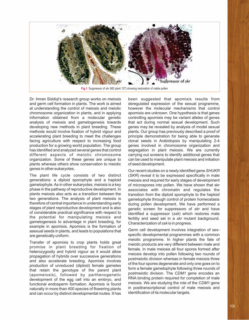
Dr. Imran Siddiqi's research group works on meiosis and germ cell formation in plants. The work is aimed at understanding the control of meiosis and meiotic chromosome organization in plants, and in applying information obtained from a molecular genetic analysis of meiosis and gametogenesis towards developing new methods in plant breeding. These methods would involve fixation of hybrid vigour and accelerating plant breeding to meet the challenges facing agriculture with respect to increasing food production for a growing world population. The group has identified and analyzed several genes that control different aspects of meiotic chromosome organization. Some of these genes are unique to plants whereas others show conservation to meiotic genes in other eukaryotes.
The plant life cycle consists of two distinct generations: a diploid sporophyte and a haploid gametophyte. As in other eukaryotes, meiosis is a key phase in the pathway of reproductive development. In plants meiosis also acts as a transition between the two generations. The analysis of plant meiosis is therefore of central importance in understanding early stages of plant reproductive development and is also of considerable practical significance with respect to the potential for manipulating meiosis and gametogenesis to advantage in plant breeding, for example in apomixis. Apomixis is the formation of asexual seeds in plants, and leads to populations that are genetically uniform.
Transfer of apomixis to crop plants holds great promise in plant breeding for fixation of heterozygosity and hybrid vigour as it would allow propagation of hybrids over successive generations and also accelerate breeding. Apomixis involves production of unreduced (diploid) female gametes that retain the genotype of the parent plant (apomeiosis), followed by parthenogenetic development of the egg cell into an embryo, and functional endosperm formation. Apomixis is found naturally in more than 400 species of flowering plants and can occur by distinct developmental routes. It has
been suggested that apomixis results from deregulated expression of the sexual programme, however the molecular mechanisms that control apomixis are unknown. One hypothesis is that genes controlling apomixis may be variant alleles of genes that act during normal sexual development. Such genes may be revealed by analysis of model sexual plants. Our group has previously described a proof of principle demonstration for being able to generate clonal seeds in Arabidopsis by manipulating 2-4 genes involved in chromosome organization and segregation in plant meiosis. We are currently carrying out screens to identify additional genes that can be used to manipulate plant meiosis and initiation of seed development.
Our recent studies on a newly identified gene SHUKR (SKR) reveal it to be expressed specifically in male meiosis and required for early stages of development of microspores into pollen. We have shown that skr associates with chromatin and regulates the transition from the diploid sporophyte to the haploid gametophyte through control of protein homeostasis during pollen development. We have performed a genetic screen for suppressors of skr and have identified a suppressor (ssk) which restores male fertility and seed set in a skr mutant background. Characterization of ssk is in progress.
Germ cell development involves integration of sex-specific developmental programmes with a common meiotic programme. In higher plants the fate of meiotic products are very different between male and female. In male meiosis all four spores formed after meiosis develop into pollen following two rounds of postmeiotic division whereas in female meiosis three of the four spores degenerate and only one goes on to form a female gametophyte following three rounds of postmeiotic division. The CDM1 gene encodes an RNA binding protein required for completion of male meiosis. We are studying the role of the CDM1 gene in posttranscriptional control of male meiosis and identification of its molecular targets.
Fig.1: Suppressor of skr (M2 plant 127) showing restoration of viable pollen
109

RESEARCH INTERESTS:
• Ubiquitin proteasome system
• Autophagy
• Lysosomal Proteases
• Antimalarial targets
• Malaria vaccine development
Selected recent publications
1. Ashok Yadav P, Pavan Kumar C, Siva B, Suresh Babu K, Allanki
AD, Sijwali PS, Jain N, Veerabhadra Rao A. (2017) Synthesis and
evaluation of anti-plasmodial and cytotoxic activities of epoxy-
azadiradione derivatives. 134:242-257.Eur J Med Chem.
2. Al-Nihmi FM, Kolli SK, Reddy SR, Mastan BS, Togiri J, Maruthi M,
Gupta R, Sijwali PS, Mishra S and Kumar KA (2017) A Novel
and conserved Plasmodium sporozoite membrane protein
SPELD is required for maturation of exo-erythrocytic forms. Sci
Rep. 7: 40407.
3. Klionsky et al. (2016) Guidelines for the use and interpretation of
assays for monitoring autophagy (3rd edition). 12(1):1-Autophagy
222. (REVIEW).
4. Balabadra S, Kotni M, Manga V, Allanki AD, Prasad R and Sijwali
PS (2016) Synthesis and evaluation of naphthyl bearing 1,2,3-
triazole analogs as antiplasmodial agents, cytotoxicity and
docking studies. 2017;25(1):221-232.BioOrg Med Chem.Our group has
showed that Malaria parasiteshave a limited autophagy repertoire
with atypical features and indispensable role
From left to right: Nandita T, S Thanumalayan, Manish Bhattacharjee, Deepak Kumar, Neeradi Dinesh, Zeba Rizvi, Puran Singh Sijwali, Navin Adhikari, Amit Nagwani, Divya Das and Renu Sudhakar
Puran Singh SijwaliRoles of the Ubiquitin Proteasome System and Autophagy in Malaria Parasite Biology and Pathogenesis

We are studying the proteolytic systems of malaria
pathogen Plasmodium to determine their roles in
parasite biology and disease pathogenesis. Multi-
stage development of malaria parasites in different
intracellular and extracellular environments has been
a major challenge to design/develop therapies for
disease control/elimination. A major research interest
of the lab is to identify proteases that have essential
functions during parasite development and are
parasite-specific or significantly different from the
host homologs, so that they can be targeted for
development of new drugs to control malaria. We are
focusing on autophagy and the ubiquitin proteasome
system (UPS), which have been shown to have
crucial roles both in cellular homeostasis and
regulatory processes. Inhibitors of these two
machineries have been shown to kill malaria
parasites, supporting their essential roles in parasite
development and potential as drug targets.
Autophagy is a lysosome-dependent process that
delivers a variety of cellular contents, including
organelles, to the lysosome, for degradation.
However, autophagy is also involved in selective
transport of proteins and lipid catabolism. Since
malaria parasites acquire and degrade several self
and host cellular contents during their multi-stage
development, we are characterizing various
autophagy proteins to determine their functions
during parasite development. The work from our and
other laboratories revealed a limited autophagy
repertoire in malaria parasites, which comprises only
15 proteins as opposed to ~40 proteins in mammalian
cells and yeast. It includes the canonical autophagy
marker Atg8 and the enzymes required for its
conjugation to the membranes. Immunolocalization
studies showed that Atg8 is expressed and
associated with punctate structures in all the major
stages. It is exclusively found in the pellet fraction as
an integral membrane protein, which is in contrast to
the yeast or mammalian Atg8 that is distributed in both
cytosolic and membrane fractions, suggesting that a
constitutive autophagy is operative in malaria
parasites. Starvation is a typical autophagy inducer
that enables cells to survive the starvation phase by
recycling dispensable cellular contents, which
accompanies with increased Atg8 expression level
and the number of Atg8-associated puncta. However,
starvation decreased PfAtg8 level. Neither the Atg8-
associated puncta nor the Atg8 expression level was
noticeably altered upon treatment of parasites with
routinely used autophagy inhibitors. Prolonged
inhibition of the major food vacuole protease activity
did not cause accumulation of the Atg8-associated
puncta in the food vacuole, suggesting that
autophagy is primarily not meant for degradative
function in malaria parasites.
To investigate how Atg8-associated structures are
formed, we are studying Atg18 that has been shown
Fig.1: P. falciparum Atg18 is associated
with the food vacuole. P. falciparum was
transfected with plasmids coding for
PfAtg18 (GFP-PfAtg18 or its mutant
(mPIP)) or yeast Atg18 (ScAtg18).
Parasites expressing GFP-Atg18 were
treated with the PI3K inhibitor LY294002
for its effect on Atg18 localization. Live
trophozoite stage parasites were
evaluated for localization of Atg18 using
a fluorescence microscope. The images
show signal for GFP-Atg18, nucleic acid
staining (Hoechst), boundaries of the
parasite and infected erythrocyte
(Bright), and overlap of all images
(Merged). Note that the PfAtg18 signal
colocalizes with the dark pigment, which
is haemozoin in the food vacuole; this
localization is lost upon mutation of the
PIP motif or treatment with LY294002.
111

to have a role in nucleation and assembly of the
phagophore, a cup-like Atg-8 labelled structure.
Immunolocalization experiments showed that Atg18
is expressed in all erythrocytic stages of P. falciparum,
with exclusive localization to the food vacuole, the
parasite lysosome wherein bulk degradation of
haemoglobin occurs (Figure 1). Mutations in a
putative phosphatidylinositol-binding motif of
PfAtg18 completely altered its localization from the
food vacuole to cytosol (Figure 1). Similar effect was
also observed upon treatment with the PIP3K inhibitor
LY294002 (Figure 1), indicating that binding with the
phosphatidylinositol is essential for Atg18 localization
to the food vacuole. However, yeast Atg18, which
shares the same motif and has been shown to bind to
phosphatidylinositol, did not localize to the food
vacuole (Figure 1), suggesting that the presence of
phosphatidylinositol-binding motif alone is not
sufficient for recruitment to the food vacuole. Several
antimalarials and inhibitors were assessed for their
effects on Atg18. Interestingly, some of the
antimalarials showed a noticeable effect on Atg18
localization and expression, suggesting that Atg18 is
another target by which these antimalarials kill
malaria parasites.
Phagophore is expanded into the autophagosome by
Atg8 and Atg12-Atg5-Atg16 conjugation systems.
The Atg12-Atg5-Atg16 conjugation system appears
to be incomplete in malaria parasites, as Atg10 and
Atg16 homologs are absent. Nonetheless, results
from the gene knockout/knock-down experiments
indicate that Atg12-Atg5 conjugation system is
essential for parasite development. This raises
interesting questions about the role and mechanisms
by which the Plasmodium Atg12-Atg5 conjugation
system works. Collectively, our results reveal several
atypical features of the autophagy system in malaria
parasites, and also demonstrate its indispensability
for parasite development. Studies are underway to
dissect the autophagy repertoire and determine its
roles in parasite development.

113
Our lab is working on Adult stem cells and tissue
engineering. Stem cells obtained from medical waste like placenta, Wharton’s jelly, cord blood,
bone marrow and adipose lipo-aspirate are expanded and differentiated into different
lineages. Tissue engineering involves creation of organoids or tissue constructs with cells,
associated matrices and few factors in order to produce a functional structure. For regenerative
medicine, stem cells can also be injected directly at the site of injury or intravenously for injuries
like spinal cord, bone repair, cardiac, etc., and the entire tissue can be reconstructed by tissue
engineering such as cornea, liver, pancreas.
RESEARCH INTERESTS:
• Use of adult stem cells for lineage derivation by alteration of niche, small molecules etc. Tissue engineering using modified niche, natural scaffolds etc.
Selected recent publications
1. Begum G, Reddy T N, Kumar K P, Dhevendar K, Shashi Singh,
Amarnath M, Misra S, Rangari VK and Rohit K R (2016) An in
situ Strategy to Encapsulate Antibiotics in a Bio-inspired
CaCO3 Structure Enabling pH-Sensitive Drug Release Apt for
Therapeutic and Imaging , 8 ACS Appl. Mater. Interfaces
(34):22056–22063 doi: 10.1021/acsami.6b07177.
2. Bondarenko O M, Heinlaan M, Sihtmäe M, Ivask A, Kurvet I,
Joonas E, Jemec A, Mannerström M, Heinonen T, Rekulapelly
R, Singh S, Zou J, Pyykkö I, Drobne D, Kahru A. (2016)
Multilaboratory evaluation of 15 bioassays for (eco) toxicity
screening and hazard ranking of engineered nanomaterials:
FP7 project NANOVALID. 10(9):1229-42. doi: Nanotoxicology
10.1080/17435390.2016.1196251. Epub 2016 Jun 28.
3. Kos M, Kahru A, Drobne D, Shashi Singh, Kalčíková G, Kühnel
D, Rohit R, Gotvajn AZ, and Jemec A (2016) A case study to
optimise and validate the brine shrimp Artemiafranciscana
immobilisation assay with silver nanoparticles: The role of
harmonization. 213: 173-183.Envir. Pollution
4. Jemec A, Kahru A, Potthof A, Drobne D, Heinlaan M, Böhme S,
Geppert M, Novak S, Schirmer k, Rohit R, Shashi Singh, Aruoja
V, Sihtmäe M, Juganson K, AKäkinen, and Kühnel D. (2016) An
interlaboratory comparison of nano silver characterisation and
hazard identification: Harmonising techniques for high quality
data 87 20–32.Environment International :
5. Snehal R and Shashi Singh (2016) Calcium-phosphate
nanobioceramics. 5: 389-396.Lett in Applied Nano BioScience
Shashi Singh
Shashi SinghAdult Stem Cells from Various Sources and Tissue Engineering

Differentiation of MSC and their compatibility with ScaffoldsWe are using some of the matrix proteins to create matrices for cell growth and differentiation. Collagen Type I extracted from rat tails is being used along with fibronectin, laminin and proteoglycans or plant based glycan rich exudates (gums) for making electro-spun fibers that are collected on coverslips. Various cells like Mesenchymal Stem Cells (MSC) or differentiated cells are plated and their growth pattern is followed over a period. The cells are finally checked for their differentiation state.
3D scaffolds of matrix proteins with plant based gums in form of hydrogels, have been created for matrix driven differentiation of MSC. A series of constructs with different gums and protein ratios are screened for various cell types. Our screen reveals differential differentiation of cells in various compositions.
Natural scaffolds: We have successfully obtained organ scaffold by decellularizing for Pancreas and Kidney. The pancreatic scaffold was recellularized using mesenchymal stem cells. Efficacy of the recellularized structures was tested by implanting in streptozotocin induced diabetic mice. The mice started showing gluco-regulation by day 25 and the structure of the implanted organ resembles a normal pancreas with acini and fully developed islets. The immunolocalization of exocrine and endocrine markers showed the positive reaction in respective segments. The vascularization of the grafted pancreas also appeared normal. The empty scaffold without recellularization always resulted in graft rejection. The recellularization was done using murine cells or Human MSC and both the structures
were well tolerated by the mice up to 40 days. Analysis of mouse serum of implanted vs. sham operated mice did not show any difference in cytokines up to 30 days. The development pathways involved in the course of transition of MSCs into the pancreatic cell types is being worked out in this process.
Differentiation/Dedifferentiation using small moleculesWe are interested in screening of small molecules for achieving reprogramming of cells. The idea is to look for molecules targeting signaling pathways leading to an altered state of cells. We are aiming for both dedifferentiation of cell into iPS and differentiation into beta cells. We ran parallel screens for Oct 4 and Nanog, and also other signaling pathways reporters, Wnt, Notch and the Hedgehog pathway. We have screened about 135 molecules and have shortlisted 10 for final screening after validation. Since these molecules were inducing Oct4 and Nanog, our next screen was to use them with the chemi-inducers for iPS and we shortlisted two molecules. We are further working on these molecules to induce human cells into iPS.
In another screen we are targeting antidiabetic molecules. We have designed about 7 tests for antidiabetic properties include effects for type I and type II diabetes. In this category we have about 67 molecules that were screened for the 7 tests. These tests will also pick up molecules that can be used for induction of cells to beta cell types and among the set of 67 molecules we have shortlisted 3 for direct reprogramming, these molecules are being further tested for validation.
Fig.1: Natural scaffolds
obtained by decellularization
of Pancreas repopulated by
MSC obtained from mice (A),
Human MSC (B) and stained
with markers for islets and
Acinar cells show re-
generation of the organ. Islets
cells stained for beta cell
using insulin antibody (green)
and Delta cells using
somatostatin antibody (Red)
in Fig C; Fig D shows beta
cells stained for neurogenin
3 (green) and Acinar cell
stained red for
Carboxypeptidase.

115
My research focuses on understanding molecular regulation
and control of ovarian function, and to develop assisted reproductive
technologies to support conservation and planned breeding
of endangered wildlife.
RESEARCH INTERESTS:
• Assisted Reproductive Technologies
• Molecular regulation of ovarian function
• Effect of Inbreeding on reproductive function
Selected recent publications
1. Sontakke SD, Umapathy G and Shivaji S (2009) Yohimbine
antagonizes the anesthetic effects of ketamine-xylazinein
captive Indian wild felids (lions, tigers and leopards) Veterinary
Anesthesia and Analgesia 36 :34- 41.
2. Donadeu FX, Sontakke SD and Jason I (2016) MicroRNA
indicators of follicular steroidogenesis Reproduction, Fertility
and Development 29(5) 906-912.
3. Bushra T M, Sontakke SD, Ioannidis J, Duncan WC and
Donadeu FX (2017) The adequate corpus luteum: miR-96
promotes luteal cell survival and progesterone production.
Journal of Clinical Endocrinology and Metabolism doi:
10.1210/jc.2017-0025.
4. Sontakke SD, and Umapathy G (2017) A manual on Chemical
immobilization of wild animals. CSIR-CCMB and Central Zoo
Authority, New Delhi publication (in press).
Sadanand D Sontakke
Sadanand D SontakkeReproductive Biotechnologies for Wildlife Conservation

Studies on semen preservation of
endangered Indian Leopard for effective
captive breeding
Assisted reproduction and semen banking go hand-in-hand for effective captive breeding and genetic management of stagnated captive population. Undoubtedly, semen cryopreservation is a valuable tool in wildlife conservation because it facilitates the dispersion of the existing gene pool without the risks associated with transportation of valuable sires between zoos. Although, semen cryopreservation has been very effective in various ungulates, it had a limited success in wild felids because spermatozoa of felids are sensitive to cold shock due to high content of polyunsaturated fatty acids in sperm membrane. The maximum post-thaw sperm recovery of cryopreserved semen reported so far in any wild felid is < 40%. In such instances, fertilizing efficiency of frozen sperm remains questionable for a successful artificial insemination. So ideally, in addition to the long-term semen cryopreservation, development of short-term preservation method would be useful in exchange of gene pool between zoos and even from the wild. The present study aimed to develop better ways of semen preservation, viz. cold storage (at refrigerator)and also an efficient cryo preservation protocol (in liquid nitrogen) of leopard spermatozoa in order to facilitate conservation breeding of wild felids in captivity.
Short-term semen storage: Semen samples were collected by electro-ejaculation procedure (Jayaprakash et al. 2001) under general anaesthesia (Sontakke et al. 2009), from 3 leopards housed at the LaCONES Animal Holding Facility. Samples were diluted with Tes-n-Tris-yolk buffer (TEST) with or without supplementation of trehalose at two different concentrations (final conc. 50 and 100 mM) and were
ostored in the refrigerator (at ~ 5 C) for 3 days. Sperm variables such as motility and acrosomal integrity declined over a period of storage. Supplementation of trehalose at both concentrations significantly improved these sperm parameters, and the maximum
sperm recovery was obtained in 50 mM trehalose-treated samples than those in 100 mM trehalose or TEST alone. Sperm motility up to 50% was maintained for up to 3 days of cold storage (Figure 1). Such cold-stored semen could be easily shippedby road or air within the country from one zoo to another in a cold container for use in a short period of time (up to 3 days).
Cryopreservation of semen: Previous study on cryopreservation of leopard semen in ourlaboratory resulted in only 30% of post-thaw sperm recovery (Jayaprakash et al. 2001). Therefore, the present study was carried out to improve the cryopreservation efficiency of leopard semenwith the addition of Trehalose. Semen sampleswere subjected to slow freezing in TEST cryoextender with two concentrations of trehalose (50 and 100mM) or control TEST extender alone. It was foundthat trehalose supplementation at 100mM concentration but not 50 mM significantlyimproved post-thaw sperm recovery of leopard spermatozoa yielding 40-45% of viable motilesperm post-thawing as compared to without trehalose. Further, the acrosomal integrity of spermatozoa was comparatively better in trehalose supplemented samples.
In conclusion, our studies suggest thatleopard semen (as a model for highly endangered felids) could be successfully preserved at refrigeration for easy shipmentfrom one zoo to another within 3 days. Further, supplementation of Trehalose sugar in cryoextender improved post-thaw recoveryof leopard sperm motility. The fertilizing e f f i c i e n c y o f t h e c o l d - s t o r e d a n dcryopreserved spermatozoa needs to be addressed by car ry ing out ar t i f i c ia linsemination trials. These studies wouldhelp for establishment of genome bankingand conservation breeding of endangeredwild felids in India.
Fig.1: Short-term storage of Leopard semen. Semen was aliquoted in TEST with or without Trehalose supplementation at two concentrations
(50 and 100mM). Addition of 100mMTrehalose in sperm extender maintained sperm motility of about 40% during 3-days of cold storage.
Fig.2: Cryopreservation of Leopard spermatozoa. Semen samples were cryopreserved in TEST alone or in combination with Trehalose at 50 and
100 mM concentration. Trehalose supplementation (100mM concentration) significantly improved post-thaw sperm recovery following freeze-thawing.
117
Induction and suppression of innate immunity in
plants.
Enhancing scope of marker assisted selection in plant breeding
RESEARCH INTERESTS:
• Bacterial virulence functions
• Plant resistance responses
• Marker assisted selection in plant breeding
Selected recent publications
1. Sujatha S, Alok P, Vishnupriya MR, Hitendra K Patel and R V Sonti (2011) The ColRS system of Xanthomonasoryzaepv. oryzae is required for virulence and growth in iron-limiting conditions. 13: 690-703.Molecular Plant Pathology
2. Dipanwita S, Mahesh K G, Hitendra K Patel, Ashish R and R V Sonti. (2013). Cell wall degrading enzyme induced rice innate immune responses are suppressed by the Type 3 secretion system effectors XopN, XopQ, XopX and XopZ of Xanthomonasoryzaepv. oryzae. 8(9): e75867.PLoS ONE.
3. Mahesh K G, Dipanwita S, Rajkanwar N, AsfarulHaque, Sankaranarayanan R and R V Sonti (2015). Mutations in the predicted active site of Xanthomonasoryzaepv. oryzaeXopQ differentially affect virulence, suppression of host innateimmunity and induction of HR in a non-host plant. Molecular Plant-Microbe Interactions 28: 195-206.
4. LavanyaT, Roshan M V, Hitendra K Patel and R V Sonti (2016). Action of multiple cell wall degrading enzymes is required for elicitation of innate immune responses during Xanthomonas-oryzaepv. oryzae infection in rice.' Molecular Plant-Microbe Interactions. http://dx.doi.org/10.1094/MPMI-02-16-0039-R.
5. Anil M G, Bipin K K, Madhavi M B, Ramesh P, Sridivya V, Hitendra K Patel and Ramesh V Sonti. (2017). Rice leaf transcriptional profiling suggests a functional interplaybetween Xanthomonas-oryzaepv. oryzae lipopolysaccharide and extracellular polysaccharide in modulation of defense responses during infection. Molecular Plant-Microbe Interactions. 30 (1): 16-27. http://dx.doi.org/10.1094/MPMI-08-16-0157-R.
From left to right (front row): Hitendra Kumar Patel, B. Kranthi, Sohini Deb, Rajkanwar Nathawat, Neha Kachewar, Shailaja Kanumuri, Sandhya, Hota Shiva Jyothi, (middle row): TayiLavanya, Ramesh V. Sonti, Chandan Kumar, Shakuntala Pillai, Vishnu Narayanan, Raju Madanala, Sridivya Vungarala,
(back row): Roshan MV, Bipin Kumar, Gokulan CG, Kamal Kumar Malukani
Ramesh V SontiPlant-Pathogen Interactions

Our group is studying the mechanisms by which
microbes attack plants and plants defend themselves
against microbial attack. The model system that we
study is the interaction between the bacterial
pathogen Xanthomonasoryzaepv. oryzae (Xoo) and
it's host, the rice plant. A major focal theme of our
research is to understand the mechanisms by which
rice innate immune responses are induced and
suppressed following bacterial infection. In previous
work, we have shown that plant cell wall degrading
enzymes, including a Lipase/Esterase (LipA),
cellulase (endoglucanase; ClsA) and cellobiosidase
(exoglucanase; CbsA) that are secreted by the Xoo
pathogen induce plant defense responses. The Xoo
secreted plant cell wall-degrading enzymes (CWDEs)
are important virulence factors but the damage that
they cause to plant cell walls, serves as a mark of
infection that results in induction of defense
responses. In order to cause disease, Xoo
suppresses these plant defense responses using a
particular protein secretion system called the Type 3
secretion system (T3S) which secretes proteins -directly into rice cells. A T3S Xoo mutant is deficient in
suppression of plant defense responses and is, in
fact, an elicitor of plant defense responses because it
continues to secrete plant cell wall-degrading
enzymes.
What are the T3S secreted proteins of Xoo that
suppress rice innate immune responses? We have
cloned into Agrobacterial T-DNA vectors, the genes
for 16 different T3S secreted proteins of Xoo. Our
results obtained using Agrobacterium-mediated
transient transfection assays indicate that 4 out of
these 16 Xoo T3S secreted proteins are involved in
suppression of cell wall damage-induced rice innate
immunity. These proteins are called XopN
(Xanthomonas Outer protein N), XopQ, XopX and
XopZ. Genetic analysis suggests that there is a
redundancy with regard to the functioning of these
proteins in suppression of host innate immune
responses and that even one of these four effectors
can suppress rice innate immunity. The XopQ protein
is homologous to nucleoside hydrolases in bacteria
and protozoa. However, purified XopQ protein does
not have any activity against standard nucleoside
substrates but does exhibit a weak ribose hydrolase
activity against the chromogenic substrate 4-
nitrophenyl β-D-ribofuranoside. Mutants of XopQ that
are defective in biochemical activity have been
identified. These mutations affect the ability of XopQ
protein to promote virulence of Xoo on rice but do not
affect ability to suppress innate immunity. The XopQ
homolog of a xanthomonad pathogen that infects
plants such as tomato and tobacco has been shown
recently to suppress host defense responses by
interaction with plant 14-3-3 proteins. We have
created a XopQ variant with mutation in the 14-3-3
binding motif and this mutant is defective in the ability
to suppress rice innate immunity. Our current
hypothesis is that Xoo XopQ suppresses rice innate
immune responses by interacting with host 14-3-3
(signaling) proteins and that XopQ has an additional
role in promoting virulence that is independent of the
ability to suppress innate immunity. We postulate that
the biochemical activity of XopQ is involved in this
additional role.
In our earlier studies, the major cellulases and
xylanases secreted by Xoo have been identified and
their role in virulence was determined. In one of our
recent studies, we have further identified the pectin
degrading enzymes of Xoo and assessed their role in
virulence. Bioinformatics analysis indicated the
presence of four pectin homogalacturonan (HG)
degrading genes in the genome of Xoo. The four HG
degrading genes include one polygalacturonase
(pglA), one pectin methyl esterase (pmt) and two
pectate lyases (pel and pelL). There was no
difference in the expression of pglA, pmt and
pelgenes by laboratory wild type Xoo grown in
nutrient rich PS medium and in plant mimic XOM2
medium whereas the expression of pelL gene was
induced in XOM2 medium as indicated by qRT- PCR
experiments. Gene disruption mutations were
generated in each of these four genes. The -polygalacturonase mutant pglA was completely
deficient in degrading the substrate Na-
polygalacturonicacid (PGA), whi le pect in - methylesterase mutant pmt and mutants of pectate
- -lyasespel and pelL were as efficient as wild type Xoo
in cleaving PGA, clearly indicating that PglA is the
major pectin degrading enzyme produced by Xoo.
Pectin methyl esterase Pmt is the pectin de-
esterifying enzyme secreted by Xoo as evident from
the enzymatic activity assay performed using pectin
as the substrate. Mutations in these pectin degrading
genes pglA, pmt, pel and pelL have minimal effects in
virulence on rice as compared to wild type Xoo. This
suggests that, as compared to cellulases and
xylanases, this class of CWDEs may have no major
role in the pathogenicity of Xoo.
Prior treatment of rice leaves with purified CWDEs
such as LipA can confer enhanced resistance against
subsequent Xoo infection. In order to understand
CWDE-induced rice defense responses, microarray
analysis was performed 12 h after enzyme treatment
of rice leaves. The analysis revealed that 42 out of a

total of 267 differentially expressed genes encode
transcription factors (TFs). Transient overexpression
of one of the highly expressed TFs, namely OsRERJ1
(a helix loop helix TF), in rice has been shown to
induce immune responses. Rice lines with mutations
in OsRERJ1 show defects in their ability to induce
immune responses following treatment with CWDEs
suggesting that OsRERJ1 plays an important role in
CWDE induced defense response. Furthermore,
inducible heterologous over expression of OsRERJ1
in independent Arabidopsis lines has also been
shown to induce callose deposition and enhanced
tolerance to the bacterial pathogen, Pseudomonas
syringae. This suggests that the immune response
pathways modulated by OsRERJ1 is broadly
conserved across species.
In collaboration with the Indian Institute of Rice
Research (formerly Directorate of Rice Research), an
ethyl methane sulfonate mutagenized population
has been developed in the genetic background of the
elite rice variety, Samba Mahsuri. The mutagenized
population has been screened for resistance/
tolerance to the yellow stem borer insect pest, sheath
blight fungal disease and to Xoo. Several advance
generation mutagenized lines were found to be
tolerant to the rice insect pest-yellow stem borer.
Several of these lines are found to have a promising
level of tolerance and will be tested in farmers' fields
during Kharif 2017. Several mutant lines that are
tolerant to the serious fungal disease, sheath blight,
and others that are tolerant to Xoo have been
identified and are being characterized. In addition to
these lines which exhibit biotic stress tolerance, many
stabilized mutant lines have also been identified
which are high yielding, exhibit complete panicle
emergence(CPE; which also leads to high yield),
mature early, have strong culm/stem characteristic
(has greater capacity to resist wind damage), etc.
Selected entries including strong culm and CPE lines
have been crossed with parent Samba Mahsuri and
F population have been phenotyped. F mutant bulks 2 2
for strong culm and CPE were sequenced and
MutMap is being done to map the mutations in strong
culm and CPE mutant rice lines.
119

RESEARCH INTERESTS:
• Molecular mechanisms of neurodegeneration caused by mutations in optineurin.
• Mechanisms of autophagy
• Mechanisms of signal transduction by Ipaf, a mediator of innate immune response
Selected recent publications
1. Raghawan A K, Sripada A, Gopinath G, Pushpanjali P, Kumar Y,
Radha V and Swarup G (2017) A Disease-associated Mutant of
NLRC4 Shows Enhanced Interaction with SUG1 Leading to
Constitutive FADD-dependent Caspase-8 Activation and Cell
Death. 292(4):1218-1230. J Biol Chem.
2. Sirohi K and Swarup G (2016) Defects in autophagy caused
by glaucoma-associated mutations in optineurin. Exp Eye
Res.144, 54-63.
3. Sirohi K, Chalasani ML, Sudhakar C, Kumari A, Radha V and
Swarup G (2013) M98K-OPTN induces transferrin receptor
degradation and RAB12-mediated autophagic death in retinal
ganglion cells. 94, 510-527.Autophagy
4. Vaibhava V, Nagabhushana A, Chalasani ML, Sudhakar C, Kumari
A and Swarup G (2012). Optineurin mediates a negative regulation
of Rab8 by the GTPase activating protein TBC1D17. J. Cell
Science, 125: 5026-5039.
5. Nagabhushana A, Chalasani ML, Jain N, Radha V, Rangaraj N,
Balasubramanian D and Swarup G (2010). Regulation of
endocytic trafficking of transferrin receptor by optineurin
and its impairment by a glaucoma-associated mutant.
BMC Cell Biol. 11: 4.
From left to right : Zuberwasim Sayyad, Shivranjani C Moharir, Rajashree Ramaswamy, Ghansyam Swarup, Akhouri Kishore Raghawan, R Gopalakrishna
Ghanshyam SwarupMolecular Mechanisms of Neuro-degeneration caused by Mutations in Optineurin
• Op t i neu r in promotes autophagosome formation by
potentiating LC3-II production and maturation of phagophore into autophagosome.
• Glaucoma-associated mutants of optineurin are defective in autophagy, vesicle trafficking
and signalling.
• A disease-associated mutant of NLRC4, an innate immune receptor, constitutively
activates caspase-8, in addition to activating caspase-1.
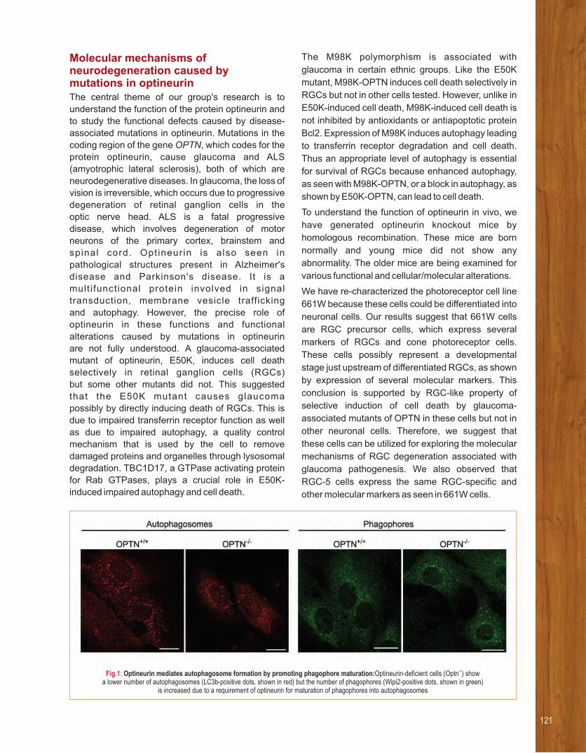
Molecular mechanisms of neurodegeneration caused by mutations in optineurinThe central theme of our group's research is to
understand the function of the protein optineurin and
to study the functional defects caused by disease-
associated mutations in optineurin. Mutations in the
coding region of the gene OPTN, which codes for the
protein optineurin, cause glaucoma and ALS
(amyotrophic lateral sclerosis), both of which are
neurodegenerative diseases. In glaucoma, the loss of
vision is irreversible, which occurs due to progressive
degeneration of retinal ganglion cells in the
optic nerve head. ALS is a fatal progressive
disease, which involves degeneration of motor
neurons of the primary cortex, brainstem and
spinal cord. Opt ineur in is also seen in
pathological structures present in Alzheimer's
disease and Parkinson's disease. It is a
mult i functional protein involved in signal
transduction, membrane vesicle trafficking
and autophagy. However, the precise role of
optineurin in these functions and functional
alterations caused by mutations in optineurin
are not fully understood. A glaucoma-associated
mutant of optineurin, E50K, induces cell death
selectively in retinal ganglion cells (RGCs)
but some other mutants did not. This suggested
that the E50K mutant causes glaucoma
possibly by directly inducing death of RGCs. This is
due to impaired transferrin receptor function as well
as due to impaired autophagy, a quality control
mechanism that is used by the cell to remove
damaged proteins and organelles through lysosomal
degradation. TBC1D17, a GTPase activating protein
for Rab GTPases, plays a crucial role in E50K-
induced impaired autophagy and cell death.
The M98K polymorphism is associated with
glaucoma in certain ethnic groups. Like the E50K
mutant, M98K-OPTN induces cell death selectively in
RGCs but not in other cells tested. However, unlike in
E50K-induced cell death, M98K-induced cell death is
not inhibited by antioxidants or antiapoptotic protein
Bcl2. Expression of M98K induces autophagy leading
to transferrin receptor degradation and cell death.
Thus an appropriate level of autophagy is essential
for survival of RGCs because enhanced autophagy,
as seen with M98K-OPTN, or a block in autophagy, as
shown by E50K-OPTN, can lead to cell death.
To understand the function of optineurin in vivo, we
have generated optineurin knockout mice by
homologous recombination. These mice are born
normally and young mice did not show any
abnormality. The older mice are being examined for
various functional and cellular/molecular alterations.
We have re-characterized the photoreceptor cell line
661W because these cells could be differentiated into
neuronal cells. Our results suggest that 661W cells
are RGC precursor cells, which express several
markers of RGCs and cone photoreceptor cells.
These cells possibly represent a developmental
stage just upstream of differentiated RGCs, as shown
by expression of several molecular markers. This
conclusion is supported by RGC-like property of
selective induction of cell death by glaucoma-
associated mutants of OPTN in these cells but not in
other neuronal cells. Therefore, we suggest that
these cells can be utilized for exploring the molecular
mechanisms of RGC degeneration associated with
glaucoma pathogenesis. We also observed that
RGC-5 cells express the same RGC-specific and
other molecular markers as seen in 661W cells.
Fig.1: -/-Optineurin mediates autophagosome formation by promoting phagophore maturation:Optineurin-deficient cells (Optn ) show a lower number of autophagosomes (LC3b-positive dots, shown in red) but the number of phagophores (Wipi2-positive dots, shown in green)
is increased due to a requirement of optineurin for maturation of phagophores into autophagosomes
121

Mechanisms of autophagy and
intracellular membrane vesicle
traffickingWe have explored the role of optineurin in
autophagosome formation. During autophagy,
signaling events lead to the formation of a cup-
shaped structure called the phagophore that matures
into the autophagosome. Fibroblasts from the Optn
knockout mouse showed a lower number of
autophagosomes and autolysosomes during both
basal and starvation-induced autophagy. However,
the number of phagophores was not decreased in
Optn-deficient cells. Phosphorylation of optineurin at
S177 was required for autophagosome formation.
Our results suggest that optineurin potentiates
LC3-II production and maturation of phagophore
into autophagosome, by facilitating the recruitment
of Atg12-5-16L1 complex to Wipi2-positive
phagophore. An ALS-associated mutant E478G-
OPTN is defective in autophagosome formation.
Rab GTPases control almost all the steps involved in
trafficking of membrane vesicles. We have proposed
a novel mechanism of regulating a Rab GTPase by an
effector protein (optineurin) that acts as an adaptor to
bring together a Rab (Rab8) and its GTPase
activating protein (TBC1D17). The E50K mutant
causes enhanced inhibition of Rab8 by TBC1D17
resulting in defective endocytic recycling of transferrin
receptor.
Mechanisms of signal transduction by
NLRC4, a mediator of innate immune
responseOur innate immune system responds to invading
microorganisms, which are recognized by cell surface
(Toll-like receptors) or cytoplasmic receptors (Nod-
like receptors, NLRs) to induce a protective response.
NLRC4 (Ipaf), a NLR family member, activates
caspase-1 in response to intracellular pathogens to
induce interleukin production. We have shown that
NLRC4 is a transcriptional target of p53 and is
required for induction of cell death in response to
chemotherapeutic drugs.The mechanisms, which are
involved in the activation of NLRC4 in response to
upstream signals such as those generated by
pathogen infection or chemotherapeutic drugs are not
known. In collaboration with Dr. V. Radha, we
identified a novel interaction between NLRC4 and the
proteasome component Sug1, which enables
ubiquitination of NLRC4 leading to FADD-dependent
caspase-8 activation and cell death. Phosphorylation
of NLRC4 at Ser533 plays a crucial role in caspase-8
activation and cell death.
Fig.2: Proposed model showing molecular events during NLRC4-H443P induced caspase-8 activation and cell death. WT-NLRC4 mediated caspase-8 activation is dependent on SUG1 over expression and Ser533 phosphorylation. Mutation of His443 to Proline (seen in patients with familial cold auto inflammatory
syndrome) causes enhanced interaction with cellular SUG1 compared to WT-NLRC4; SUG1 mediates ubiquitination of NLRC4-H443P leading to FADD and caspase-8 dependent apoptotic cell death. Presence of this mutation releases dependency of caspase-8 activation on Ser533 phosphorylation.

123
Our research
is focussed towards understanding
the mechanisms of establishment
and persistence of M.tb infection in its
human host.
RESEARCH INTERESTS:
• Host-pathogen interactions during M.tb infection
• Identification of mycobacterial virulence determinants
• Molecular correlates of M.tb latency
• Mechanisms of antibiotic resistance in mycobacteria
Selected recent publications
1. Viswanathan G, Yadav S and Raghunand T R (2017)
Identification of mycobacterial genes involved in β-lactam
sensitivity - Implications in treatment of tuberculosis with β-
lactam containing regimens. Antimicrobial Agents and
Chemotherapy doi: 10.1128/AAC.00425-17.
2. Viswanathan G, Yadav S, Joshi S V, and Raghunand T R (2017)
Insights into the function of FhaA, a cell-division associated
protein in mycobacteria. 364 (2): FEMS Microbiology Letters
fnw294.
3. Yeruva V C, Savanagouder M, Khandelwal R, Kulkarni A M,
Sharma Y and Raghunand TR (2016) The Mycobacterium 2+tuberculosis desaturase DesA1 (Rv0824c) is a Ca binding
protein. 480(1):29-35.Biochem Biophys Res Com
4. Yeruva VC, Kulkarni A M, Khandelwal R, Sharma,Y and
Raghunand T R (2016). The PE_PGRS proteins of 2+Mycobacterium tuberculosis are Ca binding mediators of host-
pathogen interaction. 55(33):4675-87.Biochemistry
5. Sharma SGS, Kishore V and Raghunand T R (2016)
Identification and functional annotation of mycobacterial
septum formation genes using cell division mutants of
Escherichia coli. Res Microbiol, 167(2):142-8.
From left to right : Ravi Prasad Mukku, Mamata Savanagouder, Veena Chaitanya, Raghunand Tirumalai, Vimal Kishore, Gopinath Viswanathan, R Gopalakrishna
Raghunand R TirumalaiDissecting the Molecular Basis of Mycobacterium tuberculosis Pathogenesis
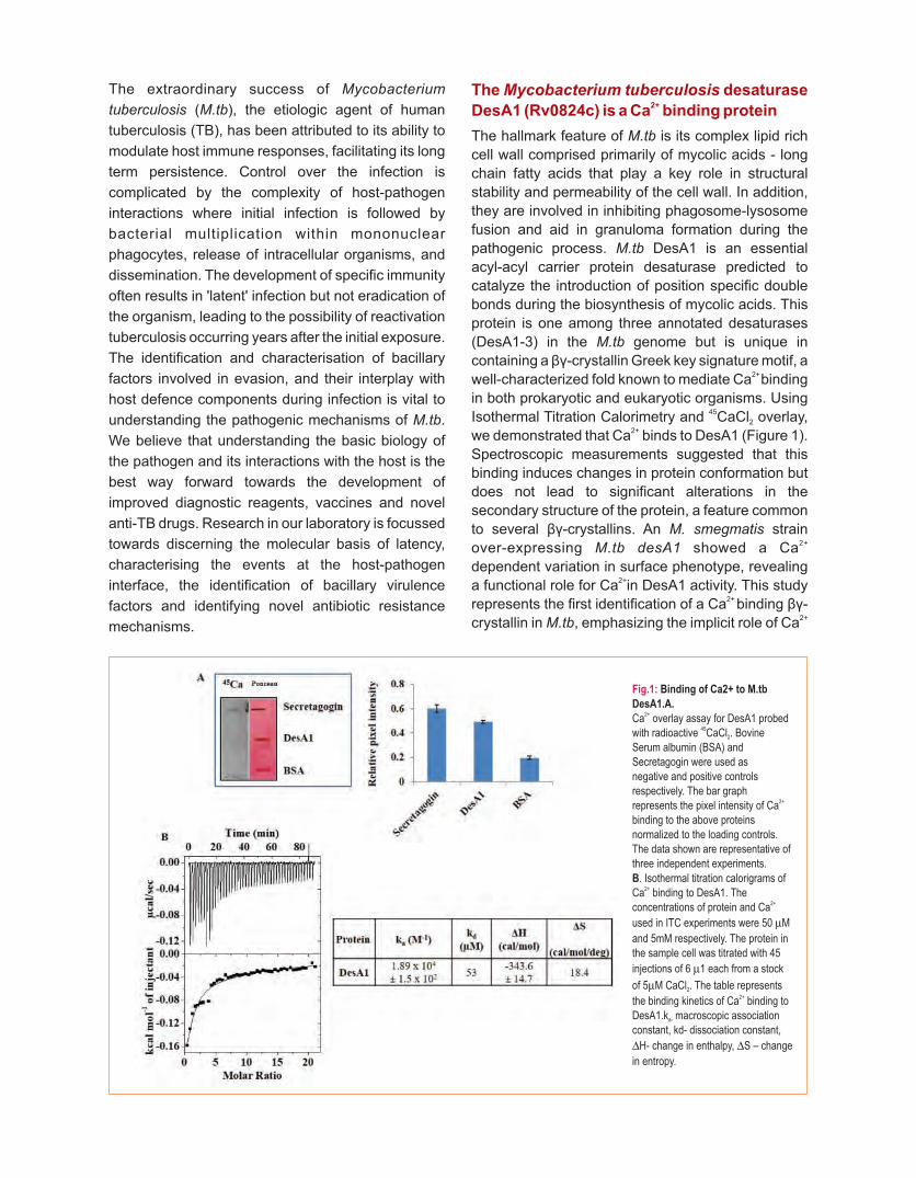
The extraordinary success of Mycobacterium
tuberculosis (M.tb), the etiologic agent of human
tuberculosis (TB), has been attributed to its ability to
modulate host immune responses, facilitating its long
term persistence. Control over the infection is
complicated by the complexity of host-pathogen
interactions where initial infection is followed by
bacterial multiplication within mononuclear
phagocytes, release of intracellular organisms, and
dissemination. The development of specific immunity
often results in 'latent' infection but not eradication of
the organism, leading to the possibility of reactivation
tuberculosis occurring years after the initial exposure.
The identification and characterisation of bacillary
factors involved in evasion, and their interplay with
host defence components during infection is vital to
understanding the pathogenic mechanisms of M.tb.
We believe that understanding the basic biology of
the pathogen and its interactions with the host is the
best way forward towards the development of
improved diagnostic reagents, vaccines and novel
anti-TB drugs. Research in our laboratory is focussed
towards discerning the molecular basis of latency,
characterising the events at the host-pathogen
interface, the identification of bacillary virulence
factors and identifying novel antibiotic resistance
mechanisms.
The Mycobacterium tuberculosis desaturase 2+DesA1 (Rv0824c) is a Ca binding protein
The hallmark feature of M.tb is its complex lipid rich
cell wall comprised primarily of mycolic acids - long
chain fatty acids that play a key role in structural
stability and permeability of the cell wall. In addition,
they are involved in inhibiting phagosome-lysosome
fusion and aid in granuloma formation during the
pathogenic process. M.tb DesA1 is an essential
acyl-acyl carrier protein desaturase predicted to
catalyze the introduction of position specific double
bonds during the biosynthesis of mycolic acids. This
protein is one among three annotated desaturases
(DesA1-3) in the M.tb genome but is unique in
containing a βγ-crystallin Greek key signature motif, a 2+ well-characterized fold known to mediate Ca binding
in both prokaryotic and eukaryotic organisms. Using 45Isothermal Titration Calorimetry and CaCl overlay, 2
2+we demonstrated that Ca binds to DesA1 (Figure 1).
Spectroscopic measurements suggested that this
binding induces changes in protein conformation but
does not lead to significant alterations in the
secondary structure of the protein, a feature common
to several βγ-crystallins. An M. smegmatis strain 2+ over-expressing M.tb desA1 showed a Ca
dependent variation in surface phenotype, revealing 2+a functional role for Ca in DesA1 activity. This study
2+ represents the first identification of a Ca binding βγ-2+crystallin in M.tb, emphasizing the implicit role of Ca
Fig.1: Binding of Ca2+ to M.tb
DesA1.A.2+Ca overlay assay for DesA1 probed
45with radioactive CaCl . Bovine 2
Serum albumin (BSA) and Secretagogin were used asnegative and positive controls respectively. The bar graph
2+represents the pixel intensity of Ca binding to the above proteins normalized to the loading controls. The data shown are representative of three independent experiments.
B. Isothermal titration calorigrams of 2+Ca binding to DesA1. The
2+concentrations of protein and Ca
used in ITC experiments were 50 mM
and 5mM respectively. The protein in the sample cell was titrated with 45
injections of 6 m1 each from a stock
of 5mM CaCl . The table represents 2
2+the binding kinetics of Ca binding to DesA1.k macroscopic association a-
constant, kd- dissociation constant,
DH- change in enthalpy, DS – change
in entropy.

in the pathogenesis of M.tb. We have extended this
study and are now examining structure-function
relationships in the desaturase and βγ-crystallin
domains of this protein.
Deciphering the role of the unusal PPE50
(Rv3135) – PPE51 (Rv3136) gene cluster in
M.tb physiology
The genome of M.tb encodes multiple immuno-
modulatory proteins, including several genes of
multigenic PE_PPE (named after the conserved
Proline-Glutamate and Proline-Proline-Glutamate
residues at their N-termini) family, which comprise
about 10% of its coding potential. The presence of
these proteins in pathogenic mycobacteria strongly
suggests that they play a role in disease
pathogenesis. Several of the PE_PPE genes are
organised in clusters that include PE-PPE, PE-PE
and PPE-PPE genes arranged in tandem. While the
functional properties of individual PE/PPE proteins
have been examined, little information exists on the
integrated functions of such gene clusters. To
understand its role in M.tb physiology we have begun
to characterise the PPE50 (Rv3135)-PPE51
(Rv3136) gene cluster, one of four PPE-PPE
clusters in the M.tb genome. Using RT-PCR we
demonstrate that this cluster encodes a co-
transcriptional unit and then hypothesised that
this locus encodes interacting proteins, a common
feature of operons. Using Mycobacterial Protein
Fragment Complementat ion (M-PFC) we
demonstrate that PPE50 and PPE51 interact
in vivo (Figure 2). This observation was validated by
in vitro pull-downs using purified preparations of
these proteins. To examine the role of these proteins
in host-pathogen interaction we expressed PPE50
and PPE51 as c-myc fusions in the surrogate
saprophytic host Mycobacterium smegmatis
and observed that they localise to the cell surface.
CFU counts of THP-1 macrophages infected
with recombinant M. smegmatis strains expressing
PPE50 and PPE51individually and in combination,
suggested that PPE50 and PPE51 play a role in
intracellular bacillary survival. We are now performing
cytokine profiling experiments to study the possible
immunomodulatory functions of these proteins during
infection.
Fig.2: The PPE50-PPE51 locus
encodes interacting proteins.
A. Schematic representation of
the ppe50-ppe51 gene cluster
gene cluster showing its location
in the M.tb genome and the
intergenic distances. B. M-PFC
analysis of PPE50-PPE51
interaction. The figure shows the
growth pattern of M.smegmatis
co-transformants on Middlebrook
7H11 agar plates in the presence
(+) or absence (-) of 40 mg/mL
Trimethoprim (TRIM). PPE50-
PPE51 interaction results in
growth of co-transformants
containing pUAB400:ppe50 and
pUAB300:ppe51 but not the
control plasmid sets in TRIM-
containing media.
125
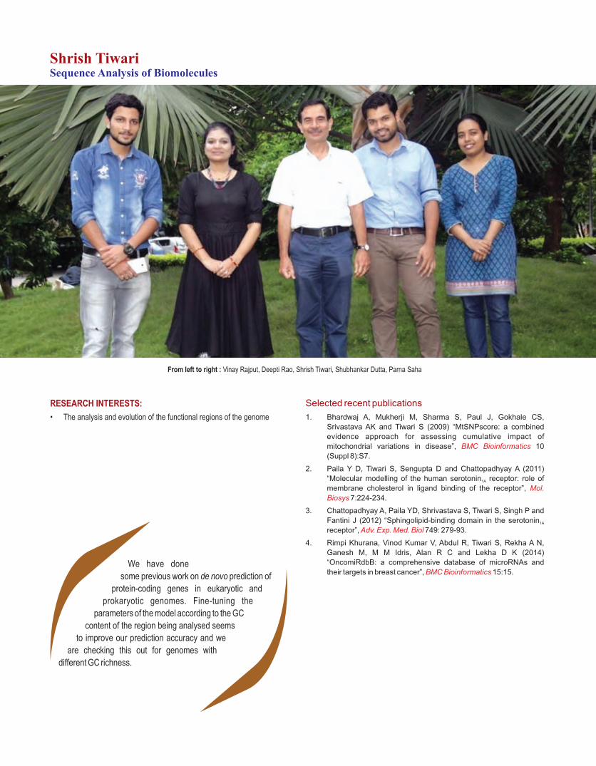
We have done
some previous work on de novo prediction of
protein-coding genes in eukaryotic and
prokaryotic genomes. Fine-tuning the
parameters of the model according to the GC
content of the region being analysed seems
to improve our prediction accuracy and we
are checking this out for genomes with
different GC richness.
RESEARCH INTERESTS:
• The analysis and evolution of the functional regions of the genome
Selected recent publications
1. Bhardwaj A, Mukherji M, Sharma S, Paul J, Gokhale CS,
Srivastava AK and Tiwari S (2009) “MtSNPscore: a combined
evidence approach for assessing cumulative impact of
mitochondrial variations in disease”, 10 BMC Bioinformatics
(Suppl 8):S7.
2. Paila Y D, Tiwari S, Sengupta D and Chattopadhyay A (2011)
“Molecular modelling of the human serotonin receptor: role of 1A
membrane cholesterol in ligand binding of the receptor”, Mol.
Biosys 7:224-234.
3. Chattopadhyay A, Paila YD, Shrivastava S, Tiwari S, Singh P and
Fantini J (2012) “Sphingolipid-binding domain in the serotonin 1A
receptor”, 749: 279-93.Adv. Exp. Med. Biol
4. Rimpi Khurana, Vinod Kumar V, Abdul R, Tiwari S, Rekha A N,
Ganesh M, M M Idris, Alan R C and Lekha D K (2014)
“OncomiRdbB: a comprehensive database of microRNAs and
their targets in breast cancer”, 15:15.BMC Bioinformatics
From left to right : Vinay Rajput, Deepti Rao, Shrish Tiwari, Shubhankar Dutta, Parna Saha
Shrish TiwariSequence Analysis of Biomolecules

We are getting into the area of prediction of non-
coding RNAs and other regulatory motifs in the
genome. At present this is restricted to a comparative
genomics approach, but with the availability of
genomes from closely related species, this approach
is becoming increasingly powerful and accurate.
Using a combination of experimental and
computational approaches we have shown that a
non-coding RNA expressed from the distal
heterochromatin of the human Y chromosome
contributes the 5'UTR of a testis-specific isoform of
the autosomal gene CDC2L2. We have also
uncovered a piRNA cluster on the mouse Y
chromosome, which may be regulating autosomal
genes.
Our search for SNPs that might be used as diagnostic
markers resulted in a web-based platform, MtSNP
score that helps to prioritise SNPs which may be
involved in mitochondrial diseases. MtSNP score
computes a score for every SNP in the mitochondrial
genes that has a significantly higher frequency in
patients compared to normal individuals. It takes
inputs from several analysis programs to assess the
damaging effect of the SNP, the higher the score, the
more damaging the SNP. The score of each SNP in a
patient is used to assess their combined effect. It was
applied to a dataset of 90 patients suffering from
sporadic ataxia, where all nuclear factors were
eliminated, and was shown to be highly
discriminatory, with no false positives, i.e. no normal
individual was predicted to have ataxia based on their
score. It was further tested on 6 Japanese datasets,
each consisting of 96 mitochondrial genomes of
individuals with a particular phenotype. The 6
phenotypes were individuals with Alzheimer's,
Parkinson's, diabetes with and without angiopathy,
obesity and thin phenotype.
In these studies we found that occasionally
synonymous SNPs were also associated with a
phenotype. This can happen when a rarely used
codon is mutated to a more frequently used one, or
vice versa. A possible explanation is that the rare
codon introduces a translational pause, which allows
the already formed protein chain to partially fold, thus
avoiding aggregation. The translational machinery
gets disrupted due to the mutation leading to
misfolding of the protein. We are exploring this
phenomenon, with an aim to predict which
synonymous changes lead to altered protein function.
With the advent of next generation sequencing
technology, genome sequencing has become
routine. We have analysed two NGS datasets
generated for rice. In one case an elite indica restorer
line was introgressed with chromosomal segments of
a wild rice using molecular markers and a major yield
QTL from the wild rice was found to be associated with
higher yield in the selected introgression line. The
restorer line and one of the introgressed lines were
sequenced and analysed to identify the introgressed
regions. In the other project, resistance genes to
blight infection from a wild rice were introgressed
in sambhamahsuri (SM), a high quality rice from
Andhra Pradesh. The resistant sambhamahsuri were
selected and propagated to get the improved
sambhamahsuri (ISM). SM and ISM (and a sister line
of ISM (SISM)) were sequenced and analysed to
identify regions of introgression. A protocol was
developed based on SNPs to identify introgressed
regions on all chromosomes. We are also in the
process of analysing EMS mutant lines of rice with
agronomically useful traits with the hope of identifying
the genes responsible for the phenotype.
We have also done some molecular modelling and
docking studies on the GPCR serotonin 1A receptor.
In particular we showed that the receptor with bound
cholesterol has a more compact structure. Several
agonists and antagonists, including serotonin, were
docked on the structure predicted through homology
modelling. We see that the binding sites for
antagonists were relatively shallow compared to
those of the agonists.
127
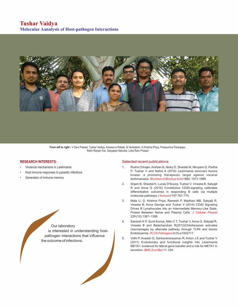
Our laboratory
is interested in understanding host-
pathogen interactions that influence
the outcome of infections.
RESEARCH INTERESTS:
• Virulence mechanisms in Leishmania
• Host immune responses to parasitic infections
• Generation of immune memory
Selected recent publications
1. Rudra Chhajer, Anirban B, Nicky D, Shadab M, Nirupam D, Partha
P, Tushar V and Nahid A (2016) Leishmania donovani Aurora
kinase: a promising therapeutic target against visceral
leishmaniasis. 1860: 1973-1988.Biochem et Biophys Acta
2. Srijani B, Sheetal K, Lucas D'Souza, Tushar V, Vineeta B, Satyajit
R and Anna G (2016) Constitutive CD40-signaling calibrates
differentiation outcomes in responding B cells via multiple
molecular pathways 197:761-770.J Immunol
3. Mala U, G. Krishna Priya, Ramesh P, Madhavi MB, Satyajit R,
Vineeta B, Anna George and Tushar V (2014) CD40 Signaling
Drives B Lymphocytes into an Intermediate Memory-Like State,
Poised Between Naïve and Plasma Cells. J Cellular Physiol
229 (10):1387–1396.
4. Santosh K P, Sunil Kumar, Nitin C T, Tushar V, Anna G, Satyajit R,
Vineeta B and Balachandran R(2012)Chitohexaose activates
macrophages by alternate pathway through TLR4 and blocks
Endotoxemia. 8 (5):e1002717.PLOS Pathogens
5. Vidhi P, Aneesh G, Sankaranarayanan R, Anton J E and Tushar V
(2011) Evolutionary and functional insights into Leishmania
META1: evidence for lateral gene transfer and a role for META1 in
secretion. 11: 334.BMC Evol Biol
From left to right : V Devi Prasad, Tushar Vaidya, Kaneez-e-Rabab, B Venkatesh, G Krishna Priya, Pradyumna Paranjape,Rathi Ranjan Kar, Satyajeet Salunke, Loka Ram Prasad
Tushar VaidyaMolecular Aanalysis of Host-pathogen Interactions

Our model system is the protozoan pathogen,
Leishmania donovani. Leishmania are transmitted
from one human to another by an insect vector, the
sand fly. In the insect, Leishmania exist as free-living,
motile Promastigotes while within the mammalian
host, Leishmania reside within macrophages as
immotile Amastigotes. Our long-term objectives are to
identify and analyze molecules and mechanisms that
confer any advantage to the parasite during the
various stages of its life cycle. To facilitate our studies
of these events, we have established a cell-free in
vitro system for the growth and inter-conversion of
promastigotes to amastigote forms of virulent L.
donovani.
We have identified several genes that are
differentially expressed in different stages of
Leishmania. One such gene, META1, is a candidate
virulence molecule. We have consistently observed
that virulent Leishmania cells have greater META1
expression than in attenuated cells, over a wide range
of conditions and environments. Using multiple
criteria including sequence similarity, nucleotide
composition, phylogenetic analysis and selection
pressure on gene sequence, we have established
that Leishmania META1 has been transferred by an
ancient lateral gene transfer event between bacteria
and a trypanosomatid ancestor. Superposition of
META1 sequence on the solved structure of MxiM, a
homolog in Shigella highlighted a putative
hydrophobic cavity in META1. Mutagenesis of select
hydrophobic residues in this cavity affects the
secretion of the secreted acid phosphatase (SAP),
indicating the involvement of META1 in secretory
processes in Leishmania.
In ongoing experiments, we have observed that over
expression of META1 causes a morphological
reduction in size of Leishmania. In late stationary
phase cells, greater than 90 % of META over
expressing Leishmania are one-fifth the size of the
wildtype cells. This is in contrast to just about 5% of
wild type Leishmania undergoing a similar change.
This phenotype is completely reversible; cells regain
their size within 72 h, when they are fed with fresh
media. The observed reduction in cell size is not
associated with cell death nor induced merely by
absence of nutrition or by the presence of spent
media. Instead, parallel experiments suggest that
META over expression reduces the uptake and/or
utilisation of nutrients. However, under all conditions
tested, overexpression of META1 is necessary. In
both wildtype and META1 over expressing
Leishmania there is a 10-20 fold increase in META1
transcript at the time of the morphological reduction.
However, upon refeeding the cells with fresh media,
both the endogenous and ectopically expressed
transcript levels reduce back to log phase levels,
consistent with reversal in cell morphology. This also
suggests a post-transcriptional control on META1
levels that limits the amount of META1 in a stage
specific manner. A specific point mutation in the
putative pocket of META1 earlier shown by us to
associate with increased secretion, diminishes the
cells ability to undergo this transition. This, together
with our observation of reduced nutrient uptake,
underscores role for META1 in Leishmania secretory
systems. Quite surprisingly, while over expression of
META1 derived either from L.donovani (LdMETA1)
and L. major (LmjMETA1) can induce a reduction in
cell size against L. donovani background, both genes
fail to do so in a L. major cell background, alluding to a
possible role for META1 in tissue tropism.
In addition, we have identified a novel pair of
paralogous genes in Leishmania, which we have
named Differential Regulated Genes, DRG1 and
DRG2. In all Leishmania species examined, DRGs
occur as two paralogs (DRG1 and DRG2) on the
same chromosome, each gene encoding virtually
identical proteins except for the last six amino acid
residues. The DRG proteins are predicted to have two
transmembrane domains, with the C-terminal
transmembrane domain being highly conserved in all
related Trypanosomatids. Interestingly, despite
coding for such closely related proteins, DRGs differ
in stability, localization and function. We have
generated transgenic Leishmania ectopically
expressing a) DRG1 b) DRG2 and c) DRG, just the
common DRG sequence, lacking the respective
specific elements. Preliminary in vitro infectivity
studies with J774 macrophages and these transgenic
Leishmania suggest that DRG1 over expression
seems to enhance the internalization of Leishmania
while over expression of the common DRG region
alone seems to have a negative effect on the ability of
Leishmania o infect macrophages. These findings
suggest a role for DRG1 in the infective process and
at the same time raises a possibility that the two
paralog proteins cooperate to function.
Currently, we are studying the differential regulation of
DRG1 vs DRG2 transcripts, in particular, the role of
the evolutionarily conserved respective 3'UTRs. In
Leishmania, gene regulation is thought to occur
primarily at a post-transcriptional level. We initiated
this effort by using bioinformatics approaches to
predict likely cis-acting RNA elements (Pyrimidine
rich tracts + COSMOS, a Statistical approach for
Conserved Motif Search across species) in DRG1 vs.
129
DRG2 mRNAs as well as the likely RNA binding
proteins (RPI-Seq, a RNA-Protein interaction
prediction + CISBP, a Catalog of Inferred Sequence
Binding Preferences). Using these combinatorial
approaches, we have identified and are testing broad
putative regulatory regions in DRG1 / DRG2 3' UTR.
Once the cis-regions are identified, this work will be
extended to identifying their differential binding
protein partners through immunoprecipitation
experiments and building an interaction map of the
involved proteins.
As a corollary to our interest in host responses to
infections, we have been studying the generation of
immunological memory in B-lymphocytes.
Interactions between CD40 on B cells and CD40L on
T cells are responsible for several aspects of acquired
immune responses including generation of memory B
cells. We are currently studying the molecular
mechanisms underlying the generation of memory B
cells, stimulated by CD40 mediated events. In order
to gain insights into early events leading to memory B
cell formation, we analyzed the genome-wide
expression profile of murine naive B cells stimulated
in the presence of anti-CD40. We have identified
nearly 10000 genes whose expression is altered
minimally 1.5 fold at least at one-time point over a
three day time course. The array analysis indicates
that changes in expression level of maximum number
of these genes occur within 24 hours of anti-CD40
treatment. Another approach to study the events
following CD40 ligation has been to examine the
expression of known regulators of naive B cell to
Plasma cell transition. The expression profile of these
regulatory genes indicates firstly, that CD40 signaling
moves B cells to a memory phenotype that is
intermediary between the naive and plasma cell
stages of the B cell differentiation. Secondly, the
major known regulator of plasma cell differentiation,
BLIMP1, gets irreversibly down regulated upon anti-
CD40 treatment. We also observed that CD40
signaling dominates over LPS signaling, in that even
after anti-CD40 is removed from the cells, BLIMP1
continues to be repressed. This observation in
combination with our microarray results and our
studies on candidate genes encoding chromatin-
modifying factors suggests a role for epigenetic
events in the maintenance of this memory-like state.
Additionally, our data suggests that CD40 signaling
mediates BLIMP1 down regulation by non-Pax5/non-
Bcl6 dependent mechanisms. Our results also
indicate that CD40 signal affects BLIMP1 regulation
at a post-transcriptional level. We are extending these
studies by characterizing the cells post CD40
signaling along various parameters including cell
surface markers, isotypes of Ig transcripts and
longevity and survival. Additionally, we are studying
the mechanisms by which CD40 signaling targets
BLIMP1 down regulation.
In addition to the experimental approaches described
above, we are currently in the process of assembling
a Draft genome of the parasitic nematode Setaria
digitata, a pathogen of significance in farm animals,
and a comparative analysis with parasitic nematodes
that infect humans. Genomic DNA isolated from
individual worms was submitted for next generation
sequencing. The Illumina Mi-Seq platform was used
to generate paired end and mate pair reads from
libraries ranging from 375bp, 500bp, 1.5kb and 5kb
insert size. The assembly was improved by gap
closing and extension of scaffolds using the bigger
insert size libraries. The final assembly that we
submitted to NCBI has the following statistics:
Nucleotide size of ~118 MB, distributed over 17549
scaffolds, with N50 of 30415 and 14512 predicted
genes.
This Venn diagram shows the distribution of shared
gene families among Setaria digitata, three worms
pathogenic to humans (Brugia malayi, Loa loa and
Onchocerca volvulus), a non-pathogenic worm,
Caenorhabditis elegans and an outlier organism,
Drosophila melangaster. The bar graph below Venn
diagram indicates total clusters predicted for
individual species (include unique clusters and
clusters shared with two or more species). The
bottom most color gradient pictorial representation
demonstrates the total clusters unique to one
organism, shared between any two, and so on.
Currently efforts are in progress towards annotation
of the Setaria genome for predicted genes. KEGG
pathway analysis using GhostKOALA has given us
information on KEGG modules, enzymes and
BRITE reconstruction pathways of Setaria digitata.
We have also done the GhostKOALA run for
B.malayi, C.elegans, Bostaurus (cattle), Ovisaries
(sheep), H.sapiens (human). Comparing metabolic
modules from the KEGG analysis of S.digitata with
other nematodes, its own obligate host (bovine),
aberrant host(Sheep) and Humans which it doesn't
infect, are in progress for leads into key differences
from other filarial worms. Additionally, Ortho Venn
analysis has been done to identify ortholog clusters
shared between Setaria digitata versus other animal
pathogens -Loa loa, B.malayi, O.volvulus, non
pathogenic C.elegans and D.melanogaster.
D.melanogaster was selected as a well curated and
annotated out group from Nematode genus. The

paralogs and orthologs clusters indicated many
protein clusters of S.digitata that were majorly shared
with other parasitic nematodes than free living
C.elegans. Comparative analysis of the draft Setaria
genome assembly with that parasitic nematodes that
do infect humans such B. malayi, W. banchrofti will
help understand their pathogenesis.
An interesting insight has been with regard to
Wolbachia species, which are obligate bacterial
endosymbionts essential for survival of the human
filarial parasitic nematodes. The Wolbachia genome
was downloaded from NCBI nucleotide database and
local alignment of the genome was performed against
the best SSPACE scaffold generated so far from the
Setaria sequence, but we observed no significant hits
against the Setaria genome. This suggests that
Setaria digitata may not harbour Wolbachia as
endosymbionts and may have evolved strategies
distinct from the human parasitic nematodes to
survive without Wolbachia.
Fig.1: This Venn diagram shows
the distribution of shared gene
families among Setaria digitata,
three worms pathogenic to humans
(Brugia malayi, Loa loa and
Onchocerca volvulus), a non-
pathogenic worm, Caenorhabditis
elegans and an outlier organism,
Drosophila melangaster. The bar
graph below Venn diagram
indicates total clusters predicted for
individual species (include unique
clusters and clusters shared with
two or more species). The bottom
most color gradient pictorial
representation demonstrates the
total clusters unique to one
organism, shared between any two
, and so on.
131

- Development of
molecular assays for quantifying
prevalence of chytridiomycosis in
amphibians
- Developed robust methods to monitor
biological invasion in Andaman Islands
through key-informants
- Phylogenetic studies on bulbuls; egg-
eating snake; gharial
- Quantifying geographic variation in
saw-scaled venom constituents
RESEARCH INTERESTS:
• The group works on ecology, evolutionary biogeography and systematics with a focus on south Asian amphibians and reptiles. Studies focussed on specific endangered reptile and amphibian species, on their ecology and conservation are being carried out.
Selected recent publications
1. Jegath Janani S, Vasudevan K, Perindini E, Dutta S K,
Aggarwal R K (2017). A new species of the genus
Nasikabatrachus. 34 (1-4): 1-19.Alytes
2. Mohanty N P, Harikirshnan S and Vasudevan K (2016). Watch out
where you sleep: Nocturnal sleeping behaviour of Bay Island
lizards. 4:e1856;DOI10.7717/peerj.1856.PeerJ
From left to right (front row) : Snehalatha Vadigi, Karthikeyan Vasudevan, Gayathri Sreedaran, Siddharth Bhatia: K Rajyalakshmi, Afsar Soghra, Ravi Kumar Singh, Milind C Mutnale, Ashish Jha(back row)
Karthikeyan VasudevanEcology and Conservation of Endangered Species

The National Chambal Sanctuary in India holds the largest, most-stable and most-studied gharial subpopulation in the wild. This sanctuary stretches across the states of Rajasthan, Madhya Pradesh and Uttar Pradesh. The Gharial (Gavialis gangeticus) is endemic to the Indian subcontinent and Critically Endangered, and conservation work has been undertaken in the protected areas of Chambal. It is one among the longest lived reptilian species (up to 100 years) and spends half of its life as a reproductively active animal. The animal is also one of the largest crocodiles. The males grow 5-6 m, while the females grow to about 3.5-4.5 m. Since it has a prolonged pre-reproductive period, there wouldbe no immediate apparent consequences of removal of breeding individuals. However the future ramifications would be a dramatic decline in their population size, hence the study of breeding biology of prime importance in gharials. We are focusing mainly on two key questions, which are 1) what is the kinship status in the breeding groups of gharials (big, medium, and small)? 2) is there multiple paternity in the communal nesting groups? To address these questions, we have collected about 3000 egg shell samples from a 200 km stretch of Chambal River (figure 1). We have covered nine communal nesting sites during the sample collection from 26 May to 15 June 2017. Back at LaCONES, we started isolating DNA from the egg membranes. The samples have yielded appreciable amount of DNA. We will use previously discovered microsatellite primers for gharials as well as Indian muggers, and screen them. We will be using the screened-in primers for genotyping of microsatellite markers using next generation sequencing. These sequences of microsatellite markers will unravel the breeding biology of gharials and will also serve as reference data for any further genetic study.
Gharials are one of the top predators present in the riparian ecosystem of the Chambal sanctuary. They feed on almost all organisms present in this system like molluscs, reptiles and fishes etc. It is one the largest
crocodiles present in India, which turns from a hatchling weighing a mere 100 grams to an adult weighing 1000 kilograms. Being a top predator, it feeds on large predatory fishes which in turn eat fishes of commercial value to the local people. In order to protect this species, it is very important to study the ecosystem functioning provided by this animal. We used stable isotope analysis techniques (using Stable Isotope Mass Spectrometry at CSIR-NGRI) to characterize the composition of stable carbon and nitrogen isotopes (expressed as δ13C and δ15N). These measures define the items that are consumed as part of their diet. Specically, tissue δ15N compositions are a relative indicator of trophic position, and tissue δ13C compositions reect the habitats and food webs that animals use to acquire prey
We used tail scute clippings of the animals collected by GCA (Gharial Conservation Alliance) team. The samples were analysed in collaboration with CSIR-NGRI. We divided our samples into four categories based on body length, namely: juvenile, sub adult, adult female and adult male. We made a biplot of tissue δ13C compositions (‰) and δ15N compositions (‰). We found low δ13C values and less variation in δ13C as well as δ15N values only among adult males, which suggests that the dietary niche of the adult gharial males is more specialized as compared to other categories. Less enrichment in δ13C values is indicative of their food source being confined to those aquatic life forms occupying second or third trophic levels in food chain. On the other hand juveniles show maximum variation in their δ13C and δ15N values, this indicates that their diet forms a complex food web. Juveniles, sub adults and adult females also show huge variation in their δ13C and δ15N values, which points towards their more generalised dietary habit as compared to adult male. To sketch a comprehensive picture of gharial’s dietary behaviour, we will need all the possible dietary samples from Chambal River, which will be accomplished in next couple of years.
Emerging Infectious Disease (EDI) is a threat to wildlife. One of such disease is Chytridiomycosis, considered to be a biodiversity disease capable of causing mass mortality events in global amphibian species. It is caused by two aquatic fungalpathogens Batrachochytrium dendrobatidis (Bd)and Batrachochytriums alamandrivorans (Bs). Amphibians have highly permeable skin and are hence highly sensitive to changes in the environment making them ideal bio-indicators for the health of their surrounding ecosystem. Bd fungus colonies on the keratinous layer of frog skin and penetrate inside the skin epidermis to cause hyperkeratosis in frogs. The skin becomes thick-walled and interferes with gaseous exchange and osmoregulation. Very few
Fig.1: Gharial nest with freshly hatched out babies. This nest opened and assisted by our team to hatch out. (photo: Ravi Kumar Singh)
133

treatment options are available for Bd and Bs fungus. It is, therefore, necessary for effective surveillance to be able to detect the disease at its early onset in order to successfully conserve endemic frog species. We have limited data about the Bd fungus in India and only the Western Ghats has been surveyed for this fungus. The status of infection in other regions like The Himalayan region, North-East region, Eastern Ghats and Andaman Nicobar is unknown.
We used non-invasive swabbing technique wherein a cotton swab was firmly run 70 times over a frog’s body. A new fresh pair of gloves was used in between animal to avoid cross contamination. Sampling was carried out in the Western Ghats, Eastern Ghats, North East India, Himalayan region and the Andaman and Nicobar Islands. DNA was extracted from the cotton swabs. A nested PCR was performed andPCR products were sequenced. All sequences were used to construct Bayesian tree and Median-joining haplotype network. A total of 1870 samples were collected from 142 locations comprising 30 genera, and 111 species. Nested PCR was used to screen al the samples. A total of 158 samples found to be positive for Bd fungus with prevalence 8.4%. Low prevalence was found in North East India with 0% prevalence while the Western Himalaya was leading with 14% prevalence. In the sequencing nested PCR product, two samples showed insertion of two adenine residues at the Taqman binding site while remaining two samples had transition and transversion mutation at Taqman binding site which can cause false negatives in qPCR analyses. We found a total of 57 haplotypes of which 46 were unique to India and 11 were shared between other countries. Two haplotypes namely IN02 and IN10 were prevalent in India. Both these haplotypes contributed 65% of total positive samples. Haplotypes IN02 was most prevalent, represented in all the sampled areas (Figure 2). Haplotype IN10 was present in all sampled area except Western Himalayas (Figure 2). We constructed median joining network using all DNA sequences obtained in this study. The Indian haplotypes IN05 and IN55 containing mutations at the Taqman binding site were clustered together with the China haplotype CN30 and the Japan haplotypes JP09 and JP10.
The Yellow-throated Bulbul (YTB) is an endemic and threatened species found in small pockets of suitable habitats in thorny scrub forests across Eastern Ghats. Being endemic to the region with sparse and disjunct distribution, genetics of YTB populations secure signatures of ancient landscape level changes and geographic evolution. We are sampling YTB populations across its geographic range using mist netting (Figure 3) to understand the genetic differences between different populations. Also, we
are studying evolution of 24 species of Bulbuls from Indian Subcontinent. We are seeking answer to why is YTB endemic and rare while congeneric species are widespread and dominant. Our data shows that less than 100 populations of YTB are found across peninsular India with less than 1000 sq Km suitable habitat available. Preliminary phylogeny of Indian Bulbuls reveal that YTB does not have a sister species and has a unique evolutionary history. The ongoing genetic study of individual YTB populations will reveal if the populations are interconnected (signifying the ability of YTB to fly over to new habitats) or inbred (signifying reproductively isolated populations). Also the haplotype network will reveal if discreet YTB populations are a result of shrinkage of suitable habitat or in contrast, whether an ancient YTB population from one habitat dispersed and occupied suitable habitats around. It is expected that this work will push the cause of protection of scrub habitats across the Eastern Ghats and also the concept of preventive conservation for similar species.
Snake venoms contain complex mixtures of pharmacologically active molecules, including organic and mineral components, small peptides and
Fig.2: Bd haplotypes recovered from the different locations. Brown colored haplotype IN02 is wide spread thought India. Red colored haplotype IN10 is
present at all the location sampled except at Western Himalaya. Bd diversity was more at the Western Ghats.
Fig.3: Mistnetting at Melukote and a bunch of keenly observant field staff and volunteers
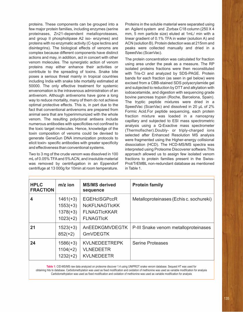
proteins. These components can be grouped into a few major protein families, including enzymes (serine proteinases, Zn21-dependent metalloproteases,and group II phospholipase A2 iso- enzymes) and proteins with no enzymatic activity (C-type lectins and disintegrins). The biological effects of venoms are complex because different components have distinct actions and may, in addition, act in concert with other venom molecules. The synergistic action of venom proteins may either enhance their activities or contribute to the spreading of toxins. Snake bite poses a serious threat mainly in tropical countries including India with snake bite mortality estimated at 50000. The only effective treatment for systemic envenomation is the intravenous administration of an antivenom. Although antivenoms have gone a long way to reduce mortality, many of them do not achieve optimal protective effects. This is, in part due to the fact that conventional antivenoms are prepared from animal sera that are hyperimmunized with the whole venom. The resulting polyclonal antisera include numerous antibodies with specificities not confined to the toxic target molecules. Hence, knowledge of the toxin composition of venoms could be devised to generate GeneGun DNA immunization protocols to elicit toxin- specific antibodies with greater specificity and effectiveness than conventional systems.
Two to 3 mg of the crude venom was dissolved in 100 mL of 0.05% TFA and 5% ACN, and insoluble material was removed by centrifugation in an Eppendorf centrifuge at 13 000g for 10min at room temperature.
Proteins in the soluble material were separated using an Agilent system and Zorbax C18 column (250 X 4 mm, 5 mm particle size) eluted at 1mL/ min with a linear gradient of 0.1% TFA in water (solution A) and ACN (solution B). Protein detection was at 215nm and peaks were collected manually and dried in a SpeedVac (ScanVac).
The protein concentration was calculated for fraction using area under the peak as a measure. The RP isolated proteins fractions were then reconstituted with Tris-Cl and analyzed by SDS-PAGE. Protein bands for each fraction (as seen in gel below) were excised from a CBB-stained SDS polyacrylamide gel and subjected to reduction by DTT and alkylation with iodoacetamide, and digestion with sequencing grade bovine pancreas trypsin (Roche, Barcelona, Spain). The tryptic peptide mixtures were dried in aSpeedVac (ScanVac) and dissolved in 20 μL of 2% Formic Acid.For peptide sequencing, each protein fraction mixture was loaded in a nanospraycapillary and subjected to ESI mass spectrometric analysis using a Q-Exactive mass spectrometer (Thermofischer).Doubly- or triply-charged ions selected after Enhanced Resolution MS analysis were fragmented using the Higher-energy collisional dissociation (HCD). The HCD-MS/MS spectra was interpreted using Proteome Discoverer software.This approach allowed us to assign few isolated venom fractions to protein families present in the Swiss-Prot/TrEMBL non-redundant database as mentioned in Table 1.
Table 1: CID-MS/MS raw data analyzed on proteome discover 1.4 using UNIPROT snake venom database. Sequest HT was used for obtaining hits to database. Carbidomethylation was used as fixed modification and oxidation of methionine was used as variable modification for analysis
Carbidomethylation was used as fixed modification and oxidation of methionine was used as variable modification for analysis
HPLC m/z ion MS/MS derived Protein familyFRACTION sequence
4 1461(+3) EGEHcISGPccR Metalloproteinases (Echis c. sochureki)
1553(+3) NcKFLNAGTIcKK
1378(+3) FLNAGTIcKKAR
1023(+2) FLNAGTIcK
21 1523(+3) AnEEDKGMVDEGTK P-III Snake venom metalloproteinases
852(+2) GmVDEGTK
24 1586(+3) KVLNEDEETREPK Serine Proteases
1104(+2) VLNEDEETR
1232(+2) KVLNEDEETR
135

During the year we examined how C3G is regulated, and identified novel
effectors that explain its essential role in maintaining cellular homeostasis. Dynamic nucleo-cytoplasmic
exchange of C3G and its localization to nuclear speckles & the centrosome, suggest its role in regulating nuclear and
centrosome functions. These findings advance our understanding of embryonic development as well as
control of proliferation vs differentiation. Experiments using disease associated mutants of Ipaf have identified
novel mechanisms of cell death signalling. These studies impact our understanding of normal cell functions at a
molecular level and how deregulation can cause pathology.
RESEARCH INTERESTS:
• Regulation of cytoskeleton and cell differentiation
• Mechanism of action of cell death regulators
Selected recent publications
1. Shakyawar DK, Dayma K, Ramadhas A, Varalakshmi C, Radha
V(2017) C3G shows regulated nucleo-cytoplasmic exchange and
represses histone modifications associated with euchromatin. Mol
Biol Cell 28: 984-995.
2. Akhouri K R, Anand S, Gayathri G, Pendyala P, Yatender K, Radha
V and Ghanshyam S (2017) A disease-associated mutant
ofNLRC4 shows enhanced interaction with SUG1 leading to
constitutive FADD dependent caspase-8 activation and cell
death. 292(4):1218-1230.J Biol Chem
3. Sasi K K, Ramadhas A, Nayak SC, Kaniyappan S, Dayma K,
Radha V (2015) C3G (RapGEF1), a regulator of actin dynamics
promotes survival and myogenicdifferentiation of mouse
mesenchymal cells. 1853: Biochim Biophys Acta- Mol Cell Res
2629-2639.
4. Dayma K, Ramadhas A, Sasikumar K and Radha V (2012).
Reciprocal negative regulation between the guanine nucleotide
exchange factor C3G and β-catenin. 3:564-577.Genes & Cancer
5. Mitra A and V Radha (2010). F-actin binding domain of c-Abl
regulates localized phosphorylation of C3G: Role of C3G in c-Abl
mediated cell death. 29:4528-4542.Oncogene
From left to right (front row) : Dhruv Kumar, Kunal Dayma, Vegesna Radha, Bh. Muralikrishna, Divya Sriram, Sanjeev Chavan Nayak, Ch. Ramulu.
Vegesna RadhaSignaling to the Cytoskeleton and Regulation of Cellular Functions

Physio log ica l processes such as t issue
morphogenesis, wound healing, immune cell
targeting, cell junction& shape maintenance,
neuronal differentiation, adhesion and vesicle
trafficking are dependent on cytoskeletal dynamics.
A large number of human disorders are associated
with defective cytoskeletal regulation. Our research
focuses on characterizing regulatory molecules of
intracellular signalling pathways leading to
cytoskeletal remodelling, to understand how this
fundamental process is controlled, and how
deregulation results in pathological situations. We
hope to identify molecular determinants regulating
cell fate. These studies impact our understanding of
normal development and maintenance of adult tissue
integrity. Currently, our focus is on the guanine
nucleotide exchange factor, C3G, which is
ubiquitously expressed and functions in multiple
signalling pathways. C3G regulates activation of
small GTPases, Rap, R-Ras and TC10 and is known
to have functions dependent on both catalytic and
protein interaction domains. C3G is essential for
mammalian embryonic development and many
cellular functions in adult tissues. The molecular
effectors and function of C3G in foetal and adult
tissues are poorly defined. Studies in our laboratory
showed for the first time an involvement of C3G in
signaling to actin reorganization, and its role in
differentiation. We have shown that C3G functions as
a negative regulator of β-catenin signaling and may
therefore control cell fate. How C3G regulates
differentiation and other cellular functions is not fully
understood.
C3G was earlier known to localize exclusively to the
cytoplasm. Our studies showed that dynamic nucleo-
cytoplasmic exchange of C3G is regulated by
functional NLSs and NES present in its primary
sequence. GSK-3β& okadaic acid sensitive
phosphatases regulate dynamic exchange of C3G
between cytoplasm & nucleus. In the nucleus, C3G
represses histone modifications associated with
euchromatin, and causes peripheralization of
heterochromatin, indicating that C3G plays a role in
regulating chromatin dynamics. This effect of C3G is
mediated by histone deacetylases. Differentiation of
myocytes and exposure of epithelial cells to sub-
lethal doses of DNA damage induce nuclear
translocation of C3G, and changes in chromatin.
Using shRNA and CRISPR/Cas approaches, we
showed that C3G is required for maintaining histone
modifications and expression of CDK inhibitors, p21 &
p27. C2C12 clones lacking C3G, donot show
upregulation of MHC, & donot fuse to form myotubes
when grown in differentiation medium.
Reversible association of C3G with speckles was
seen upon inhibition of transcription and splicing.
C3G shows partial colocalization with SC35, and is
recruited to a chromatin and RNase sensitive fraction
of speckles. Its presence in speckles is dependent on
intact cellular actin cytoskeleton, and is lost upon
expression of the kinase, Clk1, and transient heat
stress. Rap1, a substrate of C3G, is also present in
nuclear speckles and inactivation of Rap signalling by
expression of RapGAP, reduces speckle number.
Enhanced association of C3G with speckles is seen
upon GSK3β inhibition, or differentiation of C2C12
cells to myotubes. CRISPR/Cas9 mediated
knockdown of C3G resulted in decreased splicing
activity and reduced staining for SC35 in speckles.
C3G knockout clones of C2C12 as well as MDA-MB-
231 showed reduced protein levels of several splicing
factors compared to control cells. Our results identify
C3G and Rap1 as novel components of nuclear
speckles and a role for C3G in regulating cellular RNA
splicing activity.
GSK3b interacts with, and phosphorylates C3G to
retain it in the cytoplasm. In vivo and invitro assays
using phosphorylation site mutants showed multiple
sites of phosphorylation by GSK3b. C3G over-
expression represses, and knockdown of C3G
enhances GSK3b activity. Examination of the
expression of C3G in different cell types and
functional regions of the brain showed high levels of
expression in the dentate gyrus and in neuronal
precursor cells. C3G expression was high in
embryonic brain, even before neuronal differentiation
is completed. Adult mouse olfactory bulb showed
C3G expression in neurons but not in glial cells. In
neurons, C3G shows predominant cytoplasmic
localization.
In animal cells, the centrosome is the primary
microtubule organizing centre, and is also the
platform for primary cilium growth. These organelles
serve as important signalling centres, and defects in
their structure or function is associated with many
human disorders. Recent findings show that
endogenous C3G localizes to the mother centriole, is
137

present at the base of primary cilium (Figure 1), and
interacts with cenexin. In prophase cells, C3G is seen
at only one of the centrosomes, and in later phases of
mitosis, is present in both the centrosomes,
suggesting that it marks the mother centriole at the
end of prophase. Knockdown of C3G showed that it
plays a role in regulating centrosome duplication and
primary cilium formation. We are currently addressing
how C3G regulates centrosome functions.
In collaboration with Dr. GhanshyamSwarup, we have
shown that one of the autoinflammatory syndrome-
causing mutants of NLRC4, H443P, but not T337A
and V341A, constitutively activates caspase-8 and
induces apoptotic cell death in human lung epithelial
cells. Compared to wild type NLRC4, the H443P
mutant shows stronger interaction with SUG1 and
with ubiquitinated cellular proteins. Phosphorylation
of NLRC4 at Ser533 plays a crucial role in caspase-8
activation and cell death. However, H443P mutant
does not require Ser533 phosphorylation for
caspase-8 activation and cell death. Our findings
show that the auto-inflammation associated H443P
mutant is altered in interaction with SUG1 and
ubiquitinated proteins, triggering constitutive
caspase-8 mediated cell death dependent on FADD,
but independent of Ser533 phosphorylation.
Fig.1: C3G dynamically localizes to SC35 nuclear speckles. A. MDA-MB-231 cells were subjected to a-Amanitin treatment and immunostained for
C3G and SC35. B. Three dimensional visualization of speckle regions of cells dually labelled with antibodies against C3G (Red) and SC35 (Green).
Line scan showing local intensity distribution of C3G in red and SC35 in green in the ROI drawn across two speckles are shown below the panels.

139
The focus of my laboratory is to study the signal
transduction pathways in human health and disease. We also carry out
applied research in the area of wild flora and wild fauna forensics and
conservation.
RESEARCH INTERESTS:
• Signal Transduction in human health and disease conditions
• Molecular Evolution of Genes and Species
• Wildlife Forensics
Selected recent publications
1. A Revolution in Wildlife Forensics. India Perspectives, 2015,
Vol.29, Issue 4, pp 74-75. (A cover “Success Story” by Ministry
of External Affairs Govt. of India, published in 16 languages
and distributed in 170 countries).
2. Nayak A, Salt G, Verma S K and Kishore U (2015). Proteomics
Approach to Identify Biomarkers in Neurodegenerative
Diseases. 121:59-86.Int Rev Neurobiol
3. Verma S K and Sarkar M K (2015) Decalepisarayalpathra (J.
Joseph & V. Chandras.) Venter, an endemic and endangered
ethno medicinal plant from Western Ghats, India. Nat Prod Res
29:394-395.
4. Verma S K and Goswami G K (2014) DNA evidence: Current
perspective and future challenges in India. Forensic Sci Int
241:183-189.
5. Verma S K, Ganesan T S, Kishore U and Parker, P. J. (2011).
The tumour suppressor RASSF1A is a novel effector of small
G protein Rap1A. , 2:237-249.Protein Cell
From left to right : Sanjay Kumar Suman, Sunil Kumar Verma, Sampurna Balde, Megha Kumari, R. Siva Dharshini, Sanjana Shirva, Nabanita Biswas
Sunil Kumar VermaSignal Transduction Studies in Human Health and Disease and Molecular Biology Applications in Wildlife Conservation

The “Universal Primer Technology” invented by us in
the past became a milestone in the area of Wildlife
Forensics, in particular the DNA based Identification
of species to satisfy legal evidence requirements in a
court of law. This invention became instrumental to
solve more than 1300 cases pertaining to wildlife
crime. However, due to absence of any training facility
in the country on the application part of it, the
beneficiaries of this invention such as Wildlife
Officers, IPS, IFS, Police personnel and Judiciary
lagged behind and could not update their skills in this
arena. In this year, we have started a unique program
under the skill India mission of Government of India
which aims to fill these gaps by establishing a state of
the art National Wildlife Forensics Training
Laboratory in the LaCONES of CCMB (Figure 1).
With the increased practical implications of advanced
DNA technologies in various sectors, in particular the
judiciary, police, wildlife and forest and most recently
the food safety departments, this programme will be
another milestone in developing our national
capability in this area for proper implementation of
India's wildlife forensics and conservation efforts.
Other than this my laboratory is also playing a pivotal
role in establishing the yet unexplored arena of wild
flora forensics to help combat India with illegal plant
trade as well as to identify and protect the national
wealth of our medicinal plants.
Fig.1: Illustration of various stakeholders who will benefit from this skill development program.
(i) Officers working in establishments under various departments, subordinate offices, attached offices, autonomous bodies and organizations, PSUs and
missions of various Ministries of Government of India, such as:
• Ministry of Environment, Forests and Climate Change (MoEF)
• Ministry of Law and Justice (MoLJ)
• Ministry of Commerce and Industry (MoCI)
• Ministry of Food Processing Industries (MoFPI)
• Ministry of Health & Family Welfare (MoHFW)
• Ministry of Home Affairs (MoHA)
• Ministry of Agriculture and Farmers Welfare (MoAFW)
• Ministry of Finance - Department Of Revenue
(ii) Academic, Research Institutions and Universities: Training of the trainers and teachers
(iii) Licensees and interested parties, including private and public prosecutors willing to upgrade their skill on DNA based interventions
(iv) Students interested in taking the above arena as their career plan

List of Publications and Patents
PhD Program
Training Programs
Research Facilities
Research Resources
Services
CCMB Innovation Hub


1. Ahmad M, Nongmaithem SS, Krishnaveni GV,
Fall CHD, Yajnik CS and Chandak GR (2016)
Lack of replication of association of THSD7A
with obesity. 40(4) :725-726. Int J Obesity
2. Ahn TY, Kang JH, Kang DJ, Venkatesan J,
Chang HK, Bhatnagar I, Chang KY, Hwang JH,
Salameh Z, Kim SK, Kim HT and Kim DG (2016)
Interact ion of stem cel ls wi th nano
hydroxyapatite-fucoidan bionanocomposites for
bone tissue regeneration. Int J Biol Macromol
93:1488-1491.
3. Ahsan SM and Mohan RC (2017) The role of
surface charge in the desolvation process of
gelatin: implications in nanoparticle synthesis
and modulation of drug release. Int J Nanomed
12:795-808.
4. Ahsan SM and Mohan RC (2017) Structural
studies on aqueous gelatin solutions:
Implications in designing a thermo-responsive
nanoparticulate formulation. Int J Biol Macromol
95:1126-1134.
5. Al-Nihmi FMA, Kolli SK, Reddy SR, Mastan BS,
Togiri J, Maruthi M, Gupta R, Sijwali PS, Mishra
S and Kumar KA (2017) A novel and conserved
plasmodium sporozoite membrane protein
SPELD is required for maturation of exo-
erythrocytic forms. Scientif ic Reports
7: 40407.
6. Amin H, Nayak D, Rasool RU, Chakraborty S,
Kumar A, Yousuf K, Sharma PR, Ahmed Z,
Sharma N, Magotra A, Mukherjee D, Kumar LD,
Goswami A (2016) Par-4 dependent modulation
of cellular -catenin by medicinal plant natural
product derivative 3-azido WithaferinA. Mol
Carcinogenesis 55(5):864-881.
7. Babu NS, Murthy CLN, Kakara S, Sharma R,
Swamy CVB, and Idris MM (2016) 1-Methyl-4-
phenyl-1,2,3,6-tetrahydropyridine induced
Parkinson's disease in zebrafish. Proteomics
16(9):1407-1420.
8. Babu, N S, Krishnan, S, Swamy, C V B,
Subbaiah, G P V, Reddy, A V G, and Idris, M M
(2016) Quantitative proteomic analysis of
normal and degenerated human intervertebral
disc. 16(8): 989-1000.Spine J
9. Bagga P and Patel AB (2016) Pretreatment of
caffeine leads to partial neuroprotection in
MPTP model of Parkinson's disease. Neural
Regen Res 11(11):1750-751.
10. Balabadra S, Kotni M, Manga V, Allanki AD,
Prasad R and Sijwali, Puran Singh (2017)
Synthesis and evaluation of naphthyl bearing 1,
2, 3-triazole analogs as antiplasmodial agents,
cytotoxicity and docking studies. BioOrg &
Medicinal Chem 25(1): 221-232.
11. Banerjee P, Surendran H, Chowdhury DR,
Prabhakar K and Pal, R (2016) Metformin
mediated reversal of epithelial to mesenchymal
transition is triggered by epigenetic changes
in E-cadherin promoter. 94(12):J Mol Med
1397-1409.
12. Basu S, Kaw S, D'Souza L, Vaidya T, Bal V, Rath
S, and George, A ( 2016) Constitutive CD40
Signaling Calibrates Differentiation Outcomes in
Responding B Cells via Multiple Molecular
Pathways. 197(3):761-770.J Immunology
13. Begum G, Reddy TN, Kumar KP, Dhevendar K,
Singh S, Arnarnath M, Misra S, Rangari VK, and
Rana RK (2016) In Situ Strategy to Encapsulate
Antibiotics in a Bioinspired CaCO3 Structure
Enabling pH-Sensitive Drug Release Apt for
Therapeutic and Imaging Applications. ACS
Appl Mat & Interfaces 8(34): 22056-22063.
14. Belagal P, Normand C, Shukla A, Wang RJ,
Leger-Silvestre I, Dez C, Bhargava P and Gadal
Olivier (2016) Decoding the principles
underlying the frequency of association with
nucleoli for RNA polymerase III-transcribed
genes in budding yeast. Mol Biol Cell
27(20):3164-3177.
15. Bera S, Raghuram V, Mikhaylova M and Kreutz
MR (2016) A plasmid-based expression system
to study protein-protein interactions at the Golgi
in vivo. 502:50-52.Analytical Biochem
16. Bhunia S, Radha V and Chaudhuri A (2017)
CDC20siRNA and paclitaxel co-loaded
nanometric liposomes of a nipecotic acid-
derived cationic amphiphile inhibit xenografted
neuroblastoma. 9(3):1201-1212. Nanoscale
17. Bokara KK, Kim JH, Kim JY, and Lee, JE (2016)
Transfection of arginine decarboxylase gene
increases the neuronal differentiation of neural
progenitor cells. Stem Cell Research
17 (2):256-265.
III.1 List of Publications and Patents (2016-2017)
143

18. Bondarenko OM, Heinlaan M, Sihtmae M, Ivask
A, Kurvet I, Joonas E, Jemec A, Mannerstrom M,
Heinonen T, Rekulapelly R, Singh S, Zou J,
Pyykko I, Drobne D, and Kahru, Anne (2016)
Multilaboratory evaluation of 15 bioassays
for (eco)toxicity screening and hazard
ranking of engineered nanomaterials: FP7
project NANOVALID. 10(9): Nanotoxicology
1229-1242.
19. Budithi NRB, Kumar V, Yalla SK, Rai U, and
Umapathy, G (2016) Non-invasive monitoring of
reproductive and stress hormones in the
endangered red panda (Ailurus fulgens
fulgens). 172:173-18. Animal Reprod Sci
20. Byreddy AR, Rao NM, Barrow CJ and Puri
Munish (2017) Tween 80 influences the
product ion of intracel lu lar l ipase by
Schizochytrium S31 in a stirred tank reactor.
Process Biochem 53:30-35.
21. Chakraborty D, Tiwari S, Reddy D M, Umapathy,
G (2016) Prevalence of Gastrointestinal
Parasites in Civets of Fragmented Rainforest
Patches in Anamalai Hills, Western Ghats, India.
J Parasitology 102(4):463-467.
22. Chakraborty H, Lentz BR, Kombrabail M,
Krishnamoorthy G and Chattopadhyay
Amitabha (2017) Depth-dependent membrane
ordering by hemagglutinin fusion peptide
promotes fusion. 121(7): J Physical Chem B
1640-1648.
23. Chakravarty S, Jhelum P, Bhat UA, Rajan WD,
Maitra S, Pathak SS, Patel AB and Kumar,
Arvind (2017) Insights into the epigenetic
mechanisms involving histone lysine
methylation and demethylation in ischemia
induced damage and repair has therapeutic
implication. 1863(1)Biochim Biophys Acta
152-164.
24. Chatterjee B, Makarov A, Clemmer DE, Steen H,
Steen J, Saffell-Clemmer W, Moghekar AR,
Rao, C M, Bradshaw, R A and Thakur SS (2016)
Proteomics in India: A Report on a Brainstorming
Meeting at Hyderabad, India. Mol & Cellular
Proteomics 15(7): 2229-2235.
25. Chattopadhyay, A (2016) Journal of Membrane
Biology: Membranes in Disease Section.
J Membrane Biol 249(1-2): 9
26. Chaudhuri A, Prasanna X, Agiru P, Chakraborty
H, Rydstrom A, Ho JCS, Svanborg C, Sengupta
D and Chattopadhyay Amitabha (2016) Protein-
dependent Membrane Interaction of A Partially
Disordered Protein Complex with Oleic Acid:
Implications for Cancer Lipidomics. Scientific
Reports 6:35015.
27. Chhajer R, Bhattacharyya A, Didwania N,
Shadab M, Das N, Palit P, Vaidya T and Ali, N
(2016) Leishmania donovani Aurora kinase : A
promising therapeutic target against visceral
leishmaniasis. Biochim Biophysica Acta
1860(9): 1973-1988.
28. Chiliveri SC and Deshmukh MV(2016) : Recent
excitements in protein NMR: Large proteins and
biologically relevant dynamics. J Biosci
1(4):787-803.
29. Clark EL, Macdonald SE, Thenmozhi V, Kundu
K, Garg R, Kumar S, Ayoade S, Fornace, KM,
Jatau, ID, Moftah A, Nolan MJ, Sudhakar NR,
Adebambo AO, Lawal IA, Zapata RA, Awuni JA,
Chapman HD, Karimuribo E, Mugasa CM,
Namangala, B, Rushton, J, Suo, X, Thangaraj K,
Rao ASRS, Tewari AK, Banerjee PS, Raj GD,
Raman M, Tomley FM and Blake, DP (2016)
Cryptic Eimeria genotypes are common across
the southern but not northern hemisphere. Int J
Parasitol 46(9): 537-544.
30. Coronado LM, Montealegre S, Chaverra Z,
Mojica L, Espinosa C, Almanza A, Correa R,
Stoute JA, Gittens RA, and Spadafora C. (2016):
Blood Stage Plasmodium falciparum Exhibits
Biological Responses to Direct Current Electric
Fields. 11(8): e0161207. PLoS ONE
31. Dash M, Yordanov YS, Georgieva T,
Tschaplinski TJ, Yordanova E and Busov Victor
(2017) Poplar PtabZIP1-like enhances lateral
root formation and biomass growth under
drought stress. 89(4): 692-705. Plant J
32. Dave K and Dhayal Marshal (2017) Fluorometric
estimation of amino acids interaction with
colloidal suspension of FITC functionalized
graphene oxide nanoparticles. Appl Surface Sci
396:978-985.
33. Deepak V, Noon BR and Vasudevan K (2016)
Fine Scale Habitat Selection in Travancore
Tortoises (Indotestudo travancorica) in the
Annamalai Hills, Western Ghats. J Herpetology
50(2):278-283.

145
34. Epand RM and Amitabha Chattopadhyay (2016)
Introduction to the Special Issue on "Properties
and Functions of Cholesterol." Chem Phys
Lipids 199:1-2.
35. Fuchsberger C et al., (2016) The genetic
architecture of type 2 diabetes. Nature
536(7614):41-+ doi: 10.1038/nature18642.
36. Ganesan M, Nizamuddin S, Katkam SK,
Kumaraswami K, Hosad UK, Lobo LL, Kutala VK
and Thangaraj Kumarasamy (2016) A Mutation
in CETP Is Associated with Coronary Artery
Disease in South Indians. 11(10): PLoS ONE
e0164151.
37. Ganesan, M (2016) Genetic variants of PCSK9
gene in coronary artery disease among south
indian patients. 252:e47. Atherosclerosis
38. Gautam P, Mushahary D, Hassan W, Upadhyay
SK, Madan T, Sirdeshmukh R, Sundaram CS
and Sarma PU (2016) In-depth 2-DE reference
map of Aspergillus fumigatus and its proteomic
profiling on exposure to itraconazole. Medical
Mycol 54(5) : 524-536.
39. Ghosh S, Sinha JK, Raghunath M, (2016)
Epigenomic maintenance through dietary
intervention can facilitate DNA repair process to
slow down the progress of premature aging.
IUBMB Life 68(9):717-721.
40. Girija AM, Kinathi BK, Madhavi MB, Ramesh P,
Vungarala S, Patel HK and Sonti RV (2017) Rice
leaf transcriptional profiling suggests a
functional interplay between Xanthomonas
oryzae pv. oryzae lipopolysaccharide and
extracellular polysaccharide in modulation of
defense responses during infection. Mol Plant-
Microbe Int 30(1):16-27.
41. Gogoi M, Varadarajan KS, Patel AB and Deb P
(2016) Facile Development of Iron-Platinum
Nanoparticles to Harness Multi functionality in
Single Entity. Acta Metallurgica Sinica-English
lett 29(12): 1098-1106.
42. Govatati S, Malempati S, Saradamma B,
Divyamaanasa D, Naidu BP, Bramhachari PV,
Narayana N Shivaji S, Bhanoori M, Tamanam
RR, Rao PS, and Nallanchakravarthula V (2016)
Manganese-superoxide dismutase (Mn-SOD)
overexpression is a common event in colorectal
cancers with mitochondrial microsatellite
instability. 37(8):10357-10364. Tumor Biology
43. Gujjula R, Veeraiah S, Kumar K, Thakur SS,
Mishra K and Rupinder kaur (2016)
Ident i f icat ion of Components of the
SUMOylation Machinery in Candida glabrata:
Role of the desumoylation peptidase CGULP2
in virulence. 291(37):19573-19589 J Biol Chem
doi: 10.1074/jbc.M115.706044.
44. Gundlapally Sathyanarayana Reddy and
Garcia-Pichel F (2017) Description of
Deinococcus oregonensis sp nov., from
biological soil crusts in the Southwestern arid
lands of the United States of America. Arch
Microbiol 199(1): 69-76.
45. Haroon MM, Dar GH, Jeyalakshmi D,
Venkatraman U, Saba K, Rangaraj N, Patel AB
and Gopal Vijaya (2016) A designed
recombinant fusion protein for targeted delivery
of siRNA to the mouse brain. J Controlled
Release 228:120-131.
46. Iype T, Thomas J, Mohan S, Johnson KK,
George LE, Ambattu LA, Bhati A, Ailsworth K,
Menon B, Rayabandla SM, Jesudasan RA,
Santhosh S and Ramchand, Chaniyilparampu N
(2017) A novel method for immobilization of
proteins via entrapment of magnetic
nanoparticles through epoxy cross-linking.
Analytical Biochem 519:42-50.
47. Jafurulla M, Nalli A and Chattopadhyay
Amitabha (2017) Membrane cholesterol
oxidation in live cells enhances the function of
serotonin(1A) receptors. Chem and Phys Lipids
203:71-77.
48. Jain S, Rai N, Kumar G, Pruthi PA, Thangaraj K,
Bajpai S and Pruthi Vikas (2017) Ancient DNA
reveals late Pleistocene existence of Ostriches
in Indian sub-Continent. 12(3): PLoS ONE
e0164823.
49. Jha DK, Saikia K, Chakrabarti S, Bhattacharya
K, Varadarajan KS, Patel AB, Goyary D,
Chattopadhyay P and Deb P (2017) Direct one-
pot synthesis of glutathione capped hydrophilic
FePt-CdS nanoprobe for efficient bimodal
imaging application. Materials Sci &
Engineering c-materials for Biol Appl
72:415-424.
50. Jhelum P, Karisetty BC, Kumar A and
Chakravarty, S (2017) Implications of epigenetic
mechanisms and their targets in cerebral
ischemia models. Curr Neuro Pharmacol
15(6):815-830.

51. Jhelum P, Reddy RG, Kumar A and Chakravarty
S (2016) Natural product based novel small
molecules with promising neurotrophic,
neurogenic and anti-neuroinflammatory actions
can be developed as stroke therapeutics. Neural
Regen Research 11(6): 916-917.
52. Jhelum P, Wahul AB, Kamle A, Kumawat S,
Kumar A, Bhutani KK, Tripathi SM, Chakravarty
Sumana (2017) Sameerpannag Ras
Mixture (SRM) improved neurobehavioral
deficits following acute ischemic stroke by
attenuating neuro-inflammatory response.
J Ethnopharmacol 197:147-156.
53. Kamal A, Nayak VL, Nagesh N, Vishnuvardhan
MVPS and Reddy NVS (2016) Benzo[b]furan
derivatives induces apoptosis by targeting the
PI3K/Akt/mTOR signaling pathway in human
breast cancer cells. Bioorganic Chemistry
66:124-131.
54. Kar iset ty BC, Mai t ra S, Wahul AB,
Musalamadugu A, Khandelwal N, Guntupalli S,
Garikapati R, Jhansyrani T, Kumar A and
Chakravarty, Sumana (2017) Differential effect
of chronic stress on mouse hippocampal
memory and affective behavior: Role of major
ovarian hormones. Behavioural Brain Res
318:36-44.
55. Karnewar S, Vasamsetti SB, Gopoju R,
Kanugula AK, Ganji SK, Prabhakar S, Rangaraj
N, Tupperwar N, Kumar JM and Kotamraju S
(2016) Mitochondria-targeted esculetin
alleviates mitochondrial dysfunction by AMPK-
mediated nitric oxide and SIRT3 regulation in
endothelial cells: potential implications in
atherosclerosis. 6: 24108. Scientific Reports
56. Khandelwal R, Sharma AK, Chadalawada S and
Sharma Yogendra (2017) Secretagogin Is a
Redox-Responsive Ca2+ Sensor. Biochem
56(2):411-420.
57. Kinatukara P, Patel KD, Hague AS, Singh R,
Gokhale RS and Sankaranarayananan R (2016)
Structural insights into the regulation of NADPH
binding to reductase domains of non ribosomal
peptide synthetases: A concerted loop
movement model. 194(3):368-J Structural Biol
374.
58. Koneru M, Sahu, B D, Kumar JM, Kuncha M,
Kadari A, Kilari EK, Sistla R (2016) Fisetin
protects liver from binge alcohol-induced toxicity
by mechanisms including inhibition of matrix
metalloproteinases (MMPs) and oxidative
stress. , 22: 588-601.J Functional Foods
59. Kos M, Kahru A, Drobne D, Singh S, Kalcikova
G, Kuhnel D, Rohit R, Gotvajn AZ and Jemec A
(2016) A case study to optimise and validate the
brine shrimp Artemia franciscana immobilisation
assay with silver nanoparticles: The role of
harmonization. 213:173-183. Env Pollution
60. Krishna M, Kumar GM, Veena SR, Krishnaveni
GV, Kumaran K, Karat SC, Coakley P, Osmond
C, Copeland JRM, Chandak G, Bhat D,
Varghese M, Prince M and Fall, Caroline (2017)
Birth size, risk factors across life and cognition in
late life: protocol of prospective longitudinal
follow- up of the MYNAH ( MYsore studies of
Natal effects on Ageing and Health) cohort. BMJ
Open 7(2): e012552.
61. Kumar A, Rai U, Roka B, Jha AK, and Reddy P
Anuradha (2016) Genetic assessment of
captive red panda (Ailurus fulgens) population.
Springer Plus 5:1750.
62. Kumar AS, Sowpati DT and Mishra RK (2016)
Single Amino Acid Repeats in the Proteome
World: Structural, Functional, and Evolutionary
Insights. 11(11):e0166854. PLoS ONE
63. Kumar BH, Reddy RA, Kumar JM, Kumar BD
and Diwan PV (2017) Effects of fisetin on
hyperhomocysteinemia-induced experimental
endothelial dysfunction and vascular dementia.
Canad J Physiol Pharmacol 95(1): 32-42.
64. Kumar GA, Jafurulla M and Amitabha
Chattopadhyay (2016) The membrane as the
gatekeeper of infection: Cholesterol in host
pathogen interaction. Chem Phys Lipids
199:179-185.
65. Kumar GA, Roy S, Jafurulla M, Mandal C and
Amitabha Chattopadhyay(2016) Statin-induced
chronic cholesterol deplet ion inhibits
Leishmania donovani infection: Relevance of
optimum host membrane cholesterol. Biochim
Biophys Acta 1858(9):2088 2096.
66. Lajevardipour A, Chon JWM, Chattopadhyay A
and Clayton AHA (2016) : Imaging Cellular
Dynamics with Spectral Relaxation Imaging
Microscopy: Distinct Spectral Dynamics in Golgi
Membranes of Living Cells. Scientific Reports
6:37038.

67. Luis A, Davidar P and Reddy P Anuradha (2017)
Potential of cross-species microsatellite
markers to assess population genetics of the
endemic, endangered Nilgiri tahr (Nilgiritragus
hylocrius). 63(1): 16. Eur J Wildlife Res
68. Maini J, Ghasemi M, Yandhuri D, Thakur SS and
Brahmachari Vani (2017) Human PRE-
PIK3C2B, an intronic cis-element with dual
function of activation and repression. Biochim et
Biophys Acta- Gene Regulatory Mechanisms
1860 (2) 196-204.
69. Mallick S et al., (2016) The Simons Genome
Diversity Project: 300 genomes from 142
diverse populations. 538(7624):201-+. Nature
70. Mantri S, Chinthalagiri MR, and Gundlapally SR
(2016) Description of Hydrogenophaga
laconesensis sp nov isolated from tube well
water. 198(7): 637-644. Archiv Microbiol
71. Mason RP, Jacob RF, Shrivastava S, Sherratt
S C R a n d C h a t t o p a d h y a y A ( 2 0 1 6 )
Eicosapentaenoic acid reduces membrane
fluidity, inhibits cholesterol domain formation,
and normalizes bilayer width in atherosclerotic-
like model membranes. Biochim Biophys Acta
1858(12): 3131-3140.
72. Matukumalli SR, Tangirala R and Rao, C M
(2017) Clusterin: full-length protein and one of
its chains show opposing effects on cellular lipid
accumulation. 7 Article Scientific Reports
Number: 41235.
73. Midha S, Bansal K, Kumar S, Girija AM, Mishra
D, Brahma K, Laha GS, Sundaram RM, Sonti
RV and Patil, PB (2017) Population genomic
insights into variation and evolution of
Xanthomonas oryzae pv. oryzae. Scientific
Reports 7: 40694.
74. Mishra A, Iyer S, Kesarwani A, Baligar P, Arya
SP, Arindkar S, Kumar MJM, Upadhyay P,
Majumdar SS, and Nagarajan P. (2016) Role of
antigen presenting cell invariant chain in the
development of hepatic steatosis in mouse
model. 346(2):188-197. Exp Cell Research
75. Mishra A, Nizammuddin S, Mallick CB, Singh S,
Prakash S, Siddiqui NA, Rai N, Carlus SJ,
Sudhakar DVS, Tripathi VP, Mols M, Kim-
Howard X, Dewangan H, Mishra A, Reddy AG,
Roy B, Pandey K, Chaubey G, Das P, Nath SK,
Singh L and Thangaraj Kumarasamy (2017)
Genotype-Phenotype study of the middle
gangetic plain in India shows association of
rs2470102 with skin pigmentation. J Investig
Dermatol 137(3): 670-677.
76. Mitra S, Kumar GSS, Tiwari V, Lakshmi BJ,
Thakur SS, and Kumar S (2016) Implication of
Genetic Deletion of Wdr13 in Mice: Mild Anxiety,
Better Performance in Spatial Memory Task,
with Upregulation of Multiple Synaptic Proteins.
Frontiers in Mol Neurosci 9:73.
77. Mohanty NP, Harikrishnan S, Vasudevan K.
(2016) Watch out where you sleep: nocturnal
sleeping behaviour of Bay Island lizards. PeerJ
4:e1856.
78. Nair RS, Kumar JM, Jose J, Somasundaram V,
Hemalatha SK, Sengodan SK, Nadhan R,
Anilkumar TV and Srinivas P (2016) Increased
sensitivity of BRCA defective triple negative
breast tumors to plumbagin through induction of
DNA Double Strand Breaks (DSB). Scientific
Reports 6: 26631.
79. Nayak VL, Nagesh N, Ravikumar A, Bagul C,
Vishnuvardhan MVPS, Srinivasulu V and Kamal
Ahmed (2017) 2-aryl benzimidazole conjugate
induced apoptosis in human breast cancer
MCF-7 cells through caspase independent
pathway. 22(1):118-134. Apoptosis
80. Nizamuddin S, Govindaraj P, Saxena S,
Kashyap M, Mishra A, Singh S, Rotti H, Raval R,
Nayak J, Bhat BK, Prasanna BV, Dhumal VR,
Bhale S, Joshi KS, Dedge AP, Bharadwaj R,
Gangadharan GG, Nair S, Gopinath PM,
Patwardhan B, Kondaiah P, Satyamoorthy K,
Valiathan MS and Thangaraj K (2016) Reply to
'Lack of replication of association of THSD7A
with obesity. 40(4) :727-728. Int J Obesity
81. Pachahara SK, Subbalakshmi C and Nagaraj, R
(2017) Formation of Nanostructures by
Pept ides . Curr Pro t & Pept ide Sc i
18(9):920-938.
82. Paliwal S, Bhaskar S, Reddy DN, Rao GV,
Thomas V, Singh SP and Chandak, GR (2016)
Association analysis of PRSS1-PRSS2
and CLDN2-MORC4 variants in nonalcoholic
ch ron i c Panc rea t i t i s us i ng t r op i ca l
calcific Pancreatitis as model. Pancreas
45(8):1153-1157.
147

83. Patel AB, Lai JCK, Chowdhury GIM, Rothman
DL and Behar KL (2017) Comparison of
Glutamate Turnover in Nerve Terminals and
Brain Tissue During [1,6-C-13(2)] Glucose
Metabolism in Anesthetized Rats. NeuroChem
Res 42(1):173-190.
84. Pathak P, Pankaj D, Vivek D, Magarkar A, Danne
R, Govindarajan S, Ghosh S, Steiniger F,
Chaudhari P, Gopal V, Bunker A, Rog T, Fahr A
and Nagarsenker M (2016) Design of
cholesterol arabinogalactan anchored
liposomes for asialoglycoprotein receptor
mediated targeting to hepatocellular carcinoma:
In silico modeling, in vitro and in vivo evaluation.
Int J Pharmaceutics 509(1-2): 149-158.
85. Pathaki SS, Maitra S, Chakravarty S and Kumar
Arvind (2017) Histone Lysine demethylases
of JMJD2 or KDM4 family are important
epigenetic regulators in reward circuitry
in the et iopatho logy of depress ion.
Neuropsychopharmacol 42(4):854-863.
86. Pawar KI, Suma K, Seenivasan A, Kuncha SK,
R o u t h S B , K r u p a r a n i S P a n d
Sankaranarayanan Rajan (2017) Role of D-
aminoacyl-tRNA deacylase beyond chiral
proofreading as a cellular defense against
glycine mischarging by AlaRS. 6:e24001. e-Life
87. Phanindranath, R, Sudhakar, D V S, Sharma, A
K, Thangaraj, K and Sharma, Y (2016)
Optimization of purification method and
characterization of recombinant human
Centrin-1. 124:48-54. Prot Exp Purification
88. Pilli SVSS, Anuradha D, Manoj N, Nagesh N,
Kumar RS and Aradhyam GK (2016) Phenotypic
Variations in the 22q11 Deletion Syndrome -
Study in a South Indian Population. Ind J
Pediatrics. 83(7):746-747.
89. Polisetty RV, Gautam P, Gupta MK, Sharma R,
Gowda H, Renu D, Shivakumar BM,
Lakshmikantha, A, Mariswamappa K, Ankathi P,
Purohit AK, Uppin MS, Sundaram C and
Sirdeshmukh R (2016) Microsomal membrane
proteome of low grade diffuse astrocytomas:
Differentially expressed proteins and candidate
surveillance biomarkers. Scientific Reports
6:26882.
90. Pothana L, Devi L and Goel Sandeep (2017)
Cryopreservation of adult cervid testes.
Cryobiology 74:103-109.
91. Pothana L, Devi L, Venna NK, Pentakota N,
Varma VP, Jose J and Goel S (2016)
Replacement of serum with ocular fluid for
cryopreservation of immature testes. Cryobiol
73(3):356-366.
92. Pothana L, Venna NK, Devi L, Singh A,
Chatterjee I and Goel, Sandeep (2016)
Cryopreservation of adult primate testes. Euro J
wildlife Res 62 (5) 619-626.
93. Prasanna X, Jafurulla M, Sengupta D, and
Chattopadhyay A (2016) The ganglioside GM1
interacts with the serotonin(1A) receptor via the
sphingolipid binding domain. Biochim Biophys
Acta 1858(11):2818-2826.
94. Prasanna X, Sengupta D, and Chattopadhyay A
(2016) Cholesterol-dependent Conformational
Plasticity in GPCR Dimers. 6: Scientific Reports
31858.
95. Pydi SP, Jafurulla M, Wai LS, Bhullar RP,
Chelikani P, and Amitabha Chattopadhyay
(2016) Cholesterol modulates bitter taste
receptor function. Biochim Biophys Acta
1858(9):2081-2087.
96. Raghawan AK, Sripada A, Gopinath G,
Pushpanjali P, Kumar Y, Radha V and Swarup,
Ghanshyam (2017) A Disease-associated
Mutant of NLRC4 Shows Enhanced Interaction
with SUG1 Leading to Constitutive FADD-
dependent Caspase-8 Activation and Cell
Death. 292(4):1218-+ .J Biol Chem
97. Rai E, Mahajan A, Kumar P, Angural A, Dhar MK,
Razdan S, Thangaraj K, Wise CA, Ikegawa S,
Pandita KK, Sharma S (2016) Whole Exome
Screening Identifies Novel and Recurrent
WISP3 Mutations Causing Progressive
Pseudorheumatoid Dysplasia in Jammu and
Kashmir-India. 6: 27684. Scientific Reports
98. Ram MS, Kittur SM, Biswas J, Nag S, Shil J,
Umapathy G (2016) Genetic Diversity and
Structure among Isolated Populations of the
Endangered Gees Golden Langur in Assam,
India 11(8): e0161866. PLoS ONE
99. Rao BD, Shrivastava S and Chattopadhyay
Amitabha (2016) Effect of local anaesthetics on
serotonin(1A) receptor function. Chem and
Phys Lipids 201:41-49.

149
100. Rastogi M, Sharma N and Singh, S K (2016)
Flavivirus NS1: a multifaceted enigmatic viral
protein. 13:131. Virol J
101. Reddy BR, Maitra S, Jhelum P, Kumar KP, Bagul
PK, Kaur G, Banerjee SK, Kumar A and
Chakravarty, S (2016) Sirtuin 1 and 7 mediate
Resveratrol-induced recovery from hyper-
anxiety in high-fructose-fed prediabetic rats.
J Biosci 41(3): 407-417.
102. Reddy PA, Ramesh K, Sarkar MS, Srivastava A,
Bhavanishankar M and Shivaji S (2016)
Significance of mate selection and adult sex
ratio in tiger reintroduction/reinforcement
programs. 299(2): 132-141. J Zoology
103. Reddy ST, Shrivastava S, Mallesham K and
Chattopadhyay A (2016) Cholesterol-dependent
thermotropic behavior and organization of
neuronal membranes. Biochim Biophys Acta
1858(11):2611-2616.
104. Routh SB and Sankaranarayanan, R (2017)
Mechanistic Insights Into Catalytic RNA-Protein
Complexes Involved in Translation of the
Genetic Code. Structural and Mechanistic
Enzymology In: Advances in Prot Chem and
Structural Biol 109:305-353.
105. Routh SB, Pawar KI, Ahmad S, Singh S, Suma
K, Kumar M, Kuncha SK, Yadav K, Kruparani SP
and Sankaranarayanan R (2016) Elongation
Factor Tu Prevents Misediting of Gly-tRNA(Gly)
Caused by the Design Behind the Chiral
Proofreading Site of D-Aminoacyl-tRNA
Deacylase. 14(5): e1002465. PLoS Biology
106. Sajadian SO, Tripura C, Samani FS, Ruoss M,
Dooley S, Baharvand H and Nussler AK (2016)
Vitamin C enhances epigenetic modifications
induced by 5-azacytidine and cell cycle arrest in
the hepatocellular carcinoma cell lines HLE and
Huh7. 8:46. Clinical Epigenetics
107. Saxena S, Purushothaman S, Meghah V, Bhatti
B, Poruri A, Lakshmi MGM, Babu NS, Murthy
CLN, Mandal KK, Kumar A and Idris MM (2016)
Role of annexin gene and its regulation during
zebrafish caudal fin regeneration. Wound
Repair and Regeneration 24(3):551-559.
108. Saxena SK, Chitti SV and Gadugu, S (2016)
Complementary and alternative medicine in
alliance with conventional medicine for influenza
therapeutics and prevention. Future Virol
11(10); 661-664.
109. Sharma G, Sowpati DT, Singh P, Khan MZ,
Ganji R, Upadhyay S, Banerjee S, Nandicoori
VK and Khosla S (2016) Genome-wide
non-CpG methylation of the host genome
during M. tuberculosis infection. Scientific
Reports 6: 25006.
110. Sharma N and Singh SK (2016) : Implications of
non-coding RNAs in viral infections. Reviews in
Med Virol 26(5):356-368.
111. Sharma N, Kumawat KL, Rastogi M, Basu A and
Singh SK (2016) : Japanese Encephalitis Virus
exploits the microRNA-432 to regulate the
expression of Suppressor of Cytokine Signalling
(SOCS) 5, 6: 27685. Scientific Reports
112. Shrivastava S, Dutta D and Chattopadhyay A
(2016) Effect of local anesthetics on the
organization and dynamics in membranes of
varying phase: A fluorescence approach. Chem
and Phys of Lipids 198: 21-27.
113. Singh VP, Katta Sand Kumar Satish (2017) WD-
repeat protein WDR13 is a novel transcriptional
regulator of c-Jun and modulates intestinal
homeostasis in mice. 17:148. BMC Cancer
114. Sisinthy S, Konduri R, Kotakonda A,
Gundlapally RS, and Sharma, S (2016) Global
gene expression in ocular isolates of
Escherichia coli with potential to form
biofilm. Investigative Ophthalmol & Vis. Sci.
57(12): 2346.
115. Sivasankaran A, Murthy K, Oruganti VP,
Deenadayalu A, Samuel CR and Kandukuri
Lakshmi Rao (2017) De-novo "pure' partial
trisomy (6)(p22.3pter): a case report and review
of the literature. 26(1): Clinic Dysmorphol
26-32.
116. Sonam K, Bindu PS, Bharath MMS, Govindaraj
P, Gayathri N, Arvinda HR, Chiplunkar S,
Nagappa M, Sinha S, Khan NA, Nunia V,
Paramasivam A, Thangaraj K and Taly AB
(2017) Mitochondrial oxidative phosphorylation
disorders in children: Phenotypic, genotypic and
biochemical correlations in 85 patients from
South India. 32:42-49. Mitochondrion
117. Srivastava SS, Jamkhindikar AA, Raman R,
Jobby MK, Chadalawada S, Sankaranarayanan
R and Sharma Y (2017) A transition metal-
binding, trimeric beta gamma-Crystallin from
methane-producing Thermophilic Archaea,
Methanosaeta thermophila. Biochemistry
56(9):1299-1310.

118. Sure S, Ackland ML, Gaur A, Gupta P, Adholeya
A and Kochar M (2016) Probing Synechocystis-
Arsenic interactions through extracellular
Nanowires. 7:1134. Frontiers in Microbiol
119. Tayi L, Roshan M, Patel, H K, and Sonti, RV
(2016) Action of Multiple Cell Wall-Degrading
Enzymes Is Required for Elicitation of Innate
Immune Responses During Xanthomonas
oryzae pv. oryzae Infection in Rice. Mol Plant-
Microbe Interaction 29(8): 599-608.
120. Vandana VP, Bindu PS, Sonam K, Govindaraj P,
Chiplunkar S, Gayathri N, Govindaraj C, Arvinda
HR, Nagappa M, Sinha S, Thangaraj K and Taly
AB (2016) Reply to Letter to the Editor: Hearing
impairment in m.3243A > G carriers requires
comprehensive work- and follow-up. Clinical
Neurol Neurosurgery 150:198-199.
121. Vandana VP, Bindu PS, Sonam K, Govindaraj P,
Taly AB, Gayathri N, Chiplunkar S, Govindaraju
C, Arvinda HR, Nagappa M, Sinha S and
Thangaraj, K (2016) Audiological manifestations
in mitochondria encephalomyopathy lactic
acidosis and stroke like episodes (MELAS)
syndrome. Clinical Neurol Neurosurgery
148:17-21.
122. Vasamsetti SB, Karnewar S, Gopoju R, Gollavilli
PN, Narra S R, Kumar J M, Kotamraju S (2016)
Resverat ro l a t tenuates monocyte- to-
macrophage differentiation and associated
inflammation via modulation of intracellular GSH
homeostasis: Relevance in atherosclerosis.
Free Radical Biol & Medicine 96:392405.
123. Vedagiri D, Lashkari HV, Mangani AS, Kumar
JM, Jose J, Thatipalli AR, Harshan, KH (2016)
An Atypical System for Studying Epithelial-
Mesenchymal Transition in Hepatocellular
Carcinoma. 6:26282. Scientific Reports
124. Velavan TP, Hoang TV, Ojurongbe O, Thangaraj
K, Toan NL, Song LH, Messias-Reason IJ
and Meyer CG (2016) Lectin complement
proteins in infectious diseases. 221 Immunobiol
(10):1131-1131.
125. Venkateshwari A, Clark DW, Nallari P, Vinod C,
Kumarasamy T, Reddy G, Jyothy A, Kumar MV,
Ramaiyer R and Palle Komaraiah (2017)
BRIP1/FANCJ Mutation analysis in a family with
history of male and female Breast Cancer in
India. 20(1):104-107. J Breast Cancer
126. Viswanathan G, Yadav S, Joshi SV and
Raghunand TR (2017) Insights into the function
of FhaA, a cell division-associated protein in
mycobacteria. 364 (2): FEMS Microbiol lett
fnw294.
127. Yelisetti UM, Komjeti S, Katari VC, Sisinthy S
and Brahmasani SR (2016) Interspecies nuclear
transfer using fibroblasts from leopard, tiger, and
lion ear piece collected post-mortem as donor
cells and rabbit oocytes as recipients.
In vitro Cellular & Dev Biology-Animal 52(6):
632-645.
128. Yeruva VC, Kulkarni A, Khandelwal R, Sharma Y
and Raghunand TR (2016) The PE_PGRS
Proteins of Mycobacterium tuberculosis Are
Ca2+ Binding Mediators of Host-Pathogen
Interaction. 55(33):4675-4687. Biochemistry
129. Yeruva VC, Savanagouder M, Khandelwal R,
Kulkarni A, Sharma Y and Raghunand, TR
(2017) The Mycobacterium tuberculosis
desaturase DesA1 (Rv0824c) is a Ca2+ binding
protein The Mycobacterium tuberculosis
desaturase DesA1 (Rv0824c) is a Ca2+ binding
protein. Biochem and Biophys Res Comm
480(1): 29-35.
130. Zou WB, Boulling A, Masamune A, Issarapu P,
Masson E, Wu H, Sun XT, Hu LH, Zhou DZ, He
L, Fichou Y, Nakano E, Hamada S, Kakuta Y,
Kume K, Isayama H, Paliwal S, Mani KR,
Bhaskar S, Cooper DN, Ferec C, Shimosegawa
T, Chandak GR, Chen JM, Li ZS and Liao Zhuan
(2016). No Association between CEL-HYB
Hybrid allele and Chronic Pancreatitis in Asian
Populations. 150(7):1558-. Gastroenterology

Chintalagiri Mohan Rao, Kunchala Sridhar Rao.
Filed in Great Britain on 4th May 2016. Patent
Appln No. 11711133.6, NF No. 0147NF2009/GB
7. Biomarkers useful for detection of grades of
human breast cancer. Dinesh Kumar Lekha,
Verma Vinod Kumar, Appukuttan Nair Rekha,
Jem Prabhakar, Katoor Jayasree. Filed in Great
Britain on 19th May 2016. Patent Appln No.
1608839.5, NF No. 0037NF2011/GB
8. Production of metal nanoparticles in aqueous
solution. Sankalp Vinod Agarwal, Shyam
Sunder Reddy, Marshal. Filed in Europe, on26th
May 2016. Patent Appln No. 14821300.2, NF
No. 0230NF2013/EP
9. Production of metal nanoparticles in aqueous
solution. Sankalp Vinod Agarwal, Shyam
Sunder Reddy, Marshal. Filed in China 24th
June 2016. Patent Appln No. 2.0148E+11, NF
No. 0230NF2013/CN
10. Recombinant protein-based method for the
delivery of silencer RNA to target the brain.
Gopal Vijaya, Ghulam Hassan Dar. filed in US on
15th Sep2016. Patent Appln No. 15/266642, NF
No. 0147NF2016/US
11. An International patent(PCT), Novel Chlorine
based compounds, a process for preparation
thereof and use as photodynamic therapeutic
agents and fluorescent probes. Marydasan
Betsy, Bollapalli Madhuri, Danaboyina
Ramaiah, Kunchala Sridhar Rao, Chintalagiri
Mohan Rao, Tavarekere Kalliah Chandrashekar,
Suneesh Chandramathi Karunakaran. Filed at
PCT on 21 Sep2016. Patent Appln No.
PCT/IN2016/050318, NF No. 0093NF2015/WO
12. An international Patent(PCT), Protein
nanostructures for the delivery of therapeutic
agents to the anterior segment of the eye.
Saad Mohammad Ahsan, Chintalagiri Mohan
Rao. Filed on 16 Nov 2016. Patent
Appln No. PCT/IN2016/050400, NF No.
0153NF2015/WO.
Patents granted
1. 2526103 [Denmark(DE)]: Novel porphyrin
derivatives for photodynamic therapy (pdt): a
process for the preparation thereof and their use
as pdt agents and fluorescence probes for
biological applications. Danoboyina Ramaiah,
Suneesh C Karunakaran, Vadakkancheril S
Jisha, Tavarekere K Chandrashekhar, Alagar
Patents (2016-17)
Patents filed Abroad
1. Production of metal nanoparticles in aqueous
solution. Sankalp Vinod Agarwal, Shyam
Sunder Reddy, Marshal. Filed in Canada on 2
May 2016. Patent Appln No.292943,1 NF
No.0230NF2013/CA
2. Production of metal nanoparticles in aqueous
solution. Sankalp Vinod Agarwal, Shyam
Sunder Reddy, Marshal. Filed in US on 2 May
2016. Patent Appln No.15/033741, NF No.
0230NF2013/US
3. Production of metal nanoparticles in aqueous
solution. Sankalp Vinod Agarwal, Shyam
Sunder Reddy, Marshal. Filed in Australia on 3
May 2016. Patent Appln No. 2014343178, NF
No. 0230NF2013/AU
4. An International Patent (CCMB, NIIST) on Novel
Porphyrin derivatives for Photodynamic
Therapy (PDT): A process for the preparation
thereof and their use as PDT agents and
fluorescence probes for biological applications.
D a n o b o y i n a R a m a i a h , S u n e e s h C
Karunakaran, Vadakkancheril S Jisha,
Tavarekere K Chandrashekhar, Alagar
Srinivasan, Madhavan Radhakrishna Pillai,
Sivakumari Asha Nair, Saneesh Babu P Saras,
Chintalagiri Mohan Rao, Kunchala Sridhar Rao.
Filed in Denmark on 4th May 2016. Patent Appln
No. 11711133.6, NF No. 0147NF2009/DE
5. Novel Porphyrin derivatives for Photodynamic
Therapy (PDT): A process for the preparation
thereof and their use as PDT agents and
fluorescence probes for biological applications.
D a n o b o y i n a R a m a i a h , S u n e e s h C
Karunakaran, Vadakkancheril S Jisha,
Tavarekere K Chandrashekhar, Alagar
Srinivasan, Madhavan Radhakrishna Pillai,
Sivakumari Asha Nair, Saneesh Babu P Saras,
Chintalagiri Mohan Rao, Kunchala Sridhar Rao.
Filed in France on 4th May 2016. Patent Appln
No. 11711133.6, NF No. 0147NF2009/FR
6. Novel Porphyrin derivatives for Photodynamic
Therapy (PDT): A process for the preparation
thereof and their use as PDT agents and
fluorescence probes for biological applications.
D a n o b o y i n a R a m a i a h , S u n e e s h C
Karunakaran, Vadakkancheril S Jisha,
Tavarekere K Chandrashekhar, Alagar
Srinivasan, Madhavan Radhakrishna Pillai,
Sivakumari Asha Nair, Saneesh Babu P Saras,
151

Srinivasan, Madhavan Radhakrishna Pillai,
Sivakumari Asha Nair, Saneesh Babu P Saras,
Chintalagiri Mohan Rao, Kunchala Sridhar Rao.
Granted on 04-May-16.
2. 2526103 [France(FR)]: Novel porphyrin
derivatives for Photodynamic Therapy (PDT): a
process for the preparation thereof and their use
as pdt agents and fluorescence probes for
biological applications. Danoboyina Ramaiah,
Suneesh C Karunakaran, Vadakkancheril S
Jisha, Tavarekere K Chandrashekhar, Alagar
Srinivasan, Madhavan Radhakrishna Pillai,
Sivakumari Asha Nair, Saneesh Babu P Saras,
Chintalagiri Mohan Rao, Kunchala Sridhar Rao.
Granted on 04 May 2016.
3. 2526103 [Great Britain]: Novel porphyrin
derivatives for Photodynamic Therapy (PDT): A
process for the preparation thereof and their use
as pdt agents and fluorescence probes for
biological applications. Danoboyina Ramaiah,
Suneesh C Karunakaran, Vadakkancheril S
Jisha, Tavarekere K Chandrashekhar, Alagar
Srinivasan, Madhavan Radhakrishna Pillai,
Sivakumari Asha Nair, Saneesh Babu P Saras,
Chintalagiri Mohan Rao, Kunchala Sridhar Rao.
Granted on 04 May 2016.
4. 2526103 [PCT]: Novel porphyrin derivatives for
photodynamic therapy (PDT): a process for the
preparation thereof and their use as pdt agents
and fluorescence probes for biological
applications. Danoboyina Ramaiah, Suneesh C
Karunakaran, Vadakkancheril S Jisha,
Tavarekere K Chandrashekhar, Alagar
Srinivasan, Madhavan Radhakrishna Pillai,
Sivakumari Asha Nair, Saneesh Babu P Saras,
Chintalagiri Mohan Rao, Kunchala Sridhar Rao.
Granted on 04 May 2016.
5. ZL201280034542.X [China]: Wdr13 as a novel
biomarker useful for treating diabetes and
cancer. Satish Kumar, Vijay Pratap Singh.
Granted on 31 Aug 2016.
6. 2536374 [Great Britain] Biomarkers useful for
detection of grades of human breast cancer.
Lekha Dinesh Kumar, Vinod K verma, Rekha A
nair, JemPrabhakar, Jayasree Kattoor. Granted
on 26 October 2016.
Academic Cell [PhD (JNU & AcSIR) Program]
The CCMB-PhD program targets students who intend
to pursue research careers in interdisciplinary
areas within or outside academia. Our aim is to
provide students a strong technical background,
enhance their capacity for analytical thinking,
and address new kinds of problems for the
advancement of science and society. CSIR-CCMB
imparts training to doctoral students in an academic
program linked either to Jawaharlal Nehru
University (JNU), New Delhi or Academy of
Scientific and Innovative Research(AcSIR).
The PhD program is run by Academic Cell, which
consists of two Academic Coordinators, an Academic
In-charge, and an assistant. This cell handles all the
academic activities related to PhD students,
including selection twice a year in January and
August and recruitment of students, course work,
lab allotment and the meetings of doctoral
advisory committee (DAC). The Academic Cell keeps
record of performance in course work, progress
reports of the PhD work, and all AcSIR related
documents. In case of JNU-CCMB PhD program,
JNU-CCMB committee takes care of its
administrative matters. The students can apply
through CCMB-JNU, CCMB-ACSIR and CCMB-
JGEEBILS streams. 16 students were selected for
August 2016 and 5 students were selected for
January 2017 PhD programs.

1.� Jaya Krishnan Genome organization chromatin structure and
gene regulation: Role of simple sequence repeats (04-04-2016)
Guide: Dr. Rakesh K Mishra
2.� Saad Mohammad Ahsan Micro/nano-particles in ophthalmic drug delivery
(05-04-2016)
Guide: Dr. Ch. Mohan Rao
3.� Salil Saurav Pathak Study of epigenetic mechanisms in depression
associated neuroadaptations in brain reward circuitry: Role of few Jumonji domain containing histone H3 demethylases (26-04-2016)
Guide: Dr. Arvind Kumar
4.� Dipesh Kumar Singh Func t iona l ana lys is o f ch romosome
organization in plants: The role of AtCTF7 and PATRONUS1 in meiotic sister chromatid cohesion (16-05-2016)
Guide: Dr. Imran Siddiqi
5.� Basil Mathew Insights into the structure - activity relationships
of human defensins (31-05-2016)
Guide: Dr. R Nagaraj
6.� Ashaq Hussain Role of exoribonuclease R and RNA helicases in
low-temperature adaptation of the Antarctic bacterium Pseudomonas syringae (09-06-2016)
Guide: Dr. Malay K Ray & Dr. N M Rao
7.� S Durgesh Kumar Signal transduction in a terminally differentiated
mammalian cell: Functional characterization of candidate proteins in Hamster Spermatozoa (04-07-2016)
Guide: Dr. Archana B Siva & Dr. S Shivaji
8.� Megha Bansal� Regulation of signal transduction and autophagy
by optineurin (22-07-2016)
� Guide: Dr. Ghanshyam Swarup�9.� Sai Chaitanya Chiliveri Structure-function relationship of dsRNA binding
proteins involved in RNAi pathway of C. elegans and A. thaliana (09-08-2016)
Guide: Dr. Mandar V Deshmukh
10. Debaditya De Functional analysis of nuclear matrix
components in Drosophila Melanogaster: Role of boundary element associated factor (BEAF) in cell cycle (17-09-2016)
Guide: Dr. Rakesh K Mishra
11.� Bal Krishnan Mishra Diverse facets of non-lens bg-crystallins:
biophysics and biology (24-10-2016)
Guide: Dr. Yogendra Sharma
12.� Shiladitya Mitra Molecular analysis of WDr13 gene function (25-
11-2016)
Guide: Dr. Satish Kumar
13.� K Priyadarshan Structural and mechanistic basis of fatty-acyl
activation for lipid metabolism in Mycobacterium (16-11-2016)
Guide: Dr. R Sankaranarayanan
14.� Ashutosh Shukla Relation of Histone variant H2A.Z with the RNA
polymerase III transcription (27-12-2016)
Guide: Dr. Purnima Bhargava
15.� Rahul Navale Variant antigens and autophagy proteins of
malaria parasites: roles in parasite development and virulence (13-01-2017)
Guide: Dr. Puran Singh Sijwali
16.� Satya Brata Routh Exploring the mechanism of chiral proof-
reading during translation of the genetic code (25-01-2017)
Guide: Dr. R Sankaranarayanan
17.� Nikhil Sharma Study on Japanese encephalitis virus (JEV)
neuropathogenesis (15-02-2017) Guide: Dr. Puran Singh Sijwali &
Dr. Sunit K Singh
18.� T Lavanya Functional studies on Xanthomonas oryzae pv.
Oryzae Type 2 secretion system secreted proteins (09-03-2017)
Guide: Dr. Ramesh V Sonti
19.� Gopinath V Identifying virulence genes of Mycobacterium
tuberculosis: A mutagenesis based approach using Mycobacterium smegmatis (31-03-2017)
Guide: Dr. Raghunand Tirumalai
List of Students awarded PhD degrees during the period April 2016 to March 2017
III.2 PhD PROGRAM
153

Summer Training ProgramThe Summer Training Program of CCMB was started to provide students a real time working opportunity and encourage young students to pursue science as a career option comprising serious research training for 8 weeks during the summer months. The program has become a pan-India endeavour with applications from across the country. During 2012-2017, 418 summer interns were trained at CCMB. This year we again invited applications where more than 1000 students in their final year of Master and BTech courses applied from across the nation. A total of 84 applicants were selected wherein 73 students joined, including 01 from our neighbouring nation, Nepal for this 2-months’ program during May-July 2017. We make serious efforts to run this program successfully wherein our intention is to provide students a real time working opportunity in a vibrant research laboratory.
Dissertation Research Training Program The Dissertation Research Training Program (DRTP) is an interdisciplinary training program for graduate students from any field of life sciences to do six months to one year research project at CCMB under the supervision of a Scientist towards their partial fulfilment of bachelor's (B.Tech, B.Pharm, BDS, MBBS) or master's (M.Sc, M.Tech, M. Pharm, MD) degree. In this program, in addition to routine laboratory training, candidates are exposed to recent research developments, scientific ethics, good laboratory practices and career opportunities in life sciences. At the end of the training, candidates will be
presenting their work in the form of posters to the scientific community at CCMB. Ever since the program is formalized under skill initiative in June 2017, we have received 350 applications from all over the country and 69 candidates were selected by the end of September 2017.
Skill Development Program at CCMBCentre for Cellular and Molecular Biology (CSIR-CCMB), since its inception, has been committed to training of human resource in the advanced areas of biology to serve the needs of development in these areas. This has been propelled further by the initiation of the CSIR-Integrated Skill Initiative, under whose ambit this year CCMB initiated the Skill Development Program (SDP) in Biological Sciences. As an initial step, dedicated Skilling Labs were made-ready with equipment meant for running the offered courses.
The Skilling courses offered are in the areas of Cell Biology, Wildlife Forensics and Bioinformatics. (http://www.ccmb.res.in/index.php?view=training&mid=0&id=73&nid=121)
Another key addition to the SDP activities at CCMB was the training program conceived on “Research Methodologies for Medical Students (MedSRT)”. This, in addition to other collaborative activities; was as a part of the MoU signed between CCMB and the Directorate of Medical Education (DME), Telangana State. The 1st MedSRT training was held from 8th May - 8th June 2017 and this also in a way marked the formal launch of the CCMB’s Skill Development Program.
III.3 Training Programs

single and multi labeled samples combinedwith DIC, 3D Reconstruction, Kinetic Analysis,Ratio Analysis, Spectral Analysis, Fluorescence Recovery after Photo bleaching (FRAP), Fluorescence Resonance Energy transfer(FRET), Anisotropy and Fluorescence Correlation Spectroscopy (FCS).
In addition, the facility also has the following Advanced Microscopes:
1. Universal research microscope modelAxioplan 2 imaging
2. Apotome Fluorescence Imaging system withfully motorized Axioimager.Z1 Microscope and monochrome digital camera
3. Fluorescence Stereo Microscope with 3D system and color CCD camera
4. Total Internal Reflection Fluorescence Microscopy System
5. Live cell-imaging system
6. Advanced Fluorescence Stereo Microscope with Apotome
7. Atomic Force Microscope (AFM)
8. Raman Spectroscopy And Raman Rapid Imaging
Apart from the above the facility also has Transmission and Scanning Electron Microscopes (TEM and SEM)
This year, 206 samples were analysed using TEM and 290 samples analysed using SEM facility. Samples from other institutes were also analysed on payment basis.
Advanced Microscopy and Imaging Facility
Confocal Microscopes
Confocal microscope scans specimens and allows data collection in 3-Dimensions. This facility uses inverted microscopes with various objectives and receives illumination from various laser lines. The systems are provided with facilities for applications like Scanning and analysis of single and multilabelled samples combined with DIC, 3D Reconstruction, Kinetic Analysis, Ratio Analysis, Spectral Analysis, Fluorescence Recovery after Photo bleaching (FRAP) and fluorescence resonance energy transfer (FRET).
Multiphoton confocal microscopy system
It has the IR-laser (Chameleon Ultra II), which allows greater penetration with minimal photobleaching and Non-descanned detectors (NDD) to efficiently collect fluorescence from specimens that scatter light strongly such as brain sections.
Live Cell Laser Scanning Confocal Microscopy System for real time imaging
While maintaining samples at temperatures ranging
from ambient to 40°C with CO2 supply, this system
can be used for FRET, FRAP, Multiple time series experiments.
High Resolution and Sensitive Laser scanning Confocal Microscope for Imaging Biological Samples
The system is used for the scanning and analysis of
CCMB has a number of facility groups consisting of scientists and technical staff with expertise in specific
techniques in biology and biochemistry. They provide support to various in-house ongoing research programs
and also extend help to other institutions all over the country. Some of the fully functional major facilities are
described below:
III.4 Research Facilities
155

Animal House Facility
The CCMB Animal House was established in 1987 with the main objective of supplying defined strains of mice, rats, hamsters and rabbits to the scientific community of the centre for research under strict regulatory oversight. CCMB AH also provides orientation and training to all authorized animal house users to maintain high standards of humane, ethical and responsible use of animals in their research.
The Animal House includes a National Facility for Transgenic and Knockout Animals established in 2002 by a grant from CSIR and DST, and subsequently maintained by core funds. Together, the facility maintains 45 strains of mice including transgenic & knockout mouse models, immuno-compromised (SCID and nude) mice, two strains of rats, one strain of hamster and one strain ofrabbit. All the mice and rat colonies are housed in individually ventilated caging system (IVCs) where inflowing air is filtered through a hepa filter system. All animal rooms are environmentally controlled and monitored for temperature, humidity and automatically timed 12 hr light dark cycle.
The Animal House team comprises of two trained veterinarians and 14 trained staff members whoare involved in breeding, management of variouslab animals and providing technical supportto a variety of ongoing projects. The Total number projects approved for animal experimentationunder Institutional Animal Ethical Committee in this year is 137.
Brain & Behaviour Facility
Brain and Behavioral Facility at CCMB is fully
equipped with adequate instrumentation to carry out
behavioral phenotyping of transgenic animals at
CCMB and to perform behavioral neuroscience
experiments to investigate the etiopathology of
neuropsychiatric disorders using diverse mouse and
other rodent models. The facility is integral to the
interdisciplinary efforts of CCMB and has been
catering to the requirements of researchers within the
institute as well as across the country for the
behavioral and neuropharmacological analysis in
animals. This state of the art facility has been
instrumental in advancing fundamental work in basic
neuroscience and also for expanding the horizon of
translational efforts towards the overall mental health
and neurodegenerative disorders viz., depression,
stroke, Alzheimer’s and vascular dementia.
BSL 2 & 3 Laboratories
The Biosafety levels 2 and 3 laboratories provide a
safe working environment for high risk and high-
security microbial pathogens, where trained users
handle various pathogenic microbes. The labs cater
to the infrastructural requirements of research groups
in CCMB working in the areas of virology, bacteriology
and parasitology. The laboratory design and standard
operating procedures protect the user, environment,
and the community from any risk. Each level of
containment has specific microbiological practices,
safety equipment and facility safeguards for the
corresponding level of risk associated with handling
of a particular agent.
Cell Culture Facility
The centralized Cell Culture Facility of CCMB caters
to the need of all groups in CCMB that uses cultured
cells for their research. The facility maintains a variety
of cells for experimental purpose, and provides cell
lines, media, serum, plastic-ware and other specific
solutions for more than 100 users in CCMB. Experts
help in training CCMB staff, students and researchers
in cell culture techniques. The facility also serves as a

repository for cells, and provides cell lines to various
scientific organizations, educational institutions and
Industries in the country.
The Facility is well equipped with Laminar flow hoods,
CO2 incubators, inverted microscopes, freezers, cold
storage, liquid nitrogen storage facility, Floid cell
imaging system, electroporator, nucleofector,
automated cell counters, photo-dynamic therapy
instrument, hypoxia chamber, etc. A dedicated BSL2
facility is available that permits use of reagents/
viruses/human primary cells requiring biosafety
measures. The staff is well trained in maintenance of
cell lines, organ explant and primary cultures, cell
fusion to produce Monoclonal antibodies, DNA
transfection to establish stable clones and
cryopreservation of cells. Staff also provides
technical help to facility users from various groups in
CCMB as and when required. Around 150 different
cell lines are at present being maintained in the
facility. We ensure that the lines are validated to be
free of contamination. This year we have introduced a
short term training course on Animal Cell Culture for
students/Faculty/Researchers from Universities/
Institutes/Industry interested in learning cell culture
techniques.
DNA Microarray Facility
Microarray is a high-throughput technique for analyzing expression levels of thousands of genesor genotyping large numbers of SNPs in a single experiment.
The microarray facility is equipped to do genome wide
analysis with applications in basic research as well as
in biomedicine and agro biotechnology. Microarrays
(also known as DNA/Gene chips) are generated by a
technology that integrates molecular biology and
information technology.
The facility has AffymetrixGeneChip System for
analyzingAffymetrix Chips and the IlluminaHiScan
System for sensitive and accurate imaging of Illumina
BeadArrays for Gene Expression, Genotyping, and
DNA Methylation. The entire microarray facility is
housed in a dust free room at CCMB main building.
The applications that have been used are largely in
the areas of gene expression analysis, micro RNA
profiling, and genotyping. Gene expression studies
have been done with mammalian (Mouse, Rat and
Human), plant (Rice and Arabidopsis), and insect
(Drosophila). Similarly, the genotyping studies have
been carried out in the area of human population
genetics and disease association studies.
Apart from managing the facility on day to day basis,
staff provides assistance and training in wet lab
experiments and in silico data analysis with respect to
differential gene expression for users within CCMB
and also to users outside CCMB.
FACS Facility
The CCMB FACS facility is centralized facility
equipped with two high-speed sorters and two high-
speed analysers. The facility provides training to
students/project staff as well as support in design and
analysis of experiments to all CCMB investigators
and to the local scientific community outside CCMB.
The facility has a 3 Laser sterile sorting enabled
MoFlo-XDP with class II Biosafety cabinet and non-
sterile sorter MoFlo-Legacy. A high speed 3 Laser
Galliosanalyzer with auto loader caters to the analysis
of experiments.
From left to right: Srinivas, P Venugopal, T Dayakar, B V V Pardhasaradhi, S Easra, Zareena Begum, Ch Varalakshmi, Pulamma, Dr V Radha
157

FLY LAB
Drosophila melanogaster, the Fruit fly is one of the
most studied organisms in genetics research.
Almost a third of known human diseases have a
match in fruit flies, and over half their proteins are
similar to human. That's why studying patterns of
inheritance of certain fruit fly traits can be so valuable
to humans. They're also important for genetic studies
because fruit fly mutations are often easily observed.
In CCMB fly lab we maintain and stock about 1500
different fly strains for ongoing research activities
including the studies of body patterning,
development, stress, longevity. We also maintain
transgenic fly for human disease model which are
used for drug screening. This includes Parkinson’s
disease and Alzheimer’s disease. We have full-
fledged imaging setup for the bright field and
fluorescent samples. Students and teaching staff
from different universities visit fly lab to get the hands-
on experience to culture and maintain the flies. Fly lab
of CCMB provides flies to different colleges in the city
for teaching purpose.
Histology Facility
The Histology Facility at CCMB provides the
equipment and technical support for producing high
quality tissue sections and staining for microscopy.
All histological procedures from tissue acquisition,
processing, sectioning, and standard histological,
and immuno staining is carried out. Our equipment
supports both paraffin-embedded and frozen cryo
sectioning. This facility supports a wide range of
projects of the research groups at CCMB.
NMR Micro-imaging And Spectroscopy
Facility
A. Widebore 600 MHz NMR Microimager and
Spectrometer
In this system, a 600 MHz Avance II Microimager and
Spectrometer is interfaced with a wide bore (89 mm)
14.1 T magnet system. The Microimager is equipped
with actively shielded mini and micro probes with
maximum gradient strength of 150 Gauss/cm, which
provides in vivo images at micron resolution. The
system is equipped with several volume coils
(1H, 13C, 31P) for in vivo imaging and spectroscopic
study with mice and rats. This provides an opportunity
to acquire 20-30 images in short time. Moreover,
localized in vivo NMR spectroscopy could be carried
out from a voxel of 2X2X2 mm3 in mice brain.
Additionally, the spectrometer is equipped with
high resolution triple resonance and broad band
probes for detection of X-nuclei (13C, 19F, 31P) for
NMR analysis in solution. CCMB Scientists are
using the current setup to identify subtle changes in
brain atrophy, and understanding neurometabolites
homeostasis, and energetics of excitatory and
inhibitory neurotransmitters in different neurological
and psychiatric disorders like Parkinson’s,
Alzheimer’s, Amyotrophic sclerosis, Depression,
Addictions, etc using different models. Additionally,
the setup is used for development of MRI contrast
agents.
B. 600 MHz NMR Spectrometer
The 600 MHz narrow bore NMR facility was setup in
2009 with an aim to study biomolecular structure and
function at the physiological condition in the solution.
The spectrometer is equipped with a cryogenically
cooled probe. The enhanced sensitivity of the
cryoprobe allows de novo 3D structure determination
of relatively large proteins (MW > 25 kDa) and nucleic
acids as well as their ligand bound complexes at the
physiological condition. The spectrometer is routinely
used to derive biologically relevant conformational
flexibility of proteins and nucleic acids in situ. Over the
years, the NMR spectrometer has become an integral
part of CCMB’s research activities and had
immensely contributed to numerous projects
including studies on solution structures like RDE-4,
DRB4, Crc (~ 32 kDa) and their interactions with RNA;
understanding the mechanism of chiral proofreading
during protein translation; studies and design of

stationary phases and mobile phase options. The
different modes and choice of detectors allows
analysis of wide range of samples.
Radio Isotope Facility
Radio isotopes are one of the imperative tools in
biological research. Researchers can label and trace
the biomolecules using radio labeled precursor
molecules of their interested reaction. CCMB is one of
the major users of 32P labeled nucleotides to label
DNA and RNA. CCMB also uses other radio isotopes
of Hydrogen (3H), Carbon (14C), Iodine (125I),
Calcium (45Ca), Chromium (51Cr), Sulphur (35S),
and Zink (65Zn) in the form of labeled molecules and
bio-molecules. The radio isotope facility works under
the guidelines of Atomic Energy regulatory Board
(AERB).The facility monitors the safety of
environment, user and the general public. The radio
isotope users are regularly monitored by personal
monitoring system (PMS) to ensure that the users are
in the safe exposure level. The facility also manages
the active waste management. The active wastes are
collected periodically and disposed as per the
guidelines of the AERB.
Transgenic and Gene Knock out Facility
(TGKF)
The Transgenic and gene knockout mice core facility
was established to create, procure and maintain,
Transgenic and gene knockout mice models. Gene
targeting in embryonic stem cells, micromanipulation,
survival surgeries are performed to generate
Transgenic and gene knockout mice models. The
facility generates transgenic animals by pronuclear
injections into F1 embryos. The facility generates
targeted ES cells lines, which are used in blastocyst
injections experiments to generate gene knock mice
models.
thermostable Lipases, studies on antimicrobial
peptides and interaction of intracellular loops of
GPCRs with membranes, structure-function
relationship of key proteins in P. falciparum.
Proteomics Facility
The Proteomics Facility of CCMB provides
in f ras t ruc ture for the ident i f i ca t ion and
characterization of proteins. Mass spectrometry (MS)
based proteomics is fast becoming an essential
analytical tool for biological scientists. Modern
instrumentation and data analysis software can
identify and quantify hundreds or thousands of
proteins from complex biological mixtures such as cell
lysates or body fluids. At CSIR-CCMB, we are
equipped with state-of-the-art chromatography
systems and mass spectrometers for LC-MS and LC-
MS/MS, with a wide range of bioinformatic tools for
data interpretation and evaluation. The facility
provides a range of services, including:
• Intact molecular weight measurement of proteins
• Protein identification from gel bands
• Protein identification from complex mixtures
• Identification of post-translational modifications
• SILAC, iTRAQ, and label-free quantification of
peptides and proteins
Our instrument platforms include cutting-edge Q-
Exactive-HF, Q-Exactive, OrbitrapVelos, and MDS
SCIEX 4800 MALDI TOF/TOF mass spectrometers,
coupled to ultra-high performance EASY-nLC 1200
Systems.
We also have multiple High Performance Liquid
Chromatography (HPLC) instruments. These
analytical instruments are routinely used for
separation and quantification of mixture of
proteins/chemical compounds derived either from
natural products or synthetic processes. HPLC-
facility offers viable solutions due to vast choice of
159

X-RAY Crystallography
The facility provides state-of-the-art resources to
elucidate 3-D structures of macromolecules and their
complexes at atomic level. X-ray diffractometers are
equipped with powerful microfocus rotating anode
generators:(i)MicroMax™ 007 HF (Rigaku) Cu anode
generator with Mar345-dtb image plate detector and
(ii)FR-E+ SuperBright (Rigaku) dual wavelength
Cu/Cr anode generators with R-axis IV++ image plate
detector. FR-E+ system is the most intense home lab
source available today for macromolecular
crystallography, with focusing optics that can deliver a
flux comparable to second generation synchrotron
beamlines. Data collected from diffraction source is
processed using crystallographic computational
software. Molecular-modeling studies are performed
using Intel Quad-Core windows and linux-based
systems, Silicon Graphics (SGI-Fuel) workstations
and software that are installed on CCMB server.
Small Angle X-ray Scattering (SAXS)
Facility is also equipped with in-house Small Angle X-
ray Scattering System (S3-MICRO Point-Focus,
Hecus X-ray systems, GmbH) for deciphering
physical and structural features of macromolecules in
solution. SAXS allows toprobe size, shape,
quarternary structure and complex formation of
biomolecules without crystallization. It helps in
understanding (i)structural parameters [radius of
gyration (Rg), maximum Dimension (Dmax), partial-
specific volume (Vp) etc], (ii)dynamics of molecules
and (iii) generation of low-resolution shapes of
macromolecules.
Dynamic light scattering-DLS (Nano-Biochem
Technology-NaBiTech) is a useful tool to diagnose
size distribution, stability, and aggregation state of
macromolecules in solution prior to crystallization and
SAXS studies.
For details: http://www.ccmb.res.in/index.php?
view=x-rayfacility&mid=154&id=43
High Throughput (HT) Crystallization Facility
A state-of-the-art HT-Crystallization facility provides
automation of the complete crystallization set-up.
Three major components operational are:
(i) Alchemist for liquid handling, (ii)Crystallization
robotic systems: Mosquito, Oryx 4 and Hydra II-eDrop
for protein dispensing and crystallization drop
setting and (iii)Minstrel III along with two incubators
(4ºC and 20ºC) automated for incubation, storage and
inspection of plates for crystal growth. It is supported
by ‘on plate’ UV imaging of crystals.
For details: http://www.ccmb.res.in/index.php?view=
crystallography&mid=154&id=41
Zebrafish Facility
State of art Zebrafish facility is equipped with large
scale breeding & embryo collection capacities, live
feed (Artemia) hatching facility and Centralized air
facility. Advanced automated Standalone systems
maintain lines for developmental biology, cell biology
and behavioural biology studies. Facility also houses
High end microscopy and imaging system (Model
M205 FA) that has motorized advanced stereo
fluorescence for multichannel fluorescence and
bright-field imaging of zebrafish. Facility is equipped
with Micro manipulation systems and trained staff to
help researchers with genome editing technologies
and to generate transgenic fishes. A computer aided
tracking system (Danio vision with etho vision
software) is available to carry research on
behavioural aspects of the vertebrate model.

161
Instrumentation
CCMB has a strong and highly supportive Instrumentation group which takes care of the installation and maintenance of instruments. All the repair works are carried out in-house and no maintenance contracts are given. Maintenance of UPS, Video projection systems, the centralized Digital Imaging and Reprographic facilities is also taken care by the group. The group conducts training programs on the usage of instruments with safety instructions for the new research students during August every year, for the summer students in May and other research staff throughout the year. The state-of-the-art facilities are managed, maintained and run without much down-time due to the support and services provided by the group. Further, the
group takes care of in-house design, development, modification and fabrication of instruments and also offers technical advice to other institutes in the usage of scientific instruments. The group is also involved in the Young Innovators Program where young school children are taught designing small experiments in Electronics and Physics. The group's contribution to Symposia, Seminars, Workshops, and other events are multifarious, particularly for audio-video and exhibition arrangements.
Information Technology GroupThe Information Technology group plays a major role
in projecting the research and expertise of the lab to
the external world, providing communication to
III.5 Research Resources
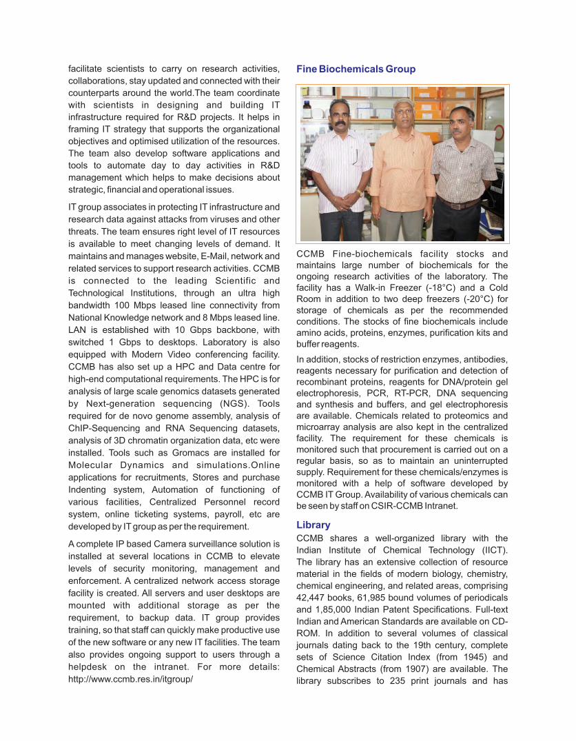
facilitate scientists to carry on research activities,
collaborations, stay updated and connected with their
counterparts around the world.The team coordinate
with scientists in designing and building IT
infrastructure required for R&D projects. It helps in
framing IT strategy that supports the organizational
objectives and optimised utilization of the resources.
The team also develop software applications and
tools to automate day to day activities in R&D
management which helps to make decisions about
strategic, financial and operational issues.
IT group associates in protecting IT infrastructure and
research data against attacks from viruses and other
threats. The team ensures right level of IT resources
is available to meet changing levels of demand. It
maintains and manages website, E-Mail, network and
related services to support research activities. CCMB
is connected to the leading Scientific and
Technological Institutions, through an ultra high
bandwidth 100 Mbps leased line connectivity from
National Knowledge network and 8 Mbps leased line.
LAN is established with 10 Gbps backbone, with
switched 1 Gbps to desktops. Laboratory is also
equipped with Modern Video conferencing facility.
CCMB has also set up a HPC and Data centre for
high-end computational requirements. The HPC is for
analysis of large scale genomics datasets generated
by Next-generation sequencing (NGS). Tools
required for de novo genome assembly, analysis of
ChIP-Sequencing and RNA Sequencing datasets,
analysis of 3D chromatin organization data, etc were
installed. Tools such as Gromacs are installed for
Molecular Dynamics and simulations.Online
applications for recruitments, Stores and purchase
Indenting system, Automation of functioning of
various facilities, Centralized Personnel record
system, online ticketing systems, payroll, etc are
developed by IT group as per the requirement.
A complete IP based Camera surveillance solution is
installed at several locations in CCMB to elevate
levels of security monitoring, management and
enforcement. A centralized network access storage
facility is created. All servers and user desktops are
mounted with additional storage as per the
requirement, to backup data. IT group provides
training, so that staff can quickly make productive use
of the new software or any new IT facilities. The team
also provides ongoing support to users through a
helpdesk on the intranet. For more details:
http://www.ccmb.res.in/itgroup/
Fine Biochemicals Group
CCMB Fine-biochemicals facility stocks and maintains large number of biochemicals for the ongoing research activities of the laboratory. The facility has a Walk-in Freezer (-18°C) and a Cold Room in addition to two deep freezers (-20°C) for storage of chemicals as per the recommended conditions. The stocks of fine biochemicals include amino acids, proteins, enzymes, purification kits and buffer reagents.
In addition, stocks of restriction enzymes, antibodies, reagents necessary for purification and detection of recombinant proteins, reagents for DNA/protein gel electrophoresis, PCR, RT-PCR, DNA sequencing and synthesis and buffers, and gel electrophoresis are available. Chemicals related to proteomics and microarray analysis are also kept in the centralized facility. The requirement for these chemicals is monitored such that procurement is carried out on a regular basis, so as to maintain an uninterrupted supply. Requirement for these chemicals/enzymes is monitored with a help of software developed by CCMB IT Group. Availability of various chemicals can be seen by staff on CSIR-CCMB Intranet.
Library
CCMB shares a well-organized library with the
Indian Institute of Chemical Technology (IICT).
The library has an extensive collection of resource
material in the fields of modern biology, chemistry,
chemical engineering, and related areas, comprising
42,447 books, 61,985 bound volumes of periodicals
and 1,85,000 Indian Patent Specifications. Full-text
Indian and American Standards are available on CD-
ROM. In addition to several volumes of classical
journals dating back to the 19th century, complete
sets of Science Citation Index (from 1945) and
Chemical Abstracts (from 1907) are available. The
library subscribes to 235 print journals and has

163
access over 4000 titles of online journals. In addition,
CCMB has access to 80 e-Journals. A collection of
PhD theses submitted by CCMB students is being
maintained by assigning proper accession numbers
since 2005. Haldane's book collection is also
maintained by the Library staff. The library has an in-
house bindery, reprographic facility, good Internet
connectivity and a security surveillance system.
Experimental Farm, Horticulture and Landscaping
The Horticulture group maintains the lush green
surroundings of the CCMB, LaCONES and CCMB
Annexure-2. The newly developed experimental
farm is being maintained by the staff of this
group to meet the requirements of the plant
genetic fingerprinting work. Different varieties
of rice (indica, japonica), maize, as well as drought-
resistant plants such as jojoba are grown in the
farm. The Green House facility developed by this
group is fully functional and is used for studies in
plant molecular biology. Plants such as paddy,
mustard and Arabidopsis are being grown here.
Rajbhasha Unit
This unit helps the Institution mainly in complying
with various provisions of Official Language (OL)
envisage by the GOI. For the past 17 years this
unit is bringing out a popular science magazine
JIGYASA in Hindi dedicating every issue to a special
topic of Life Sciences. This unit of Rajbhasha
conducts every year "HINDI DAY" on 14th September
and various Hindi competitions and programmes
are organized as part of the occasion.
This year on 24th March the Rajbhasha unit
has organized a KAVISAMMELAN EVEM
MUSHAYARA by inviting famous Hindi & Urdu
poets of Hyderabad. The Rajbhasha Unit has a
very good library consisting 2600 Hindi books
on various subjects viz., classic works of Hindi
literation, science, translations and books of
general interest. This year 81 books have been
added to this collection.
Engineering Services
CCMB Complex has a number of modern laboratories
including the Annexe-1, LaCONES at Attapur and
Medical biotechnology complex, Annexe-2, 2 Km
away from the CCMB main campus. Facilities such as
workshop, centralized air conditioning, class 10K
clean rooms, bio-safety level III labs, ultralow
freezers, walk-in cold rooms, walk-in freezer rooms,
piped gas supply, pretreated water supply, liquid
nitrogen, 3000Kva back-up generator sets, 33kv and
11Kv HT sub stations, distribution system, 30Kw solar
power unit, etc., are being operated and maintained
by the Engineering staff.

A. DiagnosticsMolecular Diagnostics
Advances in molecular and cell biology have provided
an understanding of the mechanisms of disease at
molecular and genetic levels, which can now be
translated into diagnostic, prognostic, and
therapeutic applications in modern medicine. A
number of genetic disorders are known to result from
the defects in a single gene. In the absence of specific
treatment, molecular diagnosis, carrier detection,
genetic counseling, pre-pregnancy monitoring, pre-
implantation genetic diagnosis and prenatal
diagnosis for these disorders becomes the best
approach to prevent their transmission to next
generation. The Molecular Diagnostics Facility,
CSIR-CCMB, Hyderabad provides diagnostic
services for close to 30 such monogenic disorders.
The facility provides DNA-based testing for a number
of inherited and acquired genetic diseases including
Hemoglobinopathies, Musculopathies, Bleeding and
clotting disorders and Neurodegenerative diseases,
etc. The major thrust of these diagnostic services is to
provide reliable genetic testing services to the
common man within a rapid turnaround time and at
affordable rates. In addition, the facility provides
comprehensive education & training in application of
molecular sciences for disease management.
Chromosome Diagnostics
Chromosomal abnormalities are implicated in mental
retardation, congenital malformations, dysmorphic
features, primary and secondary amenorrhea,
reproductive wastage, infertility, disorders of sex
development, neoplastic diseases. Cytogenetic
evaluation of patients is helpful in the counselling and
management of affected individuals and families.
Prenatal diagnosis of chromosomal abnormalities in
high-r isk pregnancies helps in detect ing
chromosomally abnormal fetuses. The state-of-the-
art facility offers cytogenetic tests such as
karyotyping (conventional-G banding techniques)
and FISH (fluorescence in situ hybridization which
includes probes using WCP and LSI, mFISH,
mBAND, SKY), which involves investigation of
genetic defects at the chromosome level.
B. Laboratory for the Conservation of Endangered Species (LaCONES): Facilities & services
LaCONES, CSIR-CCMB Annexe 1, is situated in
Attapur, Rajendernagar, Hyderabad. LaCONES
provides extensive service to the nation for wildlife
III.6 Services

diagnostics. The different types of services provided
at LaCONES are:
• Species identification
• Individual identification/ relatedness/paternity
testing
• Sexing of birds
• PCR based diagnosis (RTPCR) for RNA/DNA virus
related diseases
• Diagnosis of Batrachochytrium dendrobatidis (BD)
using nested PCR and qPCR
• Fertility (Progesterone/Estradiol/Testosterone)
including pregnancy detection and stress hormone
(Cortisol) analysis
• Semen and Sperm profiling with/without chemical
immobilization
Wildlife diagnostics
LaCONES is playing an important role in generating
meaningful DNA evidence for law enforcement
agencies to unambiguously identify species targeted
in poaching and illegal wildlife trade since 2000. The
DNA technique developed and patented by CCMB
allows identification of a biological specimen of
unknown origin and delineates its utility to the level of
family, genus and species. Biological specimens are
received from all over the country confiscated and
forwarded by state forest, judiciary, police and custom
departments. During the period (2000–till August
2017) a total of 1554 wildlife crime cases were solved
at LaCONES, which includes more than 3492
biological samples. This effort has also significantly
helped in increasing the awareness amongst the
forest and police officials regarding the usefulness of
this technique and also in inculcating a sense of fear
amongst the poachers. In addition, LaCONES is also
providing genetic health check-up, disease diagnosis
and reproductive hormones monitoring services to
various zoological parks in the country under
Conservation Breeding program of Central Zoo
Authority of India for 25 endangered species. More
than 100 reports have been submitted to different zoo
in the period 2007-2017.
Cryo-banking
Cryo-preservation of semen of endangered
species is an ongoing activity in LaCONES and
cryopreservation protocols have been optimized for
semen from lions, tigers, leopards, spotted deer and
black buck, blue rock pigeon and white-backed
vultures. These studies indicated that the protocol for
cryopreservation is species specific and needs to be
standardized for and each every species. The cryo-
preserved semen samples could be used for AI under
circumstances when a male factor is involved in
normal mating could be used to produce embryos by
in vitro fertilization. In contrast to semen, non-
availability of oocytes of wild animals is a major
challenge in cryopreservation of the female gamete.
Though electro-ejaculation is permitted for semen
collection, laparoscopic intervention is not permitted
for oocyte collection. An alternative approach would
be to recover oocytes from the ovaries at postmortem
activate them, mature them and store them.
LaCONES has carried out pioneering work in this
area and has succeeded in oocyte recovery,
cryopreservation, IVM and IVF of oocytes collected
post-mortem from the ovaries of black buck, nilgai
and chousingha at the Nehru Zoological Park (NZP),
Hyderabad. In addition, attempts could be made to
establish species-specific protocols for cryo-
preservation of ovaries and testes from dead wild/
endangered animals. Cryo-banking of genes, tissues,
gametes, ovaries, testes and embryos provides a
holistic approach towards preservation of the
germplasm from endangered animals. These cryo-
preserved bioresources could over the years be used
for comparing genetic polyporphism with the existing
populations and also used for somatic cell nuclear
transfer or inter-species nuclear transfer, thus
satisfying the long term goal of LaCONES in wildlife
conservation.
Lectures, Training & workshops
LaCONES also works towards generating
awareness/creating skilled resources through regular
lectures, in-house hand’s on trainings/workshops
about the importance and usage of molecular
approaches towards wildlife conservation for
dissertation students (M. Sc., B. Tech. & M. Tech),
students of National Institute of Criminology and
Forensic Science, Delhi, Officers and Probationers of
Telangana/AP Forest Academy, Central Forensic
Science Laboratory, Indian Forest Academy and
National Police Academy. The scientists also visit
forest head quarters/forest academies to impart
knowledge about DNA and other techniques and
demonstrate sample collection, storage &
transportation to the lab.
165
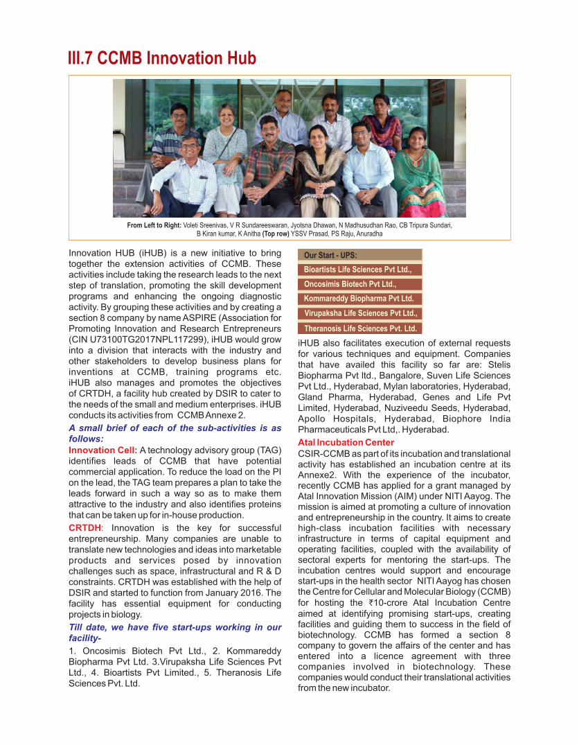
Innovation HUB (iHUB) is a new initiative to bring together the extension activities of CCMB. These activities include taking the research leads to the next step of translation, promoting the skill development programs and enhancing the ongoing diagnostic activity. By grouping these activities and by creating a section 8 company by name ASPIRE (Association for Promoting Innovation and Research Entrepreneurs (CIN U73100TG2017NPL117299), iHUB would growinto a division that interacts with the industry andother stakeholders to develop business plans for inventions at CCMB, training programs etc.iHUB also manages and promotes the objectivesof CRTDH, a facility hub created by DSIR to cater to the needs of the small and medium enterprises. iHUB conducts its activities from CCMB Annexe 2.
A small brief of each of the sub-activities is as follows:Innovation Cell: A technology advisory group (TAG) identifies leads of CCMB that have potential commercial application. To reduce the load on the PI on the lead, the TAG team prepares a plan to take the leads forward in such a way so as to make them attractive to the industry and also identifies proteins that can be taken up for in-house production.
CRTDH: Innovation is the key for successful entrepreneurship. Many companies are unable to translate new technologies and ideas into marketable products and services posed by innovation challenges such as space, infrastructural and R & D constraints. CRTDH was established with the help of DSIR and started to function from January 2016. The facility has essential equipment for conducting projects in biology.
Till date, we have five start-ups working in our facility-
1. Oncosimis Biotech Pvt Ltd., 2. Kommareddy Biopharma Pvt Ltd. 3.Virupaksha Life Sciences Pvt Ltd., 4. Bioartists Pvt Limited., 5. Theranosis Life Sciences Pvt. Ltd.
iHUB also facilitates execution of external requests for various techniques and equipment. Companies that have availed this facility so far are: Stelis Biopharma Pvt ltd., Bangalore, Suven Life Sciences Pvt Ltd., Hyderabad, Mylan laboratories, Hyderabad, Gland Pharma, Hyderabad, Genes and Life Pvt Limited, Hyderabad, Nuziveedu Seeds, Hyderabad, Apollo Hospitals, Hyderabad, Biophore India Pharmaceuticals Pvt Ltd,. Hyderabad.
Atal Incubation CenterCSIR-CCMB as part of its incubation and translational activity has established an incubation centre at its Annexe2. With the experience of the incubator, recently CCMB has applied for a grant managed by Atal Innovation Mission (AIM) under NITI Aayog. The mission is aimed at promoting a culture of innovation and entrepreneurship in the country. It aims to create high-class incubation facilities with necessary infrastructure in terms of capital equipment and operating facilities, coupled with the availability of sectoral experts for mentoring the start-ups. The incubation centres would support and encourage start-ups in the health sector NITI Aayog has chosen the Centre for Cellular and Molecular Biology (CCMB) for hosting the ₹10-crore Atal Incubation Centre aimed at identifying promising start-ups, creating facilities and guiding them to success in the field of biotechnology. CCMB has formed a section 8 company to govern the affairs of the center and has entered into a licence agreement with three companies involved in biotechnology. These companies would conduct their translational activities from the new incubator.
III.7 CCMB Innovation Hub
Our Start - UPS:
Bioartists Life Sciences Pvt Ltd.,
Oncosimis Biotech Pvt Ltd.,
Kommareddy Biopharma Pvt Ltd.
Virupaksha Life Sciences Pvt Ltd.,
Theranosis Life Sciences Pvt. Ltd.
From Left to Right: Voleti Sreenivas, V R Sundareeswaran, Jyotsna Dhawan, N Madhusudhan Rao, CB Tripura Sundari,B Kiran kumar, K Anitha (Top row) YSSV Prasad, PS Raju, Anuradha

167
IV. Administration and Management
The overall administration of the Centre and the supervision of ancillary services such as transport and
telecommunications are under the purview of the administration. In addition, secretarial assistance is provided
to the staff for the preparation of the reports, manuscripts and correspondence.
Director’s Office
From Left to Right: MR Vasanth Kumar, S Madhuri, D Lavanya, Rakesh K Mishra, RV Subba Rao, BVN Naveen Kumar, G Satyanarayana


169
Planning Monitoring and Evaluation (PME)
The primary responsibility of this group is toassist the Director CCMB, in all activities of CCMB and act as a liaison between the Director and the other research groups, CSIR-HQ and other organisations. The PME unit takes care of variousin-house, sponsor, collaborative, grant-in-aid,and NIMTLI projects and provides inputs in the preparation of project proposals and other documents and reports of the CCMB. It organizes National and International visits of Presidents, Ministers, Par l iamentar ians and Sciencedelegations to the Centre. This group co-ordinates organising national and international seminars, symposia, workshops, conferences and training programs and maintains contacts with all CSIR
departments in obtaining sanctions and required approvals for this activity. In addition it coordinates periodical audit sessions on CCMB by CSIR audit teams. PME takes up Diagnostic services and coordinates popular school visits. The team also participates in conferences, exhibitions to showcase CCMB technologies in coordination with CSIR-HQ. This group also coordinates data for sending RTI replies.
During the year, the group has developed PROJECT
is, a project IT module, with the help of the IT division
of CCMB by providing all inputs and data relevant to
the maintenance of projects. The module contains
details of ongoing projects in a user-friendly format
with a provision of obtaining status reports of any in-
house project at a given time including financials.
Management Support Services
From Left to Right: P Satguru, MV Subbarao, PS Raju, Gulzar Khan, MR Vishnu Priya, K Anitha, K Satyanarayana, BV Ramakrishna
Guest House StaffCCMB guest house has 28 well-furnished suites/ rooms and accommodates visitors from India and Abroad.
Canteen ServicesCCMB Canteen has three restaurants that serve all catering needs of the staff and is functional on allworking days.

Business Development Unit
This unit interacts with sponsors in identifying their
needs and generating project proposals. It also
participates in conferences, exhibitions to showcase
CCMB technologies and coordinates with the CSIR in
filling and granting of patents.
Publications and Public Relations
The main responsibility of this unit is to make the
general public aware of the latest developments of
the Centre. Information about CCMB research is
conveyed to public via print and electronic media,
publications such as Annual Report and through other
brochures. Press releases describing CCMB’s new
initiatives, innovations, scientific leads and path-
breaking results are given to both national and local
level newspapers. During the year, several press
meets were organized to explain the major
contributions of CCMB to the electronic media by
CCMB scientists.

V. General Information


Research Council and Management Council
A. Research Council
Research Council of a laboratory under CSIR provides direction and vision and helps it to formulate R & D
programmes keeping in view the National priorities and opportunity niches and facilitates to design a road map
to achieve it. The following are the constituent members.
Dr Rajesh Gokhale MemberDirectorInstitute of Genomics and Integrative BiologyDelhi
Prof Ram Rajasekharan MemberDirectorCSIR-Central Food Technological Research
Institute Mysore
Prof Siddhartha Roy MemberSenior Professor & Dean of StudiesBose InstituteKolkata
Dr Rakesh K Mishra MemberDirectorCentre for Cellular & Molecular BiologyHyderabad
Dr Sudeep Kumar MemberHead, Mission DirectorateCSIRNew Delhi 110 001
Shri A V Chainulu MemberScientist FDSIR, Technology BhawanNew Delhi
Dr K Thangaraj SecretarySenior Principal ScientistCentre for Cellular & Molecular BiologyHyderabad
Dr Sandip K Basu ChairmanProfessor of Eminence
National Institute of Immunology
New Delhi
Prof Vidyanand Nanjundiah Member
Centre for Human Genetics
Bangalore
Dr Dinakar M Salunke Member
Executive Director
Regional Centre for Biotechnology
Gurgaon
Prof J P Khurana Member
Professor
Department of Plant Molecular Biology
University of Delhi South Campus
New Delhi
Prof R Sukumar Member
Centre for Ecological Sciences
Indian Institute of Science
Bengaluru
Dr Krishna Ella Member
Chairman and Managing Director
Bharat Biotech International Limited
Hyderabad
Dr T S Rao Member
Adviser
Department of Biotechnology
New Delhi
173

Research Council and Management Council
B. Management Council
Following is the composition of the Management Council of CSIR-CCMB for the period 01.07.2016 to
31.12.2017 as approved under Rule-65 of the CSIR Rules & Regulations:
Dr. Ramesh V. Sonti MemberChief Scientist CSIR-CCMBHyderabad
Dr. A.S. Sreedhar MemberPrincipal Scientist CSIR-CCMBHyderabad
Shri Y V Rama Rao MemberPrincipal Technical OfficerCSIR-CCMBHyderabad
Prof. R K Sinha MemberDirectorCSIR-IMTECHChandigarh-160036
Finance & Accounts Officer MemberCSIR-CCMBHyderabad
Dr. Rakesh K Mishra ChairmanDirector, CSIR-CCMBUppal Road Hyderabad-500007
Dr. Bokara Kiran Kumar MemberScientist CSIR-CCMBHyderabad
Dr. H.H. Krishnan MemberSenior Scientist CSIR-CCMB Hyderabad
Dr. V.M. Tiwari MemberDirector CSIR-NGRI Hyderabad-500007
Dr. M R Vishnu Priya MemberHead, PME, CSIR-CCMBHyderabad
Controller of Administration Member-SecretaryCSIR-CCMBHyderabad

Ongoing MOUs & Agreements
CRTDH MOUs and License Agreements
Ÿ NIMS, Hyderabad
Ÿ Theranosis Life Sciences Pvt. Ltd, Hyderabad
Ÿ National Research Development Corporation
(NRDC), New Delhi
Ÿ Virupaksha Lifesciences Pvt. Ltd. Hyderabad
Ÿ Bioartis Life Sciences Pvt. Ltd., Hyderabad
Ÿ Oncosimis Biotech. Pvt. Ltd.
Ÿ IKP Knowledge Park
Ÿ Kommareddi Biopharma Private Limited
Ÿ DME, Govt of Telangana
Ÿ iBUILD Innovations India Ltd.
Ÿ Bioserve Biotechnologies (India) Pvt. Ltd.
Atal Incubation Centre-CCMB CRTDH
Agreement
Ÿ Magellan Life Sciences Private Limited,
Hyderabad
Ÿ Lycan Lab, Bangalore
Ÿ SAATRA Capital Advisors LLP
Ÿ Royal Melbourne Institute of Technology,
Melbourne, Australia
Ÿ DUPONT India Pvt. Ltd. Hyderabad
Ÿ Acharya N.G. Ranga Agrl. University, Hyderabad
Ÿ King Edward Memorial Hospital Research Centre,
Pune
Ÿ Directorate of Rice Research, Hyderabad and
Metahelix Life Sciences, Hyderabad
Ÿ MNR Dental College & Hospital, Medak District
Ÿ NIMHANS, Bangalore
Ÿ Madras Diabetes Research Foundation, Chennai
Ÿ Sunshine Medical Academy of Research &
Training, Hyderabad
Ÿ Directorate of Rice Research & Agri Biotech.
Foundation, Hyderabad
Ÿ Deccan College Post Graduate and Research
Institute, Pune
Ÿ All India Institute of Medical Sciences, Delhi
Ÿ L.V. Prasad Eye Institute, Hyderabad
Ÿ Sickle Cell Institute Chhattisgarh, Raipur
Ÿ Nizam’s Institute of Medical Sciences, Hyderabad
Ÿ Administrative Staff College of India
Ÿ Indian Institute of Rice Research (IIRR),
Hyderabad
Ÿ National Academy of Agricultural Extension
Management (MANAGE) Hyderabad
Ÿ Telangana State Director of Medical Education
Ÿ B.E. Ltd
Ÿ IncuMed, Hyderabad
Ÿ CCMB-IMMT-Genomix Biotech Inc. USA
175

Visitors to CCMB & SEMINARS
Dr Sambashiva Banala, Howard Hughes Medical Institute, Ashburn, VA,
USA. "Development of Novel Chemical Tools for
Biological Applications" June 27,2016
Dr Arni S.R.Srinivasa Rao, Dept of Biostatistics & Epidemiology, Augusta
University, USA "Modeling movement of poultry birds for
understanding parasite spread" June 29,2016
Dr Thirumala-Devi Kanneganti, St. Jude Children's Research Hospital,Memphis,
USA "Regulators of Inflammatory Responses"
June 30,2016
Dr Gopinath Meenakshisundaram, Laboratory of Translation Control of Disease, Institute of Medical Biology, Singapore. "Can injury cause cancer? Epithelial carcinomas
hijack a molecular switch from wound healing to
promote their invasion" July 04,2016
Dr Tofajjen Hossain Sk, University of Miami, Miami, FL, USA "Breaking Down Barriers: How RNase R Degrades
Structured RNA" July 18,2016
Dr Regalla Kumarswamy, Hanover Medical School, Hannover, Germany "Functional non-coding RNAs in patho-physiology
of the heart" July 28,2016
Dr Jameel Ahmad Khan, Lifecode Technologies Pvt. Ltd.New Delhi "Good Laboratory Practices (GLP) in Cell
Culture lab: Cell Line Authentication and
Cross-Contamination" August 05,2016
Dr Ashish Arora, Molecular and Structural Biology Division, CSIR-
Central Drug Research Institute, Lucknow "Unraveling the stereochemical and dynamic
aspects of the catalytic site of bacterial peptidyl-
tRNA hydrolase" August 09,2016
Dr Pradeep Kumar Burma, Department of Genetics, University of Delhi South Campus, New Delhi "Developing insect resistant Bt cotton lines - our
experience" April 04,2016
Dr Manikandan Narayanan, National Institutes of Health, Bethesda, MD, USA "Gene networks in health and disease: a
computational systems approach" April 18,2016
Dr Suhel Quader, Scientist - Nature Conservation Foundation &
Head - Citizen Science Programmes at NCBS,
Bangalore "Citizen Science: a powerful approach to
ecological research and monitoring" April 19,2016
Dr Samarendra K Singh, University of Virginia, Charlottesville, Virginia,
USA "Regulation of Cell Cycle and Replication in
Cancer Cells" May 10,2016
Nathan Ikaika Joel Naktsuka, Harvard Medical School, Boston, USA "Admixture Mapping in African-Americans
Uncovers Variants Explaining the Difference in
Prevalence of Multiple Sclerosis in Europeans
Compared to Africans" June 03,2016
Dr Jairam K.P. Vanamala, Center for Molecular Immunology & Infectious
Diseases, Penn State University, USA "Food-Approach to Target Cancer Stem Cells"
June 14,2016
Dr Anita Malhotra, Bangor University, UK "Anolis lizards: Coping with climate change"
June 14,2016
Dr Wolgang Wuster, Bangor University, UK "Why is snake venom composition so variable?"
June 15, 2016
The national and international linkages forged by the CCMB resulted in drawing a large number of scientists,
experts, consultants and applicants in the frontier areas of cellular and molecular biology to visit and deliver
lectures at the Centre. The list of such seminars organized during the year is given below:
177
Dr S Subramanian, Dept of Surgery, University of Minnesota, USA "Mechanisms of Malignant Transformation
and Immune Response in Colon Cancer"
August 16,2016
Dr P Suresh, National Animal Resource Facility, NIN,
Hyderabad "CPCSEA SOP for IAEC - Role and responsibilities
in animal experimentation" August 17, 2016
Prof Geeta K Vemuganti, School of Medical Sciences, University of
Hyderabad, Hyderabad "Regulatory approvals for conducting Stem Cell
Research in India: an overview" August 17,2016
Dr Aruni Wilson, Division of Microbiology and Molecular Genetics, School of Medicine, Loma Linda University, USA "Novel Microbial factors leading to microbiome
dysbiosis predisposing to systemic diseases "
August 29,2016
Dr Shankar Srinivas, Department of Physiology Anatomy & Genetics,
University of Oxford, UK. "Development of form and function of the
embryonic heart" August 29,2016
Dr Marilyn J. Telen, Duke University Medical Center, Durham, North
Carolina, USA "Outcome Modifying Genes in Sickle Cell Disease
and Outlook for New Pharmaco therapeutic
Targets" September 14,2016
Dr Dhanasekaran Shanmugam, Biochemical Sciences Division National Chemical
Laboratory, Pune "Metabolic plasticity dictates fitness and
differentiation in Toxoplasma gondii and reveals
potential targets for therapeutic intervention"
September 22,2016
Prof L S Shashidhara, Indian Institute of Science, Education & Research
(IISER), Pune "Growth control: lessons from Drosophila for
cancer biology" October 05,2016
Dr Suchitra Gopinath Pediatric Biology Center, THSTI, Faridabad "Vitamin D signaling in skeletal muscle growth and
atrophy" October 19, 2016
Dr Randal Halfmann, Stowers Institute for Medical Research, Kansas
City, USA "Quantitative Prion Biology" October 20, 2016
Dr Chetana Sachidanandan, IGIB New Delhi "Modeling human disease in zebrafish" October
20,2016
Dr Ramkumar Sambasivan, InStem, Bengaluru "Mesoderm patterning and early vertebrate
development" October 20,2016
Dr Gireesha Mohannath, Departments of Biology, Molecular and Cellular Biochemistry, Indiana
University, USA "New hypothesis: chromosomal context
determines activity status of rRNA genes on a
multimegabase scale in Arabidopsis" October
28,2016
Dr Srimonta Gayen, Department of Human Genetics, University of
Michigan Medical School, USA "Epigenetic regulation by long non-coding
RNAs and histone modifiers through the lens of
X-chromosome inactivation" November 03, 2016
Dr Ratna Ghosal, Minnesota Aquatic Invasive Species Research
Center, University of Minnesota, USA. "From endangered to invasive species: hormone-
pheromone-behavior interactions in conservation"
November 10,2016
Dr Venkata R Chalamcharla, National Cancer Institute, NIH, USA "Transcription termination primes RNA-mediated
epigenetic genome control" November 16,2016
Prof. Rekha Samuel, MC Vellore "Pathophysiology and Molecular Pathways
Regulating Pericyte Phenotype in Type 2
Diabetes. The Gestational Diabetes Mellitus
Placental Model." November 17,2016
Dr Kartik Sunagar, Hebrew University of Jerusalem, Israel "Deadly innovations. ‘Venomics’ for evolution,
ecology, and snakebite management" November
18,2016

Dr Shekhar Kumar Niraj, IFS, TRAFFIC - India at World Wide Fund for Nature,
Delhi "Illegal Wildlife Trade Scenarios in India and South
Asia" November 22,2016
Dr Santosh Kumar, Stanford University, USA "Molecular Events of Lymphocyte Recognition and
Response" December 02,2016
Dr Dipankar Bhandari, Max Planck Institute for Developmental Biology,
Tubingen, Germany "Role of the CCR4-NOT deadenylase complex in
the post-transcriptional gene silencing" December
14,2016
Dr Sergey Naidenko, A.N. Severtsov Institute of Ecology and Evolution,
Moscow, Russia "Behavioral Ecology of carnivores in Russia"
December 19,2016
Dr John Waitumbi, United States Army Medical Research Directorate
(USAMRU), Nairobi, Kenya "Effects of Gender on the Intra-erythrocytic
concentration of the Malaria Protective deoxy-
hemoglobin S and Interactions of alpha-thal
and G6PD Genotypes on Malaria in Children
Living in the Lake Victoria Basin, Kenya"
December 20, 2016
Prof James Bradley, University of Liverpool, England "Plasma discharges for the ambient processing
and detection of materials" December 21, 2016
Dr Ganesh Bagler, IIIT, Delhi "Data-driven discovery of the molecular essence
of Indian cuisine" December 23,2016
Dr Kaustuv Sanyal, JNCASR, Bangalore "Genome indexing by histone H3 variants in
Candida albicans" December 27,2016
Dr Bhavana Muralidharan, Dept of Biological Sciences, TIFR, Mumbai "Molecular mechanisms regulating cell fate
specification in the developing cerebral cortex"
January 12,2017
Dr Dhananjay Huilgol “Developmental mechanisms of projection
neurons in the forebrain” January 12, 2017.
Prof Aseem Ansari, The Genome Center of Wisconsin, Department of Biochemistry, University of
Wisconsin-Madison, USA "New writers and old readers of under-explored
phosphorylation marks on the tail of RNA
polymerase II" January 17, 2017
Dr Sundar Balasubramanian, Medical University of South Carolina, Charleston,
USA "A Journey from Pranayama to Biomarkers"
January 24,2017
Dr Dibyendu Bhattacharyya, Advanced Centre for Treatment, Research and
Education in Cancer (ACTREC), Tata Memorial Centre, Navi Mumbai "Size, Shape and Dynamics of Golgi and ER"
January 24,2017
Dr Robert J. Freishtat, George Washington University School of Medicine
and Health Sciences, Washington, USA "Adipocyte-der ived exosomes: a novel
mechanism for obesity-related disease" January
25,2017
Dr Jose Sebastian, Carnegie Institution for Science, Stanford
University, USA "Dealing with Stress: Cereal roots enact austerity
measures during drought to bank water" February
06,2017
Dr Jayasri Das Sarma, Department of Biological Science, IISER, Kolkata "Gliopathy are early events in viral induced CNS
demyelination" February 15,2017
Prof Andrew Clayton, Swinburne University of Technology, Melbourne,
Australia "Can fluorescence help us to understand the
structure of cell-surface molecules?" February
20,2017
Prof Jean-Marie Ruysschaert, Universite Libre de Bruxelles, Brussels, Belgium "Protein and lipid nanoparticles activate the
immune system through recognition of Toll-like
receptors" February 21,2017
Dr Nigel M. Hooper, University of Manchester, Manchester, U.K. "Lipid Rafts in Prion Biology and Alzheimer`s
Disease" March 06,2017

Dr Bruce A. Shapiro, RNA Biology Laboratory, National Cancer Institute, NIH, Frederick,
Maryland, U.S.A. "Understanding RNA Structure and Function:
Prediction, Analysis and Nanodesign" March
07,2017
Dr Alok K Sinha, National Institute of Plant Genome Research, New
Delhi "Submergence Tolerance in Rice: A new twist in
regulation of SUB1A1 by a mitogen activated
protein kinase" March 09,2017
Dr Murali Krishna Cherukuri, National Cancer Institute, NIH, Bethesda, USA "Metabolic MRI: Preclinical and Clinical
applications in Cancer Imaging" March 17,2017
Dr Somenath Bakshi, Systems Biology Laboratory, Harvard University,
USA "Single-cell Measurement of Microbial Stress-
response Dynamics in Complex Growth
conditions" March 17,2017
Dr Prasad Kasturi, Max-Planck Institute for Biochemistry, Munich,
Germany "Protein homeostasis during stress and aging in
C.elegans" March 20,2017
Dr Ritesh Kumar, Texas A&M, Health Science Centre, Houston,
Texas, USA "Microbes meet cancer: Role of Streptococcus
gallolyticus in colorectal tumor development"
March 21,2017
Dr Saikat Chowdhury, Dept. of Integrative Structural & Computational
Biology, The Scripps Research Institute, La Jolla, USA "Visualizing dynamic cellular machinery with
electron microscopy" March 30,2017
Dr Gyaneshwer Chaubey, Department of Evolutionary Biology, Estonian
Biocentre, Tartu "Peopling of South Asia: Genesis and
Implications" March 31,2017
• Nagindas Khandwala college,
• Jain University, Banglore
• Sree Sankara College, Kalady, Ernakulam
• Abhyasa School, Medak
• St.Xavier college, Kerala
• Sathaye college, Mumbai
• Davangere University, Karnataka
• National Institute of Nutrition(NIN), Hyderabad
• MES Mampad college, Kerala
• St.Joseph college, kerala
• Govt. Degree college, Siddipet
• University of North Bengal, West Bengal
• Sage School, Hyderabad
• Kerala Veternary& Animal Sciences University,
Mannuthy
• Orissa University of Agriculture and Technology,
Bhubaneswar
• PSGR Krishnammal college for women,
Coimbatore
• LV Prasad Eye Institute, Hyderabad
• Ahmediya International School, Mauritius
• Wisconsin College,USA
• Science INDIA Forum, Bahrain
• Edinberg University Professors, UK
• Kendriya Vidyalaya AFS,Hyderabad
• ASCI,Hyderabad
• Telangana Tribal Welfare Society, All districts of
Telangana
• College Of Veterinary Science, Gujarat
• Bhavans College, Mumbai
• Fergussons College, Pune
• Indian Institute of Rice Research, Hyderabad
• University of Kashmir, Srinagar
• CMS College of Science & Commerce, Coimbatore
• Nizams Institute of Medical Sciences, Hyderabad
• Shivaji University, Kohlapur
• KIIT University, Bhubaneshwar
• Loyola College, Hyderabad
• Savithribai phule University, Pune
• Tamilnadu Agricultural University, Tamilnadu
Visits to CCMB by students/faculty/experts during 2016-17
CCMB encourages visitors from different parts of the country and abroad. During the year 2016-17, the total
visits were 60 and the visitors were 1913. Some of the visitors during 2016-17 are as follows:
179
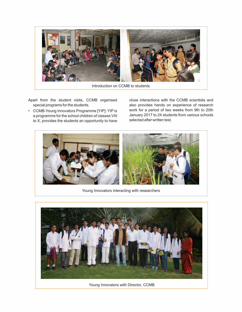
Apart from the student visits, CCMB organised
special programs for the students.
• CCMB-Young Innovators Programme [YIP]: YIP is
a programme for the school children of classes VIII
to X, provides the students an opportunity to have
Introduction on CCMB to students
close interactions with the CCMB scientists and
also provides hands on experience of research
work for a period of two weeks from 9th to 20th
January 2017 to 24 students from various schools
selected after written test.
Young Innovators interacting with researchers
Young Innovators with Director, CCMB

181
The precursor events of IISF 2016 at CSIR-CCMB, Hyderabad from November 01-15, 2016:
CSIR-Centre for Cellular and Molecular Biology, Hyderabad planned and executed several programmes from
November1 to 15, 2016 as a part of India International Science Festival (IISF-2016), under the Ministry of
Science and technology and Ministry of Earth sciences in association with Vigyana Bharati (VIBHA). The event
involved visit to CCMB by many students, faculty and public.
A. Research staff
Amitabha Chattopadhyay
* Awarded The World Academy of Sciences
(TWAS) Prize in Biology
* Elected as Associate Editor, Molecular
Membrane Biology
* Elected as Member, Wellcome Trust DBT India
Alliance's Early Career Fellowship Selection
Committee
* Invited to be Discussion Leader, Gordon
Research Conference on Bio interface Science:
Active, Adaptive, and Responsive Bio interfaces,
Les Diablerets, Switzerland
* Invited to give Keynote Lecture at the IUBMB/
IUPAB/IUPS Joint Advanced School on
Receptors and Signaling, Spetses island,
Greece
* Invited to give 74th CSIR Foundation Day
Lecture, Central Drug Research Institute,
Lucknow
* Invited to give Mentor Lecture at the Science
Communication Workshop, DBT-
* Wellcome Trust India Alliance, Hyderabad
Ch. Mohan Rao
Life Time Achievement Award(Felicitation by the
Chief Minister of AP, on the occasion of AP Science
Congress, Andhra Pradesh Akademi of Sciences,
Tirupati) (2016)
Adjunct Professor, RMIT University, Melbourne,
Australia (2016-2019)
Honorary Visiting Professor, University of Mysore
(2016-)
Adjunct Professor, Dept of Microbiology, Mangalore
University, Mangalore (2016-
Invited talks and conference presentations during
the year:
* IUPAB Council meeting, The 19th International
Union of Pure and Applied Biophysics (IUPAB)
and 11th European Biophysical Societies'
Association (EBSA) Congress, Edinburgh,
Scotland, 16-10 July, 2017
* “Clusterin: full-length protein and one of its chains show opposing effects on cellular lipid accumulation” 5th Conference of the Asia Pacific Protein Association and the 12th International Symposium of the Protein Society of Thailand (5th APPA/12th PST joint conference), Bangsaen, Thailand, 11-14 July, 2017.
* 27th The World Academy of Sciences, General
Meeting, Kigali, Rwanda, 12-17 November,
2016.
Rajan Sankaranarayanan
Dr. M. R. Das Memorial lecture, 2017 awarded by
INSA, New Delhi
Yogendra Sharma
Research featured on the Cover Page of American
Chemical Society journal Biochemistry, March 7,
2017 issue (http://pubs.acs.org/toc/bichaw/ current).
Jyotsna Dhawan
* Member, Scientific Advisory Committee, World
without GNE Myopathy
* Member, Board of Governors, Trans-Disciplinary
University, Bengaluru
* Co-Organizer, International Congress of Cell
Biology 2018
* DBT-Danish Research Foundation grant for
Indo-Danish Col laborat ive Center for
Musculoskeletal Stem Cells and Regeneration
(MUSTER)
Purnima Bhargava
Invited as Session Chair and Speaker at “The 10th
International Conference on RNA polymerases I, III,
IV and V”, University of Michigan, Ann Arbor, USA
from June 24th-27th, 2016.
Mandar Deshmukh
Invited speaker at 7th Asia Pacific NMR Symposium,
Bangalore, 02/2017.
Promoted speaker at 27th ICMRBS (International
Conference on Magnetic Resonance in Biological
Systems) held at Kyoto, Japan 08/2016.
Amit Asthana
Received Award of Honor, as Distinguished Speaker
at Lab-on-a-Chip and Microfluidic Conference,
Organized by Select Bio India, 26th-27th May 2016,
Bangalore, India.
Institutional Honours
Honours & Awards
An inter-institutional team of researchers from CCMB and the Indian Institute of Rice Research (formerly
Directorate of Rice Research) have received the BIOTECH PRODUCT PROCESS DEVELOPMENT AND
COMMERCIALIZATION AWARD-2016 awarded by Department of Biotechnology, Government of India.

Regalla Kumaraswamy
Wellcome Trust/ DBT India Alliance Intermediate
Fellowship
Lekha Dinesh Kumar
* Selected as the external expert for DME
Telangana MERIT program.
* Editor of Springer protocols Molecular Biology
Series; RNA interference and cancer therapy.
K. Lakshmi Rao
Elected as Fellow of Andhra Pradesh Academy of
Sciences (FAPAS), Amravathi, AP, India 2016
V. Vijaybhaskar
Selected as Editorial board member of International
Journal of Applied Agricultural Sciences(IJAAS),
Science Publishing Group, New York, USA.
Ira Bhatnagar
Received International Association of Advanced
Materials Scientist Medal (IAAM Scientist Medal) for
the year 2017 from the International Association of
Advanced Materials at the International Conference
on Nanomaterials and Nanotechnology 2017, 01st
March 2017.
Manjula Reddy
* Invited speaker at EMBO conference on
‘Bacterial cell division: orchestrating the ring
cycle’ held at Prague, Czech Republic from 14-17
September 2016
* Invited speaker at EMBO conference on
‘Bacterial Morphogenesis, Survival and
Virulence: Regulation in 4D held at Trivandrum,
India from 27-30 November 2016
Umapathy
* Delivered a talk and participated in the Science
Camp, sponsored by the Department of Science
and Technology (DST), Government of India as
part of its INSPIRE program, at SKR & SKR Govt.
College for Women, Kadapa, Andhra Pradesh
from 20th Oct 2016 to 24th Oct 2016
* Delivered a talk and participated in the Science
Camp, sponsored by the Department of Science
and Technology (DST), Government of India as
part of its INSPIRE program, at LOYOLA
DEGREE & PG COLLEGE (YSRR) ,
PULIVENDLA, Kadapa(dt), Andhra Pradesh
from 29th November to 3rd December 2016.
G. Mala
Received Travel Grant from DBT - INDIA and CSIR
to attend and present a poster at the 84th European
Atherosclerosis Society Congress at INNSBRUCK--
AUSTRIA held between May 29th-June1st-2016.
Sanjeev Chavan Nayak
Award of National Overseas Scholarship, Under the
Scheme of National Overseas scholarship for ST
students, Government of India, Ministry of Tribal
Affairs.
B. Awards to PhD students and other
staff members
Anand Kumar Sharma
* Awarded "National Institute of Diabetes and
Digestive and Kidney Diseases (NIDDK)
Scholarship" from Keystone Symposia meeting
New Therapeutics for Diabetes and Obesity
(G1), held at Estancia La Jolla Hotel & Spa, La
Jolla, California, USA April 17-20, 2016.
* Selected for the Travel award for oral/poster
presentation in “Neuronal Calcium Sensors in
health and disease” Meeting, Germany,
December 3-7, 2016.
Shanti Swaroop Srivastava
2017 Ratna Phadke Award of Indian Biophysical
Society (IBS), Annual Meeting at IISER, Mohali.
Radhika Khandelwal
* Selected for the Travel award for oral/poster
presentationin “Neuronal Calcium Sensors in
health and disease” Meeting, Germany,
December 3-7, 2016.
* Received Carl Storm International Diversity
(CSID) fellowship from Gordon Research
Conference (GRC) to attend GRC-Calcium
Signaling Conference at Renaissance Tuscany Il
Ciocco Lucca (Barga), Italy, 2017.
M. Jafurulla
Awarded IUBMB/IUPAB/IUPS fellowship to
participate in IUBMB/IUPAB/IUPS Joint Advanced
School on Receptors and Signaling, Spetses island,
Greece.
Parijat Sarkar
Awarded FEBS Youth Travel Fund to participate in
FEBS advanced course on Lipid-protein interactions
and organelle function, Spetses island, Greece.
Debarya Saha
Travel Award for Poster Presentation at Annual
Review of Research 2017 (inStem).
183
Pratibha Bhalla
* Recipient of EMBO travel grant for “Gene
transcription in yeast: from chromatin to RNA and
back” conference held at Sant Feliu de Guixols,
Spain (June 11-16, 2016).
* Recipient of EMBL travel grant for “Transcription
and Chromatin” conference held at EMBL,
Heidelberg, Germany (Aug 27-31, 2016)
Ashutosh Shukla
* Recipient of EMBO travel grant for “Chromatin
and Epigenetics” conference held at EMBL,
Heidelberg, Germany (May 3-6, 2017).
* Best poster presentation award in “11th Asian
Epigenomics meeting”, held at JNCASR,
Bengaluru, India (September 30-October 1, 2016).
* Recipient of EMBO travel grant for “Gene
transcription in yeast: from chromatin to RNA and
back” conference held at Sant Feliu de Guixols,
Spain (June 11-16, 2016).
Prabhavathy Devan
Awarded DST National PDF fellowship
Renu Sudhakar
Received Best POSTER AWARD at the "Malaria
Parasite Biology: Drug Designing & Vaccine
Development” conference at Nirma University,
Ahmedabad, September 9-10, 2016.
Participation in International Meetings, Courses and DeputationsVijaya GopalItalyApril 18-22, 2016
G SrinivasUSAMay 9-13, 2016
Anant Bahadur PatelSingaporeMay 7-13, 2016
Prof Amitabha ChattopadhyayGreeceMay 23-27, 2016
Mohammad JafurullaGreeceMay 23-27, 2016
Prof Amitabha ChattopadhyaySwitzerlandJune 12-17, 2016
G R ChandakChinaJuly 1-3, 2016
V RadhaCzech RepublicJuly 21-25, 2016
Jyotsna DhawanUSAJuly 24-29, 2016
Mandar V DeshmukhJapanAugust 21-26, 2016
R SankaranarayananSouth KoreaSeptember 4-8, 2016
Manjula ReddyCzech RepublicSeptember 14-17, 2016
K ThangarajItalySeptember 16-19, 2016
K ThangarajAustraliaOctober 11-16, 2016
Rakesh K MishraSouth KoreaOctober 12-14, 2016
Mandar V DeshmukhGermanySeptember 4-8, 2017
Jyotsna DhawanFranceSeptember 19, 2017
UmapathyRussiaMarch 19 - 31, 2017

The Annual Meeting of "Society for
Neurochemistry India (SNCI) & Workshop”
The National Conference on “Recent trends in
Neurological and Psychiatric Research” was held
from December 9 to 11, 2016, followed by a Workshop
on “Modern Molecular Techniques to study
Neurological and Psychiatric Disorders” from
December 12 to 16, 2016. There were interesting
lectures by 33 faculty from various prestigious
research institutions and hospitals in the country,
attended by 133 participants from various
universities, colleges and research institutions in the
count ry. There was a cu l tura l program
‘PANCHABHOOTAS’ by Shri Sanjay Kumar Joshi
and disciples December 10, 2016.
Workshop on "High-resolution
Respirometry: February 07 - 08, 2017”
The CSIR-CCMB and the SMRM, OROBOROS
Instruments (Austria), had jointly organized a
workshop on High Resolution Respirometry for 20
participants from February 07-08, 2017, in the CCMB.
The workshop involved a lecture by Dr. Robert
Pitceathly, UCL, London on “Clinical and molecular
characteristics of adult mitochondrial disorders” and
Demo- experiment on High-resolution Respirometry,
discussion on hands on data analysis and Q&A
session on design of experimental protocols.
6th Meeting of Asian Forum for
Chromatin and Chromosome Biology:
March 03-05, 2017
Since 1996, the CSIR-CCMB and the Jawaharlal
Nehru Centre for Advanced Scientific Research
(JNCASR), Bengaluru, have been alternately hosting
the biennial meetings of the Asian Forum for
Chromatin and Chromosome Biology. The 6th
Meeting was hosted by the CSIR-CCMB from March
03 to 05, 2017 in the CCMB. 193 participants
including 30 National and International Speakers and
students, postdoctoral fellows from India and abroad
participated the meeting, which involved 21 lectures
and 16 short talks.
Conference on "Next Generation
Sequencing, 22 - 24 Feb 2017”
The CSIR-CCMB and the Bioserve Bio-
technologies (India) Private Limited, jointly
organized the Second Conference on Next
G e n e r a t i o n S e q u e n c i n g f r o m F e b r u a r y
22 to 24, 2017, in the CCMB. The conference
involved lectures on Animal Genomics, Plant
Genomics and Cancer Genomics. In the
second hal f of the February 22, 2017,
the CCMB Founder’s Day was celebrated
mark i ng t he 89 th b i r t hday o f CCMB ’s
founder Director, Dr. Pushpa Mitra Bhargava.
Conferences, Symposia & Training Programmes
185

3-day hands on training workshop on DNA-
based wildlife forensic diagnostics: 23-25
Mar 2017
CCMB-LaCONES is playing an important
role in generating meaningful DNA evidence
for law enforcement agencies to unambiguously
identify species targeted in poaching and
illegal wildlife trade since 2000. Partnership with the
line agencies involved in controlling and preventing
wildlife crimes is crucial for effective exchange of
information. Based on this, a 3-day hands-on training/
workshop - “DNA-based wi ld l i fe fo rens ic
diagnostics”, was conducted at CCMB Annexe-I
(LaCONES) from March 23 to 25, 2017.
There were 11 participants from Wildlife
Institute, colleges and government organizations,
who got an opportunity of hands-on practical
t raining on biological sample col lect ion,
preservation, DNA isolation, quantification,
PCR amplification, DNA sequencing and data
analysis for species identification, individual
identification, relatedness and sexing.
Events & Popular talks

187


189

Anant B Patel Group
Anant B Patel Principal Scientist
K S Vardarajan Technical Assistant
Pravin Kumar Mishra Ph.D. extension
Narayan Datt Soni SRF
Dipak Roy JRF
Salil Saurav Pathak Project SRF
Kamal Saba Project Assistant-III
Madhuri Puvvada Project Assistant-II
Sreemantula Arun Kumar Project Assistant-II
Shoumik Roy Project Assistant-II
Pooja Gautam Project JRF
Sampath Kumar T K Project JRF
Arvind Kumar Group
Arvind Kumar Senior Scientist
Sachin Singh Scientist
Avijeet Kamle Project Investigator
Shams Ul Haq Talee SRF
Unis Ahmad Bhatt SRF
Nitin Khandelwal Research Associate-I
G R Chandak Group
G R Chandak Sr Principal Scientist
K Radha Mani Principal Tech Officer
Seema Bhaskar Senior Tech Officer (3)
Inder Deo Mali Lab Assistant
P Ashok Lab Attendant (2)
Meraj Ahmad Ph.D. extension
N Suraj Singh Ph.D. extension
Dilip Kumar Yadav Ph.D. extension
Prachand Issarapu SRF
Ashutosh Singh Tomar SRF
Lovejeet Kaur Research Associate-I
Ajay Deepak Verma Research Associate-I
Radhika P Ramachandran Project Assistant-III
Vishlavath Jyothi Project Assistant-II
Ramya B Project Assistant-II
Swathi Bayyana Project Assistant-II
Pujitha Kommineni Project Assistant-II
Vinay Donipadi Project Assistant-II
Challapalli Mounika Project JRF
Sara Sajjadi Project JRF
Akshay Dedaniya Project JRF
Jyotsna Dhawan Group
Jyotsna Dhawan Chief Scientist
Nandini Rangaraj Sr Principal Scientist
Ch Sudhakar Sr Principal Scientist
C Subbalakshmi Principal Tech Officer
Suman Bhandari Technical Asst [FACS]
Sujoy Deb SRF
Debarya Saha SRF
Swetha S JRF
Priti A S JRF
Gunasekaran Subramaniam Research Associate-I
Gunjan Purohit Research Associate-I
Prabhavathy Devan Research Associate-I
Lamuk Zaveri Project Assistant-III
Amena Saleh Project Assistant-III
Ananga Ghosh Project Assistant-II
Ajoy Aloysius Project SRF
Swetha Gopal Project JRF
K Thangaraj Group
K Thangaraj Sr Principal Scientist
Nitin C Tupperwar* Senior Scientist
G Mala Senior Tech Officer (3)
S Deepa Selvi Rani Senior Tech Officer (2)
Jagamohan Chhatai Technical Assistant
D V S Sudhakar Ph.D. extension
Sunil Kumar Tripathi SRF
Rajan Kumar Jha SRF
Nipa Basak SRF
Jaydeep A Badarukhiya SRF
Lomous Kumar SRF
Umesh Kumar JRF
Deepak Kumar Kashyap JRF
Paramasivam A Research Associate-I
Narmada Research Associate-I[* on EOL]
Karthikeyan Vasudevan Group
Karthikeyan Vasudevan Senior Principal Scientist
B Sambasiva Rao Senior Scientist
P Anuradha Reddy Scientist
Y V Subba Lakshmi (Canteen) Sr Tech Officer (1)
S Harika Technical Assistant
SCIENTIFIC GROUPS
GROUP LEADERS
Staff, Research Students, Project Staff & Others as on November 01, 2017

191
K Rajya Lakshmi Technical Assistant
Afsar Sogra Lab Assistant
Snehalatha Vadigi DST Inspire Faculty
Ashish Jha SRF
Siddharth Bhatia SRF
Gayathri Sreedharan JRF
Ravi Kumar Singh JRF
Manjula Reddy Group
Manjula Reddy Sr Principal Scientist
G S N Reddy Principal Tech Officer
S Venugopal Senior Technician (2)
T Apuratha Pandiyan Ph.D. extension
Nilanjan Som SRF
Pavan Kumar Ch SRF
Raj Bahadur JRF
Shambhavi Garde JRF
Anju Bala Project RA
Balaji V Project RA
Moneca Kaul Project Assistant-II
Puran Singh Sijwali Group
Puran Singh Sijwali Senior Scientist
S Thanumalayan Senior Tech Officer (2)
Nandita T SRF
Renu Sudhakar SRF
Manish Bhattacharjee SRF
Deepak Kumar JRF
Zeba Rizvi JRF
Neeradi Dinesh Research Associate-I
Amit Kumar Nagwani Project JRF
Divya Das Project JRF
Rajan Sankaranarayanan Group
Rajan Sankaranarayanan Chief Scientist
P Shobha Krupa Rani Senior Scientist
Biswajit Pal Senior Scientist
R Rukmini Senior Tech Officer
P Sambhavi Technical Assistant
K Mallesham Technical Assistant
Patil Gananan Shrikant SRF
Santosh Kumar K SRF
Mazeed Mohammad SRF
Rajkanwar Nathawat* SRF
Satya Brata Routh SRF
Jotin Gogoi JRF
Sudipta Mondal JRF
Salam Herojeet Singh# Research Associate-I
Katta Suma Research Associate-I
Raghvendra Singh Research Associate-I
K Priyadarshan Research Associate-I
Murali Krishna Madduri Project JRF
Bhavita Kattula Project JRF
Priyanka Dahate Project JRF
Ayshwarya S Project JRF
Subhash Narasimhan Project JRF
[*Joint student with Dr. Ramesh V Sonti]
[#Joint student with Dr. Rakesh K Mishra]
Rakesh Kumar Mishra Group
Rakesh Kumar Mishra Director
Rashmi Upadhyay Pathak Principal Scientist
A Srinivasan Senior Tech Officer (1)
Runa Hamid DST-Project Investigator
Divya Tej Sowpati Project Scientist
Surabhi Srivastava Project Scientist
Gopal Kushawah Ph.D. extension
Titus Surendra Ponrathna Ph.D. extension
Avinash Srivastava SRF
Shagufta Khan SRF
Phanindhar K SRF
Parna Saha# SRF
Rahul Sureka SRF
Nikhil Hajirnis JRF
Ashish Bihani JRF
Ravina Saini JRF
Mohammed Arif Research Associate-III
Mukesh Pratap Yadav Research Associate-I
Shreekant Verma Research Associate-I
Salam Herojeet Singh* Research Associate-I
Chakrapani Vemulawada Research Associate-I
Aishymaa Yusef M M Hassan TWAS PG Fellow
Ishanee Srivastava Project Assistant-II
Avvaru Akshay Kumar Project Assistant-II
Nisha M Project Assistant-I
[*Joint student with Dr. R Sankaranarayanan]
[#Joint student with Dr. Shrish Tiwari]
Ramesh K Aggarwal Group
Ramesh K Aggarwal Chief Scientist
V Purushotham Technical Assistant
Ch Viswanatham Lab Assistant
Chhavi Dhawar JRF
Ramesh V Sonti Group
Ramesh V Sonti Chief Scientist
Hitendra Kumar Patel Senior Scientist
P Ramesh Principal Tech Officer
Raju Madanala Senior Tech Officer (1)
M B Madhavi Tech Officer
B Kranthi Technical Assistant
K Bipin Kumar Ph.D. extension
Vishnu Narayanan M SRF
Kamal Kumar Malukani SRF
Shakuntala E Pillai SRF
Neha Rajendra Kachewar SRF
Sohini Deb SRF
Komal Ashok Awalellu* JRF
Gokulan C G JRF
Roshan M V Project RA
Hota Shiva Jyothi Project Assistant-II
Shailaja Kanumuri Project Assistant-II
Sridivya V Project Assistant-II
(*Joint student with Dr. Imran Siddiqi)
Satish Kumar Group
Satish Kumar Chief Scientist
Alok Kumar Ph.D. extension
Rajesh V Iyer SRF
Manikandan Mayakannan Research Associate-I
Sivapriya Pavuluri Research Associate-I
Sadiya Parveen Project SRF
Bhim Bahadur Biswa Project JRF
Tushar Vaidya Group
Tushar Vaidya Sr Principal Scientist
Loka Ram Prasad SRF
Pradyumna Swanand Paranjape SRF
Devi Prasad V SRF
Salunkhe Satyajeet Sunil SRF
V Radha Group
V Radha Chief Scientist
Bh Muralikrishna Principal Tech Officer
M Sanjeev Chavan Nayak Technical Assistant
G Vidyasagar Lab Attendant (1)
Dhruv Kumar Ph.D. extension
Divya S SRF
Ramulu Ch Research Associate-I
Kunal Dayma Research Associate-I
Yogendra Sharma Group
Yogendra Sharma Chief Scientist
R Phanindranath Senior Tech Officer (1)
Syed Sayeed Abdul Lab Attendant (1)
Anand Kumar Sharma SRF
Asmita Dhansing Pawar SRF
Radhika Khandelwal SRF
Aditya Anand Jamkhindikar* SRF
Amrutha H C JRF
Venu Sankeshi Research Associate-I
Shanti Swaroop Srivastava Research Associate-I
Sai Uday Kiran P Project Assistant-III
[*Joint student with Dr. R Sankaranarayanan]
PROJECT LEADERSA S Sreedhar Group
A S Sreedhar Principal Scientist
A Vijaya Lakshmi Principal Scientist
K R Paithankar Principal Tech Officer
Akhil Kotwal Ph.D. extension
K Abhijnya Vijayavittal Ph.D. extension
Pankaj Kumar SRF
Satish Siripini Project Assistant-II
Guntipally Mounika Project JRF
Ajay Gaur Group
Ajay Gaur Principal Scientist
A Sreenivas Tech Officer
Siuli Mitra Research Associate-I
Vaishnavi K Project Assistant-III
Parmar Drashti Rameshbhai Project Assistant-II
Venkata Charan Kumar B Project Assistant-II
Amit Asthana Group
Amit Asthana Principal Scientist
Ira Bhatnagar Senior Scientist
Shahila Parween Research Associate-I
Sourabh Kumar Srivastava Research Associate-I
Lavleen Bhati Project Trainee
G Umapathy Group
G Umapathy Principal Scientist
Vinod Kumar Technical Assistant
M S Ram Ph.D. extension
Mihir Trivedi JRF
Deepanwita Purohit Project SRF
Gopi Suresh Oggu Project Assistant-III
Raja Beaulah Budithi Neema Project Assistant-III
Mahendar Reddy D Project Assistant-II
Abirami K Project Assistant-I

193
K Guruprasad Group
K Guruprasad Sr Principal Scientist
A Sharada Devi Principal Tech Officer
Pallabini Dash Project Assistant-II
K Lakshmi Rao Group
K Lakshmi Rao Principal Scientist
M K Kanakavalli Senior Tech Officer (1)
O V Padmalatha Senior Tech Officer (1)
H H Krishnan Group
H H Krishnan Senior Scientist
M Mohan Singh Technical Assistant
Manish Kumar Johri Ph.D. extension
Sana Parveen SRF
Dhiviya V SRF
Haripriya Parthasarathy JRF
Divya Gupta JRF
Hitha G Nair Project Assistant
Lekha Dinesh Kumar Group
Lekha Dinesh Kumar Principal Scientist
M Mohammed Idris Group
M Mohammed Idris Principal Scientist
M V Jaganadham Group
M V Jaganadham Sr Principal Scientist
Deepika Ph.D. extension
Mandar V Deshmukh Group
Mandar V Deshmukh Senior Scientist
Upasana Rai SRF
Sneha Paturi JRF
Aute Ramdas Annasaheb Project Assistant-II
P Chandra Shekar Group
P Chandra Shekar Senior Scientist
G Srinivas Senior Tech Officer (1)
Kale Hanuman Tulashiram SRF
Debabrata Jana JRF
Vishnu Vijay JRF
Usha Kabilan Project Assistant-II
Malini S Project JRF
Palani Murugan Rangasamy Group
Palani Murugan Rangasamy Senior Scientist
Ashis Kumar Pradhan SRF
Babu R Project Assistant-II
Raghunand R Tirumalai Group
Raghunand R Tirumalai Senior Scientist
Amit Kumar* Technical Assistant
Vimal Kishore Ph.D. extension
Ravi Prasad Mukku SRF
Mamata Sumant Savanagouder Project Assistant-II
[* BSL II/III facility]
Sadanand D Sontakke Group
Sadanand D Sontakke Senior Scientist
Sujata Dev Project Assistant-II
Shashi Singh Group
Shashi Singh Chief Scientist
Shrish Tiwari Group
Shrish Tiwari Principal Scientist
Parna Saha* SRF
Deepti Rao JRF
Shubhankar Dutta Project Assistant-II
[*Joint student with Dr. Rakesh K Mishra]
Sunil Kumar Verma Group
Sunil Kumar Verma Principal Scientist
Sanjay Kumar Suman Technical Assistant
SCIENTIST FELLOWS Venkata R Aditya Chalamcharla Group
Venkata R Aditya Chalamcharla Project Scientist
Anubhav Bhardwaj Technical Assistant
Mukesh Lodha Group
Mukesh Lodha DBT Ramalingaswami
Fellow
Akanksha Garhewal SRF
Preethi Jampala JRF
Aditya Undru JRF
Supriya Sarma Research Associate-I
Asha Ganpatrao Mane Project JRF
Swasti Raychaudhuri Group
Swasti Raychaudhuri DBT Ramalingaswami
Fellow
Shivali Rawat SRF
Shemin Mansuri JRF
Bodhisattwa Saha Research Associate-I
Debodyuti Mondal Project Assistant-II

Valpadashi Anusha Project Assistant-II
Swathi Chadalwada Project JRF
Kumaraswamy Regalla Group
Kumaraswamy Regalla Wellcome Trust DBT Fellow
Abishek Bharadwaj JRF
Manisha Kumari Sahu Project JRF
J C BOSE FELLOWSAmitabha Chattopadhyay J C Bose Fellow
Sandeep Shrivastava* Senior Tech Officer (1)
Mohammad Jafurulla* Senior Tech Officer (1)
Parijat Sarkar SRF
Aditya Kumar G SRF
Bhagyashree D Rao SRF
Sreetama Pal SRF
Sukanya Bhowmick Project Assistant-II
Nikita Prakash Chutake Office Assistant
[*Reporting Dr.V Radha for Administrative purposes]
Ch Mohan Rao J C Bose Fellow
Kranthi Kiran Akula Ph.D. extension
Budnar Prashanth Ph.D. extension
Kamakshi Dandu SRF
Ghanshyam Swarup J C Bose Fellow
Akhouri Kishore Raghawan Ph.D. extension
Shivranjani C Moharir SRF
Gopalakrishna R SRF
Sayyad Zuber W Quamaruddin SRF
Imran Siddiqi Group
Imran Siddiqi Chief Scientist
V Sreenivasulu Principal Scientist
V Subbaiah Principal Tech Officer
Davda Jayesh K Narsibhai Ph.D. extension
Aswan Nalli Ph.D. extension
A Venkata Pardha Sardhi Ph.D. extension
Saurabh Pandey Ph.D. extension
Frank Keith Max SRF
Survi Mahesh SRF
Aparna Singh Research Associate-III
Madhumita Dash Research Associate-I
Saddala Surendra Project Assistant-II
Vaitla Vinya Project Assistant-II
Kaladhar Bethoju Project Assistant-II
R Nagaraj J C Bose Fellow
Pratapa Gayatri Project Assistant-II
Veena Parnaik J C Bose Fellow
Richa Khanna Ph.D. extension
Vidhya Krishnamoorthy Ph.D. extension
EMERITUS SCIENTISTPurnima Bhargava Emeritus Scientist
Geethanjali Ravindran Research Associate I
Ashutosh Shukla Ph.D. extension
CONSULTANT SCIENTISTN Madhusudhana Rao Consultant Scientist
Sankara Rao Kola Ph.D. extension
Tushar Ranjan Moharana Ph.D. extension
Director Group
Suman Siddharth Thakur Senior Scientist
Prachi Singh Scientist
iHUB
Jyotsna Dhawan In-charge
Archana Bharadwaj Siva Principal Scientist
K Rajkumari (innovation cell) Sr Principal Scientist
N Nagesh (innovation cell) Sr Principal Scientist
P Kavin Kennedy Sr Principal Scientist
Jomini Liza Stephen Senior Scientist
Manoj Balyan Senior Scientist
C B Tripura Sundari Scientist
Challa Venkatapathi Project Assistant-II
B Kiran Kumar Scientist
V Srinivas Principal Tech Officer
V Anuradha Senior Technician
Hemalatha Senior Steno
K Srinath Lab Attendant (1)
Dayalu D Job Contract
ON LIEN/ DEPUTATIONSandeep Goel Principal Scientist
Shailendra K Saxena Senior Scientist
Marshal Dayal Scientist
ACADEMICSAcademic Cell
Manjula Reddy Coordinator
Puran Singh Sijwali Coordinator
V Anitha Project Assistant-II
Ph.D. students on Lab RotationBedaballi Dey
Harshit Vaish
Pradeep Kumar
Jaydeep Paul
Harsh Joshi
Sagnik Dhar
Sivakumar P
Shraddha Vijay Lahoti
Annapoorna P Karthyayani
Mansi Srivastava
Priyanka Pant
TECHNICAL GROUPS
Research Facilities
Animal House
M Jerald Mahesh Kumar Principal Scientist
Jayashree Chiring Phukon Scientist
A Rajasekharan Principal Tech Officer
Jedy Jose Senior Tech Officer (1)
N Sairam Technical Assistant
T Sreeramulu Senior Technician (2)
V Allaiah Senior Technician (2)
S Prashanth Technician (1)
B Yadagiri Lab Assistant
P Ravi Lab Assistant
R Ellesh Lab Assistant
M Nageswara Rao Lab Attendant (2)
R Siddaramappa Lab Attendant (2)
K Raju Lab Attendant (2)
B Lalaiah Lab Attendant (1)
M Rajeshwari Multitask Staff
Drosophila Facility
Rashmi Upadhyay Pathak Principal Scientist
V Bharathi Senior Tech Officer (1)
K Ramachandra Rao Tech Officer
P Sabitha Lab Attendant (1)
Proteomics Facility
Swasti Raychaudhuri DBT Ramalingaswamy
Fellow
V Krishna Kumari Principal Tech Officer
C Sivakama Sundari Principal Tech Officer
B Raman Senior Tech Officer (3)
Y Kameshwari Senior Tech Officer (3)
Tissue Culture Facility
V Radha Chief Scientist
Ch Varalakshmi Principal Tech Officer
V R Sundereswaran Principal Tech Officer
Zareena Begum Principal Tech Officer
B V V Pardhasaradhi Senior Tech Officer (3)
T Avinash Raj* Technical Officer
S Easra Senior Technician (2)
P Venugopal Lab Attendant (2)
T Dayakar Lab Attendant (1)
[*Histology lab]
Transgenic Knockout Facility
P Chandra Shekar Senior Scientist
B Jyothi Lakshmi Senior Tech Officer (1)
D Partha Sarathi Senior Tech Officer (1)
S Purnima Sailasree Tech Officer
Asha Kumari Technical Assistant
Zebrafish Facility
Rashmi Upadhyay Pathak Principal Scientist
K Ravinder Principal Tech Officer
M L Arvinda Swamy Technical Assistant
P Shivaji Senior Technician (2)
K Sashi Kumar Lab Assistant
Damera Santhosh Kumar Project Assistant-I
Support Facilities
Planning, Monitoring and Evaluation (PME)
M R Vishnu Priya Sr Principal Scientist
B V Ramakrishna Senior Tech Officer (3)
M V Subba Rao Junior Steno
K Satyanarayana Lab Assistant
Gulzar Khan Lab Attendant (1)
Pulipati Sadguru Project Assistant-II
Human Resources Group (HRG)
Archana B Siva Principal Scientist
R Leela Kumari Principal Tech Officer
Business Development Group (BDG)
K Lakshmi Rao Principal Scientist
P S Raju Principal Tech Officer
K Anitha Technician (1)
Fine Biochemicals
Y Rama Dasu Principal Tech Officer
Kishore Joshi Senior Tech Officer (3)
M C Joseph Lab Assistant
195

Instrumentation Group
I Asha Ramesh Principal Tech Officer
Lora B Narayana Principal Tech Officer
P Vanaja Principal Tech Officer
J D Fernandes Senior Tech Officer (3)
Mahesh Prasad Senior Tech Officer (3)
U S T R B Bapi Raju Senior Tech Officer (3)
B Venkata Narayana Senior Tech Officer (2)
N Ravindra Chakravarthi Senior Tech Officer (1)
Sudatt T Tambe Senior Tech Officer (1)
Dattatrya N Gurkhel Senior Tech Officer (1)
A Harikrishna Senior Tech Officer (1)
K Sanjeev Kumar Senior Tech Officer (1)
A Bala Murugan Senior Tech Officer (1)
A Syam Kumar Senior Tech Officer (1)
A Narsing Rao Private Secretary
Devender Sundi Technical Assistant
Arpita Maji Technical Assistant
Chetan R Khapekar Technical Assistant
D Ramesh Senior Technician (2)
YSSV Prasad Job Contract
Information Technology (IT) Group
Geetha Thanu Senior Scientist
Sublari Balaraju Scientist
Aparna Kumari Scientist
Biswajit Roy Scientist
P Nagalinga Chary Senior Tech Officer (3)
P Radhakrishna Murthy Senior Tech Officer (3)
K Sambasiva Rao Senior Tech Officer (2)
N Siva Rama Prasad Senior Tech Officer (2)
A Padmavathi Senior Tech Officer (1)
S Mahalingam Senior Tech Officer (1)
K Rama Chary Senior Tech Officer (1)
Y Padmavathi Senior Steno
M Srinivasa Rao Lab Attendant (1)
Sreekanth Mamidala Project Assistant-II
Laboratory Technical Services
Y V Rama Rao Senior Suptd Engineer
G Rajendra Prasad Suptd Engineer
Mani Ramana Rao Senior Steno
S Krishna Lab Assistant
P Gyaneshwar Lab Assistant
L Laxman Dora Lab Assistant
M A Jaleel Lab Attendant (2)
B Sanjeeva Rao Lab Attendant (2)
Library & Documentation
V V Hanumaprasad Principal Tech Officer
P Nageshwara Rao Senior Tech Officer (3)
Science Communications & PR
V Vijaya Bhaskar Principal Scientist
B V Ramakrishna Senior Tech Officer (3)
Horticulture
A Veerabhadra Rao Chief Scientist
K Gopichand Senior Technician (2)
M M Rajendran Lab Assistant
Head-Engineering Services
G C Thanu Senior Suptd Engineer
AC, Refrig & Elec Engineering
B Vijaya Kumar Asst Exe Engineer
K Nagendrababu Asst Exe Engineer
A Varaprasada Rao Asst Exe Engineer
V Prabhakar Senior Technician (2)
N Rajan Senior Technician (2)
K Prabhakara Rao Senior Technician (2)
S Sreehari Senior Technician (2)
G Ramakrishna Chary Senior Technician (2)
M P Premkumar Senior Technician (2)
Ch Ravindra Babu Senior Technician (2)
K Mohan Senior Technician (2)
V Prabhakar Technician (2)
A J Narsing Rao Senior Technician (1)
K Shankar SeniorTechnician (1)
D Vinod Kumar Technician (1)
L Kumar Technician (1)
Anirban Adhikari Technician (1)
Mallikanti Srinu Technician (1)
S Venkata Sastry Lab Assistant
P Venkatarama Rao Lab Assistant
V Chandramohan Lab Assistant
A V Ramakrishna Reddy Lab Assistant
T Venkateswar Rao Lab Assistant
M Mazhar Ali Lab Assistant
K Sreeram Lab Assistant
M Padmanabhan Lab Assistant
K Nagabhushanam Lab Assistant
B M Nagesh Lab Assistant
T Sambasiva Rao Lab Attendant (1)

Civil Engineering
Ch Bikshamaiah Suptd Engineer
Sheelwantayya Suptd Engineer
Devidas M Nikhar Assistant Exe Engineer
G Ramesh Assistant (G) Gr.I
A Prem Kumar Senior Technician (2)
M Tirumala Rao Senior Technician (1)
Ananda S Pahurkar Senior Technician (1)
M Anjaneyulu Lab Assistant
K Narayana Lab Assistant
General Engineering
Ashok Baswa Suptd Engineer
L Murali Senior Technician (2)
R Koteswara Rao Senior Technician (2)
B Venugopal Senior Technician (2)
N Sudhakar Senior Technician (2)
Suresh Prasad Senior Technician (2)
K Venkatramana Senior Technician (2)
N Nagabhushanam Lab Assistant
B Balakrishna Reddy Lab Assistant
N Narasinga Rao Lab Assistant
K Y Das Lab Assistant
Syed Khundmier Lab Assistant
C Rosaiah Lab Attendant (2)
V Shankar Rao Lab Attendant (2)
B Satyanarayana Lab Attendant (2)
P Srinivas Lab Attendant (1)
ADMINISTRATION & MANAGEMENT
Director's Office
S Madhuri Senior Tech Officer (2)
D Lavanya Senior Tech Officer (1)
R V Subba Rao Private Secretary
B V N Naveen Kumar Technical Assistant
G Satyanarayana Lab Attendant (2)
Administration
J Shankar Rao Controller of Admin
P Sudha Rani Administrative Officer
R Chandrasekhar Hindi Officer
J Venu Senior Tech Officer (1)
Sunil Kumar SO (G)
Govind Kumar Jha SO (G)
M Ranga Rao Senior Steno
K Pratap Assistant (G) Gr.II
Assunta Pradeep Assistant (G) Gr.I
Shaheen Bano Assistant (G) Gr.I
S Mukhtar Ahmed Assistant (G) Gr.I
S Kanchanamala Assistant (G) Gr.I
R Gopal Assistant (G) Gr.I
Vivek Khare Assistant (G) Gr.I
Manju Singh Assistant (G) Gr.I
T Surekha Devi Assistant (G) Gr.I
T Rajani Assistant (G) Gr.I
Ch Sridevi Assistant (G) Gr.I
K Madhavi Receptionist
Mahendra Senior Technician (2)
B Srinivas Senior Technician (2)
K Satyanarayana Senior Technician (2)
K Venkateswar Senior Technician (2)
G Anand Senior Technician (2)
N Ravinder Senior Technician (2)
Mohd Pasha Senior Technician (2)
Mohd Ashrafuddin Senior Technician (2)
V Malleshwar Senior Technician (2)
R Nagabhushanam Senior Technician (1)
Mahender Vynala Technician (1)
B Madhusudhana Reddy Lab Assistant
Mohd Gazanfar Ali Lab Assistant
K Krishnamacharyulu Lab Assistant
Ravindranath Lab Assistant
B Venkateswarulu Lab Assistant
Ch Chandrashekar Lab Attendant (2)
J Anjaiah Lab Attendant (2)
M Sharadha Bearer (Adm)
C V S Padmaja Bearer
Ambe Naveen Kumar Multitask Staff
Ch Swapna Job Contract
Finance & Accounts
A Prabhakar Rao Fin. & Accounts Officer
Ch Vijaya SO (F&A)
Vimala Prakash Assistant (F&A) Gr.II
K Sujatha Assistant (F&A) Gr.II
V V L Prasanna Assistant (G) Gr.I
K Rama Krishna Assistant (F&A) Gr.I
K Ganga Bhavani Senior Technician (2)
W Sudhakar Senior Technician (2)
M Vishnu Yadav Technician (1)
K Venkateswarulu Lab Attendant (2)
Nagamani A K Project Assistant-I
Chinthala Praneeth Kumar Data Entry Operator
Ramakrishna U Job Contract
Swarajya Lakshmi G Job Contract
S Anuradha Job Contract
197
Guest House
Anil Kumar Sahu Principal Tech Officer
G Christy Wilson Senior Technician (2)
B Sadanandam Senior Technician (1)
A Selvam Senior Technician (1)
Mohd Jaffer Lab Assistant
K Yadagiri Lab Assistant
Raymond Peter Job Contract
Canteen Group
Vikram Kumar Technical Assistant
M Venkatesan Senior Technician (2)
P M Mani Maran Senior Technician (2)
Benedict Senior Technician (2)
D Ramesh Senior Technician (2)
K Ramesh Babu Senior Technician (1)
M Devendra Nath Senior Technician (1)
B M Narasing Rao Lab Assistant
D Laxmi Devi Lab Assistant
Mohd Athar Ali Lab Attendant (2)
N Aruna Lab Attendant (2)
M Tataji Lab Attendant (2)
R Suresh Kumar Lab Attendant (2)
Savitri Luhura Lab Attendant (1)
S Yadaiah Lab Attendant (1)
K Lakshmi Bearer (Adm)
Stores & Purchase
Dharmendra Kumar Stores & Purchase Off.
B Rajender Kumar SO (S&P)
S S Lakshmi Assistant (S&P) Gr.III
D V Ramana Murthy Assistant (S&P) Gr.I
S Aruna Assistant (S&P) Gr.I
D Balaji Prasad Senior Technician (2)
K Sekhar Babu Senior Technician (2)
K Manik Rao Senior Technician (2)
S Riyasat Ali Senior Technician (1)
Maqsood Ali Junior Steno
Sharif Abdul Aleem Lab Assistant
Mohd Yakub Akheel Lab Attendant (1)
Mallesh R Job Contract
Medical Services
G Sujatha Medical Officer
A Mahesh Technical Assistant
T Nagalakshmi Technical Assistant
U V Sitaramamma Senior Technician (2)
M R Ravindra Nath Senior Technician (2)
R Palnitkar Consultant
Ravinder Reddy D Project Fellow
Nusrath Banu Job Contract
Thirupathaiah G Job Contract
Security
C V Tirumala Rao Security Senior Security Officer
K V V S Raveendra Kumar Security Officer

JONAKI-BRIT/DAE 32P-LABELLED BIOMOLECULES LABORATORY


VI. Regional Centre, Jonaki, BRIT
The Labelled Biomolecules Laboratory, Regional
Centre (RC), Jonaki, Board of Radiation & Isotope
Technology (BRIT), Department of Atomic Energy,
situated in the Centre for Cellular & Molecular Biology
(CCMB) campus has been serving various national
laboratories, universities, industrial research centres,
and hospitals involved in biotechnology, agriculture, 32life sciences & medical research by providing P
labelled nucleotides since 1988. 35We supply S labelled amino acids and a range
99mof Tc-radiopharmaceutical cold kits produced
at Radiopharmaceutical laboratory of BRIT in
Mumbai. Cold kits are for use in conjunction with99m Tc- Pertechnatate, in imaging of human organs for
diagnosis and treatment, at the nuclear medicine
centres of the hospitals and diagnostic centres in
Telangana and Andhra Pradesh.
In order to expand the service, we will soon begin 99m -supply of TcO (sodium pertechnetate) from4
RC, BRIT, JONAKI. We are in the process of setting
up laboratory for Radiopharmaceutical and Bio99m -QC of TcO &its formulations. Further, we are in the 4
final stages of obtaining license from AERB to start
the facility.
We supply Taq DNA polymerase, PCR master mix,
and Nucleic acid Isolation kits across the country on a
regular basis.
Real time PCR Applications for Molecular
Diagnostics is one of the research areas of the
Regional Centre.
LIST OF PRODUCTS 321. P Nucleotides:
CODE� PRODUCT32101 [ P] ATP32102 [ P] dCTP32103 [ P] dATP32104 [ P] dGTP32106� [ P] ATP32107� [ P] GTP
RADIOACTIVE BIOCHEMICALS32108 [ P] UTP 32109� [ P] CTP �
321010 [3',5'- P]pCP321011� [ P] GTP�
32LCP 32 [ P]-Orthophosphoric Acid
The above products are available in two formulations
(dry ice and ambient temperature shipments)
fortnightly.
352. S Amino Acids
CODE� PRODUCT35LCS 1/LCS 2� S Methionine35LCS 3 S Cysteine35LCS 6 S Glutathione35LCS 7 S Methionine-Cysteine mix Eleg mix
LCS 8 Protein in vivo twin label mix
3. Non-radioactive Biochemicals
CODE� � PRODUCT
LCK-1 Nick Translation Kit (20 reactions)
LCK-2 Random Primer Labelling Kit (30 reactions)
LCK-1601� dNTP mix for PCR (1 set of 4 dNTPs in 4 x 25 μl)
LCK-1602� dNTP mix for PCR (3 set of 4 dNTPs in 4 x 25 μl)
LCK-1603� dNTP mix for PCR (5 set of 4 dNTPs in 4 x 25 μl)
LCK-1604� dNTP mix for PCR (10 set of 4 dNTPs in 4 x 25 μl)
LCK 20 Genomic DNA Isolation kit (50 reaction kit)
LCK 21 Genomic DNA Isolation kit (100 reaction kit)
LCK 22 DNA Isolation kit (Plasmid) (50 reaction kit)
LCK 23 DNA Isolation kit (Plasmid) (100 reaction kit)
LCK 24 DNA Gel Purification kit (50 reaction kit)
LCK 25 DNA Gel Purification kit (100 reaction kit)
LCK 26 PCR Product Purification kit (50 reaction kit)
LCK 27 PCR Product Purification kit (100 reaction kit)
LCE-101� Taq DNA Polymerase Enzyme (100 Units)
LCE-102� Taq DNA Polymerase Enzyme (250 & 500 Units)
LCE-103� Taq DNA Polymerase Enzyme (1000-5000 Units)
LCE 104 Taq DNA Polymerase Enzyme (5001-50000 Units)
201

LCE 105 Taq DNA Polymerase Enzyme (50001 up to 99999 Units)
LCE 1000 Bulk packs more than 100000 units on enquiry
PMX 01 PCR Master Mix [100 Rxn (2 x 50)]
PMX 02PCR Master Mix [250 Rxn (5 x 50)]
PMX 05 PCR Master Mix [500 Rxn (5 x 100)]
PMX 10 PCR Master Mix [1000 Rxn (5 x 200)]
PMX 1000 PCR Master Mix bulk quantity (please enquire)
4. Cold kits for formulation of 99m Tc-radiopharmaceuticals
Code� Kit for preparation Radio- of Tc-labelled � pharmaceutical Short form
99mTCK-5� Sulphur Colloid� Tc S/C
TCK-7� Diethylene TriaminoPenta 99m Acetic Acid� Tc-DTPA
99mTCK-15Glucoheptonate� Tc-GHA99mTCK-16�Phytate� Tc-Phy
99mTCK-30�Methylene DiPhosphonate Tc-MDP99mTCK-33�DMSA Injection� Tc-(III) DMSA99mTCK-35�DMSA� Tc-DMSA
TCK-38�Stannous-pyrophosphate� Sn-pyp
99mTCK-39�Mebrofenin� Tc-Mebro99mTCK-42�Ethyl cystinate dimer� Tc ECD99mTCK-43�Ethylene di cysteine� Tc-EC99mTCK-50�MIBI� Tc-Mibi
Staff of BRIT, JONAKI (as on 10-11-2017)
1. Smt Papia Hazra, Oic, Rc, Brit, Hyderabad
2. Dr. B.R. Varma, Manager
3. Dr. T.K. Sankaranarayanan, Manager
4. Shri M. Sreenivasulu
5. Shri N. Ambedkar
6. Smt. T. Raja Rajeswari
7. Shri S. Srikanth
8. Shri T.K. Sudhir
9. Shri M.B. Kumbhar
10. Shri P.B. Morey
11. Shri Jagdish Chandra
12. Shri S. Venkatesh
13. Shaik Yakub Ali
Order for all products can be directly placed with:Officer-in-charge, Regional Centre,
Jonaki, Brit, CCMB Campus,
Uppal Road, hyderabad-500 007.
E-mail:[email protected]