Annual Report 2009-2010 - European Molecular Biology ...
134
-
Upload
khangminh22 -
Category
Documents
-
view
0 -
download
0
Transcript of Annual Report 2009-2010 - European Molecular Biology ...


Annual Report 2009-2010European Molecular Biology Laboratory
ContentsThe DirectorGeneral’s Report
Foreword
State of the Laboratoryvi Research
viii Services
xi Training
xiii Alumni
xiv Outreach
xiv Technology Transfer
xiv Administration
xv Facilities
Integration of European Researchxvi Member state relations
xvi EMBL partnerships
xviii European Research Infrastructures
xviii EIROforum
xix Initiative for Science in Europe
xx Personnel statistics
xxii Financial report
xxiv Reviews of EMBL Scientific Units
Scientific Report
4 Vital ingredients
11 Mapping the future
12 Ancient tweaking
16 Shaping up HIV
20 An accurate forecast of regulatory activity
21 It’s a jungle in there
22 Young at heart
2
28 On call for DNA damage
32 Viral surveillance
36 The usual suspects
40 Poles apart
41 Take a deep breath
42 Surviving drought
26
The grandscheme ofthings
When disasterstrikes

92 A year in the life of EMBL
104 Index of group & team leaders
Imprint/Acknowledgements
48 Built for speed
52 Getting on the right track
53 An elementary connection
54 Knowledge is strength
58 Biology gets the picture
62 The royal matchmaker
63 The grand scales of things
64 The EBI finds its wild side
68 Technology transfer at a glance
72 How females fight off their inner male
76 Fat chance for a cure
77 A nervous switch
78 Cue factors
80 Aberrant appendages
81 Tagging the tail on the histone
82 Movies for the human genome
87 When I grow up…
88 Waxing cutaneous
91 Decloaking the germ
70
46
Getting aroundand staying intouch
Upholding lawand order

The Director General ’s Report v
hen describing EMBL to people less familiar with it than I am, I oftenjokingly call it the last communist state. The reason is that at EMBLwe still work according to five-year plans. The EMBL Programmespecifies future strategies for all of our activities and is the basis onwhich our 20 member states decide on a five-year budget for the
Laboratory. As the current Programme comes to an end in 2011 we have begun todevelop our plans for 2012-2016. Looking so far into the future is never easy, becausethe ways in which science and technology evolve are heavily influenced by unexpectedbreakthroughs and are therefore hard to predict. At the same time it is a great opportu-nity to look both back and forward, to reflect and engage in critical self-evaluation.
EMBL’s recent performance has been remarkably good. It consistently ranks as the topEuropean research institute in molecular biology and genetics according to citationsand holds rank four in the global comparison for the period 1999-2009. As Europe’sonly international research organisation in the life sciences EMBL’s responsibility goesbeyond excellent research. It provides cutting-edge infrastructure and services to thescientific community, offers advanced training for scientists, fulfills a crucial role in theintegration of European research and acts as a role model for other research institutions.
Good performance, however, requires looking forward. The world of science is chang-ing rapidly. Parts of biology are turning into ‘big science’; big science in terms of dataproduction, and in the requirement for international collaboration, infrastructure andtechnology, as well as interdisciplinary expertise and training.
Large-scale genome sequencing illustrates this new dimension. The same number ofbase pairs that took the Human Genome Project almost 10 years to produce, modernsequencing machines can now generate in hours at a fraction of the cost. The result:in 2009 DNA sequencing produced roughly 15 petabytes of data, the same amount theLarge Hadron Collider at CERN will produce in a year when it is fully functional.Turning such quantities of data into useful information is a huge challenge.
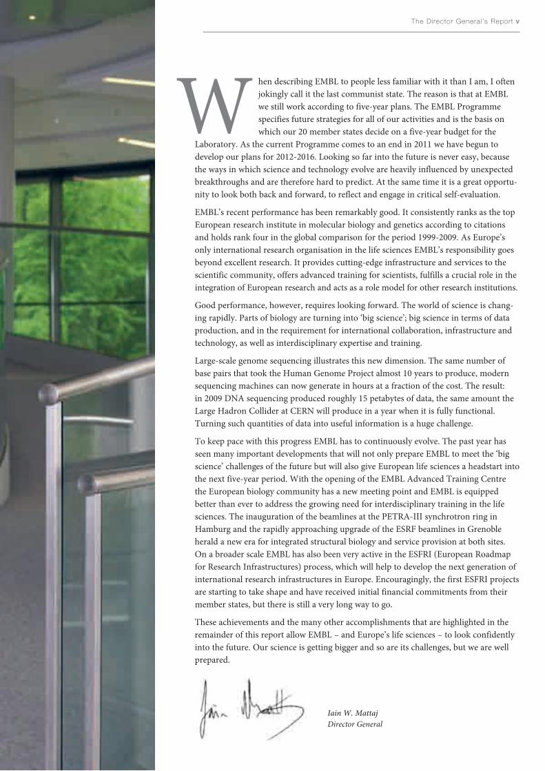
To keep pace with this progress EMBL has to continuously evolve. The past year hasseen many important developments that will not only prepare EMBL to meet the ‘bigscience’ challenges of the future but will also give European life sciences a headstart intothe next five-year period. With the opening of the EMBL Advanced Training Centrethe European biology community has a new meeting point and EMBL is equippedbetter than ever to address the growing need for interdisciplinary training in the lifesciences. The inauguration of the beamlines at the PETRA-III synchrotron ring inHamburg and the rapidly approaching upgrade of the ESRF beamlines in Grenobleherald a new era for integrated structural biology and service provision at both sites.On a broader scale EMBL has also been very active in the ESFRI (European Roadmapfor Research Infrastructures) process, which will help to develop the next generation ofinternational research infrastructures in Europe. Encouragingly, the first ESFRI projectsare starting to take shape and have received initial financial commitments from theirmember states, but there is still a very long way to go.
These achievements and the many other accomplishments that are highlighted in theremainder of this report allow EMBL – and Europe’s life sciences – to look confidentlyinto the future. Our science is getting bigger and so are its challenges, but we are wellprepared.
Iain W. MattajDirector General
W

vi EMBL Annual Report 09 · 10
chemical conglomerate. ENI donatedpart of the existing research infrastruc-tures for activities that were co-ordinat-ed by CNR’s Institute of Cell Biology(ICB/CNR) and overseen by campusco-ordinator Prof. Glauco P. Tocchini-Valentini.
Despite these ongoing administrativeand organisational changes, EMBLMonterotondo’s research activities havebeen highly successful during the pastyear. For example, Claus Nerlov and hisgroup, in collaboration with colleaguesin Madrid, discovered two proteins thatcontrol when and how stem cells at thebase of the skin stop multiplying andswitch to being skin cells. This worksheds light on the basic mechanismsinvolved not only in the formation ofskin, but also on aspects of skin andother epithelial cancers (page 88).
Structural Biology
The past year saw a remarkable researchhighlight in the area of structural biolo-gy. What started in 2004 as a Unit-widecollaboration, spearheaded by the thenjoint Heads of the Structural andComputational Biology Unit Peer Borkand Luis Serrano, resulted in the publi-cation of three papers back-to-back inScience in November 2009. The integrat-ed structural biology study of the
Research
This section features a few selectedresearch highlights of the past year.More detail on EMBL’s diverse researchactivities can be found in the followingchapter, the Scientific Report.
Mouse Biology
The past year has been particularlyeventful for EMBL’s Mouse Biology Unitin Monterotondo, which celebrated itstenth anniversary in June 2009. Much ofthe success of the Unit can be attributedto its current Head, Nadia Rosenthal.She has directed EMBL Monterotondosince 2001, establishing it as a centre ofexcellence for mouse research and as acentral hub in the international networkof mouse biology. Nadia has now accept-ed the position of Scientific Head of theEMBL Australia partner laboratory net-work (see page xvi). She has been instru-mental in building EMBL’s relationshipwith Australia – EMBL’s first associatemember state – and will also hold thepost of Director of the AustralianRegenerative Medicine Institute atMonash University. She will continueto run EMBL’s Mouse Biology Unit inItaly until her successor is found.
EMBL Monterotondo’s site, the AdrianoBuzzati-Traverso campus located in theLazio region about 30 km north ofRome, has been purchased by theConsiglio Nazionale delle Ricerche(CNR). The 158,000m2 international sci-entific campus was created in 1996 by aconsortium of the CNR and internation-al scientific organisations EMBL, EMMA(the European Mouse Mutant Archive)and the ICGEB (International Centre forGenetic Engineering and Biotechnology),and aimed to contribute to the develop-ment and internationalisation of Italianbiological and biomedical research. Thecampus was built and until now ownedby ENI SpA, a major Italian oil, gas and
bacterium Mycoplasma pneumoniae alsoinvolved the groups of Anne-ClaudeGavin, Rob Russell, Bettina Boettcherand Achilleas Frangakis, and support bythe EMBL Core Facilities. Together, theyhave produced the most comprehensivepicture of a “simple” bacterial cell to date(page 4).
EMBL’s cryo-electron microscopy(cryo-EM) research possibilities are setto expand with the purchase of a state-of-the-art piece of equipment. The next-generation Titan KriosTM transmissionelectron microscope, made by FEI, willbe delivered to EMBL Heidelberg in theautumn of 2010. This high-end instru-ment, of which there are only a fewrecently installed examples available tothe scientific community, will allowseveral of EMBL’s groups to enjoy morestability and higher throughput in theirEM work. Refurbishments of the formernuclear magnetic resonance (NMR) areaare underway to provide room for thenew microscope, which will allowStructural and Computational BiologyUnit groups to build on areas such as theautomated study of the structural diver-sity of viral and eukaryotic coat proteinsat the membrane, the molecular mecha-nisms of autophagy and the structureand function of large macromolecularassemblies.
EMBL Scientific Publications and Collaborations
· Total number of peer-reviewed publications: 343
· Internal collaborations: Publications co-authored by more thanone EMBL group leader: 23
· External collaborations: 795 in total of which 84 resulted inpublications
State of the Laboratory

The Director General ’s Report vii
EMBL’s structural biology outstations inGrenoble and Hamburg have also pro-duced interesting research results duringthe past year. José Antonio Márquez andhis group in Grenoble discovered thatthe key to a plant’s response to droughtlies in the structure of a protein receptorcalled PYR1 that interacts with the planthormone abscissic acid (page 42).Meanwhile, the group of EMBLHamburg Head Matthias Wilmannshas determined the structure of thesignalling molecule Death-AssociatedProtein Kinase bound to calmodulin(page 54).
Cell Biology and Biophysics
The Cell Biology and Biophysics (CBB)Unit saw a change in leadership in 2010.Eric Karsenti, who led the Unit since1998 and then ran it jointly with JanEllenberg in 2009, handed over soleresponsibility to Jan in January 2010 sothat he could co-ordinate the Tara
Oceans expedition. This three-year,150,000 km, marine journey waslaunched in September 2009 with Eric asthe scientific co-ordinator and involvesscientists from 50 laboratories in 15countries. It will study questions of bio-diversity and climate, the functioning ofmarine ecosystems and life’s origin andevolution.
In his new role as sole Head of Unit, JanEllenberg, who led the Gene ExpressionUnit for three years before moving toCBB, aims to build on the Unit’s existingstrengths and its move towards a moresystems-based, interdisciplinaryapproach. Jan also wants to developCBB’s interactions with other Units andadd to its already powerful imaging andmicroscopy technology base. Togetherwith Rainer Pepperkok, co-ordinatorof EMBL’s Advanced Light MicroscopyFacility, and other collaborators in theEuropean Commission-fundedMitocheck consortium, Jan’s group
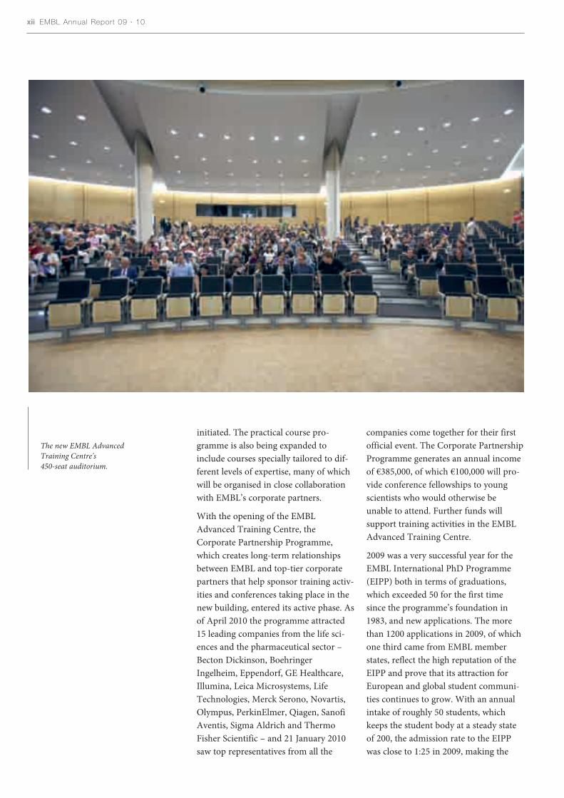
The EMBL Advanced Training Centrewith the new canteen (left), under
which the new training laboratoriesare located.
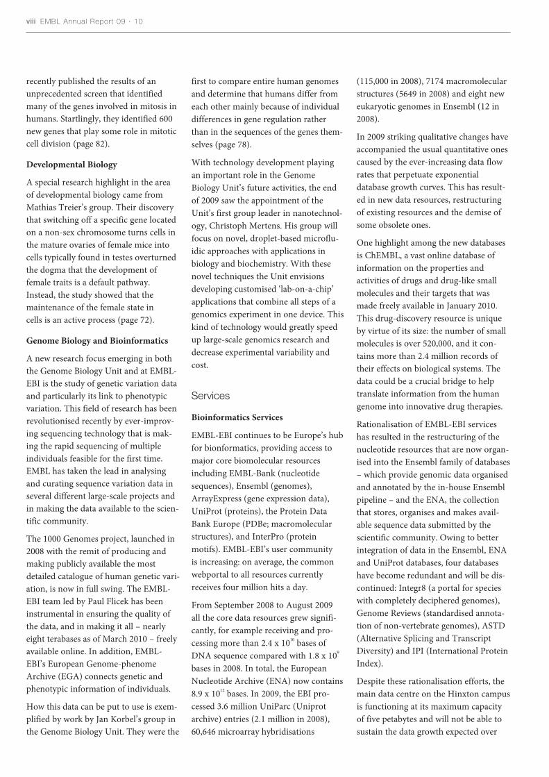
(115,000 in 2008), 7174 macromolecularstructures (5649 in 2008) and eight neweukaryotic genomes in Ensembl (12 in2008).
In 2009 striking qualitative changes haveaccompanied the usual quantitative onescaused by the ever-increasing data flowrates that perpetuate exponentialdatabase growth curves. This has result-ed in new data resources, restructuringof existing resources and the demise ofsome obsolete ones.
One highlight among the new databasesis ChEMBL, a vast online database ofinformation on the properties andactivities of drugs and drug-like smallmolecules and their targets that wasmade freely available in January 2010.This drug-discovery resource is uniqueby virtue of its size: the number of smallmolecules is over 520,000, and it con-tains more than 2.4 million records oftheir effects on biological systems. Thedata could be a crucial bridge to helptranslate information from the humangenome into innovative drug therapies.
Rationalisation of EMBL-EBI serviceshas resulted in the restructuring of thenucleotide resources that are now organ-ised into the Ensembl family of databases– which provide genomic data organisedand annotated by the in-house Ensemblpipeline – and the ENA, the collectionthat stores, organises and makes avail-able sequence data submitted by thescientific community. Owing to betterintegration of data in the Ensembl, ENAand UniProt databases, four databaseshave become redundant and will be dis-continued: Integr8 (a portal for specieswith completely deciphered genomes),Genome Reviews (standardised annota-tion of non-vertebrate genomes), ASTD(Alternative Splicing and TranscriptDiversity) and IPI (International ProteinIndex).
Despite these rationalisation efforts, themain data centre on the Hinxton campusis functioning at its maximum capacityof five petabytes and will not be able tosustain the data growth expected over
first to compare entire human genomesand determine that humans differ fromeach other mainly because of individualdifferences in gene regulation ratherthan in the sequences of the genes them-selves (page 78).
With technology development playingan important role in the GenomeBiology Unit’s future activities, the endof 2009 saw the appointment of theUnit’s first group leader in nanotechnol-ogy, Christoph Mertens. His group willfocus on novel, droplet-based microflu-idic approaches with applications inbiology and biochemistry. With thesenovel techniques the Unit envisionsdeveloping customised ‘lab-on-a-chip’applications that combine all steps of agenomics experiment in one device. Thiskind of technology would greatly speedup large-scale genomics research anddecrease experimental variability andcost.
Services
Bioinformatics Services
EMBL-EBI continues to be Europe’s hubfor bionformatics, providing access tomajor core biomolecular resourcesincluding EMBL-Bank (nucleotidesequences), Ensembl (genomes),ArrayExpress (gene expression data),UniProt (proteins), the Protein DataBank Europe (PDBe; macromolecularstructures), and InterPro (proteinmotifs). EMBL-EBI’s user communityis increasing: on average, the commonwebportal to all resources currentlyreceives four million hits a day.
From September 2008 to August 2009all the core data resources grew signifi-cantly, for example receiving and pro-cessing more than 2.4 x 10¹⁰ bases ofDNA sequence compared with 1.8 x 10⁹
bases in 2008. In total, the EuropeanNucleotide Archive (ENA) now contains8.9 x 1012 bases. In 2009, the EBI pro-cessed 3.6 million UniParc (Uniprotarchive) entries (2.1 million in 2008),60,646 microarray hybridisations
viii EMBL Annual Report 09 · 10
recently published the results of anunprecedented screen that identifiedmany of the genes involved in mitosis inhumans. Startlingly, they identified 600new genes that play some role in mitoticcell division (page 82).
Developmental Biology
A special research highlight in the areaof developmental biology came fromMathias Treier’s group. Their discoverythat switching off a specific gene locatedon a non-sex chromosome turns cells inthe mature ovaries of female mice intocells typically found in testes overturnedthe dogma that the development offemale traits is a default pathway.Instead, the study showed that themaintenance of the female state incells is an active process (page 72).
Genome Biology and Bioinformatics
A new research focus emerging in boththe Genome Biology Unit and at EMBL-EBI is the study of genetic variation dataand particularly its link to phenotypicvariation. This field of research has beenrevolutionised recently by ever-improv-ing sequencing technology that is mak-ing the rapid sequencing of multipleindividuals feasible for the first time.EMBL has taken the lead in analysingand curating sequence variation data inseveral different large-scale projects andin making the data available to the scien-tific community.
The 1000 Genomes project, launched in2008 with the remit of producing andmaking publicly available the mostdetailed catalogue of human genetic vari-ation, is now in full swing. The EMBL-EBI team led by Paul Flicek has beeninstrumental in ensuring the quality ofthe data, and in making it all – nearlyeight terabases as of March 2010 – freelyavailable online. In addition, EMBL-EBI’s European Genome-phenomeArchive (EGA) connects genetic andphenotypic information of individuals.
How this data can be put to use is exem-plified by work by Jan Korbel’s group inthe Genome Biology Unit. They were the
The Director General ’s Report ix
the coming years. To address this,EMBL-EBI has received funding fromthe UK Research Councils to lease twonew state-of-the-art data centres inLondon. For the research community,the primary benefit will be improvedaccess to the flood of biological informa-tion, and the new facilities will alsoprovide the central hub for theemerging pan-European Life ScienceInfrastructure for Biological Information(ELIXIR, page xviii).
Structural Biology Services
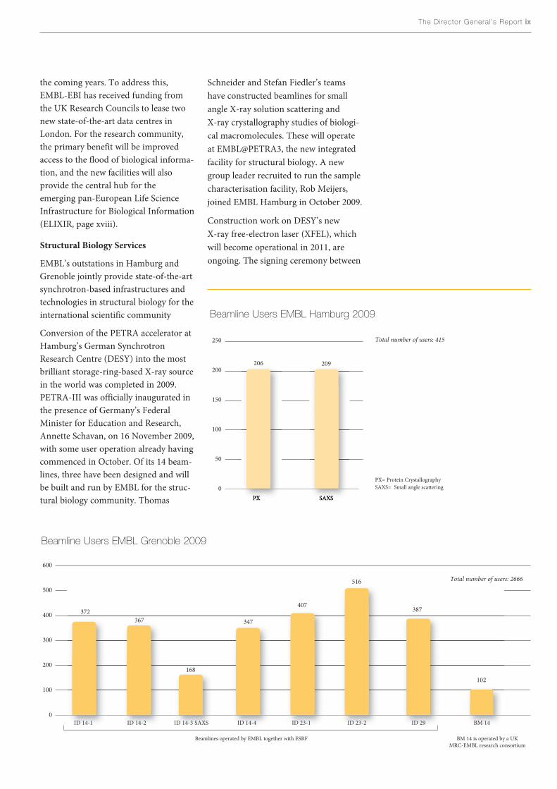
EMBL’s outstations in Hamburg andGrenoble jointly provide state-of-the-artsynchrotron-based infrastructures andtechnologies in structural biology for theinternational scientific community
Conversion of the PETRA accelerator atHamburg’s German SynchrotronResearch Centre (DESY) into the mostbrilliant storage-ring-based X-ray sourcein the world was completed in 2009.PETRA-III was officially inaugurated inthe presence of Germany’s FederalMinister for Education and Research,Annette Schavan, on 16 November 2009,with some user operation already havingcommenced in October. Of its 14 beam-lines, three have been designed and willbe built and run by EMBL for the struc-tural biology community. Thomas
Schneider and Stefan Fiedler’s teamshave constructed beamlines for smallangle X-ray solution scattering andX-ray crystallography studies of biologi-cal macromolecules. These will operateat EMBL@PETRA3, the new integratedfacility for structural biology. A newgroup leader recruited to run the samplecharacterisation facility, Rob Meijers,joined EMBL Hamburg in October 2009.
Construction work on DESY’s newX-ray free-electron laser (XFEL), whichwill become operational in 2011, areongoing. The signing ceremony between
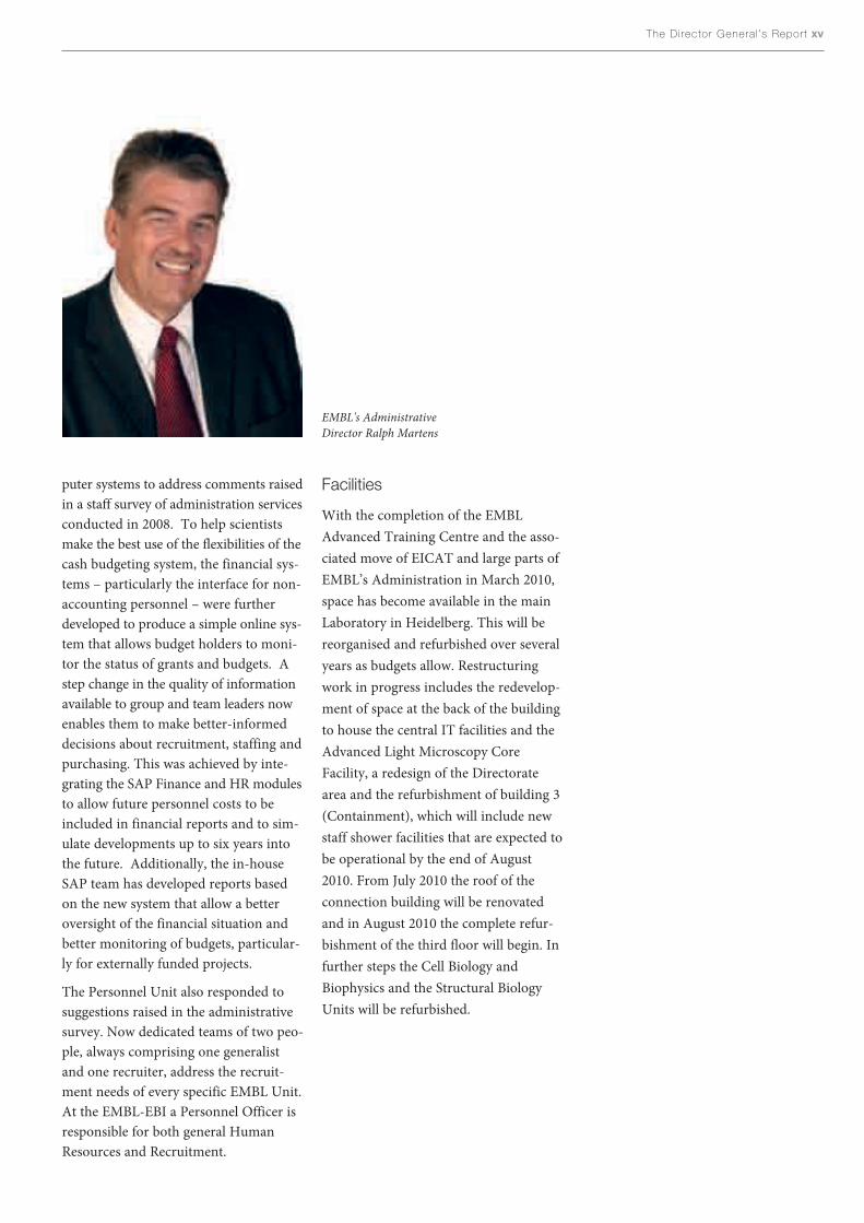
Beamline Users EMBL Grenoble 2009
600
500
400
300
200
100
0
372367
168
347
407
Total number of users: 2666516
387
102
250
200
150
100
50
0PX SAXS
206 209
Total number of users: 415
ID 14-1 ID 14-2 ID 14-3 SAXS ID 14-4 ID 23-1 ID 23-2 ID 29 BM 14
Beamline Users EMBL Hamburg 2009
Beamlines operated by EMBL together with ESRF BM 14 is operated by a UKMRC-EMBL research consortium
PX= Protein CrystallographySAXS= Small angle scattering

links with the Indian crystallographycommunity.
Core Facilities
EMBL’s Core Facilities offer cutting-edgetechnology and expert support toresearchers at EMBL and, when excesscapacity allows, to external users. Like itsresearch Units, EMBL also subjects itsservices to regular stringent quality con-trol. In March 2010 the Core Facilitieswere reviewed by a panel of externalexperts and received a very positiveevaluation. As well as the external evalu-ators, internal users are also very satis-fied with the services offered by the CoreFacilities. In December 2009 a large usersurvey was conducted to assess the quali-ty of services, the general user satisfac-tion and the need for more or differentservices. The response was overwhelm-ingly favourable, with all eight facilitiesscoring good, very good or excellent foraccessibility, comparison with otherfacilities elsewhere, staff competence,staff support and standard of resultsobtained.
x EMBL Annual Report 09 · 10
the partners from several countries tookplace on 30 November 2009. Once com-pleted, the laser will allow researchers toanalyse materials in atomic detail, filmchemical reactions, generate three-dimensional images of the nanoworldand study processes under extreme con-ditions such as those occurring in theinterior of planets. EMBL Hamburg sci-entists will be involved in exploring pos-sible uses of the XFEL with biologicalsamples.
For the beamline BM14 at the EuropeanSynchrotron Radiation Facility (ESRF),2009 was a transition year in which theUK Medical Research Council (MRC),EMBL and India shared beam time.When the UK MRC contract finished asplanned at the end of the year, a consor-tium comprising EMBL, campus partnerESRF and the Indian government tookover the running of the beamline for aperiod of five years from 1 January 2010.The agreement guarantees the continua-tion of this state-of-the-art Multi-wavelength Anomalous Dispersion(MAD) beamline, and will strengthen
User satisfaction with Core Facilities and IT Services
100
80
60
40
20
0ALMF GeneCore PEPCF Proteomics FCCF EMCF ChemBio CF MACF IT Services
Internal users were asked to rank their satisfaction with the Core Facilities and the central IT Services that provide IT support for EMBL Heidelberg andMonterotondo. EMBL-EBI, Hamburg and Grenoble operate their IT infrastructure locally.
Average
user
satisfaction(%
)
ALMF= Advanced Light Microscopy Facility, GeneCore= Genomics Core Facility, PEPCF= Protein Expression and Purification Core Facility, Proteomics= Proteomics Core Facility,FCCF= Flow Cytometry Core Facility, EMCF= Electron Microscopy Core Facility, ChemBio CF= Chemical Biology Core Facility, MACF= Monoclonal Antibodies Core Facility

Over the past year the Genomics CoreFacility has seen major changes withnext-generation sequencing havingmoved to the centre of activities. Thenew technology required significantinvestment both in terms of equipment –the facility currently operates three (fourfrom June 2010) next-generationsequencing machines – but also in termsof staff training. In future it will be indis-pensible to upgrade the software and ITsupport and strengthen the bioinformat-ics expertise in the facility to ensure thatthe increased data production is matchedwith the necessary capacity for analysis.
Training
Advanced training has always been oneof EMBL’s core missions and the EMBLInternational Centre for AdvancedTraining (EICAT) organises a range ofintramural and extramural trainingactivities for EMBL staff and the scientif-ic community. Over the period 2007-2009 130 courses, conferences andworkshops were held at all five EMBLsites, reaching around 10,000 externalparticipants.
In the future we will be able to furtherexpand and develop our efforts in train-ing, thanks to the new EMBL AdvancedTraining Centre, which officially openedon 9 March 2010 on the Heidelberg cam-pus. The inauguration ceremony tookplace in the new 450-seat auditorium thatwill allow scientific conferences ofunprecedented size to be held at EMBL.Among the 340 distinguished guests andfriends of EMBL were the Ministers forResearch of Germany, Professor AnnetteSchavan, and Israel, Professor DanielHershkowitz, Council delegates and rep-resentatives of ministries from manyother EMBL member states, prominentscientists from all over the world andKlaus Tschira, whose ideas for the formof the building initiated the project andwhose foundation provided generoussupport towards its realisation.
The higher capacity of the EMBLAdvanced Training Centre will allow
The Director General ’s Report xi
more scientists to benefit from EMBL’straining activites, with the number ofparticipants coming to the mainLaboratory expected to more than dou-ble from the current level of roughly2000 per year. Most importantly, howev-er, the EMBL Advanced Training Centrewill help us to explore new forms oftraining and build on the quality of ourexisting events. It will also strengthenEMBL’s role as a central hub foradvanced life science training in Europeand EMBO’s role as a major funder ofcourses, conferences and workshops.
EMBL’s Course and Conference Officehas used the past year to gear up for theupcoming increase in activities. In closecollaboration with EMBO, a new for-ward-looking meeting format coveringimportant topics of the life sciences hasbeen devised, the EMBO|EMBL Symposia.Furthermore, to celebrate the inauguralyear of the EMBL Advanced TrainingCentre a series of lectures by Nobel Prizelaureates, entitled ‘Vision 2020’, has been
EMBL Director General Iain Mattajand German Minister for Educationand Research, Annette Schavan, at theEMBL Advanced Training Centreinauguration ceremony.
xii EMBL Annual Report 09 · 10
companies come together for their firstofficial event. The Corporate PartnershipProgramme generates an annual incomeof €385,000, of which €100,000 will pro-vide conference fellowships to youngscientists who would otherwise beunable to attend. Further funds willsupport training activities in the EMBLAdvanced Training Centre.
2009 was a very successful year for theEMBL International PhD Programme(EIPP) both in terms of graduations,which exceeded 50 for the first timesince the programme’s foundation in1983, and new applications. The morethan 1200 applications in 2009, of whichone third came from EMBL memberstates, reflect the high reputation of theEIPP and prove that its attraction forEuropean and global student communi-ties continues to grow. With an annualintake of roughly 50 students, whichkeeps the student body at a steady stateof 200, the admission rate to the EIPPwas close to 1:25 in 2009, making the
initiated. The practical course pro-gramme is also being expanded toinclude courses specially tailored to dif-ferent levels of expertise, many of whichwill be organised in close collaborationwith EMBL’s corporate partners.
With the opening of the EMBLAdvanced Training Centre, theCorporate Partnership Programme,which creates long-term relationshipsbetween EMBL and top-tier corporatepartners that help sponsor training activ-ities and conferences taking place in thenew building, entered its active phase. Asof April 2010 the programme attracted15 leading companies from the life sci-ences and the pharmaceutical sector –Becton Dickinson, BoehringerIngelheim, Eppendorf, GE Healthcare,Illumina, Leica Microsystems, LifeTechnologies, Merck Serono, Novartis,Olympus, PerkinElmer, Qiagen, SanofiAventis, Sigma Aldrich and ThermoFisher Scientific – and 21 January 2010saw top representatives from all the
The new EMBL AdvancedTraining Centre's450-seat auditorium.

The Director General ’s Report xiii
EIPP one of the most competitive PhDprogrammes in biology in Europe. Tostay abreast of this increasing popularityand the overall growth of the pro-gramme, we now run two similarly sizedrounds of applications a year. This reor-ganisation is also a response to theEurope-wide harmonisation of studiesaccording to the Bologna protocol andits impact on student schedules. Toincrease applications from currentlyunder-represented member states and toattract students from other disciplines –most notably physics, chemistry andengineering – we are refining our adver-tising strategies and have initiatedrecruitment events targeted at specificcountries and student communities. Aspart of this initiative we have also imple-mented a student ambassadors schemeto enhance the visibility of the EIPPamong member state undergraduatecommunities.
Owing to the success of the EMBLInterdisciplinary Postdoc (EIPOD) ini-tiative in obtaining external fundingfrom the European Commission FP7Marie-Curie Co-fund scheme, the EMBLPostdoctoral Programme is now man-aged by a full-time PostdoctoralAdministrator together with a dedicatedacademic mentor, and has introducedseveral new developments in the pastyear. The 2009 EIPOD selection saw anincreased number of nearly 200 applica-tions, from which 19 were selected. Anew postdoctoral ‘second mentor’scheme encourages all EMBL postdocs toobtain additional advice and guidancefrom a mentor other than their dedicat-ed academic supervisor. Together withother activities of the PostdoctoralAssociation and complementary trainingcourses, EMBL postdocs are well pre-pared for a future career in academia orindustry. In addition, with the beginningof the new Indicative Scheme in 2012,the programme will start to provideadditional social benefits for all postdoc-toral fellows following the recommenda-tion of the European Charter forResearchers.
In the past year the European LearningLaboratory for the Life Sciences (ELLS)has organised five hands-on courses forsecondary school teachers, three inHeidelberg and two in Monterotondo. Incollaboration with EMBL-EBI, ELLSlaunched a new course dedicated tobioinformatics in the classroom atHinxton in March 2010.
At EMBL-EBI the new IT suite hasaccommodated over 1400 trainees, host-ing not only the EBI Hands-on TrainingProgramme, but also offering a largenumber of courses and workshops to thescientific community. To complementthese face-to-face training activities andto reach out to an even larger audience, anew e-learning pilot training programmewas implemented and is now beingexpanded further.
To complement scientific training,EMBL started a formal programme ofvocational training, the General Trainingand Development Programme, for all itsscientific, administrative and supportstaff in 2008. We offer a variety of train-ing courses from computer skills to lan-guage training to leadership andmanagement expertise. This programmehas been very popular with the staff,indeed it is enormously oversubscribed
even though more than 100 courses wereorganised for the benefit of around 500participants in 2009. The GeneralTraining and Development Programmehelps the Laboratory to fulfill its obliga-tion to the member states by ensuringthat all categories of staff leave EMBLwith a skill set that makes them attrac-tive candidates for organisations andinstitutions in the national systems.
Alumni
The total number of EMBL alumni hasgrown to 4764 with 240 people leavingEMBL (including fellows, trainees andvisitors) in the past year. 80% have takenup positions in the EMBL memberstates. The EMBL Alumni Association(EAA) acquired 85 new members, whichbrings the total up to 1649 memberswith a membership rate of 34%.
2009-2010 was a very eventful year forEMBL alumni. Local chapters met inHeidelberg, Porto, Helsinki, Dublin andDilofo. The local chapter meeting inDublin on 24 February 2010 was fol-lowed by an event organised by ScienceFoundation Ireland that showcasedEMBL to the wider Irish scientific com-munity and which was attended by theIrish Minister for Science, Technologyand Innovation, Conor Lenihan TD.
The highlight of the year was the staff-alumni reunion in the EMBL AdvancedTraining Centre on 8 March 2010, theday before the official opening ceremo-ny. 200 participants heard a variety oftalks with lots of opportunities to net-work in between. The event culminatedwith the opening ceremony of the MattiSaraste Courtyard, which was madepossible by donations of staff andalumni.
Former EMBL Hamburg predoc JensPreben Morth, who is now an AssociateProfessor at Aarhus University, has beenselected as the winner of the 2010 JohnKendrew Award – which recognisesexcellence in science communication oracademic achievement after leaving
xiv EMBL Annual Report 09 · 10
EMBL. Jens Preben Morth receives theprize for his outstanding contribution tothe structural biology of membrane pro-teins, and for his enthusiastic involve-ment in science education for schoolstudents.
The overwhelming response to the JohnKendrew Award, as shown by the highnumber of outstanding applications andits enthusiastic reception by EMBL staffand alumni, motivated the EAA board toaim to offer the award indefinitely bylaunching the first EAA fundraisingcampaign in October 2009. To date€6000 has been raised, which will securethe award until 2014.
The EAA also launched an initiative in2010 to establish a European MolecularBiology Archive (EMBA). The idea wasinspired by the 2007 statement bySydney Brenner and Richard Roberts inNature: “Let’s not wait until memorieshave faded and papers been discarded atthe end of a career before deciding tosave our heritage.” Although the initia-tive gained instant support from key pastand present EMBL figures, much workwill be required to secure resources forestablishing such an archive.
Outreach
The tremendous potential the life sci-ences holds for societal benefits endowsscientists with the social responsibility toinform the public about advances intheir research, as well as its potentialapplications, inherent risks and benefitsand ethical implications. For this reasonthe Office of Information and PublicAffairs (OIPA), EMBL-EBI’s Outreachand Training team, the Science andSociety Programme and EICAT, sup-ported by many of EMBL’s scientists,organise a range of outreach activities.
In the first half of 2010 alone 14 groupsof students from various schools anduniversities have visited EMBL com-pared with 13 in the whole of the previ-ous year. Most came from relativelyclose by – e.g. France, Spain and Belgium
– but one group travelled from as far asIsrael to learn about EMBL.
In 2009 EMBL relaunched its websiteswith a new look and feel based on acompletely re-engineered content man-agement system. A more recent additionto EMBL’s internet presence is a newintranet portal – a dynamic ‘one-stop-shop’ for news and events that comple-ments the EMBL Etcetera newsletter as aplatform for information disseminationwithin the EMBL community. In asecond phase, in 2010, further featuresof the content management system –including an optimised search facility,RSS feeds and a multimedia gallery –will be developed.
EMBL regularly communicates researchhighlights and news about other activi-ties of the organisation to a broad rangeof international media. Particularly pop-ular with the press in the past year werethe first comprehensive picture of a min-imal cell (Mycoplasma pneumoniae) andthe screen conducted by the internation-al Mitocheck consortium that identified600 genes involved in mitosis in humans.Also the announcement of a £10 millioninvestment in the ELIXIR project(page xviii) by the Biotechnology andBiological Sciences Research Council(BBSRC) received widespread attentionby both local and European researchmedia.
The Science and Society Programme’sannual conference in November 2009was organised under the lead of EMBOand enjoyed a record number of 270participants. The two-day event, ‘Food,Sustainability and Plant Science: AGlobal Challenge’ focussed on our foodsupply which, next to climate change, isthe greatest challenge that faces theworld. Other Science and Society eventsincluded Forum Lectures by Jens Reich,James Mallory and Buddhist monkMatthieu Ricard and an EMBL-EBIScience & Society symposium inCambridge, which tackled the questionof ‘Who owns science? Promises andpitfalls of public-private partnerships’.
Technology Transfer
One of EMBL’s missions is to develop itsdiscoveries to the benefit of society.When EMBL’s technology-transfer sub-sidiary, EMBL Enterprise ManagementTechnology Transfer GmbH – EMBLEMfor short – was established in 1999, itwas hoped it would break even withinten years. In fact, as the company cele-brated the end of its first decade on 19June 2009, there was even more cause forcelebration. Exceeding all expectations,EMBLEM has actually been generating aprofit for EMBL, its scientists and themember states since 2004, breaking evenin less than half the time predicted.
As well as ensuring a modest but steadyincome for EMBL and benefiting themember states and society by translatingbasic research results into marketabletools and products, the creation ofEMBLEM has allowed the protectionand commercialisation of innovations tobe streamlined. To date more than 400EMBL staff are on record as inventors,over 250 commercial partners world-wideare licensing EMBL-derived technologiesand EMBLEM has a portfolio of morethan 260 granted patents and patentapplications, over 90 copyrights andtrademarks and 12 spin-out companies.
Administration
This year saw the departure of Bernd-Uwe Jahn after his eight-year leadershipof the Administration, which producedsignificant improvements in staff rela-tions and in the efficiency of the admin-istration services. He was succeeded byRalph Martens at the beginning of 2010.Ralph joins us from his previous positionas Director of Common AdministrativeServices at the International CriminalCourt in The Hague. Previously he hasheld positions in both public and privatesectors in various countries in Europeand the US and was listed in the ‘Who’sWho in Leading Executives of America’.
During the course of the year we havemade many improvements to our com-
The Director General ’s Report xv
puter systems to address comments raisedin a staff survey of administration servicesconducted in 2008. To help scientistsmake the best use of the flexibilities of thecash budgeting system, the financial sys-tems – particularly the interface for non-accounting personnel – were furtherdeveloped to produce a simple online sys-tem that allows budget holders to moni-tor the status of grants and budgets. Astep change in the quality of informationavailable to group and team leaders nowenables them to make better-informeddecisions about recruitment, staffing andpurchasing. This was achieved by inte-grating the SAP Finance and HR modulesto allow future personnel costs to beincluded in financial reports and to sim-ulate developments up to six years intothe future. Additionally, the in-houseSAP team has developed reports basedon the new system that allow a betteroversight of the financial situation andbetter monitoring of budgets, particular-ly for externally funded projects.
The Personnel Unit also responded tosuggestions raised in the administrativesurvey. Now dedicated teams of two peo-ple, always comprising one generalistand one recruiter, address the recruit-ment needs of every specific EMBL Unit.At the EMBL-EBI a Personnel Officer isresponsible for both general HumanResources and Recruitment.
Facilities
With the completion of the EMBLAdvanced Training Centre and the asso-ciated move of EICAT and large parts ofEMBL’s Administration in March 2010,space has become available in the mainLaboratory in Heidelberg. This will bereorganised and refurbished over severalyears as budgets allow. Restructuringwork in progress includes the redevelop-ment of space at the back of the buildingto house the central IT facilities and theAdvanced Light Microscopy CoreFacility, a redesign of the Directoratearea and the refurbishment of building 3(Containment), which will include newstaff shower facilities that are expected tobe operational by the end of August2010. From July 2010 the roof of theconnection building will be renovatedand in August 2010 the complete refur-bishment of the third floor will begin. Infurther steps the Cell Biology andBiophysics and the Structural BiologyUnits will be refurbished.
EMBL's AdministrativeDirector Ralph Martens
xvi EMBL Annual Report 09 · 10
Member state relations
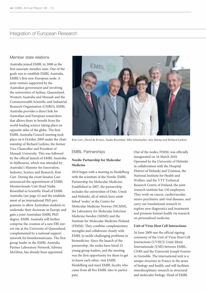
Australia joined EMBL in 2008 as thefirst associate member state. One of thegoals was to establish EMBL Australia,EMBL’s first non-European node. Ajoint venture supported by theAustralian government and involvingthe universities of Sydney, Queensland,Western Australia and Monash and theCommonwealth Scientific and IndustrialResearch Organisation (CSIRO), EMBLAustralia provides a direct link forAustralian and European researchersthat allows them to benefit from theworld-leading science taking place onopposite sides of the globe. The firstEMBL Australia Council meeting tookplace on 6 October 2009 under the chair-manship of Richard Larkins, the formerVice-Chancellor and President ofMonash University. This was followedby the official launch of EMBL Australiain Melbourne, which was attended byAustralia’s Minister for Innovation,Industry, Science and Research, KimCarr. During the event Senator Carrannounced the appointment of EMBLMonterotondo Unit Head NadiaRosenthal as Scientific Head of EMBLAustralia (see page vi) and the establish-ment of an international PhD pro-gramme to allow Australian students toundertake their doctorate in Europe andgain a joint Australian-EMBL PhDdegree. EMBL Australia will furthercomprise the creation of a new EBI mir-ror site at the University of Queensland,complemented by a national supportnetwork for bioinformaticians. The firstgroup leader in the EMBL AustraliaPartner Laboratory Network, EdwinaMcGlinn, has already been appointed.
EMBL Partnerships
Nordic Partnership for MolecularMedicine
2010 began with a meeting in Heidelbergwith the scientists of the Nordic EMBLPartnership for Molecular Medicine.Established in 2007, the partnershipincludes the universities of Oslo, Umeåand Helsinki, all of which have estab-lished ‘nodes’ at the Centre forMolecular Medicine Norway (NCMM),the Laboratory for Molecular InfectionMedicine Sweden (MIMS) and theInstitute for Molecular Medicine Finland(FIMM). They combine complementarystrengths and collaborate closely withEMBL to tackle challenging problems inbiomedicine. Since the launch of thepartnership, the nodes have hired 25young group leaders, and the meetingwas the first opportunity for them to getto know each other, visit EMBLHeidelberg and meet EMBL faculty whocame from all five EMBL sites to partici-pate.
One of the nodes, FIMM, was officiallyinaugurated on 16 March 2010.Operated by the University of Helsinkiin collaboration with the HospitalDistrict of Helsinki and Uusimaa, theNational Institute for Health andWelfare, and the VTT TechnicalResearch Centre of Finland, the jointresearch institute has 150 employees.They work on cancer, cardiovascular,neuro-psychiatric and viral diseases, andcarry out translational research toexplore new diagnostics and treatmentsand promote human health via researchon personalised medicine.
Unit of Virus Host Cell Interactions
26 June 2009 saw the official signingceremony of the Unit of Virus Host CellInteractions (UVHCI) Unité MixteInternationale (UMI) between EMBL,CNRS and the Université Joseph Fourierin Grenoble. The international unit is aunique structure in France in the areasof biology and health, and will facilitateinterdisciplinary research in structuraland molecular biology. Head of EMBL
Integration of European Research
Kim Carr, David de Kretser, Nadia Rosenthal, Silke Schumacher, Iain Mattaj and Richard Larkins

The Director General ’s Report xvii
Grenoble Stephen Cusack will direct theunit for the first five years, and theDeputy Head will be Rob Ruigrok,Professor at the Université JosephFourier. The international unit alreadyunderwent its first review by Frenchreviewing body AERES in February 2010and achieved favourable results.
EMBL Grenoble’s campus, the PolygoneScientifique, is to be developed into aworld-class science and technology park– an ‘ecosystem’ of innovation namedGIANT – in an initiative supported bythe French government. GIANT’s firstconstruction projects are underway andmark the beginning of a €500 millionoverhaul for the area, which has longboasted a top-class research infrastruc-ture that includes the ESRF, the ILL,EMBL, CEA, CNRS and the UniversitéJoseph Fourier, as well as three centres oftechnological excellence and severalhigh-level university programmes. Withthe planned new teaching and researchbuildings, recreational facilities andmeeting places for researchers, transport
links and sustainable housing, it is pro-jected that the site will welcome 20,000scientists and students and 10,000 inhab-itants by the year 2015. The entrance tothe site will also feature a VisitorsCentre, which will present the work ofall the campus’ scientific institutes to thegrowing numbers of interested membersof the public using videos, models andinteractive exhibits.
To exploit research results obtained atEMBL Grenoble a new EMBL spin-outcompany, Savira Pharmaceuticals, wascreated in September 2009 to focus onthe development of drugs for the treat-ment of influenza. Co-founded by theVienna-based biotech companyOnepharm Research and DevelopmentGmbH, Savira builds on the break-through results from the groups ofStephen Cusack and Darren Hart atEMBL Grenoble, who produced high-resolution images of several crucialdomains of the influenza virus poly-merase, the enzyme that copies thevirus’s genetic material and allows it to
multiply in human cells. These findingsopen new avenues for the structure-based development of anti-influenzadrugs, which could target the viral poly-merase to selectively stop its reproduc-tive cycle. The spin-out company, whichis based in Vienna, received €1 millionsupport from the AustriaWirtschafts-service (AWS) and was awarded thirdplace by the City of Vienna FutureAward in the category ‘Newcomers andStart-ups’.
Collaborations with institutes innon-member states
Since the signing of an Agreement ofAcademic Exchange between EMBLand Japan’s National Institute of BasicBiology (NIBB) in 2005, both instituteshave worked together to span the dis-tance with exchange visits for lectures,workshops, conferences and otheracademic activities. In addition, jointsymposia on the topics of developmentalbiology, microscopy and imaging,epigenetics, systems biology, functional
Members of the Nordic EMBLPartnership for Molecular Medicine
gathered in Heidelberg

xviii EMBL Annual Report 09 · 10
genomics, mouse biology and structuralbiology have been held regularly in Japanand Europe to promote academicexchange between researchers of bothinstitutes as well as other Japanese andEuropean scientists. In October 2009 thefirst NIBB-EMBL PhD student mini-symposium took place in Heidelberg,with 20 students from both institutespresenting their research. The agreementwas renewed during a visit by the NIBBDirector General, Prof. Kiyotaka Okada,to EMBL in February 2010.
European ResearchInfrastructures
EMBL is involved in seven out of tenEuropean Strategy Forum on ResearchInfrastructures (ESFRI) biomedical sci-ence projects. For those included on theESFRI roadmap in 2006 the EuropeanCommission FP7-funded preparatoryphase is now entering its third year.ELIXIR (European Life ScienceInfrastructure for Biological Infor-mation) is co-ordinated by the EMBL-EBI Director Janet Thornton and EMBLis participating in INSTRUCT, BBMRIand Infrafrontier. The projects from the2008 update of the ESFRI roadmap willenter their preparatory phase later thisyear. Euro-BioImaging will be co-ordi-nated by Jan Ellenberg (Head of the CellBiology and Biophysics Unit) togetherwith a representative of the medicalimaging community, and EMBL will alsoparticipate in EU-Openscreen and theEuropean Marine Biology ResourceCentre.
ELIXIR
The aim of ELIXIR is to construct a sus-tainable infrastructure for biomoleculardata and related information in Europe.Over the past two years, an extensivestakeholder consultation has soughtinput into ELIXIR’s scope and structure.One important conclusion from thisstakeholder consultation is that ELIXIR’sstructure should comprise a hub, based
at EMBL-EBI in Hinxton, Cambridge,UK, and several nodes located through-out Europe. The hub will be responsiblefor holding the core data collections andenabling the development and integra-tion of nodes into a European-wide dis-tributed infrastructure.
A request for suggestions to contributeto ELIXIR’s construction was publishedin April 2010. Its purpose is: to consoli-date the extensive stakeholder input thatwe have had to date; to generate ideas forfurther discussion with national fundersas to how ELIXIR’s stakeholders wish tocontribute to ELIXIR on a pan-Europeanlevel; and to gather information on thekinds of support that potential nodesexpect from the hub as input to furtherdiscussions with research organisationsand funders. It is intended that long-term infrastructure support will be paidfor through national, European, EMBLand other funding streams. As part ofthis a centrally managed fund will beestablished that will both support the co-ordination activities (at the hub) andhelp leverage further investment. At theorganisational level, ELIXIR may initiallybe established as an EMBL SpecialProject.
Euro-BioImaging
Euro-BioImaging aims to provide accessand training to imaging technologiesacross the full scale of biological andmedical applications, from molecule topatient. Euro-BioImaging is at an earlystage. It has just been awarded fundingfor the preparatory phase by theEuropean Commission and will enter itsthree-year preparatory phase in 2011.The first stakeholder meeting of Euro-BioImaging, which involves 23 countriesacross Europe, took place on 21-22September 2009 at EMBL Heidelberg.The meeting gathered representatives ofthe scientific community, funding andgovernmental organisations and industryto discuss potential participation and thecontent and structure of the project.
EIROforum
From July 2009 EMBL took over for ayear as the chair of EIROforum, apartnership of the seven largest intergov-ernmental research infrastructure organ-isations in Europe (CERN, EFDA-JET,EMBL, ESA, ESO, ESRF and ILL).
The main achievements during this yearhave been the renewal of the Statementof Intent between EIROforum and theEuropean Commission, the publicationof a policy paper on research infrastruc-tures and the organisation of a technolo-gy-transfer conference.
The EC-EIROforum Statement of Intentwas originally signed in 2003. Over thepast year it was reviewed and updated.The new European Commissioner forResearch and Innovation MáireGeoghegan-Quinn and the EIROforumDirector Generals expressed their mutualinterest in continuing the co-operation.
The seven EIROforum organisations arethe largest providers of research infras-tructures in Europe. To make their col-lective experience more widely available,especially to the new ESFRI projects,EIROforum published a position paperentitled ‘Establishing New ResearchInfrastructures in Europe – TheEIROforum Experience’ in March 2010just before the sixth EuropeanConference on Research Infrastructures,ECRI 2010, took place in Barcelona.Representatives of European researchacross all disciplines and science policymakers gathered to discuss challengesand issues currently facing Europeanresearch infrastructures, such as the pri-oritisation of research infrastructures,management and financial issues, gover-nance structures and a general futurestrategy for European research.
In November 2009, EMBL and itsEIROforum partners organised aconference on technology transfer inHeidelberg to exchange knowledge andbest practices across disciplines. Around100 participants attended the conferenceincluding representatives from Europeanscience and technology infrastructures,

The Director General ’s Report xix
European politicians, representatives ofthe European industry, representativesof the national governments of theEuropean Union, representatives of theEuropean Investment Bank, theEuropean Patent Office, representativesof the institutions of the EuropeanCommission and other research com-munities. On the basis of the discussionsat the conference a recommendation onthe management of intellectual propertyand knowledge transfer was developed.
In July 2010 EMBL will hand over theEIROforum chairmanship to EFDA-Jet,the European Fusion DevelopmentAgreement.
Initiative for Science in Europe
EMBL is a founder member of theInitiative for Science in Europe (ISE), anorganisation of European scientific soci-eties and organisations. ISE participated,with the Spanish EU presidency, theEuropean Commission and ESFRI, inthe organisation of ECRI 2010 and inMay 2010, organised a conference on thefuture of the European Research Council(ERC) ‘ERC – From Programme toInstitution’. The event featured a discus-sion on the achievements, challengesand future of the ERC and was attendedby many representatives of Europeanscience and science policy.
The EIROforum Director Generalassembly took place at EMBL
Heidelberg

xx EMBL Annual Report 09 · 10
Personnel statistics
Total 434
Personnel on 31 December 2009
Visitors to EMBL Units during 2009
90
80
70
60
50
40
30
20
10
0
Cell Biologyand Biophysics
CoreFacilities
DevelopmentalBiology
Directors’research
GenomeBiology
Structural andComputational
Biology
EMBLGrenoble
OthersEMBLHamburg
EMBL-EBIHinxton
EMBLMonterotondo
On 31 December 2009, 1529 people, including visitors, from more than 60 nations were employed by EMBL.
76
55
46
41
60
21
40
28
19 18
30
816
171
107
214
221
Staff members
Visitors, trainees anddiploma students
Ancillaries andsupernumeraries
Predoctoral fellows
Postdoctoral fellows
Total: 1529

The Director General ’s Report xxi
EMBL member statesNon-European countriesOther European countries
Staff Nationalities – All
Staff Nationalities – Research
5 %
17 %78 %
Please refer to CD for more information
Non EU24%
E 4%
IRL 2%
AUT 2%
NL 2%
CH 2%
B 1%
P 1%
DK 1%FIN 1%
HR 1%
S 1%AUS 1%
IS, L, N, IL
D 23%
UK 18%F 10%
I 7%

xxii EMBL Annual Report 09 · 10
Financial report
INCOME 2009 2008
€ 000 € 000
Member states contributions
Ordinary
One-off contribution from Germany
Internal Funding
External Funding
Other Receipts
Total Income
EXPENDITURE
Staff Costs 81,25582,933
Operating Costs 49,10154,001
Capital Expenditure 25,94223,555
Total Expenditure 156,298160,489
(4,034) 6,897
Income/expenditure statement
External grant funding2009 2008
Surplus (deficit) for the year
€ 000 % € 000 %
BBSRC 1,675 4.9 1,457 3.6BMBF 4,144 12.1 3,257 8.1DFG 1,404 4.1 1,461 3.7EC 11,695 34.1 16,893 42.2HFSPO 457 1.3 492 1.2MRC 8 0 7 0NIH 7,750 22.6 6,169 15.4VW Foundation 243 0.7 294 0.7Wellcome Trust 4,160 12.1 5,240 13.1Others 2,747 8.0 4,735 11.8
TOTAL 34,283 100 40,005 100
EMBL budget 2009: 156€ million
86,436 82,947
1,648 2,642
18,609 18,734
34,283 40,005
15,479 18,867
156,455 163,195
€ 000 % € 000 % € 000 € 000
The Director General ’s Report xxiii
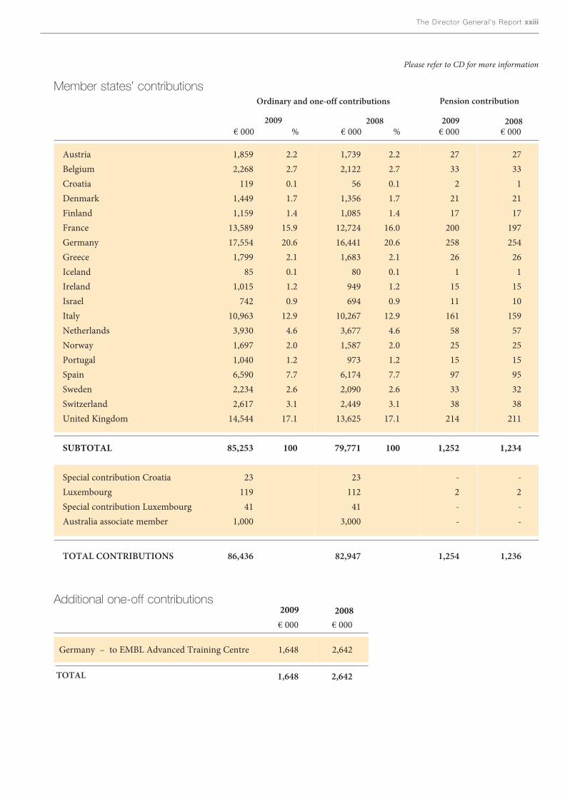
Member states’ contributionsOrdinary and one-off contributions Pension contribution
2009 2008 2009 2008
Germany – to EMBL Advanced Training Centre 1,648 2,642
TOTAL 1,648 2,642
2009 2008
Please refer to CD for more information
Additional one-off contributions
€ 000 € 000
Austria 1,859 2.2 1,739 2.2 27 27Belgium 2,268 2.7 2,122 2.7 33 33Croatia 119 0.1 56 0.1 2 1Denmark 1,449 1.7 1,356 1.7 21 21Finland 1,159 1.4 1,085 1.4 17 17France 13,589 15.9 12,724 16.0 200 197Germany 17,554 20.6 16,441 20.6 258 254Greece 1,799 2.1 1,683 2.1 26 26Iceland 85 0.1 80 0.1 1 1Ireland 1,015 1.2 949 1.2 15 15Israel 742 0.9 694 0.9 11 10Italy 10,963 12.9 10,267 12.9 161 159Netherlands 3,930 4.6 3,677 4.6 58 57Norway 1,697 2.0 1,587 2.0 25 25Portugal 1,040 1.2 973 1.2 15 15Spain 6,590 7.7 6,174 7.7 97 95Sweden 2,234 2.6 2,090 2.6 33 32Switzerland 2,617 3.1 2,449 3.1 38 38United Kingdom 14,544 17.1 13,625 17.1 214 211
SUBTOTAL 85,253 100 79,771 100 1,252 1,234
Special contribution Croatia 23 23 - -Luxembourg 119 112 2 2Special contribution Luxembourg 41 41 - -Australia associate member 1,000 3,000 - -
TOTAL CONTRIBUTIONS 86,436 82,947 1,254 1,236
2009/2010Reviews ofEMBL Scientific UnitsEMBL Units are reviewed in depth every four years by expert international panelsorganised by the Scientific Advisory Committee. To ensure openness, the reviewreports are submitted in confidence to EMBL Council and the Director General.The formal responses of the Director General to the reports are made public, tocommunicate the adjustments made by the Laboratory in response to the reviews,when needed.
The Director General ’s Report xxv
Director General’s Response to the CellBiology and Biophysics Unit Review ReportHeidelberg · 6 and 7 May 2009
1. I wish to thank the review panel for their in-depth and constructive review report.Because of the circumstances at the time of review the number of group and teamleaders in the Cell Biology and Biophysics Unit (CBB) was temporarily increasedto fourteen, which meant that the panel had to work extremely effectively to staywithin the time constraints. They did this without losing sight of the need to pro-vide detailed evaluations in large part due to the excellent chairmanship of SandraSchmid.
2. The panel was very positive about the broadening of the research focus of the Unitover the review period. In 2005, many of the groups in the Unit were working onaspects of the biology of microtubules and, although research in this area is stillconsidered by the panel to be a major asset of the Unit and an important field ofresearch, several new topics have been added by a broad recruitment policy thatthe panel viewed very favourably.
3. There was particular praise from the panel for Eric Karsenti’s scientific vision andleadership throughout the review period as well as enthusiasm for the decision toappoint Jan Ellenberg as Eric’s successor.
4. The panel appreciated the technology development efforts in the area of lightmicroscopy, underlining the novelty and broad usefulness of both the Light SheetMicroscopy methods pioneered by the Stelzer lab and the high throughput, highcontent, cell-based phenotyping systems developed in the collaborative effortbetween Ellenberg and Pepperkok.
5. A particular feature of the Unit is the uniquely successful, in the view of the panel,blend of physicists and biologists that collaborate extensively in the analysis ofcomplex biological functions by combining modelling and simulation with experi-ment. The catalytic role of Nédélec and Karsenti in generating and supporting thisinterdisciplinary environment was highlighted. The panel advised consideration offormal mechanisms that will help maintain and increase the exchange between thephysicists, biologists and chemists in CBB at all levels of seniority in the future.
6. In view of the significant level of upcoming turnover, the panel endorsed JanEllenberg’s plan to search widely, but advised to place emphasis on the possibilityof synergy between new recruits and existing activities in order to avoid too muchdispersal of activity. The panel also noted that there are currently no female facultymembers in CBB and, although they were satisfied that this did not reflect aninherent bias, they recommended that CBB pay particular attention to attractingexcellent female group or team leaders in future. They also advised Jan Ellenbergto ensure that the level of mentoring of new faculty recruits in CBB should bebrought into line with that seen in other EMBL Units.

xxvi EMBL Annual Report 09 · 10
7. Although not explicitly part of the review, as these activities will be reviewed in2010, the panel praised Pepperkok and Antony for their excellent performances inrunning the light microscopy and electron microscopy core facilities. The panelnoted that these are both critical to the future success of not only CBB but alsomany other parts of EMBL. They warned of the need for ongoing investment inthese core facilities to ensure that they remain state of the art, and commentedpositively on the plans to reorganise the light microscopy facility, that is currentlyscattered throughout much of EMBL Heidelberg, into one location. They alsorecommended that this location should be adjacent to the computational servercluster in order to avoid potential data transmission problems.
Iain W. MattajDirector General28 May 2009

ScientificReport


“The view of Jerusalem is the history of the world; it is more, it is the his-tory of Earth and heaven,” wrote Benjamin Disraeli. It’s not hard to seewhat he means. As well as being the focus of three major world reli-
gions, at nearly 3000 years old, Jerusalem is one of the world’s oldest continuouslyinhabited cities. The modern-day hustle and bustle of its winding streets echoes adaily rhythm of city life that stretches back into antiquity.
This diurnal pattern is both unique and universal: the individual dwellers and theirlives are peculiar to Jerusalem, but the basic principles of how the city is built,organised and run is something it shares with other cities around the world. It maysound odd, but cities such as Jersualem also have a lot in common with how ourcells and organs – and those of other creatures – are organised. For example, thecells in our bodies all have their own specialised functions, rather like professions,to perform. Yet even while they focus on their own particular niches, they must allco-operate to make the body as a whole function normally. Even within cells,molecules must follow certain rules to keep everything working properly.
So the organisational systems of biology are not so far removed from those of abusy town. Molecules need to travel to places, information must be exchanged,damage has to be detected and repaired, building plans drawn up and carried out,and law and order kept. Only when all these elements are in place can the cellularor molecular denizens of an organism come together to create a greater whole.
Of course, all cities must start somewhere. They originally began haphazardly,acquiring streets, buildings and districts according to the whims of the populace,with each new development building on what went before. By contrast, moderncities are planned in meticulous detail. The organisation of cells and bodies has asimilar history – evolution has blindly produced ways of planning and buildingliving systems, as biologists are now discovering. Teams at EMBL Heidelberg, forexample, have dissected the biology of one of the simplest free-living bacteria tounderstand how its constituent parts work together and to pinpoint the bare essen-tials a cell needs to survive. At EMBL Monterotondo, scientists have looked at howthe processes that build an embryo’s heart could help adults to recover from heartattacks.
Other EMBL researchers have been studying how cells and embryos deploy thebuilding plans laid down in DNA, to both understand these processes and look forways of exploiting them in medicine. What’s more, EMBL is finding ways to makethese data accessible to researchers all over the world, welcoming their input, just asany truly cosmopolitan city should.
The grandscheme ofthings

Vital ingredients

Sebastian Kühner, Anne-Claude Gavin,Peer Bork and Vera van Noort
What are the bare essentials of life? Which compo-nents and processes can an organism simply not live
without? How are they organised in space and time in orderto make a random collection of molecules come alive? Theseare mighty big questions that go well beyond what a singlebiologist can handle. But the answers are a lot more withinreach since a group of EMBL scientists joined forces andpooled their skills and expertise to tackle the problemtogether.
It all started five years ago when Peer Bork and Luis Serrano,who were then joint coordinators of the Structural andComputational Biology Unit in Heidelberg, got the Unit’sgroup leaders together to brainstorm about a common pro-ject that would put the complementary technologies andknow-how of their groups to good use. “Within the Unit wehave a unique combination of structural and computationalmethods that span a broad spectrum of scales. We can studyeverything from tiny individual molecules to the overallarrangement of the inner workings of a cell,” says Peer. Theidea: to combine these methods in an interdisciplinaryapproach to produce the first blueprint of a minimal cell, acell that is stripped down to the absolute essentials.
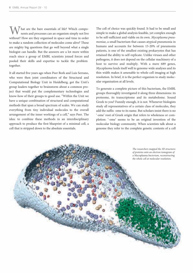
The cell of choice was quickly found. It had to be small andsimple to make a global analysis feasible, yet complex enoughto be self-sufficient and viable on its own. Mycoplasma pneu-moniae, a small bacterium that causes atypical pneumonia inhumans and accounts for between 15-20% of pneumoniapatients, is one of the smallest existing prokaryotes that hasretained the ability to self-replicate. Unlike viruses and otherpathogens, it does not depend on the cellular machinery of ahost to survive and multiply. With a mere 689 genes,Mycoplasma lends itself well to genome-wide analyses and itsthin width makes it amenable to whole-cell imaging at highresolution. In brief, it is the perfect organism to study molec-ular organisation at all levels.
To generate a complete picture of this bacterium, the EMBLgroups thoroughly investigated it along three dimensions: itsproteome, its transcriptome and its metabolome. SoundGreek to you? Funnily enough, it is not. Whenever biologistsstudy all representatives of a certain class of molecules, theyadd the suffix -ome to its name. But scholars insist there is no‘-ome’ root of Greek origin that refers to wholeness or com-pletion: ‘-ome’ seems to be an original invention of themolecular biology community. When scientists talk about agenome they refer to the complete genetic contents of a cell
6 EMBL Annual Report 09 · 10
The researchers mapped the 3D structuresof proteins onto an electron tomogram ofa Mycoplasma bacterium, reconstructingthe whole cell at molecular resolution.

The grand scheme of things 7
G R A B B I N G T H E L I M E L I G H T
This in-depth study ofMycoplasma pneumoniae caught
the attention of the top scientificjournals and the media in general,throughout the world. Nature Newsand Scientific American focused onhow this single-celled bacterium putsits genome to many uses, a sentiment
the Science signalling editorialsummed up in two words: “SimplyMycoplasma”. The work merited areview by Craig Venter in MolecularSystems Biology, and was divulged toFrench, German and Dutch scienceenthusiasts by Sciences & Avenir,Spektrum der Wissenschaft and
Explore, respectively. From TheNew York Times, El País and theFrankfurter Allgemeine Zeitung towebsites like Genome Web andGalileo, everyone was keen to reporton the unexpected complexity of thisblueprint for life.
and the proteome is nothing more than all of its proteins. TheRNAs make up a cell’s transcriptome because they are pro-duced through a process called transcription. Similarly,metabolome refers to the collection of small-moleculemetabolites found in a cell or organism. All these -omes arevery dynamic and often change from one second to the next,which is why they cannot be fully characterised using onlyone analytical method. This is where even more molecularbiology jargon comes in: proteomics, transcriptomics andmetabolomics are the newly created disciplines that are con-cerned with understanding the respective -omes, by applyinga range of genetic, biochemical and computational tech-niques.
The proteome
Characterising Mycoplasma’s proteome alone demanded acombination of around ten different techniques. So, theteams of Anne-Claude Gavin, Rob Russell, Peer Bork, BettinaBoettcher and Achilleas Frangakis, with the support of theEMBL Core Facilities, pooled their skills to form a proteomicstask force.
Anne-Claude’s lab was first in line. Using a method calledTandem Affinity Purification, PhD student Sebastian Kühnerfirst fished all soluble protein complexes from theMycoplasma cell and then determined their components bymass spectrometry, a technique that identifies proteins basedon their weight and charge. With the help of skilled bioinfor-matician Vera Van Noort and colleagues from Peer’s andRob’s groups, the mass spectrometry data were then integrat-ed with information from STRING, a database that storesinformation on protein interactions, to reconstruct whichproteins are part of which complex.
They identified around 200 protein complexes, half of whichhad not been found in previous studies of the bacterium. Thisis the first time that an exhaustive proteome analysis has beencarried out in a prokaryote. Prokaryotes are simple, mostlysingle-celled organisms, such as bacteria, that lack a nucleusand other membrane-bound organelles. Eukaryotes, on theother hand, are defined by cells organised into complex struc-tures surrounded by a membrane and comprise all animals,plants and fungi. A few years earlier, Anne-Claude, Rob and
8 EMBL Annual Report 09 · 10
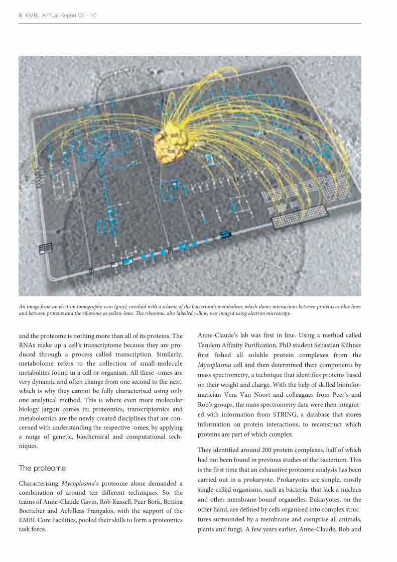
An image from an electron tomography scan (grey), overlaid with a scheme of the bacterium’s metabolism, which shows interactions between proteins as blue linesand between proteins and the ribosome as yellow lines. The ribosome, also labelled yellow, was imaged using electron microscopy.
Peer had applied the same proteomics approach to a eukary-ote, baker’s yeast.
“We find many similarities between the two organisms,” saysAnne-Claude. Many of the proteins and complexes theyfound in yeast also exist in Mycoplasma, suggesting that theyhave essential functions that have been conserved during evo-lution. “What is even more interesting is that key principlesof protein organisation also seem to be conserved.” Just likein yeast, Mycoplasma proteins rarely act alone. Most cellularprocesses are carried out by molecular machines comprisingseveral different proteins. These machines are often organ-ised into higher order assemblies, like in a factory wheremachines completing different steps of a task are located side-by-side in pipelines. In addition, both yeast and Mycoplasmaproteins tend to be promiscuous: they interact with differentbinding partners and take part in more than one complex.This suggests a great deal of multifunctionality among pro-teins and provides a mechanism by which cellular processescan be coupled in space and time. For example, the teamfound physical links between the bacterium’s RNA poly-merase, the enzyme that transcribes the DNA code into mes-senger RNAs, and the ribosome, the machine responsible fortranslating messenger RNA into protein.
To gain a better understanding of what the protein complex-es look like and of their intricate spatial organisation insidethe cell, Bettina and her team studied the purified complexeswith an electron microscope. Electron microscopes havemuch greater resolving power than light microscopes and canmagnify a specimen up to two million times. Although this isjust about enough to visualise the overall shape of proteincomplexes, it does not tell you anything about how its com-ponents fit together and interact. Luckily, bioinformatics hada solution at hand. The atomic structure of most Mycoplasmaproteins had been determined and deposited in databases.Using advanced computational methods Rob’s team fitted thestructures of 484 Mycoplasma proteins into the overall shapesBettina had produced for the complexes. Next, the largest andmost readily identifiable of these complexes, including ribo-somes and the RNA polymerase, were mapped onto an elec-tron tomogram of a Mycoplasma cell that was generated byAchilleas’ group. Electron tomograms are three-dimensionalreconstructions of parts of cells, or even whole cells in thecase of small prokaryotes such as Mycoplasma. They have asufficiently high resolution to see individual protein com-plexes in their natural context in the cell. “Electron tomogra-phy bridges the gap between high-resolution structuralapproaches and light microscopy,” says Achilleas. “It allowsus to explore the whole inner space of small prokaryotic cellsand localise atomic structures of molecules inside those cells,in their native positions and environment.”
Never before has there been such a complete and detailed,three-dimensional model of a prokaryote’s proteome, makingMycoplasma pneumoniae one of the most structurally knownorganisms to date. But to obtain a full picture of the bacteri-um and understand all processes and interactions that hap-pen in a minimal cell, the scientists needed to look at morethan the cell’s protein content. Next up: the transcriptome!
The transcriptome
Mycoplasma’s genome contains less than 700 protein-codinggenes, only 44 RNAs and uses no more than eight regulatoryproteins to control gene expression. This simplicity allowedmembers of Peer’s lab at EMBL and Luis’s group, now basedat the Centre for Genomic Regulation in Barcelona, to studyall the parts of the genome that are transcribed into RNA.Supported by the staff at EMBL’s Genomics Core Facility,postdoctoral fellows Marc Güell in Barcelona and Vera vanNoort in Heidelberg analysed all RNA transcripts found in aMycoplasma cell under different conditions. The result:Mycoplasma’s genome organisation and its transcription arenot as simple as thought. Not only did the scientists identify67 completely new transcripts, but they also found that thegenome is transcribed differently depending on the environ-ment of the bacterium.
Normally the genomes of bacteria are thought to be quitestraightforward: they are divided into operons, which areunits of transcription comprising one or more genes that aretranscribed from the same promoter (transcription start site)and controlled by the same regulatory element (operator).Under normal conditions the researchers counted 340 suchoperons in Mycoplasma, but altering different factors such asthe food source, temperature or pH changed the number ofoperons and the resulting transcripts. Some of these changesare because of internal promoters, found in the middle ofknown operons. These internal promoters are activated byenvironmental factors and break down known operons intosmaller transcription units, producing so-called alternativetranscripts of a DNA region. This dynamic structure allowsMycoplasma to react flexibly and rapidly to changing envi-ronments and to produce certain molecules only whenrequired.
But alternative transcripts are not the only novelty revealedby the analysis. It also brought to light a second type of tran-script that is equally surprising – so-called antisense tran-scripts. Antisense transcripts are generated when a protein-coding stretch of DNA is transcribed back-to-front ratherthan in the conventional direction. The role of such antisenseRNAs is not clear. They do not encode proteins but theycould help to regulate gene expression in various ways. Untilquite recently, scientists did not even know they existed. It
The grand scheme of things 9
was assumed that the transcription machinery could onlymove along one direction of a DNA strand, and thus at firstthe occasional ‘backwards’ RNAs that scientists stumbledacross were dismissed as noise of the transcription process.New research shows, however, that bidirectional transcrip-tion leading to antisense RNA is quite common in many dif-ferent organisms, especially eukaryotes. EMBL group leadersLars Steinmetz and Wolfgang Huber, for example, found thatbidirectional transcription is probably the rule rather than theexception in yeast and other studies have come to a similarconclusion for humans. So, as was found for its proteome,Mycoplasma’s transcriptome also shares many features witheukaryotic cells.
The metabolome
What is the only thing missing at this stage to complete thefull picture of a minimal cell? An accurate account of itsmetabolism. As a first step Luis and his team in Barcelonadeveloped a minimal, defined medium that supports thegrowth of Mycoplasma. “This was extremely challenging,because the human body, Mycoplasma’s natural environ-ment, provides nutrients as higher order molecules. So, thebacterium has lost the ability to live on simple building blockslike amino acids and needs peptides to survive. This made itvery difficult to find the simplest ingredients that would stillsustain growth,” explains Luis. Yet such a defined minimalmedium is crucial for obtaining precise, quantitative mea-surements of an organism’s metabolites and its exchangeswith the environment. Roughly 1300 experiments later, thescientists arrived at a mixture of 19 essential nutrients that thebacterium would happily grow in and a comprehensive mapof Mycoplasma’s metabolism, consisting of 180 reactions car-ried out by 129 enzymes.
The map identified 78 genes that are essential for the bacteri-um’s metabolism and a bioinformatics scrutiny by Peer’sgroup showed that the fraction of multifunctional enzymes ismuch higher than in more complex bacteria, even though thepathways are more linear, indicating a more streamlinedmetabolism. It also uncovered that, unlike other bacteria,Mycoplasma’s metabolism is not geared towards multiplyingas fast as possible. With a duplication time of at least eighthours, the bacterium reproduces relatively slowly, which is aresult of its pathogenic lifestyle and adaptation to its host.What it shares with larger bacteria such as Escherichia coli,however, is a great deal of flexibility and adaptability tochanging environments. Surprisingly for such a simple bac-terium with only eight regulatory proteins, Mycoplasmaorchestrates complex changes in gene expression in responseto different environmental stress factors. It seems to do so byassigning genes to one of four categories: catabolism, celldefence, biosynthesis and signal transduction. The genes in
each of these categories are regulated together as a group andjointly bring about specific responses to different stress situa-tions, such as sugar or amino-acid starvation or pH changes.
Integrating all the information collected on the three -omes –proteome, transcriptome and metabolome – the EMBLresearchers have produced what is currently the most com-prehensive picture of an entire organism. They have provid-ed a complete parts list of all cellular components, anoverview of their organisation and quantitative accounts oftheir dynamic interactions. “This is a unique resource for sys-tems biology. We will make all our data available to the sci-entific community, who can use it to build and test mathe-matical models of individual processes,” says Peer. He expectsthis approach to provide insights of unprecedented accuracyinto whole-organism biology.
The overwhelming conclusion so far: in all aspects studied thebacterium turned out to be more complex and dynamic thanpreviously assumed. The temporal and spatial organisation ofits molecular machinery, the transcription process and itsmetabolism share many features with larger, more complexprokaryotes and even with eukaryotic cells. This complexityis unexpected considering Mycoplasma’s small genome andalleged simplicity, and it makes the bacterium a powerfulmodel organism. It is simple enough for large-scale analysis,yet sufficiently complex for its cellular organisation to be rep-resentative of other prokaryotes, eukaryotes and even higher,multicellular organisms.
At the same time Mycoplasma is also representative of some-thing else: minimal life. Owing to its unique evolution as apathogen, the bacterium has been stripped of everything thatis not absolutely essential. It has become adapted for life inthe human body – a very special and rich biological niche thatmade many genes redundant so that they were lost over time.What is left today is the minimal structure and machineryrequired for survival, and thanks to the EMBL taskforce thishas been captured in a blueprint. This blueprint and the uni-versal organisational principles it highlights are probably theclosest scientists have ever come to identifying the essentialsof life. “The key lies in those features that Mycoplasma shareswith all other organisms. These are the things that not eventhe simplest organism can do without and that have remaineduntouched by millions of years of evolution,” Peer concludes.
Kühner S, et al (2009) Proteome Organization in a Genome-Reduced Bacterium. Science 326
Güel M, et al (2009) Transcriptome Complexity in a Genome-Reduced Bacterium. Science 326
Yus E, et al (2009) Impact of Genome Reduction on BacterialMetabolism and Its Regulation. Science 326
10 EMBL Annual Report 09 · 10

The huge wealth of data generatedby the past decade’s genome pro-
jects is now much easier for biologiststo study thanks to a new online toolcreated at the EBI. Group leader AlvisBrazma and his team have made itpossible for biologists to explore infor-mation about a particular gene’s activi-ty in much greater depth than everbefore, without the need for specialisttraining in bioinformatics.
The new tool, the Gene ExpressionAtlas, was launched in March 2009,and is freely available to all. Biologistscan search data for information abouthow a gene’s activity, or expression,varies between different tissues, underdifferent biological conditions (such asdrug treatment), in disease and muchmore. Geneticists hunting down dis-ease genes will be able to investigatepossible leads much more quickly andextensively. “It will help biologistsdevelop and test new hypotheses,”says Alvis.
The seeds of the Atlas were sown whenAlvis joined the EBI more than adecade ago. At the time, data frommicroarrays – experiments that allowscientists to study the activity of thou-
sands of genes at once – were accumu-lating and Alvis wanted to find ways toget the most out of these results.
He and his colleagues developed a pro-tocol that allows researchers to labelthe pieces of information in theirmicroarray data in a uniform way.This led to an EBI database calledArrayExpress, in which these datacould be deposited. Thanks to thelabelling system, researchers couldmine the data for new insights into thebiology of genes and disease. “The planwas to summarise these data in a waythat meant we could ask biologicalquestions,” says Alvis. “It was neverintended just to be an archive.”
The trouble was, this mining was farfrom straightforward, and so was reallyonly an option for computational data-analysis experts. Alvis and his teamdecided to create something that wasmuch more accessible. TheArrayExpress curators revisited thedata and created new ways of enrich-ing and labelling it. Led by MishaKapushesky, a project leader withinAlvis’s group, researchers developedand implemented statistical softwarethat could exploit this new labelling toanalyse possible relationships between
all the items of data more effectivelythan before.
Biologists can now analyse these datasimply by typing a few details – thename of a gene, a species or a disease –into a box on the EBI website. TheAtlas displays detailed results, rankedin order of relevance, and users canclick through to relevant informationon other EBI databases.
Thousands of academic researchersare already using the Atlas and theEBI’s industry partners are also inter-ested in taking the technology further,says Alvis. Currently, the Atlas con-tains about 15% of the data fromArrayExpress, and describes more than200 000 genes in more than 6000 dif-ferent biological states. Alvis’ teamplans to boost this to 50% in the nearfuture, and has already started workon a similar Atlas for protein data.
Kapushesky M, Emam I, Holloway E,Kurnosov P, Zorin A, Malone J, Rustici G,Williams E, Parkinson H, Brazma A (2010)Gene Expression Atlas at the EuropeanBioinformatics Institute. Nucleic AcidsResearch 38, Database issue
The grand scheme of things 11
Mapping the future
Misha Kapusheskyand Alvis Brazma
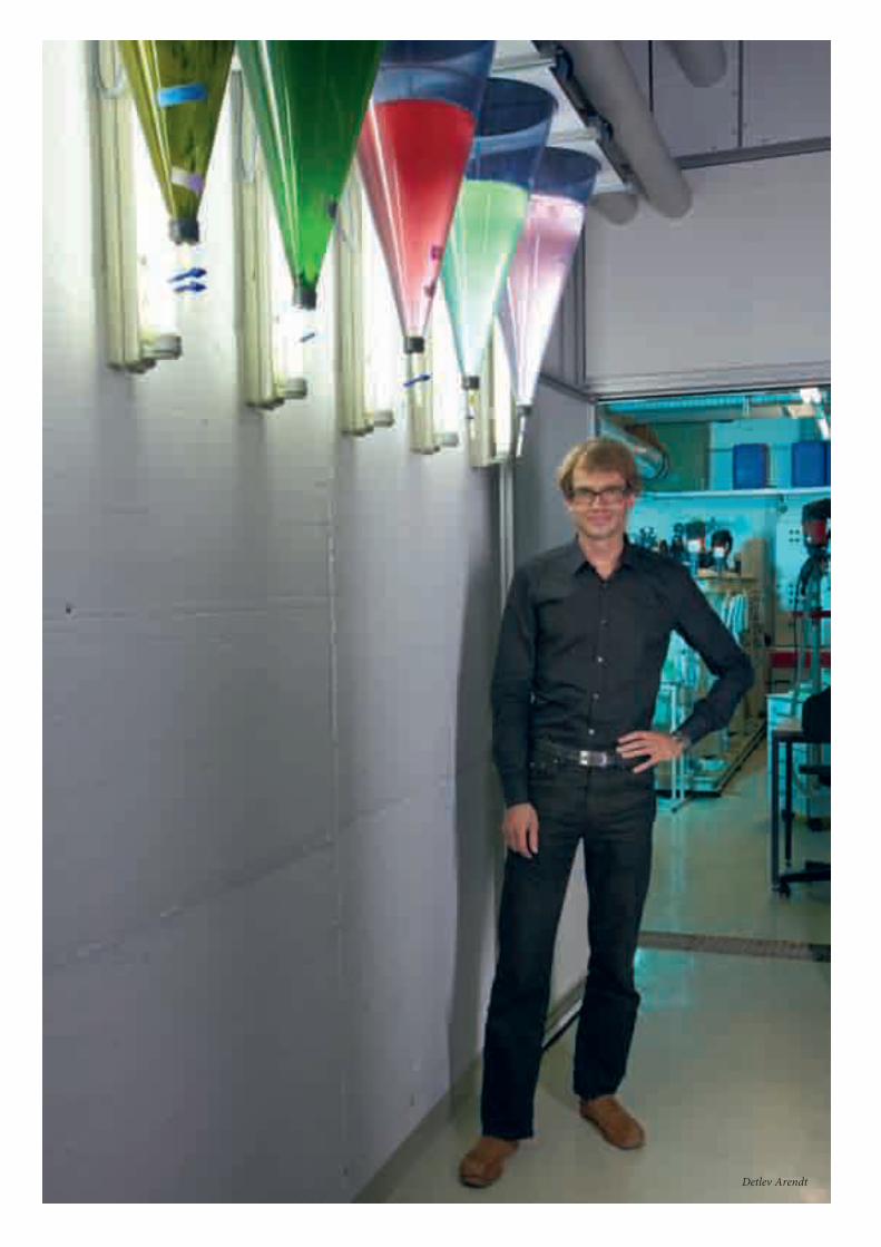
Detlev Arendt

Twenty years ago, scientists knew nothing of thescraps of RNA that are now known to influence just
about every process in our bodies. Back then, the text-books were simpler: genes code for proteins via the inter-mediate of RNA, and proteins called transcription factorsregulate other proteins. This recipe was so entrenched inthe basic orthodoxy of molecular biology that it was evengiven the name ‘the central dogma’ by the co-discovererof DNA, Francis Crick.
Scientists now know, however, that this classic view ofprotein regulation is far too blunderingly inefficient forevolution to settle for. At some point hundreds of mil-lions of years ago, the generation of a small stretch ofRNA that could tweak this process gave an individual theedge over everyone else. And so regulatory RNA wasborn. These scraps of RNA – on average only 22nucleotides long and now dubbed microRNAs, ormiRNAs for short – bind to some messenger RNAs andlabel them for inactivation or destruction.
So far thousands of miRNAs have been identified in ani-mals. These superintendents of protein regulation areinvolved in the earliest stages of an animal’s development,determining which cell types grow where and when, andhow these cells differentiate into the different body parts.However, since the discovery of miRNAs, many scientistshave wondered whether the same miRNAs govern specif-ic tissues in different animals. Knowing this would notonly give clues to the age of these different miRNAs, butalso to the age of the cells in which they are found.
Ancient tweaking
Developmental biologist Detlev Arendt from EMBLHeidelberg, whose work has recently traced the evolu-tionary origins of the brain and other organs, wonderedwhether he could study the oldest known animal miRNAsin a group of animals that at least look old themselves. Indoing so, he hoped to investigate whether miRNA expres-sion is conserved across the animal kingdom.
Studying ancient-looking animals is crucial for this pro-cess because “all animals evolve, but the speed with whichthey change differs,” explains Detlev. “Animals livingnear the coastline today exist in a similar environment tothat which their ancestors thrived in, and so some of theseanimals haven’t been forced to change their body plans,or the genes that control these plans, because they arealready well-adapted to this ancient environment.” Bystudying these animals, biologists can glean clues toancient morphologies.
Detlev and Fay Christodoulou, who was a PhD student inDetlev’s group, used one such ‘living fossil’, the marineragworm Platynereis dumerilii, which is thought to havechanged little over the past 600 million years. In collabo-ration with an American team from Cold Spring HarborLaboratory who sequenced the ragworm’s miRNAs, andPeer Bork’s group at EMBL who contributed to the bioin-formatic analysis, they probed for 34 ancient miRNAs inthe bodies of young worms.
By tracing a blue dye that becomes trapped when specificprobes bind to the miRNA, Fay was able to see where themiRNAs were expressed in the worm embryos. “Initiallythe high temperatures required for the probes to bind
The grand scheme of things 1313

14 EMBL Annual Report 09 · 10
MicroRNAs present in the ragworm, sea urchin and worm probably existedalready in their last common ancestor, which separated from the seaanemone’s branch of the evolutionary tree around 600 million years ago.
RagwormPlatynereis dumerilii
Marine wormCapitella
HumanHomo sapiens
Sea urchinStrongylocentrotus
Sea anemoneNematostella
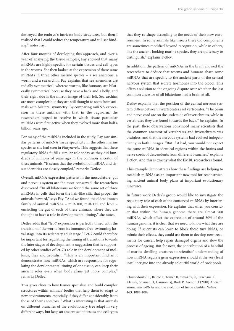
destroyed the embryo’s intricate body structures, but then Irealised that I could reduce the temperature and still see bind-ing,” notes Fay.
After four months of developing this approach, and over ayear of analysing the tissue samples, Fay showed that manymiRNAs are highly specific for certain tissues and cell typesin the worms. She then looked at the expression of these samemiRNAs in three other marine species – a sea anemone, aworm and a sea urchin. Fay explains that sea anemones areradially symmetrical, whereas worms, like humans, are bilat-erally symmetrical because they have a back and a belly, andtheir right side is the mirror image of their left. Sea urchinsare more complex but they are still thought to stem from ani-mals with bilateral symmetry. By comparing miRNA expres-sion in these animals with that in the ragworm, theresearchers hoped to resolve in which tissue particularmiRNAs were first active when they evolved more than half abillion years ago.
For many of the miRNAs included in the study, Fay saw sim-ilar patterns of miRNA tissue specificity in the other marinespecies as she had seen in Platynereis. This suggests that theseregulatory RNAs fulfill a similar role today as they did hun-dreds of millions of years ago in the common ancestor ofthese animals. “It seems that the evolution of miRNA and tis-sue identities are closely coupled,” remarks Detlev.
Overall, miRNA expression patterns in the musculature, gutand nervous system are the most conserved, the researchersdiscovered. “In all bilaterians we found the same set of threemiRNAs in cells that form the hair-like cilia that propel theanimals forward,” says Fay. “And we found the oldest knownfamily of animal miRNAs – miR-100, miR-125 and let-7 –encircling the gut of each of these animals, where they arethought to have a role in developmental timing,” she notes.
Detlev adds that “let-7 expression is perfectly timed with thetransition of the worm from its immature free-swimming lar-val stage into its sedentary adult stage.” Let-7 could thereforebe important for regulating the timing of transitions towardsthe later stages of development, a suggestion that is support-ed by other studies of let-7’s role in the development of mol-luscs, flies and zebrafish. “This is an important find as itdemonstrates how miRNAs, which are responsible for regu-lating the developmental timing of one tissue, can keep theirancient roles even when body plans get more complex,”remarks Detlev.
This gives clues to how tissues specialise and build complexstructures within animals’ bodies that help them to adapt tonew environments, especially if they differ considerably fromthose of their ancestors. “What is interesting is that animalson different branches of the evolutionary tree adapt in verydifferent ways, but keep an ancient set of tissues and cell types
that they re-shape according to the needs of their new envi-ronment. In some animals like insects these old componentsare sometimes modified beyond recognition, while in others,like the ancient-looking marine species, they are quite easy todistinguish,” explains Detlev.
In addition, the pattern of miRNAs in the brain allowed theresearchers to deduce that worms and humans share somemiRNAs that are specific to the ancient parts of the centralnervous system that secrete hormones into the blood. Thisoffers a solution to the ongoing dispute over whether the lastcommon ancestor of all bilaterians had a brain at all.
Detlev explains that the position of the central nervous sys-tem differs between invertebrates and vertebrates. “The brainand nerve cord are on the underside of invertebrates, while invertebrates they are found towards the back,” he explains. Inthe past, these observations convinced many scientists thatthe common ancestor of vertebrates and invertebrates wasbrainless, and that the nervous systems had evolved indepen-dently in both lineages. “But if it had, you would not expectthe same miRNA in identical regions within the brains andnerve cords of descendents from different branches,” explainsDetlev. And this is exactly what the EMBL researchers found.
This example demonstrates how these findings are helping toestablish miRNAs as an important new tool for reconstruct-ing ancient animal body plans at important evolutionaryjunctures.
In future work Detlev’s group would like to investigate theregulatory role of each of the conserved miRNAs by interfer-ing with their expression. He explains that when you consid-er that within the human genome there are almost 700miRNAs, which affect the expression of around 30% of thehuman genome, it is clear that we need to know what they aredoing. If scientists can learn to block these tiny RNAs, ormimic their effects, they could use them to develop new treat-ments for cancer, help repair damaged organs and slow theprocess of ageing. But for now, the contribution of a handfulof marine-dwelling creatures to scientists’ understanding ofhow miRNA regulate gene expression should at the very leastinstil intrigue into the already colourful world of rock pools.
Christodoulou F, Raible F, Tomer R, Simakov, O, Trachana K,Klaus S, Snyman H, Hannon GJ, Bork P, Arendt D (2010) Ancientanimal microRNAs and the evolution of tissue identity. Nature463: 1084-1088
The grand scheme of things 15

Shaping up HIV

John Briggs, James Riches and Alex de Marco

In the 1960s, a Danish company, seeking toimprove on the traditional football made
from the bladder and stomach of animals,invented the modern football. The designersrealised that to form a perfect ball they need-ed to combine 20 leather hexagons with 12pentagons, and in so doing demonstrated oneof the basic laws of shape – that you cannot wrapa sheet of six-sided hexagons around a sphere. Toinduce the sheet to bend, the company had to introducefive-sided pentagons alongside the hexagons.
On the micro scale, the human immunodeficiency virus(HIV), which causes AIDS, faces a similar challenge duringthe assembly of new viral particles: how to coerce its hexagon-shaped building blocks to form the spherical envelope thatsurrounds its viral innards. Lifting a page from the footballmanual, structural biologist John Briggs, group leader atEMBL Heidelberg, wondered if HIV likewise solved thisshape conundrum by introducing pentagons between thehexagons.
At the early stages of assembly, HIV relies on a single proteinto bring together everything that is needed for a new viralparticle. This protein, called gag, is shaped like a string of dia-mond-shaped beads and is produced when the virus is copiedby the host cell’s machinery. These copies are exported fromthe cell’s nucleus, and are turned into long chains of aminoacids that fold into the gag protein. Hundreds of these gagproteins aggregate just beneath the cell’s membrane wherethey team up newly copied viral DNA with proteins andenzymes that are vital for the virus to infect new cells. Whileeverything is being corralled into position, the gag proteinsbundle together – six at a time – to form hexagon-shapedplates.
It is the arrangement of these plates that John and his teamwanted to inspect. But to do this, they needed to halt thevirus’ development while preserving its structure very close toits natural state. In collaboration with Hans-GeorgKräusslich’s team at the University Clinic Heidelberg, John’sgroup took advantage of a technique called cryo-electrontomography – cryo-ET for short – which combines flash-freezing with electron beam scanning to create three-dimen-sional images of the internal structures of the virus.
“What is nice about this approach is that it allows us to lookinside samples of biological material without breaking themapart or staining them, as we had to do in the past, whichoften led to unexplainable artefacts,” says John.
This work requires “swift, agile hands” explains James Riches,an engineer in the Briggs group, and whose fiddly job it is toplace the 3mm copper disks containing frozen viral particles
on to a platform that automatically moves through a range oftilt angles while beams of electrons illuminate the sample. Ateach angle, a thin region of the sample is exposed to the neg-atively charged particles. When the electron beam collideswith this solid structure, it casts a shadow onto a camera.These two-dimensional projections are then fed to a comput-er, which assembles the data into a composite three-dimen-sional image.
Inspecting these images, the researchers found that thehexagon-shaped plates were arranged into a lattice, but thatthis lattice was incomplete; it had gaps within it – areas wherethe gag proteins were missing altogether. “We were surprisedthat these gaps were not pentagon-shaped, but were insteadirregularly shaped, variable in size and that there was alwaysmore than one of them,” says John.
He thinks that these gaps prevent the hexagons from becom-ing too closely packed together when the lattice curves toform the viral envelope. He goes on to explain that during theformation of this envelope, nicks in the gag protein transformthe immature virus into its infectious form. An enzyme calledprotease performs this function, and preventing its cuttingaction has been the target of some of the most successful HIVdrugs of the past 10 years.
Knowing more about this maturation process could help sci-entists develop new ways to stop the virus from maturing andspreading to other cells. With this in mind, John’s group got
18 EMBL Annual Report 09 · 10
This 3D computer reconstructionshows the lattice of hexagonal Gagproteins that will mature into theshell of the infectious HIV virus.

in closer to the HIV virus, to have a more detailed look at howthe gag proteins bundle together to form each hexagon-shaped plate.
Understanding the role of the gag protein requires not onlyknowing how the envelope is laid down, but also identifyingthe structure of the individual gag proteins within the enve-lope. “You can solve the structure of the brick, but if you wantto know the structure of the house then you need to knowhow the bricks pack together,” reckons John.
To zoom in on the three-dimensional image of the virus’structure, his team sampled small segments of the viral enve-lope and assembled them one on top of the other. This wasessential because each close-up is missing some information;at this focus, parts of the image will be fuzzy or missing. Johnexplains that a small, but different, part of each image is con-cealed and so by overlaying the images you should get to seethe whole thing.
“It’s a bit like if you were to take a picture of a person stand-ing in a field in a snow storm,” explains John. “Now if youtake one picture, some of the person will be obscured by the falling snow, but if you took 50 pictures, each with a dif-ferent pattern of snowflakes, and average all those imagestogether then you would probably get a clear image of theperson.”
Up this close, the researchers discovered that each hexagon-shaped plate consists of six gag proteins arranged with their
flat side facing inwards towards a central hole. And theyrecognised that this configuration is very different from howthe gag proteins arrange themselves in mature viruses, inwhich they turn their flat side outwards.
At some point during maturation the proteins must rotate,reckons John, whose team is now working on producing aneven higher resolution structure of the gag proteins to gain amore detailed understanding of the virus’ assembly and maturation.
“We know that the cutting of the gag protein in five differentpositions is essential to this maturation process,” explainsJohn. “But what happens if you only allow cuts one, two andthree but not four and five, what kinds of structures do youget out?” To investigate this, Alex de Marco, a PhD student inJohn’s group, is studying mutant lines in which some of thecutting by the protease is prevented, and the team will thenexamine the changes they see in the viral structures. Imagingthese structures could give scientists clues on how to throw aspanner in the viral works, and stop the formation of new viralparticles and their spread to other cells. New ways to tackle thespread of HIV are needed because the virus, being small andfast to mutate, always has the upper hand in this match.
Briggs J, Riches J, Glass B, Bartonova V, Zanettia G, Kräusslich H-G (2009) Structure and assembly of immature HIV. PNAS 106:11090–11095
The grand scheme of things 19
host genome
actin
microtubules
CD4 and coreceptor
Gag
MA
CA
NC
viral glycoproteins
RT, IN and other viral proteins
viral RNA
viral DNAnucleus
cytoplasm
2
1 12
11
10
9
8
76
5
4
3
plasma membrane
nuclear membrane
HIV’s lifecycle. Once the virus has infected a host cell and harnessed its machinery to produce copies of itself (1-9), the structural protein Gag is essential fordirecting the assembly of the viral coat (10). Once the virus has left the host cell, Gag must be cleaved so that it can mature (12) and become capable of fusingwith a new susceptible cell.
20 EMBL Annual Report 09 · 10
An accurate forecast ofregulatory activity
As predicted, the same CRM isactive (red/pink) early in
Drosophila development (left) inthe tissue that will give rise to all
muscle types, but is active only in theembryo’s body wall muscle (blue) at a
later stage (middle), when a differentCRM drives development in the gut muscle
(green, right).
– i.e. the combinations of transcrip-tion factors binding at different timesand places – of approximately 8000CRMs involved in regulating muscledevelopment in the fruit flyDrosophila. The activity of a numberof these CRMs had been previouslystudied, and the EMBL team used thisinformation to group them into class-es according to the type of muscle andthe developmental stages in whichthey are active. The scientists thentrained a computer to unravel thebinding profiles for each of thesegroups, and to then search the 8000newly identified CRMs for ones whosebinding profiles fitted one of the pic-tures. Such CRMs were thereby pre-dicted to have similar activity patterns,implying that they are involved in reg-ulating the development of the samemuscle type.
“When we tested the predictionsexperimentally, the results were notonly accurate but also enlightening,”says Eileen. She explains: “It turns outthat the regulatory code, where onebinding profile leads to one pattern ofCRM activity, is actually not thatstraightforward.” They found thatCRMs with strikingly different bind-ing profiles can have similar patterns
of activity. Although it may seemunnecessarily redundant to have sev-eral different work teams capable ofbuilding the same part of the house,this unexpected plasticity does makesense in evolutionary terms, theresearchers say. The fact that differentcombinations of transcription factors,or binding codes, can regulate thesame developmental process meansthat even if some transcription factorsor CRMs change or are lost during anorganism’s evolution, it can still devel-op a gut muscle, for instance. In otherwords, having different work teamsthat use different tactics to build thesame room provides a back-up system:if one team has an accident, anothercan step in and do the job.
“What’s exciting for me is that thisstudy shows that it is possible to pre-dict when and where genes areexpressed, so we can infer what func-tions those genes may perform,” con-cludes Eileen. “It is an important firststep towards understanding how regu-latory networks drive development.”
Zinzen RP, Girardot C, Gagneur J, BraunM & Furlong EEM (2009) Combinatorialbinding predicts spatio-temporal cis-regu-latory activity. Nature 462: 65-70
To build an apartment block, aconstruction company must
have the blueprint for the entire build-ing. But workers on the ground onlyneed to refer to small sections of thedocument that contain the instruc-tions that are relevant to them.Similarly, each cell in an embryo car-ries the genetic blueprint for the wholeorganism but, by referring to differentparts of this developmental plan, cellscan perform different functionsdepending on where they are and howfar the embryo has developed. Theforemen who control what part of thegenetic blueprint the cellular machin-ery will act on are called transcriptionfactors. They bind to stretches of DNAknown as cis-regulatory modules(CRMs) and, once bound, they canswitch genes on or off. “We wondered:would it be possible to use informa-tion on when and where this bindinghappens to predict CRM activity?”says Eileen Furlong, joint head of theGenome Biology Unit at EMBLHeidelberg.
Biologist Robert P. Zinzen, computerscientist Charles Girardot and statisti-cian Julien Gagneur teamed up to takea novel, integrated approach toanswering this question. They identi-fied and recorded the binding profiles

The grand scheme of things 21
It’s a jungle in there E. coli, one of manybacteria that live in thehuman gut.
Unit at EMBL Heidelberg, togetherwith an international team of scien-tists, turned to metagenomics, a tech-nique that involves sequencing asmany genes as possible straight froman environmental sample. In this case,it was samples of faeces taken from124 European volunteers as part of anEU project called Metagenomics of theHuman Intestinal Tract, or MetaHIT.The approach allowed the team toconstruct a “metagenome”, a virtuallycomprehensive catalogue of all thegenes present in the gut’s microbialecosystem.
This was made possible thanks to a new kind of technology calledIllumina sequencing, which yields veryshort stretches of DNA sequences.Although this dramatically cuts costs,it presents a considerable challenge forbioinformaticians, who have to find away of piecing these fragments ofsequence together correctly into entiregenes. Until now, researchers didn’tthink it was possible to do this formetagenomics work. But Peer and hiscolleagues have shown that if theysequence a large number of samples, itcan indeed be done. “It’s a dramaticadvance,” says Peer. “We can nowreally tackle ecology.”
Although the team hasn’t sequencedenough to find every single gene, thenew dataset is 200 times bigger than
any other metagenomic study done sofar. “There will certainly be moregenes to come,” says Peer of the gutmetagenome, “but we don’t expectmany more.”
The team used the data to construct a“minimal genome”, the bare essentialset of genes needed for a bacterium tosurvive in the gut, as well as a “mini-mal metagenome”, the set of genesneeded for the entire ecosystem tofunction. Intriguingly, the team alsofound that, contrary to previous sug-gestions, people seem to share most oftheir microbial species. They alsoshowed that people with bowel dis-eases such as Crohn’s disease harbourdistinctive microbial populations.
Although the creation of this cata-logue is a significant milestone, Peeremphasises that it is just the beginningof a new area of research. “We intendto follow up on the disease findingsand the ecology,” he says. Who knows,years from now, it might not only bepolite, but de rigeur to discuss thecomposition of one’s gut ecosystem atthe dinner table, over a dessert ofyogurt microbiologically customisedto increase your well-being.
Qin J et al (2010) A human gut microbial genecatalogue established by metagenomic sequenc-ing. Nature 464: 59-64
Etiquette dictates that there aresome conversation topics one
should avoid at a polite dinner party.Religion and politics are the obviousones. Faeces, you might think, wouldbe another. But that might be about tochange. EMBL researchers have creat-ed a detailed catalogue of the micro-bial genes found in human faeces andin so doing have built a fascinatingpicture of the intricate ecosystemflourishing inside our intestines. Thecatalogue will act as an invaluable ref-erence for researchers trying to under-stand how this ecosystem contributesto human health and disease – surelythe stuff of dignified dinnertime dis-course.
It’s a slightly uncomfortable thought,but the microbes that live in or onyour body outnumber your own cellsby at least a factor of ten. Most ofthese 100 trillion or so organisms liveinside your gut, and are thought toplay a role in many aspects of ourbiology, such as modulating ourimmune system and contributing toour nutrition. Changes in the balanceof this ecosystem might be involved indiseases such as inflammatory boweldisease or obesity.
But many of these microbes can’t begrown in the lab, making it hard todetermine what species are present.Instead, Peer Bork, joint Head of theStructural and Computational Biology

Nadia Rosenthal

Pity Tithonus! Lover of Eos, the Titan of dawn, he wasgranted the gift of eternal life by Zeus, king of the
gods. Trouble was, the absent-minded Eos forgot to askZeus to grant him eternal youth as well. As the yearspassed, the poor immortal Tithonus withered and aged sogrievously that he implored daily for death. Eos took pityon him and, unable to restore his youth, turned him intoa grasshopper to end his suffering.
Although this fable is thousands of years old, it has par-ticular resonance today. Thanks to modern medicine, wecan expect to live far longer now than ever before. Butmuch of this increased lifespan is plagued by diseases ofageing and biologists working in the field of regenerativemedicine are trying to find ways to replace ailing anddamaged tissue. Some are hoping to do this by reawaken-ing the processes behind embryonic development in adulttissues, so that old organs might renew themselves. Now,a team led by Nadia Rosenthal at EMBL Monterotondohas discovered a possible way of tapping into the heart’sinner youth so that it can repair itself. In doing so, theyhave also gained new insights into how heart develop-ment can go awry in the womb. And like Tithonus, thesolution lies in an insect – not in the chirping legs of agrasshopper, but in the gauzy wings of a fruit fly.
Heart disease remains the world’s biggest killer of chil-dren and adults alike. Congenital heart problems are themost common form of birth defect, and heart failurecaused by long-term high blood pressure and heartattacks are prevalent causes of heart problems in adults.So researchers are keen to both dissect the biological pro-
Young at heart
cesses governing heart development and devise new treat-ments for heart damage.
Although Nadia and her team have ultimately foundsome answers to these problems, they originally startedwork on a different project. PhD student PaschalisKratsios had joined the lab, and was studying a genecalled NFκB. The protein made by this gene is involved ina signalling system that induces cells to divide or to die.Paschalis was using a genetic technique to turn off NFκBsignalling in the hearts of adult mice to study its role inkeeping the heart healthy. But the work progressed slow-ly, despite assistance from another lab member, FoteiniMourkioti. “I am eternally grateful to her,” says Paschalis,“she was the postdoc who took me by the hand.” Theexperiments were working, but Paschalis was frustratedby having to wait for several months before trying toidentify any physical defects in the hearts of the mice.And even at the end of the wait, it was hard to spot any-thing wrong.
So in the meantime, he started looking for another pro-ject, one that he could develop himself and call his own.“I wanted my own baby!” he says. “I’m really grateful toNadia for the opportunity to join the EMBL family andhave the intellectual freedom to explore.” Reading up onthe literature about heart disease, he came across a familyof genes called the Notch family. Unlike the NFκB path-way, there were a number of human heart malformationsassociated with mutations in these genes. One of these,Alagille syndrome, results in congenital heart defectsincluding holes between chambers, an enlarged right ven-tricle and narrowing of certain arteries. Research in mice
The grand scheme of things 2323

had also confirmed that Notch played an important role inheart development. Other work, meanwhile, hinted thatNotch could be involved in regenerating damaged tissues, andwas therefore worth investigating as a potential focus fortherapies to treat heart disease. “From the existing literatureon several model organisms, it looked like Notch would be agood target,” says Paschalis.
Notch genes take their name from the first member of thefamily to be discovered in the fruit fly Drosophilamelanogaster. Fly geneticists had been studying mutant fliesthat had little nicks, or notches, in the edges of their wings.When they isolated the associated gene, they found that itcoded for a receptor – the cellular equivalent of a TV satellitedish – that sits in the outer membrane of cells to receive sig-nals from the environment and other cells.
But dissecting Notch function in mice wasn’t going to be easy.There are a total of four Notch genes in the mouse, all ofwhich are active in the developing heart. It was quite possiblethat one gene could compensate for the loss of another, mak-ing traditional genetic experiments to switch off the activityof genes extremely hard to do. So Paschalis and his colleaguestook the opposite approach – they used a genetic trick tomake Notch signalling overactive in their chosen cell types.
They began by looking at overactive Notch in developingmouse hearts and saw that the mice developed heart defects.“We saw that the mice’s hearts became bigger,” saysPaschalis. Further analysis showed this was because the devel-oping heart muscle cells had undergone too much cell divi-sion and had not developed their specialised functions prop-erly. The team then used another genetic trick that allowedthem to switch off all Notch signalling in the developinghearts. Sure enough, this had the opposite effect to overacti-vating Notch: one of the heart chambers was abnormallysmall and its walls thin – just like human Alagille syndromepatients. This all points to the levels and timing of Notch sig-nalling as being crucial for controlling developing heart mus-cle cell division, and gives a valuable new insight into its rolein congenital heart disease.
The role of Notch in heart cell division was also an excitingfinding for research into treatments for acquired heart disease– conditions that develop later in life, such as heart failure fol-lowing heart muscle damage caused by a heart attack. There isa great deal of interest in developing therapies that make theheart repair itself, which include boosting cell division. “Ourfindings lend support to the notion that, in certain situations,redeployment of embryonic signalling pathways could provebeneficial for tissue regeneration in the adult,” says Nadia.
With this in mind, the team decided to switch on Notch sig-nalling in adult mice whose hearts had been surgically dam-aged to mimic the effects of a heart attack. The mice withextra, genetically induced, Notch signalling had improvedheart function and were more likely to survive than thosewithout. “It turned out to be beneficial,” says Paschalis.
Obviously, it would be impossible to perform the same kindof genetic tricks in human patients, so the team investigatedanother – more clinically relevant – tactic to raise Notch sig-nalling in the mice. This time, they injected the damagedhearts with an antibody that switches on Notch signalling andagain found significant improvements compared with theuntreated mice. As well as having reduced scarring, the treat-ed hearts grew new blood vessels. And in contrast to the find-ings in the embryonic heart, there was little extra cell divisionin the adults. Instead, the Notch signalling seemed to help theexisting muscle cells survive. “Overall, these results highlightthe importance of timing and context in biological communi-cation mechanisms,” says Nadia. To complete the experi-ments, they then switched off Notch function in mice withdamaged hearts. As expected, these mice were much morelikely to die and exhibit severe heart failure.
In theory at least, this suggests that researchers could indeedredeploy a developmental process to repair damaged heartsand exploit Notch as a possible way to treat congenital heartdisease. Nadia’s team will, however, be taking the work for-
24 EMBL Annual Report 09 · 10
In a normal newborn mouse’s heart (left), the two major heartchambers are clearly separated by tissue, but when Notch signalling
was inactivated in an embryo’s heart muscle cells (right), this septumbetween the ventricles was incomplete.
In these microscopy images of adult hearts, healthy tissue isshown in red and damaged tissue in blue. Normally (left), a
heart attack causes extensive tissue damage to the left ventricle(right-hand cavity), but this tissue damage was reduced in mice
in which Notch was re-activated after the heart attack (right).

ward without Paschalis, who is now pursuing his postdoctor-al studies in a neurobiology lab in New York. But the storydoesn’t end there. It turns out that Paschalis did indeed finishhis NFκB experiments, and uncovered yet more intriguingresults for Nadia’s team to follow up.
When Paschalis disabled NFκB signalling in the mouse heart,he found the mice developed heart muscle wasting, inflam-mation and heart failure as they aged. These symptomsweren’t obvious in younger mice, which is why he’d had ahard time spotting them. But when he increased the bloodpressure of these young mice, the symptoms appeared muchsooner. In other tissues, NFκB is known to play a role inreducing oxidative stress – damage caused by chemically reac-tive by-products of metabolism. To see whether a lack ofoxidative stress protection could explain the mice’s symptoms,Paschalis fed them a diet rich in anti-oxidants. This partly
resolved the symptoms, but didn’t reverse them completely,suggesting that other unknown factors were coming into play.
Overall, the findings pointed to NFκB signalling being need-ed to protect the adult heart from mechanical and age-relatedstress although it’s not yet clear how this might work inhuman heart patients. “So far, we don’t know of any humanNFκB pathway mutations that are associated with heart dis-ease,” says Paschalis. Even so, the work done by Nadia andher team raise the intriguing possibility that we might one daybe able to enjoy our later years minus at least some of the rav-ages of age – and we won’t need to be magically transformedinto insects to do so.
Kratsios P, Catela C, Salimova E, Huth M, Berno V, Rosenthal N,Mourkioti F (2009) Distinct roles for cell-autonomous Notch sig-nalling in cardiomyocytes of the embryonic and adult heart.Circulation Research 106: 559 - 572
The grand scheme of things 25
Artist’s representationof how looking atembryonic hearts canhelp repair adult hearts.


Alittle after midnight on Sunday 2nd September 1666, a fire broke out in a bakery in Pudding Lane in the city of London. Fanned by the wind,flames quickly spread to neighbouring houses and streets. The dithering
Lord Mayor delayed demolishing houses to create firebreaks, the only major fire-fighting technique available at the time. As a result, the blaze turned into a ragingfirestorm that engulfed the city for three days, consuming thousands of homes,destroying St Paul’s Cathedral and turning hundreds of thousands of Londonersinto destitute refugees. At last, on the Wednesday, the fire died down, leaving whatcontemporary diarist Samuel Pepys described as “the saddest sight of desolationthat I ever saw.”
Had London had a dedicated firefighting organisation – and a more decisive LordMayor – much of this destruction could have been averted. Modern firefightingtechniques and services mean that small fires no longer turn into major conflagra-tions. What’s more, other emergency services, such as ambulance crews, can help tolimit the damage and loss of life, and minimise the rebuilding work that needs to bedone. Of course, preventing disasters, both big and small, is far preferable, which iswhy governments dedicate time and money to monitoring, gathering intelligenceand defence.
Organisms and cells have their own equivalents of emergency services: immunesurveillance systems to warn of microbial attack, proteins that detect and minimisedamage, and cells that trigger the repair of damaged tissue. EMBL researchers havebeen investigating these mechanisms in a range of creatures, from plants tohumans. A team at EMBL Grenoble, for example, has determined the structure of a hormone receptor that helps plants to respond to water shortages, giving scientists a new avenue for the development of more drought-tolerant crops. OtherEMBL researchers, meanwhile, have uncovered a gene that makes mosquitoes resistinfection by the malaria parasite – a possible step towards restricting the spread ofthis deadly disease.
Within our own cells, molecular emergency workers are constantly fending offcalamity. Our DNA is under endless chemical and physical attack, and it is thanksto dedicated DNA-repair systems – being investigated by a team at EMBLHeidelberg – that our genes sustain only a fraction of the damage that hits them. Of course, no system is perfect, and a collaboration between Grenoble andHeidelberg is revealing how the influenza virus slips through our defences, how it forms virulent new strains and, ultimately, how it triggers lethal pandemics.Understanding how such mishaps unfold at the cellular level will hopefully allowresearchers to find ways of erecting the equivalent of firebreaks and other timelymeasures to stop them becoming full-scale catastrophes like the Great Fire of 1666.
When disasterstrikes
On call for DNA damage
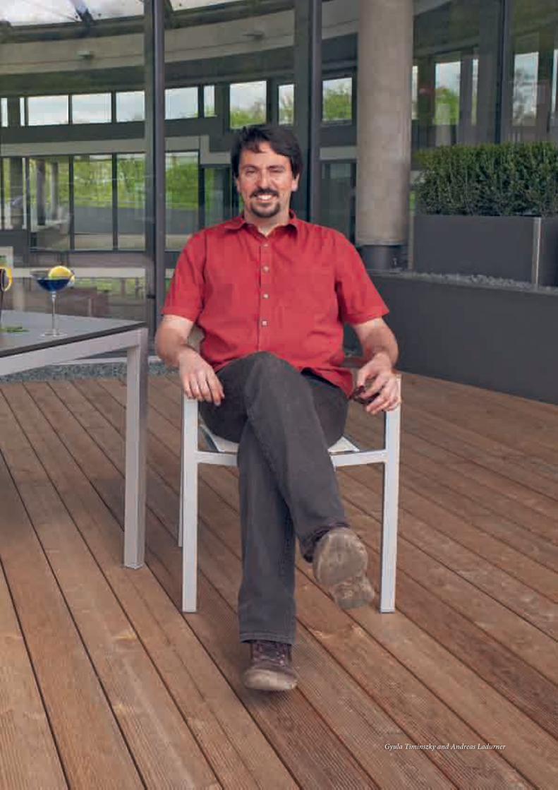
On call for DNA damage
Gyula Timinszky and Andreas Ladurner
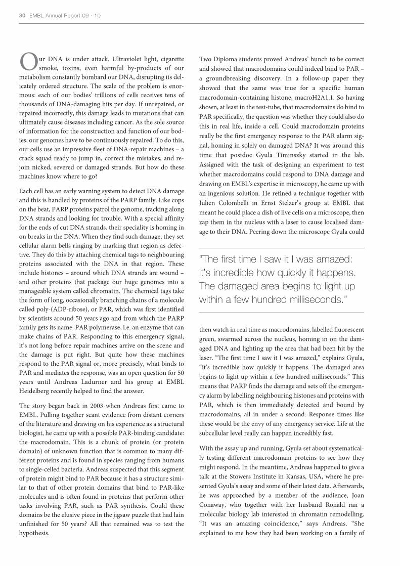
Our DNA is under attack. Ultraviolet light, cigarettesmoke, toxins, even harmful by-products of our
metabolism constantly bombard our DNA, disrupting its del-icately ordered structure. The scale of the problem is enor-mous: each of our bodies’ trillions of cells receives tens ofthousands of DNA-damaging hits per day. If unrepaired, orrepaired incorrectly, this damage leads to mutations that canultimately cause diseases including cancer. As the sole sourceof information for the construction and function of our bod-ies, our genomes have to be continuously repaired. To do this,our cells use an impressive fleet of DNA-repair machines – acrack squad ready to jump in, correct the mistakes, and re-join nicked, severed or damaged strands. But how do thesemachines know where to go?
Each cell has an early warning system to detect DNA damageand this is handled by proteins of the PARP family. Like copson the beat, PARP proteins patrol the genome, tracking alongDNA strands and looking for trouble. With a special affinityfor the ends of cut DNA strands, their speciality is homing inon breaks in the DNA. When they find such damage, they setcellular alarm bells ringing by marking that region as defec-tive. They do this by attaching chemical tags to neighbouringproteins associated with the DNA in that region. Theseinclude histones – around which DNA strands are wound –and other proteins that package our huge genomes into amanageable system called chromatin. The chemical tags takethe form of long, occasionally branching chains of a moleculecalled poly-(ADP-ribose), or PAR, which was first identifiedby scientists around 50 years ago and from which the PARPfamily gets its name: PAR polymerase, i.e. an enzyme that canmake chains of PAR. Responding to this emergency signal,it’s not long before repair machines arrive on the scene andthe damage is put right. But quite how these machinesrespond to the PAR signal or, more precisely, what binds toPAR and mediates the response, was an open question for 50years until Andreas Ladurner and his group at EMBLHeidelberg recently helped to find the answer.
The story began back in 2003 when Andreas first came toEMBL. Pulling together scant evidence from distant cornersof the literature and drawing on his experience as a structuralbiologist, he came up with a possible PAR-binding candidate:the macrodomain. This is a chunk of protein (or proteindomain) of unknown function that is common to many dif-ferent proteins and is found in species ranging from humansto single-celled bacteria. Andreas suspected that this segmentof protein might bind to PAR because it has a structure simi-lar to that of other protein domains that bind to PAR-likemolecules and is often found in proteins that perform othertasks involving PAR, such as PAR synthesis. Could thesedomains be the elusive piece in the jigsaw puzzle that had lainunfinished for 50 years? All that remained was to test thehypothesis.
Two Diploma students proved Andreas’ hunch to be correctand showed that macrodomains could indeed bind to PAR –a groundbreaking discovery. In a follow-up paper theyshowed that the same was true for a specific humanmacrodomain-containing histone, macroH2A1.1. So havingshown, at least in the test-tube, that macrodomains do bind toPAR specifically, the question was whether they could also dothis in real life, inside a cell. Could macrodomain proteinsreally be the first emergency response to the PAR alarm sig-nal, homing in solely on damaged DNA? It was around thistime that postdoc Gyula Timinszky started in the lab.Assigned with the task of designing an experiment to testwhether macrodomains could respond to DNA damage anddrawing on EMBL’s expertise in microscopy, he came up withan ingenious solution. He refined a technique together withJulien Colombelli in Ernst Stelzer’s group at EMBL thatmeant he could place a dish of live cells on a microscope, thenzap them in the nucleus with a laser to cause localised dam-age to their DNA. Peering down the microscope Gyula could
30 EMBL Annual Report 09 · 10
“The first time I saw it I was amazed:it’s incredible how quickly it happens.The damaged area begins to light upwithin a few hundred milliseconds.”
then watch in real time as macrodomains, labelled fluorescentgreen, swarmed across the nucleus, homing in on the dam-aged DNA and lighting up the area that had been hit by thelaser. “The first time I saw it I was amazed,” explains Gyula,“it’s incredible how quickly it happens. The damaged areabegins to light up within a few hundred milliseconds.” Thismeans that PARP finds the damage and sets off the emergen-cy alarm by labelling neighbouring histones and proteins withPAR, which is then immediately detected and bound bymacrodomains, all in under a second. Response times likethese would be the envy of any emergency service. Life at thesubcellular level really can happen incredibly fast.
With the assay up and running, Gyula set about systematical-ly testing different macrodomain proteins to see how theymight respond. In the meantime, Andreas happened to give atalk at the Stowers Institute in Kansas, USA, where he pre-sented Gyula’s assay and some of their latest data. Afterwards,he was approached by a member of the audience, JoanConaway, who together with her husband Ronald ran amolecular biology lab interested in chromatin remodelling.“It was an amazing coincidence,” says Andreas. “Sheexplained to me how they had been working on a family of

chromatin remodellers including one called CHD1-like. Thiswas the alternative name for Alc1 – one of the macrodomainproteins we had in our pipeline. She basically told me that herPhD student had already done all the biochemistry on thisprotein and asked whether we would like to team up togeth-er for publication!” What they had shown was that Alc1, inaddition to its macrodomain, contains a domain involved inremodelling chromatin. Their data showed that, by creatingthe PAR alarm signal, PARP promotes chromatin remod-elling in damaged regions of the genome by recruiting Alc1.Relaxing the chromatin might help to repair the damage byallowing access for the repair machinery. With Gyula’s assayshowing that Alc1 is rapidly recruited to damaged DNA –and is actually the fastest macrodomain protein that Gyulatested – their data combined to make a really interestingstory.
Gyula, Andreas and the lab then focussed their attention on asecond case looking again at the macrodomain-containinghistone, macroH2A1.1. At first glance it seems bizarre that ahistone should possess a domain that makes it respond toDNA damage in this way. As Andreas explains, “We tend tothink of chromatin in architectural terms – as a relatively stat-ic infrastructure, to which modifications are made. Whywould a histone, wrapped up in DNA and embedded in thechromatin, be able to dash across the nucleus to bind PARgroups at sites of DNA damage?” But their assays showed thatit can. “Histone macroH2A1.1 is slower than Alc1,” admitsGyula, “which isn’t so surprising – you can imagine that they
have quite a fight to get there, dragging all the chromatinthey’re wound up in with them. But within five minutes ofDNA damage, these histones do arrive at the site of the dam-age.” “We think that what this does is to rearrange the chro-matin around the damaged area,” says Andreas. “This mayhelp keep the ends of severed DNA strands closer togetheruntil the breaks can be repaired. The bottom line is that youhave histones out there that can sense when PARP rings thealarm bell.” This comprehensive study shows for the firsttime that cells have at their disposal a whole family of proteinsready to respond to the PAR signal. Determining the detailsof what happens next – for example, precisely howmacrodomain proteins bring about changes in chromatinstructure and how they relay information to the DNA-repairmachinery – will undoubtedly keep the group busy solvingpuzzles for many years to come.
Gottschalk AJ, Timinszky G, Kong SE, Jin J, Cai Y, Swanson SK,Washburn MP, Florens L, Ladurner AG, Conaway JW, ConawayRC (2009) Poly(ADP-ribosyl)ation directs recruitment and activa-tion of an ATP-dependent chromatin remodeler. PNAS 106:13770-13774
Timinszky G, Till S, Hassa PO, Hothorn M, Kustatscher G,Nijmeijer B, Colombelli J, Altmeyer M, Stelzer EHK, Scheffzek K,Hottiger MO & Ladurner AG (2009) A macrodomain-containinghistone rearranges chromatin upon sensing PARP1 activation. NatStruct Mol Biol 16: 923-929
When disaster strikes 31
Chromatin containing the macroH2A1.1 macrodomains responds to the emergency signal (PARP1) and helps mediate the DNA repair machinery.
PARP 1macromacro
MacroH2A1.1
Stephen Cusack, Sebastien Huet and Jan Ellenberg
State-of-the-art gadgets. Aerial surveillance of a suspi-cious target. An adversary with a penchant for gam-
bling. While these might sound like the ingredients of adetective novel or a spy thriller, they are in fact aspects ofa successful collaboration between two EMBL teams,which has revealed new insights into how the influenzavirus develops pandemic strains. The collaboration,between Stephen Cusack’s team at EMBL Grenoble andJan Ellenberg’s group at EMBL Heidelberg, has also high-lighted the benefits of the EMBL InterdisciplinaryPostdoctoral Programme, EIPOD. By combining theirexpertise, the teams have shown how the flu virus can winat a kind of genetic card game and create deadly virusesthat are able to jump from animals to humans.
For humanity, the consequences of this card game can bedire. The 1918 Spanish flu pandemic killed around 40million people – more than the Great War. Last year, a flustrain that originated in pigs developed the ability infectand spread between humans. And researchers have longbeen keeping an eye on one highly pathogenic birdinfluenza virus, called H5N1, which seems poised to trig-ger another lethal pandemic. Now, Jan and Stephen havemanaged to track vital parts of the virus as the particlesassemble themselves in living cells. “As well as giving newleads for much-needed new drugs against flu, the workoffers valuable new insights into how the virus can jumpfrom one species to another and become highly virulent,”says Stephen.
The secret of influenza’s ability to seemingly ambush usout of nowhere lies in its predilection for gambling.
Viral surveillance
Normally, the viral strains that cause seasonal flu out-breaks each winter differ only slightly from the previousyear’s strains. This is because the virus’s genes have beenaltered only slightly through minor mutations. It’s a bitlike playing poker with a similar hand of cards from onegame to the next. As a result, most people manage tobuild up an immunity that lasts from one flu season to thenext and vaccines based on past strains work well.
The trouble really starts when two very different flu virus-es infect the same cell at the same time. The virus’s genet-ic material is divided into eight segments, each carryinginstructions for building different parts of the virus.When a single virus infects a cell, it makes copies of thesesegments, which are then packaged with proteins to makenew viruses. If, however, two different flu viruses arecopying themselves in the same cell, segments from onevirus can get packaged with segments from the other.This results in a “reassortant” virus with a completely newcombination of genes – rather like shuffling and dealing apack of cards.
The outcome of this shuffling depends on the genetichand each new virus gets. Usually, the new combinationsof genes are incompatible and result in weaker or defunctviruses. Sometimes, however, a virus is dealt a winninghand: a set of compatible genes that result in a virus thatis completely new to the human immune system. This isstrike one in the virus’s favour. Strike two comes whenthe new virus – particularly one that previously onlyinfected animals or birds – acquires the ability to infect
When disaster strikes 3333
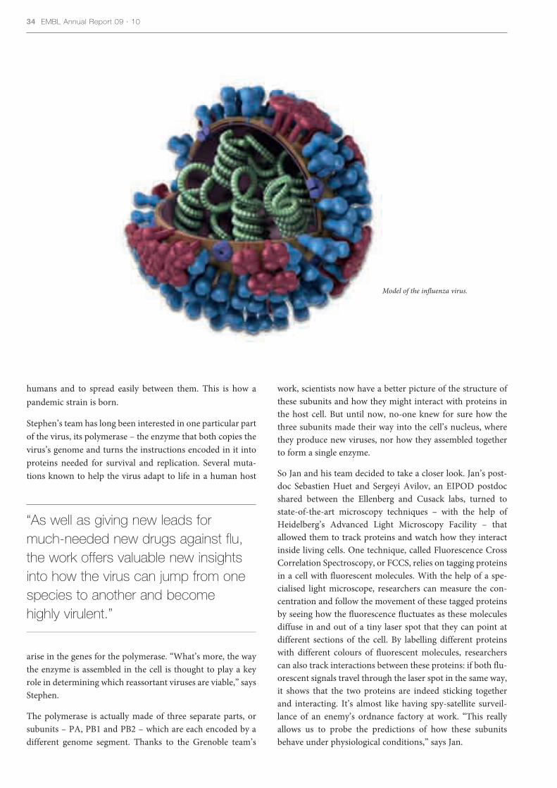
humans and to spread easily between them. This is how apandemic strain is born.
Stephen’s team has long been interested in one particular partof the virus, its polymerase – the enzyme that both copies thevirus’s genome and turns the instructions encoded in it intoproteins needed for survival and replication. Several muta-tions known to help the virus adapt to life in a human host
arise in the genes for the polymerase. “What’s more, the waythe enzyme is assembled in the cell is thought to play a keyrole in determining which reassortant viruses are viable,” saysStephen.
The polymerase is actually made of three separate parts, orsubunits – PA, PB1 and PB2 – which are each encoded by adifferent genome segment. Thanks to the Grenoble team’s
work, scientists now have a better picture of the structure ofthese subunits and how they might interact with proteins inthe host cell. But until now, no-one knew for sure how thethree subunits made their way into the cell’s nucleus, wherethey produce new viruses, nor how they assembled togetherto form a single enzyme.
So Jan and his team decided to take a closer look. Jan’s post-doc Sebastien Huet and Sergeyi Avilov, an EIPOD postdocshared between the Ellenberg and Cusack labs, turned tostate-of-the-art microscopy techniques – with the help ofHeidelberg’s Advanced Light Microscopy Facility – thatallowed them to track proteins and watch how they interactinside living cells. One technique, called Fluorescence CrossCorrelation Spectroscopy, or FCCS, relies on tagging proteinsin a cell with fluorescent molecules. With the help of a spe-cialised light microscope, researchers can measure the con-centration and follow the movement of these tagged proteinsby seeing how the fluorescence fluctuates as these moleculesdiffuse in and out of a tiny laser spot that they can point atdifferent sections of the cell. By labelling different proteinswith different colours of fluorescent molecules, researcherscan also track interactions between these proteins: if both flu-orescent signals travel through the laser spot in the same way,it shows that the two proteins are indeed sticking togetherand interacting. It’s almost like having spy-satellite surveil-lance of an enemy’s ordnance factory at work. “This reallyallows us to probe the predictions of how these subunitsbehave under physiological conditions,” says Jan.
34 EMBL Annual Report 09 · 10
Model of the influenza virus.
“As well as giving new leads for much-needed new drugs against flu,the work offers valuable new insightsinto how the virus can jump from onespecies to another and become highly virulent.”

In the first study to apply this technique to viral proteins, Janand his team tracked the behaviour of the three polymerasesubunits in cells. They found that PB1 and PA bind togetherin the cell’s cytoplasm before moving into the nucleus. PB2,meanwhile, hangs around by itself in the cytoplasm andenters the nucleus separately, before associating with thePB1/PA duo to form the complete polymerase.
Further work allowed the team to translate the Grenoblegroup’s earlier findings about the polymerase’s structure intowhat is happening inside living cells. “This is why Stephenand I are so keen on this collaboration,” says Jan. PB2 hitch-es a ride into the nucleus on a host protein called importin byusing a kind of molecular grappling hook called a nuclearlocalisation sequence. “We know this based on the structuralwork of Stephen’s group,” says Jan. Jan’s team altered theamino acids in PB2’s nuclear localisation sequence andfound, as expected, that PB2 stayed in the cytoplasm.Surprisingly, however, the other subunits also stayed in thecytoplasm, and bound to PB2 to form a polymerase enzymethat was intact but in the wrong location.
This suggests that PB2 helps to keep the other subunits in theright place – the nucleus – until they can assemble into thepolymerase. “If you could interfere with that process withdrugs,” says Jan, “that would be a good way of trapping thepolymerase in the cytoplasm and stopping it from getting intothe nucleus.” It might also explain why some reassortantviruses fail to replicate properly, he adds. If the polymerase
subunits don’t fit together well, they won’t stay in the nucle-us for long enough to make a working enzyme.
By measuring the brightness of the fluorescence given off bylabelled subunits that were normal and non-mutated, Jan’steam determined that a small number of these polymeraseswere sticking together. This suggests that polymerases alreadyworking on the virus’s genetic material help attract otherpolymerases entering the nucleus to the right location, soboosting the rate of viral replication. As for the rest of thesubunits, FCCS revealed that they were moving around thecell far more slowly than expected. This suggests that they arebound to a range of host proteins, which may be importantfor polymerase entry and assembly within the nucleus.
In future, Jan and Stephen plan to study these interactionsbetween the host cell proteins and the influenza polymerasein more detail. Because FCCS gives such precise measure-ments, it will allow Jan and his team to study how mutationsin the polymerase subunits affect how efficiently the subunitsinteract with host cell proteins – and so how they allow thevirus to adapt to new host species such as humans.
Huet S, Avilov S, Ferbitz L, Daigle N, Cusack S, Ellenberg J (2010)Nuclear import and assembly of the influenza A virus RNA poly-merase studied in live cells by Fluorescence Cross CorrelationSpectroscopy. J Virol. 84: 1254-1264
When disaster strikes 35
Schematic representation of the particlescontaining the viral RNA, the nucleoprotein andthe polymerase complex superimposed to anelectron micrograph of the virus.
Fluorescence Cross Correlation Spectroscopyimage of the three different influenza polymerasesubunits: PA is shown in purple, PB1 in greenand PB2 in blue.

Lars Steinmetz and Stephanie Blandin

From Hercule Poirot to CSI, most detective storiesand TV police shows revolve around the investiga-
tors narrowing down their list of suspects. At the start ofthe story, there are often as many suspects as there arecharacters, but thanks to varying measures of deductivepower and luck, the good guys systematically strike sus-pects off their list until finally they are left with only one– the culprit.
Although they use different interrogation techniques,geneticists often face the same task of whittling down alist of suspects – and in some cases, they too are dealingwith killers. In this particular story, the killer’s identity isknown from the start: malaria, a disease that claimsalmost one million human lives a year. The parasites thatcause malaria are also well known to scientists. Belongingto the genus Plasmodium, they spend part of their lifecycle inside humans or another mammalian host –depending on the exact species of parasite – and anotherpart inside mosquitoes. And just as we suffer from malar-ia, mosquitoes infected with Plasmodium parasites do to.However, this is where the mystery begins: not allmosquitoes that are infected with malaria parasites fall ill.Instead, some are resistant to the parasite, meaning thattheir immune system is able to eliminate the disease-causing agent from their bodies. Why can some individu-al mosquitoes fight off the parasite better than others?Enter our lead investigators: Lars Steinmetz, joint head ofthe Genome Biology Unit at EMBL Heidelberg, andEMBL alumni Rui Wang-Sattler, now at the HelmholtzZentrum in Munich, and Stephanie Blandin, now at the
The usual suspects
Institut National de la Santé et de la Recherche Médicale(INSERM) in Strasbourg, France.
To get to the bottom of this enigmatic variation in indi-vidual resistance, Lars, Rui and Stephanie turned toAnopheles gambiae mosquitoes, the main carriers of theparasite that causes the most severe form of humanmalaria in Africa, and to Plasmodium berghei, whichcauses malaria in rodents. The scientists compared thecomplete DNA sequences, or genomes, of mosquitoesthat are resistant to malaria with those of mosquitoes thatare susceptible to the disease. This comparison suggestedthat a section of one of the mosquitoes’ chromosomes islinked to the insects’ ability to fend off Plasmodium. Butas this section contains 975 genes, this still left our inves-tigators plenty of suspects.
“To really understand this matter, we needed to go fur-ther,” says Rui. “But the question was how to go from thissection of DNA to the single gene that makes mosquitoesresistant to malaria,” Stephanie adds. This is when Lars’expertise was called upon.
Lars and his group had previously developed a way ofdoing just this in baker’s yeast. To go from a large DNAsection, or genetic interval, to the gene that causes a par-ticular trait, they took two yeast strains with different ver-sions, or alleles, of a particular gene and crossed them,obtaining one strain of yeast with both alleles of that gene.They could then take these yeast cells, divide them intotwo sets and delete – or knockout –one allele in one setand the other allele in the other. As both sets of cells weregenetically identical for that one gene, any differences in
When disaster strikes 3737

the traits of the two sets of cells must stem from the fact thatthey had different alleles of that gene. By doing this systemat-ically for all the genes in the interval, Lars and colleagues wereable to narrow down their suspects and determine exactlywhich genes played a role in a particular trait. “Unfortunately,we can delete genes in yeast, but not in mosquitoes,”Stephanie explains, “so we couldn’t just apply that samemethod to mosquitoes.”
Nevertheless, the fundamental problem was the same, so itmade sense to try a similar approach – it just had to be onethat would work in mosquitoes. Although you can’t knockgenes out in mosquitoes, you can knock them down. Ratherthan altering an organism’s DNA sequence as they would in agene knockout, scientists knock a gene down by using spe-cially engineered RNA molecules to interfere with the gene’sexpression, i.e. to hinder the production of the protein thatthe gene encodes. Lars, Stephanie and Rui took advantage oftechnological advances in this field of RNA interference andcreated a new method that essentially uses this knockdownapproach in the same way that the previous method usedgene deletions. This meant devising a way to knock down notjust a specific gene, but a specific allele of that gene.
To test their new technique – called reciprocal allele-specificRNA interference – Stephanie and her colleagues at INSERMproduced mosquitoes that each carried two different alleles ofa gene called TEP1: one allele from the strain of malaria-resis-tant mosquitoes, and the other from a strain of susceptiblemosquitoes. They then individually knocked down each ofthe alleles, creating mosquitoes whose only functional versionof TEP1 was either the ‘resistance’ or the ‘susceptibility’ allele.
38 EMBL Annual Report 09 · 10
Reciprocal allele-specific RNAi entails obtaining individuals each carryingthe 2 different alleles of interest, silencing one or other of those alleles, andcomparing the results.
More parasites survived (fluorescent green dots) in the midgut of a mosquito with only the ‘susceptibility’ allele turned on (right) than in a genetically identicalmosquito with only the ‘resistance’ allele turned on (left), which contained mainly dead parasites (black dots).
Resistant (RR)
Resistant
(RS)
Susceptible (SS)
Susceptible
SilencedResistant
allele
SilencedSusceptibleallele

When they compared the ability of these otherwise identicalmosquitoes to fight off Plasmodium, the scientists found thatthe immune systems of mosquitoes whose only functionalTEP1 allele came from the resistant strain were able to killPlasmodium parasites, whereas in mosquitoes with only the‘susceptibility’ allele, parasite survival was much higher. Inshort, silencing different alleles produced mosquitoes withdifferent degrees of resistance to malaria, meaning that anindividual mosquito’s resistance to the Plasmodium parasitedepends largely on which form(s) of this one gene it carries.
The team didn’t choose TEP1 at random. Previous work byStephanie and others in the lab of former EMBL DirectorGeneral Fotis Kafatos had shown that this gene encodes aprotein that attaches itself to malaria parasites in themosquito’s gut, so that they are then eliminated by themosquito’s immune system. Nevertheless, Lars points out, “Ifyou consider all the complexity that could have been goingon, we were lucky – not only did our new technique work, butthe biology fell into place too: we’d picked the right gene!”
Our investigators did what all those detectives in mystery sto-ries dream of: they devised a way to efficiently narrow downthe suspect list and identify the culprit. Crucially, scientistswill now be able to do the same in many other situations, asthis method is applicable to many different species and celltypes, including human cell lines in culture. “Along withother advances like the ability to induce pluripotent stemcells, this kind of technique really opens up the door tounderstanding the function of individual alleles,” Lars says,“and to do this in a single individual.” In this respect, the newtechnique is complementary to pre-existing approaches, in
They are also investigating human malaria, for which evi-dence seems to point to the same suspect. If TEP1 enablesmosquitoes to fight the parasites that cause human malaria asit does for the rodent malaria parasite, this could prove to bea boon to malaria eradication programmes, a large part ofwhich focus on eliminating the mosquitoes that transmit thedisease. “Knowing which allele or alleles confer resistance tomalaria could help to make mosquito eradication pro-grammes more effective,” Stephanie posits. If scientists canidentify the alleles responsible, and distinguish between resis-tant and susceptible mosquitoes in the wild, such pro-grammes could be restricted to areas where they are mostnecessary: areas where most mosquitoes are susceptible, andtherefore likely to carry the disease. Thus, in a plot twist that’sreminiscent of the best detective stories, this winged enemymay, in fact, turn out to be an ally.
Blandin SA, Wang-Sattler R, Lamacchia M, Gagneur J, Lycett G,Ning Y, Levashina EA, Steinmetz LM (2009) Dissecting the geneticbasis of resistance to malaria parasites in Anopheles gambiae.Science 326: 147-150
When disaster strikes 39
Approximately 12 days after infection, a Plasmodium oocyst is burstingopen, releasing thousands of developing parasites (labeled green) into themosquito’s bloodstream.
“Not only did our new technique work,but the biology fell into place too: we’d picked the right gene!”
which scientists had to scour genetic data from many indi-viduals. “And you can apply it to any organism in which youcan do RNA interference, which is a huge expansion of themethod we developed for yeast,” Lars emphasises.
In the meantime, as far as mosquitoes’ resistance is con-cerned, Stephanie and colleagues in the malaria field have aculprit: TEP1. This culprit may, however, have accomplices,so the scientists would like to investigate the roles of othermosquito genes in increasing – or decreasing – an individualmosquitoes’ ability to defeat the malaria parasite.

40 EMBL Annual Report 09 · 10
The discovery of how certainimmune cells make a Jekyll and
Hyde-style switch in their behaviour isoffering new insights into how thebody repairs damaged muscle tissue,say researchers at EMBLMonterotondo. The teams, led byClaus Nerlov and Nadia Rosenthal,have unearthed the genetic mecha-nism that allows immune cells tochange from a state in which theyboost the body’s defence mechanismsto another that promotes healing andrepair. The researchers hope the workpaves the way for new treatments formuscles damaged by disease, injury orsurgery.
When a tissue is damaged, the bodyhas to strike a delicate balancebetween cleaning up the debris andfending off infection, and fosteringregeneration and repair. If there is toomuch cleaning and defence, the tissuecan’t regenerate well and will tend toscar; if this process is impaired the tis-sue can get infected and cells die. Bothcleaning/defence and regeneration areneeded, in sequence and under strictcontrol. But some disease conditionsdisrupt this regulation, which delayshealing and can cause further damage.
One of the key players in tissue heal-ing is an immune cell called amacrophage. Macrophages crawlaround tissues like an army of biologi-cal Pac-men, gobbling up microbesand fragments of dead and dying cells.They also release chemical signals thatpromote inflammation, a conditionthat attracts other immune cells to theinjury and primes them to attackinvaders.
In recent years, scientists have foundevidence to suggest that macrophagescan also promote tissue healing bydramatically switching their character-istics to become a cell type called anM2 macrophage. M2 cells release
chemical signals that damp downinflammation, thus promoting regeneration. This shift is calledmacrophage polarisation but untilnow, scientists knew little about how itwas triggered. Thanks to a stroke ofserendipity, the EMBL teams nowhave an answer.
Claus and his group were studying aprotein called C/EBPβ, which is pro-duced by tissues in response to inflam-mation. They had altered the gene forC/EBPβ in mice so that it could nolonger respond to inflammation, to seewhat effect the lack of C/EBPβ wouldhave on immune cell development.Disappointingly, the answer seemed tobe very little: the mice developed nor-mal immune cells. Intriguingly, how-ever, their macrophages failed topolarise when challenged with inflam-matory signals.
This finding caught the attention ofNadia, who wondered whether itmight be relevant to her group’s workon muscle regeneration. She and Clausteamed up to see how his miceresponded to muscle injury. Sureenough, the mice’s macrophages couldclear up the damage debris, but theycouldn’t make the all-important M2
switch and their muscles failed toregenerate properly. “If macrophagesdon’t make this switch, then the mus-cle won’t repair itself,” says Nadia.“You just end up with scar, instead ofnew tissue.”
Blocking the C/EBPβ gene in musclesonly, but not in macrophages, had noeffect, showing that macrophagepolarisation is indeed essential forregeneration. Just like the potion Dr Jekyll mixed to transform into Mr Hyde, a cocktail of chemical sig-nals in inflamed tissue acts on theC/EBPβ gene, triggering it to switchthe cell into an M2 macrophage. This,say the teams, makes the gene and theother genes it controls attractive tar-gets for therapies. “From a medicalpoint of view, it would seem that thetrick to improve muscle repair is find-ing a way to increase C/EBPβ produc-tion and keep it high,” says Claus.
Ruffell D, Mourkioti F, Gambardella A,Kirstetter P, Lopez R, Rosenthal N, Nerlov C(2009) A CREB-C/EBPβ cascade induces M2macrophage-specific gene expression and promotes muscle injury repair. Proc Natl AcadSci USA 106: 17475-17480
Poles apart
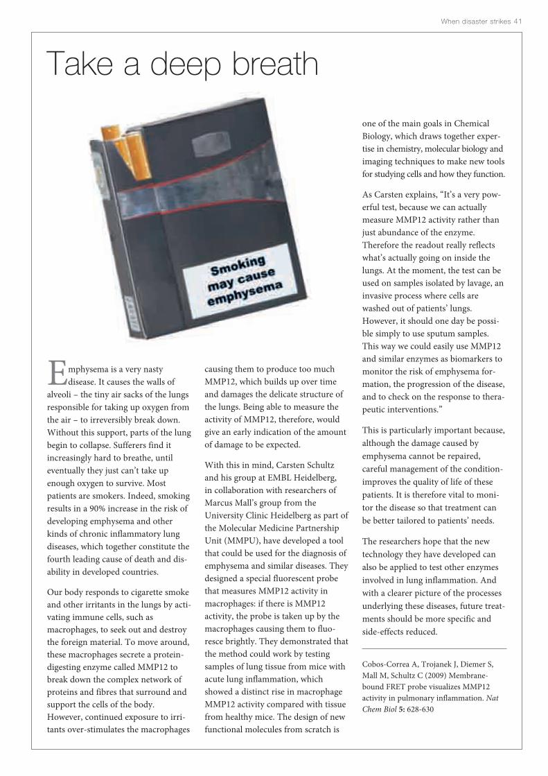
When disaster strikes 41
one of the main goals in ChemicalBiology, which draws together exper-tise in chemistry, molecular biology andimaging techniques to make new toolsfor studying cells and how they function.
As Carsten explains, “It’s a very pow-erful test, because we can actuallymeasure MMP12 activity rather thanjust abundance of the enzyme.Therefore the readout really reflectswhat’s actually going on inside thelungs. At the moment, the test can beused on samples isolated by lavage, aninvasive process where cells arewashed out of patients’ lungs.However, it should one day be possi-ble simply to use sputum samples.This way we could easily use MMP12and similar enzymes as biomarkers tomonitor the risk of emphysema for-mation, the progression of the disease,and to check on the response to thera-peutic interventions.”
This is particularly important because,although the damage caused byemphysema cannot be repaired, careful management of the condition-improves the quality of life of thesepatients. It is therefore vital to moni-tor the disease so that treatment canbe better tailored to patients’ needs.
The researchers hope that the newtechnology they have developed canalso be applied to test other enzymesinvolved in lung inflammation. Andwith a clearer picture of the processesunderlying these diseases, future treat-ments should be more specific andside-effects reduced.
Cobos-Correa A, Trojanek J, Diemer S,Mall M, Schultz C (2009) Membrane-bound FRET probe visualizes MMP12activity in pulmonary inflammation. NatChem Biol 5: 628-630
Emphysema is a very nasty disease. It causes the walls of
alveoli – the tiny air sacks of the lungsresponsible for taking up oxygen fromthe air – to irreversibly break down.Without this support, parts of the lungbegin to collapse. Sufferers find itincreasingly hard to breathe, untileventually they just can’t take upenough oxygen to survive. Mostpatients are smokers. Indeed, smokingresults in a 90% increase in the risk ofdeveloping emphysema and otherkinds of chronic inflammatory lungdiseases, which together constitute thefourth leading cause of death and dis-ability in developed countries.
Our body responds to cigarette smokeand other irritants in the lungs by acti-vating immune cells, such asmacrophages, to seek out and destroythe foreign material. To move around,these macrophages secrete a protein-digesting enzyme called MMP12 tobreak down the complex network ofproteins and fibres that surround andsupport the cells of the body.However, continued exposure to irri-tants over-stimulates the macrophages
causing them to produce too muchMMP12, which builds up over timeand damages the delicate structure ofthe lungs. Being able to measure theactivity of MMP12, therefore, wouldgive an early indication of the amountof damage to be expected.
With this in mind, Carsten Schultzand his group at EMBL Heidelberg, in collaboration with researchers ofMarcus Mall’s group from theUniversity Clinic Heidelberg as part ofthe Molecular Medicine PartnershipUnit (MMPU), have developed a toolthat could be used for the diagnosis ofemphysema and similar diseases. Theydesigned a special fluorescent probethat measures MMP12 activity inmacrophages: if there is MMP12 activity, the probe is taken up by themacrophages causing them to fluo-resce brightly. They demonstrated thatthe method could work by testingsamples of lung tissue from mice withacute lung inflammation, whichshowed a distinct rise in macrophageMMP12 activity compared with tissuefrom healthy mice. The design of newfunctional molecules from scratch is
Take a deep breath

José Márquez

We’ve all felt it: a quickening of the heart and aslight shortness of breath as you walk into an
exam room. Most of us recognise that the hormoneadrenaline is responsible for this reaction, but we’re notunique in responding to stress with a release of hor-mones. Plants do this too – but unlike you and I, theydon’t have the option to flee; rooted to the spot, they canonly stay and fight it out. To do this, plants release thehormone abscisic acid (ABA), which coordinates theirresponse to stresses such as drought, extreme tempera-ture and high salt levels.
ABA acts as a chemical courier, relaying messages fromone cell to another. Cells respond to the hormone if theypossess a receptor, which, once bound to the hormone,signals to the cell to go on the offensive. For plants, thismeans closing the tiny holes in their leaves to avoid waterloss, diverting resources to their roots to increase wateruptake and switching on the production of proteins thatprotect cells from dehydration.
Understanding how plants transmit these water-savingmeasures to their roots and leaves could help farmersward off the effects of drought in crops, and has prompt-ed many scientists to search for the receptor that bindsABA. But identifying this receptor has proved unusuallychallenging.
Since 2006, several proteins have been suggested but,because of conflicting findings, their exact role remainedcontroversial. Then in 2009, two groups independentlyhomed in on yet another potential ABA receptor , PYR1,
Surviving drought
and a family of proteins that bound ABA was identified,although it was still not clear how these proteins interact-ed with the hormone. Given the struggles of the past threeyears, some plant biologists reserved final judgment onwhether the hunt for the ABA receptor was over untilthey could visualise the receptor physically interactingwith the hormone. This is where plant biologist PedroLuis Rodriguez from the Universidad Politecnica deValencia in Spain stepped in, approaching structural biol-ogist José Márquez from EMBL Grenoble with a question:would it be possible to obtain the structure of the ABAreceptor?
“For such a contentious field, knowing the structure is thefinal proof, because you not only see the receptors butyou also see how they bind the hormone,” explains José,who, together with his team, examines protein structuresto understand how they transmit messages. “Because ofour interest in signalling, we were very keen to collaborateon this project.”
But José and Pedro were not alone in finding this aninteresting puzzle to solve. Four other groups – two inAsia and two in the US – simultaneously set out to definethe structure of the receptor. And so the race began. Thefirst step for José’s group was to produce vast quantities ofthe ABA receptor in bacteria before adding the protein toa cocktail of chemicals to try to induce it to form a crys-tal. José explains that the proteins usually fall to the bot-tom of the flask in an amorphous blob, but occasionallythey stack together in a regular fashion. This is what theywere searching for — crystals.
When disaster strikes 4343

To increase the likelihood of crystal formation, they set uphundreds of experiments, each one differing slightly in thechemicals added. “Growing crystals can take days, weeks, oreven months,” says José, adding that each flask needs to bechecked every few days. “So as you can see, with all these sam-ples you would quickly run into problems.”
Fortunately, José’s high-throughput crystallisation facilitywas on hand to help solve the problem. Using robots to dis-pense and regularly check close to 3000 samples, it was only amatter of weeks before the researchers found a crystal. “Thiswas exceptionally fast and shows the power of this type of col-laboration,” says Pedro. Another advantage of automatingthe crystal-forming process is that less of the protein is wast-ed because of the accuracy with which the robots can dispensethe samples. “They can dispense one tenth of a millionth of alitre – a quantity that humans would find impossible torepeatedly pipette,” explains Pedro. By reducing proteinwaste, the researchers could set up more experiments and sowere more likely to secure the perfect cocktail of chemicals inwhich to grow the crystal.
With the crystal in hand, José’s group used the powerful X-ray beams of the European Synchrotron Radiation Facility inGrenoble to determine the structure of the crystalline ABAreceptor (for an overview of this technique, see page 56).Ahead of the other international teams, and in collaboration
44 EMBL Annual Report 09 · 10
The stress of water shortage leads PYR1 to close over ABA, creating adocking site for serine threonine phosphatase and thus removing itfrom circulation so that the cell’s drought response can be activated.
The structure of PYR1 (coloured ribbons) in itsopen, unbound state (light green loops) and how it
folds around ABA (white rods) when it binds to thishormone (turquoise and purple loops).
PYR 1
serine threonine
phosphataseABA
Water stress
PYR 1

with Adam Round from EMBL Grenoble who is involved inthe Partnership for Structural Biology, they revealed that theABA receptor acts in pairs: two copies of the protein bindtogether, each side creating a pocket into which ABA slots.Guarding the entrance to these pockets, they found long flex-ible loops, which close like a lid over the hormone trappedinside.
This closing mechanism led to an unexpected discovery.José’s group realised that the ‘closing lid’ could create a dock-ing site for an enzyme called serine threonine phosphatase,which is known for having the capacity to suppress the stress-signalling pathway. But when locked in place on the dockingsite, it can no longer prevent the series of signals cascadingthrough the plant that prepare it for the stress of losing water.“The revelation came when we superimposed the receptorbound to ABA with the unbound form, and saw the differ-ence in the loops,” says José. He thinks that when the enzymebinds at the docking site, this inactivates it and prevents itfrom circulating freely. This then allows another enzyme, a
protein kinase, to activate transcription factors that turn onthe stress-response genes.
José hopes that this finding could help alleviate crop damagein drought-prone areas. If scientists find a molecule thatmimics ABA – which is too expensive to manufacture in largequantities – they could simulate the stress response in plantsto help protect them against water loss. The new moleculecould be sprayed over crops before a period of dry weather,stimulating them to save water for the harsh times to come.
This is something that José’s group has already started to dousing their structural models. José explains: “We’re trying tofit millions of different molecules into the receptor’s pocket inthe hope of getting one to fit.”
If this works, it could have far-reaching effects for all of us,because most of our food is grown in semi-arid regions suchas North Africa and South Asia, which suffer recursivedroughts. These regions see huge quantities of food go towaste because of damage caused by unusually dry periodswithin the growing season. Reducing this waste would partic-ularly benefit people in developing countries who typicallysustain themselves on cheaper, more traditional crop varietiesthat are less drought-resistant. Who’d have thought stresscould be a good thing?
Santiago J, Dupeux F, Round A, Antoni R, Park S-Y, Jamin M,Cutler S, Rodriguez P, Márquez J (2009) The abscisic acid receptorPYR1 in complex with abscisic acid. Nature 462: 665-668
When disaster strikes 45
After 15 days of drought, an Arabidopsis thaliana plant will normally bewithered and dry (far left), unless it has been genetically engineered to
enhance its response to ABA (centre left, centre right and right).


Had anyone been tempted of late to underestimate the importance of aworking transport network, the events of April 2010 soon put themstraight. Icelandic volcano Eyjafjallajökull belched countless tonnes of
ash into the atmosphere, shutting down air travel across most of Europe for a week.Millions of passengers were stranded, airline phone systems jammed by frustratedcustomers, and trains, coaches and ferries overwhelmed by the sheer number ofpeople struggling to get back home. Airlines lost millions in revenue daily, busi-nesses were forced to close. The events brought home just how much our ownlivelihoods, and those of our countries and cities, depends on being able to traveland keep in touch.
Like any city or economy, our cells depend on effective transport and communica-tions systems to survive. Proteins and other molecules need to be manufacturedand freighted to their destinations. Cells need to communicate with each other tomonitor their external environments and work out how to respond. They also needto co-ordinate their responses to the outside world with what is happening insidethem. If a cell experiences the biochemical equivalent of Eyjafjallajökull – perhaps amutation that paralyses its signalling network – serious diseases such as cancer canresult.
For example, the internal scaffolding of a cell, its cytoskeleton, acts rather likeLondon’s Underground train network. Researchers at EMBL Heidelberg haveshown how this allows molecules to commute to the appropriate cellular location toperform their jobs correctly. Just as Paris is divided into districts such as the busi-ness centre La Défense or the artistic area of Montmartre, so the cell is divided upinto compartments with different functions. EMBL Director General Iain Mattajand his team are tracing the earliest evolutionary origins of these compartments,like archaeologists sifting through clues at the ancient city of Ur.
Communication within and between cells is also vital to keep hearts beating, creatememories, control growth and for development. EMBL scientists have uncoveredthe detailed workings of one such communication system, opening the way for newdrugs to treat disease. Other EMBL teams, meanwhile, have applied the city analogyone step further – to biologists themselves. Thanks to this work, life scientists nowhave their own communication framework, so they can clearly and effectivelyunpick the daily hubbub of a busy living system.
Gettingaround andstaying intouch

Christoph Müller

The launch of the Speedo LZR Racer swimsuit inFebruary 2008 started a revolution in the swim-
ming pool. With swimmers squeezed into figure-huggingfull-length bodysuits in gun-metal black, world recordswere toppled across the board. Two years later, criticisedas ‘technological doping’ and accused of devaluing honestcompetition, the controversial polyurethane supersuitshave been banned. So extreme was the difference theymade to the performance of the athletes that, if they areallowed to stand, it is unclear whether the suit-assistedrecords will ever be beaten. Aside from the special low-drag, water-repellent properties of their polyurethanepanels, these suits worked by compressing the swimmer’sbody, making as compact and streamlined a silhouette as possible. Evidently, the key to speed in the water is compaction.
In the body, sperm are the only cells that can swim. Onlythe fastest wins the chance to pass on their genes to thenext generation, and so sperm have evolved to becomeexceptionally streamlined. For them too, this is a matterof compaction. As sperm have no other purpose than car-rying the father’s DNA, the sperm head contains very lit-tle else: all non-essential cellular components aresqueezed out during production, leaving only the DNA-containing nucleus. In the pursuit of hydrodynamics, theonly thing left to do is to ensure that the DNA itself takesup as little space as possible.
To do this, sperm DNA undergoes drastic re-packagingduring spermatogenesis. Under normal circumstances,DNA – an incredibly long and unwieldy molecule – is
Built for speed
wound around proteins called histones to form nucleo-somes that are then organised into an even more complexstructure called chromatin. However, in developingsperm cells, most of these histones are removed andreplaced by smaller proteins that wrap the DNA up moretightly, shrinking and reshaping the nucleus and stream-lining the sperm head. This is an incredibly dramatic pro-cess that must be tightly controlled. It remains unclearhow this is orchestrated, but like other changes in chro-matin structure, it is directed by histone modifications.These modifications take the form of molecular tags,which are chemically attached to the tail portion of his-tone proteins and which flag up regions of the genome forthe attention of chromatin remodelling machines. Duringspermatogenesis, histones throughout the genome aremodified in several huge waves, each wave adding a dif-ferent kind of tag. The very last of these is a wave of acety-lation during which many acetyl groups are attachedspecifically to the tails of one type of histone, histone H4.Specific proteins recognise these tags, bind to them, andtrigger the massive changes that bring about compactionof the DNA.
This process is the reason why Christoph Müller, a struc-tural biologist who studies transcriptional regulation,became involved in a story about sperm development. Atthe time, he and his lab were based at EMBL Grenoblewhere they were working to understand how chromatinand DNA-binding proteins influence patterns of geneexpression. Christoph was approached by SaadiKhochbin, a group leader at the Institut Albert Bonniotalso based in Grenoble, who is primarily interested in the
Getting around and staying in touch 4949

50 EMBL Annual Report 09 · 10
The structure of the Brdt protein (purple) enables itto bind simultaneously to two tags attached to ahistone (cyan rods), making relatively diffusechromatin (blue, top cells) compact into tighterbundles (bottom cells), and thus contributing to the hydrodynamic form of sperm (background).

With some interesting preliminary structural data the groupsrealised that to really understand how the Brdt bromod-omains worked, they would need to determine their preferredtargets. So they started testing the binding of histone tailswith different combinations of acetyl tags attached to the Brdtbromodomains. To their surprise, it seemed that one of theBrdt bromodomains favoured a histone tail with two acetyltags rather than just one.
The path of a good project never runs as smoothly as its finalpublication would suggest, so with Jeanne leaving the lab hav-ing completed her PhD and Christoph relocating the rest ofthe lab to Heidelberg, structural analysis of the Brdt bromod-omain was handed over to Carlo Petosa. Formerly a staff sci-entist in Christoph’s group, Carlo stayed on in Grenoble,becoming an independent group leader at the Institut deBiologie Structurale (IBS). After a lot of hard work and anal-ysis, Carlo was able to piece together a complete picture,showing that the Brdt bromodomain forms a pocket thatbinds to both tags at once.
But why was this so unexpected? The explanation leads usinto the history of the field of chromatin research. The con-cept that histone tags might act as a code to which different‘interpreter’ proteins bind to induce changes in chromatinstructure was only proposed about 10 years ago. Since then,
the details have grown increasingly more elaborate. Differentprotein domains – functional protein sub-units preserved byevolution and re-used in different proteins – have been iden-tified that bind to different kinds of tags: for example, bro-modomains for acetyl tags and chromodomains for methyltags.
“We knew that individual proteins could bind either to singletags via one tag-binging domain, or sometimes to several tagsat once via multiple domains. But it was just assumed thateach domain would only bind to one tag,” explains Carlo. “Sofinding a single bromodomain able to bind two tags simulta-neously was quite a revelation.”
On re-examining the structures of other bromodomains inthe protein databases they now think that the ability to bindtwo tags is not restricted to just Brdt – several other bromod-omain proteins probably work the same way.
“This finding adds an extra level of complexity to the inter-pretation of the histone code,” explains Christoph. “In a way,it’s like any other language. The tags are like letters in analphabet. Used individually they don’t convey much mean-ing. But, combined and read out as words, you suddenly findthere’s a lot more you can say. If single proteins or even sin-gle protein domains can read out combinations of tags, thehistone code can direct a greater variety of specific chromatinremodelling responses.”
But what does all this have to do with sperm DNA com-paction? How could the special way in which Brdt binds tohistones be important for this unique role? “Well, we canspeculate,” says Christoph. “Compaction of the DNA onlybegins when the wave of acetylation is complete and H4 his-tones are fully tagged. The acetyl groups are added to eachhistone sequentially and Brdt binds to the last two tags in thissequence, making Brdt binding the very last step in the pro-cess – perhaps the final signal for compaction to begin.”
From this point on the compaction process seems to unfoldautomatically. The DNA throws off its bulky histones, replac-ing them with smaller proteins, and tightly winds itself into adense, compact package. Completing the rest of their devel-opment, the slimmed-down and streamlined sperm are nowrace-fit and ready. A little wiser to the secrets of speedysperm, the researchers are now trying to find out whetherdefects in Brdt might be a cause of male infertility.
Morinière J, Rousseaux S, Steuerwald U, Soler-López M, Curtet S,Vitte A-L, Govin J, Gaucher J, Sadoul K, Hart D, Krijgsveld J,Khochbin S, Müller C, Petosa C (2009) Cooperative binding of twoacetylation marks on a histone tail by a single bromodomain.Nature 461: 664-668
Getting around and staying in touch 51
“The tags are like letters in an alphabet. Used individually they don’tconvey much meaning. But, combinedand read out as words, you suddenlyfind there’s a lot more you can say.”
role that chromatin plays in sperm development and fertility.Presuming that the wave of histone H4 acetylation prior tosperm DNA compaction must be a crucial step in the process,Saadi’s group had gone on a hunt for sperm proteins thatcontain so-called ‘bromodomains’ – protein modules thatbind to acetyl tags. After testing a few candidates, they hit onBrdt, a unique protein found exclusively in developing spermcells that seemed to be crucial for compaction. Saadi pro-posed a collaboration to study the structure of Brdt’s two bro-modomains and Christoph agreed, initially incorporatingthem into a larger screen to study bromodomains from manydifferent histone-binding proteins. However, through thework of Jeanne Morinière, a pre-doc in Christoph’s lab, itquickly became clear that the Brdt bromodomains were themost interesting in the screen and so they became the focus ofproject.
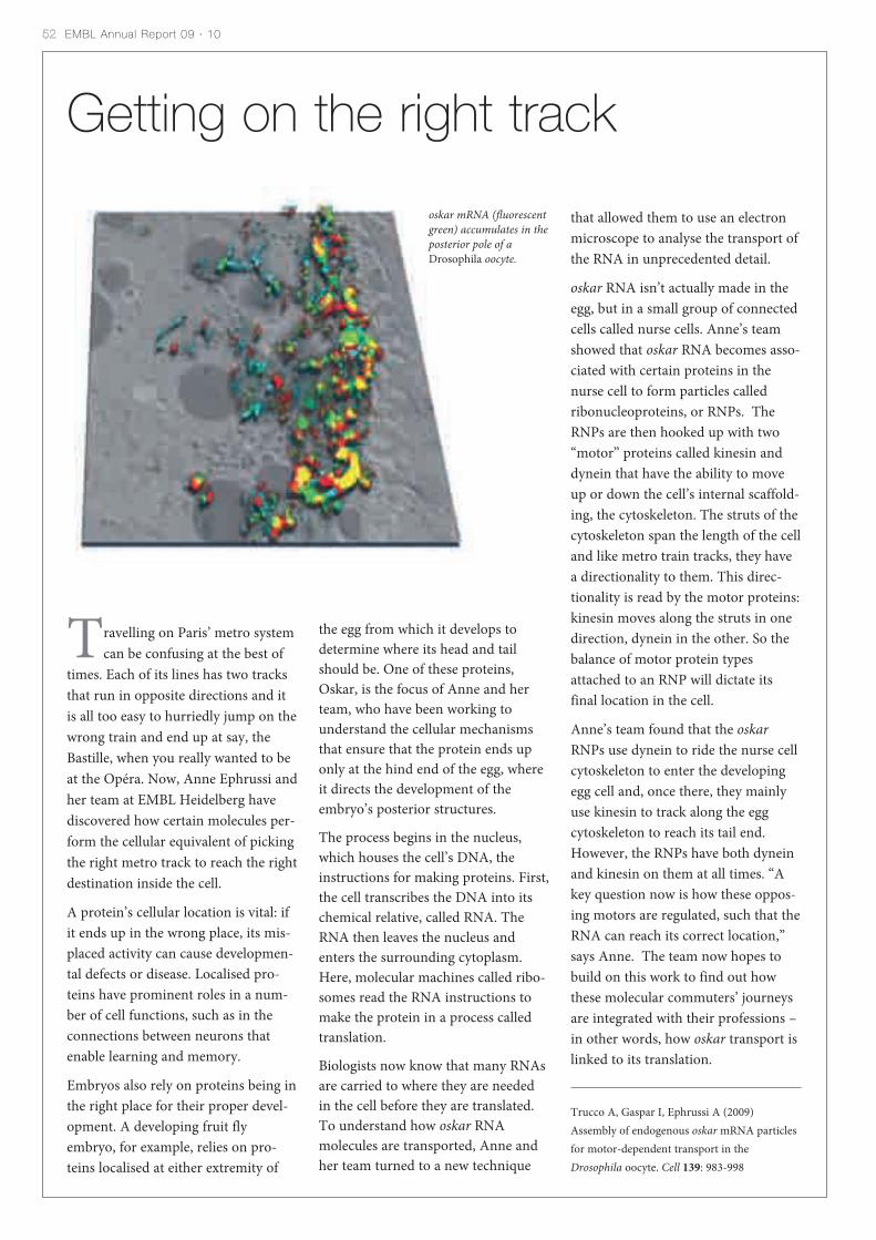
52 EMBL Annual Report 09 · 10
Getting on the right track
oskar mRNA (fluorescentgreen) accumulates in theposterior pole of aDrosophila oocyte.
that allowed them to use an electronmicroscope to analyse the transport ofthe RNA in unprecedented detail.
oskar RNA isn’t actually made in theegg, but in a small group of connectedcells called nurse cells. Anne’s teamshowed that oskar RNA becomes asso-ciated with certain proteins in thenurse cell to form particles calledribonucleoproteins, or RNPs. TheRNPs are then hooked up with two“motor” proteins called kinesin anddynein that have the ability to moveup or down the cell’s internal scaffold-ing, the cytoskeleton. The struts of thecytoskeleton span the length of the celland like metro train tracks, they havea directionality to them. This direc-tionality is read by the motor proteins:kinesin moves along the struts in onedirection, dynein in the other. So thebalance of motor protein typesattached to an RNP will dictate itsfinal location in the cell.
Anne’s team found that the oskarRNPs use dynein to ride the nurse cellcytoskeleton to enter the developingegg cell and, once there, they mainlyuse kinesin to track along the eggcytoskeleton to reach its tail end.However, the RNPs have both dyneinand kinesin on them at all times. “Akey question now is how these oppos-ing motors are regulated, such that theRNA can reach its correct location,”says Anne. The team now hopes tobuild on this work to find out howthese molecular commuters’ journeysare integrated with their professions –in other words, how oskar transport islinked to its translation.
Trucco A, Gaspar I, Ephrussi A (2009)Assembly of endogenous oskar mRNA particlesfor motor-dependent transport in theDrosophila oocyte. Cell 139: 983-998
Travelling on Paris’ metro systemcan be confusing at the best of
times. Each of its lines has two tracksthat run in opposite directions and itis all too easy to hurriedly jump on thewrong train and end up at say, theBastille, when you really wanted to beat the Opéra. Now, Anne Ephrussi andher team at EMBL Heidelberg havediscovered how certain molecules per-form the cellular equivalent of pickingthe right metro track to reach the rightdestination inside the cell.
A protein’s cellular location is vital: ifit ends up in the wrong place, its mis-placed activity can cause developmen-tal defects or disease. Localised pro-teins have prominent roles in a num-ber of cell functions, such as in theconnections between neurons thatenable learning and memory.
Embryos also rely on proteins being inthe right place for their proper devel-opment. A developing fruit flyembryo, for example, relies on pro-teins localised at either extremity of
the egg from which it develops todetermine where its head and tailshould be. One of these proteins,Oskar, is the focus of Anne and herteam, who have been working tounderstand the cellular mechanismsthat ensure that the protein ends uponly at the hind end of the egg, whereit directs the development of theembryo’s posterior structures.
The process begins in the nucleus,which houses the cell’s DNA, theinstructions for making proteins. First,the cell transcribes the DNA into itschemical relative, called RNA. TheRNA then leaves the nucleus andenters the surrounding cytoplasm.Here, molecular machines called ribo-somes read the RNA instructions tomake the protein in a process calledtranslation.
Biologists now know that many RNAsare carried to where they are neededin the cell before they are translated.To understand how oskar RNAmolecules are transported, Anne andher team turned to a new technique
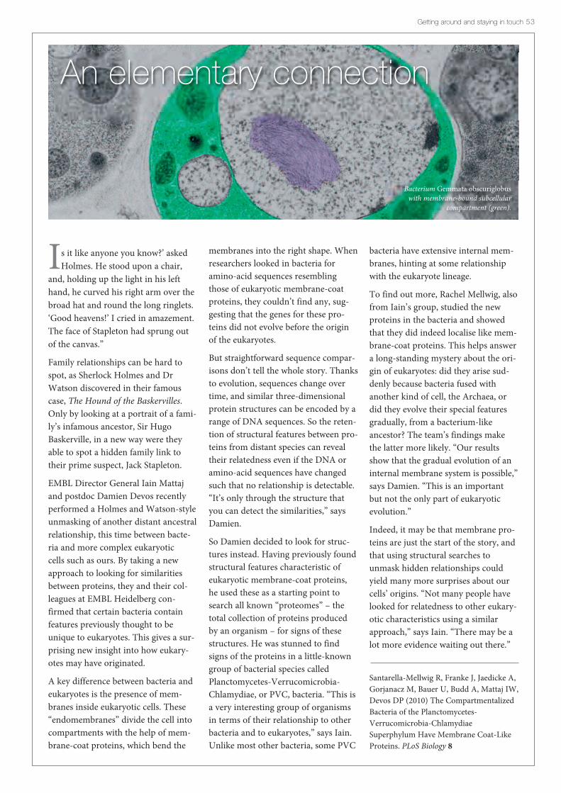
Is it like anyone you know?’ askedHolmes. He stood upon a chair,
and, holding up the light in his lefthand, he curved his right arm over thebroad hat and round the long ringlets.‘Good heavens!’ I cried in amazement.The face of Stapleton had sprung outof the canvas.”
Family relationships can be hard tospot, as Sherlock Holmes and DrWatson discovered in their famouscase, The Hound of the Baskervilles.Only by looking at a portrait of a fami-ly’s infamous ancestor, Sir HugoBaskerville, in a new way were theyable to spot a hidden family link totheir prime suspect, Jack Stapleton.
EMBL Director General Iain Mattajand postdoc Damien Devos recentlyperformed a Holmes and Watson-styleunmasking of another distant ancestralrelationship, this time between bacte-ria and more complex eukaryotic cells such as ours. By taking a newapproach to looking for similaritiesbetween proteins, they and their col-leagues at EMBL Heidelberg con-firmed that certain bacteria containfeatures previously thought to beunique to eukaryotes. This gives a sur-prising new insight into how eukary-otes may have originated.
A key difference between bacteria andeukaryotes is the presence of mem-branes inside eukaryotic cells. These“endomembranes” divide the cell intocompartments with the help of mem-brane-coat proteins, which bend the
membranes into the right shape. Whenresearchers looked in bacteria foramino-acid sequences resemblingthose of eukaryotic membrane-coatproteins, they couldn’t find any, sug-gesting that the genes for these pro-teins did not evolve before the originof the eukaryotes.
But straightforward sequence compar-isons don’t tell the whole story. Thanksto evolution, sequences change overtime, and similar three-dimensionalprotein structures can be encoded by arange of DNA sequences. So the reten-tion of structural features between pro-teins from distant species can revealtheir relatedness even if the DNA oramino-acid sequences have changedsuch that no relationship is detectable.“It’s only through the structure thatyou can detect the similarities,” saysDamien.
So Damien decided to look for struc-tures instead. Having previously foundstructural features characteristic ofeukaryotic membrane-coat proteins,he used these as a starting point tosearch all known “proteomes” – thetotal collection of proteins producedby an organism – for signs of thesestructures. He was stunned to findsigns of the proteins in a little-knowngroup of bacterial species calledPlanctomycetes-Verrucomicrobia-Chlamydiae, or PVC, bacteria. “This isa very interesting group of organismsin terms of their relationship to otherbacteria and to eukaryotes,” says Iain.Unlike most other bacteria, some PVC
bacteria have extensive internal mem-branes, hinting at some relationshipwith the eukaryote lineage.
To find out more, Rachel Mellwig, alsofrom Iain’s group, studied the newproteins in the bacteria and showedthat they did indeed localise like mem-brane-coat proteins. This helps answera long-standing mystery about the ori-gin of eukaryotes: did they arise sud-denly because bacteria fused withanother kind of cell, the Archaea, ordid they evolve their special featuresgradually, from a bacterium-likeancestor? The team’s findings makethe latter more likely. “Our resultsshow that the gradual evolution of aninternal membrane system is possible,”says Damien. “This is an importantbut not the only part of eukaryoticevolution.”
Indeed, it may be that membrane pro-teins are just the start of the story, andthat using structural searches tounmask hidden relationships couldyield many more surprises about ourcells’ origins. “Not many people havelooked for relatedness to other eukary-otic characteristics using a similarapproach,” says Iain. “There may be alot more evidence waiting out there.”
Santarella-Mellwig R, Franke J, Jaedicke A,Gorjanacz M, Bauer U, Budd A, Mattaj IW,Devos DP (2010) The CompartmentalizedBacteria of the Planctomycetes-Verrucomicrobia-ChlamydiaeSuperphylum Have Membrane Coat-LikeProteins. PLoS Biology 8
Getting around and staying in touch 53
An elementary connection
Bacterium Gemmata obscuriglobuswith membrane-bound subcellular
compartment (green).

Matthias Wilmanns
Big Brother is watching you. This famous, ominousphrase was coined by novelist George Orwell more
than 60 years ago in his novel, Nineteen Eighty-Four. Ittold the story of a future dystopia where the government’sfigurehead, Big Brother, constantly monitored everyone’sconversations via TV-like “telescreens”. Worse, BigBrother dictated the vocabulary that citizens, or com-rades, could use. By controlling their language, he con-trolled their thoughts and behaviour. It is a cautionarytale about the power of words.
So it might come as a surprise to learn that some biolo-gists are trying to follow in Big Brother’s footsteps. Butthis is not as sinister as it sounds. Instead of spying onpeople’s conversations, they are trying to tap into thecommunication between the molecules in our cells. Thiscommunication is essential to life, and by working outwhat these molecules are saying to each other, theseresearchers hope to learn more about diseases such ascancer and neurodegeneration. And by controlling thiscommunication with drugs – rather like Big Brother’scontrol over conversations – they hope to find new treat-ments for such illnesses.
One such biologist is Matthias Wilmanns at EMBLHamburg. Matthias and his team recently performed themolecular equivalent of installing a telescreen in thehome of one particular protein called death-associatedprotein kinase, or DAPK. They have uncovered the struc-ture of DAPK as it interacts with another key proteininvolved in cell regulation. This has allowed them to findout how these proteins ‘talk’ to each other, and how sci-
Knowledge is strength
entists might be able to intervene. “These kinases are apretty hot target,” says Matthias. The discovery is all themore important because DAPK is similar to several othervital signalling proteins in the cell. So the insights theteam have gained into DAPK’s function might also beapplicable to these other molecules.
The idea of manipulating cellular signalling with drugs istried and tested. The problem is that, although scientistsknow a lot about which proteins are involved in cell reg-ulation, and which proteins interact with each other, theyknow relatively little about how most of these interactionsoccur. Furthermore, molecules in cells use many differentmechanisms to communicate, in the same way as we mayuse face-to-face speech, mobiles, landline phones or e-mail to talk to each other. Scientists know more aboutsome of these mechanisms than they do about others.
Matthias and his team decided to look at one of the lesserunderstood mechanisms, called calcium-dependent sig-nalling. This mechanism is involved in many vital parts ofour biology, such as controlling our heartbeat, musclecontractions and the communication between neurons inthe brain. When an appropriate signal arrives at a cell –say a chemical signal from one neuron to another – ittriggers a flood of calcium ions within the cell. This floodis detected by a protein called calmodulin, which becomesactive when calcium ions bind to it. Calmodulin thenturns this calcium signal into action by switching variousparts of the cell’s machinery on or off, thus altering thecell’s behaviour.
Getting around and staying in touch 5555
Matthias Wilmanns

X - R A Y V I S I O N
complex formed when severalmolecules bind to each other. To doso, they first induce the molecule toform crystals, then expose thosecrystals to an X-ray beam and recordthe angles and intensities of the X-rays that emanate from that sample.This diffraction pattern allows theresearchers to produce a 3-dimen-sional model of the arrangement ofatoms within the crystal. Finally,they use computer software to refinethat model: the software calculatesthe diffraction pattern which themodel structure would generate,compares it to the one the scientistsobtained in their experiments, andalters the model until it fits thoseobservations.
This technique was initially devel-oped by geologists interested in thearrangement of minerals, but eversince it was first employed to deter-mine the structure of a protein inthe late 1950s, molecular biologistshave been turning to it regularly inorder to peer into the intricacies ofhow life’s molecules are arranged,and how they interact. The numberof protein structures solved by X-raycrystallography is now over 50,000,and counting. Whether they are rep-resented as globular masses or ascoloured loops and ribbons, themodels generated by this biologicalequivalent of Superman removinghis glasses are still bringing scientistscloser to solving such diverse mys-teries as how cells communicate (seepage 54), how plants survivedrought (see page 42), and how bac-teria evade our immune system (seepage 91).
56 EMBL Annual Report 09 · 10
To understand the 3-dimen-sional structure of biologi-
cal molecules and complexes,structural biologists have theirown equivalent to Superman’s X-ray vision: X-ray crystallography.This technique allows scientists todetermine how molecules oratoms are arranged within a crys-tal, by exposing the crystal to X-rays and studying how it scattersthem.
The atoms that make up a crystalare located at regular intervals,and when X-rays strike them, theytremble. This trembling eventual-ly makes each atom emit X-rayswith the same energy in all direc-tions, in a process called elasticscattering. The waves of energytravelling outward from eachatom interfere with those comingfrom other atoms, reinforcing orcancelling each other out depend-ing on the angle at which theymeet. The resulting pattern ofdiffracted waves, called a diffrac-tion pattern (see image), can berecorded by placing a detector infront of the crystal. Every sub-stance has its own unique diffrac-tion pattern, depending on theidentity and the position of theatoms within its crystals, so byanalysing that pattern scientistscan determine what atoms makeup a particular crystal, and howthey are positioned in relation toeach other.
X-ray crystallography enables sci-entists to use this property ofcrystals to determine the 3 Dstructure of a molecule, or of a Diffraction pattern (middle), and ribbon
and globular representations of proteinstructures (top and bottom, respectively).

One of the main ways calmodulin does this is via a team ofcellular errand boys called kinases, each of which controls theactivity of specific parts of the cell’s vastly complicatedmachinery. Kinases are an extremely important part of thecell’s control mechanisms and there are about 600 of themencoded in the human genome. Some have already been suc-cessfully targeted with drugs such as Gleevec® (Imatinib),which is used to treat chronic myeloid leukaemia.Calmodulin is predicted to control about 10% of all the cell’skinases, but how it does so was unknown.
Matthias and his team wanted to find out how calmodulininteracted with its kinases. This meant that they needed toproduce a three-dimensional molecular structure of calmod-ulin bound to one of its targets. To do this, they turned to atechnique called X-ray crystallography, which involves mixingcalmodulin and one of its kinases together to allow the pro-teins to interact, and then crystallising the resulting mixture.Shining X-rays on to the crystal allows the structure of theproteins to be deduced by the way the X-rays are scattered.
They began by working on a calmodulin-controlled kinasecalled titin. But try as they might, the team could not get titinand calmodulin to form crystals. Their luck changed whenthey were contacted by Adi Kimchi, a biologist at theWeizmann Institute in Israel. Adi’s team was working onDAPK and they wanted to understand how calmodulin con-trolled its activity. Adi had heard about Matthias’s work, andso offered to collaborate with him.
Matthias jumped at the chance. DAPK is interesting for twomain reasons. One is that it is known to malfunction in anumber of cancers, and so has potential uses in developingnew therapies and diagnostic techniques. The other is thatmany calmodulin-controlled kinases share similar structuralfeatures. So finding out how calmodulin controls DAPKcould give useful insights into how it controls these other tar-gets. “Our motivation was to identify a prototype proteinkinase,” says Matthias. “Many things that are true of the pro-totype will also be true of other members of this kinase family.”
No-one had ever produced a structure like this before, andwith good reason. DAPK and calmodulin are large proteins,which makes it difficult to crystallise them both together. Butthe work was made possible thanks to Hamburg’s high-throughput crystallisation facility run by team leader JochenMüller-Dieckmann.. “It was critical for us,” says Matthias.With the help of high-energy X-rays produced by theEuropean Synchrotron Radiation Facility (ESRF) in Grenobleand the German Synchrotron Radiation Facility (DESY) inHamburg, the team was able to deduce a structure.
This showed how calmodulin bound to a particular section ofDAPK, switching the kinase on so that it could activate its tar-
gets. Intriguingly, the team’s finding is different from earlierwork in this area. Previous studies by other groups havelooked at very short, synthetic sections, or peptides, of DAPKbound to calmodulin. Matthias’s team compared one suchpeptide with the much larger DAPK fragment that they used,and showed that it behaved very differently. Without wantingto disparage this previous work, Matthias says his findingsmean scientists may have to reconsider the results of this andsimilar experiments. They also show the value of being able tocrystallise larger, more life-like protein fragments for study.
Next, the team altered some of the building blocks, or aminoacids, in their DAPK fragment to see which ones were need-ed for calmodulin to bind. They found one in particular thatwas vital for the interaction. “That will provide a platform toget into drug discovery,” says Matthias. The work also givesbiologists a starting point to study how calmodulin controlsother kinases, although Matthias cautions that some gapsremain to be filled. For example, can DAPK adopt otherstructures to become active? And what other control mecha-nisms for these kinases remain to be found? “All these kinas-es have multiple layers of regulation,” says Matthias.
He and his team are planning to explore these questions withthe help of chemist Carsten Schultz at EMBL Heidelberg, aswell as continuing the collaboration with Adi Kimchi. If allgoes to plan, scientists may be able to both listen in on cellu-lar conversations and control what is being said to help treatdisease. Who knows? One day, the phrase “Big Brother iswatching you” may be a comforting thought – from a cellularperspective at least.
de Diego I, Kuper J, Bakalova N, Kursula P, Wilmanns M (2010)Molecular Basis of the Death-Associated Protein Kinase–Calcium/Calmodulin Regulator Complex. Science Sinaling 3: 106
Getting around and staying in touch 57
The structure of DAPK (green and yellow) bound to
calmodulin (violet and blue).

From a multitude of graphical languages to a standard way of depicting biological processes.
Imagine that you wanted to witness the creation ofexciting new scientific ideas. Where would you go? A
lab, perhaps? Possibly. There’s no doubt that a hugeamount of fantastic work gets done there. But if you real-ly want to see the raw creative sparks flying then the bestadvice is to head for the research institution tea room. It’shere, among the coffee stains and chocolate biscuit wrap-pers that animated arguments take place. Ideas are moot-ed, discussed and challenged. Diagrams are scribbled onpaper napkins, cardboard plates and, occasionally, piecesof fruit. If you’re in the tea room of an engineeringdepartment, chances are you could take these scribbles toany engineer anywhere and he or she would be able tounderstand them. The tea time scrawls of biologists are,however, much harder to decipher.
The problem is that, unlike engineers, biologists have nouniversal standardised way of depicting biological pro-cesses in a graphical form. So a set of symbols that makesense to one biologist could well be gibberish to another.Now, Nicolas Le Novère of EMBL-EBI and his colleaguesare creating a universal notation for biologists to explainwhat they mean clearly and succinctly – not only to eachother but to computers as well. “People won’t have toread the legend in figures,” says Nicolas. “When they seethe diagram they will know what it is in the same way anengineer will recognise symbols for batteries and switch-es and so on.”
Until now, different groups of biologists, such as bio-chemists or geneticists, had their own peculiar conven-tions for drawing pictures of processes such as gene inter-
Biology gets the picture
actions or metabolic pathways. Many times, if they had toexplain something in a paper, they would invent theirown symbols and furnish their diagrams with detailed fig-ure legends that were often bigger than the diagram itself.And while biology remained a largely academic disci-pline, this hardly mattered. Over the past decade, howev-er, various biological disciplines have been transformedinto applied sciences, more similar to engineering thanblue-skies exploration. New disciplines such as syntheticbiology, cell reprogramming and biological engineeringare using or manipulating biological material to builddevices or new tissues. ‘Pure’ biologists are also uncover-ing ever more complexity in the metabolic pathways andnetworks of gene interactions within a cell. “People needstandardisation when representing these networks andpathways,” says Nicolas. As well as helping researcherscommunicate more accurately and efficiently with eachother, a standardised notation would make it much easi-er to build computer software that could process infor-mation presented as diagrams.
Work on the new notation started back in 2005, whenNicolas was collaborating with Japanese researcherHiroaki Kitano from Tokyo’s Systems Biology Institute,to develop a software language for systems biology calledSystems Biology Markup Language, or SBML. Kitano hadalready tried to develop a standardised notation andtogether with Nicolas, had secured a grant from theJapanese government to really take the idea forward.They convened an international team of collaborators,including Kurt Kohn, a researcher from the NationalCancer Institute in Maryland, United States, who had also
Getting around and staying in touch 5959

attempted to develop a universal notation. Given the impor-tance of standardisation, it might seem surprising that it hastaken so long for work like this to start in earnest. But Nicolaspoints out that until recently, there has been very littledemand. It is only in the past 10 years that certain areas ofbiology have become more like engineering disciplines andthat industry has developed more of an interest in them.What’s more, funding bodies have only just begun to perceivethe need for this kind of work.
The team started work on the new system, which they namedThe Systems Biology Graphical Notation, or SBGN. Althoughthe system has one name, the team actually developed threedifferent symbolic methods of describing biological processesfrom three different perspectives. But these methods are farmore than sets of symbols. They have the properties of lan-guages: symbols have specific meanings, or semantics, andthey have a defined set of rules, or syntax, about how theyshould be used to construct communication. Importantly,says Nicolas, the relationships, or ontology, these symbolshave to each other follow biological rules. “SBGN is not justanother graphical language,” he says. Putting something so spe-cific together wasn’t easy, but it appealed to his way of working.“I am a very picky person,” he says. “I liked the challenge ofdeveloping something that was semantically very neat.”
Nicolas became the main co-ordinator and driver of the pro-ject, which published full details of the three languages in thejournal Nature Biotechnology in August 2009. Each one isused for a different purpose. “There are three different repre-sentations of an underlying biological reality,” explainsNicolas. “You choose your language depending on your dataand the question you are asking.”
The first, Process Descriptions, is used when researchers wantto describe how something happens, such as how a moleculeis altered as it proceeds through a series of biochemical reac-tions as part of the cell’s metabolism. This allows researchersto describe the mechanics of the process in a sequential,unambiguous manner. It’s a great way of depicting processes,but it has one big drawback. If you try to represent all theinteractions a molecule has in this kind of diagram, itbecomes overwhelmingly complex. “You end up with billionsof billions of combinations,” says Nicolas.
Clearly, another way of representing every one of a molecule’sinteractions is needed, and this is where the second language,called Entity Relationships comes in. This allows you to drawa map of all the possible relationships between items or “enti-ties” in the diagram clearly and simply, but it does not showthe order in which interactions occur. The third language,called Activity Flows, is more abstract. Here, the entities in
60 EMBL Annual Report 09 · 10
Nicolas Le Novère

the diagram aren’t molecules themselves, but molecular activ-ity, such as a particular kind of enzyme reaction. This kind ofdiagram is useful for biologists studying the chemical sig-nalling systems cells use to communicate. Often, their exper-iments will reveal the presence of a particular kind of enzymeactivity, without revealing what that enzyme is. So having away to express these relationships in an abstract, if ambigu-ous, way is very useful.
But it’s not just molecular biologists who stand to benefit.Nicolas has been working with clinicians to describe diseaseprocesses using entity relationship diagrams and is develop-ing new ways of describing other biological phenomena, suchas blood flow, using SBGN. Ultimately, he wants to roll SBGNout to cover all of the biological sciences. “We are trying to develop something robust and to cover everyone’s needs,”he says. To this end, any biology researcher can participate in SBGN development via the project’s website:http://sbgn.org/. Decisions about whether new symbols andideas should become part of SBGN are democratic and asteering committee oversees the development of the project.
As well as continuing to develop SBGN, the next big chal-lenge is getting biologists everywhere to use it. Nicolas is con-fident that this will come in time as researchers begin to seethe benefits, including software that uses the language.
What’s more, SBGN is not hard to learn. “It is really easy,”says Nicolas. “It just takes a bit of repetition. After all, we areall scientists, juggling with gene and protein names; we mustbe clever enough to learn a few symbols!”
Certainly, SBGN received an enthusiastic reaction from theresearch community and industry when it was published,with biologists keen to learn more and collaborate. This will-ingness to share is good news, says Nicolas, as SBGN is partof a larger movement to scale up standardised notation andcomputational methods in biology. At the moment, much ofthis work is divided into specialised ghettos, with small com-munities of researchers focussed exclusively on one topic andnot really communicating with the others. “We have to bringall these people together,” says Nicolas. “This is the only wayto build something coherent and transparent to allresearchers.” Ultimately, the hope is to transcend geographi-cal and discipline barriers in biology – to create a kind ofworld-wide tea room where scribbles will need no morelengthy explanations.
Le Novère N et al (2009) The Systems Biology Graphical Notation.
Nature Biotechnology 27: 735-741
Getting around and staying in touch 61
The same biological process (the phosphorylation of MAP kinase) represented in the three SBGN languages.
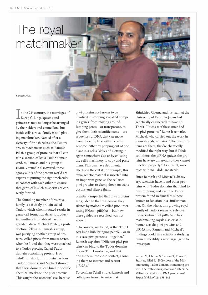
In the 21st century, the marriages ofEurope’s kings, queens and
princesses may no longer be arrangedby their elders and councillors, butinside cells a royal family is still play-ing matchmaker. Named after adynasty of British rulers, the Tudorsare, to biochemists such as RameshPillai, a group of proteins that all con-tain a section called a Tudor domain.And, as Ramesh and his group atEMBL Grenoble discovered, theseagony aunts of the protein world areexperts at putting the right moleculesin contact with each other to ensurethat germ cells such as sperm are cor-rectly formed.
The founding member of this royalfamily is a fruit fly protein calledTudor, which when mutated results ingerm-cell formation defects, produc-ing mothers incapable of havinggrandchildren. Michael Reuter, a post-doctoral fellow in Ramesh’s group,was purifying another group of pro-teins, called piwis, from mouse testes,when he found that they were attachedto a Tudor protein. Called Tudordomain-containing protein-1, orTdrd1 for short, this protein has fourTudor domains, and Michael showedthat these domains can bind to specificchemical marks on the piwi proteins.This caught the scientists’ eye, because
62 EMBL Annual Report 09 · 10
The royalmatchmaker
piwi proteins are known to beinvolved in stopping so-called ‘jump-ing genes’ from moving around.Jumping genes – or transposons, togive them their scientific name – aresequences of DNA that can movefrom place to place within a cell’sgenome, either by popping out of oneplace in a cell’s DNA and slotting inagain somewhere else or by enlistingthe cell’s machinery to copy and pastethem. This can have detrimentaleffects on the cell if, for example, thisextra genetic material is inserted intoan important gene, so the cell usespiwi proteins to clamp down on trans-posons and silence them.
Scientists suspected that piwi proteinsare guided to the transposons theysilence by molecules called piwi-inter-acting RNAs – piRNAs – but howthese guides are recruited was notknown.
“The answer, we found, is that Tdrd1acts like a hub, bringing people – or inthis case piwi proteins – together,”Ramesh explains: “Different piwi pro-teins can bind to the Tudor domainsin one Tdrd1 molecule, and thatbrings them into close contact, allow-ing them to interact and recruitpiRNAs.”
To confirm Tdrd1’s role, Ramesh andcolleagues turned to mice that
Shinichiro Chuma and his team at theUniversity of Kyoto in Japan hadgenetically engineered to have noTdrd1. “It was as if these mice had no piwi proteins,” Ramesh remarks.Michael, who carried out the work inRamesh’s lab, explains: “The piwi pro-teins are there, they’re chemicallymodified the right way, but if Tdrd1isn’t there, the piRNA guides the pro-teins have are different, so they cannotfunction properly.” As a result, malemice with no Tdrd1 are sterile.
Since Ramesh and Michael’s discov-ery, scientists have found other pro-teins with Tudor domains that bind topiwi proteins, and even the Tudorprotein found in fruit flies is nowknown to function in a similar man-ner. On the whole, this growing royalfamily of Tudors seems to rule overthe recruitment of piRNAs. Thesematchmaking royals also exist inhumans, as do piwi proteins andpiRNAs, so Ramesh and Michael’sfindings could give scientists studyinghuman infertility a new target gene toinvestigate.
Reuter M, Chuma S, Tanaka T, Franz T,Stark A, Pillai R (2009) Loss of the Mili-interacting Tudor domain–containing pro-tein-1 activates transposons and alters theMili-associated small RNA profile. NatStruct Mol Biol 16: 639-646
Ramesh Pillai
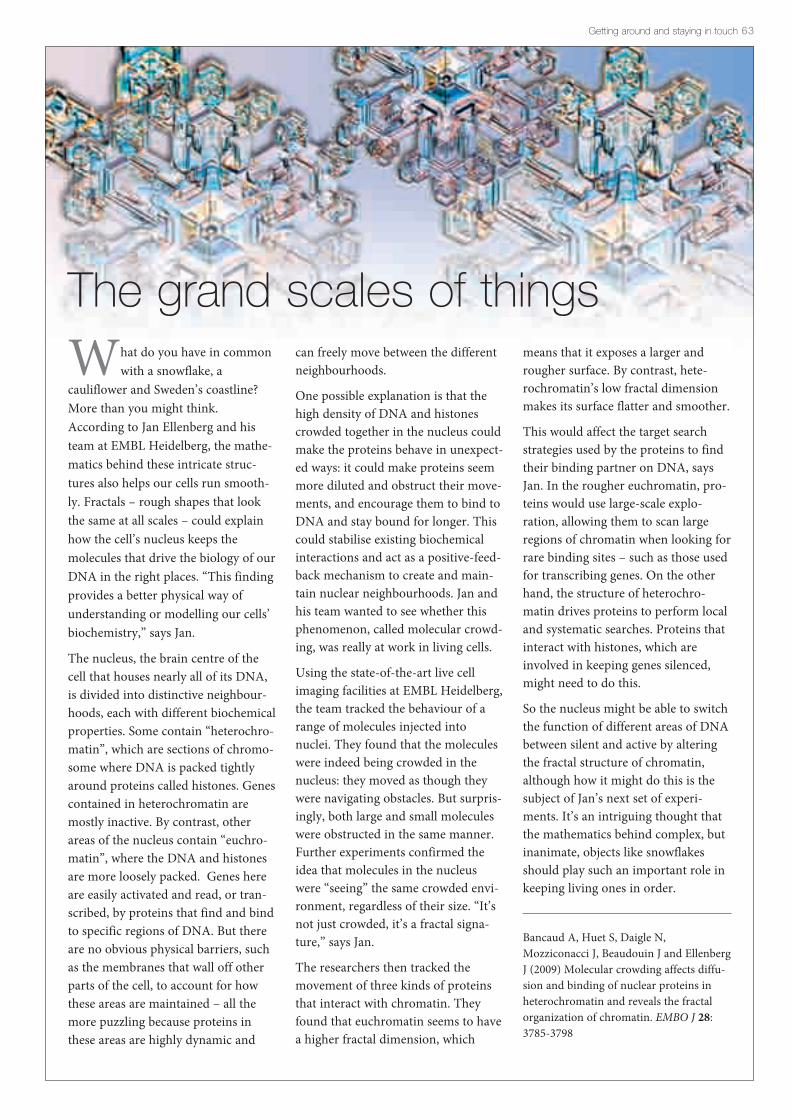
What do you have in commonwith a snowflake, a
cauliflower and Sweden’s coastline?More than you might think.According to Jan Ellenberg and histeam at EMBL Heidelberg, the mathe-matics behind these intricate struc-tures also helps our cells run smooth-ly. Fractals – rough shapes that lookthe same at all scales – could explainhow the cell’s nucleus keeps themolecules that drive the biology of ourDNA in the right places. “This findingprovides a better physical way ofunderstanding or modelling our cells’biochemistry,” says Jan.
The nucleus, the brain centre of thecell that houses nearly all of its DNA,is divided into distinctive neighbour-hoods, each with different biochemicalproperties. Some contain “heterochro-matin”, which are sections of chromo-some where DNA is packed tightlyaround proteins called histones. Genescontained in heterochromatin aremostly inactive. By contrast, otherareas of the nucleus contain “euchro-matin”, where the DNA and histonesare more loosely packed. Genes hereare easily activated and read, or tran-scribed, by proteins that find and bindto specific regions of DNA. But thereare no obvious physical barriers, suchas the membranes that wall off otherparts of the cell, to account for howthese areas are maintained – all themore puzzling because proteins inthese areas are highly dynamic and
Getting around and staying in touch 63
The grand scales of thingscan freely move between the differentneighbourhoods.
One possible explanation is that thehigh density of DNA and histonescrowded together in the nucleus couldmake the proteins behave in unexpect-ed ways: it could make proteins seemmore diluted and obstruct their move-ments, and encourage them to bind toDNA and stay bound for longer. Thiscould stabilise existing biochemicalinteractions and act as a positive-feed-back mechanism to create and main-tain nuclear neighbourhoods. Jan andhis team wanted to see whether thisphenomenon, called molecular crowd-ing, was really at work in living cells.
Using the state-of-the-art live cellimaging facilities at EMBL Heidelberg,the team tracked the behaviour of arange of molecules injected intonuclei. They found that the moleculeswere indeed being crowded in thenucleus: they moved as though theywere navigating obstacles. But surpris-ingly, both large and small moleculeswere obstructed in the same manner.Further experiments confirmed theidea that molecules in the nucleuswere “seeing” the same crowded envi-ronment, regardless of their size. “It’snot just crowded, it’s a fractal signa-ture,” says Jan.
The researchers then tracked themovement of three kinds of proteinsthat interact with chromatin. Theyfound that euchromatin seems to havea higher fractal dimension, which
means that it exposes a larger androugher surface. By contrast, hete-rochromatin’s low fractal dimensionmakes its surface flatter and smoother.
This would affect the target searchstrategies used by the proteins to findtheir binding partner on DNA, saysJan. In the rougher euchromatin, pro-teins would use large-scale explo-ration, allowing them to scan largeregions of chromatin when looking forrare binding sites – such as those usedfor transcribing genes. On the otherhand, the structure of heterochro-matin drives proteins to perform localand systematic searches. Proteins thatinteract with histones, which areinvolved in keeping genes silenced,might need to do this.
So the nucleus might be able to switchthe function of different areas of DNAbetween silent and active by alteringthe fractal structure of chromatin,although how it might do this is thesubject of Jan’s next set of experi-ments. It’s an intriguing thought thatthe mathematics behind complex, butinanimate, objects like snowflakesshould play such an important role inkeeping living ones in order.
Bancaud A, Huet S, Daigle N,Mozziconacci J, Beaudouin J and EllenbergJ (2009) Molecular crowding affects diffu-sion and binding of nuclear proteins inheterochromatin and reveals the fractalorganization of chromatin. EMBO J 28:3785-3798

Paul Kersey
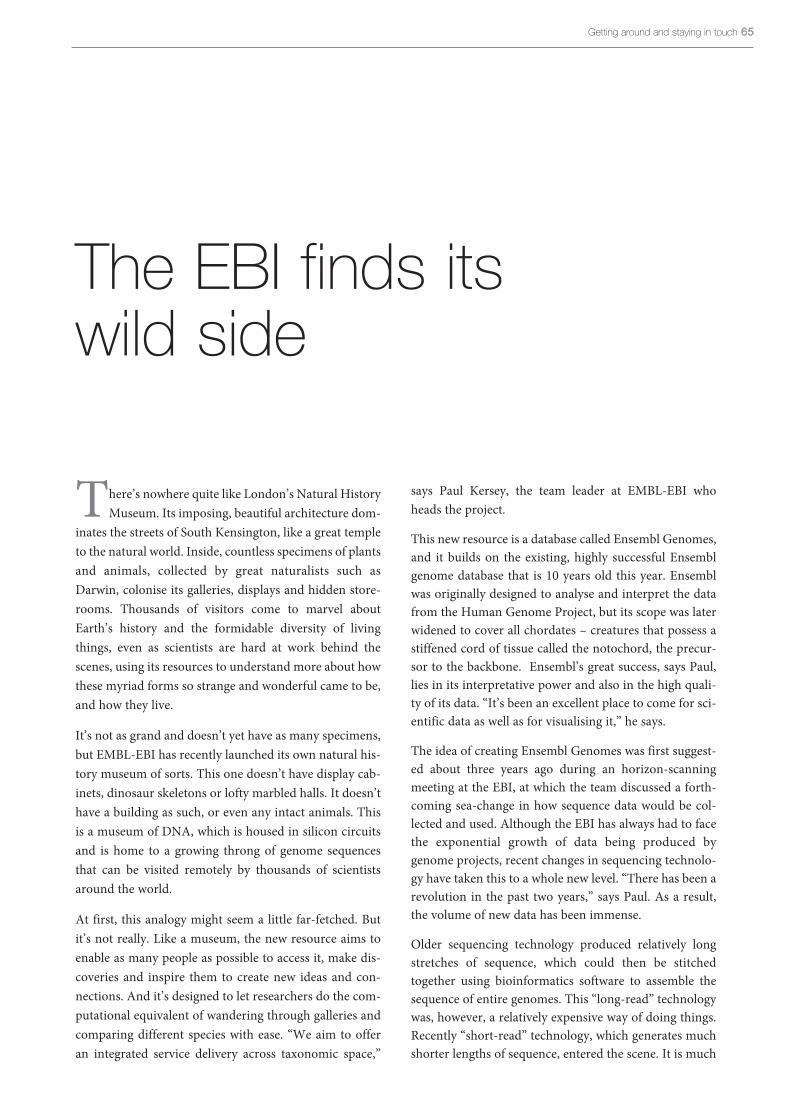
There’s nowhere quite like London’s Natural HistoryMuseum. Its imposing, beautiful architecture dom-
inates the streets of South Kensington, like a great templeto the natural world. Inside, countless specimens of plantsand animals, collected by great naturalists such asDarwin, colonise its galleries, displays and hidden store-rooms. Thousands of visitors come to marvel aboutEarth’s history and the formidable diversity of livingthings, even as scientists are hard at work behind thescenes, using its resources to understand more about howthese myriad forms so strange and wonderful came to be,and how they live.
It’s not as grand and doesn’t yet have as many specimens,but EMBL-EBI has recently launched its own natural his-tory museum of sorts. This one doesn’t have display cab-inets, dinosaur skeletons or lofty marbled halls. It doesn’thave a building as such, or even any intact animals. Thisis a museum of DNA, which is housed in silicon circuitsand is home to a growing throng of genome sequencesthat can be visited remotely by thousands of scientistsaround the world.
At first, this analogy might seem a little far-fetched. Butit’s not really. Like a museum, the new resource aims toenable as many people as possible to access it, make dis-coveries and inspire them to create new ideas and con-nections. And it’s designed to let researchers do the com-putational equivalent of wandering through galleries andcomparing different species with ease. “We aim to offeran integrated service delivery across taxonomic space,”
The EBI finds itswild side
says Paul Kersey, the team leader at EMBL-EBI whoheads the project.
This new resource is a database called Ensembl Genomes,and it builds on the existing, highly successful Ensemblgenome database that is 10 years old this year. Ensemblwas originally designed to analyse and interpret the datafrom the Human Genome Project, but its scope was laterwidened to cover all chordates – creatures that possess astiffened cord of tissue called the notochord, the precur-sor to the backbone. Ensembl’s great success, says Paul,lies in its interpretative power and also in the high quali-ty of its data. “It’s been an excellent place to come for sci-entific data as well as for visualising it,” he says.
The idea of creating Ensembl Genomes was first suggest-ed about three years ago during an horizon-scanningmeeting at the EBI, at which the team discussed a forth-coming sea-change in how sequence data would be col-lected and used. Although the EBI has always had to facethe exponential growth of data being produced bygenome projects, recent changes in sequencing technolo-gy have taken this to a whole new level. “There has been arevolution in the past two years,” says Paul. As a result,the volume of new data has been immense.
Older sequencing technology produced relatively longstretches of sequence, which could then be stitchedtogether using bioinformatics software to assemble thesequence of entire genomes. This “long-read” technologywas, however, a relatively expensive way of doing things.Recently “short-read” technology, which generates muchshorter lengths of sequence, entered the scene. It is much
Getting around and staying in touch 6565

harder to stitch the pieces together, but it is vastly cheaperthan the older methods. The sudden drop in costs – sequenc-ing an entire bacterial genome is now about 50 000 timescheaper than it was before – has transformed the rate andscale of data production. “The cost that remains is makingsense of the data,” says Paul.
And there is a lot of making sense to do. 1200 bacterialgenomes have been sequenced over the past 15 years and sev-eral projects aiming to greatly expand that number are under-way. Many labs are also now going back and “re-sequencing”species whose genomes have already been read. Originally,only one member of each species had their genomes read to
produce a single “reference” sequence characteristic of thatspecies. Now, however, researchers want to study the varia-tion in the genome sequences that exists between individualsof the same species, and to work out how this relates to vari-ations in individual physical attributes and physiology. The1000 Genomes project – in which the EBI is directly involved– has attracted most attention with its focus on humangenomes, but similar projects relevant to Ensembl Genomesare also underway, for example the project to sequence 1001strains of the plant Arabidopsis.
The ready availability of relatively cheap whole-genomesequencing is also changing the way biological research is
66 EMBL Annual Report 09 · 10
The ENSEMBL databases – a digital museum of sorts
done, says Paul. Not so long ago, a biologist encountering anew species might study its physiology, ecology or biochem-istry, and hope that one day a consortium of researcherswould be awarded a large grant to sequence its genome. Theday is not far off, Paul says, when researchers will sequence anew species’ genome first, and ask biological questions later.“Whole-genome sequencing is being used in a diagnosticcapacity to find out what a new species is,” he points out.“The technologies are being used on a very wide scale.”
Cheaper, faster sequencing is also allowing biologists to tack-le biologically and economically important genomes thatuntil now have been difficult to attempt. Chief among theseare the crop plants such as cereals, the genomes of which arehuge and riddled with sections of sequence that are technical-ly hard to read and reconstruct. “I think 2010 will see a num-ber of very important plants being sequenced,” says Paul.“The wheat genome, which is both large and complex, is thelast big challenge. That will be the last hurdle for primarygenomics.”
But it’s more than just organisation. The data in EnsemblGenomes are presented in a way that is both meaningful andeasy to access – users come to interpreted information first,rather than just raw data. “You put the raw data in a biologi-cal context,” explains Paul. “One of the key aims is to allowusers to see their data in the context of other genomes.” Userscan then click through to see the raw evidence if they wish.“You want to guide users to references, interpret data andthen let them sensibly navigate through the informatics with-out drowning in it,” says Paul.
Importantly, the EBI is working in partnership with differentgenomics communities to complement, rather than dupli-cate, their bioinformatics efforts. “We aim for robustness ofinfrastructure and breadth of scope,” says Paul. For example,his team is a collaborator in VectorBase, a communitydatabase funded by the National Institutes of Health in theUSA that focusses on the genomics of mosquitoes and otherinsects implicated in disease. The EBI contributes to theannotation of the genomes of these species, then provides aportal through which this information can be accessed in abroader taxonomic context.
To reflect its breadth of scope, Ensembl Genomes is dividedinto five main parts, each representing one of the five king-doms of life: bacteria, protists, fungi, plants and invertebratemetazoa (multicellular animals). This complements the verte-brate focus of the existing Ensembl site. The bacteria, protistsand invertebrate sections were launched in April 2009, andthe plants and fungi sections followed in October 2009. Thenumber of users is already increasing. “It’s still a youngresource,” says Paul. He and his colleagues at the EBI on theEnsembl and Ensembl Genomes projects, Paul Flicek andEwan Birney, are hard at work expanding the resources:forming collaborations with specialist communities andadding more species, and improving the power of user inter-faces. “We want to make sure we have all the importantmodel organisms in the database,” says Paul.
In future, the team also plans to find ways of handling datafrom bacterial genomes more effectively, given the sheernumbers of species being sequenced. But, like any exactingmuseum curators, the researchers are being choosy aboutwhich organisms they collect on the database, ensuring thatonly the ones that are most relevant and useful to their visi-tors make the grade. “The driver is the interest of the scientif-ic community,” says Paul. “If they love their genome, then wewant to care for it too.”
www.ensembl.org
www.ensemblgenomes.org
Getting around and staying in touch 67
“The driver is the interest of the scientific community,” says Paul. “If they love their genome, then wewant to care for it too.”
While things might be getting easier for the DNA sequencers,the opposite is true for bioinformatics service providers suchas the EBI – and for their users. “Historically, genome datawere rare and valuable things. They have now become com-monplace,” says Paul. “It’s very hard for the EBI, with its ser-vice mission, to deliver that data to the public in a usableway.” His team realised that it would be unfeasible for the EBIto become directly involved in the work on so many genomes,and yet there was a clear need for a resource that would helpbiologists to find and use the data, without the need for spe-cialised bioinformatics training.
So they had the idea of working with communities to offerhelp with the bioinformatics where needed, but to also pro-vide a user-friendly, centralised interface, via the EBI,through which users can access data from all these differentresources and projects in an integrated manner. GivenEnsembl’s success, it made sense to follow its model: to usethe genome as an index system for all the different kinds ofbiological data about a gene or organism. “We have this hugeflood of many new types of data,” says Paul. “The genome isa natural way of organising access to it.”

Technology transfer at a glance
What is technology transfer, and what does this suc-cess story actually mean for EMBL and its scientists?With innovation being driven by excellent basicresearch, it is essential to have mechanisms thatensure that scientific results and discoveries canproperly and rapidly be translated into practical appli-cations and marketable products – be it equipment,medicines, diagnostic tools or even entire companies– for the benefit of society as a whole. This hasalways been one of EMBL’s missions and with thecreation of EMBLEM, the previously ad hoc protec-tion and commercialisation of intellectual propertyand inventions has been professionalised and
� ARP/wARP The transformation of electron density maps from crys-tallography experiments into three-dimensional struc-tures requires complex mathematical modelling. In the1990s, ‘off-the-shelf’ software solutions were rare andinsufficient for research at the cutting edge of structuralbiology. To address this, scientists at EMBL Hamburgdeveloped ARP/wARP, a software suite for automaticstructure determination from crystallographic data. It isnow used by several thousand users in academic and indus-trial research and has been continuously updated duringthe past ten years to meet changing user needs and envi-ronments. The latest version was launched in January 2010.
� SPIM/DSLMSingle Plane Illumination Microscopy (SPIM) wasinvented by EMBL scientists and it revolutionised thefield of light microscopy and live imaging. It enables thestudy of large, living specimens from different angles,under physiological conditions and with minimal harm.Images obtained along different axes and at varying timepoints are assembled into three-dimensional images ormovies, which provide insights into the dynamic cellularprocesses that are occurring in living organisms. A licenseagreement was concluded with Zeiss and first prototypeswere developed. SPIM underwent a major upgrade withthe introduction of the Digital Scanned Laser Light SheetMicroscope (DSLM), which makes it possible to illumi-nate a specimen with a thin laser beam and scan it line byline, horizontally and vertically. DSLM was crucial inobtaining the first complete developmental blueprint of avertebrate in 2009.
The need for technology transfer
Technology transfer at EMBL, managed by wholly owned subsidiary EMBL EnterpriseManagement Technology Transfer GmbH (EMBLEM, established in 1999), hasbecome an integral component of EMBL. EMBLEM’s proactive technology transferapproach ensures the rapid commercial development of promising innovations whileconcomitantly securing the free dissemination of knowledge for basic research purposes. This allows for the development of medicines, diagnostic tools, devicesand spin-out companies to the benefit of the member states.
68 EMBL Annual Report 09 · 10
� EMBLEM’s technology portfolioEMBLEM’s technology portfolio broadly spans the lifesciences and includes enabling technologies, moleculartools and techniques, animal models, instruments anddevices and software programmes and databases.

� ESPRITESPRIT, the library-based construct screening technolo-gy, was developed at EMBL Grenoble to identify solubleconstructs of ‘difficult-to-express’ protein targets thatresist the classical approach of bioinformatics and PCRcloning. A diverse random library of DNA constructs isgenerated and screened to identify rare clones of interest.All unidirectional truncations of the target gene are syn-thesised by exonuclease degradation to generate potentialexpression constructs, and a ‘scanning’ version identifiesinternal domains. In each experiment, up to 28 000 indi-vidual constructs are assayed in parallel for yield and sol-ubility using colony picking and liquid-handling robotics.
streamlined, benefiting the member states and soci-ety with the rapid commercial development ofpromising innovations.
Successful technology transfer builds on the renownof the institute and boosts public trust, and the sci-entist inventors can enjoy the recognition and remu-neration that marketing an invention brings. Morethan 400 EMBL scientists are on record as inventors;statistically, every third EMBL scientist has beenactively engaged in technology transfer.
EMBLEM now has a portfolio of more than 260 grant-ed patents and patent applications, in excess of 90copyrights and trademarks and 12 spin-out compa-nies. Of the more than 250 satisfied commerciallicensees of EMBL technologies, over half are recur-ring customers interested in establishing a long-termrelationship with EMBL and EMBLEM.
� Portfolio companiesEMBL holds a reservoir of unique opportunities suitablefor venture capital financing, and EMBLEM invests incore technologies that can be foundations for buildingsuccessful, profitable businesses over the long term.Current spin-out companies are dedicated to such areasas the discovery and development of drugs for the treat-ment of influenza; disorders of the central nervous systemand cancer; DNA engineering; and research services intogenome-based applications of RNA-mediated interfer-ence.• Sygnis Pharma AG, formerly Lion Bioscience AG (1997)• Cenix Bioscience GmbH (1999)• Cellzome AG (2000)• Anadys Pharmaceuticals, Inc. (2000)• Gene Bridges GmbH (2000)• ENVIVO Pharmaceuticals, Inc. (2001)• Triskel Therapeutics Ltd. (2006)• Elara Pharmaceuticals GmbH (2006)• BioByte Solutions GmbH (2008)• Savira Pharmaceuticals GmbH (2009)
� Outreach and trainingThrough education and teaching initiatives, site visits tomember states and regular contributions to the events ofthe Association of European Science & TechnologyTransfer Professionals, EMBLEM acts as a source of bestpractice and is considered the benchmark in Europe fortechnology transfer from an international academic set-ting. As well as providing inventors and founders with allthe tools and support required to rapidly develop anddeploy their ideas, EMBLEM provides training for scien-tists in what sort of results are commercialisable,patentability criteria and in the components of a success-ful start-up company. EMBLEM has been instrumental increating a pan-European technology transfer network bycollaborating with the technology transfer divisions ofvarious national and international research institutes anduniversities, and continues to extend its services to EMBLalumni across Europe and has entered into partnershipswith other EIROforum members such as the EuropeanSpace Agency (ESA). Since the beginning of 2007, in aconsortium with the technology transfer division of theGerman Cancer Research Centre (DKFZ), it has beenresponsible for the technology transfer activities of theUniversity of Heidelberg Medical Faculty and associatedclinics.
� PROcellcareAdvanced Light Microscopy Facility scientists and stafffrom EMBL’s mechanical workshop built a demonstrationmodel of an automatic dispenser system for microscopy.The system assures the stability of cell-culture conditionswith an incubation chamber positioned in the optical axisof a microscope and facilitates live-cell imaging underchanging conditions. A commercial prototype – devel-oped in collaboration with the external engineering com-pany PROdesign and patented and licensed throughEMBLEM – has been available to researchers under thename ‘PROcellcare’ since October 2009.
Getting around and staying in touch 69


By day, he was the mild-mannered, rich philanthropist Bruce Wayne. Bynight, he was the sinister, slightly unhinged superhero Batman, the DarkKnight who stalked the underworld of Gotham city, rounding up thieves,
swindlers and other evil-doers to dole out justice. And it’s just as well he did.Gotham is perhaps the most dysfunctional of all fictitious cities. Not only was itridden with crime and plagued by bizarre villains like the Joker and the Penguin,but its police force and judiciary were hopelessly corrupt. It’s a good metaphor forwhat happens in our bodies and cells when order breaks down, and shows whybiology needs its own kind of police force and superheroes to keep chaos and dis-ease at bay.
Living systems, be they cells, organs, bodies or ecosystems, are astoundingly intri-cate and complex. To function, their components must all follow strict sets of bio-logical rules: enzymes need to manage reactions in the right place, at the right timeand at the right speed; genes need to co-ordinate their activities to build a develop-ing embryo; cells must acquire their specialist functions and remember them; tis-sues must control how their constituent cells are renewed. If cells disobey theserules, or if the biological policing of them becomes corrupt, the system can rapidlybreak down. Cells ‘forgetting’ to follow their specialist programmes, for example,can result in diseases such as cancer.
Not surprisingly, biology has come up with mechanisms to try to make sure thisdoesn’t happen. Several teams at EMBL, for example, are studying how the cell con-trols the activity of its genes. Some are looking at how cells use proteins that bindto DNA and switch genes on or off. Others are studying how the protein scaffold-ing around which DNA is wound controls gene behaviour. As well as being con-trolled, certain genes do a lot of rule enforcement themselves. The proteins theymake direct how a cell acquires its specialist function. A mutation that disables oneof these genes can have a dramatic effect: a team at EMBL Heidelberg, for example,has found that the loss of just one gene can cause certain cells in the ovary to switchsex and become testis cells. Other researchers have been studying how geneticmutations can result in abnormal cell division, generating movies that show theeffects of turning off each of the 23 000 human genes.
As well as giving new insights into how our cells police their behaviour, findingssuch as these offer biologists the chance to develop drugs to correct faulty biologicallaw enforcement – and so, in their own way, take on the role of cellular super-heroes.
Upholdinglaw andorder
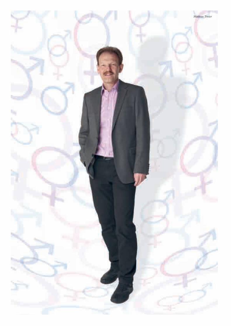
Mathias Treier

“The difference between the sexes is, happily, oneof great profundity.” So wrote the author
Virginia Woolf, in her famous novel Orlando: A biogra-phy. It told the tale of a man who lived for 400 years andawoke one day to find himself transformed into a woman.Most people would probably agree with the quote aboutsex differences and find the idea of one sex transforminginto another absurd. But as researchers at EMBLHeidelberg recently showed, transforming female cellsinto male cells may also be possible.
The first question anyone asks when given news of a birthis the sex, often so that they know what colour gifts tobuy: blue for a boy or pink for a girl. But biology is neveras simple as society might like it to be. Rather than beingdivided into two discrete camps – pink and blue – humansex can also be ill-defined, running from pink throughpurple to blue. Some people, for example, have some maleand some female traits, a condition known as intersex. Inextreme cases, individuals can carry the chromosomestypical of one sex, yet physically be of the other, a phe-nomenon known as sex reversal.
Now, Mathias Treier and his team have made a fascinat-ing discovery that confounds more of our preconceptionsabout sex. It seems that sexual identity might not be as setin stone as we once supposed. And the celebrateddifférence between male and female is in fact surprisinglyfragile – from a biochemical perspective at least. Theteam’s work has also uncovered other unexpected simi-larities between the way mammals and other vertebratesdetermine their sex, and this has raised new questionsabout how sex determination works and how it evolved.
How females fight offtheir inner male
About 15 years ago, the model of sex determination inmost mammals went something like this: the developinggonad initially has the potential to form either an ovary ora testis. The default development setting is female – in theabsence of any other instructions it will develop as anovary. This is what normally happens if an embryo is car-rying two X chromosomes. In embryos with an X and a Ychromosome, a gene located on the Y called Sry triggersthe male pathway of development resulting in a testis. Itdoes so by switching on the activity of another gene calledSox9, which in turn switches on genes that direct maledevelopment. Once the development of the ovary or thetestis is complete, there is no further need for these path-ways: once an ovary or testis, always an ovary or testis.
Hints began to emerge, however, that things are rathermore complicated than this. Some of these hints camefrom studies of people and animals whose chromosomalsex didn’t match their physical gender, such as XX males.These studies revealed the presence of genes that seemedto be actively repressing Sox9 – and thus testis develop-ment – in females, which raised the intriguing prospectthat female development wasn’t just the default pathway,although the role of these newly found genes still wasn’tclear.
About a decade ago, Mathias and his team first isolatedone of these genes, called Foxl2. The FOXL2 protein wasalready known to be important for ovary development –mutations in the gene in humans trigger a syndrome thatincludes premature menopause, and disturbances in theequivalent gene in goats causes XX kids to develop asmales. This suggested that FOXL2 is important for the
Upholding law and order 7373

cells in the ovary to develop their specialised functions – to‘differentiate’ – and that the protein might suppress parts ofthe male development programme.
To find out more, Mathias’s team inactivated FOXL2 infemale mice from the point of conception onwards, to seewhether this would cause the ovaries to develop more liketestes. The experiment worked, but the results were hard tointerpret: the ovaries began to develop normally, but thenwithered after birth, so the team could not see whether theyhad developed any features characteristic of testes. So theyturned to a newer technology that allows biologists to inacti-vate a gene at any point that they choose during the lifespanof an animal. “We were interested in what FOXL2 was actu-ally doing in the adult,” says Mathias. “And we got a surpris-ing result.”
When the team turned FOXL2 off in adult female mice foronly a few days, they found that the somatic cells – the cellsthat are not egg cells – of the ovaries had, effectively, under-gone a sex change. Instead of containing cells that would nor-mally nourish developing eggs, the ovaries now housed tube-like structures typical of testes. What’s more, these cells hadactivated genes typical of the Sertoli and Leydig cells of thetestis, cell types that aid sperm development. They even made
the male hormone testosterone. The few developing eggs thatremained in these ovaries were in the process of dying. “It wasvery clear cut that there was a full somatic sex transforma-tion,” says Mathias. Further investigation showed that thetransformation did indeed result from the loss of FOXL2activity in the ovarian cells themselves, and was not depen-dent on any interactions with the developing eggs in theovary.
This suggested something remarkable – that one kind of adulttissue was capable of switching completely to another kind,something that developmental biologists had long believedwas impossible, or at least very rare. This phenomenon, calledtransdifferentiation, had been documented in only a handfulof adult cell types in the body. “Our result may be one of lessthan ten examples,” says Mathias. For many years, this kindof flexibility, or ‘plasticity’ was believed to be the preserve ofimmature cells and stem cells. “I think we will see many moreexamples like this as more molecular switches like FOXL2 areidentified,” says Mathias.
The work could give a boost to researchers looking for alter-natives to human embryos as sources of stem cells to use astherapies. It lends weight to the idea that researchers coulddevelop drugs to make existing tissues switch to another
74 EMBL Annual Report 09 · 10
The presence or absence of SRY and SOX9defines the sex of mouse embryos but, forfemales, maintaining that sex in adulthooddepends on FOXL2 and oestrogen receptors(ESR1&2) countering the effects of SOX9.
SRY
SOX9
XY
XYXX
time
Sox9 off ovary Sox9 on testis
XX
FOXL2ESR1&2 Sox9

Indeed, perhaps the most salient outcome of the team’s find-ings is how they change biologists’ understanding of themechanism and evolution of sex determination. On the faceof it, vertebrates have a smorgasbord of sex determinationstrategies. Some reptiles use the temperature of the environ-ment, mammals use a method by which the sex with two dif-ferent sex chromosomes (XY) is male, whereas birds do theexact opposite: the female has a ZW chromosome set. Andthe duck-billed platypus has a bizarre system of ten sex chro-mosomes, which seem to have both mammal- and bird-likecharacteristics.
But the upshot of Mathias’s work is that all of these diversemethods may ultimately feed into a universal mechanism thathas at its heart FOXL2 and SOX9 opposing each other’saction. All that is needed is some kind of cue to tip this Yinand Yang-style balance in favour of one sex or the other.“What we see with SRY on the Y chromosome is anotherlayer of regulation,” says Mathias. “How you trigger theswitch – why should it matter?”
Mathias and his team now plan to look at the universal mech-anism in more detail to determine its components and howthey work. “We want to dissect what the core module is,” hesays. Whatever they find, it is likely to further question soci-ety’s simplistic notions of what it means to be a boy or a girl.Perhaps it is time we ditched our two-tone attitude andembraced the kaleidoscopic nature of life.
Uhlenhaut H, Jakob S, Anlag K, Eisenberger T, Sekido R, KlugmannC, Treier A-C, Kress J, Klasen C, Holter NI, Riethmacher D, SchützG, Cooney AJ, Lovell-Badge R, Treier M (2009) Somatic SexReprogramming of Adult Ovaries to Testes by FOXL2 Ablation. Cell139: 1130-1142
desired type. If cancer cells harbour similar kinds of flexibili-ty, perhaps they too could be coaxed into forming harmlesscell types. “It tells us that there may be alternative routes cir-cumventing the use of stem cells, which has huge implica-tions for regenerative medicine,” says Mathias.
The other surprise is that the ovary needs FOXL2 activitythroughout adulthood to repress Sox9 and prevent the devel-opment of male characteristics. Further experiments revealedthat FOXL2 is essential for the production of the female hor-mone oestrogen to keep the ovary in the pink. Mathias envis-ages a balance of power between female factors (oestrogenand FOXL2) and male factors (SOX9 activity) that dictatesthe sexual identity of the adult gonad. He hopes this modelwill give new insights into premature ovarian failure, a condi-tion that causes infertility and early menopause in youngwomen. But it also raises the intriguing possibility thatdeclining FOXL2 activity levels could underlie the normalonset of the menopause. Could it be that a woman’s ovariesshut down in later life because the balance shifts moretowards testis differentiation? This could explain some of themasculine traits, such as facial hair, often seen in olderwomen, says Mathias.
This might sound preposterous, but many ‘lower’ vertebratescan switch the sex of their gonads during their lifetimes. Theyoften do so in response to sex hormones such as oestrogen,and many are known to harbour both Sox9 and Foxl2 genes.And although mammals are not known to completely switchtheir gonadal sex under normal circumstances, Mathias’sexperiments show that it might be possible – when forced toat least. “It shows that the plasticity has been preserved inmammals and that switching somatic sex is still possible,”says Mathias.
Upholding law and order 75
Ovary of a normal adult female mouse, with close-up showing the typical female granulosa cells (left), which, when Foxl2 was silenced (right), took oncharacteristics of cells normally found in testes.

Parasitic worms can no longerrely on secrecy to steal vital
nutrients from their hosts, thanks towork by structural biologists at EMBLHamburg. The team, led by PaulTucker, has deduced the molecularstructure of a protein like the onessuch worms use to commandeer theirrations. With this blueprint revealed,researchers can now learn more aboutthe biology of these worms andexplore ways of starving them out byblocking the proteins with drugs.
The parasites in question are round-worms, or nematodes, and they are asource of immense human sufferingand economic loss worldwide.Researchers estimate that about a sixthof the world’s population is infectedwith nematodes, which cause severediseases such as river blindness andelephantiasis. For some, there is nocure. Worms also infect livestock andcrop plants, decimating agriculturalrevenue.
So finding a weak spot that could betargeted by better drugs or pesticidescould make a big difference, and thisis just what Paul and his team mayhave found. Parasitic nematodes can-not make certain molecules, calledfatty acids and retinoids, that are vitalfor their survival. Instead, they secreteproteins called fatty acid- andretinoid-binding proteins, or FARproteins, that grab these moleculesfrom the host’s gut or bloodstreamand deliver them back to the worm.
76 EMBL Annual Report 09 · 10
Fat chance for a cure
Paul first became interested in FARproteins when Rositsa Jordanova, aMarie-Curie Trainee, joined the lab tostudy them in Ascaris galli, a nema-tode that was devastating poultryfarming in her native Bulgaria. But shehit a big problem: these worms cannotbe grown in the lab and this made theexperiments she wanted to doextremely difficult. So the team turnedto another nematode calledCaenorhabditis elegans, which can begrown in the lab and which is com-monly used for biomedical research.
Although it is not a parasite, C. eleganshas several FAR proteins that seem tobe similar to those in parasitic species.“It made a great deal of sense to lookfor similar proteins in C. elegans,” saysPaul. It still wasn’t easy, though – thebiochemistry of the proteins makes ithard to get them to form the crystalsneeded for X-ray studies – andHamburg’s high-throughput crystalli-sation facility was key to the success ofthe project. “It was a long and hardprocess to get useful crystals,” saysPaul.
The team successfully deduced thestructure and found that the proteinhad a structural feature, or fold, thathad not been seen anywhere else. “It’sa unique structure,” says Paul. Thismakes it an ideal drug target: drugsthat block fatty-acid binding will beunlikely to affect other proteins and sothis minimises the risk of harmful sideeffects.
Further experiments showed wherethe protein was active in the wormand that its fold can bind a range offat-like molecules. Although useful forthe worm, molecules with fat-likeproperties aren’t normally thought ofas drugs because they cannot enterhuman cells. But in the case of anti-FAR drugs, this would be a goodthing. “You don’t want the drug toenter the host cell, just the nematode,”says Paul. So tackling FARs willrequire a new approach based onstate-of-the-art methods, says Paul.“This highlights the importance ofchemical biology,” he concludes.
Jordanova R, Groves MR, Kostova E,Woltersdorf C, Liebau E, Tucker PA(2009) Fatty Acid- and Retinoid-bindingProteins Have Distinct Binding Pockets forthe Two Types of Cargo. J Biol Chem 284:35818 - 35826
C. elegans worm with a FAR protein tagged fluorescent green.
Paul Tucker

In 1863 a Heidelberg doctordescribed a devastating neurode-
generative condition that causes chil-dren to forget how to walk and talkbefore their teens. The symptomsbegin with muscle weakness, poor bal-ance and a slurring of speech, anddevelop into a gradual breakdown inall motor control. Mysteriously, physi-cians sometimes noticed similar,although milder, symptoms in the par-ents of afflicted children, and evenmilder effects in the grandparents,suggesting that the disease mightresult from a genetic defect that growsmore and more severe each genera-tion. Only in the 1990s did geneticistsdiscover long tracks of the amino acidglutamine within certain proteins ofchildren suffering from these strangeneurological symptoms. Substantiallylengthening these glutamine trackscaused the proteins to clump togetherin the nuclei of cells – usually nervecells – leading to the cell’s demise. Atleast nine inherited diseases, whichcan affect both adults and childrenand include Huntington’s disease, areknown to result from these repeatmutations.
But these glutamine repeats, it seems,are not solely to blame for the deterio-rating nerve control. For clues to othercontributing factors to the develop-ment of these diseases, scientists aretrying to identify the roles of theaffected proteins.
One such protein, which causes a rarebut devastating neurological conditionknown as spinocerebellar ataxia type1, is the focus of chemist AnnalisaPastore, a former EMBL group leaderwho now works at the NationalInstitute for Medical Research inLondon. Annalisa hopes to gain cluesabout the function of this protein –called Ataxin-1 – by investigating itsstructural form to predict with which
Upholding law and order 77
A nervous switch Toby Gibson
molecules it interacts. However,Ataxin-1, unlike most proteins, doesnot fold into three-dimensionalshapes, but instead remains floppy andmostly unfolded. Thus, the structuralmodels most researchers usually use topredict protein–protein interactionswere of no use for this protein.
So Annalisa teamed up with computa-tional biologist Toby Gibson fromEMBL Heidelberg, who has developeda computer program to search forinteraction sites on the non-foldedregions of proteins. The researchersapplied this bioinformatic resource,called the Eukaryotic Linear MotifResource, to compare the amino-acidsequence of Ataxin-1 across manyspecies, ranging from mosquitoes tozebrafish to humans. In this way, theysought to predict which short regions,or motifs, of Ataxin-1’s amino-acidsequence might interact with otherproteins.
“Annalisa and I were sitting togetherwhen my database threw up a promis-ing candidate motif that suggested thatAtaxin-1 might interact with a protein,called U2AF65, that regulates alterna-tive splicing – a means by which a sin-gle gene can produce many variants,”says Toby. “But what got Annalisa
even more excited was that this motifoverlapped with an existing motif thatwas already known to interact withtwo other proteins,” says Toby.
In the lab, Annalisa and her groupconfirmed Ataxin-1’s role in alterna-tive splicing – an important find forscientists examining proteins involvedin these neurological diseases for cluesabout why they are the targets of glu-tamine repeats. Annalisa also verifiedthat she and Toby had spotted in thedatabase a three-way molecular switch,which involves three proteins bindingat one interaction site. “What is key inthis case is that Ataxin-1 can only bindone of its interacting proteins at atime,” Toby explains.
Toby’s group is keen to find moremotifs that could be important for thedevelopment of many other diseases.“We suspect that there are over a mil-lion of these regulatory motifs inhuman proteins, and you could saythat it is my team’s holy grail to learnhow to find them all!” Toby says.
de Chiara C, Menon R, Strom M, GibsonT, Pastore A (2009) Phosphorylation ofS776 and 14-3-3 Binding Modulate Ataxin-1 Interaction with Splicing Factors. PLoSONE: 4

78 EMBL Annual Report 09 · 10
Our genes were once thought to be responsible forshaping who we are. But now scientists are having a
rethink. Thanks to a glut of data from new sequencing pro-jects, researchers are beginning to recognise that the regionsof the human genome that encode proteins are unlikely to bebehind the millions of differences between people. So thequestion remains: what accounts for these differences?
Searching for an answer, biologists have pored over the fewindividual genome sequences that have been completed sofar. And these researchers have asked: if the rare stretches ofDNA that code for proteins are not responsible for many ofthe differences found between humans, then what about theremaining 98% of the genome that does not encode proteins –the so-called non-coding DNA?
Small regions of non-coding DNA are known to serve asdocking sites for regulatory proteins called transcription fac-tors, which are responsible for cueing when and where genesare turned on and off. These tiny transcription factor-bindingsites are usually found just upstream of genes – although theycan occur anywhere within the genome – and they recruitother proteins that are essential for transforming genes intotheir respective proteins.
Scientists now suspect that variation in the efficiency of tran-scription factor binding, and the effect of this binding on howgenes are turned on, could be responsible for many of the lit-tle quirks that make each of us unique. Jan Korbel, a genomebiologist from EMBL Heidelberg, is particularly interested invariation among people. So he teamed up with StanfordUniversity geneticist Michael Snyder to investigate the effectof differences in transcription factor binding on the variationin gene expression in 10 humans – five Europeans, threeAfricans, and two East Asians – by looking at their completegenome sequences.
By chemically gluing transcription factors to their DNA-binding sites, the researchers identified the binding patterns
Cue factors
of these regulatory proteins and looked to see whether theybound every site equally strongly in all 10 people. The pro-teins they studied were NFkB, which is involved in theimmune response, and Pol-II, a protein that helps convertDNA to RNA. For NFkB, less than 10% of the 15 000 bind-ing sites varied between people. Yet for Pol-II, about a quar-ter of its 19 000 binding sites were altered across individuals– considerably more variation than is seen in coding regions,which vary, on average, by only a fraction of a percent amongpeople.
Jan’s group was able to trace some of this variation back toeither small differences or larger rearrangements in the DNAcomprising these binding sites. Still, the majority of bindingsites showed no difference in their DNA sequences, despitetheir variable binding affinity among people. In these cases,Jan explains, the researchers frequently found variations innearby regions that could explain the differences. “This is avery interesting finding, as it suggests that these regulatoryproteins are not working alone, but instead co-operatingwith nearby transcription factors to regulate how genes areexpressed,” he says. In fact, Jan’s team has just developed anew test for assessing the extent of this transcription factorco-operation.
Jan and his team went on to show that the variation at thesebinding sites had a profound impact on an individual’s geneexpression. “Many genes’ expression differed by greater thantwo orders of magnitude between people as a result of varia-tion in transcription factor binding,” says Jan.
“What makes this study novel is that it determined how dif-ferences in gene expression among people are associatedwith the genome-wide variation in transcription factor bind-ing, and also with sequence variation in the factor’s bindingsites,” remarks Jan. “Our findings suggest that variation innon-coding DNA may be responsible for many of the differ-ences we see between people.”

Upholding law and order 79
What’s more, these findings also offer an explanation for thedifferences between humans and their closest cousins. WhenJan’s team looked in both humans and chimpanzees, theyfound that nearly a third of the Pol-II-binding sites differedbetween the two species. Jan explains that there seems to bealmost as much variation among humans as between humansand chimpanzees.
The researchers’ results were buoyed by a second study inwhich genome biologist Lars Steinmetz of EMBL Heidelberg,also in collaboration with the Snyder group, used yeast tomap genome regions bound by a transcription factor calledSte12 that is important in yeast mating.
Lars and his team compared binding sites between two strainsof the common baker’s yeast Saccharomyces cerevisiae, one ofwhich descended from cells collected from a rotten fig inMerced, California, and the other derived from cells isolatedfrom the lung of a person suffering from AIDS-relatedimmune problems in San Francisco.
The yeast’s small genome and short generation time offeredLars and his group the opportunity to fine-map these sitesacross many more individuals than had previously been pos-sible using human cells. This gave the researchers the power todetect very small differences in transcription factor binding.Owing to the sensitivity of this approach, Lars’ group foundeven higher rates of variation in Ste12 transcription factorbinding between the yeasts than was seen in the human study,and because of yeast’s frequent genome shuffling, they were ableto ascribe this variation to very narrow regions of the genome.
“Our results, and Jan’s study involving humans cell lines, sug-gest that the bulk of the differences among individuals are notfound in the genes themselves, but in the non-coding por-tions of the genome and in particular in the regulatoryregions, which we know relatively little about,” says Lars.
When mapping the DNA responsible for the variation intranscription factor binding, Lars and his team found that
almost 90% of the binding differences are influenced bysequence variation in nearby regions. This offers tantalisingevidence that variation in the regulation of genes by tran-scription factors is often controlled by sequence differencesclose to the genes. However, in several cases the data revealedvariations far from the genes that also influence gene expres-sion. These regions can encode the transcription factorsthemselves or signalling molecules that influence the efficien-cy of a certain transcription factor’s binding ability.
Knowing whether genes are regulated by nearby regions orregions on entirely different chromosomes will determinehow scientists study variation in gene regulation. If geneexpression variation is indeed often controlled locally – by socalled cis-regulatory elements – then researchers are safe tocontinue to take a reductionist approach and look to ascribethe differences between individuals to the regulatory regionsaround genes. However, if variation in gene regulation turnsout to be under the control of regions scattered far and widethroughout the genome – often termed trans-regulatory vari-ation – then researchers need to change tack and study thewhole genome simultaneously. Such a broad approachrequires the skills of system biologists who specialise in study-ing entire, intact biological systems.
Either way, once scientists have mapped these regulatoryregions, they can look at how individual variations in generegulation affect how we look, how we think, whether we aresusceptible to certain diseases and how we will react to spe-cific treatments. This will open doors to personalisedmedicine, which seeks to make use of an individual’s differ-ences to provide better medical care.
Kasowski M et al (2010) Variation in transcription factor bindingamong humans. Science 328: 232-235
Zheng W, Zhao H, Mancera E, Steinmetz L, Snyder M (2010)Genetic analysis of variation in transcription factor binding inyeast. Nature 464: 1187-1191
Lars Steinmetz and Jan Korbel

Having a second pair of handsmight seem like an advantage
but animals born with extra limbs,because of changes in their DNA, gen-erally do not fair well. For more than25 years, scientists have known aboutthe existence of a mutation in a fruitfly gene that causes just such aberrantappendages, yet the identity of thisgene remained a mystery.
That is until developmental biologistJürg Müller and his team at EMBLHeidelberg set out to find the generesponsible. By comparing the DNAof mutant and normal flies, Jürg’sgroup pinpointed the mutation andfound that it disrupts the genetic codefor the protein Ogt, an enzyme thatsticks sugar molecules to the outsideof proteins.
“Ogt is atypical because, unlike otherenzymes that add sugars to proteinsthat eventually reside on the cell’s sur-face or are secreted by the cell, Ogtadds a sugar to proteins in the cell’snucleus and cytoplasm,” says Jürg.
“The sugar modification added by Ogthas been found on hundreds of otherproteins, and so it was a little unex-
80 EMBL Annual Report 09 · 10
Aberrant appendages
pected that removing this one proteinwould cause such profound develop-mental defects,” explains MariaCristina Gambetta, the PhD studentwho carried out this work in Jürg’slab. For this reason, Ogt was the lastgene on a shortlist of 11 candidategenes that the group examined.
Because flies lacking Ogt show dra-matic changes in their head-to-tailbody patterning, the researchers sus-pected that the Ogt protein adds asugar to a family of proteins that regu-late the genes required for normal patterning. These proteins – calledPolycomb proteins – do this byremodelling the DNA into compactstructures called chromatin. By sittingon the chromatin, the proteins con-dense the DNA, making it inaccessibleto the cellular machinery that wouldtransform the genetic code into activeproteins.
To confirm Ogt’s role in adding asugar to the Polycomb proteins, thenext step for Jürg’s group was to lookwhether the sugar and Polycomb pro-teins typically sit together on the chro-matin. By tagging the proteins andsugars, they found that they were
bound together in regions that areimportant for controlling whethergenes are turned on or off. These find-ings hinted that the sugar was some-how needed for Polycomb proteins tosilence the genes involved in buildingthe fruit fly’s body plan.
Jürg’s team went on to show that thesugar was attached to just one mem-ber of the Polycomb protein family – aprotein called Ph. ”We don’t knowwhy adding a sugar to Ph is importantfor its function, but this is what wehope to investigate next,” says Jürg.“But what we have found so far is anovel and unexpected role for thesugar added by Ogt; the fact that fruitflies that lack this sugar show compro-mised Polycomb silencing but noother obvious defects is remarkable.”Ogt is likely to fulfil a similar role inhumans, and so it could determine thepositioning and number of our ownarms, legs, ribs and, well, everything.
Gambetta MC, Oktaba K, Müller J (2009)Essential Role of the GlycosyltransferaseSxc/Ogt in Polycomb Repression. Science325: 93 - 96
Katarzyna Oktaba Sosin,Maria-Cristina Gambetta and
Jürg Müller
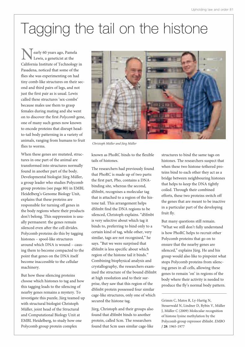
Nearly 60 years ago, PamelaLewis, a geneticist at the
California Institute of Technology inPasadena, noticed that some of theflies she was experimenting on hadtiny comb-like structures on their sec-ond and third pairs of legs, and notjust the first pair as is usual. Lewiscalled these structures ‘sex-combs‘because males use them to graspfemales during mating and she wenton to discover the first Polycomb gene,one of many such genes now knownto encode proteins that disrupt head-to-tail body patterning in a variety ofanimals, ranging from humans to fruitflies to worms.
When these genes are mutated, struc-tures in one part of the animal aretransformed into structures normallyfound in another part of the body.Developmental biologist Jürg Müller, a group leader who studies Polycombgroup proteins (see page 80) in EMBLHeidelberg’s Genome Biology Unit,explains that these proteins areresponsible for turning off genes inthe body regions where their productsdon’t belong. This suppression is usu-ally permanent: the genes remainsilenced even after the cell divides.Polycomb proteins do this by tagginghistones – spool-like structuresaround which DNA is wound – caus-ing them to become compacted to thepoint that genes on the DNA itselfbecome inaccessible to the cellularmachinery.
But how these silencing proteinschoose which histones to tag and howthis tagging leads to the silencing ofnearby genes remains a mystery. Toinvestigate this puzzle, Jürg teamed upwith structural biologist ChristophMüller, joint head of the Structuraland Computational Biology Unit atEMBL Heidelberg, to study how onePolycomb group protein complex
Upholding law and order 81
Tagging the tail on the histone
known as PhoRC binds to the flexibletails of histones.
The researchers had previously foundthat PhoRC is made up of two parts:the first part, Pho, contains a DNA-binding site, whereas the second,dSfmbt, recognises a molecular tagthat is attached to a region of the his-tone tail. This arrangement helpsdSfmbt find the DNA regions to besilenced, Christoph explains. “dSfmbtis very selective about which tag itbinds to, preferring to bind only to acertain kind of tag, while other, verysimilar, tags are not recognised,” hesays. “But we were surprised thatdSfmbt is less specific about whichregion of the histone tail it binds.”Combining biophysical analysis andcrystallography, the researchers exam-ined the structure of the bound dSfmbtat high resolution and to their sur-prise, they saw that this region of thedSfmbt protein possessed four similarcage-like structures, only one of whichsecured the histone tag.
Jürg, Christoph and their groups alsofound that dSfmbt binds to anotherprotein, called Scm. The researchersfound that Scm uses similar cage-like
structures to bind the same tags onhistones. The researchers suspect thatwhen these two histone-tethered pro-teins bind to each other they act as abridge between neighbouring histonesthat helps to keep the DNA tightlycoiled. Through their combinedefforts, these two proteins switch offthe genes that are meant to be inactivein a particular part of the developingfruit fly.
But many questions still remain.“What we still don’t fully understandis how PhoRC helps to recruit otherPolycomb proteins that go on toensure that the nearby genes aresilenced,” explains Jürg. He and hisgroup would also like to pinpoint whatstops Polycomb proteins from silenc-ing genes in all cells, allowing thesegenes to remain ‘on’ in regions of thebody where their activity is needed toproduce the fly’s normal body pattern.
Grimm C, Matos R, Ly-Hartig N,Steuerwald N, Lindner D, Rybin V, MüllerJ, Müller C (2009) Molecular recognitionof histone lysine methylation by thePolycomb group repressor dSfmbt. EMBOJ 28: 1965-1977
Christoph Müller and Jürg Müller

Christian Conrad, Beate Neumann, Jan Ellenberg, Jutta Bulkescher, Thomas Walter, Jean-Karim Hériché

Movies for thehuman genome
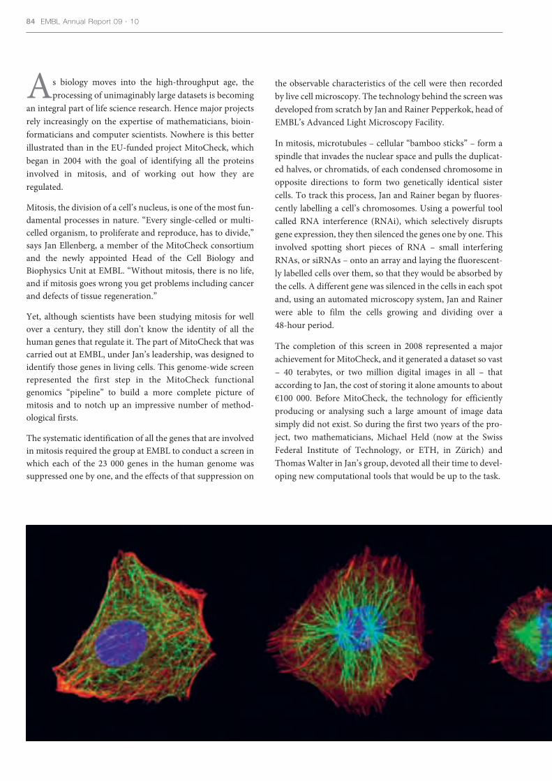
As biology moves into the high-throughput age, theprocessing of unimaginably large datasets is becoming
an integral part of life science research. Hence major projectsrely increasingly on the expertise of mathematicians, bioin-formaticians and computer scientists. Nowhere is this betterillustrated than in the EU-funded project MitoCheck, whichbegan in 2004 with the goal of identifying all the proteinsinvolved in mitosis, and of working out how they are regulated.
Mitosis, the division of a cell’s nucleus, is one of the most fun-damental processes in nature. “Every single-celled or multi-celled organism, to proliferate and reproduce, has to divide,”says Jan Ellenberg, a member of the MitoCheck consortiumand the newly appointed Head of the Cell Biology andBiophysics Unit at EMBL. “Without mitosis, there is no life,and if mitosis goes wrong you get problems including cancerand defects of tissue regeneration.”
Yet, although scientists have been studying mitosis for wellover a century, they still don’t know the identity of all thehuman genes that regulate it. The part of MitoCheck that wascarried out at EMBL, under Jan’s leadership, was designed toidentify those genes in living cells. This genome-wide screenrepresented the first step in the MitoCheck functionalgenomics “pipeline” to build a more complete picture ofmitosis and to notch up an impressive number of method-ological firsts.
The systematic identification of all the genes that are involvedin mitosis required the group at EMBL to conduct a screen inwhich each of the 23 000 genes in the human genome wassuppressed one by one, and the effects of that suppression on
the observable characteristics of the cell were then recordedby live cell microscopy. The technology behind the screen wasdeveloped from scratch by Jan and Rainer Pepperkok, head ofEMBL’s Advanced Light Microscopy Facility.
In mitosis, microtubules – cellular “bamboo sticks” – form aspindle that invades the nuclear space and pulls the duplicat-ed halves, or chromatids, of each condensed chromosome inopposite directions to form two genetically identical sistercells. To track this process, Jan and Rainer began by fluores-cently labelling a cell’s chromosomes. Using a powerful toolcalled RNA interference (RNAi), which selectively disruptsgene expression, they then silenced the genes one by one. Thisinvolved spotting short pieces of RNA – small interferingRNAs, or siRNAs – onto an array and laying the fluorescent-ly labelled cells over them, so that they would be absorbed bythe cells. A different gene was silenced in the cells in each spotand, using an automated microscopy system, Jan and Rainerwere able to film the cells growing and dividing over a 48-hour period.
The completion of this screen in 2008 represented a majorachievement for MitoCheck, and it generated a dataset so vast– 40 terabytes, or two million digital images in all – thataccording to Jan, the cost of storing it alone amounts to about€100 000. Before MitoCheck, the technology for efficientlyproducing or analysing such a large amount of image datasimply did not exist. So during the first two years of the pro-ject, two mathematicians, Michael Held (now at the SwissFederal Institute of Technology, or ETH, in Zürich) andThomas Walter in Jan’s group, devoted all their time to devel-oping new computational tools that would be up to the task.
84 EMBL Annual Report 09 · 10

These tools included pattern recognition algorithms thatallow the automatic measurement of the biological conse-quences of gene silencing on cell division, or more specifical-ly on the morphology of chromosomes. On the basis of thisquantitative cell division signature extracted from themicroscopy movies, the genes could be grouped into clustersaccording to the similarity of their effects.
Having identified the genes involved in mitosis, theMitoCheck consortium was presented with a bioinformaticchallenge: whereas it was interested in the systems view ofmitosis – that is, in all the proteins encoded by the genes Jan’sgroup had identified, and their complex interactions –databases tend to be structured to store information aboutsingle biological entities, such as a gene or protein sequence.
“We now have sophisticated database systems which are ableto give us a wealth of information, but they are not very welldesigned to give us the bigger picture,” says EMBL bioinfor-matician Reinhard Schneider who, with group memberVenkata Satagopam and others, came up with new data-min-ing tools fit for probing the functions of these proteins. Thesetools have now been fed into the www.mitocheck.orgdatabase, which disseminates all the consortium’s data. Thedatabase was set up by Jean-Karim Hériché, who worked pre-viously with MitoCheck member Richard Durbin at theSanger Institute in Cambridge, UK, and who is now part ofJan’s group at EMBL.
Given the problems their dataset presented, why did Jan andRainer choose two days as an appropriate running time fortheir cellular movies? “There is always a trade-off in high-throughput experiments,” Jan says. They had to weigh up
technical, biological and economic considerations. The cellsin question – human cancer cells in culture – divide aboutonce a day, so two days gave them data on two full divisioncycles. A day less, and they may have missed some effects ofgene suppression that only kicked in in the second cycle. Aday more, and they would have been dealing with a datasethalf as big again, with all the extra processing time and costthat would incur.
In many cases, Jan’s screen implicated novel genes, aboutwhich little was known, in important aspects of mitosis, suchas the assembly of the spindle. In other cases, the gene clus-ters revealed a role, either direct or indirect, for whole cellu-lar processes that had not previously been linked with mito-sis, such as RNA splicing – the modification of RNA thattakes place once DNA has been transcribed but before it canbe translated into a protein. “We have some very intriguingresults that suggest that there is a specific splicing regulationof mitotic proteins that control how they work in differentphases of the cell cycle,” says Jan.
They found that around 5% (approximately 1200) of humangenes are involved in mitosis and for about half of these theycould confirm this function in a second validation screenwith independent silencing reagents. Moreover, the cellmovies identified gene effects on many other cellular func-tions such as migration and survival that, when combinedwith what they were learning about mitotic genes, allowed theEMBL researchers to annotate 13% of the human genomewith a potential cellular function. As Jan emphasises, howev-er, the time-lapse movies, although highly informative, showonly indirectly that a gene is required for a particular cellular
Upholding law and order 85
Normal cell division, with DNA labelled blue, microtubules green and actin filaments red.
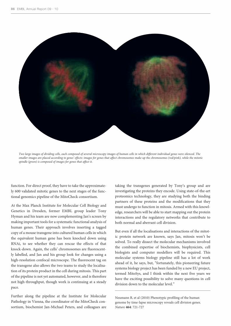
function. For direct proof, they have to take the approximate-ly 600 validated mitotic genes to the next stages of the func-tional genomics pipeline of the MitoCheck consortium.
At the Max Planck Institute for Molecular Cell Biology andGenetics in Dresden, former EMBL group leader TonyHyman and his team are now complementing Jan’s screen bymaking important tools for a systematic functional analysis ofhuman genes. Their approach involves inserting a taggedcopy of a mouse transgene into cultured human cells in whichthe equivalent human gene has been knocked down usingRNAi, to see whether they can rescue the effects of thatknock-down. Again, the cells’ chromosomes are fluorescent-ly labelled, and Jan and his group look for changes using ahigh-resolution confocal microscope. The fluorescent tag onthe transgene also allows the two teams to study the localisa-tion of its protein product in the cell during mitosis. This partof the pipeline is not yet automated, however, and is thereforenot high-throughput, though work is continuing at a steadypace.
Further along the pipeline at the Institute for MolecularPathology in Vienna, the coordinator of the MitoCheck con-sortium, biochemist Jan-Michael Peters, and colleagues are
taking the transgenes generated by Tony’s group and areinvestigating the proteins they encode. Using state-of-the-artproteomics technology, they are studying both the bindingpartners of these proteins and the modifications that theymust undergo to function in mitosis. Armed with this knowl-edge, researchers will be able to start mapping out the proteininteractions and the regulatory networks that contribute toboth normal and aberrant cell division.
But even if all the localisations and interactions of the mitot-ic protein network are known, says Jan, mitosis won’t besolved. To really dissect the molecular mechanisms involvedthe combined expertise of biochemists, biophysicists, cellbiologists and computer modellers will be required. Thismolecular systems biology pipeline still has a lot of workahead of it, he says, but, “fortunately, this pioneering futuresystems biology project has been funded by a new EU project,termed MitoSys, and I think within the next five years wehave the exciting possibility to solve many questions in celldivision down to the molecular level.”
Neumann B, et al (2010) Phenotypic profiling of the humangenome by time-lapse microscopy reveals cell division genes.Nature 464: 721-727
86 EMBL Annual Report 09 · 10
Two large images of dividing cells, each composed of several microscopy images of human cells in which different individual genes were silenced. Thesmaller images are placed according to genes’ effects: images for genes that affect chromosomes make up the chromosomes (red/pink), while the mitoticspindle (green) is composed of images for genes that affect it.

tells researchers very little about theones that are correctly identified andcan miss those transcription factorsthat don’t contain the commonsequences.
So Nick and his postdoc JuanmaVaquerizas studied each of the possi-ble transcription factors suggested bythe automated searches in more detail.They used additional informationfrom the EBI’s Ensembl and InterProdatabases to look for more evidencethat the candidates did indeed func-tion as transcription factors, as well asto look for candidates that the auto-mated searches had missed.
The team found a total of 1391 con-firmed transcription factors. “Themost striking thing is that most ofthem haven’t been identified in termsof what they do,” says Nick.
To find out more, the team searchedanother database to see in which tis-sues the genes were active. They weresurprised to find that the transcriptionfactors were largely divided into twocamps: those that were active every-where and those that were active injust one or two tissues.
Nick’s team is now working withEwan Birney and other EBI colleaguesto use this data for ENCODE, a pro-ject that aims to determine the func-tion of every gene in the humangenome.
The researchers also compared DNAsequences between different species tostudy the evolution of transcriptionfactors. They found sudden bursts inthe numbers and diversity of tran-scription factors whenever livingthings became significantly more complex, for example when single cellsclubbed together to form multicellularanimals. Understanding more aboutthe history and biology of these pro-teins should yield fascinating newinsights into how evolution itselfworks.
Nick’s group now plans to investigatehow different combinations of tran-scription factors work together todefine different cell types. One day, itmight even be possible to treat diseaseby taking the control of a cell’s careerdecisions into our own hands.
Vaquerizas J, Kummerfeld S, TeichmannS, Luscombe N (2009). A census of humantranscription factors: function, expressionand Evolution. Nature Reviews Genetics 10,253
If you had a hard time decidingwhat you wanted to be when you
grew up, spare a thought for the cellsin your body. If they make the wrongcareer decision – if a brain cell, say,accidentally turns into a muscle cell –disaster can ensue. Nick Luscombeand his team at the EBI have nowcompleted a census of the proteinsthat make these decisions, and in sodoing have created a valuable resourcefor research into human health anddisease.
One cell type differs from anotherbecause different sets of genes areactive in each. But every one of yourcells contains all the genes in thehuman genome, and so needs to betold which genes to switch on or off sothat the correct sets are active. Thisjob falls to proteins called transcrip-tion factors, which bind to DNA tocontrol gene activity. Until now, no-one had a clear picture of how manytranscription factors existed inhumans, or in which cell types theyeach worked.
Previously, researchers had producedrough head counts of these proteinsusing computer programs that auto-matically search the human genomefor DNA sequences commonly foundin genes that code for transcriptionfactors. But this approach results in alot of genes being wrongly labelled,
Upholding law and order 87
When I grow up…

Claus Nerlov

When we’re in the bath, our skin prevents bothwater from moving into our bodies and essen-
tial nutrients from leaching into the tub. But becausemost of us don’t spend our entire lives submerged under-water, our skin’s chief role is to control how much waterevaporates from our bodies. In fact, the skin’s role as asemi-impermeable barrier to fluid loss is so importantthat people suffering from serious burns often die, not as a direct consequence of their injuries, but from de hy-dration.
Each of us is covered by about 2 square meters of skin –about the area of a queen-size bed. For this waterproofsuit to do its job, stem cells at the base of the skin replen-ish the layers above by producing a continuous stream ofnew cells initially like themselves and then a variety ofspecialised cell types. As a result of this continuous pro-duction, the specialised cells – which are destined tobecome the different layers of skin – move outwards untilthey are finally shed.
The reason why stem cells stop producing more of them-selves and start producing other types of cells had flum-moxed researchers ever since the existence of skin stemcells was uncovered in the 1970s. So cell biologist ClausNerlov and his colleagues at EMBL Monterotondo setabout identifying the proteins responsible for this switch.As the processes of cell proliferation and differentiationare so tightly coupled, the researchers suspected that asingle protein acted as the toggle between them. From thestart, Claus’ team looked to a group of proteins called
Waxing cutaneous
C/EBPs, because they are known to regulate this shift inother parts of the body.
To examine whether C/EBPs form the potential stem cellswitch in skin, Claus and his group, in collaboration withcolleagues at the Centro de Investigaciones Energéticas,Medioambientales y Tecnologicas (CIEMAT) in Madrid,looked at how skin forms in mice in their absence.Eliminating C/EBPs from all cells prevented mouseembryo development at an early stage – too early for theresearchers to observe the proteins’ role in skin forma-tion. And so Rodolphe Lopez, a postdoc working inClaus’ lab, created a mouse strain in which these proteinscould be deactivated in skin cell tissue while continuingto function in all other tissues. This allowed the mouseembryo to develop more or less normally.
The researchers found that mice without C/EBPs hadtaut, shiny skin that didn’t act as a barrier to water, andthat they died of dehydration shortly after birth. Whenthey looked more closely at the skin of these young mice,they saw that the cells at the base of the skin were not dif-ferentiating into mature skin cells; instead, they remainedin their immature state and continued to multiply.
“We saw what was happening at the cellular level, but wedidn’t know what was happening at the molecular level,”explains Claus. To investigate, he and his team intro-duced mutations into different stretches of fully workingcopies of the genes that encode the C/EBP proteins. Theyfound that some mutations prevented the C/EBPs fromstifling the activity of proteins that halt cell proliferation,whereas other mutations blocked C/EBPs from binding
Upholding law and order 8989

to DNA and activating genes that signal to the cells to startdifferentiating. “Essentially, we found that C/EBPs are doingtwo things – stopping proliferation and starting differentia-tion,” says Claus. “C/EBPs tie together these two processes bydoing both of them simultaneously, which makes a lot ofsense when you consider how closely coupled these processesare in the cell.”
In addition to confirming their suspicions, this experimenthinted at something more unexpected, admits Claus. In adultmice with no working copies of the C/EBP proteins, his teamdiscovered a slew of genes that are usually expressed only inembryonic stem cells but were still active in the fully matureskin. These same genes are expressed in highly aggressive skincancers, suggesting that C/EBPs are actively suppressinggenes usually associated with malignancy – an important findfor anyone interested in understanding how epithelial can-cers like skin, breast, and oral cancers develop.
But the role of C/EBPs in cancer goes more than skin deep.Since 2001, Claus has been studying a form of blood cancercalled acute myeloid leukaemia, and his pursuit has taken himdeep within our bones. He explains that in the bone marrowof a healthy individual, blood stem cells churn out more cellslike themselves and generate others that are destined tobecome the medley of cells that form our blood – for exam-ple, red blood cells, white blood cells and platelets.
Such cellular differentiation is hierarchical, with blood stemcells at the top giving rise to immature progenitor cells, whichthen become more and more differentiated to form manytypes of specialist cell. But when a person develops leukaemia,there is an explosion of one type of immature progenitor cellthat overtakes the production of red blood cells in the bonemarrow and eventually results in anaemia and death. “Fiftyyears ago, before chemotherapy, this type of leukaemia killedyou within weeks,” says Claus.
When doctors looked at the DNA of patients suffering fromthis cancer they found that 90% of the tumours harbour twotypes of mutations in the C/EBP gene. Claus wondered howthese mutations affect the functioning of the resulting C/EBPprotein so Oxana Bereshchenko, a postdoc in Claus’ lab, cre-ated mice with each mutation individually, and then anotherstrain with both mutations. From these mice they extractedblood, which they injected into the bone marrow of a healthymouse, and watched to see how the cancers developed.
They saw that mice with both mutations became sick anddied more rapidly than the mice with only one. They there-fore concluded that these mutations have complementaryroles: one of them allows the expansion of the malignant pop-ulation of stem cells, whereas the other helps the stem cells todifferentiate into the progenitors, which ultimately grow tosuch high densities that they kill the mouse.
“This has important implications for the treatment of thesecancers,” Claus says. If clinicians use drugs to kill the popula-tion of progenitors – which seem to be the problem when apatient’s blood is examined – then they are aiming at thewrong target, he explains. This is because the mutations areoccurring in the stem cells that seed the cancer and not theprogenitor cells that maintain it, and so even if the progeni-tors are eliminated, the stem cells will just give rise to more.
Lopez R, Garcia-Silva S, Moore S, Bereshchenko O, Martinez-CruzA, Ermakova O, Kurz E, Paramio J & Nerlov C (2009) C/EBPα andβ couple interfollicular keratinocyte proliferation arrest to commit-ment and terminal differentiation. Nat Cell Biol 11: 1181-1190
Bereshchenko O, Mancini E, Moore S, Bilbao D, Mansson R, Luc S,Grover A, Jacobsen S, Bryder D & Nerlov C (2009) HematopoieticStem Cell Expansion Precedes the Generation of CommittedMyeloid Leukemia-Initiating Cells in C/EBPα Mutant AML.Cancer Cell 16: 390-400
90 EMBL Annual Report 09 · 10
In normal skin (left), the stem cells at thebase (green) differentiate into skin cells (red).In mice whose skin has neither C/EBPα norC/EBPβ (middle), stem cells appear in upperlayers of skin, and there are no differentiatedskin cells. In skin where C/EBPα is presentbut has lost its capacity to interact with E2F(right), skin cells start differentiatingabnormally, before they have properly exitedthe stem cell programme (yellow/orange).

Upholding law and order 91
The bacterium Listeria infectshumans through contaminated
food. Once in the gut, this pathogencan be life-threatening if contractedduring pregnancy or by newborns andthose with weakened immune systems.But for most people, an encounterwith Listeria causes nothing morethan vomiting and diarrhoea becauseour immune system recognises thelong, propeller-like projections on thebacterial surface – called flagella – andmounts an assault on Listeria until it iswiped out. Listeria, however, hasevolved a way to dodge this fate.
To anyone who has ever tried to crossenemy lines, this bacterium has anenviable ruse. After detecting thewarmth of the human body, Listeriashuts down the production of flagella– the equivalent of enveloping itself inan invisible cloak. It does this by acti-vating a protein called motility generepressor, or MogR for short, whichbinds to DNA close to the flagellagene and suppresses it.
While thinking about how to ridhumans of this irksome and some-times serious threat, researchersAimee Shen and Darren Higgins atHarvard Medical School in Boston,USA had the idea of stopping MogRfrom doing its job. This strategywould allow them to de-cloak the bac-teria, leaving it exposed to the wrathof the immune system. But to do this,they needed to understand how MogRrecognises its binding site.
The scientists knew that the MogRprotein regulates about 25 genes in the bacteria’s genome. These bindingsites are very rich in As and Ts, two ofthe four nucleotide ‘letters’ that makeup the DNA alphabet, and are palin-dromic – that is, they read the sameforwards as they do backwards.Knowing this, the researchers lookedfor where this particular motifoccurred in the bacteria’s DNA;
Decloaking the germto their dismay, they found more than500 matches. This suggested that theDNA sequence of this regulatory motifwas not sufficient to explain MogR’sbinding pattern. To understand howMogR selects for specific binding sites,the researchers turned to structuralbiologist Daniel Panne at EMBLGrenoble.
Daniel’s lab was able to determine thecrystal structure of the MogR proteinbound to its DNA-docking site. Fromthis, he saw that MogR recognisesboth the DNA’s sequence and also itsoverall warped shape, which is createdby the particular chemical propertiesof the AT-rich region that cause theDNA to bend to a 52-degree angle.This provided the first example ofDNA shape-dependent recognition.
But this was not the whole picture:Daniel also noticed that MogR bind-ing relied on specific electrostaticforces – similar to the static forcesexperienced by clothes just out of the
dryer – that are generated by the different charges of the chemical bases that make up the docking site.Importantly, only the 25 MogR-regu-lated genes boast docking sites thatrely on DNA sequence, shape andelectrostatic forces for binding.
Scientists have since found moreexamples of proteins that use similarmodes of DNA recognition to MogR.Daniel explains that these findings notonly provide insight into a new modeof DNA recognition that is seeminglymore common than suspected, butcould also help bacteriologists developa drug that offers protection againstListeria infection by disrupting MogRbinding and thus stopping the bacteri-um from donning its invisibility cloak.
Shen A, Higgins D, Panne D (2009)Recognition of AT-Rich DNA BindingSites by the MogR Repressor. Structure 17:769–777
Daniel Panne
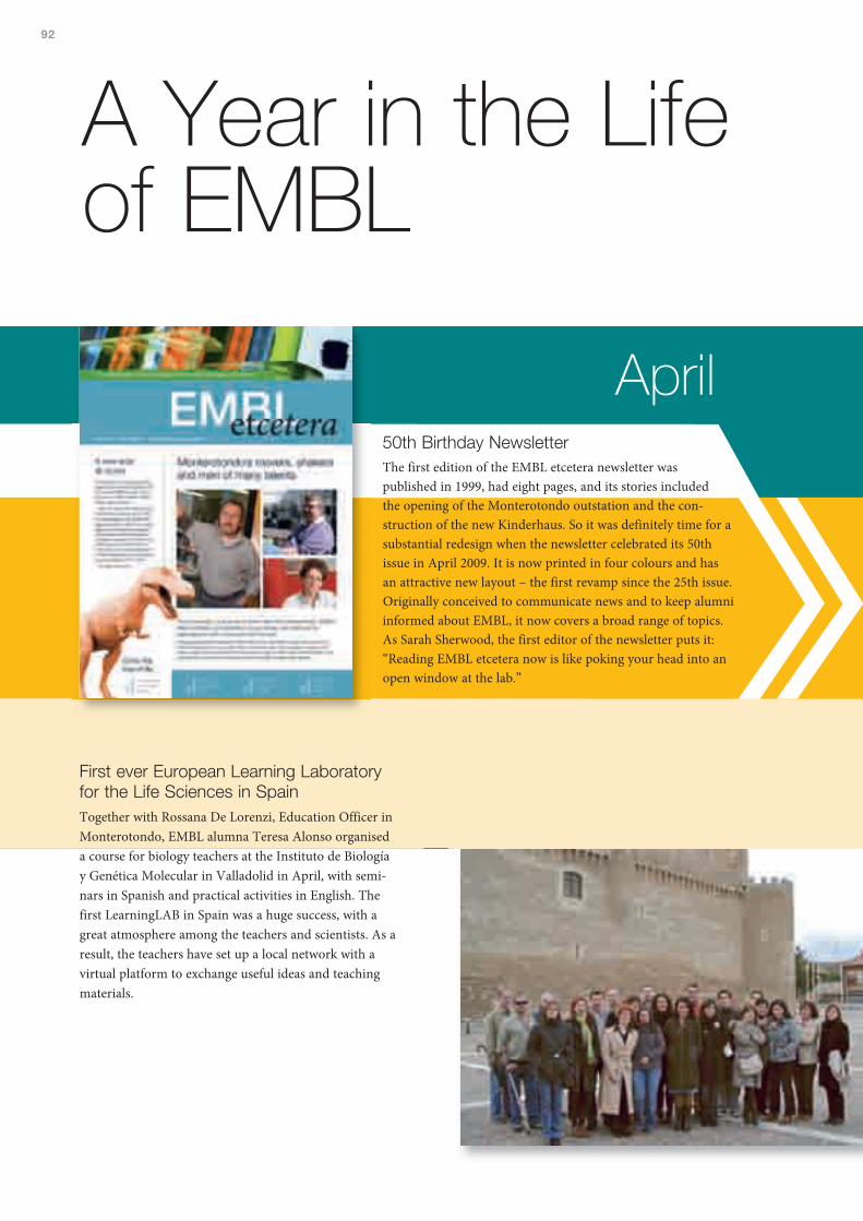
92
A Year in the Lifeof EMBL
50th Birthday NewsletterThe first edition of the EMBL etcetera newsletter waspublished in 1999, had eight pages, and its stories includedthe opening of the Monterotondo outstation and the con-struction of the new Kinderhaus. So it was definitely time for asubstantial redesign when the newsletter celebrated its 50thissue in April 2009. It is now printed in four colours and hasan attractive new layout – the first revamp since the 25th issue.Originally conceived to communicate news and to keep alumniinformed about EMBL, it now covers a broad range of topics.As Sarah Sherwood, the first editor of the newsletter puts it:“Reading EMBL etcetera now is like poking your head into anopen window at the lab.”
First ever European Learning Laboratoryfor the Life Sciences in SpainTogether with Rossana De Lorenzi, Education Officer inMonterotondo, EMBL alumna Teresa Alonso organiseda course for biology teachers at the Instituto de Biologíay Genética Molecular in Valladolid in April, with semi-nars in Spanish and practical activities in English. Thefirst LearningLAB in Spain was a huge success, with agreat atmosphere among the teachers and scientists. As aresult, the teachers have set up a local network with avirtual platform to exchange useful ideas and teachingmaterials.
April

Spanish students visit the LaboratoryA group of biotechnology students from theUniversidad Francisco de Vitoria in Madridvisited EMBL Heidelberg on 8 May as part of atour of universities and research institutes inEurope. The visitors, who were particularly inter-ested in the PhD Programme, learnt about the wayEMBL works from the Head of CommunicationsLena Raditsch and the Dean of Graduate StudiesHelke Hillebrand. The visit was rounded off withtwo presentations by EMBL predocs on theirresearch projects and a look inside the laboratories.
New web pagesThe newsletter was not the only medium to be completelyrevamped. The EMBL web pages also received a fresh,modern design with their re-launch in mid-May. The siteis now managed via a Content Management System, whichallows the pages to be updated more easily. Visitors canchoose their EMBL site from the new 'portal' page androtating banners promote conferences and other events.The immediately recognisable intranet pages contain theday’s events, news, announcements, and quick links todownloads, the library and all the externally availablesections such as research, services, training, press releasesand jobs.
May
93
2009
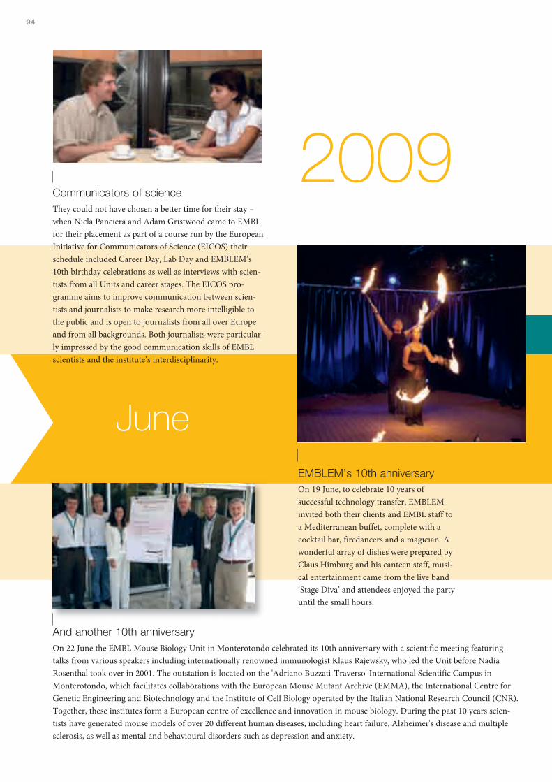
94
2009Communicators of scienceThey could not have chosen a better time for their stay –when Nicla Panciera and Adam Gristwood came to EMBLfor their placement as part of a course run by the EuropeanInitiative for Communicators of Science (EICOS) theirschedule included Career Day, Lab Day and EMBLEM’s10th birthday celebrations as well as interviews with scien-tists from all Units and career stages. The EICOS pro-gramme aims to improve communication between scien-tists and journalists to make research more intelligible tothe public and is open to journalists from all over Europeand from all backgrounds. Both journalists were particular-ly impressed by the good communication skills of EMBLscientists and the institute’s interdisciplinarity.
EMBLEM’s 10th anniversaryOn 19 June, to celebrate 10 years ofsuccessful technology transfer, EMBLEMinvited both their clients and EMBL staff toa Mediterranean buffet, complete with acocktail bar, firedancers and a magician. Awonderful array of dishes were prepared byClaus Himburg and his canteen staff, musi-cal entertainment came from the live band‘Stage Diva’ and attendees enjoyed the partyuntil the small hours.
And another 10th anniversaryOn 22 June the EMBL Mouse Biology Unit in Monterotondo celebrated its 10th anniversary with a scientific meeting featuringtalks from various speakers including internationally renowned immunologist Klaus Rajewsky, who led the Unit before NadiaRosenthal took over in 2001. The outstation is located on the 'Adriano Buzzati-Traverso' International Scientific Campus inMonterotondo, which facilitates collaborations with the European Mouse Mutant Archive (EMMA), the International Centre forGenetic Engineering and Biotechnology and the Institute of Cell Biology operated by the Italian National Research Council (CNR).Together, these institutes form a European centre of excellence and innovation in mouse biology. During the past 10 years scien-tists have generated mouse models of over 20 different human diseases, including heart failure, Alzheimer's disease and multiplesclerosis, as well as mental and behavioural disorders such as depression and anxiety.
June
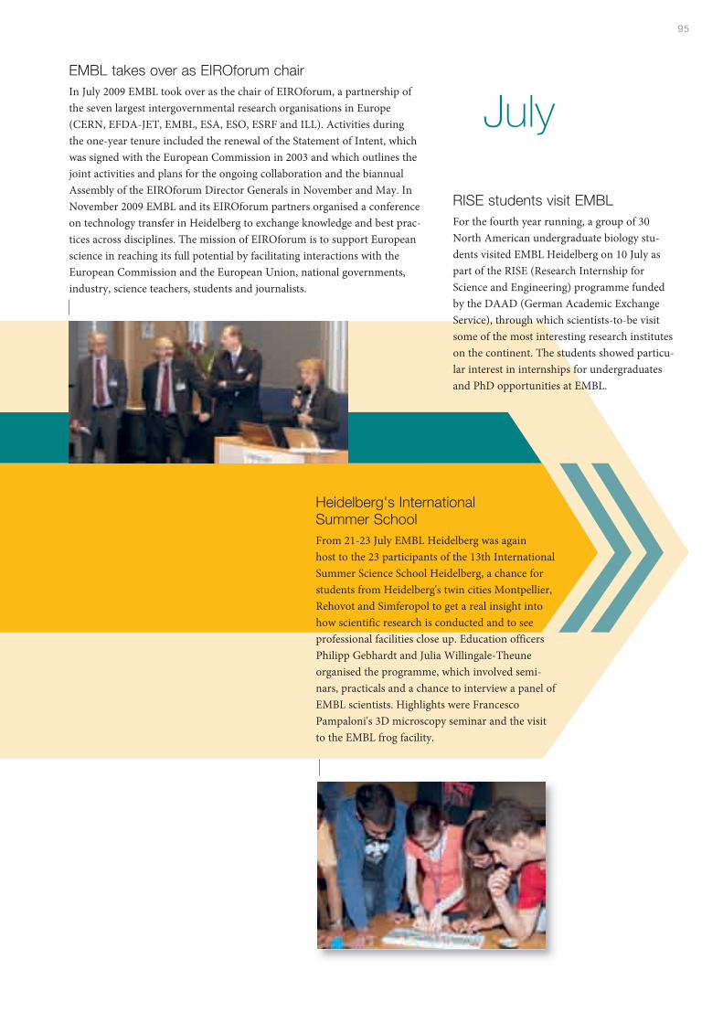
95
Heidelberg's InternationalSummer SchoolFrom 21-23 July EMBL Heidelberg was againhost to the 23 participants of the 13th InternationalSummer Science School Heidelberg, a chance forstudents from Heidelberg's twin cities Montpellier,Rehovot and Simferopol to get a real insight intohow scientific research is conducted and to seeprofessional facilities close up. Education officersPhilipp Gebhardt and Julia Willingale-Theuneorganised the programme, which involved semi-nars, practicals and a chance to interview a panel ofEMBL scientists. Highlights were FrancescoPampaloni's 3D microscopy seminar and the visitto the EMBL frog facility.
EMBL takes over as EIROforum chairIn July 2009 EMBL took over as the chair of EIROforum, a partnership ofthe seven largest intergovernmental research organisations in Europe(CERN, EFDA-JET, EMBL, ESA, ESO, ESRF and ILL). Activities duringthe one-year tenure included the renewal of the Statement of Intent, whichwas signed with the European Commission in 2003 and which outlines thejoint activities and plans for the ongoing collaboration and the biannualAssembly of the EIROforum Director Generals in November and May. InNovember 2009 EMBL and its EIROforum partners organised a conferenceon technology transfer in Heidelberg to exchange knowledge and best prac-tices across disciplines. The mission of EIROforum is to support Europeanscience in reaching its full potential by facilitating interactions with theEuropean Commission and the European Union, national governments,industry, science teachers, students and journalists.
July
RISE students visit EMBLFor the fourth year running, a group of 30North American undergraduate biology stu-dents visited EMBL Heidelberg on 10 July aspart of the RISE (Research Internship forScience and Engineering) programme fundedby the DAAD (German Academic ExchangeService), through which scientists-to-be visitsome of the most interesting research instituteson the continent. The students showed particu-lar interest in internships for undergraduatesand PhD opportunities at EMBL.
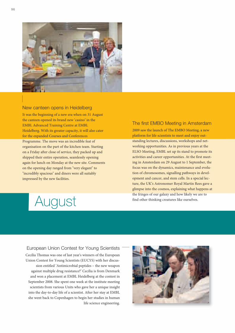
96
The first EMBO Meeting in Amsterdam2009 saw the launch of The EMBO Meeting, a newplatform for life scientists to meet and enjoy out-standing lectures, discussions, workshops and net-working opportunities. As in previous years at theELSO Meeting, EMBL set up its stand to promote itsactivities and career opportunities. At the first meet-ing in Amsterdam on 29 August to 1 September, thefocus was on the dynamics, maintenance and evolu-tion of chromosomes, signalling pathways in devel-opment and cancer, and stem cells. In a special lec-ture, the UK’s Astronomer Royal Martin Rees gave aglimpse into the cosmos, explaining what happens atthe fringes of our galaxy and how likely we are tofind other thinking creatures like ourselves.
European Union Contest for Young ScientistsCecilia Thomas was one of last year's winners of the EuropeanUnion Contest for Young Scientists (EUCYS) with her discus-
sion entitled ‘Antimicrobial peptides – the new weaponagainst multiple drug resistance?’ Cecilia is from Denmarkand won a placement at EMBL Heidelberg at the contest in
September 2008. She spent one week at the institute meetingscientists from various Units who gave her a unique insight
into the day-to-day life of a scientist. After her stay at EMBLshe went back to Copenhagen to begin her studies in human
life science engineering.
August
New canteen opens in HeidelbergIt was the beginning of a new era when on 31 Augustthe canteen opened its brand new ‘casino’ in theEMBL Advanced Training Centre at EMBLHeidelberg. With its greater capacity, it will also caterfor the expanded Courses and ConferencesProgramme. The move was an incredible feat oforganisation on the part of the kitchen team. Startingon a Friday after close of service, they packed up andshipped their entire operation, seamlessly openingagain for lunch on Monday at the new site. Commentson the opening day ranged from "very elegant" to"incredibly spacious" and diners were all suitablyimpressed by the new facilities.

97
2009
Tara sets sailThree years to sail around the world, 150,000 kmto cover, 60 ports to visit and 50 participatinglaboratories – these are only a few of the figuresbehind the enormous project called the TaraOceans Expedition. The vessel left Brittany on 5September for a three-year scientific adventure tostudy the marine ecosystems across the world’soceans, making it 'an historic visit to the past', asone of the scientists on board put it. More than12 fields of research are involved in the project,which will bring together an international team ofoceanographers, ecologists, biologists, geneticists,and physicists from prestigious laboratories. Theproject is headed by Eric Karsenti, the formerhead of the Cell Biology and Biophysics Unit atEMBL.
September
Forum for Young Life Scie ntists at the DKFZOn 10-11 September EMBL predocs got together with PhD students from var-ious research institutes in Heidelberg to hold the first Heidelberg Forum forYoung Life Scientists at the DKFZ. The programme contained six sessions,each of which was organised by one of the institutes and EMBL’s segment,‘Through the Looking Glass: Understanding Molecular Biology throughEvolution’, featured Nobuhiko Tokuriki from Israel’sWeizmann Institute ofScience as the keynote speaker. The Forum provides an ideal opportunity tobring together the diverse know-how of scientists from the Heidelberg insti-tutes and expertise of renowned researchers from abroad. With PhD studentsof different nationalities and diverse educational backgrounds, the atmospherewas highly interdisciplinary and interactive with plenty of networking amongstudents from the different institutes.

98
Science Days in RustEach year Science Days, a major science festival hosted bythe Europa Park in the south of Germany, attracts thou-sands of visitors of all ages. As a mini celebration of theYear of Darwin, this year the stand, organised by EMBL’sEuropean Learning Laboratory for the Life Sciences, fea-tured marine worms (Platynereis dumerilii) an evolution-ary fossil to explain eye development linked to studies per-formed at EMBL, an Axolotl couple, and various otheractivities including “genome tape measures” aimed atbringing the modern concepts of evolution and evolution-ary research nearer to the public.
A packed OperonOctober saw a couple of highlights in this year's EMBL Science & SocietyProgramme when Jorge Cham and Mattieu Ricard – two speakers who couldn'tbe more different – both packed the Operon to the rafters with their EMBLForum lectures. Jorge Cham, creator and artist of 'Piled Higher and Deeper'– thecomic strip about the trials and tribulations of doing a PhD – is something of afolk hero among predocs all over the world and his followers flocked to hear histalk on 'The Power of Procrastination' whereas Matthieu Ricard is probably thefirst Buddhist monk to set foot on EMBL premises. His talk entitled 'Train yourMind, Change your Brain – Cultivating the Inner Condition for GenuineHappiness' not only impressed the general public who turned up in largenumbers to hear the Dalai Lama’s French interpreter but also the hard-corescientists at EMBL.
Puzzles in BiologyMore than 200 attendees – including students and visi-tors from as far afield as Armenia, Israel and India –learnt about ‘Puzzles in Biology’ at the 11th EMBL PhDStudent Symposium on 29 October. Distinguishedspeakers included Stefan Hell, the inventor ofStimulated Emission Depletion microscopy (STED), AriHelenius, winner of the 2007 Marcel Benoist prize, thehighest research award in Switzerland, Kim Nasmythand Pierre Chambon. Highlights included the presenta-tion of the PhD Symposium Writing Prize, which thisyear went to EMBL-EBI’s Diva Tommei for ‘The DarkSide of Stem Cells’.
October
2009
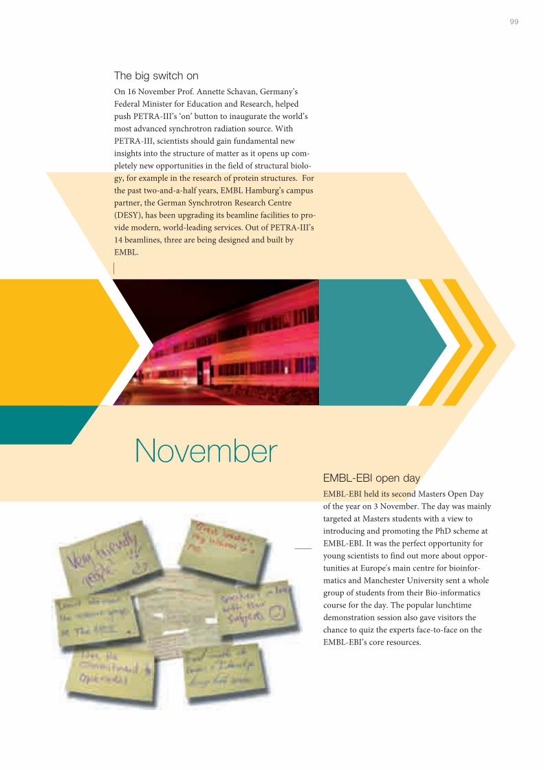
99
November
The big switch onOn 16 November Prof. Annette Schavan, Germany’sFederal Minister for Education and Research, helpedpush PETRA-III’s ‘on’ button to inaugurate the world’smost advanced synchrotron radiation source. WithPETRA-III, scientists should gain fundamental newinsights into the structure of matter as it opens up com-pletely new opportunities in the field of structural biolo-gy, for example in the research of protein structures. Forthe past two-and-a-half years, EMBL Hamburg’s campuspartner, the German Synchrotron Research Centre(DESY), has been upgrading its beamline facilities to pro-vide modern, world-leading services. Out of PETRA-III’s14 beamlines, three are being designed and built byEMBL.
EMBL-EBI open dayEMBL-EBI held its second Masters Open Dayof the year on 3 November. The day was mainlytargeted at Masters students with a view tointroducing and promoting the PhD scheme atEMBL-EBI. It was the perfect opportunity foryoung scientists to find out more about oppor-tunities at Europe's main centre for bioinfor-matics and Manchester University sent a wholegroup of students from their Bio-informaticscourse for the day. The popular lunchtimedemonstration session also gave visitors thechance to quiz the experts face-to-face on theEMBL-EBI’s core resources.
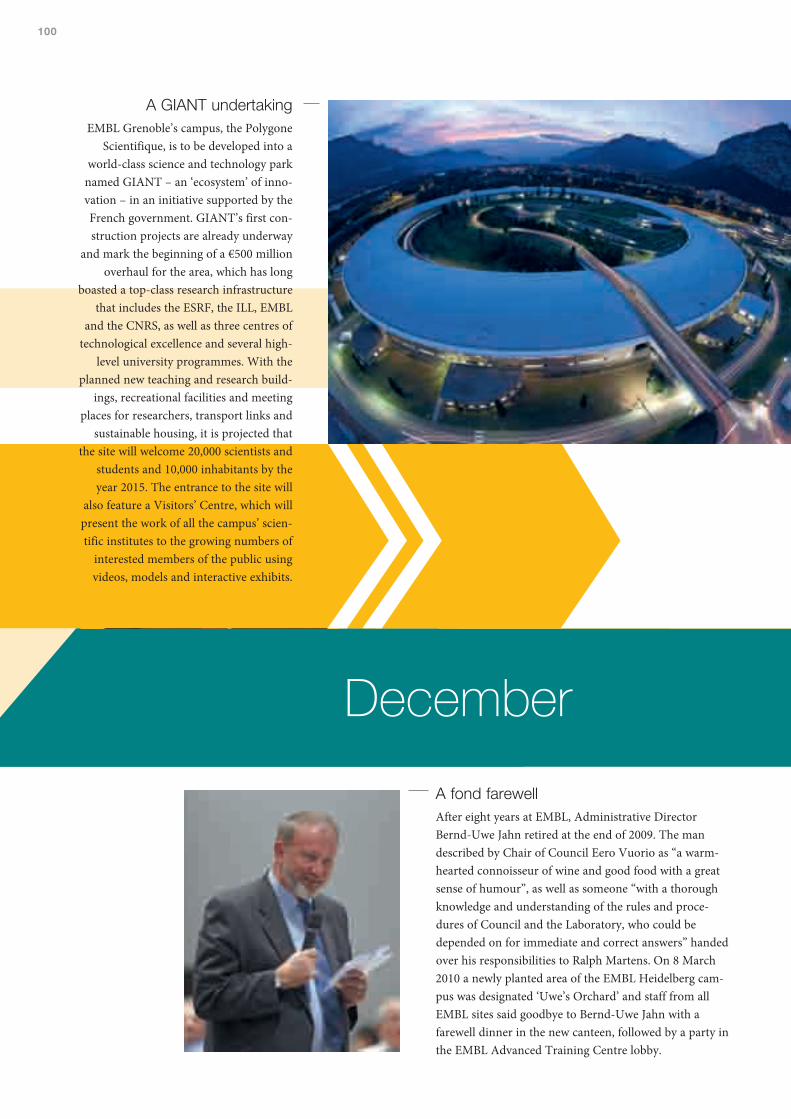
100
December
A fond farewellAfter eight years at EMBL, Administrative DirectorBernd-Uwe Jahn retired at the end of 2009. The mandescribed by Chair of Council Eero Vuorio as “a warm-hearted connoisseur of wine and good food with a greatsense of humour”, as well as someone “with a thoroughknowledge and understanding of the rules and proce-dures of Council and the Laboratory, who could bedepended on for immediate and correct answers” handedover his responsibilities to Ralph Martens. On 8 March2010 a newly planted area of the EMBL Heidelberg cam-pus was designated ‘Uwe’s Orchard’ and staff from allEMBL sites said goodbye to Bernd-Uwe Jahn with afarewell dinner in the new canteen, followed by a party inthe EMBL Advanced Training Centre lobby.
A GIANT undertakingEMBL Grenoble’s campus, the Polygone
Scientifique, is to be developed into aworld-class science and technology park
named GIANT – an ‘ecosystem’ of inno-vation – in an initiative supported by theFrench government. GIANT’s first con-struction projects are already underway
and mark the beginning of a €500 millionoverhaul for the area, which has long
boasted a top-class research infrastructurethat includes the ESRF, the ILL, EMBL
and the CNRS, as well as three centres oftechnological excellence and several high-
level university programmes. With theplanned new teaching and research build-
ings, recreational facilities and meetingplaces for researchers, transport links and
sustainable housing, it is projected thatthe site will welcome 20,000 scientists and
students and 10,000 inhabitants by theyear 2015. The entrance to the site will
also feature a Visitors’ Centre, which willpresent the work of all the campus’ scien-tific institutes to the growing numbers of
interested members of the public usingvideos, models and interactive exhibits.
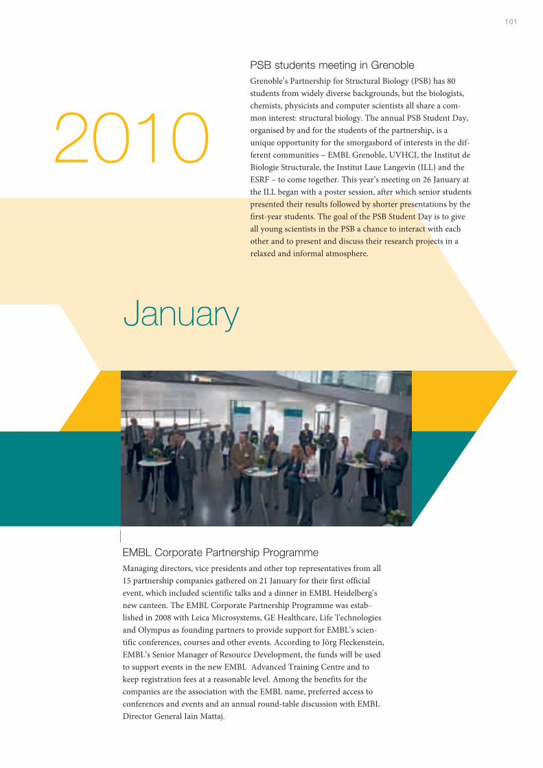
101
PSB students meeting in GrenobleGrenoble’s Partnership for Structural Biology (PSB) has 80students from widely diverse backgrounds, but the biologists,chemists, physicists and computer scientists all share a com-mon interest: structural biology. The annual PSB Student Day,organised by and for the students of the partnership, is aunique opportunity for the smorgasbord of interests in the dif-ferent communities – EMBL Grenoble, UVHCI, the Institut deBiologie Structurale, the Institut Laue Langevin (ILL) and theESRF – to come together. This year’s meeting on 26 January atthe ILL began with a poster session, after which senior studentspresented their results followed by shorter presentations by thefirst-year students. The goal of the PSB Student Day is to giveall young scientists in the PSB a chance to interact with eachother and to present and discuss their research projects in arelaxed and informal atmosphere.
EMBL Corporate Partnership ProgrammeManaging directors, vice presidents and other top representatives from all15 partnership companies gathered on 21 January for their first officialevent, which included scientific talks and a dinner in EMBL Heidelberg’snew canteen. The EMBL Corporate Partnership Programme was estab-lished in 2008 with Leica Microsystems, GE Healthcare, Life Technologiesand Olympus as founding partners to provide support for EMBL’s scien-tific conferences, courses and other events. According to Jörg Fleckenstein,EMBL’s Senior Manager of Resource Development, the funds will be usedto support events in the new EMBL Advanced Training Centre and tokeep registration fees at a reasonable level. Among the benefits for thecompanies are the association with the EMBL name, preferred access toconferences and events and an annual round-table discussion with EMBLDirector General Iain Mattaj.
2010
January
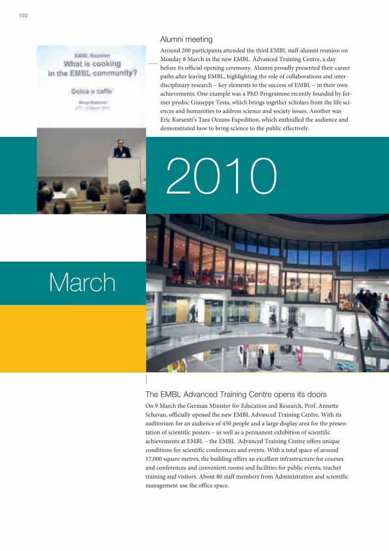
102
Alumni meetingAround 200 participants attended the third EMBL staff-alumni reunion onMonday 8 March in the new EMBL Advanced Training Centre, a daybefore its official opening ceremony. Alumni proudly presented their careerpaths after leaving EMBL, highlighting the role of collaborations and inter-disciplinary research – key elements to the success of EMBL – in their ownachievements. One example was a PhD Programme recently founded by for-mer predoc Giuseppe Testa, which brings together scholars from the life sci-ences and humanities to address science and society issues. Another wasEric Karsenti’s Tara Oceans Expedition, which enthralled the audience anddemonstrated how to bring science to the public effectively.
The EMBL Advanced Training Centre opens its doorsOn 9 March the German Minister for Education and Research, Prof. AnnetteSchavan, officially opened the new EMBL Advanced Training Centre. With itsauditorium for an audience of 450 people and a large display area for the presen-tation of scientific posters – as well as a permanent exhibition of scientificachievements at EMBL – the EMBL Advanced Training Centre offers uniqueconditions for scientific conferences and events. With a total space of around17,000 square metres, the building offers an excellent infrastructure for coursesand conferences and convenient rooms and facilities for public events, teachertraining and visitors. About 80 staff members from Administration and scientificmanagement use the office space.
2010
March
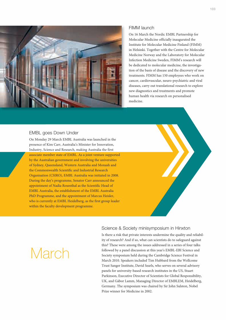
103
EMBL goes Down UnderOn Monday 29 March EMBL Australia was launched in thepresence of Kim Carr, Australia’s Minister for Innovation,Industry, Science and Research, making Australia the firstassociate member state of EMBL. As a joint venture supportedby the Australian government and involving the universitiesof Sydney, Queensland, Western Australia and Monash andthe Commonwealth Scientific and Industrial ResearchOrganisation (CSIRO), EMBL Australia was initiated in 2008.During the day’s programme, Senator Carr announced theappointment of Nadia Rosenthal as the Scientific Head ofEMBL Australia, the establishment of the EMBL AustraliaPhD Programme, and the appointment of Marcus Heisler,who is currently at EMBL Heidelberg, as the first group leaderwithin the faculty development programme.
Science & Society minisymposium in HinxtonIs there a risk that private interests undermine the quality and reliabil-ity of research? And if so, what can scientists do to safeguard againstthis? These were among the issues addressed in a series of four talksfollowed by a panel discussion at this year's EMBL-EBI Science andSociety symposium held during the Cambridge Science Festival inMarch 2010. Speakers included Tim Hubbard from the WellcomeTrust Sanger Institute, David Searls, who serves on several advisorypanels for university-based research institutes in the US, StuartParkinson, Executive Director of Scientists for Global Responsibility,UK, and Gábor Lamm, Managing Director of EMBLEM, Heidelberg,Germany. The symposium was chaired by Sir John Sulston, NobelPrize winner for Medicine in 2002.
FIMM launchOn 16 March the Nordic EMBL Partnership forMolecular Medicine officially inaugurated theInstitute for Molecular Medicine Finland (FIMM)in Helsinki. Together with the Centre for MolecularMedicine Norway and the Laboratory for MolecularInfection Medicine Sweden, FIMM’s research willbe dedicated to molecular medicine, the investiga-tion of the basis of disease and the discovery of newtreatments. FIMM has 150 employees who work oncancer, cardiovascular, neuro-psychiatric and viraldiseases, carry out translational research to explorenew diagnostics and treatments and promotehuman health via research on personalisedmedicine.
March
Index of group & team leaders
AArendt, Detlev . . . . . . . . . . . . . . . . . . . . . . . . . . 13
BBirney, Ewan . . . . . . . . . . . . . . . . . . . . . . . . 67, 87
Boettcher, Bettina . . . . . . . . . . . . . . . . . . . . . . . . 8
Bork, Peer. . . . . . . . . . . . . . . . . . . . . . . . 6, 13, 21
Brazma, Alvis. . . . . . . . . . . . . . . . . . . . . . . . . . . 11
Briggs, John . . . . . . . . . . . . . . . . . . . . . . . . . . . 18
CCusack, Stephen. . . . . . . . . . . . . . . . . . . . . . . . 33
EEllenberg, Jan . . . . . . . . . . . . . . . . . . . . 33, 63, 84
Ephrussi, Anne . . . . . . . . . . . . . . . . . . . . . . . . . 52
FFlicek, Paul . . . . . . . . . . . . . . . . . . . . . . . . . . . . 67
Frangakis, Achilleas . . . . . . . . . . . . . . . . . . . . . . . 8
Furlong, Eileen. . . . . . . . . . . . . . . . . . . . . . . . . . 20
GGavin, Anne-Claude . . . . . . . . . . . . . . . . . . . . . . 8
Gibson, Toby . . . . . . . . . . . . . . . . . . . . . . . . . . 77
HHuber, Wolfgang . . . . . . . . . . . . . . . . . . . . . . . . 10
KKersey, Paul . . . . . . . . . . . . . . . . . . . . . . . . . . . 65
Korbel, Jan . . . . . . . . . . . . . . . . . . . . . . . . . . . . 78
LLadurner, Andreas . . . . . . . . . . . . . . . . . . . . . . . 30
Le Novère, Nicolas . . . . . . . . . . . . . . . . . . . . . . 59
Luscombe, Nicholas . . . . . . . . . . . . . . . . . . . . . 87
MMárquez, José . . . . . . . . . . . . . . . . . . . . . . . . . 43
Mattaj, Iain. . . . . . . . . . . . . . . . . . . . . . . . . . 47, 53
Müller-Dieckmann, Jochen. . . . . . . . . . . . . . . . . 57
Müller, Christoph . . . . . . . . . . . . . . . . . . . . . 49, 81
Müller, Jürg . . . . . . . . . . . . . . . . . . . . . . . . . 80, 81
NNerlov, Claus. . . . . . . . . . . . . . . . . . . . . . . . 40, 89
PPanne, Daniel . . . . . . . . . . . . . . . . . . . . . . . . . . 91
Pepperkok, Rainer . . . . . . . . . . . . . . . . . . . . . . . 84
Pillai, Ramesh . . . . . . . . . . . . . . . . . . . . . . . . . . 62
RRosenthal, Nadia . . . . . . . . . . . . . . . . . . . . . 23, 40
Round, Adam . . . . . . . . . . . . . . . . . . . . . . . . . . 45
Russel, Rob . . . . . . . . . . . . . . . . . . . . . . . . . . . . 8
SSchneider, Reinhard . . . . . . . . . . . . . . . . . . . . . 85
Schultz, Carsten . . . . . . . . . . . . . . . . . . . . . 41, 57
Steinmetz, Lars . . . . . . . . . . . . . . . . . . . 10, 37, 79
Stelzer, Ernst . . . . . . . . . . . . . . . . . . . . . . . . . . . 30
TTreier, Mathias. . . . . . . . . . . . . . . . . . . . . . . . . . 73
Tucker, Paul . . . . . . . . . . . . . . . . . . . . . . . . . . . 76
WWilmanns, Matthias . . . . . . . . . . . . . . . . . . . . . . 55


Annual Report 2009-2010
EMBL HeidelbergMeyerhofstraße 169117 HeidelbergGermany
Tel. +49 (0)6221 387 0
Fax +49 (0)6221 387 8306
EMBL Grenoble6, rue Jules Horowitz, BP 18138042 Grenoble, Cedex 9France
Tel. +33 (0)4 76 20 72 69
Fax +33 (0)4 76 20 71 99
EMBL Hamburgc/o DESYNotkestraße 8522603 HamburgGermany
Tel. +49 (0)40 89 902 0
Fax +49 (0)40 89 902 104
EMBL-EBIWellcome Trust GenomeCampus, HinxtonCambridge CB10 1SDUnited Kingdom
Tel. +44 (0)1223 494 444
Fax +44 (0)1223 494 468
EMBL MonterotondoAdriano Buzzati-Traverso CampusVia Ramarini, 3200016 Monterotondo (Rome)Italy
Tel. +39 06 900 912 85
Fax +39 06 900 912 72
www.embl.org
Publishe dby the EMBL Office of Information and Public AffairsLena Raditsch, Sonia Furtado
DG ReportIain Mattaj
Science writersClaire Ainsworth, Jennifer Carpenter, Sonia Furtado, Lucy Patterson, Anna-Lynn Wegener
Graphics and cover designPetra Riedinger
PhotographyEMBL Photolab (Hugo Neves, Udo Ringeisen, Marietta Schupp)
Layout and compositionNicola Graf
Printed byColorDruck Leimen, Germany
AcknowledgementsPage 2: The National Library of Israel, Eran Laor Cartographic Collection, Shapell Family Digitization
Project and The Hebrew University of Jerusalem, Department of Geography – Historic CitiesResearch Project
Page 14: Nüchter and Holstein, Universität Heidelberg (Nematostella), Kirt L. Onthank (Strongylocentrotus),The Field Museum, Chicago (Cambrian sea)
Page 34: Douglas Jordan/CDC
Page 38: Marina Lamacchia/INSERM
Page 45: (top) Pedro Luís Rodriguez
Page 50: Carlo Petosa/IBS
A special thank you to the EMBL group leaders and Heads of Units for their cooperation.
The paper this report is printed on is 60% recycled and 40% from certified managed forests.
© Copyright European Molecular Biology Laboratory 2010