Highlights and prospects of potyvirus molecular biology
16
Journal of General Virology (1992), 73, 1-16. Printed in Great Britain 1 Review article Highlights and prospects of potyvirus molecular biology Jos~ Luis Riechmann,* Sonia Lain and Juan Antonio Garcia Centro de Biologia Molecular (CSIC-UAM), Universidad AutSnoma de Madrid, Cantoblanco, 28049 Madrid, Spain Introduction The potyvirus group [named after its type member, potato virus Y (PVY)] is the largest of the 34 plant virus groups and families currently recognized (Ward & Shukla, 1991). It contains at least 180 definitive and possible members (or 30% of all known plant viruses) which cause significant losses in agricultural, pasture, horticultural and ornamental crops (Ward & Shukla, 1991). These viruses are unique in the diversity of inclusion bodies that are formed during the infection cycle (see Lesemann, 1988). A feature shared by all potyviruses is the induction of characteristic pinwheel or scroll-shaped inclusion bodies in the cytoplasm of the infected cells (Edwardson, 1974). These cylindrical inclusion (CI) bodies are formed by a virus-encoded protein and can be considered as the most important phenotypic criterion for assigning viruses to the poty- virus group (Milne, 1988; Shukla et al., 1989; Ward & Shukla, 1991). Many potyviruses also induce cytoplasmic amorphous inclusion bodies and some form nuclear inclusions. Virions are flexuous and rod-shaped, 680 to 900 nm long and 11 to 15 nm wide, made up of about 2000 units of a single structural protein surrounding one molecule of ssRNA of approximately 10000 nucleotides and messenger polarity (Dougherty & Carrington, 1988). Although some of these viruses are transmitted by mites, and possibly by whiteflies, the predominant transmission of potyviruses is by aphids. In addition, there are viruses that share many characteristics with the typical potyvir- uses but have some important peculiarities. They are transmitted by fungi, have bipartite ssRNA genomes that are encapsidated in two different rod-shaped virions (approximately 275 and 550 nm long), and are not serologically related to potyviruses, differences that would set them apart from the potyvirus group (Kashiwazaki et al., 1990). On the other hand, their cistron order, strategy of genome expression and amino acid sequence homologies as well as the formation of similar cylindrical inclusions in infected cells indicate a common ancestry and a close evolutionary relationship between these viruses [whose type member is barley yellow mosaic virus (BaYMV)] and typical potyviruses (Kashiwazaki et al., 1990). The taxonomy of the potyvirus group, which has been studied extensively, may therefore be quite complex (reviewed in Milne, 1988; Ward & Shukla, 1991 ; for a proposal on potyvirus taxonomy, see Barnett, 1991). A breakthrough in potyvirus studies was achieved in 1986 when the complete genome sequences of two members of this group, tobacco etch virus (TEV; Allison et al., 1986) and tobacco vein mottling virus (TVMV; Domier et al., 1986) were reported for the first time. Intensive research during the following years led to a greater understanding of the potyviral genome structure and expression. The complete genome sequences of two more potyviruses, PVY (Robaglia et al., 1989) and plum pox virus (PPV; Lain et al., 1989a; Maiss et al., 1989; Teycheney et al., 1989) are now available. Unsuspected relationships between potyviruses and other groups of plant and animal viruses arose from sequence compari- sons (Domier et al., 1987; Lain et al., 1989b), allowing functions to be proposed for most of the potyviral gene products. However, actual knowledge about these functions is still scarce, and not all of the gene products have been identified in vivo. A second breakthrough in potyvirus research is probably near, given the recent isolation of full-length cDNA clones of three potyviruses from which infectious transcripts can be synthesized: TVMV (Domier et al., 1989), PPV (Riechmann et al., 1990) and zucchini yellow mosaic virus (Gal-On et al., 1991). The ability to generate virus infection from cloned cDNA of potyviruses opens the possibility of applying genetic engineering techni- ques for the study of their biology at the molecular level. Aspects of the potyvirus life cycle such as translation, replication, symptom induction, and cell-to-cell and plant-to-plant propagation are now amenable to a new experimental approach, and the functions that have been proposed for potyviral gene products can now be tested in vivo. Also, discoveries have been made working with potyviruses that will influence our knowledge of the biology of most plus-stranded RNA viruses; on the other hand, results obtained by working with other viruses are 0001-0485 © 1992SGM
Transcript of Highlights and prospects of potyvirus molecular biology
Journal of General Virology (1992), 73, 1-16. Printed in Great Britain 1
Review article
Highlights and prospects of potyvirus molecular biology
Jos~ Luis Riechmann,* Sonia Lain and Juan Antonio Garcia
Centro de Biologia Molecular (CSIC-UAM), Universidad AutSnoma de Madrid, Cantoblanco, 28049 Madrid, Spain
Introduction
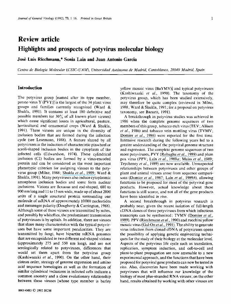
The potyvirus group [named after its type member, potato virus Y (PVY)] is the largest of the 34 plant virus groups and families currently recognized (Ward & Shukla, 1991). It contains at least 180 definitive and possible members (or 30% of all known plant viruses) which cause significant losses in agricultural, pasture, horticultural and ornamental crops (Ward & Shukla, 1991). These viruses are unique in the diversity of inclusion bodies that are formed during the infection cycle (see Lesemann, 1988). A feature shared by all potyviruses is the induction of characteristic pinwheel or scroll-shaped inclusion bodies in the cytoplasm of the infected cells (Edwardson, 1974). These cylindrical inclusion (CI) bodies are formed by a virus-encoded protein and can be considered as the most important phenotypic criterion for assigning viruses to the poty- virus group (Milne, 1988; Shukla et al., 1989; Ward & Shukla, 1991). Many potyviruses also induce cytoplasmic amorphous inclusion bodies and some form nuclear inclusions. Virions are flexuous and rod-shaped, 680 to 900 nm long and 11 to 15 nm wide, made up of about 2000 units of a single structural protein surrounding one molecule of ssRNA of approximately 10000 nucleotides and messenger polarity (Dougherty & Carrington, 1988). Although some of these viruses are transmitted by mites, and possibly by whiteflies, the predominant transmission of potyviruses is by aphids. In addition, there are viruses that share many characteristics with the typical potyvir- uses but have some important peculiarities. They are transmitted by fungi, have bipartite ssRNA genomes that are encapsidated in two different rod-shaped virions (approximately 275 and 550 nm long), and are not serologically related to potyviruses, differences that would set them apart from the potyvirus group (Kashiwazaki et al., 1990). On the other hand, their cistron order, strategy of genome expression and amino acid sequence homologies as well as the formation of similar cylindrical inclusions in infected cells indicate a common ancestry and a close evolutionary relationship between these viruses [whose type member is barley
yellow mosaic virus (BaYMV)] and typical potyviruses (Kashiwazaki et al., 1990). The taxonomy of the potyvirus group, which has been studied extensively, may therefore be quite complex (reviewed in Milne, 1988; Ward & Shukla, 1991 ; for a proposal on potyvirus taxonomy, see Barnett, 1991).
A breakthrough in potyvirus studies was achieved in 1986 when the complete genome sequences of two members of this group, tobacco etch virus (TEV; Allison et al., 1986) and tobacco vein mottling virus (TVMV; Domier et al., 1986) were reported for the first time. Intensive research during the following years led to a greater understanding of the potyviral genome structure and expression. The complete genome sequences of two more potyviruses, PVY (Robaglia et al., 1989) and plum pox virus (PPV; Lain et al., 1989a; Maiss et al., 1989; Teycheney et al., 1989) are now available. Unsuspected relationships between potyviruses and other groups of plant and animal viruses arose from sequence compari- sons (Domier et al., 1987; Lain et al., 1989b), allowing functions to be proposed for most of the potyviral gene products. However, actual knowledge about these functions is still scarce, and not all of the gene products have been identified in vivo.
A second breakthrough in potyvirus research is probably near, given the recent isolation of full-length cDNA clones of three potyviruses from which infectious transcripts can be synthesized: TVMV (Domier et al., 1989), PPV (Riechmann et al., 1990) and zucchini yellow mosaic virus (Gal-On et al., 1991). The ability to generate virus infection from cloned cDNA of potyviruses opens the possibility of applying genetic engineering techni- ques for the study of their biology at the molecular level. Aspects of the potyvirus life cycle such as translation, replication, symptom induction, and cell-to-cell and plant-to-plant propagation are now amenable to a new experimental approach, and the functions that have been proposed for potyviral gene products can now be tested in vivo. Also, discoveries have been made working with potyviruses that will influence our knowledge of the biology of most plus-stranded RNA viruses; on the other hand, results obtained by working with other viruses are
0001-0485 © 1992 SGM
2 J . L . Riechmann, S. Lain and J. A. Garcia
affecting our perception of the potyvirus group. For all these reasons, this is a timely moment to review progress in potyvirus research. The aim of this article is not to present a comprehensive review but to focus on the most relevant and novel aspects of potyvirus molecular biology and on the expected trends for the forthcoming years. Previous reviews that are more extensive or that cover mainly other topics of potyvirus biology are already available (Dougherty & Carrington, 1988; Shaw et al., 1990; Dougherty et al., 1990; Shukla et al., 1991).
Genome structure and organization
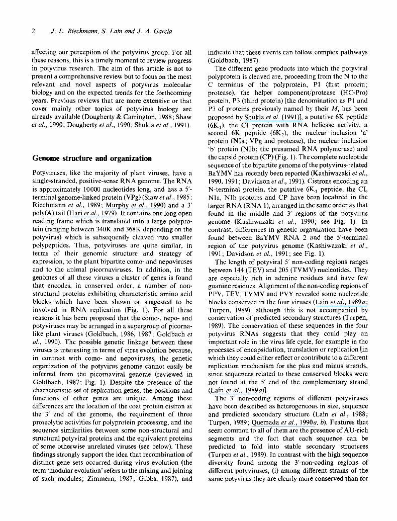
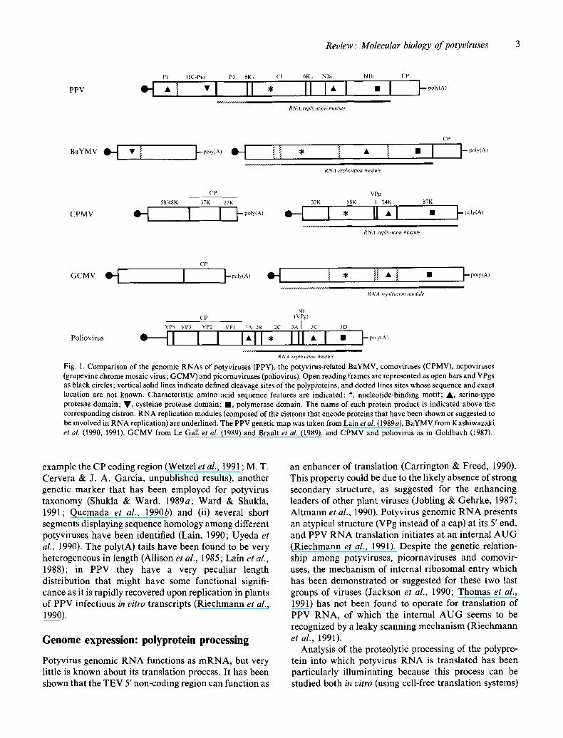
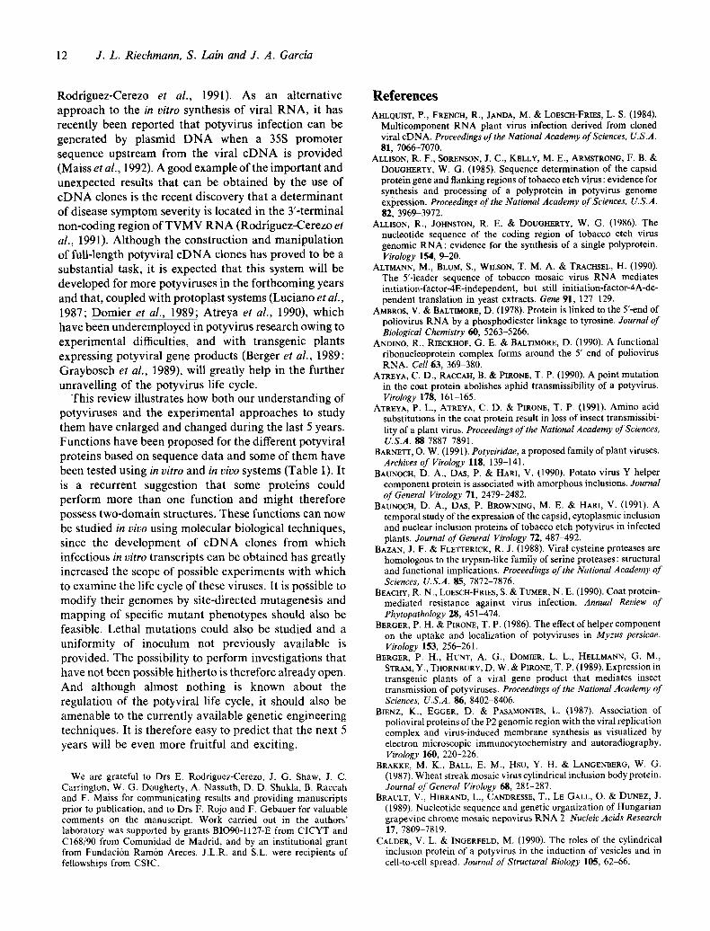
Potyviruses, like the majority of plant viruses, have a single-stranded, positive-sense RNA genome. The RNA is approximately 10000 nucleotides long, and has a 5'- terminal genome-linked protein (VPg) (Siaw et al., 1985; Riechmann et al., 1989; Murphy et al., 1990) and a 3' poly(A) tail (Hari et al., 1979). It contains one long open reading frame which is translated into a large polypro- tein (ranging between 340K and 368K depending on the potyvirus) which is subsequently cleaved into smaller polypeptides. Thus, potyviruses are quite similar, in terms of their genomic structure and strategy of expression, to the plant bipartite como- and nepoviruses and to the animal picornaviruses. In addition, in the genomes of all these viruses a cluster of genes is found that encodes, in conserved order, a number of non- structural proteins exhibiting characteristic amino acid blocks which have been shown or suggested to be involved in RNA replication (Fig. 1), For all these reasons it has been proposed that the como-, nepo- and potyviruses may be arranged in a supergroup of picorna- like plant viruses (Goldbach, 1986, 1987; Goldbach et al., 1990). The possible genetic linkage between these viruses is interesting in terms of virus evolution because, in contrast with como- and nepoviruses, the genetic organization of the potyvirus genome cannot easily be inferred from the picornaviral genome (reviewed in Goldbach, 1987; Fig. 1). Despite the presence of the characteristic set of replication genes, the positions and functions of other genes are unique. Among these differences are the location of the coat protein cistron at the 3' end of the genome, the requirement of three proteolytic activities for polyprotein processing, and the sequence similarities between some non-structural and structural potyviral proteins and the equivalent proteins of some otherwise unrelated viruses (see below). These findings strongly support the idea that recombination of distinct gene sets occurred during virus evolution (the term 'modular evolution' refers to the mixing and joining of such modules; Zimmern, 1987; Gibbs, 1987), and
indicate that these events can follow complex pathways (Goldbach, 1987).
The different gene products into which the potyviral polyprotein is cleaved are, proceeding from the N to the C terminus of the polyprotein, P1 (first protein; protease), the helper component/protease (HC-Pro) protein, P3 (third protein) [the denomination as P1 and P3 of proteins previously named by their Mr has been proposed by Shukla et al. (1991)], a putative 6K peptide (6K1), the CI protein with RNA helicase activity, a second 6K peptide (6K2), the nuclear inclusion 'a' protein (NIa; VPg and protease), the nuclear inclusion 'b' protein (NIb; the presumed RNA polymerase) and the capsid protein (CP) (Fig. 1). The complete nucleotide sequence of the bipartite genome of the potyvirus-related BaYMV has recently been reported (Kashiwazaki et al., 1990, 1991 ; Davidson et al., 1991). Cistrons encoding an N-terminal protein, the putative 6K1 peptide, the CI, NIa, NIb proteins and CP have been localized in the larger RNA (RNA 1), arranged in the same order as that found in the middle and 3' regions of the potyvirus genome (Kashiwazaki et al., 1990; see Fig. 1). In contrast, differences in genetic organization have been found between BaYMV RNA 2 and the 5'-terminal region of the potyvirus genome (Kashiwazaki et al., 1991; Davidson et al., 1991; see Fig. 1).
The length of potyviral 5' non-coding regions ranges between 144 (TEV) and 205 (TVMV) nucleotides. They are especially rich in adenine residues and have few guanine residues. Alignment of the non-coding regions of PPV, TEV, TVMV and PVY revealed some nucleotide blocks conserved in the four viruses (Lain et al., 1989a; Turpen, 1989), although this is not accompanied by conservation of predicted secondary structures (Turpen, 1989). The conservation of these sequences in the four potyvirus RNAs suggests that they could play an important role in the virus life cycle, for example in the processes of encapsidation, translation or replication [in which they could either reflect or contribute to a different replication mechanism for the plus and minus strands, since sequences related to these conserved blocks were not found at the 5' end of the complementary strand (Lain et al., 1989a)].
The 3' non-coding regions of different potyviruses have been described as heterogeneous in size, sequence and predicted secondary structure (Lain et al., 1988; Turpen, 1989; Quemada et al., 1990a, b). Features that seem common to all of them are the presence of AU-rich segments and the fact that each sequence can be predicted to fold into stable secondary structures (Turpen et al., 1989). In contrast with the high sequence diversity found among the T-non-coding regions of different potyviruses, (i) among different strains of the same potyvirus they are clearly more conserved than for
Review: Molecular biology o f potyviruses 3
PI HC-Pro P3 6K: CI 6K2 Nla Nlb CP
RNA replication module
CP
BaYMV 9 ~ T i ~--po~y~A) t ~ f ~', I * ~f • l,, II I ~-poly,A)
RNA replication module
CP VPg 58/48K 37K 27K 32K 58K [ 24K 87K
CPMV ~ I I ~-poly(A) ~ I * I1"1 • t -~°~ '~ '
RNA replication module
CP
G f M V ~ ' t ] ~poly(A) ~ -~ ~ ,
RNA replicutilm module
3B CP (VPg)
VP4 VP3 VP2 VPI 2A 2B 2C 3A ] 3C 3D
- ' •1 III • Poliovirus - - t l I I I I , I " -poly(A) RNA ~eplication module
Fig. 1. Comparison of the genomic RNAs of potyvi~ses (PPV), the potyvirus-related BaYMV, comoviruses (CPMV), nepoviruses (grapevine chrome mosaic virus; GCMV) and picornaviruses (poliovirus). Open reading frames are represented as open bars and VPgs as black circles; vertical solid lines indicate defined cleavage sites of the polyproteins, and dotted lines sites whose sequence and exact location are not known. Characteristic amino acid sequence features are indicated: *, nucleotide-binding motif; A , serine-type protease domain; V, cysteine protease domain; II , polymerase domain. The name of each protein product is indicated above the corresponding cistron. RNA replication modules (composed of the cistrons that encode proteins that have been shown or suggested to be involved in RNA replication) are underlined. The PPV genetic map was taken from Lain et al. (1989a), BaYMV from Kashiwazaki et al. (1990, 1991), GCMV from Le Gall et aL (1989) and Brault et at. (1989), and CPMV and poliovirus as in Goldbach (1987).
example the CP coding region (Wetzel et al., 1991 ; M. T. Cervera & J. A. Garcia, unpublished results), another genetic marker that has been employed for potyvirus taxonomy (Shukla & Ward, 1989a; Ward & Shukla, 1991; Quemada et al., 1990b) and (ii) several short segments displaying sequence homology among different potyviruses have been identified (Lain, 1990; Uyeda et al., 1990). The poly(A) tails have been found to be very heterogeneous in length (Allison et al., 1985; Lain et aL, 1988); in PPV they have a very peculiar length distribution that might have some functional signifi- cance as it is rapidly recovered upon replication in plants of PPV infectious in vitro transcripts (Riechmann et al., 1990).
Genome expression: polyprotein processing
Potyvirus genomic RNA functions as mRNA, but very little is known about its translation process. It has been shown that the TEV 5' non-coding region can function as
an enhancer of translation (Carrington & Freed, 1990). This property could be clue to the likely absence of strong secondary structure, as suggested for the enhancing leaders of other plant viruses (Jobling & Gehrke, 1987; Altmann et al., 1990). Potyvirus genomic RNA presents an atypical structure (VPg instead of a cap) at its 5' end, and PPV RNA translation initiates at an internal AUG (Riechmann et al., 1991). Despite the genetic relation- ship among potyviruses, picornaviruses and comovir- uses, the mechanism of internal ribosomal entry which has been demonstrated or suggested for these two last groups of viruses (Jackson et al., 1990; Thomas et al., 1991) has not been found to operate for translation of PPV RNA, of which the internal AUG seems to be recognized by a leaky scanning mechanism (Riechmann et al., 1991).
Analysis of the proteolytic processing of the polypro- tein into which potyvirus RNA is translated has been particularly illuminating because this process can be studied both in vitro (using cell-free translation systems)
4 J . L . R iechmann, S. Lain and J. A. Garcia
PI
PI HC-Pro } t AB
HC-Pro P3 6K,
Nla
CI
CD E F v v
6K2 Nla Nlb CP
PPV
HC-Pro Y L v G / G L
B QA Q S
C E C H T
m E EVV G
E E F V Y T S
F NV V A
V EEVD H E S
TEV TVMV PVY
Y N V G / G M Y K V G / G L Y R V G / G V
BaYMV
li LiE I T
I Y L
V F
E L V S
N L F
EDLTF
I A
S
S
G
G
S
E G
NNI IRrQI'S 'S A SYO 'I
z TIvLRt£ QI/ s Y E / ~ D E~wlk~/[~
Q ~ V A F E / S Q E V E a E / A
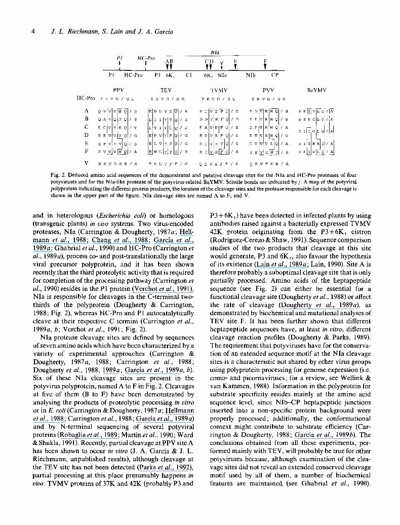
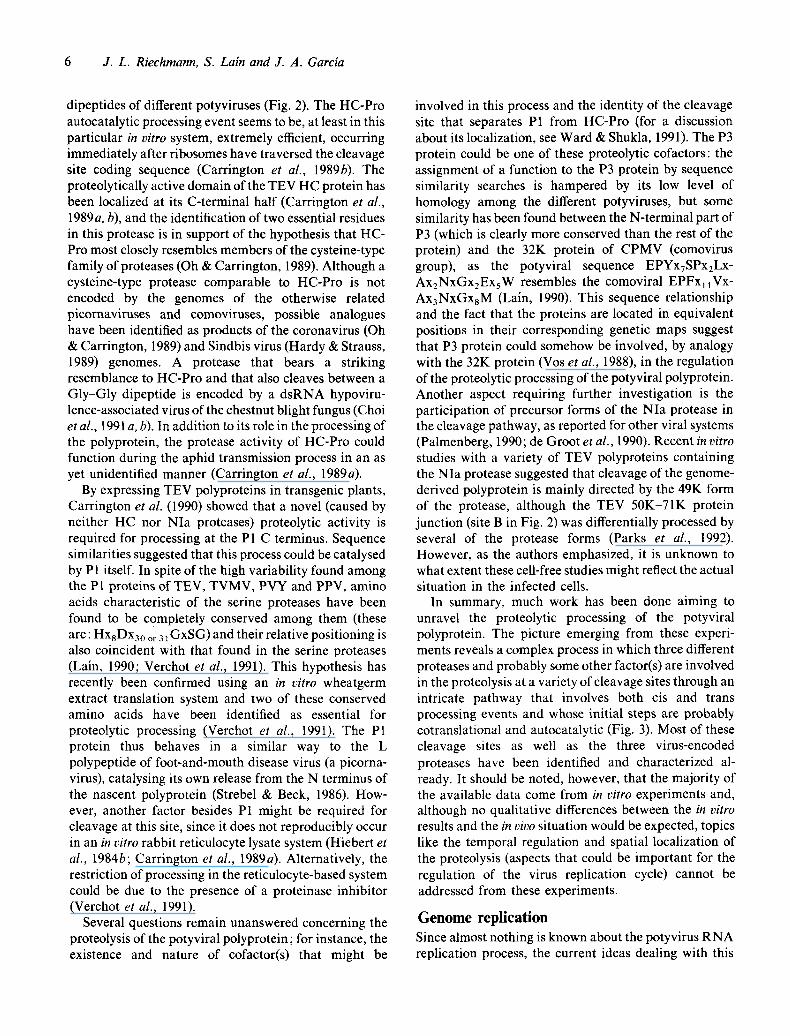
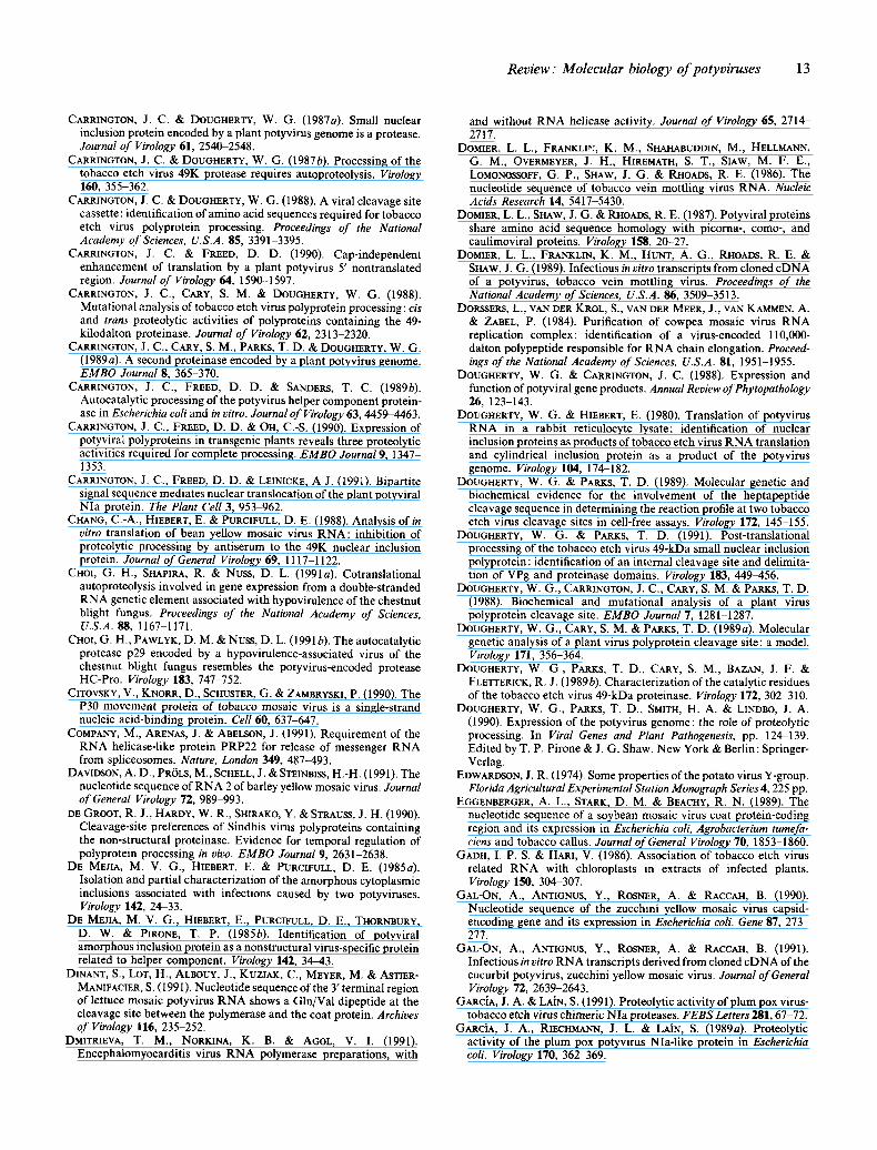
Fig. 2. Deduced amino acid sequences of the demonstrated and putative cleavage sites for the NIa and HC-Pro proteases of four potyviruses and for the NIa-like protease of the potyvirus-related BaYMV. Scissile bonds are indicated by/. A map of the potyviral polyprotein indicating the different protein products, the location of the cleavage sites and the protease responsible for each cleavage is shown in the upper part of the figure. NIa cleavage sites are named A to F, and V.
and in heterologous (Escherichia coli) or homologous (transgenic plants) in vivo systems. Two virus-encoded proteases, NIa (Carrington & Dougherty, 1987a; Hell- mann et al., 1988; Chang et al., 1988; Garcia et al., 1989a; Ghabrial et al., 1990) and HC-Pro (Carrington et al., 1989 a), process co- and post-translationally the large viral precursor polyprotein, and it has been shown recently that the third proteolytic activity that is required for completion of the processing pathway (Carrington et al., 1990) resides in the P1 protein (Verchot et al., 1991). NIa is responsible for cleavages in the C-terminal two- thirds of the polyprotein (Dougherty & Carrington, 1988; Fig. 2), whereas HC-Pro and P1 autocatalytically cleave at their respective C termini (Carrington et al., 1989a, b; Verchot et al., 1991; Fig. 2).
NIa protease cleavage sites are defined by sequences of seven amino acids which have been characterized by a variety of experimental approaches (Carrington & Dougherty, 1987a, 1988; Carrington et al., 1988; Dougherty et al., 1988, 1989a; Garcia et al., 1989a, b). Six of these NIa cleavage sites are present in the potyvirus polyprotein, named A to F in Fig. 2. Cleavages at five of them (B to F) have been demonstrated by analysing the products of proteolytic processing in vitro or in E. coli (Carrington & Dougherty, 1987a; Hellmann et al., 1988; Carrington et al., 1988; Garcia et al., 1989a) and by N-terminal sequencing of several potyviral proteins (Robaglia et al., 1989; MartEn et al., 1990; Ward & Shukla, 1991). Recently, partial cleavage at PPV site A has been shown to occur in vitro (J. A. Garcia & J. L. Riechmann, unpublished results), although cleavage at the TEV site has not been detected (Parks et al., 1992), partial processing at this place presumably happens in vivo: TVMV proteins of 37K and 42K (probably P3 and
P3 + 6K1) have been detected in infected plants by using antibodies raised against a bacterially expressed TVMV 42K protein originating from the P3+6K1 cistron (Rodriguez-Cerezo & Shaw, 1991). Sequence comparison studies of the two products that cleavage at this site would generate, P3 and 6K1, also favour the hypothesis of its existence (Lain et al., 1989a; Lain, 1990). Site A is therefore probably a suboptimal cleavage site that is only partially processed. Amino acids of the heptapeptide sequence (see Fig. 2) can either be essential for a functional cleavage site (Dougherty et al., 1988) or affect the rate of cleavage (Dougherty et al., 1989a), as demonstrated by biochemical and mutational analyses of TEV site F. It has been further shown that different heptapeptide sequences have, at least in vitro, different cleavage reaction profiles (Dougherty & Parks, 1989). The requirement that potyviruses have for the conserva- tion of an extended sequence motif at the NIa cleavage sites is a characteristic not shared by other virus groups using polyprotein processing for genome expression (i.e. como- and picornaviruses; for a review, see Wellink & van Kammen, 1988). Information in the polyprotein for substrate specificity resides mainly at the amino acid sequence level, since NIb-CP heptapeptide junctions inserted into a non-specific protein background were properly processed; additionally, the conformational context might contribute to substrate efficiency (Car- rington & Dougherty, 1988; Garcia et al., 1989b). The conclusions obtained from all these experiments, per- formed mainly with TEV, will probably be true for other potyviruses because, although examination of the clea- vage sites did not reveal an extended conserved cleavage motif used by all of them, a number of biochemical features are maintained. (see Ghabrial et al., 1990).
Rev iew: Molecular biology o f potyviruses 5
However, and although NIa proteases are highly homologous (Garcia et al., 1989a), PPV, TEV and TVMV cleavage sites are efficiently recognized only by their respective proteases (Garcia et al., 1989 b; G arcia & Lain, 1991; Parks & Dougherty, 1991), indicating that very specific interactions must take place during the proteolytic reaction.
NIa protease also appears to catalyse post-transla- tional proteolysis at an internal cleavage site which has been characterized recently for TEV (Dougherty & Parks, 1991). The sequence of this cleavage site (named V in Fig. 2), differs, although maintaining some characteristic features, from the consensus sequences of the other six NIa (A to F) sites; such differences might have the effect of promoting suboptimal cleavage and, in fact, this proteolytic event seems to happen in some but not all the NIa molecules (Dougherty & Parks, 1991). This site is located between the VPg and protease domains of the NIa protein, and its cleavage might therefore be related to some step of the viral RNA replication process (see below).
Experiments using in vitro transcription and transla- tion systems and E. coli expression systems have shown that NIa cleavage sites differ in their susceptibility to processing in cis (intramolecularly) or in trans (intermo- lecularly) (Carrington & Dougherty, 1987a, b; Carring- ton et al., 1988; Hellmann et al., 1988; Garcia et al., 1989a, c, 1990). NIa protease is catalytically active within the polyprotein, excising itself by cis cleavages at sites D and E in a process that might happen as a primary event (maybe cotranslational) in the proteolytic cascade (Carrington & Dougherty, 1987a, b; Carrington et al., 1988; Hellmann et al., 1988; Garcia et al., 1990). In summary, NIa-catalysed proteolytic maturation of the potyviral polyprotein is a regulated process involving cis and trans cleavages at sites which are recognized with different efficiencies and processed at different rates. Therefore, viruses like the potyviruses, unable to regulate their gene expression differentially at the levels of transcription and translation, may have evolved to be capable of regulating the expression of gene products by sequential proteolytic events (Dougherty et al., 1989a). This post-translational regulation could imply that products and precursors have different functions, or it may be a way in which the ratio of inactive : active viral moieties is regulated (Dougherty & Parks, 1989). The possibility that processing intermediates of the proteoly- tic pathway have a function is still an open question (see below).
Molecular genetic analyses have supported the hy- pothesis that NIa and other virus-encoded proteases [such as the picornavirus 3C and the cowpea mosaic virus (CPMV) 24K proteases] are related to the trypsin-like family of cellular serine proteases, but with the substitu-
tion of a Cys as the active site nucleophile (Bazan & Fletterick, 1988; Gorbalenya et al., 1989a). The pro- posed catalytic triad, composed of His, Asp and Cys residues, is located in the C-terminal half of the molecule (Dougherty et al., 1989 b; Garcia et al., 1990; Ghabrial et al., 1990) and, in fact, NIa protease is probably multifunctional as its N-terminal half is not relevant for its proteolytic activity in vitro or in E. coli (Carrington & Dougherty, 1987a; Garcia & Lain, 1991; Dougherty & Parks, 1991) but has been found covalently attached to the genomic RNA (VPg; see below). Interestingly, a substitution of Glu for the catalytic Asp in the TEV and PPV NIa proteases resulted in anomalous proteolytic activities, the effect of these substitutions depending on the particular cleavage site analysed (Dougherty et al., 1989b; Garcia et al., 1990). It could be inferred from these results that there are different structural require- ments in the protease, probably similar in TEV and PPV, for processing at the various cleavage sites. The amino acid similarity among NIa proteases of different potyvir- uses is lower at their C-terminal end, suggesting that this region of the protease may be important in forming the substrate-binding pocket which allows each of the potyviral NIa proteases to recognize its own unique cleavage sequences (Ghabrial et al., 1990). Efforts have been made to delimit protease regions involved in catalysis and in substrate recognition through the construction of hybrid NIa proteases (Garcia & Lain, 1991; Parks & Dougherty, 1991). The results obtained suggest that the recognition and catalytic sites of the NIa proteases are closely interlinked and that the main determinants for substrate specificity lie in the C- terminal one-third of the protease (Garcia & Lain, 1991 ; Parks & Dougherty, 1991). It has also been suggested that the extreme carboxyl end of the protease could be involved in maintenance of the proper structure that would allow other parts of the protein to interact correctly with the various cleavage sites, since the effect caused by different mutations in this region depends on the cleavage site considered (Carrington & Dougherty, 1987a; Garcia et al., 1990). Finally, it has been shown that the potyviral NIa protease processes a great deal of substrate efficiently (Garcia et al., 1989c).
The helper component protease [HC-Pro; a protein formerly known as HC that was renamed HC-Pro for its two activities, insect transmission and protease (Carring- ton et al., 1989a)] is responsible for processing at its own C terminus via an autocatalytic mechanism and, in vitro, exhibits little or no proteolytic activity in trans (Carring- ton et al., 1989a, b). Cleavage in vitro occurs between a Gly-Gly dipeptide (Carrington et al., 1989a) and, although the primary sequence features that define HC- Pro cleavage sites have not been determined, there is a good consensus around the presumed Gly-Gly substrate
6 J . L . Riechmann, S. Lain and J. A. Garcia
dipeptides of different potyviruses (Fig. 2). The HC-Pro autocatalytic processing event seems to be, at least in this particular in vitro system, extremely efficient, occurring immediately after ribosomes have traversed the cleavage site coding sequence (Carrington et al., 1989b). The proteolytically active domain of the TEV HC protein has been localized at its C-terminal half (Carrington et al., 1989 a, b), and the identification of two essential residues in this protease is in support of the hypothesis that HC- Pro most closely resembles members of the cysteine-type family of proteases (Oh & Carrington, 1989). Although a cysteine-type protease comparable to HC-Pro is not encoded by the genomes of the otherwise related picornaviruses and comoviruses, possible analogues have been identified as products of the coronavirus (Oh & Carrington, 1989) and Sindbis virus (Hardy & Strauss, 1989) genomes. A protease that bears a striking resemblance to HC-Pro and that also cleaves between a Gly-Gly dipeptide is encoded by a dsRNA hypoviru- lence-associated virus of the chestnut blight fungus (Choi et al., 1991 a, b). In addition to its role in the processing of the polyprotein, the protease activity of HC-Pro could function during the aphid transmission process in an as yet unidentified manner (Carrington et al., 1989a).
By expressing TEV polyproteins in transgenic plants, Carrington et al. (1990) showed that a novel (caused by neither HC nor NIa proteases) proteolytic activity is required for processing at the P 1 C terminus. Sequence similarities suggested that this process could be catalysed by PI itself. In spite of the high variability found among the PI proteins of TEV, TVMV, PVY and PPV, amino acids characteristic of the serine proteases have been found to be completely conserved among them (these are: HxsDx30 or 31GxSG) and their relative positioning is also coincident with that found in the serine proteases (Lain, 1990; Verchot et al., 1991). This hypothesis has recently been confirmed using an in vitro wheatgerm extract translation system and two of these conserved amino acids have been identified as essential for proteolytic processing (Verchot et al., 1991). The P1 protein thus behaves in a similar way to the L polypeptide of foot-and-mouth disease virus (a picorna- virus), catalysing its own release from the N terminus of the nascent polyprotein (Strebel & Beck, 1986). How- ever, another factor besides P1 might be required for cleavage at this site, since it does not reproducibly occur in an in vitro rabbit reticulocyte lysate system (Hiebert et al., 1984b; Carrington et al., 1989a). Alternatively, the restriction of processing in the reticulocyte-based system could be due to the presence of a proteinase inhibitor (Verchot et al., 1991).
Several questions remain unanswered concerning the proteolysis of the potyviral polyprotein; for instance, the existence and nature of cofactor(s) that might be
involved in this process and the identity of the cleavage site that separates P1 from HC-Pro (for a discussion about its localization, see Ward & Shukla, 1991). The P3 protein could be one of these proteolytic cofactors: the assignment of a function to the P3 protein by sequence similarity searches is hampered by its low level of homology among the different potyviruses, but some similarity has been found between the N-terminal part of P3 (which is clearly more conserved than the rest of the protein) and the 32K protein of CPMV (comovirus group), as the potyviral sequence EPYxTSPx2Lx- Ax2NxGx2ExsW resembles the comoviral EPFxllVx- Ax3NxGxsM (Lain, 1990). This sequence relationship and the fact that the proteins are located in equivalent positions in their corresponding genetic maps suggest that P3 protein could somehow be involved, by analogy with the 32K protein (Vos et al., 1988), in the regulation of the proteolytic processing of the potyviral polyprotein. Another aspect requiring further investigation is the participation of precursor forms of the NIa protease in the cleavage pathway, as reported for other viral systems (Palmenberg, 1990; de Groot et al., 1990). Recent in vitro studies with a variety of TEV polyproteins containing the NIa protease suggested that cleavage of the genome- derived polyprotein is mainly directed by the 49K form of the protease, although the TEV 50K-71K protein junction (site B in Fig. 2) was differentially processed by several of the protease forms (Parks et al., 1992). However, as the authors emphasized, it is unknown to what extent these cell-free studies might reflect the actual situation in the infected cells.
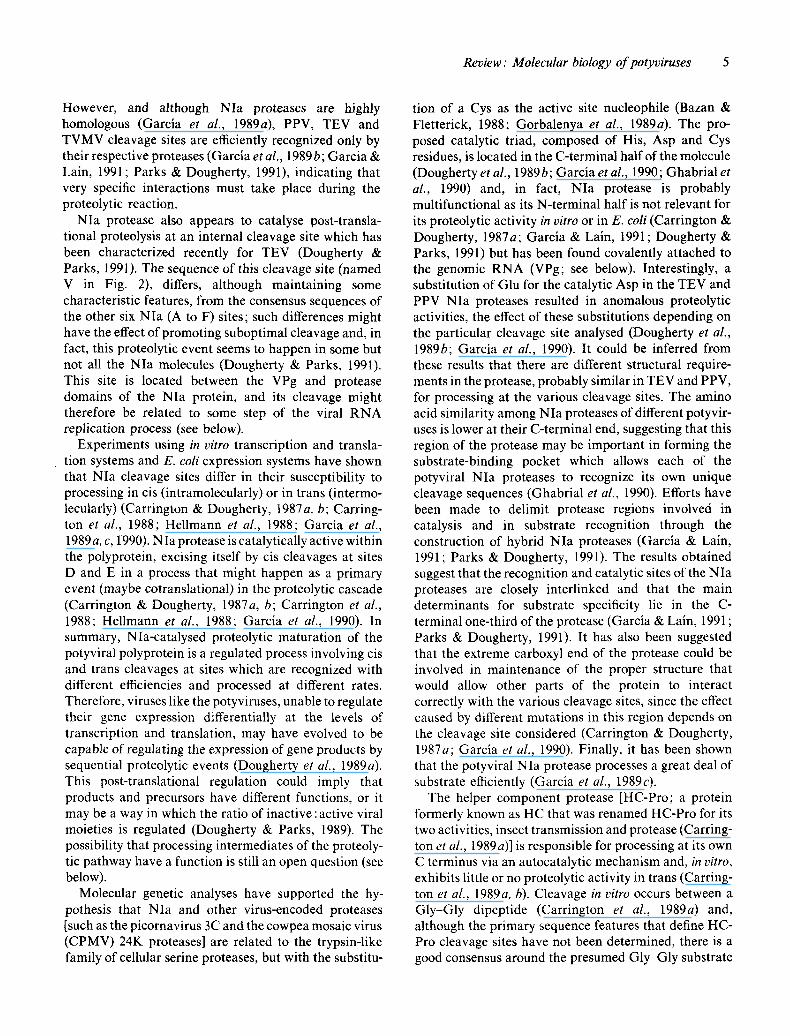
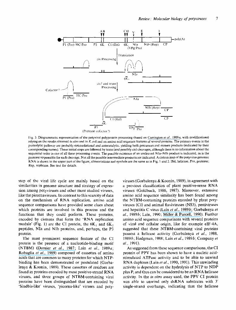
In summary, much work has been done aiming to unravel the proteolytic processing of the potyviral polyprotein. The picture emerging from these experi- ments reveals a complex process in which three different proteases and probably some other factor(s) are involved in the proteolysis at a variety of cleavage sites through an intricate pathway that involves both cis and trans processing events and whose initial steps are probably cotranslational and autocatalytic (Fig. 3). Most of these cleavage sites as well as the three virus-encoded proteases have been identified and characterized al- ready. It should be noted, however, that the majority of the available data come from in vitro experiments and, although no qualitative differences between the in vitro results and the in vivo situation would be expected, topics like the temporal regulation and spatial localization of the proteolysis (aspects that could be important for the regulation of the virus replication cycle) cannot be addressed from these experiments.
Genome replication Since almost nothing is known about the potyvirus RNA replication process, the current ideas dealing with this
Review: Molecular biology o f potyviruses 7
' I Pl (Pro) HC-Pro
Cofactor ? ~ . ~
Pl (Pro) HC-Pro
P3
AB CD E F vv vv v v II II I I I t -p°ly¢A)
6K~ CI (Hel) 6K. Nla Nlb (Rep) CP (VPg/Pro)
cis Processing / Translati°n
, , Nla
6~, t I I cis'? and trans I
Processing I
C N la
P3 VPg Pro (Protease cofactor'?)
NIb (Rep) CP
I
CP
Fig. 3. Diagrammatic representation of the potyviral polyprotem processing (based on Carrington et al., 1989a, with modifications) relying on the results obtained in vitro and in E. coli and on amino acid sequence features of several proteins. The primary events in the proteolytic pathway are probably cotranslational and autocatalytic, yielding both precursors and mature products (indicated by their corresponding names). These initial steps are followed by trans (and possibly cis) cleavages, although there is no information about the sequential order in vivo of all these processing events. The possible existence of an uncleaved NIa-NIb product is indicated, as is the protease responsible for each cleavage. Not all the possible intermediate products are indicated. A cistron map of the potyvirus genomic RNA is shown in the upper part of the figure; abbreviations and symbols are the same as in Fig. 1 and 2. Hel, helicase; Pro, protease; Rep, replicase. See text for details.
step of the viral life cycle are mainly based on the similarities in genome structure and strategy of expres- sion among potyviruses and other more studied viruses, like the picornaviruses. In contrast to this scarcity of data on the mechanism of RNA replication, amino acid sequence comparisons have provided some clues about which proteins are involved in this process and the functions that they could perform. These proteins, encoded by cistrons that form the 'RNA replication module' (Fig. 1) are the CI protein, the 6K~ and 6K2 peptides, NIa and NIb proteins, and, perhaps, the P3 protein.
The most prominent sequence feature of the CI protein is the presence of a nucleotide-binding motif (NTBM) (Domier et al., 1987; Lain et al., 1989a; Robaglia et al., 1989) composed of cassettes of amino acids that are common to many proteins for which NTP- binding has been demonstrated or postulated (Gorba- lenya & Koonin, 1989). These cassettes of residues are found in proteins encoded by most positive-strand RNA viruses, and three groups of NTBM-containing viral proteins have been distinguished that are encoded by 'Sindbis-like' viruses, 'picorna-like' viruses and poty-
viruses (Gorbalenya & Koonin, 1989), in agreement with a previous classification of plant positive-sense RNA viruses (Goldbach, 1986, 1987). Moreover, extensive amino acid sequence similarity has been found among the NTBM-containing proteins encoded by plant poty- viruses (CI) and animal flaviviruses (NS3), pestiviruses and hepatitis C virus (Lain et al., 1989b; Gorbalenya et al., 1989b; Lain, 1990; Miller & Purcell, 1990). Further amino acid sequence comparisons with several proteins of viral and cellular origin, like for example elF-4A, suggested that these NTBM-containing viral proteins possess a helicase activity (Gorbalenya et al., 1988, 1989 b; Hodgman, 1988; Lain et al., 1989 b; Company et al., 1991).
As suggested from these sequence comparisons, the CI protein of PPV has been shown to have a nucleic acid- stimulated ATPase activity and to be able to unwind RNA duplexes (Lain et al., 1990, 1991). This unwinding activity is dependent on the hydrolysis of NTP to NDP plus Pi and thus can be considered to be an RNA helicase activity. In the in vitro assay used, the PPV CI protein was able to unwind only dsRNA substrates with 3' single-strand overhangs, indicating that the helicase

8 J . L . Riechmann, S. Lain and J. A. Garcia
activity functions in the 3' to 5' direction (Lain et al., 1990). This was the first report on a helicase activity associated with a protein encoded by an RNA virus. Although still scarce, experimental data indicate that the NTBM-containing proteins which the majority of positive-strand RNA virus genomes encode are involved in their replication. The genome of brome mosaic virus (BMV; bromovirus group; 'Sindbis-like' supergroup of plant viruses) encodes a helicase-like protein, named the la protein (Hodgman, 1988), which is required for continued accumulation of all classes of BMV RNA (positive-strand, negative-strand and subgenomic) (Kroner et al., 1990). In agreement with the presumed necessity of an unwinding activity for RNA replication, it has been found that an RNA helicase activity is present in RNA polymerase preparations of encephalo- myocarditis virus (a picornavirus; Dmitrieva et al., 1991) and alfalfa mosaic virus (ilarvirus group; Quadt et al., 1991). Moreover, an in vitro replication system able to catalyse complete replication of cucumber mosaic virus (CMV) RNAs has been developed recently (Hayes & Buck, 1990), and it was shown that the helicase-like CMV protein is an essential subunit of the purified RNA-dependent RNA polymerase (Hayes & Buck, 1990). In addition, the 2C protein, the NTBM-contain- ing protein of poliovirus, seems to be involved in the release of newly synthesized positive-sense RNA mol- ecules from the replication complex (Bienz et al., 1987; Li & Baltimore, 1988), an event which could be nicely explained by the action of an RNA helicase activity. It should be noted, however, that only the N-terminal region of the CI protein is related by sequence comparisons with the helicase activity (Lain et al., 1989b). Therefore, the C-terminal region of about 230 amino acids might be dispensable for the helicase activity. It could be involved in some of the other functions which have been proposed for the CI protein, for example the attachment of the replication complex to membranes (Domier et al., 1987) in a similar way to the NTBM-containing protein, 2C, of poliovirus (Bienz et al., 1987), the induction of vesicles in the infected cells (Calder & Ingerfeld, 1990), or the virus transport from cell to cell (Langenberg, 1986; see below). Given the aforementioned ubiquity of helicase-like proteins among positive-strand RNA viruses, suggesting that RNA unwinding is probably a general requirement for genomic replication, the study of the enzymic properties of the potyvirus CI protein will be of interest in the further understanding of RNA replication mechanisms and in the design of strategies to interfere with positive- strand RNA virus infections.
The two small peptides 6K1 and 6K2 that are predicted from the amino acid sequence of the potyviral polyprotein might also play a role in RNA replication,
although they have not yet been identified in vivo. The presence of a stretch of hydrophobic amino acids in the 6K1 and in the 6K2 peptides, and the relative position of their cistrons in the potyviral genetic map (see Fig. 1) suggest that they could be analogous to the picornaviral 2B and 3A peptides, respectively (Lain et al., 1989a). Protein 2B seems to be involved in RNA replication (Johnson & Sarnow, 1991), and the function that has been proposed for 3A is the membrane binding of the poliovirus VPg precursor, protein 3AB (Giachetti & Semler, 1991). It is thus conceivable that 6K 2 could anchor the NIa/VPg (see below) to the membranes and that processing at the 6K2-NIajunction might be related to some step of the RNA replication process, although cleavage at this site might not be the last processing event to release the VPg from the polyprotein precursor (see below).
NIa and NIb must be involved in RNA replication; the NIb protein has been postulated as the potyviral RNA-dependent RNA polymerase based on the pres- ence of conserved sequence motifs characteristic of these enzymes (Domier et al., 1987; Lain et al., 1989a; Robaglia et al., 1989; Poch et al., 1989). In addition to its role in the proteolytic processing of the potyviral polyprotein, for which only its carboxyl half is required (see above), the NIa protein (or its N-terminal part) has been shown to be the VPg. The VPgs of TVMV (Siaw et al., 1985) and PPV (Riechmann et al., 1989) have been identified as proteins of 24K and 22K, respectively. The TVMV VPg cistron has been mapped showing that the VPg is the N-terminal 24K portion of the NIa protein (Shahabuddin et al., 1988), and the TEV VPg has been found to be either the 49K NIa protein of its N-terminal 24K half (Murphy et al., 1990). It would be interesting to see whether the complete TVMV and PPV NIa proteins could be found attached to their respective RNAs, and whether the presence of the two different proteins at the 5' end ofTEV RNA has any functional significance. The cleavage site that separates the VPg and protease domains of NIa protein has been identified (Dougherty & Parks, 199l ; see above), and its proteolysis might be temporarily or functionally connected with the RNA replication process. It is interesting to note that this is a suboptimal cleavage site, and the final VPg precursor might therefore be the whole NIa molecule. In contrast, the VPg precursors of the related como- and picorna- viruses are the 60K (58K protein + VPg) and 3AB polyproteins, respectively (Vos et al., 1988; Giachetti & Semler, 1991; see Fig. 1), indicating that different mechanisms might accomplish the necessary tuning between proteolytic and replication processes that must be similar to a certain extent among these viruses. A Tyr residue in the NIa protein of TVMV has recently been shown to link the TVMV VPg to the genomic RNA
Rev i ew : Molecular biology o f potyviruses 9
(Murphy et al., 1991). This residue is found in a block of amino acids completely conserved in the NIa proteins of TVMV, TEV, PPV and PVY (Murphy et al., 1991). Studies on the function of the poliovirus VPg, which is also linked to the RNA via a Tyr residue (Ambros & Baltimore, 1978; Rothberg et al., 1978), suggest that it is involved in RNA replication acting as primer (Takeda et al., 1986) and/or cleaving a replicative form of the RNA (Tobin et al., 1989). Interestingly, a segment of the N- terminal part of the NIa proteins of PPV, TEV, TVMV and PVY contains a high content of aromatic amino acids (Tyr and Phe), and Gly and basic amino acids (Lain, 1990); this composition is typical of the consensus recognition motifs of proteins that interact with RNA (Query et al., 1989).
A striking characteristic of the NIa and NIb proteins is the fact that they aggregate in equimolar ratios to form the nuclear inclusions that are induced by several pctyviruses (Knuhtsen et al., 1974; Dougherty & Hiebert, 1980; Dougherty & Carrington, 1988, and references therein). TEV proteins NIa and NIb were found to accumulate predominantly in the nucleus of TEV-infected cells at all times during which viral antigens could be detected, implying that they possess efficient nuclear transport signals (Restrepo et al., 1990; Baunoch et al., 1991) which have recently been identified (Carrington et al., 1991), but the functional significance of this transport is not understood. Potyviruses are believed, by analogy to picornaviruses, to replicate in the cytoplasm of the infected cells in a membrane-associated process. In addition, the CI protein is localized only in the cytoplasm (Restrepo et al., 1990). Thus, given the strategy of expression of potyviral genomes by polypro- tein processing, the fraction of NIa and NIb proteins localized to the nucleus may represent accumulation in excess of that needed for replicase formation (Restrepo et al., 1990). Association of TEV negative-strand RNA with chloroplasts in extracts of infected plants has been reported (Gadh & Hari, 1986) and a crude membrane fraction from PPV-infected plants has been shown to be able to catalyse specific PPV RNA synthesis (Martin & Garcia, 1991). Nevertheless, we are still lacking clear data on the subcellular localization of potyviral replica- tion, data that might be difficult to obtain given the varied morphological alterations that are induced in potyvirus-infected cells (for example, wheat streak mosaic virus induces the invagination of the nuclear membrane, thereby sometimes introducing cytoplasm into the nucleus; A Nassuth, personal communication). Timing of specific events (e.g. translation, RNA synthesis, encapsidation) in the replication cycle may be controlled, at least partially, by the levels of non- structural proteins present within the cytoplasm (Res- trepo et al., 1990). It should be remembered, however,
that not all potyviruses induce the formation of nuclear inclusions. In addition to NIa and NIb, a high Mr product (94K to 110K) formed by both proteins has been observed in the nuclear inclusions (Knuhtsen et al., 1974; Hiebert et al., 1984a; Martin et al., 1990). Terminal amino acid sequencing of the PPV product (110K) has suggested that it is a non-processed NIa-NIb polypep- tide (Martin et al., 1990). But whether this potyvirus polypeptide is processed or not, its functional signifi- cance remains to be confirmed. In this regard, it is interesting to note that an uncleaved protease-polymer- ase precursor of CPMV has been shown to be involved in RNA replication (Dorssers et al., 1984). It has also been found that a functional ribonucleoprotein complex, composed of a cellular protein and the two poliovirus proteins (3C pr° and 3D p°l) equivalent to potyviral NIa and NIb, forms around the 5' end of poliovirus RNA and is required for positive-strand RNA synthesis but dispensable for negative-strand synthesis (Andino et al., 1990). It is speculated that these two proteins might be present in that complex as an uncleaved precursor (Andino et al., 1990).
In summary, the potyviral replication process is poorly understood. However, the recent preparation of a membrane fraction from infected cells able to catalyse PPV RNA synthesis (Martin & Garcia, 1991) might be a first step towards the development of well-defined in vitro RNA replication systems that will greatly help in the unravelling of this process.
Virus spread
Four viral proteins, P1, CI, HC and CP, have been suggested or demonstrated to be involved in either ceil- to-cell or plant-to-plant virus propagation.
The P1 protein has been proposed to be involved in the cell-to-cell spread of the infection on the basis of sequence similarity between P1 of TVMV (but not of TEV, PPV and PVY) and the 30K movement protein of tobacco mosaic virus (TMV; Domier et al., 1987; Lain et al., 1989a; Robaglia et al., 1989). This sequence similarity involves the 30K TMV protein domain that has been implicated by Citovsky et al. (1990) in RNA binding. Since proteins presumably involved in cell-to- cell movement appear to display very little similarity, even among members of the same group (reviewed in Hull, 1989), a fact that could reflect the specificity of the virus host interactions, the lack of sequence relation- ships does not permit exclusion of a cell-to-cell spread function for the potyviral N-terminal protein. Also this function cannot be ruled out by the recent finding that P 1 has a proteolytic activity, because, since the proposed protease domain of P1 is confined to its carboxyl region (Verchot et al., 1991), additional function(s) might be
10 J. L. Riechmann, S. L a & and J. A. Garcla
performed by this protein. In fact, its low conservation and its localization at the 5' end of the polyprotein, as in the case of the proposed movement proteins of the related como- (Wellink & van Kammen, 1989; van Lent et al., 1990) and nepoviruses, support this hypothesis (Lain et al., 1989a).
Another protein that might be involved in cell-to-cell spread is the CI protein, as suggested on the basis of electron microscopy observations. The CI protein forms the cytoplasmic pinwheel or scroll-shaped inclusions that are induced in all potyvirus-infected plants (Edwardson, 1974; Dougherty & Hiebert, 1980). These inclusions have been found associated with the plasma membrane, with plasmodesmata, with the endoplasmic reticulum, and free in the cytoplasm (Lawson & Hearon, 1971; Langenberg, 1986; Lesemann, 1988; Baunoch et al., 1991). Additionally, CP or virions of several viruses have been detected by immunogold labelling techniques associated with these inclusion bodies (Langenberg, 1986; Langenberg & Purcifull, 1989; Langenberg et al., 1989). Based only on these ultrastructural observations, it has been proposed that the cylindrical inclusions, and therefore the CI protein, could be involved in virus transport from cell to cell through plasmodesmata (Lawson & Hearon, 1971; Langenberg, 1986; Calder & Ingerfeld, 1990). It has also been suggested that the CI protein could serve a structural function within the inclusion body, possibly to modify the cell cytoskeleton to adapt it for virus synthesis and/or translocation (Brakke et al., 1987).
As is the case with most plant viruses, natural plant-to- plant spread is accomplished by specific vectors, which for the majority of potyviruses are aphids. The HC protein is involved in the aphid transmission of potyviruses and must be acquired by the insect in conjunction with the virus (Pirone & Thornbury, 1983; Thornbury & Pirone, 1983; Hiebert et al., 1984b; Thornbury et al., 1985; Berger & Pirone, 1986). The HC protein has been associated with the amorphous inclu- sions that are induced by some potyviruses by several criteria (Hiebert et al., 1984b; De Mejia et al., 1985a, b; Baunoch et al., 1990), although functional studies have suggested that either the inclusion-bound form of this protein has been inactivated or, alternatively, the helper component activity is associated with a modified form of the inclusion protein (Thornbury & Pirone, 1983; Thornbury et al., 1985; Dougherty & Carrington, 1988). The biologically active form of the HC protein is believed to be a dimer (Hellmann et al., 1983; Thornbury et al., 1985). As Berger & Pirone (1986) have demon- strated, the HC protein mediates a selective localization of the virus into aphid mouthparts, suggesting a binding mechanism for the mode of action of HC. Since the proteolytically active domain has been localized to the C-
terminal half of the TEV HC protein (Carrington et al., 1989b; see above), it seems likely that the sequences responsible for its role in transmission might be localized at its N-terminal region, which has a level of conserva- tion among different potyviruses similar to that of the carboxyl region. The HC amino acid sequence of PVC (an aphid non-transmissible strain of PVY) and an aphid-transmissible strain of PVY have been determined and compared, and two of the differences that exist between these two HCs are localized in the homologous regions of the N-terminal part of HC which are conserved among all potyviruses sequenced to date (Thornbury et al., 1990). One of these two changes is found in a cysteine-rich region that might allow the dimerization of the molecule by the common fixation of metal atoms (Robaglia et al., 1989). These two amino acids are prime targets for mutational analyses of HC activity (Thornbury et al., 1990), studies that could be performed using transgenic plants expressing HC, although the use of full-length clones should be much more convenient (Berger et al., 1989).
The CP has been the most extensively characterized potyviral gene product in terms of sequence data (Ward & Shukla, 1991 and references therein). The interest in CP comes mainly from its usefulness in taxonomic and evolutionary studies, in diagnosis and in the so-called CP-mediated resistance, topics that have been exten- sively reviewed elsewhere [see Shukla & Ward (1988, 1989a, b), Shukla et al. (1991) and Ward & Shukla (1991), for taxonomy and diagnosis, and Beachy et al. (1990) for CP-mediated resistance]. For an extensive review on CP structure, see Shukla & Ward (1989b). The primary function of CP is, obviously, to encapsidate viral RNA, a process in which the highly conserved central and C-terminal regions characteristic of potyviral CPs are likely to be involved (Shukla & Ward, 1989b). In fact, the C-terminal part of potyvirus CPs shares sequence similarity with the CPs of the otherwise unrelated (in terms of genomic structure and strategy of expression) filamentous plant viruses potex-, carla-, clostero- and tobamoviruses (Morozov et al., 1987; Lain et al., 1988; Dinant et al., 1991). The molecular characterization of particle assembly and genome encapsidation processes would be facilitated by the recent development of micro- bial (E. coli and yeast) assembly systems (Jagadish et al., 1991). The high variability observed in the surface- exposed N-terminal region of potyvirus CP (Ward & Shukla, 1991) suggests that it is involved in virus-specific functions or host-vector-virus interactions. The CP could, for example, affect host range and symptom induction: the CP gene of a rod-shaped plant RNA virus, TMV, encodes a host response determinant (Saito et al., 1987). Sequence data have accumulated to support the hypothesis (Harrison & Robinson, 1988; Lain et al.,
Review: Molecular biology o f potyviruses l 1
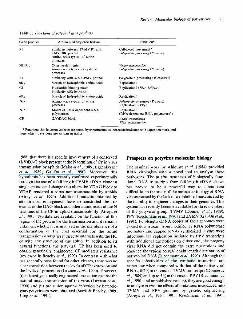
Table 1. Functions of potyviral gene products
Gene product Amino acid sequence feature Function*
P1 Similarity between TVMV P1 and Cell-to-cell movement? TMV 30K protein Polyprotein processing (Protease) Amino acids typical of serine proteases
HC-Pro Cysteine-rich region Amino acids typical of cysteine proteases
P3 Similarity with 32K CPMV protein Polyprotein processing? (Cofactor?)
6K~ Stretch of hydrophobic amino acids Replication?
CI Nucleotide-binding motif Replication? (RNA helicase) Similarity with helicases
6K2 Stretch of hydrophobic amino acids Replication?
NIa Amino acids typical of serine Polyprotein processing (Protease) proteases Replication? (VPg)
Nlb Motifs of RNA-dependent RNA Replication? polymerases (RNA-dependent RNA polymerase?)
CP (I/V)DAG block Aphid transmission RNA encapsidation
Vector transmission Polyprotein processing (Protease)
* Functions that have not yet been supported by experimental evidence are indicated with a question mark, and those which have been are written in italics.
1988) that there is a specific involvement of a conserved (I/V)DAG block present at the N terminus of CP in virus transmission by aphids (Maiss et al., 1989; Eggenberger et al., 1989; Gal-On et al., 1990). Moreover, this hypothesis has been recently confirmed experimentally through the use of a full-length TVMV cDNA clone: a single amino acid change that alters the VDAG block to VDAE rendered a virus non-transmissible by aphids (Atreya et al., 1990). Additional mutants obtained by site-directed mutagenesis have demonstrated the rel- evance of the DAG block and other amino acids at the N terminus of the CP in aphid transmissibility (Atreya et al., 1991). No data are available on the function of this region of the protein for the transmission and it remains unknown whether it is involved in the maintenance of a conformation of the coat essential for the aphid transmission or whether it directly interacts with the HC or with any structure of the aphid. In addition to its natural functions, the potyviral CP has been used to obtain genetically engineered CP-mediated resistance (reviewed in Beachy et al., 1990). In contrast with what has generally been found for other viruses, there was no clear correlation between the levels of CP expression and the levels of protection (Lawson et al., 1990). However, (i) efficient genetically engineered protection against the natural insect transmission of the virus (Lawson et al.,
1990) and (ii) protection against infection by heterolo- gous potyviruses were obtained (Stark & Beachy, 1989; Ling et al., 1991).
Prospects on potyvirus molecular biology
The seminal work by Ahlquist et al. (1984) provided RNA virologists with a novel tool to analyse these pathogens. The in vitro synthesis of biologically func- tional RNA transcripts from full-length eDNA clones has proved to be a powerful way to circumvent difficulties in the study of the molecular biology of RNA viruses caused by the lack of well-defined mutants and by the inability to engineer changes in their genomes. This system has recently become available for three members of the potyvirus group, TVMV (Domier et al., 1989), PPV (Riechmann et al., 1990) and ZYMV (Gal-On et al., 1991). Full-length cDNA copies of their genomes were cloned downstream from modified T7 RNA polymerase promoters and capped RNAs synthesized in vitro were infectious. On replication initiated by PPV transcripts with additional nucleotides on either end, the progeny viral RNA did not contain the extra nucleotides and regained the typical poly(A) chain length distribution of native viral RNA (Riechmann et al., 1990). Although the specific infectivities of the synthetic transcripts are rather low when compared with that of the native viral RNAs, 0.2 % in the case of TVMV transcripts (Domier et al., 1989) and up to 5% in the case ofPPV (Riechmann et al., 1990; and unpublished results), they are good enough to analyse in vivo the effects of mutations introduced into TVMV and PPV genomes by genetic engineering (Atreya et at., 1990, 1991; Riechmann et al., 1991;
12 J. L, Riechmann, S. Lain and J. A. Garcia
Rodriguez-Cerezo et al., 1991). As an alternative approach to the in vitro synthesis of viral RNA, it has recently been reported that potyvirus infection can be generated by plasmid D N A when a 35S promoter sequence upstream from the viral cDNA is provided (Maiss et al., 1992). A good example of the important and unexpected results that can be obtained by the use of cDNA clones is the recent discovery that a determinant of disease symptom severity is located in the 3'-terminal non-coding region of TVMV RNA (Rodriguez-Cerezo et al., 1991). Although the construction and manipulation of full-length potyviral cDNA clones has proved to be a substantial task, it is expected that this system will be developed for more potyviruses in the forthcoming years and that, coupled with protoplast systems (Luciano et al., 1987; Domier et al., 1989; Atreya et al., 1990), which have been underemployed in potyvirus research owing to experimental difficulties, and with transgenic plants expressing potyviral gene products (Berger et al., 1989; Graybosch et al., 1989), will greatly help in the further unravelling of the potyvirus life cycle.
This review illustrates how both our understanding of potyviruses and the experimental approaches to study them have enlarged and changed during the last 5 years. Functions have been proposed for the different potyviral proteins based on sequence data and some of them have been tested using in vitro and in vivo systems (Table 1). It is a recurrent suggestion that some proteins could perform more than one function and might therefore possess two-domain structures. These functions can now be studied in vivo using molecular biological techniques, since the development of cDNA clones from which infectious in vitro transcripts can be obtained has greatly increased the scope of possible experiments with which to examine the life cycle of these viruses. It is possible to modify their genomes by site-directed mutagenesis and mapping of specific mutant phenotypes should also be feasible. Lethal mutations could also be studied and a uniformity of inoculum not previously available is provided. The possibility to perform investigations that have not been possible hitherto is therefore already open. And although almost nothing is known about the regulation of the potyviral life cycle, it should also be amenable to the currently available genetic engineering techniques. It is therefore easy to predict that the next 5 years will be even more fruitful and exciting.
We are grateful to Drs E. Rodriguez-Cerezo, J. G. Shaw, J. C. Carrington, W. G. Dougherty, A. Nassuth, D. D. Shukla, B. Raccah and E. Maiss for communicating results and providing manuscripts prior to publication, and to Drs F. Rojo and F. Gebauer for valuable comments on the manuscript. Work carried out in the authors' laboratory was supported by grants BIO90-1127-E from CICYT and C168/90 from Comunidad de Madrid, and by an institutional grant from Fundaci6n Rambn Areces. J.L.R. and S.L. were recipients of fellowships from CSIC.
References AHLQUIST, P., FRENCH, R., JANDA, M. & LOESCH-FRIES, L. S. (1984).
Multicomponent RNA plant virus infection derived from cloned viral cDNA. Proceedings of the National Academy of Sciences, U.S.A. 81, 7066-7070.
ALLISON, R. F., SORENSON, J. C., KELLY, M. E., ARMSTRONG, F. B. & DOUGHERTY, W. G. (1985). Sequence determination of the capsid protein gene and flanking regions of tobacco etch virus: evidence for synthesis and processing of a polyprotein in potyvirus genome expression. Proceedings of the National Academy of Sciences, U.S.A. 82, 3969-3972.
ALLISON, R., JOHNSTON, R. E. & DOUGHERTY, W. G. (1986). The nucleotide sequence of the coding region of tobacco etch virus genomic RNA: evidence for the synthesis of a single polyprotein. Virology 154, 9-20.
ALTMANN, M., BLUM, S., WILSON, T. M. A. & TRACrlSEL, H. (1990). The Y-leader sequence of tobacco mosaic virus RNA mediates initiation-factor-4E-independent, but still initiation-factor-4A-de- pendent translation in yeast extracts. Gene 91, 127-129.
AMBROS, V. & BALTIMORE, D. (1978). Protein is linked to the 5'-end of poliovirus RNA by a phosphodiester linkage to tyrosine. Journal of Biological Chemistry 60, 5263-5266.
ANDINO, R., RIECKHOF, G. E. & BALTIMORE, D. (1990). A functional ribonucleoprotein complex forms around the 5' end of poliovirus RNA. Cell 63, 369-380.
ATREYA, C. D., RACCAH, B. & PIRONE, T. P. (1990). A point mutation in the coat protein abolishes aphid transmissibility of a potyvirus. Virology 178, 161-165.
A'rR~YA, P. L., A'rREYA, C. D. & PIRONE, T. P. (1991). Amino acid substitutions in the coat protein result in loss of insect transmissibi- lity of a plant virus. Proceedings of the National Academy of Sciences, U.S.A. 88 7887-7891.
BARNETT, O. W. (1991). Potyviridae, a proposed family of plant viruses. Archives of Virology 118, 139-141.
BAUNOCH, D. A., DAS, P. & HARI, V. (1990). Potato virus Y helper component protein is associated with amorphous inclusions. Journal of General Virology 71, 2479-2482.
BAUNOCH, D. A., DAS, P. BROWNING, M. E. & HARI, V. (1991). A temporal study of the expression of the capsid, cytoplasmic inclusion and nuclear inclusion proteins of tobacco etch potyvirus in infected plants. Journal of General Virology 72, 487-492.
BAZAN, J. F. & FLETTERICK, R. J. (1988). Viral cysteine proteases are homologous to the trypsin-like family of serine proteases: structural and functional implications. Proceedings of the National Academy of Sciences, U.S.A. 85, 7872-7876.
BEACHY, R. N., LOESCH-FRIES, S. & TUMER, N. E. (1990). Coat protein- mediated resistance against virus infection. Annual Review of Phytopathology 2,8, 451-474.
BERGER, P. H. & PIRONE, T. P. (1986). The effect of helper component on the uptake and localization of potyviruses in Myzus persicae. Virology 153, 256-261.
BERGER, P. H., HUNT, A. G., DOMIER, L. L., HELLMANN, G. M., STRAM, Y., THORNBURY, D. W. & PIRONE, T. P. (1989). Expression in transgenic plants of a viral gene product that mediates insect transmission of potyviruses. Proceedings of the National Academy of Sciences, U.S.A. 86, 8402-8406.
BIENZ, K., EGGER, D. & PASAMONTES, L. (1987). Association of polioviral proteins of the P2 genomic region with the viral replication complex and virus-induced membrane synthesis as visualized by electron microscopic immunocytochemistry and autoradiography. Virology 160, 220-226.
BRAKKE, M. K., BALL, E. M., Hsu, Y. H. & LANGENBERG, W. G. (1987). Wheat streak mosaic virus cylindrical inclusion body protein. Journal of General Virology 68, 281-287.
BRAULT, V., HIBRAND, L., CANDRESSE, T., LE GALL, O. & DUNEZ, J. (1989). N ucleotide sequence and genetic organization of Hungarian grapevine chrome mosaic nepovirus RNA 2. Nucleic Acids Research 17, 7809-7819.
CALDER, V. L. & INGERFELD, M. (1990). The roles of the cylindrical inclusion protein of a potyvirus in the induction of vesicles and in cell-to-cell spread. Journal of Structural Biology 105, 62-66.
Review: Molecular biology o f potyviruses 13
CARRINGTON, J. C. & DOUGHERTY, W. G. (1987a). Small nuclear inclusion protein encoded by a plant potyvirus genome is a protease. Journal of Virology 61, 2540-2548.
CARRINGTON, J. C. & DOUGHERTY, W. G. (1987b). Processing of the tobacco etch virus 49K protease requires autoproteolysis. Virology 160, 355-362.
CARRINGTON, J. C. & DOUGHERTY, W. G. (1988). A viral cleavage site cassette: identification of amino acid sequences required for tobacco etch virus polyprotein processing. Proceedings of the National Academy of Sciences, U.S.A. 85, 3391-3395.
CARRINGTON, J. C. & FREED, n . D. (1990). Cap-independent enhancement of translation by a plant potyvirus 5' nontranslated region. Journal of Virology 64, 1590-1597.
CARRINGTON, J. C., CARY, S. M. & DOUGHERTY, W. G. (1988). Mutational analysis of tobacco etch virus polyprotein processing: cis and trans proteolytic activities of polyproteins containing the 49- kilodalton proteinase. Journal of Virology 62, 2313-2320.
CARRINGTON, J. C., CARY, S. M., PARKS, T. D. & DOUGHERTY, W. G. (1989a). A second proteinase encoded by a plant potyvirus genome. EMBO Journal 8, 365-370.
CARRINGTON, J. C., FREED, D. D. & SANDERS, T. C. (1989b). Autocatalytic processing of the potyvirus helper component protein- ase in Escherichia coli and in vitro. Journal of Virology 63, 4459-4463.
CARRINGTON, J. C., FREED, D. D. & OH, C.-S. (1990). Expression of potyviral polyproteins in transgenic plants reveals three proteolytic activities required for complete processing. EMBO Journal 9, 1347- 1353.
CARRINGTON, J. C., FREED, D. D. & LEINICKE, A J. (1991). Bipartite signal sequence mediates nuclear translocation of the plant potyviral NIa protein. The Plant Cell 3, 953462.
CHANG, C.-A., HIEBERT, E. & PURCIFULL, D. E. (1988). Analysis of in vitro translation of bean yellow mosaic virus RNA: inhibition of proteolytic processing by antiserum to the 49K nuclear inclusion protein. Journal of General Virology 69, 1117-1122.
CHOI, G. H., SHAPIRA, R. ~g NUSS, D. L. (1991a). Cotranslational autoproteolysis involved in gene expression from a double-stranded RNA genetic element associated with hypovirulence of the chestnut blight fungus. Proceedings of the National Academy of Sciences, U.S.A. 88, 1167-1171.
CHOI, G. H., PAWLYK, D. M. & NUSS, D. L. (1991 b). The autocatalytic protease p29 encoded by a hypovirulence-associated virus of the chestnut blight fungus resembles the potyvirus-encoded protease HC-Pro. Virology 183, 747-752.
CXTOVSKY, V., KNORR, D., SCHUSTER, G. & ZAMBRYSKI, P. (1990). The PD0 movement protein of tobacco mosaic virus is a single-strand nucleic acid-binding protein. Cell 60, 637-647.
COMPANY, M., ARENAS, J. & AEELSON, J. (1991). Requirement of the RNA helicase-like protein PRP22 for release of messenger RNA from spliceosomes. Nature, London 349, 487-493.
DAVlDSON, A. D., PR6LS, M., SCHELL, J. & STEINBISS, H.-H. (1991). The nucleotide sequence of RNA 2 of barley yellow mosaic virus. Journal of General Virology 72, 989-993.
DE GROOT, R. J., HARDY, W. R., SHIRAKO, Y. & STRAUSS, J. H. (1990). Cleavage-site preferences of Sindbis virus polyproteins containing the non-structural proteinase. Evidence for temporal regulation of polyprotein processing in vivo. EMBO Journal 9, 2631-2638.
DE MEJIA, M. V. G., HIEBERT, E. & PURCIEULL, D. E. (1985a). Isolation and partial characterization of the amorphous cytoplasmic inclusions associated with infections caused by two potyviruses. Virology 142, 24-33.
DE MEJIA, M. V. G., HIEBERT, E., PURCIFULL, D. E., THORNBURY, D. W. & PIRONE, T. P. (1985b). Identification of potyviral amorphous inclusion protein as a nonstructural virus-specific protein related to helper component. Virology 142, 34-43.
DINANT, S., LOT, H., ALBOUY, J., KUZIAK, C., MEYER, M. & ASTIER- MANIFACIER, S. (I 99 I). Nucleotide sequence of the 3' terminal region of lettuce mosaic potyvirus RNA shows a Gln/Val dipeptide at the cleavage site between the polymerase and the coat protein. Archives of Virology 116, 235-252.
DMITRIEVA, T. M., NORKINA, K. B. & AGOL, V. I. (1991). Encephalomyocarditis virus RNA polymerase preparations, with
and without RNA helicase activity. Journal of Virology 65, 2714- 2717.
DOMIER, L. L., FRANKLIN, K. M., SHAHABUDDIN, M., HELLMANN, G. M., OVERMEYER, J. H., HIREMATH, S. T., SIAW, M. F. E., LOMONOSSOFF, G. P., SHAW, J. G. & RHOADS, R. E. (1986). The nucleotide sequence of tobacco vein mottling virus RNA. Nucleic Acids Research 14, 5417-5430.
DOMIER, L. L., SHAw, J. G. & RHOADS, R. E. (1987). Potyviral proteins share amino acid sequence homology with picorna-, como-, and caulimoviral proteins. Virology 158, 20-27.
DOMIER, L. L., FRANKLIN, g . M., HUNT, A. G., RHOADS, R. E. & SHAw, J. G. (1989). Infectious in vitro transcripts from cloned cDNA of a potyvirus, tobacco vein mottling virus. Proceedings of the National Academy of Sciences, U.S.A. 86, 3509-3513.
DORSSERS, L., VAN DER KROL, S., VAN DER MEER, J., VAN KAMMEN, A. ~g ZABEL, P. (1984). Purification of cowpea mosaic virus RNA replication complex: identification of a virus-encoded l l0,000- dalton polypeptide responsible for RNA chain elongation. Proceed- ings of the National Academy of Sciences, U.S.A. 81, 1951-1955.
DOUGHERTY, W. G. & CARRINGTON, J. C. (1988). Expression and function of potyviral gene products. Annual Review of Phytopathology 26, 123-143.
DOUGHERTY, W. G. & HIEBERT, E. (1980). Translation of potyvirus RNA in a rabbit reticulocyte lysate: identification of nuclear inclusion proteins as products of tobacco etch virus RNA translation and cylindrical inclusion protein as a product of the potyvirus genome. Virology 104, 174-182.
DOUGHERTY, W. G. & PARKS, T. D. (1989). Molecular genetic and biochemical evidence for the involvement of the heptapeptide cleavage sequence in determining the reaction profile at two tobacco etch virus cleavage sites in cell-free assays. Virology 172, 145-155.
DOUGHERTY, W. G. & PARKS, T. D. (1991). Post-translational processing of the tobacco etch virus 49-kDa small nuclear inclusion polyprotein: identification of an internal cleavage site and delimita- tion of VPg and proteinase domains. Virology 183, 449-456.
DOUGHERTY, W. G., CARRINGTON, J. C., CARY, S. M. & PARKS, T. D. (1988). Biochemical and mutational analysis of a plant virus polyprotein cleavage site. EMBO Journal 7, 1281-1287.
DOUGHERTY, W. G., CARY, S. M. & PARKS, T. D. (1989a). Molecular genetic analysis of a plant virus polyprotein cleavage site: a model. Virology 171, 356-364.
DOUGHERTY, W. G., PARKS, T. D., CARY, S. M., BAZAN, J. F. & FLETTERICK, R. J. (1989 b). Characterization of the catalytic residues of the tobacco etch virus 49-kDa proteinase. Virology 172, 302 310.
DOUGHERTY, W. G~, PARKS, T. D., SMITH, H. A. & LINDBO, J. m. (1990). Expression of the potyvirus genome: the role of proteolytic processing. In Viral Genes and Plant Pathogenesis, pp. 124-139. Edited by T. P. Pirone & J. G. Shaw. New York & Berlin: Springer- Verlag.
EDWARDSON, J. R. (1974). Some properties of the potato virus Y-group. Florida Agricultural Experimental Station Monograph Series 4, 225 pp.
EGGENBERGER, A. L., STARK, D. M. & BEACHY, R. N. (1989). The nucleotide sequence of a soybean mosaic virus coat protein-coding region and its expression in Escherichia coli, Agrobacterium tumefa- ciens and tobacco callus. Journal of General Virology 70, 1853-1860.
GADH, I. P. S. & HARI, V. (1986). Association of tobacco etch virus related RNA with chloroplasts in extracts of infected plants. Virology 150, 304-307.
GAL-ON, A., ANTIGNUS, Y., ROSNER, A. & RACCAH, B. (1990). Nucleotide sequence of the zucchini yellow mosaic virus capsid- encoding gene and its expression in Escherichia coll. Gene 87, 273- 277.
GAL-ON, A., ANTIGNUS, Y., ROSNER, A. & RACCAH, B. (1991). Infectious in vitro RNA transcripts derived from cloned cDNA of the cucurbit potyvirus, zucchini yellow mosaic virus. Journal of General Virology 72, 2639-2643.
GARCiA, J. A. & LAIN, S. (1991). Proteolytic activity of plum pox virus- tobacco etch virus chimeric NIa proteases. FEBS Letters 281, 67-72.
GARCiA, J. A., RIECHMANN, J. L. & LAIN, S. (1989a). Proteolytic activity of the plum pox potyvirus NIa-like protein in Escherichia coil Virology 170, 362-369.
14 J. L. Riechmann, S. Lain and J. A . Garcia
GARCiA, J. A., RIECHMANN, J. L. & LAIN, S. (1989b). Artificial cleavage site recognized by plum pox potyvirus protease in Escherichia coll. Journal of Virology 63, 2457-2460.
GARCiA, J. A., RIECHMANN, J. L.I MARTIN, M. T. & LAIN, S. (1989c). Proteolytic activity of the plum pox potyvirus NIa-like protein on excess of natural and artificial substrates in Escherichia coll. FEBS Letters 257, 269-273.
GARCiA, J. A., LAIN, S., CERVERA, M. T., RIECHMANN, J. L. & MARTIN, M. T. (1990). Mutational analysis of plum pox potyvirus processing by the NIa protease in Escherichia coll. JournalofGeneral Virology 71, 2773-2779.
GHABRIAL, S. A., SMITH, H. A., PARKS, T. D. & DOUGHERTY, W. G. (1990). Molecular genetic analyses of the soybean mosaic virus NIa protease. Journal of General Virology 71, 1921-1927.
GIACHETTI, C. & SEMLER, B. L. (1991). Role of a viral membrane polypeptide in strand-specific initiation of poliovirus RNA synthe- sis. Journal of Virology 65, 2647-2654.
GIBBS, A. (1987). Molecular evolution of viruses: 'trees', 'clocks' and 'modules'. Journal of Cell Science (Supplement) 7, 319-337.
GOLDBACH, R. (1986). Molecular evolution of plant RNA viruses. Annual Review of Phytopathology 24, 289-310.
GOLDBACH, R. (1987). Genome similarities between plant and animal RNA viruses. Microbiological Sciences 4, 197-202.
GOLDBACH, R., EGGEN, R., DE JAGER, C., VAN KAMMEN, A., VAN LENT, R., REZELMAN, G. & WELLINK, J. (1990). Genetic organization, evolution and expression of plant viral RNA genomes. In Recognition and Response in Plant-Virus Interactions, NA TO ASI Series, vol. H41, pp. 147 162. Edited by R. S. S. Fraser. Berlin & Heidelberg: Springer-Verlag.
GORBALENYA, A. E. & KOONIN, E. V. (1989). Viral proteins containing the purine NTP-binding sequence pattern. Nucleic Acids Research 17, 8413 8440.
GORBALENYA, A. E., KOONIN, E. V., DONCHENKO, A. P. & BLINOV, V. M. (1988). A novel superfamily of nucleoside triphosphate- binding motif containing proteins which are probably involved in duplex unwinding in DNA and RNA replication and recombina- tion. FEBS Letters 235, 16-24.
GORBALENYA, A. E., DONCHENKO, A. P., BLINOV, V. M. & KOONIN, E. V. (1989a). Cysteine proteases of positive strand RNA viruses and chymotrypsin-like serine proteases: a distinct protein superfamily with a common structural fold. FEBS Letters 243, 103 114.
GORBALENYA, A. E., KOONIN, E. V., DONCHENKO, A. P. & BLINOV, V. M. (1989b). Two related superfamilies of putative helicases involved in replication, recombination, repair, and expression of DNA and RNA genomes. Nucleic Acids Research 17, 4713-4730.
GRAYBOSCH, R., HELLMANN, G. M., SHAW, J. G., RHOADS, R. E. & HUNT, A. G. (1989). Expression of a potyvirus non-structural protein in transgenic tobacco. Biochemical and Biophysical Research Commu- nications 160, 425-432.
HARDY, W. R. & STRAUSS, J. H. (1989). Processing the non-structural polyproteins of Sindbis virus: non-structural proteinase is in the C- terminal half of nsP2 and functions in cis and trans. Journal of Virology 63, 4653-4664.
HARI, V., SIEGEL, A., ROZEK, D. & TIMBERLAKE, W. E. (1979). The RNA of tobacco etch virus contains poly(A). Virology 92, 568-571.
HARRISON, B. D. & ROBINSON, D. J. (1988). Molecular variations in vector-borne plant viruses: epidemiological significance. Philosophi- cal Transactions of the Royal Society of London B 321, 447-462.
HAYES, R. J. & BUCK, K. W. (1990). Complete replication of a eukaryotic virus RNA in vitro by a purified RNA-dependent RNA polymerase. Cell 63, 363-368.
HELLMANN, G. M., THORNBURY, D. W., HIEBERT, E., SHAW, J. G., PIRONE, T. P. & RHOADS, R. E. (1983). Cell-free translation of tobacco vein mottling virus RNA. II. Immunoprecipitation of products by antisera to cylindrical inclusion, nuclear inclusion and helper component proteins. Virology 124, 434-444.
HELLMANN, G. M., SHAW, J. G. & RHOADS, R. E. (1988). In vitro analysis of tobacco vein mottling virus NIa cistron: evidence for a virus-encoded protease. Virology 163, 554-562.
HIEBERT, E., PURCIFULL, D. E. & CHRISTIE, R. G. (1984a). Purification and immunological analysis of plant viral inclusion bodies. In
Methods in Virology, vol. 8, pp. 225-280. Edited by K. Maramorosch & H. Koprowski. Orlando: Academic Press.
HIEBERT, E., THORNBURY, D. W. & PIRONE, T. P. (1984b). Immunopre- cipitation analysis of potyviral in vitro translation products using antisera to helper component of tobacco vein mottling virus and potato virus Y. Virology 135, 1-9.
HODGMAN, T. C. (1988). A new superfamily of replicative proteins. Nature, London 333, 22-23 (Erratum, 333, 578).
HULL, R. (1989). The movement of viruses in plants. Annual Review of Phytopathology 27, 213-240.
JACKSON, R. J., HOWELL, M. T. & KAMINSKI, A. (1990). The novel mechanism of initiation of picornavirus RNA translation. Trends in Biochemical Sciences 15, 477-483.
JAGADISH, M. N., WARD, C. W., GOUGH, K. H., TULLOCH, P. A., WHI'I-rAKER, L. A. & SHUKLA, D. D. (1991). Expression of potyvirus coat protein in Escherichia coli and yeast and its assembly into virus- like particles. Journal of General Virology 72, 1543-1550.
JOBLING, S. A. & GEHRKE, L. (1987). Enhanced translation of chimeric messenger RNAs containing a plant viral untranslated leader sequence. Nature, London 325, 622-625.
JOHNSON, K. L. & SARNOW, P. (1991). Three poliovirus 2B mutants exhibit non-complementable defects in viral RNA amplification and display dosage-dependent dominance over wild-type poliovirus. Journal of Virology 65, 4341-4349.
KASmWAZAKI, S., MINOBE, Y., OMURA, T. & HImNO, H. (1990). Nucleotide sequence of barley yellow mosaic virus RNA 1 : a close evolutionary relationship with potyviruses. Journal of General Virology 71, 2781 2790.
KASHIWAZAKI, S., MINOBE, Y. & HIBINO, H. (1991). Nucleotide sequence of barley yellow mosaic virus RNA 2. Journal of General Virology 72, 995-999.
KNurrrSEN, H., HIEBERT, E. & PURCIFULL, D. E. (1974). Partial purification and some properties of tobacco etch virus induced intranuclear inclusions. Virology 61, 200-209.
KRONER, P. A., YOUNG, B. M. & AHLQUIST, P. (1990). Analysis of the role of brome mosaic virus 1 a protein domains in RNA replication, using linker insertion mutagenesis. Journal of Virology 64, 6110- 6120.
LAIN, S. (1990). Secuencia del RNA gen6mico del virus de la sharka (PPV) ." actividad RNA helicasa asociada a una proteina viral. Ph.D. thesis, Universidad Aut6noma de Madrid.
LAIN, S., RIECHMANN, J. L., MI3NDEZ, E. & GARCiA, J. A. (1988). Nucleotide sequence of the 3' terminal region of plum pox potyvirus RNA. Virus Research 10, 325-342.
LAIN, S., RIECHMANN, J. L. & GARCiA, J. A. (1989a). The complete nucleotide sequence of plum pox potyvirus RNA. Virus Research 13, 157-172.
LAIN, S., RIECHMANN, J. L., MARTIN, M. T. & GARCiA, J. A. (1989b). Homologous potyvirus and flavivirus proteins belonging to a superfamily of helicase-like proteins. Gene 82, 357-362.
LAIN, S., RIECHMANN, J. L. & GARCiA, J. A. (1990). RNA helicase: a novel activity associated with a protein encoded by a positive strand RNA virus. Nucleic Acids Research I$, 7003-7006.
LAIN, S., MARTIN, M. T., RIECHMANN, J. L. & GARCiA, J. A. (1991). Novel catalytic activity associated with positive-strand RNA virus infection: nucleic acid-stimulated ATPase activity of the plum pox potyvirus helicaselike protein. Journal of Virology 63, 1-6.
LANGENBERG, W. G. (1986). Virus protein associated with cylindrical inclusions of two viruses that infect wheat. Journal of General Virology 67, 1161-1168.
LANGENBERG, W. G. & PURCIFULL, D. E. (1989). Interactions between pepper ringspot virus and cylindrical inclusions of potyviruses. Journal of Ultrastructure and Molecular Structure Research 102, 53- 58.
LANGENBERG, W. G., LOMMEL, S. A. & PURCIFULL, n. E. (1989). Sorghum chlorotic spot virus binds to potyvirus cylindrical inclu- sions in tobacco leaf cells. Journal of Ultrastructure and Molecular Structure Research 102, 47-52.
LAWSON, C., KANIEWSKI, W., HALEY, L., ROZMAN, R., NEWELL, C., SANDERS, P. & TUMER, N. E. (1990). Engineering resistance to mixed virus infection in a commercial potato cultivar: resistance to potato
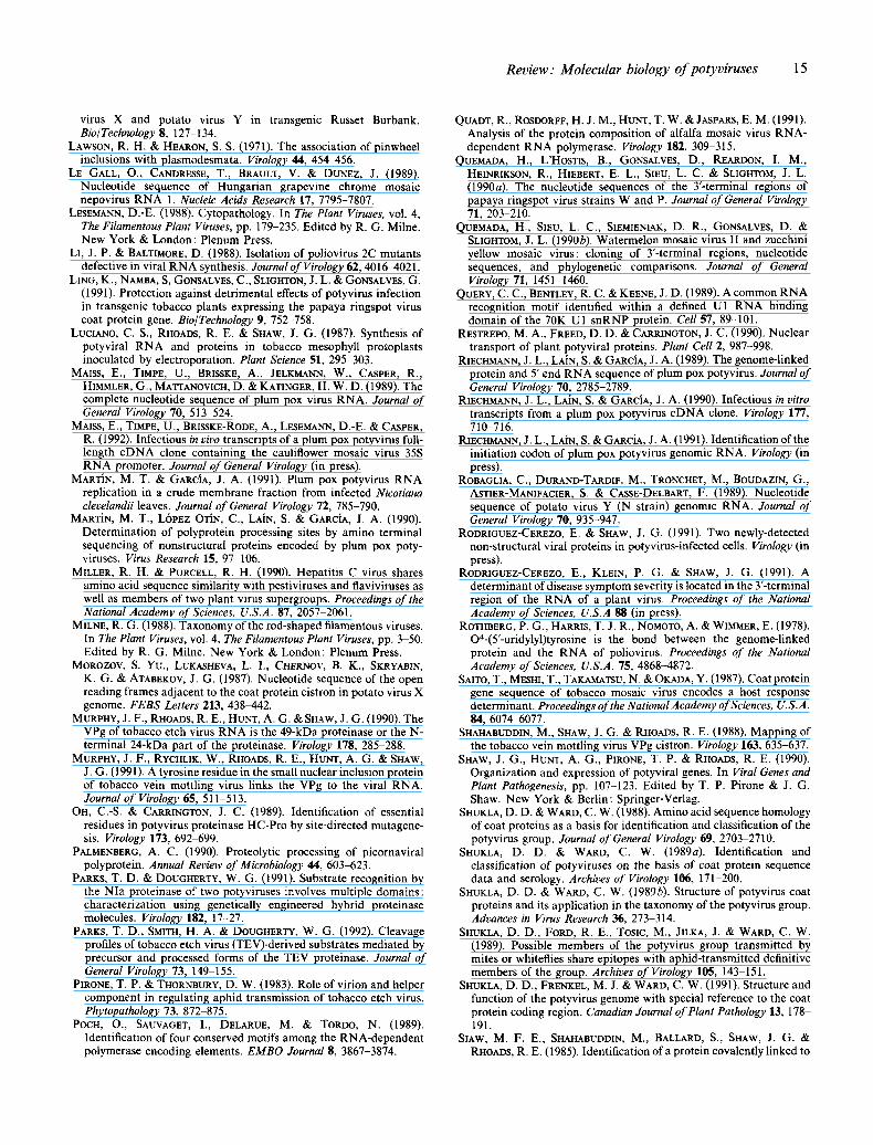
Review." Molecular biology o f potyviruses 15
virus X and potato virus Y in transgenic Russet Burbank. Bio/Technology $, 127-134.
LAWSON, R. H. & HEARON, S. S. (1971). The association of pinwheel inclusions with plasmodesmata. Virology 44, 454-456.
LE GALL, O., CANDRESSE, T., BRAULT, V. & DUNEZ, J. (1989). Nucleotide sequence of Hungarian grapevine chrome mosaic nepovirus RNA 1. Nucleic Acids Research 17, 7795-7807.
LESEMANN, D.-E. (1988). Cytopathology. In The Plant Viruses, vol. 4, The Filamentous Plant Viruses, pp. 179-235. Edited by R. G. Milne. New York & London: Plenum Press.
El, J. P. & BALTIMORE, D. (1988). Isolation of poliovirus 2C mutants defective in viral RNA synthesis. Journal of Virology 62, 4016-4021.
LING, K., NAMBA, S, GONSALVES, C., SLIGHTON, J. L. & GONSALVES, G. (1991). Protection against detrimental effects of potyvirus infection in transgenic tobacco plants expressing the papaya ringspot virus coat protein gene. Bio/Technology 9, 752-758.
LUCIANO, C. S., RHOADS, R. E. & SHAw, J. G. (1987). Synthesis of potyviral RNA and proteins in tobacco mesophyll protoplasts inoculated by electroporation. Plant Science 51, 295-303.
MAISS, E., TIMPE, U., BRISSKE, m., JELKMANN, W., CASPER, R., HIMMLER, G., MATTANOVICH, D. & KATINGER, H. W. D. (1989). The complete nucleotide sequence of plum pox virus RNA. Journal of General Virology 70, 513 524.
MAISS, E., TIMPE, U., BRISSKE-RODE, A., LESEMANN, D.-E. & CASPER, R. (1992). Infectious in vivo transcripts of a plum pox potyvirus full- length cDNA clone containing the cauliflower mosaic virus 35S RNA promoter. Journal of General Virology (in press).
MARTIN, M. T. & GARCiA, J. A. (1991). Plum pox potyvirus RNA replication in a crude membrane fraction from infected Nicotiana clevelandii leaves. Journal of General Virology 72, 785-790.
MARTIN, M. T., L6PEZ OriN, C., LAIN, S. & GARCiA, J. A. (1990). Determination of polyprotein processing sites by amino terminal sequencing of nonstructural proteins encoded by plum pox poty- viruses. Virus Research 15, 97-106.
MILLER, R. H. & PURCELL, R. H. (1990). Hepatitis C virus shares amino acid sequence similarity with pestiviruses and flaviviruses as well as members of two plant virus supergroups. Proceedings of the National Academy of Sciences, U.S.A. 87, 2057-2061.
MILNE, R. G. (1988). Taxonomy of the rod-shaped filamentous viruses. In The Plant Viruses, vol. 4, The Filamentous Plant Viruses, pp. 3-50. Edited by R. G. Milne. New York & London: Plenum Press.
MOROZOV, S. Yu., LUKASHEVA, L. I., CHERNOV, B. K., SKRYABIN, K. G. 8/. ATABEKOV, J. G. (1987). Nucleotide sequence of the open reading frames adjacent to the coat protein cistron in potato virus X genome. FEBS Letters 213, 438~142.
MURPHY, J. F., RFIOADS, R. E., HUNT, A. G. & SHAw, J. G. (1990). The VPg of tobacco etch virus RNA is the 49-kDa proteinase or the N- terminal 24-kDa part of the proteinase. Virology 178, 285-288.
MURPHY, J. F., RYCHLIK, W., RHOADS, R. E., HUNT, A. G. & SHAW, J. G. (1991). A tyrosine residue in the small nuclear inclusion protein of tobacco vein mottling virus links the VPg to the viral RNA. Journal of Virology 65, 511-513.
OH, C.-S. & CARRINGTON, J. C. (1989). Identification of essential residues in potyvirus proteinase HC-Pro by site-directed mutagene- sis. Virology 173, 692-699.
PALMENBERG, A. C. (1990). Proteolytic processing of picornaviral polyprotein. Annual Review of Microbiology 44, 603-623.
PARKS, T. D. & DOUGHERTY, W. G. (1991). Substrate recognition by the NIa proteinase of two potyviruses involves multiple domains: characterization using genetically engineered hybrid proteinase molecules. Virology 182, 17-27.
PARKS, T. D., SMITH, H. A. & DOUGHERTY, W. G. (1992). Cleavage profiles of tobacco etch virus (TEV)-derived substrates mediated by precursor and processed forms of the TEV proteinase. Journal of General Virology 73, 1 ~19-155.
PIRONE, T. P. & THORNBURY, D. W. (1983). Role of virion and helper component in regulating aphid transmission of tobacco etch virus. Phytopathology 73, 872 875.
POCH, O., SAUVAGET, I., DELARUE, M. & TORDO, N. (1989). Identification of four conserved motifs among the RNA-dependent polymerase encoding elements. EMBO Journal 8, 3867-3874.
QUADT, R., ROSDORFF, H. J. M., HUNT, T. W. ~. JASPARS, E. M. (1991). Analysis of the protein composition of alfalfa mosaic virus RNA- dependent RNA polymerase. Virology 182, 309-315.
QUEMADA, H., L'HosTIS, B., GONSALVES, D., REARDON, I. M., HEINRIKSON, R., HIEBERT, E. L., SIEU, L. C. & SLIGHTOM, J. L. (1990a). The nucleotide sequences of the 3'-terrninal regions of papaya ringspot virus strains W and P. Journal of General Virology 71, 203-210.
QUEMADA, H., SIEU, L. C., SIEMIENIAK, D. R., GONSALVES, D. & SLIGHTOM, J. L. (1990b). Watermelon mosaic virus II and zucchini yellow mosaic virus: cloning of Y-terminal regions, nucleotide sequences, and phylogenetic comparisons. Journal of General Virology 71, 1451 1460.
QUERY, C. C., BENTLEY, R. C. & KEENE, J. D. (1989). A common RNA recognition motif identified within a defined U1 RNA binding domain of the 70K U1 snRNP protein. Cell 57, 89-101.
RESTREPO, M. A., FREED, D. D. & CARRINGTON, J. C. (1990). Nuclear transport of plant potyviral proteins. Plant Cell 2, 987-998.
RIECrlMANN, J. L., LAIN, S. & GARCiA, J. A. (1989). The genome-linked protein and 5' end RNA sequence of plum pox potyvirus. Journal of General Virology 70, 2785-2789.
RIECHMANN, J. L., LAIN, S. & GARCiA, J. A. (1990). Infectious in vitro transcripts from a plum pox potyvirus cDNA clone. Virology 177, 710-716.
RIECrtMANN, J. L., LAIN, S. & GARCiA, J. A. (1991). Identification of the initiation codon of plum pox potyvirus genomic RNA. Virology (in press).
ROBAGLIA, C., DURAND-TARDIF, M., TRONCHET, M., BOUDAZIN, G., ASTIER-MANIEACIER, S. & CASSE-DELEART, F. (1989). Nucleotide sequence of potato virus Y (N strain) genomic RNA. Journal of General Virology 70, 935-947.
RODRIGUEZ-CEREZO, E. & SHAW, J. G. (1991). Two newly-detected non-structural viral proteins in potyvirus-infected cells. Virology (in press).
RODRIGUEZ-CEREZO, E., KLEIN, P. G. & SHAW, J. G. (1991). A determinant of disease symptom severity is located in the 3'-terminal region of the RNA of a plant virus. Proceedings of the National Academy of Sciences, U.S.A 88 (in press).
ROTHBERG, P. G., HARRIS, T. J. R., NOMOTO, A. & WIMMER, E. (1978). O*-(5'-uridylyl)tyrosine is the bond between the genome-linked protein and the RNA of poliovirus. Proceedings of the National Academy of Sciences, U.S.A. 75, 4868-4872.
SAITO, T., MESHI, T., TAKAMATSU, N. & OKADA, Y. (1987). Coat protein gene sequence of tobacco mosaic virus encodes a host response determinant. Proceedings of the National Academy of Sciences, U.S.A. 84, 6074q5077.
SHAHABUDDIN, M., SHAW, J. G. & RHOADS, R. E. (1988). Mapping of the tobacco vein mottling virus VPg cistron. Virology 163, 635-637.
SHAW, J. G., HUNT, A. G., PIRONE, T. P. & RHOADS, R. E. (1990). Organization and expression of potyviral genes. In Viral Genes and Plant Pathogenesis, pp. 107-123. Edited by T. P. Pirone & J. G. Shaw. New York & Berlin: Springer-Verlag.
SHUKLA, D. D. & WARD, C. W. (1988). Amino acid sequence homology of coat proteins as a basis for identification and classification of the potyvirus group. Journal of General Virology 69, 2703-2710.
SHUKLA, D. D. & WARD, C. W. (1989a). Identification and classification of potyviruses on the basis of coat protein sequence data and serology. Archives of Virology 106, 171-200.
SHUKLA, D. D. & WARD, C. W. (1989b). Structure of potyvirus coat proteins and its application in the taxonomy of the potyvirus group. Advances in Virus Research 36, 273-314.
SHUKLA, D. D., FORD, R. E., TOSIC, M., JILKA, J. & WARD, C. W. (1989). Possible members of the potyvirus group transmitted by mites or whiteflies share epitopes with aphid-transmitted definitive members of the group. Archives of Virology 105, 143 151.
SHUKLA, D. D., FRENKEL, M. J. & WARD, C. W. (1991). Structure and function of the potyvirus genome with special reference to the coat protein coding region. Canadian Journal of Plant Pathology 13, 178 191.
SLAW, M. F. E., SHAHABUDDIN, M., BALLARD, S., SHAW, J. G. & RHOADS, R. E. (1985). Identification of a protein covalently linked to
16 J. L. Riechmann, S. Lain and J. A. Garcia
the 5' terminus of tobacco vein mottling virus RNA. Virology 142, 134-143.
STARK, D. M. & BEACHY, R. N. (1989). Protection against potyvirus infection in transgenic plants: evidence for broad spectrum resistance. Bio/Technology 7, 1257-1262.
STREBEL, K. & BECK, E. (1986). A second protease of foot-and-mouth disease virus. Journal of Virology 58, 893-899.
TAKEDA, N., KUHN, R. J., YANG, C.-F., TAKEGAMI, T. & WIMMER, E. 0986). Initiation of poliovirus plus-strand RNA synthesis in a membrane complex of infected HeLa cells. Journal of Virology 60, 43-53.
TEYCHENEY, P. Y., TAVERT, G., DELBOS, R., RAVELONANDRO, M. & DUNEZ, J. (1989). The complete nucleotide sequence of plum pox virus RNA (strain D). Nucleic Acids Research 17, 10115-10116.
THOMAS, A. A. M., TER HAAR, E., WELLINK, J. St. VOORMA, H. O. (1991). Cowpea mosaic virus middle component RNA contains a sequence that allows internal binding of ribosomes and that requires eukaryotic initiation factor 4F for optimal translation. Journal of Virology 65, 2953-2959.
THORNBURY, D. W. & PIRONE, T. P. (1983). Helper components of two potyviruses are serologically distinct. Virology 125, 487~490.
THORNBURY, D. W., HELLMANN, G. M., RHOADS, R. E. & PIRONE, T. P. (1985). Purification and characterization of potyvirus helper component. Virology 144, 260-267.
THORNBURY, D. W., PA'VI'ERSON, C. A., DESSENS, J. T. & PIRONE, T. P. (1990). Comparative sequence of the helper component (HC) region of potato virus Y and a HC-defective strain, potato virus C. Virology 178, 573-578.
TOBIN, G. J., YOUNG, D. C. & FLANEGAN, J. B. (1989). Self-catalyzed linkage of poliovirus terminal protein VPg to poliovirus RNA. Cell 59, 511-519.
TURPEN, T. (1989). Molecular cloning of a potato virus Y genome: nucleotide sequence homology in non-coding regions of potyviruses. Journal of General Virology 70, 1951-1960.
UYEDA, I., TAKAHASHI, T. & SHIKATA, E. (1990). Sequence relatedness of clover yellow vein virus to bean yellow mosaic virus. Abstracts, VllIth International Congress of Virology, p. 128.
VAN LENT, J., WELLINK, J. & GOLDBACH, R. (1990). Evidence for the involvement of the 58K and 48K proteins in the intercellular movement of cowpea mosaic virus. Journal of General Virology 71, 219-223.
VERCHOT, J., KOONIN, E. V. • CARRINGTON, J. C. (1991). The 35-kDa protein from the N-terminus of a potyviral polyprotein functions as a third virus-encoded proteinase. Virology (in press).
Vos, P., VERVER, J., JAEGLE, M., WELLINK, J., VAN KAMMEN, A. & GOLDBACH, R. (1988). Two viral proteins involved in the proteolytic processing of the cowpea mosaic virus polyproteins. Nucleic Acids Research 16, 1967-1985.
WARD, C. W. & SHUKLA, D. D. (1991). Taxonomy of potyviruses: current problems and some solutions. Intervirology 32, 269-296.
WELLINK, J. & VAN KAMMEN, A. (1988). Proteases involved in the processing of viral polyproteins. Archives of Virology 98, 1-26.
WELLINK, J. & VAN KAMMEN, A. (1989). Cell-to-cell transport of cowpea mosaic virus requires both the 58K/48K proteins and the capsid proteins. Journal of General Virology 70, 2279 2286.
WETZEL, T., CANDRESSE, T., RAVELONANDRO, M., DELBOS, R. P., MAZYAD, H., ABOUL-ATA, A. E. & DUNEZ, J. (1991). Nucleotide sequence of the 3'-terminal region of the RNA of the E1 Amar strain of plum pox potyvirus. Journal of General Virology 72, 1741-1746.
ZIMMERN, D. (1987). Evolution of RNA viruses, in RNA Genetics, pp. 211-244. Edited by J. Holland, E. Domingo & P. Ahlquist. Boca Raton: CRC Press.